EP3699142A1 - Method of immobilization nucleic acid structure on surface of graphene, graphene modified by immobilization of nucleic acid structure and its use - Google Patents
Method of immobilization nucleic acid structure on surface of graphene, graphene modified by immobilization of nucleic acid structure and its use Download PDFInfo
- Publication number
- EP3699142A1 EP3699142A1 EP19461514.2A EP19461514A EP3699142A1 EP 3699142 A1 EP3699142 A1 EP 3699142A1 EP 19461514 A EP19461514 A EP 19461514A EP 3699142 A1 EP3699142 A1 EP 3699142A1
- Authority
- EP
- European Patent Office
- Prior art keywords
- graphene
- molecule
- nucleic acid
- dna origami
- dna
- Prior art date
- Legal status (The legal status is an assumption and is not a legal conclusion. Google has not performed a legal analysis and makes no representation as to the accuracy of the status listed.)
- Pending
Links
Images


Classifications
-
- C—CHEMISTRY; METALLURGY
- C01—INORGANIC CHEMISTRY
- C01B—NON-METALLIC ELEMENTS; COMPOUNDS THEREOF; METALLOIDS OR COMPOUNDS THEREOF NOT COVERED BY SUBCLASS C01C
- C01B32/00—Carbon; Compounds thereof
- C01B32/15—Nano-sized carbon materials
- C01B32/182—Graphene
- C01B32/194—After-treatment
-
- C—CHEMISTRY; METALLURGY
- C01—INORGANIC CHEMISTRY
- C01B—NON-METALLIC ELEMENTS; COMPOUNDS THEREOF; METALLOIDS OR COMPOUNDS THEREOF NOT COVERED BY SUBCLASS C01C
- C01B32/00—Carbon; Compounds thereof
- C01B32/15—Nano-sized carbon materials
- C01B32/182—Graphene
- C01B32/184—Preparation
-
- C—CHEMISTRY; METALLURGY
- C12—BIOCHEMISTRY; BEER; SPIRITS; WINE; VINEGAR; MICROBIOLOGY; ENZYMOLOGY; MUTATION OR GENETIC ENGINEERING
- C12Q—MEASURING OR TESTING PROCESSES INVOLVING ENZYMES, NUCLEIC ACIDS OR MICROORGANISMS; COMPOSITIONS OR TEST PAPERS THEREFOR; PROCESSES OF PREPARING SUCH COMPOSITIONS; CONDITION-RESPONSIVE CONTROL IN MICROBIOLOGICAL OR ENZYMOLOGICAL PROCESSES
- C12Q1/00—Measuring or testing processes involving enzymes, nucleic acids or microorganisms; Compositions therefor; Processes of preparing such compositions
- C12Q1/68—Measuring or testing processes involving enzymes, nucleic acids or microorganisms; Compositions therefor; Processes of preparing such compositions involving nucleic acids
- C12Q1/6813—Hybridisation assays
- C12Q1/6834—Enzymatic or biochemical coupling of nucleic acids to a solid phase
-
- B—PERFORMING OPERATIONS; TRANSPORTING
- B82—NANOTECHNOLOGY
- B82Y—SPECIFIC USES OR APPLICATIONS OF NANOSTRUCTURES; MEASUREMENT OR ANALYSIS OF NANOSTRUCTURES; MANUFACTURE OR TREATMENT OF NANOSTRUCTURES
- B82Y15/00—Nanotechnology for interacting, sensing or actuating, e.g. quantum dots as markers in protein assays or molecular motors
Definitions
- the present invention relates to a method of immobilizing nucleic acid structure such as DNA Origami on a surface of graphene, the graphene modified by immobilization of nucleic acid structure and its use.
- the present invention is based on "Structural DNA nanotechnology" and "Scaffolded DNA Origami” techniques.
- the former approach initiated in the early 1980s by Nadrian Seeman, takes advantage of the molecular recognition properties of synthetic DNA oligonucleotides to create finite size objects or infinite periodic lattices through complementary Watson-Crick base pairing, sticky-end cohesion and stacking interactions.
- Scaffolded DNA Origami introduced in 2006 by Paul Rothemund, simplifies the access to finite DNA nanoobjects.
- single-stranded circular DNA molecules of typically 7 - 8.5 kb size are folded by the aid of staple strands into an array of helices through an arrangement of periodic crossovers.
- Origami structures can be used as "molecular pegboards" with an addressable surface area of a few thousand nm 2 for arranging arbitrary objects-of-interest (OOI) with a "single pixel" resolution of about 6 nanometers.
- OOIs are metal or semiconductor nanoparticles for potential applications in microelectronics and (non-linear) optics, or proteins, peptides and other ligands for potential applications in biosensing, cell culture, or other areas of biological and biomedical research.
- DNA origami structures could advantageously allow for control of the absolute number, stoichiometry and precise nanoscale orientation of OOls, e.g. receptors and additional cell membrane proteins for presentation to living cells.
- Many of the aforementioned potential applications require or would at least benefit from methods to immobilize DNA origami structures on solid supports, in particular in a site-specific manner.
- nanoparticle-decorated origami structures have been deposited on nanopatterned surfaces prepared by electron-beam lithography and etching processes to create DNA origami shaped binding sites on non-transparent substrates. While these approaches proved feasible for generating large-area spatially ordered arrays of DNA origami, the production of substrates is very cost and time consuming. Even more important, site-selectivity could not yet be realized because origami immobilization is governed by mere electrostatic interactions.
- Graphene is a unique two-dimensional 2D material with promising applications in the nanoscience.
- a main limitation has been the problem of selective and controlled chemical modification without disturbing its properties. This has limited its usefulness especially in electronic and optical sensing applications.
- Graphene is a 2D carbon lattice resembling a honeycomb, which has attracted great attention since 2004, when it was experimentally isolated for the first time. Due to its unique electronic, optical and mechanical properties, it has been intensively explored worldwide and found applications probably in every branch of science. (Ferrari et al., 2014) Its gapless energy band structure and linear dispersion relation near the corners of the Brillouin zone result in a frequency-independent light absorption, governed solely by the fine-structure constant, ⁇ ⁇ 1/137.
- d 0 8 to 20 nm.
- the technical problem underlying the present invention is how to have a well defined chemical modification of graphene to make its quenching properties exploitable for biochemical sensing assays.
- the solution is to use nucleic acid structures such as DNA origami as chemical adapters. Beyond the mere chemical adaptation of any assay that might be incorporated within the DNA origami, the DNA origami is also a nanopositioner: it can place the assay at the height where the fluorescence signal is most sensitive to minimal distance alterations.
- Another technical problem underlying the present invention is to provide methods for immobilization of nucleic acid structures such as DNA origami structures on graphene. Further, respective assemblies of immobilized nucleic acid structures such as DNA origami on graphene and uses thereof should be provided.
- the subject of the invention is a method of immobilization of nucleic acid structures on graphene, said method comprising the steps of:
- step i) comprises the following sub-steps of:
- said nucleic acid structure is selected from the group comprising DNA-based structures, such as: DNA origami structures, DNA junctions, DNA tiles, DNA bricks, DNA wireframe structures etc.
- step i) comprises the following sub-step of:
- step ii) is carried out for at least 10 seconds, and/or step ii) is carried out at temperature from the range of 10-40 °C, preferably 15-30°C.
- said molecule which is able to attach to graphene via ⁇ -interactions is pyrene.
- said method comprising the steps of:
- said molecule able to attach to graphene via ⁇ -interactions is pyrene, and/or the number of pyrene molecules per one DNA origami structure is at least 2, preferably 4-10.
- step c) incubation in step c) is carried out for 2 hours in 37°C.
- said nucleic acid structures carry one or more type of objects-of-interest (OOI), wherein the OOIs are selected from the group comprising proteins, peptides, nanoparticles, carbohydrates, small molecule ligands, dyes, nanocrystals, DNA, RNA, chemical moieties such as thiol or biotin etc.
- OOI objects-of-interest
- step of immobilization of nucleic acid structures on the graphene further step of washing the immobilized sample with any solution in which DNA origami is stable, such as Milli-Q water, PBS buffer, 10 mM Tris or 1 ⁇ TE buffer containing 0-15 mM MgCl 2 is carried out.
- any solution in which DNA origami is stable such as Milli-Q water, PBS buffer, 10 mM Tris or 1 ⁇ TE buffer containing 0-15 mM MgCl 2 is carried out.
- Another subject of the present invention is graphene modified by immobilization of nucleic acid structures comprising at least one type of nucleic acid structure and graphene layer, wherein said nucleic acid structure is immobilized on the graphene layer via molecule-modified oligonucleotides, wherein said molecule is able to attach to graphene via ⁇ -interactions, and wherein said molecule-modified oligonucleotides are complementary for Watson-Crick pairing to oligonucleotides comprised in said nucleic acid structures.
- Another subject of the present invention is graphene modified by immobilization of DNA origami structures comprising at least one type of DNA origami structure and graphene layer, wherein said DNA origami structure is immobilized on the graphene layer via pyrene-modified oligonucleotides which are complementary for Watson-Crick pairing to oligonucleotides protruding from the DNA origami structure and/or built into said DNA origami structure.
- said graphene is modified by immobilization of nucleic acid structures such as DNA origami structures, wherein nucleic acid structures such as DNA origami structures carry one or more type of objects-of-interest (OOI), wherein the OOIs are selected from the group comprising proteins, peptides, nanoparticles, carbohydrates, small molecule ligands, dyes, nanocrystals, DNA, RNA, chemical moieties such as thiol or biotin, etc.
- nucleic acid structures such as DNA origami structures
- nucleic acid structures such as DNA origami structures carry one or more type of objects-of-interest (OOI)
- OOIs are selected from the group comprising proteins, peptides, nanoparticles, carbohydrates, small molecule ligands, dyes, nanocrystals, DNA, RNA, chemical moieties such as thiol or biotin, etc.
- Another subject of the present invention is use of graphene modified by immobilization of nucleic acid structures such as DNA origami structures defined above in biosensing and energy conversion as well as photovoltaics and optoelectronics.
- DNA origami surface chemistry converters/nanopositioners are programmed nanostructures of roughly 21.000 nm 3 .
- DNA origami nanostructures can be functionalized in many other ways the surface chemistry can be adapted from a graphene surface to many other chemical functionalities.
- Pyrene (Benzo[ def ]phenanthrene, CAS Number 129-00-0) is a polycyclic aromatic hydrocarbon (PAH) consisting of four fused benzene rings, resulting in a flat aromatic system.
- PAH polycyclic aromatic hydrocarbon
- the chemical formula is C16H10. This colorless solid is the smallest peri-fused PAH (one where the rings are fused through more than one face). Pyrene forms during incomplete combustion of organic compounds.
- DNA origami surface chemistry converters by placing single dye molecules at specific, designed distances in the range of 1 - 60 nm from the graphene surface is demonstrated. With this arrangement the distance dependent fluorescence quenching of dyes that will be exploited for biosensing assays in the future is determined. Besides changing surface chemistry, the fluorescence assay can be placed at a distance to the graphene surface that exhibits highest sensitivity to fluorescence changes.
- the present Inventors used DNA origami ( Rothemund, P. W. K. Nature 2006, 440 (7082), 297-302 ) as a surface chemistry converter to generally overcome the problem of chemical functionalization of graphene for optical biosensing assays.
- the DNA origami technique enables the formation of custom-designed DNA nanostructures with a volume of ⁇ 21.000 nm 3 , and the rich DNA chemistry available allows placing of arbitrary objects and complex biomolecular assays ( Acuna, G. P. et al.; ACS Nano 2012, 6 (4), 3189-3195 ; Vietz, C. et al.; Nano Lett. 2017, 17, 6496-6500 ; Ochmann, S. E.
- DNA origami can act as a biocompatible surface ( Gietl, A. et al.; Nucleic Acids Res. 2012, 40 (14), 1-10 ).
- DNA origami structures were coupled to pristine graphene either to increase their stability ( Matkovi ⁇ , A. et al.; New J. Phys. 2016, 18 (2), 025016 ) or as a template for metallized DNA nanolithography ( Jin, Z. et al.; Nat. Commun. 2013, 4, 1663 ).
- it was demonstrated with TEM imaging that DNA origami nanoplates were denatured due to hydrophobic interactions of the DNA bases with graphene upon adsorption Kabiri, Y. et al.; Small 2017, 1-8 ; Green, N. S. et al.; Mater. Res. Express 2018, 5, 045035 ).
- the narrow intensity-fluorescence lifetime distributions confirm the d -4 law with a precise value of d 0 of 17.7 nm in aqueous buffer solution and present the basis for a broad range of applications in biosensing and optoelectronics.
- Each DNA origami structure contains one dye molecule (ATTO542) marked as a small green sphere, which is positioned at the height of: 3 nm ( NR ), 7 nm ( ND ), 12 nm ( NP1 ), 16 nm ( NP2 ), 24 nm ( NP3 ) and 53 nm ( NP4 ).
- the selected values cover the range of distances for which the energy transfer from a single dye molecule to graphene is expected to vary from 0 to almost 100%.
- the dyes are attached to the DNA origami by 6-carbon linkers and measurements were carried out in buffer solution, it is expected that the fluorescent dyes assume all possible dipole orientations during the measurement.
- 6-8 staple strands with single-stranded extensions protruding from the DNA origamis were incorporated.
- the protrusions are used to label the DNA origami with pyrene-modified complementary oligonucleotides.
- the pyrene enabled selective binding of the DNA origami with the bottom side to the graphene layer ( Figure 1 ).
- the stability of this interaction in certain temperature ranges is achieved by binding minimally 2 and optimally 4-10 pyrene units per one DNA origami structure, beacuse most likely one pyrene molecule is not enough to obtain stable bonds between the DNA origami and the graphene layer.
- External labeling with extended staple strands allowed modular modifications also for other moieties.
- biotin-modified oligonucleotides were used and DNA origami structures were immobilized on neutravidin-biotinylated BSA surfaces. Single-molecule fluorescence measurements were carried out with a home-built confocal microscope.
- Figure 2 depicts ATTO542 fluorescence intensity images for each of the six DNA origami structures using 532 nm excitation.
- Laser powers ranged from 1 ⁇ W for three nanopillar samples ( NP2 , NP3 and NP4 ), 2 ⁇ W for one nanopillar sample and the nanodisc ( NP1 and ND ), up to 5 ⁇ W for the nanorectangle ( NR ) DNA origami structure.
- FIG. 7B shows the relative fluorescence intensity I G ⁇ I ref ⁇ as a function of the fluorescence lifetime, where I G is fluorescence intensity of a dye molecule within DNA origami bound to graphene, and ⁇ I ref ⁇ is the average fluorescence intensity obtained for the reference sample, NP4. Due to the differences in the applied laser powers in our measurements, all the obtained values of fluorescence intensities were normalized to the laser power of 1 ⁇ W. It is noteworthy that narrow and clearly separated populations of fluorescence intensity and fluorescence lifetime are obtained, indicating the selectivity and robustness of the immobilization strategy.
- Figures 4a and 4b show the distance dependent energy transfer efficiency calculated by both methods.
- the DNA origami structures were incubated with pyrene-modified (for graphene samples) or biotin-modified (for glass samples) staple strands, complementary to the oligonucleotides protruding from DNA origami structures, for 2 h in 37 °C.
- Such prepared structures were immobilized on the glass surface of a Lab-Tek chamber (Thermo Fisher Scientific) coated with BSA-biotin/neutravidin (Sigma-Aldrich) or directly on a single layer of CVD graphene.
- the designed height values of 3 nm ( NR ), 7 nm ( ND ), 12 nm ( NP1 ), 16 nm ( NP2 ), 24 nm ( NP3 ) and 53 nm ( NP4 ) were calculated using the size parameter of a double helix, namely its diameter of 2.2 nm and a length of 0.34 nm per base pair. Taking into account previous report about the overestimation of the calculated values (the measured values smaller of about 10% compared to the designed values), these numbers were corrected. Additionally, a thickness of 1 nm for the pyrene-modified protruding strands was added.
- the green laser beams (532 nm LDH-P-FA-530B, Picoquant) is controlled by a PDL 828 "Sepiall" (Picoquant).
- the green fiber laser is decoupled by a collimator (F2220APC-532, Thorlabs). After passing through cleanup filters (532/2) and a dichroic mirror (640DCXR, AHF) the laser beam is coupled into a fiber (P3-488PM-FC, Thorlabs) with a collimator (PAF2-2A, Thorlabs) and decoupled with a collimator (G169015000, Qioptics).
- a combination of a linear polarizer (WP12L-Vis, Thorlabs) and a quarter wave plate (AQWP05M-600, Thorlabs) is used to obtain circularly polarized light.
- a dual band dichroic beam splitter z532/633, AHF
- the light beam is focused by an oil-immersion objective (UPLSAPO 100XO, NA 1.40, Olympus) on the measurement chamber, which can be positioned accurately by a piezo-stage (P-527.3CD, Physiklnstrumente) which is driven by a E-727 controller (Physiklnstrumente).
- the emission of the fluorophores is collected by the same objective, focused on a 50 ⁇ m pinhole (Linos), collimated with a lens (AC050-150-A-ML, Thorlabs) and split spectrally by another dichroic beam splitter (640DCXR, AHF).
- the green laser beams cleaned with a filter set (HC582/75, AHF and LP 532, both Semrock) and focused with a lens (AC080-020-A-ML, Thorlabs) on the APD (SPCM-AQRH-TR-14, Excelitas)
- the signals of the APD are detected by a HydraHarp 400 (Picoquant) and the whole system is operated with SymPhoTime 64 (PicoQuant).
- the correct folding of the DNA origami structures was characterized with atomic force microscopy (AFM, Nanowizard 3 ultra, JPK Instruments) in solution.
- AFM atomic force microscopy
- 10 ⁇ L of a 10 mM NiCl 2 x 6 H 2 O solution were incubated for 5 min.
- 10 ⁇ L 1 nM DNA origami structure solution diluted in AFM buffer (40 mM TRIS, 2 mM EDTA disodium salt dihydrate and 12.5 mM Mg(OAc) 2 x 6 H 2 O) were added and incubated for 5 min.
- 300 ⁇ L AFM buffer were added after purging three times with 300 ⁇ L AFM buffer.
- the solution measurements were performed with cantilevers USC-F0.3-k0.3-10 from Nano World.
- TEM grids (TedPella, formvar/carbon, 400 mesh, Cu) were Ar-plasma cleaned and incubated for 60 sec with DNA origami sample (5 ⁇ L, ⁇ 2-10 nM). Grids were washed with 2 % uranyl formate solution (5 ⁇ L) and incubated again afterwards again 4 s with 2 % uranyl formate solution (5 ⁇ L) for staining.
- TEM imaging were performed with a JOEL JEM-1100 with an acceleration voltage of 80 kV.Images of NR, ND and NP1-NP4 are given in Figure 8 .
- Fluorescent images are given in Figure 2 .
- DNA origami as chemical converters will enable placing biomolecular assays on the graphene with the optimized distance to the surface for optimized sensitivity. Placing assays at a height of 17 - 18 nm will yield an extremely sensitive method of detecting small distance changes to the surface while avoiding direct contact to the interfering hydrophobic graphene surface.
- DNA origami as a chemical converting and placement platform could be used to incorporate further functionalities for electronic, nanophotonic, and energy conversion devices with graphene and other 2D materials opening a myriad of new possibilities.
Landscapes
- Chemical & Material Sciences (AREA)
- Organic Chemistry (AREA)
- Engineering & Computer Science (AREA)
- Life Sciences & Earth Sciences (AREA)
- Health & Medical Sciences (AREA)
- Materials Engineering (AREA)
- Nanotechnology (AREA)
- Inorganic Chemistry (AREA)
- Zoology (AREA)
- Wood Science & Technology (AREA)
- Proteomics, Peptides & Aminoacids (AREA)
- Biotechnology (AREA)
- Biochemistry (AREA)
- Immunology (AREA)
- Microbiology (AREA)
- Molecular Biology (AREA)
- Analytical Chemistry (AREA)
- Physics & Mathematics (AREA)
- Biophysics (AREA)
- Bioinformatics & Cheminformatics (AREA)
- General Engineering & Computer Science (AREA)
- General Health & Medical Sciences (AREA)
- Genetics & Genomics (AREA)
- Apparatus Associated With Microorganisms And Enzymes (AREA)
- Measuring Or Testing Involving Enzymes Or Micro-Organisms (AREA)
Abstract
Description
- The present invention relates to a method of immobilizing nucleic acid structure such as DNA Origami on a surface of graphene, the graphene modified by immobilization of nucleic acid structure and its use.
- The present invention is based on "Structural DNA nanotechnology" and "Scaffolded DNA Origami" techniques. The former approach, initiated in the early 1980s by Nadrian Seeman, takes advantage of the molecular recognition properties of synthetic DNA oligonucleotides to create finite size objects or infinite periodic lattices through complementary Watson-Crick base pairing, sticky-end cohesion and stacking interactions. Scaffolded DNA Origami, introduced in 2006 by Paul Rothemund, simplifies the access to finite DNA nanoobjects. In this technique, single-stranded circular DNA molecules of typically 7 - 8.5 kb size are folded by the aid of staple strands into an array of helices through an arrangement of periodic crossovers. Extension of this concept to assemble 3D DNA structures has led to the establishment of a robust and reliable method to fabricate DNA nanostructures with dimensions in the 20 to 200 nm range. Origami structures can be used as "molecular pegboards" with an addressable surface area of a few thousand nm2 for arranging arbitrary objects-of-interest (OOI) with a "single pixel" resolution of about 6 nanometers. Typical OOIs are metal or semiconductor nanoparticles for potential applications in microelectronics and (non-linear) optics, or proteins, peptides and other ligands for potential applications in biosensing, cell culture, or other areas of biological and biomedical research. In this context, DNA origami structures could advantageously allow for control of the absolute number, stoichiometry and precise nanoscale orientation of OOls, e.g. receptors and additional cell membrane proteins for presentation to living cells. Many of the aforementioned potential applications require or would at least benefit from methods to immobilize DNA origami structures on solid supports, in particular in a site-specific manner. So far, nanoparticle-decorated origami structures have been deposited on nanopatterned surfaces prepared by electron-beam lithography and etching processes to create DNA origami shaped binding sites on non-transparent substrates. While these approaches proved feasible for generating large-area spatially ordered arrays of DNA origami, the production of substrates is very cost and time consuming. Even more important, site-selectivity could not yet be realized because origami immobilization is governed by mere electrostatic interactions.
- Graphene is a unique two-dimensional 2D material with promising applications in the nanoscience. A main limitation has been the problem of selective and controlled chemical modification without disturbing its properties. This has limited its usefulness especially in electronic and optical sensing applications.
- Graphene is a 2D carbon lattice resembling a honeycomb, which has attracted great attention since 2004, when it was experimentally isolated for the first time. Due to its unique electronic, optical and mechanical properties, it has been intensively explored worldwide and found applications probably in every branch of science. (Ferrari et al., 2014) Its gapless energy band structure and linear dispersion relation near the corners of the Brillouin zone result in a frequency-independent light absorption, governed solely by the fine-structure constant, α ≈ 1/137. As a result, this only one-atom thick material absorbs as much as πα ≈ 2.3% of light, over the visible and near-infrared spectral regions.(Nair et al., 2008) In consequence, graphene behaves as an extraordinary energy sink and a unique acceptor system, which is one of the key characteristics of graphene and graphene-related two-dimensional materials exploited in the field of optical biosensors and distance rulers.(Anichini et al., 2018; Zhu, Du, & Lin, 2015) Fluorescent dyes placed close to graphene are strongly quenched and their displacement from graphene can restore fluorescence.(Chen, Berciaud, Nuckolls, Heinz, & Brus, 2010; Federspiel et al., 2015; Gaudreau et al., 2013; Mazzamuto et al., 2014) It has been demonstrated both theoretically and experimentally that the energy transfer from a molecule (a single dipole) to graphene ("2D array of dipoles") strongly depends on the distance d between both, and scales proportional to d -4.(Brenneis et al., 2015; Gaudreau et al., 2013; Swathi & Sebastian, 2008, 2009) Whereas the distance dependence is well understood, reports vary with respect to the d0 value which states the distance of 50% quenching efficiency. This variation is related to the different emitters used (e.g. quantum dots, nitrogen-vacancy centers or dyes embedded in crystals) and how the distance to the graphene layer was controlled, and reported values range from d0 = 8 to 20 nm.(Brenneis et al., 2015; Chen et al., 2010; Federspiel et al., 2015; Gonçalves et al., 2016; Mazzamuto et al., 2014) In one work quenching up to 60 nm was reported.(Gonçalves et al., 2016).
- Hitherto, gold surfaces and not graphene surfaces are commonly used in fluorescence quenching biosensing as besides the quenching a well-developed surface chemistry exists. Gold has thus been the material of choice for biosensing (Langer, A. et al.; Sci. Rep. 2015, 5 (January), 1-15; Schlichtiger, A. et al.; Anal. Bioanal. Chem. 2013, 405 (1), 275-285; Langer, A. et al.; Nat. Commun. 2013, 4), as well as for MIET (Chizhik, A. I. et al.; Nat. Photonics 2014, 8 (February), 124-127; Isbaner, S. et al.; Nano Lett. 2018, 18 (4), 2616-2622) (metal induced energy transfer) super-resolution imaging. Compared to gold, graphene offers the outstanding advantage of good optical transparency in the far-field and less background fluorescence. So far, however, graphene and other related 2D materials lack the chemical flexibility to carry out complex biomolecular assays. Main problems include missing control over surface chemistry, construct composition and fabrication, as well as low reproducibility of graphene-based hybrid structures.
- It was demonstrated with TEM imaging that DNA origami nanoplates were denatured due to hydrophobic interactions of the DNA bases with graphene upon adsorption.(Green, Pham, Crow, Burke, & Norton, 2018; Kabiri et al., 2017)
- Accordingly, the technical problem underlying the present invention is how to have a well defined chemical modification of graphene to make its quenching properties exploitable for biochemical sensing assays. The solution is to use nucleic acid structures such as DNA origami as chemical adapters. Beyond the mere chemical adaptation of any assay that might be incorporated within the DNA origami, the DNA origami is also a nanopositioner: it can place the assay at the height where the fluorescence signal is most sensitive to minimal distance alterations.
- Another technical problem underlying the present invention is to provide methods for immobilization of nucleic acid structures such as DNA origami structures on graphene. Further, respective assemblies of immobilized nucleic acid structures such as DNA origami on graphene and uses thereof should be provided.
- The solution to the above technical problems is achieved by the embodiments characterized in the claims.
- The subject of the invention is a method of immobilization of nucleic acid structures on graphene, said method comprising the steps of:
- i) providing at least one type of nucleic acid structure having at least one type of molecule-modified oligonucleotide, wherein said molecule is able to attach to graphene via π-interactions,
- ii) contacting said nucleic acid structure having at least one type of molecule-modified oligonucleotide with graphene layer at temperature from the range of 0-100 °C, so that immobilization of nucleic acid structures on the graphene is achieved.
- Preferably, step i) comprises the following sub-steps of:
- providing at least one type of nucleic acid structure having at least one type of oligonucleotide,
- providing at least one type of molecule-modified oligonucleotide, wherein said molecule is able to attach to graphene via π-interactions, and wherein said molecule-modified oligonucleotide is at least partially complementary to at least one type of oligonucleotide comprised in said nucleic acid structure,
- incubating said nucleic acid structure with said molecule-modified oligonucleotide for more than 5 min, preferably more than 30 min, more preferably for 30-120 min, at temperature from the range of 0-100 °C, preferably 10-40°C, more preferably 30-38°C in order to allow for Watson-Crick pairing between oligonucleotides of nucleic acid structures and said molecule-modified complementary oligonucleotides.
- Preferably, said nucleic acid structure is selected from the group comprising DNA-based structures, such as: DNA origami structures, DNA junctions, DNA tiles, DNA bricks, DNA wireframe structures etc.
- Preferably, step i) comprises the following sub-step of:
- forming DNA origami structures by use of molecule-modified staple strands, wherein said molecule is able to attach to graphene via π-interactions.
- Preferably, step ii) is carried out for at least 10 seconds, and/or step ii) is carried out at temperature from the range of 10-40 °C, preferably 15-30°C.
- Preferably, said molecule, which is able to attach to graphene via π-interactions is pyrene.
- Preferably, said method comprising the steps of:
- a) providing at least one type of DNA origami structure having at least one type of oligonucleotide protruding from the DNA origami structure and/or built into said DNA origami structure,
- b) providing at least one type of molecule-modified oligonucleotide, wherein said molecule able to attach to graphene via π-interactions is pyrene, and wherein said oligonucleotide is at least partially complementary to the oligonucleotides protruding from the DNA origami structure and/or built into said DNA origami structure,
- c) incubating said DNA origami structure with molecule-modified oligonucleotide for more than 5 min, preferably more than 30 min, more preferably for 30-120 min, at temperature from the range of 0-100°C, preferably 10-40°C, more preferably 30-38°C, in order to allow for Watson-Crick pairing between oligonucleotides protruding from the DNA origami structure and/or built into said DNA origami structure and said molecule-modified complementary oligonucleotides,
- d) contacting said incubated DNA origami structure with graphene layer for more than 10 seconds, at temperature from the range of 0-100 °C, preferably 10-40 °C, so that immobilization of the DNA origami structures on the graphene is achieved.
- Preferably, said molecule able to attach to graphene via π-interactions is pyrene, and/or the number of pyrene molecules per one DNA origami structure is at least 2, preferably 4-10.
- Preferably, incubation in step c) is carried out for 2 hours in 37°C.
- Preferably, said nucleic acid structures carry one or more type of objects-of-interest (OOI), wherein the OOIs are selected from the group comprising proteins, peptides, nanoparticles, carbohydrates, small molecule ligands, dyes, nanocrystals, DNA, RNA, chemical moieties such as thiol or biotin etc.
- Preferably, after step of immobilization of nucleic acid structures on the graphene, further step of washing the immobilized sample with any solution in which DNA origami is stable, such as Milli-Q water, PBS buffer, 10 mM Tris or 1×TE buffer containing 0-15 mM MgCl2 is carried out.
- Another subject of the present invention is graphene modified by immobilization of nucleic acid structures comprising at least one type of nucleic acid structure and graphene layer, wherein said nucleic acid structure is immobilized on the graphene layer via molecule-modified oligonucleotides, wherein said molecule is able to attach to graphene via π-interactions, and wherein said molecule-modified oligonucleotides are complementary for Watson-Crick pairing to oligonucleotides comprised in said nucleic acid structures.
- Another subject of the present invention is graphene modified by immobilization of DNA origami structures comprising at least one type of DNA origami structure and graphene layer, wherein said DNA origami structure is immobilized on the graphene layer via pyrene-modified oligonucleotides which are complementary for Watson-Crick pairing to oligonucleotides protruding from the DNA origami structure and/or built into said DNA origami structure.
- Preferably, said graphene is modified by immobilization of nucleic acid structures such as DNA origami structures, wherein nucleic acid structures such as DNA origami structures carry one or more type of objects-of-interest (OOI), wherein the OOIs are selected from the group comprising proteins, peptides, nanoparticles, carbohydrates, small molecule ligands, dyes, nanocrystals, DNA, RNA, chemical moieties such as thiol or biotin, etc.
- Another subject of the present invention is use of graphene modified by immobilization of nucleic acid structures such as DNA origami structures defined above in biosensing and energy conversion as well as photovoltaics and optoelectronics.
- In particular, the above technical problem is overcome by using DNA origami surface chemistry converters/nanopositioners. DNA origamis are programmed nanostructures of roughly 21.000 nm3. Here, by incorporating pyrene moieties on specific DNA strands it is possible to selectively place DNA origami nanostructures on top of graphene. As DNA origami nanostructures can be functionalized in many other ways the surface chemistry can be adapted from a graphene surface to many other chemical functionalities.
- Pyrene (Benzo[def]phenanthrene, CAS Number 129-00-0) is a polycyclic aromatic hydrocarbon (PAH) consisting of four fused benzene rings, resulting in a flat aromatic system. The chemical formula is C16H10. This colorless solid is the smallest peri-fused PAH (one where the rings are fused through more than one face). Pyrene forms during incomplete combustion of organic compounds.

- The usefulness of DNA origami surface chemistry converters by placing single dye molecules at specific, designed distances in the range of 1 - 60 nm from the graphene surface is demonstrated. With this arrangement the distance dependent fluorescence quenching of dyes that will be exploited for biosensing assays in the future is determined. Besides changing surface chemistry, the fluorescence assay can be placed at a distance to the graphene surface that exhibits highest sensitivity to fluorescence changes.
-
-
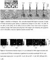
Figure 1 . Sketches of rectangular-, disc- and pillar-shaped DNA origami structures. A single ATTO542 fluorophore (green sphere) is positioned at a height of 3 nm (NR), 7 nm (ND), 12 nm (NP1), 16 nm (NP2), 24 nm (NP3) and 53 nm (NP4). A blue frame: zoom-in of pyrene-modified (orange) DNA strand protruding from DNA origami and interacting with graphene via π-π interactions. Grey bars represent double-stranded DNA. -
Figure 2 . Fluorescence intensity images (5 x 5 µm) obtained for DNA origami structures with a single dye (ATTO542) immobilized on graphene, forexcitation wavelength 532 nm, and laser power ranging from 1 to 5 µW (NR - 5 µW, ND and NP1 - 2 µW, NP2, NP3 and NP4 - 1 µW). -
Figure 3 . Examples of fluorescence transients obtained for DNA origami structures with a single dye (ATTO542) immobilized on graphene, forexcitation wavelength 532 nm, and laser power ranging from 1 to 5 µW (NR - 5 µW, ND and NP1 - 2 µW, NP2, NP3 and NP4 - 1 µW). -
Figure 4 . Raman spectrum of a single graphene layer, averaged from a map of 30 measured spots. -
Figure 5 . (A) Normalized fluorescence intensity decays of ATTO542 at difference distances to graphene (averaged from 20 decays for each sample). (B) Relative fluorescence intensity as a function of fluorescence lifetime (fluorescence lifetimes were obtained by reconvolution). -
Figure 6 . Normalized fluorescence intensity decays of Atto542 within DNA origami structure NP4 (a dye placed at the height of 53 nm) immobilized on glass (■) or graphene (●). -
Figure 7 . (a) Mean values of the energy transfer efficiency calculated from fluorescence lifetime values; (b) mean values of the excitation energy transfer efficiency calculated from fluorescence intensity values, all plotted as a function of the distance between ATTO542 and graphene. Standard errors calculated from the fitted normal distribution (not shown) for N = 140 - 250 molecules are shown. Fitted curve (red line) of the energy transfer efficiency as the function of the distance d between graphene and emitter,
-
Figure 8 . (Top) TEM images of: (from left) Nanodisc and Nanopillar. (Bottom) AFM images of: (from left) Nanorectangle, Nanodisc and Nanopillar. -
Figure 9 . Experimental details for fluorescent measurements for NR, ND, NP1-NP4. - The present Inventors used DNA origami (Rothemund, P. W. K. Nature 2006, 440 (7082), 297-302) as a surface chemistry converter to generally overcome the problem of chemical functionalization of graphene for optical biosensing assays. The DNA origami technique enables the formation of custom-designed DNA nanostructures with a volume of ∼21.000 nm3, and the rich DNA chemistry available allows placing of arbitrary objects and complex biomolecular assays (Acuna, G. P. et al.; ACS Nano 2012, 6 (4), 3189-3195; Vietz, C. et al.; Nano Lett. 2017, 17, 6496-6500; Ochmann, S. E. et al.; Anal. Chem. 2017, 89, 13000-13007) at programmed positions on the DNA nanostructure (Douglas, S. M. et al.; Nature 2009, 459 (7245), 414-418; Hong, F. et al.; Chem. Rev. 2017, 117, 12584-12640; Ke, Y. et al.; Science (80-.) 2008, 319 (2008), 180-183). For biomolecular assays DNA origami can act as a biocompatible surface (Gietl, A. et al.; Nucleic Acids Res. 2012, 40 (14), 1-10). In previous works, DNA origami structures were coupled to pristine graphene either to increase their stability (Matković, A. et al.; New J. Phys. 2016, 18 (2), 025016) or as a template for metallized DNA nanolithography (Jin, Z. et al.; Nat. Commun. 2013, 4, 1663). On the other hand, it was demonstrated with TEM imaging that DNA origami nanoplates were denatured due to hydrophobic interactions of the DNA bases with graphene upon adsorption (Kabiri, Y. et al.; Small 2017, 1-8; Green, N. S. et al.; Mater. Res. ). As a universal glue to connect the DNA origami constructs to the graphene layer without denaturation, several pyrene modified DNA strands were used that are hybridized to the DNA origami on one hand and interact with the graphene lattice via π-π stacking interactions on the other hand. It is shown that this immobilization scheme provides stable DNA origami structures for different geometries and enables placing of single, freely rotating fluorescent molecules at defined distances to the graphene layer. The exquisite distance control of single fluorescent dyes to graphene was exploited in order to revisit the distance dependence of fluorescence quenching by graphene. The narrow intensity-fluorescence lifetime distributions confirm the d-4 law with a precise value of d0 of 17.7 nm in aqueous buffer solution and present the basis for a broad range of applications in biosensing and optoelectronics.
- The selection of DNA origami shapes was guided by the possibility to position a single dye molecule at a designed height from the bottom (distance from graphene) and included rectangular- (nanorectangle = NR), disc- (nanodisc = ND) and pillar-shaped (nanopillar = NP) self-assembled DNA origami structures (see
Figure 1 ). Each DNA origami structure contains one dye molecule (ATTO542) marked as a small green sphere, which is positioned at the height of: 3 nm (NR), 7 nm (ND), 12 nm (NP1), 16 nm (NP2), 24 nm (NP3) and 53 nm (NP4). The selected values cover the range of distances for which the energy transfer from a single dye molecule to graphene is expected to vary from 0 to almost 100%. As the dyes are attached to the DNA origami by 6-carbon linkers and measurements were carried out in buffer solution, it is expected that the fluorescent dyes assume all possible dipole orientations during the measurement.
Additionally, at the bottom of each structure, 6-8 staple strands with single-stranded extensions protruding from the DNA origamis were incorporated. The protrusions are used to label the DNA origami with pyrene-modified complementary oligonucleotides. The pyrene enabled selective binding of the DNA origami with the bottom side to the graphene layer (Figure 1 ). The stability of this interaction in certain temperature ranges (e.g. 10-40°C) is achieved by binding minimally 2 and optimally 4-10 pyrene units per one DNA origami structure, beacuse most likely one pyrene molecule is not enough to obtain stable bonds between the DNA origami and the graphene layer. External labeling with extended staple strands allowed modular modifications also for other moieties. For example, in order to measure the same samples on glass, biotin-modified oligonucleotides were used and DNA origami structures were immobilized on neutravidin-biotinylated BSA surfaces. Single-molecule fluorescence measurements were carried out with a home-built confocal microscope.Figure 2 depicts ATTO542 fluorescence intensity images for each of the six DNA origami structures using 532 nm excitation. The closer the emitter is to the graphene layer, the more its fluorescence is quenched. Therefore, we adjusted the excitation power to always work with a reasonable count rate in a regime that showed linear excitation intensity dependent emission. Laser powers ranged from 1 µW for three nanopillar samples (NP2, NP3 and NP4), 2 µW for one nanopillar sample and the nanodisc (NP1 and ND), up to 5 µW for the nanorectangle (NR) DNA origami structure. On all measured images, we observed that the fluorescence intensities of most spots, representing dye molecules, are very homogeneous, with occasional very bright spots. - In order to characterize interactions between dye molecules and graphene, fluorescence transients were recorded for each spot. This enabled the identification of single DNA origami structures by observing blinking events and single-step photobleaching (examples of fluorescence transients are depicted in
Figure 3 ). We identified two types of deviations from the typical single-molecule behavior, which is related to the mentioned brighter spots in the images. In cases with multi-step photobleaching, we attributed the signal to aggregates of multiple DNA origami structures. In other cases, the brighter spots exhibited single-step photobleaching and showed similar intensities and fluorescence lifetimes as molecules of the reference structures measured on glass cover slips. We hence attribute these molecules to DNA origami structures immobilized within small defects/cracks of the graphene layer. Nevertheless, taking into account the small number of such spots (<10%), together with the quality control performed by Graphenea® and our additional characterization using Raman spectroscopy (several tens of spectra obtained from Raman measurements, an exemplary spectrum presented inFigure 4 ), we conclude that graphene samples used in our measurements were of high quality. For further analysis, only transients with single bleaching steps were considered and used to determine the fluorescence intensity and fluorescence lifetime of each spot (see Imaging and Analysis for details). As expected, dye molecules incorporated in DNA origami structures bound to graphene exhibit shorter fluorescence lifetimes compared to samples immobilized on glass (seeFigure 5 ). Only for the largest distance of 53 nm to graphene (NP4), the fluorescence properties are not affected (seeFigure 6 ) and the unperturbed fluorescence lifetime of 3.25 ns for ATTO542 is obtained. - The reduction of fluorescence intensity and fluorescence lifetime is correlated and arise from the strong near-field interactions between the emitter and graphene.
Figure 7B shows the relative fluorescence intensity
- For a quantitative analysis of the interaction between single dye molecules and graphene, and its strong distance dependence, we investigated how the quenching (relative intensity) and the energy transfer efficiency to graphene both depend on the emitter-graphene distance. The energy transfer efficiency η was calculated from fluorescence lifetimes


Figures 4a and 4b show the distance dependent energy transfer efficiency calculated by both methods. While the shape of the graphs is similar lower energy transfer efficiencies were obtained from the fluorescence lifetime graph for the shortest distances (Figure 4a ), which we attribute to uncertainty induced by the limited time resolution of the setup (see decays and instrument response function (IRF) inFigure 3a ). We assume, however, that fluorescence intensity and lifetime are in this case proportional. The energy transfer efficiency obtained from intensities reaches up to 97% quenching for the smallest distance. We fitted the experimental data (Figure 4b ) with the expected d-4 dependence (red line) and obtained a d0 -value (the distance of 50% energy transfer efficiency) of 17.7 ± 0.5 nm. Additionally, inFigure 4b we compare experimental data with results calculated from the semiclassical model, which describes the near-field interactions between an emitter and graphene
Figure 4b , we show how the intensity of the emitter decreases with distance from graphene when the dipole is oriented either parallel (∥, ν = 1) or perpendicular (⊥, ν = 2) to graphene (blue dashed lines). We obtained d0 equals 16.8 nm for ν = 1 and 20.0 nm for ν = 2 which is in excellent agreement with our measurement. As it is expected that the fluorescent dye is free to rotate on the flexible linker during the excited state lifetime simple geometric averaging is not meaningful but the distance dependence should be more similar to the parallel case as is also reflected in our experimental result for d0. - Samples of single layer CVD (chemical vapor deposition) graphene on glass coverslips were purchased from Graphenea®. Quality of graphene layers were characterized using Raman spectroscopy (
Figure 4 ). - DNA origami Nanorectangles, Nanodiscs and Nanopillars were prepared as described elsewhere (Puchkova, A. et al.; Nano Lett. 2015, 15 (12), 8354-8359; Vietz, C. et al.; New J. Phys. 2016, 18, 045012).
- The DNA origami structures were incubated with pyrene-modified (for graphene samples) or biotin-modified (for glass samples) staple strands, complementary to the oligonucleotides protruding from DNA origami structures, for 2 h in 37 °C. Such prepared structures were immobilized on the glass surface of a Lab-Tek chamber (Thermo Fisher Scientific) coated with BSA-biotin/neutravidin (Sigma-Aldrich) or directly on a single layer of CVD graphene. Finally, the sample was washed with 1×TE buffer (10 mM Tris (tris(hydroxymethyl)aminomethane), brought to pH 8.0 with HCl and 1 mM EDTA (ethylenediaminetetraacetic acid)) containing 12 mM MgCl2 and single-molecule fluorescence measurements were performed.
- The designed height values of 3 nm (NR), 7 nm (ND), 12 nm (NP1), 16 nm (NP2), 24 nm (NP3) and 53 nm (NP4) were calculated using the size parameter of a double helix, namely its diameter of 2.2 nm and a length of 0.34 nm per base pair. Taking into account previous report about the overestimation of the calculated values (the measured values smaller of about 10% compared to the designed values), these numbers were corrected. Additionally, a thickness of 1 nm for the pyrene-modified protruding strands was added.
- Single-molecule fluorescence measurements were performed on a custom built confocal microscope based on an Olympus IX71 inverted microscope. The green laser beams (532 nm LDH-P-FA-530B, Picoquant) is controlled by a PDL 828 "Sepiall" (Picoquant). The green fiber laser is decoupled by a collimator (F2220APC-532, Thorlabs). After passing through cleanup filters (532/2) and a dichroic mirror (640DCXR, AHF) the laser beam is coupled into a fiber (P3-488PM-FC, Thorlabs) with a collimator (PAF2-2A, Thorlabs) and decoupled with a collimator (G169015000, Qioptics). A combination of a linear polarizer (WP12L-Vis, Thorlabs) and a quarter wave plate (AQWP05M-600, Thorlabs) is used to obtain circularly polarized light. After passing a dual band dichroic beam splitter (z532/633, AHF), the light beam is focused by an oil-immersion objective (UPLSAPO 100XO, NA 1.40, Olympus) on the measurement chamber, which can be positioned accurately by a piezo-stage (P-527.3CD, Physiklnstrumente) which is driven by a E-727 controller (Physiklnstrumente). The emission of the fluorophores is collected by the same objective, focused on a 50 µm pinhole (Linos), collimated with a lens (AC050-150-A-ML, Thorlabs) and split spectrally by another dichroic beam splitter (640DCXR, AHF). The green laser beams cleaned with a filter set (HC582/75, AHF and
LP 532, both Semrock) and focused with a lens (AC080-020-A-ML, Thorlabs) on the APD (SPCM-AQRH-TR-14, Excelitas) The signals of the APD are detected by a HydraHarp 400 (Picoquant) and the whole system is operated with SymPhoTime 64 (PicoQuant). - The correct folding of the DNA origami structures was characterized with atomic force microscopy (AFM,
Nanowizard 3 ultra, JPK Instruments) in solution. On a freshly cleaved mica surface (Qualty V1, Piano GmbH) 10 µL of a 10 mM NiCl2 x 6 H2O solution were incubated for 5 min. After three times washing with 300 µL miliQ-water (Merck Milli-Q) and drying with compressed air, 10µL 1 nM DNA origami structure solution (diluted in AFM buffer (40 mM TRIS, 2 mM EDTA disodium salt dihydrate and 12.5 mM Mg(OAc)2 x 6 H2O) were added and incubated for 5 min. Afterwards 300 µL AFM buffer were added after purging three times with 300 µL AFM buffer. The solution measurements were performed with cantilevers USC-F0.3-k0.3-10 from Nano World. - TEM grids (TedPella, formvar/carbon, 400 mesh, Cu) were Ar-plasma cleaned and incubated for 60 sec with DNA origami sample (5 µL, ∼2-10 nM). Grids were washed with 2 % uranyl formate solution (5 µL) and incubated again afterwards again 4 s with 2 % uranyl formate solution (5 µL) for staining. TEM imaging were performed with a JOEL JEM-1100 with an acceleration voltage of 80 kV.Images of NR, ND and NP1-NP4 are given in
Figure 8 . - The experimental details are summarized in Bd! Nie można odnaleźć źród
 a odwo
 ania..
- Fluorescent images are given in
Figure 2 . - We immobilized DNA origami structures on graphene using pyrene modifications of DNA oligonucleotides. The specificity and robustness of the immobilization without denaturation enabled placing of fluorescent dyes at defined distances to the graphene layer. We confirmed the d-4 dependence of energy transfer from the dye to graphene and determined the distance of 50% energy transfer: d0 = 17.7 nm. The homogeneity of the population indicates that distances to graphene can be determined with very high precision in a range of 5 - 30 nm. Together with its good transparency in the far-field graphene might become the substrate of choice for superresolution microscopy involving fluorescence lifetime measurement for determining the z-position of dyes. Beyond that, DNA origami as chemical converters will enable placing biomolecular assays on the graphene with the optimized distance to the surface for optimized sensitivity. Placing assays at a height of 17 - 18 nm will yield an extremely sensitive method of detecting small distance changes to the surface while avoiding direct contact to the interfering hydrophobic graphene surface. DNA origami as a chemical converting and placement platform could be used to incorporate further functionalities for electronic, nanophotonic, and energy conversion devices with graphene and other 2D materials opening a myriad of new possibilities.
Claims (15)
- Method of immobilization of nucleic acid structures on graphene, said method comprising the steps of:iii) providing at least one type of nucleic acid structure having at least one type of molecule-modified oligonucleotide, wherein said molecule is able to attach to graphene via π-interactions,iv) contacting said nucleic acid structure having at least one type of molecule-modified oligonucleotide with graphene layer at temperature from the range of 0-100 °C, so that immobilization of nucleic acid structures on the graphene is achieved.
- Method according to claim 1, wherein step i) comprises the following sub-steps of:- providing at least one type of nucleic acid structure having at least one type of oligonucleotide,- providing at least one type of molecule-modified oligonucleotide, wherein said molecule is able to attach to graphene via π-interactions, and wherein said molecule-modified oligonucleotide is at least partially complementary to at least one type of oligonucleotide comprised in said nucleic acid structure,- incubating said nucleic acid structure with said molecule-modified oligonucleotide for more than 5 min, preferably more than 30 min, more preferably for 30-120 min, at temperature from the range of 0-100 °C, preferably 10-40°C, more preferably 30-38°C in order to allow for Watson-Crick pairing between oligonucleotides of nucleic acid structures and said molecule-modified complementary oligonucleotides.
- Method according to claim 1 or 2, wherein said nucleic acid structure is selected from the group comprising DNA-based structures, such as: DNA origami structures, DNA junctions, DNA tiles, DNA bricks, DNA wireframe structures etc.
- Method according to claim 1 or 3, wherein step i) comprises the following sub-step of:- forming DNA origami structures by use of molecule-modified staple strands, wherein said molecule is able to attach to graphene via π-interactions.
- Method according to any of claims 1-4, wherein step ii) is carried out for at least 10 seconds, and/or step ii) is carried out at temperature from the range of 10-40 °C, preferably 15-30°C.
- Method according to any of claims 1-5, wherein said molecule, which is able to attach to graphene via π-interactions is pyrene.
- Method according to any of claims 1-6, said method comprising the steps of:e) providing at least one type of DNA origami structure having at least one type of oligonucleotide protruding from the DNA origami structure and/or built into said DNA origami structure,f) providing at least one type of molecule-modified oligonucleotide, wherein said molecule able to attach to graphene via π-interactions is pyrene, and wherein said oligonucleotide is at least partially complementary to the oligonucleotides protruding from the DNA origami structure and/or built into said DNA origami structure,g) incubating said DNA origami structure with molecule-modified oligonucleotide for more than 5 min, preferably more than 30 min, more preferably for 30-120 min, at temperature from the range of 0-100°C, preferably 10-40°C, more preferably 30-38°C, in order to allow for Watson-Crick pairing between oligonucleotides protruding from the DNA origami structure and/or built into said DNA origami structure and said molecule-modified complementary oligonucleotides,h) contacting said incubated DNA origami structure with graphene layer for more than 10 seconds, at temperature from the range of 0-100 °C, preferably 10-40 °C, so that immobilization of the DNA origami structures on the graphene is achieved.
- Method according to claim 7, wherein said molecule able to attach to graphene via π-interactions is pyrene, and/or the number of pyrene molecules per one DNA origami structure is at least 2, preferably 4-10.
- Method according to claim 7 or 8, wherein incubation in step c) is carried out for 2 hours in 37°C.
- Method according to any of claims 1-9, wherein said nucleic acid structures carry one or more type of objects-of-interest (OOI), wherein the OOls are selected from the group comprising proteins, peptides, nanoparticles, carbohydrates, small molecule ligands, dyes, nanocrystals, DNA, RNA, chemical moieties such as thiol or biotin etc.
- Method according to any of claims 1-10, wherein after step of immobilization of nucleic acid structures on the graphene, further step of washing the immobilized sample with any solution in which DNA origami is stable, such as Milli-Q water, PBS buffer, 10 mM Tris or 1×TE buffer containing 0-15 mM MgCl2 is carried out.
- Graphene modified by immobilization of nucleic acid structures comprising at least one type of nucleic acid structure and graphene layer, wherein said nucleic acid structure is immobilized on the graphene layer via molecule-modified oligonucleotides, wherein said molecule is able to attach to graphene via π-interactions, and wherein said molecule-modified oligonucleotides are complementary for Watson-Crick pairing to oligonucleotides comprised in said nucleic acid structures.
- Graphene modified by immobilization of DNA origami structures comprising at least one type of DNA origami structure and graphene layer, wherein said DNA origami structure is immobilized on the graphene layer via pyrene-modified oligonucleotides which are complementary for Watson-Crick pairing to oligonucleotides protruding from the DNA origami structure and/or built into said DNA origami structure.
- Graphene modified by immobilization of nucleic acid structures such as DNA origami structures according to claims 12 or 13, wherein nucleic acid structures such as DNA origami structures carry one or more type of objects-of-interest (OOI), wherein the OOls are selected from the group comprising proteins, peptides, nanoparticles, carbohydrates, small molecule ligands, dyes, nanocrystals, DNA, RNA, chemical moieties such as thiol or biotin, etc.
- Use of graphene modified by immobilization of nucleic acid structures such as DNA origami structures defined in any of claims 12-14 in biosensing and energy conversion as well as photovoltaics and optoelectronics.
Priority Applications (1)
| Application Number | Priority Date | Filing Date | Title |
|---|---|---|---|
| EP19461514.2A EP3699142A1 (en) | 2019-02-20 | 2019-02-20 | Method of immobilization nucleic acid structure on surface of graphene, graphene modified by immobilization of nucleic acid structure and its use |
Applications Claiming Priority (1)
| Application Number | Priority Date | Filing Date | Title |
|---|---|---|---|
| EP19461514.2A EP3699142A1 (en) | 2019-02-20 | 2019-02-20 | Method of immobilization nucleic acid structure on surface of graphene, graphene modified by immobilization of nucleic acid structure and its use |
Publications (1)
| Publication Number | Publication Date |
|---|---|
| EP3699142A1 true EP3699142A1 (en) | 2020-08-26 |
Family
ID=65685283
Family Applications (1)
| Application Number | Title | Priority Date | Filing Date |
|---|---|---|---|
| EP19461514.2A Pending EP3699142A1 (en) | 2019-02-20 | 2019-02-20 | Method of immobilization nucleic acid structure on surface of graphene, graphene modified by immobilization of nucleic acid structure and its use |
Country Status (1)
| Country | Link |
|---|---|
| EP (1) | EP3699142A1 (en) |
Cited By (2)
| Publication number | Priority date | Publication date | Assignee | Title |
|---|---|---|---|---|
| CN113185973A (en) * | 2021-04-06 | 2021-07-30 | 上海交通大学 | Preparation method of zero-dimensional nano carbon material/origami structure deoxyribonucleic acid composite material |
| WO2022192591A1 (en) * | 2021-03-11 | 2022-09-15 | Nautilus Biotechnology, Inc. | Systems and methods for biomolecule retention |
Citations (2)
| Publication number | Priority date | Publication date | Assignee | Title |
|---|---|---|---|---|
| KR20110036204A (en) * | 2009-10-01 | 2011-04-07 | 한국과학기술원 | Graphene manufacturing method, thereby prepared graphene, DNA analysis method and graphene-nanoparticle composite manufacturing method using the same |
| WO2017112941A1 (en) * | 2015-12-23 | 2017-06-29 | The Regents Of The University Of California | Nano-sensors for nucleic acid detection and discrimination |
-
2019
- 2019-02-20 EP EP19461514.2A patent/EP3699142A1/en active Pending
Patent Citations (2)
| Publication number | Priority date | Publication date | Assignee | Title |
|---|---|---|---|---|
| KR20110036204A (en) * | 2009-10-01 | 2011-04-07 | 한국과학기술원 | Graphene manufacturing method, thereby prepared graphene, DNA analysis method and graphene-nanoparticle composite manufacturing method using the same |
| WO2017112941A1 (en) * | 2015-12-23 | 2017-06-29 | The Regents Of The University Of California | Nano-sensors for nucleic acid detection and discrimination |
Non-Patent Citations (24)
| Title |
|---|
| ACUNA, G. P. ET AL., ACS NANO, vol. 6, no. 4, 2012, pages 3189 - 3195 |
| ANASTASIYA PUCHKOVA ET AL: "DNA Origami Nanoantennas with over 5000-fold Fluorescence Enhancement and Single-Molecule Detection at 25 [mu]M", NANO LETTERS, vol. 15, no. 12, 9 December 2015 (2015-12-09), US, pages 8354 - 8359, XP055584731, ISSN: 1530-6984, DOI: 10.1021/acs.nanolett.5b04045 * |
| CAROLIN VIETZ ET AL: "Functionalizing large nanoparticles for small gaps in dimer nanoantennas", NEW JOURNAL OF PHYSICS, INSTITUTE OF PHYSICS PUBLISHING, BRISTOL, GB, vol. 18, no. 4, 15 April 2016 (2016-04-15), pages 45012, XP020300527, ISSN: 1367-2630, [retrieved on 20160415], DOI: 10.1088/1367-2630/18/4/045012 * |
| CHIZHIK, A. I. ET AL., NAT. PHOTONICS, vol. 8, February 2014 (2014-02-01), pages 124 - 127 |
| DATABASE WPI Week 201141, Derwent World Patents Index; AN 2011-E00622, XP002790946 * |
| DOUGLAS, S. M. ET AL., NATURE, vol. 459, no. 7245, 2009, pages 414 - 418 |
| GIETL, A. ET AL., NUCLEIC ACIDS RES., vol. 40, no. 14, 2012, pages 1 - 10 |
| GREEN, N. S. ET AL., MATER. RES. EXPRESS, vol. 5, 2018, pages 045035 |
| GUANGFU WU ET AL, SUPPLEMENTARY MATERIAL: "Graphene Field-Effect Transistors for the Sensitive and Selective Detection of Escherichia coli Using Pyrene-Tagged DNA Aptamer", ADVANCED HEALTHCARE MATERIALS, vol. 6, no. 19, 1 October 2017 (2017-10-01), DE, pages 1700736, XP055584661, ISSN: 2192-2640, DOI: 10.1002/adhm.201700736 * |
| GUANGFU WU ET AL: "Graphene Field-Effect Transistors for the Sensitive and Selective Detection of Escherichia coli Using Pyrene-Tagged DNA Aptamer", ADVANCED HEALTHCARE MATERIALS, vol. 6, no. 19, 1 October 2017 (2017-10-01), DE, pages 1700736, XP055584620, ISSN: 2192-2640, DOI: 10.1002/adhm.201700736 * |
| HONG, F. ET AL., CHEM. REV., vol. 117, 2017, pages 12584 - 12640 |
| ISBANER, S. ET AL., NANO LETT., vol. 18, no. 4, 2018, pages 2616 - 2622 |
| JIN, Z. ET AL., NAT. COMMUN., vol. 4, 2013, pages 1663 |
| KABIRI, Y. ET AL., SMALL, 2017, pages 1 - 8 |
| KE, Y. ET AL., SCIENCE, vol. 319, 2008, pages 180 - 183 |
| LANGER, A. ET AL., NAT. COMMUN., vol. 4, 2013 |
| LANGER, A. ET AL., SCI. REP., vol. 5, January 2015 (2015-01-01), pages 1 - 15 |
| MATKOVIC, A. ET AL., NEW J. PHYS., vol. 18, no. 2, 2016, pages 025016 |
| OCHMANN, S. E. ET AL., ANAL. CHEM., vol. 89, 2017, pages 13000 - 13007 |
| PUCHKOVA, A. ET AL., NANO LETT., vol. 15, no. 12, 2015, pages 8354 - 8359 |
| ROTHEMUND, P. W. K., NATURE, vol. 440, no. 7082, 2006, pages 297 - 302 |
| SCHLICHTIGER, A. ET AL., ANAL. BIOANAL. CHEM., vol. 405, no. 1, 2013, pages 275 - 285 |
| VIETZ, C. ET AL., NANO LETT., vol. 17, 2017, pages 6496 - 6500 |
| VIETZ, C. ET AL., NEW J. PHYS., vol. 18, 2016, pages 045012 |
Cited By (10)
| Publication number | Priority date | Publication date | Assignee | Title |
|---|---|---|---|---|
| WO2022192591A1 (en) * | 2021-03-11 | 2022-09-15 | Nautilus Biotechnology, Inc. | Systems and methods for biomolecule retention |
| US11505796B2 (en) | 2021-03-11 | 2022-11-22 | Nautilus Biotechnology, Inc. | Systems and methods for biomolecule retention |
| US11760997B2 (en) | 2021-03-11 | 2023-09-19 | Nautilus Subsidiary, Inc. | Systems and methods for biomolecule retention |
| US11912990B2 (en) | 2021-03-11 | 2024-02-27 | Nautilus Subsidiary, Inc. | Systems and methods for biomolecule retention |
| US12071618B2 (en) | 2021-03-11 | 2024-08-27 | Nautilus Subsidiary, Inc. | Systems and methods for biomolecule retention |
| US12305167B2 (en) | 2021-03-11 | 2025-05-20 | Nautilus Subsidiary, Inc. | Systems and methods for biomolecule retention |
| US12503694B2 (en) | 2021-03-11 | 2025-12-23 | Nautilus Subsidiary, Inc. | Systems and methods for biomolecule retention |
| AU2022232933B2 (en) * | 2021-03-11 | 2026-02-12 | Nautilus Subsidiary, Inc. | Systems and methods for biomolecule retention |
| CN113185973A (en) * | 2021-04-06 | 2021-07-30 | 上海交通大学 | Preparation method of zero-dimensional nano carbon material/origami structure deoxyribonucleic acid composite material |
| CN113185973B (en) * | 2021-04-06 | 2023-04-28 | 上海交通大学 | Preparation method of zero-dimensional nano carbon material/paper folding structure deoxyribonucleic acid composite material |
Similar Documents
| Publication | Publication Date | Title |
|---|---|---|
| US20220373543A1 (en) | Surface-immobilized bistable polynucleotide devices for the sensing and quantification of molecular events | |
| Kaminska et al. | Distance dependence of single-molecule energy transfer to graphene measured with DNA origami nanopositioners | |
| US12173281B2 (en) | Bistable polynucleotide devices for the sensing and quantification of molecular events | |
| Gopinath et al. | Engineering and mapping nanocavity emission via precision placement of DNA origami | |
| Bidault et al. | Picosecond lifetimes with high quantum yields from single-photon-emitting colloidal nanostructures at room temperature | |
| Kaminska et al. | Strong plasmonic enhancement of a single peridinin–chlorophyll a–protein complex on DNA origami-based optical antennas | |
| US20060192115A1 (en) | Addressable field enhancement microscopy | |
| AU2019437529B2 (en) | Surface-immobilized bistable polynucleotide devices for the sensing and quantification of molecular events | |
| US20070116627A1 (en) | Carbon nanotube compositions and devices and methods of making thereof | |
| Ferdele et al. | Strong coupling in porphyrin J-aggregate excitons and plasmons in nano-void arrays | |
| Zhang et al. | Near-infrared photoluminescence biosensing platform with gold nanorods-over-gallium arsenide nanohorn array | |
| Kim et al. | Fluorescence amplification using colloidal photonic crystal platform in sensing dye-labeled deoxyribonucleic acids | |
| Yaadav et al. | Broad-band fluorescence enhancement of QDs captured in the hotspot of DNA origami nanonantennas | |
| Sun et al. | DNA-based artificial nanostructures: Fabrication, properties, and applications | |
| Koch et al. | Super-Resolved Fluorescence Lifetime Imaging of Single Cy3 Molecules and Quantum Dots Using Time-Correlated Single Photon Counting with a Four-Pixel Fiber Optic Array Camera | |
| CA2629931A1 (en) | Method | |
| Arzhanov et al. | DNA-Origami Apertured Tiles Self-Assembly and Surface AFM-Characterization in the Presence of Spontaneous Attachment of Single Colloidal Quantum Dot | |
| Prokhorov et al. | Atomic force and scanning near-field optical microscopy study of carbocyanine dye J-aggregates | |
| Shetty et al. | Low-cost, bottom-up fabrication of large-scale single-molecule nanoarrays by DNA origami placement | |
| Green | Nanoscale optical and correlative microscopies for quantitative characterization of DNA nanostructures | |
| Baker et al. | Immobilization of Quantum Dot-DNA Conjugates Templated by DNA Self-Assembled Monolayers on Single-Crystal Gold Bead Electrodes Toward Advanced Sensing | |
| Lanzavecchia et al. | Three-dimensional plasmonic nanopores for DNA-PAINT and dual-material Au/Si architectures | |
| Masud | Plasmon Enhanced Single Molecule Fluorescence in Zero Mode Waveguides (ZMWs) | |
| Rahmani | Engineering Multifunctional Nanoparticle Assemblies through DNA Guided Self-Assembly | |
| Weilbaecher et al. | Development of a novel nanomaterial-based optical platform for a protease biosensor |
Legal Events
| Date | Code | Title | Description |
|---|---|---|---|
| PUAI | Public reference made under article 153(3) epc to a published international application that has entered the european phase |
Free format text: ORIGINAL CODE: 0009012 |
|
| STAA | Information on the status of an ep patent application or granted ep patent |
Free format text: STATUS: THE APPLICATION HAS BEEN PUBLISHED |
|
| AK | Designated contracting states |
Kind code of ref document: A1 Designated state(s): AL AT BE BG CH CY CZ DE DK EE ES FI FR GB GR HR HU IE IS IT LI LT LU LV MC MK MT NL NO PL PT RO RS SE SI SK SM TR |
|
| AX | Request for extension of the european patent |
Extension state: BA ME |
|
| STAA | Information on the status of an ep patent application or granted ep patent |
Free format text: STATUS: REQUEST FOR EXAMINATION WAS MADE |
|
| STAA | Information on the status of an ep patent application or granted ep patent |
Free format text: STATUS: EXAMINATION IS IN PROGRESS |
|
| 17P | Request for examination filed |
Effective date: 20210226 |
|
| RBV | Designated contracting states (corrected) |
Designated state(s): AL AT BE BG CH CY CZ DE DK EE ES FI FR GB GR HR HU IE IS IT LI LT LU LV MC MK MT NL NO PL PT RO RS SE SI SK SM TR |
|
| 17Q | First examination report despatched |
Effective date: 20210401 |
|
| REG | Reference to a national code |
Ref country code: DE Ref legal event code: R079 Free format text: PREVIOUS MAIN CLASS: C01B0032184000 Ipc: C12Q0001683400 |
|
| GRAP | Despatch of communication of intention to grant a patent |
Free format text: ORIGINAL CODE: EPIDOSNIGR1 |
|
| STAA | Information on the status of an ep patent application or granted ep patent |
Free format text: STATUS: GRANT OF PATENT IS INTENDED |
|
| RIC1 | Information provided on ipc code assigned before grant |
Ipc: C12Q 1/6834 20180101AFI20251111BHEP Ipc: C01B 32/184 20170101ALI20251111BHEP Ipc: C01B 32/194 20170101ALI20251111BHEP Ipc: B82Y 15/00 20110101ALI20251111BHEP |
|
| INTG | Intention to grant announced |
Effective date: 20251126 |
|
| GRAJ | Information related to disapproval of communication of intention to grant by the applicant or resumption of examination proceedings by the epo deleted |
Free format text: ORIGINAL CODE: EPIDOSDIGR1 |
|
| STAA | Information on the status of an ep patent application or granted ep patent |
Free format text: STATUS: EXAMINATION IS IN PROGRESS |
|
| GRAP | Despatch of communication of intention to grant a patent |
Free format text: ORIGINAL CODE: EPIDOSNIGR1 |
|
| STAA | Information on the status of an ep patent application or granted ep patent |
Free format text: STATUS: GRANT OF PATENT IS INTENDED |
|
| INTC | Intention to grant announced (deleted) | ||
| INTG | Intention to grant announced |
Effective date: 20260417 |
|
| GRAS | Grant fee paid |
Free format text: ORIGINAL CODE: EPIDOSNIGR3 |