EP3698814A1 - Composition for surface modification of medical implant and medical implant surface-modified thereby - Google Patents
Composition for surface modification of medical implant and medical implant surface-modified thereby Download PDFInfo
- Publication number
- EP3698814A1 EP3698814A1 EP18868312.2A EP18868312A EP3698814A1 EP 3698814 A1 EP3698814 A1 EP 3698814A1 EP 18868312 A EP18868312 A EP 18868312A EP 3698814 A1 EP3698814 A1 EP 3698814A1
- Authority
- EP
- European Patent Office
- Prior art keywords
- implant
- group
- itaconic acid
- medical implant
- gelatin
- Prior art date
- Legal status (The legal status is an assumption and is not a legal conclusion. Google has not performed a legal analysis and makes no representation as to the accuracy of the status listed.)
- Granted
Links
Images




Classifications
-
- A—HUMAN NECESSITIES
- A61—MEDICAL OR VETERINARY SCIENCE; HYGIENE
- A61L—METHODS OR APPARATUS FOR STERILISING MATERIALS OR OBJECTS IN GENERAL; DISINFECTION, STERILISATION OR DEODORISATION OF AIR; CHEMICAL ASPECTS OF BANDAGES, DRESSINGS, ABSORBENT PADS OR SURGICAL ARTICLES; MATERIALS FOR BANDAGES, DRESSINGS, ABSORBENT PADS OR SURGICAL ARTICLES
- A61L27/00—Materials for grafts or prostheses or for coating grafts or prostheses
- A61L27/28—Materials for coating prostheses
-
- A—HUMAN NECESSITIES
- A61—MEDICAL OR VETERINARY SCIENCE; HYGIENE
- A61L—METHODS OR APPARATUS FOR STERILISING MATERIALS OR OBJECTS IN GENERAL; DISINFECTION, STERILISATION OR DEODORISATION OF AIR; CHEMICAL ASPECTS OF BANDAGES, DRESSINGS, ABSORBENT PADS OR SURGICAL ARTICLES; MATERIALS FOR BANDAGES, DRESSINGS, ABSORBENT PADS OR SURGICAL ARTICLES
- A61L27/00—Materials for grafts or prostheses or for coating grafts or prostheses
- A61L27/50—Materials characterised by their function or physical properties, e.g. injectable or lubricating compositions, shape-memory materials, surface modified materials
-
- A—HUMAN NECESSITIES
- A61—MEDICAL OR VETERINARY SCIENCE; HYGIENE
- A61L—METHODS OR APPARATUS FOR STERILISING MATERIALS OR OBJECTS IN GENERAL; DISINFECTION, STERILISATION OR DEODORISATION OF AIR; CHEMICAL ASPECTS OF BANDAGES, DRESSINGS, ABSORBENT PADS OR SURGICAL ARTICLES; MATERIALS FOR BANDAGES, DRESSINGS, ABSORBENT PADS OR SURGICAL ARTICLES
- A61L27/00—Materials for grafts or prostheses or for coating grafts or prostheses
- A61L27/28—Materials for coating prostheses
- A61L27/34—Macromolecular materials
-
- A—HUMAN NECESSITIES
- A61—MEDICAL OR VETERINARY SCIENCE; HYGIENE
- A61L—METHODS OR APPARATUS FOR STERILISING MATERIALS OR OBJECTS IN GENERAL; DISINFECTION, STERILISATION OR DEODORISATION OF AIR; CHEMICAL ASPECTS OF BANDAGES, DRESSINGS, ABSORBENT PADS OR SURGICAL ARTICLES; MATERIALS FOR BANDAGES, DRESSINGS, ABSORBENT PADS OR SURGICAL ARTICLES
- A61L27/00—Materials for grafts or prostheses or for coating grafts or prostheses
- A61L27/36—Materials for grafts or prostheses or for coating grafts or prostheses containing ingredients of undetermined constitution or reaction products thereof, e.g. transplant tissue, natural bone, extracellular matrix
- A61L27/38—Materials for grafts or prostheses or for coating grafts or prostheses containing ingredients of undetermined constitution or reaction products thereof, e.g. transplant tissue, natural bone, extracellular matrix containing added animal cells
- A61L27/3839—Materials for grafts or prostheses or for coating grafts or prostheses containing ingredients of undetermined constitution or reaction products thereof, e.g. transplant tissue, natural bone, extracellular matrix containing added animal cells characterised by the site of application in the body
-
- A—HUMAN NECESSITIES
- A61—MEDICAL OR VETERINARY SCIENCE; HYGIENE
- A61L—METHODS OR APPARATUS FOR STERILISING MATERIALS OR OBJECTS IN GENERAL; DISINFECTION, STERILISATION OR DEODORISATION OF AIR; CHEMICAL ASPECTS OF BANDAGES, DRESSINGS, ABSORBENT PADS OR SURGICAL ARTICLES; MATERIALS FOR BANDAGES, DRESSINGS, ABSORBENT PADS OR SURGICAL ARTICLES
- A61L27/00—Materials for grafts or prostheses or for coating grafts or prostheses
- A61L27/50—Materials characterised by their function or physical properties, e.g. injectable or lubricating compositions, shape-memory materials, surface modified materials
- A61L27/54—Biologically active materials, e.g. therapeutic substances
-
- A—HUMAN NECESSITIES
- A61—MEDICAL OR VETERINARY SCIENCE; HYGIENE
- A61L—METHODS OR APPARATUS FOR STERILISING MATERIALS OR OBJECTS IN GENERAL; DISINFECTION, STERILISATION OR DEODORISATION OF AIR; CHEMICAL ASPECTS OF BANDAGES, DRESSINGS, ABSORBENT PADS OR SURGICAL ARTICLES; MATERIALS FOR BANDAGES, DRESSINGS, ABSORBENT PADS OR SURGICAL ARTICLES
- A61L31/00—Materials for other surgical articles, e.g. stents, stent-grafts, shunts, surgical drapes, guide wires, materials for adhesion prevention, occluding devices, surgical gloves, tissue fixation devices
- A61L31/08—Materials for coatings
-
- A—HUMAN NECESSITIES
- A61—MEDICAL OR VETERINARY SCIENCE; HYGIENE
- A61L—METHODS OR APPARATUS FOR STERILISING MATERIALS OR OBJECTS IN GENERAL; DISINFECTION, STERILISATION OR DEODORISATION OF AIR; CHEMICAL ASPECTS OF BANDAGES, DRESSINGS, ABSORBENT PADS OR SURGICAL ARTICLES; MATERIALS FOR BANDAGES, DRESSINGS, ABSORBENT PADS OR SURGICAL ARTICLES
- A61L31/00—Materials for other surgical articles, e.g. stents, stent-grafts, shunts, surgical drapes, guide wires, materials for adhesion prevention, occluding devices, surgical gloves, tissue fixation devices
- A61L31/08—Materials for coatings
- A61L31/10—Macromolecular materials
-
- A—HUMAN NECESSITIES
- A61—MEDICAL OR VETERINARY SCIENCE; HYGIENE
- A61L—METHODS OR APPARATUS FOR STERILISING MATERIALS OR OBJECTS IN GENERAL; DISINFECTION, STERILISATION OR DEODORISATION OF AIR; CHEMICAL ASPECTS OF BANDAGES, DRESSINGS, ABSORBENT PADS OR SURGICAL ARTICLES; MATERIALS FOR BANDAGES, DRESSINGS, ABSORBENT PADS OR SURGICAL ARTICLES
- A61L31/00—Materials for other surgical articles, e.g. stents, stent-grafts, shunts, surgical drapes, guide wires, materials for adhesion prevention, occluding devices, surgical gloves, tissue fixation devices
- A61L31/14—Materials characterised by their function or physical properties, e.g. injectable or lubricating compositions, shape-memory materials, surface modified materials
-
- A—HUMAN NECESSITIES
- A61—MEDICAL OR VETERINARY SCIENCE; HYGIENE
- A61L—METHODS OR APPARATUS FOR STERILISING MATERIALS OR OBJECTS IN GENERAL; DISINFECTION, STERILISATION OR DEODORISATION OF AIR; CHEMICAL ASPECTS OF BANDAGES, DRESSINGS, ABSORBENT PADS OR SURGICAL ARTICLES; MATERIALS FOR BANDAGES, DRESSINGS, ABSORBENT PADS OR SURGICAL ARTICLES
- A61L31/00—Materials for other surgical articles, e.g. stents, stent-grafts, shunts, surgical drapes, guide wires, materials for adhesion prevention, occluding devices, surgical gloves, tissue fixation devices
- A61L31/14—Materials characterised by their function or physical properties, e.g. injectable or lubricating compositions, shape-memory materials, surface modified materials
- A61L31/16—Biologically active materials, e.g. therapeutic substances
-
- C—CHEMISTRY; METALLURGY
- C08—ORGANIC MACROMOLECULAR COMPOUNDS; THEIR PREPARATION OR CHEMICAL WORKING-UP; COMPOSITIONS BASED THEREON
- C08G—MACROMOLECULAR COMPOUNDS OBTAINED OTHERWISE THAN BY REACTIONS ONLY INVOLVING UNSATURATED CARBON-TO-CARBON BONDS
- C08G77/00—Macromolecular compounds obtained by reactions forming a linkage containing silicon with or without sulfur, nitrogen, oxygen or carbon in the main chain of the macromolecule
- C08G77/04—Polysiloxanes
- C08G77/38—Polysiloxanes modified by chemical after-treatment
-
- C—CHEMISTRY; METALLURGY
- C08—ORGANIC MACROMOLECULAR COMPOUNDS; THEIR PREPARATION OR CHEMICAL WORKING-UP; COMPOSITIONS BASED THEREON
- C08J—WORKING-UP; GENERAL PROCESSES OF COMPOUNDING; AFTER-TREATMENT NOT COVERED BY SUBCLASSES C08B, C08C, C08F, C08G or C08H
- C08J3/00—Processes of treating or compounding macromolecular substances
- C08J3/24—Crosslinking, e.g. vulcanising, of macromolecules
- C08J3/245—Differential crosslinking of one polymer with one crosslinking type, e.g. surface crosslinking
-
- A—HUMAN NECESSITIES
- A61—MEDICAL OR VETERINARY SCIENCE; HYGIENE
- A61L—METHODS OR APPARATUS FOR STERILISING MATERIALS OR OBJECTS IN GENERAL; DISINFECTION, STERILISATION OR DEODORISATION OF AIR; CHEMICAL ASPECTS OF BANDAGES, DRESSINGS, ABSORBENT PADS OR SURGICAL ARTICLES; MATERIALS FOR BANDAGES, DRESSINGS, ABSORBENT PADS OR SURGICAL ARTICLES
- A61L2400/00—Materials characterised by their function or physical properties
- A61L2400/18—Modification of implant surfaces in order to improve biocompatibility, cell growth, fixation of biomolecules, e.g. plasma treatment
-
- A—HUMAN NECESSITIES
- A61—MEDICAL OR VETERINARY SCIENCE; HYGIENE
- A61L—METHODS OR APPARATUS FOR STERILISING MATERIALS OR OBJECTS IN GENERAL; DISINFECTION, STERILISATION OR DEODORISATION OF AIR; CHEMICAL ASPECTS OF BANDAGES, DRESSINGS, ABSORBENT PADS OR SURGICAL ARTICLES; MATERIALS FOR BANDAGES, DRESSINGS, ABSORBENT PADS OR SURGICAL ARTICLES
- A61L2420/00—Materials or methods for coatings medical devices
- A61L2420/02—Methods for coating medical devices
-
- A—HUMAN NECESSITIES
- A61—MEDICAL OR VETERINARY SCIENCE; HYGIENE
- A61L—METHODS OR APPARATUS FOR STERILISING MATERIALS OR OBJECTS IN GENERAL; DISINFECTION, STERILISATION OR DEODORISATION OF AIR; CHEMICAL ASPECTS OF BANDAGES, DRESSINGS, ABSORBENT PADS OR SURGICAL ARTICLES; MATERIALS FOR BANDAGES, DRESSINGS, ABSORBENT PADS OR SURGICAL ARTICLES
- A61L2420/00—Materials or methods for coatings medical devices
- A61L2420/06—Coatings containing a mixture of two or more compounds
-
- A—HUMAN NECESSITIES
- A61—MEDICAL OR VETERINARY SCIENCE; HYGIENE
- A61L—METHODS OR APPARATUS FOR STERILISING MATERIALS OR OBJECTS IN GENERAL; DISINFECTION, STERILISATION OR DEODORISATION OF AIR; CHEMICAL ASPECTS OF BANDAGES, DRESSINGS, ABSORBENT PADS OR SURGICAL ARTICLES; MATERIALS FOR BANDAGES, DRESSINGS, ABSORBENT PADS OR SURGICAL ARTICLES
- A61L2430/00—Materials or treatment for tissue regeneration
- A61L2430/02—Materials or treatment for tissue regeneration for reconstruction of bones; weight-bearing implants
-
- A—HUMAN NECESSITIES
- A61—MEDICAL OR VETERINARY SCIENCE; HYGIENE
- A61L—METHODS OR APPARATUS FOR STERILISING MATERIALS OR OBJECTS IN GENERAL; DISINFECTION, STERILISATION OR DEODORISATION OF AIR; CHEMICAL ASPECTS OF BANDAGES, DRESSINGS, ABSORBENT PADS OR SURGICAL ARTICLES; MATERIALS FOR BANDAGES, DRESSINGS, ABSORBENT PADS OR SURGICAL ARTICLES
- A61L2430/00—Materials or treatment for tissue regeneration
- A61L2430/04—Materials or treatment for tissue regeneration for mammary reconstruction
-
- A—HUMAN NECESSITIES
- A61—MEDICAL OR VETERINARY SCIENCE; HYGIENE
- A61L—METHODS OR APPARATUS FOR STERILISING MATERIALS OR OBJECTS IN GENERAL; DISINFECTION, STERILISATION OR DEODORISATION OF AIR; CHEMICAL ASPECTS OF BANDAGES, DRESSINGS, ABSORBENT PADS OR SURGICAL ARTICLES; MATERIALS FOR BANDAGES, DRESSINGS, ABSORBENT PADS OR SURGICAL ARTICLES
- A61L2430/00—Materials or treatment for tissue regeneration
- A61L2430/12—Materials or treatment for tissue regeneration for dental implants or prostheses
-
- A—HUMAN NECESSITIES
- A61—MEDICAL OR VETERINARY SCIENCE; HYGIENE
- A61L—METHODS OR APPARATUS FOR STERILISING MATERIALS OR OBJECTS IN GENERAL; DISINFECTION, STERILISATION OR DEODORISATION OF AIR; CHEMICAL ASPECTS OF BANDAGES, DRESSINGS, ABSORBENT PADS OR SURGICAL ARTICLES; MATERIALS FOR BANDAGES, DRESSINGS, ABSORBENT PADS OR SURGICAL ARTICLES
- A61L2430/00—Materials or treatment for tissue regeneration
- A61L2430/20—Materials or treatment for tissue regeneration for reconstruction of the heart, e.g. heart valves
-
- A—HUMAN NECESSITIES
- A61—MEDICAL OR VETERINARY SCIENCE; HYGIENE
- A61L—METHODS OR APPARATUS FOR STERILISING MATERIALS OR OBJECTS IN GENERAL; DISINFECTION, STERILISATION OR DEODORISATION OF AIR; CHEMICAL ASPECTS OF BANDAGES, DRESSINGS, ABSORBENT PADS OR SURGICAL ARTICLES; MATERIALS FOR BANDAGES, DRESSINGS, ABSORBENT PADS OR SURGICAL ARTICLES
- A61L2430/00—Materials or treatment for tissue regeneration
- A61L2430/24—Materials or treatment for tissue regeneration for joint reconstruction
-
- A—HUMAN NECESSITIES
- A61—MEDICAL OR VETERINARY SCIENCE; HYGIENE
- A61L—METHODS OR APPARATUS FOR STERILISING MATERIALS OR OBJECTS IN GENERAL; DISINFECTION, STERILISATION OR DEODORISATION OF AIR; CHEMICAL ASPECTS OF BANDAGES, DRESSINGS, ABSORBENT PADS OR SURGICAL ARTICLES; MATERIALS FOR BANDAGES, DRESSINGS, ABSORBENT PADS OR SURGICAL ARTICLES
- A61L2430/00—Materials or treatment for tissue regeneration
- A61L2430/34—Materials or treatment for tissue regeneration for soft tissue reconstruction
-
- A—HUMAN NECESSITIES
- A61—MEDICAL OR VETERINARY SCIENCE; HYGIENE
- A61L—METHODS OR APPARATUS FOR STERILISING MATERIALS OR OBJECTS IN GENERAL; DISINFECTION, STERILISATION OR DEODORISATION OF AIR; CHEMICAL ASPECTS OF BANDAGES, DRESSINGS, ABSORBENT PADS OR SURGICAL ARTICLES; MATERIALS FOR BANDAGES, DRESSINGS, ABSORBENT PADS OR SURGICAL ARTICLES
- A61L2430/00—Materials or treatment for tissue regeneration
- A61L2430/38—Materials or treatment for tissue regeneration for reconstruction of the spine, vertebrae or intervertebral discs
Definitions
- the disclosure relates to a composition for surface modification of a medical implant and a medical implant surface-modified thereby.
- Medical implants such as implants, dental implants, stents, screws and bone replacements are inserted for the purpose of treatment or plastic surgery. However, being inserted into the body, the medical implants are recognized as foreign substances in the body, and a clotting mechanism is activated, or foreign body reaction is generated to prevent bleeding and blood loss. In addition, since medical implants make permanent contact with biological tissues, the surface biocompatibility and in vivo affinity thereof are required.
- the inventors of the disclosure confirmed the fibrosis inhibition and inflammatory response inhibition at the implantation site of medical implants by using itaconic acid and completed the inventive concept.
- An aspect is to provide a composition including itaconic acid for surface modification of a medical implant.
- Another aspect is to provide a medical implant surface-modified with the itaconic acid.
- Another aspect is to provide a method of manufacturing the medical implant.
- An aspect provides a composition including itaconic acid for surface modification of a medical implant.
- the itaconic acid may be represented by Formula 1 below.
- composition may further include gelatin.
- the term "gelatin” may mean a protein obtained by partial hydrolysis of collagen which is the main protein component of connective tissues such as bones, cartilages, skins, etc., of animals.
- the gelatin may include gelatin derivatives in addition to pure gelatin.
- the gelatin derivative may include at least one of a phthalated gelatin, an esterified gelatin, an amidated gelatin, or a formylated gelatin.
- the kind (source) thereof is not specifically limited, and various kinds of gelatin derived from, for example, mammals and fishes, for example, cow bones, cow skins, swine bones, swine skins, etc., may be used.
- the gelatin may have a molecular weight from about 10,000 to about 30,000.
- the gelatin may be crosslinked.
- the gelatin may be chemically binded with itaconic acid.
- the composition may be provided in a powder type.
- Another aspect provides a medical implant surface-modified with itaconic acid.
- a surface-modified medical implant of which at least a portion of the surface of the implant is modified through the binding with itaconic acid may be provided as a medical implant.
- surface modification may mean the change of the chemical structure and physical structure of the surface of particles.
- the medical implant may be additionally binded with gelatin.
- the gelatin may be crosslinked and binded with the surface of the implant through itaconic acid.
- the itaconic acid and gelatin may have a nanoparticle type or a polymer type.
- At least a portion of the surface of the medical implant may introduce an amino group.
- the surface of the medical implant may be silanized for the binding with itaconic acid.
- An aminosilane compound for silanization may be 3-aminopropyltriethoxysilane (APTES), (3-aminopropyl)-tetraethylorthosilicate, [3-(2-aminoethylamino)propyl]trimethoxysilane, 3-(2-(2-aminoethylamino)ethylamino)propyltrimethoxysilane, or combinations thereof.
- the itaconic acid may be directly binded with the surface of the implant through the fixing compound at the surface of the implant.
- the fixing compound may include a compound having biotin, avidin, streptavidin, carbohydrate, poly L-lycine, a thiol group, an amine group, an alcohol group, a carboxyl group, an amino group, a sulfur group, an aldehyde group, a carbonyl group, a succinimide group, a maleimide group, an epoxy group, or an isothiocyanate group, or combinations thereof.
- Examples of the compound having an amino group may include 3-aminopropyltrimethoxysilane, N-(2-aminoethyl)-3-aminopropyltrimethoxysilane (EDA), trimethoxysilylpropyldiethylenetriamine (DETA), 3-(2-aminoethylaminopropyl) trimethoxysilane, and 3-aminopropyltriethoxysilane, and the compound having an aldehyde group may include glutaraldehyde.
- Examples of the compound having a thiol group may include 4-mercaptopropyltrimethoxysilane (MPTS).
- examples of the compound having an epoxy group may include 3-glycidoxypropyltrimethoxysilane
- examples of the compound having an isothiocyanate group may include 4-phenylenediisothiocyanate (PDITC)
- examples of the compound having succinimide and maleimide may include disuccinimidyl carbonate (DSC) or succinimidyl 4-(maleimidephenyl)butyrate (SMPB).
- the medical implant may be a biocompatible material.
- the biocompatible material may include one or more materials selected from the group consisting of gold (Au), silver (Ag), platinum (Pt), palladium (Pd), copper (Cu), steel, tantalum (Ta), magnesium (Mg), nickel (Ni), chromium (Cr), iron (Fe), a nitinol alloy
- NiTi a cobalt-chromium alloy (CoCr) gallium arsenic (GaAs), titanium (Ti), polylactic acid, poly(glycolic acid) (PGA), poly(lactic-co-glycolic acid) (PLGA), poly- ⁇ -(caprolactone), polyanhydride, polyorthoester, polyvinyl alcohol, polyethylene glycol, polyurethane, polyacrylic acid, poly-N-isopropylacrylamide, poly(ethylene oxide)-poly(propylene oxide)-poly(ethylene oxide), polytetrafluoroethylene, polycarbonate, polyurethane, nitrocellulose, polystyrene, polyethylene, polyethylene terephthalate (PET), polydimethylsiloxane (PDMS), polyether ether ketone (PEEK), silicon oxide (SiO 2 ), titanium oxide (TiO 2 ), aluminum oxide (Al 2 O 3 ), niobium oxide (Nb 2 O 5 ),
- the medical implant may be selected from the group consisting of breast implants, spinal implants, dental implants, cardiovascular implants, cardiac implants, stents, artificial joints, artificial head bones, and cell culture mediums.
- the surface modified medical implant may have a water contact angle from about 20° to about 90°, from about 25° to about 85°, from about 25° to about 80°, from about 25° to about 70°, from about 30° to about 60°, or from about 30° to about 40°.
- the surface modified medical implant may have improved fibrosis inhibition or inflammatory response inhibition at an implantation site when compared with that before surface modification.
- Another aspect provides a method of surface modifying a medical implant, including a step of functionalizing at least a portion of a surface of a medical implant; and a step of binding itaconic acid with the surface of the functionalized medical implant.
- the functionalization step may be a process for binding itaconic acid and/or gelatin with the surface of the implant.
- the functionalization may include a step of introducing an amino group, or a step of functionalizing using a fixing compound as described above.
- the step of binding itaconic acid may be a step of binding gelatin additionally.
- This step may include a step of preparing a composite of itaconic acid and gelatin and then, binding, binding itaconic acid and then, binding gelatin, or binding gelatin and then, combining itaconic acid.
- composition for surface modification of a medical implant according to an aspect and a medical implant surface-modified thereby itaconic acid may be binded with the surface of the medical implant in high binding stability, and effects showing activity on fibrosis inhibition and inflammatory response inhibition at an implantation site may be achieved.
- a medical implant surface-modified with itaconic acid was manufactured as shown in FIG. 1 .
- a polydimethylsiloxane (PDMS) substrate (Sylgard® 184 silicone elastomer kit, Dow Corning, USA) was treated with oxygen plasma (CUTE-1B, Femto Science, USA) for 1 minute so as to attach hydroxyl groups (-OH) on the surface of PDMS. After that, the surface of the substrate was silanized using 3-aminopropyl triethoxysilane (APTES) at 60°C for 2 hours.
- APTES 3-aminopropyl triethoxysilane
- IA itaconic acid
- EDC 1-ethyl-3-(3-dimethylamino)propyl carbodiimide
- NHS N-hydroxy succinimide
- Example 2 Manufacture of medical implant surface-modified with itaconic acid and gelatin.
- a medical implant surface-modified with itaconic acid and gelatin (IA-GT) was manufactured as shown in FIG. 2 .
- a gelatin type B (derived from cow skin, Bloon 75) powder was mixed with DPBS and dissolved at 135°C to 140°C for 16 hours. Then, with the dissolved gelatin type B, 50 mmol of EDC and 50 mmol of NHS solutions were mixed, and 100 mmol of itaconic acid was put and reacted for 2 hours. Additionally, DPBS was added to the mixture, and the reaction with gelatin and itaconic acid was performed at 60°C for 30 minutes and then finished. The final mixture was dialyzed through a cellulose membrane (MWCO 6-8kD), and unreacted itaconic acid, EDC/NHS, and salts were removed. After that, freeze drying was performed at -80°C for 48 hours to obtain an IA-GT powder.
- IA-GTpoly-PDMS a surface modified PDMS was manufactured by the same method as in Example 1 except for using DW + EDC 10 mmol + NHS 10 mmol to manufacture the surface modified PDMS of IA-GTpoly-0.25 wt%, and using DW + EDC 20 mmol + NHS 20 mmol to manufacture the surface modified PDMS of IA-GTpoly-0.50 wt%.
- FIG. 3 is a graph showing the contact angles of implants surface-modified with itaconic acid and/or gelatin according to an embodiment.
- the PDMSs surface-modified with itaconic acid and/or gelatin had smaller contact angles when compared with the conventional PDMS (control group).
- the surfaces of the implants manufactured in Examples 1 and 2 were observed by a scanning electron microscope (SEM) (SEM, S-3400N, Hitachi, Tokyo, Japan), and the results are shown in FIG. 4 .
- SEM scanning electron microscope
- FIG. 4 shows scanning electron microscopy images showing the contact angles of implants surface-modified with itaconic acid and/or gelatin according to an embodiment.
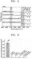
- Attenuated total reflection (ATR)-FTIR analysis was performed.
- each spectrum at room temperature was recorded from 400 cm -1 to 4000 cm -1 with a resolution of 4 cm -1 using a FT-IR Spectroscopy (Frontier spectrophotometer equipped with and attenuated total reflection (ATR-FTIR) module (Nicolet 6700, Thermo Scientific, USA), the scanning was performed 200 times and averaged, and the results are shown in FIG. 5 .
- FT-IR Spectroscopy Fastier spectrophotometer equipped with and attenuated total reflection (ATR-FTIR) module
- FIG. 5 is a graph showing the ATR-FTIR analysis results of implants surface-modified with itaconic acid and/or gelatin according to an embodiment.
- the protein absorbing capacity of the surfaces of the implants manufactured in Examples 1 and 2 was analyzed.
- bovine serum albumin (4.5 mg/mL) was dissolved in DPBS.
- BSA bovine serum albumin
- IA-PDMS and IA-GTpoly-PDMS size: 1 cm 2
- BSA protein - cultured bare, IA-conjugated and IA-GTpoly conjugated PDMS surfaces triple
- the absorbance value of each sample was analyzed.
- the amount of absorbed protein was quantified using Micro BCATM protein analysis kit according to the instruction of a manufacturer.
- the absorbance value was measured using a microplate spectrophotometer with a fixed wavelength of 570 nm. All values were represented by average ⁇ standard deviation, and the results are shown in FIG. 6 .
- FIG. 6 is a graph confirming the absorption degree of protein of implants surface-modified with itaconic acid and/or gelatin according to an embodiment.
- IA-GTpoly-PDMS IA-GTpoly-0.50 wt% having a size of 2 cm 2 was used as a specimen for heat treatment, and the heat treatment was performed three times in total at 120°C for 1 hour. After that, contact angles were measured by the same method as in Experimental Example 1.1, and the results are shown in FIG. 7 .
- FIG. 7 is a graph showing the thermal stability of implants surface-modified with itaconic acid and/or gelatin according to an embodiment.
- NIH3T3 mouse fibroblast lines were used.
- the fibroblast lines were cultured at the surface of PDMS having a size of 1 cm x 1 cm with cells of 20,000 cell/ml at 37°C. After initiating the culture, the cell proliferation rates and viability at 12 hour, 24 hour and 48 hour were analyzed. The cell proliferation rate was observed by an optical microscope (Euromex IF-Series, Netherlands), by treating cells with trypsin (GE Healthcare Life Sciences HyClone Laboratories, USA.) and counting the number of cells, or by counting the number of cells through an MTT assay (Cell Biolabs, INC, USA). The observation results by the optical microscope are shown in FIG. 8 , and the counting results of cells after treating with trypsin and the MTT assay results are shown in FIG. 9 .
- the cell viability was observed through a Live/Dead analysis method (L-3224, Invitrogen, LIVE/DEAD® Viability/Cytotoxicity Kit, for mammalian cells (Molecular Probes®)).
- L-3224 Invitrogen, LIVE/DEAD® Viability/Cytotoxicity Kit, for mammalian cells (Molecular Probes®)
- 10 ml of PBS was added to a 15 ml tube, and 2 mM EthD-1 and 4 mM calcein AM were added in 20 ⁇ and 5 ⁇ l, respectively.
- the tube was wrapped with an aluminum foil, well mixed through pipetting, and stored under refrigeration.
- the cultured cells were put in PBS and washed for 20 minutes, PBS was removed, and the Live/Dead analysis solution thus prepared was added to a cell specimen. After that, culturing was performed in an incubator for 30 minutes, photographs were taken using a fluorescence and confocal microscope, and the results are shown in FIG. 10 .
- FIG. 8 shows optical microscope photographs observing the cell culture results of implants surface-modified with itaconic acid and/or gelatin according to an embodiment.
- FIG. 9 shows graphs showing the analysis results of cell proliferation rates at implants surface-modified with itaconic acid and/or gelatin according to an embodiment.
- FIG. 10 shows photographs showing the analysis results of cell viability at implants surface-modified with itaconic acid and/or gelatin according to an embodiment.
- the implants surface-modified with itaconic acid and/or gelatin according to an embodiment showed similar or increased cell proliferation rates when compared with the control group.
- the implants surface-modified with itaconic acid and/or gelatin according to an embodiment showed higher cell viability when compared with the surface unmodified PDMS of the control group.
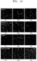
- the cells were stained with a Rhodamin/DAPI staining (Invitrogen, ThermoFisher Scientific, USA). This was observed with a fluorescence microscope (CKX-41, OLYMPUS, Japan), and the results are shown in FIG. 11 .
- FIG. 11 shows microscopic photographs analyzing cell attachment patterns at implants surface-modified with itaconic acid and/or gelatin according to an embodiment.
- the cells were spread better at the implants surface-modified with itaconic acid and/or gelatin according to an embodiment.
- the implants manufactured in Examples 1 and 2 were transplanted to animal models, and the fibrosis states at implantation sites were observed.
- capsule thickness ii) number of fibroblast and iii) number of myofibroblast, were quantified.
- capsule thickness was analyzed through staining with Hematoxylin & Eosin.
- the capsule thickness was measured with respect to collagen and tissue layers accumulated on the bottom part of subcutaneous muscles, the analysis was performed by measuring a portion where the thinnest image phase capsule was formed, and the results are shown in Table 1.
- IA-PDMS and IA-GT-PDMS groups were confirmed to show decreased capsule thicknesses in all cases after 2, 4 and 8 weeks when compared with the control group, and accordingly, it could be found that the fibrosis was reduced by IA.
- the thinnest capsule thickness was observed in IA-GT-PDMS high, and this means that the capsule thickness of IA-GT high PDMS was reduced to about 63% when compared with the control group (IA-GT high: 528 ⁇ m (37%) against control group: 1426 ⁇ m (100%)).
- fibroblast in tissues In order to evaluate the number of fibroblast in tissues, immunostaining was performed, only fibroblast was specifically stained using an antibody (ab92546, Abcam, CA, USA) binded with Vimentin which is a specific factor of fibroblast, and only cells expressing fluorescence were selectively evaluated to confirm the number of cells. Particularly, for the progress of the experiment, the biopsy of tissues was performed after a certain time period, and the biopsied tissues were manufactured into slides through a paraffin embedded technique.
- the significant decrease of myofibroblasts was observed in IA high group after 2 weeks from transplantation.
- the significant decrease of myofibroblasts was confirmed in all experimental groups (IA high, IA-GT low, IA-GT high) excluding the IA low group when compared with the control group (p ⁇ 0.05).
- the decrease of the myofibroblast number was observed in IA high and IA-GT high groups when compared with the control group. This corresponds to an effect of decreasing the collagen density of IA-GT high PDMS to about 50% when compared with the control group (IA high: 21 (53.8%) / IA-GT high: 20.75 (53.2%) against control group: 39 (100%)).
- biopsied tissues were stained using Hematoxylin & Eosin. Then, only polynuclear cells (inflammatory mediated cells) in the capsule formation area of stained tissues were selected and quantified using an image software. Particularly, the staining with Hematoxylin & Eosin was performed for biopsied tissues, and only cells considered to be polynuclear cells after the staining of the tissues were specifically selected and counted. Scores were configured based on the counted specific polynuclear cells, inflammatory response score in each group was analyzed according to the range of the configuration, and the results are shown in Table 4.
- an implant surface-modified with itaconic acid and/or gelatin reduces fibrosis or inflammatory response at an implantation site when compared with a control group and accordingly, the implant could be useful as a medical implant.
Landscapes
- Health & Medical Sciences (AREA)
- Life Sciences & Earth Sciences (AREA)
- Chemical & Material Sciences (AREA)
- Veterinary Medicine (AREA)
- Public Health (AREA)
- General Health & Medical Sciences (AREA)
- Animal Behavior & Ethology (AREA)
- Epidemiology (AREA)
- Medicinal Chemistry (AREA)
- Oral & Maxillofacial Surgery (AREA)
- Transplantation (AREA)
- Dermatology (AREA)
- Vascular Medicine (AREA)
- Heart & Thoracic Surgery (AREA)
- Surgery (AREA)
- Biomedical Technology (AREA)
- Engineering & Computer Science (AREA)
- Chemical Kinetics & Catalysis (AREA)
- Organic Chemistry (AREA)
- Polymers & Plastics (AREA)
- Molecular Biology (AREA)
- Zoology (AREA)
- Cell Biology (AREA)
- Botany (AREA)
- General Chemical & Material Sciences (AREA)
- Materials For Medical Uses (AREA)
Abstract
Description
- The disclosure relates to a composition for surface modification of a medical implant and a medical implant surface-modified thereby.
- Medical implants such as implants, dental implants, stents, screws and bone replacements are inserted for the purpose of treatment or plastic surgery. However, being inserted into the body, the medical implants are recognized as foreign substances in the body, and a clotting mechanism is activated, or foreign body reaction is generated to prevent bleeding and blood loss. In addition, since medical implants make permanent contact with biological tissues, the surface biocompatibility and in vivo affinity thereof are required.
- Accordingly, technique for modifying the surface of medical implants and effective methods for fixing bioactive materials are required.
- Meanwhile, recently, it has been reported that itaconic acid inhibits the decomposition enzyme of isocitric acid, which is an important enzyme of the energy metabolic pathway of microorganisms, thereby contributing to the antibacterial activity of macrophages.
- Accordingly, the inventors of the disclosure confirmed the fibrosis inhibition and inflammatory response inhibition at the implantation site of medical implants by using itaconic acid and completed the inventive concept.
- An aspect is to provide a composition including itaconic acid for surface modification of a medical implant.
- Another aspect is to provide a medical implant surface-modified with the itaconic acid.
- Another aspect is to provide a method of manufacturing the medical implant.
- An aspect provides a composition including itaconic acid for surface modification of a medical implant.
- In the disclosure, the itaconic acid may be represented by Formula 1 below.

- The composition may further include gelatin.
- In the disclosure, the term "gelatin" may mean a protein obtained by partial hydrolysis of collagen which is the main protein component of connective tissues such as bones, cartilages, skins, etc., of animals. The gelatin may include gelatin derivatives in addition to pure gelatin. For example, the gelatin derivative may include at least one of a phthalated gelatin, an esterified gelatin, an amidated gelatin, or a formylated gelatin. Relating to gelatin, the kind (source) thereof is not specifically limited, and various kinds of gelatin derived from, for example, mammals and fishes, for example, cow bones, cow skins, swine bones, swine skins, etc., may be used. In addition, the gelatin may have a molecular weight from about 10,000 to about 30,000.
- The gelatin may be crosslinked. In addition, the gelatin may be chemically binded with itaconic acid.
- In an embodiment, the composition may be provided in a powder type.
- Another aspect provides a medical implant surface-modified with itaconic acid.
- Particularly, a surface-modified medical implant of which at least a portion of the surface of the implant is modified through the binding with itaconic acid may be provided as a medical implant.
- In the disclosure, the term "surface modification" may mean the change of the chemical structure and physical structure of the surface of particles.
- The medical implant may be additionally binded with gelatin. The gelatin may be crosslinked and binded with the surface of the implant through itaconic acid.
- The itaconic acid and gelatin may have a nanoparticle type or a polymer type.
- At least a portion of the surface of the medical implant may introduce an amino group. Particularly, the surface of the medical implant may be silanized for the binding with itaconic acid. An aminosilane compound for silanization may be 3-aminopropyltriethoxysilane (APTES), (3-aminopropyl)-tetraethylorthosilicate, [3-(2-aminoethylamino)propyl]trimethoxysilane, 3-(2-(2-aminoethylamino)ethylamino)propyltrimethoxysilane, or combinations thereof.
- In addition, the itaconic acid may be directly binded with the surface of the implant through the fixing compound at the surface of the implant. The fixing compound may include a compound having biotin, avidin, streptavidin, carbohydrate, poly L-lycine, a thiol group, an amine group, an alcohol group, a carboxyl group, an amino group, a sulfur group, an aldehyde group, a carbonyl group, a succinimide group, a maleimide group, an epoxy group, or an isothiocyanate group, or combinations thereof. Examples of the compound having an amino group may include 3-aminopropyltrimethoxysilane, N-(2-aminoethyl)-3-aminopropyltrimethoxysilane (EDA), trimethoxysilylpropyldiethylenetriamine (DETA), 3-(2-aminoethylaminopropyl) trimethoxysilane, and 3-aminopropyltriethoxysilane, and the compound having an aldehyde group may include glutaraldehyde. Examples of the compound having a thiol group may include 4-mercaptopropyltrimethoxysilane (MPTS). In addition, examples of the compound having an epoxy group may include 3-glycidoxypropyltrimethoxysilane, examples of the compound having an isothiocyanate group may include 4-phenylenediisothiocyanate (PDITC), and examples of the compound having succinimide and maleimide may include disuccinimidyl carbonate (DSC) or succinimidyl 4-(maleimidephenyl)butyrate (SMPB).
- The medical implant may be a biocompatible material. Examples of the biocompatible material may include one or more materials selected from the group consisting of gold (Au), silver (Ag), platinum (Pt), palladium (Pd), copper (Cu), steel, tantalum (Ta), magnesium (Mg), nickel (Ni), chromium (Cr), iron (Fe), a nitinol alloy
- (NiTi), a cobalt-chromium alloy (CoCr) gallium arsenic (GaAs), titanium (Ti), polylactic acid, poly(glycolic acid) (PGA), poly(lactic-co-glycolic acid) (PLGA), poly-ε-(caprolactone), polyanhydride, polyorthoester, polyvinyl alcohol, polyethylene glycol, polyurethane, polyacrylic acid, poly-N-isopropylacrylamide, poly(ethylene oxide)-poly(propylene oxide)-poly(ethylene oxide), polytetrafluoroethylene, polycarbonate, polyurethane, nitrocellulose, polystyrene, polyethylene, polyethylene terephthalate (PET), polydimethylsiloxane (PDMS), polyether ether ketone (PEEK), silicon oxide (SiO2), titanium oxide (TiO2), aluminum oxide (Al2O3), niobium oxide (Nb2O5), silicon, silicone rubber, and glass.
- In addition, the medical implant may be selected from the group consisting of breast implants, spinal implants, dental implants, cardiovascular implants, cardiac implants, stents, artificial joints, artificial head bones, and cell culture mediums.
- In a particular embodiment, the surface modified medical implant may have a water contact angle from about 20° to about 90°, from about 25° to about 85°, from about 25° to about 80°, from about 25° to about 70°, from about 30° to about 60°, or from about 30° to about 40°.
- In another particular embodiment, the surface modified medical implant may have improved fibrosis inhibition or inflammatory response inhibition at an implantation site when compared with that before surface modification.
- Another aspect provides a method of surface modifying a medical implant, including a step of functionalizing at least a portion of a surface of a medical implant; and a step of binding itaconic acid with the surface of the functionalized medical implant.
- The functionalization step may be a process for binding itaconic acid and/or gelatin with the surface of the implant. The functionalization may include a step of introducing an amino group, or a step of functionalizing using a fixing compound as described above.
- In addition, the step of binding itaconic acid may be a step of binding gelatin additionally. This step may include a step of preparing a composite of itaconic acid and gelatin and then, binding, binding itaconic acid and then, binding gelatin, or binding gelatin and then, combining itaconic acid.
- According to the composition for surface modification of a medical implant according to an aspect and a medical implant surface-modified thereby, itaconic acid may be binded with the surface of the medical implant in high binding stability, and effects showing activity on fibrosis inhibition and inflammatory response inhibition at an implantation site may be achieved.
-
-
FIG. 1 is a diagram schematically showing the manufacturing process of a medical implant surface-modified with itaconic acid according to an embodiment. -
FIG. 2 is a diagram schematically showing the manufacturing process of a medical implant surface-modified with itaconic acid and gelatin according to an embodiment. -
FIG. 3 is a graph showing the contact angles of medical implants surface-modified with itaconic acid and/or gelatin according to an embodiment. -
FIG. 4 shows scanning electron microscopy micrographs of medical implants surface-modified with itaconic acid and/or gelatin according to an embodiment. -
FIG. 5 is a graph showing the ATR-FTIR analysis results of implants surface-modified with itaconic acid and/or gelatin according to an embodiment. -
FIG. 6 is a graph confirming the absorption degree of protein of implants surface-modified with itaconic acid and/or gelatin according to an embodiment. -
FIG. 7 is a graph showing the thermal stability of implants surface-modified with itaconic acid and/or gelatin according to an embodiment. -
FIG. 8 shows photomicrographs observing the cell culture results of implants surface-modified with itaconic acid and/or gelatin according to an embodiment. -
FIG. 9 shows graphs on the analysis results of cell proliferation rates at implants surface-modified with itaconic acid and/or gelatin according to an embodiment. -
FIG. 10 shows photographs on the analysis results of cell viability at implants surface-modified with itaconic acid and/or gelatin according to an embodiment. -
FIG. 11 shows microscopic photographs analyzing cell attachment patterns at implants surface-modified with itaconic acid and/or gelatin according to an embodiment. - Hereinafter, the present disclosure will be explained in more detail referring to embodiments. However, the embodiments are illustrated for explaining the present disclosure, and the scope of the present disclosure is not limited thereto.
- A medical implant surface-modified with itaconic acid was manufactured as shown in
FIG. 1 . - Particularly, a polydimethylsiloxane (PDMS) substrate (Sylgard® 184 silicone elastomer kit, Dow Corning, USA) was treated with oxygen plasma (CUTE-1B, Femto Science, USA) for 1 minute so as to attach hydroxyl groups (-OH) on the surface of PDMS. After that, the surface of the substrate was silanized using 3-aminopropyl triethoxysilane (APTES) at 60°C for 2 hours. Then, 50 mmol, or 150 mmol of itaconic acid (IA, analytical grade, assay ≥99%, MW: 130.10 g/mol, density: 1.573 g/mL at 25°C (lit.), Sigma-Aldrich, South Korea) was reacted at 60°C for 2 hours in the presence of 1-ethyl-3-(3-dimethylamino)propyl carbodiimide (EDC) and N-hydroxy succinimide (NHS) (EDC/NHS) to bind the itaconic acid with the surface of the PDMS substrate. Then, EDC/NHS residues were removed, and drying was performed for 12 hours to manufacture a medical implant surface-modified with itaconic acid (hereinafter, will be referred to as "IA-PDMS").
- A medical implant surface-modified with itaconic acid and gelatin (IA-GT) was manufactured as shown in
FIG. 2 . - Particularly, in order to prepare a polymer of itaconic acid and gelatin, a gelatin type B (derived from cow skin, Bloon 75) powder was mixed with DPBS and dissolved at 135°C to 140°C for 16 hours. Then, with the dissolved gelatin type B, 50 mmol of EDC and 50 mmol of NHS solutions were mixed, and 100 mmol of itaconic acid was put and reacted for 2 hours. Additionally, DPBS was added to the mixture, and the reaction with gelatin and itaconic acid was performed at 60°C for 30 minutes and then finished. The final mixture was dialyzed through a cellulose membrane (MWCO 6-8kD), and unreacted itaconic acid, EDC/NHS, and salts were removed. After that, freeze drying was performed at -80°C for 48 hours to obtain an IA-GT powder.
- The IA-GT powder thus obtained was binded with the surface of a PDMS substrate as in Example 1 to manufacture a medical implant surface-modified with itaconic acid and gelatin (hereinafter, will be referred to as "IA-GTpoly-PDMS"). Briefly, a surface modified PDMS was manufactured by the same method as in Example 1 except for using DW +
EDC 10 mmol +NHS 10 mmol to manufacture the surface modified PDMS of IA-GTpoly-0.25 wt%, and using DW +EDC 20 mmol +NHS 20 mmol to manufacture the surface modified PDMS of IA-GTpoly-0.50 wt%. - On the surface of the implants manufactured in Examples 1 and 2, 4 µl of distilled water was dropped, and contact angles were measured at room temperature using a laboratory-made contact angle goniometer with a charge-coupled device camera (IMT 3, IMT Solutions), and the results are shown in
FIG. 3 . -
FIG. 3 is a graph showing the contact angles of implants surface-modified with itaconic acid and/or gelatin according to an embodiment. - As shown in shown
FIG. 3 , it could be found that the PDMSs surface-modified with itaconic acid and/or gelatin had smaller contact angles when compared with the conventional PDMS (control group). - The surfaces of the implants manufactured in Examples 1 and 2 were observed by a scanning electron microscope (SEM) (SEM, S-3400N, Hitachi, Tokyo, Japan), and the results are shown in
FIG. 4 . -
FIG. 4 shows scanning electron microscopy images showing the contact angles of implants surface-modified with itaconic acid and/or gelatin according to an embodiment. - As shown in
FIG. 4 , it could be confirmed that white modification layers were observed on the surface of PDMSs. - In order to confirm the chemical bond at the surface of the implant, attenuated total reflection (ATR)-FTIR analysis was performed.
- Particularly, with respect to the implants manufactured in Examples 1 and 2, each spectrum at room temperature was recorded from 400 cm-1 to 4000 cm-1 with a resolution of 4 cm-1 using a FT-IR Spectroscopy (Frontier spectrophotometer equipped with and attenuated total reflection (ATR-FTIR) module (Nicolet 6700, Thermo Scientific, USA), the scanning was performed 200 times and averaged, and the results are shown in
FIG. 5 . -
FIG. 5 is a graph showing the ATR-FTIR analysis results of implants surface-modified with itaconic acid and/or gelatin according to an embodiment. - As shown in
FIG. 5 , the peaks of carbonyl groups (1,640 cm-1 for IA-50 mmol, 1,648 cm-1 for IA-150 mmol, 1,637 cm-1 for IA-GTpoly-0.25 wt% & 1,646 cm-1 for IA-GTpoly-0.50 wt%) were observed, and through the results, the binding of IA with PDMS was confirmed. In addition, through the observation of the peaks of nitrile groups (1,542 cm-1 for IA-50 mmol, 1,553 cm-1 for IA-150 mmol, 1,536 cm-1 for IA-GTpoly-0.25 wt% & 1,535 cm-1 for IA-GTpoly-0.50 wt%), amide bonds were confirmed. - The protein absorbing capacity of the surfaces of the implants manufactured in Examples 1 and 2 was analyzed.
- Particularly, bovine serum albumin (BSA) (4.5 mg/mL) was dissolved in DPBS. Separately, the surfaces of IA-PDMS and IA-GTpoly-PDMS (size: 1 cm2) were cultured on a hot stirring plate in a protein solution while stirring constantly for 1 hour at 37°C in 200 rpm conditions. After washing with clean DPBS twice, BSA protein - cultured bare, IA-conjugated and IA-GTpoly conjugated PDMS surfaces (triple) were transported to a 24-well plate containing DW and an operation reagent mixture. After culturing the well plate at 37°C for 30 minutes, the absorbance value of each sample was analyzed. The amount of absorbed protein was quantified using Micro BCA™ protein analysis kit according to the instruction of a manufacturer. The absorbance value was measured using a microplate spectrophotometer with a fixed wavelength of 570 nm. All values were represented by average ± standard deviation, and the results are shown in
FIG. 6 . -
FIG. 6 is a graph confirming the absorption degree of protein of implants surface-modified with itaconic acid and/or gelatin according to an embodiment. - As shown in
FIG. 6 , it could be confirmed that strong inhibition of protein absorption was observed for the implants surface-modified with itaconic acid and/or gelatin according to an embodiment when compared with the control group. - In order to analyze the binding stability of itaconic acid and/or gelatin, heat treatment was performed with respect to the surface of the implants manufactured in Examples 1 and 2.
- Particularly, IA-GTpoly-PDMS (IA-GTpoly-0.50 wt%) having a size of 2 cm2 was used as a specimen for heat treatment, and the heat treatment was performed three times in total at 120°C for 1 hour. After that, contact angles were measured by the same method as in Experimental Example 1.1, and the results are shown in
FIG. 7 . -
FIG. 7 is a graph showing the thermal stability of implants surface-modified with itaconic acid and/or gelatin according to an embodiment. - As shown in
FIG. 7 , it could be confirmed that after the heat treatment three times, each contact angle was gradually increased, but the change of the binding stability of itaconic acid and/or gelatin was not observed. - In order to analyze the cell proliferation rate and viability in the implants manufactured in Examples 1 and 2, NIH3T3 mouse fibroblast lines were used.
- Particularly, the fibroblast lines were cultured at the surface of PDMS having a size of 1 cm x 1 cm with cells of 20,000 cell/ml at 37°C. After initiating the culture, the cell proliferation rates and viability at 12 hour, 24 hour and 48 hour were analyzed. The cell proliferation rate was observed by an optical microscope (Euromex IF-Series, Netherlands), by treating cells with trypsin (GE Healthcare Life Sciences HyClone Laboratories, USA.) and counting the number of cells, or by counting the number of cells through an MTT assay (Cell Biolabs, INC, USA). The observation results by the optical microscope are shown in
FIG. 8 , and the counting results of cells after treating with trypsin and the MTT assay results are shown inFIG. 9 . - In addition, the cell viability was observed through a Live/Dead analysis method (L-3224, Invitrogen, LIVE/DEAD® Viability/Cytotoxicity Kit, for mammalian cells (Molecular Probes®)). Particularly, in order to prepare a Live/Dead analysis solution, 10 ml of PBS was added to a 15 ml tube, and 2 mM EthD-1 and 4 mM calcein AM were added in 20 µ and 5 µl, respectively. Then, the tube was wrapped with an aluminum foil, well mixed through pipetting, and stored under refrigeration. In addition, the cultured cells were put in PBS and washed for 20 minutes, PBS was removed, and the Live/Dead analysis solution thus prepared was added to a cell specimen. After that, culturing was performed in an incubator for 30 minutes, photographs were taken using a fluorescence and confocal microscope, and the results are shown in
FIG. 10 . -
FIG. 8 shows optical microscope photographs observing the cell culture results of implants surface-modified with itaconic acid and/or gelatin according to an embodiment. -
FIG. 9 shows graphs showing the analysis results of cell proliferation rates at implants surface-modified with itaconic acid and/or gelatin according to an embodiment. -
FIG. 10 shows photographs showing the analysis results of cell viability at implants surface-modified with itaconic acid and/or gelatin according to an embodiment. - As shown in
FIG. 8 , it could be found that the cell culture was conducted well at the implants surface-modified with itaconic and/or gelatin according to an embodiment. - In addition, as shown in
FIG. 9 , the implants surface-modified with itaconic acid and/or gelatin according to an embodiment showed similar or increased cell proliferation rates when compared with the control group. - In addition, as shown in
FIG. 10 , the implants surface-modified with itaconic acid and/or gelatin according to an embodiment showed higher cell viability when compared with the surface unmodified PDMS of the control group. - In order to analyze cell attachment pattern, the cells were stained with a Rhodamin/DAPI staining (Invitrogen, ThermoFisher Scientific, USA). This was observed with a fluorescence microscope (CKX-41, OLYMPUS, Japan), and the results are shown in
FIG. 11 . -
FIG. 11 shows microscopic photographs analyzing cell attachment patterns at implants surface-modified with itaconic acid and/or gelatin according to an embodiment. - As shown in
FIG. 11 , the cells were spread better at the implants surface-modified with itaconic acid and/or gelatin according to an embodiment. - The implants manufactured in Examples 1 and 2 were transplanted to animal models, and the fibrosis states at implantation sites were observed.
- First, four 8-week Sprague-Daley rats (Young Bio, KOREA) were used for each group as the animal models. Animal experiment was conducted after final approval from Institutional Animal Care and Use Committee (IACAU) by Seoul National University Hospital in Bundang (approval number: BA1608-206/045-01). Experiment was conducted by dividing into five groups as follows. In order to prevent inflammation and infection at a wounded area during transplanting an implant, disinfection was conducted using betadine. The disinfection was performed twice before and after surgical procedure. For transplantation, the back was incised by 2 cm, a subcutaneous pocket was formed, and implants of each group were transplanted in the pocket. After the transplantation, the wounded area was sutured using
Nylon 4/0 (Ethicon), the wounded area was finally disinfected using betadine, and a gauze dressing was applied. - Five groups of implants below were all transplanted in one rat, the rat was euthanized after 2, 4 and 8 weeks, and tissues at an implantation site were obtained.
- Group 1 (control group): untreated PDMS
- Group 2 (
IA 50 mmol (low) PDMS): PDMS surface treated with IA having a low concentration - Group 3 (IA 150 mmol (high) PDMS): PDMS surface treated with IA having a high concentration
- Group 4 (IA-GT 0.25% (low) PDMS): PDMS surface treated with IA chemically bonded to gelatin, having a low concentration
- Group 5 (IA-GT 0.50% (high) PDMS): PDMS surface treated with IA chemically bonded to gelatin, having a high concentration
- In order to evaluate a fibrosis degree, i) capsule thickness, ii) number of fibroblast and iii) number of myofibroblast, were quantified.
- Particularly, capsule thickness was analyzed through staining with Hematoxylin & Eosin. The capsule thickness was measured with respect to collagen and tissue layers accumulated on the bottom part of subcutaneous muscles, the analysis was performed by measuring a portion where the thinnest image phase capsule was formed, and the results are shown in Table 1.
[Table 1] Capsule thickness results (unit: µm) Week Control IA_Low IA_High IA_GT Low IA_GT High 2 813 ± 260.9 514.8 ± 302.3 487.9 ± 45.8 403.3 ± 145 323.2 ± 158.5 4 1060.6 ± 83.7 527.9 ± 110.6 621.7 ± 70 506.6 ± 73.1 544.8 ± 60.5 8 1426.1 ± 250.6 789.1 ± 284.9 786.7 ± 210.5 774 ± 136.9 528.6 ± 183.3 - As shown in Table 1, IA-PDMS and IA-GT-PDMS groups were confirmed to show decreased capsule thicknesses in all cases after 2, 4 and 8 weeks when compared with the control group, and accordingly, it could be found that the fibrosis was reduced by IA. Particularly, the thinnest capsule thickness was observed in IA-GT-PDMS high, and this means that the capsule thickness of IA-GT high PDMS was reduced to about 63% when compared with the control group (IA-GT high: 528 µm (37%) against control group: 1426 µm (100%)).
- In order to evaluate the number of fibroblast in tissues, immunostaining was performed, only fibroblast was specifically stained using an antibody (ab92546, Abcam, CA, USA) binded with Vimentin which is a specific factor of fibroblast, and only cells expressing fluorescence were selectively evaluated to confirm the number of cells. Particularly, for the progress of the experiment, the biopsy of tissues was performed after a certain time period, and the biopsied tissues were manufactured into slides through a paraffin embedded technique. After the pre-treatment of the slide (Deparaffine, Rehydration), an immune factor bonding to the fibroblast specific factor was used, color expressed by the bonding of the specific factor and DAPI where staining of a cell nucleus occurs were selectively counted and analyzed per week, and the results are shown in Table 2.
[Table 2] Fibroblast number (unit: n) Week Control IA_Low IA_High IA_GT Low IA_GT High 2 36.6 ± 13.41 32 ± 4.73 24.17 ± 4.88 28.83 ± 5.81 27.17 ± 6.15 4 27.5 ± 11.48 31.67 ± 10.93 23.67 ± 7.45 29.67 ± 5.16 27.17 ± 5.67 8 39 ± 11.83 30.17 ± 13.38 21 ± 5.52 23.8 ± 7.66 20.75 ± 5.06 - As shown in Table 2, somewhat lower number of fibroblasts was confirmed in all groups until 2 weeks and 4 weeks from transplantation when compared with the control group, but the difference was insignificant. However, in groups after 8 weeks from the transplantation, smaller fibroblasts were observed in IA high and IA-GT high groups when compared with the control group (p<0.05). This corresponds to an effect of reducing the collagen density of IA-GT high PDMS to about 50% when compared with the control group (IA high: 21 (53.8%) / IA-GT high: 20.75 (53.2%) against control group: 39 (100%)).
- In order to evaluate the number of myofibroblasts in tissues, immunostaining was performed, only myofibroblasts were specifically stained using an antibody binded with alpha-SMA which is a specific factor of myofibroblast, and only cells expressing fluorescence were selectively evaluated to confirm the number of cells. Particularly, for the progress of the experiment, the biopsy of tissues was performed after a certain time period, and the biopsied tissues were manufactured into slides through a paraffin embedded technique. After the pre-treatment of the slide (Deparaffine, Rehydration), an immune factor bonding to a myofibroblast specific factor was used, color expressed by the bonding of the specific factor and DAPI where staining of a cell nucleus occurs were selectively counted and analyzed per week, and the results are shown in Table 3.
[Table 3] Myofibroblast number (unit: n) Week Control IA_Low IA_High IA_GT Low IA_GT High 2 46 ± 6.03 41.17 ± 4.12 29.5 ± 5.17 39.5 ± 5.68 34.33 ± 7.5 4 62.5 ± 14.03 49.5 ± 12 32 ± 10.53 37.17 ± 8.21 41.17 ± 1.94 8 40 ± 11.73 37.5 ± 7.79 29.83 ± 10 33 ± 6.72 27.67 ± 8.78 - As shown in Table 3, the significant decrease of myofibroblasts was observed in IA high group after 2 weeks from transplantation. In addition, after 4 weeks, the significant decrease of myofibroblasts was confirmed in all experimental groups (IA high, IA-GT low, IA-GT high) excluding the IA low group when compared with the control group (p<0.05). In the groups after 8 weeks, the decrease of the myofibroblast number was observed in IA high and IA-GT high groups when compared with the control group. This corresponds to an effect of decreasing the collagen density of IA-GT high PDMS to about 50% when compared with the control group (IA high: 21 (53.8%) / IA-GT high: 20.75 (53.2%) against control group: 39 (100%)).
- In order to analyze inflammatory response at an implantation site, biopsied tissues were stained using Hematoxylin & Eosin. Then, only polynuclear cells (inflammatory mediated cells) in the capsule formation area of stained tissues were selected and quantified using an image software. Particularly, the staining with Hematoxylin & Eosin was performed for biopsied tissues, and only cells considered to be polynuclear cells after the staining of the tissues were specifically selected and counted. Scores were configured based on the counted specific polynuclear cells, inflammatory response score in each group was analyzed according to the range of the configuration, and the results are shown in Table 4.
[Table 4] Inflammatory response score (unit: score) Week Control IA_Low IA_High IA_GT Low IA_GT High 2 2.48 ± 0.44 1.96 ± 0.15 1.82 ± 0.29 1.58 ± 0.38 1.3 ± 0.31 4 1.98 ± 0.43 1.54 ± 0.27 1.38 ± 0.27 1.37 ± 0.53 1.17 ± 0.43 8 1.35 ± 0.14 1.42 ± 0.4 1.03 ± 0.24 1.05 ± 0.33 1 ± 0.3 - As shown in Table 4, it could be found that the inflammatory response was significantly decreased in all experimental groups when compared with the control group.
- From the results above, it could be found that an implant surface-modified with itaconic acid and/or gelatin according to an embodiment reduces fibrosis or inflammatory response at an implantation site when compared with a control group and accordingly, the implant could be useful as a medical implant.
Claims (14)
- A composition comprising itaconic acid, for surface modification of a medical implant.
- The composition according to claim 1, further comprising gelatin.
- A medical implant, wherein at least a portion of a surface of the implant is binded with itaconic acid for surface modification.
- The implant according to claim 3, wherein the at least a portion of the surface is silanized for the binding with the itaconic acid.
- The medical implant according to claim 3, wherein gelatin is additionally binded.
- The implant according to claim 3, wherein the itaconic acid is directly binded with the surface of the implant through a fixing compound at the surface of the implant.
- The implant according to claim 6, wherein the fixing compound is a compound having biotin, avidin, streptavidin, carbohydrate, poly L-lycine, a thiol group, an amine group, an alcohol group, a carboxyl group, an amino group, a sulfur group, an aldehyde group, a carbonyl group, a succinimide group, a maleimide group, an epoxy group, or an isothiocyanate group, or combinations thereof.
- The implant according to claim 3, wherein the medical implant comprises one or more materials selected from the group consisting of gold (Au), silver (Ag), platinum (Pt), palladium (Pd), copper (Cu), steel, tantalum (Ta), magnesium (Mg), nickel (Ni), chromium (Cr), iron (Fe), a nitinol alloy (NiTi), a cobalt-chromium alloy (CoCr) gallium arsenic (GaAs), titanium (Ti), polylactic acid, poly(glycolic acid) (PGA), poly(lactic-co-glycolic acid) (PLGA), poly-ε-(caprolactone), polyanhydride, polyorthoester, polyvinyl alcohol, polyethylene glycol, polyurethane, polyacrylic acid, poly-N-isopropylacrylamide, poly(ethylene oxide)-poly(propylene oxide)-poly(ethylene oxide), polytetrafluoroethylene, polycarbonate, polyurethane, nitrocellulose, polystyrene, polyethylene, polyethylene terephthalate (PET), polydimethylsiloxane (PDMS), polyether ether ketone (PEEK), silicon oxide (SiO2), titanium oxide (TiO2), aluminum oxide (Al2O3), niobium oxide (Nb2O5), silicon, silicone rubber, and glass.
- The implant according to claim 5, wherein the gelatin is crosslinked and binded with the surface of the implant through the itaconic acid.
- The implant according to claim 3, wherein a water contact angle is from 20° to 90°.
- The implant according to claim 3, wherein the medical implant is selected from the group consisting of a breast implant, a spinal implant, a dental implant, a cardiovascular implant, a cardiac implant, a stent, an artificial joint, an artificial head bone, and a cell culture medium.
- The implant according to claim 3, wherein fibrosis inhibition or inflammatory response inhibition at an implantation site is increased when compared with that before surface modification.
- A method of surface modifying a medical implant, the method comprising:a step of functionalizing at least a portion of a surface of the medical implant; anda step of binding itaconic acid with the surface of the functionalized medical implant.
- The method according to claim 13, wherein gelatin is additionally binded in the step of binding itaconic acid.
Applications Claiming Priority (2)
| Application Number | Priority Date | Filing Date | Title |
|---|---|---|---|
| KR1020170135862A KR102101936B1 (en) | 2017-10-19 | 2017-10-19 | Composition for suface modification of medical implants and suface modified medical implants using the same |
| PCT/KR2018/008100 WO2019078454A1 (en) | 2017-10-19 | 2018-07-18 | Composition for surface modification of medical implant and medical implant surface-modified thereby |
Publications (3)
| Publication Number | Publication Date |
|---|---|
| EP3698814A1 true EP3698814A1 (en) | 2020-08-26 |
| EP3698814A4 EP3698814A4 (en) | 2021-07-14 |
| EP3698814B1 EP3698814B1 (en) | 2025-09-10 |
Family
ID=66173759
Family Applications (1)
| Application Number | Title | Priority Date | Filing Date |
|---|---|---|---|
| EP18868312.2A Active EP3698814B1 (en) | 2017-10-19 | 2018-07-18 | Composition for surface modification of medical implant and medical implant surface-modified thereby |
Country Status (5)
| Country | Link |
|---|---|
| US (1) | US20200268937A1 (en) |
| EP (1) | EP3698814B1 (en) |
| KR (1) | KR102101936B1 (en) |
| CN (1) | CN111246895B (en) |
| WO (1) | WO2019078454A1 (en) |
Families Citing this family (2)
| Publication number | Priority date | Publication date | Assignee | Title |
|---|---|---|---|---|
| WO2020226250A1 (en) * | 2019-05-08 | 2020-11-12 | 서울대학교병원 | Medical implant surface-modified with functional polypeptide |
| CN115873712B (en) * | 2022-11-28 | 2023-05-12 | 杭州皓丰生物技术有限公司 | Deep hole cell culture plate, preparation method and application thereof |
Family Cites Families (22)
| Publication number | Priority date | Publication date | Assignee | Title |
|---|---|---|---|---|
| US4920158A (en) * | 1989-10-11 | 1990-04-24 | Medipro Sciences Limited | Hydrogel-forming wound dressing or skin coating material |
| US5811151A (en) * | 1996-05-31 | 1998-09-22 | Medtronic, Inc. | Method of modifying the surface of a medical device |
| CN1313842C (en) * | 1999-10-12 | 2007-05-02 | 庄臣及庄臣视力保护公司 | Contact lens coating selection and manufacturing process thereof |
| AU2002235694B2 (en) * | 2001-02-28 | 2007-06-14 | Covalon Technologies Inc. | Method of making anti-microbial polymeric surfaces |
| US6884829B2 (en) * | 2002-10-18 | 2005-04-26 | Robert L. Albright | Hemocompatible coated polymer and related one-step methods |
| CN1950114A (en) * | 2004-05-12 | 2007-04-18 | 苏尔莫迪克斯公司 | Natural biodegradable polysaccharide coatings for medical articles |
| US7411029B2 (en) * | 2004-06-25 | 2008-08-12 | Bausch & Lomb Incorporated | Prepolymers for improved surface modification of contact lenses |
| AR054300A1 (en) | 2004-08-10 | 2007-06-20 | Alza Corp | APPARATUS AND MICROPROJECTION SYSTEM WITH INFECTIOUS POTENTIAL ESCASS |
| FR2877846B1 (en) * | 2004-11-15 | 2008-12-05 | Univ Lille Sciences Tech | BIOMATERIAL CARRIERS OF CYCLODEXTRINS WITH IMPROVED ABSORPTION PROPERTIES AND PROGRESSIVE AND DELAYED RELEASE OF THERAPEUTIC MOLECULES |
| BRPI0720268B8 (en) * | 2006-12-15 | 2021-06-22 | Coloplast As | method for preparing a medical device element, and medical device |
| WO2008106048A1 (en) * | 2007-02-28 | 2008-09-04 | Corning Incorporated | Surfaces and methods for biosensor cellular assays |
| WO2009155450A2 (en) * | 2008-06-20 | 2009-12-23 | Board Of Regents, The University Of Texas System | Biodegradable photoluminescent polymers |
| CN101491699B (en) * | 2009-03-03 | 2011-09-14 | 陕西科技大学 | Preparation method of carbon nano-tube reinforced bone-cement biological composite material |
| SG185988A1 (en) * | 2009-05-15 | 2012-12-28 | Univ Nanyang Tech | Composition for manufacturing a scaffold for tissue engineering, and a method of making it |
| WO2011123110A1 (en) * | 2010-03-30 | 2011-10-06 | Daniel Sunho Oh | Method of preparing ceramic/polymer composite scaffolds with bioactive molecules for hard tissue regeneration |
| KR101340044B1 (en) * | 2011-10-27 | 2013-12-10 | 한국과학기술원 | Hyaluronic acid catechol conjugate and use thereof |
| EP2609941A1 (en) * | 2011-12-26 | 2013-07-03 | Laboratorios Sanifit, S.L. | Biocompatible implant |
| WO2013126718A1 (en) * | 2012-02-24 | 2013-08-29 | Bvw Holding Ag | Anti-erosion soft tissue repair device |
| KR101522462B1 (en) * | 2012-09-19 | 2015-05-21 | 아주대학교산학협력단 | Preparation Method of In situ forming Hydrogel Using Enzyme-Immobilized Supports and Biomedical Use Thereof |
| KR101663353B1 (en) * | 2013-11-26 | 2016-10-07 | 단국대학교 산학협력단 | Surface Modified Dental Implant Material with CNT-Gelatin-Hydroxyapatite Nanohybrids for and Manufacturing Methods Thereof |
| FR3017533B1 (en) * | 2014-02-19 | 2016-03-11 | Cousin Biotech | IMPLANTABLE DEVICE, IN PARTICULAR FOR ABDOMINAL WALL REFECTION |
| JP2017519026A (en) * | 2014-06-24 | 2017-07-13 | クロクス テクノロジーズ インコーポレイテッド | Biophotonic composition containing halogen and use thereof |
-
2017
- 2017-10-19 KR KR1020170135862A patent/KR102101936B1/en active Active
-
2018
- 2018-07-18 WO PCT/KR2018/008100 patent/WO2019078454A1/en not_active Ceased
- 2018-07-18 US US16/756,831 patent/US20200268937A1/en not_active Abandoned
- 2018-07-18 EP EP18868312.2A patent/EP3698814B1/en active Active
- 2018-07-18 CN CN201880068308.6A patent/CN111246895B/en active Active
Also Published As
| Publication number | Publication date |
|---|---|
| WO2019078454A1 (en) | 2019-04-25 |
| CN111246895B (en) | 2022-11-15 |
| EP3698814A4 (en) | 2021-07-14 |
| KR20190043844A (en) | 2019-04-29 |
| KR102101936B1 (en) | 2020-04-17 |
| EP3698814B1 (en) | 2025-09-10 |
| CN111246895A (en) | 2020-06-05 |
| US20200268937A1 (en) | 2020-08-27 |
Similar Documents
| Publication | Publication Date | Title |
|---|---|---|
| US20230037048A1 (en) | Extracellular Matrix-Derived Gels and Related Methods | |
| Zhou et al. | Materials strategies to overcome the foreign body response | |
| Zhu et al. | Polydopamine-mediated covalent functionalization of collagen on a titanium alloy to promote biocompatibility with soft tissues | |
| AU2013390806B2 (en) | Polymer chelator conjugates | |
| Yakufu et al. | Covalently functionalized poly (etheretherketone) implants with osteogenic growth peptide (OGP) to improve osteogenesis activity | |
| Ma et al. | A novel chitosan–collagen-based hydrogel for use as a dermal filler: initial in vitro and in vivo investigations | |
| Griffin et al. | Enhancing tissue integration and angiogenesis of a novel nanocomposite polymer using plasma surface polymerisation, an in vitro and in vivo study | |
| AU2017207015A1 (en) | Vascular extracellular matrix hydrogel | |
| JP5009291B2 (en) | Biological artificial blood vessel and method for preparing the same | |
| Birajdar et al. | Inhibition of capsular contracture of poly (dimethyl siloxane) medical implants by surface modification with itaconic acid conjugated gelatin | |
| Li et al. | Facile engineering of interactive double network hydrogels for heart valve regeneration | |
| CN112717202A (en) | Metal @ tea polyphenol nanoparticle self-assembly biological tissue and preparation method thereof | |
| EP3698814B1 (en) | Composition for surface modification of medical implant and medical implant surface-modified thereby | |
| Huang et al. | A multifunctional collagen-base bilayer membrane integrated with a bimetallic/polydopamine network for enhanced guided bone regeneration | |
| WO2006038866A1 (en) | Improved coating comprising a bioadhesive polyphenolic protein derived from a byssus-forming mussel | |
| Butruk et al. | Surface endothelialization of polyurethanes | |
| Pandey et al. | Tuning biomaterial surfaces to modulate host-immune reactions | |
| JP7017726B2 (en) | Use of peptides that specifically bind to vascular endothelial cells, and peptides | |
| KR101240075B1 (en) | Medical Implant and Method for Manufacturing the Same | |
| Balaji et al. | Engineering Antioxidant and Oxygen-Releasing Lignin Composites to Accelerate Wound Healing | |
| Shi et al. | An oxidized chondroitin sulfate-crosslinked and CuCDs-loaded decellularized bovine pericardium with improved anti-coagulation, pro-endothelialization and anti-calcification properties for BHVs | |
| CN114470324A (en) | A new strategy for general bone implant modification for the intervention of diabetic bone defects | |
| KR102182883B1 (en) | Collagen Membrane and Method for Fabricating the Same | |
| JP7320796B2 (en) | Use of peptide that specifically binds to vascular endothelial cells, and peptide | |
| Yin et al. | Dynamic coordination hydrogel with simultaneous anti-infected and angiogenic activities for promoting infected wound healing |
Legal Events
| Date | Code | Title | Description |
|---|---|---|---|
| STAA | Information on the status of an ep patent application or granted ep patent |
Free format text: STATUS: THE INTERNATIONAL PUBLICATION HAS BEEN MADE |
|
| PUAI | Public reference made under article 153(3) epc to a published international application that has entered the european phase |
Free format text: ORIGINAL CODE: 0009012 |
|
| STAA | Information on the status of an ep patent application or granted ep patent |
Free format text: STATUS: REQUEST FOR EXAMINATION WAS MADE |
|
| 17P | Request for examination filed |
Effective date: 20200518 |
|
| AK | Designated contracting states |
Kind code of ref document: A1 Designated state(s): AL AT BE BG CH CY CZ DE DK EE ES FI FR GB GR HR HU IE IS IT LI LT LU LV MC MK MT NL NO PL PT RO RS SE SI SK SM TR |
|
| AX | Request for extension of the european patent |
Extension state: BA ME |
|
| DAV | Request for validation of the european patent (deleted) | ||
| DAX | Request for extension of the european patent (deleted) | ||
| A4 | Supplementary search report drawn up and despatched |
Effective date: 20210611 |
|
| RIC1 | Information provided on ipc code assigned before grant |
Ipc: A61L 27/28 20060101AFI20210607BHEP Ipc: A61L 27/38 20060101ALI20210607BHEP Ipc: A61L 31/08 20060101ALI20210607BHEP Ipc: A61L 27/34 20060101ALI20210607BHEP Ipc: A61L 27/50 20060101ALI20210607BHEP Ipc: A61L 27/54 20060101ALI20210607BHEP Ipc: A61L 31/10 20060101ALI20210607BHEP Ipc: A61L 31/14 20060101ALI20210607BHEP Ipc: A61L 31/16 20060101ALI20210607BHEP |
|
| STAA | Information on the status of an ep patent application or granted ep patent |
Free format text: STATUS: EXAMINATION IS IN PROGRESS |
|
| 17Q | First examination report despatched |
Effective date: 20230227 |
|
| GRAP | Despatch of communication of intention to grant a patent |
Free format text: ORIGINAL CODE: EPIDOSNIGR1 |
|
| STAA | Information on the status of an ep patent application or granted ep patent |
Free format text: STATUS: GRANT OF PATENT IS INTENDED |
|
| INTG | Intention to grant announced |
Effective date: 20250408 |
|
| GRAS | Grant fee paid |
Free format text: ORIGINAL CODE: EPIDOSNIGR3 |
|
| GRAA | (expected) grant |
Free format text: ORIGINAL CODE: 0009210 |
|
| STAA | Information on the status of an ep patent application or granted ep patent |
Free format text: STATUS: THE PATENT HAS BEEN GRANTED |
|
| AK | Designated contracting states |
Kind code of ref document: B1 Designated state(s): AL AT BE BG CH CY CZ DE DK EE ES FI FR GB GR HR HU IE IS IT LI LT LU LV MC MK MT NL NO PL PT RO RS SE SI SK SM TR |
|
| REG | Reference to a national code |
Ref country code: GB Ref legal event code: FG4D |
|
| REG | Reference to a national code |
Ref country code: CH Ref legal event code: EP |
|
| REG | Reference to a national code |
Ref country code: DE Ref legal event code: R096 Ref document number: 602018085528 Country of ref document: DE |
|
| REG | Reference to a national code |
Ref country code: IE Ref legal event code: FG4D |
|
| PG25 | Lapsed in a contracting state [announced via postgrant information from national office to epo] |
Ref country code: NO Free format text: LAPSE BECAUSE OF FAILURE TO SUBMIT A TRANSLATION OF THE DESCRIPTION OR TO PAY THE FEE WITHIN THE PRESCRIBED TIME-LIMIT Effective date: 20251210 |
|
| REG | Reference to a national code |
Ref country code: LT Ref legal event code: MG9D |
|
| PG25 | Lapsed in a contracting state [announced via postgrant information from national office to epo] |
Ref country code: FI Free format text: LAPSE BECAUSE OF FAILURE TO SUBMIT A TRANSLATION OF THE DESCRIPTION OR TO PAY THE FEE WITHIN THE PRESCRIBED TIME-LIMIT Effective date: 20250910 |
|
| REG | Reference to a national code |
Ref country code: NL Ref legal event code: MP Effective date: 20250910 |
|
| PG25 | Lapsed in a contracting state [announced via postgrant information from national office to epo] |
Ref country code: HR Free format text: LAPSE BECAUSE OF FAILURE TO SUBMIT A TRANSLATION OF THE DESCRIPTION OR TO PAY THE FEE WITHIN THE PRESCRIBED TIME-LIMIT Effective date: 20250910 |
|
| PG25 | Lapsed in a contracting state [announced via postgrant information from national office to epo] |
Ref country code: GR Free format text: LAPSE BECAUSE OF FAILURE TO SUBMIT A TRANSLATION OF THE DESCRIPTION OR TO PAY THE FEE WITHIN THE PRESCRIBED TIME-LIMIT Effective date: 20251211 |
|
| PG25 | Lapsed in a contracting state [announced via postgrant information from national office to epo] |
Ref country code: SE Free format text: LAPSE BECAUSE OF FAILURE TO SUBMIT A TRANSLATION OF THE DESCRIPTION OR TO PAY THE FEE WITHIN THE PRESCRIBED TIME-LIMIT Effective date: 20250910 |
|
| PG25 | Lapsed in a contracting state [announced via postgrant information from national office to epo] |
Ref country code: LV Free format text: LAPSE BECAUSE OF FAILURE TO SUBMIT A TRANSLATION OF THE DESCRIPTION OR TO PAY THE FEE WITHIN THE PRESCRIBED TIME-LIMIT Effective date: 20250910 |
|
| PG25 | Lapsed in a contracting state [announced via postgrant information from national office to epo] |
Ref country code: BG Free format text: LAPSE BECAUSE OF FAILURE TO SUBMIT A TRANSLATION OF THE DESCRIPTION OR TO PAY THE FEE WITHIN THE PRESCRIBED TIME-LIMIT Effective date: 20250910 Ref country code: PL Free format text: LAPSE BECAUSE OF FAILURE TO SUBMIT A TRANSLATION OF THE DESCRIPTION OR TO PAY THE FEE WITHIN THE PRESCRIBED TIME-LIMIT Effective date: 20250910 |
|
| PG25 | Lapsed in a contracting state [announced via postgrant information from national office to epo] |
Ref country code: RS Free format text: LAPSE BECAUSE OF FAILURE TO SUBMIT A TRANSLATION OF THE DESCRIPTION OR TO PAY THE FEE WITHIN THE PRESCRIBED TIME-LIMIT Effective date: 20251210 |
|
| PG25 | Lapsed in a contracting state [announced via postgrant information from national office to epo] |
Ref country code: ES Free format text: LAPSE BECAUSE OF FAILURE TO SUBMIT A TRANSLATION OF THE DESCRIPTION OR TO PAY THE FEE WITHIN THE PRESCRIBED TIME-LIMIT Effective date: 20250910 |
|
| REG | Reference to a national code |
Ref country code: AT Ref legal event code: MK05 Ref document number: 1835213 Country of ref document: AT Kind code of ref document: T Effective date: 20250910 |
|
| PG25 | Lapsed in a contracting state [announced via postgrant information from national office to epo] |
Ref country code: NL Free format text: LAPSE BECAUSE OF FAILURE TO SUBMIT A TRANSLATION OF THE DESCRIPTION OR TO PAY THE FEE WITHIN THE PRESCRIBED TIME-LIMIT Effective date: 20250910 |
|
| PG25 | Lapsed in a contracting state [announced via postgrant information from national office to epo] |
Ref country code: SM Free format text: LAPSE BECAUSE OF FAILURE TO SUBMIT A TRANSLATION OF THE DESCRIPTION OR TO PAY THE FEE WITHIN THE PRESCRIBED TIME-LIMIT Effective date: 20250910 |
|
| PG25 | Lapsed in a contracting state [announced via postgrant information from national office to epo] |
Ref country code: AT Free format text: LAPSE BECAUSE OF FAILURE TO SUBMIT A TRANSLATION OF THE DESCRIPTION OR TO PAY THE FEE WITHIN THE PRESCRIBED TIME-LIMIT Effective date: 20250910 |
|
| PG25 | Lapsed in a contracting state [announced via postgrant information from national office to epo] |
Ref country code: IT Free format text: LAPSE BECAUSE OF FAILURE TO SUBMIT A TRANSLATION OF THE DESCRIPTION OR TO PAY THE FEE WITHIN THE PRESCRIBED TIME-LIMIT Effective date: 20250910 |
|
| PG25 | Lapsed in a contracting state [announced via postgrant information from national office to epo] |
Ref country code: IS Free format text: LAPSE BECAUSE OF FAILURE TO SUBMIT A TRANSLATION OF THE DESCRIPTION OR TO PAY THE FEE WITHIN THE PRESCRIBED TIME-LIMIT Effective date: 20260110 |
|
| PG25 | Lapsed in a contracting state [announced via postgrant information from national office to epo] |
Ref country code: PT Free format text: LAPSE BECAUSE OF FAILURE TO SUBMIT A TRANSLATION OF THE DESCRIPTION OR TO PAY THE FEE WITHIN THE PRESCRIBED TIME-LIMIT Effective date: 20260112 Ref country code: CZ Free format text: LAPSE BECAUSE OF FAILURE TO SUBMIT A TRANSLATION OF THE DESCRIPTION OR TO PAY THE FEE WITHIN THE PRESCRIBED TIME-LIMIT Effective date: 20250910 |
|
| PG25 | Lapsed in a contracting state [announced via postgrant information from national office to epo] |
Ref country code: EE Free format text: LAPSE BECAUSE OF FAILURE TO SUBMIT A TRANSLATION OF THE DESCRIPTION OR TO PAY THE FEE WITHIN THE PRESCRIBED TIME-LIMIT Effective date: 20250910 Ref country code: SK Free format text: LAPSE BECAUSE OF FAILURE TO SUBMIT A TRANSLATION OF THE DESCRIPTION OR TO PAY THE FEE WITHIN THE PRESCRIBED TIME-LIMIT Effective date: 20250910 |