EP3680661A1 - Customized growth factor screening system for cell culture - Google Patents
Customized growth factor screening system for cell culture Download PDFInfo
- Publication number
- EP3680661A1 EP3680661A1 EP18854428.2A EP18854428A EP3680661A1 EP 3680661 A1 EP3680661 A1 EP 3680661A1 EP 18854428 A EP18854428 A EP 18854428A EP 3680661 A1 EP3680661 A1 EP 3680661A1
- Authority
- EP
- European Patent Office
- Prior art keywords
- cell
- growth factor
- cells
- measuring
- medium
- Prior art date
- Legal status (The legal status is an assumption and is not a legal conclusion. Google has not performed a legal analysis and makes no representation as to the accuracy of the status listed.)
- Withdrawn
Links
Images



Classifications
-
- C—CHEMISTRY; METALLURGY
- C40—COMBINATORIAL TECHNOLOGY
- C40B—COMBINATORIAL CHEMISTRY; LIBRARIES, e.g. CHEMICAL LIBRARIES
- C40B30/00—Methods of screening libraries
- C40B30/06—Methods of screening libraries by measuring effects on living organisms, tissues or cells
-
- C—CHEMISTRY; METALLURGY
- C12—BIOCHEMISTRY; BEER; SPIRITS; WINE; VINEGAR; MICROBIOLOGY; ENZYMOLOGY; MUTATION OR GENETIC ENGINEERING
- C12N—MICROORGANISMS OR ENZYMES; COMPOSITIONS THEREOF; PROPAGATING, PRESERVING, OR MAINTAINING MICROORGANISMS; MUTATION OR GENETIC ENGINEERING; CULTURE MEDIA
- C12N5/00—Undifferentiated human, animal or plant cells, e.g. cell lines; Tissues; Cultivation or maintenance thereof; Culture media therefor
- C12N5/0018—Culture media for cell or tissue culture
-
- G—PHYSICS
- G01—MEASURING; TESTING
- G01N—INVESTIGATING OR ANALYSING MATERIALS BY DETERMINING THEIR CHEMICAL OR PHYSICAL PROPERTIES
- G01N21/00—Investigating or analysing materials by the use of optical means, i.e. using sub-millimetre waves, infrared, visible or ultraviolet light
- G01N21/62—Systems in which the material investigated is excited whereby it emits light or causes a change in wavelength of the incident light
- G01N21/63—Systems in which the material investigated is excited whereby it emits light or causes a change in wavelength of the incident light optically excited
- G01N21/64—Fluorescence; Phosphorescence
-
- G—PHYSICS
- G01—MEASURING; TESTING
- G01N—INVESTIGATING OR ANALYSING MATERIALS BY DETERMINING THEIR CHEMICAL OR PHYSICAL PROPERTIES
- G01N33/00—Investigating or analysing materials by specific methods not covered by groups G01N1/00 - G01N31/00
- G01N33/48—Biological material, e.g. blood, urine; Haemocytometers
- G01N33/483—Physical analysis of biological material
- G01N33/4833—Physical analysis of biological material of solid biological material, e.g. tissue samples, cell cultures
-
- G—PHYSICS
- G01—MEASURING; TESTING
- G01N—INVESTIGATING OR ANALYSING MATERIALS BY DETERMINING THEIR CHEMICAL OR PHYSICAL PROPERTIES
- G01N33/00—Investigating or analysing materials by specific methods not covered by groups G01N1/00 - G01N31/00
- G01N33/48—Biological material, e.g. blood, urine; Haemocytometers
- G01N33/50—Chemical analysis of biological material, e.g. blood, urine; Testing involving biospecific ligand binding methods; Immunological testing
- G01N33/5005—Chemical analysis of biological material, e.g. blood, urine; Testing involving biospecific ligand binding methods; Immunological testing involving human or animal cells
-
- G—PHYSICS
- G01—MEASURING; TESTING
- G01N—INVESTIGATING OR ANALYSING MATERIALS BY DETERMINING THEIR CHEMICAL OR PHYSICAL PROPERTIES
- G01N33/00—Investigating or analysing materials by specific methods not covered by groups G01N1/00 - G01N31/00
- G01N33/48—Biological material, e.g. blood, urine; Haemocytometers
- G01N33/50—Chemical analysis of biological material, e.g. blood, urine; Testing involving biospecific ligand binding methods; Immunological testing
- G01N33/5005—Chemical analysis of biological material, e.g. blood, urine; Testing involving biospecific ligand binding methods; Immunological testing involving human or animal cells
- G01N33/5008—Chemical analysis of biological material, e.g. blood, urine; Testing involving biospecific ligand binding methods; Immunological testing involving human or animal cells for testing or evaluating the effect of chemical or biological compounds, e.g. drugs, cosmetics
-
- G—PHYSICS
- G01—MEASURING; TESTING
- G01N—INVESTIGATING OR ANALYSING MATERIALS BY DETERMINING THEIR CHEMICAL OR PHYSICAL PROPERTIES
- G01N33/00—Investigating or analysing materials by specific methods not covered by groups G01N1/00 - G01N31/00
- G01N33/48—Biological material, e.g. blood, urine; Haemocytometers
- G01N33/50—Chemical analysis of biological material, e.g. blood, urine; Testing involving biospecific ligand binding methods; Immunological testing
- G01N33/5005—Chemical analysis of biological material, e.g. blood, urine; Testing involving biospecific ligand binding methods; Immunological testing involving human or animal cells
- G01N33/5008—Chemical analysis of biological material, e.g. blood, urine; Testing involving biospecific ligand binding methods; Immunological testing involving human or animal cells for testing or evaluating the effect of chemical or biological compounds, e.g. drugs, cosmetics
- G01N33/502—Chemical analysis of biological material, e.g. blood, urine; Testing involving biospecific ligand binding methods; Immunological testing involving human or animal cells for testing or evaluating the effect of chemical or biological compounds, e.g. drugs, cosmetics for testing non-proliferative effects
- G01N33/5026—Chemical analysis of biological material, e.g. blood, urine; Testing involving biospecific ligand binding methods; Immunological testing involving human or animal cells for testing or evaluating the effect of chemical or biological compounds, e.g. drugs, cosmetics for testing non-proliferative effects on cell morphology
-
- G—PHYSICS
- G01—MEASURING; TESTING
- G01N—INVESTIGATING OR ANALYSING MATERIALS BY DETERMINING THEIR CHEMICAL OR PHYSICAL PROPERTIES
- G01N33/00—Investigating or analysing materials by specific methods not covered by groups G01N1/00 - G01N31/00
- G01N33/48—Biological material, e.g. blood, urine; Haemocytometers
- G01N33/50—Chemical analysis of biological material, e.g. blood, urine; Testing involving biospecific ligand binding methods; Immunological testing
- G01N33/5005—Chemical analysis of biological material, e.g. blood, urine; Testing involving biospecific ligand binding methods; Immunological testing involving human or animal cells
- G01N33/5008—Chemical analysis of biological material, e.g. blood, urine; Testing involving biospecific ligand binding methods; Immunological testing involving human or animal cells for testing or evaluating the effect of chemical or biological compounds, e.g. drugs, cosmetics
- G01N33/502—Chemical analysis of biological material, e.g. blood, urine; Testing involving biospecific ligand binding methods; Immunological testing involving human or animal cells for testing or evaluating the effect of chemical or biological compounds, e.g. drugs, cosmetics for testing non-proliferative effects
- G01N33/5029—Chemical analysis of biological material, e.g. blood, urine; Testing involving biospecific ligand binding methods; Immunological testing involving human or animal cells for testing or evaluating the effect of chemical or biological compounds, e.g. drugs, cosmetics for testing non-proliferative effects on cell motility
-
- G—PHYSICS
- G01—MEASURING; TESTING
- G01N—INVESTIGATING OR ANALYSING MATERIALS BY DETERMINING THEIR CHEMICAL OR PHYSICAL PROPERTIES
- G01N33/00—Investigating or analysing materials by specific methods not covered by groups G01N1/00 - G01N31/00
- G01N33/48—Biological material, e.g. blood, urine; Haemocytometers
- G01N33/50—Chemical analysis of biological material, e.g. blood, urine; Testing involving biospecific ligand binding methods; Immunological testing
- G01N33/68—Chemical analysis of biological material, e.g. blood, urine; Testing involving biospecific ligand binding methods; Immunological testing involving proteins, peptides or amino acids
- G01N33/6872—Intracellular protein regulatory factors and their receptors, e.g. including ion channels
-
- G—PHYSICS
- G06—COMPUTING OR CALCULATING; COUNTING
- G06T—IMAGE DATA PROCESSING OR GENERATION, IN GENERAL
- G06T7/00—Image analysis
- G06T7/0002—Inspection of images, e.g. flaw detection
- G06T7/0012—Biomedical image inspection
-
- G—PHYSICS
- G16—INFORMATION AND COMMUNICATION TECHNOLOGY [ICT] SPECIALLY ADAPTED FOR SPECIFIC APPLICATION FIELDS
- G16B—BIOINFORMATICS, i.e. INFORMATION AND COMMUNICATION TECHNOLOGY [ICT] SPECIALLY ADAPTED FOR GENETIC OR PROTEIN-RELATED DATA PROCESSING IN COMPUTATIONAL MOLECULAR BIOLOGY
- G16B50/00—ICT programming tools or database systems specially adapted for bioinformatics
- G16B50/30—Data warehousing; Computing architectures
-
- C—CHEMISTRY; METALLURGY
- C12—BIOCHEMISTRY; BEER; SPIRITS; WINE; VINEGAR; MICROBIOLOGY; ENZYMOLOGY; MUTATION OR GENETIC ENGINEERING
- C12N—MICROORGANISMS OR ENZYMES; COMPOSITIONS THEREOF; PROPAGATING, PRESERVING, OR MAINTAINING MICROORGANISMS; MUTATION OR GENETIC ENGINEERING; CULTURE MEDIA
- C12N2501/00—Active agents used in cell culture processes, e.g. differentation
- C12N2501/10—Growth factors
-
- G—PHYSICS
- G01—MEASURING; TESTING
- G01N—INVESTIGATING OR ANALYSING MATERIALS BY DETERMINING THEIR CHEMICAL OR PHYSICAL PROPERTIES
- G01N2333/00—Assays involving biological materials from specific organisms or of a specific nature
- G01N2333/435—Assays involving biological materials from specific organisms or of a specific nature from animals; from humans
- G01N2333/475—Assays involving growth factors
-
- G—PHYSICS
- G01—MEASURING; TESTING
- G01N—INVESTIGATING OR ANALYSING MATERIALS BY DETERMINING THEIR CHEMICAL OR PHYSICAL PROPERTIES
- G01N33/00—Investigating or analysing materials by specific methods not covered by groups G01N1/00 - G01N31/00
- G01N33/48—Biological material, e.g. blood, urine; Haemocytometers
- G01N33/483—Physical analysis of biological material
-
- G—PHYSICS
- G06—COMPUTING OR CALCULATING; COUNTING
- G06T—IMAGE DATA PROCESSING OR GENERATION, IN GENERAL
- G06T2207/00—Indexing scheme for image analysis or image enhancement
- G06T2207/10—Image acquisition modality
- G06T2207/10024—Color image
-
- G—PHYSICS
- G06—COMPUTING OR CALCULATING; COUNTING
- G06T—IMAGE DATA PROCESSING OR GENERATION, IN GENERAL
- G06T2207/00—Indexing scheme for image analysis or image enhancement
- G06T2207/10—Image acquisition modality
- G06T2207/10064—Fluorescence image
-
- G—PHYSICS
- G06—COMPUTING OR CALCULATING; COUNTING
- G06T—IMAGE DATA PROCESSING OR GENERATION, IN GENERAL
- G06T2207/00—Indexing scheme for image analysis or image enhancement
- G06T2207/30—Subject of image; Context of image processing
- G06T2207/30004—Biomedical image processing
- G06T2207/30024—Cell structures in vitro; Tissue sections in vitro
Definitions
- It relates to a customized growth factor screening system for tumor spheroid culture.
- a method of screening a customized growth factor combination for cell culture including: adding at least one growth factor and cells isolated from a subject to a culture medium; culturing the cells; and measuring the cell proliferative activity of the cells.
- An aspect provides a method of selecting a customized growth factor combination for cell culture, the method including: adding at least one growth factor and cells isolated from a subject to a culture medium; culturing the cells; and measuring the cell proliferative activity of the cells.
- FIG. 1 is a schematic diagram showing a method of screening a customized growth factor combination for cell culture according to an aspect of the present disclosure.
- any cell to be cultured may be selected.
- the cells may refer to tumor spheroids that need to be utilized for the purpose of medical research and treatment, commercialized cells that need to increase culture efficiency, or all cells that require cell culture.
- the cells may be sorted according to certain conditions as needed.
- the conditions may be the tissue from which cells are derived, the type of disease, the severity of the disease (for example, in the case of cancer, tumor grade, genotype, or other molecular-pathologic features).
- the growth factor candidate groups to be added to the cell culture medium are classified from the omics-based database such as genome, transcriptome, and proteome.
- the database may be data which is published in a pathway accessible to those of ordinary skill in the art (for example, protein databases, http://www.proteinatlas.org/, http://www.uniprot.org/help/uniprotkb) or data accumulated by the skilled person in the laboratory.
- the selected candidate groups may be a growth factor set including two or more growth factors.
- the selected candidate groups may be added simultaneously to dozens or thousands of media (wells, chips, beads) using a high-throughput screening (HTS) apparatus and method, and subsequent culture and analysis may be performed using a manual or automatic system.
- HTS high-throughput screening
- one or more cell types may be used, and at the same time, one or more selected growth factor candidate groups may be used.
- the cells or selected growth factor candidate groups may be dispensed into the medium as intended by one of ordinary skill in the art who performs the present procedures, or may be randomly dispensed into the tens to thousands of mediums by using an HTS apparatus. When the cell proliferative activity of a particular medium is changed compared to the control, the cells, growth factors and other substances included in the medium are analyzed using the HTS apparatus.
- HTS appratuses may include plates, cell and growth factor dispensers, constant-temperature incubators, automated delivery devices, image analyzers, data storage devices, and data analysis devices, and analysis programs.
- the degree of influence on cell proliferation can be performed using a quantitation analysis or an image analysis.
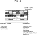
- FIG. 2 schematically shows an analysis method for selecting a growth factor combination according to one aspect.
- changes in cell proliferation, cell death (apoptosis or necrosis), cell invasion or migration, and changes in cell characteristics (stem cell capacity/differentiation capacity, signaling-path sensitivity) may be measured and determined.
- the measurement may be performed by a quantitation analysis such as colorimetry, fluorescence measurement, luminescence measurement, or an image analysis using staining such as cell sphere formation, cell morphology, cell count, cell density, invasion/migration, cell proliferation, cell death, or cell senescence.
- Apparatuses, kits and the like for performing the measurements are commercially available and may be easily used by those skilled in the art according to the manuals provided by the vendors.
- candidate groups that affect (help) the cell proliferation (z-score value> 0.8) in 70% or more cell samples may be selected as a first selection condition, that is, candidate groups having a common hit on cell proliferation
- candidate groups having a specific hit on cell proliferation (z-score value> 0.8) may be selected as the second selection condition, that is, candidate groups having specific hit on the cell proliferation.
- the candidate groups may be growth factor sets including two or more growth factors, or growth factor combination candidate groups.
- Candidate groups that have a significant effect on cell proliferation, which is confirmed through the measurements, may be registered in a separate growth factor search platform.
- signaling means that there is a statistical probability that can be said that the observed characteristic of the sample is obtained not accidentally but is the actual character of the subject, and whether the result is a significant result may be identified by methods and equations that are known in the art. In this regard, the significance level may be 5%.
- FIG. 3 is a schematic diagram illustrating a process for selecting a growth factor according to an aspect of the present disclosure.
- the process of selecting a growth factor to be included in the cell culture medium to promote cell culture can be performed.
- the selecting may include a) analysing of the components contained in the culture medium, b) identifying of information on growth factors affecting cell proliferation published in pathways readily accessible to those skilled in the art, and c) identifying of components autocrined from cells, and finally selecting the growth factors to be added to the cell culture medium.
- the cell culture may be performed using a culture medium including the combination.
- Combinations of growth factors which have been shown to be involved in cell proliferation, can also be used to determine whether they affect culture of specific cell populations such as cancer stem cells.
- the combinations of growth factors that have a significant effect on cell proliferation, which was confirmed through the measurements, may be registered in a separate growth factor search platform.
- the growth factor combinations registered in the growth factor search platform may be updated as additional data is collected, and a new condition may be added in addition to the above-described cell or growth factor classification conditions or selection conditions.
- growth factor combinations that did not show significant results may be registered in the growth factor search platform as insignificant data. Such insignificant data can be updated as additional data is collected.
- Another aspect provides a cell-customized growth factor selecting apparatus for performing the growth factor search.
- the apparatus may include, to perform a screening the cell-customized growth factor, an HTS performing apparatus having several tens to hundreds of plates (wells, chips, beads) at the same time, a measuring device capable of performing quantitative or qualitative analysis of cells, and an analytical program capable of performing hit selection, a database and a storage device, including an algorithm for associating and storing a cell type (classification) and a growth factor combination showing a change in cell proliferation rate above a certain level.
- the optimal conditions for cell culture can be efficiently selected. Accordingly, the efficiency of cell culture can be maximized.
- cancer tissues were collected after written consent from cancer patients.
- the cells were dispensed in the culture medium of 384-well plates (Neurobasal A medium, N2 Supplement (0.5X - 1X), B27 Supplement w/o vitA (0.5X -1X), L-glutamine 200 mM), and by adding single or combined addition of growth factors of EGF, FGF, HGF, IGF1, IL6, MDK, NRG1, PEDF, PDGF, PIGF, SEMA3A, SHH, TGFB1, RSPO1, WNT3A, NOG, A2M, ANGPT2, CHI3L1, FN1, LGALS1, IGFBP2, TNC, CLU, GPC3, THBS1, CST3, LAMA4, LAMC1, PTGDS, IGFBP7, COL6A1, CTSB, ICAM2, CSK, MMP7, S100A10, SELP, and Laminin, the cellular proliferative activity, morphology, and various molecular changes of cells were evaluated according to each condition
- the growth factor used for the culture-condition selection was determined according to the process specified in FIG. 3 (hereinafter, a process for screening an appropriate culture composition by measuring the cell responses on the single or combined condition of the growth factors will be referred to as GFSCAN.)
- Common hits and specific hits can be selected depending on a subject to be applied, and may be used for cell culture.
- the concentration adequacy of the growth factor combinations mounted on GFSCAN was tested with respect to 10 types of tumor spheroids. Specifically, the growth factor combinations registered in Example 1 were added to the cell culture medium at a concentration of 0.2 ng/ml to 125 ng/ml (0.2 ng/ml, 1 ng/ml, 5 ng/ml, 25 ng/ml, and 125 ng/ml) to measure the effects on the cell proliferation.
- EGF/bFGF which is a basic growth factor condition
- the effects on cell proliferation were greatest at the concentration of 5 ng/ml, and when the growth factor was further added, the cell proliferative activity was not improved.
- the additional effect according to the amount of growth factor added did not occur at the concentration of 1 ng/ml or more. Rather, in the case of PEDF and MDK, the added growth factors were found to have a negative effect on cell proliferation.
- step 1 cells having different cell-proliferation rates in three stages (step 1: 0.5-2, stage 2: 2-5, and stage 3: 5) were selected and the initial cell count of each cell was controlled to 500, 1000, and 2000, and the GFSCAN was performed.
- the obtained results were compared with results obtained under the conventional conditions.
- the initial cell count at which the difference in the cell proliferative activity after 7 days is maximized was found to be 500 (top of FIG. 5 ).
- the tumor-sphere formation ability of cells was compared in a no proliferation condition and a growth factor combination condition, wherein the no proliferation condition was verified by GFSCAN as having no influence on the cell proliferation, and the growth factor combination condition was verified by GFSCAN as helping the cell proliferation.
- the growth factor combination verified by GFSCAN as helping the cell proliferation [Neurobasal A medium, N2 Supplement(0.5X), B27 Supplement w/o vit A(0.5X), L-glutamine 200 mM(Thermo fisher Inc.), and EGF 20 ng/ml and bGFG 20 ng/ml (R & D Biosystems)] showed up to 10-fold increase in tumor-sphere formation ability. Accordingly, it was verified that growth factor conditions promoting cell proliferation and tumor-sphere formation can be obtained according to the present disclosure.
- the reproducibility of the data stored in GFSCAN was evaluated. Three experiments (repeated test cycle was one week) were repeatedly performed using randomly extracted growth factor combinations and two tumor spheroids. As a result, as shown in FIG. 7 , the correlation coefficient value, (R 2 ) of the respective experiments was 0.9898 to 0.9957. Accordingly, it was verified that the reproducibility was high.
- the cell proliferative activity was evaluated under the optimal conditions for each cell selected through GFSCAN (EGF/bFGF 5 ng/ml + SHH/PEDF 1 ng/ml) compared to the conventional growth factor conditions (EGF/bFGF 20 ng/ml).
- GFSCAN was performed on 20 tumor spheroids to determine whether there is a difference in the proliferative activity in the identical growth factor condition according to characteristics of the individual patients.
- one representative condition (EGF/bFGF 5 ng/ml + TGF-beta 1 ng/ml) was selected, and tumor spheroids with enhanced or inhibited cellular proliferative activity were selected, and GFSCAN results were verified by confirming that the cell proliferative activity was the same as GFSCAN results according to the presence or absence of TGF-beta, through respective cell experiments.
Landscapes
- Health & Medical Sciences (AREA)
- Life Sciences & Earth Sciences (AREA)
- Engineering & Computer Science (AREA)
- Biomedical Technology (AREA)
- Chemical & Material Sciences (AREA)
- Immunology (AREA)
- Physics & Mathematics (AREA)
- Molecular Biology (AREA)
- Urology & Nephrology (AREA)
- Hematology (AREA)
- General Health & Medical Sciences (AREA)
- Biochemistry (AREA)
- Biotechnology (AREA)
- General Physics & Mathematics (AREA)
- Cell Biology (AREA)
- Pathology (AREA)
- Analytical Chemistry (AREA)
- Medicinal Chemistry (AREA)
- Food Science & Technology (AREA)
- Microbiology (AREA)
- Bioinformatics & Cheminformatics (AREA)
- Tropical Medicine & Parasitology (AREA)
- Toxicology (AREA)
- Organic Chemistry (AREA)
- Biophysics (AREA)
- Theoretical Computer Science (AREA)
- Optics & Photonics (AREA)
- Zoology (AREA)
- Genetics & Genomics (AREA)
- Wood Science & Technology (AREA)
- Medical Informatics (AREA)
- Nuclear Medicine, Radiotherapy & Molecular Imaging (AREA)
- Proteomics, Peptides & Aminoacids (AREA)
- Databases & Information Systems (AREA)
- Bioethics (AREA)
- Bioinformatics & Computational Biology (AREA)
- Evolutionary Biology (AREA)
- Spectroscopy & Molecular Physics (AREA)
- General Engineering & Computer Science (AREA)
- General Chemical & Material Sciences (AREA)
Abstract
Description
- The present application claims the priority of Korean Patent Application No.
10-2017-0114696, filed on September 7, 2017 - It relates to a customized growth factor screening system for tumor spheroid culture.
- In vitro biological research requires cell procurement. However, since it is difficult to implement, in cell culture media, conditions that are exactly the same as in vivo conditions, culture conditions (particularly culture media) capable of artificially promoting cell proliferation or maintaining cells have been applied to cell culture. In this case, various culture medium compositions are required according to the characteristics of the primary tissue of the cell, the in vitro culture environment, and the purpose of the culture, and in particular, the selection of a medium composition that can maximize cell culture efficiency and reflects the characteristics of primary cells is an essential element for in vitro cell culture.
- In particular, as unmet clinical needs with respect to established cell lines emerge, the number of cases in which cells (tumor spheroids) collected from patients' disease sites are cultured and the cultured cells are used for patient-specific treatment and disease mechanism research has increased. Accordingly, the culturing of tumor spheroids has emerged as a necessary requirement. However, since there are differences in molecular genetic characteristics of individual patients and characteristics of primary tissues, it is difficult to select culture conditions in which cells isolated from the patient (particularly disease-specific cells only) can be grown/maintained. Under general culture conditions, 1) it is difficult to control the proliferation of cells other than the disease-specific cells, and thus, disease-specific cells in the cultured cells may not be cultured, and 2) cells of a specific group in the primary tissue only may be selected and cultured.
- Therefore, there is a need to develop a method of screening culture conditions that can minimize the impact of these problems and maximize the culture efficiency of tumor spheroids.
- Provided is a method of screening a customized growth factor combination for cell culture, the method including: adding at least one growth factor and cells isolated from a subject to a culture medium; culturing the cells; and measuring the cell proliferative activity of the cells.
- An aspect provides a method of selecting a customized growth factor combination for cell culture, the method including: adding at least one growth factor and cells isolated from a subject to a culture medium; culturing the cells; and measuring the cell proliferative activity of the cells.
-
FIG. 1 is a schematic diagram showing a method of screening a customized growth factor combination for cell culture according to an aspect of the present disclosure. - First, any cell to be cultured may be selected. The cells may refer to tumor spheroids that need to be utilized for the purpose of medical research and treatment, commercialized cells that need to increase culture efficiency, or all cells that require cell culture. The cells may be sorted according to certain conditions as needed. The conditions may be the tissue from which cells are derived, the type of disease, the severity of the disease (for example, in the case of cancer, tumor grade, genotype, or other molecular-pathologic features).
- According to the selected cells, the growth factor candidate groups to be added to the cell culture medium are classified from the omics-based database such as genome, transcriptome, and proteome. The database may be data which is published in a pathway accessible to those of ordinary skill in the art (for example, protein databases, http://www.proteinatlas.org/, http://www.uniprot.org/help/uniprotkb) or data accumulated by the skilled person in the laboratory. At this time, the selected candidate groups may be a growth factor set including two or more growth factors.
- The selected candidate groups may be added simultaneously to dozens or thousands of media (wells, chips, beads) using a high-throughput screening (HTS) apparatus and method, and subsequent culture and analysis may be performed using a manual or automatic system. In this case, one or more cell types may be used, and at the same time, one or more selected growth factor candidate groups may be used. The cells or selected growth factor candidate groups may be dispensed into the medium as intended by one of ordinary skill in the art who performs the present procedures, or may be randomly dispensed into the tens to thousands of mediums by using an HTS apparatus. When the cell proliferative activity of a particular medium is changed compared to the control, the cells, growth factors and other substances included in the medium are analyzed using the HTS apparatus. One skilled in the art may perform HTS including the procedures described above by using commercialized HTS apparatus and method. Such HTS appratuses may include plates, cell and growth factor dispensers, constant-temperature incubators, automated delivery devices, image analyzers, data storage devices, and data analysis devices, and analysis programs. The degree of influence on cell proliferation can be performed using a quantitation analysis or an image analysis.
-
FIG. 2 schematically shows an analysis method for selecting a growth factor combination according to one aspect. In order to measure the degree of influence on cell proliferation, changes in cell proliferation, cell death (apoptosis or necrosis), cell invasion or migration, and changes in cell characteristics (stem cell capacity/differentiation capacity, signaling-path sensitivity) may be measured and determined. The measurement may be performed by a quantitation analysis such as colorimetry, fluorescence measurement, luminescence measurement, or an image analysis using staining such as cell sphere formation, cell morphology, cell count, cell density, invasion/migration, cell proliferation, cell death, or cell senescence. Apparatuses, kits and the like for performing the measurements are commercially available and may be easily used by those skilled in the art according to the manuals provided by the vendors. - In this regard, candidate groups that affect (help) the cell proliferation (z-score value> 0.8) in 70% or more cell samples, may be selected as a first selection condition, that is, candidate groups having a common hit on cell proliferation Also, even when less than 70%, candidate groups having a specific hit on cell proliferation (z-score value> 0.8) may be selected as the second selection condition, that is, candidate groups having specific hit on the cell proliferation. The candidate groups may be growth factor sets including two or more growth factors, or growth factor combination candidate groups. Candidate groups that have a significant effect on cell proliferation, which is confirmed through the measurements, may be registered in a separate growth factor search platform. The term "significance" or "statistical significance" means that there is a statistical probability that can be said that the observed characteristic of the sample is obtained not accidentally but is the actual character of the subject, and whether the result is a significant result may be identified by methods and equations that are known in the art. In this regard, the significance level may be 5%.
-
FIG. 3 is a schematic diagram illustrating a process for selecting a growth factor according to an aspect of the present disclosure. - Once the culture medium to help the culture of the cell is identified, the process of selecting a growth factor to be included in the cell culture medium to promote cell culture can be performed. The selecting may include a) analysing of the components contained in the culture medium, b) identifying of information on growth factors affecting cell proliferation published in pathways readily accessible to those skilled in the art, and c) identifying of components autocrined from cells, and finally selecting the growth factors to be added to the cell culture medium.
- As described above, when a combination is obtained of desired growth factors selected from a) a growth factor added to the culture medium, b) a known growth factor, and c) an autocrined factor, the cell culture may be performed using a culture medium including the combination. Combinations of growth factors, which have been shown to be involved in cell proliferation, can also be used to determine whether they affect culture of specific cell populations such as cancer stem cells. The combinations of growth factors that have a significant effect on cell proliferation, which was confirmed through the measurements, may be registered in a separate growth factor search platform.
- As described above, the growth factor combinations registered in the growth factor search platform may be updated as additional data is collected, and a new condition may be added in addition to the above-described cell or growth factor classification conditions or selection conditions. In addition, growth factor combinations that did not show significant results may be registered in the growth factor search platform as insignificant data. Such insignificant data can be updated as additional data is collected.
- Another aspect provides a cell-customized growth factor selecting apparatus for performing the growth factor search.
- The apparatus may include, to perform a screening the cell-customized growth factor, an HTS performing apparatus having several tens to hundreds of plates (wells, chips, beads) at the same time, a measuring device capable of performing quantitative or qualitative analysis of cells, and an analytical program capable of performing hit selection, a database and a storage device, including an algorithm for associating and storing a cell type (classification) and a growth factor combination showing a change in cell proliferation rate above a certain level.
- According to a method of screening a customized growth factor combination for cell culture according to one aspect, the optimal conditions for cell culture can be efficiently selected. Accordingly, the efficiency of cell culture can be maximized.
-
-
FIG. 1 is a schematic diagram showing a method of screening a customized growth factor combination for cell culture according to an aspect of the present disclosure. -
FIG. 2 schematically shows an analysis method for selecting a growth factor combination according to one aspect. -
FIG. 3 is a schematic diagram illustrating a process for selecting a growth factor according to an aspect of the present disclosure. -
FIG. 4 shows test results according to concentrations of selected growth factors according to one aspect. -
FIG. 5 shows the results of testing the cell count required for growth factor screening. -
FIG. 6 shows the test results of measuring the tumor-sphere formation ability of cells to verify the selected growth factor conditions according to one aspect. -
FIG. 7 illustrates the reproducibility of a growth factor screening method according to an aspect of the present disclosure. -
FIG. 8A shows that selected growth factors statistically significantly increase cell proliferation, according to one aspect. -
FIG. 8B shows that the effects of selected growth factors are confirmed by tumor-sphere formation ability results, according to an aspect. -
FIG. 9A illustrates selecting cells that are differently affected in the same growth factor conditions, by using a growth factor screening method according to one aspect of the present disclosure. -
FIG. 9B shows that the cells affected differently for the same growth factor conditions experience changes in intracellular signal transduction pathways. - Hereinafter, the present disclosure will be described in more detail with reference to the following examples. However, these examples are provided for illustratively describing present disclosure, and the scope of present disclosure is not limited to these examples.
- According to the guidelines of the Institutional Review Board of Samsung Medical Center, cancer tissues were collected after written consent from cancer patients.
- After separating the single cells from the cancer tissues, the cells were dispensed in the culture medium of 384-well plates (Neurobasal A medium, N2 Supplement (0.5X - 1X), B27 Supplement w/o vitA (0.5X -1X), L-
glutamine 200 mM), and by adding single or combined addition of growth factors of EGF, FGF, HGF, IGF1, IL6, MDK, NRG1, PEDF, PDGF, PIGF, SEMA3A, SHH, TGFB1, RSPO1, WNT3A, NOG, A2M, ANGPT2, CHI3L1, FN1, LGALS1, IGFBP2, TNC, CLU, GPC3, THBS1, CST3, LAMA4, LAMC1, PTGDS, IGFBP7, COL6A1, CTSB, ICAM2, CSK, MMP7, S100A10, SELP, and Laminin, the cellular proliferative activity, morphology, and various molecular changes of cells were evaluated according to each condition. At this time, the growth factor used for the culture-condition selection was determined according to the process specified inFIG. 3 (hereinafter, a process for screening an appropriate culture composition by measuring the cell responses on the single or combined condition of the growth factors will be referred to as GFSCAN.) - The growth factor single/combination that causes cell proliferation or tumor-sphere formation ability or improvement of early cell characteristic reflection ability in 70% or more cell samples of the GFSCAN results, was selected as a common hit, and growth factor combinations, which do not correspond to the common hit, but help cell proliferation, tumor-sphere formation, or the improvement of cell characteristic reflection ability in specific cells, were classified as cell-specific hit. Common hits and specific hits can be selected depending on a subject to be applied, and may be used for cell culture.
- The concentration adequacy of the growth factor combinations mounted on GFSCAN was tested with respect to 10 types of tumor spheroids. Specifically, the growth factor combinations registered in Example 1 were added to the cell culture medium at a concentration of 0.2 ng/ml to 125 ng/ml (0.2 ng/ml, 1 ng/ml, 5 ng/ml, 25 ng/ml, and 125 ng/ml) to measure the effects on the cell proliferation.
- As shown in
FIG. 4 , in the case of EGF/bFGF, which is a basic growth factor condition, on average, the effects on cell proliferation were greatest at the concentration of 5 ng/ml, and when the growth factor was further added, the cell proliferative activity was not improved. In addition, regarding the improvement of proliferative activity according to the concentration of a growth factor in PEDF, NRG, and MDK among the growth factors mounted on the GFSCAN, the additional effect according to the amount of growth factor added did not occur at the concentration of 1 ng/ml or more. Rather, in the case of PEDF and MDK, the added growth factors were found to have a negative effect on cell proliferation. - An experiment was conducted to determine the cell count initially dispensed at the time of growth-factor screening. 500 to 2000 cells (500, 1000, or 2000 cells) were dispensed to a plate, and 5 ng/ml EGF/bFGF or any growth factor combination (1 ng/ml) registered in GFSCAN was added thereto, wherein these concentrations were set as a reference concentration. The cell count of grown cells after 7 days was measured, and the proliferative activity was defined as a value obtained by dividing cell viability after 7 days by cell viability on 0 day, and calculated. In order to confirm the effect of the proliferation rate of the cell itself on the GFSCAN results and the optimization conditions, cells having different cell-proliferation rates in three stages (step 1: 0.5-2, stage 2: 2-5, and stage 3: 5) were selected and the initial cell count of each cell was controlled to 500, 1000, and 2000, and the GFSCAN was performed. The obtained results were compared with results obtained under the conventional conditions. As shown in the upper table of
FIG. 5 , it was confirmed that there were differences in 1) the proliferative activity of cells, and 2) the difference in the cell proliferative activity after 7 days according to the initial cell count. In all cell proliferative activity conditions, commonly, the initial cell count at which the difference in the cell proliferative activity after 7 days is maximized was found to be 500 (top ofFIG. 5 ). - In addition, after verifying the proper concentration condition of the growth factor mounted on GFSCAN at the initial cell count of 500 (1X: reference concentration; 2X: 2 times the reference concentration; and 4X: 4 times the reference concentration), it was confirmed that the proliferative activity was maximized at the reference concentration (1X) condition or reference concentration (2X) condition (bottom of
FIG. 5 ). - To verify the culture conditions selected through GFSCAN, it was determined whether growth factor combinations were appropriate data with actual cell proliferation effects. To this end, after dispensing a certain number of cells (1, 5, 10, 20, 50, 100, 200, 400) of three tumor spheroids, the tumor-sphere formation ability of cells was compared in a no proliferation condition and a growth factor combination condition, wherein the no proliferation condition was verified by GFSCAN as having no influence on the cell proliferation, and the growth factor combination condition was verified by GFSCAN as helping the cell proliferation.
- As a result, as shown in
FIG. 6 , compared to the no proliferation condition, the growth factor combination verified by GFSCAN as helping the cell proliferation [Neurobasal A medium, N2 Supplement(0.5X), B27 Supplement w/o vit A(0.5X), L-glutamine 200 mM(Thermo fisher Inc.), andEGF 20 ng/mℓ andbGFG 20 ng/mℓ (R & D Biosystems)] showed up to 10-fold increase in tumor-sphere formation ability. Accordingly, it was verified that growth factor conditions promoting cell proliferation and tumor-sphere formation can be obtained according to the present disclosure. - The reproducibility of the data stored in GFSCAN was evaluated. Three experiments (repeated test cycle was one week) were repeatedly performed using randomly extracted growth factor combinations and two tumor spheroids. As a result, as shown in
FIG. 7 , the correlation coefficient value, (R2) of the respective experiments was 0.9898 to 0.9957. Accordingly, it was verified that the reproducibility was high. - By applying GFSCAN to 20 types of brain tumor spheroids, the cell proliferative activity was evaluated under the optimal conditions for each cell selected through GFSCAN (EGF/
bFGF 5 ng/ml + SHH/PEDF 1 ng/ml) compared to the conventional growth factor conditions (EGF/bFGF 20 ng/ml). - As shown in
FIG. 8 , it was confirmed that the growth factor combinations of the GFSCAN data significantly increased the cell proliferative activity (a). At this time, with respect to two tumor spheroids, tumor culture proceeded under the conventional growth factor condition and the condition selected through GFSCAN to confirm cell morphology change and tumor-sphere formation ability. It was confirmed that in the novel condition selected through GFSCAN, cell proliferation or tumor-sphere formation ability was improved compared to the conventional condition. - GFSCAN was performed on 20 tumor spheroids to determine whether there is a difference in the proliferative activity in the identical growth factor condition according to characteristics of the individual patients. Among the GFSCAN conditions, one representative condition (EGF/
bFGF 5 ng/ml + TGF-beta 1 ng/ml) was selected, and tumor spheroids with enhanced or inhibited cellular proliferative activity were selected, and GFSCAN results were verified by confirming that the cell proliferative activity was the same as GFSCAN results according to the presence or absence of TGF-beta, through respective cell experiments. - Also, when cultured in cells with improved cell proliferative activity, when TGF-beta was added, signal transduction pathways associated with the promotion of proliferation were activated, or related factors were overexpressed, and the pathways of signal transduction suppressing apoptosis and cell senescence or transcription and related molecules were found to be decreased. (
FIG. 9B ).
Claims (21)
- A method of screening a customized growth factor combination for cell culture, the method comprising:adding at least one growth factor and cells isolated from a subject to a culture medium;culturing the cells; andmeasuring proliferative activity of the cells.
- The method of claim 1, wherein the cells are isolated from a disease tissue.
- The method of claim 2, further comprising classifying the cells according to at least one condition selected from derived subject, derived tissue, disease type, disease severity, or genetic information.
- The method of claim 3, wherein the growth factor is selected from a list predicted to increase the proliferation rate of cells from an omics-based database according to the classifying of the cells.
- The method of claim 1, wherein the culturing and the measuring are performed by high-throughput screening (HTS).
- The method of claim 1, wherein the measuring is performed by quantitative analysis and image analysis.
- The method of claim 6, wherein the quantitative analysis is performed by using any one of colorimetry, fluorescence measurement, or luminescence measurement.
- The method of claim 6, wherein the image analysis is analysis of any one of cell sphere formation, cell morphology, cell count, cell density, invasion/migration, cell proliferation, cell death, or cell senescence.
- The method of claim 1, further comprising, in the measuring, specifying a medium of which cell proliferative activity is changed compared to a control(z-score value > 0.8).
- The method of claim 9, further comprising, in the measuring of the cell proliferative activity, selecting, as a first selection condition, growth factors that affect cell proliferation (z-score value> 0.8) in 70% or more of cell samples.
- The method of claim 9, further comprising, in the measuring of the cell proliferative activity, selecting, as a second selection condition, growth factors that affect cell proliferation (z-score value> 0.8) in less than 70% of cell samples.
- The method of claim 11, further comprising selecting a growth factor, which is selected by the second selection condition, as a growth factor that has a specific effect with respect to a corresponding cell sample.
- The method of claim 9, further comprising analyzing a growth factor included in the medium.
- The method of claim 13, further comprising storing a growth factor included in the medium as a selected growth factor in a data storage.
- The method of claim 14, further comprising storing a growth factor included in media other than the medium as an unselected growth factor in the data storage.
- A method of screening a cell-customized cell culture condition, the method comprising:a) adding at least one growth factor and cells isolated from a subject to a culture medium;b) culturing the cells;c) measuring cell proliferative activity of the cells;d) specifying a medium of which the cell proliferative activity is changed compared to a control(z-score value > 0.8) in the measuring,;e) storing a growth factor included in the medium as a selected growth factor in a data storage;f) repeatedly performing the processes of a) through d) using the growth factor stored in the process e); andg) updating pre-stored data using data obtained in process f).
- The method of claim 16, wherein the process f) is performed by adding growth factors other than the growth factor of process a).
- The method of claim 16, wherein the process f) is performed by adding cells other than the cells of process a).
- The method of claim 16, wherein the measuring is performed by quantitative analysis and image analysis.
- The method of claim 19, wherein the quantitative analysis is performed by using any one of colorimetry, fluorescence measurement, or luminescence measurement.
- The method of claim 19, wherein the image analysis is analysis of any one of cell sphere formation, cell morphology, cell count, cell density, invasion/migration, cell proliferation, cell death, or cell senescence.
Applications Claiming Priority (2)
| Application Number | Priority Date | Filing Date | Title |
|---|---|---|---|
| KR20170114696 | 2017-09-07 | ||
| PCT/KR2018/010458 WO2019050314A1 (en) | 2017-09-07 | 2018-09-07 | Customized growth factor screening system for cell culture |
Publications (2)
| Publication Number | Publication Date |
|---|---|
| EP3680661A1 true EP3680661A1 (en) | 2020-07-15 |
| EP3680661A4 EP3680661A4 (en) | 2021-05-26 |
Family
ID=65634301
Family Applications (1)
| Application Number | Title | Priority Date | Filing Date |
|---|---|---|---|
| EP18854428.2A Withdrawn EP3680661A4 (en) | 2017-09-07 | 2018-09-07 | ADAPTED GROWTH FACTOR SCREENING SYSTEM FOR CELL CULTURES |
Country Status (4)
| Country | Link |
|---|---|
| US (1) | US20210063379A1 (en) |
| EP (1) | EP3680661A4 (en) |
| KR (1) | KR102269072B1 (en) |
| WO (1) | WO2019050314A1 (en) |
Families Citing this family (4)
| Publication number | Priority date | Publication date | Assignee | Title |
|---|---|---|---|---|
| KR102785329B1 (en) * | 2023-04-07 | 2025-03-25 | (주)그래디언트 바이오컨버전스 | Culture medium condition output device, method for outputting culture medium conditions of tumor cells, and computer program |
| KR20240177497A (en) | 2023-06-20 | 2024-12-27 | (주)엑셀세라퓨틱스 | A platform system for designing medium formulations tailored to cell-specific characteristics |
| KR102763225B1 (en) * | 2023-09-26 | 2025-02-07 | 주식회사 그래디언트바이오컨버전스 | Device for determining optical culture medium combination, method and computer program for determining the optimal combination by calculating survival rates for culture medium combinations |
| CN120081923B (en) * | 2025-04-28 | 2025-08-01 | 四川华德生物工程有限公司 | Intelligent optimization method and system for animal-derived epidermal growth factor |
Family Cites Families (5)
| Publication number | Priority date | Publication date | Assignee | Title |
|---|---|---|---|---|
| KR100463714B1 (en) * | 2002-06-27 | 2004-12-29 | 한국생명공학연구원 | Method for high throughput screening of plant growth regulator |
| EP3699915A1 (en) * | 2010-04-29 | 2020-08-26 | The Regents of The University of California | Pathway recognition algorithm using data integration on genomic models (paradigm) |
| CN105358707B (en) * | 2013-06-28 | 2019-06-14 | 株式会社钟化 | Method for screening pluripotent stem cell growth-promoting factors |
| KR102418824B1 (en) * | 2013-10-14 | 2022-07-11 | 아레스 트레이딩 에스.아. | New medium for high performance mammalian fed-batch cultures |
| KR20170114696A (en) | 2016-04-06 | 2017-10-16 | 현대자동차주식회사 | Insert structure of mount nut |
-
2018
- 2018-09-07 EP EP18854428.2A patent/EP3680661A4/en not_active Withdrawn
- 2018-09-07 WO PCT/KR2018/010458 patent/WO2019050314A1/en not_active Ceased
- 2018-09-07 KR KR1020180107381A patent/KR102269072B1/en active Active
- 2018-09-07 US US16/644,267 patent/US20210063379A1/en not_active Abandoned
Also Published As
| Publication number | Publication date |
|---|---|
| KR102269072B1 (en) | 2021-06-24 |
| US20210063379A1 (en) | 2021-03-04 |
| KR20190027768A (en) | 2019-03-15 |
| WO2019050314A1 (en) | 2019-03-14 |
| EP3680661A4 (en) | 2021-05-26 |
Similar Documents
| Publication | Publication Date | Title |
|---|---|---|
| US20210063379A1 (en) | Customized Growth Factor Screening System for Cell Culture | |
| Zinnall et al. | Micropapillary urothelial carcinoma: evaluation of HER2 status and immunohistochemical characterization of the molecular subtype | |
| Read et al. | Identification of CD15 as a marker for tumor-propagating cells in a mouse model of medulloblastoma | |
| Lanza et al. | Immunohistochemical pattern of MLH1/MSH2 expression is related to clinical and pathological features in colorectal adenocarcinomas with microsatellite instability | |
| US9766249B2 (en) | Compositions and methods for prediction of drug sensitivity, resistance, and disease progression | |
| Peppercorn et al. | Molecular subtypes in breast cancer evaluation and management: divide and conquer | |
| Lotan et al. | PTEN loss is associated with upgrading of prostate cancer from biopsy to radical prostatectomy | |
| Aleskandarany et al. | Epithelial mesenchymal transition in early invasive breast cancer: an immunohistochemical and reverse phase protein array study | |
| Perez-Rodriguez et al. | Investigation of somatic CNVs in brains of synucleinopathy cases using targeted SNCA analysis and single cell sequencing | |
| JP2011528442A (en) | Prostate cancer related signs and PCDETERMINANT and methods of use thereof | |
| Deniz et al. | Expression of growth factors and structural proteins in chordomas: basic fibroblast growth factor, transforming growth factor α, and fibronectin are correlated with recurrence | |
| Hashmi et al. | Molecular subtypes of breast cancer in South Asian population by immunohistochemical profile and Her2neu gene amplification by FISH technique: association with other clinicopathologic parameters | |
| JP2016523527A (en) | Assays, methods and kits for predicting prognosis of cancer patients and for personalized treatment methods, analyzing sensitivity and resistance to anti-cancer drugs | |
| Ma et al. | A novel 96well-formatted micro-gap plate enabling drug response profiling on primary tumour samples | |
| US20140206559A1 (en) | Assay for metastatic potential of tumor cells | |
| Qi et al. | Reliable gene expression profiling from small and hematoxylin and eosin–stained clinical formalin-fixed, paraffin-embedded specimens using the HTG EdgeSeq Platform | |
| Cui et al. | Establishment of human pituitary neuroendocrine tumor derived organoid and its pilot application for drug screening | |
| Torres et al. | Comprehensive determination of prostate tumor ETS gene status in clinical samples using the CLIA decipher assay | |
| Slembrouck et al. | Decentralization of next-generation RNA sequencing-based MammaPrint® and BluePrint® kit at University Hospitals Leuven and Curie Institute Paris | |
| Izar et al. | GILA, a replacement for the soft‐agar assay that permits high‐throughput drug and genetic screens for cellular transformation | |
| Bartlett | Biomarkers and patient selection for PI3K/Akt/mTOR targeted therapies: current status and future directions | |
| EP1704416A2 (en) | Protein expression profiling and breast cancer prognosis | |
| WO2013079188A1 (en) | Methods for the diagnosis, the determination of the grade of a solid tumor and the prognosis of a subject suffering from cancer | |
| Figarella-Branger et al. | Histomolecular classification of adult diffuse gliomas: the diagnostic value of immunohistochemical markers | |
| Tabarestani et al. | Detection of gene amplification by multiplex ligation-dependent probe amplification in comparison with in situ hybridization and immunohistochemistry |
Legal Events
| Date | Code | Title | Description |
|---|---|---|---|
| STAA | Information on the status of an ep patent application or granted ep patent |
Free format text: STATUS: THE INTERNATIONAL PUBLICATION HAS BEEN MADE |
|
| PUAI | Public reference made under article 153(3) epc to a published international application that has entered the european phase |
Free format text: ORIGINAL CODE: 0009012 |
|
| STAA | Information on the status of an ep patent application or granted ep patent |
Free format text: STATUS: REQUEST FOR EXAMINATION WAS MADE |
|
| 17P | Request for examination filed |
Effective date: 20200313 |
|
| AK | Designated contracting states |
Kind code of ref document: A1 Designated state(s): AL AT BE BG CH CY CZ DE DK EE ES FI FR GB GR HR HU IE IS IT LI LT LU LV MC MK MT NL NO PL PT RO RS SE SI SK SM TR |
|
| AX | Request for extension of the european patent |
Extension state: BA ME |
|
| DAV | Request for validation of the european patent (deleted) | ||
| DAX | Request for extension of the european patent (deleted) | ||
| A4 | Supplementary search report drawn up and despatched |
Effective date: 20210429 |
|
| RIC1 | Information provided on ipc code assigned before grant |
Ipc: G01N 33/50 20060101AFI20210422BHEP Ipc: G01N 33/483 20060101ALI20210422BHEP Ipc: G01N 33/68 20060101ALI20210422BHEP |
|
| STAA | Information on the status of an ep patent application or granted ep patent |
Free format text: STATUS: EXAMINATION IS IN PROGRESS |
|
| 17Q | First examination report despatched |
Effective date: 20240924 |
|
| STAA | Information on the status of an ep patent application or granted ep patent |
Free format text: STATUS: THE APPLICATION IS DEEMED TO BE WITHDRAWN |
|
| 18D | Application deemed to be withdrawn |
Effective date: 20250125 |