EP3663311A1 - Chimeric protein switch for the optogenetic control of amyloidogenesis - Google Patents
Chimeric protein switch for the optogenetic control of amyloidogenesis Download PDFInfo
- Publication number
- EP3663311A1 EP3663311A1 EP18382882.1A EP18382882A EP3663311A1 EP 3663311 A1 EP3663311 A1 EP 3663311A1 EP 18382882 A EP18382882 A EP 18382882A EP 3663311 A1 EP3663311 A1 EP 3663311A1
- Authority
- EP
- European Patent Office
- Prior art keywords
- cell
- acid sequence
- fusion polypeptide
- protein
- repa
- Prior art date
- Legal status (The legal status is an assumption and is not a legal conclusion. Google has not performed a legal analysis and makes no representation as to the accuracy of the status listed.)
- Withdrawn
Links
- 230000003941 amyloidogenesis Effects 0.000 title description 16
- 108020001507 fusion proteins Proteins 0.000 title description 11
- 102000037865 fusion proteins Human genes 0.000 title description 11
- 108090000765 processed proteins & peptides Proteins 0.000 claims abstract description 86
- 102000004196 processed proteins & peptides Human genes 0.000 claims abstract description 80
- 229920001184 polypeptide Polymers 0.000 claims abstract description 78
- 230000004927 fusion Effects 0.000 claims abstract description 36
- 125000003275 alpha amino acid group Chemical group 0.000 claims abstract description 28
- 230000001580 bacterial effect Effects 0.000 claims abstract description 22
- 230000015572 biosynthetic process Effects 0.000 claims abstract description 18
- 231100000433 cytotoxic Toxicity 0.000 claims abstract description 15
- 230000001472 cytotoxic effect Effects 0.000 claims abstract description 15
- 230000001939 inductive effect Effects 0.000 claims abstract description 7
- 210000004027 cell Anatomy 0.000 claims description 100
- 108090000623 proteins and genes Proteins 0.000 claims description 71
- 102000004169 proteins and genes Human genes 0.000 claims description 63
- 150000007523 nucleic acids Chemical group 0.000 claims description 37
- 230000002068 genetic effect Effects 0.000 claims description 35
- 108091028043 Nucleic acid sequence Proteins 0.000 claims description 30
- 238000000338 in vitro Methods 0.000 claims description 27
- 239000003814 drug Substances 0.000 claims description 14
- 238000000034 method Methods 0.000 claims description 12
- 208000015181 infectious disease Diseases 0.000 claims description 11
- 210000004899 c-terminal region Anatomy 0.000 claims description 9
- 239000000523 sample Substances 0.000 claims description 9
- 238000004519 manufacturing process Methods 0.000 claims description 7
- 208000035143 Bacterial infection Diseases 0.000 claims description 6
- 208000022362 bacterial infectious disease Diseases 0.000 claims description 6
- 108010054624 red fluorescent protein Proteins 0.000 claims description 5
- 238000011282 treatment Methods 0.000 claims description 5
- 230000000813 microbial effect Effects 0.000 claims description 4
- 230000002265 prevention Effects 0.000 claims description 4
- 210000001236 prokaryotic cell Anatomy 0.000 claims description 4
- FWMNVWWHGCHHJJ-SKKKGAJSSA-N 4-amino-1-[(2r)-6-amino-2-[[(2r)-2-[[(2r)-2-[[(2r)-2-amino-3-phenylpropanoyl]amino]-3-phenylpropanoyl]amino]-4-methylpentanoyl]amino]hexanoyl]piperidine-4-carboxylic acid Chemical group C([C@H](C(=O)N[C@H](CC(C)C)C(=O)N[C@H](CCCCN)C(=O)N1CCC(N)(CC1)C(O)=O)NC(=O)[C@H](N)CC=1C=CC=CC=1)C1=CC=CC=C1 FWMNVWWHGCHHJJ-SKKKGAJSSA-N 0.000 claims description 2
- 238000005286 illumination Methods 0.000 abstract description 35
- 241000894006 Bacteria Species 0.000 abstract description 17
- 102000009091 Amyloidogenic Proteins Human genes 0.000 abstract description 13
- 108010048112 Amyloidogenic Proteins Proteins 0.000 abstract description 13
- 230000003942 amyloidogenic effect Effects 0.000 abstract description 12
- 239000000017 hydrogel Substances 0.000 abstract description 11
- 108010020577 Phototropins Proteins 0.000 abstract description 9
- 239000002245 particle Substances 0.000 abstract description 7
- 238000004220 aggregation Methods 0.000 abstract description 5
- 239000012636 effector Substances 0.000 abstract description 5
- 230000002147 killing effect Effects 0.000 abstract description 5
- 230000002776 aggregation Effects 0.000 abstract description 4
- 235000018102 proteins Nutrition 0.000 description 59
- 230000014509 gene expression Effects 0.000 description 33
- 102220038185 rs144829356 Human genes 0.000 description 18
- 241000588724 Escherichia coli Species 0.000 description 16
- 239000000872 buffer Substances 0.000 description 16
- 206010002022 amyloidosis Diseases 0.000 description 15
- 241000282414 Homo sapiens Species 0.000 description 12
- 239000013604 expression vector Substances 0.000 description 12
- RAXXELZNTBOGNW-UHFFFAOYSA-N imidazole Natural products C1=CNC=N1 RAXXELZNTBOGNW-UHFFFAOYSA-N 0.000 description 12
- 239000013612 plasmid Substances 0.000 description 12
- 238000001727 in vivo Methods 0.000 description 11
- 239000000203 mixture Substances 0.000 description 11
- 239000012071 phase Substances 0.000 description 11
- 108091033319 polynucleotide Proteins 0.000 description 11
- 102000040430 polynucleotide Human genes 0.000 description 11
- 239000002157 polynucleotide Substances 0.000 description 11
- 201000010099 disease Diseases 0.000 description 10
- 208000037265 diseases, disorders, signs and symptoms Diseases 0.000 description 10
- 230000001575 pathological effect Effects 0.000 description 10
- PEDCQBHIVMGVHV-UHFFFAOYSA-N Glycerine Chemical compound OCC(O)CO PEDCQBHIVMGVHV-UHFFFAOYSA-N 0.000 description 9
- 238000010521 absorption reaction Methods 0.000 description 9
- IQFVPQOLBLOTPF-HKXUKFGYSA-L congo red Chemical compound [Na+].[Na+].C1=CC=CC2=C(N)C(/N=N/C3=CC=C(C=C3)C3=CC=C(C=C3)/N=N/C3=C(C4=CC=CC=C4C(=C3)S([O-])(=O)=O)N)=CC(S([O-])(=O)=O)=C21 IQFVPQOLBLOTPF-HKXUKFGYSA-L 0.000 description 9
- 102000039446 nucleic acids Human genes 0.000 description 9
- 108020004707 nucleic acids Proteins 0.000 description 9
- 238000001542 size-exclusion chromatography Methods 0.000 description 9
- FAPWRFPIFSIZLT-UHFFFAOYSA-M Sodium chloride Chemical compound [Na+].[Cl-] FAPWRFPIFSIZLT-UHFFFAOYSA-M 0.000 description 8
- 238000004458 analytical method Methods 0.000 description 8
- 238000003776 cleavage reaction Methods 0.000 description 8
- 230000017854 proteolysis Effects 0.000 description 8
- 230000007017 scission Effects 0.000 description 8
- 238000004062 sedimentation Methods 0.000 description 8
- 238000001228 spectrum Methods 0.000 description 8
- 230000007704 transition Effects 0.000 description 8
- 108020004414 DNA Proteins 0.000 description 7
- 241000196324 Embryophyta Species 0.000 description 7
- 230000000694 effects Effects 0.000 description 7
- 238000011534 incubation Methods 0.000 description 7
- 230000035772 mutation Effects 0.000 description 7
- 238000000746 purification Methods 0.000 description 7
- 230000001105 regulatory effect Effects 0.000 description 7
- 241000894007 species Species 0.000 description 7
- 108090000317 Chymotrypsin Proteins 0.000 description 6
- 108091005804 Peptidases Proteins 0.000 description 6
- HEMHJVSKTPXQMS-UHFFFAOYSA-M Sodium hydroxide Chemical compound [OH-].[Na+] HEMHJVSKTPXQMS-UHFFFAOYSA-M 0.000 description 6
- 235000001014 amino acid Nutrition 0.000 description 6
- 229960002376 chymotrypsin Drugs 0.000 description 6
- 238000002474 experimental method Methods 0.000 description 6
- BPHPUYQFMNQIOC-NXRLNHOXSA-N isopropyl beta-D-thiogalactopyranoside Chemical compound CC(C)S[C@@H]1O[C@H](CO)[C@H](O)[C@H](O)[C@H]1O BPHPUYQFMNQIOC-NXRLNHOXSA-N 0.000 description 6
- 238000010899 nucleation Methods 0.000 description 6
- 239000002773 nucleotide Substances 0.000 description 6
- 125000003729 nucleotide group Chemical group 0.000 description 6
- 239000000126 substance Substances 0.000 description 6
- 239000006228 supernatant Substances 0.000 description 6
- 238000012360 testing method Methods 0.000 description 6
- 241001515965 unidentified phage Species 0.000 description 6
- 241001465754 Metazoa Species 0.000 description 5
- 102000035195 Peptidases Human genes 0.000 description 5
- 239000004365 Protease Substances 0.000 description 5
- 238000006243 chemical reaction Methods 0.000 description 5
- 238000013461 design Methods 0.000 description 5
- FVTCRASFADXXNN-SCRDCRAPSA-N flavin mononucleotide Chemical compound OP(=O)(O)OC[C@@H](O)[C@@H](O)[C@@H](O)CN1C=2C=C(C)C(C)=CC=2N=C2C1=NC(=O)NC2=O FVTCRASFADXXNN-SCRDCRAPSA-N 0.000 description 5
- 229940013640 flavin mononucleotide Drugs 0.000 description 5
- FVTCRASFADXXNN-UHFFFAOYSA-N flavin mononucleotide Natural products OP(=O)(O)OCC(O)C(O)C(O)CN1C=2C=C(C)C(C)=CC=2N=C2C1=NC(=O)NC2=O FVTCRASFADXXNN-UHFFFAOYSA-N 0.000 description 5
- 239000011768 flavin mononucleotide Substances 0.000 description 5
- 239000012634 fragment Substances 0.000 description 5
- 230000003834 intracellular effect Effects 0.000 description 5
- 239000007788 liquid Substances 0.000 description 5
- 239000012528 membrane Substances 0.000 description 5
- 108020004999 messenger RNA Proteins 0.000 description 5
- 239000008188 pellet Substances 0.000 description 5
- 230000002829 reductive effect Effects 0.000 description 5
- 230000004044 response Effects 0.000 description 5
- 235000019231 riboflavin-5'-phosphate Nutrition 0.000 description 5
- 238000002415 sodium dodecyl sulfate polyacrylamide gel electrophoresis Methods 0.000 description 5
- 238000013518 transcription Methods 0.000 description 5
- 230000035897 transcription Effects 0.000 description 5
- 230000002103 transcriptional effect Effects 0.000 description 5
- 238000004627 transmission electron microscopy Methods 0.000 description 5
- 102000004190 Enzymes Human genes 0.000 description 4
- 108090000790 Enzymes Proteins 0.000 description 4
- LFQSCWFLJHTTHZ-UHFFFAOYSA-N Ethanol Chemical compound CCO LFQSCWFLJHTTHZ-UHFFFAOYSA-N 0.000 description 4
- 108020002230 Pancreatic Ribonuclease Proteins 0.000 description 4
- 102000005891 Pancreatic ribonuclease Human genes 0.000 description 4
- 229920001030 Polyethylene Glycol 4000 Polymers 0.000 description 4
- 239000005030 aluminium foil Substances 0.000 description 4
- 239000004599 antimicrobial Substances 0.000 description 4
- 238000013459 approach Methods 0.000 description 4
- 238000003556 assay Methods 0.000 description 4
- 230000033228 biological regulation Effects 0.000 description 4
- 206010061592 cardiac fibrillation Diseases 0.000 description 4
- 238000004113 cell culture Methods 0.000 description 4
- 230000006037 cell lysis Effects 0.000 description 4
- 238000001142 circular dichroism spectrum Methods 0.000 description 4
- 238000004925 denaturation Methods 0.000 description 4
- 230000036425 denaturation Effects 0.000 description 4
- 229940088598 enzyme Drugs 0.000 description 4
- 230000002600 fibrillogenic effect Effects 0.000 description 4
- 230000006870 function Effects 0.000 description 4
- 238000002523 gelfiltration Methods 0.000 description 4
- 230000012010 growth Effects 0.000 description 4
- 239000002609 medium Substances 0.000 description 4
- 230000037361 pathway Effects 0.000 description 4
- 108091008695 photoreceptors Proteins 0.000 description 4
- 230000010076 replication Effects 0.000 description 4
- 238000012163 sequencing technique Methods 0.000 description 4
- 238000013207 serial dilution Methods 0.000 description 4
- 239000011780 sodium chloride Substances 0.000 description 4
- 238000003786 synthesis reaction Methods 0.000 description 4
- 239000013598 vector Substances 0.000 description 4
- 238000001262 western blot Methods 0.000 description 4
- 102000053602 DNA Human genes 0.000 description 3
- 108010051815 Glutamyl endopeptidase Proteins 0.000 description 3
- 239000006142 Luria-Bertani Agar Substances 0.000 description 3
- 241000283973 Oryctolagus cuniculus Species 0.000 description 3
- 108010076504 Protein Sorting Signals Proteins 0.000 description 3
- 108700005078 Synthetic Genes Proteins 0.000 description 3
- 238000000862 absorption spectrum Methods 0.000 description 3
- 230000004913 activation Effects 0.000 description 3
- 239000004480 active ingredient Substances 0.000 description 3
- 238000013378 biophysical characterization Methods 0.000 description 3
- 238000001514 detection method Methods 0.000 description 3
- MTHSVFCYNBDYFN-UHFFFAOYSA-N diethylene glycol Chemical compound OCCOCCO MTHSVFCYNBDYFN-UHFFFAOYSA-N 0.000 description 3
- 230000029087 digestion Effects 0.000 description 3
- 238000009826 distribution Methods 0.000 description 3
- 238000010828 elution Methods 0.000 description 3
- 239000011521 glass Substances 0.000 description 3
- 230000005764 inhibitory process Effects 0.000 description 3
- 238000002955 isolation Methods 0.000 description 3
- 230000035755 proliferation Effects 0.000 description 3
- 239000000243 solution Substances 0.000 description 3
- 238000010186 staining Methods 0.000 description 3
- 238000006467 substitution reaction Methods 0.000 description 3
- 239000000758 substrate Substances 0.000 description 3
- 230000014616 translation Effects 0.000 description 3
- 238000013519 translation Methods 0.000 description 3
- JKMHFZQWWAIEOD-UHFFFAOYSA-N 2-[4-(2-hydroxyethyl)piperazin-1-yl]ethanesulfonic acid Chemical compound OCC[NH+]1CCN(CCS([O-])(=O)=O)CC1 JKMHFZQWWAIEOD-UHFFFAOYSA-N 0.000 description 2
- 229920001817 Agar Polymers 0.000 description 2
- 102000007698 Alcohol dehydrogenase Human genes 0.000 description 2
- 108010021809 Alcohol dehydrogenase Proteins 0.000 description 2
- 102000001049 Amyloid Human genes 0.000 description 2
- 108010094108 Amyloid Proteins 0.000 description 2
- 108010039627 Aprotinin Proteins 0.000 description 2
- CURLTUGMZLYLDI-UHFFFAOYSA-N Carbon dioxide Chemical compound O=C=O CURLTUGMZLYLDI-UHFFFAOYSA-N 0.000 description 2
- 108091006146 Channels Proteins 0.000 description 2
- 238000011537 Coomassie blue staining Methods 0.000 description 2
- 230000004543 DNA replication Effects 0.000 description 2
- 238000001712 DNA sequencing Methods 0.000 description 2
- KCXVZYZYPLLWCC-UHFFFAOYSA-N EDTA Chemical compound OC(=O)CN(CC(O)=O)CCN(CC(O)=O)CC(O)=O KCXVZYZYPLLWCC-UHFFFAOYSA-N 0.000 description 2
- 241000282412 Homo Species 0.000 description 2
- CSNNHWWHGAXBCP-UHFFFAOYSA-L Magnesium sulfate Chemical compound [Mg+2].[O-][S+2]([O-])([O-])[O-] CSNNHWWHGAXBCP-UHFFFAOYSA-L 0.000 description 2
- 241000124008 Mammalia Species 0.000 description 2
- 108010062010 N-Acetylmuramoyl-L-alanine Amidase Proteins 0.000 description 2
- 239000007832 Na2SO4 Substances 0.000 description 2
- 238000012408 PCR amplification Methods 0.000 description 2
- 101710090029 Replication-associated protein A Proteins 0.000 description 2
- AUNGANRZJHBGPY-SCRDCRAPSA-N Riboflavin Chemical compound OC[C@@H](O)[C@@H](O)[C@@H](O)CN1C=2C=C(C)C(C)=CC=2N=C2C1=NC(=O)NC2=O AUNGANRZJHBGPY-SCRDCRAPSA-N 0.000 description 2
- PMZURENOXWZQFD-UHFFFAOYSA-L Sodium Sulfate Chemical compound [Na+].[Na+].[O-]S([O-])(=O)=O PMZURENOXWZQFD-UHFFFAOYSA-L 0.000 description 2
- COQLPRJCUIATTQ-UHFFFAOYSA-N Uranyl acetate Chemical compound O.O.O=[U]=O.CC(O)=O.CC(O)=O COQLPRJCUIATTQ-UHFFFAOYSA-N 0.000 description 2
- 239000008272 agar Substances 0.000 description 2
- 150000001413 amino acids Chemical class 0.000 description 2
- 229960004405 aprotinin Drugs 0.000 description 2
- 210000004436 artificial bacterial chromosome Anatomy 0.000 description 2
- 210000001106 artificial yeast chromosome Anatomy 0.000 description 2
- 230000000903 blocking effect Effects 0.000 description 2
- 239000013592 cell lysate Substances 0.000 description 2
- 239000006285 cell suspension Substances 0.000 description 2
- 230000001413 cellular effect Effects 0.000 description 2
- 230000033077 cellular process Effects 0.000 description 2
- 238000004587 chromatography analysis Methods 0.000 description 2
- 238000010276 construction Methods 0.000 description 2
- 230000001276 controlling effect Effects 0.000 description 2
- 125000000151 cysteine group Chemical group N[C@@H](CS)C(=O)* 0.000 description 2
- 210000000805 cytoplasm Anatomy 0.000 description 2
- 230000003013 cytotoxicity Effects 0.000 description 2
- 231100000135 cytotoxicity Toxicity 0.000 description 2
- 230000001687 destabilization Effects 0.000 description 2
- 238000011161 development Methods 0.000 description 2
- 239000000539 dimer Substances 0.000 description 2
- 238000001035 drying Methods 0.000 description 2
- 230000008030 elimination Effects 0.000 description 2
- 238000003379 elimination reaction Methods 0.000 description 2
- 239000003623 enhancer Substances 0.000 description 2
- 230000007613 environmental effect Effects 0.000 description 2
- 230000005284 excitation Effects 0.000 description 2
- 230000005281 excited state Effects 0.000 description 2
- 238000005194 fractionation Methods 0.000 description 2
- 239000000499 gel Substances 0.000 description 2
- 238000001879 gelation Methods 0.000 description 2
- 239000005090 green fluorescent protein Substances 0.000 description 2
- 210000000688 human artificial chromosome Anatomy 0.000 description 2
- 238000007654 immersion Methods 0.000 description 2
- 230000001900 immune effect Effects 0.000 description 2
- ZPNFWUPYTFPOJU-LPYSRVMUSA-N iniprol Chemical compound C([C@H]1C(=O)NCC(=O)NCC(=O)N[C@H]2CSSC[C@H]3C(=O)N[C@@H](CCCCN)C(=O)N[C@@H](C)C(=O)N[C@@H](CCCNC(N)=N)C(=O)N[C@H](C(N[C@H](C(=O)N[C@@H](CCCNC(N)=N)C(=O)N[C@@H](CC=4C=CC(O)=CC=4)C(=O)N[C@@H](CC=4C=CC=CC=4)C(=O)N[C@@H](CC=4C=CC(O)=CC=4)C(=O)N[C@@H](CC(N)=O)C(=O)N[C@@H](C)C(=O)N[C@@H](CCCCN)C(=O)N[C@@H](C)C(=O)NCC(=O)N[C@@H](CC(C)C)C(=O)N[C@@H](CSSC[C@H](NC(=O)[C@H](CC(O)=O)NC(=O)[C@H](CCC(O)=O)NC(=O)[C@H](C)NC(=O)[C@H](CO)NC(=O)[C@H](CCCCN)NC(=O)[C@H](CC=4C=CC=CC=4)NC(=O)[C@H](CC(N)=O)NC(=O)[C@H](CC(N)=O)NC(=O)[C@H](CCCNC(N)=N)NC(=O)[C@H](CCCCN)NC(=O)[C@H](C)NC(=O)[C@H](CCCNC(N)=N)NC2=O)C(=O)N[C@@H](CCSC)C(=O)N[C@@H](CCCNC(N)=N)C(=O)N[C@@H]([C@@H](C)O)C(=O)N[C@@H](CSSC[C@H](NC(=O)[C@H](CC=2C=CC=CC=2)NC(=O)[C@H](CC(O)=O)NC(=O)[C@H]2N(CCC2)C(=O)[C@@H](N)CCCNC(N)=N)C(=O)N[C@@H](CC(C)C)C(=O)N[C@@H](CCC(O)=O)C(=O)N2[C@@H](CCC2)C(=O)N2[C@@H](CCC2)C(=O)N[C@@H](CC=2C=CC(O)=CC=2)C(=O)N[C@@H]([C@@H](C)O)C(=O)NCC(=O)N2[C@@H](CCC2)C(=O)N3)C(=O)NCC(=O)NCC(=O)N[C@@H](C)C(O)=O)C(=O)N[C@@H](CCC(N)=O)C(=O)N[C@H](C(=O)N[C@@H](CC=2C=CC=CC=2)C(=O)N[C@H](C(=O)N1)C(C)C)[C@@H](C)O)[C@@H](C)CC)=O)[C@@H](C)CC)C1=CC=C(O)C=C1 ZPNFWUPYTFPOJU-LPYSRVMUSA-N 0.000 description 2
- 150000002500 ions Chemical class 0.000 description 2
- 239000012139 lysis buffer Substances 0.000 description 2
- 230000002503 metabolic effect Effects 0.000 description 2
- 230000006911 nucleation Effects 0.000 description 2
- 230000003287 optical effect Effects 0.000 description 2
- 210000003463 organelle Anatomy 0.000 description 2
- 230000036961 partial effect Effects 0.000 description 2
- 244000052769 pathogen Species 0.000 description 2
- 230000000144 pharmacologic effect Effects 0.000 description 2
- 230000035790 physiological processes and functions Effects 0.000 description 2
- 229920002401 polyacrylamide Polymers 0.000 description 2
- 230000008569 process Effects 0.000 description 2
- 239000000047 product Substances 0.000 description 2
- 239000013636 protein dimer Substances 0.000 description 2
- 239000012460 protein solution Substances 0.000 description 2
- 230000012743 protein tagging Effects 0.000 description 2
- 239000010453 quartz Substances 0.000 description 2
- 239000003642 reactive oxygen metabolite Substances 0.000 description 2
- 238000011084 recovery Methods 0.000 description 2
- 238000009738 saturating Methods 0.000 description 2
- 238000012216 screening Methods 0.000 description 2
- 230000035945 sensitivity Effects 0.000 description 2
- VYPSYNLAJGMNEJ-UHFFFAOYSA-N silicon dioxide Inorganic materials O=[Si]=O VYPSYNLAJGMNEJ-UHFFFAOYSA-N 0.000 description 2
- 238000002741 site-directed mutagenesis Methods 0.000 description 2
- 229910052938 sodium sulfate Inorganic materials 0.000 description 2
- 239000000725 suspension Substances 0.000 description 2
- 230000001988 toxicity Effects 0.000 description 2
- 231100000419 toxicity Toxicity 0.000 description 2
- 230000035899 viability Effects 0.000 description 2
- 238000012800 visualization Methods 0.000 description 2
- NLMKTBGFQGKQEV-UHFFFAOYSA-N 2-[2-[2-[2-[2-[2-[2-[2-[2-[2-[2-[2-[2-[2-[2-[2-[2-[2-[2-(2-hexadecoxyethoxy)ethoxy]ethoxy]ethoxy]ethoxy]ethoxy]ethoxy]ethoxy]ethoxy]ethoxy]ethoxy]ethoxy]ethoxy]ethoxy]ethoxy]ethoxy]ethoxy]ethoxy]ethoxy]ethanol Chemical compound CCCCCCCCCCCCCCCCOCCOCCOCCOCCOCCOCCOCCOCCOCCOCCOCCOCCOCCOCCOCCOCCOCCOCCOCCOCCO NLMKTBGFQGKQEV-UHFFFAOYSA-N 0.000 description 1
- DLZKEQQWXODGGZ-KCJUWKMLSA-N 2-[[(2r)-2-[[(2s)-2-amino-3-(4-hydroxyphenyl)propanoyl]amino]propanoyl]amino]acetic acid Chemical compound OC(=O)CNC(=O)[C@@H](C)NC(=O)[C@@H](N)CC1=CC=C(O)C=C1 DLZKEQQWXODGGZ-KCJUWKMLSA-N 0.000 description 1
- 241001479434 Agfa Species 0.000 description 1
- 108091093088 Amplicon Proteins 0.000 description 1
- 208000031295 Animal disease Diseases 0.000 description 1
- 108010083590 Apoproteins Proteins 0.000 description 1
- 102000006410 Apoproteins Human genes 0.000 description 1
- 239000001904 Arabinogalactan Substances 0.000 description 1
- 244000075850 Avena orientalis Species 0.000 description 1
- 235000007319 Avena orientalis Nutrition 0.000 description 1
- 238000012935 Averaging Methods 0.000 description 1
- 108010077805 Bacterial Proteins Proteins 0.000 description 1
- 101100465058 Caenorhabditis elegans prk-2 gene Proteins 0.000 description 1
- OKTJSMMVPCPJKN-UHFFFAOYSA-N Carbon Chemical compound [C] OKTJSMMVPCPJKN-UHFFFAOYSA-N 0.000 description 1
- 102000011727 Caspases Human genes 0.000 description 1
- 108010076667 Caspases Proteins 0.000 description 1
- 108020004705 Codon Proteins 0.000 description 1
- RYGMFSIKBFXOCR-UHFFFAOYSA-N Copper Chemical compound [Cu] RYGMFSIKBFXOCR-UHFFFAOYSA-N 0.000 description 1
- 102000012410 DNA Ligases Human genes 0.000 description 1
- 108010061982 DNA Ligases Proteins 0.000 description 1
- 241000672609 Escherichia coli BL21 Species 0.000 description 1
- 241001646716 Escherichia coli K-12 Species 0.000 description 1
- 241001591554 Escherichia coli str. K-12 substr. MDS42 Species 0.000 description 1
- 108010014173 Factor X Proteins 0.000 description 1
- 241000233866 Fungi Species 0.000 description 1
- 108700039691 Genetic Promoter Regions Proteins 0.000 description 1
- 108010043121 Green Fluorescent Proteins Proteins 0.000 description 1
- 102000004144 Green Fluorescent Proteins Human genes 0.000 description 1
- 108010029660 Intrinsically Disordered Proteins Proteins 0.000 description 1
- 108091092195 Intron Proteins 0.000 description 1
- 102000004310 Ion Channels Human genes 0.000 description 1
- 108090000862 Ion Channels Proteins 0.000 description 1
- 241000713666 Lentivirus Species 0.000 description 1
- 108060001084 Luciferase Proteins 0.000 description 1
- 239000005089 Luciferase Substances 0.000 description 1
- 102100025169 Max-binding protein MNT Human genes 0.000 description 1
- 241000581835 Monodora junodii Species 0.000 description 1
- 102000007474 Multiprotein Complexes Human genes 0.000 description 1
- 108010085220 Multiprotein Complexes Proteins 0.000 description 1
- 102000016943 Muramidase Human genes 0.000 description 1
- 108010014251 Muramidase Proteins 0.000 description 1
- 125000000729 N-terminal amino-acid group Chemical group 0.000 description 1
- 239000000020 Nitrocellulose Substances 0.000 description 1
- 101150059085 ORC4 gene Proteins 0.000 description 1
- 108091034117 Oligonucleotide Proteins 0.000 description 1
- 239000002033 PVDF binder Substances 0.000 description 1
- 229930040373 Paraformaldehyde Natural products 0.000 description 1
- 108010002747 Pfu DNA polymerase Proteins 0.000 description 1
- 108091000080 Phosphotransferase Proteins 0.000 description 1
- 108010047161 Plant Photoreceptors Proteins 0.000 description 1
- 239000002202 Polyethylene glycol Substances 0.000 description 1
- 102000029797 Prion Human genes 0.000 description 1
- 108091000054 Prion Proteins 0.000 description 1
- 102000001253 Protein Kinase Human genes 0.000 description 1
- 108020004511 Recombinant DNA Proteins 0.000 description 1
- 108700008625 Reporter Genes Proteins 0.000 description 1
- 206010057190 Respiratory tract infections Diseases 0.000 description 1
- 102100037486 Reverse transcriptase/ribonuclease H Human genes 0.000 description 1
- 240000004808 Saccharomyces cerevisiae Species 0.000 description 1
- LDEBVRIURYMKQS-WISUUJSJSA-N Ser-Thr Chemical group C[C@@H](O)[C@@H](C(O)=O)NC(=O)[C@@H](N)CO LDEBVRIURYMKQS-WISUUJSJSA-N 0.000 description 1
- 241000191967 Staphylococcus aureus Species 0.000 description 1
- 239000012505 Superdex™ Substances 0.000 description 1
- 239000006180 TBST buffer Substances 0.000 description 1
- 108090000190 Thrombin Proteins 0.000 description 1
- 102000006601 Thymidine Kinase Human genes 0.000 description 1
- 108020004440 Thymidine kinase Proteins 0.000 description 1
- 102000040945 Transcription factor Human genes 0.000 description 1
- 108091023040 Transcription factor Proteins 0.000 description 1
- 108090000631 Trypsin Proteins 0.000 description 1
- 102000004142 Trypsin Human genes 0.000 description 1
- 241000700605 Viruses Species 0.000 description 1
- JLCPHMBAVCMARE-UHFFFAOYSA-N [3-[[3-[[3-[[3-[[3-[[3-[[3-[[3-[[3-[[3-[[3-[[5-(2-amino-6-oxo-1H-purin-9-yl)-3-[[3-[[3-[[3-[[3-[[3-[[5-(2-amino-6-oxo-1H-purin-9-yl)-3-[[5-(2-amino-6-oxo-1H-purin-9-yl)-3-hydroxyoxolan-2-yl]methoxy-hydroxyphosphoryl]oxyoxolan-2-yl]methoxy-hydroxyphosphoryl]oxy-5-(5-methyl-2,4-dioxopyrimidin-1-yl)oxolan-2-yl]methoxy-hydroxyphosphoryl]oxy-5-(6-aminopurin-9-yl)oxolan-2-yl]methoxy-hydroxyphosphoryl]oxy-5-(6-aminopurin-9-yl)oxolan-2-yl]methoxy-hydroxyphosphoryl]oxy-5-(6-aminopurin-9-yl)oxolan-2-yl]methoxy-hydroxyphosphoryl]oxy-5-(6-aminopurin-9-yl)oxolan-2-yl]methoxy-hydroxyphosphoryl]oxyoxolan-2-yl]methoxy-hydroxyphosphoryl]oxy-5-(5-methyl-2,4-dioxopyrimidin-1-yl)oxolan-2-yl]methoxy-hydroxyphosphoryl]oxy-5-(4-amino-2-oxopyrimidin-1-yl)oxolan-2-yl]methoxy-hydroxyphosphoryl]oxy-5-(5-methyl-2,4-dioxopyrimidin-1-yl)oxolan-2-yl]methoxy-hydroxyphosphoryl]oxy-5-(5-methyl-2,4-dioxopyrimidin-1-yl)oxolan-2-yl]methoxy-hydroxyphosphoryl]oxy-5-(6-aminopurin-9-yl)oxolan-2-yl]methoxy-hydroxyphosphoryl]oxy-5-(6-aminopurin-9-yl)oxolan-2-yl]methoxy-hydroxyphosphoryl]oxy-5-(4-amino-2-oxopyrimidin-1-yl)oxolan-2-yl]methoxy-hydroxyphosphoryl]oxy-5-(4-amino-2-oxopyrimidin-1-yl)oxolan-2-yl]methoxy-hydroxyphosphoryl]oxy-5-(4-amino-2-oxopyrimidin-1-yl)oxolan-2-yl]methoxy-hydroxyphosphoryl]oxy-5-(6-aminopurin-9-yl)oxolan-2-yl]methoxy-hydroxyphosphoryl]oxy-5-(4-amino-2-oxopyrimidin-1-yl)oxolan-2-yl]methyl [5-(6-aminopurin-9-yl)-2-(hydroxymethyl)oxolan-3-yl] hydrogen phosphate Polymers Cc1cn(C2CC(OP(O)(=O)OCC3OC(CC3OP(O)(=O)OCC3OC(CC3O)n3cnc4c3nc(N)[nH]c4=O)n3cnc4c3nc(N)[nH]c4=O)C(COP(O)(=O)OC3CC(OC3COP(O)(=O)OC3CC(OC3COP(O)(=O)OC3CC(OC3COP(O)(=O)OC3CC(OC3COP(O)(=O)OC3CC(OC3COP(O)(=O)OC3CC(OC3COP(O)(=O)OC3CC(OC3COP(O)(=O)OC3CC(OC3COP(O)(=O)OC3CC(OC3COP(O)(=O)OC3CC(OC3COP(O)(=O)OC3CC(OC3COP(O)(=O)OC3CC(OC3COP(O)(=O)OC3CC(OC3COP(O)(=O)OC3CC(OC3COP(O)(=O)OC3CC(OC3COP(O)(=O)OC3CC(OC3COP(O)(=O)OC3CC(OC3CO)n3cnc4c(N)ncnc34)n3ccc(N)nc3=O)n3cnc4c(N)ncnc34)n3ccc(N)nc3=O)n3ccc(N)nc3=O)n3ccc(N)nc3=O)n3cnc4c(N)ncnc34)n3cnc4c(N)ncnc34)n3cc(C)c(=O)[nH]c3=O)n3cc(C)c(=O)[nH]c3=O)n3ccc(N)nc3=O)n3cc(C)c(=O)[nH]c3=O)n3cnc4c3nc(N)[nH]c4=O)n3cnc4c(N)ncnc34)n3cnc4c(N)ncnc34)n3cnc4c(N)ncnc34)n3cnc4c(N)ncnc34)O2)c(=O)[nH]c1=O JLCPHMBAVCMARE-UHFFFAOYSA-N 0.000 description 1
- 239000001073 acetylated oxidized starch Substances 0.000 description 1
- 230000002378 acidificating effect Effects 0.000 description 1
- 230000003213 activating effect Effects 0.000 description 1
- 239000000654 additive Substances 0.000 description 1
- 239000002671 adjuvant Substances 0.000 description 1
- 230000003281 allosteric effect Effects 0.000 description 1
- 125000000539 amino acid group Chemical group 0.000 description 1
- BFNBIHQBYMNNAN-UHFFFAOYSA-N ammonium sulfate Chemical compound N.N.OS(O)(=O)=O BFNBIHQBYMNNAN-UHFFFAOYSA-N 0.000 description 1
- 229910052921 ammonium sulfate Inorganic materials 0.000 description 1
- 239000001166 ammonium sulphate Substances 0.000 description 1
- 235000011130 ammonium sulphate Nutrition 0.000 description 1
- 229960000723 ampicillin Drugs 0.000 description 1
- AVKUERGKIZMTKX-NJBDSQKTSA-N ampicillin Chemical compound C1([C@@H](N)C(=O)N[C@H]2[C@H]3SC([C@@H](N3C2=O)C(O)=O)(C)C)=CC=CC=C1 AVKUERGKIZMTKX-NJBDSQKTSA-N 0.000 description 1
- 238000000137 annealing Methods 0.000 description 1
- 230000000844 anti-bacterial effect Effects 0.000 description 1
- 230000000845 anti-microbial effect Effects 0.000 description 1
- 230000006907 apoptotic process Effects 0.000 description 1
- 238000003491 array Methods 0.000 description 1
- 238000000429 assembly Methods 0.000 description 1
- 230000000712 assembly Effects 0.000 description 1
- QVGXLLKOCUKJST-UHFFFAOYSA-N atomic oxygen Chemical compound [O] QVGXLLKOCUKJST-UHFFFAOYSA-N 0.000 description 1
- 230000035578 autophosphorylation Effects 0.000 description 1
- 230000008901 benefit Effects 0.000 description 1
- 238000012742 biochemical analysis Methods 0.000 description 1
- 230000003115 biocidal effect Effects 0.000 description 1
- 230000031018 biological processes and functions Effects 0.000 description 1
- 239000012472 biological sample Substances 0.000 description 1
- 230000001851 biosynthetic effect Effects 0.000 description 1
- 238000009835 boiling Methods 0.000 description 1
- 229910052799 carbon Inorganic materials 0.000 description 1
- 239000001569 carbon dioxide Substances 0.000 description 1
- 229910002092 carbon dioxide Inorganic materials 0.000 description 1
- 230000003197 catalytic effect Effects 0.000 description 1
- 238000006555 catalytic reaction Methods 0.000 description 1
- 230000009087 cell motility Effects 0.000 description 1
- 230000003833 cell viability Effects 0.000 description 1
- 238000005119 centrifugation Methods 0.000 description 1
- 239000003795 chemical substances by application Substances 0.000 description 1
- 229960005091 chloramphenicol Drugs 0.000 description 1
- WIIZWVCIJKGZOK-RKDXNWHRSA-N chloramphenicol Chemical compound ClC(Cl)C(=O)N[C@H](CO)[C@H](O)C1=CC=C([N+]([O-])=O)C=C1 WIIZWVCIJKGZOK-RKDXNWHRSA-N 0.000 description 1
- 230000019633 chloroplast relocation Effects 0.000 description 1
- 210000000349 chromosome Anatomy 0.000 description 1
- 238000000978 circular dichroism spectroscopy Methods 0.000 description 1
- 238000010367 cloning Methods 0.000 description 1
- 239000013599 cloning vector Substances 0.000 description 1
- 238000002648 combination therapy Methods 0.000 description 1
- 239000002299 complementary DNA Substances 0.000 description 1
- 239000012141 concentrate Substances 0.000 description 1
- 238000012790 confirmation Methods 0.000 description 1
- 239000000470 constituent Substances 0.000 description 1
- 229910052802 copper Inorganic materials 0.000 description 1
- 239000010949 copper Substances 0.000 description 1
- 210000004292 cytoskeleton Anatomy 0.000 description 1
- 238000012899 de-mixing Methods 0.000 description 1
- 230000007423 decrease Effects 0.000 description 1
- 230000007850 degeneration Effects 0.000 description 1
- 230000001419 dependent effect Effects 0.000 description 1
- 230000000368 destabilizing effect Effects 0.000 description 1
- 238000001152 differential interference contrast microscopy Methods 0.000 description 1
- 230000001079 digestive effect Effects 0.000 description 1
- 238000010790 dilution Methods 0.000 description 1
- 239000012895 dilution Substances 0.000 description 1
- 238000006471 dimerization reaction Methods 0.000 description 1
- 229940042399 direct acting antivirals protease inhibitors Drugs 0.000 description 1
- 229940079593 drug Drugs 0.000 description 1
- 238000000635 electron micrograph Methods 0.000 description 1
- 238000001962 electrophoresis Methods 0.000 description 1
- 238000001317 epifluorescence microscopy Methods 0.000 description 1
- 238000001704 evaporation Methods 0.000 description 1
- 230000008020 evaporation Effects 0.000 description 1
- MHMNJMPURVTYEJ-UHFFFAOYSA-N fluorescein-5-isothiocyanate Chemical compound O1C(=O)C2=CC(N=C=S)=CC=C2C21C1=CC=C(O)C=C1OC1=CC(O)=CC=C21 MHMNJMPURVTYEJ-UHFFFAOYSA-N 0.000 description 1
- 108091006047 fluorescent proteins Proteins 0.000 description 1
- 102000034287 fluorescent proteins Human genes 0.000 description 1
- 230000004907 flux Effects 0.000 description 1
- 239000007789 gas Substances 0.000 description 1
- PCHJSUWPFVWCPO-UHFFFAOYSA-N gold Chemical compound [Au] PCHJSUWPFVWCPO-UHFFFAOYSA-N 0.000 description 1
- 229910052737 gold Inorganic materials 0.000 description 1
- 239000010931 gold Substances 0.000 description 1
- 230000005283 ground state Effects 0.000 description 1
- 239000001963 growth medium Substances 0.000 description 1
- 230000036541 health Effects 0.000 description 1
- 238000004128 high performance liquid chromatography Methods 0.000 description 1
- 230000001976 improved effect Effects 0.000 description 1
- 238000000126 in silico method Methods 0.000 description 1
- 230000002779 inactivation Effects 0.000 description 1
- 239000000411 inducer Substances 0.000 description 1
- 230000006698 induction Effects 0.000 description 1
- 230000002401 inhibitory effect Effects 0.000 description 1
- 238000012966 insertion method Methods 0.000 description 1
- 238000001155 isoelectric focusing Methods 0.000 description 1
- 101150109249 lacI gene Proteins 0.000 description 1
- 101150066555 lacZ gene Proteins 0.000 description 1
- 230000031700 light absorption Effects 0.000 description 1
- 230000000670 limiting effect Effects 0.000 description 1
- 150000002632 lipids Chemical class 0.000 description 1
- 239000002502 liposome Substances 0.000 description 1
- 238000004811 liquid chromatography Methods 0.000 description 1
- 239000012160 loading buffer Substances 0.000 description 1
- 238000011068 loading method Methods 0.000 description 1
- 238000000504 luminescence detection Methods 0.000 description 1
- 239000006166 lysate Substances 0.000 description 1
- 239000004325 lysozyme Substances 0.000 description 1
- 229960000274 lysozyme Drugs 0.000 description 1
- 235000010335 lysozyme Nutrition 0.000 description 1
- 229910052943 magnesium sulfate Inorganic materials 0.000 description 1
- 239000000463 material Substances 0.000 description 1
- 238000005259 measurement Methods 0.000 description 1
- 230000008172 membrane trafficking Effects 0.000 description 1
- 244000000010 microbial pathogen Species 0.000 description 1
- 244000005700 microbiome Species 0.000 description 1
- 238000000386 microscopy Methods 0.000 description 1
- 230000003278 mimic effect Effects 0.000 description 1
- 230000006676 mitochondrial damage Effects 0.000 description 1
- 230000004048 modification Effects 0.000 description 1
- 238000012986 modification Methods 0.000 description 1
- 238000002703 mutagenesis Methods 0.000 description 1
- 231100000350 mutagenesis Toxicity 0.000 description 1
- 239000002105 nanoparticle Substances 0.000 description 1
- 230000001537 neural effect Effects 0.000 description 1
- 229920001220 nitrocellulos Polymers 0.000 description 1
- 231100000065 noncytotoxic Toxicity 0.000 description 1
- 230000002020 noncytotoxic effect Effects 0.000 description 1
- 239000002667 nucleating agent Substances 0.000 description 1
- 230000030648 nucleus localization Effects 0.000 description 1
- 235000015097 nutrients Nutrition 0.000 description 1
- 238000006384 oligomerization reaction Methods 0.000 description 1
- 238000001543 one-way ANOVA Methods 0.000 description 1
- 229910052760 oxygen Inorganic materials 0.000 description 1
- 239000001301 oxygen Substances 0.000 description 1
- 238000012856 packing Methods 0.000 description 1
- 229920002866 paraformaldehyde Polymers 0.000 description 1
- 230000007170 pathology Effects 0.000 description 1
- 239000000137 peptide hydrolase inhibitor Substances 0.000 description 1
- 239000008194 pharmaceutical composition Substances 0.000 description 1
- 239000000546 pharmaceutical excipient Substances 0.000 description 1
- 150000003904 phospholipids Chemical class 0.000 description 1
- 102000020233 phosphotransferase Human genes 0.000 description 1
- 230000027870 phototropism Effects 0.000 description 1
- 239000006187 pill Substances 0.000 description 1
- 230000008488 polyadenylation Effects 0.000 description 1
- 229920001223 polyethylene glycol Polymers 0.000 description 1
- 229920002981 polyvinylidene fluoride Polymers 0.000 description 1
- 239000011148 porous material Substances 0.000 description 1
- 230000001124 posttranscriptional effect Effects 0.000 description 1
- 235000008476 powdered milk Nutrition 0.000 description 1
- 238000001556 precipitation Methods 0.000 description 1
- 230000002028 premature Effects 0.000 description 1
- 238000002360 preparation method Methods 0.000 description 1
- 230000003449 preventive effect Effects 0.000 description 1
- 229930189370 prionoid Natural products 0.000 description 1
- 235000019833 protease Nutrition 0.000 description 1
- 235000019419 proteases Nutrition 0.000 description 1
- 238000002331 protein detection Methods 0.000 description 1
- 108020001580 protein domains Proteins 0.000 description 1
- 108060006633 protein kinase Proteins 0.000 description 1
- 238000000455 protein structure prediction Methods 0.000 description 1
- 230000003161 proteinsynthetic effect Effects 0.000 description 1
- 230000002797 proteolythic effect Effects 0.000 description 1
- 230000008707 rearrangement Effects 0.000 description 1
- 238000003259 recombinant expression Methods 0.000 description 1
- 230000022532 regulation of transcription, DNA-dependent Effects 0.000 description 1
- 108700014832 replication initiator Proteins 0.000 description 1
- 230000004043 responsiveness Effects 0.000 description 1
- 230000002441 reversible effect Effects 0.000 description 1
- 150000003839 salts Chemical class 0.000 description 1
- 239000012898 sample dilution Substances 0.000 description 1
- 239000012047 saturated solution Substances 0.000 description 1
- 238000010187 selection method Methods 0.000 description 1
- 238000000926 separation method Methods 0.000 description 1
- 230000011664 signaling Effects 0.000 description 1
- 239000007787 solid Substances 0.000 description 1
- 239000007790 solid phase Substances 0.000 description 1
- 238000007921 solubility assay Methods 0.000 description 1
- 239000002904 solvent Substances 0.000 description 1
- 239000012798 spherical particle Substances 0.000 description 1
- 230000006641 stabilisation Effects 0.000 description 1
- 230000003019 stabilising effect Effects 0.000 description 1
- 238000011105 stabilization Methods 0.000 description 1
- 230000000638 stimulation Effects 0.000 description 1
- 238000003756 stirring Methods 0.000 description 1
- 238000003860 storage Methods 0.000 description 1
- 230000008093 supporting effect Effects 0.000 description 1
- 208000024891 symptom Diseases 0.000 description 1
- 230000008685 targeting Effects 0.000 description 1
- 230000002123 temporal effect Effects 0.000 description 1
- OFVLGDICTFRJMM-WESIUVDSSA-N tetracycline Chemical compound C1=CC=C2[C@](O)(C)[C@H]3C[C@H]4[C@H](N(C)C)C(O)=C(C(N)=O)C(=O)[C@@]4(O)C(O)=C3C(=O)C2=C1O OFVLGDICTFRJMM-WESIUVDSSA-N 0.000 description 1
- 229930101283 tetracycline Natural products 0.000 description 1
- JGVWCANSWKRBCS-UHFFFAOYSA-N tetramethylrhodamine thiocyanate Chemical compound [Cl-].C=12C=CC(N(C)C)=CC2=[O+]C2=CC(N(C)C)=CC=C2C=1C1=CC=C(SC#N)C=C1C(O)=O JGVWCANSWKRBCS-UHFFFAOYSA-N 0.000 description 1
- 238000010257 thawing Methods 0.000 description 1
- 238000002560 therapeutic procedure Methods 0.000 description 1
- 229960004072 thrombin Drugs 0.000 description 1
- 108091006107 transcriptional repressors Proteins 0.000 description 1
- 238000012546 transfer Methods 0.000 description 1
- 230000009466 transformation Effects 0.000 description 1
- 239000012588 trypsin Substances 0.000 description 1
- 238000005199 ultracentrifugation Methods 0.000 description 1
- 241000701161 unidentified adenovirus Species 0.000 description 1
- 241000701447 unidentified baculovirus Species 0.000 description 1
- 241001430294 unidentified retrovirus Species 0.000 description 1
- 239000003981 vehicle Substances 0.000 description 1
- 239000013603 viral vector Substances 0.000 description 1
- 230000000007 visual effect Effects 0.000 description 1
- XLYOFNOQVPJJNP-UHFFFAOYSA-N water Substances O XLYOFNOQVPJJNP-UHFFFAOYSA-N 0.000 description 1
- 239000012130 whole-cell lysate Substances 0.000 description 1
Images





Classifications
-
- C—CHEMISTRY; METALLURGY
- C07—ORGANIC CHEMISTRY
- C07K—PEPTIDES
- C07K14/00—Peptides having more than 20 amino acids; Gastrins; Somatostatins; Melanotropins; Derivatives thereof
- C07K14/195—Peptides having more than 20 amino acids; Gastrins; Somatostatins; Melanotropins; Derivatives thereof from bacteria
- C07K14/24—Peptides having more than 20 amino acids; Gastrins; Somatostatins; Melanotropins; Derivatives thereof from bacteria from Enterobacteriaceae (F), e.g. Citrobacter, Serratia, Proteus, Providencia, Morganella, Yersinia
- C07K14/245—Escherichia (G)
-
- C—CHEMISTRY; METALLURGY
- C07—ORGANIC CHEMISTRY
- C07K—PEPTIDES
- C07K14/00—Peptides having more than 20 amino acids; Gastrins; Somatostatins; Melanotropins; Derivatives thereof
- C07K14/415—Peptides having more than 20 amino acids; Gastrins; Somatostatins; Melanotropins; Derivatives thereof from plants
-
- C—CHEMISTRY; METALLURGY
- C07—ORGANIC CHEMISTRY
- C07K—PEPTIDES
- C07K14/00—Peptides having more than 20 amino acids; Gastrins; Somatostatins; Melanotropins; Derivatives thereof
- C07K14/195—Peptides having more than 20 amino acids; Gastrins; Somatostatins; Melanotropins; Derivatives thereof from bacteria
-
- C—CHEMISTRY; METALLURGY
- C07—ORGANIC CHEMISTRY
- C07K—PEPTIDES
- C07K2319/00—Fusion polypeptide
- C07K2319/35—Fusion polypeptide containing a fusion for enhanced stability/folding during expression, e.g. fusions with chaperones or thioredoxin
-
- C—CHEMISTRY; METALLURGY
- C07—ORGANIC CHEMISTRY
- C07K—PEPTIDES
- C07K2319/00—Fusion polypeptide
- C07K2319/60—Fusion polypeptide containing spectroscopic/fluorescent detection, e.g. green fluorescent protein [GFP]
-
- C—CHEMISTRY; METALLURGY
- C07—ORGANIC CHEMISTRY
- C07K—PEPTIDES
- C07K2319/00—Fusion polypeptide
- C07K2319/70—Fusion polypeptide containing domain for protein-protein interaction
- C07K2319/735—Fusion polypeptide containing domain for protein-protein interaction containing a domain for self-assembly, e.g. a viral coat protein (includes phage display)
Definitions
- This invention belongs to the fields of amyloidosis, optogenetics and protein synthetic conformational switches.
- this invention refers to a chimeric fusion protein based on the photoreceptor domain LOV2 fused to an effector protein with amyloidogenic potential.
- This chimeric protein allows the light-control of amyloidogenesis within cells, since the excitation of the protein with blue light leads to the formation of cytotoxic soluble amyloid aggregates or oligomers in the cell expressing the chimeric protein.
- This kind of light-switchable fusion proteins have multiple applications, for example, as antimicrobial agents based on triggering amyloidosis within undesired bacterial cells.
- LOV-based optogenetic tools have been gaining wide popularity in recent years to control a myriad of cellular events, including cell motility ( Wu, Yi I., et al., 2009, Nature, 461 (7260): 104-8 ), subcellular organelle distribution ( van Bergeijk, et al., 2015, Nature, 518 (7537): 111-4 ), formation of membrane contact sites ( Jing, Ji, et al., 2015, Nature Cell Biology, 17(10): 1339-47 ), and protein degradation ( Renicke, Christian, et al., 2013, Chemistry & Biology, 20 (4): 619-626 ).
- amyloid opto(epi)genetics remains yet unexplored.
- Amyloids which are among the most stable natural macromolecular structures, are made of ⁇ -strand segments from different individual molecules of a given protein that assemble, through intermediate oligomeric states, into mature fibres. Amyloidogenesis is readily accessible to short peptides or intrinsically disordered protein domains but, for fully folded proteins with a stable three-dimensional fold, partial unfolding to a metastable state is mandatory, which is usually attained through disease-linked destabilizing mutations in vivo or by resorting to harsh physical-chemical conditions in vitro.
- RepA-WH1 ( Giraldo, R., et al., 2016, Prion, 10, 41-49 ) is a manifold domain from a plasmid-encoded bacterial protein that undergoes conformational changes that capacitate it either as a transcriptional repressor, as a DNA replication initiator or, through its assembly as amyloid oligomers, to hinder premature replication rounds. Although very stable in solution, RepA-WH1 dimers become metastable upon binding to dsDNA or acidic phospholipids, thus paving the pathway towards amyloidogenesis.
- RepA-WH1 was previously engineered to boost amyloidogenicity and uncouple its conformational remodelling from its natural function, thus generating the only intracellular proteinopathy described so far in bacteria, which has been useful as a minimal model to deconstruct a 'generic' amyloid disease ( Fernández, C., et al., 2016, Sci. Rep., 6, 23144 ).
- RepA-WH1 amyloidosis recapitulates some of the hallmarks of the mitochondrial damage associated with human amyloid diseases, including the formation oligomeric pores at the internal membrane, the generation of reactive oxygen species (ROS) and the loss of function, due to co-aggregation, of essential cell factors.
- RepA-WH1 has been used as a bench proof for the design of synthetic tools to probe protein amyloidogenesis, including gold nanoparticle-based sensors ( Fernández, C., et al., 2016, Angew. Chem. Int. Ed., 55, 11237-11241 ) and screening devices based on the disruption of protein translation either in yeast ( Gasset-Rosa, F. & Giraldo, R., 2015, Front. Microbiol., 6, 311 ) or in bacteria ( Molina-Garcia, L. & Giraldo, R., 2017, Sci. Rep., 7, 11908 ).
- the present invention provides an optogenetic chimeric fusion polypeptide comprising an optimized (mutated) amino acid sequence of the plant phototropin LOV2 domain fused to an amino acid sequence of the bacterial amyloidogenic effector RepA-WH1.
- Optimized LOV2 enables navigation through the folding landscape of RepA-WH1 from solubility to its aggregation as oligomers or amyloid fibres.
- this designed polypeptide assembles as hydrogels and amyloid fibres in the darkness, while under blue light illumination forms oligomeric particles that are proteotoxic for cells, preferably bacteria.
- This invention describes therefore the construction of a blue light-responsive chimera between an optimized plant phototropin LOV2 domain and the bacterial prion-like protein RepA-WH1.
- this chimera In the darkness, and in a crowded environment in vitro, this chimera exhibits low sensitivity to proteases and is competent to nucleate on RepA-WH1 the assembly of amyloid fibres and hydrogels. When expressed in Escherichia coli, this chimera forms in the darkness large intracellular amyloid aggregates.
- the same chimera has increased sensitivity to proteolysis and templates the assembly of discrete oligomeric RepA-WH1 particles and liquid droplets in vitro. Such lit-state oligomers exhibit enhanced cytotoxicity in vivo.
- the chimeric protein described in this invention was first optimized by modulating the phase and length of the linker J ⁇ - ⁇ 1 helix (LOV 543 -WH1 11 ; see Fig. 1 ), plus the inclusion of mutations that stabilize the dark state conformation of J ⁇ ( Figs. 7-9 ).
- the chimera presented in this invention enriches and expands the catalogue of available optogenetic tools with a novel way to guide the conformational landscape of proteins towards amyloidogenesis. Its applications include, without limitations:
- a first aspect of the present invention refers to a fusion polypeptide, hereinafter "the polypeptide of the invention”, “the fusion protein of the invention”, “the chimera of the invention” or “the chimeric protein of the invention”, comprising the mutated amino acid sequence of the LOV2 domain shown in SEQ ID NO: 4 fused by its C-terminal end to the N-terminal end of the RepA-WH1 protein shown in SEQ ID NO: 5, wherein said fusion polypeptide comprises the amino acid sequence of SEQ ID NO: 1.
- SEQ ID NO: 1 of the present invention is the chimera or mutant also called "LOV543m3-WH1" or "LOV2m3-WH1".
- LOV2 domain refers to the plant phototropin LOV2 domain or plant LOV2 photoreceptor.
- LOV means "Light-oxygen-voltage-sensing domain” and it is a protein sensor used by a large variety of higher plants, microalgae, fungi and bacteria to sense environmental conditions. In higher plants, it is used to control phototropism, chloroplast relocation, and stomatal opening. It has a blue-light sensitive flavin chromophore, which in the signaling state is covalently linked to the protein core via an adjacent cysteine residue. LOV domains are e.g.
- Phototropins are composed of two LOV domains, each containing a non-covalently bound flavin mononucleotide (FMN) chromophore in its dark-state form, and a C-terminal Ser-Thr kinase.
- FMN flavin mononucleotide
- a covalent bond between the FMN chromophore and an adjacent reactive cysteine residue of the apo-protein is formed in the LOV2 domain. This subsequently mediates the activation of the kinase, which induces a signal in the organism through phototropin autophosphorylation.
- the "mutated amino acid sequence of the LOV2 domain" referred to in the present invention is the amino acid sequence shown in SEQ ID NO: 4 and it will be also called in the present invention "LOV543m3" or "LOV2m3".
- This SEQ ID NO: 4 comprises the amino acid substitutions G528A, L531E and I532A regarding the wild type amino acid sequence of the LOV2 domain (see Fig. 2 ).
- this mutated variant of the LOV2 domain among those mutants tested by the inventor was light-responsive, i. e. differentially increased its solubility upon blue light illumination allowing thus the formation of cytotoxic amyloid oligomers.
- LOV543wt refers to the LOV2 domain without the three amino acid substitutions indicated in the paragraph above.
- LOV543wt is the SEQ ID NO: 4 but comprising a G at position 528, a L at position 531 and an I at position 532 (see Figure 2 ).
- the "RepA-WH1 protein” or "WH1” is the amino acid sequence shown in SEQ ID NO: 5.
- the half-life of the lit (light-excited) state of the polypeptide of the invention is increased by 2.3-fold when the polypeptide is fused in its C-terminal end to the mCherry protein. Furthermore, this fusion to mCherry shows enhanced thermal stability in the polypeptide of the invention compared to the same polypeptide without mCherry. The fusion to mCherry also enhances in the polypeptide of the invention the metastability of RepA-WH1 and thus its amyloidogenicity.
- the amino acid sequence of SEQ ID NO: 1 is fused, in its C-terminal end, to the amino acid sequence of the mCherry fluorescent protein probe. More preferably, the amino acid sequence of the mCherry fluorescent protein probe is SEQ ID NO: 2.
- the polypeptide of the invention comprises the amino acid sequence SEQ ID NO: 3. In the most preferred embodiment of this aspect of the invention, the polypeptide of the invention consists of the amino acid sequence SEQ ID NO: 3.
- SEQ ID NO: 3 of the present invention is the chimera of the invention also called "LOV543m3-WH1-mCherry” or "LOV2m3-WH1-mCherry”.
- the polypeptide of the invention may be produced by chemical synthesis or, as a recombinant, by an organism or cell that expresses a nucleotide sequence that encodes it.
- the polypeptide of the invention can be synthesised, for example, but without limitations, in vitro. For example, by means of the synthesis of solid-phase polypeptides or recombinant DNA approaches. It can be produced in a recombinant manner, including its production as a mature polypeptide or as a pre-protein that includes a signal peptide.
- the polypeptide of the invention may further comprise a signal peptide in its N-terminal end.
- Another aspect of the invention refers to a nucleic acid sequence, hereinafter “the polynucleotide of the invention” or “the nucleic acid sequence of the invention”, encoding the polypeptide of the invention.
- nucleotide sequences can encode the same amino acid sequence.
- nucleic acid molecule is a nucleic acid molecule (polynucleotide) that has been extracted from its natural medium (i.e. it has been subjected to human manipulation) and can include DNA, RNA or DNA or RNA derivatives, including cDNA.
- the nucleotide sequence of the present invention may or may not be chemically or biochemically modified and can be artificially obtained by means of cloning and selection methods or by means of sequencing.
- the polynucleotide sequence of the invention can encode the mature polypeptide or a pre-protein consisting of a signal peptide linked to the mature polypeptide that must be subsequently processed.
- polynucleotide sequence of the present invention may also comprise other elements, such as introns, non-encoding sequences at ends 3' and/or 5', ribosome binding sites, etc.
- This nucleotide sequence can also include encoding sequences for additional amino acids that are useful for the purification or stability of the encoded polypeptide.
- polynucleotide sequence of the invention can be included in a gene or genetic construct, preferably in a recombinant expression vector.
- Said genetic construct may also comprise one or more gene expression-regulating sequences, such as promoters, terminators, enhancers, etc.
- the genetic construct of the invention comprising the nucleic acid sequence of the invention, preferably wherein said genetic construct is an expression vector, more preferably a bacteriophage.
- the expression vector referred to in the present invention may be a plasmid, preferably a low copy-number plasmid replicon, such as pRK2, pSC101, R1/F, or even multicopy plasmids (pUC, p15A, pBBR1), although others are not excluded.
- a plasmid preferably a low copy-number plasmid replicon, such as pRK2, pSC101, R1/F, or even multicopy plasmids (pUC, p15A, pBBR1), although others are not excluded.
- the genetic construct of the invention may further comprise one or more sequences encoding for a specifically cleavable linker peptide functionally interposed between the mutated amino acid sequence of the LOV2 domain and the RepA-WH1 protein and/or between the RepA-WH1 protein and the mCherry protein.
- a linker peptide may be, for instance, a peptide sensitive to thrombin cleavage, factor X cleavage or other peptidase cleavage.
- the genetic construct of the invention may further comprise one or more sequences encoding for purification tag/s linked to the polypeptide of the invention.
- the genetic construct of the invention will generally be constructed such that the sequence encoding for the polypeptide of the invention is positioned adjacent to and under the control of an effective promoter.
- the promotor will comprise a prokaryotic promoter where the genetic construct is adapted for expression in a prokaryotic host cell.
- the promoter will comprise a eukaryotic promoter where the genetic construct is adapted for expression in a eukaryotic host cell.
- the genetic construct when used for expression in eukaryotic hosts, will typically further include a polyadenylation signal at position 3' of the carboxy-terminal amino acid, and within a transcriptional unit of the encoded polypeptide.
- Promoters of particular utility in the genetic construct of the invention are bacterial promoters, preferably bacterial promoters functional in E. coli cells, such as for instance but without limitations, Ptac (IPTG/lactose-inducible), Ptet-ON/OFF (tetracyclin or doxicylin inducible/repressible), ParaBAD (arabinose-inducible) or P ⁇ CI ts (temperature-inducible).
- Ptac IPTG/lactose-inducible
- Ptet-ON/OFF tetracyclin or doxicylin inducible/repressible
- ParaBAD arabinose-inducible
- P ⁇ CI ts temperature-inducible
- nucleic acid construct relates to a functional unit required to transfer or express a nucleic acid sequence of interest, herein the nucleotide sequence of the invention as described, and regulatory sequences including, for example, a promoter, operably linked to the sequence that encodes the polypeptide, in an expression system. It refers to a nucleic acid molecule, mono or bicatenary, which is isolated from a natural gene or that is modified to contain nucleic acid segments in such a manner that they would otherwise not exist in nature.
- nucleic acid construct is synonymous to the expression "expression cassette” when the construct of nucleic acid contains the control sequences required for the expression of the encoding sequence.
- expression vector also known as "expression construct” or “plasmid”
- expression construct relates to a DNA molecule, linear or circular, that comprises the nucleic acid sequence of the invention operably linked to additional segments that provide the transcription of the encoded polypeptide.
- a plasmid is used to introduce a specific nucleic acid sequence in a target cell. Once the expression vector is in the interior of the cell, the protein encoded by the nucleic acid sequence is produced by means of the ribosome complexes of the cellular transcription and translation machinery.
- the plasmid is often subject to engineering to contain regulatory sequences that act as enhancer and promoter regions that lead to an efficient transcription of the nucleic acid sequence carried on the expression vector.
- the objective of a well-designed expression vector is the production of large amounts of stable messenger RNA and, therefore, of proteins.
- Expression vectors are basic tools for biotechnology and for the production of proteins, such as chimeric fusion proteins.
- the expression vector of the invention is introduced in a host cell such that the vector remains as a chromosome constituent or as an extra-chromosome self-replicating vector.
- the term "expression” relates to the process whereby a polypeptide is synthesised from a polynucleotide.
- the term includes the transcription of the polynucleotide in a messenger RNA (mRNA) and the translation of said mRNA into a protein or polypeptide.
- mRNA messenger RNA
- useful expression vectors are phages, cosmids, phagemids, yeast artificial chromosomes (YAC), bacterial artificial chromosomes (BAC), human artificial chromosomes (HAC) or viral vectors, such as adenovirus, baculovirus, retrovirus or lentivirus.
- polypeptide of the invention can be prepared using any known means in the state of the art, such as the transformation of the nucleic acid sequence of the invention in an adequate host cell and the expression of said sequence to obtain the polypeptide of the invention.
- Another aspect of the invention refers to a cell, hereinafter "the cell of the invention", comprising the fusion polypeptide, the nucleic acid sequence or the genetic construct of the invention.
- the cell of the invention may be either a eukaryotic or a prokaryotic cell.
- This cell is the recipient of an expression vector, cloning vector or any other DNA molecule. Therefore, it includes any cultivable cell that may be modified through the introduction of DNA not contained naturally therein.
- this cell is that in which the polynucleotide of the invention may be expressed, giving rise to a stable polypeptide, post-translationally modified and located in the appropriate subcellular compartment.
- the election of an appropriate cell may also be influenced by the election of the detection signal.
- constructs with reporter genes can provide a signal selectable through the activation or inhibition of the transcription of the nucleotide sequence of interest in response to a transcription-regulating protein.
- reporter genes for example, lacZ, luciferase, thymidine kinase, green fluorescent protein "GFP” or red fluorescent protein “mCherry
- GFP green fluorescent protein
- mCherry red fluorescent protein
- the polynucleotide or the genetic construct of the invention encoding the polypeptide of the invention is placed under the transcriptional control of regulatory signals functional in the cell. Said regulatory signals appropriately control the expression of the polypeptide of the invention to allow any necessary transcriptional and post transcriptional modification.
- the cell of the invention is a prokaryotic cell, more preferably a bacterial cell, even more preferably an E. coli cell.
- Another aspect of the invention relates to the use of the cell of the invention for the production of the fusion polypeptide of the invention.
- the cell of the invention may be cultivated for such purpose.
- a cell culture relates to the in vitro process of maintaining and growing cells.
- Cell cultures need controlled conditions of temperature, pH, percentages of gases (oxygen and carbon dioxide), in addition to the presence of the adequate nutrients to allow cellular viability and division.
- gases oxygen and carbon dioxide
- the skill in the art will know which conditions must be applied to the cell culture depending on the requirements of the selected cell.
- Cell cultures can be carried out in solid substrates, such as agar, or in a liquid medium, which enables the expansion of large amounts of cells in suspension.
- the term "to purify”, as used in the description, relates to the isolation of the polypeptide of the invention from the other polypeptides present in the culture medium in which the cell of the invention has grown.
- the isolation of the polypeptide can be carried out using differential solubility techniques, chromatography, electrophoresis or isoelectric focusing. Chromatography techniques can be based on molecular weight, ion charge (based on the ionisation state of the amino acids under working conditions), the affinity of the protein for certain matrixes or chromatographic columns, or by means of purification tags, and can be carried out on a column, on paper or on a plate.
- the isolation of the polypeptide can be carried out, for example, by means of precipitation with ammonium sulphate, fast protein liquid chromatography (FPLC) or high performance liquid chromatography (HPLC), using automated systems that significantly reduce purification time and increase purification efficiency.
- FPLC fast protein liquid chromatography
- HPLC high performance liquid chromatography
- Another aspect of the invention refers to an in vitro use of the fusion polypeptide, the nucleic acid sequence or the genetic construct of the invention for inducing the formation of cytotoxic amyloid oligomers in a cell, preferably in a prokaryotic cell, more preferably in a bacterial cell, even more preferably in a E. coli cell.
- this use comprises exposing the cell to blue light.
- the cell is expressing the fusion polypeptide of the invention.
- in vitro means the use as described above on cells growing in an in vitro culture or on cells that are outside of the human or animal body present, for instance but without limitation, in an ex vivo biofilm, in an ex vivo isolated biological sample (e,g., transcription-translation coupled systems such as PURE®), in an inert surface, in a scaffold, inside lipid vesicles (liposomes) or within a bioreactor.
- an ex vivo isolated biological sample e,g., transcription-translation coupled systems such as PURE®
- inert surface e.g., inert surface, in a scaffold, inside lipid vesicles (liposomes) or within a bioreactor.
- Blue light is understood, in the context of this invention, as light emission intensity in the range of, preferably, between 1,000 Lux and 30,000 Lux.
- the polypeptide of the invention allows triggering the formation of cytotoxic amyloid oligomers within cells upon exposure to blue light, it can be used for killing undesired cells ex vivo (for instance, in a biofilm, bioreactor, surface or consortium) or undesired cells within organisms where they are inducing an infection or an undesired pathological condition.
- another aspect of the invention refers to the use of the fusion polypeptide, the nucleic acid sequence, the genetic construct or the cell of the invention for eliminating or killing undesired cells ex vivo, more preferably cells present in a biofilm, bioreactor, surface or consortium.
- the undesired cells are bacterial cells, more preferably E. coli or S. aureus cells, even more preferably E. coli cells.
- ex vivo means outside of the human or animal body.
- Another aspect of the invention refers to the use of the fusion polypeptide, the nucleic acid sequence, the genetic construct or the cell of the invention for:
- composition of the invention comprising the polypeptide, the nucleic acid sequence, the genetic construct or the cell of the invention, preferably the polypeptide of the invention comprising, more preferably consisting of, SEQ ID NO: 3.
- composition of the invention comprises the polypeptide, the nucleic acid sequence, the genetic construct or the cell of the invention in a therapeutically effective amount.
- a “therapeutically effective amount” is understood to be the amount of polypeptide, the nucleic acid sequence, the genetic construct or the cell of the invention that, when administered to the patient, produces the desired effect, thereby triggering the formation of cytotoxic amyloid aggregates upon blue-light exposure and therefore killing the cell/s causing the pathological condition, preferably the infection, more preferably the bacterial infection.
- the therapeutically effective amount may vary depending on a variety of factors, for example, but not limited to, the type of pathological condition and its severity, as well as age, weight, sex, physical condition, response or tolerance, etc., of the individual to whom the composition of the invention is going to be administered.
- this composition of the invention further comprises a blue LED device or a blue LEDs light source for illuminating the cells comprising the polypeptide, the nucleic acid sequence or the genetic construct of the invention.
- composition of the invention further comprises a pharmaceutically acceptable vehicle or excipient, adjuvant and/or other active ingredient.
- Another aspect of the invention refers to the fusion polypeptide, the nucleic acid sequence, the genetic construct or the cell of the invention for use as a medicament.
- this aspect of the invention refers to the use of the fusion polypeptide, the nucleic acid sequence, the genetic construct or the cell of the invention for the manufacture of a medicament.
- the medicament of the invention is an antimicrobial medicament, more preferably an antibacterial medicament.
- medicament makes reference to any substance used to prevent, alleviate, treat or cure diseases, conditions or pathologies, preferably bacterial infections, more preferably E. coli infections, in humans, or in any other animal.
- the term "medicament” relates to a preparation that comprises the polypeptide, polynucleotide, gene construct or cell of the invention; preferably the polypeptide of the invention, more preferably the polypeptide of the invention comprising, even more preferably consisting of, SEQ ID NO: 3.
- the medicament to which the present invention refers may be for human or veterinary use.
- the "medicament for human use” is any substance or combination of substances that have the properties for treating or preventing diseases in human beings or that can be used in human beings or administered to humans for the purpose of restoring, correcting or modifying physiological functions by exercising a pharmacological, immunological or metabolic action.
- the “medicament for veterinary use” is any substance or combination of substances having curative or preventive properties with respect to animal diseases or conditions or that can be administered to the animal in order to restore, correct or modify its physiological functions by exercising a pharmacological, immunological or metabolic action.
- the medicament referred to in the present invention may be used together with other active ingredients or therapies in the manner of a combined therapy.
- the other active ingredients may form part of the same composition or can be provided by means of a different composition, being administered at the same time or at different times (simultaneous or sequential administration).
- Another aspect of the invention refers to the fusion polypeptide, the nucleic acid sequence, the genetic construct or the cell of the invention for use in the treatment or prevention of microbial infections, preferably bacterial infections, more preferably E. coli infections.
- this aspect of the invention refers to the use of the fusion polypeptide, the nucleic acid sequence, the genetic construct or the cell of the invention for the manufacture of a medicament for the treatment or prevention of microbial infections, preferably bacterial infections, more preferably E. coli infections.
- the "bacterial infections” may be produced by, for instance but without limitations, E. coli or common skin pathogens such as S. aureus.
- microbial infections include, but without limitations, skin/dermatological infections, digestive infections or respiratory infections.
- treatment refers to combating the effects caused as a result of the disease or pathological condition of interest in an individual (preferably a mammal and, more preferably, a human), which includes:
- prevention consists of avoiding the appearance of the disease or pathological condition in an individual (preferably a mammal and, more preferably, a human), particularly when said individual is susceptible of developing the disease or pathological condition but has not been diagnosed yet.
- Another aspect of the invention relates to a method, preferably an in vitro method, for inducing the formation of cytotoxic amyloid oligomers in a cell, preferably in a bacterial cell, which comprises:
- the fusion polypeptide of the invention may be expressed in the cell by transfecting the nucleic acid sequence or the genetic construct of the invention, preferably through a bacteriophage, and placing the cell under suitable culture conditions that allow the expression of the polypeptide.
- the expression of the polypeptide of the invention in said cells may be achieved by the use of adequate expression vectors, preferably bacteriophages, that specifically direct the expression of the polypeptide to the interior of said cells.
- the exposure of the cell to blue light, according to step (b) of the method is preferably performed during at least 1h, but can extend longer and be applied either in a continuous regime or through light/darkness pulses of variable length.
- LOV2-WH1 chimeras design of a suitable helical linker.
- the key determinant of RepA-WH1 (in short, WH1) stability is the formation of a helical latch by locking the C-terminal helix ⁇ 5 in between the V-shaped N-terminal helices ⁇ 1- ⁇ 2.
- the possibility to manipulate the stability of WH1 by straining this domain at its N-terminus was explored constructing a chimeric continuous ⁇ -helix between the C-terminal J ⁇ helix in the Avena sativa phototropin domain LOV2 and ⁇ 1 in WH1.
- Proteolysis is a useful test for the accessibility to the solvent of target peptide sequences, as well as for the stability of folded protein domains.
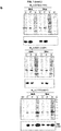
- Peptides were separated by SDS-PAGE, revealing that blue light enhanced the cleavage by chymotrypsin and V8 protease, which yielded two main protein bands whose sizes roughly corresponded to the expected for the individual LOV2 and WH1 domains.
- LOV2-WH1 switch by mutagenesis.
- a major concern in the design of any synthetic switch through protein fusion is how this affects the dynamic range of the device, i.e., the net ratio between the response of a chimera to the ON and the OFF stimuli, which in optogenetics ultimately depends on the balance between the fraction of molecules that remain in the OFF (pseudo-dark) state upon illumination and the fraction of molecules that stay in the ON (pseudo-lit) state in the darkness.
- Three mutations were introduced in LOV543wt-WH1, to generate LOV543m3-WH1 ( Fig.
- Fig. 8a displays a high-score in silico model supporting the structural feasibility of the three mutations within the fully folded LOV2 and WH1 frames, which showed no stereochemical violations ( Fig. 8b ).
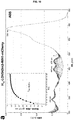
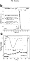
- the LOV543wt-WH1 and LOV543m3-WH1 chimeras were expressed in E. coli, purified and characterized through biophysical approaches ( Fig. 9 ). Measurements of the return to the dark state after saturating blue-light stimulation, by following the evolution of the absorption spectra of the FMN prosthetic group (band at 447 nm), indicated a three-fold increase in the half-life of the excited state for the m3 mutant (16.7 s) compared with its parental wt protein (5.6 s) ( Fig. 9a ).
- Light modulates the capacity of LOV2-WH1 to cross-seed RepA-WH1(A31V) amyloidogenesis Seeding, i.e., the ability of a pre-formed amyloid aggregate to template and nucleate amyloid growth from soluble molecules of the same (or a closely related) protein, is a hallmark of amyloidogenesis.
- seeding i.e., the ability of a pre-formed amyloid aggregate to template and nucleate amyloid growth from soluble molecules of the same (or a closely related) protein, is a hallmark of amyloidogenesis.
- substoichometric amounts of the purified chimeras were supplied to an excess of soluble RepA-WH1(A31V) ( Fig. 3a ).
- a nucleation agent such as purified RepA-WH1(A31V) aggregates preformed in vivo
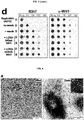
- the formation of RepA-WH1(A31V) fibres was thus explored either under continuous blue light illumination or in the darkness. Fibrillation was monitored by transmission electron microscopy (TEM) ( Fig. 3b ), thioflavin-S (ThS) fluorescence emission, Congo red (CR) birefringence under polarized light ( Fig.
- LOV543m3-WH1-mCherry enables optogenetic control on phase transitions.
- the red fluorescent reporter protein mCherry was fused to the C-terminus of the WH1 domain.
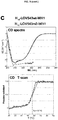
- the LOV543m3-WH1-mCherry protein was then purified and characterized through several biophysical approaches ( Fig. 10 ).
- LOV543m3-WH1-mCherry is an optogenetic switch for bacterial proliferation.
- Expression in the darkness of the LOV543m3-WH1 (WT)-mCherry triple chimera resulted in filamentation of bacterial cells ( Fig. 5a ), a phenotype characteristic of RepA-WH1 (A31V)-mCherry expression but not of RepA-WH1 (WT)-mCherry, suggesting that LOV2 indeed destabilizes the fold of the native RepA-WH1 turning it into amyloidogenic.
- Quantitation is difficult due to clumping of a substantial fraction of the cells, 35% of them exhibited intense ThS-stainable protein aggregates, an indication for their amyloid nature.
- the LOV2-WH1 chimeras were first optimized by modulating the phase and length of the linker J ⁇ - ⁇ 1 helix (LOV 543 -WH1 11 ; Fig. 1 ), plus the inclusion of mutations to stabilize the dark state conformation of J ⁇ ( Figs. 7-9 ).
- In vitro studies ( Fig. 3 ) suggest that, in the darkness, the stiff J ⁇ - ⁇ 1 chimeric helix connecting LOV2 and WH1 ( Fig. 2 ) would act as a lever that, by unleashing the three-helix bundle ( ⁇ 1- ⁇ 2- ⁇ 5) that locks the fold of WH1, would enable the protein to template, on a hyper-amyloidogenic RepA-WH1 variant, the assembly of amyloid fibrils ( Fig.
- Amyloid opto(epi)genetics remained yet unexplored.
- the LOV2-WH1 chimeras presented herein enrich and expand the catalogue of available optogenetic tools with a novel way to guide the conformational landscape of proteins towards amyloidogenesis.
- Their potential applications may include controlling the assembly of amyloid nanoscaffolds to engage enzymes in sequential reaction steps; building transcriptional switches for synthetic gene expression circuits and light-controlled plasmid replication cassettes; the selective elimination of particular bacteria within a consortium, once they have fulfilled their task in a bioprocess; or the development of a completely new kind of antimicrobials based on triggering amyloidosis by, e.g., bacteriophages encoding light-switchable cytotoxic amyloids.
- the AsLOV2 gene (SEQ ID NO: 6) was custom-synthesized at ATG:biosynthetics (Merzhausen, Germany), with its codon composition optimized to the usage in E. coli (SEQ ID NO: 7), and delivered as a pUC18 derivative.
- the template source of repA-WH1 was pWH1(WT). Both genes were independently amplified by PCR, using Pfu DNA polymerase, in such a way that the primers at 3' end of LOV2 and at the 5'end of repA-WH1 hybridize in the next step through their 5' ends.
- G528A_I532A-F SEQ ID NO: 8: 5' GTGATGCGGCGGAACGTGAAGCCGTGATGCTGGCTAAAAAAACCGCAG AAAACATTGAT and G528A_I532A-R (SEQ ID NO: 9): 5' ATCAATGTTTTCTGCGGTTTTTTTAGCCAGCATCACGGCTTCACGTTCCG CCGCATCAC, to build the G528A and I532A double mutant.
- G528A_L531E_I532A-F (SEQ ID NO: 10): 5' CGGAACGTGAAGCCGTGATGGAGGCTAAAAAAACCGCAGAAAACA and G528A_L531E_I532A-R (SEQ ID NO: 11): 5' TGTTTTCTGCGGTTTTTTTAGCCTCCATCACGGCTTCACGTTCCG.
- the LOV2m3-WH1-mCherry chimera was built in an analogous way, but using pRG-Ptac-His 10 -L OV543m3-WH1 (see above) and mCherry (from pRG-Ptac-His 6 - mCherry ) as the templates for the two PCR amplifications at the initial step.
- the RepA-WH1(A31V) protein used in the fibrillation studies was purified as described ( Giraldo, R., 2007, Proc. Natl. Acad. Sci. USA, 104, 17388-17393 ).
- the LOV2-WH1 chimeras (Hio-LOV543wt/m3-WH1 and H 10 -LOV543m3-WH1-mCherry) were expressed in the E. coli strain BL21, in the presence of a helper plasmid providing T7 lysozyme to facilitate cell lysis.
- Cells were harvested, washed with cold 0.9 NaCl and resuspended in 15 mL of lysis buffer (0.5 M NaCl, 0.05 M imidazole pH 8.0, 1% Brij-58, 10% glycerol plus 1 pill of EDTA-free Roche protease inhibitors). Cell suspension was frozen at - 70 oC.
- lysis buffer 0.5 M NaCl, 0.05 M imidazole pH 8.0, 1% Brij-58, 10% glycerol plus 1 pill of EDTA-free Roche protease inhibitors.
- Solubility of the chimeras, expressed in the E. coli K-12 reduced genome strain MDS42, was assayed in whole cell lysates from 15 mL of cultures grown at 37 oC in LB plus Ap 100 . When bacterial cultures reached OD 600nm 0.2, IPTG was added to 0.5 mM and they were split into two aliquots, to be grown either in the darkness or under blue light illumination (1,070 Lux). After 4 h of induction cells were harvested and resuspended in 0.2x lysis buffer (see above), at a ratio of 0.33 mL per each unit of optical density (1.5 x 10 9 cells).
- EDTA to 1 mM
- lysozyme to 1 ⁇ mL -1
- Hio-LOV543wt-WH1 Three ⁇ g aliquots of purified Hio-LOV543wt-WH1 were displayed in 15 ⁇ L of SEC buffer and trypsin (0.025 units), chymotrypsin (0.004 u.) or V8 (0.05 u.) proteases (Sigma) were supplied on ice. Digestions were left to proceed, either in the darkness or under blue light illumination (30,000 Lux), for 1 and 2 h at RT. Reactions were stopped by adding SDS-PAGE loading buffer and immediately boiling for 5 min, followed by electrophoretic separation in 12.5% polyacrylamide gels and Coomassie blue staining.
- Amyloidogenesis assays in vitro were carried out as described ( Molina-Garcia, L., et al., 2018, Methods Mol. Biol., 1779, 289-312 ), by setting on ice in 2 mL Eppendorf tubes 50 ⁇ L aliquots made of: RepA-WH1(A31V) (25 ⁇ M) in fibril assembly buffer (0.1M Na 2 SO 4 , 4 mM MgSO 4 , 20 mM Hepes ⁇ NaOH pH 8.0, 14% PEG4000, 6% MPD), but using as seeds sub-stoichiometric amounts (1:100) of the purified Hio-LOV543wt/m3-WH1 chimeras.
- Purified protein chimeras were characterized for their response to darkness and blue light illumination regarding their photocycle, association state, secondary structure and stability in SEC buffer.
- the photocycle of the LOV2 moiety in the H 10 -LOV543wt/m3-WH1 chimeras was studied by saturating with blue LEDs illumination (30,000 Lux, for 10 min) 600 ⁇ L protein solutions (3 ⁇ M wt and 5 ⁇ M m3) that were displayed in quartz cuvettes (1 cm path length) placed into the sample holder of an Ultrospec 3300pro spectrophotometer (GE Healthcare).
- Each sample was distributed between two centrifuge cells: one of them was scanned at 275 nm (for the double chimera) or 590 nm (for the triple chimera) as the dark state, and the other was illuminated at 450 nm as the lit state. Sedimentation coefficients distributions were calculated with SEDFIT.
- Circular dichroism spectroscopy was performed with the chimeras in a Jasco 720 spectropolarimeter, with 150 ⁇ L (2.5 ⁇ M) of the protein samples in SEC buffer. Proteins were set in 0.1 cm path length quartz cuvettes hold at 20 oC, and 7 spectra were acquired, in the darkness, at 50 nm ⁇ min -1 and accumulated for signal averaging. Protein stability was estimated by thermal denaturation, measuring the variation of ellipticity ( ⁇ ) at 220 nm with the increase of temperature (20-90 oC).
Landscapes
- Chemical & Material Sciences (AREA)
- Health & Medical Sciences (AREA)
- Organic Chemistry (AREA)
- Life Sciences & Earth Sciences (AREA)
- General Health & Medical Sciences (AREA)
- Biophysics (AREA)
- Biochemistry (AREA)
- Genetics & Genomics (AREA)
- Medicinal Chemistry (AREA)
- Molecular Biology (AREA)
- Proteomics, Peptides & Aminoacids (AREA)
- Gastroenterology & Hepatology (AREA)
- Botany (AREA)
- Peptides Or Proteins (AREA)
Abstract
The present invention provides an optogenetic chimeric fusion polypeptide comprising an optimized amino acid sequence of the plant phototropin domain LOV2 fused to an amino acid sequence of the bacterial amyloidogenic effector RepA-WH1. Optimized LOV2 enables navigation through the folding landscape of RepA-WH1 from solubility to its aggregation as oligomers or amyloid fibres. Thus, this polypeptide assembles as hydrogels and amyloid fibres in the darkness, while under blue light illumination forms oligomeric particles that are proteotoxic for cells, preferably bacteria. This polypeptide is therefore proposed for inducing the formation of cytotoxic amyloid oligomers in cells that are targeted for killing.
Description
- This invention belongs to the fields of amyloidosis, optogenetics and protein synthetic conformational switches. In particular, this invention refers to a chimeric fusion protein based on the photoreceptor domain LOV2 fused to an effector protein with amyloidogenic potential. This chimeric protein allows the light-control of amyloidogenesis within cells, since the excitation of the protein with blue light leads to the formation of cytotoxic soluble amyloid aggregates or oligomers in the cell expressing the chimeric protein. This kind of light-switchable fusion proteins have multiple applications, for example, as antimicrobial agents based on triggering amyloidosis within undesired bacterial cells.
- Through the absorption of photons by chromogenic prosthetic groups, light has the capacity to elicit conformational re-arrangements in natural effector proteins. An emerging discipline, Optogenetics, has implemented the engineering of such photoreceptors to create synthetic conformational switches that much expand the functional abilities of proteins (Khamo, J. S., et al., 2017, J. Mol. Biol., 429, 2999-3017). One of the most used photoreceptors in optogenetics is the LOV domain, with many variants widespread across the whole phylogenetic tree (Glantz, S. T., et al., 2016, Proc. Natl. Acad. Sci. USA, 113, E1442-E1451). One of the LOV domains most commonly used in optogenetic is the plant photoreceptor LOV2 (Zimmerman, S. P., et al., 2016, Methods Enzymol., 580, 169-190).
- Engineering chimeric proteins to become light-responsive switches has recently enabled physical control on cellular processes such as channel gating, gene expression, organelle targeting and proteolysis. The design of protein chimeras including a light-responsive domain, which couples conformational changes, with a functional switch in its fusion partner, has equipped Synthetic Biology with powerful optogenetic tools to get physical control on a plethora of cellular processes. Thus, over more than a decade, optogenetic parts have been designed, and successfully assayed, to govern ion fluxes across membrane channels and thus neuronal circuits, cytoskeleton dynamics and nuclear localization, membrane trafficking, allosteric photo-regulation of enzyme catalysis, apoptosis through activating caspases, protein degradation by ubiquitinilation, or gene expression through regulation of the binding of proteins to DNA. In many of these instances, effector peptide tags became exposed upon illumination of cells with light of a wavelength (UV/Vis/IR) matching an absorption band in a chromophoric prosthetic group bound to the light-responsive domain.
- LOV-based optogenetic tools have been gaining wide popularity in recent years to control a myriad of cellular events, including cell motility (Wu, Yi I., et al., 2009, Nature, 461 (7260): 104-8), subcellular organelle distribution (van Bergeijk, et al., 2015, Nature, 518 (7537): 111-4), formation of membrane contact sites (Jing, Ji, et al., 2015, Nature Cell Biology, 17(10): 1339-47), and protein degradation (Renicke, Christian, et al., 2013, Chemistry & Biology, 20 (4): 619-626).
- However, no optogenetic protein tool has been described having direct control on the aggregation pathways leading to the assembly of amyloids. Thus, amyloid opto(epi)genetics remains yet unexplored.
- Amyloids, which are among the most stable natural macromolecular structures, are made of β-strand segments from different individual molecules of a given protein that assemble, through intermediate oligomeric states, into mature fibres. Amyloidogenesis is readily accessible to short peptides or intrinsically disordered protein domains but, for fully folded proteins with a stable three-dimensional fold, partial unfolding to a metastable state is mandatory, which is usually attained through disease-linked destabilizing mutations in vivo or by resorting to harsh physical-chemical conditions in vitro.
- RepA-WH1 (Giraldo, R., et al., 2016, Prion, 10, 41-49) is a manifold domain from a plasmid-encoded bacterial protein that undergoes conformational changes that capacitate it either as a transcriptional repressor, as a DNA replication initiator or, through its assembly as amyloid oligomers, to hinder premature replication rounds. Although very stable in solution, RepA-WH1 dimers become metastable upon binding to dsDNA or acidic phospholipids, thus paving the pathway towards amyloidogenesis. In the bacterial cytoplasm, the fusion of a hyper-amyloidogenic mutant variant of RepA-WH1 to the fluorescent protein mCherry generates a prion-like protein (prionoid) that epigenetically propagates from mother-to-daughter cells, causing a synthetic amyloid proteinopathy (Fernández-Tresguerres, M. E., et al., 2010, Microbiol., 77, 1456-1469). Thus, RepA-WH1 was previously engineered to boost amyloidogenicity and uncouple its conformational remodelling from its natural function, thus generating the only intracellular proteinopathy described so far in bacteria, which has been useful as a minimal model to deconstruct a 'generic' amyloid disease (Fernández, C., et al., 2016, Sci. Rep., 6, 23144).
- RepA-WH1 amyloidosis recapitulates some of the hallmarks of the mitochondrial damage associated with human amyloid diseases, including the formation oligomeric pores at the internal membrane, the generation of reactive oxygen species (ROS) and the loss of function, due to co-aggregation, of essential cell factors. In addition, RepA-WH1 has been used as a bench proof for the design of synthetic tools to probe protein amyloidogenesis, including gold nanoparticle-based sensors (Fernández, C., et al., 2016, Angew. Chem. Int. Ed., 55, 11237-11241) and screening devices based on the disruption of protein translation either in yeast (Gasset-Rosa, F. & Giraldo, R., 2015, Front. Microbiol., 6, 311) or in bacteria (Molina-Garcia, L. & Giraldo, R., 2017, Sci. Rep., 7, 11908).
- However, there is a need for optogenetic protein tools that allow the direct control on the assembly of cytotoxic amyloid fibres or oligomers which are useful for triggering amyloidosis within undesired cells that need to be killed.
- The present invention provides an optogenetic chimeric fusion polypeptide comprising an optimized (mutated) amino acid sequence of the plant phototropin LOV2 domain fused to an amino acid sequence of the bacterial amyloidogenic effector RepA-WH1. Optimized LOV2 enables navigation through the folding landscape of RepA-WH1 from solubility to its aggregation as oligomers or amyloid fibres. Thus, this designed polypeptide assembles as hydrogels and amyloid fibres in the darkness, while under blue light illumination forms oligomeric particles that are proteotoxic for cells, preferably bacteria.
- Here it is shown the feasibility of using optogenetic protein switches to surf the conformational landscape of a protein on a pathway leading to amyloidosis. In this invention optogenetic control on a phase transition leading to the assembly of amyloid fibres or oligomers with characteristic toxicity has been achieved.
- This invention describes therefore the construction of a blue light-responsive chimera between an optimized plant phototropin LOV2 domain and the bacterial prion-like protein RepA-WH1. In the darkness, and in a crowded environment in vitro, this chimera exhibits low sensitivity to proteases and is competent to nucleate on RepA-WH1 the assembly of amyloid fibres and hydrogels. When expressed in Escherichia coli, this chimera forms in the darkness large intracellular amyloid aggregates. However, under blue light illumination the same chimera has increased sensitivity to proteolysis and templates the assembly of discrete oligomeric RepA-WH1 particles and liquid droplets in vitro. Such lit-state oligomers exhibit enhanced cytotoxicity in vivo.
- The chimeric protein described in this invention was first optimized by modulating the phase and length of the linker Jα-α1 helix (LOV543-WH111; see
Fig. 1 ), plus the inclusion of mutations that stabilize the dark state conformation of Jα (Figs. 7-9 ). - The chimera presented in this invention enriches and expands the catalogue of available optogenetic tools with a novel way to guide the conformational landscape of proteins towards amyloidogenesis. Its applications include, without limitations:
- Controlling the assembly of amyloid nanoscaffolds to engage enzymes in sequential reaction steps. Nanoscaffolds are protein architectures (fibrilar, tubular, laminar, icosahedral, gel-like) to which other proteins, nucleic acids or functionalized organic molecules can bind to enhance their biological (e.g., catalytic) activities by the force of their immobilization, densities and intermolecular channelling.
- Building transcriptional switches for synthetic gene expression circuits and light-controlled plasmid replication cassettes. The conditional, light-dependent activation or inactivation of transcription factors would enable to block or trigger the synthesis of mRNA; whereas the effect of light on a DNA replication factor would do the same on the synthesis and propagation of a genetic mobile element (e.g., a plasmid). Achieving control on both, the expression/repression of a gene of interest and the propagation of the vector that carries it, are central biotechnological goals.
- The selective elimination of particular bacteria within a consortium, for instance once they have fulfilled their task in a bioprocess. In the design of bioprocesses, either for environmental or industrial applications, it is often crucial to get rid of bacteria after these have finished one bio-transformative step, in order to allow the proliferation and activity of a second different microorganism that might be outcompeted by the first. Light-enabled killing of the resident bacteria would provide an efficient way of achieving that goal.
- The development of a completely new kind of antimicrobials based on triggering amyloidosis by, e.g., bacteriophages encoding light-switchable cytotoxic amyloids. The emergence of antibiotic resistant bacteria currently is a major problem for human health worldwide. Thus the discovery of radically new ways to combat and kill pathogenic microorganisms is mandatory. Proteotoxic modules such as RepA-WH1, which could be delivered through bacteriophages (viruses), to kill bacteria upon light illumination would have a deep impact on clinics (e.g., in dermatological treatments against common skin pathogens such as Staphylococcus aureus).
- Therefore, a first aspect of the present invention refers to a fusion polypeptide, hereinafter "the polypeptide of the invention", "the fusion protein of the invention", "the chimera of the invention" or "the chimeric protein of the invention", comprising the mutated amino acid sequence of the LOV2 domain shown in SEQ ID NO: 4 fused by its C-terminal end to the N-terminal end of the RepA-WH1 protein shown in SEQ ID NO: 5, wherein said fusion polypeptide comprises the amino acid sequence of SEQ ID NO: 1.
- SEQ ID NO: 1 of the present invention is the chimera or mutant also called "LOV543m3-WH1" or "LOV2m3-WH1".
- The "LOV2 domain" refers to the plant phototropin LOV2 domain or plant LOV2 photoreceptor. "LOV" means "Light-oxygen-voltage-sensing domain" and it is a protein sensor used by a large variety of higher plants, microalgae, fungi and bacteria to sense environmental conditions. In higher plants, it is used to control phototropism, chloroplast relocation, and stomatal opening. It has a blue-light sensitive flavin chromophore, which in the signaling state is covalently linked to the protein core via an adjacent cysteine residue. LOV domains are e.g. encountered in phototropins, which are blue-light-sensitive protein complexes regulating a great diversity of biological processes in higher plants as well as in microalgae. Phototropins are composed of two LOV domains, each containing a non-covalently bound flavin mononucleotide (FMN) chromophore in its dark-state form, and a C-terminal Ser-Thr kinase. Upon blue-light absorption, a covalent bond between the FMN chromophore and an adjacent reactive cysteine residue of the apo-protein is formed in the LOV2 domain. This subsequently mediates the activation of the kinase, which induces a signal in the organism through phototropin autophosphorylation.
- The "mutated amino acid sequence of the LOV2 domain" referred to in the present invention is the amino acid sequence shown in SEQ ID NO: 4 and it will be also called in the present invention "LOV543m3" or "LOV2m3". This SEQ ID NO: 4 comprises the amino acid substitutions G528A, L531E and I532A regarding the wild type amino acid sequence of the LOV2 domain (see
Fig. 2 ). As shown in examples below, only this mutated variant of the LOV2 domain among those mutants tested by the inventor was light-responsive, i. e. differentially increased its solubility upon blue light illumination allowing thus the formation of cytotoxic amyloid oligomers. - In this invention, the term "LOV543wt" or "LOV2wt" refers to the LOV2 domain without the three amino acid substitutions indicated in the paragraph above. Thus, LOV543wt is the SEQ ID NO: 4 but comprising a G at position 528, a L at position 531 and an I at position 532 (see
Figure 2 ). - The "RepA-WH1 protein" or "WH1" is the amino acid sequence shown in SEQ ID NO: 5.
- The half-life of the lit (light-excited) state of the polypeptide of the invention is increased by 2.3-fold when the polypeptide is fused in its C-terminal end to the mCherry protein. Furthermore, this fusion to mCherry shows enhanced thermal stability in the polypeptide of the invention compared to the same polypeptide without mCherry. The fusion to mCherry also enhances in the polypeptide of the invention the metastability of RepA-WH1 and thus its amyloidogenicity.
- Therefore, in a preferred embodiment, the amino acid sequence of SEQ ID NO: 1 is fused, in its C-terminal end, to the amino acid sequence of the mCherry fluorescent protein probe. More preferably, the amino acid sequence of the mCherry fluorescent protein probe is SEQ ID NO: 2.
- In an even more preferably embodiment of this aspect of the invention, the polypeptide of the invention comprises the amino acid sequence SEQ ID NO: 3. In the most preferred embodiment of this aspect of the invention, the polypeptide of the invention consists of the amino acid sequence SEQ ID NO: 3.
- SEQ ID NO: 3 of the present invention is the chimera of the invention also called "LOV543m3-WH1-mCherry" or "LOV2m3-WH1-mCherry".
- The polypeptide of the invention may be produced by chemical synthesis or, as a recombinant, by an organism or cell that expresses a nucleotide sequence that encodes it. The polypeptide of the invention can be synthesised, for example, but without limitations, in vitro. For example, by means of the synthesis of solid-phase polypeptides or recombinant DNA approaches. It can be produced in a recombinant manner, including its production as a mature polypeptide or as a pre-protein that includes a signal peptide. Thus, the polypeptide of the invention may further comprise a signal peptide in its N-terminal end.
- Another aspect of the invention refers to a nucleic acid sequence, hereinafter "the polynucleotide of the invention" or "the nucleic acid sequence of the invention", encoding the polypeptide of the invention.
- Due to the degeneration of the genetic code, various nucleotide sequences can encode the same amino acid sequence.
- In accordance with the present invention, a "nucleic acid molecule", "nucleotide sequence", "nucleic acid sequence" or "polynucleotide" is a nucleic acid molecule (polynucleotide) that has been extracted from its natural medium (i.e. it has been subjected to human manipulation) and can include DNA, RNA or DNA or RNA derivatives, including cDNA. The nucleotide sequence of the present invention may or may not be chemically or biochemically modified and can be artificially obtained by means of cloning and selection methods or by means of sequencing.
- The polynucleotide sequence of the invention can encode the mature polypeptide or a pre-protein consisting of a signal peptide linked to the mature polypeptide that must be subsequently processed.
- The polynucleotide sequence of the present invention may also comprise other elements, such as introns, non-encoding sequences at ends 3' and/or 5', ribosome binding sites, etc. This nucleotide sequence can also include encoding sequences for additional amino acids that are useful for the purification or stability of the encoded polypeptide.
- The polynucleotide sequence of the invention can be included in a gene or genetic construct, preferably in a recombinant expression vector. Said genetic construct may also comprise one or more gene expression-regulating sequences, such as promoters, terminators, enhancers, etc.
- Thus, another aspect of the invention refers to a genetic construct, hereinafter "the genetic construct of the invention", comprising the nucleic acid sequence of the invention, preferably wherein said genetic construct is an expression vector, more preferably a bacteriophage.
- Alternatively, the expression vector referred to in the present invention may be a plasmid, preferably a low copy-number plasmid replicon, such as pRK2, pSC101, R1/F, or even multicopy plasmids (pUC, p15A, pBBR1), although others are not excluded.
- The genetic construct of the invention may further comprise one or more sequences encoding for a specifically cleavable linker peptide functionally interposed between the mutated amino acid sequence of the LOV2 domain and the RepA-WH1 protein and/or between the RepA-WH1 protein and the mCherry protein. Such a linker peptide may be, for instance, a peptide sensitive to thrombin cleavage, factor X cleavage or other peptidase cleavage.
- The genetic construct of the invention may further comprise one or more sequences encoding for purification tag/s linked to the polypeptide of the invention.
- The genetic construct of the invention will generally be constructed such that the sequence encoding for the polypeptide of the invention is positioned adjacent to and under the control of an effective promoter. In certain cases, the promotor will comprise a prokaryotic promoter where the genetic construct is adapted for expression in a prokaryotic host cell. In other cases, the promoter will comprise a eukaryotic promoter where the genetic construct is adapted for expression in a eukaryotic host cell. In the later cases, when used for expression in eukaryotic hosts, the genetic construct will typically further include a polyadenylation signal at position 3' of the carboxy-terminal amino acid, and within a transcriptional unit of the encoded polypeptide. Promoters of particular utility in the genetic construct of the invention are bacterial promoters, preferably bacterial promoters functional in E. coli cells, such as for instance but without limitations, Ptac (IPTG/lactose-inducible), Ptet-ON/OFF (tetracyclin or doxicylin inducible/repressible), ParaBAD (arabinose-inducible) or PλCIts (temperature-inducible).
- The expression "genetic construct", "gene construct" or "nucleic acid construct" as used herein relates to a functional unit required to transfer or express a nucleic acid sequence of interest, herein the nucleotide sequence of the invention as described, and regulatory sequences including, for example, a promoter, operably linked to the sequence that encodes the polypeptide, in an expression system. It refers to a nucleic acid molecule, mono or bicatenary, which is isolated from a natural gene or that is modified to contain nucleic acid segments in such a manner that they would otherwise not exist in nature. The expression "nucleic acid construct" is synonymous to the expression "expression cassette" when the construct of nucleic acid contains the control sequences required for the expression of the encoding sequence.
- The term "expression vector", also known as "expression construct" or "plasmid", relates to a DNA molecule, linear or circular, that comprises the nucleic acid sequence of the invention operably linked to additional segments that provide the transcription of the encoded polypeptide. Generally, a plasmid is used to introduce a specific nucleic acid sequence in a target cell. Once the expression vector is in the interior of the cell, the protein encoded by the nucleic acid sequence is produced by means of the ribosome complexes of the cellular transcription and translation machinery. The plasmid is often subject to engineering to contain regulatory sequences that act as enhancer and promoter regions that lead to an efficient transcription of the nucleic acid sequence carried on the expression vector. The objective of a well-designed expression vector is the production of large amounts of stable messenger RNA and, therefore, of proteins. Expression vectors are basic tools for biotechnology and for the production of proteins, such as chimeric fusion proteins. The expression vector of the invention is introduced in a host cell such that the vector remains as a chromosome constituent or as an extra-chromosome self-replicating vector.
- The term "expression" relates to the process whereby a polypeptide is synthesised from a polynucleotide. The term includes the transcription of the polynucleotide in a messenger RNA (mRNA) and the translation of said mRNA into a protein or polypeptide.
- Examples of useful expression vectors are phages, cosmids, phagemids, yeast artificial chromosomes (YAC), bacterial artificial chromosomes (BAC), human artificial chromosomes (HAC) or viral vectors, such as adenovirus, baculovirus, retrovirus or lentivirus.
- The polypeptide of the invention can be prepared using any known means in the state of the art, such as the transformation of the nucleic acid sequence of the invention in an adequate host cell and the expression of said sequence to obtain the polypeptide of the invention.
- Another aspect of the invention refers to a cell, hereinafter "the cell of the invention", comprising the fusion polypeptide, the nucleic acid sequence or the genetic construct of the invention.
- The cell of the invention may be either a eukaryotic or a prokaryotic cell. This cell is the recipient of an expression vector, cloning vector or any other DNA molecule. Therefore, it includes any cultivable cell that may be modified through the introduction of DNA not contained naturally therein. Preferably, this cell is that in which the polynucleotide of the invention may be expressed, giving rise to a stable polypeptide, post-translationally modified and located in the appropriate subcellular compartment. The election of an appropriate cell may also be influenced by the election of the detection signal. For example, the use of constructs with reporter genes (for example, lacZ, luciferase, thymidine kinase, green fluorescent protein "GFP" or red fluorescent protein "mCherry") can provide a signal selectable through the activation or inhibition of the transcription of the nucleotide sequence of interest in response to a transcription-regulating protein. In order to achieve optimum selection or screening, the phenotype of the cell must be considered.
- In any case, the polynucleotide or the genetic construct of the invention encoding the polypeptide of the invention is placed under the transcriptional control of regulatory signals functional in the cell. Said regulatory signals appropriately control the expression of the polypeptide of the invention to allow any necessary transcriptional and post transcriptional modification.
- Preferably, the cell of the invention is a prokaryotic cell, more preferably a bacterial cell, even more preferably an E. coli cell.
- Another aspect of the invention relates to the use of the cell of the invention for the production of the fusion polypeptide of the invention.
- The cell of the invention may be cultivated for such purpose. A cell culture relates to the in vitro process of maintaining and growing cells. Cell cultures need controlled conditions of temperature, pH, percentages of gases (oxygen and carbon dioxide), in addition to the presence of the adequate nutrients to allow cellular viability and division. The skill in the art will know which conditions must be applied to the cell culture depending on the requirements of the selected cell. Cell cultures can be carried out in solid substrates, such as agar, or in a liquid medium, which enables the expansion of large amounts of cells in suspension.
- Once the cell of the invention has been cultivated and the polypeptide of the invention has been expressed, it can be purified. The term "to purify", as used in the description, relates to the isolation of the polypeptide of the invention from the other polypeptides present in the culture medium in which the cell of the invention has grown. The isolation of the polypeptide can be carried out using differential solubility techniques, chromatography, electrophoresis or isoelectric focusing. Chromatography techniques can be based on molecular weight, ion charge (based on the ionisation state of the amino acids under working conditions), the affinity of the protein for certain matrixes or chromatographic columns, or by means of purification tags, and can be carried out on a column, on paper or on a plate. The isolation of the polypeptide can be carried out, for example, by means of precipitation with ammonium sulphate, fast protein liquid chromatography (FPLC) or high performance liquid chromatography (HPLC), using automated systems that significantly reduce purification time and increase purification efficiency.
- Another aspect of the invention refers to an in vitro use of the fusion polypeptide, the nucleic acid sequence or the genetic construct of the invention for inducing the formation of cytotoxic amyloid oligomers in a cell, preferably in a prokaryotic cell, more preferably in a bacterial cell, even more preferably in a E. coli cell. Preferably, this use comprises exposing the cell to blue light. In this in vitro use, the cell is expressing the fusion polypeptide of the invention.
- The term "in vitro" means the use as described above on cells growing in an in vitro culture or on cells that are outside of the human or animal body present, for instance but without limitation, in an ex vivo biofilm, in an ex vivo isolated biological sample (e,g., transcription-translation coupled systems such as PURE®), in an inert surface, in a scaffold, inside lipid vesicles (liposomes) or within a bioreactor.
- "Blue light" is understood, in the context of this invention, as light emission intensity in the range of, preferably, between 1,000 Lux and 30,000 Lux. Blue light may be provided by, for instance but without limitation, a custom-built device made of 27 x 8 blue AlGaInP LED bulbs (5 mm ø, 20 deg cone, 3 V, λmax= 468 nm, 8 cd each), that allows regulation of the light emission intensity between 1,000 (min.) and 30,000 (max.) Lux, or any commercial blue (actinic) light LEDs arrays.
- Since the polypeptide of the invention allows triggering the formation of cytotoxic amyloid oligomers within cells upon exposure to blue light, it can be used for killing undesired cells ex vivo (for instance, in a biofilm, bioreactor, surface or consortium) or undesired cells within organisms where they are inducing an infection or an undesired pathological condition.
- Thus, another aspect of the invention refers to the use of the fusion polypeptide, the nucleic acid sequence, the genetic construct or the cell of the invention for eliminating or killing undesired cells ex vivo, more preferably cells present in a biofilm, bioreactor, surface or consortium.
- In a preferred embodiment, the undesired cells are bacterial cells, more preferably E. coli or S. aureus cells, even more preferably E. coli cells.
- The term "ex vivo" means outside of the human or animal body.
- Another aspect of the invention refers to the use of the fusion polypeptide, the nucleic acid sequence, the genetic construct or the cell of the invention for:
- assembling amyloid nanoscaffolds useful, for instance but without limitations, for engaging enzymes in sequential reaction steps, or
- building transcriptional switches useful, for instance but without limitations, for synthetic gene expression circuits or light-controlled plasmid replication cassettes.
- Another aspect of the invention refers to a pharmaceutical composition, hereinafter "the composition of the invention", comprising the polypeptide, the nucleic acid sequence, the genetic construct or the cell of the invention, preferably the polypeptide of the invention comprising, more preferably consisting of, SEQ ID NO: 3.
- This composition of the invention comprises the polypeptide, the nucleic acid sequence, the genetic construct or the cell of the invention in a therapeutically effective amount. A "therapeutically effective amount" is understood to be the amount of polypeptide, the nucleic acid sequence, the genetic construct or the cell of the invention that, when administered to the patient, produces the desired effect, thereby triggering the formation of cytotoxic amyloid aggregates upon blue-light exposure and therefore killing the cell/s causing the pathological condition, preferably the infection, more preferably the bacterial infection. The therapeutically effective amount may vary depending on a variety of factors, for example, but not limited to, the type of pathological condition and its severity, as well as age, weight, sex, physical condition, response or tolerance, etc., of the individual to whom the composition of the invention is going to be administered.
- Preferably, this composition of the invention further comprises a blue LED device or a blue LEDs light source for illuminating the cells comprising the polypeptide, the nucleic acid sequence or the genetic construct of the invention.
- In a more preferred embodiment, the composition of the invention further comprises a pharmaceutically acceptable vehicle or excipient, adjuvant and/or other active ingredient.
- Another aspect of the invention refers to the fusion polypeptide, the nucleic acid sequence, the genetic construct or the cell of the invention for use as a medicament. Alternatively, this aspect of the invention refers to the use of the fusion polypeptide, the nucleic acid sequence, the genetic construct or the cell of the invention for the manufacture of a medicament.
- Preferably, the medicament of the invention is an antimicrobial medicament, more preferably an antibacterial medicament.
- The term "medicament" or "drug" makes reference to any substance used to prevent, alleviate, treat or cure diseases, conditions or pathologies, preferably bacterial infections, more preferably E. coli infections, in humans, or in any other animal.
- In the context of the present invention, the term "medicament" relates to a preparation that comprises the polypeptide, polynucleotide, gene construct or cell of the invention; preferably the polypeptide of the invention, more preferably the polypeptide of the invention comprising, even more preferably consisting of, SEQ ID NO: 3.
- The medicament to which the present invention refers may be for human or veterinary use. The "medicament for human use" is any substance or combination of substances that have the properties for treating or preventing diseases in human beings or that can be used in human beings or administered to humans for the purpose of restoring, correcting or modifying physiological functions by exercising a pharmacological, immunological or metabolic action. The "medicament for veterinary use" is any substance or combination of substances having curative or preventive properties with respect to animal diseases or conditions or that can be administered to the animal in order to restore, correct or modify its physiological functions by exercising a pharmacological, immunological or metabolic action.
- The medicament referred to in the present invention may be used together with other active ingredients or therapies in the manner of a combined therapy. The other active ingredients may form part of the same composition or can be provided by means of a different composition, being administered at the same time or at different times (simultaneous or sequential administration).
- Another aspect of the invention refers to the fusion polypeptide, the nucleic acid sequence, the genetic construct or the cell of the invention for use in the treatment or prevention of microbial infections, preferably bacterial infections, more preferably E. coli infections. Alternatively, this aspect of the invention refers to the use of the fusion polypeptide, the nucleic acid sequence, the genetic construct or the cell of the invention for the manufacture of a medicament for the treatment or prevention of microbial infections, preferably bacterial infections, more preferably E. coli infections.
- The "bacterial infections" may be produced by, for instance but without limitations, E. coli or common skin pathogens such as S. aureus.
- Examples of microbial infections include, but without limitations, skin/dermatological infections, digestive infections or respiratory infections.
- The term "treatment", as understood in the present invention, refers to combating the effects caused as a result of the disease or pathological condition of interest in an individual (preferably a mammal and, more preferably, a human), which includes:
- (i) inhibiting the disease or pathological condition, i.e. interrupting its course;
- (ii) alleviating the disease or pathological condition, i.e. causing the regression of the disease or pathological condition or its symptoms;
- (iii) stabilising the disease or pathological condition.
- The term "prevention", as understood in the present invention, consists of avoiding the appearance of the disease or pathological condition in an individual (preferably a mammal and, more preferably, a human), particularly when said individual is susceptible of developing the disease or pathological condition but has not been diagnosed yet.
- Another aspect of the invention relates to a method, preferably an in vitro method, for inducing the formation of cytotoxic amyloid oligomers in a cell, preferably in a bacterial cell, which comprises:
- a. expressing the fusion polypeptide of the invention in the undesired cell to be killed, and
- b. exposing the cell of step (a) to blue light.
- As explained above, the fusion polypeptide of the invention may be expressed in the cell by transfecting the nucleic acid sequence or the genetic construct of the invention, preferably through a bacteriophage, and placing the cell under suitable culture conditions that allow the expression of the polypeptide.
- When the cells to be killed are within the human or animal body, the expression of the polypeptide of the invention in said cells may be achieved by the use of adequate expression vectors, preferably bacteriophages, that specifically direct the expression of the polypeptide to the interior of said cells.
- The exposure of the cell to blue light, according to step (b) of the method, is preferably performed during at least 1h, but can extend longer and be applied either in a continuous regime or through light/darkness pulses of variable length.
- Unless otherwise defined, all technical and scientific terms used herein have the same meaning as commonly understood by one of ordinary skilled in the art to which this invention belongs. Methods and materials similar or equivalent to those described herein can be used in the practice of the present invention. Throughout the description and claims the word "comprise" and its variations are not intended to exclude other technical features, additives, components, or steps. Additional objects, advantages and features of the invention will become apparent to those skilled in the art upon examination of the description or may be learned by practice of the invention. The following examples, drawings and sequence listing are provided by way of illustration and are not intended to be limiting of the present invention.
-
-
Fig. 1 . Assessing the solubility of distinct helical phases for the Jα-α1 linker in LOV2-RepA-WH1 chimeras. (a) Schematic representation of the arrangement of the three chimeras assayed. The linker helix increases its length by a helical turn between LOV540 (closest position related to WH1) and LOV543 (farthest to WH1), displaying both domains on opposite faces of the helix, whereas in LOV542 both are found on the same face and intermediate position. (b) Fractionation of bacterial cells (C) expressing the chimeras into the soluble (S) and insoluble (P) fractions (top panel), plus their detection by Western blotting (anti-His tag antibody; bottom). Only LOV543-WH1 generates a major soluble fraction, with no major differences when cultures were carried out in the darkness or under blue light illumination. (c) These results are compatible with steric hindrance due to close apposition of the domains by a short linker (LOV540-WH1) and/or their mutual interference for folding (LOV542-WH1), whereas only LOV543-WH1 has the right distribution of both domains to fold independently. -
Fig. 2 . Limited proteolysis maps the Jα-α1 helical linker as the major light-altered structural element in LOV543-WH1. (a) SDS-PAGE analysis of LOV543-WH1 digested, in the darkness or under blue light illumination, by three alternative proteases. A major cleavage site for chymotrypsin and V8 protease is made accessible in the illuminated state, generating two fragments with the sizes expected for LOV2 (≈17 kDa) and WH1 (≈15 kDa). (b) Sequence of the LOV543-WH1 fusion protein. Residue numbers are the originals from the AsLOV phototropin and RepA. N-terminal sequencing of the proteolytic fragments identified unambiguously the cleavage sites (arrows) for chymotrypsin (Ch: I532KK and E409RI) and V8 protease (V8: G528VM). The N-terminal Met residue appears to have been removed in vivo (g-23ss; tag peptide in lower case letters). Box outlines the linker Jα-α1 helix. The three amino acid substitutions included in the LOV543m3-WH1 (SEQ ID NO: 1) mutant are also indicated. -
Fig. 3 . In vitro cross-seeding of the assembly of the hyper-amyloidogenic protein RepA-WH1(A31V) (light grey) by sub-stoichiometric amounts (1:100) of the LOV543wt/m3-WH1 optogenetic switches. (a) Schematic overview of the seeding experiment. (b) Electron micrographs of the aggregates generated during the incubation of RepA-WH1(A31V) as substrate and the indicated optogenetic seeds, either in the darkness or under blue light illumination. Control samples, un-seeded or adding as seeds RepA-WH1(A31V)-mCherry aggregates purified from E. coli (S), are also displayed. LOV543wt-WH1 templates on RepA-WH1(A31V) the assembly of large fibres (F), irrespective of being illuminated or not. LOV543m3-WH1 differentially seeds the growth of short fibres (darkness) or an ensemble of needle-shaped spikes (N) and spherical/drop-like oligomers (D) (blue light). Magnification: 20,000x (insets: 50,000x). (c) Analysis of the fluorescence emitted by ThS (left) and optical birefringence of Congo Red (CR, right) when incubated with samples in (a). Both amyloidotropic molecules bind to RepA-WH1(A31V) aggregates when templated by control seeds or by LOV543wt-WH1 (whether it was incubated in the darkness or under blue light), thus they are amyloids. Particles assembled by LOV543m3-WH1, especially when incubated under blue light, bind weaker to ThS and CR. (d) Dot-blot analyses. Serial dilutions of samples assembled in the darkness (D) or under blue light (B) are displayed. B3h7, an antibody specific for the conformation of amyloidogenic RepA-WH1 oligomers, shows that these are more abundant when seeded with LOV543m3-WH1 (left). A replica incubated with α-WH1, a polyclonal antibody against RepA-WH1, is included as a control (right). Overall, LOV543m3-WH1 generates an operational switch of amyloidogenesis, discriminating between the assembly of mature amyloid fibres (darkness) and amyloidogenic oligomeric particles (blue light). -
Fig. 4 . Optogenetic regulation of a phase transition (liquid-hydrogel) in LOV543m3-WH1-mCherry. (a) The hydrogel formed in the darkness, when examined by TEM, is made of a mixture of tightly packed fibres (F) and large spherical, drop-like oligomers (D), whereas the liquid formed under blue light contains discrete annular oligomers (o). Magnification: 20,000x (insets: 50,000x). (b) Epifluorescence visualization of a time course gelation experiment (top row). Samples were assembled under blue light and, after 1 h, transferred to the darkness for up to 24 h. Images are the superposition of DIC and TRICT (mCherry) channels. The boundary separating the liquid (L) and the expanding hydrogel (H) phases is clearly seen. Bottom row: zoom (5x) of the sectors boxed above. -
Fig. 5 . Expression of LOV543m3-WH1-mCherry in E. coli results in the formation of large intracellular amyloid particles in the darkness, and smaller cytotoxic aggregates when grown under blue light. (a) DIC and epifluorescence microscopies, showing the intrinsic mCherry fluorescence and the extrinsic ThS staining. Arrows point to intracellular aggregates. Experiment was independently repeated three times. 454 cells grown in the darkness and 1,534 under blue light were counted in total. (b) Fractionation of cell lysates of bacteria grown in (a). After centrifugation, proteins were analysed by SDS-PAGE (left) and then Western-blotted with an anti-mCherry antibody (middle). (-), non-induced cells control. P: pellets, S: supernatants. D: darkness, B: Blue light. Quantitation of the three culture replicas (one way ANOVA with Tukey's comparison test to 95% confidence; ***: p< 0.005, **: p< 0.05) indicated that blue light illumination during protein expression increases the solubility of the triple chimera. (c) Serial dilutions of cultures on agar plates show no significant inhibition of bacterial growth by blue light itself. Expression of LOV543m3-WH1-mCherry in the darkness results in much smaller colony sizes and in ≥102-fold inhibition in the case of blue light illumination, an indication for cytotoxicity of the oligomers generated by the optogenetic switch when excited with blue light. -
Fig. 6 . Conformational pathways regulated by blue light in the LOV2-WH1 optogenetic device. LOV2 allosterically selects between the assembly of the prion-like protein RepA-WH1 into large, non-cytotoxic hydrogels made of amyloid fibrils (right; darkness) and cytotoxic oligomers (left; blue light). In both scenarios, the Jα-α1 linker would act on the WH1 domain differentially, either as a rigid helical lever in amyloidogenic unfolding (darkness-promoted) or through the local destabilization of α1 (blue light-elicited). -
Fig. 7 . Site-directed mutagenesis to generate LOV543m3-WH1. DNA sequencing chromatographic profiles for the wt and the m3 versions of the mutagenized stretch in LOV543-WH1. -
Fig. 8 . Three-dimensional model of LOV543m3-WH1 in its dark state. (a) Overview of a high score (0.86) LOV543m3-WH111 model (top), as generated by the ROBETTA fully automated protein structure prediction server (Kim, D. E., Chivian, D. & Baker, D., 2004, Nucleic Acids Res., 32, W526-W531) on the structures of RepA-WH1 (1HKQ.pdb) and AsLOV2 in its dark state (2V0U.pdb). ROBETTA uses the ROSETTA fragment insertion method. Models were generated from structures detected by PSI-BLAST or HHSEARCH and aligned by HHSEARCH and SPARKS. Loops were built from fragments and optimized to fit the template structures. Bottom: Expanded view of the region of LOV543m3-WH1 comprising the chimeric Jα-α1 helix and its backing LOV2 β-sheet. Mutant side chains are displayed as sticks. (b) A Ramachandran plot, generated by PROCHECK, showing the stereochemical compatibility of the generated model. It is noteworthy that the first N-terminal residue modelled is Leu404 in LOV2, to which ROBETTA assigns the first position. -
Fig. 9 . Biophysical characterization of LOV543wt-WH1 (left column) and LOV543m3-WH1 (right). (a) Absorption spectra showing a time course of the return of the blue light-excited chimeras to the ground (dark) state. Spectra were acquired with 15 s intervals after switching the blue LEDs off and then plotted overlaid. Inset: kinetics of the return to the ground state, measured as the value of the absorption band at 447 nm for each spectrum. Curves display double exponential function fittings to the data points. τ½: half-life of the lit state. (b) Gel filtration elution profiles of the purified chimeras, run in the darkness or under continuous blue light illumination. The major species correspond to protein dimers, in spite of having been illuminated or not, as it was confirmed, for LOV543m3-WH1, through sedimentation velocity analyses (inset). Arrows point to minor oligomeric species. MW standards: aprotinin (APR), RNase A (RNA), ovoalbumin (OVO) and alcohol dehydrogenase (ADH). (c) CD spectra (left) and thermal denaturation profiles (right) of the purified chimeras. -
Fig. 10 . Biophysical characterization of the LOV2m3-WH1-mCherry chimera. (a) Absorption spectra displaying the return of the blue light-excited chimera to the ground (dark) state. Spectra were acquired with 15 s intervals after switching the blue LEDs off. Inset: kinetics of the recovery of the dark state, measured as the value of the absorption band at 447 nm for each spectrum. Curve shows a double exponential fitting for the data points. τ½: half-life of the lit state. (b) Gel filtration elution profiles of the purified chimeras, run in the darkness or under continuous blue light illumination. The major species corresponds to a protein dimer, as confirmed by sedimentation velocity profiles (inset; single asterisk). Arrows and double asterisks point to minor oligomeric aggregated species. (c) CD thermal denaturation profile. Inset: CD spectrum. - LOV2-WH1 chimeras: design of a suitable helical linker. The key determinant of RepA-WH1 (in short, WH1) stability is the formation of a helical latch by locking the C-terminal helix α5 in between the V-shaped N-terminal helices α1-α2. The possibility to manipulate the stability of WH1 by straining this domain at its N-terminus was explored constructing a chimeric continuous α-helix between the C-terminal Jα helix in the Avena sativa phototropin domain LOV2 and α1 in WH1. It is well established that upon absorption of blue light photons (
λmax 447 nm) by the FMN chromophore in LOV2, Jα unfolds and detaches from the core of the domain, thus unconstraining the conformation of any sequence to which this helix had been intentionally linked. Three different helical phases in the Jα-α1 linker, and thus three distinct relative geometrical arrangements of the LOV2 and WH1, were constructed by PCR. The RepA-WH1 wild-type domain was used in the chimeras due to its higher solubility compared with some mutant variants (e.g., A31V, which is intrinsically hyper-amyloidogenic). The constructs (Fig. 1a ) displayed both domains either at opposites sides (LOV540-WH1 and LOV543-WH1) or at the same side (LOV542-WH1) of the Jα-α1 helix. The difference between the former two chimeras is the length of the linker, being about a helical turn longer (4 amino acid residues) in LOV543-WH1 than in LOV540-WH1. The three chimeras were expressed in Escherichia coli with N-terminal Hisio tags, either in the darkness or under illumination with blue light (using a custom-built LED device) and, upon cell lysis, their sedimentation behaviour was tested (Fig. 1b ). It turned out that only the LOV543-WH1 chimera differentially increased its solubility upon blue light illumination. The unresponsiveness to light of the other two constructs may well reflect either blocking of the switch by a too tight package of the domains, imposed by the reduced length of the linker (in LOV540-WH1), or steric hindrance by the N-terminal LOV2 domain to proper folding of the C-terminal WH1 (most likely in LOV542-WH1) (Fig. 1c ). All subsequent experiments were thus performed with the LOV543-WH1 chimera. - The LED device used was made of 27 x 8 blue AlGaInP LED bulbs (5 mm ø, 20 deg cone, 3 V, λmax= 468 nm, 8 cd each; TheLEDLight company), distributed in six independently switchable sectors and with a relay wheel that allows regulation of the light emission intensity between 1,070 (min.) and 30,000 (max.) Lux. These intensities were used, respectively, for the in vivo and the in vitro experimental settings.
- Addressing light-responsiveness of LOV2-WH1 through limited proteolysis. Proteolysis is a useful test for the accessibility to the solvent of target peptide sequences, as well as for the stability of folded protein domains. Three of the specific proteases that were used to probe the full length RepA protein, thus uncovering the existence of its two tandem WH domains, were this time assayed on LOV543-WH1, either in the dark or under blue light illumination (
Fig. 2a ). Peptides were separated by SDS-PAGE, revealing that blue light enhanced the cleavage by chymotrypsin and V8 protease, which yielded two main protein bands whose sizes roughly corresponded to the expected for the individual LOV2 and WH1 domains. This suggested increased accessibility of the linker due to unfolding of the Jα-α1 helix upon blue light absorption. Then N-terminal peptide sequencing was performed on the chymotrypsin and V8 digestions that had been incubated under blue light, allowing for the identification of the major cleavage sites for each protease in the LOV543-WH1 sequence (Fig. 2b ). Specific cleavage sites were found precisely at the joint between Jα-α1, confirming that the helical linker became unfolded, as designed, upon illumination of the LOV543-WH1 chimera with blue light. - Improving the LOV2-WH1 switch by mutagenesis. A major concern in the design of any synthetic switch through protein fusion is how this affects the dynamic range of the device, i.e., the net ratio between the response of a chimera to the ON and the OFF stimuli, which in optogenetics ultimately depends on the balance between the fraction of molecules that remain in the OFF (pseudo-dark) state upon illumination and the fraction of molecules that stay in the ON (pseudo-lit) state in the darkness. Three mutations were introduced in LOV543wt-WH1, to generate LOV543m3-WH1 (
Fig. 7 ): I532A, G528A, and L531E, which improve the packing of the N-terminus of Jα with the backing β-sheet in LOV2, leading to a more tightly bound Jα helix in the dark state. The structure of the LOV543m3-WH1 chimera was modelled on the PDB coordinates of its two component domains using the Rosetta de novo structure prediction method.Fig. 8a displays a high-score in silico model supporting the structural feasibility of the three mutations within the fully folded LOV2 and WH1 frames, which showed no stereochemical violations (Fig. 8b ). - The LOV543wt-WH1 and LOV543m3-WH1 chimeras were expressed in E. coli, purified and characterized through biophysical approaches (
Fig. 9 ). Measurements of the return to the dark state after saturating blue-light stimulation, by following the evolution of the absorption spectra of the FMN prosthetic group (band at 447 nm), indicated a three-fold increase in the half-life of the excited state for the m3 mutant (16.7 s) compared with its parental wt protein (5.6 s) (Fig. 9a ). No significant differences between the dark and lit sates, or between the wt and m3 chimeras, were observed regarding the association state of the proteins, as indicated by gel filtration analyses independently performed in the darkness and under blue light illumination (Fig. 9b ). The higher solubility of the LOV543m3-WH1 chimera allowed confirmation of its dimeric state by sedimentation velocity analysis performed under both illumination conditions (Fig. 9b , right, inset). This observation suggests that, in the chimeras, dimerization continues to be dictated by the stable antiparallel β-sheet interface in RepA-WH1. Strikingly, LOV543wt-WH1 aggregated massively on any attempt to either freeze or concentrate it further than 6 µM, while LOV543m3-WH1 withstood both storage in a frozen state and at least 10-fold higher concentrations. Circular dichroism spectra of both chimeras in the darkness showed the typical α-helical profile, with a more pronounced band at 208 nm for the m3 chimera (Fig. 9c , left). This is a signature for increased α-helical content, as expected from the stabilization of Jα in the dark state by the three mutations engineered in LOV543m3-WH1. Thermal denaturation analyses (Fig. 9c , left) indicated that both chimeras were stable, showing a single transition between two states (folded and unfolded) with a Tm (50%) value of 57 ºC (Fig. 9c , right). - Light modulates the capacity of LOV2-WH1 to cross-seed RepA-WH1(A31V) amyloidogenesis. Seeding, i.e., the ability of a pre-formed amyloid aggregate to template and nucleate amyloid growth from soluble molecules of the same (or a closely related) protein, is a hallmark of amyloidogenesis. To test the capacity of LOV543-WH1 to act as a light-controlled switch in amyloid nucleation, substoichometric amounts of the purified chimeras (either the wt or m3 variant) were supplied to an excess of soluble RepA-WH1(A31V) (
Fig. 3a ). This is a hyper-amyloidogenic RepA-WH1 variant that efficiently assembles as fibres under standard in vitro conditions provided that a nucleation agent, such as purified RepA-WH1(A31V) aggregates preformed in vivo, are supplied as seeds. The formation of RepA-WH1(A31V) fibres was thus explored either under continuous blue light illumination or in the darkness. Fibrillation was monitored by transmission electron microscopy (TEM) (Fig. 3b ), thioflavin-S (ThS) fluorescence emission, Congo red (CR) birefringence under polarized light (Fig. 3c ) and probing with B3h7, a monoclonal antibody specific for oligomers of RepA-WH1 with an amyloidogenic conformation, but showing reduced affinity for the fibres (Fig. 3d ). The results of these assays indicated that just spare amorphous aggregates were found in the absence of any supplied seed, while fibres were the product of nucleation by the intracellular RepA-WH1 (A31V) aggregates. Interestingly, similar fibrilar aggregates were generated upon the addition of LOV543-WH1 in its wt version. However, no differential fibrillation response to the darkness/blue light regimes was observed for this chimera. On the contrary, the LOV543-WH1m3 variant nucleated on RepA-WH1 (A31V) the formation of defined, short fibrils in the darkness, but of drop-like oligomeric particles and needles under blue light (Fig. 3b ). These oligomeric species showed much reduced amyloid character, when compared to the mature fibres, according to both ThS and CR staining (Fig. 3c ). The higher affinity of the B3h7 antibody in dot-blot assays for the reactions seeded with LOV543m3-WH1 supports their enrichment in amyloidogenic RepA-WH1 oligomers (Fig. 3d ). Because only the LOV543m3-WH1 chimera was able to modulate the fibrilar/oligomeric assembly of RepA-WH1 in vitro under switch conditions (i.e., darkness/blue light), this protein fusion was selected to explore the capability to build by itself complex supramolecular assemblies, both in vitro and in vivo. - LOV543m3-WH1-mCherry enables optogenetic control on phase transitions. To visually follow the effect of darkness/blue light illumination on LOV543m3-WH1 aggregation, both in vitro and in vivo, the red fluorescent reporter protein mCherry was fused to the C-terminus of the WH1 domain. The LOV543m3-WH1-mCherry protein was then purified and characterized through several biophysical approaches (
Fig. 10 ). The half-life of the excited state, measured as the recovery of the absorption band at 447 nm after transferring pre-illuminated samples to the darkness, was improved by 2.3-fold (to 38.3 s) compared to the LOV543m3-WH1 fusion, with the mCherry band (at 586 nm) dominating the spectra and showing no temporal variation (Fig. 10a ). As it was shown for its parental double chimera (Fig. 9b , right), according to gel filtration and sedimentation velocity analyses, LOV543m3-WH1-mCherry remained as a dimer either in the darkness or under blue light (Fig. 10b ). The CD spectrum (Fig. 10c ) was much dominated by the β-sheet structure of mCherry, and the triple chimera showed enhanced thermal stability (85.5 ºC) compared to the double chimera (57 ºC;Fig. 9c , right). However, as for the LOV543wt/m3-WH1 fusions, it is noteworthy that these assays were carried out under ideal (diluted) buffer conditions, not attempting to mimic the crowded environment of the bacterial cytoplasm. - The assembly potential of LOV543m3-WH1-mCherry either in the darkness or under blue light illumination was then tested in vitro at a very high protein concentration (0.25 mM), in a low salt buffer and in the presence of polyethylene glycol (PEG) 4000, a crowding agent that enhances RepA-WH1 fibrillation (
Fig. 4 ). In the darkness, the triple chimera decanted to the bottom of the test tubes with the visual appearance of a hydrogel, which clearly separated from the buffer supernatant. TEM revealed the coexistence within the hydrogel of spherical particles and tightly packed fibril bundles (Fig. 4a ). On the contrary, when samples were illuminated with blue light, they stayed as a translucent red solution, in which just ring-like oligomers were evident by TEM (Fig. 4a ). Experiments carried out to explore such phase transitions under an epifluorescence microscope revealed that samples remained liquid as long as they were illuminated with blue light, with some de-mixing into drops containing LOV543m3-WH1-mCherry. However, when turned into the darkness these drops readily fused and the protein solution evolved within hours into a thick hydrogel with the appearance of a sponge (Fig. 4b ). As expected for amyloidogenesis, darkness-promoted gelation of LOV543m3-WH1-mCherry was not reversible. - LOV543m3-WH1-mCherry is an optogenetic switch for bacterial proliferation. Expression in the darkness of the LOV543m3-WH1 (WT)-mCherry triple chimera resulted in filamentation of bacterial cells (
Fig. 5a ), a phenotype characteristic of RepA-WH1 (A31V)-mCherry expression but not of RepA-WH1 (WT)-mCherry, suggesting that LOV2 indeed destabilizes the fold of the native RepA-WH1 turning it into amyloidogenic. Although quantitation is difficult due to clumping of a substantial fraction of the cells, 35% of them exhibited intense ThS-stainable protein aggregates, an indication for their amyloid nature. On the contrary, if expression of LOV543m3-WH1-mCherry was carried out under blue light illumination, just 5% of the cells showed ThS fluorescence emission, and this was of much lower intensity than when bacteria were cultured in the darkness. Biochemical analysis of the solubility of the triple chimera upon cell lysis and subsequent sedimentation plus Western-blotting (Fig. 5b ) confirmed that the fraction of LOV543m3-WH1-mCherry in the supernatant increased in cultures illuminated with blue light (59%) compared with those grown in the darkness (23%). - To survey a possible effect of the optogenetically-regulated expression of LOV543m3-WH1-mCherry on bacterial proliferation, serial dilutions of exponential phase cultures that had been grown in the darkness were plated on LB-agar including (or not) the inducer IPTG (
Fig. 5c ). While in the absence of expression of the triple chimera no difference was appreciated between drops incubated in the darkness or under blue light illumination, LOV543m3-WH1-mCherry expression reduced the growth of bacteria under blue light by a least two logs when compared to dark-state incubation conditions. Since under blue light illumination both LOV543m3-WH1 (Fig. 3 ) and LOV543m3-WH1-mCherry (Fig. 4 ) form amyloidogenic oligomers in vitro, and RepA-WH1(A31V)-mCherry oligomers are the most cytotoxic molecular species of in vivo, oligomerization of the triple chimera likely is the basis for the observed decrease in E. coli viability upon blue light illumination. - In summary, the LOV2-WH1 chimeras were first optimized by modulating the phase and length of the linker Jα-α1 helix (LOV543-WH111;
Fig. 1 ), plus the inclusion of mutations to stabilize the dark state conformation of Jα (Figs. 7-9 ). In vitro studies (Fig. 3 ) suggest that, in the darkness, the stiff Jα-α1 chimeric helix connecting LOV2 and WH1 (Fig. 2 ) would act as a lever that, by unleashing the three-helix bundle (α1-α2-α5) that locks the fold of WH1, would enable the protein to template, on a hyper-amyloidogenic RepA-WH1 variant, the assembly of amyloid fibrils (Fig. 6a , right). On the contrary, under blue light illumination the partial unfolding of the Jα-α1 linker (Fig. 2 ) would compromise the α1-α2-α5 latch in a milder way, resulting in a conformational ensemble that templates the assembly of distinct oligomers (Fig. 3 ), either needle-shaped or drop-like, that show a weaker amyloid nature (Fig. 6a , left). Such optogenetic destabilization of the N-terminus of RepA-WH1 is a new way to trigger amyloidogenesis in this protein. - Therefore, light-modulated conformational remodelling of the LOV543m3-WH1 chimeras is a reliable approach to gain control on RepA-WH1 amyloidogenesis and toxicity, either in vitro or in vivo. Here it is shown that an optogenetic switch achieving control over protein amyloidosis, both in vitro and in vivo, can be built by combining in a chimera a light-responsive LOV2 domain with a versatile amyloidogenic bacterial module, RepA-WH1.
- Amyloid opto(epi)genetics remained yet unexplored. The LOV2-WH1 chimeras presented herein enrich and expand the catalogue of available optogenetic tools with a novel way to guide the conformational landscape of proteins towards amyloidogenesis. Their potential applications may include controlling the assembly of amyloid nanoscaffolds to engage enzymes in sequential reaction steps; building transcriptional switches for synthetic gene expression circuits and light-controlled plasmid replication cassettes; the selective elimination of particular bacteria within a consortium, once they have fulfilled their task in a bioprocess; or the development of a completely new kind of antimicrobials based on triggering amyloidosis by, e.g., bacteriophages encoding light-switchable cytotoxic amyloids.
- The AsLOV2 gene (SEQ ID NO: 6) was custom-synthesized at ATG:biosynthetics (Merzhausen, Germany), with its codon composition optimized to the usage in E. coli (SEQ ID NO: 7), and delivered as a pUC18 derivative. The template source of repA-WH1 was pWH1(WT). Both genes were independently amplified by PCR, using Pfu DNA polymerase, in such a way that the primers at 3' end of LOV2 and at the 5'end of repA-WH1 hybridize in the next step through their 5' ends. Three alternative pairs of these linker primers were designed to generate three distinct transitions in the fusion between both domains: LOV540-WH112, LOV542-WH112 and LOV543-WH111. A second PCR round on an equimolar mixture of both amplicons, and with the primers annealing at the 5' end of LOV2 and the 3' end of repA-WH1, yielded the three distinctly phased chimeras. The amplified chimeric fragments were then cloned into pRG-Ptac-His10-ORC4, by replacing the resident ORC4 gene through restriction with Sacll and HindIII plus ligation (T4 DNA ligase).
- For the site-directed mutagenesis to generate LOV543m3-WH1 the following oligonucleotides were used: G528A_I532A-F (SEQ ID NO: 8): 5' GTGATGCGGCGGAACGTGAAGCCGTGATGCTGGCTAAAAAAACCGCAG AAAACATTGAT and G528A_I532A-R (SEQ ID NO: 9): 5' ATCAATGTTTTCTGCGGTTTTTTTAGCCAGCATCACGGCTTCACGTTCCG CCGCATCAC, to build the G528A and I532A double mutant. On this double mutant the following primers were used to build the triple mutant (including the L531E mutation): G528A_L531E_I532A-F (SEQ ID NO: 10): 5' CGGAACGTGAAGCCGTGATGGAGGCTAAAAAAACCGCAGAAAACA and G528A_L531E_I532A-R (SEQ ID NO: 11): 5' TGTTTTCTGCGGTTTTTTTAGCCTCCATCACGGCTTCACGTTCCG.
- The LOV2m3-WH1-mCherry chimera was built in an analogous way, but using pRG-Ptac-His10-LOV543m3-WH1 (see above) and mCherry (from pRG-Ptac-His6-mCherry) as the templates for the two PCR amplifications at the initial step. The product of the second PCR amplification round, once digested with Spel and Hindlll, was cloned into pRK2-Ptac-His10+lacIq , a derivative of the low copy-number vector pSEVA121. All constructs were verified through DNA sequencing (Secugen, Madrid).
- The RepA-WH1(A31V) protein used in the fibrillation studies was purified as described (Giraldo, R., 2007, Proc. Natl. Acad. Sci. USA, 104, 17388-17393). The LOV2-WH1 chimeras (Hio-LOV543wt/m3-WH1 and H10-LOV543m3-WH1-mCherry) were expressed in the E. coli strain BL21, in the presence of a helper plasmid providing T7 lysozyme to facilitate cell lysis. 0.75 L of Terrific Broth medium supplemented with ampicillin (Ap) to 100 µg·mL-1 was inoculated with colonies from overnight LB agar plates with Ap100 and chloramphenicol (Cm) to 30 µg·mL-1 and grown at 37 ºC to an OD600nm ≈ 0.8. Then, IPTG was supplied to 1.0 mM and the flasks covered with aluminium foil. Expression proceeded for 5 h at room temperature (RT). Cells were harvested, washed with cold 0.9 NaCl and resuspended in 15 mL of lysis buffer (0.5 M NaCl, 0.05 M imidazole pH 8.0, 1% Brij-58, 10% glycerol plus 1 pill of EDTA-free Roche protease inhibitors). Cell suspension was frozen at - 70 ºC.
- Cell lysis was enabled by thawing the cell suspension to RT and a clarified lysate was obtained by ultracentrifugation at 62,000 x g for 1 h at 4 ºC. Supernatant was distributed in two aliquots and each one was independently loaded into an ABT Ni-
affinity 5 mL cartridge wrapped in aluminium foil and coupled to an ÄKTA basic 10 FPLC (GE Healthcare). After an extensive wash with column buffer A (0.5 M NaCl, 0.05 M imidazole pH 7.8, 10% glycerol), a 25 mL linear gradient was run between this buffer and column buffer B (0.5 M NaCl, 0.75 M imidazole pH 7.8, 10% glycerol). Peak fractions were pooled and stored at 4 ºC. Further purification plus buffer exchange, to eliminate imidazole which inhibits the transition of LOV2 to the lit state, was achieved by size-exclusion chromatography (SEC) in a Superdex HR-200 column (GE Healthcare) equilibrated and run at 0.4 mL·min-1 flow in SEC buffer (0.05 M Na2SO4, 0.010 M Hepes·NaOH pH 7.6, 0.1 mM EDTA). Peak elution profiles were monitored at A280, A447 and (for the chimera including mCherry) A590 nm. - The concentration of the purified proteins was determined by absorption at 280 (RepA-WH; ε= 11,548 M-1·cm-1), 447 (H10-LOV543wt/m3-WH1; ε= 13,800 M-1·cm-1) or 590 (H10-LOV543m3-WH1-mCherry; ε= 70,700 M-1·cm-1) nm. Protein chimeras were stored at 4 ºC in the darkness for up to a week.
- Solubility of the chimeras, expressed in the E. coli K-12 reduced genome strain MDS42, was assayed in whole cell lysates from 15 mL of cultures grown at 37 ºC in LB plus Ap100. When bacterial cultures reached OD600nm= 0.2, IPTG was added to 0.5 mM and they were split into two aliquots, to be grown either in the darkness or under blue light illumination (1,070 Lux). After 4 h of induction cells were harvested and resuspended in 0.2x lysis buffer (see above), at a ratio of 0.33 mL per each unit of optical density (1.5 x 10⁹ cells). EDTA (to 1 mM) and lysozyme (to 1 µ·mL-1) were supplemented and incubation proceeded for 15 min at RT. Cell lysates were centrifuged at 16,100 x g for 1 h at 4 ºC and the supernatant and pellet fractions were carefully separated.
- Both fractions were then analysed by SDS-PAGE (12.5% polyacrylamide gels), loading equal volumes of each supernatant and its corresponding resuspended pellet. Samples were run in duplicate: one set for Coomassie blue staining (whole protein detection) and the other for Western blotting. Transference to PVDF membranes was carried out by semi-dry blotting, followed by blocking in TTBS plus powder milk. Primary antibodies were used at 1:20,000 dilution, either mouse anti-His (Sigma) or rabbit anti-mCherry (Abcam), then incubated with HRP-conjugated secondary antimouse/rabbit antibodies at 1:20,000 (Sigma). Antibody binding was detected using the
ECL 2 substrate (Pierce-Thermo) and X-ray films (AGFA Curix RP2 plus). - Three µg aliquots of purified Hio-LOV543wt-WH1 were displayed in 15µL of SEC buffer and trypsin (0.025 units), chymotrypsin (0.004 u.) or V8 (0.05 u.) proteases (Sigma) were supplied on ice. Digestions were left to proceed, either in the darkness or under blue light illumination (30,000 Lux), for 1 and 2 h at RT. Reactions were stopped by adding SDS-PAGE loading buffer and immediately boiling for 5 min, followed by electrophoretic separation in 12.5% polyacrylamide gels and Coomassie blue staining. Replicated samples of the chymotrypsin and V8 digestions (1 h, blue light) were transferred frozen to the Protein Chemistry facility at CIB-CSIC for Edman's N-terminal sequencing (5 cycles in a Procise 494 sequencer, Applied Biosystems).
- Amyloidogenesis assays in vitro were carried out as described (Molina-Garcia, L., et al., 2018, Methods Mol. Biol., 1779, 289-312), by setting on ice in 2
mL Eppendorf tubes 50 µL aliquots made of: RepA-WH1(A31V) (25µM) in fibril assembly buffer (0.1M Na2SO4, 4 mM MgSO4, 20 mM Hepes·NaOH pH 8.0, 14% PEG4000, 6% MPD), but using as seeds sub-stoichiometric amounts (1:100) of the purified Hio-LOV543wt/m3-WH1 chimeras. As controls, both un-seeded samples and others including RepA-WH1(A31V)-mCherry aggregates (0.1 µg) as seeds were also casted. Samples were then incubated under continuous shaking, at 300 rpm and 25 ºC for 3 h, in a thermomixer (Eppendorf), either in the darkness or illuminating with blue light (30,000 Lux). The tubes were pre-covered with aluminium foil or a thin PVC film, respectively, to avoid or allow the passage of light while preventing evaporation. - Two µL aliquots of the samples were then immediately diluted (1:10) in water, blotted to carbon-coated copper grids (400 mesh; EM Sciences) for negative staining with 2% uranyl acetate and subsequent visualization in a JEOL JEM-1230 electron microscope operated at 100 kV.
- For amyloid detection with amyloidotropic molecules, five µL aliquots of the samples were incubated with 200 µL of a thioflavin-S (ThS) solution (0.05% w/v in 12.5 ethanol) for 30 min at RT. The suspension was then centrifuged (16,100 x g for 1 h at 4 °C) and the pellets washed twice with 200 µL of PBS buffer. Final aggregates were resuspended gently in 5 µL PBS and displayed on glass slides until drying. Samples were inspected in a Nikon Eclipse 90i epifluorescence microscope using 40x Plan Fluor objective (NA= 0.75) and a FITC filter (λex= 482/35; λem= 536/40; exposure: 1 s). In parallel, 2 µL aliquots were displayed on glass slides, left to dry and incubated with 2µL of a saturated solution of Congo red (CR) in 70% ethanol. Samples were then inspected for green-apple birefringence in a stereomicroscope (Leica MZ125) working at 3.2x magnification and with its two linear polarizers crossed at 90º. For immunodetection of amyloidogenic protein species, two µL sample aliquots were also serially diluted in fibril assembly buffer, but with no PEG4000 or MPD added, and spotted on a nitrocellulose membrane (Hybond-C extra; GE Healthcare) casted in a Bio-Dot vacuum-blotter (Bio-Rad). Membranes were then blocked as for Western blots (see above) and incubated with B3h7, a mouse conformational antibody specific for amyloidogenic oligomers of RepA-WH1 (1:5,000), or the rabbit polyclonal antibody α-WH1 (1:1,000). Secondary antibodies incubations and luminescence detection were performed as above.
- 40 µL samples were assembled in Eppendorf tubes under blue light (30,000 Lux), including LOV543m3-WH1-mCherry (250 µM) in 0.5x SEC buffer, 0.008% ThS (see above), 9% PEG4000 and 4% MPD. After 2 h illumination, pictures were taken and the tubes were transferred to the darkness and incubated overnight at room temperature before taking a second set of photos. Two µL of each type of samples were picked-up by pipetting, stained with uranyl acetate and observed by TEM (see above).
- In parallel, 10 µL drops with identical composition were casted on glass slides under blue light illumination, and then immediately layered with a cover slip. Specimens, sealed with nail polish to avoid drying, were kept under blue light illumination at the microscope setting for 1 h, and then light was switched-off and incubation proceeded for up to 24 h at room temperature. Epifluorescence was examined at the indicated time intervals in a Nikon Eclipse 90i microscope, using a 60x Plan Apo oil immersion objective (NA= 0.95) and TRITC (λex= 543/22; λem= 593/40; exposure: 0.5 s) and cyan (λex= 438/24; λem= 483/32; exp.: 0.3 s) filters. Differential interference contrast (DIC) images were also acquired (exp.: 0.3 s).
- E. coli MDS42 cultures carrying the pRK2-LOV543m3-WH1-mCherry plasmid were grown in 100 mL of LB medium plus Ap100, in the darkness at 37 ºC, to OD600nm= 0.2, when cultures were split into two 45 mL aliquots, then placed in sterile bottles that included a magnetic bar. The rest of the cultures were left to grow to OD600nm= 1.0, when serial dilutions (7 µL drops) were spotted on LB-agar plus Ap100 +/- 1.0 mM IPTG for testing viability of bacteria, upon incubation with blue light (1,070 Lux) or in the darkness, for 30 h at 37 ºC. To the two 45 mL culture aliquots, IPTG was added (to 0.5 mM) and one of the flasks was covered with aluminium foil while the other was left unwrapped. Incubation under blue light (as above) or in the darkness proceeded at 37 ºC with stirring (150 rpm) for up to 4 h. Cells from 15 mL of the cultures were harvested and washed twice with PBS. Pellets were resuspended in 1 mL of PBS and, while 0.9 mL were centrifuged and the cells stored at -70 ºC for solubility tests (see above), bacteria from the other 0.1 mL were fixed with 4% paraformaldehyde (Sigma) for microscopy. Fixed cells were then stained with ThS, as indicated above. Bacteria were observed in a Nikon Eclipse 90i microscope, as in the previous section, but using a 100x Plan Apo oil immersion objective (NA= 1.4).
- Purified protein chimeras were characterized for their response to darkness and blue light illumination regarding their photocycle, association state, secondary structure and stability in SEC buffer.
- The photocycle of the LOV2 moiety in the H10-LOV543wt/m3-WH1 chimeras was studied by saturating with blue LEDs illumination (30,000 Lux, for 10 min) 600 µL protein solutions (3 µM wt and 5 µM m3) that were displayed in quartz cuvettes (1 cm path length) placed into the sample holder of an Ultrospec 3300pro spectrophotometer (GE Healthcare). To measure the return of excited flavin chromophore to the dark state, immediately after switching the light off time-lapsed spectra acquisition started under the control of the Swift II software, with the following parameters: 300-600 nm wavelength interval (0.5 nm/data point); 2,649 nm/min scan speed; 24 accumulated spectra (i.e., one every 15 s). For the H10-LOV543m3-WH1-mCherry chimera (8 µM), wavelength acquisition interval was extended to 650 nm to get the full band from excitation of mCherry. Return to dark state was analysed by fitting (MATLAB, The MathWorks Inc., release 2010a) a biexponential Levenberg-Marquardt function (R2= 0.9941 wt, 0.9771 m3, 0.9929 mCherry) to the A447nm data points, corresponding to the main dark absorption band in LOV2.
- For determining the association state of the chimeras, besides SEC (100 µL samples, 50 µM; see above), sedimentation velocity experiments were performed in a Beckman-Coulter Optima XL-I analytical ultracentrifuge, at 48,000 rpm and 20 ºC, with a protein concentration (in SEC buffer) of 5 µM (H10-LOV543m3-WH1) and 8 µM (H10-LOV543m3-WH1-mCherry). Each sample was distributed between two centrifuge cells: one of them was scanned at 275 nm (for the double chimera) or 590 nm (for the triple chimera) as the dark state, and the other was illuminated at 450 nm as the lit state. Sedimentation coefficients distributions were calculated with SEDFIT.
- Circular dichroism spectroscopy was performed with the chimeras in a Jasco 720 spectropolarimeter, with 150 µL (2.5 µM) of the protein samples in SEC buffer. Proteins were set in 0.1 cm path length quartz cuvettes hold at 20 ºC, and 7 spectra were acquired, in the darkness, at 50 nm·min-1 and accumulated for signal averaging. Protein stability was estimated by thermal denaturation, measuring the variation of ellipticity (θ) at 220 nm with the increase of temperature (20-90 ºC).




Claims (15)
- A fusion polypeptide comprising the mutated amino acid sequence of the LOV2 domain shown in SEQ ID NO: 4 fused by its C-terminal end to the N-terminal end of the RepA-WH1 protein shown in SEQ ID NO: 5, wherein said fusion polypeptide comprises the amino acid sequence of SEQ ID NO: 1.
- The fusion polypeptide according to claim 1, wherein the amino acid sequence of SEQ ID NO: 1 is fused, in its C-terminal end, to the amino acid sequence of the mCherry fluorescent protein probe.
- The fusion polypeptide according to claim 2, wherein the amino acid sequence of the mCherry fluorescent protein probe is SEQ ID NO: 2.
- The fusion polypeptide according to claim 3, which comprises the amino acid sequence SEQ ID NO: 3.
- The fusion polypeptide according to claim 4, which consists of the amino acid sequence SEQ ID NO: 3.
- A nucleic acid sequence encoding the fusion polypeptide according to any one of claims 1 to 5.
- A genetic construct comprising the nucleic acid sequence according to claim 6, preferably wherein the genetic construct is a phage.
- A cell comprising the fusion polypeptide according to any one of claims 1 to 5, the nucleic acid sequence according to claim 6 or the genetic construct according to claim 7.
- The cell according to claim 8 which is a prokaryotic cell, preferably a bacterial cell.
- Use of the cell according to claims 8 or 9 for the production of the fusion polypeptide according to any one of claims 1 to 5.
- In vitro use of the fusion polypeptide according to any one of claims 1 to 5, the nucleic acid sequence according to claim 6 or the genetic construct according to claim 7 for inducing the formation of cytotoxic amyloid oligomers in a cell, preferably in a bacterial cell.
- In vitro use according to claim 11, which comprises exposing the cell to blue light.
- The fusion polypeptide according to any one of claims 1 to 5, the nucleic acid sequence according to claim 6, the genetic construct according to claim 7 or the cell according to claims 8 or 9 for use as a medicament.
- The fusion polypeptide according to any one of claims 1 to 5, the nucleic acid sequence according to claim 6, the genetic construct according to claim 7 or the cell according to claims 8 or 9 for use in the treatment or prevention of microbial infections, preferably bacterial infections.
- An in vitro method for inducing the formation of cytotoxic amyloid oligomers in a cell, preferably in a bacterial cell, which comprises:a. expressing the fusion polypeptide according to any one of claims 1 to 5 in the cell, andb. exposing the cell of step (a) to blue light.
Priority Applications (4)
| Application Number | Priority Date | Filing Date | Title |
|---|---|---|---|
| EP18382882.1A EP3663311A1 (en) | 2018-12-03 | 2018-12-03 | Chimeric protein switch for the optogenetic control of amyloidogenesis |
| US17/298,911 US20220056084A1 (en) | 2018-12-03 | 2019-12-03 | Chimeric protein switch for the optogenetic control of amyloidogenesis |
| PCT/EP2019/083384 WO2020114996A1 (en) | 2018-12-03 | 2019-12-03 | Chimeric protein switch for the optogenetic control of amyloidogenesis |
| EP19816264.6A EP3891171A1 (en) | 2018-12-03 | 2019-12-03 | Chimeric protein switch for the optogenetic control of amyloidogenesis |
Applications Claiming Priority (1)
| Application Number | Priority Date | Filing Date | Title |
|---|---|---|---|
| EP18382882.1A EP3663311A1 (en) | 2018-12-03 | 2018-12-03 | Chimeric protein switch for the optogenetic control of amyloidogenesis |
Publications (1)
| Publication Number | Publication Date |
|---|---|
| EP3663311A1 true EP3663311A1 (en) | 2020-06-10 |
Family
ID=64746467
Family Applications (2)
| Application Number | Title | Priority Date | Filing Date |
|---|---|---|---|
| EP18382882.1A Withdrawn EP3663311A1 (en) | 2018-12-03 | 2018-12-03 | Chimeric protein switch for the optogenetic control of amyloidogenesis |
| EP19816264.6A Withdrawn EP3891171A1 (en) | 2018-12-03 | 2019-12-03 | Chimeric protein switch for the optogenetic control of amyloidogenesis |
Family Applications After (1)
| Application Number | Title | Priority Date | Filing Date |
|---|---|---|---|
| EP19816264.6A Withdrawn EP3891171A1 (en) | 2018-12-03 | 2019-12-03 | Chimeric protein switch for the optogenetic control of amyloidogenesis |
Country Status (3)
| Country | Link |
|---|---|
| US (1) | US20220056084A1 (en) |
| EP (2) | EP3663311A1 (en) |
| WO (1) | WO2020114996A1 (en) |
Cited By (1)
| Publication number | Priority date | Publication date | Assignee | Title |
|---|---|---|---|---|
| CN113321717A (en) * | 2021-06-16 | 2021-08-31 | 华中农业大学 | LOV protein mutant and application thereof |
Families Citing this family (1)
| Publication number | Priority date | Publication date | Assignee | Title |
|---|---|---|---|---|
| CN116286722B (en) * | 2023-04-24 | 2024-05-24 | 中国科学院深圳先进技术研究院 | Light-operated DNA synthetase with complete genetic code, nucleic acid and application |
Citations (3)
| Publication number | Priority date | Publication date | Assignee | Title |
|---|---|---|---|---|
| WO2011002977A2 (en) * | 2009-07-01 | 2011-01-06 | The University Of North Carolina At Chapel Hill | Genetically encoded photomanipulation of protein and peptide activity |
| US20140249295A1 (en) * | 2013-03-01 | 2014-09-04 | The Board Of Trustees Of The Leland Stanford Junior University | Light-inducible system for regulating protein stability |
| WO2016162385A1 (en) * | 2015-04-07 | 2016-10-13 | Consejo Superior De Investigaciones Científicas (Csic) | Bacterial system for the identification of amyloidogenic peptides and the screening of inhibitors of amyloidosis |
Family Cites Families (1)
| Publication number | Priority date | Publication date | Assignee | Title |
|---|---|---|---|---|
| US11859223B2 (en) * | 2016-04-07 | 2024-01-02 | The Trustees Of Princeton University | System and method of optogenetically controlling metabolic pathways for the production of chemicals |
-
2018
- 2018-12-03 EP EP18382882.1A patent/EP3663311A1/en not_active Withdrawn
-
2019
- 2019-12-03 EP EP19816264.6A patent/EP3891171A1/en not_active Withdrawn
- 2019-12-03 WO PCT/EP2019/083384 patent/WO2020114996A1/en not_active Ceased
- 2019-12-03 US US17/298,911 patent/US20220056084A1/en not_active Abandoned
Patent Citations (3)
| Publication number | Priority date | Publication date | Assignee | Title |
|---|---|---|---|---|
| WO2011002977A2 (en) * | 2009-07-01 | 2011-01-06 | The University Of North Carolina At Chapel Hill | Genetically encoded photomanipulation of protein and peptide activity |
| US20140249295A1 (en) * | 2013-03-01 | 2014-09-04 | The Board Of Trustees Of The Leland Stanford Junior University | Light-inducible system for regulating protein stability |
| WO2016162385A1 (en) * | 2015-04-07 | 2016-10-13 | Consejo Superior De Investigaciones Científicas (Csic) | Bacterial system for the identification of amyloidogenic peptides and the screening of inhibitors of amyloidosis |
Non-Patent Citations (20)
| Title |
|---|
| "Peptide, Protein and Enzyme Design; IN: Methods in enzymology , ISSN 0076-6879 ; Vol. 580", vol. 580, 1 July 2016, ACADEMIC PRESS, US, ISBN: 978-0-12-805380-5, article S.P. ZIMMERMAN ET AL: "Engineering and Application of LOV2-Based Photoswitches", pages: 169 - 190, XP055580728, DOI: 10.1016/bs.mie.2016.05.058 * |
| FERNANDEZ, C. ET AL., ANGEW. CHEM. INT. ED., vol. 55, 2016, pages 11237 - 11241 |
| FERNANDEZ, C. ET AL., SCI. REP., vol. 6, 2016, pages 23144 |
| FERNANDEZ-TRESGUERRES, M. E. ET AL., MICROBIOL., vol. 77, 2010, pages 1456 - 1469 |
| GASSET-ROSA, F.; GIRALDO, R., FRONT. MICROBIOL., vol. 6, 2015, pages 311 |
| GIRALDO RAFAEL ED - LU TIMOTHY K ET AL: "Optogenetic Navigation of Routes Leading to Protein Amyloidogenesis in Bacteria", JOURNAL OF MOLECULAR BIOLOGY, vol. 431, no. 6, 2 February 2019 (2019-02-02), pages 1186 - 1202, XP085630102, ISSN: 0022-2836, DOI: 10.1016/J.JMB.2019.01.037 * |
| GIRALDO, R. ET AL., PRION, vol. 10, 2016, pages 41 - 49 |
| GIRALDO, R., PROC. NATL. ACAD. SCI. USA, vol. 104, 2007, pages 17388 - 17393 |
| GLANTZ, S. T. ET AL., PROC. NATL. ACAD. SCI., vol. 113, 2016, pages E1442 - E1451 |
| JING, JI ET AL., NATURE CELL BIOLOGY, vol. 17, no. 10, 2015, pages 1339 - 47 |
| KHAMO, J. S. ET AL., J. MOL. BIOL., vol. 429, 2017, pages 2999 - 3017 |
| KIM, D. E.; CHIVIAN, D.; BAKER, D., NUCLEIC ACIDS RES., vol. 32, 2004, pages W526 - W531 |
| M. ELENA FERNÁNDEZ-TRESGUERRES ET AL: "A DNA-promoted amyloid proteinopathy in Escherichia coli : Synthetic bacterial amyloidosis", MOLECULAR MICROBIOLOGY., vol. 77, no. 6, 30 July 2010 (2010-07-30), GB, pages 1456 - 1469, XP055580724, ISSN: 0950-382X, DOI: 10.1111/j.1365-2958.2010.07299.x * |
| MOLINA-GARCIA, L. ET AL., METHODS MOL. BIOL., vol. 1779, 2018, pages 289 - 312 |
| MOLINA-GARCIA, L.; GIRALDO, R., SCI. REP., vol. 7, 2017, pages 11908 |
| RAFAEL GIRALDO: "Amyloid Assemblies: Protein Legos at a Crossroads in Bottom-Up Synthetic Biology", CHEMBIOCHEM, vol. 11, no. 17, 26 October 2010 (2010-10-26), pages 2347 - 2357, XP055580723, ISSN: 1439-4227, DOI: 10.1002/cbic.201000412 * |
| RENICKE; CHRISTIAN ET AL., CHEMISTRY & BIOLOGY, vol. 20, no. 4, 2013, pages 619 - 626 |
| VAN BERGEIJK ET AL., NATURE, vol. 518, no. 7537, 2015, pages 111 - 4 |
| WU, YI I. ET AL., NATURE, vol. 461, no. 7260, 2009, pages 104 - 8 |
| ZIMMERMAN, S. P. ET AL., METHODS ENZYMOL., vol. 580, 2016, pages 169 - 190 |
Cited By (1)
| Publication number | Priority date | Publication date | Assignee | Title |
|---|---|---|---|---|
| CN113321717A (en) * | 2021-06-16 | 2021-08-31 | 华中农业大学 | LOV protein mutant and application thereof |
Also Published As
| Publication number | Publication date |
|---|---|
| WO2020114996A1 (en) | 2020-06-11 |
| US20220056084A1 (en) | 2022-02-24 |
| EP3891171A1 (en) | 2021-10-13 |
Similar Documents
| Publication | Publication Date | Title |
|---|---|---|
| Saint Jean et al. | Effects of hydrophobic amino acid substitutions on antimicrobial peptide behavior | |
| Panteleev et al. | Design of antimicrobial peptide arenicin analogs with improved therapeutic indices | |
| Zhao et al. | Nisin-and ripcin-derived hybrid lanthipeptides display selective antimicrobial activity against Staphylococcus aureus | |
| Amaral et al. | Predicting antimicrobial peptides from eukaryotic genomes: in silico strategies to develop antibiotics | |
| Domeneghetti et al. | Structural and antimicrobial features of peptides related to Myticin C, a special defense molecule from the Mediterranean mussel Mytilus galloprovincialis | |
| Valachova et al. | Midgut lysozymes of L ucilia sericata–new antimicrobials involved in maggot debridement therapy | |
| Sun et al. | Identification, synthesis and characterization of a novel antimicrobial peptide HKPLP derived from Hippocampus kuda Bleeker | |
| Grafskaia et al. | Discovery of novel antimicrobial peptides: A transcriptomic study of the sea anemone Cnidopus japonicus | |
| Guo et al. | Rombocin, a short stable natural nisin variant, displays selective antimicrobial activity against Listeria monocytogenes and employs a dual mode of action to kill target bacterial strains | |
| Rodrigues et al. | Adevonin, a novel synthetic antimicrobial peptide designed from the Adenanthera pavonina trypsin inhibitor (ApTI) sequence | |
| Ding et al. | Gene cloning and expression of a partial sequence of Hirudomacin, an antimicrobial protein that is increased in leech (Hirudo nipponica Whitman) after a blood meal | |
| Li et al. | Production of antimicrobial peptide arasin-like Sp in Escherichia coli via an ELP-intein self-cleavage system | |
| US20220056084A1 (en) | Chimeric protein switch for the optogenetic control of amyloidogenesis | |
| Ajish et al. | Proadrenomedullin N-terminal 20 peptide (PAMP) and its C-terminal 12-residue peptide, PAMP (9–20): Cell selectivity and antimicrobial mechanism | |
| López-García et al. | Production of the biotechnologically relevant AFP from Aspergillus giganteus in the yeast Pichia pastoris | |
| Bejder et al. | A short-lived peptide signal regulates cell-to-cell communication in Listeria monocytogenes | |
| Gao et al. | The evolutionary novelty of insect defensins: from bacterial killing to toxin neutralization | |
| Song et al. | BsR1, a broad-spectrum antibacterial peptide with potential for plant protection | |
| Kwakman et al. | Native thrombocidin-1 and unfolded thrombocidin-1 exert antimicrobial activity via distinct structural elements | |
| US20230076614A1 (en) | Leader sequence | |
| Giraldo | Optogenetic navigation of routes leading to protein amyloidogenesis in bacteria | |
| Giraldo | Chimeric protein switch for the optogenetic control of amyloidogenesis | |
| Sun et al. | Chlamydomonas reinhardtii-derived triple Oh-defensin inhibits the growth of bacteria by disrupting cell membrane integrity | |
| Colautti et al. | Lack of evidence that Pseudomonas aeruginosa AmpDh3‐PA0808 constitute a type VI secretion system effector–immunity pair | |
| Gan et al. | The C-terminal domain of the type III secretion chaperone HpaB contributes to dissociation of chaperone-effector complex in Xanthomonas campestris pv. campestris |
Legal Events
| Date | Code | Title | Description |
|---|---|---|---|
| PUAI | Public reference made under article 153(3) epc to a published international application that has entered the european phase |
Free format text: ORIGINAL CODE: 0009012 |
|
| STAA | Information on the status of an ep patent application or granted ep patent |
Free format text: STATUS: THE APPLICATION HAS BEEN PUBLISHED |
|
| AK | Designated contracting states |
Kind code of ref document: A1 Designated state(s): AL AT BE BG CH CY CZ DE DK EE ES FI FR GB GR HR HU IE IS IT LI LT LU LV MC MK MT NL NO PL PT RO RS SE SI SK SM TR |
|
| AX | Request for extension of the european patent |
Extension state: BA ME |
|
| STAA | Information on the status of an ep patent application or granted ep patent |
Free format text: STATUS: THE APPLICATION IS DEEMED TO BE WITHDRAWN |
|
| 18D | Application deemed to be withdrawn |
Effective date: 20201211 |