EP3660144A1 - Osteoblasts derived from oral neuroectodermal stem cells and their use in jaw repair - Google Patents
Osteoblasts derived from oral neuroectodermal stem cells and their use in jaw repair Download PDFInfo
- Publication number
- EP3660144A1 EP3660144A1 EP18306591.1A EP18306591A EP3660144A1 EP 3660144 A1 EP3660144 A1 EP 3660144A1 EP 18306591 A EP18306591 A EP 18306591A EP 3660144 A1 EP3660144 A1 EP 3660144A1
- Authority
- EP
- European Patent Office
- Prior art keywords
- culture medium
- stem cells
- gscs
- osteoblasts
- serum
- Prior art date
- Legal status (The legal status is an assumption and is not a legal conclusion. Google has not performed a legal analysis and makes no representation as to the accuracy of the status listed.)
- Withdrawn
Links
- 210000000130 stem cell Anatomy 0.000 title claims abstract description 25
- 210000000963 osteoblast Anatomy 0.000 title claims abstract description 19
- 230000004069 differentiation Effects 0.000 claims abstract description 38
- 238000000034 method Methods 0.000 claims abstract description 29
- 102000018997 Growth Hormone Human genes 0.000 claims abstract description 25
- 108010051696 Growth Hormone Proteins 0.000 claims abstract description 25
- 239000000122 growth hormone Substances 0.000 claims abstract description 25
- 210000000988 bone and bone Anatomy 0.000 claims abstract description 23
- HTTJABKRGRZYRN-UHFFFAOYSA-N Heparin Chemical compound OC1C(NC(=O)C)C(O)OC(COS(O)(=O)=O)C1OC1C(OS(O)(=O)=O)C(O)C(OC2C(C(OS(O)(=O)=O)C(OC3C(C(O)C(O)C(O3)C(O)=O)OS(O)(=O)=O)C(CO)O2)NS(O)(=O)=O)C(C(O)=O)O1 HTTJABKRGRZYRN-UHFFFAOYSA-N 0.000 claims abstract description 18
- 229960002897 heparin Drugs 0.000 claims abstract description 18
- 229920000669 heparin Polymers 0.000 claims abstract description 18
- 230000007547 defect Effects 0.000 claims abstract description 13
- 239000006166 lysate Substances 0.000 claims abstract description 13
- 210000001519 tissue Anatomy 0.000 claims abstract description 12
- 238000002659 cell therapy Methods 0.000 claims abstract description 11
- 230000001939 inductive effect Effects 0.000 claims abstract description 9
- 210000002050 maxilla Anatomy 0.000 claims abstract description 9
- 210000004027 cell Anatomy 0.000 claims description 25
- 239000001963 growth medium Substances 0.000 claims description 25
- 230000001582 osteoblastic effect Effects 0.000 claims description 24
- 230000035755 proliferation Effects 0.000 claims description 14
- PEDCQBHIVMGVHV-UHFFFAOYSA-N Glycerine Chemical compound OCC(O)CO PEDCQBHIVMGVHV-UHFFFAOYSA-N 0.000 claims description 12
- DHCLVCXQIBBOPH-UHFFFAOYSA-N Glycerol 2-phosphate Chemical compound OCC(CO)OP(O)(O)=O DHCLVCXQIBBOPH-UHFFFAOYSA-N 0.000 claims description 10
- 239000012458 free base Substances 0.000 claims description 8
- 230000004663 cell proliferation Effects 0.000 claims description 7
- 229960003957 dexamethasone Drugs 0.000 claims description 5
- UREBDLICKHMUKA-CXSFZGCWSA-N dexamethasone Chemical compound C1CC2=CC(=O)C=C[C@]2(C)[C@]2(F)[C@@H]1[C@@H]1C[C@@H](C)[C@@](C(=O)CO)(O)[C@@]1(C)C[C@@H]2O UREBDLICKHMUKA-CXSFZGCWSA-N 0.000 claims description 5
- 102000002265 Human Growth Hormone Human genes 0.000 claims description 4
- 108010000521 Human Growth Hormone Proteins 0.000 claims description 4
- 239000000854 Human Growth Hormone Substances 0.000 claims description 4
- 150000002148 esters Chemical class 0.000 claims description 4
- 101150118570 Msx2 gene Proteins 0.000 claims description 3
- 239000003246 corticosteroid Substances 0.000 claims description 3
- 229960001334 corticosteroids Drugs 0.000 claims description 3
- 238000004113 cell culture Methods 0.000 claims description 2
- 238000000574 gas--solid chromatography Methods 0.000 abstract description 80
- 239000012679 serum free medium Substances 0.000 abstract description 9
- 239000003102 growth factor Substances 0.000 abstract description 7
- 238000012258 culturing Methods 0.000 abstract description 2
- 239000002609 medium Substances 0.000 description 42
- 108091003079 Bovine Serum Albumin Proteins 0.000 description 19
- 239000012894 fetal calf serum Substances 0.000 description 19
- 230000002188 osteogenic effect Effects 0.000 description 10
- 210000004271 bone marrow stromal cell Anatomy 0.000 description 8
- 230000001965 increasing effect Effects 0.000 description 8
- 238000002955 isolation Methods 0.000 description 8
- 241000282412 Homo Species 0.000 description 7
- 238000010186 staining Methods 0.000 description 7
- IAZDPXIOMUYVGZ-UHFFFAOYSA-N Dimethylsulphoxide Chemical compound CS(C)=O IAZDPXIOMUYVGZ-UHFFFAOYSA-N 0.000 description 6
- 230000002519 immonomodulatory effect Effects 0.000 description 6
- 238000012360 testing method Methods 0.000 description 6
- 210000002901 mesenchymal stem cell Anatomy 0.000 description 5
- 101001057504 Homo sapiens Interferon-stimulated gene 20 kDa protein Proteins 0.000 description 4
- 101001055144 Homo sapiens Interleukin-2 receptor subunit alpha Proteins 0.000 description 4
- 102100026878 Interleukin-2 receptor subunit alpha Human genes 0.000 description 4
- 210000001185 bone marrow Anatomy 0.000 description 4
- 230000010478 bone regeneration Effects 0.000 description 4
- 229910052500 inorganic mineral Inorganic materials 0.000 description 4
- 210000001847 jaw Anatomy 0.000 description 4
- 239000011707 mineral Substances 0.000 description 4
- 210000000933 neural crest Anatomy 0.000 description 4
- 238000000399 optical microscopy Methods 0.000 description 4
- 238000004321 preservation Methods 0.000 description 4
- 210000001789 adipocyte Anatomy 0.000 description 3
- 230000000735 allogeneic effect Effects 0.000 description 3
- 230000033558 biomineral tissue development Effects 0.000 description 3
- 230000015572 biosynthetic process Effects 0.000 description 3
- 210000002449 bone cell Anatomy 0.000 description 3
- 230000024245 cell differentiation Effects 0.000 description 3
- 230000001413 cellular effect Effects 0.000 description 3
- 238000011161 development Methods 0.000 description 3
- 230000018109 developmental process Effects 0.000 description 3
- 238000000605 extraction Methods 0.000 description 3
- 230000035876 healing Effects 0.000 description 3
- 239000011159 matrix material Substances 0.000 description 3
- 239000002245 particle Substances 0.000 description 3
- 230000037361 pathway Effects 0.000 description 3
- 210000003289 regulatory T cell Anatomy 0.000 description 3
- 238000011160 research Methods 0.000 description 3
- 239000013589 supplement Substances 0.000 description 3
- 238000001356 surgical procedure Methods 0.000 description 3
- 230000001225 therapeutic effect Effects 0.000 description 3
- JKYKXTRKURYNGW-UHFFFAOYSA-N 3,4-dihydroxy-9,10-dioxo-9,10-dihydroanthracene-2-sulfonic acid Chemical compound O=C1C2=CC=CC=C2C(=O)C2=C1C(O)=C(O)C(S(O)(=O)=O)=C2 JKYKXTRKURYNGW-UHFFFAOYSA-N 0.000 description 2
- HJCMDXDYPOUFDY-WHFBIAKZSA-N Ala-Gln Chemical compound C[C@H](N)C(=O)N[C@H](C(O)=O)CCC(N)=O HJCMDXDYPOUFDY-WHFBIAKZSA-N 0.000 description 2
- APKFDSVGJQXUKY-KKGHZKTASA-N Amphotericin-B Natural products O[C@H]1[C@@H](N)[C@H](O)[C@@H](C)O[C@H]1O[C@H]1C=CC=CC=CC=CC=CC=CC=C[C@H](C)[C@@H](O)[C@@H](C)[C@H](C)OC(=O)C[C@H](O)C[C@H](O)CC[C@@H](O)[C@H](O)C[C@H](O)C[C@](O)(C[C@H](O)[C@H]2C(O)=O)O[C@H]2C1 APKFDSVGJQXUKY-KKGHZKTASA-N 0.000 description 2
- 206010065687 Bone loss Diseases 0.000 description 2
- 230000005778 DNA damage Effects 0.000 description 2
- 231100000277 DNA damage Toxicity 0.000 description 2
- 206010061218 Inflammation Diseases 0.000 description 2
- MIJPAVRNWPDMOR-ZAFYKAAXSA-N L-ascorbic acid 2-phosphate Chemical compound OC[C@H](O)[C@H]1OC(=O)C(OP(O)(O)=O)=C1O MIJPAVRNWPDMOR-ZAFYKAAXSA-N 0.000 description 2
- 206010028980 Neoplasm Diseases 0.000 description 2
- LCTONWCANYUPML-UHFFFAOYSA-M Pyruvate Chemical compound CC(=O)C([O-])=O LCTONWCANYUPML-UHFFFAOYSA-M 0.000 description 2
- MUMGGOZAMZWBJJ-DYKIIFRCSA-N Testostosterone Chemical compound O=C1CC[C@]2(C)[C@H]3CC[C@](C)([C@H](CC4)O)[C@@H]4[C@@H]3CCC2=C1 MUMGGOZAMZWBJJ-DYKIIFRCSA-N 0.000 description 2
- 210000000577 adipose tissue Anatomy 0.000 description 2
- APKFDSVGJQXUKY-INPOYWNPSA-N amphotericin B Chemical compound O[C@H]1[C@@H](N)[C@H](O)[C@@H](C)O[C@H]1O[C@H]1/C=C/C=C/C=C/C=C/C=C/C=C/C=C/[C@H](C)[C@@H](O)[C@@H](C)[C@H](C)OC(=O)C[C@H](O)C[C@H](O)CC[C@@H](O)[C@H](O)C[C@H](O)C[C@](O)(C[C@H](O)[C@H]2C(O)=O)O[C@H]2C1 APKFDSVGJQXUKY-INPOYWNPSA-N 0.000 description 2
- 229960003942 amphotericin b Drugs 0.000 description 2
- 210000005258 dental pulp stem cell Anatomy 0.000 description 2
- 238000005516 engineering process Methods 0.000 description 2
- 239000003797 essential amino acid Substances 0.000 description 2
- 235000020776 essential amino acid Nutrition 0.000 description 2
- 210000002950 fibroblast Anatomy 0.000 description 2
- 230000004054 inflammatory process Effects 0.000 description 2
- 210000004698 lymphocyte Anatomy 0.000 description 2
- 238000004519 manufacturing process Methods 0.000 description 2
- 230000007246 mechanism Effects 0.000 description 2
- 210000003716 mesoderm Anatomy 0.000 description 2
- 230000009818 osteogenic differentiation Effects 0.000 description 2
- 108090000623 proteins and genes Proteins 0.000 description 2
- 238000011076 safety test Methods 0.000 description 2
- 229960005322 streptomycin Drugs 0.000 description 2
- 239000000126 substance Substances 0.000 description 2
- 238000012546 transfer Methods 0.000 description 2
- 206010001488 Aggression Diseases 0.000 description 1
- 206010002091 Anaesthesia Diseases 0.000 description 1
- 208000023275 Autoimmune disease Diseases 0.000 description 1
- 208000010392 Bone Fractures Diseases 0.000 description 1
- 101100422770 Caenorhabditis elegans sup-1 gene Proteins 0.000 description 1
- 208000032544 Cicatrix Diseases 0.000 description 1
- 206010010356 Congenital anomaly Diseases 0.000 description 1
- 108020004414 DNA Proteins 0.000 description 1
- 239000006144 Dulbecco’s modified Eagle's medium Substances 0.000 description 1
- WQZGKKKJIJFFOK-GASJEMHNSA-N Glucose Natural products OC[C@H]1OC(O)[C@H](O)[C@@H](O)[C@@H]1O WQZGKKKJIJFFOK-GASJEMHNSA-N 0.000 description 1
- 238000010867 Hoechst staining Methods 0.000 description 1
- 108700005087 Homeobox Genes Proteins 0.000 description 1
- 208000028389 Nerve injury Diseases 0.000 description 1
- 230000006052 T cell proliferation Effects 0.000 description 1
- 208000035472 Zoonoses Diseases 0.000 description 1
- 230000009815 adipogenic differentiation Effects 0.000 description 1
- 230000002293 adipogenic effect Effects 0.000 description 1
- 210000004504 adult stem cell Anatomy 0.000 description 1
- 230000016571 aggressive behavior Effects 0.000 description 1
- 238000001949 anaesthesia Methods 0.000 description 1
- 230000037005 anaesthesia Effects 0.000 description 1
- 238000010171 animal model Methods 0.000 description 1
- 230000000890 antigenic effect Effects 0.000 description 1
- 206010003246 arthritis Diseases 0.000 description 1
- 230000001580 bacterial effect Effects 0.000 description 1
- 239000002585 base Substances 0.000 description 1
- 239000012620 biological material Substances 0.000 description 1
- 230000005540 biological transmission Effects 0.000 description 1
- 244000309466 calf Species 0.000 description 1
- 239000006143 cell culture medium Substances 0.000 description 1
- 239000012592 cell culture supplement Substances 0.000 description 1
- 230000008859 change Effects 0.000 description 1
- 230000015271 coagulation Effects 0.000 description 1
- 238000005345 coagulation Methods 0.000 description 1
- 206010009887 colitis Diseases 0.000 description 1
- 238000004891 communication Methods 0.000 description 1
- 210000002808 connective tissue Anatomy 0.000 description 1
- 230000001276 controlling effect Effects 0.000 description 1
- 238000005138 cryopreservation Methods 0.000 description 1
- 238000012364 cultivation method Methods 0.000 description 1
- 231100000433 cytotoxic Toxicity 0.000 description 1
- 230000001472 cytotoxic effect Effects 0.000 description 1
- 238000010790 dilution Methods 0.000 description 1
- 239000012895 dilution Substances 0.000 description 1
- 230000000694 effects Effects 0.000 description 1
- 238000002474 experimental method Methods 0.000 description 1
- 238000000684 flow cytometry Methods 0.000 description 1
- 238000007710 freezing Methods 0.000 description 1
- 230000008014 freezing Effects 0.000 description 1
- 238000012252 genetic analysis Methods 0.000 description 1
- 239000008103 glucose Substances 0.000 description 1
- 230000012010 growth Effects 0.000 description 1
- 230000002163 immunogen Effects 0.000 description 1
- 239000007943 implant Substances 0.000 description 1
- 230000006872 improvement Effects 0.000 description 1
- 238000000338 in vitro Methods 0.000 description 1
- 230000002757 inflammatory effect Effects 0.000 description 1
- 230000028709 inflammatory response Effects 0.000 description 1
- 208000014674 injury Diseases 0.000 description 1
- 230000000968 intestinal effect Effects 0.000 description 1
- 230000003834 intracellular effect Effects 0.000 description 1
- 230000003902 lesion Effects 0.000 description 1
- 210000002540 macrophage Anatomy 0.000 description 1
- 238000012423 maintenance Methods 0.000 description 1
- 230000036244 malformation Effects 0.000 description 1
- 210000003071 memory t lymphocyte Anatomy 0.000 description 1
- 238000000386 microscopy Methods 0.000 description 1
- 210000000214 mouth Anatomy 0.000 description 1
- 230000008764 nerve damage Effects 0.000 description 1
- 230000001537 neural effect Effects 0.000 description 1
- 210000001020 neural plate Anatomy 0.000 description 1
- 230000024121 nodulation Effects 0.000 description 1
- 230000007170 pathology Effects 0.000 description 1
- 208000028169 periodontal disease Diseases 0.000 description 1
- 230000003239 periodontal effect Effects 0.000 description 1
- 210000005259 peripheral blood Anatomy 0.000 description 1
- 239000011886 peripheral blood Substances 0.000 description 1
- 230000035479 physiological effects, processes and functions Effects 0.000 description 1
- 230000008092 positive effect Effects 0.000 description 1
- 230000008569 process Effects 0.000 description 1
- 235000018102 proteins Nutrition 0.000 description 1
- 102000004169 proteins and genes Human genes 0.000 description 1
- 230000008929 regeneration Effects 0.000 description 1
- 238000011069 regeneration method Methods 0.000 description 1
- 230000001105 regulatory effect Effects 0.000 description 1
- 238000007634 remodeling Methods 0.000 description 1
- 238000002271 resection Methods 0.000 description 1
- 206010039073 rheumatoid arthritis Diseases 0.000 description 1
- 231100000241 scar Toxicity 0.000 description 1
- 230000037387 scars Effects 0.000 description 1
- 210000002966 serum Anatomy 0.000 description 1
- 238000003860 storage Methods 0.000 description 1
- YCUVUDODLRLVIC-VPHDGDOJSA-N sudan black b Chemical compound C1=CC(=C23)NC(C)(C)NC2=CC=CC3=C1\N=N\C(C1=CC=CC=C11)=CC=C1\N=N\C1=CC=CC=C1 YCUVUDODLRLVIC-VPHDGDOJSA-N 0.000 description 1
- 229960003604 testosterone Drugs 0.000 description 1
- 238000010257 thawing Methods 0.000 description 1
- 238000002560 therapeutic procedure Methods 0.000 description 1
- 210000004357 third molar Anatomy 0.000 description 1
- 230000017423 tissue regeneration Effects 0.000 description 1
- 238000002054 transplantation Methods 0.000 description 1
- 230000008733 trauma Effects 0.000 description 1
- 230000035899 viability Effects 0.000 description 1
- 230000029663 wound healing Effects 0.000 description 1
- 206010048282 zoonosis Diseases 0.000 description 1
Images



Classifications
-
- C—CHEMISTRY; METALLURGY
- C12—BIOCHEMISTRY; BEER; SPIRITS; WINE; VINEGAR; MICROBIOLOGY; ENZYMOLOGY; MUTATION OR GENETIC ENGINEERING
- C12N—MICROORGANISMS OR ENZYMES; COMPOSITIONS THEREOF; PROPAGATING, PRESERVING, OR MAINTAINING MICROORGANISMS; MUTATION OR GENETIC ENGINEERING; CULTURE MEDIA
- C12N5/00—Undifferentiated human, animal or plant cells, e.g. cell lines; Tissues; Cultivation or maintenance thereof; Culture media therefor
- C12N5/06—Animal cells or tissues; Human cells or tissues
- C12N5/0602—Vertebrate cells
- C12N5/0652—Cells of skeletal and connective tissues; Mesenchyme
- C12N5/0654—Osteocytes, Osteoblasts, Odontocytes; Bones, Teeth
-
- A—HUMAN NECESSITIES
- A61—MEDICAL OR VETERINARY SCIENCE; HYGIENE
- A61K—PREPARATIONS FOR MEDICAL, DENTAL OR TOILETRY PURPOSES
- A61K35/00—Medicinal preparations containing materials or reaction products thereof with undetermined constitution
- A61K35/12—Materials from mammals; Compositions comprising non-specified tissues or cells; Compositions comprising non-embryonic stem cells; Genetically modified cells
- A61K35/32—Bones; Osteocytes; Osteoblasts; Tendons; Tenocytes; Teeth; Odontoblasts; Cartilage; Chondrocytes; Synovial membrane
-
- C—CHEMISTRY; METALLURGY
- C12—BIOCHEMISTRY; BEER; SPIRITS; WINE; VINEGAR; MICROBIOLOGY; ENZYMOLOGY; MUTATION OR GENETIC ENGINEERING
- C12N—MICROORGANISMS OR ENZYMES; COMPOSITIONS THEREOF; PROPAGATING, PRESERVING, OR MAINTAINING MICROORGANISMS; MUTATION OR GENETIC ENGINEERING; CULTURE MEDIA
- C12N5/00—Undifferentiated human, animal or plant cells, e.g. cell lines; Tissues; Cultivation or maintenance thereof; Culture media therefor
- C12N5/0018—Culture media for cell or tissue culture
- C12N5/0037—Serum-free medium, which may still contain naturally-sourced components
-
- C—CHEMISTRY; METALLURGY
- C12—BIOCHEMISTRY; BEER; SPIRITS; WINE; VINEGAR; MICROBIOLOGY; ENZYMOLOGY; MUTATION OR GENETIC ENGINEERING
- C12N—MICROORGANISMS OR ENZYMES; COMPOSITIONS THEREOF; PROPAGATING, PRESERVING, OR MAINTAINING MICROORGANISMS; MUTATION OR GENETIC ENGINEERING; CULTURE MEDIA
- C12N5/00—Undifferentiated human, animal or plant cells, e.g. cell lines; Tissues; Cultivation or maintenance thereof; Culture media therefor
- C12N5/06—Animal cells or tissues; Human cells or tissues
- C12N5/0602—Vertebrate cells
- C12N5/0625—Epidermal cells, skin cells; Cells of the oral mucosa
- C12N5/0632—Cells of the oral mucosa
-
- C—CHEMISTRY; METALLURGY
- C12—BIOCHEMISTRY; BEER; SPIRITS; WINE; VINEGAR; MICROBIOLOGY; ENZYMOLOGY; MUTATION OR GENETIC ENGINEERING
- C12N—MICROORGANISMS OR ENZYMES; COMPOSITIONS THEREOF; PROPAGATING, PRESERVING, OR MAINTAINING MICROORGANISMS; MUTATION OR GENETIC ENGINEERING; CULTURE MEDIA
- C12N2501/00—Active agents used in cell culture processes, e.g. differentation
- C12N2501/30—Hormones
- C12N2501/305—Growth hormone [GH], aka. somatotropin
-
- C—CHEMISTRY; METALLURGY
- C12—BIOCHEMISTRY; BEER; SPIRITS; WINE; VINEGAR; MICROBIOLOGY; ENZYMOLOGY; MUTATION OR GENETIC ENGINEERING
- C12N—MICROORGANISMS OR ENZYMES; COMPOSITIONS THEREOF; PROPAGATING, PRESERVING, OR MAINTAINING MICROORGANISMS; MUTATION OR GENETIC ENGINEERING; CULTURE MEDIA
- C12N2501/00—Active agents used in cell culture processes, e.g. differentation
- C12N2501/90—Polysaccharides
- C12N2501/91—Heparin
-
- C—CHEMISTRY; METALLURGY
- C12—BIOCHEMISTRY; BEER; SPIRITS; WINE; VINEGAR; MICROBIOLOGY; ENZYMOLOGY; MUTATION OR GENETIC ENGINEERING
- C12N—MICROORGANISMS OR ENZYMES; COMPOSITIONS THEREOF; PROPAGATING, PRESERVING, OR MAINTAINING MICROORGANISMS; MUTATION OR GENETIC ENGINEERING; CULTURE MEDIA
- C12N2502/00—Coculture with; Conditioned medium produced by
- C12N2502/11—Coculture with; Conditioned medium produced by blood or immune system cells
- C12N2502/115—Platelets, megakaryocytes
Definitions
- the present invention concerns a method for inducing differentiation of neuroectodermal oral stem cells, in particular derived from gingival tissue (GSCs), into osteoblasts by culturing them in an serum-free optimal medium supplemented by necessary components such as platelet lysate, growth hormone, heparin, and/or growth factors.
- GSCs gingival tissue
- the method of the present invention provides osteoblasts for cell therapy, particularly for the restoration of bone defects in maxillary bones.
- references in square brackets ([ ]) refer to the list of references at the end of the text.
- MSC Mesenchymal stem cell
- MSCs have been considered equivalent throughout the skeleton and clinically exploited without considering their anatomical origin.
- bone cell therapy based on MSCs from bone marrow is not the appropriate therapy given the differences in embryonic origin, phenotype and expression of the homeogens that control bone healing.
- the axial and appendicular bones derive from the mesoderm and the maxillofacial bone from the neuroectoderm.
- Hox genes are expressed in MSCs, and genetic analyses have provided evidence that these genes work during the bone fracture healing process, which could affect bone regeneration outcomes by transplanting an MSC type from a region with a different Hox profile (Seifert et al., 2015)[9].
- neuroectodermal stem cells i.e. derived from CNs
- mesodermal sources bone marrow
- autologous transplantation a source of MSC in sites close to the jaw bones.
- Clinical work has already established the bone regeneration potential of these cells (d'Aquino et al., 2009) [10] using dental pulp stem cells (DPSC).
- DPSC dental pulp stem cells
- GSC gingival tissue
- NCs neural crests
- Their ability to differentiate, particularly in osteoblasts, and the limiting and non-selective access for the patient during routine surgical procedures under local anaesthesia seem to offer a more realistic and practical alternative. Indeed, gum healing is considered embryo-like.
- Cells are easy to grow and stem cells are particularly resistant.
- Gingival tissue is probably the most aggressive tissue of oragnism (mechanical, thermal, chemical and bacterial aggressions), however no scars are visible in the oral cavity even after dental extractions.
- these cells can modulate the inflammatory response by increasing regulatory T cells, thus making it possible to treat animal models of rheumatoid arthritis and intestinal colitis (Zhang et al., 2009; Chen et al., 2013) [12, 13].
- the immunomodulatory property of the GSCs would finally make it possible to perpetuate cell transplant by controlling the inflammatory phenomena associated with this transplant. This could also be used for various therapeutic purposes (autoimmune diseases, tissue regeneration other than bone).
- the Inventors have therefore developed a GSC culture protocol without the use of fetal calf serum (FCS): the most widely used supplement in conventional MSC cell culture media, rich in proteins and growth factors necessary for cell proliferation and differentiation, but with a risk of transmission of xenogenic immunogenic factors or emerging zoonotic diseases when used in humans. While the purpose is to be able to use GSCs in human bone cell therapy.
- FCS fetal calf serum
- the Inventors used a human substitute for FCS, represented by platelet lysate (PL) (Naria et al., 2011) [16], by combining it with a cocktail of growth factors, and selected an ideal medium for serum-free GSC isolation and cell proliferation (phase 1) at rates equivalent to those obtained with FCS. Osteoblastic differentiation (phase 2) and preservation of the immunomodulatory properties of GSCs have also been studied under these new conditions.
- PL platelet lysate
- the ultimate clinical objective is to isolate GSCs from an individual, amplify them in vitro, differentiate them towards the osteoblastic pathway, using the protocol of the invention, then re-implant them in the site of interest (e.g. maxillary bone defect) of the same individual, in order to reconstruct the bone site in an optimal and sustainable manner.
- site of interest e.g. maxillary bone defect
- An object of the present invention is therefore a method for inducing differentiation of oral neuroectodemal stem cells into osteoblasts, said method comprising or consisting of:
- PL is used at a concentration ranging from 2-20%, preferably at a concentration of 10%.
- the culture of step a) is carried out until a confluence of 80% is obtained.
- the culture of step b) is carried out for 21 to 28 days.
- the culture medium of steps a) and b) is further supplemented with heparin.
- the culture medium of step a) contains a concentration ranging from 1-2 IU/ml heparin, preferably of 2 IU/ml heparin and/or the culture medium of step b) contains a concentration ranging from 0.1-0.6 IU/ml heparin, preferably of 0.6 IU/ml.
- the said at least one osteoblastic differentiation factor is chosen from the group consisting of corticosteroids and glycerol phosphoric esters.
- said at least one osteoblastic differentiation factor is dexamethasone and/or ⁇ -glycerophosphate.
- neuroectodemic oral stem cells are derived from gingival tissue.
- Another object of the present invention is also a cellular population comprising or consisting of osteoblasts produced by the method according to the invention, said osteoblasts expressing the Msx2 homeogen specific for remodeling the craniofacial area (which is not the case for other osteoblasts obtained from other stem cells and/or other art processes). Moreover, for appendicular bones, it should be noted that Msx2 is not expressed.
- Another object of the present invention is also the cell population according to the invention, for use in cell therapy, preferably for the restoration of bone defects in the maxillary bones.
- Another object of the present invention is also a culture medium for the proliferation of oral neuroectodermal stem cells comprising or consisting of a serum-free base culture medium supplemented with platelet lysate (PL) and human growth hormone (GH), and optionally heparin, for example at a concentration of 1-2 IU/ml, preferably at a concentration of 2 IU/ml.
- PL platelet lysate
- GH human growth hormone
- Another object of the present invention is also a culture medium for the differentiation of oral neuroectodermal stem cells into osteoblasts comprising or consisting of a serum-free base culture medium supplemented with a platelet lysate (PL) and at least one osteoblastic differentiation factor, and optionally heparin, for example at a concentration of 0.1-0.6 IU/ml, preferably at a concentration of 0.6 IU/ml.
- a culture medium for the differentiation of oral neuroectodermal stem cells into osteoblasts comprising or consisting of a serum-free base culture medium supplemented with a platelet lysate (PL) and at least one osteoblastic differentiation factor, and optionally heparin, for example at a concentration of 0.1-0.6 IU/ml, preferably at a concentration of 0.6 IU/ml.
- PL platelet lysate
- the said at least one osteoblastic differentiation factor of the culture medium for the differentiation of neuroectodermal oral stem cells into osteoblasts is chosen from the group consisting of corticosteroids and glycerol phosphoric esters.
- said at least one osteoblastic differentiation factor is dexamethasone and/or ⁇ -glycerophosphate.
- EXAMPLE 1 PROLIFERATION AND CELLULAR DIFFERENTIATION OF GSC IN SERUM-FREE MEDIUM
- PL level Cell culture supplements Change of culture medium Culture time Cell proliferation in serum-free medium (PL + GH medium) 2D DMEM-LG GlutaMAX® and pyruvate supplement (Gibco-Life Technologies, Carlsbad, CA, USA) 10% 4ng/ml GH (Peprotech, USA) 2x per week up to a confluence of 80% L-Ascorbic acid 2-phosphate (50 ⁇ g/ml; Sigma-Aldrich) 10ml/l Penicillin-Streptomycin (5UI/ml, Gibco) 1% non-essential aminoacids (NEAA; Gibco) 2,5mg/l d'amphotericin B (250 ⁇ g/ml ; Gibco) Heparin 2UI/ml Proliferation test
- the PL + GH medium has proliferation rates equivalent to or higher than the conventional medium supplemented with 10% FCS Primary culture Isolation of GSCs in primary culture is possible without the use of FCS with conservation of their properties (called L-GSC in
- L-GSCs are able to maintain their potential for differentiation. Their osteoblastic differentiation potential is very much improved compared to S-GSC Osteoblastic differentiation in serum-free medium (PL medium) 2D et 3D DMEM-LG GlutaMAX® and pyruvate supplement (Gibco-Life Technologies, Carlsbad, CA, USA) 5% L-Ascorbic acid 2-phosphate (50 ⁇ g/ml; Sigma-Aldrich) 2x per week 21 to 28 days 100nM dexamethasone 10mM ⁇ -glycerophosphate 10ml/l Penicillin-Streptomycin (5UI/ml, Gibco) 1% non-essential aminoacids (NEAA; Gibco) 2,5mg/l d'amphotericin B (250 ⁇ g/ml ; Gibco) Heparin 0,6UI/ml 2D culture
- the osteogenic medium PL 5% allows to obtain more mineral nodules than the medium supplemented with FSC 3D culture Culture on
- the Inventors also looked for elements in the FCS components that the PL did not contain and that could impact cell proliferation.
- Growth hormone (GH) and testosterone were selected and added to the 10% PL medium.
- the Inventors also assumed that the concentrations of growth factors present in the PL (FGFb, EGF, etc.) were not sufficient in some PL batches to support the proliferation of GSCs in the absence of FCS. Indeed, PL batches may show variations related to donors as well as in the manufacturing method (number of freeze/thaw cycles, storage method), which could impact the quality and content of growth factors (Doucet et al., 2005; Bernardi et al., 2013) [17, 18].
- the isolation of GSCs (phase 1) in the serum-free medium PL 10% + GH was as effective as the isolation of GSCs in the classical 10% FCS medium, by the explant method according to the well-known method to the skilled person, as shown in Figure 1 .
- a denser cell population was observed at 3 weeks in L-GSC than in S-GSC.
- the population was named L-GSC when obtained with the serum-free medium PL 10% + GH, in contrast to the population named S-GSC when obtained with the classical 10% FCS medium.
- the PL10% + GH medium has preserved the properties of L-GSCs and improved the ability to form clones.
- Figure 2 showed the freeze/thaw of L-GSCs in the PL medium with preservation of their multipotency. Indeed Figure 2B showed the ability of L-GSCs to differentiate into osteoblasts, and figure 2C showed another ability of L-GSCs to differentiate into adipocytes.
- Figure 3 showed the absence of significant DNA damage in L-GSC cells ( figure 3C ) compared to S-GSC cells ( figure 3B ) and UV-irradiated gingival fibroblasts (Ctrl+) ( figure 3A ), by Hoechst staining (Anti H2ax) according to a protocol well-known to the skilled person.
- L-GSCs express the same intra-cellular and extra-cellular phenotypic markers as S-GSCs.
- Figure 4 showed the differentiation into osteoblasts (phase 2) by an inductive medium containing either FSC or PL, of stem cells (GSC) or control cells (GF with less than 5% stem cells) previously obtained (phase 1) in a culture medium with platelet lysate (L-GSF and L-GSC) or a culture medium with FSC (S-GSC and S-GF).
- GSC stem cells
- GF GF with less than 5% stem cells
- Figure 4 showed two phenomena: first, GSCs produced more mineralized tissue than GFs, secondly and more importantly, PL was much more effective that FCS at inducing mineralization. Indeed the highest mineralization rate was observed when phases 1 and 2 were carried out in PL medium (see last histogram in figure 4 ). Thus it was shown an improvement in osteogenic potential and mineral nodule formation for L-GSCs and in a serum-free osteogenic medium supplemented with PL.
- osteogenic differentiation could be explained by the effect of TGF ⁇ 1 in the early stages of differentiation (Zhao et al., 2009) [21] as well as other platelet growth factors, through mechanisms that remain to be explored.
- GH could also have a role in increasing the osteoblastic differentiation potential of GSCs, either by increasing their multipotency capacity by increasing the TGF ⁇ 1 rate, or by acting directly on osteoblastic differentiation.
- L-GSCs The adipocyte differentiation of L-GSCs was significantly increased under serum-free adipogenic conditions compared to S-GSCs. Finally, the ability to differentiate to the myofibroblastic pathway and the formation of neurospheres could have therapeutic applications in wound healing and nerve damage in humans with these newly isolated cells.
- PBMCs peripheral blood stem cells
- L-GSCs peripheral blood stem cells
- CD4+ CD3+ CD3+ CD25+ CD25+ FoxP3+ T cell proliferation was increased in the presence of S-GSC and L-GSC, as well as in the presence of PL10%+GH medium alone.
- S-GSC and L-GSC had the same immunomodulatory properties and that the new medium acted on the lymphocyte profile by increasing the CD4+ regulatory population.
- the immunomodulatory properties of GSCs could be used to improve bone grafting, especially allogeneic.
- GSCs in the serum-free medium supplemented with PL (preferably PL10%) and GH allowed these cells to retain their cellular phenotypes and properties required for MSCs, with an improved ability to form clones and differentiate towards the osteoblastic pathway.
- the immunomodulatory properties of GSCs have also been preserved with a likely role of PL alone on immunomodulation by reducing CD8+ CD25+ and CD8+ CD45RO+ populations and inducing CD4+ CD3+ CD25+ FoxP3+ population.
Landscapes
- Health & Medical Sciences (AREA)
- Engineering & Computer Science (AREA)
- Life Sciences & Earth Sciences (AREA)
- Biomedical Technology (AREA)
- Biotechnology (AREA)
- Chemical & Material Sciences (AREA)
- Zoology (AREA)
- Bioinformatics & Cheminformatics (AREA)
- Genetics & Genomics (AREA)
- Organic Chemistry (AREA)
- Wood Science & Technology (AREA)
- General Health & Medical Sciences (AREA)
- Cell Biology (AREA)
- Biochemistry (AREA)
- Microbiology (AREA)
- General Engineering & Computer Science (AREA)
- Rheumatology (AREA)
- Orthopedic Medicine & Surgery (AREA)
- Medicinal Chemistry (AREA)
- Developmental Biology & Embryology (AREA)
- Immunology (AREA)
- Virology (AREA)
- Dermatology (AREA)
- Pharmacology & Pharmacy (AREA)
- Epidemiology (AREA)
- Animal Behavior & Ethology (AREA)
- Public Health (AREA)
- Veterinary Medicine (AREA)
- Medicines Containing Material From Animals Or Micro-Organisms (AREA)
- Micro-Organisms Or Cultivation Processes Thereof (AREA)
Abstract
The present invention concerns a method for inducing differentiation of neuroectodermal oral stem cells, in particular from gingival tissue (GSCs), into osteoblasts by culturing them in an optimal serum-free medium supplemented by necessary components such as platelet lysate, growth hormone, heparin, and/or growth factors. The invention method provides osteoblasts for cell therapy, particularly for the restoration of bone defects in maxillary bones.
Description
- The present invention concerns a method for inducing differentiation of neuroectodermal oral stem cells, in particular derived from gingival tissue (GSCs), into osteoblasts by culturing them in an serum-free optimal medium supplemented by necessary components such as platelet lysate, growth hormone, heparin, and/or growth factors. The method of the present invention provides osteoblasts for cell therapy, particularly for the restoration of bone defects in maxillary bones.
- In the description below, the references in square brackets ([ ]) refer to the list of references at the end of the text.
- Dentists and maxillofacial surgeons are daily confronted with the problems of bone loss in the jaws. These bone defects range from a few millimetres to several centimetres, ranging from small periodontal defects, post-extraction leakages to defects caused by congenital malformations, tumour resections or trauma. For small maxillary bone defects (<1cm), WHO has established that the global economic cost incurred as a result was estimated at $442 billion in 2010, with carious and periodontal diseases as the main causes (Listl et al., 2015)[1]. WHO estimates that the average rates of total edentulism at 60 years of age are 16-67% in Europe, 26% in the United States and 58% in Canada (Petersen and Yamamoto, 2005)[2]. Other more mutilating malformative and tumour pathologies combine with other causes of jawbone defects, and the main treatment remains surgical treatment (Gundlach and Christina, 2006; Montero and Patel, 2015)[3, 4]. This treatment is mainly based on bone grafts, often autogenous, and requiring an extra-oral donor site for massive bone loss. These autografts are therefore often very mutilating due to the presence of two intervention sites. In addition, their bone regeneration capacities remain useful and effective but not optimal in ectopic grafts, given the difference in expression of homeogens between maxillary and appendicular bones (Leucht et al, 2008)[5], which also differ in their phenotypes (Jacques et al., 2014)[6] as well as in their embryological origins: jaw bones derive from neural crests (NC), while appendicular bones derive from mesoderm (Nassif, 2016)[7].
- To overcome this situation, cell therapy appears to be an attractive alternative, in line with the evolution of the treatment of major bone defects in surgical orthopaedics. It is based on fundamental knowledge and practical use of adult stem cells. Mesenchymal stem cell (MSC) tissue engineering could restore voluminous substance losses from different tissues in humans, including maxillary bones. These MSCs, whatever their isolation sites and origins (bone marrow, adipose tissue, etc...) have significant plasticity and capacities for self-renewal and cell differentiation. They also regulate inflammatory reactions, by acting on different factors (regulatory T lymphocytes, NK, macrophages, etc...) through various mechanisms, giving hope for many therapeutic purposes (Najar et al., 2016)[8]. In the field of bone regeneration, many stem cell reservoirs have historically been exploited, particularly bone marrow. On the orthopaedic scene, other sources are currently being explored, including adipose tissue.
- Historically, MSCs have been considered equivalent throughout the skeleton and clinically exploited without considering their anatomical origin. However, bone cell therapy based on MSCs from bone marrow is not the appropriate therapy given the differences in embryonic origin, phenotype and expression of the homeogens that control bone healing. Indeed, the axial and appendicular bones derive from the mesoderm and the maxillofacial bone from the neuroectoderm. In addition, in the adult skeleton, Hox genes are expressed in MSCs, and genetic analyses have provided evidence that these genes work during the bone fracture healing process, which could affect bone regeneration outcomes by transplanting an MSC type from a region with a different Hox profile (Seifert et al., 2015)[9].
- Thus, cell therapy using neuroectodermal stem cells (i.e. derived from CNs) could offer a better alternative to cells from mesodermal sources (bone marrow) and autologous transplantation. However, the current pitfall is to obtain a source of MSC in sites close to the jaw bones. Clinical work has already established the bone regeneration potential of these cells (d'Aquino et al., 2009) [10] using dental pulp stem cells (DPSC). This source involves the prior extraction of a tooth and seems difficult to apply in daily practice. However, it remains interesting in children with temporary teeth or in patients with non-functional wisdom teeth.
- Inventors have recently isolated oral neuroectodermal stem cells from gingival tissue (GSC) (Fournier et al., 2010) [11]. These GSCs have the same embryological origin as the jaw bones, i.e. the neural crests (NCs). Their ability to differentiate, particularly in osteoblasts, and the limiting and non-selective access for the patient during routine surgical procedures under local anaesthesia seem to offer a more realistic and practical alternative. Indeed, gum healing is considered embryo-like. Cells are easy to grow and stem cells are particularly resistant. Gingival tissue is probably the most aggressive tissue of oragnism (mechanical, thermal, chemical and bacterial aggressions), however no scars are visible in the oral cavity even after dental extractions. Among other things, these cells can modulate the inflammatory response by increasing regulatory T cells, thus making it possible to treat animal models of rheumatoid arthritis and intestinal colitis (Zhang et al., 2009; Chen et al., 2013) [12, 13]. The immunomodulatory property of the GSCs would finally make it possible to perpetuate cell transplant by controlling the inflammatory phenomena associated with this transplant. This could also be used for various therapeutic purposes (autoimmune diseases, tissue regeneration other than bone).
- The use of GSCs in bone cell therapy to restore maxillary bone defects therefore seems to be a priori a very good perspective that amply meets the specifications necessary for their use. Multiple studies have described the potential for bone reconstruction of GSCs (Tomar et al., 2010; Wang et al., 2011) [14, 15]. However, many adjustments are nevertheless necessary, and suitable cultivation methods have yet to be defined before they can be transferred to humans.
- As part of the regeneration of bone defects in maxillary bones in humans, the Inventors have therefore developed a GSC culture protocol without the use of fetal calf serum (FCS): the most widely used supplement in conventional MSC cell culture media, rich in proteins and growth factors necessary for cell proliferation and differentiation, but with a risk of transmission of xenogenic immunogenic factors or emerging zoonotic diseases when used in humans. While the purpose is to be able to use GSCs in human bone cell therapy.
- To do this, the Inventors used a human substitute for FCS, represented by platelet lysate (PL) (Naveau et al., 2011) [16], by combining it with a cocktail of growth factors, and selected an ideal medium for serum-free GSC isolation and cell proliferation (phase 1) at rates equivalent to those obtained with FCS. Osteoblastic differentiation (phase 2) and preservation of the immunomodulatory properties of GSCs have also been studied under these new conditions.
- The ultimate clinical objective is to isolate GSCs from an individual, amplify them in vitro, differentiate them towards the osteoblastic pathway, using the protocol of the invention, then re-implant them in the site of interest (e.g. maxillary bone defect) of the same individual, in order to reconstruct the bone site in an optimal and sustainable manner.
- An object of the present invention is therefore a method for inducing differentiation of oral neuroectodemal stem cells into osteoblasts, said method comprising or consisting of:
- (a) a step of cell proliferation of neuroectodermal oral stem cells in a serum-free base culture medium supplemented with platelet lysate (PL) and human growth hormone (GH);
- b) an osteoblastic differentiation step of the cell culture resulting from step a) in a serum-free base culture medium supplemented with a platelet lysate (PL) and at least one osteoblastic differentiation factor.
- According to a particular embodiment of the method of the present invention, PL is used at a concentration ranging from 2-20%, preferably at a concentration of 10%.
- According to a particular embodiment of the method of the present invention, the culture of step a) is carried out until a confluence of 80% is obtained.
- According to a particular embodiment of the method of the present invention, the culture of step b) is carried out for 21 to 28 days.
- According to a particular embodiment of the method of the present invention, the culture medium of steps a) and b) is further supplemented with heparin. Preferably, the culture medium of step a) contains a concentration ranging from 1-2 IU/ml heparin, preferably of 2 IU/ml heparin and/or the culture medium of step b) contains a concentration ranging from 0.1-0.6 IU/ml heparin, preferably of 0.6 IU/ml.
- According to a particular embodiment of the method of the present invention, the said at least one osteoblastic differentiation factor is chosen from the group consisting of corticosteroids and glycerol phosphoric esters. Preferably, said at least one osteoblastic differentiation factor is dexamethasone and/or β-glycerophosphate.
- According to a particular embodiment of the method of the present invention, neuroectodemic oral stem cells are derived from gingival tissue.
- Another object of the present invention is also a cellular population comprising or consisting of osteoblasts produced by the method according to the invention, said osteoblasts expressing the Msx2 homeogen specific for remodeling the craniofacial area (which is not the case for other osteoblasts obtained from other stem cells and/or other art processes). Moreover, for appendicular bones, it should be noted that Msx2 is not expressed.
- Another object of the present invention is also the cell population according to the invention, for use in cell therapy, preferably for the restoration of bone defects in the maxillary bones.
- Another object of the present invention is also a culture medium for the proliferation of oral neuroectodermal stem cells comprising or consisting of a serum-free base culture medium supplemented with platelet lysate (PL) and human growth hormone (GH), and optionally heparin, for example at a concentration of 1-2 IU/ml, preferably at a concentration of 2 IU/ml.
- Another object of the present invention is also a culture medium for the differentiation of oral neuroectodermal stem cells into osteoblasts comprising or consisting of a serum-free base culture medium supplemented with a platelet lysate (PL) and at least one osteoblastic differentiation factor, and optionally heparin, for example at a concentration of 0.1-0.6 IU/ml, preferably at a concentration of 0.6 IU/ml.
- According to a particular method of the present invention, the said at least one osteoblastic differentiation factor of the culture medium for the differentiation of neuroectodermal oral stem cells into osteoblasts is chosen from the group consisting of corticosteroids and glycerol phosphoric esters. Preferably said at least one osteoblastic differentiation factor is dexamethasone and/or β-glycerophosphate.
-
-
Figure 1 shows optical microscopy photos (no staining) representing the isolation of (A) GSCs in the base medium with FCS 10% at week 1 andweek 3, (B) GSCs in the new serum-free medium supplemented with PL10%+GH at week 1 andweek 3; by the explant method well-known to the skilled person. -
Figure 2 shows optical microscopy photos representing the possible freeze/thaw of GSCs in the PL onday 5 of re-cultivation after thawing (A) no staining, 4x magnification (B) Alizarin Red S staining (specific of osteoblasts) according to a protocol well-known to the skilled person, 4x magnification (C) Sudan black B staining (specific of adipocytes) according to a protocol well-known to the skilled person, 20x magnification. -
Figure 3 shows optical microscopy photos (under anti-H2AX-Hoesht staining according to a protocol well-known to the skilled person, 63x magnification) comparing the possible occurrence of significant DNA damage in (A) UV-irradiated gingival cells (Ctrl+) (B) S-GSCs cells, and (C) L-GSCs cells. -
Figure 4 shows optical microscopy photos (Alizarin red S staining according to a protocol well-known to the skilled person) representing the osteoblastic differentiation (phase 2) of gingival fibroblasts (S-GF or L-GF depending on whether they come from a proliferation medium with FCS or PL medium, respectively) and gingival stem cells (S-GSCs or L-GSCs depending on whether they come from a proliferation medium with FCS or PL medium, respectively) in 2D, in an inductive medium chosen from FSC osteogenic medium or serum-free PL osteogenic medium, over 28 days. -
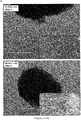
Figure 5 shows electronic microscopy photos (no staining) representing the osteoblastic differentiation (phase 2) of L-GSCs or S-GSCs depending on whether they come from a proliferation medium with FCS or PL medium, respectively, in an inductive medium chosen from FSC osteogenic medium or serum-free PL osteogenic medium, in 3D, over 7, 14 and 21 days, with human allogeneic bone particles according to a protocol well-known to the skilled person. -
Culture type basic medium PL level Cell culture supplements Change of culture medium Culture time Cell proliferation in serum-free medium (PL + GH medium) 2D DMEM-LG GlutaMAX® and pyruvate supplement (Gibco-Life Technologies, Carlsbad, CA, USA) 10% 4ng/ml GH (Peprotech, USA) 2x per week up to a confluence of 80% L-Ascorbic acid 2-phosphate (50µg/ml; Sigma-Aldrich) 10ml/l Penicillin-Streptomycin (5UI/ml, Gibco) 1% non-essential aminoacids (NEAA; Gibco) 2,5mg/l d'amphotericin B (250µg/ml ; Gibco) Heparin 2UI/ml Proliferation test The PL + GH medium has proliferation rates equivalent to or higher than the conventional medium supplemented with 10% FCS Primary culture Isolation of GSCs in primary culture is possible without the use of FCS with conservation of their properties (called L-GSC in contrast to S-GSCs isolated in the FCS) CFU-F L-GSCs are able to form larger clones compared to S-GSCs, after a culture in limit dilution Freeze/thaw Freezing of GSCs is possible by using: 50% PL+GH medium, 40% PL, 10% DMSO, with preservation of their proliferation and multipotency properties Safety test No significant DNA lesions of L-GSCs were found when compared to S-GSCs and UV-irradiated gingival cells (Ctrl+) Genotype test L-GSCs express similar phenotypic markers. Differentiation test L-GSCs are able to maintain their potential for differentiation. Their osteoblastic differentiation potential is very much improved compared to S-GSC Osteoblastic differentiation in serum-free medium (PL medium) 2D et 3D DMEM-LG GlutaMAX® and pyruvate supplement (Gibco-Life Technologies, Carlsbad, CA, USA) 5% L-Ascorbic acid 2-phosphate (50µg/ml; Sigma-Aldrich) 2x per week 21 to 28 days 100nM dexamethasone 10mM β-glycerophosphate 10ml/l Penicillin-Streptomycin (5UI/ml, Gibco) 1% non-essential aminoacids (NEAA; Gibco) 2,5mg/l d'amphotericin B (250µg/ml ; Gibco) Heparin 0,6UI/ml 2D culture The osteogenic medium PL 5% allows to obtain more mineral nodules than the medium supplemented with FSC3D culture Culture on allogeneic bone particles for 21 days shows that the cells spread and form pseudopods with mineral nodules in both media with PL and FCS DMEM-LG : Dulbecco's Modified Eagle Medium Low Glucose
FCS : Foetal Calf Serum
GH : Growth Hormone
PL : Platelet Lysate - It was first demonstrated that GSCs amplified in a 10% PL medium showed a significantly higher proliferation rate than 1%, 2.5% and 5% PL media (p<0.05%). However, this rate remained lower than that obtained in a traditional FCS 10% medium.
- The Inventors also looked for elements in the FCS components that the PL did not contain and that could impact cell proliferation. Growth hormone (GH) and testosterone were selected and added to the 10% PL medium. In addition, the Inventors also assumed that the concentrations of growth factors present in the PL (FGFb, EGF, etc.) were not sufficient in some PL batches to support the proliferation of GSCs in the absence of FCS. Indeed, PL batches may show variations related to donors as well as in the manufacturing method (number of freeze/thaw cycles, storage method), which could impact the quality and content of growth factors (Doucet et al., 2005; Bernardi et al., 2013) [17, 18].
- The results of cellular proliferation in serum-free medium in the presence of GH with 10% PL medium showed significantly higher proliferation rates than the 10% PL medium alone. This was also confirmed by 24-hour flow cytometry (CFSE) results where an increase in phase S was noted in this new PL 10% + GH medium.
- In addition, the isolation of GSCs (phase 1) in the serum-free medium PL 10% + GH was as effective as the isolation of GSCs in the classical 10% FCS medium, by the explant method according to the well-known method to the skilled person, as shown in
Figure 1 . However a denser cell population was observed at 3 weeks in L-GSC than in S-GSC. The population was named L-GSC when obtained with the serum-free medium PL 10% + GH, in contrast to the population named S-GSC when obtained with the classical 10% FCS medium. - This was the first time that a culture medium for GSC proliferation has been supplemented with GH for the growth and isolation of these cells.
- The PL10% + GH medium has preserved the properties of L-GSCs and improved the ability to form clones.
- Cryopreservation of GSCs was possible in 50% PL medium 10% + GH, 40% PL10%, 10% DMSO, at -80°C, for up to 3 months. Beyond that, the cells presented more difficulty to recover.
-
Figure 2 showed the freeze/thaw of L-GSCs in the PL medium with preservation of their multipotency. IndeedFigure 2B showed the ability of L-GSCs to differentiate into osteoblasts, andfigure 2C showed another ability of L-GSCs to differentiate into adipocytes. -
Figure 3 showed the absence of significant DNA damage in L-GSC cells (figure 3C ) compared to S-GSC cells (figure 3B ) and UV-irradiated gingival fibroblasts (Ctrl+) (figure 3A ), by Hoechst staining (Anti H2ax) according to a protocol well-known to the skilled person. - If the morphology of isolated L-GSCs differs from that of S-GSGs, as already reported in the literature for MSCs (Vogel et al., 2006; Chevallier et al., 2010) [19, 20], L-GSCs express the same intra-cellular and extra-cellular phenotypic markers as S-GSCs.
- The maintenance of the differentiation capacity of L-GSCs clearly showed the positive effect of this production and isolation method, which has enabled the multipotency properties to be maintained with a very significant increase in osteogenic and adipogenic differentiation.
-
Figure 4 showed the differentiation into osteoblasts (phase 2) by an inductive medium containing either FSC or PL, of stem cells (GSC) or control cells (GF with less than 5% stem cells) previously obtained (phase 1) in a culture medium with platelet lysate (L-GSF and L-GSC) or a culture medium with FSC (S-GSC and S-GF). -
Figure 4 showed two phenomena: first, GSCs produced more mineralized tissue than GFs, secondly and more importantly, PL was much more effective that FCS at inducing mineralization. Indeed the highest mineralization rate was observed when phases 1 and 2 were carried out in PL medium (see last histogram infigure 4 ). Thus it was shown an improvement in osteogenic potential and mineral nodule formation for L-GSCs and in a serum-free osteogenic medium supplemented with PL. - Without being limited by this explanation, osteogenic differentiation could be explained by the effect of TGFβ1 in the early stages of differentiation (Zhao et al., 2009) [21] as well as other platelet growth factors, through mechanisms that remain to be explored. GH could also have a role in increasing the osteoblastic differentiation potential of GSCs, either by increasing their multipotency capacity by increasing the TGFβ1 rate, or by acting directly on osteoblastic differentiation.
- The development of an osteogenic medium supplemented with PL for the differentiation of GSCs has been very difficult. The concentrations of PL5% and 10% were found to be the most suitable for GSCs. Heparin concentration was also important for the culture of GSCs in media containing PL. Regarding osteoblastic differentiation, this concentration of heparin was significant. Indeed, a concentration at 2 IU/ml, as recommended by the manufacturer, prevented osteoblastic differentiation in early experiments or significantly reduced its potential in some strains of GSC. The lowest possible heparin concentration (0.6 IU/ml) that is effective in preventing gel formation (coagulation) was therefore used. Cultured on a 3D matrix of porous allogenic bone (allograft Zimmer) showed good viability (Calceïn AM) and the presence of GSC mineralization around and between the nodules of this matrix.
Figure 5 showed the formation of mineral pseudopods and nodules for S-GSC and L-GSC. However it must be noted that the matrix is much better organized in the PL osteogenic medium (serum-free) than in the FSC osteogenic medium. It thus confirmed the possibility of transfer of L-GSC cells in humans for bone reconstruction. This makes it possible to consider the transfer of L-GSC cells in humans for bone reconstruction. - The adipocyte differentiation of L-GSCs was significantly increased under serum-free adipogenic conditions compared to S-GSCs. Finally, the ability to differentiate to the myofibroblastic pathway and the formation of neurospheres could have therapeutic applications in wound healing and nerve damage in humans with these newly isolated cells.
- The study of the immune profile of peripheral blood stem cells (PBMCs) cocultured with GSCs in the presence of antigenic particles showed that in these conditions, cytotoxic memory and regulatory T cells are significantly increased by the presence of L-GSCs. This showed the preservation of the role of GSCs in maintaining immune balance in lymphocyte populations during an inflammatory reaction. CD4+ CD3+ CD3+ CD25+ CD25+ FoxP3+ T cell proliferation was increased in the presence of S-GSC and L-GSC, as well as in the presence of PL10%+GH medium alone. This showed that S-GSC and L-GSC had the same immunomodulatory properties and that the new medium acted on the lymphocyte profile by increasing the CD4+ regulatory population. Also, from a clinical point of view, the immunomodulatory properties of GSCs could be used to improve bone grafting, especially allogeneic.
- In conclusion, GSCs in the serum-free medium supplemented with PL (preferably PL10%) and GH allowed these cells to retain their cellular phenotypes and properties required for MSCs, with an improved ability to form clones and differentiate towards the osteoblastic pathway. The immunomodulatory properties of GSCs have also been preserved with a likely role of PL alone on immunomodulation by reducing CD8+ CD25+ and CD8+ CD45RO+ populations and inducing CD4+ CD3+ CD25+ FoxP3+ population.
-
- 1. Listl et al., Journal of Dental Research, 0022034515602879, 2015
- 2. Petersen et Yamamoto, Community Dentistry and Oral Epidemiology, 33(2) : 81-92, 2005
- 3. Gundlach et Christina, Journal of Cranio-Maxillofacial Surgery, 34 : 1-2, 2006
- 4. Montero et Patel, Surgical Oncology Clinics of North America, 24(3) : 491, 2015
- 5. Leucht et al., Development, 135(17) : 2845-2854, 2008
- 6. Jacques et al., Connective Tissue Research, 55(sup1) : 117-120, 2014
- 7. Nassif, These « Role des cellules orales dérivées des crêtes neurales dans la morphogenèse craniofaciale », 2016
- 8. Najar et al., Cytotherapy, 18(2) : 160-171, 2016
- 9. Seifert et al., World Journal of Stem Cells, 7(3) : 583, 2015
- 10. d'Aquino et al., Eur. Cell Mater., 18(7) : 75-83, 2009
- 11. Fournier et al., Tissue Engineering Part A, 16(9) : 2891-2899, 2010
- 12. Zhang et al., The Journal of Immunology, 183(12) : 7787-7798, 2009
- 13. Chen et al., ARthritis & Rheumatism, 65(5) : 1181-1193, 2013
- 14. Tomar et al., Biochemical and Biophysical Research Communications, 393(3) : 377-383, 2010
- 15. Wang et al., Stem Cells and Development, 20(12) : 2093-2102, 2011
- 16. Naveau et al., Journal of Periodontology, 82(4) : 632-641, 2011
- 17. Doucet et al., Journal of Cellular Physiology, 205(2) : 228-236, 2005
- 18. Bernardi et al., Cytotherapy, 5(8) : 920-929, 2013
- 19. Vogel et al., Platelets, 17(7) : 462-469, 2006
- 20. Chevallier et al., Biomaterials, 31(2) : 270-278, 2010
- 21. Zhao et al., Tissue Engineering part A, 16(2) : 725-733, 2009
Claims (15)
- A method for inducing differentiation of neuroectodemic oral stem cells into osteoblasts, said method comprising:(a) a step of cell proliferation of neuroectodermal oral stem cells in a serum-free base culture medium supplemented with platelet lysate (PL) and human growth hormone (GH);b) an osteoblastic differentiation step of the cell culture resulting from step a) in a serum-free base culture medium supplemented with a platelet lysate (PL) and at least one osteoblastic differentiation factor.
- Method according to claim 1, wherein the culture of step a) is carried out up to a confluence of 80%.
- Method according to claim 1 or 2, wherein the culture of step b) is carried out during 21 to 28 days.
- Method according to any of claim 1 to 3, wherein the culture medium of steps a) and b) is further supplemented with heparin.
- Method according to claim 4, wherein the culture medium of step a) contains from 1-2 IU/ml of heparin, and/or the culture medium of step b) contains from 0.1-0.6 IU/ml of heparin.
- Method according to any of claims 1 to 5, wherein said at least one osteoblastic differentiation factor is chosen from the group consisting of corticosteroids and glycerol phosphoric esters.
- Method according to claim 6, wherein the said at least one differentiation factor is dexamethasone and/or β-glycerophosphate.
- Method according to any of claims 1 to 7, wherein the neuroectodermic oral stem cells are derived from gingival tissue.
- Cell population comprising osteoblasts produced by the method according to any of claims 1 to 8, said osteoblasts expressing the homeogen Msx2.
- Cell population as defined in claim 9, for use in cell therapy.
- Cell population for use according to claim 10, for the restoration of bone defects of maxillary bones.
- Culture medium for the proliferation of oral neuroectodermal stem cells comprising a serum-free base culture medium supplemented with platelet lysate (PL) and human growth hormone (GH), and optionally heparin at a concentration ranging from 1-2 IU/ml.
- Culture medium for the differentiation of neuroectodermal oral stem cells into osteoblasts comprising a serum-free base culture medium supplemented with a platelet lysate (PL) and at least one osteoblastic differentiation factor, and optionally heparin at a concentration ranging from 0.1-0.6 IU/ml.
- Culture medium according to claim 13, wherein said at least one osteoblastic differentiation factor is chosen from the group consisting of corticosteroides and glycerol phosphoric esters.
- Culture medium according to claim 14, wherein said at least one osteoblastic differentiation factor is dexamethasone and/or β-glycerophosphate.
Priority Applications (5)
| Application Number | Priority Date | Filing Date | Title |
|---|---|---|---|
| EP18306591.1A EP3660144A1 (en) | 2018-11-30 | 2018-11-30 | Osteoblasts derived from oral neuroectodermal stem cells and their use in jaw repair |
| PCT/EP2019/082970 WO2020109507A1 (en) | 2018-11-30 | 2019-11-28 | Osteoblasts derived from oral neuroectodermal stem cells and their use in jaw repair |
| EP19809479.9A EP3887506A1 (en) | 2018-11-30 | 2019-11-28 | Osteoblasts derived from oral neuroectodermal stem cells and their use in jaw repair |
| US17/291,661 US20210403869A1 (en) | 2018-11-30 | 2019-11-28 | Osteoblasts derived from oral neuroectodermal stem cells and their use in jaw repair |
| CA3119235A CA3119235A1 (en) | 2018-11-30 | 2019-11-28 | Osteoblasts derived from oral neuroectodermal stem cells and their use in jaw repair |
Applications Claiming Priority (1)
| Application Number | Priority Date | Filing Date | Title |
|---|---|---|---|
| EP18306591.1A EP3660144A1 (en) | 2018-11-30 | 2018-11-30 | Osteoblasts derived from oral neuroectodermal stem cells and their use in jaw repair |
Publications (1)
| Publication Number | Publication Date |
|---|---|
| EP3660144A1 true EP3660144A1 (en) | 2020-06-03 |
Family
ID=64665347
Family Applications (2)
| Application Number | Title | Priority Date | Filing Date |
|---|---|---|---|
| EP18306591.1A Withdrawn EP3660144A1 (en) | 2018-11-30 | 2018-11-30 | Osteoblasts derived from oral neuroectodermal stem cells and their use in jaw repair |
| EP19809479.9A Withdrawn EP3887506A1 (en) | 2018-11-30 | 2019-11-28 | Osteoblasts derived from oral neuroectodermal stem cells and their use in jaw repair |
Family Applications After (1)
| Application Number | Title | Priority Date | Filing Date |
|---|---|---|---|
| EP19809479.9A Withdrawn EP3887506A1 (en) | 2018-11-30 | 2019-11-28 | Osteoblasts derived from oral neuroectodermal stem cells and their use in jaw repair |
Country Status (4)
| Country | Link |
|---|---|
| US (1) | US20210403869A1 (en) |
| EP (2) | EP3660144A1 (en) |
| CA (1) | CA3119235A1 (en) |
| WO (1) | WO2020109507A1 (en) |
Citations (1)
| Publication number | Priority date | Publication date | Assignee | Title |
|---|---|---|---|---|
| WO2013076726A1 (en) * | 2011-11-21 | 2013-05-30 | Ramot At Tel-Aviv University Ltd. | Stem cell-derived neural cells for cell therapy in neurological disorders |
-
2018
- 2018-11-30 EP EP18306591.1A patent/EP3660144A1/en not_active Withdrawn
-
2019
- 2019-11-28 US US17/291,661 patent/US20210403869A1/en not_active Abandoned
- 2019-11-28 EP EP19809479.9A patent/EP3887506A1/en not_active Withdrawn
- 2019-11-28 WO PCT/EP2019/082970 patent/WO2020109507A1/en not_active Ceased
- 2019-11-28 CA CA3119235A patent/CA3119235A1/en active Pending
Patent Citations (1)
| Publication number | Priority date | Publication date | Assignee | Title |
|---|---|---|---|---|
| WO2013076726A1 (en) * | 2011-11-21 | 2013-05-30 | Ramot At Tel-Aviv University Ltd. | Stem cell-derived neural cells for cell therapy in neurological disorders |
Non-Patent Citations (25)
| Title |
|---|
| BERNARDI ET AL., CYTOTHERAPY, vol. 5, no. 8, 2013, pages 920 - 929 |
| BP FOURNIER ET AL: "Characterisation of human gingival neural crest-derived stem cells in monolayer and neurosphere cultures", EUROPEAN CELLS AND MATERIALS, vol. 31, 5 January 2016 (2016-01-05), pages 40 - 58, XP055328746, DOI: 10.22203/eCM.v031a04 * |
| CHEN ET AL., ARTHRITIS & RHEUMATISM, vol. 65, no. 5, 2013, pages 1181 - 1193 |
| CHEVALLIER ET AL., BIOMATERIALS, vol. 31, no. 2, 2010, pages 270 - 278 |
| D'AQUINO ET AL., EUR. CELL MATER., vol. 18, no. 7, 2009, pages 75 - 83 |
| DOUCET ET AL., JOURNAL OF CELLULAR PHYSIOLOGY, vol. 205, no. 2, 2005, pages 228 - 236 |
| FOURNIER BENJAMIN P J ET AL: "Multipotent progenitor cells in gingival connective tissue.", TISSUE ENGINEERING. PART A SEP 2010, vol. 16, no. 9, September 2010 (2010-09-01), pages 2891 - 2899, XP002790855, ISSN: 1937-335X * |
| FOURNIER ET AL., TISSUE ENGINEERING PART A, vol. 16, no. 9, 2010, pages 2891 - 2899 |
| FRANCOIS C FERRE ET AL: "Formation of Cartilage and Synovial Tissue by Human Gingival Stem Cells", STEM CELLS AND DEVELOPMENT, vol. 23, no. 23, 1 December 2014 (2014-12-01), pages 2895 - 2907, XP055412401, ISSN: 1547-3287, DOI: 10.1089/scd.2013.0547 * |
| GUNDLACH; CHRISTINA, JOURNAL OF CRANIO-MAXILLOFACIAL SURGERY, vol. 34, 2006, pages 1 - 2 |
| JACQUES ET AL., CONNECTIVE TISSUE RESEARCH, vol. 55, no. 1, 2014, pages 117 - 120 |
| LEUCHT ET AL., DEVELOPMENT, vol. 135, no. 17, 2008, pages 2845 - 2854 |
| LISTL ET AL., JOURNAL OF DENTAL RESEARCH, 2015 |
| MONTERO; PATEL, SURGICAL ONCOLOGY CLINICS OF NORTH AMERICA, vol. 24, no. 3, 2015, pages 491 |
| NAJAR ET AL., CYTOTHERAPY, vol. 18, no. 2, 2016, pages 160 - 171 |
| NASSIF: "Role des cellules orales derivees des cretes neurales dans la morphogenese craniofaciale", THESE, 2016 |
| NAVEAU ADRIEN ET AL: "Phenotypic study of human gingival fibroblasts in a medium enriched with platelet lysate.", JOURNAL OF PERIODONTOLOGY APR 2011, vol. 82, no. 4, April 2011 (2011-04-01), pages 632 - 641, XP002790854, ISSN: 1943-3670 * |
| NAVEAU ET AL., JOURNAL OF PERIODONTOLOGY, vol. 82, no. 4, 2011, pages 632 - 641 |
| PETERSEN; YAMAMOTO, COMMUNITY DENTISTRY AND ORAL EPIDEMIOLOGY, vol. 33, no. 2, 2005, pages 81 - 92 |
| SEIFERT ET AL., WORLD JOURNAL OF STEM CELLS, vol. 7, no. 3, 2015, pages 583 |
| TOMAR ET AL., BIOCHEMICAL AND BIOPHYSICAL RESEARCH COMMUNICATIONS, vol. 393, no. 3, 2010, pages 377 - 383 |
| VOGEL ET AL., PLATELETS, vol. 17, no. 7, 2006, pages 462 - 469 |
| WANG ET AL., STEM CELLS AND DEVELOPMENT, vol. 20, no. 12, 2011, pages 2093 - 2102 |
| ZHANG ET AL., JOURNAL OF IMMUNOLOGY, vol. 183, no. 12, 2009, pages 7787 - 7798 |
| ZHAO ET AL., TISSUE ENGINEERING PART A, vol. 16, no. 2, 2009, pages 725 - 733 |
Also Published As
| Publication number | Publication date |
|---|---|
| CA3119235A1 (en) | 2020-06-04 |
| EP3887506A1 (en) | 2021-10-06 |
| US20210403869A1 (en) | 2021-12-30 |
| WO2020109507A1 (en) | 2020-06-04 |
Similar Documents
| Publication | Publication Date | Title |
|---|---|---|
| US20220096564A1 (en) | Methods and compositions for treatment of bone defects with placental cell populations | |
| Hynes et al. | Clinical utility of stem cells for periodontal regeneration | |
| US20170157291A1 (en) | Tissue transplant compositions and methods for use | |
| EP1934332B1 (en) | Methods for differentiating stem cells and use thereof in the treatment of dental conditions | |
| EP3660144A1 (en) | Osteoblasts derived from oral neuroectodermal stem cells and their use in jaw repair | |
| Zhao et al. | Evaluation of immunocompatibility of tissue-engineered periosteum | |
| CN103857788A (en) | Uses of growth and differentiation factor 8 (gdf-8) | |
| Li et al. | Comparison of Stemness and Immunogenicity of Osteo-Differentiated Mesenchymal Stem Cells Derived from Rabbit Bone Marrow and Wharton's Jelly | |
| de Peppo et al. | Human progenitor cells for bone engineering applications | |
| Ali | Stemness and osteogenic differentiation of induced pluripotent stem cells generated in xeno-free conditions | |
| Honda et al. | Dental Stem Cells for Tooth Tissue Engineering | |
| Brooks | Clinical Aspects of the Use of Stem Cells and Biomaterials for Bone Repair and Regeneration | |
| Jabari et al. | Isolation Stem Cells from Human Dental Pulps and Differentiation Them to Adiposite and Osteoblast | |
| HK40006584A (en) | Methods and compositions for treatment of bone defects with placental cell populations | |
| Kassem et al. | Human (Skeletal) Mesenchymal Stem Cells: Basic Biology and Clinical Applications for Bone Tissue Regeneration | |
| Aldahmash | Human (Skeletal) Mesenchymal Stem Cells: Basic Biology and Clinical Applications | |
| AU2014201880A1 (en) | Methods and compositions for treatment of bone defects with placental cell populations | |
| HK1166997A (en) | Methods and compositions for treatment of bone defects with placental cell populations |
Legal Events
| Date | Code | Title | Description |
|---|---|---|---|
| PUAI | Public reference made under article 153(3) epc to a published international application that has entered the european phase |
Free format text: ORIGINAL CODE: 0009012 |
|
| STAA | Information on the status of an ep patent application or granted ep patent |
Free format text: STATUS: THE APPLICATION HAS BEEN PUBLISHED |
|
| AK | Designated contracting states |
Kind code of ref document: A1 Designated state(s): AL AT BE BG CH CY CZ DE DK EE ES FI FR GB GR HR HU IE IS IT LI LT LU LV MC MK MT NL NO PL PT RO RS SE SI SK SM TR |
|
| AX | Request for extension of the european patent |
Extension state: BA ME |
|
| STAA | Information on the status of an ep patent application or granted ep patent |
Free format text: STATUS: THE APPLICATION IS DEEMED TO BE WITHDRAWN |
|
| 18D | Application deemed to be withdrawn |
Effective date: 20201204 |