EP3657432A1 - Image normalization method and image processing device - Google Patents
Image normalization method and image processing device Download PDFInfo
- Publication number
- EP3657432A1 EP3657432A1 EP19170153.1A EP19170153A EP3657432A1 EP 3657432 A1 EP3657432 A1 EP 3657432A1 EP 19170153 A EP19170153 A EP 19170153A EP 3657432 A1 EP3657432 A1 EP 3657432A1
- Authority
- EP
- European Patent Office
- Prior art keywords
- image
- input image
- value
- cell
- input
- Prior art date
- Legal status (The legal status is an assumption and is not a legal conclusion. Google has not performed a legal analysis and makes no representation as to the accuracy of the status listed.)
- Granted
Links
Images




Classifications
-
- G—PHYSICS
- G06—COMPUTING OR CALCULATING; COUNTING
- G06T—IMAGE DATA PROCESSING OR GENERATION, IN GENERAL
- G06T5/00—Image enhancement or restoration
- G06T5/90—Dynamic range modification of images or parts thereof
- G06T5/92—Dynamic range modification of images or parts thereof based on global image properties
-
- G—PHYSICS
- G06—COMPUTING OR CALCULATING; COUNTING
- G06T—IMAGE DATA PROCESSING OR GENERATION, IN GENERAL
- G06T5/00—Image enhancement or restoration
- G06T5/40—Image enhancement or restoration using histogram techniques
-
- G—PHYSICS
- G06—COMPUTING OR CALCULATING; COUNTING
- G06T—IMAGE DATA PROCESSING OR GENERATION, IN GENERAL
- G06T5/00—Image enhancement or restoration
- G06T5/60—Image enhancement or restoration using machine learning, e.g. neural networks
-
- G—PHYSICS
- G06—COMPUTING OR CALCULATING; COUNTING
- G06T—IMAGE DATA PROCESSING OR GENERATION, IN GENERAL
- G06T2207/00—Indexing scheme for image analysis or image enhancement
- G06T2207/10—Image acquisition modality
- G06T2207/10024—Color image
-
- G—PHYSICS
- G06—COMPUTING OR CALCULATING; COUNTING
- G06T—IMAGE DATA PROCESSING OR GENERATION, IN GENERAL
- G06T2207/00—Indexing scheme for image analysis or image enhancement
- G06T2207/10—Image acquisition modality
- G06T2207/10056—Microscopic image
-
- G—PHYSICS
- G06—COMPUTING OR CALCULATING; COUNTING
- G06T—IMAGE DATA PROCESSING OR GENERATION, IN GENERAL
- G06T2207/00—Indexing scheme for image analysis or image enhancement
- G06T2207/20—Special algorithmic details
- G06T2207/20081—Training; Learning
-
- G—PHYSICS
- G06—COMPUTING OR CALCULATING; COUNTING
- G06T—IMAGE DATA PROCESSING OR GENERATION, IN GENERAL
- G06T2207/00—Indexing scheme for image analysis or image enhancement
- G06T2207/20—Special algorithmic details
- G06T2207/20084—Artificial neural networks [ANN]
-
- G—PHYSICS
- G06—COMPUTING OR CALCULATING; COUNTING
- G06T—IMAGE DATA PROCESSING OR GENERATION, IN GENERAL
- G06T2207/00—Indexing scheme for image analysis or image enhancement
- G06T2207/20—Special algorithmic details
- G06T2207/20172—Image enhancement details
- G06T2207/20208—High dynamic range [HDR] image processing
-
- G—PHYSICS
- G06—COMPUTING OR CALCULATING; COUNTING
- G06T—IMAGE DATA PROCESSING OR GENERATION, IN GENERAL
- G06T2207/00—Indexing scheme for image analysis or image enhancement
- G06T2207/30—Subject of image; Context of image processing
- G06T2207/30004—Biomedical image processing
- G06T2207/30024—Cell structures in vitro; Tissue sections in vitro
-
- G—PHYSICS
- G06—COMPUTING OR CALCULATING; COUNTING
- G06T—IMAGE DATA PROCESSING OR GENERATION, IN GENERAL
- G06T2207/00—Indexing scheme for image analysis or image enhancement
- G06T2207/30—Subject of image; Context of image processing
- G06T2207/30004—Biomedical image processing
- G06T2207/30096—Tumor; Lesion
Definitions
- the invention is related to an image normalization method and an image processing device, and more particularly, to an image normalization method and an image processing device for visually displaying a cell.
- the microscopic image acquisition of circulating cancer cells mainly includes placing a stained sample of blood cells on a glass slide and photographing the sample with a microscope.
- the difference in brightness between the stained area and the unstained area on the microscope image is very large.
- the brightness of the stained cells is very similar to that of the stained area. If the cell image is not processed first, then the color difference between the stained cells and the stained background area cannot be distinguished because the brightness of the stained area is too low. Therefore, how to perform image processing on stained samples of cells to visually display cells is an object that those skilled in the art should strive for.
- the invention provides an image normalization method and an image processing device performing image processing on stained samples of cells to visually display cells.
- the invention provides an image normalization method adapted to visually display at least one cell from an input image.
- the input image includes a stained area and an unstained area. At least one cell is located in the stained area.
- the image normalization method includes the following steps. An input image is obtained. A cell visualization operation is performed on each pixel of the input image, wherein the cell visualization operation generates an output value of each pixel of the input image according to an input value of each pixel of the input image, a color distribution center corresponding to at least one cell, a contrast adjustment value, and a brightness adjustment value. An output image is generated according to the output value of each pixel of the input image to be displayed on a display or entered into an artificial intelligence (AI) model or a deep-learning model as training data or input data.
- AI artificial intelligence
- the color distribution center is the peak position of a cell distribution in a histogram of the input image.
- the color distribution center is obtained by a machine learning or deep-learning method according to a histogram feature of the input image.
- the output value is between 0 and 1.
- the invention provides an image processing device including a storage device storing an input image; a display; and a processor coupled to the storage device and the display.
- the processor obtains an input image.
- the processor performs a cell visualization operation on each pixel of the input image, wherein the cell visualization operation generates an output value of each pixel of the input image according to an input value of each pixel of the input image, a color distribution center corresponding to at least one cell, a contrast adjustment value, and a brightness adjustment value.
- the processor generates an output image for display on the display according to the output value of each pixel of the input image.
- the color distribution center is the peak position of a cell distribution in a histogram of the input image.
- the color distribution center is obtained by a machine learning or deep-learning method according to a histogram feature of the input image.
- the output value is between 0 and 1.
- the image normalization method and the image processing device of the invention may perform a cell visualization operation according to an input value of each pixel of the input image, a color distribution center corresponding to at least one cell, a contrast adjustment value, and a brightness adjustment value to generate an output image and display the output image on a display.
- FIG. 1 is a block diagram of an image processing device according to an embodiment of the invention.
- an image processing device 100 of an embodiment of the invention includes a processor 110 and a storage device 120 and a display 130 coupled to the processor 110.
- the storage device 120 may store an input image.
- the input image includes a stained area and an unstained area and cells are located in the stained area.
- the cells may include circulating cancer cells and other cells in the blood.
- the processor 110 may perform a cell visualization operation on the input image to generate an output image and display the output image on the display 130 so that the human eye may recognize the stained cells of the output image.
- the processor 110 may be a central processing unit (CPU) or a general-purpose or special-purpose programmable microprocessor, digital signal processor (DSP), programmable controller, application-specific integrated circuit (ASIC), other similar devices, or a combination of the above devices, but the invention is not limited thereto.
- CPU central processing unit
- DSP digital signal processor
- ASIC application-specific integrated circuit
- the storage device 120 may be any form of a fixed or movable random-access memory (RAM), read-only memory (ROM), flash memory, hard disk drive (HDD), solid-state drive (SSD), similar devices, or a combination of the above devices.
- RAM random-access memory
- ROM read-only memory
- HDD hard disk drive
- SSD solid-state drive
- FIG. 2 is a schematic of a histogram of an input image according to an embodiment of the invention.
- the horizontal axis is the brightness and the vertical axis is the number of pixels.
- the brightness information of the input image is stored in 12 bits (e.g., brightness range of 0 to 4095), and the brightness information of the output image is stored in 16 bits (e.g., brightness range of 0 to 65535).
- the histogram of FIG. 2 includes a stained background 210 having the lowest brightness, an unstained area 220 having the highest brightness, and stained cells 230 having a slightly higher brightness than the stained background 210. Since the brightness of the stained cells 230 and the stained background 210 are too similar, the human eye cannot distinguish the color difference between the stained cells 230 and the stained background 210.
- the processor 110 may perform histogram equalization on the input image to generate an output image, as shown in FIG. 3.
- FIG. 3 shows an output image 310 generated by performing histogram equalization on the input image and a partially enlarged view 320 of the output image 310.
- the partially enlarged view 320 the color distributions of the stained cells 230 and the stained background 210 are too similar and the output image 310 has a large unstained area 220, resulting such that the image brightness of the output image 310 is too high and a large amount of noise is generated.
- the processor 110 may perform histogram equalization and gamma correction on the input image to generate an output image, as shown in FIG. 4.
- FIG. 4 shows an output image 410 generated by performing histogram equalization and gamma correction on the input image and a partially enlarged view 420 of the output image 410.
- performing histogram equalization and gamma correction on the input image solves the issue of excessive brightness and suppresses some noise, but cell information is still easily lost.
- ⁇ is the output value and k is the input value.
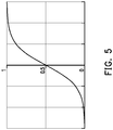
- FIG. 5 each input value of the horizontal axis corresponds to an output value between 0 and 1 of the vertical axis via the above equation.
- the output value approaches 1 and when the input value is too small, the output value approaches 0.
- an effect similar to filtering the parts of the image that are too bright and too dark using two threshold values may be achieved.
- the processor 110 may perform a cell visualization operation on each pixel of the input image.
- y is the output value
- c is the contrast adjustment value
- ⁇ is the brightness adjustment value
- t is the color distribution center
- x is the input value.
- the color distribution center is the peak position of a cell distribution in a histogram of the input image (i.e., the horizontal axis value of the peak of the stained cells 230).
- the color distribution center is obtained by a machine learning or deep-learning method according to a histogram feature of the input image.
- the color distribution center may also be set according to empirical rules. The closer the color distribution center is to the intermediate value of the plurality of input values of the stained cells 230, the easier it is for the user to recognize the cells from the output image. It is worth noting that the output value y is between 0 and 1.
- the processor 110 converts the output value y into a brightness value ranging from 0 to 65535 to generate the output image.
- the processor 110 may simultaneously filter areas of the input image that are too bright (e.g., the unstained area 220) and too dark (e.g., the stained background 210) via the second equation and retain and amplify important information of the stained cells 230 via the color distribution center t and the contrast adjustment value C. In addition, the processor 110 may also adjust the overall brightness of the output image via the brightness adjustment value ⁇ .
- FIG. 6 is an output image 610 generated by performing a cell visualization operation on an input image and a partially enlarged view 620 of the output image 610.
- a cell information 630 is more completely retained than a cell information 430 of FIG. 4 .
- FIG. 7 is a flowchart of an image normalization method according to an embodiment of the invention.
- step S701 an input image is obtained.
- step S702 a cell visualization operation is performed on each pixel of the input image, wherein the cell visualization operation generates an output value of each pixel of the input image according to an input value of each pixel of the input image, a color distribution center corresponding to at least one cell, a contrast adjustment value, and a brightness adjustment value.
- step S703 an output image is generated for display on a display according to the output value of each pixel of the input image.
- the image normalization method and the image processing device of the invention may perform a cell visualization operation according to an input value of each pixel of the input image, a color distribution center corresponding to at least one cell, a contrast adjustment value, and a brightness adjustment value to generate an output image, and the output image is displayed on a display or entered into an AI model or a deep-learning model as training data or input data.
- a cell visualization operation areas of the input image that are too bright and too dark may be filtered, and important information of the cells is retained and enlarged, making it easier for the user to recognize cells from the output image.
Landscapes
- Physics & Mathematics (AREA)
- General Physics & Mathematics (AREA)
- Engineering & Computer Science (AREA)
- Theoretical Computer Science (AREA)
- Image Analysis (AREA)
- Image Processing (AREA)
Abstract
Description
- The invention is related to an image normalization method and an image processing device, and more particularly, to an image normalization method and an image processing device for visually displaying a cell.
- The microscopic image acquisition of circulating cancer cells mainly includes placing a stained sample of blood cells on a glass slide and photographing the sample with a microscope. However, the difference in brightness between the stained area and the unstained area on the microscope image is very large. In addition, the brightness of the stained cells is very similar to that of the stained area. If the cell image is not processed first, then the color difference between the stained cells and the stained background area cannot be distinguished because the brightness of the stained area is too low. Therefore, how to perform image processing on stained samples of cells to visually display cells is an object that those skilled in the art should strive for.
- The invention provides an image normalization method and an image processing device performing image processing on stained samples of cells to visually display cells.
- The invention provides an image normalization method adapted to visually display at least one cell from an input image. The input image includes a stained area and an unstained area. At least one cell is located in the stained area. The image normalization method includes the following steps. An input image is obtained. A cell visualization operation is performed on each pixel of the input image, wherein the cell visualization operation generates an output value of each pixel of the input image according to an input value of each pixel of the input image, a color distribution center corresponding to at least one cell, a contrast adjustment value, and a brightness adjustment value. An output image is generated according to the output value of each pixel of the input image to be displayed on a display or entered into an artificial intelligence (AI) model or a deep-learning model as training data or input data.
- In an embodiment of the invention, the cell visualization operation generates an output value of each pixel of the input image according to an equation y =

- In an embodiment of the invention, the color distribution center is the peak position of a cell distribution in a histogram of the input image.
- In an embodiment of the invention, the color distribution center is obtained by a machine learning or deep-learning method according to a histogram feature of the input image.
- In an embodiment of the invention, the output value is between 0 and 1.
- The invention provides an image processing device including a storage device storing an input image; a display; and a processor coupled to the storage device and the display. The processor obtains an input image. The processor performs a cell visualization operation on each pixel of the input image, wherein the cell visualization operation generates an output value of each pixel of the input image according to an input value of each pixel of the input image, a color distribution center corresponding to at least one cell, a contrast adjustment value, and a brightness adjustment value. The processor generates an output image for display on the display according to the output value of each pixel of the input image.
- In an embodiment of the invention, the cell visualization operation generates the output value of each pixel of the input image according to an equation y=

- In an embodiment of the invention, the color distribution center is the peak position of a cell distribution in a histogram of the input image.
- In an embodiment of the invention, the color distribution center is obtained by a machine learning or deep-learning method according to a histogram feature of the input image.
- In an embodiment of the invention, the output value is between 0 and 1.
- Based on the above, the image normalization method and the image processing device of the invention may perform a cell visualization operation according to an input value of each pixel of the input image, a color distribution center corresponding to at least one cell, a contrast adjustment value, and a brightness adjustment value to generate an output image and display the output image on a display.
- In order to make the aforementioned features and advantages of the disclosure more comprehensible, embodiments accompanied with figures are described in detail below.
- The accompanying drawings are included to provide a further understanding of the invention, and are incorporated in and constitute a part of this specification. The drawings illustrate embodiments of the invention and, together with the description, serve to explain the principles of the invention.
-
FIG. 1 is a block diagram of an image processing device according to an embodiment of the invention. -
FIG. 2 is a schematic of a histogram of an input image according to an embodiment of the invention. -
FIG. 3 is a schematic of an output image generated by performing histogram equalization on an input image according to an embodiment of the invention. -
FIG. 4 is a schematic of an output image generated by performing histogram equalization and gamma correction on an input image according to an embodiment of the invention. -
FIG. 5 is a schematic of converting an input value to an output value by a first equation according to an embodiment of the invention. -
FIG. 6 is a schematic of an output image generated by a second equation according to an embodiment of the invention. -
FIG. 7 is a flowchart of an image normalization method according to an embodiment of the invention. -
FIG. 1 is a block diagram of an image processing device according to an embodiment of the invention. - Referring to
FIG. 1 , animage processing device 100 of an embodiment of the invention includes aprocessor 110 and astorage device 120 and adisplay 130 coupled to theprocessor 110. Thestorage device 120 may store an input image. The input image includes a stained area and an unstained area and cells are located in the stained area. The cells may include circulating cancer cells and other cells in the blood. Theprocessor 110 may perform a cell visualization operation on the input image to generate an output image and display the output image on thedisplay 130 so that the human eye may recognize the stained cells of the output image. - The
processor 110 may be a central processing unit (CPU) or a general-purpose or special-purpose programmable microprocessor, digital signal processor (DSP), programmable controller, application-specific integrated circuit (ASIC), other similar devices, or a combination of the above devices, but the invention is not limited thereto. - The
storage device 120 may be any form of a fixed or movable random-access memory (RAM), read-only memory (ROM), flash memory, hard disk drive (HDD), solid-state drive (SSD), similar devices, or a combination of the above devices. -
FIG. 2 is a schematic of a histogram of an input image according to an embodiment of the invention. - Referring to
FIG. 2 , in the histogram of the input image, the horizontal axis is the brightness and the vertical axis is the number of pixels. In an embodiment, the brightness information of the input image is stored in 12 bits (e.g., brightness range of 0 to 4095), and the brightness information of the output image is stored in 16 bits (e.g., brightness range of 0 to 65535). The histogram ofFIG. 2 includes astained background 210 having the lowest brightness, anunstained area 220 having the highest brightness, andstained cells 230 having a slightly higher brightness than thestained background 210. Since the brightness of thestained cells 230 and thestained background 210 are too similar, the human eye cannot distinguish the color difference between thestained cells 230 and thestained background 210. - In an embodiment, the
processor 110 may perform histogram equalization on the input image to generate an output image, as shown inFIG. 3. FIG. 3 shows anoutput image 310 generated by performing histogram equalization on the input image and a partially enlargedview 320 of theoutput image 310. As may be seen from the partially enlargedview 320, the color distributions of thestained cells 230 and thestained background 210 are too similar and theoutput image 310 has a largeunstained area 220, resulting such that the image brightness of theoutput image 310 is too high and a large amount of noise is generated. - In an embodiment, the
processor 110 may perform histogram equalization and gamma correction on the input image to generate an output image, as shown inFIG. 4. FIG. 4 shows anoutput image 410 generated by performing histogram equalization and gamma correction on the input image and a partially enlargedview 420 of theoutput image 410. As may be seen from theoutput image 410 and the partially enlargedview 420, performing histogram equalization and gamma correction on the input image solves the issue of excessive brightness and suppresses some noise, but cell information is still easily lost. - In an embodiment, the
processor 110 may operate on each pixel of the input image according to the first equation
FIG. 5 . InFIG. 5 , each input value of the horizontal axis corresponds to an output value between 0 and 1 of the vertical axis via the above equation. When the input value is too large, the output value approaches 1 and when the input value is too small, the output value approaches 0. Using the mathematical properties of the first equation, an effect similar to filtering the parts of the image that are too bright and too dark using two threshold values may be achieved. - In an embodiment, the
processor 110 may perform a cell visualization operation on each pixel of the input image. The cell visualization operation generates an output value corresponding to each pixel of the input image according to the second equation
FIG. 5 . In an embodiment, the color distribution center is the peak position of a cell distribution in a histogram of the input image (i.e., the horizontal axis value of the peak of the stained cells 230). In another embodiment, the color distribution center is obtained by a machine learning or deep-learning method according to a histogram feature of the input image. In another embodiment, the color distribution center may also be set according to empirical rules. The closer the color distribution center is to the intermediate value of the plurality of input values of the stainedcells 230, the easier it is for the user to recognize the cells from the output image. It is worth noting that the output value y is between 0 and 1. In an embodiment, after the output value y is calculated, theprocessor 110 converts the output value y into a brightness value ranging from 0 to 65535 to generate the output image. - The
processor 110 may simultaneously filter areas of the input image that are too bright (e.g., the unstained area 220) and too dark (e.g., the stained background 210) via the second equation and retain and amplify important information of the stainedcells 230 via the color distribution center t and the contrast adjustment value C. In addition, theprocessor 110 may also adjust the overall brightness of the output image via the brightness adjustment value γ. - The output image generated according to the second equation is shown in
FIG. 6. FIG. 6 is anoutput image 610 generated by performing a cell visualization operation on an input image and a partiallyenlarged view 620 of theoutput image 610. In the partiallyenlarged view 620, acell information 630 is more completely retained than acell information 430 ofFIG. 4 . -
FIG. 7 is a flowchart of an image normalization method according to an embodiment of the invention. - Referring to
FIG. 7 , in step S701, an input image is obtained. - In step S702, a cell visualization operation is performed on each pixel of the input image, wherein the cell visualization operation generates an output value of each pixel of the input image according to an input value of each pixel of the input image, a color distribution center corresponding to at least one cell, a contrast adjustment value, and a brightness adjustment value.
- In step S703, an output image is generated for display on a display according to the output value of each pixel of the input image.
- Based on the above, the image normalization method and the image processing device of the invention may perform a cell visualization operation according to an input value of each pixel of the input image, a color distribution center corresponding to at least one cell, a contrast adjustment value, and a brightness adjustment value to generate an output image, and the output image is displayed on a display or entered into an AI model or a deep-learning model as training data or input data. Via the cell visualization operation, areas of the input image that are too bright and too dark may be filtered, and important information of the cells is retained and enlarged, making it easier for the user to recognize cells from the output image.
Claims (10)
- An image normalization method adapted to visually display at least one cell from an input image, wherein the input image comprises a stained area and an unstained area (220), and the at least one cell is located in the stained area, the image normalization method comprising:obtaining the input image; andperforming a cell visualization operation on each pixel of the input image, wherein the cell visualization operation generates an output value of each of the pixels of the input image according to an input value of each of the pixels of the input image, a color distribution center corresponding to the at least one cell, a contrast adjustment value, and a brightness adjustment value.
- The image normalization method of claim 1, wherein the cell visualization operation generates the output value of each of the pixels of the input image according to an equation

- The image normalization method of claim 1 or 2, wherein the color distribution center is a peak position of a cell distribution in a histogram of the input image.
- The image normalization method of any one of claim 1 to 3, wherein the cell color distribution center is obtained by a machine learning or deep-learning method according to a histogram feature of the input image.
- The image normalization method of any one of claim 1 to 4, wherein the output value is between 0 and 1.
- An image processing device (100), comprising:a storage device (120) storing an input image;a display (130); anda processor (110) coupled to the storage device (120) and the display (130), whereinthe processor (110) obtains the input image;the processor (110) performs a cell visualization operation on each pixel of the input image, wherein the cell visualization operation generates an output value of each of the pixels of the input image according to an input value of each of the pixels of the input image, a color distribution center corresponding to the at least one cell, a contrast adjustment value, and a brightness adjustment value;the processor (110) generates an output image for display on the display according to the output value of each of the pixels of the input image.
- The image processing device of claim 6, wherein the cell visualization operation generates the output value of each of the pixels of the input image according to an equation

- The image processing device of claim 6 or 7, wherein the color distribution center is a peak position of a cell distribution in a histogram of the input image.
- The image processing device of any one of claim 6 to 8, wherein the color distribution center is obtained by a machine learning or deep-learning method according to a histogram feature of the input image.
- The image processing device of any one of claim 6 to 9, wherein the output value is between 0 and 1.
Applications Claiming Priority (1)
| Application Number | Priority Date | Filing Date | Title |
|---|---|---|---|
| TW107141912A TWI687898B (en) | 2018-11-23 | 2018-11-23 | Image normalization method and image processing device |
Publications (2)
| Publication Number | Publication Date |
|---|---|
| EP3657432A1 true EP3657432A1 (en) | 2020-05-27 |
| EP3657432B1 EP3657432B1 (en) | 2021-07-28 |
Family
ID=66239845
Family Applications (1)
| Application Number | Title | Priority Date | Filing Date |
|---|---|---|---|
| EP19170153.1A Active EP3657432B1 (en) | 2018-11-23 | 2019-04-18 | Image normalization method and image processing device |
Country Status (3)
| Country | Link |
|---|---|
| US (1) | US10755393B2 (en) |
| EP (1) | EP3657432B1 (en) |
| TW (1) | TWI687898B (en) |
Families Citing this family (2)
| Publication number | Priority date | Publication date | Assignee | Title |
|---|---|---|---|---|
| WO2022201447A1 (en) * | 2021-03-25 | 2022-09-29 | 株式会社日立ハイテク | Position coordinate detection device and method |
| TWI811069B (en) | 2022-08-19 | 2023-08-01 | 倍利科技股份有限公司 | System and method for detecting specific cells based on image recognition |
Citations (2)
| Publication number | Priority date | Publication date | Assignee | Title |
|---|---|---|---|---|
| WO2005076197A2 (en) * | 2004-01-31 | 2005-08-18 | Bioimagene, Inc. | Method and system for morphology based mitosis identification and classification of digital images |
| EP1887517A1 (en) * | 2006-08-08 | 2008-02-13 | STMicroelectronics Asia Pacific Pte Ltd. | Automatic contrast enhancement |
Family Cites Families (7)
| Publication number | Priority date | Publication date | Assignee | Title |
|---|---|---|---|---|
| JP3431075B2 (en) * | 2001-08-27 | 2003-07-28 | 科学技術振興事業団 | Liquid crystal display panel unevenness classification processing method, apparatus and program |
| WO2005027015A2 (en) * | 2003-09-10 | 2005-03-24 | Bioimagene, Inc. | Method and system for quantitatively analyzing biological samples |
| US20140233826A1 (en) * | 2011-09-27 | 2014-08-21 | Board Of Regents Of The University Of Texas System | Systems and methods for automated screening and prognosis of cancer from whole-slide biopsy images |
| EP2841927B1 (en) * | 2012-04-27 | 2018-01-17 | Bio-rad Laboratories, Inc. | Stain-free protein quantification and normalization |
| CN103020585B (en) * | 2012-11-06 | 2015-08-26 | 华南师范大学 | A kind of immuning tissue's positive cell and negative cells recognition methods |
| CN107851965B (en) * | 2015-05-20 | 2021-01-05 | 宽腾矽公司 | Light source for fluorescence lifetime analysis |
| TWI756365B (en) * | 2017-02-15 | 2022-03-01 | 美商脫其泰有限責任公司 | Image analysis systems and related methods |
-
2018
- 2018-11-23 TW TW107141912A patent/TWI687898B/en active
-
2019
- 2019-02-26 US US16/285,242 patent/US10755393B2/en active Active
- 2019-04-18 EP EP19170153.1A patent/EP3657432B1/en active Active
Patent Citations (2)
| Publication number | Priority date | Publication date | Assignee | Title |
|---|---|---|---|---|
| WO2005076197A2 (en) * | 2004-01-31 | 2005-08-18 | Bioimagene, Inc. | Method and system for morphology based mitosis identification and classification of digital images |
| EP1887517A1 (en) * | 2006-08-08 | 2008-02-13 | STMicroelectronics Asia Pacific Pte Ltd. | Automatic contrast enhancement |
Non-Patent Citations (1)
| Title |
|---|
| OSMAN M K ET AL: "Contrast enhancement for Ziehl-Neelsen tissue slide images using linear stretching and histogram equalization technique", INDUSTRIAL ELECTRONICS&APPLICATIONS, 2009. ISIEA 2009. IEEE SYMPOSIUM ON, IEEE, PISCATAWAY, NJ, USA, 4 October 2009 (2009-10-04), pages 431 - 435, XP031582316, ISBN: 978-1-4244-4681-0 * |
Also Published As
| Publication number | Publication date |
|---|---|
| US20200167902A1 (en) | 2020-05-28 |
| US10755393B2 (en) | 2020-08-25 |
| TWI687898B (en) | 2020-03-11 |
| TW202020805A (en) | 2020-06-01 |
| EP3657432B1 (en) | 2021-07-28 |
Similar Documents
| Publication | Publication Date | Title |
|---|---|---|
| EP2849431B1 (en) | Method and apparatus for detecting backlight | |
| EP3989534B1 (en) | Image collection method and apparatus, and device and storage medium | |
| CN103971330B (en) | Image enchancing method and device | |
| EP2285097B1 (en) | Image processing apparatus, image processing method, and program | |
| US20210224977A1 (en) | Fundus image processing method, computer apparatus, and storage medium | |
| EP1703436A2 (en) | Image processing system, image processing apparatus and method, recording medium, and program | |
| CN106575497B (en) | Image correction device and video content reproduction device | |
| EP2858036A1 (en) | Image processing device, image capturing apparatus, and image processing method for obtaining depth information | |
| CN111652817B (en) | A method for clearing underwater images based on human visual perception mechanism | |
| CN106017367B (en) | The assay method and device of a kind of canopy density | |
| EP3657432B1 (en) | Image normalization method and image processing device | |
| CN102722868A (en) | Tone mapping method for high dynamic range image | |
| EP3016070A1 (en) | Detection device, learning device, detection method, learning method, and program | |
| JP2007047965A (en) | Method, apparatus and program for detecting object of digital image | |
| KR20190116077A (en) | Image processing | |
| CN104537650A (en) | A method and device for determining the polarity direction of an aluminum electrolytic capacitor | |
| CN106548471A (en) | The medical microscopic images definition evaluation method of coarse-fine focusing | |
| CN107545557A (en) | Egg detecting method and device in excrement image | |
| CN113237881B (en) | A specific cell detection method, device and pathological slice detection system | |
| US20190244360A1 (en) | Image-processing apparatus, image-processing method, and storage medium storing image-processing program | |
| CN121214884A (en) | A display method and device | |
| CN111179197B (en) | Contrast enhancement method and device | |
| CN116228574B (en) | Grayscale image processing method and device | |
| Chakrabarty et al. | A novel approach to glaucoma screening using computer vision | |
| US12511751B2 (en) | Oral cavity examination method, oral cavity examination device and computer-readable storage medium |
Legal Events
| Date | Code | Title | Description |
|---|---|---|---|
| PUAI | Public reference made under article 153(3) epc to a published international application that has entered the european phase |
Free format text: ORIGINAL CODE: 0009012 |
|
| STAA | Information on the status of an ep patent application or granted ep patent |
Free format text: STATUS: THE APPLICATION HAS BEEN PUBLISHED |
|
| AK | Designated contracting states |
Kind code of ref document: A1 Designated state(s): AL AT BE BG CH CY CZ DE DK EE ES FI FR GB GR HR HU IE IS IT LI LT LU LV MC MK MT NL NO PL PT RO RS SE SI SK SM TR |
|
| AX | Request for extension of the european patent |
Extension state: BA ME |
|
| STAA | Information on the status of an ep patent application or granted ep patent |
Free format text: STATUS: REQUEST FOR EXAMINATION WAS MADE |
|
| 17P | Request for examination filed |
Effective date: 20200527 |
|
| RBV | Designated contracting states (corrected) |
Designated state(s): AL AT BE BG CH CY CZ DE DK EE ES FI FR GB GR HR HU IE IS IT LI LT LU LV MC MK MT NL NO PL PT RO RS SE SI SK SM TR |
|
| GRAP | Despatch of communication of intention to grant a patent |
Free format text: ORIGINAL CODE: EPIDOSNIGR1 |
|
| STAA | Information on the status of an ep patent application or granted ep patent |
Free format text: STATUS: GRANT OF PATENT IS INTENDED |
|
| RIC1 | Information provided on ipc code assigned before grant |
Ipc: G06T 5/00 20060101AFI20210212BHEP Ipc: G06T 5/40 20060101ALI20210212BHEP |
|
| INTG | Intention to grant announced |
Effective date: 20210310 |
|
| GRAS | Grant fee paid |
Free format text: ORIGINAL CODE: EPIDOSNIGR3 |
|
| GRAA | (expected) grant |
Free format text: ORIGINAL CODE: 0009210 |
|
| STAA | Information on the status of an ep patent application or granted ep patent |
Free format text: STATUS: THE PATENT HAS BEEN GRANTED |
|
| AK | Designated contracting states |
Kind code of ref document: B1 Designated state(s): AL AT BE BG CH CY CZ DE DK EE ES FI FR GB GR HR HU IE IS IT LI LT LU LV MC MK MT NL NO PL PT RO RS SE SI SK SM TR |
|
| REG | Reference to a national code |
Ref country code: GB Ref legal event code: FG4D |
|
| REG | Reference to a national code |
Ref country code: CH Ref legal event code: EP |
|
| REG | Reference to a national code |
Ref country code: AT Ref legal event code: REF Ref document number: 1415386 Country of ref document: AT Kind code of ref document: T Effective date: 20210815 |
|
| REG | Reference to a national code |
Ref country code: IE Ref legal event code: FG4D |
|
| REG | Reference to a national code |
Ref country code: DE Ref legal event code: R096 Ref document number: 602019006351 Country of ref document: DE |
|
| REG | Reference to a national code |
Ref country code: LT Ref legal event code: MG9D |
|
| REG | Reference to a national code |
Ref country code: NL Ref legal event code: MP Effective date: 20210728 |
|
| REG | Reference to a national code |
Ref country code: AT Ref legal event code: MK05 Ref document number: 1415386 Country of ref document: AT Kind code of ref document: T Effective date: 20210728 |
|
| PG25 | Lapsed in a contracting state [announced via postgrant information from national office to epo] |
Ref country code: AT Free format text: LAPSE BECAUSE OF FAILURE TO SUBMIT A TRANSLATION OF THE DESCRIPTION OR TO PAY THE FEE WITHIN THE PRESCRIBED TIME-LIMIT Effective date: 20210728 Ref country code: BG Free format text: LAPSE BECAUSE OF FAILURE TO SUBMIT A TRANSLATION OF THE DESCRIPTION OR TO PAY THE FEE WITHIN THE PRESCRIBED TIME-LIMIT Effective date: 20211028 Ref country code: LT Free format text: LAPSE BECAUSE OF FAILURE TO SUBMIT A TRANSLATION OF THE DESCRIPTION OR TO PAY THE FEE WITHIN THE PRESCRIBED TIME-LIMIT Effective date: 20210728 Ref country code: PT Free format text: LAPSE BECAUSE OF FAILURE TO SUBMIT A TRANSLATION OF THE DESCRIPTION OR TO PAY THE FEE WITHIN THE PRESCRIBED TIME-LIMIT Effective date: 20211129 Ref country code: NL Free format text: LAPSE BECAUSE OF FAILURE TO SUBMIT A TRANSLATION OF THE DESCRIPTION OR TO PAY THE FEE WITHIN THE PRESCRIBED TIME-LIMIT Effective date: 20210728 Ref country code: NO Free format text: LAPSE BECAUSE OF FAILURE TO SUBMIT A TRANSLATION OF THE DESCRIPTION OR TO PAY THE FEE WITHIN THE PRESCRIBED TIME-LIMIT Effective date: 20211028 Ref country code: HR Free format text: LAPSE BECAUSE OF FAILURE TO SUBMIT A TRANSLATION OF THE DESCRIPTION OR TO PAY THE FEE WITHIN THE PRESCRIBED TIME-LIMIT Effective date: 20210728 Ref country code: ES Free format text: LAPSE BECAUSE OF FAILURE TO SUBMIT A TRANSLATION OF THE DESCRIPTION OR TO PAY THE FEE WITHIN THE PRESCRIBED TIME-LIMIT Effective date: 20210728 Ref country code: FI Free format text: LAPSE BECAUSE OF FAILURE TO SUBMIT A TRANSLATION OF THE DESCRIPTION OR TO PAY THE FEE WITHIN THE PRESCRIBED TIME-LIMIT Effective date: 20210728 Ref country code: SE Free format text: LAPSE BECAUSE OF FAILURE TO SUBMIT A TRANSLATION OF THE DESCRIPTION OR TO PAY THE FEE WITHIN THE PRESCRIBED TIME-LIMIT Effective date: 20210728 Ref country code: RS Free format text: LAPSE BECAUSE OF FAILURE TO SUBMIT A TRANSLATION OF THE DESCRIPTION OR TO PAY THE FEE WITHIN THE PRESCRIBED TIME-LIMIT Effective date: 20210728 |
|
| PG25 | Lapsed in a contracting state [announced via postgrant information from national office to epo] |
Ref country code: PL Free format text: LAPSE BECAUSE OF FAILURE TO SUBMIT A TRANSLATION OF THE DESCRIPTION OR TO PAY THE FEE WITHIN THE PRESCRIBED TIME-LIMIT Effective date: 20210728 Ref country code: LV Free format text: LAPSE BECAUSE OF FAILURE TO SUBMIT A TRANSLATION OF THE DESCRIPTION OR TO PAY THE FEE WITHIN THE PRESCRIBED TIME-LIMIT Effective date: 20210728 Ref country code: GR Free format text: LAPSE BECAUSE OF FAILURE TO SUBMIT A TRANSLATION OF THE DESCRIPTION OR TO PAY THE FEE WITHIN THE PRESCRIBED TIME-LIMIT Effective date: 20211029 |
|
| PG25 | Lapsed in a contracting state [announced via postgrant information from national office to epo] |
Ref country code: DK Free format text: LAPSE BECAUSE OF FAILURE TO SUBMIT A TRANSLATION OF THE DESCRIPTION OR TO PAY THE FEE WITHIN THE PRESCRIBED TIME-LIMIT Effective date: 20210728 |
|
| REG | Reference to a national code |
Ref country code: DE Ref legal event code: R097 Ref document number: 602019006351 Country of ref document: DE |
|
| PG25 | Lapsed in a contracting state [announced via postgrant information from national office to epo] |
Ref country code: SM Free format text: LAPSE BECAUSE OF FAILURE TO SUBMIT A TRANSLATION OF THE DESCRIPTION OR TO PAY THE FEE WITHIN THE PRESCRIBED TIME-LIMIT Effective date: 20210728 Ref country code: SK Free format text: LAPSE BECAUSE OF FAILURE TO SUBMIT A TRANSLATION OF THE DESCRIPTION OR TO PAY THE FEE WITHIN THE PRESCRIBED TIME-LIMIT Effective date: 20210728 Ref country code: RO Free format text: LAPSE BECAUSE OF FAILURE TO SUBMIT A TRANSLATION OF THE DESCRIPTION OR TO PAY THE FEE WITHIN THE PRESCRIBED TIME-LIMIT Effective date: 20210728 Ref country code: EE Free format text: LAPSE BECAUSE OF FAILURE TO SUBMIT A TRANSLATION OF THE DESCRIPTION OR TO PAY THE FEE WITHIN THE PRESCRIBED TIME-LIMIT Effective date: 20210728 Ref country code: CZ Free format text: LAPSE BECAUSE OF FAILURE TO SUBMIT A TRANSLATION OF THE DESCRIPTION OR TO PAY THE FEE WITHIN THE PRESCRIBED TIME-LIMIT Effective date: 20210728 Ref country code: AL Free format text: LAPSE BECAUSE OF FAILURE TO SUBMIT A TRANSLATION OF THE DESCRIPTION OR TO PAY THE FEE WITHIN THE PRESCRIBED TIME-LIMIT Effective date: 20210728 |
|
| PLBE | No opposition filed within time limit |
Free format text: ORIGINAL CODE: 0009261 |
|
| STAA | Information on the status of an ep patent application or granted ep patent |
Free format text: STATUS: NO OPPOSITION FILED WITHIN TIME LIMIT |
|
| 26N | No opposition filed |
Effective date: 20220429 |
|
| PG25 | Lapsed in a contracting state [announced via postgrant information from national office to epo] |
Ref country code: IT Free format text: LAPSE BECAUSE OF FAILURE TO SUBMIT A TRANSLATION OF THE DESCRIPTION OR TO PAY THE FEE WITHIN THE PRESCRIBED TIME-LIMIT Effective date: 20210728 |
|
| REG | Reference to a national code |
Ref country code: CH Ref legal event code: PL |
|
| REG | Reference to a national code |
Ref country code: BE Ref legal event code: MM Effective date: 20220430 |
|
| PG25 | Lapsed in a contracting state [announced via postgrant information from national office to epo] |
Ref country code: MC Free format text: LAPSE BECAUSE OF FAILURE TO SUBMIT A TRANSLATION OF THE DESCRIPTION OR TO PAY THE FEE WITHIN THE PRESCRIBED TIME-LIMIT Effective date: 20210728 Ref country code: LU Free format text: LAPSE BECAUSE OF NON-PAYMENT OF DUE FEES Effective date: 20220418 Ref country code: LI Free format text: LAPSE BECAUSE OF NON-PAYMENT OF DUE FEES Effective date: 20220430 Ref country code: CH Free format text: LAPSE BECAUSE OF NON-PAYMENT OF DUE FEES Effective date: 20220430 |
|
| PG25 | Lapsed in a contracting state [announced via postgrant information from national office to epo] |
Ref country code: BE Free format text: LAPSE BECAUSE OF NON-PAYMENT OF DUE FEES Effective date: 20220430 |
|
| PG25 | Lapsed in a contracting state [announced via postgrant information from national office to epo] |
Ref country code: IE Free format text: LAPSE BECAUSE OF NON-PAYMENT OF DUE FEES Effective date: 20220418 |
|
| PG25 | Lapsed in a contracting state [announced via postgrant information from national office to epo] |
Ref country code: MK Free format text: LAPSE BECAUSE OF FAILURE TO SUBMIT A TRANSLATION OF THE DESCRIPTION OR TO PAY THE FEE WITHIN THE PRESCRIBED TIME-LIMIT Effective date: 20210728 Ref country code: CY Free format text: LAPSE BECAUSE OF FAILURE TO SUBMIT A TRANSLATION OF THE DESCRIPTION OR TO PAY THE FEE WITHIN THE PRESCRIBED TIME-LIMIT Effective date: 20210728 |
|
| PG25 | Lapsed in a contracting state [announced via postgrant information from national office to epo] |
Ref country code: HU Free format text: LAPSE BECAUSE OF FAILURE TO SUBMIT A TRANSLATION OF THE DESCRIPTION OR TO PAY THE FEE WITHIN THE PRESCRIBED TIME-LIMIT; INVALID AB INITIO Effective date: 20190418 |
|
| PG25 | Lapsed in a contracting state [announced via postgrant information from national office to epo] |
Ref country code: TR Free format text: LAPSE BECAUSE OF FAILURE TO SUBMIT A TRANSLATION OF THE DESCRIPTION OR TO PAY THE FEE WITHIN THE PRESCRIBED TIME-LIMIT Effective date: 20210728 |
|
| PG25 | Lapsed in a contracting state [announced via postgrant information from national office to epo] |
Ref country code: MT Free format text: LAPSE BECAUSE OF FAILURE TO SUBMIT A TRANSLATION OF THE DESCRIPTION OR TO PAY THE FEE WITHIN THE PRESCRIBED TIME-LIMIT Effective date: 20210728 |
|
| PGFP | Annual fee paid to national office [announced via postgrant information from national office to epo] |
Ref country code: FR Payment date: 20250310 Year of fee payment: 7 |
|
| PGFP | Annual fee paid to national office [announced via postgrant information from national office to epo] |
Ref country code: GB Payment date: 20250313 Year of fee payment: 7 |
|
| PGFP | Annual fee paid to national office [announced via postgrant information from national office to epo] |
Ref country code: DE Payment date: 20250311 Year of fee payment: 7 |