EP3652190B1 - Novel uses of a saponin and method for its isolation - Google Patents
Novel uses of a saponin and method for its isolation Download PDFInfo
- Publication number
- EP3652190B1 EP3652190B1 EP18746610.7A EP18746610A EP3652190B1 EP 3652190 B1 EP3652190 B1 EP 3652190B1 EP 18746610 A EP18746610 A EP 18746610A EP 3652190 B1 EP3652190 B1 EP 3652190B1
- Authority
- EP
- European Patent Office
- Prior art keywords
- saponin
- residue
- transfection
- acetyl
- formula
- Prior art date
- Legal status (The legal status is an assumption and is not a legal conclusion. Google has not performed a legal analysis and makes no representation as to the accuracy of the status listed.)
- Active
Links
Images






Classifications
-
- C—CHEMISTRY; METALLURGY
- C07—ORGANIC CHEMISTRY
- C07H—SUGARS; DERIVATIVES THEREOF; NUCLEOSIDES; NUCLEOTIDES; NUCLEIC ACIDS
- C07H15/00—Compounds containing hydrocarbon or substituted hydrocarbon radicals directly attached to hetero atoms of saccharide radicals
- C07H15/20—Carbocyclic rings
- C07H15/24—Condensed ring systems having three or more rings
-
- C—CHEMISTRY; METALLURGY
- C07—ORGANIC CHEMISTRY
- C07H—SUGARS; DERIVATIVES THEREOF; NUCLEOSIDES; NUCLEOTIDES; NUCLEIC ACIDS
- C07H15/00—Compounds containing hydrocarbon or substituted hydrocarbon radicals directly attached to hetero atoms of saccharide radicals
- C07H15/20—Carbocyclic rings
- C07H15/24—Condensed ring systems having three or more rings
- C07H15/256—Polyterpene radicals
-
- A—HUMAN NECESSITIES
- A61—MEDICAL OR VETERINARY SCIENCE; HYGIENE
- A61K—PREPARATIONS FOR MEDICAL, DENTAL OR TOILETRY PURPOSES
- A61K31/00—Medicinal preparations containing organic active ingredients
- A61K31/70—Carbohydrates; Sugars; Derivatives thereof
- A61K31/7028—Compounds having saccharide radicals attached to non-saccharide compounds by glycosidic linkages
- A61K31/7034—Compounds having saccharide radicals attached to non-saccharide compounds by glycosidic linkages attached to a carbocyclic compound, e.g. phloridzin
- A61K31/704—Compounds having saccharide radicals attached to non-saccharide compounds by glycosidic linkages attached to a carbocyclic compound, e.g. phloridzin attached to a condensed carbocyclic ring system, e.g. sennosides, thiocolchicosides, escin, daunorubicin
-
- A—HUMAN NECESSITIES
- A61—MEDICAL OR VETERINARY SCIENCE; HYGIENE
- A61K—PREPARATIONS FOR MEDICAL, DENTAL OR TOILETRY PURPOSES
- A61K31/00—Medicinal preparations containing organic active ingredients
- A61K31/70—Carbohydrates; Sugars; Derivatives thereof
- A61K31/7088—Compounds having three or more nucleosides or nucleotides
- A61K31/713—Double-stranded nucleic acids or oligonucleotides
-
- A—HUMAN NECESSITIES
- A61—MEDICAL OR VETERINARY SCIENCE; HYGIENE
- A61K—PREPARATIONS FOR MEDICAL, DENTAL OR TOILETRY PURPOSES
- A61K36/00—Medicinal preparations of undetermined constitution containing material from algae, lichens, fungi or plants, or derivatives thereof, e.g. traditional herbal medicines
- A61K36/18—Magnoliophyta (angiosperms)
- A61K36/185—Magnoliopsida (dicotyledons)
- A61K36/36—Caryophyllaceae (Pink family), e.g. babysbreath or soapwort
-
- A—HUMAN NECESSITIES
- A61—MEDICAL OR VETERINARY SCIENCE; HYGIENE
- A61K—PREPARATIONS FOR MEDICAL, DENTAL OR TOILETRY PURPOSES
- A61K47/00—Medicinal preparations characterised by the non-active ingredients used, e.g. carriers or inert additives; Targeting or modifying agents chemically bound to the active ingredient
- A61K47/06—Organic compounds, e.g. natural or synthetic hydrocarbons, polyolefins, mineral oil, petrolatum or ozokerite
- A61K47/26—Carbohydrates, e.g. sugar alcohols, amino sugars, nucleic acids, mono-, di- or oligo-saccharides; Derivatives thereof, e.g. polysorbates, sorbitan fatty acid esters or glycyrrhizin
-
- C—CHEMISTRY; METALLURGY
- C12—BIOCHEMISTRY; BEER; SPIRITS; WINE; VINEGAR; MICROBIOLOGY; ENZYMOLOGY; MUTATION OR GENETIC ENGINEERING
- C12N—MICROORGANISMS OR ENZYMES; COMPOSITIONS THEREOF; PROPAGATING, PRESERVING, OR MAINTAINING MICROORGANISMS; MUTATION OR GENETIC ENGINEERING; CULTURE MEDIA
- C12N15/00—Mutation or genetic engineering; DNA or RNA concerning genetic engineering, vectors, e.g. plasmids, or their isolation, preparation or purification; Use of hosts therefor
- C12N15/09—Recombinant DNA-technology
- C12N15/87—Introduction of foreign genetic material using processes not otherwise provided for, e.g. co-transformation
-
- C—CHEMISTRY; METALLURGY
- C12—BIOCHEMISTRY; BEER; SPIRITS; WINE; VINEGAR; MICROBIOLOGY; ENZYMOLOGY; MUTATION OR GENETIC ENGINEERING
- C12N—MICROORGANISMS OR ENZYMES; COMPOSITIONS THEREOF; PROPAGATING, PRESERVING, OR MAINTAINING MICROORGANISMS; MUTATION OR GENETIC ENGINEERING; CULTURE MEDIA
- C12N9/00—Enzymes; Proenzymes; Compositions thereof; Processes for preparing, activating, inhibiting, separating or purifying enzymes
- C12N9/14—Hydrolases (3)
- C12N9/16—Hydrolases (3) acting on ester bonds (3.1)
- C12N9/22—Ribonucleases [RNase]; Deoxyribonucleases [DNase]
Definitions
- the present invention relates to the in vitro use of a saponin according to the preamble of claim 1, to the in vivo use of such a saponin according to the preamble of claims 3, to a transfection composition according to the preamble of claim 7, to a method for an in vitro transfection according to the preamble of claim 10, and to a method for isolating such a saponin according to the preamble of claim 15.
- a saponin that can be isolated from Gypsophila elegans M. Bieb has superior effects regarding the delivery of small molecules, in particular regarding the delivery of nucleic acids to cells.
- Such a saponin corresponds to formula (I):
- R 1 is a xylose residue or an arabinose residue bonded with its C1 atom to the corresponding xylose residue of formula (I); and R 2 is independent from other R 2 residues in the same molecule H or an acetyl residue, with the proviso that at least two acetyl residues are present in the saponin.
- the saponin has a common saponin core structure and does not comprise any unusual bonds. Rather, it differs from other saponins only in its sugar residues and/or its acetyl residues.
- arabinose corresponds to formula (II):
- a compound according to formula (I) showed a transfection efficiency that was approximately twice as high as the transfection efficiency of SO1861. This is a completely unexpected and surprising result since one would expect only slight increases in transfection efficiency with respect to SO1861. Due to the high number of already existing saponins, the inventors could not expect that the compounds and the use of which is claimed here would show such a high transfection efficiency even if these compounds are known per se. Saponins cannot be synthesized and can generally not simply be purchased from a supplier of chemicals. Rather, they need to be isolated from plants or parts of plants. This makes it particularly difficult to investigate the properties of saponins.
- saponins according to general formula (I) can be isolated from Gypsophila elegans M. Bieb that show an unexpected high transfection efficiency. It has not been described before that such saponins can be extracted from Gypsophila elegans M. Bieb. Rather it has only been described by Haddad et al. (see above for details) that a saponin falling under general formula (I) has been isolated from a completely different plant (Acanthophyllum pachystegium) without, however, giving any indication that this saponin (called herein GE1471) could increase the transfection efficiency.
- the present invention relates to the use of a saponin according to general formula (I) in the in-vitro delivery of a nucleic acid, a lipid, a peptide and/or a protein to a cell.
- the in vitro delivery of nucleic acid is a particularly appropriate use of the saponin.
- Appropriate nucleic acids to be delivered are DNA, such as plasmid DNA or minicircle DNA, and RNA, such as small interfering RNA (siRNA).
- the nucleic acid or other molecule to be delivered can be present in the form of a nanoplex. Nanoplexes are typically formed by peptides or lipids being able to bind the small molecule to be delivered.
- a nucleic acid delivering nanoplex comprises a lipid or non-lipid carrier to which a nucleic acid is bound. Nanoplexes as can also be referred to as nanoparticles.
- the nucleic acid to be delivered is a minicircle DNA.
- the molecule to be delivered is a peptide minicircle DNA particle.
- the cell to which the nucleic acid, the lipid, the peptide and/or the protein is to be delivered is a eukaryotic cell.
- Human cells, animal cells (such as rodent cells) or plant cells are particularly appropriate to be used as cells.
- Yeast cells can also be used.
- the invention relates to a saponin according to general formula (I) for use in therapy or diagnostics.
- the saponin is for use in an in-vivo delivery of a nucleic acid, a lipid, a peptide and/or a protein to a human or animal.
- the animal is a mammal, in particular a rodent.
- a mixture of at least two saponins, in particular of exactly two saponins is used, wherein residue R 1 denotes xylose in a first saponin and arabinose in a second saponin.
- all sugars are present in their D stereoisomeric form.
- R 1 is a xylose residue.
- saponin corresponds to the following formula (III):
- R 1 is an arabinose residue.
- saponin corresponds to the following formula (IV):
- the saponin is present in a stereoisomeric form according to general formula (V).
- the xylose residue and the arabinose residue are present, in an embodiment, in a stereoisomeric form according to formulae (VI) or (VII):
- the saponin carries exactly two acetyl groups, i.e., two of the R 2 residues are acetyl residues and the remaining R 2 residue is a hydrogen.
- the acetyl groups are bonded to the oxygen atoms in C3 and C4 position of the corresponding quinovose residue, resulting in a saponin of formula (VIII):
- all three R 2 residues are acetyl residues, resulting in a saponin of formula (IX):
- a saponin according to general formula (I) does not only increase the transfection efficiency when used alone, but is particularly appropriate to enhance the transfection efficiency of classic transfection reagents. Thus, it can be used as booster for such transfection reagents.
- the above-described uses encompass the use of such a saponin as booster for classic transfection reagents.
- the saponin is used in vivo or in vitro in combination with at least one transfection reagent chosen from the group consisting of liposomal-based transfection reagents and polymer-based transfection reagent. Examples of such transfection reagents are listed below.
- the invention relates to a transfection composition, comprising a classic transfection reagent and at least one saponin according to formula (I), wherein the residues R 1 and R 2 have the meanings described above.
- Classic transfection reagents can be divided into two subgroups, namely liposomal-based transfection reagents and polymer-based transfection reagents.
- Liposomal-based transfection reagents consist of mostly cationic charged lipids with hydrophobic fatty acid chains bound to polar head groups such as trimethylammonium propane.
- Polymer-based transfection reagents consist of organic polymers containing a charged head group such as polyethylenimine (PEl).
- Examples for commercially available classic liposomal-based transfection reagents are METAFECTENE; TransFast; Stemfect; and TransFectin.
- R 1 of the saponin in the transfection composition is a xylose residue.
- the saponin in the transfection composition carries exactly two acetyl groups.
- the acetyl groups are bonded to the oxygen atoms in C3 and C4 position of the corresponding quinovose residue.
- the transfection reagent is a liposomal-based transfection reagent.
- the transfection reagent is a polymer-based transfection reagent.
- the transfection reagent is chosen from the group consisting of a TransIT transfection reagents, X-tremeGene transfection reagents, and GeneCellin.
- the transfection reagent is GeneCellin.
- the invention relates to a saponin or a mixture of at least two saponins according to the preceding explanations for use in a method for delivering a nucleic acid, a lipid, a peptide and/or a protein to a human or animal in need thereof.
- the invention relates to a method for an in vitro transfection of a cell.
- This method comprises the step of incubating a cell with a nucleic acid in the presence of a saponin or a mixture of at least two saponins according to the preceding explanations.
- the transfection can be a transient transfection or a stable transfection.
- DNA and RNA are appropriate nucleic acids, wherein DNA is particularly appropriate.
- the nucleic acid to be delivered is a minicircle DNA.
- the cell is a eukaryotic cell.
- Human cells, animal cells (such as rodent cells) or plant cells are particularly appropriate to be used as cells.
- Yeast cells can also be used.
- the nucleic acid forms part of a nanoparticle.
- a DNA nanoparticle may be used.
- Peptide minicircle DNA particles or particles comprising a peptide bound to DNA are further appropriate examples.
- RNA nanoparticles are appropriate entities to carry out the transfection.
- the saponin is used in a concentration lying in a range between 1 ⁇ g/mL and 25 ⁇ g/mL, in particular between 1.5 ⁇ g/mL and 20 ⁇ g/mL, in particular between 2 ⁇ g/mL and 15 ⁇ g/mL, in particular between 2.5 ⁇ g/mL and 12.5 ⁇ g/mL, in particular between 3 ⁇ g/mL and 10 ⁇ g/mL, in particular between 4 ⁇ g/mL and 9 ⁇ g/mL, in particular between 5 ⁇ g/mL and 8 ⁇ g/mL, in particular between 6 ⁇ g/mL and 7 ⁇ g/mL.
- the saponin is used within the claimed method in combination with at least one transfection reagent chosen from the group consisting of liposomal-based transfection reagents and polymer-based transfection reagent. Examples of such transfection reagents are listed above.
- the present invention relates to method for isolating a saponin or a mixture of at least two saponins according to the preceding explanations from Gypsophila elegans M. Bieb.
- This method comprises the steps explained in the following. Thereby, the steps need not necessarily be performed in the indicated order. Rather, any other sensible order of method steps can also be applied.
- roots of Gypsophila elegans M. Bieb are cut.
- the cut roots are washed.
- the cut roots are freeze-dried and ground. In doing so, a root powder is obtained.
- the root powder is extracted with a high-concentrated organic solvent to obtain a root extract.
- high-concentrated organic solvent relates to an organic solvent having a concentration of more than 50 % (v/v). Methanol or any other short-chain organic alcohol is a suited organic solvent. To give an example, a solvent concentration of 70 to 100 %, in particular of 75 to 95 %, in particular of 80 to 90 % is an appropriate high concentration. If the organic solvent is miscible with water, water can be used as co-solvent.
- the high-concentrated organic solvent is removed from the root extracts to obtain a dry extract. This removal can be performed, e.g., by vacuum distillation.
- the dry extract is then dissolved in a low-concentrated organic solvent to obtain an extract solution.
- low-concentrated organic solvent relates to an organic solvent having a concentration of less than 50 % (v/v).
- a concentration of 10 % to 50 %, in particular of 15 % to 45 %, in particular of 20 % to 40 %, in particular of 25 % to 30 % is an appropriate concentration range for the low-concentrated organic solvent.
- methanol or any other short-chain organic alcohol is a suited organic solvent.
- water can be used as co-solvent, if the low concentrated organic solvent is miscible with water.
- the extract solution is then subjected to at least one chromatographic separation step.
- a purified saponin solution is obtained.
- the chromatographic separation can be implemented by high-performance liquid chromatography (HPLC).
- HPLC high-performance liquid chromatography
- the solvent is removed from the purified saponin solution after the at least one chromatographic separation step. This can, e.g., be done by vacuum distillation or by freeze-drying. A purified saponin powder results.
- the dried material was dissolved in 50 % acetonitrile at a concentration of 4 mg/mL.
- the solution (each 0.5 mL) was subjected to semi-preparative HPLC using a Kinetex ® 5 ⁇ m C18 100 ⁇ , LC Column 250 ⁇ 10.0 mm.
- An acetonitrile (A)/water, 0.01 % TFA (B) gradient was used from 30 % to 50 % (A) over 24 min and then to 30 % (A) over 1 min.
- Detection wavelength was at 210 nm.
- Flow rate was 4 mL/min. The peak at RT ⁇ 18 min was collected. Several cycles were repeated.
- the acetonitrile was evaporated by vacuum centrifugation and the remaining water was removed by freeze-drying.
- the samples were analyzed by using an Agilent 6200 Series Q-TOF LC-ESI-MS/MS system.
- LC-ESI-MS analysis of the recovered fractions a Kinetex ® C18 HPLC column (2.6 ⁇ m, 100 ⁇ , (150 ⁇ 4.6 mm), and acetonitrile (A)/water (B) gradient, 0.01% formic acid was used starting with 30 % to 70 % (A) over 30 min using a flow rate of 0.7 mL/min.
- the corresponding chromatogram is depicted in Figure 2 . Two prominent peaks were detected. A mass of 1538.67 was assigned to the substance of the first peak, and a mass of 1741.73 was assigned to the substance of the second peak. The second peak was chosen for further purification.
- the dried material of the second peak was dissolved in 50% acetonitrile at a concentration of 4 mg/mL.
- the solution (each 0.5 mL) was subjected to semi-preparative HPLC using the semi-preparative Kinetex ® C-18 column (see above).
- An acetonitrile (A)/water, 0.01% TFA (B) gradient was used from 30% to 50% (A) over 24 min and then to 30% (A) over 1 min using a flow rate of 4.0 mL/min.
- Detection wavelength was at 210 nm.
- the peak at RT ⁇ 18 min was collected in several repeating cycles.
- the acetonitrile was evaporated by vacuum centrifugation and the remaining water was removed by freeze-drying.
- the dry material was dissolved in 50 % acetonitrile and subjected (0.5 mL per run) to HPLC using an Ultrasep ES PEO, LC Column, 250 ⁇ 4 mm, 5 ⁇ m.
- the column was conditioned by 5 % acetonitrile.
- the polarity of the solvent was abruptly changed by applying an acetonitrile(A)/water, 0.01 % TFA (B) gradient starting with 100 % to 5 % (A) from 1.25 to 21 min. Detection wavelength was at 210 nm and flow rate was 1 ml/min.
- the product (GE1741; also referred to as gypsophilosid A) was collected at RT 4-6 min. The acetonitrile was removed by vacuum centrifugation. After freeze drying GE1741 was obtained as white powder.
- Murine neuroblastoma cells Neuro2a cells, ATTC CCL-131 TM
- human colorectal carcinoma cells HTC-116, (ATCC ® CCL-247 TM ) were cultured in Dulbecco's Modified Eagle's Medium (DMEM), containing 1g/L D-Glucose, 10% FBS and stable glutamine.
- DMEM Dulbecco's Modified Eagle's Medium
- Neuro-2A-cells were incubated at 37 °C and 5 % CO 2 . These cells were then incubated with DNA nanoparticles (YD) with or without the novel saponin GE1741.
- the DNA nanoparticles used for the transfection experiments contained DNA encoding for the green fluorescent protein (GFP). Therewith, the transfection efficiency could very easily be monitored by detecting the fluorescence of the cells that have been incubated with the corresponding DNA nanoparticles.
- the corresponding incubation period was chosen to be 48 hours in the present case.
- peptide minicircle DNA particles (PM) have been added to the Neuro2a cells without any transfection enhancer, a transfection efficiency of only 1.35 % could be observed based on the detected fluorescence (middle panel of Figure 5 ). If, however, the same peptide minicircle DNA particles have been added together with GE1741 (the novel saponin), a transfection efficiency of 98.65 % could be observed based on the detected fluorescence (right panel of Figure 5 ). These flow cytometric measurements detecting fluorescent cells already indicates a high transfection efficiency enhancement by GE1741.
- nanoplexes were formulated as follows: The poly-lysine peptides (P or Y) and the p-EGFP-N3 (D), mRNA (m) or minicircle (M) vector were diluted in water (each 50 ⁇ L) and mixed thoroughly by fast pipetting in a ratio of 4:1. The nanoplexes were allowed to form in a 30-minute incubation step. Thereafter, the nanoplex suspension was diluted with OptiMEM to a total volume of 1 mL.
- the commercial transfection reagents TransIT-X2 ® Dynamic Delivery System, Xtreme GENE TM HP Transfection Reagent, Genecellin and Lipofectamin ® were formulated as described by the manufacturer.
- Neuro2a cells or HTC-116 cells (15,000 cells/well) were seeded in a 24-well-plate with a well volume of 400 ⁇ L culture medium and incubated for 24 h.
- the transfection reagents were formulated as described above and admixed with saponin solution, if required.
- the culture medium was replaced with the transfection medium with a final amount of 500 ng DNA/RNA.
- the transfection medium was removed, the cells were trypsinized and transferred in a polystyrene tube for flow cytometry (Cytoflex). For each measurement 10,000 cells were acquired.
- the transfection efficiency was determined by the analysis software Cyflogic (by comparison of the sample plots with the negative control in terms of fluorescence.
- the impedance measuring device iCELLigence ® was deployed. 8000 Neuro2a cells per well were seeded into two 8-well E-plates L8 and incubated for 24 h in a volume of 800 ⁇ L. For transfection toxicity studies 50 ⁇ L of reagent was added after the respective volume of culture medium was removed. A non-toxic concentration of 2 ⁇ g/mL GE1741 was applied. For permeability studies increasing concentrations of GE1741 from 2.5 ⁇ g/mL to 60 ⁇ g/mL were applied to the cells. Each 10 minutes the impedance/viability was measured. The results were analyzed and displayed with the RTCA Data Analysis Software.
- the samples for NMR-spectroscopy were prepared by dissolving 2 mg of GE 1741 in 600 ⁇ l d 4 -methanol (99,95 % deuterium content, Deutero, Kastellaun, Germany) and by dissolving 4 mg of GE 1741 in 600 ⁇ l d 5 -pyridine (99,9 % deuterium content, Deutero, Kastellaun, Germany) to yield a concentration of 1.9 and 3.8 mM/I, respectively.
- the solutions were transferred to 5 mm sample tubes which were sealed to prevent evaporation of solvent or accumulation of water.
- NMR spectra were recorded at 300 K at 600 ( 1 H frequency) on Bruker AV-III spectrometers (Bruker Biospin, Rheinstetten, Germany) using cryogenically cooled 5 mm TCI-triple resonance probe equipped with one-axis self-shielded gradients.
- the software used to control the spectrometer was topspin 3.5 pl6. Temperature had been calibrated using d 4 -methanol and the formula of Findeisen et al. (Findeisen 2007).
- the heteronuclear spectra were recorded as follows: A 13 C-HSQC (Bodenhausen and Ruben, 1980), 13 C-HSQC-TOCSY, 13 C-HSQC-CLIP-COSY and 13 C-HSQC-NOESY were recorded using 512 ⁇ 1024 complex data points, acquisition times of 51 and 45 ms in F2 and F1, respectively, and 48, 96, 96 and 288 scans, respectively. A 13 C DEPT-HMQC (Kessler et al., 1989), and a 13 C-HQQC (Kessler et al., 1991) were recorded using 512 ⁇ 512 complex data points, acquisition times of 51 and 23 ms in F2 and F1, respectively, and 64 and 60 scans, respectively.
- a gradient-enhanced 13 C-HMBC (Bax and Summers, 1986; Cicero et al., 2001) was recorded using 2048 ⁇ 1024 complex data points, acquisition times of 204 and 33 ms in F2 and F1, respectively, and 64 scans.
- Data were processed using topspin3.2, typically a squared sine bell shifted by 90 ° was used in both dimensions, in case of the HMBC a sine bell was used in F2. Datasets were processed to yield a data matrix of 4096 by 2048 points.
- the molecular formula was determined to be C 79 H 122 O 42 (calculated: 1741.73325) from the measured mass for the negatively charged ion (HR-ESI-MS m/z 1741.7336). This corresponds to a mass difference of only 0.2 ppm.
- the structure of the bidesmosic quillaic acid type saponin GE1741 was fully elucidated by 1D and 2D-NMR spectroscopic experiments, including 1 H, 13 C and HSQC, HMBC, HQQC, DQF-COSY, TOCSY, HSQC-TOCSY and NOESY using d4- methanol.
- 1D and 2D-NMR spectroscopic experiments including 1 H, 13 C and HSQC, HMBC, HQQC, DQF-COSY, TOCSY, HSQC-TOCSY and NOESY using d4- methanol.
- two double intensity resonances ⁇ C 75.33, 71.43 ppm
- a triterpene moiety and the existence of 8 sugars with two points of acetylation was seen.
- the C30 corpus displayed two methyl groups less than expected which was explained by the occurrence of an aldehyde function ( ⁇ C 211.5 ppm, ⁇ H 9.47 ppm) and an acidic function ( ⁇ C 175.9 ppm). Together with resonances for a double bond at ⁇ C 123.2 (CH), 144.8 ppm (quaternary C), a broad triplet signal ( ⁇ H 5.31 ppm), a substituted oxymethin function at C-3 ( ⁇ C 86.4 , ⁇ H dd 3.86) combined with HMBC correlation data elucidated a hydroxy-oleanan-12-ene type backbone.
- GE1741 A total number of eight monosaccharides were detected in the structure of GE1741 by 1 H- and 13 C NMR spectroscopy by observation of characteristic chemical shifts of anomer resonances.
- the nomenclature of GE1741 is as follows: 3-O-( ⁇ -D-Galactopyranosyl-(1 ⁇ 2)-[ ⁇ -D-xylopyranosyl-(1 ⁇ 3)]- ⁇ -D-glucuronopyranosyl)-28-O-( ⁇ -D-xylopyranosyl-(1 ⁇ 3)-[ ⁇ -D-xylopyranosyl-(1 ⁇ 4)]- ⁇ -L-rhamnopyranosyl-(1 ⁇ 2)-[3,4-di-(O-acetyl)- ⁇ -D-quinovopyranosyl]-(1 ⁇ 4)- ⁇ -D-fucopyranosyl)-quillaic acid
- a trisaccharide unit is linked to C-3 and a hexasaccharide to C-28 in this bisdesmosidic quillaic acid type saponin.
- the sugar identities, and linkages with respective 1H/13C connectivities were unambiguously identified by a combination of HMBC (cf. Figure 7 ), HSQC (high resolution), HSQC-DEPT, HQQC, HSQC-TOCSY, and DQF-COSY, 1 H/ 1 H-TOCSY, 1 H/ 1 H-NOESY, and non-decoupled HSQC.
- the 1 J CH-connectivities of all sugar methin atoms were clearly derived by HSQC recorded with high resolution in the indirect dimension (TD: F2 1024, F1 2048).
- the trisaccharide linked to C-3 consist of a glucuronic acid where the free carboxylic acid function at C-6 was not detected by 13 C nor by HMBC, a galactose, and a xylose with anomeric resonances at ⁇ H/ ⁇ C in [ppm] and 3 J HH/ 1 J CH in [Hz]: ⁇ - GlcA-1 ( ⁇ 4.42, 104.6; d, 7.4, 161), ⁇ -Gal-1 ( ⁇ 4.80, 103.7; d ,7.8, 163), ⁇ -Xyl-1 ( ⁇ 4.57, 104.9; d, 7.8, 162).
- the pentasaccharide unit at C-28 consist of ⁇ -Fuc-1 ( ⁇ 5.29, 94.8; d, 7.8, 166), ⁇ -Rha-1 ( ⁇ 5.36, 101.5; d, 1.4, 171), ⁇ -Xyl'-1 ( ⁇ 4.49, 107.0; d, 7.2, 159), ⁇ -Qui-1 ( ⁇ 4.61, 105.8; d, 8.2, 161), ⁇ -Xyl"-1 ( ⁇ 4.51, 105.6; d, 7.0, 162).
- the 1 J CH coupling values J in [Hz] for the anomers were directly derived from the HMBC.
- the so-called 'bulk area' with non-anomeric protons occurred in the range 3.15-3.95 ppm, partly four to five sugar proton signals overlapped.
- 3 J HH methin proton coupling pattern in the different sugar ring systems were detected and extracted by a non-decoupled HSQC experiment measured in the highly crowded signal range ( 1 H: ⁇ 3.15 - 3.95 ppm, and 13 C: ⁇ 60-90 ppm where 1 J CH and almost all 3 J HH coupling constants were visualized and accurately determined.
- the non-decoupled HSQC clearly distinguished and elucidated GlcA from a possible GalA linked to C-3 seen by a large trans-diaxial coupling pattern of J 8.0 Hz (triplet) of GlcA-H-4 ( ⁇ 3.55 ppm) to H-3 and H-5. Also the TOCSY signal GlcA-H-1 (4.42 ppm) (spin-lock time: 50 ms), and the HSQC-TOCSY detected the position GlcA-5 indicating large coupling constants in this spin-system.
- the identified anomer 1H signals were used as 'structural-reporter groups' (Ventethart, 2006), (Bubb, 2003), (Duus et al., 2000)). All independent spin-systems with scalar couplings in the existing 'bulk region' ( ⁇ H: 3.15-4.7 ppm) in the oligosaccharide chains of GE1741 were elucidated by a combination of DQF-COSY, 1 H/ 1 H-TOCSY, and were confirmed by a HSQC-TOCSY experiment.
- the long-range HMBC correlation signals (cf. Figure 7 ) clearly identified the interglycosidic connecting points, e.g.
- the xylose is bound to GlcA-C-3 seen by 2,3 J CH of Xyl-H-1 ( ⁇ 4.57) to GlcA-C-3 ( ⁇ 86.6), and in vice versa direction from Glc-H-3 to Xyl-C-1.
- the inter-glycosidic connectivities were also partly seen in the NOESY showing the through-space interaction over the glycosidic bonds. All structure relevant 2,3 J CH long-range signals in the sugar units are presented in Figure 7 .
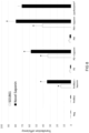
- Figure 8 compares the enhancement of the transfection efficiency by GE1741 ("novel saponin") with the enhancement of the transfection efficiency by a prior art saponin (SO1861) as well as the current "gold standard” Lipofectamin.
- Neuro2a cells have been incubated without an addition of particles comprising a nucleic acid.
- mRNA Upon adding particles comprising a peptide bound to mRNA (PmRNA), a transfection efficiency in the single-digit range could be observed. After adding a saponin as enhancer to the respective cell suspension, the transfection efficiency could be increased to approximately 15 % when using SO1861, but to even 35 % when using GE1741.
- a transfection efficiency of almost 80 % when using peptide minicircle DNA nanoparticles is even better than a transfection efficiency of approximately 70 % observed for the "gold standard" Lipofectamin.
- GE1741 is not only better than previously isolated saponins, but shows an even higher enhancement of the transfection efficiency than the gold standard being currently on the market.
- HCT-116 cells were transfected with commercial transfection reagents in order to examine the impact of GE1741 on the transfection efficiency. The results are shown in Figure 9 .
- the addition of GE1741 led to an increase in transfection efficiency of each transfection method. While the transfection efficiency with TransIT was only slightly increased by 3 percentage points and that of Xtreme Gene by 8 percentage points, GE1741 boosted the transfection efficiency of Genecellin by 30 percentage points from 12 % (without GE1741) to 42% (with GE1741).
- the impedance which shows a direct correlation with cell viability, was investigated in order to determine the toxicity and the permeabilizing properties of GE1741.
- GE1741 does not exhibit cell-toxic effects when used in a concentration that is fully sufficient for enhancing the transfection efficiency.
- this saponin was applied in different concentrations to Neuro2a cells and incubated over a period of 50 hours.
- concentrations of 2.5 ⁇ g/mL and 6.25 ⁇ g/mL a normalized cell index is (within the error of measurement) identical to the normalized cell index observed for the negative control that has been subjected to a transfection process without an addition of GE1741. This is indicated in Figure 10 by other three overlaid curves 1. If the GE1741 concentration is increased to 12.5 ⁇ g/mL, a slightly decreased normalized cell index could be observed (curve 2). The normalized cell index slightly further decreased upon increasing the GE1741 concentration to 25 ⁇ g/mL (curve 3).
- DNA nanoparticles (curve 9), DNA nanoparticles with 2 ⁇ g/mL GE1741 (GE1741; curve 10) or 2 ⁇ g/mL GE1741 alone (curve 11) was added to Neuro2a cells.
- the DNA nanoparticles comprised DNA encoding for the Green Fluorescent Protein (GFP).
- Buffer was used as negative control (curve 12).
- the transfection efficiency of GE1741 was also examined by transfecting cells with DNA encoding saporin. Saporin is a cytotoxic protein. If the transfection is successful, the transfected cells will die. The corresponding results are shown in Figure 12 .
- the negative control (curve 5) refers to Neuro2a cells incubated without any peptide DNA nanoparticles.
- Curve 6 refers to a transfection experiment of Neuro2a cells with nanoparticles comprising DNA encoding for saporin.
- the normalized cell index for this setup approximately corresponds to the normalized cell index of the negative control, indicating a very poor transfection efficiency.
- GE1741 The universal applicability of GE1741 in transfection experiments was investigated. A delivery of different nucleic acids, incorporated into nanoplexes, could be considerably increased. mRNA transfections were less effective than DNA transfections due to a faster degradation by cellular enzymes. However, a missing transcription step allowed the highest efficiency even after 24 h incubation time and therefore representing a rapid transfection method. Minicircle transfection yielded the highest efficiency, presumably caused by the smaller size of minicircle-compared to plasmid DNA.
- GE1741 is a novel and valuable compound for use in the process of saponin-mediated transfection, the so-called sapofection.
Landscapes
- Health & Medical Sciences (AREA)
- Chemical & Material Sciences (AREA)
- Life Sciences & Earth Sciences (AREA)
- Engineering & Computer Science (AREA)
- Genetics & Genomics (AREA)
- Organic Chemistry (AREA)
- General Health & Medical Sciences (AREA)
- Molecular Biology (AREA)
- Biotechnology (AREA)
- Biochemistry (AREA)
- Zoology (AREA)
- Wood Science & Technology (AREA)
- Bioinformatics & Cheminformatics (AREA)
- Medicinal Chemistry (AREA)
- General Engineering & Computer Science (AREA)
- Biomedical Technology (AREA)
- Epidemiology (AREA)
- Veterinary Medicine (AREA)
- Animal Behavior & Ethology (AREA)
- Pharmacology & Pharmacy (AREA)
- Public Health (AREA)
- Microbiology (AREA)
- Natural Medicines & Medicinal Plants (AREA)
- Physics & Mathematics (AREA)
- Biophysics (AREA)
- Plant Pathology (AREA)
- Chemical Kinetics & Catalysis (AREA)
- General Chemical & Material Sciences (AREA)
- Oil, Petroleum & Natural Gas (AREA)
- Mycology (AREA)
- Medical Informatics (AREA)
- Botany (AREA)
- Alternative & Traditional Medicine (AREA)
- Pharmaceuticals Containing Other Organic And Inorganic Compounds (AREA)
- Saccharide Compounds (AREA)
- Steroid Compounds (AREA)
- Medicinal Preparation (AREA)
- Compounds Of Unknown Constitution (AREA)
- Medicines That Contain Protein Lipid Enzymes And Other Medicines (AREA)
Description
- The present invention relates to the in vitro use of a saponin according to the preamble of
claim 1, to the in vivo use of such a saponin according to the preamble ofclaims 3, to a transfection composition according to the preamble ofclaim 7, to a method for an in vitro transfection according to the preamble ofclaim 10, and to a method for isolating such a saponin according to the preamble ofclaim 15. - Many saponins have been described in the past. Exemplarily, reference is made to Weng, Alexander, et al. "A simple method for isolation of Gypsophila saponins for the combined application of targeted toxins and saponins in tumor therapy." Planta medica 75 (13) (2009): 1421-1422; Weng, Alexander, et al. "A convenient method for saponin isolation in tumour therapy." Journal of Chromatography 6 878 (7) (2010): 713-718; Weng, Alexander, et al. "The toxin component of targeted anti-tumor toxins determines their efficacy increase by saponins." Molecular oncology 6 (3) (2012): 323-332; and Weng, Alexander, et al. "Saponins modulate the intracellular trafficking of protein toxins." Journal of controlled release 164 (1) (2012): 74-86.
- Further saponins are described in the following publications: Thakur, Mayank, et al. "Highspeed countercurrent chromatographic recovery and off-line electrospray ionization mass spectrometry profiling of bisdesmodic saponins from Saponaria officinalis possessing synergistic toxicity enhancing properties on targeted antitumor toxins." Journal of Chromatography B 955 (2014): 1-9; Jia, Zhonghua, Kazuo Koike, and Tamotsu Nikaido. "Major triterpenoid saponins from Saponaria officinalis." Journal of natural products 61 (11) (1998): 1368-1373; Haddad, Mohamed, et al. "New triterpene saponins from Acanthophyllum pachystegium." Helvetica chimica acta 87 (1) (2004): 73-81; Fu, Hongzheng et al. "Silenorubicosides A-D, Triterpenoid Saponins from Silene rubicunda." Journal of Natural Products 68 (5) (2005): 754-758; Moniuszko-Szajwaj, Barbara, et al. "Highly Polar Triterpenoid Saponins from the Roots of Saponaria officinalis L." Helvetica Chimica Acta 99 (5) (2016): 347-354; Fuchs, Hendrik, et al. "Glycosylated Triterpenoids as Endosomal Escape Enhancers in Targeted Tumor Therapies." Biomedicines 5 (2) (2017): 14.
- It has also been described that a specific saponin (SO1861 also called Sapofectosid) can mediate an improved intracellular delivery of peptide and lipid nanoparticles (Weng, Alexander, et al. "Improved intracellular delivery of peptide- and lipid-nanoplexes by natural glycosides." Journal of Controlled Release 206 (2015) 75-90), or of DNA and RNA (Sama, Simko, et al. "Sapofectosid - Ensuring non-toxic and effective DNA and RNA delivery" International Journal of Pharmaceutics 534 (2017): 195-205).
- It is an object of the present invention to provide a compound having even better properties with respect to intracellular delivery of small compounds such as peptides and nucleic acids than the saponin SO1861.
- Surprisingly, a saponin that can be isolated from Gypsophila elegans M. Bieb has superior effects regarding the delivery of small molecules, in particular regarding the delivery of nucleic acids to cells. Such a saponin corresponds to formula (I):

- Thereby, R1 is a xylose residue or an arabinose residue bonded with its C1 atom to the corresponding xylose residue of formula (I); and R2 is independent from other R2 residues in the same molecule H or an acetyl residue, with the proviso that at least two acetyl residues are present in the saponin.
- The saponin has a common saponin core structure and does not comprise any unusual bonds. Rather, it differs from other saponins only in its sugar residues and/or its acetyl residues.
- For a better identification of the individual sugar residues in formula (I), they are marked with a corresponding abbreviation in the above depiction of formula (I). Thereby, the abbreviations have the following meaning:
Abbreviation Sugar residue Fuc Fucose Gal Galactose GlcA Glucuronic acid Qui Quinovose Rha Rhamnose Xyl Xylose - The structure of arabinose (Ara) corresponds to formula (II):

- A compound according to formula (I) showed a transfection efficiency that was approximately twice as high as the transfection efficiency of SO1861. This is a completely unexpected and surprising result since one would expect only slight increases in transfection efficiency with respect to SO1861. Due to the high number of already existing saponins, the inventors could not expect that the compounds and the use of which is claimed here would show such a high transfection efficiency even if these compounds are known per se. Saponins cannot be synthesized and can generally not simply be purchased from a supplier of chemicals. Rather, they need to be isolated from plants or parts of plants. This makes it particularly difficult to investigate the properties of saponins.
- The inventors surprisingly found that saponins according to general formula (I) can be isolated from Gypsophila elegans M. Bieb that show an unexpected high transfection efficiency. It has not been described before that such saponins can be extracted from Gypsophila elegans M. Bieb. Rather it has only been described by Haddad et al. (see above for details) that a saponin falling under general formula (I) has been isolated from a completely different plant (Acanthophyllum pachystegium) without, however, giving any indication that this saponin (called herein GE1471) could increase the transfection efficiency.
- Thus, the present invention relates to the use of a saponin according to general formula (I) in the in-vitro delivery of a nucleic acid, a lipid, a peptide and/or a protein to a cell. Thereby, the in vitro delivery of nucleic acid is a particularly appropriate use of the saponin. Appropriate nucleic acids to be delivered are DNA, such as plasmid DNA or minicircle DNA, and RNA, such as small interfering RNA (siRNA). The nucleic acid or other molecule to be delivered can be present in the form of a nanoplex. Nanoplexes are typically formed by peptides or lipids being able to bind the small molecule to be delivered. Thus, a nucleic acid delivering nanoplex comprises a lipid or non-lipid carrier to which a nucleic acid is bound. Nanoplexes as can also be referred to as nanoparticles. In an embodiment, the nucleic acid to be delivered is a minicircle DNA. In an embodiment, the molecule to be delivered is a peptide minicircle DNA particle.
- In an embodiment, the cell to which the nucleic acid, the lipid, the peptide and/or the protein is to be delivered is a eukaryotic cell. Human cells, animal cells (such as rodent cells) or plant cells are particularly appropriate to be used as cells. Yeast cells can also be used.
- In an aspect, the invention relates to a saponin according to general formula (I) for use in therapy or diagnostics. Thereby, the saponin is for use in an in-vivo delivery of a nucleic acid, a lipid, a peptide and/or a protein to a human or animal.
- In an embodiment, the animal is a mammal, in particular a rodent.
- In an aspect, a mixture of at least two saponins, in particular of exactly two saponins is used, wherein residue R1 denotes xylose in a first saponin and arabinose in a second saponin.
- In an embodiment, all sugars are present in their D stereoisomeric form.
- In an embodiment, R1 is a xylose residue. Then, the saponin corresponds to the following formula (III):

- In an embodiment, R1 is an arabinose residue. Then, the saponin corresponds to the following formula (IV):

- In an embodiment, the saponin is present in a stereoisomeric form according to general formula (V). Likewise, the xylose residue and the arabinose residue are present, in an embodiment, in a stereoisomeric form according to formulae (VI) or (VII):


- In an embodiment, the saponin carries exactly two acetyl groups, i.e., two of the R2 residues are acetyl residues and the remaining R2 residue is a hydrogen.
- In an embodiment, the acetyl groups are bonded to the oxygen atoms in C3 and C4 position of the corresponding quinovose residue, resulting in a saponin of formula (VIII):

- In an embodiment, all three R2 residues are acetyl residues, resulting in a saponin of formula (IX):

- The inventors surprisingly found out that a saponin according to general formula (I) does not only increase the transfection efficiency when used alone, but is particularly appropriate to enhance the transfection efficiency of classic transfection reagents. Thus, it can be used as booster for such transfection reagents. The above-described uses encompass the use of such a saponin as booster for classic transfection reagents. In an embodiment, the saponin is used in vivo or in vitro in combination with at least one transfection reagent chosen from the group consisting of liposomal-based transfection reagents and polymer-based transfection reagent. Examples of such transfection reagents are listed below.
- The general inventive concept between the independent claims is to be seen in the significant enhancement of transfection efficiency of the saponins according to general formula (I) that was uncovered by the inventors for the first time.
- In an aspect, the invention relates to a transfection composition, comprising a classic transfection reagent and at least one saponin according to formula (I), wherein the residues R1 and R2 have the meanings described above. Classic transfection reagents can be divided into two subgroups, namely liposomal-based transfection reagents and polymer-based transfection reagents. Liposomal-based transfection reagents consist of mostly cationic charged lipids with hydrophobic fatty acid chains bound to polar head groups such as trimethylammonium propane. Polymer-based transfection reagents consist of organic polymers containing a charged head group such as polyethylenimine (PEl).
- Examples for commercially available classic liposomal-based transfection reagents are METAFECTENE; TransFast; Stemfect; and TransFectin.
- Examples for commercially available classic polymer-based transfection reagents are GeneCellin; X-tremeGene transfection reagents such as
X-tremeGene 9 DNA Transfection Reagent, X-tremeGene HP DNA Transfection Reagent, and X-tremeGene siRNA Transfection Reagent; TransIT transfection reagents such as TranslT-X2 Dynamic Delivery System, TransIT-LT1 Reagent, TransIT-2020 Reagent, TransIT-PRO Reagent, TransIT-VirusGEN Reagent, TransIT-Lenti Reagent, TransIT-Insect Reagent, TransIT-293 Reagent, TransIT-BrCa Reagent, TransIT-CHO Transfection Reagent, TransIT-HeLaMONSTER Transfection Reagent, TransIT-Jurkat Reagent, TransIT-Keratinocyte Reagent, TransIT-mRNA Transfection Reagent, TransIT-TKO Reagent, TransIT-siQUEST Reagent, and TransIT-Oligo Reagent; Viafect; FuGENE; Xfect; TurboFect; and GenJet. - In an embodiment, R1 of the saponin in the transfection composition is a xylose residue.
- In an embodiment, the saponin in the transfection composition carries exactly two acetyl groups.
- In an embodiment, the acetyl groups are bonded to the oxygen atoms in C3 and C4 position of the corresponding quinovose residue.
- In an embodiment, the transfection reagent is a liposomal-based transfection reagent.
- In an embodiment, the transfection reagent is a polymer-based transfection reagent.
- In an embodiment, the transfection reagent is chosen from the group consisting of a TransIT transfection reagents, X-tremeGene transfection reagents, and GeneCellin.
- In an embodiment, the transfection reagent is GeneCellin.
- In an aspect, the invention relates to a saponin or a mixture of at least two saponins according to the preceding explanations for use in a method for delivering a nucleic acid, a lipid, a peptide and/or a protein to a human or animal in need thereof.
- In an aspect, the invention relates to a method for an in vitro transfection of a cell. This method comprises the step of incubating a cell with a nucleic acid in the presence of a saponin or a mixture of at least two saponins according to the preceding explanations. The transfection can be a transient transfection or a stable transfection. DNA and RNA are appropriate nucleic acids, wherein DNA is particularly appropriate. In an embodiment, the nucleic acid to be delivered is a minicircle DNA.
- In an embodiment, the cell is a eukaryotic cell. Human cells, animal cells (such as rodent cells) or plant cells are particularly appropriate to be used as cells. Yeast cells can also be used.
- In an embodiment, the nucleic acid forms part of a nanoparticle. To give an example, a DNA nanoparticle may be used. Peptide minicircle DNA particles or particles comprising a peptide bound to DNA are further appropriate examples. Likewise, RNA nanoparticles are appropriate entities to carry out the transfection.
- In an embodiment, the saponin is used in a concentration lying in a range between 1 µg/mL and 25 µg/mL, in particular between 1.5 µg/mL and 20 µg/mL, in particular between 2 µg/mL and 15 µg/mL, in particular between 2.5 µg/mL and 12.5 µg/mL, in particular between 3 µg/mL and 10 µg/mL, in particular between 4 µg/mL and 9 µg/mL, in particular between 5 µg/mL and 8 µg/mL, in particular between 6 µg/mL and 7 µg/mL.
- In an embodiment, the saponin is used within the claimed method in combination with at least one transfection reagent chosen from the group consisting of liposomal-based transfection reagents and polymer-based transfection reagent. Examples of such transfection reagents are listed above.
- In an aspect, the present invention relates to method for isolating a saponin or a mixture of at least two saponins according to the preceding explanations from Gypsophila elegans M. Bieb. This method comprises the steps explained in the following. Thereby, the steps need not necessarily be performed in the indicated order. Rather, any other sensible order of method steps can also be applied.
- In a first step, roots of Gypsophila elegans M. Bieb are cut. Optionally, the cut roots are washed. Subsequently, the cut roots are freeze-dried and ground. In doing so, a root powder is obtained.
- The root powder is extracted with a high-concentrated organic solvent to obtain a root extract. The term "high-concentrated organic solvent" relates to an organic solvent having a concentration of more than 50 % (v/v). Methanol or any other short-chain organic alcohol is a suited organic solvent. To give an example, a solvent concentration of 70 to 100 %, in particular of 75 to 95 %, in particular of 80 to 90 % is an appropriate high concentration. If the organic solvent is miscible with water, water can be used as co-solvent.
- Afterwards, the high-concentrated organic solvent is removed from the root extracts to obtain a dry extract. This removal can be performed, e.g., by vacuum distillation.
- The dry extract is then dissolved in a low-concentrated organic solvent to obtain an extract solution. The term "low-concentrated organic solvent" relates to an organic solvent having a concentration of less than 50 % (v/v). To give an example, a concentration of 10 % to 50 %, in particular of 15 % to 45 %, in particular of 20 % to 40 %, in particular of 25 % to 30 % is an appropriate concentration range for the low-concentrated organic solvent. Once again, methanol or any other short-chain organic alcohol is a suited organic solvent. Likewise, water can be used as co-solvent, if the low concentrated organic solvent is miscible with water.
- The extract solution is then subjected to at least one chromatographic separation step. Thereby, a purified saponin solution is obtained. The chromatographic separation can be implemented by high-performance liquid chromatography (HPLC). Typically, it is advisable to perform more than one chromatographic separation step in order to enhance the purity of the purified saponin solution. Thus, it is possible to subject the purified saponin solution obtained after the first chromatographic separation step to at least one subsequent chromatographic separation step. Thereby, different columns can be used in the individual chromatographic separation steps.
- It is possible to perform the individual chromatographic separation steps (if more than one chromatographic separation step is carried out) in different stationary and/or mobile phases. Thus, it is possible to change the solvents used in the individual chromatographic separation steps between these steps.
- Finally, the solvent is removed from the purified saponin solution after the at least one chromatographic separation step. This can, e.g., be done by vacuum distillation or by freeze-drying. A purified saponin powder results.
- All embodiments disclosed herein can be combined in any desired way. Furthermore, embodiments of the explained uses and methods can be transferred to the described other methods and uses in any desired way. The described transfection composition can also make use of any of the described embodiments.
- Further details of aspects of the present invention will be explained with respect to an exemplary embodiment and accompanying Figures. In the Figures:
- Figure 1
- shows an HPLC chromatogram of dry raw extract from the roots of Gypsophila elegans M. Bieb;
- Figure 2
- shows an LC-ESI-MS chromatogram of the substance present in peak P2 of the chromatogram of
Figure 1 after further purification by semi-preparative HPLC; - Figure 3
- shows and LC-ESI-MS chromatogram of the isolated saponin GE1741;
- Figure 4
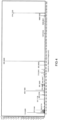
- shows an MS/MS spectrum of the isolated saponin GE1741;
- Figure 5
- shows the results of flow cytometric measurements;
- Figure 6
- shows the transfection efficiency of nanoplexes ± triterpene saponins in Neuro2a cells;
- Figure 7
- shows the chemical structure of GE1741 elucidated by mass spectrometry and NMR spectroscopy;
- Figure 8
- shows a plot on the transfection efficiency of different transfection enhancers;
- Figure 9
- shows the impact on GE1741 on the transfection efficiency of different commercial transfection methods;
- Figure 10
- shows the results of a first experiment for examining the toxicity of GE1741 on Neuro2a cells;
- Figure 11
- shows results of a second experiment for examining the toxicity of GE1741 on Neuro2a cells and
- Figure 12
- shows the results of a targeted suicide gene therapy.
- Seeds from Gypsophila elegans M. Bieb were seeded on a field in the state of Brandenburg, Germany. The mature plants were harvested and the roots were cut. The roots were washed, freeze-dried and ground to powder. The powder was extracted by 90 % methanol in water. Following filtration, the methanol was removed by vacuum distillation. The remaining aqueous extract was finally freeze-dried (dry extract). The dry extract was dissolved in 30 % methanol at a concentration of 60 mg/mL.
- The solution (each 0.5 mL) was subjected to semi-preparative HPLC using a
Kinetex ® 5µm C18 100 Å,LC Column 250 × 10.0 mm. A methanol(A)/water, 0.01 % TFA (B) gradient was used from 30 % to 90 % (A) over 20 min and then to 30 % (A) over 10 min. Flow rate was 4 mL/min. Detection wavelength was at 210 and 300 nm. A corresponding chromatogram is depicted inFigure 1 , wherein the upper curve shows the detector signal at 210 nm and the lower curve the detector signal at 300 nm. The peak at a retention time (RT) ≈ 18 to 20 (≈ 19) min (marked as P2) was collected. A high number of isolation cycles was repeated and the collected peaks were finally pooled. The methanol was evaporated by vacuum centrifugation and the remaining water was removed by freeze-drying. - The dried material was dissolved in 50 % acetonitrile at a concentration of 4 mg/mL. The solution (each 0.5 mL) was subjected to semi-preparative HPLC using a
Kinetex ® 5µm C18 100 Å,LC Column 250 × 10.0 mm. An acetonitrile (A)/water, 0.01 % TFA (B) gradient was used from 30 % to 50 % (A) over 24 min and then to 30 % (A) over 1 min. Detection wavelength was at 210 nm. Flow rate was 4 mL/min. The peak at RT ≈ 18 min was collected. Several cycles were repeated. The acetonitrile was evaporated by vacuum centrifugation and the remaining water was removed by freeze-drying. - The samples were analyzed by using an Agilent 6200 Series Q-TOF LC-ESI-MS/MS system. For LC-ESI-MS analysis of the recovered fractions a Kinetex® C18 HPLC column (2.6 µm, 100 Å, (150 × 4.6 mm), and acetonitrile (A)/water (B) gradient, 0.01% formic acid was used starting with 30 % to 70 % (A) over 30 min using a flow rate of 0.7 mL/min. The corresponding chromatogram is depicted in
Figure 2 . Two prominent peaks were detected. A mass of 1538.67 was assigned to the substance of the first peak, and a mass of 1741.73 was assigned to the substance of the second peak. The second peak was chosen for further purification. - The dried material of the second peak was dissolved in 50% acetonitrile at a concentration of 4 mg/mL. The solution (each 0.5 mL) was subjected to semi-preparative HPLC using the semi-preparative Kinetex® C-18 column (see above). An acetonitrile (A)/water, 0.01% TFA (B) gradient was used from 30% to 50% (A) over 24 min and then to 30% (A) over 1 min using a flow rate of 4.0 mL/min. Detection wavelength was at 210 nm. The peak at RT ≈ 18 min was collected in several repeating cycles. The acetonitrile was evaporated by vacuum centrifugation and the remaining water was removed by freeze-drying.
- The dry material was dissolved in 50 % acetonitrile and subjected (0.5 mL per run) to HPLC using an Ultrasep ES PEO, LC Column, 250 × 4 mm, 5 µm. The column was conditioned by 5 % acetonitrile. The polarity of the solvent was abruptly changed by applying an acetonitrile(A)/water, 0.01 % TFA (B) gradient starting with 100 % to 5 % (A) from 1.25 to 21 min. Detection wavelength was at 210 nm and flow rate was 1 ml/min. The product (GE1741; also referred to as gypsophilosid A) was collected at RT 4-6 min. The acetonitrile was removed by vacuum centrifugation. After freeze drying GE1741 was obtained as white powder.
- The product was once again analyzed by LC-ESI-MS as outlined above. The corresponding chromatogram is depicted in
Figure 3 . Only one prominent peak was detected, indicating a successful purification. A mass of 1741.73 was assigned to the substance of this peak. This mass gave the product its name, GE1741. - For elucidating the chemical structure of GE1471, LC-ESI-MS/MS measurements were performed. A corresponding spectrum is shown in
Figure 4 . - Based on this mass spectrometric data and further data obtained by NMR spectroscopy (see below for details), it could be established that the structure of this saponin corresponds to formula (VIII), wherein R1 is a xylose residue. However, it is assumed that the biological function of GE1741 will most probably not depend on the concrete structure of residue R1. Rather, the presence of at least one acetyl residue on the quinovose residue of GE1741 is considered to be a relevant factor for the properties of GE1741 that will be explained in the following in more detail. While GE1741 comprises two acetyl residues (one in C3 position and one in C4 position), preliminary data suggests that the amount and position of the acetyl residues can be varied within the indicated limits without significantly changing the properties of the respective saponin.
- Murine neuroblastoma cells (Neuro2a cells, ATTC CCL-131™) and human colorectal carcinoma cells (HTC-116, (ATCC® CCL-247™) were cultured in Dulbecco's Modified Eagle's Medium (DMEM), containing 1g/L D-Glucose, 10% FBS and stable glutamine. Neuro-2A-cells were incubated at 37 °C and 5 % CO2. These cells were then incubated with DNA nanoparticles (YD) with or without the novel saponin GE1741.
- The DNA nanoparticles used for the transfection experiments contained DNA encoding for the green fluorescent protein (GFP). Therewith, the transfection efficiency could very easily be monitored by detecting the fluorescence of the cells that have been incubated with the corresponding DNA nanoparticles. The corresponding incubation period was chosen to be 48 hours in the present case.
- As negative control (left panel of
Figure 5 ), no DNA nanoparticles at all have been added to the Neuro2a cells. A fluorescence indicating an apparent transfection efficiency of 0.37 % could be observed. This value represents the measuring error. - If peptide minicircle DNA particles (PM) have been added to the Neuro2a cells without any transfection enhancer, a transfection efficiency of only 1.35 % could be observed based on the detected fluorescence (middle panel of
Figure 5 ). If, however, the same peptide minicircle DNA particles have been added together with GE1741 (the novel saponin), a transfection efficiency of 98.65 % could be observed based on the detected fluorescence (right panel ofFigure 5 ). These flow cytometric measurements detecting fluorescent cells already indicates a high transfection efficiency enhancement by GE1741. - 20 mg of positively charged poly-lysine peptides with (Y) and without (P) an integrin receptor targeting amino sequence were purchased from Genecust. The vector of p-EGFP-N3, coding for the green fluorescing protein (GFP), was obtained and propagated with DH5α - E.Coli cells (1.645 mg/mL). StemMACS™ eGFP mRNA (20 µg) and GFP encoding minicircle DNA (Gene Bank Accession: U55761) were used as further nucleic acids. In order to conduct the transfection, nanoplexes were formulated as follows: The poly-lysine peptides (P or Y) and the p-EGFP-N3 (D), mRNA (m) or minicircle (M) vector were diluted in water (each 50 µL) and mixed thoroughly by fast pipetting in a ratio of 4:1. The nanoplexes were allowed to form in a 30-minute incubation step. Thereafter, the nanoplex suspension was diluted with OptiMEM to a total volume of 1 mL. The commercial transfection reagents TransIT-X2® Dynamic Delivery System, Xtreme GENE™ HP Transfection Reagent, Genecellin and Lipofectamin® were formulated as described by the manufacturer.
- Neuro2a cells or HTC-116 cells (15,000 cells/well) were seeded in a 24-well-plate with a well volume of 400 µL culture medium and incubated for 24 h. The transfection reagents were formulated as described above and admixed with saponin solution, if required. The culture medium was replaced with the transfection medium with a final amount of 500 ng DNA/RNA. After a 48 h (DNA transfections) or 24 h (RNA transfections) incubation period, the transfection medium was removed, the cells were trypsinized and transferred in a polystyrene tube for flow cytometry (Cytoflex). For each measurement 10,000 cells were acquired. The transfection efficiency was determined by the analysis software Cyflogic (by comparison of the sample plots with the negative control in terms of fluorescence.
- For real-time toxicity measurements the impedance measuring device iCELLigence® was deployed. 8000 Neuro2a cells per well were seeded into two 8-well E-plates L8 and incubated for 24 h in a volume of 800 µL. For
transfection toxicity studies 50 µL of reagent was added after the respective volume of culture medium was removed. A non-toxic concentration of 2 µg/mL GE1741 was applied. For permeability studies increasing concentrations of GE1741 from 2.5 µg/mL to 60 µg/mL were applied to the cells. Each 10 minutes the impedance/viability was measured. The results were analyzed and displayed with the RTCA Data Analysis Software. - The samples for NMR-spectroscopy were prepared by dissolving 2 mg of GE 1741 in 600 µl d4-methanol (99,95 % deuterium content, Deutero, Kastellaun, Germany) and by dissolving 4 mg of GE 1741 in 600 µl d5-pyridine (99,9 % deuterium content, Deutero, Kastellaun, Germany) to yield a concentration of 1.9 and 3.8 mM/I, respectively. The solutions were transferred to 5 mm sample tubes which were sealed to prevent evaporation of solvent or accumulation of water.
- NMR spectra were recorded at 300 K at 600 (1H frequency) on Bruker AV-III spectrometers (Bruker Biospin, Rheinstetten, Germany) using cryogenically cooled 5 mm TCI-triple resonance probe equipped with one-axis self-shielded gradients. The software used to control the spectrometer was topspin 3.5 pl6. Temperature had been calibrated using d4-methanol and the formula of Findeisen et al. (Findeisen 2007).
- Experiments using the sample in methanol: One-dimensional 1H- and 13C-spectra were recorded at 600 MHz using 32 and 18432 scans, respectively. In case of the carbon spectrum proton broadband decoupling was applied. Homonuclear spectra: A DQF-COSY (Piantini et al., 1982), a TOCSY (Braunschweiler and Ernst, 1983; Bax and Davis, 1985a) and a NOESY (Jeener et al., 1979) were recorded using 2048 × 512 complex data points and acquisition times of 204 and 61 ms in F2 and F1, respectively. DQF-COSY and TOCSY (mixing
time 80 ms) were recorded using 8 scans, the NOESY using 64 scans with a mixing time of 80 msec. - The heteronuclear spectra were recorded as follows: A 13C-HSQC (Bodenhausen and Ruben, 1980), 13C-HSQC-TOCSY, 13 C-HSQC-CLIP-COSY and 13 C-HSQC-NOESY were recorded using 512 × 1024 complex data points, acquisition times of 51 and 45 ms in F2 and F1, respectively, and 48, 96, 96 and 288 scans, respectively. A 13C DEPT-HMQC (Kessler et al., 1989), and a 13C-HQQC (Kessler et al., 1991) were recorded using 512 × 512 complex data points, acquisition times of 51 and 23 ms in F2 and F1, respectively, and 64 and 60 scans, respectively. For the determination of the 1J HC and the 3J HH a 13C-HSQC was recorded without refocussing and carbon decoupling using 16384 × 512 complex data points, acquisition times of 1638 and 73 ms in F2 and F1, respectively, and using 32 scans. All the above heteronuclear spectra were recorded using a BIRD pulse for suppression of protons bound to 12C (Bax and Subramanian, 1986). A gradient-enhanced 13C-HMBC (Bax and Summers, 1986; Cicero et al., 2001) was recorded using 2048 × 1024 complex data points, acquisition times of 204 and 33 m s in F2 and F1, respectively, and 128 scans.
- Experiments using the sample in pyridine: One-dimensional 1H- and 13C-spectra were recorded at 600 MHz using 32 and 14349 scans, respectively. In case of the carbon spectrum proton broadband decoupling was applied. A DQF-COSY (Piantini et al., 1982), was recorded using 2048 × 512 complex data points, 8 scans and an acquisition times of 204 and 61 ms in F2 and F1, respectively. A 13C-HSQC (Bodenhausen and Ruben, 1980), was recorded using 512 × 1024 complex data points, 32 scans and an acquisition times of 51 and 45 ms in F2 and F1, respectively. A gradient-enhanced 13C-HMBC (Bax and Summers, 1986; Cicero et al., 2001) was recorded using 2048 × 1024 complex data points, acquisition times of 204 and 33 ms in F2 and F1, respectively, and 64 scans.
- Data were processed using topspin3.2, typically a squared sine bell shifted by 90 ° was used in both dimensions, in case of the HMBC a sine bell was used in F2. Datasets were processed to yield a data matrix of 4096 by 2048 points.
- Initially the seed and root extract was tested regarding transfection enhancing activity. The results are shown in
Figure 6 . The HPLC chromatogram of the dry root extract (cf.Figure 1 ) was analyzed, the peaks P2, P3 and P4 were collected and their biological activity was tested for their biological activity. These results are also shown inFigure 6 . Subsequently, P2 was subjected to further HPLC purification, as explained above. Subsequent LC-ESI-MS guided semi-preparative purification steps (cf.Figures 2 and3 ) led to an isolation of a peak, which activity was repeatedly tested. LC-ESI-MS/MS measurements detected in the negative ionization mode the deprotonated molecular ion signal [M-H] - at m/z 1741 (cf.Figure 4 ). The MS/MS fragment ion signal at m/z 955 was assigned to the saponin after secession of the diacetyl-pentasaccharide chain linked to C-28 of the quillaic acid, and m/z 460 resulted after the additional cleavage of the trisaccharide unit at C-3. The molecular formula was determined to be C79H122O42 (calculated: 1741.73325) from the measured mass for the negatively charged ion (HR-ESI-MS m/z 1741.7336). This corresponds to a mass difference of only 0.2 ppm. - The structure of the bidesmosic quillaic acid type saponin GE1741 was fully elucidated by 1D and 2D-NMR spectroscopic experiments, including 1H, 13C and HSQC, HMBC, HQQC, DQF-COSY, TOCSY, HSQC-TOCSY and NOESY using d4- methanol. Amongst the signals in the 13C NMR spectrum of GE174, two double intensity resonances (δC 75.33, 71.43 ppm) indicated completely overlapping of C-atoms. As partial structural units, a triterpene moiety and the existence of 8 sugars with two points of acetylation was seen. Using HSQC and HQQC experiments, six sp3-hybridised resonances (δC 10.9, 16.4, 17.7, 24.8, 27.1, 33.3 ppm) were recognized as methyl groups in the triterpene moiety, and additionally three methyl groups were assigned to deoxy-sugars (δC 17.0, 17.6, 18.3). The 1H NMR gave singlet resonances (δH 0.74, 0.87, 0.94, 1.00, 1.16, 1.38 ppm) for the triterpene methyl-functions, and doublets (δH 1.16, 1.23, 1.31) for the deoxy-functions.
- The C30 corpus displayed two methyl groups less than expected which was explained by the occurrence of an aldehyde function (δC 211.5 ppm, δH 9.47 ppm) and an acidic function (δC 175.9 ppm). Together with resonances for a double bond at δC 123.2 (CH), 144.8 ppm (quaternary C), a broad triplet signal (δH 5.31 ppm), a substituted oxymethin function at C-3 (δC 86.4 ,δH dd 3.86) combined with HMBC correlation data elucidated a hydroxy-oleanan-12-ene type backbone. The corresponding results are shown in
Figure 7 and will be explained in more detail in the following
2,3 J CH correlations from the aldehyde proton δ 9.47 ppm to C-4 (56.3 ppm), CH3-24 (δ 10.9), and C-5. The long-range correlation of H-3 (δ 3.86) to the quaternary position C-4 and C-24 suggested the CHO-function to be at C-23. The second hydroxylation at C-16 (δC 74.5) was seen by 2,3 J-CH signals from protons of H-18 (δ 2.94), CH2-22 (δ 1.93, 1.16). A total number of eight monosaccharides were detected in the structure of GE1741 by 1H- and 13C NMR spectroscopy by observation of characteristic chemical shifts of anomer resonances. The nomenclature of GE1741 is as follows: 3-O-(β-D-Galactopyranosyl-(1→2)-[β-D-xylopyranosyl-(1→3)]-β-D-glucuronopyranosyl)-28-O-(β-D-xylopyranosyl-(1→3)-[β-D-xylopyranosyl-(1→4)]-α-L-rhamnopyranosyl-(1→2)-[3,4-di-(O-acetyl)-β-D-quinovopyranosyl]-(1→4)-β-D-fucopyranosyl)-quillaic acid - A trisaccharide unit is linked to C-3 and a hexasaccharide to C-28 in this bisdesmosidic quillaic acid type saponin. The sugar identities, and linkages with respective 1H/13C connectivities were unambiguously identified by a combination of HMBC (cf.
Figure 7 ), HSQC (high resolution), HSQC-DEPT, HQQC, HSQC-TOCSY, and DQF-COSY, 1H/1H-TOCSY, 1H/1H-NOESY, and non-decoupled HSQC. The 1 J CH-connectivities of all sugar methin atoms were clearly derived by HSQC recorded with high resolution in the indirect dimension (TD: F2 1024, F1 2048). - The trisaccharide linked to C-3 consist of a glucuronic acid where the free carboxylic acid function at C-6 was not detected by 13C nor by HMBC, a galactose, and a xylose with anomeric resonances at δH/δC in [ppm] and 3 J HH/ 1 J CH in [Hz]: β-GlcA-1 (δ 4.42, 104.6; d, 7.4, 161), β-Gal-1 (δ 4.80, 103.7; d,7.8, 163), β-Xyl-1 (δ 4.57, 104.9; d, 7.8, 162). The pentasaccharide unit at C-28 consist of β-Fuc-1 (δ 5.29, 94.8; d, 7.8, 166), α-Rha-1 (δ 5.36, 101.5; d, 1.4, 171), β-Xyl'-1 (δ 4.49, 107.0; d, 7.2, 159), β-Qui-1 (δ 4.61, 105.8; d, 8.2, 161), β-Xyl"-1 (δ 4.51, 105.6; d, 7.0, 162).
- Stereochemical properties of the glycosidic linkages were determined by characteristic values of 3 J HH and 1J CH coupling constants J (Hz), respectively. A large trans-diaxial coupling (3 J HH: 7-8 Hz) for H-1/H-2 in case of glucose and galactose type sugars define a β-configuration. Characteristic 1 J CH coupling values indicate the anomeric configuration of hexopyranoses with ~170 Hz for an α, and ~160 Hz for a β configuration. The detected values were in absolute accordance to these literature data and can also be applied for the quinovose (6-deoxy-glc), fucose (6-deoxygal) and rhamnose (6-deoxy-mannose).
- The 1 J CH coupling values J in [Hz] for the anomers were directly derived from the HMBC. In the 1H NMR of GE1741, the so-called 'bulk area' with non-anomeric protons occurred in the range 3.15-3.95 ppm, partly four to five sugar proton signals overlapped. 3 J HH methin proton coupling pattern in the different sugar ring systems were detected and extracted by a non-decoupled HSQC experiment measured in the highly crowded signal range (1H: δ 3.15 - 3.95 ppm, and 13C: δ 60-90 ppm where 1 J CH and almost all 3 J HH coupling constants were visualized and accurately determined. The non-decoupled HSQC clearly distinguished and elucidated GlcA from a possible GalA linked to C-3 seen by a large trans-diaxial coupling pattern of J 8.0 Hz (triplet) of GlcA-H-4 (δ 3.55 ppm) to H-3 and H-5. Also the TOCSY signal GlcA-H-1 (4.42 ppm) (spin-lock time: 50 ms), and the HSQC-TOCSY detected the position GlcA-5 indicating large coupling constants in this spin-system.
- The identified anomer 1H signals were used as 'structural-reporter groups' (Vliegenthart, 2006), (Bubb, 2003), (Duus et al., 2000)). All independent spin-systems with scalar couplings in the existing 'bulk region' (δH: 3.15-4.7 ppm) in the oligosaccharide chains of GE1741 were elucidated by a combination of DQF-COSY, 1H/1H-TOCSY, and were confirmed by a HSQC-TOCSY experiment. The long-range HMBC correlation signals (cf.
Figure 7 ) clearly identified the interglycosidic connecting points, e.g. connection to C-28 (δ 177.1) seen by Fuc-H-1 (δ 5.29), and the sequence and linkages of the branched pentasaccharide units with the central sugar unit Fucose: Fuc- H-4 (δ 3.76) to Qui-C-1 (δ 105.8), Fuc-H-2 (δ 3.79) to Rha-C-1 (δ 101.5), Rha-H-4 (δ 3.51) to Xyl'-C-1 (δ 107.0), and Xyl'-H-4 (δ 3.52) to Xyl"-C-1 (δ 105.6). - The two significant acetylation shifts observed for Qui-H-3 (δ 5.01) and Qui-H-4 (δ 4.63) clearly indicated the acetylation in the quinovose and moved the sugar methin resonances out of the 'proton bulk region'. The two points of acetylation were clearly elucidated by 2,3 J CH long range cross-signals in the HMBC (cf.
Figure 7 from Qui-H-3 and Qui-H-4 to the acetate carbonyl functions (δ 172.1, 171.6). This double acetylation in the quinovose unit of GE1741 was unexpected and can be seen as a relevant structural feature and possibly as an important factor for the high activity of this saponin in the conducted assays. - The connection of the branched trisaccharide unit starting at glucuronic acid is seen by the 2,3 J CH from GlcA-H-1 (δ 4.42) to C-3 (δ 86.4), and vice versa from H-3 (δ 3.86) to GlcA-H-1. Galactose is linked to GlcA seen by 2,3 J CH from GlcA-H-2 (δ 3.64) to Gal-C-1 (δ 103.7) and vice-versa by Gal-H-1 (δ 4.80) to Fuc-C-2 (δ 75.1). The xylose is bound to GlcA-C-3 seen by 2,3 J CH of Xyl-H-1 (δ 4.57) to GlcA-C-3 (δ 86.6), and in vice versa direction from Glc-H-3 to Xyl-C-1. The inter-glycosidic connectivities were also partly seen in the NOESY showing the through-space interaction over the glycosidic bonds. All structure relevant 2,3 J CH long-range signals in the sugar units are presented in
Figure 7 . - The transfection efficiency from crude extracts, via several purification steps through to the isolated saponin GE1741, was determined. All tested saponins increased the transfection efficiency significantly compared to a solely application of PD nanoplexes. The seed and root extract yielded comparable transfection efficiencies (~40 %). However a lower concentration was used for the root extract (5µg/ mL). P2 implied a significantly higher transfection enhancing potential (72 %) compared to P3 (29 %) and P4 (27 %). GE1741, which was isolated from P2, achieved a remarkable efficiency with 60 % transfected cells.
- These results are shown in
Figure 6 depicting the transfection efficiency of nanoplexes ± triterpene saponins in Neuro2a cells. Biological activities of different purification steps were investigated until the final isolation of GE1741. P2, which was obtained after HPLC purification of the raw extract, achieved the highest efficiency, even exceeding the isolated GE1741. An asterisk (*) marks results being significant to the transfection method without GE1741 co-application as determined by a t-test with p < 0.05 and n ≥ 3. -
Figure 8 compares the enhancement of the transfection efficiency by GE1741 ("novel saponin") with the enhancement of the transfection efficiency by a prior art saponin (SO1861) as well as the current "gold standard" Lipofectamin. - As negative control, Neuro2a cells have been incubated without an addition of particles comprising a nucleic acid.
- Upon adding particles comprising a peptide bound to mRNA (PmRNA), a transfection efficiency in the single-digit range could be observed. After adding a saponin as enhancer to the respective cell suspension, the transfection efficiency could be increased to approximately 15 % when using SO1861, but to even 35 % when using GE1741.
- When incubating Neuro2a cells with particles comprising a peptide bound to DNA (PD), once again a transfection efficiency in the single-digit range could be observed. When SO1861 was added to the according solution, the transfection efficiency was increased to approximately 35 %. If GE1741 was used instead of SO1861, a transfection efficiency of almost 60 % could be observed.
- When incubating Neuro2a cells with nanoparticles comprising a peptide bound to minicircle DNA (PM), once again a transfection efficiency in the single-digit area could be observed. After adding SO1861 to the according cell suspension, the transfection efficiency could be increased to more than 40%. If, on the other hand, GE1741 was added to the cell suspension, the transfection efficiency was increased to almost 80 %.
- A transfection efficiency of almost 80 % when using peptide minicircle DNA nanoparticles is even better than a transfection efficiency of approximately 70 % observed for the "gold standard" Lipofectamin. Thus, GE1741 is not only better than previously isolated saponins, but shows an even higher enhancement of the transfection efficiency than the gold standard being currently on the market.
- The asterisks in
Figure 8 indicate a significant difference to the transfection method without saponin co-application by applying a t-test (p < 0.05, n ≥ 3). - HCT-116 cells were transfected with commercial transfection reagents in order to examine the impact of GE1741 on the transfection efficiency. The results are shown in
Figure 9 . The addition of GE1741 led to an increase in transfection efficiency of each transfection method. While the transfection efficiency with TransIT was only slightly increased by 3 percentage points and that of Xtreme Gene by 8 percentage points, GE1741 boosted the transfection efficiency of Genecellin by 30 percentage points from 12 % (without GE1741) to 42% (with GE1741). - The impedance, which shows a direct correlation with cell viability, was investigated in order to determine the toxicity and the permeabilizing properties of GE1741.
- GE1741 does not exhibit cell-toxic effects when used in a concentration that is fully sufficient for enhancing the transfection efficiency. To test the toxic effects of GE1741, this saponin was applied in different concentrations to Neuro2a cells and incubated over a period of 50 hours. At concentrations of 2.5 µg/mL and 6.25 µg/mL, a normalized cell index is (within the error of measurement) identical to the normalized cell index observed for the negative control that has been subjected to a transfection process without an addition of GE1741. This is indicated in
Figure 10 by other three overlaidcurves 1. If the GE1741 concentration is increased to 12.5 µg/mL, a slightly decreased normalized cell index could be observed (curve 2). The normalized cell index slightly further decreased upon increasing the GE1741 concentration to 25 µg/mL (curve 3). - A significant effect on the cell viability could only be observed at a GE1741 concentration of 50 µg/mL (curve 4). In this case, the normalized cell index markedly decreased approximately 12 hours after transfection.
- Since an enhancement of the transfection efficiency could already be observed at a GE1741 concentration of 2 µg/mL (this concentration was used in the experiments the results of which are shown, e. g, in
Figures 5 and8 ), it can be concluded that GE1741 exhibits no cytotoxic effects in the relevant concentration range. - In another toxicity experiment, DNA nanoparticles (curve 9), DNA nanoparticles with 2 µg/mL GE1741 (GE1741; curve 10) or 2 µg/mL GE1741 alone (curve 11) was added to Neuro2a cells. Thereby, the DNA nanoparticles comprised DNA encoding for the Green Fluorescent Protein (GFP). Buffer was used as negative control (curve 12). After adding the respective substances to the cells at the time point marked with an arrow and the word "transfection", no significant difference in the cell viability could be observed. Thus, no toxic effects were revealed for any of the tested substances.
- The transfection efficiency of GE1741 was also examined by transfecting cells with DNA encoding saporin. Saporin is a cytotoxic protein. If the transfection is successful, the transfected cells will die. The corresponding results are shown in
Figure 12 . The negative control (curve 5) refers to Neuro2a cells incubated without any peptide DNA nanoparticles.Curve 6 refers to a transfection experiment of Neuro2a cells with nanoparticles comprising DNA encoding for saporin. The normalized cell index for this setup approximately corresponds to the normalized cell index of the negative control, indicating a very poor transfection efficiency. A likewise high normalized cell index could be observed if peptide DNA nanoparticles comprising DNA encoding for the green fluorescent protein were added to the cells together with 2 µg/mL GE1741. As shown in the previous Figures, such combination of peptide DNA nanoparticles and GE1741 serves for a highly efficient transfection. Since GFP is non-toxic for Neuro2a cells, the normalized cell index of this experimental setup (curve 7) roughly corresponds to the normalized cell index of the negative control (curve 5). - If, however, peptide DNA nanoparticles comprising DNA encoding for saporin are added together with 2 µg/mL GE1741 to Neuro2a cells, no further cell growth could be observed at approximately 15 hours after transfection (curve 8). Rather, the normalized cell index remains on a value below 3 that is considerable lower than in case of the negative control. This clearly shows the highly efficient transfection of the Neuro2a cells with DNA encoding for the cytotoxic protein saporin.
- Summarizing, new transfection enhancing compounds could be identified by using a bioassay-guided isolation strategy on the mostly unexplored plant Gypsophila elegans M.Bieb. With this strategy, an alternating approach of purification and activity tests was conducted to find the highest active compound for advanced biochemical experiments. The HPLC chromatogram of the raw root extract revealed a number of peaks at a notable retention time, which were directly tested on a peptide-based transfection method. The distinct transfection enhancing effect of P2, compared to lower levels of P3 and P4, led to the assumption of a highly active compound in the specific fraction. The further purification and isolation via LC/MS and the identification of GE1741 via MS/MS provided a compound, the strong activity of which was confirmed. The lower efficiency of GE1741 towards P2 could be explained by the presence of a saponin mixture in P2 comprising, besides GE1741, a saponin the terminal xylose residue of which is replaced by arabinose.
- The universal applicability of GE1741 in transfection experiments was investigated. A delivery of different nucleic acids, incorporated into nanoplexes, could be considerably increased. mRNA transfections were less effective than DNA transfections due to a faster degradation by cellular enzymes. However, a missing transcription step allowed the highest efficiency even after 24 h incubation time and therefore representing a rapid transfection method. Minicircle transfection yielded the highest efficiency, presumably caused by the smaller size of minicircle-compared to plasmid DNA.
- The impact of GE1741 on commercial transfection methods was evaluated in order to underline the universal and simple applicability. An increase of transfection efficiency could be achieved for each method. A particular significant increase could be achieved for Genecellin transfections.
- A common problem and limitation for the use of transfection reagents in specific in vitro and in vivo experiments are the toxic effects, which often come along with the high efficiency. By conducting impedance measurements, representing the cell viability, GE1741-mediated transfections were analyzed in terms of toxicity. The results revealed that cells that are transfected with a combination of nanoplexes and GE1741 were as viable as the untreated negative control. The application of different GE1741 concentrations showed a toxicity starting above a concentration of 6.25 µg/mL and first lytic effects at a concentration of 50 µg/mL, indicated by a considerable impedance drop. As transfections efficiency measurements were conducted in a concentration of 2 µg/mL, ultimately, a high activity with no toxicity can be confirmed for GE1741.
- GE1741 is a novel and valuable compound for use in the process of saponin-mediated transfection, the so-called sapofection.
-
- Findeisen, M.; Brand, T.; Berger, S., A 1H-NMR thermometer suitable for cryoprobes. Magn Reson Chem 2007, 45 (2), 175-8.
- Piantini, U.; Sorensen, O.W.; Ernst, R.R. Multiple quantum filters for elucidating NMR coupling networks. Journal of the American Chemical Society 1982, 104, 6800-6801.
- Bax, A.; Davis, D.G. Practical aspects of two-dimensional transverse NOE spectroscopy. Journal of Magnetic Resonance (1969) 1985, 63, 207-213.
- Braunschweiler, L.; Ernst, R.R. Coherence transfer by isotropic mixing: Application to proton correlation spectroscopy. Journal of Magnetic Resonance (1969) 1983, 53, 521-528.
- Jeener, J.; Meier, B.H.; Bachmann, P.; Ernst, R.R. Investigation of exchange processes by two-dimensional NMR spectroscopy. The Journal of Chemical Physics 1979, 71, 4546-4553
- Bodenhausen, G.; Ruben, D.J. Natural abundance nitrogen-15 NMR by enhanced heteronuclear spectroscopy. Chemical Physics Letters 1980, 69, 185-189.
- Kessler, H.; Schmieder, P.; Köck, M.; Reggelin, M. Selection of methyl resonances in proton-detected heteronuclear shift correlation, the HQQC experiment. Journal of Magnetic Resonance (1969) 1991, 91, 375-379.
- Kessler, H.; Schmieder, P.; Kurz, M. Implementation of the DEPT sequence in inverse shift correlation; the DEPT-HMQC. Journal of Magnetic Resonance (1969) 1989, 85, 400-405.
- Bax, A.; Subramanian, S. Sensitivity-enhanced two-dimensional heteronuclear shift correlation NMR spectroscopy. Journal of Magnetic Resonance (1969) 1986, 67, 565-569.
- Cicero, D.O.; Barbate, G.; Bazzo, R. Sensitivity enhancement of a two-dimensional experiment for the measurement of heteronuclear long-range coupling constants, by a new scheme of coherence selection by gradients. Journal of magnetic resonance (San Diego, Calif.: 1997) 2001, 148, 209-213.
- Bax, A.; Summers, M.F. Proton and carbon-13 assignments from sensitivity-enhanced detection of heteronuclear multiple-bond connectivity by 2D multiple quantum NMR. Journal of the American Chemical Society 1986, 108, 2093-2094.
Claims (15)
- Use of a saponin according to formula (I) in the in-vitro delivery of a nucleic acid, a lipid, a peptide and/or a protein to a cell:
 R1 is a xylose residue or an arabinose residue bonded with its C1 atom to the corresponding xylose residue of formula (I); andR2 is independently from other R2 residues in the same molecule H or an acetyl residue,with the proviso that at least two acetyl residues are present in the saponin.
 - Use according to claim 1, characterized in that the cell is an eukaryotic cell.
- Saponin according to formula (I) for use in therapy or diagnostics by an in-vivo delivery of a nucleic acid, a lipid, a peptide and/or a protein to a human or animal:
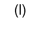
 R1 is a xylose residue or an arabinose residue bonded with its C1 atom to the corresponding xylose residue of formula (I); andR2 is independently from other R2 residues in the same molecule H or an acetyl residue,with the proviso that at least two acetyl residues are present in the saponin.
 - Use according to claim 1 or 2 or saponin for use according to claim 3, characterized in that R1 is a xylose residue and/or in that the saponin carries exactly two acetyl groups.
- Use according to claim 4 or saponin for use according to claim 4, characterized in that the acetyl groups are bonded to the oxygen atoms in C3 and C4 position of the corresponding quinovose residue.
- Use according to any of claims 1, 2, 4 or 5, or saponin for use according to any of claims 3 to 5, characterized in that the saponin is applied in combination with at least one transfection reagent chosen from the group consisting of liposomal-based transfection reagents and polymer-based transfection reagents.
- Transfection composition, comprising at least one transfection reagent chosen from the group consisting of liposomal-based transfection reagents and polymer-based transfection reagents as well as a saponin according to formula (I):
 R1 is a xylose residue or an arabinose residue bonded with its C1 atom to the corresponding xylose residue of formula (I); andR2 is independently from other R2 residues in the same molecule H or an acetyl residue,with the proviso that at least two acetyl residues are present in the saponin.
 - Transfection composition according to claim 7, characterized in that R1 is a xylose residue and/or in that the saponin carries exactly two acetyl groups.
- Transfection composition according to claim 8, characterized in that the acetyl groups are bonded to the oxygen atoms in C3 and C4 position of the corresponding quinovose residue.
- Method for an in vitro transfection, comprising the step of incubating a cell with a nucleic acid in the presence of a saponin according to formula (I):
 R1 is a xylose residue or an arabinose residue bonded with its C1 atom to the corresponding xylose residue of formula (I); andR2 is independently from other R2 residues in the same molecule H or an acetyl residue,with the proviso that at least two acetyl residues are present in the saponin.
 - Method according to claim 10, characterized in that the cell is an eukaryotic cell.
- Method according to claim 10 or 11, characterized in that the nucleic acid forms part of a nanoparticle.
- Method according to any of claims 10 to 12, characterized in that the saponin is used in a concentration lying in a range of 1 µg/mL to 25 µg/mL.
- Method according to any of claims 10 to 13, characterized in that the saponin is used in combination with at least one transfection reagent chosen from the group consisting of liposomal-based transfection reagents and polymer-based transfection reagents.
- Method for isolating a saponin according to formula (I):
 R1 is a xylose residue or an arabinose residue bonded with its C1 atom to the corresponding xylose residue of formula (I); andR2 is independently from other R2 residues in the same molecule H or an acetyl residue,with the proviso that at least two acetyl residues are present in the saponin from Gypsophila elegans M. Bieb, comprising the following steps:• cutting roots of Gypsophila elegans M. Bieb,• freeze-drying and grinding the cut roots to obtain a root powder,• extracting the root powder with a high-concentrated organic solvent to obtain a root extract,• removing the high-concentrated organic solvent from the root extract to obtain a dry extract,• dissolving the dry extract in a low-concentrated organic solvent to obtain an extract solution,• subjecting the extract solution to at least one chromatographic separation step to obtain a purified saponin solution, and• removing any solvent from the purified saponin solution to obtain a purified saponin powder;wherein the high-concentrated organic solvent is an organic solvent having a concentration of more than 50% (v/v) and wherein the low-concentrated organic solvent is an organic solvent having a concentration of less than 50% (v/v).
Priority Applications (1)
| Application Number | Priority Date | Filing Date | Title |
|---|---|---|---|
| EP23177336.7A EP4234692A3 (en) | 2017-07-11 | 2018-07-10 | Novel uses of a saponin and method for its isolation |
Applications Claiming Priority (3)
| Application Number | Priority Date | Filing Date | Title |
|---|---|---|---|
| EP17180730 | 2017-07-11 | ||
| EP18152304.4A EP3514240A1 (en) | 2018-01-18 | 2018-01-18 | Novel uses of a saponin and method for its isolation |
| PCT/EP2018/068663 WO2019011914A1 (en) | 2017-07-11 | 2018-07-10 | Novel uses of a saponin and method for its isolation |
Related Child Applications (1)
| Application Number | Title | Priority Date | Filing Date |
|---|---|---|---|
| EP23177336.7A Division EP4234692A3 (en) | 2017-07-11 | 2018-07-10 | Novel uses of a saponin and method for its isolation |
Publications (2)
| Publication Number | Publication Date |
|---|---|
| EP3652190A1 EP3652190A1 (en) | 2020-05-20 |
| EP3652190B1 true EP3652190B1 (en) | 2023-06-14 |
Family
ID=63041979
Family Applications (2)
| Application Number | Title | Priority Date | Filing Date |
|---|---|---|---|
| EP18746610.7A Active EP3652190B1 (en) | 2017-07-11 | 2018-07-10 | Novel uses of a saponin and method for its isolation |
| EP23177336.7A Pending EP4234692A3 (en) | 2017-07-11 | 2018-07-10 | Novel uses of a saponin and method for its isolation |
Family Applications After (1)
| Application Number | Title | Priority Date | Filing Date |
|---|---|---|---|
| EP23177336.7A Pending EP4234692A3 (en) | 2017-07-11 | 2018-07-10 | Novel uses of a saponin and method for its isolation |
Country Status (8)
| Country | Link |
|---|---|
| US (2) | US11708388B2 (en) |
| EP (2) | EP3652190B1 (en) |
| JP (2) | JP2020526536A (en) |
| KR (1) | KR20200026908A (en) |
| CN (1) | CN110914285A (en) |
| CA (1) | CA3069604A1 (en) |
| ES (1) | ES2955285T3 (en) |
| WO (1) | WO2019011914A1 (en) |
Families Citing this family (11)
| Publication number | Priority date | Publication date | Assignee | Title |
|---|---|---|---|---|
| US11785893B2 (en) | 2017-12-22 | 2023-10-17 | Bradken Resources Pty Limited | Grinding mill liner |
| US20220023433A1 (en) | 2018-12-21 | 2022-01-27 | Sapreme Technologies B.V. | Saponin Conjugates |
| BR112022001389A8 (en) * | 2019-07-25 | 2023-04-25 | Univ Berlin Charite | SAPONIN DERIVATIVES WITH IMPROVED THERAPEUTIC WINDOW |
| EP4003425A2 (en) * | 2019-07-25 | 2022-06-01 | Sapreme Technologies B.V. | Bioactive saponin linked to a functional moiety |
| EP3838910B1 (en) | 2019-12-18 | 2022-09-28 | Freie Universität Berlin | Efficient gene delivery tool with a wide therapeutic margin |
| KR20230043113A (en) * | 2020-06-24 | 2023-03-30 | 사프렘 테크놀로지스 비.브이. | Saponin derivatives for medicinal use |
| US20240279684A1 (en) * | 2021-05-28 | 2024-08-22 | Sapreme Technologies B.V. | Cytosolic delivery of genome editing tools |
| WO2022265493A1 (en) * | 2021-06-18 | 2022-12-22 | Sapreme Technologies B.V. | Conjugate of saponin, oligonucleotide and galnac |
| KR20250113455A (en) | 2022-11-24 | 2025-07-25 | 프로큐알 테라퓨틱스 Ⅱ 비.브이. | Antisense oligonucleotides for the treatment of hereditary hemochromatosis-HFE |
| GB202300865D0 (en) | 2023-01-20 | 2023-03-08 | Proqr Therapeutics Ii Bv | Delivery of oligonucleotides |
| CN122277643A (en) * | 2026-05-28 | 2026-06-26 | 中国农业科学院麻类研究所 | A low-polarity quinoa saponin, its preparation method and application |
Family Cites Families (3)
| Publication number | Priority date | Publication date | Assignee | Title |
|---|---|---|---|---|
| DE60229118D1 (en) * | 2001-04-04 | 2008-11-13 | Nordic Vaccine Technology As | POLYNUCLEOTIDE-BINDING COMPLEXES CONTAINING STEROLS AND SAPONINS |
| CN104687988B (en) | 2015-01-27 | 2016-06-29 | 嘉兴超纳金真空镀膜科技有限公司 | A kind of rack |
| CN104688798B (en) * | 2015-03-24 | 2018-09-21 | 山东省医学科学院药物研究所 | A kind of preparation method and application of rosy clouds grass extract |
-
2018
- 2018-07-10 EP EP18746610.7A patent/EP3652190B1/en active Active
- 2018-07-10 KR KR1020207002372A patent/KR20200026908A/en not_active Abandoned
- 2018-07-10 CN CN201880046298.6A patent/CN110914285A/en active Pending
- 2018-07-10 JP JP2020501113A patent/JP2020526536A/en active Pending
- 2018-07-10 CA CA3069604A patent/CA3069604A1/en active Pending
- 2018-07-10 EP EP23177336.7A patent/EP4234692A3/en active Pending
- 2018-07-10 WO PCT/EP2018/068663 patent/WO2019011914A1/en not_active Ceased
- 2018-07-10 US US16/628,838 patent/US11708388B2/en active Active
- 2018-07-10 ES ES18746610T patent/ES2955285T3/en active Active
-
2023
- 2023-02-15 US US18/169,266 patent/US20240285708A1/en not_active Abandoned
- 2023-06-28 JP JP2023106442A patent/JP2023115283A/en active Pending
Also Published As
| Publication number | Publication date |
|---|---|
| JP2023115283A (en) | 2023-08-18 |
| US20200131219A1 (en) | 2020-04-30 |
| EP3652190A1 (en) | 2020-05-20 |
| US11708388B2 (en) | 2023-07-25 |
| CA3069604A1 (en) | 2019-01-17 |
| EP4234692A3 (en) | 2024-01-17 |
| KR20200026908A (en) | 2020-03-11 |
| US20240285708A1 (en) | 2024-08-29 |
| ES2955285T3 (en) | 2023-11-29 |
| WO2019011914A1 (en) | 2019-01-17 |
| CN110914285A (en) | 2020-03-24 |
| EP4234692A2 (en) | 2023-08-30 |
| JP2020526536A (en) | 2020-08-31 |
Similar Documents
| Publication | Publication Date | Title |
|---|---|---|
| US20240285708A1 (en) | Uses of a saponin and method for its isolation | |
| EP3838910B1 (en) | Efficient gene delivery tool with a wide therapeutic margin | |
| Sandvoss et al. | Combination of matrix solid-phase dispersion extraction and direct on-line liquid chromatography–nuclear magnetic resonance spectroscopy–tandem mass spectrometry as a new efficient approach for the rapid screening of natural products:: Application to the total asterosaponin fraction of the starfish Asterias rubens | |
| Sama et al. | Plant derived triterpenes from Gypsophila elegans M. Bieb. enable non-toxic delivery of gene loaded nanoplexes | |
| Pérez García et al. | Multidimensional solid‐state NMR studies of the structure and dynamics of pectic polysaccharides in uniformly 13C‐labeled Arabidopsis primary cell walls | |
| Abrankó et al. | In‐source fragmentation and accurate mass analysis of multiclass flavonoid conjugates by electrospray ionization time‐of‐flight mass spectrometry | |
| Corbin et al. | Development and validation of an efficient ultrasound assisted extraction of phenolic compounds from flax (Linum usitatissimum L.) seeds | |
| Hilbert et al. | Flavonol profiles in berries of wild Vitis accessions using liquid chromatography coupled to mass spectrometry and nuclear magnetic resonance spectrometry | |
| Clochard et al. | A new acetylated triterpene saponin from Agrostemma githago L. modulates gene delivery efficiently and shows a high cellular tolerance | |
| CA3032871C (en) | Method for determining carbohydrates structure | |
| Sama et al. | Sapofectosid–Ensuring non-toxic and effective DNA and RNA delivery | |
| Waridel et al. | Identification of the polar constituents of Potamogeton species by HPLC-UV with post-column derivatization, HPLC-MSn and HPLC-NMR, and isolation of a new ent-labdane diglycoside | |
| Xia et al. | A strategy for characterization of triterpene saponins in Caulophyllum robustum hairy roots by liquid chromatography with electrospray ionization quadrupole time-of-flight mass spectrometry | |
| Renukappa et al. | Application of high-performance liquid chromatography coupled to nuclear magnetic resonance spectrometry, mass spectrometry and bioassay for the determination of active saponins from Bacopa monniera Wettst. | |
| EP3514240A1 (en) | Novel uses of a saponin and method for its isolation | |
| Bauermeister et al. | Mass spectrometric approaches for the identification of anthracycline analogs produced by actinobacteria | |
| KR100293968B1 (en) | 20 (S) - Production method of Ginsenoside AL | |
| Chen et al. | Identification of polyoxypregnane glycosides from the stems of Marsdenia tenacissima by high-performance liquid chromatography/tandem mass spectrometry | |
| Panda et al. | Pregnane glycosides | |
| Broberg et al. | Use of high-resolution magic angle spinning nuclear magnetic resonance spectroscopy for in situ studies of low-molecular-mass compounds in red algae | |
| Tamura et al. | Structure determination of acylated anthocyanins in Muscat Bailey a grapes by homonuclear Hartmann‐Hahn (HOHAHA) spectroscopy and liquid chromatography‐mass spectrometry | |
| Hövelmann et al. | Identification of a novel N-caprylhistamine-β-glucoside from tomato fruits and LC-MS/MS-based food screening for imidazole alkaloids | |
| Noté et al. | Structure elucidation of new acacic acid‐type saponins from Albizia coriaria | |
| Song et al. | Hydroxyl groups introducing NMR strategy for structural elucidation of a heptasaccharide isolated from Trillium tschonoskii | |
| Shen et al. | Polar constituents from Sageretia thea leaf characterized by HPLC‐SPE‐NMR assisted approaches |
Legal Events
| Date | Code | Title | Description |
|---|---|---|---|
| STAA | Information on the status of an ep patent application or granted ep patent |
Free format text: STATUS: UNKNOWN |
|
| STAA | Information on the status of an ep patent application or granted ep patent |
Free format text: STATUS: THE INTERNATIONAL PUBLICATION HAS BEEN MADE |
|
| PUAI | Public reference made under article 153(3) epc to a published international application that has entered the european phase |
Free format text: ORIGINAL CODE: 0009012 |
|
| STAA | Information on the status of an ep patent application or granted ep patent |
Free format text: STATUS: REQUEST FOR EXAMINATION WAS MADE |
|
| 17P | Request for examination filed |
Effective date: 20200110 |
|
| AK | Designated contracting states |
Kind code of ref document: A1 Designated state(s): AL AT BE BG CH CY CZ DE DK EE ES FI FR GB GR HR HU IE IS IT LI LT LU LV MC MK MT NL NO PL PT RO RS SE SI SK SM TR |
|
| AX | Request for extension of the european patent |
Extension state: BA ME |
|
| DAV | Request for validation of the european patent (deleted) | ||
| DAX | Request for extension of the european patent (deleted) | ||
| REG | Reference to a national code |
Ref country code: DE Ref legal event code: R079 Ref document number: 602018051844 Country of ref document: DE Free format text: PREVIOUS MAIN CLASS: C07H0015240000 Ipc: A61K0031704000 |
|
| RIC1 | Information provided on ipc code assigned before grant |
Ipc: C12N 9/22 20060101ALI20221121BHEP Ipc: C12N 15/87 20060101ALI20221121BHEP Ipc: C07H 15/256 20060101ALI20221121BHEP Ipc: A61K 31/704 20060101AFI20221121BHEP |
|
| GRAP | Despatch of communication of intention to grant a patent |
Free format text: ORIGINAL CODE: EPIDOSNIGR1 |
|
| STAA | Information on the status of an ep patent application or granted ep patent |
Free format text: STATUS: GRANT OF PATENT IS INTENDED |
|
| INTG | Intention to grant announced |
Effective date: 20230103 |
|
| GRAS | Grant fee paid |
Free format text: ORIGINAL CODE: EPIDOSNIGR3 |
|
| GRAA | (expected) grant |
Free format text: ORIGINAL CODE: 0009210 |
|
| STAA | Information on the status of an ep patent application or granted ep patent |
Free format text: STATUS: THE PATENT HAS BEEN GRANTED |
|
| AK | Designated contracting states |
Kind code of ref document: B1 Designated state(s): AL AT BE BG CH CY CZ DE DK EE ES FI FR GB GR HR HU IE IS IT LI LT LU LV MC MK MT NL NO PL PT RO RS SE SI SK SM TR |
|
| REG | Reference to a national code |
Ref country code: CH Ref legal event code: EP |
|
| REG | Reference to a national code |
Ref country code: DE Ref legal event code: R096 Ref document number: 602018051844 Country of ref document: DE |
|
| REG | Reference to a national code |
Ref country code: NL Ref legal event code: FP |
|
| REG | Reference to a national code |
Ref country code: AT Ref legal event code: REF Ref document number: 1578736 Country of ref document: AT Kind code of ref document: T Effective date: 20230715 |
|
| REG | Reference to a national code |
Ref country code: SE Ref legal event code: TRGR |
|
| REG | Reference to a national code |
Ref country code: LT Ref legal event code: MG9D |
|
| PG25 | Lapsed in a contracting state [announced via postgrant information from national office to epo] |
Ref country code: NO Free format text: LAPSE BECAUSE OF FAILURE TO SUBMIT A TRANSLATION OF THE DESCRIPTION OR TO PAY THE FEE WITHIN THE PRESCRIBED TIME-LIMIT Effective date: 20230914 |
|
| REG | Reference to a national code |
Ref country code: AT Ref legal event code: MK05 Ref document number: 1578736 Country of ref document: AT Kind code of ref document: T Effective date: 20230614 |
|
| REG | Reference to a national code |
Ref country code: ES Ref legal event code: FG2A Ref document number: 2955285 Country of ref document: ES Kind code of ref document: T3 Effective date: 20231129 |
|
| PG25 | Lapsed in a contracting state [announced via postgrant information from national office to epo] |
Ref country code: RS Free format text: LAPSE BECAUSE OF FAILURE TO SUBMIT A TRANSLATION OF THE DESCRIPTION OR TO PAY THE FEE WITHIN THE PRESCRIBED TIME-LIMIT Effective date: 20230614 Ref country code: LV Free format text: LAPSE BECAUSE OF FAILURE TO SUBMIT A TRANSLATION OF THE DESCRIPTION OR TO PAY THE FEE WITHIN THE PRESCRIBED TIME-LIMIT Effective date: 20230614 Ref country code: LT Free format text: LAPSE BECAUSE OF FAILURE TO SUBMIT A TRANSLATION OF THE DESCRIPTION OR TO PAY THE FEE WITHIN THE PRESCRIBED TIME-LIMIT Effective date: 20230614 Ref country code: HR Free format text: LAPSE BECAUSE OF FAILURE TO SUBMIT A TRANSLATION OF THE DESCRIPTION OR TO PAY THE FEE WITHIN THE PRESCRIBED TIME-LIMIT Effective date: 20230614 Ref country code: GR Free format text: LAPSE BECAUSE OF FAILURE TO SUBMIT A TRANSLATION OF THE DESCRIPTION OR TO PAY THE FEE WITHIN THE PRESCRIBED TIME-LIMIT Effective date: 20230915 |
|
| PG25 | Lapsed in a contracting state [announced via postgrant information from national office to epo] |
Ref country code: FI Free format text: LAPSE BECAUSE OF FAILURE TO SUBMIT A TRANSLATION OF THE DESCRIPTION OR TO PAY THE FEE WITHIN THE PRESCRIBED TIME-LIMIT Effective date: 20230614 |
|
| PG25 | Lapsed in a contracting state [announced via postgrant information from national office to epo] |
Ref country code: SK Free format text: LAPSE BECAUSE OF FAILURE TO SUBMIT A TRANSLATION OF THE DESCRIPTION OR TO PAY THE FEE WITHIN THE PRESCRIBED TIME-LIMIT Effective date: 20230614 |
|
| PG25 | Lapsed in a contracting state [announced via postgrant information from national office to epo] |
Ref country code: IS Free format text: LAPSE BECAUSE OF FAILURE TO SUBMIT A TRANSLATION OF THE DESCRIPTION OR TO PAY THE FEE WITHIN THE PRESCRIBED TIME-LIMIT Effective date: 20231014 |
|
| PG25 | Lapsed in a contracting state [announced via postgrant information from national office to epo] |
Ref country code: SM Free format text: LAPSE BECAUSE OF FAILURE TO SUBMIT A TRANSLATION OF THE DESCRIPTION OR TO PAY THE FEE WITHIN THE PRESCRIBED TIME-LIMIT Effective date: 20230614 Ref country code: SK Free format text: LAPSE BECAUSE OF FAILURE TO SUBMIT A TRANSLATION OF THE DESCRIPTION OR TO PAY THE FEE WITHIN THE PRESCRIBED TIME-LIMIT Effective date: 20230614 Ref country code: RO Free format text: LAPSE BECAUSE OF FAILURE TO SUBMIT A TRANSLATION OF THE DESCRIPTION OR TO PAY THE FEE WITHIN THE PRESCRIBED TIME-LIMIT Effective date: 20230614 Ref country code: PT Free format text: LAPSE BECAUSE OF FAILURE TO SUBMIT A TRANSLATION OF THE DESCRIPTION OR TO PAY THE FEE WITHIN THE PRESCRIBED TIME-LIMIT Effective date: 20231016 Ref country code: IS Free format text: LAPSE BECAUSE OF FAILURE TO SUBMIT A TRANSLATION OF THE DESCRIPTION OR TO PAY THE FEE WITHIN THE PRESCRIBED TIME-LIMIT Effective date: 20231014 Ref country code: EE Free format text: LAPSE BECAUSE OF FAILURE TO SUBMIT A TRANSLATION OF THE DESCRIPTION OR TO PAY THE FEE WITHIN THE PRESCRIBED TIME-LIMIT Effective date: 20230614 Ref country code: CZ Free format text: LAPSE BECAUSE OF FAILURE TO SUBMIT A TRANSLATION OF THE DESCRIPTION OR TO PAY THE FEE WITHIN THE PRESCRIBED TIME-LIMIT Effective date: 20230614 Ref country code: AT Free format text: LAPSE BECAUSE OF FAILURE TO SUBMIT A TRANSLATION OF THE DESCRIPTION OR TO PAY THE FEE WITHIN THE PRESCRIBED TIME-LIMIT Effective date: 20230614 |
|
| PG25 | Lapsed in a contracting state [announced via postgrant information from national office to epo] |
Ref country code: PL Free format text: LAPSE BECAUSE OF FAILURE TO SUBMIT A TRANSLATION OF THE DESCRIPTION OR TO PAY THE FEE WITHIN THE PRESCRIBED TIME-LIMIT Effective date: 20230614 |
|
| REG | Reference to a national code |
Ref country code: CH Ref legal event code: PL |
|
| PG25 | Lapsed in a contracting state [announced via postgrant information from national office to epo] |
Ref country code: MC Free format text: LAPSE BECAUSE OF FAILURE TO SUBMIT A TRANSLATION OF THE DESCRIPTION OR TO PAY THE FEE WITHIN THE PRESCRIBED TIME-LIMIT Effective date: 20230614 |
|
| REG | Reference to a national code |
Ref country code: DE Ref legal event code: R097 Ref document number: 602018051844 Country of ref document: DE |
|
| REG | Reference to a national code |
Ref country code: BE Ref legal event code: MM Effective date: 20230731 |
|
| PG25 | Lapsed in a contracting state [announced via postgrant information from national office to epo] |
Ref country code: LU Free format text: LAPSE BECAUSE OF NON-PAYMENT OF DUE FEES Effective date: 20230710 |
|
| PG25 | Lapsed in a contracting state [announced via postgrant information from national office to epo] |
Ref country code: MC Free format text: LAPSE BECAUSE OF FAILURE TO SUBMIT A TRANSLATION OF THE DESCRIPTION OR TO PAY THE FEE WITHIN THE PRESCRIBED TIME-LIMIT Effective date: 20230614 Ref country code: LU Free format text: LAPSE BECAUSE OF NON-PAYMENT OF DUE FEES Effective date: 20230710 |
|
| PLBE | No opposition filed within time limit |
Free format text: ORIGINAL CODE: 0009261 |
|
| STAA | Information on the status of an ep patent application or granted ep patent |
Free format text: STATUS: NO OPPOSITION FILED WITHIN TIME LIMIT |
|
| REG | Reference to a national code |
Ref country code: IE Ref legal event code: MM4A |
|
| PG25 | Lapsed in a contracting state [announced via postgrant information from national office to epo] |
Ref country code: DK Free format text: LAPSE BECAUSE OF FAILURE TO SUBMIT A TRANSLATION OF THE DESCRIPTION OR TO PAY THE FEE WITHIN THE PRESCRIBED TIME-LIMIT Effective date: 20230614 Ref country code: CH Free format text: LAPSE BECAUSE OF NON-PAYMENT OF DUE FEES Effective date: 20230731 |
|
| REG | Reference to a national code |
Ref country code: SE Ref legal event code: EUG |
|
| PG25 | Lapsed in a contracting state [announced via postgrant information from national office to epo] |
Ref country code: SI Free format text: LAPSE BECAUSE OF FAILURE TO SUBMIT A TRANSLATION OF THE DESCRIPTION OR TO PAY THE FEE WITHIN THE PRESCRIBED TIME-LIMIT Effective date: 20230614 |
|
| 26N | No opposition filed |
Effective date: 20240315 |
|
| PG25 | Lapsed in a contracting state [announced via postgrant information from national office to epo] |
Ref country code: SI Free format text: LAPSE BECAUSE OF FAILURE TO SUBMIT A TRANSLATION OF THE DESCRIPTION OR TO PAY THE FEE WITHIN THE PRESCRIBED TIME-LIMIT Effective date: 20230614 Ref country code: BE Free format text: LAPSE BECAUSE OF NON-PAYMENT OF DUE FEES Effective date: 20230731 |
|
| PG25 | Lapsed in a contracting state [announced via postgrant information from national office to epo] |
Ref country code: IE Free format text: LAPSE BECAUSE OF NON-PAYMENT OF DUE FEES Effective date: 20230710 |
|
| PG25 | Lapsed in a contracting state [announced via postgrant information from national office to epo] |
Ref country code: IT Free format text: LAPSE BECAUSE OF NON-PAYMENT OF DUE FEES Effective date: 20230710 Ref country code: IE Free format text: LAPSE BECAUSE OF NON-PAYMENT OF DUE FEES Effective date: 20230710 Ref country code: FR Free format text: LAPSE BECAUSE OF NON-PAYMENT OF DUE FEES Effective date: 20230814 |
|
| REG | Reference to a national code |
Ref country code: ES Ref legal event code: FD2A Effective date: 20240827 |
|
| PG25 | Lapsed in a contracting state [announced via postgrant information from national office to epo] |
Ref country code: SE Free format text: LAPSE BECAUSE OF NON-PAYMENT OF DUE FEES Effective date: 20230711 |
|
| PG25 | Lapsed in a contracting state [announced via postgrant information from national office to epo] |
Ref country code: ES Free format text: LAPSE BECAUSE OF NON-PAYMENT OF DUE FEES Effective date: 20230711 |
|
| PG25 | Lapsed in a contracting state [announced via postgrant information from national office to epo] |
Ref country code: ES Free format text: LAPSE BECAUSE OF NON-PAYMENT OF DUE FEES Effective date: 20230711 |
|
| PG25 | Lapsed in a contracting state [announced via postgrant information from national office to epo] |
Ref country code: BG Free format text: LAPSE BECAUSE OF FAILURE TO SUBMIT A TRANSLATION OF THE DESCRIPTION OR TO PAY THE FEE WITHIN THE PRESCRIBED TIME-LIMIT Effective date: 20230614 |
|
| PG25 | Lapsed in a contracting state [announced via postgrant information from national office to epo] |
Ref country code: BG Free format text: LAPSE BECAUSE OF FAILURE TO SUBMIT A TRANSLATION OF THE DESCRIPTION OR TO PAY THE FEE WITHIN THE PRESCRIBED TIME-LIMIT Effective date: 20230614 |
|
| PG25 | Lapsed in a contracting state [announced via postgrant information from national office to epo] |
Ref country code: CY Free format text: LAPSE BECAUSE OF FAILURE TO SUBMIT A TRANSLATION OF THE DESCRIPTION OR TO PAY THE FEE WITHIN THE PRESCRIBED TIME-LIMIT; INVALID AB INITIO Effective date: 20180710 |
|
| PG25 | Lapsed in a contracting state [announced via postgrant information from national office to epo] |
Ref country code: HU Free format text: LAPSE BECAUSE OF FAILURE TO SUBMIT A TRANSLATION OF THE DESCRIPTION OR TO PAY THE FEE WITHIN THE PRESCRIBED TIME-LIMIT; INVALID AB INITIO Effective date: 20180710 |
|
| PGFP | Annual fee paid to national office [announced via postgrant information from national office to epo] |
Ref country code: NL Payment date: 20250715 Year of fee payment: 8 |
|
| PGFP | Annual fee paid to national office [announced via postgrant information from national office to epo] |
Ref country code: DE Payment date: 20250728 Year of fee payment: 8 |
|
| PGFP | Annual fee paid to national office [announced via postgrant information from national office to epo] |
Ref country code: GB Payment date: 20250722 Year of fee payment: 8 |
|
| PG25 | Lapsed in a contracting state [announced via postgrant information from national office to epo] |
Ref country code: TR Free format text: LAPSE BECAUSE OF FAILURE TO SUBMIT A TRANSLATION OF THE DESCRIPTION OR TO PAY THE FEE WITHIN THE PRESCRIBED TIME-LIMIT Effective date: 20230614 |