EP3623010B1 - Chimeric antigen receptor, nucleic acid, chimeric antigen receptor expression plasmid, chimeric antigen receptor expressing cell and pharmaceutical composition for treating cancer - Google Patents
Chimeric antigen receptor, nucleic acid, chimeric antigen receptor expression plasmid, chimeric antigen receptor expressing cell and pharmaceutical composition for treating cancer Download PDFInfo
- Publication number
- EP3623010B1 EP3623010B1 EP19193456.1A EP19193456A EP3623010B1 EP 3623010 B1 EP3623010 B1 EP 3623010B1 EP 19193456 A EP19193456 A EP 19193456A EP 3623010 B1 EP3623010 B1 EP 3623010B1
- Authority
- EP
- European Patent Office
- Prior art keywords
- chimeric antigen
- antigen receptor
- seq
- cell line
- acid sequence
- Prior art date
- Legal status (The legal status is an assumption and is not a legal conclusion. Google has not performed a legal analysis and makes no representation as to the accuracy of the status listed.)
- Active
Links
Images

















Classifications
-
- A—HUMAN NECESSITIES
- A61—MEDICAL OR VETERINARY SCIENCE; HYGIENE
- A61K—PREPARATIONS FOR MEDICAL, DENTAL OR TOILETRY PURPOSES
- A61K31/00—Medicinal preparations containing organic active ingredients
- A61K31/33—Heterocyclic compounds
- A61K31/395—Heterocyclic compounds having nitrogen as a ring hetero atom, e.g. guanethidine or rifamycins
- A61K31/41—Heterocyclic compounds having nitrogen as a ring hetero atom, e.g. guanethidine or rifamycins having five-membered rings with two or more ring hetero atoms, at least one of which being nitrogen, e.g. tetrazole
- A61K31/4164—1,3-Diazoles
- A61K31/4188—1,3-Diazoles condensed with other heterocyclic ring systems, e.g. biotin, sorbinil
-
- A—HUMAN NECESSITIES
- A61—MEDICAL OR VETERINARY SCIENCE; HYGIENE
- A61K—PREPARATIONS FOR MEDICAL, DENTAL OR TOILETRY PURPOSES
- A61K31/00—Medicinal preparations containing organic active ingredients
- A61K31/28—Compounds containing heavy metals
- A61K31/282—Platinum compounds
-
- A—HUMAN NECESSITIES
- A61—MEDICAL OR VETERINARY SCIENCE; HYGIENE
- A61K—PREPARATIONS FOR MEDICAL, DENTAL OR TOILETRY PURPOSES
- A61K31/00—Medicinal preparations containing organic active ingredients
- A61K31/33—Heterocyclic compounds
- A61K31/395—Heterocyclic compounds having nitrogen as a ring hetero atom, e.g. guanethidine or rifamycins
- A61K31/495—Heterocyclic compounds having nitrogen as a ring hetero atom, e.g. guanethidine or rifamycins having six-membered rings with two or more nitrogen atoms as the only ring heteroatoms, e.g. piperazine or tetrazines
-
- A—HUMAN NECESSITIES
- A61—MEDICAL OR VETERINARY SCIENCE; HYGIENE
- A61K—PREPARATIONS FOR MEDICAL, DENTAL OR TOILETRY PURPOSES
- A61K31/00—Medicinal preparations containing organic active ingredients
- A61K31/33—Heterocyclic compounds
- A61K31/555—Heterocyclic compounds containing heavy metals, e.g. hemin, hematin, melarsoprol
-
- A—HUMAN NECESSITIES
- A61—MEDICAL OR VETERINARY SCIENCE; HYGIENE
- A61K—PREPARATIONS FOR MEDICAL, DENTAL OR TOILETRY PURPOSES
- A61K31/00—Medicinal preparations containing organic active ingredients
- A61K31/70—Carbohydrates; Sugars; Derivatives thereof
- A61K31/7028—Compounds having saccharide radicals attached to non-saccharide compounds by glycosidic linkages
- A61K31/7034—Compounds having saccharide radicals attached to non-saccharide compounds by glycosidic linkages attached to a carbocyclic compound, e.g. phloridzin
- A61K31/704—Compounds having saccharide radicals attached to non-saccharide compounds by glycosidic linkages attached to a carbocyclic compound, e.g. phloridzin attached to a condensed carbocyclic ring system, e.g. sennosides, thiocolchicosides, escin, daunorubicin
-
- A—HUMAN NECESSITIES
- A61—MEDICAL OR VETERINARY SCIENCE; HYGIENE
- A61K—PREPARATIONS FOR MEDICAL, DENTAL OR TOILETRY PURPOSES
- A61K31/00—Medicinal preparations containing organic active ingredients
- A61K31/70—Carbohydrates; Sugars; Derivatives thereof
- A61K31/7042—Compounds having saccharide radicals and heterocyclic rings
- A61K31/7052—Compounds having saccharide radicals and heterocyclic rings having nitrogen as a ring hetero atom, e.g. nucleosides, nucleotides
- A61K31/706—Compounds having saccharide radicals and heterocyclic rings having nitrogen as a ring hetero atom, e.g. nucleosides, nucleotides containing six-membered rings with nitrogen as a ring hetero atom
- A61K31/7064—Compounds having saccharide radicals and heterocyclic rings having nitrogen as a ring hetero atom, e.g. nucleosides, nucleotides containing six-membered rings with nitrogen as a ring hetero atom containing condensed or non-condensed pyrimidines
- A61K31/7068—Compounds having saccharide radicals and heterocyclic rings having nitrogen as a ring hetero atom, e.g. nucleosides, nucleotides containing six-membered rings with nitrogen as a ring hetero atom containing condensed or non-condensed pyrimidines having oxo groups directly attached to the pyrimidine ring, e.g. cytidine, cytidylic acid
-
- A—HUMAN NECESSITIES
- A61—MEDICAL OR VETERINARY SCIENCE; HYGIENE
- A61K—PREPARATIONS FOR MEDICAL, DENTAL OR TOILETRY PURPOSES
- A61K31/00—Medicinal preparations containing organic active ingredients
- A61K31/70—Carbohydrates; Sugars; Derivatives thereof
- A61K31/7088—Compounds having three or more nucleosides or nucleotides
- A61K31/711—Natural deoxyribonucleic acids, i.e. containing only 2'-deoxyriboses attached to adenine, guanine, cytosine or thymine and having 3'-5' phosphodiester links
-
- A—HUMAN NECESSITIES
- A61—MEDICAL OR VETERINARY SCIENCE; HYGIENE
- A61K—PREPARATIONS FOR MEDICAL, DENTAL OR TOILETRY PURPOSES
- A61K40/00—Cellular immunotherapy
- A61K40/10—Cellular immunotherapy characterised by the cell type used
- A61K40/11—T-cells, e.g. tumour infiltrating lymphocytes [TIL] or regulatory T [Treg] cells; Lymphokine-activated killer [LAK] cells
-
- A—HUMAN NECESSITIES
- A61—MEDICAL OR VETERINARY SCIENCE; HYGIENE
- A61K—PREPARATIONS FOR MEDICAL, DENTAL OR TOILETRY PURPOSES
- A61K40/00—Cellular immunotherapy
- A61K40/10—Cellular immunotherapy characterised by the cell type used
- A61K40/15—Natural-killer [NK] cells; Natural-killer T [NKT] cells
-
- A—HUMAN NECESSITIES
- A61—MEDICAL OR VETERINARY SCIENCE; HYGIENE
- A61K—PREPARATIONS FOR MEDICAL, DENTAL OR TOILETRY PURPOSES
- A61K40/00—Cellular immunotherapy
- A61K40/30—Cellular immunotherapy characterised by the recombinant expression of specific molecules in the cells of the immune system
- A61K40/31—Chimeric antigen receptors [CAR]
-
- A—HUMAN NECESSITIES
- A61—MEDICAL OR VETERINARY SCIENCE; HYGIENE
- A61K—PREPARATIONS FOR MEDICAL, DENTAL OR TOILETRY PURPOSES
- A61K40/00—Cellular immunotherapy
- A61K40/40—Cellular immunotherapy characterised by antigens that are targeted or presented by cells of the immune system
- A61K40/41—Vertebrate antigens
- A61K40/42—Cancer antigens
-
- A—HUMAN NECESSITIES
- A61—MEDICAL OR VETERINARY SCIENCE; HYGIENE
- A61K—PREPARATIONS FOR MEDICAL, DENTAL OR TOILETRY PURPOSES
- A61K45/00—Medicinal preparations containing active ingredients not provided for in groups A61K31/00 - A61K41/00
- A61K45/06—Mixtures of active ingredients without chemical characterisation, e.g. antiphlogistics and cardiaca
-
- A—HUMAN NECESSITIES
- A61—MEDICAL OR VETERINARY SCIENCE; HYGIENE
- A61P—SPECIFIC THERAPEUTIC ACTIVITY OF CHEMICAL COMPOUNDS OR MEDICINAL PREPARATIONS
- A61P35/00—Antineoplastic agents
-
- C—CHEMISTRY; METALLURGY
- C07—ORGANIC CHEMISTRY
- C07K—PEPTIDES
- C07K14/00—Peptides having more than 20 amino acids; Gastrins; Somatostatins; Melanotropins; Derivatives thereof
- C07K14/435—Peptides having more than 20 amino acids; Gastrins; Somatostatins; Melanotropins; Derivatives thereof from animals; from humans
- C07K14/705—Receptors; Cell surface antigens; Cell surface determinants
- C07K14/70503—Immunoglobulin superfamily
- C07K14/70539—MHC-molecules, e.g. HLA-molecules
-
- C—CHEMISTRY; METALLURGY
- C07—ORGANIC CHEMISTRY
- C07K—PEPTIDES
- C07K16/00—Immunoglobulins [IG], e.g. monoclonal or polyclonal antibodies
- C07K16/18—Immunoglobulins [IG], e.g. monoclonal or polyclonal antibodies against material from animals or humans
- C07K16/28—Immunoglobulins [IG], e.g. monoclonal or polyclonal antibodies against material from animals or humans against receptors, cell surface antigens or cell surface determinants
- C07K16/2803—Immunoglobulins [IG], e.g. monoclonal or polyclonal antibodies against material from animals or humans against receptors, cell surface antigens or cell surface determinants against the immunoglobulin superfamily
- C07K16/2833—Immunoglobulins [IG], e.g. monoclonal or polyclonal antibodies against material from animals or humans against receptors, cell surface antigens or cell surface determinants against the immunoglobulin superfamily against MHC-molecules, e.g. HLA-molecules
-
- C—CHEMISTRY; METALLURGY
- C12—BIOCHEMISTRY; BEER; SPIRITS; WINE; VINEGAR; MICROBIOLOGY; ENZYMOLOGY; MUTATION OR GENETIC ENGINEERING
- C12N—MICROORGANISMS OR ENZYMES; COMPOSITIONS THEREOF; PROPAGATING, PRESERVING, OR MAINTAINING MICROORGANISMS; MUTATION OR GENETIC ENGINEERING; CULTURE MEDIA
- C12N5/00—Undifferentiated human, animal or plant cells, e.g. cell lines; Tissues; Cultivation or maintenance thereof; Culture media therefor
- C12N5/06—Animal cells or tissues; Human cells or tissues
- C12N5/0602—Vertebrate cells
- C12N5/0634—Cells from the blood or the immune system
- C12N5/0646—Natural killers cells [NK], NKT cells
-
- C—CHEMISTRY; METALLURGY
- C12—BIOCHEMISTRY; BEER; SPIRITS; WINE; VINEGAR; MICROBIOLOGY; ENZYMOLOGY; MUTATION OR GENETIC ENGINEERING
- C12N—MICROORGANISMS OR ENZYMES; COMPOSITIONS THEREOF; PROPAGATING, PRESERVING, OR MAINTAINING MICROORGANISMS; MUTATION OR GENETIC ENGINEERING; CULTURE MEDIA
- C12N9/00—Enzymes; Proenzymes; Compositions thereof; Processes for preparing, activating, inhibiting, separating or purifying enzymes
- C12N9/14—Hydrolases (3)
- C12N9/16—Hydrolases (3) acting on ester bonds (3.1)
- C12N9/22—Ribonucleases [RNase]; Deoxyribonucleases [DNase]
-
- A—HUMAN NECESSITIES
- A61—MEDICAL OR VETERINARY SCIENCE; HYGIENE
- A61K—PREPARATIONS FOR MEDICAL, DENTAL OR TOILETRY PURPOSES
- A61K39/00—Medicinal preparations containing antigens or antibodies
- A61K2039/505—Medicinal preparations containing antigens or antibodies comprising antibodies
-
- A—HUMAN NECESSITIES
- A61—MEDICAL OR VETERINARY SCIENCE; HYGIENE
- A61K—PREPARATIONS FOR MEDICAL, DENTAL OR TOILETRY PURPOSES
- A61K2239/00—Indexing codes associated with cellular immunotherapy of group A61K40/00
- A61K2239/38—Indexing codes associated with cellular immunotherapy of group A61K40/00 characterised by the dose, timing or administration schedule
-
- A—HUMAN NECESSITIES
- A61—MEDICAL OR VETERINARY SCIENCE; HYGIENE
- A61K—PREPARATIONS FOR MEDICAL, DENTAL OR TOILETRY PURPOSES
- A61K2239/00—Indexing codes associated with cellular immunotherapy of group A61K40/00
- A61K2239/46—Indexing codes associated with cellular immunotherapy of group A61K40/00 characterised by the cancer treated
- A61K2239/47—Brain; Nervous system
-
- A—HUMAN NECESSITIES
- A61—MEDICAL OR VETERINARY SCIENCE; HYGIENE
- A61K—PREPARATIONS FOR MEDICAL, DENTAL OR TOILETRY PURPOSES
- A61K2239/00—Indexing codes associated with cellular immunotherapy of group A61K40/00
- A61K2239/46—Indexing codes associated with cellular immunotherapy of group A61K40/00 characterised by the cancer treated
- A61K2239/49—Breast
-
- A—HUMAN NECESSITIES
- A61—MEDICAL OR VETERINARY SCIENCE; HYGIENE
- A61K—PREPARATIONS FOR MEDICAL, DENTAL OR TOILETRY PURPOSES
- A61K2239/00—Indexing codes associated with cellular immunotherapy of group A61K40/00
- A61K2239/46—Indexing codes associated with cellular immunotherapy of group A61K40/00 characterised by the cancer treated
- A61K2239/54—Pancreas
-
- A—HUMAN NECESSITIES
- A61—MEDICAL OR VETERINARY SCIENCE; HYGIENE
- A61K—PREPARATIONS FOR MEDICAL, DENTAL OR TOILETRY PURPOSES
- A61K2239/00—Indexing codes associated with cellular immunotherapy of group A61K40/00
- A61K2239/46—Indexing codes associated with cellular immunotherapy of group A61K40/00 characterised by the cancer treated
- A61K2239/59—Reproductive system, e.g. uterus, ovaries, cervix or testes
-
- C—CHEMISTRY; METALLURGY
- C07—ORGANIC CHEMISTRY
- C07K—PEPTIDES
- C07K2317/00—Immunoglobulins specific features
- C07K2317/50—Immunoglobulins specific features characterized by immunoglobulin fragments
- C07K2317/56—Immunoglobulins specific features characterized by immunoglobulin fragments variable (Fv) region, i.e. VH and/or VL
- C07K2317/565—Complementarity determining region [CDR]
-
- C—CHEMISTRY; METALLURGY
- C07—ORGANIC CHEMISTRY
- C07K—PEPTIDES
- C07K2317/00—Immunoglobulins specific features
- C07K2317/60—Immunoglobulins specific features characterized by non-natural combinations of immunoglobulin fragments
- C07K2317/62—Immunoglobulins specific features characterized by non-natural combinations of immunoglobulin fragments comprising only variable region components
- C07K2317/622—Single chain antibody (scFv)
-
- C—CHEMISTRY; METALLURGY
- C07—ORGANIC CHEMISTRY
- C07K—PEPTIDES
- C07K2319/00—Fusion polypeptide
-
- C—CHEMISTRY; METALLURGY
- C07—ORGANIC CHEMISTRY
- C07K—PEPTIDES
- C07K2319/00—Fusion polypeptide
- C07K2319/01—Fusion polypeptide containing a localisation/targetting motif
- C07K2319/03—Fusion polypeptide containing a localisation/targetting motif containing a transmembrane segment
-
- C—CHEMISTRY; METALLURGY
- C07—ORGANIC CHEMISTRY
- C07K—PEPTIDES
- C07K2319/00—Fusion polypeptide
- C07K2319/33—Fusion polypeptide fusions for targeting to specific cell types, e.g. tissue specific targeting, targeting of a bacterial subspecies
-
- C—CHEMISTRY; METALLURGY
- C12—BIOCHEMISTRY; BEER; SPIRITS; WINE; VINEGAR; MICROBIOLOGY; ENZYMOLOGY; MUTATION OR GENETIC ENGINEERING
- C12N—MICROORGANISMS OR ENZYMES; COMPOSITIONS THEREOF; PROPAGATING, PRESERVING, OR MAINTAINING MICROORGANISMS; MUTATION OR GENETIC ENGINEERING; CULTURE MEDIA
- C12N2510/00—Genetically modified cells
-
- C—CHEMISTRY; METALLURGY
- C12—BIOCHEMISTRY; BEER; SPIRITS; WINE; VINEGAR; MICROBIOLOGY; ENZYMOLOGY; MUTATION OR GENETIC ENGINEERING
- C12N—MICROORGANISMS OR ENZYMES; COMPOSITIONS THEREOF; PROPAGATING, PRESERVING, OR MAINTAINING MICROORGANISMS; MUTATION OR GENETIC ENGINEERING; CULTURE MEDIA
- C12N2800/00—Nucleic acids vectors
- C12N2800/80—Vectors containing sites for inducing double-stranded breaks, e.g. meganuclease restriction sites
Definitions
- the present disclosure relates to a pharmaceutical product containing an antigen or an antibody. More particularly, the present disclosure relates to a chimeric antigen receptor, a nucleic acid encoding the chimeric antigen receptor, a chimeric antigen receptor expression plasmid, a chimeric antigen receptor expressing cell, and a pharmaceutical composition for use in the treatment of cancer.
- Cancer also known as malignancy, is a state of abnormal proliferation of cells, and these proliferating cells may invade other parts of the body as a disease caused by a malfunction in the control of cell division and proliferation.
- the number of people suffering from cancer worldwide has a growing trend. Cancer is one of the top ten causes of death for the Chinese people and has been the top ten causes of death for twenty-seven consecutive years.
- Cancer immunotherapy is another method for treating cancer except the above methods.
- the immune system of the patient is activated in the cancer immunotherapy by using tumor cells or tumor antigens to induce specific cellular and humoral immune responses for enhancing the anti-cancer ability of the patient, preventing the growth, spread, and recurrence of tumors, and achieving the purpose of removing or controlling tumors.
- the chimeric antigen receptor immune cell technology is one of the cell therapies developing very rapidly in recent years.
- the chimeric antigen receptor immune cell transfecting a chimeric protein, which couples the antigen binding portion having capable of recognizing a certain tumor antigen of the antibody to the intracellular portion of the CD3- ⁇ chain or Fc ⁇ RI ⁇ in vitro, into the immune cell by a transduction method to express the chimeric antigen receptor.
- the chimeric antigen receptor immune cell technology has a significant therapeutic effect in the treatment of acute leukemia and non-Hodgkin's lymphoma, and it is considered to be one of the most promising treatments for cancer.
- WO 2016/160622 A2 discloses chimeric antigen receptor (CAR) cells targeting and antibodies human HLA-G as a method of cancer treatment
- WO 2018/057904 A1 discloses chimeric antigen receptors (CARs) targeting human LHR, B7-H4, HLA-G, or HLA-DR and therapeutic methods of their use for inhibiting the growth of a tumor and/or treating a cancer.
- the cell therapy of the chimeric antigen receptor immune cell currently has the following disadvantages: lack of unique tumor-associated antigens, low efficiency of homing of immune cells to tumor sites, and inability to overcome the immunosuppressive microenvironment of solid tumors. Accordingly, the efficacy of the chimeric antigen receptor immune cell in solid tumors is greatly limited.
- a chimeric antigen receptor specific to human leukocyte antigen G includes, in order from an N-terminus to a C-terminus, the anti-HLA-G antibody including an amino acid sequence of SEQ ID NO: 1, the amino acid sequence of SEQ ID NO: 2, and the amino acid sequence of SEQ ID NO: 3.
- the chimeric antigen receptor can further include a suicide protein including an amino acid sequence of SEQ ID NO: 4, wherein the suicide protein is linked to the C-terminus of the amino acid sequence of SEQ ID NO: 3.
- the chimeric antigen receptor can further include a 2A peptide, wherein the 2A peptide links the amino acid sequence of SEQ ID NO: 2 and the amino acid sequence of SEQ ID NO: 3.
- a nucleic acid encoding the aforementioned chimeric antigen receptor includes, in order from a 5' end to a 3' end, the anti-HLA-G antibody coding fragment including a nucleic acid sequence of SEQ ID NO: 11, the nucleic acid sequence of SEQ ID NO: 12, and the nucleic acid sequence of SEQ ID NO: 13.
- the nucleic acid can further include a suicide gene including a nucleic acid sequence of SEQ ID NO: 14, wherein the suicide gene is linked to the 3' end of the nucleic acid sequence of SEQ ID NO: 13.
- the nucleic acid can further include a 2A peptide coding fragment, wherein the 2A peptide coding fragment links the nucleic acid sequence of SEQ ID NO: 12 and the nucleic acid sequence of SEQ ID NO: 13.
- a chimeric antigen receptor expression plasmid includes, in order from a 5' end to a 3' end, a promoter including a nucleic acid sequence of SEQ ID NO: 16 and the aforementioned nucleic acid.
- a chimeric antigen receptor expressing cell includes an immune cell and the aforementioned chimeric antigen receptor expression plasmid.
- the immune cell can be a T lymphocyte or a natural killer (NK) cell.
- NK natural killer
- the NK cell can be a NK-92 cell line or a primary NK cell.
- a pharmaceutical composition for use in the treatment of cancer includes the aforementioned chimeric antigen receptor expressing cell and a pharmaceutically acceptable carrier.
- the pharmaceutical composition for use in the treatment of cancer can further include a chemotherapy drug.
- the chemotherapy drug can be doxorubicin, temozolomide, gemcitabine or carboplatin.
- the cancer can be a breast cancer, a polymorphic glioblastoma, a pancreatic cancer or an ovarian cancer.
- a chimeric antigen receptor, a nucleic acid encoding the chimeric antigen receptor, a chimeric antigen receptor expression plasmid including the nucleic acid, a chimeric antigen receptor expressing cell including the chimeric antigen receptor expression plasmid, and a pharmaceutical composition for use in the treatment of cancer including the chimeric antigen receptor expressing cell are provided.
- HLA-G human leukocyte antigen G
- the chimeric antigen receptor expressing cell of the present disclosure which expresses the chimeric antigen receptor of the present disclosure, can specifically target the tumor cells to avoid the off-target effect, thereby effectively killing the tumor cells. Therefore, the chimeric antigen receptor expressing cell can be used for inhibiting the proliferation of the tumor cells in a subject in need for a treatment of a tumor.
- the pharmaceutical composition for use in the treatment of cancer of the present disclosure includes the chimeric antigen receptor expressing cell of the present disclosure, and can further include a chemotherapy drug, which can effectively kill tumor cells and thereby treat cancer.
- HLA-G human leukocyte antigen G
- MHC major histocompatibility complex
- HLA-G is expressed on fetal derived placental cells, and is active in the negative regulation of immune response. HLA-G may play a role in immune tolerance in pregnancy.
- the chimeric antigen receptor of the present disclosure is specific to HLA-G, and includes, in order from an N-terminus to a C-terminus, the anti-HLA-G antibody including an amino acid sequence of SEQ ID NO: 1, the amino acid sequence of SEQ ID NO: 2, and the amino acid sequence of SEQ ID NO: 3.
- a suicide protein including an amino acid sequence of SEQ ID NO: 4 is linked to the C-terminus of the amino acid sequence of SEQ ID NO: 3, and a 2A peptide including an amino acid sequence of SEQ ID NO: 10 links the amino acid sequence of SEQ ID NO: 2 and the amino acid sequence of SEQ ID NO: 3.
- the amino acid sequence of SEQ ID NO: 2 is killer cell immunoglobulin-like receptor 2DS4 (KIR2DS4).
- the amino acid sequence of SEQ ID NO: 3 is DNAX activating protein 12 (DAP12).
- the suicide protein including an amino acid sequence of SEQ ID NO: 4 is iCas9 protein.
- the nucleic acid of the present disclosure encodes the chimeric antigen receptor of the present disclosure.
- the nucleic acid includes, in order from a 5' end to a 3' end, the anti-HLA-G antibody coding fragment including a nucleic acid sequence of SEQ ID NO: 11, the nucleic acid sequence of SEQ ID NO: 12, and the nucleic acid sequence of SEQ ID NO: 13.
- a suicide gene including a nucleic acid sequence of SEQ ID NO: 14 is linked to the 3' end of the nucleic acid sequence of SEQ ID NO: 13, and a 2A peptide coding fragment including a nucleic acid sequence of SEQ ID NO: 15 links the nucleic acid sequence of SEQ ID NO: 12 and the nucleic acid sequence of SEQ ID NO: 13.
- the anti-HLA-G antibody coding fragment including a nucleic acid sequence of SEQ ID NO: 11 encodes the anti-HLA-G antibody including an amino acid sequence of SEQ ID NO: 1.
- the nucleic acid sequence of SEQ ID NO: 12 encodes the amino acid sequence of SEQ ID NO: 2.
- the nucleic acid sequence of SEQ ID NO: 13 encodes the amino acid sequence of SEQ ID NO: 3.
- the suicide gene including a nucleic acid sequence of SEQ ID NO: 14 encodes the suicide protein including an amino acid sequence of SEQ ID NO: 4.
- the 2A peptide coding fragment including a nucleic acid sequence of SEQ ID NO: 15 encodes the 2A peptide including an amino acid sequence of SEQ ID NO: 10.
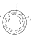
- Fig. 2 is a schematic view showing a construction of a chimeric antigen receptor expression plasmid according to another embodiment of the present disclosure.
- the chimeric antigen receptor expression plasmid is Lenti-EF1a-CAR-100517-S1A plasmid, wherein the insert thereof includes a promoter, the anti-HLA-G antibody coding fragment, the nucleic acid sequence of SEQ ID NO: 12, and the nucleic acid sequence of SEQ ID NO: 13.
- the promoter is the EF-1 alpha promoter including a nucleic acid sequence of SEQ ID NO: 16.
- the anti-HLA-G antibody coding fragment includes the nucleic acid sequence of SEQ ID NO: 11.
- the insert of the Lenti-EF1a-CAR-100517-S1A plasmid further includes a signal peptide coding fragment including a nucleic acid sequence of SEQ ID NO: 17, the suicide gene including the nucleic acid sequence of SEQ ID NO: 14, and the 2A peptide coding fragment including the nucleic acid sequence of SEQ ID NO: 15.
- the signal peptide coding fragment is linked to the 5' end of the anti-HLA-G antibody coding fragment
- the suicide gene is linked to the 3' end of the nucleic acid sequence of SEQ ID NO: 13
- the 2A peptide coding fragment links the nucleic acid sequence of SEQ ID NO: 12 and the nucleic acid sequence of SEQ ID NO: 13.
- the insert is constructed on Creative Biolabs vector (Creative Biolabs, NY, USA) to obtain the Lenti-EF1a-CAR-100517-S1A plasmid.
- the Creative Biolabs vector is a lentivirus vector system, so that the constructed chimeric antigen receptor expression plasmid can be transfected into expression cells to produce lentiviruses, and the chimeric antigen receptor can be subsequently transduced into the immune cells using lentiviruses.
- the chimeric antigen receptor expressing cell of the present disclosure is obtained by transducing the chimeric antigen receptor of the present disclosure into the immune cell using lentiviruses.
- the immune cell can be a T lymphocyte or a natural killer (NK) cell.
- the NK cell can be a NK-92 cell line or a primary NK cell.
- the constructed Lenti-EF1a-CAR-100517-S1A plasmid is transfected into a 293T cell line using lipofectamine 3000 (Invitrogen) to prepare the lentiviruses carrying the chimeric antigen receptor of the present disclosure.
- the supernatant containing the prepared lentiviruses and Opti-MEM (Invitrogen) containing 8 ⁇ g/ml of polybrene (Sigma-Aldrich) are used to culture the primary T lymphocytes for 3 days to transduce the chimeric antigen receptor of the present disclosure into the primary T lymphocytes.
- the supernatant containing the prepared lentiviruses and the Opti-MEM (Invitrogen) containing 50 ⁇ g/ml of protamine sulfate (Sigma-Aldrich) are used to culture the primary NK cells or the NK-92 cell line for 7 days to transduce the chimeric antigen receptor of the present disclosure into the primary NK cell or the NK-92 cell line.
- the obtained chimeric antigen receptor expressing cell has an effect of inducing tumor cell death in mammals, so that the chimeric antigen receptor expressing cell can be used for inhibiting a proliferation of tumor cells in a subject in need for a treatment of a tumor.
- the tumor cell can be a breast cancer cell, a polymorphic glioblastoma cell, a pancreatic cancer cell or an ovarian cancer cell.
- the pharmaceutical composition for use in the treatment of a cancer of the present disclosure includes the chimeric antigen receptor expressing cell of the present disclosure and a pharmaceutically acceptable carrier.
- the pharmaceutical composition for use in the treatment of cancer can further include a chemotherapy drug.
- the chemotherapy drug can be doxorubicin (Dox), temozolomide (TMZ), gemcitabine (Gem) or carboplatin (CB).
- the chimeric antigen receptor expressing cell and the pharmaceutical composition for use in the treatment of the cancer will be further described by the following embodiments.
- an Example 1, an Example 2 and an Example 3 will be further provided to illustrate the accompanied efficacies of chimeric antigen receptor expressing cell and the pharmaceutical composition for use in the treatment of the cancer on inducing tumor cell death.
- the tumor cells used are human breast cancer cell line MDA-MB-231, human malignant brain tumor cell line DBTRG-05MG (hereinafter referred to as DBTRG), human pancreatic cancer cell line AsPC1, and human ovarian cancer cell line SKOV3.
- the tumor cell lines used are all purchased from the American Type Culture Collection (ATCC).
- the human breast cancer cell line MDA-MB-231 is a triple-negative breast cancer cell line, that is, the hormone receptor (ER, PR) and HER-2 receptor thereof are negative, and the human breast cancer cell line MDA-MB-231 is cultured in RPMI culture medium containing 10% fetal bovine serum (FBS).
- the human malignant brain tumor cell line DBTRG is cultured in DMEM culture medium containing 10% FBS.
- the human pancreatic cancer cell line AsPC1 is cultured in RPMI culture medium containing 10% FBS.
- the human ovarian cancer cell line SKOV3 is cultured in McCoy's 5A culture medium containing 10% FBS.
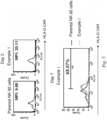
- the chimeric antigen receptor of the present disclosure is transduced into the NK-92 cell line to obtain the chimeric antigen receptor expressing cell of Example 1 of the present disclosure, and the expression level of the chimeric antigen receptor of the obtained chimeric antigen receptor expressing cell of Example 1 is analyzed by flow cytometry.
- Fig. 3 is a graph showing the expression level of chimeric antigen receptors in the chimeric antigen receptor expressing cell according to Example 1 of the present disclosure.
- FIG. 3 shows the expression level of the chimeric antigen receptor of the parental NK-92 cell line without transducing the chimeric antigen receptor of the present disclosure, and the expression level of the chimeric antigen receptor of the chimeric antigen receptor expressing cell of Example 1 on day 3 and day 7 after transduction the chimeric antigen receptor.
- the mean fluorescence intensity (MFI) of the parental NK-92 cell line is only 9.98%, while the MFI of the chimeric antigen receptor expressing cell of Example 1 on day 3 and day 7 after transduction can reach 20.11 % and 65.07%, respectively.
- the results indicate that the chimeric antigen receptor expressing cell of Example 1 can stably express the chimeric antigen receptor of the present disclosure.
- the effects of the chimeric antigen receptor expressing cell of Example 1 and the pharmaceutical composition for use in the treatment of cancer including the chimeric antigen receptor expressing cell of Example 1 of the present disclosure on inducing the death of the breast cancer cells, the glioblastoma multiforme cells, the pancreatic cancer cells, and the ovarian cancer cells are further demonstrated in following experiments.
- the human breast cancer cell line MDA-MB-231, the human malignant brain tumor cell line DBTRG, the human pancreatic cancer cell line AsPC1 and the human ovarian cancer cell line SKOV3 are seeded in a 12-well plate at a density of 1 ⁇ 10 5 cells/well. The cells are subsequently incubated for 24 hours. Each type of the tumor cells is divided into six groups. In a control, the tumor cells are untreated. In a group 1, the tumor cells are treated with the chemotherapy drug. In a group 2, the tumor cells are treated with the parental NK-92 cell line. In a group 3, the tumor cells are treated with the parental NK-92 cell line and the chemotherapy drug.
- the number of the parental NK-92 cell line treated is 1 ⁇ 10 5 cells.
- the tumor cells are treated with the chimeric antigen receptor expressing cell of Example 1.
- the tumor cells are treated with the chimeric antigen receptor expressing cell of Example 1 and the chemotherapy drug.
- the number of the chimeric antigen receptor expressing cell of Example 1 treated is 1 ⁇ 10 5 cells.
- the chemotherapy drug used for treating the human breast cancer cell line MDA-MB-231 is doxorubicin (200 nM), the chemotherapy drug used for treating the human malignant brain tumor cell line DBTRG is temozolomide (80 ⁇ g/mL), the chemotherapy drug used for treating the human pancreatic cancer cell line AsPC1 is gemcitabine (20 ⁇ M), the chemotherapy drug used for treating the human ovarian cancer cell line SKOV3 is carboplatin (20 ⁇ M).
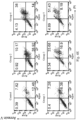
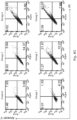
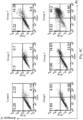
- the treated cells are stained with Annexin V-FITC and propidium iodide (PI), and the apoptosis and the death of the tumor cells are detected by the flow cytometry.
- the sum of the percentage of cells stained with Annexin V-FITC and/or PI (that is the percentage of cells in the first quadrant, the second quadrant, and the fourth quadrant of the bivariate flow cytometry scatter plot) are calculated to obtain the cytotoxicity.
- the results of the cytotoxicity are counted after the three independent trials in each group.
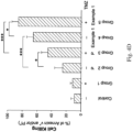
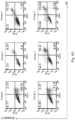
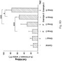
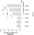
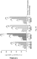
- Figs. 4A , 4B , 4C , 4D , 4E , 4F , 4G , 4H and 4I show analytical results of tumor cell death induced by the chimeric antigen receptor expressing cells according to Example 1 of the present disclosure.
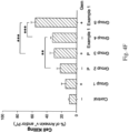
- Fig. 4A is a graph showing the analytical results of the death of the human breast cancer cell line MDA-MB-231 induced by the chimeric antigen receptor expressing cell of Example 1
- Fig. 4B is a statistical chart of Fig. 4A after the three independent trials.
- Fig. 4C is a graph showing the analytical results of the death of the human malignant brain tumor cell line DBTRG induced by the chimeric antigen receptor expressing cell of Example 1, and Fig.
- FIG. 4D is a statistical chart of Fig. 4C after the three independent trials.
- Fig. 4E is a graph showing the analytical results of the death of the human pancreatic cancer cell line AsPC1 induced by the chimeric antigen receptor expressing cell of Example 1
- Fig. 4F is a statistical chart of Fig. 4E after the three independent trials.
- Fig. 4G is a graph showing the analytical results of the death of the human ovarian cancer cell line SKOV3 induced by the chimeric antigen receptor expressing cell of Example 1
- Fig. 4H is a statistical chart of Fig. 4G after the three independent trials.
- Fig. 4I is a statistical chart of Figs.
- P represents the parental NK-92 cell line
- H represents the chimeric antigen receptor expressing cell of Example 1
- D represents doxorubicin
- T represents temozolomide
- G represents gemcitabine
- C represents carboplatin.
- the death rate of the human breast cancer cell line MDA-MB-231 is only about 10%.
- the death rate of the human breast cancer cell line MDA-MB-231 is increased, but there is no statistically significant difference compared to the control.
- the death rate of the human breast cancer cell line MDA-MB-231 can increase to 40%, and there is a statistically significant difference (p ⁇ 0.05) compared to the group 2.
- the death rate of the human breast cancer cell line MDA-MB-231 is about 60%, and there is a statistically significant difference (p ⁇ 0.001) compared to the group 2. Furthermore, in the group 5 treated with the doxorubicin and the chimeric antigen receptor expressing cell of Example 1, the death rate of the human breast cancer cell line MDA-MB-231 can reach 80%, and there is a statistically significant difference (p ⁇ 0.05) compared to the group 4 and a statistically significant difference (p ⁇ 0.01) compared to the group 3, respectively.
- the death rate of the human malignant brain tumor cell line DBTRG is less than 10%.
- the death rate of the human malignant brain tumor cell line DBTRG is increased, but there is no statistically significant difference compared to the control.
- the death rate of the human malignant brain tumor cell line DBTRG can increase to 40%, and there is a statistically significant difference (p ⁇ 0.05) compared to the group 2.
- the death rate of the human malignant brain tumor cell line DBTRG is more than 60%, and there is a statistically significant difference (p ⁇ 0.001) compared to the group 2. Furthermore, in the group 5 treated with the temozolomide and the chimeric antigen receptor expressing cell of Example 1, the death rate of the human malignant brain tumor cell line DBTRG can reach more than 80%, and there is a statistically significant difference (p ⁇ 0.05) compared to the group 4 and a statistically significant difference ( p ⁇ 0.001) compared to the group 3, respectively.
- the death rate of the human pancreatic cancer cell line AsPC1 is less than 10%.
- the death rate of the human pancreatic cancer cell line AsPC1 is increased, but there is no statistically significant difference compared to the control.
- the death rate of the human pancreatic cancer cell line AsPC1 can increase to 30%, but there is no statistically significant difference compared to the control.
- the death rate of the human pancreatic cancer cell line AsPC1 is approximately 40%, and there is a statistically significant difference (p ⁇ 0.01) compared to the group 2. Furthermore, in the group 5 treated with the gemcitabine and the chimeric antigen receptor expressing cell of Example 1, the death rate of the human pancreatic cancer cell line AsPC1 can reach 60%, and there is a statistically significant difference (p ⁇ 0.001) compared to the group 4 and a statistically significant difference (p ⁇ 0.001) compared to the group 3, respectively.
- the death rate of the human ovarian cancer cell line SKOV3 is less than 10%.
- the death rate of the human ovarian cancer cell line SKOV3 is increased, but there is no statistically significant difference compared to the control.
- the death rate of the human ovarian cancer cell line SKOV3 can increase to 30%, and there is a statistically significant difference ( p ⁇ 0.05) compared to the group 2.
- the death rate of the human ovarian cancer cell line SKOV3 is approximately 40%, and there is a statistically significant difference (p ⁇ 0.01) compared to the group 2. Furthermore, in the group 5 treated with the carboplatin and the chimeric antigen receptor expressing cell of Example 1, the death rate of the human ovarian cancer cell line SKOV3 can reach 60%, and there is a statistically significant difference (p ⁇ 0.05) compared to the group 4 and a statistically significant difference (p ⁇ 0.01) compared to the group 3, respectively.
- the results indicate that the chimeric antigen receptor expressing cell of Example 1 can be used to treat with the human breast cancer cell line MDA-MB-231, the human malignant brain tumor cell line DBTRG, the human pancreatic cancer cell line AsPC1 and the human ovarian cancer cell line SKOV3 for excellent cell killing. Therefore, the chimeric antigen receptor expressing cell of the present disclosure can be used for inhibiting the proliferation of the tumor cells in the subject in need for the treatment of the tumor.
- the tumor cell can be the breast cancer cell, the polymorphic glioblastoma cell, the pancreatic cancer cell or the ovarian cancer cell.
- the simultaneous treatment of the chemotherapy drug and the chimeric antigen receptor expressing cell of Example 1 can significantly increase the toxic effect on inducing death of the human breast cancer cell line MDA-MB-231, the human malignant brain tumor cell line DBTRG, the human pancreatic cancer cell line AsPC1 and the human ovarian cancer cell line SKOV3.
- the results indicate that the method for treating cancer of the present disclosure can effectively inhibit the growth of the tumor cells and treat cancer.
- the chimeric antigen receptor of the present disclosure is transduced into the primary NK cell to obtain the chimeric antigen receptor expressing cell of Example 2 of the present disclosure, and the expression level of the chimeric antigen receptor of the obtained chimeric antigen receptor expressing cell of Example 2 is analyzed by the flow cytometry.
- Fig. 5 is a graph showing an expression level of chimeric antigen receptors in a chimeric antigen receptor expressing cell according to Example 2 of the present disclosure.
- FIG. 5 shows the expression level of the chimeric antigen receptor of the parental primary NK cell without transducing the chimeric antigen receptor of the present disclosure, and the expression level of the chimeric antigen receptor of the chimeric antigen receptor expressing cell of Example 2 on day 3 and day 7 after transduction the chimeric antigen receptor.
- the MFI of the parental primary NK cell is 22.09%, while the MFI of the chimeric antigen receptor expressing cell of Example 2 on day 3 and day 7 after transduction can reach 29.02% and 50.21%, respectively.
- the results indicate that the chimeric antigen receptor expressing cell of Example 2 can stably express the chimeric antigen receptor of the present disclosure.
- the effects of the chimeric antigen receptor expressing cell of Example 2 and the pharmaceutical composition for use in the treatment of cancer including the chimeric antigen receptor expressing cell of Example 2 of the present disclosure on inducing the death of the breast cancer cells, the glioblastoma multiforme cells, the pancreatic cancer cells, and the ovarian cancer cells are further demonstrated in following experiments.
- the human breast cancer cell line MDA-MB-231, the human malignant brain tumor cell line DBTRG, the human pancreatic cancer cell line AsPC1 and the human ovarian cancer cell line SKOV3 are seeded in a 12-well plate at a density of 1 ⁇ 10 5 cells/well. The cells are subsequently incubated for 24 hours. Each type of the tumor cells is divided into six groups. In a control, the tumor cells are untreated. In a group 1, the tumor cells are treated with the chemotherapy drug. In a group 2, the tumor cells are treated with the parental primary NK cell. In a group 3, the tumor cells are treated with the parental primary NK cell and the chemotherapy drug. In the groups 2 and 3, the number of the parental primary NK cell treated is 1 ⁇ 10 5 cells.
- the tumor cells are treated with the chimeric antigen receptor expressing cell of Example 2.
- the tumor cells are treated with the chimeric antigen receptor expressing cell of Example 2 and the chemotherapy drug.
- the number of the chimeric antigen receptor expressing cell of Example 2 treated is 1 ⁇ 10 5 cells.
- the chemotherapy drug used for treating the human breast cancer cell line MDA-MB-231 is doxorubicin (200 nM), the chemotherapy drug used for treating the human malignant brain tumor cell line DBTRG is temozolomide (80 ⁇ g/mL), the chemotherapy drug used for treating the human pancreatic cancer cell line AsPC1 is gemcitabine (20 ⁇ M), the chemotherapy drug used for treating the human ovarian cancer cell line SKOV3 is carboplatin (20 ⁇ M).
- the treated cells are stained with Annexin V-FITC and propidium iodide (PI), and the apoptosis and the death of the tumor cells are detected by the flow cytometry.
- the sum of the percentage of cells stained with Annexin V-FITC and/or PI (that is the percentage of cells in the first quadrant, the second quadrant, and the fourth quadrant of the bivariate flow cytometry scatter plot) are calculated to obtain the cytotoxicity.
- the results of the cytotoxicity are counted after the three independent trials in each group.
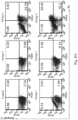
- Figs. 6A , 6B , 6C , 6D , 6E , 6F , 6G , 6H and 6I show analytical results of tumor cell death induced by the chimeric antigen receptor expressing cells according to Example 2 of the present disclosure.
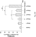
- Fig. 6A is a graph showing the analytical results of the death of the human breast cancer cell line MDA-MB-231 induced by the chimeric antigen receptor expressing cell of Example 2
- Fig. 6B is a statistical chart of Fig. 6A after the three independent trials.
- Fig. 6C is a graph showing the analytical results of the death of the human malignant brain tumor cell line DBTRG induced by the chimeric antigen receptor expressing cell of Example 2
- FIG. 6D is a statistical chart of Fig. 6C after the three independent trials.
- Fig. 6E is a graph showing the analytical results of the death of the human pancreatic cancer cell line AsPC1 induced by the chimeric antigen receptor expressing cell of Example 2
- Fig. 6F is a statistical chart of Fig. 6E after the three independent trials.
- Fig. 6G is a graph showing the analytical results of the death of the human ovarian cancer cell line SKOV3 induced by the chimeric antigen receptor expressing cell of Example 2
- Fig. 6H is a statistical chart of Fig. 6G after the three independent trials.
- Fig. 6I is a statistical chart of Figs.
- P represents the parental primary NK cell
- H represents the chimeric antigen receptor expressing cell of Example 2
- D represents doxorubicin
- T represents temozolomide
- G represents gemcitabine
- C represents carboplatin.
- the death rate of the human breast cancer cell line MDA-MB-231 is only about 10%.
- the death rate of the human breast cancer cell line MDA-MB-231 is increased, but there is no statistically significant difference compared to the control.
- the death rate of the human breast cancer cell line MDA-MB-231 can increase to 30%, and there is a statistically significant difference (p ⁇ 0.05) compared to the group 2.
- the death rate of the human breast cancer cell line MDA-MB-231 is more than 50%, and there is a statistically significant difference (p ⁇ 0.01) compared to the group 2. Furthermore, in the group 5 treated with the doxorubicin and the chimeric antigen receptor expressing cell of Example 2, the death rate of the human breast cancer cell line MDA-MB-231 can reach 80%, and there is a statistically significant difference (p ⁇ 0.05) compared to the group 4 and a statistically significant difference (p ⁇ 0.01) compared to the group 3, respectively.
- the death rate of the human malignant brain tumor cell line DBTRG is less than 10%.
- the death rate of the human malignant brain tumor cell line DBTRG is increased, but there is no statistically significant difference compared to the control.
- the death rate of the human malignant brain tumor cell line DBTRG can increase to about 30%, and there is a statistically significant difference (p ⁇ 0.05) compared to the group 2.
- the death rate of the human malignant brain tumor cell line DBTRG is more than 20%, and there is a statistically significant difference ( p ⁇ 0.05) compared to the group 2. Furthermore, in the group 5 treated with the temozolomide and the chimeric antigen receptor expressing cell of Example 2, the death rate of the human malignant brain tumor cell line DBTRG can reach about 60%, and there is a statistically significant difference (p ⁇ 0.01) compared to the group 4 and a statistically significant difference (p ⁇ 0.05) compared to the group 3, respectively.
- the death rate of the human pancreatic cancer cell line AsPC1 is less than 10%.
- the death rate of the human pancreatic cancer cell line AsPC1 is increased, but there is no statistically significant difference compared to the control.
- the death rate of the human pancreatic cancer cell line AsPC1 can increase to 30%, and there is a statistically significant difference (p ⁇ 0.05) compared to the group 2.
- the death rate of the human pancreatic cancer cell line AsPC1 is approximately 20%, and there is a statistically significant difference (p ⁇ 0.01) compared to the group 2. Furthermore, in the group 5 treated with the gemcitabine and the chimeric antigen receptor expressing cell of Example 2, the death rate of the human pancreatic cancer cell line AsPC1 can reach 50%, and there is a statistically significant difference (p ⁇ 0.01) compared to the group 4 and a statistically significant difference (p ⁇ 0.05) compared to the group 3, respectively.
- the death rate of the human ovarian cancer cell line SKOV3 is less than 10%.
- the death rate of the human ovarian cancer cell line SKOV3 is comparable to that of the control.
- the death rate of the human ovarian cancer cell line SKOV3 can increase to more than 20%, and there is a statistically significant difference (p ⁇ 0.05) compared to the group 2.
- the death rate of the human ovarian cancer cell line SKOV3 is approximately 20%, and there is a statistically significant difference ( p ⁇ 0.05) compared to the group 2. Furthermore, in the group 5 treated with the carboplatin and the chimeric antigen receptor expressing cell of Example 2, the death rate of the human ovarian cancer cell line SKOV3 can reach 50%, and there is a statistically significant difference ( p ⁇ 0.01) compared to the group 4 and a statistically significant difference ( p ⁇ 0.05) compared to the group 3, respectively.
- the results indicate that the chimeric antigen receptor expressing cell of Example 2 can be used to treat with the breast cancer cell, the polymorphic glioblastoma cell, the pancreatic cancer cell or the ovarian cancer cell for excellent cell killing. Therefore, the chimeric antigen receptor expressing cell of the present disclosure can be used for inhibiting the proliferation of the tumor cells in the subject in need for the treatment of the tumor.
- the simultaneous treatment of the chemotherapy drug and the chimeric antigen receptor expressing cell of Example 2 can significantly increase the toxic effect on inducing death of the human breast cancer cell line MDA-MB-231, the human malignant brain tumor cell line DBTRG, the human pancreatic cancer cell line AsPC1 and the human ovarian cancer cell line SKOV3.
- the results indicate that the method for treating cancer of the present disclosure can effectively inhibit the growth of the tumor cells and treat cancer.
- the chimeric antigen receptor of the present disclosure is transduced into the primary T lymphocyte to obtain the chimeric antigen receptor expressing cell of Example 3 of the present disclosure, and the expression level of the chimeric antigen receptor of the obtained chimeric antigen receptor expressing cell of Example 3 is analyzed by the flow cytometry.
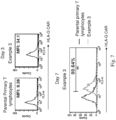
- Fig. 7 is a graph showing an expression level of a chimeric antigen receptor in a chimeric antigen receptor expressing cell according to Example 3 of the present disclosure.
- FIG. 7 shows the expression level of the chimeric antigen receptor of the parental primary T lymphocyte without transducing the chimeric antigen receptor of the present disclosure, and the expression level of the chimeric antigen receptor of the chimeric antigen receptor expressing cell of Example 3 on day 3 and day 7 after transduction the chimeric antigen receptor.
- the MFI of the parental primary T lymphocyte only is 9.36%, while the MFI of the chimeric antigen receptor expressing cell of Example 3 on day 3 and day 7 after transduction can reach 34.1% and 88.64%, respectively.
- the results indicate that the chimeric antigen receptor expressing cell of Example 3 can stably express the chimeric antigen receptor of the present disclosure.
- the effects of the chimeric antigen receptor expressing cell of Example 3 and the pharmaceutical composition for use in the treatment of cancer including the chimeric antigen receptor expressing cell of Example 3 of the present disclosure on inducing the death of the breast cancer cells, the glioblastoma multiforme cells, the pancreatic cancer cells, and the ovarian cancer cells are further demonstrated in following experiments.
- the human breast cancer cell line MDA-MB-231, the human malignant brain tumor cell line DBTRG, the human pancreatic cancer cell line AsPC1 and the human ovarian cancer cell line SKOV3 are seeded in a 12-well plate at a density of 1 ⁇ 10 5 cells/well. The cells are subsequently incubated for 24 hours. Each type of the tumor cells is divided into six groups. In a control, the tumor cells are untreated. In a group 1, the tumor cells are treated with the chemotherapy drug. In a group 2, the tumor cells are treated with the parental primary T lymphocyte. In a group 3, the tumor cells are treated with the parental T lymphocyte and the chemotherapy drug. In the groups 2 and 3, the number of the parental primary T lymphocyte treated is 1 ⁇ 10 5 cells.
- the tumor cells are treated with the chimeric antigen receptor expressing cell of Example 3.
- the tumor cells are treated with the chimeric antigen receptor expressing cell of Example 3 and the chemotherapy drug.
- the number of the chimeric antigen receptor expressing cell of Example 3 treated is 1 ⁇ 10 5 cells.
- the chemotherapy drug used for treating the human breast cancer cell line MDA-MB-231 is doxorubicin (200 nM), the chemotherapy drug used for treating the human malignant brain tumor cell line DBTRG is temozolomide (80 ⁇ g/mL), the chemotherapy drug used for treating the human pancreatic cancer cell line AsPC1 is gemcitabine (20 ⁇ M), the chemotherapy drug used for treating the human ovarian cancer cell line SKOV3 is carboplatin (20 ⁇ M).
- the treated cells are stained with Annexin V-FITC and propidium iodide (PI), and the apoptosis and the death of the tumor cells are detected by the flow cytometry.
- the sum of the percentage of cells stained with Annexin V-FITC and/or PI (that is the percentage of cells in the first quadrant, the second quadrant, and the fourth quadrant of the bivariate flow cytometry scatter plot) are calculated to obtain the cytotoxicity.
- the results of the cytotoxicity are counted after the three independent trials in each group.
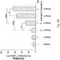
- Figs. 8A , 8B , 8C , 8D , 8E , 8F , 8G , 8H and 8I show analytical results of tumor cell death induced by the chimeric antigen receptor expressing cells according to Example 3 of the present disclosure.
- Fig. 8A is a graph showing the analytical results of the death of the human breast cancer cell line MDA-MB-231 induced by the chimeric antigen receptor expressing cell of Example 3
- Fig. 8B is a statistical chart of Fig. 8A after the three independent trials.
- Fig. 8C is a graph showing the analytical results of the death of the human malignant brain tumor cell line DBTRG induced by the chimeric antigen receptor expressing cell of Example 3, and Fig.
- Fig. 8D is a statistical chart of Fig. 8C after the three independent trials.
- Fig. 8E is a graph showing the analytical results of the death of the human pancreatic cancer cell line AsPC1 induced by the chimeric antigen receptor expressing cell of Example 3, and
- Fig. 8F is a statistical chart of Fig. 8E after the three independent trials.
- Fig. 8G is a graph showing the analytical results of the death of the human ovarian cancer cell line SKOV3 induced by the chimeric antigen receptor expressing cell of Example 3, and
- Fig. 8H is a statistical chart of Fig. 8G after the three independent trials.
- Fig. 8I is a statistical chart of Figs.
- P represents the parental primary T lymphocyte
- H represents the chimeric antigen receptor expressing cell of Example 3
- D represents doxorubicin
- T represents temozolomide
- G represents gemcitabine
- C represents carboplatin.
- the death rate of the human breast cancer cell line MDA-MB-231 is only about 10%.
- the death rate of the human breast cancer cell line MDA-MB-231 is increased, but there is no statistically significant difference compared to the control.
- the death rate of the human breast cancer cell line MDA-MB-231 can increase to 20%, and there is a statistically significant difference (p ⁇ 0.01) compared to the group 2.
- the death rate of the human breast cancer cell line MDA-MB-231 is more than 30%, and there is a statistically significant difference (p ⁇ 0.001) compared to the group 2. Furthermore, in the group 5 treated with the doxorubicin and the chimeric antigen receptor expressing cell of Example 3, the death rate of the human breast cancer cell line MDA-MB-231 can reach about 50%, and there is a statistically significant difference (p ⁇ 0.05) compared to the group 4 and a statistically significant difference (p ⁇ 0.001) compared to the group 3, respectively.
- the death rate of the human malignant brain tumor cell line DBTRG is less than 20%.
- the death rate of the human malignant brain tumor cell line DBTRG is increased, but there is no statistically significant difference compared to the control.
- the death rate of the human malignant brain tumor cell line DBTRG can increase to about 30%, there is no statistically significant difference compared to the control.
- the death rate of the human malignant brain tumor cell line DBTRG is more than 50%, and there is a statistically significant difference (p ⁇ 0.001) compared to the group 2. Furthermore, in the group 5 treated with the temozolomide and the chimeric antigen receptor expressing cell of Example 3, the death rate of the human malignant brain tumor cell line DBTRG can reach about 80%, and there is a statistically significant difference (p ⁇ 0.05) compared to the group 4 and a statistically significant difference (p ⁇ 0.001) compared to the group 3, respectively.
- the death rate of the human pancreatic cancer cell line AsPC1 is less than 20%.
- the death rate of the human pancreatic cancer cell line AsPC1 is comparable to that of the control.
- the death rate of the human pancreatic cancer cell line AsPC1 can increase to 30%, and there is a statistically significant difference (p ⁇ 0.05) compared to the group 2.
- the death rate of the human pancreatic cancer cell line AsPC1 is increased to more than 50%, and there is a statistically significant difference (p ⁇ 0.001) compared to the group 2. Furthermore, in the group 5 treated with the gemcitabine and the chimeric antigen receptor expressing cell of Example 3, the death rate of the human pancreatic cancer cell line AsPC1 can reach 60%, and there is a statistically significant difference ( p ⁇ 0.01) compared to the group 4 and a statistically significant difference ( p ⁇ 0.01) compared to the group 3, respectively.
- the death rate of the human ovarian cancer cell line SKOV3 is less than 10%.
- the death rate of the human ovarian cancer cell line SKOV3 is increased, but there is no statistically significant difference compared to the control.
- the death rate of the human ovarian cancer cell line SKOV3 can increase to about 30%, and there is a statistically significant difference ( p ⁇ 0.05) compared to the group 2.
- the death rate of the human ovarian cancer cell line SKOV3 is approximately 60%, and there is a statistically significant difference ( p ⁇ 0.001) compared to the group 2. Furthermore, in the group 5 treated with the carboplatin and the chimeric antigen receptor expressing cell of Example 3, the death rate of the human ovarian cancer cell line SKOV3 can reach more than 60%, and there is a statistically significant difference ( p ⁇ 0.05) compared to the group 4 and a statistically significant difference ( p ⁇ 0.001) compared to the group 3, respectively.
- the results indicate that the chimeric antigen receptor expressing cell of Example 3 can be used to treat with the breast cancer cell, the polymorphic glioblastoma cell, the pancreatic cancer cell or the ovarian cancer cell for excellent cell killing. Therefore, the chimeric antigen receptor expressing cell of the present disclosure can be used for inhibiting the proliferation of the tumor cells in the subject in need for the treatment of the tumor.
- the simultaneous treatment of the chemotherapy drug and the chimeric antigen receptor expressing cell of Example 3 can significantly increase the toxic effect on inducing death of the human breast cancer cell line MDA-MB-231, the human malignant brain tumor cell line DBTRG, the human pancreatic cancer cell line AsPC1 and the human ovarian cancer cell line SKOV3.
- the results indicate that the method for treating cancer of the present disclosure can effectively inhibit the growth of the tumor cells and treat cancer.
- the tumor cells are treated with the chemotherapy drug, and then detecting the HLA-G expression level of the tumor cells.
- the human breast cancer cell line MDA-MB-231, the human malignant brain tumor cell line DBTRG, the human pancreatic cancer cell line AsPC1 and the human ovarian cancer cell line SKOV3 are seeded in a 6-well plate at a density of 2 ⁇ 10 5 cells/well. The cells are subsequently incubated for 24 hours. Each type of the tumor cells is divided into two groups. In a control, the tumor cells are untreated. In an experiment group, the tumor cells are treated with the chemotherapy drug for 48 hours.
- the chemotherapy drug used for treating the human breast cancer cell line MDA-MB-231 is doxorubicin (200 nM), the chemotherapy drug used for treating the human malignant brain tumor cell line DBTRG is temozolomide (80 ⁇ g/mL), the chemotherapy drug used for treating the human pancreatic cancer cell line AsPC1 is gemcitabine (20 ⁇ M), the chemotherapy drug used for treating the human ovarian cancer cell line SKOV3 is carboplatin (20 ⁇ M). Then, the HLA-G expression level of the tumor cells are detected by immunofluorescence staining assay and flow cytometry.
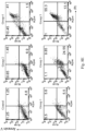
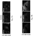
- Figs. 9A, 9B , 9C, 9D , 9E, 9F , 9G and 9H are analytical results of immunofluorescence staining assay showing the HLA-G expression level of tumor cells treated with the chemotherapy drug.
- Fig. 9A is analytical result of immunofluorescence staining assay showing the HLA-G expression level in the control group of the human breast cancer cell line MDA-MB-231
- Fig. 9B is analytical result of immunofluorescence staining assay showing the HLA-G expression level of the human breast cancer cell line MDA-MB-231 treated with doxorubicin (Dox).
- Fig. 9A is analytical result of immunofluorescence staining assay showing the HLA-G expression level in the control group of the human breast cancer cell line MDA-MB-231
- Fig. 9B is analytical result of immunofluorescence staining assay showing the HLA-G expression level of the human breast cancer cell line MDA-MB-231 treated with doxorubicin (Do
- FIG. 9C is analytical result of immunofluorescence staining assay showing the HLA-G expression level in the control group of the human malignant brain tumor cell line DBTRG
- Fig. 9D is analytical result of immunofluorescence staining assay showing the HLA-G expression level of the human malignant brain tumor cell line DBTRG treated with temozolomide (TMZ).
- Fig. 9E is analytical result of immunofluorescence staining assay showing the HLA-G expression level in the control group of the human pancreatic cancer cell line AsPC1
- Fig. 9F is analytical result of immunofluorescence staining assay showing the HLA-G expression level of the human pancreatic cancer cell line AsPC1 treated with gemcitabine (Gem).
- Fig. 9E is analytical result of immunofluorescence staining assay showing the HLA-G expression level in the control group of the human pancreatic cancer cell line AsPC1 treated with gemcitabine (Gem).
- 9G is analytical result of immunofluorescence staining assay showing the HLA-G expression level in the control group of the human ovarian cancer cell line SKOV3, and Fig. 9H is analytical result of immunofluorescence staining assay showing the HLA-G expression level of the human ovarian cancer cell line SKOV3 treated with carboplatin (CB).
- CB carboplatin
- doxorubicin can increase the HLA-G expression level on the plasma membrane of the human breast cancer cell line MDA-MB-231.
- temozolomide can increase the HLA-G expression level on the plasma membrane of the human malignant brain tumor cell line DBTRG.
- gemcitabine can increase the HLA-G expression level on the plasma membrane of the human pancreatic cancer cell line AsPC1.
- Figs. 9G and 9H treatment of carboplatin can increase the HLA-G expression level on the plasma membrane of the human ovarian cancer cell line SKOV3.
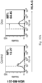
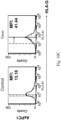
- Figs. 10A , 10B , 10C , 10D and 10E are analytical results of flow cytometry showing a HLA-G expression of tumor cells treated with a chemotherapy drug.
- Fig. 10A is analytical result of flow cytometry of the human breast cancer cell line MDA-MB-231.
- Fig. 10B is analytical result of flow cytometry of the human malignant brain tumor cell line DBTRG.
- Fig. 10C is analytical result of flow cytometry of the human pancreatic cancer cell line AsPC1.
- Fig. 10D is analytical result of flow cytometry of the human ovarian cancer cell line SKOV3.
- Fig. 10E is a statistical chart of Figs. 10A , 10B , 10C and 10D .
- the mean fluorescence intensity (MFI) of the control group of the human breast cancer cell line MDA-MB-231 is only 12.27%, while the MFI of the experiment group of the human breast cancer cell line MDA-MB-231 can reach 64.45%, which is statistically significant (p ⁇ 0.001).
- MFI of the control group of the human malignant brain tumor cell line DBTRG is only 14.01%, while the MFI of the experiment group of the human malignant brain tumor cell line DBTRG can reach 22.33%, which is statistically significant (p ⁇ 0.001).
- Fig. 10A the mean fluorescence intensity (MFI) of the control group of the human breast cancer cell line MDA-MB-231 is only 12.27%, while the MFI of the experiment group of the human breast cancer cell line MDA-MB-231 can reach 64.45%, which is statistically significant (p ⁇ 0.001).
- Fig. 10B the MFI of the control group of the human malignant brain tumor cell line DBTRG is only 14.01%, while the MFI of the experiment
- the MFI of the control group of the human pancreatic cancer cell line AsPC1 is only 13.18%, while the MFI of the experiment group of the human pancreatic cancer cell line AsPC1 can reach 41.44%, which is statistically significant (p ⁇ 0.01).
- the MFI of the control group of the human ovarian cancer cell line SKOV3 is only 14.69%, while the MFI of the experiment group of the human ovarian cancer cell line SKOV3 can reach 38.58%, which is statistically significant ( p ⁇ 0.01).
- the method for treating cancer of the present disclosure not part of the invention further administers a composition containing a plurality of chimeric antigen receptor expressing cells to the subject in need for a treatment of cancer, in which the chimeric antigen receptor expressing cells expresses the chimeric antigen receptor specific to HLA-G, in order to enhance the effect of killing tumor cells.
- the treatment of the chemotherapy drug and the composition containing the chimeric antigen receptor expressing cells can be in a sequence or simultaneous.
- Fig. 11 is a schematic view showing the theoretical structure and mechanism of the chimeric antigen receptor in the plasma membrane of the chimeric antigen receptor expressing cell of the present disclosure.
- the chimeric antigen receptor expressing cell of the present disclosure is a genetically engineered NK cell or T cell which expresses the chimeric antigen receptor of the present disclosure, and the chimeric antigen receptor of the present disclosure is a tumor-targeting receptor complex included the anti-HLA-G antibody (scFv), the KIR2DS4 and the DAP12.
- the chimeric antigen receptor of the present disclosure can further include the suicide protein iCas9.
- the chimeric antigen receptor expressing cell of the present disclosure can specifically recognize the HLA-G on the tumor plasma membrane.
- the HLA-G expression on the plasma membrane of the tumor cell can be positively regulated.
- the chimeric antigen receptor expressing cell of the present disclosure binds to the HLA-G, which is specifically recognized on the surface of the tumor cell, signal transduction is triggered, and a signal cascade is generated to cause activation and proliferation of the chimeric antigen receptor expressing cell of the present disclosure. In turn, it also triggers exocytosis of lytic granules and killing of the target tumor cells.
- the chimeric antigen receptor of the present disclosure has excellent specific binding ability to the tumor cells, in particular, specific binding to HLA-G expressed on the cell membrane of tumor cells. Accordingly, the chimeric antigen receptor expressing cell of the present disclosure, which expresses the chimeric antigen receptor of the present disclosure, can specifically target the tumor cells to avoid the off-target effect, thereby effectively killing the tumor cells. Therefore, the chimeric antigen receptor expressing cell can be used for inhibiting the proliferation of the tumor cells in the subject in need for the treatment of the tumor.
- the pharmaceutical composition for use in the treatment of the cancer includes the chimeric antigen receptor expressing cell of the present disclosure and the pharmaceutically acceptable carrier, which can effectively kill tumor cells and thereby treat cancer.
- the pharmaceutical composition for use in the treatment of the cancer further including the chemotherapy drug can increase the HLA-G expression level on the plasma membrane of tumor cells.
- the chimeric antigen receptor expressed by the chimeric antigen receptor expressing cell of the present disclosure has excellent specific binding ability to the tumor cells, especially specific binding to HLA-G expressed on the plasma membrane of tumor cells, and can specifically target the tumor cells to avoid the off-target effect, thereby effectively killing the tumor cells. Accordingly, the pharmaceutical composition for use in the treatment of the cancer further including the chemotherapy drug has more excellent tumor cell toxicity.
Landscapes
- Health & Medical Sciences (AREA)
- Life Sciences & Earth Sciences (AREA)
- Chemical & Material Sciences (AREA)
- General Health & Medical Sciences (AREA)
- Animal Behavior & Ethology (AREA)
- Public Health (AREA)
- Veterinary Medicine (AREA)
- Medicinal Chemistry (AREA)
- Organic Chemistry (AREA)
- Epidemiology (AREA)
- Immunology (AREA)
- Molecular Biology (AREA)
- Engineering & Computer Science (AREA)
- Pharmacology & Pharmacy (AREA)
- Genetics & Genomics (AREA)
- Zoology (AREA)
- Biochemistry (AREA)
- Biomedical Technology (AREA)
- Wood Science & Technology (AREA)
- Bioinformatics & Cheminformatics (AREA)
- Biotechnology (AREA)
- Cell Biology (AREA)
- Microbiology (AREA)
- General Engineering & Computer Science (AREA)
- Proteomics, Peptides & Aminoacids (AREA)
- Biophysics (AREA)
- Hematology (AREA)
- Chemical Kinetics & Catalysis (AREA)
- Nuclear Medicine, Radiotherapy & Molecular Imaging (AREA)
- General Chemical & Material Sciences (AREA)
- Toxicology (AREA)
- Gastroenterology & Hepatology (AREA)
- Peptides Or Proteins (AREA)
- Medicines Containing Material From Animals Or Micro-Organisms (AREA)
- Virology (AREA)
- Developmental Biology & Embryology (AREA)
- Pharmaceuticals Containing Other Organic And Inorganic Compounds (AREA)
- Micro-Organisms Or Cultivation Processes Thereof (AREA)
- Medicines That Contain Protein Lipid Enzymes And Other Medicines (AREA)
- Medicines Containing Antibodies Or Antigens For Use As Internal Diagnostic Agents (AREA)
Description
- The present disclosure relates to a pharmaceutical product containing an antigen or an antibody. More particularly, the present disclosure relates to a chimeric antigen receptor, a nucleic acid encoding the chimeric antigen receptor, a chimeric antigen receptor expression plasmid, a chimeric antigen receptor expressing cell, and a pharmaceutical composition for use in the treatment of cancer.
- Cancer, also known as malignancy, is a state of abnormal proliferation of cells, and these proliferating cells may invade other parts of the body as a disease caused by a malfunction in the control of cell division and proliferation. The number of people suffering from cancer worldwide has a growing trend. Cancer is one of the top ten causes of death for the Chinese people and has been the top ten causes of death for twenty-seven consecutive years.
- Conventional cancer treatments include surgery, radiation therapy, chemotherapy, and target therapy. Cancer immunotherapy is another method for treating cancer except the above methods. The immune system of the patient is activated in the cancer immunotherapy by using tumor cells or tumor antigens to induce specific cellular and humoral immune responses for enhancing the anti-cancer ability of the patient, preventing the growth, spread, and recurrence of tumors, and achieving the purpose of removing or controlling tumors.
- There are three main directions for the cancer immunotherapy: the tumor vaccine, the cell therapy and the immune checkpoint inhibitor. The chimeric antigen receptor immune cell technology is one of the cell therapies developing very rapidly in recent years. In conventional technology, the chimeric antigen receptor immune cell transfecting a chimeric protein, which couples the antigen binding portion having capable of recognizing a certain tumor antigen of the antibody to the intracellular portion of the CD3-δ chain or FcεRIγ in vitro, into the immune cell by a transduction method to express the chimeric antigen receptor. The chimeric antigen receptor immune cell technology has a significant therapeutic effect in the treatment of acute leukemia and non-Hodgkin's lymphoma, and it is considered to be one of the most promising treatments for cancer. For example,
WO 2016/160622 A2 discloses chimeric antigen receptor (CAR) cells targeting and antibodies human HLA-G as a method of cancer treatment, andWO 2018/057904 A1 discloses chimeric antigen receptors (CARs) targeting human LHR, B7-H4, HLA-G, or HLA-DR and therapeutic methods of their use for inhibiting the growth of a tumor and/or treating a cancer. However, the cell therapy of the chimeric antigen receptor immune cell currently has the following disadvantages: lack of unique tumor-associated antigens, low efficiency of homing of immune cells to tumor sites, and inability to overcome the immunosuppressive microenvironment of solid tumors. Accordingly, the efficacy of the chimeric antigen receptor immune cell in solid tumors is greatly limited. - The scope of the invention is defined by the appended set of claims. According to one aspect of the present disclosure, a chimeric antigen receptor specific to human leukocyte antigen G (HLA-G) includes, in order from an N-terminus to a C-terminus, the anti-HLA-G antibody including an amino acid sequence of SEQ ID NO: 1, the amino acid sequence of SEQ ID NO: 2, and the amino acid sequence of SEQ ID NO: 3.
- In one example, the chimeric antigen receptor can further include a suicide protein including an amino acid sequence of SEQ ID NO: 4, wherein the suicide protein is linked to the C-terminus of the amino acid sequence of SEQ ID NO: 3.
- In one example, the chimeric antigen receptor can further include a 2A peptide, wherein the 2A peptide links the amino acid sequence of SEQ ID NO: 2 and the amino acid sequence of SEQ ID NO: 3.
- According to another aspect of the present disclosure, a nucleic acid encoding the aforementioned chimeric antigen receptor includes, in order from a 5' end to a 3' end, the anti-HLA-G antibody coding fragment including a nucleic acid sequence of SEQ ID NO: 11, the nucleic acid sequence of SEQ ID NO: 12, and the nucleic acid sequence of SEQ ID NO: 13.
- In one example, the nucleic acid can further include a suicide gene including a nucleic acid sequence of SEQ ID NO: 14, wherein the suicide gene is linked to the 3' end of the nucleic acid sequence of SEQ ID NO: 13.
- In one example, the nucleic acid can further include a 2A peptide coding fragment, wherein the 2A peptide coding fragment links the nucleic acid sequence of SEQ ID NO: 12 and the nucleic acid sequence of SEQ ID NO: 13.
- According to still another aspect of the present disclosure, a chimeric antigen receptor expression plasmid includes, in order from a 5' end to a 3' end, a promoter including a nucleic acid sequence of SEQ ID NO: 16 and the aforementioned nucleic acid.
- According to yet another aspect of the present disclosure, a chimeric antigen receptor expressing cell includes an immune cell and the aforementioned chimeric antigen receptor expression plasmid.
- In one example, the immune cell can be a T lymphocyte or a natural killer (NK) cell.
- In one example, the NK cell can be a NK-92 cell line or a primary NK cell.
- According to further another aspect of the present disclosure, a pharmaceutical composition for use in the treatment of cancer includes the aforementioned chimeric antigen receptor expressing cell and a pharmaceutically acceptable carrier.
- In one example, the pharmaceutical composition for use in the treatment of cancer can further include a chemotherapy drug.
- In one example, the chemotherapy drug can be doxorubicin, temozolomide, gemcitabine or carboplatin.
- In one example, the cancer can be a breast cancer, a polymorphic glioblastoma, a pancreatic cancer or an ovarian cancer.
- The present disclosure can be more fully understood by reading the following detailed description of the embodiment, with reference made to the accompanying drawings as follows:
-
Fig. 1 is a schematic view showing a protein structure of the anti-HLA-G antibody according to one embodiment of the present disclosure. -
Fig. 2 is a schematic view showing a construction of a chimeric antigen receptor expression plasmid according to another embodiment of the present disclosure. -
Fig. 3 is a graph showing an expression level of chimeric antigen receptors in a chimeric antigen receptor expressing cell according to Example 1 of the present disclosure. -
Figs. 4A ,4B ,4C ,4D ,4E ,4F ,4G ,4H and4I show analytical results of tumor cell death induced by chimeric antigen receptor expressing cells according to Example 1 of the present disclosure. -
Fig. 5 is a graph showing an expression level of chimeric antigen receptors in a chimeric antigen receptor expressing cell according to Example 2 of the present disclosure. -
Figs. 6A ,6B ,6C ,6D ,6E ,6F ,6G ,6H and6I show analytical results of tumor cell death induced by chimeric antigen receptor expressing cells according to Example 2 of the present disclosure. -
Fig. 7 is a graph showing an expression level of a chimeric antigen receptor in a chimeric antigen receptor expressing cell according to Example 3 of the present disclosure. -
Figs. 8A ,8B ,8C ,8D ,8E ,8F ,8G ,8H and8I show analytical results of tumor cell death induced by chimeric antigen receptor expressing cells according to Example 3 of the present disclosure. -
Figs. 9A, 9B ,9C, 9D ,9E, 9F ,9G and 9H are analytical results of immunofluorescence staining assay showing a HLA-G expression level of tumor cells treated with a chemotherapy drug. -
Figs. 10A ,10B ,10C ,10D and10E are analytical results of flow cytometry showing a HLA-G expression of tumor cells treated with a chemotherapy drug. -
Fig. 11 is a schematic view showing the theoretical structure and mechanism of a chimeric antigen receptor in the plasma membrane of a chimeric antigen receptor expressing cell of the present disclosure. - A chimeric antigen receptor, a nucleic acid encoding the chimeric antigen receptor, a chimeric antigen receptor expression plasmid including the nucleic acid, a chimeric antigen receptor expressing cell including the chimeric antigen receptor expression plasmid, and a pharmaceutical composition for use in the treatment of cancer including the chimeric antigen receptor expressing cell are provided. A tumor cell specific binding ability of the chimeric antigen receptor of the present disclosure, especially a specific binding ability to human leukocyte antigen G (HLA-G) expressed on the cell membrane of tumor cells, is confirmed by in vitro cell assay of the tumor cells. Accordingly, the chimeric antigen receptor expressing cell of the present disclosure, which expresses the chimeric antigen receptor of the present disclosure, can specifically target the tumor cells to avoid the off-target effect, thereby effectively killing the tumor cells. Therefore, the chimeric antigen receptor expressing cell can be used for inhibiting the proliferation of the tumor cells in a subject in need for a treatment of a tumor. The pharmaceutical composition for use in the treatment of cancer of the present disclosure includes the chimeric antigen receptor expressing cell of the present disclosure, and can further include a chemotherapy drug, which can effectively kill tumor cells and thereby treat cancer.
- The term "human leukocyte antigen G (HLA-G)" is a protein that in humans is encoded by the HLA-G gene. The HLA-G belongs to nonclassical class I major histocompatibility complex (MHC) with a heavy chain of approximately 45 kDa. HLA-G is expressed on fetal derived placental cells, and is active in the negative regulation of immune response. HLA-G may play a role in immune tolerance in pregnancy.
- Reference will now be made in detail to the present embodiments of the present disclosure, examples of which are illustrated in the accompanying drawings.
- The chimeric antigen receptor of the present disclosure is specific to HLA-G, and includes, in order from an N-terminus to a C-terminus, the anti-HLA-G antibody including an amino acid sequence of SEQ ID NO: 1, the amino acid sequence of SEQ ID NO: 2, and the amino acid sequence of SEQ ID NO: 3. Preferably, a suicide protein including an amino acid sequence of SEQ ID NO: 4 is linked to the C-terminus of the amino acid sequence of SEQ ID NO: 3, and a 2A peptide including an amino acid sequence of SEQ ID NO: 10 links the amino acid sequence of SEQ ID NO: 2 and the amino acid sequence of SEQ ID NO: 3. In detail, the anti-HLA-G antibody including the amino acid sequence of SEQ ID NO: 1 includes a heavy chain (HC) immunoglobulin variable domain sequence and a light chain (LC) immunoglobulin variable domain sequence. The HC immunoglobulin variable domain sequence includes a CDRH1 including an amino acid sequence of SEQ ID NO: 5, a CDRH2 including an amino acid sequence of SEQ ID NO: 6, and a CDRH3 including an amino acid sequence of SEQ ID NO: 7. The LC immunoglobulin variable domain sequence includes a CDRL2 including an amino acid sequence of SEQ ID NO: 8, and a CDRL3 including an amino acid sequence of SEQ ID NO: 9. Please refer to
Fig. 1 , which is a schematic view showing a protein structure of the anti-HLA-G antibody according to one embodiment of the present disclosure. The looped region in which the sprinkle is indicated represents the variable domain in the anti-HLA-G antibody of the present disclosure. The amino acid sequence of SEQ ID NO: 2 is killer cell immunoglobulin-like receptor 2DS4 (KIR2DS4). The amino acid sequence of SEQ ID NO: 3 is DNAX activating protein 12 (DAP12). The suicide protein including an amino acid sequence of SEQ ID NO: 4 is iCas9 protein. - The nucleic acid of the present disclosure encodes the chimeric antigen receptor of the present disclosure. The nucleic acid includes, in order from a 5' end to a 3' end, the anti-HLA-G antibody coding fragment including a nucleic acid sequence of SEQ ID NO: 11, the nucleic acid sequence of SEQ ID NO: 12, and the nucleic acid sequence of SEQ ID NO: 13. Preferably, a suicide gene including a nucleic acid sequence of SEQ ID NO: 14 is linked to the 3' end of the nucleic acid sequence of SEQ ID NO: 13, and a 2A peptide coding fragment including a nucleic acid sequence of SEQ ID NO: 15 links the nucleic acid sequence of SEQ ID NO: 12 and the nucleic acid sequence of SEQ ID NO: 13. The anti-HLA-G antibody coding fragment including a nucleic acid sequence of SEQ ID NO: 11 encodes the anti-HLA-G antibody including an amino acid sequence of SEQ ID NO: 1. The nucleic acid sequence of SEQ ID NO: 12 encodes the amino acid sequence of SEQ ID NO: 2. The nucleic acid sequence of SEQ ID NO: 13 encodes the amino acid sequence of SEQ ID NO: 3. The suicide gene including a nucleic acid sequence of SEQ ID NO: 14 encodes the suicide protein including an amino acid sequence of SEQ ID NO: 4. The 2A peptide coding fragment including a nucleic acid sequence of SEQ ID NO: 15 encodes the 2A peptide including an amino acid sequence of SEQ ID NO: 10.
-
Fig. 2 is a schematic view showing a construction of a chimeric antigen receptor expression plasmid according to another embodiment of the present disclosure. In detail, according to one example of this embodiment, the chimeric antigen receptor expression plasmid is Lenti-EF1a-CAR-100517-S1A plasmid, wherein the insert thereof includes a promoter, the anti-HLA-G antibody coding fragment, the nucleic acid sequence of SEQ ID NO: 12, and the nucleic acid sequence of SEQ ID NO: 13. The promoter is the EF-1 alpha promoter including a nucleic acid sequence of SEQ ID NO: 16. The anti-HLA-G antibody coding fragment includes the nucleic acid sequence of SEQ ID NO: 11. In addition, the insert of the Lenti-EF1a-CAR-100517-S1A plasmid further includes a signal peptide coding fragment including a nucleic acid sequence of SEQ ID NO: 17, the suicide gene including the nucleic acid sequence of SEQ ID NO: 14, and the 2A peptide coding fragment including the nucleic acid sequence of SEQ ID NO: 15. The signal peptide coding fragment is linked to the 5' end of the anti-HLA-G antibody coding fragment, the suicide gene is linked to the 3' end of the nucleic acid sequence of SEQ ID NO: 13, and the 2A peptide coding fragment links the nucleic acid sequence of SEQ ID NO: 12 and the nucleic acid sequence of SEQ ID NO: 13. Then, the insert is constructed on Creative Biolabs vector (Creative Biolabs, NY, USA) to obtain the Lenti-EF1a-CAR-100517-S1A plasmid. The Creative Biolabs vector is a lentivirus vector system, so that the constructed chimeric antigen receptor expression plasmid can be transfected into expression cells to produce lentiviruses, and the chimeric antigen receptor can be subsequently transduced into the immune cells using lentiviruses. - The chimeric antigen receptor expressing cell of the present disclosure is obtained by transducing the chimeric antigen receptor of the present disclosure into the immune cell using lentiviruses. Preferably, the immune cell can be a T lymphocyte or a natural killer (NK) cell. More preferably, the NK cell can be a NK-92 cell line or a primary NK cell. In detail, the constructed Lenti-EF1a-CAR-100517-S1A plasmid is transfected into a 293T cell line using lipofectamine 3000 (Invitrogen) to prepare the lentiviruses carrying the chimeric antigen receptor of the present disclosure. For obtaining one example of the chimeric antigen receptor expressing cell, the supernatant containing the prepared lentiviruses and Opti-MEM (Invitrogen) containing 8 µg/ml of polybrene (Sigma-Aldrich) are used to culture the primary T lymphocytes for 3 days to transduce the chimeric antigen receptor of the present disclosure into the primary T lymphocytes. For obtaining another example of the chimeric antigen receptor expressing cell, the supernatant containing the prepared lentiviruses and the Opti-MEM (Invitrogen) containing 50 µg/ml of protamine sulfate (Sigma-Aldrich) are used to culture the primary NK cells or the NK-92 cell line for 7 days to transduce the chimeric antigen receptor of the present disclosure into the primary NK cell or the NK-92 cell line. The obtained chimeric antigen receptor expressing cell has an effect of inducing tumor cell death in mammals, so that the chimeric antigen receptor expressing cell can be used for inhibiting a proliferation of tumor cells in a subject in need for a treatment of a tumor. Preferably, the tumor cell can be a breast cancer cell, a polymorphic glioblastoma cell, a pancreatic cancer cell or an ovarian cancer cell.
- The pharmaceutical composition for use in the treatment of a cancer of the present disclosure includes the chimeric antigen receptor expressing cell of the present disclosure and a pharmaceutically acceptable carrier. Preferably, the pharmaceutical composition for use in the treatment of cancer can further include a chemotherapy drug. More preferably, the chemotherapy drug can be doxorubicin (Dox), temozolomide (TMZ), gemcitabine (Gem) or carboplatin (CB).
- The chimeric antigen receptor expressing cell and the pharmaceutical composition for use in the treatment of the cancer will be further described by the following embodiments. In the following, an Example 1, an Example 2 and an Example 3 will be further provided to illustrate the accompanied efficacies of chimeric antigen receptor expressing cell and the pharmaceutical composition for use in the treatment of the cancer on inducing tumor cell death. The tumor cells used are human breast cancer cell line MDA-MB-231, human malignant brain tumor cell line DBTRG-05MG (hereinafter referred to as DBTRG), human pancreatic cancer cell line AsPC1, and human ovarian cancer cell line SKOV3. The tumor cell lines used are all purchased from the American Type Culture Collection (ATCC). The human breast cancer cell line MDA-MB-231 is a triple-negative breast cancer cell line, that is, the hormone receptor (ER, PR) and HER-2 receptor thereof are negative, and the human breast cancer cell line MDA-MB-231 is cultured in RPMI culture medium containing 10% fetal bovine serum (FBS). The human malignant brain tumor cell line DBTRG is cultured in DMEM culture medium containing 10% FBS. The human pancreatic cancer cell line AsPC1 is cultured in RPMI culture medium containing 10% FBS. The human ovarian cancer cell line SKOV3 is cultured in McCoy's 5A culture medium containing 10% FBS.
- The chimeric antigen receptor of the present disclosure is transduced into the NK-92 cell line to obtain the chimeric antigen receptor expressing cell of Example 1 of the present disclosure, and the expression level of the chimeric antigen receptor of the obtained chimeric antigen receptor expressing cell of Example 1 is analyzed by flow cytometry. Please refer to
Fig. 3 , which is a graph showing the expression level of chimeric antigen receptors in the chimeric antigen receptor expressing cell according to Example 1 of the present disclosure.Fig. 3 shows the expression level of the chimeric antigen receptor of the parental NK-92 cell line without transducing the chimeric antigen receptor of the present disclosure, and the expression level of the chimeric antigen receptor of the chimeric antigen receptor expressing cell of Example 1 onday 3 andday 7 after transduction the chimeric antigen receptor. InFig. 3 , the mean fluorescence intensity (MFI) of the parental NK-92 cell line is only 9.98%, while the MFI of the chimeric antigen receptor expressing cell of Example 1 onday 3 andday 7 after transduction can reach 20.11 % and 65.07%, respectively. The results indicate that the chimeric antigen receptor expressing cell of Example 1 can stably express the chimeric antigen receptor of the present disclosure. - The effects of the chimeric antigen receptor expressing cell of Example 1 and the pharmaceutical composition for use in the treatment of cancer including the chimeric antigen receptor expressing cell of Example 1 of the present disclosure on inducing the death of the breast cancer cells, the glioblastoma multiforme cells, the pancreatic cancer cells, and the ovarian cancer cells are further demonstrated in following experiments.
- First, the human breast cancer cell line MDA-MB-231, the human malignant brain tumor cell line DBTRG, the human pancreatic cancer cell line AsPC1 and the human ovarian cancer cell line SKOV3 are seeded in a 12-well plate at a density of 1×10⁵ cells/well. The cells are subsequently incubated for 24 hours. Each type of the tumor cells is divided into six groups. In a control, the tumor cells are untreated. In a
group 1, the tumor cells are treated with the chemotherapy drug. In agroup 2, the tumor cells are treated with the parental NK-92 cell line. In agroup 3, the tumor cells are treated with the parental NK-92 cell line and the chemotherapy drug. In thegroups group 4, the tumor cells are treated with the chimeric antigen receptor expressing cell of Example 1. In agroup 5, the tumor cells are treated with the chimeric antigen receptor expressing cell of Example 1 and the chemotherapy drug. In thegroups -
Figs. 4A ,4B ,4C ,4D ,4E ,4F ,4G ,4H and4I show analytical results of tumor cell death induced by the chimeric antigen receptor expressing cells according to Example 1 of the present disclosure.Fig. 4A is a graph showing the analytical results of the death of the human breast cancer cell line MDA-MB-231 induced by the chimeric antigen receptor expressing cell of Example 1, andFig. 4B is a statistical chart ofFig. 4A after the three independent trials.Fig. 4C is a graph showing the analytical results of the death of the human malignant brain tumor cell line DBTRG induced by the chimeric antigen receptor expressing cell of Example 1, andFig. 4D is a statistical chart ofFig. 4C after the three independent trials.Fig. 4E is a graph showing the analytical results of the death of the human pancreatic cancer cell line AsPC1 induced by the chimeric antigen receptor expressing cell of Example 1, andFig. 4F is a statistical chart ofFig. 4E after the three independent trials.Fig. 4G is a graph showing the analytical results of the death of the human ovarian cancer cell line SKOV3 induced by the chimeric antigen receptor expressing cell of Example 1, andFig. 4H is a statistical chart ofFig. 4G after the three independent trials.Fig. 4I is a statistical chart ofFigs. 4A ,4C ,4E and4G after the three independent trials, wherein P represents the parental NK-92 cell line, H represents the chimeric antigen receptor expressing cell of Example 1, D represents doxorubicin, T represents temozolomide, G represents gemcitabine, and C represents carboplatin. - Please refer to
Figs. 4A and4B . In the control, the death rate of the human breast cancer cell line MDA-MB-231 is only about 10%. In thegroup 1 treated with the doxorubicin and thegroup 2 treated with the parental NK-92 cell line, the death rate of the human breast cancer cell line MDA-MB-231 is increased, but there is no statistically significant difference compared to the control. In thegroup 3 treated with the doxorubicin and the parental NK-92 cell line, the death rate of the human breast cancer cell line MDA-MB-231 can increase to 40%, and there is a statistically significant difference (p < 0.05) compared to thegroup 2. In thegroup 4 treated with the chimeric antigen receptor expressing cell of Example 1, the death rate of the human breast cancer cell line MDA-MB-231 is about 60%, and there is a statistically significant difference (p < 0.001) compared to thegroup 2. Furthermore, in thegroup 5 treated with the doxorubicin and the chimeric antigen receptor expressing cell of Example 1, the death rate of the human breast cancer cell line MDA-MB-231 can reach 80%, and there is a statistically significant difference (p < 0.05) compared to thegroup 4 and a statistically significant difference (p < 0.01) compared to thegroup 3, respectively. - Please refer to
Figs. 4C and4D . In the control, the death rate of the human malignant brain tumor cell line DBTRG is less than 10%. In thegroup 1 treated with the temozolomide and thegroup 2 treated with the parental NK-92 cell line, the death rate of the human malignant brain tumor cell line DBTRG is increased, but there is no statistically significant difference compared to the control. In thegroup 3 treated with the temozolomide and the parental NK-92 cell line, the death rate of the human malignant brain tumor cell line DBTRG can increase to 40%, and there is a statistically significant difference (p < 0.05) compared to thegroup 2. In thegroup 4 treated with the chimeric antigen receptor expressing cell of Example 1, the death rate of the human malignant brain tumor cell line DBTRG is more than 60%, and there is a statistically significant difference (p < 0.001) compared to thegroup 2. Furthermore, in thegroup 5 treated with the temozolomide and the chimeric antigen receptor expressing cell of Example 1, the death rate of the human malignant brain tumor cell line DBTRG can reach more than 80%, and there is a statistically significant difference (p < 0.05) compared to thegroup 4 and a statistically significant difference (p < 0.001) compared to thegroup 3, respectively. - Please refer to
Figs. 4E and4F . In the control, the death rate of the human pancreatic cancer cell line AsPC1 is less than 10%. In thegroup 1 treated with the gemcitabine and thegroup 2 treated with the parental NK-92 cell line, the death rate of the human pancreatic cancer cell line AsPC1 is increased, but there is no statistically significant difference compared to the control. In thegroup 3 treated with the gemcitabine and the parental NK-92 cell line, the death rate of the human pancreatic cancer cell line AsPC1 can increase to 30%, but there is no statistically significant difference compared to the control. In thegroup 4 treated with the chimeric antigen receptor expressing cell of Example 1, the death rate of the human pancreatic cancer cell line AsPC1 is approximately 40%, and there is a statistically significant difference (p < 0.01) compared to thegroup 2. Furthermore, in thegroup 5 treated with the gemcitabine and the chimeric antigen receptor expressing cell of Example 1, the death rate of the human pancreatic cancer cell line AsPC1 can reach 60%, and there is a statistically significant difference (p < 0.001) compared to thegroup 4 and a statistically significant difference (p < 0.001) compared to thegroup 3, respectively. - Please refer to
Figs. 4G and4H . In the control, the death rate of the human ovarian cancer cell line SKOV3 is less than 10%. In thegroup 1 treated with the carboplatin and thegroup 2 treated with the parental NK-92 cell line, the death rate of the human ovarian cancer cell line SKOV3 is increased, but there is no statistically significant difference compared to the control. In thegroup 3 treated with the carboplatin and the parental NK-92 cell line, the death rate of the human ovarian cancer cell line SKOV3 can increase to 30%, and there is a statistically significant difference (p < 0.05) compared to thegroup 2. In thegroup 4 treated with the chimeric antigen receptor expressing cell of Example 1, the death rate of the human ovarian cancer cell line SKOV3 is approximately 40%, and there is a statistically significant difference (p < 0.01) compared to thegroup 2. Furthermore, in thegroup 5 treated with the carboplatin and the chimeric antigen receptor expressing cell of Example 1, the death rate of the human ovarian cancer cell line SKOV3 can reach 60%, and there is a statistically significant difference (p < 0.05) compared to thegroup 4 and a statistically significant difference (p < 0.01) compared to thegroup 3, respectively. - Please refer to
Fig. 4I , the results indicate that the chimeric antigen receptor expressing cell of Example 1 can be used to treat with the human breast cancer cell line MDA-MB-231, the human malignant brain tumor cell line DBTRG, the human pancreatic cancer cell line AsPC1 and the human ovarian cancer cell line SKOV3 for excellent cell killing. Therefore, the chimeric antigen receptor expressing cell of the present disclosure can be used for inhibiting the proliferation of the tumor cells in the subject in need for the treatment of the tumor. Preferably, the tumor cell can be the breast cancer cell, the polymorphic glioblastoma cell, the pancreatic cancer cell or the ovarian cancer cell. Further, the simultaneous treatment of the chemotherapy drug and the chimeric antigen receptor expressing cell of Example 1 can significantly increase the toxic effect on inducing death of the human breast cancer cell line MDA-MB-231, the human malignant brain tumor cell line DBTRG, the human pancreatic cancer cell line AsPC1 and the human ovarian cancer cell line SKOV3. The results indicate that the method for treating cancer of the present disclosure can effectively inhibit the growth of the tumor cells and treat cancer. - The chimeric antigen receptor of the present disclosure is transduced into the primary NK cell to obtain the chimeric antigen receptor expressing cell of Example 2 of the present disclosure, and the expression level of the chimeric antigen receptor of the obtained chimeric antigen receptor expressing cell of Example 2 is analyzed by the flow cytometry. Please refer to
Fig. 5 , which is a graph showing an expression level of chimeric antigen receptors in a chimeric antigen receptor expressing cell according to Example 2 of the present disclosure.Fig. 5 shows the expression level of the chimeric antigen receptor of the parental primary NK cell without transducing the chimeric antigen receptor of the present disclosure, and the expression level of the chimeric antigen receptor of the chimeric antigen receptor expressing cell of Example 2 onday 3 andday 7 after transduction the chimeric antigen receptor. InFig. 5 , the MFI of the parental primary NK cell is 22.09%, while the MFI of the chimeric antigen receptor expressing cell of Example 2 onday 3 andday 7 after transduction can reach 29.02% and 50.21%, respectively. The results indicate that the chimeric antigen receptor expressing cell of Example 2 can stably express the chimeric antigen receptor of the present disclosure. - The effects of the chimeric antigen receptor expressing cell of Example 2 and the pharmaceutical composition for use in the treatment of cancer including the chimeric antigen receptor expressing cell of Example 2 of the present disclosure on inducing the death of the breast cancer cells, the glioblastoma multiforme cells, the pancreatic cancer cells, and the ovarian cancer cells are further demonstrated in following experiments.
- First, the human breast cancer cell line MDA-MB-231, the human malignant brain tumor cell line DBTRG, the human pancreatic cancer cell line AsPC1 and the human ovarian cancer cell line SKOV3 are seeded in a 12-well plate at a density of 1×10⁵ cells/well. The cells are subsequently incubated for 24 hours. Each type of the tumor cells is divided into six groups. In a control, the tumor cells are untreated. In a
group 1, the tumor cells are treated with the chemotherapy drug. In agroup 2, the tumor cells are treated with the parental primary NK cell. In agroup 3, the tumor cells are treated with the parental primary NK cell and the chemotherapy drug. In thegroups group 4, the tumor cells are treated with the chimeric antigen receptor expressing cell of Example 2. In agroup 5, the tumor cells are treated with the chimeric antigen receptor expressing cell of Example 2 and the chemotherapy drug. In thegroups -
Figs. 6A ,6B ,6C ,6D ,6E ,6F ,6G ,6H and6I show analytical results of tumor cell death induced by the chimeric antigen receptor expressing cells according to Example 2 of the present disclosure.Fig. 6A is a graph showing the analytical results of the death of the human breast cancer cell line MDA-MB-231 induced by the chimeric antigen receptor expressing cell of Example 2, andFig. 6B is a statistical chart ofFig. 6A after the three independent trials.Fig. 6C is a graph showing the analytical results of the death of the human malignant brain tumor cell line DBTRG induced by the chimeric antigen receptor expressing cell of Example 2, andFig. 6D is a statistical chart ofFig. 6C after the three independent trials.Fig. 6E is a graph showing the analytical results of the death of the human pancreatic cancer cell line AsPC1 induced by the chimeric antigen receptor expressing cell of Example 2, andFig. 6F is a statistical chart ofFig. 6E after the three independent trials.Fig. 6G is a graph showing the analytical results of the death of the human ovarian cancer cell line SKOV3 induced by the chimeric antigen receptor expressing cell of Example 2, andFig. 6H is a statistical chart ofFig. 6G after the three independent trials.Fig. 6I is a statistical chart ofFigs. 6A ,6C ,6E and6G after the three independent trials, wherein P represents the parental primary NK cell, H represents the chimeric antigen receptor expressing cell of Example 2, D represents doxorubicin, T represents temozolomide, G represents gemcitabine, and C represents carboplatin. - Please refer to
Figs. 6A and6B . In the control, the death rate of the human breast cancer cell line MDA-MB-231 is only about 10%. In thegroup 1 treated with the doxorubicin and thegroup 2 treated with the parental primary NK cell, the death rate of the human breast cancer cell line MDA-MB-231 is increased, but there is no statistically significant difference compared to the control. In thegroup 3 treated with the doxorubicin and the parental primary NK cell, the death rate of the human breast cancer cell line MDA-MB-231 can increase to 30%, and there is a statistically significant difference (p < 0.05) compared to thegroup 2. In thegroup 4 treated with the chimeric antigen receptor expressing cell of Example 2, the death rate of the human breast cancer cell line MDA-MB-231 is more than 50%, and there is a statistically significant difference (p < 0.01) compared to thegroup 2. Furthermore, in thegroup 5 treated with the doxorubicin and the chimeric antigen receptor expressing cell of Example 2, the death rate of the human breast cancer cell line MDA-MB-231 can reach 80%, and there is a statistically significant difference (p < 0.05) compared to thegroup 4 and a statistically significant difference (p < 0.01) compared to thegroup 3, respectively. - Please refer to
Figs. 6C and6D . In the control, the death rate of the human malignant brain tumor cell line DBTRG is less than 10%. In thegroup 1 treated with the temozolomide and thegroup 2 treated with the parental primary NK cell, the death rate of the human malignant brain tumor cell line DBTRG is increased, but there is no statistically significant difference compared to the control. In thegroup 3 treated with the temozolomide and the parental primary NK cell, the death rate of the human malignant brain tumor cell line DBTRG can increase to about 30%, and there is a statistically significant difference (p < 0.05) compared to thegroup 2. In thegroup 4 treated with the chimeric antigen receptor expressing cell of Example 2, the death rate of the human malignant brain tumor cell line DBTRG is more than 20%, and there is a statistically significant difference (p < 0.05) compared to thegroup 2. Furthermore, in thegroup 5 treated with the temozolomide and the chimeric antigen receptor expressing cell of Example 2, the death rate of the human malignant brain tumor cell line DBTRG can reach about 60%, and there is a statistically significant difference (p < 0.01) compared to thegroup 4 and a statistically significant difference (p < 0.05) compared to thegroup 3, respectively. - Please refer to
Figs. 6E and6F . In the control, the death rate of the human pancreatic cancer cell line AsPC1 is less than 10%. In thegroup 1 treated with the gemcitabine and thegroup 2 treated with the parental primary NK cell, the death rate of the human pancreatic cancer cell line AsPC1 is increased, but there is no statistically significant difference compared to the control. In thegroup 3 treated with the gemcitabine and the parental primary NK cell, the death rate of the human pancreatic cancer cell line AsPC1 can increase to 30%, and there is a statistically significant difference (p < 0.05) compared to thegroup 2. In thegroup 4 treated with the chimeric antigen receptor expressing cell of Example 2, the death rate of the human pancreatic cancer cell line AsPC1 is approximately 20%, and there is a statistically significant difference (p < 0.01) compared to thegroup 2. Furthermore, in thegroup 5 treated with the gemcitabine and the chimeric antigen receptor expressing cell of Example 2, the death rate of the human pancreatic cancer cell line AsPC1 can reach 50%, and there is a statistically significant difference (p < 0.01) compared to thegroup 4 and a statistically significant difference (p < 0.05) compared to thegroup 3, respectively. - Please refer to
Figs. 6G and6H . In the control, the death rate of the human ovarian cancer cell line SKOV3 is less than 10%. In thegroup 1 treated with the carboplatin and thegroup 2 treated with the parental primary NK cell, the death rate of the human ovarian cancer cell line SKOV3 is comparable to that of the control. In thegroup 3 treated with the carboplatin and the parental primary NK cell, the death rate of the human ovarian cancer cell line SKOV3 can increase to more than 20%, and there is a statistically significant difference (p < 0.05) compared to thegroup 2. In thegroup 4 treated with the chimeric antigen receptor expressing cell of Example 2, the death rate of the human ovarian cancer cell line SKOV3 is approximately 20%, and there is a statistically significant difference (p < 0.05) compared to thegroup 2. Furthermore, in thegroup 5 treated with the carboplatin and the chimeric antigen receptor expressing cell of Example 2, the death rate of the human ovarian cancer cell line SKOV3 can reach 50%, and there is a statistically significant difference (p < 0.01) compared to thegroup 4 and a statistically significant difference (p < 0.05) compared to thegroup 3, respectively. - Please refer to
Fig. 6I , the results indicate that the chimeric antigen receptor expressing cell of Example 2 can be used to treat with the breast cancer cell, the polymorphic glioblastoma cell, the pancreatic cancer cell or the ovarian cancer cell for excellent cell killing. Therefore, the chimeric antigen receptor expressing cell of the present disclosure can be used for inhibiting the proliferation of the tumor cells in the subject in need for the treatment of the tumor. Further, the simultaneous treatment of the chemotherapy drug and the chimeric antigen receptor expressing cell of Example 2 can significantly increase the toxic effect on inducing death of the human breast cancer cell line MDA-MB-231, the human malignant brain tumor cell line DBTRG, the human pancreatic cancer cell line AsPC1 and the human ovarian cancer cell line SKOV3. The results indicate that the method for treating cancer of the present disclosure can effectively inhibit the growth of the tumor cells and treat cancer. - The chimeric antigen receptor of the present disclosure is transduced into the primary T lymphocyte to obtain the chimeric antigen receptor expressing cell of Example 3 of the present disclosure, and the expression level of the chimeric antigen receptor of the obtained chimeric antigen receptor expressing cell of Example 3 is analyzed by the flow cytometry. Please refer to
Fig. 7 , which is a graph showing an expression level of a chimeric antigen receptor in a chimeric antigen receptor expressing cell according to Example 3 of the present disclosure.Fig. 7 shows the expression level of the chimeric antigen receptor of the parental primary T lymphocyte without transducing the chimeric antigen receptor of the present disclosure, and the expression level of the chimeric antigen receptor of the chimeric antigen receptor expressing cell of Example 3 onday 3 andday 7 after transduction the chimeric antigen receptor. InFig. 7 , the MFI of the parental primary T lymphocyte only is 9.36%, while the MFI of the chimeric antigen receptor expressing cell of Example 3 onday 3 andday 7 after transduction can reach 34.1% and 88.64%, respectively. The results indicate that the chimeric antigen receptor expressing cell of Example 3 can stably express the chimeric antigen receptor of the present disclosure. - The effects of the chimeric antigen receptor expressing cell of Example 3 and the pharmaceutical composition for use in the treatment of cancer including the chimeric antigen receptor expressing cell of Example 3 of the present disclosure on inducing the death of the breast cancer cells, the glioblastoma multiforme cells, the pancreatic cancer cells, and the ovarian cancer cells are further demonstrated in following experiments.
- First, the human breast cancer cell line MDA-MB-231, the human malignant brain tumor cell line DBTRG, the human pancreatic cancer cell line AsPC1 and the human ovarian cancer cell line SKOV3 are seeded in a 12-well plate at a density of 1×10⁵ cells/well. The cells are subsequently incubated for 24 hours. Each type of the tumor cells is divided into six groups. In a control, the tumor cells are untreated. In a
group 1, the tumor cells are treated with the chemotherapy drug. In agroup 2, the tumor cells are treated with the parental primary T lymphocyte. In agroup 3, the tumor cells are treated with the parental T lymphocyte and the chemotherapy drug. In thegroups group 4, the tumor cells are treated with the chimeric antigen receptor expressing cell of Example 3. In agroup 5, the tumor cells are treated with the chimeric antigen receptor expressing cell of Example 3 and the chemotherapy drug. In thegroups -
Figs. 8A ,8B ,8C ,8D ,8E ,8F ,8G ,8H and8I show analytical results of tumor cell death induced by the chimeric antigen receptor expressing cells according to Example 3 of the present disclosure.Fig. 8A is a graph showing the analytical results of the death of the human breast cancer cell line MDA-MB-231 induced by the chimeric antigen receptor expressing cell of Example 3, andFig. 8B is a statistical chart ofFig. 8A after the three independent trials.Fig. 8C is a graph showing the analytical results of the death of the human malignant brain tumor cell line DBTRG induced by the chimeric antigen receptor expressing cell of Example 3, andFig. 8D is a statistical chart ofFig. 8C after the three independent trials.Fig. 8E is a graph showing the analytical results of the death of the human pancreatic cancer cell line AsPC1 induced by the chimeric antigen receptor expressing cell of Example 3, andFig. 8F is a statistical chart ofFig. 8E after the three independent trials.Fig. 8G is a graph showing the analytical results of the death of the human ovarian cancer cell line SKOV3 induced by the chimeric antigen receptor expressing cell of Example 3, andFig. 8H is a statistical chart ofFig. 8G after the three independent trials.Fig. 8I is a statistical chart ofFigs. 8A ,8C ,8E and8G after the three independent trials, wherein P represents the parental primary T lymphocyte, H represents the chimeric antigen receptor expressing cell of Example 3, D represents doxorubicin, T represents temozolomide, G represents gemcitabine, and C represents carboplatin. - Please refer to
Figs. 8A and8B . In the control, the death rate of the human breast cancer cell line MDA-MB-231 is only about 10%. In thegroup 1 treated with the doxorubicin and thegroup 2 treated with the parental primary T lymphocyte, the death rate of the human breast cancer cell line MDA-MB-231 is increased, but there is no statistically significant difference compared to the control. In thegroup 3 treated with the doxorubicin and the parental primary T lymphocyte, the death rate of the human breast cancer cell line MDA-MB-231 can increase to 20%, and there is a statistically significant difference (p < 0.01) compared to thegroup 2. In thegroup 4 treated with the chimeric antigen receptor expressing cell of Example 3, the death rate of the human breast cancer cell line MDA-MB-231 is more than 30%, and there is a statistically significant difference (p < 0.001) compared to thegroup 2. Furthermore, in thegroup 5 treated with the doxorubicin and the chimeric antigen receptor expressing cell of Example 3, the death rate of the human breast cancer cell line MDA-MB-231 can reach about 50%, and there is a statistically significant difference (p < 0.05) compared to thegroup 4 and a statistically significant difference (p < 0.001) compared to thegroup 3, respectively. - Please refer to
Figs. 8C and8D . In the control, the death rate of the human malignant brain tumor cell line DBTRG is less than 20%. In thegroup 1 treated with the temozolomide and thegroup 2 treated with the parental primary T lymphocyte, the death rate of the human malignant brain tumor cell line DBTRG is increased, but there is no statistically significant difference compared to the control. In thegroup 3 treated with the temozolomide and the parental primary T lymphocyte, the death rate of the human malignant brain tumor cell line DBTRG can increase to about 30%, there is no statistically significant difference compared to the control. In thegroup 4 treated with the chimeric antigen receptor expressing cell of Example 3, the death rate of the human malignant brain tumor cell line DBTRG is more than 50%, and there is a statistically significant difference (p < 0.001) compared to thegroup 2. Furthermore, in thegroup 5 treated with the temozolomide and the chimeric antigen receptor expressing cell of Example 3, the death rate of the human malignant brain tumor cell line DBTRG can reach about 80%, and there is a statistically significant difference (p < 0.05) compared to thegroup 4 and a statistically significant difference (p < 0.001) compared to thegroup 3, respectively. - Please refer to
Figs. 8E and8F . In the control, the death rate of the human pancreatic cancer cell line AsPC1 is less than 20%. In thegroup 1 treated with the gemcitabine and thegroup 2 treated with the parental primary T lymphocyte, the death rate of the human pancreatic cancer cell line AsPC1 is comparable to that of the control. In thegroup 3 treated with the gemcitabine and the parental primary T lymphocyte, the death rate of the human pancreatic cancer cell line AsPC1 can increase to 30%, and there is a statistically significant difference (p < 0.05) compared to thegroup 2. In thegroup 4 treated with the chimeric antigen receptor expressing cell of Example 3, the death rate of the human pancreatic cancer cell line AsPC1 is increased to more than 50%, and there is a statistically significant difference (p < 0.001) compared to thegroup 2. Furthermore, in thegroup 5 treated with the gemcitabine and the chimeric antigen receptor expressing cell of Example 3, the death rate of the human pancreatic cancer cell line AsPC1 can reach 60%, and there is a statistically significant difference (p < 0.01) compared to thegroup 4 and a statistically significant difference (p < 0.01) compared to thegroup 3, respectively. - Please refer to
Figs. 8G and8H . In the control, the death rate of the human ovarian cancer cell line SKOV3 is less than 10%. In thegroup 1 treated with the parental primary T lymphocyte, the death rate of the human ovarian cancer cell line SKOV3 is increased, but there is no statistically significant difference compared to the control. In thegroup 3 treated with the carboplatin and the parental primary T lymphocyte, the death rate of the human ovarian cancer cell line SKOV3 can increase to about 30%, and there is a statistically significant difference (p < 0.05) compared to thegroup 2. In thegroup 4 treated with the chimeric antigen receptor expressing cell of Example 3, the death rate of the human ovarian cancer cell line SKOV3 is approximately 60%, and there is a statistically significant difference (p < 0.001) compared to thegroup 2. Furthermore, in thegroup 5 treated with the carboplatin and the chimeric antigen receptor expressing cell of Example 3, the death rate of the human ovarian cancer cell line SKOV3 can reach more than 60%, and there is a statistically significant difference (p < 0.05) compared to thegroup 4 and a statistically significant difference (p < 0.001) compared to thegroup 3, respectively. - Please refer to
Fig. 8I , the results indicate that the chimeric antigen receptor expressing cell of Example 3 can be used to treat with the breast cancer cell, the polymorphic glioblastoma cell, the pancreatic cancer cell or the ovarian cancer cell for excellent cell killing. Therefore, the chimeric antigen receptor expressing cell of the present disclosure can be used for inhibiting the proliferation of the tumor cells in the subject in need for the treatment of the tumor. Further, the simultaneous treatment of the chemotherapy drug and the chimeric antigen receptor expressing cell of Example 3 can significantly increase the toxic effect on inducing death of the human breast cancer cell line MDA-MB-231, the human malignant brain tumor cell line DBTRG, the human pancreatic cancer cell line AsPC1 and the human ovarian cancer cell line SKOV3. The results indicate that the method for treating cancer of the present disclosure can effectively inhibit the growth of the tumor cells and treat cancer. - To investigate effect of the simultaneous treatment of the chemotherapy drug and the chimeric antigen receptor expressing cell of the present disclosure on the tumor cells, the tumor cells are treated with the chemotherapy drug, and then detecting the HLA-G expression level of the tumor cells.
- First, the human breast cancer cell line MDA-MB-231, the human malignant brain tumor cell line DBTRG, the human pancreatic cancer cell line AsPC1 and the human ovarian cancer cell line SKOV3 are seeded in a 6-well plate at a density of 2×10⁵ cells/well. The cells are subsequently incubated for 24 hours. Each type of the tumor cells is divided into two groups. In a control, the tumor cells are untreated. In an experiment group, the tumor cells are treated with the chemotherapy drug for 48 hours. The chemotherapy drug used for treating the human breast cancer cell line MDA-MB-231 is doxorubicin (200 nM), the chemotherapy drug used for treating the human malignant brain tumor cell line DBTRG is temozolomide (80 µg/mL), the chemotherapy drug used for treating the human pancreatic cancer cell line AsPC1 is gemcitabine (20 µM), the chemotherapy drug used for treating the human ovarian cancer cell line SKOV3 is carboplatin (20 µM). Then, the HLA-G expression level of the tumor cells are detected by immunofluorescence staining assay and flow cytometry.
- Please refer to
Figs. 9A, 9B ,9C, 9D ,9E, 9F ,9G and 9H , which are analytical results of immunofluorescence staining assay showing the HLA-G expression level of tumor cells treated with the chemotherapy drug.Fig. 9A is analytical result of immunofluorescence staining assay showing the HLA-G expression level in the control group of the human breast cancer cell line MDA-MB-231, andFig. 9B is analytical result of immunofluorescence staining assay showing the HLA-G expression level of the human breast cancer cell line MDA-MB-231 treated with doxorubicin (Dox).Fig. 9C is analytical result of immunofluorescence staining assay showing the HLA-G expression level in the control group of the human malignant brain tumor cell line DBTRG, andFig. 9D is analytical result of immunofluorescence staining assay showing the HLA-G expression level of the human malignant brain tumor cell line DBTRG treated with temozolomide (TMZ).Fig. 9E is analytical result of immunofluorescence staining assay showing the HLA-G expression level in the control group of the human pancreatic cancer cell line AsPC1, andFig. 9F is analytical result of immunofluorescence staining assay showing the HLA-G expression level of the human pancreatic cancer cell line AsPC1 treated with gemcitabine (Gem).Fig. 9G is analytical result of immunofluorescence staining assay showing the HLA-G expression level in the control group of the human ovarian cancer cell line SKOV3, andFig. 9H is analytical result of immunofluorescence staining assay showing the HLA-G expression level of the human ovarian cancer cell line SKOV3 treated with carboplatin (CB). - In
Figs. 9A and 9B , treatment of doxorubicin can increase the HLA-G expression level on the plasma membrane of the human breast cancer cell line MDA-MB-231. InFigs. 9C and 9D , treatment of temozolomide can increase the HLA-G expression level on the plasma membrane of the human malignant brain tumor cell line DBTRG. InFigs. 9E and 9F , treatment of gemcitabine can increase the HLA-G expression level on the plasma membrane of the human pancreatic cancer cell line AsPC1. InFigs. 9G and 9H , treatment of carboplatin can increase the HLA-G expression level on the plasma membrane of the human ovarian cancer cell line SKOV3. - Please refer to
Figs. 10A ,10B ,10C ,10D and10E , which are analytical results of flow cytometry showing a HLA-G expression of tumor cells treated with a chemotherapy drug.Fig. 10A is analytical result of flow cytometry of the human breast cancer cell line MDA-MB-231.Fig. 10B is analytical result of flow cytometry of the human malignant brain tumor cell line DBTRG.Fig. 10C is analytical result of flow cytometry of the human pancreatic cancer cell line AsPC1.Fig. 10D is analytical result of flow cytometry of the human ovarian cancer cell line SKOV3.Fig. 10E is a statistical chart ofFigs. 10A ,10B ,10C and10D . - In
Fig. 10A , the mean fluorescence intensity (MFI) of the control group of the human breast cancer cell line MDA-MB-231 is only 12.27%, while the MFI of the experiment group of the human breast cancer cell line MDA-MB-231 can reach 64.45%, which is statistically significant (p < 0.001). InFig. 10B , the MFI of the control group of the human malignant brain tumor cell line DBTRG is only 14.01%, while the MFI of the experiment group of the human malignant brain tumor cell line DBTRG can reach 22.33%, which is statistically significant (p < 0.001). InFig. 10C , the MFI of the control group of the human pancreatic cancer cell line AsPC1 is only 13.18%, while the MFI of the experiment group of the human pancreatic cancer cell line AsPC1 can reach 41.44%, which is statistically significant (p < 0.01). InFig. 10D , the MFI of the control group of the human ovarian cancer cell line SKOV3 is only 14.69%, while the MFI of the experiment group of the human ovarian cancer cell line SKOV3 can reach 38.58%, which is statistically significant (p < 0.01). - The results in
Figs 9A to 10E indicate that the treatment of the chemotherapy drug can increase the HLA-G expression level on the plasma membrane of the tumor cells. Therefore, the method for treating cancer of the present disclosure not part of the invention further administers a composition containing a plurality of chimeric antigen receptor expressing cells to the subject in need for a treatment of cancer, in which the chimeric antigen receptor expressing cells expresses the chimeric antigen receptor specific to HLA-G, in order to enhance the effect of killing tumor cells. The treatment of the chemotherapy drug and the composition containing the chimeric antigen receptor expressing cells can be in a sequence or simultaneous. -
Fig. 11 is a schematic view showing the theoretical structure and mechanism of the chimeric antigen receptor in the plasma membrane of the chimeric antigen receptor expressing cell of the present disclosure. The chimeric antigen receptor expressing cell of the present disclosure is a genetically engineered NK cell or T cell which expresses the chimeric antigen receptor of the present disclosure, and the chimeric antigen receptor of the present disclosure is a tumor-targeting receptor complex included the anti-HLA-G antibody (scFv), the KIR2DS4 and the DAP12. Preferably, the chimeric antigen receptor of the present disclosure can further include the suicide protein iCas9. The chimeric antigen receptor expressing cell of the present disclosure can specifically recognize the HLA-G on the tumor plasma membrane. When the tumor cells are treated with the chemotherapy drug, the HLA-G expression on the plasma membrane of the tumor cell can be positively regulated. Accordingly, the chimeric antigen receptor expressing cell of the present disclosure binds to the HLA-G, which is specifically recognized on the surface of the tumor cell, signal transduction is triggered, and a signal cascade is generated to cause activation and proliferation of the chimeric antigen receptor expressing cell of the present disclosure. In turn, it also triggers exocytosis of lytic granules and killing of the target tumor cells. - To sum up, the chimeric antigen receptor of the present disclosure has excellent specific binding ability to the tumor cells, in particular, specific binding to HLA-G expressed on the cell membrane of tumor cells. Accordingly, the chimeric antigen receptor expressing cell of the present disclosure, which expresses the chimeric antigen receptor of the present disclosure, can specifically target the tumor cells to avoid the off-target effect, thereby effectively killing the tumor cells. Therefore, the chimeric antigen receptor expressing cell can be used for inhibiting the proliferation of the tumor cells in the subject in need for the treatment of the tumor. The pharmaceutical composition for use in the treatment of the cancer includes the chimeric antigen receptor expressing cell of the present disclosure and the pharmaceutically acceptable carrier, which can effectively kill tumor cells and thereby treat cancer. The pharmaceutical composition for use in the treatment of the cancer further including the chemotherapy drug can increase the HLA-G expression level on the plasma membrane of tumor cells. The chimeric antigen receptor expressed by the chimeric antigen receptor expressing cell of the present disclosure has excellent specific binding ability to the tumor cells, especially specific binding to HLA-G expressed on the plasma membrane of tumor cells, and can specifically target the tumor cells to avoid the off-target effect, thereby effectively killing the tumor cells. Accordingly, the pharmaceutical composition for use in the treatment of the cancer further including the chemotherapy drug has more excellent tumor cell toxicity.
Claims (14)
- A chimeric antigen receptor specific to human leukocyte antigen G (HLA-G) comprising, in order from an N-terminus to a C-terminus:the anti-HLA-G antibody comprising an amino acid sequence of SEQ ID NO: 1;the amino acid sequence of SEQ ID NO: 2; andthe amino acid sequence of SEQ ID NO: 3.
- The chimeric antigen receptor of claim 1, further comprising a suicide protein comprising an amino acid sequence of SEQ ID NO: 4, wherein the suicide protein is linked to the C-terminus of the amino acid sequence of SEQ ID NO: 3.
- The chimeric antigen receptor of claim 1 or claim 2, further comprising a 2A peptide, wherein the 2A peptide links the amino acid sequence of SEQ ID NO: 2 and the amino acid sequence of SEQ ID NO: 3.
- A nucleic acid encoding the chimeric antigen receptor of claim 1, comprising, in order from a 5' end to a 3' end:the anti-HLA-G antibody coding fragment comprising a nucleic acid sequence of SEQ ID NO: 11;the nucleic acid sequence of SEQ ID NO: 12; andthe nucleic acid sequence of SEQ ID NO: 13.
- The nucleic acid of claim 4, further comprising a suicide gene comprising a nucleic acid sequence of SEQ ID NO: 14, wherein the suicide gene is linked to the 3' end of the nucleic acid sequence of SEQ ID NO: 13.
- The nucleic acid of claim 4 or claim 5, further comprising a 2A peptide coding fragment, wherein the 2A peptide coding fragment links the nucleic acid sequence of SEQ ID NO: 12 and the nucleic acid sequence of SEQ ID NO: 13.
- A chimeric antigen receptor expression plasmid comprising, in order from a 5' end to a 3' end:a promoter comprising a nucleic acid sequence of SEQ ID NO: 16; andthe nucleic acid of any of claims 4-6.
- A chimeric antigen receptor expressing cell, comprising:an immune cell; andthe chimeric antigen receptor expression plasmid of claim 7.
- The chimeric antigen receptor expressing cell of the claim 8, wherein the immune cell is a T lymphocyte or a natural killer (NK) cell.
- The chimeric antigen receptor expressing cell of the claim 9, wherein the NK cell is a NK-92 cell line or a primary NK cell.
- A pharmaceutical composition for use in treatment of cancer, comprising:the chimeric antigen receptor expressing cell of claim 8; anda pharmaceutically acceptable carrier.
- The pharmaceutical composition for use in treatment of cancer of the claim 11, further comprising a chemotherapy drug.
- The pharmaceutical composition for use in treatment of cancer of the claim 12, wherein the chemotherapy drug is doxorubicin, temozolomide, gemcitabine or carboplatin.
- The pharmaceutical composition for use in treatment of cancer of claim 11, wherein the cancer is a breast cancer, a polymorphic glioblastoma, a pancreatic cancer or an ovarian cancer.
Applications Claiming Priority (1)
| Application Number | Priority Date | Filing Date | Title |
|---|---|---|---|
| TW107132664A TWI694083B (en) | 2018-09-17 | 2018-09-17 | Chimeric antigen receptor, nucleic acid, chimeric antigen receptor expression plasmid, chimeric antigen receptor expressing cell, use thereof and pharmaceutical composition for treating cancer |
Publications (2)
| Publication Number | Publication Date |
|---|---|
| EP3623010A1 EP3623010A1 (en) | 2020-03-18 |
| EP3623010B1 true EP3623010B1 (en) | 2024-09-25 |
Family
ID=67770364
Family Applications (1)
| Application Number | Title | Priority Date | Filing Date |
|---|---|---|---|
| EP19193456.1A Active EP3623010B1 (en) | 2018-09-17 | 2019-08-23 | Chimeric antigen receptor, nucleic acid, chimeric antigen receptor expression plasmid, chimeric antigen receptor expressing cell and pharmaceutical composition for treating cancer |
Country Status (6)
| Country | Link |
|---|---|
| US (2) | US20200085868A1 (en) |
| EP (1) | EP3623010B1 (en) |
| JP (1) | JP6653775B1 (en) |
| AU (1) | AU2019206018B2 (en) |
| CA (1) | CA3052854C (en) |
| TW (1) | TWI694083B (en) |
Families Citing this family (9)
| Publication number | Priority date | Publication date | Assignee | Title |
|---|---|---|---|---|
| MX2021002316A (en) | 2018-08-31 | 2021-07-15 | Invectys SA | CHIMERIC ANTIGEN RECEPTORS AGAINST MULTIPLE HLA-G ISOFORMS. |
| TWI717880B (en) | 2019-10-24 | 2021-02-01 | 中國醫藥大學附設醫院 | Hla-g specific chimeric antigen receptor, nucleic acid, hla-g specific chimeric antigen receptor expression plasmid, hla-g specific chimeric antigen receptor expressing cell, use thereof and pharmaceutical composition for treating cancer |
| WO2022083590A1 (en) * | 2020-10-19 | 2022-04-28 | 南京卡提医学科技有限公司 | Chimeric receptor containing dap 12 and co-stimulatory signal molecule signal domain, and method for using same |
| CN114369168B (en) * | 2020-10-19 | 2024-08-30 | 南京卡提医学科技有限公司 | Chimeric receptors comprising DAP12 and co-stimulatory signaling molecule signaling domains and methods of use thereof |
| CN118234744A (en) * | 2021-05-26 | 2024-06-21 | 得克萨斯大学体系董事会 | Chimeric Antigen Receptor Targeting HLA-G Positive Cancers |
| TWI823402B (en) | 2021-07-21 | 2023-11-21 | 長聖國際生技股份有限公司 | Multispecific nanobodies chimeric antigen receptor and t-cell engager, nucleic acid, expressing cell the same, use thereof, and pharmaceutical composition for treating cancer |
| CN116218780A (en) * | 2021-12-03 | 2023-06-06 | 洪明奇 | Production of exosomes and their uses |
| EP4532565A1 (en) * | 2022-06-03 | 2025-04-09 | McMaster University | Synthetic antigen receptors for covalent immune recruiters and methods and uses thereof |
| KR20250139268A (en) * | 2022-11-28 | 2025-09-23 | 인벡티스, 인크. | Humanized anti-HLA-G chimeric antigen receptor and uses thereof |
Family Cites Families (3)
| Publication number | Priority date | Publication date | Assignee | Title |
|---|---|---|---|---|
| ES2891332T3 (en) * | 2014-09-17 | 2022-01-27 | Novartis Ag | Targeting cytotoxic cells with chimeric receptors for adoptive immunotherapy |
| CN107533051B (en) * | 2015-03-27 | 2020-11-13 | 南加利福尼亚大学 | HLA-G as a new target for CAR T-cell immunotherapy |
| EP3515493A4 (en) * | 2016-09-23 | 2020-08-19 | University of Southern California | CHIMERIC ANTIGEN RECEPTORS AND COMPOSITIONS AND METHOD OF USE THEREOF |
-
2018
- 2018-09-17 TW TW107132664A patent/TWI694083B/en active
-
2019
- 2019-01-10 JP JP2019002280A patent/JP6653775B1/en active Active
- 2019-04-22 US US16/390,308 patent/US20200085868A1/en not_active Abandoned
- 2019-04-22 US US16/390,359 patent/US11554140B2/en active Active
- 2019-07-16 AU AU2019206018A patent/AU2019206018B2/en not_active Ceased
- 2019-08-23 EP EP19193456.1A patent/EP3623010B1/en active Active
- 2019-08-23 CA CA3052854A patent/CA3052854C/en active Active
Non-Patent Citations (1)
| Title |
|---|
| DWIVEDI ALKA ET AL: "Lymphocytes in Cellular Therapy: Functional Regulation of CAR T Cells", FRONTIERS IN IMMUNOLOGY, vol. 9, 1 January 2019 (2019-01-01), pages 3180, XP093003415, Retrieved from the Internet <URL:https://www.ncbi.nlm.nih.gov/pmc/articles/PMC6345708/pdf/fimmu-09-03180.pdf> DOI: 10.3389/fimmu.2018.03180 * |
Also Published As
| Publication number | Publication date |
|---|---|
| EP3623010A1 (en) | 2020-03-18 |
| US20200216543A1 (en) | 2020-07-09 |
| TW202012435A (en) | 2020-04-01 |
| TWI694083B (en) | 2020-05-21 |
| JP2020043848A (en) | 2020-03-26 |
| AU2019206018B2 (en) | 2021-02-25 |
| CA3052854C (en) | 2022-06-28 |
| JP6653775B1 (en) | 2020-02-26 |
| US11554140B2 (en) | 2023-01-17 |
| AU2019206018A1 (en) | 2020-04-02 |
| US20200085868A1 (en) | 2020-03-19 |
| CA3052854A1 (en) | 2020-03-17 |
Similar Documents
| Publication | Publication Date | Title |
|---|---|---|
| CA3052854C (en) | Chimeric antigen receptor, nucleic acid, chimeric antigen receptor expression plasmid, chimeric antigen receptor expressing cell, use thereof, and pharmaceutical composition for treating cancer | |
| EP3819309B1 (en) | Hla-g specific chimeric antigen receptor, nucleic acid, hla-g specific chimeric antigen receptor expression plasmid, hla-g specific chimeric antigen receptor expressing cell, use thereof, and pharmaceutical composition for treating cancer | |
| Chen et al. | Gene-modified NK-92MI cells expressing a chimeric CD16-BB-ζ or CD64-BB-ζ receptor exhibit enhanced cancer-killing ability in combination with therapeutic antibody | |
| EP3411473B1 (en) | Mesenchymal stem cells to enhance anti-tumor activity of immunotherapy | |
| Mirzaei et al. | Construction and functional characterization of a fully human anti‐CD19 chimeric antigen receptor (huCAR)‐expressing primary human T cells | |
| EP3394105B1 (en) | Chimeric antigen receptor with cytokine receptor activating or blocking domain | |
| Dokouhaki et al. | NKG 2 D regulates production of soluble TRAIL by ex vivo expanded human γδ T cells | |
| CN111699001A (en) | Control and modulation of the function of genetically modified chimeric antigen receptor T cells by dasatinib and other tyrosine kinase inhibitors | |
| CN108424927A (en) | A kind of plasmid construct and its construction method that can be expressed CD19 CAR simultaneously and strike low T cell surface PD-1 expression | |
| WO2020025039A1 (en) | T cell expressing chimeric antigen receptor, chimeric antigen-related expression vector and use thereof | |
| US8142791B2 (en) | Multi-modal cancer therapy using viral hitch-hiking | |
| CN110903399B (en) | Chimeric antigen receptor, nucleic acid thereof, expression plasmid, cell, use and composition | |
| US20240374726A1 (en) | Bag6 specific chimeric antigen receptor, nucleic acid, bag6 specific chimeric antigen receptor expression plasmid, bag6 specific chimeric antigen receptor expressing cell, use thereof, and pharmaceutical composition for treating cancer | |
| CN112707967B (en) | HLA-G specific chimeric antigen receptor, nucleic acid, expression plasmid, cell, use and composition thereof | |
| CN117164720A (en) | Preparation method of GITR-OX40 bispecific single chain antibody | |
| Dhiman et al. | Virus and Bispecific Antibody Combinatorial Therapy amplifies T Cell-Mediated Antitumour Immunity | |
| HK40001425B (en) | Mesenchymal stem cells to enhance anti-tumor activity of immunotherapy | |
| HK40001425A (en) | Mesenchymal stem cells to enhance anti-tumor activity of immunotherapy |
Legal Events
| Date | Code | Title | Description |
|---|---|---|---|
| PUAI | Public reference made under article 153(3) epc to a published international application that has entered the european phase |
Free format text: ORIGINAL CODE: 0009012 |
|
| STAA | Information on the status of an ep patent application or granted ep patent |
Free format text: STATUS: REQUEST FOR EXAMINATION WAS MADE |
|
| 17P | Request for examination filed |
Effective date: 20190823 |
|
| AK | Designated contracting states |
Kind code of ref document: A1 Designated state(s): AL AT BE BG CH CY CZ DE DK EE ES FI FR GB GR HR HU IE IS IT LI LT LU LV MC MK MT NL NO PL PT RO RS SE SI SK SM TR |
|
| AX | Request for extension of the european patent |
Extension state: BA ME |
|
| STAA | Information on the status of an ep patent application or granted ep patent |
Free format text: STATUS: EXAMINATION IS IN PROGRESS |
|
| 17Q | First examination report despatched |
Effective date: 20230113 |
|
| RIC1 | Information provided on ipc code assigned before grant |
Ipc: A61K 39/00 20060101ALI20240205BHEP Ipc: C07K 14/74 20060101ALI20240205BHEP Ipc: A61K 38/17 20060101ALI20240205BHEP Ipc: C07K 16/28 20060101ALI20240205BHEP Ipc: A61P 35/00 20060101AFI20240205BHEP |
|
| GRAP | Despatch of communication of intention to grant a patent |
Free format text: ORIGINAL CODE: EPIDOSNIGR1 |
|
| STAA | Information on the status of an ep patent application or granted ep patent |
Free format text: STATUS: GRANT OF PATENT IS INTENDED |
|
| INTG | Intention to grant announced |
Effective date: 20240319 |
|
| GRAS | Grant fee paid |
Free format text: ORIGINAL CODE: EPIDOSNIGR3 |
|
| GRAA | (expected) grant |
Free format text: ORIGINAL CODE: 0009210 |
|
| STAA | Information on the status of an ep patent application or granted ep patent |
Free format text: STATUS: THE PATENT HAS BEEN GRANTED |
|
| AK | Designated contracting states |
Kind code of ref document: B1 Designated state(s): AL AT BE BG CH CY CZ DE DK EE ES FI FR GB GR HR HU IE IS IT LI LT LU LV MC MK MT NL NO PL PT RO RS SE SI SK SM TR |
|
| REG | Reference to a national code |
Ref country code: GB Ref legal event code: FG4D |
|
| REG | Reference to a national code |
Ref country code: CH Ref legal event code: EP |
|
| REG | Reference to a national code |
Ref country code: DE Ref legal event code: R096 Ref document number: 602019059362 Country of ref document: DE |
|
| REG | Reference to a national code |
Ref country code: IE Ref legal event code: FG4D |
|
| REG | Reference to a national code |
Ref country code: LT Ref legal event code: MG9D |
|
| PG25 | Lapsed in a contracting state [announced via postgrant information from national office to epo] |
Ref country code: NO Free format text: LAPSE BECAUSE OF FAILURE TO SUBMIT A TRANSLATION OF THE DESCRIPTION OR TO PAY THE FEE WITHIN THE PRESCRIBED TIME-LIMIT Effective date: 20241225 |
|
| PG25 | Lapsed in a contracting state [announced via postgrant information from national office to epo] |
Ref country code: GR Free format text: LAPSE BECAUSE OF FAILURE TO SUBMIT A TRANSLATION OF THE DESCRIPTION OR TO PAY THE FEE WITHIN THE PRESCRIBED TIME-LIMIT Effective date: 20241226 Ref country code: FI Free format text: LAPSE BECAUSE OF FAILURE TO SUBMIT A TRANSLATION OF THE DESCRIPTION OR TO PAY THE FEE WITHIN THE PRESCRIBED TIME-LIMIT Effective date: 20240925 |
|
| PG25 | Lapsed in a contracting state [announced via postgrant information from national office to epo] |
Ref country code: BG Free format text: LAPSE BECAUSE OF FAILURE TO SUBMIT A TRANSLATION OF THE DESCRIPTION OR TO PAY THE FEE WITHIN THE PRESCRIBED TIME-LIMIT Effective date: 20240925 |
|
| PG25 | Lapsed in a contracting state [announced via postgrant information from national office to epo] |
Ref country code: LV Free format text: LAPSE BECAUSE OF FAILURE TO SUBMIT A TRANSLATION OF THE DESCRIPTION OR TO PAY THE FEE WITHIN THE PRESCRIBED TIME-LIMIT Effective date: 20240925 |
|
| PG25 | Lapsed in a contracting state [announced via postgrant information from national office to epo] |
Ref country code: RS Free format text: LAPSE BECAUSE OF FAILURE TO SUBMIT A TRANSLATION OF THE DESCRIPTION OR TO PAY THE FEE WITHIN THE PRESCRIBED TIME-LIMIT Effective date: 20241225 |
|
| REG | Reference to a national code |
Ref country code: NL Ref legal event code: MP Effective date: 20240925 |
|
| PG25 | Lapsed in a contracting state [announced via postgrant information from national office to epo] |
Ref country code: RS Free format text: LAPSE BECAUSE OF FAILURE TO SUBMIT A TRANSLATION OF THE DESCRIPTION OR TO PAY THE FEE WITHIN THE PRESCRIBED TIME-LIMIT Effective date: 20241225 Ref country code: NO Free format text: LAPSE BECAUSE OF FAILURE TO SUBMIT A TRANSLATION OF THE DESCRIPTION OR TO PAY THE FEE WITHIN THE PRESCRIBED TIME-LIMIT Effective date: 20241225 Ref country code: LV Free format text: LAPSE BECAUSE OF FAILURE TO SUBMIT A TRANSLATION OF THE DESCRIPTION OR TO PAY THE FEE WITHIN THE PRESCRIBED TIME-LIMIT Effective date: 20240925 Ref country code: GR Free format text: LAPSE BECAUSE OF FAILURE TO SUBMIT A TRANSLATION OF THE DESCRIPTION OR TO PAY THE FEE WITHIN THE PRESCRIBED TIME-LIMIT Effective date: 20241226 Ref country code: FI Free format text: LAPSE BECAUSE OF FAILURE TO SUBMIT A TRANSLATION OF THE DESCRIPTION OR TO PAY THE FEE WITHIN THE PRESCRIBED TIME-LIMIT Effective date: 20240925 Ref country code: BG Free format text: LAPSE BECAUSE OF FAILURE TO SUBMIT A TRANSLATION OF THE DESCRIPTION OR TO PAY THE FEE WITHIN THE PRESCRIBED TIME-LIMIT Effective date: 20240925 |
|
| REG | Reference to a national code |
Ref country code: AT Ref legal event code: MK05 Ref document number: 1726181 Country of ref document: AT Kind code of ref document: T Effective date: 20240925 |
|
| PG25 | Lapsed in a contracting state [announced via postgrant information from national office to epo] |
Ref country code: NL Free format text: LAPSE BECAUSE OF FAILURE TO SUBMIT A TRANSLATION OF THE DESCRIPTION OR TO PAY THE FEE WITHIN THE PRESCRIBED TIME-LIMIT Effective date: 20240925 |
|
| PG25 | Lapsed in a contracting state [announced via postgrant information from national office to epo] |
Ref country code: PT Free format text: LAPSE BECAUSE OF FAILURE TO SUBMIT A TRANSLATION OF THE DESCRIPTION OR TO PAY THE FEE WITHIN THE PRESCRIBED TIME-LIMIT Effective date: 20250127 Ref country code: IS Free format text: LAPSE BECAUSE OF FAILURE TO SUBMIT A TRANSLATION OF THE DESCRIPTION OR TO PAY THE FEE WITHIN THE PRESCRIBED TIME-LIMIT Effective date: 20250125 |
|
| PG25 | Lapsed in a contracting state [announced via postgrant information from national office to epo] |
Ref country code: SM Free format text: LAPSE BECAUSE OF FAILURE TO SUBMIT A TRANSLATION OF THE DESCRIPTION OR TO PAY THE FEE WITHIN THE PRESCRIBED TIME-LIMIT Effective date: 20240925 Ref country code: RO Free format text: LAPSE BECAUSE OF FAILURE TO SUBMIT A TRANSLATION OF THE DESCRIPTION OR TO PAY THE FEE WITHIN THE PRESCRIBED TIME-LIMIT Effective date: 20240925 |
|
| PG25 | Lapsed in a contracting state [announced via postgrant information from national office to epo] |
Ref country code: ES Free format text: LAPSE BECAUSE OF FAILURE TO SUBMIT A TRANSLATION OF THE DESCRIPTION OR TO PAY THE FEE WITHIN THE PRESCRIBED TIME-LIMIT Effective date: 20240925 |
|
| PG25 | Lapsed in a contracting state [announced via postgrant information from national office to epo] |
Ref country code: EE Free format text: LAPSE BECAUSE OF FAILURE TO SUBMIT A TRANSLATION OF THE DESCRIPTION OR TO PAY THE FEE WITHIN THE PRESCRIBED TIME-LIMIT Effective date: 20240925 Ref country code: AT Free format text: LAPSE BECAUSE OF FAILURE TO SUBMIT A TRANSLATION OF THE DESCRIPTION OR TO PAY THE FEE WITHIN THE PRESCRIBED TIME-LIMIT Effective date: 20240925 |
|
| PG25 | Lapsed in a contracting state [announced via postgrant information from national office to epo] |
Ref country code: CZ Free format text: LAPSE BECAUSE OF FAILURE TO SUBMIT A TRANSLATION OF THE DESCRIPTION OR TO PAY THE FEE WITHIN THE PRESCRIBED TIME-LIMIT Effective date: 20240925 Ref country code: PL Free format text: LAPSE BECAUSE OF FAILURE TO SUBMIT A TRANSLATION OF THE DESCRIPTION OR TO PAY THE FEE WITHIN THE PRESCRIBED TIME-LIMIT Effective date: 20240925 |
|
| PG25 | Lapsed in a contracting state [announced via postgrant information from national office to epo] |
Ref country code: IT Free format text: LAPSE BECAUSE OF FAILURE TO SUBMIT A TRANSLATION OF THE DESCRIPTION OR TO PAY THE FEE WITHIN THE PRESCRIBED TIME-LIMIT Effective date: 20240925 Ref country code: SK Free format text: LAPSE BECAUSE OF FAILURE TO SUBMIT A TRANSLATION OF THE DESCRIPTION OR TO PAY THE FEE WITHIN THE PRESCRIBED TIME-LIMIT Effective date: 20240925 |
|
| REG | Reference to a national code |
Ref country code: DE Ref legal event code: R097 Ref document number: 602019059362 Country of ref document: DE |
|
| PG25 | Lapsed in a contracting state [announced via postgrant information from national office to epo] |
Ref country code: DK Free format text: LAPSE BECAUSE OF FAILURE TO SUBMIT A TRANSLATION OF THE DESCRIPTION OR TO PAY THE FEE WITHIN THE PRESCRIBED TIME-LIMIT Effective date: 20240925 |
|
| PLBE | No opposition filed within time limit |
Free format text: ORIGINAL CODE: 0009261 |
|
| STAA | Information on the status of an ep patent application or granted ep patent |
Free format text: STATUS: NO OPPOSITION FILED WITHIN TIME LIMIT |
|
| 26N | No opposition filed |
Effective date: 20250626 |
|
| PG25 | Lapsed in a contracting state [announced via postgrant information from national office to epo] |
Ref country code: SE Free format text: LAPSE BECAUSE OF FAILURE TO SUBMIT A TRANSLATION OF THE DESCRIPTION OR TO PAY THE FEE WITHIN THE PRESCRIBED TIME-LIMIT Effective date: 20240925 |
|
| PG25 | Lapsed in a contracting state [announced via postgrant information from national office to epo] |
Ref country code: HR Free format text: LAPSE BECAUSE OF FAILURE TO SUBMIT A TRANSLATION OF THE DESCRIPTION OR TO PAY THE FEE WITHIN THE PRESCRIBED TIME-LIMIT Effective date: 20240925 |
|
| REG | Reference to a national code |
Ref country code: DE Ref legal event code: R119 Ref document number: 602019059362 Country of ref document: DE |
|
| REG | Reference to a national code |
Ref country code: CH Ref legal event code: H13 Free format text: ST27 STATUS EVENT CODE: U-0-0-H10-H13 (AS PROVIDED BY THE NATIONAL OFFICE) Effective date: 20260324 |
|
| PG25 | Lapsed in a contracting state [announced via postgrant information from national office to epo] |
Ref country code: MC Free format text: LAPSE BECAUSE OF FAILURE TO SUBMIT A TRANSLATION OF THE DESCRIPTION OR TO PAY THE FEE WITHIN THE PRESCRIBED TIME-LIMIT Effective date: 20240925 |
|
| PG25 | Lapsed in a contracting state [announced via postgrant information from national office to epo] |
Ref country code: LU Free format text: LAPSE BECAUSE OF NON-PAYMENT OF DUE FEES Effective date: 20250823 |
|
| PG25 | Lapsed in a contracting state [announced via postgrant information from national office to epo] |
Ref country code: CH Free format text: LAPSE BECAUSE OF NON-PAYMENT OF DUE FEES Effective date: 20250831 |
|
| GBPC | Gb: european patent ceased through non-payment of renewal fee |
Effective date: 20250823 |