EP3622961A1 - Cell formulation for treating brain tumor - Google Patents
Cell formulation for treating brain tumor Download PDFInfo
- Publication number
- EP3622961A1 EP3622961A1 EP18797557.8A EP18797557A EP3622961A1 EP 3622961 A1 EP3622961 A1 EP 3622961A1 EP 18797557 A EP18797557 A EP 18797557A EP 3622961 A1 EP3622961 A1 EP 3622961A1
- Authority
- EP
- European Patent Office
- Prior art keywords
- cells
- stem cells
- neural stem
- suicide gene
- gene
- Prior art date
- Legal status (The legal status is an assumption and is not a legal conclusion. Google has not performed a legal analysis and makes no representation as to the accuracy of the status listed.)
- Withdrawn
Links
- 208000003174 Brain Neoplasms Diseases 0.000 title claims abstract description 39
- 239000000203 mixture Substances 0.000 title description 2
- 238000009472 formulation Methods 0.000 title 1
- 210000004027 cell Anatomy 0.000 claims abstract description 180
- 108090000623 proteins and genes Proteins 0.000 claims abstract description 118
- 210000001178 neural stem cell Anatomy 0.000 claims abstract description 84
- 238000002360 preparation method Methods 0.000 claims abstract description 47
- 229940002612 prodrug Drugs 0.000 claims abstract description 33
- 239000000651 prodrug Substances 0.000 claims abstract description 33
- 230000014509 gene expression Effects 0.000 claims abstract description 28
- 210000004881 tumor cell Anatomy 0.000 claims abstract description 12
- 230000035755 proliferation Effects 0.000 claims abstract description 9
- 239000003814 drug Substances 0.000 claims abstract description 8
- 102000004190 Enzymes Human genes 0.000 claims abstract description 6
- 108090000790 Enzymes Proteins 0.000 claims abstract description 6
- 229940079593 drug Drugs 0.000 claims abstract description 6
- 238000000034 method Methods 0.000 claims description 34
- 229960002963 ganciclovir Drugs 0.000 claims description 31
- IRSCQMHQWWYFCW-UHFFFAOYSA-N ganciclovir Chemical compound O=C1NC(N)=NC2=C1N=CN2COC(CO)CO IRSCQMHQWWYFCW-UHFFFAOYSA-N 0.000 claims description 31
- 229960003722 doxycycline Drugs 0.000 claims description 13
- 230000001105 regulatory effect Effects 0.000 claims description 13
- 241000700584 Simplexvirus Species 0.000 claims description 10
- 108020004440 Thymidine kinase Proteins 0.000 claims description 10
- XQTWDDCIUJNLTR-CVHRZJFOSA-N doxycycline monohydrate Chemical compound O.O=C1C2=C(O)C=CC=C2[C@H](C)[C@@H]2C1=C(O)[C@]1(O)C(=O)C(C(N)=O)=C(O)[C@@H](N(C)C)[C@@H]1[C@H]2O XQTWDDCIUJNLTR-CVHRZJFOSA-N 0.000 claims 2
- 210000000130 stem cell Anatomy 0.000 description 34
- 206010028980 Neoplasm Diseases 0.000 description 23
- 230000001225 therapeutic effect Effects 0.000 description 23
- 239000013598 vector Substances 0.000 description 23
- 238000004458 analytical method Methods 0.000 description 19
- 210000004556 brain Anatomy 0.000 description 16
- 230000004083 survival effect Effects 0.000 description 16
- SGKRLCUYIXIAHR-AKNGSSGZSA-N (4s,4ar,5s,5ar,6r,12ar)-4-(dimethylamino)-1,5,10,11,12a-pentahydroxy-6-methyl-3,12-dioxo-4a,5,5a,6-tetrahydro-4h-tetracene-2-carboxamide Chemical compound C1=CC=C2[C@H](C)[C@@H]([C@H](O)[C@@H]3[C@](C(O)=C(C(N)=O)C(=O)[C@H]3N(C)C)(O)C3=O)C3=C(O)C2=C1O SGKRLCUYIXIAHR-AKNGSSGZSA-N 0.000 description 15
- 239000004098 Tetracycline Substances 0.000 description 12
- 229960002180 tetracycline Drugs 0.000 description 12
- 229930101283 tetracycline Natural products 0.000 description 12
- 235000019364 tetracycline Nutrition 0.000 description 12
- 150000003522 tetracyclines Chemical class 0.000 description 12
- 238000002054 transplantation Methods 0.000 description 12
- 230000004069 differentiation Effects 0.000 description 11
- 206010018338 Glioma Diseases 0.000 description 9
- 210000002242 embryoid body Anatomy 0.000 description 8
- 231100000135 cytotoxicity Toxicity 0.000 description 7
- 230000003013 cytotoxicity Effects 0.000 description 7
- 230000000694 effects Effects 0.000 description 7
- 238000003384 imaging method Methods 0.000 description 7
- 230000006698 induction Effects 0.000 description 7
- 238000004020 luminiscence type Methods 0.000 description 7
- YMHOBZXQZVXHBM-UHFFFAOYSA-N 2,5-dimethoxy-4-bromophenethylamine Chemical compound COC1=CC(CCN)=C(OC)C=C1Br YMHOBZXQZVXHBM-UHFFFAOYSA-N 0.000 description 6
- 208000032612 Glial tumor Diseases 0.000 description 6
- 241000545067 Venus Species 0.000 description 6
- 238000002659 cell therapy Methods 0.000 description 6
- 241000699666 Mus <mouse, genus> Species 0.000 description 5
- 230000001939 inductive effect Effects 0.000 description 5
- 210000002901 mesenchymal stem cell Anatomy 0.000 description 5
- 241000699670 Mus sp. Species 0.000 description 4
- 239000000853 adhesive Substances 0.000 description 4
- 230000001070 adhesive effect Effects 0.000 description 4
- 239000002299 complementary DNA Substances 0.000 description 4
- 239000003112 inhibitor Substances 0.000 description 4
- 238000010172 mouse model Methods 0.000 description 4
- 230000008672 reprogramming Effects 0.000 description 4
- 102000006601 Thymidine Kinase Human genes 0.000 description 3
- 210000005013 brain tissue Anatomy 0.000 description 3
- 230000030833 cell death Effects 0.000 description 3
- 238000001415 gene therapy Methods 0.000 description 3
- 238000001990 intravenous administration Methods 0.000 description 3
- 230000001537 neural effect Effects 0.000 description 3
- 239000002243 precursor Substances 0.000 description 3
- 230000006798 recombination Effects 0.000 description 3
- 238000005215 recombination Methods 0.000 description 3
- 238000011160 research Methods 0.000 description 3
- 239000000126 substance Substances 0.000 description 3
- 239000013603 viral vector Substances 0.000 description 3
- FWBHETKCLVMNFS-UHFFFAOYSA-N 4',6-Diamino-2-phenylindol Chemical compound C1=CC(C(=N)N)=CC=C1C1=CC2=CC=C(C(N)=N)C=C2N1 FWBHETKCLVMNFS-UHFFFAOYSA-N 0.000 description 2
- CDOVNWNANFFLFJ-UHFFFAOYSA-N 4-[6-[4-(1-piperazinyl)phenyl]-3-pyrazolo[1,5-a]pyrimidinyl]quinoline Chemical compound C1CNCCN1C1=CC=C(C2=CN3N=CC(=C3N=C2)C=2C3=CC=CC=C3N=CC=2)C=C1 CDOVNWNANFFLFJ-UHFFFAOYSA-N 0.000 description 2
- 239000012583 B-27 Supplement Substances 0.000 description 2
- 108010080611 Cytosine Deaminase Proteins 0.000 description 2
- IAZDPXIOMUYVGZ-UHFFFAOYSA-N Dimethylsulphoxide Chemical compound CS(C)=O IAZDPXIOMUYVGZ-UHFFFAOYSA-N 0.000 description 2
- 102000010292 Peptide Elongation Factor 1 Human genes 0.000 description 2
- 108010077524 Peptide Elongation Factor 1 Proteins 0.000 description 2
- 210000001744 T-lymphocyte Anatomy 0.000 description 2
- 102000040945 Transcription factor Human genes 0.000 description 2
- 108091023040 Transcription factor Proteins 0.000 description 2
- 230000000981 bystander Effects 0.000 description 2
- 230000002950 deficient Effects 0.000 description 2
- 238000002073 fluorescence micrograph Methods 0.000 description 2
- 230000002401 inhibitory effect Effects 0.000 description 2
- 238000007912 intraperitoneal administration Methods 0.000 description 2
- 230000002147 killing effect Effects 0.000 description 2
- 238000004519 manufacturing process Methods 0.000 description 2
- 108020001162 nitroreductase Proteins 0.000 description 2
- 239000013612 plasmid Substances 0.000 description 2
- 102000004169 proteins and genes Human genes 0.000 description 2
- 230000009467 reduction Effects 0.000 description 2
- 230000002441 reversible effect Effects 0.000 description 2
- 239000011435 rock Substances 0.000 description 2
- 239000000243 solution Substances 0.000 description 2
- 238000010186 staining Methods 0.000 description 2
- 238000002560 therapeutic procedure Methods 0.000 description 2
- WYWHKKSPHMUBEB-UHFFFAOYSA-N tioguanine Chemical compound N1C(N)=NC(=S)C2=C1N=CN2 WYWHKKSPHMUBEB-UHFFFAOYSA-N 0.000 description 2
- JKMHFZQWWAIEOD-UHFFFAOYSA-N 2-[4-(2-hydroxyethyl)piperazin-1-yl]ethanesulfonic acid Chemical compound OCC[NH+]1CCN(CCS([O-])(=O)=O)CC1 JKMHFZQWWAIEOD-UHFFFAOYSA-N 0.000 description 1
- RJOXFJDOUQJOMQ-UHFFFAOYSA-N 6-sulfanylidene-3,7-dihydropurin-2-one Chemical compound S=C1NC(=O)NC2=C1NC=N2 RJOXFJDOUQJOMQ-UHFFFAOYSA-N 0.000 description 1
- 206010002091 Anaesthesia Diseases 0.000 description 1
- 238000011729 BALB/c nude mouse Methods 0.000 description 1
- 241000894006 Bacteria Species 0.000 description 1
- 229920001342 Bakelite® Polymers 0.000 description 1
- 208000005623 Carcinogenesis Diseases 0.000 description 1
- 206010007953 Central nervous system lymphoma Diseases 0.000 description 1
- IGXWBGJHJZYPQS-SSDOTTSWSA-N D-Luciferin Chemical compound OC(=O)[C@H]1CSC(C=2SC3=CC=C(O)C=C3N=2)=N1 IGXWBGJHJZYPQS-SSDOTTSWSA-N 0.000 description 1
- CYCGRDQQIOGCKX-UHFFFAOYSA-N Dehydro-luciferin Natural products OC(=O)C1=CSC(C=2SC3=CC(O)=CC=C3N=2)=N1 CYCGRDQQIOGCKX-UHFFFAOYSA-N 0.000 description 1
- 102000009024 Epidermal Growth Factor Human genes 0.000 description 1
- 101800003838 Epidermal growth factor Proteins 0.000 description 1
- 102000003974 Fibroblast growth factor 2 Human genes 0.000 description 1
- 108090000379 Fibroblast growth factor 2 Proteins 0.000 description 1
- 108090000331 Firefly luciferases Proteins 0.000 description 1
- BJGNCJDXODQBOB-UHFFFAOYSA-N Fivefly Luciferin Natural products OC(=O)C1CSC(C=2SC3=CC(O)=CC=C3N=2)=N1 BJGNCJDXODQBOB-UHFFFAOYSA-N 0.000 description 1
- GHASVSINZRGABV-UHFFFAOYSA-N Fluorouracil Chemical compound FC1=CNC(=O)NC1=O GHASVSINZRGABV-UHFFFAOYSA-N 0.000 description 1
- 239000007995 HEPES buffer Substances 0.000 description 1
- 241000282412 Homo Species 0.000 description 1
- 101500025419 Homo sapiens Epidermal growth factor Proteins 0.000 description 1
- 101001052035 Homo sapiens Fibroblast growth factor 2 Proteins 0.000 description 1
- 101000942967 Homo sapiens Leukemia inhibitory factor Proteins 0.000 description 1
- 108010091358 Hypoxanthine Phosphoribosyltransferase Proteins 0.000 description 1
- PIWKPBJCKXDKJR-UHFFFAOYSA-N Isoflurane Chemical compound FC(F)OC(Cl)C(F)(F)F PIWKPBJCKXDKJR-UHFFFAOYSA-N 0.000 description 1
- 102000004058 Leukemia inhibitory factor Human genes 0.000 description 1
- 108090000581 Leukemia inhibitory factor Proteins 0.000 description 1
- DDWFXDSYGUXRAY-UHFFFAOYSA-N Luciferin Natural products CCc1c(C)c(CC2NC(=O)C(=C2C=C)C)[nH]c1Cc3[nH]c4C(=C5/NC(CC(=O)O)C(C)C5CC(=O)O)CC(=O)c4c3C DDWFXDSYGUXRAY-UHFFFAOYSA-N 0.000 description 1
- 208000000172 Medulloblastoma Diseases 0.000 description 1
- 206010029260 Neuroblastoma Diseases 0.000 description 1
- 229930040373 Paraformaldehyde Natural products 0.000 description 1
- JNTOCHDNEULJHD-UHFFFAOYSA-N Penciclovir Chemical compound N1C(N)=NC(=O)C2=C1N(CCC(CO)CO)C=N2 JNTOCHDNEULJHD-UHFFFAOYSA-N 0.000 description 1
- 208000007913 Pituitary Neoplasms Diseases 0.000 description 1
- 108700008625 Reporter Genes Proteins 0.000 description 1
- 102000007562 Serum Albumin Human genes 0.000 description 1
- 108010071390 Serum Albumin Proteins 0.000 description 1
- 229930006000 Sucrose Natural products 0.000 description 1
- CZMRCDWAGMRECN-UGDNZRGBSA-N Sucrose Chemical compound O[C@H]1[C@H](O)[C@@H](CO)O[C@@]1(CO)O[C@@H]1[C@H](O)[C@@H](O)[C@H](O)[C@@H](CO)O1 CZMRCDWAGMRECN-UGDNZRGBSA-N 0.000 description 1
- 102000043168 TGF-beta family Human genes 0.000 description 1
- 108091085018 TGF-beta family Proteins 0.000 description 1
- 229960004150 aciclovir Drugs 0.000 description 1
- MKUXAQIIEYXACX-UHFFFAOYSA-N aciclovir Chemical compound N1C(N)=NC(=O)C2=C1N(COCCO)C=N2 MKUXAQIIEYXACX-UHFFFAOYSA-N 0.000 description 1
- 229960001997 adefovir Drugs 0.000 description 1
- WOZSCQDILHKSGG-UHFFFAOYSA-N adefovir depivoxil Chemical compound N1=CN=C2N(CCOCP(=O)(OCOC(=O)C(C)(C)C)OCOC(=O)C(C)(C)C)C=NC2=C1N WOZSCQDILHKSGG-UHFFFAOYSA-N 0.000 description 1
- 230000002411 adverse Effects 0.000 description 1
- 230000037005 anaesthesia Effects 0.000 description 1
- 239000003242 anti bacterial agent Substances 0.000 description 1
- 230000002528 anti-freeze Effects 0.000 description 1
- 230000002421 anti-septic effect Effects 0.000 description 1
- 229940064004 antiseptic throat preparations Drugs 0.000 description 1
- 230000008901 benefit Effects 0.000 description 1
- 230000003115 biocidal effect Effects 0.000 description 1
- 230000008033 biological extinction Effects 0.000 description 1
- 239000000872 buffer Substances 0.000 description 1
- 230000036952 cancer formation Effects 0.000 description 1
- 231100000504 carcinogenesis Toxicity 0.000 description 1
- 239000000969 carrier Substances 0.000 description 1
- 201000008208 central nervous system sarcoma Diseases 0.000 description 1
- 210000003710 cerebral cortex Anatomy 0.000 description 1
- 230000008859 change Effects 0.000 description 1
- 239000003795 chemical substances by application Substances 0.000 description 1
- 238000011109 contamination Methods 0.000 description 1
- 210000000806 cranial fontanelle Anatomy 0.000 description 1
- 238000012258 culturing Methods 0.000 description 1
- 238000013211 curve analysis Methods 0.000 description 1
- 238000009792 diffusion process Methods 0.000 description 1
- 230000008034 disappearance Effects 0.000 description 1
- -1 disintegrators Substances 0.000 description 1
- 239000003995 emulsifying agent Substances 0.000 description 1
- 238000005516 engineering process Methods 0.000 description 1
- 229940116977 epidermal growth factor Drugs 0.000 description 1
- 230000003203 everyday effect Effects 0.000 description 1
- XRECTZIEBJDKEO-UHFFFAOYSA-N flucytosine Chemical compound NC1=NC(=O)NC=C1F XRECTZIEBJDKEO-UHFFFAOYSA-N 0.000 description 1
- 229960004413 flucytosine Drugs 0.000 description 1
- 238000012757 fluorescence staining Methods 0.000 description 1
- 229960002949 fluorouracil Drugs 0.000 description 1
- MKXKFYHWDHIYRV-UHFFFAOYSA-N flutamide Chemical compound CC(C)C(=O)NC1=CC=C([N+]([O-])=O)C(C(F)(F)F)=C1 MKXKFYHWDHIYRV-UHFFFAOYSA-N 0.000 description 1
- 230000004927 fusion Effects 0.000 description 1
- 238000002695 general anesthesia Methods 0.000 description 1
- 229920000669 heparin Polymers 0.000 description 1
- ZFGMDIBRIDKWMY-PASTXAENSA-N heparin Chemical compound CC(O)=N[C@@H]1[C@@H](O)[C@H](O)[C@@H](COS(O)(=O)=O)O[C@@H]1O[C@@H]1[C@@H](C(O)=O)O[C@@H](O[C@H]2[C@@H]([C@@H](OS(O)(=O)=O)[C@@H](O[C@@H]3[C@@H](OC(O)[C@H](OS(O)(=O)=O)[C@H]3O)C(O)=O)O[C@@H]2O)CS(O)(=O)=O)[C@H](O)[C@H]1O ZFGMDIBRIDKWMY-PASTXAENSA-N 0.000 description 1
- 229960001008 heparin sodium Drugs 0.000 description 1
- 102000046645 human LIF Human genes 0.000 description 1
- 229940116978 human epidermal growth factor Drugs 0.000 description 1
- 238000011503 in vivo imaging Methods 0.000 description 1
- 208000015181 infectious disease Diseases 0.000 description 1
- 238000007917 intracranial administration Methods 0.000 description 1
- 229960002725 isoflurane Drugs 0.000 description 1
- 239000007788 liquid Substances 0.000 description 1
- 238000012423 maintenance Methods 0.000 description 1
- 239000000463 material Substances 0.000 description 1
- 238000005259 measurement Methods 0.000 description 1
- 206010027191 meningioma Diseases 0.000 description 1
- 230000002503 metabolic effect Effects 0.000 description 1
- GVUGOAYIVIDWIO-UFWWTJHBSA-N nepidermin Chemical compound C([C@@H](C(=O)N[C@@H]([C@@H](C)CC)C(=O)NCC(=O)N[C@@H](CCC(O)=O)C(=O)N[C@@H](CCCNC(N)=N)C(=O)N[C@@H](CS)C(=O)N[C@@H](CCC(N)=O)C(=O)N[C@@H](CC=1C=CC(O)=CC=1)C(=O)N[C@@H](CCCNC(N)=N)C(=O)N[C@@H](CC(O)=O)C(=O)N[C@@H](CC(C)C)C(=O)N[C@@H](CCCCN)C(=O)N[C@@H](CC=1C2=CC=CC=C2NC=1)C(=O)N[C@@H](CC=1C2=CC=CC=C2NC=1)C(=O)N[C@@H](CCC(O)=O)C(=O)N[C@@H](CC(C)C)C(=O)N[C@@H](CCCNC(N)=N)C(O)=O)NC(=O)CNC(=O)[C@@H](NC(=O)[C@@H](NC(=O)[C@H](CS)NC(=O)[C@H](CC(N)=O)NC(=O)[C@H](CS)NC(=O)[C@H](C)NC(=O)[C@H](CC=1C=CC(O)=CC=1)NC(=O)[C@H](CCCCN)NC(=O)[C@H](CC(O)=O)NC(=O)[C@H](CC(C)C)NC(=O)[C@H](C)NC(=O)[C@H](CCC(O)=O)NC(=O)[C@@H](NC(=O)[C@H](CC=1C=CC(O)=CC=1)NC(=O)[C@H](CCSC)NC(=O)[C@H](CS)NC(=O)[C@@H](NC(=O)CNC(=O)[C@H](CC(O)=O)NC(=O)[C@H](CC=1NC=NC=1)NC(=O)[C@H](CC(C)C)NC(=O)[C@H](CS)NC(=O)[C@H](CC=1C=CC(O)=CC=1)NC(=O)CNC(=O)[C@H](CC(O)=O)NC(=O)[C@H](CC=1NC=NC=1)NC(=O)[C@H](CO)NC(=O)[C@H](CC(C)C)NC(=O)[C@H]1N(CCC1)C(=O)[C@H](CS)NC(=O)[C@H](CCC(O)=O)NC(=O)[C@H](CO)NC(=O)[C@H](CC(O)=O)NC(=O)[C@H](CO)NC(=O)[C@@H](N)CC(N)=O)C(C)C)[C@@H](C)CC)C(C)C)C(C)C)C1=CC=C(O)C=C1 GVUGOAYIVIDWIO-UFWWTJHBSA-N 0.000 description 1
- 208000007538 neurilemmoma Diseases 0.000 description 1
- 210000004498 neuroglial cell Anatomy 0.000 description 1
- 210000002569 neuron Anatomy 0.000 description 1
- 239000002773 nucleotide Substances 0.000 description 1
- 125000003729 nucleotide group Chemical group 0.000 description 1
- 229920002866 paraformaldehyde Polymers 0.000 description 1
- 229960001179 penciclovir Drugs 0.000 description 1
- 210000003819 peripheral blood mononuclear cell Anatomy 0.000 description 1
- 239000000546 pharmaceutical excipient Substances 0.000 description 1
- 239000002504 physiological saline solution Substances 0.000 description 1
- 239000004033 plastic Substances 0.000 description 1
- 210000001778 pluripotent stem cell Anatomy 0.000 description 1
- 239000003755 preservative agent Substances 0.000 description 1
- 208000016800 primary central nervous system lymphoma Diseases 0.000 description 1
- 239000000047 product Substances 0.000 description 1
- 230000002062 proliferating effect Effects 0.000 description 1
- 230000001177 retroviral effect Effects 0.000 description 1
- 206010062261 spinal cord neoplasm Diseases 0.000 description 1
- 239000003381 stabilizer Substances 0.000 description 1
- 238000003860 storage Methods 0.000 description 1
- 239000005720 sucrose Substances 0.000 description 1
- 239000000375 suspending agent Substances 0.000 description 1
- 229960004556 tenofovir Drugs 0.000 description 1
- VCMJCVGFSROFHV-WZGZYPNHSA-N tenofovir disoproxil fumarate Chemical compound OC(=O)\C=C\C(O)=O.N1=CN=C2N(C[C@@H](C)OCP(=O)(OCOC(=O)OC(C)C)OCOC(=O)OC(C)C)C=NC2=C1N VCMJCVGFSROFHV-WZGZYPNHSA-N 0.000 description 1
- 238000012360 testing method Methods 0.000 description 1
- 229960003087 tioguanine Drugs 0.000 description 1
- 210000001519 tissue Anatomy 0.000 description 1
- 231100000331 toxic Toxicity 0.000 description 1
- 230000002588 toxic effect Effects 0.000 description 1
- 241000701161 unidentified adenovirus Species 0.000 description 1
- 238000011144 upstream manufacturing Methods 0.000 description 1
- 108091000036 uracil phosphoribosyltransferase Proteins 0.000 description 1
- VBEQCZHXXJYVRD-GACYYNSASA-N uroanthelone Chemical compound C([C@@H](C(=O)N[C@H](C(=O)N[C@@H](CS)C(=O)N[C@@H](CC(N)=O)C(=O)N[C@@H](CS)C(=O)N[C@H](C(=O)N[C@@H]([C@@H](C)CC)C(=O)NCC(=O)N[C@@H](CC=1C=CC(O)=CC=1)C(=O)N[C@@H](CO)C(=O)NCC(=O)N[C@@H](CC(O)=O)C(=O)N[C@@H](CCCNC(N)=N)C(=O)N[C@@H](CS)C(=O)N[C@@H](CCC(N)=O)C(=O)N[C@@H]([C@@H](C)O)C(=O)N[C@@H](CCCNC(N)=N)C(=O)N[C@@H](CC(O)=O)C(=O)N[C@@H](CC(C)C)C(=O)N[C@@H](CCCNC(N)=N)C(=O)N[C@@H](CC=1C2=CC=CC=C2NC=1)C(=O)N[C@@H](CC=1C2=CC=CC=C2NC=1)C(=O)N[C@@H](CCC(O)=O)C(=O)N[C@@H](CC(C)C)C(=O)N[C@@H](CCCNC(N)=N)C(O)=O)C(C)C)[C@@H](C)O)NC(=O)[C@H](CO)NC(=O)[C@H](CC(O)=O)NC(=O)[C@H](CC(C)C)NC(=O)[C@H](CO)NC(=O)[C@H](CCC(O)=O)NC(=O)[C@@H](NC(=O)[C@H](CC=1NC=NC=1)NC(=O)[C@H](CCSC)NC(=O)[C@H](CS)NC(=O)[C@@H](NC(=O)CNC(=O)CNC(=O)[C@H](CC(N)=O)NC(=O)[C@H](CC(C)C)NC(=O)[C@H](CS)NC(=O)[C@H](CC=1C=CC(O)=CC=1)NC(=O)CNC(=O)[C@H](CC(O)=O)NC(=O)[C@H](CC=1C=CC(O)=CC=1)NC(=O)[C@H](CO)NC(=O)[C@H](CO)NC(=O)[C@H]1N(CCC1)C(=O)[C@H](CS)NC(=O)CNC(=O)[C@H]1N(CCC1)C(=O)[C@H](CC=1C=CC(O)=CC=1)NC(=O)[C@H](CO)NC(=O)[C@@H](N)CC(N)=O)C(C)C)[C@@H](C)CC)C1=CC=C(O)C=C1 VBEQCZHXXJYVRD-GACYYNSASA-N 0.000 description 1
- 108700026220 vif Genes Proteins 0.000 description 1
Images

Classifications
-
- A—HUMAN NECESSITIES
- A61—MEDICAL OR VETERINARY SCIENCE; HYGIENE
- A61K—PREPARATIONS FOR MEDICAL, DENTAL OR TOILETRY PURPOSES
- A61K31/00—Medicinal preparations containing organic active ingredients
- A61K31/33—Heterocyclic compounds
- A61K31/395—Heterocyclic compounds having nitrogen as a ring hetero atom, e.g. guanethidine or rifamycins
- A61K31/495—Heterocyclic compounds having nitrogen as a ring hetero atom, e.g. guanethidine or rifamycins having six-membered rings with two or more nitrogen atoms as the only ring heteroatoms, e.g. piperazine or tetrazines
- A61K31/505—Pyrimidines; Hydrogenated pyrimidines, e.g. trimethoprim
- A61K31/519—Pyrimidines; Hydrogenated pyrimidines, e.g. trimethoprim ortho- or peri-condensed with heterocyclic rings
- A61K31/52—Purines, e.g. adenine
- A61K31/522—Purines, e.g. adenine having oxo groups directly attached to the heterocyclic ring, e.g. hypoxanthine, guanine, acyclovir
-
- A—HUMAN NECESSITIES
- A61—MEDICAL OR VETERINARY SCIENCE; HYGIENE
- A61K—PREPARATIONS FOR MEDICAL, DENTAL OR TOILETRY PURPOSES
- A61K31/00—Medicinal preparations containing organic active ingredients
- A61K31/65—Tetracyclines
-
- A—HUMAN NECESSITIES
- A61—MEDICAL OR VETERINARY SCIENCE; HYGIENE
- A61K—PREPARATIONS FOR MEDICAL, DENTAL OR TOILETRY PURPOSES
- A61K35/00—Medicinal preparations containing materials or reaction products thereof with undetermined constitution
- A61K35/12—Materials from mammals; Compositions comprising non-specified tissues or cells; Compositions comprising non-embryonic stem cells; Genetically modified cells
- A61K35/30—Nerves; Brain; Eyes; Corneal cells; Cerebrospinal fluid; Neuronal stem cells; Neuronal precursor cells; Glial cells; Oligodendrocytes; Schwann cells; Astroglia; Astrocytes; Choroid plexus; Spinal cord tissue
-
- A—HUMAN NECESSITIES
- A61—MEDICAL OR VETERINARY SCIENCE; HYGIENE
- A61K—PREPARATIONS FOR MEDICAL, DENTAL OR TOILETRY PURPOSES
- A61K35/00—Medicinal preparations containing materials or reaction products thereof with undetermined constitution
- A61K35/12—Materials from mammals; Compositions comprising non-specified tissues or cells; Compositions comprising non-embryonic stem cells; Genetically modified cells
- A61K35/48—Reproductive organs
- A61K35/54—Ovaries; Ova; Ovules; Embryos; Foetal cells; Germ cells
- A61K35/545—Embryonic stem cells; Pluripotent stem cells; Induced pluripotent stem cells; Uncharacterised stem cells
-
- A—HUMAN NECESSITIES
- A61—MEDICAL OR VETERINARY SCIENCE; HYGIENE
- A61K—PREPARATIONS FOR MEDICAL, DENTAL OR TOILETRY PURPOSES
- A61K48/00—Medicinal preparations containing genetic material which is inserted into cells of the living body to treat genetic diseases; Gene therapy
-
- A—HUMAN NECESSITIES
- A61—MEDICAL OR VETERINARY SCIENCE; HYGIENE
- A61P—SPECIFIC THERAPEUTIC ACTIVITY OF CHEMICAL COMPOUNDS OR MEDICINAL PREPARATIONS
- A61P25/00—Drugs for disorders of the nervous system
-
- A—HUMAN NECESSITIES
- A61—MEDICAL OR VETERINARY SCIENCE; HYGIENE
- A61P—SPECIFIC THERAPEUTIC ACTIVITY OF CHEMICAL COMPOUNDS OR MEDICINAL PREPARATIONS
- A61P35/00—Antineoplastic agents
-
- A—HUMAN NECESSITIES
- A61—MEDICAL OR VETERINARY SCIENCE; HYGIENE
- A61P—SPECIFIC THERAPEUTIC ACTIVITY OF CHEMICAL COMPOUNDS OR MEDICINAL PREPARATIONS
- A61P43/00—Drugs for specific purposes, not provided for in groups A61P1/00-A61P41/00
Definitions
- the present invention relates to a cell preparation for treating brain tumors and a method for preparing neural stem cells used for the cell preparation.
- Patent Literature 1 Japanese Patent Laid-Open No. 2006-345726
- Patent Literature 1 discloses that mesenchymal stem cells derived from rat marrow cells exhibited a higher bystander effect than neural stem cells collected from the rat cerebral cortex (Examples 2 and 3). Although a suicide gene therapy thus using the mesenchymal stem cells is expected to exhibit a high therapeutic effect, it is considered that the administration of the mesenchymal stem cells to the human brain has problems with safety and the like.
- neural stem cells are safer than the mesenchymal stem cells, it is very difficult to obtain neural stem cells as mentioned above.
- Objects of the present invention is to solve the difficulty of obtaining neural stem cells, which is problematic, in such a context and to provide new means for treating brain tumors using neural stem cells.
- the present inventor has earnestly examined repeatedly to solve the above-mentioned problem and consequently found that inducing the differentiation of neural stem cells from iPS cells enables obtaining neural stem cells in an amount enough to be used for a suicide gene therapy.
- the present inventor has also found that when the herpes simplex virus thymidine kinase gene, which is a typical suicide gene, is introduced into iPS cells, and this gene is expressed to cause cytotoxicity.
- the present inventor has also found that regulating herpes simplex virus thymidine kinase gene expression with doxycycline enables solving this problem with cytotoxicity.
- the present invention has been completed based on the above knowledge.
- the present invention provides the following [1] to [10].
- the present invention provides the following [A].
- the present invention provides new means for treating brain tumors using neural stem cells derived from iPS cells or ES cells.
- a cell preparation for treating brain tumors of the present invention is characterized in that the cell preparation is used in combination with a prodrug of a drug that kills, or inhibits proliferation of, tumor cells, wherein the cell preparation comprises neural stem cells derived from iPS cells or ES cells having a suicide gene, and the prodrug is a prodrug activated with an enzyme produced by expression of the suicide gene comprised in the neural stem cells.
- the "brain tumors” in the present invention means tumors which are developed in intracranial tissue. Specific examples thereof include gliomas, medulloblastomas, neuroblastomas, meningiomas, pituitary adenomas, neurinomas, primary central nervous system lymphomas, sarcomas and spinal cord tumors. In the present invention, although all of these brain tumors can be objects to be treated, it is preferable that gliomas be objects to be treated.
- the "treatment” in the present invention means not only killing tumor cells but also reducing tumor cells or inhibiting the proliferation of tumor cells.
- the "suicide gene” in the present invention means a gene which is expressed in cells and can kill the cells.
- a metabolic toxic gene can be used. Suitable specific examples thereof include the herpes simplex virus thymidine kinase (HSVtk) gene ( Proc. Natl. Acad. Sci, USA 78 (1981) 1441-1445 ).
- suicide genes other than the herpes simplex virus thymidine kinase gene include a cytosine deaminase gene, a uracil phosphoribosyl transferase gene, a guanine phosphoribosyl transferase gene and a nitroreductase gene.
- the suicide gene is not particularly limited.
- the suicide gene since some suicide genes exhibit cytotoxicity in iPS cells (for example, HSVtk gene), it is preferable that the suicide gene be a gene structure, the expression of which can be regulated artificially.
- the gene structure containing such a suicide gene, the expression of which can be regulated artificially enables suppressing the expression of the suicide gene in iPS cells and inducing the expression of the suicide gene at the time of the treatment of brain tumors.
- a problem with cytotoxicity can be solved.
- Such a gene structure can be produced using a sequence which enables regulating the expression of the suicide gene artificially as mentioned below.
- the prodrug in the present invention is a prodrug of a drug that kills, or inhibits proliferation of, tumor cells, and the prodrug is activated with an enzyme produced by expression of the suicide gene and comes to exhibit the effect of killing tumor cells or inhibiting the proliferation thereof
- the prodrug may be any prodrug.
- a prodrug to be used can be determined depending on the suicide gene. For example, when the suicide gene is a herpes simplex virus thymidine kinase gene, ganciclovir (GCV), aciclovir, penciclovir, adefovir, tenofovir or the like can be used as the prodrug.
- the suicide gene When the suicide gene is a cytosine deaminase gene, 5-fluorocytosine or the like can be used as the prodrug.
- the suicide gene When the suicide gene is an uracil phosphoribosyltransferase gene, 5-fluorouracil or the like can be used as the prodrug.
- the suicide gene When the suicide gene is a guanine phosphoribosyltransferase gene, 6-thioxanthine, 6-thioguanine or the like can be used as a prodrug.
- the suicide gene When the suicide gene is a nitroreductase gene, CB1954 or the like can be used as the prodrug.
- neural stem cells in the present invention means stem cells having the capability to supply cells which differentiates into neurons and glia cells.
- the cell preparation of the present invention contains these neural stem cells. As long as the cell preparation does not exert a great adverse influence on the effect of treating brain tumors, the cell preparation may contain cells other than neural stem cells.
- neural stem cells are prepared from iPS cells in accordance with a method described in the below-mentioned Mol Brain 9: 85, 2016, not only neural stem cells but also neural precursor cells are produced. For this reason, the cell preparation of the present invention usually contains both neural stem cells and neural precursor cells.
- Neural stem cells used in the present invention is neural stem cells derived from pluripotent stem cell such as iPS cells, and neural stem cells collected from the brain or the like is not used as they are.
- Neural stem cells derived from iPS cells having a suicide gene can be prepared by sequentially performing steps (A1) and (A2) or steps (B1) and (B2) described below.
- step (A1) a suicide gene is introduced into iPS cells.
- iPS cells to be used can be differentiated into neural stem cells, the origin thereof, a reprogramming factor to be introduced, a method for introducing a reprogramming factor and the like are not particularly limited. Since the cell preparation of the present invention is used mainly for treating human brain tumors, it is however preferable to use iPS cells derived from a human in such a case. In this case, iPS cells derived from a patient to whom the cell preparation is administered may be used, and iPS cells derived from a human other than the patient may be used. As mentioned above, although a method for introducing a reprogramming factor is not limited, either, it is preferable to use integration-free iPS cells.
- iPS cells to be used may be prepared in accordance with a well-known method, iPS cells can also be obtained from research institutions such as the Center for iPS Cell Research and Application (CiRA), Kyoto University. Examples of suitable iPS cell lines include 1210B2 which the CiRA has.
- Examples of the method of introducing a suicide gene into iPS cells include a method of introducing a vector in which a suicide gene is incorporated. It is preferable to use viral vectors such as adenovirus vectors, adeno-associated viral vectors, retroviral vectors and lentiviral vectors as the vector. Among these, a vector which incorporates a suicide gene into a genome is preferable, and lentiviral vectors are the most preferable.
- a sequence which enables regulating the expression of a suicide gene artificially is preferably included in the vector besides the suicide gene.
- the tetracycline induction system which is a well-known expression induction system ( PLoS One 8: e59890, 2013 ), as such a sequence.
- This tetracycline induction system comprises a region which expresses a reverse tetracycline-controlled transactivator (rtTA), a gene of interest and a region including a TRE promoter disposed upstream thereof.
- this system enables inducing the expression of the suicide gene by the addition of tetracycline.
- the expression in this system can also be induced with tetracycline as mentioned above, but is usually induced with doxycycline, which is a derivative of tetracycline.
- the sequence which enables regulating the expression of the suicide gene artificially is not limited to this tetracycline induction system, and various inducible promoters such as medicine inducible promoters may be used.
- Sequences such as a reporter gene, IRES and the like may be included in the vector besides the suicide gene or the sequence which enables regulating the expression of the suicide gene artificially.
- the suicide gene can be introduced into iPS cells by a vector in accordance with a well-known method depending on the type of vector to be used.
- iPS cells are differentiated into neural stem cells.
- Differentiation from iPS cells to neural stem cells may be performed in accordance with either well-known method of a method for preparation through embryoid bodies and a method for preparation without forming embryoid bodies.
- the differentiation can be performed in accordance with a method described in Mol Brain 9:85, 2016 .
- embryoid bodies can be formed from iPS cells using well-known embryoid body-forming medium, and the embryoid body medium contains a TGF ⁇ family inhibitor (for example, SB431542) and a BMB inhibitor (for example, LDN-193189).
- a TGF ⁇ family inhibitor for example, SB431542
- a BMB inhibitor for example, LDN-193189
- the neurosphere medium contains an epidermal growth factor and a fibroblast growth factor-2, a leukemia inhibitory factor, a B-27 supplement and the like. Culturing the embryoid bodies in the neurosphere medium forms cell masses called neurospheres and containing neural stem cells. The formed neurospheres are collected and disassembled into single cells. The single cells are cultured in neurosphere medium to form neurospheres again. Such operation is repeated several times, and neural stem cells can be obtained by collecting neurospheres.
- neural stem cells derived from iPS cells having a suicide gene are prepared by sequentially performing the steps (A1) and (A2), there is the advantage of neural stem cells for treatment being able to be supplied steadily by proliferating iPS cells.
- iPS cells are differentiated into neural stem cells.
- the differentiation from iPS cells to neural stem cells in the step (B1) can be performed in the same way as the differentiation in the step (A2).
- a suicide gene is introduced into neural stem cells.
- the introduction of a suicide gene into neural stem cells in a step (B2) can be performed in the same way as the introduction in the step (A1). Since a problem with cytotoxicity may occur in the step (A1), a sequence which enables regulating the expression of a suicide gene artificially is however included in a vector preferably. Since such a problem occurs very rarely in the step (B2), a sequence which enables regulating the expression of a suicide gene artificially is, however, usually unnecessary.
- neural stem cells having a suicide gene can be prepared from iPS cells by sequentially performing the steps (A1) and (A2) or the steps (B1) and (B2).
- iPS cells and ES cells have the same nature. Even though ES cells are used instead of iPS cells, neural stem cells having a suicide gene can therefore be prepared.
- neural stem cells which are a “product,” are not specified by structure or properties but specified by a preparation method herein. This is because since cells are parts of the living body, the structure and properties thereof are very complicated, and markedly excessive economic expenditure and time are taken to work to specify them.
- the cell preparation of the present invention may contain other pharmaceutically acceptable components besides neural stem cells.
- other components include carriers, excipients, disintegrators, buffers, emulsifiers, suspending agents, soothing agents, stabilizers, preservatives, antiseptics and physiological saline solutions.
- Dimethyl sulfoxide, serum albumin or the like may be contained to protect cells at the time of freeze storage, and an antibiotic or the like may be contained to prevent the contamination and proliferation of bacteria.
- the number of neural stem cells contained in the cell preparation of the present invention can be optionally determined in view of the sex, age or weight of a subject, the state of an affected part, the state of cells to be used or the like so as to obtain a desired effect (for example, the disappearance of tumors or the reduction of tumor size) in the treatment of brain tumors.
- the cell preparation of the present invention may be administered a plurality of times (for example, 2 to 10 times) at intervals (for example, twice per day, once per day, twice per week, once per week, once per two week).
- the dose can be optionally determined in view of the sex, age or weight of a subject, the state of an affected part, the state of cells to be used or the like, the dose is preferably 1 ⁇ 10 4 to 1 ⁇ 10 6 cells per individual (human) 1 to 10 times.
- the administration site or administration method of the cell preparation of the present invention is not particularly limited. Examples of the administration method include tumor local administration, intracarotidarterial administration and intravenous administration.
- the administration site or administration method of a prodrug used in combination with the cell preparation of the present invention is not particularly limited, either.
- Examples of the administration method also include intraperitoneal administration besides tumor local administration, intracarotidarterial administration and intravenous administration mentioned above.
- the prodrug may be administered before or after the administration of the cell preparation of the present invention or at the same time as the administration of the cell preparation, but is usually administered in divided doses after the administration of the cell preparation.
- the dose of the prodrug can be optionally determined in view of the type of prodrug to be used, the sex, age or weight of a subject or the state of an affected part.
- ganciclovir is preferably administered at 3 to 5 mg/kg per once, twice per day for 2 to 3 weeks (28 times to 42 times) per individual (human).
- doxycycline When the tetracycline induction system is used to prevent cytotoxicity, doxycycline is also administered at the time of treatment to express a suicide gene.
- a method for administer doxycycline include oral administration, tumor local administration, intracarotidarterial administration, intravenous administration and intraperitoneal administration.
- Doxycycline may be administered before or after the administration of the cell preparation of the present invention or at the same time as the administration of the cell preparation, but is usually administered after the administration of the cell preparation.
- the used human iPS cell (1210B2) was obtained from the Center for iPS Cell Research and Application (CiRA), Kyoto University. Then, 1210B2 was established by a method for introducing a reprogramming factor using an episomal vector for human peripheral blood mononuclear cells ( Stem Cells 31: 458-66, 2013 ). Human iPS cells were inoculated on a plastic culture dish coated with iMatrix-511 (produced by Nippi, Incorporated) and subjected to maintenance culture using Stem Fit AK03 or AK03N medium (produced by AJINOMOTO CO., INC.) by a known feeder-free method ( Sci Rep 4: 3594, 2014 ).
- the human iPS cells disassembled into the single cells were suspended in EB (embryoid body) - forming medium to which a 10 ⁇ M ROCK inhibitor Y276352 was added (10 ⁇ M SB431542 and 100 nM LDN-193189 were added to Stem Fit medium to which C liquid was not added), inoculated at a concentration of 9.0 ⁇ 10 3 cells/75 ⁇ l per 1 well of a low adhesive 96 hole-culture plate (Prime Surface 96V, manufactured by Sumitomo Bakelite Co., Ltd.) and cultured.
- EB epioid body
- a 10 ⁇ M ROCK inhibitor Y276352 10 ⁇ M SB431542 and 100 nM LDN-193189 were added to Stem Fit medium to which C liquid was not added
- EB-forming media 75 ⁇ l of EB-forming media was added 1 day after, a half amount of EB-forming medium was replaced everyday on and after, and the human iPS cells were cultured for 13 to 14 days to obtain EBs.
- the EBs were collected from each well and cultured in NS (neurosphere) medium (to D-MEM/Ham's F-12 medium (containing HEPES) (produced by Wako Pure Chemical Corporation) were added a 20 ng/ml recombination human epidermal growth factor (produced by PeproTech, Inc.), a 20 ng/ml recombination human fibroblast growth factor-2 (produced by PeproTech, Inc.), a 10 3 units/ml recombination human leukemia inhibitory factor (produced by NACALAI TESQUE, INC.), a 2% B-27 supplement (produced by Thermo Fisher Scientific K.K.) and 1 unit/ml heparin sodium (produced by AY PHARMACEUTICALS CO.
- the medium was replaced once per 3 or 4 days during culture.
- Cell masses were collected by a centrifuge, disassembled into single cells using TrypLE Select and suspended in NS medium.
- the single cells were inoculated into a low adhesive flask (manufactured by Corning Incorporated) at a concentration of 1 ⁇ 10 5 cells/ml and cultured for 10 days with the medium replaced every 3 to 4 days to obtain primary NS/PCs.
- the primary NS/PCs were collected by the centrifuge, disassembled into single cells using TrypLE Select and suspended in NS medium.
- the single cells were inoculated into a low adhesive flask at a concentration of 1 ⁇ 10 5 cells/ml and cultured for 7 to 10 days with the medium replaced every 3 to 4 days to obtain secondary, tertiary, quaternary and quinary NS/PCs.
- the HSV1tk cDNA was subcloned to the lentiviral vector plasmids CSII-EF-RfA-IRES2-hKO1 and CSIV-RfA-TRE-EF-KT to obtain CSIV-EF-HSV1tk-IRES2-hKO1 and CSIV-HSV1tk-TRE-EF-KT.
- CSIV-EF-HSV1tk-IRES2-hKO1 was used to produce a lentiviral vector which expresses HSV1tk and hKO1 (Humanized Kusabira-Orange fluorescence protein gene) under the EF-1 ⁇ promoter.
- CSIV-HSV1tk-TRE-EF-KT was used to produce a lentiviral vector which expresses hKO1 and rtTA (reverse Tet-controlled transactivator) under the EF-1 ⁇ promoter and expresses HSV1tk under the tetracycline (Tet)-induced promoter.
- the lentiviral vectors were produced by a known method ( J Virol 72: 8150-8157, 1998 ; PLoS One 8: e59890, 2013 ; Sci Rep 5: 10434, 2015 ).
- the human glioma cell line U87 was infected with the ffLuc (fusion gene of Venus fluorescence protein and Luc2 firefly luciferase) expression lentiviral vector (CSII-EF-ffLuc) ( Cell Transplant 20:727-739, 2011 ).
- the clones were sorted with a cell sorter to obtain the U87 cell line, which expresses ffLuc highly and steadily.
- NS/PCs differentiation-induced from human iPS cells were collected with the centrifuge and disassembled into single cells using TrypLE Select.
- the single cells were suspended in NS medium, inoculated into a low adhesive flask at a concentration of 1 ⁇ 10 5 cells/ml, infected with the CSIV-EF-HSV1tk-IRES2-hKO1 lentiviral vector at an MOI (multiplicity of infection) of 0.5 to 1, and cultured for 7 days with the medium replaced every 3 to 4 days.
- MOI multiplicity of infection
- the obtained NS/PCs were infected with the CSIV-EF-HSV1tk-IRES2-hKO1 lentiviral vector at an MOI of 0.5 to 1 in the same way again to prepare therapeutic stem cells (TSCs), that is, NS/PCs expressing HSVtk. It was confirmed that the cell death of the NS/PCs expressing HSVtk was induced by adding 1 ⁇ g/ml ganciclovir (GCV) to the medium.
- TSCs therapeutic stem cells
- 1 ⁇ 10 5 cells/2 ⁇ l U87 cells expressing ffLuc and 5 ⁇ 10 5 cells/2 ⁇ l NS/PCs expressing HSVtk were mixed and stereotactically transplanted to the right striate body (2 mm away from the anterior fontanel to the right and 3 mm deep from the brain surface) of each T cell-deficient mouse (female BALB/c nude mouse, 20g, 6w) subjected to general anesthesia.
- mice of each of both groups were decapitated in the third week, and the actual tumor volumes were measured.
- the mice were decapitated by perfusing the mice with 4% paraformaldehyde for fixation through the heart.
- the brain tissues taken out were replaced with 10% and 20% sucrose, and the brain tissues were sliced to a thickness of 20 ⁇ m.
- An 8-well dish the wells of which each contain 1 ml of an antifreeze solution was charged with the slices sequentially and stored at -20°C.
- the brain slices were observed by staining with HE or through a fluorescence microscope since U87 expresses Venus fluorescence protein to measure the tumor volumes in more detail. It was evaluated whether the transplanted TSCs remained by fluorescently staining the brain tissues of both groups with an anti-KO antibody in the third week in the same way. Finally, the survival curves of both groups were compared.
- Human iPS cells were infected with the CSIV-HSV1tk-TRE-EF-KT lentiviral vector at an MOI of 2.5 to 10 and cultured. The clones were then sorted with the cell sorter to obtain an iPS cell line expressing hKO1 steadily and highly. It was confirmed that the cell death of Tet-induction iPS cells expressing HSVtk was induced by adding 1 ⁇ g/ml doxycycline (Dox) and 1 ⁇ g/ml GCV to the medium.
- Dox doxycycline
- Tet-induced iPS cells expressing HSVtk were differentiation-induced into NS/PCs to obtain secondary and tertiary Tet-induced NS/PCs expressing HSVtk. It was confirmed that the cell death of these Tet-induction NS/PCs expressing HSVtk was also induced by adding 1 ⁇ g/ml doxycycline (Dox) and 1 ⁇ g/ml GCV to the medium. ⁇ Examination of the therapeutic effect of Tet-induced NS/PCs expressing HSVtk in brain tumor model mice>
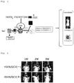
- FIG 1 shows the summary of a suicide gene cell therapy for malignant brain tumors using therapeutic stem cells expressing HSVtk.
- Induced pluripotent stem (iPS) cells were differentiation-induced into neural stem cells (NSCs).
- the herpes simplex virus thymidine kinase (HSVtk) gene which is a suicide gene, was then introduced with the lentiviral vector.
- Therapeutic stem cells (TSCs) expressing HSVtk were established.
- a mixture of 1 ⁇ 10 5 human glioma cells (U87) and 5 ⁇ 10 5 TSCs was transplanted to the T cell-deficient mouse brain (right striate body).
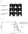
- ganciclovir In a treated group, 50 mg/kg of ganciclovir (GCV) was administered twice per day for 21 days from the day following the transplantation, and PBS was administered to a control group. The survival analysis was performed while the luminescence imaging analysis (IVIS) of tumors was performed chronologically. Some mice were decapitated in the third week after the transplantation, and the histological analysis of the brains was performed.
- GCV ganciclovir
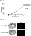
- Tumors tended to be smaller in the treated group (GCV+) than in the control group (GCV-) ( Figure 2 ).
- TSCs Tet-induced therapeutic stem cells
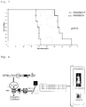
- FIG 8 shows the summary of a suicide gene cell therapy using Tet-induced therapeutic stem cells (TSCs) expressing HSVtk.
- TSCs Tet-induced therapeutic stem cells
- the present invention can be used in industrial fields such as medicine manufacture.
Landscapes
- Health & Medical Sciences (AREA)
- Life Sciences & Earth Sciences (AREA)
- Animal Behavior & Ethology (AREA)
- Pharmacology & Pharmacy (AREA)
- Medicinal Chemistry (AREA)
- General Health & Medical Sciences (AREA)
- Public Health (AREA)
- Veterinary Medicine (AREA)
- Chemical & Material Sciences (AREA)
- Cell Biology (AREA)
- Developmental Biology & Embryology (AREA)
- Epidemiology (AREA)
- Engineering & Computer Science (AREA)
- Biomedical Technology (AREA)
- Biotechnology (AREA)
- Zoology (AREA)
- Reproductive Health (AREA)
- Immunology (AREA)
- Virology (AREA)
- Organic Chemistry (AREA)
- Chemical Kinetics & Catalysis (AREA)
- General Chemical & Material Sciences (AREA)
- Nuclear Medicine, Radiotherapy & Molecular Imaging (AREA)
- Neurosurgery (AREA)
- Neurology (AREA)
- Gynecology & Obstetrics (AREA)
- Ophthalmology & Optometry (AREA)
- Bioinformatics & Cheminformatics (AREA)
- Genetics & Genomics (AREA)
- Molecular Biology (AREA)
- Pharmaceuticals Containing Other Organic And Inorganic Compounds (AREA)
- Medicines Containing Material From Animals Or Micro-Organisms (AREA)
- Micro-Organisms Or Cultivation Processes Thereof (AREA)
- Medicines That Contain Protein Lipid Enzymes And Other Medicines (AREA)
Abstract
Description
- The present invention relates to a cell preparation for treating brain tumors and a method for preparing neural stem cells used for the cell preparation.
- Malignant gliomas are very difficultly treated in the present therapies due to treatment resistance by brain tumor stem cells (BTSCs), which infiltrate the brain substance widely. Since suicide gene therapies using viral vectors (HSVtK) exerted bystander effects, the effectiveness was expected. Since the diffusion among infiltrating tumor cells was insufficient, the results of the clinical tests were however limited. It has been revealed recently that neural stem cells (NSCs) have the properties of migrating and accumulating in brain tumor stem cells, and attempts to apply neural stem cells to suicide gene treatment as cell vehicles attract attention. It was however difficult in view of problems including an ethical problem to obtain an adequate amount of neural stem cells which can be administered to humans. Then, a technique as to suicide gene treatment using mesenchymal stem cells, which has properties similar to neural stem cells has been developed (Patent Literature 1).
- Patent Literature 1:
Japanese Patent Laid-Open No.2006-345726 -
Patent Literature 1 discloses that mesenchymal stem cells derived from rat marrow cells exhibited a higher bystander effect than neural stem cells collected from the rat cerebral cortex (Examples 2 and 3). Although a suicide gene therapy thus using the mesenchymal stem cells is expected to exhibit a high therapeutic effect, it is considered that the administration of the mesenchymal stem cells to the human brain has problems with safety and the like. - Meanwhile, although neural stem cells are safer than the mesenchymal stem cells, it is very difficult to obtain neural stem cells as mentioned above.
- Objects of the present invention is to solve the difficulty of obtaining neural stem cells, which is problematic, in such a context and to provide new means for treating brain tumors using neural stem cells.
- The present inventor has earnestly examined repeatedly to solve the above-mentioned problem and consequently found that inducing the differentiation of neural stem cells from iPS cells enables obtaining neural stem cells in an amount enough to be used for a suicide gene therapy. The present inventor has also found that when the herpes simplex virus thymidine kinase gene, which is a typical suicide gene, is introduced into iPS cells, and this gene is expressed to cause cytotoxicity. The present inventor has also found that regulating herpes simplex virus thymidine kinase gene expression with doxycycline enables solving this problem with cytotoxicity.
- The present invention has been completed based on the above knowledge.
- That is, the present invention provides the following [1] to [10].
- [1] A cell preparation for treating brain tumors used in combination with a prodrug of a drug that kills, or inhibits proliferation of, tumor cells, wherein the cell preparation comprises neural stem cells derived from iPS cells or ES cells having a suicide gene, and the prodrug is a prodrug activated with an enzyme produced by expression of the suicide gene comprised in the neural stem cells.
- [2] The cell preparation for treating brain tumors according to [1], wherein the neural stem cells derived from iPS cells or ES cells having a suicide gene are neural stem cells obtained by sequentially performing the following steps (1) and (2):
- (1) introducing a suicide gene into iPS cells or ES cells,
- (2) differentiating the iPS cells or the ES cells into neural stem cells.
- [3] The cell preparation for treating brain tumors according to [2], wherein the suicide gene is a gene structure, expression of which can be regulated artificially.
- [4] The cell preparation for treating brain tumors according to [3], wherein the suicide gene structure is a gene structure expressed by addition of doxycycline.
- [5] The cell preparation for treating brain tumors according to [1], wherein the neural stem cells derived from iPS cells or ES cells having a suicide gene are neural stem cells obtained by sequentially performing the following steps (1) and (2):
- (1) differentiating iPS cells or ES cells into neural stem cells,
- (2) introducing a suicide gene into the neural stem cells.
- [6] The cell preparation for treating brain tumors according to any one of [1] to [5], wherein the suicide gene is a herpes simplex virus thymidine kinase gene, and the prodrug is ganciclovir.
- [7] A method for preparing neural stem cells having a suicide gene, comprising performing the following steps (A1) and (A2) or steps (B1) and (B2) sequentially:
- (A1) introducing a suicide gene into iPS cells or ES cells,
- (A2) differentiating the iPS cells or the ES cells into neural stem cells,
- (B1) differentiating iPS cells or ES cells into neural stem cells,
- (B2) introducing a suicide gene into the neural stem cells.
- [8] The method for preparing neural stem cells having a suicide gene according to [7], wherein the suicide gene in the step (A1) is a gene structure, expression of which can be regulated artificially.
- [9] The method for preparing neural stem cells having a suicide gene according to [8], wherein the suicide gene structure in the step (A1) is a gene structure expressed by addition of doxycycline.
- [10] The method for preparing neural stem cells having a suicide gene according to any one of [7] to [9], wherein the suicide gene is a herpes simplex virus thymidine kinase gene.
- The present invention provides the following [A].
- [A] A method for treating brain tumors, comprising the following steps (1) and (2):
- (1) administering a cell preparation to a patient, wherein the cell preparation comprises neural stem cells derived from iPS cells or ES cells having a suicide gene,
- (2) administering a prodrug to the patient, wherein the prodrug is a prodrug of a drug that kills, or inhibits proliferation of, tumor cells, and the prodrug is activated with an enzyme produced by expression of the suicide gene comprised in the neural stem cells.
- The present specification includes the contents described in the specification and/or drawings of the Japanese Patent Application No.
2017-92973 - The present invention provides new means for treating brain tumors using neural stem cells derived from iPS cells or ES cells.
-
- [
Figure 1] Figure 1 shows the summary of a suicide gene cell therapy for malignant brain tumors using therapeutic stem cells (TSCs) expressing HSVtk. - [
Figure 2] Figure 2 shows the results of the luminescence imaging analysis (IVIS) of tumors. Stem cells expressing HSVtk were used as therapeutic stem cells. - [
Figure 3] Figure 3 shows the measurement results of the ROI values of the luminescence imaging. Stem cells expressing HSVtk were used as therapeutic stem cells. - [
Figure 4] Figure 4 shows HE-stained images (left) and Venus fluorescence images (right) of mouse brain sections. Stem cells expressing HSVtk were used as therapeutic stem cells. - [
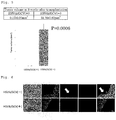
Figure 5] Figure 5 shows the results of the analysis of tumor volumes. Stem cells expressing HSVtk were used as therapeutic stem cells. - [
Figure 6] Figure 6 shows the results of the survival analysis of the transplanted TSCs. Stem cells expressing HSVtk were used as therapeutic stem cells. The upper row shows the brains of an untreated group, and the lower row shows the brains of a treated group. From the left, images are bright fields, images stained with Venus (U87 tumor cells are stained), images stained with KO (stem cells are stained), images stained with DAPI (nuclei are stained), and a merge (fused image of Venus, KO and DAPI). - [
Figure 7] Figure 7 shows the results of the survival analysis of brain tumor models. Stem cells expressing HSVtk were used as therapeutic stem cells. - [
Figure 8] Figure 8 shows the summary of a suicide gene cell therapy using Tet-induced therapeutic stem cells (TSCs) expressing HSVtk. - [
Figure 9] Figure 9 shows the result of the luminescence imaging analysis (IVIS) of tumors. Tet-induced stem cells expressing HSVtk were used as therapeutic stem cells. - [
Figure 10] Figure 10 shows the results of the survival analysis of brain tumor models. Tet-induced stem cells expressing HSVtk were used as therapeutic stem cells. - [
Figure 11] Figure 11 shows HE-stained images (left) and Venus fluorescence images (right) of mouse brain sections. Tet-induced stem cells expressing HSVtk were used as therapeutic stem cells. - [
Figure 12] Figure 12 shows the results of the analysis of tumor volumes. Tet-induced stem cells expressing HSVtk were used as therapeutic stem cells. - The present invention will be described in detail hereinafter.
- A cell preparation for treating brain tumors of the present invention is characterized in that the cell preparation is used in combination with a prodrug of a drug that kills, or inhibits proliferation of, tumor cells, wherein the cell preparation comprises neural stem cells derived from iPS cells or ES cells having a suicide gene, and the prodrug is a prodrug activated with an enzyme produced by expression of the suicide gene comprised in the neural stem cells.
- The "brain tumors" in the present invention means tumors which are developed in intracranial tissue. Specific examples thereof include gliomas, medulloblastomas, neuroblastomas, meningiomas, pituitary adenomas, neurinomas, primary central nervous system lymphomas, sarcomas and spinal cord tumors. In the present invention, although all of these brain tumors can be objects to be treated, it is preferable that gliomas be objects to be treated.
- The "treatment" in the present invention means not only killing tumor cells but also reducing tumor cells or inhibiting the proliferation of tumor cells.
- The "suicide gene" in the present invention means a gene which is expressed in cells and can kill the cells. As the suicide gene, a metabolic toxic gene can be used. Suitable specific examples thereof include the herpes simplex virus thymidine kinase (HSVtk) gene (Proc. Natl. Acad. Sci, USA 78 (1981) 1441-1445). Examples of suicide genes other than the herpes simplex virus thymidine kinase gene include a cytosine deaminase gene, a uracil phosphoribosyl transferase gene, a guanine phosphoribosyl transferase gene and a nitroreductase gene.
- As long as the suicide gene can be expressed at the time of treatment, the suicide gene is not particularly limited. As mentioned above, since some suicide genes exhibit cytotoxicity in iPS cells (for example, HSVtk gene), it is preferable that the suicide gene be a gene structure, the expression of which can be regulated artificially. Using the gene structure containing such a suicide gene, the expression of which can be regulated artificially, enables suppressing the expression of the suicide gene in iPS cells and inducing the expression of the suicide gene at the time of the treatment of brain tumors. A problem with cytotoxicity can be solved. Such a gene structure can be produced using a sequence which enables regulating the expression of the suicide gene artificially as mentioned below.
- As long as the prodrug in the present invention is a prodrug of a drug that kills, or inhibits proliferation of, tumor cells, and the prodrug is activated with an enzyme produced by expression of the suicide gene and comes to exhibit the effect of killing tumor cells or inhibiting the proliferation thereof, the prodrug may be any prodrug. A prodrug to be used can be determined depending on the suicide gene. For example, when the suicide gene is a herpes simplex virus thymidine kinase gene, ganciclovir (GCV), aciclovir, penciclovir, adefovir, tenofovir or the like can be used as the prodrug. When the suicide gene is a cytosine deaminase gene, 5-fluorocytosine or the like can be used as the prodrug. When the suicide gene is an uracil phosphoribosyltransferase gene, 5-fluorouracil or the like can be used as the prodrug. When the suicide gene is a guanine phosphoribosyltransferase gene, 6-thioxanthine, 6-thioguanine or the like can be used as a prodrug. When the suicide gene is a nitroreductase gene, CB1954 or the like can be used as the prodrug.
- The "neural stem cells" in the present invention means stem cells having the capability to supply cells which differentiates into neurons and glia cells. The cell preparation of the present invention contains these neural stem cells. As long as the cell preparation does not exert a great adverse influence on the effect of treating brain tumors, the cell preparation may contain cells other than neural stem cells. When neural stem cells are prepared from iPS cells in accordance with a method described in the below-mentioned Mol Brain 9: 85, 2016, not only neural stem cells but also neural precursor cells are produced. For this reason, the cell preparation of the present invention usually contains both neural stem cells and neural precursor cells. Neural stem cells used in the present invention is neural stem cells derived from pluripotent stem cell such as iPS cells, and neural stem cells collected from the brain or the like is not used as they are.
- Neural stem cells derived from iPS cells having a suicide gene can be prepared by sequentially performing steps (A1) and (A2) or steps (B1) and (B2) described below.
- In the step (A1), a suicide gene is introduced into iPS cells.
- As long as iPS cells to be used can be differentiated into neural stem cells, the origin thereof, a reprogramming factor to be introduced, a method for introducing a reprogramming factor and the like are not particularly limited. Since the cell preparation of the present invention is used mainly for treating human brain tumors, it is however preferable to use iPS cells derived from a human in such a case. In this case, iPS cells derived from a patient to whom the cell preparation is administered may be used, and iPS cells derived from a human other than the patient may be used. As mentioned above, although a method for introducing a reprogramming factor is not limited, either, it is preferable to use integration-free iPS cells. Although iPS cells to be used may be prepared in accordance with a well-known method, iPS cells can also be obtained from research institutions such as the Center for iPS Cell Research and Application (CiRA), Kyoto University. Examples of suitable iPS cell lines include 1210B2 which the CiRA has.
- Examples of the method of introducing a suicide gene into iPS cells include a method of introducing a vector in which a suicide gene is incorporated. It is preferable to use viral vectors such as adenovirus vectors, adeno-associated viral vectors, retroviral vectors and lentiviral vectors as the vector. Among these, a vector which incorporates a suicide gene into a genome is preferable, and lentiviral vectors are the most preferable.
- A sequence which enables regulating the expression of a suicide gene artificially is preferably included in the vector besides the suicide gene. It is preferable to use the tetracycline induction system, which is a well-known expression induction system (PLoS One 8: e59890, 2013), as such a sequence. This tetracycline induction system comprises a region which expresses a reverse tetracycline-controlled transactivator (rtTA), a gene of interest and a region including a TRE promoter disposed upstream thereof. The rtTA bonds to the TRE promoter by bonding to tetracycline and induces the expression of the gene of interest disposed downstream. Using this system enables inducing the expression of the suicide gene by the addition of tetracycline. Although the expression in this system can also be induced with tetracycline as mentioned above, but is usually induced with doxycycline, which is a derivative of tetracycline. The sequence which enables regulating the expression of the suicide gene artificially is not limited to this tetracycline induction system, and various inducible promoters such as medicine inducible promoters may be used.
- Sequences such as a reporter gene, IRES and the like may be included in the vector besides the suicide gene or the sequence which enables regulating the expression of the suicide gene artificially.
- The suicide gene can be introduced into iPS cells by a vector in accordance with a well-known method depending on the type of vector to be used.
- In the step (A2), iPS cells are differentiated into neural stem cells.
- Differentiation from iPS cells to neural stem cells may be performed in accordance with either well-known method of a method for preparation through embryoid bodies and a method for preparation without forming embryoid bodies. For example, the differentiation can be performed in accordance with a method described in Mol Brain 9:85, 2016.
- According to the above-mentioned method, embryoid bodies can be formed from iPS cells using well-known embryoid body-forming medium, and the embryoid body medium contains a TGFβ family inhibitor (for example, SB431542) and a BMB inhibitor (for example, LDN-193189).
- Differentiation from the embryoid bodies to neural stem cells can be performed using well-known neurosphere medium. For example, the neurosphere medium contains an epidermal growth factor and a fibroblast growth factor-2, a leukemia inhibitory factor, a B-27 supplement and the like. Culturing the embryoid bodies in the neurosphere medium forms cell masses called neurospheres and containing neural stem cells. The formed neurospheres are collected and disassembled into single cells. The single cells are cultured in neurosphere medium to form neurospheres again. Such operation is repeated several times, and neural stem cells can be obtained by collecting neurospheres.
- When neural stem cells derived from iPS cells having a suicide gene are prepared by sequentially performing the steps (A1) and (A2), there is the advantage of neural stem cells for treatment being able to be supplied steadily by proliferating iPS cells.
- In the step (B1), iPS cells are differentiated into neural stem cells.
- The differentiation from iPS cells to neural stem cells in the step (B1) can be performed in the same way as the differentiation in the step (A2).
- In the step (B2), a suicide gene is introduced into neural stem cells.
- The introduction of a suicide gene into neural stem cells in a step (B2) can be performed in the same way as the introduction in the step (A1). Since a problem with cytotoxicity may occur in the step (A1), a sequence which enables regulating the expression of a suicide gene artificially is however included in a vector preferably. Since such a problem occurs very rarely in the step (B2), a sequence which enables regulating the expression of a suicide gene artificially is, however, usually unnecessary.
- As mentioned above, neural stem cells having a suicide gene can be prepared from iPS cells by sequentially performing the steps (A1) and (A2) or the steps (B1) and (B2). iPS cells and ES cells have the same nature. Even though ES cells are used instead of iPS cells, neural stem cells having a suicide gene can therefore be prepared.
- As mentioned above, "neural stem cells," which are a "product," are not specified by structure or properties but specified by a preparation method herein. This is because since cells are parts of the living body, the structure and properties thereof are very complicated, and markedly excessive economic expenditure and time are taken to work to specify them.
- The cell preparation of the present invention may contain other pharmaceutically acceptable components besides neural stem cells. Examples of such other components include carriers, excipients, disintegrators, buffers, emulsifiers, suspending agents, soothing agents, stabilizers, preservatives, antiseptics and physiological saline solutions. Dimethyl sulfoxide, serum albumin or the like may be contained to protect cells at the time of freeze storage, and an antibiotic or the like may be contained to prevent the contamination and proliferation of bacteria.
- The number of neural stem cells contained in the cell preparation of the present invention can be optionally determined in view of the sex, age or weight of a subject, the state of an affected part, the state of cells to be used or the like so as to obtain a desired effect (for example, the disappearance of tumors or the reduction of tumor size) in the treatment of brain tumors.
- The cell preparation of the present invention may be administered a plurality of times (for example, 2 to 10 times) at intervals (for example, twice per day, once per day, twice per week, once per week, once per two week). Although the dose can be optionally determined in view of the sex, age or weight of a subject, the state of an affected part, the state of cells to be used or the like, the dose is preferably 1 × 10⁴ to 1 × 10⁶ cells per individual (human) 1 to 10 times.
- The administration site or administration method of the cell preparation of the present invention is not particularly limited. Examples of the administration method include tumor local administration, intracarotidarterial administration and intravenous administration.
- The administration site or administration method of a prodrug used in combination with the cell preparation of the present invention is not particularly limited, either. Examples of the administration method also include intraperitoneal administration besides tumor local administration, intracarotidarterial administration and intravenous administration mentioned above.
- The prodrug may be administered before or after the administration of the cell preparation of the present invention or at the same time as the administration of the cell preparation, but is usually administered in divided doses after the administration of the cell preparation. The dose of the prodrug can be optionally determined in view of the type of prodrug to be used, the sex, age or weight of a subject or the state of an affected part. When ganciclovir is administered, ganciclovir is preferably administered at 3 to 5 mg/kg per once, twice per day for 2 to 3 weeks (28 times to 42 times) per individual (human).
- When the tetracycline induction system is used to prevent cytotoxicity, doxycycline is also administered at the time of treatment to express a suicide gene. Examples of a method for administer doxycycline include oral administration, tumor local administration, intracarotidarterial administration, intravenous administration and intraperitoneal administration. Doxycycline may be administered before or after the administration of the cell preparation of the present invention or at the same time as the administration of the cell preparation, but is usually administered after the administration of the cell preparation.
- Next, although the present invention will be described in more detail by giving Examples, the present invention is not limited to these Examples.
- The used human iPS cell (1210B2) was obtained from the Center for iPS Cell Research and Application (CiRA), Kyoto University. Then, 1210B2 was established by a method for introducing a reprogramming factor using an episomal vector for human peripheral blood mononuclear cells (Stem Cells 31: 458-66, 2013). Human iPS cells were inoculated on a plastic culture dish coated with iMatrix-511 (produced by Nippi, Incorporated) and subjected to maintenance culture using Stem Fit AK03 or AK03N medium (produced by AJINOMOTO CO., INC.) by a known feeder-free method (Sci Rep 4: 3594, 2014).
- Differentiation induction from human iPS cells to NS/PCs was performed by a known method (Mol Brain 9: 85, 2016). First, a 10 µM ROCK inhibitor Y276352 (produced by Wako Pure Chemical Corporation) was added to medium of iPS cells, the iPS cells were incubated for 1 to 3 hours, then washed with PBS and disassembled into single cells using TrypLE Select (produced by Life Technologies Corporation). The human iPS cells disassembled into the single cells were suspended in EB (embryoid body) - forming medium to which a 10 µM ROCK inhibitor Y276352 was added (10 µM SB431542 and 100 nM LDN-193189 were added to Stem Fit medium to which C liquid was not added), inoculated at a concentration of 9.0 × 10³ cells/75 µl per 1 well of a low adhesive 96 hole-culture plate (Prime Surface 96V, manufactured by Sumitomo Bakelite Co., Ltd.) and cultured. Then, 75 µl of EB-forming media was added 1 day after, a half amount of EB-forming medium was replaced everyday on and after, and the human iPS cells were cultured for 13 to 14 days to obtain EBs. The EBs were collected from each well and cultured in NS (neurosphere) medium (to D-MEM/Ham's F-12 medium (containing HEPES) (produced by Wako Pure Chemical Corporation) were added a 20 ng/ml recombination human epidermal growth factor (produced by PeproTech, Inc.), a 20 ng/ml recombination human fibroblast growth factor-2 (produced by PeproTech, Inc.), a 103units/ml recombination human leukemia inhibitory factor (produced by NACALAI TESQUE, INC.), a 2% B-27 supplement (produced by Thermo Fisher Scientific K.K.) and 1 unit/ml heparin sodium (produced by AY PHARMACEUTICALS CO., LTD.)) for 7 days. The medium was replaced once per 3 or 4 days during culture. Cell masses were collected by a centrifuge, disassembled into single cells using TrypLE Select and suspended in NS medium. The single cells were inoculated into a low adhesive flask (manufactured by Corning Incorporated) at a concentration of 1 × 10⁵ cells/ml and cultured for 10 days with the medium replaced every 3 to 4 days to obtain primary NS/PCs. Similarly, the primary NS/PCs were collected by the centrifuge, disassembled into single cells using TrypLE Select and suspended in NS medium. The single cells were inoculated into a low adhesive flask at a concentration of 1 × 10⁵ cells/ml and cultured for 7 to 10 days with the medium replaced every 3 to 4 days to obtain secondary, tertiary, quaternary and quinary NS/PCs.
- The cDNA of the Humanized, CpG-free HSVtk gene (HSV1tk) wherein the cDNA was subcloned from the pSelect-zeo-HSV1tk plasmid (produced by InvivoGen Corporation) to pENTR/D-TOPO (produced by Thermo Fisher Scientific K.K.) by PCR and the nucleotide sequence of the cDNA was confirmed, was used. The HSV1tk cDNA was subcloned to the lentiviral vector plasmids CSII-EF-RfA-IRES2-hKO1 and CSIV-RfA-TRE-EF-KT to obtain CSIV-EF-HSV1tk-IRES2-hKO1 and CSIV-HSV1tk-TRE-EF-KT. CSIV-EF-HSV1tk-IRES2-hKO1 was used to produce a lentiviral vector which expresses HSV1tk and hKO1 (Humanized Kusabira-Orange fluorescence protein gene) under the EF-1α promoter. CSIV-HSV1tk-TRE-EF-KT was used to produce a lentiviral vector which expresses hKO1 and rtTA (reverse Tet-controlled transactivator) under the EF-1α promoter and expresses HSV1tk under the tetracycline (Tet)-induced promoter. The lentiviral vectors were produced by a known method (J Virol 72: 8150-8157, 1998; PLoS One 8: e59890, 2013; Sci Rep 5: 10434, 2015).
- The human glioma cell line U87 was infected with the ffLuc (fusion gene of Venus fluorescence protein and Luc2 firefly luciferase) expression lentiviral vector (CSII-EF-ffLuc) (Cell Transplant 20:727-739, 2011). The clones were sorted with a cell sorter to obtain the U87 cell line, which expresses ffLuc highly and steadily.
- Secondary or tertiary NS/PCs differentiation-induced from human iPS cells were collected with the centrifuge and disassembled into single cells using TrypLE Select. The single cells were suspended in NS medium, inoculated into a low adhesive flask at a concentration of 1 × 10⁵ cells/ml, infected with the CSIV-EF-HSV1tk-IRES2-hKO1 lentiviral vector at an MOI (multiplicity of infection) of 0.5 to 1, and cultured for 7 days with the medium replaced every 3 to 4 days. The obtained NS/PCs were infected with the CSIV-EF-HSV1tk-IRES2-hKO1 lentiviral vector at an MOI of 0.5 to 1 in the same way again to prepare therapeutic stem cells (TSCs), that is, NS/PCs expressing HSVtk. It was confirmed that the cell death of the NS/PCs expressing HSVtk was induced by adding 1 µg/ml ganciclovir (GCV) to the medium.
- First, 1 × 10⁵ cells/2 µl U87 cells expressing ffLuc and 5 × 10⁵ cells/2µl NS/PCs expressing HSVtk were mixed and stereotactically transplanted to the right striate body (2 mm away from the anterior fontanel to the right and 3 mm deep from the brain surface) of each T cell-deficient mouse (female BALB/c nude mouse, 20g, 6w) subjected to general anesthesia.
- Then, 50 mg/kg of GCV was intraperitoneally administered to the treated group twice per day for 21 days from the day following the transplantation (group 1: HSVtk (+)/ GCV (+), n = 10). Next, 200 µl of 1 × PBS was intraperitoneally administered to the control group for the same period (group 2: HSVtk (+)/ GCV (-), n = 10).
- Change in the tumor of the same individual over time was observed with an IVIS, that is, an in vivo imaging system, every week. At the time of photographing with IVIS, 200 µl of 30mg/ml VivoGlo (TM) Luciferin was intraperitoneally administered to the individual subjected to isoflurane inhalation anesthesia, and the tumor was photographed for 10 minutes when the peak was reached.
- Four mice of each of both groups were decapitated in the third week, and the actual tumor volumes were measured. The mice were decapitated by perfusing the mice with 4% paraformaldehyde for fixation through the heart. The brain tissues taken out were replaced with 10% and 20% sucrose, and the brain tissues were sliced to a thickness of 20 µm. An 8-well dish the wells of which each contain 1 ml of an antifreeze solution was charged with the slices sequentially and stored at -20°C.
- The brain slices were observed by staining with HE or through a fluorescence microscope since U87 expresses Venus fluorescence protein to measure the tumor volumes in more detail. It was evaluated whether the transplanted TSCs remained by fluorescently staining the brain tissues of both groups with an anti-KO antibody in the third week in the same way. Finally, the survival curves of both groups were compared.
- Human iPS cells were infected with the CSIV-HSV1tk-TRE-EF-KT lentiviral vector at an MOI of 2.5 to 10 and cultured. The clones were then sorted with the cell sorter to obtain an iPS cell line expressing hKO1 steadily and highly. It was confirmed that the cell death of Tet-induction iPS cells expressing HSVtk was induced by adding 1 µg/ml doxycycline (Dox) and 1 µg/ml GCV to the medium.
- Tet-induced iPS cells expressing HSVtk were differentiation-induced into NS/PCs to obtain secondary and tertiary Tet-induced NS/PCs expressing HSVtk. It was confirmed that the cell death of these Tet-induction NS/PCs expressing HSVtk was also induced by adding 1 µg/ml doxycycline (Dox) and 1 µg/ml GCV to the medium. <Examination of the therapeutic effect of Tet-induced NS/PCs expressing HSVtk in brain tumor model mice>
- Three groups which are a treated group 1 (Tet-HSVtk (+)/ Dox (+)/GCV (+), n = 12), a control group 2 (Tet-HSVtk (+)/ Dox (-)/ GCV (+), n = 8), and a control group 3 (Tet-HSVtk (-) / Dox (+)/ GCV (+), n= 4) were produced. All of the method for evaluating tumors, the survival curve analysis and the like were performed in the same way in the above-mentioned Examination of therapeutic effect of NS/PCs expressing HSVtk (Feed containing 200 mg/kg of Dox was orally ingested).
-
Figure 1 shows the summary of a suicide gene cell therapy for malignant brain tumors using therapeutic stem cells expressing HSVtk. Induced pluripotent stem (iPS) cells were differentiation-induced into neural stem cells (NSCs). The herpes simplex virus thymidine kinase (HSVtk) gene, which is a suicide gene, was then introduced with the lentiviral vector. Therapeutic stem cells (TSCs) expressing HSVtk were established. A mixture of 1 × 10⁵ human glioma cells (U87) and 5 × 10⁵ TSCs was transplanted to the T cell-deficient mouse brain (right striate body). In a treated group, 50 mg/kg of ganciclovir (GCV) was administered twice per day for 21 days from the day following the transplantation, and PBS was administered to a control group. The survival analysis was performed while the luminescence imaging analysis (IVIS) of tumors was performed chronologically. Some mice were decapitated in the third week after the transplantation, and the histological analysis of the brains was performed. - Tumors tended to be smaller in the treated group (GCV+) than in the control group (GCV-) (
Figure 2 ). When the ROI values of the luminescence imaging was measured, the ROI values of the treated group were significantly low, and the ROI values were remarkably low especially from the second week onward (Figure 3 ). (P = 0.02) - Since the Venus fluorescence protein gene was introduced into the glioma cells (U87), the tumor volumes were quantitatively analyzed using the fluorescence microscope (
Figure 4 ). The tumor volume of a treated group (GCV+) was 0.17 ± 0.07 mm3, and was significantly small as compared with 51.76 ± 7.87 mm3 of a control group (GCV-) (Figure 5 ). (P = 0.0006) - The extinction of the above-mentioned transplanted TSCs in the mouse brains was confirmed as to the safety of the present therapy (the risk of the tumorigenesis of iPS cells). Since the Kusabira-Orange(KO) fluorescence protein gene was introduced into the TSCs, the TSCs were analyzed by fluorescence staining using an anti-KO antibody. Consequently, although the transplanted TSCs remained in the brains in the control group (GCV-), clear survival of the transplanted TSCs was not observed in the treated group (GCV+) (
Figure 6 ). It was therefore confirmed that the transplanted TSCs died within three weeks by the HSVtk/GCV system (each n = 4). - In the treated group (GCV+), significant extension of the survival period was observed as compared with the control group (GCV-) (
Figure 7 ). The average survival period of the treated group was 4 days, and the average survival period of the control group was 19 days (p < 0.01) . -
Figure 8 shows the summary of a suicide gene cell therapy using Tet-induced therapeutic stem cells (TSCs) expressing HSVtk. Although it was attempted to introduce the HSVtk gene into human iPS cells for supplying the cells steadily for clinical application, an iPS cell line expressing HSVtk could not be established due to cytotoxicity. Then, a Tet-induced iPS cell line expressing HSVtk was established using the tetracycline (Tet)-induced promoter which could regulate gene expression. The cell line could be differentiated into neural stem cells (NS/PCs), and Tet-induced therapeutic stem cells (TSCs) expressing HSVtk could be steadily supplied. - In the Tet-induced HSVtk, only when doxycycline (Dox) is added, the HSVtk gene is expressed.
- In IVIS analysis, the reduction of the tumors was observed in the treated group under the conditions of TSC transplantation (+)/ Dox (+)/ GCV (+) as compared with the control group under the conditions of TSC transplantation (-)/ Dox (+)/ GCV (+) and the control group under the conditions of TSC transplantation (+) / Dox (-) / GCV (+) (
Figure 9 ). - In survival analysis, significant extension of the survival period was observed in the treated group under the conditions of TSC transplantation (+) /Dox (+)/ GCV (+) as compared with the control group under the conditions of TSC transplantation (-)/ Dox (+)/ GCV (+) (
Figure 10 ). The average survival period of the treated group was 34 days, and the average survival period of the control group was 25.5 days (p < 0.01). (Meanwhile, the statistically significant difference was not observed between the control group under the conditions of TSC transplant (+)/ Dox (-)/ GCV (+) and the treated group.) In the Tet-induced HSVtk, only when doxycycline (Dox) is added, the HSVtk gene is expressed. - As compared with the control group under the conditions of TSC transplantation (+)/ Dox (-)/ GCV (+), a significant effect of reducing tumors was observed in the treated group under the conditions of TSC transplantation (+)/ Dox (+)/ GCV (+) (
Figures 11 and 12 ). (p = 0.003) - All of the publications, patents and patent applications cited herein are incorporated herein as reference as they are.
- Since a cell preparation for treating brain tumors of the present invention can be used as a pharmaceutical, the present invention can be used in industrial fields such as medicine manufacture.
Claims (10)
- A cell preparation for treating brain tumors used in combination with a prodrug of a drug that kills, or inhibits proliferation of, tumor cells, wherein the cell preparation comprises neural stem cells derived from iPS cells or ES cells having a suicide gene, and the prodrug is a prodrug activated with an enzyme produced by expression of the suicide gene comprised in the neural stem cells.
- The cell preparation for treating brain tumors according to claim 1, wherein the neural stem cells derived from iPS cells or ES cells having a suicide gene are neural stem cells obtained by sequentially performing the following steps (1) and (2):(1) introducing a suicide gene into iPS cells or ES cells,(2) differentiating the iPS cells or the ES cells into neural stem cells.
- The cell preparation for treating brain tumors according to claim 2, wherein the suicide gene is a gene structure, expression of which can be regulated artificially.
- The cell preparation for treating brain tumors according to claim 3, wherein the suicide gene structure is a gene structure expressed by addition of doxycycline.
- The cell preparation for treating brain tumors according to claim 1, wherein the neural stem cells derived from iPS cells or ES cells having a suicide gene are neural stem cells obtained by sequentially performing the following steps (1) and (2):(1) differentiating iPS cells or ES cells into neural stem cells,(2) introducing a suicide gene into the neural stem cells.
- The cell preparation for treating brain tumors according to any one of claims 1 to 5, wherein the suicide gene is a herpes simplex virus thymidine kinase gene, and the prodrug is ganciclovir.
- A method for preparing neural stem cells having a suicide gene, comprising performing the following steps (A1) and (A2) or steps (B1) and (B2) sequentially:(A1) introducing a suicide gene into iPS cells or ES cells,(A2) differentiating the iPS cells or the ES cells into neural stem cells,(B1) differentiating iPS cells or ES cells into neural stem cells,(B2) introducing a suicide gene into the neural stem cells.
- The method for preparing neural stem cells having a suicide gene according to claim 7, wherein the suicide gene in the step (A1) is a gene structure, expression of which can be regulated artificially.
- The method for preparing neural stem cells having a suicide gene according to claim 8, wherein the suicide gene structure in the step (A1) is a gene structure expressed by addition of doxycycline.
- The method for preparing neural stem cells having a suicide gene according to any one of claims 7 to 9, wherein the suicide gene is a herpes simplex virus thymidine kinase gene.
Applications Claiming Priority (2)
| Application Number | Priority Date | Filing Date | Title |
|---|---|---|---|
| JP2017092973 | 2017-05-09 | ||
| PCT/JP2018/017888 WO2018207808A1 (en) | 2017-05-09 | 2018-05-09 | Cell formulation for treating brain tumor |
Publications (2)
| Publication Number | Publication Date |
|---|---|
| EP3622961A1 true EP3622961A1 (en) | 2020-03-18 |
| EP3622961A4 EP3622961A4 (en) | 2021-01-13 |
Family
ID=64105532
Family Applications (1)
| Application Number | Title | Priority Date | Filing Date |
|---|---|---|---|
| EP18797557.8A Withdrawn EP3622961A4 (en) | 2017-05-09 | 2018-05-09 | CELL FORMULATION FOR THE TREATMENT OF BRAIN TUMORS |
Country Status (5)
| Country | Link |
|---|---|
| US (1) | US20210128639A1 (en) |
| EP (1) | EP3622961A4 (en) |
| JP (1) | JP7051060B2 (en) |
| CN (1) | CN110603045A (en) |
| WO (1) | WO2018207808A1 (en) |
Cited By (2)
| Publication number | Priority date | Publication date | Assignee | Title |
|---|---|---|---|---|
| EP4129311A4 (en) * | 2020-03-31 | 2024-06-12 | Keio University | THERAPEUTIC AGENT USING A GENOME-EDITED PLURIPOTENT STEM CELL |
| US12090172B2 (en) | 2017-11-20 | 2024-09-17 | Keio University | Suicide gene therapeutic agent for brain tumors using pluripotent stem cell |
Families Citing this family (1)
| Publication number | Priority date | Publication date | Assignee | Title |
|---|---|---|---|---|
| CN116024178B (en) * | 2022-08-23 | 2026-02-10 | 南京市口腔医院 | Tumor-associated fibroblasts that can be targeted to induce death, their construction methods and applications |
Family Cites Families (6)
| Publication number | Priority date | Publication date | Assignee | Title |
|---|---|---|---|---|
| US7186409B2 (en) * | 1998-08-14 | 2007-03-06 | The Children's Medical Center Corporation | Neural stem cells and use thereof for brain tumor therapy |
| CA2666635C (en) | 2001-02-21 | 2016-03-22 | United Video Properties, Inc. | Systems and methods for interactive program guides with personal video recording features |
| JP2006345726A (en) | 2005-06-14 | 2006-12-28 | Hamamatsu Univ School Of Medicine | Expression vector for brain tumor treatment |
| EP2043662B1 (en) * | 2006-07-26 | 2015-09-09 | Intrexon Corporation | Methods and compositions for treating disease |
| WO2014133090A1 (en) * | 2013-02-28 | 2014-09-04 | 国立大学法人浜松医科大学 | Pluripotent stem cell for treating brain tumor |
| WO2015107888A1 (en) * | 2014-01-14 | 2015-07-23 | 国立大学法人鹿児島大学 | Novel method for labelling cancerization-causing cell in stem cells, and therapy method |
-
2018
- 2018-05-09 CN CN201880030324.6A patent/CN110603045A/en not_active Withdrawn
- 2018-05-09 US US16/611,979 patent/US20210128639A1/en not_active Abandoned
- 2018-05-09 EP EP18797557.8A patent/EP3622961A4/en not_active Withdrawn
- 2018-05-09 WO PCT/JP2018/017888 patent/WO2018207808A1/en not_active Ceased
- 2018-05-09 JP JP2019517657A patent/JP7051060B2/en not_active Expired - Fee Related
Cited By (2)
| Publication number | Priority date | Publication date | Assignee | Title |
|---|---|---|---|---|
| US12090172B2 (en) | 2017-11-20 | 2024-09-17 | Keio University | Suicide gene therapeutic agent for brain tumors using pluripotent stem cell |
| EP4129311A4 (en) * | 2020-03-31 | 2024-06-12 | Keio University | THERAPEUTIC AGENT USING A GENOME-EDITED PLURIPOTENT STEM CELL |
Also Published As
| Publication number | Publication date |
|---|---|
| US20210128639A1 (en) | 2021-05-06 |
| WO2018207808A1 (en) | 2018-11-15 |
| JP7051060B2 (en) | 2022-04-11 |
| EP3622961A4 (en) | 2021-01-13 |
| CN110603045A (en) | 2019-12-20 |
| JPWO2018207808A1 (en) | 2020-03-12 |
Similar Documents
| Publication | Publication Date | Title |
|---|---|---|
| KR101022401B1 (en) | Cancer therapeutic composition comprising mesenchymal stem cells expressing suicide genes | |
| TWI787687B (en) | Chimeric antigen receptor targeting cd19 and uses thereof | |
| JP2001518289A (en) | Generation of hematopoietic cells from multipotent neural stem cells | |
| JP7074358B2 (en) | Natural killer cells containing extrinsic mitochondria and pharmaceutical compositions containing the cells | |
| EP3266865B1 (en) | Cell therapeutic agent for cancer treatment and combination therapy with same | |
| US20120213751A1 (en) | Novel stem cells, nucleotide sequences and proteins therefrom | |
| EP3622961A1 (en) | Cell formulation for treating brain tumor | |
| US20180200301A1 (en) | Low-Oxygen-Treated Mesenchymal Stem Cell and Use Thereof | |
| JP2022103424A (en) | Suicide gene therapeutic agent for brain tumors using pluripotent stem cell | |
| KR102162727B1 (en) | Novel cell therapeutics composition by simultaneous administration of human cell-derived extracellular vesicles and cells | |
| US20190038717A1 (en) | Mesenchymal stem cell expressing hepatocyte growth factor, and use thereof | |
| Kim et al. | Stereological analysis on migration of human neural stem cells in the brain of rats bearing glioma | |
| WO2024179465A1 (en) | Tumor infiltrating lymphocyte expressing membrane-bound cytokine | |
| Jin et al. | VP22 and cytosine deaminase fusion gene modified tissue-engineered neural stem cells for glioma therapy | |
| WO2019027298A9 (en) | Pharmaceutical composition comprising mesenchymal stem cell expressing trail and cd as effective ingredient for prevention or treatment of cancer | |
| CN114470238A (en) | Application of AhR inhibitor CH223191 in preparation of drugs for inhibiting tumor-associated macrophages | |
| CN114835820A (en) | Chimeric Fc receptor for genetically modified pluripotent stem cells and natural killer cells | |
| CN118772260B (en) | An isolated TCR and its use | |
| CN114642668B (en) | New pharmaceutical application of latanoprost | |
| US20210338738A1 (en) | Cells engineered for co-expression of decoy receptor 1 and tnf-related apoptosis-inducing ligand and uses therefor | |
| WO2024102777A2 (en) | Compositions and method for expansion of embryonic stem cells | |
| KR20140010576A (en) | Human neural stem cell expressing therapeutic gene and pharmaceutical composition comprising the same for treating metastatic cancer | |
| WO2019151514A1 (en) | Monocyte differentiation inducer containing albendazole | |
| Su et al. | Effects of platelet-derived growth factor on the ex vivo expansion of cord blood CD34+ cells | |
| JPWO2001048151A1 (en) | Cells capable of differentiating into cardiomyocytes |
Legal Events
| Date | Code | Title | Description |
|---|---|---|---|
| STAA | Information on the status of an ep patent application or granted ep patent |
Free format text: STATUS: THE INTERNATIONAL PUBLICATION HAS BEEN MADE |
|
| PUAI | Public reference made under article 153(3) epc to a published international application that has entered the european phase |
Free format text: ORIGINAL CODE: 0009012 |
|
| STAA | Information on the status of an ep patent application or granted ep patent |
Free format text: STATUS: REQUEST FOR EXAMINATION WAS MADE |
|
| 17P | Request for examination filed |
Effective date: 20191113 |
|
| AK | Designated contracting states |
Kind code of ref document: A1 Designated state(s): AL AT BE BG CH CY CZ DE DK EE ES FI FR GB GR HR HU IE IS IT LI LT LU LV MC MK MT NL NO PL PT RO RS SE SI SK SM TR |
|
| AX | Request for extension of the european patent |
Extension state: BA ME |
|
| DAV | Request for validation of the european patent (deleted) | ||
| DAX | Request for extension of the european patent (deleted) | ||
| A4 | Supplementary search report drawn up and despatched |
Effective date: 20201214 |
|
| RIC1 | Information provided on ipc code assigned before grant |
Ipc: A61P 35/00 20060101ALI20201208BHEP Ipc: A61K 31/522 20060101ALI20201208BHEP Ipc: A61K 45/00 20060101ALI20201208BHEP Ipc: A61K 31/65 20060101ALI20201208BHEP Ipc: A61K 48/00 20060101ALI20201208BHEP Ipc: C12N 15/38 20060101ALI20201208BHEP Ipc: A61K 35/30 20150101AFI20201208BHEP Ipc: A61K 35/545 20150101ALI20201208BHEP Ipc: A61P 43/00 20060101ALI20201208BHEP Ipc: A61P 25/00 20060101ALI20201208BHEP |
|
| STAA | Information on the status of an ep patent application or granted ep patent |
Free format text: STATUS: THE APPLICATION HAS BEEN WITHDRAWN |
|
| 18W | Application withdrawn |
Effective date: 20220817 |