EP3620464A1 - Car cell having crosslinked disulfide bridge on antigen recognizing moiety - Google Patents
Car cell having crosslinked disulfide bridge on antigen recognizing moiety Download PDFInfo
- Publication number
- EP3620464A1 EP3620464A1 EP18193369.8A EP18193369A EP3620464A1 EP 3620464 A1 EP3620464 A1 EP 3620464A1 EP 18193369 A EP18193369 A EP 18193369A EP 3620464 A1 EP3620464 A1 EP 3620464A1
- Authority
- EP
- European Patent Office
- Prior art keywords
- unit
- domain
- cell
- cells
- cross linker
- Prior art date
- Legal status (The legal status is an assumption and is not a legal conclusion. Google has not performed a legal analysis and makes no representation as to the accuracy of the status listed.)
- Withdrawn
Links
- 239000000427 antigen Substances 0.000 title claims abstract description 65
- 108091007433 antigens Proteins 0.000 title claims abstract description 65
- 102000036639 antigens Human genes 0.000 title claims abstract description 65
- 108010019670 Chimeric Antigen Receptors Proteins 0.000 claims abstract description 65
- 239000004971 Cross linker Substances 0.000 claims abstract description 62
- 125000006850 spacer group Chemical group 0.000 claims abstract description 46
- 230000011664 signaling Effects 0.000 claims abstract description 17
- 210000004027 cell Anatomy 0.000 claims description 142
- YBJHBAHKTGYVGT-ZKWXMUAHSA-N (+)-Biotin Chemical compound N1C(=O)N[C@@H]2[C@H](CCCCC(=O)O)SC[C@@H]21 YBJHBAHKTGYVGT-ZKWXMUAHSA-N 0.000 claims description 61
- 235000020958 biotin Nutrition 0.000 claims description 38
- 229960002685 biotin Drugs 0.000 claims description 37
- 239000011616 biotin Substances 0.000 claims description 37
- 210000001744 T-lymphocyte Anatomy 0.000 claims description 23
- 238000000034 method Methods 0.000 claims description 13
- 108090000765 processed proteins & peptides Proteins 0.000 claims description 11
- 108090000623 proteins and genes Proteins 0.000 claims description 10
- 235000018102 proteins Nutrition 0.000 claims description 9
- 102000004169 proteins and genes Human genes 0.000 claims description 9
- -1 fluorochromes Chemical class 0.000 claims description 6
- 239000003153 chemical reaction reagent Substances 0.000 claims description 5
- 102000004196 processed proteins & peptides Human genes 0.000 claims description 5
- 108010064548 Lymphocyte Function-Associated Antigen-1 Proteins 0.000 claims description 4
- 235000014633 carbohydrates Nutrition 0.000 claims description 4
- 150000001720 carbohydrates Chemical class 0.000 claims description 4
- DDRJAANPRJIHGJ-UHFFFAOYSA-N creatinine Chemical compound CN1CC(=O)NC1=N DDRJAANPRJIHGJ-UHFFFAOYSA-N 0.000 claims description 4
- 150000002632 lipids Chemical class 0.000 claims description 4
- JZRWCGZRTZMZEH-UHFFFAOYSA-N thiamine Chemical compound CC1=C(CCO)SC=[N+]1CC1=CN=C(C)N=C1N JZRWCGZRTZMZEH-UHFFFAOYSA-N 0.000 claims description 4
- 125000001931 aliphatic group Chemical group 0.000 claims description 3
- 150000001413 amino acids Chemical class 0.000 claims description 3
- 210000003719 b-lymphocyte Anatomy 0.000 claims description 3
- 125000004432 carbon atom Chemical group C* 0.000 claims description 3
- 238000004519 manufacturing process Methods 0.000 claims description 3
- 210000000822 natural killer cell Anatomy 0.000 claims description 3
- 229920001223 polyethylene glycol Polymers 0.000 claims description 3
- 229940088594 vitamin Drugs 0.000 claims description 3
- 229930003231 vitamin Natural products 0.000 claims description 3
- 235000013343 vitamin Nutrition 0.000 claims description 3
- 239000011782 vitamin Substances 0.000 claims description 3
- KVBAKSQRUXXHCK-UHFFFAOYSA-N 3,4-Dichloro-5-hydroxy-2H-pyrrol-2-one Chemical class ClC1=C(Cl)C(=O)NC1=O KVBAKSQRUXXHCK-UHFFFAOYSA-N 0.000 claims description 2
- BIKSKRPHKQWJCW-UHFFFAOYSA-N 3,4-dibromopyrrole-2,5-dione Chemical class BrC1=C(Br)C(=O)NC1=O BIKSKRPHKQWJCW-UHFFFAOYSA-N 0.000 claims description 2
- 102100023990 60S ribosomal protein L17 Human genes 0.000 claims description 2
- 102100027207 CD27 antigen Human genes 0.000 claims description 2
- 101150013553 CD40 gene Proteins 0.000 claims description 2
- 101000914511 Homo sapiens CD27 antigen Proteins 0.000 claims description 2
- 101001109503 Homo sapiens NKG2-C type II integral membrane protein Proteins 0.000 claims description 2
- 101000914514 Homo sapiens T-cell-specific surface glycoprotein CD28 Proteins 0.000 claims description 2
- 101000851376 Homo sapiens Tumor necrosis factor receptor superfamily member 8 Proteins 0.000 claims description 2
- 101000851370 Homo sapiens Tumor necrosis factor receptor superfamily member 9 Proteins 0.000 claims description 2
- BAQMYDQNMFBZNA-UHFFFAOYSA-N N-biotinyl-L-lysine Natural products N1C(=O)NC2C(CCCCC(=O)NCCCCC(N)C(O)=O)SCC21 BAQMYDQNMFBZNA-UHFFFAOYSA-N 0.000 claims description 2
- 102100022683 NKG2-C type II integral membrane protein Human genes 0.000 claims description 2
- 101710089372 Programmed cell death protein 1 Proteins 0.000 claims description 2
- 102100027213 T-cell-specific surface glycoprotein CD28 Human genes 0.000 claims description 2
- 102100022153 Tumor necrosis factor receptor superfamily member 4 Human genes 0.000 claims description 2
- 101710165473 Tumor necrosis factor receptor superfamily member 4 Proteins 0.000 claims description 2
- 102100040245 Tumor necrosis factor receptor superfamily member 5 Human genes 0.000 claims description 2
- 102100036857 Tumor necrosis factor receptor superfamily member 8 Human genes 0.000 claims description 2
- 102100036856 Tumor necrosis factor receptor superfamily member 9 Human genes 0.000 claims description 2
- MUXFZBHBYYYLTH-UHFFFAOYSA-N Zaltoprofen Chemical compound O=C1CC2=CC(C(C(O)=O)C)=CC=C2SC2=CC=CC=C21 MUXFZBHBYYYLTH-UHFFFAOYSA-N 0.000 claims description 2
- 239000002253 acid Substances 0.000 claims description 2
- 150000007513 acids Chemical class 0.000 claims description 2
- 125000000217 alkyl group Chemical group 0.000 claims description 2
- 230000029936 alkylation Effects 0.000 claims description 2
- 238000005804 alkylation reaction Methods 0.000 claims description 2
- BAQMYDQNMFBZNA-MNXVOIDGSA-N biocytin Chemical compound N1C(=O)N[C@@H]2[C@H](CCCCC(=O)NCCCC[C@H](N)C(O)=O)SC[C@@H]21 BAQMYDQNMFBZNA-MNXVOIDGSA-N 0.000 claims description 2
- 229940077731 carbohydrate nutrients Drugs 0.000 claims description 2
- 150000001735 carboxylic acids Chemical class 0.000 claims description 2
- 150000001875 compounds Chemical class 0.000 claims description 2
- 229940109239 creatinine Drugs 0.000 claims description 2
- 210000004443 dendritic cell Anatomy 0.000 claims description 2
- 125000004030 farnesyl group Chemical group [H]C([*])([H])C([H])=C(C([H])([H])[H])C([H])([H])C([H])([H])C([H])=C(C([H])([H])[H])C([H])([H])C([H])([H])C([H])=C(C([H])([H])[H])C([H])([H])[H] 0.000 claims description 2
- 239000005556 hormone Substances 0.000 claims description 2
- 229940088597 hormone Drugs 0.000 claims description 2
- 102000039446 nucleic acids Human genes 0.000 claims description 2
- 108020004707 nucleic acids Proteins 0.000 claims description 2
- 150000007523 nucleic acids Chemical class 0.000 claims description 2
- 239000005014 poly(hydroxyalkanoate) Substances 0.000 claims description 2
- 229920000903 polyhydroxyalkanoate Polymers 0.000 claims description 2
- 229920006395 saturated elastomer Polymers 0.000 claims description 2
- 235000019157 thiamine Nutrition 0.000 claims description 2
- 239000011721 thiamine Substances 0.000 claims description 2
- 238000012546 transfer Methods 0.000 claims description 2
- 108091007741 Chimeric antigen receptor T cells Proteins 0.000 description 25
- 102100022005 B-lymphocyte antigen CD20 Human genes 0.000 description 22
- 101000897405 Homo sapiens B-lymphocyte antigen CD20 Proteins 0.000 description 22
- 238000002372 labelling Methods 0.000 description 21
- 102100024222 B-lymphocyte antigen CD19 Human genes 0.000 description 17
- 101000980825 Homo sapiens B-lymphocyte antigen CD19 Proteins 0.000 description 17
- 206010028980 Neoplasm Diseases 0.000 description 16
- 230000004913 activation Effects 0.000 description 15
- IAZDPXIOMUYVGZ-UHFFFAOYSA-N Dimethylsulphoxide Chemical compound CS(C)=O IAZDPXIOMUYVGZ-UHFFFAOYSA-N 0.000 description 12
- 102000004127 Cytokines Human genes 0.000 description 11
- 108090000695 Cytokines Proteins 0.000 description 11
- 230000002147 killing effect Effects 0.000 description 11
- 229960004641 rituximab Drugs 0.000 description 11
- 210000004881 tumor cell Anatomy 0.000 description 9
- 241000699670 Mus sp. Species 0.000 description 8
- 230000001745 anti-biotin effect Effects 0.000 description 8
- 150000001615 biotins Chemical class 0.000 description 8
- 230000001965 increasing effect Effects 0.000 description 8
- 239000000523 sample Substances 0.000 description 8
- 239000012636 effector Substances 0.000 description 7
- 230000008685 targeting Effects 0.000 description 7
- 241000282414 Homo sapiens Species 0.000 description 6
- 238000005516 engineering process Methods 0.000 description 6
- 102000005962 receptors Human genes 0.000 description 6
- 108020003175 receptors Proteins 0.000 description 6
- 238000002474 experimental method Methods 0.000 description 5
- 238000004895 liquid chromatography mass spectrometry Methods 0.000 description 5
- 210000004698 lymphocyte Anatomy 0.000 description 5
- 102100025243 Myeloid cell surface antigen CD33 Human genes 0.000 description 4
- 238000003556 assay Methods 0.000 description 4
- 230000006287 biotinylation Effects 0.000 description 4
- 238000007413 biotinylation Methods 0.000 description 4
- 201000011510 cancer Diseases 0.000 description 4
- 230000021615 conjugation Effects 0.000 description 4
- 230000000875 corresponding effect Effects 0.000 description 4
- 231100000433 cytotoxic Toxicity 0.000 description 4
- 230000001472 cytotoxic effect Effects 0.000 description 4
- 238000001727 in vivo Methods 0.000 description 4
- 235000018977 lysine Nutrition 0.000 description 4
- 238000004949 mass spectrometry Methods 0.000 description 4
- 230000004048 modification Effects 0.000 description 4
- 238000012986 modification Methods 0.000 description 4
- 239000013642 negative control Substances 0.000 description 4
- 239000013641 positive control Substances 0.000 description 4
- 230000028327 secretion Effects 0.000 description 4
- 238000010186 staining Methods 0.000 description 4
- ATYCFNRXENKXSE-MHPIHPPYSA-N (2,5-dioxopyrrolidin-1-yl) 6-[6-[5-[(3as,4s,6ar)-2-oxo-1,3,3a,4,6,6a-hexahydrothieno[3,4-d]imidazol-4-yl]pentanoylamino]hexanoylamino]hexanoate Chemical compound C([C@H]1[C@H]2NC(=O)N[C@H]2CS1)CCCC(=O)NCCCCCC(=O)NCCCCCC(=O)ON1C(=O)CCC1=O ATYCFNRXENKXSE-MHPIHPPYSA-N 0.000 description 3
- IXHBSOXJLNEOPY-UHFFFAOYSA-N 2'-anilino-6'-(n-ethyl-4-methylanilino)-3'-methylspiro[2-benzofuran-3,9'-xanthene]-1-one Chemical compound C=1C=C(C2(C3=CC=CC=C3C(=O)O2)C2=CC(NC=3C=CC=CC=3)=C(C)C=C2O2)C2=CC=1N(CC)C1=CC=C(C)C=C1 IXHBSOXJLNEOPY-UHFFFAOYSA-N 0.000 description 3
- 102100025137 Early activation antigen CD69 Human genes 0.000 description 3
- 101000934374 Homo sapiens Early activation antigen CD69 Proteins 0.000 description 3
- 101001057504 Homo sapiens Interferon-stimulated gene 20 kDa protein Proteins 0.000 description 3
- 101001055144 Homo sapiens Interleukin-2 receptor subunit alpha Proteins 0.000 description 3
- 101000934338 Homo sapiens Myeloid cell surface antigen CD33 Proteins 0.000 description 3
- 101000610551 Homo sapiens Prominin-1 Proteins 0.000 description 3
- 102100026878 Interleukin-2 receptor subunit alpha Human genes 0.000 description 3
- 102100032913 Leukocyte surface antigen CD47 Human genes 0.000 description 3
- 241001465754 Metazoa Species 0.000 description 3
- 206010061309 Neoplasm progression Diseases 0.000 description 3
- 102100040120 Prominin-1 Human genes 0.000 description 3
- 210000004899 c-terminal region Anatomy 0.000 description 3
- 238000003501 co-culture Methods 0.000 description 3
- 238000004132 cross linking Methods 0.000 description 3
- 238000009826 distribution Methods 0.000 description 3
- XEKOWRVHYACXOJ-UHFFFAOYSA-N ethyl acetate Substances CCOC(C)=O XEKOWRVHYACXOJ-UHFFFAOYSA-N 0.000 description 3
- MHMNJMPURVTYEJ-UHFFFAOYSA-N fluorescein-5-isothiocyanate Chemical compound O1C(=O)C2=CC(N=C=S)=CC=C2C21C1=CC=C(O)C=C1OC1=CC(O)=CC=C21 MHMNJMPURVTYEJ-UHFFFAOYSA-N 0.000 description 3
- 238000002347 injection Methods 0.000 description 3
- 239000007924 injection Substances 0.000 description 3
- 239000000047 product Substances 0.000 description 3
- 241000894007 species Species 0.000 description 3
- 150000003573 thiols Chemical class 0.000 description 3
- 230000005751 tumor progression Effects 0.000 description 3
- YMXHPSHLTSZXKH-RVBZMBCESA-N (2,5-dioxopyrrolidin-1-yl) 5-[(3as,4s,6ar)-2-oxo-1,3,3a,4,6,6a-hexahydrothieno[3,4-d]imidazol-4-yl]pentanoate Chemical compound C([C@H]1[C@H]2NC(=O)N[C@H]2CS1)CCCC(=O)ON1C(=O)CCC1=O YMXHPSHLTSZXKH-RVBZMBCESA-N 0.000 description 2
- QRZUPJILJVGUFF-UHFFFAOYSA-N 2,8-dibenzylcyclooctan-1-one Chemical compound C1CCCCC(CC=2C=CC=CC=2)C(=O)C1CC1=CC=CC=C1 QRZUPJILJVGUFF-UHFFFAOYSA-N 0.000 description 2
- IJGRMHOSHXDMSA-UHFFFAOYSA-N Atomic nitrogen Chemical compound N#N IJGRMHOSHXDMSA-UHFFFAOYSA-N 0.000 description 2
- 102100031650 C-X-C chemokine receptor type 4 Human genes 0.000 description 2
- 102100026094 C-type lectin domain family 12 member A Human genes 0.000 description 2
- 102100032912 CD44 antigen Human genes 0.000 description 2
- 102100023126 Cell surface glycoprotein MUC18 Human genes 0.000 description 2
- RWSOTUBLDIXVET-UHFFFAOYSA-N Dihydrogen sulfide Chemical compound S RWSOTUBLDIXVET-UHFFFAOYSA-N 0.000 description 2
- KCXVZYZYPLLWCC-UHFFFAOYSA-N EDTA Chemical compound OC(=O)CN(CC(O)=O)CCN(CC(O)=O)CC(O)=O KCXVZYZYPLLWCC-UHFFFAOYSA-N 0.000 description 2
- 108090000331 Firefly luciferases Proteins 0.000 description 2
- 102100034458 Hepatitis A virus cellular receptor 2 Human genes 0.000 description 2
- 101000922348 Homo sapiens C-X-C chemokine receptor type 4 Proteins 0.000 description 2
- 101000868273 Homo sapiens CD44 antigen Proteins 0.000 description 2
- 101000623903 Homo sapiens Cell surface glycoprotein MUC18 Proteins 0.000 description 2
- 101000935043 Homo sapiens Integrin beta-1 Proteins 0.000 description 2
- 101000998120 Homo sapiens Interleukin-3 receptor subunit alpha Proteins 0.000 description 2
- 101000868279 Homo sapiens Leukocyte surface antigen CD47 Proteins 0.000 description 2
- 101001133056 Homo sapiens Mucin-1 Proteins 0.000 description 2
- 101000581981 Homo sapiens Neural cell adhesion molecule 1 Proteins 0.000 description 2
- 102100025304 Integrin beta-1 Human genes 0.000 description 2
- 102100033493 Interleukin-3 receptor subunit alpha Human genes 0.000 description 2
- 108010002586 Interleukin-7 Proteins 0.000 description 2
- 102100020943 Leukocyte-associated immunoglobulin-like receptor 1 Human genes 0.000 description 2
- KDXKERNSBIXSRK-UHFFFAOYSA-N Lysine Natural products NCCCCC(N)C(O)=O KDXKERNSBIXSRK-UHFFFAOYSA-N 0.000 description 2
- 239000004472 Lysine Substances 0.000 description 2
- 102100034256 Mucin-1 Human genes 0.000 description 2
- 241000699666 Mus <mouse, genus> Species 0.000 description 2
- 102100035488 Nectin-2 Human genes 0.000 description 2
- 102100027347 Neural cell adhesion molecule 1 Human genes 0.000 description 2
- 102000007999 Nuclear Proteins Human genes 0.000 description 2
- 108010089610 Nuclear Proteins Proteins 0.000 description 2
- 102100029740 Poliovirus receptor Human genes 0.000 description 2
- 102100020718 Receptor-type tyrosine-protein kinase FLT3 Human genes 0.000 description 2
- 229920005654 Sephadex Polymers 0.000 description 2
- 239000012507 Sephadex™ Substances 0.000 description 2
- 230000006044 T cell activation Effects 0.000 description 2
- 238000010521 absorption reaction Methods 0.000 description 2
- 235000001014 amino acid Nutrition 0.000 description 2
- 125000003277 amino group Chemical group 0.000 description 2
- 230000008901 benefit Effects 0.000 description 2
- 230000029918 bioluminescence Effects 0.000 description 2
- 238000005415 bioluminescence Methods 0.000 description 2
- 230000006037 cell lysis Effects 0.000 description 2
- 239000013068 control sample Substances 0.000 description 2
- 230000009089 cytolysis Effects 0.000 description 2
- 238000003384 imaging method Methods 0.000 description 2
- 238000011534 incubation Methods 0.000 description 2
- 230000003834 intracellular effect Effects 0.000 description 2
- 238000010253 intravenous injection Methods 0.000 description 2
- 150000002669 lysines Chemical class 0.000 description 2
- 238000002826 magnetic-activated cell sorting Methods 0.000 description 2
- 239000000203 mixture Substances 0.000 description 2
- 210000003819 peripheral blood mononuclear cell Anatomy 0.000 description 2
- 238000002360 preparation method Methods 0.000 description 2
- XJMOSONTPMZWPB-UHFFFAOYSA-M propidium iodide Chemical compound [I-].[I-].C12=CC(N)=CC=C2C2=CC=C(N)C=C2[N+](CCC[N+](C)(CC)CC)=C1C1=CC=CC=C1 XJMOSONTPMZWPB-UHFFFAOYSA-M 0.000 description 2
- 239000006228 supernatant Substances 0.000 description 2
- 238000004448 titration Methods 0.000 description 2
- 230000001960 triggered effect Effects 0.000 description 2
- PJOHVEQSYPOERL-SHEAVXILSA-N (e)-n-[(4r,4as,7ar,12br)-3-(cyclopropylmethyl)-9-hydroxy-7-oxo-2,4,5,6,7a,13-hexahydro-1h-4,12-methanobenzofuro[3,2-e]isoquinoline-4a-yl]-3-(4-methylphenyl)prop-2-enamide Chemical compound C1=CC(C)=CC=C1\C=C\C(=O)N[C@]1(CCC(=O)[C@@H]2O3)[C@H]4CC5=CC=C(O)C3=C5[C@]12CCN4CC1CC1 PJOHVEQSYPOERL-SHEAVXILSA-N 0.000 description 1
- FSPQCTGGIANIJZ-UHFFFAOYSA-N 2-[[(3,4-dimethoxyphenyl)-oxomethyl]amino]-4,5,6,7-tetrahydro-1-benzothiophene-3-carboxamide Chemical compound C1=C(OC)C(OC)=CC=C1C(=O)NC1=C(C(N)=O)C(CCCC2)=C2S1 FSPQCTGGIANIJZ-UHFFFAOYSA-N 0.000 description 1
- 108091023037 Aptamer Proteins 0.000 description 1
- 101100279855 Arabidopsis thaliana EPFL5 gene Proteins 0.000 description 1
- 238000009010 Bradford assay Methods 0.000 description 1
- 101150031358 COLEC10 gene Proteins 0.000 description 1
- 241000244203 Caenorhabditis elegans Species 0.000 description 1
- BVKZGUZCCUSVTD-UHFFFAOYSA-L Carbonate Chemical compound [O-]C([O-])=O BVKZGUZCCUSVTD-UHFFFAOYSA-L 0.000 description 1
- 108010067225 Cell Adhesion Molecules Proteins 0.000 description 1
- 102000016289 Cell Adhesion Molecules Human genes 0.000 description 1
- 206010010099 Combined immunodeficiency Diseases 0.000 description 1
- 241000252212 Danio rerio Species 0.000 description 1
- 241000255601 Drosophila melanogaster Species 0.000 description 1
- 108010087819 Fc receptors Proteins 0.000 description 1
- 102000009109 Fc receptors Human genes 0.000 description 1
- 229920001917 Ficoll Polymers 0.000 description 1
- 238000012413 Fluorescence activated cell sorting analysis Methods 0.000 description 1
- 102100031573 Hematopoietic progenitor cell antigen CD34 Human genes 0.000 description 1
- 101710083479 Hepatitis A virus cellular receptor 2 homolog Proteins 0.000 description 1
- 101000912622 Homo sapiens C-type lectin domain family 12 member A Proteins 0.000 description 1
- 101100496086 Homo sapiens CLEC12A gene Proteins 0.000 description 1
- 101000777663 Homo sapiens Hematopoietic progenitor cell antigen CD34 Proteins 0.000 description 1
- 101000599951 Homo sapiens Insulin-like growth factor I Proteins 0.000 description 1
- 101000980823 Homo sapiens Leukocyte surface antigen CD53 Proteins 0.000 description 1
- 101000917858 Homo sapiens Low affinity immunoglobulin gamma Fc region receptor III-A Proteins 0.000 description 1
- 101000917839 Homo sapiens Low affinity immunoglobulin gamma Fc region receptor III-B Proteins 0.000 description 1
- 101000596234 Homo sapiens T-cell surface protein tactile Proteins 0.000 description 1
- 108010054477 Immunoglobulin Fab Fragments Proteins 0.000 description 1
- 102000001706 Immunoglobulin Fab Fragments Human genes 0.000 description 1
- 108010021625 Immunoglobulin Fragments Proteins 0.000 description 1
- 102000008394 Immunoglobulin Fragments Human genes 0.000 description 1
- 102100037852 Insulin-like growth factor I Human genes 0.000 description 1
- 102000018682 Interleukin Receptor Common gamma Subunit Human genes 0.000 description 1
- 108010066719 Interleukin Receptor Common gamma Subunit Proteins 0.000 description 1
- 102100024221 Leukocyte surface antigen CD53 Human genes 0.000 description 1
- 102100029185 Low affinity immunoglobulin gamma Fc region receptor III-B Human genes 0.000 description 1
- 208000025205 Mantle-Cell Lymphoma Diseases 0.000 description 1
- 102100027754 Mast/stem cell growth factor receptor Kit Human genes 0.000 description 1
- 241000699660 Mus musculus Species 0.000 description 1
- 101100240347 Mus musculus Nectin2 gene Proteins 0.000 description 1
- 102000019040 Nuclear Antigens Human genes 0.000 description 1
- 108010051791 Nuclear Antigens Proteins 0.000 description 1
- 239000004698 Polyethylene Substances 0.000 description 1
- 108010014608 Proto-Oncogene Proteins c-kit Proteins 0.000 description 1
- 102000016971 Proto-Oncogene Proteins c-kit Human genes 0.000 description 1
- 101710151245 Receptor-type tyrosine-protein kinase FLT3 Proteins 0.000 description 1
- 241000283984 Rodentia Species 0.000 description 1
- 108010029180 Sialic Acid Binding Ig-like Lectin 3 Proteins 0.000 description 1
- 108010003723 Single-Domain Antibodies Proteins 0.000 description 1
- 229940126547 T-cell immunoglobulin mucin-3 Drugs 0.000 description 1
- 102100035268 T-cell surface protein tactile Human genes 0.000 description 1
- 241000251539 Vertebrata <Metazoa> Species 0.000 description 1
- 241000269368 Xenopus laevis Species 0.000 description 1
- 238000002835 absorbance Methods 0.000 description 1
- 230000001464 adherent effect Effects 0.000 description 1
- 230000002152 alkylating effect Effects 0.000 description 1
- 238000011129 allogeneic cell therapy Methods 0.000 description 1
- 238000004458 analytical method Methods 0.000 description 1
- 238000013459 approach Methods 0.000 description 1
- 230000005784 autoimmunity Effects 0.000 description 1
- 238000011130 autologous cell therapy Methods 0.000 description 1
- 150000001540 azides Chemical class 0.000 description 1
- 230000015572 biosynthetic process Effects 0.000 description 1
- 230000000903 blocking effect Effects 0.000 description 1
- 230000010261 cell growth Effects 0.000 description 1
- 238000002659 cell therapy Methods 0.000 description 1
- 238000006243 chemical reaction Methods 0.000 description 1
- 239000003638 chemical reducing agent Substances 0.000 description 1
- 230000002596 correlated effect Effects 0.000 description 1
- 230000000139 costimulatory effect Effects 0.000 description 1
- 230000003247 decreasing effect Effects 0.000 description 1
- 230000001419 dependent effect Effects 0.000 description 1
- 238000013461 design Methods 0.000 description 1
- 238000001514 detection method Methods 0.000 description 1
- 206010012601 diabetes mellitus Diseases 0.000 description 1
- 230000037213 diet Effects 0.000 description 1
- 235000005911 diet Nutrition 0.000 description 1
- BFMYDTVEBKDAKJ-UHFFFAOYSA-L disodium;(2',7'-dibromo-3',6'-dioxido-3-oxospiro[2-benzofuran-1,9'-xanthene]-4'-yl)mercury;hydrate Chemical compound O.[Na+].[Na+].O1C(=O)C2=CC=CC=C2C21C1=CC(Br)=C([O-])C([Hg])=C1OC1=C2C=C(Br)C([O-])=C1 BFMYDTVEBKDAKJ-UHFFFAOYSA-L 0.000 description 1
- 230000000694 effects Effects 0.000 description 1
- 102000052116 epidermal growth factor receptor activity proteins Human genes 0.000 description 1
- 108700015053 epidermal growth factor receptor activity proteins Proteins 0.000 description 1
- 150000002148 esters Chemical class 0.000 description 1
- 239000007850 fluorescent dye Substances 0.000 description 1
- 238000009472 formulation Methods 0.000 description 1
- 238000007306 functionalization reaction Methods 0.000 description 1
- 230000012010 growth Effects 0.000 description 1
- 230000003862 health status Effects 0.000 description 1
- 229940127121 immunoconjugate Drugs 0.000 description 1
- 230000002163 immunogen Effects 0.000 description 1
- 229940027941 immunoglobulin g Drugs 0.000 description 1
- 230000020287 immunological synapse formation Effects 0.000 description 1
- 238000011503 in vivo imaging Methods 0.000 description 1
- 238000010348 incorporation Methods 0.000 description 1
- 230000001939 inductive effect Effects 0.000 description 1
- 238000002955 isolation Methods 0.000 description 1
- 208000032839 leukemia Diseases 0.000 description 1
- 108010025001 leukocyte-associated immunoglobulin-like receptor 1 Proteins 0.000 description 1
- 239000003446 ligand Substances 0.000 description 1
- 239000007788 liquid Substances 0.000 description 1
- VWKNUUOGGLNRNZ-UHFFFAOYSA-N methylbimane Chemical compound CC1=C(C)C(=O)N2N1C(C)=C(C)C2=O VWKNUUOGGLNRNZ-UHFFFAOYSA-N 0.000 description 1
- 239000010445 mica Substances 0.000 description 1
- 229910052618 mica group Inorganic materials 0.000 description 1
- 239000011325 microbead Substances 0.000 description 1
- YOHYSYJDKVYCJI-UHFFFAOYSA-N n-[3-[[6-[3-(trifluoromethyl)anilino]pyrimidin-4-yl]amino]phenyl]cyclopropanecarboxamide Chemical compound FC(F)(F)C1=CC=CC(NC=2N=CN=C(NC=3C=C(NC(=O)C4CC4)C=CC=3)C=2)=C1 YOHYSYJDKVYCJI-UHFFFAOYSA-N 0.000 description 1
- 229910052757 nitrogen Inorganic materials 0.000 description 1
- 210000000056 organ Anatomy 0.000 description 1
- 230000004962 physiological condition Effects 0.000 description 1
- 229920000573 polyethylene Polymers 0.000 description 1
- 230000001323 posttranslational effect Effects 0.000 description 1
- 230000002250 progressing effect Effects 0.000 description 1
- 230000000770 proinflammatory effect Effects 0.000 description 1
- 239000011541 reaction mixture Substances 0.000 description 1
- 230000001105 regulatory effect Effects 0.000 description 1
- 230000004044 response Effects 0.000 description 1
- 230000033764 rhythmic process Effects 0.000 description 1
- 230000035945 sensitivity Effects 0.000 description 1
- 238000000926 separation method Methods 0.000 description 1
- 230000019491 signal transduction Effects 0.000 description 1
- 239000000725 suspension Substances 0.000 description 1
- 230000009885 systemic effect Effects 0.000 description 1
- 238000002560 therapeutic procedure Methods 0.000 description 1
- 230000001988 toxicity Effects 0.000 description 1
- 231100000419 toxicity Toxicity 0.000 description 1
- 230000026683 transduction Effects 0.000 description 1
- 238000010361 transduction Methods 0.000 description 1
- 230000004614 tumor growth Effects 0.000 description 1
Images


Classifications
-
- C—CHEMISTRY; METALLURGY
- C07—ORGANIC CHEMISTRY
- C07K—PEPTIDES
- C07K16/00—Immunoglobulins [IGs], e.g. monoclonal or polyclonal antibodies
- C07K16/18—Immunoglobulins [IGs], e.g. monoclonal or polyclonal antibodies against material from animals or humans
- C07K16/28—Immunoglobulins [IGs], e.g. monoclonal or polyclonal antibodies against material from animals or humans against receptors, cell surface antigens or cell surface determinants
- C07K16/2803—Immunoglobulins [IGs], e.g. monoclonal or polyclonal antibodies against material from animals or humans against receptors, cell surface antigens or cell surface determinants against the immunoglobulin superfamily
-
- A—HUMAN NECESSITIES
- A61—MEDICAL OR VETERINARY SCIENCE; HYGIENE
- A61K—PREPARATIONS FOR MEDICAL, DENTAL OR TOILETRY PURPOSES
- A61K39/00—Medicinal preparations containing antigens or antibodies
- A61K39/46—Cellular immunotherapy
- A61K39/461—Cellular immunotherapy characterised by the cell type used
- A61K39/4611—T-cells, e.g. tumor infiltrating lymphocytes [TIL], lymphokine-activated killer cells [LAK] or regulatory T cells [Treg]
-
- A—HUMAN NECESSITIES
- A61—MEDICAL OR VETERINARY SCIENCE; HYGIENE
- A61K—PREPARATIONS FOR MEDICAL, DENTAL OR TOILETRY PURPOSES
- A61K39/00—Medicinal preparations containing antigens or antibodies
- A61K39/46—Cellular immunotherapy
- A61K39/463—Cellular immunotherapy characterised by recombinant expression
- A61K39/4631—Chimeric Antigen Receptors [CAR]
-
- A—HUMAN NECESSITIES
- A61—MEDICAL OR VETERINARY SCIENCE; HYGIENE
- A61K—PREPARATIONS FOR MEDICAL, DENTAL OR TOILETRY PURPOSES
- A61K39/00—Medicinal preparations containing antigens or antibodies
- A61K39/46—Cellular immunotherapy
- A61K39/464—Cellular immunotherapy characterised by the antigen targeted or presented
- A61K39/4643—Vertebrate antigens
- A61K39/4644—Cancer antigens
- A61K39/464402—Receptors, cell surface antigens or cell surface determinants
- A61K39/464411—Immunoglobulin superfamily
- A61K39/464412—CD19 or B4
-
- A—HUMAN NECESSITIES
- A61—MEDICAL OR VETERINARY SCIENCE; HYGIENE
- A61K—PREPARATIONS FOR MEDICAL, DENTAL OR TOILETRY PURPOSES
- A61K39/00—Medicinal preparations containing antigens or antibodies
- A61K39/46—Cellular immunotherapy
- A61K39/464—Cellular immunotherapy characterised by the antigen targeted or presented
- A61K39/4643—Vertebrate antigens
- A61K39/4644—Cancer antigens
- A61K39/464402—Receptors, cell surface antigens or cell surface determinants
- A61K39/464424—CD20
-
- C—CHEMISTRY; METALLURGY
- C07—ORGANIC CHEMISTRY
- C07K—PEPTIDES
- C07K14/00—Peptides having more than 20 amino acids; Gastrins; Somatostatins; Melanotropins; Derivatives thereof
- C07K14/435—Peptides having more than 20 amino acids; Gastrins; Somatostatins; Melanotropins; Derivatives thereof from animals; from humans
- C07K14/705—Receptors; Cell surface antigens; Cell surface determinants
- C07K14/70503—Immunoglobulin superfamily
- C07K14/7051—T-cell receptor (TcR)-CD3 complex
-
- C—CHEMISTRY; METALLURGY
- C07—ORGANIC CHEMISTRY
- C07K—PEPTIDES
- C07K16/00—Immunoglobulins [IGs], e.g. monoclonal or polyclonal antibodies
- C07K16/18—Immunoglobulins [IGs], e.g. monoclonal or polyclonal antibodies against material from animals or humans
- C07K16/28—Immunoglobulins [IGs], e.g. monoclonal or polyclonal antibodies against material from animals or humans against receptors, cell surface antigens or cell surface determinants
- C07K16/2887—Immunoglobulins [IGs], e.g. monoclonal or polyclonal antibodies against material from animals or humans against receptors, cell surface antigens or cell surface determinants against CD20
-
- A—HUMAN NECESSITIES
- A61—MEDICAL OR VETERINARY SCIENCE; HYGIENE
- A61K—PREPARATIONS FOR MEDICAL, DENTAL OR TOILETRY PURPOSES
- A61K2239/00—Indexing codes associated with cellular immunotherapy of group A61K39/46
- A61K2239/10—Indexing codes associated with cellular immunotherapy of group A61K39/46 characterized by the structure of the chimeric antigen receptor [CAR]
- A61K2239/23—On/off switch
- A61K2239/24—Dimerizable CARs; CARs with adapter
-
- A—HUMAN NECESSITIES
- A61—MEDICAL OR VETERINARY SCIENCE; HYGIENE
- A61K—PREPARATIONS FOR MEDICAL, DENTAL OR TOILETRY PURPOSES
- A61K2239/00—Indexing codes associated with cellular immunotherapy of group A61K39/46
- A61K2239/31—Indexing codes associated with cellular immunotherapy of group A61K39/46 characterized by the route of administration
-
- A—HUMAN NECESSITIES
- A61—MEDICAL OR VETERINARY SCIENCE; HYGIENE
- A61K—PREPARATIONS FOR MEDICAL, DENTAL OR TOILETRY PURPOSES
- A61K2239/00—Indexing codes associated with cellular immunotherapy of group A61K39/46
- A61K2239/38—Indexing codes associated with cellular immunotherapy of group A61K39/46 characterised by the dose, timing or administration schedule
-
- A—HUMAN NECESSITIES
- A61—MEDICAL OR VETERINARY SCIENCE; HYGIENE
- A61K—PREPARATIONS FOR MEDICAL, DENTAL OR TOILETRY PURPOSES
- A61K2239/00—Indexing codes associated with cellular immunotherapy of group A61K39/46
- A61K2239/46—Indexing codes associated with cellular immunotherapy of group A61K39/46 characterised by the cancer treated
- A61K2239/48—Blood cells, e.g. leukemia or lymphoma
-
- C—CHEMISTRY; METALLURGY
- C07—ORGANIC CHEMISTRY
- C07K—PEPTIDES
- C07K2317/00—Immunoglobulins specific features
- C07K2317/50—Immunoglobulins specific features characterized by immunoglobulin fragments
- C07K2317/55—Fab or Fab'
-
- C—CHEMISTRY; METALLURGY
- C07—ORGANIC CHEMISTRY
- C07K—PEPTIDES
- C07K2319/00—Fusion polypeptide
- C07K2319/01—Fusion polypeptide containing a localisation/targetting motif
- C07K2319/03—Fusion polypeptide containing a localisation/targetting motif containing a transmembrane segment
Definitions
- the present invention is directed to CAR-cells (e.g. T cells, NK cells, B cells etc.) provided with a an adapter moiety comprising a (site-specific) cross linked thio-bridge.
- CAR-cells e.g. T cells, NK cells, B cells etc.
- an adapter moiety comprising a (site-specific) cross linked thio-bridge.
- the CAR binding motif expressed at the cell surface binds a desired target binding molecule in a non-specific manner, where the binding motif of the CAR is CD16 which is a low affinity Fc receptor that binds unspecifically to Immunoglobulin G antibodies. It is a disadvantage of this system that antibodies in vivo act as natural competitors to the administered antibody of interest supposed to enable specific tumor target lysis upon binding by the CAR T cells.
- WO2012082841A2 a universal, yet adaptable, anti-tag chimeric antigen receptor (AT-CAR) system is disclosed which provides T cells with the ability and specificity to recognize and kill target cells, such as tumor cells, that have been marked by tagged antibodies, e.g. FITC- or biotin labeled antibodies.
- the disadvantage is that the labels are for the subject to be treated either foreign molecules, e.g. FITC, and therefore immunogenic or the labels are identical to endogenous molecules, e.g. biotin, resulting in blocking of the CAR binding motif by the endogenous molecule which might prohibit the CAR from binding its intended molecule-labelled antibody, e.g. biotinylated antibody.
- labels are conjugated to the antibodies by random conjugation, resulting in variable degrees of tags attached to a single antibody,
- a CAR system makes use of CARs that target a moiety that is not produced or expressed by cells of the subject being treated.
- This CAR system thus allows for focused targeting of the cytotoxic lymphocytes to target cells, such as cancer cells.
- the targeted moiety is part of a small conjugate molecule (SCM) that also comprises a ligand of a tumor cell receptor. Because small organic molecules are typically used as the targeted moiety, clearance of the SCM from the bloodstream can be achieved within about 20 minutes.
- SCM small conjugate molecule
- the lymphocyte response can be targeted to only those cells expressing the tumor receptor, thereby reducing off-target toxicity, and the activation of lymphocytes can be more easily controlled due to the rapid clearance of the SCM.
- the CAR-expressing lymphocytes can also be used as a "universal" cytotoxic cell to target a wide variety of tumors without the need to prepare separate CAR constructs.
- the target moieties can be FITC or biotin, this system faces the same disadvantages as WO2012082841A2 and/or WO2013044225A1 .
- switches for regulating the activity of a chimeric antigen receptor effector cells (CAR-ECs). These switches generally comprise a chimeric antigen receptor-interacting domain (CAR-ID) and a target interacting domain (TID).
- CAR-ID may be e.g. FITC or biotin.
- This system features site-specific labeling of target cell binding moieties but relies on co-translational incorporation of non-natural amino acids. Again, this system faces the same disadvantages as WO2012082841A2 and/or WO2013044225A1 .
- a chimeric antigen receptor-effector cell switch comprising: a) a peptidic antigen that binds a chimeric antigen receptor on an effector cell; and b) a targeting moiety that binds a cell surface molecule on a target cell.
- the peptidic antigen is recombinantly expressed and based on or derived from a naturally occurring peptide or from a non-naturally occurring peptide.
- the disadvantage of this system is that there is a high risk of autoimmunity by tags derived from a human nuclear protein.
- a universal chimeric antigen receptor comprising three domains, wherein the first domain is a tag-binding domain, the second domain is a linking peptide chain including an extracellular hinge and a transmembrane domain and the third domain is a signal transduction domain, wherein the tag-binding domain binds to a tag derived from any human nuclear protein.
- suitable tags are peptide sequences from nuclear antigens, which cannot be accessed and bound by the corresponding tag-binding domain in the context of the native protein under physiological conditions.
- a disadvantage of this system is that the tag can only be added in a co-translational manner, thereby excluding the use of established monoclonal antibodies as tagged antigen binding molecules without prior redesign of the gene encoding said antigen binding molecule.
- US 2016/0000933 A1 discloses the crosslinking of antibodies at their disulfide bridge for the preparation of antibody conjugates.
- CAR-T cells are genetically engineered cells that express a chimeric antigen receptor (CAR) moiety on their surface which is used to recognize and target unwanted cells. Upon binding, the CAR triggers an activation signal on the CAR-T cells to kill the target cells.
- CAR chimeric antigen receptor
- an adapter molecule In order to mediate the recognition and binding between CAR-T cells and the target cells, an adapter molecule is required.
- An universal CAR T cell adapter molecule is composed of an antibody or antibody fragment (e.g. Fab) specific to recognize an antigen on the target cells plus a secondary moiety which is recognized only by the CAR-T cells. This way, the adapter molecule serves as a recognition bridge between the target cells and the CAR-T cells. By using Ab and Fabs different cell populations can be targeted. And the CAR-Tcells are only functional in the presence of the adapter molecule.
- affinity tags that are recognized by the CAR-T cells are Vitamins, which are naturally occurring ones and non-biologically orthogonal, e.g. Biotin .
- the invention is based on using a site-directed crosslinker that targets disulfide bridges on Ab and/or Fabs.
- the benefit relies on: i) specific location of disulfide bridge, which is known in the sequence and structure of Ab and Fabs at fixed positions; ii) the crosslinker covalently attaches to both reduced thiols of the disulfide bridge, thus preserving the native 3D structure and function of the Ab and Fab leaving no free thiols behind; iii) each crosslinker carries only one CAR recognition moiety e.g. Biotin), so for each disulfide bridge one will obtain one biotin providing a known and predictable stoichiometry (i.e.
- fabs only contain one disulfide bridge at the C-terminal); iv) the crosslink reaction is quantitative, meaning above 95% of Ab and Fab is labeled; v) degree of labeling can be stoichiometric controlled (i.e. for Ab).
- object of the invention is a Cell with an antigen recognizing structure comprising the sequence: signaling domain, transmembrane domain, chimeric antigen receptor unit, affinity unit, spacer unit, cross linker unit and antigen recognizing domain characterized in that the antigen recognizing domain comprises a disulfide bond which is bound to the cross linker unit and wherein affinity unit is bound to the spacer unit and wherein the spacer unit is bound to the cross linker unit.
- it is an object of the invention to provide a method to provide a cell with an antigen recognizing structure comprising the sequence: signaling domain, transmembrane domain, chimeric antigen receptor unit, affinity unit, spacer unit, cross linker unit and antigen recognizing domain characterized in that a cell provided with an sequence comprising signaling domain, transmembrane domain, chimeric antigen receptor unit is bound to an adapter molecule comprising affinity unit, spacer unit, cross linker unit and antigen recognizing domain wherein the chimeric antigen receptor bind specifically to the unit.
- Yet another object of the invention is a use or a method for use of a cell with an antigen recognizing structure comprising the sequence: signaling domain, transmembrane domain, chimeric antigen receptor unit, affinity unit, spacer unit, cross linker unit and antigen recognizing domain wherein the antigen recognizing domain comprises a disulfide bond which is bound to the cross linker unit and wherein affinity unit is bound to the spacer unit and wherein the spacer unit is bound to the cross linker unit for recognizing cells expressing an antigen specifically binding to the antigen recognizing domain.
- the invention is directed to a method to manufacture an adapter molecule comprising an affinity unit, a spacer unit, cross linker unit and one antigen recognizing domain characterized in that the antigen recognizing domain comprises a disulfide bond which is bound to the cross linker unit and wherein affinity unit is bound to the spacer unit and wherein the spacer unit is bound to the cross linker unit.
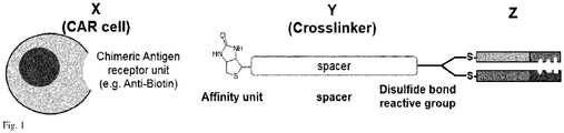
- Fig.1 The general concept of the invention is shown in Fig.1 , showing a cell bearing an chimeric antigen receptor ("CAR") with the sequence of units signaling domain (not shown), transmembrane domain (not shown), chimeric antigen receptor unit (all depicted as X), affinity unit, spacer unit, cross linker unit (all depicted as Y) and antigen recognizing domain Z.
- CAR chimeric antigen receptor
- CAR cells CAR-T cell adapter molecule technology
- a person skilled in the art is aware how to obtain a cell e.g. a chimeric antigen receptor ("CAR") cell with the sequence of units signaling domain (not shown), transmembrane domain (not shown), chimeric antigen receptor unit (all depicted as X).
- the cell is a T-Cell, B-cell, NK cell or a dendritic cell.
- the cell e.g. the chimeric antigen receptor ("CAR") may be provided with a signaling domain is provided with a co-stimulatory domain.
- a co-stimulatory molecule examples include CD27, CD28, 4-1BB (CD137), OX40, CD30, CD40, PD-1, ICOS, lymphocyte function-associated antigen- 1 (LFA-1), CD2, CD7, LIGHT, NKG2C, B7-H3.
- affinity units which are selected from the group consisting of amino acids, peptides, proteins, creatinine, biotin, biocytin, thiamin, fluorochromes, lipids, hormones, vitamins, carbohydrates.
- affinity unit shall bind as specifically as possible to the chimeric antigen receptor unit, a person skilled in the art will have no difficulty to choose the appropriate chimeric antigen receptor unit for the cell.
- affinity unit an anti-biotin antibody or anti-biotin FAB should be selected as chimeric antigen receptor unit.
- antigen recognizing domain refers to any kind of antibody, fragmented antibody or fragmented antibody derivatives, directed against a target moiety expressed on a biological specimen of interest.
- the term relates to fully intact antibodies, fragmented antibody or fragmented antibody derivatives, e.g., Fab, Fab', F(ab')2, sdAb, scFv, di-scFv, nanobodies.
- fragmented antibody derivatives may be synthesized by recombinant procedures including covalent and non-covalent conjugates containing these kind of molecules.
- antigen recognizing moieties are peptide/MHC-complexes targeting TCR molecules, cell adhesion receptor molecules, receptors for costimulatory molecules, artificial engineered binding molecules, e.g., peptides or aptamers which target, e.g., cell surface molecules.
- Antigen recognizing domain refers to a moiety directed against antigen expressed by the biological specimens (target cells) intracellular, like IL2, FoxP3, CD154, CD33 (Siglec-3), CD123 (IL3RA), CD135 (FLT-3), CD44 (HCAM), CD44V6, CD47, CD184 (CXCR4), CLEC12A (CLL1), LeY, FR ⁇ , MICA/B, CD305 (LAIR-1), CD366 (TIM-3), CD96 (TACTILE), CD133, CD56, CD29 (ITGB1), CD44 (HCAM), CD47 (IAP), CD66 (CEA), CD112 (Nectin2), CD117 (c-Kit), CD133, CD146 (MCAM), CD155 (PVR), CD171 (LICAM), CD221 (IGF1), CD227 (MUC1), CD243 (MRD1), CD246 (ALK), CD271 (LNGFR), CD19, CD20, GD2, and EG
- antigen recognizing domain utilized in the present invention comprise a disulfide bond capable of reacting with the cross linker unit as disclosed later.
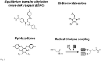
- Suitable cross linker unit comprise at least one reactive group selected from the group consisting of the family of equilibrium transfer alkylation reagents with at least one bis-sulfone leaving group, dibromo maleimides, dichloro maleimides and pyridazediones.
- the spacer unit is selected from the group consisting of PEG (polyethylene glycole), LC, peptides, nucleic acids, carbohydrates, poly hydroxyalkanoates, alkyl chains, alkanoic acids, carboxylic acids, farnesyls, polyethylene glycols, lipids, LCLC, , saturated and unsaturated aliphatic residues having 2 to 200 carbon atoms.
- PEG polyethylene glycole
- LC polyethylene glycole
- peptides nucleic acids
- carbohydrates poly hydroxyalkanoates
- alkyl chains alkanoic acids
- carboxylic acids farnesyls
- farnesyls polyethylene glycols
- lipids lipids
- LCLC saturated and unsaturated aliphatic residues having 2 to 200 carbon atoms.
- LC and/or LCLC with n 1 to 10 repeating units.
- adapter molecule refers to a group or molecule comprising affinity unit, spacer unit, cross linker unit and antigen recognizing domain. It is preferred to synthesize the adapter molecule independent from the cell with an antigen recognizing structure comprising the sequence: signalling domain, transmembrane domain, chimeric antigen receptor unit.
- the adapter molecule is synthesize step by step (or unit by unit) starting with the affinity unit bound to the chimeric antigen receptor unit.
- the method of the invention is directed to provide a cell with an antigen recognizing structure comprising the sequence: signalling domain, transmembrane domain, chimeric antigen receptor unit, affinity unit, spacer unit, cross linker unit and antigen recognizing domain characterized in that a cell provided with an sequence comprising signaling domain, transmembrane domain, chimeric antigen receptor unit is bound to an adapter molecule comprising affinity unit, spacer unit, cross linker unit and antigen recognizing domain wherein the chimeric antigen receptor bind specifically to the unit.
- Another object of the invention is a method to manufacture an adapter molecule comprising an affinity unit, a spacer unit, cross linker unit and one antigen recognizing domain characterized in that the antigen recognizing domain comprises a disulfide bond which is bound to the cross linker unit and wherein affinity unit is bound to the spacer unit and wherein the spacer unit is bound to the cross linker unit.
- the cells of the invention can be used to recognize, detect, kill or at least manipulate target cells like cancerous cells.
- the advantage of the cells and method of the invention is that due to the adapter technology, "standard CAR" cells can be easily converted into personalized CAR cells, equipped with a personalized antigen recognizing moiety to attack target cells.
- cells of the invention may be used in cellular therapy wherein the recipient may be the same subject from which the cells were obtained (autologous cell therapy) or from another subject of the same species (allogeneic cell therapy). In these therapies, the cells of the invention are administered to a recipient and are then able to kill or at least stop growth of cancerous cells expressing the antigen which is recognized by the cells according to the invention.
- a cell with an antigen recognizing structure comprising the sequence: signalling domain, transmembrane domain, chimeric antigen receptor unit, affinity unit, spacer unit, cross linker unit and antigen recognizing domain wherein the antigen recognizing domain comprises a disulfide bond which is bound to the cross linker unit and wherein affinity unit is bound to the spacer unit and wherein the spacer unit is bound to the cross linker unit for recognizing cells expressing an antigen specifically binding to the antigen recognizing domain.
- target cell or target moiety to be recognized with the aid of the invention can be on any biological specimen, like tissues slices, cell aggregates, suspension cells, or adherent cells.
- the cells may be living or dead.
- target moieties are antigens expressed intracellular or extracellular on biological specimen like whole animals, organs, tissues slices, cell aggregates, or single cells of invertebrates, (e.g., Caenorhabditis elegans, Drosophila melanogaster), vertebrates (e.g., Danio rerio, Xenopus laevis) and mammalians (e.g., Mus musculus, Homo sapiens). More preferred target moieties are found on cancer cells in a human subject.
- invertebrates e.g., Caenorhabditis elegans, Drosophila melanogaster
- vertebrates e.g., Danio rerio, Xenopus laevis
- mammalians
- CD19 Fab was used at a concentration of 1g/L in PEB buffer (PBS + EDTA buffer). DTT was added as a reducing agent for 1 hour. Then removed by buffer exchange and the ETAC crosslinker molecule added in a 1,8 fold excess and allow to react for 2-3 hours. After that the sample was buffer exchanged. To prepare the samples for LC-MS analysis, concentration was adjusted to 300 ⁇ g/ml by absorbance for both CD19Fab unmodified (control) and CD19Fab-monoBiotin.
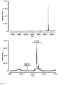
- Fig. 4a shows intact mass determination by mass spectroscopy with: CD19 Fab with MW of 51506,394 Da.
- Fig 4b CD19 Fab-Biotin after conjugation of crosslinker to Fab via disulphide bond with MW of 52337,281 Da showing the addition of only one crosslinker per Fab molecule and thus obtaining a monofunctional (e.g. with Biotin) Fab.
- CD19 Fab was biotinylated by targeting its lysines with NHS-LCLC-Biotin. The result was compared to biotinylation by targeting the disulphide bond on the C-terminus of the CD19 Fab with the crosslinker which would introduce only one Biotin.
- CD19 Fab was dissolved in carbonate buffer at pH 8,5.
- the NHS-LCLC-Biotin reagent was dissolved in DMSO and added in 15 fold molar excess and allow to react for 2 hours, after which the samples was buffer exchanged to PBS.
- the labelling of CD19 Fab with ETAC crosslinker is described in example 1 (see above).
- Fig 5a mass spectroscopy intact mass determination data by microTOF QII of CD19 Fab conjugated with NHS-Biotin, resulting in a stochastic conjugation of multiple biotin units per Fab (from 2 to 11).
- Fig 5b mass spectroscopy intact mass determination data by microTOF QII of CD19 Fab conjugated with crosslinker Y targeting the C terminal disulphite bond in the fab and carrying Biotin as affinity unit resulting in a single conjugation product.
- CD20 positive Jeko-1 cells were seeded in a 96-well plate (50000 cells/well) and incubated for 10min at 4°C with biotin-crosslinker conjugated anti-CD20 FAb (Rtx Fab MS2) at different concentrations (0,01 -100 ⁇ g/ml and 0 ⁇ g/ml as negative control) in a total volume of 50 ⁇ l Buffer A (CliniMACS PBS/EDTA Buffer + 0.5% BSA). After that 50 ⁇ l of anti- Biotin APC (1:50 in Buffer A, Miltenyi Biotec, Art. No. 130-110-952) was added, and the sample was further incubated at 4°C for additional 10 min.
- Buffer A CliniMACS PBS/EDTA Buffer + 0.5% BSA
- Fig. 6 shows: Titration of Biotin-crosslinker Y conjugated to CD20 (Rtx Fab) on CD20+ Jeko-1 target cells. Different concentrations of FAb (0,01 - 100 ⁇ g/ml) was added to 50000? target cells in 50 ⁇ l and secondary staining was performed with anti-Biotin APC (Miltenyi Biotec). In a control sample direct anti-CD20 staining was performed using an anti-CD20-APC conjugate (Miltenyi Biotec).
- Example #6 showing functionality of killing target cancer cells by combination of X-Y-Z units
- PBMCs were isolated from buffy coat of healthy donors by separation on Ficoll.
- T cells were selected from PBMCs by MACS technology using the Pan T cell isolation kit (Miltenyi Biotec, 130-096-535).
- the T cells were seeded at 1x10⁶ cells/mL in TexMACS medium containing IL-7 (10 ng/mL) and IL-15 (10 ng/mL) and 1% (v/v) T Cell TransAct, human (Miltenyi Biotec, Art No: 130-111-160) and incubated at 37°C, 5% CO2. Transduction was performed on day 1 after activation.
- LV supernatant encoding for adapter CARs was added to T cells at a MOI of 5 and cells were carefully resuspended by pipetting up and down. TransAct was removed on day 3 and T cells were further expanded maintaining a cell density of 2x10⁶ cells/mL.
- LNGFR-expressing cells were separated by MACS using anti-LNGFR microbeads on an LS column (Miltenyi Biotec) according to the manufacturer's instructions. Selected, LNGFR+ cells were further expanded up to day 12 and then frozen in aliquots at 1x10⁷ cells/ml in TexMACS + 20% (v/v) FCS + 10% (v/v) DMSO and stored in liquid nitrogen. Aliquots of cells were thawed and cells were washed and recovered in TexMACS containing IL-7 (10 ng/mL) and IL-15 (10 ng/mL) for 48h before the experiment.
- Fig. 7 shows a proof-of-concept with the specific killing of target cancer cells (Jeko-1 cells) expressing an antigen recognized by the X-Y-Z construct comprised of: Anti-Biotin CAR T cells (X), biotin-CD19Fab (Y-Z).
- X Anti-Biotin CAR T cells
- Y-Z biotin-CD19Fab
- the Y-Z complex was added in increasing amounts (0-2,5 ⁇ g) to 10000 Jeko cells and 50000 CAR T cells (closed circles). Positive control (filled triangle) or negative control Mock (UTD) cells (open circles).
- the Direct CD19 CAR killing after 16h is in the range of 80%.
- maximal killing 77% is already observed at adapter doses as low as 0,0025 ng in a sample of 200 ⁇ l (0,0125 ng/ml) and decreased only by approx. 5 % over 6 decades of increasing different adapter concentrations.
- adapter associated background killing activity with Mock T cells is not detected up to 2,5 ng adapter added in the 200 ⁇ l sample (12,5 ng/ml).
- the Fab-based adapter in combination with the adapter CAR T cells enables a high degree of specific lysis (65%) over a broad concentration range covering over 4 decades of adapter concentrations.
- Two different types of adapter molecule were evaluated: i) Rituximab (anti-CD20 antibody) which was conjugated by NHS-esters and has on average 2-3 Biotin affinity units and the ii) Fab fragment which has 1 Biotin affinity unit/ molecule.
- Activation of T cells was analysed on a MACSQuant flow cytometer and the fraction of activated cells was defined by the fraction of CD69+ and CD25+ cells.
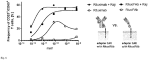
- both adapters efficiently triggered activation of adapter CAR T cells in the presence of target cells in a concentration range of 1x10⁻¹⁰ - 1x10⁻⁸ mol/l adapter (50% activation, depending on E:T ratio).
- the antibody was able to induce activation of the T cells (maximum activation was observed at a concentration of 1x10⁻¹⁰ mol/1), while monobiotinylated Fab was not able to induce activation of T cells in absence of target cells up to a concentration of 1x10⁻⁹ mol/l
- Fig. 8 shows monobiotinylated Fabs inducing activation of adapter CAR T cells only in presence of target cells, while adapter molecules based on full length antibodies, and with multiple affinity units, can induce activation of CAR T cells also in absence of target cells.
- Antibody (Rituximab) or Fab (RituxiFAb) is added at different concentrations (as indicated). The frequency of activated cells (defined by expression of CD69+ and CD25+) is shown.
- Activation of CAR T cells in general should be strictly dependent on presence of target cells and cognate adapter molecules, thereby increasing the safety of the approach, activation of T cells in absence of adapter molecules therefore is an undesired property of the adapter which is observed with the whole antibody molecule but not with the monobiotinylated Fab.
- Use of monobiotinylated Fab as adapter molecule might therefore improve overall safety of the adapter CAR technology.
- Example #8 Monobiotinylated Fab shows increased cytokine secretion from CAR T cells compare to the biotinylated antibody
- IL-2 proinflammatory cytokine
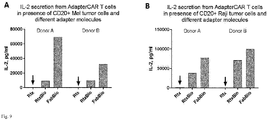
- Different target cells Mel526 (CD20+) and Raji (CD20+) were cocultured in a 96-well plate with adapter CAR T cells at an E:T ratio of 1:1 in 200 ⁇ l.
- Fig. 9 shows that the use of monobiotinylated Fabs can increase target cell specific cytokine release from adapter CAR T cells. Cytokine secretion after 18h of anti-biotin CAR T cells (25.000 cells) in co-culture with A) Mel526 tumor cells expressing CD20 and B) Raji tumor cells, at an effector to target cell ratio of 1:1. Unlabeled Rituximab (Rtx), Rituximab-Biotin (RtxBio) and monobiotinylated Fab (FabBio) were added to the co-culture at 2x10-11 mol/l. On both tumor cell lines, use of the monobiotinylated Fab shows increased cytokine secretion from CAR T cells compare to the biotinylated antibody (Detection limit: 100000 pg/ml).
- cytokine release was increased 3.2-7.4-fold on Mel526 cells and up to 2-fold on Raji cells compared to the biotinylated antibody. Overall this might be attributed to suboptimal orientations (multiple non-productive conformations) on the receptor, which are only possible for the whole antibody molecule having multiple affinity units.
- use of a monobiotinylated Fab may improve formation of productive conformations on the adapter CAR T cells in presence of target cells, allowing for an improved immunological synapse formation and cytokine release.
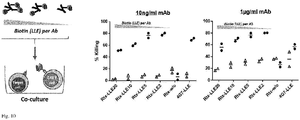
- Fig 10 shows that the low degree of labeling improves adapter (Ab) functionality.
- Rituximab-Biotin anti-CD20 Ab
- As control an anti-CD19 antibody with 6 biotins/molecule was used.
- Different concentrations of mAb 1 ⁇ g/ml and 10ng/ml are shown. Maximal tumor cell lysis after 18h is observed with antibody having a low degree of labeling (average of 2 biotin moieties/Rituximab molecule).
- NSG mice non-obese diabetic mice
- ⁇ c-/- mice mice of female sex were be used, age 7-10 weeks.
- SCID mice severe combined immunodeficiency mice derived on a non-obese diabetic (NOD) background with additional knockout of the common ⁇ -chain ( ⁇ c-/-).
- Mice were be obtained from external provider (Jackson labs) and kept in individually ventilated cages (IVC) at 5 animals per cage and group on a standard rodent diet (ssniff, Soest, Germany). Room temperature was constantly kept at 22°C with an air humidity between 50-60%. Light-dark rhythm interval was 12 hours. General health status of all animals will be monitored daily.
- Raji tumor cells genetically modified to express firefly luciferase (ffLuc), were transferred by i.v. injection (3x10⁵ cells in 100 ⁇ l) and developed systemic leukaemia in the engrafted mice. Tumor progression was regularly monitored (total tumor burden/distribution) by bioluminescence imaging (BLI) in the IVIS (in vivo imaging system). After tumor engraftment for 5 days, adapter CAR T cells and CD20 CAR T cells were be dosed by i.v. injection (1x10⁷ cells per mouse, volume 100 ⁇ l) on day 0. Adapter molecules were administered daily by i.p. injection starting on the same day of tumor injection. Administration of adapter molecules was continued for 10 days and tumor progression was continuously monitored.
- ffLuc firefly luciferase
- Fig. 11 shows the functionality of anti-affinity unit CAR T cells in presence of crosslinker-modified target cell binding domains (CD20Fab-Biotin) in vivo.
- NSG mice were engrafted with CD20+ tumor cells (Raji, expressing firefly luciferase) on day-5, CAR T cells were infused on day 0, adapter molecules (50 ⁇ g/mouse) were administered by i.p. injection on a daily basis.
Landscapes
- Health & Medical Sciences (AREA)
- Life Sciences & Earth Sciences (AREA)
- Chemical & Material Sciences (AREA)
- Immunology (AREA)
- Medicinal Chemistry (AREA)
- General Health & Medical Sciences (AREA)
- Organic Chemistry (AREA)
- Cell Biology (AREA)
- Animal Behavior & Ethology (AREA)
- Pharmacology & Pharmacy (AREA)
- Microbiology (AREA)
- Veterinary Medicine (AREA)
- Mycology (AREA)
- Public Health (AREA)
- Epidemiology (AREA)
- Biochemistry (AREA)
- Proteomics, Peptides & Aminoacids (AREA)
- Molecular Biology (AREA)
- Genetics & Genomics (AREA)
- Biophysics (AREA)
- Oncology (AREA)
- Gastroenterology & Hepatology (AREA)
- Zoology (AREA)
- Toxicology (AREA)
- Peptides Or Proteins (AREA)
Abstract
The invention is directed to a Cell with an antigen recognizing structure comprising the sequence: signalling domain, transmembrane domain, chimeric antigen receptor unit, affinity unit, spacer unit, cross linker unit and antigen recognizing domain wherein the antigen recognizing domain comprises a disulfide bond which is bound to the cross linker unit and wherein affinity unit is bound to the spacer unit and wherein the spacer unit is bound to the cross linker unit.
Description
- The present invention is directed to CAR-cells (e.g. T cells, NK cells, B cells etc.) provided with a an adapter moiety comprising a (site-specific) cross linked thio-bridge.
- "Universal" CAR systems allow for addressing different antigens using the same CAR and several systems have been described, thus far.
- In
WO2015058018A1 the CAR binding motif expressed at the cell surface binds a desired target binding molecule in a non-specific manner, where the binding motif of the CAR is CD16 which is a low affinity Fc receptor that binds unspecifically to Immunoglobulin G antibodies. It is a disadvantage of this system that antibodies in vivo act as natural competitors to the administered antibody of interest supposed to enable specific tumor target lysis upon binding by the CAR T cells. - In
WO2012082841A2 a universal, yet adaptable, anti-tag chimeric antigen receptor (AT-CAR) system is disclosed which provides T cells with the ability and specificity to recognize and kill target cells, such as tumor cells, that have been marked by tagged antibodies, e.g. FITC- or biotin labeled antibodies. Here, the disadvantage is that the labels are for the subject to be treated either foreign molecules, e.g. FITC, and therefore immunogenic or the labels are identical to endogenous molecules, e.g. biotin, resulting in blocking of the CAR binding motif by the endogenous molecule which might prohibit the CAR from binding its intended molecule-labelled antibody, e.g. biotinylated antibody. Furthermore, labels are conjugated to the antibodies by random conjugation, resulting in variable degrees of tags attached to a single antibody, - The same universal CAR system is disclosed in
WO2013044225A1 facing the same disadvantages. - In
WO2014100615A1 a CAR system is disclosed that makes use of CARs that target a moiety that is not produced or expressed by cells of the subject being treated. This CAR system thus allows for focused targeting of the cytotoxic lymphocytes to target cells, such as cancer cells. The targeted moiety is part of a small conjugate molecule (SCM) that also comprises a ligand of a tumor cell receptor. Because small organic molecules are typically used as the targeted moiety, clearance of the SCM from the bloodstream can be achieved within about 20 minutes. By administration of a SCM along with the CAR-expressing cytotoxic lymphocytes, the lymphocyte response can be targeted to only those cells expressing the tumor receptor, thereby reducing off-target toxicity, and the activation of lymphocytes can be more easily controlled due to the rapid clearance of the SCM. The CAR-expressing lymphocytes can also be used as a "universal" cytotoxic cell to target a wide variety of tumors without the need to prepare separate CAR constructs. As the target moieties can be FITC or biotin, this system faces the same disadvantages asWO2012082841A2 and/orWO2013044225A1 . - In
WO2015057852A1 switches are disclosed for regulating the activity of a chimeric antigen receptor effector cells (CAR-ECs). These switches generally comprise a chimeric antigen receptor-interacting domain (CAR-ID) and a target interacting domain (TID). The CAR-ID may be e.g. FITC or biotin. - This system features site-specific labeling of target cell binding moieties but relies on co-translational incorporation of non-natural amino acids. Again, this system faces the same disadvantages as
WO2012082841A2 and/orWO2013044225A1 . - In
WO2015057834A1 a chimeric antigen receptor-effector cell switch is disclosed comprising: a) a peptidic antigen that binds a chimeric antigen receptor on an effector cell; and b) a targeting moiety that binds a cell surface molecule on a target cell. The peptidic antigen is recombinantly expressed and based on or derived from a naturally occurring peptide or from a non-naturally occurring peptide. The disadvantage of this system is that there is a high risk of autoimmunity by tags derived from a human nuclear protein. - In
WO2016030414A1 a universal chimeric antigen receptor is disclosed, wherein the receptor comprises three domains, wherein the first domain is a tag-binding domain, the second domain is a linking peptide chain including an extracellular hinge and a transmembrane domain and the third domain is a signal transduction domain, wherein the tag-binding domain binds to a tag derived from any human nuclear protein. In particular suitable tags are peptide sequences from nuclear antigens, which cannot be accessed and bound by the corresponding tag-binding domain in the context of the native protein under physiological conditions. A disadvantage of this system is that the tag can only be added in a co-translational manner, thereby excluding the use of established monoclonal antibodies as tagged antigen binding molecules without prior redesign of the gene encoding said antigen binding molecule. - Therefore, there is a need in the art for an improved or alternative universal (adaptable) CAR system. Which allows the combination of post- translational, site-specific labeling technology which allows attachment of a tag or affinity unit at a preferred site of the proteinogenic target cell binding moieties for optimal functionality of the system.
- From a different technology field, crosslinking a disulfide bridge of an antibody is known. For example the chemistry of recognition of two neighboring reduced thiols previously forming a disulfide bridge was first described in a scientific publication in 1979 at Sumita Mitra et al. "Reagents for Cross-Linking of Proteins", JACS 101211 (1979), Liberatore F. et al. Bioconjugate Chem, 1, (1990), and Rosario et al. Bioconjugate Chem, 1990, 1, 51-59; where the crosslinker was referred to as: equilibrium alkylating cross-link (ETAC). It is described that a full antibody is reacted with a cross linker for the purpose of labeling the antibody with a fluorescent dye. Another scientific publication describes a similar use on a Fab (Wilbur et al., Bioconjugate Chem 1994, 5, 220-235).
-
US 2016/0000933 A1 discloses the crosslinking of antibodies at their disulfide bridge for the preparation of antibody conjugates. - However, these publications are silent on CAR cells.
- CAR-T cells are genetically engineered cells that express a chimeric antigen receptor (CAR) moiety on their surface which is used to recognize and target unwanted cells. Upon binding, the CAR triggers an activation signal on the CAR-T cells to kill the target cells.
- In order to mediate the recognition and binding between CAR-T cells and the target cells, an adapter molecule is required. An universal CAR T cell adapter molecule is composed of an antibody or antibody fragment (e.g. Fab) specific to recognize an antigen on the target cells plus a secondary moiety which is recognized only by the CAR-T cells. This way, the adapter molecule serves as a recognition bridge between the target cells and the CAR-T cells. By using Ab and Fabs different cell populations can be targeted. And the CAR-Tcells are only functional in the presence of the adapter molecule.
- Currently, affinity tags that are recognized by the CAR-T cells are Vitamins, which are naturally occurring ones and non-biologically orthogonal, e.g. Biotin .
- The problem of anti-adapter-CAR-T-cells and tagged antibodies comes in the formulation and design of the CAR-T cell adapter molecule. Current preparations are based on classical non-site-specific labeling strategies, i.e. biotinylation of antibodies (Ab) or Fabs by targeting amino groups with NHS-biotin reagents. This produces conjugates with a randomized site-labeling of biotin on the Ab or Fab, with a distribution on the number of biotins per Ab and Fabs ranging between 2 to 20 biotins per Ab or Fab. The result is a non-well-defined adapter molecules which translates in a non-optimal performance.
- It was therefore an object of the invention to provide a chemically well-defined adapter molecule with an univariant number of biotins per Ab and/or Fab in site-specific positions, which allows for optimal sensitivity.
- The invention is based on using a site-directed crosslinker that targets disulfide bridges on Ab and/or Fabs. The benefit relies on: i) specific location of disulfide bridge, which is known in the sequence and structure of Ab and Fabs at fixed positions; ii) the crosslinker covalently attaches to both reduced thiols of the disulfide bridge, thus preserving the native 3D structure and function of the Ab and Fab leaving no free thiols behind; iii) each crosslinker carries only one CAR recognition moiety e.g. Biotin), so for each disulfide bridge one will obtain one biotin providing a known and predictable stoichiometry (i.e. fabs only contain one disulfide bridge at the C-terminal); iv) the crosslink reaction is quantitative, meaning above 95% of Ab and Fab is labeled; v) degree of labeling can be stoichiometric controlled (i.e. for Ab).
- Accordingly, object of the invention is a Cell with an antigen recognizing structure comprising the sequence: signaling domain, transmembrane domain, chimeric antigen receptor unit, affinity unit, spacer unit, cross linker unit and antigen recognizing domain characterized in that the antigen recognizing domain comprises a disulfide bond which is bound to the cross linker unit and wherein affinity unit is bound to the spacer unit and wherein the spacer unit is bound to the cross linker unit.
- Furthermore, it is an object of the invention to provide a method to provide a cell with an antigen recognizing structure comprising the sequence: signaling domain, transmembrane domain, chimeric antigen receptor unit, affinity unit, spacer unit, cross linker unit and antigen recognizing domain characterized in that a cell provided with an sequence comprising signaling domain, transmembrane domain, chimeric antigen receptor unit is bound to an adapter molecule comprising affinity unit, spacer unit, cross linker unit and antigen recognizing domain wherein the chimeric antigen receptor bind specifically to the unit.
- Yet another object of the invention is a use or a method for use of a cell with an antigen recognizing structure comprising the sequence: signaling domain, transmembrane domain, chimeric antigen receptor unit, affinity unit, spacer unit, cross linker unit and antigen recognizing domain wherein the antigen recognizing domain comprises a disulfide bond which is bound to the cross linker unit and wherein affinity unit is bound to the spacer unit and wherein the spacer unit is bound to the cross linker unit for recognizing cells expressing an antigen specifically binding to the antigen recognizing domain.
- Further, the invention is directed to a method to manufacture an adapter molecule comprising an affinity unit, a spacer unit, cross linker unit and one antigen recognizing domain characterized in that the antigen recognizing domain comprises a disulfide bond which is bound to the cross linker unit and wherein affinity unit is bound to the spacer unit and wherein the spacer unit is bound to the cross linker unit.
-
-
Fig. 1 : general concept of the cell of the invention -
Fig. 2 : Suitable cross linker unit -
Fig. 3 : Chemistry of reacting the cross linker unit with the disulfide bond of the antigen recognizing domain -
Fig. 4 to 11 : Results of the experiments / examples - The general concept of the invention is shown in
Fig.1 , showing a cell bearing an chimeric antigen receptor ("CAR") with the sequence of units signaling domain (not shown), transmembrane domain (not shown), chimeric antigen receptor unit (all depicted as X), affinity unit, spacer unit, cross linker unit (all depicted as Y) and antigen recognizing domain Z. - As already pointed, there are numerous patent publications disclosing CAR cells CAR-T cell adapter molecule technology. Insofar, a person skilled in the art is aware how to obtain a cell e.g. a chimeric antigen receptor ("CAR") cell with the sequence of units signaling domain (not shown), transmembrane domain (not shown), chimeric antigen receptor unit (all depicted as X). Preferable, the cell is a T-Cell, B-cell, NK cell or a dendritic cell.
- The cell e.g. the chimeric antigen receptor ("CAR") may be provided with a signaling domain is provided with a co-stimulatory domain. Examples for a co-stimulatory molecule are CD27, CD28, 4-1BB (CD137), OX40, CD30, CD40, PD-1, ICOS, lymphocyte function-associated antigen- 1 (LFA-1), CD2, CD7, LIGHT, NKG2C, B7-H3.
- The cell and in the method of the invention makes use of affinity units, which are selected from the group consisting of amino acids, peptides, proteins, creatinine, biotin, biocytin, thiamin, fluorochromes, lipids, hormones, vitamins, carbohydrates.
- Since the affinity unit shall bind as specifically as possible to the chimeric antigen receptor unit, a person skilled in the art will have no difficulty to choose the appropriate chimeric antigen receptor unit for the cell. By way of example, if biotin is selected as affinity unit, an anti-biotin antibody or anti-biotin FAB should be selected as chimeric antigen receptor unit.
- The term "antigen recognizing domain " refers to any kind of antibody, fragmented antibody or fragmented antibody derivatives, directed against a target moiety expressed on a biological specimen of interest. The term relates to fully intact antibodies, fragmented antibody or fragmented antibody derivatives, e.g., Fab, Fab', F(ab')2, sdAb, scFv, di-scFv, nanobodies. Such fragmented antibody derivatives may be synthesized by recombinant procedures including covalent and non-covalent conjugates containing these kind of molecules. Further examples of antigen recognizing moieties are peptide/MHC-complexes targeting TCR molecules, cell adhesion receptor molecules, receptors for costimulatory molecules, artificial engineered binding molecules, e.g., peptides or aptamers which target, e.g., cell surface molecules.
- Preferable, the term "Antigen recognizing domain " refers to a moiety directed against antigen expressed by the biological specimens (target cells) intracellular, like IL2, FoxP3, CD154, CD33 (Siglec-3), CD123 (IL3RA), CD135 (FLT-3), CD44 (HCAM), CD44V6, CD47, CD184 (CXCR4), CLEC12A (CLL1), LeY, FRβ, MICA/B, CD305 (LAIR-1), CD366 (TIM-3), CD96 (TACTILE), CD133, CD56, CD29 (ITGB1), CD44 (HCAM), CD47 (IAP), CD66 (CEA), CD112 (Nectin2), CD117 (c-Kit), CD133, CD146 (MCAM), CD155 (PVR), CD171 (LICAM), CD221 (IGF1), CD227 (MUC1), CD243 (MRD1), CD246 (ALK), CD271 (LNGFR), CD19, CD20, GD2, and EGFR., CD33, CD19, CSD20
- It should be emphasized that the antigen recognizing domain utilized in the present invention comprise a disulfide bond capable of reacting with the cross linker unit as disclosed later.
- Suitable cross linker unit comprise at least one reactive group selected from the group consisting of the family of equilibrium transfer alkylation reagents with at least one bis-sulfone leaving group, dibromo maleimides, dichloro maleimides and pyridazediones.
- Such cross linker units are shown in
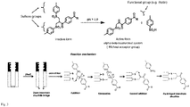
Fig. 2 with R, R1, R2 = aliphatic residue having 1 to 20 carbon atoms. - The chemistry of reacting the cross linker unit with the disulfide bond of the antigen recognizing domain is shown in
Fig. 3 . - Preferable, the spacer unit is selected from the group consisting of PEG (polyethylene glycole), LC, peptides, nucleic acids, carbohydrates, poly hydroxyalkanoates, alkyl chains, alkanoic acids, carboxylic acids, farnesyls, polyethylene glycols, lipids, LCLC, , saturated and unsaturated aliphatic residues having 2 to 200 carbon atoms.
- Most preferred are PEG having n=1 - 50 repeating units, LC and/or LCLC with n = 1 to 10 repeating units.
- The term "adapter molecule" refers to a group or molecule comprising affinity unit, spacer unit, cross linker unit and antigen recognizing domain. It is preferred to synthesize the adapter molecule independent from the cell with an antigen recognizing structure comprising the sequence: signalling domain, transmembrane domain, chimeric antigen receptor unit.
- For example, such adapter molecule provided with the sequence affinity unit/spacer unit/cross linker unit comprises one of the compounds is shown in formula I to III


- In a variant of the invention, the adapter molecule is synthesize step by step (or unit by unit) starting with the affinity unit bound to the chimeric antigen receptor unit.
- The method of the invention is directed to provide a cell with an antigen recognizing structure comprising the sequence: signalling domain, transmembrane domain, chimeric antigen receptor unit, affinity unit, spacer unit, cross linker unit and antigen recognizing domain characterized in that a cell provided with an sequence comprising signaling domain, transmembrane domain, chimeric antigen receptor unit is bound to an adapter molecule comprising affinity unit, spacer unit, cross linker unit and antigen recognizing domain wherein the chimeric antigen receptor bind specifically to the unit.
- All features, embodiments and variants described for the cell can be employed within the method.
- Another object of the invention is a method to manufacture an adapter molecule comprising an affinity unit, a spacer unit, cross linker unit and one antigen recognizing domain characterized in that the antigen recognizing domain comprises a disulfide bond which is bound to the cross linker unit and wherein affinity unit is bound to the spacer unit and wherein the spacer unit is bound to the cross linker unit.
- Again, all features, embodiments and variants described for the cell can be employed within this method.
- The cells of the invention can be used to recognize, detect, kill or at least manipulate target cells like cancerous cells. The advantage of the cells and method of the invention is that due to the adapter technology, "standard CAR" cells can be easily converted into personalized CAR cells, equipped with a personalized antigen recognizing moiety to attack target cells. Accordingly, cells of the invention may be used in cellular therapy wherein the recipient may be the same subject from which the cells were obtained (autologous cell therapy) or from another subject of the same species (allogeneic cell therapy). In these therapies, the cells of the invention are administered to a recipient and are then able to kill or at least stop growth of cancerous cells expressing the antigen which is recognized by the cells according to the invention.
- Accordingly it is an object of the invention to use use of a cell with an antigen recognizing structure comprising the sequence: signalling domain, transmembrane domain, chimeric antigen receptor unit, affinity unit, spacer unit, cross linker unit and antigen recognizing domain wherein the antigen recognizing domain comprises a disulfide bond which is bound to the cross linker unit and wherein affinity unit is bound to the spacer unit and wherein the spacer unit is bound to the cross linker unit for recognizing cells expressing an antigen specifically binding to the antigen recognizing domain.
- The target cell or target moiety to be recognized with the aid of the invention can be on any biological specimen, like tissues slices, cell aggregates, suspension cells, or adherent cells. The cells may be living or dead. Preferable, target moieties are antigens expressed intracellular or extracellular on biological specimen like whole animals, organs, tissues slices, cell aggregates, or single cells of invertebrates, (e.g., Caenorhabditis elegans, Drosophila melanogaster), vertebrates (e.g., Danio rerio, Xenopus laevis) and mammalians (e.g., Mus musculus, Homo sapiens). More preferred target moieties are found on cancer cells in a human subject.
- CD19 Fab was used at a concentration of 1g/L in PEB buffer (PBS + EDTA buffer). DTT was added as a reducing agent for 1 hour. Then removed by buffer exchange and the ETAC crosslinker molecule added in a 1,8 fold excess and allow to react for 2-3 hours. After that the sample was buffer exchanged. To prepare the samples for LC-MS analysis, concentration was adjusted to 300µg/ml by absorbance for both CD19Fab unmodified (control) and CD19Fab-monoBiotin. The LCMS results show that in both cases there is only one significant peak corresponding to the full Fab, which in case of the control has a mass of 51506,394 Dalton, and in the case of the modified Fab it shifts to 52337,281 Dalton, with the mass difference corresponding to the added mass by a single crosslinker molecule per Fab unit.
-
Fig. 4a shows intact mass determination by mass spectroscopy with: CD19 Fab with MW of 51506,394 Da.Fig 4b : CD19 Fab-Biotin after conjugation of crosslinker to Fab via disulphide bond with MW of 52337,281 Da showing the addition of only one crosslinker per Fab molecule and thus obtaining a monofunctional (e.g. with Biotin) Fab. - CD19 Fab was biotinylated by targeting its lysines with NHS-LCLC-Biotin. The result was compared to biotinylation by targeting the disulphide bond on the C-terminus of the CD19 Fab with the crosslinker which would introduce only one Biotin. For Lysine labelling, CD19 Fab was dissolved in carbonate buffer at
pH -
Fig 5a : mass spectroscopy intact mass determination data by microTOF QII of CD19 Fab conjugated with NHS-Biotin, resulting in a stochastic conjugation of multiple biotin units per Fab (from 2 to 11).Fig 5b : mass spectroscopy intact mass determination data by microTOF QII of CD19 Fab conjugated with crosslinker Y targeting the C terminal disulphite bond in the fab and carrying Biotin as affinity unit resulting in a single conjugation product. - The table below summarizes a list of Fabs of different specificities which have been labelled with different disulphide crosslinkers introducing a variety of affinity units and tags. All samples were analysed by LC-MS and compared to the unmodified control to determine the mass difference and hence the number of affinity units per Fab, which in all cases is always only one. This exemplifies how different affinity units can be used to tag different Fabs.
anti-human Fab, specificity Crosslinker mass, dalton Affinity unit Mass of unmodified Fab, dalton Mass of crosslinked Fab, dalton delta mass, dalton CD20 832 DBCO 47178,148 48008,834 830,686 CD20 511,74 Azide 47178,148 47691,015 512,867 CD20 1812,943 Peptide 48008,834 49822,971 1814,137 CD33 619 Biotin 49752,082 50370,331 618,249 CD19 619 Biotin 51506,394 52124,958 618,564 CD19 832 DBCO 51506,394 52337,993 831,599 CD19 190,19 Bimane 51506,394 51696,468 190,074 CD133 619 Biotin 49717,376 50337,41 620,034 CD53 619 Biotin 50058,133 50677,698 619,565 CD34 619 Biotin 51060,003 51679,537 619,534 CD4 619 Biotin 50405,093 51023,723 618,63 CD8 619 Biotin 49308,708 49928,587 619,879 CD56 619 Biotin 50600,276 51218,199 617,923 - Y conjugated to the antigen recognition moiety Z (CD20 Fab) binding to CD20 antigen on CD20
positive Jeko 1 cells and staining with fluorescent anti-Biotin APC. - CD20 positive Jeko-1 cells were seeded in a 96-well plate (50000 cells/well) and incubated for 10min at 4°C with biotin-crosslinker conjugated anti-CD20 FAb (Rtx Fab MS2) at different concentrations (0,01 -100 µg/ml and 0 µg/ml as negative control) in a total volume of 50 µl Buffer A (CliniMACS PBS/EDTA Buffer + 0.5% BSA). After that 50µl of anti- Biotin APC (1:50 in Buffer A, Miltenyi Biotec, Art. No. 130-110-952) was added, and the sample was further incubated at 4°C for additional 10 min. As positive control anti-human CD20 APC conjugate was used to stain a control sample according to the manufacturer's protocol (Miltenyi Biotec, Art. No. 130-111-525). Finally, 100µl of Buffer A was added to each sample and data was acquired at the MACSQuant Analyzer 10 (Miltenyi Biotec 130-096-343). Frequency of CD20+ cells (gated on only aBio APC as negative control) and median fluorescence intensity of positive cells was analysed.
-
Fig. 6 shows: Titration of Biotin-crosslinker Y conjugated to CD20 (Rtx Fab) on CD20+ Jeko-1 target cells. Different concentrations of FAb (0,01 - 100 µg/ml) was added to 50000? target cells in 50µl and secondary staining was performed with anti-Biotin APC (Miltenyi Biotec). In a control sample direct anti-CD20 staining was performed using an anti-CD20-APC conjugate (Miltenyi Biotec). - Fluorescence and thereby biotinylated FAb bound to target cells was detected at concentration as low as 0,1 µg/ml and median fluorescence intensity increased up to a concentration of 100 µg/ml, indicating that free CD20 binding sites are still available at concentrations of 10 µg/ml, under the conditions used.
- PBMCs were isolated from buffy coat of healthy donors by separation on Ficoll. T cells were selected from PBMCs by MACS technology using the Pan T cell isolation kit (Miltenyi Biotec, 130-096-535). For T cell activation and -expansion the T cells were seeded at 1x10⁶ cells/mL in TexMACS medium containing IL-7 (10 ng/mL) and IL-15 (10 ng/mL) and 1% (v/v) T Cell TransAct, human (Miltenyi Biotec, Art No: 130-111-160) and incubated at 37°C, 5% CO2. Transduction was performed on
day 1 after activation. For this, LV supernatant encoding for adapter CARs was added to T cells at a MOI of 5 and cells were carefully resuspended by pipetting up and down. TransAct was removed onday 3 and T cells were further expanded maintaining a cell density of 2x10⁶ cells/mL. Onday 6 LNGFR-expressing cells were separated by MACS using anti-LNGFR microbeads on an LS column (Miltenyi Biotec) according to the manufacturer's instructions. Selected, LNGFR+ cells were further expanded up to day 12 and then frozen in aliquots at 1x10⁷ cells/ml in TexMACS + 20% (v/v) FCS + 10% (v/v) DMSO and stored in liquid nitrogen. Aliquots of cells were thawed and cells were washed and recovered in TexMACS containing IL-7 (10 ng/mL) and IL-15 (10 ng/mL) for 48h before the experiment. - Immediately before the experiment GFP transduced Jeko-1 target cells were resuspended in TexMACS without cytokines and 10000 cells were added to each well of a 96-well plate. Then CAR transduced, and untransduced (Mock) T cells were resuspended in TexMACS without cytokines and 50000 cells were added to the respective wells of a 96-well plate. Subsequently, crosslinked and biotinylated CD19 Fab was added to each well at the indicated concentrations and the sample (V = 200 µl) containing Jeko-1 target cells, T cells and Fab, was mixed by carefully pipetting up and down. As a positive control direct anti-CD19 CAR T cells (50000) were co-incubated with 10000 target cells without addition of Fab. All samples were incubated at 37°C, 5% CO2 for 16h. Killing was quantified on a MACSQuant flow cytometer. Propidium iodide was added to the cells immediately before the assay. Killing was evaluated by counting GFP positive and viable (propidium iodide negative) target cells and is expressed as [Killing, %] = [viable GFP+ target cells in untreated sample] / [viable GFP+ target cells in treated sample] x 100 %
-
Fig. 7 shows a proof-of-concept with the specific killing of target cancer cells (Jeko-1 cells) expressing an antigen recognized by the X-Y-Z construct comprised of: Anti-Biotin CAR T cells (X), biotin-CD19Fab (Y-Z). The Y-Z complex was added in increasing amounts (0-2,5 µg) to 10000 Jeko cells and 50000 CAR T cells (closed circles). Positive control (filled triangle) or negative control Mock (UTD) cells (open circles). - Upon titration of the Fab molecule the Direct CD19 CAR killing after 16h is in the range of 80%. In case of the adapter CAR, maximal killing (75%) is already observed at adapter doses as low as 0,0025 ng in a sample of 200µl (0,0125 ng/ml) and decreased only by approx. 5 % over 6 decades of increasing different adapter concentrations. Furthermore, adapter associated background killing activity with Mock T cells is not detected up to 2,5 ng adapter added in the 200 µl sample (12,5 ng/ml). Overall this indicates that the Fab-based adapter in combination with the adapter CAR T cells enables a high degree of specific lysis (65%) over a broad concentration range covering over 4 decades of adapter concentrations.
- Expression of markers CD25 and CD69 is correlated with activation of T cells, which in turn is related to their cytotoxic effector functions. In order to monitor T cell activation, adapter CAR T cells were incubated for 24h at 37°C and 5% CO2 at increasing concentrations of adapter molecule in presence (closed symbols, E:T = 1:1) or absence (open symbols) of target cells. Two different types of adapter molecule were evaluated: i) Rituximab (anti-CD20 antibody) which was conjugated by NHS-esters and has on average 2-3 Biotin affinity units and the ii) Fab fragment which has 1 Biotin affinity unit/ molecule. Activation of T cells was analysed on a MACSQuant flow cytometer and the fraction of activated cells was defined by the fraction of CD69+ and CD25+ cells. Importantly both adapters efficiently triggered activation of adapter CAR T cells in the presence of target cells in a concentration range of 1x10⁻¹⁰ - 1x10⁻⁸ mol/l adapter (50% activation, depending on E:T ratio). However, in absence of target cells only the antibody was able to induce activation of the T cells (maximum activation was observed at a concentration of 1x10⁻¹⁰ mol/1), while monobiotinylated Fab was not able to induce activation of T cells in absence of target cells up to a concentration of 1x10⁻⁹ mol/l
-
Fig. 8 shows monobiotinylated Fabs inducing activation of adapter CAR T cells only in presence of target cells, while adapter molecules based on full length antibodies, and with multiple affinity units, can induce activation of CAR T cells also in absence of target cells. Co-culture (24h) of adapter CAR T and Raji target cells at an effector to target cell ratio of 1. Antibody (Rituximab) or Fab (RituxiFAb) is added at different concentrations (as indicated). The frequency of activated cells (defined by expression of CD69+ and CD25+) is shown. - Activation of CAR T cells in general should be strictly dependent on presence of target cells and cognate adapter molecules, thereby increasing the safety of the approach, activation of T cells in absence of adapter molecules therefore is an undesired property of the adapter which is observed with the whole antibody molecule but not with the monobiotinylated Fab. Use of monobiotinylated Fab as adapter molecule might therefore improve overall safety of the adapter CAR technology.
- Use of monobiotinylated Fab molecules can induce efficient release of proinflammatory cytokine (IL-2) from T cells. Different target cells Mel526 (CD20+) and Raji (CD20+) were cocultured in a 96-well plate with adapter CAR T cells at an E:T ratio of 1:1 in 200 µl. Adapter molecules Rituximab- Biotin (n=2-3 Biotin/molecule) and monobiotinylated Rituximab Fab were added at 2x10-11 mol/l, as a negative control non-biotinylated Rituximab was used. After 18h incubation at 37°C and 5% CO2 100µl supernatant was carefully removed from the culture and analysed using the MACSPlex Cytokine 12 Kit, human (Miltenyi Biotec Art. No. 130-099-169) according to the manufacturer's protocol. While in the samples containing only antibody no significant IL-2 release was observed, presence of biotinylated antibody triggered specific cytokine secretion on Mel526 and Raji cells.
-
Fig. 9 shows that the use of monobiotinylated Fabs can increase target cell specific cytokine release from adapter CAR T cells. Cytokine secretion after 18h of anti-biotin CAR T cells (25.000 cells) in co-culture with A) Mel526 tumor cells expressing CD20 and B) Raji tumor cells, at an effector to target cell ratio of 1:1. Unlabeled Rituximab (Rtx), Rituximab-Biotin (RtxBio) and monobiotinylated Fab (FabBio) were added to the co-culture at 2x10-11 mol/l. On both tumor cell lines, use of the monobiotinylated Fab shows increased cytokine secretion from CAR T cells compare to the biotinylated antibody (Detection limit: 100000 pg/ml). - Notably in presence of the same concentration of biotinylated Fab, cytokine release was increased 3.2-7.4-fold on Mel526 cells and up to 2-fold on Raji cells compared to the biotinylated antibody. Overall this might be attributed to suboptimal orientations (multiple non-productive conformations) on the receptor, which are only possible for the whole antibody molecule having multiple affinity units. Thereby use of a monobiotinylated Fab may improve formation of productive conformations on the adapter CAR T cells in presence of target cells, allowing for an improved immunological synapse formation and cytokine release.
- Functionality of adapter molecules having a variable number of affinity units was further addressed in an assay in which Rituximab was conjugated using different amounts of Biotin. The antibody modification by random labeling by succinimidyl-esters at amino groups was done according to protocols well known in the art. For modification, antibodies were rebuffered by passing over an Sephadex G25 column equilibrated in PEB buffer. Collected fractions were assayed for protein content using the Bradford assay. Protein containing fractions were pooled and the total volume determined. Final protein concentration was measured by absorption at 280nm. Subsequently, the corresponding amount of Biotin-LC-LC-NHS (ThermoFisher Scientific, Mw 567,70 g/mol, Cat. No. 21343, CAS-No. 89889-52-1) was dissolved in DMSO. To obtain different degrees of labelling, different amounts of Biotin-LC-LC-NHS was added to reaction mix (3, 6, 12 and 25-fold molar excess). The antibody and DMSO/label mix was incubated at 30°C for 1h and then passed over a Sephadex G25 column. Again fractions were collected, assayed for protein concentration and protein containing fractions were pooled. Final protein concentration was measured by absorption at 280nm. Successful biotinylation was confirmed by LC-MS and by incubation of the antibodies on Jeko-1 cell line expressing the CD20 antibody target and secondary staining with a fluorochrome conjugated anti-biotin antibody, followed by FACS analysis. Rituximab conjugates having a degree of labelling form 2-3 up to 20 biotins/antibody were obtained and subsequently used as adapters used in a killing assay
-
Fig 10 shows that the low degree of labeling improves adapter (Ab) functionality. CAR T effector cells (red) or Mock (untransduced, blue) T cells were co-cultured with Jeko-1 mantle cell lymphoma (E:T = 5:1, 18h), in presence of Rituximab-Biotin (anti-CD20 Ab) with different degrees of labeling (number of Biotins/Rituximab ranging from 0 (Rtx-LLE-w/o) up to 20 (Rtx-LLE20). As control an anti-CD19 antibody with 6 biotins/molecule was used. Different concentrations of mAb: 1µg/ml and 10ng/ml are shown. Maximal tumor cell lysis after 18h is observed with antibody having a low degree of labeling (average of 2 biotin moieties/Rituximab molecule). - While all biotinylated antibodies were able to mediate killing of target cells, maximum specific target cell lysis was observed with rituximab having a low degree of labelling, with 2-3 biotins per antibody. Notably, no lysines, representing the primary sites of modification for NHS esters can be found in the CDRs of Rituximab. Overall this may highlight importance of using a low degree of labelling on adapter molecules and highlight the importance of monofunctionalized adapters.
- For the in vivo experiments NOD-SCID common γ chain-/- (NSG) mice of female sex were be used, age 7-10 weeks. These are severe combined immunodeficiency (SCID) mice derived on a non-obese diabetic (NOD) background with additional knockout of the common γ -chain (γc-/-). Mice were be obtained from external provider (Jackson labs) and kept in individually ventilated cages (IVC) at 5 animals per cage and group on a standard rodent diet (ssniff, Soest, Germany). Room temperature was constantly kept at 22°C with an air humidity between 50-60%. Light-dark rhythm interval was 12 hours. General health status of all animals will be monitored daily. Raji tumor cells, genetically modified to express firefly luciferase (ffLuc), were transferred by i.v. injection (3x10⁵ cells in 100 µl) and developed systemic leukaemia in the engrafted mice. Tumor progression was regularly monitored (total tumor burden/distribution) by bioluminescence imaging (BLI) in the IVIS (in vivo imaging system). After tumor engraftment for 5 days, adapter CAR T cells and CD20 CAR T cells were be dosed by i.v. injection (1x10⁷ cells per mouse,
volume 100 µl) onday 0. Adapter molecules were administered daily by i.p. injection starting on the same day of tumor injection. Administration of adapter molecules was continued for 10 days and tumor progression was continuously monitored. -
Fig. 11 shows the functionality of anti-affinity unit CAR T cells in presence of crosslinker-modified target cell binding domains (CD20Fab-Biotin) in vivo. NSG mice were engrafted with CD20+ tumor cells (Raji, expressing firefly luciferase) on day-5, CAR T cells were infused onday 0, adapter molecules (50µg/mouse) were administered by i.p. injection on a daily basis. B) Tumor progression (median of groups with n=5 mice) was monitored by bioluminescence imaging (IVIS). Control of tumor growth was only observed in the mice receiving both CAR T cells and adapter molecules and the CD20 direct CAR positive control. - While the untreated group which only received tumor cells, BLI was progressing from 1x10⁶ on
day 0 to up to 2x10⁹ onday 10, CD20 CAR T cells were able to control tumor outgrowth efficiently with BLI signal of 7x10⁵ onday 10. Adapter CAR T cells in presence of adapter showed a very similar tumor control with median of 8x10⁵ onday 10. In contrast Mock T cells in absence or presence of adapter and adapter CAR T cells in absence of adapter did not control tumor outgrowth, demonstrating that both adapter and adapter CAR T cells need to be present for an efficient tumor control in vivo. Furthermore this experiment highlights the functionality of the monobiotinylated CD20 Fab conjugate in a preclinical setting.
Claims (10)
- Cell with an antigen recognizing structure comprising the sequence: signaling domain, transmembrane domain, chimeric antigen receptor unit, affinity unit, spacer unit, cross linker unit and antigen recognizing domain
characterized in that
the antigen recognizing domain comprises a disulfide bond which is bound to the cross linker unit and wherein affinity unit is bound to the spacer unit and wherein the spacer unit is bound to the cross linker unit. - Cell according to claim 1 characterized in that the cross linker unit comprises at least one reactive group selected from the group consisting of the family of equilibrium transfer alkylation reagents with at least one bis-sulfone leaving group, dibromo maleimides, dichloro maleimides and pyridazediones.
- Cell according to claim 1 or 2 characterized in that the spacer group is selected from the group consisting of PEG, LC, peptides, nucleic acids, carbohydrates, polyhydroxyalkanoates, alkyl chains, alkanoic acids, carboxylic acids, farnesyls, polyethylene glycols, lipids, LCLC, saturated and unsaturated aliphatic residues having 2 to 200 carbon atoms.
- Cell according to any of the claims 1 to 3 characterized in that the affinity unit is selected from the group consisting of amino acids, peptides, proteins, creatinine, biotin, biocytin, thiamin, fluorochromes, lipids, hormones, vitamins, carbohydrates.
- Cell according to any of the claims 1 to 4 characterized in that the signalling domain is provided with a co-stimulatory domain. Examples for a co-stimulatory molecule are CD27, CD28, 4-1BB (CD137), OX40, CD30, CD40, PD-1, ICOS, lymphocyte function-associated antigen- 1 (LFA-1), CD2, CD7, LIGHT, NKG2C, B7-H3.
- Cell according to any of the claims 1 to 5 characterized in that the sequence affinity unit/spacer unit/cross linker unit comprises one of the compounds according to formula I to III


- Cell according to any of the claims 1 to 6 characterized in that the cell is a T-Cell, B-cell, NK cell or dendritic cell.
- Method to provide a cell with an antigen recognizing structure comprising the sequence:signalling domain, transmembrane domain, chimeric antigen receptor unit, affinity unit, spacer unit, cross linker unit and antigen recognizing domaincharacterized in thata cell provided with an sequence comprising signalling domain, transmembrane domain, chimeric antigen receptor unit is bound to an adapter molecule comprising affinity unit, spacer unit, cross linker unit and antigen recognizing domain wherein the chimeric antigen receptor bind specifically to the unit.
- Method to manufacture an adapter molecule comprising an affinity unit, a spacer unit, cross linker unit and one antigen recognizing domain characterized in that the antigen recognizing domain comprises a disulfide bond which is bound to the cross linker unit and wherein affinity unit is bound to the spacer unit and wherein the spacer unit is bound to the cross linker unit.
- Use of a cell with an antigen recognizing structure comprising the sequence: signalling domain, transmembrane domain, chimeric antigen receptor unit, affinity unit, spacer unit, cross linker unit and antigen recognizing domain wherein the antigen recognizing domain comprises a disulfide bond which is bound to the cross linker unit and wherein affinity unit is bound to the spacer unit and wherein the spacer unit is bound to the cross linker unit for recognizing cells expressing an antigen specifically binding to the antigen recognizing domain.
Priority Applications (1)
| Application Number | Priority Date | Filing Date | Title |
|---|---|---|---|
| EP18193369.8A EP3620464A1 (en) | 2018-09-10 | 2018-09-10 | Car cell having crosslinked disulfide bridge on antigen recognizing moiety |
Applications Claiming Priority (1)
| Application Number | Priority Date | Filing Date | Title |
|---|---|---|---|
| EP18193369.8A EP3620464A1 (en) | 2018-09-10 | 2018-09-10 | Car cell having crosslinked disulfide bridge on antigen recognizing moiety |
Publications (1)
| Publication Number | Publication Date |
|---|---|
| EP3620464A1 true EP3620464A1 (en) | 2020-03-11 |
Family
ID=63683632
Family Applications (1)
| Application Number | Title | Priority Date | Filing Date |
|---|---|---|---|
| EP18193369.8A Withdrawn EP3620464A1 (en) | 2018-09-10 | 2018-09-10 | Car cell having crosslinked disulfide bridge on antigen recognizing moiety |
Country Status (1)
| Country | Link |
|---|---|
| EP (1) | EP3620464A1 (en) |
Cited By (4)
| Publication number | Priority date | Publication date | Assignee | Title |
|---|---|---|---|---|
| EP3878464A1 (en) * | 2020-03-09 | 2021-09-15 | Miltenyi Biotec B.V. & Co. KG | Use of a car cell having crosslinked disulfide bridge on antigen recognizing moiety for targeting cancer cells |
| EP3916388A1 (en) | 2020-05-27 | 2021-12-01 | Miltenyi Biotec B.V. & Co. KG | Reagents for detection of chimeric antigen receptor cells |
| WO2022043023A1 (en) * | 2020-08-27 | 2022-03-03 | Miltenyi Biotec B.V. & Co. KG | Releasable reagents for cell separation |
| EP4234559A1 (en) | 2022-02-24 | 2023-08-30 | Miltenyi Biotec B.V. & Co. KG | Serum resistant bis-sulfhydryl specific biotinylation reagent |
Citations (11)
| Publication number | Priority date | Publication date | Assignee | Title |
|---|---|---|---|---|
| WO2012082841A2 (en) | 2010-12-14 | 2012-06-21 | University Of Maryland, Baltimore | Universal anti-tag chimeric antigen receptor-expressing t cells and methods of treating cancer |
| WO2013044225A1 (en) | 2011-09-22 | 2013-03-28 | The Trustees Of The University Of Pennsylvania | A universal immune receptor expressed by t cells for the targeting of diverse and multiple antigens |
| WO2014100615A1 (en) | 2012-12-20 | 2014-06-26 | Purdue Research Foundation | Chimeric antigen receptor-expressing t cells as anti-cancer therapeutics |
| WO2015057852A1 (en) | 2013-10-15 | 2015-04-23 | The California Institute For Biomedical Research | Chimeric antigen receptor t cell switches and uses thereof |
| WO2015058018A1 (en) | 2013-10-17 | 2015-04-23 | National University Of Singapore | Chimeric receptor that triggers antibody-dependent cell cytotoxicity against multiple tumors |
| WO2015057834A1 (en) | 2013-10-15 | 2015-04-23 | The California Institute For Biomedical Research | Peptidic chimeric antigen receptor t cell switches and uses thereof |
| US20160000933A1 (en) | 2014-07-01 | 2016-01-07 | Andrei POLUKHTIN | Conjugated biological molecules and their preparation |
| WO2016030414A1 (en) | 2014-08-29 | 2016-03-03 | Gemoab Monoclonals Gmbh | Universal chimeric antigen receptor expressing immune cells for targeting of diverse multiple antigens and method of manufacturing the same and use of the same for treatment of cancer, infections and autoimmune disorders |
| WO2016154621A1 (en) * | 2015-03-26 | 2016-09-29 | The California Institute For Biomedical Research | SWITCHABLE NON-scFv CHIMERIC RECEPTORS, SWITCHES, AND USES THEREOF |
| WO2016168769A1 (en) * | 2015-04-15 | 2016-10-20 | The California Institute For Biomedical Research | Chimeric receptor t cell switches for her2 |
| WO2017177149A2 (en) * | 2016-04-08 | 2017-10-12 | Purdue Research Foundation | Methods and compositions for car t cell therapy |
-
2018
- 2018-09-10 EP EP18193369.8A patent/EP3620464A1/en not_active Withdrawn
Patent Citations (11)
| Publication number | Priority date | Publication date | Assignee | Title |
|---|---|---|---|---|
| WO2012082841A2 (en) | 2010-12-14 | 2012-06-21 | University Of Maryland, Baltimore | Universal anti-tag chimeric antigen receptor-expressing t cells and methods of treating cancer |
| WO2013044225A1 (en) | 2011-09-22 | 2013-03-28 | The Trustees Of The University Of Pennsylvania | A universal immune receptor expressed by t cells for the targeting of diverse and multiple antigens |
| WO2014100615A1 (en) | 2012-12-20 | 2014-06-26 | Purdue Research Foundation | Chimeric antigen receptor-expressing t cells as anti-cancer therapeutics |
| WO2015057852A1 (en) | 2013-10-15 | 2015-04-23 | The California Institute For Biomedical Research | Chimeric antigen receptor t cell switches and uses thereof |
| WO2015057834A1 (en) | 2013-10-15 | 2015-04-23 | The California Institute For Biomedical Research | Peptidic chimeric antigen receptor t cell switches and uses thereof |
| WO2015058018A1 (en) | 2013-10-17 | 2015-04-23 | National University Of Singapore | Chimeric receptor that triggers antibody-dependent cell cytotoxicity against multiple tumors |
| US20160000933A1 (en) | 2014-07-01 | 2016-01-07 | Andrei POLUKHTIN | Conjugated biological molecules and their preparation |
| WO2016030414A1 (en) | 2014-08-29 | 2016-03-03 | Gemoab Monoclonals Gmbh | Universal chimeric antigen receptor expressing immune cells for targeting of diverse multiple antigens and method of manufacturing the same and use of the same for treatment of cancer, infections and autoimmune disorders |
| WO2016154621A1 (en) * | 2015-03-26 | 2016-09-29 | The California Institute For Biomedical Research | SWITCHABLE NON-scFv CHIMERIC RECEPTORS, SWITCHES, AND USES THEREOF |
| WO2016168769A1 (en) * | 2015-04-15 | 2016-10-20 | The California Institute For Biomedical Research | Chimeric receptor t cell switches for her2 |
| WO2017177149A2 (en) * | 2016-04-08 | 2017-10-12 | Purdue Research Foundation | Methods and compositions for car t cell therapy |
Non-Patent Citations (7)
| Title |
|---|
| "Bioconjugate Techniques", 1 January 2013, ACADEMIC PRESS, ISBN: 978-0-12-382239-0, article GREG T. HERMANSON: "Antibody Modification and Conjugation", pages: 867 - 920, XP055589306, DOI: 10.1016/B978-0-12-382239-0.00020-0 * |
| HANIEH KHALILI ET AL: "Comparative Binding of Disulfide-Bridged PEG-Fabs", BIOCONJUGATE CHEMISTRY, vol. 23, no. 11, 21 September 2012 (2012-09-21), pages 2262 - 2277, XP055096650, ISSN: 1043-1802, DOI: 10.1021/bc300372r * |
| K. TAMADA ET AL: "Redirecting Gene-Modified T Cells toward Various Cancer Types Using Tagged Antibodies", CLINICAL CANCER RESEARCH, vol. 18, no. 23, 2 October 2012 (2012-10-02), pages 6436 - 6445, XP055154500, ISSN: 1078-0432, DOI: 10.1158/1078-0432.CCR-12-1449 * |
| LIBERATORE F. ET AL., BIOCONJUGATE CHEM, vol. 1, 1990 |
| ROSARIO ET AL., BIOCONJUGATE CHEM, vol. 1, 1990, pages 51 - 59 |
| SUMITA MITRA ET AL.: "Reagents for Cross-Linking of Proteins", JACS, vol. 101211, 1979 |
| WILBUR ET AL., BIOCONJUGATE CHEM, vol. 5, 1994, pages 220 - 235 |
Cited By (4)
| Publication number | Priority date | Publication date | Assignee | Title |
|---|---|---|---|---|
| EP3878464A1 (en) * | 2020-03-09 | 2021-09-15 | Miltenyi Biotec B.V. & Co. KG | Use of a car cell having crosslinked disulfide bridge on antigen recognizing moiety for targeting cancer cells |
| EP3916388A1 (en) | 2020-05-27 | 2021-12-01 | Miltenyi Biotec B.V. & Co. KG | Reagents for detection of chimeric antigen receptor cells |
| WO2022043023A1 (en) * | 2020-08-27 | 2022-03-03 | Miltenyi Biotec B.V. & Co. KG | Releasable reagents for cell separation |
| EP4234559A1 (en) | 2022-02-24 | 2023-08-30 | Miltenyi Biotec B.V. & Co. KG | Serum resistant bis-sulfhydryl specific biotinylation reagent |
Similar Documents
| Publication | Publication Date | Title |
|---|---|---|
| AU2019257362B2 (en) | Erythrocyte-binding therapeutics | |
| EP3620464A1 (en) | Car cell having crosslinked disulfide bridge on antigen recognizing moiety | |
| JP7054948B2 (en) | Protease cleavage resistance, Shiga toxin A subunit effector polypeptide and cell targeting molecule containing it | |
| KR20190117667A (en) | Functionalized red blood cells | |
| KR102499753B1 (en) | Engineering t-cell receptors | |
| KR102415259B1 (en) | Engineered high-affinity human t cell receptors | |
| CA2328144A1 (en) | Multivalent t cell receptor complexes | |
| CN104271148A (en) | Erythrocyte-binding therapeutics | |
| US20170112878A1 (en) | Programmable universal cell receptors and method of using the same | |
| WO2001090198A1 (en) | Multicomponent conjugates which bind to target molecules and stimulate cell lysis | |
| Liet et al. | Multifunctional glycoconjugates for recruiting natural antibodies against cancer cells | |
| Amoury et al. | SNAP-tag based agents for preclinical in vitro imaging in malignant diseases | |
| Kalousková et al. | Tumor marker B7-H6 bound to the coiled coil peptide-polymer conjugate enables targeted therapy by activating human natural killer cells | |
| US20230013963A1 (en) | Reversible cell detection via mhc with conjugates having an enzymatically cleavable detection moiety | |
| US20210275586A1 (en) | Use of a car cell having crosslinked disulfide bridge on antigen recognizing moiety for targetting cancer cells | |
| US20200299352A1 (en) | Programmable immunocyte receptor complex system | |
| US20040068100A1 (en) | Multicomponent conjugates which bind to target molecules and stimulate cell lysis | |
| Descalzi‐Montoya et al. | Synthetic Antibody Mimics Based on Cancer‐Targeting Immunostimulatory Peptides | |
| RU2779315C2 (en) | Functionalized erythroid cells | |
| US20220195006A1 (en) | Peptide markers to track genetically engineered cells | |
| WO2024081824A2 (en) | Synthetic intermediates for universal chimeric antigen receptor immune cell therapies | |
| Li | Development of VHH-and antibody-based imaging and diagnostic tools |
Legal Events
| Date | Code | Title | Description |
|---|---|---|---|
| PUAI | Public reference made under article 153(3) epc to a published international application that has entered the european phase |
Free format text: ORIGINAL CODE: 0009012 |
|
| STAA | Information on the status of an ep patent application or granted ep patent |
Free format text: STATUS: THE APPLICATION HAS BEEN WITHDRAWN |
|
| AK | Designated contracting states |
Kind code of ref document: A1 Designated state(s): AL AT BE BG CH CY CZ DE DK EE ES FI FR GB GR HR HU IE IS IT LI LT LU LV MC MK MT NL NO PL PT RO RS SE SI SK SM TR |
|
| AX | Request for extension of the european patent |
Extension state: BA ME |
|
| 18W | Application withdrawn |
Effective date: 20200211 |