EP3608313A1 - 15-idene-14-deoxy-11,12-dehydroandrographolide derivative and application thereof in preparing anti-fibrosis drugs - Google Patents
15-idene-14-deoxy-11,12-dehydroandrographolide derivative and application thereof in preparing anti-fibrosis drugs Download PDFInfo
- Publication number
- EP3608313A1 EP3608313A1 EP18777705.7A EP18777705A EP3608313A1 EP 3608313 A1 EP3608313 A1 EP 3608313A1 EP 18777705 A EP18777705 A EP 18777705A EP 3608313 A1 EP3608313 A1 EP 3608313A1
- Authority
- EP
- European Patent Office
- Prior art keywords
- fibrosis
- chlorophenyl
- cooh
- compound
- bromophenyl
- Prior art date
- Legal status (The legal status is an assumption and is not a legal conclusion. Google has not performed a legal analysis and makes no representation as to the accuracy of the status listed.)
- Granted
Links
- SRCFQMOGKWYADE-UHFFFAOYSA-N Cc1c[nH]c2c1ccc(Cl)c2 Chemical compound Cc1c[nH]c2c1ccc(Cl)c2 SRCFQMOGKWYADE-UHFFFAOYSA-N 0.000 description 1
- XKABPZXWGLYGDP-UHFFFAOYSA-N Fc1c[nH]c2c1ccc(Cl)c2 Chemical compound Fc1c[nH]c2c1ccc(Cl)c2 XKABPZXWGLYGDP-UHFFFAOYSA-N 0.000 description 1
Images


















Classifications
-
- A—HUMAN NECESSITIES
- A61—MEDICAL OR VETERINARY SCIENCE; HYGIENE
- A61P—SPECIFIC THERAPEUTIC ACTIVITY OF CHEMICAL COMPOUNDS OR MEDICINAL PREPARATIONS
- A61P1/00—Drugs for disorders of the alimentary tract or the digestive system
- A61P1/16—Drugs for disorders of the alimentary tract or the digestive system for liver or gallbladder disorders, e.g. hepatoprotective agents, cholagogues, litholytics
-
- A—HUMAN NECESSITIES
- A61—MEDICAL OR VETERINARY SCIENCE; HYGIENE
- A61K—PREPARATIONS FOR MEDICAL, DENTAL OR TOILETRY PURPOSES
- A61K31/00—Medicinal preparations containing organic active ingredients
- A61K31/33—Heterocyclic compounds
- A61K31/335—Heterocyclic compounds having oxygen as the only ring hetero atom, e.g. fungichromin
- A61K31/34—Heterocyclic compounds having oxygen as the only ring hetero atom, e.g. fungichromin having five-membered rings with one oxygen as the only ring hetero atom, e.g. isosorbide
- A61K31/341—Heterocyclic compounds having oxygen as the only ring hetero atom, e.g. fungichromin having five-membered rings with one oxygen as the only ring hetero atom, e.g. isosorbide not condensed with another ring, e.g. ranitidine, furosemide, bufetolol, muscarine
-
- A—HUMAN NECESSITIES
- A61—MEDICAL OR VETERINARY SCIENCE; HYGIENE
- A61K—PREPARATIONS FOR MEDICAL, DENTAL OR TOILETRY PURPOSES
- A61K31/00—Medicinal preparations containing organic active ingredients
- A61K31/33—Heterocyclic compounds
- A61K31/335—Heterocyclic compounds having oxygen as the only ring hetero atom, e.g. fungichromin
- A61K31/365—Lactones
-
- A—HUMAN NECESSITIES
- A61—MEDICAL OR VETERINARY SCIENCE; HYGIENE
- A61K—PREPARATIONS FOR MEDICAL, DENTAL OR TOILETRY PURPOSES
- A61K31/00—Medicinal preparations containing organic active ingredients
- A61K31/33—Heterocyclic compounds
- A61K31/395—Heterocyclic compounds having nitrogen as a ring hetero atom, e.g. guanethidine or rifamycins
- A61K31/40—Heterocyclic compounds having nitrogen as a ring hetero atom, e.g. guanethidine or rifamycins having five-membered rings with one nitrogen as the only ring hetero atom, e.g. sulpiride, succinimide, tolmetin, buflomedil
- A61K31/4025—Heterocyclic compounds having nitrogen as a ring hetero atom, e.g. guanethidine or rifamycins having five-membered rings with one nitrogen as the only ring hetero atom, e.g. sulpiride, succinimide, tolmetin, buflomedil not condensed and containing further heterocyclic rings, e.g. cromakalim
-
- A—HUMAN NECESSITIES
- A61—MEDICAL OR VETERINARY SCIENCE; HYGIENE
- A61K—PREPARATIONS FOR MEDICAL, DENTAL OR TOILETRY PURPOSES
- A61K31/00—Medicinal preparations containing organic active ingredients
- A61K31/33—Heterocyclic compounds
- A61K31/395—Heterocyclic compounds having nitrogen as a ring hetero atom, e.g. guanethidine or rifamycins
- A61K31/435—Heterocyclic compounds having nitrogen as a ring hetero atom, e.g. guanethidine or rifamycins having six-membered rings with one nitrogen as the only ring hetero atom
- A61K31/44—Non condensed pyridines; Hydrogenated derivatives thereof
- A61K31/4427—Non condensed pyridines; Hydrogenated derivatives thereof containing further heterocyclic ring systems
- A61K31/443—Non condensed pyridines; Hydrogenated derivatives thereof containing further heterocyclic ring systems containing a five-membered ring with oxygen as a ring hetero atom
-
- A—HUMAN NECESSITIES
- A61—MEDICAL OR VETERINARY SCIENCE; HYGIENE
- A61P—SPECIFIC THERAPEUTIC ACTIVITY OF CHEMICAL COMPOUNDS OR MEDICINAL PREPARATIONS
- A61P19/00—Drugs for skeletal disorders
- A61P19/04—Drugs for skeletal disorders for non-specific disorders of the connective tissue
-
- C—CHEMISTRY; METALLURGY
- C07—ORGANIC CHEMISTRY
- C07D—HETEROCYCLIC COMPOUNDS
- C07D307/00—Heterocyclic compounds containing five-membered rings having one oxygen atom as the only ring hetero atom
- C07D307/02—Heterocyclic compounds containing five-membered rings having one oxygen atom as the only ring hetero atom not condensed with other rings
- C07D307/34—Heterocyclic compounds containing five-membered rings having one oxygen atom as the only ring hetero atom not condensed with other rings having two or three double bonds between ring members or between ring members and non-ring members
- C07D307/56—Heterocyclic compounds containing five-membered rings having one oxygen atom as the only ring hetero atom not condensed with other rings having two or three double bonds between ring members or between ring members and non-ring members with hetero atoms or with carbon atoms having three bonds to hetero atoms with at the most one bond to halogen, e.g. ester or nitrile radicals, directly attached to ring carbon atoms
- C07D307/58—One oxygen atom, e.g. butenolide
-
- C—CHEMISTRY; METALLURGY
- C07—ORGANIC CHEMISTRY
- C07D—HETEROCYCLIC COMPOUNDS
- C07D307/00—Heterocyclic compounds containing five-membered rings having one oxygen atom as the only ring hetero atom
- C07D307/02—Heterocyclic compounds containing five-membered rings having one oxygen atom as the only ring hetero atom not condensed with other rings
- C07D307/34—Heterocyclic compounds containing five-membered rings having one oxygen atom as the only ring hetero atom not condensed with other rings having two or three double bonds between ring members or between ring members and non-ring members
- C07D307/56—Heterocyclic compounds containing five-membered rings having one oxygen atom as the only ring hetero atom not condensed with other rings having two or three double bonds between ring members or between ring members and non-ring members with hetero atoms or with carbon atoms having three bonds to hetero atoms with at the most one bond to halogen, e.g. ester or nitrile radicals, directly attached to ring carbon atoms
- C07D307/60—Two oxygen atoms, e.g. succinic anhydride
-
- C—CHEMISTRY; METALLURGY
- C07—ORGANIC CHEMISTRY
- C07D—HETEROCYCLIC COMPOUNDS
- C07D405/00—Heterocyclic compounds containing both one or more hetero rings having oxygen atoms as the only ring hetero atoms, and one or more rings having nitrogen as the only ring hetero atom
- C07D405/02—Heterocyclic compounds containing both one or more hetero rings having oxygen atoms as the only ring hetero atoms, and one or more rings having nitrogen as the only ring hetero atom containing two hetero rings
- C07D405/06—Heterocyclic compounds containing both one or more hetero rings having oxygen atoms as the only ring hetero atoms, and one or more rings having nitrogen as the only ring hetero atom containing two hetero rings linked by a carbon chain containing only aliphatic carbon atoms
-
- C—CHEMISTRY; METALLURGY
- C07—ORGANIC CHEMISTRY
- C07D—HETEROCYCLIC COMPOUNDS
- C07D405/00—Heterocyclic compounds containing both one or more hetero rings having oxygen atoms as the only ring hetero atoms, and one or more rings having nitrogen as the only ring hetero atom
- C07D405/02—Heterocyclic compounds containing both one or more hetero rings having oxygen atoms as the only ring hetero atoms, and one or more rings having nitrogen as the only ring hetero atom containing two hetero rings
- C07D405/10—Heterocyclic compounds containing both one or more hetero rings having oxygen atoms as the only ring hetero atoms, and one or more rings having nitrogen as the only ring hetero atom containing two hetero rings linked by a carbon chain containing aromatic rings
-
- C—CHEMISTRY; METALLURGY
- C07—ORGANIC CHEMISTRY
- C07D—HETEROCYCLIC COMPOUNDS
- C07D405/00—Heterocyclic compounds containing both one or more hetero rings having oxygen atoms as the only ring hetero atoms, and one or more rings having nitrogen as the only ring hetero atom
- C07D405/02—Heterocyclic compounds containing both one or more hetero rings having oxygen atoms as the only ring hetero atoms, and one or more rings having nitrogen as the only ring hetero atom containing two hetero rings
- C07D405/12—Heterocyclic compounds containing both one or more hetero rings having oxygen atoms as the only ring hetero atoms, and one or more rings having nitrogen as the only ring hetero atom containing two hetero rings linked by a chain containing hetero atoms as chain links
Definitions
- the disclosure relates to an andrographolide derivative and a method of using the same for treatment or prevention of fibrosis, and more particularly to a 15-methylene-14-deoxy-11,12-dehydro-andrographolide derivative.
- Tissue fibrosis is a chronic condition that occurs in the liver, lungs, heart, kidney and other organs.
- fibrotic diseases although there are interferon, phosphatidylcholine, verapamil, glucocorticoids and other clinical drugs, they are frequently accompanied by some side effects. Therefore, the research and development of anti-fibrotic drugs based on Chinese herbal medicine has a broad market prospect
- Liver fibrosis is a common pathological process in the development of various chronic liver diseases to hepatic cirrhosis, and is generally caused by the excessive accumulation of extracellular matrix (ECM) components in the liver that results from wound repair in the body.
- ECM extracellular matrix
- Factors causing liver fibrosis include viral hepatitis, metabolic diseases, autoimmune liver diseases, as well as chronic liver diseases caused by alcohol, drug, etc.
- liver fibrosis is reversible ( Yu Hui, J iang Feng, Medical Information, 2015.28(7): 334-335 ), and liver fibrosis can be ameliorated by treating primary disease, inhibiting inflammatory activity, inhibiting oxidation-reduction reactions, decreasing activity of hepatic stellate cells and regulating epithelial-mesenchymal transition, while the mechanism of liver fibrosis is complicated and the regulation mechanism that is affected by many factors.
- Pulmonary fibrosis is the final outcome of the development, evolution, and scarring of many lung diseases, resulting in existing diverse causes of pulmonary fibrosis.
- Many chronic lung diseases including asthma, bronchiectasis, chronic obstructive pulmonary disease, tuberculosis, lung cancer, and interstitial lung disease, are accompanied by pathological changes in fibrosis.
- pulmonary fibrosis The pathological features of pulmonary fibrosis are persistent alveolar damage caused by a diffuse inflammation of lung tissue and the repeated destruction, repair, reconstruction and excessive accumulation of extracellular matrix components ( Li Xianwei et al., Chinese J ournal of Applied Physiology, 2017.33(1): 16-21 ).
- lung diseases such as idiopathic pulmonary fibrosis, respiratory distress syndrome, eosinophilic granuloma
- the degree of fibrosis existent in the lung tissue determines the clinical recovery of the disease.
- Alveolitis typically occurs after early lung injury, the inflammation and abnormal repair further lead to the proliferation of pulmonary interstitial cells, and gradually form a fibrotic lung.
- the clinical manifestations include dyspnea and respiratory failure.
- Renal fibrosis is caused by various pathogenic factors such as trauma, infection, inflammation, blood circulation disorder, and immune reaction, leading to the intrinsic renal cells are damaged. Subsequently a large amount of deposition and accumulation of collagen occurs in the later stage, causing the renal parenchyma to gradually harden and form scars until the kidney completely lose its organ function. The process of cellular fibrosis and hardening in the kidney is also the process of renal fibrosis. Many acute and chronic kidney diseases are also closely related to the development of tissue fibrosis, especially the changes of renal fibrosis caused by diabetic nephropathy and hypertension.
- MF Myocardial fibrosis
- CFs cardiac fibroblasts
- ECM extracellular matrix
- MF is closely related to many heart diseases, such as atrial fibrillation, myocardial infarction, chronic heart failure, rheumatic heartvalve disease, etc. And MF is typically triggered by diseases such as rheumatic heart disease, hypertension, myocardial infarction and heart failure. MF also occurs during aging and coronary atherosclerosis. The main pathological manifestations are increased myocardial stiffness and decreased myocardial contractility, reduced coronary flow reserve, and even malignant arrhythmia and sudden death.
- TGF- ⁇ 1/S mad signaling pathway Wnt/ ⁇ -catenin signaling pathway
- PI3K/Akt phosphatidylinositol 3-kinase/protein kinase
- limonin down-regulates -SMA and collagen production by inhibiting the TGF- ⁇ signaling pathway in the fibrotic lung tissue of a mouse ( CN 105560255 A ).
- a wedelolactone-sulfate significantly inhibits the proliferation of human embryonic lung fibroblast cells induced by TGF- ⁇ and reduces the hydroxyproline (HYP) content, thus exerting pharmacological properties of reducing the degree of pulmonary fibrosis ( CN 103919769 A ).
- PI3K inhibitors that act downstream of TGF- ⁇ are useful in the treatment of pulmonary fibrotic diseases ( CN 104093408 A ).
- Ganolactone D can significantly inhibit the phosphorylation of Smad3 protein in tubular epithelial cells induced by TGF- ⁇ 1, and has the capacity for a therapeutic effect on renal fibrosis ( CN 106220643 A ).
- the Third affiliated Hospital of the Third Military Medical University of the Chinese People's Liberation Army found that apigenin mediates the intracellular influx of Ca2 + through TRPV4, activates the AMPK/SITR1 signaling pathway, and inhibits renal fibrosis.
- Andrographolide is a relatively simple diterpene lactone, also the main active ingredient in Andrographis paniculata (Burm. f.) Nees of the family Acanthaceae. Andrographolide is mainly used for heat-clearing and detoxication, antibacterial and anti-inflammatory, and also has obvious effects in anti-fibrosis, blocking the early pathway of fibrosis by inhibiting the NF-1B inflammatory pathway ( CN106831669A ).
- Andrographolide has a good protective effect against experimental liver injuries in numerous animals. Yao Qing et al. suggested that andrographolide has a protective effect against acute cocaine-induced liver injury, and this mechanism of liver protection may be related to preventing lipid peroxidation and reducing the generation of oxygen free radicals in tissues. Visen PK [J Ethnopharmacol, 1993, 40(2), 131-136 ] and Handa S [Indian J Med Res, 1990, 92: 284-292 ] have demonstrated that andrographolide has a protective effect against paracetamol-induced liver injury. Additionally, Handa S's study also showed that andrographolide has a protective effect against galactosamine-induced hepatotoxicity.
- the inventors acquired a large number of novel andrographolide derivatives during the preliminary studies ( CN200510107247.4 , CN200710053807.1 , CN200710053806.7 , CN200610017357.6 , CN201210358667X ), among which some derivatives have applied for patent protection in applications such as anti-tumor, anti-inflammatory, anti-HBV, HCV and acute liver injury protection.
- the invention further investigates the activity of the 15-methylene-14-deoxy-11,12-dehydroandrographolide derivative in anti-tissue (organ) fibrosis. No relevant reports have been reported as active ingredients for the preparation of anti-fibrotic drugs.
- the inventors conducted a series of experiments to study the activities of the synthesized compounds in anti-fibrosis of liver, lung, kidney and myocardium. It has been found that the 15-methylene-14-deoxy-11,12-dehydro-andrographolide derivative having the structure shown in Formula 1 has remarkable effects in preventing and treating fibrosis of human tissues and organs, and is highly effective and low in toxicity, and has the potential to be developed as an anti-fibrotic drug. Therefore, it is an object of the disclosure to provide 15-methylene-14-deoxy-11,12-dehydro-andrographolide derivatives and their applications in the preparation of anti-fibrotic drugs.
- the disclosure provides a 15-methylene-14-deoxy-11,12-dehydro-andrographolide derivative represented by Formula (I): where R 1 , R 2 independently, at each occurrence, represent H or a C 1 - 5 alkyl; or R 1 , R 2 independently, at each occurrence, represent H, a phenyl, 2-methoxyphenyl, 3-methoxyphenyl, 4-methoxyphenyl, 2,3,5-trimethoxyphenyl, 2-hydroxyphenyl, 3-hydroxyphenyl, 4-hydroxyphenyl, 2-fluorophenyl, 2-chlorophenyl, 2-bromophenyl, 3-fluorophenyl, 3-chlorophenyl, 3-bromophenyl, 4-fluorophenyl, 4-chlorophenyl, 4-bromophenyl, 2-fluoro-3-methoxyphenyl, 3-methoxy-4-chlorophenyl, 2,4-difluorophenyl, 2,4-d
- R 1 and R 2 When one of R 1 and R 2 is H, the other can be selected from a methyl, ethyl or propyl, phenyl, 2-methoxyphenyl, 3-methoxyphenyl, 4-methoxyphenyl, 2-fluorophenyl, 2-chlorophenyl, 2-bromophenyl, 3-fluorophenyl, 3-chlorophenyl, 3-bromophenyl, 4-fluorophenyl, 4-chlorophenyl, 4-bromophenyl, 2-fluoro-3-methoxyphenyl, 3-methoxy-4-chlorophenyl, 2,4-difluorophenyl, 2,4-dichlorophenyl, 2,4-dibromophenyl, 2-fluoro-4-chlorophenyl, 2-bromo-4-chlorophenyl, 3-fluoro-4-chlorophenyl, 3-bromo-4-chlorophenyl, 3,4-
- R 1 and R 2 When one of R 1 and R 2 is H, the other can be selected from a phenyl, 2-methoxyphenyl, 3-methoxyphenyl, 4-methoxyphenyl, 2-fluorophenyl, 2-chlorophenyl, 2-bromophenyl, 3-fluorophenyl, 3-chlorophenyl, 3-bromophenyl, 4-fluorophenyl, 4-chlorophenyl, 4-bromophenyl, 2-fluoro-3-methoxyphenyl, 3-methoxy-4-chlorophenyl, 2,4-difluorophenyl, 2,4-dichlorophenyl, 2,4-dibromophenyl, 2-fluoro-4-chlorophenyl, 2-bromo-4-chlorophenyl, 3-fluoro-4-chlorophenyl, 3-bromo-4-chlorophenyl, 3,4- difluorophenyl, 3,4-dich
- R 1 and R 2 When one of R 1 and R 2 is H, the other can be selected from a methyl, an ethyl, a propyl, 2-furyl, 2-pyrrolyl, 6-chloro-3-indolyl, 3-indolyl, 5-chloro-3-indolyl, 6-chloro-2-pyridyl, 3-pyridyl, 4-n-propylphenyl, 4-isopropylphenyl, 4-isobutylphenyl, 3-fluoro-4-(4-methylpiperazinyl)phenyl, or R 1 and R 2 are taken together to form a cyclohexyl group, R 3 and R 4 both represent H.
- the compound can be:
- hepatic stellate cells LX-2 are used to determine the inhibitory effect of the compounds of the disclosure on cell migration and activation, evaluating the anti-fibrotic activity of the compounds; furthermore, a SD rat model of CCl 4 -induced liver fibrosis, a Wistar rat model of pig serum-induced liver fibrosis and a SD rat model of bile duct ligation (BDL)-induced liver fibrosis are used as representatives, the anti-liver fibrosis activity of the compounds was evaluated by measuring the collagen content of the liver tissue (Masson ⁇ trichrome staining) and the expression level of the HSC activation marker molecule -SMA.
- the anti-pulmonary fibrosis activity of the compounds was evaluated by using the human alveolar type II cell A549 to study the inhibitory activity of the compound of the disclosure on TGF- ⁇ 1-induced A549 cells to mesenchymal cells transition.
- a rat model of pulomycin-induced pulmonary fibrosis was further used to study the in vivo anti-pulmonary fibrosis effect of the compounds.
- the inhibitory activity of the compound of the disclosure on TGF-beta1-induced HK-2 cell to mesenchymal cell transition was evaluated using proximal renal tubular epithelial cell HK-2, and the anti-renal fibrosis activity of the compounds was evaluated.
- Unilateral Ureteral Obstruction (UUO) rat model was further used to study the anti-renal fibrosis activity of the compounds of the disclosure. After stimulation of primary cardiac fibroblast HCFB with AngII, the anti-myocardial fibrosis effect of the compounds of the disclosure was evaluated based on the detection of cell proliferation rate.
- the cis and trans isomers of the compounds have anti-fibrotic activity.
- the compounds as effective pharmaceutical ingredients, or various prodrugs of the compounds, or alone or in combination with other drugs, according to various conventional pharmaceutical methods and process requirements, and after mixing with acceptable and/or added ingredients in pharmaceuticals to form various anti-fibrotic pharmaceutical dosages such as an oral preparation and an injection preparation.
- the medicament is prepared for treating or preventing various types of fibrotic diseases such as liver, lung, kidney, and heart
- the oral preparation is a tablet, a pill, a capsule, a granule or a syrup; the injection preparation includes an injection or a freeze-dried powder injection.
- the above compounds can fight against the fibrosis of organ and/or tissue.
- the experiment proves that the compounds of the disclosure can improve anti-liver, anti-lung, anti-kidney and/or anti-myocardial fibrosis compared to the parent compound, andrographolide (AD). Therefore, the use of the compound for treatment or prevention of fibrosis provides a new concept for the treatment and prevention of diseases associated with fibrosis.
- the invention is illustrated below in conjunction with specific embodiments. It is to be understood that these embodiments are only illustrative of the disclosure and are not intended to limit the scope of the disclosure.
- the compound is not limited to the representative structure used in the embodiments. For example, a different substituent at the 15-position can be replaced to obtain a compound having anti-fibrotic activity; or various factors inducing fibrosis are used as research objects to demonstrate that the compounds of the disclosure have anti-fibrotic effects; or other in vitro and in vivo research methods (models) are utilized to demonstrate that the compounds of the disclosure have an anti-fibrotic effect
- the compounds of the disclosure inhibit LX-2 migration in human hepatic stellate cells
- Hepatic stellate cells migrate to the inflammatory site of damaged liver tissue under the stimulation of various inflammatory mediators and growth factors, and further proliferate and activate, in which the synthesis of ECM components such as collagen is crucial to the development of liver fibrosis.
- the effect of the compound fighting against liver fibrosis are evaluated by a scratch test method.
- the human hepatic stellate cell LX-2 (provided by Beijing Beina Chuanglian Biotechnology Institute) was used to compared with andrographolide (AD) to determine the in vitro anti-fibrosis effect of the compounds of the disclosure.
- LX-2 cells were cultured in RPMI1640 medium containing 10% (V/V) fetal bovine serum, 100 ⁇ g/mL streptomycin, and 100 IU/mL penicillin, and then were incubated in an incubator at 37éC and 5% CO 2 , humidified atmosphere.
- LX-2 cells in logarithmic growth phase were digested with 0.25% (w/v) trypsin, and diluted into 3.5B10 5 /mL cell suspension with RPMI1640 medium containing 10% (v/v) fetal bovine serum. After each 96-well plate was filled with 200 ⁇ L cell suspension, the plates were placed in an incubator at 37éC and 5% CO 2 for 24 h. The medium containing different concentrations of the drug was added, and the final concentration of the drug was up to 30.00 ⁇ M, and each treatment was repeated in 4 wells. Following 48 h of incubation, each plate was added with 20 ⁇ L MTT (5 mg / mL) per well and incubate for 4 h.
- LX-2 cells in logarithmic growth phase were digested with 0.25% (w/v) trypsin, and diluted into 1.0B10 6 /mL cell suspension with RPMI1640 medium containing 10% (v/v) fetal bovine serum. After each 96-well plate was filled with 200 ⁇ L cell suspension, the plates were placed in an incubator for 24 h in order to complete the cell fusion. The original medium was discarded, and the medium with 0.5% serum was added and re-synchronized for 12 h in order to streaking, and the cells were washed twice with PBS. Following the aspiration of 200 ⁇ L of RPMI1640 medium containing the test compound (5 ⁇ M), photographs were taken under a microscope.
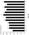
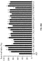
- FIGS. 1A and 1B showed that the compounds of the disclosure showed an absence of significant inhibition on the proliferation of human hepatic stellate cells at a concentration of 15 ⁇ M, and the survival rate was significantly higher than that of the parent compound AD.
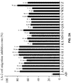
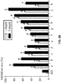
- FIGS. 1A , 1B , 2A , 2B and 3 the results show that, the compound of the disclosure show significantly inhibition compared with AD on the migration of LX-2 cells at a non-toxic concentration, and has a stronger inhibitory effect on the migration of human hepatic stellate cells, and has a higher safety index.
- the compounds of the disclosure reduce the degree of liver fibrosis in SD rats induced by carbon tetrachloride (CCl 4 )
- the liver fibrosis model of rat induced by CCl 4 is one of the most commonly used animal models of liver fibrosis. This model shows great similarities with human liver fibrosis including morphology and pathophysiology. This rat model can thus serve as a good model for well simulating the pathological changes of human liver fibrosis, performing the features of toxic and drug-induced liver fibrosis, as well as the similar pathological features after hepatitis B virus (HBV) infection.
- HBV hepatitis B virus
- animals showed abnormal liver function similar to human liver cirrhosis, furthermore, the molecular mechanism of fibrosis, serum markers after injury, and pathological changes of liver tissue were also very similar to humans. Therefore, the model of liver fibrosis induced by CCl 4 is widely used to study the pathogenesis of liver fibrosis, the screening of anti-fibrosis drugs, and the mechanism of anti-fibrosis drugs.
- SPF grade Sprague Dawley (SD) rats healthy, male, body weight 200 ⁇ 20 g, were purchased from Hunan Silaike Jingda Laboratory Animal Co Ltd. (License No. SCXK (Xiang) 2011-0003).
- Andrographolide was produced by Sichuan Shifang Jinxin Biotechnology Co., Ltd. (Batch No.: 120822), purity greater than 99%; the compounds of the disclosure were synthesized in a laboratory, purity greater than 99%; Pharmaceutical grade sodium carboxyl methyl cellulose (CMC-Na) was produced byAnhui Sunhere Pharmaceutical Excipients Co., Ltd. (Batch No.: 131114); Marine Capsules, was produced by Zhengda Tianqing Pharmaceutical Co., Ltd. (Batch No.: SDA License No.: GUOYAOZHUNZI H20010763). Silibinin Capsules, was produced by Tianjin Tianshili S hengte Pharmaceutical Co., Ltd.
- S D rats were fed ad libitum for 3 days, and then randomly divided into normal group and model group.
- the dosages of andrographolide, oxymatrine, silibinin and the compound A of the disclosure were both 20 mg/kg, and the doses of other compounds were the same molar mass as compound A, and the model group and the normal group were given an equal amount of 0.5% CMC -Na.
- modeling and administration were carried out simultaneously, furthermore, the modeling was stopped at the 7th and 8th weeks, while the administered continued to carry out Drugs were administrated by gavage on an empty stomach in the morning every day, and were given daily for 10 days followed by 1-day break.
- the bedding of the rats should be replaced at the first 8 hours of the last gavage, strictly fasting with nothing but water.
- an anesthetic of 3% pentobarbital sodium (2 mL/kg) was intraperitoneally injected.
- the liver was quickly excised after blood collection.
- the collected blood was allowed to stand in an incubator at 37eC for 45 min, and then the upper serum was collected by centrifugation at 3500 rpm for 15 min at 4éC.
- the liver of the lower left lobe of the rat was fixed in 10 times volume of 4% paraformaldehyde fixative, and the fixative was changed after 24 h.
- Pathological sections were obtained after fixation, and Masson's trichrome staining was then used to observe the degree of liver fibrosis.
- Image-Pro Plus software performed semi-quantitative analysis of fibrosis histology on the photographs of Masson stained sections.
- the relative collagen area (average area of the administration group - average area of the normal group) /(average area of the model group - average area of the normal group) B 100%, and the results were shown in FIGS. 4 and 5 .
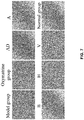
- the expression of -SMA (a marker of the degree of HSC activation) in liver tissue was evaluated by immunohistochemistry, and the positive expression was quantified using Image-Pro Plus, the results were shown in FIGS. 6 and 7 .
- Data were processed and analyzed using S PSS 17.0 statistical software. Data were expressed as mean ⁇ standard deviation (X ⁇ S); There were significant differences between groups when P ⁇ 0.05.
- FIGS. 4 , 5 , 6 and 7 showed that the compounds of the disclosure significantly reduce the degree of fibrosis in liver tissue of animals.
- the collagen area of the liver tissue of the model group of the disclosure was significantly reduced compared with AD, with more prominent effect compared with the positive reference silibinin and oxymatrine.
- the compound of the disclosure significantly down-regulated the expression level of -SMA in liver tissue, and the difference was significant compared with AD, and the effect was even better than that of the drug in the positive control group, indicating that the anti-fibrosis effect was related to the inhibition of hepatic stellate cell activation.
- Compounds of the disclosure significantly reduce the degree of liver fibrosis induced by porcine serum in Wistar rats.
- Immune hepatic fibrosis is mainly caused by the immune response of the liver itself, and liver fibrosis caused by other factors (virus, alcohol, schistosomiasis and certain chemicals) were often accompanied by different degrees of immune response.
- xenogeneic serum or protein porcine serum g, schistosomiasis, human or bovine serum albumin, etc.
- heterologous serum mainly activates hepatic stem cells through MHCII molecules and inflammatory factors to produce liver damage caused by immune response.
- liver fibrosis and immune response continue, which can well reproduce the possible processes and mechanisms of liver fibrosis caused by human autoimmune liver injury that other models cannot simulate, and has great advantages in the screening and evaluation of immune drugs against liver fibrosis.
- Silibinin Capsules was produced by Tianjin Tianshili Shengte Pharmaceutical Co., Ltd. (Batch No.: SDA License No.: GUOYAOZHUNZI H20040299). Pig serum, was produced by Guangzhou Ruite Biotechnology Co., Ltd. (Batch No.: 160608).
- Other test drugs and compounds were the same as in Example 2, and other reagents were commercially available analytical grades. The drugs were formulated as a 0.5% sodium carboxymethylcellulose (CMC-Na) suspension.
- Wistar rats were fed ad libitum for 3 days, they were weighed individually and then randomly divided into six rats per group. Except for the normal group, other rats were intraperitoneally injected with porcine serum, 1 mL/time, 2 times every week, lasting for six weeks. The same dose of normal saline was injected to the rats of normal group. The experiment was finished until the end of 8th weeks. Drugs were administrated by gavage on an empty stomach in the morning every day, and were given daily for 10 days followed by 1-day break. The preventive administration group (H: 2.5 mg/kg) was administered from the day of modeling.
- the rats in 50 mg/kg silibinin group, 20 mg/kg andrographolide group, 5 mg/kg compound H group and 10 mg/kg compound H group were administered beginning the 5th week, while the rats in the 20 mg/kg compound H group was administered from the 7th week.
- the control group and the model group were administered with 0.5% CMC-Na via gavage.
- the methods of rat liver collection and fixation, serum preparation, pathological section preparation, and Masson's trichrome stain were the same as in Example 2, and the results were shown in FIGS. 8 and 9 .
- the expression of -SMA (a marker of the degree of HSC activation) in liver tissue was evaluated by immunohistochemistry, and the positive expression was quantified using Image-Pro Plus, the results were shown in FIGS. 10 and 11 .
- Data were processed and analyzed using SPSS 17.0 statistical software. Data were expressed as mean ⁇ standard deviation (X ⁇ S); There were significant differences between groups when P ⁇ 0.05.
- FIGS. 8 , 9 , 10 , and 11 showed that the compounds of the disclosure produced good anti-fibrosis effects on the pig serum-induced liver fibrosis model.
- Prophylactic or therapeutic administration both exhibited significantly reduced degree of liver fibrosis in rats, whose effect was better than that of the AD and 50 mg/kg silibinin group.
- Compound H group also exhibited good therapeutic effect and although the rats in this group were administered from the 6th week of modeling and this lasted for two weeks.
- the compounds of the disclosure significantly down-regulated the expression level of -SMA in liver tissue, and the difference was significant compared with AD, and the effect was even better than that of the positive drug, indicating that the anti-fibrosis effect was related to the inhibition of hepatic stellate cell activation.
- the compound of the disclosure significantly reduces the degree of liver fibrosis in S D rats induced by BDL
- SPF grade SD rats healthy, male, body weigh 200 ⁇ 20 g, were purchased from Hunan Silaike Jingda Laboratory Animal Co Ltd. (License No. SCXK (Xiang) 2016-0002).
- Ursodeoxycholic acid was produced by Shanghai Xinyi Pharmaceutical Co., Ltd. (Batch No.: SDA License No.: GUOYAOZHUNZI H31021875).
- Other test drugs and compounds were the same as in Example 2, and other reagents were commercially available analytical grades.
- the drugs were formulated as a 0.5% sodium carboxymethylcellulose (CMC-Na) suspension.
- S D rats were fed ad libitum for 3 days, they were weighed individually and were randomly divided into six groups including a sham-operated control group, a model group, two AD control groups (5 mg/kg and 20 mg/kg), an ursodeoxycholic acid control group (25 mg/kg), and a compound H group, six rats per group.
- the sham operation group and the model group were administered with 0.5% CMC-Na via gavage, and the other administration groups were administered 0.5% CMC-Na suspension of the corresponding drug, and the administration lasted for 4 weeks.
- the bedding of the rats should be replaced at the first 8 hours of the last gavage, strictly fasting with nothing but water.
- the rats were intramuscularly injected with 80,000 U/mu of penicillin 2 h before gavage, and 3% sodium pentobarbital (2 mL/kg) was intraperitoneally injected 1 h after gavage. After limb immobilization in a supine position, the anesthetized rats were prepared. The abdomen of the rats was shaved and then swabbed with iodine to sterilize the skin regions. After spreading of surgery cloth, the abdomen was opened along the abdominal wall in the midline.
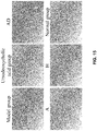
- FIGS. 12 , 13 , 14 , and 15 showed thatthe anti-liver fibrosis effect of the compound of the disclosure was significantly stronger than that of AD and positive drug treatment in the model of rat liver fibrosis induced by bile duct ligation. Furthermore, it was observed that the compound of the disclosure significantly down-regulated the expression level of -S MA in liver tissue, and the difference was significant compared with AD, and the effect was better than that of the positive drug. It was indicated that its anti-fibrosis effect was related to the inhibition of hepatic stellate cell activation.
- collagen is an important component of the sinus interstitial membrane, which is mainly C-I and C-IV.
- C-I and C-IV When the liver was damaged, a large amount of collagen and glycoprotein were transcribed, translated and assembled, which is mainly LN, C-Iand C-III. Serum LN, C-I, PC-III and C-IV levels were important indicators for the clinical diagnosis of patients with fibrosis.
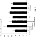
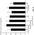
- the levels of LN, PC-III and C-IV in the serum and the levels of C-I in the liver tissue of the animals were determined by ELISA using the compound H of the disclosure as a representative.
- the changes in tissue and serum collagen index were shown in FIGS. 16-19 .
- Data were processed and analyzed using SPSS 17.0 statistical software. Data were expressed as mean ⁇ standard deviation (X ⁇ S); There were significant differences between groups when P ⁇ 0.05.
- the treatment group significantly reduced the LN content in the liver tissue, and the high dose group of the compound H of the disclosure was significantly different from the positive drug oxymatrine group.
- Oxymatrine, AD and the compounds of the disclosure all significantly reduced the content of C-I in liver tissue, compound H decreased the C-I level in a dose-dependent manner; low-dose and high-dose compound H group significantly reduced the level of PC-III in the serum.
- FIGS. 16 , 17 , 18 , and 19 showed that the compound of the disclosure significantly enhances the inhibitory activity against PC-III while maintaining the significant decrease in the levels of LN, C-I and C-IV of the parent compound AD.
- the compound of the disclosure lowered LN, PC-III, C-IV to a normal level at a dose of 20 mg/kg.
- the degree of down-regulation of PC-III and C-IV by compound H at the dose of 5 mg/kg was comparable to that of AD at the dose of 20 mg/kg.
- HSC Activation of HSC can induce macrophages to produce large amounts of TNF- , continue to participate in the differentiation of HSC, and also enhance the inflammatory response in the liver.
- IL-6 which is one of the pro-fibrotic factors, is involved in complex physiological processes such as inflammation, lipid peroxidation, apoptosis and regeneration in liver tissue, and is also an effector molecule downstream of NF- ⁇ B.
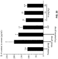
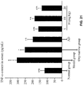
- the level of IL-6 and TNF- in the serum of the animal was determined by E LISA using the compound H of the disclosure as a representative, the results were shown in FIGS. 20 and 21 .
- Data were processed and analyzed using SPSS 17.0 statistical software. Data were expressed as mean ⁇ standard deviation (X ⁇ S); There were significant differences between groups when P ⁇ 0.05.
- FIGS. 20 and 21 showed that three doses of compound H significantly reduced the levels of TNF- and IL-6 in the serum of rats with hepatic fibrosis, while the effect of the high dose group was the most significant.
- the levels of IL-6 and TNF- in the serum at the dose of 20 mg/kg were comparable to those in the normal group.
- SOD and MDA are important indicators for evaluating lipid peroxidation.
- SOD is an antioxidant that inhibits lipid peroxidation caused by free radical initiation, and also scavenges free radicals to protect the integrity of biofilms, and is a sensitive indicator of antioxidant capacity in the body. Lipid peroxidation produces a large amount of MDA, which is proportional to the degree of peroxidative damage to tissues.
- the level of SOD and MDA in the liver tissue of the animal was determined by the compound H of the disclosure, the results were shown in FIGS 22 and 23 .
- Data were processed and analyzed using SPSS 17.0 statistical software. Data were expressed as mean ⁇ standard deviation (X ⁇ S); There were significant differences between groups when P ⁇ 0.05.
- Compound of the disclosure significantly improved the inflammatory state of liver tissue in SD rats with liver fibrosis induced by carbon tetrachloride (CCl 4 )
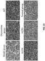
- the compound H of the disclosure was selected as a representative, and H&E staining was used to observe the improvement of the immune inflammatory state of the liver tissue by the compound H, and the results were shown in FIGS. 24 and 25 .
- Data were processed and analyzed using SPSS 17.0 statistical software. Data were expressed as mean ⁇ standard deviation (X ⁇ S); There were significant differences between groups when P ⁇ 0.05.
- FIGS. 24 , and 25 showed that the structure of liver lobule of the normal group was intact, the hepatic cords were arranged in neat order, no abnormal changes observed, and the vascular area was normal. Hepatocytes showed no deformation, necrosis, or formation of fibrous connective tissue. Rats in the model group showed chaotically arranged hepatic cell cords, a significantly increased nuclear-to-plasma ratio, accompanied by some visible, flaky and focal necrotic areas, severe lymphocytic infiltration, and severe damage to the structure of hepatic lobule. The liver injury of the animals in the drug-administered group was improved to some extent compared with the model group.
- the compound H of the disclosure was most prominent in the middle and high-dose groups, the hepatocytes were only slightly enlarged, and the structure of the hepatic cell cord was normal, which was significantly better than the positive drug oxymatrine treatment group and the AD treatment group.
- Compound of the disclosure inhibits TGF- 1-induced conversion of human alveolar type IIA549 cells to mesenchymal cells
- Type II alveolar epithelial cells in the alveoli are stimulated by cytokines such as inflammatory mediators and growth factors.
- cytokines such as inflammatory mediators and growth factors.
- the cell morphology changes from cobblestone to fusiform, completing epithelial mesenchymal transition (EMT) and functioning as an interstitial cell.
- EMT epithelial mesenchymal transition
- Collagen fibers are further synthesized, but a large amount of collagen fiber deposition can aggravate the course and severity of interstitial pulmonary fibrosis.
- the human hepatic stellate A549 cell (provided by Beijing Beina Chuanglian Biotechnology Institute) was compared with andrographolide (AD) to determine the anti-fibrosis effect of the compound of the disclosure in vitro.
- A549 cells were cultured in RPMI1640 medium containing 10% (V/V) fetal bovine serum, 100 ⁇ g/mL streptomycin, and 100 IU/mL penicillin respectively, and then were incubated in an incubator at 37éC and 5% CO 2 , humidified atmosphere.
- A549 cells in logarithmic growth phase were digested with 0.25% (w/v) trypsin, and diluted into 2.5B10 4 /mL cell suspension with RPMI1640 medium containing 10% (v/v) fetal bovine serum. After each 96-well plate was filled with 200 ⁇ L cell suspension, the plates were placed in an incubator at 37éC and 5% CO 2 for 24 h. The medium containing different concentrations of the drug was added, and the maximum final concentration of the drugs was 30.00 ⁇ M, and each treatment was repeated in 4 wells. Following 48 h of incubation, other steps were the same as in the Example 1. The results were averaged, as shown in FIGS. 26A and 26B .
- A549 cells in logarithmic growth phase were digested with 0.25% (w/v) trypsin, and diluted into 2.5B10 4 /mL cell suspension with RPMI1640 medium containing 10% (v/v) fetal bovine serum. After each 96-well plate was filled with 200 ⁇ L cell suspension, the plates were placed in an incubator for 24 h in order to complete the cell fusion. The original medium was discarded, and the medium without serum was added and re-synchronized for 24 h in order to streaking, and the cells were washed twice with PBS. Following the aspiration of 200 ⁇ L of medium containing the TGF- 1 (5 ng/mL), photographs were taken under a microscope.
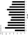
- FIGS. 26A and 26B showed that the cytotoxic activity of the compounds of the disclosure againstA549 cells was not enhanced at a concentration of 30.00 ⁇ M (except for f, g, h, k of 3.00 ⁇ M) compared to AD.
- FIGS. 27A and 27B showed that the compound of the disclosure can significantly inhibit the epithelial-mesenchymal transition ofA549 cells at a non-toxic concentration, and has a stronger inhibitory effect on the mesenchymal transition of human type II alveolar epithelial cells compared to AD, and the safety index was higher.
- the compounds of the disclosure reduce the degree of pulmonary fibrosis induced by bleomycin in KM mice
- Pulmonary fibrosis is a lung injury caused by a variety of reasons, illustrating that the pathogenesis of pulmonary fibrosis is very complicated.
- Different pathogenic factors stimulate inflammation and immune response, involving a variety of cells including vascular endothelial cells, alveolar epithelial cells, fibroblasts and macrophages, and the interaction of various cytokines and inflammatory mediators.
- Bleomycin is an alkaline glycopeptide anticancer antibiotic, and one of its serious toxic adverse reactions is pulmonary fibrosis. It has been confirmed in animal experiments that the histopathological changes of pulmonary fibrosis induced by bleomycin are very similar to those of human pulmonary fibrosis, and are commonly used as models for studying pulmonary fibrosis.
- mice After KM mice were fed ad libitum for 3 days, they were weighed individually and were randomly divided into six groups including a sham-operated control group, a model group, a prednisone control group (5 mg/kg), an AD group (250 mg/kg) and two compound H groups (62.5 mg/kg and 250. mg/kg), fifteen mice per group.
- the mice were anesthetized with intraperitoneal injection of 4% sodium pentobarbital (2 ml/kg). After the immobilization in a supine position, the anesthetized mice were prepared. The necks of the mice were shaved and then swabbed with iodine to sterilize the skin regions.
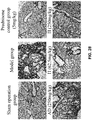
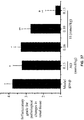
- mice The bedding of the mice should be replaced at the first 12 hours of the last gavage, strictly fasting with nothing but water. After administration for 1.5 h, the whole blood from the eyeball was collected. Use of cervical dislocation to euthanize the mice. The lungs of the mice were fixed in 4% paraformaldehyde fixative after photographing. Serum preparation, Masson's trichrome staining and statistical methods of results were the same as in Example2, Masson's trichrome staining and relative collagen area results were shown in FIGS. 28 and 29 .
- FIGS. 28 and 29 showed that for the bleomycin-induced mice pulmonary fibrosis model, the compound H of the disclosure significantly reduced the fibrotic area of lung tissue of pulmonary fibrosis KM mice, and the effect was significantly stronger than AD.
- the compound of the disclosure significantly improves the inflammatory state of lung tissue in Boremycin-induced pulmonary fibrosis in KM mice
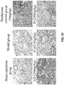
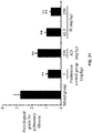
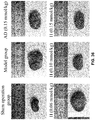
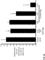
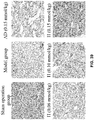
- the compound H of the disclosure was selected as a representative, and H&E staining was used to observe the improvement of the inflammatory state of the lung tissue by the compound H, and the staining results and pathological scores were shown in FIGS. 30 and 31 .
- Data were processed and analyzed using SPSS 17.0 statistical software. Data were expressed as mean ⁇ standard deviation (X ⁇ S); There were significant differences between groups when P ⁇ 0.05.
- FIGS. 30 and 31 showed that in the sham operated group, the structure of lung tissue was intact, the lung interval was uniform, no abnormal changes occurred, and no inflammatory cell infiltration that was visible to the naked eye. But the alveolar structure of mice in the model group was disordered, the lung interval became thicker, and the pulmonary interstitial fibrosis material segmented the alveoli to form reconstructed alveoli, and a large number of inflammatory cells infiltrated.
- the lung tissue damage of the animals in the drug-administered group was improved to some extent, especially in the high-dose treatment group of the compound H of the disclosure, the alveolar structure of the lung tissue was basically intact and normal, and inflammatory cell infiltration was difficult to identify with the naked eye, illustrating that efficacy of drug-administered group was significantly better than the AD treatment group.
- Compound of the disclosure inhibits TGF- 1-induced conversion of human proximal tubular epithelial cells HK-2 to mesenchymal cells
- tubular epithelial cells can transdifferentiate to fibroblasts and express their fibroblast-specific protein (FSP1).
- FSP1 fibroblast-specific protein
- Epithelial-mesenchymal transition of tubular epithelial cells is one of the important pathogenesis of renal interstitial fibrosis. Therefore, after TGF- 1 stimulation, the anti-renal fibrosis effect of the compound of the disclosure was evaluated by morphological observation.
- HK-2 Human proximal tubular epithelial cells HK-2 (provided by the China Center forType Culture Collection) were used to study the in vitro anti-renal fibrosis effect of the compounds of the disclosure in comparison with andrographolide (AD).HK-2 cells were cultured in DME M-F12 medium containing 10% (V/V) fetal bovine serum, 100 ⁇ g/mL streptomycin, and 100 IU/mL penicillin respectively, and then incubated in an incubator at 5% CO 2 and 37éC, humidified atmosphere.
- DME M-F12 medium 10% (V/V) fetal bovine serum, 100 ⁇ g/mL streptomycin, and 100 IU/mL penicillin respectively
- HK-2 cells in logarithmic growth phase were digested with 0.25% (w/v) trypsin and 0.02% EDTA (W/V), and diluted into 7.0B10 4 /mL cell suspension with DMEM-F12 medium containing 10% (v/v) fetal bovine serum. After each 96-well plate was filled with 200 ⁇ L cell suspension, the plates were placed in an incubator at 37éC and 5% CO 2 for 24 h. The medium containing different concentrations of the drugs were added respectively, and the maximum final concentration of the drugs were 30.00 ⁇ M, and each treatment was repeated in 4 wells. Following 48 h of incubation, other steps were the same as in Example 1. The results were averaged, as shown in FIGS. 32A and 32B . 3. Effect of drugs on the morphology of HK-2 cells after TGF- 1 stimulation
- HK-2 cells in logarithmic growth phase were digested with 0.25% (w/v) trypsin and 0.02% E DTA (W/V), and diluted into 5.0B10 6 /mL cell suspension with DMEM-F12 medium containing 10% (v/v) fetal bovine serum. After each 96-well plate was filled with 200 ⁇ L cell suspension, the plates were placed in an incubator for 24 h in order to generate a single layer of cells. The original medium was discarded, and the cells were washed twice with PBS, and the medium without serum was added.
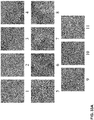
- FIGS. 32A and 32B showed that the inhibition of HK-2 cell proliferation by the compounds of the disclosure was significantly reduced at a concentration of 30.00 ⁇ M compared to AD.
- Tables 1A and 1B and FIGS. 33A and 33B showed that the compounds of the disclosure can significantly inhibit TGF- 1-induced conversion of human proximal tubular epithelial cells HK-2 to mesenchymal cells at a non-toxic concentration.
- the inhibition of HK-2 cells to mesenchymal transition was stronger and the safety index was higher compared with AD.
- test concentrations were 0.08-1.25 ⁇ M.
- Control There was an interaction between epithelial cells, and the intercellular linkage was tight, and the cells were arranged in an interlocking fashion like paving stones. Extremely strong (inhibition): the cells are almost the same as the control. No spindle cells are found in the visual field, the intercellular recovery interaction, and the morphology restores its typical paving stone shape. Strong (inhibition): the invasiveness of the cells was inhibited, and the cells were tight, and the cell state was almost completely recovered, and the spindle fibroblasts were rare. Medium (inhibition): the cell density becomes larger and most of the cells were still in a cubic state.
- test concentration is 0.08-1.25 ⁇ M
- Control There was an interaction between epithelial cells, and the intercellular linkage was tight, and the cells were arranged in an interlocking fashion like paving stones. Extremely strong (inhibition): the cells were almost the same as the control. No spindle cells were found in the visual field, the intercellular recovery interaction, and the morphology restores its typical paving stone shape. Strong (inhibition): the spread of the cells was inhibited, and the cells were tightly arranged; the cell state was completely recovered; the spindle fibroblasts were rare. Medium (inhibition): the cell density becomes larger and most of the cells were still in a cubic state.
- UUO Unilateral Ureteral Obstruction
- the model is characterized by accumulation of cellular components in the tubulointerstitial, differentiation/proliferation of fibroblast, an increase of ECM deposition, and tubular atrophy.
- the modeling method is simple, the modeling success rate is 100%, the lesions are uniform, and the reproducibility is good, which can cause fibrosis in a short period of time.
- the UUO model is widely used in the study of the mechanism of renal interstitial fibrosis and the evaluation of the therapeutic effect of improving renal interstitial fibrosis.
- S D rats were fed ad libitum for 3 days, they were weighed individually and were randomly divided into six groups including a sham-operated control group, a model group, an AD group (0.15 mmol/kg), and two compound H groups (0.06 mmol/kg, 0.10 mmol/kg and 0.15 mmol/kg), four rats per group.
- the preoperative preparation and anesthesia method were the same as in Example 4.
- the anesthetized rats were prepared. The hair from the lower edge of the sternum to the hind limbs was shaved, and then the surgery cloth was spread out and swabbed with iodine to sterilize the skin regions.
- the sham-operated group only freed the ureter but did not ligature or segment
- the rats were administered by gavage at the same time.
- the administration method was the same as in Example2, and the experiment was over after 14 days of administration.
- the bedding of the rats should be replaced at the first 12 hours of the last gavage, strictly fasting with nothing but water.
- 3% barbital sodium (2 mL/kg) anesthetic were intraperitoneally injected to rats. Following the collection of blood, the left kidney was quickly and completely dissected. The kidney weigh and kidney size were subsequently measured.
- the kidney of rats was fixed in 4% paraformaldehyde fixative after photographing. Masson's trichrome staining and statistical methods of results were the same as in Example2, Masson's trichrome staining and relative collagen area results were shown in FIGS. 34 and 35 .
- FIGS. 34 and 35 showed that on the UUO model, the representative compound H of the disclosure significantly reduced the degree of renal fibrosis, improved the structure of the diseased kidney, and had a significantly stronger effect than AD.
- the compound of the disclosure significantly improved the inflammatory state of renal tissue in renal fibrosis of SD rats induced by unilateral ureteral ligation
- the compound H of the disclosure was selected as a representative, and the improvement of kidney tissue inflammatory state by the compound H of the disclosure was observed by observing anatomical kidney and H&E-stained pathological section.
- the staining results and pathological scores were shown in FIGS. 36-39 .
- Data were processed and analyzed using SPSS 17.0 statistical software.
- Data were processed and analyzed using SPSS 17.0 statistical software.
- Data were expressed as mean ⁇ standard deviation (X ⁇ S); There were significant differences between groups when P ⁇ 0.05.
- FIGS. 36 , 37 , 38 , and 39 showed that the sham operation group, the surface of the kidney tissue was moist and shiny, the structure of the glomerulus was intact, the renal tubules were tight and compact, and there was no visible lesion.
- the kidney tissue became swollen, with a large amount of effusion in the middle and adhesion to the surrounding tissues.
- the renal interstitial fibrosis material was used to compress the renal tubules. The tubules were severely atrophied and a large number of inflammatory cells infiltrate in the renal interstitium.
- the renal tissue damage of the animals in the drug-administered group was improved to some extent, especially in the high-dose treatment group of the compound H of the disclosure, the surface of the kidney tissue was smooth and moist, non-adhesive, and the kidney was slightly inflated. There was no effusion in the middle, the structure of the glomerulus was basically intact, and the atrophy of the renal tubule was not obvious, and its efficacy was significantly better than that of the AD treatment group.
- the compound of the disclosure inhibits angiotensin II (AngII)-induced proliferation of human primary myocardial fibrosis cells HCFB
- cardiac fibroblasts are the main effector cells of myocardial fibrosis, and they proliferate when stimulated by active substances such as AngII, and their phenotypes are transformed into myofibroblasts that secrete extracellular matrix. Therefore, the cell inhibition of AngII after stimulation of primary cardiac fibroblast HCFB was examined by MTT assay to evaluate the anti-myocardial fibrosis effect of Compound H of the disclosure.
- Human primary cardiac fibroblast HCFB (provided by Shangcheng Beina Chuanglian Biotech Co., Ltd.) was compared with andrographolide to study the in vitro anti-myocardial fibrosis effect of the compound H of the disclosure.
- HCFB cells were cultured in H-DME M medium containing 10% (V/V) fetal bovine serum (GIBCO, USA: Cat No. 302220F), 100 ⁇ g/mL streptomycin, and 100 IU/mL penicillin respectively, and then incubated in an incubator (Binder, Germany) at 5% CO 2 and 37éC, humidified atmosphere.
- HCFB cells in logarithmic growth phase were digested with 0.25% (w/v) trypsin and 0.02% E DTA, and diluted into 5.0B10 4 /mL cell suspension with H-DMEM medium containing 10% (v/v) fetal bovine serum.
- H-DMEM medium containing 10% (v/v) fetal bovine serum.
- the plates were placed in an incubator at 37éC and 5% CO 2 for 24 h. The medium containing different concentrations of the compound AD or H was added.
- each 96-well plate was added with 20 ⁇ L MTT (5 mg/mL) and continued to incubate for 4 h.
- HCFB cells in logarithmic growth phase were digested with 0.25% (w/v) trypsin and 0.02% EDTA, and diluted into 5.0B10 4 /mL cell suspension with H-DMEM medium containing 10% (v/v) fetal bovine serum. After each 96-well plate was filled with 200 ⁇ L cell suspension, the plates were placed in an incubator for 24 h in order to generate a single layer of cells. The original medium was discarded, and the cells were washed twice with PBS, and the medium without serum was added. After 24 h of re-synchronized, 200 ⁇ L of DMEM-F12 medium containing different concentrations of the test compound and the stimulating factorAngII (10 -7 mol/L) was added.
- H-DME M medium containing 0.5% DMSO was used as a negative control
- H-DME M medium containing stimulating factor AngII (10 -7 mol/L) and 0.5% DMSO was used as a positive control.
- the cell survival ratio in each well was calculated, the results were shown in FIG. 41 .
- Data were processed and analyzed using S PSS 17.0 statistical software. Data were expressed as mean ⁇ standard deviation (X ⁇ S); There were significant differences between groups when P ⁇ 0.05.
- FIG. 40 showed that the compound H of the disclosure showed no significant inhibitory effect on the proliferation of human primary cardiac fibroblast HCFB cells at a concentration of 15.0 ⁇ M.
- FIG. 41 showed that the compound H of the disclosure could significantly inhibit the proliferation of HCFB by AngII at a non-toxic concentration, and had a stronger inhibition effect on human HCFB proliferation and a higher safety index than AD.
Landscapes
- Health & Medical Sciences (AREA)
- Chemical & Material Sciences (AREA)
- Organic Chemistry (AREA)
- Public Health (AREA)
- Life Sciences & Earth Sciences (AREA)
- Veterinary Medicine (AREA)
- General Health & Medical Sciences (AREA)
- Pharmacology & Pharmacy (AREA)
- Medicinal Chemistry (AREA)
- Animal Behavior & Ethology (AREA)
- Epidemiology (AREA)
- Engineering & Computer Science (AREA)
- Nuclear Medicine, Radiotherapy & Molecular Imaging (AREA)
- General Chemical & Material Sciences (AREA)
- Chemical Kinetics & Catalysis (AREA)
- Bioinformatics & Cheminformatics (AREA)
- Physical Education & Sports Medicine (AREA)
- Biomedical Technology (AREA)
- Gastroenterology & Hepatology (AREA)
- Pharmaceuticals Containing Other Organic And Inorganic Compounds (AREA)
Abstract
Description
- The disclosure relates to an andrographolide derivative and a method of using the same for treatment or prevention of fibrosis, and more particularly to a 15-methylene-14-deoxy-11,12-dehydro-andrographolide derivative.
- Tissue fibrosis is a chronic condition that occurs in the liver, lungs, heart, kidney and other organs. One-third of the world's people die from tissue fibrosis and the resulting organ failure. A series of cellular reactions take place at the injury site after the tissue has been damaged, which results in the excessive accumulation of extracellular matrix components and the tissue fibrosis. If highly progressive, the fibrotic process eventually leads to organ dysfunction and death. In the medical treatment of fibrotic diseases, although there are interferon, phosphatidylcholine, verapamil, glucocorticoids and other clinical drugs, they are frequently accompanied by some side effects. Therefore, the research and development of anti-fibrotic drugs based on Chinese herbal medicine has a broad market prospect
- Chronic liver disease is a major type of disease that seriously harms human health. Liver fibrosis is a common pathological process in the development of various chronic liver diseases to hepatic cirrhosis, and is generally caused by the excessive accumulation of extracellular matrix (ECM) components in the liver that results from wound repair in the body. Factors causing liver fibrosis include viral hepatitis, metabolic diseases, autoimmune liver diseases, as well as chronic liver diseases caused by alcohol, drug, etc. According to existing reports, it is widely believed that the process of liver fibrosis is reversible (Yu Hui, J iang Feng, Medical Information, 2015.28(7): 334-335), and liver fibrosis can be ameliorated by treating primary disease, inhibiting inflammatory activity, inhibiting oxidation-reduction reactions, decreasing activity of hepatic stellate cells and regulating epithelial-mesenchymal transition, while the mechanism of liver fibrosis is complicated and the regulation mechanism that is affected by many factors.
- In recent years, the incidence and mortality of pulmonary fibrosis have been on the rise. Pulmonary fibrosis (PF) is the final outcome of the development, evolution, and scarring of many lung diseases, resulting in existing diverse causes of pulmonary fibrosis. Many chronic lung diseases, including asthma, bronchiectasis, chronic obstructive pulmonary disease, tuberculosis, lung cancer, and interstitial lung disease, are accompanied by pathological changes in fibrosis. The pathological features of pulmonary fibrosis are persistent alveolar damage caused by a diffuse inflammation of lung tissue and the repeated destruction, repair, reconstruction and excessive accumulation of extracellular matrix components (Li Xianwei et al., Chinese J ournal of Applied Physiology, 2017.33(1): 16-21). For a variety of lung diseases such as idiopathic pulmonary fibrosis, respiratory distress syndrome, eosinophilic granuloma, the degree of fibrosis existent in the lung tissue determines the clinical recovery of the disease. Alveolitis typically occurs after early lung injury, the inflammation and abnormal repair further lead to the proliferation of pulmonary interstitial cells, and gradually form a fibrotic lung. The clinical manifestations include dyspnea and respiratory failure. In the past 20 years, the incidence of idiopathic pulmonary fibrosis has shown a significant increase. The average survival time idiopathic pulmonary fibrosis after diagnosis is about 3 years, and the 5-year survival rate is 30% to 50%, illustrating that the conventional treatment effect is in the extremely unsatisfactory condition.
- Renal fibrosis is caused by various pathogenic factors such as trauma, infection, inflammation, blood circulation disorder, and immune reaction, leading to the intrinsic renal cells are damaged. Subsequently a large amount of deposition and accumulation of collagen occurs in the later stage, causing the renal parenchyma to gradually harden and form scars until the kidney completely lose its organ function. The process of cellular fibrosis and hardening in the kidney is also the process of renal fibrosis. Many acute and chronic kidney diseases are also closely related to the development of tissue fibrosis, especially the changes of renal fibrosis caused by diabetic nephropathy and hypertension.
- Myocardial fibrosis (MF) is a pathological name proposed on the basis of modern medical research. MF refers to the proliferation of cardiac fibroblasts (CFs) in the pathological state, and the abnormal deposition and accumulation of extracellular matrix (ECM) (mainly type I and type III collagen fibers), leading to a decrease in ventricular compliance, and affecting the normal contraction and diastolic function of the heart MF is a common pathological change in the development of a variety of heart diseases to a certain stage, and is also a key reason for ventricular remodeling. In recent years, a large number of studies have shown that MF is closely related to many heart diseases, such as atrial fibrillation, myocardial infarction, chronic heart failure, rheumatic heartvalve disease, etc. And MF is typically triggered by diseases such as rheumatic heart disease, hypertension, myocardial infarction and heart failure. MF also occurs during aging and coronary atherosclerosis. The main pathological manifestations are increased myocardial stiffness and decreased myocardial contractility, reduced coronary flow reserve, and even malignant arrhythmia and sudden death.
- At present, research shows that many signaling pathways related to the development of tissue and organ fibrosis include TGF-∮1/S mad signaling pathway, Wnt/∮-catenin signaling pathway, and phosphatidylinositol 3-kinase/protein kinase (PI3K/Akt) signal pathway, etc. For example, limonin down-regulates -SMA and collagen production by inhibiting the TGF-∮ signaling pathway in the fibrotic lung tissue of a mouse (
CN 105560255 A ). A wedelolactone-sulfate significantly inhibits the proliferation of human embryonic lung fibroblast cells induced by TGF-∮ and reduces the hydroxyproline (HYP) content, thus exerting pharmacological properties of reducing the degree of pulmonary fibrosis (CN 103919769 A ). PI3K inhibitors that act downstream of TGF-∮ are useful in the treatment of pulmonary fibrotic diseases (CN 104093408 A ). There is also a report on patent that discloses the treatment of pulmonary fibrosis using the sesquiterpene-isoastolone derivatives and their salts in Inulae Radix (CN 106496243 A ). On the aspect of anti-renal fibrosis, Ganolactone D can significantly inhibit the phosphorylation of Smad3 protein in tubular epithelial cells induced by TGF-∮1, and has the capacity for a therapeutic effect on renal fibrosis (CN 106220643 A ). The Third Affiliated Hospital of the Third Military Medical University of the Chinese People's Liberation Army found that apigenin mediates the intracellular influx of Ca2+ through TRPV4, activates the AMPK/SITR1 signaling pathway, and inhibits renal fibrosis. - Although there are currently some drugs for treating the above-mentioned fibrotic diseases in clinical practice, the effects of the drugs are not satisfactory. Andrographolide is a relatively simple diterpene lactone, also the main active ingredient in Andrographis paniculata (Burm. f.) Nees of the family Acanthaceae. Andrographolide is mainly used for heat-clearing and detoxication, antibacterial and anti-inflammatory, and also has obvious effects in anti-fibrosis, blocking the early pathway of fibrosis by inhibiting the NF-1B inflammatory pathway (
CN106831669A ). Additionally, modern pharmacological studies have shown that andrographolide and its derivatives have a wide range of pharmacological activities, including anti-inflammatory, anti-viral, anti-tumor, liver-protecting and analgesic activities, and also have a wide clinical application and a remarkable curative effect(LuoYu etal., Chinese Journal of Traditional Chinese Medicine, 2017. 42 (20): 3847-3859). - Andrographolide has a good protective effect against experimental liver injuries in numerous animals. Yao Qing et al. suggested that andrographolide has a protective effect against acute cocaine-induced liver injury, and this mechanism of liver protection may be related to preventing lipid peroxidation and reducing the generation of oxygen free radicals in tissues. Visen PK [J Ethnopharmacol, 1993, 40(2), 131-136] and Handa S [Indian J Med Res, 1990, 92: 284-292] have demonstrated that andrographolide has a protective effect against paracetamol-induced liver injury. Additionally, Handa S's study also showed that andrographolide has a protective effect against galactosamine-induced hepatotoxicity. KapilA [Biochem Pharmacol, 1993, 46(1): 182-185] etal. have demonstrated that andrographolide, andrographoside and neoandrographolide have protective effects against carbon tetrachloride and tert-butyl hydroperoxide induced hepatotoxicity. Singha P [J Ethnopharmacol, 2007, 111(1): 13-21] et al. have demonstrated that andrographolide has a protective effect against ethanol-induced liver and kidney injury in mice. Roy DN [Toxicol Appl Pharmacol, 2011; 250(1): 54-68] et al. have demonstrated that combining andrographolide with D-penicillamine in the treatment of copper poisoning leads to a larger effect than the effect of each drug taken independently. A patent application (
CN201010266185.2) by Ning Guang et al. - The inventors acquired a large number of novel andrographolide derivatives during the preliminary studies (
CN200510107247.4 CN200710053807.1 CN200710053806.7 CN200610017357.6 CN201210358667X - The inventors conducted a series of experiments to study the activities of the synthesized compounds in anti-fibrosis of liver, lung, kidney and myocardium. It has been found that the 15-methylene-14-deoxy-11,12-dehydro-andrographolide derivative having the structure shown in
Formula 1 has remarkable effects in preventing and treating fibrosis of human tissues and organs, and is highly effective and low in toxicity, and has the potential to be developed as an anti-fibrotic drug. Therefore, it is an object of the disclosure to provide 15-methylene-14-deoxy-11,12-dehydro-andrographolide derivatives and their applications in the preparation of anti-fibrotic drugs. - The disclosure provides a 15-methylene-14-deoxy-11,12-dehydro-andrographolide derivative represented by Formula (I):

- When one of R1 and R2 is H, the other can be selected from a methyl, ethyl or propyl, phenyl, 2-methoxyphenyl, 3-methoxyphenyl, 4-methoxyphenyl, 2-fluorophenyl, 2-chlorophenyl, 2-bromophenyl, 3-fluorophenyl, 3-chlorophenyl, 3-bromophenyl, 4-fluorophenyl, 4-chlorophenyl, 4-bromophenyl, 2-fluoro-3-methoxyphenyl, 3-methoxy-4-chlorophenyl, 2,4-difluorophenyl, 2,4-dichlorophenyl, 2,4-dibromophenyl, 2-fluoro-4-chlorophenyl, 2-bromo-4-chlorophenyl, 3-fluoro-4-chlorophenyl, 3-bromo-4-chlorophenyl, 3,4- difluorophenyl, 3,4-dichlorophenyl, 3,4 dibromophenyl, 2-chloro-4-fluorophenyl, 2-bromo-4-fluorophenyl, 3-chloro-4-fluorobenzene, 3-bromo-4-fluorophenyl, 2-fluoro-4-bromophenyl, 2-chloro-4-bromophenyl, 3-fluoro-4-bromophenyl, 3-chloro-4-bromophenyl, 2-methoxy-4-chlorophenyl, 4-n-propylphenyl, 4-isopropylphenyl, 4-isobutylphenyl, 4-(N,N-dimethylamino)phenyl, 3-fluoro-4-(4-morpholinyl)phenyl, 3-fluoro-4-(4-methylpiperazinyl)phenyl, 2-furyl, 2-pyrrolyl, 6-chloro-3-indolyl, 3-indolyl, 5- chloro-3-indolyl, 6-chloro-2-pyridyl, 3-pyridyl; or R1 and R2 are taken together to form a cyclohexyl; R3 and R4 both represent H; or R3 and R4 independently, at each occurrence, represent CH2CH2COOH, CH2CH2CH2CH2COOH, CH2CH2CH2CH2CH2COOH, or CH2CH2CH2CH2CH2CH2CH2COOH; or R3 and R4 both represent COR5, and R5 is a 3-pyridyl or CH2CH2COOH, wherein R3 and R4 are the same or different substituent group.
- When one of R1 and R2 is H, the other can be selected from a phenyl, 2-methoxyphenyl, 3-methoxyphenyl, 4-methoxyphenyl, 2-fluorophenyl, 2-chlorophenyl, 2-bromophenyl, 3-fluorophenyl, 3-chlorophenyl, 3-bromophenyl, 4-fluorophenyl, 4-chlorophenyl, 4-bromophenyl, 2-fluoro-3-methoxyphenyl, 3-methoxy-4-chlorophenyl, 2,4-difluorophenyl, 2,4-dichlorophenyl, 2,4-dibromophenyl, 2-fluoro-4-chlorophenyl, 2-bromo-4-chlorophenyl, 3-fluoro-4-chlorophenyl, 3-bromo-4-chlorophenyl, 3,4- difluorophenyl, 3,4-dichlorophenyl, 3,4 dibromophenyl, 2-chloro-4-fluorophenyl, 2-bromo-4-fluorophenyl, 3-chloro-4-fluorobenzene, 3-bromo-4-fluorophenyl, 2-fluoro-4-bromophenyl, 2-chloro-4-bromophenyl, 3-fluoro-4-bromophenyl, 3-chloro-4-bromobenzene Base, 2-methoxy-4-chlorophenyl; R3 and R4 both represent H; or R3 and R4 independently, at each occurrence, represent CH2CH2COOH, CH2CH2CH2CH2COOH, CH2CH2CH2CH2CH2COOH, or CH2CH2CH2CH2CH2CH2CH2COOH; or R3 and R4 both represent COR5, and R5 is CH2CH2COOH, wherein R3 and R4 are the same substituent group.
- When one of R1 and R2 is H, the other can be selected from a methyl, an ethyl, a propyl, 2-furyl, 2-pyrrolyl, 6-chloro-3-indolyl, 3-indolyl, 5-chloro-3-indolyl, 6-chloro-2-pyridyl, 3-pyridyl, 4-n-propylphenyl, 4-isopropylphenyl, 4-isobutylphenyl, 3-fluoro-4-(4-methylpiperazinyl)phenyl, or R1 and R2 are taken together to form a cyclohexyl group, R3 and R4 both represent H.
- The compound can be:
- A: R1=H, R2=C6H5, R3= R4=H;
- B: R1=H, R2=2-F-C6H4, R3= R4=H;
- C: R1=H, R2=2-Cl-C6H4, R3= R4=H;
- D: R1=H, R2=2-Br-C6H4, R3= R4=H;
- E: R1=H, R2=3-F-C6H4, R3= R4= H;
- F: R1=H, R2=3-Cl-C6H4, R3= R4= H;
- G: R1=H, R2=3-Br-C6H4, R3= R4= H;
- H: R1=H, R2=4-Cl-C6H4, R3= R4= H;
- I: R1=H, R2=4-F-C6H4, R3= R4= H;
- J: R1=H, R2=4-Br-C6H4, R3= R4= H;
- K: R1=H, R2=4-CH3O-C6H4, R3= R4= H;
- L: R1=H, R2=2-CH3O-4-Cl-C6H3, R3= R4= H;
- M: R2=H, R1=2-Br-C6H4, R3= R4= H;
- N: R2=H, R1=3-Cl-C6H4, R3= R4= H;
- O: R2=H, R1=2-F-4-Cl-C6H3, R3= R4= H;
- P: R2=H, R1=2,4-diCl-C6H3, R3= R4= H;
- Q: R2=H, R1= 4-F-C6H4, R3= R4= H;
- R: R2=H, R1=C6H5, R3= R4= H;
- S: R1-H, R2=3-F-4-Cl-C6H3, R3= R4= H;
- T: R1=H, R2= 2,4-diF-C6H3, R3= R4= H;
- U: R1=H, R2= 3,4-diCl-C6H3, R3= R4= H;
- V: R1=H, R2=4-Cl-C6H4, R3= R4= COR5, R5=3-pyridyl;
- W: R1=H, R2=4-Cl-C6H4, R3= R4= CH2CH2COOH;
- X: R1=H, R2=4-Cl-C6H4, R3= R4= COR5, R5= CH2CH2COOH;
- Y: R2=H, R1=4-Cl-C6H4, R3= R4= H;
- Z: R2=H, R1=4-Cl-C6H4, R3= R4= COR5, R5=3-pyridyl.
- a. R1=CH3, R2= CH3, R3=R4=H;
- b. 15-cyclohexylidene-14-deoxy-11,12-dehydro-andrographolide; R3=R4=H;
- c. R1=H, R2=3-F-4-(4-methylpiperazine group)-C6H3, R3=R4=H;
- d. R1=H, R2=

- e. R1=H, R2=

- f. R1=H, R2=

- g. R1=H, R2=

- h. R1=H, R2=

- i. R1=H, R2=

- j. R1=H, R2=

- k. R1=H, R2=

- The compounds a-k are characterized as follows:
- a: C23H32O4; 1H NMR (400 MHz, DMSO-d6) 7.98 (s, 1 H), 6.52 (dd, J = 15.7, 10.6 Hz, 1H), 6.21 (d, J = 15.7 Hz, 1H), 5.50 (s, 1H), 5.12 (s, 1H), 4.30 (s, 1H), 3.94 (d, J = 10.9 Hz, 1H), 3.40 (d, J = 10.9 Hz, 1H), 3.23 (dd, J = 10.9, 4.1 Hz, 1H), 2.48 (d, J = 10.8 Hz, 1H), 2.01 (s, 2H), 1.95 (s,3H),1.94 (s,3H) 1.68 - 1.48 (m, 3H), 1.46 (s, 3H), 1.26 (dd, J = 10.6, 6.2 Hz, 1H), 1.17 (td, J = 13.0, 3.5 Hz, 1H), 1.08 (s, 3H), 0.80 (s, 3H).13C NMR (100 MHz, DMSO-d6) 169.06, 144.93, 137.67, 132.91, 132.57, 126.13, 123.18, 122.65, 122.32, 79.37, 62.99, 60.16, 49.98, 42.11, 38.28, 36.11, 27.82, 23.71, 23.22, 22.68, 18.84, 18.80, 15.99. HRMS (ESI): m/z calcd for C23H32NaO4 [M+Na]+, 395.2198; found, 395.2196.
- b: C26H36O4; 1H NMR (400 MHz, DMSO-d6) 8.02 (s, 1 H), 6.52 (dd, J = 15.7, 10.6 Hz, 1H), 6.21 (d, J = 15.7 Hz, 1H), 5.50 (s, 1H), 5.12 (s, 1H), 4.30 (s, 1H), 3.94 (d, J = 10.9 Hz, 1H), 3.40 (d, J = 11.1 Hz, 1H), 3.22 (dd, J = 10.9, 4.1 Hz, 1H), 2.48 (d, J = 11.0 Hz, 1H), 2.45 - 2.36 (m, 4H), 2.01 (s, 2H), 1.59 (s, 6H), 1.57 - 1.47 (m, 3H), 1.46 (s, 3H), 1.26 (dd, J = 10.6, 6.3 Hz, 1H), 1.17 (td, J = 13.1, 3.7 Hz, 1H), 1.08 (s, 3H), 0.79 (s, 3H). 13C NMR (100 MHz, DMSO-d6) 169.15, 142.54, 137.70, 132.59, 132.57, 130.84, 126.33, 122.65, 122.33, 79.37, 62.99, 60.16, 49.97, 42.11, 38.28, 36.11, 28.94, 28.72, 28.16, 27.82, 27.49, 26.08, 23.71, 23.22, 22.67, 15.99. HRMS (ESI): m/z calcd for C26H36NaO4 [M+Na]+, 435.2511; found, 435.2516.
- c: C32H41FN2O4; 1H NMR(400 MHz, DMSO-d6) 7.69 (s, 1H), 7.52 (dd, J = 14.9, 1.7 Hz, 1H), 7.46 (dd, J =8.6, 1.5 Hz, 1H), 7.06 (t, J = 9.0 Hz, 1H), 6.58 (dd, J = 15.7, 10.6 Hz, 1H), 6.29 (d, J = 15.7 Hz, 1H), 6.26 (s, 1H), 5.50 (s, 1H), 5.12 (d, J = 4.9 Hz, 1H), 4.31 (d, J = 5.0Hz, 1H), 3.94 (d, J = 9.5 Hz, 1H), 3.45 - 3.39 (m, 1H), 3.22 (dd, J = 10.4, 4.9 Hz,1H), 3.14 - 3.06 (m, 4H), 2.51 (s, 1H), 2.49 - 2.44 (m, 4H), 2.22 (s, 3H), 2.01 (s, 2H),1.64 - 1.49 (m, 3H), 1.47 (s, 3H), 1.25 (dd, J = 10.5, 6.3 Hz, 1H), 1.22 - 1.13 (m, 1H),1.08 (s, 3H), 0.80 (s, 3H).13C NMR (100 MHz, DMSO-d6) 168.93, 154.54 (d, J =243 Hz), 147.10, 140.65 (d, J = 9 Hz), 138.68, 137.17, 132.47, 127.89 (d, J = 2 Hz),127.33 (d, J = 8 Hz), 125.61, 122.75, 122.28, 119.53, 119.49, 117.58, 117.36, 112.47(d, J = 2 Hz), 79.37, 62.99, 60.24, 54.94, 49.95, 49.91, 46.20, 42.12, 38.31, 36.21,27.82, 23.71, 23.22, 22.70, 16.02. HRMS (ESI): m/z calcd for C32H42FN2O4 [M+H]+,537.3129; found, 537.3129.
- d: C26H30ClNO4; 1H NMR (400 MHz, DMSO) 7.96 (d, J = 1.6 Hz, 1H), 7.95 (s,1H), 7.82 (s, 1H), 7.48 - 7.42 (m, 1H), 6.89 (dd, J = 15.8, 10.2 Hz, 1H), 6.33 (s, 1H), 6.31 (d, J = 16.8 Hz, 1H), 5.05 (d, J = 4.5 Hz, 1H), 4.76 (s, 1H), 4.44 (s, 1H), 4.14 (d, J = 4.9 Hz, 1H), 3.85 (d, J = 10.8 Hz, 1H), 3.32 - 3.27 (m, 1H), 3.27 - 3.20 (m, 1H), 2.47 (d, J = 10.1 Hz, 1 H), 2.38 (d, J = 13.6 Hz, 1H), 1.99 (dd, J = 12.9, 8.4 Hz, 1H), 1.74 (d, J = 13.2 Hz, 1H), 1.61 (dd, J = 25.5, 7.3 Hz, 2H), 1.49 - 1.30 (m, 2H), 1.24 - 1.14 (m, 2H), 1.10 (s, 3H), 0.80 (s, 3H).13C NMR (101 MHz, DMSO) 168.00, 152.75, 150.45, 148.71, 140.47, 136.64, 128.01, 124.09, 110.84, 78.59, 62.64, 60.74, 53.68, 42.39, 36.22, 27.63, 23.15, 23.00, 15.46.
- e: C26H31NO4: 1H NMR (400 MHz, DMSO) 8.83 (s, 1H), 8.52 (d, J = 3.6 Hz, 1 H), 8.18 - 8.164 (m, 1H), 7.79 (s, 1 H), 7.48 (m, 4.8 Hz, 1 H), 6.87 (dd, J = 16.7, 9.2 Hz, 1H), 6.39 (s, 1H), 6.27 (d, J = 16.6 Hz, 1H), 5.05 (s, 1H), 4.76 (s, 1H), 4.45 (s, 1H), 4.15 (dd, J = 7.4, 2.6 Hz, 1H), 3.85 (dd, J = 10.9, 2.6 Hz, 1H), 3.26 (m, 2H), 2.44 (d, J = 11.9 Hz, 1H), 2.38(d,12Hz, 1H), 1.99 (t, J = 11.0 Hz, 1H), 1.74 (d, J = 13.4 Hz, 1 H), 1.58 - 1.60 (m, 2H), 1.45-1.3(m,2H), 1.23-1.18(overlap, 1H), 1.10 (s, 3H), 0.80 (s, 3H).
- f: C29H32ClNO4; 1H NMR (400 MHz, DMSO) 11.89 (s, 1H), 7.95 (s, 1H), 7.90 (d, J = 8.5 Hz, 1 H), 7.69 (s, 1H), 7.52 (d, J = 1.8 Hz, 1H), 7.17 (dd, J = 8.5, 1.9 Hz, 1 H), 6.77 (dd, J = 15.7, 10.1 Hz, 1H), 6.70 (s, 1H), 6.25 (d, J = 15.7 Hz, 1H), 5.03 (d, J = 4.7 Hz, 1H), 4.75 (s, 1H), 4.46 (s, 1H), 4.14 (d, J = 4.6 Hz, 1H), 3.86 (d, J = 11.1 Hz, 1H), 3.29 - 3.18 (m, 2H), 2.40 (t, J = 11.5 Hz, 2H), 1.99 (t, J = 10.9 Hz, 1H), 1.74 (d, J = 13.4 Hz, 1H), 1.66 - 1.52 (m, 2H), 1.50 - 1.30 (m, 2H), 1.18 (dd, J = 19.8, 7.6 Hz, 2H).1.11 (s, 3H), 0.79 (s, 3H).
- g: C29H33NO4; 1H NMR (400 MHz, DMSO) 11.84 (s, 1H), 7.94 (s, 1H), 7.89 (d, J = 7.6 Hz, 1H), 7.68 (s, 1H), 7.48 (d, J = 7.8 Hz, 1H), 7.18 (ddd, J = 14.8, 13.8, 6.6 Hz, 2H), 6.82 - 6.74 (m, 1H), 6.71 (s, 1H), 6.24 (d, J = 15.7 Hz, 1H), 5.04 (s, 1H), 4.75 (s, 1H), 4.47 (s, 1H), 4.15 (s, 1H), 3.88 (d, J = 10.9 Hz, 1H), 3.33 - 3.20 (m, 2H), 2.43 - 2.33 (m, 2H), 1.99 (dd, J = 13.0, 8.4 Hz, 1H), 1.72 (t, J = 13.5 Hz, 1H), 1.60 (dd, J = 15.2, 10.3 Hz, 2H), 1.49 - 1.31 (m, 2H), 1.17 (dd, J = 18.2, 8.9 Hz, 2H).1.11 (s, 3H), 0.79 (s, 3H).13C NMR (101 MHz, DMSO) 168.67, 148.99, 144.24, 136.22, 136.04, 134.45, 129.52, 126.20, 123.10, 122.45, 121.83, 120.42, 118.59, 112.17, 109.69, 106.98, 78.66, 62.69, 60.86, 53.78, 42.37, 36.26, 27.68, 23.16, 22.99, 15.48.
- h: C29H32ClNO4; 1H NMR (400 MHz, DMSO) 11.96 (s, 1H), 8.00 (d, J = 1.9 Hz, 1H), 7.98 (s, 1H), 7.66 (s, 1H), 7.48 (d, J = 8.6 Hz, 1H), 7.20 (dd, J = 8.6, 2.0 Hz, 1H), 6.83 - 6.76 (m, 1H), 6.74 (s, 1H), 6.25 (d, J = 15.7 Hz, 1H), 5.03 (s, 1H), 4.75 (s, 1H), 4.47 (s, 1H), 4.13 (s, 1H), 3.87 (d, J = 10.9 Hz, 1H), 3.27 (d, J = 18.7 Hz, 2H), 2.39 (t, J = 10.1 Hz, 2H), 2.00 (t, J = 10.7 Hz, 1H), 1.74 (d, J = 12.9 Hz, 1H), 1.60 (dd, J = 15.6, 10.5 Hz, 2H), 1.41 (m, J = 17.2, 13.2, 9.4 Hz, 2H), 1.17 (m, J = 20.4, 7.6 Hz, 2H).1.11 (s, 3H), 0.79 (s, 3H).13C NMR (101 MHz, DMSO) 168.58, 148.97, 144.44, 136.15, 134.80, 134.54, 130.85, 127.37, 125.13, 123.54, 122.39, 121.77, 118.27, 113.74, 109.51, 108.15, 106.41, 78.65, 62.67, 60.88, 53.77.
- i: C25H30O5; 1H NMR (400 MHz, DMSO) 7.87 (d, J = 1.3 Hz, 1H), 7.70 (s, 1H), 6.93 - 6.88 (m, 1H), 6.85 - 6.77 (m, 1H), 6.69 - 6.66 (m, 1H), 6.39 (s, 1H), 6.29 (dd, J = 23.3, 15.8Hz, 1H), 5.04 (d, J = 4.9 Hz, 1H), 4.75 (s, 1H), 4.44 (s, 1H), 4.14 (dd, J = 7.4, 2.8 Hz, 1H), 3.85 (dd, J = 10.9, 2.6 Hz, 1H), 3.31 - 3.18 (m, 2H), 2.41 (dd, J = 21.6, 11.7 Hz, 2H), 1.98 (d, J = 11.6 Hz, 1H), 1.73 (d, J = 12.9 Hz, 1H), 1.66 - 1.52 (m, 2H), 1.44 (m, J = 14.0, 10.3 Hz, 1H), 1.35 (m, J = 13.4, 2.5 Hz, 1H), 1.17 (m, J = 21.2, 8.6 Hz, 2H).1.10 (s, 3H), 0.79 (s, 2H).13C NMR (101 MHz, DMSO) 168.25, 148.97, 148.82, 146.56, 145.42, 145.21, 136.76, 135.80, 128.34, 126.00, 121.44, 114.86, 113.19, 101.15, 78.60, 62.65, 60.76, 53.71, 42.38, 36.23, 27.63, 26.31, 23.16, 22.99, 15.45.
- j: C25H31NO4; 1H NMR (400 MHz, DMSO) 11.17 (s, 1H), 7.69 (s, 1H), 7.10 - 6.97 (m, 1H), 6.73 (dd, J = 15.8, 10.1 Hz, 1H), 6.64 (s, 1H), 6.25 (s, 1H), 6.22 (dd, J = 14.2, 9.1 Hz, 2H), 5.03 (d, J = 4.8 Hz, 1H), 4.75 (s, 1H), 4.45 (s, 1H), 4.13 (dd, J = 7.3, 2.5 Hz, 1H), 3.86 (dd, J = 10.9, 2.1 Hz, 1H), 3.25 (ddd, J = 15.1, 10.6, 4.0 Hz, 2H), 2.39 (t, J = 11.8 Hz, 2H), 1.99 (dd, J = 13.1, 8.5 Hz, 1H), 1.73 (d, J = 13.0 Hz, 1H), 1.59 (dd, J = 15.4, 10.3 Hz, 2H), 1.50 - 1.28 (m, 2H), 1.25 - 1.12 (m, 2H).1.10 (s, 3H), 0.78 (s, 3H).13C NMR (101 MHz, DMSO) 168.58, 148.98, 143.12, 136.08, 134.72, 126.18, 123.37, 123.25, 121.66, 114.79, 110.78, 108.14, 104.83, 78.64, 62.67, 60.80, 53.76, 48.57, 42.38, 36.25, 27.65, 23.16, 23.00, 15.47.
- k: C31H40O4; s 1H NMR (400 MHz, DMSO) 7.73 (s, 1H), 7.68 (d, J = 8.5 Hz, 2H), 7.48 (d, J = 8.5 Hz, 2H), 6.83 (dd, J = 15.8, 10.1 Hz, 1H), 6.31 (s, 1H), 6.26 (d, J = 15.8 Hz, 1H), 5.05 (d, J = 4.4 Hz, 1H), 4.75 (s, 1H), 4.45 (s, 1H), 4.14 (d,J = 5.2 Hz, 1H), 3.86 (d,J = 10.8 Hz, 1H), 3.31 - 3.18 (m, 2H), 2.41 (dd, J = 20.9, 11.9 Hz, 2H), 1.99 (t, J = 10.6 Hz, 1H), 1.71 (t, J = 16.9 Hz, 1H), 1.67 - 1.51 (m, 2H), 1.49 - 1.31 (m, 2H), 1.29 (s, 9H), 1.24 - 1.13 (m, 2H), 1.10 (s, 3H), 0.79 (s, 3H).
- To study the application effect of the compounds for treatment or prevention of fibrosis, hepatic stellate cells LX-2, as a material, are used to determine the inhibitory effect of the compounds of the disclosure on cell migration and activation, evaluating the anti-fibrotic activity of the compounds; furthermore, a SD rat model of CCl4-induced liver fibrosis, a Wistar rat model of pig serum-induced liver fibrosis and a SD rat model of bile duct ligation (BDL)-induced liver fibrosis are used as representatives, the anti-liver fibrosis activity of the compounds was evaluated by measuring the collagen content of the liver tissue (Massonš trichrome staining) and the expression level of the HSC activation marker molecule -SMA. Further, through the CCl4 model, systematic studies have demonstrated the main relevant roles and mechanisms of representative compounds that play a role in anti-fibrosis. The anti-pulmonary fibrosis activity of the compounds was evaluated by using the human alveolar type II cell A549 to study the inhibitory activity of the compound of the disclosure on TGF-∮1-induced A549 cells to mesenchymal cells transition. A rat model of pulomycin-induced pulmonary fibrosis was further used to study the in vivo anti-pulmonary fibrosis effect of the compounds. The inhibitory activity of the compound of the disclosure on TGF-beta1-induced HK-2 cell to mesenchymal cell transition was evaluated using proximal renal tubular epithelial cell HK-2, and the anti-renal fibrosis activity of the compounds was evaluated. Unilateral Ureteral Obstruction (UUO) rat model was further used to study the anti-renal fibrosis activity of the compounds of the disclosure. After stimulation of primary cardiac fibroblast HCFB with AngII, the anti-myocardial fibrosis effect of the compounds of the disclosure was evaluated based on the detection of cell proliferation rate.
- The cis and trans isomers of the compounds have anti-fibrotic activity. Using the compounds as effective pharmaceutical ingredients, or various prodrugs of the compounds, or alone or in combination with other drugs, according to various conventional pharmaceutical methods and process requirements, and after mixing with acceptable and/or added ingredients in pharmaceuticals to form various anti-fibrotic pharmaceutical dosages such as an oral preparation and an injection preparation. Preferably, the medicament is prepared for treating or preventing various types of fibrotic diseases such as liver, lung, kidney, and heart The oral preparation is a tablet, a pill, a capsule, a granule or a syrup; the injection preparation includes an injection or a freeze-dried powder injection.
- The above compounds can fight against the fibrosis of organ and/or tissue. The experiment proves that the compounds of the disclosure can improve anti-liver, anti-lung, anti-kidney and/or anti-myocardial fibrosis compared to the parent compound, andrographolide (AD). Therefore, the use of the compound for treatment or prevention of fibrosis provides a new concept for the treatment and prevention of diseases associated with fibrosis.
-
FIG. 1A shows the effect of AD and the representative compounds (15.00 ≈M) of the disclosure on the activity of human hepatic stellate cells LX-2; compared with the AD group, #P<0.05; -
FIG. 1B shows the effect of AD and the representative compounds (30.00 ≈M) of the disclosure on the activity of human hepatic stellate cells LX-2; compared with the AD group, #P<0.05; -
FIG. 2A shows the results (statistical results) of inhibition of human hepatic stellate cells LX-2 migration by using AD and the representative compounds of the disclosure (5.00 ≈M); compared with the AD group, #P<0.05; -
FIG. 2B shows the results (statistical results) of inhibition of human hepatic stellate cells LX-2 migration by using AD and the representative compounds of the disclosure (1.00 ≈M and 5.00 ≈M); compared with the AD group, #P<0.05; -
FIG. 3 is a graph showing the inhibition of human hepatic stellate cells LX-2 migration by using AD and the representative compounds of the disclosure (5.00 ≈M) (partial photomicrograph; B100 times); -
FIG. 4 is a graph showing the effect of partial representative compounds on the degree of CCl4-induced liver fibrosis in SD rats (relative collagen area/%); compared with the model group, *P<0.05; compared with the AD group, # P<0.05; compared with the silybin group, &P<0.05; -
FIG. 5 is a graph showing the effect of partial representative compounds of the disclosure on the degree of CCl4-induced liver fibrosis in SD rats (partial tissue after Massonš trichrome staining; B100 times); -
FIG. 6 is a graph showing the effect of some representative compounds on the expression of -smooth muscle actin (-SMA) in liver tissue of SD rats with CCl4-induced liver (statistical results); compared with the model group, *P<0.05; compared with the AD group, #P<0.05; compared with the silibinin group, &P<0.05; -
FIG. 7 is a graph showing the effect of partial representative compounds of the disclosure on the expression level of -SMA in liver tissue of SD rats with CCl4-induced liver fibrosis (partial immunohistochemistry picture; B100 times); -
FIG. 8 is a graph showing the effect of partial representative compounds of the disclosure on liver fibrosis induced by porcine serum in Wistar rats (relative collagen area/%); compared with the model group, *P<0.05; compared with the AD group, #P<0.05; compared with the silibinin group, &P<0.05; -
FIG. 9 is a graph showing the effect of partial representative compounds of the disclosure on the degree of liver fibrosis in Wistar rats induced by porcine serum (partial tissue of Massonš trichrome staining;B 100 times); -
FIG. 10 is a graph showing the effect of partial representative compounds on the expression of -SMA in liver tissue of Wistar rats induced by porcine serum (statistical results); compared with the model group, *P<0.05; compared with the AD group, # P<0.05; compared with the silibinin group, &P<0.05; -
FIG. 11 is a graph showing the effect of partial representative compounds of the disclosure on the expression level of -S MA in liver tissue of Wistar rats induced by porcine serum (partial immunohistochemistry image; B100 times); -
FIG. 12 is a graph showing the effect of partial representative compounds on the degree of BDL-induced liver fibrosis in SD rats (relative collagen area/%); compared with the model group, *P<0.05; compared with the AD group, #P< 0.05; compared with ursodeoxycholic acid group, &P<0.05; -
FIG. 13 is a graph showing the effect of partial representative compounds of the disclosure on the degree of BDL-induced liver fibrosis in SD rats (partial tissue of Massonš trichrome staining; x 100 times); -
FIG. 14 is a graph showing the effect of partial representative compounds on the expression of -S MA in liver tissue of S D rats induced by BDL (statistical results); compared with the model group, *P<0.05; compared with the AD group, #P<0.05; compared with the ursodeoxycholic acid group, &P<0.05; -
FIG. 15 is a graph showing the effect of partial representative compounds of the disclosure on the expression level of -SMA of SD rats with liver fibrosis induced by BDL (partial immunohistochemistry image; B100 times); -
FIG. 16 is a graph showing the effect of representative compound H on the level of laminin (LN) in serum of SD rats with liver fibrosis induced by CCl4; compared with the model group, *P<0.05; compared with the oxymatrine group, &P < 0.05; -
FIG. 17 is a graph showing the effect of representative compound H on the level of type I collagen (C-I) in liver tissues of SD rats with liver fibrosis induced by CCl4; compared with the model group, *P<0.05; -
FIG. 18 is a graph showing the effect of representative compound H on the level of type III procollagen (PC-III) in serum of SD rats with liver fibrosis induced by CCl4; compared with the model group, *P<0.05; compared with the AD group, #P <0.05; compared with the oxymatrine group, &P<0.05; -
FIG. 19 is a graph showing the effect of representative compound H on the level of type IV collagen (C-IV) in serum of SD rats with liver fibrosis induced by CCl4; compared with the model group, *P<0.05; compared with the oxymatrine group, &P <0.05; -
FIG. 20 is a graph showing the effect of representative compound H on the level of interleukin-6 (IL-6) in serum of SD rats with liver fibrosis induced by CCl4; compared with the model group, *P<0.05; compared with the AD group, #P< 0.05; compared with the oxymatrine group, &P<0.05; -
FIG. 21 is a graph showing the effect of representative compound H on the level of tumor necrosis factor- (TNF-) in serum of S D rats with liver fibrosis induced by CCl4; compared with the model group, *P<0.05; compared with the AD group, #P<0.05; compared with the oxymatrine group, &P<0.05; -
FIG. 22 is a graph showing the effect of representative compound H on the level of superoxide dismutase (SOD) in liver tissue of SD rats with liver fibrosis induced by CCl4; compared with the model group, *P<0.05; -
FIG. 23 is a graph showing the effect of representative compound H on the level of malondialdehyde (MDA) in liver tissue of SD rats with liver fibrosis induced by CCl4; compared with the model group, *P<0.05; -
FIG. 24 is a graph showing the effect of representative compound H on the inflammatory grade for pathological sections of liver tissue in S D rats with liverfibrosis induced by CCl4 (statistical results), compared with the model group, *P<0.05; compared with the AD group, #P <0.05; compared with the oxymatrine group, &P<0.05; -
FIG. 25 is a graph showing the effect of representative compound H on the inflammatory grade for pathological sections of liver tissue in S D rats with liver fibrosis induced by CCl4 (H&E staining; B100 times); -
FIG. 26A shows the effect of AD and the compounds30.00 ≈M of the disclosure on the activity of human alveolar type II epithelial A549 cells; compared with the AD group, #P<0.05;
-
FIG. 26B shows the effect of AD and the compound of the disclosure on the activity of human alveolar type II epithelial A549 cells. In the figure, the test concentrations of f, g, h, and k are 3.00 ≈M, and the rest are 30.00 ≈M; compared with the AD group, #P<0.05; -
FIG. 27A shows the inhibition of TGF-∮1-induced conversion of human alveolar type IIA549 cells to mesenchymal cells (statistical results). The low and high concentrations of the compounds AD, N, P, Q, S-W and Z were 0.63 ≈M and 1.25 ≈M respectively; the low and high concentrations of compounds C, E-G, I-L, O, R, X and Y are 0.31 ≈M and 0.63 ≈M respectively; the low and high concentrations of compounds A, B, D, H and M are 0.16 ≈M and 0.31 ≈M respectively; compared with the TGF-∮ control group, #P<0.05; compared with the AD control group, *P<0.05; -
FIG. 27B shows thatAD and compounds represented by the disclosure inhibit TGF-∮1-induced conversion of human alveolar type IIA549 cells to mesenchymal cells (statistical results). The low and high concentrations of the compounds AD-d, f, g are 0.63 ≈M and 1.25 ≈M respectively, the low and high concentrations of e, h-k are 0.31 ≈M and 0.63 ≈M respectively; compared with the TGF-∮1 control group, #P<0.05; compared with the AD control group, *P<0.05; -
FIG. 28 is a graph showing the effect of representative compound H of the disclosure on the degree of bleomycin-induced pulmonary fibrosis in KM mice (partial tissue of Massonš trichrome staining; x 100 times); -
FIG. 29 is a graph showing the effect of representative compound H on the degree of bleomycin-induced pulmonary fibrosis in KM mice (collagen area/%); compared with the model group, *P<0.05, **P<0.01; compared with the AD group, ##P<0.01; -
FIG. 30 is a graph showing the effect of compound H on the inflammatory grade for pathological sections of lung tissue in KM mice induced by bleomycin (H&E staining; x 100 times); -
FIG. 31 is a graph showing the effect of compound H on the inflammatory grade for pathological sections of lung tissue in KM mice induced by bleomycin (statistical results), compared with the model group, **P<0.01; compared with the AD group, ##P <0.01. Note: Pathological grading criteria used for pulmonary fibrosis: 0 = without alveolitis; 1 = areas of alveolitis is less than 1/5; 2 =areas of alveolitis is above 1/5, 1/2 or more; 3 = areas of alveolitis is 1/2 or more; compared with the model group, **P<0.01; compared with the AD group, ##P<0.01; -
FIG. 32A shows the effect of AD and the compounds of the disclosure on the activity of human proximal tubular epithelial HK-2 cells, the compound concentration is 30.00 ≈M; compared with the AD group, #P<0.05; -
FIG. 32B is a graph showing the effect of AD and the compound of the disclosure on the activity of human proximal tubular epithelial HK-2 cells; the compound concentration is 30.00 ≈M; compared with the AD group, #P<0.05; -
FIG. 33A is a diagram showing thatAD and the compound of the disclosure inhibit the TGF-1-induced conversion of human proximal tubular epithelial cells HK-2 cells to mesenchymal cells (partial photomicrograph; x 100 times), wherein 1. Control; 2. TGF- 1; 3. TGF-1+AD (0.63 ≈M); 4. TGF-1+T (0.63 ≈M); 5. TGF-1+A (0.63 ≈M); 6. TGF-1+H (0.08 ≈M); 7.TGF-1+F (0.63 ≈M); 8.TGF-1+J (0.63 ≈M); 9. TGF-1+K (0.63 ≈M); 10. TGF -1+Z (0.63 ≈M); 11. TGF-1+Y (0.16 ≈M);
 -
FIG. 33B is a diagram showing thatAD and the compound of the disclosure inhibit the TGF-1-induced conversion of human proximal tubular epithelial cells HK-2 cells to mesenchymal cells (partial photomicrograph; x 100 times), wherein 1. Control; 2. TGF-1; 3. TGF-1+AD (0.63 ≈M); 4. TGF-1+d (0.31 ≈M); 5. TGF-1+e (0.08 ≈M); 6. TGF-1+j (0.31 ≈M); 7. TGF-1+h (0.08 ≈M); 8. TGF-1+i (0.08 ≈M); 9. TGF-1+b (0.31 ≈M); -
FIG. 34 is a graph showing the effect of representative compound H on the degree of renal fibrosis induced by unilateral ureteral ligation in SD rats (relative collagen area/%); compared with the model group, *P<0.05, **P<0.01; compared with the AD group, ##P<0.01; -
FIG. 35 is a graph showing the effect of representative compound H of the disclosure on the degree of renal fibrosis induced by unilateral ureteral ligation in SD rats (partial tissue of Masson's trichrome staining; x 100 times); -
FIG. 36 is a graph showing the effect of compound H of the disclosure on the size of kidney tissue induced by unilateral ureteral ligation in S D rats (partial picture of tissue anatomy of the ligated kidney after administration); -
FIG. 37 is a graph showing the effect of representative compound H of the disclosure on the inflammatory grade for pathological changes in renal tissue induced by unilateral ureteral ligation in SD rats (statistical results); compared with the model group, *P<0.05, **P<0.01; compared with the AD group, #P<0.05. Note: The pathological grading criteria for renal interstitial fibrosis: 1 = basically normal interstitial, mild tubular degeneration; 2 = interstitial fibrosis, tubular atrophy <20%, scattered inflammatory cell infiltration; 3 = interstitial fibrosis, tubular atrophy accounted for 30%, scattered and/or diffuse inflammatory cell infiltration; 4 = interstitial fibrosis, tubular atrophy >50%, scattered and/or diffuse inflammatory cell infiltration. Compared with the model group, *P<0.05, **P<0.01; compared with the AD group, #P<0.05; -
FIG. 38 is a graph showing the effect of compound H on the inflammatory score of pathological sections in renal tissue induced by unilateral ureteral ligation in SD rats (statistical results); compared with the model group, *P<0.05, **P<0.01; compared with the AD group, #P<0.05. Note: Denatured and necrotic tubules are scored as follows: 0 = no denaturation or necrosis, 1 = mild, 2 = moderate, and 3 = severe. The overall scores is divided into three grades: 0 (Grade 0); 1-4 (Grade 1); 5-8 (Grade 2); 9-12 (Grade 3). Compared with the model group, *P<0.05, **P<0.01; compared with the AD group, #P<0.05; -
FIG. 39 is a graph showing the effect of compound H on the inflammatory grade for pathological sections in renal tissue induced by unilateral ureteral ligation in SD rats (H&E staining; B100 times); -
FIG. 40 is a graph showing the effect of AD and the representative compounds H (0.3, 3.0 and 15.0 ≈M) of the disclosure on the proliferation of human primary myocardial fibrosis HCFB cells; and -
FIG. 41 is a graph showing thatAD (0.63 ≈M) and the representative compounds H (0.08 ≈M, 0.16 ≈M, 0.31 ≈M and 0.63 ≈M) of the disclosure inhibit angiotensin II (Ang II)-induced proliferation of human primary myocardial fibrosis HCFB cells, compared with AngII group, *P<0.05, **P<0.01; compared with the AD group, #P<0.05, ##P<0.01. - The invention is illustrated below in conjunction with specific embodiments. It is to be understood that these embodiments are only illustrative of the disclosure and are not intended to limit the scope of the disclosure. The compound is not limited to the representative structure used in the embodiments. For example, a different substituent at the 15-position can be replaced to obtain a compound having anti-fibrotic activity; or various factors inducing fibrosis are used as research objects to demonstrate that the compounds of the disclosure have anti-fibrotic effects; or other in vitro and in vivo research methods (models) are utilized to demonstrate that the compounds of the disclosure have an anti-fibrotic effect
- The compounds of the disclosure inhibit LX-2 migration in human hepatic stellate cells
- Hepatic stellate cells migrate to the inflammatory site of damaged liver tissue under the stimulation of various inflammatory mediators and growth factors, and further proliferate and activate, in which the synthesis of ECM components such as collagen is crucial to the development of liver fibrosis. The effect of the compound fighting against liver fibrosis are evaluated by a scratch test method.
- The human hepatic stellate cell LX-2 (provided by Beijing Beina Chuanglian Biotechnology Institute) was used to compared with andrographolide (AD) to determine the in vitro anti-fibrosis effect of the compounds of the disclosure. LX-2 cells were cultured in RPMI1640 medium containing 10% (V/V) fetal bovine serum, 100 ≈g/mL streptomycin, and 100 IU/mL penicillin, and then were incubated in an incubator at 37éC and 5% CO2, humidified atmosphere.
- LX-2 cells in logarithmic growth phase were digested with 0.25% (w/v) trypsin, and diluted into 3.5B105/mL cell suspension with RPMI1640 medium containing 10% (v/v) fetal bovine serum. After each 96-well plate was filled with 200 ≈L cell suspension, the plates were placed in an incubator at 37éC and 5% CO2 for 24 h. The medium containing different concentrations of the drug was added, and the final concentration of the drug was up to 30.00 ≈M, and each treatment was repeated in 4 wells. Following 48 h of incubation, each plate was added with 20 ≈L MTT (5 mg / mL) per well and incubate for 4 h. After the supernatant was discard, 150 ≈L DMSO was added and shaken for 10 min. Absorbance at 570 nm was measured and reference absorbance was at450 nm. The cell survival ratio in each well after compound treatment was calculated, and the survival ratio (%) = A value of treatment group /A value of cell
control group B 100%, and the results were shown inFIGS. 1A and1B . Data were processed and analyzed using SPSS 17.0 statistical software. Data were expressed as mean ě standard deviation (XěS); There were significant differences between groups when P < 0.05. - LX-2 cells in logarithmic growth phase were digested with 0.25% (w/v) trypsin, and diluted into 1.0B106/mL cell suspension with RPMI1640 medium containing 10% (v/v) fetal bovine serum. After each 96-well plate was filled with 200 ≈L cell suspension, the plates were placed in an incubator for 24 h in order to complete the cell fusion. The original medium was discarded, and the medium with 0.5% serum was added and re-synchronized for 12 h in order to streaking, and the cells were washed twice with PBS. Following the aspiration of 200 ≈L of RPMI1640 medium containing the test compound (5 ≈M), photographs were taken under a microscope. Each treatment was repeated in 4 wells and control group was set up. Following 48 h of incubation, the cells were photographed and measured under a microscope. Migration inhibition rate = 1 - (scratch distance at 0 h in administration group - scratch distance at 24 h) / (scratch distance at 0 h in blank group - scratch distance at 24 h)
B 100%. The results were shown inFIGS 2A ,2B and3 . Data were expressed as mean ě standard deviation (XěS); There were significant differences between groups when P < 0.05. - The results of
FIGS. 1A and1B showed thatthe compounds of the disclosure showed an absence of significant inhibition on the proliferation of human hepatic stellate cells at a concentration of 15 ≈M, and the survival rate was significantly higher than that of the parent compound AD.FIGS. 1A ,1B ,2A ,2B and3 , the results show that, the compound of the disclosure show significantly inhibition compared with AD on the migration of LX-2 cells at a non-toxic concentration, and has a stronger inhibitory effect on the migration of human hepatic stellate cells, and has a higher safety index. - The compounds of the disclosure reduce the degree of liver fibrosis in SD rats induced by carbon tetrachloride (CCl4)
- The liver fibrosis model of rat induced by CCl4 is one of the most commonly used animal models of liver fibrosis. This model shows great similarities with human liver fibrosis including morphology and pathophysiology. This rat model can thus serve as a good model for well simulating the pathological changes of human liver fibrosis, performing the features of toxic and drug-induced liver fibrosis, as well as the similar pathological features after hepatitis B virus (HBV) infection. After long-term stimulation of a low dose of CCl4, animals showed abnormal liver function similar to human liver cirrhosis, furthermore, the molecular mechanism of fibrosis, serum markers after injury, and pathological changes of liver tissue were also very similar to humans. Therefore, the model of liver fibrosis induced by CCl4 is widely used to study the pathogenesis of liver fibrosis, the screening of anti-fibrosis drugs, and the mechanism of anti-fibrosis drugs.
- SPF grade Sprague Dawley (SD) rats, healthy, male,
body weight 200 ě 20 g, were purchased from Hunan Silaike Jingda Laboratory Animal Co Ltd. (License No. SCXK (Xiang) 2011-0003). - Andrographolide was produced by Sichuan Shifang Jinxin Biotechnology Co., Ltd. (Batch No.: 120822), purity greater than 99%; the compounds of the disclosure were synthesized in a laboratory, purity greater than 99%; Pharmaceutical grade sodium carboxyl methyl cellulose (CMC-Na) was produced byAnhui Sunhere Pharmaceutical Excipients Co., Ltd. (Batch No.: 131114); Marine Capsules, was produced by Zhengda Tianqing Pharmaceutical Co., Ltd. (Batch No.: SDA License No.: GUOYAOZHUNZI H20010763). Silibinin Capsules, was produced by Tianjin Tianshili S hengte Pharmaceutical Co., Ltd. (Batch No.: SDA License No.: GUOYAOZHUNZI H20040299). Drug mixed with 0.5% CMC-Na. CCl4 was produced by Tianjin Kaiji Chemical ReagentCo., Ltd. Other reagents were commercially available analytical grades.
- S D rats were fed ad libitum for 3 days, and then randomly divided into normal group and model group. Subcutaneous injection of 40% CCl4 of soybean oil to the rats of model group for the first time. Following an initial dose of 4 mL/kg, each subsequent dose was 2 mL/kg, 2 times every week. The corresponding volumes of soy oil were injected subcutaneously to the normal group. After 4 weeks of modeling, except for the normal group, other rats were weighed individually and then randomly divided into eight rats per group. The dosages of andrographolide, oxymatrine, silibinin and the compound A of the disclosure were both 20 mg/kg, and the doses of other compounds were the same molar mass as compound A, and the model group and the normal group were given an equal amount of 0.5% CMC -Na. During the 5th and 6th weeks of the experiment, modeling and administration were carried out simultaneously, furthermore, the modeling was stopped at the 7th and 8th weeks, while the administered continued to carry out Drugs were administrated by gavage on an empty stomach in the morning every day, and were given daily for 10 days followed by 1-day break. The bedding of the rats should be replaced at the first 8 hours of the last gavage, strictly fasting with nothing but water. After 1 h of administration, an anesthetic of 3% pentobarbital sodium (2 mL/kg) was intraperitoneally injected. The liver was quickly excised after blood collection. The collected blood was allowed to stand in an incubator at 37eC for 45 min, and then the upper serum was collected by centrifugation at 3500 rpm for 15 min at 4éC. The liver of the lower left lobe of the rat was fixed in 10 times volume of 4% paraformaldehyde fixative, and the fixative was changed after 24 h. Pathological sections were obtained after fixation, and Masson's trichrome staining was then used to observe the degree of liver fibrosis. Image-Pro Plus software performed semi-quantitative analysis of fibrosis histology on the photographs of Masson stained sections. The relative collagen area = (average area of the administration group - average area of the normal group) /(average area of the model group - average area of the normal group)
B 100%, and the results were shown inFIGS. 4 and5 . The expression of -SMA (a marker of the degree of HSC activation) in liver tissue was evaluated by immunohistochemistry, and the positive expression was quantified using Image-Pro Plus, the results were shown inFIGS. 6 and7 . Data were processed and analyzed using S PSS 17.0 statistical software. Data were expressed as mean ě standard deviation (XěS); There were significant differences between groups when P < 0.05. - The results of
FIGS. 4 ,5 ,6 and7 showed that the compounds of the disclosure significantly reduce the degree of fibrosis in liver tissue of animals. The collagen area of the liver tissue of the model group of the disclosure was significantly reduced compared with AD, with more prominent effect compared with the positive reference silibinin and oxymatrine. Furthermore, it was observed that the compound of the disclosure significantly down-regulated the expression level of -SMA in liver tissue, and the difference was significant compared with AD, and the effect was even better than that of the drug in the positive control group, indicating that the anti-fibrosis effect was related to the inhibition of hepatic stellate cell activation. - Compounds of the disclosure significantly reduce the degree of liver fibrosis induced by porcine serum in Wistar rats.
- Immune hepatic fibrosis is mainly caused by the immune response of the liver itself, and liver fibrosis caused by other factors (virus, alcohol, schistosomiasis and certain chemicals) were often accompanied by different degrees of immune response. Repeated intraperitoneal injection of xenogeneic serum or protein (porcine serum g, schistosomiasis, human or bovine serum albumin, etc.) into experimental animals is one of the major modeling methods. The uniqueness of this method is that heterologous serum mainly activates hepatic stem cells through MHCII molecules and inflammatory factors to produce liver damage caused by immune response. In this process, liver fibrosis and immune response continue, which can well reproduce the possible processes and mechanisms of liver fibrosis caused by human autoimmune liver injury that other models cannot simulate, and has great advantages in the screening and evaluation of immune drugs against liver fibrosis.
- S PF grade Wistar rats, healthy, male, body weigh 140 ě 20 g, were purchased from Nanjing J unke Bioengineering Co., Ltd. (license No. SCXK (Liao) 2015-0001).
- Silibinin Capsules, was produced by Tianjin Tianshili Shengte Pharmaceutical Co., Ltd. (Batch No.: SDA License No.: GUOYAOZHUNZI H20040299). Pig serum, was produced by Guangzhou Ruite Biotechnology Co., Ltd. (Batch No.: 160608). Other test drugs and compounds were the same as in Example 2, and other reagents were commercially available analytical grades. The drugs were formulated as a 0.5% sodium carboxymethylcellulose (CMC-Na) suspension.
- After Wistar rats were fed ad libitum for 3 days, they were weighed individually and then randomly divided into six rats per group. Except for the normal group, other rats were intraperitoneally injected with porcine serum, 1 mL/time, 2 times every week, lasting for six weeks. The same dose of normal saline was injected to the rats of normal group. The experiment was finished until the end of 8th weeks. Drugs were administrated by gavage on an empty stomach in the morning every day, and were given daily for 10 days followed by 1-day break. The preventive administration group (H: 2.5 mg/kg) was administered from the day of modeling. The rats in 50 mg/kg silibinin group, 20 mg/kg andrographolide group, 5 mg/kg compound H group and 10 mg/kg compound H group were administered beginning the 5th week, while the rats in the 20 mg/kg compound H group was administered from the 7th week. The control group and the model group were administered with 0.5% CMC-Na via gavage. The methods of rat liver collection and fixation, serum preparation, pathological section preparation, and Masson's trichrome stain were the same as in Example 2, and the results were shown in
FIGS. 8 and9 . The expression of -SMA (a marker of the degree of HSC activation) in liver tissue was evaluated by immunohistochemistry, and the positive expression was quantified using Image-Pro Plus, the results were shown inFIGS. 10 and11 . Data were processed and analyzed using SPSS 17.0 statistical software. Data were expressed as mean ě standard deviation (XěS); There were significant differences between groups when P < 0.05. - The results of
FIGS. 8 ,9 ,10 , and11 showed that the compounds of the disclosure produced good anti-fibrosis effects on the pig serum-induced liver fibrosis model. Prophylactic or therapeutic administration both exhibited significantly reduced degree of liver fibrosis in rats, whose effect was better than that of the AD and 50 mg/kg silibinin group. Compound H group also exhibited good therapeutic effect and although the rats in this group were administered from the 6th week of modeling and this lasted for two weeks. Furthermore, it was observed that the compounds of the disclosure significantly down-regulated the expression level of -SMA in liver tissue, and the difference was significant compared with AD, and the effect was even better than that of the positive drug, indicating that the anti-fibrosis effect was related to the inhibition of hepatic stellate cell activation. - The compound of the disclosure significantly reduces the degree of liver fibrosis in S D rats induced by BDL
- SPF grade SD rats, healthy, male, body weigh 200 ě 20 g, were purchased from Hunan Silaike Jingda Laboratory Animal Co Ltd. (License No. SCXK (Xiang) 2016-0002).
- Ursodeoxycholic acid, was produced by Shanghai Xinyi Pharmaceutical Co., Ltd. (Batch No.: SDA License No.: GUOYAOZHUNZI H31021875). Other test drugs and compounds were the same as in Example 2, and other reagents were commercially available analytical grades. The drugs were formulated as a 0.5% sodium carboxymethylcellulose (CMC-Na) suspension.
- After S D rats were fed ad libitum for 3 days, they were weighed individually and were randomly divided into six groups including a sham-operated control group, a model group, two AD control groups (5 mg/kg and 20 mg/kg), an ursodeoxycholic acid control group (25 mg/kg), and a compound H group, six rats per group. The sham operation group and the model group were administered with 0.5% CMC-Na via gavage, and the other administration groups were administered 0.5% CMC-Na suspension of the corresponding drug, and the administration lasted for 4 weeks. The bedding of the rats should be replaced at the first 8 hours of the last gavage, strictly fasting with nothing but water. The rats were intramuscularly injected with 80,000 U/mu of penicillin 2 h before gavage, and 3% sodium pentobarbital (2 mL/kg) was intraperitoneally injected 1 h after gavage. After limb immobilization in a supine position, the anesthetized rats were prepared. The abdomen of the rats was shaved and then swabbed with iodine to sterilize the skin regions. After spreading of surgery cloth, the abdomen was opened along the abdominal wall in the midline. It was easy to find the duodenum along the stomach, then pull upwards to just separate from the common bile duct Double-ligated with 4/0 silk thread and disconnected the common bile duct with a distance of 0.5 cm away from the hepatic hilum. Following the examination for no bleeding and bile leakage, the 3/0 silk thread continuous suture method was used to close the abdomen layer by layer. The wound was swabbed with iodine, and the rats rest in a 37éC warm environment until the animals were fully awake. The sham-operated group only performed anesthesia, laparotomy and freed common bile duct, but did not ligate and break the common bile duct. Drugs were administrated by gavage on an empty stomach in the morning every day, and were given daily for 10 days followed by 1-day break. The results of rat liver collection and fixation, serum preparation, pathological section preparation, and Masson's trichrome staining were the same as in Example 2, and the results were shown in
FIGS. 12 and13 . The expression of -SMA (a marker of the degree of HSC activation) in liver tissue was evaluated by immunohistochemistry, and the positive expression was quantified using Image-Pro Plus, the results were shown inFIGS. 14 and15 . Data were processed and analyzed using SPSS 17.0 statistical software. Data were expressed as mean ě standard deviation (XěS); There were significant differences between groups when P < 0.05. - The results of
FIGS. 12 ,13 ,14 , and15 showed thatthe anti-liver fibrosis effect of the compound of the disclosure was significantly stronger than that of AD and positive drug treatment in the model of rat liver fibrosis induced by bile duct ligation. Furthermore, it was observed that the compound of the disclosure significantly down-regulated the expression level of -S MA in liver tissue, and the difference was significant compared with AD, and the effect was better than that of the positive drug. It was indicated that its anti-fibrosis effect was related to the inhibition of hepatic stellate cell activation. - Effect of the compound of the disclosure on serum indices of collagen of liver fibrosis induced by carbon tetrachloride (CCl4) in SD rats
- In normal liver tissue, collagen is an important component of the sinus interstitial membrane, which is mainly C-I and C-IV. When the liver was damaged, a large amount of collagen and glycoprotein were transcribed, translated and assembled, which is mainly LN, C-Iand C-III. Serum LN, C-I, PC-III and C-IV levels were important indicators for the clinical diagnosis of patients with fibrosis.
- The same as Example 2.
- The levels of LN, PC-III and C-IV in the serum and the levels of C-I in the liver tissue of the animals were determined by ELISA using the compound H of the disclosure as a representative. The changes in tissue and serum collagen index were shown in
FIGS. 16-19 . Data were processed and analyzed using SPSS 17.0 statistical software. Data were expressed as mean ě standard deviation (XěS); There were significant differences between groups when P < 0.05. - The results showed that the LN and the three collagen contents in the model group were significantly increased compared with the normal group. The treatment group significantly reduced the LN content in the liver tissue, and the high dose group of the compound H of the disclosure was significantly different from the positive drug oxymatrine group. Oxymatrine, AD and the compounds of the disclosure all significantly reduced the content of C-I in liver tissue, compound H decreased the C-I level in a dose-dependent manner; low-dose and high-dose compound H group significantly reduced the level of PC-III in the serum. Especially when the concentration was 20mg/kg, it was significant compared with the oxymatrine group and the AD group; Oxymatrine, AD and compound H significantly reduced the content of C-IV in the serum, and compound H decreased the serum C-IV level in a dose-dependent manner.
- The results of
FIGS. 16 ,17 ,18 , and19 showed that the compound of the disclosure significantly enhances the inhibitory activity against PC-III while maintaining the significant decrease in the levels of LN, C-I and C-IV of the parent compound AD. The compound of the disclosure lowered LN, PC-III, C-IV to a normal level at a dose of 20 mg/kg. The degree of down-regulation of PC-III and C-IV by compound H at the dose of 5 mg/kg was comparable to that of AD at the dose of 20 mg/kg. - Effect of the compound of the disclosure on serum IL-6 and TNF- levels in liver fibrosis of SD rats induced by carbon tetrachloride (CCl4)
- Activation of HSC can induce macrophages to produce large amounts of TNF- , continue to participate in the differentiation of HSC, and also enhance the inflammatory response in the liver. IL-6, which is one of the pro-fibrotic factors, is involved in complex physiological processes such as inflammation, lipid peroxidation, apoptosis and regeneration in liver tissue, and is also an effector molecule downstream of NF- κB.
- The same as Example 2.
- The level of IL-6 and TNF- in the serum of the animal was determined by E LISA using the compound H of the disclosure as a representative, the results were shown in
FIGS. 20 and21 . Data were processed and analyzed using SPSS 17.0 statistical software. Data were expressed as mean ě standard deviation (X ěS); There were significant differences between groups when P < 0.05. - The results of
FIGS. 20 and21 showed that three doses of compound H significantly reduced the levels of TNF- and IL-6 in the serum of rats with hepatic fibrosis, while the effect of the high dose group was the most significant. The levels of IL-6 and TNF- in the serum at the dose of 20 mg/kg were comparable to those in the normal group. The results suggest that the anti-liver fibrosis effect of the compound of the disclosure was closely related to the inhibition of the expression of TNF- and IL-6, and the inflammatory reaction was inhibited by decreasing the contents of TNF- and IL-6. - Effects of the compounds of the disclosure on the levels of SOD and MDA in livertissues of SD rats induced by carbon tetrachloride (CCl4)
- SOD and MDA are important indicators for evaluating lipid peroxidation. SOD is an antioxidant that inhibits lipid peroxidation caused by free radical initiation, and also scavenges free radicals to protect the integrity of biofilms, and is a sensitive indicator of antioxidant capacity in the body. Lipid peroxidation produces a large amount of MDA, which is proportional to the degree of peroxidative damage to tissues.
- The same as Example 2.
- The level of SOD and MDA in the liver tissue of the animal was determined by the compound H of the disclosure, the results were shown in
FIGS 22 and23 . Data were processed and analyzed using SPSS 17.0 statistical software. Data were expressed as mean ě standard deviation (XěS); There were significant differences between groups when P < 0.05. - The results of
FIGS. 22 , and23 showed thatthe SOD levels of the 5, 10, and 20 mg/kg doses groups of the disclosure significantly increased and were close to normal levels. The MDA levels of the 10 and 20 mg/kg doses groups were significantly reduced and close to normal values, indicating thatthe compounds of the disclosure retain the strong anti-lipid peroxidation activity of AD. - Compound of the disclosure significantly improved the inflammatory state of liver tissue in SD rats with liver fibrosis induced by carbon tetrachloride (CCl4)
- The same as Example 2.
- The compound H of the disclosure was selected as a representative, and H&E staining was used to observe the improvement of the immune inflammatory state of the liver tissue by the compound H, and the results were shown in
FIGS. 24 and25 . Data were processed and analyzed using SPSS 17.0 statistical software. Data were expressed as mean ě standard deviation (XěS); There were significant differences between groups when P < 0.05. - The results of
FIGS. 24 , and25 showed that the structure of liver lobule of the normal group was intact, the hepatic cords were arranged in neat order, no abnormal changes observed, and the vascular area was normal. Hepatocytes showed no deformation, necrosis, or formation of fibrous connective tissue. Rats in the model group showed chaotically arranged hepatic cell cords, a significantly increased nuclear-to-plasma ratio, accompanied by some visible, flaky and focal necrotic areas, severe lymphocytic infiltration, and severe damage to the structure of hepatic lobule. The liver injury of the animals in the drug-administered group was improved to some extent compared with the model group. In particular, the compound H of the disclosure was most prominent in the middle and high-dose groups, the hepatocytes were only slightly enlarged, and the structure of the hepatic cell cord was normal, which was significantly better than the positive drug oxymatrine treatment group and the AD treatment group. - Compound of the disclosure inhibits TGF-1-induced conversion of human alveolar type IIA549 cells to mesenchymal cells
- Type II alveolar epithelial cells in the alveoli are stimulated by cytokines such as inflammatory mediators and growth factors. The cell morphology changes from cobblestone to fusiform, completing epithelial mesenchymal transition (EMT) and functioning as an interstitial cell. Collagen fibers are further synthesized, but a large amount of collagen fiber deposition can aggravate the course and severity of interstitial pulmonary fibrosis.
- The human hepatic stellate A549 cell (provided by Beijing Beina Chuanglian Biotechnology Institute) was compared with andrographolide (AD) to determine the anti-fibrosis effect of the compound of the disclosure in vitro. A549 cells were cultured in RPMI1640 medium containing 10% (V/V) fetal bovine serum, 100 ≈g/mL streptomycin, and 100 IU/mL penicillin respectively, and then were incubated in an incubator at 37éC and 5% CO2, humidified atmosphere.
- A549 cells in logarithmic growth phase were digested with 0.25% (w/v) trypsin, and diluted into 2.5B104/mL cell suspension with RPMI1640 medium containing 10% (v/v) fetal bovine serum. After each 96-well plate was filled with 200 ≈L cell suspension, the plates were placed in an incubator at 37éC and 5% CO2 for 24 h. The medium containing different concentrations of the drug was added, and the maximum final concentration of the drugs was 30.00 ≈M, and each treatment was repeated in 4 wells. Following 48 h of incubation, other steps were the same as in the Example 1. The results were averaged, as shown in
FIGS. 26A and26B . - A549 cells in logarithmic growth phase were digested with 0.25% (w/v) trypsin, and diluted into 2.5B104/mL cell suspension with RPMI1640 medium containing 10% (v/v) fetal bovine serum. After each 96-well plate was filled with 200 ≈L cell suspension, the plates were placed in an incubator for 24 h in order to complete the cell fusion. The original medium was discarded, and the medium without serum was added and re-synchronized for 24 h in order to streaking, and the cells were washed twice with PBS. Following the aspiration of 200 ≈L of medium containing the TGF-1 (5 ng/mL), photographs were taken under a microscope. 3 wells were repeated for each treatment and control group was set up. Following 48 h of incubation, the cells were photographed and measured under a microscope. A total of 5 fields were selected from the three wells treated at the same concentration for each compound, and more than 100 cells were measured. The photos were processed using photoshop CS6 software and the circularity was calculated (Formula e=4(u)BS/C2, where e represents circularity, S represents area, and C represents perimeter). The results were averaged, as shown in
FIGS. 27A and27B . - The results of
FIGS. 26A and26B showed that the cytotoxic activity of the compounds of the disclosure againstA549 cells was not enhanced at a concentration of 30.00 ≈M (except for f, g, h, k of 3.00 ≈M) compared to AD. -
FIGS. 27A and27B showed that the compound of the disclosure can significantly inhibit the epithelial-mesenchymal transition ofA549 cells at a non-toxic concentration, and has a stronger inhibitory effect on the mesenchymal transition of human type II alveolar epithelial cells compared to AD, and the safety index was higher. - The compounds of the disclosure reduce the degree of pulmonary fibrosis induced by bleomycin in KM mice
- Pulmonary fibrosis is a lung injury caused by a variety of reasons, illustrating that the pathogenesis of pulmonary fibrosis is very complicated. Different pathogenic factors stimulate inflammation and immune response, involving a variety of cells including vascular endothelial cells, alveolar epithelial cells, fibroblasts and macrophages, and the interaction of various cytokines and inflammatory mediators. Bleomycin is an alkaline glycopeptide anticancer antibiotic, and one of its serious toxic adverse reactions is pulmonary fibrosis. It has been confirmed in animal experiments that the histopathological changes of pulmonary fibrosis induced by bleomycin are very similar to those of human pulmonary fibrosis, and are commonly used as models for studying pulmonary fibrosis.
- S PF grade KM mice, healthy, male, body weigh 20 ě 2 g, were purchased from Experimental Animal Center of Henan Province. (License No. SCXK (Yu) 2015-0004).
- Boremycin hydrochloride injection was produced by Hisun-Pfizer Pharmaceuticals Co., Ltd. (Batch No.: YBH15562005, SDA License No.: GUOYAOZHUNZI 20055883). Prednisone acetate tablets was produced by Zhejiang Xianyi Pharmaceutical Co., Ltd. (Batch No.: 170410, SDA License No.: GUOYAOZHUNZI 33021207). Other test drugs and compounds were the same as in Example 2, and the drugs were formulated as a 0.5% sodium carboxymethylcellulose (CMC-Na) suspension.
- After KM mice were fed ad libitum for 3 days, they were weighed individually and were randomly divided into six groups including a sham-operated control group, a model group, a prednisone control group (5 mg/kg), an AD group (250 mg/kg) and two compound H groups (62.5 mg/kg and 250. mg/kg), fifteen mice per group. The mice were anesthetized with intraperitoneal injection of 4% sodium pentobarbital (2 ml/kg). After the immobilization in a supine position, the anesthetized mice were prepared. The necks of the mice were shaved and then swabbed with iodine to sterilize the skin regions. An incision with a length of about 1 cm was made along the neck for separating bronchus. After the injection of bleomycin (2 mg/mL) of 50 ≈L, 150 ≈L of air was immediately injected. The mice were quickly rotated to evenly distribute the drug solution. The incision was sutured with 4/0 silk thread. The wound was subsequently swabbed with iodine, and the mice rest in a 37éC warming environment until the animals were fully awake. The sham operation group was injected with the same volume of physiological saline. After 24 h of modeling, the mice were administered by gavage at the same time. The administration method was the same as in Example 2, and the experiment was over after 28 days of administration. The bedding of the mice should be replaced at the first 12 hours of the last gavage, strictly fasting with nothing but water. After administration for 1.5 h, the whole blood from the eyeball was collected. Use of cervical dislocation to euthanize the mice. The lungs of the mice were fixed in 4% paraformaldehyde fixative after photographing. Serum preparation, Masson's trichrome staining and statistical methods of results were the same as in Example2, Masson's trichrome staining and relative collagen area results were shown in
FIGS. 28 and29 . - The results of
FIGS. 28 and29 showed that for the bleomycin-induced mice pulmonary fibrosis model, the compound H of the disclosure significantly reduced the fibrotic area of lung tissue of pulmonary fibrosis KM mice, and the effect was significantly stronger than AD. - The compound of the disclosure significantly improves the inflammatory state of lung tissue in Boremycin-induced pulmonary fibrosis in KM mice
- The same as Example 10.
- The compound H of the disclosure was selected as a representative, and H&E staining was used to observe the improvement of the inflammatory state of the lung tissue by the compound H, and the staining results and pathological scores were shown in
FIGS. 30 and31 . Data were processed and analyzed using SPSS 17.0 statistical software. Data were expressed as mean ě standard deviation (XěS); There were significant differences between groups when P < 0.05. - The results of
FIGS. 30 and31 showed that in the sham operated group, the structure of lung tissue was intact, the lung interval was uniform, no abnormal changes occurred, and no inflammatory cell infiltration that was visible to the naked eye. But the alveolar structure of mice in the model group was disordered, the lung interval became thicker, and the pulmonary interstitial fibrosis material segmented the alveoli to form reconstructed alveoli, and a large number of inflammatory cells infiltrated. Compared with the model group, the lung tissue damage of the animals in the drug-administered group was improved to some extent, especially in the high-dose treatment group of the compound H of the disclosure, the alveolar structure of the lung tissue was basically intact and normal, and inflammatory cell infiltration was difficult to identify with the naked eye, illustrating that efficacy of drug-administered group was significantly better than the AD treatment group. - Compound of the disclosure inhibits TGF-1-induced conversion of human proximal tubular epithelial cells HK-2 to mesenchymal cells
- Early studies have found that tubular epithelial cells can transdifferentiate to fibroblasts and express their fibroblast-specific protein (FSP1). Epithelial-mesenchymal transition of tubular epithelial cells is one of the important pathogenesis of renal interstitial fibrosis. Therefore, after TGF-1 stimulation, the anti-renal fibrosis effect of the compound of the disclosure was evaluated by morphological observation.
- Human proximal tubular epithelial cells HK-2 (provided by the China Center forType Culture Collection) were used to study the in vitro anti-renal fibrosis effect of the compounds of the disclosure in comparison with andrographolide (AD).HK-2 cells were cultured in DME M-F12 medium containing 10% (V/V) fetal bovine serum, 100 ≈g/mL streptomycin, and 100 IU/mL penicillin respectively, and then incubated in an incubator at 5% CO2 and 37éC, humidified atmosphere.
- HK-2 cells in logarithmic growth phase were digested with 0.25% (w/v) trypsin and 0.02% EDTA (W/V), and diluted into 7.0B104/mL cell suspension with DMEM-F12 medium containing 10% (v/v) fetal bovine serum. After each 96-well plate was filled with 200 ≈L cell suspension, the plates were placed in an incubator at 37éC and 5% CO2 for 24 h. The medium containing different concentrations of the drugs were added respectively, and the maximum final concentration of the drugs were 30.00 ≈M, and each treatment was repeated in 4 wells. Following 48 h of incubation, other steps were the same as in Example 1. The results were averaged, as shown in
FIGS. 32A and32B .
3. Effect of drugs on the morphology of HK-2 cells after TGF-1 stimulation - HK-2 cells in logarithmic growth phase were digested with 0.25% (w/v) trypsin and 0.02% E DTA (W/V), and diluted into 5.0B106/mL cell suspension with DMEM-F12 medium containing 10% (v/v) fetal bovine serum. After each 96-well plate was filled with 200 ≈L cell suspension, the plates were placed in an incubator for 24 h in order to generate a single layer of cells. The original medium was discarded, and the cells were washed twice with PBS, and the medium without serum was added. After 24 h of re-synchronized, 200 ≈L of DMEM-F12 medium containing different concentrations of the test compound and the stimulating factor TGF-1 (5 ng/mL) was added. Each treatment was repeated in 4 wells and control group was set up. After 48 h of incubation, the cells were photographed under a microscope. The morphological changes of partial compounds of the disclosure after administration to cells were shown in
FIGS. 33A and33B . - The results of
FIGS. 32A and32B showed thatthe inhibition of HK-2 cell proliferation by the compounds of the disclosure was significantly reduced at a concentration of 30.00 ≈M compared to AD. - The results of Tables 1A and 1B and
FIGS. 33A and33B showed that the compounds of the disclosure can significantly inhibit TGF-1-induced conversion of human proximal tubular epithelial cells HK-2 to mesenchymal cells at a non-toxic concentration. The inhibition of HK-2 cells to mesenchymal transition was stronger and the safety index was higher compared with AD.
Table 1A Compounds of the disclosure inhibit TGF-1-induced conversion of human proximal tubular epithelial cells HK-2 to mesenchymal cells
Compounds Optimal effective dose range (≈M) Inhibitory effect Compounds Optimal effective dose range (≈M) Inhibitory effect AD 0.31-1.25 Medium N 0.16-1.25 Strong A 0.16-1.25 Strong O 0.16-1.25 Strong B 0.16-1.25 Strong P 0.16-1.25 Strong C 0.16-1.25 Strong Q 0.16-1.25 Medium D 0.16-1.25 Strong R 0.16-1.25 Strong E 0.16-1.25 Strong S 0.16-1.25 Strong F 0.16-1.25 Strong T 0.16-1.25 Strong G 0.08-0.31 Extremely strong U 0.16-1.25 Strong H 0.08-0.31 Extremely strong V 0.16-1.25 Strong I 0.08-0.31 Extremely strong W 0.16-1.25 Medium J 0.16-1.25 Strong X 0.16-1.25 Medium K 0.16-1.25 Strong Y 0.08-0.31 Extremely strong L 0.08-0.31 Extremely strong Z 0.16-1.25 Strong M 0.16-1.25 Strong - Note: The test concentrations were 0.08-1.25 ≈M.
- Control: There was an interaction between epithelial cells, and the intercellular linkage was tight, and the cells were arranged in an interlocking fashion like paving stones. Extremely strong (inhibition): the cells are almost the same as the control. No spindle cells are found in the visual field, the intercellular recovery interaction, and the morphology restores its typical paving stone shape. Strong (inhibition): the invasiveness of the cells was inhibited, and the cells were tight, and the cell state was almost completely recovered, and the spindle fibroblasts were rare. Medium (inhibition): the cell density becomes larger and most of the cells were still in a cubic state.
Table 1B Compounds of the disclosure inhibit TGF--induced conversion of human proximal tubular epithelial cells HK-2 to mesenchymal cells
Compounds Optimal effective dose range (≈M) Inhibitory effect Compounds Optimal effective dose range (≈M) Inhibitory effect AD 0.31-1.25 Medium f 0.16-0.63 Strong a 0.16-0.63 Strong j 0.31-1.25 Medium b 0.16-0.63 Strong h 0.08-0.31 Extremely strong c 0.31-1.25 Medium i 0.16-0.63 Strong d 0.16-0.63 Strong j 0.16-0.63 Strong e 0.08-0.31 Extremely strong k 0.31-0.63 Strong - Note: The test concentration is 0.08-1.25 ≈M;
- Control: There was an interaction between epithelial cells, and the intercellular linkage was tight, and the cells were arranged in an interlocking fashion like paving stones. Extremely strong (inhibition): the cells were almost the same as the control. No spindle cells were found in the visual field, the intercellular recovery interaction, and the morphology restores its typical paving stone shape. Strong (inhibition): the spread of the cells was inhibited, and the cells were tightly arranged; the cell state was completely recovered; the spindle fibroblasts were rare. Medium (inhibition): the cell density becomes larger and most of the cells were still in a cubic state.
- Compound of the disclosure significantly reduced the degree of renal fibrosis induced by unilateral ureteral ligation in SD rats
- Unilateral Ureteral Obstruction (UUO) rat model is one of the classic models of renal fibrosis. The model is characterized by accumulation of cellular components in the tubulointerstitial, differentiation/proliferation of fibroblast, an increase of ECM deposition, and tubular atrophy. The modeling method is simple, the modeling success rate is 100%, the lesions are uniform, and the reproducibility is good, which can cause fibrosis in a short period of time. In terms of studying the pathogenesis and mechanism of renal interstitial fibrosis, it is a relatively fast and reliable animal model. Therefore, the UUO model is widely used in the study of the mechanism of renal interstitial fibrosis and the evaluation of the therapeutic effect of improving renal interstitial fibrosis.
- S PF grade S D rats, healthy, male, body weigh 200 ě 20 g, were purchased from Experimental Animal Center of Henan Province. (License No. SCXK (Yu) 2015-0004).
- The source and formulation of andrographolide, sodium carboxymethylcellulose and the test compound were the same as in Example 2.
- After S D rats were fed ad libitum for 3 days, they were weighed individually and were randomly divided into six groups including a sham-operated control group, a model group, an AD group (0.15 mmol/kg), and two compound H groups (0.06 mmol/kg, 0.10 mmol/kg and 0.15 mmol/kg), four rats per group. The preoperative preparation and anesthesia method were the same as in Example 4. After the immobilization in a supine position, the anesthetized rats were prepared. The hair from the lower edge of the sternum to the hind limbs was shaved, and then the surgery cloth was spread out and swabbed with iodine to sterilize the skin regions. An incision with a length of about 2 cm was made about 0.2 cm along the lower edge of the sternum. It was easy to extrude the kidney and then pull ureter upwards to separate from donor abdomen. Double-ligated with 4/0 silk thread and disconnected the ureter with a distance of about 1/3 of the ureter from the bladder. After the kidney was sent back to the abdomen, the 4/0 silk thread continuous suture method was used to close the abdomen layer by layer. The wound was swabbed with iodine and wrapped with sterile gauze. Then the rats rest in a 37éC warm environment until the animals were fully awake. The sham-operated group only freed the ureter but did not ligature or segment After 24 h of modeling, the rats were administered by gavage at the same time. The administration method was the same as in Example2, and the experiment was over after 14 days of administration. The bedding of the rats should be replaced at the first 12 hours of the last gavage, strictly fasting with nothing but water. After administration for 1.5 h, 3% barbital sodium (2 mL/kg) anesthetic were intraperitoneally injected to rats. Following the collection of blood, the left kidney was quickly and completely dissected. The kidney weigh and kidney size were subsequently measured. The kidney of rats was fixed in 4% paraformaldehyde fixative after photographing. Masson's trichrome staining and statistical methods of results were the same as in Example2, Masson's trichrome staining and relative collagen area results were shown in
FIGS. 34 and35 . - The results of
FIGS. 34 and35 showed that on the UUO model, the representative compound H of the disclosure significantly reduced the degree of renal fibrosis, improved the structure of the diseased kidney, and had a significantly stronger effect than AD. - The compound of the disclosure significantly improved the inflammatory state of renal tissue in renal fibrosis of SD rats induced by unilateral ureteral ligation
- The same as Example 13.
- The compound H of the disclosure was selected as a representative, and the improvement of kidney tissue inflammatory state by the compound H of the disclosure was observed by observing anatomical kidney and H&E-stained pathological section. The staining results and pathological scores were shown in
FIGS. 36-39 . Data were processed and analyzed using SPSS 17.0 statistical software. Data were processed and analyzed using SPSS 17.0 statistical software. Data were expressed as mean ě standard deviation (XěS); There were significant differences between groups when P < 0.05. - The results of
FIGS. 36 ,37 ,38 , and39 showed that the sham operation group, the surface of the kidney tissue was moist and shiny, the structure of the glomerulus was intact, the renal tubules were tight and compact, and there was no visible lesion. In the model group, the kidney tissue became swollen, with a large amount of effusion in the middle and adhesion to the surrounding tissues. There was fibroproliferative tissue in the glomerulus and part of the necrosis. The renal interstitial fibrosis material was used to compress the renal tubules. The tubules were severely atrophied and a large number of inflammatory cells infiltrate in the renal interstitium. Compared with the model group, the renal tissue damage of the animals in the drug-administered group was improved to some extent, especially in the high-dose treatment group of the compound H of the disclosure, the surface of the kidney tissue was smooth and moist, non-adhesive, and the kidney was slightly inflated. There was no effusion in the middle, the structure of the glomerulus was basically intact, and the atrophy of the renal tubule was not obvious, and its efficacy was significantly better than that of the AD treatment group. - The compound of the disclosure inhibits angiotensin II (AngII)-induced proliferation of human primary myocardial fibrosis cells HCFB
- Studies have shown that cardiac fibroblasts are the main effector cells of myocardial fibrosis, and they proliferate when stimulated by active substances such as AngII, and their phenotypes are transformed into myofibroblasts that secrete extracellular matrix. Therefore, the cell inhibition of AngII after stimulation of primary cardiac fibroblast HCFB was examined by MTT assay to evaluate the anti-myocardial fibrosis effect of Compound H of the disclosure.
- Human primary cardiac fibroblast HCFB (provided by Shangcheng Beina Chuanglian Biotech Co., Ltd.) was compared with andrographolide to study the in vitro anti-myocardial fibrosis effect of the compound H of the disclosure. HCFB cells were cultured in H-DME M medium containing 10% (V/V) fetal bovine serum (GIBCO, USA: Cat No. 302220F), 100 ≈g/mL streptomycin, and 100 IU/mL penicillin respectively, and then incubated in an incubator (Binder, Germany) at 5% CO2 and 37éC, humidified atmosphere.
- HCFB cells in logarithmic growth phase were digested with 0.25% (w/v) trypsin and 0.02% E DTA, and diluted into 5.0B104/mL cell suspension with H-DMEM medium containing 10% (v/v) fetal bovine serum. After each 96-well plate (Costar, USA) was filled with 200 ≈L cell suspension, the plates were placed in an incubator at 37éC and 5% CO2 for 24 h. The medium containing different concentrations of the compound AD or H was added. Following 48 h of incubation, each 96-well plate was added with 20 ≈L MTT (5 mg/mL) and continued to incubate for 4 h. After the supernatant was discard, 150 ≈L DMSO was added and shaken for 10 min. The mixture was transferred to each well of an Enzyme-Labeled Instrument. Absorbance at 570 nm was measured and reference absorbance was at450 nm. The cell survival ratio in each well after compound treatment was calculated, and the survival ratio (%) = A value of treatment group /A value of cell
control group B 100%, and the results were shown inFIG. 40 . Data were processed and analyzed using SPSS 17.0 statistical software. - HCFB cells in logarithmic growth phase were digested with 0.25% (w/v) trypsin and 0.02% EDTA, and diluted into 5.0B104/mL cell suspension with H-DMEM medium containing 10% (v/v) fetal bovine serum. After each 96-well plate was filled with 200 ≈L cell suspension, the plates were placed in an incubator for 24 h in order to generate a single layer of cells. The original medium was discarded, and the cells were washed twice with PBS, and the medium without serum was added. After 24 h of re-synchronized, 200 ≈L of DMEM-F12 medium containing different concentrations of the test compound and the stimulating factorAngII (10-7 mol/L) was added. Each treatment was repeated in 3 wells, furthermore, H-DME M medium containing 0.5% DMSO was used as a negative control, and H-DME M medium containing stimulating factor AngII (10-7 mol/L) and 0.5% DMSO was used as a positive control. Following 48 h of incubation, the cell survival ratio in each well was calculated, the results were shown in
FIG. 41 . Data were processed and analyzed using S PSS 17.0 statistical software. Data were expressed as mean ě standard deviation (XěS); There were significant differences between groups when P < 0.05. -
FIG. 40 showed that the compound H of the disclosure showed no significant inhibitory effect on the proliferation of human primary cardiac fibroblast HCFB cells at a concentration of 15.0 ≈M. -
FIG. 41 showed that the compound H of the disclosure could significantly inhibit the proliferation of HCFB by AngII at a non-toxic concentration, and had a stronger inhibition effect on human HCFB proliferation and a higher safety index than AD.
Claims (9)
- Use of a 15-methylene-14-deoxy-11,12-dehydro-andrographolide derivative having a formula (I) for preparation of a drug for treatment or prevention of fibrosis of human tissue or organ:
 R1, R2 independently, at each occurrence, represent H, a C1-5 alkyl, a phenyl, 2-methoxyphenyl, 3-methoxyphenyl, 4-methoxyphenyl, 2,3,5-trimethoxyphenyl, 2-hydroxyphenyl, 3-hydroxyphenyl, 4-hydroxyphenyl, 2-fluorophenyl, 2-chlorophenyl, 2-bromophenyl, 3-fluorophenyl, 3-chlorophenyl, 3-bromophenyl, 4-fluorophenyl, 4-chlorophenyl, 4-bromophenyl, 2-fluoro-3-methoxyphenyl, 3-methoxy-4-chlorophenyl, 2,4-difluorophenyl, 2,4-dichlorophenyl, 2,4-dibromophenyl, 2-fluoro-4-chlorophenyl, 2-bromo-4-chlorophenyl, 3-fluoro-4-chlorophenyl, 3-bromo-4-chlorophenyl, 3,4-difluorophenyl, 3,4-dichlorophenyl, 3,4-dibromophenyl, 2-chloro-4-fluorophenyl, 2-bromo-4 -fluorophenyl, 3-chloro-4-fluorophenyl, 3-bromo-4-fluorophenyl, 2-fluoro-4-bromophenyl, 2-chloro-4-bromophenyl, 3-fluoro-4-bromophenyl, 3-chloro-4-bromophenyl, 2,3,4-trichlorophenyl, 2-methoxy-4-chlorophenyl, 2-hydroxy-4-chlorophenyl, 2-hydroxy-4-methoxyphenyl, 3-amino-4-chlorophenyl, 2-amino-4-chlorophenyl, 2-nitro-4-fluorophenyl, 2-nitro-4-chlorophenyl, a C1-5 alkyl substituted phenyl, a halogen and morpholinyl substituted phenyl, a halogen and methylpiperidine substituted phenyl, or N,N-dialkylaminophenyl; or pyridyl, furyl, thienyl, pyrrolyl, indoyl or halogen-substituted pyridyl, furyl, thienyl, pyrrolyl, indoyl; or benzofuranyl, benzimidazolyl, benzothiopyranyl, benzothiazolyl, benzotetrazolyl, benzisoxazolyl, benzisothiazolyl, benzimidazolyl, benzopyrazolyl, benzotriazolyl, benzothienyl, or benzoxazolyl; or R1 and R2 are taken together to form a cyclohexyl;R1 and R2 are the same or different, but do not synchronously represent H; andR3 and R4 both represent H; or R3 and R4 independently, at each occurrence, represent a methylsulfonyl, a triphenylmethyl, a 3-pyridyl, CH2CH2COOH, CH2CH2CH2CH2COOH, CH2CH2CH2CH2CH2COOH, or CH2CH2CH2CH2CH2CH2CH2COOH; or R3 and R4 both represent COR5, and R5 is a 3-pyridyl, CH2CH2COOH, CH2CH2CH2CH2COOH, CH2CH2CH2CH2CH2COOH, or CH2CH2CH2CH2CH2CH2CH2COOH; and R3 and R4 are the same or different substituent
 - The use of claim 1, wherein:
when one of R1 and R2 is H, the other is selected from a methyl, eth yl or propyl, phenyl, 2-methoxyphenyl, 3-methoxyphenyl, 4-methoxyphenyl, 2-fluorophenyl, 2-chlorophenyl, 2-bromophenyl, 3-fluorophenyl, 3-chlorophenyl, 3-bromophenyl, 4-fluorophenyl, 4-chlorophenyl, 4-bromophenyl, 2-fluoro-3-methoxyphenyl, 3-methoxy-4-chlorophenyl, 2,4-difluorophenyl, 2,4-dichlorophenyl, 2,4-dibromophenyl, 2-fluoro-4-chlorophenyl, 2-bromo-4-chlorophenyl, 3-fluoro-4-chlorophenyl, 3-bromo-4-chlorophenyl, 3,4- difluorophenyl, 3,4-dichlorophenyl, 3,4 dibromophenyl, 2-chloro-4-fluorophenyl, 2-bromo-4-fluorophenyl, 3-chloro-4-fluorobenzene, 3-bromo-4-fluorophenyl, 2-fluoro-4-bromophenyl, 2-chloro-4-bromophenyl, 3-fluoro-4-bromophenyl, 3-chloro-4-bromophenyl, 2-methoxy-4-chlorophenyl, 4-n-propylphenyl, 4-isopropylphenyl, 4-isobutylphenyl, 4-(N,N-dimethylamino)phenyl, 3-fluoro-4-(4-morpholinyl)phenyl, 3-fluoro-4-(4-methylpiperazinyl)phenyl, 2-furyl, 2-pyrrolyl, 6-chloro-3-indolyl, 3-indolyl, 5- chloro-3-indolyl, 6-chloro-2-pyridyl, 3-pyridyl; or R1 and R2 are taken together to form a cyclohexyl; R3 and R4 both represent H; or R3 and R4 independently, at each occurrence, represent CH2CH2COOH, CH2CH2CH2CH2COOH, CH2CH2CH2CH2CH2COOH, or CH2CH2CH2CH2CH2CH2CH2COOH; or R3 and R4 both represent COR5, and R5 is a 3-pyridyl or CH2CH2COOH, wherein R3 and R4 are the same or different substituent group. - The use of claim 1, wherein the compound is defined as follows:A: R1=H, R2=C6H5, R3= R4=H;B: R1=H, R2=2-F-C6H4, R3= R4=H;C: R1=H, R2=2-Cl-C6H4, R3= R4=H;D: R1=H, R2=2-Br-C6H4, R3= R4=H;E: R1=H, R2=3-F-C6H4, R3= R4= H;F: R1=H, R2=3-Cl-C6H4, R3= R4= H;G: R1=H, R2=3-Br-C6H4, R3= R4= H;H: R1=H, R2=4-Cl-C6H4, R3= R4= H;I: R1=H, R2=4-F-C6H4, R3= R4= H;J: R1=H, R2=4-Br-C6H4, R3= R4= H;K: R1=H, R2=4-CH3O-C6H4, R3= R4= H;L: R1=H, R2=2-CH3O-4-Cl-C6H3, R3= R4= H;M: R2=H, R1=2-Br-C6H4, R3= R4= H;N: R2=H, R1=3-Cl-C6H4, R3= R4= H;O: R2=H, R1=2-F-4-Cl-C6H3, R3= R4= H;P: R2=H, R1=2,4-diCl-C6H3, R3= R4= H;Q: R2=H, R1= 4-F-C6H4, R3= R4= H;R: R2=H, R1=C6H5, R3= R4= H;S: R1-H, R2=3-F-4-Cl-C6H3, R3= R4= H;T: R1=H, R2= 2,4-diF-C6H3, R3= R4= H;U: R1=H, R2= 3,4-diCl-C6H3, R3= R4= H;V: R1=H, R2=4-Cl-C6H4, R3= R4= COR5, R5=3-pyridyl;W: R1=H, R2=4-Cl-C6H4, R3= R4= CH2CH2COOH;X: R1=H, R2=4-Cl-C6H4, R3= R4= COR5, R5= CH2CH2COOH;Y: R2=H, R1=4-Cl-C6H4, R3= R4= H;Z: R2=H, R1=4-Cl-C6H4, R3= R4= COR5, R5=3-pyridyl;a. R1=CH3, R2= CH3, R3=R4=H;b. 15-cyclohexylidene-14-deoxy-11,12-dehydro-andrographolide; R3=R4=H;c. R1=H, R2=3-F-4-(4-methylpiperazine group)-C6H3, R3=R4=H;d. R1=H, R2=
 e. R1=H, R2=
 f. R1=H, R2=
 g. R1=H, R2=
 h. R1=H, R2=
 i. R1=H, R2=
 j. R1=H, R2=
 k. R1=H, R2=
- The use of any one of claims 1-3, wherein the fibrosis of human tissue or organ comprises liver fibrosis, pulmonary fibrosis, renal fibrosis and myocardial fibrosis.
- The use of claim 4, wherein the method further comprises mixing the compound with a prodrug or a pharmaceutically acceptable auxiliary, and formulating a resulting product into an oral preparation or an injection preparation.
- The use of claim 5, wherein the oral preparation is a tablet, a pill, a capsule, a granule or a syrup; the injection preparation is an aqueous injection or a freeze-dried powder injection.
- A 15-methylene-14-deoxy-11,12-dehydro-andrographolide derivative, having a formula (I):
 R1, R2 independently, at each occurrence, represent H, a C1-5 alkyl, pyridyl, furyl, thienyl, pyrrolyl, indoyl, or halogen-substituted pyridyl, furyl, thienyl, pyrrolyl, indoyl; or a C1-5 alkyl substituted phenyl, a halogen and morpholinyl substituted phenyl, a halogen and methylpiperidine substituted phenyl, or N,N-dialkylaminophenyl; or benzofuranyl, benzimidazolyl, benzothiopyranyl, benzothiazolyl, benzotetrazolyl, benzisoxazolyl, benzisothiazolyl, benzimidazolyl, benzopyrazolyl, benzotriazolyl, benzothienyl, or benzoxazolyl; or R1 and R2 are taken together to form a cyclohexyl;R1 and R2 are the same or different, but do not synchronously represent H;R3 and R4 both represent H; or R3 and R4 independently, at each occurrence, represent a methylsulfonyl, a triphenylmethyl, or a 3-pyridyl; or R3 and R4 are the same, and both represent COR5, R5 is a 3-pyridyl or CH2CH2COOH; and R3 and R4 are the same or different substituent
 - The compound of claim 7, wherein when one of R1 and R2 is H, the other is selected from a methyl, an ethyl, a propyl, 2-furyl, 2-pyrrolyl, 6-chloro-3-indolyl, 3-indolyl, 5-chloro-3-indolyl, 6-chloro-2-pyridyl, 3-pyridyl, 4-n-propylphenyl, 4-isopropylphenyl, 4-isobutylphenyl, 3-fluoro-4-(4-methylpiperazinyl)phenyl, or R1 and R2 are taken together to form a cyclohexyl group, R3 and R4 both represent H.
- The compound of claim 7, wherein the compound is defined as follows:a. R1=CH3, R2= CH3, R3=R4=H;b. 15-cyclohexylidene-14-deoxy-11,12-dehydro-andrographolide; R3=R4=H;c. R1=H, R2=3-F-4-(4-methylpiperazine group)-C6H3, R3=R4=H;d. R1=H, R2=
 e. R1=H, R2=
 f. R1=H, R2=
 g. R1=H, R2=
 h. R1=H, R2=
 i. R1=H, R2=
 j. R1=H, R2=
 k. R1=H, R2=
Applications Claiming Priority (4)
| Application Number | Priority Date | Filing Date | Title |
|---|---|---|---|
| CN201710214066.4A CN106946821B (en) | 2017-04-01 | 2017-04-01 | Application of andrographolide derivatives and their 3,19 esters in the preparation of anti-hepatic fibrosis drugs |
| CN201810174773 | 2018-03-02 | ||
| CN201810174798 | 2018-03-02 | ||
| PCT/CN2018/080776 WO2018177301A1 (en) | 2017-04-01 | 2018-03-28 | 15-idene-14-deoxy-11,12-dehydroandrographolide derivative and application thereof in preparing anti-fibrosis drugs |
Publications (4)
| Publication Number | Publication Date |
|---|---|
| EP3608313A1 true EP3608313A1 (en) | 2020-02-12 |
| EP3608313A4 EP3608313A4 (en) | 2020-04-01 |
| EP3608313C0 EP3608313C0 (en) | 2024-07-31 |
| EP3608313B1 EP3608313B1 (en) | 2024-07-31 |
Family
ID=63675207
Family Applications (1)
| Application Number | Title | Priority Date | Filing Date |
|---|---|---|---|
| EP18777705.7A Active EP3608313B1 (en) | 2017-04-01 | 2018-03-28 | 15-idene-14-deoxy-11,12-dehydroandrographolide derivative and application thereof in preparing anti-fibrosis drugs |
Country Status (3)
| Country | Link |
|---|---|
| US (1) | US11369584B2 (en) |
| EP (1) | EP3608313B1 (en) |
| WO (1) | WO2018177301A1 (en) |
Families Citing this family (1)
| Publication number | Priority date | Publication date | Assignee | Title |
|---|---|---|---|---|
| CN119954783B (en) * | 2025-03-04 | 2026-05-29 | 遵义医科大学 | Heterocyclically substituted gamma, delta-unsaturated butenolide and use thereof |
Family Cites Families (16)
| Publication number | Priority date | Publication date | Assignee | Title |
|---|---|---|---|---|
| CN100478336C (en) * | 2006-01-18 | 2009-04-15 | 郑州大学 | Derivative of methyol series of andrographolide C15 |
| CN101416958B (en) * | 2008-12-15 | 2010-12-22 | 郑州大学 | Use of 15-methano-substituted-andrographolide derivative in preparing anti-hepatitis B medicine |
| CN101972247B (en) * | 2010-10-22 | 2012-06-06 | 郑州大学 | Medicinal application of 15-benzyl subunit-1 4-deoxy-11,12-dehydrogenation andrographolide derivative |
| CN102302487B (en) * | 2011-07-04 | 2013-04-10 | 郑州大学 | Application of andrographolide C15 substituted series derivatives to preparation of medicine for resisting hepatitis B |
| CA2861521A1 (en) | 2012-02-06 | 2013-08-15 | Glaxosmithkline Intellectual Property (No.2) Limited | Novel use |
| CN102600129B (en) * | 2012-02-10 | 2014-05-28 | 郑州大学 | Application of andrographolide C15 substitution derivative in manufacturing anti-hepatitis drug |
| CN102526026B (en) * | 2012-02-10 | 2013-07-03 | 郑州大学 | Use of 15-benzylidene-14-deoxy-11, 12-dehydro-andrographolide derivative for preparation of liver protection medicaments |
| CN102716122B (en) * | 2012-07-03 | 2014-06-25 | 郑州大学 | Application of 15-benzal-14-deoxy-11, 12-dehydro-andrographolide derivatives in preparing medicines for treating diabetes mellitus |
| CN102838571B (en) * | 2012-09-25 | 2014-12-03 | 郑州大学 | Andrographolide derivative containing gamma-subunit butenolide, synthetic method and application thereof |
| CN103919769A (en) | 2014-01-28 | 2014-07-16 | 中国药科大学 | Application of demethylwedelolactone-7-sulfate in preparation of anti-pulmonary fibrosis drug |
| CN105560255B (en) | 2016-01-27 | 2018-02-23 | 南京大学 | Application and its medicine of the obakulactone in treatment pulmonary fibrosis, chronic pneumonia, chronic respiratory disease medicine is prepared |
| CN106220643A (en) | 2016-08-08 | 2016-12-14 | 中国科学院昆明植物研究所 | Ganolactone D and pharmaceutical composition thereof and its application in pharmacy and food |
| CN106496243B (en) | 2016-10-09 | 2018-11-09 | 南开大学 | Isoalantolactone derivative and its salt are preparing the application in treating pulmonary fibrosis medicine |
| CN106831669A (en) | 2017-02-28 | 2017-06-13 | 中国药科大学 | 14- deoxidations-andrographolide -19- carboxylic acid derivates, preparation method and its medical usage |
| CN106946821B (en) * | 2017-04-01 | 2019-04-19 | 郑州大学 | Application of andrographolide derivatives and their 3,19 esters in the preparation of anti-hepatic fibrosis drugs |
| CN106974906A (en) * | 2017-05-03 | 2017-07-25 | 广州中医药大学 | Application of the andrographolide in the medicine for improving bleomycin antitumous effect is prepared |
-
2018
- 2018-03-28 WO PCT/CN2018/080776 patent/WO2018177301A1/en not_active Ceased
- 2018-03-28 EP EP18777705.7A patent/EP3608313B1/en active Active
-
2019
- 2019-09-30 US US16/589,101 patent/US11369584B2/en active Active
Also Published As
| Publication number | Publication date |
|---|---|
| US11369584B2 (en) | 2022-06-28 |
| EP3608313C0 (en) | 2024-07-31 |
| US20200030280A1 (en) | 2020-01-30 |
| WO2018177301A1 (en) | 2018-10-04 |
| EP3608313A4 (en) | 2020-04-01 |
| EP3608313B1 (en) | 2024-07-31 |
Similar Documents
| Publication | Publication Date | Title |
|---|---|---|
| CA2762013C (en) | Granulation of pirfenidone and pharmaceutically acceptable excipients | |
| EA030651B1 (en) | SUBSTITUTED AROMATIC COMPOUNDS AND RELATED METHOD FOR THE TREATMENT OF FIBROSIS | |
| CN106946821A (en) | The application of andrographolidume derivative and its 3,19 carboxylates in anti-hepatic fibrosis medicines are prepared | |
| WO2018062134A1 (en) | Pharmaceutical composition and method for treatment of non-alcoholic fatty liver disease | |
| CN112438973A (en) | Pharmaceutical composition and application thereof | |
| US11279695B2 (en) | Andrographolide compound and method for treatment or prevention of fibrotic disease using the same | |
| WO2016015524A1 (en) | A use of alkaloid in preparing pharmaceutical compositions for preventing or treating pulmonary fibrosis | |
| EP3608313B1 (en) | 15-idene-14-deoxy-11,12-dehydroandrographolide derivative and application thereof in preparing anti-fibrosis drugs | |
| EP3320901B1 (en) | Dimethylaminomicheliolide for use in the treatment of pulmonary fibrosis | |
| CN115887471A (en) | Application of Rudesiwei in preparing medicament for treating skin fibrosis disease | |
| JP2001139483A (en) | Brain cell or nerve cell protective agent consisting of ginseng | |
| EP1714648A1 (en) | Combination drug | |
| CN109771416B (en) | Use of 14-deoxy-11,12-dehydro-7,8-ene-andrographolide and 15-subunit-substituted derivatives | |
| CN109851602B (en) | 14-deoxy-11,12-dehydro-8,12-epoxy-andrographolide and 15-substituted derivatives thereof | |
| CN104771408B (en) | The purposes of notoginsenoside Ft1 | |
| KR20210076863A (en) | Composition for preventing and treating fibrosis comprising niclosamide | |
| CN109824654B (en) | 15-methylene-14-deoxy-11, 12-dehydroandrographolide derivatives and pharmaceutical application thereof | |
| CN113304163B (en) | Application of trifolium pterocarpus santalin in treating arthritis | |
| CN109730990B (en) | Application of 15-benzylidene-14-deoxy-11,12-dehydroandrographolide derivatives in anti-fibrotic drugs | |
| CN112755192A (en) | Application of bicyclol compounds in preparation of medicines for preventing or treating atherosclerotic diseases and pharmaceutical composition | |
| CN116492354A (en) | Application of Baige glucoside in the preparation of medicine for gastroesophageal reflux disease | |
| WO2022194166A1 (en) | Use of methotrexate analogue in treatment of inflammatory skin disease | |
| CN103860561B (en) | A kind of pharmaceutical composition and application thereof preventing and treating breast carcinoma | |
| CN104434920B (en) | A kind of pharmaceutical composition treating heart failure and application thereof | |
| CN115645394A (en) | Application of carvedilol hydrochloride in preparation of medicine for treating skin fibrosis diseases |
Legal Events
| Date | Code | Title | Description |
|---|---|---|---|
| STAA | Information on the status of an ep patent application or granted ep patent |
Free format text: STATUS: THE INTERNATIONAL PUBLICATION HAS BEEN MADE |
|
| PUAI | Public reference made under article 153(3) epc to a published international application that has entered the european phase |
Free format text: ORIGINAL CODE: 0009012 |
|
| STAA | Information on the status of an ep patent application or granted ep patent |
Free format text: STATUS: REQUEST FOR EXAMINATION WAS MADE |
|
| 17P | Request for examination filed |
Effective date: 20191101 |
|
| AK | Designated contracting states |
Kind code of ref document: A1 Designated state(s): AL AT BE BG CH CY CZ DE DK EE ES FI FR GB GR HR HU IE IS IT LI LT LU LV MC MK MT NL NO PL PT RO RS SE SI SK SM TR |
|
| AX | Request for extension of the european patent |
Extension state: BA ME |
|
| RIC1 | Information provided on ipc code assigned before grant |
Ipc: A61K 31/443 20060101ALI20200110BHEP Ipc: C07D 307/60 20060101AFI20200110BHEP Ipc: A61K 31/365 20060101ALI20200110BHEP Ipc: A61P 1/16 20060101ALI20200110BHEP Ipc: C07D 405/12 20060101ALI20200110BHEP |
|
| RIC1 | Information provided on ipc code assigned before grant |
Ipc: A61P 1/16 20060101ALI20200117BHEP Ipc: C07D 405/12 20060101ALI20200117BHEP Ipc: A61K 31/443 20060101ALI20200117BHEP Ipc: C07D 307/60 20060101AFI20200117BHEP Ipc: A61K 31/365 20060101ALI20200117BHEP |
|
| A4 | Supplementary search report drawn up and despatched |
Effective date: 20200228 |
|
| RIC1 | Information provided on ipc code assigned before grant |
Ipc: A61P 1/16 20060101ALI20200222BHEP Ipc: C07D 405/12 20060101ALI20200222BHEP Ipc: A61K 31/443 20060101ALI20200222BHEP Ipc: A61K 31/365 20060101ALI20200222BHEP Ipc: C07D 307/60 20060101AFI20200222BHEP |
|
| RIN1 | Information on inventor provided before grant (corrected) |
Inventor name: ZHAO, JIN Inventor name: WANG, YAKE Inventor name: SHEN, PENGPENG Inventor name: SHANG, NING Inventor name: XU, HAIWEI Inventor name: WU, FENGJUAN Inventor name: ZHANG, XIAOPEI Inventor name: WU, DI Inventor name: DAI, GUIFU Inventor name: GUAN, ZHENZHEN |
|
| STAA | Information on the status of an ep patent application or granted ep patent |
Free format text: STATUS: EXAMINATION IS IN PROGRESS |
|
| 17Q | First examination report despatched |
Effective date: 20200512 |
|
| DAV | Request for validation of the european patent (deleted) | ||
| DAX | Request for extension of the european patent (deleted) | ||
| 19U | Interruption of proceedings before grant |
Effective date: 20191104 |
|
| 19W | Proceedings resumed before grant after interruption of proceedings |
Effective date: 20210401 |
|
| R17P | Request for examination filed (corrected) |
Effective date: 20191101 |
|
| GRAP | Despatch of communication of intention to grant a patent |
Free format text: ORIGINAL CODE: EPIDOSNIGR1 |
|
| STAA | Information on the status of an ep patent application or granted ep patent |
Free format text: STATUS: GRANT OF PATENT IS INTENDED |
|
| INTG | Intention to grant announced |
Effective date: 20240322 |
|
| GRAS | Grant fee paid |
Free format text: ORIGINAL CODE: EPIDOSNIGR3 |
|
| GRAA | (expected) grant |
Free format text: ORIGINAL CODE: 0009210 |
|
| STAA | Information on the status of an ep patent application or granted ep patent |
Free format text: STATUS: THE PATENT HAS BEEN GRANTED |
|
| AK | Designated contracting states |
Kind code of ref document: B1 Designated state(s): AL AT BE BG CH CY CZ DE DK EE ES FI FR GB GR HR HU IE IS IT LI LT LU LV MC MK MT NL NO PL PT RO RS SE SI SK SM TR |
|
| REG | Reference to a national code |
Ref country code: CH Ref legal event code: EP Ref country code: GB Ref legal event code: FG4D |
|
| REG | Reference to a national code |
Ref country code: DE Ref legal event code: R096 Ref document number: 602018072457 Country of ref document: DE |
|
| REG | Reference to a national code |
Ref country code: IE Ref legal event code: FG4D |
|
| U01 | Request for unitary effect filed |
Effective date: 20240821 |
|
| U07 | Unitary effect registered |
Designated state(s): AT BE BG DE DK EE FI FR IT LT LU LV MT NL PT RO SE SI Effective date: 20240902 |
|
| PG25 | Lapsed in a contracting state [announced via postgrant information from national office to epo] |
Ref country code: NO Free format text: LAPSE BECAUSE OF FAILURE TO SUBMIT A TRANSLATION OF THE DESCRIPTION OR TO PAY THE FEE WITHIN THE PRESCRIBED TIME-LIMIT Effective date: 20241031 |
|
| PG25 | Lapsed in a contracting state [announced via postgrant information from national office to epo] |
Ref country code: GR Free format text: LAPSE BECAUSE OF FAILURE TO SUBMIT A TRANSLATION OF THE DESCRIPTION OR TO PAY THE FEE WITHIN THE PRESCRIBED TIME-LIMIT Effective date: 20241101 Ref country code: PL Free format text: LAPSE BECAUSE OF FAILURE TO SUBMIT A TRANSLATION OF THE DESCRIPTION OR TO PAY THE FEE WITHIN THE PRESCRIBED TIME-LIMIT Effective date: 20240731 |
|
| PG25 | Lapsed in a contracting state [announced via postgrant information from national office to epo] |
Ref country code: IS Free format text: LAPSE BECAUSE OF FAILURE TO SUBMIT A TRANSLATION OF THE DESCRIPTION OR TO PAY THE FEE WITHIN THE PRESCRIBED TIME-LIMIT Effective date: 20241130 |
|
| PG25 | Lapsed in a contracting state [announced via postgrant information from national office to epo] |
Ref country code: HR Free format text: LAPSE BECAUSE OF FAILURE TO SUBMIT A TRANSLATION OF THE DESCRIPTION OR TO PAY THE FEE WITHIN THE PRESCRIBED TIME-LIMIT Effective date: 20240731 |
|
| PG25 | Lapsed in a contracting state [announced via postgrant information from national office to epo] |
Ref country code: ES Free format text: LAPSE BECAUSE OF FAILURE TO SUBMIT A TRANSLATION OF THE DESCRIPTION OR TO PAY THE FEE WITHIN THE PRESCRIBED TIME-LIMIT Effective date: 20240731 Ref country code: RS Free format text: LAPSE BECAUSE OF FAILURE TO SUBMIT A TRANSLATION OF THE DESCRIPTION OR TO PAY THE FEE WITHIN THE PRESCRIBED TIME-LIMIT Effective date: 20241031 |
|
| PG25 | Lapsed in a contracting state [announced via postgrant information from national office to epo] |
Ref country code: RS Free format text: LAPSE BECAUSE OF FAILURE TO SUBMIT A TRANSLATION OF THE DESCRIPTION OR TO PAY THE FEE WITHIN THE PRESCRIBED TIME-LIMIT Effective date: 20241031 Ref country code: PL Free format text: LAPSE BECAUSE OF FAILURE TO SUBMIT A TRANSLATION OF THE DESCRIPTION OR TO PAY THE FEE WITHIN THE PRESCRIBED TIME-LIMIT Effective date: 20240731 Ref country code: NO Free format text: LAPSE BECAUSE OF FAILURE TO SUBMIT A TRANSLATION OF THE DESCRIPTION OR TO PAY THE FEE WITHIN THE PRESCRIBED TIME-LIMIT Effective date: 20241031 Ref country code: IS Free format text: LAPSE BECAUSE OF FAILURE TO SUBMIT A TRANSLATION OF THE DESCRIPTION OR TO PAY THE FEE WITHIN THE PRESCRIBED TIME-LIMIT Effective date: 20241130 Ref country code: HR Free format text: LAPSE BECAUSE OF FAILURE TO SUBMIT A TRANSLATION OF THE DESCRIPTION OR TO PAY THE FEE WITHIN THE PRESCRIBED TIME-LIMIT Effective date: 20240731 Ref country code: GR Free format text: LAPSE BECAUSE OF FAILURE TO SUBMIT A TRANSLATION OF THE DESCRIPTION OR TO PAY THE FEE WITHIN THE PRESCRIBED TIME-LIMIT Effective date: 20241101 Ref country code: ES Free format text: LAPSE BECAUSE OF FAILURE TO SUBMIT A TRANSLATION OF THE DESCRIPTION OR TO PAY THE FEE WITHIN THE PRESCRIBED TIME-LIMIT Effective date: 20240731 |
|
| U20 | Renewal fee for the european patent with unitary effect paid |
Year of fee payment: 8 Effective date: 20250120 |
|
| PG25 | Lapsed in a contracting state [announced via postgrant information from national office to epo] |
Ref country code: SM Free format text: LAPSE BECAUSE OF FAILURE TO SUBMIT A TRANSLATION OF THE DESCRIPTION OR TO PAY THE FEE WITHIN THE PRESCRIBED TIME-LIMIT Effective date: 20240731 |
|
| PG25 | Lapsed in a contracting state [announced via postgrant information from national office to epo] |
Ref country code: CZ Free format text: LAPSE BECAUSE OF FAILURE TO SUBMIT A TRANSLATION OF THE DESCRIPTION OR TO PAY THE FEE WITHIN THE PRESCRIBED TIME-LIMIT Effective date: 20240731 |
|
| PG25 | Lapsed in a contracting state [announced via postgrant information from national office to epo] |
Ref country code: SK Free format text: LAPSE BECAUSE OF FAILURE TO SUBMIT A TRANSLATION OF THE DESCRIPTION OR TO PAY THE FEE WITHIN THE PRESCRIBED TIME-LIMIT Effective date: 20240731 |
|
| PLBE | No opposition filed within time limit |
Free format text: ORIGINAL CODE: 0009261 |
|
| STAA | Information on the status of an ep patent application or granted ep patent |
Free format text: STATUS: NO OPPOSITION FILED WITHIN TIME LIMIT |
|
| 26N | No opposition filed |
Effective date: 20250501 |
|
| PG25 | Lapsed in a contracting state [announced via postgrant information from national office to epo] |
Ref country code: MC Free format text: LAPSE BECAUSE OF FAILURE TO SUBMIT A TRANSLATION OF THE DESCRIPTION OR TO PAY THE FEE WITHIN THE PRESCRIBED TIME-LIMIT Effective date: 20240731 |
|
| REG | Reference to a national code |
Ref country code: CH Ref legal event code: H13 Free format text: ST27 STATUS EVENT CODE: U-0-0-H10-H13 (AS PROVIDED BY THE NATIONAL OFFICE) Effective date: 20251023 |
|
| GBPC | Gb: european patent ceased through non-payment of renewal fee |
Effective date: 20250328 |
|
| PG25 | Lapsed in a contracting state [announced via postgrant information from national office to epo] |
Ref country code: GB Free format text: LAPSE BECAUSE OF NON-PAYMENT OF DUE FEES Effective date: 20250328 |
|
| PG25 | Lapsed in a contracting state [announced via postgrant information from national office to epo] |
Ref country code: CH Free format text: LAPSE BECAUSE OF NON-PAYMENT OF DUE FEES Effective date: 20250331 |
|
| PG25 | Lapsed in a contracting state [announced via postgrant information from national office to epo] |
Ref country code: IE Free format text: LAPSE BECAUSE OF NON-PAYMENT OF DUE FEES Effective date: 20250328 |
|
| U20 | Renewal fee for the european patent with unitary effect paid |
Year of fee payment: 9 Effective date: 20260209 |
|
| PG25 | Lapsed in a contracting state [announced via postgrant information from national office to epo] |
Ref country code: CY Free format text: LAPSE BECAUSE OF FAILURE TO SUBMIT A TRANSLATION OF THE DESCRIPTION OR TO PAY THE FEE WITHIN THE PRESCRIBED TIME-LIMIT Effective date: 20240731 |
|
| PG25 | Lapsed in a contracting state [announced via postgrant information from national office to epo] |
Ref country code: TR Free format text: LAPSE BECAUSE OF FAILURE TO SUBMIT A TRANSLATION OF THE DESCRIPTION OR TO PAY THE FEE WITHIN THE PRESCRIBED TIME-LIMIT Effective date: 20240731 |