EP3586142B1 - Detection of membrane proteins - Google Patents
Detection of membrane proteins Download PDFInfo
- Publication number
- EP3586142B1 EP3586142B1 EP18708464.5A EP18708464A EP3586142B1 EP 3586142 B1 EP3586142 B1 EP 3586142B1 EP 18708464 A EP18708464 A EP 18708464A EP 3586142 B1 EP3586142 B1 EP 3586142B1
- Authority
- EP
- European Patent Office
- Prior art keywords
- membrane protein
- detergent
- solution
- protein
- hydroxy
- Prior art date
- Legal status (The legal status is an assumption and is not a legal conclusion. Google has not performed a legal analysis and makes no representation as to the accuracy of the status listed.)
- Active
Links
Images





Classifications
-
- G—PHYSICS
- G01—MEASURING; TESTING
- G01N—INVESTIGATING OR ANALYSING MATERIALS BY DETERMINING THEIR CHEMICAL OR PHYSICAL PROPERTIES
- G01N33/00—Investigating or analysing materials by specific methods not covered by groups G01N1/00 - G01N31/00
- G01N33/48—Biological material, e.g. blood, urine; Haemocytometers
- G01N33/50—Chemical analysis of biological material, e.g. blood, urine; Testing involving biospecific ligand binding methods; Immunological testing
- G01N33/68—Chemical analysis of biological material, e.g. blood, urine; Testing involving biospecific ligand binding methods; Immunological testing involving proteins, peptides or amino acids
- G01N33/6803—General methods of protein analysis not limited to specific proteins or families of proteins
- G01N33/6848—Methods of protein analysis involving mass spectrometry
-
- C—CHEMISTRY; METALLURGY
- C07—ORGANIC CHEMISTRY
- C07F—ACYCLIC, CARBOCYCLIC OR HETEROCYCLIC COMPOUNDS CONTAINING ELEMENTS OTHER THAN CARBON, HYDROGEN, HALOGEN, OXYGEN, NITROGEN, SULFUR, SELENIUM OR TELLURIUM
- C07F9/00—Compounds containing elements of Groups 5 or 15 of the Periodic Table
- C07F9/02—Phosphorus compounds
- C07F9/06—Phosphorus compounds without P—C bonds
- C07F9/08—Esters of oxyacids of phosphorus
- C07F9/09—Esters of phosphoric acids
- C07F9/091—Esters of phosphoric acids with hydroxyalkyl compounds with further substituents on alkyl
-
- H—ELECTRICITY
- H01—ELECTRIC ELEMENTS
- H01J—ELECTRIC DISCHARGE TUBES OR DISCHARGE LAMPS
- H01J49/00—Particle spectrometers or separator tubes
- H01J49/02—Details
- H01J49/10—Ion sources; Ion guns
- H01J49/16—Ion sources; Ion guns using surface ionisation, e.g. field-, thermionic- or photo-emission
- H01J49/165—Electrospray ionisation
-
- G—PHYSICS
- G01—MEASURING; TESTING
- G01N—INVESTIGATING OR ANALYSING MATERIALS BY DETERMINING THEIR CHEMICAL OR PHYSICAL PROPERTIES
- G01N2333/00—Assays involving biological materials from specific organisms or of a specific nature
- G01N2333/435—Assays involving biological materials from specific organisms or of a specific nature from animals; from humans
- G01N2333/705—Assays involving receptors, cell surface antigens or cell surface determinants
- G01N2333/72—Assays involving receptors, cell surface antigens or cell surface determinants for hormones
- G01N2333/726—G protein coupled receptor, e.g. TSHR-thyrotropin-receptor, LH/hCG receptor, FSH
-
- G—PHYSICS
- G01—MEASURING; TESTING
- G01N—INVESTIGATING OR ANALYSING MATERIALS BY DETERMINING THEIR CHEMICAL OR PHYSICAL PROPERTIES
- G01N2560/00—Chemical aspects of mass spectrometric analysis of biological material
Definitions
- the present invention relates to the detection of membrane proteins. More particularly, the present invention relates to methods for the detection of membrane protein complexes by mass spectrometry. The methods disclosed herein may be used to detect membrane proteins, including complexes of membrane proteins with ligands such as therapeutic agents.
- Membrane proteins are responsible for a wide range of biological functions. Some of the most prevalent human diseases, including some cancers, result from their dysfunction. Despite representing around a third of the human genome, membrane proteins represent targets for more than half of all current therapeutic agents. As a significant biological target in disease and cancer, their study by traditional structural biology approaches, such as X-ray crystallography and nuclear magnetic resonance, has been frustrated by limitations relating to their expression and solubility. Furthermore, X-ray analysis, in the majority of cases, has been limited by crystallographic resolution hindering the assignment of bound moieties.
- MS mass spectrometry
- Membrane protein MS is normally performed using a detergent micelle in which the protein is contained.
- the membrane protein may be prepared by buffer exchange of the purified protein into an MS-compatible buffer supplemented with the detergent.
- the micellar solution is then ionized by means of nanoelectrospray and transmission into the mass spectrometer.
- Non-ionic detergents such as n-dodecyl- ⁇ -D-maltoside (DDM) are typically used to form these micellar solutions, as high concentrations of non-ionic detergents can be tolerated more readily during the electrospray process.
- DDM n-dodecyl- ⁇ -D-maltoside
- G protein-coupled receptors belong to the largest family of cell-surface proteins and are involved in a diverse array of physiological processes. The entire GPCR family, which comprises approximately 1000 members, shares the conserved topology with seven transmembrane helices and can be categorised into six classes according to structural and functional criteria. GPCR activation is thought to involve rearrangement of transmembrane helices, induced by ligand binding, and is one of the critical steps that allow interaction with G proteins. This triggers the activation of G protein subunits for downstream signalling and further modulates various aspects of cellular physiology.
- GPCRs have become the largest class of drug targets in the human genome due to their intricate roles in numerous physiological events. Understanding the molecular basis of their activation provides invaluable information for therapeutic drug development. However, problems of instability and low expression yields have largely hindered progress and GPCRs still remain one of the most important challenges in pharmaceutical research. It would be particularly desirable to provide methods which allow the analysis of folded GPCRs with ligand binding intact are desired. Methods of studying complexes of GPCRs with ligands such as therapeutic agents, nucleotides and lipids, which are thought to be involved in mechanisms of activating GPCRs, are also desired.
- BIOCHIM BIOPHYS ACTA - BIOMEMBRANES, 1818(3), 2011, 584-591 discloses a solution comprising a detergent micelle of dodecylphosphocholine (DPC) and the GPCR CXCR1 for purification and refolding of said membrane protein.
- DPC dodecylphosphocholine
- Quesada et al, BIOPHYS J, 112(3), 2017, 227a , 1123-Pos Board B191 discloses the solubilisation of the membrane protein nAChR with cyclofoscholines CF-4 to CF-7 and the analysis of the resulting membrane-detergent complexes by UPLC-ESI-MS/MS.
- Mass spectrometry was merely used to analyze the molecular composition of all co-solubilized biological phospholipid classes.
- G Berridge et al, ANAL BIOCHEM, 410(2), 2010, 272-280 discloses the LC-MS analysis of intact integral membrane proteins that have been solubilized by detergents such as dodecylphosphocholine (FC12) or tetradecylphosphocholine (FC14).
- FC12 dodecylphosphocholine
- FC14 tetradecylphosphocholine
- the present invention is based, at least in part, on a surprising discovery that the use of phosphate ester detergents to form micellar solutions of membrane proteins affords various advantages in the detection of membrane proteins by mass spectrometry.
- phosphate ester detergents provide for a significant reduction in receptor aggregation, and also allow ligand binding activity to be substantially preserved.
- phosphate ester detergents have been found to be particularly desirable for use in the detection of GPCRs, and may be used to analyse GPCRs, and their interactions with ligands, in a substantially intact, native-like state.
- a membrane protein by mass spectrometry comprising:
- a solution comprising a detergent micelle in which a membrane protein is contained, wherein the solution comprises a phosphate ester detergent which forms said detergent micelle.
- alkyl refers to a straight or branched chain alkyl moiety having from 1 to 30 carbon atoms.
- an alkyl group may have from 1 to 20 carbon atoms, e.g. from 1 to 12 carbon atoms, e.g. from 1 to 10 carbon atoms.
- an alkyl group may have 1, 2, 3, 4, 5 or 6 carbon atoms.
- Examples of alkyl groups include methyl, ethyl, propyl (n-propyl or isopropyl), butyl (n-butyl, sec-butyl or tert- butyl), pentyl, and hexyl.
- alkylene refers to a divalent alkyl group.
- cycloalkyl refers to an aliphatic carbocyclic moiety having from 3 to 20 ring carbon atoms.
- a cycloalkyl group may have from 3 to 16 carbon atoms, e.g. from 3 to 10 carbon atoms.
- a cycloalkyl group may have 3, 4, 5 or 6 ring carbon atoms.
- a cycloalkyl group may be a monocyclic, polycyclic (e.g. bicyclic) or bridged ring system. Examples of cycloalkyl groups include cyclopropyl, cyclobutyl, cyclopentyl, cyclohexyl and norbornyl.
- hydrocarbyl refers to a group consisting exclusively of hydrogen and carbon atoms, the group having from 1 to 30 carbon atoms.
- a hydrocarbyl group may have from 1 to 20 carbon atoms, e.g. from 1 to 12 carbon atoms, e.g. from 1 to 10 carbon atoms.
- a hydrocarbyl group may be an acyclic group, a cyclic group, or may comprise both an acyclic portion and a cyclic portion. Examples of hydrocarbyl groups include alkyl, alkenyl, alkynyl, cycloalkyl, cycloalkylalkyl, cycloalkenyl, aryl and aralkyl.
- substituted' as used herein in connection with a chemical group means that one or more (e.g. 1, 2, 3, 4 or 5) of the hydrogen atoms in that group are replaced independently of each other by a corresponding number of substituents. It will, of course, be understood that the one or more substituents may only be at positions where they are chemically possible, i.e. that any substitution is in accordance with permitted valence of the substituted atom and the substituent and that the substitution results in a stable compound. The term is contemplated to include all permissible substituents of a chemical group or compound. It will be understood by those skilled in the art that one or more hydrogen atoms on a given substituent can themselves be substituted, if appropriate.
- interrupted as used herein in connection with an alkylene linker means that one or more of the specified moieties may be positioned at any position along the alkylene linker, including at either end of the linker.
- detergent refers to a substance which lowers the surface tension of the medium in which it is dissolved, and/or the interfacial tension with one or more other phases.
- Detergents are generally amphipathic molecules, comprising both hydrophilic and hydrophobic groups, and may be anionic, cationic, non-ionic or zwitterionic.
- the present invention provides a method of detecting a membrane protein by mass spectrometry.
- a method of the present invention involves the use of a solution comprising a detergent micelle in which the membrane protein is contained.
- the solution contains a phosphate ester detergent which is used to form the detergent micelle.
- the phosphate ester detergent used in methods of the present invention is a compound of formula (I).
- the solution is vaporised using a nanoelectrospray ionisation source under conditions such that the membrane protein is released from the detergent micelle.
- the membrane protein is ionised, and subsequently resolved and detected.
- the method of the present invention may be particularly advantageous for detecting membrane protein complexes in a folded state.
- Membrane proteins can be grouped into integral membrane proteins and peripheral membrane proteins. Integral membrane proteins may have one or more segments embedded within a membrane and may be bound to the lipid bilayer. Peripheral membrane proteins may be temporarily associated with the lipid bilayer and/or integral membrane proteins. In an embodiment, the membrane protein is an integral membrane protein.
- Membrane proteins may be composed of one (mono) or more (multi) associated polypeptide chains.
- the membrane protein may be a monomeric or a multimeric membrane protein, for example an oligomeric membrane protein.
- Oligomeric membrane proteins include both homooligomeric (identical polypeptide chains) and heterooligomeric (different polypeptide chains) proteins.
- the membrane protein is an integral membrane protein selected from G protein-coupled receptors (GPCRs), membrane transporters, membrane channels, ATP-binding cassette transporters (ABC-transporters) and proton driven transporters.
- GPCRs G protein-coupled receptors
- ABSC-transporters ATP-binding cassette transporters
- the membrane protein has a molecular weight of from about 10 3 Daltons to about 10 12 Daltons, e.g. from about 10 3 Daltons to about 10 6 Daltons.
- the membrane protein may be in the form of a complex with a ligand.
- the membrane protein may be in the form of a complex with one or more ligands selected from therapeutic agents, lipids, nucleotides and nucleosides.
- the membrane protein is in the form of a complex with one or more therapeutic agents.
- the present methods may be used to detect binding between a membrane protein and a therapeutic agent.
- a method of the present invention may allow one or more structural characteristics (e.g. stoichiometry) of a membrane protein-therapeutic agent complex to be determined, and/or may also be used to detect conformational changes that take place upon binding of a therapeutic agent to the membrane protein.
- a method of the present invention may allow therapeutic agents to be screened.
- the present method may allow therapeutic agents to be screened directly.
- a method may be used to screen for the binding of activators and transporter substrates which are difficult to screen using conventional in vivo methodologies.
- the present methods are not complicated by the inherent structural flexibility of membrane protein-therapeutic agent complexes and may allow the dynamical behaviour of membrane proteins and their interaction with therapeutic agents to be studied.
- the therapeutic agent may be an active compound which, when administered to an organism (human or non-human animal), induces a desired pharmacologic, immunogenic, and/or physiologic effect by local and/or systemic action.
- therapeutic agents include, without limitation, drugs, vaccines and biopharmaceutical agents.
- therapeutic agents may include small molecule drugs, therapeutic proteins, peptides and fragments thereof (whether naturally occurring, chemically synthesised or recombinantly produced), and nucleic acid molecules (including both double-and single-stranded molecules, gene constructs, expression vectors, antisense molecules and the like).
- Therapeutic agents may also include substrates, inhibitors, activators, neurotransmitters, agonists and antagonists.
- the therapeutic agent may be a synthetic or naturally occurring compound.
- the therapeutic agent may be a drug candidate or other agent suspected of having therapeutic application.
- therapeutic agents include, but are not limited to, anti-cancer agents, anti-infective agents (e.g. antibiotics and antiviral agents), analgesic agents, anorexic agents, anti-inflammatory agents, antiepileptic agents, anaesthetic agents, hypnotic agents, sedatives, antipsychotic agents, neuroleptic agents, antidepressants, anxiolytics, antagonists, neuron blocking agents, anticholinergic and cholinomimetic agents, antimuscarinic and muscarinic agents, antiadrenergics agents, hormones, nutrients, antiarthritics agents, antiasthmatic agents, anticonvulsants, antihistamines, antinauseants agents, antineoplastic agents, antipruritics agents, antipyretic agents; antispasmodic agents, cardiovascular agents (e.g.
- the therapeutic agent may exhibit optical isomerism and/or diastereoisomerism. Accordingly, the therapeutic agent may be in the form of a single enantiomer or diastereoisomer, or a mixture (e.g. a racemic mixture) thereof.
- the therapeutic agent has a molecular weight of less than 2000 Daltons, e.g. less than 1500 Daltons, e.g. less than 1000 Daltons, e.g. less than 500 Daltons.
- the therapeutic agent is a non-polymeric organic compound having a molecular weight of less than 1000 Daltons, e.g. less than 800 Daltons, e.g. less than 500 Daltons.
- the therapeutic agent is an inhibitor or an activator, e.g. an activator or inhibitor of the membrane protein to which it is bound.
- the therapeutic agent is an anti-cancer agent.
- Binding of the therapeutic agent to the membrane protein may be via a non-covalent or a covalent interaction.
- binding of the therapeutic agent to the membrane protein may be via intermolecular forces such as ionic bonds, hydrogen bonds and van der Waals forces.
- Binding of the therapeutic agent to the membrane protein may be reversible or irreversible.
- the therapeutic agent is bound to the membrane protein via a reversible bond.
- the membrane protein-therapeutic agent complex may comprise one or more other components.
- the complex further comprises a lipid.
- a method of the present invention may be used to determine whether the presence of a lipid affects binding of the therapeutic agent to the membrane protein.
- the complex comprises a nucleotide.
- the complex comprises a lipid and a nucleotide.
- the method of the present invention has been found to be particularly useful for the analysis of GPCRs.
- the present method has been found to be useful in the analysis of complexes of GPCRs with one or more ligands such as therapeutic agents, nucleotides, lipids, or combinations thereof.
- the membrane protein is a GPCR.
- the GPCR is selected from neurotensin receptor 1 (NTR1), human purinergic receptor P2Y 1 R, and beta-1-adrenergic receptor (B1AR).
- the GPCR is in the form of a complex bound to one or more ligands.
- ligands include therapeutic agents, lipids, nucleotides and nucleosides.
- the membrane protein is a GPCR in the form of a complex with one or more phospholipids.
- the one or more phospholipids may be selected from phosphatidylserine, phosphatidylinositol, phosphatidic acid, phosphatidylinositol 4,5-bisphosphate, and combinations thereof.
- the membrane protein is in the form of a complex comprising: (i) a membrane protein selected from P2Y 1 R, B1AR, and NTR1; and (ii) one or more phospholipids selected from phosphatidylserine, phosphatidylinositol, phosphatidic acid and phosphatidylinositol 4,5-bisphosphate.
- the GPCR is in the form of a complex with a therapeutic agent.
- the therapeutic agent is an agonist or an antagonist of the GPCR.
- the present method involves the use of a micellar solution which comprises a detergent micelle in which the membrane protein is contained.
- the membrane protein is encapsulated in a micelle for solubilisation, which may at least partially shield the protein during the electrospray ionisation process.
- the micelle may shield the protein during the droplet phase of the electrospray ionisation process and, moreover, may afford at least partial shielding from ionisation of the protein during this process.
- the detergent micelle may exert a pressure sufficient to maintain the structure of the membrane protein, thereby minimising the deleterious effects associated with vaporisation and substantially retaining interactions between the membrane protein and any ligand and interactions within any subunits of the membrane protein.
- the solution will typically comprise a plurality of micelles containing the membrane protein.
- the micellar solution may be formed by e.g. incubating the membrane protein in the presence of the detergent.
- the membrane protein is maintained in the detergent micelle in an intact, folded state. This may allow the membrane protein to be detected in its "native" state. Alternatively, the membrane protein may be present within the detergent micelle in a partially folded or unfolded state.
- a phosphate ester detergent is used to form the detergent micelle.
- the phosphate ester detergent is a compound of the formula (I): wherein
- n is 1, 2, 3 or 4, e.g. 1, 2 or 3.
- n is 2.
- each R 1 is independently hydrogen or C 1-6 alkyl. Preferably, each R 1 is methyl.
- n is 2 and each R 1 is methyl.
- the detergent is a compound of the formula (II): wherein X and R 2 are as defined above.
- X is a group selected from -(CH 2 ) k -, wherein k is 2, 3, 4, 5 or 6; -C(O)-O-CH 2 CH(OH)CH 2 -; and -OCH 2 CH(OH)CH 2 -.
- R 2 is alkyl, alkenyl, cycloalkyl or cycloalkylalkyl, any of which is optionally substituted.
- R 2 is alkyl, e.g. C 1-20 alkyl or C 8-16 alkyl.
- R 2 -X- is selected from: n-octyl; n-nonyl; n-decyl; n-undecyl; n-dodecyl; n-tridecyl; n-tetradecyl; n-heptadecyl; n-hexadecyl; 2,6-dimethylheptyl; 2,8-dimethylnonyl; and 10-undecylenyl.
- R 2 -X- is selected from: 1-dodecanoyl-2-hydroxy-sn-glycero-3; 1-tetradecanoyl-2-hydroxy-sn-glycero-3; 1-hexadecanoyl-2-hydroxy-sn-glycero-3; 1-dodecyl-2-hydroxy-sn-glycero-3; 1-tetradecyl-2-hydroxy-sn-glycero-3; and 1-hexadecyl-2-hydroxy-sn-glycero-3.
- R 2 -X- is selected from: 2-cyclohexyl-1-ethyl; 3-cyclohexyl-1-propyl; 4-cyclohexyl-1-butyl; 5-cyclohexyl-1-pentyl; 6-cyclohexyl-1-hexyl; and 7-cyclohexyl-1-heptyl.
- the phosphate ester detergent is selected from the following compounds:
- the phosphate ester detergent is selected from the following compounds:
- micellar solution may be formed using a single detergent or a mixture of different phosphate ester detergents.
- the micellar solution may contain one or more detergents in addition to the phosphate ester detergent(s).
- detergents include non-ionic detergents such as n-dodecyl-D-maltoside, nonylglucoside, glycosides, neopentyl glycols, facade EM, maltosides, glucosides, and mixtures thereof.
- the micellar solution does not contain any detergents apart from the phosphate ester detergent.
- the detergent is present in the micellar solution at a concentration at least equal to, and preferably at two times, the critical micelle concentration (CMC) of the detergent.
- CMC critical micelle concentration
- the critical micelle concentration of the detergent may be determined experimentally using methods known in the art, or it may be obtained from e.g. a textbook, product catalogue or website. For example, the Anatrace products catalogue provides CMC data.
- critical micelle concentration values are determined in water at 25 °C.
- the solution is an aqueous solution and the detergent is present in the solution at a concentration at least equal to, and preferably greater than, the critical micelle concentration of the detergent in water at 25 °C.
- the detergent is preferably present at a concentration at least twice than that of the critical micelle concentration of the detergent in water at 25 °C.
- the detergent is present in the micellar solution at a concentration of from about 100 ⁇ M to about 100 mM, e.g. from about 100 ⁇ M to about 200 ⁇ M.
- the membrane protein is present in the micellar solution at a concentration of from about 1 nM to about 1 mM, e.g. from about 800 nM to about 900 nM.
- the molar ratio of the detergent to the membrane protein is from about 10:1 to 150:1, e.g. from about 30:1 to about 125:1, e.g. from about 50:1 to about 100:1. In a preferred embodiment, the molar ratio of the detergent to the membrane protein is less than or equal to 100:1.
- the micellar solution preferably comprises a molar excess of the therapeutic agent as compared to the membrane protein.
- the molar ratio of the therapeutic agent to the membrane protein is at least 2:1, e.g . at least 5:1, e.g . at least 10:1.
- the therapeutic agent is present in the micellar solution at a concentration of at least 100 nM, e.g . from 100 nM to 900 nM.
- the micellar solution may comprise one or more other components.
- the micellar solution preferably contains a buffer.
- Ammonium acetate is particularly preferred in this regard.
- the concentration of ammonium acetate is preferably at least 200 millimolar.
- the pH of the buffer is in the range of from about 5 to about 8. When working with proteins containing His-tags, it may be preferable to use a pH of about 8 to avoid protein insolubility or precipitation which may otherwise occur in lower pH buffered solutions.
- Buffer exchange and concentration of the micellar solution may be achieved using suitable techniques and devices known in the art, e.g. using a Micro Bio-Spin ® column (Bio-Rad Laboratories) or a Vivaspin device (GE Healthcare).
- the membrane protein is detected using a mass spectrometer comprising a nanoelectrospray ionisation source, a mass analyser and a detector.
- the mass spectrometer is preferably adapted to transmit and detect ions having mass-to-charge ( m / z ) ratios in the range of e.g. from about 100 m / z to about 32,000 m / z.
- the mass spectrometer is operated under conditions suitable for maintaining and focusing large macromolecular ions.
- the mass spectrometer may be a Synapt HDMS ion-trap-IM-MS instrument (more preferably a G1 instrument) or a Q-Exactive hybrid quadrupole-orbitrap mass spectrometer.
- the resolution provided by such instruments is particularly suited to resolving peaks generated from a complex comprising a membrane protein bound to a therapeutic agent or other ligand.
- Nanoelectrospray ionisation source is used to vaporise the micellar solution.
- Nanoelectrospray ionisation is a technique well known in the art (see e.g. Wilm et al, Anal. Chem. 1996, 68, 1-8 ; and Wilm et al, Int. J. of Mass Spec. and Ion Proc. 1994, 132, 167-180 ).
- the use of nanoelectrospray ionisation allows ions, and in particular highly charged ions, to be generated directly from solution.
- the formation of highly charged ions may allow the detection of high mass complexes at relatively low mass-to-charge ( m / z ) ratios.
- nanoelectrospray ionisation is also desirable from the point of view of allowing a membrane protein complex, or subunits of a complex, to remain substantially intact.
- a nanoflow capillary e.g. a gold-coated nanoflow capillary, to vaporise the solution.
- the micellar solution is vaporised under conditions such that the membrane protein is released from the micelle.
- the membrane protein may be released from the micelle as a result of collisions between the electrospray and the micelle which serve to disrupt the detergent assembly.
- the vaporisation conditions are selected so that the membrane protein is released from the micelle substantially intact.
- the conditions inside the mass spectrometer are selected to rapidly remove the micelle from the membrane protein. Ionisation of the membrane protein may occur during the step of vaporising and/or after release of the protein from the micelle. In some instances, portions of the membrane protein, e.g. hydrophilic/cytoplasmic domains, may become ionised prior to release of the protein from the micelle.
- ionisation of the membrane protein occurs during and/or after release of the protein from the micelle.
- release and/or ionisation of the protein from the micelle occurs in a collision cell present within the mass spectrometer. Release and/or ionisation of the protein from the micelle may be achieved by adjusting acceleration voltages and/or pressures within the collision cell to remove the detergent while retaining the peaks of the membrane protein.
- Mass spectrometer parameters may be optimised for maximal desolvation and detergent removal, while minimising protein activation.
- one or more of the following parameters may be optimised: collision voltage, cone voltage, collision gas pressure, collision gas type, and source pressure.
- Optimisation of parameters may be achieved by first setting the instrument parameters to relatively high activation settings for membrane proteins. Then iteratively, each of the aforementioned four parameters may be adjusted to produce resolved mass spectra while minimizing over-activation of the target protein.
- the mass spectrometer is operated under one or more of the following conditions: (i) a capillary voltage of from about 0.8 to about 2.2 kV, e.g. from about 1.0 to about 2.0 kV, e.g. from about 1.2 to about 1.8 kV; (ii) a cone voltage of from about 5 to about 240 V, e.g. from about 5 to about 50 V, e.g. from about 100 to about 200 V; (iii) a trap collision energy of from about 0 to about 240 V, e.g. from about 100 to about 220 V, e.g.
- a source temperature of from about 0 to about 100 °C, e.g. from about 0 to about 50 °C, e.g. from about 10 to about 30 °C , e.g. about 20 °C;
- a bias voltage of from about 15 to about 200 V, e.g. from about 60 to about 180 V, e.g. from about 80 to about 160 V; and
- a backing pressure of from about 1 to about 8 mBar, e.g. from about 3 to about 6 mBar, e.g. from about 4 to about 5 mBar.
- the mass spectrometer is operated with a bias voltage of from about 15 to about 200 V, e.g. from about 60 to about 180 V, e.g. from about 80 to about 160 V.
- the trap collision energy is from about 50 to about 200 V.
- the mass spectrometer is a Synapt HDMS ion-trap-IM-MS instrument (more preferably a G1 instrument) and is operated under one or more of the following conditions: (i) a capillary voltage of from about 0.8 to about 2.2 kV, e.g. from about 1.0 to about 2.0 kV, e.g. from about 1.2 to about 1.8 kV; (ii) a cone voltage of from about 5 to about 200 V, e.g. from about 100 to about 200 V, e.g. from about 5 to about 50 V; (iii) a trap collision energy of from about 0 to about 240 V, e.g. from about 100 to about 220 V, e.g.
- the mass spectrometer is operated with a bias voltage of from about 15 to about 200 V, e.g. from about 60 to about 180 V, e.g. from about 80 to about 160 V.
- the trap collision energy is from about 50 to about 200 V.
- the mass spectrometer is a Q-Exactive hybrid quadrupole-orbitrap mass spectrometer and is operated under one or more of the following conditions: (i) a capillary voltage of from about 0.8 to about 2.2 kV, e.g. from about 1.0 to about 2.0 kV, e.g. from about 1.2 to about 1.8 kV; (ii) an acceleration voltage in the higher-energy collisional dissociation (HCD) cell of from about 0 to about 200 V, e.g. from about 5 to about 50 V, e.g. from about 100 to about 200 V; (iii) a source temperature of from about 25 to about 100 °C, e.g.
- HCD collisional dissociation
- a DC voltage in the transfer multipole of from about 2 to about 4 V, e.g. from about 3 to about 4 V
- a voltage in the C-trap entrance lens of from about 2 to about 7 V, e.g. from about 3 to about 4 V, e.g. from about 5 to about 6 V
- a pressure in the HCD cell of from about 6.0 x 10 -10 to about 1.5 x 10 -9 mBar, e.g. from about 6.0 x 10 -10 to about 9 x 10 -10 mBar, e.g. from about 1 ⁇ 10 -9 to about 1.5 ⁇ 10 -9 mBar.
- the laboratory frame energy is from about 500 to about 5000 electron volts, e.g. from about 500 to about 1500 electron volts.
- the term "laboratory frame energy" as used herein refers to the collision voltage multiplied by charge state of the membrane protein.
- the ionised membrane protein is then resolved and detected and, if desired, further characterised.
- the membrane protein is complexed with a therapeutic agent
- ions in which the therapeutic agent is bound to the membrane protein or a fragment thereof can be detected directly using the mass spectrometer, rather than inferred indirectly from mass spectra of the separate components (therapeutic agent and protein).
- the micellar solution or the complex comprises one or more additional components, e.g. one or more components selected from lipids and nucleotides, the binding of one or more of said components to the membrane protein may be detected simultaneously.
- the method may comprise detecting a plurality of ions selected from the group consisting of ions containing the therapeutic agent bound to the membrane protein or a fragment thereof, ions containing one or more additional components (e.g. selected from lipid and nucleotides) bound to the membrane protein or a fragment thereof, and ions in which the therapeutic agent and one or more additional components are bound to the membrane protein or a fragment thereof.
- the present methods may be used to detect concomitant binding of the membrane protein with e.g. a therapeutic agent and one or more other species which compete for binding sites.
- the membrane protein is detected using ion mobility-mass spectrometry (IM-MS).
- the mass spectrometer may comprise an ion mobility cell to assess the folded state of membrane protein complexes.
- IM-MS may allow the stoichiometry of ligand agent binding, and the overall effects of ligand binding on the dynamics, stabilities, oligomeric structures and conformations of membrane proteins, to be determined.
- the stoichiometry and oligomeric structure of complexes may be characterised by assessing mass differences.
- dynamics, stabilities and conformations may be characterised by changes in charge states or by differences in arrival time distributions ( i.e. ion mobility).
- BODIPY FL-X NHS ester reagent was purchased from Thermo Fisher.
- Neurotensin 8-13 was synthesized from Biomatik.
- Mouse 6x-His tag monoclonal antibody (4A12E4) was purchased from Thermo Fisher. Rabbit anti-mouse IgG-peroxidase conjugated antibody was acquired from Sigma. Lipofectamine 2000 transfection reagent was obtained from Thermo.
- P2Y 1 R and P2Y 1 R-MRS2500 complex were prepared as described in Zhang et al., Nature, 2015, 520, 317 .
- MRS2500 (1'R,2'S,4'S,5'S)-4-(2-iodo-6-methylamino-purine-9-yl)-1-[(phosphato)methyl] 2(phosphato)bicycle[3.1.0]-hexane) was obtained from Tocris Bioscience.
- Beta-1 adrenergic receptor in apo and isoprenaline-bound state were prepared as described previously (see Warne et al., Nature, 2011, 469, 241 ).
- Engineered G ⁇ subunit was prepared as described in Carpenter et al., Protein Eng Des Sel, 2016, 12, 583 .
- Fos-Choline-16, Fos-Choline-14, Fos-Choline-12, LysoFos Choline-14, LysoFos Choline-12, and CycloFos-6 were obtained from Anatrace.
- Vivaspin concentrator was obtained from GE Healthcare.
- rat NTR1 wild-type, 1-424
- Plasmids were transfected into HEK293T cells via a liposome-based method with Lipofectamine 2000 transfection reagent and the cells were harvested 24 hrs after transfection for further experiments.
- HEK293T cells with NTR1 overexpression were lysed in lysis buffer (100 mM sodium phosphate, pH 7.2, 150 mM NaCl, 10% glycerol, 1x EDTA-free protease inhibitor cocktail from Roche) containing 1% various detergents for screening.
- Cell lysate was harvested after pelleting down the debris by centrifugation, and protein concentration was measured by Pierce 660nm protein assay reagent (Pierce). Equal amounts of protein extract in different detergents were applied for western blotting to detect NTR1 by using an anti-His tag antibody. Semi-quantification was performed by Gel-pro software to quantify the protein amount and relative ratio of protein aggregation.
- BODIPY-NT 8-13 BODIPY-labelled neurotensin 8-13
- Purified GPCRs were buffer exchanged into 50-200 mM ammonium acetate containing Fos-Choline detergents at 2 times critical micelle concentration (CMC) by using Bio-Spin size exclusion spin column (Bio-rad) prior to mass spectrometer analysis. Detergent exchange could be also performed by size exclusion chromatography equipped with FPLC system. Protein samples were fractionized by the size exclusion columns equilibrated with 50-200 mM ammonium acetate containing Fos-choline detergents at 2 times CMC, and the protein fractions were collected and concentrated by centrifugation-based protein concentrators Vivaspin.
- CMC critical micelle concentration
- PIP 2 powder (Avanti) was dissolved into solvent containing chloroform, methanol and H 2 O with volume ratio of 20:9:1. The aliquoted PIP 2 solution was dried out by gas-assisted evaporation and re-suspended into 50-200 mM ammonium acetate containing 0.00106% Fos-Choline-16, followed by sonication in water bath for 30 mins.
- the measurement of GPCRs was performed on a modified Synapt G1 (Waters) or a modified Q-Exactive (Thermo) mass spectrometer.
- the data in Examples 2 and 4 were obtained using a Q-Exactive mass spectrometer, whereas the data in Example 3 were obtained using a Synapt G1 mass spectrometer.
- the Synapt instrument (see Laganowsky et al., Nature, 510, 172-175, 2014 ) was typically set to a source pressure of 5-7 mBar, capillary voltage of 1.4-1.7 kV, capillary nanoflow of 0.03-0.2 mBar and argon as collision gas with flow rate set to 5-8 mlmin -1 ( ⁇ 6.6-6.7 e -2 mBar).
- the optimized collision voltage for GPCRs ranged from 60 to 120 V.
- the sample and extraction cone, and trap bias voltages, quadrupole profile, and collision (trap) gas pressure were optimized for maximal ion intensity of GPCRs.
- the Q-Exactive mass spectrometer was set up according to Gault et al. (Nat. Methods 13, 333-336, 2016 ). Briefly, a gentle voltage gradient was applied (injection flatapole, inter flatapole, bent flatapole, transfer multipole: 7.9, 6.94, 5.9, 4 V respectively).
- the acceleration voltage applied in HCD cell was optimized for GPCRs (60-150 V) to remove the detergent micelles from protein ions. Spectra were acquired with ten microscans and the noise level parameter of 3 was applied. Backing pressure was maintained at 1.05-1.3 x 10 -9 mBar to allow better transmission of GPCR ions.
- Various detergents were assessed for their ability to preserve the conformation and activity of NTR1.
- the detergents assessed were: (1) n-dodecyl- ⁇ -D-maltoside (DDM); (2) n-undecyl- ⁇ -D-maltoside (UDM); (3) n-nonyl- ⁇ -D-glucopyranoside (NG); (4) n-octyl- ⁇ -D-glucopyranoside (OG); (5) octyl glucose neopentyl glycol (OGNG); (6) n-octyl- ⁇ -D-thioglucopyranoside (OTG); (7) tetraethylene glycol monooctyl ether (C 8 E 4 ); (8) hexaethylene glycol monodecyl ether (C 10 E 6 ); (9) polyethylene glycol tert-octylphenyl ether (Triton X-100); (10) lauryldimethylamine-N-oxide (
- Figure 1 shows that the use of a phosphate ester detergent (Fos-Choline-16; detergent 11) is advantageous as compared with the other detergents tested.
- Figure 1A and B show that less receptor aggregation and oligomerisation occurred with the phosphate ester detergent than with other detergents.
- Figure 1C shows that the phosphate ester detergent also allowed ligand binding activity to be substantially preserved.
- P2Y 1 R P2Y 1 purinergic receptor
- MRS2500 a high affinity antagonist
- This receptor a class A GPCR, functions as a receptor for extracellular adenosine 5' -diphosphate (ADP) and adenosine 5' -triphosphate (ATP), and regulates many physiological processes.
- ADP adenosine 5' -diphosphate
- ATP adenosine 5' -triphosphate
- P2Y 1 R is fully activated by ADP to facilitate platelet aggregation via calcium wave propagation and thus serves as an important antithrombotic drug target. Inhibition of P2Y 1 R activation shows significant decrease in ADP-induced platelet aggregation.
- the present invention can be used to capture GPCR-ligand binding events by non-denaturing mass spectrometry.
- the interaction of human P2Y 1 purinergic receptor was defined with agonist/antagonist.
- the resolution of mass spectrometry allows the interplay of compound interactions to be examined for further understanding their binding mechanisms.
- Figure 6A shows the measurement of PIP 2 binding of NTR1 in a concentration-dependent manner. Multiple binding of PIP 2 was observed after incubation of PIP 2 and NTR1 in Fos-Choline-16.
- Figure 6B depicts the binding kinetics of various PIP 2 to NTR1 (upper: PI(4,5)P 2 ; lower: PI(3,4)P 2 ). Binding curves of different number of PIP 2 were plotted according to the quantification of MS spectra, allowing calculation of the dissociation constant (Kd).
- NTR1 was buffer exchanged into various phosphate-containing detergents and monitored by native mass spectrometry.
Landscapes
- Life Sciences & Earth Sciences (AREA)
- Health & Medical Sciences (AREA)
- Engineering & Computer Science (AREA)
- Molecular Biology (AREA)
- Chemical & Material Sciences (AREA)
- Physics & Mathematics (AREA)
- Bioinformatics & Computational Biology (AREA)
- Urology & Nephrology (AREA)
- Biomedical Technology (AREA)
- Hematology (AREA)
- Immunology (AREA)
- Bioinformatics & Cheminformatics (AREA)
- Analytical Chemistry (AREA)
- General Health & Medical Sciences (AREA)
- Biochemistry (AREA)
- Organic Chemistry (AREA)
- Biophysics (AREA)
- Biotechnology (AREA)
- Cell Biology (AREA)
- Proteomics, Peptides & Aminoacids (AREA)
- Spectroscopy & Molecular Physics (AREA)
- Microbiology (AREA)
- Food Science & Technology (AREA)
- Medicinal Chemistry (AREA)
- General Physics & Mathematics (AREA)
- Pathology (AREA)
- Plasma & Fusion (AREA)
- Other Investigation Or Analysis Of Materials By Electrical Means (AREA)
Description
- The present invention relates to the detection of membrane proteins. More particularly, the present invention relates to methods for the detection of membrane protein complexes by mass spectrometry. The methods disclosed herein may be used to detect membrane proteins, including complexes of membrane proteins with ligands such as therapeutic agents.
- Membrane proteins are responsible for a wide range of biological functions. Some of the most prevalent human diseases, including some cancers, result from their dysfunction. Despite representing around a third of the human genome, membrane proteins represent targets for more than half of all current therapeutic agents. As a significant biological target in disease and cancer, their study by traditional structural biology approaches, such as X-ray crystallography and nuclear magnetic resonance, has been frustrated by limitations relating to their expression and solubility. Furthermore, X-ray analysis, in the majority of cases, has been limited by crystallographic resolution hindering the assignment of bound moieties.
- In contrast to classical structural biology methods, mass spectrometry (MS) of intact complexes, sometimes referred to as "native MS", is a rapid and sensitive technique that can provide invaluable information on protein complexes, such as specifically bound small molecules. MS of intact soluble membrane proteins has emerged as a powerful technique to study the stoichiometry, structure-function and dynamics of protein assemblies. Recent developments have extended this technique to the study of membrane protein complexes, where it has already revealed subunit stoichiometries and specific phospholipid interactions. Consequently, MS of intact membrane protein complexes is set to become an indispensable tool for membrane protein biochemical and structural studies.
- Membrane protein MS is normally performed using a detergent micelle in which the protein is contained. The membrane protein may be prepared by buffer exchange of the purified protein into an MS-compatible buffer supplemented with the detergent. The micellar solution is then ionized by means of nanoelectrospray and transmission into the mass spectrometer. Non-ionic detergents such as n-dodecyl-β-D-maltoside (DDM) are typically used to form these micellar solutions, as high concentrations of non-ionic detergents can be tolerated more readily during the electrospray process.
- G protein-coupled receptors (GPCRs) belong to the largest family of cell-surface proteins and are involved in a diverse array of physiological processes. The entire GPCR family, which comprises approximately 1000 members, shares the conserved topology with seven transmembrane helices and can be categorised into six classes according to structural and functional criteria. GPCR activation is thought to involve rearrangement of transmembrane helices, induced by ligand binding, and is one of the critical steps that allow interaction with G proteins. This triggers the activation of G protein subunits for downstream signalling and further modulates various aspects of cellular physiology.
- GPCRs have become the largest class of drug targets in the human genome due to their intricate roles in numerous physiological events. Understanding the molecular basis of their activation provides invaluable information for therapeutic drug development. However, problems of instability and low expression yields have largely hindered progress and GPCRs still remain one of the most important challenges in pharmaceutical research. It would be particularly desirable to provide methods which allow the analysis of folded GPCRs with ligand binding intact are desired. Methods of studying complexes of GPCRs with ligands such as therapeutic agents, nucleotides and lipids, which are thought to be involved in mechanisms of activating GPCRs, are also desired.
- There exists a need for improved MS methods for detecting membrane proteins, especially methods for analysing and studying GPCRs. In particular, there exists a need for improved MS methods which minimise receptor aggregation yet preserve the native oligomerisation, and/or which substantially preserve ligand binding activity.
- Wang X et al, J COLLOID INTERFACE SCI, 437, 2014, 170-180 discloses the structural analysis of bacteriorhodopsin by spectroscopic and potentiometric analyses. For this purpose, the membrane protein is solubilised in phosphocholine detergent micelles of hexadecylphosphocholine (FC16), tetradecylphosphocholine (FC14), or dodecylphosphocholine (FC12). Sang Ho Park et al, BIOCHIM BIOPHYS ACTA - BIOMEMBRANES, 1818(3), 2011, 584-591 discloses a solution comprising a detergent micelle of dodecylphosphocholine (DPC) and the GPCR CXCR1 for purification and refolding of said membrane protein. Quesada et al, BIOPHYS J, 112(3), 2017, 227a, 1123-Pos Board B191 discloses the solubilisation of the membrane protein nAChR with cyclofoscholines CF-4 to CF-7 and the analysis of the resulting membrane-detergent complexes by UPLC-ESI-MS/MS. Mass spectrometry was merely used to analyze the molecular composition of all co-solubilized biological phospholipid classes. G Berridge et al, ANAL BIOCHEM, 410(2), 2010, 272-280 discloses the LC-MS analysis of intact integral membrane proteins that have been solubilized by detergents such as dodecylphosphocholine (FC12) or tetradecylphosphocholine (FC14). Said document expressly teaches that detergent molecules are to be avoided in the solution undergoing mass spectrometry, necessitating the use of liquid chromatography.
- The present invention is based, at least in part, on a surprising discovery that the use of phosphate ester detergents to form micellar solutions of membrane proteins affords various advantages in the detection of membrane proteins by mass spectrometry. In particular, it has been found that phosphate ester detergents provide for a significant reduction in receptor aggregation, and also allow ligand binding activity to be substantially preserved. Moreover, phosphate ester detergents have been found to be particularly desirable for use in the detection of GPCRs, and may be used to analyse GPCRs, and their interactions with ligands, in a substantially intact, native-like state.
- Thus, according to a first aspect of the invention there is provided a method of detecting a membrane protein by mass spectrometry, wherein the method comprises:
- (a) providing a solution comprising a detergent micelle in which said membrane protein is contained,
- (b) providing a mass spectrometer comprising a nanoelectrospray ionisation source, a mass analyser and a detector;
- (c) vaporising the solution using the nanoelectrospray ionisation source under conditions such that the membrane protein is released from the micelle;
- (d) ionising the membrane protein;
- (e) resolving the ionised membrane protein using the mass analyser; and
- (f) detecting the resolved membrane protein using the detector;
- wherein the solution contains a phosphate ester detergent which forms said detergent micelle and is present in the solution at a concentration which is greater than or equal to the critical micelle concentration of the detergent in said solution;
- wherein the phosphate ester detergent is a compound of the formula (I):

- n is 1, 2, 3, 4, 5 or 6;
- each R1 is independently selected from hydrogen and alkyl;
- X is an alkylene linker having from 2 to 6 carbon atoms, wherein the carbon atoms of the alkylene linker are optionally interrupted by one or more moieties selected from -O-, -C(O)-, -NH-, -S-, -C(S)-, -S(O)- and -S(O)2-; and wherein the carbon atoms of the alkylene linker are optionally substituted by one or more moieties selected from hydroxy, halo and amino; and
- R2 is optionally substituted hydrocarbyl.
- Also disclosed, but not claimed, is a solution comprising a detergent micelle in which a membrane protein is contained, wherein the solution comprises a phosphate ester detergent which forms said detergent micelle.
- Further aspects of the invention relate to uses of said solutions and phosphate ester detergents for the detection of membrane proteins by mass spectrometry.
-
-
Figure 1 shows the results of experiments in which various detergents were used to solubilise membrane fractions of mammalian cells with wild type neurotensin receptor 1 (NTR1) overexpression. NTR1 after solubilisation was detected by western blotting to evaluate the impact of the detergents on receptor conformation (seeFigure 1A ). Receptor aggregation (seeFigure 1B ) and ligand binding activity (seeFigure 1C ) were also assessed. -
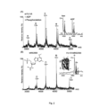
Figure 2 shows mass spectra of: (A) P2Y1R, revealing binding of endogenous ADP and phosphorylation; and (B) P2Y1R-MRS2500, indicating the high binding potency of MRS2500. The structure of the seven transmembrane P2Y1R receptor showing the binding site of MRS2500 (PDB 4XNW) is also shown. The spectra were obtained using a phosphate ester detergent. -
Figure 3 depicts mass spectra of P2Y1R incubated with different molar ratios of ATP. Shown are: (A) P2Y1R without ATP incubation; (B) P2Y1R incubated with ATP at 0.5 molar ratio, revealing that ADP-binding attenuates ATP-binding of receptor; and (C) P2Y1R incubated with ATP at 2 molar ratio further decreases ADP-binding. The spectra were obtained using a phosphate ester detergent. -
Figure 4 shows mass spectra of P2Y1R-MRS2500 complex incubated with increasing molar ratios of ATP. Quantification analysis indicates full conversion of ATP binding to receptor. The spectra were obtained using a phosphate ester detergent. -
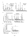
Figure 5 shows: (A) native mass spectra of beta-1 adrenergic receptor (B1AR) and NTR1, revealing the binding of endogenous lipids to receptors; (B) collision-induced dissociation of lipids from B1AR-lipid complexes, allowing precise identification of bound lipids as phosphatidylserine and phosphatidylinositol (PIP); and (C) identification of bound phosphatidic acid (PA) from NTR1 by collision-induced dissociation. The spectra were obtained using a phosphate ester detergent. -
Figure 6 depicts: (A) the measurement of PIP2 binding of NTR1 in a concentration-dependent manner; and (B) binding kinetics of PIP2 to NTR1 (upper: PI(4,5)P2; lower: PI(3,4)P2). The spectra were obtained using a phosphate ester detergent. -
Figures 7 and8 depict mass spectra of NTR1 in various phosphate ester detergents. - For the purposes of the present invention, the following terms as used herein shall, unless otherwise indicated, be understood to have the following meanings.
- The term "alkyl" as used herein refers to a straight or branched chain alkyl moiety having from 1 to 30 carbon atoms. For instance, an alkyl group may have from 1 to 20 carbon atoms, e.g. from 1 to 12 carbon atoms, e.g. from 1 to 10 carbon atoms. In particular, an alkyl group may have 1, 2, 3, 4, 5 or 6 carbon atoms. Examples of alkyl groups include methyl, ethyl, propyl (n-propyl or isopropyl), butyl (n-butyl, sec-butyl or tert- butyl), pentyl, and hexyl. The term "alkylene" refers to a divalent alkyl group.
- The term "cycloalkyl" as used herein refers to an aliphatic carbocyclic moiety having from 3 to 20 ring carbon atoms. For instance, a cycloalkyl group may have from 3 to 16 carbon atoms, e.g. from 3 to 10 carbon atoms. In particular, a cycloalkyl group may have 3, 4, 5 or 6 ring carbon atoms. A cycloalkyl group may be a monocyclic, polycyclic (e.g. bicyclic) or bridged ring system. Examples of cycloalkyl groups include cyclopropyl, cyclobutyl, cyclopentyl, cyclohexyl and norbornyl.
- The term "hydrocarbyl" as used herein refers to a group consisting exclusively of hydrogen and carbon atoms, the group having from 1 to 30 carbon atoms. For instance, a hydrocarbyl group may have from 1 to 20 carbon atoms, e.g. from 1 to 12 carbon atoms, e.g. from 1 to 10 carbon atoms. A hydrocarbyl group may be an acyclic group, a cyclic group, or may comprise both an acyclic portion and a cyclic portion. Examples of hydrocarbyl groups include alkyl, alkenyl, alkynyl, cycloalkyl, cycloalkylalkyl, cycloalkenyl, aryl and aralkyl.
- The term "optionally substituted" as used herein means unsubstituted or substituted.
- The term "substituted' as used herein in connection with a chemical group means that one or more (e.g. 1, 2, 3, 4 or 5) of the hydrogen atoms in that group are replaced independently of each other by a corresponding number of substituents. It will, of course, be understood that the one or more substituents may only be at positions where they are chemically possible, i.e. that any substitution is in accordance with permitted valence of the substituted atom and the substituent and that the substitution results in a stable compound. The term is contemplated to include all permissible substituents of a chemical group or compound. It will be understood by those skilled in the art that one or more hydrogen atoms on a given substituent can themselves be substituted, if appropriate.
- The term "optionally interrupted" as used herein means interrupted or not interrupted.
- The term "interrupted" as used herein in connection with an alkylene linker means that one or more of the specified moieties may be positioned at any position along the alkylene linker, including at either end of the linker.
- The term "detergent" as used herein refers to a substance which lowers the surface tension of the medium in which it is dissolved, and/or the interfacial tension with one or more other phases. Detergents are generally amphipathic molecules, comprising both hydrophilic and hydrophobic groups, and may be anionic, cationic, non-ionic or zwitterionic.
- The present invention provides a method of detecting a membrane protein by mass spectrometry. A method of the present invention involves the use of a solution comprising a detergent micelle in which the membrane protein is contained. The solution contains a phosphate ester detergent which is used to form the detergent micelle. The phosphate ester detergent used in methods of the present invention is a compound of formula (I).
- The solution is vaporised using a nanoelectrospray ionisation source under conditions such that the membrane protein is released from the detergent micelle. The membrane protein is ionised, and subsequently resolved and detected. The method of the present invention may be particularly advantageous for detecting membrane protein complexes in a folded state.
- Membrane proteins can be grouped into integral membrane proteins and peripheral membrane proteins. Integral membrane proteins may have one or more segments embedded within a membrane and may be bound to the lipid bilayer. Peripheral membrane proteins may be temporarily associated with the lipid bilayer and/or integral membrane proteins. In an embodiment, the membrane protein is an integral membrane protein.
- Membrane proteins may be composed of one (mono) or more (multi) associated polypeptide chains. Thus, the membrane protein may be a monomeric or a multimeric membrane protein, for example an oligomeric membrane protein. Oligomeric membrane proteins include both homooligomeric (identical polypeptide chains) and heterooligomeric (different polypeptide chains) proteins.
- In an embodiment, the membrane protein is an integral membrane protein selected from G protein-coupled receptors (GPCRs), membrane transporters, membrane channels, ATP-binding cassette transporters (ABC-transporters) and proton driven transporters.
- In an embodiment, the membrane protein has a molecular weight of from about 103 Daltons to about 1012 Daltons, e.g. from about 103 Daltons to about 106 Daltons.
- Methods for the purification and expression of membrane proteins are known in the art. By way of example, Barrera et al., Nat. describe methods for the purification of MacB, LmrCD, and EmrE. Moreover, Drew et al., Nat. Protoc., 2008, 3, 784-798 describe a GFP fusion construct methodology in which yields in the overexpression and purification of membrane proteins are improved, while Aller et al., Science, 2009, 323, 1718-1722 describe P-glycoprotein expression and purification. Reference may also be made to Zhang et al., Nature, 2015, 520, 317. In order to produce high quality mass spectra, the membrane protein should preferably be relatively pure and homogenous, equivalent to crystallographic-grade material.
- The membrane protein may be in the form of a complex with a ligand. Thus, for example, the membrane protein may be in the form of a complex with one or more ligands selected from therapeutic agents, lipids, nucleotides and nucleosides.
- In one embodiment, the membrane protein is in the form of a complex with one or more therapeutic agents. In this regard, the present methods may be used to detect binding between a membrane protein and a therapeutic agent. In particular, a method of the present invention may allow one or more structural characteristics (e.g. stoichiometry) of a membrane protein-therapeutic agent complex to be determined, and/or may also be used to detect conformational changes that take place upon binding of a therapeutic agent to the membrane protein.
- A method of the present invention may allow therapeutic agents to be screened. In contrast to indirect methods such as fluorescence or calorimetry, the present method may allow therapeutic agents to be screened directly. In particular, a method may be used to screen for the binding of activators and transporter substrates which are difficult to screen using conventional in vivo methodologies. Moreover, unlike X-ray crystallography, the present methods are not complicated by the inherent structural flexibility of membrane protein-therapeutic agent complexes and may allow the dynamical behaviour of membrane proteins and their interaction with therapeutic agents to be studied.
- The therapeutic agent may be an active compound which, when administered to an organism (human or non-human animal), induces a desired pharmacologic, immunogenic, and/or physiologic effect by local and/or systemic action. Examples of therapeutic agents include, without limitation, drugs, vaccines and biopharmaceutical agents. Thus, therapeutic agents may include small molecule drugs, therapeutic proteins, peptides and fragments thereof (whether naturally occurring, chemically synthesised or recombinantly produced), and nucleic acid molecules (including both double-and single-stranded molecules, gene constructs, expression vectors, antisense molecules and the like). Therapeutic agents may also include substrates, inhibitors, activators, neurotransmitters, agonists and antagonists. The therapeutic agent may be a synthetic or naturally occurring compound. The therapeutic agent may be a drug candidate or other agent suspected of having therapeutic application.
- Particular examples of therapeutic agents include, but are not limited to, anti-cancer agents, anti-infective agents (e.g. antibiotics and antiviral agents), analgesic agents, anorexic agents, anti-inflammatory agents, antiepileptic agents, anaesthetic agents, hypnotic agents, sedatives, antipsychotic agents, neuroleptic agents, antidepressants, anxiolytics, antagonists, neuron blocking agents, anticholinergic and cholinomimetic agents, antimuscarinic and muscarinic agents, antiadrenergics agents, hormones, nutrients, antiarthritics agents, antiasthmatic agents, anticonvulsants, antihistamines, antinauseants agents, antineoplastic agents, antipruritics agents, antipyretic agents; antispasmodic agents, cardiovascular agents (e.g. calcium channel blockers, beta-blockers, beta-agonists, antiarrhythmic agents, antihypertensive agents, diuretics and vasodilators), central nervous system stimulants; decongestants, hormones, bone growth stimulants, bone resorption inhibitors, immunosuppressive agents, muscle relaxants, psychostimulants, sedatives and tranquilisers. It will be appreciated that this list of therapeutic agents is merely illustrative and should not be considered to be limiting. Many other therapeutic agents are known in the art and may be utilised in a method of the present invention. A detailed description of various therapeutic agents may be found in e.g. Remington's Pharmaceutical Sciences (21st edition, 2005, Mack Publishing Company). The therapeutic agent may exhibit optical isomerism and/or diastereoisomerism. Accordingly, the therapeutic agent may be in the form of a single enantiomer or diastereoisomer, or a mixture (e.g. a racemic mixture) thereof.
- In an embodiment, the therapeutic agent has a molecular weight of less than 2000 Daltons, e.g. less than 1500 Daltons, e.g. less than 1000 Daltons, e.g. less than 500 Daltons. In an embodiment, the therapeutic agent is a non-polymeric organic compound having a molecular weight of less than 1000 Daltons, e.g. less than 800 Daltons, e.g. less than 500 Daltons.
- In an embodiment, the therapeutic agent is an inhibitor or an activator, e.g. an activator or inhibitor of the membrane protein to which it is bound. In an embodiment, the therapeutic agent is an anti-cancer agent.
- Binding of the therapeutic agent to the membrane protein may be via a non-covalent or a covalent interaction. In particular, binding of the therapeutic agent to the membrane protein may be via intermolecular forces such as ionic bonds, hydrogen bonds and van der Waals forces. Binding of the therapeutic agent to the membrane protein may be reversible or irreversible. In an embodiment, the therapeutic agent is bound to the membrane protein via a reversible bond.
- The membrane protein-therapeutic agent complex may comprise one or more other components. In one embodiment, the complex further comprises a lipid. Thus, for instance, a method of the present invention may be used to determine whether the presence of a lipid affects binding of the therapeutic agent to the membrane protein. In an embodiment, the complex comprises a nucleotide. In an embodiment, the complex comprises a lipid and a nucleotide.
- The method of the present invention has been found to be particularly useful for the analysis of GPCRs. In particular, the present method has been found to be useful in the analysis of complexes of GPCRs with one or more ligands such as therapeutic agents, nucleotides, lipids, or combinations thereof.
- Thus, preferably, the membrane protein is a GPCR. In an embodiment, the GPCR is selected from neurotensin receptor 1 (NTR1), human purinergic receptor P2Y1R, and beta-1-adrenergic receptor (B1AR).
- Preferably, the GPCR is in the form of a complex bound to one or more ligands. Examples of ligands include therapeutic agents, lipids, nucleotides and nucleosides.
- The present invention is particularly useful for detecting the binding of phospholipids with GPCRs. Thus, in a preferred embodiment, the membrane protein is a GPCR in the form of a complex with one or more phospholipids. The one or more phospholipids may be selected from phosphatidylserine, phosphatidylinositol, phosphatidic acid,
phosphatidylinositol 4,5-bisphosphate, and combinations thereof. - In a preferred embodiment, the membrane protein is in the form of a complex comprising: (i) a membrane protein selected from P2Y1R, B1AR, and NTR1; and (ii) one or more phospholipids selected from phosphatidylserine, phosphatidylinositol, phosphatidic acid and
phosphatidylinositol 4,5-bisphosphate. - In a preferred embodiment, the GPCR is in the form of a complex with a therapeutic agent. In an embodiment, the therapeutic agent is an agonist or an antagonist of the GPCR.
- The present method involves the use of a micellar solution which comprises a detergent micelle in which the membrane protein is contained. The membrane protein is encapsulated in a micelle for solubilisation, which may at least partially shield the protein during the electrospray ionisation process. Without wishing to be bound by theory, it is believed that the micelle may shield the protein during the droplet phase of the electrospray ionisation process and, moreover, may afford at least partial shielding from ionisation of the protein during this process. The detergent micelle may exert a pressure sufficient to maintain the structure of the membrane protein, thereby minimising the deleterious effects associated with vaporisation and substantially retaining interactions between the membrane protein and any ligand and interactions within any subunits of the membrane protein. The solution will typically comprise a plurality of micelles containing the membrane protein. The micellar solution may be formed by e.g. incubating the membrane protein in the presence of the detergent.
- Preferably, the membrane protein is maintained in the detergent micelle in an intact, folded state. This may allow the membrane protein to be detected in its "native" state. Alternatively, the membrane protein may be present within the detergent micelle in a partially folded or unfolded state.
- According to the present invention, a phosphate ester detergent is used to form the detergent micelle. The phosphate ester detergent is a compound of the formula (I):

- n is 1, 2, 3, 4, 5 or 6;
- each R1 is independently selected from hydrogen and alkyl;
- X is an alkylene linker having from 2 to 6 carbon atoms, wherein the carbon atoms of the alkylene linker are optionally interrupted by one or more moieties selected from -O-, -C(O)-, -NH-, -S-, -C(S)-, -S(O)- and -S(O)2-; and wherein the carbon atoms of the alkylene linker are optionally substituted by one or more moieties selected from hydroxy, halo and amino; and
- R2 is optionally substituted hydrocarbyl.
- In an embodiment, n is 1, 2, 3 or 4, e.g. 1, 2 or 3. Preferably, n is 2.
- In an embodiment, each R1 is independently hydrogen or C1-6 alkyl. Preferably, each R1 is methyl.
- In a preferred embodiment, n is 2 and each R1 is methyl. Thus, preferably, the detergent is a compound of the formula (II):

- In preferred embodiments of the compounds of formulae (I) and (II), X is a group selected from -(CH2)k-, wherein k is 2, 3, 4, 5 or 6; -C(O)-O-CH2CH(OH)CH2-; and -OCH2CH(OH)CH2-.
- In preferred embodiments of the compounds of formulae (I) and (II), R2 is alkyl, alkenyl, cycloalkyl or cycloalkylalkyl, any of which is optionally substituted.
- In preferred embodiments of the compounds of formulae (I) and (II), R2 is alkyl, e.g. C1-20 alkyl or C8-16 alkyl.
- In preferred embodiments of the compounds of formulae (I) and (II), R2-X- is selected from: n-octyl; n-nonyl; n-decyl; n-undecyl; n-dodecyl; n-tridecyl; n-tetradecyl; n-heptadecyl; n-hexadecyl; 2,6-dimethylheptyl; 2,8-dimethylnonyl; and 10-undecylenyl.
- In preferred embodiments of the compounds of formulae (I) and (II), R2-X- is selected from: 1-dodecanoyl-2-hydroxy-sn-glycero-3; 1-tetradecanoyl-2-hydroxy-sn-glycero-3; 1-hexadecanoyl-2-hydroxy-sn-glycero-3; 1-dodecyl-2-hydroxy-sn-glycero-3; 1-tetradecyl-2-hydroxy-sn-glycero-3; and 1-hexadecyl-2-hydroxy-sn-glycero-3.
- In preferred embodiments of the compounds of formulae (I) and (II), R2-X- is selected from: 2-cyclohexyl-1-ethyl; 3-cyclohexyl-1-propyl; 4-cyclohexyl-1-butyl; 5-cyclohexyl-1-pentyl; 6-cyclohexyl-1-hexyl; and 7-cyclohexyl-1-heptyl.
- In an embodiment, the phosphate ester detergent is selected from the following compounds:
- n-octylphosphocholine ("Fos-Choline-8");
- n-nonylphosphocholine ("Fos-Choline-9");
- n-decylphosphocholine ("Fos-Choline-10");
- n-undecylphosphocholine ("Fos-Choline-11")
- n-tridecylphosphocholine ("Fos-Choline-13");
- n-pentadecylphosphocholine ("Fos-Choline-15");
- 2,6-dimethyl-4-heptylphosphocholine ("Fos-Choline-ISO-9");
- 2,8-dimethyl-5-nonylphosphocholine ("Fos-Choline-ISO-11"); and
- 10-undecylenyl-1-phosphocholine ("Fos-Choline-Unsat-11-10").
- In a particularly preferred embodiment, the phosphate ester detergent is selected from the following compounds:
- n-hexadecylphosphocholine ("Fos-Choline-16");
- n-tetradecylphosphocholine ("Fos-Choline-14");
- n-dodecylphosphocholine ("Fos-Choline-12");
- 1-tetradecanoyl-2-hydroxy-sn-glycero-3-phosphocholine ("Lyso-Fos-Choline-14);
- 1-dodecanoyl-2-hydroxy-sn-glycero-3-phosphocholine ("Lyso-Fos-Choline-12"); and
- 6-cyclohexyl-1-hexylphosphocholine ("CycloFos-6").
- The structures of these compounds are shown below:
Detergent Structure Fos-Choline-16 
Fos-Choline-14 
Fos-Choline-12 
LysoFos Choline-14 
LysoFos Choline-12 
CycloFos-6 
- The micellar solution may be formed using a single detergent or a mixture of different phosphate ester detergents.
- The micellar solution may contain one or more detergents in addition to the phosphate ester detergent(s). Examples of other detergents include non-ionic detergents such as n-dodecyl-D-maltoside, nonylglucoside, glycosides, neopentyl glycols, facade EM, maltosides, glucosides, and mixtures thereof. In an embodiment, the micellar solution does not contain any detergents apart from the phosphate ester detergent.
- In order to minimise dissociation of the membrane protein or precipitation of the protein, the detergent is present in the micellar solution at a concentration at least equal to, and preferably at two times, the critical micelle concentration (CMC) of the detergent. The critical micelle concentration of the detergent may be determined experimentally using methods known in the art, or it may be obtained from e.g. a textbook, product catalogue or website. For example, the Anatrace products catalogue provides CMC data. Typically, critical micelle concentration values are determined in water at 25 °C. Thus, in an embodiment, the solution is an aqueous solution and the detergent is present in the solution at a concentration at least equal to, and preferably greater than, the critical micelle concentration of the detergent in water at 25 °C. The detergent is preferably present at a concentration at least twice than that of the critical micelle concentration of the detergent in water at 25 °C.
- In an embodiment, the detergent is present in the micellar solution at a concentration of from about 100 µM to about 100 mM, e.g. from about 100 µM to about 200 µM. In an embodiment, the membrane protein is present in the micellar solution at a concentration of from about 1 nM to about 1 mM, e.g. from about 800 nM to about 900 nM.
- In an embodiment, the molar ratio of the detergent to the membrane protein is from about 10:1 to 150:1, e.g. from about 30:1 to about 125:1, e.g. from about 50:1 to about 100:1. In a preferred embodiment, the molar ratio of the detergent to the membrane protein is less than or equal to 100:1.
- Where the membrane protein is in the form of a complex with a therapeutic agent, the micellar solution preferably comprises a molar excess of the therapeutic agent as compared to the membrane protein. In an embodiment, the molar ratio of the therapeutic agent to the membrane protein is at least 2:1, e.g. at least 5:1, e.g. at least 10:1. In an embodiment, the therapeutic agent is present in the micellar solution at a concentration of at least 100 nM, e.g. from 100 nM to 900 nM.
- The micellar solution may comprise one or more other components. In particular, the micellar solution preferably contains a buffer. Ammonium acetate is particularly preferred in this regard. The concentration of ammonium acetate is preferably at least 200 millimolar. Preferably the pH of the buffer is in the range of from about 5 to about 8. When working with proteins containing His-tags, it may be preferable to use a pH of about 8 to avoid protein insolubility or precipitation which may otherwise occur in lower pH buffered solutions.
- Buffer exchange and concentration of the micellar solution may be achieved using suitable techniques and devices known in the art, e.g. using a Micro Bio-Spin ® column (Bio-Rad Laboratories) or a Vivaspin device (GE Healthcare).
- The membrane protein is detected using a mass spectrometer comprising a nanoelectrospray ionisation source, a mass analyser and a detector. The mass spectrometer is preferably adapted to transmit and detect ions having mass-to-charge (m/z) ratios in the range of e.g. from about 100 m/z to about 32,000 m/z. Preferably, the mass spectrometer is operated under conditions suitable for maintaining and focusing large macromolecular ions. By way of illustration, and without limitation, the mass spectrometer may be a Synapt HDMS ion-trap-IM-MS instrument (more preferably a G1 instrument) or a Q-Exactive hybrid quadrupole-orbitrap mass spectrometer. The resolution provided by such instruments is particularly suited to resolving peaks generated from a complex comprising a membrane protein bound to a therapeutic agent or other ligand.
- The nanoelectrospray ionisation source is used to vaporise the micellar solution. Nanoelectrospray ionisation is a technique well known in the art (see e.g. Wilm et al, Anal. Chem. 1996, 68, 1-8; and Wilm et al, Int. J. of Mass Spec. and Ion Proc. 1994, 132, 167-180). The use of nanoelectrospray ionisation allows ions, and in particular highly charged ions, to be generated directly from solution. The formation of highly charged ions may allow the detection of high mass complexes at relatively low mass-to-charge (m/z) ratios. The use of a nanoelectrospray ionisation is also desirable from the point of view of allowing a membrane protein complex, or subunits of a complex, to remain substantially intact. In performing a method of the present invention, it may be preferable to use a nanoflow capillary, e.g. a gold-coated nanoflow capillary, to vaporise the solution.
- The micellar solution is vaporised under conditions such that the membrane protein is released from the micelle. The membrane protein may be released from the micelle as a result of collisions between the electrospray and the micelle which serve to disrupt the detergent assembly. Preferably, the vaporisation conditions are selected so that the membrane protein is released from the micelle substantially intact. Preferably, the conditions inside the mass spectrometer are selected to rapidly remove the micelle from the membrane protein. Ionisation of the membrane protein may occur during the step of vaporising and/or after release of the protein from the micelle. In some instances, portions of the membrane protein, e.g. hydrophilic/cytoplasmic domains, may become ionised prior to release of the protein from the micelle. Typically, ionisation of the membrane protein occurs during and/or after release of the protein from the micelle. In an embodiment, release and/or ionisation of the protein from the micelle occurs in a collision cell present within the mass spectrometer. Release and/or ionisation of the protein from the micelle may be achieved by adjusting acceleration voltages and/or pressures within the collision cell to remove the detergent while retaining the peaks of the membrane protein.
- Mass spectrometer parameters may be optimised for maximal desolvation and detergent removal, while minimising protein activation. In particular, one or more of the following parameters may be optimised: collision voltage, cone voltage, collision gas pressure, collision gas type, and source pressure. Optimisation of parameters may be achieved by first setting the instrument parameters to relatively high activation settings for membrane proteins. Then iteratively, each of the aforementioned four parameters may be adjusted to produce resolved mass spectra while minimizing over-activation of the target protein.
- In an embodiment, the mass spectrometer is operated under one or more of the following conditions: (i) a capillary voltage of from about 0.8 to about 2.2 kV, e.g. from about 1.0 to about 2.0 kV, e.g. from about 1.2 to about 1.8 kV; (ii) a cone voltage of from about 5 to about 240 V, e.g. from about 5 to about 50 V, e.g. from about 100 to about 200 V; (iii) a trap collision energy of from about 0 to about 240 V, e.g. from about 100 to about 220 V, e.g. from about 120 to about 200 V; (iv) a source temperature of from about 0 to about 100 °C, e.g. from about 0 to about 50 °C, e.g. from about 10 to about 30 °C , e.g. about 20 °C; (v) a bias voltage of from about 15 to about 200 V, e.g. from about 60 to about 180 V, e.g. from about 80 to about 160 V; and (vi) a backing pressure of from about 1 to about 8 mBar, e.g. from about 3 to about 6 mBar, e.g. from about 4 to about 5 mBar. In a particular embodiment, the mass spectrometer is operated with a bias voltage of from about 15 to about 200 V, e.g. from about 60 to about 180 V, e.g. from about 80 to about 160 V. In a particular embodiment, the trap collision energy is from about 50 to about 200 V.
- In an embodiment, the mass spectrometer is a Synapt HDMS ion-trap-IM-MS instrument (more preferably a G1 instrument) and is operated under one or more of the following conditions: (i) a capillary voltage of from about 0.8 to about 2.2 kV, e.g. from about 1.0 to about 2.0 kV, e.g. from about 1.2 to about 1.8 kV; (ii) a cone voltage of from about 5 to about 200 V, e.g. from about 100 to about 200 V, e.g. from about 5 to about 50 V; (iii) a trap collision energy of from about 0 to about 240 V, e.g. from about 100 to about 220 V, e.g. from about 120 to about 200 V; (iv) a source temperature of from about 25 to about 40 °C, e.g. from about 10 to about 30 °C , e.g. about 20 °C; (v) a bias voltage of from about 20 to about 200 V, e.g. from about 60 to about 180 V, e.g. from about 80 to about 160 V; and (vi) a backing pressure of from about 1 to about 8 mBar, e.g. from about 3 to about 6 mBar, e.g. from about 4 to about 5 mBar. In a particular embodiment, the mass spectrometer is operated with a bias voltage of from about 15 to about 200 V, e.g. from about 60 to about 180 V, e.g. from about 80 to about 160 V. In a particular embodiment, the trap collision energy is from about 50 to about 200 V.
- In an embodiment, the mass spectrometer is a Q-Exactive hybrid quadrupole-orbitrap mass spectrometer and is operated under one or more of the following conditions: (i) a capillary voltage of from about 0.8 to about 2.2 kV, e.g. from about 1.0 to about 2.0 kV, e.g. from about 1.2 to about 1.8 kV; (ii) an acceleration voltage in the higher-energy collisional dissociation (HCD) cell of from about 0 to about 200 V, e.g. from about 5 to about 50 V, e.g. from about 100 to about 200 V; (iii) a source temperature of from about 25 to about 100 °C, e.g. from about 10 to about 30 °C, e.g. about 20 °C; (iv) a DC voltage in the transfer multipole of from about 2 to about 4 V, e.g. from about 3 to about 4 V; (v) a voltage in the C-trap entrance lens of from about 2 to about 7 V, e.g. from about 3 to about 4 V, e.g. from about 5 to about 6 V; and (vi) a pressure in the HCD cell of from about 6.0 x 10⁻¹⁰ to about 1.5 x 10⁻⁹ mBar, e.g. from about 6.0 x 10⁻¹⁰ to about 9 x 10⁻¹⁰ mBar, e.g. from about 1 × 10⁻⁹ to about 1.5 × 10⁻⁹ mBar.
- Preferably, minimal activation energy is used to liberate the membrane protein from the detergent micelle. In an embodiment, the laboratory frame energy is from about 500 to about 5000 electron volts, e.g. from about 500 to about 1500 electron volts. The term "laboratory frame energy" as used herein refers to the collision voltage multiplied by charge state of the membrane protein.
- The ionised membrane protein is then resolved and detected and, if desired, further characterised. In particular, in embodiments in which the membrane protein is complexed with a therapeutic agent, ions in which the therapeutic agent is bound to the membrane protein or a fragment thereof can be detected directly using the mass spectrometer, rather than inferred indirectly from mass spectra of the separate components (therapeutic agent and protein). Moreover, where the micellar solution or the complex comprises one or more additional components, e.g. one or more components selected from lipids and nucleotides, the binding of one or more of said components to the membrane protein may be detected simultaneously. For instance, the method may comprise detecting a plurality of ions selected from the group consisting of ions containing the therapeutic agent bound to the membrane protein or a fragment thereof, ions containing one or more additional components (e.g. selected from lipid and nucleotides) bound to the membrane protein or a fragment thereof, and ions in which the therapeutic agent and one or more additional components are bound to the membrane protein or a fragment thereof. Thus, the present methods may be used to detect concomitant binding of the membrane protein with e.g. a therapeutic agent and one or more other species which compete for binding sites.
- In a particular embodiment, the membrane protein is detected using ion mobility-mass spectrometry (IM-MS). In this case, the mass spectrometer may comprise an ion mobility cell to assess the folded state of membrane protein complexes. The use of IM-MS may allow the stoichiometry of ligand agent binding, and the overall effects of ligand binding on the dynamics, stabilities, oligomeric structures and conformations of membrane proteins, to be determined. For instance, the stoichiometry and oligomeric structure of complexes may be characterised by assessing mass differences. As a further example, dynamics, stabilities and conformations may be characterised by changes in charge states or by differences in arrival time distributions (i.e. ion mobility). Methods for the detection of membrane proteins, as well as complexes thereof, by mass spectrometry are disclosed in
WO 2012/172378 andWO 2014/096821 . - The following non-limiting Examples illustrate the present invention.
- The following materials and methods were employed in the experiments of the Examples.
- BODIPY FL-X NHS ester reagent was purchased from Thermo Fisher.
- Neurotensin 8-13 was synthesized from Biomatik.
- Mouse 6x-His tag monoclonal antibody (4A12E4) was purchased from Thermo Fisher. Rabbit anti-mouse IgG-peroxidase conjugated antibody was acquired from Sigma.
Lipofectamine 2000 transfection reagent was obtained from Thermo. - P2Y1R and P2Y1R-MRS2500 complex were prepared as described in Zhang et al., Nature, 2015, 520, 317.
- MRS2500 (1'R,2'S,4'S,5'S)-4-(2-iodo-6-methylamino-purine-9-yl)-1-[(phosphato)methyl] 2(phosphato)bicycle[3.1.0]-hexane) was obtained from Tocris Bioscience.
- Beta-1 adrenergic receptor in apo and isoprenaline-bound state were prepared as described previously (see Warne et al., Nature, 2011, 469, 241).
- Engineered Gα subunit was prepared as described in Carpenter et al., Protein Eng Des Sel, 2016, 12, 583.
- 18:1
Phosphatidylinositol 3,4-bisphosphate (PI(3,4)P2) and 18:1phosphatidylinositol 4,5-bisphosphate (PI(4,5)P2) were purchased from Avanti. - Fos-Choline-16, Fos-Choline-14, Fos-Choline-12, LysoFos Choline-14, LysoFos Choline-12, and CycloFos-6 were obtained from Anatrace.
- Vivaspin concentrator was obtained from GE Healthcare.
- The gene fragment of rat NTR1 (wild-type, 1-424) with exogenous c-terminal Flag- and His- tag was constructed into modified pHLsec vector for transient protein overexpression. Plasmids were transfected into HEK293T cells via a liposome-based method with
Lipofectamine 2000 transfection reagent and the cells were harvested 24 hrs after transfection for further experiments. - HEK293T cells with NTR1 overexpression were lysed in lysis buffer (100 mM sodium phosphate, pH 7.2, 150 mM NaCl, 10% glycerol, 1x EDTA-free protease inhibitor cocktail from Roche) containing 1% various detergents for screening. Cell lysate was harvested after pelleting down the debris by centrifugation, and protein concentration was measured by Pierce 660nm protein assay reagent (Pierce). Equal amounts of protein extract in different detergents were applied for western blotting to detect NTR1 by using an anti-His tag antibody. Semi-quantification was performed by Gel-pro software to quantify the protein amount and relative ratio of protein aggregation.
- Protein extract from HEK293T cells with NTR1 overexpression was incubated with BODIPY-labelled neurotensin 8-13 (BODIPY-NT8-13) at room temperature for 1 hr. The unbound BODIPY-NT8-13 was removed by Bio-Spin size exclusion spin column (Bio-rad) and the fluorescent signal of BODIPY-NT8-13 bound to receptor was detected by fluorescence microplate reader.
- Purified GPCRs were buffer exchanged into 50-200 mM ammonium acetate containing Fos-Choline detergents at 2 times critical micelle concentration (CMC) by using Bio-Spin size exclusion spin column (Bio-rad) prior to mass spectrometer analysis. Detergent exchange could be also performed by size exclusion chromatography equipped with FPLC system. Protein samples were fractionized by the size exclusion columns equilibrated with 50-200 mM ammonium acetate containing Fos-choline detergents at 2 times CMC, and the protein fractions were collected and concentrated by centrifugation-based protein concentrators Vivaspin.
- PIP2 powder (Avanti) was dissolved into solvent containing chloroform, methanol and H2O with volume ratio of 20:9:1. The aliquoted PIP2 solution was dried out by gas-assisted evaporation and re-suspended into 50-200 mM ammonium acetate containing 0.00106% Fos-Choline-16, followed by sonication in water bath for 30 mins.
- The measurement of GPCRs was performed on a modified Synapt G1 (Waters) or a modified Q-Exactive (Thermo) mass spectrometer. The data in Examples 2 and 4 were obtained using a Q-Exactive mass spectrometer, whereas the data in Example 3 were obtained using a Synapt G1 mass spectrometer.
- The Synapt instrument (see Laganowsky et al., Nature, 510, 172-175, 2014) was typically set to a source pressure of 5-7 mBar, capillary voltage of 1.4-1.7 kV, capillary nanoflow of 0.03-0.2 mBar and argon as collision gas with flow rate set to 5-8 mlmin-1 (~6.6-6.7 e-2 mBar). The optimized collision voltage for GPCRs ranged from 60 to 120 V. The sample and extraction cone, and trap bias voltages, quadrupole profile, and collision (trap) gas pressure were optimized for maximal ion intensity of GPCRs.
- The Q-Exactive mass spectrometer was set up according to Gault et al. (Nat. ). Briefly, a gentle voltage gradient was applied (injection flatapole, inter flatapole, bent flatapole, transfer multipole: 7.9, 6.94, 5.9, 4 V respectively). The acceleration voltage applied in HCD cell was optimized for GPCRs (60-150 V) to remove the detergent micelles from protein ions. Spectra were acquired with ten microscans and the noise level parameter of 3 was applied. Backing pressure was maintained at 1.05-1.3 x 10⁻⁹ mBar to allow better transmission of GPCR ions.
- Various detergents were assessed for their ability to preserve the conformation and activity of NTR1. The detergents assessed were: (1) n-dodecyl-β-D-maltoside (DDM); (2) n-undecyl-β-D-maltoside (UDM); (3) n-nonyl-β-D-glucopyranoside (NG); (4) n-octyl-β-D-glucopyranoside (OG); (5) octyl glucose neopentyl glycol (OGNG); (6) n-octyl-β-D-thioglucopyranoside (OTG); (7) tetraethylene glycol monooctyl ether (C8E4); (8) hexaethylene glycol monodecyl ether (C10E6); (9) polyethylene glycol tert-octylphenyl ether (Triton X-100); (10) lauryldimethylamine-N-oxide (LDAO); and (11) hexadecylphosphocholine (Fos-Choline-16).
- It can be seen from the results presented in
Figure 1 that the use of a phosphate ester detergent (Fos-Choline-16; detergent 11) is advantageous as compared with the other detergents tested. In more detail,Figure 1A and B show that less receptor aggregation and oligomerisation occurred with the phosphate ester detergent than with other detergents.Figure 1C shows that the phosphate ester detergent also allowed ligand binding activity to be substantially preserved. - Mass spectrometry was used to study the human P2Y1 purinergic receptor (P2Y1R) and its interactions with nucleotides and a high affinity antagonist, MRS2500. This receptor, a class A GPCR, functions as a receptor for extracellular adenosine 5' -diphosphate (ADP) and adenosine 5' -triphosphate (ATP), and regulates many physiological processes. P2Y1R is fully activated by ADP to facilitate platelet aggregation via calcium wave propagation and thus serves as an important antithrombotic drug target. Inhibition of P2Y1R activation shows significant decrease in ADP-induced platelet aggregation.
- High-resolution MS was used to probe the drug/ligand interactions of human P2Y1R purified from insect cells. By controlled application of collisional activation, intact P2Y1R was liberated from its detergent micelle, preserving the non-covalent interactions formed in solution. The phosphate-containing detergent of Example 1 (Fos-16) was used in these experiments.
- ESI-MS spectra of purified P2Y1R revealed P2Y1R in its apo state (measured 49685.1±0.7 Da; theoretical 49556.7 Da, the experimental mass increase of P2Y1R corresponding to protein modifications that include carboxyamidomethylation and oxidation and several adducts were observed with mass increases of 80 Da and 426.7±2 Da, corresponding to molecular weight of phosphorylation and ADP, respectively (see
Figure 2A ). To quantify the populations of phosphorylated and ADP-bound P2Y1R, intensity values of each charge state were evaluated by UniDec deconvolution software. The ATP-binding ratio of phosphorylated P2Y1R (39.6 %), was similar to receptor without phosphorylation (41.5 %), suggesting P2Y1 phosphorylation may not influence its ADP binding activity. - To investigate the drug binding of P2Y1R, P2Y1R purified in the presence of MRS2500 was analysed. Under identical conditions, a single charge state distribution corresponding to a mass of 50247.7±1 Da was observed. The mass difference of observed species to apo P2Y1R is in very close agreement to the theoretical mass of MRS2500 (theoretical 561.3 Da) (see
Figure 2B ). These results define the high efficacy of MRS2500 with 100 percentage of interaction and illustrate the advantageous nature of the present invention. No ADP binding was detected in the presence of the drug, confirming the high inhibitory potency of MRS2500. In addition to phosphorylation of the receptor, a significant population assigned to binding of a single zinc ion was observed, which was also present in the rubredoxin fusion protein according to the x-ray crystal structure of P2Y1R-MRS2500 (see Zhang etal., Nature, 2015, 520, 317). - ATP binding of P2Y1R was examined by pre-incubating exogenous ATP with the receptor at different molar ratios prior to analysis by mass spectrometry (see
Figure 3 ). At low ATP concentration (1: 0.5 molar ratio of protein to ATP), around 80 % of the apo receptor and 30 % of the ADP-bound receptor were converted into the ATP bound state (Figure 3B ), indicating ADP binding of receptor attenuates its interaction with ATP. Incubation at higher ATP concentrations (2 molar excess) was able to further decrease 40% of ADP binding (seeFigure 3C ). This incomplete displacement of ADP at high ATP concentration observed in these experiments explains the lower agonist potency of ATP to ADP in receptor activation, in agreement with ATP as partial agonist/antagonist depending on the expression level of P2Y1R. - The competitive behavior of ATP to MRS2500 was further investigated by incubating the P2Y1R-MRS2500 complex with increasing molar ratios of exogenous ATP (see
Figure 4 ). Mass spectra recorded after 30 min incubation at 37 °C showed that both ATP and drug bound P2Y1R were observed at low concentration of ATP (0.25 and 0.5 molar ratios to receptor). Incubation at higher ATP concentrations (1: 2 molar P2Y1R: ATP) raised the possibility of a second ATP binding site and demonstrated complete displacement of MRS2500 by substoichiometric quantities of ATP. - Thus, as the results of this experiment demonstrate, the present invention can be used to capture GPCR-ligand binding events by non-denaturing mass spectrometry. The interaction of human P2Y1 purinergic receptor was defined with agonist/antagonist. The resolution of mass spectrometry allows the interplay of compound interactions to be examined for further understanding their binding mechanisms. These results revealed the equilibration kinetics of MRS2500 and its competitive behavior with nature nucleotide ligands, providing valuable information in the study of P2Y1R pharmacology.
- In a further experiment, mass spectrometry was used to characterise the binding affinity of PIP2 to NTR1. The phosphate-containing detergent of Example 1 (Fos-16) was used in this experiment.
-
Figure 6A shows the measurement of PIP2 binding of NTR1 in a concentration-dependent manner. Multiple binding of PIP2 was observed after incubation of PIP2 and NTR1 in Fos-Choline-16.Figure 6B depicts the binding kinetics of various PIP2 to NTR1 (upper: PI(4,5)P2; lower: PI(3,4)P2). Binding curves of different number of PIP2 were plotted according to the quantification of MS spectra, allowing calculation of the dissociation constant (Kd). - In a further experiment, purified NTR1 was buffer exchanged into various phosphate-containing detergents and monitored by native mass spectrometry. The detergents tested were: n-hexadecylphosphocholine (Fos-Choline-16; CMC = 0.013 mM); n-tetradecyl-phosphocholine (Fos-Choline-14; CMC = 0.12 mM); n-dodecylphosphocholine (Fos-Choline-12; CMC = 1.5 mM); 1-myristoyl-2-hydroxy-sn-glycero-3-phosphocholine (LysoFos-Choline-14; CMC = 0.036 mM); 1-dodecanoyl-2-hydroxy-sn-glycero-3-phospho-choline (LysoFos-Choline-12; CMC = 0.32 mM); and 6-cyclohexyl-1-hexylphosphocholine (CycloFos-6; CMC = 2.68 mM).
- The spectra are presented in
Figures 7 and8 , and illustrate the capability of phosphate-containing detergents for membrane protein analysis, in particular GPCR analysis.
Claims (16)
- A method of detecting a membrane protein by mass spectrometry, wherein the method comprises:(a) providing a solution comprising a detergent micelle in which said membrane protein is contained;(b) providing a mass spectrometer comprising a nanoelectrospray ionisation source, a mass analyser and a detector;(c) vaporising the solution using the nanoelectrospray ionisation source under conditions such that the membrane protein is released from the micelle;(d) ionising the membrane protein;(e) resolving the ionised membrane protein using the mass analyser; and(f) detecting the resolved membrane protein using the detector;wherein the solution contains a phosphate ester detergent which forms said detergent micelle and is present in the solution at a concentration which is greater than or equal to the critical micelle concentration of the detergent in said solution;wherein the phosphate ester detergent is a compound of the formula (I):
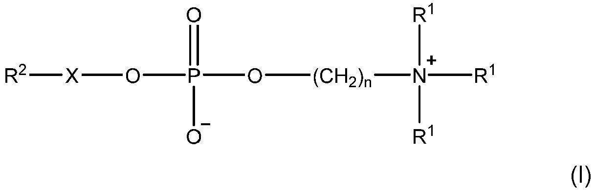 n is 1, 2, 3, 4, 5 or 6;each R1 is independently selected from hydrogen and alkyl;X is an alkylene linker having from 2 to 6 carbon atoms, wherein the carbon atoms of the alkylene linker are optionally interrupted by one or more moieties selected from -O-, -C(O)-, -NH-, -S-, -C(S)-, -S(O)- and -S(O)2-; and wherein the carbon atoms of the alkylene linker are optionally substituted by one or more moieties selected from hydroxy, halo and amino; andR2 is optionally substituted hydrocarbyl.
 - A method according to claim 1, wherein the membrane protein is a G protein-coupled receptor, and preferably wherein the G protein-coupled receptor is selected from neurotensin receptor 1 (NTR1), human purinergic receptor P2Y1R, and beta-1-adrenergic receptor (B1AR).
- A method according to claim 1 or claim 2, wherein the membrane protein is in the form of a complex with one or more ligands, wherein the one or more ligands are preferably selected from therapeutic agents, lipids, nucleotides and nucleosides, and more preferably wherein the membrane protein is in the form of a complex with a therapeutic agent.
- A method according to any preceding claim, wherein:the solution is an aqueous solution; and whereinthe molar ratio of the detergent to the membrane protein in the solution is from 10:1 to 150:1.
- A method according to any preceding claim, wherein:the solution is an aqueous solution; and whereinthe solution is sprayed using a gold-coated nanoflow capillary.
- A method according to any preceding claim, wherein the membrane protein is released from the micelle substantially intact.
- A method according to any preceding claim, wherein the structure or conformation of the membrane protein is characterised.
- A method according to any preceding claim, wherein:the mass spectrometer is operated under one or more of the following conditions: (i) the capillary voltage of the nanoelectrospray ionisation source is from 0.8 to 2.2 kV; (ii) the cone voltage of the nanoelectrospray ionisation source is from 5 to 240 V; (iii) the trap collision energy is from 0 to 240 V: (iv) the source temperature is from 0 to 50 °C; (v) the bias voltage is from 15 to 200 V, e.g. from 30 to 200 V, e.g. from 60 to 180 V, e.g. from 80 to 160 V; and (vi) the backing pressure is from 1 to 8 mBar; and/orthe membrane protein is detected by ion mobility-mass spectrometry.
- A method according to any preceding claim, wherein:n is 2; and/oreach R1 is independently hydrogen or C1-6 alkyl, e.g. hydrogen or methyl, and preferably wherein each R1 is methyl.
- A method according to any preceding claim, wherein the detergent is a compound of the formula (II):

- A method according to any preceding claim, wherein:X is a group selected from -(CH2)k-, wherein k is 2, 3, 4, 5 or 6; -C(O)-O-CH2CH(OH)CH2-; and -OCH2CH(OH)CH2-; and/orR2 is alkyl, alkenyl or cycloalkyl, cycloalkylalkyl, any of which is optionally substituted, and preferably wherein R2 is alkyl, e.g. C1-20 alkyl or C8-16 alkyl;and preferably wherein R2-X- is selected from:n-octyl; n-nonyl; n-decyl; n-undecyl; n-dodecyl; n-tridecyl; n-tetradecyl; n-heptadecyl; n-hexadecyl; 2,6-dimethylheptyl; 2,8-dimethylnonyl; and 10-undecylenyl;1-dodecanoyl-2-hydroxy-sn-glycero-3; 1-tetradecanoyl-2-hydroxy-sn-glycero-3; 1-hexadecanoyl-2-hydroxy-sn-glycero-3; 1-dodecyl-2-hydroxy-sn-glycero-3; 1-tetradecyl-2-hydroxy-sn-glycero-3; and 1-hexadecyl-2-hydroxy-sn-glycero-3; or2-cyclohexyl-1-ethyl; 3-cyclohexyl-1-propyl; 4-cyclohexyl-1-butyl; 5-cyclohexyl-1-pentyl; 6-cyclohexyl-1-hexyl; and 7-cyclohexyl-1-heptyl.
- A method according to any preceding claim, wherein the phosphate ester detergent is selected from the following compounds:n-hexadecylphosphocholine;n-tetradecylphosphocholine;n-dodecylphosphocholine;1-tetradecanoyl-2-hydroxy-sn-glycero-3-phosphocholine;1-dodecanoyl-2-hydroxy-sn-glycero-3-phosphocholine; and6-cyclohexyl-1-hexylphosphocholine.
- Use of a solution comprising a detergent micelle in which a membrane protein is contained in the detection of a membrane protein by mass spectrometry, wherein the solution comprises a phosphate ester detergent as defined in any preceding claim which forms said detergent micelle, and preferably wherein the membrane protein is in the form of a complex with one or more ligands, wherein the one or more ligands are preferably selected from therapeutic agents, lipids, nucleotides and nucleosides.
- Use according to claim 13, wherein the membrane protein is as defined in claim 2.
- Use of a phosphate ester detergent as defined in any preceding claim in the detection of a membrane protein by mass spectrometry.
- Use according to claim 15, wherein the membrane protein is as defined in claim 2.
Applications Claiming Priority (2)
| Application Number | Priority Date | Filing Date | Title |
|---|---|---|---|
| GBGB1703033.9A GB201703033D0 (en) | 2017-02-24 | 2017-02-24 | Detection of membrane proteins |
| PCT/GB2018/050479 WO2018154318A1 (en) | 2017-02-24 | 2018-02-23 | Detection of membrane proteins |
Publications (2)
| Publication Number | Publication Date |
|---|---|
| EP3586142A1 EP3586142A1 (en) | 2020-01-01 |
| EP3586142B1 true EP3586142B1 (en) | 2024-12-25 |
Family
ID=58544386
Family Applications (1)
| Application Number | Title | Priority Date | Filing Date |
|---|---|---|---|
| EP18708464.5A Active EP3586142B1 (en) | 2017-02-24 | 2018-02-23 | Detection of membrane proteins |
Country Status (4)
| Country | Link |
|---|---|
| US (1) | US10955421B2 (en) |
| EP (1) | EP3586142B1 (en) |
| GB (1) | GB201703033D0 (en) |
| WO (1) | WO2018154318A1 (en) |
Families Citing this family (3)
| Publication number | Priority date | Publication date | Assignee | Title |
|---|---|---|---|---|
| GB201814356D0 (en) * | 2018-09-04 | 2018-10-17 | Univ Oxford Innovation Ltd | Methods of detergents |
| GB201814454D0 (en) | 2018-09-05 | 2018-10-17 | Univ Oxford Innovation Ltd | Detection of membrane proteins |
| GB202004961D0 (en) | 2020-04-03 | 2020-05-20 | Micromass Ltd | De-clustering guide |
Family Cites Families (4)
| Publication number | Priority date | Publication date | Assignee | Title |
|---|---|---|---|---|
| CA2634079A1 (en) * | 2005-12-20 | 2007-10-25 | The Lubrizol Corporation | Method of preparing an overbased or neutral detergent |
| WO2010050918A1 (en) | 2008-10-31 | 2010-05-06 | Qinghai Zhang | Branched chain detergents for membrane protein structural biology |
| GB201110272D0 (en) * | 2011-06-17 | 2011-08-03 | Isis Innovation | Metod of detection by mass spectrometry |
| GB201222833D0 (en) | 2012-12-18 | 2013-01-30 | Isis Innovation | Detection of membrane proteins |
-
2017
- 2017-02-24 GB GBGB1703033.9A patent/GB201703033D0/en not_active Ceased
-
2018
- 2018-02-23 US US16/488,364 patent/US10955421B2/en active Active
- 2018-02-23 WO PCT/GB2018/050479 patent/WO2018154318A1/en not_active Ceased
- 2018-02-23 EP EP18708464.5A patent/EP3586142B1/en active Active
Also Published As
| Publication number | Publication date |
|---|---|
| GB201703033D0 (en) | 2017-04-12 |
| US10955421B2 (en) | 2021-03-23 |
| US20200240997A1 (en) | 2020-07-30 |
| WO2018154318A1 (en) | 2018-08-30 |
| EP3586142A1 (en) | 2020-01-01 |
Similar Documents
| Publication | Publication Date | Title |
|---|---|---|
| Haberkant et al. | Fat & fabulous: bifunctional lipids in the spotlight | |
| EP2887061B1 (en) | Method for biomolecule analysis using raman spectroscopy | |
| Koivusalo et al. | Quantitative determination of phospholipid compositions by ESI-MS: effects of acyl chain length, unsaturation, and lipid concentration on instrument response | |
| EP2721419B1 (en) | Detection of membrane protein-therapeutic agent complexes by mass spectrometry | |
| Colson et al. | Discrimination of regioisomeric and stereoisomeric saponins from Aesculus hippocastanum seeds by ion mobility mass spectrometry | |
| US10955421B2 (en) | Detection of membrane proteins | |
| Lawrence et al. | Membrane Protein Complexity Revealed Through Native Mass Spectrometry | |
| Cai et al. | Graphene oxide as a novel evenly continuous phase matrix for TOF-SIMS | |
| EP3423836B1 (en) | Detection of membrane proteins | |
| Jamalapuram et al. | Ultra‐performance liquid chromatography tandem mass spectrometry method for the determination of AZ66, a sigma receptor ligand, in rat plasma and its application to in vivo pharmacokinetics | |
| EP3847463B1 (en) | Dendritic detergents for the analysis of proteins by mass spectrometry | |
| Lewicky et al. | Strengthening peptide-based drug activity with novel glyconanoparticle | |
| US20240264164A1 (en) | Detergents and methods | |
| Fehérvári et al. | Advances and Challenges in Structural Studies of Bioactive Peptide-Anthracycline Conjugates: A Mass Spectrometric Insight | |
| EP3847462B1 (en) | Detection of membrane proteins by mass spectrometry | |
| Mohammed-Saeid et al. | Mass spectrometric analysis of amino acid/di-peptide modified gemini surfactants used as gene delivery agents: Establishment of a universal mass spectrometric fingerprint | |
| Romański et al. | Liquid chromatography–tandem mass spectrometry method for simultaneous determination of three N-7-guanine adducts of the active epoxides of prodrug treosulfan in DNA in vitro | |
| Feng et al. | Development and validation of a high-throughput and green ultra-performance convergence chromatography-tandem mass spectrometry assay for quantification of methoxy-polyethylene glycol propionic acid polymers | |
| Yu et al. | Development and validation of a liquid chromatography–tandem mass spectroscopy method for simultaneous determination of (+)-(13aS)-deoxytylophorinine and its pharmacologically active 3-O-desmethyl metabolite in rat plasma | |
| Rainville et al. | Separating phospholipids with UPLC/MS | |
| Lermyte et al. | Native Mass Spectrometry Approaches Using Ion Mobility‐Mass Spectrometry | |
| Jin | ASSESSMENT OF THE BEHAVIOR OF GEMINI SURFACTANT NANOPARTICLES WITHIN BIOLOGICAL SYSTEMS USING MASS SPECTROMETRY | |
| Liu et al. | Development and validation of an LC‐ESI‐MS/MS approach to determine a highly hydrophobic drug, norcantharidin palmitate, and apply to a preliminary pharmacokinetic study in rats | |
| Jin | Determination of tandospirone in human plasma by a liquid chromatography–tandem mass spectrometry method | |
| El-Aneed | The determination of gemini surfactants used as gene delivery agents in cellular matrix using validated tandem mass spectrometric method |
Legal Events
| Date | Code | Title | Description |
|---|---|---|---|
| STAA | Information on the status of an ep patent application or granted ep patent |
Free format text: STATUS: UNKNOWN |
|
| STAA | Information on the status of an ep patent application or granted ep patent |
Free format text: STATUS: THE INTERNATIONAL PUBLICATION HAS BEEN MADE |
|
| PUAI | Public reference made under article 153(3) epc to a published international application that has entered the european phase |
Free format text: ORIGINAL CODE: 0009012 |
|
| STAA | Information on the status of an ep patent application or granted ep patent |
Free format text: STATUS: REQUEST FOR EXAMINATION WAS MADE |
|
| 17P | Request for examination filed |
Effective date: 20190923 |
|
| AK | Designated contracting states |
Kind code of ref document: A1 Designated state(s): AL AT BE BG CH CY CZ DE DK EE ES FI FR GB GR HR HU IE IS IT LI LT LU LV MC MK MT NL NO PL PT RO RS SE SI SK SM TR |
|
| AX | Request for extension of the european patent |
Extension state: BA ME |
|
| DAV | Request for validation of the european patent (deleted) | ||
| DAX | Request for extension of the european patent (deleted) | ||
| STAA | Information on the status of an ep patent application or granted ep patent |
Free format text: STATUS: EXAMINATION IS IN PROGRESS |
|
| 17Q | First examination report despatched |
Effective date: 20221206 |
|
| GRAP | Despatch of communication of intention to grant a patent |
Free format text: ORIGINAL CODE: EPIDOSNIGR1 |
|
| STAA | Information on the status of an ep patent application or granted ep patent |
Free format text: STATUS: GRANT OF PATENT IS INTENDED |
|
| INTG | Intention to grant announced |
Effective date: 20240723 |
|
| GRAS | Grant fee paid |
Free format text: ORIGINAL CODE: EPIDOSNIGR3 |
|
| GRAA | (expected) grant |
Free format text: ORIGINAL CODE: 0009210 |
|
| STAA | Information on the status of an ep patent application or granted ep patent |
Free format text: STATUS: THE PATENT HAS BEEN GRANTED |
|
| P01 | Opt-out of the competence of the unified patent court (upc) registered |
Free format text: CASE NUMBER: APP_60845/2024 Effective date: 20241112 |
|
| AK | Designated contracting states |
Kind code of ref document: B1 Designated state(s): AL AT BE BG CH CY CZ DE DK EE ES FI FR GB GR HR HU IE IS IT LI LT LU LV MC MK MT NL NO PL PT RO RS SE SI SK SM TR |
|
| REG | Reference to a national code |
Ref country code: GB Ref legal event code: FG4D |
|
| REG | Reference to a national code |
Ref country code: CH Ref legal event code: EP |
|
| REG | Reference to a national code |
Ref country code: DE Ref legal event code: R096 Ref document number: 602018077892 Country of ref document: DE |
|
| REG | Reference to a national code |
Ref country code: IE Ref legal event code: FG4D Ref country code: NL Ref legal event code: FP |
|
| REG | Reference to a national code |
Ref country code: SE Ref legal event code: TRGR |
|
| REG | Reference to a national code |
Ref country code: LT Ref legal event code: MG9D |
|
| PG25 | Lapsed in a contracting state [announced via postgrant information from national office to epo] |
Ref country code: HR Free format text: LAPSE BECAUSE OF FAILURE TO SUBMIT A TRANSLATION OF THE DESCRIPTION OR TO PAY THE FEE WITHIN THE PRESCRIBED TIME-LIMIT Effective date: 20241225 |
|
| PG25 | Lapsed in a contracting state [announced via postgrant information from national office to epo] |
Ref country code: FI Free format text: LAPSE BECAUSE OF FAILURE TO SUBMIT A TRANSLATION OF THE DESCRIPTION OR TO PAY THE FEE WITHIN THE PRESCRIBED TIME-LIMIT Effective date: 20241225 |
|
| PG25 | Lapsed in a contracting state [announced via postgrant information from national office to epo] |
Ref country code: BG Free format text: LAPSE BECAUSE OF FAILURE TO SUBMIT A TRANSLATION OF THE DESCRIPTION OR TO PAY THE FEE WITHIN THE PRESCRIBED TIME-LIMIT Effective date: 20241225 |
|
| PG25 | Lapsed in a contracting state [announced via postgrant information from national office to epo] |
Ref country code: NO Free format text: LAPSE BECAUSE OF FAILURE TO SUBMIT A TRANSLATION OF THE DESCRIPTION OR TO PAY THE FEE WITHIN THE PRESCRIBED TIME-LIMIT Effective date: 20250325 |
|
| PG25 | Lapsed in a contracting state [announced via postgrant information from national office to epo] |
Ref country code: LV Free format text: LAPSE BECAUSE OF FAILURE TO SUBMIT A TRANSLATION OF THE DESCRIPTION OR TO PAY THE FEE WITHIN THE PRESCRIBED TIME-LIMIT Effective date: 20241225 Ref country code: GR Free format text: LAPSE BECAUSE OF FAILURE TO SUBMIT A TRANSLATION OF THE DESCRIPTION OR TO PAY THE FEE WITHIN THE PRESCRIBED TIME-LIMIT Effective date: 20250326 |
|
| PG25 | Lapsed in a contracting state [announced via postgrant information from national office to epo] |
Ref country code: RS Free format text: LAPSE BECAUSE OF FAILURE TO SUBMIT A TRANSLATION OF THE DESCRIPTION OR TO PAY THE FEE WITHIN THE PRESCRIBED TIME-LIMIT Effective date: 20250325 |
|
| REG | Reference to a national code |
Ref country code: AT Ref legal event code: MK05 Ref document number: 1754578 Country of ref document: AT Kind code of ref document: T Effective date: 20241225 |
|
| PG25 | Lapsed in a contracting state [announced via postgrant information from national office to epo] |
Ref country code: SM Free format text: LAPSE BECAUSE OF FAILURE TO SUBMIT A TRANSLATION OF THE DESCRIPTION OR TO PAY THE FEE WITHIN THE PRESCRIBED TIME-LIMIT Effective date: 20241225 |
|
| PG25 | Lapsed in a contracting state [announced via postgrant information from national office to epo] |
Ref country code: PL Free format text: LAPSE BECAUSE OF FAILURE TO SUBMIT A TRANSLATION OF THE DESCRIPTION OR TO PAY THE FEE WITHIN THE PRESCRIBED TIME-LIMIT Effective date: 20241225 |
|
| PG25 | Lapsed in a contracting state [announced via postgrant information from national office to epo] |
Ref country code: ES Free format text: LAPSE BECAUSE OF FAILURE TO SUBMIT A TRANSLATION OF THE DESCRIPTION OR TO PAY THE FEE WITHIN THE PRESCRIBED TIME-LIMIT Effective date: 20241225 |
|
| PG25 | Lapsed in a contracting state [announced via postgrant information from national office to epo] |
Ref country code: IS Free format text: LAPSE BECAUSE OF FAILURE TO SUBMIT A TRANSLATION OF THE DESCRIPTION OR TO PAY THE FEE WITHIN THE PRESCRIBED TIME-LIMIT Effective date: 20250425 |
|
| PG25 | Lapsed in a contracting state [announced via postgrant information from national office to epo] |
Ref country code: PT Free format text: LAPSE BECAUSE OF FAILURE TO SUBMIT A TRANSLATION OF THE DESCRIPTION OR TO PAY THE FEE WITHIN THE PRESCRIBED TIME-LIMIT Effective date: 20250428 |
|
| PG25 | Lapsed in a contracting state [announced via postgrant information from national office to epo] |
Ref country code: EE Free format text: LAPSE BECAUSE OF FAILURE TO SUBMIT A TRANSLATION OF THE DESCRIPTION OR TO PAY THE FEE WITHIN THE PRESCRIBED TIME-LIMIT Effective date: 20241225 |
|
| PG25 | Lapsed in a contracting state [announced via postgrant information from national office to epo] |
Ref country code: AT Free format text: LAPSE BECAUSE OF FAILURE TO SUBMIT A TRANSLATION OF THE DESCRIPTION OR TO PAY THE FEE WITHIN THE PRESCRIBED TIME-LIMIT Effective date: 20241225 Ref country code: RO Free format text: LAPSE BECAUSE OF FAILURE TO SUBMIT A TRANSLATION OF THE DESCRIPTION OR TO PAY THE FEE WITHIN THE PRESCRIBED TIME-LIMIT Effective date: 20241225 |
|
| PG25 | Lapsed in a contracting state [announced via postgrant information from national office to epo] |
Ref country code: SK Free format text: LAPSE BECAUSE OF FAILURE TO SUBMIT A TRANSLATION OF THE DESCRIPTION OR TO PAY THE FEE WITHIN THE PRESCRIBED TIME-LIMIT Effective date: 20241225 |
|
| PG25 | Lapsed in a contracting state [announced via postgrant information from national office to epo] |
Ref country code: CZ Free format text: LAPSE BECAUSE OF FAILURE TO SUBMIT A TRANSLATION OF THE DESCRIPTION OR TO PAY THE FEE WITHIN THE PRESCRIBED TIME-LIMIT Effective date: 20241225 |
|
| PG25 | Lapsed in a contracting state [announced via postgrant information from national office to epo] |
Ref country code: IT Free format text: LAPSE BECAUSE OF FAILURE TO SUBMIT A TRANSLATION OF THE DESCRIPTION OR TO PAY THE FEE WITHIN THE PRESCRIBED TIME-LIMIT Effective date: 20241225 |
|
| PG25 | Lapsed in a contracting state [announced via postgrant information from national office to epo] |
Ref country code: MC Free format text: LAPSE BECAUSE OF FAILURE TO SUBMIT A TRANSLATION OF THE DESCRIPTION OR TO PAY THE FEE WITHIN THE PRESCRIBED TIME-LIMIT Effective date: 20241225 |
|
| REG | Reference to a national code |
Ref country code: DE Ref legal event code: R097 Ref document number: 602018077892 Country of ref document: DE |
|
| PG25 | Lapsed in a contracting state [announced via postgrant information from national office to epo] |
Ref country code: DK Free format text: LAPSE BECAUSE OF FAILURE TO SUBMIT A TRANSLATION OF THE DESCRIPTION OR TO PAY THE FEE WITHIN THE PRESCRIBED TIME-LIMIT Effective date: 20241225 |
|
| PG25 | Lapsed in a contracting state [announced via postgrant information from national office to epo] |
Ref country code: LU Free format text: LAPSE BECAUSE OF NON-PAYMENT OF DUE FEES Effective date: 20250223 |
|
| PLBE | No opposition filed within time limit |
Free format text: ORIGINAL CODE: 0009261 |
|
| STAA | Information on the status of an ep patent application or granted ep patent |
Free format text: STATUS: NO OPPOSITION FILED WITHIN TIME LIMIT |
|
| REG | Reference to a national code |
Ref country code: CH Ref legal event code: L10 Free format text: ST27 STATUS EVENT CODE: U-0-0-L10-L00 (AS PROVIDED BY THE NATIONAL OFFICE) Effective date: 20251105 |
|
| 26N | No opposition filed |
Effective date: 20250926 |
|
| REG | Reference to a national code |
Ref country code: BE Ref legal event code: MM Effective date: 20250228 |
|
| PG25 | Lapsed in a contracting state [announced via postgrant information from national office to epo] |
Ref country code: BE Free format text: LAPSE BECAUSE OF NON-PAYMENT OF DUE FEES Effective date: 20250228 |
|
| PG25 | Lapsed in a contracting state [announced via postgrant information from national office to epo] |
Ref country code: IE Free format text: LAPSE BECAUSE OF NON-PAYMENT OF DUE FEES Effective date: 20250223 |
|
| REG | Reference to a national code |
Ref country code: CH Ref legal event code: U11 Free format text: ST27 STATUS EVENT CODE: U-0-0-U10-U11 (AS PROVIDED BY THE NATIONAL OFFICE) Effective date: 20260301 |
|
| PGFP | Annual fee paid to national office [announced via postgrant information from national office to epo] |
Ref country code: NL Payment date: 20260218 Year of fee payment: 9 |
|
| PGFP | Annual fee paid to national office [announced via postgrant information from national office to epo] |
Ref country code: SE Payment date: 20260218 Year of fee payment: 9 |
|
| PGFP | Annual fee paid to national office [announced via postgrant information from national office to epo] |
Ref country code: GB Payment date: 20260219 Year of fee payment: 9 |
|
| PGFP | Annual fee paid to national office [announced via postgrant information from national office to epo] |
Ref country code: DE Payment date: 20260218 Year of fee payment: 9 |
|
| PGFP | Annual fee paid to national office [announced via postgrant information from national office to epo] |
Ref country code: FR Payment date: 20260218 Year of fee payment: 9 |
|
| PGFP | Annual fee paid to national office [announced via postgrant information from national office to epo] |
Ref country code: CH Payment date: 20260301 Year of fee payment: 9 |