EP3578981A1 - Anti-cancer treatment of preselected subjects and screening methods to identify susceptible subjects - Google Patents
Anti-cancer treatment of preselected subjects and screening methods to identify susceptible subjects Download PDFInfo
- Publication number
- EP3578981A1 EP3578981A1 EP18176272.5A EP18176272A EP3578981A1 EP 3578981 A1 EP3578981 A1 EP 3578981A1 EP 18176272 A EP18176272 A EP 18176272A EP 3578981 A1 EP3578981 A1 EP 3578981A1
- Authority
- EP
- European Patent Office
- Prior art keywords
- ensembl
- expression
- proliferative
- compound
- subject
- Prior art date
- Legal status (The legal status is an assumption and is not a legal conclusion. Google has not performed a legal analysis and makes no representation as to the accuracy of the status listed.)
- Granted
Links
- 0 *c(c(*)c1*)c(*)c(*c2c(*)c(*)c(*)c(*)c2Cl)c1O* Chemical compound *c(c(*)c1*)c(*)c(*c2c(*)c(*)c(*)c(*)c2Cl)c1O* 0.000 description 2
Images





Classifications
-
- G—PHYSICS
- G01—MEASURING; TESTING
- G01N—INVESTIGATING OR ANALYSING MATERIALS BY DETERMINING THEIR CHEMICAL OR PHYSICAL PROPERTIES
- G01N33/00—Investigating or analysing materials by specific methods not covered by groups G01N1/00 - G01N31/00
- G01N33/48—Biological material, e.g. blood, urine; Haemocytometers
- G01N33/50—Chemical analysis of biological material, e.g. blood, urine; Testing involving biospecific ligand binding methods; Immunological testing
- G01N33/53—Immunoassay; Biospecific binding assay; Materials therefor
- G01N33/575—Immunoassay; Biospecific binding assay; Materials therefor for cancer
-
- G—PHYSICS
- G01—MEASURING; TESTING
- G01N—INVESTIGATING OR ANALYSING MATERIALS BY DETERMINING THEIR CHEMICAL OR PHYSICAL PROPERTIES
- G01N33/00—Investigating or analysing materials by specific methods not covered by groups G01N1/00 - G01N31/00
- G01N33/48—Biological material, e.g. blood, urine; Haemocytometers
- G01N33/50—Chemical analysis of biological material, e.g. blood, urine; Testing involving biospecific ligand binding methods; Immunological testing
- G01N33/5005—Chemical analysis of biological material, e.g. blood, urine; Testing involving biospecific ligand binding methods; Immunological testing involving human or animal cells
- G01N33/5008—Chemical analysis of biological material, e.g. blood, urine; Testing involving biospecific ligand binding methods; Immunological testing involving human or animal cells for testing or evaluating the effect of chemical or biological compounds, e.g. drugs, cosmetics
-
- G—PHYSICS
- G01—MEASURING; TESTING
- G01N—INVESTIGATING OR ANALYSING MATERIALS BY DETERMINING THEIR CHEMICAL OR PHYSICAL PROPERTIES
- G01N2500/00—Screening for compounds of potential therapeutic value
- G01N2500/10—Screening for compounds of potential therapeutic value involving cells
-
- G—PHYSICS
- G01—MEASURING; TESTING
- G01N—INVESTIGATING OR ANALYSING MATERIALS BY DETERMINING THEIR CHEMICAL OR PHYSICAL PROPERTIES
- G01N2800/00—Detection or diagnosis of diseases
- G01N2800/52—Predicting or monitoring the response to treatment, e.g. for selection of therapy based on assay results in personalised medicine; Prognosis
Definitions
- the invention provides methods of screening a subject for a proliferative disease risk factor which comprises detecting the presence or absence of target cells.
- the presence of target cells in the subject indicates the subject is at increased risk of developing a proliferative disease or recurrence of a previously treated proliferative disease.
- Methods of screening compounds for the treatment of proliferative diseases based on the presence of target cells and the expression of certain biomarkers by said target cells are also disclosed, along with appropriate compounds and their use in methods of treating such diseases.
- a great difficulty in combating cancer lies in the fact that cancer cells are plastic, meaning that a cell can assume different states. Each state operates different signal transduction pathways and is susceptible to different treatments. These differences can be so great that a treatment that kills the cell in one state may actually help the cell in another state to grow better.
- cancer cells in a state characterized by high Hes3 expression can be killed by Hes3 RNA interference whereas the same cells in a low Hes3 expression state are mostly unaffected.
- treatment with a JAK inhibitor improves the growth of cells in the Hes3 high state, it kills cells in the Hes3 low state.
- cell culture systems used in basic research and drug screening programs are not sufficiently able to model the plastic properties of cancer cells.
- brain tumor cells have a homogeneously large morphology, express low levels of Hes3, and their growth is not opposed by Hes3 RNA interference; in contrast, in serum-free containing systems supplemented with mitogens they have a homogeneously small morphology similar to that of non-cancerous neural stem cells, express high levels of Hes3 and Hes3 RNA interference induces cell death (Park et al., 2013).
- a tumor cell can assume under different culture conditions, to understand their significance to the in vivo situation, and to use these different states to identify drugs that target them.
- Drugs that target one state may be very different from drugs that target another.
- drugs that kill a cancer cell in one state may actually promote its growth in another.
- Janus kinase (JAK) inhibition is considered as a therapeutic in oncology (Buchert et al., 2016, Mukthavaram et al., 2015), but we have also demonstrated that it powerfully promotes the growth of cultured primary human brain tumor cells with cancer stem cell characteristics in serum-free media (Park et al., 2013).
- Hes3 JAK activity opposes the expression of Hes3 (Androutsellis-Theotokis et al., 2006), a transcriptional and passive repressor (Imayoshi and Kageyama, 2014).
- Hes3 RNA interference opposes their growth (Park et al., 2013, Androutsellis-Theotokis et al., 2010a). This is of potential clinical interest as Hes3 expressing cells are found in tumor biopsies (Androutsellis-Theotokis et al., 2010a, Park et al., 2013) and www.proteinatlas.org.
- the invention is based on the fact that the presence of target cells in a subject indicates the subject is at increased risk of developing a proliferative disease or recurrence of a previously treated proliferative disease.
- the invention provides a method of screening a subject for a proliferative disease risk factor, comprising detecting the presence or absence of target cells in said subject; the presence of target cells indicating said subject is at increased risk of developing a proliferative disease.
- Said method of screening a subject for a proliferative disease risk factor enables the characterization and definition of patient subgroups which are susceptible for a selected treatment strategy of a proliferative disease.
- a second aspect of the invention relates to a screening method for identifying an anti-proliferative compound for use in the treatment of a subject that has been identified to be at increased risk of developing a proliferative disease, wherein said method comprises the contacting and incubating a tumor cell line, which resembles a CSC state of the target cells, with a test compound, and the selection of a test compound that reduces the cell number of the tumor cell line.
- a third aspect of the present invention is a method of screening a compound for efficacy in the treatment of a proliferative disease.
- the method comprises providing a group of subjects characterized by the presence of target cells.
- the compound to be tested is then administered to the subjects, and the efficacy of the compound in the treatment of the proliferative disease is determined.
- a fourth aspect of the present invention is an in vitro method of screening compounds for efficacy in treating a proliferative disease.
- the method comprises determining in vitro whether the compound reduces the cell number of a tumor cell line, i.e. kills cells which resemble a CSC state of the target cells, and/or inhibits the growth said tumor cells.
- the reduction of the cell number of said tumor cell line and/or the inhibition of the growth of said tumor cells indicate the compound is useful in treating the proliferative disease.
- the invention provides anti-proliferative compounds or a pharmaceutical composition comprising said anti-proliferative compounds for use in the prevention or treatment of a proliferative disease in a subject, wherein said subject has been preselected for treatment by a screening method comprising the detection of the presence or absence of target cells in a biological sample obtained from the subject, wherein the presence of said target cells is an indicator for the susceptibility of said subject for a positive treatment outcome with said anti-proliferative compound.
- target cell refers to immature cancer cells.
- a target cell in the sense of the invention is in particular an immature cancer cell of a certain state, i.e. characterized by low JAK activity, high STAT3-Ser to STAT3-Tyr phosphorylation ratio, and high Hes3 expression; and by physical properties, such as smallness and deformability.
- the target cells have properties of a Cancer Stem Cell (CSC) when placed in culture.
- CSC Cancer Stem Cell
- the target cells are sensitive to Hes3 siRNA.
- the target cells are in a particular state characterized by high Hes3 expression. This state is further characterized by the absence or low expression of additional biomarkers selected from:
- the measurement of mRNA levels delivers a single value of mRNA levels in a biopsy.
- patients with high expression are selected.
- Patients with "Hes3 high expression” are defined as the top 50% of patients, preferably top 40% of patients, more preferably top 30% of patients, most preferably top 20 % of patients based on the measurement of the Hes3 mRNA level.
- patients with low Hes3 expression were selected.
- Patients with "Hes3 low expression” are defined as the bottom 50% of patients, preferably bottom 40% of patients, more preferably bottom 30% of patients, most preferably bottom 20 % of patients based on the measurement of the Hes3 mRNA level.
- Each biopsy can be assessed by comparing expression levels to other biopsies (a series of 10 or more biopsies may be required to standardize the comparison; alternatively, data can be compared to the data published at www.proteinatlas.org).
- a biopsy comprising intensely stained Hes3+ cells characterizes a patient to comprise target cells.
- a biopsy comprising cells that are completely devoid of expression of the other 5 biomarkers characterizes a patient to comprise target cells.
- a biopsy comprising many cells expressing medium levels of Hes3 characterizes a patient to comprise target cells.
- a biopsy comprising many cells that are mostly (even though not completely) negative for the other 5 biomarkers characterizes a patient to comprise target cells.
- risk factor indicates subjects that possess the indicated trait or factor and face an increased risk of developing a proliferative diseases than subjects who do not possess the risk factor.
- treat refers to any type of treatment that imparts a clinical improvement or alleviation in the condition of the patient, or delays the progression of the disease.
- proliferative disease refers to both cancer and non-cancer disease.
- the proliferative disease is one characterized by the presence of target cells in the afflicted patients.
- Illustrative non-cancer diseases include inflammatory and/or immunoproliferative disorders such as arthritis, fibrosis, asthma and allergies.
- the invention can be used to screen for risk of and/or treat a variety of different types of cancer cells, particularly malignant (and preferably solid) tumors of epithelial or mesenchymal cells.
- proliferative disease further includes tumor metastasis or tumor recurrence.
- the treatment of gliosarcomas is a particularly preferred target for carrying out the present invention.
- the treatment of a cancer disease selected from breast cancer, prostate cancer, pancreatic cancer and adrenal cancer is particularly preferred according to the present invention.
- Steps All possible stereoisomers of the claimed compounds are included in the present invention. Where the compounds according to this invention have at least one chiral center, they may accordingly exist as enantiomers. Where the compounds possess two or more chiral centers, they may additionally exist as diastereomers. It is to be understood that all such isomers and mixtures thereof are encompassed within the scope of the present invention.
- stereoisomers may be separated by conventional techniques such as preparative chromatography.
- the compounds may be prepared in racemic form, or individual enantiomers may be prepared either by enantiospecific synthesis or by resolution.
- the compounds may, for example, be resolved into their components enantiomers by standard techniques, such as the formation of diastereomeric pairs by salt formation with an optically active acid, such as (-)-di-p-toluoyl-d-tartaric acid and/or (+)-di-p-toluoyl-1-tartaric acid followed by fractional crystallization and regeneration of the free base.
- the compounds may also be resolved by formation of diastereomeric esters or amides, followed by chromatographic separation and removal of the chiral auxiliary. Alternatively, the compounds may be resolved using a chiral HPLC column.
- “Pharmaceutically acceptable salts” In view of the close relationship between the free compounds and the compounds in the form of their salts or solvates, whenever a compound is referred to in this context, a corresponding salt, solvate or polymorph is also intended, provided such is possible or appropriate under the circumstances.
- Salts and solvates of the compounds of formula (I) and physiologically functional derivatives thereof which are suitable for use in medicine are those wherein the counter-ion or associated solvent is pharmaceutically acceptable.
- salts and solvates having non-pharmaceutically acceptable counter-ions or associated solvents are within the scope of the present invention, for example, for use as intermediates in the preparation of other compounds and their pharmaceutically acceptable salts and solvates.
- Suitable salts according to the invention include those formed with both organic and inorganic acids or bases.
- Pharmaceutically acceptable acid addition salts include those formed from hydrochloric, hydrobromic, sulfuric, nitric, citric, tartaric, phosphoric, lactic, pyruvic, acetic, trifluoroacetic, triphenylacetic, sulfamic, sulfanilic, succinic, oxalic, fumaric, maleic, malic, mandelic, glutamic, aspartic, oxaloacetic, methanesulfonic, ethanesulfonic, arylsulfonic (for example p-toluenesulfonic, benzenesulfonic, naphthalenesulfonic or naphthalenedisulfonic), salicylic, glutaric, gluconic, tricarballylic, cinnamic, substituted cinnamic (for example, phenyl
- Pharmaceutically acceptable base salts include ammonium salts, alkali metal salts such as those of sodium and potassium, alkaline earth metal salts such as those of calcium and magnesium and salts with organic bases such as dicyclohexylamine and N-methyl-D-glucamine.
- Polymorph crystal forms Furthermore, some of the crystalline forms of the compounds may exist as polymorphs and as such are intended to be included in the present invention. In addition, some of the compounds may form solvates with water (i.e. hydrates) or common organic solvents, and such solvates are also intended to be encompassed within the scope of this invention.
- the compounds, including their salts, can also be obtained in the form of their hydrates, or include other solvents used for their crystallization.
- the present invention is primarily concerned with the screening and treatment of human subjects, the invention may also be carried out on animal subjects such as dogs, cats, and horses for veterinary purposes.
- the invention provides in a first aspect a method of screening a subject for a proliferative disease risk factor comprising detecting the presence or absence of target cells.
- the presence of target cells in the subject indicates the subject is at increased risk of developing a proliferative disease or recurrence of a previously treated proliferative disease.
- the method can be carried out whether or not the subject has been previously diagnosed as being afflicted with a proliferative disease, and whether or not the subject has been previously prognosed to be at risk of developing the proliferative disease.
- the method may be carried out to monitor the progression of that disease, or monitor the efficacy of drug treatments that the patient has undergone for the treatment of that disease.
- the step of detecting whether target cells are present or absent can be carried out by any suitable means.
- the step may be carried out by detecting the presence of target cells in a biological sample taken from the subject.
- the biological sample is preferably a tissue sample, more preferably a biopsy obtained from a tissue which is supposed to be in a proliferative disease state.
- Cancer cells are plastic, switching between signaling pathways to regulate growth under different conditions. In the tumor microenvironment this likely helps them evade therapies that target specific pathways.
- One such signaling state is characterized by expression of the transcription factor Hes3 and sensitivity to Hes3 knockdown and can be modeled in vitro by the use of defined culture conditions. Therefore, one method to detect target cells in a tissue sample from a subject is to investigate whether biopsies obtained from a subject co-express Hes3 and/or produce Hes3 in high concentrations.
- the step of detecting whether the biopsy co-expresses Hes3 can be carried out by any suitable means.
- the step may be carried out by detecting increased Hes3 mRNA levels in cells of the subject, or by detecting increased levels of the Hes3 protein in cells of the subject.
- High level Hes3 expression is indicative for the presence of target cells and a subject comprising target cells is at increased risk of developing a proliferative disease.
- the target cells are characterized by a set of biomarkers, wherein said biomarkers are selected from the group consisting of
- biomarker TPM4 tropomyosin 4
- Ensembl ID: ENSG00000167460 is encoded by the gene, which has the nucleic acid sequence of SEQ ID NO: 101.
- biomarker ASNS asparagine synthetase (glutamine-hydrolyzing)
- Ensembl ID: ENSG00000070669 is encoded by the gene, which has the nucleic acid sequence of SEQ ID NO: 102.
- biomarker F3 Coagulation factor III, tissue factor
- Ensembl ID: ENSG00000117525 is encoded by the gene, which has the nucleic acid sequence of SEQ ID NO: 103.
- biomarker ADAM9 (ADAM metallopeptidase domain 9); Ensembl ID: ENSG00000168615 is encoded by the gene, which has the nucleic acid sequence of SEQ ID NO: 104.
- biomarker GANAB (glucosidase, alpha; neutral AB); Ensembl ID: ENSG00000089597 is encoded by the gene, which has the nucleic acid sequence of SEQ ID NO: 105.
- At least two, at least three or at least four biomarkers selected from TPM4, ASNS, F3, ADAM9 and GANAB are low-level expressed or not expressed and therefore absent in the target cells.
- biomarkers TPM4, ASNS, F3, ADAM9 and GANAB are low-level expressed or not expressed and therefore absent in the target cells.
- TPM4-201 ENST00000300933.8 (SEQ ID NO: 1) ENSP00000300933 (SEQ ID NO: 2)
- Preferred TPM4-222 ENST00000646974.1 (SEQ ID NO: 3)
- ENSP00000494125 SEQ ID NO: 4
- Preferred TPM4-219 ENST00000643579.1 (SEQ ID NO: 5) ENSP00000495347 (SEQ ID NO: 6)
- Preferred TPM4-202 ENST00000344824.10 (SEQ ID NO: 7) ENSP00000345230 (SEQ ID NO: 8)
- Preferred TPM4-216 ENST00000642221.1 (SEQ ID NO: 9)
- ENSP00000495135 SEQ ID NO: 10
- Preferred TPM4-224 ENST00000647464.1 (SEQ ID NO: 11) ENSP00000
- the TPM-4 biomarker according to the invention is a splice variant encoded by a nucleic acid having a sequence selected from SEQ ID NOs: 1, 3, 5, 7, 9, 11, 13, 15, 17, 19, 21, 23, 25, 27, 29, 31, 33, 35 and 37. More preferably, the TPM-4 biomarker according to the invention is a splice variant encoded by a nucleic acid having a sequence selected from SEQ ID NOs: 1, 3, 5, 7, 9, 11, 13, 15, 17, 19, 21, 23, 25 and 27. Even more preferably, the TPM-4 biomarker according to the invention is a splice variant which is expressed and which has an amino acid sequence selected from SEQ ID NOs 2, 4, 6, 8, 10, 12, 14, 16, 18, 20, 22, 24, 26 and 28.
- the ASNS biomarker according to the invention is a splice variant encoded by a nucleic acid having a sequence selected from SEQ ID NOs: 39, 41, 43, 45, 47, 49, 51, 53, 55, 57, 59, 61, 63 and 65. More preferably, the ASNS biomarker according to the invention is a splice variant encoded by a nucleic acid having a sequence selected from SEQ ID NOs: 39, 41, 43, 45, 47, 49, 51, 53, 55, 57, 59, 61 and 63.
- the ASNS biomarker according to the invention is a splice variant which is expressed and which has an amino acid sequence selected from SEQ ID NOs: 40, 42, 44, 46, 48, 50, 52, 54, 56, 58, 60, 62 and 64.
- the F3 biomarker according to the invention is a splice variant encoded by a nucleic acid having a sequence selected from SEQ ID NOs:67 and 69. More preferably, the F3 biomarker according to the invention is a splice variant which is expressed and which has an amino acid sequence selected from SEQ ID NOs: 68 and 70.
- the ADAM9 biomarker according to the invention is a splice variant encoded by a nucleic acid having a sequence selected from SEQ ID NOs: 71, 73, 75, 77, 79 and 81. More preferably, the ADAM9 biomarker according to the invention is a splice variant encoded by a nucleic acid having a sequence selected from SEQ ID NOs: 71, 73 and 75. Even more preferably, the ADAM9 biomarker according to the invention is a splice variant which is expressed and which has an amino acid sequence selected from SEQ ID NOs: 72, 74 and 76.
- the GANAB biomarker according to the invention is a splice variant encoded by a nucleic acid having a sequence selected from SEQ ID NOs: 83, 85, 87, 89, 91, 93, 95, 97 and 99. More preferably, the GANAB biomarker according to the invention is a splice variant encoded by a nucleic acid having a sequence selected from SEQ ID NOs: 83, 85, 87, 89 and 91. Even more preferably, the GANAB biomarker according to the invention is a splice variant which is expressed and which has an amino acid sequence selected from SEQ ID NOs: 84, 86, 88, 90 and 92.
- Target patients should be LOW or NEGATIVE for the above mentioned biomarkers.
- immature cells such as target cells express fewer genes than differentiated cells, such as differentiated Cancer Cells. This is because undifferentiated cells do little more than divide and differentiated cells can assume many different fates, each of which expresses different gene sets.
- differentiated Cancer Cells express fewer genes than differentiated cells, such as differentiated Cancer Cells. This is because undifferentiated cells do little more than divide and differentiated cells can assume many different fates, each of which expresses different gene sets.
- the step of detecting whether the biopsy co-expresses one or more of the aforesaid biomarkers or not can be carried out by any suitable means.
- the step may be carried out by detecting mRNA of at least one of a biomarker selected from the group consisting of TPM4, ASNS, F3, ADAM9 and GANAB in cells of the subject, or by detecting increased levels of the proteins of said biomarkers in cells of the subject. Low levels or non-detecting the mRNA and/or the protein of at least one of the foresaid biomarkers indicates that the cells tested in a biopsy obtained from a subject are target cells.
- the present invention provides in an advantageous manner a method to detect the presence of target cells and thereby to preselect subjects in need of an effective treatment, i.e. patients who are reasonably likely to show a positive treatment efficacy for an anti-proliferative compound among the entire group of cancer patients or subjects that are prognosed to be afflicted with a proliferative disease.
- the invention provides a method to distinguish between patients that are reasonably likely to show a positive treatment efficacy for an anti-proliferative compound from patients that are not likely to show a positive treatment efficacy for said anti-proliferative compound.
- the method of the invention enables the definition of a patient subgroup that is characterized by the presence of target cells and therefore by a high risk of developing a severe cancer disease.
- the invention provides biomarkers for diagnosing the risk of developing a proliferative disease in a subject, wherein said biomarkers are selected from the group consisting of
- the invention provides a screening method for identifying an anti-proliferative compound for use in the treatment of a subject that has been identified to be at increased risk of developing a proliferative disease, comprising the steps of:
- Preferred according to the invention is a test compound that does not reduce or does not significantly reduce the cell number of the tumor cell line under the second culture condition.
- the first culture condition of the screening method of the invention preferably comprises culturing the tumor cell line in FGFJI medium.
- FGFJI basic Fibroblast Growth Factor (bFGF) including a JAK inhibitor.
- bFGF basic Fibroblast Growth Factor
- a JAK inhibitor is for example selected from the group consisting of
- the second culture condition of the screening method of the invention preferably comprises culturing the tumor cell line in serum-supplemented RPMI medium.
- RPMI medium was developed at the Roswell Park Memorial Institute (RPMI) has been found suitable for a variety of mammalian cells, including HeLa, Jurkat, MCF-7, PC12, PBMC, astrocytes, and carcinomas.
- RPMI medium can be obtained by a number of distributors, such as Sigma-Aldrich, GIBCO and Thermo Fisher Scientific and is distributed as RPMI-1640.
- “Serum-supplemented RPMI medium” comprises fetal bovine serum, for example 20 % fetal bovine serum or lower, preferably 15 % fetal bovine serum or lower, more preferably 10 % fetal bovine serum or lower, most preferably 10 % fetal bovine serum.
- the screening method of the invention further comprises contacting and incubating the tumor cell line under a third culture condition with a test compound, wherein said third culture condition comprises culturing the tumor cell line in EGF medium.
- the tumor cell line used in the screening method of the invention is preferably the cell line U-87.
- the differences among human brain tumor cells can be assessed when the cells are cultured under three different monolayer (“2D") conditions: (a) Commonly used conditions induced by serum-containing media that are characterized by high JAK activity and low Hes3 expression (RMPI-medium), (b) Serum-free media containing the mitogen Epidermal Growth Factor (EGF) that maintain cells in an intermediate Hes3 expression level (Park et al., 2013) (this condition is oftentimes used for 3D glioblastoma cell culture systems (Brocard et al., 2015), and (c) Serum-free media containing the mitogen basic Fibroblast Growth Factor (bFGF) and a JAK inhibitor which are characterized by low JAK activity and high Hes3 expression levels (RMPI-medium).
- EGF mitogen Epidermal Growth Factor
- bFGF mitogen basic Fibro
- Human brain tumor cell line (U-87) is commonly used in drug screening efforts.
- tumor cell line U-87 is used to identify FDA-approved compounds that are effective in cells expressing high Hes3 levels, but not effective in the more commonly used, serum-containing state with low-level or no Hes3 expression, thus focusing the method on potentially overlooked putative therapeutics.
- the screening method of the invention further comprises the step of investigating the cell morphology and/or mechanical properties of the tumor cell line under the first and the second culture conditions, and optionally under the third culture condition, wherein the cells under the first culture condition show smaller and more neural stem cell-like morphology.
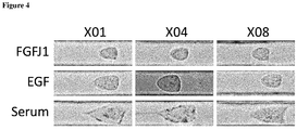
- the analysis of the mechanical phenotype can be performed using Real-Time Deformability Cytometry (RT-DC) (Otto et al., 2015) to quantify cell size (cross-sectional area) and deformation under hydrodynamic shear stress in a microfluidic channel in each cell line and in each culture medium. All three cell lines respond to the three different culture conditions in a specific manner. The different cell culture conditions were associated with a specific morphological phenotype, which was similar for the three different cell lines: In FGFJI, cells from all three patients were smaller in size; in EGF, they were larger; in serum-containing media, they were significantly larger still.
- RT-DC Real-Time Deformability Cytometry
- the data can be plotted as deformation vs. cell area. It has been found that there is an obvious grouping depending on cell culture medium, meaning that cell culture conditions affect the mechanical phenotype of the cells similarly across different cell lines.
- PCA principal component analysis
- PCA is an unsupervised machine learning method by which original multiple variables (here referring to genes) are converted into a set of linearly uncorrelated orthogonal variables (principal components, or PCs) in such a way that the first principal component (PC1) accounts for the largest variability of the data, and the succeeding principal components (PCs) account for the variance in decreasing order under the constraint that each component is orthogonal to the preceding ones (Ringner, 2008).
- the average elastic modulus can now be plotted vs. PC1 for each culture condition to determine a possible correlation (linear regression of the normalized PC1 vs. elastic modulus variables).
- the analysis demonstrated significant correlation between the genomic pattern and cell mechanics. In fact, there was seen a grouping of all three cell lines based upon culture condition.
- the screening method of the invention further comprises the step of detecting the expression of at least one biomarker by said tumor cell line under the first and the second culture conditions, and optionally under the third culture condition, and compared to a control without test compound, wherein said at least one biomarker is selected from the group consisting of:
- the screening method of the invention further comprises the step of detecting the expression of Hes3 by said tumor cell line under the first and the second culture conditions, and optionally under the third culture condition, and compared to a control without test compound, wherein a test compound is selected that decreases the amount of Hes3 in said tumor cell line under the first culture condition and wherein the said Hes3 decreasing activity is indicating that the test compound is amenable for use in the treatment of a proliferative disease.
- Hes3 (ENSG00000173673) is encoded by a gene which has the nucleic acid if SEQ ID NO: 106.
- Gene expression profiling of the above biomarkers and/or Hes3 can e.g. be performed by RNA sequencing. Respective methods and materials for RNA sequencing are known to the person skilled in the art.
- the invention provides an anti-proliferative compound or a pharmaceutical composition comprising said anti-proliferative compound for use in the prevention or treatment of a proliferative disease in a subject, wherein said subject has been preselected for treatment by a screening method comprising the detection of the presence or absence of target cells in a biological sample obtained from the subject.
- the presence of said target cells is an indicator for the susceptibility of said subject for a positive treatment outcome with said anti-proliferative compound.
- the invention further provides a method of preventing or treating a proliferative diseases comprising the administering to a subject in need thereof a therapeutically effective amount an anti-proliferative compound or a pharmaceutical composition comprising said anti-proliferative compound to said subject, wherein said subject has been preselected for treatment by a screening method comprising the detection of the presence or absence of target cells in a biological sample obtained from the subject.
- a screening method comprising the detection of the presence or absence of target cells in a biological sample obtained from the subject.
- the presence of said target cells is an indicator for the susceptibility of said subject for a positive treatment outcome with said anti-proliferative compound.
- the invention further provides the use of an anti-proliferative compound or a pharmaceutical composition comprising said anti-proliferative compound in the preparation of a medicament for the prevention or treatment of a proliferative disease in a subject, wherein said subject has been preselected for treatment by a screening method comprising the detection of the presence or absence of target cells in a biological sample obtained from the subject.
- a screening method comprising the detection of the presence or absence of target cells in a biological sample obtained from the subject.
- the presence of said target cells is an indicator for the susceptibility of said subject for a positive treatment outcome with said anti-proliferative compound.
- the invention relates to the aforementioned anti-proliferative compound for use, method or use, wherein said preselection method further comprises the step of detecting the expression by said target cells of at least one biomarker selected from the group consisting of:
- the invention relates to the aforementioned anti-proliferative compound for use, method or use wherein said preselection method further comprises the step of detecting the expression by said target cells of Hes3, wherein the presence and/or expression of Hes3 is an indicator for the susceptibility of said subject for a positive treatment outcome with said anti-proliferative compound.
- the anti-proliferative compound according to the invention belongs to a group selected from dichlorophenethyl-imidazoles, chlorobenzylphenoles, quinolinoles and macrocyclic lactones.
- said dichlorophenylethyl-imidazole is a 2,4-dichlorophenylethyl-imidazole of formula (I): wherein R 1 -R 11 independently are H; halogen, such as fluorine, chlorine, bromine; or C 1-12 alkyl, preferably C 1-10 alkyl, more preferably C 1-8 alkyl, most preferably C 1-6 alkyl, such as methyl, ethyl, propyl, butyl, pentyl or hexyl; X 1 , X 2 , Y 1 , Y 2 independently are C, O, N, S or a bond.
- R 1 -R 11 independently are H; halogen, such as fluorine, chlorine, bromine; or C 1-12 alkyl, preferably C 1-10 alkyl, more preferably C 1-8 alkyl, most preferably C 1-6 alkyl, such as methyl, ethyl, propyl, butyl, penty
- said chlorobenzylphenol is a compound of formula (II): wherein R 1 -R 9 independently are H; halogen, such as fluorine, chlorine, bromine; or C 1-12 alkyl, preferably C 1-10 alkyl, more preferably C 1-8 alkyl, most preferably C 1-6 alkyl, such as methyl, ethyl, propyl, butyl, pentyl or hexyl; and X is C, O, N, or S.
- R 1 -R 9 independently are H; halogen, such as fluorine, chlorine, bromine; or C 1-12 alkyl, preferably C 1-10 alkyl, more preferably C 1-8 alkyl, most preferably C 1-6 alkyl, such as methyl, ethyl, propyl, butyl, pentyl or hexyl; and X is C, O, N, or S.
- said quinolinole is a compound of formula (III): wherein R 1 -R 7 independently are H; halogen, such as fluorine, chlorine, bromine; or C 1-12 alkyl, preferably C 1-10 alkyl, more preferably C 1-8 alkyl, most preferably C 1-6 alkyl, such as methyl, ethyl, propyl, butyl, pentyl or hexyl.
- R 1 -R 7 independently are H; halogen, such as fluorine, chlorine, bromine; or C 1-12 alkyl, preferably C 1-10 alkyl, more preferably C 1-8 alkyl, most preferably C 1-6 alkyl, such as methyl, ethyl, propyl, butyl, pentyl or hexyl.
- the anti-proliferative compound according to the invention is selected from the group consisting of abamectin, doramectin, quinestrol, benzethonium Cl, ebselen, raloxifene HCl, bisacodyl, econazole nitrate, ramelteon, bithionate Na, escin, ritonavir, broxaldine, hexetidine, selamectin, broxyquinoline, lasalocid Na, sulconazole nitrate, butoconazole, levocetirizine 2HCl, suloctidil, chlorhexidine 2HCl, miconazole nitrate, tioconazole, chloroxine, moxidectin, triclosan, clioquinol, norethynodrel, vinblastine sulfate, clofoctol, oxiconazole nitrate
- the anti-proliferative compound according to the invention is selected from the group consisting of Ramelteon, Quinestrol, raloxifene HCl, diltiazem HCl, levocetirizine 2HCl, Norethynodrel and xylazine.
- the present invention is based on a novel signal transduction pathway that regulates plastic cells, including CSC, the so-called "STAT3-Ser/Hes3 Signaling Axis".
- This unique pathway provides an advantage in drug screening efforts: It can be used as a molecular "blueprint” to devise cell culture conditions that maintain cancer cells in the Hes3 high state (expressing high levels of Hes3), in order to identify drugs that kill them.
- the screening methods of the invention enable the introduction/administration of these drugs to a specific patient subgroup that is more likely to respond to the treatments. Thereby, it is avoided prescribing a medication comprising said drugs to patients who may not benefit from the medication. Moreover, there is provided a better therapeutic prediction to those patients who will be prescribed with the drugs.
- the patient selection rationale is based on the observations that Hes3 high cells behave very differently from Hes3 low cells. More specifically, tumor cells were placed in culture media that lock them in a Hes3 high state vs. a Hes3 low state, which allowed the identification of useful biomarkers that distinguish between the two culture media and cell states. Moreover, cells were placed in culture media that lock them in a Hes3 high state and then Hes3 was knocked down, thereby enabling the identification of additional useful biomarkers.
- Biomarkers were identified by experiments and interrogation, using RNA sequencing, of the entire transcriptome of the cells. For the biomarker identification experiments three different primary human brain tumor cell lines were used, each from a different patient. Therefore, the results are generalizable to many patients.
- the widely used human brain tumor cell line "U-87" was chosen for performing the screening method for suitable anti-proliferative compounds. It was shown that this cell line can be cultured under improved cell culture conditions (grown in FGFIJ medium) and that it behaves like a CSC (i.e., it expresses high levels of the transcription factor Hes3, Hes3 RNA interference opposes growth, cell morphology and global gene expression are different) than when cultured in more commonly-used platforms.
- a library of 1,600 FDA-approved compounds was screened on U-87 cells cultured in the improved conditions. Each drug was screened at both 3 micromolar and 10 micromolar concentrations. Cell viability was assessed by measuring ATP levels.
- Drugs that opposed cell viability were further investigated. These drugs were re-screened under the more common cell culture conditions (in serum-supplemented RMPI medium, where cells are more differentiated). Some drugs were identified that also opposed growth under the more differentiated conditions. We also identified some drugs that only opposed growth in the improved conditions. It was focused on the latter drugs only as being the target compounds of the present invention.
- Estrogen ag/antag, anti-breast cancer 4 DILTIAZEM HCl YES NO YES Ca blocker/vasodilator 5
- Radiodar is an indication of oral bioavailability
- BBB denotes whether the drugs are predicted to cross the blood-brain-barrier
- PGP denotes whether the drugs are likely to be substrates for exporter mechanisms on cells.
- TPM4 tropomyosin 4
- Ensembl ID ENSG00000167460
- ASNS asparagine synthetase (glutamine-hydrolyzing)
- ADAM9 (ADAM metallopeptidase domain 9); Ensembl ID: ENSG00000168615
- GANAB (glucosidase, alpha; neutral AB); Ensembl ID: ENSG00000089597
- X01 and X04 are from patients with glioblastoma multiforme whereas X08 is from a patient with gliosarcoma (Soeda et al., 2008, Soeda et al., 2009).
- the cell lines were previously established from acutely resected human tumor tissues. All human tissues in this study were obtained during surgical resections from patients with newly diagnosed or recurrent tumors. Materials in excess of pathological evaluation were used for research purposes in accordance with protocols approved by the Institutional Review Board of the National Institutes of Health. Written consent was obtained and all research tumor tissues were de-identified (Park et al., 2013).
- Resected tissue was triturated in N2 medium containing 20ng/ml EGF with a 1 ml pipette until no tissue clamps were seen; the triturate was allowed to settle for 1 min and the supernatant was collected, diluted in N2 containing EGF and plated.
- Cells were expanded in serum-free DMEM/F12 medium (Mediatech, 10-090-CV) with N2 supplement and EGF (20 ng/ml, R&D Systems) for 5 days under 5% oxygen conditions and were re-plated fresh or from frozen stocks at 1,000-10,000 cells per cm2. This was repeated for several passages. For the experiments, frozen stocks were thawed and expanded as described before and upon passaging, they were re-plated into different culture conditions as described in the Results section. Gene expression and mechanical analysis were performed after five days in culture. Cell culture experiments were generally performed in accordance with Poser et al, 2013 (Poser and Androutsellis-Theotokis, 2013).
- Human brain tumor cell line U-87 MG (ATCC® HTB-14TM) was also used. This was propagated in serum-containing RPMI medium. It was then re-plated in different media according to experimental needs.
- the primary glioblastoma cell lines used here had never been exposed to serum or other undefined culture media supplements. Their tumorigenic potential was previously established (Soeda et al., 2009). For standard expansion and passaging, cell lines were cultured in monolayer form in the presence of Epidermal Growth Factor (EGF) ( Figure 1 ), based on previously published methods (Park et al., 2013).
- EGF Epidermal Growth Factor
- the three conditions maintain the cells under different signal transduction states, all of which allow for efficient growth (Park et al., 2013).
- Serum contains many strong activators of the JAK-STAT signaling pathway which oppose Hes3 expression (Androutsellis-Theotokis et al., 2006); EGF is a relatively mild activator of the JAK-STAT pathway and maintains these cells in a state of intermediate Hes3 expression; FGFJI suppresses the JAK-STAT pathway and strongly promotes Hes3 expression.
- RNA sequencing Wang et al., 2009
- RT-DC real-time deformability cytometry
- Reads were aligned to the human reference (GRCh38) using GSNAP (v 2014-12-17) (Wu and Nacu, 2010) and Ensembl gene annotation version 78 was used to detect splice sites.
- the uniquely aligned reads were counted with featureCounts (v1.4.6) (Liao et al., 2014) and the same Ensembl annotation. Normalization of the raw read counts based on the library size and testing for differential expression between conditions was performed with the DESeq2 R package (v1.6.2) (Love et al., 2014).
- Experiments addressing the effect of cell culture media composition were performed in triplicates; effects addressing the effect of Hes3 RNA interference were performed in quadruplicates.
- Example 3 Systems biomedical analysis (Experiments under different culture conditions)
- PCA principal component analysis
- PC1 loadings were normalized according to the maximum absolute value to -1 and 1. The top 1% most significantly regulated genes were selected for further analysis.
- samples were ordered from X01, X04 and X08 for each culture condition.
- the genes were ordered according to an unsupervised machine learning technique, MCE, by using the Euclidean distance (Cannistraci et al., 2013, Cannistraci et al., 2010).
- MCE unsupervised machine learning technique
- PC1 Principal Component Analysis
- PC2 is related with cell line (each cell line is derived from a different patient)
- Fig. 2 The data show an undisputable group discrimination (evident linear separations) based upon PC1, suggesting that all three cell lines respond similarly in terms of gene expression when placed under each culture condition.
- the Serum culture condition is separated from the other two conditions by a larger margin (support vector machine (Cortes and Vapnik, 1995) maximum margin computation values: FGFJI to EGF: 0.2; EFG to Serum: 1.0).
- Raw gene read counts of all samples were regularized logarithmic transformed (rlog) using DESeq2 R package (Love et al., 2014). Samples were clustered using Spearman rank correlation, Pearson correlation and Euclidean distance based on transformed counts of all genes. Cluster heatmaps were drawn using the ComplexHeatmap R package (Gu et al., 2016).
- Hes3 RNA interference opposes the growth of all three primary cell lines used here, when cultured in FGFJI (Park et al., 2013), pointing to possible core molecular mechanisms that may be targeted in oncology.
- FGFJI Park et al., 2013
- RNA interference was performed with an siRNA that has been previously validated to oppose Hes3 expression and cell viability (Park et al., 2013). 24 hours later, we collected RNA for transcriptomics analysis by RNA sequencing methods.
- FGF21 one of the 25 triple-common genes, is a well-studied activator of the MAPK signaling pathway.
- FGFJI human brain cancer cell lines, each from a different patient
- EGF which maintains cells in a medium Hes3 expression state
- Serum which maintains cells in a low Hes3 expression state
- Cells (3 human brain cancer cell lines, each from a different patient) were cultured under FGFJI conditions (which maintains cells in a high Hes3 expression state) and then we treated them with either control or Hes3 siRNA. Biomarkers were identified whose expression significantly changes due to Hes3 siRNA.
- TPM4 was identified using both experiments.
- RT-DC Real-time deformability cytometry
- RT-DC Real-Time Deformability Cytometry
- RNA quality was assessed using an Agilent 2100 Bioanalyzer.
- White Corning 384 well plates (Cat #3570) were coated with 40 ⁇ l of 4950 ⁇ g/ml polyornithine (Sigma, # P-365) at 37oC overnight. The next day the plates were washed 5 times with water using a BioTek EL 406 plate washer. The plates were then coated with 40 ⁇ l of 1mg/ml fibronectin (R&D Systems, #1030-Fn) for 2 hours at 37oC and then washed 2 times with PBS.
- the human brain tumor cell line U-87 was used, because it is commonly used in drug screening programs.
- FGFJI the commonly used serum-supplemented RPMI medium (i.e., the two culture conditions with the largest difference in Hes3 expression).
- FIG. 7A shows image examples from 1-day and 8-day cultures in both media. The morphology of the cells differs in the two media, with FGFJI inducing a smaller and more neural stem cell-like morphology.
- PCR analysis demonstrated that cells in FGFJI express higher levels of Hes3 than in RPMI ( Figure 7B ). Cells cultured in FGFJI were also more susceptible to death following Hes3 RNA interference ( Figure 7C ; Y-axis is the cell number as a % of cell number of Cntrl siRNA, in each culture condition).
- cell number in FGFJI was at 21.0% ⁇ 6.8, relative to control (scrambled) - transfected cells (100% ⁇ 16.8), with a significance p value (TTEST) of 3.5x10⁻⁶.
- Cell number in RPMI was at 48.3% ⁇ 25.0, relative to control (scrambled) - transfected cells (100% ⁇ 34.2), with a significance p value (TTEST) of 0.0029.
- a library of 1,600 FDA-approved compounds was screened at two different concentrations (10 ⁇ M and 3 ⁇ M) on U-87 cells cultured in FGFJI ( Figure 8 ). The compounds that significantly reduced cell number were then re-screened, in parallel, with the U-87 cells cultured in both FGFJI and serum conditions. In this way, we identified 37 compounds that only reduced cell number in FGFJI conditions (Table 6). Table 6: List of 37 compounds (from a library of 1,600 FDA-approved compounds) that reduce cell number in FGFJI but not in serum culture conditions.
- the seven compounds selected for dose curve experiments cover six different chemical scaffolds, thus providing broad diversity.
- a clustering was performed based on the structural similarity of the 37 compounds ( Figure 10 ).
- the heatmap reveals that the 37 compounds cover structurally very diverse scaffolds and that there are four larger groups of very closely related compounds and many singletons ( Figure 10 ).
- the first group, "dichlorophenylethyl-imidazole” comprises six compounds, which contain an imidazole group and some chloro groups (Chemical structures are provided in Table 2). All of these compounds have an antifungal effect and are used in different indications as shown in Table 7.
- Table 7 List of 37 compounds with disease indication, number of targets, and protein structures.
- the disease indications of the 37 compounds was analyzed. In total, they cover a broad spectrum including anticancer, antipsychotic, antihistamine, anti-inflammatory, laxative, and sleep agents. However, there was a particular strong focus on anthelmintic, antifungal, and antibiotic actions for 19 of the 37 compounds ( Figure 11 ). With this analysis a complete drug target - disease network was generated and a drug - drug target network.
Landscapes
- Health & Medical Sciences (AREA)
- Life Sciences & Earth Sciences (AREA)
- Engineering & Computer Science (AREA)
- Immunology (AREA)
- Biomedical Technology (AREA)
- Molecular Biology (AREA)
- Chemical & Material Sciences (AREA)
- Hematology (AREA)
- Urology & Nephrology (AREA)
- Physics & Mathematics (AREA)
- General Physics & Mathematics (AREA)
- Cell Biology (AREA)
- Food Science & Technology (AREA)
- Medicinal Chemistry (AREA)
- Biotechnology (AREA)
- Analytical Chemistry (AREA)
- Biochemistry (AREA)
- General Health & Medical Sciences (AREA)
- Microbiology (AREA)
- Pathology (AREA)
- Bioinformatics & Cheminformatics (AREA)
- Toxicology (AREA)
- Tropical Medicine & Parasitology (AREA)
- Measuring Or Testing Involving Enzymes Or Micro-Organisms (AREA)
- Hospice & Palliative Care (AREA)
- Oncology (AREA)
- Pharmaceuticals Containing Other Organic And Inorganic Compounds (AREA)
- Medicines That Contain Protein Lipid Enzymes And Other Medicines (AREA)
Abstract
Description
- The invention provides methods of screening a subject for a proliferative disease risk factor which comprises detecting the presence or absence of target cells. The presence of target cells in the subject indicates the subject is at increased risk of developing a proliferative disease or recurrence of a previously treated proliferative disease. Methods of screening compounds for the treatment of proliferative diseases based on the presence of target cells and the expression of certain biomarkers by said target cells are also disclosed, along with appropriate compounds and their use in methods of treating such diseases.
- The World Health organization states: "Cancer causes 20% of deaths in the European Region. With more than 3 million new cases and 1.7 million deaths each year, cancer is the most important cause of death and morbidity in Europe after cardiovascular diseases". Current therapies for cancer are highly inadequate. Despite valiant efforts to shrink tumor size, patients are vulnerable to disease progression, recurrence, and metastasis, the manifestations of the regenerative properties of cancer.
- A great difficulty in combating cancer lies in the fact that cancer cells are plastic, meaning that a cell can assume different states. Each state operates different signal transduction pathways and is susceptible to different treatments. These differences can be so great that a treatment that kills the cell in one state may actually help the cell in another state to grow better. For example, cancer cells in a state characterized by high Hes3 expression can be killed by Hes3 RNA interference whereas the same cells in a low Hes3 expression state are mostly unaffected. Also, whereas treatment with a JAK inhibitor improves the growth of cells in the Hes3 high state, it kills cells in the Hes3 low state.
- This fundamental problem is hindering drug discovery programs. Most drug screening platforms use established media formulations that maintain the cells in a Hes3 low state. Consequently, the drugs that are identified are effective against this state and not necessarily against the Hes3 high state. Therefore, when these drugs are used therapeutically, they are able to kill many cells (the Hes3 low cells), thus shrinking the tumor but they are not able to kill the Hes3 high cells and the tumor comes back. It almost always comes back in a more aggressive form and it is speculated that this is because the Hes3 high cells are more primitive and more capable of regenerating the tumor. In other words, Hes3 high cells behave as cancer stem cells (CSC).
- More specifically, cell culture systems used in basic research and drug screening programs are not sufficiently able to model the plastic properties of cancer cells. For example, under commonly-used serum-containing systems, brain tumor cells have a homogeneously large morphology, express low levels of Hes3, and their growth is not opposed by Hes3 RNA interference; in contrast, in serum-free containing systems supplemented with mitogens they have a homogeneously small morphology similar to that of non-cancerous neural stem cells, express high levels of Hes3 and Hes3 RNA interference induces cell death (Park et al., 2013).
- It is therefore likely that each different culture condition is only able to interrogate a specific state of these cells. This is an important limitation because it is likely that in the changing microenvironment of a tumor, a cell may be able to adopt different states during its lifespan, each of which may be susceptible to different anti-cancer treatments (Kodack et al., 2017).
- It is important to try and define the different states that a tumor cell can assume under different culture conditions, to understand their significance to the in vivo situation, and to use these different states to identify drugs that target them. Drugs that target one state may be very different from drugs that target another. In fact, drugs that kill a cancer cell in one state may actually promote its growth in another. For example, Janus kinase (JAK) inhibition is considered as a therapeutic in oncology (Buchert et al., 2016, Mukthavaram et al., 2015), but we have also demonstrated that it powerfully promotes the growth of cultured primary human brain tumor cells with cancer stem cell characteristics in serum-free media (Park et al., 2013).
- JAK activity opposes the expression of Hes3 (Androutsellis-Theotokis et al., 2006), a transcriptional and passive repressor (Imayoshi and Kageyama, 2014). We previously demonstrated that glioblastoma cells can grow efficiently under defined conditions that suppress JAK activity and promote Hes3 expression. Under these conditions, Hes3 RNA interference opposes their growth (Park et al., 2013, Androutsellis-Theotokis et al., 2010a). This is of potential clinical interest as Hes3 expressing cells are found in tumor biopsies (Androutsellis-Theotokis et al., 2010a, Park et al., 2013) and www.proteinatlas.org.
- In conclusion, current anti-cancer drugs show no or only limited efficacy against target cells, which leads to the fact that there is a high risk for cancer recurrence after a successful treatment of a cancer disease in a subject. Moreover, patients are insufficiently characterized in regard to the presence of target cells and, accordingly, in regard to the selection of an appropriate treatment strategy and prognosis of treatment success.
- It is therefore the purpose of the invention to overcame the obstacles of the prior art and to provide methods to characterize and predefine patient groups, which are susceptible to certain treatment strategy for proliferative diseases resulting in a successful and long-lasting alleviation of diseases symptoms.
- As further detailed below, the invention is based on the fact that the presence of target cells in a subject indicates the subject is at increased risk of developing a proliferative disease or recurrence of a previously treated proliferative disease.
- In a first aspect, the invention provides a method of screening a subject for a proliferative disease risk factor, comprising detecting the presence or absence of target cells in said subject; the presence of target cells indicating said subject is at increased risk of developing a proliferative disease. Said method of screening a subject for a proliferative disease risk factor enables the characterization and definition of patient subgroups which are susceptible for a selected treatment strategy of a proliferative disease.
- A second aspect of the invention relates to a screening method for identifying an anti-proliferative compound for use in the treatment of a subject that has been identified to be at increased risk of developing a proliferative disease, wherein said method comprises the contacting and incubating a tumor cell line, which resembles a CSC state of the target cells, with a test compound, and the selection of a test compound that reduces the cell number of the tumor cell line.
- A third aspect of the present invention is a method of screening a compound for efficacy in the treatment of a proliferative disease. The method comprises providing a group of subjects characterized by the presence of target cells. The compound to be tested is then administered to the subjects, and the efficacy of the compound in the treatment of the proliferative disease is determined.
- A fourth aspect of the present invention is an in vitro method of screening compounds for efficacy in treating a proliferative disease. The method comprises determining in vitro whether the compound reduces the cell number of a tumor cell line, i.e. kills cells which resemble a CSC state of the target cells, and/or inhibits the growth said tumor cells. The reduction of the cell number of said tumor cell line and/or the inhibition of the growth of said tumor cells indicate the compound is useful in treating the proliferative disease.
- In a fifth aspect, the invention provides anti-proliferative compounds or a pharmaceutical composition comprising said anti-proliferative compounds for use in the prevention or treatment of a proliferative disease in a subject, wherein said subject has been preselected for treatment by a screening method comprising the detection of the presence or absence of target cells in a biological sample obtained from the subject, wherein the presence of said target cells is an indicator for the susceptibility of said subject for a positive treatment outcome with said anti-proliferative compound.
- The present invention is explained in greater detail in the drawings herein and the specification set forth below.
-
-
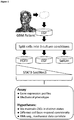
Figure 1 shows the modelling different cell growth states in vitro. Three different primary brain tumor cell lines (each from a different patient) were established following surgical excision and propagated in serum-free medium containing EGF. Cells were then passaged into new flasks, each containing the same base medium formulation but supplemented with different factors [FGFJI, EGF, or Serum]. After five days, cells were collected for either gene expression profiling or RT-DC. [The human diagram was made on the www.biodigital.com website]. -
Figure 2 shows a plot of principal component analysis (PCA). Plot of principal component analysis (PCA) shows a patient discriminative pattern of culture condition (PC1) versus patient cell line (PC2). Numbers indicate SVM maximum margin computation values among the three culture conditions groups. -
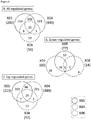
Figure 3 shows the gene expression regulation following Hes3 RNA interference. (A-C) Number of genes regulated by Hes3 siRNA in each of the three cell lines, compared to control (scrambled) siRNA [abs(log2-fold change) >1, and p-adjusted <0.05]. Data are split into all genes regulated, up-regulated genes, and down-regulated genes. -
Figure 4 shows distinct mechanical phenotypes of cells cultured in different conditions. Representative images from the different cell lines in the different culture conditions obtained by RT-DC. -
Figure 5 shows distinct mechanical phenotypes of cells cultured in different conditions. Plots of deformation (1 - circularity) vs. cell size (cross sectional area) for each cell line in each of the three culture conditions. Shown are the 50% density lines of the actual distributions. -
Figure 6 shows distinct mechanical phenotypes of cells cultured in different conditions. (A) Plot of deformation vs. cell size for each cell line in each culture condition. The data show the mode-values of the distributions of thousands of individual cells analyzed for each condition and patient cell line; isoelasticity lines (grey) show places of equivalent elastic modulus. (B) Linear regression plot of z-score (PC1) and z-score (elastic modulus) for each cell line in each culture condition. The PC1 coordinate of each symbol is obtained as the average of the PC1 coordinates of the same respective symbols inFigure 1B . The elastic modulus coordinate of each symbol is calculated as the average of the elastic moduli of the respective symbols in panel A. Both coordinates are z-score transformed to adjust for the different physical scales. -
Figure 7 shows U-87 cells in different culture conditions. (A) U-87 cells grow efficiently in both common media (RPMI supplemented with serum) and FGFJI medium. Images are fromdays -
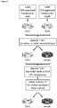
Figure 8 shows drug screening in different culture conditions and the drug screening strategy. -
Figure 9 shows the drug screening in different culture conditions. (A) Dose curves of 6 selected compounds in the serum-containing cell culture condition. (B) Dose curves of the same 6 selected compounds in the FGFJI cell culture condition. [Arrows point to the four of the six compounds used in the dose curve experiment that demonstrated efficacy in FGFJI but not in serum]. -
Figure 10 shows a chemical similarity plot of the 37 hit compounds. Pairwise similarities of compounds reveals four larger groups of related compounds and many singletons. The six selected compounds cover the imidazole, the phenol, and the quinolinole groups as well as three singleton groups thus capturing a great variety of structural scaffolds. -
Figure 11 shows the drug disease network of the 37 hit compounds. Drugs and their disease indications. 19 out of the 37 drugs have an anthelmintic, antifungal, and antibiotic effect. -
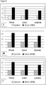
Figure 12 shows biomarker expression (data from three human cell lines pooled together) for the biomarkers TPM4, F3 and ADAM9. These three biomarkers were identified by placing the cells under different cell culture conditions. Here, the gene expression comparison between the condition "Serum" (set to 1) and the condition "FGFJI") is shown. The data from all three cell lines used were calculated together. Thus, the bar graph represents all three cell lines. -
Figure 13 shows gene expression data for the cell lines X01 (A), X04 (B) and X08 (C) for the biomarkers TPM4, ASNS and GANAB. These three biomarkers were identified by placing the cells under the FGFJI condition and then they were treated with either control or Hes3 siRNA. The data from this experiment treated each cell line separately. Therefore, three separate graphs are shown. The graphs are remarkably similar, demonstrating the strength of the data. (Fold expression for the controls is set to "1"). - The term "target cell" as used herein refers to immature cancer cells. A target cell in the sense of the invention is in particular an immature cancer cell of a certain state, i.e. characterized by low JAK activity, high STAT3-Ser to STAT3-Tyr phosphorylation ratio, and high Hes3 expression; and by physical properties, such as smallness and deformability. In particular, the target cells have properties of a Cancer Stem Cell (CSC) when placed in culture. The target cells are sensitive to Hes3 siRNA. The target cells are in a particular state characterized by high Hes3 expression. This state is further characterized by the absence or low expression of additional biomarkers selected from:
- i. TPM4 (tropomyosin 4)
- ii. ASNS (asparagine synthetase (glutamine-hydrolyzing)
- iii. F3 (Coagulation factor III, tissue factor)
- iv. ADAM9 (ADAM metallopeptidase domain 9)
- v. GANAB (glucosidase, alpha; neutral AB)
- The measurement of mRNA levels delivers a single value of mRNA levels in a biopsy. For Hes3, patients with high expression are selected. Patients with "Hes3 high expression" are defined as the top 50% of patients, preferably top 40% of patients, more preferably top 30% of patients, most preferably top 20 % of patients based on the measurement of the Hes3 mRNA level. For the other 5 biomarkers, patients with low Hes3 expression were selected. Patients with "Hes3 low expression" are defined as the bottom 50% of patients, preferably bottom 40% of patients, more preferably bottom 30% of patients, most preferably bottom 20 % of patients based on the measurement of the Hes3 mRNA level. Each biopsy can be assessed by comparing expression levels to other biopsies (a series of 10 or more biopsies may be required to standardize the comparison; alternatively, data can be compared to the data published at www.proteinatlas.org).
- Immunohistochemistry experiments, when performed on a biopsy, deliver two values:
- Incidence of cells; and
- Intensity of staining per cell.
- Both measurements and values are important to characterize a patient to comprise target cells. In a preferred embodiment of the invention, a biopsy comprising intensely stained Hes3+ cells characterizes a patient to comprise target cells. In a further preferred embodiment, a biopsy comprising cells that are completely devoid of expression of the other 5 biomarkers characterizes a patient to comprise target cells. In a further preferred embodiment, a biopsy comprising many cells expressing medium levels of Hes3 characterizes a patient to comprise target cells. In a further preferred embodiment, a biopsy comprising many cells that are mostly (even though not completely) negative for the other 5 biomarkers characterizes a patient to comprise target cells.
- Most preferably, in regard to the immunohistochemistry for Hes3 (the Hes3 high cells are targeted):
- Biopsies where 1% or more of the cells express high levels of Hes3 characterize a patient to comprise target cells. "High Hes3" in this case means a Hes3 level above the average intensity of each cell in a series of biopsies (or, preferably, at the top of 40%, or 30%, or 20% etc. of biopsies measured).
- Biopsies, in which at least 20% or more of the cells express at least medium Hes3 levels characterize a patient to comprise target cells.
- Further most preferred in regard to the immunohistochemistry of the other 5 biomarkers TPM4, ASNS, F3, ADAM9 and GANAB (biomarker low expressing cells are targeted):
- Biopsies, in which 1% or more of the cells express very low levels of at least one of the 5 biomarkers, preferably of two, three or four, most preferably of all five biomarkers characterize a patient to comprise target cells. "Very low" in this case means significantly below the average intensity of each cell in a series of biopsies (e.g., 30%, or 20% etc.).
- Biopsies where at least 20% or more of the cells express medium or lower levels of at least one of the 5 biomarkers, preferably of two, three or four, most preferably of all five biomarkers characterize a patient to comprise target cells. "Medium or low levels" refer to those cells expressing below average levels of the biomarkers.
- The term "risk factor" as used herein indicates subjects that possess the indicated trait or factor and face an increased risk of developing a proliferative diseases than subjects who do not possess the risk factor.
- The term "treat" as used herein refers to any type of treatment that imparts a clinical improvement or alleviation in the condition of the patient, or delays the progression of the disease.
- The term "proliferative disease" as used herein refers to both cancer and non-cancer disease. Preferably the proliferative disease is one characterized by the presence of target cells in the afflicted patients. Illustrative non-cancer diseases include inflammatory and/or immunoproliferative disorders such as arthritis, fibrosis, asthma and allergies. The invention can be used to screen for risk of and/or treat a variety of different types of cancer cells, particularly malignant (and preferably solid) tumors of epithelial or mesenchymal cells. The term "proliferative disease further includes tumor metastasis or tumor recurrence. Examples of cancers that can be screened for risk of and/or treated by the present invention include breast cancer, prostate cancer, pancreatic cancer, adrenal cancer, melanoma, lung cancer, colon cancer, leukemia (a liquid or non-solid tumor), soft tissue and bone sarcomas, neuroendocrine tumors such as islet cell carcinoma or medullary carcinoma of the thyroid, squamous carcinomas (particularly of the head and neck), adenocarcinomas, gliosarcomas such as glioblastoma multiforme etc. The treatment of gliosarcomas is a particularly preferred target for carrying out the present invention. In a further embodiment the treatment of a cancer disease selected from breast cancer, prostate cancer, pancreatic cancer and adrenal cancer is particularly preferred according to the present invention.
- "Stereoisomers": All possible stereoisomers of the claimed compounds are included in the present invention. Where the compounds according to this invention have at least one chiral center, they may accordingly exist as enantiomers. Where the compounds possess two or more chiral centers, they may additionally exist as diastereomers. It is to be understood that all such isomers and mixtures thereof are encompassed within the scope of the present invention.
- Preparation and isolation of stereoisomers: Where the processes for the preparation of the compounds according to the invention give rise to a mixture of stereoisomers, these isomers may be separated by conventional techniques such as preparative chromatography. The compounds may be prepared in racemic form, or individual enantiomers may be prepared either by enantiospecific synthesis or by resolution. The compounds may, for example, be resolved into their components enantiomers by standard techniques, such as the formation of diastereomeric pairs by salt formation with an optically active acid, such as (-)-di-p-toluoyl-d-tartaric acid and/or (+)-di-p-toluoyl-1-tartaric acid followed by fractional crystallization and regeneration of the free base. The compounds may also be resolved by formation of diastereomeric esters or amides, followed by chromatographic separation and removal of the chiral auxiliary. Alternatively, the compounds may be resolved using a chiral HPLC column.
- "Pharmaceutically acceptable salts": In view of the close relationship between the free compounds and the compounds in the form of their salts or solvates, whenever a compound is referred to in this context, a corresponding salt, solvate or polymorph is also intended, provided such is possible or appropriate under the circumstances. Salts and solvates of the compounds of formula (I) and physiologically functional derivatives thereof which are suitable for use in medicine are those wherein the counter-ion or associated solvent is pharmaceutically acceptable. However, salts and solvates having non-pharmaceutically acceptable counter-ions or associated solvents are within the scope of the present invention, for example, for use as intermediates in the preparation of other compounds and their pharmaceutically acceptable salts and solvates.
- Suitable salts according to the invention include those formed with both organic and inorganic acids or bases. Pharmaceutically acceptable acid addition salts include those formed from hydrochloric, hydrobromic, sulfuric, nitric, citric, tartaric, phosphoric, lactic, pyruvic, acetic, trifluoroacetic, triphenylacetic, sulfamic, sulfanilic, succinic, oxalic, fumaric, maleic, malic, mandelic, glutamic, aspartic, oxaloacetic, methanesulfonic, ethanesulfonic, arylsulfonic (for example p-toluenesulfonic, benzenesulfonic, naphthalenesulfonic or naphthalenedisulfonic), salicylic, glutaric, gluconic, tricarballylic, cinnamic, substituted cinnamic (for example, phenyl, methyl, methoxy or halo substituted cinnamic, including 4-methyl and 4-methoxycinnamic acid), ascorbic, oleic, naphthoic, hydroxynaphthoic (for example 1- or 3-hydroxy-2-naphthoic), naphthaleneacrylic (for example naphthalene-2-acrylic), benzoic, 4 methoxybenzoic, 2- or 4-hydroxybenzoic, 4-chlorobenzoic, 4-phenylbenzoic, benzeneacrylic (for example 1,4-benzenediacrylic), isethionic acids, perchloric, propionic, glycolic, hydroxyethanesulfonic, pamoic, cyclohexanesulfamic, salicylic, saccharinic and trifluoroacetic acid. Pharmaceutically acceptable base salts include ammonium salts, alkali metal salts such as those of sodium and potassium, alkaline earth metal salts such as those of calcium and magnesium and salts with organic bases such as dicyclohexylamine and N-methyl-D-glucamine.
- All pharmaceutically acceptable acid addition salt forms of the compounds of the present invention are intended to be embraced by the scope of this invention.
- "Polymorph crystal forms": Furthermore, some of the crystalline forms of the compounds may exist as polymorphs and as such are intended to be included in the present invention. In addition, some of the compounds may form solvates with water (i.e. hydrates) or common organic solvents, and such solvates are also intended to be encompassed within the scope of this invention. The compounds, including their salts, can also be obtained in the form of their hydrates, or include other solvents used for their crystallization.
- While the present invention is primarily concerned with the screening and treatment of human subjects, the invention may also be carried out on animal subjects such as dogs, cats, and horses for veterinary purposes.
- In order to solve the purpose of the invention, the invention provides in a first aspect a method of screening a subject for a proliferative disease risk factor comprising detecting the presence or absence of target cells. The presence of target cells in the subject indicates the subject is at increased risk of developing a proliferative disease or recurrence of a previously treated proliferative disease.
- The method can be carried out whether or not the subject has been previously diagnosed as being afflicted with a proliferative disease, and whether or not the subject has been previously prognosed to be at risk of developing the proliferative disease.
- When the subject has previously been diagnosed as afflicted with a proliferative disease, the method may be carried out to monitor the progression of that disease, or monitor the efficacy of drug treatments that the patient has undergone for the treatment of that disease.
- Decreased cell numbers of target cells would be indicative of efficacy of the drug treatment.
- The step of detecting whether target cells are present or absent can be carried out by any suitable means. For example, the step may be carried out by detecting the presence of target cells in a biological sample taken from the subject. The biological sample is preferably a tissue sample, more preferably a biopsy obtained from a tissue which is supposed to be in a proliferative disease state.
- Cancer cells are plastic, switching between signaling pathways to regulate growth under different conditions. In the tumor microenvironment this likely helps them evade therapies that target specific pathways. One such signaling state is characterized by expression of the transcription factor Hes3 and sensitivity to Hes3 knockdown and can be modeled in vitro by the use of defined culture conditions. Therefore, one method to detect target cells in a tissue sample from a subject is to investigate whether biopsies obtained from a subject co-express Hes3 and/or produce Hes3 in high concentrations. The step of detecting whether the biopsy co-expresses Hes3 can be carried out by any suitable means. For example, the step may be carried out by detecting increased Hes3 mRNA levels in cells of the subject, or by detecting increased levels of the Hes3 protein in cells of the subject. High level Hes3 expression is indicative for the presence of target cells and a subject comprising target cells is at increased risk of developing a proliferative disease.
- Surprisingly, it has been found by the inventors that the target cells, independently from Hes3, are characterized by a set of biomarkers, wherein said biomarkers are selected from the group consisting of
- TPM4 (tropomyosin 4); Ensembl ID: ENSG00000167460;
- ASNS (asparagine synthetase (glutamine-hydrolyzing)); Ensembl ID: ENSG00000070669;
- F3 (Coagulation factor III, tissue factor); Ensembl ID: ENSG00000117525;
- ADAM9 (ADAM metallopeptidase domain 9); Ensembl ID: ENSG00000168615; and
- GANAB (glucosidase, alpha; neutral AB); Ensembl ID: ENSG00000089597;
- In a preferred embodiment the biomarker TPM4 (tropomyosin 4); Ensembl ID: ENSG00000167460 is encoded by the gene, which has the nucleic acid sequence of SEQ ID NO: 101.
- In a preferred embodiment the biomarker ASNS (asparagine synthetase (glutamine-hydrolyzing)); Ensembl ID: ENSG00000070669 is encoded by the gene, which has the nucleic acid sequence of SEQ ID NO: 102.
- In a preferred embodiment the biomarker F3 (Coagulation factor III, tissue factor); Ensembl ID: ENSG00000117525 is encoded by the gene, which has the nucleic acid sequence of SEQ ID NO: 103.
- In a preferred embodiment the biomarker ADAM9 (ADAM metallopeptidase domain 9); Ensembl ID: ENSG00000168615 is encoded by the gene, which has the nucleic acid sequence of SEQ ID NO: 104.
- In a preferred embodiment the biomarker GANAB (glucosidase, alpha; neutral AB); Ensembl ID: ENSG00000089597 is encoded by the gene, which has the nucleic acid sequence of SEQ ID NO: 105.
- Preferably, at least two, at least three or at least four biomarkers selected from TPM4, ASNS, F3, ADAM9 and GANAB are low-level expressed or not expressed and therefore absent in the target cells.
- Most preferably, all of the biomarkers TPM4, ASNS, F3, ADAM9 and GANAB are low-level expressed or not expressed and therefore absent in the target cells.
- For each of the biomarkers TPM4, ASNS, F3, ADAM9 and GANAB, several splice variants exist, resulting in several mRNA transcripts and expressed polypeptides of different length. The table below shows the known coding sequences for mRNA transcripts and expressed proteins for each of the biomarkers TPM4, ASNS, F3, ADAM9 and GANAB.
Table 1. Coding sequences for known mRNA transcripts and expressed proteins for each of the biomarkers TPM-4 (ENSG00000167460) Name Transcript ID Translation ID Preference TPM4-201 ENST00000300933.8 (SEQ ID NO: 1) ENSP00000300933 (SEQ ID NO: 2) Preferred TPM4-222 ENST00000646974.1 (SEQ ID NO: 3) ENSP00000494125 (SEQ ID NO: 4) Preferred TPM4-219 ENST00000643579.1 (SEQ ID NO: 5) ENSP00000495347 (SEQ ID NO: 6) Preferred TPM4-202 ENST00000344824.10 (SEQ ID NO: 7) ENSP00000345230 (SEQ ID NO: 8) Preferred TPM4-216 ENST00000642221.1 (SEQ ID NO: 9) ENSP00000495135 (SEQ ID NO: 10) Preferred TPM4-224 ENST00000647464.1 (SEQ ID NO: 11) ENSP00000496648 (SEQ ID NO: 12) Preferred TPM4-205 ENST00000586833.6 (SEQ ID NO: 13) ENSP00000467087 (SEQ ID NO: 14) Preferred TPM4-221 ENST00000646575.1 (SEQ ID NO: 15) ENSP00000496574 (SEQ ID NO: 16) Preferred TPM4-209 ENST00000588483.1 (SEQ ID NO: 17) ENSP00000466106 (SEQ ID NO: 18) Preferred TPM4-204 ENST00000586499.5 (SEQ ID NO: 19) ENSP00000468246 (SEQ ID NO: 20) Preferred TPM4-215 ENST00000592138.5 (SEQ ID NO: 21) ENSP00000466654 (SEQ ID NO: 22) Preferred TPM4-211 ENST00000589897.1 (SEQ ID NO: 23) ENSP00000466158 (SEQ ID NO: 24) Preferred TPM4-208 ENST00000588410.1 (SEQ ID NO: 25) ENSP00000467250 (SEQ ID NO: 26) Preferred TPM4-210 ENST00000588507.5 (SEQ ID NO: 27) ENSP00000467558 (SEQ ID NO: 28) Preferred TPM4-220 ENST00000645471.1 (SEQ ID NO: 29) ENSP00000494867 (SEQ ID NO: 30) TPM4-223 ENST00000647037.1 (SEQ ID NO: 31) ENSP00000495506 (SEQ ID NO: 32) TPM4-218 ENST00000643494.1 (SEQ ID NO: 33) ENSP00000496389 (SEQ ID NO: 34) TPM4-217 ENST00000642789.1 (SEQ ID NO: 35) ENSP00000494589 (SEQ ID NO: 36) TPM4-207 ENST00000588032.5 (SEQ ID NO: 37) ENSP00000467319 (SEQ ID NO: 38) TPM4-212 ENST00000590180.2 - TPM4-214 ENST00000591645.2 - TPM4-213 ENST00000591226.1 - TPM4-206 ENST00000587201.5 - TPM4-203 ENST00000586193.5 - ASNS (ENSG00000070669) Name Transcript ID Translation ID Preference ASNS-201 ENST00000175506.8 (SEQ ID NO: 39) ENSP00000175506 (SEQ ID NO: 40) Preferred ASNS-203 ENST00000394309.7 (SEQ ID NO: 41) ENSP00000377846 (SEQ ID NO: 42) Preferred ASNS-202 ENST00000394308.7 (SEQ ID NO: 43) ENSP00000377845 (SEQ ID NO: 44) Preferred ASNS-205 ENST00000422745.5 (SEQ ID NO: 45) ENSP00000414901 (SEQ ID NO: 46) Preferred ASNS-209 ENST00000444334.5 (SEQ ID NO: 47) ENSP00000406994 (SEQ ID NO: 48) Preferred ASNS-206 ENST00000437628.5 (SEQ ID NO: 49) ENSP00000414379 (SEQ ID NO: 50) Preferred ASNS-214 ENST00000455086.5 (SEQ ID NO: 51) ENSP00000408472 (SEQ ID NO: 52) Preferred ASNS-208 ENST00000442734.5 (SEQ ID NO: 53) ENSP00000400422 (SEQ ID NO: 54) Preferred ASNS-207 ENST00000437657.5 (SEQ ID NO: 55) ENSP00000394242 (SEQ ID NO: 56) Preferred ASNS-204 ENST00000414884.1 (SEQ ID NO: 57) ENSP00000413797 (SEQ ID NO: 58) Preferred ASNS-210 ENST00000448127.1 (SEQ ID NO: 59) ENSP00000402350 (SEQ ID NO: 60) Preferred ASNS-212 ENST00000453600.5 (SEQ ID NO: 61) ENSP00000408797 (SEQ ID NO: 62) Preferred ASNS-211 ENST00000451771.5 (SEQ ID NO: 63) ENSP00000397802 (SEQ ID NO: 64) Preferred ASNS-213 ENST00000454046.5 (SEQ ID NO: 65) ENSP00000401651 (SEQ ID NO: 66) ASNS-217 ENST00000495255.1 - ASNS-215 ENST00000462436.1 - ASNS-216 ENST00000487714.1 - F3 (ENSG00000117525) Name Transcript ID Translation ID Preference F3-201 ENST00000334047.11 (SEQ ID NO: 67) ENSP00000334145 (SEQ ID NO: 68) Preferred F3-202 ENST00000370207.4 (SEQ ID NO: 69) ENSP00000359226 (SEQ ID NO: 70) Preferred F3-204 ENST00000480356.1 - F3-203 ENST00000478217.5 - ADAM9 (ENSG00000168615) Name Transcript ID Translation ID Preference ADAM9-209 ENST00000487273.6 (SEQ ID NO: 71) ENSP00000419446 (SEQ ID NO: 72) Preferred ADAM9-206 ENST00000481513.5 (SEQ ID NO: 73) ENSP00000417066 (SEQ ID NO: 74) Preferred ADAM9-203 ENST00000466936.5 (SEQ ID NO: 75) ENSP00000420257 (SEQ ID NO: 76) Preferred ADAM9-201 ENST00000379917.7 (SEQ ID NO: 77) ENSP00000369249 (SEQ ID NO: 78) ADAM9-207 ENST00000481873.7 (SEQ ID NO: 79) ENSP00000418437 (SEQ ID NO: 80) ADAM9-204 ENST00000468065.5 (SEQ ID NO: 81) ENSP00000418737 (SEQ ID NO: 82) ADAM9-208 ENST00000484143.1 - ADAM9-202 ENST00000463437.2 - ADAM9-205 ENST00000481058.1 - GANAB (ENSG00000089597) Name Transcript ID Translation ID Preference GANAB-201 ENST00000346178.8 (SEQ ID NO: 83) ENSP00000340466 (SEQ ID NO: 84) Preferred GANAB-216 ENST00000540933.5 (SEQ ID NO: 85) ENSP00000442962 (SEQ ID NO: 86) Preferred GANAB-202 ENST00000356638.7 (SEQ ID NO: 87) ENSP00000349053 (SEQ ID NO: 88) Preferred GANAB-215 ENST00000534779.5 (SEQ ID NO: 89) ENSP00000435306 (SEQ ID NO: 90) Preferred GANAB-204 ENST00000525994.1 (SEQ ID NO: 91) ENSP00000434805 (SEQ ID NO: 92) Preferred GANAB-211 ENST00000532402.5 (SEQ ID NO: 93) ENSP00000432181 (SEQ ID NO: 94) GANAB-214 ENST00000534613.5 (SEQ ID NO: 95) ENSP00000434921 (SEQ ID NO: 96) GANAB-209 ENST00000529737.5 (SEQ ID NO: 97) ENSP00000432593 (SEQ ID NO: 98) GANAB-205 ENST00000526210.1 (SEQ ID NO: 99) ENSP00000433799 (SEQ ID NO: 100) GANAB-213 ENST00000534422.5 - GANAB-212 ENST00000534419.1 - GANAB-207 ENST00000526732.5 - GANAB-203 ENST00000524437.5 - GANAB-208 ENST00000528503.1 - GANAB-210 ENST00000531563.1 - GANAB-206 ENST00000526392.1 - - In a preferred embodiment of the invention, the TPM-4 biomarker according to the invention is a splice variant encoded by a nucleic acid having a sequence selected from SEQ ID NOs: 1, 3, 5, 7, 9, 11, 13, 15, 17, 19, 21, 23, 25, 27, 29, 31, 33, 35 and 37. More preferably, the TPM-4 biomarker according to the invention is a splice variant encoded by a nucleic acid having a sequence selected from SEQ ID NOs: 1, 3, 5, 7, 9, 11, 13, 15, 17, 19, 21, 23, 25 and 27. Even more preferably, the TPM-4 biomarker according to the invention is a splice variant which is expressed and which has an amino acid sequence selected from
SEQ ID NOs - In a preferred embodiment of the invention, the ASNS biomarker according to the invention is a splice variant encoded by a nucleic acid having a sequence selected from SEQ ID NOs: 39, 41, 43, 45, 47, 49, 51, 53, 55, 57, 59, 61, 63 and 65. More preferably, the ASNS biomarker according to the invention is a splice variant encoded by a nucleic acid having a sequence selected from SEQ ID NOs: 39, 41, 43, 45, 47, 49, 51, 53, 55, 57, 59, 61 and 63. Even more preferably, the ASNS biomarker according to the invention is a splice variant which is expressed and which has an amino acid sequence selected from SEQ ID NOs: 40, 42, 44, 46, 48, 50, 52, 54, 56, 58, 60, 62 and 64.
- In a preferred embodiment of the invention, the F3 biomarker according to the invention is a splice variant encoded by a nucleic acid having a sequence selected from SEQ ID NOs:67 and 69. More preferably, the F3 biomarker according to the invention is a splice variant which is expressed and which has an amino acid sequence selected from SEQ ID NOs: 68 and 70.
- In a preferred embodiment of the invention, the ADAM9 biomarker according to the invention is a splice variant encoded by a nucleic acid having a sequence selected from SEQ ID NOs: 71, 73, 75, 77, 79 and 81. More preferably, the ADAM9 biomarker according to the invention is a splice variant encoded by a nucleic acid having a sequence selected from SEQ ID NOs: 71, 73 and 75. Even more preferably, the ADAM9 biomarker according to the invention is a splice variant which is expressed and which has an amino acid sequence selected from SEQ ID NOs: 72, 74 and 76.
- In a preferred embodiment of the invention, the GANAB biomarker according to the invention is a splice variant encoded by a nucleic acid having a sequence selected from SEQ ID NOs: 83, 85, 87, 89, 91, 93, 95, 97 and 99. More preferably, the GANAB biomarker according to the invention is a splice variant encoded by a nucleic acid having a sequence selected from SEQ ID NOs: 83, 85, 87, 89 and 91. Even more preferably, the GANAB biomarker according to the invention is a splice variant which is expressed and which has an amino acid sequence selected from SEQ ID NOs: 84, 86, 88, 90 and 92.
- Target patients should be LOW or NEGATIVE for the above mentioned biomarkers. Generally, immature cells such as target cells express fewer genes than differentiated cells, such as differentiated Cancer Cells. This is because undifferentiated cells do little more than divide and differentiated cells can assume many different fates, each of which expresses different gene sets. Thus, with the absence or low expression of the above mentioned biomarkers it is possible to define patient subgroups with particularly aggressive tumors.
- The step of detecting whether the biopsy co-expresses one or more of the aforesaid biomarkers or not can be carried out by any suitable means. For example, the step may be carried out by detecting mRNA of at least one of a biomarker selected from the group consisting of TPM4, ASNS, F3, ADAM9 and GANAB in cells of the subject, or by detecting increased levels of the proteins of said biomarkers in cells of the subject. Low levels or non-detecting the mRNA and/or the protein of at least one of the foresaid biomarkers indicates that the cells tested in a biopsy obtained from a subject are target cells.
- Accordingly, the present invention provides in an advantageous manner a method to detect the presence of target cells and thereby to preselect subjects in need of an effective treatment, i.e. patients who are reasonably likely to show a positive treatment efficacy for an anti-proliferative compound among the entire group of cancer patients or subjects that are prognosed to be afflicted with a proliferative disease. The invention provides a method to distinguish between patients that are reasonably likely to show a positive treatment efficacy for an anti-proliferative compound from patients that are not likely to show a positive treatment efficacy for said anti-proliferative compound. In particular, the method of the invention enables the definition of a patient subgroup that is characterized by the presence of target cells and therefore by a high risk of developing a severe cancer disease.
- In a further aspect, the invention provides biomarkers for diagnosing the risk of developing a proliferative disease in a subject, wherein said biomarkers are selected from the group consisting of
- TPM4 (tropomyosin 4); Ensembl ID: ENSG00000167460;
- ASNS (asparagine synthetase (glutamine-hydrolyzing)); Ensembl ID: ENSG00000070669;
- F3 (Coagulation factor III, tissue factor); Ensembl ID: ENSG00000117525;
- ADAM9 (ADAM metallopeptidase domain 9); Ensembl ID: ENSG00000168615; and
- GANAB (glucosidase, alpha; neutral AB); Ensembl ID: ENSG00000089597;
- In a further aspect the invention provides a screening method for identifying an anti-proliferative compound for use in the treatment of a subject that has been identified to be at increased risk of developing a proliferative disease, comprising the steps of:
- Culturing a mammalian, preferably a human tumor cell line under a first cell culture condition which suppresses the expression of target cells biomarkers; and under a second cell culture condition, which promotes the expression of CSC biomarkers;
- Contacting and incubating the tumor cell line under the first and second culture conditions with a test compound for a period of at least 6 hours, preferably 12 hours, more preferably 18 hours, most preferably 24 hours;
- Determining the cell number of the tumor cell lines after incubation with the test compound under the first and second culture conditions, and of control without test compound;
- Selecting a test compound that reduces the cell number of the tumor cell line cultured under the first culture condition;
- Preferred according to the invention is a test compound that does not reduce or does not significantly reduce the cell number of the tumor cell line under the second culture condition.
- The first culture condition of the screening method of the invention preferably comprises culturing the tumor cell line in FGFJI medium. "FGFJI" medium comprises basic Fibroblast Growth Factor (bFGF) including a JAK inhibitor. A JAK inhibitor is for example selected from the group consisting of
- 2-(1,1-Dimethylethyl)-9-fluoro-3,6-dihydro-7H-benz[h]-imidaz[4,5-f]isoquinolin-7-one;
- 3-Amino-5-(N-tert-butylsulfonamido-4-phenyl)-indazole;
- 2-Naphthyl-(N-isopropyl,N-benzyl)-β-aminoethylketone;
- 9-(3,4-Dichlorophenyl)-2,7-bis(dimethylaminomethyl)-3,4,5,6,7,9-hexahydro-2H-xanthene-1,8-dione;
- (3Z)-5-(3-Pyridinyl)-3-(1H-pyrrol-2-ylmethylene)-1,3-dihydro-2H-indol-2-one methanesulfonate;
- 4-(4'-Hydroxyphenyl)amino-6,7-dimethoxyquinazoline;
- N-(Pyridin-4-yl)-3-[1-(4-chlorobenzyl)indol-3-yll-propanamide;
- 2-Methyl-1-phenyl-4-pyridin-2-yl-2-(2-pyridin-2-ylethyl)butan-1-one;
- 2-Naphthylvinyl Ketone;
- 4-[(3'-Bromo-4'-hydroxyphenyl)amino]-6,7-dimethoxyquinazoline;
- 10,13-Dimethyl-17-(2-(6-sulfanylidene-3H-purin-9-yl)acetyl)-1,2,6,7,8,9,11,12,14,15,16,17-dodecahydrocyclopenta[a]phenanthren-3-one;
- 1,2,3,4,5,6-Hexabromocyclohexane;
- (S,E)-3-(6-Bromopyridin-2-yl)-2-cyano-N-(cyclopropyl(phenyl)methyl)acrylamide;
- 3-(5-(4-(2-Hydroxy-2-methyl-propionyl)-piperazin-1-yl)-2-trifluoromethyl-phenyl)-4-(1H-indol-3-yl)-pyrrole-2,5-dione;
- 4-Aminobenzoic hydrazide; and

- The second culture condition of the screening method of the invention preferably comprises culturing the tumor cell line in serum-supplemented RPMI medium. "RPMI medium" was developed at the Roswell Park Memorial Institute (RPMI) has been found suitable for a variety of mammalian cells, including HeLa, Jurkat, MCF-7, PC12, PBMC, astrocytes, and carcinomas. RPMI medium can be obtained by a number of distributors, such as Sigma-Aldrich, GIBCO and Thermo Fisher Scientific and is distributed as RPMI-1640. "Serum-supplemented RPMI medium" comprises fetal bovine serum, for example 20 % fetal bovine serum or lower, preferably 15 % fetal bovine serum or lower, more preferably 10 % fetal bovine serum or lower, most preferably 10 % fetal bovine serum.
- In a further embodiment, the screening method of the invention further comprises contacting and incubating the tumor cell line under a third culture condition with a test compound, wherein said third culture condition comprises culturing the tumor cell line in EGF medium.
- The tumor cell line used in the screening method of the invention is preferably the cell line U-87.
- The differences among human brain tumor cells can be assessed when the cells are cultured under three different monolayer ("2D") conditions: (a) Commonly used conditions induced by serum-containing media that are characterized by high JAK activity and low Hes3 expression (RMPI-medium), (b) Serum-free media containing the mitogen Epidermal Growth Factor (EGF) that maintain cells in an intermediate Hes3 expression level (Park et al., 2013) (this condition is oftentimes used for 3D glioblastoma cell culture systems (Brocard et al., 2015), and (c) Serum-free media containing the mitogen basic Fibroblast Growth Factor (bFGF) and a JAK inhibitor which are characterized by low JAK activity and high Hes3 expression levels (RMPI-medium). This condition is more commonly used with monolayer primary neural stem cell cultures (Androutsellis-Theotokis et al., 2006). Gene expression and mechanical properties (by real-time deformability cytometry) can be assessed in the different culture conditions. Moreover, the response to Hes3 RNA interference in the conditions characterized by high Hes3 expression can be assessed.
- Human brain tumor cell line (U-87) is commonly used in drug screening efforts. In the screening method of the present invention, tumor cell line U-87 is used to identify FDA-approved compounds that are effective in cells expressing high Hes3 levels, but not effective in the more commonly used, serum-containing state with low-level or no Hes3 expression, thus focusing the method on potentially overlooked putative therapeutics.
- In a further embodiment, the screening method of the invention further comprises the step of investigating the cell morphology and/or mechanical properties of the tumor cell line under the first and the second culture conditions, and optionally under the third culture condition, wherein the cells under the first culture condition show smaller and more neural stem cell-like morphology.
- The analysis of the mechanical phenotype can be performed using Real-Time Deformability Cytometry (RT-DC) (Otto et al., 2015) to quantify cell size (cross-sectional area) and deformation under hydrodynamic shear stress in a microfluidic channel in each cell line and in each culture medium. All three cell lines respond to the three different culture conditions in a specific manner. The different cell culture conditions were associated with a specific morphological phenotype, which was similar for the three different cell lines: In FGFJI, cells from all three patients were smaller in size; in EGF, they were larger; in serum-containing media, they were significantly larger still.
- Moreover, the data can be plotted as deformation vs. cell area. It has been found that there is an obvious grouping depending on cell culture medium, meaning that cell culture conditions affect the mechanical phenotype of the cells similarly across different cell lines.
- Culture conditions affect both gene expression and mechanical phenotype in a specific manner. This correlation can be quantified. Since the size of the cell and its deformation in the channel are not independent, the apparent elastic modulus for each cell (a means of combining size and deformation into one value) can be calculated, assuming an isotropic, homogeneous elastic object, according to Mietke et al. (Mietke et al., 2015). For the RNA-sequence data analysis, a principal component analysis (PCA) can be performed in order to investigate the presence of gene expression patterns related with the experimental culture conditions and the cell lines in a non-biased (unsupervised) manner (Cannistraci et al., 2010). PCA is an unsupervised machine learning method by which original multiple variables (here referring to genes) are converted into a set of linearly uncorrelated orthogonal variables (principal components, or PCs) in such a way that the first principal component (PC1) accounts for the largest variability of the data, and the succeeding principal components (PCs) account for the variance in decreasing order under the constraint that each component is orthogonal to the preceding ones (Ringner, 2008). The average elastic modulus can now be plotted vs. PC1 for each culture condition to determine a possible correlation (linear regression of the normalized PC1 vs. elastic modulus variables). The analysis demonstrated significant correlation between the genomic pattern and cell mechanics. In fact, there was seen a grouping of all three cell lines based upon culture condition.
- In a further embodiment, the screening method of the invention further comprises the step of detecting the expression of at least one biomarker by said tumor cell line under the first and the second culture conditions, and optionally under the third culture condition, and compared to a control without test compound, wherein said at least one biomarker is selected from the group consisting of:
- TPM4 (tropomyosin 4); Ensembl ID: ENSG00000167460;
- ASNS (asparagine synthetase (glutamine-hydrolyzing)); Ensembl ID: ENSG00000070669;
- F3 (Coagulation factor III, tissue factor); Ensembl ID: ENSG00000117525;
- ADAM9 (ADAM metallopeptidase domain 9); Ensembl ID: ENSG00000168615; and
- GANAB (glucosidase, alpha; neutral AB); Ensembl ID: ENSG00000089597;
- In a further embodiment, the screening method of the invention further comprises the step of detecting the expression of Hes3 by said tumor cell line under the first and the second culture conditions, and optionally under the third culture condition, and compared to a control without test compound, wherein a test compound is selected that decreases the amount of Hes3 in said tumor cell line under the first culture condition and wherein the said Hes3 decreasing activity is indicating that the test compound is amenable for use in the treatment of a proliferative disease.
- In a preferred embodiment, Hes3 (ENSG00000173673) is encoded by a gene which has the nucleic acid if SEQ ID NO: 106.
- Gene expression profiling of the above biomarkers and/or Hes3 can e.g. be performed by RNA sequencing. Respective methods and materials for RNA sequencing are known to the person skilled in the art.
- In a further aspect, the invention provides an anti-proliferative compound or a pharmaceutical composition comprising said anti-proliferative compound for use in the prevention or treatment of a proliferative disease in a subject, wherein said subject has been preselected for treatment by a screening method comprising the detection of the presence or absence of target cells in a biological sample obtained from the subject. The presence of said target cells is an indicator for the susceptibility of said subject for a positive treatment outcome with said anti-proliferative compound.
- The invention further provides a method of preventing or treating a proliferative diseases comprising the administering to a subject in need thereof a therapeutically effective amount an anti-proliferative compound or a pharmaceutical composition comprising said anti-proliferative compound to said subject, wherein said subject has been preselected for treatment by a screening method comprising the detection of the presence or absence of target cells in a biological sample obtained from the subject. The presence of said target cells is an indicator for the susceptibility of said subject for a positive treatment outcome with said anti-proliferative compound.
- The invention further provides the use of an anti-proliferative compound or a pharmaceutical composition comprising said anti-proliferative compound in the preparation of a medicament for the prevention or treatment of a proliferative disease in a subject, wherein said subject has been preselected for treatment by a screening method comprising the detection of the presence or absence of target cells in a biological sample obtained from the subject. The presence of said target cells is an indicator for the susceptibility of said subject for a positive treatment outcome with said anti-proliferative compound.
- In a preferred embodiment, the invention relates to the aforementioned anti-proliferative compound for use, method or use, wherein said preselection method further comprises the step of detecting the expression by said target cells of at least one biomarker selected from the group consisting of:
- TPM4 (tropomyosin 4); Ensembl ID: ENSG00000167460;
- ASNS (asparagine synthetase (glutamine-hydrolyzing)); Ensembl ID: ENSG00000070669;
- F3 (Coagulation factor III, tissue factor); Ensembl ID: ENSG00000117525;
- ADAM9 (ADAM metallopeptidase domain 9); Ensembl ID: ENSG00000168615; and
- GANAB (glucosidase, alpha; neutral AB); Ensembl ID: ENSG00000089597 ;
- In a further preferred embodiment, the invention relates to the aforementioned anti-proliferative compound for use, method or use wherein said preselection method further comprises the step of detecting the expression by said target cells of Hes3, wherein the presence and/or expression of Hes3 is an indicator for the susceptibility of said subject for a positive treatment outcome with said anti-proliferative compound.
- In a more preferred embodiment, the anti-proliferative compound according to the invention belongs to a group selected from dichlorophenethyl-imidazoles, chlorobenzylphenoles, quinolinoles and macrocyclic lactones.
- In a preferred embodiment, said dichlorophenylethyl-imidazole is a 2,4-dichlorophenylethyl-imidazole of formula (I):
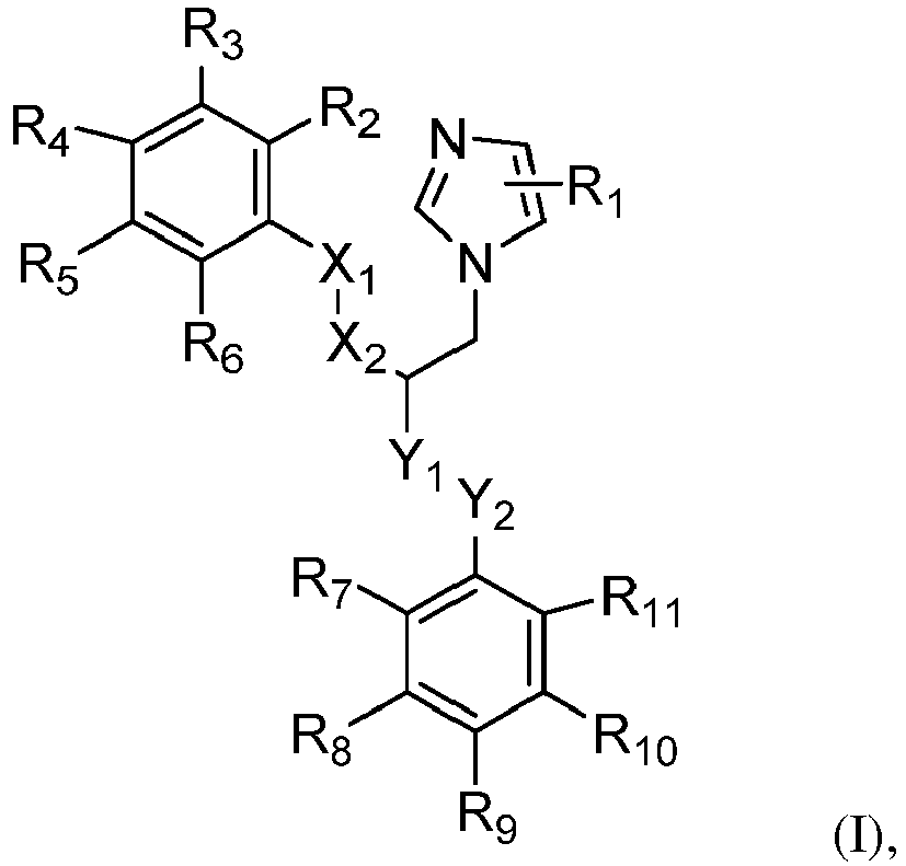
R1-R11 independently are H; halogen, such as fluorine, chlorine, bromine; or C1-12 alkyl, preferably C1-10 alkyl, more preferably C1-8 alkyl, most preferably C1-6 alkyl, such as methyl, ethyl, propyl, butyl, pentyl or hexyl;
X1, X2, Y1, Y2 independently are C, O, N, S or a bond. - In a preferred embodiment, said chlorobenzylphenol is a compound of formula (II):

R1-R9 independently are H; halogen, such as fluorine, chlorine, bromine; or C1-12 alkyl, preferably C1-10 alkyl, more preferably C1-8 alkyl, most preferably C1-6 alkyl, such as methyl, ethyl, propyl, butyl, pentyl or hexyl; and
X is C, O, N, or S. - In a preferred embodiment, said quinolinole is a compound of formula (III):

R1-R7 independently are H; halogen, such as fluorine, chlorine, bromine; or C1-12 alkyl, preferably C1-10 alkyl, more preferably C1-8 alkyl, most preferably C1-6 alkyl, such as methyl, ethyl, propyl, butyl, pentyl or hexyl. - Most preferably, the anti-proliferative compound according to the invention is selected from the group consisting of abamectin, doramectin, quinestrol, benzethonium Cl, ebselen, raloxifene HCl, bisacodyl, econazole nitrate, ramelteon, bithionate Na, escin, ritonavir, broxaldine, hexetidine, selamectin, broxyquinoline, lasalocid Na, sulconazole nitrate, butoconazole, levocetirizine 2HCl, suloctidil, chlorhexidine 2HCl, miconazole nitrate, tioconazole, chloroxine, moxidectin, triclosan, clioquinol, norethynodrel, vinblastine sulfate, clofoctol, oxiconazole nitrate, xylazine, dichlorophen, oxyclozanide, diltiazem HCl, and pimozide.
- Even most preferably, the anti-proliferative compound according to the invention is selected from the group consisting of Ramelteon, Quinestrol, raloxifene HCl, diltiazem HCl, levocetirizine 2HCl, Norethynodrel and xylazine.
- The following table contains more detailed information about the specific antiproliferative compounds used herein:
Table 2. Detailed information on the specific antiproliferative compounds. Compound name Chemical structure Sample publication, which includes the chemical structure 1 ABAMECTIN 
Wang et al., 1982 Wright et al., 1987 2 BENZETHONIUM Cl 
Weiss et al., 1951 Pivnick et al., 1963 benzyl-dimethyl-[2-[2-[4-(2,4,4-trimethylpentan-2-yl)phenoxy]ethoxy]ethyl]azanium chloride 3 BISACODYL 
Wald et al., 2003 Dreiling et al., 1959 [4-[(4-acetyloxyphenyl)-pyridin-2-ylmethyl]phenyl] acetate 4 BITHIONATE Na 
Hopper and Wood, 1958 sodium 2,4-dichloro-6-(3,5-dichloro-2-hydroxyphenyl)sulfanylphenol 5 BROXALDINE 
Bourquin et al., 1962 Sharma et al., 1975 (5,7-dibromo-2-methylquinolin-8-yl)benzoate 6 BROXYQUINOLINE 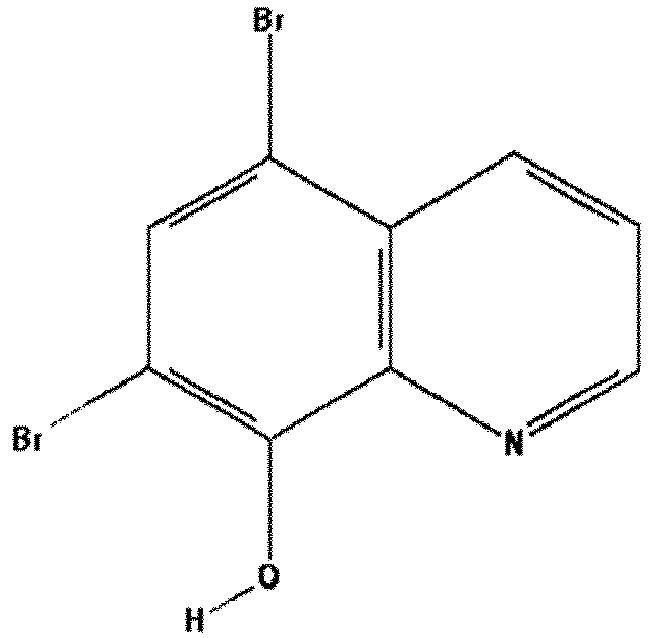
Rodriguez et al., 1968 5,7-dibromoquinolin-8-ol 7 BUTOCONAZOLE 
Odds et al., 1984 Droegemueller et al., 1984 1-[4-(4-chlorophenyl)-2-(2,6-dichlorophenyl)sulfanylbutyl]imidazole 8 CHLORHEXIDINE 2HCl 
Davies et al., 1954 Foulkes, 1973 Kenyon et al, 1986 (1E)-2-[6-[[amino-[(E)-[amino-(4-chloroanilino)methylidene] amino]methylidene]amino]hexyl]-1-[amino-(4-chloroanilino)methylidene]guanidine dihydrochloride 9 CHLOROXINE 
Rohde et al., 1976 Ellenrieder et al., 1970 5,7-dichloroquinolin-8-ol 10 CLIOQUINOL 
Neldner, 1977 Arnett, 1947 5-chloro-7 -iodoquinolin-8-ol 11 CLOFOCTOL 
Danesi et al., 1985 Ghilardi and Casani, 1985 2-[(2,4-dichlorophenyl)methyl]-4-(2,4,4-trimethylpentan-2-yl)phenol 12 DICHLOROPHEN 
Lienert et al., 1966 Miller et al., 1966 Idris et al., 1980 4-chloro-2-[(5-chloro-2-hydroxyphenyl)methyl]phenol 13 DILTIAZEM HCl 
Bevan et al., 1983 Nagao et al., 1973 Sato et al., 1971 [(2S,3S)-5-[2-(dimethylamino)ethyl]-2-(4-methoxyphenyl)-4-oxo-2,3-dihydro-1,5-benzothiazepin-3-yl] acetate hydrochloride 14 DORAMECTIN 
Dutton et al., 1991 Lavy et al., 2003 15 EBSELEN 
Sies et al., 1993 Schewe et al., 1995 Parnham et al., 1990 2-phenyl-1,2-benzoselenazol-3-one 16 ECONAZOLE NITRATE 
Heel et al., 1978 Thienpont et al., 1975 Godefroi et al., 1969 1-[2-[(4-chlorophenyl)methoxy]-2-(2,4-dichlorophenyl)ethyl]imidazolenitric acid 17 ESCIN Ia 
Voigtlander and Rosenberg, 1963 (2S,3S,4S,5R,6R)-6-[[(3S,4S,4aR,6aR,6bS,8R,8aR,9R,10R,12a S,14aR,14bR)-9-acetyloxy-8-hydroxy-4,8a-bis(hydroxymethyl)-4,6a,6b,11,11,14b-hexamethyl-10-[(E)-2-methylbut-2-enoyl]oxy-1,2,3,4a,5,6,7,8,9,10,12,12a,14,14a-tetradecahydropicen-3-yl]oxy]-4-hydroxy-3,5-bis[[(2S,3R,4S,5S,6R)-3,4,5-trihydroxy-6-(hydroxymethyl)oxan-2-yl]oxy]oxane-2-carboxylic acid 18 HEXETIDINE 
Moermann and Muehlemann, 1983 1,3-bis(2-ethylhexyl)-5-methyl-1,3-diazinan-5-amine 19 LASALOCID Na 
Horton and Stockdale, 1981 sodium 6-[(3R,4S,5S,7R)-7-[(2S,3S,5S)-5-ethyl-5-[(2R,5R,6S)-5-ethyl-5-hydroxy-6-methyloxan-2-yl]-3-methyloxolan-2-yl]-4-hydroxy-3,5-dimethyl-6-oxononyl]-2-hydroxy-3-methylbenzoate 20 LEVOCETIRIZINE 2HCl 
Klimek, 2009 Mansfield et al., 2010 2-[2-[4-[(R)-(4-chlorophenyl)-phenylmethyl]piperazin-1-yl] ethoxy]acetic acid dihydrochloride 21 MICONAZOLE NITRATE 
Sawyer et al., 1975 Van Cutsem and Thienpont, 1972 Godefroi et al., 1969 1-[2-(2,4-dichlorophenyl)-2-[(2,4-dichlorophenyl)methoxy]ethyl]imidazole nitric acid 22 MOXIDECTIN 
Webb et al., 1991 Zimmerman et al., 1992 23 NORETHYNODREL 
Edgren, 1991 Pullen, 1962 Paulsen et al., 1962 (8R,9S,13S,14S,17R)-17-ethynyl-17-hydroxy-13-methyl-1,2,4,6,7,8,9,11,12,14,15,16-dodecahydrocyclopenta[a]phenanthren-3-one 24 OXICONAZOLE NITRATE 
Mixich and Thiele, 1979 Polak, 1982 (Z)-1-(2,4-dichlorophenyl)-N-[(2,4-dichlorophenyl)methoxy]-2-imidazol-1-ylethanimine nitric acid 25 OXYCLOZANIDE 
Mrozik et al., 1969 Rajamuthiah et al., 2015 2,3,5-trichloro-N-(3,5-dichloro-2-hydroxyphenyl)-6-hydroxybenzamide 26 PIMOZIDE 
Beninger and Hahn, 1983 Janssen et al., 1968 3-[1-[4,4-bis(4-fluorophenyl)butyl]piperidin-4-yl]-1H-benzimidazol- 2-one 27 QUINESTROL 
Roland et al., 1966 Cohen, 1966 (8R,9S,13S,14S,17R)-3-cyclopentyloxy-17-ethynyl-13-methyl-7,8,9,11,12,14,15,16-octahydro-6H-cyclopenta[a]phenanthren-17-ol 28 RALOXIFENE HCl 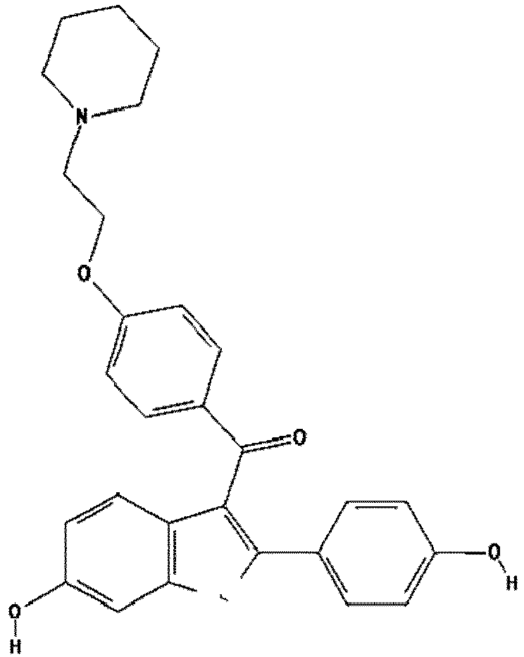
Buelke-Sam et al, 1998 Cummings et al., 1999 Delmas, 2002 Cl [6-hydroxy-2-(4-hydroxyphenyl)-1-benzothiophen-3-yl]-[4-(2-piperidin-1-ylethoxy)phenyl]methanonehydrochloride 29 RAMELTEON 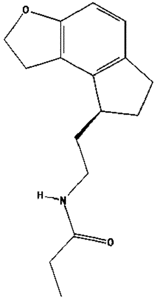
Kato et al., 2005 Greenblatt et al., 2007 N-[2-[(8S)-2,6,7,8-tetrahydro-1H-cyclopenta[e][1]benzofuran-8-yl]ethyl]propanamide 30 RITONA VIR 
Lea and Faulds, 1996 Nachman et al., 2000 Walmsley et al., 2002 1,3-thiazol-5-ylmethyl N-[(2S,3S,5S)-3-hydroxy-5-[[(2S)-3-methyl-2-[[methyl-[(2-propan-2-yl-1,3-thiazol-4-yl)methyl]carbamoyl]amino]butanoyl]amino]-1,6-diphenylhexan-2-yl]carbamate 31 SELAMECTIN 
Benchaoui et al., 2000 Chailleux and Paradis, 2002 32 SULCONAZOLE NITRATE 
Benfield and Clissold, 1988 Akers et al., 1989 1-[2- [(4-chlorophenyl)methylsulfanyl] -2-(2,4-dichlorophenyl)ethyl]imidazole nitric acid 33 SULOCTIDIL 
Chung et al. 1988 Robertson et al., 2012 2-(octylamino)-1-(4-propan-2-ylsulfanylphenyl)propan-1-ol 34 TIOCONAZOLE 
Clayton et al., 1982 Marriott et al., 1983 1-[2-[(2-chlorothiophen-3-yl)methoxy]-2-(2,4-dichlorophenyl)ethyl]imidazole 35 TRICLOSAN 
Lyman and Furia, 1969 Mandel, 1994 5-chloro-2-(2,4-dichlorophenoxy)phenol 36 VINBLASTINE SULFATE 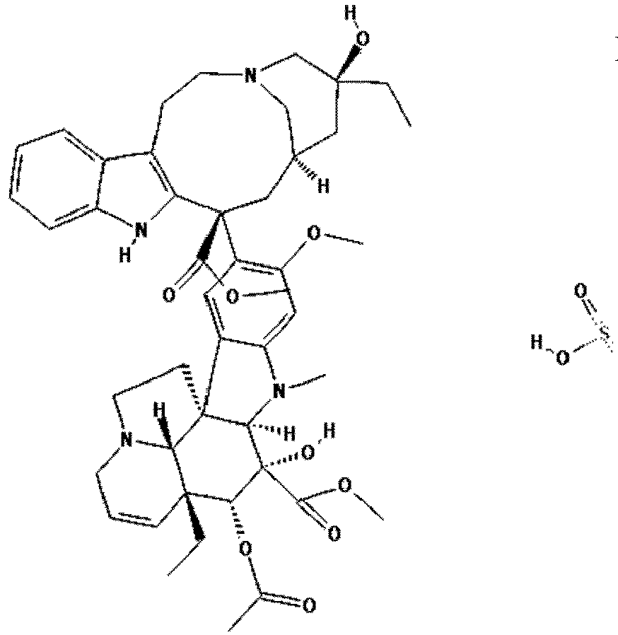
Gobbi et al., 1996 Hertz et al., 1960 37 XYLAZINE 
Clarke and Hall, 1969 N-(2,6-dimethylphenyl)-5,6-dihydro-4H-1,3-thiazin-2-amine - The present invention is based on a novel signal transduction pathway that regulates plastic cells, including CSC, the so-called "STAT3-Ser/Hes3 Signaling Axis". The use of this unique pathway provides an advantage in drug screening efforts: It can be used as a molecular "blueprint" to devise cell culture conditions that maintain cancer cells in the Hes3 high state (expressing high levels of Hes3), in order to identify drugs that kill them.
- Specifically, several FDA-approved drugs were identified with the goal to repurpose them for use in oncology. These drugs as identified herein are not efficient at killing Hes3 low cells (expressing no or only low levels of Hes3), but are very efficient at killing the cells in the Hes3 high state.
- The screening methods of the invention enable the introduction/administration of these drugs to a specific patient subgroup that is more likely to respond to the treatments. Thereby, it is avoided prescribing a medication comprising said drugs to patients who may not benefit from the medication. Moreover, there is provided a better therapeutic prediction to those patients who will be prescribed with the drugs. The patient selection rationale is based on the observations that Hes3 high cells behave very differently from Hes3 low cells. More specifically, tumor cells were placed in culture media that lock them in a Hes3 high state vs. a Hes3 low state, which allowed the identification of useful biomarkers that distinguish between the two culture media and cell states. Moreover, cells were placed in culture media that lock them in a Hes3 high state and then Hes3 was knocked down, thereby enabling the identification of additional useful biomarkers.
- Biomarkers were identified by experiments and interrogation, using RNA sequencing, of the entire transcriptome of the cells. For the biomarker identification experiments three different primary human brain tumor cell lines were used, each from a different patient. Therefore, the results are generalizable to many patients.
- Following data analysis, a number of putative biomarkers was identified that could be used for selecting a patient subgroup. To select the best biomarkers, a number of criteria was applied:
The widely used human brain tumor cell line "U-87" was chosen for performing the screening method for suitable anti-proliferative compounds. It was shown that this cell line can be cultured under improved cell culture conditions (grown in FGFIJ medium) and that it behaves like a CSC (i.e., it expresses high levels of the transcription factor Hes3, Hes3 RNA interference opposes growth, cell morphology and global gene expression are different) than when cultured in more commonly-used platforms. - A library of 1,600 FDA-approved compounds was screened on U-87 cells cultured in the improved conditions. Each drug was screened at both 3 micromolar and 10 micromolar concentrations. Cell viability was assessed by measuring ATP levels.
- Drugs that opposed cell viability were further investigated. These drugs were re-screened under the more common cell culture conditions (in serum-supplemented RMPI medium, where cells are more differentiated). Some drugs were identified that also opposed growth under the more differentiated conditions. We also identified some drugs that only opposed growth in the improved conditions. It was focused on the latter drugs only as being the target compounds of the present invention.
- Altogether 37 compounds opposed cell viability only under the improved (more CSC-like) conditions (when cells were cultured in FGFJI medium).
- Overall, 7 compounds have been identified that in the oppose viability of U-87 cells only when these are cultured under the improved conditions (Hes3 high, FGFJI medium). The 7 selected compounds are advantageous because they are orally available and can be administered orally. The relevant lists of compounds is shown in the table below:
Table 3. List of the 7 compounds. Compound Radar BBB PGP Function 1 Ramelteon YES YES YES Sleep 2 Quinestrol YES YES NO Estrogen replacement 3 RALOXIFENE HCl YES ? ? Estrogen ag/antag, anti-breast cancer 4 DILTIAZEM HCl YES NO YES Ca blocker/vasodilator 5 LEVOCETIRIZINE 2HCl YES YES YES antihistamine 6 Norethynodrel YES YES YES Contraceptive 7 XYLAZINE YES YES NO Vet Med adren alpha-2 agonist - "Radar" is an indication of oral bioavailability; "BBB" denotes whether the drugs are predicted to cross the blood-brain-barrier; "PGP" denotes whether the drugs are likely to be substrates for exporter mechanisms on cells.
- The invention is now described in more detail and in the following working examples .
-
- RNA expression in gliomas: 55 FPKM
- How many types of cancer express its RNA: 17/17
- How many types of cancer express its protein: 20/20
- How good is the antibody used above: "Approved"
- Favorability of gene expression in gliomas: None
- Histopathology expression range: Broad (i.e., from -ve to +ve patients)
- Favorability of gene expression in other cancers: Unfavorable (2 cancer types)
- Direction of expression in our experiments: UP after Hes3 siRNA AND regulated by culture conditions
- Target patient group: Biomarker negative patients
- Special attribute: Biomarker identified by both experiments (i.e., changing culture conditions AND Hes3 siRNA)
-
- RNA expression in gliomas: 16 FPKM
- How many types of cancer express its RNA: 17/17
- How many types of cancer express its protein: 14/20
- How good is the antibody used above: "Approved"
- Favorability of gene expression in gliomas: None
- Histopathology expression range: Broad
- Favorability of gene expression in other cancers: Unfavorable (4 cancer types)
- Direction of expression in our experiments: UP after Hes3 siRNA
- Target patient group: Biomarker negative patients
- Special attribute: Unfavorable in 4 cancer types (this number is quite high)
-
- RNA expression in gliomas: 67 FPKM
- How many types of cancer express its RNA: 17/17
- How many types of cancer express its protein: 17/20
- How good is the antibody used above: "Approved"
- Favorability of gene expression in gliomas: None. Tendency for Unfavorable
- Histopathology expression range: Broad
- Favorability of gene expression in other cancers: Unfavorable (2 cancer types)
- Direction of expression in our experiments: DOWN as conditions increase Hes3
- Target patient group: Biomarker negative patients
- Special attribute: Strong separation; patients are either -ve or very high. Also, Unfavorable tendency in gliomas, so particularly unexpected biomarker.
-
- RNA expression in gliomas: 30 FPKM
- How many types of cancer express its RNA: 17/17
- How many types of cancer express its protein: Pending from www.proteinatlas.org
- How good is the antibody used above: Pending
- Favorability of gene expression in gliomas: None from www.proteinatlas.org.
A recent publication (PMID: 27571068): Unfavorable! - Histopathology expression range: Pending
- Favorability of gene expression in other cancers: Unfavorable (5 cancer types)
- Direction of expression in our experiments: DOWN as conditions increase Hes3
- Target patient group: Biomarker negative patients
- Special attribute: This is a new biomarker. Unfavorable in 5 cancers as well as in gliomas (the latter according to a recent publication).
-
- RNA expression in gliomas: 84 FPKM
- How many types of cancer express its RNA: 17/17
- How many types of cancer express its protein: 19/20
- How good is the antibody used above: "Enhanced"
- Favorability of gene expression in gliomas: None
- Histopathology expression range: Broad
- Favorability of gene expression in other cancers: Unfavorable (2 cancer types)
- Direction of expression in our experiments: UP after Hes3 siRNA
- Target patient group: Biomarker negative patients
- Special attribute: Overall sturdy (many cancer types, good antibody staining)
- Three primary human brain tumor cell lines ("X01", "X04", and "X08") were used in this work. X01 and X04 are from patients with glioblastoma multiforme whereas X08 is from a patient with gliosarcoma (Soeda et al., 2008, Soeda et al., 2009). The cell lines were previously established from acutely resected human tumor tissues. All human tissues in this study were obtained during surgical resections from patients with newly diagnosed or recurrent tumors. Materials in excess of pathological evaluation were used for research purposes in accordance with protocols approved by the Institutional Review Board of the National Institutes of Health. Written consent was obtained and all research tumor tissues were de-identified (Park et al., 2013). Resected tissue was triturated in N2 medium containing 20ng/ml EGF with a 1 ml pipette until no tissue clamps were seen; the triturate was allowed to settle for 1 min and the supernatant was collected, diluted in N2 containing EGF and plated. Cells were expanded in serum-free DMEM/F12 medium (Mediatech, 10-090-CV) with N2 supplement and EGF (20 ng/ml, R&D Systems) for 5 days under 5% oxygen conditions and were re-plated fresh or from frozen stocks at 1,000-10,000 cells per cm2. This was repeated for several passages. For the experiments, frozen stocks were thawed and expanded as described before and upon passaging, they were re-plated into different culture conditions as described in the Results section. Gene expression and mechanical analysis were performed after five days in culture. Cell culture experiments were generally performed in accordance with Poser et al, 2013 (Poser and Androutsellis-Theotokis, 2013).
- Human brain tumor cell line U-87 MG (ATCC® HTB-14™) was also used. This was propagated in serum-containing RPMI medium. It was then re-plated in different media according to experimental needs.
- The primary glioblastoma cell lines used here had never been exposed to serum or other undefined culture media supplements. Their tumorigenic potential was previously established (Soeda et al., 2009). For standard expansion and passaging, cell lines were cultured in monolayer form in the presence of Epidermal Growth Factor (EGF) (
Figure 1 ), based on previously published methods (Park et al., 2013). Each cell line was split into separate flasks and cultured in three distinct conditions for five days, containing: (a) 10% fetal bovine serum (Serum), (b) EGF, or, (c) basic Fibroblast Growth Factor (bFGF) including a JAK inhibitor (bFGF+JAK Inhibitor = "FGFJI"), all in a 5% oxygen incubator. The three conditions maintain the cells under different signal transduction states, all of which allow for efficient growth (Park et al., 2013). Serum contains many strong activators of the JAK-STAT signaling pathway which oppose Hes3 expression (Androutsellis-Theotokis et al., 2006); EGF is a relatively mild activator of the JAK-STAT pathway and maintains these cells in a state of intermediate Hes3 expression; FGFJI suppresses the JAK-STAT pathway and strongly promotes Hes3 expression. - Following culture under each specific condition, cells were collected for RNA sequencing (Wang et al., 2009) or real-time deformability cytometry (RT-DC) (Otto et al., 2015).
- RNA was extracted using the High Pure RNA Isolation Kit (Roche) and samples were subjected to the standard workflow for strand-specific RNA-Seq library preparation (Ultra Directional RNA Library Prep, NEB). Libraries were equimolar, pooled and sequenced on an Illumina HiSeq 2500, resulting in ca. 26 - 35 million single-end reads per library. After sequencing, FastQC (http://www.bioinformatics.babraham.ac.uk/) was used to perform a basic quality control on the resulting reads. As an additional control, library diversity was assessed by redundancy investigation in the reads. Reads were aligned to the human reference (GRCh38) using GSNAP (v 2014-12-17) (Wu and Nacu, 2010) and Ensembl gene annotation version 78 was used to detect splice sites. The uniquely aligned reads were counted with featureCounts (v1.4.6) (Liao et al., 2014) and the same Ensembl annotation. Normalization of the raw read counts based on the library size and testing for differential expression between conditions was performed with the DESeq2 R package (v1.6.2) (Love et al., 2014). Experiments addressing the effect of cell culture media composition were performed in triplicates; effects addressing the effect of Hes3 RNA interference were performed in quadruplicates.
- For the RNA-seq. data analysis, we performed principal component analysis (PCA) in order to investigate the presence of gene expression patterns related with the experimental culture conditions and the cell lines in a non-biased (unsupervised) manner (Cannistraci et al., 2010). PCA is an unsupervised machine learning method by which original multiple variables (here referring to genes) are converted into a set of linearly uncorrelated orthogonal variables (principal components, or PCs) in such a way that the first PC (PC1) accounts for the largest variability of the data, and the succeeding PCs account for the variance in decreasing order under the constraint that each component is orthogonal to the preceding ones (Ringner, 2008).
- For the heat map generation, PC1 loadings were normalized according to the maximum absolute value to -1 and 1. The top 1% most significantly regulated genes were selected for further analysis. For the x-axis, samples were ordered from X01, X04 and X08 for each culture condition. For the y-axis, the genes were ordered according to an unsupervised machine learning technique, MCE, by using the Euclidean distance (Cannistraci et al., 2013, Cannistraci et al., 2010). The color bar indicates the expression, as the log10 (1+raw expression read counts).
- A Principal Component Analysis (PCA) was performed and it was found that PC1 is related with culture condition and PC2 is related with cell line (each cell line is derived from a different patient) (
Fig. 2 ). The data show an undisputable group discrimination (evident linear separations) based upon PC1, suggesting that all three cell lines respond similarly in terms of gene expression when placed under each culture condition. The Serum culture condition is separated from the other two conditions by a larger margin (support vector machine (Cortes and Vapnik, 1995) maximum margin computation values: FGFJI to EGF: 0.2; EFG to Serum: 1.0). - In order to determine the main functions related to the top 1% most significantly regulated genes, we performed functional annotation analysis in DAVID (Huang da et al., 2009b, Huang da et al., 2009a) using Ensemble geneID. Then all the significant GO terms and KEGG pathways were considered that achieved a Benjamini corrected p-value lower than 0.05. Since the highest significant GO term (GO:0008092∼cytoskeletal protein binding) and the highest significant pathway (hsa04510:Focal adhesion) were related with cell mechanics, it was decided to further investigate the list of genes contained in the top 20 significant cell mechanics-related GO annotations. To this aim, it was proceeded to the construction of a 'mechanomic' protein-protein interaction (PPI) network where only those genes belonging to the top 20 significant cell mechanics-related GO annotations were considered (201 unique genes were retrieved from the original list of the top 1% regulated genes that were 400). Nearly half of the top 1% most significantly regulated genes are involved with cell-mechanics. STRING was used to build the PPI network (Szklarczyk et al., 2015) and the Cytoscape software for visualizations (Shannon et al., 2003). To select reliable interactions, only links validated by experimental evidence and higher than a 0.7 cut-off were considered to build the networkIn order to correct for the magnitude discrepancy of each gene (forcing an equal contribution of every gene to the final pattern) the expression of every gene is firstly normalized by dividing the sum of this gene's expression in FGFJI or EGF in the investigated module, and then all the composed genes of this module in FGFJI or EGF state are averaged to get the general expression of the particular state.
- The GO enrichment analysis of these genes revealed that both the most significant GO term (GO:0008092∼cytoskeletal protein binding, corrected P-Value<0.0001) and the most significant pathway (hsa04510:Focal adhesion, corrected P-Value<0.0001) are related with cell mechanics (Table 4).
Table 4: GO Terms related to mechanical phenotypes derived from the list of top 400 genes. Term Count % p Value GO:0008092∼cytoskeletal protein binding 53 13.58974 4.47E-18 GO:0003779∼actin binding 43 11.02564 3.20E-18 GO:0031012∼extracellular matrix 41 10.51282 4.26E-15 GO:0005578∼proteinaceous extracellular matrix 38 9.74359 5.35E-14 GO:0044420∼extracellular matrix part 24 6.153846 5.29E-14 GO:0015629∼actin cytoskeleton 33 8.461538 1.38E-12 hsa04510:Focal adhesion 33 8.461538 1.40E-12 GO:0005856∼cytoskeleton 82 21.02564 5.84E-12 GO:0030036∼actin cytoskeleton organization 30 7.692308 8.19E-13 GO:0044421∼extracellular region part 64 16.41026 2.27E-11 GO:0030029∼actin filament-based process 31 7.948718 7.25E-13 GO:0007010∼cytoskeleton organization 40 10.25641 9.95E-12 GO:0022610∼biological adhesion 52 13.33333 1.34E-11 GO:0006928-cell motion 42 10.76923 8.76E-12 GO:0007155∼cell adhesion 52 13.33333 1.29E-11 hsa04512:ECM- receptor interaction 20 5.128205 1.46E-10 GO:0043062∼extracellular structure organization 21 5.384615 7.03E-09 GO:0005576∼extracellular region 94 24.10256 3.98E-08 GO:0032989∼cellular component morphogenesis 32 8.205128 3.76E-08 GO:0030198∼ extracellular matrix organization 16 4.102564 6.34E-08 - For the Gene Ontology (GO) Term analysis, the 400 top genes were used for DAVID annotation analysis (using Ensemble geneID). We focused on mechanics - related GO terms which were sorted by Benjamini multiple correction and the top 20 GO terms were chosen for further network construction.
- Pathway analysis was done using two different methods. First, Gene Ontology (GO) term and KEGG pathway enrichment of differentially expressed genes (adjusted p <0.05) were calculated using DAVID Bioinformatics Resource (Huang da et al., 2009b) based on Ensembl IDs. The background set consisted of all genes passed to DESeq2. Second, the R package fgsea (https://doi.org/10.1101/060012) was used for a full gene set enrichment analysis based on all genes. -log10(p-value) · log2(fold-change) was used as rank function and 100,000 permutations were done for enrichment p-value calculation. KEGG pathways were plotted using the R package pathview (Luo et al., 2002).
- Raw gene read counts of all samples were regularized logarithmic transformed (rlog) using DESeq2 R package (Love et al., 2014). Samples were clustered using Spearman rank correlation, Pearson correlation and Euclidean distance based on transformed counts of all genes. Cluster heatmaps were drawn using the ComplexHeatmap R package (Gu et al., 2016).
- Hes3 RNA interference opposes the growth of all three primary cell lines used here, when cultured in FGFJI (Park et al., 2013), pointing to possible core molecular mechanisms that may be targeted in oncology. Here it was set out to identify the genes and signaling pathways affected by Hes3 RNA interference in vitro that are common in all three cell lines, and which may provide therapeutically amenable putative drug targets.
- The X01, X04, and X08 cell lines were cultured under FGFJI conditions (i.e., under conditions that maintain high Hes3 expression). Then, RNA interference was performed with an siRNA that has been previously validated to oppose Hes3 expression and cell viability (Park et al., 2013). 24 hours later, we collected RNA for transcriptomics analysis by RNA sequencing methods.
- Hes3 RNA interference induced gene expression changes in all three cell lines. It was focused on those genes that were significantly regulated at least two-fold from control (scrambled) siRNA (log2-fold change cut-off, with an adjusted p-value <0.05). There were 290 differentially regulated genes for X01, 449 for X04, and 56 for X08. 25 genes were common to all three cell lines (triple-common) (
Fig. 3A-C , Table 5).Table 5: Triple-common regulated genes in the cell lines X01, X04, and X08, following Hes3 RNA interference (log2 Fold change, p-adj<0.05). Ensembl_ID Gene_Symbol Description Upregulated by Hes3 siRNA ENSG00000167460 TPM4 tropomyosin 4 ENSG00000254332 GS1-44D20.1 ENSG00000089597 GANAB glucosidase, alpha; neutral AB ENSG00000171700 RGS19 regulator of G- protein signaling 19ENSG00000204611 ZNF616 zinc finger protein 616 ENSG00000213846 AC098614.2 ENSG00000101255 TRIB3 tribbles pseudokinase 3 ENSG00000158373 HIST1H2BD histone cluster 1, H2bd ENSG00000128165 ADM2 adrenomedullin 2 ENSG00000139269 INHBE inhibin, beta E ENSG00000070669 ASNS asparagine synthetase (glutamine-hydrolyzing) ENSG00000272405 RP11-284F21.10 ENSG00000100889 PCK2 phosphoenolpyruvate carboxykinase 2 (mitochondrial) ENSG00000182459 TEX19 testis expressed 19 ENSG00000261371 PECAM1 platelet/endothelial cell adhesion molecule 1ENSG00000138678 AGPAT9 1-acylglycerol-3-phosphate O- acyltransferase 9ENSG00000105550 FGF21 fibroblast growth factor 21 ENSG00000235513 RP4-756G23.5 ENSG00000272068 RP11-284F21.9 Downregulated by Hes3 siRNA ENSG00000181061 HIGD1A HIG1 hypoxia inducible domain family, member 1A ENSG00000160877 NACC1 nucleus accumbens associated 1, BEN and BTB (POZ) domain containing ENSG00000171150 SOCS5 suppressor of cytokine signaling 5 ENSG00000258016 HIGD1AP1 HIG1 hypoxia inducible domain family, member 1A pseudogene 1ENSG00000072401 UBE2D1 ubiquitin-conjugating enzyme E2D 1ENSG00000248785 HIGD1AP14 HIG1 hypoxia inducible domain family, member 1A pseudogene 14Downregulated from Serum to EGF AND from Serum to FGFJI ENSG00000117525 F3 coagulation factor III Downregulated from Serum to FGFJI ENSG00000168615 ADAM9 ADAM metallopeptidase domain 9 - We found three GO Term categories that were regulated in all three cell lines by Hes3 siRNA (GO:0005654∼nucleoplasm, GO:0005515∼protein binding, GO:0005737∼cytoplasm). Additional GO Terms were found regulated when only the X01 and X04 cell lines were used in the analysis, of which GO:0006260∼DNA replication, GO:0051301∼cell division and GO:0006281∼DNA repair are noteworthy.
- FGF21, one of the 25 triple-common genes, is a well-studied activator of the MAPK signaling pathway.
- Unsupervised (non-selected) clustering of all differentially regulated genes shown in
Figure 3A-C was done based on Pearson correlation of rlog transformed expression values. - Two distinct groups of experiments were performed:
- Cells (3 human brain cancer cell lines, each from a different patient) were cultured under three different cell culture conditions (FGFJI, which maintains cells in a high Hes3 expression state; EGF, which maintains cells in a medium Hes3 expression state; and Serum, which maintains cells in a low Hes3 expression state). Biomarkers were identified that change significantly among these states.
- Cells (3 human brain cancer cell lines, each from a different patient) were cultured under FGFJI conditions (which maintains cells in a high Hes3 expression state) and then we treated them with either control or Hes3 siRNA. Biomarkers were identified whose expression significantly changes due to Hes3 siRNA.
- TPM4 was identified using both experiments.
- F3 and ADAM9 were identified from
experiment 1. - ASNS and GANAB were identified from
experiment 2. - Real-time deformability cytometry (RT-DC) was performed as previously described (Otto et al., 2015, Mietke et al., 2015). Briefly, it allows a marker-free, continuous cell mechanical characterization of large cell populations with analysis rates greater than 100 cells/s. Cells are flowed through a microfluidic channel constriction and deformed without contact by shear stresses and pressure gradients. Cell size and deformation is extracted from the high-speed camera images in real-time and can be transformed into an elastic modulus (of stiffness of a cell) by applying an analytical model relating geometrical parameters to material properties (Mietke et al., 2015). Statistical data analysis was carried out utilizing mixed models (Bates et al., 2014b, Bates et al., 2014a) by assuming random as well as fixed effects for the experimental repeats.
- Due to the cell mechanics implications raised by the gene expression data, an analysis of the mechanical phenotype was performed, using Real-Time Deformability Cytometry (RT-DC) (Otto et al., 2015) to quantify cell size (cross-sectional area) and deformation under hydrodynamic shear stress in a microfluidic channel in each cell line and in each culture medium. Example images demonstrate the specific manner by which all three cell lines respond to the three different culture conditions (
Figure 4 ). The different cell culture conditions were associated with a specific morphological phenotype, which was similar for the three different cell lines: In FGFJI, cells from all three patients were smaller in size; in EGF, they were larger; in serum-containing media, they were significantly larger still. - When the data is plotted as deformation vs. cell area, again, there is an obvious grouping depending on cell culture medium (
Figures 5 and6A ), meaning that cell culture conditions affect the mechanical phenotype of the cells similarly across different cell lines. - The specific manner in which culture conditions affect both gene expression and mechanical phenotype prompted us to quantify this correlation. Since the size of the cell and its deformation in the channel are not independent, the apparent elastic modulus for each cell was calculated (a means of combining size and deformation into one value), assuming an isotropic, homogeneous elastic object, according to Mietke et al. (Mietke et al., 2015). We plotted the average elastic modulus vs. PC1 for each condition (
Figure 6B ) to determine a possible correlation (linear regression of the normalized PC1 vs. elastic modulus variables). The analysis demonstrates significant correlation (p < 0.001; linear regression correlation coefficient R = 0.92) between the two variables, therefore indicating high correlation between the genomic pattern and cell mechanics. In fact, there was again a grouping of all three cell lines based upon culture condition. - Cells were plated into 6 well plates containing N2 medium supplemented with 20 ng/ml FGF and 200 nM Jak inhibitor ("FGFJI"). 48 hours later, cells were transfected with either scrambled control siRNA (Santa Cruz Biotechnology SC37007) or Hes3 siRNA (Santa Cruz Biotechnology SC 88003) using Lipofectamine RNAiMax (Thermo Fischer Scientific) transfection reagent as described by the manufacturer. Cells were collected 24 hours post-transfection, and total RNA was isolated using a HighPure RNA isolation kit (Roche). Experiments were performed in quadruplicates for each cell line and siRNA transfection. RNA quality was assessed using an Agilent 2100 Bioanalyzer.
- White Corning 384 well plates (Cat #3570) were coated with 40µl of 4950 µg/ml polyornithine (Sigma, # P-365) at 37oC overnight. The next day the plates were washed 5 times with water using a BioTek EL 406 plate washer. The plates were then coated with 40µl of 1mg/ml fibronectin (R&D Systems, #1030-Fn) for 2 hours at 37oC and then washed 2 times with PBS. Either 15nl or 50nl of either 10 mM library compounds (MicroSource Pharmakon library) or 100% DMSO as negative control or 1mM Staurosporine (ACROS #328532500) were dispensed with a Labcyte Echo 550 to screen the library at 3µM and 10µM respectively. U87 cells were seeded in the plates with a WellMate drop dispenser (ThermoFischer) at a density of 3000 cells per well in 50µl N2 medium and incubated for 20 hours at 37oC in 5% C02. Cell viability was assayed using Perkin Elmer ATPLite (Cat 6016731) according to vendor's instructions. For the dose dependent verification assay, cells were seeded in N2 medium with or without serum to test the specificity of compound action. The six compounds that were selected for the dose curve experiment were Ebselen (Abcam, ab142424), Ramelteon (Selleckchen, S1259), Raloxifene HCl (Selleckchen, S1227), Triclosan (Selleckchen, S4541), Clioquinol (Selleckchen, S4601), and Miconazole Nitrate (Selleckchen, S1956).
- The different gene expression and mechanomics properties of the cells when placed in different conditions suggested that their response to treatment with small molecules may also differ under different culture conditions. Drug screening the same cell line under different culture conditions may help reduce the chances of false negatives.
- To address this hypothesis, the human brain tumor cell line U-87 was used, because it is commonly used in drug screening programs. We chose two cell culture conditions: FGFJI and the commonly used serum-supplemented RPMI medium (i.e., the two culture conditions with the largest difference in Hes3 expression).
- U-87 cells grow efficiently in both media formulation.
Figure 7A shows image examples from 1-day and 8-day cultures in both media. The morphology of the cells differs in the two media, with FGFJI inducing a smaller and more neural stem cell-like morphology. PCR analysis demonstrated that cells in FGFJI express higher levels of Hes3 than in RPMI (Figure 7B ). Cells cultured in FGFJI were also more susceptible to death following Hes3 RNA interference (Figure 7C ; Y-axis is the cell number as a % of cell number of Cntrl siRNA, in each culture condition). One day after transfection, cell number in FGFJI was at 21.0% ±6.8, relative to control (scrambled) - transfected cells (100% ±16.8), with a significance p value (TTEST) of 3.5x10⁻⁶. Cell number in RPMI was at 48.3% ±25.0, relative to control (scrambled) - transfected cells (100% ±34.2), with a significance p value (TTEST) of 0.0029. - A library of 1,600 FDA-approved compounds was screened at two different concentrations (10µM and 3µM) on U-87 cells cultured in FGFJI (
Figure 8 ). The compounds that significantly reduced cell number were then re-screened, in parallel, with the U-87 cells cultured in both FGFJI and serum conditions. In this way, we identified 37 compounds that only reduced cell number in FGFJI conditions (Table 6).Table 6: List of 37 compounds (from a library of 1,600 FDA-approved compounds) that reduce cell number in FGFJI but not in serum culture conditions. Compound Compound Compound 1 ABAMECTIN 14 DORAMECTIN 27 QUINESTROL 2 BENZETHONIUM Cl 15 EBSELEN 28 RALOXIFENE HCl 3 BISACODYL 16 ECONAZOLE NITRATE 29 RAMELTEON 4 BITHIONATE Na 17 ESCIN 30 RITONAVIR 5 BROXALDINE 18 HEXETIDINE 31 SELAMECTIN 6 BROXYQUINOLINE 19 LASALOCID Na 32 SULCONAZOLE NITRATE 7 BUTOCONAZOLE 20 LEVOCETIRIZINE 2HCl 33 SULOCTIDIL 8 CHLORHEXIDINE 2HCl 21 MICONAZOLE NITRATE 34 TIOCONAZOLE 9 CHLOROXINE 22 MOXIDECTIN 35 TRICLOSAN 10 CLIOQUINOL 23 NORETHYNODREL 36 VINBLASTINE SULFATE 11 CLOFOCTOL 24 OXICONAZOLE NITRATE 37 XYLAZINE 12 DICHLOROPHEN 25 OXYCLOZANIDE 13 DILTIAZEM HCl 26 PIMOZIDE - As a final confirmation, six compounds were chosen for dose curve assays in both FGFJI and serum conditions. Of these, four compounds (marked by arrows in the figure) showed a much greater effect in FGFJI than in serum (
Figure 9A-B ). - Compound structures were downloaded from PubChem on August 7th of 2017 in SDF format and compared using the Score Matrix Service with standard settings of 2D Tanimoto similarity. The heatmap was generated with the Heatplus package v2.8.0 in R 3.0.2 with hierarchical clustering over average distances. Drug targets were retrieved from Binding DB, drug disease relations from Therpeutic Target Database (TTD), and protein structures from PDB. Data current as of DATE. The corresponding drug-target-disease networks were visualized with Cytoscape v.3.5.1. The drug-target network shows only targets with two or more compounds.
- The seven compounds selected for dose curve experiments cover six different chemical scaffolds, thus providing broad diversity. To get an overview of the compounds a clustering was performed based on the structural similarity of the 37 compounds (
Figure 10 ). The heatmap reveals that the 37 compounds cover structurally very diverse scaffolds and that there are four larger groups of very closely related compounds and many singletons (Figure 10 ). For example, the first group, "dichlorophenylethyl-imidazole" comprises six compounds, which contain an imidazole group and some chloro groups (Chemical structures are provided in Table 2). All of these compounds have an antifungal effect and are used in different indications as shown in Table 7.Table 7: List of 37 compounds with disease indication, number of targets, and protein structures. Name Group CID Disease/effect Targets Structures 1 butoconazole 1 47472 antifungal 1 0 2 sulconazole 1 5318 antifungal 12 0 3 tioconazole 1 5482 antifungal 1 0 4 oxiconazole 1 5353853 antifungal 2 0 5 econazole 1 3198 antifungal 20 4 6 miconazole 1 4189 antifungal 27 0 7 lasalocid 5360807 antibiotic 0 1 8 quinestrol 9046 anticancer 0 0 9 oxyclozanide 2 16779 anthelmintic 0 0 10 clofoctol 2 2799 antibiotic 0 0 11 dichlorophen 2 3037 anticestodal 2 0 12 Bithionol (bithionate Na) 2 2406 anthelmintic 12 2 13 triclosan 2 5564 antibiotic 16 31 14 ritonavir 392622 antiretroviral 23 15 15 vinblastine 13342 anticancer 13 4 16 bisacodyl 2391 laxative 2 0 17 diltiazem 39186 blood pressure treatment 15 0 18 raloxifene hydrochloride 5035 osteoporosis 21 5 19 benzethonium 2335 antimicrobial 0 0 20 ramelteon 208902 sleep agent 2 0 21 levocetirizine 1549000 anti histamine 1 1 22 suloctidil 5354 blood pressure treatment 0 0 23 ebselen 3194 anti inflammatory 26 0 24 pimozide 16362 anti psychotic 47 0 25 chlorhexidine 53589 antimicrobial 0 0 26 xylazine 5707 anesthesia 5 0 27 chloroxine 3 2722 antibiotic 9 0 28 clioquinol 3 2788 antifungal 13 1 29 broxaldine 3 77262 antiprotozoal 0 0 30 broxyquinoline 3 2453 antiprotozoal 1 0 31 hexetidine 3607 antibiotic 0 0 32 norethynodrel 6231 oral contraceptive 1 0 33 sodium aescinate 4 3084345 lung injury treatment 0 0 34 abamectin 4 71312393 anthelmintic 0 0 35 doramectin 4 11954226 anthelmintic 0 0 36 milbemycin 4 6436009 anthelmintic 0 0 37 selamectin 4 6445091 anthelmintic 0 0 - The disease indications of the 37 compounds was analyzed. In total, they cover a broad spectrum including anticancer, antipsychotic, antihistamine, anti-inflammatory, laxative, and sleep agents. However, there was a particular strong focus on anthelmintic, antifungal, and antibiotic actions for 19 of the 37 compounds (
Figure 11 ). With this analysis a complete drug target - disease network was generated and a drug - drug target network. - Examples of biomarker regulation in different culture conditions are shown in
Figures 12 and13 . -
- 1. Akers, W.A., Lane, A., Lynfield, Y., Greenberg, J., Hall, J., Mangan, C., and Tinker, A. (1989). .
- 2. Androutsellis-Theotokis, A., Leker, R.R., Soldner, F., Hoeppner, D.J., Ravin, R., Poser, S.W., Rueger, M.A., Bae, S.K., Kittappa, R., and McKay, R.D. (2006). Notch signalling regulates stem cell numbers in vitro and in vivo. Nature 442, 823-826.
- 3. Androutsellis-Theotokis, A., Rueger, M.A., Park, D.M., Boyd, J.D., Padmanabhan, R., Campanati, L., Stewart, C.V., LeFranc, Y., Plenz, D., Walbridge, S., et al. (2010). Angiogenic factors stimulate growth of adult neural stem cells. PLoS One 5, e9414.
- 4. Arnett, J.H. (1947). Treatment of carriers of endamoeba histolytica and other protozoa with carbarsone, chiniofon and vioform. The American journal of the .
- 5. Bates, D., Mächler, M., Bolker, B., and Walker, S. (2014a). Fitting linear mixed-effects models using 1me4. arXiv preprint arXiv: 14065823.
- 6. Bates, D., Maechler, M., Bolker, B., and Walker, S. (2014b). 1me4: Linear mixed-effects models using Eigen and S4. .
- 7. Benchaoui, H.A., Clemence, R.G., Clements, P.J., Jones, R.L., Watson, P., Shanks, D.J., Smith, D.G., Sture, G.H., Jernigan, A.D., and Rowan, T.G. (2000). Efficacy and safety of selamectin against fleas on dogs and cats presented as veterinary patients in Europe. Veterinary parasitology 91, 223-232.
- 8. Benfield, P., and Clissold, S.P. (1988). Sulconazole. A review of its antimicrobial activity and therapeutic use in superficial dermatomycoses. Drugs 35, 143-153.
- 9. Beninger, R.J., and Hahn, B.L. (1983). Pimozide blocks establishment but not expression of amphetamine-produced environment-specific conditioning. Science 220, 1304-1306.
- 10. Bevan, J.A. (1983). Diltiazem selectively inhibits cerebrovascular extrinsic but not intrinsic myogenic tone. A review. Circ Res 52, I104-109.
- 11. Bourquin, J.P., Griot, R., and Schenker, E. (1962). [Synthesis of esters of halogenated quinaldine and quinoline]. Archiv der Pharmazie und Berichte der Deutschen Pharmazeutischen Gesellschaft 295, 383-399.
- 12. Brocard, E., Oizel, K., Lalier, L., Pecqueur, C., Paris, F., Vallette, F.M., and Oliver, L. (2015). Radiation-induced PGE2 sustains human glioma cells growth and survival through EGF signaling. .
- 13. Buchert, M., Burns, C.J., and Ernst, M. (2016). Targeting JAK kinase in solid tumors: emerging opportunities and challenges. Oncogene 35, 939-951.
- 14. Buelke-Sam, J., Bryant, H.U., and Francis, P.C. (1998). The selective estrogen receptor modulator, raloxifene: an overview of nonclinical pharmacology and reproductive and developmental testing. Reproductive toxicology 12, 217-221.
- 15. Cannistraci, C.V., Alanis-Lobato, G., and Ravasi, T. (2013). Minimum curvilinearity to enhance topological prediction of protein interactions by network embedding. Bioinformatics 29, i199-209.
- 16. Cannistraci, C.V., Ravasi, T., Montevecchi, F.M., Ideker, T., and Alessio, M. (2010). Nonlinear dimension reduction and clustering by Minimum Curvilinearity unfold neuropathic pain and tissue embryological classes. Bioinformatics 26, i531-539.
- 17. Chailleux, N., and Paradis, M. (2002). Efficacy of selamectin in the treatment of naturally acquired cheyletiellosis in cats. The Canadian veterinary journal = La revue veterinaire canadienne 43, 767-770.
- 18. Chung, M.W., Komorowski, R.A., and Varma, R.R. (1988). Suloctidil-induced hepatotoxicity. Gastroenterology 95, 490-491.
- 19. Clarke, K.W., and Hall, L.W. (1969). "Xylazine"--a new sedative for horses and cattle. The Veterinary record 85, 512-517.
- 20. Clayton, Y.M., Hay, R.J., McGibbon, D.H., and Pye, R.J. (1982). Double blind comparison of the efficacy of tioconazole and miconazole for the treatment of fungal infection of the skin or erythrasma. Clinical and .
- 21. Cohen, M.R. (1966). Quinestrol therapy in functional infertility. Fertility and sterility 17, 541-546.
- 22. Cortes, C., and Vapnik, V. (1995). Support-Vector Networks. In Machine Learning, L. Saitta, ed. (Boston: Kluwer Academic Publishers), pp. 273-297.
- 23. Cummings, S.R., Eckert, S., Krueger, K.A., Grady, D., Powles, T.J., Cauley, J.A., Norton, L., Nickelsen, T., Bjarnason, N.H., Morrow, M., et al. (1999). The effect of raloxifene on risk of breast cancer in postmenopausal women: results from the MORE randomized trial. Multiple Outcomes of Raloxifene Evaluation. .
- 24. Danesi, R., and Del Tacca, M. (1985). Clinical study on the efficacy of clofoctol in the treatment of infectious respiratory diseases. International journal of clinical pharmacology research 5, 175-179.
- 25. Davies, G.E., Francis, J., Martin, A.R., Rose, F.L., and Swain, G. (1954). 1:6-Di-4'-chlorophenyldiguanidohexane (hibitane); laboratory investigation of a new antibacterial agent of high potency. British journal of pharmacology and .
- 26. Delmas, P.D., Ensrud, K.E., Adachi, J.D., Harper, K.D., Sarkar, S., Gennari, C., Reginster, J.Y., Pols, H.A., Recker, R.R., Harris, S.T., et al. (2002). Efficacy of raloxifene on vertebral fracture risk reduction in postmenopausal women with osteoporosis: four-year results from a randomized clinical trial. J Clin Endocrinol Metab 87, 3609-3617.
- 27. Dreiling, D.A., Fischl, A., and Fernandez, O. (1959). The therapeutic usefulness of dulcolax (bisacodyl), a new nonpurgative laxative. The American journal of .
- 28. Droegemueller, W., Adamson, D.G., Brown, D., Cibley, L., Fleury, F., LePage, M.E., and Henzl, M.R. (1984). Three-day treatment with butoconazole nitrate for vulvovaginal candidiasis. Obstetrics and gynecology 64, 530-534.
- 29. Dutton, C.J., Gibson, S.P., Goudie, A.C., Holdom, K.S., Pacey, M.S., Ruddock, J.C., Bu'Lock, J.D., and Richards, M.K. (1991). Novel avermectins produced by mutational biosynthesis. The Journal of antibiotics 44, 357-365.
- 30. Edgren, R.A. (1991). Oral contraception: a review. International journal of fertility 36 .
- 31. Ellenrieder, M., Zeiller, P., Dinter, H., and Sensch, K.H. (1970). [Effect of quinoline derivatives on pathogenic and apathogenic intestinal microorganisms]. Arzneimittel-.
- 32. Foulkes, D.M. (1973). Some toxicological observations on chlorhexidine. Journal of periodontal research Supplement 12, 55-60.
- 33. Ghilardi, P.L., and Casani, A. (1985). Treatment of ear, nose and throat infections with clofoctol. Drugs under experimental and .
- 34. Gobbi, P.G., Pieresca, C., Frassoldati, A., Carotenuto, M., Di Renzo, N., La Sala, A., Berte, R., Avanzini, P., Federico, M., Silingardi, V., et al. (1996). Vinblastine, bleomycin, and methotrexate chemotherapy plus extended-field radiotherapy in early, favorably presenting, clinically staged Hodgkin's patients: the Gruppo Italiano per lo Studio dei Linfomi Experience. .
- 35. Godefroi, E.F., Heeres, J., Van Cutsem, J., and Janssen, P.A. (1969). The preparation and antimycotic properties of derivatives of 1-phenethylimidazole. Journal of medicinal chemistry 12, 784-791.
- 36. Greenblatt, D.J., Harmatz, J.S., and Karim, A. (2007). Age and gender effects on the pharmacokinetics and pharmacodynamics of ramelteon, a hypnotic agent acting via melatonin receptors MT1 and MT2. Journal of clinical pharmacology 47, 485-496.
- 37. Gu, Z., Eils, R., and Schlesner, M. (2016). Complex heatmaps reveal patterns and correlations in multidimensional genomic data. Bioinformatics 32, 2847-2849.
- 38. Heel, R.C., Brogden, R.N., Speight, T.M., and Avery, G.S. (1978). Econazole: a review of its antifungal activity and therapeutic efficacy. .
- 39. Hertz, R., Lipsett, M.B., and Moy, R.H. (1960). Effect of Vincaleukoblastine on metastatic choriocarcinoma and related trophoblastic tumors in women. .
- 40. Hopper, S.H., and Wood, K.M. (1958). Development of a germicidal soap containing bithionol. Journal of the American Pharmaceutical Association American Pharmaceutical Association 47, 317-318.
- 41. Horton, G.M., and Stockdale, P.H. (1981). Lasalocid and monensin in finishing diets for early weaned lambs with naturally occurring coccidiosis. American journal of veterinary research 42, 433-436.
- 42. Huang da, W., Sherman, B.T., and Lempicki, R.A. (2009a). Bioinformatics enrichment tools: paths toward the comprehensive functional analysis of large gene lists. .
- 43. Huang da, W., Sherman, B.T., and Lempicki, R.A. (2009b). Systematic and integrative analysis of large gene lists using DAVID bioinformatics resources. .
- 44. Idris, M., Rahman, K.M., Muttalib, M.A., and Azad Khan, A.K. (1980). The treatment of fasciolopsiasis with niclosamide and dichlorophen. The Journal of tropical medicine and hygiene 83, 71-74.
- 45. Imayoshi, I., and Kageyama, R. (2014). bHLH factors in self-renewal, multipotency, and fate choice of neural progenitor cells. Neuron 82, 9-23.
- 46. Janssen, P.A., Niemegeers, C.J., Schellekens, K.H., Dresse, A., Lenaerts, F.M., Pinchard, A., Schaper, W.K., van Nueten, J.M., and Verbruggen, F.J. (1968). Pimozide, a chemically novel, highly potent and orally long-acting neuroleptic drug. I. The comparative pharmacology of pimozide, haloperidol, and chlorpromazine. Arzneimittel-Forschung 18, 261-279.
- 47. Kato, K., Hirai, K., Nishiyama, K., Uchikawa, O., Fukatsu, K., Ohkawa, S., Kawamata, Y., Hinuma, S., and Miyamoto, M. (2005). Neurochemical properties of ramelteon (TAK-375), a selective MT1/MT2 receptor agonist. Neuropharmacology 48, 301-310.
- 48. Kenyon, A.J., Hamilton, S.G., and Douglas, D.M. (1986). Controlled wound repair in guinea pigs, using antimicrobials that alter fibroplasia. American journal of veterinary research 47, 96-101.
- 49. Klimek, L. (2009). Levocetirizine: from scientific evidence to a potent modern-day treatment of today's allergic patients. Drugs of today 45, 213-225.
- 50. Kodack, D.P., Askoxylakis, V., Ferraro, G.B., Sheng, Q., Badeaux, M., Goel, S., Qi, X., Shankaraiah, R., Cao, Z.A., Ramjiawan, R.R., et al. (2017). The brain microenvironment mediates resistance in luminal breast cancer to PI3K inhibition through HER3 activation. Science .
- 51. Lavy, E., Harrus, S., Mazaki-Tovi, M., Bark, H., Markovics, A., Hagag, A., Aizenberg, I., and Aroch, I. (2003). Spirocerca lupi in dogs: prophylactic effect of doramectin. Research in veterinary science 75, 217-222.
- 52. Lea, A.P., and Faulds, D. (1996). Ritonavir. Drugs 52, 541-546; discussion 547-548.
- 53. Liao, Y., Smyth, G.K., and Shi, W. (2014). featureCounts: an efficient general purpose program for assigning sequence reads to genomic features. Bioinformatics 30, 923-930.
- 54. Lienert, E., and Jahn, F. (1966). [Studies on the mechanism of action of anti-trematode agents. II. 2,2-Methylene-bisphenols provided with electron-attracting substitutes are electron acceptors; they have a trematodicide effect because of the strong power of attraction of their oxygen atoms for electrons]. .
- 55. Love, M.I., Huber, W., and Anders, S. (2014). Moderated estimation of fold change and dispersion for RNA-seq data with DESeq2. .
- 56. Luo, W., and Brouwer, C. (2013). Pathview: an R/Bioconductor package for pathway-based data integration and visualization. Bioinformatics 29, 1830-1831.
- 57. Lyman, F.L., and Furia, T. (1969). Toxicology of 2, 4, 4'-trichloro-2'-hydroxy-diphenyl ether. IMS, Industrial medicine and .
- 58. Mandel, I.D. (1994). Antimicrobial mouthrinses: overview and update. Journal of the American Dental Association 125 .
- 59. Mansfield, L.E., Hampel, F., Haeusler, J.M., and Georges, G. (2010). Study of levocetirizine in seasonal allergic rhinitis. Current medical research and opinion 26, 1269-1275.
- 60. Marriott, M.S., Baird, J.R., Brammer, K.W., Faulkner, J.K., Halliwell, G., Jevons, S., and Tarbit, M.H. (1983). Tioconazole, a new imidazole-antifungal agent for the treatment of dermatomycoses. Antifungal and pharmacologic properties. Dermatologica 166 .
- 61. Mietke, A., Otto, O., Girardo, S., Rosendahl, P., Taubenberger, A., Golfier, S., Ulbricht, E., Aland, S., Guck, J., and Fischer-Friedrich, E. (2015). Extracting Cell Stiffness from Real-Time Deformability Cytometry: Theory and Experiment. Biophysical journal 109, 2023-2036.
- 62. Miller, T.A. (1966). Anthelmintic activity of toluene and dichlorophen against various stages of Ancylostoma caninum in young dogs. American journal of veterinary research 27, 1755-1758.
- 63. Mixich, G., and Thiele, K. (1979). [A contribution to the stereospecific synthesis of antifungal imidazolyloxime-ethers/Oxiconazole nitrate (Sgd 301-76), a new broadspectrum antifungal agent (author's transl)]. Arzneimittel-Forschung 29, 1510-1513.
- 64. Moermann, J.E., and Muehlemann, H.R. (1983). Synergistic inhibitory effect of zinc and hexetidine on in vitro growth and acid production of Streptococcus mutans. Journal of dental research 62, 135-137.
- 65. Mrozik, H., Jones, H., Friedman, J., Schwartzkopf, G., Schardt, R.A., Patchett, A.A., Hoff, D.R., Yakstis, J.J., Riek, R.F., Ostlind, D.A., et al. (1969). A new agent for the treatment of liver fluke infection (fascioliasis). .
- 66. Mukthavaram, R., Ouyang, X., Saklecha, R., Jiang, P., Nomura, N., Pingle, S.C., Guo, F., Makale, M., and Kesari, S. (2015). Effect of the JAK2/STAT3 inhibitor SAR317461 on human glioblastoma tumorspheres. Journal of .
- 67. Nachman, S.A., Stanley, K., Yogev, R., Pelton, S., Wiznia, A., Lee, S., Mofenson, L., Fiscus, S., Rathore, M., Jimenez, E., et al. (2000). Nucleoside analogs plus ritonavir in stable antiretroviral therapy-experienced HIV-infected children: a randomized controlled trial. Pediatric AIDS Clinical Trials Group 338 Study Team. Jama 283, 492-498.
- 68. Nagao, T., Sato, M., Nakajima, H., and Kiyomoto, A. (1973). Studies on a new 1,5-benzothiazepine derivative (CRD-401). IV. Coronary vasodilating effect and structure-activity relationship. Chemical & pharmaceutical bulletin 21, 92-97.
- 69. Neldner, K.H. (1977). The halogenated 8-hydroxyquinolines. International journal of .
- 70. Odds, F.C., Webster, C.E., and Abbott, A.B. (1984). Antifungal relative inhibition factors: BAY 1-9139, bifonazole, butoconazole, isoconazole, itraconazole (R 51211), oxiconazole, Ro 14-4767/002, sulconazole, terconazole and vibunazole (BAY n-7133) compared in vitro with nine established antifungal agents. The Journal of .
- 71. Otto, O., Rosendahl, P., Mietke, A., Golfier, S., Herold, C., Klaue, D., Girardo, S., Pagliara, S., Ekpenyong, A., Jacobi, A., et al. (2015). Real-time deformability cytometry: on-the-fly cell mechanical phenotyping. Nature methods 12, 199-202, 194 p following 202.
- 72. Park, D.M., Jung, J., Masjkur, J., Makrogkikas, S., Ebermann, D., Saha, S., Rogliano, R., Paolillo, N., Pacioni, S., McKay, R.D., et al. (2013). Hes3 regulates cell number in cultures from glioblastoma multiforme with stem cell characteristics. .
- 73. Parnham, M.J. (1990). Biological activities and clinical potential of Ebselen. Adv Exp Med Biol 264, 193-197.
- 74. Paulsen, C.A., Leach, R.B., Lanman, J., Goldston, N., Maddock, W.O., and Heller, C.G. (1962). Inherent estrogenicity of norethindrone and norethynodrel: comparison with other synthetic progestins and progesterone. J Clin Endocrinol Metab 22, 1033-1039.
- 75. Pivnick, H., Tracy, J.M., and Glass, D.G. (1963). Studies of Preservatives of Poliomyelitis (Salk) Vaccine. I. Benzethonium Chloride. Journal of pharmaceutical sciences 52, 883-888.
- 76. Polak, A. (1982). Oxiconazole, a new imidazole derivative. Evaluation of antifungal activity in vitro and in vivo. Arzneimittel-Forschung 32, 17-24.
- 77. Poser, S.W., and Androutsellis-Theotokis, A. (2013). Growing neural stem cells from conventional and nonconventional regions of the adult rodent brain. J Vis Exp, e50880.
- 78. Pullen, D. (1962). "Conovid-E" as an oral contraceptive. British .
- 79. Rajamuthiah, R., Fuchs, B.B., Conery, A.L., Kim, W., Jayamani, E., Kwon, B., Ausubel, F.M., and Mylonakis, E. (2015). Repurposing salicylanilide anthelmintic drugs to combat drug resistant Staphylococcus aureus. PLoS One 10, e0124595.
- 80. Ringner, M. (2008). What is principal component analysis? Nature biotechnology 26, 303-304.
- 81. Robertson, L., Ghouri, M.A., and Kovacs, F. (2012). Antiplatelet and anticoagulant drugs for prevention of restenosis/reocclusion following peripheral endovascular treatment. The Cochrane database of systematic reviews, CD002071.
- 82. Rodriguez, L.A., and Close, J.A. (1968). The metabolism of the 5-7-dibromo-8-hydroxyquinoline (broxyquinoline) in man. Biochemical pharmacology 17, 1647-1653.
- 83. Rohde, W., Mikelens, P., Jackson, J., Blackman, J., Whitcher, J., and Levinson, W. (1976). Hydroxyquinolines inhibit ribonucleic acid-dependent deoxyribonucleic acid polymerase and inactivate Rous sarcoma virus and herpes simplex virus. Antimicrobial agents and chemotherapy 10, 234-240.
- 84. Roland, M., Clyman, M.J., Decker, A., Ober, W.B., and Bronstein, S.B. (1966). Clinical study of quinestrol in infertile women. Fertility and sterility 17, 531-540.
- 85. Sato, M., Nagao, T., Yamaguchi, I., Nakajima, H., and Kiyomoto, A. (1971). Pharmacological studies on a new 1,5-benzothiazepine derivative (CRD-401). Arzneimittel-Forschung 21, 1338-1343.
- 86. Sawyer, P.R., Brogden, R.N., Pinder, R.M., Speight, T.M., and Avery, G.S. (1975). Miconazole: a review of its antifungal activity and therapeutic efficacy. .
- 87. Schewe, T. (1995). Molecular actions of ebselen--an antiinflammatory antioxidant. General pharmacology 26, 1153-1169.
- 88. Shannon, P., Markiel, A., Ozier, O., Baliga, N.S., Wang, J.T., Ramage, D., Amin, N., Schwikowski, B., and Ideker, T. (2003). Cytoscape: a software environment for integrated models of biomolecular interaction networks. .
- 89. Sharma, C.S. (1975). Letter: Effect of broxyquinoline and broxaldine in leprosy. Lancet 1,405.
- 90. Sies, H. (1993). Ebselen, a selenoorganic compound as glutathione peroxidase mimic. Free radical biology & .
- 91. Soeda, A., Inagaki, A., Oka, N., Ikegame, Y., Aoki, H., Yoshimura, S., Nakashima, S., Kunisada, T., and Iwama, T. (2008). Epidermal growth factor plays a crucial role in mitogenic regulation of human brain tumor stem cells. J Biol Chem 283, 10958-10966.
- 92. Soeda, A., Park, M., Lee, D., Mintz, A., Androutsellis-Theotokis, A., McKay, R.D., Engh, J., Iwama, T., Kunisada, T., Kassam, A.B., et al. (2009). Hypoxia promotes expansion of the CD133-positive glioma stem cells through activation of HIF-1alpha. Oncogene 28, 3949-3959.
- 93. Szklarczyk, D., Franceschini, A., Wyder, S., Forslund, K., Heller, D., Huerta-Cepas, J., Simonovic, M., Roth, A., Santos, A., Tsafou, K.P., et al. (2015). STRING v10: protein-protein interaction networks, integrated over the tree of life. Nucleic Acids Res 43, D447-452.
- 94. Thienpont, D., Van Cutsem, J., Van Nueten, J.M., Niemegeers, C.J., and Marsboom, R. (1975). Bilogical and toxicological properties of econazole, a broad-spectrum antimycotic. Arzneimittel-.
- 95. Van Cutsem, J.M., and Thienpont, D. (1972). Miconazole, a broad-spectrum antimycotic agent with antibacterial activity. Chemotherapy 17, 392-404.
- 96. Voigtlander, H.W., and Rosenberg, W. (1963). [Prosapogenin GB (beta-aescin). A contribution to the chemistry of horse chestnut saponins]. Arzneimittel-.
- 97. Wald, A. (2003). Is chronic use of stimulant laxatives harmful to the colon? Journal of clinical gastroenterology 36, 386-389.
- 98. Walmsley, S., Bernstein, B., King, M., Arribas, J., Beall, G., Ruane, P., Johnson, M., Johnson, D., Lalonde, R., Japour, A., et al. (2002). Lopinavir-ritonavir versus nelfinavir for the initial treatment of HIV infection. N Engl J Med 346, 2039-2046.
- 99. Wang, C.C., and Pong, S.S. (1982). Actions of avermectin B1a on GABA nerves. Progress in clinical and biological research 97, 373-395.
- 100. Wang, Z., Gerstein, M., and Snyder, M. (2009). RNA-Seq: a revolutionary tool for transcriptomics. Nat Rev Genet 10, 57-63.
- 101. Webb, J.D., Burg, J.G., and Knapp, F.W. (1991). Moxidectin evaluation against Solenoptes capillatus (Anoplura: Linognathidae), Bovicola bovis (Mallophaga: trichodectidae), and Musca autumnalis (Diptera: Muscidae) on cattle. Journal of economic entomology 84, 1266-1269.
- 102. Weiss, P., Cordasco, M.G., Carman, W., and Reiner, L. (1951). The effect of ring chlorination on the antibacterial action of aryloxyethoxyethylbenzyl-dimethylammonium chlorides. Journal of the American Pharmaceutical Association American Pharmaceutical Association 40, 267-272.
- 103. Wright, D.J. (1987). Avermectins: action on target pest species. .
- 104. Wu, T.D., and Nacu, S. (2010). Fast and SNP-tolerant detection of complex variants and splicing in short reads. Bioinformatics 26, 873-881.
- 105. Zimmerman, G.L., Hoberg, E.P., and Pankavich, J.A. (1992). Efficacy of orally administered moxidectin against naturally acquired gastrointestinal nematodes in cattle. American journal of .






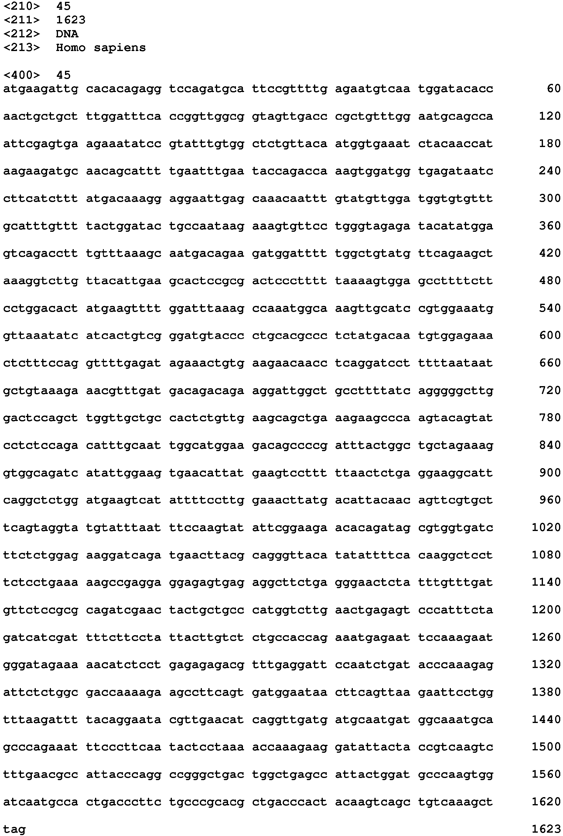


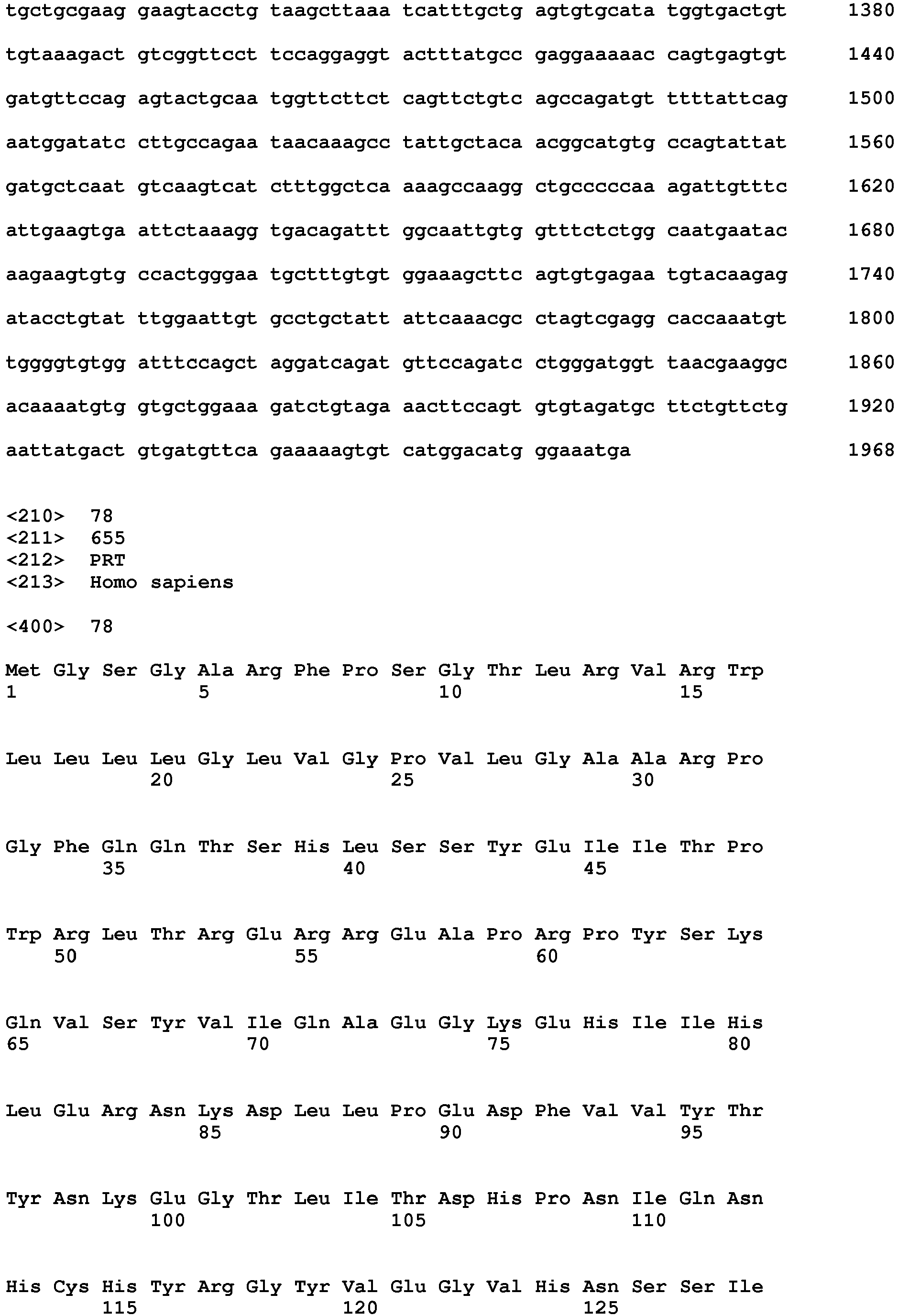
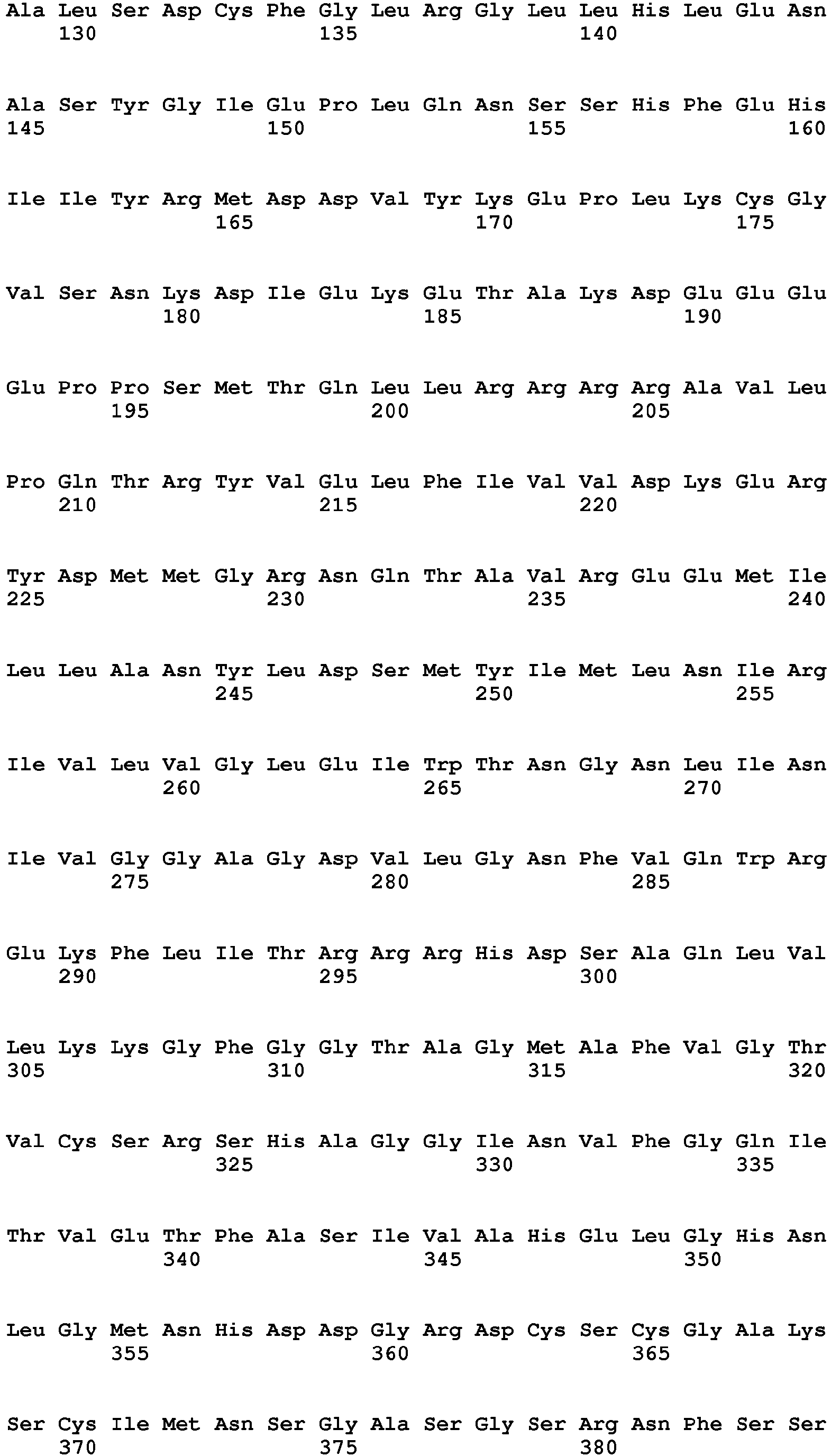
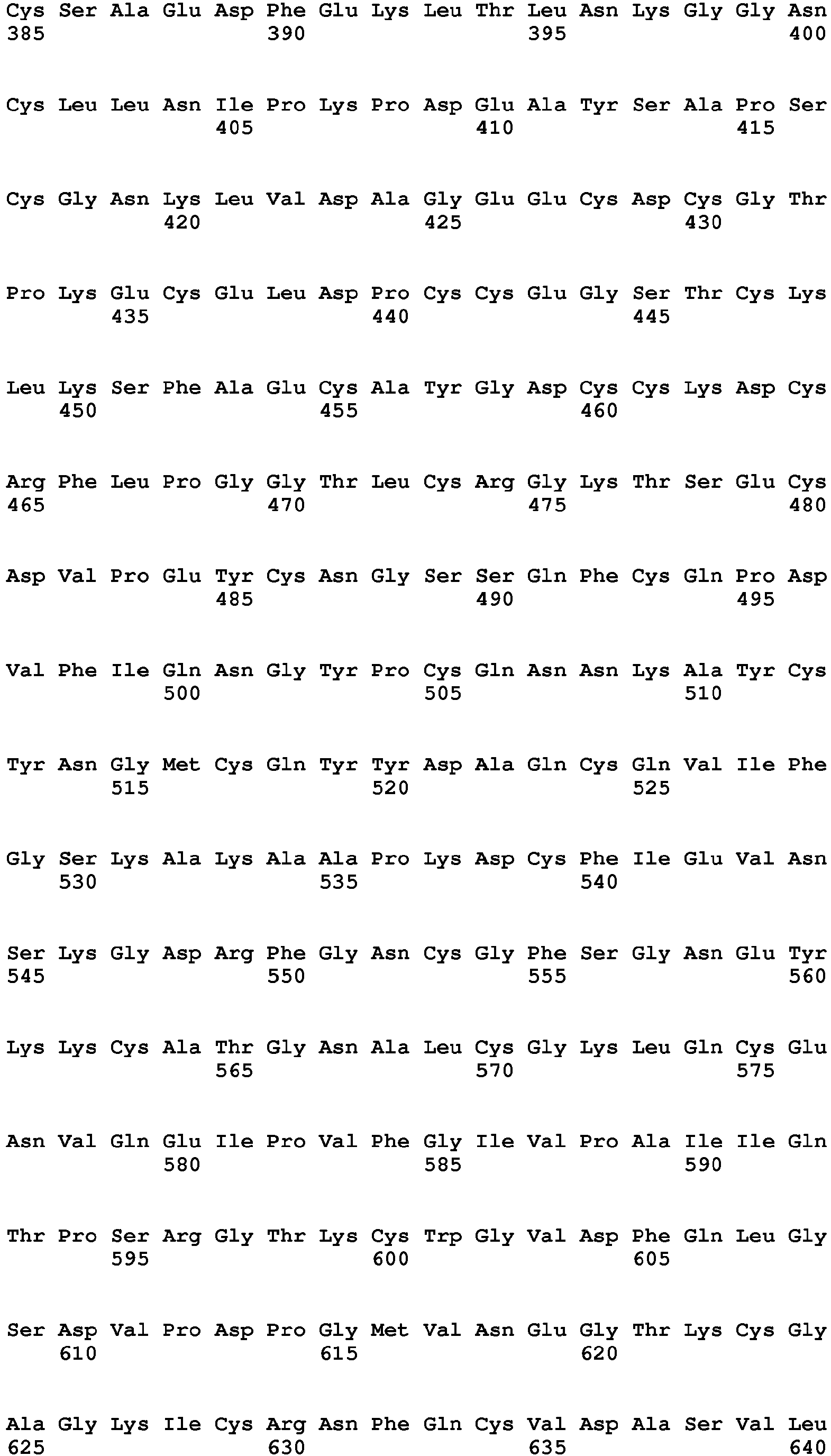


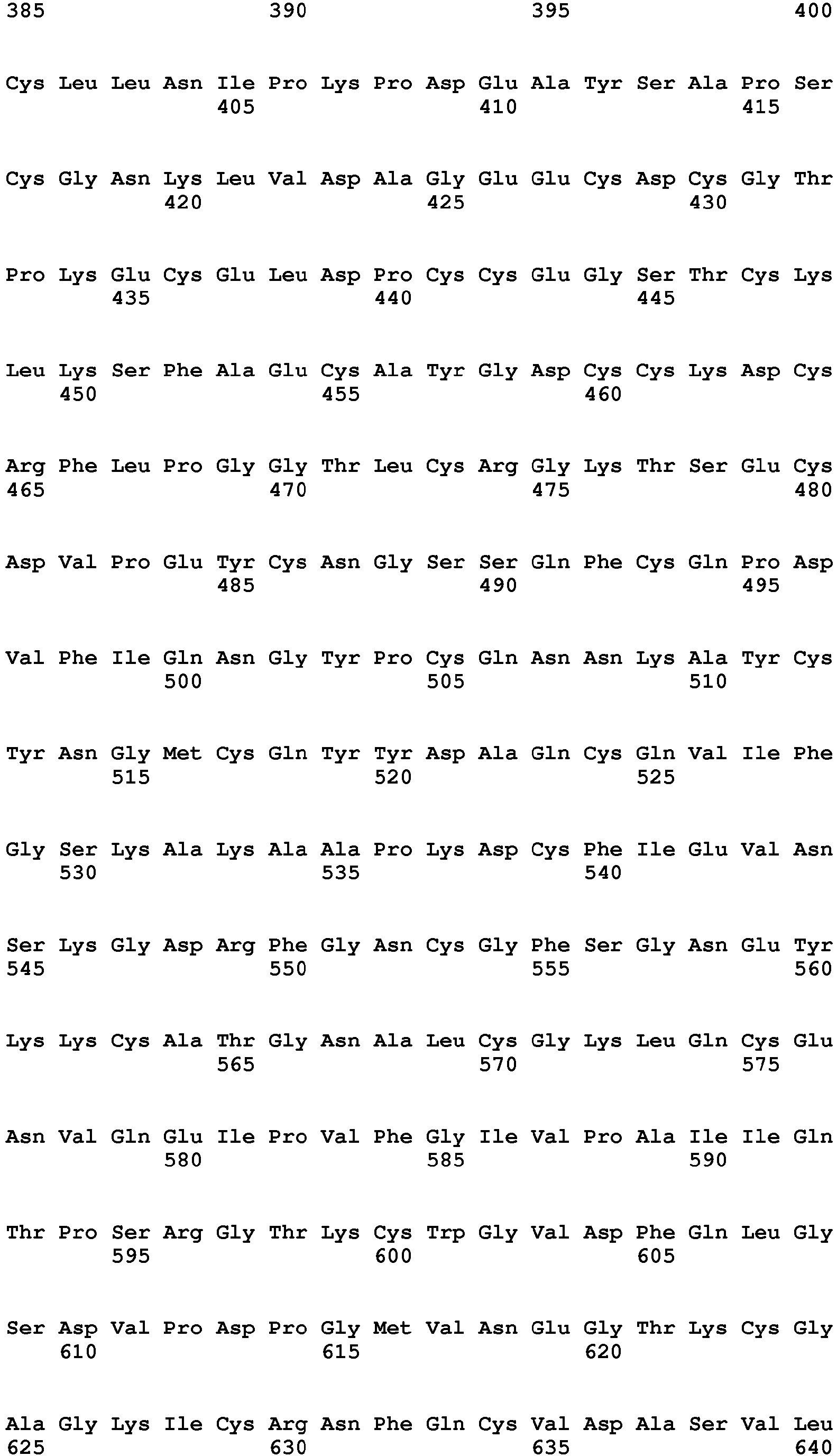
















Claims (15)
- An anti-proliferative compound or a pharmaceutical composition comprising said anti-proliferative compound for use in the prevention or treatment of a proliferative disease in a subject, wherein said subject has been preselected for treatment by a screening method comprising the detection of the presence or absence of target cells in a biological sample obtained from the subject, wherein the presence of said target cells is an indicator for the susceptibility of said subject for a positive treatment outcome with said anti-proliferative compound, characterized in that said preselection comprises the step of detecting the expression by said target cells of at least one biomarker selected from the group consisting of:• TPM4 (tropomyosin 4); Ensembl ID: ENSG00000167460;• ASNS (asparagine synthetase (glutamine-hydrolyzing)); Ensembl ID: ENSG00000070669;• F3 (Coagulation factor III, tissue factor); Ensembl ID: ENSG00000117525;• ADAM9 (ADAM metallopeptidase domain 9); Ensembl ID: ENSG00000168615; and• GANAB (glucosidase, alpha; neutral AB); Ensembl ID: ENSG00000089597;or a splice variant thereof, wherein the absence and/or non-expression of said at least one biomarker is an indicator for the susceptibility of said subject for a positive treatment outcome with said anti-proliferative compound.
- The anti-proliferative compound for use according to claim 1, wherein said preselection further comprises the step of detecting the expression by said target cells of Hes3, wherein the presence and/or expression of Hes3 is an indicator for the susceptibility of said subject for a positive treatment outcome with said anti-proliferative compound.
- The anti-proliferative compound for use according to claim 1 to 2, wherein said anti-proliferative compound belongs to a group selected from dichlorophenethyl-imidazoles, chlorobenzylphenoles, quinolinoles and macrocyclic lactones, wherein
said dichlorophenylethyl-imidazole is a 2,4-dichlorophenylethyl-imidazole of formula (I):
R1-R11 independently are H; halogen, such as fluorine, chlorine, bromine; or C1-12 alkyl, preferably C1-10 alkyl, more preferably C1-8 alkyl, most preferably C1-6 alkyl, such as methyl, ethyl, propyl, butyl, pentyl or hexyl;
X1, X2, Y1, Y2 independently are C, O, N, S or a bond;
said chlorobenzylphenol is a compound of formula (II):
R1-R9 independently are H; halogen, such as fluorine, chlorine, bromine; or C1-12 alkyl, preferably C1-10 alkyl, more preferably C1-8 alkyl, most preferably C1-6 alkyl, such as methyl, ethyl, propyl, butyl, pentyl or hexyl; and
X is C, O, N, or S; and
said quinolinole is a compound of formula (III):
R1-R7 independently are H; halogen, such as fluorine, chlorine, bromine; or C1-12 alkyl, preferably C1-10 alkyl, more preferably C1-8 alkyl, most preferably C1-6 alkyl, such as methyl, ethyl, propyl, butyl, pentyl or hexyl. - The anti-proliferative compound for use according any one of claims 1 to 3, wherein said compound is selected from the group consisting of abamectin, doramectin, quinestrol, benzethonium Cl, ebselen, raloxifene HCl, bisacodyl, econazole nitrate, ramelteon, bithionate Na, escin, ritonavir, broxaldine, hexetidine, selamectin, broxyquinoline, lasalocid Na, sulconazole nitrate, butoconazole, levocetirizine 2HCl, suloctidil, chlorhexidine 2HCl, miconazole nitrate, tioconazole, chloroxine, moxidectin, triclosan, clioquinol, norethynodrel, vinblastine sulfate, clofoctol, oxiconazole nitrate, xylazine, dichlorophen, oxyclozanide, diltiazem HCl, and pimozide, preferably from the group consisting of Ramelteon, Quinestrol, raloxifene HCl, diltiazem HCl, levocetirizine 2HCl, Norethynodrel and xylazine.
- A method of screening a subject for a proliferative disease risk factor, comprising detecting the presence or absence of target cells in said subject; the presence of target cells indicating said subject is at increased risk of developing a proliferative disease, characterized in that said detecting the presence or absence of target cells comprises the step of detecting the expression by said target cells of at least one biomarker selected from the group consisting of:• TPM4 (tropomyosin 4); Ensembl ID: ENSG00000167460;• ASNS (asparagine synthetase (glutamine-hydrolyzing)); Ensembl ID: ENSG00000070669;• F3 (Coagulation factor III, tissue factor); Ensembl ID: ENSG00000117525;• ADAM9 (ADAM metallopeptidase domain 9); Ensembl ID: ENSG00000168615; and• GANAB (glucosidase, alpha; neutral AB); Ensembl ID: ENSG00000089597;or a splice variant thereof, wherein the absence and/or non-expression of said at least one biomarker is an indicator for the susceptibility of said subject for a positive treatment outcome with said anti-proliferative compound.
- The method according to claim 5, wherein said subject has been previously diagnosed or prognosed as afflicted with said proliferative disease; or wherein said subject has not been previously diagnosed or prognosed as afflicted with said proliferative disease; or wherein said subject has undergone treatment for said proliferative disease.
- The method according to claim 5 or 6, wherein said detecting step is carried out by further detecting the expression by said target cells of Hes3, wherein the presence and/or expression of Hes3 is indicating that said subject is at increased risk of developing a proliferative disease.
- The anti-proliferative compound for use or the method according to any one of the preceding claims, wherein said proliferative disease is selected from cancer, such as malignant (and preferably solid) tumors of epithelial or mesenchymal cells, breast cancer, prostate cancer, pancreatic cancer, adrenal cancer, melanoma, lung cancer, colon cancer, leukemia (a liquid or non-solid tumor), soft tissue and bone sarcomas, neuroendocrine tumors such as islet cell carcinoma or medullary carcinoma of the thyroid, squamous carcinomas (particularly of the head and neck), adenocarcinomas and gliosarcomas such as glioblastoma multiforme; or a non-cancer disease, such as inflammatory and/or immunoproliferative disorders such as arthritis, fibrosis, asthma and allergies.
- The anti-proliferative compound for use or the method according to any one of claims 1 to 7, wherein said proliferative disease is tumor metastasis or tumor recurrence.
- A screening method for identifying an anti-proliferative compound for use in the treatment of a subject that has been identified to be at increased risk of developing a proliferative disease, comprising the steps of:• Culturing a mammalian, preferably a human tumor cell line under a first cell culture condition which suppresses the expression of target cell biomarkers; and under a second cell culture condition, which promotes the expression of target cells biomarkers;• Contacting and incubating the tumor cell line under the first and second culture conditions with a test compound for a period of at least 6 hours, preferably 24 hours;• Determining the cell number of the tumor cell lines after incubation with the test compound under the first and second culture conditions, and of control without test compound;• Selecting a test compound that reduces the cell number of the tumor cell line cultured under the first culture condition;wherein said biomarkers are selected from the group consisting of:• TPM4 (tropomyosin 4); Ensembl ID: ENSG00000167460;• ASNS (asparagine synthetase (glutamine-hydrolyzing)); Ensembl ID: ENSG00000070669;• F3 (Coagulation factor III, tissue factor); Ensembl ID: ENSG00000117525;• ADAM9 (ADAM metallopeptidase domain 9); Ensembl ID: ENSG00000168615; and• GANAB (glucosidase, alpha; neutral AB); Ensembl ID: ENSG00000089597;or a splice variant thereof; and wherein a compound that reduces the cell number under the first culture condition compared to the control, is amenable for use in the treatment of a proliferative disease.
- The screening method according to claim 10, wherein said test compound does not reduce or does not significantly reduce the cell number of the tumor cell line under the second culture condition.
- The screening method according to claim 10 or 11, wherein the first culture condition comprises culturing the tumor cell line in FGFJI medium and wherein the second culture condition comprises culturing the tumor cell line in serum-supplemented RPMI medium.
- The screening method according to any one of claims 10 to 12, further comprising contacting and incubating the tumor cell line under a third culture condition with a test compound, wherein said third culture condition comprises culturing the tumor cell line in EGF medium.
- The screening method according to any one of claims 10 to 13, further comprising the step of detecting the expression of at least one biomarker by said tumor cell line under the first and the second culture conditions and compared to a control without test compound, wherein said at least one biomarker is selected from the group consisting of:• TPM4 (tropomyosin 4); Ensembl ID: ENSG00000167460;• ASNS (asparagine synthetase (glutamine-hydrolyzing)); Ensembl ID: ENSG00000070669;• F3 (Coagulation factor III, tissue factor); Ensembl ID: ENSG00000117525;• ADAM9 (ADAM metallopeptidase domain 9); Ensembl ID: ENSG00000168615; and• GANAB (glucosidase, alpha; neutral AB); Ensembl ID: ENSG00000089597;or a splice variant thereof, wherein a test compound is selected that does not promote the expression of said at least one biomarker under the first culture condition and wherein the absence and/or non-expression of said at least one biomarker is indicating that the test compound is amenable for use in the treatment of a proliferative disease.
- The screening method according to any one of claims 10 to 14, further comprising the step of investigating the cell morphology and/or mechanical properties of the tumor cell line under the first and the second culture conditions, wherein the cells under the first culture condition show smaller and more neural stem cell-like morphology;
and/or
further comprising the step of detecting the expression of Hes3 by said tumor cell line under the first and the second culture conditions and compared to a control without test compound, wherein a test compound is selected that decreases the amount of Hes3 in said tumor cell line under the first culture condition and wherein the said Hes3 decreasing activity is indicating that the test compound is amenable for use in the treatment of a proliferative disease.
Priority Applications (5)
| Application Number | Priority Date | Filing Date | Title |
|---|---|---|---|
| EP18176272.5A EP3578981B1 (en) | 2018-06-06 | 2018-06-06 | Anti-cancer treatment of preselected subjects and screening methods to identify susceptible subjects |
| US16/972,015 US12590963B2 (en) | 2018-06-06 | 2019-05-23 | Anti-cancer treatment of preselected subjects and screening methods to identify susceptible subjects |
| EP19725195.2A EP3803402B1 (en) | 2018-06-06 | 2019-05-23 | Anti-cancer treatment of preselected subjects and screening methods to identify susceptible subjects |
| PCT/EP2019/063340 WO2019233779A1 (en) | 2018-06-06 | 2019-05-23 | Anti-cancer treatment of preselected subjects and screening methods to identify susceptible subjects |
| CN201980052275.0A CN112534265A (en) | 2018-06-06 | 2019-05-23 | Screening method for preselecting individual for anti-cancer treatment and identifying individual susceptible to disease |
Applications Claiming Priority (1)
| Application Number | Priority Date | Filing Date | Title |
|---|---|---|---|
| EP18176272.5A EP3578981B1 (en) | 2018-06-06 | 2018-06-06 | Anti-cancer treatment of preselected subjects and screening methods to identify susceptible subjects |
Publications (2)
| Publication Number | Publication Date |
|---|---|
| EP3578981A1 true EP3578981A1 (en) | 2019-12-11 |
| EP3578981B1 EP3578981B1 (en) | 2021-10-27 |
Family
ID=62683107
Family Applications (2)
| Application Number | Title | Priority Date | Filing Date |
|---|---|---|---|
| EP18176272.5A Active EP3578981B1 (en) | 2018-06-06 | 2018-06-06 | Anti-cancer treatment of preselected subjects and screening methods to identify susceptible subjects |
| EP19725195.2A Active EP3803402B1 (en) | 2018-06-06 | 2019-05-23 | Anti-cancer treatment of preselected subjects and screening methods to identify susceptible subjects |
Family Applications After (1)
| Application Number | Title | Priority Date | Filing Date |
|---|---|---|---|
| EP19725195.2A Active EP3803402B1 (en) | 2018-06-06 | 2019-05-23 | Anti-cancer treatment of preselected subjects and screening methods to identify susceptible subjects |
Country Status (4)
| Country | Link |
|---|---|
| US (1) | US12590963B2 (en) |
| EP (2) | EP3578981B1 (en) |
| CN (1) | CN112534265A (en) |
| WO (1) | WO2019233779A1 (en) |
Cited By (1)
| Publication number | Priority date | Publication date | Assignee | Title |
|---|---|---|---|---|
| WO2023242103A1 (en) | 2022-06-13 | 2023-12-21 | KHR Biotec GmbH | Novel ras inhibitors |
Families Citing this family (1)
| Publication number | Priority date | Publication date | Assignee | Title |
|---|---|---|---|---|
| US20230038646A1 (en) * | 2020-01-20 | 2023-02-09 | Texas Tech University System | Novel and potent jak/stat inhibitor |
Citations (3)
| Publication number | Priority date | Publication date | Assignee | Title |
|---|---|---|---|---|
| WO2014162008A2 (en) * | 2013-04-05 | 2014-10-09 | University Of Ha'il | Novel biomarker signature and uses thereof |
| WO2015181556A1 (en) * | 2014-05-28 | 2015-12-03 | Almac Diagnostics Limited | Pro-angiogenic expression signature |
| US20150376710A1 (en) * | 2010-04-14 | 2015-12-31 | The Board Of Regents Of The University Of Texas System | Methods of evaluating response to cancer therapy |
-
2018
- 2018-06-06 EP EP18176272.5A patent/EP3578981B1/en active Active
-
2019
- 2019-05-23 EP EP19725195.2A patent/EP3803402B1/en active Active
- 2019-05-23 US US16/972,015 patent/US12590963B2/en active Active
- 2019-05-23 CN CN201980052275.0A patent/CN112534265A/en active Pending
- 2019-05-23 WO PCT/EP2019/063340 patent/WO2019233779A1/en not_active Ceased
Patent Citations (3)
| Publication number | Priority date | Publication date | Assignee | Title |
|---|---|---|---|---|
| US20150376710A1 (en) * | 2010-04-14 | 2015-12-31 | The Board Of Regents Of The University Of Texas System | Methods of evaluating response to cancer therapy |
| WO2014162008A2 (en) * | 2013-04-05 | 2014-10-09 | University Of Ha'il | Novel biomarker signature and uses thereof |
| WO2015181556A1 (en) * | 2014-05-28 | 2015-12-03 | Almac Diagnostics Limited | Pro-angiogenic expression signature |
Non-Patent Citations (108)
| Title |
|---|
| A. M. MENDES-PEREIRA ET AL: "Genome-wide functional screen identifies a compendium of genes affecting sensitivity to tamoxifen", PROCEEDINGS OF THE NATIONAL ACADEMY OF SCIENCES, vol. 109, no. 8, 21 February 2012 (2012-02-21), pages 2730 - 2735, XP055082333, ISSN: 0027-8424, DOI: 10.1073/pnas.1018872108 * |
| AKERS, W.A.; LANE, A.; LYNFIELD, Y.; GREENBERG, J.; HALL, J.; MANGAN, C.; TINKER, A.: "Sulconazole nitrate 1% cream in the treatment of chronic moccasin-type tinea pedis caused by Trichophyton rubrum", JOURNAL OF THE AMERICAN ACADEMY OF DERMATOLOGY, vol. 21, 1989, pages 686 - 689, XP025596849, DOI: doi:10.1016/S0190-9622(89)70237-1 |
| ANDROUTSELLIS-THEOTOKIS, A.; LEKER, R.R.; SOLDNER, F.; HOEPPNER, D.J.; RAVIN, R.; POSER, S.W.; RUEGER, M.A.; BAE, S.K.; KITTAPPA,: "Notch signalling regulates stem cell numbers in vitro and in vivo", NATURE, vol. 442, 2006, pages 823 - 826 |
| ANDROUTSELLIS-THEOTOKIS, A.; RUEGER, M.A.; PARK, D.M.; BOYD, J.D.; PADMANABHAN, R.; CAMPANATI, L.; STEWART, C.V.; LEFRANC, Y.; PLE: "Angiogenic factors stimulate growth of adult neural stem cells", PLOS ONE, vol. 5, 2010, pages e9414 |
| ARNETT, J.H.: "Treatment of carriers of endamoeba histolytica and other protozoa with carbarsone, chiniofon and vioform", THE AMERICAN JOURNAL OF THE MEDICAL SCIENCES, vol. 213, 1947, pages 608 - 610 |
| BATES, D.; MACHLER, M.; BOLKER, B.; WALKER, S.: "Fitting linear mixed-effects models using lme4", ARXIV PREPRINT ARXIV: 14065823, 2014 |
| BATES, D.; MAECHLER, M.; BOLKER, B.; WALKER, S, LME4: LINEAR MIXED-EFFECTS MODELS USING EIGEN AND S4, 2014 |
| BENCHAOUI, H.A.; CLEMENCE, R.G.; CLEMENTS, P.J.; JONES, R.L.; WATSON, P.; SHANKS, D.J.; SMITH, D.G.; STURE, G.H.; JERNIGAN, A.D.;: "Efficacy and safety of selamectin against fleas on dogs and cats presented as veterinary patients in Europe", VETERINARY PARASITOLOGY, vol. 91, 2000, pages 223 - 232, XP002202199, DOI: doi:10.1016/S0304-4017(00)00294-6 |
| BENFIELD, P.; CLISSOLD, S.P.: "Sulconazole. A review of its antimicrobial activity and therapeutic use in superficial dermatomycoses", DRUGS, vol. 35, 1988, pages 143 - 153 |
| BENINGER, R.J.; HAHN, B.L.: "Pimozide blocks establishment but not expression of amphetamine-produced environment-specific conditioning", SCIENCE, vol. 220, 1983, pages 1304 - 1306 |
| BEVAN, J.A.: "Diltiazem selectively inhibits cerebrovascular extrinsic but not intrinsic myogenic tone", CIRC RES, vol. 52, 1983, pages 1104 - 109 |
| BOURQUIN, J.P.; GRIOT, R.; SCHENKER, E.: "Synthesis of esters of halogenated quinaldine and quinoline", ARCHIV DER PHARMAZIE UND BERICHTE DER DEUTSCHEN PHARMAZEUTISCHEN GESELLSCHAFT, vol. 295, 1962, pages 383 - 399 |
| BROCARD, E.; OIZEL, K.; LALIER, L.; PECQUEUR, C.; PARIS, F.; VALLETTE, F.M.; OLIVER, L.: "Radiation-induced PGE2 sustains human glioma cells growth and survival through EGF signaling", ONCOTARGET, vol. 6, 2015, pages 6840 - 6849 |
| BUCHERT, M.; BURNS, C.J.; ERNST, M.: "Targeting JAK kinase in solid tumors: emerging opportunities and challenges", ONCOGENE, vol. 35, 2016, pages 939 - 951 |
| BUELKE-SAM, J.; BRYANT, H.U.; FRANCIS, P.C.: "The selective estrogen receptor modulator, raloxifene: an overview of nonclinical pharmacology and reproductive and developmental testing", REPRODUCTIVE TOXICOLOGY, vol. 12, 1998, pages 217 - 221 |
| CANNISTRACI, C.V.; ALANIS-LOBATO, G.; RAVASI, T.: "Minimum curvilinearity to enhance topological prediction of protein interactions by network embedding", BIOINFORMATICS, vol. 29, 2013, pages i199 - 209 |
| CANNISTRACI, C.V.; RAVASI, T.; MONTEVECCHI, F.M.; IDEKER, T.; ALESSIO, M.: "Nonlinear dimension reduction and clustering by Minimum Curvilinearity unfold neuropathic pain and tissue embryological classes", BIOINFORMATICS, vol. 26, 2010, pages i531 - 539 |
| CHAILLEUX, N.; PARADIS, M.: "Efficacy of selamectin in the treatment of naturally acquired cheyletiellosis in cats", THE CANADIAN VETERINARY JOURNAL = LA REVUE VETERINAIRE CANADIENNE, vol. 43, 2002, pages 767 - 770 |
| CHUNG, M.W.; KOMOROWSKI, R.A.; VARMA, R.R.: "Suloctidil-induced hepatotoxicity", GASTROENTEROLOGY, vol. 95, 1988, pages 490 - 491 |
| CLARKE, K.W.; HALL, L.W.: "Xylazine''--a new sedative for horses and cattle", THE VETERINARY RECORD, vol. 85, 1969, pages 512 - 517 |
| CLAYTON, Y.M.; HAY, R.J.; MCGIBBON, D.H.; PYE, R.J.: "Double blind comparison of the efficacy of tioconazole and miconazole for the treatment of fungal infection of the skin or erythrasma", CLINICAL AND EXPERIMENTAL DERMATOLOGY, vol. 7, 1982, pages 543 - 549 |
| COHEN, M.R.: "Quinestrol therapy in functional infertility", FERTILITY AND STERILITY, vol. 17, 1966, pages 541 - 546 |
| CORTES, C.; VAPNIK, V.: "Machine Learning", 1995, KLUWER ACADEMIC PUBLISHERS, article "Support-Vector Networks", pages: 273 - 297 |
| CUMMINGS, S.R.; ECKERT, S.; KRUEGER, K.A.; GRADY, D.; POWLES, T.J.; CAULEY, J.A.; NORTON, L.; NICKELSEN, T.; BJARNASON, N.H.; MORR: "The effect of raloxifene on risk of breast cancer in postmenopausal women: results from the MORE randomized trial. Multiple Outcomes of Raloxifene Evaluation", JAMA, vol. 281, 1999, pages 2189 - 2197 |
| DANESI, R.; DEL TACCA, M.: "Clinical study on the efficacy of clofoctol in the treatment of infectious respiratory diseases", INTERNATIONAL JOURNAL OF CLINICAL PHARMACOLOGY RESEARCH, vol. 5, 1985, pages 175 - 179 |
| DAVIES, G.E.; FRANCIS, J.; MARTIN, A.R.; ROSE, F.L.; SWAIN, G.: "1:6-Di-4'-chlorophenyldiguanidohexane (hibitane); laboratory investigation of a new antibacterial agent of high potency", BRITISH JOURNAL OF PHARMACOLOGY AND CHEMOTHERAPY, vol. 9, 1954, pages 192 - 196, XP008058928 |
| DELMAS, P.D.; ENSRUD, K.E.; ADACHI, J.D.; HARPER, K.D.; SARKAR, S.; GENNARI, C.; REGINSTER, J.Y.; POLS, H.A.; RECKER, R.R.; HARRIS: "Efficacy of raloxifene on vertebral fracture risk reduction in postmenopausal women with osteoporosis: four-year results from a randomized clinical trial", J CLIN ENDOCRINOL METAB, vol. 87, 2002, pages 3609 - 3617 |
| DERIC M. PARK ET AL: "Hes3 regulates cell number in cultures from glioblastoma multiforme with stem cell characteristics", SCIENTIFIC REPORTS, vol. 3, 5 February 2013 (2013-02-05), XP055172008, DOI: 10.1038/srep01095 * |
| DREILING, D.A.; FISCHL, A.; FERNANDEZ, O.: "The therapeutic usefulness of dulcolax (bisacodyl), a new nonpurgative laxative", THE AMERICAN JOURNAL OF DIGESTIVE DISEASES, vol. 4, 1959, pages 311 - 320 |
| DROEGEMUELLER, W.; ADAMSON, D.G.; BROWN, D.; CIBLEY, L.; FLEURY, F.; LEPAGE, M.E.; HENZL, M.R.: "Three-day treatment with butoconazole nitrate for vulvovaginal candidiasis", OBSTETRICS AND GYNECOLOGY, vol. 64, 1984, pages 530 - 534 |
| DUTTON, C.J.; GIBSON, S.P.; GOUDIE, A.C.; HOLDOM, K.S.; PACEY, M.S.; RUDDOCK, J.C.; BU'LOCK, J.D.; RICHARDS, M.K.: "Novel avermectins produced by mutational biosynthesis", THE JOURNAL OF ANTIBIOTICS, vol. 44, 1991, pages 357 - 365, XP002079430 |
| EDGREN, R.A.: "Oral contraception: a review", INTERNATIONAL JOURNAL OF FERTILITY, vol. 36, no. 3, 1991, pages 16 - 25 |
| ELLENRIEDER, M.; ZEILLER, P.; DINTER, H.; SENSCH, K.H.: "Effect of quinoline derivatives on pathogenic and apathogenic intestinal microorganisms", ARZNEIMITTEL-FORSCHUNG, vol. 20, 1970, pages 821 - 824 |
| FOULKES, D.M.: "Some toxicological observations on chlorhexidine", JOURNAL OF PERIODONTAL RESEARCH SUPPLEMENT, vol. 12, 1973, pages 55 - 60 |
| GHILARDI, P.L.; CASANI, A.: "Treatment of ear, nose and throat infections with clofoctol", DRUGS UNDER EXPERIMENTAL AND CLINICAL RESEARCH, vol. 11, 1985, pages 815 - 818 |
| GOBBI, P.G.; PIERESCA, C.; FRASSOLDATI, A.; CAROTENUTO, M.; DI RENZO, N.; LA SALA, A.; BERTE, R.; AVANZINI, P.; FEDERICO, M.; SILI: "Vinblastine, bleomycin, and methotrexate chemotherapy plus extended-field radiotherapy in early, favorably presenting, clinically staged Hodgkin's patients: the Gruppo Italiano per lo Studio dei Linfomi Experience", J CLIN ONCOL, vol. 14, 1996, pages 527 - 533 |
| GODEFROI, E.F.; HEERES, J.; VAN CUTSEM, J.; JANSSEN, P.A.: "The preparation and antimycotic properties of derivatives of 1-phenethylimidazole", JOURNAL OF MEDICINAL CHEMISTRY, vol. 12, 1969, pages 784 - 791, XP000561809, DOI: doi:10.1021/jm00305a014 |
| GREENBLATT, D.J.; HARMATZ, J.S.; KARIM, A.: "Age and gender effects on the pharmacokinetics and pharmacodynamics of ramelteon, a hypnotic agent acting via melatonin receptors MT1 and MT2", JOURNAL OF CLINICAL PHARMACOLOGY, vol. 47, 2007, pages 485 - 496 |
| GU, Z.; EILS, R.; SCHLESNER, M.: "Complex heatmaps reveal patterns and correlations in multidimensional genomic data", BIOINFORMATICS, vol. 32, 2016, pages 2847 - 2849 |
| HEEL, R.C.; BROGDEN, R.N.; SPEIGHT, T.M.; AVERY, G.S.: "Econazole: a review of its antifungal activity and therapeutic efficacy", DRUGS, vol. 16, 1978, pages 177 - 201 |
| HERTZ, R.; LIPSETT, M.B.; MOY, R.H.: "Effect of Vincaleukoblastine on metastatic choriocarcinoma and related trophoblastic tumors in women", CANCER RES, vol. 20, 1960, pages 1050 - 1053 |
| HOPPER, S.H.; WOOD, K.M.: "Development of a germicidal soap containing bithionol", JOURNAL OF THE AMERICAN PHARMACEUTICAL ASSOCIATION AMERICAN PHARMACEUTICAL ASSOCIATION, vol. 47, 1958, pages 317 - 318 |
| HORTON, G.M.; STOCKDALE, P.H.: "Lasalocid and monensin in finishing diets for early weaned lambs with naturally occurring coccidiosis", AMERICAN JOURNAL OF VETERINARY RESEARCH, vol. 42, 1981, pages 433 - 436 |
| HUANG DA, W.; SHERMAN, B.T.; LEMPICKI, R.A.: "Bioinformatics enrichment tools: paths toward the comprehensive functional analysis of large gene lists", NUCLEIC ACIDS RES, vol. 37, 2009, pages 1 - 13, XP055011237, DOI: doi:10.1093/nar/gkn923 |
| HUANG DA, W.; SHERMAN, B.T.; LEMPICKI, R.A.: "Systematic and integrative analysis of large gene lists using DAVID bioinformatics resources", NATURE PROTOCOLS, vol. 4, 2009, pages 44 - 57, XP009153774, DOI: doi:10.1038/nprot.2008.211 |
| IDRIS, M.; RAHMAN, K.M.; MUTTALIB, M.A.; AZAD KHAN, A.K.: "The treatment of fasciolopsiasis with niclosamide and dichlorophen", THE JOURNAL OF TROPICAL MEDICINE AND HYGIENE, vol. 83, 1980, pages 71 - 74 |
| IMAYOSHI, I.; KAGEYAMA, R.: "bHLH factors in self-renewal, multipotency, and fate choice of neural progenitor cells", NEURON, vol. 82, 2014, pages 9 - 23, XP028842125, DOI: doi:10.1016/j.neuron.2014.03.018 |
| JANSSEN, P.A.; NIEMEGEERS, C.J.; SCHELLEKENS, K.H.; DRESSE, A.; LENAERTS, F.M.; PINCHARD, A.; SCHAPER, W.K.; VAN NUETEN, J.M.; VER: "Pimozide, a chemically novel, highly potent and orally long-acting neuroleptic drug. I", THE COMPARATIVE PHARMACOLOGY OF PIMOZIDE, HALOPERIDOL, AND CHLORPROMAZINE. ARZNEIMITTEL-FORSCHUNG, vol. 18, 1968, pages 261 - 279 |
| KATO, K.; HIRAI, K.; NISHIYAMA, K.; UCHIKAWA, O.; FUKATSU, K.; OHKAWA, S.; KAWAMATA, Y.; HINUMA, S.; MIYAMOTO, M.: "Neurochemical properties of ramelteon (TAK-375), a selective MT1/MT2 receptor agonist", NEUROPHARMACOLOGY, vol. 48, 2005, pages 301 - 310, XP004731978, DOI: doi:10.1016/j.neuropharm.2004.09.007 |
| KENYON, A.J.; HAMILTON, S.G.; DOUGLAS, D.M.: "Controlled wound repair in guinea pigs, using antimicrobials that alter fibroplasia", AMERICAN JOURNAL OF VETERINARY RESEARCH, vol. 47, 1986, pages 96 - 101 |
| KLIMEK, L.: "Levocetirizine: from scientific evidence to a potent modern-day treatment of today's allergic patients", DRUGS OF TODAY, vol. 45, 2009, pages 213 - 225 |
| KODACK, D.P.; ASKOXYLAKIS, V.; FERRARO, G.B.; SHENG, Q.; BADEAUX, M.; GOEL, S.; QI, X.; SHANKARAIAH, R.; CAO, Z.A.; RAMJIAWAN, R.R: "The brain microenvironment mediates resistance in luminal breast cancer to PI3K inhibition through HER3 activation", SCIENCE TRANSLATIONAL MEDICINE, vol. 9, 2017 |
| LAVY, E.; HARRUS, S.; MAZAKI-TOVI, M.; BARK, H.; MARKOVICS, A.; HAGAG, A.; AIZENBERG, I.; AROCH, I.: "Spirocerca lupi in dogs: prophylactic effect of doramectin", RESEARCH IN VETERINARY SCIENCE, vol. 75, 2003, pages 217 - 222 |
| LEA, A.P.; FAULDS, D.: "Ritonavir", DRUGS, vol. 52, 1996, pages 541 - 546 |
| LIAO, Y.; SMYTH, G.K.; SHI, W.: "featureCounts: an efficient general purpose program for assigning sequence reads to genomic features", BIOINFORMATICS, vol. 30, 2014, pages 923 - 930 |
| LIENERT, E.; JAHN, F.: "Studies on the mechanism of action of anti-trematode agents. II. 2,2-Methylene-bisphenols provided with electron-attracting substitutes are electron acceptors; they have a trematodicide effect because of the strong power of attraction of their oxygen atoms for electrons", WIENER TIERARZTLICHE MONATSSCHRIFT, vol. 53, 1966, pages 795 - 802 |
| LOVE, M.I.; HUBER, W.; ANDERS, S.: "Moderated estimation of fold change and dispersion for RNA-seq data with DESeq2", GENOME BIOL, vol. 15, 2014, pages 550, XP021210395, DOI: doi:10.1186/s13059-014-0550-8 |
| LUO, W.; BROUWER, C.: "Pathview: an R/Bioconductor package for pathway-based data integration and visualization", BIOINFORMATICS, vol. 29, 2013, pages 1830 - 1831 |
| LYMAN, F.L.; FURIA, T.: "Toxicology of 2, 4, 4'-trichloro-2'-hydroxy-diphenyl ether", IMS, INDUSTRIAL MEDICINE AND SURGERY, vol. 38, 1969, pages 64 - 71 |
| MANDEL, I.D.: "Antimicrobial mouthrinses: overview and update", JOURNAL OF THE AMERICAN DENTAL ASSOCIATION, vol. 125, no. 2, 1994, pages 2S - 10S |
| MANSFIELD, L.E.; HAMPEL, F.; HAEUSLER, J.M.; GEORGES, G.: "Study of levocetirizine in seasonal allergic rhinitis", CURRENT MEDICAL RESEARCH AND OPINION, vol. 26, 2010, pages 1269 - 1275 |
| MARRIOTT, M.S.; BAIRD, J.R.; BRAMMER, K.W.; FAULKNER, J.K.; HALLIWELL, G.; JEVONS, S.; TARBIT, M.H.: "Tioconazole, a new imidazole-antifungal agent for the treatment of dermatomycoses", ANTIFUNGAL AND PHARMACOLOGIC PROPERTIES. DERMATOLOGICA, vol. 166, no. 1, 1983, pages 1 - 7 |
| MIETKE, A.; OTTO, O.; GIRARDO, S.; ROSENDAHL, P.; TAUBENBERGER, A.; GOLFIER, S.; ULBRICHT, E.; ALAND, S.; GUCK, J.; FISCHER-FRIEDR: "Extracting Cell Stiffness from Real-Time Deformability Cytometry: Theory and Experiment", BIOPHYSICAL JOURNAL, vol. 109, 2015, pages 2023 - 2036, XP029300858, DOI: doi:10.1016/j.bpj.2015.09.006 |
| MILLER, T.A.: "Anthelmintic activity of toluene and dichlorophen against various stages of Ancylostoma caninum in young dogs", AMERICAN JOURNAL OF VETERINARY RESEARCH, vol. 27, 1966, pages 1755 - 1758 |
| MIXICH, G.; THIELE, K.: "A contribution to the stereospecific synthesis of antifungal imidazolyloxime-ethers/Oxiconazole nitrate (Sgd 301-76), a new broadspectrum antifungal agent (author's transl", ARZNEIMITTEL-FORSCHUNG, vol. 29, 1979, pages 1510 - 1513 |
| MOERMANN, J.E.; MUEHLEMANN, H.R.: "Synergistic inhibitory effect of zinc and hexetidine on in vitro growth and acid production of Streptococcus mutans", JOURNAL OF DENTAL RESEARCH, vol. 62, 1983, pages 135 - 137 |
| MROZIK, H.; JONES, H.; FRIEDMAN, J.; SCHWARTZKOPF, G.; SCHARDT, R.A.; PATCHETT, A.A.; HOFF, D.R.; YAKSTIS, J.J.; RIEK, R.F.; OSTLI: "A new agent for the treatment of liver fluke infection (fascioliasis", EXPERIENTIA, vol. 25, 1969, pages 883 |
| MUKTHAVARAM, R.; OUYANG, X.; SAKLECHA, R.; JIANG, P.; NOMURA, N.; PINGLE, S.C.; GUO, F.; MAKALE, M.; KESARI, S.: "Effect of the JAK2/STAT3 inhibitor SAR317461 on human glioblastoma tumorspheres", JOURNAL OF TRANSLATIONAL MEDICINE, vol. 13, 2015, pages 269, XP021228213, DOI: doi:10.1186/s12967-015-0627-5 |
| NACHMAN, S.A.; STANLEY, K.; YOGEV, R.; PELTON, S.; WIZNIA, A.; LEE, S.; MOFENSON, L.; FISCUS, S.; RATHORE, M.; JIMENEZ, E. ET AL.: "Nucleoside analogs plus ritonavir in stable antiretroviral therapy-experienced HIV-infected children: a randomized controlled trial. Pediatric AIDS Clinical Trials Group 338 Study Team", JAMA, vol. 283, 2000, pages 492 - 498 |
| NAGAO, T.; SATO, M.; NAKAJIMA, H.; KIYOMOTO, A.: "Studies on a new 1,5-benzothiazepine derivative (CRD-401). IV. Coronary vasodilating effect and structure-activity relationship", CHEMICAL & PHARMACEUTICAL BULLETIN, vol. 21, 1973, pages 92 - 97 |
| NELDNER, K.H.: "The halogenated 8-hydroxyquinolines", INTERNATIONAL JOURNAL OF DERMATOLOGY, vol. 16, 1977, pages 267 - 273 |
| ODDS, F.C.; WEBSTER, C.E.; ABBOTT, A.B.: "Antifungal relative inhibition factors: BAY 1-9139, bifonazole, butoconazole, isoconazole, itraconazole (R 51211), oxiconazole, Ro 14-4767/002, sulconazole, terconazole and vibunazole (BAY n-7133) compared in vitro with nine established antifungal agents", THE JOURNAL OF ANTIMICROBIAL CHEMOTHERAPY, vol. 14, 1984, pages 105 - 114 |
| OTTO, O.; ROSENDAHL, P.; MIETKE, A.; GOLFIER, S.; HEROLD, C.; KLAUE, D.; GIRARDO, S.; PAGLIARA, S.; EKPENYONG, A.; JACOBI, A. ET A: "Real-time deformability cytometry: on-the-fly cell mechanical phenotyping", NATURE METHODS, vol. 12, 2015, pages 199 - 202 |
| PARK, D.M.; JUNG, J.; MASJKUR, J.; MAKROGKIKAS, S.; EBERMANN, D.; SAHA, S.; ROGLIANO, R.; PAOLILLO, N.; PACIONI, S.; MCKAY, R.D. E: "Hes3 regulates cell number in cultures from glioblastoma multiforme with stem cell characteristics", SCI REP, vol. 3, 2013, pages 1095 |
| PARNHAM, M.J.: "Biological activities and clinical potential of Ebselen", ADV EXP MED BIOL, vol. 264, 1990, pages 193 - 197 |
| PAULSEN, C.A.; LEACH, R.B.; LANMAN, J.; GOLDSTON, N.; MADDOCK, W.O.; HELLER, C.G.: "Inherent estrogenicity of norethindrone and norethynodrel: comparison with other synthetic progestins and progesterone", J CLIN ENDOCRINOL METAB, vol. 22, 1962, pages 1033 - 1039 |
| PIVNICK, H.; TRACY, J.M.; GLASS, D.G.: "Studies of Preservatives of Poliomyelitis (Salk) Vaccine. I. Benzethonium Chloride", JOURNAL OF PHARMACEUTICAL SCIENCES, vol. 52, 1963, pages 883 - 888 |
| POLAK, A.: "Oxiconazole, a new imidazole derivative. Evaluation of antifungal activity in vitro and in vivo", ARZNEIMITTEL-FORSCHUNG, vol. 32, 1982, pages 17 - 24 |
| POSER, S.W.; ANDROUTSELLIS-THEOTOKIS, A.: "Growing neural stem cells from conventional and nonconventional regions of the adult rodent brain", J VIS EXP, 2013, pages e50880 |
| PULLEN, D.: "Conovid-E'' as an oral contraceptive", BRITISH MEDICAL JOURNAL, vol. 2, 1962, pages 1016 - 1019 |
| RAJAMUTHIAH, R.; FUCHS, B.B.; CONERY, A.L.; KIM, W.; JAYAMANI, E.; KWON, B.; AUSUBEL, F.M.; MYLONAKIS, E.: "Repurposing salicylanilide anthelmintic drugs to combat drug resistant Staphylococcus aureus", PLOS ONE, vol. 10, 2015, pages e0124595, XP055221148, DOI: doi:10.1371/journal.pone.0124595 |
| RINGNER, M.: "What is principal component analysis?", NATURE BIOTECHNOLOGY, vol. 26, 2008, pages 303 - 304 |
| ROBERTSON, L.; GHOURI, M.A.; KOVACS, F.: "Antiplatelet and anticoagulant drugs for prevention of restenosis/reocclusion following peripheral endovascular treatment", THE COCHRANE DATABASE OF SYSTEMATIC REVIEWS, 2012, pages CD002071 |
| RODRIGUEZ, L.A.; CLOSE, J.A.: "The metabolism of the 5-7-dibromo-8-hydroxyquinoline (broxyquinoline) in man", BIOCHEMICAL PHARMACOLOGY, vol. 17, 1968, pages 1647 - 1653, XP025887738, DOI: doi:10.1016/0006-2952(68)90226-8 |
| ROHDE, W.; MIKELENS, P.; JACKSON, J.; BLACKMAN, J.; WHITCHER, J.; LEVINSON, W.: "Hydroxyquinolines inhibit ribonucleic acid-dependent deoxyribonucleic acid polymerase and inactivate Rous sarcoma virus and herpes simplex virus", ANTIMICROBIAL AGENTS AND CHEMOTHERAPY, vol. 10, 1976, pages 234 - 240, XP008050819 |
| ROLAND, M.; CLYMAN, M.J.; DECKER, A.; OBER, W.B.; BRONSTEIN, S.B.: "Clinical study of quinestrol in infertile women", FERTILITY AND STERILITY, vol. 17, 1966, pages 531 - 540 |
| SATO, M.; NAGAO, T.; YAMAGUCHI, I.; NAKAJIMA, H.; KIYOMOTO, A.: "Pharmacological studies on a new 1,5-benzothiazepine derivative (CRD-401", ARZNEIMITTEL-FORSCHUNG, vol. 21, 1971, pages 1338 - 1343 |
| SAWYER, P.R.; BROGDEN, R.N.; PINDER, R.M.; SPEIGHT, T.M.; AVERY, G.S.: "Miconazole: a review of its antifungal activity and therapeutic efficacy", DRUGS, vol. 9, 1975, pages 406 - 423, XP000562155, DOI: doi:10.2165/00003495-197509060-00002 |
| SCHEWE, T.: "Molecular actions of ebselen--an antiinflammatory antioxidant", GENERAL PHARMACOLOGY, vol. 26, 1995, pages 1153 - 1169, XP002940585, DOI: doi:10.1016/0306-3623(95)00003-J |
| SHANNON, P.; MARKIEL, A.; OZIER, O.; BALIGA, N.S.; WANG, J.T.; RAMAGE, D.; AMIN, N.; SCHWIKOWSKI, B.; IDEKER, T.: "Cytoscape: a software environment for integrated models of biomolecular interaction networks", GENOME RESEARCH, vol. 13, 2003, pages 2498 - 2504, XP055105995, DOI: doi:10.1101/gr.1239303 |
| SHARMA, C.S.: "Letter: Effect of broxyquinoline and broxaldine in leprosy", LANCET, vol. 1, 1975, pages 405 |
| SIES, H.: "Ebselen, a selenoorganic compound as glutathione peroxidase mimic", FREE RADICAL BIOLOGY & MEDICINE, vol. 14, 1993, pages 313 - 323, XP023522165, DOI: doi:10.1016/0891-5849(93)90028-S |
| SOEDA, A.; INAGAKI, A.; OKA, N.; IKEGAME, Y.; AOKI, H.; YOSHIMURA, S.; NAKASHIMA, S.; KUNISADA, T.; IWAMA, T.: "Epidermal growth factor plays a crucial role in mitogenic regulation of human brain tumor stem cells", J BIOL CHEM, vol. 283, 2008, pages 10958 - 10966 |
| SOEDA, A.; PARK, M.; LEE, D.; MINTZ, A.; ANDROUTSELLIS-THEOTOKIS, A.; MCKAY, R.D.; ENGH, J.; IWAMA, T.; KUNISADA, T.; KASSAM, A.B.: "Hypoxia promotes expansion of the CD133-positive glioma stem cells through activation of HIF-lalpha", ONCOGENE, vol. 28, 2009, pages 3949 - 3959 |
| SZKLARCZYK, D.; FRANCESCHINI, A.; WYDER, S.; FORSLUND, K.; HELLER, D.; HUERTA-CEPAS, J.; SIMONOVIC, M.; ROTH, A.; SANTOS, A.; TSAF: "STRING v10: protein-protein interaction networks, integrated over the tree of life", NUCLEIC ACIDS RES, vol. 43, 2015, pages D447 - 452 |
| THIENPONT, D.; VAN CUTSEM, J.; VAN NUETEN, J.M.; NIEMEGEERS, C.J.; MARSBOOM, R.: "Bilogical and toxicological properties of econazole, a broad-spectrum antimycotic", ARZNEIMITTEL-FORSCHUNG, vol. 25, 1975, pages 224 - 230 |
| VAN CUTSEM, J.M.; THIENPONT, D.: "Miconazole, a broad-spectrum antimycotic agent with antibacterial activity", CHEMOTHERAPY, vol. 17, 1972, pages 392 - 404 |
| VOIGTLANDER, H.W.; ROSENBERG, W.: "Prosapogenin GB (beta-aescin). A contribution to the chemistry of horse chestnut saponins", ARZNEIMITTEL-FORSCHUNG, vol. 13, 1963, pages 385 - 386 |
| WALD, A.: "Is chronic use of stimulant laxatives harmful to the colon?", JOURNAL OF CLINICAL GASTROENTEROLOGY, vol. 36, 2003, pages 386 - 389 |
| WALMSLEY, S.; BERNSTEIN, B.; KING, M.; ARRIBAS, J.; BEALL, G.; RUANE, P.; JOHNSON, M.; JOHNSON, D.; LALONDE, R.; JAPOUR, A. ET AL.: "Lopinavir-ritonavir versus nelfinavir for the initial treatment of HIV infection", N ENGL J MED, vol. 346, 2002, pages 2039 - 2046 |
| WANG, C.C.; PONG, S.S.: "Actions of avermectin B1a on GABA nerves", PROGRESS IN CLINICAL AND BIOLOGICAL RESEARCH, vol. 97, 1982, pages 373 - 395, XP009028124 |
| WANG, Z.; GERSTEIN, M.; SNYDER, M.: "RNA-Seq: a revolutionary tool for transcriptomics", NAT REV GENET, vol. 10, 2009, pages 57 - 63, XP055152757, DOI: doi:10.1038/nrg2484 |
| WEBB, J.D.; BURG, J.G.; KNAPP, F.W.: "Moxidectin evaluation against Solenoptes capillatus (Anoplura: Linognathidae), Bovicola bovis (Mallophaga: trichodectidae), and Musca autumnalis (Diptera: Muscidae) on cattle", JOURNAL OF ECONOMIC ENTOMOLOGY, vol. 84, 1991, pages 1266 - 1269 |
| WEISS, P.; CORDASCO, M.G.; CARMAN, W.; REINER, L.: "The effect of ring chlorination on the antibacterial action of aryloxyethoxyethylbenzyl-dimethylammonium chlorides", JOURNAL OF THE AMERICAN PHARMACEUTICAL ASSOCIATION AMERICAN PHARMACEUTICAL ASSOCIATION, vol. 40, 1951, pages 267 - 272 |
| WRIGHT, D.J.: "Avermectins: action on target pest species", BIOCHEMICAL SOCIETY TRANSACTIONS, vol. 15, 1987, pages 65 - 67 |
| WU, T.D.; NACU, S.: "Fast and SNP-tolerant detection of complex variants and splicing in short reads", BIOINFORMATICS, vol. 26, 2010, pages 873 - 881 |
| ZIMMERMAN, G.L.; HOBERG, E.P.; PANKAVICH, J.A.: "Efficacy of orally administered moxidectin against naturally acquired gastrointestinal nematodes in cattle", AMERICAN JOURNAL OF VETERINARY RESEARCH, vol. 53, 1992, pages 1409 - 1410 |
| ZSUZSANNA MIHÁLY ET AL: "A meta-analysis of gene expression-based biomarkers predicting outcome after tamoxifen treatment in breast cancer", BREAST CANCER RESEARCH AND TREATMENT., vol. 140, no. 2, 9 July 2013 (2013-07-09), US, pages 219 - 232, XP055505453, ISSN: 0167-6806, DOI: 10.1007/s10549-013-2622-y * |
Cited By (1)
| Publication number | Priority date | Publication date | Assignee | Title |
|---|---|---|---|---|
| WO2023242103A1 (en) | 2022-06-13 | 2023-12-21 | KHR Biotec GmbH | Novel ras inhibitors |
Also Published As
| Publication number | Publication date |
|---|---|
| EP3803402A1 (en) | 2021-04-14 |
| WO2019233779A1 (en) | 2019-12-12 |
| CN112534265A (en) | 2021-03-19 |
| US12590963B2 (en) | 2026-03-31 |
| EP3803402B1 (en) | 2023-04-26 |
| EP3578981B1 (en) | 2021-10-27 |
| US20210231661A1 (en) | 2021-07-29 |
Similar Documents
| Publication | Publication Date | Title |
|---|---|---|
| US20250289794A1 (en) | Compounds and methods for treating cancer | |
| Song et al. | Senescent cells: emerging targets for human aging and age-related diseases | |
| JP7158023B2 (en) | Combination of LSD1 inhibitors for use in the treatment of solid tumors | |
| Chen et al. | Mebendazole elicits potent antimyeloma activity by inhibiting the USP5/c-Maf axis | |
| Lee et al. | In vitro and in vivo studies of the anticancer action of terbinafine in human cancer cell lines: G0/G1 p53‐associated cell cycle arrest | |
| Li et al. | BCL6 is regulated by the MAPK/ELK1 axis and promotes KRAS-driven lung cancer | |
| US20230000870A1 (en) | Methods of treating cancer in biomarker-identified patients with inhibitors of cyclin-dependent kinase 7 (cdk7) | |
| Boudny et al. | Novel CHK1 inhibitor MU380 exhibits significant single-agent activity in TP53-mutated chronic lymphocytic leukemia cells | |
| Rico-Bautista et al. | Small molecule-induced mitochondrial disruption directs prostate cancer inhibition via unfolded protein response signaling | |
| Chang et al. | Arsenic trioxide inhibits CXCR4-mediated metastasis by interfering miR-520h/PP2A/NF-κB signaling in cervical cancer | |
| EP3803402B1 (en) | Anti-cancer treatment of preselected subjects and screening methods to identify susceptible subjects | |
| US20070049602A1 (en) | Selective Apoptotic Induction in Cancer Cells Including Activation of Procaspase-3 | |
| Winter et al. | Antitumoral activity of a trichloromethyl pyrimidine analogue: molecular cross-talk between intrinsic and extrinsic apoptosis | |
| Ghassemifar et al. | MDM2 antagonism by nutlin-3 induces death in human medulloblastoma cells | |
| Song et al. | Butein inhibits cancer cell growth by rescuing the wild-type thermal stability of mutant p53 | |
| US20220112157A1 (en) | Myc-max inhibitor compound therapeutics for cancer treatment, methods and uses associated therewith | |
| US20080200531A1 (en) | CDKI pathway inhibitors as inhibitors of tumor cell growth | |
| US20230158034A1 (en) | Co-treatment with cdk4/6 and cdk2 inhibitors to suppress tumor adaptation to cdk2 inhibitors | |
| Tadele et al. | A cell competition–based small molecule screen identifies a novel compound that induces dual c-Myc depletion and p53 activation | |
| Comert Onder et al. | Nitro compounds inhibit breast cancer cell proliferation, migration, and colony formation: molecular docking, molecular dynamics simulations and pharmacological properties | |
| US20090264533A1 (en) | Methods and Compositions for Modulating RHO-Mediated Gene Transcription | |
| US20230146923A1 (en) | Compositions and methods for inhibition and targeting of p97 | |
| US20250288572A1 (en) | Compositions and methods for treating pulmonary vascular disease | |
| JP7648999B2 (en) | RNA methyltransferase inhibitors, screening method thereof, marker for determining efficacy of anticancer drugs, and kit for predicting efficacy of FTSJ1 inhibitors | |
| US20260076969A1 (en) | Methods and compositions for the treatment of cancer |
Legal Events
| Date | Code | Title | Description |
|---|---|---|---|
| PUAI | Public reference made under article 153(3) epc to a published international application that has entered the european phase |
Free format text: ORIGINAL CODE: 0009012 |
|
| STAA | Information on the status of an ep patent application or granted ep patent |
Free format text: STATUS: THE APPLICATION HAS BEEN PUBLISHED |
|
| AK | Designated contracting states |
Kind code of ref document: A1 Designated state(s): AL AT BE BG CH CY CZ DE DK EE ES FI FR GB GR HR HU IE IS IT LI LT LU LV MC MK MT NL NO PL PT RO RS SE SI SK SM TR |
|
| AX | Request for extension of the european patent |
Extension state: BA ME |
|
| STAA | Information on the status of an ep patent application or granted ep patent |
Free format text: STATUS: REQUEST FOR EXAMINATION WAS MADE |
|
| STAA | Information on the status of an ep patent application or granted ep patent |
Free format text: STATUS: EXAMINATION IS IN PROGRESS |
|
| 17P | Request for examination filed |
Effective date: 20200611 |
|
| RBV | Designated contracting states (corrected) |
Designated state(s): AL AT BE BG CH CY CZ DE DK EE ES FI FR GB GR HR HU IE IS IT LI LT LU LV MC MK MT NL NO PL PT RO RS SE SI SK SM TR |
|
| 17Q | First examination report despatched |
Effective date: 20200710 |
|
| RAP1 | Party data changed (applicant data changed or rights of an application transferred) |
Owner name: TUDAG TU DRESDEN AKTIENGESELLSCHAFT |
|
| GRAP | Despatch of communication of intention to grant a patent |
Free format text: ORIGINAL CODE: EPIDOSNIGR1 |
|
| STAA | Information on the status of an ep patent application or granted ep patent |
Free format text: STATUS: GRANT OF PATENT IS INTENDED |
|
| INTG | Intention to grant announced |
Effective date: 20210520 |
|
| GRAS | Grant fee paid |
Free format text: ORIGINAL CODE: EPIDOSNIGR3 |
|
| GRAA | (expected) grant |
Free format text: ORIGINAL CODE: 0009210 |
|
| STAA | Information on the status of an ep patent application or granted ep patent |
Free format text: STATUS: THE PATENT HAS BEEN GRANTED |
|
| AK | Designated contracting states |
Kind code of ref document: B1 Designated state(s): AL AT BE BG CH CY CZ DE DK EE ES FI FR GB GR HR HU IE IS IT LI LT LU LV MC MK MT NL NO PL PT RO RS SE SI SK SM TR |
|
| REG | Reference to a national code |
Ref country code: GB Ref legal event code: FG4D |
|
| REG | Reference to a national code |
Ref country code: CH Ref legal event code: EP |
|
| REG | Reference to a national code |
Ref country code: AT Ref legal event code: REF Ref document number: 1442243 Country of ref document: AT Kind code of ref document: T Effective date: 20211115 |
|
| REG | Reference to a national code |
Ref country code: DE Ref legal event code: R096 Ref document number: 602018025556 Country of ref document: DE |
|
| REG | Reference to a national code |
Ref country code: IE Ref legal event code: FG4D |
|
| REG | Reference to a national code |
Ref country code: SE Ref legal event code: TRGR |
|
| REG | Reference to a national code |
Ref country code: LT Ref legal event code: MG9D |
|
| REG | Reference to a national code |
Ref country code: NL Ref legal event code: MP Effective date: 20211027 |
|
| REG | Reference to a national code |
Ref country code: AT Ref legal event code: MK05 Ref document number: 1442243 Country of ref document: AT Kind code of ref document: T Effective date: 20211027 |
|
| PG25 | Lapsed in a contracting state [announced via postgrant information from national office to epo] |
Ref country code: RS Free format text: LAPSE BECAUSE OF FAILURE TO SUBMIT A TRANSLATION OF THE DESCRIPTION OR TO PAY THE FEE WITHIN THE PRESCRIBED TIME-LIMIT Effective date: 20211027 Ref country code: LT Free format text: LAPSE BECAUSE OF FAILURE TO SUBMIT A TRANSLATION OF THE DESCRIPTION OR TO PAY THE FEE WITHIN THE PRESCRIBED TIME-LIMIT Effective date: 20211027 Ref country code: FI Free format text: LAPSE BECAUSE OF FAILURE TO SUBMIT A TRANSLATION OF THE DESCRIPTION OR TO PAY THE FEE WITHIN THE PRESCRIBED TIME-LIMIT Effective date: 20211027 Ref country code: BG Free format text: LAPSE BECAUSE OF FAILURE TO SUBMIT A TRANSLATION OF THE DESCRIPTION OR TO PAY THE FEE WITHIN THE PRESCRIBED TIME-LIMIT Effective date: 20220127 Ref country code: AT Free format text: LAPSE BECAUSE OF FAILURE TO SUBMIT A TRANSLATION OF THE DESCRIPTION OR TO PAY THE FEE WITHIN THE PRESCRIBED TIME-LIMIT Effective date: 20211027 |
|
| PG25 | Lapsed in a contracting state [announced via postgrant information from national office to epo] |
Ref country code: PT Free format text: LAPSE BECAUSE OF FAILURE TO SUBMIT A TRANSLATION OF THE DESCRIPTION OR TO PAY THE FEE WITHIN THE PRESCRIBED TIME-LIMIT Effective date: 20220228 Ref country code: PL Free format text: LAPSE BECAUSE OF FAILURE TO SUBMIT A TRANSLATION OF THE DESCRIPTION OR TO PAY THE FEE WITHIN THE PRESCRIBED TIME-LIMIT Effective date: 20211027 Ref country code: NO Free format text: LAPSE BECAUSE OF FAILURE TO SUBMIT A TRANSLATION OF THE DESCRIPTION OR TO PAY THE FEE WITHIN THE PRESCRIBED TIME-LIMIT Effective date: 20220127 Ref country code: NL Free format text: LAPSE BECAUSE OF FAILURE TO SUBMIT A TRANSLATION OF THE DESCRIPTION OR TO PAY THE FEE WITHIN THE PRESCRIBED TIME-LIMIT Effective date: 20211027 Ref country code: LV Free format text: LAPSE BECAUSE OF FAILURE TO SUBMIT A TRANSLATION OF THE DESCRIPTION OR TO PAY THE FEE WITHIN THE PRESCRIBED TIME-LIMIT Effective date: 20211027 Ref country code: HR Free format text: LAPSE BECAUSE OF FAILURE TO SUBMIT A TRANSLATION OF THE DESCRIPTION OR TO PAY THE FEE WITHIN THE PRESCRIBED TIME-LIMIT Effective date: 20211027 Ref country code: GR Free format text: LAPSE BECAUSE OF FAILURE TO SUBMIT A TRANSLATION OF THE DESCRIPTION OR TO PAY THE FEE WITHIN THE PRESCRIBED TIME-LIMIT Effective date: 20220128 Ref country code: ES Free format text: LAPSE BECAUSE OF FAILURE TO SUBMIT A TRANSLATION OF THE DESCRIPTION OR TO PAY THE FEE WITHIN THE PRESCRIBED TIME-LIMIT Effective date: 20211027 |
|
| REG | Reference to a national code |
Ref country code: DE Ref legal event code: R097 Ref document number: 602018025556 Country of ref document: DE |
|
| PG25 | Lapsed in a contracting state [announced via postgrant information from national office to epo] |
Ref country code: SM Free format text: LAPSE BECAUSE OF FAILURE TO SUBMIT A TRANSLATION OF THE DESCRIPTION OR TO PAY THE FEE WITHIN THE PRESCRIBED TIME-LIMIT Effective date: 20211027 Ref country code: SK Free format text: LAPSE BECAUSE OF FAILURE TO SUBMIT A TRANSLATION OF THE DESCRIPTION OR TO PAY THE FEE WITHIN THE PRESCRIBED TIME-LIMIT Effective date: 20211027 Ref country code: RO Free format text: LAPSE BECAUSE OF FAILURE TO SUBMIT A TRANSLATION OF THE DESCRIPTION OR TO PAY THE FEE WITHIN THE PRESCRIBED TIME-LIMIT Effective date: 20211027 Ref country code: EE Free format text: LAPSE BECAUSE OF FAILURE TO SUBMIT A TRANSLATION OF THE DESCRIPTION OR TO PAY THE FEE WITHIN THE PRESCRIBED TIME-LIMIT Effective date: 20211027 Ref country code: DK Free format text: LAPSE BECAUSE OF FAILURE TO SUBMIT A TRANSLATION OF THE DESCRIPTION OR TO PAY THE FEE WITHIN THE PRESCRIBED TIME-LIMIT Effective date: 20211027 Ref country code: CZ Free format text: LAPSE BECAUSE OF FAILURE TO SUBMIT A TRANSLATION OF THE DESCRIPTION OR TO PAY THE FEE WITHIN THE PRESCRIBED TIME-LIMIT Effective date: 20211027 |
|
| PLBE | No opposition filed within time limit |
Free format text: ORIGINAL CODE: 0009261 |
|
| STAA | Information on the status of an ep patent application or granted ep patent |
Free format text: STATUS: NO OPPOSITION FILED WITHIN TIME LIMIT |
|
| 26N | No opposition filed |
Effective date: 20220728 |
|
| PG25 | Lapsed in a contracting state [announced via postgrant information from national office to epo] |
Ref country code: AL Free format text: LAPSE BECAUSE OF FAILURE TO SUBMIT A TRANSLATION OF THE DESCRIPTION OR TO PAY THE FEE WITHIN THE PRESCRIBED TIME-LIMIT Effective date: 20211027 |
|
| PG25 | Lapsed in a contracting state [announced via postgrant information from national office to epo] |
Ref country code: SI Free format text: LAPSE BECAUSE OF FAILURE TO SUBMIT A TRANSLATION OF THE DESCRIPTION OR TO PAY THE FEE WITHIN THE PRESCRIBED TIME-LIMIT Effective date: 20211027 |
|
| PG25 | Lapsed in a contracting state [announced via postgrant information from national office to epo] |
Ref country code: MC Free format text: LAPSE BECAUSE OF FAILURE TO SUBMIT A TRANSLATION OF THE DESCRIPTION OR TO PAY THE FEE WITHIN THE PRESCRIBED TIME-LIMIT Effective date: 20211027 |
|
| REG | Reference to a national code |
Ref country code: BE Ref legal event code: MM Effective date: 20220630 |
|
| PG25 | Lapsed in a contracting state [announced via postgrant information from national office to epo] |
Ref country code: LU Free format text: LAPSE BECAUSE OF NON-PAYMENT OF DUE FEES Effective date: 20220606 |
|
| PG25 | Lapsed in a contracting state [announced via postgrant information from national office to epo] |
Ref country code: IT Free format text: LAPSE BECAUSE OF FAILURE TO SUBMIT A TRANSLATION OF THE DESCRIPTION OR TO PAY THE FEE WITHIN THE PRESCRIBED TIME-LIMIT Effective date: 20211027 Ref country code: BE Free format text: LAPSE BECAUSE OF NON-PAYMENT OF DUE FEES Effective date: 20220630 |
|
| P01 | Opt-out of the competence of the unified patent court (upc) registered |
Effective date: 20230517 |
|
| PG25 | Lapsed in a contracting state [announced via postgrant information from national office to epo] |
Ref country code: MK Free format text: LAPSE BECAUSE OF FAILURE TO SUBMIT A TRANSLATION OF THE DESCRIPTION OR TO PAY THE FEE WITHIN THE PRESCRIBED TIME-LIMIT Effective date: 20211027 Ref country code: CY Free format text: LAPSE BECAUSE OF FAILURE TO SUBMIT A TRANSLATION OF THE DESCRIPTION OR TO PAY THE FEE WITHIN THE PRESCRIBED TIME-LIMIT Effective date: 20211027 |
|
| PG25 | Lapsed in a contracting state [announced via postgrant information from national office to epo] |
Ref country code: HU Free format text: LAPSE BECAUSE OF FAILURE TO SUBMIT A TRANSLATION OF THE DESCRIPTION OR TO PAY THE FEE WITHIN THE PRESCRIBED TIME-LIMIT; INVALID AB INITIO Effective date: 20180606 |
|
| PG25 | Lapsed in a contracting state [announced via postgrant information from national office to epo] |
Ref country code: MT Free format text: LAPSE BECAUSE OF FAILURE TO SUBMIT A TRANSLATION OF THE DESCRIPTION OR TO PAY THE FEE WITHIN THE PRESCRIBED TIME-LIMIT Effective date: 20211027 |
|
| PGFP | Annual fee paid to national office [announced via postgrant information from national office to epo] |
Ref country code: DE Payment date: 20250625 Year of fee payment: 8 |
|
| PGFP | Annual fee paid to national office [announced via postgrant information from national office to epo] |
Ref country code: GB Payment date: 20250620 Year of fee payment: 8 |
|
| PGFP | Annual fee paid to national office [announced via postgrant information from national office to epo] |
Ref country code: IS Payment date: 20250630 Year of fee payment: 8 |
|
| PGFP | Annual fee paid to national office [announced via postgrant information from national office to epo] |
Ref country code: FR Payment date: 20250626 Year of fee payment: 8 |
|
| PGFP | Annual fee paid to national office [announced via postgrant information from national office to epo] |
Ref country code: IE Payment date: 20250618 Year of fee payment: 8 |
|
| PGFP | Annual fee paid to national office [announced via postgrant information from national office to epo] |
Ref country code: SE Payment date: 20250618 Year of fee payment: 8 |
|
| PGFP | Annual fee paid to national office [announced via postgrant information from national office to epo] |
Ref country code: CH Payment date: 20250701 Year of fee payment: 8 |
|
| REG | Reference to a national code |
Ref country code: DE Ref legal event code: R079 Ref document number: 602018025556 Country of ref document: DE Free format text: PREVIOUS MAIN CLASS: G01N0033574000 Ipc: G01N0033575000 |
|
| PG25 | Lapsed in a contracting state [announced via postgrant information from national office to epo] |
Ref country code: TR Free format text: LAPSE BECAUSE OF FAILURE TO SUBMIT A TRANSLATION OF THE DESCRIPTION OR TO PAY THE FEE WITHIN THE PRESCRIBED TIME-LIMIT Effective date: 20211027 |