EP3577179B1 - Multi-arm block-copolymers for multifunctional self-assembled systems - Google Patents
Multi-arm block-copolymers for multifunctional self-assembled systems Download PDFInfo
- Publication number
- EP3577179B1 EP3577179B1 EP18748384.7A EP18748384A EP3577179B1 EP 3577179 B1 EP3577179 B1 EP 3577179B1 EP 18748384 A EP18748384 A EP 18748384A EP 3577179 B1 EP3577179 B1 EP 3577179B1
- Authority
- EP
- European Patent Office
- Prior art keywords
- peg
- pam
- coating
- medical device
- implant
- Prior art date
- Legal status (The legal status is an assumption and is not a legal conclusion. Google has not performed a legal analysis and makes no representation as to the accuracy of the status listed.)
- Active
Links
Images








Classifications
-
- C—CHEMISTRY; METALLURGY
- C09—DYES; PAINTS; POLISHES; NATURAL RESINS; ADHESIVES; COMPOSITIONS NOT OTHERWISE PROVIDED FOR; APPLICATIONS OF MATERIALS NOT OTHERWISE PROVIDED FOR
- C09D—COATING COMPOSITIONS, e.g. PAINTS, VARNISHES OR LACQUERS; FILLING PASTES; CHEMICAL PAINT OR INK REMOVERS; INKS; CORRECTING FLUIDS; WOODSTAINS; PASTES OR SOLIDS FOR COLOURING OR PRINTING; USE OF MATERIALS THEREFOR
- C09D171/00—Coating compositions based on polyethers obtained by reactions forming an ether link in the main chain; Coating compositions based on derivatives of such polymers
- C09D171/02—Polyalkylene oxides
-
- A—HUMAN NECESSITIES
- A61—MEDICAL OR VETERINARY SCIENCE; HYGIENE
- A61L—METHODS OR APPARATUS FOR STERILISING MATERIALS OR OBJECTS IN GENERAL; DISINFECTION, STERILISATION OR DEODORISATION OF AIR; CHEMICAL ASPECTS OF BANDAGES, DRESSINGS, ABSORBENT PADS OR SURGICAL ARTICLES; MATERIALS FOR BANDAGES, DRESSINGS, ABSORBENT PADS OR SURGICAL ARTICLES
- A61L27/00—Materials for grafts or prostheses or for coating grafts or prostheses
- A61L27/28—Materials for coating prostheses
- A61L27/34—Macromolecular materials
-
- A—HUMAN NECESSITIES
- A61—MEDICAL OR VETERINARY SCIENCE; HYGIENE
- A61L—METHODS OR APPARATUS FOR STERILISING MATERIALS OR OBJECTS IN GENERAL; DISINFECTION, STERILISATION OR DEODORISATION OF AIR; CHEMICAL ASPECTS OF BANDAGES, DRESSINGS, ABSORBENT PADS OR SURGICAL ARTICLES; MATERIALS FOR BANDAGES, DRESSINGS, ABSORBENT PADS OR SURGICAL ARTICLES
- A61L27/00—Materials for grafts or prostheses or for coating grafts or prostheses
- A61L27/50—Materials characterised by their function or physical properties, e.g. injectable or lubricating compositions, shape-memory materials, surface modified materials
- A61L27/54—Biologically active materials, e.g. therapeutic substances
-
- C—CHEMISTRY; METALLURGY
- C08—ORGANIC MACROMOLECULAR COMPOUNDS; THEIR PREPARATION OR CHEMICAL WORKING-UP; COMPOSITIONS BASED THEREON
- C08G—MACROMOLECULAR COMPOUNDS OBTAINED OTHERWISE THAN BY REACTIONS ONLY INVOLVING UNSATURATED CARBON-TO-CARBON BONDS
- C08G75/00—Macromolecular compounds obtained by reactions forming a linkage containing sulfur with or without nitrogen, oxygen, or carbon in the main chain of the macromolecule
- C08G75/02—Polythioethers
- C08G75/04—Polythioethers from mercapto compounds or metallic derivatives thereof
- C08G75/045—Polythioethers from mercapto compounds or metallic derivatives thereof from mercapto compounds and unsaturated compounds
-
- A—HUMAN NECESSITIES
- A61—MEDICAL OR VETERINARY SCIENCE; HYGIENE
- A61L—METHODS OR APPARATUS FOR STERILISING MATERIALS OR OBJECTS IN GENERAL; DISINFECTION, STERILISATION OR DEODORISATION OF AIR; CHEMICAL ASPECTS OF BANDAGES, DRESSINGS, ABSORBENT PADS OR SURGICAL ARTICLES; MATERIALS FOR BANDAGES, DRESSINGS, ABSORBENT PADS OR SURGICAL ARTICLES
- A61L2420/00—Materials or methods for coatings medical devices
- A61L2420/02—Methods for coating medical devices
-
- A—HUMAN NECESSITIES
- A61—MEDICAL OR VETERINARY SCIENCE; HYGIENE
- A61L—METHODS OR APPARATUS FOR STERILISING MATERIALS OR OBJECTS IN GENERAL; DISINFECTION, STERILISATION OR DEODORISATION OF AIR; CHEMICAL ASPECTS OF BANDAGES, DRESSINGS, ABSORBENT PADS OR SURGICAL ARTICLES; MATERIALS FOR BANDAGES, DRESSINGS, ABSORBENT PADS OR SURGICAL ARTICLES
- A61L2420/00—Materials or methods for coatings medical devices
- A61L2420/08—Coatings comprising two or more layers
-
- C—CHEMISTRY; METALLURGY
- C08—ORGANIC MACROMOLECULAR COMPOUNDS; THEIR PREPARATION OR CHEMICAL WORKING-UP; COMPOSITIONS BASED THEREON
- C08G—MACROMOLECULAR COMPOUNDS OBTAINED OTHERWISE THAN BY REACTIONS ONLY INVOLVING UNSATURATED CARBON-TO-CARBON BONDS
- C08G2261/00—Macromolecular compounds obtained by reactions forming a carbon-to-carbon link in the main chain of the macromolecule
- C08G2261/10—Definition of the polymer structure
- C08G2261/12—Copolymers
- C08G2261/126—Copolymers block
-
- C—CHEMISTRY; METALLURGY
- C08—ORGANIC MACROMOLECULAR COMPOUNDS; THEIR PREPARATION OR CHEMICAL WORKING-UP; COMPOSITIONS BASED THEREON
- C08L—COMPOSITIONS OF MACROMOLECULAR COMPOUNDS
- C08L2205/00—Polymer mixtures characterised by other features
- C08L2205/05—Polymer mixtures characterised by other features containing polymer components which can react with one another
Definitions
- the technical field generally relates to the synthesis and use of multi-arm block-copolymers.
- the technical field relates to multi-arm block-copolymers that utilize polyallyl mercaptan (PAM).
- PAM polyallyl mercaptan
- the use of PAM in conjunction with multi-arm poly(ethylene glycol) (PEG) has the ability to generate useful functional, biodegradable coatings that are useful for biomedical implants (e.g., orthopedic and other implants).
- Orthopedic implant infections persist and cost the U.S. health system more than $8 billion per year in additional expenses, despite best efforts at host modification, environmental sterility, and systemic antibiotic therapy.
- Current standard of care for local antibiotic delivery is polymethylmethacralate (PMMA); a biologically inert delivery system with poor elution characteristics that is used for its mechanical properties, with local antibiotic delivery as a beneficial "secondary" function.
- PMMA polymethylmethacralate
- PEG-PPS branched poly(ethylene glycol)-poly(propylene sulfide)
- Factory-coated implants also suffer from the limitation that there is no versatility offered as each coating is loaded with the same antibiotic. This lack of personalization in the coating makeup is important because the physician may want to target a particular bacteria species that may be endemic to the geographical local area or local patient population. Factory-coated implants also may degrade over time limiting the overall shelf life of the implant device.
- the invention is directed to a method of coating one or more surfaces of a medical device, according to claim 1 and a coating material according to claim 10. Further developments of the invention are according to dependent claims 2-9 and 11-14.
- the PEG-PAM coating permits medical devices such as orthopedic implants to remain sterile leaving industry or other storage warehouses and provides surgeons versatility to apply a drug-containing coating on a medical device or on or within the patient directly within the operating room.
- the drug or medicament includes an antimicrobial agent or drug that can be applied directly to all or part of a medical device.
- the choice of antimicrobial agent used with the coating may be specifically tailored to the patient's needs.
- a variety of modalities may be used to apply the drug-containing coating to the medical device surface or a tissue surface of the patient.
- the coating may be applied as an aerosol such as a spray.
- the coating may also be applied as a paint using an applicator such as a brush or the like.
- the coating may also be applied to fill voids or spaces using an injection device such as a syringe.
- the applied coating is then, in one embodiment, subject to illumination from an appropriate wavelength-emitting light source (e.g., ultraviolet light source).
- an appropriate wavelength-emitting light source e.g., ultraviolet light source.
- the multi-arm block-copolymer based system described herein provides a cost-effective, practical alternative to PMMA that biodegrades, has consistent, tailorable release kinetics, and allows patient-specific tailoring of antibiotic loading.
- the PEG-PAM material described herein may also be used as a coating on or within tissue or an injectable gel.
- a polymeric delivery platform is disclosed that is formed from a PEG-PAM copolymer of polyallyl mercaptan (PAM) and a multi-arm poly(ethylene glycol).
- the PEG-PAM copolymer system is used for the delivery of medicinal or therapeutic compounds such as antibiotics to living tissue.
- the polymeric delivery platform in one embodiment, is coated in situ on one or more surfaces of a medical device or implant that is to be used in a mammalian subject. In other embodiments, the polymeric delivery platform may be directly delivered to tissue or tissue regions of a mammalian subject.
- a multi-arm poly(ethylene glycol) (PEG) is reacted with polyallyl mercaptan (PAM) in the presence of an initiator compound (e.g., photoinitiator) and one or more drugs, medicaments, or pharmaceutical compounds.
- an initiator compound e.g., photoinitiator
- the mixture is exposed to an initiator source such as light, heat, or a redox initiator to polymerize the copolymer.
- the initiator compound is a photoinitiator and the mixture is first applied to one or more surfaces of a medical device or implant and is then exposed to an appropriate polymerizing light source (e.g., ultraviolet light source or visible light source) to form a PEG-PAM copolymer coating or layer(s) that is formed in situ on the medical device or implant.
- an appropriate polymerizing light source e.g., ultraviolet light source or visible light source
- the PEG-PAM coating permits medical devices or implants to remain in a sterile state prior to use and provides surgeons the versatility to customize the drug, medicament, or pharmaceutical compound-containing coating on a medical device or implant (or applied on or within the patient directly within the operating room in other applications).
- the drug, medicament, or pharmaceutical compound includes one or more antimicrobial agents or drugs that can be applied directly to all or part of a medical device or implant.
- the choice of antimicrobial agent used with the PEG-PAM coating may be specifically tailored to the patient's needs.
- the PEG-PAM coating described herein uses PAM as one of the copolymer blocks.
- PAM was previously known to be created by thiol-ene click polyaddition of ⁇ , ⁇ -alkylene thiols, for example, as described in Deubel et al., Polythioethers by Thiol-Ene Click Polyaddition of ⁇ , ⁇ -Alkylene Thiols, Macromol. Rapid Commun., 34, 1020-1025 (2013 ).
- this synthesis process is a multi-step process that takes a considerable amount of time.
- a new method of synthesizing PAM is disclosed. FIG.
- the polymerization may be performed under 365nm UV light irradiation with the presence of a photoinitiator and, depending on the relative stoichiometric ratio of 1,3 propanedithiol and allylsulfide produces PAM that is either thiol terminated, allyl terminated, or thiol and allyl terminated.
- PAM is formed that has ends that are both thiol and allyl terminated.
- DMPA 2,2-Dimethoxy-2-phenylacetophenone
- PEG-PAM may be synthesized using the PAM described above in one of two synthesis methods.
- PAM n between 5 and 30
- PEG-PAM conjugated with multi-arm PEG through any number of thiol reactive groups on the PEG molecule using a catalytic amount of base catalyst such as triethylamine (TEA).
- TAA triethylamine
- the reactive groups on PEG usable with this synthesis method include by way of example, PEG-acrylate, PEG-vinylsulfone, PEG-maleimide, PEG-acrylamide, PEG-isocyanate, PEG-bromide, and PEG-epoxide.
- PEG-PAM copolymer material using the multi-arm PEG-maleimide (e.g., eight arm) and PAM (n between 5 and 30) in the presence of triethylamine (TEA) catalyst.
- TAA triethylamine
- Any number of arms of for the multi-arm PEG may be used.
- Commercially available PEG with three (3), four (4), five (5), six (6), seven(7), eight(8) or more arms are available and usable with the synthesis method described herein.
- PAM and the 8-Arm-PEG maleimide were dissolved in deuterated chloroform (CDCl 3 ) and a catalytic amount of triethylamine was added into the mixture and then stirred for 3 hours. The solvent was removed under vacuum and the crude product was precipitated in cold ether (4 °C).
- the thiol end group of PAM can be used in various thiol based click chemistries for polymer modification or functionalization.
- thiolmaleimide was used for Michael addition to synthesize multi-Arm-PEG-PAM diblock polymers both starting from 8-Arm-PEG-maleimide (Mw: 40,000).
- the fully functionalized block polymer (8-Arm-PEG-PEG) was dissolved in the CDCl 3 (10 wt%) and instantly formed organogel, which is not soluble in water. If instead of reacting the eight (8) arms of the PEG with PAM, one only reacts three (3) arms, which would decrease the percentage of hydrophobic in the structure; the polymer is then water soluble.
- the PAM with between three and five arms of the 8-Arm-PEG-maleimide as seen in FIG. 3 .
- the degree to which PAM reacts with the arms of the PEG-maleimide structure may be controlled by the molar ratio of PAM to PEG-maleimide with a larger amount of PAM being used to react with a higher number of arms while a lower amount of PAM yields to fewer arms being occupied.
- the PEG-PAM copolymer is soluble in water at 5 wt% for stable hydrogel formation. Because the gelation mechanism is based on the hydrophobic interaction between PAM parts of the copolymer, it is expected to exhibit shear-thinning behavior like other supermolecular hydrogels such as PEG-PPS. Small amplitude oscillatory shear rheometry was performed to demonstrate the supermolecular interactions in PEG-PAM hydrogel. Strain sweeps at a constant oscillatory frequency of 6.5 rad/s at 25 °C revealed that the hydrogel could be disrupted under high strain condition.
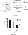
- FT-IR was used to demonstrate the chemical transformation of thio-ether to sulfone/sulfoxide by monitoring the appearance of special peaks after H 2 O 2 oxidation as seen in FIG. 5A .
- polythioether P2 0.5 g was dissolved in chloroform (50 mL) at 64 °C. Subsequently 30% hydrogen peroxide solution (0.5 mL) was added to the reaction flask. After 2 h of reaction, the formed polymer was precipitated in a cold ether and dry in a vacuum oven at 50 °C.
- FIG. 5B illustrates the rheometry measurement of storage modulus and loss modulus of PEG-PAM before and after H 2 O 2 treatment.
- PEG-PAM polymer was further investigated as a coating material for use with medical devices and implants and in particular for orthopedic implants that would controllably release antibiotics or other drugs from the PEG-PAM polymer network.
- another separate light polymerization-based synthesis method was investigated for the ability to polymerize PEG-PAM polymer directly on one or more surfaces of a medical device or implant. This second method of synthesizing PEG-PAM is used to form a coating on a substrate such as a medical device or implant (or even tissue).
- the coating is formed by polymerizing a mixture containing polyallyl mercaptan (PAM), a multi-arm poly(ethylene glycol) (PEG), an initiator, and one or more drugs, medicaments, or pharmaceutical compounds. Any number of arms of for the multi-arm PEG may be used. Commercially available PEG with three (3), four (4), five (5), six (6), seven(7), eight(8) or more arms are available and usable with the synthesis method described herein.
- polymerization is initiated in response to applied stimulus that works in conjunction with the initiator.
- the initiator may include a UV sensitive initiator such as DMPA and the mixture is polymerized by exposing the mixture to UV light.
- the initiator may include a visible light sensitive initiator such as Irgacure 2959 ® (which can also be used for UV light).
- a thermal initiator may be used such as azobisisobutyronitrile (AIBN).
- a redox initiator may be used such as ammonium persulfate (APS)/N,N,N ,N - tetramethylethylenediamine (TMEDA).
- the initiator is a photoinitiator.
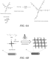
- FIG. 6A illustrates one embodiment of the synthesis of PEG-PAM using a four-arm PEG.
- 4-arm PEG-thiol (C 2 H 4 O) m S 4 wherein m is between 5 and 500
- PAM in this example having allyl and thiol ends
- n is between 5 and 30 in the presence of an initiator (e.g., DMPA) and a drug, medicament, or pharmaceutical compound contained in an organic solvent (not shown in FIG. 6A ).
- the mixture is subject to UV light to form PEG-PAM having a chemical formula (C 2 H 4 O) m S 4 [C 3 H 6 S] n .
- the reactive groups on PEG usable with this synthesis method include by way of example, PEG-acrylate, PEG-vinylsulfone, PEG-maleimide, PEG-acrylamide, PEG-epoxide, PEG-thiol, PEG-norborene, PEG-vinyl, PEG-allyl, and PEG-alkyne.
- an initiator such as DMPA is used along with UV light to form the PEG-PAM copolymer from PEG and PAM as well as the drug, medicament, or pharmaceutical compound (which is omitted from FIG. 6A ) and an organic solvent.
- FIG. 6B schematically illustrates one embodiment of a method of coating one or more surfaces of a medical device or implant 10.
- the medical device or implant 10 may include, by way of illustration and not limitation, an orthopedic implant, a spinal implant, a joint replacement device, pacemakers, insulin pumps, surgical pins, screws, or other surgical hardware.
- the coating is applied to a metallic surface of the medical device or implant 10 such as titanium or stainless steel but it should be appreciated that the coating and method of use is not limited to the particular composition of the substrate on which the coating resides. For example, it may also be applied to polymer or ceramic surfaces.
- PAM and a four-arm PEG-SH are mixed with a drug, medicament, or pharmaceutical component along with a small amount of organic solvent such as chloroform.
- organic solvents that may be used include dichloromethane (DCM), chloroform, and dimethyl sulfide (DMS).
- DCM dichloromethane
- DMS dimethyl sulfide
- the amount of organic solvent may vary but is typically under 10 % weight basis (e.g., 6% by weight).
- the drug that is incorporated into the PEG-PAM coating is the antibiotic vancomycin.
- other drugs, medicaments, and pharmaceutical compounds may be loaded within the PEG-PAM coating.
- the drugs, medicaments, or pharmaceutical compounds should generally be dissolvable or soluble is organic solvents. It should also be appreciated that more than one type of drug, medicament, or pharmaceutical compounds may be carried by the coating or gel formed by the methods described herein.
- the surgeon or physician is provided with or assembles the mixture of PEG, PAM, initiator, organic solvent, and the drug, medicament, or pharmaceutical compound.
- the relative amounts of PEG and PAM in the mixture depends on the type of PEG used and the number of arms that are intended to be occupied. For example, for a four-arm PEG molecule, there typically would be PAM in a 4x (molar basis) so that all the PEG arms are bound to PAM.
- the amount of drug, medicament, or pharmaceutical compound that is added may vary.
- a typical concentration of the drug, medicament, or pharmaceutical compound is generally within or below about 10-20 mg/mL of mixture of PEG and PAM.
- This mixture may be added to a delivery device such as spray bottle 12 or the like that can be used to spray a coating onto the medical device or implant 10 (other applicators such as an air-brush type applicator may also be used).
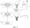
- the spray bottle 12 may actuated to apply a generally uniform coating of the mixture onto one or more surfaces of the medical device or implant 10 as seen in operation 1000 of FIG. 7 .
- medical device or implant 10 may be coated with the mixture including PEG, PAM, initiator, and drug with an applicator 14 such as a brush.
- the medical device or implant 10 may be dipped into the mixture and removed.
- the medical device or implant 10 is irradiated with light from a light source 16 (e.g., visible or UV light depending on the initiator) to polymerize the PEG-PAM polymer directly on one or more surfaces of the medical device or implant 10.
- the light source 16 may include a light, gun, wand, or other device that is typically found in hospitals and other medical settings and is used to illuminate a surface with light.
- the coating is illuminated for a short period of time (e.g., less than 5 minutes) to properly form the PEG-PAM copolymer. After applying the coating to the medical device or implant 10 some amount of time may elapse to enable the solvent to evaporate prior to illumination. This optional waiting period may last a few minutes. Thus, polymerization takes place in situ on the surface of the medical device or implant 10.
- multiple layers may optionally be coated on the surface of the medical device or implant 10 as seen by operation 1050.
- the mixture is applied and then subject to irradiation followed by application of another layer of the mixture followed by irradiation. This can proceed for a number of cycles to create the desired thickness or number of layers on the medical device or implant 10.
- only a portion of the surface of the medical device or implant 10 is coated.
- the coating system described herein may be used with any number of medical devices or implants 10 but has particular suitability to joint replacement devices.
- the coating system may be used also with orthopedic trauma implants or spinal implants.
- Other implantable medical devices may also be coated such as pacemakers, insulin pumps, and the like.
- the surfaces that are coated are typically metal such as titanium, stainless steel, cobalt-chrome although other materials may also be coated. These include plastics, ceramics, as well as allograft cadaver bone.
- the initiator is a photoinitiator which requires activation by light. It should be understood that in other embodiments, a heat applicator may be used to apply heat when the initiator is a thermal initiator. In other embodiments, for example, where the initiator is a redox initiator, there may be no need for a separate stimulation device. The process from start to finish is may be accomplished relatively quickly, for example, under 10 minutes (e.g., around 5 minutes) depending on the number and size of surfaces to be coated. Finally, as seen in operation 1200, the medical device or implant 10 is implanted in the subject.
- PEG-PAM polymers loaded with antibiotic were coated on the surface of titanium pins (i.e., K-wires) through in situ UV coating as described above which greatly reduced the amount to time compared with traditional methods.
- antibiotic vancomycin
- 4-arm PEG-thiol and PAM polymers were dissolved in dichloromethane (DCM) to make 6 % (w/v) solution, which was used to dissolve vancomycin at concentration of 20 mg/ml.
- DCM dichloromethane
- the K-wires were submerged in the antibiotic-encapsulated polymer solution 1 min and later taken out allowing evaporation of DCM.
- FIG. 8A illustrates scanning electron microscopic (SEM) images of the surfaces of a bare titanium pin (8-mm in diameter) and FIG. 8B illustrates a SEM image of a titanium pin coated with PEG-PAM containing vancomycin.
- the coating thickness is ⁇ 2 ⁇ m which is an inconsequential amount for press fit implants.
- FIG. 9 illustrates a graph of the in vitro passive release of a single layer of vancomycin containing PEG-PAM coated on the titanium pins.
- the daily release results indicated the concentration of vancomycin maintained above the minimum inhibition concentration (MIC) for 14 days.
- the PEG-PAM coated pins were subject to an in vivo assay to measure their antibacterial efficacy.
- the in vivo assay employed bioluminescent Staphylococcus aureus Xen36 strain that naturally produces a blue-green light emitted only by metabolically active bacteria as an "indicator" that reflects bacterial infections.
- Uncoated pins, coated pins with PEG-PAM and coated pins with PEG-PAM containing vancomycin were implanted into the femoral intramedullary canal of a mouse in a retrograde fashion and inoculated with the S. Aureus.
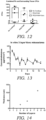
- FIG. 13 illustrates the release profile from three (3) layers of PEG-PAM coating on a titanium K-wire implant.
- the thickness of the PEG-PAM coatings increases with the number of coating layers that are applied to the medical device or implant 10.
- FIG. 14 illustrates the thickness of the resultant layer(s) on a K-wire implant for a single layer, three layers, five layers, and eight layers. A general linear relationship exists between the number of layers and the total thickness of the applied coating.
- FIG. 15 illustrates that by increasing the number of layers of PEG-PAM coating, the loading of a drug, medicament, or pharmaceutical compound (in this example vancomycin) is increased.
- FIG. 16 illustrates a graph showing the cumulative release of vancomycin from a single layer of vancomycin-containing PEG-PAM coated on a titanium pin. As seen in FIG. 16 after about 3 days most of the vancomycin was released. In this experiment, the vancomycin was released relatively quickly.
- Titanium pins that were bare (not coated), coated with PEG-PAM only, and coated with PEG-PMA and vancomycin were tested for their ability to inhibit the growth of S . Aureus.
- the respective pins were placed in in a petri dish containing growth media and seeded with S. Aureus. After 3 days the petri dishes containing the respective coated/non-coated pins were examined for inhibited bacterial growth which could be visualized as a zone or region around the respective pins where bacteria did not grow.
- the pin containing PEG-PMA and vancomycin showed a clear and marked zone without bacterial growth whereas the other two pins with no coating or PEG-PAM only did not show any sort of similar inhibition zone.
- FIG. 17 illustrates the quantification of the diameters of the inhibition zones for the non-coated pin, the pin coated with PEG-PAM only, and the pin coated with PEG-PAM and vancomycin.
- Additional antibiotics were also evaluated using the PEG-PAM system disclosed herein.
- in vitro bioluminescence assay measurements were performed on titanium pins with PEG-PAM coatings (all single layer) having nine (9) different antibiotics including cefazolin, ceftriaxone, cefepime, tobramycin, piperacillin and tazobactam, vancomycin, rifampin, clindamycin, linezolid, and control. Pins were inserted into the wells of a 96 well plate and then measured using the IVIS ® Spectrum in vivo imaging system (PerkinElmer). All of the tested antibiotics loaded into the PEG-PAM system killed S. Aureus within twenty-four (24) hours.
- FIG. 19 illustrates the quantification of the diameters of the inhibition zones for a non-coated pin (control) and a pin coated with PEG-PAM loaded with Tobra against Pseudomonas (PAO1 strain) using similar testing procedure as described above with respect to FIG. 17 .
- FIG. 20 illustrates the quantification of the diameters of the inhibition zones for a non-coated pin (control) and a pin coated with PEG-PAM, piperacillin and Tazobacta against E. Coli using similar testing procedure as described above with respect to FIG. 17 .
- Periprosthetic osteolysis is a serious complication in orthopedic implants, including, for example, total hip replacement (THR) surgery. Osteolysis can lead to prosthesis loosening and periprosthetic fracture. These complications cause significant morbidity and often require additional surgical intervention.
- THR total hip replacement
- a titanium pin was implanted into the femur of a mouse. Three different types of pins were implanted: a non-coated implant, an implant coated with PEG-PAM (no drug), and an implant coated with PEG-PAM and vancomycin. As seen in FIG.
- implants with no coatings or coated with PEG-PAM alone showed a dramatic degree of periprosthetic osteolysis that became evident by post-operative days (POD) 21.
- antibiotic (vancomycin) encapsulated PEG-PAM implants showed no detectable radiographic periprosthetic osteolysis.
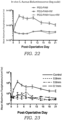
- FIG. 22 illustrates the post-operative in vivo bioluminescence analysis of titanium pins implanted into the femur of a mouse.
- the pins that were implanted had PEG-PAM (no drug), PEG-PAM loaded with rifampin, and PEG-PAM loaded with rifampin and vancomycin.
- the top, exposed part of the implant was inoculated with S. aureus and was analyzed using the IVIS ® Spectrum (PerkinElmer ) in vivo imaging system. Higher signal levels correspond to high or progressed infection post-surgery.
- both the rifampin and rifampin/vancomycin loaded pins maintain the signal at around baseline which indicates that it clears the injections coming from the bacterial challenge.
- FIG. 23 illustrates the post-operative in vivo analysis of various sized stainless steel pins implanted into the femur of a mouse.
- the pins that where implanted including a control pin (no coating) as well as various diameter pins (0.1 mm, 0.6 mm, 0.8 mm) coated with PEG-PAM loaded vancomycin.
- the top, exposed part of the implant was inoculated with S. aureus and was analyzed using the IVIS ® Spectrum as described above. Higher signal levels correspond to high or progressed infection post-surgery.
- the PEG-PAM + vancomycin group of pins maintains the measured bioluminescence signal at or near baseline levels.
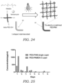
- FIG. 24 schematically illustrates the synthesis of PEG-PAM diallyl (PEG-PAMDA) coatings according to one embodiment.
- the PAM that is used to form the PEG-PAMDA includes two allyl terminal ends, i.e., PAM diallyl or PAMDA.
- PAM diallyl or PAMDA a four arm PEG-thiol molecule was used in conjunction with PAMDA and the drug vancomycin to coat an L-shaped stainless steel pin that was used for spinal surgery. Any number of arms of for the multi-arm PEG may be used.
- Commercially available PEG with three (3), four (4), five (5), six (6), seven(7), eight(8) or more arms are available and usable with the synthesis method described herein.
- Crosslinking is initiated using a photoinitiator and source of polymerizing light as explained previously. It was found that standard, single layer PEG-PAM loaded with vancomycin did not work as intended in the in the spinal surgery model. While not wishing to be bound to any particularly theory, it is believed that the single layer PEG-PAM released the vancomycin too quickly into the surrounding tissue. Thus, it was desired to incorporate a stronger, chemically crosslinked coating that would allow the antibiotic to more slowly diffuse out of the coating and into the surrounding environment. PEG-PAMDA allows for slower diffusion of the entrained drug, medicament, or pharmaceutical compound.
- FIG. 25 illustrates the in vitro release of vancomycin from an L-shaped stainless steel pin having a single layer of PEG-PAM. Also illustrated in the in vitro release of vancomycin from an L-shaped stainless steel pin having three layers of PEG-PAMDA. As seen in FIG. 25 , the vancomycin is released much more slowly with the chemically crosslinked PEG-PAMDA coating. Thus, chemical crosslinking of the coating may be employed to control the time release of the drug, medicament, or pharmaceutical compound.
- FIG. 26 illustrates the post-operative in vivo analysis of 0.1 mm sized stainless steel L-shaped implants that were implanted in the vertebrae of a mouse.
- the implants that where implanted and tested for in vivo bioluminescence included an implant coated with three layers of PEG-PAMDA (with no drug) and another implant coated with three layers of PEG-PAMDA and also loaded with vancomycin. After implantation, exposed part of the implant was inoculated with S. aureus and was analyzed using the IVIS ® Spectrum through 25 days. As seen in FIG. 26 , the implant coated with PEG-PAMDA loaded with vancomycin maintained the bioluminescent signal at around baseline levels. Thus, the drug loaded implant was able to clear infections coming from this bacterial challenge.
- PEG-PAM hydrogel was assembled by hydrophobic interaction and it was a physical crosslinked hydrogel whose stability was affected by various external stimulus.
- the stability of PEG-PAM hydrogel can be easily tuned through incorporation of chemical crosslinker. Because the terminals of PEG-PAM are allyl group which is a typical substrate for thiol-ene crosslinking, difunctional thiol molecules were employed in the chemical crosslinking reaction to achieve the transformation of physically crosslinked PEG-PAM hydrogel to chemically crosslinked PEG-PAM hydrogel.
- PEG-PAM could be crosslinked with dithiol containing polymers to convert the coating into chemically crosslinked hydrogel, which would lead to decreased diffusion ability of antibiotics inside of the hydrogel.
- PEG-PAM is produced using the first synthesis method described herein, namely, PAM is conjugated with PEG through a thiol reactive groups on the PEG molecule using a catalytic amount of base catalyst such as triethylamine (TEA).
- TAA triethylamine
- PEG-PAM is then hydrated and PEG-dithiol is added along with the drug (e.g., vancomycin) and photoinitiator (e.g., lithium phenyl-2,4,6-trimethylbenzoylphosphinate(LAP)) were mixed together and 365nm UV light was used to trigger the crosslinking reaction.
- the drug e.g., vancomycin
- photoinitiator e.g., lithium phenyl-2,4,6-trimethylbenzoylphosphinate(LAP)
- HS-MMP-peptide-Alexa488 was incubated with 4-arm PEG-PAM and exposed to UV light to induce attachment of the peptide to the vinyl groups in PAM.
- the hydrogel was allowed to form through physical interactions.
- the incubation of the gel in collagenase led to rapid release, achieving ⁇ 60% cumulative release in one day.
- less than 10% release was observed in the absence of collagenase at the same one day time period.
- the addition of protease degradable peptides to the PEG-PAM coating may be used to accelerate the release of drugs, medicaments, or pharmaceuticals from the PEG-PAM coating.
- the eluting properties of the PEG-PAM coating can be adjusted or tuned depending on the desired properties of the coated device or implant.
- the drug, medicament, or pharmaceutical may be eluted in a linear fashion over time (this may be a slow release or a rapid release).
- an increase or spike in elution may be desired either right after implantation or several days or weeks after implementation.
- the PEG-PAM elution kinetics may be modified for the appropriate application.
Landscapes
- Health & Medical Sciences (AREA)
- Chemical & Material Sciences (AREA)
- Medicinal Chemistry (AREA)
- Life Sciences & Earth Sciences (AREA)
- General Health & Medical Sciences (AREA)
- Veterinary Medicine (AREA)
- Public Health (AREA)
- Animal Behavior & Ethology (AREA)
- Epidemiology (AREA)
- Transplantation (AREA)
- Dermatology (AREA)
- Oral & Maxillofacial Surgery (AREA)
- Engineering & Computer Science (AREA)
- Chemical Kinetics & Catalysis (AREA)
- Organic Chemistry (AREA)
- Molecular Biology (AREA)
- Biomedical Technology (AREA)
- Polymers & Plastics (AREA)
- Materials Engineering (AREA)
- Wood Science & Technology (AREA)
- Materials For Medical Uses (AREA)
- Medicinal Preparation (AREA)
Description
- The technical field generally relates to the synthesis and use of multi-arm block-copolymers. In particular, the technical field relates to multi-arm block-copolymers that utilize polyallyl mercaptan (PAM). The use of PAM in conjunction with multi-arm poly(ethylene glycol) (PEG) has the ability to generate useful functional, biodegradable coatings that are useful for biomedical implants (e.g., orthopedic and other implants).
- Orthopedic implant infections persist and cost the U.S. health system more than $8 billion per year in additional expenses, despite best efforts at host modification, environmental sterility, and systemic antibiotic therapy. Current standard of care for local antibiotic delivery is polymethylmethacralate (PMMA); a biologically inert delivery system with poor elution characteristics that is used for its mechanical properties, with local antibiotic delivery as a beneficial "secondary" function. As an alternative to PMMA, attempts have been made to engineer colloidal-based biodegradable systems with self-assembling block polymers as an alternative. For example, a "smart" implant coating using branched poly(ethylene glycol)-poly(propylene sulfide) (PEG-PPS) polymer has been designed to deliver antibiotics both passively and actively. See Stavrakis et al., In Vivo Efficacy of a "Smart" Antimicrobial Implant Coating, Journal of Bone and Joint Surgery, 98(14): 1183-89 (2016). However, these coatings have been labor-intensive to manufacture and thus required assembly prior to use in the operating room. This also has the unfortunate consequence of transforming conventional orthopedic implants into "drug delivery devices" that trigger additional burdensome regulatory approvals required by government agencies such as the Food and Drug Administration. For example, regulations on drug delivery devices have made factory-applied coatings impractical. Further, factory-coated implants increase the cost of all implants rather than allowing the surgeon to add a particular coating when the patient's risk-profile or other patient-specific circumstances warrant the coating's use. Factory-coated implants also suffer from the limitation that there is no versatility offered as each coating is loaded with the same antibiotic. This lack of personalization in the coating makeup is important because the physician may want to target a particular bacteria species that may be endemic to the geographical local area or local patient population. Factory-coated implants also may degrade over time limiting the overall shelf life of the implant device.
- The invention is directed to a method of coating one or more surfaces of a medical device, according to
claim 1 and a coating material according toclaim 10. Further developments of the invention are according to dependent claims 2-9 and 11-14. - The PEG-PAM coating permits medical devices such as orthopedic implants to remain sterile leaving industry or other storage warehouses and provides surgeons versatility to apply a drug-containing coating on a medical device or on or within the patient directly within the operating room. In some embodiments, the drug or medicament includes an antimicrobial agent or drug that can be applied directly to all or part of a medical device. The choice of antimicrobial agent used with the coating may be specifically tailored to the patient's needs.
- A variety of modalities may be used to apply the drug-containing coating to the medical device surface or a tissue surface of the patient. For example, the coating may be applied as an aerosol such as a spray. The coating may also be applied as a paint using an applicator such as a brush or the like. The coating may also be applied to fill voids or spaces using an injection device such as a syringe. The applied coating is then, in one embodiment, subject to illumination from an appropriate wavelength-emitting light source (e.g., ultraviolet light source). The multi-arm block-copolymer based system described herein provides a cost-effective, practical alternative to PMMA that biodegrades, has consistent, tailorable release kinetics, and allows patient-specific tailoring of antibiotic loading. In other embodiments, the PEG-PAM material described herein may also be used as a coating on or within tissue or an injectable gel.
-
-
FIG. 1 illustrates the synthesis of PAM according to one embodiment from the radial polymerization of 1,3 propanedithiol and allylsulfide. The stoichiometric ratio of allylsulfide to 1,3 propanedithiol may be adjusted to control whether the terminal ends of the PAM are thiol groups, allyl groups, or both. -
FIG. 2 illustrates the synthesis of PEG-PAM using a multi-arm PEG-maleimide and PAM in the presence of triethylamine (TEA) catalyst according to one embodiment. -
FIG. 3 illustrates a schematic representation of multi-arm PEG-PAM amphiphilic diblock polymer formed using different ratios of thiol to PEG-maleimide. -
FIG. 4 illustrates dynamic strain amplitude cycles (γ = 5% or 100%) of 5 wt % PEG-PAM hydrogel at a temperature of 25 °C. The graph illustrates reversible shear-thinning behavior. Rheology measurement: PEG-PAM hydrogels were allowed to self-assemble for 30 min before transferred to an 8 mm plate-to-plate rheometer (Physica MCR 301, Anton Paar, Ashland, VA). An evaporation blocker system was used during measurements. For frequency sweep, the data were collected for the modulus with a frequency range of 0. 1-100 rad/s under a 1% constrain at 37 °C. For amplitude sweep, the data were collected for the modulus with a frequency of 10 rad/s under a constrain range of 0.1-100% at 37 °C. A strain recovery experiment was performed with 4 cycles of strain steps of 1% and 50% strain, each at 6.5 rad/s. -
FIG. 5A illustrates FT-IR spectra of PAM and the oxidized forms. FT-IR Measurement: FT-IR spectra was collected by Jasco 420 FTIR Spectrophotometer. The sample pellet was prepared by mixing the 10 mg polymer and 500mg KBr. GPC Measurement: Gel permeation chromatography/light scattering (GPC/LS) was performed on a SSI Accuflow Series III liquid chromatograph pump equipped with Wyatt DAWN EOS light scattering (LS) and Optilab rEX refractive index (RI) detectors. Separations were achieved using 105 , 104 , and 103 Å Phenomenex Phenogel 5 µm columns in chloroform. GPC/LS samples were prepared at concentrations of 5 mg/mL -
FIG. 5B illustrates the rheometry measurement of storage modulus and loss modulus of PEG-PAM before and after H2O2 treatment. -
FIG. 6A illustrates the synthesis of PEG-PAM copolymer using an eight arm PEG-thiol, a photoinitiator (e.g., DMPA), and PAM. UV light is used to polymerize the mixture. -
FIG. 6B schematically illustrates the formation of PEG-PAM copolymer on a medical device or implant containing a drug, medicament, or pharmaceutical compound therein using multi-arm PEG-thiol, a photoinitiator, and PAM. Ultraviolet light is used to polymerize the mixture. -
FIG. 7 illustrates a process for applying or forming a PEG-PAM coating on medical device or implant according to one embodiment. -
FIG. 8A illustrates scanning electron microscopic (SEM) images of the surfaces of a bare titanium pin (8-mm in diameter). -
FIG. 8B illustrates a SEM image of a titanium pin coated with PEG-PAM containing vancomycin. -
FIG. 9 illustrates a graph of the in vitro passive release (concentration/area) of a single layer of vancomycin containing PEG-PAM coated on the titanium pins. Quantified via UV-Vis, the daily release of vancomycin per pin over 14 days. -
FIG. 10 illustrates postoperative in vivo S. aureus bioluminescence signals (logarithmic scale). Bioluminescence was measured using an IVIS® Spectrum (PerkinElmer). -
FIG. 11 illustrates a graph of bioluminescence as a function of time for the pin with no coating, pin with PEG-PAM, and pin with PEG-PAM loaded with vancomycin. -
FIG. 12 illustrates a graph of colony-forming unit (CFU) enumeration from implants and peri-implant tissue for implants with no coatings PEG-PAM coating (no drug), and PG-PAM (with vancomycin). -
FIG. 13 illustrates a graph showing the release profile (release/area) from three (3) layers of PEG-PAM coating on a titanium K-wire implant. -
FIG. 14 illustrates a graph of the thickness of a single layer and multiple layers deposited on a K-wire implant. Data is shown for a single layer, three layers, five layers, and eight layers. -
FIG. 15 is a graph showing the loading of vancomycin for coatings having a single layer, three layers, five layers, and eight layers. -
FIG. 16 is a graph showing the cumulative release percentage of vancomycin from a single layer of vancomycin-containing PEG-PAM coated on a titanium pin. -
FIG. 17 illustrates the quantification of the diameters of the inhibition zones for the non-coated pin, the pin coated with PEG-PAM only, and the pin coated with PEG-PAM and vancomycin. -
FIG. 18 illustrates the results of the in vitro bioluminescence assay of PEG=PAM coating with the nine (9) different antibiotics tested against S. Aureus. -
FIG. 19 illustrates the quantification of the diameters of the inhibition zones for a non-coated pin (control) and a pin coated with PEG-PAM loaded with Tobra against Pseudomonas (PAO1 strain). -
FIG. 20 illustrates the quantification of the diameters of the inhibition zones for a non-coated pin (control) and a pin coated with PEG-PAM, piperacillin and Tazobacta against E. Coli. -
FIG. 21 illustrates lateral radiographs of titanium implants containing no coating, PEG-PAM (no drug), and PEG-PAM loaded with vancomycin. POD stands for post-operative days. -
FIG. 22 illustrates the post-operative in vivo bioluminescence analysis of titanium pins implanted into the femur of a mouse. Specifically, a graph of in vivo bioluminescence (mean maximum flus) of S. Aureus is shown as a function of time (PODs). -
FIG. 23 illustrates the post-operative in vivo bioluminescence analysis of various diameter titanium pins implanted into the femur of a mouse. -
FIG. 24 schematically illustrates the synthesis of a PEG-PAM diallyl (PEG-PAMDA) coating according to one embodiment. The coating is formed on an L-shaped stainless steel medical device or implant for use in spinal applications. -
FIG. 25 illustrates the in vitro release of vancomycin from an L-shaped stainless steel pin having a single layer of PEG-PAM. Also illustrated in the in vitro release of vancomycin from an L-shaped stainless steel pin having three layers of PEG-PAMDA. -
FIG. 26 illustrates the post-operative in vivo analysis of 0.1 mm sized stainless steel L-shaped implants that were implanted in the vertebrae of a mouse. The implants had either PEG-PAMDA (3 layers) and no drug or PEG-PAMDA (3 layers) and vancomycin. -
FIG. 27 illustrates a graph of cumulative release of vancomycin as a function of time for PEG-PAM crosslinked with dithiol containing polymers to convert the coating into chemically crosslinked hydrogel. -
FIG. 28 illustrates a graph of cumulative release of Alex-488 from PEG-PAM hydrogel functionalized with MMP-Alex-488. The presence of collagenase dramatically increases the rate of release. - In one embodiment, a polymeric delivery platform is disclosed that is formed from a PEG-PAM copolymer of polyallyl mercaptan (PAM) and a multi-arm poly(ethylene glycol). The PEG-PAM copolymer system is used for the delivery of medicinal or therapeutic compounds such as antibiotics to living tissue. The polymeric delivery platform, in one embodiment, is coated in situ on one or more surfaces of a medical device or implant that is to be used in a mammalian subject. In other embodiments, the polymeric delivery platform may be directly delivered to tissue or tissue regions of a mammalian subject. As explained herein, in one preferred embodiment, a multi-arm poly(ethylene glycol) (PEG) is reacted with polyallyl mercaptan (PAM) in the presence of an initiator compound (e.g., photoinitiator) and one or more drugs, medicaments, or pharmaceutical compounds. The mixture is exposed to an initiator source such as light, heat, or a redox initiator to polymerize the copolymer. In one embodiment, the initiator compound is a photoinitiator and the mixture is first applied to one or more surfaces of a medical device or implant and is then exposed to an appropriate polymerizing light source (e.g., ultraviolet light source or visible light source) to form a PEG-PAM copolymer coating or layer(s) that is formed in situ on the medical device or implant.
- The PEG-PAM coating permits medical devices or implants to remain in a sterile state prior to use and provides surgeons the versatility to customize the drug, medicament, or pharmaceutical compound-containing coating on a medical device or implant (or applied on or within the patient directly within the operating room in other applications). In some embodiments, the drug, medicament, or pharmaceutical compound includes one or more antimicrobial agents or drugs that can be applied directly to all or part of a medical device or implant. The choice of antimicrobial agent used with the PEG-PAM coating may be specifically tailored to the patient's needs.
- The PEG-PAM coating described herein uses PAM as one of the copolymer blocks. PAM was previously known to be created by thiol-ene click polyaddition of α,ω-alkylene thiols, for example, as described in Deubel et al., Polythioethers by Thiol-Ene Click Polyaddition of α,ω-Alkylene Thiols, Macromol. Rapid Commun., 34, 1020-1025 (2013). Unfortunately, this synthesis process is a multi-step process that takes a considerable amount of time. In one embodiment as explained herein, a new method of synthesizing PAM is disclosed.
FIG. 1 illustrates this new method which involves the synthesis of PAM from radial polymerization of 1,3 propanedithiol and allylsulfide. The synthesis of PAM is based on the thiol-ene copolymerization between 1,3 propanedithiol and allyl sulfide which are both commercially available with high purity. - The polymerization may be performed under 365nm UV light irradiation with the presence of a photoinitiator and, depending on the relative stoichiometric ratio of 1,3 propanedithiol and allylsulfide produces PAM that is either thiol terminated, allyl terminated, or thiol and allyl terminated. In particular, when the mixture of 1,3 propanedithiol and allylsulfide is in a 1:1 stoichiometric ratio PAM is formed that has ends that are both thiol and allyl terminated. When the mixture of 1,3 propanedithiol and allylsulfide has a stoichiometric ratio of allylsulfide to 1,3 propanedithiol that is greater than 1, the PEM ends are allyl terminated. Conversely, when the mixture of 1,3 propanedithiol and allylsulfide has a stoichiometric ratio of allylsulfide to 1,3 propanedithiol that is less than 1, the PEM ends are thiol terminated. The linear polymer that has thiol and/or allyl at the ends enables further functionalization from end groups. The degree of polymerization may vary from between 5-15 with the length controlled by monomer amount. It is important to note that the method of synthesis is completely different from that used for the synthesis of polypropylene sulfide) (PPS). PAM is synthesized via radical polymerization while PPS is synthesized using anionic polymerization.
- In one exemplary example of the synthesis of PAM, 1
mol% 2,2-Dimethoxy-2-phenylacetophenone (DMPA) photoinitiator was added to a 1:1 stoichiometric ratio mixture of 1,3 propanedithiol and allylsulfide and then the mixture was illuminated under 365nm UV light (20mw/cm2) irradiation for 30 minutes. Next, the PAM product is precipitated in the presence of cooled methanol and dried as a white wax. - PEG-PAM may be synthesized using the PAM described above in one of two synthesis methods. In a first synthesis method, PAM (n between 5 and 30) is conjugated with multi-arm PEG through any number of thiol reactive groups on the PEG molecule using a catalytic amount of base catalyst such as triethylamine (TEA). The reactive groups on PEG usable with this synthesis method include by way of example, PEG-acrylate, PEG-vinylsulfone, PEG-maleimide, PEG-acrylamide, PEG-isocyanate, PEG-bromide, and PEG-epoxide.
FIG. 2 illustrates one example of the synthesis of PEG-PAM copolymer material using the multi-arm PEG-maleimide (e.g., eight arm) and PAM (n between 5 and 30) in the presence of triethylamine (TEA) catalyst. Any number of arms of for the multi-arm PEG may be used. Commercially available PEG with three (3), four (4), five (5), six (6), seven(7), eight(8) or more arms are available and usable with the synthesis method described herein. In this embodiment, PAM and the 8-Arm-PEG maleimide were dissolved in deuterated chloroform (CDCl3) and a catalytic amount of triethylamine was added into the mixture and then stirred for 3 hours. The solvent was removed under vacuum and the crude product was precipitated in cold ether (4 °C). - The thiol end group of PAM can be used in various thiol based click chemistries for polymer modification or functionalization. In this particular embodiment, thiolmaleimide was used for Michael addition to synthesize multi-Arm-PEG-PAM diblock polymers both starting from 8-Arm-PEG-maleimide (Mw: 40,000). The fully functionalized block polymer (8-Arm-PEG-PEG) was dissolved in the CDCl3 (10 wt%) and instantly formed organogel, which is not soluble in water. If instead of reacting the eight (8) arms of the PEG with PAM, one only reacts three (3) arms, which would decrease the percentage of hydrophobic in the structure; the polymer is then water soluble. Generally, it is preferable to react the PAM with between three and five arms of the 8-Arm-PEG-maleimide as seen in
FIG. 3 . The degree to which PAM reacts with the arms of the PEG-maleimide structure may be controlled by the molar ratio of PAM to PEG-maleimide with a larger amount of PAM being used to react with a higher number of arms while a lower amount of PAM yields to fewer arms being occupied. - The PEG-PAM copolymer is soluble in water at 5 wt% for stable hydrogel formation. Because the gelation mechanism is based on the hydrophobic interaction between PAM parts of the copolymer, it is expected to exhibit shear-thinning behavior like other supermolecular hydrogels such as PEG-PPS. Small amplitude oscillatory shear rheometry was performed to demonstrate the supermolecular interactions in PEG-PAM hydrogel. Strain sweeps at a constant oscillatory frequency of 6.5 rad/s at 25 °C revealed that the hydrogel could be disrupted under high strain condition. At higher applied strains (γ >10%) both G' (storage modulus) and G" (loss modulus) drop and G' decreases below G" at approximately γ = 35% applied strain, indicative of a transition from gel-like to liquid-like state at high strain. The gel can rapidly recover its mechanical properties after high strain deformation, as evidenced by a dynamic step-strain test seen in
FIG. 4 . When a 5 wt % PEG-PAM hydrogel at 6.5% strain is subjected to an 100% step strain, G' drops substantially from G' = 400 Pa to G' = 30 Pa. Reversing the strain to 5% results in rapid recovery (<20s) to be original modulus. This recovery process was performed several cycles, indicating very good self-healing property of the gels. - After the demonstration of physical cross-linked behavior of PEG-PAM hydrogel, another experiment was conducted to investigate the oxidation responsive behavior of this hydrogel because of the thio-ether backbone in the hydrophobic block. Thio-ether functional groups are very hydrophobic segments that enable the gelation of PEG-PAM, however, upon oxidation by peroxides or other reactive oxygen species, thio-ether backbones would be oxidized to sulfones or sulfoxides, which are so hydrophilic that cannot provide the physical interaction for gelation. According to this mechanism, thio-ether backbones have been widely employed in responsive materials for drug delivery. FT-IR was used to demonstrate the chemical transformation of thio-ether to sulfone/sulfoxide by monitoring the appearance of special peaks after H2O2 oxidation as seen in
FIG. 5A . For oxidation studies, polythioether P2 (0.5 g) was dissolved in chloroform (50 mL) at 64 °C. Subsequently 30% hydrogen peroxide solution (0.5 mL) was added to the reaction flask. After 2 h of reaction, the formed polymer was precipitated in a cold ether and dry in a vacuum oven at 50 °C. The peak of 1100, 1300 cm-1 and 1030 cm-1 from FT-IR are assigned as the peaks of sulfone and sulfoxide respectively which provides direct proof of thio-ether oxidation. In addition, PEG-PAM gel can be also disrupted by treatment of H2O2 which induced dramatically decrease of modulus upon oxidation.FIG. 5B illustrates the rheometry measurement of storage modulus and loss modulus of PEG-PAM before and after H2O2 treatment. - Because the PEG-PAM copolymer could be disrupted by oxidative species, PEG-PAM polymer was further investigated as a coating material for use with medical devices and implants and in particular for orthopedic implants that would controllably release antibiotics or other drugs from the PEG-PAM polymer network. In this regard, another separate light polymerization-based synthesis method was investigated for the ability to polymerize PEG-PAM polymer directly on one or more surfaces of a medical device or implant. This second method of synthesizing PEG-PAM is used to form a coating on a substrate such as a medical device or implant (or even tissue). The coating is formed by polymerizing a mixture containing polyallyl mercaptan (PAM), a multi-arm poly(ethylene glycol) (PEG), an initiator, and one or more drugs, medicaments, or pharmaceutical compounds. Any number of arms of for the multi-arm PEG may be used. Commercially available PEG with three (3), four (4), five (5), six (6), seven(7), eight(8) or more arms are available and usable with the synthesis method described herein. In some embodiments, polymerization is initiated in response to applied stimulus that works in conjunction with the initiator. For example, the initiator may include a UV sensitive initiator such as DMPA and the mixture is polymerized by exposing the mixture to UV light. Likewise, the initiator may include a visible light sensitive initiator such as Irgacure 2959® (which can also be used for UV light). In yet another alternative, a thermal initiator may be used such as azobisisobutyronitrile (AIBN). In still another alternative, a redox initiator may be used such as ammonium persulfate (APS)/N,N,N ,N - tetramethylethylenediamine (TMEDA). In one preferred embodiment that is used to coat substrates such as medical devices or implants, the initiator is a photoinitiator.
-
FIG. 6A illustrates one embodiment of the synthesis of PEG-PAM using a four-arm PEG. In this particular example, 4-arm PEG-thiol (C2H4O)mS4, wherein m is between 5 and 500, is mixed with PAM (in this example having allyl and thiol ends) having a chemical formula [C3H6S]n, wherein n is between 5 and 30 in the presence of an initiator (e.g., DMPA) and a drug, medicament, or pharmaceutical compound contained in an organic solvent (not shown inFIG. 6A ). The mixture is subject to UV light to form PEG-PAM having a chemical formula (C2H4O)mS4[C3H6S]n. - The reactive groups on PEG usable with this synthesis method include by way of example, PEG-acrylate, PEG-vinylsulfone, PEG-maleimide, PEG-acrylamide, PEG-epoxide, PEG-thiol, PEG-norborene, PEG-vinyl, PEG-allyl, and PEG-alkyne. In this synthesis method, an initiator such as DMPA is used along with UV light to form the PEG-PAM copolymer from PEG and PAM as well as the drug, medicament, or pharmaceutical compound (which is omitted from
FIG. 6A ) and an organic solvent.FIG. 6B schematically illustrates one embodiment of a method of coating one or more surfaces of a medical device orimplant 10. The medical device orimplant 10 may include, by way of illustration and not limitation, an orthopedic implant, a spinal implant, a joint replacement device, pacemakers, insulin pumps, surgical pins, screws, or other surgical hardware. Typically, the coating is applied to a metallic surface of the medical device orimplant 10 such as titanium or stainless steel but it should be appreciated that the coating and method of use is not limited to the particular composition of the substrate on which the coating resides. For example, it may also be applied to polymer or ceramic surfaces. In the particular example ofFIG. 6B , PAM and a four-arm PEG-SH are mixed with a drug, medicament, or pharmaceutical component along with a small amount of organic solvent such as chloroform. Other organic solvents that may be used include dichloromethane (DCM), chloroform, and dimethyl sulfide (DMS). The amount of organic solvent may vary but is typically under 10 % weight basis (e.g., 6% by weight). In the example ofFIG. 6B , the drug that is incorporated into the PEG-PAM coating is the antibiotic vancomycin. It should be appreciated that other drugs, medicaments, and pharmaceutical compounds may be loaded within the PEG-PAM coating. Because of the use of the organic solvent, the drugs, medicaments, or pharmaceutical compounds should generally be dissolvable or soluble is organic solvents. It should also be appreciated that more than one type of drug, medicament, or pharmaceutical compounds may be carried by the coating or gel formed by the methods described herein. - In the operating room or other surgical setting, as best seen in
FIG. 7 , the surgeon or physician is provided with or assembles the mixture of PEG, PAM, initiator, organic solvent, and the drug, medicament, or pharmaceutical compound. The relative amounts of PEG and PAM in the mixture depends on the type of PEG used and the number of arms that are intended to be occupied. For example, for a four-arm PEG molecule, there typically would be PAM in a 4x (molar basis) so that all the PEG arms are bound to PAM. The amount of drug, medicament, or pharmaceutical compound that is added may vary. A typical concentration of the drug, medicament, or pharmaceutical compound is generally within or below about 10-20 mg/mL of mixture of PEG and PAM. - This mixture may be added to a delivery device such as
spray bottle 12 or the like that can be used to spray a coating onto the medical device or implant 10 (other applicators such as an air-brush type applicator may also be used). Thespray bottle 12 may actuated to apply a generally uniform coating of the mixture onto one or more surfaces of the medical device orimplant 10 as seen inoperation 1000 ofFIG. 7 . Alternatively, medical device orimplant 10 may be coated with the mixture including PEG, PAM, initiator, and drug with anapplicator 14 such as a brush. As yet another alternative, the medical device orimplant 10 may be dipped into the mixture and removed. Next, as seen inoperation 1100, the medical device orimplant 10 is irradiated with light from a light source 16 (e.g., visible or UV light depending on the initiator) to polymerize the PEG-PAM polymer directly on one or more surfaces of the medical device orimplant 10. Thelight source 16 may include a light, gun, wand, or other device that is typically found in hospitals and other medical settings and is used to illuminate a surface with light. The coating is illuminated for a short period of time (e.g., less than 5 minutes) to properly form the PEG-PAM copolymer. After applying the coating to the medical device orimplant 10 some amount of time may elapse to enable the solvent to evaporate prior to illumination. This optional waiting period may last a few minutes. Thus, polymerization takes place in situ on the surface of the medical device orimplant 10. - As seen in
FIG. 7 , multiple layers may optionally be coated on the surface of the medical device orimplant 10 as seen byoperation 1050. For multiple layers, the mixture is applied and then subject to irradiation followed by application of another layer of the mixture followed by irradiation. This can proceed for a number of cycles to create the desired thickness or number of layers on the medical device orimplant 10. - In some embodiments, only a portion of the surface of the medical device or
implant 10 is coated. For example, it may be preferred that only those surfaces that are in contact with bone or bodily tissue are to be coated. Those surfaces that serve as contact or articulating surfaces for other components may not be coated. The coating system described herein may be used with any number of medical devices orimplants 10 but has particular suitability to joint replacement devices. The coating system may be used also with orthopedic trauma implants or spinal implants. Other implantable medical devices may also be coated such as pacemakers, insulin pumps, and the like. The surfaces that are coated are typically metal such as titanium, stainless steel, cobalt-chrome although other materials may also be coated. These include plastics, ceramics, as well as allograft cadaver bone. - In the above embodiment, the initiator is a photoinitiator which requires activation by light. It should be understood that in other embodiments, a heat applicator may be used to apply heat when the initiator is a thermal initiator. In other embodiments, for example, where the initiator is a redox initiator, there may be no need for a separate stimulation device. The process from start to finish is may be accomplished relatively quickly, for example, under 10 minutes (e.g., around 5 minutes) depending on the number and size of surfaces to be coated. Finally, as seen in
operation 1200, the medical device orimplant 10 is implanted in the subject. - PEG-PAM polymers loaded with antibiotic (vancomycin) were coated on the surface of titanium pins (i.e., K-wires) through in situ UV coating as described above which greatly reduced the amount to time compared with traditional methods. For the in situ UV-coating of PEG-PAM on titanium pins, 4-arm PEG-thiol and PAM polymers were dissolved in dichloromethane (DCM) to make 6 % (w/v) solution, which was used to dissolve vancomycin at concentration of 20 mg/ml. The K-wires were submerged in the antibiotic-encapsulated
polymer solution 1 min and later taken out allowing evaporation of DCM. Then the wires were irradiated with 365nm UV light (20mw/cm2) for 1 min. The surface features of the polymer coatings was confirmed by SEM and more detailed elemental analysis also provided the proof about chemical elements derived from PEG-PAM.FIG. 8A illustrates scanning electron microscopic (SEM) images of the surfaces of a bare titanium pin (8-mm in diameter) andFIG. 8B illustrates a SEM image of a titanium pin coated with PEG-PAM containing vancomycin. The coating thickness is ~2µm which is an inconsequential amount for press fit implants. - The in vitro elution of vancomycin from the PEG-PAM coating on the pins was also evaluated.
FIG. 9 illustrates a graph of the in vitro passive release of a single layer of vancomycin containing PEG-PAM coated on the titanium pins. The daily release results indicated the concentration of vancomycin maintained above the minimum inhibition concentration (MIC) for 14 days. The PEG-PAM coated pins were subject to an in vivo assay to measure their antibacterial efficacy. The in vivo assay employed bioluminescent Staphylococcus aureus Xen36 strain that naturally produces a blue-green light emitted only by metabolically active bacteria as an "indicator" that reflects bacterial infections. Uncoated pins, coated pins with PEG-PAM and coated pins with PEG-PAM containing vancomycin were implanted into the femoral intramedullary canal of a mouse in a retrograde fashion and inoculated with the S. Aureus. The results from monitoring the bioluminescent signal indicated the vancomycin containing PEG-PAM pins has significantly lower signal comparing with other two groups within 21 days after implantation as seen inFIG. 10 . - In addition, in vivo data showed that four-arm PEG-PAM loaded titanium implants with vancomycin completely eradicated the infection after bacterial challenge. In this experiment, titanium pins as described herein (e.g., made with 4-arm PEG like
FIG. 6A ) were coated and implanted as described above. The pins were loaded with PEG-PAM/vancomycin. Impressively, as seen inFIG. 11 , PEG-PAM loaded with vancomycin completely eradicated the infection, with no bacteria seen throughout the experiment. In contrast, the control PEG-PAM only or implant only controls retained high levels of bacteria throughout the study. Colony-forming unit (CFU) enumeration, as seen inFIG. 12 , from implants and peri-implant tissue demonstrated a complete absence of bacteria in the polymer-vancomycin group, compared to more than 500 CFUs in both control groups. - It should be understood that multiple separate layers of PEG-PAM containing the drug, medicament, or pharmaceutical composition may be applied to a medical device or
implant 10.FIG. 13 illustrates the release profile from three (3) layers of PEG-PAM coating on a titanium K-wire implant. Thus, one can tailor the release profile by using fewer or more layers of coating. A larger total release of a drug, medicament, or pharmaceutical compound can be accomplished using multiple layers. The thickness of the PEG-PAM coatings increases with the number of coating layers that are applied to the medical device orimplant 10.FIG. 14 illustrates the thickness of the resultant layer(s) on a K-wire implant for a single layer, three layers, five layers, and eight layers. A general linear relationship exists between the number of layers and the total thickness of the applied coating.FIG. 15 illustrates that by increasing the number of layers of PEG-PAM coating, the loading of a drug, medicament, or pharmaceutical compound (in this example vancomycin) is increased.FIG. 16 illustrates a graph showing the cumulative release of vancomycin from a single layer of vancomycin-containing PEG-PAM coated on a titanium pin. As seen inFIG. 16 after about 3 days most of the vancomycin was released. In this experiment, the vancomycin was released relatively quickly. - Titanium pins that were bare (not coated), coated with PEG-PAM only, and coated with PEG-PMA and vancomycin were tested for their ability to inhibit the growth of S. Aureus. The respective pins were placed in in a petri dish containing growth media and seeded with S. Aureus. After 3 days the petri dishes containing the respective coated/non-coated pins were examined for inhibited bacterial growth which could be visualized as a zone or region around the respective pins where bacteria did not grow. The pin containing PEG-PMA and vancomycin showed a clear and marked zone without bacterial growth whereas the other two pins with no coating or PEG-PAM only did not show any sort of similar inhibition zone.
FIG. 17 illustrates the quantification of the diameters of the inhibition zones for the non-coated pin, the pin coated with PEG-PAM only, and the pin coated with PEG-PAM and vancomycin. - Additional antibiotics were also evaluated using the PEG-PAM system disclosed herein. Here, in vitro bioluminescence assay measurements were performed on titanium pins with PEG-PAM coatings (all single layer) having nine (9) different antibiotics including cefazolin, ceftriaxone, cefepime, tobramycin, piperacillin and tazobactam, vancomycin, rifampin, clindamycin, linezolid, and control. Pins were inserted into the wells of a 96 well plate and then measured using the IVIS® Spectrum in vivo imaging system (PerkinElmer). All of the tested antibiotics loaded into the PEG-PAM system killed S. Aureus within twenty-four (24) hours. Measurements were taken at 0 hours, two (2) hours, ten (10) hours, and twenty-four 24 hours.
FIG. 18 illustrates the results of the in vitro bioluminescence assay of PEG=PAM coating with the nine (9) different antibiotics tested against S. Aureus. -
FIG. 19 illustrates the quantification of the diameters of the inhibition zones for a non-coated pin (control) and a pin coated with PEG-PAM loaded with Tobra against Pseudomonas (PAO1 strain) using similar testing procedure as described above with respect toFIG. 17 .FIG. 20 illustrates the quantification of the diameters of the inhibition zones for a non-coated pin (control) and a pin coated with PEG-PAM, piperacillin and Tazobacta against E. Coli using similar testing procedure as described above with respect toFIG. 17 . - Periprosthetic osteolysis is a serious complication in orthopedic implants, including, for example, total hip replacement (THR) surgery. Osteolysis can lead to prosthesis loosening and periprosthetic fracture. These complications cause significant morbidity and often require additional surgical intervention. To test the ability of the current PEG-PAM platform to reduce the incidence of periprosthetic osteolysis, a titanium pin was implanted into the femur of a mouse. Three different types of pins were implanted: a non-coated implant, an implant coated with PEG-PAM (no drug), and an implant coated with PEG-PAM and vancomycin. As seen in
FIG. 21 , implants with no coatings or coated with PEG-PAM alone showed a dramatic degree of periprosthetic osteolysis that became evident by post-operative days (POD) 21. In contrast, antibiotic (vancomycin) encapsulated PEG-PAM implants showed no detectable radiographic periprosthetic osteolysis. -
FIG. 22 illustrates the post-operative in vivo bioluminescence analysis of titanium pins implanted into the femur of a mouse. The pins that were implanted had PEG-PAM (no drug), PEG-PAM loaded with rifampin, and PEG-PAM loaded with rifampin and vancomycin. After implantation of the pins, the top, exposed part of the implant was inoculated with S. aureus and was analyzed using the IVIS® Spectrum (PerkinElmer ) in vivo imaging system. Higher signal levels correspond to high or progressed infection post-surgery. As seen inFIG. 22 , both the rifampin and rifampin/vancomycin loaded pins maintain the signal at around baseline which indicates that it clears the injections coming from the bacterial challenge. -
FIG. 23 illustrates the post-operative in vivo analysis of various sized stainless steel pins implanted into the femur of a mouse. The pins that where implanted including a control pin (no coating) as well as various diameter pins (0.1 mm, 0.6 mm, 0.8 mm) coated with PEG-PAM loaded vancomycin. After implantation of the pins, the top, exposed part of the implant was inoculated with S. aureus and was analyzed using the IVIS® Spectrum as described above. Higher signal levels correspond to high or progressed infection post-surgery. As seen inFIG. 23 , the PEG-PAM + vancomycin group of pins (of all sizes) maintains the measured bioluminescence signal at or near baseline levels. -
FIG. 24 schematically illustrates the synthesis of PEG-PAM diallyl (PEG-PAMDA) coatings according to one embodiment. As seen inFIG. 24 , the PAM that is used to form the PEG-PAMDA includes two allyl terminal ends, i.e., PAM diallyl or PAMDA. In this embodiment, a four arm PEG-thiol molecule was used in conjunction with PAMDA and the drug vancomycin to coat an L-shaped stainless steel pin that was used for spinal surgery. Any number of arms of for the multi-arm PEG may be used. Commercially available PEG with three (3), four (4), five (5), six (6), seven(7), eight(8) or more arms are available and usable with the synthesis method described herein. Crosslinking is initiated using a photoinitiator and source of polymerizing light as explained previously. It was found that standard, single layer PEG-PAM loaded with vancomycin did not work as intended in the in the spinal surgery model. While not wishing to be bound to any particularly theory, it is believed that the single layer PEG-PAM released the vancomycin too quickly into the surrounding tissue. Thus, it was desired to incorporate a stronger, chemically crosslinked coating that would allow the antibiotic to more slowly diffuse out of the coating and into the surrounding environment. PEG-PAMDA allows for slower diffusion of the entrained drug, medicament, or pharmaceutical compound. -
FIG. 25 illustrates the in vitro release of vancomycin from an L-shaped stainless steel pin having a single layer of PEG-PAM. Also illustrated in the in vitro release of vancomycin from an L-shaped stainless steel pin having three layers of PEG-PAMDA. As seen inFIG. 25 , the vancomycin is released much more slowly with the chemically crosslinked PEG-PAMDA coating. Thus, chemical crosslinking of the coating may be employed to control the time release of the drug, medicament, or pharmaceutical compound. -
FIG. 26 illustrates the post-operative in vivo analysis of 0.1 mm sized stainless steel L-shaped implants that were implanted in the vertebrae of a mouse. The implants that where implanted and tested for in vivo bioluminescence included an implant coated with three layers of PEG-PAMDA (with no drug) and another implant coated with three layers of PEG-PAMDA and also loaded with vancomycin. After implantation, exposed part of the implant was inoculated with S. aureus and was analyzed using the IVIS® Spectrum through 25 days. As seen inFIG. 26 , the implant coated with PEG-PAMDA loaded with vancomycin maintained the bioluminescent signal at around baseline levels. Thus, the drug loaded implant was able to clear infections coming from this bacterial challenge. - In the embodiments described above, PEG-PAM hydrogel was assembled by hydrophobic interaction and it was a physical crosslinked hydrogel whose stability was affected by various external stimulus. However, the stability of PEG-PAM hydrogel can be easily tuned through incorporation of chemical crosslinker. Because the terminals of PEG-PAM are allyl group which is a typical substrate for thiol-ene crosslinking, difunctional thiol molecules were employed in the chemical crosslinking reaction to achieve the transformation of physically crosslinked PEG-PAM hydrogel to chemically crosslinked PEG-PAM hydrogel.
- In a preliminary study, it was confirmed that PEG-PAM could be crosslinked with dithiol containing polymers to convert the coating into chemically crosslinked hydrogel, which would lead to decreased diffusion ability of antibiotics inside of the hydrogel. In this embodiment, PEG-PAM is produced using the first synthesis method described herein, namely, PAM is conjugated with PEG through a thiol reactive groups on the PEG molecule using a catalytic amount of base catalyst such as triethylamine (TEA). The PEG-PAM is then hydrated and PEG-dithiol is added along with the drug (e.g., vancomycin) and photoinitiator (e.g., lithium phenyl-2,4,6-trimethylbenzoylphosphinate(LAP)) were mixed together and 365nm UV light was used to trigger the crosslinking reaction. In vitro release of vancomycin from PEG-PAM and crosslinked PEG-PAM was monitored by UV-Vis spectrometer. As seen in
FIG. 27 , after installation of crosslinker (PEG-dithiol), the hydrogel maintained 60% of loaded vancomycin in the gel afterday 1, while without the crosslinker, PEG-PAM hydrogel maintains 23% of loaded vancomycin. This confirms the hypothesis that chemical crosslinks will decrease the release rate of antibiotics. Thus, release rate of the drug or pharmaceutical can be controlled through the addition of a crosslinker such as, for example, PEG-dithiol. - To demonstrate the versatility of crosslinker, a functional protease degradable peptide was added that would ensure PEG-PAM could be made "smarter", releasing payload in the face of bacterial pressure. Using the same chemistry as that used for crosslinking the PEG-PAM structure, pendent thiol containing species such as peptides containing cysteine were used. This was demonstrated by attaching a fluorescent molecule to the multi-arm PEG-PAM coating through a protease degradable peptide such as a degradable matrix metalloproteinase (MMP). Here, HS-MMP-peptide-Alexa488 was incubated with 4-arm PEG-PAM and exposed to UV light to induce attachment of the peptide to the vinyl groups in PAM. The hydrogel was allowed to form through physical interactions. With reference to
FIG. 28 , the incubation of the gel in collagenase led to rapid release, achieving ~60% cumulative release in one day. In contrast, less than 10% release was observed in the absence of collagenase at the same one day time period. Thus, the addition of protease degradable peptides to the PEG-PAM coating may be used to accelerate the release of drugs, medicaments, or pharmaceuticals from the PEG-PAM coating. - Note that the eluting properties of the PEG-PAM coating can be adjusted or tuned depending on the desired properties of the coated device or implant. In some embodiments, the drug, medicament, or pharmaceutical may be eluted in a linear fashion over time (this may be a slow release or a rapid release). In other embodiments, an increase or spike in elution may be desired either right after implantation or several days or weeks after implementation. The PEG-PAM elution kinetics may be modified for the appropriate application.
- While embodiments of the present invention have been shown and described, various modifications may be made without departing from the scope of the present invention. The invention, therefore, should not be limited except to the following claims and their equivalents.
Claims (14)
- A method of coating one or more surfaces of a medical device comprising:providing a mixture of polyallyl mercaptan (PAM), a multi-arm poly(ethylene glycol) (PEG), and a photoinitiator in an organic solvent containing one or more drugs, medicaments, or pharmaceutical compounds comprising an antibiotic;applying the mixture to one or more surfaces of the medical device; andirradiating the mixture with polymerizing light to form a PEG-PAM coating on the one or more surfaces of the medical device containing the one or more drugs, medicaments, or pharmaceutical compounds.
- The method of claim 1, wherein the multi-arm poly(ethylene glycol) (PEG) has between 3 and 8 arms.
- The method of claim 1, wherein the multi-arm poly(ethylene glycol) (PEG) comprises a four arm poly(ethylene glycol) (PEG)-thiol and the PEG-PAM coating comprises:

- The method of claim 1, wherein the mixture is applied to one or more surfaces of
the medical device by spraying, painting or dipping. - The method of claim 1, wherein the medical device comprises one or more metallic surfaces that are coated with the PEG-PAM coating.
- The method of claim 1, wherein the medical device comprises an orthopedic implant, a spinal implant or a joint replacement device.
- The method of claim 1, wherein the polyallyl mercaptan (PAM) is thiol-terminated.
- The method of claim 1, wherein the polyallyl mercaptan (PAM) is allyl-terminated.
- The method of claim 1, wherein the polyallyl mercaptan (PAM) is both thiol-terminated and allyl-terminated.
- A coating material comprising:
polymerized polyallyl mercaptan (PAM) and a multi-arm poly(ethylene glycol) (PEG), wherein the coating material contains one or more drugs, medicaments, or pharmaceutical compounds comprising an antibiotic therein. - The coating material of claim 10, wherein the coating material is disposed on one or more surfaces of a medical device.
- The coating material of claim 10, wherein the coating material comprises multiple layers.
- The coating material of claim 10, wherein the coating material is degradable.
- The coating material of claim 10, wherein the coating material comprises a protease degradable peptide.
Applications Claiming Priority (2)
| Application Number | Priority Date | Filing Date | Title |
|---|---|---|---|
| US201762452914P | 2017-01-31 | 2017-01-31 | |
| PCT/US2018/016016 WO2018144481A1 (en) | 2017-01-31 | 2018-01-30 | Multi-arm block-copolymers for multifunctional self-assembled systems |
Publications (3)
| Publication Number | Publication Date |
|---|---|
| EP3577179A1 EP3577179A1 (en) | 2019-12-11 |
| EP3577179A4 EP3577179A4 (en) | 2020-01-08 |
| EP3577179B1 true EP3577179B1 (en) | 2024-06-19 |
Family
ID=63041078
Family Applications (1)
| Application Number | Title | Priority Date | Filing Date |
|---|---|---|---|
| EP18748384.7A Active EP3577179B1 (en) | 2017-01-31 | 2018-01-30 | Multi-arm block-copolymers for multifunctional self-assembled systems |
Country Status (3)
| Country | Link |
|---|---|
| US (1) | US12208178B2 (en) |
| EP (1) | EP3577179B1 (en) |
| WO (1) | WO2018144481A1 (en) |
Cited By (1)
| Publication number | Priority date | Publication date | Assignee | Title |
|---|---|---|---|---|
| US12208178B2 (en) | 2017-01-31 | 2025-01-28 | The Regents Of The University Of California | Multi-arm block-copolymers for multifunctional self-assembled systems |
Families Citing this family (5)
| Publication number | Priority date | Publication date | Assignee | Title |
|---|---|---|---|---|
| AU2016353345B2 (en) | 2015-11-12 | 2021-12-23 | University Of Virginia Patent Foundation | Compositions and methods for vas-occlusive contraception and reversal thereof |
| US12383421B2 (en) | 2017-01-05 | 2025-08-12 | Contraline, Inc. | Contraceptive devices |
| AU2019380413B2 (en) | 2018-11-13 | 2025-04-17 | Contraline, Inc. | Systems and methods for delivering biomaterials |
| WO2021035217A1 (en) * | 2019-08-22 | 2021-02-25 | Contraline, Inc. | Compositions and methods for sustained drug release from an injectable hydrogel |
| JP7630848B2 (en) * | 2020-02-26 | 2025-02-18 | ザ リージェンツ オブ ザ ユニバーシティ オブ カリフォルニア | Crosslinked poly(lactide-co-glycolide) (PLGA)-dimethacrylate coatings and methods of use thereof |
Family Cites Families (30)
| Publication number | Priority date | Publication date | Assignee | Title |
|---|---|---|---|---|
| US4278651A (en) * | 1978-09-27 | 1981-07-14 | Becton Dickinson & Company | Supported receptor and use thereof in an assay |
| EP1355965B1 (en) | 2000-10-19 | 2012-09-19 | Ecole Polytechnique Fédérale de Lausanne (EPFL) | Method of synthesizing block copolymers for multifunctional self-assembled systems |
| US7029697B2 (en) | 2001-02-14 | 2006-04-18 | Northwestern University | Controlled surface-associated delivery of genes and oligonucleotides |
| US7501157B2 (en) * | 2001-06-26 | 2009-03-10 | Accelr8 Technology Corporation | Hydroxyl functional surface coating |
| US7670616B2 (en) | 2004-03-11 | 2010-03-02 | Cornell Research Foundation, Inc. | Hydrogel forming system comprising PEG derivative precursor |
| GB0507612D0 (en) * | 2005-04-15 | 2005-05-25 | Univ Durham | A method for producing a thiol functionalised surface |
| JP5329949B2 (en) | 2005-05-31 | 2013-10-30 | エコーレ ポリテクニーク フェデラーレ デ ローザンヌ | Triblock copolymers for cytoplasmic delivery of gene-based drugs |
| DE102005056529A1 (en) | 2005-11-28 | 2007-05-31 | Mnemoscience Gmbh | Compressible tubular material support or stent for insertion into vessels to be supported has tube folded one or more times in its longitudinal axis in temporary form of shape memory material |
| CA2637560C (en) | 2006-01-19 | 2013-11-26 | Allexcel, Inc. | Solubilization and targeted delivery of drugs with self-assembling amphiphilic polymers |
| WO2008098019A2 (en) | 2007-02-05 | 2008-08-14 | Carbylan Biosurgery, Inc. | Polymer formulations for delivery of bioactive agents |
| ES2700863T3 (en) | 2008-07-30 | 2019-02-19 | Acclarent Inc | Devices for localization of the paranasal ostium |
| US8263133B2 (en) | 2009-02-18 | 2012-09-11 | The Regents Of The University Of California | Multivalent clustering targeting strategy for drug carriers |
| WO2010147831A1 (en) * | 2009-06-15 | 2010-12-23 | Wayne State University | Dendrimer based nanodevices for therapeutic and imaging purposes |
| US8734824B2 (en) * | 2010-05-27 | 2014-05-27 | Covidien LLP | Hydrogel implants with varying degrees of crosslinking |
| US9080004B2 (en) * | 2010-10-07 | 2015-07-14 | Prc-Desoto International, Inc. | Diethylene glycol monomethyl ether resistant coating |
| BR112013023241B1 (en) * | 2011-03-11 | 2019-07-02 | W. L. Gore & Associates, Inc. | Hyper-branched cationic polymer molecule and device |
| WO2013033717A1 (en) | 2011-09-02 | 2013-03-07 | The Regents Of The University Of California | Enzyme responsive nanocapsules for protein delivery |
| US20150104427A1 (en) | 2011-11-10 | 2015-04-16 | The Regents Of The University Of California | Enzyme-assisted spatial decoration of biomaterials |
| CN104271772A (en) | 2012-03-29 | 2015-01-07 | 科罗拉多州立大学董事会,公司实体 | Click nucleic acids |
| WO2014039245A1 (en) | 2012-09-07 | 2014-03-13 | The Regents Of The University Of California | Method of creating hydrogels through oxime bond formation |
| NL2010040C2 (en) * | 2012-12-21 | 2014-06-24 | Internat Inst For Diagnostic And Analitical Affairs B V | Cleavable coating material having microbial functionality. |
| WO2015120290A2 (en) | 2014-02-06 | 2015-08-13 | The Regents Of The University Of Colorado, A Body Corporate | Thiol-x click foldamers for polymer affinity and catalysis libraries |
| US9700492B2 (en) | 2014-03-07 | 2017-07-11 | The Regents Of The University Of Colorado, A Body Corporate | Dental composites systems and methods of making the same and using same |
| BR112017000813B1 (en) | 2014-07-17 | 2021-03-16 | The Regents Of The University Of California | microporous gel systems, uses of a plurality of microgel particles in an aqueous solution and an annealing agent, layers of covalently stabilized scavengers of microgel particles, and a covalently stabilized scavenger of microgel particles with interstitial spaces, as well as method for the preparation of microgel particles |
| EP3229851A4 (en) * | 2014-12-09 | 2018-08-01 | Tangible Science LLC | Medical device coating with a biocompatible layer |
| CN104628975B (en) | 2014-12-18 | 2017-03-08 | 东华大学 | A kind of medicinal amphipathic copolymer networks and preparation method thereof |
| GB201509919D0 (en) * | 2015-06-08 | 2015-07-22 | Jmedtech Pte Ltd | Coating |
| EP3344302B1 (en) * | 2015-09-04 | 2022-04-13 | Alcon Inc. | Soft silicone medical devices with durable lubricious coatings thereon |
| US10849988B2 (en) | 2016-04-08 | 2020-12-01 | The Regents Of The University Of California | Hydrogel for engineered immune response to D-chirality peptides |
| EP3577179B1 (en) | 2017-01-31 | 2024-06-19 | The Regents of The University of California | Multi-arm block-copolymers for multifunctional self-assembled systems |
-
2018
- 2018-01-30 EP EP18748384.7A patent/EP3577179B1/en active Active
- 2018-01-30 WO PCT/US2018/016016 patent/WO2018144481A1/en not_active Ceased
- 2018-01-30 US US16/480,864 patent/US12208178B2/en active Active
Cited By (1)
| Publication number | Priority date | Publication date | Assignee | Title |
|---|---|---|---|---|
| US12208178B2 (en) | 2017-01-31 | 2025-01-28 | The Regents Of The University Of California | Multi-arm block-copolymers for multifunctional self-assembled systems |
Also Published As
| Publication number | Publication date |
|---|---|
| EP3577179A4 (en) | 2020-01-08 |
| WO2018144481A1 (en) | 2018-08-09 |
| US12208178B2 (en) | 2025-01-28 |
| US20190388583A1 (en) | 2019-12-26 |
| EP3577179A1 (en) | 2019-12-11 |
Similar Documents
| Publication | Publication Date | Title |
|---|---|---|
| US12208178B2 (en) | Multi-arm block-copolymers for multifunctional self-assembled systems | |
| Xi et al. | Point-of-care antimicrobial coating protects orthopaedic implants from bacterial challenge | |
| US8158143B2 (en) | Systems for releasing active ingredients, based on biodegradable or biocompatible polymers with a shape memory effect | |
| EP2397164B1 (en) | Low-swelling biocompatible hydrogels | |
| US10358521B2 (en) | Durable hydrogen bonded hydrogels | |
| EP2233161A2 (en) | Low-swelling biocompatible hydrogels | |
| EP2233160A2 (en) | Low-swelling biocompatible hydrogels | |
| EP2391399B1 (en) | Antibacterial hydrogel and use thereof in orthopedics | |
| US20070237803A1 (en) | Biodegradable Biocompatible Amphiphilic Copolymers for Coating and Manufacturing Medical Devices | |
| US20090294049A1 (en) | Biodegradable Adhesive Hydrogels | |
| Censi et al. | Thermosensitive hybrid hydrogels for the controlled release of bioactive vancomycin in the treatment of orthopaedic implant infections | |
| WO2008115694A2 (en) | Polymerization of multifunctional azides, and polymers therefrom | |
| US6929818B2 (en) | Methods and clinical devices for the inhibition or prevention of mammalian cell growth | |
| CA2445654A1 (en) | Anastomosis devices and methods for treating anastomotic sites | |
| AU2017214566A1 (en) | Drug eluting polymer composed of biodegradable polymers applied to surface of medical device | |
| CN112533651B (en) | Bone cement composition | |
| US20090238815A1 (en) | Nondegradable Hydrogels For Medical Device Application | |
| EP4110880B1 (en) | Crosslinked poly(lactide-co-glycolide) (plga)-dimethacrylate coatings and methods of use | |
| US20240366830A1 (en) | Ready-to-use monomer-free acrylic bone cement with accelerated in-site curing properties | |
| US8084513B2 (en) | Implant filling material and method | |
| US20230047214A1 (en) | Methods for microwave synthesis of degradable polymers for drug delivery | |
| Bredikhin et al. | Highly Adhesive Coatings on Orthopedic Pins to Prevent Infection. | |
| Bernthal et al. | Weixian Xi1, 2, 5, Vishal Hegde2, 5, Stephen D. Zoller2, 5, Howard Y. Park2, 5, Christopher M. Hart2, 5, Takeru Kondo3, Christopher D. Hamad2, Yan Hu 2, Amanda H. Loftin2, Daniel O. Johansen2, Zachary Burke2, Samuel Clarkson2, Chad Ishmael2, Kellyn Hori2, Zeinab Mamouei2, Hiroko Okawa3, Ichiro Nishimura3 | |
| Gao et al. | Poly (Dimethylsiloxane) Coatings for Controlled Drug Release | |
| RO132541B1 (en) | Process for preparing a reticulated structure for biomedical application |
Legal Events
| Date | Code | Title | Description |
|---|---|---|---|
| STAA | Information on the status of an ep patent application or granted ep patent |
Free format text: STATUS: THE INTERNATIONAL PUBLICATION HAS BEEN MADE |
|
| PUAI | Public reference made under article 153(3) epc to a published international application that has entered the european phase |
Free format text: ORIGINAL CODE: 0009012 |
|
| STAA | Information on the status of an ep patent application or granted ep patent |
Free format text: STATUS: REQUEST FOR EXAMINATION WAS MADE |
|
| 17P | Request for examination filed |
Effective date: 20190808 |
|
| AK | Designated contracting states |
Kind code of ref document: A1 Designated state(s): AL AT BE BG CH CY CZ DE DK EE ES FI FR GB GR HR HU IE IS IT LI LT LU LV MC MK MT NL NO PL PT RO RS SE SI SK SM TR |
|
| AX | Request for extension of the european patent |
Extension state: BA ME |
|
| A4 | Supplementary search report drawn up and despatched |
Effective date: 20191209 |
|
| RIC1 | Information provided on ipc code assigned before grant |
Ipc: C09D 171/08 20060101ALI20191203BHEP Ipc: C08L 81/02 20060101ALI20191203BHEP Ipc: C08L 71/08 20060101ALI20191203BHEP Ipc: C09D 171/02 20060101ALI20191203BHEP Ipc: C09D 181/02 20060101AFI20191203BHEP |
|
| DAV | Request for validation of the european patent (deleted) | ||
| DAX | Request for extension of the european patent (deleted) | ||
| STAA | Information on the status of an ep patent application or granted ep patent |
Free format text: STATUS: EXAMINATION IS IN PROGRESS |
|
| 17Q | First examination report despatched |
Effective date: 20221103 |
|
| P01 | Opt-out of the competence of the unified patent court (upc) registered |
Effective date: 20230608 |
|
| GRAP | Despatch of communication of intention to grant a patent |
Free format text: ORIGINAL CODE: EPIDOSNIGR1 |
|
| STAA | Information on the status of an ep patent application or granted ep patent |
Free format text: STATUS: GRANT OF PATENT IS INTENDED |
|
| INTG | Intention to grant announced |
Effective date: 20240110 |
|
| GRAS | Grant fee paid |
Free format text: ORIGINAL CODE: EPIDOSNIGR3 |
|
| GRAA | (expected) grant |
Free format text: ORIGINAL CODE: 0009210 |
|
| STAA | Information on the status of an ep patent application or granted ep patent |
Free format text: STATUS: THE PATENT HAS BEEN GRANTED |
|
| AK | Designated contracting states |
Kind code of ref document: B1 Designated state(s): AL AT BE BG CH CY CZ DE DK EE ES FI FR GB GR HR HU IE IS IT LI LT LU LV MC MK MT NL NO PL PT RO RS SE SI SK SM TR |
|
| REG | Reference to a national code |
Ref country code: GB Ref legal event code: FG4D |
|
| REG | Reference to a national code |
Ref country code: CH Ref legal event code: EP |
|
| REG | Reference to a national code |
Ref country code: DE Ref legal event code: R096 Ref document number: 602018070768 Country of ref document: DE |
|
| REG | Reference to a national code |
Ref country code: NL Ref legal event code: FP |
|
| PG25 | Lapsed in a contracting state [announced via postgrant information from national office to epo] |
Ref country code: BG Free format text: LAPSE BECAUSE OF FAILURE TO SUBMIT A TRANSLATION OF THE DESCRIPTION OR TO PAY THE FEE WITHIN THE PRESCRIBED TIME-LIMIT Effective date: 20240619 |
|
| PG25 | Lapsed in a contracting state [announced via postgrant information from national office to epo] |
Ref country code: FI Free format text: LAPSE BECAUSE OF FAILURE TO SUBMIT A TRANSLATION OF THE DESCRIPTION OR TO PAY THE FEE WITHIN THE PRESCRIBED TIME-LIMIT Effective date: 20240619 Ref country code: HR Free format text: LAPSE BECAUSE OF FAILURE TO SUBMIT A TRANSLATION OF THE DESCRIPTION OR TO PAY THE FEE WITHIN THE PRESCRIBED TIME-LIMIT Effective date: 20240619 |
|
| REG | Reference to a national code |
Ref country code: LT Ref legal event code: MG9D |
|
| PG25 | Lapsed in a contracting state [announced via postgrant information from national office to epo] |
Ref country code: GR Free format text: LAPSE BECAUSE OF FAILURE TO SUBMIT A TRANSLATION OF THE DESCRIPTION OR TO PAY THE FEE WITHIN THE PRESCRIBED TIME-LIMIT Effective date: 20240920 |
|
| PG25 | Lapsed in a contracting state [announced via postgrant information from national office to epo] |
Ref country code: LV Free format text: LAPSE BECAUSE OF FAILURE TO SUBMIT A TRANSLATION OF THE DESCRIPTION OR TO PAY THE FEE WITHIN THE PRESCRIBED TIME-LIMIT Effective date: 20240619 |
|
| PG25 | Lapsed in a contracting state [announced via postgrant information from national office to epo] |
Ref country code: NO Free format text: LAPSE BECAUSE OF FAILURE TO SUBMIT A TRANSLATION OF THE DESCRIPTION OR TO PAY THE FEE WITHIN THE PRESCRIBED TIME-LIMIT Effective date: 20240919 Ref country code: LV Free format text: LAPSE BECAUSE OF FAILURE TO SUBMIT A TRANSLATION OF THE DESCRIPTION OR TO PAY THE FEE WITHIN THE PRESCRIBED TIME-LIMIT Effective date: 20240619 Ref country code: HR Free format text: LAPSE BECAUSE OF FAILURE TO SUBMIT A TRANSLATION OF THE DESCRIPTION OR TO PAY THE FEE WITHIN THE PRESCRIBED TIME-LIMIT Effective date: 20240619 Ref country code: GR Free format text: LAPSE BECAUSE OF FAILURE TO SUBMIT A TRANSLATION OF THE DESCRIPTION OR TO PAY THE FEE WITHIN THE PRESCRIBED TIME-LIMIT Effective date: 20240920 Ref country code: FI Free format text: LAPSE BECAUSE OF FAILURE TO SUBMIT A TRANSLATION OF THE DESCRIPTION OR TO PAY THE FEE WITHIN THE PRESCRIBED TIME-LIMIT Effective date: 20240619 Ref country code: BG Free format text: LAPSE BECAUSE OF FAILURE TO SUBMIT A TRANSLATION OF THE DESCRIPTION OR TO PAY THE FEE WITHIN THE PRESCRIBED TIME-LIMIT Effective date: 20240619 Ref country code: RS Free format text: LAPSE BECAUSE OF FAILURE TO SUBMIT A TRANSLATION OF THE DESCRIPTION OR TO PAY THE FEE WITHIN THE PRESCRIBED TIME-LIMIT Effective date: 20240919 |
|
| REG | Reference to a national code |
Ref country code: AT Ref legal event code: MK05 Ref document number: 1695919 Country of ref document: AT Kind code of ref document: T Effective date: 20240619 |
|
| PG25 | Lapsed in a contracting state [announced via postgrant information from national office to epo] |
Ref country code: PT Free format text: LAPSE BECAUSE OF FAILURE TO SUBMIT A TRANSLATION OF THE DESCRIPTION OR TO PAY THE FEE WITHIN THE PRESCRIBED TIME-LIMIT Effective date: 20241021 |
|
| PG25 | Lapsed in a contracting state [announced via postgrant information from national office to epo] |
Ref country code: PT Free format text: LAPSE BECAUSE OF FAILURE TO SUBMIT A TRANSLATION OF THE DESCRIPTION OR TO PAY THE FEE WITHIN THE PRESCRIBED TIME-LIMIT Effective date: 20241021 |
|
| PG25 | Lapsed in a contracting state [announced via postgrant information from national office to epo] |
Ref country code: PL Free format text: LAPSE BECAUSE OF FAILURE TO SUBMIT A TRANSLATION OF THE DESCRIPTION OR TO PAY THE FEE WITHIN THE PRESCRIBED TIME-LIMIT Effective date: 20240619 |
|
| PG25 | Lapsed in a contracting state [announced via postgrant information from national office to epo] |
Ref country code: EE Free format text: LAPSE BECAUSE OF FAILURE TO SUBMIT A TRANSLATION OF THE DESCRIPTION OR TO PAY THE FEE WITHIN THE PRESCRIBED TIME-LIMIT Effective date: 20240619 |
|
| PG25 | Lapsed in a contracting state [announced via postgrant information from national office to epo] |
Ref country code: IS Free format text: LAPSE BECAUSE OF FAILURE TO SUBMIT A TRANSLATION OF THE DESCRIPTION OR TO PAY THE FEE WITHIN THE PRESCRIBED TIME-LIMIT Effective date: 20241019 Ref country code: AT Free format text: LAPSE BECAUSE OF FAILURE TO SUBMIT A TRANSLATION OF THE DESCRIPTION OR TO PAY THE FEE WITHIN THE PRESCRIBED TIME-LIMIT Effective date: 20240619 |
|
| PG25 | Lapsed in a contracting state [announced via postgrant information from national office to epo] |
Ref country code: CZ Free format text: LAPSE BECAUSE OF FAILURE TO SUBMIT A TRANSLATION OF THE DESCRIPTION OR TO PAY THE FEE WITHIN THE PRESCRIBED TIME-LIMIT Effective date: 20240619 |
|
| PG25 | Lapsed in a contracting state [announced via postgrant information from national office to epo] |
Ref country code: SK Free format text: LAPSE BECAUSE OF FAILURE TO SUBMIT A TRANSLATION OF THE DESCRIPTION OR TO PAY THE FEE WITHIN THE PRESCRIBED TIME-LIMIT Effective date: 20240619 Ref country code: RO Free format text: LAPSE BECAUSE OF FAILURE TO SUBMIT A TRANSLATION OF THE DESCRIPTION OR TO PAY THE FEE WITHIN THE PRESCRIBED TIME-LIMIT Effective date: 20240619 |
|
| PG25 | Lapsed in a contracting state [announced via postgrant information from national office to epo] |
Ref country code: ES Free format text: LAPSE BECAUSE OF FAILURE TO SUBMIT A TRANSLATION OF THE DESCRIPTION OR TO PAY THE FEE WITHIN THE PRESCRIBED TIME-LIMIT Effective date: 20240619 Ref country code: SM Free format text: LAPSE BECAUSE OF FAILURE TO SUBMIT A TRANSLATION OF THE DESCRIPTION OR TO PAY THE FEE WITHIN THE PRESCRIBED TIME-LIMIT Effective date: 20240619 |
|
| PG25 | Lapsed in a contracting state [announced via postgrant information from national office to epo] |
Ref country code: SM Free format text: LAPSE BECAUSE OF FAILURE TO SUBMIT A TRANSLATION OF THE DESCRIPTION OR TO PAY THE FEE WITHIN THE PRESCRIBED TIME-LIMIT Effective date: 20240619 Ref country code: SK Free format text: LAPSE BECAUSE OF FAILURE TO SUBMIT A TRANSLATION OF THE DESCRIPTION OR TO PAY THE FEE WITHIN THE PRESCRIBED TIME-LIMIT Effective date: 20240619 Ref country code: RO Free format text: LAPSE BECAUSE OF FAILURE TO SUBMIT A TRANSLATION OF THE DESCRIPTION OR TO PAY THE FEE WITHIN THE PRESCRIBED TIME-LIMIT Effective date: 20240619 Ref country code: PL Free format text: LAPSE BECAUSE OF FAILURE TO SUBMIT A TRANSLATION OF THE DESCRIPTION OR TO PAY THE FEE WITHIN THE PRESCRIBED TIME-LIMIT Effective date: 20240619 Ref country code: IS Free format text: LAPSE BECAUSE OF FAILURE TO SUBMIT A TRANSLATION OF THE DESCRIPTION OR TO PAY THE FEE WITHIN THE PRESCRIBED TIME-LIMIT Effective date: 20241019 Ref country code: ES Free format text: LAPSE BECAUSE OF FAILURE TO SUBMIT A TRANSLATION OF THE DESCRIPTION OR TO PAY THE FEE WITHIN THE PRESCRIBED TIME-LIMIT Effective date: 20240619 Ref country code: EE Free format text: LAPSE BECAUSE OF FAILURE TO SUBMIT A TRANSLATION OF THE DESCRIPTION OR TO PAY THE FEE WITHIN THE PRESCRIBED TIME-LIMIT Effective date: 20240619 Ref country code: CZ Free format text: LAPSE BECAUSE OF FAILURE TO SUBMIT A TRANSLATION OF THE DESCRIPTION OR TO PAY THE FEE WITHIN THE PRESCRIBED TIME-LIMIT Effective date: 20240619 Ref country code: AT Free format text: LAPSE BECAUSE OF FAILURE TO SUBMIT A TRANSLATION OF THE DESCRIPTION OR TO PAY THE FEE WITHIN THE PRESCRIBED TIME-LIMIT Effective date: 20240619 |
|
| PG25 | Lapsed in a contracting state [announced via postgrant information from national office to epo] |
Ref country code: IT Free format text: LAPSE BECAUSE OF FAILURE TO SUBMIT A TRANSLATION OF THE DESCRIPTION OR TO PAY THE FEE WITHIN THE PRESCRIBED TIME-LIMIT Effective date: 20240619 |
|
| REG | Reference to a national code |
Ref country code: DE Ref legal event code: R097 Ref document number: 602018070768 Country of ref document: DE |
|
| PG25 | Lapsed in a contracting state [announced via postgrant information from national office to epo] |
Ref country code: DK Free format text: LAPSE BECAUSE OF FAILURE TO SUBMIT A TRANSLATION OF THE DESCRIPTION OR TO PAY THE FEE WITHIN THE PRESCRIBED TIME-LIMIT Effective date: 20240619 |
|
| PLBE | No opposition filed within time limit |
Free format text: ORIGINAL CODE: 0009261 |
|
| STAA | Information on the status of an ep patent application or granted ep patent |
Free format text: STATUS: NO OPPOSITION FILED WITHIN TIME LIMIT |
|
| 26N | No opposition filed |
Effective date: 20250320 |
|
| PG25 | Lapsed in a contracting state [announced via postgrant information from national office to epo] |
Ref country code: SE Free format text: LAPSE BECAUSE OF FAILURE TO SUBMIT A TRANSLATION OF THE DESCRIPTION OR TO PAY THE FEE WITHIN THE PRESCRIBED TIME-LIMIT Effective date: 20240619 |
|
| PG25 | Lapsed in a contracting state [announced via postgrant information from national office to epo] |
Ref country code: LU Free format text: LAPSE BECAUSE OF NON-PAYMENT OF DUE FEES Effective date: 20250130 Ref country code: MC Free format text: LAPSE BECAUSE OF FAILURE TO SUBMIT A TRANSLATION OF THE DESCRIPTION OR TO PAY THE FEE WITHIN THE PRESCRIBED TIME-LIMIT Effective date: 20240619 |
|
| REG | Reference to a national code |
Ref country code: CH Ref legal event code: R18 Free format text: ST27 STATUS EVENT CODE: U-0-0-R10-R18 (AS PROVIDED BY THE NATIONAL OFFICE) Effective date: 20251031 |
|
| PG25 | Lapsed in a contracting state [announced via postgrant information from national office to epo] |
Ref country code: IE Free format text: LAPSE BECAUSE OF NON-PAYMENT OF DUE FEES Effective date: 20250130 |
|
| REG | Reference to a national code |
Ref country code: CH Ref legal event code: U11 Free format text: ST27 STATUS EVENT CODE: U-0-0-U10-U11 (AS PROVIDED BY THE NATIONAL OFFICE) Effective date: 20260201 |
|
| PGFP | Annual fee paid to national office [announced via postgrant information from national office to epo] |
Ref country code: NL Payment date: 20260126 Year of fee payment: 9 |
|
| PGFP | Annual fee paid to national office [announced via postgrant information from national office to epo] |
Ref country code: GB Payment date: 20260127 Year of fee payment: 9 |
|
| PGFP | Annual fee paid to national office [announced via postgrant information from national office to epo] |
Ref country code: DE Payment date: 20260128 Year of fee payment: 9 |
|
| PGFP | Annual fee paid to national office [announced via postgrant information from national office to epo] |
Ref country code: BE Payment date: 20260127 Year of fee payment: 9 |
|
| PGFP | Annual fee paid to national office [announced via postgrant information from national office to epo] |
Ref country code: FR Payment date: 20260126 Year of fee payment: 9 |
|
| PGFP | Annual fee paid to national office [announced via postgrant information from national office to epo] |
Ref country code: CH Payment date: 20260201 Year of fee payment: 9 |