EP3574328B1 - Modular, multifunctional nanoparticle-based bioconjugate for realtime visualization of cellular membrane potential - Google Patents
Modular, multifunctional nanoparticle-based bioconjugate for realtime visualization of cellular membrane potential Download PDFInfo
- Publication number
- EP3574328B1 EP3574328B1 EP18744054.0A EP18744054A EP3574328B1 EP 3574328 B1 EP3574328 B1 EP 3574328B1 EP 18744054 A EP18744054 A EP 18744054A EP 3574328 B1 EP3574328 B1 EP 3574328B1
- Authority
- EP
- European Patent Office
- Prior art keywords
- peptide
- electron
- nanoparticle
- donor
- membrane potential
- Prior art date
- Legal status (The legal status is an assumption and is not a legal conclusion. Google has not performed a legal analysis and makes no representation as to the accuracy of the status listed.)
- Not-in-force
Links
Images




Classifications
-
- G—PHYSICS
- G01—MEASURING; TESTING
- G01N—INVESTIGATING OR ANALYSING MATERIALS BY DETERMINING THEIR CHEMICAL OR PHYSICAL PROPERTIES
- G01N33/00—Investigating or analysing materials by specific methods not covered by groups G01N1/00 - G01N31/00
- G01N33/48—Biological material, e.g. blood, urine; Haemocytometers
- G01N33/50—Chemical analysis of biological material, e.g. blood, urine; Testing involving biospecific ligand binding methods; Immunological testing
- G01N33/58—Chemical analysis of biological material, e.g. blood, urine; Testing involving biospecific ligand binding methods; Immunological testing involving labelled substances
- G01N33/588—Chemical analysis of biological material, e.g. blood, urine; Testing involving biospecific ligand binding methods; Immunological testing involving labelled substances with semiconductor nanocrystal label, e.g. quantum dots
-
- G—PHYSICS
- G01—MEASURING; TESTING
- G01N—INVESTIGATING OR ANALYSING MATERIALS BY DETERMINING THEIR CHEMICAL OR PHYSICAL PROPERTIES
- G01N33/00—Investigating or analysing materials by specific methods not covered by groups G01N1/00 - G01N31/00
- G01N33/48—Biological material, e.g. blood, urine; Haemocytometers
- G01N33/50—Chemical analysis of biological material, e.g. blood, urine; Testing involving biospecific ligand binding methods; Immunological testing
- G01N33/53—Immunoassay; Biospecific binding assay; Materials therefor
- G01N33/536—Immunoassay; Biospecific binding assay; Materials therefor with immune complex formed in liquid phase
- G01N33/542—Immunoassay; Biospecific binding assay; Materials therefor with immune complex formed in liquid phase with steric inhibition or signal modification, e.g. fluorescent quenching
-
- G—PHYSICS
- G01—MEASURING; TESTING
- G01N—INVESTIGATING OR ANALYSING MATERIALS BY DETERMINING THEIR CHEMICAL OR PHYSICAL PROPERTIES
- G01N33/00—Investigating or analysing materials by specific methods not covered by groups G01N1/00 - G01N31/00
- G01N33/48—Biological material, e.g. blood, urine; Haemocytometers
- G01N33/50—Chemical analysis of biological material, e.g. blood, urine; Testing involving biospecific ligand binding methods; Immunological testing
- G01N33/58—Chemical analysis of biological material, e.g. blood, urine; Testing involving biospecific ligand binding methods; Immunological testing involving labelled substances
- G01N33/585—Chemical analysis of biological material, e.g. blood, urine; Testing involving biospecific ligand binding methods; Immunological testing involving labelled substances with a particulate label, e.g. coloured latex
- G01N33/587—Nanoparticles
-
- G—PHYSICS
- G01—MEASURING; TESTING
- G01N—INVESTIGATING OR ANALYSING MATERIALS BY DETERMINING THEIR CHEMICAL OR PHYSICAL PROPERTIES
- G01N33/00—Investigating or analysing materials by specific methods not covered by groups G01N1/00 - G01N31/00
- G01N33/48—Biological material, e.g. blood, urine; Haemocytometers
- G01N33/50—Chemical analysis of biological material, e.g. blood, urine; Testing involving biospecific ligand binding methods; Immunological testing
- G01N33/68—Chemical analysis of biological material, e.g. blood, urine; Testing involving biospecific ligand binding methods; Immunological testing involving proteins, peptides or amino acids
- G01N33/6872—Intracellular protein regulatory factors and their receptors, e.g. including ion channels
-
- C—CHEMISTRY; METALLURGY
- C07—ORGANIC CHEMISTRY
- C07K—PEPTIDES
- C07K2319/00—Fusion polypeptide
- C07K2319/01—Fusion polypeptide containing a localisation/targetting motif
- C07K2319/035—Fusion polypeptide containing a localisation/targetting motif containing a signal for targeting to the external surface of a cell, e.g. to the outer membrane of Gram negative bacteria, GPI- anchored eukaryote proteins
Definitions
- the method comprises; a) providing a living cell with a first reagent comprising a charged hydrophobic molecule which is typically a fluorescence resonance energy transfer (FRET) acceptor or donor, or is a quencher and is capable of redistributing within the membrane of a biological membrane in response to changes in the potential across the membrane; b) providing the cell with a second reagent that can label the first face or the second face of a biological membrane within the cell; c) detecting light emission from the first reagent or the second reagent.
- FRET fluorescence resonance energy transfer
- One aspect of this method involves monitoring membrane potential changes in subcellular organelle membranes in a living cells. Another aspect disclosed in this document is the use of certain embodiments of the method for the screening of test chemicals for activity to modulate the activity of a target ion channel. Another aspect disclosed in this document is a transgenic organism comprising a first reagent that comprises a charged hydrophobic fluorescent molecule, and a second reagent comprising a bioluminescent or naturally fluorescent protein.
- Liang-Shi Li "Fluorescence Probes for Membrane Potentials Based on Mesoscopic Electron Transfer," Nano Letters, Vol. 7, No. 10, 24 July 2007, pages 2981-2986, XP55690234 , US describes a new type of voltage-sensitive dye based on the electric-field dependence of electron transfer. These dyes contain an electron donor-acceptor pair in which intramolecular electron transfer competes with fluorescence emission, converting changes in electric field to those in fluorescence intensity. With electron-transfer distance of nanometers, theoretical analysis shows that these dyes can have high sensitivity to neuron action potentials with high fluorescence quantum yield, allowing for fast optical neuroimaging with large signal-to-noise ratio.
- the present invention provides a method of imaging cellular plasma membrane potential, the method comprising providing a construct comprising a nanoparticle operable as an electron donor; a modular peptide attached to the nanoparticle; and an electron acceptor, the peptide comprising a nanoparticle association domain, a motif configured to mediate peptide insertion into the plasma membrane, and at least one attachment point for an attachment of the peptide to the electron acceptor positioned at a controlled distance from the nanoparticle; contacting a cell with the construct; and measuring a change in photoluminescence from the electron donor, wherein a rate of electron transfer between the electron donor and the electron acceptor is modulated by changes in cellular plasma membrane potential and the construct correlates modulation of the rate of electron transfer by the change in photoluminescence, wherein the modular peptide is selected from the group consisting of SEQ ID No: 1, SEQ ID No: 2, and SEQ ID No: 3, wherein the nanoparticle is a quantum dot, and wherein the electron acceptor is C 60 .
- NP-biological hybrid bioconjugates e.g., semiconductor quantum dots (QDs) appended with functional peptides or proteins
- QDs semiconductor quantum dots
- Such constructs based on energy or charge transfer have much to offer in the development of optical or opto-electrical sensors for assessing cellular states ( e . g ., health, disease, membrane potential).
- VSDs electrochromic voltage-sensitive dyes
- molecular wires based on electron transfer quenching
- the construct comprises the following components: (a) a photoluminescent NP electron donor, (b) a modular, multidomain membrane insertion peptide, and (c) an electron acceptor ( FIG. 1A ).
- the peptide includes (1) a NP association domain, (2) amino acid motifs to mediate peptide insertion into the plasma membrane, (3) one or more attachment points for attachment of an electron acceptor at discreetly controlled locations/distances from the electron donor, and (4) an electron acceptor.
- FIG. 1B shows a specific example of the donor-acceptor bioconjugate where the electron donor is a semiconductor quantum dot (QD) that is connected to an electron acceptor, a carbon allotrope (C 60 or fullerene) via a membrane-insertion peptide.
- FIG. 1C shows a schematic representation of the QD-peptide-C 60 construct reporting on the transition of the cell from resting potential state (QD signal "on”) to depolarized state (QD signal "off”).
- a method of imaging cellular plasma membrane potential comprising providing a construct comprising a nanoparticle operable as an electron donor; a modular peptide attached to the nanoparticle; and an electron acceptor, the peptide comprising a nanoparticle association domain, a motif configured to mediate peptide insertion into the plasma membrane, and at least one attachment point for an attachment of the peptide to the electron acceptor positioned at a controlled distance from the nanoparticle; contacting a cell with the construct; and measuring a change in photoluminescence from the electron donor, wherein a rate of electron transfer between the electron donor and the electron acceptor is modulated by changes in cellular plasma membrane potential and the construct correlates modulation of the rate of electron transfer by the change in photoluminescence, wherein the modular peptide is selected from the group consisting of SEQ ID No: 1, SEQ ID No: 2, and SEQ ID No: 3, wherein the nanoparticle is a quantum dot, and wherein the electron acceptor is C 60 .
- Multifunctional modular peptides for membrane insertion and QD-coupled membrane potential sensing Peptide Sequence* JBD-1 Ac-AAAALAAAAALAALAA K AAAGGH 6 -COOH JBD-2 Ac- K AAALAAAAALAAWAALAAAGGH 6 -COOH JBD-3 AC- K AAALAAAAALAAWAALAAAP 9 GGH 6 -COOH *N terminus blocked with an acetyl group; C terminus blocked with an amide K unique lysine for attachment of C 60 electron acceptor
- Peptides of varying lengths and attachment points for the C 60 fullerene electron acceptor were synthesized and iteratively tested for their ability to quench the excited state QD donor (Table 1). These peptides each contain a His 6 tract to mediate attachment of the peptide to the ZnS shell of the QD. Each peptide also bears a unique lysine residue for the covalent attachment of the C 60 to position the C 60 moiety at discreetly controlled distances from the QD donor center.
- DHLA-PEG750-OMe pegylated DHLA capped with a terminal methoxy group
- DHLA QD-peptide-C 60 complexes were used for cellular assays.
- FIG. 4 shows DIC and corresponding fluorescence micrographs of A549 cells labeled with the various QD-peptide-C 60 constructs.
- the cellular labeling is clearly membranous, consistent with the insertion of the peptide-C 60 moiety into the plasma membrane and the docking of the QD onto the surface of the plasma membrane.
- Immuno-silver staining assays confirmed the insertion of the C 60 moiety into the aliphatic portion of the plasma membrane.
- FIG. 5A shows representative confocal fluorescence images for cells labeled with QD-JBD-1-C 60 conjugates before and after the addition of the KCl solution.
- Potential application areas include those where stable, long term imaging of changes in membrane potential are desired, for example imaging/optical recording of the electrical activity in one or more cultured cells, or in tissue slices, whole tissues, and/or animals.
- Targeted cell types in these applications would include (but are not limited to) electrically active cells such as neurons and muscles cells. These material constructs could also find utility in quantum dot-based LED cells where the tuning of QD luminescence in the presence of an electric field is desired/required.
- NP-peptide-acceptor assemblies include the following. They are potentially amenable to both covalent and noncovalent attachment strategies. Modular design of functional domains allows for flexibility in iterative peptide development and testing. The strategy is amenable to the assembly of conceivably any class of NP with any modular, multifunctional polymer.
- the NP surface can be functionalized with different types of modular, multifunctional peptides ("mixed" surfaces) giving ratiometric control over the nature of the decorated NP surface and iterative control over the rate of donor-acceptor electron transfer.
- the peptide self-assembles noncovalently to the QD donor surface without the need for complex covalent attachment chemistries that use high concentrations of excess reactants that require purification.
- the peptide assembles to the QD rapidly (10 min) with high (nM) affinity.
- the assembled construct labels cell membranes rapidly (10 minutes) after conjugate assembly, with 20 minutes total time for cellular labeling with the QD-peptide constructs.
- the peptide linker does not conduct, shuttle or otherwise direct the transfer of electrons from the donor to the acceptor which simplifies the design and synthesis of QD-C60 linkers.
- Electron transfer is completely distance-dependent which is a key distinguishing factor relative to other membrane potential imaging molecular wires (e.g., FluoVolt sold by ThermoFisher), and this dependency can be iteratively controlled by controlling the donor-acceptor separation distance.
- membrane potential imaging molecular wires e.g., FluoVolt sold by ThermoFisher
- the ratio or valence (and thus the avidity) of the peptide for the NP can be controlled and can be used to tune the rate or efficiency of donor quenching/electron transfer.
- the exceptional photostability of QD-based constructs allows for much longer imaging times (> 100x) compared to voltage-sensitive dyes.
- the significantly large two-photon action cross section of QD materials (10 2 -10 3 greater than organic dyes) makes them ideal for deep tissue imaging.
Landscapes
- Health & Medical Sciences (AREA)
- Life Sciences & Earth Sciences (AREA)
- Engineering & Computer Science (AREA)
- Chemical & Material Sciences (AREA)
- Immunology (AREA)
- Molecular Biology (AREA)
- Urology & Nephrology (AREA)
- Biomedical Technology (AREA)
- Hematology (AREA)
- Cell Biology (AREA)
- Biotechnology (AREA)
- Biochemistry (AREA)
- Pathology (AREA)
- Food Science & Technology (AREA)
- Medicinal Chemistry (AREA)
- Physics & Mathematics (AREA)
- Analytical Chemistry (AREA)
- Microbiology (AREA)
- General Health & Medical Sciences (AREA)
- General Physics & Mathematics (AREA)
- Nanotechnology (AREA)
- Crystallography & Structural Chemistry (AREA)
- Materials Engineering (AREA)
- Proteomics, Peptides & Aminoacids (AREA)
- Peptides Or Proteins (AREA)
- Investigating, Analyzing Materials By Fluorescence Or Luminescence (AREA)
Description
- A need exists for techniques for imaging cellular plasma membrane potential.
-
US Patent Publication 2004/002123 A1 (Tsien, Roger Y et al. ), (2004-01-01) describes methods and compositions for detecting changes in membrane potential in membranes biological systems. In one aspect, the method comprises; a) providing a living cell with a first reagent comprising a charged hydrophobic molecule which is typically a fluorescence resonance energy transfer (FRET) acceptor or donor, or is a quencher and is capable of redistributing within the membrane of a biological membrane in response to changes in the potential across the membrane; b) providing the cell with a second reagent that can label the first face or the second face of a biological membrane within the cell; c) detecting light emission from the first reagent or the second reagent. One aspect of this method involves monitoring membrane potential changes in subcellular organelle membranes in a living cells. Another aspect disclosed in this document is the use of certain embodiments of the method for the screening of test chemicals for activity to modulate the activity of a target ion channel. Another aspect disclosed in this document is a transgenic organism comprising a first reagent that comprises a charged hydrophobic fluorescent molecule, and a second reagent comprising a bioluminescent or naturally fluorescent protein. - Liang-Shi Li: "Fluorescence Probes for Membrane Potentials Based on Mesoscopic Electron Transfer," Nano Letters, Vol. 7, No. 10, 24 July 2007, pages 2981-2986, XP55690234, US describes a new type of voltage-sensitive dye based on the electric-field dependence of electron transfer. These dyes contain an electron donor-acceptor pair in which intramolecular electron transfer competes with fluorescence emission, converting changes in electric field to those in fluorescence intensity. With electron-transfer distance of nanometers, theoretical analysis shows that these dyes can have high sensitivity to neuron action potentials with high fluorescence quantum yield, allowing for fast optical neuroimaging with large signal-to-noise ratio.
- The present invention provides a method of imaging cellular plasma membrane potential, the method comprising providing a construct comprising a nanoparticle operable as an electron donor; a modular peptide attached to the nanoparticle; and an electron acceptor, the peptide comprising a nanoparticle association domain, a motif configured to mediate peptide insertion into the plasma membrane, and at least one attachment point for an attachment of the peptide to the electron acceptor positioned at a controlled distance from the nanoparticle; contacting a cell with the construct; and measuring a change in photoluminescence from the electron donor, wherein a rate of electron transfer between the electron donor and the electron acceptor is modulated by changes in cellular plasma membrane potential and the construct correlates modulation of the rate of electron transfer by the change in photoluminescence, wherein the modular peptide is selected from the group consisting of SEQ ID No: 1, SEQ ID No: 2, and SEQ ID No: 3, wherein the nanoparticle is a quantum dot, and wherein the electron acceptor is C60.
-
-
FIGS. 1A-1C show nanoparticle (NP)-based membrane-inserting donor-acceptor electron construct for sensing membrane potential.FIG. 1A is a general scheme of a NP-peptide-electron acceptor assembly. The construct includes (1) a core NP electron donor scaffold with a (2) hydrophilic coating. A modular peptide (3) is self-assembled to the NP surface via a NP-binding domain (4). The peptide further contains a membrane-insertion domain (5) for the insertion of the peptide into the plasma membrane bilayer and has multiple points of attachment for an electron acceptor (6).FIG. 1B is an example schematic in which the NP is a quantum dot (QD), the peptide is a helical-forming peptide that has multiple points for the covalent attachment of a C60 fullerene electron acceptor thus allowing control over the donor-acceptor distance and, as a result, the rate of electron transfer from the donor to acceptor. A His6 domain drives assembly of the peptide to the QD surface.FIG. 1C illustrates that at resting potential, the electrons from the photo-excited QD donor are attracted to the positively-charged outer leaflet of the membrane which keeps the QD photoluminescence (PL) on ("QD on"). When the membrane potential reverses (depolarized), electrons from the photoexcited QD are attracted to the positively-charged inner leaflet of membrane and the C60 electron acceptor, which quenches the QD PL ("QD off"). -
FIGS. 2A and 2B illustrate models of QD-peptide- C60 conjugates shows distance dependent positioning of C60 and the role of ligand in C60 accessibility. Shown is 605 nm-emitting QD capped with (A) DHLA or (B) DHLA-PEG750-OMe ligands. In each case the peptides JBD-1, JBD-2 and JBD-3 are assembled to the QD surface and show the distance-dependent nature of the position of the C60 acceptor afforded by the attachment at a unique lysine residue within each peptide sequence. QDs coated with DHLA ligands permit solvent accessibility of the C60 moiety when displayed by each of the three peptides while only JBD-3 allows solvent display of the C60 on DHLA-PEG750-OMe-capped QDs. Peptide JBD-1 appends the C60 closest to the QD center while peptide JBD-3 positions the C60 at the most distal position. -
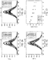
FIGS. 3A - 3D show steady state quenching of QD PL by peptide- C60 is dependent on peptide ratio and QD- C60 separation distance. Each peptide was assembled onto a fixed amount of QD at the ratios shown and spectra collected. Shown are the quenching spectra for C60 peptide conjugates of (A) JBD-1, (B) JBD-2 and (C) JBD-3 on DHLA-capped QDs. The quenching efficiency for each peptide as a function of peptide ratio (valence) is shown inFIG. 3D . The data show the valence- and C60 distance-dependent nature of the efficiency of peptide-C60-mediated QD quenching. -
FIG. 4 shows confocal laser scanning microscopy (CLSM) images of A549 cells stained with QD-peptide- C60 complexes. Shown are differential interference contrast (DIC) (left) and confocal microscopy images (right) showing fluorescence emission of QD-peptide- C60 bound to A549 cells. The cells were incubated with 20 nM QD complexed with JBD-1-C60 peptides (QD:peptide ratio 1:20) in PBS (pH 8.2) for 10 min at 37°C. For imaging the samples were excited at QD excitation at 402 nm with fluorescence detection channel set to 570-620 nm (red). A dichroic mirror at 561 nm was used to reflect incident excitation light. -
FIGS. 5A and 5B are confocal images and time-resolved quantification of fluorescence emission of live HeLa cells in resting and depolarizing state using different potassium chloride (KCl) solutions, respectively.FIG. 5A shows DIC (left) and confocal images (right) of cells labeled with QD-JBD1- C60 and imaged in an isotonic solution containing 2.5 mM KCl or 140 mM KCl. The samples were excited at 402 nm with a fluorescence detection channels set to 570-620 nm. (red) with dichroic mirrors at 561 nm.FIG. 5B shows time-resolved quantification of fluorescence emission live HeLa cells labeled with commercial FluoVolt, QD-peptide- C60 and or QD alone. Image collection began at t=0 when cells were in 100% isotonic solution containing 2.5 mM KCl (0.15 mL). Isotonic solution containing 140 mM KCl was perfused onto the cells at a rate of ~1.0 mL/min (~3 mL total volume added). Fluorescence images were collected every 20 s for 5 min. Fluorescence quantification was done by sampling multiple regions of interest (ROI) in individual cells for each peptide construct. The data represent multiple ROIs from 50-70 cells from 4 independent experiments for each QD-peptide sample. - The singular forms "a", "an," and "the" do not preclude plural referents, unless the content clearly dictates otherwise.
- The term "and/or" includes any and all combinations of one or more of the associated listed items.
- The term "about" when used in conjunction with a stated numerical value or range denotes somewhat more or somewhat less than the stated value or range, to within a range of ±10% of that stated.
- The controlled interfacing of nanoparticle (NP) materials with cellular systems has been employed for a variety of applications including cellular labeling/tracking, imaging, sensing, and drug delivery. NP-biological hybrid bioconjugates (e.g., semiconductor quantum dots (QDs) appended with functional peptides or proteins) are value-added materials wherein the ensemble bioconjugate performs a function not attainable by the individual component materials alone. Such constructs based on energy or charge transfer have much to offer in the development of optical or opto-electrical sensors for assessing cellular states (e.g., health, disease, membrane potential). Sensors aimed at reporting on the electrical activity/membrane potential of cells are critical for understanding the communication process amongst neurons in the brain (brain mapping) as well as for the assessment of the activity of other electrically active cell types (e.g., muscle cells). To date, most opto-electrical sensors for imaging cellular membrane potential are based on either (1) stand-alone electrochromic voltage-sensitive dyes (VSDs) or (2) molecular wires based on electron transfer quenching. VSDs are plagued by poor solubility in aqueous media, nonspecific labeling of non-membrane cellular structures, poor photostability and inherent cytotoxicity. Molecular wires require intricate molecular synthesis and purification strategies, the incorporation of a tailored molecular "wire bridge" to conduct electrons from donor to acceptor, and often use poorly photostable molecular fluorophores (e.g., fluorescein) as the optical readout moiety. Cumulatively, these issues represent significant limitations of currently available electro-optical materials for optically sensing membrane potential.
- Aspects of the disclosure provide a bioconjugate including a modular, multifunctional nanoparticle-based electron donor-acceptor bioconjugate for the realtime perception of changes in cellular membrane potential. The construct comprises the following components: (a) a photoluminescent NP electron donor, (b) a modular, multidomain membrane insertion peptide, and (c) an electron acceptor (
FIG. 1A ). The peptide includes (1) a NP association domain, (2) amino acid motifs to mediate peptide insertion into the plasma membrane, (3) one or more attachment points for attachment of an electron acceptor at discreetly controlled locations/distances from the electron donor, and (4) an electron acceptor. The rate of electron transfer between the donor and acceptor is modulated by changes in membrane potential and the construct reports on this modulation by a measurable change in donor photoluminescence (PL).FIG. 1B shows a specific example of the donor-acceptor bioconjugate where the electron donor is a semiconductor quantum dot (QD) that is connected to an electron acceptor, a carbon allotrope (C60 or fullerene) via a membrane-insertion peptide.FIG. 1C shows a schematic representation of the QD-peptide-C60 construct reporting on the transition of the cell from resting potential state (QD signal "on") to depolarized state (QD signal "off"). - A method of imaging cellular plasma membrane potential is provided, the method comprising providing a construct comprising a nanoparticle operable as an electron donor; a modular peptide attached to the nanoparticle; and an electron acceptor, the peptide comprising a nanoparticle association domain, a motif configured to mediate peptide insertion into the plasma membrane, and at least one attachment point for an attachment of the peptide to the electron acceptor positioned at a controlled distance from the nanoparticle; contacting a cell with the construct; and measuring a change in photoluminescence from the electron donor, wherein a rate of electron transfer between the electron donor and the electron acceptor is modulated by changes in cellular plasma membrane potential and the construct correlates modulation of the rate of electron transfer by the change in photoluminescence, wherein the modular peptide is selected from the group consisting of SEQ ID No: 1, SEQ ID No: 2, and SEQ ID No: 3, wherein the nanoparticle is a quantum dot, and wherein the electron acceptor is C60.
- Peptides of varying lengths and attachment points for the C60 fullerene electron acceptor were synthesized and iteratively tested for their ability to quench the excited state QD donor (Table 1). These peptides each contain a His6 tract to mediate attachment of the peptide to the ZnS shell of the QD. Each peptide also bears a unique lysine residue for the covalent attachment of the C60 to position the C60 moiety at discreetly controlled distances from the QD donor center. The peptides in Table 1 have sequence identification as follows: JBD-1 is SEQ ID No: 1; JBD-2 is SEQ ID No: 2; and JBD-3 is SEQ ID No: 3.
Table 1. Multifunctional modular peptides for membrane insertion and QD-coupled membrane potential sensing Peptide Sequence* JBD-1 Ac-AAAALAAAAALAALAAKAAAGGH6-COOH JBD-2 Ac-KAAALAAAAALAAWAALAAAGGH6-COOH JBD-3 AC-KAAALAAAAALAAWAALAAAP9GGH6-COOH *N terminus blocked with an acetyl group; C terminus blocked with an amide K = unique lysine for attachment of C60 electron acceptor - Peptides of varying lengths and attachment points for the C60 fullerene electron acceptor were synthesized and iteratively tested for their ability to quench the excited state QD donor (Table 1). These peptides each contain a His6 tract to mediate attachment of the peptide to the ZnS shell of the QD. Each peptide also bears a unique lysine residue for the covalent attachment of the C60 to position the C60 moiety at discreetly controlled distances from the QD donor center.
- Molecular modeling of the orientation of the three peptide species once assembled to the QD via the His6 tract revealed the predicted orientation and distance of the C60 acceptor from the QD center when presented to the QD as a covalently attached moiety to the peptide backbone. In the progression from peptide JBD-1 to JBD-3, the C60 acceptor is positioned at increasingly further distances from the QD center (
FIG. 2 ). It is clear from the modeling that the nature of the hydrophilic ligand on the QD surface impacts the solvent accessibility of the C60 moiety. QDs coated with dihydrolipoic acid (DHLA) ligands showed complete display of the C60 by peptides JBD-2 and JBD-3 and partial display in the context of peptide JBD-1. QDs coated with DHLA-PEG750-OMe (pegylated DHLA capped with a terminal methoxy group) showed display of the C60 only in the context of peptide JBD-3 while in the other two peptide species the C60 was buried within the ligand layer. As the DHLA-coated QDs mediated better cell binding than DHLA-PEG750-OMe, DHLA QD-peptide-C60 complexes were used for cellular assays. - Steady state fluorescence measurements were performed to determine the efficiency of electron transfer between the QD and the various peptide-C60 species. The readout for the assay is the quenching of QD PL upon attachment of the peptide-C60 conjugate to the QD surface. Two key parameters were assessed for their role in affecting efficient quenching of the photo-excited QD donor: (1) peptide-C60 valence (or number of peptide-C60 conjugates arrayed around the QD surface) and (2) the distance of the C60 electron acceptor from the QD donor center. As shown in
FIG. 3 each peptide-C60 species displayed the ability to efficiently quench the PL emission of the QD donor in a ratio metric manner (i.e., the quenching efficiency increased as the number of peptides arrayed around the QD increased). - Next, fluorescence imaging was used to confirm the successful labeling of the plasma membrane with the QD-peptide-C60 conjugates. A549 (human lung adenocarcinoma) cells were labeled with 605 QD-DHLA-peptide-C60 conjugates and compared to cells incubated with QD alone.
FIG. 4 shows DIC and corresponding fluorescence micrographs of A549 cells labeled with the various QD-peptide-C60 constructs. The cellular labeling is clearly membranous, consistent with the insertion of the peptide-C60 moiety into the plasma membrane and the docking of the QD onto the surface of the plasma membrane. Immuno-silver staining assays confirmed the insertion of the C60 moiety into the aliphatic portion of the plasma membrane. - The ability of the QD-peptide-C60 systems to visualize changes in membrane potential was confirmed by performing depolarization experiments on HeLa (human cervical carcinoma) cells that were labeled with the QD-peptide-C60 conjugates. The cells were depolarized with an isotonic solution containing 140 mM potassium chloride (KCl). Incubation of cells in this solution causes depolarization of the cells by the influx of K+ ions through K+ leak channels.
FIG. 5A shows representative confocal fluorescence images for cells labeled with QD-JBD-1-C60 conjugates before and after the addition of the KCl solution. - The images clearly show the time-resolved reduction in QD PL upon perfusion of the KCl solution onto the cells and subsequent depolarization. Similar responses for QD-peptide conjugates of JBD-2-C60 and JBD-3-C60 were obtained (images not shown). A graph of the resulting time-resolved PL intensities in response to depolarization is shown in
FIG. 5B . Minimal change in PL of QDs not decorated with the peptide-C60 conjugates was noted, clearly demonstrating the dependence of the response on the presence of the peptide-C60 conjugate. Also included is a comparison of QD-peptide-C60-derived cellular response to that obtained in HeLa cells labeled with a "state-of-the-art" molecular wire-based membrane potential probe (FluoVolt; commercially available from ThermoFisher). For comparison, the QD-JBD-1-C60 conjugate exhibited a maximum response (PL decrease) of 31% at 5 min while the FluoVolt probe displayed a fluorescence response of 19% (PL increase) over this same time course. These data clearly demonstrate the superior sensitivity of the QD-JBD-1-C60 conjugate compared to the commercial probe. Further, the magnitude of the QD PL response exhibited a distinct distance dependent response to depolarization. The PL response tracked inversely with the separation distance between the QD donor center and the C60 moiety, in good agreement with the plate-based steady state data presented inFIG. 3 . - Potential application areas include those where stable, long term imaging of changes in membrane potential are desired, for example imaging/optical recording of the electrical activity in one or more cultured cells, or in tissue slices, whole tissues, and/or animals. Targeted cell types in these applications would include (but are not limited to) electrically active cells such as neurons and muscles cells. These material constructs could also find utility in quantum dot-based LED cells where the tuning of QD luminescence in the presence of an electric field is desired/required.
- Advantages of NP-peptide-acceptor assemblies include the following. They are potentially amenable to both covalent and noncovalent attachment strategies. Modular design of functional domains allows for flexibility in iterative peptide development and testing. The strategy is amenable to the assembly of conceivably any class of NP with any modular, multifunctional polymer. The NP surface can be functionalized with different types of modular, multifunctional peptides ("mixed" surfaces) giving ratiometric control over the nature of the decorated NP surface and iterative control over the rate of donor-acceptor electron transfer.
- More particular advantages of the QD-peptide-C60 construct include the following. The peptide self-assembles noncovalently to the QD donor surface without the need for complex covalent attachment chemistries that use high concentrations of excess reactants that require purification. The peptide assembles to the QD rapidly (10 min) with high (nM) affinity. The assembled construct labels cell membranes rapidly (10 minutes) after conjugate assembly, with 20 minutes total time for cellular labeling with the QD-peptide constructs. The peptide linker does not conduct, shuttle or otherwise direct the transfer of electrons from the donor to the acceptor which simplifies the design and synthesis of QD-C60 linkers. Electron transfer is completely distance-dependent which is a key distinguishing factor relative to other membrane potential imaging molecular wires (e.g., FluoVolt sold by ThermoFisher), and this dependency can be iteratively controlled by controlling the donor-acceptor separation distance.
- Furthermore, the ratio or valence (and thus the avidity) of the peptide for the NP can be controlled and can be used to tune the rate or efficiency of donor quenching/electron transfer. The exceptional photostability of QD-based constructs allows for much longer imaging times (> 100x) compared to voltage-sensitive dyes. The significantly large two-photon action cross section of QD materials (102-103 greater than organic dyes) makes them ideal for deep tissue imaging.
-
- <110> THE GOVERNMENT OF THE UNITED STATES OF AMERICA, AS REPRESENTED BY THE SECRETARY OF THE NAVY
- <120> Modular, Multifunctional Nanoparticle-Based Bioconjugate for Realtime Visualization of Cellular Membrane Potential
- <130> 104017
- <150>
US 62/452097
<151> 2017-01-30 - <160> 3
- <170> PatentIn version 3.5
- <210> 1
<211> 28
<212> PRT
<213> Artificial Sequence - <220>
<223> synthetic construct - <400> 1

- <210> 2
<211> 28
<212> PRT
<213> Artificial Sequence - <220>
<223> synthetic construct - <400> 2

- <210> 3
<211> 37
<212> PRT
<213> Artificial Sequence - <220>
<223> synthetic construct - <400> 3

Claims (1)
- A method of imaging cellular plasma membrane potential, the method comprising:providing a construct comprising a nanoparticle operable as an electron donor; a modular peptide attached to the nanoparticle; and an electron acceptor, the peptide comprising a nanoparticle association domain, a motif configured to mediate peptide insertion into the plasma membrane, and at least one attachment point for an attachment of the peptide to the electron acceptor positioned at a controlled distance from the nanoparticle;contacting a cell with the construct; andmeasuring a change in photoluminescence from the electron donor, wherein a rate of electron transfer between the electron donor and the electron acceptor is modulated by changes in plasma membrane potential and the construct correlates modulation of the rate of electron transfer by the change in photoluminescence,wherein the modular peptide is selected from the group consisting of SEQ ID No: 1, SEQ ID No: 2, and SEQ ID No: 3, andwherein the nanoparticle is a quantum dot, andwherein the electron acceptor is C60.
Applications Claiming Priority (2)
| Application Number | Priority Date | Filing Date | Title |
|---|---|---|---|
| US201762452097P | 2017-01-30 | 2017-01-30 | |
| PCT/US2018/015713 WO2018140866A1 (en) | 2017-01-30 | 2018-01-29 | Modular, multifunctional nanoparticle-based bioconjugate for realtime visualization of cellular membrane potential |
Publications (3)
| Publication Number | Publication Date |
|---|---|
| EP3574328A1 EP3574328A1 (en) | 2019-12-04 |
| EP3574328A4 EP3574328A4 (en) | 2020-07-15 |
| EP3574328B1 true EP3574328B1 (en) | 2021-12-01 |
Family
ID=62978844
Family Applications (1)
| Application Number | Title | Priority Date | Filing Date |
|---|---|---|---|
| EP18744054.0A Not-in-force EP3574328B1 (en) | 2017-01-30 | 2018-01-29 | Modular, multifunctional nanoparticle-based bioconjugate for realtime visualization of cellular membrane potential |
Country Status (4)
| Country | Link |
|---|---|
| US (2) | US10705092B2 (en) |
| EP (1) | EP3574328B1 (en) |
| ES (1) | ES2902775T3 (en) |
| WO (1) | WO2018140866A1 (en) |
Families Citing this family (2)
| Publication number | Priority date | Publication date | Assignee | Title |
|---|---|---|---|---|
| US12624347B2 (en) | 2021-09-20 | 2026-05-12 | The Government Of The United States Of America, As Represented By The Secretary Of The Navy | Wireless activation of channelrhodopsin via in situ self-assembly of semiconductor quantum dots at the plasma membrane |
| EP4526067A4 (en) | 2022-05-17 | 2026-03-25 | Us Gov Sec Navy | SEEDLESS SYNTHESIS OF ANISOTROPIC GOLDEN NANOFLOWERS FOR CELL CONTROL AND DRUG RELEASE APPLICATIONS |
Family Cites Families (2)
| Publication number | Priority date | Publication date | Assignee | Title |
|---|---|---|---|---|
| US6342379B1 (en) | 1995-06-07 | 2002-01-29 | The Regents Of The University Of California | Detection of transmembrane potentials by optical methods |
| US8835172B2 (en) | 2009-10-22 | 2014-09-16 | The United States Of America, As Represented By The Secretary Of The Navy | Modular functional peptides for delivery of nanoparticles |
-
2018
- 2018-01-29 WO PCT/US2018/015713 patent/WO2018140866A1/en not_active Ceased
- 2018-01-29 EP EP18744054.0A patent/EP3574328B1/en not_active Not-in-force
- 2018-01-29 US US15/882,259 patent/US10705092B2/en active Active
- 2018-01-29 ES ES18744054T patent/ES2902775T3/en active Active
-
2020
- 2020-06-08 US US16/894,920 patent/US11287430B2/en active Active
Also Published As
| Publication number | Publication date |
|---|---|
| WO2018140866A1 (en) | 2018-08-02 |
| US10705092B2 (en) | 2020-07-07 |
| US20180217153A1 (en) | 2018-08-02 |
| ES2902775T3 (en) | 2022-03-29 |
| EP3574328A1 (en) | 2019-12-04 |
| EP3574328A4 (en) | 2020-07-15 |
| US11287430B2 (en) | 2022-03-29 |
| US20200355696A1 (en) | 2020-11-12 |
Similar Documents
| Publication | Publication Date | Title |
|---|---|---|
| Kompa et al. | Exchangeable HaloTag ligands for super-resolution fluorescence microscopy | |
| Vu et al. | Quantum dots for quantitative imaging: from single molecules to tissue | |
| Jin et al. | Nanoparticles for super-resolution microscopy and single-molecule tracking | |
| Chang et al. | Tracking bio‐molecules in live cells using quantum dots | |
| Sauer et al. | Single-molecule localization microscopy in eukaryotes | |
| Medintz et al. | Intracellular delivery of quantum dot− protein cargos mediated by cell penetrating peptides | |
| Butkevich et al. | Photoactivatable fluorescent dyes with hydrophilic caging groups and their use in multicolor nanoscopy | |
| Suzuki et al. | Development of functional fluorescent molecular probes for the detection of biological substances | |
| Fernández-Suárez et al. | Redirecting lipoic acid ligase for cell surface protein labeling with small-molecule probes | |
| Pinaud et al. | Probing cellular events, one quantum dot at a time | |
| Ghanam et al. | Single molecule localization microscopy for studying small extracellular vesicles | |
| Boeneman et al. | Intracellular bioconjugation of targeted proteins with semiconductor quantum dots | |
| JP7168155B2 (en) | Target substance detection method and kit | |
| Du et al. | Optical molecular imaging for systems biology: from molecule to organism | |
| Schirripa Spagnolo et al. | Choosing the probe for single-molecule fluorescence microscopy | |
| Solhi et al. | Recent advances on the biosensing and bioimaging based on polymer dots as advanced nanomaterial: Analytical approaches | |
| Rahim et al. | Phasor analysis of fluorescence lifetime enables quantitative multiplexed molecular imaging of three probes | |
| Michelis et al. | Imaging and measuring vesicular acidification with a plasma membrane-targeted ratiometric pH probe | |
| Xu et al. | Labeling cytosolic targets in live cells with blinking probes | |
| Zou et al. | Cytoplasmic delivery and selective, multicomponent labeling with oligoarginine-linked protein tags | |
| Ruedas-Rama et al. | Flim strategies for intracellular sensing: Fluorescence lifetime imaging as a tool to quantify analytes of interest | |
| Aknine et al. | Lipid-directed covalent labeling of plasma membranes for long-term imaging, barcoding and manipulation of cells | |
| Chang et al. | A bright light to reveal mobility: single quantum dot tracking reveals membrane dynamics and cellular mechanisms | |
| US11287430B2 (en) | Modular, multifunctional nanoparticle-based bioconjugate for realtime visualization of cellular membrane potential | |
| Wang et al. | In‐capillary probing of quantum dots and fluorescent protein self‐assembly and displacement using Förster resonance energy transfer |
Legal Events
| Date | Code | Title | Description |
|---|---|---|---|
| STAA | Information on the status of an ep patent application or granted ep patent |
Free format text: STATUS: THE INTERNATIONAL PUBLICATION HAS BEEN MADE |
|
| PUAI | Public reference made under article 153(3) epc to a published international application that has entered the european phase |
Free format text: ORIGINAL CODE: 0009012 |
|
| STAA | Information on the status of an ep patent application or granted ep patent |
Free format text: STATUS: REQUEST FOR EXAMINATION WAS MADE |
|
| 17P | Request for examination filed |
Effective date: 20190620 |
|
| AK | Designated contracting states |
Kind code of ref document: A1 Designated state(s): AL AT BE BG CH CY CZ DE DK EE ES FI FR GB GR HR HU IE IS IT LI LT LU LV MC MK MT NL NO PL PT RO RS SE SI SK SM TR |
|
| AX | Request for extension of the european patent |
Extension state: BA ME |
|
| RIN1 | Information on inventor provided before grant (corrected) |
Inventor name: OH, EUNKEU Inventor name: MEDINTZ, IGOR, L. Inventor name: DELEHANTY, JAMES, B. Inventor name: FIELD, LAUREN, D. Inventor name: NAG, OKHIL Inventor name: STEWART, MICHAEL, H. Inventor name: HUSTON, ALAN, L. Inventor name: DESCHAMPS, JEFFREY, R. Inventor name: SUSUMU, KIMIHIRO Inventor name: DAWSON, PHILIP, E. Inventor name: EFROS, ALEXANDER, L. |
|
| DAV | Request for validation of the european patent (deleted) | ||
| DAX | Request for extension of the european patent (deleted) | ||
| A4 | Supplementary search report drawn up and despatched |
Effective date: 20200612 |
|
| RIC1 | Information provided on ipc code assigned before grant |
Ipc: G01N 33/483 20060101ALI20200606BHEP Ipc: G01N 33/68 20060101ALI20200606BHEP Ipc: G01N 33/58 20060101AFI20200606BHEP Ipc: G01N 21/64 20060101ALI20200606BHEP Ipc: G01N 33/542 20060101ALI20200606BHEP |
|
| STAA | Information on the status of an ep patent application or granted ep patent |
Free format text: STATUS: EXAMINATION IS IN PROGRESS |
|
| 17Q | First examination report despatched |
Effective date: 20210205 |
|
| GRAP | Despatch of communication of intention to grant a patent |
Free format text: ORIGINAL CODE: EPIDOSNIGR1 |
|
| STAA | Information on the status of an ep patent application or granted ep patent |
Free format text: STATUS: GRANT OF PATENT IS INTENDED |
|
| INTG | Intention to grant announced |
Effective date: 20210702 |
|
| GRAS | Grant fee paid |
Free format text: ORIGINAL CODE: EPIDOSNIGR3 |
|
| GRAA | (expected) grant |
Free format text: ORIGINAL CODE: 0009210 |
|
| STAA | Information on the status of an ep patent application or granted ep patent |
Free format text: STATUS: THE PATENT HAS BEEN GRANTED |
|
| AK | Designated contracting states |
Kind code of ref document: B1 Designated state(s): AL AT BE BG CH CY CZ DE DK EE ES FI FR GB GR HR HU IE IS IT LI LT LU LV MC MK MT NL NO PL PT RO RS SE SI SK SM TR |
|
| REG | Reference to a national code |
Ref country code: GB Ref legal event code: FG4D |
|
| REG | Reference to a national code |
Ref country code: AT Ref legal event code: REF Ref document number: 1452262 Country of ref document: AT Kind code of ref document: T Effective date: 20211215 Ref country code: CH Ref legal event code: EP |
|
| REG | Reference to a national code |
Ref country code: IE Ref legal event code: FG4D |
|
| REG | Reference to a national code |
Ref country code: DE Ref legal event code: R096 Ref document number: 602018027517 Country of ref document: DE |
|
| REG | Reference to a national code |
Ref country code: LT Ref legal event code: MG9D |
|
| REG | Reference to a national code |
Ref country code: ES Ref legal event code: FG2A Ref document number: 2902775 Country of ref document: ES Kind code of ref document: T3 Effective date: 20220329 |
|
| REG | Reference to a national code |
Ref country code: NL Ref legal event code: MP Effective date: 20211201 |
|
| REG | Reference to a national code |
Ref country code: AT Ref legal event code: MK05 Ref document number: 1452262 Country of ref document: AT Kind code of ref document: T Effective date: 20211201 |
|
| PG25 | Lapsed in a contracting state [announced via postgrant information from national office to epo] |
Ref country code: RS Free format text: LAPSE BECAUSE OF FAILURE TO SUBMIT A TRANSLATION OF THE DESCRIPTION OR TO PAY THE FEE WITHIN THE PRESCRIBED TIME-LIMIT Effective date: 20211201 Ref country code: LT Free format text: LAPSE BECAUSE OF FAILURE TO SUBMIT A TRANSLATION OF THE DESCRIPTION OR TO PAY THE FEE WITHIN THE PRESCRIBED TIME-LIMIT Effective date: 20211201 Ref country code: FI Free format text: LAPSE BECAUSE OF FAILURE TO SUBMIT A TRANSLATION OF THE DESCRIPTION OR TO PAY THE FEE WITHIN THE PRESCRIBED TIME-LIMIT Effective date: 20211201 Ref country code: BG Free format text: LAPSE BECAUSE OF FAILURE TO SUBMIT A TRANSLATION OF THE DESCRIPTION OR TO PAY THE FEE WITHIN THE PRESCRIBED TIME-LIMIT Effective date: 20220301 Ref country code: AT Free format text: LAPSE BECAUSE OF FAILURE TO SUBMIT A TRANSLATION OF THE DESCRIPTION OR TO PAY THE FEE WITHIN THE PRESCRIBED TIME-LIMIT Effective date: 20211201 |
|
| PG25 | Lapsed in a contracting state [announced via postgrant information from national office to epo] |
Ref country code: SE Free format text: LAPSE BECAUSE OF FAILURE TO SUBMIT A TRANSLATION OF THE DESCRIPTION OR TO PAY THE FEE WITHIN THE PRESCRIBED TIME-LIMIT Effective date: 20211201 Ref country code: PL Free format text: LAPSE BECAUSE OF FAILURE TO SUBMIT A TRANSLATION OF THE DESCRIPTION OR TO PAY THE FEE WITHIN THE PRESCRIBED TIME-LIMIT Effective date: 20211201 Ref country code: NO Free format text: LAPSE BECAUSE OF FAILURE TO SUBMIT A TRANSLATION OF THE DESCRIPTION OR TO PAY THE FEE WITHIN THE PRESCRIBED TIME-LIMIT Effective date: 20220301 Ref country code: LV Free format text: LAPSE BECAUSE OF FAILURE TO SUBMIT A TRANSLATION OF THE DESCRIPTION OR TO PAY THE FEE WITHIN THE PRESCRIBED TIME-LIMIT Effective date: 20211201 Ref country code: HR Free format text: LAPSE BECAUSE OF FAILURE TO SUBMIT A TRANSLATION OF THE DESCRIPTION OR TO PAY THE FEE WITHIN THE PRESCRIBED TIME-LIMIT Effective date: 20211201 Ref country code: GR Free format text: LAPSE BECAUSE OF FAILURE TO SUBMIT A TRANSLATION OF THE DESCRIPTION OR TO PAY THE FEE WITHIN THE PRESCRIBED TIME-LIMIT Effective date: 20220302 |
|
| PG25 | Lapsed in a contracting state [announced via postgrant information from national office to epo] |
Ref country code: NL Free format text: LAPSE BECAUSE OF FAILURE TO SUBMIT A TRANSLATION OF THE DESCRIPTION OR TO PAY THE FEE WITHIN THE PRESCRIBED TIME-LIMIT Effective date: 20211201 |
|
| PG25 | Lapsed in a contracting state [announced via postgrant information from national office to epo] |
Ref country code: SM Free format text: LAPSE BECAUSE OF FAILURE TO SUBMIT A TRANSLATION OF THE DESCRIPTION OR TO PAY THE FEE WITHIN THE PRESCRIBED TIME-LIMIT Effective date: 20211201 Ref country code: SK Free format text: LAPSE BECAUSE OF FAILURE TO SUBMIT A TRANSLATION OF THE DESCRIPTION OR TO PAY THE FEE WITHIN THE PRESCRIBED TIME-LIMIT Effective date: 20211201 Ref country code: RO Free format text: LAPSE BECAUSE OF FAILURE TO SUBMIT A TRANSLATION OF THE DESCRIPTION OR TO PAY THE FEE WITHIN THE PRESCRIBED TIME-LIMIT Effective date: 20211201 Ref country code: PT Free format text: LAPSE BECAUSE OF FAILURE TO SUBMIT A TRANSLATION OF THE DESCRIPTION OR TO PAY THE FEE WITHIN THE PRESCRIBED TIME-LIMIT Effective date: 20220401 Ref country code: EE Free format text: LAPSE BECAUSE OF FAILURE TO SUBMIT A TRANSLATION OF THE DESCRIPTION OR TO PAY THE FEE WITHIN THE PRESCRIBED TIME-LIMIT Effective date: 20211201 Ref country code: CZ Free format text: LAPSE BECAUSE OF FAILURE TO SUBMIT A TRANSLATION OF THE DESCRIPTION OR TO PAY THE FEE WITHIN THE PRESCRIBED TIME-LIMIT Effective date: 20211201 |
|
| REG | Reference to a national code |
Ref country code: DE Ref legal event code: R119 Ref document number: 602018027517 Country of ref document: DE |
|
| PG25 | Lapsed in a contracting state [announced via postgrant information from national office to epo] |
Ref country code: MC Free format text: LAPSE BECAUSE OF FAILURE TO SUBMIT A TRANSLATION OF THE DESCRIPTION OR TO PAY THE FEE WITHIN THE PRESCRIBED TIME-LIMIT Effective date: 20211201 |
|
| REG | Reference to a national code |
Ref country code: CH Ref legal event code: PL |
|
| PG25 | Lapsed in a contracting state [announced via postgrant information from national office to epo] |
Ref country code: IS Free format text: LAPSE BECAUSE OF FAILURE TO SUBMIT A TRANSLATION OF THE DESCRIPTION OR TO PAY THE FEE WITHIN THE PRESCRIBED TIME-LIMIT Effective date: 20220401 |
|
| REG | Reference to a national code |
Ref country code: BE Ref legal event code: MM Effective date: 20220131 |
|
| PLBE | No opposition filed within time limit |
Free format text: ORIGINAL CODE: 0009261 |
|
| STAA | Information on the status of an ep patent application or granted ep patent |
Free format text: STATUS: NO OPPOSITION FILED WITHIN TIME LIMIT |
|
| PG25 | Lapsed in a contracting state [announced via postgrant information from national office to epo] |
Ref country code: LU Free format text: LAPSE BECAUSE OF NON-PAYMENT OF DUE FEES Effective date: 20220129 Ref country code: DK Free format text: LAPSE BECAUSE OF FAILURE TO SUBMIT A TRANSLATION OF THE DESCRIPTION OR TO PAY THE FEE WITHIN THE PRESCRIBED TIME-LIMIT Effective date: 20211201 Ref country code: DE Free format text: LAPSE BECAUSE OF NON-PAYMENT OF DUE FEES Effective date: 20220802 Ref country code: AL Free format text: LAPSE BECAUSE OF FAILURE TO SUBMIT A TRANSLATION OF THE DESCRIPTION OR TO PAY THE FEE WITHIN THE PRESCRIBED TIME-LIMIT Effective date: 20211201 |
|
| 26N | No opposition filed |
Effective date: 20220902 |
|
| GBPC | Gb: european patent ceased through non-payment of renewal fee |
Effective date: 20220301 |
|
| PG25 | Lapsed in a contracting state [announced via postgrant information from national office to epo] |
Ref country code: SI Free format text: LAPSE BECAUSE OF FAILURE TO SUBMIT A TRANSLATION OF THE DESCRIPTION OR TO PAY THE FEE WITHIN THE PRESCRIBED TIME-LIMIT Effective date: 20211201 Ref country code: FR Free format text: LAPSE BECAUSE OF NON-PAYMENT OF DUE FEES Effective date: 20220201 Ref country code: BE Free format text: LAPSE BECAUSE OF NON-PAYMENT OF DUE FEES Effective date: 20220131 |
|
| PG25 | Lapsed in a contracting state [announced via postgrant information from national office to epo] |
Ref country code: LI Free format text: LAPSE BECAUSE OF NON-PAYMENT OF DUE FEES Effective date: 20220131 Ref country code: CH Free format text: LAPSE BECAUSE OF NON-PAYMENT OF DUE FEES Effective date: 20220131 |
|
| PG25 | Lapsed in a contracting state [announced via postgrant information from national office to epo] |
Ref country code: IE Free format text: LAPSE BECAUSE OF NON-PAYMENT OF DUE FEES Effective date: 20220129 Ref country code: GB Free format text: LAPSE BECAUSE OF NON-PAYMENT OF DUE FEES Effective date: 20220301 |
|
| PGFP | Annual fee paid to national office [announced via postgrant information from national office to epo] |
Ref country code: ES Payment date: 20230201 Year of fee payment: 6 |
|
| PG25 | Lapsed in a contracting state [announced via postgrant information from national office to epo] |
Ref country code: IT Free format text: LAPSE BECAUSE OF FAILURE TO SUBMIT A TRANSLATION OF THE DESCRIPTION OR TO PAY THE FEE WITHIN THE PRESCRIBED TIME-LIMIT Effective date: 20211201 |
|
| PG25 | Lapsed in a contracting state [announced via postgrant information from national office to epo] |
Ref country code: MK Free format text: LAPSE BECAUSE OF FAILURE TO SUBMIT A TRANSLATION OF THE DESCRIPTION OR TO PAY THE FEE WITHIN THE PRESCRIBED TIME-LIMIT Effective date: 20211201 Ref country code: CY Free format text: LAPSE BECAUSE OF FAILURE TO SUBMIT A TRANSLATION OF THE DESCRIPTION OR TO PAY THE FEE WITHIN THE PRESCRIBED TIME-LIMIT Effective date: 20211201 |
|
| PG25 | Lapsed in a contracting state [announced via postgrant information from national office to epo] |
Ref country code: HU Free format text: LAPSE BECAUSE OF FAILURE TO SUBMIT A TRANSLATION OF THE DESCRIPTION OR TO PAY THE FEE WITHIN THE PRESCRIBED TIME-LIMIT; INVALID AB INITIO Effective date: 20180129 |
|
| PG25 | Lapsed in a contracting state [announced via postgrant information from national office to epo] |
Ref country code: TR Free format text: LAPSE BECAUSE OF FAILURE TO SUBMIT A TRANSLATION OF THE DESCRIPTION OR TO PAY THE FEE WITHIN THE PRESCRIBED TIME-LIMIT Effective date: 20211201 |
|
| PG25 | Lapsed in a contracting state [announced via postgrant information from national office to epo] |
Ref country code: MT Free format text: LAPSE BECAUSE OF FAILURE TO SUBMIT A TRANSLATION OF THE DESCRIPTION OR TO PAY THE FEE WITHIN THE PRESCRIBED TIME-LIMIT Effective date: 20211201 |
|
| REG | Reference to a national code |
Ref country code: ES Ref legal event code: FD2A Effective date: 20250304 |
|
| PG25 | Lapsed in a contracting state [announced via postgrant information from national office to epo] |
Ref country code: ES Free format text: LAPSE BECAUSE OF NON-PAYMENT OF DUE FEES Effective date: 20240130 |