EP3571506B1 - Metabolic biomarker set for assessing ovarian cancer - Google Patents
Metabolic biomarker set for assessing ovarian cancer Download PDFInfo
- Publication number
- EP3571506B1 EP3571506B1 EP18704150.4A EP18704150A EP3571506B1 EP 3571506 B1 EP3571506 B1 EP 3571506B1 EP 18704150 A EP18704150 A EP 18704150A EP 3571506 B1 EP3571506 B1 EP 3571506B1
- Authority
- EP
- European Patent Office
- Prior art keywords
- ovarian cancer
- phosphatidylcholine
- acylcarnitine
- residue sum
- acyl
- Prior art date
- Legal status (The legal status is an assumption and is not a legal conclusion. Google has not performed a legal analysis and makes no representation as to the accuracy of the status listed.)
- Active
Links
Images




Classifications
-
- G—PHYSICS
- G01—MEASURING; TESTING
- G01N—INVESTIGATING OR ANALYSING MATERIALS BY DETERMINING THEIR CHEMICAL OR PHYSICAL PROPERTIES
- G01N33/00—Investigating or analysing materials by specific methods not covered by groups G01N1/00 - G01N31/00
- G01N33/48—Biological material, e.g. blood, urine; Haemocytometers
- G01N33/50—Chemical analysis of biological material, e.g. blood, urine; Testing involving biospecific ligand binding methods; Immunological testing
- G01N33/53—Immunoassay; Biospecific binding assay; Materials therefor
- G01N33/575—Immunoassay; Biospecific binding assay; Materials therefor for cancer
- G01N33/57545—Immunoassay; Biospecific binding assay; Materials therefor for cancer of the ovaries
-
- G—PHYSICS
- G01—MEASURING; TESTING
- G01N—INVESTIGATING OR ANALYSING MATERIALS BY DETERMINING THEIR CHEMICAL OR PHYSICAL PROPERTIES
- G01N33/00—Investigating or analysing materials by specific methods not covered by groups G01N1/00 - G01N31/00
- G01N33/48—Biological material, e.g. blood, urine; Haemocytometers
- G01N33/50—Chemical analysis of biological material, e.g. blood, urine; Testing involving biospecific ligand binding methods; Immunological testing
- G01N33/68—Chemical analysis of biological material, e.g. blood, urine; Testing involving biospecific ligand binding methods; Immunological testing involving proteins, peptides or amino acids
-
- G—PHYSICS
- G01—MEASURING; TESTING
- G01N—INVESTIGATING OR ANALYSING MATERIALS BY DETERMINING THEIR CHEMICAL OR PHYSICAL PROPERTIES
- G01N33/00—Investigating or analysing materials by specific methods not covered by groups G01N1/00 - G01N31/00
- G01N33/48—Biological material, e.g. blood, urine; Haemocytometers
- G01N33/50—Chemical analysis of biological material, e.g. blood, urine; Testing involving biospecific ligand binding methods; Immunological testing
- G01N33/92—Chemical analysis of biological material, e.g. blood, urine; Testing involving biospecific ligand binding methods; Immunological testing involving lipids, e.g. cholesterol, lipoproteins, or their receptors
-
- G—PHYSICS
- G01—MEASURING; TESTING
- G01N—INVESTIGATING OR ANALYSING MATERIALS BY DETERMINING THEIR CHEMICAL OR PHYSICAL PROPERTIES
- G01N2405/00—Assays, e.g. immunoassays or enzyme assays, involving lipids
- G01N2405/04—Phospholipids, i.e. phosphoglycerides
-
- G—PHYSICS
- G01—MEASURING; TESTING
- G01N—INVESTIGATING OR ANALYSING MATERIALS BY DETERMINING THEIR CHEMICAL OR PHYSICAL PROPERTIES
- G01N2405/00—Assays, e.g. immunoassays or enzyme assays, involving lipids
- G01N2405/08—Sphingolipids
-
- G—PHYSICS
- G01—MEASURING; TESTING
- G01N—INVESTIGATING OR ANALYSING MATERIALS BY DETERMINING THEIR CHEMICAL OR PHYSICAL PROPERTIES
- G01N33/00—Investigating or analysing materials by specific methods not covered by groups G01N1/00 - G01N31/00
- G01N33/48—Biological material, e.g. blood, urine; Haemocytometers
-
- G—PHYSICS
- G01—MEASURING; TESTING
- G01N—INVESTIGATING OR ANALYSING MATERIALS BY DETERMINING THEIR CHEMICAL OR PHYSICAL PROPERTIES
- G01N33/00—Investigating or analysing materials by specific methods not covered by groups G01N1/00 - G01N31/00
- G01N33/48—Biological material, e.g. blood, urine; Haemocytometers
- G01N33/50—Chemical analysis of biological material, e.g. blood, urine; Testing involving biospecific ligand binding methods; Immunological testing
- G01N33/68—Chemical analysis of biological material, e.g. blood, urine; Testing involving biospecific ligand binding methods; Immunological testing involving proteins, peptides or amino acids
- G01N33/6803—General methods of protein analysis not limited to specific proteins or families of proteins
- G01N33/6848—Methods of protein analysis involving mass spectrometry
Definitions
- the present invention relates to the use of biomarkers for assessing ovarian cancer being more sensitive, particularly at early stage of disease. Moreover, the present invention relates to a method for assessing ovarian cancer from a patient to be examined.
- Metabolomics is a comprehensive quantitative measurement of low molecular weight compounds covering systematically the key metabolites, which represent the whole range of pathways of intermediary metabolism. In a systems biology approach, it provides a functional readout of changes determined by genetic blueprint, regulation, protein abundance and modification, and environmental influence. The capability to analyse large arrays of metabolites extracts biochemical information reflecting true functional end-points of overt biological events while other functional genomics technologies such as transcriptomics and proteomics, though highly valuable, merely indicate the potential cause for phenotypic response. Therefore, they cannot necessarily predict drug effects, toxicological response or disease states at the phenotype level unless functional validation is added.
- Metabolomics bridges this information gap by depicting in particular such functional information since metabolite differences in biological fluids and tissues provide the closest link to the various phenotypic responses. Needless to say, such changes in the biochemical phenotype are of direct interest to pharmaceutical, biotech and health industries once appropriate technology allows the cost-efficient mining and integration of this information.
- phenotype is not necessarily predicted by genotype.
- genotype The gap between genotype and phenotype is spanned by many biochemical reactions, each with individual dependencies to various influences, including drugs, nutrition and environmental factors.
- metabolites are the quantifiable molecules with the closest link to phenotype.
- Many phenotypic and genotypic states, such as a toxic response to a drug or disease prevalence are predicted by differences in the concentrations of functionally relevant metabolites within biological fluids and tissue.
- Ovarian cancer is the second most common and most lethal of all gynaecologic diseases, displaying a world-wide increase of incidence and prevalence over the past decade.
- women diagnosed between the years 2000 and 2007 showed a mean five-year survival of only around 35%.
- more than 20,000 new ovarian cancer cases are expected each year leading to over 14,000 deaths (Siegel et al. 2015).
- the five year survival rate is even less than 25 % for women diagnosed with advanced ovarian cancer stages III or IV (Shapira et al. 2014; Berkenblit und Cannistra 2005; Vaughan et al. 2011). Therefore, a reliable diagnosis with validated methods that could detect ovarian cancer with a high sensitivity in order to correctly assign women with the disease and specificity to avoid false-positive results would be essential.
- CA-125 Cancer Antigen 125
- HE4 Human Epididymis Secretory Protein 4
- FDA U.S. Food and Drug Administration
- An increase in blood concentrations of CA-125 can be used as an indicator of disease recurrence (Suh et al. 2010).
- elevated levels of soluble CA-125 has also been found in a variety of other malignancies such as breast cancer, lymphomas, endometriosis or gastric cancer (Norum et al. 2001; Yamamoto et al.
- CA-125 alone cannot serve as an ideal biomarker for ovarian cancer.
- CA-125 together with transvaginal sonography is only able to detect around 30% of women with early-stage ovarian cancer (Roupa et al. 2004).
- the object underlying the present invention is the provision of the use of biomarkers for assessing ovarian cancer wherein markers are more sensitive, particularly at early stage of disease.
- the marker should be easily detectable in a biological sample such as in blood and/or plasma, its level should be consistently related to the degree of ovary injury and its level should change.
- WO 2016/038157 A1 discloses a metabolic biomarker set (and a kit) for assessing breast cancer which comprises an acylcarnitine having at least 10 carbon atoms (e.g. acylcarnitine c 18:2), a lipid having a maximum of three unsaturated bonds (e.g. lyso PC a C18:2 or lyso PC a C18:1) and further optional metabolites, such as histidine or arginine (see p. 28, 31 , Tables 3-5).
- an acylcarnitine having at least 10 carbon atoms e.g. acylcarnitine c 18:2
- a lipid having a maximum of three unsaturated bonds e.g. lyso PC a C18:2 or lyso PC a C18:1
- further optional metabolites such as histidine or arginine (see p. 28, 31 , Tables 3-5).
- US 2012/0129265 A1 describes a metabolic biomarker set incl. kit for assessing kidney diseases, which comprises an acylcarnitine (e.g. acylcarnitine C18:1), an aminoacid (e.g. glutamate, tryptophan, alanine, arginine, or histidine) and a phosphatidylcholine (e.g. lyso PC a C18:2, lyso PC a C18:1, or PC aa C32:2), see par. 53-56, claims, Tables 1, 4-5).
- an acylcarnitine e.g. acylcarnitine C18:1
- an aminoacid e.g. glutamate, tryptophan, alanine, arginine, or histidine
- a phosphatidylcholine e.g. lyso PC a C18:2, lyso PC a C18:1, or PC aa C32:
- WO 2010/139341 A1 refers to a biomarker panel for assessing kidney diseases comprising at least amino acids, acyl carnitines and biogenic amines.
- WO 2007/003344 A1 on behalf of the applicants discloses a kit for quantification of lipids and other metabolites, which is commercialized under the name AbsolutelDQ® p400 HR (cf. www.biocrates.com).
- SANAD ALONEZI ET AL "Metabolomic Profiling of the Effects of Melittin on Cisplatin Resistant and Cisplatin Sensitive Ovarian Cancer Cells Using Mass Spectrometry and Biolog. Microarray Technology", METABOLITES, (2016), vol. 6, no. 4, 35 , disclose the use of panels of metabolic markers present in serum for the assessment of ovarian cancer.
- BACHMAYR-HEYDA ET AL disclose a panel with selected unsaturated gycerophospholipids, amino acids, sphingolipids and acylcarnitines.
- the panels of CHAOFU KE ET AL comprise lysophosphatidylcholines, amino acids (histidine and/or tryptophane) and acylcarnitines; and the panel of SANAD ALONEZI ET AL comprises phosphatidylcholines, amino acids and carnitines.
- the inventors based their investigations on metabolomics as it could give insight in the biochemical changes occurring in the ovary during the course of disease and offer several novel and potentially improved biomarkers. Hence, it would be a significant improvement to have metabolic biomarkers for ovarian cancer, which would also give more information about the function of the ovary and the biochemical reactions therein. The inventors found that a more comprehensive picture of all involved pathways and mechanisms is realised when using a panel of metabolites that are altered with progressing ovarian cancer rather than employing only single-markers as provided in the prior art.
- the present invention provides for use of biomarkers suitable for assessing ovarian cancer being more sensitive for pathological changes in the ovary, particularly at early stage of disease. Moreover, the present invention also provides for a method for assessing ovarian cancer in a patient.
- “Assessment” or “Assessing” in the sense of the present invention means the diagnosis or prediction of the onset and monitoring of the progression of the disease, in particular the detection and marking of the disease at the different stages.
- the present invention allows to predict and diagnose ovarian cancer in an improved manner and at an early stage of the disease and provides a more sensitive detection for pathological changes in the ovary.
- the biomarkers according to the invention are easily detectable in samples, in particular in blood and/or in plasma, their level is consistently related to the degree of ovary disease /injury and their level changes.
- a biomarker is a valuable tool due to the possibility to distinguish two or more biological states from one another, working as an indicator of a normal biological process, a pathogenic process or as a reaction to a pharmaceutical intervention.
- a metabolic biomarker provides more comprehensive information than for example a protein or hormone which are biomarkers, but even not metabolic biomarkers.
- metabolic biomarker as used herein is defined to be a compound suitable as an indicator of the state of ovarian cancer, being a metabolite or metabolic compound occurring during metabolic processes in the mammalian body.
- metabolic biomarker set comprises the following species of metabolites (i.e. analytes) at least
- the present invention refers to a use of a metabolic biomarker set comprising or consisting of the following species of metabolites
- the present invention relates to a method for assessing, in particular diagnosing ovarian cancer, obtaining a sample, preferably blood and/ or plasma, from a patient to be examined and determining in the sample the amount of at least
- the metabolic biomarker set and related method may be extended with at least one metabolite according to Table 1 presenting species of the above-mentioned classes.
- measuring a set of biomarkers comprising these classes and species of metabolites allows to predict and diagnose ovarian cancer in an improved manner and at an early stage of the disease. In particular, it allows a more sensitive detection for pathological changes in the ovary. If one class or specie of metabolites of this group is omitted or if the number thereof is decreased the assessment of ovarian cancer becomes less sensitive and less reliable. This particularly applies for the early stages of the disease being not reliably-detectable according to known methods using known biomarkers at all. In fact, at least
- the use of metabolic biomarker set or related method further comprises the measurement of a ratio of selected biomarkers, in particular at least one ratio of at least two biomarkers of i.) to iv.) is determined.
- a ratio of selected biomarkers in particular at least one ratio of at least two biomarkers of i.) to iv.
- the said ratio has a value of greater than zero.
- metabolic biomarker set or related method according to the invention further comprises one or more metabolites selected from the group of amino acids, biogenic amines, acylcarnitines, hexoses, sphingolipids and glycerophospholipids.
- metabolites selected from the group of amino acids, biogenic amines, acylcarnitines, hexoses, sphingolipids and glycerophospholipids.
- Preferred examples of these classes are presented in Table 1 below.
- a particularly preferred use of metabolic biomarker set or related method is the one wherein the amino acids are selected from arginine, tryptophan, and / or the ratios are selected from C18:2/lysoPC a C18:2, C18:2/SM (OH) C24:1, Glu/Ala, Glu/PC aa C32:2.
- body fluid preferably blood
- body fluid is drawn from the patient to be examined, optionally full blood or serum, or available plasma, and the diagnosis is carried out in vitro/ex vivo, e.g. outside of the human or animal body.
- the invention therefore also relates to identifying patients having an increased risk and/or an unfavourable prognosis of ovarian cancer, especially in symptomatic and/or asymptomatic patients.
- the invention also relates to a method for the diagnosis and/or risk classification of patients having ovarian cancer for carrying out clinical decisions, such as the continuative treatment and therapy by means of pharmaceuticals, including the decision of hospitalization of the patient.
- the assessment is for the diagnosis and/or risk classification, for the prognosis, for differential diagnostic, for early stage detection and recognition.
- ovarian cancer refers to a type of gynecologic tumors having no or few symptoms in the early stage of a patient or female patient.
- Ovarian cancer is a malignant disease of the ovary in the genital tract in women, (cf. Pschyrembel, de Gruyter, 263rd edition (2012), Berl in).
- the term "patient, in particular female patient” is understood to mean any test subject (human or mammal), with the provision that the test subject is tested for ovarian cancer.
- female patient is understood to mean any female test subject.
- metabolic biomarker set may be part of a kit adapted for carrying out the method according to the invention, wherein the kit comprises a device which contains one or more wells and one or more inserts impregnated with at least one internal standard.
- a device which contains one or more wells and one or more inserts impregnated with at least one internal standard.
- Such a device is in detail described in WO 2007/003344 and WO 2007/003343 .
- chromatography may comprise GC, LC, HPLC, and UPLC
- spectroscopy may comprise UV/Vis, IR, and NMR
- mass spectrometry may comprise ESI-QqQ, ESI-QqTOF, MALDI-QqQ, MALDI-QqTOF, and MALDI-TOF-TOF.
- FIA- and HPLC-tandem mass spectrometry are generally known to the skilled person.
- targeted metabolomics is used to quantify the metabolites in the sample including the analyte classes of amino acids, biogenic amines, acylcarnitines, hexoses, sphingolipids and glycerophospholipids.
- the quantification is carried out using in the presence of isotopically labeled internal standards and determined by the methods as described above.
- a list of analytes including their abbreviations (BC codes) being suitable as metabolites to be measured according to the invention is indicated in the following Table 1.
- Table 1a Amino acids and biogenic amines BC code Analyte Ala Alanine Arg Arginine Asn Asparagine Asp Aspartate Cit Citrulline Gln Glutamine Glu Glutamate Gly Glycine His Histidine Ile Isoleucine Leu Leucine Lys Lysine Met Methionine Orn Ornithine Phe Phenylalanine Pro Proline Ser Serine Thr Threonine Trp Tryptophane Tyr Tyrosine Val Valine Ac-Orn Acetylornithine ADMA Asymmetric dimethylarginine SDMA Symmetric dimethylarginine total DMA Total dimethylarginine alpha-AAA alpha-Aminoadipic acid Carnosine Carnosine Creatinine Creatinine Histamine Histamine Kynurenine Kynurenine Met-SO Methioninesulfoxide Nitro-Tyr Nitrotyrosine OH-Pro Hydroxyproline PEA Phenylethylamine Putrescine
- the detected signal is a sum of several isobaric lipids with the same molecular weight ( ⁇ 0.5 Da range) within the same class.
- the signal of PC aa C36:6 can arise from different lipid species that have different fatty acid composition (e.g. PC 16:1/20:5 versus PC 18:4/18:2), various positioning of fatty acids sn-1/sn-2 (e.g. PC 18:4/18:2 versus PC 18:2/18:4) and different double bond positions and stereochemistry in those fatty acid chains (e.g. PC(18:4(6Z,9Z,12Z,15Z)/18:2(9Z,12Z)) versus PC(18:4(9E,11E,13E,15E)/18:2(9Z,12Z))).
- the data set for training the classifier consisted of 100 healthy patient control samples, 34 ovarian cancer samples and 80 early stage breast cancer samples, whereby one ovarian cancer sample was removed due to delayed plasma separation, as indicated by pre-analytical quality filtering, leading to 33 ovarian cancer samples.
- the ovarian cancer samples consisted of 2 FIGO Stage I, 1 FIGO Stage II, 28 FIGO stage III and 2 FIGO stage IV samples (Prat 2014). In the following FIGO stage I and II are designated as early stage ovarian cancer, whereas FIGO stage III and IV are designated as late stage ovarian cancer.
- the data set for validating the classifier consisted of 50 healthy control samples, 35 ovarian cancer cases and 109 early stage breast cancer patients.
- the ovarian cancer cases consisted of 5 FIGO stage I, 5 FIGO stage II, 15 FIGO stage III and 6 FIGO stage IV samples. Additionally, 4 samples do not feature any FIGO staging information.
- a random forest classifier was trained on this resulting set of 9 features, since it showed the best overall performance compared to other classifiers i.e. C5.0, generalized boosted regression modelling, logistic regression, or classification trees as implemented in the R-package caret (Max Kuhn; Jerome H. Friedman 2002; Jerome H. Friedman, Trevor Hastie, Rob Tibshirani 2009; Strobl et al. 2009; Steven L. Salzberg).
- the features were reduced to a smaller subset of features based on their variable importance measured with the Mean Decrease Gini importance measure.
- the obtained set of 3 features (C18.2/lysoPC a C18:2, Trp, Ala) consisting of 4 different metabolites was trained with random forest classification by repeated 10 fold cross validation, resulting in an area under the curve (AUC) of 1.0 ( Figure 1 left), a sensitivity of 100%, and a specificity of 100%.
- the classifier was validated on an independent validation dataset described above.
- the classifier performance was evaluated by a receiver operator characteristic (ROC) curve, showing an AUC of 0.96 ( Figure 1 right) and by predicting the outcome with the fixed cut-off obtained in the training set leading to 88.5% sensitivity and 100% specificity.
- ROC receiver operator characteristic
- 31 of 35 ovarian cancer patients were correctly identified and 50 of 50 healthy controls were correctly identified.
- this model was used to test the differential diagnosis and included breast cancer patient samples without metastasis for comparison. Thereby 104 of 109 of the breast cancer samples were identified as non-ovarian cancer, corresponding to a specificity of 96.8% and an AUC of 0.94 ( Figure 2 ).
- the prediction potential of each metabolite from the model as single metabolite and ratio in the training set ( Figure 3A ) and in the validation set ( Figure 3B ).
- the classifier performance on early stage ovarian cancer patients (10 patients in total) was evaluated by a receiver operator characteristic (ROC) curve, showing an AUC of 0.98 ( Figure 4 left) and by predicting the outcome with the fixed cut-off obtained in the training set leading to 90% sensitivity and 100% specificity.
- ROC receiver operator characteristic
- 9 of 10 early stage ovarian cancer patients were correctly identified.
- Differential diagnosis including breast cancer patients without metastasis for comparison shows an AUC of 0.97 ( Figure 4 right).
- the prediction potential of each metabolite from the model was investigated as single metabolite and ratio in the training set ( Figure 5A ) and in the validation set ( Figure 5B ).
- the classifier performance on late stage ovarian cancer patients was evaluated by a receiver operator characteristic (ROC) curve, showing an AUC of 0.95 ( Figure 6 left) and by predicting the outcome with the fixed cut-off obtained in the training set leading to 88% sensitivity and 100% specificity.
- ROC receiver operator characteristic
- 22 of 25 late stage ovarian cancer patients were correctly.
- Differential diagnosis including breast cancer patients without metastasis for comparison shows an AUC of 0.92 ( Figure 6 right).
- the prediction potential of each metabolite from the model was investigated as a single metabolite and ratio in the training set ( Figure 7A ) and in the validation set ( Figure 7B ).
Landscapes
- Health & Medical Sciences (AREA)
- Life Sciences & Earth Sciences (AREA)
- Engineering & Computer Science (AREA)
- Immunology (AREA)
- Molecular Biology (AREA)
- Biomedical Technology (AREA)
- Chemical & Material Sciences (AREA)
- Hematology (AREA)
- Urology & Nephrology (AREA)
- Food Science & Technology (AREA)
- Biochemistry (AREA)
- Cell Biology (AREA)
- Biotechnology (AREA)
- Medicinal Chemistry (AREA)
- Physics & Mathematics (AREA)
- Analytical Chemistry (AREA)
- Microbiology (AREA)
- General Health & Medical Sciences (AREA)
- General Physics & Mathematics (AREA)
- Pathology (AREA)
- Biophysics (AREA)
- Endocrinology (AREA)
- Proteomics, Peptides & Aminoacids (AREA)
- Investigating Or Analysing Biological Materials (AREA)
- Other Investigation Or Analysis Of Materials By Electrical Means (AREA)
Description
- The present invention relates to the use of biomarkers for assessing ovarian cancer being more sensitive, particularly at early stage of disease. Moreover, the present invention relates to a method for assessing ovarian cancer from a patient to be examined.
- Metabolomics is a comprehensive quantitative measurement of low molecular weight compounds covering systematically the key metabolites, which represent the whole range of pathways of intermediary metabolism. In a systems biology approach, it provides a functional readout of changes determined by genetic blueprint, regulation, protein abundance and modification, and environmental influence. The capability to analyse large arrays of metabolites extracts biochemical information reflecting true functional end-points of overt biological events while other functional genomics technologies such as transcriptomics and proteomics, though highly valuable, merely indicate the potential cause for phenotypic response. Therefore, they cannot necessarily predict drug effects, toxicological response or disease states at the phenotype level unless functional validation is added.
- Metabolomics bridges this information gap by depicting in particular such functional information since metabolite differences in biological fluids and tissues provide the closest link to the various phenotypic responses. Needless to say, such changes in the biochemical phenotype are of direct interest to pharmaceutical, biotech and health industries once appropriate technology allows the cost-efficient mining and integration of this information.
- In general, phenotype is not necessarily predicted by genotype. The gap between genotype and phenotype is spanned by many biochemical reactions, each with individual dependencies to various influences, including drugs, nutrition and environmental factors. In this chain of biomolecules from the genes to phenotype, metabolites are the quantifiable molecules with the closest link to phenotype. Many phenotypic and genotypic states, such as a toxic response to a drug or disease prevalence are predicted by differences in the concentrations of functionally relevant metabolites within biological fluids and tissue.
- Ovarian cancer (OC) is the second most common and most lethal of all gynaecologic diseases, displaying a world-wide increase of incidence and prevalence over the past decade. In Europe, women diagnosed between the years 2000 and 2007 showed a mean five-year survival of only around 35%. In the United States more than 20,000 new ovarian cancer cases are expected each year leading to over 14,000 deaths (Siegel et al. 2015). Despite receiving aggressive combined treatment strategies including adjuvant chemotherapy and debulking surgery, the five year survival rate is even less than 25 % for women diagnosed with advanced ovarian cancer stages III or IV (Shapira et al. 2014; Berkenblit und Cannistra 2005; Vaughan et al. 2011). Therefore, a reliable diagnosis with validated methods that could detect ovarian cancer with a high sensitivity in order to correctly assign women with the disease and specificity to avoid false-positive results would be essential.
- The currently applied screening tools for ovarian cancer are mainly based on imaging methods such as transvaginal sonography, pelvic examination or protein biomarkers. CA-125 (Cancer Antigen 125) and HE4 (Human Epididymis Secretory Protein 4) are the only two protein-markers for ovarian cancer monitoring, which have been approved by the U.S. Food and Drug Administration (FDA). An increase in blood concentrations of CA-125 can be used as an indicator of disease recurrence (Suh et al. 2010). However, elevated levels of soluble CA-125 has also been found in a variety of other malignancies such as breast cancer, lymphomas, endometriosis or gastric cancer (Norum et al. 2001; Yamamoto et al. 2007; Bairey et al. 2003; Kitawaki et al. 2005). Therefore, due to its limited specificity and sensitivity, CA-125 alone cannot serve as an ideal biomarker for ovarian cancer. Notably, CA-125 together with transvaginal sonography is only able to detect around 30% of women with early-stage ovarian cancer (Roupa et al. 2004). Previous studies indicated that the combination of CA-125 and HE4 levels in serum samples might produce a method with increased sensitivity and specificity in identifying ovarian cancer. In addition, a recently developed Risk of Malignancy Algorithm (ROMA) that integrates both CA-125 and HE4 with the menopausal status of women has been approved by the FDA for distinguishing malignant from benign pelvic masses, showing an overall better performance in the premenopausal women than postmenopausal (Moore et al. 2011; Wei et al. 2016).
- However, the accuracy for the most commonly used single serum markers CA-125 and HE4 are controversial or insufficient and there is a future need for non-protein based biomarkers such as metabolites for the solid diagnosis and screening for ovarian cancer.
- Since Ovarian Cancer is thought to be treatable and preventable at earlier stages an earlier detection would ease patients from complications or suffering and reduce health care costs for both public health systems and the patients themselves.
- In view of the above-mentioned problems existing in the prior art, the object underlying the present invention is the provision of the use of biomarkers for assessing ovarian cancer wherein markers are more sensitive, particularly at early stage of disease. Optimally, the marker should be easily detectable in a biological sample such as in blood and/or plasma, its level should be consistently related to the degree of ovary injury and its level should change. Moreover, it is an object of the present invention to provide for a method for assessing ovarian cancer in a biological sample.
-
WO 2016/038157 A1 discloses a metabolic biomarker set (and a kit) for assessing breast cancer which comprises an acylcarnitine having at least 10 carbon atoms (e.g. acylcarnitine c 18:2), a lipid having a maximum of three unsaturated bonds (e.g. lyso PC a C18:2 or lyso PC a C18:1) and further optional metabolites, such as histidine or arginine (see p. 28, 31 , Tables 3-5). -
US 2012/0129265 A1 describes a metabolic biomarker set incl. kit for assessing kidney diseases, which comprises an acylcarnitine (e.g. acylcarnitine C18:1), an aminoacid (e.g. glutamate, tryptophan, alanine, arginine, or histidine) and a phosphatidylcholine (e.g. lyso PC a C18:2, lyso PC a C18:1, or PC aa C32:2), see par. 53-56, claims, Tables 1, 4-5). - Also,
WO 2010/139341 A1 refers to a biomarker panel for assessing kidney diseases comprising at least amino acids, acyl carnitines and biogenic amines. -
WO 2007/003344 A1 on behalf of the applicants discloses a kit for quantification of lipids and other metabolites, which is commercialized under the name AbsolutelDQ® p400 HR (cf. www.biocrates.com). - The publications ANNA BACHMAYR-HEYDA ET AL, "Integrative Systemic and Local Metabolomics with Impact on Survival in High-Grade Serous Ovarian Cancer", CLINICAL CANCER RESEARCH, US, (2016), vol. 23, no. 8, 2081 - 2092; CHAOFU KE ET AL, "Large-scale profiling of metabolic dysregulation in ovarian cancer : Shedding new lights on ovarian cancer", INTERNATIONAL JOURNAL OF CANCER, US, (2014), 520-522; CHAOFU KE ET AL, "Metabolic phenotyping for monitoring ovarian cancer patients", SCIENTIFIC REPORTS, (2016), vol. 6, no. 1 and SANAD ALONEZI ET AL, "Metabolomic Profiling of the Effects of Melittin on Cisplatin Resistant and Cisplatin Sensitive Ovarian Cancer Cells Using Mass Spectrometry and Biolog. Microarray Technology", METABOLITES, (2016), vol. 6, no. 4, 35, disclose the use of panels of metabolic markers present in serum for the assessment of ovarian cancer. In particular, BACHMAYR-HEYDA ET AL disclose a panel with selected unsaturated gycerophospholipids, amino acids, sphingolipids and acylcarnitines. The panels of CHAOFU KE ET AL comprise lysophosphatidylcholines, amino acids (histidine and/or tryptophane) and acylcarnitines; and the panel of SANAD ALONEZI ET AL comprises phosphatidylcholines, amino acids and carnitines.
- In order to solve the objects underlying the present invention the inventors based their investigations on metabolomics as it could give insight in the biochemical changes occurring in the ovary during the course of disease and offer several novel and potentially improved biomarkers. Hence, it would be a significant improvement to have metabolic biomarkers for ovarian cancer, which would also give more information about the function of the ovary and the biochemical reactions therein. The inventors found that a more comprehensive picture of all involved pathways and mechanisms is realised when using a panel of metabolites that are altered with progressing ovarian cancer rather than employing only single-markers as provided in the prior art.
- Therefore, the present invention, as presented in the claims, provides for use of biomarkers suitable for assessing ovarian cancer being more sensitive for pathological changes in the ovary, particularly at early stage of disease. Moreover, the present invention also provides for a method for assessing ovarian cancer in a patient.
- By employing the specific use of biomarkers and the method according to the present invention it has become possible to more properly and reliably assess ovarian cancer.
- "Assessment" or "Assessing" in the sense of the present invention means the diagnosis or prediction of the onset and monitoring of the progression of the disease, in particular the detection and marking of the disease at the different stages.
- The present invention allows to predict and diagnose ovarian cancer in an improved manner and at an early stage of the disease and provides a more sensitive detection for pathological changes in the ovary. In fact, the biomarkers according to the invention are easily detectable in samples, in particular in blood and/or in plasma, their level is consistently related to the degree of ovary disease /injury and their level changes.
- In general, a biomarker is a valuable tool due to the possibility to distinguish two or more biological states from one another, working as an indicator of a normal biological process, a pathogenic process or as a reaction to a pharmaceutical intervention. A metabolite is a low molecular compound (< 1 kDa), smaller than most proteins, DNA and other macromolecules. Small changes in activity of proteins result in huge changes in the biochemical reactions and their metabolites (=metabolic biomarker, looking at the body's metabolism), whose concentrations, fluxes and transport mechanisms are sensitive to diseases and drug intervention. This enables getting an individual profile of physiological and pathophysiological substances, reflecting both genetics and environmental factors like nutrition, physical activity, gut microbial and medication. Thus, a metabolic biomarker provides more comprehensive information than for example a protein or hormone which are biomarkers, but even not metabolic biomarkers.
- In view, thereof, the term metabolic biomarker as used herein is defined to be a compound suitable as an indicator of the state of ovarian cancer, being a metabolite or metabolic compound occurring during metabolic processes in the mammalian body.
- The use of metabolic biomarker set according to the present invention comprises the following species of metabolites (i.e. analytes) at least
- i.) one amino acid selected from the group of alanine, arginine, histidine, tryptophan, glutamate, and
- ii.) one phospholipid selected from the group of lyso PC a C18:1, lyso PC a C18:2, and PC aa C32:2, and
- iii.) one acylcarnitine selected from the group of acylcarnitine C18:1, acylcarnitine C18:2,
and optional - iv.) one sphingolipid SM(OH)C24:1,
- Hence, the present invention refers to a use of a metabolic biomarker set comprising or consisting of the following species of metabolites
- i.) one amino acid selected from the group of alanine, arginine, histidine, tryptophan, glutamate, and
- ii.) one phospholipid selected from the group of lyso PC a C18:1, lyso PC a C18:2, and PC aa C32:2, and
- iii.) one acylcarnitine selected from the group of acylcarnitine C18:1, preferably acylcarnitine C18:2, and optional
- iv.) one sphingolipid SM(OH)C24:1.
- Moreover, the present invention relates to a method for assessing, in particular diagnosing ovarian cancer, obtaining a sample, preferably blood and/ or plasma, from a patient to be examined and determining in the sample the amount of at least
- i.) one amino acid selected from the group of alanine, arginine, histidine, tryptophan, glutamate, and
- ii.) one phospholipid selected from the group of lyso PC a C18:1, lyso PC a C18:2, and PC aa C32:2, and
- iii.) one acylcarnitine selected from the group of acylcarnitine C18:1, preferably acylcarnitine C18:2,
and optional - iv.) one sphingolipid SM(OH)C24:1 are determined,
- The definitions of the related classes and species are known to the skilled person in the art, however, preferred members of these classes are summarized in Table 1 below directed to amino acids, biogenic amines, acylcarnitines, hexoses, sphingolipids and glycerophospholipids.
- The metabolic biomarker set and related method may be extended with at least one metabolite according to Table 1 presenting species of the above-mentioned classes.
- It has surprisingly been found that measuring a set of biomarkers comprising these classes and species of metabolites allows to predict and diagnose ovarian cancer in an improved manner and at an early stage of the disease. In particular, it allows a more sensitive detection for pathological changes in the ovary. If one class or specie of metabolites of this group is omitted or if the number thereof is decreased the assessment of ovarian cancer becomes less sensitive and less reliable. This particularly applies for the early stages of the disease being not reliably-detectable according to known methods using known biomarkers at all. In fact, at least
- i.) one amino acid selected from the group of alanine, arginine, histidine, tryptophan, glutamate, and
- ii.) one phospholipid selected from the group of lyso PC a C18:1, lyso PC a C18:2, and PC aa C32:2, and
- iii.) one acylcarnitine selected from the group of acylcarnitine C18:1, preferably acylcarnitine C18:2, and optional
- iv.) one sphingolipid SM(OH)C24:1 are determined,
- In a preferred embodiment of the invention the use of metabolic biomarker set or related method further comprises the measurement of a ratio of selected biomarkers, in particular at least one ratio of at least two biomarkers of i.) to iv.) is determined. By measuring these ratio(s) the diagnostic performance of the biomarker set and the method according to the invention can be further improved.
- The metabolomic set of biomarker are measured and the amount is preferably assessed by electrospray ionization tandem mass spectrometry in MRM mode using internal standard calibration, the said ratio can be determined by quantification of the single metabolites and calculating the ratios (e.g. C18:2/lyso PC a C18:2 = 0.00167 with c(C18:2) = 37.074 µM and c(lyso PC a C18:2) = 0.062µM) and accordingly measuring the concentrations of the target analytes and calculating the ratios. However, the said ratio has a value of greater than zero.
- More preferably, the use of metabolic biomarker set or related method according to the invention further comprises one or more metabolites selected from the group of amino acids, biogenic amines, acylcarnitines, hexoses, sphingolipids and glycerophospholipids. Preferred examples of these classes are presented in Table 1 below. Again, by measuring in addition metabolites of these classes the diagnostic performance of the biomarker set and the method according to the invention can be further improved.
- A particularly preferred use of metabolic biomarker set or related method is the one wherein the amino acids are selected from arginine, tryptophan, and / or the ratios are selected from C18:2/lysoPC a C18:2, C18:2/SM (OH) C24:1, Glu/Ala, Glu/PC aa C32:2.
- In one embodiment of the method according to the invention body fluid, preferably blood, is drawn from the patient to be examined, optionally full blood or serum, or available plasma, and the diagnosis is carried out in vitro/ex vivo, e.g. outside of the human or animal body.
- The invention therefore also relates to identifying patients having an increased risk and/or an unfavourable prognosis of ovarian cancer, especially in symptomatic and/or asymptomatic patients.
- Therefore, the invention also relates to a method for the diagnosis and/or risk classification of patients having ovarian cancer for carrying out clinical decisions, such as the continuative treatment and therapy by means of pharmaceuticals, including the decision of hospitalization of the patient.
- In a further preferred embodiment of the method according to the invention the assessment is for the diagnosis and/or risk classification, for the prognosis, for differential diagnostic, for early stage detection and recognition.
- The term "ovarian cancer" refers to a type of gynecologic tumors having no or few symptoms in the early stage of a patient or female patient. Ovarian cancer is a malignant disease of the ovary in the genital tract in women, (cf. Pschyrembel, de Gruyter, 263rd edition (2012), Berlin).
- Within the scope of this invention, the term "patient, in particular female patient" is understood to mean any test subject (human or mammal), with the provision that the test subject is tested for ovarian cancer. The term "female patient" is understood to mean any female test subject.
- Moreover, the use of metabolic biomarker set may be part of a kit adapted for carrying out the method according to the invention, wherein the kit comprises a device which contains one or more wells and one or more inserts impregnated with at least one internal standard. Such a device is in detail described in
WO 2007/003344 andWO 2007/003343 . - For the measurement or determination of the metabolite concentrations / amounts including ratios in the sample a quantitative analytical method such as chromatography, spectroscopy, and mass spectrometry is to be employed, while mass spectrometry is particularly preferred. The chromatography may comprise GC, LC, HPLC, and UPLC; spectroscopy may comprise UV/Vis, IR, and NMR; and mass spectrometry may comprise ESI-QqQ, ESI-QqTOF, MALDI-QqQ, MALDI-QqTOF, and MALDI-TOF-TOF. Preferred is the use of FIA- and HPLC-tandem mass spectrometry. These analytical methods are generally known to the skilled person.
- For measuring or determining the metabolite amounts targeted metabolomics is used to quantify the metabolites in the sample including the analyte classes of amino acids, biogenic amines, acylcarnitines, hexoses, sphingolipids and glycerophospholipids. The quantification is carried out using in the presence of isotopically labeled internal standards and determined by the methods as described above. A list of analytes including their abbreviations (BC codes) being suitable as metabolites to be measured according to the invention is indicated in the following Table 1.
-
Table 1a: Amino acids and biogenic amines BC code Analyte Ala Alanine Arg Arginine Asn Asparagine Asp Aspartate Cit Citrulline Gln Glutamine Glu Glutamate Gly Glycine His Histidine Ile Isoleucine Leu Leucine Lys Lysine Met Methionine Orn Ornithine Phe Phenylalanine Pro Proline Ser Serine Thr Threonine Trp Tryptophane Tyr Tyrosine Val Valine Ac-Orn Acetylornithine ADMA Asymmetric dimethylarginine SDMA Symmetric dimethylarginine total DMA Total dimethylarginine alpha-AAA alpha-Aminoadipic acid Carnosine Carnosine Creatinine Creatinine Histamine Histamine Kynurenine Kynurenine Met-SO Methioninesulfoxide Nitro-Tyr Nitrotyrosine OH-Pro Hydroxyproline PEA Phenylethylamine Putrescine Putrescine Sarcosine Sarcosine Serotonin Serotonin Spermidine Spermidine Spermine Spermine Taurine Taurine Table 1b: Acylcarnitines BC code Analyte C0 Carnitine C2 Acetylcarnitine C3 Propionylcarnitine C3:1 Propenoylcarnitine C3-OH Hydroxypropionylcarnitine C4 Butyrylcarnitine C4:1 Butenylcarnitine C4-OH (C3-DC) Hydroxybutyrylcarnitine C5 Valerylcarnitine C5:1 Tiglylcarnitine C5:1-DC Glutaconylcarnitine C5-DC (C6-OH) Glutarylcarnitine* (Hydroxyhexanoylcarnitine) C5-M-DC Methylglutarylcarnitine C5-OH (C3-DC-M) Hydroxyvalerylcarnitine (Methylmalonylcarnitine) C6 (C4:1-DC) Hexanoylcarnitine (Fumarylcarnitine) C6:1 Hexenoylcarnitine C7-DC Pimelylcarnitine C8 Octanoylcarnitine C9 Nonaylcarnitine C10 Decanoylcarnitine C10:1 Decenoylcarnitine C10:2 Decadienylcarnitine C12 Dodecanoylcarnitine C12:1 Dodecenoylcarnitine C12-DC Dodecanedioylcarnitine C14 Tetradecanoylcarnitine C14:1 Tetradecenoylcarnitine C14:1-OH Hydroxytetradecenoylcarnitine C14:2 Tetradecadienylcarnitine C14:2-OH Hydroxytetradecadienylcarnitine C16 Hexadecanoylcarnitine C16:1 Hexadecenoylcarnitine C16:1-OH Hydroxyhexadecenoylcarnitine C16:2 Hexadecadienylcarnitine C16:2-OH Hydroxyhexadecadienylcarnitine C16-OH Hydroxyhexadecanoylcarnitine C18 Octadecanoylcarnitine C18:1 Octadecenoylcarnitine C18:1-OH Hydroxyoctadecenoylcarnitine C18:2 Octadecadienylcarnitine C10:1 Decenoylcarnitine C10:2 Decadienylcarnitine C12 Dodecanoylcarnitine C12:1 Dodecenoylcarnitine C12-DC Dodecanedioylcarnitine C14 Tetradecanoylcarnitine C14:1 Tetradecenoylcarnitine C14:1-OH Hydroxytetradecenoylcarnitine C14:2 Tetradecadienylcarnitine C14:2-OH Hydroxytetradecadienylcarnitine C16 Hexadecanoylcarnitine Table 1c: Hexoses BC code Analyte H1 Hexose Table 1d: Sphingolipids BC code Analyte SM (OH) C14:1 Hydroxysphingomyelin with acyl residue sum C14:1 SM (OH) C16:1 Hydroxysphingomyelin with acyl residue sum C16:1 SM (OH) C22:1 Hydroxysphingomyelin with acyl residue sum C22:1 SM (OH) C22:2 Hydroxysphingomyelin with acyl residue sum C22:2 SM (OH) C24:1 Hydroxysphingomyelin with acyl residue sum C24:1 SM C16:0 sphingomyelin with acyl residue sum C16:0 SM C16:1 sphingomyelin with acyl residue sum C16:1 SM C18:0 sphingomyelin with acyl residue sum C18:0 SM C18:1 sphingomyelin with acyl residue sum C18:1 SM C20:2 sphingomyelin with acyl residue sum C20:2 SM C22:3 sphingomyelin with acyl residue sum C22:3 SM C24:0 sphingomyelin with acyl residue sum C24:0 SM C24:1 sphingomyelin with acyl residue sum C24:1 SM C26:0 sphingomyelin with acyl residue sum C26:0 SM C26:1 sphingomyelin with acyl residue sum C26:1 Table 1e: Glycerophospholipids BC code Analyte lysoPC a C14:0 Lysophosphatidylcholine with acyl residue C14:0 lysoPC a C16:0 Lysophosphatidylcholine with acyl residue C16:0 lysoPC a C16:1 Lysophosphatidylcholine with acyl residue C16:1 lysoPC a C17:0 Lysophosphatidylcholine with acyl residue C17:0 lysoPC a C18:0 Lysophosphatidylcholine with acyl residue C18:0 lysoPC a C18:1 Lysophosphatidylcholine with acyl residue C18:1 lysoPC a C18:2 Lysophosphatidylcholine with acyl residue C18:2 lysoPC a C20:3 Lysophosphatidylcholine with acyl residue C20:3 lysoPC a C20:4 Lysophosphatidylcholine with acyl residue C20:4 lysoPC a C24:0 Lysophosphatidylcholine with acyl residue C24:0 lysoPC a C26:0 Lysophosphatidylcholine with acyl residue C26:0 lysoPC a C26:1 Lysophosphatidylcholine with acyl residue C26:1 lysoPC a C28:0 Lysophosphatidylcholine with acyl residue C28:0 lysoPC a C28:1 Lysophosphatidylcholine with acyl residue C28:1 PC aa C24:0 Phosphatidylcholine with diacyl residue sum C24:0 PC aa C26:0 Phosphatidylcholine with diacyl residue sum C26:0 PC aa C28:1 Phosphatidylcholine with diacyl residue sum C28:1 PC aa C30:0 Phosphatidylcholine with diacyl residue sum C30:0 PC aa C30:2 Phosphatidylcholine with diacyl residue sum C30:2 PC aa C32:0 Phosphatidylcholine with diacyl residue sum C32:0 PC aa C32:1 Phosphatidylcholine with diacyl residue sum C32:1 PC aa C32:2 Phosphatidylcholine with diacyl residue sum C32:2 PC aa C32:3 Phosphatidylcholine with diacyl residue sum C32:3 PC aa C34:1 Phosphatidylcholine with diacyl residue sum C34:1 PC aa C34:2 Phosphatidylcholine with diacyl residue sum C34:2 PC aa C34:3 Phosphatidylcholine with diacyl residue sum C34:3 PC aa C34:4 Phosphatidylcholine with diacyl residue sum C34:4 PC aa C36:0 Phosphatidylcholine with diacyl residue sum C36:0 PC aa C36:1 Phosphatidylcholine with diacyl residue sum C36:1 PC aa C36:2 Phosphatidylcholine with diacyl residue sum C36:2 PC aa C36:3 Phosphatidylcholine with diacyl residue sum C36:3 PC aa C36:4 Phosphatidylcholine with diacyl residue sum C36:4 PC aa C36:5 Phosphatidylcholine with diacyl residue sum C36:5 PC aa C36:6 Phosphatidylcholine with diacyl residue sum C36:6 PC aa C38:0 Phosphatidylcholine with diacyl residue sum C38:0 PC aa C38:1 Phosphatidylcholine with diacyl residue sum C38:1 PC aa C38:3 Phosphatidylcholine with diacyl residue sum C38:3 PC aa C38:4 Phosphatidylcholine with diacyl residue sum C38:4 PC aa C38:5 Phosphatidylcholine with diacyl residue sum C38:5 PC aa C38:6 Phosphatidylcholine with diacyl residue sum C38:6 PC aa C40:1 Phosphatidylcholine with diacyl residue sum C40:1 PC aa C40:2 Phosphatidylcholine with diacyl residue sum C40:2 PC aa C40:3 Phosphatidylcholine with diacyl residue sum C40:3 PC aa C40:4 Phosphatidylcholine with diacyl residue sum C40:4 PC aa C40:5 Phosphatidylcholine with diacyl residue sum C40:5 PC aa C40:6 Phosphatidylcholine with diacyl residue sum C40:6 PC aa C42:0 Phosphatidylcholine with diacyl residue sum C42:0 PC aa C42:1 Phosphatidylcholine with diacyl residue sum C42:1 PC aa C42:2 Phosphatidylcholine with diacyl residue sum C42:2 PC aa C42:4 Phosphatidylcholine with diacyl residue sum C42:4 PC aa C42:5 Phosphatidylcholine with diacyl residue sum C42:5 PC aa C42:6 Phosphatidylcholine with diacyl residue sum C42:6 PC ae C30:0 Phosphatidylcholine with acyl-alkyl residue sum C30:0 PC ae C30:1 Phosphatidylcholine with acyl-alkyl residue sum C30:1 PC ae C30:2 Phosphatidylcholine with acyl-alkyl residue sum C30:2 PC ae C32:1 Phosphatidylcholine with acyl-alkyl residue sum C32:1 PC ae C32:2 Phosphatidylcholine with acyl-alkyl residue sum C32:2 PC ae C34:0 Phosphatidylcholine with acyl-alkyl residue sum C34:0 PC ae C34:1 Phosphatidylcholine with acyl-alkyl residue sum C34:1 PC ae C34:2 Phosphatidylcholine with acyl-alkyl residue sum C34:2 PC ae C34:3 Phosphatidylcholine with acyl-alkyl residue sum C34:3 PC ae C36:0 Phosphatidylcholine with acyl-alkyl residue sum C36:0 PC ae C36:1 Phosphatidylcholine with acyl-alkyl residue sum C36:1 PC ae C36:2 Phosphatidylcholine with acyl-alkyl residue sum C36:2 PC ae C36:3 Phosphatidylcholine with acyl-alkyl residue sum C36:3 PC ae C36:4 Phosphatidylcholine with acyl-alkyl residue sum C36:4 PC ae C36:5 Phosphatidylcholine with acyl-alkyl residue sum C36:5 PC ae C38:0 Phosphatidylcholine with acyl-alkyl residue sum C38:0 PC ae C38:1 Phosphatidylcholine with acyl-alkyl residue sum C38:1 PC ae C38:2 Phosphatidylcholine with acyl-alkyl residue sum C38:2 PC ae C38:3 Phosphatidylcholine with acyl-alkyl residue sum C38:3 PC ae C38:4 Phosphatidylcholine with acyl-alkyl residue sum C38:4 PC ae C38:5 Phosphatidylcholine with acyl-alkyl residue sum C38:5 PC ae C38:6 Phosphatidylcholine with acyl-alkyl residue sum C38:6 PC ae C40:1 Phosphatidylcholine with acyl-alkyl residue sum C40:1 PC ae C40:2 Phosphatidylcholine with acyl-alkyl residue sum C40:2 PC ae C40:3 Phosphatidylcholine with acyl-alkyl residue sum C40:3 PC ae C40:4 Phosphatidylcholine with acyl-alkyl residue sum C40:4 PC ae C40:5 Phosphatidylcholine with acyl-alkyl residue sum C40:5 PC ae C40:6 Phosphatidylcholine with acyl-alkyl residue sum C40:6 PC ae C42:0 Phosphatidylcholine with acyl-alkyl residue sum C42:0 PC ae C42:1 Phosphatidylcholine with acyl-alkyl residue sum C42:1 PC ae C42:2 Phosphatidylcholine with acyl-alkyl residue sum C42:2 PC ae C42:3 Phosphatidylcholine with acyl-alkyl residue sum C42:3 PC ae C42:4 Phosphatidylcholine with acyl-alkyl residue sum C42:4 PC ae C42:5 Phosphatidylcholine with acyl-alkyl residue sum C42:5 PC ae C44:3 Phosphatidylcholine with acyl-alkyl residue sum C44:3 PC ae C44:4 Phosphatidylcholine with acyl-alkyl residue sum C44:4 PC ae C44:5 Phosphatidylcholine with acyl-alkyl residue sum C44:5 PC ae C44:6 Phosphatidylcholine with acyl-alkyl residue sum C44:6 - In the case of any lipids it should be noted that due to limitations of the mass resolution in the preferably employed MS/MS measurements the detected signal is a sum of several isobaric lipids with the same molecular weight (±0.5 Da range) within the same class. For example the signal of PC aa C36:6 can arise from different lipid species that have different fatty acid composition (e.g. PC 16:1/20:5 versus PC 18:4/18:2), various positioning of fatty acids sn-1/sn-2 (e.g. PC 18:4/18:2 versus PC 18:2/18:4) and different double bond positions and stereochemistry in those fatty acid chains (e.g. PC(18:4(6Z,9Z,12Z,15Z)/18:2(9Z,12Z)) versus PC(18:4(9E,11E,13E,15E)/18:2(9Z,12Z))).
- The data set for training the classifier consisted of 100 healthy patient control samples, 34 ovarian cancer samples and 80 early stage breast cancer samples, whereby one ovarian cancer sample was removed due to delayed plasma separation, as indicated by pre-analytical quality filtering, leading to 33 ovarian cancer samples. The ovarian cancer samples consisted of 2 FIGO Stage I, 1 FIGO Stage II, 28 FIGO stage III and 2 FIGO stage IV samples (Prat 2014). In the following FIGO stage I and II are designated as early stage ovarian cancer, whereas FIGO stage III and IV are designated as late stage ovarian cancer.
- The data set for validating the classifier consisted of 50 healthy control samples, 35 ovarian cancer cases and 109 early stage breast cancer patients. In the validation set the ovarian cancer cases consisted of 5 FIGO stage I, 5 FIGO stage II, 15 FIGO stage III and 6 FIGO stage IV samples. Additionally, 4 samples do not feature any FIGO staging information.
- From the initial set of 188 metabolites measured by mass-spectrometry, 53 (28%) analytes were removed, since their concentration values were composed of more than 20% values below the limit of detection (LOD) either in the training dataset or in the validation dataset. The concentration of the remaining 135 metabolites was further filtered by three criteria i.e. (i) the fold change had to be higher than the coefficient of variance (CV) of replicated samples, (ii) the analytes needed to be significantly different in ovarian cancer patients versus healthy control samples in the training data set according to a t-test (p-value < 0.05), or the random forest Mean Decrease Gini measure (as implemented in the R-package randomForest, available at http://cran.r-project.org/) had to be greater than 0.8, and (iii) no LOD value had to be present in the samples (Breiman 2001; Louppe, Gilles and Wehenkel, Louis and Sutera, Antonio and Geurts, Pierre 2013). These conservative filtering criteria led to 70 metabolites, which were enriched with 138 metabolite ratios, each consisting of a metabolite pair.
- For feature selection, only 31 serous ovarian cancer samples were used, therefore 2 endometrioid ovarian cancer samples were removed, in order to decrease biological heterogeneity. The resulting 208 features were then log2 transformed and reduced by elastic net regularization regression analysis (Tibshirani et al. 2012) as implemented in the R-package "glmnet". Thereby, the model was trained on the training set for 100 alpha levels, ranging from 0 to 1, with a step size of 0.01. At each alpha level 10-fold cross validation was performed, in order to find the optimal fit. From the resulting optimal fits at 100 alpha levels the best one was chosen, by maximizing the sensitivity and specificity, plus minimizing the number of coefficients and the error obtained from the cross validation fits. This resulted in an optimal parametrized model fit with alpha level 96 and lambda level 0.285 consisting of a subset of 9 features consisting of in total 9 metabolites:
Ala, Arg, His, SM (OH) C24:1, Trp, lyso PC a C18:2, C18:2, Glu, PC aa C32:2 - These features are composed of 5 single metabolites (Ala, Arg, His, SM (OH) C24:1, Trp) and 4 metabolite ratios (C18:2/lysoPC a C18:2, C18:2/SM (OH) C24:1, Glu/Ala, Glu/PC aa C32:2).
- A random forest classifier was trained on this resulting set of 9 features, since it showed the best overall performance compared to other classifiers i.e. C5.0, generalized boosted regression modelling, logistic regression, or classification trees as implemented in the R-package caret (Max Kuhn; Jerome H. Friedman 2002; Jerome H. Friedman, Trevor Hastie, Rob Tibshirani 2009; Strobl et al. 2009; Steven L. Salzberg). Thus, the features were reduced to a smaller subset of features based on their variable importance measured with the Mean Decrease Gini importance measure. The obtained set of 3 features (C18.2/lysoPC a C18:2, Trp, Ala) consisting of 4 different metabolites was trained with random forest classification by repeated 10 fold cross validation, resulting in an area under the curve (AUC) of 1.0 (
Figure 1 left), a sensitivity of 100%, and a specificity of 100%. - Following training, the classifier was validated on an independent validation dataset described above. The classifier performance was evaluated by a receiver operator characteristic (ROC) curve, showing an AUC of 0.96 (
Figure 1 right) and by predicting the outcome with the fixed cut-off obtained in the training set leading to 88.5% sensitivity and 100% specificity. In detail, 31 of 35 ovarian cancer patients were correctly identified and 50 of 50 healthy controls were correctly identified. In a second step, we used this model to test the differential diagnosis and included breast cancer patient samples without metastasis for comparison. Thereby 104 of 109 of the breast cancer samples were identified as non-ovarian cancer, corresponding to a specificity of 96.8% and an AUC of 0.94 (Figure 2 ). In order to evaluate the prediction power of the single features we investigated the prediction potential of each metabolite from the model as single metabolite and ratio in the training set (Figure 3A ) and in the validation set (Figure 3B ). - The classifier performance on early stage ovarian cancer patients (10 patients in total) was evaluated by a receiver operator characteristic (ROC) curve, showing an AUC of 0.98 (
Figure 4 left) and by predicting the outcome with the fixed cut-off obtained in the training set leading to 90% sensitivity and 100% specificity. In detail, 9 of 10 early stage ovarian cancer patients were correctly identified. Differential diagnosis including breast cancer patients without metastasis for comparison shows an AUC of 0.97 (Figure 4 right). In order to evaluate the prediction power of the single features we investigated the prediction potential of each metabolite from the model as single metabolite and ratio in the training set (Figure 5A ) and in the validation set (Figure 5B ). - The classifier performance on late stage ovarian cancer patients (25 patients in total) was evaluated by a receiver operator characteristic (ROC) curve, showing an AUC of 0.95 (
Figure 6 left) and by predicting the outcome with the fixed cut-off obtained in the training set leading to 88% sensitivity and 100% specificity. In detail, 22 of 25 late stage ovarian cancer patients were correctly. Differential diagnosis including breast cancer patients without metastasis for comparison shows an AUC of 0.92 (Figure 6 right). In order to evaluate the prediction power of the single features we investigated the prediction potential of each metabolite from the model as a single metabolite and ratio in the training set (Figure 7A ) and in the validation set (Figure 7B ). - In the annex of the specification reference is made to the following
Figures 1-7 . -
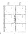
Figure 1 : Receiver operating characteristics (ROC) curve of the classifier for the differentiation between healthy controls (CTRL) and ovarian cancer patients (OVCA).
Performance on the training set is shown to the left whereas the performance on the validation set is shown to the right. The continuous line with the dot represents the discrimination power of the classifier, whereby the filled circle (threshold) is calculated by (1-sensitivity)2 + (1-specificity)2. The x-axis and y-axis show the false positive rate and the true positive rate, respectively. The dash-dotted diagonal line indicates no discrimination power, i.e. random classification. -
Figure 2 : Receiver operating characteristics (ROC) curve of the classifier for the differential diagnosis between ovarian cancer patients (OVCA) and breast cancer patients (BCA).
Performance on the training set is shown to the left, whereas the performance on the validation set is shown to the right. The continuous line with the dot represents the discrimination power of the classifier, whereby the filled circle (threshold) is calculated by (1-sensitivity)2 + (1-specificity)2. The x-axis and y-axis show the false positive rate and the true positive rate, respectively. The dash-dotted diagonal line indicates no discrimination power, i.e. random classification. -
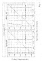
Figure 3 : Receiver operating characteristics (ROC) curve in (A) training data and (B) validation data of the single features from left to right (C18:2/lyso PC a C18:2, Trp, Ala).
ROC curve of the differential diagnosis between ovarian cancer patients (OVCA) and breast cancer patients (BCA) is shown as a continuous line with a cut-off point, depicted as filled circle. ROC curve of the differentiation between healthy controls (CTRL) and ovarian cancer patients (OVCA) is shown as dashed line with a filled triangle as cut-off indication. All cut-off points are calculated by (1-sensitivity)2 + (1-specificity)2. The x-axis and y-axis show the false positive rate and the true positive rate, respectively. The dash-dotted diagonal line indicates no discrimination power, i.e. random classification. -
Figure 4 : Left: Receiver operating characteristics (ROC) curve of the classifier performance on the validation set for the differentiation between healthy controls (CTRL) and early stage ovarian cancer patients (OVCA). Right: ROC curve of the classifier performance on the validation set for differentiation between early stage breast cancer patients (BCA) and early stage ovarian cancer patients (OVCA).
The continuous line with the filled circle (threshold) represents the discrimination power of the classifier, whereby the filled circle is calculated by (1-sensitivity)2 + (1-specificity)2. The x-axis and y-axis show the false positive rate and the true positive rate, respectively. The dash-dotted diagonal line indicates no discrimination power, i.e. random classification. -
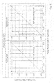
Figure 5 : Receiver operating characteristics (ROC) curves in (A) training data and (B) validation data of the single features in early stage ovarian cancer from left to right (C18:2/lyso PC a C18:2, Trp, Ala) of the classifier. ROC curve of the differential diagnostic between early stage ovarian cancer patients (OVCA) and early stage breast cancer patients (BCA) is shown as continuous line with a cut-off point, depicted as filled circle. ROC curve of the differentiation between healthy controls (CTRL) and early stage ovarian cancer patients (OVCA) is shown as dashed line with a filled triangle as cut-off indication. All cut-off points are calculated by (1-sensitivity)2 + (1-specificity)2. The x-axis and y-axis show the false positive rate and the true positive rate, respectively. The dash-dotted diagonal line indicates no discrimination power, i.e. random classification. -
Figure 6 : Left: Receiver operating characteristics (ROC) curve of the classifier performance on the validation set for the differentiation between healthy controls (CTRL) and late stage ovarian cancer patients (OVCA). Right: Receiver operating characteristics (ROC) curve of the classifier performance on the validation set for the differentiation between early stage breast cancer patients (BCA) and late stage ovarian cancer patients (OVCA). The continuous line with the filled circle (threshold) represents the discrimination power of the classifier, whereby the filled circle is calculated by (1-sensitivity)2 + (1-specificity)2. The x-axis and y-axis show the false positive rate and the true positive rate, respectively. The dash-dotted diagonal line indicates no discrimination power, i.e. random classification. -
Figure 7 : Receiver operating characteristics (ROC) curve in (A) training data and (B) validation data of the single features in early stage ovarian cancer from left to right (C18:2/lyso PC a C18:2, Trp, Ala) of the classifier. ROC curve of the differential diagnostic between late stage ovarian cancer patients (OVCA) and early stage breast cancer patients (BCA) is shown as continuous line with a cut-off point, depicted as filled circle. ROC curve of the differentiation between healthy controls (CTRL) and late stage ovarian cancer patients (OVCA) is shown as dashed line with a filled triangle as cut-off indication. All cut-off points are calculated by (1-sensitivity)2 + (1-specificity)2. The x-axis and y-axis show the false positive rate and the true positive rate, respectively. The dash-dotted diagonal line indicates no discrimination power, i.e. random classification. -
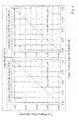
Figure 8 : Receiver operating characteristics (ROC) curve in (A) training data and (B) validation data of the single features from left to right (Glu / PC aa C32:2, C18:2 / SM (OH) C24:1, Glu / Ala).
ROC curve of the differential diagnosis between ovarian cancer patients (OVCA) and breast cancer patients (BCA) is shown as a continuous line with a cut-off point, depicted as filled circle. ROC curve of the differentiation between healthy controls (CTRL) and ovarian cancer patients (OVCA) is shown as dashed line with a filled triangle as cut-off indication. All cut-off points are calculated by (1-sensitivity)2 + (1-specificity)2. The x-axis and y-axis show the false positive rate and the true positive rate, respectively. The dash-dotted diagonal line indicates no discrimination power, i.e. random classification. -
Figure 9 : Receiver operating characteristics (ROC) curve in (A) training data and (B) validation data of the single features from left to right (Arg, His, SM (OH) C24:1).
ROC curve of the differential diagnosis between ovarian cancer patients (OVCA) and breast cancer patients (BCA) is shown as a continuous line with a cut-off point, depicted as filled circle. ROC curve of the differentiation between healthy controls (CTRL) and ovarian cancer patients (OVCA) is shown as dashed line with a filled triangle as cut-off indication. All cut-off points are calculated by (1-sensitivity)2 + (1-specificity)2. The x-axis and y-axis show the false positive rate and the true positive rate, respectively. The dash-dotted diagonal line indicates no discrimination power, i.e. random classification. -
- Bairey, Osnat; Blickstein, Dorit; Stark, Pinhas; Prokocimer, Miron; Nativ, Hila Magen; Kirgner, Ilia; Shaklai, Mati (2003): Serum CA 125 as a prognostic factor in non-Hodgkin's lymphoma. In: Leukemia & lymphoma 44 (10), S. 1733-1738. DOI: 10.1080/1042819031000104079.
- Berkenblit, Anna; Cannistra, Stephen A. (2005): Advances in the management of epithelial ovarian cancer. In: The Journal of reproductive medicine 50 (6), S. 426-438.
- Breiman, L. (2001): Random Forests, Machine Learning.
- Jerome H. Friedman (2002): Stochastic gradient boosting. In: Journal Computational Statistics & Data Analysis, S. 367-378. DOI: 10.1016/S0167-9473(01)00065-2.
- Jerome H. Friedman, Trevor Hastie, Rob Tibshirani (2009): Regularization Paths for Generalized Linear Models via Coordinate Descent. In: Journal of Statistical Software. DOI: 10.18637/jss.v033.i01.
- Kitawaki, Jo; Ishihara, Hiroaki; Koshiba, Hisato; Kiyomizu, Miyo; Teramoto, Mariko; Kitaoka, Yui; Honjo, Hideo (2005): Usefulness and limits of CA-125 in diagnosis of endometriosis without associated ovarian endometriomas. In: Human reproduction (Oxford, England) 20 (7), S. 1999-2003. DOI: 10.1093/humrep/deh890.
- Louppe, Gilles and Wehenkel, Louis and Sutera, Antonio and Geurts, Pierre (2013): Understanding Variable Importances in Forests of Randomized Trees. Proceedings of the 26th International Conference on Neural Information Processing Systems.
- Max Kuhn: Building Predictive Models in R Using the caret Package. In: Journal of Statistical Software 2008. DOI: 10.18637/jss.v028.i05.
- Moore, Richard G.; Miller, M. Craig; Disilvestro, Paul; Landrum, Lisa M.; Gajewski, Walter; Ball, John J.; Skates, Steven J. (2011): Evaluation of the diagnostic accuracy of the risk of ovarian malignancy algorithm in women with a pelvic mass. In: Obstetrics and gynecology 118 (2 Pt 1), S. 280-288. DOI: 10.1097/AOG.0b013e318224fce2.
- Norum, L. F.; Erikstein, B.; Nustad, K. (2001): Elevated CA125 in breast cancer--A sign of advanced disease. In: Tumour biology : the journal of the International Society for Oncodevelopmental Biology and Medicine 22 (4), S. 223-228.
- Prat, Jaime (2014): Staging classification for cancer of the ovary, fallopian tube, and peritoneum. In: International journal of gynaecology and obstetrics: the official organ of the International Federation of Gynaecology and Obstetrics 124 (1), S. 1-5. DOI: 10.1016/j.ijgo.2013.10.001.
- Roupa, Zoi; Faros, Efthimios; Raftopoulos, Vasilios; Tzavelas, Georgios; Kotrotsiou, Evangelia; Sotiropoulou, Penelope et al. (2004): Serum CA 125 combined with transvaginal ultrasonography for ovarian cancer screening. In: In vivo (Athens, Greece) 18 (6), S. 831-836.
- Shapira, I.; Oswald, M.; Lovecchio, J.; Khalili, H.; Menzin, A.; Whyte, J. et al. (2014): Circulating biomarkers for detection of ovarian cancer and predicting cancer outcomes. In: British journal of cancer 110 (4), S. 976-983. DOI: 10.1038/bjc.2013.795.
- Siegel, Rebecca L.; Miller, Kimberly D.; Jemal, Ahmedin (2015): Cancer statistics, 2015. In: CA: a cancer journal for clinicians 65 (1), S. 5-29. DOI: 10.3322/caac.21254.
- Steven L. Salzberg: C4.5: Programs for Machine Learning. In: Machine Learning 1994. DOI: 10.1007/BF00993309.
- Strobl, Carolin; Malley, James; Tutz, Gerhard (2009): An introduction to recursive partitioning: rationale, application, and characteristics of classification and regression trees, bagging, and random forests. In: Psychological methods 14 (4), S. 323-348. DOI: 10.1037/a0016973.
- Suh, K. Stephen; Park, Sang W.; Castro, Angelica; Patel, Hiren; Blake, Patrick; Liang, Michael; Goy, Andre (2010): Ovarian cancer biomarkers for molecular biosensors and translational medicine. In: Expert review of molecular diagnostics 10 (8), S. 1069-1083. DOI: 10.1586/erm.10.87.
- Tibshirani, Robert; Bien, Jacob; Friedman, Jerome; Hastie, Trevor; Simon, Noah; Taylor, Jonathan; Tibshirani, Ryan J. (2012): Strong rules for discarding predictors in lasso-type problems. In: Journal of the Royal Statistical Society. Series B, Statistical methodology 74 (2), S. 245-266. DOI: 10.1111/j.1467-9868.2011.01004.x.
- Vaughan, Sebastian; Coward, Jermaine I.; Bast, Robert C.; Berchuck, Andy; Berek, Jonathan S.; Brenton, James D. et al. (2011): Rethinking ovarian cancer: recommendations for improving outcomes. In: Nature reviews. Cancer 11 (10), S. 719-725. DOI: 10.1038/nrc3144.
- Wei, S. U.; Li, Hui; Zhang, Bei (2016): The diagnostic value of serum HE4 and CA-125 and ROMA index in ovarian cancer. In: Biomedical reports 5 (1), S. 41-44. DOI: 10.3892/br.2016.682.
- Yamamoto, Manabu; Baba, Hideo; Toh, Yasushi; Okamura, Takeshi; Maehara, Yoshihiko (2007): Peritoneal lavage CEA/CA125 is a prognostic factor for gastric cancer patients. In: Journal of cancer research and clinical oncology 133 (7), S. 471-476. DOI: 10.1007/s00432-006-0189-2.
at the same time allowing a more reliable diagnosis of ovarian cancer, in particular related to FIGO stages I and II, see below. Such a fact has neither been described in nor made obvious from the prior art.
Claims (11)
- In-vitro use of a metabolic biomarker set for assessing ovarian cancer comprising at leasti.) one amino acid selected from the group of alanine, arginine, histidine, tryptophan, glutamate, andii.) one phospholipid selected from the group of lyso PC a C18:1, lyso PC a C18:2, and PC aa C32:2,iii.) one acylcarnitine selected from the group of acylcarnitine C18:1, acylcarnitine C18:2, and optionaliv.) one sphingolipid SM(OH)C24:1.
- In-vitro use of a metabolic biomarker set according to claim 1, wherein at least one further biomarker is an amino acid, a biogenic amine, an acylcarnitine, a hexose, a sphingolipid or a glycerophospholipid selected from those listed in Table 1.
- In-vitro use of a metabolic biomarker set for assessing ovarian cancer according to any one of claims 1 to 2,
wherein the assessment is for the diagnosis and/or risk classification, for the prognosis, for differential diagnostic, for early stage detection and recognition. - An in-vitro method for assessing ovarian cancer, characterized in that it comprises determining in a sample, preferably blood and/ or plasma, obtained from a patient to be examined the amount of at leasti.) one amino acid selected from the group of alanine, arginine, histidine, tryptophan, glutamate, andii.) one phospholipid selected from the group of lyso PC a C18:1, lyso PC a C18:2, and PC aa C32:2, andiii.) one acylcarnitine selected from the group of acylcarnitine C18:1, preferably acylcarnitine C18:2, and optionaliv.) one sphingolipid SM(OH)C24:1 are determined,and wherein at least one ratio of at least two biomarkers of i.) to iv.) is determined.
- The method according to claim 4 further comprising determining in the sample at least one ratio of at least two biomarkers selected from the group of C18:2/lysoPC a C18:2, C18:2/SM (OH) C24:1, Glu/Ala, Glu/PC aa C32:2.
- The method according to claim 4, wherein arginine and tryptophan are selected and the ratio of C18:2/lysoPC a C18:2 is determined.
- The method according to any one of claims 4 to 6, wherein the patient is a symptomatic and/or asymptomatic patient.
- The method according to any one of claims 4 to 6, wherein the assessment is diagnosis or prognosis.
- The method according to any one of claims 4 to 6, wherein the assessment is for the diagnosis and/or risk classification, for the prognosis, for differential diagnostic, for early stage detection and recognition, in particular for carrying out clinical decisions, such as the continuative treatment and therapy by means of pharmaceuticals, including the decision of hospitalization of the patient.
- The method according to any one of claims 4 to 6, wherein the measurement is based on a quantitative analytical method, preferably chromatography, spectroscopy, and mass spectrometry.
- The method according to claim 10, wherein chromatography comprises GC, LC, HPLC, and UPLC; spectroscopy comprises UV/Vis, IR, and NMR; and mass spectrometry comprises ESI-QqQ, ESI-QqTOF, MALDI-QqQ, MALDI-QqTOF, and MALDI-TOF-TOF.
Applications Claiming Priority (2)
| Application Number | Priority Date | Filing Date | Title |
|---|---|---|---|
| EP17152072.9A EP3351938A1 (en) | 2017-01-18 | 2017-01-18 | New biomarkers for assessing ovarian cancer |
| PCT/EP2018/051254 WO2018134329A1 (en) | 2017-01-18 | 2018-01-18 | Metabolic biomarker set for assessing ovarian cancer |
Publications (2)
| Publication Number | Publication Date |
|---|---|
| EP3571506A1 EP3571506A1 (en) | 2019-11-27 |
| EP3571506B1 true EP3571506B1 (en) | 2021-09-01 |
Family
ID=57860708
Family Applications (2)
| Application Number | Title | Priority Date | Filing Date |
|---|---|---|---|
| EP17152072.9A Withdrawn EP3351938A1 (en) | 2017-01-18 | 2017-01-18 | New biomarkers for assessing ovarian cancer |
| EP18704150.4A Active EP3571506B1 (en) | 2017-01-18 | 2018-01-18 | Metabolic biomarker set for assessing ovarian cancer |
Family Applications Before (1)
| Application Number | Title | Priority Date | Filing Date |
|---|---|---|---|
| EP17152072.9A Withdrawn EP3351938A1 (en) | 2017-01-18 | 2017-01-18 | New biomarkers for assessing ovarian cancer |
Country Status (5)
| Country | Link |
|---|---|
| US (2) | US11506665B2 (en) |
| EP (2) | EP3351938A1 (en) |
| JP (1) | JP7169278B2 (en) |
| CN (1) | CN110199198B (en) |
| WO (1) | WO2018134329A1 (en) |
Families Citing this family (7)
| Publication number | Priority date | Publication date | Assignee | Title |
|---|---|---|---|---|
| BR112021003477A2 (en) | 2018-06-14 | 2021-05-18 | Metabolomycs, Inc. | Metabolomic signatures for predicting, diagnosing and prognosticating various diseases including cancer |
| US11409549B2 (en) | 2018-10-15 | 2022-08-09 | AIble Inc. | Interface for generating models with customizable interface configurations |
| US10586164B1 (en) | 2018-10-15 | 2020-03-10 | AIble Inc. | Interface for visualizing and improving model performance |
| WO2020099683A1 (en) * | 2018-11-16 | 2020-05-22 | Biocrates Life Sciences Ag | Metabolites for use in ovarian cancer detection and diagnosis |
| RU2726971C1 (en) * | 2019-12-05 | 2020-07-17 | федеральное государственное автономное образовательное учреждение высшего образования "Национальный исследовательский университет ИТМО" (Университет ИТМО) | Method of non-invasive early diagnosis of endometriosis |
| CN117594243B (en) * | 2023-10-13 | 2024-05-14 | 太原理工大学 | Ovarian cancer prognosis prediction method based on cross-modal view association discovery network |
| CN117538530B (en) * | 2023-11-07 | 2024-07-19 | 中国医学科学院肿瘤医院 | Biomarker composition and kit for detecting metastatic breast cancer and application of biomarker composition and kit |
Family Cites Families (12)
| Publication number | Priority date | Publication date | Assignee | Title |
|---|---|---|---|---|
| CA2467563A1 (en) | 2001-11-20 | 2003-05-30 | Genentech, Inc. | Cell and tissue arrays and microarrays and methods of use |
| CA2608965C (en) | 2005-06-30 | 2014-08-19 | Biocrates Life Sciences Ag | Device for quantitative analysis of a metabolite profile |
| WO2009151967A1 (en) * | 2008-05-28 | 2009-12-17 | Georgia Tech Research Corporation | Metabolic biomarkers for ovarian cancer and methods of use thereof |
| CN104316701B (en) * | 2008-06-20 | 2018-05-15 | 味之素株式会社 | The evaluation method of female genital cancer |
| CN101832977A (en) * | 2009-03-09 | 2010-09-15 | 复旦大学附属妇产科医院 | Ovarian tumor serum marker |
| BRPI0924639B1 (en) | 2009-06-02 | 2021-12-07 | Biocrates Life Sciences Ag | NEW BIOMARKERS FOR EVALUATION OF KIDNEY DISEASES |
| WO2012164525A2 (en) * | 2011-06-01 | 2012-12-06 | Ecole Polytechnique Federale De Lausanne (Epfl) | Aging biomarkers |
| CA2960696C (en) * | 2014-09-10 | 2022-12-06 | Idcgs Clinica De Diagnosticos Medicos Ltda | Biomarkers for assessing breast cancer |
| AU2015326756A1 (en) * | 2014-10-02 | 2017-05-11 | Zora Biosciences Oy | Methods for detecting ovarian cancer |
| US20170097355A1 (en) * | 2015-10-06 | 2017-04-06 | University Of Washington | Biomarkers and methods to distinguish ovarian cancer from benign tumors |
| WO2018136085A1 (en) * | 2017-01-20 | 2018-07-26 | Mprobe Inc. | Methods and compositions for providing an early stage ovarian cancer assessment with metabolites |
| BR112021003477A2 (en) * | 2018-06-14 | 2021-05-18 | Metabolomycs, Inc. | Metabolomic signatures for predicting, diagnosing and prognosticating various diseases including cancer |
-
2017
- 2017-01-18 EP EP17152072.9A patent/EP3351938A1/en not_active Withdrawn
-
2018
- 2018-01-18 EP EP18704150.4A patent/EP3571506B1/en active Active
- 2018-01-18 CN CN201880007189.3A patent/CN110199198B/en active Active
- 2018-01-18 WO PCT/EP2018/051254 patent/WO2018134329A1/en not_active Ceased
- 2018-01-18 JP JP2019538601A patent/JP7169278B2/en active Active
- 2018-01-18 US US16/478,819 patent/US11506665B2/en active Active
-
2022
- 2022-10-20 US US17/969,834 patent/US20240192216A1/en not_active Abandoned
Also Published As
| Publication number | Publication date |
|---|---|
| WO2018134329A1 (en) | 2018-07-26 |
| CN110199198A (en) | 2019-09-03 |
| EP3351938A1 (en) | 2018-07-25 |
| JP7169278B2 (en) | 2022-11-10 |
| JP2020506382A (en) | 2020-02-27 |
| US11506665B2 (en) | 2022-11-22 |
| US20240192216A1 (en) | 2024-06-13 |
| EP3571506A1 (en) | 2019-11-27 |
| CN110199198B (en) | 2022-09-06 |
| US20200386762A1 (en) | 2020-12-10 |
Similar Documents
| Publication | Publication Date | Title |
|---|---|---|
| US20240192216A1 (en) | Metabolic biomarker set for assessing ovarian cancer | |
| US11493516B2 (en) | Biomarkers for assessing breast cancer | |
| CN113396332B (en) | Methods and applications for evaluating pregnancy progress and premature abortion for clinical intervention | |
| Porto-Mascarenhas et al. | Salivary biomarkers in the diagnosis of breast cancer: A review | |
| CN109983137A (en) | For predicted exposure in the biomarker of the premature labor of the pregnant female of progestational hormone | |
| WO2019185692A1 (en) | Metabolite-based breast cancer detection and diagnosis | |
| JPWO2018066621A1 (en) | Evaluation method of colon cancer, evaluation device, evaluation program, evaluation system, and terminal device | |
| WO2025123592A1 (en) | Use of metabolic marker for diagnosis of lung cancer staging and kit | |
| Kałużna-Czaplińska et al. | Current applications of chromatographic methods for diagnosis and identification of potential biomarkers in cancer | |
| ES2688121T3 (en) | Diagnostic means and procedures for prostate cancer recurrence after prostatectomy | |
| JP7673770B2 (en) | Acquisition method, calculation method, evaluation device, calculation device, evaluation program, calculation program, and evaluation system | |
| KR20170132303A (en) | Evaluation method, evaluation apparatus, evaluation program product, evaluation system, and terminal apparatus | |
| WO2020099683A1 (en) | Metabolites for use in ovarian cancer detection and diagnosis | |
| US20250306042A1 (en) | Method for diagnosing endometriosis in a subject | |
| JP7435856B2 (en) | Acquisition method, calculation method, evaluation device, calculation device, evaluation program, calculation program, recording medium, and evaluation system | |
| CN119738487A (en) | Hypoxanthine and taurine, a biomarker for lung cancer diagnosis | |
| Rakhshan et al. | Applications of mass spectroscopy in understanding cancer proteomics | |
| HK40062939A (en) | Methods for evaluation of gestational progress and preterm abortion for clinical intervention and applications thereof | |
| AU2024257583A1 (en) | Method of diagnosis and treatment of pancreatic cancer | |
| WO2007022625A1 (en) | Peptide reversion methods and uses thereof | |
| BRPI1105603B1 (en) | METHOD FOR IDENTIFICATION OF METABOLYTE PROFILES FOR DETERMINING THE RESISTANCE OF ACUTE LYMPHOID LEUKEMIA (ALL) CANCEROUS CELLS TO CHEMOTHERAPY |
Legal Events
| Date | Code | Title | Description |
|---|---|---|---|
| STAA | Information on the status of an ep patent application or granted ep patent |
Free format text: STATUS: UNKNOWN |
|
| STAA | Information on the status of an ep patent application or granted ep patent |
Free format text: STATUS: THE INTERNATIONAL PUBLICATION HAS BEEN MADE |
|
| PUAI | Public reference made under article 153(3) epc to a published international application that has entered the european phase |
Free format text: ORIGINAL CODE: 0009012 |
|
| STAA | Information on the status of an ep patent application or granted ep patent |
Free format text: STATUS: REQUEST FOR EXAMINATION WAS MADE |
|
| 17P | Request for examination filed |
Effective date: 20190819 |
|
| AK | Designated contracting states |
Kind code of ref document: A1 Designated state(s): AL AT BE BG CH CY CZ DE DK EE ES FI FR GB GR HR HU IE IS IT LI LT LU LV MC MK MT NL NO PL PT RO RS SE SI SK SM TR |
|
| AX | Request for extension of the european patent |
Extension state: BA ME |
|
| DAV | Request for validation of the european patent (deleted) | ||
| DAX | Request for extension of the european patent (deleted) | ||
| STAA | Information on the status of an ep patent application or granted ep patent |
Free format text: STATUS: EXAMINATION IS IN PROGRESS |
|
| 17Q | First examination report despatched |
Effective date: 20200528 |
|
| GRAP | Despatch of communication of intention to grant a patent |
Free format text: ORIGINAL CODE: EPIDOSNIGR1 |
|
| STAA | Information on the status of an ep patent application or granted ep patent |
Free format text: STATUS: GRANT OF PATENT IS INTENDED |
|
| INTG | Intention to grant announced |
Effective date: 20210323 |
|
| GRAS | Grant fee paid |
Free format text: ORIGINAL CODE: EPIDOSNIGR3 |
|
| GRAA | (expected) grant |
Free format text: ORIGINAL CODE: 0009210 |
|
| STAA | Information on the status of an ep patent application or granted ep patent |
Free format text: STATUS: THE PATENT HAS BEEN GRANTED |
|
| AK | Designated contracting states |
Kind code of ref document: B1 Designated state(s): AL AT BE BG CH CY CZ DE DK EE ES FI FR GB GR HR HU IE IS IT LI LT LU LV MC MK MT NL NO PL PT RO RS SE SI SK SM TR |
|
| REG | Reference to a national code |
Ref country code: GB Ref legal event code: FG4D |
|
| REG | Reference to a national code |
Ref country code: CH Ref legal event code: EP Ref country code: AT Ref legal event code: REF Ref document number: 1426755 Country of ref document: AT Kind code of ref document: T Effective date: 20210915 |
|
| REG | Reference to a national code |
Ref country code: DE Ref legal event code: R096 Ref document number: 602018022784 Country of ref document: DE |
|
| REG | Reference to a national code |
Ref country code: IE Ref legal event code: FG4D |
|
| REG | Reference to a national code |
Ref country code: LT Ref legal event code: MG9D |
|
| REG | Reference to a national code |
Ref country code: NL Ref legal event code: MP Effective date: 20210901 |
|
| PG25 | Lapsed in a contracting state [announced via postgrant information from national office to epo] |
Ref country code: NO Free format text: LAPSE BECAUSE OF FAILURE TO SUBMIT A TRANSLATION OF THE DESCRIPTION OR TO PAY THE FEE WITHIN THE PRESCRIBED TIME-LIMIT Effective date: 20211201 Ref country code: BG Free format text: LAPSE BECAUSE OF FAILURE TO SUBMIT A TRANSLATION OF THE DESCRIPTION OR TO PAY THE FEE WITHIN THE PRESCRIBED TIME-LIMIT Effective date: 20211201 Ref country code: LT Free format text: LAPSE BECAUSE OF FAILURE TO SUBMIT A TRANSLATION OF THE DESCRIPTION OR TO PAY THE FEE WITHIN THE PRESCRIBED TIME-LIMIT Effective date: 20210901 Ref country code: RS Free format text: LAPSE BECAUSE OF FAILURE TO SUBMIT A TRANSLATION OF THE DESCRIPTION OR TO PAY THE FEE WITHIN THE PRESCRIBED TIME-LIMIT Effective date: 20210901 Ref country code: SE Free format text: LAPSE BECAUSE OF FAILURE TO SUBMIT A TRANSLATION OF THE DESCRIPTION OR TO PAY THE FEE WITHIN THE PRESCRIBED TIME-LIMIT Effective date: 20210901 Ref country code: HR Free format text: LAPSE BECAUSE OF FAILURE TO SUBMIT A TRANSLATION OF THE DESCRIPTION OR TO PAY THE FEE WITHIN THE PRESCRIBED TIME-LIMIT Effective date: 20210901 Ref country code: ES Free format text: LAPSE BECAUSE OF FAILURE TO SUBMIT A TRANSLATION OF THE DESCRIPTION OR TO PAY THE FEE WITHIN THE PRESCRIBED TIME-LIMIT Effective date: 20210901 Ref country code: FI Free format text: LAPSE BECAUSE OF FAILURE TO SUBMIT A TRANSLATION OF THE DESCRIPTION OR TO PAY THE FEE WITHIN THE PRESCRIBED TIME-LIMIT Effective date: 20210901 |
|
| REG | Reference to a national code |
Ref country code: AT Ref legal event code: MK05 Ref document number: 1426755 Country of ref document: AT Kind code of ref document: T Effective date: 20210901 |
|
| PG25 | Lapsed in a contracting state [announced via postgrant information from national office to epo] |
Ref country code: PL Free format text: LAPSE BECAUSE OF FAILURE TO SUBMIT A TRANSLATION OF THE DESCRIPTION OR TO PAY THE FEE WITHIN THE PRESCRIBED TIME-LIMIT Effective date: 20210901 Ref country code: LV Free format text: LAPSE BECAUSE OF FAILURE TO SUBMIT A TRANSLATION OF THE DESCRIPTION OR TO PAY THE FEE WITHIN THE PRESCRIBED TIME-LIMIT Effective date: 20210901 Ref country code: GR Free format text: LAPSE BECAUSE OF FAILURE TO SUBMIT A TRANSLATION OF THE DESCRIPTION OR TO PAY THE FEE WITHIN THE PRESCRIBED TIME-LIMIT Effective date: 20211202 |
|
| PG25 | Lapsed in a contracting state [announced via postgrant information from national office to epo] |
Ref country code: AT Free format text: LAPSE BECAUSE OF FAILURE TO SUBMIT A TRANSLATION OF THE DESCRIPTION OR TO PAY THE FEE WITHIN THE PRESCRIBED TIME-LIMIT Effective date: 20210901 |
|
| PG25 | Lapsed in a contracting state [announced via postgrant information from national office to epo] |
Ref country code: IS Free format text: LAPSE BECAUSE OF FAILURE TO SUBMIT A TRANSLATION OF THE DESCRIPTION OR TO PAY THE FEE WITHIN THE PRESCRIBED TIME-LIMIT Effective date: 20220101 Ref country code: SM Free format text: LAPSE BECAUSE OF FAILURE TO SUBMIT A TRANSLATION OF THE DESCRIPTION OR TO PAY THE FEE WITHIN THE PRESCRIBED TIME-LIMIT Effective date: 20210901 Ref country code: SK Free format text: LAPSE BECAUSE OF FAILURE TO SUBMIT A TRANSLATION OF THE DESCRIPTION OR TO PAY THE FEE WITHIN THE PRESCRIBED TIME-LIMIT Effective date: 20210901 Ref country code: RO Free format text: LAPSE BECAUSE OF FAILURE TO SUBMIT A TRANSLATION OF THE DESCRIPTION OR TO PAY THE FEE WITHIN THE PRESCRIBED TIME-LIMIT Effective date: 20210901 Ref country code: PT Free format text: LAPSE BECAUSE OF FAILURE TO SUBMIT A TRANSLATION OF THE DESCRIPTION OR TO PAY THE FEE WITHIN THE PRESCRIBED TIME-LIMIT Effective date: 20220103 Ref country code: NL Free format text: LAPSE BECAUSE OF FAILURE TO SUBMIT A TRANSLATION OF THE DESCRIPTION OR TO PAY THE FEE WITHIN THE PRESCRIBED TIME-LIMIT Effective date: 20210901 Ref country code: EE Free format text: LAPSE BECAUSE OF FAILURE TO SUBMIT A TRANSLATION OF THE DESCRIPTION OR TO PAY THE FEE WITHIN THE PRESCRIBED TIME-LIMIT Effective date: 20210901 Ref country code: CZ Free format text: LAPSE BECAUSE OF FAILURE TO SUBMIT A TRANSLATION OF THE DESCRIPTION OR TO PAY THE FEE WITHIN THE PRESCRIBED TIME-LIMIT Effective date: 20210901 Ref country code: AL Free format text: LAPSE BECAUSE OF FAILURE TO SUBMIT A TRANSLATION OF THE DESCRIPTION OR TO PAY THE FEE WITHIN THE PRESCRIBED TIME-LIMIT Effective date: 20210901 |
|
| REG | Reference to a national code |
Ref country code: DE Ref legal event code: R097 Ref document number: 602018022784 Country of ref document: DE |
|
| PLBE | No opposition filed within time limit |
Free format text: ORIGINAL CODE: 0009261 |
|
| STAA | Information on the status of an ep patent application or granted ep patent |
Free format text: STATUS: NO OPPOSITION FILED WITHIN TIME LIMIT |
|
| PG25 | Lapsed in a contracting state [announced via postgrant information from national office to epo] |
Ref country code: IT Free format text: LAPSE BECAUSE OF FAILURE TO SUBMIT A TRANSLATION OF THE DESCRIPTION OR TO PAY THE FEE WITHIN THE PRESCRIBED TIME-LIMIT Effective date: 20210901 Ref country code: DK Free format text: LAPSE BECAUSE OF FAILURE TO SUBMIT A TRANSLATION OF THE DESCRIPTION OR TO PAY THE FEE WITHIN THE PRESCRIBED TIME-LIMIT Effective date: 20210901 |
|
| 26N | No opposition filed |
Effective date: 20220602 |
|
| PG25 | Lapsed in a contracting state [announced via postgrant information from national office to epo] |
Ref country code: SI Free format text: LAPSE BECAUSE OF FAILURE TO SUBMIT A TRANSLATION OF THE DESCRIPTION OR TO PAY THE FEE WITHIN THE PRESCRIBED TIME-LIMIT Effective date: 20210901 Ref country code: MC Free format text: LAPSE BECAUSE OF FAILURE TO SUBMIT A TRANSLATION OF THE DESCRIPTION OR TO PAY THE FEE WITHIN THE PRESCRIBED TIME-LIMIT Effective date: 20210901 |
|
| REG | Reference to a national code |
Ref country code: BE Ref legal event code: MM Effective date: 20220131 |
|
| PG25 | Lapsed in a contracting state [announced via postgrant information from national office to epo] |
Ref country code: LU Free format text: LAPSE BECAUSE OF NON-PAYMENT OF DUE FEES Effective date: 20220118 |
|
| PG25 | Lapsed in a contracting state [announced via postgrant information from national office to epo] |
Ref country code: BE Free format text: LAPSE BECAUSE OF NON-PAYMENT OF DUE FEES Effective date: 20220131 |
|
| PG25 | Lapsed in a contracting state [announced via postgrant information from national office to epo] |
Ref country code: IE Free format text: LAPSE BECAUSE OF NON-PAYMENT OF DUE FEES Effective date: 20220118 |
|
| PG25 | Lapsed in a contracting state [announced via postgrant information from national office to epo] |
Ref country code: MK Free format text: LAPSE BECAUSE OF FAILURE TO SUBMIT A TRANSLATION OF THE DESCRIPTION OR TO PAY THE FEE WITHIN THE PRESCRIBED TIME-LIMIT Effective date: 20210901 Ref country code: CY Free format text: LAPSE BECAUSE OF FAILURE TO SUBMIT A TRANSLATION OF THE DESCRIPTION OR TO PAY THE FEE WITHIN THE PRESCRIBED TIME-LIMIT Effective date: 20210901 |
|
| PG25 | Lapsed in a contracting state [announced via postgrant information from national office to epo] |
Ref country code: HU Free format text: LAPSE BECAUSE OF FAILURE TO SUBMIT A TRANSLATION OF THE DESCRIPTION OR TO PAY THE FEE WITHIN THE PRESCRIBED TIME-LIMIT; INVALID AB INITIO Effective date: 20180118 |
|
| PG25 | Lapsed in a contracting state [announced via postgrant information from national office to epo] |
Ref country code: MT Free format text: LAPSE BECAUSE OF FAILURE TO SUBMIT A TRANSLATION OF THE DESCRIPTION OR TO PAY THE FEE WITHIN THE PRESCRIBED TIME-LIMIT Effective date: 20210901 |
|
| REG | Reference to a national code |
Ref country code: DE Ref legal event code: R079 Ref document number: 602018022784 Country of ref document: DE Free format text: PREVIOUS MAIN CLASS: G01N0033574000 Ipc: G01N0033575000 |
|
| PG25 | Lapsed in a contracting state [announced via postgrant information from national office to epo] |
Ref country code: TR Free format text: LAPSE BECAUSE OF FAILURE TO SUBMIT A TRANSLATION OF THE DESCRIPTION OR TO PAY THE FEE WITHIN THE PRESCRIBED TIME-LIMIT Effective date: 20210901 |
|
| REG | Reference to a national code |
Ref country code: CH Ref legal event code: U11 Free format text: ST27 STATUS EVENT CODE: U-0-0-U10-U11 (AS PROVIDED BY THE NATIONAL OFFICE) Effective date: 20260201 |
|
| PGFP | Annual fee paid to national office [announced via postgrant information from national office to epo] |
Ref country code: GB Payment date: 20260119 Year of fee payment: 9 |
|
| PGFP | Annual fee paid to national office [announced via postgrant information from national office to epo] |
Ref country code: DE Payment date: 20260120 Year of fee payment: 9 |
|
| PGFP | Annual fee paid to national office [announced via postgrant information from national office to epo] |
Ref country code: FR Payment date: 20260120 Year of fee payment: 9 |
|
| PGFP | Annual fee paid to national office [announced via postgrant information from national office to epo] |
Ref country code: CH Payment date: 20260201 Year of fee payment: 9 |