EP3570767B1 - Sternotomy closure apparatus using polymeric cable - Google Patents
Sternotomy closure apparatus using polymeric cable Download PDFInfo
- Publication number
- EP3570767B1 EP3570767B1 EP18709190.5A EP18709190A EP3570767B1 EP 3570767 B1 EP3570767 B1 EP 3570767B1 EP 18709190 A EP18709190 A EP 18709190A EP 3570767 B1 EP3570767 B1 EP 3570767B1
- Authority
- EP
- European Patent Office
- Prior art keywords
- pressure
- sternum
- cable
- fragments
- tension
- Prior art date
- Legal status (The legal status is an assumption and is not a legal conclusion. Google has not performed a legal analysis and makes no representation as to the accuracy of the status listed.)
- Active
Links
Images


Classifications
-
- A—HUMAN NECESSITIES
- A61—MEDICAL OR VETERINARY SCIENCE; HYGIENE
- A61B—DIAGNOSIS; SURGERY; IDENTIFICATION
- A61B17/00—Surgical instruments, devices or methods
- A61B17/56—Surgical instruments or methods for treatment of bones or joints; Devices specially adapted therefor
- A61B17/58—Surgical instruments or methods for treatment of bones or joints; Devices specially adapted therefor for osteosynthesis, e.g. bone plates, screws or setting implements
- A61B17/68—Internal fixation devices, including fasteners and spinal fixators, even if a part thereof projects from the skin
- A61B17/82—Internal fixation devices, including fasteners and spinal fixators, even if a part thereof projects from the skin for bone cerclage
- A61B17/823—Internal fixation devices, including fasteners and spinal fixators, even if a part thereof projects from the skin for bone cerclage for the sternum
-
- A—HUMAN NECESSITIES
- A61—MEDICAL OR VETERINARY SCIENCE; HYGIENE
- A61B—DIAGNOSIS; SURGERY; IDENTIFICATION
- A61B17/00—Surgical instruments, devices or methods
- A61B17/16—Instruments for performing osteoclasis; Drills or chisels for bones; Trepans
- A61B17/17—Guides or aligning means for drills, mills, pins or wires
- A61B17/1739—Guides or aligning means for drills, mills, pins or wires specially adapted for particular parts of the body
- A61B17/1789—Guides or aligning means for drills, mills, pins or wires specially adapted for particular parts of the body for the sternum
-
- A—HUMAN NECESSITIES
- A61—MEDICAL OR VETERINARY SCIENCE; HYGIENE
- A61B—DIAGNOSIS; SURGERY; IDENTIFICATION
- A61B17/00—Surgical instruments, devices or methods
- A61B17/56—Surgical instruments or methods for treatment of bones or joints; Devices specially adapted therefor
- A61B17/58—Surgical instruments or methods for treatment of bones or joints; Devices specially adapted therefor for osteosynthesis, e.g. bone plates, screws or setting implements
- A61B17/68—Internal fixation devices, including fasteners and spinal fixators, even if a part thereof projects from the skin
- A61B17/84—Fasteners therefor or fasteners being internal fixation devices
- A61B17/842—Flexible wires, bands or straps
-
- A—HUMAN NECESSITIES
- A61—MEDICAL OR VETERINARY SCIENCE; HYGIENE
- A61B—DIAGNOSIS; SURGERY; IDENTIFICATION
- A61B17/00—Surgical instruments, devices or methods
- A61B17/56—Surgical instruments or methods for treatment of bones or joints; Devices specially adapted therefor
- A61B17/58—Surgical instruments or methods for treatment of bones or joints; Devices specially adapted therefor for osteosynthesis, e.g. bone plates, screws or setting implements
- A61B17/88—Osteosynthesis instruments; Methods or means for implanting or extracting internal or external fixation devices
- A61B17/8869—Tensioning devices
-
- A—HUMAN NECESSITIES
- A61—MEDICAL OR VETERINARY SCIENCE; HYGIENE
- A61B—DIAGNOSIS; SURGERY; IDENTIFICATION
- A61B17/00—Surgical instruments, devices or methods
- A61B17/56—Surgical instruments or methods for treatment of bones or joints; Devices specially adapted therefor
- A61B17/58—Surgical instruments or methods for treatment of bones or joints; Devices specially adapted therefor for osteosynthesis, e.g. bone plates, screws or setting implements
- A61B17/88—Osteosynthesis instruments; Methods or means for implanting or extracting internal or external fixation devices
- A61B17/8861—Apparatus for manipulating flexible wires or straps
-
- A—HUMAN NECESSITIES
- A61—MEDICAL OR VETERINARY SCIENCE; HYGIENE
- A61B—DIAGNOSIS; SURGERY; IDENTIFICATION
- A61B90/00—Instruments, implements or accessories specially adapted for surgery or diagnosis and not covered by any of the groups A61B1/00 - A61B50/00, e.g. for luxation treatment or for protecting wound edges
- A61B90/06—Measuring instruments not otherwise provided for
- A61B2090/064—Measuring instruments not otherwise provided for for measuring force, pressure or mechanical tension
- A61B2090/065—Measuring instruments not otherwise provided for for measuring force, pressure or mechanical tension for measuring contact or contact pressure
Definitions
- the sternum Upon completion of open heart surgery (or other surgery requiring access to the chest cavity, such as a lung transplant for example), the sternum must be closed. Usually this involves binding the two divided halves of the sternum together using a cerclage technique. The objective is to bring the two bone segments into proper apposition so that they may heal and again provide the normal function of the sternum in the musculo-skeletal system. The desired outcome is full bony union so that the sternum is restored as nearly as possible to its pre-surgical condition. In order to achieve this goal, the closure must be sufficiently robust so that full bone formation and healing can occur across the sternotomy.
- sternotomy closures are performed using stainless steel or other metallic alloy wire, with the aid of instruments such as a wire tightener and wire cutters.
- Braided metallic cable may be used, either alone or in conjunction with bone fixation devices such as plates, screws, or the like.
- US 2005/273983 A1 discloses a bone cerclage system comprising an elastic polymer cable and a cable clamp, the polymer cable being configured to achieve an elongation which increases the polymer cable by a percentage between 60 and 140 per cent of its original length, at a working tension up to approximately 800 Newtons.
- US 2014/142638 A1 discloses a sternal closure system comprising surgical cables and a tensioning tool.
- WO 2014/209442 A1 discloses a bone cerclage system comprising an elastic polymer cable and a tensioning tool.
- the present invention provides an apparatus for fixing a surgically cut sternum during healing, as defined in claim 1.
- the cerclage mechanism as described herein is capable of maintaining proper bony compression (within a desired range) across the sternotomy cut, in spite of a range of external forces and significant lateral distraction due to these external forces.
- the elastic member is preferably a polymer cable comprising an elongated elastic polymeric core coaxially surrounded by a sheath woven from ultra-high molecular-weight polyethylene strands.
- the present sternotomy closure technique preferably employs a polymeric engineered cable available from Kinamed, Inc. in Camarillo, CA and sold under the name "SuperCable.”
- the cable is constructed from an elongated elastic polymeric core, suitably Nylon, coaxially surrounded by a sheath woven from ultra-high molecular-weight polyethylene strands. Further information and description of this "SuperCable” is given in US patent 6,589,246 to Mattchen.
- This cable is very flexible, elastic, with high fatigue strength and tensile strength. Such cable is capable of repeated cycles of extension/contraction during the healing phase while maintaining the ability to provide compressive load across the osteotomy plane.
- FIG. 1 shows cyclical behavior of extension versus time for an anatomical sternotomy model that has been secured by a SuperCable.
- the lower curve represents the "SuperCable" assembly (sternotomy model secured with SuperCables) which consisted for this test of only four SuperCables (each doubled as a circumferential loop).
- Similar curves are shown for steel monofilament wire (seven loops, top curve), and for braided steel cable (four loops, middle curve).
- the sternal halves were forcibly laterally distracted with a cyclic force varying periodically from zero to just over 1000 Newtons, for five cycles using a Chatillon LRX materials testing system.
- the steel wire and cable fail to maintain reliable compressive force across the divided sternum because of at least two factors: first, they have high stiffness and fail to accommodate extensive extension and contraction without losing their initial bone contact length. Much of the elasticity in the full model assembly is believed to lie in the bony model rather than the wire or cable. Metal cables and wires may unwind slightly, thereby lengthening irreversibly. Secondly, such cables tend to bite or cut into the bone, tending to further generate potential for slack in the cerclage. This further tends to generate irreversible slack across the osteotomy plane. Both metal wires and metal cables are known to sometimes cut completely through the sternum, resulting not only in failure to fix the median sternotomy but also bisecting the divided sternum into halves which can significantly complicate a subsequent reconstruction.
- a single, active force sensor was placed consistently within the sternotomy plane on all test samples, adjacent to the 3rd intercostal space with enough offset to avoid interference by the metal wire or cable placements.
- Four other inactive sensors of the same type were spaced equally along the sternotomy plane to ensure even loading.
- FIG. 3 shows the results: the graph shows force (measured by the pressure sensor) as a function of time during the application of cyclic, time varying lateral distraction force.
- the results are shown for SuperCable, for the metal cables, and for the metal wire loops.
- Table 1 summarizes the residual force, percent retention of force, and magnitude of force recovered for all three cable systems.
- Parameter SuperCable construct Stainless steel wire construct Stainless steel cable construct Residual force between sternal halves after the cyclic loading regime (N) 237.0 18.6 49.4 Percent retention of force [residual force after cyclic loading divided by force before cyclic loading] 71.0% 17.7% 49.0%
- Additional testing further demonstrates important differences in how the SuperCable construct performed in comparison to the metal wire and metal constructs.
- the testing outlined above was repeated with two key modifications.
- an additional fixation model was added that included two SuperCables in a parasternal wrapping technique at the 2 nd & 4 th intercostal spaces, and two SuperCables in a parasternal/transsternal "figure-eight" wrapping technique at the 1 st & 5 th intercostal spaces and transternally in the manibrium and xiphoid (similar to superior and inferior cables in FIG 2 ).
- Each cable was tensioned to achieve 360 Newton (80 lbs.) of compressive force across the divided sternum.
- FIG. 4 shows the results: the graph shows residual force (measured by the pressure sensors) as a function of location along the sternotomy models. The results are shown for both SuperCable configurations, for the metal Pioneer cables, and for the metal wire loops. Both of the SuperCable configurations resulted in higher residual force than the metal cables or wire loops in all three sensor locations which is representative of the entire length of the sternotomy models.
- a higher residual force between the sternal halves is a measure of the stability after a cyclic loading regime (such as a sneezing or coughing fit) and can be predictive of continued approximation of the sternal halves for the promotion of bony healing.
- a higher percent retention of force is a measure of whether the residual force after cyclic loading will continue to persist as additional cycles are applied throughout the healing process.
- a higher magnitude of force recovered is a measure of how much force was absorbed by the sternal closure construct instead of being transmitted to the sternal halves (leading to cut-through into the bone). This absorption of force will logically minimize potential cut-through of the wires/cables since that also minimizes the stress being applied to the sternal halves.
- the results of testing demonstrate at least three important differences in how the SuperCable construct performed in comparison to the metal wire and metal cable constructs. These differences are: (1) the residual force between the sternal halves after the cyclic loading, (2) the percent retention of compressive force between sternal halves, and (3) the magnitude of force recovered. The calculated values for these parameters are summarized in Table 1.
- a method for securing a divided sternum during a healing period comprising aligning the sternotomy fragments in apposition disposed around a longitudinal seam; securing the fragments by wrapping with a polymer cable comprising an elongated elastic polymeric core, coaxially surrounded by a sheath woven from ultra-high molecular-weight polyethylene strands; and tensioning the strands to a desired tension to compress the sternum to a desired compression range, such that multiple cycles of physiological distracting forces across the sternum can be accepted while maintaining contact pressure across the longitudinal seam within a desired range to minimize potential cut through, for a length of time sufficient for bony healing.
- the length of time needed can vary widely depending on variables such as surgical technique and patient compliance, but normally bony healing is reported as requiring 6 to 12 weeks.
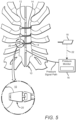
- FIG. 5 One possible embodiment of an apparatus as might be employed when practicing the technique described herein, together with the sternum, is shown in FIG. 5 .
- This arrangement is suitably employed to carry out a "pressure calibration method". Superior results are obtained by a surgical technique to calibrate the relationship between the pressure across the junction (between the sternotomy fragments) and the tension applied to the cerclage cables.
- the polymeric "SuperCable” is used, together with a tensioning tool.
- the tensioning tool is preferably the SuperCable Cerclage Tensioning Instrument, w/60° Angle, which is commercially available from Kinamed, Inc under catalog number 35-800-7000.
- the tensioning instrument allows between 80 and 120 lbs (360 to 530 N) of compressive force to be applied across the sternotomy plane by the cables. Improvements and variations of the tensioning tool may be included without departing from the invention.
- the method for securing a divided sternum 10 during a healing period comprises: aligning the sternum fragments in apposition disposed around a longitudinal seam; inserting at least one thin pressure sensor 12 in the longitudinal seam between the sternum fragments; securing the fragments by wrapping with a polymer cable 14 comprising an elongated elastic polymeric core, coaxially surrounded by a sheath woven from ultra-high molecular-weight polyethylene strands; using a pressure monitor 16 to monitor the pressure sensed by the pressure sensor while applying variable tension on the polymer cable with a tensioning tool 18; detecting a relationship between said variable tension and pressure sensed by the pressure sensor; removing the pressure sensor; tensioning the polymer cable to a desired tension, based upon the detected relationship between variable tension and pressure, to produce a desired pressure in the seam between the sternum fragments; and maintaining said pressure during healing.
- the pressure calibration method could be performed using any sufficiently elastic cable or other resilient element, including springs or the like.
- SuperCable a polymer cable comprising an elongated elastic polymeric core, coaxially surrounded by a sheath woven from ultra-high molecular-weight polyethylene strands
- SuperCable a polymer cable comprising an elongated elastic polymeric core, coaxially surrounded by a sheath woven from ultra-high molecular-weight polyethylene strands
- Pressure sensors are available which are sufficiently thin and accurate to determine the applied pressure.
- 100 lb (450 N), thin-film, capacitive force sensors are commercially available from Pressure Profiles Systems Inc under catalog number CS15-450N. They are sub-branded as SingleTact sensors and additional information can be found here: https://www.singletact.com/micro-force-sensor/calibrated-sensors/15mm-450-newton/ .
- the sensors are provided calibrated along with open-source software for data capture. Other thin sensors could be used.
- multiple sensors are used or a sensor capable of measuring a pressure profile, varying along an axis such as the axis of the sternum cut (seam)-generally from the superior pole (manubrium) to the inferior pole (xiphoid process) of the bone, or along a substantial portion of that axis.
- an axis such as the axis of the sternum cut (seam)-generally from the superior pole (manubrium) to the inferior pole (xiphoid process) of the bone, or along a substantial portion of that axis.
- the pressure calibration method described above is performed together with the method for securing a divided sternum described above.
- the pressure calibration method is advantageous because the relationship between tension (in the cerclage) and pressure (across the bony seam) may depend on a number of variables, including the particular anatomical proportions of the patient, the age, sex and weight of the patient, the bony composition and health of the tissues in the sternum, and the particular number and wrapping method chosen by the surgeon.
- the inventors believe that the pressure across the osteotomy is a more reliable index of optimal fixation as compared to the tension in the cable, which is often the only force metric reported for metal cerclage systems. Based on empirical data, a pressure range (as applied to the cortical bone) of 0.7 to 4.9 Megapascals is believed to be suitable, with the ideal pressure dependent on some or all of the variables listed above.
- the pressure calibration method might also be employed as a means to acquire empirical data, which could then be used to provide tensioning guidance during a sternal closure surgery without the need for pressure sensors.
- empirical testing using pressure sensors could be performed that determines the proper load to be applied for patients of different ages, weights, sexes, bone qualities, etc.
- pre-operative CT-scan sections made perpendicular to the osteotomy plane, can be used to quantify the amount of bone present at the level of the sternum wherein the CT "slice" is taken for each patient. As the amount of bone that exists correlates to the load applied to the section by the patient at the slice location, the desired load to be applied in order to assure reliable stability across the osteotomy plane can be deduced.
- DEXA dual-energy x-ray absorptiometry
- the polymer cables can suitably be tensioned and joined by known methods such as a cable lock as described in US patent number 7,207,090 .
- This cable fixation device is currently preferred because it provides secure fixation of polymeric cable at a maintainable, desired tension and because the components are suitably fabricated from materials which minimally interfere with imaging techniques such as magnetic resonance imaging, X-rays and computer aided tomography.
- the polymeric cable cerclage is applied as multiple cables: at least an upper, lower, and central cerclage.
- Each cable is preferably applied in a loop or "figure-eight" arrangement in either parasternal or transsternal configuration.
- Each section is then tensioned, potentially in a preferred sequence, preferably to a different tension to achieve or approximate a desired pressure profile across the cut sternum.
- the different tensions may be achieved in a first variation by simply tensioning the multiple cables differently with a tensioning tool.
- at least one of the three (or more) polymeric cables may be engineered to supply different tension/extension characteristics.
- At least one of the polymeric cerclage cables fixing the sternum may be engineered to a different strength, stiffness, and/or size than the others by combinations of: varying the core diameter, varying the angles of the weave of the sheath, and/or varying the total size of the cable.
- cerclage cables may be used, but preferably less than six distinct cerclage cables are used. This is a lower number than typical configurations used with conventional metallic wires; the lower number that is permissible using SuperCable is effective to facilitate convenient and time-efficient closure, without enhancing the risk of cable cutting into the bone; the polymeric cable is believed to have less tendency to abrade and cut into resilient bony surfaces as compared to metal wire or cable.
- the invention comprises: a thin pressure transducer 12 capable of insertion between the fragments of a sternum 10 that has been cut into at least two fragments; at least one cerclage cable 14 comprising a polymer cable comprising an elongated elastic polymeric core, coaxially surrounded by a sheath woven from ultra-high molecular weight polyethylene strands, said at least one cable capable of securing said sternum fragments in apposition in proper position to promote bony healing; a pressure monitoring system 16 coupled to said pressure transducer and configured to display pressure sensed by said transducer; a device such as a clasp 20 for securing said cerclage cable in position around the sternum fragments; a tensioning tool 18 capable of applying variable tension to said at least one cerclage cable in response to user manipulation or programming; whereby the user can manipulate said tensioning tool while monitoring the pressure displayed by said pressure monitoring system to determine a relationship between the variable tension and resulting pressure between the sternum fragments, thus determining a desired tension to be
- the apparatus include multiple polymer cerclage cables; at least three (upper, lower, central) are preferable.
- the device for securing the cable may suitably be such as that described in US Patent No. 7,207,090 .
- Other possible securing devices include, for example, a disposable tensioner, or a plastic clasp such that the entire construct is radiolucent.
- the fixation tool may suitably be that described in US Patent No. 9,107,720 or US Patent No. 8,469,967 or available from Kinamed, Inc. as described above, or a variation or improvement of such tool. Further improvements and variants are contemplated within the scope of the invention.
- the tensioning tool may be coupled mechanically or electrically (22) to the pressure monitoring system to help automate the calibration of pressure in relation to tension.
- calibration should be understood to mean the action of establishing a relationship between applied cable tension and resulting pressure between the cut surfaces of the osteotomy.
- a tension sensor electronic transducer responsive to cable tension or tool torque
- the pressure monitoring system may include tension monitoring and/or data processing capabilities (via either analog processing or digital data processing) to facilitate and display the calibration of pressure to cable tension.
- the pressure monitoring system may include mechanized or automated tensioning once the desired pressure is programmed by the user. Such variations, if they fall within the scope of the appended claims, are also within the scope of the invention and may possess independent sources of novelty.
Landscapes
- Health & Medical Sciences (AREA)
- Orthopedic Medicine & Surgery (AREA)
- Surgery (AREA)
- Life Sciences & Earth Sciences (AREA)
- Molecular Biology (AREA)
- Public Health (AREA)
- Engineering & Computer Science (AREA)
- Biomedical Technology (AREA)
- Heart & Thoracic Surgery (AREA)
- Medical Informatics (AREA)
- Veterinary Medicine (AREA)
- Animal Behavior & Ethology (AREA)
- General Health & Medical Sciences (AREA)
- Nuclear Medicine, Radiotherapy & Molecular Imaging (AREA)
- Neurology (AREA)
- Dentistry (AREA)
- Oral & Maxillofacial Surgery (AREA)
- Surgical Instruments (AREA)
- Measuring Pulse, Heart Rate, Blood Pressure Or Blood Flow (AREA)
- Woven Fabrics (AREA)
- Ropes Or Cables (AREA)
Description
- This application claims the benefit of provisional patent application number
62/448,309 to C. Pratt et al., filed January 19, 2017 - Many surgeries involving the heart and other thoracic structures require full access to areas inside the chest. Generally, this access is obtained by splitting the sternum by a full length osteotomy: using a saw to divide the sternum from its superior pole to its inferior pole. This osteotomy of the sternum is commonly referred to as a median sternotomy. In the US, approximately 700,000 sternal osteotomies (i.e. sternotomies) are performed every year.
- Upon completion of open heart surgery (or other surgery requiring access to the chest cavity, such as a lung transplant for example), the sternum must be closed. Usually this involves binding the two divided halves of the sternum together using a cerclage technique. The objective is to bring the two bone segments into proper apposition so that they may heal and again provide the normal function of the sternum in the musculo-skeletal system. The desired outcome is full bony union so that the sternum is restored as nearly as possible to its pre-surgical condition. In order to achieve this goal, the closure must be sufficiently robust so that full bone formation and healing can occur across the sternotomy.
- Most sternotomy closures are performed using stainless steel or other metallic alloy wire, with the aid of instruments such as a wire tightener and wire cutters. Braided metallic cable may be used, either alone or in conjunction with bone fixation devices such as plates, screws, or the like.
- Conventional methods often fail to achieve high-quality bony union. Reliable bone healing requires good bone fragment stability and close approximation of fragments with minimum gap extension. Poor stability and excessive gap extension can lead to non-union, fibro-osseous-union, or partial bony union. Poor stability and bone healing after median sternotomy contribute to a number of potential complications ranging from long-term patient discomfort to acute dehiscence and deep sternal wound infection (mediastinitis), with very high treatment costs, and high patient morbidity and mortality rates.
- A number of factors negatively influence the success of sternal closure. These factors include the failure to accurately quantify and apply a controlled tightening force (load) across the divided bone, failure to maintain bony apposition in the correct position, allowance of too much motion (or too little), and failure to transmit load across the osteotomy during the healing period. Further factors include a tendency of wire or metallic cable to cut through bone and soft tissue due to movement and/or force, loss of fixation due to fatigue of the metallic wire or cable, or breakage of the metallic wire or cable itself due to fatigue. Furthermore, wound healing or the systemic health of the patient may be impacted by metal ions released from metal wires, cables, plates and screws.
- For best healing, three factors must be present: bony apposition, restricted movement, and force transmission across the osteotomy plane. In all cases, adequate blood supply must be present for healing.
- In order to stabilize the reassembled (divided) sternum, forces must be applied to the bone sections so that they are held together during the normal activities that occur during the healing process. This is particularly challenging as the sternum experiences variable and often transient forces transmitted through the ribs or other tissues, during activities such as breathing, coughing, moving, lying down, rising, and other activities of daily living. For example, sneezing has been shown to generate as much as 814 Newtons of lateral distraction force on the sternum.
US 2005/273983 A1 discloses a bone cerclage system comprising an elastic polymer cable and a cable clamp, the polymer cable being configured to achieve an elongation which increases the polymer cable by a percentage between 60 and 140 per cent of its original length, at a working tension up to approximately 800 Newtons. -
US 2014/142638 A1 discloses a sternal closure system comprising surgical cables and a tensioning tool. -
WO 2014/209442 A1 discloses a bone cerclage system comprising an elastic polymer cable and a tensioning tool. - The present invention provides an apparatus for fixing a surgically cut sternum during healing, as defined in
claim 1. The cerclage mechanism as described herein is capable of maintaining proper bony compression (within a desired range) across the sternotomy cut, in spite of a range of external forces and significant lateral distraction due to these external forces. - The elastic member is preferably a polymer cable comprising an elongated elastic polymeric core coaxially surrounded by a sheath woven from ultra-high molecular-weight polyethylene strands.
- These and other features, aspects, and advantages of the present invention will become better understood with reference to the following drawings, description, and claims.
-
-
FIG. 1 is a graph illustrating extension versus time for an anatomical sternotomy model that has been secured by 1) a SuperCable, 2) steel cable, and 3) steel wire. -
FIG. 2 is a photograph illustrating four stainless steel sternal cables installed on a sternotomy model using a parasternal/transternal "figure-eight" wrapping technique. -
FIG. 3 is a graph showing force as a function of time during the application of a cyclic, time varying lateral distraction force on a sternotomy model for SuperCables, metal cables, and metal wire loops. -
FIG. 4 is a graph showing residual force as a function of location along a sternotomy model for two SuperCable configurations, wire cables, and wire loops. -
FIG. 5 illustrates one possible embodiment of an apparatus according to the invention, as might be employed when practicing the technique described herein, together with a divided sternum. - Rather than steel or other metallic wire or cable, the present sternotomy closure technique preferably employs a polymeric engineered cable available from Kinamed, Inc. in Camarillo, CA and sold under the name "SuperCable." The cable is constructed from an elongated elastic polymeric core, suitably Nylon, coaxially surrounded by a sheath woven from ultra-high molecular-weight polyethylene strands. Further information and description of this "SuperCable" is given in
US patent 6,589,246 to Mattchen. This cable is very flexible, elastic, with high fatigue strength and tensile strength. Such cable is capable of repeated cycles of extension/contraction during the healing phase while maintaining the ability to provide compressive load across the osteotomy plane. -
FIG. 1 shows cyclical behavior of extension versus time for an anatomical sternotomy model that has been secured by a SuperCable. The lower curve represents the "SuperCable" assembly (sternotomy model secured with SuperCables) which consisted for this test of only four SuperCables (each doubled as a circumferential loop). For comparison, similar curves are shown for steel monofilament wire (seven loops, top curve), and for braided steel cable (four loops, middle curve). In each case the sternal halves were forcibly laterally distracted with a cyclic force varying periodically from zero to just over 1000 Newtons, for five cycles using a Chatillon LRX materials testing system. It can be seen that all sternum halves in the test demonstrate extension in response to the application of force. However, the steel cable and steel wire models fail to return to their original position and exhibit greater sternal displacement for a given force after several cycles, as compared to the SuperCable construct. This phenomenon is believed to result from a combination of effects, including irreversible metal cable stretch (or metal wire unwinding) and cut-through into the bone model. Both are believed to contribute to the slackening of the steel wire and steel cable models. In contrast, the SuperCable construct returns repeatedly to its original position even after multiple cycles of stretching and relaxation. In this test, the steel wire (No. 5 steel wire from Ethicon) and steel cable (Pioneer Surgical Technology, now Fusion Innovations) did not maintain the sternum model intact, but the SuperCable model did maintain the model intact after cyclic loading. Both the steel cable- and steel wire-secured sternum completely failed after the loading cycles. - It is believed that the steel wire and cable fail to maintain reliable compressive force across the divided sternum because of at least two factors: first, they have high stiffness and fail to accommodate extensive extension and contraction without losing their initial bone contact length. Much of the elasticity in the full model assembly is believed to lie in the bony model rather than the wire or cable. Metal cables and wires may unwind slightly, thereby lengthening irreversibly. Secondly, such cables tend to bite or cut into the bone, tending to further generate potential for slack in the cerclage. This further tends to generate irreversible slack across the osteotomy plane. Both metal wires and metal cables are known to sometimes cut completely through the sternum, resulting not only in failure to fix the median sternotomy but also bisecting the divided sternum into halves which can significantly complicate a subsequent reconstruction.
- Additional testing was performed to support the belief that a more elastic, polymer cable such as "SuperCable" will better maintain pressure between the sternal halves, thereby maintaining stability.
- Anatomical synthetic sternum models (320 kg/m3 (20 lb/ft3 foam, model number: 1025-2, Sawbones®, Vashon, WA, USA) were used for testing [Trumble, D. R., McGregor, W. E. & Magovern, J. A. (2002). Validation of a Bone Analog Model for Studies of Sternal Closure. The Annals of Thoracic Surgery, 74(3): 739-745]. The sternum models were potted, divided along their midline, and fixed as follows:
- 1. Four SuperCables (Kinamed®) in a parasternal wrapping technique at the 1st, 2nd, 4th and 5th intercostal spaces, each tensioned to approximately 80lbs.
- 2. Seven No. 5 stainless steel surgical wires (A&E or Ethicon®), 5 of which employed in a parasternal wrapping technique at the 1st through 5th intercostal spaces, 1 placed transternally in the manubrium and 1 placed transternally in the xiphoid. All were tensioned per the manufacturer's instructions and the excess twisted wire was cut off and the cut end turned down towards the surface of the sternum.
- 3. Four stainless steel sternal cables (Pioneer®) in a parasternal/transternal "figure-eight" wrapping technique as shown in
FIG. 2 and tensioned per the manufacturer's recommended procedure. - Prior to testing, all force sensors (SingleTact, model number: CS15-450N, Pressure Profile Systems, Los Angeles, CA, USA) were calibrated using a standard cyclical run with 10 cycles from 0N to ~400 N on a materials testing system (Chatillon, model number: LRX). These calibration values were used to calibrate the raw data obtained by the force sensors during construct testing.
- A single, active force sensor was placed consistently within the sternotomy plane on all test samples, adjacent to the 3rd intercostal space with enough offset to avoid interference by the metal wire or cable placements. Four other inactive sensors of the same type were spaced equally along the sternotomy plane to ensure even loading.
- Because sneezing can generate 814 Newtons of lateral distraction force on the sternum [Adams, J. et al. (2014). Comparison of Force Exerted on the Sternum During a Sneeze Versus During Low-, Moderate-, and High-Intensity Bench Press Resistance Exercise With and Without the Valsalva Maneuver in Healthy Volunteers. The American Journal of Cardiology, 113(6): 1045-1048], the sternal halves were laterally distracted with a cyclic force ranging from 0 to >800 Newtons for ten cycles using a materials testing system (Chatillon, model number: LRX). The tests were captured on video while measuring and recording forces across the sternal fragments throughout the cyclic loading episode, and after cyclic loading was completed (i.e., with no load being applied).
-
FIG. 3 shows the results: the graph shows force (measured by the pressure sensor) as a function of time during the application of cyclic, time varying lateral distraction force. The results are shown for SuperCable, for the metal cables, and for the metal wire loops.Table 1 summarizes the residual force, percent retention of force, and magnitude of force recovered for all three cable systems. Parameter SuperCable construct Stainless steel wire construct Stainless steel cable construct Residual force between sternal halves after the cyclic loading regime (N) 237.0 18.6 49.4 Percent retention of force [residual force after cyclic loading divided by force before cyclic loading] 71.0% 17.7% 49.0% Magnitude of force recovered from the final loading peak to after the entire loading regime (N) 220.8 18.3 53.7 - Additional testing further demonstrates important differences in how the SuperCable construct performed in comparison to the metal wire and metal constructs. The testing outlined above was repeated with two key modifications. First, an additional fixation model was added that included two SuperCables in a parasternal wrapping technique at the 2nd & 4th intercostal spaces, and two SuperCables in a parasternal/transsternal "figure-eight" wrapping technique at the 1st & 5th intercostal spaces and transternally in the manibrium and xiphoid (similar to superior and inferior cables in
FIG 2 ). Each cable was tensioned to achieve 360 Newton (80 lbs.) of compressive force across the divided sternum. Second, two more active force sensors were placed in the sternum models with one each in the manubrium and xiphoid, adjacent to the superior and inferior edges of the models, respectively. All other setup and calibration details were kept consistent.FIG. 4 shows the results: the graph shows residual force (measured by the pressure sensors) as a function of location along the sternotomy models. The results are shown for both SuperCable configurations, for the metal Pioneer cables, and for the metal wire loops. Both of the SuperCable configurations resulted in higher residual force than the metal cables or wire loops in all three sensor locations which is representative of the entire length of the sternotomy models. - A higher residual force between the sternal halves is a measure of the stability after a cyclic loading regime (such as a sneezing or coughing fit) and can be predictive of continued approximation of the sternal halves for the promotion of bony healing. A higher percent retention of force is a measure of whether the residual force after cyclic loading will continue to persist as additional cycles are applied throughout the healing process. A higher magnitude of force recovered is a measure of how much force was absorbed by the sternal closure construct instead of being transmitted to the sternal halves (leading to cut-through into the bone). This absorption of force will logically minimize potential cut-through of the wires/cables since that also minimizes the stress being applied to the sternal halves.
- A favorable combination of these parameters should increase the stability of fixation and ensure close approximation of the sternal halves throughout the healing process, resulting in improved healing, reduced patient pain, and reduced potential for complication. This testing has demonstrated that the SuperCable construct delivers superior performance in all three parameters, suggesting that the use of SuperCables should be advantageous in obtaining improved sternal bone healing versus metal wires or cables. It is expected that further study would demonstrate similar results for testing that evaluated the effects of longitudinal shear, transverse shear or a combination of all biomechanical forces to which the sternum is subjected.
- In summary, the results of testing demonstrate at least three important differences in how the SuperCable construct performed in comparison to the metal wire and metal cable constructs. These differences are: (1) the residual force between the sternal halves after the cyclic loading, (2) the percent retention of compressive force between sternal halves, and (3) the magnitude of force recovered. The calculated values for these parameters are summarized in Table 1.
- Thus, described here is a method for securing a divided sternum during a healing period, comprising aligning the sternotomy fragments in apposition disposed around a longitudinal seam; securing the fragments by wrapping with a polymer cable comprising an elongated elastic polymeric core, coaxially surrounded by a sheath woven from ultra-high molecular-weight polyethylene strands; and tensioning the strands to a desired tension to compress the sternum to a desired compression range, such that multiple cycles of physiological distracting forces across the sternum can be accepted while maintaining contact pressure across the longitudinal seam within a desired range to minimize potential cut through, for a length of time sufficient for bony healing. The length of time needed can vary widely depending on variables such as surgical technique and patient compliance, but normally bony healing is reported as requiring 6 to 12 weeks.
- The elastic properties of "SuperCable" are believed to be advantageous to absorb shock, equalize load transmitted to the sternum and thus reduce the tendency of cable to cut through the bone.
- One possible embodiment of an apparatus as might be employed when practicing the technique described herein, together with the sternum, is shown in
FIG. 5 . This arrangement is suitably employed to carry out a "pressure calibration method". Superior results are obtained by a surgical technique to calibrate the relationship between the pressure across the junction (between the sternotomy fragments) and the tension applied to the cerclage cables. In this technique the polymeric "SuperCable" is used, together with a tensioning tool. The tensioning tool is preferably the SuperCable Cerclage Tensioning Instrument, w/60° Angle, which is commercially available from Kinamed, Inc under catalog number 35-800-7000. The tensioning instrument allows between 80 and 120 lbs (360 to 530 N) of compressive force to be applied across the sternotomy plane by the cables. Improvements and variations of the tensioning tool may be included without departing from the invention. - According to the described pressure calibration method, the method for securing a divided
sternum 10 during a healing period, comprises: aligning the sternum fragments in apposition disposed around a longitudinal seam; inserting at least onethin pressure sensor 12 in the longitudinal seam between the sternum fragments; securing the fragments by wrapping with apolymer cable 14 comprising an elongated elastic polymeric core, coaxially surrounded by a sheath woven from ultra-high molecular-weight polyethylene strands; using apressure monitor 16 to monitor the pressure sensed by the pressure sensor while applying variable tension on the polymer cable with atensioning tool 18; detecting a relationship between said variable tension and pressure sensed by the pressure sensor; removing the pressure sensor; tensioning the polymer cable to a desired tension, based upon the detected relationship between variable tension and pressure, to produce a desired pressure in the seam between the sternum fragments; and maintaining said pressure during healing. - According to a more generalized method, the pressure calibration method could be performed using any sufficiently elastic cable or other resilient element, including springs or the like. However, the use of the "SuperCable" (a polymer cable comprising an elongated elastic polymeric core, coaxially surrounded by a sheath woven from ultra-high molecular-weight polyethylene strands) is believed to be very advantageous and to provide an independent source of novelty and advantage.
- Pressure sensors are available which are sufficiently thin and accurate to determine the applied pressure. For example, 100 lb (450 N), thin-film, capacitive force sensors are commercially available from Pressure Profiles Systems Inc under catalog number CS15-450N. They are sub-branded as SingleTact sensors and additional information can be found here: https://www.singletact.com/micro-force-sensor/calibrated-sensors/15mm-450-newton/. The sensors are provided calibrated along with open-source software for data capture. Other thin sensors could be used. In a preferred embodiment, multiple sensors are used or a sensor capable of measuring a pressure profile, varying along an axis such as the axis of the sternum cut (seam)-generally from the superior pole (manubrium) to the inferior pole (xiphoid process) of the bone, or along a substantial portion of that axis.
- Preferably, the pressure calibration method described above is performed together with the method for securing a divided sternum described above.
- The pressure calibration method is advantageous because the relationship between tension (in the cerclage) and pressure (across the bony seam) may depend on a number of variables, including the particular anatomical proportions of the patient, the age, sex and weight of the patient, the bony composition and health of the tissues in the sternum, and the particular number and wrapping method chosen by the surgeon. The inventors believe that the pressure across the osteotomy is a more reliable index of optimal fixation as compared to the tension in the cable, which is often the only force metric reported for metal cerclage systems. Based on empirical data, a pressure range (as applied to the cortical bone) of 0.7 to 4.9 Megapascals is believed to be suitable, with the ideal pressure dependent on some or all of the variables listed above.
- The pressure calibration method might also be employed as a means to acquire empirical data, which could then be used to provide tensioning guidance during a sternal closure surgery without the need for pressure sensors. For example, empirical testing using pressure sensors could be performed that determines the proper load to be applied for patients of different ages, weights, sexes, bone qualities, etc. In the alternative, pre-operative CT-scan sections, made perpendicular to the osteotomy plane, can be used to quantify the amount of bone present at the level of the sternum wherein the CT "slice" is taken for each patient. As the amount of bone that exists correlates to the load applied to the section by the patient at the slice location, the desired load to be applied in order to assure reliable stability across the osteotomy plane can be deduced. Alternatively, DEXA (dual-energy x-ray absorptiometry) scans could be used as they are routinely utilized to assess bone density and thus bone strength. This data would then be used to determine the proper tension or compressive force to apply (without the need for corresponding pressure sensor information) when performing sternal closure as described herein.
- The polymer cables can suitably be tensioned and joined by known methods such as a cable lock as described in
US patent number 7,207,090 . This cable fixation device is currently preferred because it provides secure fixation of polymeric cable at a maintainable, desired tension and because the components are suitably fabricated from materials which minimally interfere with imaging techniques such as magnetic resonance imaging, X-rays and computer aided tomography. - According to yet another arrangement, the polymeric cable cerclage is applied as multiple cables: at least an upper, lower, and central cerclage. Each cable is preferably applied in a loop or "figure-eight" arrangement in either parasternal or transsternal configuration. Each section is then tensioned, potentially in a preferred sequence, preferably to a different tension to achieve or approximate a desired pressure profile across the cut sternum. The different tensions may be achieved in a first variation by simply tensioning the multiple cables differently with a tensioning tool. In another variation, at least one of the three (or more) polymeric cables may be engineered to supply different tension/extension characteristics. For example, at least one of the polymeric cerclage cables fixing the sternum may be engineered to a different strength, stiffness, and/or size than the others by combinations of: varying the core diameter, varying the angles of the weave of the sheath, and/or varying the total size of the cable.
- Various numbers of cerclage cables may be used, but preferably less than six distinct cerclage cables are used. This is a lower number than typical configurations used with conventional metallic wires; the lower number that is permissible using SuperCable is effective to facilitate convenient and time-efficient closure, without enhancing the risk of cable cutting into the bone; the polymeric cable is believed to have less tendency to abrade and cut into resilient bony surfaces as compared to metal wire or cable.
- In an embodiment, the invention comprises: a
thin pressure transducer 12 capable of insertion between the fragments of asternum 10 that has been cut into at least two fragments; at least onecerclage cable 14 comprising a polymer cable comprising an elongated elastic polymeric core, coaxially surrounded by a sheath woven from ultra-high molecular weight polyethylene strands, said at least one cable capable of securing said sternum fragments in apposition in proper position to promote bony healing; apressure monitoring system 16 coupled to said pressure transducer and configured to display pressure sensed by said transducer; a device such as aclasp 20 for securing said cerclage cable in position around the sternum fragments; atensioning tool 18 capable of applying variable tension to said at least one cerclage cable in response to user manipulation or programming; whereby the user can manipulate said tensioning tool while monitoring the pressure displayed by said pressure monitoring system to determine a relationship between the variable tension and resulting pressure between the sternum fragments, thus determining a desired tension to be applied to said at least one cerclage cable for fixation to obtain a resulting desired pressure. - As with the described methods, it is preferred that the apparatus include multiple polymer cerclage cables; at least three (upper, lower, central) are preferable. The device for securing the cable may suitably be such as that described in
US Patent No. 7,207,090 . Other possible securing devices include, for example, a disposable tensioner, or a plastic clasp such that the entire construct is radiolucent. The fixation tool may suitably be that described inUS Patent No. 9,107,720 US Patent No. 8,469,967 or available from Kinamed, Inc. as described above, or a variation or improvement of such tool. Further improvements and variants are contemplated within the scope of the invention. For example, in some embodiments the tensioning tool may be coupled mechanically or electrically (22) to the pressure monitoring system to help automate the calibration of pressure in relation to tension. In this context, "calibration" should be understood to mean the action of establishing a relationship between applied cable tension and resulting pressure between the cut surfaces of the osteotomy. For this purpose, a tension sensor (electronic transducer responsive to cable tension or tool torque) can be provided and coupled to the pressure monitoring system or other data processing system. The pressure monitoring system may include tension monitoring and/or data processing capabilities (via either analog processing or digital data processing) to facilitate and display the calibration of pressure to cable tension. The pressure monitoring system may include mechanized or automated tensioning once the desired pressure is programmed by the user. Such variations, if they fall within the scope of the appended claims, are also within the scope of the invention and may possess independent sources of novelty. - The embodiments of the invention described herein are exemplary and numerous modifications, variations and rearrangements can be readily envisioned to achieve substantially equivalent results, all of which are intended to be embraced within the scope of the invention as defined in the appended claims.
Claims (6)
- An apparatus for fixing a surgically cut sternum (10) during healing, the sternum having been cut into at least two fragments, the apparatus comprising:an elastic cable (14) configured to secure said sternum fragments in apposition in proper position to promote bony healing; anda tensioning tool configured to apply variable tension to said elastic cable, said elastic cable (14) being configured to accept repeated cycles of extension and contraction while maintaining the ability to provide a compressive load on the sternum fragments, wherein the apparatus further comprises:a pressure transducer (12) configured to be inserted between the sternum fragments; anda pressure monitoring system (16) coupled to the pressure transducer and configured to measure pressure sensed by said transducer,wherein the tensioning tool (18) is configured to apply tension to said elastic cable to apply pressure between the sternum fragments (10), said tensioning tool being configured, in response to user manipulation or a programmed digital computer, to adjust the tension on the elastic cable to set the pressure between the sternum fragments to a desired compression range.
- The apparatus of claim 1, wherein the pressure transducer (12) comprises at least two pressure transducers configured to be disposed at different points between the fragments of a sternum,the at least two pressure transducers each independently coupled to the pressure monitoring system (16),and wherein said pressure monitoring system is configured to independently monitor each of said at least two pressure transducers.
- The apparatus of claim 1, further comprising a digital computer programmed to control tensioning automatically in response to a determined relationship between the variable tension and resulting pressure between the sternum fragments.
- The apparatus of claim 3, wherein said digital computer is configured to control said tensioning automatically to vary the tension applied to fix the elastic cable (14).
- The apparatus of claim 4, wherein said digital computer is configured to control said tensioning automatically to obtain a desired pressure in the range of 0.7 to 4.9 Megapascals.
- The apparatus of claim 1, wherein said elastic cable (14) comprises at least one polymer cable comprising an elongated elastic polymeric core, coaxially surrounded by a sheath woven from ultra-high molecular-weight polyethylene strands, said at least one polymer cable being configured to secure said sternum fragments in apposition in proper position to promote bony healing.
Applications Claiming Priority (2)
| Application Number | Priority Date | Filing Date | Title |
|---|---|---|---|
| US201762448309P | 2017-01-19 | 2017-01-19 | |
| PCT/US2018/014498 WO2018136782A1 (en) | 2017-01-19 | 2018-01-19 | Sternotomy closure technique using polymeric cable |
Publications (2)
| Publication Number | Publication Date |
|---|---|
| EP3570767A1 EP3570767A1 (en) | 2019-11-27 |
| EP3570767B1 true EP3570767B1 (en) | 2025-05-14 |
Family
ID=62838325
Family Applications (1)
| Application Number | Title | Priority Date | Filing Date |
|---|---|---|---|
| EP18709190.5A Active EP3570767B1 (en) | 2017-01-19 | 2018-01-19 | Sternotomy closure apparatus using polymeric cable |
Country Status (9)
| Country | Link |
|---|---|
| US (2) | US10993752B2 (en) |
| EP (1) | EP3570767B1 (en) |
| JP (3) | JP2020505129A (en) |
| AU (1) | AU2018209968B2 (en) |
| BR (1) | BR112019014745B1 (en) |
| CO (1) | CO2019008921A2 (en) |
| ES (1) | ES3033531T3 (en) |
| SA (1) | SA519402252B1 (en) |
| WO (1) | WO2018136782A1 (en) |
Families Citing this family (4)
| Publication number | Priority date | Publication date | Assignee | Title |
|---|---|---|---|---|
| AU2018209968B2 (en) * | 2017-01-19 | 2020-10-22 | Kinamed, Inc. | Sternotomy closure technique using polymeric cable |
| US12294920B2 (en) * | 2019-11-06 | 2025-05-06 | Universidad Santiago De Cali | Method for determining a level of contamination in covered zones and system for collecting and processing environmental information |
| JP2023003286A (en) * | 2021-06-23 | 2023-01-11 | 株式会社大一商会 | game machine |
| JP2023003284A (en) * | 2021-06-23 | 2023-01-11 | 株式会社大一商会 | game machine |
Citations (6)
| Publication number | Priority date | Publication date | Assignee | Title |
|---|---|---|---|---|
| US5797915A (en) * | 1996-04-17 | 1998-08-25 | Pierson, Iii; Raymond H. | Cerclage system |
| US6589246B1 (en) * | 2001-04-26 | 2003-07-08 | Poly-4 Medical, Inc. | Method of applying an active compressive force continuously across a fracture |
| US20050273983A1 (en) * | 2004-06-09 | 2005-12-15 | Kinamed, Inc. | High tension, surgical cable lock |
| WO2006060911A1 (en) * | 2004-12-06 | 2006-06-15 | Socovar Societe En Commandite | Binding component |
| US20140142638A1 (en) * | 2012-11-21 | 2014-05-22 | Robert Goodwin | Bone plate system and method |
| WO2014209442A1 (en) * | 2013-06-24 | 2014-12-31 | Kinamed, Inc. | Surgical cable tensioning system |
Family Cites Families (18)
| Publication number | Priority date | Publication date | Assignee | Title |
|---|---|---|---|---|
| IL120636A0 (en) * | 1997-04-10 | 1997-08-14 | Technion Res & Dev Foundation | Kit for sternum fixation in chest surgery |
| US5852926A (en) * | 1997-08-25 | 1998-12-29 | Wellington Leisure Products, Inc. | Balanced strand cordage |
| JP4874970B2 (en) * | 2004-06-07 | 2012-02-15 | ジンテス ゲゼルシャフト ミット ベシュレンクテル ハフツング | Orthopedic implant with sensor |
| US9463012B2 (en) * | 2004-10-26 | 2016-10-11 | P Tech, Llc | Apparatus for guiding and positioning an implant |
| US8597327B2 (en) * | 2006-02-03 | 2013-12-03 | Biomet Manufacturing, Llc | Method and apparatus for sternal closure |
| US8016859B2 (en) * | 2006-02-17 | 2011-09-13 | Medtronic, Inc. | Dynamic treatment system and method of use |
| US20080262549A1 (en) * | 2006-10-19 | 2008-10-23 | Simpirica Spine, Inc. | Methods and systems for deploying spinous process constraints |
| JP2008167869A (en) | 2007-01-10 | 2008-07-24 | Alfresa Pharma Corp | Bone fixing tape |
| US20120109129A1 (en) * | 2010-11-02 | 2012-05-03 | Bernstein Oren S | Replacement system for a surgical wire |
| US8486114B2 (en) * | 2008-10-10 | 2013-07-16 | Acute Innovations Llc | Cerclage system for bone |
| US9107720B2 (en) | 2009-06-02 | 2015-08-18 | Kinamed, Inc. | Surgical cable tensioning system |
| ES2471015T3 (en) | 2009-06-02 | 2014-06-25 | Kinamed, Inc. | Surgical Cable Tensioning System |
| US9198683B2 (en) * | 2009-09-30 | 2015-12-01 | Aegis Medical Innovations, Inc. | Tissue capture and occlusion systems and methods |
| CA2799772A1 (en) * | 2010-05-19 | 2011-11-24 | Physcient, Inc. | Methods and devices to decrease tissue trauma during surgery |
| PL2846715T3 (en) * | 2012-04-25 | 2018-07-31 | Ajay K. PATIL | Sternal closure and ribs approximator devices |
| JP6309975B2 (en) * | 2013-03-15 | 2018-04-11 | ホウフ,ルイス | Sternum fixation device and method |
| US20150297274A1 (en) * | 2014-02-19 | 2015-10-22 | Arthrex, Inc. | Locking button for tape cerclage |
| AU2018209968B2 (en) * | 2017-01-19 | 2020-10-22 | Kinamed, Inc. | Sternotomy closure technique using polymeric cable |
-
2018
- 2018-01-19 AU AU2018209968A patent/AU2018209968B2/en active Active
- 2018-01-19 WO PCT/US2018/014498 patent/WO2018136782A1/en not_active Ceased
- 2018-01-19 EP EP18709190.5A patent/EP3570767B1/en active Active
- 2018-01-19 JP JP2019539278A patent/JP2020505129A/en active Pending
- 2018-01-19 BR BR112019014745-4A patent/BR112019014745B1/en active IP Right Grant
- 2018-01-19 US US15/875,221 patent/US10993752B2/en active Active
- 2018-01-19 ES ES18709190T patent/ES3033531T3/en active Active
-
2019
- 2019-07-18 SA SA519402252A patent/SA519402252B1/en unknown
- 2019-08-16 CO CONC2019/0008921A patent/CO2019008921A2/en unknown
-
2021
- 2021-04-01 JP JP2021062815A patent/JP7386202B2/en active Active
- 2021-04-23 US US17/238,624 patent/US12408961B2/en active Active
-
2023
- 2023-11-13 JP JP2023192941A patent/JP7744399B2/en active Active
Patent Citations (6)
| Publication number | Priority date | Publication date | Assignee | Title |
|---|---|---|---|---|
| US5797915A (en) * | 1996-04-17 | 1998-08-25 | Pierson, Iii; Raymond H. | Cerclage system |
| US6589246B1 (en) * | 2001-04-26 | 2003-07-08 | Poly-4 Medical, Inc. | Method of applying an active compressive force continuously across a fracture |
| US20050273983A1 (en) * | 2004-06-09 | 2005-12-15 | Kinamed, Inc. | High tension, surgical cable lock |
| WO2006060911A1 (en) * | 2004-12-06 | 2006-06-15 | Socovar Societe En Commandite | Binding component |
| US20140142638A1 (en) * | 2012-11-21 | 2014-05-22 | Robert Goodwin | Bone plate system and method |
| WO2014209442A1 (en) * | 2013-06-24 | 2014-12-31 | Kinamed, Inc. | Surgical cable tensioning system |
Also Published As
| Publication number | Publication date |
|---|---|
| SA519402252B1 (en) | 2023-02-28 |
| US20180199968A1 (en) | 2018-07-19 |
| US12408961B2 (en) | 2025-09-09 |
| JP2024010244A (en) | 2024-01-23 |
| CO2019008921A2 (en) | 2020-01-17 |
| US20210236184A1 (en) | 2021-08-05 |
| EP3570767A1 (en) | 2019-11-27 |
| AU2018209968A1 (en) | 2019-08-08 |
| ES3033531T3 (en) | 2025-08-05 |
| WO2018136782A1 (en) | 2018-07-26 |
| JP7744399B2 (en) | 2025-09-25 |
| BR112019014745A2 (en) | 2020-03-03 |
| JP2021104370A (en) | 2021-07-26 |
| BR112019014745B1 (en) | 2024-02-06 |
| JP7386202B2 (en) | 2023-11-24 |
| JP2020505129A (en) | 2020-02-20 |
| AU2018209968B2 (en) | 2020-10-22 |
| US10993752B2 (en) | 2021-05-04 |
Similar Documents
| Publication | Publication Date | Title |
|---|---|---|
| US12408961B2 (en) | Sternotomy closure technique using polymeric cable | |
| US10213238B2 (en) | Method and device for correcting bone deformities | |
| US9700363B2 (en) | Surgical instrument and method for tensioning and securing a flexible suture | |
| EP3527154B1 (en) | Systems for attenuation of increased spinal flexion loads post-fusion | |
| US11382667B2 (en) | Adjusting spinal curvature | |
| US8974456B2 (en) | Hybrid medical device implant with migration management | |
| US20080262549A1 (en) | Methods and systems for deploying spinous process constraints | |
| CN105828732A (en) | Bone plate for reducing angular deformation and method of use thereof | |
| JP2003529395A (en) | Medical equipment | |
| AU2007201257A1 (en) | Fastening system for internal fixation | |
| US20180153601A1 (en) | Surgical procedure | |
| US20220233221A1 (en) | Balanced axial compression assembly for bone | |
| US7833225B2 (en) | Method and device for bone stabilization using a threaded compression wire | |
| US20150374410A1 (en) | Vector compression system | |
| Arlet et al. | Square-lashing technique in segmental spinal instrumentation: a biomechanical study | |
| CN118845180A (en) | A kind of elastic modulus buffer telescopic stabilizing device |
Legal Events
| Date | Code | Title | Description |
|---|---|---|---|
| STAA | Information on the status of an ep patent application or granted ep patent |
Free format text: STATUS: UNKNOWN |
|
| STAA | Information on the status of an ep patent application or granted ep patent |
Free format text: STATUS: THE INTERNATIONAL PUBLICATION HAS BEEN MADE |
|
| PUAI | Public reference made under article 153(3) epc to a published international application that has entered the european phase |
Free format text: ORIGINAL CODE: 0009012 |
|
| STAA | Information on the status of an ep patent application or granted ep patent |
Free format text: STATUS: REQUEST FOR EXAMINATION WAS MADE |
|
| 17P | Request for examination filed |
Effective date: 20190816 |
|
| AK | Designated contracting states |
Kind code of ref document: A1 Designated state(s): AL AT BE BG CH CY CZ DE DK EE ES FI FR GB GR HR HU IE IS IT LI LT LU LV MC MK MT NL NO PL PT RO RS SE SI SK SM TR |
|
| AX | Request for extension of the european patent |
Extension state: BA ME |
|
| DAV | Request for validation of the european patent (deleted) | ||
| DAX | Request for extension of the european patent (deleted) | ||
| PUAG | Search results despatched under rule 164(2) epc together with communication from examining division |
Free format text: ORIGINAL CODE: 0009017 |
|
| STAA | Information on the status of an ep patent application or granted ep patent |
Free format text: STATUS: EXAMINATION IS IN PROGRESS |
|
| 17Q | First examination report despatched |
Effective date: 20220929 |
|
| B565 | Issuance of search results under rule 164(2) epc |
Effective date: 20220929 |
|
| RIC1 | Information provided on ipc code assigned before grant |
Ipc: A61B 17/88 20060101ALI20220926BHEP Ipc: A61B 17/84 20060101ALI20220926BHEP Ipc: A61B 17/82 20060101AFI20220926BHEP |
|
| GRAP | Despatch of communication of intention to grant a patent |
Free format text: ORIGINAL CODE: EPIDOSNIGR1 |
|
| STAA | Information on the status of an ep patent application or granted ep patent |
Free format text: STATUS: GRANT OF PATENT IS INTENDED |
|
| INTG | Intention to grant announced |
Effective date: 20241220 |
|
| RIN1 | Information on inventor provided before grant (corrected) |
Inventor name: SARIN, VINEET KUMAR Inventor name: BRUCE, ROBERT ARTHUR Inventor name: NELSON, ADAM LEE Inventor name: PRATT, CLYDE RONALD |
|
| GRAS | Grant fee paid |
Free format text: ORIGINAL CODE: EPIDOSNIGR3 |
|
| GRAA | (expected) grant |
Free format text: ORIGINAL CODE: 0009210 |
|
| STAA | Information on the status of an ep patent application or granted ep patent |
Free format text: STATUS: THE PATENT HAS BEEN GRANTED |
|
| AK | Designated contracting states |
Kind code of ref document: B1 Designated state(s): AL AT BE BG CH CY CZ DE DK EE ES FI FR GB GR HR HU IE IS IT LI LT LU LV MC MK MT NL NO PL PT RO RS SE SI SK SM TR |
|
| REG | Reference to a national code |
Ref country code: GB Ref legal event code: FG4D |
|
| REG | Reference to a national code |
Ref country code: CH Ref legal event code: EP |
|
| REG | Reference to a national code |
Ref country code: DE Ref legal event code: R096 Ref document number: 602018081896 Country of ref document: DE |
|
| REG | Reference to a national code |
Ref country code: IE Ref legal event code: FG4D |
|
| REG | Reference to a national code |
Ref country code: ES Ref legal event code: FG2A Ref document number: 3033531 Country of ref document: ES Kind code of ref document: T3 Effective date: 20250805 |
|
| REG | Reference to a national code |
Ref country code: NL Ref legal event code: MP Effective date: 20250514 |
|
| PG25 | Lapsed in a contracting state [announced via postgrant information from national office to epo] |
Ref country code: PT Free format text: LAPSE BECAUSE OF FAILURE TO SUBMIT A TRANSLATION OF THE DESCRIPTION OR TO PAY THE FEE WITHIN THE PRESCRIBED TIME-LIMIT Effective date: 20250915 Ref country code: FI Free format text: LAPSE BECAUSE OF FAILURE TO SUBMIT A TRANSLATION OF THE DESCRIPTION OR TO PAY THE FEE WITHIN THE PRESCRIBED TIME-LIMIT Effective date: 20250514 |
|
| REG | Reference to a national code |
Ref country code: LT Ref legal event code: MG9D |
|
| PG25 | Lapsed in a contracting state [announced via postgrant information from national office to epo] |
Ref country code: GR Free format text: LAPSE BECAUSE OF FAILURE TO SUBMIT A TRANSLATION OF THE DESCRIPTION OR TO PAY THE FEE WITHIN THE PRESCRIBED TIME-LIMIT Effective date: 20250815 Ref country code: NO Free format text: LAPSE BECAUSE OF FAILURE TO SUBMIT A TRANSLATION OF THE DESCRIPTION OR TO PAY THE FEE WITHIN THE PRESCRIBED TIME-LIMIT Effective date: 20250814 |
|
| PG25 | Lapsed in a contracting state [announced via postgrant information from national office to epo] |
Ref country code: PL Free format text: LAPSE BECAUSE OF FAILURE TO SUBMIT A TRANSLATION OF THE DESCRIPTION OR TO PAY THE FEE WITHIN THE PRESCRIBED TIME-LIMIT Effective date: 20250514 Ref country code: NL Free format text: LAPSE BECAUSE OF FAILURE TO SUBMIT A TRANSLATION OF THE DESCRIPTION OR TO PAY THE FEE WITHIN THE PRESCRIBED TIME-LIMIT Effective date: 20250514 |
|
| REG | Reference to a national code |
Ref country code: AT Ref legal event code: MK05 Ref document number: 1794161 Country of ref document: AT Kind code of ref document: T Effective date: 20250514 |
|
| PG25 | Lapsed in a contracting state [announced via postgrant information from national office to epo] |
Ref country code: BG Free format text: LAPSE BECAUSE OF FAILURE TO SUBMIT A TRANSLATION OF THE DESCRIPTION OR TO PAY THE FEE WITHIN THE PRESCRIBED TIME-LIMIT Effective date: 20250514 |
|
| PG25 | Lapsed in a contracting state [announced via postgrant information from national office to epo] |
Ref country code: HR Free format text: LAPSE BECAUSE OF FAILURE TO SUBMIT A TRANSLATION OF THE DESCRIPTION OR TO PAY THE FEE WITHIN THE PRESCRIBED TIME-LIMIT Effective date: 20250514 |
|
| PG25 | Lapsed in a contracting state [announced via postgrant information from national office to epo] |
Ref country code: AT Free format text: LAPSE BECAUSE OF FAILURE TO SUBMIT A TRANSLATION OF THE DESCRIPTION OR TO PAY THE FEE WITHIN THE PRESCRIBED TIME-LIMIT Effective date: 20250514 |
|
| PG25 | Lapsed in a contracting state [announced via postgrant information from national office to epo] |
Ref country code: RS Free format text: LAPSE BECAUSE OF FAILURE TO SUBMIT A TRANSLATION OF THE DESCRIPTION OR TO PAY THE FEE WITHIN THE PRESCRIBED TIME-LIMIT Effective date: 20250814 |
|
| PG25 | Lapsed in a contracting state [announced via postgrant information from national office to epo] |
Ref country code: IS Free format text: LAPSE BECAUSE OF FAILURE TO SUBMIT A TRANSLATION OF THE DESCRIPTION OR TO PAY THE FEE WITHIN THE PRESCRIBED TIME-LIMIT Effective date: 20250914 |
|
| PG25 | Lapsed in a contracting state [announced via postgrant information from national office to epo] |
Ref country code: LV Free format text: LAPSE BECAUSE OF FAILURE TO SUBMIT A TRANSLATION OF THE DESCRIPTION OR TO PAY THE FEE WITHIN THE PRESCRIBED TIME-LIMIT Effective date: 20250514 |
|
| PGFP | Annual fee paid to national office [announced via postgrant information from national office to epo] |
Ref country code: GB Payment date: 20251211 Year of fee payment: 9 |
|
| PG25 | Lapsed in a contracting state [announced via postgrant information from national office to epo] |
Ref country code: DK Free format text: LAPSE BECAUSE OF FAILURE TO SUBMIT A TRANSLATION OF THE DESCRIPTION OR TO PAY THE FEE WITHIN THE PRESCRIBED TIME-LIMIT Effective date: 20250514 Ref country code: SM Free format text: LAPSE BECAUSE OF FAILURE TO SUBMIT A TRANSLATION OF THE DESCRIPTION OR TO PAY THE FEE WITHIN THE PRESCRIBED TIME-LIMIT Effective date: 20250514 |
|
| PGFP | Annual fee paid to national office [announced via postgrant information from national office to epo] |
Ref country code: FR Payment date: 20251216 Year of fee payment: 9 |
|
| PG25 | Lapsed in a contracting state [announced via postgrant information from national office to epo] |
Ref country code: CZ Free format text: LAPSE BECAUSE OF FAILURE TO SUBMIT A TRANSLATION OF THE DESCRIPTION OR TO PAY THE FEE WITHIN THE PRESCRIBED TIME-LIMIT Effective date: 20250514 |
|
| PG25 | Lapsed in a contracting state [announced via postgrant information from national office to epo] |
Ref country code: EE Free format text: LAPSE BECAUSE OF FAILURE TO SUBMIT A TRANSLATION OF THE DESCRIPTION OR TO PAY THE FEE WITHIN THE PRESCRIBED TIME-LIMIT Effective date: 20250514 |
|
| PG25 | Lapsed in a contracting state [announced via postgrant information from national office to epo] |
Ref country code: SK Free format text: LAPSE BECAUSE OF FAILURE TO SUBMIT A TRANSLATION OF THE DESCRIPTION OR TO PAY THE FEE WITHIN THE PRESCRIBED TIME-LIMIT Effective date: 20250514 Ref country code: RO Free format text: LAPSE BECAUSE OF FAILURE TO SUBMIT A TRANSLATION OF THE DESCRIPTION OR TO PAY THE FEE WITHIN THE PRESCRIBED TIME-LIMIT Effective date: 20250514 |
|
| REG | Reference to a national code |
Ref country code: DE Ref legal event code: R097 Ref document number: 602018081896 Country of ref document: DE |
|
| PLBE | No opposition filed within time limit |
Free format text: ORIGINAL CODE: 0009261 |
|
| STAA | Information on the status of an ep patent application or granted ep patent |
Free format text: STATUS: NO OPPOSITION FILED WITHIN TIME LIMIT |
|
| REG | Reference to a national code |
Ref country code: CH Ref legal event code: L10 Free format text: ST27 STATUS EVENT CODE: U-0-0-L10-L00 (AS PROVIDED BY THE NATIONAL OFFICE) Effective date: 20260325 |
|
| PGFP | Annual fee paid to national office [announced via postgrant information from national office to epo] |
Ref country code: ES Payment date: 20260205 Year of fee payment: 9 |
|
| PGFP | Annual fee paid to national office [announced via postgrant information from national office to epo] |
Ref country code: IT Payment date: 20260112 Year of fee payment: 9 |
|
| 26N | No opposition filed |
Effective date: 20260217 |