EP3567118B1 - Method for diagnosing cardiac disease through bacterial metagenome analysis - Google Patents
Method for diagnosing cardiac disease through bacterial metagenome analysis Download PDFInfo
- Publication number
- EP3567118B1 EP3567118B1 EP17880997.6A EP17880997A EP3567118B1 EP 3567118 B1 EP3567118 B1 EP 3567118B1 EP 17880997 A EP17880997 A EP 17880997A EP 3567118 B1 EP3567118 B1 EP 3567118B1
- Authority
- EP
- European Patent Office
- Prior art keywords
- genus
- family
- derived
- order
- bacteria
- Prior art date
- Legal status (The legal status is an assumption and is not a legal conclusion. Google has not performed a legal analysis and makes no representation as to the accuracy of the status listed.)
- Active
Links
Images









Classifications
-
- A—HUMAN NECESSITIES
- A61—MEDICAL OR VETERINARY SCIENCE; HYGIENE
- A61P—SPECIFIC THERAPEUTIC ACTIVITY OF CHEMICAL COMPOUNDS OR MEDICINAL PREPARATIONS
- A61P9/00—Drugs for disorders of the cardiovascular system
-
- C—CHEMISTRY; METALLURGY
- C12—BIOCHEMISTRY; BEER; SPIRITS; WINE; VINEGAR; MICROBIOLOGY; ENZYMOLOGY; MUTATION OR GENETIC ENGINEERING
- C12Q—MEASURING OR TESTING PROCESSES INVOLVING ENZYMES, NUCLEIC ACIDS OR MICROORGANISMS; COMPOSITIONS OR TEST PAPERS THEREFOR; PROCESSES OF PREPARING SUCH COMPOSITIONS; CONDITION-RESPONSIVE CONTROL IN MICROBIOLOGICAL OR ENZYMOLOGICAL PROCESSES
- C12Q1/00—Measuring or testing processes involving enzymes, nucleic acids or microorganisms; Compositions therefor; Processes of preparing such compositions
- C12Q1/68—Measuring or testing processes involving enzymes, nucleic acids or microorganisms; Compositions therefor; Processes of preparing such compositions involving nucleic acids
- C12Q1/6876—Nucleic acid products used in the analysis of nucleic acids, e.g. primers or probes
- C12Q1/6888—Nucleic acid products used in the analysis of nucleic acids, e.g. primers or probes for detection or identification of organisms
- C12Q1/6895—Nucleic acid products used in the analysis of nucleic acids, e.g. primers or probes for detection or identification of organisms for plants, fungi or algae
-
- A—HUMAN NECESSITIES
- A61—MEDICAL OR VETERINARY SCIENCE; HYGIENE
- A61P—SPECIFIC THERAPEUTIC ACTIVITY OF CHEMICAL COMPOUNDS OR MEDICINAL PREPARATIONS
- A61P9/00—Drugs for disorders of the cardiovascular system
- A61P9/04—Inotropic agents, i.e. stimulants of cardiac contraction; Drugs for heart failure
-
- A—HUMAN NECESSITIES
- A61—MEDICAL OR VETERINARY SCIENCE; HYGIENE
- A61P—SPECIFIC THERAPEUTIC ACTIVITY OF CHEMICAL COMPOUNDS OR MEDICINAL PREPARATIONS
- A61P9/00—Drugs for disorders of the cardiovascular system
- A61P9/06—Antiarrhythmics
-
- A—HUMAN NECESSITIES
- A61—MEDICAL OR VETERINARY SCIENCE; HYGIENE
- A61P—SPECIFIC THERAPEUTIC ACTIVITY OF CHEMICAL COMPOUNDS OR MEDICINAL PREPARATIONS
- A61P9/00—Drugs for disorders of the cardiovascular system
- A61P9/10—Drugs for disorders of the cardiovascular system for treating ischaemic or atherosclerotic diseases, e.g. antianginal drugs, coronary vasodilators, drugs for myocardial infarction, retinopathy, cerebrovascula insufficiency, renal arteriosclerosis
-
- C—CHEMISTRY; METALLURGY
- C12—BIOCHEMISTRY; BEER; SPIRITS; WINE; VINEGAR; MICROBIOLOGY; ENZYMOLOGY; MUTATION OR GENETIC ENGINEERING
- C12Q—MEASURING OR TESTING PROCESSES INVOLVING ENZYMES, NUCLEIC ACIDS OR MICROORGANISMS; COMPOSITIONS OR TEST PAPERS THEREFOR; PROCESSES OF PREPARING SUCH COMPOSITIONS; CONDITION-RESPONSIVE CONTROL IN MICROBIOLOGICAL OR ENZYMOLOGICAL PROCESSES
- C12Q1/00—Measuring or testing processes involving enzymes, nucleic acids or microorganisms; Compositions therefor; Processes of preparing such compositions
- C12Q1/68—Measuring or testing processes involving enzymes, nucleic acids or microorganisms; Compositions therefor; Processes of preparing such compositions involving nucleic acids
- C12Q1/6876—Nucleic acid products used in the analysis of nucleic acids, e.g. primers or probes
- C12Q1/6883—Nucleic acid products used in the analysis of nucleic acids, e.g. primers or probes for diseases caused by alterations of genetic material
-
- C—CHEMISTRY; METALLURGY
- C12—BIOCHEMISTRY; BEER; SPIRITS; WINE; VINEGAR; MICROBIOLOGY; ENZYMOLOGY; MUTATION OR GENETIC ENGINEERING
- C12Q—MEASURING OR TESTING PROCESSES INVOLVING ENZYMES, NUCLEIC ACIDS OR MICROORGANISMS; COMPOSITIONS OR TEST PAPERS THEREFOR; PROCESSES OF PREPARING SUCH COMPOSITIONS; CONDITION-RESPONSIVE CONTROL IN MICROBIOLOGICAL OR ENZYMOLOGICAL PROCESSES
- C12Q1/00—Measuring or testing processes involving enzymes, nucleic acids or microorganisms; Compositions therefor; Processes of preparing such compositions
- C12Q1/68—Measuring or testing processes involving enzymes, nucleic acids or microorganisms; Compositions therefor; Processes of preparing such compositions involving nucleic acids
- C12Q1/6876—Nucleic acid products used in the analysis of nucleic acids, e.g. primers or probes
- C12Q1/6888—Nucleic acid products used in the analysis of nucleic acids, e.g. primers or probes for detection or identification of organisms
- C12Q1/689—Nucleic acid products used in the analysis of nucleic acids, e.g. primers or probes for detection or identification of organisms for bacteria
-
- C—CHEMISTRY; METALLURGY
- C12—BIOCHEMISTRY; BEER; SPIRITS; WINE; VINEGAR; MICROBIOLOGY; ENZYMOLOGY; MUTATION OR GENETIC ENGINEERING
- C12Q—MEASURING OR TESTING PROCESSES INVOLVING ENZYMES, NUCLEIC ACIDS OR MICROORGANISMS; COMPOSITIONS OR TEST PAPERS THEREFOR; PROCESSES OF PREPARING SUCH COMPOSITIONS; CONDITION-RESPONSIVE CONTROL IN MICROBIOLOGICAL OR ENZYMOLOGICAL PROCESSES
- C12Q2531/00—Reactions of nucleic acids characterised by
- C12Q2531/10—Reactions of nucleic acids characterised by the purpose being amplify/increase the copy number of target nucleic acid
- C12Q2531/113—PCR
-
- C—CHEMISTRY; METALLURGY
- C12—BIOCHEMISTRY; BEER; SPIRITS; WINE; VINEGAR; MICROBIOLOGY; ENZYMOLOGY; MUTATION OR GENETIC ENGINEERING
- C12Q—MEASURING OR TESTING PROCESSES INVOLVING ENZYMES, NUCLEIC ACIDS OR MICROORGANISMS; COMPOSITIONS OR TEST PAPERS THEREFOR; PROCESSES OF PREPARING SUCH COMPOSITIONS; CONDITION-RESPONSIVE CONTROL IN MICROBIOLOGICAL OR ENZYMOLOGICAL PROCESSES
- C12Q2600/00—Oligonucleotides characterized by their use
- C12Q2600/112—Disease subtyping, staging or classification
-
- C—CHEMISTRY; METALLURGY
- C12—BIOCHEMISTRY; BEER; SPIRITS; WINE; VINEGAR; MICROBIOLOGY; ENZYMOLOGY; MUTATION OR GENETIC ENGINEERING
- C12Q—MEASURING OR TESTING PROCESSES INVOLVING ENZYMES, NUCLEIC ACIDS OR MICROORGANISMS; COMPOSITIONS OR TEST PAPERS THEREFOR; PROCESSES OF PREPARING SUCH COMPOSITIONS; CONDITION-RESPONSIVE CONTROL IN MICROBIOLOGICAL OR ENZYMOLOGICAL PROCESSES
- C12Q2600/00—Oligonucleotides characterized by their use
- C12Q2600/118—Prognosis of disease development
-
- C—CHEMISTRY; METALLURGY
- C12—BIOCHEMISTRY; BEER; SPIRITS; WINE; VINEGAR; MICROBIOLOGY; ENZYMOLOGY; MUTATION OR GENETIC ENGINEERING
- C12Q—MEASURING OR TESTING PROCESSES INVOLVING ENZYMES, NUCLEIC ACIDS OR MICROORGANISMS; COMPOSITIONS OR TEST PAPERS THEREFOR; PROCESSES OF PREPARING SUCH COMPOSITIONS; CONDITION-RESPONSIVE CONTROL IN MICROBIOLOGICAL OR ENZYMOLOGICAL PROCESSES
- C12Q2600/00—Oligonucleotides characterized by their use
- C12Q2600/158—Expression markers
Definitions
- the present disclosure relates to a method of diagnosing a heart disease through bacterial metagenomic analysis, and more particularly, to a method of diagnosing a risk factor for a heart disease by analyzing an increase or decrease in content of extracellular vesicles derived from specific bacteria by bacterial metagenomic analysis using a sample derived from a subject.
- Heart disease is a disease occurring in the heart, and examples of major diseases thereof include ischemic heart disease, coronary artery disease, angina, myocardial infarction, arrhythmia, and the like.
- coronary artery disease is a group of diseases including angina, myocardial infarction, and the like and is also known as ischemic heart disease.
- Myocardial infarction is a disease in which an infarction occurs in heart muscles due to blocking of blood vessels that is caused by thrombosis in the coronary arteries.
- risk factors for coronary artery disease hypertension, smoking, diabetes, lack of exercise, obesity, hyperlipidemia, excessive drinking, and the like are known. It has been reported that the above-described disease is prevented when fruit- and vegetable-rich foods are consumed, and a risk for the disease is increased when trans fat-rich foods are consumed.
- Dilated cardiomyopathy which is a disease in which the heart is expanded and unable to contract properly, is known as the most common cause of non-coronary artery disease and cardiomyopathy. Toxins, metabolic disorders, infectious factors, and the like have been suggested as causative factors, but causative factors have not yet been found.
- Angina is a disease occurring due to narrowing of the coronary arteries, through which blood is supplied to the heart, causing the volume of blood flowing to heart muscles to fall short of the required amount.
- Angina may be broadly divided into stable angina, unstable angina, and variant angina. Among these three types, while stable angina and unstable angina occur mostly by atherosclerotic plaques, variant angina occurs due to a different cause, which thus has its name.
- variant angina is a disease occurring due to the occurrence of spasms and poor blood circulation although the diameter of blood vessels of the coronary arteries is normal.
- Variant angina causes chest pain due to a decrease in or blocking of blood flow by spasm-derived contractions, and these symptoms occur early the next morning when drinking or receiving a lot of stress, or the like.
- Atrial fibrillation is a condition in which the atria beat continuously and irregularly and a rapid and irregular heart rate occurs.
- the atria beat 300 to 400 times per minute, while most stimuli are blocked by the atrioventricular (AV) node, and stimuli delivered to the ventricles are approximately 75 to 175 per minute.
- causes of the disease include fundamental heart problems, i.e., coronary artery disease, myocardial infarction, hypertension, and mitral valve, and further include pericarditis, pulmonary embolisms, hyperthyroidism or hypothyroidism, septicaemia, diabetes, excessive drinking, pheochromocytoma, and the like.
- the three leading causes of death in Korea are malignant neoplasm (cancer) accounting for 28.3%, cerebrovascular disease accounting for 9.6%, and heart disease accounting for 9.5%, and these account for about 50% of all causes of death. It has also been reported that coronary artery disease and cardiomyopathy account for 50% or greater of the causes of death due to heart disease.
- Heart disease is a disease that may occur suddenly by the above-described causes and has a very serious prognosis. Therefore, there is an urgent need to develop a method capable of diagnosing heart disease early and increasing therapeutic efficiency, and prior to this, it is very important to differentiate countermeasures for early diagnosis and treatment by predicting the onset of heart disease, and thus research thereon and technology development thereof are required.
- a microbiota is a microbial community that includes bacteria, archaea, and eukaryotes present in a given habitat.
- the intestinal microbiota is known to play a vital role in human's physiological phenomena and significantly affect human health and diseases through interactions with human cells.
- Bacteria co-existing in human bodies secrete nanometer-sized vesicles to exchange information about genes, proteins, and the like with other cells.
- the mucous membranes form a physical barrier membrane that does not allow particles with the size of 200 nm or more to pass therethrough, and thus bacteria symbiotically living in the mucous membranes are unable to pass therethrough, but bacterial-derived extracellular vesicles have a size of approximately 100 nm or less and thus relatively freely pass through the mucous membranes and are absorbed into the human body.
- Metagenomics also called environmental genomics, may be analytics for metagenomic data obtained from samples collected from the environment, and collectively refers to a total genome of all microbiota in the natural environment in which microorganisms exist and was first used by Jo yearsman in 1998 ( Hongman et al., 1998 Chem. Biol. 5, R245-249 ). Recently, the bacterial composition of human microbiota has been listed using a method based on 16s ribosomal RNA (16s rRNA) base sequences, and 16s ribosomal RNA analyzes sequences using a 454FLX titanium platform.
- 16s ribosomal RNA (16s rRNA
- heart disease In the onset of heart disease, however, identification of causative factors such as heart disease, particularly myocardial infarction, cardiomyopathy, variant angina, atrial fibrillation, and the like through metagenomic analysis of bacteria-derived vesicles isolated from a human-derived substance, such as blood, urine, or the like, and a method of diagnosing the heart disease have never been reported.
- the inventors of the present invention extracted DNA from bacteria-derived vesicles using serum, which is a subject-derived sample, and performed metagenomic analysis on the extracted DNA, and, as a result, identified bacteria-derived extracellular vesicles capable of acting as a causative factor of heart disease, thus completing the present invention.
- Described herein but not forming part of the invention is a method of providing information for heart disease diagnosis, comprising the following processes:
- Described herein but not forming part of the invention is a method of predicting a risk for heart disease, comprising the following processes:
- the subject sample is blood. Comparing an increase or decrease in content of extracellular vesicles have been described in the appended claims. Embodiments described herein do not form part of the invention and may comprise comparing an increase or decrease in content of:
- the heart disease is myocardial infarction, cardiomyopathy, variant angina, or atrial fibrillation.
- myocardial infarction may be diagnosed by comparing an increase or decrease in content of extracellular vesicles derived from one or more bacteria selected from the group consisting of the phylum Acidobacteria, the phylum Firmicutes, the phylum Crenarchaeota, the phylum Planctomycetes, the phylum Chloroflexi, the phylum Euryarchaeota, the phylum WS3, the phylum Nitrospirae, the phylum WPS-2, and the phylum AD3.
- myocardial infarction may be diagnosed by comparing an increase or decrease in content of extracellular vesicles derived from one or more bacteria selected from the group consisting of the class Acidobacteriia, the class DA052, the class Methanomicrobia, the class Thaumarchaeota, the class Clostridia, the class Coriobacteriia, the class Betaproteobacteria, the class Ktedonobacteria, the class Planctomycetia, the class Solibacteres, the class Erysipelotrichi, the class Verrucomicrobiae, the class TM7-3, the class Bacteroidia, the class Phycisphaerae, the class MCG, the class Nitrospira, the class Pedosphaerae, the class Thermoleophilia, the class Saprospirae, the class PRR-12, the class Spartobacteria, the class Acidimicrobiia, the class
- myocardial infarction may be diagnosed by comparing an increase or decrease in content of extracellular vesicles derived from one or more bacteria selected from the group consisting of the order Lactobacillales, the order Acidobacteriales, the order Enterobacteriales, the order Xanthomonadales, the order Clostridiales, the order Coriobacteriales, the order Ellin6513, the order Burkholderiales, the order Erysipelotrichales, the order Solibacterales, the order Verrucomicrobiales, the order Rhodospirillales, the order Gemmatales, the order Thermogemmatisporales, the order Saprospirales, the order Acidimicrobiales, the order Pedosphaerales, the order Bifidobacteriales, the order Chthoniobacterales, the order Solirubrobacterales, the order Syntrophobacterales, the order Bacteroidales, the order Nitrospirales, the order Lactobacillales, the order Acid
- myocardial infarction may be diagnosed by comparing an increase or decrease in content of extracellular vesicles derived from one or more bacteria selected from the group consisting of the family Koribacteraceae, the family Comamonadaceae, the family Enterobacteriaceae, the family Streptococcaceae, the family Coriobacteriaceae, the family Lachnospiraceae, the family Prevotellaceae, the family Ruminococcaceae, the family Xanthomonadaceae, the family Propionibacteriaceae, the family Hyphomicrobiaceae, the family Verrucomicrobiaceae, the family Solibacteraceae, the family Acidobacteriaceae, the family Erysipelotrichaceae, the family Ktedonobacteraceae, the family Thermogemmatisporaceae, the family Moraxellaceae, the family Veillonellaceae, the family Burkholderiaceae
- myocardial infarction may be diagnosed by comparing an increase or decrease in content of extracellular vesicles derived from one or more bacteria selected from the group consisting of the genus Delftia, the genus Agrobacterium, the genus Stenotrophomonas, the genus Faecalibacterium, the genus Candidatus Koribacter, the genus Akkermansia, the genus Streptococcus, the genus Salinispora, the genus Candidatus Solibacter, the genus Citrobacter, the genus Collinsella, the genus Burkholderia, the genus Coprococcus, the genus Rhodoplanes, the genus Acinetobacter, the genus Prevotella, the genus Propionibacterium, the genus Lactococcus, the genus Bifidobacterium, the genus Methanobacterium
- cardiomyopathy in process (c), may be diagnosed by comparing an increase or decrease in content of extracellular vesicles derived from one or more bacteria selected from the group consisting of the phylum Verrucomicrobia, the phylum Acidobacteria, the phylum Gemmatimonadetes, and the phylum Planctomycetes.
- cardiomyopathy in process (c), cardiomyopathy may be diagnosed by comparing an increase or decrease in content of extracellular vesicles derived from one or more bacteria selected from the group consisting of the class Verrucomicrobiae, the class Fusobacteriia, the class Acidobacteriia, the class Planctomycetia, the class DA052, the class Deltaproteobacteria, and the class Acidimicrobiia.
- cardiomyopathy in process (c), cardiomyopathy may be diagnosed by comparing an increase or decrease in content of extracellular vesicles derived from one or more bacteria selected from the group consisting of the order Pseudomonadales, the order Bacillales, the order Acidobacteriales, the order Sphingomonadales, the order Verrucomicrobiales, the order Turicibacterales, the order Acidimicrobiales, the order Ellin6513, the order Xanthomonadales, and the order Gemmatales.
- cardiomyopathy in process (c), cardiomyopathy may be diagnosed by comparing an increase or decrease in content of extracellular vesicles derived from one or more bacteria selected from the group consisting of the family Pseudomonadaceae, the family Clostridiaceae, the family Comamonadaceae, the family Oxalobacteraceae, the family Moraxellaceae, the family Verrucomicrobiaceae, the family Koribacteraceae, the family Sphingomonadaceae, the family Turicibacteraceae, the family Xanthomonadaceae, the family Gemmataceae, and the family Staphylococcaceae.
- bacteria selected from the group consisting of the family Pseudomonadaceae, the family Clostridiaceae, the family Comamonadaceae, the family Oxalobacteraceae, the family Moraxellaceae, the family Verrucomicrobiaceae, the family Koribacteraceae, the family Sphingomona
- cardiomyopathy in process (c), cardiomyopathy may be diagnosed by comparing an increase or decrease in content of extracellular vesicles derived from one or more bacteria selected from the group consisting of the genus Pseudomonas, the genus Clostridium, the genus Cupriavidus, the genus Acinetobacter, the genus Citrobacter, the genus Sphingomonas, the genus Candidatus Koribacter, the genus Staphylococcus, the genus Thermoanaerobacterium, the genus Micrococcus, the genus Akkermansia, the genus Neisseria, the genus Enhydrobacter, the genus Actinomyces, the genus Turicibacter, the genus Phascolarctobacterium, the genus Lactococcus, the genus Delftia, and the genus Stenotrophomonas.
- variant angina in process (c), may be diagnosed by comparing an increase or decrease in content of extracellular vesicles derived from one or more bacteria selected from the group consisting of the phylum Verrucomicrobia, the phylum Acidobacteria, the phylum Planctomycetes, the phylum Gemmatimonadetes, the phylum Chloroflexi, and the phylum Euryarchaeota.
- variant angina in process (c), variant angina may be diagnosed by comparing an increase or decrease in content of extracellular vesicles derived from one or more bacteria selected from the group consisting of the class Verrucomicrobiae, the class Acidobacteriia, the class Fimbriimonadia, the class Erysipelotrichi, the class Ktedonobacteria, and the class Deltaproteobacteria.
- variant angina in process (c), variant angina may be diagnosed by comparing an increase or decrease in content of extracellular vesicles derived from one or more bacteria selected from the group consisting of the order Pseudomonadales, the order Erysipelotrichales, the order Fimbriimonadales, the order Acidobacteriales, the order Verrucomicrobiales, the order Xanthomonadales, the order Myxococcales, the order Deinococcales, and the order Rhodospirillales.
- variant angina in process (c), variant angina may be diagnosed by comparing an increase or decrease in content of extracellular vesicles derived from one or more bacteria selected from the group consisting of the family Koribacteraceae, the family Oxalobacteraceae, the family Comamonadaceae, the family Moraxellaceae, the family Pseudomonadaceae, the family Hyphomicrobiaceae, the family Erysipelotrichaceae, the family Deinococcaceae, the family Clostridiaceae, the family Verrucomicrobiaceae, the family Sinobacteraceae, the family Rhodospirillaceae, the family Methylobacteriaceae, the family Aerococcaceae, the family Fusobacteriaceae, the family Fimbriimonadaceae, the family Bacillaceae, and the family Planococcaceae.
- the family Koribacteraceae the family Oxalobacteraceae, the family Comamonadaceae, the
- variant angina in process (c), variant angina may be diagnosed by comparing an increase or decrease in content of extracellular vesicles derived from one or more bacteria selected from the group consisting of the genus Citrobacter, the genus Acinetobacter, the genus Cupriavidus, the genus Clostridium, the genus Catenibacterium, the genus Pseudomonas, the genus Lactococcus, the genus Stenotrophomonas, the genus Akkermansia, the genus Bacillus, the genus Delftia, the genus Agrobacterium, the genus Deinococcus, the genus Fusobacterium, and the genus Adlercreutzia.
- Atrial fibrillation may be diagnosed by comparing an increase or decrease in content of extracellular vesicles derived from one or more bacteria selected from the group consisting of the phylum Proteobacteria, the phylum TM7, the phylum Chloroflexi, the phylum Acidobacteria, and the phylum Cyanobacteria.
- Atrial fibrillation may be diagnosed by comparing an increase or decrease in content of extracellular vesicles derived from one or more bacteria selected from the class Clostridia, the class Bacteroidia, the class Actinobacteria, the class Flavobacteriia, the class Erysipelotrichi, the class TM7-3, and the class Chloroplast.
- Atrial fibrillation in process (c), atrial fibrillation may be diagnosed by comparing an increase or decrease in content of extracellular vesicles derived from one or more bacteria selected from the group consisting of the order Pseudomonadales, the order Clostridiales, the order Bacteroidales, the order Enterobacteriales, the order Xanthomonadales, the order Bifidobacteriales, the order Pasteurellales, the order Flavobacteriales, the order Actinomycetales, the order Rhodobacterales, the order Coriobacteriales, the order Erysipelotrichales, and the order Streptophyta.
- bacteria selected from the group consisting of the order Pseudomonadales, the order Clostridiales, the order Bacteroidales, the order Enterobacteriales, the order Xanthomonadales, the order Bifidobacteriales, the order Pasteurellales, the order Flavobacteriales
- Atrial fibrillation may be diagnosed by comparing an increase or decrease in content of extracellular vesicles derived from one or more bacteria selected from the group consisting of the family Lachnospiraceae, the family Bacillaceae, the family Streptococcaceae, the family Bacteroidaceae, the family Moraxellaceae, the family Ruminococcaceae, the family Weeksellaceae, the family Bifidobacteriaceae, the family Clostridiaceae, the family Desulfovibrionaceae, the family Veillonellaceae, the family Coriobacteriaceae, the family Flavobacteriaceae, the family Rikenellaceae, the family S24-7, the family Pasteurellaceae, the family Rhodobacteraceae, the family Pseudomonadaceae, the family Gordoniaceae, and the family Enterococcaceae.
- Atrial fibrillation may be diagnosed by comparing an increase or decrease in content of extracellular vesicles derived from one or more bacteria selected from the group consisting of the genus Acinetobacter, the genus Stenotrophomonas, the genus Chryseobacterium, the genus Enterococcus, the genus Pseudomonas, the genus Delftia, the genus Alcanivorax, the genus Psychrobacter, the genus Streptococcus, the genus Ochrobactrum, the genus Bifidobacterium, the genus Coprococcus, the genus Bacteroides, the genus Faecalibacterium, the genus Enhydrobacter, the genus Agrobacterium, the genus Citrobacter, the genus Prevotella, the genus Geobacillus, the genus Clostridium,
- the blood may be whole blood, serum, or plasma.
- a risk group for heart disease can be diagnosed early and predicted by diagnosing a causative factor of heart disease through metagenomic analysis of bacteria-derived extracellular vesicles from a human body-derived sample, and thus the onset of heart disease can be delayed or heart disease may be prevented through appropriate management, and, even after heart disease occurs, early diagnosis for heart disease can be implemented, thereby lowering a disease rate and increasing therapeutic effects.
- patients diagnosed with heart disease are able to avoid exposure to causative factors predicted by metagenomic analysis, whereby the progression of heart disease is ameliorated, or recurrence of heart disease can be prevented.
- the present invention relates to a method of diagnosing heart disease through bacterial metagenomic analysis.
- the inventors of the present invention extracted genes from bacteria-derived extracellular vesicles present in subject-derived samples, performed metagenomic analysis thereon, and identified bacteria-derived extracellular vesicles capable of acting as a causative factor of heart disease.
- Described herein but not forming part of the invention is a method of providing information on heart disease diagnosis, the method including:
- prediction of a risk for heart disease refers to determining whether a patient has a risk for heart disease, whether the risk for heart disease is relatively high, or whether heart disease has already occurred.
- the method of the present invention may be used to delay the onset of heart disease through special and appropriate care for a specific patient, which is a patient having a high risk for heart disease or prevent the onset of heart disease.
- the method may be clinically used to determine treatment by selecting the most appropriate treatment method through early diagnosis of heart disease.
- the heart disease is myocardial infarction, cardiomyopathy, variant angina, or atrial fibrillation.
- metagenome refers to the total of genomes including all viruses, bacteria, fungi, and the like in isolated regions such as soil, the intestines of animals, and the like, and is mainly used as a concept of genomes that explains identification of many microorganisms at once using a sequencer to analyze non-cultured microorganisms.
- a metagenome does not refer to a genome of one species, but refers to a mixture of genomes, including genomes of all species of an environmental unit. This term originates from the view that, when defining one species in a process in which biology is advanced into omics, various species as well as existing one species functionally interact with each other to form a complete species.
- metagenomic analysis is performed using bacteria-derived extracellular vesicles isolated from blood.
- the subject sample may be whole blood, serum, or plasma, but the present invention is not limited therto.
- metagenomic analysis was performed on the bacteria-derived extracellular vesicles, and the bacteria-derived extracellular vesicles capable of acting as a cause of the onset of heart disease were actually identified by analysis at phylum, class, order, family, and genus levels.
- the content of extracelluar vesicles derived from bacteria belonging to the phylum Verrucomicrobia, the phylum Acidobacteria, the phylum Gemmatimonadetes, and the phylum Planctomycetes was significantly different between cardiomyopathy patients and normal individuals (see Example 5).
- the content of extracelluar vesicles derived from bacteria belonging to the class Verrucomicrobiae, the class Fusobacteriia, the class Acidobacteriia, the class Planctomycetia, the class DA052, the class Deltaproteobacteria, and the class Acidimicrobiia was significantly different between cardiomyopathy patients and normal individuals (see Example 5).
- the content of extracelluar vesicles derived from bacteria belonging to the order Pseudomonadales, the order Bacillales, the order Acidobacteriales, the order Sphingomonadales, the order Verrucomicrobiales, the order Turicibacterales, the order Acidimicrobiales, the order Ellin6513, the order Xanthomonadales, and the order Gemmatales was significantly different between cardiomyopathy patients and normal individuals (see Example 5).
- the content of extracelluar vesicles derived from bacteria belonging to the phylum Verrucomicrobia, the phylum Acidobacteria, the phylum Planctomycetes, the phylum Gemmatimonadetes, the phylum Chloroflexi, and the phylum Euryarchaeota was significantly different between variant angina patients and normal individuals (see Example 6).
- the content of extracelluar vesicles derived from bacteria belonging to the class Verrucomicrobiae, the class Acidobacteriia, the class Fimbriimonadia, the class Erysipelotrichi, the class Ktedonobacteria, and the class Deltaproteobacteria was significantly different between variant angina patients and normal individuals (see Example 6).
- the content of extracelluar vesicles derived from bacteria belonging to the order Pseudomonadales, the order Erysipelotrichales, the order Fimbriimonadales, the order Acidobacteriales, the order Verrucomicrobiales, the order Xanthomonadales, the order Myxococcales, the order Deinococcales, and the order Rhodospirillales was significantly different between variant angina patients and normal individuals (see Example 6).
- the content of extracelluar vesicles derived from bacteria belonging to the phylum Proteobacteria, the phylum TM7, the phylum Chloroflexi, the phylum Acidobacteria, and the phylum Cyanobacteria was significantly different between atrial fibrillation patients and normal individuals (see Example 7).
- the content of extracelluar vesicles derived from bacteria belonging to the class Clostridia, the class Bacteroidia, the class Actinobacteria, the class Flavobacteriia, the class Erysipelotrichi, the class TM7-3, and the class Chloroplast was significantly different between atrial fibrillation patients and normal individuals (see Example 7).
- bacteria-derived extracellular vesicles exhibiting a significant change in content in patients with myocardial infarction, cardiomyopathy, variant angina, or atrial fibrillation compared to normal individuals were identified by performing metagenomic analysis on extracellular vesicles isolated from blood, and heart disease may be diagnosed by analyzing an increase or decrease in the content of bacteria-derived vesicles at each level through metagenomic analysis.
- Example 1 Analysis of In Vivo Absorption, Distribution, and Excretion Patterns of Intestinal Bacteria and Bacteria-Derived Extracellular Vesicles
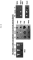
- the bacteria were not systematically absorbed when the bacteria was administered, while the bacteria-derived EVs were systematically absorbed at 5 min after administration, and, at 30 minutes after administration, fluorescence was strongly observed in the bladder, from which it was confirmed that the EVs were excreted via the urinary system through blood, and were present in the bodies up to 12 h after administration.
- DNA was extracted using the same method as that used in Example 2, and then PCR was performed thereon using 16S rDNA primers shown in Table 1 to amplify DNA, followed by sequencing (Illumina MiSeq sequencer).
- the results were output as standard flowgram format (SFF) files, and the SFF files were converted into sequence files (.fasta) and nucleotide quality score files using GS FLX software (v2.9), and then credit rating for reads was identified, and portions with a window (20 bps) average base call accuracy of less than 99% (Phred score ⁇ 20) were removed.
- SFF standard flowgram format
- Example 4 Myocardial infarction Diagnostic Model Based on Metagenomic Profiling of Bacteria-Derived EVs
- EVs were isolated from blood samples of 57 ST elevation myocardial infarction (STEMI) patients and 163 normal individuals, the two groups matched in age and gender, and then metagenomic sequencing was performed thereon using the method of Example 3.
- STEMI 57 ST elevation myocardial infarction
- metagenomic sequencing was performed thereon using the method of Example 3.
- a strain exhibiting a p value of less than 0.05 between two groups in a t-test and a difference of two-fold or more between two groups was selected, and then an area under curve (AUC), accuracy, sensitivity, and specificity, which are diagnostic performance indexes, were calculated by logistic regression analysis.
- AUC area under curve
- a diagnostic model developed using, as a biomarker, one or more bacteria from the class Acidobacteriia, the class DA052, the class Methanomicrobia, the class Thaumarchaeota, the class Clostridia, the class Coriobacteriia, the class Betaproteobacteria, the class Ktedonobacteria, the class Planctomycetia, the class Sol ⁇ bacteres, the class Erysipelotrichi, the class Verrucomicrobiae, the class TM7-3, the class Bacteroidia, the class Phycisphaerae, the class MCG, the class Nitrospira, the class Pedosphaerae, the class Thermoleophilia, the class Saprospirae, the class PRR-12, the class Spartobacteria, the class Acidimicrobiia, the class TM1, the class Deltaproteobacteria, the class
- a diagnostic model developed using, as a biomarker, one or more bacteria from the order Lactobacillales, the order Acidobacteriales, the order Enterobacteriales, the order Xanthomonadales, the order Clostridiales, the order Coriobacteriales, the order Ellin6513, the order Burkholderiales, the order Erysipelotrichales, the order Solibacterales, the order Verrucomicrobiales, the order Rhodospirillales, the order Gemmatales, the order Thermogemmatisporales, the order Saprospirales, the order Acidimicrobiales, the order Pedosphaerales, the order Bifidobacteriales, the order Chthoniobacterales, the order Solirubrobacterales, the order Syntrophobacterales, the order Bacteroidales, the order Nitrospirales, the order Ktedonobacterales, the order WD210
- o_Bacteroi dales 0.06 95 0.04 88 0.03 43 0.02 44 0.00 00 0.49 0.7 5 0.73 0.91 0.21 0.7 5 0.74 0.96 .
- o_Burkhol deriales 0.04 21 0.04 20 0.17 64 0.14 68 0.00 00 4.19 0.8 7 0.80 0.95 0.38 0.9 8 0.89 1.00 .
- o_Rhodosp irillales 0.00 28 0.00 70 0.01 59 0.01 70 0.00 00 5.73 0.8 5 0.81 0.96 0.38 0.7 1 0.77 0.92 .
- o_[Chthoni obacterales] 0.00 01 0.00 09 0.00 62 0.01 16 0.00 03 49.8 9 0.7 7 0.84 0.98 0.41 0.6 2 0.80 1.00 .
- o_[Pedosp haerales] 0.00 01 0.00 06 0.00 55 0.00 84 0.00 00 56.3 1 0.7 9 0.85 0.97 0.49 0.7 2 0.80 1.00 .
- o_Ktedono bacterales 0.00 00 0.00 02 0.00 34 0.00 69 0.00 05 174. 45 0.7 1 0.81 0.99 0.28 0.5 6 0.77 1.00 .
- o_Thermo gemmatispo rales 0.00 00 0.00 02 0.00 57 0.00 82 0.00 00 407. 10 0.8 2 0.89 0.99 0.59 0.7 5 0.77 1.00 .
- o_Ellin651 3 0.00 00 0.00 01 0.01 21 0.01 37 0.00 00 104 0.81 0.8 8 0.93 1.00 0.72 0.7 6 0.89 1.00 .
- o_Syntrop hobacterales 0.00 00 0.00 00 0.00 29 0.00 66 0.00 00 >10 0 0.7 6 0.86 1.00 0.43 0.6 4 0.77 1.00 .
- a diagnostic model developed using, as a biomarker, one or more bacteria from the family Koribacteraceae, the family Comamonadaceae, the family Enterobacteriaceae, the family Streptococcaceae, the family Coriobacteriaceae, the family Lachnospiraceae, the family Prevotellaceae, the family Ruminococcaceae, the family Xanthomonadaceae, the family Propionibacteriaceae, the family Hyphomicrobiaceae, the family Verrucomicrobiaceae, the family Solibacteraceae, the family Acidobacteriaceae, the family Erysipelotrichaceae, the family Ktedonobacteraceae, the family Thermogemmatisporaceae, the family Moraxellaceae, the family Veillonellaceae, the family Burkholderiaceae, the family Rhodospirillaceae, the family
- a diagnostic model developed using, as a biomarker, one or more bacteria from the genus Delftia, the genus Agrobacterium, the genus Stenotrophomonas, the genus Faecalibacterium, the genus Candidatus Koribacter, the genus Akkermansia, the genus Streptococcus, the genus Salinispora, the genus Candidatus Solibacter, the genus Citrobacter, the genus Collinsella, the genus Burkholderia, the genus Coprococcus, the genus Rhodoplanes, the genus Acinetobacter, the genus Prevotella, the genus Propionibacterium, the genus Lactococcus, the genus Bifidobacterium, the genus Methanobacterium, the genus Micrococcus, the
- Example 5 Dilated Cardiomyopathy Diagnostic Model Based on Metagenomic Profiling of Bacteria-Derived EVs
- EVs were isolated from blood samples of 72 dilated cardiomyopathy (DCMP) patients and 163 normal individuals, the two groups matched in age and gender, and then metagenomic sequencing was performed thereon using the method of Example 3.
- DCMP dilated cardiomyopathy
- metagenomic sequencing was performed thereon using the method of Example 3.
- a strain exhibiting a p value of less than 0.05 between two groups in a t-test and a difference of two-fold or more between two groups was selected, and then an AUC, accuracy, sensitivity, and specificity, which are diagnostic performance indexes, were calculated by logistic regression analysis.
- a diagnostic model developed using, as a biomarker, one or more bacteria from the phylum Verrucomicrobia , the phylum Acidobacteria , the phylum Gemmatimonadetes , and the phylum Planctomycetes exhibited significant diagnostic performance for DCMP (see Table 7 and FIG. 7 ).
- a diagnostic model developed using, as a biomarker, one or more bacteria from the class Verrucomicrobiae , the class Fusobacteriia , the class Acidobacteriia , the class Planctomycetia , the class DA052, the class Deltaproteobacteria , and the class Acidimicrobiia exhibited significant diagnostic performance for DCMP (see Table 8 and FIG. 8 ).
- EVs were isolated from blood samples of 80 variant angina patients and 80 normal individuals, the two groups matched in age and gender, and then metagenomic sequencing was performed thereon using the method of Example 3.
- metagenomic sequencing was performed thereon using the method of Example 3.
- a strain exhibiting a p value equal to of 0.05 between two groups in a t-test and a difference of two-fold or more between two groups was selected, and then an AUC, accuracy, sensitivity, and specificity, which are diagnostic performance indexes, were calculated by logistic regression analysis.
- a diagnostic model developed using, as a biomarker, one or more bacteria from the phylum Verrucomicrobia , the phylum Acidobacteria, the phylum Planctomycetes, the phylum Gemmatimonadetes, the phylum Chloroflexi, and the phylum Euryarchaeota exhibited significant diagnostic performance for variant angina (see Table 12 and FIG. 12 ).
- a diagnostic model developed using, as a biomarker, one or more bacteria from the class Verrucomicrobiae, the class Acidobacteriia, the class Fimbriimonadia, the class Erysipelotrichi, the class Ktedonobacteria, and the class Deltaproteobacteria exhibited significant diagnostic performance for variant angina (see Table 13 and FIG. 13 ).
- a diagnostic model developed using, as a biomarker, one or more bacteria from the order Pseudomonadales, the order Erysipelotrichales, the order Fimbriimonadales, the order Acidobacteriales, the order Verrucomicrobiales, the order Xanthomonadales, the order Myxococcales, the order Deinococcales, and the order Rhodospirillales exhibited significant diagnostic performance for variant angina (see Table 14 and FIG. 14 ).
- f_Oxalob acteraceae 0.0 291 0.0 505 0.0 081 0.0 103 0.0 005 0.28 0.7 3 0.6 6 0.57 0.75 0.6 3 0.5 8 0.38 0. 79 f_Fusoba cteriaceae 0.0 032 0.0 058 0.0 010 0.0 019 0.0 019 0.32 0.6 2 0.5 5 0.45 0.66 0.6 0 0.5 8 0.46 0. 71 f_Aeroco ccaceae 0.0 052 0.0 101 0.0 018 0.0 033 0.0 054 0.35 0.6 3 0.6 2 0.41 0.82 0.5 5 0.5 8 0.42 0.
- EVs were isolated from blood samples of 34 atrial fibrillation patients and 62 normal individuals, the two groups matched in age and gender, and then metagenomic sequencing was performed thereon using the method of Example 3.
- metagenomic sequencing was performed thereon using the method of Example 3.
- a strain exhibiting a p value of less than 0.05 between two groups in a t-test and a difference of two-fold or more between two groups was selected, and then an AUC, accuracy, sensitivity, and specificity, which are diagnostic performance indexes, were calculated by logistic regression analysis.
- a diagnostic model developed using, as a biomarker, one or more bacteria from the phylum Proteobacteria, the phylum TM7, the phylum Chloroflexi, the phylum Acidobacteria, and the phylum Cyanobacteria exhibited significant diagnostic performance for atrial fibrillation (see Table 17 and FIG. 17 ).
- a diagnostic model developed using, as a biomarker, one or more bacteria from the class Clostridia, the class Bacteroidia, the class Actinobacteria, the class Flavobacteriia, the class Erysipelotrichi, the class TM7-3, and the class Chloroplast exhibited significant diagnostic performance for atrial fibrillation (see Table 18 and FIG. 18 ).
- a diagnostic model developed using, as a biomarker, one or more bacteria from the genus Acinetobacter, the genus Stenotrophomonas, the genus Chryseobacterium, the genus Enterococcus, the genus Pseudomonas, the genus Delftia, the genus Alcanivorax, the genus Psychrobacter, the genus Streptococcus, the genus Ochrobactrum, the genus Bifidobacterium, the genus Coprococcus, the genus Bacteroides, the genus Faecalibacterium, the genus Enhydrobacter, the genus Agrobacterium, the genus Citrobacter, the genus Prevotella, the genus Geobacillus, the genus Clostridium, the genus Bacillus, the
- a risk group for heart disease can be diagnosed early and predicted by diagnosing a causative factor of heart disease through metagenomic analysis of bacteria-derived extracellular vesicles from a human body-derived sample, and thus the onset of heart disease can be delayed or heart disease may be prevented through appropriate management, and, even after heart disease occurs, early diagnosis for heart disease can be implemented, thereby lowering a disease rate and increasing therapeutic effects.
- patients diagnosed with heart disease are able to avoid exposure to causative factors predicted by metagenomic analysis, whereby the progression of heart disease is ameliorated, or recurrence of heart disease can be prevented.
Landscapes
- Chemical & Material Sciences (AREA)
- Health & Medical Sciences (AREA)
- Life Sciences & Earth Sciences (AREA)
- Organic Chemistry (AREA)
- Engineering & Computer Science (AREA)
- Proteomics, Peptides & Aminoacids (AREA)
- Analytical Chemistry (AREA)
- Bioinformatics & Cheminformatics (AREA)
- Zoology (AREA)
- Wood Science & Technology (AREA)
- General Health & Medical Sciences (AREA)
- Genetics & Genomics (AREA)
- Biotechnology (AREA)
- Biochemistry (AREA)
- General Engineering & Computer Science (AREA)
- Molecular Biology (AREA)
- Microbiology (AREA)
- Immunology (AREA)
- Biophysics (AREA)
- Physics & Mathematics (AREA)
- Cardiology (AREA)
- Nuclear Medicine, Radiotherapy & Molecular Imaging (AREA)
- Chemical Kinetics & Catalysis (AREA)
- Heart & Thoracic Surgery (AREA)
- Veterinary Medicine (AREA)
- Public Health (AREA)
- Animal Behavior & Ethology (AREA)
- Pharmacology & Pharmacy (AREA)
- Medicinal Chemistry (AREA)
- General Chemical & Material Sciences (AREA)
- Pathology (AREA)
- Mycology (AREA)
- Botany (AREA)
- Hospice & Palliative Care (AREA)
- Urology & Nephrology (AREA)
- Vascular Medicine (AREA)
- Measuring Or Testing Involving Enzymes Or Micro-Organisms (AREA)
- Micro-Organisms Or Cultivation Processes Thereof (AREA)
Description
- The present disclosure relates to a method of diagnosing a heart disease through bacterial metagenomic analysis, and more particularly, to a method of diagnosing a risk factor for a heart disease by analyzing an increase or decrease in content of extracellular vesicles derived from specific bacteria by bacterial metagenomic analysis using a sample derived from a subject.
- Heart disease is a disease occurring in the heart, and examples of major diseases thereof include ischemic heart disease, coronary artery disease, angina, myocardial infarction, arrhythmia, and the like. Among these, coronary artery disease is a group of diseases including angina, myocardial infarction, and the like and is also known as ischemic heart disease. Myocardial infarction is a disease in which an infarction occurs in heart muscles due to blocking of blood vessels that is caused by thrombosis in the coronary arteries. As risk factors for coronary artery disease, hypertension, smoking, diabetes, lack of exercise, obesity, hyperlipidemia, excessive drinking, and the like are known. It has been reported that the above-described disease is prevented when fruit- and vegetable-rich foods are consumed, and a risk for the disease is increased when trans fat-rich foods are consumed.
- Dilated cardiomyopathy, which is a disease in which the heart is expanded and unable to contract properly, is known as the most common cause of non-coronary artery disease and cardiomyopathy. Toxins, metabolic disorders, infectious factors, and the like have been suggested as causative factors, but causative factors have not yet been found.
- Angina is a disease occurring due to narrowing of the coronary arteries, through which blood is supplied to the heart, causing the volume of blood flowing to heart muscles to fall short of the required amount. When the coronary arteries are narrowed, blood supply is insufficient when in need thereof, and thus chest pain symptoms occur, and when angina remains untreated, it develops into acute myocardial infarction, thus causing sudden death. Angina may be broadly divided into stable angina, unstable angina, and variant angina. Among these three types, while stable angina and unstable angina occur mostly by atherosclerotic plaques, variant angina occurs due to a different cause, which thus has its name. Unlike general angina occurring due to narrowing of blood vessels, variant angina is a disease occurring due to the occurrence of spasms and poor blood circulation although the diameter of blood vessels of the coronary arteries is normal. Variant angina causes chest pain due to a decrease in or blocking of blood flow by spasm-derived contractions, and these symptoms occur early the next morning when drinking or receiving a lot of stress, or the like.
- Atrial fibrillation is a condition in which the atria beat continuously and irregularly and a rapid and irregular heart rate occurs. The atria beat 300 to 400 times per minute, while most stimuli are blocked by the atrioventricular (AV) node, and stimuli delivered to the ventricles are approximately 75 to 175 per minute. Causes of the disease include fundamental heart problems, i.e., coronary artery disease, myocardial infarction, hypertension, and mitral valve, and further include pericarditis, pulmonary embolisms, hyperthyroidism or hypothyroidism, septicaemia, diabetes, excessive drinking, pheochromocytoma, and the like.
- According to data released by the Korea National Statistical Office in 2013, the three leading causes of death in Korea are malignant neoplasm (cancer) accounting for 28.3%, cerebrovascular disease accounting for 9.6%, and heart disease accounting for 9.5%, and these account for about 50% of all causes of death. It has also been reported that coronary artery disease and cardiomyopathy account for 50% or greater of the causes of death due to heart disease. Modern people face stress caused by conflicts such as economic problems, workplace problems, and the like, and thus blood pressure, blood sugar, and blood fat are increased, and coronary artery disease and cardiomyopathy are very closely associated with diseases such as obesity, smoking, atherosclerosis, and the like, which continue to increase in modern people, and therefore, heart disease is a disease that may occur suddenly by the above-described causes and has a very serious prognosis. Therefore, there is an urgent need to develop a method capable of diagnosing heart disease early and increasing therapeutic efficiency, and prior to this, it is very important to differentiate countermeasures for early diagnosis and treatment by predicting the onset of heart disease, and thus research thereon and technology development thereof are required.
- Meanwhile, it is known that the number of microorganisms symbiotically living in the human body is 100 trillion which is 10 times the number of human cells, and the number of genes of microorganisms exceeds 100 times the number of human genes. A microbiota is a microbial community that includes bacteria, archaea, and eukaryotes present in a given habitat. The intestinal microbiota is known to play a vital role in human's physiological phenomena and significantly affect human health and diseases through interactions with human cells. Bacteria co-existing in human bodies secrete nanometer-sized vesicles to exchange information about genes, proteins, and the like with other cells. The mucous membranes form a physical barrier membrane that does not allow particles with the size of 200 nm or more to pass therethrough, and thus bacteria symbiotically living in the mucous membranes are unable to pass therethrough, but bacterial-derived extracellular vesicles have a size of approximately 100 nm or less and thus relatively freely pass through the mucous membranes and are absorbed into the human body.
- Metagenomics, also called environmental genomics, may be analytics for metagenomic data obtained from samples collected from the environment, and collectively refers to a total genome of all microbiota in the natural environment in which microorganisms exist and was first used by Jo Handelsman in 1998 (Handelsman et al., 1998 Chem. Biol. 5, R245-249). Recently, the bacterial composition of human microbiota has been listed using a method based on 16s ribosomal RNA (16s rRNA) base sequences, and 16s ribosomal RNA analyzes sequences using a 454FLX titanium platform. In the onset of heart disease, however, identification of causative factors such as heart disease, particularly myocardial infarction, cardiomyopathy, variant angina, atrial fibrillation, and the like through metagenomic analysis of bacteria-derived vesicles isolated from a human-derived substance, such as blood, urine, or the like, and a method of diagnosing the heart disease have never been reported.
- To diagnose heart disease such as myocardial infarction, cardiomyopathy, variant angina, atrial fibrillation, and the like, the inventors of the present invention extracted DNA from bacteria-derived vesicles using serum, which is a subject-derived sample, and performed metagenomic analysis on the extracted DNA, and, as a result, identified bacteria-derived extracellular vesicles capable of acting as a causative factor of heart disease, thus completing the present invention.
- Therefore, it is an object of the present invention to provide a method of providing information for heart disease diagnosis by metagenomic analysis of bacteria-derived extracellular vesicles.
- However, the technical goals of the present invention are not limited to the aforementioned goals, and other unmentioned technical goals will be clearly understood by those of ordinary skill in the art from the following description.
- Described herein but not forming part of the invention is a method of providing information for heart disease diagnosis, comprising the following processes:
- (a) extracting DNA from extracellular vesicles isolated from a subject sample;
- (b) performing polymerase chain reaction (PCR) on the extracted DNA using a pair of primers having SEQ ID NO:1 and SEQ ID NO: 2; and
- (c) comparing an increase or decrease in content of bacteria-derived extracellular vesicles of the subject sample with that of a normal individual-derived sample through sequencing of a product of the PCR.
- The methods of the invention of diagnosing heart disease have been described in the appended claims. Described herein but not forming part of the invention is a method of diagnosing heart disease, comprising the following processes:
- (a) extracting DNA from extracellular vesicles isolated from a subject sample;
- (b) performing polymerase chain reaction (PCR) on the extracted DNA using a pair of primers having SEQ ID NO:1 and SEQ ID NO: 2; and
- (c) comparing an increase or decrease in content of bacteria-derived extracellular vesicles of the subject sample with that of a normal individual-derived sample through sequencing of a product of the PCR.
- Described herein but not forming part of the invention is a method of predicting a risk for heart disease, comprising the following processes:
- (a) extracting DNA from extracellular vesicles isolated from a subject sample;
- (b) performing polymerase chain reaction (PCR) on the extracted DNA using a pair of primers having SEQ ID NO:1 and SEQ ID NO: 2; and
- (c) comparing an increase or decrease in content of bacteria-derived extracellular vesicles of the subject sample with that of a normal individual-derived sample through sequencing of a product of the PCR.
- The subject sample is blood. Comparing an increase or decrease in content of extracellular vesicles have been described in the appended claims. Embodiments described herein do not form part of the invention and may comprise comparing an increase or decrease in content of:
- extracellular vesicles derived from one or more bacteria selected from the group consisting of the phylum Acidobacteria, the phylum Firmicutes, the phylum Crenarchaeota, the phylum Planctomycetes, the phylum Chloroflexi, the phylum Euryarchaeota, the phylum WS3, the phylum Nitrospirae, the phylum WPS-2, the phylum AD3, the phylum Verrucomicrobia, the phylum Gemmatimonadetes, the phylum Proteobacteria, the phylum TM7, and the phylum Cyanobacteria;
- extracellular vesicles derived from one or more bacteria selected from the group consisting of the class Acidobacteriia, the class DA052, the class Methanomicrobia, the class Thaumarchaeota, the class Clostridia, the class Coriobacteriia, the class Betaproteobacteria, the class Ktedonobacteria, the class Planctomycetia, the class Solibacteres, the class Erysipelotrichi, the class Verrucomicrobiae, the class TM7-3, the class Bacteroidia, the class Phycisphaerae, the class MCG, the class Nitrospira, the class Pedosphaerae, the class Thermoleophilia, the class Saprospirae, the class PRR-12, the class Spartobacteria, the class Acidimicrobiia, the class TM1, the class Deltaproteobacteria, the class Anaerolineae, the class Thermoplasmata, the class Chthonomonadetes, the class Acidobacteria-b, the class Fusobacteriia, the class Fimbriimonadia, the class Actinobacteria, the class Flavobacteriia, and the class Chloroplast;
- extracellular vesicles derived from one or more bacteria selected from the group consisting of the order Lactobacillales, the order Acidobacteriales, the order Enterobacteriales, the order Xanthomonadales, the order Clostridiales, the order Coriobacteriales, the order Ellin6513, the order Burkholderiales, the order Erysipelotrichales, the order Solibacterales, the order Verrucomicrobiales, the order Rhodospirillales, the order Gemmatales, the order Thermogemmatisporales, the order Saprospirales, the order Acidimicrobiales, the order Pedosphaerales, the order Bifidobacteriales, the order Chthoniobacterales, the order Solžrubrobacterales, the order Syntrophobacterales, the order Bacteroidales, the order Nitrospirales, the order Ktedonobacterales, the order WD2101, the order iii1-15, the order Ellin329, the order Thiotrichales, the order Myxococcales, the order Stramenopiles, the order Vibrionales, the order Pseudomonadales, the order Bacillales, the order Sphingomonadales, the order Turicibacterales, the order Fimbriimonadales, the order Deinococcales, the order Pasteurellales, the order Flavobacteriales, the order Actinomycetales, the order Rhodobacterales, and the order Streptophyta;
- extracellular vesicles derived from one or more bacteria selected from the group consisting of the family Koribacteraceae, the family Comamonadaceae, the family Enterobacteriaceae, the family Streptococcaceae, the family Coriobacteriaceae, the family Lachnospiraceae, the family Prevotellaceae, the family Ruminococcaceae, the family Xanthomonadaceae, the family Propionibacteriaceae, the family Hyphomicrobiaceae, the family Verrucomicrobiaceae, the family Solibacteraceae, the family Acidobacteriaceae, the family Erysipelotrichaceae, the family Ktedonobacteraceae, the family Thermogemmatisporaceae, the family Moraxellaceae, the family Veillonellaceae, the family Burkholderiaceae, the family Rhodospirillaceae, the family Bifidobacteriaceae, the family Gemmataceae, the family Streptomycetaceae, the family Chitinophagaceae, the family Brucellaceae, the family Rhizobiaceae, the family Chthoniobacteraceae, the family Sinobacteraceae, the family Conexžbacteraceae, the family Oxalobacteraceae, the family Isosphaeraceae, the family Ellin515, the family Piscirickettsiaceae, the family Methylocystaceae, the family Pseudomonadaceae, the family Clostridiaceae, the family Sphingomonadaceae, the family Turicibacteraceae, the family Staphylococcaceae, the family Deinococcaceae, the family Methylobacteriaceae, the family Aerococcaceae, the family Fusobacteriaceae, the family Fimbriimonadaceae, the family Bacillaceae, the family Planococcaceae, the family BacteroidaceaeAxc family Weeksellaceae, the family Desulfovibrionaceae, the family Flavobacteriaceae, the family Rikenellaceae, the family S24-7, the family Pasteurellaceae, the family Rhodobacteraceae, the family Gordoniaceae, and the family Enterococcaceae; or
- extracellular vesicles derived from one or more bacteria selected from the group consisting of the genus Delftia, the genus Agrobacterium, the genus Stenotrophomonas, the genus Faecalibacterium, the genus Candidatus Koribacter, the genus Akkermansia, the genus Streptococcus, the genus Salinispora, the genus Candidatus Solibacter, the genus Citrobacter, the genus Collinsella, the genus Burkholderia, the genus Coprococcus, the genus Rhodoplanes, the genus Acinetobacter, the genus Prevotella, the genus Propionibacterium, the genus Lactococcus, the genus Bifidobacterium, the genus Methanobacterium, the genus Micrococcus, the genus Methanocella, the genus Brevibacterium, the genus Streptacidiphilus, the genus Streptomyces, the genus Ochrobactrum, the genus Methanosaeta, the genus Lysinibacillus, the genus Cupriavidus, the genus Pseudomonas, the genus Clostridium, the genus Sphingomonas, the genus Staphylococcus, the genus Thermoanaerobacterium, the genus Neisseria, the genus Enhydrobacter, the genus Actinomyces, the genus Turicibacter, the genus Phascolarctobacterium, the genus Catenibacterium, the genus Bacillus, the genus Deinococcus, the genus Fusobacterium, the genus Adlercreutzia, the genus Chryseobacterium, the genus Enterococcus, the genus Alcanivorax, the genus Psychrobacter, the genus Bacteroides, the genus Geobacillus, the genus Haemophilus, the genus Veillonella, the genus Paracoccus, the genus Kocuria, the genus Halomonas, the genus Ruminococcus, and the genus Porphyromonas.
- The heart disease is myocardial infarction, cardiomyopathy, variant angina, or atrial fibrillation.
- Embodiments described herein do not form part of the invention.
- In another embodiment of the present invention, in process (c), myocardial infarction may be diagnosed by comparing an increase or decrease in content of extracellular vesicles derived from one or more bacteria selected from the group consisting of the phylum Acidobacteria, the phylum Firmicutes, the phylum Crenarchaeota, the phylum Planctomycetes, the phylum Chloroflexi, the phylum Euryarchaeota, the phylum WS3, the phylum Nitrospirae, the phylum WPS-2, and the phylum AD3.
- In another embodiment of the present invention, in process (c), myocardial infarction may be diagnosed by comparing an increase or decrease in content of extracellular vesicles derived from one or more bacteria selected from the group consisting of the class Acidobacteriia, the class DA052, the class Methanomicrobia, the class Thaumarchaeota, the class Clostridia, the class Coriobacteriia, the class Betaproteobacteria, the class Ktedonobacteria, the class Planctomycetia, the class Solibacteres, the class Erysipelotrichi, the class Verrucomicrobiae, the class TM7-3, the class Bacteroidia, the class Phycisphaerae, the class MCG, the class Nitrospira, the class Pedosphaerae, the class Thermoleophilia, the class Saprospirae, the class PRR-12, the class Spartobacteria, the class Acidimicrobiia, the class TM1, the class Deltaproteobacteria, the class Anaerolineae, the class Thermoplasmata, the class Chthonomonadetes, and the class Acidobacteria-6.
- In another embodiment of the present invention, in process (c), myocardial infarction may be diagnosed by comparing an increase or decrease in content of extracellular vesicles derived from one or more bacteria selected from the group consisting of the order Lactobacillales, the order Acidobacteriales, the order Enterobacteriales, the order Xanthomonadales, the order Clostridiales, the order Coriobacteriales, the order Ellin6513, the order Burkholderiales, the order Erysipelotrichales, the order Solibacterales, the order Verrucomicrobiales, the order Rhodospirillales, the order Gemmatales, the order Thermogemmatisporales, the order Saprospirales, the order Acidimicrobiales, the order Pedosphaerales, the order Bifidobacteriales, the order Chthoniobacterales, the order Solirubrobacterales, the order Syntrophobacterales, the order Bacteroidales, the order Nitrospirales, the order Ktedonobacterales, the order WD2101, the order iii1-15, the order Ellin329, the order Thiotrichales, the order Myxococcales, the order Stramenopiles, and the order Vibrionales.
- In another embodiment of the present invention, in process (c), myocardial infarction may be diagnosed by comparing an increase or decrease in content of extracellular vesicles derived from one or more bacteria selected from the group consisting of the family Koribacteraceae, the family Comamonadaceae, the family Enterobacteriaceae, the family Streptococcaceae, the family Coriobacteriaceae, the family Lachnospiraceae, the family Prevotellaceae, the family Ruminococcaceae, the family Xanthomonadaceae, the family Propionibacteriaceae, the family Hyphomicrobiaceae, the family Verrucomicrobiaceae, the family Solibacteraceae, the family Acidobacteriaceae, the family Erysipelotrichaceae, the family Ktedonobacteraceae, the family Thermogemmatisporaceae, the family Moraxellaceae, the family Veillonellaceae, the family Burkholderiaceae, the family Rhodospirillaceae, the family Bifidobacteriaceae, the family Gemmataceae, the family Streptomycetaceae, the family Chitinophagaceae, the family Brucellaceae, the family Rhizobiaceae, the family Chthoniobacteraceae, the family Sinobacteraceae, the family Conexibacteraceae, the family Oxalobacteraceae, the family Isosphaeraceae, the family Ellin515, the family Piscirickettsiaceae, and the family Methylocystaceae.
- In another embodiment of the present invention, in process (c), myocardial infarction may be diagnosed by comparing an increase or decrease in content of extracellular vesicles derived from one or more bacteria selected from the group consisting of the genus Delftia, the genus Agrobacterium, the genus Stenotrophomonas, the genus Faecalibacterium, the genus Candidatus Koribacter, the genus Akkermansia, the genus Streptococcus, the genus Salinispora, the genus Candidatus Solibacter, the genus Citrobacter, the genus Collinsella, the genus Burkholderia, the genus Coprococcus, the genus Rhodoplanes, the genus Acinetobacter, the genus Prevotella, the genus Propionibacterium, the genus Lactococcus, the genus Bifidobacterium, the genus Methanobacterium, the genus Micrococcus, the genus Methanocella, the genus Brevibacterium, the genus Streptacidiphilus, the genus Streptomyces, the genus Ochrobactrum, the genus Methanosaeta, the genus Lysinibacillus, and the genus Cupriavidus.
- In another embodiment of the present invention, in process (c), cardiomyopathy may be diagnosed by comparing an increase or decrease in content of extracellular vesicles derived from one or more bacteria selected from the group consisting of the phylum Verrucomicrobia, the phylum Acidobacteria, the phylum Gemmatimonadetes, and the phylum Planctomycetes.
- In another embodiment of the present invention, in process (c), cardiomyopathy may be diagnosed by comparing an increase or decrease in content of extracellular vesicles derived from one or more bacteria selected from the group consisting of the class Verrucomicrobiae, the class Fusobacteriia, the class Acidobacteriia, the class Planctomycetia, the class DA052, the class Deltaproteobacteria, and the class Acidimicrobiia.
- In another embodiment of the present invention, in process (c), cardiomyopathy may be diagnosed by comparing an increase or decrease in content of extracellular vesicles derived from one or more bacteria selected from the group consisting of the order Pseudomonadales, the order Bacillales, the order Acidobacteriales, the order Sphingomonadales, the order Verrucomicrobiales, the order Turicibacterales, the order Acidimicrobiales, the order Ellin6513, the order Xanthomonadales, and the order Gemmatales.
- In another embodiment of the present invention, in process (c), cardiomyopathy may be diagnosed by comparing an increase or decrease in content of extracellular vesicles derived from one or more bacteria selected from the group consisting of the family Pseudomonadaceae, the family Clostridiaceae, the family Comamonadaceae, the family Oxalobacteraceae, the family Moraxellaceae, the family Verrucomicrobiaceae, the family Koribacteraceae, the family Sphingomonadaceae, the family Turicibacteraceae, the family Xanthomonadaceae, the family Gemmataceae, and the family Staphylococcaceae.
- In another embodiment of the present invention, in process (c), cardiomyopathy may be diagnosed by comparing an increase or decrease in content of extracellular vesicles derived from one or more bacteria selected from the group consisting of the genus Pseudomonas, the genus Clostridium, the genus Cupriavidus, the genus Acinetobacter, the genus Citrobacter, the genus Sphingomonas, the genus Candidatus Koribacter, the genus Staphylococcus, the genus Thermoanaerobacterium, the genus Micrococcus, the genus Akkermansia, the genus Neisseria, the genus Enhydrobacter, the genus Actinomyces, the genus Turicibacter, the genus Phascolarctobacterium, the genus Lactococcus, the genus Delftia, and the genus Stenotrophomonas.
- In another embodiment of the present invention, in process (c), variant angina may be diagnosed by comparing an increase or decrease in content of extracellular vesicles derived from one or more bacteria selected from the group consisting of the phylum Verrucomicrobia, the phylum Acidobacteria, the phylum Planctomycetes, the phylum Gemmatimonadetes, the phylum Chloroflexi, and the phylum Euryarchaeota.
- In another embodiment of the present invention, in process (c), variant angina may be diagnosed by comparing an increase or decrease in content of extracellular vesicles derived from one or more bacteria selected from the group consisting of the class Verrucomicrobiae, the class Acidobacteriia, the class Fimbriimonadia, the class Erysipelotrichi, the class Ktedonobacteria, and the class Deltaproteobacteria.
- In another embodiment of the present invention, in process (c), variant angina may be diagnosed by comparing an increase or decrease in content of extracellular vesicles derived from one or more bacteria selected from the group consisting of the order Pseudomonadales, the order Erysipelotrichales, the order Fimbriimonadales, the order Acidobacteriales, the order Verrucomicrobiales, the order Xanthomonadales, the order Myxococcales, the order Deinococcales, and the order Rhodospirillales.
- In another embodiment of the present invention, in process (c), variant angina may be diagnosed by comparing an increase or decrease in content of extracellular vesicles derived from one or more bacteria selected from the group consisting of the family Koribacteraceae, the family Oxalobacteraceae, the family Comamonadaceae, the family Moraxellaceae, the family Pseudomonadaceae, the family Hyphomicrobiaceae, the family Erysipelotrichaceae, the family Deinococcaceae, the family Clostridiaceae, the family Verrucomicrobiaceae, the family Sinobacteraceae, the family Rhodospirillaceae, the family Methylobacteriaceae, the family Aerococcaceae, the family Fusobacteriaceae, the family Fimbriimonadaceae, the family Bacillaceae, and the family Planococcaceae.
- In another embodiment of the present invention, in process (c), variant angina may be diagnosed by comparing an increase or decrease in content of extracellular vesicles derived from one or more bacteria selected from the group consisting of the genus Citrobacter, the genus Acinetobacter, the genus Cupriavidus, the genus Clostridium, the genus Catenibacterium, the genus Pseudomonas, the genus Lactococcus, the genus Stenotrophomonas, the genus Akkermansia, the genus Bacillus, the genus Delftia, the genus Agrobacterium, the genus Deinococcus, the genus Fusobacterium, and the genus Adlercreutzia.
- In another embodiment of the present invention, in process (c), atrial fibrillation may be diagnosed by comparing an increase or decrease in content of extracellular vesicles derived from one or more bacteria selected from the group consisting of the phylum Proteobacteria, the phylum TM7, the phylum Chloroflexi, the phylum Acidobacteria, and the phylum Cyanobacteria.
- In another embodiment of the present invention, in process (c), atrial fibrillation may be diagnosed by comparing an increase or decrease in content of extracellular vesicles derived from one or more bacteria selected from the class Clostridia, the class Bacteroidia, the class Actinobacteria, the class Flavobacteriia, the class Erysipelotrichi, the class TM7-3, and the class Chloroplast.
- In another embodiment of the present invention, in process (c), atrial fibrillation may be diagnosed by comparing an increase or decrease in content of extracellular vesicles derived from one or more bacteria selected from the group consisting of the order Pseudomonadales, the order Clostridiales, the order Bacteroidales, the order Enterobacteriales, the order Xanthomonadales, the order Bifidobacteriales, the order Pasteurellales, the order Flavobacteriales, the order Actinomycetales, the order Rhodobacterales, the order Coriobacteriales, the order Erysipelotrichales, and the order Streptophyta.
- In another embodiment of the present invention, in process (c), atrial fibrillation may be diagnosed by comparing an increase or decrease in content of extracellular vesicles derived from one or more bacteria selected from the group consisting of the family Lachnospiraceae, the family Bacillaceae, the family Streptococcaceae, the family Bacteroidaceae, the family Moraxellaceae, the family Ruminococcaceae, the family Weeksellaceae, the family Bifidobacteriaceae, the family Clostridiaceae, the family Desulfovibrionaceae, the family Veillonellaceae, the family Coriobacteriaceae, the family Flavobacteriaceae, the family Rikenellaceae, the family S24-7, the family Pasteurellaceae, the family Rhodobacteraceae, the family Pseudomonadaceae, the family Gordoniaceae, and the family Enterococcaceae.
- In another embodiment of the present invention, in process (c), atrial fibrillation may be diagnosed by comparing an increase or decrease in content of extracellular vesicles derived from one or more bacteria selected from the group consisting of the genus Acinetobacter, the genus Stenotrophomonas, the genus Chryseobacterium, the genus Enterococcus, the genus Pseudomonas, the genus Delftia, the genus Alcanivorax, the genus Psychrobacter, the genus Streptococcus, the genus Ochrobactrum, the genus Bifidobacterium, the genus Coprococcus, the genus Bacteroides, the genus Faecalibacterium, the genus Enhydrobacter, the genus Agrobacterium, the genus Citrobacter, the genus Prevotella, the genus Geobacillus, the genus Clostridium, the genus Bacillus, the genus Haemophilus, the genus Veillonella, the genus Actinomyces, the genus Paracoccus, the genus Kocuria, the genus Halomonas, the genus Micrococcus, the genus Ruminococcus, and the genus Porphyromonas.
- In another embodiment of the present invention, the blood may be whole blood, serum, or plasma.
- According to the present invention, a risk group for heart disease can be diagnosed early and predicted by diagnosing a causative factor of heart disease through metagenomic analysis of bacteria-derived extracellular vesicles from a human body-derived sample, and thus the onset of heart disease can be delayed or heart disease may be prevented through appropriate management, and, even after heart disease occurs, early diagnosis for heart disease can be implemented, thereby lowering a disease rate and increasing therapeutic effects.
- In addition, patients diagnosed with heart disease are able to avoid exposure to causative factors predicted by metagenomic analysis, whereby the progression of heart disease is ameliorated, or recurrence of heart disease can be prevented.
-
-
FIGS. 1A and1B are views for evaluating the distribution pattern of extracellular vesicles (EVs) derived from bacteria in vivo.FIG. 1A illustrates images showing the distribution pattern of intestinal bacteria and EVs derived from bacteria per time (0 h, 5 min, 3 h, 6 h, and 12 h) after being orally administered to mice.FIG. 1B illustrates images showing the distribution pattern of gut bacteria and EVs derived from bacteria after being orally administered to mice and, after 12 hours, blood and various organs (heart, lung, liver, kidney, spleen, adipose tissue, and muscle) of the mice were extracted. -
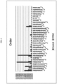
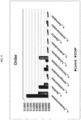
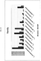
FIG. 2 illustrates distribution results of bacteria-derived EVs exhibiting significant diagnostic performance at a phylum level, after metagenomic analysis of bacteria-derived EVs isolated from myocardial infarction patient-derived blood and normal individual-derived blood. -
FIG. 3 illustrates distribution results of bacteria-derived EVs exhibiting significant diagnostic performance at a class level, after metagenomic analysis of bacteria-derived EVs isolated from myocardial infarction patient-derived blood and normal individual-derived blood. -
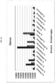
FIG. 4 illustrates distribution results of bacteria-derived EVs exhibiting significant diagnostic performance at an order level, after metagenomic analysis of bacteria-derived EVs isolated from myocardial infarction patient-derived blood and normal individual-derived blood. -
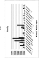
FIG. 5 illustrates distribution results of bacteria-derived EVs exhibiting significant diagnostic performance at a family level, after metagenomic analysis of bacteria-derived EVs isolated from myocardial infarction patient-derived blood and normal individual-derived blood. -
FIG. 6 illustrates distribution results of bacteria-derived EVs exhibiting significant diagnostic performance at a genus level, after metagenomic analysis of bacteria-derived EVs isolated from myocardial infarction patient-derived blood and normal individual-derived blood. -
FIG. 7 illustrates distribution results of bacteria-derived EVs exhibiting significant diagnostic performance at a phylum level, after metagenomic analysis of bacteria-derived EVs isolated from cardiomyopathy patient-derived blood and normal individual-derived blood. -
FIG. 8 illustrates distribution results of bacteria-derived EVs exhibiting significant diagnostic performance at a class level, after metagenomic analysis of bacteria-derived EVs isolated from cardiomyopathy patient-derived blood and normal individual-derived blood. -
FIG. 9 illustrates distribution results of bacteria-derived EVs exhibiting significant diagnostic performance at an order level, after metagenomic analysis of bacteria-derived EVs isolated from cardiomyopathy patient-derived blood and normal individual-derived blood. -
FIG. 10 illustrates distribution results of bacteria-derived EVs exhibiting significant diagnostic performance at a family level, after metagenomic analysis of bacteria-derived EVs isolated from cardiomyopathy patient-derived blood and normal individual-derived blood. -
FIG. 11 illustrates distribution results of bacteria-derived EVs exhibiting significant diagnostic performance at a genus level, after metagenomic analysis of bacteria-derived EVs isolated from cardiomyopathy patient-derived blood and normal individual-derived blood. -
FIG. 12 illustrates distribution results of bacteria-derived EVs exhibiting significant diagnostic performance at a phylum level, after metagenomic analysis of bacteria-derived EVs isolated from variant angina patient-derived blood and normal individual-derived blood. -
FIG. 13 illustrates distribution results of bacteria-derived EVs exhibiting significant diagnostic performance at a class level, after metagenomic analysis of bacteria-derived EVs isolated from variant angina patient-derived blood and normal individual-derived blood. -
FIG. 14 illustrates distribution results of bacteria-derived EVs exhibiting significant diagnostic performance at an order level, after metagenomic analysis of bacteria-derived EVs isolated from variant angina patient-derived blood and normal individual-derived blood. -
FIG. 15 illustrates distribution results of bacteria-derived EVs exhibiting significant diagnostic performance at a family level, after metagenomic analysis of bacteria-derived EVs isolated from variant angina patient-derived blood and normal individual-derived blood. -
FIG. 16 illustrates distribution results of bacteria-derived EVs exhibiting significant diagnostic performance at a genus level, after metagenomic analysis of bacteria-derived EVs isolated from variant angina patient-derived blood and normal individual-derived blood. -
FIG. 17 illustrates distribution results of bacteria-derived EVs exhibiting significant diagnostic performance at a phylum level, after metagenomic analysis of bacteria-derived EVs isolated from atrial fibrillation patient-derived blood and normal individual-derived blood. -
FIG. 18 illustrates distribution results of bacteria-derived EVs exhibiting significant diagnostic performance at a class level, after metagenomic analysis of bacteria-derived EVs isolated from atrial fibrillation patient-derived blood and normal individual-derived blood. -
FIG. 19 illustrates distribution results of bacteria-derived EVs exhibiting significant diagnostic performance at an order level, after metagenomic analysis of bacteria-derived EVs isolated from atrial fibrillation patient-derived blood and normal individual-derived blood. -
FIG. 20 illustrates distribution results of bacteria-derived EVs exhibiting significant diagnostic performance at a family level, after metagenomic analysis of bacteria-derived EVs isolated from atrial fibrillation patient-derived blood and normal individual-derived blood. -
FIG. 21 illustrates distribution results of bacteria-derived EVs exhibiting significant diagnostic performance at a genus level, after metagenomic analysis of bacteria-derived EVs isolated from atrial fibrillation patient-derived blood and normal individual-derived blood. - The present invention relates to a method of diagnosing heart disease through bacterial metagenomic analysis. The inventors of the present invention extracted genes from bacteria-derived extracellular vesicles present in subject-derived samples, performed metagenomic analysis thereon, and identified bacteria-derived extracellular vesicles capable of acting as a causative factor of heart disease.
- Described herein but not forming part of the invention is a method of providing information on heart disease diagnosis, the method including:
- (a) extracting DNA from extracellular vesicles isolated from a subject sample;
- (b) performing polymerase chain reaction (PCR) on the extracted DNA using a pair of primers having SEQ ID NOS: 1 and 2; and
- (c) comparing an increase or decrease in content of bacteria-derived extracellular vesicles of the subject sample with that of a normal individual-derived sample through sequencing of a product of the PCR.
- The term "prediction of a risk for heart disease" as used herein refers to determining whether a patient has a risk for heart disease, whether the risk for heart disease is relatively high, or whether heart disease has already occurred. The method of the present invention may be used to delay the onset of heart disease through special and appropriate care for a specific patient, which is a patient having a high risk for heart disease or prevent the onset of heart disease. In addition, the method may be clinically used to determine treatment by selecting the most appropriate treatment method through early diagnosis of heart disease.
- The heart disease is myocardial infarction, cardiomyopathy, variant angina, or atrial fibrillation.
- The term "metagenome" as used herein refers to the total of genomes including all viruses, bacteria, fungi, and the like in isolated regions such as soil, the intestines of animals, and the like, and is mainly used as a concept of genomes that explains identification of many microorganisms at once using a sequencer to analyze non-cultured microorganisms. In particular, a metagenome does not refer to a genome of one species, but refers to a mixture of genomes, including genomes of all species of an environmental unit. This term originates from the view that, when defining one species in a process in which biology is advanced into omics, various species as well as existing one species functionally interact with each other to form a complete species. Technically, it is the subject of techniques that analyzes all DNAs and RNAs regardless of species using rapid sequencing to identify all species in one environment and verify interactions and metabolism. In the present invention, metagenomic analysis is performed using bacteria-derived extracellular vesicles isolated from blood.
- In the present invention, the subject sample may be whole blood, serum, or plasma, but the present invention is not limited therto.
- In an embodiment of the present invention, metagenomic analysis was performed on the bacteria-derived extracellular vesicles, and the bacteria-derived extracellular vesicles capable of acting as a cause of the onset of heart disease were actually identified by analysis at phylum, class, order, family, and genus levels.
- More particularly, in one embodiment of the present invention, as a result of performing metagenomic analysis on bacteria-derived extracellular vesicles present in myocardial infarction patient-derived blood and normal individual-derived blood at a phylum level, the content of extracelluar vesicles derived from bacteria belonging to the phylum Acidobacteria, the phylum Firmicutes, the phylum Crenarchaeota, the phylum Planctomycetes, the phylum Chloroflexi, the phylum Euryarchaeota, the phylum WS3, the phylum Nitrospirae, the phylum WPS-2, and the phylum AD3 was significantly different between myocardial infarction patients and normal individuals (see Example 4).
- More particularly, in one embodiment of the present invention, as a result of performing metagenomic analysis on bacteria-derived extracellular vesicles present in myocardial infarction patient-derived blood and normal individual-derived blood at a class level, the content of extracelluar vesicles derived from bacteria belonging to the class Acidobacteriia, the class DA052, the class Methanomicrobia, the class Thaumarchaeota, the class Clostridia, the class Coriobacteriia, the class Betaproteobacteria, the class Ktedonobacteria, the class Planctomycetia, the class Solibacteres, the class Erysipelotrichi, the class Verrucomicrobiae, the class TM7-3, the class Bacteroidia, the class Phycisphaerae, the class MCG, the class Nitrospira, the class Pedosphaerae, the class Thermoleophilia, the class Saprospirae, the class PRR-12, the class Spartobacteria, the class Acidimicrobiia, the class TM1, the class Deltaproteobacteria, the class Anaerolineae, the class Thermoplasmata, the class Chthonomonadetes, and the class Acidobacteria-6 was significantly different between myocardial infarction patients and normal individuals (see Example 4).
- More particularly, in one embodiment of the present invention, as a result of performing metagenomic analysis on bacteria-derived extracellular vesicles present in myocardial infarction patient-derived blood and normal individual-derived blood at an order level, the content of extracelluar vesicles derived from bacteria belonging to the order Lactobacillales, the order Acidobacteriales, the order Enterobacteriales, the order Xanthomonadales, the order Clostridiales, the order Coriobacteriales, the order Ellin6513, the order Burkholderiales, the order Erysipelotrichales, the order Solibacterales, the order Verrucomicrobiales, the order Rhodospirillales, the order Gemmatales, the order Thermogemmatisporales, the order Saprospirales, the order Acidimicrobiales, the order Pedosphaerales, the order Bifidobacteriales, the order Chthoniobacterales, the order Solirubrobacterales, the order Syntrophobacterales, the order Bacteroidales, the order Nitrospirales, the order Ktedonobacterales, the order WD2101, the order iii1-15, the order Ellin329, the order Thiotrichales, the order Myxococcales, the order Stramenopiles, and the order Vibrionales was significantly different between myocardial infarction patients and normal individuals (see Example 4).
- More particularly, in one embodiment of the present invention, as a result of performing metagenomic analysis on bacteria-derived extracellular vesicles present in myocardial infarction patient-derived blood and normal individual-derived blood at a family level, the content of extracelluar vesicles derived from bacteria belonging to the family Koribacteraceae, the family Comamonadaceae, the family Enterobacteriaceae, the family Streptococcaceae, the family Coriobacteriaceae, the family Lachnospiraceae, the family Prevotellaceae, the family Ruminococcaceae, the family Xanthomonadaceae, the family Propionibacteriaceae, the family Hyphomicrobiaceae, the family Verrucomicrobiaceae, the family Solibacteraceae, the family Acidobacteriaceae, the family Erysipelotrichaceae, the family Ktedonobacteraceae, the family Thermogemmatisporaceae, the family Moraxellaceae, the family Veillonellaceae, the family Burkholderiaceae, the family Rhodospirillaceae, the family Bifidobacteriaceae, the family Gemmataceae, the family Streptomycetaceae, the family Chitinophagaceae, the family Brucellaceae, the family Rhizobiaceae, the family Chthoniobacteraceae, the family Sinobacteraceae, the family Conexibacteraceae, the family Oxalobacteraceae, the family Isosphaeraceae, the family Ellin515, the family Piscirickettsiaceae, and the family Methylocystaceae was significantly different between myocardial infarction patients and normal individuals (see Example 4).
- More particularly, in one embodiment of the present invention, as a result of performing metagenomic analysis on bacteria-derived extracellular vesicles present in myocardial infarction patient-derived blood and normal individual-derived blood at a genus level, the content of extracelluar vesicles derived from bacteria belonging to the genus Delftia, the genus Agrobacterium, the genus Stenotrophomonas, the genus Faecalibacterium, the genus Candidatus Koribacter, the genus Akkermansia, the genus Streptococcus, the genus Salinispora, the genus Candidatus Solibacter, the genus Citrobacter, the genus Collinsella, the genus Burkholderia, the genus Coprococcus, the genus Rhodoplanes, the genus Acinetobacter, the genus Prevotella, the genus Propionibacterium, the genus Lactococcus, the genus Bifidobacterium, the genus Methanobacterium, the genus Micrococcus, the genus Methanocella, the genus Brevibacterium, the genus Streptacidiphilus, the genus Streptomyces, the genus Ochrobactrum, the genus Methanosaeta, the genus Lysinibacillus, and the genus Cupriavidus was significantly different between myocardial infarction patients and normal individuals (see Example 4).
- More particularly, in one embodiment of the present invention, as a result of performing metagenomic analysis on bacteria-derived extracellular vesicles present in cardiomyopathy patient-derived blood and normal individual-derived blood at a phylum level, the content of extracelluar vesicles derived from bacteria belonging to the phylum Verrucomicrobia, the phylum Acidobacteria, the phylum Gemmatimonadetes, and the phylum Planctomycetes was significantly different between cardiomyopathy patients and normal individuals (see Example 5).
- More particularly, in one embodiment of the present invention, as a result of performing metagenomic analysis on bacteria-derived extracellular vesicles present in cardiomyopathy patient-derived blood and normal individual-derived blood at a class level, the content of extracelluar vesicles derived from bacteria belonging to the class Verrucomicrobiae, the class Fusobacteriia, the class Acidobacteriia, the class Planctomycetia, the class DA052, the class Deltaproteobacteria, and the class Acidimicrobiia was significantly different between cardiomyopathy patients and normal individuals (see Example 5).
- More particularly, in one embodiment of the present invention, as a result of performing metagenomic analysis on bacteria-derived extracellular vesicles present in cardiomyopathy patient-derived blood and normal individual-derived blood at an order level, the content of extracelluar vesicles derived from bacteria belonging to the order Pseudomonadales, the order Bacillales, the order Acidobacteriales, the order Sphingomonadales, the order Verrucomicrobiales, the order Turicibacterales, the order Acidimicrobiales, the order Ellin6513, the order Xanthomonadales, and the order Gemmatales was significantly different between cardiomyopathy patients and normal individuals (see Example 5).
- More particularly, in one embodiment of the present invention, as a result of performing metagenomic analysis on bacteria-derived extracellular vesicles present in cardiomyopathy patient-derived blood and normal individual-derived blood at a family level, the content of extracelluar vesicles derived from bacteria belonging to the family Pseudomonadaceae, the family Clostridiaceae, the family Comamonadaceae, the family Oxalobacteraceae, the family Moraxellaceae, the family Verrucomicrobiaceae, the family Koribacteraceae, the family Sphingomonadaceae, the family Turicibacteraceae, the family Xanthomonadaceae, the family Gemmataceae, and the family Staphylococcaceae was significantly different between cardiomyopathy patients and normal individuals (see Example 5).
- More particularly, in one embodiment of the present invention, as a result of performing metagenomic analysis on bacteria-derived extracellular vesicles present in cardiomyopathy patient-derived blood and normal individual-derived blood at a genus level, the content of extracelluar vesicles derived from bacteria belonging to the genus Pseudomonas, the genus Clostridium, the genus Cupriavidus, the genus Acinetobacter, the genus Citrobacter, the genus Sphingomonas, the genus Candidatus Koribacter, the genus Staphylococcus, the genus Thermoanaerobacterium, the genus Micrococcus, the genus Akkermansia, the genus Neisseria, the genus Enhydrobacter, the genus Actinomyces, the genus Turicibacter, the genus Phascolarctobacterium, the genus Lactococcus, the genus Delftia, and the genus Stenotrophomonas was significantly different between cardiomyopathy patients and normal individuals (see Example 5).
- More particularly, in one embodiment of the present invention, as a result of performing metagenomic analysis on bacteria-derived extracellular vesicles present in variant angina patient-derived blood and normal individual-derived blood at a phylum level, the content of extracelluar vesicles derived from bacteria belonging to the phylum Verrucomicrobia, the phylum Acidobacteria, the phylum Planctomycetes, the phylum Gemmatimonadetes, the phylum Chloroflexi, and the phylum Euryarchaeota was significantly different between variant angina patients and normal individuals (see Example 6).
- More particularly, in one embodiment of the present invention, as a result of performing metagenomic analysis on bacteria-derived extracellular vesicles present in variant angina patient-derived blood and normal individual-derived blood at a class level, the content of extracelluar vesicles derived from bacteria belonging to the class Verrucomicrobiae, the class Acidobacteriia, the class Fimbriimonadia, the class Erysipelotrichi, the class Ktedonobacteria, and the class Deltaproteobacteria was significantly different between variant angina patients and normal individuals (see Example 6).
- More particularly, in one embodiment of the present invention, as a result of performing metagenomic analysis on bacteria-derived extracellular vesicles present in variant angina patient-derived blood and normal individual-derived blood at an order level, the content of extracelluar vesicles derived from bacteria belonging to the order Pseudomonadales, the order Erysipelotrichales, the order Fimbriimonadales, the order Acidobacteriales, the order Verrucomicrobiales, the order Xanthomonadales, the order Myxococcales, the order Deinococcales, and the order Rhodospirillales was significantly different between variant angina patients and normal individuals (see Example 6).
- More particularly, in one embodiment of the present invention, as a result of performing metagenomic analysis on bacteria-derived extracellular vesicles present in variant angina patient-derived blood and normal individual-derived blood at a family level, the content of extracelluar vesicles derived from bacteria belonging to the family Koribacteraceae, the family Oxalobacteraceae, the family Comamonadaceae, the family Moraxellaceae, the family Pseudomonadaceae, the family Hyphomicrobiaceae, the family Erysipelotrichaceae, the family Deinococcaceae, the family Clostridiaceae, the family Verrucomicrobiaceae, the family Sinobacteraceae, the family Rhodospirillaceae, the family Methylobacteriaceae, the family Aerococcaceae, the family Fusobacteriaceae, the family Fimbriimonadaceae, the family Bacillaceae, and the family Planococcaceae was significantly different between variant angina patients and normal individuals (see Example 6).
- More particularly, in one embodiment of the present invention, as a result of performing metagenomic analysis on bacteria-derived extracellular vesicles present in variant angina patient-derived blood and normal individual-derived blood at a genus level, the content of extracelluar vesicles derived from bacteria belonging to the genus Citrobacter, the genus Acinetobacter, the genus Cupriavidus, the genus Clostridium, the genus Catenibacterium, the genus Pseudomonas, the genus Lactococcus, the genus Stenotrophomonas, the genus Akkermansia, the genus Bacillus, the genus Delftia, the genus Agrobacterium, the genus Deinococcus, the genus Fusobacterium, and the genus Adlercreutzia was significantly different between variant angina patients and normal individuals (see Example 6).
- More particularly, in one embodiment of the present invention, as a result of performing metagenomic analysis on bacteria-derived extracellular vesicles present in atrial fibrillation patient-derived blood and normal individual-derived blood at a phylum level, the content of extracelluar vesicles derived from bacteria belonging to the phylum Proteobacteria, the phylum TM7, the phylum Chloroflexi, the phylum Acidobacteria, and the phylum Cyanobacteria was significantly different between atrial fibrillation patients and normal individuals (see Example 7).
- More particularly, in one embodiment of the present invention, as a result of performing metagenomic analysis on bacteria-derived extracellular vesicles present in atrial fibrillation patient-derived blood and normal individual-derived blood at a class level, the content of extracelluar vesicles derived from bacteria belonging to the class Clostridia, the class Bacteroidia, the class Actinobacteria, the class Flavobacteriia, the class Erysipelotrichi, the class TM7-3, and the class Chloroplast was significantly different between atrial fibrillation patients and normal individuals (see Example 7).
- More particularly, in one embodiment of the present invention, as a result of performing metagenomic analysis on bacteria-derived extracellular vesicles present in atrial fibrillation patient-derived blood and normal individual-derived blood at an order level, the content of extracelluar vesicles derived from bacteria belonging to the order Pseudomonadales, the order Clostridiales, the order Bacteroidales, the order Enterobacteriales, the order Xanthomonadales, the order Bifidobacteriales, the order Pasteurellales, the order Flavobacteriales, the order Actinomycetales, the order Rhodobacterales, the order Coriobacteriales, the order Erysipelotrichales, and the order Streptophyta was significantly different between atrial fibrillation patients and normal individuals (see Example 7).
- More particularly, in one embodiment of the present invention, as a result of performing metagenomic analysis on bacteria-derived extracellular vesicles present in atrial fibrillation patient-derived blood and normal individual-derived blood at a family level, the content of extracelluar vesicles derived from bacteria belonging to the family Lachnospiraceae, the family Bacillaceae, the family Streptococcaceae, the family Bacteroidaceae, the family Moraxellaceae, the family Ruminococcaceae, the family Weeksellaceae, the family Bifidobacteriaceae, the family Clostridiaceae, the family Desulfovibrionaceae, the family Veillonellaceae, the family Coriobacteriaceae, the family Flavobacteriaceae, the family Rikenellaceae, the family S24-7, the family Pasteurellaceae, the family Rhodobacteraceae, the family Pseudomonadaceae, the family Gordoniaceae, and the family Enterococcaceae was significantly different between atrial fibrillation patients and normal individuals (see Example 7).
- More particularly, in one embodiment of the present invention, as a result of performing metagenomic analysis on bacteria-derived extracellular vesicles present in atrial fibrillation patient-derived blood and normal individual-derived blood at a genus level, the content of extracelluar vesicles derived from bacteria belonging to the genus Acinetobacter, the genus Stenotrophomonas, the genus Chryseobacterium, the genus Enterococcus, the genus Pseudomonas, the genus Delftia, the genus Alcanivorax, the genus Psychrobacter, the genus Streptococcus, the genus Ochrobactrum, the genus Bifidobacterium, the genus Coprococcus, the genus Bacteroides, the genus Faecalibacterium, the genus Enhydrobacter, the genus Agrobacterium, the genus Citrobacter, the genus Prevotella, the genus Geobacillus, the genus Clostridium, the genus Bacillus, the genus Haemophilus, the genus Veillonella, the genus Actinomyces, the genus Paracoccus, the genus Kocuria, the genus Halomonas, the genus Micrococcus, the genus Ruminococcus, and the genus Porphyromonas was significantly different between atrial fibrillation patients and normal individuals (see Example 7).
- From the above-described example results, it was confirmed that bacteria-derived extracellular vesicles exhibiting a significant change in content in patients with myocardial infarction, cardiomyopathy, variant angina, or atrial fibrillation compared to normal individuals, were identified by performing metagenomic analysis on extracellular vesicles isolated from blood, and heart disease may be diagnosed by analyzing an increase or decrease in the content of bacteria-derived vesicles at each level through metagenomic analysis.
- Hereinafter, the present invention will be described with reference to exemplary examples to aid in understanding of the present invention. However, these examples are provided only for illustrative purposes and are not intended to limit the scope of the present invention.
- To evaluate whether intestinal bacteria and bacteria-derived extracellular vesicles are systematically absorbed through the gastrointestinal tract, an experiment was conducted using the following method. More particularly, 50 µg of each of Pseudomonas panacis, which is an intestinal bacterium, and the bacteria-derived extracellular vesicles (EVs), labeled with fluorescence, were orally administered to the gastrointestinal tracts of mice, and fluorescence was measured at 0 h, and after 5 min, 3 h, 6 h, and 12 h.
- As a result of observing the entire images of mice, as illustrated in
FIG. 1A , the bacteria were not systematically absorbed when the bacteria was administered, while the bacteria-derived EVs were systematically absorbed at 5 min after administration, and, at 30 minutes after administration, fluorescence was strongly observed in the bladder, from which it was confirmed that the EVs were excreted via the urinary system through blood, and were present in the bodies up to 12 h after administration. - In addition to the above results, after intestinal bacteria-derived extracellular vesicles were systematically absorbed, to evaluate a pattern of invasion of the intestinal bacteria-derived EVs into various organs, 50 µg of each of the bacteria and bacteria-derived EVs, labeled with fluorescence, were administered using the same method as that used above, and then, at 12 h after administration, blood, the heart, the lungs, the liver, the kidneys, the spleen, adipose tissue, and muscle were extracted from each mouse. As a result of observing fluorescence in the extracted tissues, as illustrated in
FIG. 1B , it was confirmed that the intestinal bacteria were not absorbed into each organ, while the intestinal bacteria-derived EVs were distributed in the blood, heart, lungs, liver, kidneys, spleen, adipose tissue, and muscle. - First, blood was added to a 10 ml tube and centrifuged at 3,500 x g and 4 □ for 10 min to precipitate a suspension, and only a supernatant was then placed in a new 10 ml tube. Bacteria and impurities were removed using a 0.22 µm filter, and then placed in centripreigugal filters (50 kD) and centrifuged at 1500 x g and 4□ for 15 min to discard materials with a smaller size than 50 kD, and then concentrated to 10 ml. Once again, bacteria and impurities were removed therefrom using a 0.22 µm filter, and then the resulting concentrate was subjected to ultra-high speed centrifugation at 150,000 x g and 4 □ to remove a supernatant, and the agglomerated pellet was dissolved with phosphate-buffered saline (PBS).
- 100 µl of the vesicles isolated according to the above-described method was boiled at 100 □ to allow the internal DNA to come out of the lipid and then cooled on ice for 5 minutes. Next, the resulting vesicles were centrifuged at 10,000 x g and 4 □ for 30 minutes to remove the remaining suspension, only the supernatant was collected, and then the amount of DNA extracted was quantified using a NanoDrop sprectrophotometer. In addition, to verify whether bacteria-derived DNA was present in the extracted DNA, PCR was performed using 16s rDNA primers shown in Table 1 below to verify whether bacteria-derived DNA was present in the extracted DNA.
[Table 1] primer sequence SEQ ID NO. 16S rDNA 16S_V3_F 
1 16S_V4_R 
2 - DNA was extracted using the same method as that used in Example 2, and then PCR was performed thereon using 16S rDNA primers shown in Table 1 to amplify DNA, followed by sequencing (Illumina MiSeq sequencer). The results were output as standard flowgram format (SFF) files, and the SFF files were converted into sequence files (.fasta) and nucleotide quality score files using GS FLX software (v2.9), and then credit rating for reads was identified, and portions with a window (20 bps) average base call accuracy of less than 99% (Phred score <20) were removed. After removing the low-quality portions, only reads having a length of 300 bps or more were used (Sickle version 1.33), and, for operational taxonomy unit (OTU) analysis, clustering was performed using UCLUST and USEARCH according to sequence similarity. In particular, clustering was performed based on sequence similarity values of 94% for genus, 90% for family, 85% for order, 80% for class, and 75% for phylum, and phylum, class, order, family, and genus levels of each OTU were classified, and bacteria with a sequence similarity of 97% or more were analyzed (QIIME) using 16S RNA sequence databases (108,453 sequences) of BLASTN and GreenGenes.
- EVs were isolated from blood samples of 57 ST elevation myocardial infarction (STEMI) patients and 163 normal individuals, the two groups matched in age and gender, and then metagenomic sequencing was performed thereon using the method of Example 3. For the development of a diagnostic model, first, a strain exhibiting a p value of less than 0.05 between two groups in a t-test and a difference of two-fold or more between two groups was selected, and then an area under curve (AUC), accuracy, sensitivity, and specificity, which are diagnostic performance indexes, were calculated by logistic regression analysis.
- As a result of analyzing bacteria-derived EVs in blood at a phylum level, a diagnostic model developed using, as a biomarker, one or more bacteria from the phylum Acidobacteria, the phylum Firmicutes, the phylum Crenarchaeota, the phylum Planctomycetes, the phylum Chloroflexi, the phylum Euryarchaeota, the phylum WS3, the phylum Nitrospirae, the phylum WPS-2, and the phylum AD3 exhibited significant diagnostic performance for STEMI (see Table 2 and
FIG. 2 ).[Table 2] Control STEMI t-tes t Training Set Test Set Taxon Mea n SD Mea n SD p-va lue Ratio AU C Acu rrac y Sens itivit y Spe cific ity AU C Acu rrac y Sens itivit y s p e ci fi ci ty p_Firm icutes 0.35 15 0.10 50 0.13 54 0.06 52 0.00 00 0.39 0.97 0.93 0.95 0.88 0.94 0.85 0.86 0. 8 1 p_Eury archaeot a 0.00 13 0.00 32 0.01 60 0.01 79 0.00 00 11.95 0.81 0.87 0.98 0.56 0.77 0.85 0.96 0. 5 0 p_Chlo roflexi 0.00 10 0.00 36 0.01 72 0.01 69 0.00 00 16.82 0.84 0.87 0.98 0.56 0.86 0.91 0.98 0. 6 9 p_WP S-2 0.00 02 0.00 18 0.00 42 0.00 80 0.00 05 20.81 0.69 0.79 0.99 0.24 0.77 0.83 0.96 0. 4 4 p_AD3 0.00 00 0.00 03 0.00 16 0.00 42 0.00 72 35.51 0.67 0.77 1.00 0.15 0.58 0.79 1.00 0. 1 3 p_Plan ctomyce tes 0.00 04 0.00 17 0.01 78 0.01 73 0.00 00 44.17 0.90 0.91 0.97 0.73 0.92 0.89 0.96 0. 6 9 p_Acid obacteri a 0.00 09 0.00 27 0.07 21 0.04 02 0.00 00 77.14 0.99 0.97 0.99 0.93 1.00 0.98 1.00 0. 9 4 p_Nitr ospirae 0.00 00 0.00 04 0.00 34 0.00 69 0.00 06 94.81 0.72 0.81 0.99 0.32 0.60 0.79 1.00 0. 1 3 p_Cren archaeot a 0.00 00 0.00 00 0.01 50 0.02 08 0.00 00 >100 0.92 0.95 1.00 0.78 0.92 0.92 1.00 0. 7 5 p_WS3 0.00 00 0.00 00 0.00 26 0.00 63 0.00 00 >100 0.72 0.85 1.00 0.38 0.65 0.76 1.00 0. 2 0 - As a result of analyzing bacteria-derived EVs in blood at a class level, a diagnostic model developed using, as a biomarker, one or more bacteria from the class Acidobacteriia, the class DA052, the class Methanomicrobia, the class Thaumarchaeota, the class Clostridia, the class Coriobacteriia, the class Betaproteobacteria, the class Ktedonobacteria, the class Planctomycetia, the class Solžbacteres, the class Erysipelotrichi, the class Verrucomicrobiae, the class TM7-3, the class Bacteroidia, the class Phycisphaerae, the class MCG, the class Nitrospira, the class Pedosphaerae, the class Thermoleophilia, the class Saprospirae, the class PRR-12, the class Spartobacteria, the class Acidimicrobiia, the class TM1, the class Deltaproteobacteria, the class Anaerolineae, the class Thermoplasmata, the class Chthonomonadetes, and the class Acidobacteria-6 exhibited significant diagnostic performance for STEMI (see Table 3 and
FIG. 3 ).[Table 3] Control STEMI t-tes t Training Set Test Set Taxon Mea n SD Mea n SD p-va lue Ratio AU C Acu rrac y Sen sitiv ity Spe cific ity AU C Acu rrac y Sen sitiv ity s p e c i f i c it y c_Corioba cteriia 0.00 75 0.00 91 0.00 01 0.00 03 0.00 00 0.01 0.90 0.81 0.80 0.81 0.86 0.86 0.88 0 .8 0 c_Erysipel otricbi 0.00 82 0.01 28 0.00 06 0.00 21 0.00 00 0.07 0.82 0.76 0.96 0.24 0.87 0.79 0.96 0 .2 0 c_Verruco microbiae 0.01 43 0.01 81 0.00 17 0.00 41 0.00 00 0.12 0.81 0.77 0.81 0.64 0.90 0.82 0.86 0 .6 7 c_TM7-3 0.00 37 0.00 71 0.00 06 0.00 25 0.00 00 0.16 0.81 0.73 1.00 0.00 0.68 0.77 1.00 0 .0 0 c_Clostridi a 0.18 85 0.11 43 0.04 25 0.03 13 0.00 00 0.23 0.91 0.85 0.89 0.80 0.94 0.86 0.86 0 . 8 7 c_Bacteroi dia 0.06 95 0.04 88 0.03 43 0.02 44 0.00 00 0.49 0.79 0.77 0.92 0.36 0.67 0.74 0.88 0 .2 7 c_Betaprot eobacteria 0.05 23 0.04 22 0.18 74 0.14 71 0.00 00 3.58 0.88 0.82 0.95 0.50 0.94 0.86 1.00 0 .4 0 c_Deltapro teobacteria 0.00 11 0.00 23 0.01 04 0.01 49 0.00 00 9.60 0.67 0.81 0.96 0.40 0.84 0.86 0.98 0 .4 7 c_[Saprosp irae] 0.00 06 0.00 23 0.00 79 0.01 66 0.00 19 12.6 0 0.73 0.82 0.97 0.43 0.75 0.83 0.94 0 .4 7 c_Thermol eophilia 0.00 05 0.00 35 0.00 74 0.01 05 0.00 00 13.9 7 0.75 0.80 0.98 0.31 0.87 0.86 1.00 0 .4 0 c_Acidimi crobiia 0.00 03 0.00 13 0.00 56 0.00 91 0.00 00 20.0 3 0.70 0.82 0.97 0.40 0.73 0.82 1.00 0 .2 0 c_Acidoba cteria-6 0.00 02 0.00 14 0.00 45 0.00 82 0.00 03 25.3 8 0.62 0.78 0.99 0.21 0.72 0.85 1.00 0 .33 c_Plancto mycetia 0.00 03 0.00 15 0.01 36 0.01 54 0.00 00 41.2 5 0.87 0.88 0.97 0.64 0.93 0.88 0.96 0 .6 0 c_[Spartob acteria] 0.00 01 0.00 09 0.00 62 0.01 16 0.00 03 49.8 9 0.72 0.81 0.98 0.33 0.86 0.86 1.00 0 .4 0 c_Anaeroli neae 0.00 01 0.00 05 0.00 30 0.00 65 0.00 15 51.9 6 0.66 0.78 1.00 0.19 0.87 0.82 0.98 0 .2 7 c_[Pedosp haerae] 0.00 01 0.00 06 0.00 55 0.00 84 0.00 00 56.3 1 0.75 0.83 0.98 0.43 0.84 0.86 1.00 0 .4 0 c_Phycisp haerae 0.00 01 0.00 06 0.00 42 0.00 87 0.00 08 56.6 3 0.78 0.82 0.99 0.36 0.75 0.79 0.96 0 .2 0 c_Solibact eres 0.00 02 0.00 16 0.01 35 0.01 55 0.00 00 56.6 4 0.86 0.87 0.99 0.55 0.69 0.89 1.00 0 .5 3 c_Nitrospir a 0.00 00 0.00 04 0.00 34 0.00 69 0.00 06 94.8 1 0.75 0.79 0.99 0.24 0.78 0.85 1.00 0 .3 3 c_Ktedono bacteria 0.00 00 0.00 03 0.01 05 0.01 19 0.00 00 312. 66 0.87 0.89 0.99 0.62 0.94 0.91 1.00 0 .6 0 c_Acidoba cteriia 0.00 01 0.00 05 0.03 54 0.02 16 0.00 00 415. 08 0.97 0.97 1.00 0.88 1.00 0.98 1.00 0 .9 3 c_DA052 0.00 00 0.00 01 0.01 21 0.01 37 0.00 00 1040 .81 0.96 0.95 0.99 0.86 0.94 0.91 0.92 0 .8 7 c_Methano microbia 0.00 00 0.00 00 0.01 13 0.01 45 0.00 00 >100 0.93 0.94 1.00 0.79 0.99 0.97 1.00 0 .8 7 c_Thaumar chaeota 0.00 00 0.00 00 0.01 10 0.01 68 0.00 00 >100 0.91 0.93 1.00 0.74 0.90 0.94 1.00 0 .7 3 c_MCG 0.00 00 0.00 00 0.00 39 0.01 18 0.00 00 >100 0.76 0.83 1.00 0.38 0.72 0.89 1.00 0 .5 3 c_PRR-12 0.00 00 0.00 00 0.00 26 0.00 63 0.00 00 >100 0.72 0.81 1.00 0.29 0.63 0.86 1.00 0 .4 0 c_TM1 0.00 00 0.00 00 0.00 18 0.00 46 0.00 00 >100 0.69 0.81 1.00 0.29 0.62 0.85 1.00 0 .3 3 c_Thcrmo plasmata 0.00 00 0.00 00 0.00 23 0.00 90 0.00 14 >100 0.66 0.79 1.00 0.21 0.57 0.85 1.00 0 .3 3 c_Chthono monadetes 0.00 00 0.00 00 0.00 18 0.00 62 0.00 03 >100 0.65 0.77 1.00 0.14 0.55 0.82 1.00 0 .2 0 - As a result of analyzing bacteria-derived EVs in blood at an order level, a diagnostic model developed using, as a biomarker, one or more bacteria from the order Lactobacillales, the order Acidobacteriales, the order Enterobacteriales, the order Xanthomonadales, the order Clostridiales, the order Coriobacteriales, the order Ellin6513, the order Burkholderiales, the order Erysipelotrichales, the order Solibacterales, the order Verrucomicrobiales, the order Rhodospirillales, the order Gemmatales, the order Thermogemmatisporales, the order Saprospirales, the order Acidimicrobiales, the order Pedosphaerales, the order Bifidobacteriales, the order Chthoniobacterales, the order Solirubrobacterales, the order Syntrophobacterales, the order Bacteroidales, the order Nitrospirales, the order Ktedonobacterales, the order WD2101, the order iii1-15, the order Ellin329, the order Thiotrichales, the order Myxococcales, the order Stramenopiles, and the order Vibrionales exhibited significant diagnostic performance for STEMI (see Table 4 and
FIG. 4 ).[Table 4] Control STEMI t-tes t Training Set Test Set Taxon Mea n SD Mea n SD p-va lue Rati o AU C Acu rrac y Sensi tivity Speci ficity A UC Acu rrac y Sensi tivity e c i f i c i t o_Corioba cteriales 0.00 75 0.00 91 0.00 01 0.00 03 0.00 00 0.01 0.9 0 0.82 0.83 0.77 0.8 0 0.79 0.73 . o_Erysipel otrichales 0.00 82 0.01 28 0.00 06 0.00 21 0.00 00 0.07 0.8 7 0.79 0.81 0.72 0.7 7 0.77 0.81 . o_Enterob acteriales 0.08 48 0.06 18 0.00 97 0.01 54 0.00 00 0.11 0.9 4 0.86 0.89 0.79 0.9 3 0.82 0.81 . o_Verruco microbiales 0.01 43 0.01 81 0.00 17 0.00 41 0.00 00 0.12 0.8 5 0.77 0.84 0.54 0.7 8 0.80 0.81 . o_Clostridi ales 0.18 82 0.11 42 0.04 20 0.03 15 0.00 00 0.22 0.9 3 0.84 0.88 0.74 0.8 8 0.82 0.85 . o_Bifidoba cteriales 0.01 89 0.02 79 0.00 57 0.00 85 0.00 00 0.30 0.7 7 0.81 0.97 0.33 0.7 1 0.64 0.83 . o_Lactoba cillales 0.09 33 0.05 52 0.04 51 0.03 29 0.00 00 0.48 0.9 8 0.80 0.93 0.41 0.8 1 0.79 0.88 . o_Bacteroi dales 0.06 95 0.04 88 0.03 43 0.02 44 0.00 00 0.49 0.7 5 0.73 0.91 0.21 0.7 5 0.74 0.96 . o_Burkhol deriales 0.04 21 0.04 20 0.17 64 0.14 68 0.00 00 4.19 0.8 7 0.80 0.95 0.38 0.9 8 0.89 1.00 . o_Rhodosp irillales 0.00 28 0.00 70 0.01 59 0.01 70 0.00 00 5.73 0.8 5 0.81 0.96 0.38 0.7 1 0.77 0.92 . o_Xantho monadales 0.00 23 0.00 47 0.02 44 0.01 94 0.00 00 10.4 6 0.9 3 0.87 0.97 0.59 0.9 1 0.91 0.98 . o_Vibriona les 0.00 02 0.00 09 0.00 23 0.00 56 0.00 70 11.0 5 0.6 0 0.78 0.99 0.15 0.5 0 0.76 0.98 . ο_Myxoco ccales 0.00 02 0.00 10 0.00 27 0.00 52 0.00 09 12.0 7 0.6 4 0.81 0.98 0.31 0.6 2 0.79 1.00 . o_[Saprosp irales] 0.00 06 0.00 23 0.00 79 0.01 66 0.00 19 12.6 0 0.8 0 0.85 0.98 0.46 0.6 2 0.74 0.88 . o_Solirubr obacterales 0.00 04 0.00 34 0.00 55 0.00 85 0.00 01 13.7 7 0.7 6 0.86 1.00 0.44 0.6 2 0.77 0.92 . o_Acidimi crobiales 0.00 03 0.00 13 0.00 56 0.00 91 0.00 00 20.0 3 0.7 9 0.87 0.98 0.54 0.4 8 0.74 0.94 . o_Stramen opiles 0.00 02 0.00 16 0.00 41 0.01 08 0.00 93 22.2 9 0.6 2 0.77 0.98 0.15 0.6 1 0.79 1.00 . o_iiil-15 0.00 02 0.00 14 0.00 40 0.00 81 0.00 09 22.7 1 0.6 8 0.79 0.99 0.21 0.5 7 0.77 1.00 . o_[Chthoni obacterales] 0.00 01 0.00 09 0.00 62 0.01 16 0.00 03 49.8 9 0.7 7 0.84 0.98 0.41 0.6 2 0.80 1.00 . o_Gemmat ales 0.00 02 0.00 12 0.01 05 0.01 30 0.00 00 50.2 4 0.8 3 0.86 0.97 0.51 0.7 7 0.89 1.00 . o_[Pedosp haerales] 0.00 01 0.00 06 0.00 55 0.00 84 0.00 00 56.3 1 0.7 9 0.85 0.97 0.49 0.7 2 0.80 1.00 . o_Solibact erales 0.00 02 0.00 16 0.01 35 0.01 55 0.00 00 56.6 4 0.8 6 0.90 1.00 0.59 0.7 3 0.85 0.98 . o_Nitrospi rales 0.00 00 0.00 04 0.00 34 0.00 69 0.00 06 94.8 1 0.7 4 0.83 1.00 0.33 0.5 7 0.76 0.98 . o_WD210 1 0.00 00 0.00 04 0.00 35 0.00 82 0.00 28 100. 30 0.6 9 0.81 0.99 0.28 0.5 0 0.76 1.00 . o_Ellin329 0.00 00 0.00 04 0.00 33 0.00 87 0.00 70 100. 55 0.6 7 0.79 0.99 0.21 0.5 3 0.79 1.00 . o_Ktedono bacterales 0.00 00 0.00 02 0.00 34 0.00 69 0.00 05 174. 45 0.7 1 0.81 0.99 0.28 0.5 6 0.77 1.00 . o_Thermo gemmatispo rales 0.00 00 0.00 02 0.00 57 0.00 82 0.00 00 407. 10 0.8 2 0.89 0.99 0.59 0.7 5 0.77 1.00 . o_Acidoba 0.00 0.00 0.03 0.02 0.00 415. 0.9 0.97 1.00 0.87 1.0 0.98 1.00 . cteriales 01 05 54 16 00 08 6 0 o_Thiotric hales 0.00 00 0.00 00 0.00 30 0.00 64 0.00 08 652. 57 0.6 7 0.79 1.00 0.18 0.5 5 0.80 1.00 . o_Ellin651 3 0.00 00 0.00 01 0.01 21 0.01 37 0.00 00 104 0.81 0.8 8 0.93 1.00 0.72 0.7 6 0.89 1.00 . o_Syntrop hobacterales 0.00 00 0.00 00 0.00 29 0.00 66 0.00 00 >10 0 0.7 6 0.86 1.00 0.43 0.6 4 0.77 1.00 . - As a result of analyzing bacteria-derived EVs in blood at a family level, a diagnostic model developed using, as a biomarker, one or more bacteria from the family Koribacteraceae, the family Comamonadaceae, the family Enterobacteriaceae, the family Streptococcaceae, the family Coriobacteriaceae, the family Lachnospiraceae, the family Prevotellaceae, the family Ruminococcaceae, the family Xanthomonadaceae, the family Propionibacteriaceae, the family Hyphomicrobiaceae, the family Verrucomicrobiaceae, the family Solibacteraceae, the family Acidobacteriaceae, the family Erysipelotrichaceae, the family Ktedonobacteraceae, the family Thermogemmatisporaceae, the family Moraxellaceae, the family Veillonellaceae, the family Burkholderiaceae, the family Rhodospirillaceae, the family Bifidobacteriaceae, the family Gemmataceae, the family Streptomycetaceae, the family Chitinophagaceae, the family Brucellaceae, the family Rhizobiaceae, the family Chthoniobacteraceae, the family Sinobacteraceae, the family Conexibacteraceae, the family Oxalobacteraceae, the family Isosphaeraceae, the family Ellin515, the family Piscirickettsiaceae, and the family Methylocystaceae exhibited significant diagnostic performance for STEMI (see Table 5 and
FIG. 5 ).[Table 5] Control STEMI t-tes t Training Set Test Set Taxon Me an SD Me an SD p-va lue Ra tio AU C Acurr acy Sensit ivity Speci ficity AU C Acu rrac y Sensi tivity S pe ci fi cit y f_Coriob acteriaceae 0.0 075 0.0 09 1 0.0 001 0.0 003 0.00 00 0. 01 0.9 0 0.81 0.81 0.82 0.8 8 0.7 6 0.74 0. 85 f_Erysipe lotrichacea e 0.0 082 0.0 12 8 0.0 006 0.0 021 0.00 00 0. 07 0.8 2 0.75 0.81 0.61 0.9 0 0.8 3 0.85 0. 77 f_Entero bacteriace ae 0.0 848 0.0 61 8 0.0 097 0.0 154 0.00 00 0. 11 0.9 4 0.86 0.88 0.82 0.9 5 0.8 5 0.85 0. 85 f_Verruc omicrobia ceae 0.0 143 0.0 18 1 0.0 017 0.0 041 0.00 00 0. 12 0.8 4 0.78 0.82 0.68 0.8 3 0.8 0 0.83 0. 69 f_Prevote llaceae 0.0 242 0.0 23 8 0.0 029 0.0 066 0.00 00 0. 12 0.8 7 0.84 0.87 0.75 0.8 5 0.8 2 0.79 0. 92 f_Lachno spiraceae 0.0 478 0.0 40 2 0.0 064 0.0 115 0.00 00 0. 13 0.8 9 0.82 0.85 0.73 0.9 2 0.8 6 0.85 0. 92 f_Strepto coccaceae 0.0 390 0.0 28 9 0.0 070 0.0 091 0.00 00 0. 18 0.9 1 0.84 0.88 0.73 0.8 8 0.8 5 0.92 0. 54 f_Rumin ococcacea e 0.0 816 0.0 59 9 0.0 158 0.0 211 0.00 00 0. 19 0.8 7 0.81 0.85 0.68 0.9 0 0.8 3 0.85 0. 77 f_Veillon ellaceae 0.0 124 0.0 113 0.0 026 0.0 047 0.00 00 0. 21 0.8 1 0.75 0.81 0.59 0.8 4 0.8 8 0.92 0. 69 f_Bifidob acteriaceae 0.0 189 0.0 27 9 0.0 057 0.0 085 0.00 00 0. 30 0.7 5 0.79 0.95 0.39 0.8 4 0.7 9 0.91 0. 31 f_Moraxe llaceae 0.0 621 0.0 65 7 0.0 193 0.0 331 0.00 00 0. 31 0.8 1 0.77 0.94 0.36 0.9 1 0.9 2 1.00 0. 62 f_Rhizob iaceae 0.0 081 0.0 09 3 0.0 179 0.0 134 0.00 00 2. 22 0.7 2 0.73 0.95 0.18 0.8 8 0.8 3 0.94 0. 38 f_Propio nibacteriac eae 0.0 166 0.0 20 0 0.0 374 0.0 274 0.00 00 2. 26 0.8 5 0.74 0.90 0.34 0.7 4 0.7 6 0.85 0. 38 f_Oxalob acteraceae 0.0 291 0.0 41 3 0.1 097 0.1 497 0.00 02 3. 77 0.7 0 0.75 0.95 0.25 0.7 2 0.8 6 0.98 0. 38 f_Burkho Ideriaceae 0.0 032 0.0 08 7 0.0 169 0.0 164 0.00 00 5. 26 0.8 0 0.77 0.94 0.36 0.8 5 0.8 3 0.98 0. 23 f_Methyl ocystaceae 0.0 006 0.0 02 8 0.0 040 0.0 083 0.00 33 7. 03 0.6 0 0.75 0.98 0.18 0.7 4 0.8 3 0.96 0. 31 f_Coma monadace ae 0.0 067 0.0 10 4 0.0 469 0.0 378 0.00 00 7. 03 0.9 6 0.90 0.95 0.75 0.9 3 0.8 8 0.92 0. 69 f_Xantho monadace ae 0.0 019 0.0 04 4 0.0 192 0.0 182 0.00 00 9. 95 0.8 7 0.86 0.97 0.59 0.9 8 0.9 2 0.98 0. 69 f_Chitino phagaceae 0.0 006 0.0 02 3 0.0 077 0.0 166 0.00 23 12 .5 9 0.7 3 0.84 0.99 0.45 0.6 7 0.7 9 0.89 0. 38 f_Sinoba cteraceae 0.0 004 0.0 01 9 0.0 052 0.0 078 0.00 00 12 .9 5 0.7 0 0.81 0.98 0.36 0.6 8 0.8 2 0.92 0. 38 f_Rhodos pirillaceae 0.0 009 0.0 03 8 0.0 127 0.0 157 0.00 00 13.8 0 0.7 7 0.86 0.98 0.57 0.8 7 0.9 1 0.94 0. 77 f_Hypho microbiace ae 0.0 008 0.0 03 6 0.0 139 0.0 150 0.00 00 16 .9 4 0.8 5 0.88 0.97 0.64 0.7 7 0.8 3 0.96 0. 31 f_Brucell aceae 0.0 001 0.0 00 9 0.0 024 0.0 058 0.00 53 20 .4 6 0.7 3 0.78 0.98 0.27 0.6 6 0.8 0 0.98 0. 08 f_Ellin51 50.0 001 0.0 00 6 0.0 016 0.0 033 0.00 11 21 .9 6 0.6 7 0.77 0.98 0.23 0.7 3 0.8 3 1.00 0. 15 f_Strcpto mycetacea e 0.0 003 0.0 01 5 0.0 066 0.0 090 0.00 00 23 .9 9 0.7 4 0.84 0.98 0.50 0.7 1 0.8 5 0.98 0. 31 f_Isospha eraceae 0.0 001 0.0 01 0 0.0 028 0.0 060 0.00 15 26 .1 5 0.6 8 0.78 0.99 0.25 0.7 1 0.8 2 0.98 0. 15 f_[Chtho niobactera ceae] 0.0 001 0.0 00 9 0.0 062 0.0 116 0.00 03 49 .8 9 0.7 2 0.82 0.99 0.41 0.6 8 0.8 5 0.98 0. 31 f_Gemm ataceae 0.0 001 0.0 00 7 0.0 077 0.0 113 0.00 00 75 .2 9 0.7 5 0.85 1.00 0.48 0.7 6 0.8 5 0.96 0. 38 f_Conexi bacteracea e 0.0 000 0.0 00 2 0.0 036 0.0 076 0.00 07 15 0. 79 0.7 0 0.79 0.99 0.27 0.7 5 0.8 5 1.00 0. 23 f_Ktedon obacterace ae 0.0 000 0.0 00 2 0.0 034 0.0 069 0.00 05 17 4. 45 0.8 1 0.85 1.00 0.48 0.7 7 0.8 5 0.98 0. 31 f_Acidob acteriaceae 0.0 000 0.0 00 3 0.0 072 0.0 095 0.00 00 19 3. 78 0.8 2 0.85 0.99 0.50 0.7 3 0.8 8 0.98 0. 46 f_Therm ogemmatis poraceae 0.0 000 0.0 00 2 0.0 057 0.0 082 0.00 00 40 7. 10 0.8 1 0.85 1.00 0.48 0.7 9 0.8 6 0.98 0. 38 f_Koriba cteraceae 0.0 000 0.0 00 3 0.0 282 0.0 177 0.00 00 58 2. 60 0.9 7 0.97 1.00 0.89 0.9 6 0.9 5 1.00 0. 77 f_Pisciric kettsiaceae 0.0 000 0.0 00 0 0.0 028 0.0 062 0.00 16 59 4. 18 0.6 4 0.77 1.00 0.20 0.6 3 0.8 2 0.98 0. 15 f_Solibac teraceae 0.0 000 0.0 00 1 0.0 104 0.0 122 0.00 00 82 8. 65 0.8 3 0.88 1.00 0.57 0.7 1 0.8 8 0.98 0. 46 - As a result of analyzing bacteria-derived EVs in blood at a genus level, a diagnostic model developed using, as a biomarker, one or more bacteria from the genus Delftia, the genus Agrobacterium, the genus Stenotrophomonas, the genus Faecalibacterium, the genus Candidatus Koribacter, the genus Akkermansia, the genus Streptococcus, the genus Salinispora, the genus Candidatus Solibacter, the genus Citrobacter, the genus Collinsella, the genus Burkholderia, the genus Coprococcus, the genus Rhodoplanes, the genus Acinetobacter, the genus Prevotella, the genus Propionibacterium, the genus Lactococcus, the genus Bifidobacterium, the genus Methanobacterium, the genus Micrococcus, the genus Methanocella, the genus Brevibacterium, the genus Streptacidiphilus, the genus Streptomyces, the genus Ochrobactrum, the genus Methanosaeta, the genus Lysinibacillus, and the genus Cupriavidus exhibited significant diagnostic performance for STEMI (see Table 6 and
FIG. 6 ).[Table 6] Control STEMI t-tes t Training Set Test Set Taxon Me an SD Me an SD p-v alue Ratio AU C Acu rrac y Sensi tivity Speci ficity AU C Acu rrac y Sens itivit y s p e ci fi ci ty g_Collin sella 0.0 036 0.0 062 0.0 000 0.0 000 0.0 000 0.00 0.8 5 0.7 5 0.74 0.79 0.8 5 0.7 6 0.76 0. 7 3 g_Lactoc occus 0.0 020 0.0 038 0.0 000 0.0 000 0.0 001 0.00 0.8 2 0.7 6 0.94 0.29 0.8 2 0.8 0 0.96 0. 2 7 g_Akker mansia 0.0 143 0.0 181 0.0 002 0.0 013 0.0 000 0.01 0.8 8 0.8 1 0.82 0.79 0.9 2 0.8 6 0.84 0. 9 3 g_Brevib acterium 0.0 022 0.0 054 0.0 000 0.0 003 0.0 000 0.02 0.7 6 0.7 3 0.96 0.12 0.7 0 0.7 4 0.96 0. 0 0 g_Faccal ibacterium 0.0 311 0.0 310 0.0 010 0.0 037 0.0 000 0.03 0.9 0 0.8 3 0.79 0.93 0.8 9 0.8 6 0.88 0. 8 0 g_Citrob actcr 0.0 182 0.0 200 0.0 008 0.0 024 0.0 000 0.04 0.8 7 0.8 0 0.85 0.67 0.9 0 0.8 2 0.86 0. 6 7 g_Copro coccus 0.0 105 0.0 211 0.0 007 0.0 045 0.0 000 0.07 0.8 4 0.7 9 0.91 0.48 0.8 9 0.8 5 0.98 0. 4 0 g_Prevot ella 0.0 242 0.0 238 0.0 029 0.0 066 0.0 000 0.12 0.8 3 0.7 7 0.85 0.57 0.9 7 0.9 2 0.94 0. 8 7 g_Bifido bacterium 0.0 157 0.0 266 0.0 020 0.0 042 0.0 000 0.13 0.8 1 0.8 1 0.90 0.57 0.9 1 0.8 6 0.88 0. 8 0 g_Micro coccus 0.0 105 0.0 165 0.0 017 0.0 043 0.0 000 0.16 0.7 9 0.7 7 0.97 0.21 0.6 9 0.8 0 0.98 0. 2 0 g_Strept ococcus 0.0 368 0.0 287 0.0 070 0.0 091 0.0 000 0.19 0.8 8 0.8 2 0.89 0.64 0.9 2 0.8 5 0.90 0. 6 7 g_Acinet obacter 0.0 343 0.0 391 0.0 109 0.0 254 0.0 000 0.32 0.8 3 0.7 9 0.96 0.33 0.9 2 0.8 8 0.98 0. 5 3 g_Propio nibacteriu m 0.0 165 0.0 199 0.0 372 0.0 274 0.0 000 2.25 0.8 2 0.7 4 0.93 0.24 0.8 2 0.7 9 0.92 0. 3 3 g_Cupria vidus 0.0 092 0.0 145 0.0 393 0.0 663 0.0 014 4.25 0.6 2 0.7 7 0.96 0.26 0.5 4 0.7 9 0.96 0. 2 0 g_Strept omyces 0.0 003 0.0 015 0.0 029 0.0 056 0.0 011 10.8 4 0.7 5 0.7 7 0.97 0.24 0.7 0 0.7 9 1.00 0. 0 7 g_Lysini bacillus 0.0 002 0.0 013 0.0 028 0.0 064 0.0 035 12.2 8 0.6 8 0.7 9 0.99 0.24 0.6 6 0.8 0 0.98 0. 2 0 g_Agrob acterium 0.0 012 0.0 035 0.0 173 0.0 133 0.0 000 14.0 1 0.9 7 0.9 4 0.96 0.86 0.7 4 0.8 6 0.94 0. 6 0 g_Ochro bactrum 0.0 001 0.0 009 0.0 024 0.0 058 0.0 052 21.2 7 0.7 2 0.8 0 0.99 0.29 0.7 1 0.7 9 0.96 0. 2 0 g_Burkh olderia 0.0 004 0.0 019 0.0 109 0.0 136 0.0 000 27.0 2 0.8 4 0.8 7 0.99 0.55 0.8 4 0.8 8 0.98 0. 5 3 g_Rhode planes 0.0 003 0.0 024 0.0 125 0.0 150 0.0 000 49.8 5 0.8 3 0.8 9 0.99 0.62 0.8 4 0.8 6 1.00 0. 4 0 g_Stenot rophomon as 0.0 003 0.0 008 0.0 143 0.0 165 0.0 000 50.8 8 0.9 2 0.9 1 0.98 0.71 0.9 6 0.9 2 0.94 0. 8 7 g_Delftia 0.0 003 0.0 010 0.0 263 0.0 202 0.0 000 89.4 3 1.0 0 0.9 9 1.00 0.95 0.9 5 0.9 7 1.00 0. 8 7 g_Candi datus Koribacter 0.0 000 0.0 002 0.0 085 0.0 092 0.0 000 413. 93 0.9 0 0.9 0 0.99 0.67 0.7 4 0.8 5 0.98 0. 4 0 g_Candi datus Solibacter 0.0 000 0.0 001 0.0 101 0.0 120 0.0 000 803. 61 0.8 7 0.9 0 0.99 0.64 0.6 8 0.8 2 0.98 0. 2 7 g_Salinis pora 0.0 000 0.0 000 0.0 038 0.0 063 0.0 000 1032 9.07 0.8 7 0.9 2 1.00 0.71 0.8 7 0.9 1 0.98 0. 6 7 g_Metha nobacteriu m 0.0 000 0.0 000 0.0 023 0.0 061 0.0 000 >100 0.8 0 0.8 4 1.00 0.43 0.6 6 0.8 3 1.00 0. 2 7 g_Metha nocclla 0.0 000 0.0 000 0.0 032 0.0 077 0.0 000 >100 0.7 9 0.8 4 1.00 0.40 0.6 7 0.8 6 1.00 0. 4 0 g_Strepta cidiphilus 0.0 000 0.0 000 0.0 022 0.0 055 0.0 000 >100 0.7 5 0.8 4 1.00 0.40 0.7 4 0.8 6 1.00 0. 4 0 g_Metha nosaeta 0.0 000 0.0 000 0.0 027 0.0 061 0.0 000 >100 0.6 9 0.8 2 1.00 0.33 0.6 5 0.8 2 1.00 0. 2 0 - EVs were isolated from blood samples of 72 dilated cardiomyopathy (DCMP) patients and 163 normal individuals, the two groups matched in age and gender, and then metagenomic sequencing was performed thereon using the method of Example 3. For the development of a diagnostic model, first, a strain exhibiting a p value of less than 0.05 between two groups in a t-test and a difference of two-fold or more between two groups was selected, and then an AUC, accuracy, sensitivity, and specificity, which are diagnostic performance indexes, were calculated by logistic regression analysis.
- As a result of analyzing bacteria-derived EVs in blood at a phylum level, a diagnostic model developed using, as a biomarker, one or more bacteria from the phylum Verrucomicrobia, the phylum Acidobacteria, the phylum Gemmatimonadetes, and the phylum Planctomycetes exhibited significant diagnostic performance for DCMP (see Table 7 and
FIG. 7 ).[Table 7] Control DCMP t-tes t Training Set Test Set Taxon Me an SD Me an SD p-v alue Rati o AU C Acu rrac y Sensit ivity Speci ficity AU C Acur racy Sens itivit y s pe ci fi ci ty p_Verr ucomicr obia 0.0 147 0.0 183 0.0 618 0.0 628 0.0 000 4.20 0.8 0 0.8 1 0.97 0.46 0.7 0 0.82 0.98 0. 4 0 p_Gem matimon adetes 0.0 004 0.0 021 0.0 019 0.0 030 0.0 004 4.44 0.7 1 0.7 1 0.97 0.13 0.7 4 0.73 0.98 0. 1 0 p_Plan ctomycc tes 0.0 004 0.0 017 0.0 040 0.0 110 0.0 079 9.88 0.6 4 0.7 1 0.96 0.17 0.6 5 0.75 0.98 0. 1 5 p_Acid obacteri a 0.0 009 0.0 027 0.0 148 0.0 278 0.0 001 15.8 7 0.7 8 0.7 6 0.96 0.33 0.8 4 0.85 0.98 0. 5 0 - As a result of analyzing bacteria-derived EVs in blood at a class level, a diagnostic model developed using, as a biomarker, one or more bacteria from the class Verrucomicrobiae, the class Fusobacteriia, the class Acidobacteriia, the class Planctomycetia, the class DA052, the class Deltaproteobacteria, and the class Acidimicrobiia exhibited significant diagnostic performance for DCMP (see Table 8 and
FIG. 8 ).[Table 8] Control DCMP t-tes t Training Set Test Set Taxon Me an SD Me an SD p-va lue Ratio AU C Acu rrac y Sensit ivity Specif icity AU C Acu rrac y Sensi tivity S p ec ifi ci ty c_Fuso bacteriia 0.0 053 0.0 085 0.0 016 0.0 031 0.00 00 0.30 0.7 0 0.6 6 0.95 0.08 0.6 0 0.7 0 0.92 0. 1 1 c_Delta proteoba cteria 0.0 011 0.0 023 0.0 035 0.0 074 0.00 73 3.30 0.6 0 0.7 1 0.97 0.15 0.6 3 0.7 5 0.92 0. 2 6 c_Verr ucomicr obiae 0.0 143 0.0 181 0.0 575 0.0 637 0.00 00 4.01 0.7 1 0.7 9 0.98 0.40 0.6 7 0.8 5 1.00 0. 4 2 c_Acidi microbii a 0.0 003 0.0 013 0.0 019 0.0 046 0.00 42 6.87 0.6 0 0.7 2 0.98 0.17 0.6 2 0.7 6 0.98 0. 1 6 c_Planc tomyceti a 0.0 003 0.0 015 0.0 026 0.0 067 0.00 64 7.82 0.6 3 0.6 8 0.96 0.09 0.6 2 0.7 6 0.98 0. 1 6 c_Acid obacterii a 0.0 001 0.0 005 0.0 061 0.0 129 0.00 02 71.46 0.6 9 0.7 7 0.96 0.38 0.7 4 0.8 7 1.00 0. 5 3 c_DA0 52 0.0 000 0.0 001 0.0 029 0.0 091 0.00 85 251.0 8 0.6 3 0.7 4 0.99 0.23 0.6 3 0.8 3 1.00 0. 3 7 - As a result of analyzing bacteria-derived EVs in blood at an order level, a diagnostic model developed using, as a biomarker, one or more bacteria from the order Pseudomonadales, the order Bacillales, the order Acidobacteriales, the order Sphingomonadales, the order Verrucomicrobiales, the order Turicibacterales, the order Acidimicrobiales, the order Ellin6513, the order Xanthomonadales, and the order Gemmatales exhibited significant diagnostic performance for DCMP (see Table 9 and
FIG. 9 ).[Table 9] Control DCMP t-tes t Training Set Test Set Taxon Me an SD Me an SD p-va lue Ratio AU C Ac urra cy Sensi tivity Specif icity AU C Ac urra cy Sens itivit y Sp ec ifi cit Y c_Fuso bacteriia 0.0 053 0.0 085 0.0 016 0.0 031 0.00 00 0.30 0.7 0 0.6 6 0.95 0.08 0.6 0 0.7 0 0.92 0. 11 c_Delta proteoba cteria 0.0 011 0.0 023 0.0 035 0.0 074 0.00 73 3.30 0.6 0 0.7 1 0.97 0.15 0.6 3 0.7 5 0.92 0. 26 c_Verru comicrob iae 0.0 143 0.0 181 0.0 575 0.0 637 0.00 00 4.01 0.7 1 0.7 9 0.98 0.40 0.6 7 0.8 5 1.00 0. 42 c_Acidi microbiia 0.0 003 0.0 013 0.0 019 0.0 046 0.00 42 6.87 0.6 0 0.7 2 0.98 0.17 0.6 2 0.7 6 0.98 0. 16 c_Planc tomyceti a 0.0 003 0.0 015 0.0 026 0.0 067 0.00 64 7.82 0.6 3 0.6 8 0.96 0.09 0.6 2 0.7 6 0.98 0. 16 c_Acid obacterii a 0.0 001 0.0 005 0.0 061 0.0 129 0.00 02 71.46 0.6 9 0.7 7 0.96 0.38 0.7 4 0.8 7 1.00 0. 53 c_DA0 52 0.0 000 0.0 001 0.0 029 0.0 091 0.00 85 251.0 8 0.6 3 0.7 4 0.99 0.23 0.6 3 0.8 3 1.00 0. 37 - As a result of analyzing bacteria-derived EVs in blood at a family level, a diagnostic model developed using, as a biomarker, one or more bacteria from the family Pseudomonadaceae, the family Clostridiaceae, the family Comamonadaceae, the family Oxalobacteraceae, the family Moraxellaceae, the family Verrucomicrobiaceae, the family Koribacteraceae, the family Sphingomonadaceae, the family Turicibacteraceae, the family Xanthomonadaceae, the family Gemmataceae, and the family Staphylococcaceae exhibited significant diagnostic performance for DCMP (see Table 10 and
FIG. 10 ).[Table 10] Control DCMP t-tes t Training Set Test Set Taxon Me an SD Me an SD p-v alue Ratio AU C Acur racy Sensi tivity Spec ificit y AU C Acu rrac y Sensi tivity s p e c if i c it y f_Oxalo bacterace ae 0.0 291 0.0 413 0.0 079 0.0 105 0.0 000 0.27 0.7 5 0.70 0.91 0.18 0.6 7 0.63 0.85 0 .1 7 f_Sphin gomonad aceae 0.0 244 0.0 258 0.0 089 0.0 116 0.0 000 0.37 0.7 1 0.76 1.00 0.18 0.7 2 0.65 0.90 0 .1 3 f_Staph ylococca ceae 0.0 403 0.0 549 0.0 160 0.0 134 0.0 000 0.40 0.6 4 0.70 0.98 0.02 0.6 8 0.66 0.98 0 .0 0 f_Mora xellaceae 0.0 621 0.0 657 0.0 272 0.0 282 0.0 000 0.44 0.7 4 0.73 0.93 0.27 0.7 6 0.65 0.81 0 .3 0 f_Pseud omonada ceae 0.0 781 0.0 481 0.0 356 0.0 339 0.0 000 0.46 0.8 4 0.83 0.92 0.61 0.6 7 0.65 0.77 0 .3 9 f_Clostr idiaceae 0.0 093 0.0 121 0.0 285 0.0 212 0.0 000 3.06 0.7 8 0.81 0.94 0.51 0.7 8 0.77 0.96 0 .3 9 f_Turici bacterace ae 0.0 009 0.0 026 0.0 030 0.0 060 0.0 052 3.28 0.6 8 0.70 0.96 0.10 0.6 6 0.69 1.00 0 .0 4 f_Coma monadac eae 0.0 066 0.0 104 0.0 221 0.0 293 0.0 000 3.34 0.7 6 0.75 0.94 0.31 0.6 8 0.73 0.90 0 .3 9 f_Verru comicrob iaccac 0.0 143 0.0 181 0.0 575 0.0 637 0.0 000 4.01 0.7 4 0.80 0.97 0.41 0.6 9 0.80 0.96 0 .4 8 f_Xanth omonada ceae 0.0 019 0.0 044 0.0 080 0.0 154 0.0 017 4.13 0.6 7 0.73 0.97 0.16 0.5 4 0.70 0.98 0 .1 3 f_Gcm mataceae 0.0 001 0.0 007 0.0 017 0.0 045 0.0 042 16.52 0.6 5 0.76 0.98 0.22 0.4 8 0.70 1.00 0 .0 9 f_Korib acteracea e 0.0 000 0.0 003 0.0 053 0.0 122 0.0 005 110.0 8 0.7 2 0.80 0.98 0.37 0.7 2 0.85 1.00 0 .5 2 - As a result of analyzing bacteria-derived EVs in blood at a genus level, a diagnostic model developed using, as a biomarker, one or more bacteria from the genus Pseudomonas, the genus Clostridium, the genus Cupriavidus, the genus Acinetobacter, the genus Citrobacter, the genus Sphingomonas, the genus Candidatus Koribacter, the genus Staphylococcus, the genus Thermoanaerobacterium, the genus Micrococcus, the genus Akkermansia, the genus Neisseria, the genus Enhydrobacter, the genus Actinomyces, the genus Turicibacter, the genus Phascolarctobacterium, the genus Lactococcus, the genus Delftia, and the genus Stenotrophomonas exhibited significant diagnostic performance for DCMP (see Table 11 and
FIG. 11 ).[Table 11] Control DCMP t-tes t Training Set Test Set Taxon Mea n SD Mea n SD p-v alue Rati o AU C Acu rrac y Sensi tivity Speci ficity AU C Acu rrac y Sensi tivity s p e c if i c it y g_Cupria vidus 0.00 77 0.01 20 0.00 06 0.00 11 0.00 00 0.07 0.7 5 0.65 0.63 0.70 0.75 0.72 0.78 0 .5 0 g_Citroba ctcr 0.01 81 0.01 99 0.00 35 0.00 45 0.00 00 0.19 0.7 4 0.67 0.76 0.50 0.76 0.72 0.80 0 .4 4 g_Neisser ia 0.00 55 0.01 09 0.00 12 0.00 37 0.00 00 0.23 0.6 6 0.64 0.96 0.02 0.64 0.77 0.98 0 .0 6 g_Microc occus 0.01 05 0.01 65 0.00 33 0.00 52 0.00 00 0.32 0.6 7 0.63 0.95 0.00 0.66 0.79 1.00 0 .0 6 g_Sphing omonas 0.01 89 0.02 20 0.00 62 0.00 96 0.00 00 0.33 0.7 3 0.68 0.88 0.29 0.73 0.73 0.87 0 .2 5 g_Staphyl ococcus 0.03 95 0.05 45 0.01 52 0.01 29 0.00 00 0.38 0.7 2 0.68 0.83 0.38 0.57 0.62 0.75 0 .1 9 g_Enhydr obacter 0.02 46 0.05 24 0.01 03 0.01 42 0.00 15 0.42 0.6 6 0.66 0.96 0.09 0.64 0.80 1.00 0 .1 3 g_Pseudo monas 0.07 33 0.04 64 0.03 13 0.03 23 0.00 00 0.43 0.8 2 0.78 0.86 0.63 0.79 0.75 0.76 0 .6 9 g_Actino myces 0.00 39 0.00 60 0.00 17 0.00 27 0.00 02 0.44 0.6 6 0.64 0.96 0.02 0.56 0.80 1.00 0 .1 3 g_Acinet obacter 0.03 43 0.03 91 0.01 62 0.02 08 0.00 00 0.47 0.7 5 0.71 0.94 0.27 0.79 0.79 0.98 0 .1 3 g_Turicib acter 0.00 09 0.00 26 0.00 30 0.00 60 0.00 52 3.28 0.6 6 0.69 0.93 0.23 0.62 0.79 0.89 0 .4 4 g_Phascol arctobacter ium 0.00 11 0.00 22 0.00 40 0.00 64 0.00 04 3.73 0.6 5 0.71 0.96 0.23 0.63 0.79 0.89 0 .4 4 g_Akker mansia 0.01 43 0.01 81 0.05 54 0.06 45 0.00 00 3.88 0.6 7 0.77 0.97 0.38 0.68 0.87 1.00 0 .4 4 g_Clostri dium 0.00 15 0.00 24 0.00 90 0.01 10 0.00 00 6.12 0.7 6 0.79 0.94 0.50 0.66 0.86 0.95 0 .5 6 g_Lactoc occus 0.00 20 0.00 38 0.01 91 0.05 09 0.00 60 9.45 0.6 4 0.72 0.96 0.25 0.72 0.82 0.91 0 .5 0 g_Delftia 0.00 04 0.00 23 0.00 77 0.02 02 0.00 34 18.2 3 0.6 2 0.70 0.99 0.14 0.48 0.80 1.00 0 .1 3 g_Stenotr ophomonas 0.00 03 0.00 08 0.00 51 0.01 36 0.00 42 18.4 2 0.6 1 0.68 0.98 0.11 0.54 0.83 1.00 0 .2 5 g_Candid atus Koribacter 0.00 00 0.00 02 0.00 16 0.00 50 0.00 88 78.6 9 0.7 3 0.82 1.00 0.41 0.60 0.72 0.94 0 .2 6 g_Therm oanaerobac terium 0.00 00 0.00 00 0.00 25 0.00 38 0.00 00 >10 0 0.7 1 0.82 1.00 0.41 0.76 0.83 1.00 0 .4 8 - EVs were isolated from blood samples of 80 variant angina patients and 80 normal individuals, the two groups matched in age and gender, and then metagenomic sequencing was performed thereon using the method of Example 3. For the development of a diagnostic model, first, a strain exhibiting a p value equal to of 0.05 between two groups in a t-test and a difference of two-fold or more between two groups was selected, and then an AUC, accuracy, sensitivity, and specificity, which are diagnostic performance indexes, were calculated by logistic regression analysis.
- As a result of analyzing bacteria-derived EVs in blood at a phylum level, a diagnostic model developed using, as a biomarker, one or more bacteria from the phylum Verrucomicrobia, the phylum Acidobacteria, the phylum Planctomycetes, the phylum Gemmatimonadetes, the phylum Chloroflexi, and the phylum Euryarchaeota exhibited significant diagnostic performance for variant angina (see Table 12 and
FIG. 12 ).[Table 12] Control variant angina t-tes t Training Set Test Set Taxon Mea n SD Mea n SD p-va luc Ratio AU C Acu rrac y Sensit ivity Specif icity AU C Acu rrac y Sens itivit y s p e ci fi ci ty p_Verr ucomicr obia 0.01 90 0.02 19 0.05 06 0.05 50 0.00 00 2.66 0.7 3 0.65 0.78 0.53 0.5 6 0.56 0.76 0. 3 5 p_Chlo roflexi 0.00 12 0.00 49 0.00 52 0.01 15 0.00 56 4.36 0.6 2 0.58 0.82 0.35 0.6 4 0.65 0.84 0. 4 3 p_Gem matimon adetes 0.00 03 0.00 11 0.00 18 0.00 34 0.00 03 6.15 0.6 5 0.65 0.91 0.40 0.5 9 0.63 0.92 0. 3 0 p_Eury archaeot a 0.00 07 0.00 17 0.00 55 0.01 48 0.00 49 7.98 0.5 4 0.54 0.71 0.37 0.6 0 0.52 0.60 0. 4 3 p_Plan ctomyce tes 0.00 04 0.00 18 0.00 47 0.01 05 0.00 05 13.28 0.6 8 0.63 0.87 0.39 0.6 9 0.71 0.92 0. 4 8 p_Acid obacteri a 0.00 07 0.00 20 0.01 54 0.02 84 0.00 00 21.19 0.7 1 0.67 0.89 0.46 0.7 9 0.81 0.96 0. 6 5 - As a result of analyzing bacteria-derived EVs in blood at a class level, a diagnostic model developed using, as a biomarker, one or more bacteria from the class Verrucomicrobiae, the class Acidobacteriia, the class Fimbriimonadia, the class Erysipelotrichi, the class Ktedonobacteria, and the class Deltaproteobacteria exhibited significant diagnostic performance for variant angina (see Table 13 and
FIG. 13 ).[Table 13] Control variant angina t-tes t Training Set Test Set Taxon Me an SD Me an SD p-v alue Rati o AU C Acu rrac y Sensi tivity Speci ficity AU C Acu rrac y Sensi tivity s p e ci fi ci ty c_[Fimbr iimonadia ] 0.0 019 0.0 054 0.0 001 0.0 002 0.0 030 0.03 0.6 5 0.6 2 0.27 0.96 0.6 2 0.6 3 0.38 0. 8 8 c_Erysip elotrichi 0.0 109 0.0 158 0.0 044 0.0 062 0.0 011 0.41 0.6 3 0.5 8 0.45 0.71 0.8 0 0.7 3 0.71 0. 7 5 c_Verruc omicrobia e 0.0 188 0.0 218 0.0 480 0.0 550 0.0 000 2.55 0.6 8 0.6 3 0.80 0.46 0.5 3 0.5 6 0.71 0. 4 2 c_Dcltap roteobacte ria 0.0 012 0.0 027 0.0 036 0.0 073 0.0 077 2.99 0.5 9 0.5 7 0.77 0.38 0.6 5 0.5 6 0.75 0. 3 8 c_Ktedo nobacteria 0.0 000 0.0 002 0.0 020 0.0 064 0.0 073 78.5 3 0.6 1 0.5 6 0.93 0.20 0.6 3 0.6 5 0.96 0. 3 3 c_Acido bacteriia 0.0 001 0.0 004 0.0 076 0.0 160 0.0 001 128. 78 0.6 7 0.6 3 0.95 0.32 0.7 6 0.7 5 0.92 0. 5 8 - As a result of analyzing bacteria-derived EVs in blood at an order level, a diagnostic model developed using, as a biomarker, one or more bacteria from the order Pseudomonadales, the order Erysipelotrichales, the order Fimbriimonadales, the order Acidobacteriales, the order Verrucomicrobiales, the order Xanthomonadales, the order Myxococcales, the order Deinococcales, and the order Rhodospirillales exhibited significant diagnostic performance for variant angina (see Table 14 and
FIG. 14 ).[Table 14] Control variant angina t-tes t Training Set Test Set Taxon Mea n SD Mea n SD p-va lue Rati o AU C Acu rrac y Sens itivit y Spec ificit y AU C Acu rrac y Sensi tivity S p e ci fi ci ty o_[Fimbr iimonadalc s] 0.00 19 0.00 54 0.00 01 0.00 02 0.00 30 0.03 0.69 0.66 0.53 0.81 0.53 0.52 0.30 0. 6 8 o_Erysip elotrichale s 0.01 09 0.01 58 0.00 44 0.00 62 0.00 11 0.41 0.70 0.66 0.65 0.67 0.56 0.63 0.45 0. 7 5 o_Pseudo monadales 0.14 79 0.11 76 0.06 20 0.05 14 0.00 00 0.42 0.83 0.74 0.72 0.77 0.62 0.56 0.50 0. 6 1 o_Verruc omicrobial es 0.01 88 0.02 18 0.04 80 0.05 50 0.00 00 2.55 0.67 0.65 0.82 0.46 0.65 0.60 0.95 0. 3 6 o_Rhodo spirillales 0.00 23 0.00 53 0.00 71 0.01 42 0.00 56 3.07 0.59 0.60 0.85 0.31 0.63 0.52 0.80 0. 3 2 o_Deinoc occales 0.00 06 0.00 16 0.00 25 0.00 61 0.00 85 4.32 0.62 0.60 0.82 0.35 0.51 0.44 0.75 0. 2 5 o_Xantho monadales 0.00 21 0.00 48 0.01 01 0.02 08 0.00 11 4.94 0.65 0.61 0.90 0.27 0.68 0.60 0.95 0. 3 6 o_Myxoc occales 0.00 01 0.00 03 0.00 17 0.00 37 0.00 02 20.1 9 0.64 0.67 0.93 0.37 0.59 0.58 0.95 0. 3 2 o_Acidob acteriales 0.00 01 0.00 04 0.00 76 0.01 60 0.00 01 128. 78 0.68 0.71 0.98 0.40 0.63 0.60 0.95 0. 3 6 - As a result of analyzing bacteria-derived EVs in blood at a family level, a diagnostic model developed using, as a biomarker, one or more bacteria from the family Koribacteraceae, the family Oxalobacteraceae, the family Comamonadaceae, the family Moraxellaceae, the family Pseudomonadaceae, the family Hyphomicrobiaceae, the family Erysipelotrichaceae, the family Deinococcaceae, the family Clostridiaceae, the family Verrucomicrobiaceae, the family Sinobacteraceae, the family Rhodospirillaceae, the family Methylobacteriaceae, the family Aerococcaceae, the family Fusobacteriaceae, the family Fimbriimonadaceae, the family Bacillaceae, and the family Planococcaceae exhibited significant diagnostic performance for variant angina (see Table 15 and
FIG. 15 ).[Table 15] Control variant angina t-te st Training Set Test Set Taxon Me an SD Me an SD p-v alue Rati o AU C Acu rrac y Sens itivit y Speci ficity AU C Acu rrac y Sensi tivity Sp ec ifi cit y f_[Fimbr iimonadac eae] 0.0 019 0.0 054 0.0 001 0.0 002 0.0 030 0.03 0.6 2 0.5 9 0.32 0.86 0.6 7 0.6 7 0.50 0. 83 f_Oxalob acteraceae 0.0 291 0.0 505 0.0 081 0.0 103 0.0 005 0.28 0.7 3 0.6 6 0.57 0.75 0.6 3 0.5 8 0.38 0. 79 f_Fusoba cteriaceae 0.0 032 0.0 058 0.0 010 0.0 019 0.0 019 0.32 0.6 2 0.5 5 0.45 0.66 0.6 0 0.5 8 0.46 0. 71 f_Aeroco ccaceae 0.0 052 0.0 101 0.0 018 0.0 033 0.0 054 0.35 0.6 3 0.6 2 0.41 0.82 0.5 5 0.5 8 0.42 0. 75 f_Morax ellaceae 0.0 657 0.0 776 0.0 245 0.0 334 0.0 000 0.37 0.7 2 0.7 0 0.59 0.80 0.8 1 0.7 1 0.58 0. 83 f_Methyl obacteriac eae 0.0 048 0.0 062 0.0 020 0.0 048 0.0 014 0.41 0.6 6 0.6 2 0.43 0.80 0.7 1 0.6 9 0.58 0. 79 f_Erysip elotrichac eae 0.0 109 0.0 158 0.0 044 0.0 062 0.0 011 0.41 0.7 0 0.6 6 0.57 0.75 0.6 1 0.6 3 0.63 0. 63 f_Bacilla ceae 0.0 075 0.0 107 0.0 032 0.0 043 0.0 014 0.43 0.6 1 0.6 0 0.45 0.75 0.6 4 0.5 4 0.54 0. 54 f_Pseudo monadace ae 0.0 822 0.0 753 0.0 374 0.0 319 0.0 000 0.46 0.7 0 0.7 0 0.61 0.79 0.7 4 0.6 7 0.58 0. 75 f_Planoc occaceae 0.0 050 0.0 074 0.0 024 0.0 035 0.0 057 0.48 0.6 0 0.5 7 0.45 0.70 0.6 9 0.6 9 0.54 0. 83 f_Clostri diaceae 0.0 110 0.0 163 0.0 223 0.0 230 0.0 005 2.03 0.6 8 0.6 6 0.73 0.59 0.6 0 0.5 8 0.71 0. 46 f_Verruc omicrobia ceae 0.0 188 0.0 218 0.0 480 0.0 550 0.0 000 2.55 0.6 7 0.6 2 0.79 0.45 0.5 8 0.5 8 0.71 0. 46 f_Coma monadace ae 0.0 081 0.0 109 0.0 317 0.0 529 0.0 002 3.93 0.7 3 0.6 9 0.79 0.59 0.6 3 0.5 4 0.63 0. 46 f_Deinoc occaccac 0.0 005 0.0 014 0.0 023 0.0 060 0.0 088 4.89 0.6 9 0.6 3 0.88 0.38 0.4 2 0.4 6 0.67 0. 25 f_Rhodo spirillacea c 0.0 005 0.0 017 0.0 036 0.0 077 0.0 006 7.62 0.6 7 0.5 7 0.75 0.39 0.4 7 0.5 4 0.75 0. 33 f_Hypho microbiac eae 0.0 001 0.0 005 0.0 046 0.0 122 0.0 016 38.8 7 0.7 0 0.6 3 0.89 0.36 0.4 6 0.5 2 0.79 0. 25 f_Sinoba cteraceae 0.0 001 0.0 003 0.0 033 0.0 095 0.0 033 58.8 7 0.6 7 0.6 2 0.84 0.39 0.5 7 0.6 0 0.88 0. 33 f_Koriba cteraceae 0.0 001 0.0 004 0.0 067 0.0 142 0.0 001 118. 33 0.7 5 0.6 7 0.91 0.43 0.5 3 0.6 0 0.92 0. 29 - As a result of analyzing bacteria-derived EVs in blood at a genus level, a diagnostic model developed using, as a biomarker, one or more bacteria from the genus Citrobacter, the genus Acinetobacter, the genus Cupriavidus, the genus Clostridium, the genus Catenibacterium, the genus Pseudomonas, the genus Lactococcus, the genus Stenotrophomonas, the genus Akkermansia, the genus Bacillus, the genus Delftia, the genus Agrobacterium, the genus Deinococcus, the genus Fusobacterium, and the genus Adlercreutzia exhibited significant diagnostic performance for variant angina (see Table 16 and
FIG. 16 ).[Table 16] Control variant angina t-tes t Training Set Test Set Taxon Me an SD Me an SD p-v alue Rati o AU C Acu rrac y Sensi tivity Speci ficity AU C Acu rrac y Sensi tivity Sp eci fic ity g_Citro bacter 0.00 63 0.00 80 0.00 09 0.00 18 0.00 00 0.15 0.81 0.71 0.54 0.91 0.69 0.65 0.57 0. 72 g_Acin etobacter 0.03 62 0.05 22 0.01 38 0.02 52 0.00 08 0.38 0.77 0.73 0.67 0.80 0.74 0.73 0.70 0. 76 g_Cupri avidus 0.02 42 0.04 92 0.00 24 0.00 40 0.00 02 0.10 0.74 0.68 0.56 0.80 0.86 0.81 0.61 1. 00 g_Clost ridium 0.00 11 0.00 24 0.00 66 0.00 87 0.00 00 6.11 0.73 0.73 0.95 0.51 0.60 0.65 0.87 0. 44 g_Caten ibacteriu m 0.00 58 0.01 22 0.00 16 0.00 39 0.00 46 0.28 0.72 0.67 0.54 0.80 0.69 0.73 0.65 0. 80 g_Pseu domonas 0.07 27 0.06 49 0.03 13 0.03 08 0.00 00 0.43 0.71 0.71 0.68 0.75 0.79 0.75 0.65 0. 84 g_Lacto coccus 0.00 26 0.00 48 0.03 03 0.07 10 0.00 09 11.6 4 0.69 0.62 0.82 0.40 0.46 0.54 0.83 0. 28 g_Steno trophom onas 0.00 05 0.00 21 0.00 50 0.01 36 0.00 51 10.0 1 0.66 0.64 0.93 0.35 0.50 0.54 0.91 0. 20 g_Akke rmansia 0.01 88 0.02 19 0.04 74 0.05 48 0.00 00 2.53 0.66 0.65 0.77 0.53 0.59 0.60 0.74 0. 48 g_Bacil lus 0.00 37 0.00 74 0.00 11 0.00 26 0.00 38 0.29 0.65 0.62 0.47 0.76 0.58 0.63 0.43 0. 80 g_Delfti a 0.00 07 0.00 35 0.00 82 0.02 24 0.00 39 12.3 0 0.64 0.59 0.82 0.35 0.46 0.46 0.74 0. 20 g_Agro bacteriu m 0.00 05 0.00 14 0.00 56 0.01 31 0.00 10 10.7 3 0.62 0.54 0.84 0.24 0.58 0.58 0.91 0. 28 g_Dein ococcus 0.00 05 0.00 14 0.00 23 0.00 60 0.00 88 4.89 0.60 0.59 0.86 0.31 0.57 0.60 0.87 0. 36 g_Fuso bacteriu m 0.00 32 0.00 58 0.00 10 0.00 19 0.00 17 0.31 0.59 0.53 0.35 0.71 0.60 0.58 0.43 0. 72 g_Adler creutzia 0.00 11 0.00 26 0.00 63 0.01 47 0.00 26 5.62 0.58 0.52 0.67 0.36 0.55 0.52 0.70 0. 36 - EVs were isolated from blood samples of 34 atrial fibrillation patients and 62 normal individuals, the two groups matched in age and gender, and then metagenomic sequencing was performed thereon using the method of Example 3. For the development of a diagnostic model, first, a strain exhibiting a p value of less than 0.05 between two groups in a t-test and a difference of two-fold or more between two groups was selected, and then an AUC, accuracy, sensitivity, and specificity, which are diagnostic performance indexes, were calculated by logistic regression analysis.
- As a result of analyzing bacteria-derived EVs in blood at a phylum level, a diagnostic model developed using, as a biomarker, one or more bacteria from the phylum Proteobacteria, the phylum TM7, the phylum Chloroflexi, the phylum Acidobacteria, and the phylum Cyanobacteria exhibited significant diagnostic performance for atrial fibrillation (see Table 17 and
FIG. 17 ).[Table 17] Control Atrial fibrillatio n t-tes t Training Set Test Set Taxon Mea n SD Mea n SD p-va lue Rati o AU C Acu rrac y Sensi tivity Speci ficity AU C Acur racy Sensi tivity s p ec ifi ci ty p_Chl oroflexi 0.00 07 0.00 17 0.00 00 0.00 02 0.00 32 0.06 0.6 6 0.60 0.95 0.04 0.63 0.76 0.91 0. 1 7 p_Aci dobacte ria 0.00 08 0.00 27 0.00 01 0.00 03 0.03 46 0.07 0.6 6 0.60 0.90 0.12 0.59 0.72 0.83 0. 3 3 p_TM 7 0.00 21 0.00 43 0.00 02 0.00 08 0.00 10 0.09 0.7 0 0.67 0.85 0.38 0.56 0.62 0.74 0. 1 7 p_Cya nobacte ria 0.02 25 0.03 98 0.00 63 0.00 67 0.00 25 0.28 0.6 4 0.64 0.85 0.31 0.56 0.72 0.87 0. 1 7 p_Prot eobacte ria 0.28 33 0.10 40 0.58 66 0.20 46 0.00 00 2.07 0.8 6 0.88 0.95 0.77 0.85 0.97 1.00 0. 8 3 - As a result of analyzing bacteria-derived EVs in blood at a class level, a diagnostic model developed using, as a biomarker, one or more bacteria from the class Clostridia, the class Bacteroidia, the class Actinobacteria, the class Flavobacteriia, the class Erysipelotrichi, the class TM7-3, and the class Chloroplast exhibited significant diagnostic performance for atrial fibrillation (see Table 18 and
FIG. 18 ).[Table 18] Control Atrial fibrillatio n t-tes t Training Set Test Set Taxon Mea n SD Mea n SD p-va lue Rati o AU C Acu rrac y Sensi tivity Speci ficity AU C Acu rrac y Sensi tivity S p ec ifi ci ty c_TM 7-3 0.00 20 0.00 43 0.00 02 0.00 08 0.00 14 0.09 0.68 0.70 1.00 0.05 0.56 0.57 0.83 0. 1 8 c_Ery sipelot richi 0.00 48 0.00 73 0.00 04 0.00 07 0.00 00 0.09 0.78 0.72 0.80 0.52 0.55 0.48 0.67 0. 1 8 c_Chl oroplas t 0.02 15 0.04 00 0.00 59 0.00 68 0.00 37 0.27 0.65 0.67 0.93 0.10 0.44 0.52 0.83 0. 0 0 c_Ba cteroid ia 0.08 38 0.04 66 0.02 78 0.02 25 0.00 00 0.33 0.85 0.76 0.83 0.62 0.89 0.86 0.89 0. 8 2 c_Clo stridia 0.22 98 0.11 46 0.09 46 0.05 89 0.00 00 0.41 0.86 0.84 0.91 0.67 0.88 0.79 0.89 0. 6 4 c_Act inobact eria 0.11 64 0.07 41 0.04 96 0.04 26 0.00 00 0.43 0.79 0.78 0.87 0.57 0.84 0.83 0.94 0. 6 4 c_Fla vobact eriia 0.00 68 0.01 59 0.01 88 0.01 25 0.00 03 2.78 0.79 0.75 0.96 0.29 0.80 0.69 0.89 0. 3 6 - As a result of analyzing bacteria-derived EVs in blood at an order level, a diagnostic model developed using, as a biomarker, one or more bacteria from the order Pseudomonadales, the order Clostridiales, the order Bacteroidales, the order Enterobacteriales, the order Xanthomonadales, the order Bifidobacteriales, the order Pasteurellales, the order Flavobacteriales, the order Actinomycetales, the order Rhodobacterales, the order Coriobacteriales, the order Erysipelotrichales, and the order Streptophyta exhibited significant diagnostic performance for atrial fibrillation (see Table 19 and
FIG. 19 ).[Table 19] Control Atrial fibrillatio n t-tes t Training Set Test Set Taxon Mea n SD Mea n SD p-va lue Rati o AU C Acu rrac y Sensi tivity Speci ficity AU C Acu rrac y Sensi tivity S p ec ifi ci ty o_Erysi pelotricha les 0.00 48 0.00 73 0.00 04 0.00 07 0.00 00 0.09 0.7 0 0.63 0.70 0.48 0.81 0.72 0.85 0. 4 4 o_Bifid obacterial es 0.01 36 0.01 54 0.00 18 0.00 20 0.00 00 0.13 0.8 3 0.73 0.77 0.65 0.72 0.66 0.75 0. 4 4 o_Strept ophyta 0.02 14 0.04 01 0.00 59 0.00 68 0.00 38 0.28 0.6 4 0.67 0.91 0.22 0.51 0.59 0.70 0. 3 3 o_Rhod obacteral es 0.00 81 0.01 44 0.00 26 0.00 45 0.00 71 0.32 0.7 3 0.69 0.89 0.30 0.53 0.59 0.65 0. 4 4 o_Paste urellales 0.00 61 0.00 87 0.00 20 0.00 37 0.00 21 0.33 0.7 7 0.69 0.89 0.30 0.56 0.62 0.70 0. 4 4 o_Bacte roidales 0.08 38 0.04 66 0.02 78 0.02 25 0.00 00 0.33 0.8 9 0.78 0.82 0.70 0.84 0.79 0.85 0. 6 7 o_Enter obacterial es 0.09 02 0.05 65 0.03 26 0.03 90 0.00 00 0.36 0.8 6 0.78 0.80 0.74 0.83 0.72 0.65 0. 8 9 o_Clostr idiales 0.22 94 0.11 44 0.09 46 0.05 89 0.00 00 0.41 0.8 9 0.81 0.86 0.70 0.81 0.76 0.80 0. 6 7 o_Corio bacteriale s 0.00 70 0.00 87 0.00 31 0.00 61 0.01 54 0.45 0.7 1 0.61 0.91 0.04 0.50 0.59 0.80 0. 1 1 o_Actin omycetal es 0.10 28 0.07 48 0.04 79 0.04 29 0.00 00 0.47 0.7 4 0.70 0.84 0.43 0.75 0.62 0.65 0. 5 6 o_Flavo bacteriale s 0.00 68 0.01 59 0.01 88 0.01 25 0.00 03 2.78 0.7 6 0.84 0.91 0.70 0.84 0.83 0.85 0. 7 8 o_Xanth omonadal es 0.00 23 0.00 43 0.01 03 0.00 71 0.00 00 4.57 0.8 4 0.79 0.91 0.57 0.88 0.93 1.00 0. 7 8 o_Pseud omonadal es 0.09 71 0.08 62 0.46 22 0.23 98 0.00 00 4.76 0.8 9 0.88 0.98 0.70 0.87 0.90 0.95 0. 7 8 - As a result of analyzing bacteria-derived EVs in blood at a family level, a diagnostic model developed using, as a biomarker, one or more bacteria from the family Lachnospiraceae, the family Bacillaceae, the family Streptococcaceae, the family Bacteroidaceae, the family Moraxellaceae, the family Ruminococcaceae, the family Weeksellaceae, the family Bifidobacteriaceae, the family Clostridiaceae, the family Desulfovibrionaceae, the family Veillonellaceae, the family Coriobacteriaceae, the family Flavobacteriaceae, the family Rikenellaceae, the family S24-7, the family Pasteurellaceae, the family Rhodobacteraceae, the family Pseudomonadaceae, the family Gordoniaceae, and the family Enterococcaceae exhibited significant diagnostic performance for atrial fibrillation (see Table 20 and
FIG. 20 ).[Table 20] Control Atrial fibrillatio n t-tes t Training Set Test Set Taxon Me an SD Me an SD p-v alue Rati o AU C Acu rrac y Sens itivit y Spec ificit y AU C Acu rrac y Sens itivit y Sp ec ifi cit y f_Desulf ovibrionac eae 0.00 11 0.00 29 0.00 00 0.00 00 0.02 87 0.00 0.78 0.82 0.98 0.50 0.94 0.93 1.00 0. 80 f_S24-7 0.00 86 0.01 92 0.00 01 0.00 03 0.00 08 0.01 0.70 0.69 0.93 0.18 0.72 0.62 0.89 0. 10 f_Bacilla ceae 0.00 58 0.00 72 0.00 05 0.00 11 0.00 00 0.09 0.86 0.79 0.76 0.86 0.75 0.66 0.68 0. 60 f_Rikenel laceae 0.00 22 0.00 59 0.00 02 0.00 05 0.01 14 0.10 0.71 0.64 0.84 0.23 0.55 0.62 0.89 0. 10 f_Bifidob acteriaceae 0.01 36 0.01 54 0.00 18 0.00 20 0.00 00 0.13 0.80 0.76 0.80 0.68 0.73 0.62 0.74 0. 40 f_Gordon iaceae 0.00 06 0.00 19 0.00 01 0.00 02 0.03 57 0.13 0.54 0.69 1.00 0.05 0.37 0.55 0.84 0. 00 f Veillon ellaceae 0.01 19 0.02 01 0.00 20 0.00 30 0.00 03 0.17 0.77 0.70 0.84 0.41 0.80 0.59 0.79 0. 20 f_Strepto coccaceae 0.03 95 0.06 38 0.00 77 0.00 62 0.00 02 0.19 0.85 0.82 0.93 0.59 0.81 0.83 0.89 0. 70 f_Flavob acteriaceae 0.00 09 0.00 23 0.00 02 0.00 04 0.01 94 0.22 0.72 0.70 0.91 0.27 0.58 0.55 0.79 0. 10 f_Lachno spiraceae 0.06 30 0.06 94 0.01 53 0.00 74 0.00 00 0.24 0.88 0.87 0.98 0.64 0.92 0.93 1.00 0. 80 f_Rumin ococcacea e 0.07 90 0.05 12 0.02 47 0.02 31 0.00 00 0.31 0.83 0.78 0.80 0.73 0.87 0.76 0.89 0. 50 f_Rhodo bacteracea e 0.00 81 0.01 44 0.00 25 0.00 45 0.00 70 0.32 0.67 0.63 0.91 0.05 0.59 0.69 1.00 0. 10 f_Pasteur ellaceae 0.00 61 0.00 87 0.00 20 0.00 37 0.00 21 0.33 0.70 0.66 0.96 0.05 0.54 0.62 0.95 0. 00 f_Clostri diaceae 0.01 54 0.02 09 0.00 60 0.01 17 0.00 68 0.39 0.79 0.78 0.98 0.36 0.83 0.76 1.00 0. 30 f_Bactero idaceae 0.04 45 0.03 48 0.01 93 0.01 77 0.00 00 0.43 0.85 0.82 0.84 0.77 0.85 0.76 0.84 0. 60 f_Coriob acteriaceae 0.00 70 0.00 87 0.00 31 0.00 61 0.01 54 0.45 0.76 0.69 0.76 0.55 0.78 0.66 0.68 0. 60 f_Pseudo monadace ae 0.03 77 0.05 87 0.10 24 0.04 73 0.00 00 2.72 0.58 0.73 0.96 0.27 0.38 0.60 0.89 0. 00 f_[Weeks ellaceae] 0.00 58 0.01 55 0.01 86 0.01 23 0.00 01 3.19 0.81 0.72 0.84 0.45 0.78 0.76 0.84 0. 60 f_Enteroc occaceae 0.00 76 0.01 57 0.03 31 0.01 13 0.00 00 4.34 0.52 0.70 1.00 0.09 0.38 0.62 0.95 0. 00 f_Moraxe llaceae 0.05 94 0.06 28 0.35 96 0.19 72 0.00 00 6.05 0.84 0.85 0.89 0.77 0.81 0.76 0.79 0. 70 - As a result of analyzing bacteria-derived EVs in blood at a genus level, a diagnostic model developed using, as a biomarker, one or more bacteria from the genus Acinetobacter, the genus Stenotrophomonas, the genus Chryseobacterium, the genus Enterococcus, the genus Pseudomonas, the genus Delftia, the genus Alcanivorax, the genus Psychrobacter, the genus Streptococcus, the genus Ochrobactrum, the genus Bifidobacterium, the genus Coprococcus, the genus Bacteroides, the genus Faecalibacterium, the genus Enhydrobacter, the genus Agrobacterium, the genus Citrobacter, the genus Prevotella, the genus Geobacillus, the genus Clostridium, the genus Bacillus, the genus Haemophilus, the genus Veillonella, the genus Actinomyces, the genus Paracoccus, the genus Kocuria, the genus Halomonas, the genus Micrococcus, the genus Ruminococcus, and the genus Porphyromonas exhibited significant diagnostic performance for atrial fibrillation (see Table 21 and
FIG. 21 ).[Table 21] Control Atrial fibrillatio n t-tes t Training Set Test Set Taxon Mea n SD Mea n SD p-va lue Ratio AU C Acu rrac y Sensi tivity Spec ificit y AU C Acu rrac y Sensi tivity s P e ci fi ci t y g_Geoba cillus 0.00 30 0.00 66 0.00 00 0.00 00 0.01 32 0.00 0.73 0.66 0.79 0.42 0.71 0.69 0.86 0 .2 5 g_Citrob acter 0.00 48 0.01 18 0.00 03 0.00 08 0.00 34 0.06 0.75 0.69 0.81 0.46 0.45 0.48 0.62 0 .1 3 g_Copro coccus 0.00 67 0.00 75 0.00 08 0.00 11 0.00 00 0.12 0.78 0.64 0.67 0.58 0.80 0.69 0.71 0 .6 3 g_Bifido bacterium 0.01 30 0.01 54 0.00 16 0.00 20 0.00 00 0.13 0.84 0.76 0.87 0.50 0.66 0.55 0.71 0 .3 3 g_Faecal ibacteriu m 0.02 39 0.02 86 0.00 40 0.00 58 0.00 00 0.17 0.76 0.75 0.89 0.40 0.80 0.62 0.94 0 .17 g_Bacill us 0.00 22 0.00 33 0.00 04 0.00 11 0.00 02 0.19 0.70 0.67 0.84 0.38 0.51 0.69 0.86 0 .2 5 g_Strept ococcus 0.03 81 0.06 38 0.00 76 0.00 62 0.00 04 0.20 0.87 0.79 0.87 0.60 0.73 0.69 0.65 0 .7 5 g_Clostr idium 0.00 43 0.01 03 0.00 09 0.00 32 0.01 73 0.20 0.71 0.67 0.77 0.50 0.40 0.55 0.71 0 .1 3 g_Veillo nella 0.00 46 0.00 67 0.00 10 0.00 21 0.00 02 0.23 0.69 0.66 0.88 0.25 0.64' 0.72 0.95 0 .1 3 g_Parac occus 0.00 74 0.01 44 0.00 17 0.00 43 0.00 52 0.23 0.69 0.63 0.83 0.15 0.57 0.52 0.88 0 .0 0 g_Porph yromonas 0.00 17 0.00 33 0.00 04 0.00 10 0.00 61 0.25 0.64 0.67 0.98 0.13 0.49 0.69 0.95 0 .0 0 g_Kocur ia 0.00 14 0.00 22 0.00 03 0.00 05 0.00 05 0.25 0.68 0.66 0.86 0.29 0.42 0.66 0.81 0 .2 5 g_Micro coccus 0.00 66 0.01 13 0.00 17 0.00 16 0.00 13 0.26 0.67 0.70 0.98 0.05 0.62 0.55 0.94 0 .0 0 g_Halo monas 0.00 26 0.00 36 0.00 07 0.00 21 0.00 18 0.26 0.67 0.67 0.98 0.13 0.57 0.69 0.95 0 .0 0 g_Prevot ella 0.01 71 0.02 40 0.00 47 0.00 59 0.00 02 0.27 0.74 0.70 0.89 0.25 0.64 0.59 0.88 0 .1 7 g_[Rumi nococcus] 0.00 87 0.02 19 0.00 28 0.00 32 0.04 20 0.32 0.65 0.72 0.98 0.10 0.52 0.55 0.94 0 .0 0 g_Enhyd robacter 0.01 64 0.02 17 0.00 54 0.00 58 0.00 03 0.33 0.76 0.66 0.78 0.35 0.47 0.41 0.71 0 .0 0 g_Actin omyces 0.00 19 0.00 25 0.00 06 0.00 08 0.00 04 0.33 0.69 0.67 0.86 0.33 0.62 0.66 0.81 0 .2 5 g_Haem ophilus 0.00 45 0.00 56 0.00 18 0.00 36 0.00 62 0.40 0.70 0.61 0.88 0.13 0.51 0.69 0.90 0 .1 3 g_Bacter oides 0.04 45 0.03 48 0.01 93 0.01 77 0.00 00 0.43 0.78 0.72 0.87 0.35 0.68 0.59 0.88 0 .1 7 g_Agrob acterium 0.00 05 0.00 16 0.00 14 0.00 18 0.01 66 2.74 0.75 0.70 0.91 0.33 0.37 0.66 0.90 0 .0 0 g_Pseud omonas 0.03 56 0.05 78 0.09 80 0.04 63 0.00 00 2.75 0.91 0.88 0.94 0.75 0.70 0.72 0.88 0 .5 0 g_Enter ococcus 0.00 67 0.01 43 0.03 02 0.01 14 0.00 00 4.49 0.92 0.85 0.94 0.65 1.00 0.76 1.00 0 .4 2 g_Acine tobacter 0.04 19 0.05 69 0.34 48 0.19 38 0.00 00 8.22 0.95 0.91 0.98 0.75 0.74 0.83 1.00 0 .5 8 g_Alcan ivorax 0.00 02 0.00 11 0.00 19 0.00 19 0.00 00 9.44 0.89 0.81 0.98 0.50 0.74 0.79 0.95 0 .3 8 g_Chrys eobacteri um 0.00 17 0.00 32 0.01 81 0.01 26 0.00 00 10.8 4 0.93 0.90 0.96 0.75 0.73 0.83 1.00 0 .5 8 g_Psych robacter 0.00 03 0.00 09 0.00 33 0.00 25 0.00 00 10.8 8 0.88 0.85 0.93 0.71 0.69 0.86 1.00 0.5 0 g_Stenot rophomon as 0.00 02 0.00 08 0.00 49 0.00 40 0.00 00 20.5 4 0.94 0.90 0.95 0.79 0.70 0.83 0.90 0 .6 3 g_Ochro bactrum 0.00 01 0.00 04 0.00 17 0.00 19 0.00 00 23.1 9 0.87 0.82 0.95 0.58 0.73 0.90 1.00 0 .6 3 g_Delfti a 0.00 03 0.00 13 0.01 35 0.01 16 0.00 00 42.3 6 0.91 0.91 0.98 0.75 0.76 0.83 0.94 0 .6 7 - According to the present invention, a risk group for heart disease can be diagnosed early and predicted by diagnosing a causative factor of heart disease through metagenomic analysis of bacteria-derived extracellular vesicles from a human body-derived sample, and thus the onset of heart disease can be delayed or heart disease may be prevented through appropriate management, and, even after heart disease occurs, early diagnosis for heart disease can be implemented, thereby lowering a disease rate and increasing therapeutic effects. In addition, patients diagnosed with heart disease are able to avoid exposure to causative factors predicted by metagenomic analysis, whereby the progression of heart disease is ameliorated, or recurrence of heart disease can be prevented.
Claims (2)
- A method of diagnosing heart disease, the method comprising:(a) extracting DNA from extracellular vesicles isolated from a subject sample;(b) performing polymerase chain reaction (PCR) on the extracted DNA using a pair of primers having SEQ ID NO: 1 and SEQ ID NO: 2; and(c) comparing an increase or decrease in content of bacteria-derived extracellular vesicles of the subject sample with that of a normal individual-derived sample through sequencing of a product of the PCR,wherein the subject sample is blood;wherein the heart disease is myocardial infarction, cardiomyopathy, variant angina, or atrial fibrillation; andwherein, in process (c), myocardial infarction is predicted by comparing an increase or decrease in content of extracellular vesicles derived from one or more bacteria selected from the group consisting of the genus Salinispora, the genus Candidatus Solibacter, the genus Citrobacter, the genus Collinsella, the genus Burkholderia, the genus Coprococcus, the genus Rhodoplanes, the genus Propionibacterium, the genus Methanobacterium, the genus Methanocella, the genus Streptacidiphilus, the genus Streptomyces, the genus Methanosaeta, and the genus Lysinibacillus; orwherein, in process (c), cardiomyopathy is predicted by comparing an increase or decrease in content of extracellular vesicles derived from one or more bacteria selected from the group consisting of the genus Sphingomonas, the genus Thermoanaerobacterium, the genus Neisseria, the genus Turicibacter, and the genus Phascolarctobacterium; orwherein, in process (c), variant angina is predicted by comparing an increase or decrease in content of extracellular vesicles derived from one or more bacteria selected from the group consisting of the genus Catenibacterium, the genus Deinococcus, the genus Fusobacterium, and the genus Adlercreutzia; orwherein, in process (c), atrial fibrillation is predicted by comparing an increase or decrease in content of extracellular vesicles derived from one or more bacteria selected from the group consisting of the genus Chryseobacterium, the genus Enterococcus, the genus Alcanivorax, the genus Psychrobacter, the genus Bacteroides, the genus Geobacillus, the genus Haemophilus, the genus Veillonella, the genus Paracoccus, the genus Halomonas, the genus Ruminococcus, and the genus Porphyromonas.
- The method of claim 1, wherein the blood is whole blood, serum, plasma, or blood mononuclear cells.
Applications Claiming Priority (3)
| Application Number | Priority Date | Filing Date | Title |
|---|---|---|---|
| KR20160172414 | 2016-12-16 | ||
| KR1020170172461A KR101940423B1 (en) | 2016-12-16 | 2017-12-14 | Method for diagnosis of heart disease using analysis of bacteria metagenome |
| PCT/KR2017/014815 WO2018111028A1 (en) | 2016-12-16 | 2017-12-15 | Method for diagnosing cardiac disease through bacterial metagenome analysis |
Publications (4)
| Publication Number | Publication Date |
|---|---|
| EP3567118A1 EP3567118A1 (en) | 2019-11-13 |
| EP3567118A4 EP3567118A4 (en) | 2020-10-28 |
| EP3567118C0 EP3567118C0 (en) | 2024-08-14 |
| EP3567118B1 true EP3567118B1 (en) | 2024-08-14 |
Family
ID=62788345
Family Applications (1)
| Application Number | Title | Priority Date | Filing Date |
|---|---|---|---|
| EP17880997.6A Active EP3567118B1 (en) | 2016-12-16 | 2017-12-15 | Method for diagnosing cardiac disease through bacterial metagenome analysis |
Country Status (5)
| Country | Link |
|---|---|
| US (1) | US20200071748A1 (en) |
| EP (1) | EP3567118B1 (en) |
| JP (1) | JP6830693B2 (en) |
| KR (1) | KR101940423B1 (en) |
| CN (1) | CN110392741B (en) |
Families Citing this family (9)
| Publication number | Priority date | Publication date | Assignee | Title |
|---|---|---|---|---|
| WO2019164197A1 (en) * | 2018-02-20 | 2019-08-29 | 주식회사 엠디헬스케어 | Nano-vesicle derived from catenibacterium bacteria and use thereof |
| KR102087105B1 (en) * | 2018-02-21 | 2020-03-10 | 주식회사 엠디헬스케어 | Nanovesicles derived from Cupriavidus bacteria and Use thereof |
| KR102118197B1 (en) * | 2018-02-28 | 2020-06-02 | 주식회사 엠디헬스케어 | Nanovesicles derived from Micrococcus bacteria and Use thereof |
| KR102242196B1 (en) | 2018-12-10 | 2021-04-20 | 주식회사 엠디헬스케어 | Nanovesicles derived from Sphingomonas bacteria and Use thereof |
| JP7291424B2 (en) * | 2019-01-09 | 2023-06-15 | エムディー ヘルスケア インコーポレイテッド | Deinococcus-derived nanovesicles and uses thereof |
| JP7513869B2 (en) | 2020-03-23 | 2024-07-10 | 澁谷工業株式会社 | Exosome extraction device and exosome extraction method |
| JP7814389B2 (en) * | 2020-12-18 | 2026-02-16 | ソシエテ・デ・プロデュイ・ネスレ・エス・アー | Diagnosis of heart disease and degenerative mitral valve disease in canines |
| CN112509700A (en) * | 2021-02-05 | 2021-03-16 | 中国医学科学院阜外医院 | Stable coronary heart disease risk prediction method and device |
| WO2022166934A1 (en) * | 2021-02-05 | 2022-08-11 | 中国医学科学院阜外医院 | Gut microbiota markers for evaluating onset risk of cardiovascular diseases and uses thereof |
Family Cites Families (6)
| Publication number | Priority date | Publication date | Assignee | Title |
|---|---|---|---|---|
| WO2011027956A2 (en) * | 2009-09-04 | 2011-03-10 | 주식회사이언메딕스 | Extracellular vesicles derived from gram-positive bacteria, and disease model using same |
| WO2011027971A2 (en) * | 2009-09-01 | 2011-03-10 | 주식회사이언메딕스 | Gut flora-derived extracellular vesicles, and method for searching for a disease model, vaccine, and candidate drug and for diagnosis using same |
| WO2012050513A1 (en) * | 2010-10-11 | 2012-04-19 | Baeckhed Fredrik | Method for identifying a risk of cardiovascular disease by analysing oral microbiota |
| WO2012131099A1 (en) * | 2011-03-31 | 2012-10-04 | Institut National De La Sante Et De La Recherche Medicale (Inserm) | Use of blood or tissue bacteriome for prediction, diagnosis and prevention of metabolic diseases and their cardiovascular complications |
| KR101798176B1 (en) * | 2014-12-16 | 2017-11-15 | 주식회사 엠디헬스케어 | Method for identification of causative bacteria of bacterial infectious diseases using bacteria-derived nanovesicles |
| CN106192022B (en) * | 2016-08-08 | 2018-07-03 | 中国科学院北京基因组研究所 | The construction method of the multiple sequencing libraries of 16SrRNA |
-
2017
- 2017-12-14 KR KR1020170172461A patent/KR101940423B1/en not_active Expired - Fee Related
- 2017-12-15 JP JP2019531913A patent/JP6830693B2/en not_active Expired - Fee Related
- 2017-12-15 CN CN201780077972.2A patent/CN110392741B/en not_active Expired - Fee Related
- 2017-12-15 EP EP17880997.6A patent/EP3567118B1/en active Active
- 2017-12-15 US US16/469,193 patent/US20200071748A1/en not_active Abandoned
Also Published As
| Publication number | Publication date |
|---|---|
| JP2020501562A (en) | 2020-01-23 |
| JP6830693B2 (en) | 2021-02-17 |
| EP3567118C0 (en) | 2024-08-14 |
| CN110392741A (en) | 2019-10-29 |
| CN110392741B (en) | 2024-01-02 |
| EP3567118A4 (en) | 2020-10-28 |
| KR20180070486A (en) | 2018-06-26 |
| EP3567118A1 (en) | 2019-11-13 |
| US20200071748A1 (en) | 2020-03-05 |
| KR101940423B1 (en) | 2019-01-18 |
Similar Documents
| Publication | Publication Date | Title |
|---|---|---|
| EP3561069B1 (en) | Method for diagnosing lung cancer via bacterial metagenomic analysis | |
| EP3561070B1 (en) | Method for diagnosing breast cancer via microbial metagenomic analysis | |
| EP3567118A1 (en) | Method for diagnosing cardiac disease through bacterial metagenome analysis | |
| EP3587596B1 (en) | Method for diagnosing chronic obstructive airway disease through bacterial metagenome analysis | |
| KR101940445B1 (en) | Method for diagnosis of diabetes using analysis of bacteria metagenome | |
| EP3587597A1 (en) | Method for diagnosing parkinson's disease through bacterial metagenome analysis | |
| US20150211053A1 (en) | Biomarkers for diabetes and usages thereof | |
| US20250319141A1 (en) | Use of microbiome for assessment and treatment of obesity and type 2 diabetes | |
| EP3748018A1 (en) | Method for diagnosing depression via bacterial metagenomic analysis | |
| CN111727263B (en) | Bacterial metagenomic analysis to diagnose stroke | |
| EP3696284A1 (en) | Method for diagnosing alzheimer dementia via bacterial metagenomic analysis | |
| US20200199654A1 (en) | Method for diagnosing atopic dermatitis through microbial metagenomic analysis | |
| US12084721B2 (en) | Inflammatory bowel disease diagnostic method by means of bacterial metagenomic analysis | |
| Guo et al. | Symbiotic probiotic communities with multiple targets successfully combat obesity in high-fat-diet-fed mice | |
| CN110418848A (en) | By the macro genome analysis of bacterium come the method for diagnosis of prostate disease | |
| US12365949B2 (en) | Method for diagnosing ovarian cancer through microbial metagenome analysis | |
| US20210102259A1 (en) | Method for diagnosing cholangiocarcinoma via bacterial metagenomic analysis | |
| CN118703618A (en) | Application of intestinal Megamonas in assessing obesity risk | |
| KR101940424B1 (en) | Method for diagnosis of renal failure using analysis of bacteria metagenome | |
| US20200354795A1 (en) | Method for diagnosing pancreatic cancer via bacterial metagenomic analysis | |
| US11708615B2 (en) | Method for diagnosing lymphoma via bacterial metagenomic analysis | |
| HK40104269A (en) | Use of microbiome for assessment and treatment of obesity and type 2 diabetes |
Legal Events
| Date | Code | Title | Description |
|---|---|---|---|
| STAA | Information on the status of an ep patent application or granted ep patent |
Free format text: STATUS: THE INTERNATIONAL PUBLICATION HAS BEEN MADE |
|
| PUAI | Public reference made under article 153(3) epc to a published international application that has entered the european phase |
Free format text: ORIGINAL CODE: 0009012 |
|
| STAA | Information on the status of an ep patent application or granted ep patent |
Free format text: STATUS: REQUEST FOR EXAMINATION WAS MADE |
|
| 17P | Request for examination filed |
Effective date: 20190906 |
|
| AK | Designated contracting states |
Kind code of ref document: A1 Designated state(s): AL AT BE BG CH CY CZ DE DK EE ES FI FR GB GR HR HU IE IS IT LI LT LU LV MC MK MT NL NO PL PT RO RS SE SI SK SM TR |
|
| AX | Request for extension of the european patent |
Extension state: BA ME |
|
| DAV | Request for validation of the european patent (deleted) | ||
| DAX | Request for extension of the european patent (deleted) | ||
| A4 | Supplementary search report drawn up and despatched |
Effective date: 20200930 |
|
| RIC1 | Information provided on ipc code assigned before grant |
Ipc: C12Q 1/68 20180101AFI20200924BHEP |
|
| STAA | Information on the status of an ep patent application or granted ep patent |
Free format text: STATUS: EXAMINATION IS IN PROGRESS |
|
| 17Q | First examination report despatched |
Effective date: 20220727 |
|
| REG | Reference to a national code |
Ref country code: DE Ref legal event code: R079 Free format text: PREVIOUS MAIN CLASS: C12Q0001680000 Ipc: C12Q0001688300 Ref country code: DE Ref legal event code: R079 Ref document number: 602017084150 Country of ref document: DE Free format text: PREVIOUS MAIN CLASS: C12Q0001680000 Ipc: C12Q0001688300 |
|
| GRAP | Despatch of communication of intention to grant a patent |
Free format text: ORIGINAL CODE: EPIDOSNIGR1 |
|
| STAA | Information on the status of an ep patent application or granted ep patent |
Free format text: STATUS: GRANT OF PATENT IS INTENDED |
|
| RIC1 | Information provided on ipc code assigned before grant |
Ipc: C12Q 1/689 20180101ALI20240312BHEP Ipc: C12Q 1/6883 20180101AFI20240312BHEP |
|
| INTG | Intention to grant announced |
Effective date: 20240328 |
|
| GRAS | Grant fee paid |
Free format text: ORIGINAL CODE: EPIDOSNIGR3 |
|
| GRAA | (expected) grant |
Free format text: ORIGINAL CODE: 0009210 |
|
| STAA | Information on the status of an ep patent application or granted ep patent |
Free format text: STATUS: THE PATENT HAS BEEN GRANTED |
|
| AK | Designated contracting states |
Kind code of ref document: B1 Designated state(s): AL AT BE BG CH CY CZ DE DK EE ES FI FR GB GR HR HU IE IS IT LI LT LU LV MC MK MT NL NO PL PT RO RS SE SI SK SM TR |
|
| REG | Reference to a national code |
Ref country code: GB Ref legal event code: FG4D |
|
| REG | Reference to a national code |
Ref country code: CH Ref legal event code: EP |
|
| REG | Reference to a national code |
Ref country code: DE Ref legal event code: R096 Ref document number: 602017084150 Country of ref document: DE |
|
| REG | Reference to a national code |
Ref country code: IE Ref legal event code: FG4D |
|
| U01 | Request for unitary effect filed |
Effective date: 20240902 |
|
| U07 | Unitary effect registered |
Designated state(s): AT BE BG DE DK EE FI FR IT LT LU LV MT NL PT RO SE SI Effective date: 20240913 |
|
| PG25 | Lapsed in a contracting state [announced via postgrant information from national office to epo] |
Ref country code: NO Free format text: LAPSE BECAUSE OF FAILURE TO SUBMIT A TRANSLATION OF THE DESCRIPTION OR TO PAY THE FEE WITHIN THE PRESCRIBED TIME-LIMIT Effective date: 20241114 |
|
| PG25 | Lapsed in a contracting state [announced via postgrant information from national office to epo] |
Ref country code: GR Free format text: LAPSE BECAUSE OF FAILURE TO SUBMIT A TRANSLATION OF THE DESCRIPTION OR TO PAY THE FEE WITHIN THE PRESCRIBED TIME-LIMIT Effective date: 20241115 Ref country code: PL Free format text: LAPSE BECAUSE OF FAILURE TO SUBMIT A TRANSLATION OF THE DESCRIPTION OR TO PAY THE FEE WITHIN THE PRESCRIBED TIME-LIMIT Effective date: 20240814 |
|
| PG25 | Lapsed in a contracting state [announced via postgrant information from national office to epo] |
Ref country code: IS Free format text: LAPSE BECAUSE OF FAILURE TO SUBMIT A TRANSLATION OF THE DESCRIPTION OR TO PAY THE FEE WITHIN THE PRESCRIBED TIME-LIMIT Effective date: 20241214 |
|
| PG25 | Lapsed in a contracting state [announced via postgrant information from national office to epo] |
Ref country code: HR Free format text: LAPSE BECAUSE OF FAILURE TO SUBMIT A TRANSLATION OF THE DESCRIPTION OR TO PAY THE FEE WITHIN THE PRESCRIBED TIME-LIMIT Effective date: 20240814 |
|
| PG25 | Lapsed in a contracting state [announced via postgrant information from national office to epo] |
Ref country code: ES Free format text: LAPSE BECAUSE OF FAILURE TO SUBMIT A TRANSLATION OF THE DESCRIPTION OR TO PAY THE FEE WITHIN THE PRESCRIBED TIME-LIMIT Effective date: 20240814 Ref country code: RS Free format text: LAPSE BECAUSE OF FAILURE TO SUBMIT A TRANSLATION OF THE DESCRIPTION OR TO PAY THE FEE WITHIN THE PRESCRIBED TIME-LIMIT Effective date: 20241114 |
|
| PG25 | Lapsed in a contracting state [announced via postgrant information from national office to epo] |
Ref country code: RS Free format text: LAPSE BECAUSE OF FAILURE TO SUBMIT A TRANSLATION OF THE DESCRIPTION OR TO PAY THE FEE WITHIN THE PRESCRIBED TIME-LIMIT Effective date: 20241114 Ref country code: PL Free format text: LAPSE BECAUSE OF FAILURE TO SUBMIT A TRANSLATION OF THE DESCRIPTION OR TO PAY THE FEE WITHIN THE PRESCRIBED TIME-LIMIT Effective date: 20240814 Ref country code: NO Free format text: LAPSE BECAUSE OF FAILURE TO SUBMIT A TRANSLATION OF THE DESCRIPTION OR TO PAY THE FEE WITHIN THE PRESCRIBED TIME-LIMIT Effective date: 20241114 Ref country code: IS Free format text: LAPSE BECAUSE OF FAILURE TO SUBMIT A TRANSLATION OF THE DESCRIPTION OR TO PAY THE FEE WITHIN THE PRESCRIBED TIME-LIMIT Effective date: 20241214 Ref country code: HR Free format text: LAPSE BECAUSE OF FAILURE TO SUBMIT A TRANSLATION OF THE DESCRIPTION OR TO PAY THE FEE WITHIN THE PRESCRIBED TIME-LIMIT Effective date: 20240814 Ref country code: GR Free format text: LAPSE BECAUSE OF FAILURE TO SUBMIT A TRANSLATION OF THE DESCRIPTION OR TO PAY THE FEE WITHIN THE PRESCRIBED TIME-LIMIT Effective date: 20241115 Ref country code: ES Free format text: LAPSE BECAUSE OF FAILURE TO SUBMIT A TRANSLATION OF THE DESCRIPTION OR TO PAY THE FEE WITHIN THE PRESCRIBED TIME-LIMIT Effective date: 20240814 |
|
| PG25 | Lapsed in a contracting state [announced via postgrant information from national office to epo] |
Ref country code: SM Free format text: LAPSE BECAUSE OF FAILURE TO SUBMIT A TRANSLATION OF THE DESCRIPTION OR TO PAY THE FEE WITHIN THE PRESCRIBED TIME-LIMIT Effective date: 20240814 |
|
| PG25 | Lapsed in a contracting state [announced via postgrant information from national office to epo] |
Ref country code: CZ Free format text: LAPSE BECAUSE OF FAILURE TO SUBMIT A TRANSLATION OF THE DESCRIPTION OR TO PAY THE FEE WITHIN THE PRESCRIBED TIME-LIMIT Effective date: 20240814 |
|
| PG25 | Lapsed in a contracting state [announced via postgrant information from national office to epo] |
Ref country code: SK Free format text: LAPSE BECAUSE OF FAILURE TO SUBMIT A TRANSLATION OF THE DESCRIPTION OR TO PAY THE FEE WITHIN THE PRESCRIBED TIME-LIMIT Effective date: 20240814 |
|
| PLBE | No opposition filed within time limit |
Free format text: ORIGINAL CODE: 0009261 |
|
| STAA | Information on the status of an ep patent application or granted ep patent |
Free format text: STATUS: NO OPPOSITION FILED WITHIN TIME LIMIT |
|
| PG25 | Lapsed in a contracting state [announced via postgrant information from national office to epo] |
Ref country code: MC Free format text: LAPSE BECAUSE OF FAILURE TO SUBMIT A TRANSLATION OF THE DESCRIPTION OR TO PAY THE FEE WITHIN THE PRESCRIBED TIME-LIMIT Effective date: 20240814 |
|
| 26N | No opposition filed |
Effective date: 20250515 |
|
| REG | Reference to a national code |
Ref country code: CH Ref legal event code: PL |
|
| U90 | Renewal fees not paid: noting of loss of rights |
Free format text: RENEWAL FEE NOT PAID FOR YEAR 08 Effective date: 20250715 |
|
| GBPC | Gb: european patent ceased through non-payment of renewal fee |
Effective date: 20241215 |
|
| PG25 | Lapsed in a contracting state [announced via postgrant information from national office to epo] |
Ref country code: GB Free format text: LAPSE BECAUSE OF NON-PAYMENT OF DUE FEES Effective date: 20241215 |
|
| PG25 | Lapsed in a contracting state [announced via postgrant information from national office to epo] |
Ref country code: CH Free format text: LAPSE BECAUSE OF NON-PAYMENT OF DUE FEES Effective date: 20241231 |
|
| PG25 | Lapsed in a contracting state [announced via postgrant information from national office to epo] |
Ref country code: IE Free format text: LAPSE BECAUSE OF NON-PAYMENT OF DUE FEES Effective date: 20241215 |
|
| U93 | Unitary patent lapsed |
Free format text: RENEWAL FEE NOT PAID Effective date: 20241231 |
|
| PG25 | Lapsed in a contracting state [announced via postgrant information from national office to epo] |
Ref country code: CY Free format text: LAPSE BECAUSE OF FAILURE TO SUBMIT A TRANSLATION OF THE DESCRIPTION OR TO PAY THE FEE WITHIN THE PRESCRIBED TIME-LIMIT Effective date: 20240814 |
|
| PG25 | Lapsed in a contracting state [announced via postgrant information from national office to epo] |
Ref country code: TR Free format text: LAPSE BECAUSE OF FAILURE TO SUBMIT A TRANSLATION OF THE DESCRIPTION OR TO PAY THE FEE WITHIN THE PRESCRIBED TIME-LIMIT Effective date: 20240814 |
|
| PG25 | Lapsed in a contracting state [announced via postgrant information from national office to epo] |
Ref country code: HU Free format text: LAPSE BECAUSE OF FAILURE TO SUBMIT A TRANSLATION OF THE DESCRIPTION OR TO PAY THE FEE WITHIN THE PRESCRIBED TIME-LIMIT; INVALID AB INITIO Effective date: 20171215 |