EP3564363A1 - Production of megakaryocytes in bioreactors - Google Patents
Production of megakaryocytes in bioreactors Download PDFInfo
- Publication number
- EP3564363A1 EP3564363A1 EP18170657.3A EP18170657A EP3564363A1 EP 3564363 A1 EP3564363 A1 EP 3564363A1 EP 18170657 A EP18170657 A EP 18170657A EP 3564363 A1 EP3564363 A1 EP 3564363A1
- Authority
- EP
- European Patent Office
- Prior art keywords
- megakaryocytes
- stem cells
- aggregates
- micro
- carriers
- Prior art date
- Legal status (The legal status is an assumption and is not a legal conclusion. Google has not performed a legal analysis and makes no representation as to the accuracy of the status listed.)
- Withdrawn
Links
Images
Classifications
-
- C—CHEMISTRY; METALLURGY
- C12—BIOCHEMISTRY; BEER; SPIRITS; WINE; VINEGAR; MICROBIOLOGY; ENZYMOLOGY; MUTATION OR GENETIC ENGINEERING
- C12N—MICROORGANISMS OR ENZYMES; COMPOSITIONS THEREOF; PROPAGATING, PRESERVING, OR MAINTAINING MICROORGANISMS; MUTATION OR GENETIC ENGINEERING; CULTURE MEDIA
- C12N5/00—Undifferentiated human, animal or plant cells, e.g. cell lines; Tissues; Cultivation or maintenance thereof; Culture media therefor
- C12N5/06—Animal cells or tissues; Human cells or tissues
- C12N5/0602—Vertebrate cells
- C12N5/0634—Cells from the blood or the immune system
- C12N5/0644—Platelets; Megakaryocytes
-
- A—HUMAN NECESSITIES
- A61—MEDICAL OR VETERINARY SCIENCE; HYGIENE
- A61K—PREPARATIONS FOR MEDICAL, DENTAL OR TOILETRY PURPOSES
- A61K35/00—Medicinal preparations containing materials or reaction products thereof with undetermined constitution
- A61K35/12—Materials from mammals; Compositions comprising non-specified tissues or cells; Compositions comprising non-embryonic stem cells; Genetically modified cells
- A61K35/14—Blood; Artificial blood
- A61K35/19—Platelets; Megacaryocytes
-
- C—CHEMISTRY; METALLURGY
- C12—BIOCHEMISTRY; BEER; SPIRITS; WINE; VINEGAR; MICROBIOLOGY; ENZYMOLOGY; MUTATION OR GENETIC ENGINEERING
- C12N—MICROORGANISMS OR ENZYMES; COMPOSITIONS THEREOF; PROPAGATING, PRESERVING, OR MAINTAINING MICROORGANISMS; MUTATION OR GENETIC ENGINEERING; CULTURE MEDIA
- C12N2501/00—Active agents used in cell culture processes, e.g. differentation
- C12N2501/10—Growth factors
- C12N2501/125—Stem cell factor [SCF], c-kit ligand [KL]
-
- C—CHEMISTRY; METALLURGY
- C12—BIOCHEMISTRY; BEER; SPIRITS; WINE; VINEGAR; MICROBIOLOGY; ENZYMOLOGY; MUTATION OR GENETIC ENGINEERING
- C12N—MICROORGANISMS OR ENZYMES; COMPOSITIONS THEREOF; PROPAGATING, PRESERVING, OR MAINTAINING MICROORGANISMS; MUTATION OR GENETIC ENGINEERING; CULTURE MEDIA
- C12N2501/00—Active agents used in cell culture processes, e.g. differentation
- C12N2501/10—Growth factors
- C12N2501/145—Thrombopoietin [TPO]
-
- C—CHEMISTRY; METALLURGY
- C12—BIOCHEMISTRY; BEER; SPIRITS; WINE; VINEGAR; MICROBIOLOGY; ENZYMOLOGY; MUTATION OR GENETIC ENGINEERING
- C12N—MICROORGANISMS OR ENZYMES; COMPOSITIONS THEREOF; PROPAGATING, PRESERVING, OR MAINTAINING MICROORGANISMS; MUTATION OR GENETIC ENGINEERING; CULTURE MEDIA
- C12N2501/00—Active agents used in cell culture processes, e.g. differentation
- C12N2501/10—Growth factors
- C12N2501/155—Bone morphogenic proteins [BMP]; Osteogenins; Osteogenic factor; Bone inducing factor
-
- C—CHEMISTRY; METALLURGY
- C12—BIOCHEMISTRY; BEER; SPIRITS; WINE; VINEGAR; MICROBIOLOGY; ENZYMOLOGY; MUTATION OR GENETIC ENGINEERING
- C12N—MICROORGANISMS OR ENZYMES; COMPOSITIONS THEREOF; PROPAGATING, PRESERVING, OR MAINTAINING MICROORGANISMS; MUTATION OR GENETIC ENGINEERING; CULTURE MEDIA
- C12N2501/00—Active agents used in cell culture processes, e.g. differentation
- C12N2501/10—Growth factors
- C12N2501/165—Vascular endothelial growth factor [VEGF]
-
- C—CHEMISTRY; METALLURGY
- C12—BIOCHEMISTRY; BEER; SPIRITS; WINE; VINEGAR; MICROBIOLOGY; ENZYMOLOGY; MUTATION OR GENETIC ENGINEERING
- C12N—MICROORGANISMS OR ENZYMES; COMPOSITIONS THEREOF; PROPAGATING, PRESERVING, OR MAINTAINING MICROORGANISMS; MUTATION OR GENETIC ENGINEERING; CULTURE MEDIA
- C12N2501/00—Active agents used in cell culture processes, e.g. differentation
- C12N2501/20—Cytokines; Chemokines
- C12N2501/23—Interleukins [IL]
- C12N2501/2303—Interleukin-3 (IL-3)
-
- C—CHEMISTRY; METALLURGY
- C12—BIOCHEMISTRY; BEER; SPIRITS; WINE; VINEGAR; MICROBIOLOGY; ENZYMOLOGY; MUTATION OR GENETIC ENGINEERING
- C12N—MICROORGANISMS OR ENZYMES; COMPOSITIONS THEREOF; PROPAGATING, PRESERVING, OR MAINTAINING MICROORGANISMS; MUTATION OR GENETIC ENGINEERING; CULTURE MEDIA
- C12N2506/00—Differentiation of animal cells from one lineage to another; Differentiation of pluripotent cells
- C12N2506/45—Differentiation of animal cells from one lineage to another; Differentiation of pluripotent cells from artificially induced pluripotent stem cells
-
- C—CHEMISTRY; METALLURGY
- C12—BIOCHEMISTRY; BEER; SPIRITS; WINE; VINEGAR; MICROBIOLOGY; ENZYMOLOGY; MUTATION OR GENETIC ENGINEERING
- C12N—MICROORGANISMS OR ENZYMES; COMPOSITIONS THEREOF; PROPAGATING, PRESERVING, OR MAINTAINING MICROORGANISMS; MUTATION OR GENETIC ENGINEERING; CULTURE MEDIA
- C12N2531/00—Microcarriers
-
- C—CHEMISTRY; METALLURGY
- C12—BIOCHEMISTRY; BEER; SPIRITS; WINE; VINEGAR; MICROBIOLOGY; ENZYMOLOGY; MUTATION OR GENETIC ENGINEERING
- C12N—MICROORGANISMS OR ENZYMES; COMPOSITIONS THEREOF; PROPAGATING, PRESERVING, OR MAINTAINING MICROORGANISMS; MUTATION OR GENETIC ENGINEERING; CULTURE MEDIA
- C12N2533/00—Supports or coatings for cell culture, characterised by material
- C12N2533/30—Synthetic polymers
-
- C—CHEMISTRY; METALLURGY
- C12—BIOCHEMISTRY; BEER; SPIRITS; WINE; VINEGAR; MICROBIOLOGY; ENZYMOLOGY; MUTATION OR GENETIC ENGINEERING
- C12N—MICROORGANISMS OR ENZYMES; COMPOSITIONS THEREOF; PROPAGATING, PRESERVING, OR MAINTAINING MICROORGANISMS; MUTATION OR GENETIC ENGINEERING; CULTURE MEDIA
- C12N2533/00—Supports or coatings for cell culture, characterised by material
- C12N2533/50—Proteins
- C12N2533/52—Fibronectin; Laminin
Definitions
- the present invention relates to an in vitro process for the production megakaryocytes, especially derived from induced pluripotent stem cells (iPSC), in a cell culture vessel equipped to provide controlled culture conditions, e.g. in an agitated bioreactor.
- iPSC induced pluripotent stem cells
- the production process of the invention is suitable for producing megakaryocytes for use in medical treatment, e.g. for use in the treatment of thrombocytopenia, e.g. for use in the treatment of platelet refractoriness, for use in the state of allo immunization to HLA or human platelet antigens, in the case of pregnancy, or in a transplant patient.
- a lysate can be prepared of the megakaryocytes, and the lysate is for use in the medical treatment. It was found that the administration, e.g. by infusion, of megakaryocytes produced according to the invention to a patient results in an effective generation of platelets from the megakaryocytes.
- the process for producing megakaryocytes can comprise subsequent in vitro steps for generating platelets from the megakaryocytes for use of the platelets in medical treatment, e.g. for use in the treatment of thrombocytopenia.
- iPSC HLA-universal induced pluripotent stem cell
- US2012/0238020 A1 describes the production of platelets by culturing stem cells for generating megakaryocyte progenitor cells and seeding these onto bone marrow stroma cells previously grown on culture dishes to generate mature megakaryocytes, which are then isolated and cultured in a three-dimensional matrix using a specific medium with application of shear-stress for producing platelets.
- US 2012/0028352 A1 describes cultivating mesenchymal stem cells, which preferably are multipotent, by allowing the stem cells to attach to micro-carrier beads having a size in the range of 50 to 400 ⁇ m in agitated suspension culture.
- iPSC induced pluripotent stem cells
- a preferred object is that the process has a high production rate of megakaryocytes from the iPSC.
- the invention achieves the object by the features of the claims, especially by an in vitro process for producing megakaryocytes, and optionally platelets from the megakaryocytes, comprising the steps of cultivating stem cells which are at least pluripotent, e.g. induced pluripotent stem cells, to generate aggregated pluripotent stem cells, preferably cultivating the aggregates in suspension in medium, inducing differentiation in these aggregates, and isolating megakaryocytes from the culture medium. It was found that the aggregates release megakaryocytes into the medium.
- the process comprises or essentially consists of the following steps:
- the stem cells are cultivated to generate stem cell aggregates that are suspended in the medium, wherein the stem cell aggregates preferably contain a microcarrier, and these stem cell aggregates produce megakaryocytes that are suspended, e.g. as single megakaryocyte cells, in the medium.
- the stem cell aggregates optionally containing a microcarrier, upon addition of the differentiation factors in step d) generate megakaryocyte progenitor cells and then megakaryocytes as suspended single cells.
- cultivation is in cell culture medium under cell culture conditions, e.g. at 37°C in a 5% CO 2 atmosphere.
- the process has the advantage of providing feeder cell-free and xeno-free conditions, e.g. the process does not contact, directly or indirectly, the iPSC nor the megakaryocytes nor platelets with other cells nor with human or animal serum.
- the stem cells which are at least pluripotent preferably are induced pluripotent stem cells.
- the stem cells which are at least pluripotent can be omnipotent or totipotent stem cells.
- the stem cells, especially iPSC can originate from a biopsy, which can be a blood sample, a tissue sample, e.g. an organ sample or an isolated cell sample, possibly originating from the later recipient, i.e. patient, of the megakaryocytes or of the platelets derived from the megakaryocytes, or the stem cells can originate from another human or a stem cell line.
- the stem cells can be CD34+ and/or hematopoietic progenitor cells or stem cells, e.g. umbilical cord blood cells, or a human iPS cell line.
- step a) the stem cells are e.g. seeded in cell culture plates.
- step a) is an amplification or expansion step of the stem cells under conditions maintaining the stem cell characteristics, e.g. without differentiating the stem cells prior to step d).
- Seeding in step a) is e.g. to a density of 40000 to 60000 cells per cm 2 plate surface.
- the cells can be cultivated with daily feeding with cell culture medium.
- the stem cells in step a) preferably are passaged when reaching confluency, e.g. 2 or 3 times per week.
- the stem cells preferably are cultivated under static cell culture conditions, e.g. in culture plates, which preferably are coated for adherent growth, e.g.
- the medium for cultivating the stem cells preferably is adapted for stem cell cultivation, especially for expansion of CD34+ cells, e.g. StemMACSTM iPS-Brew XF , or StemMACS HSC Expansion media XF (both available from Miltenyi Biotech GmbH, Germany), which contains recombinant cytokines and human stem cell factor, Flt3-ligand, and thrombopoietin.
- step a) and step d the stem cells are transferred into protein-free medium.
- the cultivated stem cells are exposed to factors inducing mesoderm differentiation followed by their differentiation into megakaryocytes.
- the differentiation factors preferably are BMP4 (bone morphogenetic protein 4, e.g. added to 50 ng/mL) and VEGF (vascular endothelial growth factor, e.g. added to 50 ng/mL), both of which are preferably added both on day 0 and day 2 of step b), and additionally SCF (stem cell factor, e.g. added to 50 ng/mL), TPO (thrombopoietin, e.g. added to 50 ng/mL) and IL-3 (interleukin-3, e.g.
- the cultivated stem cells can be cultivated in serum-free medium, e.g. APEL2 medium (available from StemCell, Vancouver, Canada), preferably supplemented, e.g. at 5 %, with protein-free hybridoma medium, e.g. PFHMII (available from Gibco Life Technologies, Darmstadt, Germany).
- serum-free medium e.g. APEL2 medium (available from StemCell, Vancouver, Canada)
- protein-free hybridoma medium e.g. PFHMII (available from Gibco Life Technologies, Darmstadt, Germany).
- the medium containing SCF (50 ng/mL) and TPO (50 ng/mL) was exchanged two times per week for fresh medium.
- Step b) can be carried out with agitation, e.g. in spinner flasks.
- step c) the stem cells of step b) are cultivated to produce aggregates comprising stem cells.
- the iPS cells are cultivated as a single cell suspension to generate aggregates comprising or consisting of megakaryocytes.
- the stem cells are cultivated without the micro-carriers, i.e. in carrier-free suspension in medium.
- the medium preferably contains ROCK inhibitor, e.g. Y-27632 (available from Calbiochem Novabiochem, Sandhausen, Germany).
- the medium in this embodiment may contain protein, preferably the medium is animal serum-free, e.g. protein-free. It was found that in this embodiment, the stem cells, preferably dissociated to a single cell suspension, during the cultivation form aggregates consisting of cells only.
- Cultivation can be with agitation, e.g. in spinner flasks, e.g. agitated at 50 rpm.
- cultivated iPS cells were preferably seeded to 12.5 x 10 6 cells per 50 mL medium.
- step b) the iPS cells obtained from step a) are contacted with micro-carriers, e.g. the stem cells are in suspension in cell culture medium containing the micro-carriers, preferably for an initial static cultivation phase, e.g. of at least 6h or at least 12h, preferably of at least 24h, more preferably of 12 to 36h.
- the micro-carriers preferably are non-porous, and preferably the micro-carriers are coated with a protein promoting adherent growth, e.g.
- aggregates comprising or consisting of stem cells and micro-carriers, and it is assumed that these aggregates contain progenitor cells of megakaryocytes.
- the aggregates comprising or consisting of iPS cells, megakaryocyte-progenitor cells and micro-carriers have been found to release a higher number of megakaryocytes into suspension than stem cell aggregates which do not contain micro-carriers.
- Megakaryocytes produced from stem cell aggregates containing micro-carriers have a high capacity to produce functional platelets, also after irradiating the megakaryocytes, than stem cell aggregates without micro-carriers.
- micro-carriers can be of polymer, e.g. of polystyrene. Generally, micro-carriers can have a size distribution in the range from 100 to 300 ⁇ m, e.g. 120 to 250 ⁇ m. Preferably the micro-carriers are coated with, e.g. have a layer of, fibronectin, preferably of laminin, preferably human recombinant laminin, e.g. Laminin-521 (LN521, available from BioLamina, Sundbyberg, Sweden).
- fibronectin preferably of laminin, preferably human recombinant laminin, e.g. Laminin-521 (LN521, available from BioLamina, Sundbyberg, Sweden).
- Coating of micro-carriers can be obtained by contacting sterile micro-carriers with laminin in solution, e.g. for at least 24 h at 4 °C.
- the iPS cells preferably are cultivated in a static phase, i.e. without agitation of the medium, for at least 12 h, for at least 24 h, e.g. up to 48 h or up to 36 h, e.g. for approximately 24 h.
- the iPS cells can be used at 6.25 x 10 6 cells per 50 mL medium, and micro-carriers, preferably of non-porous polystyrene having a size distribution of 125 to 212 ⁇ m, can be used e.g. at 5 to 350 mg/mL.
- the aggregates comprising or consisting of iPS cells/megakaryocyte-progenitors are preferably cultivated for at least 19d, e.g. for 12 to 22 d, under agitation, with exchange of the medium against fresh medium.
- iPSC/megakaryocyte-progenitor aggregates are cultivated in order to increase the numbers of megakaryocytes that are released into the culture medium.
- This procedure was found to generate aggregates containing stem cells, especially stem cell aggregates containing micro-carriers, wherein the proportion of megakaryocytes released into the medium increases, e.g. in relation to the number of initially seeded stem cells.
- Megakaryocytes can be separated from the cultivated aggregates suspended in the medium by gravity separation, e.g. using centrifugation, or size separation, e.g. on a filtration membrane, from the supernatant of the culture.
- the preferred process which comprises cultivation of aggregates of megakaryocytes and micro-carriers, resulted in a yield of up to 42 megakaryocytes per initial iPSC, whereas the process using cultivation of iPSC as cell aggregates suspended in medium but without micro-carriers under otherwise identical conditions resulted in about 5 megakaryocytes per initial iPSC.
- This increase in the production level by the use of aggregates of megakaryocytes with micro-carriers (p 0.0006) shows the higher efficacy of this process.
- PSC are e.g. characterized by expressing TRA-1-60 and SSEA-4 as pluripotency markers.
- totipotent or omnipotent stem cells can be used.
- a recipient e.g. a patient, e.g. by injection into a vein
- platelets are generated by the megakaryocytes within the recipient and can be detected in the circulation, e.g. 1h or 2h subsequent to introduction of the megakaryocytes into the recipient.
- the step f) of producing platelets by in vitro cultivating the megakaryocytes under conditions for generating platelets, with further optionally isolating the platelets from the megakaryocytes and/or from the medium can be under static culture conditions, e.g. plating the megakaryocytes in cell culture plates, preferably tissue culture treated plates, in medium supplemented with thrombopoietin (TPO), e.g. at 100 ⁇ g/mL.
- TPO thrombopoietin
- the medium for platelet production from megakaryocytes may be APEL, RPMI, or IMDM, with RPMI and IMDM, respectively, preferably supplemented with 5% AB serum (vol/vol, c.c. pro).
- Megakaryocytes can be cultured in this way for e.g. up to 7 days. Platelets can be isolated from the culture supernatant.
- step f) can be performed by cultivating megakaryocytes under static conditions in one chamber of a double chamber reactor, which is separated from the other chamber by a porous membrane having 2 ⁇ m pores.
- medium is flowed through the other chamber, e.g. APEL, RPMI, or IMDM, supplemented with 100 ng/mL TPO and 50 ng/mL SDF-1 ⁇ (Peprotech) Platelets can be isolated from the medium that was flowed through the other chamber, e.g. from 6h to 48h following the start of the medium flowing through the other chamber.
- the megakaryocytes produced by the process comprising or consisting of steps a) to e) can be irradiated without significant reduction of their capability in generating functional platelets, e.g. in vivo after administration to a patient and/or in vitro.
- the step of irradiating the megakaryocytes and/or platelets produced in vitro can greatly reduce the risk of contamination by replicating nucleic acids, e.g. by prokaryotes or by a virus.
- Irradiation of the iPSC-derived megakaryocytes and/or platelets can reduce the risk of teratoma formation in a recipient. Further, it was found that platelets produced according to the process comprising or consisting of steps a) to f) can be irradiated without significant reduction of their function, e.g. of their in vivo function after administration to a patient. Accordingly, the process optionally comprises the step of g) irradiating the megakaryocytes produced by the process, and/or irradiating the platelets produced from the megakaryocytes by the process.
- the step of irradiating can e.g. be exposing the megakaryocytes and/or platelets to gamma irradiation for a dose of from 25 to 50Gy.
- the megakaryocytes and/or platelets produced by the process can be lysed, e.g. by at least 6 repetitive freezing (-20°C) and thaw (37°C) cycles to produce a lysate of the megakaryocytes and/or platelets, and the lysate can be used in the medical treatment e.g. wound healing or as a substitute of additives in cell culture e.g. FCS.
- the human iPSC line (hCBiPSC2) was cultivated in conditions without feeder cells and without serum components, giving feeder-free and xeno-free conditions. These cells were seeded at 50000 cells /cm 2 on 12 non-treated culture plates (Falcon by Corning, USA) coated with human recombinant laminin, LN521 (BioLamina). The iPSC were fed daily with StemMACS (Miltenyi Biotech) medium and passaged 2 to 3 times per week when confluency was reached.
- StemMACS StemMACS
- the differentiation of stem cells to megakaryocytes of step b) and the preferred step c) including contacting of the megakaryocytes with micro-carriers are done concurrently.
- iPS cells were harvested and suspended at a concentration of 6.25 cells/mL APEL and StemMACS (V/V) or only APEL medium, containing 50 ng/mL BMP4 and 50 ng/mL VEGF, both of which are added both on day 0 and day 2 of this culture.
- SCF was added to 50 ng/mL and TPO was added to 50 ng/mL and IL-3 was added to 25 ng/mL for inducing differentiation to megakaryocytes in APEL medium.
- animal-product free non-porous polystyrene micro-carriers (SoloHill, PALL, Dreieich, Germany) were sterilized by autoclaving and then coated with laminin by suspending the micro-carriers in 2 ⁇ g/mL human recombinant laminin (LN521, BioLamina) in PBS buffer for at least 24 h at 4°C.
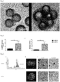
- Fig. 1 shows micrographs, at A of the aggregates of the micro-carriers with the PSC on day 0, at B at day 7 of the aggregates of micro-carriers with differentiated iPSC/megakaryocyte progenitors. These pictures show that the cultivation results in a drastic increase in size of the aggregates, indicating growth of the megakaryocytes.
- iPSC and megakaryocytes obtained by differentiation from the PSC were cultivated as pure cell aggregates, without presence of suspended micro-carriers.
- hCBiPSC2 cells that were cultivated in laminin-coated culture plates as described in Example 1 were harvested and seeded into StemMACS medium. Then, the PSC were transferred to animal serum-free synthetic medium APEL (StemCell, Vancouver, Canada) supplemented with 5% protein-free Hybridoma medium II (PFHMII, Gibco by Life Technologies, Darmstadt, Germany). The PSC were incubated in spinner flasks at 50 rpm until aggregates consisting of PSC only were formed. In case, differentiation of the aggregates was observed, the ROCK inhibitor Y-27632 (Calbiochem Novabiochem, Sandhausen, Germany) was added to a concentration of 10 ng/mL medium.
- BMP4 and VEGF were added to a concentration each of 50 ng/mL
- SCF and TPO were added to a concentration each of 50 ng/mL medium
- IL-3 was added to a concentration of 25 ng/mL medium.
- two times per week medium was exchanged for fresh medium containing the SCT and TPO, each at 50 ⁇ g/mL. Cultivation was in spinner flasks by stirring at 50 rpm.
- Megakaryocytes were separated from the culture supernatant by the steps of 5 min centrifugation 800 rpm or sedimentation for 30 min.
- Fig. 2 at A shows the total number of megakaryocytes and at B shows the number of megakaryocytes (MK) obtained per initial iPSC cell introduced into the process, obtained according to Example 2 (Cell-aggregates, left-hand column) and obtained according to Example 1 (Cell-MC-aggregates, right-hand column). ** denotes standard deviation p ⁇ 0.01.
- Fig. 2 clearly shows that the process using cultivation of aggregates of micro-carriers and megakaryocytes results in a drastic increase in production of megakaryocytes, both in absolute numbers and in relation to initial numbers of PSC that were processed by the process.
- Fig. 3 shows analytical results for the megakaryocytes produced according to Example 1 (Cell-MC-aggregates) and according to Example 2 (Cell-aggregates).
- Fig. 3A depicts the flow cytometric results of DNA content measurement in megakaryocytes stained with propidium iodide, showing that the megakaryocytes produced according to Example 1 had a higher DNA content compared to the megakaryocytes obtained according to Example 2. This indicates that the process using aggregates of megakaryocytes with micro-carriers generates megakaryocytes of a higher quality than the process using aggregates consisting of megakaryocytes only.
- the irradiated megakaryocytes and an aliquot of the same megakaryocytes that was separated before irradiating were used to generate platelets by cultivating the megakaryocytes under static conditions in APEL medium supplemented with 100ng/mL TPO and 50ng/mL SDF-1 ⁇ in the upper chamber of a double chamber bioreactor containing a 2 ⁇ m pore membrane for separating the lower chamber.
- Medium of the same composition was pumped through the lower chamber at a rate of 140 mL/min, and platelets were isolated from the medium exiting the lower chamber.
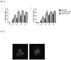
- Fig. 5 left micrograph, shows platelets produced by non-irradiated megakaryocytes, and Fig. 5 , right micrograph shows platelets produced by irradiated megakaryocytes. This results shows that functional platelets can be produced by the in vitro process from the megakaryocytes, and that the megakaryocytes can be irradiated without significantly impairing the functionality of the platelets generated by them.
- Example 4 Use of megakaryocytes as a medicament for replacing platelets in vivo
- NOD/SCID/IL-2R ⁇ c-/- mice were injected into the tail vein with 3 x 10 6 megakaryocytes that were produced according to Example 1 or Example 2.
Landscapes
- Health & Medical Sciences (AREA)
- Engineering & Computer Science (AREA)
- Life Sciences & Earth Sciences (AREA)
- Biomedical Technology (AREA)
- Hematology (AREA)
- Zoology (AREA)
- Chemical & Material Sciences (AREA)
- Biotechnology (AREA)
- Organic Chemistry (AREA)
- Bioinformatics & Cheminformatics (AREA)
- Genetics & Genomics (AREA)
- Wood Science & Technology (AREA)
- General Health & Medical Sciences (AREA)
- Immunology (AREA)
- Cell Biology (AREA)
- Biochemistry (AREA)
- General Engineering & Computer Science (AREA)
- Microbiology (AREA)
- Virology (AREA)
- Developmental Biology & Embryology (AREA)
- Medicinal Chemistry (AREA)
- Pharmacology & Pharmacy (AREA)
- Epidemiology (AREA)
- Animal Behavior & Ethology (AREA)
- Public Health (AREA)
- Veterinary Medicine (AREA)
- Micro-Organisms Or Cultivation Processes Thereof (AREA)
Abstract
The invention provides an in vitro process for producing megakaryocytes, and optionally platelets from the megakaryocytes, comprising the steps of cultivating stem cells, e.g. induced pluripotent stem cells, to generate aggregated pluripotent stem cells, preferably cultivating the aggregates in suspension in medium, inducing differentiation in these aggregates, and isolating megakaryocytes from the culture medium.
Description
- The present invention relates to an in vitro process for the production megakaryocytes, especially derived from induced pluripotent stem cells (iPSC), in a cell culture vessel equipped to provide controlled culture conditions, e.g. in an agitated bioreactor.
- The production process of the invention is suitable for producing megakaryocytes for use in medical treatment, e.g. for use in the treatment of thrombocytopenia, e.g. for use in the treatment of platelet refractoriness, for use in the state of allo immunization to HLA or human platelet antigens, in the case of pregnancy, or in a transplant patient. Optionally, a lysate can be prepared of the megakaryocytes, and the lysate is for use in the medical treatment. It was found that the administration, e.g. by infusion, of megakaryocytes produced according to the invention to a patient results in an effective generation of platelets from the megakaryocytes.
- Optionally, the process for producing megakaryocytes can comprise subsequent in vitro steps for generating platelets from the megakaryocytes for use of the platelets in medical treatment, e.g. for use in the treatment of thrombocytopenia.
- Borger et al., Mol Med 22:274-285 (2016), describe that a HLA-universal induced pluripotent stem cell (iPSC) line, designated hCBiPSC2, derived from human cord blood endothelial cells, was adapted to feeder-free and xeno-free culture by transfer onto culture plates coated with human recombinant laminin (Laminin-521, available from BioLamina). The genetic manipulation was by lentiviral transduction.
- Sivalingam et al., Tissue Engineering C, 765-780 (2016), describe expansion of hPSC by cultivation on micro-carriers coated with human recombinant Laminin-521 using serum-free culture medium with agitation to suspend the hPSC-covered micro-carriers. These hPSC formed embroid bodies on the micro-carriers for subsequent erythroid differentiation and production of red blood cells (RBC).
-
US2012/0238020 A1 describes the production of platelets by culturing stem cells for generating megakaryocyte progenitor cells and seeding these onto bone marrow stroma cells previously grown on culture dishes to generate mature megakaryocytes, which are then isolated and cultured in a three-dimensional matrix using a specific medium with application of shear-stress for producing platelets. -
US 2012/0028352 A1 describes cultivating mesenchymal stem cells, which preferably are multipotent, by allowing the stem cells to attach to micro-carrier beads having a size in the range of 50 to 400 µm in agitated suspension culture. - Reems et al., Transfus Med Rev., 24(1), 33-43 (2010), quote that CD34+ cells isolated from peripheral blood could be cultured to produce megakaryocytes, which at the peak of proplatelet formation exhibited proplatelets and platelet-sized fragments in 40% of megakaryocytes. It is described that optimization of culture conditions for stem cell self-renewal and amplification of progenitors for megakaryocytes prior to differentiation to megakaryocytes could result in yields for industrial scale production.
- It is an object of the invention to provide an alternative process for producing megakaryocytes, especially on the basis of induced pluripotent stem cells (iPSC), e.g. on the basis of an induced pluripotent stem cell line, in feeder cell-free and xeno-free conditions. A preferred object is that the process has a high production rate of megakaryocytes from the iPSC.
- The invention achieves the object by the features of the claims, especially by an in vitro process for producing megakaryocytes, and optionally platelets from the megakaryocytes, comprising the steps of cultivating stem cells which are at least pluripotent, e.g. induced pluripotent stem cells, to generate aggregated pluripotent stem cells, preferably cultivating the aggregates in suspension in medium, inducing differentiation in these aggregates, and isolating megakaryocytes from the culture medium. It was found that the aggregates release megakaryocytes into the medium. The process comprises or essentially consists of the following steps:
- a) Cultivating stem cells which are at least pluripotent, preferably induced pluripotent stem cells (iPSC), to generate cultivated stem cells,
- b) cultivating the stem cells, optionally preferably contacting the stem cells with micro-carriers, e.g. with the stem cells being suspended in cell culture medium containing the micro-carriers, preferably for a static cultivation phase, e.g. of at least 6h or at least 12h, preferably of at least 24h, more preferably of 12 to 36h, which micro-carriers can be non-porous, and which micro-carriers preferably are coated with a protein promoting adherent growth, which is e.g. recombinantly produced human laminin or fibronectin, to generate aggregates comprising or consisting of stem cells and optional micro-carriers,
- c) cultivating the aggregates comprising or consisting of stem cells and optional micro-carriers in suspension in medium with agitation to produce cultivated stem cell aggregates or cultivated aggregates of stem cells and optional micro-carrier,
- d) differentiating the cultivated stem cells to megakaryocytes by adding differentiation factors to the cultivated aggregates comprising stem cells,
- e) separating the megakaryocytes from the cultivated stem cell aggregates or from the cultivated aggregates of stem cells and micro-carrier, e.g. separating the megakaryocytes from the medium containing the cultivated stem cell aggregates or cultivated aggregates of stem cell and micro-carrier, in order to obtain the megakaryocytes,
- f) and optionally cultivating the megakaryocytes under conditions for generating platelets from megakaryocytes, and further optionally isolating the platelets,
- g) and optionally irradiating the megakaryocytes and/or platelets.
- In the process, the stem cells are cultivated to generate stem cell aggregates that are suspended in the medium, wherein the stem cell aggregates preferably contain a microcarrier, and these stem cell aggregates produce megakaryocytes that are suspended, e.g. as single megakaryocyte cells, in the medium. The stem cell aggregates, optionally containing a microcarrier, upon addition of the differentiation factors in step d) generate megakaryocyte progenitor cells and then megakaryocytes as suspended single cells.
- Generally, cultivation is in cell culture medium under cell culture conditions, e.g. at 37°C in a 5% CO2 atmosphere.
- The process has the advantage of providing feeder cell-free and xeno-free conditions, e.g. the process does not contact, directly or indirectly, the iPSC nor the megakaryocytes nor platelets with other cells nor with human or animal serum.
- In step a), the stem cells which are at least pluripotent, preferably are induced pluripotent stem cells. Further, the stem cells which are at least pluripotent can be omnipotent or totipotent stem cells. The stem cells, especially iPSC, can originate from a biopsy, which can be a blood sample, a tissue sample, e.g. an organ sample or an isolated cell sample, possibly originating from the later recipient, i.e. patient, of the megakaryocytes or of the platelets derived from the megakaryocytes, or the stem cells can originate from another human or a stem cell line. As a further example, the stem cells can be CD34+ and/or hematopoietic progenitor cells or stem cells, e.g. umbilical cord blood cells, or a human iPS cell line.
- In step a), the stem cells are e.g. seeded in cell culture plates. Generally, step a) is an amplification or expansion step of the stem cells under conditions maintaining the stem cell characteristics, e.g. without differentiating the stem cells prior to step d). Seeding in step a) is e.g. to a density of 40000 to 60000 cells per cm2 plate surface. The cells can be cultivated with daily feeding with cell culture medium. The stem cells in step a) preferably are passaged when reaching confluency, e.g. 2 or 3 times per week. The stem cells preferably are cultivated under static cell culture conditions, e.g. in culture plates, which preferably are coated for adherent growth, e.g. with fibronectin or with Laminin, e.g. with human recombinant Laminin-521, which plates preferably are otherwise non-treated culture plates. The medium for cultivating the stem cells preferably is adapted for stem cell cultivation, especially for expansion of CD34+ cells, e.g. StemMACS™ iPS-Brew XF , or StemMACS HSC Expansion media XF (both available from Miltenyi Biotech GmbH, Germany), which contains recombinant cytokines and human stem cell factor, Flt3-ligand, and thrombopoietin.
- Optionally, between step a) and step d), the stem cells are transferred into protein-free medium.
- In step d), the cultivated stem cells (iPSC) are exposed to factors inducing mesoderm differentiation followed by their differentiation into megakaryocytes. The differentiation factors preferably are BMP4 (bone morphogenetic protein 4, e.g. added to 50 ng/mL) and VEGF (vascular endothelial growth factor, e.g. added to 50 ng/mL), both of which are preferably added both on
day 0 and day 2 of step b), and additionally SCF (stem cell factor, e.g. added to 50 ng/mL), TPO (thrombopoietin, e.g. added to 50 ng/mL) and IL-3 (interleukin-3, e.g. added to 25 ng/mL) are added both on day 4 and day 8. In step b), the cultivated stem cells can be cultivated in serum-free medium, e.g. APEL2 medium (available from StemCell, Vancouver, Canada), preferably supplemented, e.g. at 5 %, with protein-free hybridoma medium, e.g. PFHMII (available from Gibco Life Technologies, Darmstadt, Germany). Preferably, from day 12, the medium containing SCF (50 ng/mL) and TPO (50 ng/mL) was exchanged two times per week for fresh medium. - Step b) can be carried out with agitation, e.g. in spinner flasks.
- In step c), the stem cells of step b) are cultivated to produce aggregates comprising stem cells.
- In one embodiment, the iPS cells (stem cells) are cultivated as a single cell suspension to generate aggregates comprising or consisting of megakaryocytes. In this embodiment, which is less preferred, in steps b) and c), the stem cells are cultivated without the micro-carriers, i.e. in carrier-free suspension in medium. In this embodiment, the medium preferably contains ROCK inhibitor, e.g. Y-27632 (available from Calbiochem Novabiochem, Sandhausen, Germany). The medium in this embodiment may contain protein, preferably the medium is animal serum-free, e.g. protein-free. It was found that in this embodiment, the stem cells, preferably dissociated to a single cell suspension, during the cultivation form aggregates consisting of cells only. Cultivation can be with agitation, e.g. in spinner flasks, e.g. agitated at 50 rpm. In this embodiment, cultivated iPS cells were preferably seeded to 12.5 x 10⁶ cells per 50 mL medium.
- In the preferred embodiment, in step b) the iPS cells obtained from step a) are contacted with micro-carriers, e.g. the stem cells are in suspension in cell culture medium containing the micro-carriers, preferably for an initial static cultivation phase, e.g. of at least 6h or at least 12h, preferably of at least 24h, more preferably of 12 to 36h. The micro-carriers preferably are non-porous, and preferably the micro-carriers are coated with a protein promoting adherent growth, e.g. coated with fibronectin and/or human laminin, preferably recombinantly produced human laminin, to generate aggregates comprising or consisting of stem cells and micro-carriers, and it is assumed that these aggregates contain progenitor cells of megakaryocytes. The aggregates comprising or consisting of iPS cells, megakaryocyte-progenitor cells and micro-carriers have been found to release a higher number of megakaryocytes into suspension than stem cell aggregates which do not contain micro-carriers. Megakaryocytes produced from stem cell aggregates containing micro-carriers have a high capacity to produce functional platelets, also after irradiating the megakaryocytes, than stem cell aggregates without micro-carriers.
- In step d), which can optionally be carried out concurrent to step c), cells generally preferably are in protein-free medium. Micro-carriers can be of polymer, e.g. of polystyrene. Generally, micro-carriers can have a size distribution in the range from 100 to 300 µm, e.g. 120 to 250 µm. Preferably the micro-carriers are coated with, e.g. have a layer of, fibronectin, preferably of laminin, preferably human recombinant laminin, e.g. Laminin-521 (LN521, available from BioLamina, Sundbyberg, Sweden). Coating of micro-carriers can be obtained by contacting sterile micro-carriers with laminin in solution, e.g. for at least 24 h at 4 °C. For attachment of iPS cells to micro-carriers, the iPS cells preferably are cultivated in a static phase, i.e. without agitation of the medium, for at least 12 h, for at least 24 h, e.g. up to 48 h or up to 36 h, e.g. for approximately 24 h. The iPS cells can be used at 6.25 x 10⁶ cells per 50 mL medium, and micro-carriers, preferably of non-porous polystyrene having a size distribution of 125 to 212 µm, can be used e.g. at 5 to 350 mg/mL.
- In step c), preferably following the static phase, the aggregates comprising or consisting of iPS cells/megakaryocyte-progenitors, preferably aggregates comprising or consisting of iPS cells/megakaryocyte-progenitors and micro-carriers, are preferably cultivated for at least 19d, e.g. for 12 to 22 d, under agitation, with exchange of the medium against fresh medium.
- In the process of the invention, iPSC/megakaryocyte-progenitor aggregates, preferably in aggregates with micro-carriers, are cultivated in order to increase the numbers of megakaryocytes that are released into the culture medium. This procedure was found to generate aggregates containing stem cells, especially stem cell aggregates containing micro-carriers, wherein the proportion of megakaryocytes released into the medium increases, e.g. in relation to the number of initially seeded stem cells.
- Megakaryocytes can be separated from the cultivated aggregates suspended in the medium by gravity separation, e.g. using centrifugation, or size separation, e.g. on a filtration membrane, from the supernatant of the culture.
- The preferred process, which comprises cultivation of aggregates of megakaryocytes and micro-carriers, resulted in a yield of up to 42 megakaryocytes per initial iPSC, whereas the process using cultivation of iPSC as cell aggregates suspended in medium but without micro-carriers under otherwise identical conditions resulted in about 5 megakaryocytes per initial iPSC. This increase in the production level by the use of aggregates of megakaryocytes with micro-carriers (p = 0.0006) shows the higher efficacy of this process.
PSC are e.g. characterized by expressing TRA-1-60 and SSEA-4 as pluripotency markers. In the alternative to PSC, totipotent or omnipotent stem cells can be used. - When the megakaryocytes produced by the process are introduced into a recipient, e.g. a patient, e.g. by injection into a vein, platelets are generated by the megakaryocytes within the recipient and can be detected in the circulation, e.g. 1h or 2h subsequent to introduction of the megakaryocytes into the recipient.
- The step f) of producing platelets by in vitro cultivating the megakaryocytes under conditions for generating platelets, with further optionally isolating the platelets from the megakaryocytes and/or from the medium, can be under static culture conditions, e.g. plating the megakaryocytes in cell culture plates, preferably tissue culture treated plates, in medium supplemented with thrombopoietin (TPO), e.g. at 100 µg/mL. The medium for platelet production from megakaryocytes may be APEL, RPMI, or IMDM, with RPMI and IMDM, respectively, preferably supplemented with 5% AB serum (vol/vol, c.c. pro). Megakaryocytes can be cultured in this way for e.g. up to 7 days. Platelets can be isolated from the culture supernatant.
- In the alternative, step f) can be performed by cultivating megakaryocytes under static conditions in one chamber of a double chamber reactor, which is separated from the other chamber by a porous membrane having 2 µm pores. After allowing the megakaryocytes to adhere to the porous membrane, medium is flowed through the other chamber, e.g. APEL, RPMI, or IMDM, supplemented with 100 ng/mL TPO and 50 ng/mL SDF-1α (Peprotech) Platelets can be isolated from the medium that was flowed through the other chamber, e.g. from 6h to 48h following the start of the medium flowing through the other chamber.
- It was found that the megakaryocytes produced by the process comprising or consisting of steps a) to e) can be irradiated without significant reduction of their capability in generating functional platelets, e.g. in vivo after administration to a patient and/or in vitro. The step of irradiating the megakaryocytes and/or platelets produced in vitro can greatly reduce the risk of contamination by replicating nucleic acids, e.g. by prokaryotes or by a virus.
- Irradiation of the iPSC-derived megakaryocytes and/or platelets can reduce the risk of teratoma formation in a recipient. Further, it was found that platelets produced according to the process comprising or consisting of steps a) to f) can be irradiated without significant reduction of their function, e.g. of their in vivo function after administration to a patient. Accordingly, the process optionally comprises the step of g) irradiating the megakaryocytes produced by the process, and/or irradiating the platelets produced from the megakaryocytes by the process.
- The step of irradiating can e.g. be exposing the megakaryocytes and/or platelets to gamma irradiation for a dose of from 25 to 50Gy.
- Further optionally, the megakaryocytes and/or platelets produced by the process can be lysed, e.g. by at least 6 repetitive freezing (-20°C) and thaw (37°C) cycles to produce a lysate of the megakaryocytes and/or platelets, and the lysate can be used in the medical treatment e.g. wound healing or as a substitute of additives in cell culture e.g. FCS.
- The invention is now described in greater detail by way of examples and with reference to the figures, which show in
-
Fig. 1 micrographs of aggregates of iPSC/megakaryocyte-progenitors and micro-carriers, at A at the end of step c) and at B at the end of step d) of the process, -
Fig. 2 graphic displays of the yield of megakaryocytes from the process, -
Fig. 3 at A results of flow cytometry, at B fluorescence micrographs, at C light micrographs of megakaryocyte aggregates produced in the process, without micro-carriers (upper row) and of aggregates of iPSC/megakaryocyte-progenitors with micro-carriers produced in the process (lower row), -
Fig. 4 a graphic display of analytical results for megakaryocyte markers, and in -
Fig. 5 micrographs of platelets produced in vitro from megakaryocytes produced according to the invention, without irradiating the megakaryocytes (left) or following irradiating of the megakaryocytes (right). - As an example for PSC, the human iPSC line (hCBiPSC2) was cultivated in conditions without feeder cells and without serum components, giving feeder-free and xeno-free conditions. These cells were seeded at 50000 cells /cm2 on 12 non-treated culture plates (Falcon by Corning, USA) coated with human recombinant laminin, LN521 (BioLamina). The iPSC were fed daily with StemMACS (Miltenyi Biotech) medium and passaged 2 to 3 times per week when confluency was reached.
- In this exemplary process, the differentiation of stem cells to megakaryocytes of step b) and the preferred step c) including contacting of the megakaryocytes with micro-carriers are done concurrently. From the culture plates, iPS cells were harvested and suspended at a concentration of 6.25 cells/mL APEL and StemMACS (V/V) or only APEL medium, containing 50 ng/mL BMP4 and 50 ng/mL VEGF, both of which are added both on
day 0 and day 2 of this culture. On day 4 and day 8, SCF was added to 50 ng/mL and TPO was added to 50 ng/mL and IL-3 was added to 25 ng/mL for inducing differentiation to megakaryocytes in APEL medium. - As micro-carriers, animal-product free non-porous polystyrene micro-carriers (SoloHill, PALL, Dreieich, Germany) were sterilized by autoclaving and then coated with laminin by suspending the micro-carriers in 2 µg/mL human recombinant laminin (LN521, BioLamina) in PBS buffer for at least 24 h at 4°C.
- On
day 0, PSC were gently mixed at a density of 12.5 x 10⁶ cells per 50 mL medium with added 281.25 mg of the laminin-coated micro-carriers per 50 mL medium and incubated without agitation for approximately 24 h. During this static incubation phase, aggregates of the micro-carriers with the PSC were formed. Then, the suspension was incubated with agitation by stirring at 50 rpm in spinner flasks for at least 12 d prior to isolating megakaryocytes, with exchange of the medium for fresh medium every 2 to 3 days. -
Fig. 1 shows micrographs, at A of the aggregates of the micro-carriers with the PSC onday 0, at B atday 7 of the aggregates of micro-carriers with differentiated iPSC/megakaryocyte progenitors. These pictures show that the cultivation results in a drastic increase in size of the aggregates, indicating growth of the megakaryocytes. - Megakaryocytes were isolated from the culture supernatant twice a week, starting from day 12 after the addition of the differentiation factors on
day 0. - Analysis of cell size showed that these megakaryocytes had an increased cell size and polyploidy resembling that of human native bone marrow-derived megakaryocytes.
- In this example, iPSC and megakaryocytes obtained by differentiation from the PSC were cultivated as pure cell aggregates, without presence of suspended micro-carriers. hCBiPSC2 cells that were cultivated in laminin-coated culture plates as described in Example 1 were harvested and seeded into StemMACS medium. Then, the PSC were transferred to animal serum-free synthetic medium APEL (StemCell, Vancouver, Canada) supplemented with 5% protein-free Hybridoma medium II (PFHMII, Gibco by Life Technologies, Darmstadt, Germany). The PSC were incubated in spinner flasks at 50 rpm until aggregates consisting of PSC only were formed. In case, differentiation of the aggregates was observed, the ROCK inhibitor Y-27632 (Calbiochem Novabiochem, Sandhausen, Germany) was added to a concentration of 10 ng/mL medium.
- On
day 0 and day 2 of differentiation of the cultivated cell-only aggregates to megakaryocytes, BMP4 and VEGF were added to a concentration each of 50 ng/mL, then on day 4 and day 8 SCF and TPO were added to a concentration each of 50 ng/mL medium and IL-3 was added to a concentration of 25 ng/mL medium. From day 12 onwards, two times per week medium was exchanged for fresh medium containing the SCT and TPO, each at 50 µg/mL. Cultivation was in spinner flasks by stirring at 50 rpm. - Megakaryocytes were separated from the culture supernatant by the steps of 5 min centrifugation 800 rpm or sedimentation for 30 min.
-
Fig. 2 at A shows the total number of megakaryocytes and at B shows the number of megakaryocytes (MK) obtained per initial iPSC cell introduced into the process, obtained according to Example 2 (Cell-aggregates, left-hand column) and obtained according to Example 1 (Cell-MC-aggregates, right-hand column). ** denotes standard deviation p<0.01.Fig. 2 clearly shows that the process using cultivation of aggregates of micro-carriers and megakaryocytes results in a drastic increase in production of megakaryocytes, both in absolute numbers and in relation to initial numbers of PSC that were processed by the process. -
Fig. 3 shows analytical results for the megakaryocytes produced according to Example 1 (Cell-MC-aggregates) and according to Example 2 (Cell-aggregates). -
Fig. 3A depicts the flow cytometric results of DNA content measurement in megakaryocytes stained with propidium iodide, showing that the megakaryocytes produced according to Example 1 had a higher DNA content compared to the megakaryocytes obtained according to Example 2. This indicates that the process using aggregates of megakaryocytes with micro-carriers generates megakaryocytes of a higher quality than the process using aggregates consisting of megakaryocytes only. - Megakaryocytes that were produced according to Example 1, using cultivation of aggregates of micro-carriers and megakaryocytes, were isolated from the culture medium and irradiated with 30Gy
- The irradiated megakaryocytes and an aliquot of the same megakaryocytes that was separated before irradiating were used to generate platelets by cultivating the megakaryocytes under static conditions in APEL medium supplemented with 100ng/mL TPO and 50ng/mL SDF-1α in the upper chamber of a double chamber bioreactor containing a 2µm pore membrane for separating the lower chamber. Medium of the same composition was pumped through the lower chamber at a rate of 140 mL/min, and platelets were isolated from the medium exiting the lower chamber.
- The platelets were deposited on a collagen-coated glass and analysed by microscopy after anti-CD61 staining.
Fig. 5 , left micrograph, shows platelets produced by non-irradiated megakaryocytes, andFig. 5 , right micrograph shows platelets produced by irradiated megakaryocytes. This results shows that functional platelets can be produced by the in vitro process from the megakaryocytes, and that the megakaryocytes can be irradiated without significantly impairing the functionality of the platelets generated by them. - As a representative of a human recipient patient, NOD/SCID/IL-2Rγc-/- mice were injected into the tail vein with 3 x 10⁶ megakaryocytes that were produced according to Example 1 or Example 2.
- After 1h following injection of the megakaryocytes, platelets originating from the injected megakaryocytes were found.
Claims (15)
- Process for producing megakaryocytes from stem cells (SC) which are at least pluripotent, the process comprising the steps ofa) cultivating the stem cells to generate cultivated stem cells,b) cultivating the stem cells, optionally contacting the stem cells with micro-carriers to generate aggregates comprising or consisting of megakaryocytes and optional micro-carriers,c) cultivating the aggregates comprising the stem cells in suspension in medium with agitation to produce cultivated aggregates comprising megakaryocytes,d) differentiating the aggregates comprising stem cells to megakaryocytes by adding differentiation factors to the cultivated stem cells,e) separating megakaryocytes from the cultivated aggregates in order to obtain the megakaryocytes,f) and optionally cultivating the megakaryocytes under conditions for generating platelets from megakaryocytes, and further optionally isolating the platelets,g) and optionally irradiating the megakaryocytes and/or platelets.
- Process according to claim 1, characterized in that the stem cells are induced pluripotent stem cells (iPSC).
- Process according to one of the preceding claims, characterized in that step c) and step d) are carried out concurrently.
- Process according to one of the preceding claims, characterized in that in step b) the stem cells are contacted in suspension with micro-carriers to generate aggregates comprising or consisting of stem cells and micro-carriers, and that in step c) the aggregates comprising or consisting of megakaryocytes and micro-carriers are cultivated in suspension in medium with agitation to produce cultivated aggregates comprising stem cells and micro-carriers.
- Process according to one of the preceding claims, characterized in that in step e) the megakaryocytes are separated from the stem cell-aggregates, respectively from the stem cell-micro-carrier-aggregates.
- Process according to one of the preceding claims, characterized in that in step b) the stem cells are incubated under cell culture conditions for a static phase for at least 12 h, followed by cultivation with agitation.
- Process according to one of the preceding claims, characterized in that the micro-carriers are non-porous.
- Process according to one of the preceding claims, characterized in that micro-carriers are coated with a protein promoting adherent growth, e.g. fibronectin and/or laminin.
- Process according to one of the preceding claims, characterized by step g), irradiating megakaryocytes or platelets with at least 5Gy.
- Process according to one of the preceding claims, characterized by lysing the megakaryocytes and/or platelets to generate a lysate from the megakaryocytes and/or platelets, wherein the lysate is for use in a medical treatment.
- Serum-free megakaryocytes obtainable by a process according to one of the preceding claims.
- Serum-free platelets obtainable by a process according to one of claims 1 to 10.
- Serum-free megakaryocytes according to claim 11 for use as a medicament in the treatment of thrombocytopenia, platelet refractoriness, for use in the treatment of alloimmunization to HLA or alloimmunization to human platelet antigens, for use in the treatment of a pregnant woman, or for use in the treatment of a transplant patient.
- Serum-free platelets according to claim 12 for use as a medicament in the treatment of thrombocytopenia, platelet refractoriness, for use in the treatment of alloimmunization to HLA or alloimmunization to human platelet antigens, for use in the treatment of a pregnant woman, or for use in the treatment of a transplant patient.
- Serum-free megakaryocytes according to claim 13 or serum-free platelets accroding to claim 14, lysed to generate a lysate, for use in the treatment of alloimmunization to HLA or alloimmunization to human platelet antigens, for use in the treatment of a pregnant woman, or for use in the treatment of a transplant patient.
Priority Applications (4)
| Application Number | Priority Date | Filing Date | Title |
|---|---|---|---|
| EP18170657.3A EP3564363A1 (en) | 2018-05-03 | 2018-05-03 | Production of megakaryocytes in bioreactors |
| US17/046,850 US12480093B2 (en) | 2018-05-03 | 2019-05-02 | Production of megakaryocytes in bioreactors |
| PCT/EP2019/061263 WO2019211383A1 (en) | 2018-05-03 | 2019-05-02 | Production of megakaryocytes in bioreactors |
| EP19720895.2A EP3788140A1 (en) | 2018-05-03 | 2019-05-02 | Production of megakaryocytes in bioreactors |
Applications Claiming Priority (1)
| Application Number | Priority Date | Filing Date | Title |
|---|---|---|---|
| EP18170657.3A EP3564363A1 (en) | 2018-05-03 | 2018-05-03 | Production of megakaryocytes in bioreactors |
Publications (1)
| Publication Number | Publication Date |
|---|---|
| EP3564363A1 true EP3564363A1 (en) | 2019-11-06 |
Family
ID=62116289
Family Applications (2)
| Application Number | Title | Priority Date | Filing Date |
|---|---|---|---|
| EP18170657.3A Withdrawn EP3564363A1 (en) | 2018-05-03 | 2018-05-03 | Production of megakaryocytes in bioreactors |
| EP19720895.2A Pending EP3788140A1 (en) | 2018-05-03 | 2019-05-02 | Production of megakaryocytes in bioreactors |
Family Applications After (1)
| Application Number | Title | Priority Date | Filing Date |
|---|---|---|---|
| EP19720895.2A Pending EP3788140A1 (en) | 2018-05-03 | 2019-05-02 | Production of megakaryocytes in bioreactors |
Country Status (3)
| Country | Link |
|---|---|
| US (1) | US12480093B2 (en) |
| EP (2) | EP3564363A1 (en) |
| WO (1) | WO2019211383A1 (en) |
Families Citing this family (1)
| Publication number | Priority date | Publication date | Assignee | Title |
|---|---|---|---|---|
| EP3813853A4 (en) * | 2018-06-29 | 2022-04-06 | Platelet Biogenesis, Inc. | COMPOSITIONS FOR DRUG DELIVERY AND METHODS OF USE THEREOF |
Citations (2)
| Publication number | Priority date | Publication date | Assignee | Title |
|---|---|---|---|---|
| US20120028352A1 (en) | 2008-03-17 | 2012-02-02 | Agency For Science, Technology And Research | Microcarriers for Stem Cell Culture |
| US20120238020A1 (en) | 2011-03-18 | 2012-09-20 | Mitchell W Beau | Megakaryocyte and Platelet Production from Stem Cells |
-
2018
- 2018-05-03 EP EP18170657.3A patent/EP3564363A1/en not_active Withdrawn
-
2019
- 2019-05-02 EP EP19720895.2A patent/EP3788140A1/en active Pending
- 2019-05-02 US US17/046,850 patent/US12480093B2/en active Active
- 2019-05-02 WO PCT/EP2019/061263 patent/WO2019211383A1/en not_active Ceased
Patent Citations (2)
| Publication number | Priority date | Publication date | Assignee | Title |
|---|---|---|---|---|
| US20120028352A1 (en) | 2008-03-17 | 2012-02-02 | Agency For Science, Technology And Research | Microcarriers for Stem Cell Culture |
| US20120238020A1 (en) | 2011-03-18 | 2012-09-20 | Mitchell W Beau | Megakaryocyte and Platelet Production from Stem Cells |
Non-Patent Citations (9)
| Title |
|---|
| BAIGGER ANJA ET AL: "Towards the Manufacture of Megakaryocytes and Platelets for Clinical Application.", TRANSFUSION MEDICINE AND HEMOTHERAPY : OFFIZIELLES ORGAN DER DEUTSCHEN GESELLSCHAFT FUR TRANSFUSIONSMEDIZIN UND IMMUNHAMATOLOGIE JUN 2017, vol. 44, no. 3, June 2017 (2017-06-01), pages 165 - 173, XP002782029, ISSN: 1660-3796 * |
| BÖRGER ANN-KATHRIN ET AL: "Generation of HLA-universal iPSCs-derived megakaryocytes and platelets for survival under refractoriness conditions.", MOLECULAR MEDICINE (CAMBRIDGE, MASS.) 16 MAY 2016, vol. 22, 16 May 2016 (2016-05-16), XP002782030, ISSN: 1528-3658 * |
| BORGER ET AL., MOL MED, vol. 22, 2016, pages 274 - 285 |
| CHEN-WACKER C ET AL: "IN VITRO DIFFERENTIATION OF RED BLOOD CELLS FROM INDUCED PLURIPOTENT STEM CELLS AS ALTERNATIVE RESOURCE FOR TRANSFUSION AND A MODEL FOR GENE THERAPY", CYTOTHERAPY, vol. 19, no. 5, Suppl. S, May 2017 (2017-05-01), & 23RD ANNUAL MEETING OF THE INTERNATIONAL-SOCIETY-FOR-CELLULAR-THERAPY (ISCT); LONDON, UK; MAY 03 -06, 2017, pages S135, XP002782031 * |
| LU SHI-JIANG ET AL: "3D microcarrier system for efficient differentiation of human pluripotent stem cells into hematopoietic cells without feeders and serum", REGENERATIVE MEDICINE, vol. 8, no. 4, July 2013 (2013-07-01), pages 413 - 424, XP002782028 * |
| LYE T. LOCK ET AL: "Expansion and Differentiation of Human Embryonic Stem Cells to Endoderm Progeny in a Microcarrier Stirred-Suspension Culture", TISSUE ENGINEERING PART A, vol. 15, no. 8, 1 August 2009 (2009-08-01), pages 2051 - 2063, XP055077769, ISSN: 1937-3341, DOI: 10.1089/ten.tea.2008.0455 * |
| REEMS ET AL., TRANSFUS MED REV., vol. 24, no. 1, 2010, pages 33 - 43 |
| SIVALINGAM ET AL., TISSUE ENGINEERING C, 2016, pages 765 - 780 |
| SUGIMOTO N ET AL: "Platelet production from induced pluripotent stem cells.", JOURNAL OF THROMBOSIS AND HAEMOSTASIS : JTH SEP 2017, vol. 15, no. 9, September 2017 (2017-09-01), pages 1717 - 1727, XP002782032, ISSN: 1538-7836 * |
Also Published As
| Publication number | Publication date |
|---|---|
| US12480093B2 (en) | 2025-11-25 |
| WO2019211383A1 (en) | 2019-11-07 |
| US20210371824A1 (en) | 2021-12-02 |
| EP3788140A1 (en) | 2021-03-10 |
Similar Documents
| Publication | Publication Date | Title |
|---|---|---|
| Liu et al. | Biomimetic three-dimensional cultures significantly increase hematopoietic differentiation efficacy of embryonic stem cells | |
| US20250333701A1 (en) | Compositions and methods for generating hematopoietic stem cells (hscs) | |
| US20040043484A1 (en) | Hematopoietic differentiation of human embryonic stem cells | |
| EP3567102A1 (en) | Method for developing natural killer cells from stem cells | |
| US8278101B2 (en) | Stem cell bioprocessing and cell expansion | |
| US20100248361A1 (en) | Platelet production methods | |
| CN114540266A (en) | Preparation and freeze-drying of fucosylated cells for therapeutic use | |
| JPWO2008056779A1 (en) | Method for culturing and subculture of primate embryonic stem cells and method for inducing differentiation thereof | |
| US7811821B2 (en) | Method of forming dendritic cells from embryonic stem cells | |
| KR20020013480A (en) | Human brain endothelial cells and growth medium and method for expansion of primitive cd34+cd38- bone marrow stem cells | |
| US12037607B2 (en) | Methods of preparing hematopoietic progenitor cells in vitro | |
| WO2008056963A1 (en) | Method for proliferating stem cells with leptin | |
| JP2023522326A (en) | Induced Pluripotent Cells Containing Controllable Transgenes for Conditional Immortalization | |
| CN111808812A (en) | A material and method for the differentiation of pluripotent stem cells into hematopoietic stem cells | |
| WO2024251234A1 (en) | Serum-free culture medium for mesenchymal stem cells and use thereof | |
| WO2025077137A1 (en) | Method for preparing hematopoietic stem cells and progenitor cells | |
| CN106119200A (en) | Relate to the cultural method broken up by pluripotent stem cell to hemocyte | |
| US12480093B2 (en) | Production of megakaryocytes in bioreactors | |
| CN110951686A (en) | Hematopoietic stem cell in-vitro amplification culture system and method | |
| WO2023019213A1 (en) | Three-dimensional culturing of pluripotent stem cells to produce hematopoietic stem cells | |
| WO1998021313A1 (en) | Method for culturing hematopoietic stem cells | |
| JP2025511790A (en) | Large-Scale Production of iPSC-Derived HSCs and Progeny | |
| WO2024113294A1 (en) | Method for differentiating nk cells, culture medium and use thereof | |
| CN115927180A (en) | A method for differentiating NK cells, culture medium and application thereof | |
| WO2021200901A1 (en) | T cell progenitor production method |
Legal Events
| Date | Code | Title | Description |
|---|---|---|---|
| PUAI | Public reference made under article 153(3) epc to a published international application that has entered the european phase |
Free format text: ORIGINAL CODE: 0009012 |
|
| AK | Designated contracting states |
Kind code of ref document: A1 Designated state(s): AL AT BE BG CH CY CZ DE DK EE ES FI FR GB GR HR HU IE IS IT LI LT LU LV MC MK MT NL NO PL PT RO RS SE SI SK SM TR |
|
| AX | Request for extension of the european patent |
Extension state: BA ME |
|
| STAA | Information on the status of an ep patent application or granted ep patent |
Free format text: STATUS: THE APPLICATION IS DEEMED TO BE WITHDRAWN |
|
| 18D | Application deemed to be withdrawn |
Effective date: 20200603 |