EP3555267B1 - Buoyancy enabled separation method and system - Google Patents
Buoyancy enabled separation method and system Download PDFInfo
- Publication number
- EP3555267B1 EP3555267B1 EP17881611.2A EP17881611A EP3555267B1 EP 3555267 B1 EP3555267 B1 EP 3555267B1 EP 17881611 A EP17881611 A EP 17881611A EP 3555267 B1 EP3555267 B1 EP 3555267B1
- Authority
- EP
- European Patent Office
- Prior art keywords
- cells
- cell
- microbubbles
- disruptable
- container
- Prior art date
- Legal status (The legal status is an assumption and is not a legal conclusion. Google has not performed a legal analysis and makes no representation as to the accuracy of the status listed.)
- Active
Links
Images







Classifications
-
- A—HUMAN NECESSITIES
- A61—MEDICAL OR VETERINARY SCIENCE; HYGIENE
- A61K—PREPARATIONS FOR MEDICAL, DENTAL OR TOILETRY PURPOSES
- A61K39/00—Medicinal preparations containing antigens or antibodies
- A61K39/0005—Vertebrate antigens
- A61K39/0011—Cancer antigens
-
- C—CHEMISTRY; METALLURGY
- C12—BIOCHEMISTRY; BEER; SPIRITS; WINE; VINEGAR; MICROBIOLOGY; ENZYMOLOGY; MUTATION OR GENETIC ENGINEERING
- C12M—APPARATUS FOR ENZYMOLOGY OR MICROBIOLOGY; APPARATUS FOR CULTURING MICROORGANISMS FOR PRODUCING BIOMASS, FOR GROWING CELLS OR FOR OBTAINING FERMENTATION OR METABOLIC PRODUCTS, i.e. BIOREACTORS OR FERMENTERS
- C12M47/00—Means for after-treatment of the produced biomass or of the fermentation or metabolic products, e.g. storage of biomass
- C12M47/02—Separating microorganisms from the culture medium; Concentration of biomass
-
- C—CHEMISTRY; METALLURGY
- C12—BIOCHEMISTRY; BEER; SPIRITS; WINE; VINEGAR; MICROBIOLOGY; ENZYMOLOGY; MUTATION OR GENETIC ENGINEERING
- C12N—MICROORGANISMS OR ENZYMES; COMPOSITIONS THEREOF; PROPAGATING, PRESERVING, OR MAINTAINING MICROORGANISMS; MUTATION OR GENETIC ENGINEERING; CULTURE MEDIA
- C12N5/00—Undifferentiated human, animal or plant cells, e.g. cell lines; Tissues; Cultivation or maintenance thereof; Culture media therefor
- C12N5/0081—Purging biological preparations of unwanted cells
-
- C—CHEMISTRY; METALLURGY
- C12—BIOCHEMISTRY; BEER; SPIRITS; WINE; VINEGAR; MICROBIOLOGY; ENZYMOLOGY; MUTATION OR GENETIC ENGINEERING
- C12N—MICROORGANISMS OR ENZYMES; COMPOSITIONS THEREOF; PROPAGATING, PRESERVING, OR MAINTAINING MICROORGANISMS; MUTATION OR GENETIC ENGINEERING; CULTURE MEDIA
- C12N5/00—Undifferentiated human, animal or plant cells, e.g. cell lines; Tissues; Cultivation or maintenance thereof; Culture media therefor
- C12N5/0081—Purging biological preparations of unwanted cells
- C12N5/0087—Purging against subsets of blood cells, e.g. purging alloreactive T cells
-
- C—CHEMISTRY; METALLURGY
- C12—BIOCHEMISTRY; BEER; SPIRITS; WINE; VINEGAR; MICROBIOLOGY; ENZYMOLOGY; MUTATION OR GENETIC ENGINEERING
- C12N—MICROORGANISMS OR ENZYMES; COMPOSITIONS THEREOF; PROPAGATING, PRESERVING, OR MAINTAINING MICROORGANISMS; MUTATION OR GENETIC ENGINEERING; CULTURE MEDIA
- C12N5/00—Undifferentiated human, animal or plant cells, e.g. cell lines; Tissues; Cultivation or maintenance thereof; Culture media therefor
- C12N5/0081—Purging biological preparations of unwanted cells
- C12N5/0093—Purging against cancer cells
-
- C—CHEMISTRY; METALLURGY
- C12—BIOCHEMISTRY; BEER; SPIRITS; WINE; VINEGAR; MICROBIOLOGY; ENZYMOLOGY; MUTATION OR GENETIC ENGINEERING
- C12N—MICROORGANISMS OR ENZYMES; COMPOSITIONS THEREOF; PROPAGATING, PRESERVING, OR MAINTAINING MICROORGANISMS; MUTATION OR GENETIC ENGINEERING; CULTURE MEDIA
- C12N5/00—Undifferentiated human, animal or plant cells, e.g. cell lines; Tissues; Cultivation or maintenance thereof; Culture media therefor
- C12N5/06—Animal cells or tissues; Human cells or tissues
- C12N5/0602—Vertebrate cells
- C12N5/0634—Cells from the blood or the immune system
- C12N5/0636—T lymphocytes
-
- G—PHYSICS
- G01—MEASURING; TESTING
- G01N—INVESTIGATING OR ANALYSING MATERIALS BY DETERMINING THEIR CHEMICAL OR PHYSICAL PROPERTIES
- G01N33/00—Investigating or analysing materials by specific methods not covered by groups G01N1/00 - G01N31/00
- G01N33/48—Biological material, e.g. blood, urine; Haemocytometers
- G01N33/50—Chemical analysis of biological material, e.g. blood, urine; Testing involving biospecific ligand binding methods; Immunological testing
- G01N33/53—Immunoassay; Biospecific binding assay; Materials therefor
- G01N33/536—Immunoassay; Biospecific binding assay; Materials therefor with immune complex formed in liquid phase
-
- G—PHYSICS
- G01—MEASURING; TESTING
- G01N—INVESTIGATING OR ANALYSING MATERIALS BY DETERMINING THEIR CHEMICAL OR PHYSICAL PROPERTIES
- G01N33/00—Investigating or analysing materials by specific methods not covered by groups G01N1/00 - G01N31/00
- G01N33/48—Biological material, e.g. blood, urine; Haemocytometers
- G01N33/50—Chemical analysis of biological material, e.g. blood, urine; Testing involving biospecific ligand binding methods; Immunological testing
- G01N33/53—Immunoassay; Biospecific binding assay; Materials therefor
- G01N33/536—Immunoassay; Biospecific binding assay; Materials therefor with immune complex formed in liquid phase
- G01N33/537—Immunoassay; Biospecific binding assay; Materials therefor with immune complex formed in liquid phase with separation of immune complex from unbound antigen or antibody
- G01N33/538—Immunoassay; Biospecific binding assay; Materials therefor with immune complex formed in liquid phase with separation of immune complex from unbound antigen or antibody by sorbent column, particles or resin strip, i.e. sorbent materials
-
- G—PHYSICS
- G01—MEASURING; TESTING
- G01N—INVESTIGATING OR ANALYSING MATERIALS BY DETERMINING THEIR CHEMICAL OR PHYSICAL PROPERTIES
- G01N33/00—Investigating or analysing materials by specific methods not covered by groups G01N1/00 - G01N31/00
- G01N33/48—Biological material, e.g. blood, urine; Haemocytometers
- G01N33/50—Chemical analysis of biological material, e.g. blood, urine; Testing involving biospecific ligand binding methods; Immunological testing
- G01N33/53—Immunoassay; Biospecific binding assay; Materials therefor
- G01N33/543—Immunoassay; Biospecific binding assay; Materials therefor with an insoluble carrier for immobilising immunochemicals
- G01N33/54313—Immunoassay; Biospecific binding assay; Materials therefor with an insoluble carrier for immobilising immunochemicals the carrier being characterised by its particulate form
- G01N33/5432—Liposomes or microcapsules
-
- G—PHYSICS
- G01—MEASURING; TESTING
- G01N—INVESTIGATING OR ANALYSING MATERIALS BY DETERMINING THEIR CHEMICAL OR PHYSICAL PROPERTIES
- G01N33/00—Investigating or analysing materials by specific methods not covered by groups G01N1/00 - G01N31/00
- G01N33/48—Biological material, e.g. blood, urine; Haemocytometers
- G01N33/50—Chemical analysis of biological material, e.g. blood, urine; Testing involving biospecific ligand binding methods; Immunological testing
- G01N33/53—Immunoassay; Biospecific binding assay; Materials therefor
- G01N33/543—Immunoassay; Biospecific binding assay; Materials therefor with an insoluble carrier for immobilising immunochemicals
- G01N33/544—Immunoassay; Biospecific binding assay; Materials therefor with an insoluble carrier for immobilising immunochemicals the carrier being organic
-
- G—PHYSICS
- G01—MEASURING; TESTING
- G01N—INVESTIGATING OR ANALYSING MATERIALS BY DETERMINING THEIR CHEMICAL OR PHYSICAL PROPERTIES
- G01N33/00—Investigating or analysing materials by specific methods not covered by groups G01N1/00 - G01N31/00
- G01N33/48—Biological material, e.g. blood, urine; Haemocytometers
- G01N33/50—Chemical analysis of biological material, e.g. blood, urine; Testing involving biospecific ligand binding methods; Immunological testing
- G01N33/53—Immunoassay; Biospecific binding assay; Materials therefor
- G01N33/569—Immunoassay; Biospecific binding assay; Materials therefor for microorganisms, e.g. protozoa, bacteria, viruses
- G01N33/56966—Animal cells
- G01N33/56972—White blood cells
-
- A—HUMAN NECESSITIES
- A61—MEDICAL OR VETERINARY SCIENCE; HYGIENE
- A61K—PREPARATIONS FOR MEDICAL, DENTAL OR TOILETRY PURPOSES
- A61K39/00—Medicinal preparations containing antigens or antibodies
- A61K2039/51—Medicinal preparations containing antigens or antibodies comprising whole cells, viruses or DNA/RNA
- A61K2039/515—Animal cells
- A61K2039/5156—Animal cells expressing foreign proteins
-
- A—HUMAN NECESSITIES
- A61—MEDICAL OR VETERINARY SCIENCE; HYGIENE
- A61K—PREPARATIONS FOR MEDICAL, DENTAL OR TOILETRY PURPOSES
- A61K39/00—Medicinal preparations containing antigens or antibodies
- A61K2039/51—Medicinal preparations containing antigens or antibodies comprising whole cells, viruses or DNA/RNA
- A61K2039/515—Animal cells
- A61K2039/5158—Antigen-pulsed cells, e.g. T-cells
-
- C—CHEMISTRY; METALLURGY
- C07—ORGANIC CHEMISTRY
- C07K—PEPTIDES
- C07K14/00—Peptides having more than 20 amino acids; Gastrins; Somatostatins; Melanotropins; Derivatives thereof
- C07K14/435—Peptides having more than 20 amino acids; Gastrins; Somatostatins; Melanotropins; Derivatives thereof from animals; from humans
- C07K14/705—Receptors; Cell surface antigens; Cell surface determinants
- C07K14/70503—Immunoglobulin superfamily
- C07K14/7051—T-cell receptor (TcR)-CD3 complex
-
- C—CHEMISTRY; METALLURGY
- C07—ORGANIC CHEMISTRY
- C07K—PEPTIDES
- C07K2319/00—Fusion polypeptide
- C07K2319/01—Fusion polypeptide containing a localisation/targetting motif
- C07K2319/03—Fusion polypeptide containing a localisation/targetting motif containing a transmembrane segment
-
- C—CHEMISTRY; METALLURGY
- C12—BIOCHEMISTRY; BEER; SPIRITS; WINE; VINEGAR; MICROBIOLOGY; ENZYMOLOGY; MUTATION OR GENETIC ENGINEERING
- C12N—MICROORGANISMS OR ENZYMES; COMPOSITIONS THEREOF; PROPAGATING, PRESERVING, OR MAINTAINING MICROORGANISMS; MUTATION OR GENETIC ENGINEERING; CULTURE MEDIA
- C12N2501/00—Active agents used in cell culture processes, e.g. differentation
- C12N2501/50—Cell markers; Cell surface determinants
-
- C—CHEMISTRY; METALLURGY
- C12—BIOCHEMISTRY; BEER; SPIRITS; WINE; VINEGAR; MICROBIOLOGY; ENZYMOLOGY; MUTATION OR GENETIC ENGINEERING
- C12N—MICROORGANISMS OR ENZYMES; COMPOSITIONS THEREOF; PROPAGATING, PRESERVING, OR MAINTAINING MICROORGANISMS; MUTATION OR GENETIC ENGINEERING; CULTURE MEDIA
- C12N2501/00—Active agents used in cell culture processes, e.g. differentation
- C12N2501/50—Cell markers; Cell surface determinants
- C12N2501/515—CD3, T-cell receptor complex
-
- C—CHEMISTRY; METALLURGY
- C12—BIOCHEMISTRY; BEER; SPIRITS; WINE; VINEGAR; MICROBIOLOGY; ENZYMOLOGY; MUTATION OR GENETIC ENGINEERING
- C12N—MICROORGANISMS OR ENZYMES; COMPOSITIONS THEREOF; PROPAGATING, PRESERVING, OR MAINTAINING MICROORGANISMS; MUTATION OR GENETIC ENGINEERING; CULTURE MEDIA
- C12N2501/00—Active agents used in cell culture processes, e.g. differentation
- C12N2501/50—Cell markers; Cell surface determinants
- C12N2501/599—Cell markers; Cell surface determinants with CD designations not provided for elsewhere
Definitions
- the present disclosure relates to a simple end-to-end device and system for the bulk isolation, serial multiple-marker sorting, stimulation, manipulation, culture, and expansion of cells and other biological agents.
- a major challenge for all cell therapies is the need to develop cost-effective and efficient manufacturing and delivery capabilities, such as creating a scalable, automated closed system to isolate a subset of T cells.
- CAR chimeric antigen receptor
- TCR T cell receptor
- Sorting and isolating a subset of T cells requires multiple markers (e.g. CD8, CD62L & CD45RA), which is challenging to scale up and automate.
- FACS fluorescence activated cell sorting
- MACS magnetic cell sorting
- MACS Magnetic Activated Cell Sorting
- the prior art contains a few methods enabling the reversible binding of cells and affinity ligand-conjugated magnetic beads. These were developed by sequentially targeting multiple cell-surface markers.
- the Streptamer technology enables the reversible target cell binding of magnetic microparticles ( ⁇ 1 ⁇ m) by conjugating to Strep-Tactin multimerized low-affinity single-chain antibodies that have been engineered to fuse with a Strep-tag.
- Other technologies in the prior art use modified antibodies that can dissociate from cells or magnetic beads. These methods usually require extensive modifications to the antibodies of interest and can be applied to other platforms beyond MACS, such as FACS, BACS and other forms of affinity purification.
- WO 2015/175344 A1 teaches a BACS method for clinical applications, including the isolation of circulating tumor cells and cord blood stem cells.
- This technology has several advantages over other prior art in performing bulk cell isolation.
- First, its lipid shell microbubble is self-molding to external forces (e.g. ultrasound and bound cells) because it is the most compressible and flexible shell-class microbubbles (others including albumin, polymer, and glass). In conjunction with a gas core, it is a very gentle material for cell isolation.
- the microbubble it uses is a "self-driving vehicle", as microbubble-bound cells automatically float to the top surface of a liquid.
- the lipid-shelled microbubbles it uses self-separate from water-based solutions and self-concentrate/aggregate to other microbubbles.
- users of this technology working with cell-bound microbubbles can disrupt their internal bonds, without causing cell damage, by increasing ambient air pressure.
- Emulsification is used to prepare perflorohexane gas microbubbles (MBs) within phospholipid shells.
- DSPE-PEG 3400-maleimide is used in the microbubble membrane to conjugate antibodies to the microbubbles. It uses Fc fragment-specific IgG carrying 1-2 thiol groups per antibody to conjugate antibodies onto maleimide-activated MBs via Michael addition.
- the technology uses targeting antibodies, such as anti-EpCAM, the most widely accepted marker for isolating circulating tumor cells of epithelial origin for coupling. Most the MBs in this process have a diameter of 3-10 ⁇ m. Each MB has, on average, 367,000 anti-Fc IgG molecules.
- BACS (or BUBLES) is an innovative cell isolation/sorting platform that can be used alone or in combination with the other two major platforms in the prior art (FACS and MACS) for challenging tasks.
- FACS and MACS two major platforms in the prior art
- the present disclosure uses BACS for the bulk isolation, multiple-marker sorting, and manipulation of human cells in a single container device.
- the present disclosure uses BACS in conjunction with FACS or MACS for this isolation, sorting, and manipulation process.
- Cell based therapy is a rapidly developing medicine of intense research and great potential to patient benefits.
- the majority of cell therapies in clinical trials today are hematopoietic or mesenchymal stem cells and immune T cells for genetic diseases and cancers, these targeted cells have common and well known characteristics of being very rare subsets and the need of multiple cell surface markers for specific isolation from the heterogeneous cell mixtures in blood or human tissues, and of being very difficult to regulate cell differentiation progress or maintain cell stemness during isolation, expansion and in vitro manipulation procedures.
- This invention is intended to overcome a number of these obstacles.
- the invention is an object to overcome two major challenges for all cell therapies, namely (1) a robust manufacturing procedure to cost-effectively scale up the sufficient quantity of targeted cells; and (2) the capability to control and maintain the biological characteristics of targeted cells prior to clinical administration to patients.
- the aforementioned goals (1) and (2) can be accomplished by a simple end-to-end example device and example system for executing the method according to the invention, providing technology to overcome/improve these two challenges, more specifically to provide the bulk isolation, serial multiple-marker sorting, stimulation, manipulation, culture, and expansion of cells and other biological agents in a sterile environment that is safe from contamination for the sequential steps of accomplishing the aforementioned goals.
- An embodiment of the invention is referred to the Buoyancy enabled separation method according to the invention can be performed with an example system for executing the method according to the invention for clinical applications, in the following referred to by the acronym BUBLES, including the isolation of circulating tumor cells, cord blood stem cells and T lymphocytes.
- BUBLES the isolation of circulating tumor cells, cord blood stem cells and T lymphocytes.
- This technology has several advantages over others in performing bulk cell isolation.
- its lipid shell microbubble is self-molding to external forces (e.g. ultrasound and bound cells), because it is the most compressible and flexible shell of microbubbles, for instance compared to albumin, polymer or glass.
- a gas core it is a very gentle material for cell isolation.
- the microbubble is a "self-driving vehicle", as microbubble-bound cells automatically float to the top surface of a liquid.
- the lipid-shelled microbubbles self-separate from water-based solutions and self-concentrate/aggregate to other microbubbles.
- cell-bound microbubbles can be disrupted from their internal bonds, without causing cell damage, by increasing ambient air pressure. The following sections will describe how these unique advantages of microbubble can help resolve the cell therapy challenges.
- the present disclosure uses serial BACS (or BUBLES), with antibody-based affinity ligands attached to microbubbles, to repeatedly isolate, sort, stimulate, expand or manipulate human cells in a single, closed container.
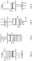
- Figures 1-4 demonstrate one variation of the present disclosure's preferred embodiment.
- the sample cells 1 are peripheral blood mononuclear cells (PBMC) 1 are mixed and incubated with anti-CD8 microbubble 2 in a syringe-like container 3.
- PBMC peripheral blood mononuclear cells
- Microbubbles 2 and targeted cells 4 gradually float to the liquid surface ( FIG. 2 ).
- the targeted cell 4 population (CD8+ cells) is retained (positive selection) by pushing the plunger 5 ( FIG. 3 ) with the container opening 6 facing the ground ( FIG. 2 ).
- the microbubbles 2 are subsequently disrupted by increasing ambient air pressure in the air space 0 within the container, which is created by compressing air when a cap 7 is installed to the opening of the container ( FIG.
- the second type of targeted microbubble 2 (anti-CD62L) with buffer is mixed with the anti-CD8 sorted cells.
- the process restarts as previously described ( Figures 2-4 ).
- the naive CD8+ T cell population (CD8+CD62L+CD45RA+) in the syringe-like container 3 is stimulated and expanded by binding to anti-CD3/anti-CD28 conjugated microbubbles 2 in culture medium.
- Subsequent manipulations e.g. viral transduction with chimeric antigen receptors can also be performed in the same container 3.
- An exemplary connecting/closing mechanism in an example system for executing the method according to the invention can comprise a standard Luer-Lok 8 that interfaces with a number of medical and laboratory instruments.
- An adaptor 9 or 11 seals the opening 6 at one end of the syringe-like container 3 to manipulate its affinity ligands conjugated microbubble, and cell contents and to collect harvested cells, and a plunger 5, with a plunger tip 10 modified to be convex or concave in shape, which fills the space between the affinity ligands conjugated microbubbles 2, and cells in solution, is inserted into the other end of the syringe-like container 3.
- the plunger may be operated automatically or manually.
- the shape of the plunger tip 10 may be adjusted or tuned by the mechanism for a diaphragm valve, as schematically shown by the arrows between FIGs. 7 and 8 demonstrating a back and forth the changing of the shape of the front face of the plunger 5.
- the shape of the plunger can be adjusted during use of the example system for executing the method according to the invention to accommodate the needs of the user during different steps of the selection process. For instance, the plunger can be adjusted concave to accommodate more floated microbubbles and covex to expel more waste from the final product. These plunger front face shapes be also adjusted during the process to enhance the performance of each step. It is also possible to exchange plungers entirely for different steps, meaning that a kit is provided having a selection of plungers. Although this provides a simplification of the design, exchanging plungers between steps may not be preferred from a sterility point of view since it would involve opening the container other than through the cap 7, Luer Lok 8 or adaptor 9.
- an exemplary device for executing the method according to the invention may consist of different hard or soft closed containers, different syringe-like container 3 configuration types, different adapter or plug types, different affinity ligand combinations, and plungers that are inverted at different angles or not inverted at all.
- the other embodiments may also isolate, sort, manipulate, stimulate, and expand other types of cells and biological agents.
- the other embodiments of the disclosure may use MACS or FACS in conjunction with BACS.
- FIGS 1-4 demonstrate one variation of an example system for executing the method according to the invention.
- the closed container 3 consists of a simple syringe-like configuration with a plunger that may be closed with a cap as described above.
- an affinity ligands conjugated microbubble 2 solution that is used to isolate, sort, manipulate, stimulate, or expand sample cells or biological agents.
- serial BACS or BUBLES
- PBMC at 10 8 /ml in 0.2% BSA buffer were first incubated with anti-CD8 MB (microbubbles) in a syringe.
- the mixture in the syringe was gently rotated at 4°C for 20 min.
- the syringe was kept in the position as shown in FIG. 2 for 10 min, followed by gently pushing the plunger to remove undesirable cells in solution, and to keep the buoyant MB layer as shown in FIG. 3 .
- the retained MB layer was washed once by refilling 0.2% BSA buffer.
- the example syringe was kept as shown in FIG.
- FIG. 5 demonstrates the top of an exemplary device for executing the method according to the invention.
- the syringe-like configuration comprising, in part, a closed container, uses a Luer Lok 8, allowing the device to interface with a variety of medical and laboratory instruments.
- FIG. 6 demonstrates a cylindrical adaptor 9 is connected to the opening 6 of the syringe-like container 3, to seal the container for creating higher ambient pressure by pushing the plunger as demonstrated in FIG. 4 to disrupt microbubbles.
- FIG. 6 demonstrates the top half of another exemplary device for executing the method according to the invention.
- a different, inverted adaptor 11 is used to collect isolated, sorted, manipulated, expanded, or stimulated cell sample. Otherwise, FIG. 6 is identical to FIG. 5 .
- adaptors such as generic 2-way, 3-way or 4-way Luer Lok connectors, are connected to the opening 6 of the syringe-like container 3, for collecting sorted, isolated, manipulated, stimulated, or expanded sample in bulk following the repeated application of BACS in the closed container.
- the sample is infused with an affinity ligands conjugated microbubble solution, and consists of target cells population from human blood and other body fluids or tissues.
- FIG. 7 demonstrates the plunger 5 of an exemplary device for executing the method according to the invention.
- the plunger tip 10 of the plunger 5 which may be inter-converted to form a concave shape through a mechanism that controls a diaphragm valve or other similar devices.
- the tip is modified through the application of liquid or air pressure into the plunger 5.
- the configuration of the plunger tip 5 is used to manipulate targeted microbubbles 2 and infused human cells.
- FIG. 8 demonstrates the plunger 5 according to another exemplary device for executing the method according to the invention.
- the plunger tip 10 of the plunger which may be inter-converted to form a convex shape through a mechanism that controls a diaphragm valve or other similar devices. Otherwise, FIG 8 is identical to FIG. 7 .
- Figures 5 - 8 demonstrate an exemplary system for executing the method according to the invention used in one of the present disclosure's preferred embodiments.
- This example system applies BACS and uses affinity ligands conjugated microbubbles 2 for the bulk isolation, sorting, manipulation, expansion, or stimulation of cells and other biological agents repeatedly in a closed container 3.
- a user would begin with the steps demonstrated in FIG. 1 or FIG. 2 , then would follow the step demonstrated in FIG. 3 , and finally would follow the step demonstrated in FIG. 4 .
- the user could begin the process again, with another type of targeted microbubble 2, by returning to the steps demonstrated in Figures 2 - 4 .
- These steps are performed repetitively to achieve bulk isolation, sorting, manipulation, stimulation, or expansion of the sample biological agent.
- FIGS 1 - 4 demonstrate an example of the present disclosure's example BACS system for executing the method according to the invention, using microbubbles infused with targeted microbubbles.
- the BACS system can be applied in a syringe-like configuration comprising the closed container that uses a Luer Lok, an adapter rather than a plug, and a plunger with an inverted tip.
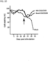
- Figures 9-10 demonstrate the cell viability and proliferation results of a preferred embodiment of the present disclosure's microbubble solution relative to other embodiments of the present disclosure's solution and microbubble solutions existing in the prior art.
- CD3/CD28 affinity ligand combination conjugated microbubbles are infused for the isolation, sorting, expansion, manipulation, or stimulation of T cells.
- Both microbubble conjugated CD3/CD28 and Dynabead conjugated CD3/CD28 were added to the cell culture at the beginning and the time point where the arrow indicates on the graph.
- Figures 9-10 demonstrate this preferred embodiment achieving greater cell viability percentages and cell proliferation than solutions used in other embodiments of the present disclosure and in the prior art (Dynabeads). This superior outcome may reflect the more natural interaction between microbubbles and T cells to recapitulate immunological synapse.
- FIG. 11 demonstrates a preferred embodiment of the present disclosure's microbubble solution.
- the microbubble solution is infused with microbubbles conjugated with CD3/CD28 ligand combination, after cell isolation in a single container has occurred.
- This anti-CD3/CD28 microbubble solution can be applied to expand T-cells with greater efficiency and quality than anti-CD3/CD28 Dynabeads, which were developed in the prior art.
- the cell expansion profile (CD8/CD4 T cell ratio) is preferred for use in cancer immunotherapy after CD3/CD28 stimulation.
- microbubbles can be conjugated with other types of ligands for similar cell-contact dependent signaling applications.
- affinity ligand combinations infused microbubbles can be disrupted over time or more precisely controlled by applying higher ambient pressure, which enables the creation of a convenient feeder-cell free cell contact dependent cell culture system.
- MBs perflorohexane gas microbubbles
- DSPE-PEG 3400-maleimide was included in the membrane in order to conjugate the antibody Fc fragment-specific IgG carrying 1-2 thiol groups per antibody was conjugated onto maleimide-activated MBs via Michael addition.
- Targeting antibodies, such as anti-EpCAM, were coupled in the second step.
- the majority of the MBs had a diameter of 3-10 ⁇ m (similar to the size of most mammalian cells). Each MB had, on average, 367,000 anti-Fc IgG molecules.
- BUBLES is an innovative cell isolation/sorting platform that can be used alone or in combination with the other two major platforms, FACS and MACS, for challenging tasks. This summary mainly focuses on the description and discussion of using BUBLES in the clinical application of cell therapy for the bulk isolation, multiple-marker sorting, and manipulation of human cells in a single container device. However, in other clinical or research situations, BUBLES can also be used alone or in conjunction with FACS or MACS to achieve most optimal results.
- FACS fluoresce activated cell sorting
- MACS Magnetic cell sorting
- Miltenyi system are used for targeted cell isolation in bulk.
- Cells bound to microbeads e.g. Dynabeads
- MACS Magnetic cell sorting
- One of the biggest problems with MACS is that isolated cells coupled to magnetic microparticles (> 1 ⁇ m) are often destroyed. Therefore, most magnetic cell sorting systems on the market use nanobead over microbead-conjugated antibodies.
- MACS Magnetic Activated Cell Sorting
- BUBLES technology provides the serial positive or negative selection to isolate rare targeted cells from heterogeneous cell mixtures
- Microbubbles can be disrupted by application of higher ambient pressure (such pressure is well tolerant by mammalian cells) within the device, this advantage of BUBLES technology enables a serial positive or negative selection of multiple cell surface markers to isolate and purify the targeted cells from cell mixtures. Furthermore, this method without negative physical impact on cells results in additional benefits to preserve cell activity and characteristics, which will be discussed further in the later section.
- BUBLES technology enables selection, sorting, culture, gene transduction, and many other in vitro manipulations in a single container
- GMP manufacturing cellular therapies One of the most important concepts of GMP manufacturing cellular therapies is to maintain the cells in a system that is closed to the external environment to maintain the sterility of the products throughout the collection and manufacturing process.
- the distinct four advantages of BUBLES allow multiple in vitro manipulations in one single container in an easy operating and cost-effective fashion for cell therapy production.
- the CAR-T-CD19 cell therapy is used as an example to describe the production steps as described in FIG. 17 .
- a first configuration denoted A is shown, where a starting material apheresis PBMC ( ⁇ 10 9 cells in 8-9 ml) obtained from blood bank or clinical center is transferred to a BUBLES syringe-like container 3.
- Microbubbles 2 targeting a first cell surface marker of the targeted cells that are a sparse subset of the cell mixture in the PBMC as a sample are added. About a third of the inner volume airspace within the syringe-like container 3 is left as airspace 0.
- the mixture of PBMC and microbubbles 2 is allowed to incubate for example for 10 - 60 minutes.
- a buffer or media may be added for dilution.
- the top of the device is closed by a cap 7 or a Luer Lok 8 / adaptor 9 combination as for instance shown in FIG. 5 after the PBMC sample, the first set of microbubbles 2 and optionally the diluent and/or media were place inside the container 3.
- the container 3 is then inverted and the waste 12 discarded by pushing down the plunger 5.
- the waste 12 that includes the non-targeted cells that did not bind to the microbubbles 2 has been discarded since the distance by which the plunger 5 is pushed determines how much of the content within the container 3 is discarded, the plunger is only pushed so far that the microbubbles 2 with the cells bound to the microbubbles 2 remain within the container 3. If useful, cell washing steps could be applied before moving to the next steps in the cell selection process.
- the next step is demonstrated by configuration C, namely the syringe - like container 3 is turned around again and the plunger 5 is pulled out, increasing the AL volume within the container 3.
- the cap 7 or the Luer Lok 8 / adaptor 9 combination is installed again, sealing the container 3, so that in the next step depicted by configuration D the air pressure is increased within the container by pushing up the plunger 5, decreasing the air volume within the container 3. This results in disruption of the microbubbles 2, and detaches the cell mixture selected so far from the microbubbles 2 as depicted by configuration E.
- the steps depicted by exemplary system configurations A - E in FIG. 17 for executing the method according to the invention can be repeated for a second set of microbubbles targeting a second cell surface marker of the targeted cells.
- microbubble conjugated antibodies can be added or discarded through Luer Lok mechanism to maintain sterility of a closed system as illustrated in FIG. 17 .
- BUBLES system can separate and concentrate desired fractions within the same container by simply inverting the container, in addition, applying ambient pressure to disrupt bubble can easily break the antibody conjugation to make the serial selections possible in one container.
- the entire manipulation can be easily configured to automation mode to streamline the GMP manufactures.
- the method and example system for executing the method according to the invention also provide the advantage of maintaining a sterile environment throughout the process.
- This advantage can be further extended to also provide for cell activation and cell expansion within the same sterile environment, namely within the container holding the sparse subset of cells at a high concentration after the multiple subsequence election processes have been completed.
- the cap 7 or Luer Lok 8 / adaptor 9 combination can be replaced by an air filter, and for example for CAR-T cell therapy, a CAR gene transduction can be performed within the container 3 by adding retroviral vectors to the isolated cells.
- microbubbles binding to CD 19 cell surface markers CD 19 expressed T-cells may be transferred to a culture bag for scaling up cells, but these steps could also, in the alternative, be performed within the container 3. Additional microbubbles for concentration may then be added to the container, and the buffer may be exchanged for making the isolated and expanded/activated cells with the exchanged buffer suitable for administration to the patient. Even if it is chosen not to perform all steps within one and the same container 3, at least a major part of these steps may be performed with in that container and therefore significantly lower the risk of contamination throughout the selection + expansion/activation process.
- a prior art process includes incubating an apheresis product with anti-CD3/anti-CD28 paramagnetic Dynabeads at a ratio of 3 CD3+ cells to 1 Dynabeads, and isolating the CD3+ cell fraction by exposure to a magnet. After washing and re-suspending the selected cells in an initiation medium with low IL-2 concentration and two days of culture, the cells and beads were added to culture bags that had been treated with RetroNectin and loaded with anti-CD 19 CAR new culture bags and viral vector and incubated for at least 24 hours. This transduction step was repeated the day after, and the cells and beads were then transferred to new culture bags and expanded for nine more days. At the completion of the cell expansion, the beads were removed and discarded by again applying the magnet, and the cells were washed and prepared for infusion.
- the described BUBLES system provides a variety of advantages over current available system used in clinic, including: (1) select, isolate, purify, and enrich the rare targeted cell subsets from blood or human tissues using serial positive or negative selections of surface markers, (2) no extra work to engineer Ab, use any readily antibody, readily extending the use of this system to many types of cell therapies, (3) all the manipulations can be performed in one single container, to simplify and control manufacture procedures in a closed system, (4) easily to be configured to automation mode, (5) cost-effective by saving from GMP grade tubings and materials.
- CAR-T cell therapy is used as an example here to describe the system
- this system can be of great use for other cell therapies, such as various hematopoietic stem cells, mesenchymal stem cells, neural stem cells, immune T cells, or immune dendritic cells, since microbubble can be easily conjugated with various antibodies to serial select a particular and rare subset of cells to meet the clinical demand.
- Gene engineering is required in CAR-T cell therapy, and can be performed in a single container of this system.
- BUBLES system also can be flexible to adapt by incorporating culture bag in the procedure flow.
- BUBLES not only minimize the physical damages on targeted cells during the in-vitro/ex-vivo operation procedures, more importantly, BUBLES also provides a distinct advantage to preserve and stimulate cell potency through the mechanism of biological significance.
- CAR-T cell therapy is used as an example here to specifically discuss this issue, it should be noted that similar principles apply to the other stem cell therapies, including HSC, MSC, neural stem cells, and others, for it is important to preserve the stem cell activity of infused cells to achieve the regeneration function in patients.
- BUBLES system minimizes in vitro manipulation steps as discussed previously including: isolation and enrichment of targeted cells, cell culture and expansion, gene transduction, and other manipulations in a single container, plus, no need to wash, centrifuge, resuspend cells between steps.
- the most compressible/flexible lipid coated microbubble is self-molding to external forces (e.g. ultrasound and bound cells), in conjunction with gas core, it is a very gentle material for cells.
- a simple analogy would be eggs (cells) cushioned by air balloons (microbubbles).
- T memory stem (T SCM ) cells are a rare subset of memory lymphocytes endowed with the stem cell-like ability to self-renew and the multipotent capacity to reconstitute the entire spectrum of memory and effector T cell subsets.
- T cells undergo a sequential loss of proliferative capacity, but acquisition of effector function occurs during the process of differentiation from antigen-inexperienced na ⁇ ve cells (T N ) to CD62L+ central memory (T CM ), CD62L- effector memory (T EM ), and effector (T E ) T cell subsets.
- T N na ⁇ ve T
- T TE terminally differentiated effector T
- T SCM cell T memory stem cell
- T CM cell central memory T cell
- T EM cell effector memory T cell
- T N and T CM may provide greater therapeutic potency.
- a number of preclinical studies have confirmed that naive T cell is an ideal cell population to employ CAR-T immunotherapy, however, clinical trials have largely employed unselected peripheral blood mononuclear cell (PBMC)-derived T cell populations due to technical difficulty for isolating the relative paucity of na ⁇ ve T cells in the circulation, and the lack of robust, clinical-grade manufacturing protocols that are capable of generating and maintaining this cell type in vitro.
- PBMC peripheral blood mononuclear cell
- the isolation of less differentiated T cell populations also has the advantage of reproducibly generating more defined T cell products, unselected populations containing high proportions of T EM and effector cells might fail to generate viable clinical products owing to poor in vitro cell expansion.
- BUBLES system provides a robust manufacturing procedure to generate na ⁇ ve T cells which can then be driven to differentiate to desired T SCM cells by addition of a panel of cytokines.
- T cells In vitro cell culture of T cells is essential for receptor engineering and cell expansion to achieve sufficient dose for immunotherapy.
- na ⁇ ve and less differentiated T cells undergo a rapid loss and maturity development during cell culture and expansion period, and present distinct outcomes of each T cell subset by different differentiation protocols.
- various cytokines and Ab/stimulators are necessary to be included in the culture.
- IL-2 is required to promote T cell proliferation, but to also induce some degree of T cell maturation
- IL-7, IL-15, and IL-21 are supplemented to maintain memory T cell phenotype.
- T cell receptor TCR
- anti-CD3 or plus anti-CD28
- PBMC feeder cells
- APCs antigen presenting cells
- T cell-APC contact sets up an axis for polarization of TCR, adhesion molecules, kinases, cytoskeletal elements, and organelles inherent in this mode of juxtacrine signaling. Further lateral organization of the TCR and adhesion molecules into radially symmetric compartments, the immunological synapse, revealed an intersecting plane of symmetry and potential for regulated symmetry breaking to control duration of T cell-APC interactions.
- the immunological synapse directs the polarized transport and secretion of cytokines and cytolytic agents across the synaptic cleft and is a site for the generation and exocytic release of bioactive microvesicles that can functionally affect recipient APC and other cells in the environment, see FIG. 18 and 19 .
- T cell-mediated immune response begins with the display of a peptide, derived from a pathogen or other biological structure and loaded in a major histocompatibility complex (MHC, creating a pMHC), on the surface of an APC.
- MHC major histocompatibility complex
- This pMHC complex is recognized by a T cell receptor (TCR) complex to initiate a cascade of intracellular signaling.
- TCR T cell receptor
- Many additional receptor-ligand complexes between T cell and APC in this small ( ⁇ 75 ⁇ m 2 ) region of immunological synapse (IS) have been observed and can be varied dependent on many factors, including the type of T cell and APC that are participating in this interaction and the presence of soluble factors in the environment.
- CD28 a member of the immunoglobulin superfamily of proteins that is presented on the T cell surface.
- CD80 or CD86 CD28 provides a costimulatory signal that augments TCR-based signaling and is essential for full activation of naive T cells.
- LFA-1 a member of the integrin family of receptors, which binds to ICAM-1 on the APC and provides cell-cell adhesion as well as a costimulatory function.
- Concurrent engagement of TCR with LFA-1 and/or CD28 provides activation of many types of T cells, and these three receptors form a core set of signals that is well suited for in vitro, reductionist studies of T cell function.
- FIG. 19 a striking feature of the IS is that receptor-ligand pairings within this interface are organized into distinctive patterns of micrometer scale and smaller consisting of a micrometer-scale central Supramolecular Activation Cluster (cSMAC) region surrounded by a peripheral Supramolecular Activation Cluster (pSMAC) corresponding to zones of concentrated TCR-pMHC and LFA-1ICAM-1 binding, respectively.
- cSMAC central Supramolecular Activation Cluster
- pSMAC peripheral Supramolecular Activation Cluster
- FIG. 19 demonstrates polarity and symmetry in the mature immunological synapse.
- the dynamic and molecular components of the interaction complex between TCR and APC within the immunological synapse determine the subsequent proliferative and lineage fate of the T cells.
- TCR, CD28, and LFA-1 are associated predominantly with phosphorylation cascades, involving intermediates that are soluble in the cell cytosol.
- Such molecules typically exhibit mobility that is high (diffusion coefficients on the order of ⁇ m 2 /millisecond) relative to the rates at which protein activity can be regulated; it is difficult to obtain graded concentrations of activity of these molecules over the micrometer-scale distances that define the IS.
- TCR/CD28/LFA-1 signaling also involves intermediates that are associated with the cell membrane, either stably via a transmembrane domain or transiently through post-translational attachment of lipids or binding to specific membrane components, and thus typically exhibit diffusion coefficients on the order of ⁇ m 2 /second, values that make spatially resolved cell signaling at these scales plausible.
- TCR signaling also involves activation of pathways that modulate the assembly, disassembly, and activity of the cytoskeleton, allowing for spatial regulation of signaling at scale of micrometers and smaller.
- FIG. 19 demonstrates the polarity and symmetry in the mature immunological synapse. Supramolecular activation clusters (SMACs) are shown in the interface between T cell and APC.
- SMACs Supramolecular activation clusters
- the axis of directed secretions is the spear passing through the SMAC.
- the plane of symmetry is the plane of the immunological synapse, which is radially symmetric when stable.
- the T cell will migrate if the adhesion ring breaks symmetry.
- the fact of the immobilization influences signaling events has made this technology limited in use in research understanding of cell signaling and communication, and not suitable for large scale of T cell activation.
- the third example is a self-assembled planar bilayer structure of phospholipids. A combination of hydration, van der Waals, and other forces maintains the bilayer in close proximity to an appropriate substrate, most commonly glass.
- a thin (sub-nanometer) layer of water separates the bilayer and support, imparting lateral mobility of the phospholipids and other membrane components, including tethered proteins, in the plane of the bilayer.
- the above described examples are used in research understanding and will not be practical to be converted for industrial use due to the extreme low through put scale and the material complexity of forming such systems.
- the current standard protocol for T cell activation is the addition of Dynabeads conjugated with CD3 or plus CD28 antibody which act as an artificial activator, as described above.
- Microscale magnetic beads are designed to have similar cell size to mimic an APC, conjugation of CD3 and CD28 is to trigger the ligand-TCR binding to simulate stimulation signal via TCR complex.
- studies have shown that the addition of Dynabeads-CD3/CD28 into the in vitro T cell expansion has caused rapid loss of na ⁇ ve and memory T cells.
- a simple analogy would be eggs (cells) sandwiched with rocks (microparticles).
- nanobeads which is analogous to eggs (cells) surrounded by sand (nanoparticles) for cell isolation.
- these ligands conjugated nanoparticles are insufficient to activate T cells, perhaps not only because of the rigid nature of nanobeads, but also because the small size of the particles is not able to induce formation of immunological synapse.
- the problem of Dynabeads to recapitulate the biological function of APC could be due to failing to trigger appropriate signal transduction pathway in T cells because of both rigid and precipitation property of magnetic beads.
- the mobile lipid surface of MBs may provide the necessary dynamic IS structure upon ligand-TCR engagement.
- T cell activation was tested using Dynabeads vs. microbubbles.
- Microbubbles conjugated with anti-CD3/CD28 outperformed anti-CD3/CD28 Dynabeads on T cell expansion, especially after the second stimulation.
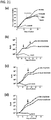
- FIG. 21 T cells from PBMC were robustly expanded after the first stimulation with anti-CD3/CD28 conjugated Dynabeads or microbubbles within the initial 2 weeks.
- the microbubbles based T cell expansion continued the same trend with the subsequent two stimulations; the Dynabeads based expansion dramatically decreased after the 2 nd stimulation ( FIG. 21 , diagram a).
- the T cell expansion is dependent on anti-CD3/CD28, as it stopped when no stimulating reagents were added when the 2 nd stimulation was performed ( FIG. 21 , mock or control groups). Results are reproducible using PBMCs from the same donor ( FIG. 21 , diagrams a and b), or different donors ( FIG. 21 , diagram b, c and d). A study from Li et al.
- microbubbles based aAPCs have a number of advantages over Dynabeads and REP as described in the following table 4: Anti-CD3/CD 28 Microbubbles Anti-CD3/CD 28 Dynabeads REP, "Rapid Expansion Protocol" Surface Lipid Solid beads Lipid Feeder cells needed? No No Yes Destructible Yes No No Particles/feeder cells removal? No Yes Yes Ligand density adjustable? Yes Yes No Cytokine source Extrinsic Extrinsic Intrinsic, uncontrolled Dynamics in cell junction? Yes No Yes Mobility of interactions? Yes No Yes No Yes Yes No Yes
- FIG. 22 demonstrates the sketch of the immunological synapse formation between T cells (indicated by T1 and T2) and anit-CD3/CD28 conjugated microbubble (MB by arrows) by immune-fluorescence assay.
- T cells indicated by T1 and T2
- anit-CD3/CD28 conjugated microbubble MB by arrows
- FIG. 22 demonstrates the sketch of the immunological synapse formation between T cells (indicated by T1 and T2) and anit-CD3/CD28 conjugated microbubble (MB by arrows) by immune-fluorescence assay.
- T cell that is not associated with MB (T3) has typical even-distributed F-actin staining within the cell.
- the cell nucleus is stained with DAPI to show the cell integrity.
- microbubbles would spontaneously burst within 10-16 hours in standard culture conditions at 37 °C.
- This unique property of microbubbles may provide additional advantages over Dynabeads to avoid overstimulation of T cells which could leads to exhaustion of T cell activity and better mimic immunological synapse formation duration as it has been shown that IS formation requires only a few hours.
- this interesting property of microbubbles not only eliminates the need for bead removal, which would reduce the chance of contaminations and the complexity for automation.
- Microbubble based technology serves as a powerful tool for other biological systems
Landscapes
- Health & Medical Sciences (AREA)
- Life Sciences & Earth Sciences (AREA)
- Engineering & Computer Science (AREA)
- Immunology (AREA)
- Chemical & Material Sciences (AREA)
- Biomedical Technology (AREA)
- Biotechnology (AREA)
- Hematology (AREA)
- Molecular Biology (AREA)
- Zoology (AREA)
- Cell Biology (AREA)
- General Health & Medical Sciences (AREA)
- Microbiology (AREA)
- Wood Science & Technology (AREA)
- Organic Chemistry (AREA)
- Bioinformatics & Cheminformatics (AREA)
- Biochemistry (AREA)
- Genetics & Genomics (AREA)
- Urology & Nephrology (AREA)
- Medicinal Chemistry (AREA)
- General Engineering & Computer Science (AREA)
- Pathology (AREA)
- General Physics & Mathematics (AREA)
- Analytical Chemistry (AREA)
- Physics & Mathematics (AREA)
- Food Science & Technology (AREA)
- Oncology (AREA)
- Sustainable Development (AREA)
- Tropical Medicine & Parasitology (AREA)
- Virology (AREA)
- Veterinary Medicine (AREA)
- Public Health (AREA)
- Animal Behavior & Ethology (AREA)
- Epidemiology (AREA)
- Pharmacology & Pharmacy (AREA)
- Mycology (AREA)
- Apparatus Associated With Microorganisms And Enzymes (AREA)
- Micro-Organisms Or Cultivation Processes Thereof (AREA)
Description
- The present disclosure relates to a simple end-to-end device and system for the bulk isolation, serial multiple-marker sorting, stimulation, manipulation, culture, and expansion of cells and other biological agents.
- A major challenge for all cell therapies is the need to develop cost-effective and efficient manufacturing and delivery capabilities, such as creating a scalable, automated closed system to isolate a subset of T cells. For example, studies in mice and humans show that the infusion of less differentiated T cells that are genetically engineered with chimeric antigen receptor (CAR) or T cell receptor (TCR) leads to superior cell expansion, persistence, and cancer elimination. Sorting and isolating a subset of T cells (e.g. naive CD8+ cells) requires multiple markers (e.g. CD8, CD62L & CD45RA), which is challenging to scale up and automate.
- FACS (fluorescence activated cell sorting) is the product most used for the detailed analysis of individual cells. Using FACS, it is difficult to scale up for bulk isolation of cells. Magnetic cell sorting (MACS) techniques, particularly the Miltenyi system, are used for targeted cell isolation in bulk. Cells bound to microbeads (e.g. Dynabeads) can be pulled out by an external magnet outside the mixing tube. However, binding antibody-conjugated microbeads to cells, rather than antibodies alone or antibody-conjugated nanobeads, slows down the process. Therefore, most magnetic cell sorting systems on the market use nanobead over microbead-conjugated antibodies. One of the biggest problems with MACS is that isolated cells coupled to magnetic microparticles (> 1 µm) are often destroyed.
- Existing MACS technology applies high-gradient magnetic cell separation columns to magnetize nanobead-labeled cells in a magnetic field, generated by a strong external magnet. MACS uses ferromagnetic steel-wool filled columns to strengthen the long-distance interaction between the low magnetic nanobeads and the external magnet. However, the use of the steel-wool packed column in this system creates other technical problems like column clogging and increased manufacturing costs. Moreover, the MACS nanobeads stick to the targeted cells, either on the cell surface or inside by endocytosis. This limits the application of MACS primarily to cell enrichment based on a single surface marker.
- As described in the International Patent Application published under the publication number
WO 2015/175344 A1 , a BUBLES (buoyancy enabled separation) or buoyancy activated cell sorting (BACS) technology by applying targeted microbubbles for cell isolation has been developed for cell isolation. - For multi-parameter MACS, the prior art contains a few methods enabling the reversible binding of cells and affinity ligand-conjugated magnetic beads. These were developed by sequentially targeting multiple cell-surface markers. For example, the Streptamer technology enables the reversible target cell binding of magnetic microparticles (∼1 µm) by conjugating to Strep-Tactin multimerized low-affinity single-chain antibodies that have been engineered to fuse with a Strep-tag. Other technologies in the prior art use modified antibodies that can dissociate from cells or magnetic beads. These methods usually require extensive modifications to the antibodies of interest and can be applied to other platforms beyond MACS, such as FACS, BACS and other forms of affinity purification.
-
WO 2015/175344 A1 teaches a BACS method for clinical applications, including the isolation of circulating tumor cells and cord blood stem cells. This technology has several advantages over other prior art in performing bulk cell isolation. First, its lipid shell microbubble is self-molding to external forces (e.g. ultrasound and bound cells) because it is the most compressible and flexible shell-class microbubbles (others including albumin, polymer, and glass). In conjunction with a gas core, it is a very gentle material for cell isolation. Second, the microbubble it uses is a "self-driving vehicle", as microbubble-bound cells automatically float to the top surface of a liquid. Third, the lipid-shelled microbubbles it uses self-separate from water-based solutions and self-concentrate/aggregate to other microbubbles. Fourth, users of this technology working with cell-bound microbubbles can disrupt their internal bonds, without causing cell damage, by increasing ambient air pressure. - Emulsification is used to prepare perflorohexane gas microbubbles (MBs) within phospholipid shells. DSPE-PEG 3400-maleimide is used in the microbubble membrane to conjugate antibodies to the microbubbles. It uses Fc fragment-specific IgG carrying 1-2 thiol groups per antibody to conjugate antibodies onto maleimide-activated MBs via Michael addition. The technology uses targeting antibodies, such as anti-EpCAM, the most widely accepted marker for isolating circulating tumor cells of epithelial origin for coupling. Most the MBs in this process have a diameter of 3-10µm. Each MB has, on average, 367,000 anti-Fc IgG molecules.
- BACS (or BUBLES) is an innovative cell isolation/sorting platform that can be used alone or in combination with the other two major platforms in the prior art (FACS and MACS) for challenging tasks. In its preferred embodiment, the present disclosure uses BACS for the bulk isolation, multiple-marker sorting, and manipulation of human cells in a single container device. In other embodiments, the present disclosure uses BACS in conjunction with FACS or MACS for this isolation, sorting, and manipulation process.
- Cell based therapy is a rapidly developing medicine of intense research and great potential to patient benefits. The majority of cell therapies in clinical trials today are hematopoietic or mesenchymal stem cells and immune T cells for genetic diseases and cancers, these targeted cells have common and well known characteristics of being very rare subsets and the need of multiple cell surface markers for specific isolation from the heterogeneous cell mixtures in blood or human tissues, and of being very difficult to regulate cell differentiation progress or maintain cell stemness during isolation, expansion and in vitro manipulation procedures. This invention is intended to overcome a number of these obstacles.
- Apart from many other objects of the invention, it is an object to overcome two major challenges for all cell therapies, namely (1) a robust manufacturing procedure to cost-effectively scale up the sufficient quantity of targeted cells; and (2) the capability to control and maintain the biological characteristics of targeted cells prior to clinical administration to patients. The aforementioned goals (1) and (2) can be accomplished by a simple end-to-end example device and example system for executing the method according to the invention, providing technology to overcome/improve these two challenges, more specifically to provide the bulk isolation, serial multiple-marker sorting, stimulation, manipulation, culture, and expansion of cells and other biological agents in a sterile environment that is safe from contamination for the sequential steps of accomplishing the aforementioned goals.
- From the
international patent application publication WO 2015/175344 A1 a system and apparatus is known using flexible shell microbubbles for isolating a cell population having markers, floating the selected cells to the top by attachment to the microbubbles. A similar method is known from Guixin Shi et al.: "Binding and isolation of tumor cells in biological media with perfluorocarbon microbubbles," METHODS, vol. 64, no. 2, 1 December 2013 (2013-12-01), pages 102 - 107, from theUnited States patent application publication US 2016/320376 A1 separating two cell types that each have its one distinct marker is known, for example CD4 lymphocyte vs. CD14 monocyte; anti-CD326 circulating tumor cells vs. anti-CD45 white blood cells. This can be accomplished by sequential isolation of CD4 lymphocytes and CD14 monocytes with anti-CD4 and anti-CD14 conjugated magnetic particles. - It is an object of the invention to isolate a cell population that requires interrogating multiple markers on the same cell type.
- This and other objects are achieved by the method as claimed in
claim 1. Advantageous further embodiments are claimed in the dependent claims. - This and other objects are achieved by the method as claimed in
claim 1 and the related system configured to perform the method as claimed inclaim 1. Advantageous further embodiments are claimed in the dependent claims. - An embodiment of the invention is referred to the Buoyancy enabled separation method according to the invention can be performed with an example system for executing the method according to the invention for clinical applications, in the following referred to by the acronym BUBLES, including the isolation of circulating tumor cells, cord blood stem cells and T lymphocytes. This technology has several advantages over others in performing bulk cell isolation. First, its lipid shell microbubble is self-molding to external forces (e.g. ultrasound and bound cells), because it is the most compressible and flexible shell of microbubbles, for instance compared to albumin, polymer or glass. In conjunction with a gas core, it is a very gentle material for cell isolation. Second, the microbubble is a "self-driving vehicle", as microbubble-bound cells automatically float to the top surface of a liquid. Third, the lipid-shelled microbubbles self-separate from water-based solutions and self-concentrate/aggregate to other microbubbles. Fourth, cell-bound microbubbles can be disrupted from their internal bonds, without causing cell damage, by increasing ambient air pressure. The following sections will describe how these unique advantages of microbubble can help resolve the cell therapy challenges.
-
-
FIG. 1 shows an exemplary system for executing the method according to an embodiment of the invention in a first configuration. -
FIG. 2 shows an exemplary system for executing the method according to an embodiment of the invention in a second configuration. -
FIG. 3 shows an exemplary system for executing the method according to an embodiment of the invention in a third configuration. -
FIG. 4 shows an exemplary system for executing the method according to an embodiment of the invention in a fourth configuration. -
FIG. 5 schematically shows a connecting/closing mechanism comprised in an exemplary system for executing the method according to the invention. -
FIG. 6 schematically shows another connecting/closing mechanism comprised in an exemplary system for executing the method according to the invention. -
FIG. 7 schematically shows a plunger comprised in an exemplary system for executing the method according to the invention. -
FIG. 8 schematically shows another plunger comprised in an exemplary system for executing the method according to the invention. -
FIG. 9 demonstrates the cell proliferation results of a preferred embodiment of the invention involving the microbubbles in comparison to the prior art. -
FIG. 10 demonstrates the cell viability results of a preferred embodiment of the invention involving the microbubbles in comparison to the prior art. -
FIG. 11 demonstrates the fold changes of cell proliferation results of a preferred embodiment of the invention involving the microbubbles in comparison to the prior art. -
FIG. 12 describes an example of naive T cells that are a rare population in blood. -
FIG. 13 describes an example of stem cells that are a rare population in tissues, here bone marrow. -
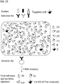
FIG. 14 demonstrates a first prior art selection strategy that results in a low targeted cells concentration mixed with other undesired cell mixtures. -
FIG. 15 demonstrates a second prior art selection strategy that results in a low targeted cells concentration mixed with other undesired cell mixtures. -
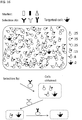
FIG. 16 demonstrates a selection strategy that results in a high targeted cells concentration that may only to a comparatively minor extent mixed with other undesired cell mixtures. -
FIG. 17 demonstrates production steps in cell therapy by positive serial selection by showing exemplary system configurations A - E in an exemplary system for executing the method according to the invention. -
FIG. 18 schematically demonstrates the interaction of ligands and receptors between the T cells and antigen presenting cells (APCs). -
FIG. 19 schematically demonstrates that receptor-ligand pairings in the immunological synapse are organized in distinctive patterns including polarity and symmetry. -
FIG. 20 schematically shows an immunological synapse between a T cell and APC, and according to an embodiment of the invention, an immunological synapse between a T cell and a microbubble simulating APC. -
FIG. 21 demonstrates comparison of T cell expansion by using anti-CD3/CD28 conjugated Dynabeads and anti-CD3/CD28 conjugated microbubbles for one or more re-stimulations with different blood donors. -
FIG. 22 demonstrates formations of immunological synapse between T cells and anti-CD3/CD28 conjugated microbubbles by fluorescence staining. - Described is an example device and example system for executing the method according to the invention for the bulk isolation, sorting, stimulation, expansion, and manipulation of cells and other biological agents in a single container. In its preferred embodiment, the present disclosure uses serial BACS (or BUBLES), with antibody-based affinity ligands attached to microbubbles, to repeatedly isolate, sort, stimulate, expand or manipulate human cells in a single, closed container.
-
Figures 1-4 demonstrate one variation of the present disclosure's preferred embodiment. Here, thesample cells 1 are peripheral blood mononuclear cells (PBMC) 1 are mixed and incubated withanti-CD8 microbubble 2 in a syringe-like container 3. Microbubbles 2 and targetedcells 4 gradually float to the liquid surface (FIG. 2 ). The targetedcell 4 population (CD8+ cells) is retained (positive selection) by pushing the plunger 5 (FIG. 3 ) with thecontainer opening 6 facing the ground (FIG. 2 ). For this positive selection, the majority of liquid is discarded. Themicrobubbles 2 are subsequently disrupted by increasing ambient air pressure in theair space 0 within the container, which is created by compressing air when acap 7 is installed to the opening of the container (FIG. 4 ), for instance by moving the plunger. For the next cycle of BACS, the second type of targeted microbubble 2 (anti-CD62L) with buffer is mixed with the anti-CD8 sorted cells. The process restarts as previously described (Figures 2-4 ). After the 3rd cycle withanti-CD45RA microbubbles 2, the naive CD8+ T cell population (CD8+CD62L+CD45RA+) in the syringe-like container 3 is stimulated and expanded by binding to anti-CD3/anti-CD28 conjugatedmicrobubbles 2 in culture medium. Subsequent manipulations (e.g. viral transduction with chimeric antigen receptors) can also be performed in the same container 3. - In the abovementioned figures, three consecutive positive selections are applied. Other applications may introduce steps for negative selection to remove targeted cells bound to
microbubbles 2. Negative selection is achieved by adopting the orientation shown inFIG. 1 with thecontainer opening 6 facing opposite to the ground instead of that showing inFIG. 2 . In connection with an alternative device or adapter specifically suited for negative selection as it has been described in theinternational patent application publication WO 2015/175344 A1 andFIG. 6 . - An exemplary connecting/closing mechanism in an example system for executing the method according to the invention can comprise a standard Luer-
Lok 8 that interfaces with a number of medical and laboratory instruments. Anadaptor opening 6 at one end of the syringe-like container 3 to manipulate its affinity ligands conjugated microbubble, and cell contents and to collect harvested cells, and aplunger 5, with aplunger tip 10 modified to be convex or concave in shape, which fills the space between the affinity ligands conjugatedmicrobubbles 2, and cells in solution, is inserted into the other end of the syringe-like container 3. The plunger may be operated automatically or manually. The shape of theplunger tip 10 may be adjusted or tuned by the mechanism for a diaphragm valve, as schematically shown by the arrows betweenFIGs. 7 and 8 demonstrating a back and forth the changing of the shape of the front face of theplunger 5. The shape of the plunger can be adjusted during use of the example system for executing the method according to the invention to accommodate the needs of the user during different steps of the selection process. For instance, the plunger can be adjusted concave to accommodate more floated microbubbles and covex to expel more waste from the final product. These plunger front face shapes be also adjusted during the process to enhance the performance of each step. It is also possible to exchange plungers entirely for different steps, meaning that a kit is provided having a selection of plungers. Although this provides a simplification of the design, exchanging plungers between steps may not be preferred from a sterility point of view since it would involve opening the container other than through thecap 7,Luer Lok 8 oradaptor 9. - Other examples of an exemplary device for executing the method according to the invention may consist of different hard or soft closed containers, different syringe-like container 3 configuration types, different adapter or plug types, different affinity ligand combinations, and plungers that are inverted at different angles or not inverted at all. The other embodiments may also isolate, sort, manipulate, stimulate, and expand other types of cells and biological agents. Finally, the other embodiments of the disclosure may use MACS or FACS in conjunction with BACS.
-
Figures 1-4 demonstrate one variation of an example system for executing the method according to the invention. In these figures, the closed container 3 consists of a simple syringe-like configuration with a plunger that may be closed with a cap as described above. Within the container 3 is an affinity ligands conjugatedmicrobubble 2 solution that is used to isolate, sort, manipulate, stimulate, or expand sample cells or biological agents. The use of serial BACS (or BUBLES) for multi-marker sorting within a single container 3 is as described above. - To demonstrate serial multi-marker isolation of naive T cells with targeted microbubbles, PBMC at 108/ml in 0.2% BSA buffer were first incubated with anti-CD8 MB (microbubbles) in a syringe. The mixture in the syringe was gently rotated at 4°C for 20 min. Subsequently, the syringe was kept in the position as shown in
FIG. 2 for 10 min, followed by gently pushing the plunger to remove undesirable cells in solution, and to keep the buoyant MB layer as shown inFIG. 3 . The retained MB layer was washed once by refilling 0.2% BSA buffer. Finally, the example syringe was kept as shown inFIG. 4 and its opening of the syringe was sealed by a cap. The microbubbles were disrupted by pushing the plunger to increase ambient pressure to about 2-3 atm. The same procedure was repeated by applying anti-CD62L MB, followed by anti-CD45RA MB. FACS analyses demonstrated that the cell purity of each single-marker isolation was more than 80% for either one of the CD8+, CD62L+ or CD45RA+ populations. In the proof-of-concept serial multi-marker isolations, the CD8+/CD62L+/CD45RA+ triple positive cell population was enriched from 2% (original PBMC) to around 60 (40-80)%. -
FIG. 5 demonstrates the top of an exemplary device for executing the method according to the invention. Here, the syringe-like configuration comprising, in part, a closed container, uses aLuer Lok 8, allowing the device to interface with a variety of medical and laboratory instruments. -
FIG. 6 demonstrates acylindrical adaptor 9 is connected to theopening 6 of the syringe-like container 3, to seal the container for creating higher ambient pressure by pushing the plunger as demonstrated inFIG. 4 to disrupt microbubbles. -
FIG. 6 demonstrates the top half of another exemplary device for executing the method according to the invention. Here, a different,inverted adaptor 11 is used to collect isolated, sorted, manipulated, expanded, or stimulated cell sample. Otherwise,FIG. 6 is identical toFIG. 5 . - Other types of adaptors, such as generic 2-way, 3-way or 4-way Luer Lok connectors, are connected to the
opening 6 of the syringe-like container 3, for collecting sorted, isolated, manipulated, stimulated, or expanded sample in bulk following the repeated application of BACS in the closed container. Here, the sample is infused with an affinity ligands conjugated microbubble solution, and consists of target cells population from human blood and other body fluids or tissues. -
FIG. 7 demonstrates theplunger 5 of an exemplary device for executing the method according to the invention. In this example device, theplunger tip 10 of theplunger 5, which may be inter-converted to form a concave shape through a mechanism that controls a diaphragm valve or other similar devices. The tip is modified through the application of liquid or air pressure into theplunger 5. The configuration of theplunger tip 5 is used to manipulate targetedmicrobubbles 2 and infused human cells. -
FIG. 8 demonstrates theplunger 5 according to another exemplary device for executing the method according to the invention. Here, theplunger tip 10 of the plunger, which may be inter-converted to form a convex shape through a mechanism that controls a diaphragm valve or other similar devices. Otherwise,FIG 8 is identical toFIG. 7 . -
Figures 5 - 8 demonstrate an exemplary system for executing the method according to the invention used in one of the present disclosure's preferred embodiments. This example system applies BACS and uses affinity ligands conjugatedmicrobubbles 2 for the bulk isolation, sorting, manipulation, expansion, or stimulation of cells and other biological agents repeatedly in a closed container 3. In operating the example system for executing the method according to the invention, a user would begin with the steps demonstrated inFIG. 1 or FIG. 2 , then would follow the step demonstrated inFIG. 3 , and finally would follow the step demonstrated inFIG. 4 . After the step demonstrated inFIG. 4 is complete, the user could begin the process again, with another type of targetedmicrobubble 2, by returning to the steps demonstrated inFigures 2 - 4 . These steps are performed repetitively to achieve bulk isolation, sorting, manipulation, stimulation, or expansion of the sample biological agent. -
Figures 1 - 4 demonstrate an example of the present disclosure's example BACS system for executing the method according to the invention, using microbubbles infused with targeted microbubbles. The BACS system can be applied in a syringe-like configuration comprising the closed container that uses a Luer Lok, an adapter rather than a plug, and a plunger with an inverted tip. -
Figures 9-10 demonstrate the cell viability and proliferation results of a preferred embodiment of the present disclosure's microbubble solution relative to other embodiments of the present disclosure's solution and microbubble solutions existing in the prior art. In this preferred embodiment of the present disclosure's microbubble solution, CD3/CD28 affinity ligand combination conjugated microbubbles are infused for the isolation, sorting, expansion, manipulation, or stimulation of T cells. Both microbubble conjugated CD3/CD28 and Dynabead conjugated CD3/CD28 were added to the cell culture at the beginning and the time point where the arrow indicates on the graph.Figures 9-10 demonstrate this preferred embodiment achieving greater cell viability percentages and cell proliferation than solutions used in other embodiments of the present disclosure and in the prior art (Dynabeads). This superior outcome may reflect the more natural interaction between microbubbles and T cells to recapitulate immunological synapse. -
FIG. 11 demonstrates a preferred embodiment of the present disclosure's microbubble solution. InFIG. 11 , the microbubble solution is infused with microbubbles conjugated with CD3/CD28 ligand combination, after cell isolation in a single container has occurred. This anti-CD3/CD28 microbubble solution can be applied to expand T-cells with greater efficiency and quality than anti-CD3/CD28 Dynabeads, which were developed in the prior art. In Table 1, the cell expansion profile (CD8/CD4 T cell ratio) is preferred for use in cancer immunotherapy after CD3/CD28 stimulation. - Comparing the results between PBMC alone (PBMC group), PBMC + anti-CD3/CD28 Dynabeads (Beads group) and PBMC + anti-CD3/CD28 microbubbles (MB group), specifically comparing the performance of Beads and MB in case of multiple stimulation times, it becomes apparent that MB significantly outperform PBMC and Beads, specifically for higher orders of re-stimulation like second re-stimulation, as summarized in the following Table 1:
Stimulation times Groups Culture Days CD4 CD8 CD4/ CD8 0 PBMC 0 48.3% 10.2% 60.5% 0 PBMC 14 55.3% 3.3% 60.2% 1x Beads 14 59.2% 39.7% 93.1% 1x MBs 14 56.5% 45.3% 94.5 % 1x Beads 30 21.2% 61.1% 83.0 % 1x MBs 30 20.3% 65.9% 84.5 % 2x Beads 30 68.3% 22.1% 86.2 % 2x MBs 30 15.2% 85.2% 95.6% - In other embodiments of the present disclosure, microbubbles can be conjugated with other types of ligands for similar cell-contact dependent signaling applications.
- In the embodiments of the present disclosure, whether preferred or not, and whether demonstrated or not, affinity ligand combinations infused microbubbles can be disrupted over time or more precisely controlled by applying higher ambient pressure, which enables the creation of a convenient feeder-cell free cell contact dependent cell culture system.
- For the BUBLES (or BACS) technology emulsification methods to prepare perflorohexane gas microbubbles (MBs) with a phospholipid shell, as for instance described in the
international patent application publication WO 2015/175344 A1 , DSPE-PEG 3400-maleimide was included in the membrane in order to conjugate the antibody Fc fragment-specific IgG carrying 1-2 thiol groups per antibody was conjugated onto maleimide-activated MBs via Michael addition. Targeting antibodies, such as anti-EpCAM, were coupled in the second step. The majority of the MBs had a diameter of 3-10µm (similar to the size of most mammalian cells). Each MB had, on average, 367,000 anti-Fc IgG molecules. Recently, we and others used the same approach to successfully manufacture microbubbles that target other cell surface proteins, including Her2, EGFR, CD3, CD4, CD8, CD34, PD1 and more. As described in our previous publications, side-by-side comparison of the binding efficiency of MBs and magnetic beads has shown that both formed rosettes with targeted cells with comparable efficiency. However, MBs showed faster binding, with over 85% of cells attached to MBs after 1 min of mixing. - BUBLES is an innovative cell isolation/sorting platform that can be used alone or in combination with the other two major platforms, FACS and MACS, for challenging tasks. This summary mainly focuses on the description and discussion of using BUBLES in the clinical application of cell therapy for the bulk isolation, multiple-marker sorting, and manipulation of human cells in a single container device. However, in other clinical or research situations, BUBLES can also be used alone or in conjunction with FACS or MACS to achieve most optimal results.
- FACS (fluoresce activated cell sorting) is the product most used for the detailed analysis of individual cells, and has not been used to scale up for bulk isolation of cells. Magnetic cell sorting (MACS) techniques, particularly the Miltenyi system, are used for targeted cell isolation in bulk. Cells bound to microbeads (e.g. Dynabeads) can be pulled out by an external magnet outside the mixing tube. One of the biggest problems with MACS is that isolated cells coupled to magnetic microparticles (> 1 µm) are often destroyed. Therefore, most magnetic cell sorting systems on the market use nanobead over microbead-conjugated antibodies. Existing MACS technology applies high-gradient magnetic cell separation columns to magnetize nanobead-labeled cells in a magnetic field, generated by a strong external magnet. MACS uses ferromagnetic steel-wool filled columns to strengthen the long-distance interaction between the low magnetic nanobeads and the external magnet. However, the use of the steel-wool packed column in this system creates other technical problems such as column clogging and increased manufacturing costs. Moreover, MACS nanobeads stick to the targeted cells, either on the cell surface or inside by endocytosis. This limits the application of MACS primarily to cell enrichment based on a single surface marker.
- The majority of cell therapies in clinical trials today are stem cells or immune T cells that are rare population in blood or tissues, examples are described in
FIG. 12 and13 . Despite overwhelming preclinical data indicating the benefit of tumor-redirecting less-differentiated T-cell subsets, clinical trials have largely used CAR-engineered T cells derived from peripheral blood mononuclear cells (PBMCs) by MACS using a single selection. As described inFIGs. 14 and15 , this selection strategy generates inconsistent cell products consisting of low targeted cells mixed with other undesired cell mixtures. An insufficient therapeutic dose of targeted cell infusion is highly likely to lead to sub-optimal clinical benefits in patients. Serial selection is able to generate high purity and enrich the quantity of targeted cells as described inFIG. 16 . A strategy enabling the reversible binding of cells and affinity ligand-conjugated magnetic beads has been recently described. For example, the Streptamer technology enables the reversible target cell binding of magnetic microparticles or nanoparticles by conjugating to Strep-Tactin multimerized low-affinity single-chain antibodies that have been engineered to fuse with a Strep-tag. Other technologies use modified antibodies that can dissociate from cells or magnetic beads. These methods usually require extensive modifications to the antibodies of interest and greatly increase the manipulation steps and complexity. In contrast, BUBLES technology uses a method which can conjugate any desired antibody that is well established, or readily commercial available, or any particular antibody based on the special research and clinical needs. Microbubbles can be disrupted by application of higher ambient pressure (such pressure is well tolerant by mammalian cells) within the device, this advantage of BUBLES technology enables a serial positive or negative selection of multiple cell surface markers to isolate and purify the targeted cells from cell mixtures. Furthermore, this method without negative physical impact on cells results in additional benefits to preserve cell activity and characteristics, which will be discussed further in the later section. - One of the most important concepts of GMP manufacturing cellular therapies is to maintain the cells in a system that is closed to the external environment to maintain the sterility of the products throughout the collection and manufacturing process. The distinct four advantages of BUBLES allow multiple in vitro manipulations in one single container in an easy operating and cost-effective fashion for cell therapy production. Here the CAR-T-CD19 cell therapy is used as an example to describe the production steps as described in
FIG. 17 . InFIG. 17 , a first configuration denoted A is shown, where a starting material apheresis PBMC (∼109 cells in 8-9 ml) obtained from blood bank or clinical center is transferred to a BUBLES syringe-like container 3.Microbubbles 2 targeting a first cell surface marker of the targeted cells that are a sparse subset of the cell mixture in the PBMC as a sample are added. About a third of the inner volume airspace within the syringe-like container 3 is left asairspace 0. In this configuration A, the mixture of PBMC andmicrobubbles 2 is allowed to incubate for example for 10 - 60 minutes. Optionally, a buffer or media may be added for dilution. In this configuration A, the top of the device is closed by acap 7 or aLuer Lok 8 /adaptor 9 combination as for instance shown inFIG. 5 after the PBMC sample, the first set ofmicrobubbles 2 and optionally the diluent and/or media were place inside the container 3. As shown in configuration B, the container 3 is then inverted and thewaste 12 discarded by pushing down theplunger 5. As shown in configuration according toFIG. 3 , thewaste 12 that includes the non-targeted cells that did not bind to themicrobubbles 2 has been discarded since the distance by which theplunger 5 is pushed determines how much of the content within the container 3 is discarded, the plunger is only pushed so far that themicrobubbles 2 with the cells bound to themicrobubbles 2 remain within the container 3. If useful, cell washing steps could be applied before moving to the next steps in the cell selection process. The next step is demonstrated by configuration C, namely the syringe - like container 3 is turned around again and theplunger 5 is pulled out, increasing the AL volume within the container 3. As a next step, thecap 7 or theLuer Lok 8 /adaptor 9 combination is installed again, sealing the container 3, so that in the next step depicted by configuration D the air pressure is increased within the container by pushing up theplunger 5, decreasing the air volume within the container 3. This results in disruption of themicrobubbles 2, and detaches the cell mixture selected so far from themicrobubbles 2 as depicted by configuration E. After optionally adding again a diluent or media, the steps depicted by exemplary system configurations A - E inFIG. 17 for executing the method according to the invention can be repeated for a second set of microbubbles targeting a second cell surface marker of the targeted cells. - Materials including microbubble conjugated antibodies, buffer or culture media, cytokines, etc. can be added or discarded through Luer Lok mechanism to maintain sterility of a closed system as illustrated in
FIG. 17 . Especially, microbubble behaving as a self-floating vehicle and with the self-concentrating/aggregating property, BUBLES system can separate and concentrate desired fractions within the same container by simply inverting the container, in addition, applying ambient pressure to disrupt bubble can easily break the antibody conjugation to make the serial selections possible in one container. In addition, the entire manipulation can be easily configured to automation mode to streamline the GMP manufactures. - Since the selection process through a plurality of subsequence election steps takes place within one and the same container without moving the targeted cells, the method and example system for executing the method according to the invention also provide the advantage of maintaining a sterile environment throughout the process. This advantage can be further extended to also provide for cell activation and cell expansion within the same sterile environment, namely within the container holding the sparse subset of cells at a high concentration after the multiple subsequence election processes have been completed. For this purpose, the
cap 7 orLuer Lok 8 /adaptor 9 combination can be replaced by an air filter, and for example for CAR-T cell therapy, a CAR gene transduction can be performed within the container 3 by adding retroviral vectors to the isolated cells. Further, optionally microbubbles binding to CD 19 cell surface markers CD 19 expressed T-cells. In the alternative, or in addition, the selected cells may be transferred to a culture bag for scaling up cells, but these steps could also, in the alternative, be performed within the container 3. Additional microbubbles for concentration may then be added to the container, and the buffer may be exchanged for making the isolated and expanded/activated cells with the exchanged buffer suitable for administration to the patient. Even if it is chosen not to perform all steps within one and the same container 3, at least a major part of these steps may be performed with in that container and therefore significantly lower the risk of contamination throughout the selection + expansion/activation process. - In contrast, other methods and platforms (such as MACS or Streptamers) require changing bags or containers in each step, and require wash and centrifugation to change buffer or concentrate cells. For comparison, A prior art process includes incubating an apheresis product with anti-CD3/anti-CD28 paramagnetic Dynabeads at a ratio of 3 CD3+ cells to 1 Dynabeads, and isolating the CD3+ cell fraction by exposure to a magnet. After washing and re-suspending the selected cells in an initiation medium with low IL-2 concentration and two days of culture, the cells and beads were added to culture bags that had been treated with RetroNectin and loaded with anti-CD 19 CAR new culture bags and viral vector and incubated for at least 24 hours. This transduction step was repeated the day after, and the cells and beads were then transferred to new culture bags and expanded for nine more days. At the completion of the cell expansion, the beads were removed and discarded by again applying the magnet, and the cells were washed and prepared for infusion.
- A comparison between the three methods MACS, Streptamers and Microbubbles is summarized in the following Table 2:

- Every extra step of operations will influence the yield of cell products and directly add stress on cell health and activity. Besides a few steps that can be operated by automation, a huge amount of labor is required to ensure proper performance of those complicated and multiple steps. Furthermore, the culture bags and sterile tubings are costly as the current MACS system demands a great amount of these GMP materials. Steptamers system also needs similar demand of multiple complicated procedures in addition to the extensive and unpredictable antibody engineering work.
- Because of the 4 distinct properties of the microbubble, the described BUBLES system provides a variety of advantages over current available system used in clinic, including: (1) select, isolate, purify, and enrich the rare targeted cell subsets from blood or human tissues using serial positive or negative selections of surface markers, (2) no extra work to engineer Ab, use any readily antibody, readily extending the use of this system to many types of cell therapies, (3) all the manipulations can be performed in one single container, to simplify and control manufacture procedures in a closed system, (4) easily to be configured to automation mode, (5) cost-effective by saving from GMP grade tubings and materials. Although the CAR-T cell therapy is used as an example here to describe the system, this system can be of great use for other cell therapies, such as various hematopoietic stem cells, mesenchymal stem cells, neural stem cells, immune T cells, or immune dendritic cells, since microbubble can be easily conjugated with various antibodies to serial select a particular and rare subset of cells to meet the clinical demand. Gene engineering is required in CAR-T cell therapy, and can be performed in a single container of this system. For other cell therapy applications, if a huge amount of cells are needed, BUBLES system also can be flexible to adapt by incorporating culture bag in the procedure flow.
- The classical definition of a stem cell is that it possesses the self-renewal and pluripotent properties. The somatic stem cells are rare cell subsets with the ability to go through cell division while remaining in the undifferentiated state, and are pluripotent to develop to a variety of mature cell types depending on the differentiation signals received. The ultimate success of cell therapy consequently depends on the degrees of stem cell characteristic maintenances (sternness) of the infusion cells. BUBLES not only minimize the physical damages on targeted cells during the in-vitro/ex-vivo operation procedures, more importantly, BUBLES also provides a distinct advantage to preserve and stimulate cell potency through the mechanism of biological significance. CAR-T cell therapy is used as an example here to specifically discuss this issue, it should be noted that similar principles apply to the other stem cell therapies, including HSC, MSC, neural stem cells, and others, for it is important to preserve the stem cell activity of infused cells to achieve the regeneration function in patients.
- First, BUBLES system minimizes in vitro manipulation steps as discussed previously including: isolation and enrichment of targeted cells, cell culture and expansion, gene transduction, and other manipulations in a single container, plus, no need to wash, centrifuge, resuspend cells between steps. In addition, the most compressible/flexible lipid coated microbubble is self-molding to external forces (e.g. ultrasound and bound cells), in conjunction with gas core, it is a very gentle material for cells. A simple analogy would be eggs (cells) cushioned by air balloons (microbubbles). These favorable advantages of the BUBLES system provide the operational and physical benefits to greatly minimize the cell damages.
- T memory stem (TSCM) cells are a rare subset of memory lymphocytes endowed with the stem cell-like ability to self-renew and the multipotent capacity to reconstitute the entire spectrum of memory and effector T cell subsets. T cells undergo a sequential loss of proliferative capacity, but acquisition of effector function occurs during the process of differentiation from antigen-inexperienced naïve cells (TN) to CD62L+ central memory (TCM), CD62L- effector memory (TEM), and effector (TE) T cell subsets. A table demonstrating the hierarchical model of human T cell differentiation is provided as the following table 3 from Gattinoni L, Nat Rev Cancer, 2012;12(10):671-684:
TN → TSCM → TCM → TEM → TTE CD45RA + + - - + CD45RO - - + + - CCR7 + + + - - CD62L + + + - - CD28 + + + +/- - CD27 + + + +/- - IL-Rex + + + +/- - CXCR3 - + + - - CD95 - + + + + CD11a - + + + + IL-2Rβ - + + + + CD58 - + + + + CD57 - - - +/- + - In the hierarchical model of human T cell differentiation, after antigen priming, naïve T (TN) cells progressively differentiate into diverse memory T cell subpopulations, and ultimately, into terminally differentiated effector T (TTE) cells. T cell subsets are distinguished by the combinatorial expression of the indicated surface markers. As TN cells differentiate progressively into the TTE cell type, they lose or acquire specific functional; and metabolic attributes. The nomenclature used in the table above stand for TSCM cell, T memory stem cell; TCM cell, central memory T cell; TEM cell, effector memory T cell.
- This suggests that selection of TN and TCM may provide greater therapeutic potency. A number of preclinical studies have confirmed that naive T cell is an ideal cell population to employ CAR-T immunotherapy, however, clinical trials have largely employed unselected peripheral blood mononuclear cell (PBMC)-derived T cell populations due to technical difficulty for isolating the relative paucity of naïve T cells in the circulation, and the lack of robust, clinical-grade manufacturing protocols that are capable of generating and maintaining this cell type in vitro. The isolation of less differentiated T cell populations also has the advantage of reproducibly generating more defined T cell products, unselected populations containing high proportions of TEM and effector cells might fail to generate viable clinical products owing to poor in vitro cell expansion. BUBLES system provides a robust manufacturing procedure to generate naïve T cells which can then be driven to differentiate to desired TSCM cells by addition of a panel of cytokines.
- In vitro cell culture of T cells is essential for receptor engineering and cell expansion to achieve sufficient dose for immunotherapy. However, naïve and less differentiated T cells undergo a rapid loss and maturity development during cell culture and expansion period, and present distinct outcomes of each T cell subset by different differentiation protocols. Besides culture media and serum, various cytokines and Ab/stimulators are necessary to be included in the culture. Typically, the addition of IL-2 is required to promote T cell proliferation, but to also induce some degree of T cell maturation, IL-7, IL-15, and IL-21 are supplemented to maintain memory T cell phenotype. It is important to maintain T cell receptor activity during the in vitro culture to ensure the function and activity of the targeted T cells by stimulation of T cell receptor (TCR), anti-CD3 (or plus anti-CD28) need to be presented on the surface of either artificial synthetic material (e.g. magnetic microparticles) or feeder cells (e.g. PBMC or K562 cells) to mimic the immunological synapse formation between the T cells and antigen presenting cells (APCs) as illustrated in
FIG. 18 . Artificial APCs (aAPC) derived from non-professional cells (feeder cells) or solid microparticles have been reported with varying degrees of success. It is preferable to use synthetic aAPC for developing in vitro assays and large scale production, to have a better controlled system for robust scale up manufacturing protocol. Indeed, it has been reported that the expression of HLA on living cells fluctuates over time. On the other hand, solid particle based aAPC suffers from another issue of ligand immobility, which would interfere with the dynamic process of immunological synapse formation. Studies had also shown that addition of Dynabeads-CD3/CD28 into the in vitro T cell expansion caused rapid loss of naïve and memory T cells. - Physical T cell-APC contact sets up an axis for polarization of TCR, adhesion molecules, kinases, cytoskeletal elements, and organelles inherent in this mode of juxtacrine signaling. Further lateral organization of the TCR and adhesion molecules into radially symmetric compartments, the immunological synapse, revealed an intersecting plane of symmetry and potential for regulated symmetry breaking to control duration of T cell-APC interactions. In addition to organizing signaling machinery, the immunological synapse directs the polarized transport and secretion of cytokines and cytolytic agents across the synaptic cleft and is a site for the generation and exocytic release of bioactive microvesicles that can functionally affect recipient APC and other cells in the environment, see
FIG. 18 and19 . - As shown in
FIG. 18 , T cell-mediated immune response begins with the display of a peptide, derived from a pathogen or other biological structure and loaded in a major histocompatibility complex (MHC, creating a pMHC), on the surface of an APC. This pMHC complex is recognized by a T cell receptor (TCR) complex to initiate a cascade of intracellular signaling. Many additional receptor-ligand complexes between T cell and APC in this small (∼75 µm2) region of immunological synapse (IS) have been observed and can be varied dependent on many factors, including the type of T cell and APC that are participating in this interaction and the presence of soluble factors in the environment. However, two key signaling systems in addition to TCR-pMHC are the major focus. The first of these is CD28, a member of the immunoglobulin superfamily of proteins that is presented on the T cell surface. Upon ligation with either CD80 or CD86, CD28 provides a costimulatory signal that augments TCR-based signaling and is essential for full activation of naive T cells. The second system is LFA-1, a member of the integrin family of receptors, which binds to ICAM-1 on the APC and provides cell-cell adhesion as well as a costimulatory function. Concurrent engagement of TCR with LFA-1 and/or CD28 provides activation of many types of T cells, and these three receptors form a core set of signals that is well suited for in vitro, reductionist studies of T cell function. - As shown in
FIG. 19 , a striking feature of the IS is that receptor-ligand pairings within this interface are organized into distinctive patterns of micrometer scale and smaller consisting of a micrometer-scale central Supramolecular Activation Cluster (cSMAC) region surrounded by a peripheral Supramolecular Activation Cluster (pSMAC) corresponding to zones of concentrated TCR-pMHC and LFA-1ICAM-1 binding, respectively. Specifically,FIG. 19 demonstrates polarity and symmetry in the mature immunological synapse. Most importantly, the dynamic and molecular components of the interaction complex between TCR and APC within the immunological synapse determine the subsequent proliferative and lineage fate of the T cells. Different T cell-APC interactions produce distinct outcomes, suggesting that the microscale and nanoscale organization of the IS conveys part of the language of communication between T cells and APCs. Importantly, these structures are observed in the 'mature' IS and highly dynamic IS over the initial few minutes of interaction. Understanding the full impact that IS structure and dynamics have on T cell function, as well as the underlying mechanisms, remains a contemporary challenge in immune cell biology due to the lack of a useful experimental tool that is able to better recapitulate the dynamic complexity of IS structure. - Another fundamental issue is that signaling downstream of TCR, CD28, and LFA-1 is associated predominantly with phosphorylation cascades, involving intermediates that are soluble in the cell cytosol. Such molecules typically exhibit mobility that is high (diffusion coefficients on the order of µm2/millisecond) relative to the rates at which protein activity can be regulated; it is difficult to obtain graded concentrations of activity of these molecules over the micrometer-scale distances that define the IS. In addition, TCR/CD28/LFA-1 signaling also involves intermediates that are associated with the cell membrane, either stably via a transmembrane domain or transiently through post-translational attachment of lipids or binding to specific membrane components, and thus typically exhibit diffusion coefficients on the order of µm2/second, values that make spatially resolved cell signaling at these scales plausible. TCR signaling also involves activation of pathways that modulate the assembly, disassembly, and activity of the cytoskeleton, allowing for spatial regulation of signaling at scale of micrometers and smaller.
- Artificial or engineered surfaces capturing specific aspects of the extracellular environment play a central role in research of cell-cell contact (juxtacrine) signaling. As the above background introduction briefly summarized, although CD3 and CD28 antibodies can be used as the core TCR complex to mimic the ligand-receptor interaction, the artificial system also needs at least to recapitulate (1) dynamic complexity of archetypal IS structure, (2) the mobility and spatial interaction of molecules on cell membrane to better trigger intracellular downstream signaling, and (3) the underlying cytoskeletal network that interacts with membrane proteins in numerous ways and over multiple distance scales ranging from micrometers to tens of nanometers.
- For example, elastomers and hydrogels of controlled elasticity have been used very successfully in the context of understanding how stromal cells sense the rigidity of the ECM, however, such engineered systems have been proven difficult to capture the interaction between T cells and APCs which exhibit additional mechanical response and cellular properties. The use of a micropatterned surface and multicomponent micropatterned surfaces presenting patterns of ligands to TCR complex within the IS is the next advance to understand cell signaling, However, a fundamental limitation is that such systems do not capture the natural mobility of the proteins along the APCs surface.
FIG. 19 demonstrates the polarity and symmetry in the mature immunological synapse. Supramolecular activation clusters (SMACs) are shown in the interface between T cell and APC. The axis of directed secretions is the spear passing through the SMAC. The plane of symmetry is the plane of the immunological synapse, which is radially symmetric when stable. The T cell will migrate if the adhesion ring breaks symmetry. The fact of the immobilization influences signaling events has made this technology limited in use in research understanding of cell signaling and communication, and not suitable for large scale of T cell activation. The third example is a self-assembled planar bilayer structure of phospholipids. A combination of hydration, van der Waals, and other forces maintains the bilayer in close proximity to an appropriate substrate, most commonly glass. A thin (sub-nanometer) layer of water separates the bilayer and support, imparting lateral mobility of the phospholipids and other membrane components, including tethered proteins, in the plane of the bilayer. - Surface-immobilized and membrane-tethered ligands to receptors on the T cell surface thus represent two experimental extremes, with the real APC membrane being somewhere between these states, owing to the microscale and nanoscale details of cellular structure. In the fourth method, a silicon oxide surface, which is one of only a handful of materials that support bilayer formation, is patterned with nanoscale barriers of another material that does not support bilayer formation, such as metal or plastic. This model is too complicated to be used in industrial scale.
- The above described examples are used in research understanding and will not be practical to be converted for industrial use due to the extreme low through put scale and the material complexity of forming such systems. The current standard protocol for T cell activation is the addition of Dynabeads conjugated with CD3 or plus CD28 antibody which act as an artificial activator, as described above. Microscale magnetic beads are designed to have similar cell size to mimic an APC, conjugation of CD3 and CD28 is to trigger the ligand-TCR binding to simulate stimulation signal via TCR complex. However, studies have shown that the addition of Dynabeads-CD3/CD28 into the in vitro T cell expansion has caused rapid loss of naïve and memory T cells. A simple analogy would be eggs (cells) sandwiched with rocks (microparticles). Therefore, the majority of current magnetic cell sorting systems on the market use nanobeads, which is analogous to eggs (cells) surrounded by sand (nanoparticles) for cell isolation. However, these ligands conjugated nanoparticles are insufficient to activate T cells, perhaps not only because of the rigid nature of nanobeads, but also because the small size of the particles is not able to induce formation of immunological synapse. With the understanding of the complexity of immunological synapse, the problem of Dynabeads to recapitulate the biological function of APC could be due to failing to trigger appropriate signal transduction pathway in T cells because of both rigid and precipitation property of magnetic beads. Potentially, the mobile lipid surface of MBs may provide the necessary dynamic IS structure upon ligand-TCR engagement.
- The effects on T cell activation were tested using Dynabeads vs. microbubbles. Microbubbles conjugated with anti-CD3/CD28 outperformed anti-CD3/CD28 Dynabeads on T cell expansion, especially after the second stimulation. As shown in
FIG. 21 , T cells from PBMC were robustly expanded after the first stimulation with anti-CD3/CD28 conjugated Dynabeads or microbubbles within the initial 2 weeks. Remarkably, while the microbubbles based T cell expansion continued the same trend with the subsequent two stimulations; the Dynabeads based expansion dramatically decreased after the 2nd stimulation (FIG. 21 , diagram a). The T cell expansion is dependent on anti-CD3/CD28, as it stopped when no stimulating reagents were added when the 2nd stimulation was performed (FIG. 21 , mock or control groups). Results are reproducible using PBMCs from the same donor (FIG. 21 , diagrams a and b), or different donors (FIG. 21 , diagram b, c and d). A study from Li et al. reported similar observations by comparing T cell responses stimulated by anti-CD3/CD28 Dynabeads and soluble anti-CD3 in the presence of Fc receptor bearing feeder cells, which was designated the "Rapid Expansion Protocol" (REP), in which the feeder cells serve as APCs to present anti-CD3 (bound to its Fc receptor) and B7 (CD80 and CD86, equivalent to anti-CD28). Microbubbles and feeder cells are surrounded by fluid lipid surfaces such that the responses of T cell survival, proliferation, and phenotype after stimulation should be similar with REP and microbubbles. On the other hand, feeder cells need to be removed from the culture while removal is not required in MB based T cell activation. Therefore, microbubbles based aAPCs have a number of advantages over Dynabeads and REP as described in the following table 4:Anti-CD3/CD 28 Microbubbles Anti-CD3/CD 28 Dynabeads REP, "Rapid Expansion Protocol" Surface Lipid Solid beads Lipid Feeder cells needed? No No Yes Destructible Yes No No Particles/feeder cells removal? No Yes Yes Ligand density adjustable? Yes Yes No Cytokine source Extrinsic Extrinsic Intrinsic, uncontrolled Dynamics in cell junction? Yes No Yes Mobility of interactions? Yes No Yes -
FIG. 22 demonstrates the sketch of the immunological synapse formation between T cells (indicated by T1 and T2) and anit-CD3/CD28 conjugated microbubble (MB by arrows) by immune-fluorescence assay. Note the shape of the T cell is polarized with F-actin polymerization band near the contact zone with MB as seen in F-actin staining panels. The T cell that is not associated with MB (T3) has typical even-distributed F-actin staining within the cell. The cell nucleus is stained with DAPI to show the cell integrity. - Furthermore, microbubbles would spontaneously burst within 10-16 hours in standard culture conditions at 37 °C. This unique property of microbubbles may provide additional advantages over Dynabeads to avoid overstimulation of T cells which could leads to exhaustion of T cell activity and better mimic immunological synapse formation duration as it has been shown that IS formation requires only a few hours. Furthermore, this interesting property of microbubbles not only eliminates the need for bead removal, which would reduce the chance of contaminations and the complexity for automation.
- The importance of spatial considerations in IS signaling is also seen in all types of cell-cell and cell-ECM communication. Communication between cells is carried out not only by the signaling molecules themselves, but also by many contextual and positional cues that arise from the way the signal is distributed and presented to the receptor. Transmembrane and secreted growth factors signal to their receptors in contact-dependent association with extracellular matrix (ECM) components and integrins. Signaling molecules can also reach cells at a distance via cytonemes that contact and activate the target cells through synapse-like structures. Besides the above described immunological synapse system, many other cell-cell and cell-matrix contact systems play critical roles to regulate a variety of biological functions. Similarly, selected ligands conjugated microbubbles can serve as artificial cells to recapitulate these interactions.
Claims (11)
- A buoyancy enabled separation method for isolation from a sample including a variety of different cells a sparse subset of cells that is differentiated by a plurality of different cell surface markers, comprising:a) placing the sample in a container;b) adding first disruptable flexible shell microbubbles having a disruptable flexible shell with an inner bubble wall enclosing a gas and an outer bubble wall conjugated to a first antibody capable of binding to a first cell surface marker of a first subset of cells encompassed in the sample;c) incubating over a time span sufficient to allow an interaction between the first antibody and the first cell surface marker binding the first antibody to the first cell surface marker;d) allowing the first disruptable flexible shell microbubbles with the first subset of cells bound to the first disruptable flexible shell microbubbles to separate by flotation from a remainder of the sample;e) removing waste from the container, including cells within the sample other than the first subset of cells;f) disrupting the first disruptable flexible shell microbubbles such that the first set of isolated cells are no longer buoyant;g) adding second disruptable flexible shell microbubbles having a disruptable flexible shell with an inner bubble wall enclosing a gas and an outer bubble wall conjugated to a second antibody capable of binding to a second cell surface marker of a second subset of cells encompassed within the first subset of cells;h) incubating over a time span sufficient to allow an interaction between the second antibody and the second cell surface marker binding the second antibody to the at least one second cell surface marker;i) allowing the second disruptable flexible shell microbubbles with the second subset of cells bound to the second disruptable flexible shell microbubbles to separate by flotation from a remainder of the sample; andj) removing waste from the container, including cells within the first subset of cells other than the second subset of cells.
- The method according to claim 1, wherein the first and second cell surface markers is one of CD8, CD62L, CD45RA, CD3 and CD28.
- The method of claim 1, further comprising:k) disrupting the second disruptable flexible shell microbubbles such that the isolated second set of cells are no longer buoyant; andI) repeating steps g) through k) for a third subset of cells by applying additional disruptable flexible shell microbubbles conjugated to not previously used antibodies capable of binding to targeted cell surface markers that have not been previously targeted.
- The method of claim 3, wherein the cell surface markers are CD8+, CD62L+ and CD45RA+, and the sparse subset of cells are naive T cells; and repeating step I) for an nth subset of cells by applying additional disruptable flexible shell microbubbles conjugated to not previously used antibodies capable of binding to targeted cell surface markers that have not been previously targeted.
- The method of one of claims 1 - 4, further comprising that the method step of removing waste from the container includes removing the waste from the bottom of the container.
- The method of one of claims 1 - 4, further comprising the method step of removing waste from the container by a negative selection by removing waste in form of undesired cells from the container by binding these undesired cells to respective microbubbles to be buoyant to the top of the container and removing these undesired cells bound to respective microbubbles from that top of the container.
- The method of claim 5 or 6, further comprising that the method step of removing waste from the container includes moving a plunger within the container.
- The method of any one of the preceding claims, further comprising that the method step of disrupting the first, second and subsequent disruptable flexible shell microbubbles includes increasing the pressure to which the disruptable flexible shell microbubbles are subjected by moving a plunger within the container.
- The method of claim 1, wherein the sparse subset of cells includes T-cells and the method further comprises:m) adding to the sparse subset of cells ligands presenting disruptable flexible shell microbubbles having a disruptable flexible shell with an inner bubble wall enclosing a gas and an outer bubble wall conjugated to ligands capable of forming an immunological synapse with the T- cells;o) incubating the T cells with the ligands presenting disruptable flexible shell microbubbles over a time span that is sufficient for activating and expanding the sparse subset of T cells;p) achieving specific T-cell activation through combining with a unique peptide bound to a recombinant MHC, and anti-CD28 or with co-stimulating molecules, such as recombinant CD80 and CD86; andq) achieving nonspecific T-cell activation through combining anti-CD3, and anti-CD28 or with co-stimulating molecules, such as recombinant CD80 and CD86.
- The method of claim 1, wherein the sample includes a variety of different cells such as one of or a combination of T cells, B cells, tumor-infiltrating lymphocytes, dendritic cells, natural killer cells, endothelial cells, stem cells and cancer cells from human or animal blood, other human or animal body fluids or artificial buffer solutions.
- The method of claim 1, wherein the isolation of the sparse subset of cells from the sample including a variety of different cells is made by a positive selection, negative selection or a sequential combination of both.
Applications Claiming Priority (2)
| Application Number | Priority Date | Filing Date | Title |
|---|---|---|---|
| US201662435083P | 2016-12-16 | 2016-12-16 | |
| PCT/US2017/066525 WO2018112262A2 (en) | 2016-12-16 | 2017-12-14 | Buoyancy enabled separation method and system |
Publications (3)
| Publication Number | Publication Date |
|---|---|
| EP3555267A2 EP3555267A2 (en) | 2019-10-23 |
| EP3555267A4 EP3555267A4 (en) | 2019-10-30 |
| EP3555267B1 true EP3555267B1 (en) | 2021-08-04 |
Family
ID=62557258
Family Applications (1)
| Application Number | Title | Priority Date | Filing Date |
|---|---|---|---|
| EP17881611.2A Active EP3555267B1 (en) | 2016-12-16 | 2017-12-14 | Buoyancy enabled separation method and system |
Country Status (6)
| Country | Link |
|---|---|
| US (2) | US10479976B2 (en) |
| EP (1) | EP3555267B1 (en) |
| JP (1) | JP7061125B2 (en) |
| CN (1) | CN110072993B (en) |
| AU (1) | AU2017378413B2 (en) |
| WO (1) | WO2018112262A2 (en) |
Families Citing this family (14)
| Publication number | Priority date | Publication date | Assignee | Title |
|---|---|---|---|---|
| US11291931B2 (en) | 2014-12-15 | 2022-04-05 | Akadeum Life Sciences, Inc. | Method and system for buoyant separation |
| US11103642B2 (en) | 2017-10-12 | 2021-08-31 | Leslie Tufts | Method and apparatus for injecting a neurotoxin into a localized area |
| EP3717630A4 (en) * | 2017-11-30 | 2021-06-23 | Yantai AusBio Laboratories Co., Ltd. | FILTRATION METHOD, SYSTEM AND UNIT FOR THE ISOLATION OF PARTICLES FROM BIOLOGICAL SAMPLES |
| WO2019178314A1 (en) * | 2018-03-14 | 2019-09-19 | Thermogenesis Corporation | Buoyancy-activated cell sorting (bacs)-compatible activation/transduction systems and methods |
| US11698364B2 (en) | 2018-06-27 | 2023-07-11 | University Of Washington | Real-time cell-surface marker detection |
| JP7189314B2 (en) | 2018-07-09 | 2022-12-13 | アカデューム ライフ サイエンセズ,インコーポレイテッド | Systems and methods for handling airborne particles |
| WO2020047363A1 (en) * | 2018-08-31 | 2020-03-05 | Busa William | Methods for isolating target cells from blood |
| CN109772597B (en) * | 2019-01-28 | 2020-10-02 | 安徽理工大学 | Piston type flotation machine |
| JP2023550064A (en) * | 2020-11-13 | 2023-11-30 | ダイアグノロジックス・エルエルシー | Artificial virus presenting cells |
| WO2022103756A1 (en) * | 2020-11-13 | 2022-05-19 | Diagnologix, Llc | Artificial virus presenting cells |
| US11819842B2 (en) | 2021-08-26 | 2023-11-21 | Akadeum Life Sciences, Inc. | Method and system for buoyant separation |
| US12196754B2 (en) | 2022-04-01 | 2025-01-14 | Akadeum Life Sciences, Inc. | Method and system for buoyant-particle-assisted cell therapy |
| JP2026511329A (en) | 2023-02-14 | 2026-04-14 | アカデューム ライフ サイエンセズ,インコーポレイテッド | Partially or fully automated buoyancy-assisted separation method and system |
| WO2025111153A1 (en) * | 2023-11-20 | 2025-05-30 | Corning Incorporated | All-in-one bioprocess vessel for activation and expansion of t-cells |
Family Cites Families (9)
| Publication number | Priority date | Publication date | Assignee | Title |
|---|---|---|---|---|
| US6352694B1 (en) * | 1994-06-03 | 2002-03-05 | Genetics Institute, Inc. | Methods for inducing a population of T cells to proliferate using agents which recognize TCR/CD3 and ligands which stimulate an accessory molecule on the surface of the T cells |
| BR9712683A (en) | 1996-10-28 | 1999-10-19 | Nyomed Imaging A S | Diagnostic and / or therapeutically active targetable agent, combined formulation, process for preparing and using it, combined formulation, and processes for generating enhanced images of a human or non-human animal body and for in vitro targeting investigation by an agent. |
| DE69916822T2 (en) * | 1998-04-28 | 2005-04-21 | Amersham Health As Oslo | IMPROVEMENT OF SEPARATION PROCESSES |
| EP1815028B1 (en) * | 2004-11-03 | 2011-10-26 | Iris Molecular Diagnostics, Inc. | Microbubbles for affinity separation |
| US9023604B2 (en) | 2011-07-18 | 2015-05-05 | Iba Gmbh | Method of reversibly staining a target cell |
| US20150219636A1 (en) * | 2014-01-28 | 2015-08-06 | Targeson, Inc. | Isolation of cells and biological substances using buoyant microbubbles |
| IL313845A (en) | 2014-04-23 | 2024-08-01 | Juno Therapeutics Inc | Methods for enriching or producing immune cell populations for stress therapy |
| CN106255537B (en) * | 2014-05-10 | 2019-09-24 | 大集有限责任公司 | Systems and devices for separating or enriching formulations by buoyancy |
| CN107614458B (en) | 2015-05-01 | 2021-05-11 | 百进生物科技公司 | Stable Nanomagnetic Particle Dispersion |
-
2017
- 2017-12-14 AU AU2017378413A patent/AU2017378413B2/en active Active
- 2017-12-14 WO PCT/US2017/066525 patent/WO2018112262A2/en not_active Ceased
- 2017-12-14 US US15/842,803 patent/US10479976B2/en active Active
- 2017-12-14 JP JP2019532680A patent/JP7061125B2/en active Active
- 2017-12-14 CN CN201780077468.2A patent/CN110072993B/en active Active
- 2017-12-14 EP EP17881611.2A patent/EP3555267B1/en active Active
-
2019
- 2019-09-26 US US16/583,264 patent/US12152250B2/en active Active
Non-Patent Citations (1)
| Title |
|---|
| None * |
Also Published As
| Publication number | Publication date |
|---|---|
| EP3555267A2 (en) | 2019-10-23 |
| US20180171295A1 (en) | 2018-06-21 |
| JP2020501569A (en) | 2020-01-23 |
| JP7061125B2 (en) | 2022-04-27 |
| US12152250B2 (en) | 2024-11-26 |
| CN110072993B (en) | 2023-07-07 |
| AU2017378413A1 (en) | 2019-07-04 |
| CN110072993A (en) | 2019-07-30 |
| WO2018112262A3 (en) | 2018-07-26 |
| WO2018112262A2 (en) | 2018-06-21 |
| US20200017830A1 (en) | 2020-01-16 |
| EP3555267A4 (en) | 2019-10-30 |
| AU2017378413B2 (en) | 2024-02-01 |
| US10479976B2 (en) | 2019-11-19 |
Similar Documents
| Publication | Publication Date | Title |
|---|---|---|
| US12152250B2 (en) | Method for activating and expanding isolated T cells | |
| US20210115401A1 (en) | Methods and Materials for the Generation of Regulatory T Cells | |
| AU2024201810B2 (en) | Systems and methods for closed loop, real-time modifications of patient cells | |
| AU2016381806B2 (en) | Cell separation devices, systems, and methods | |
| US20190366342A1 (en) | Deterministic lateral displacement in the preparation of cells and compositions for therapeutic uses | |
| CN111936180B (en) | Buoyancy Activated Cell Sorting (BACS) compatible activation/transduction systems and methods | |
| US11859176B2 (en) | Method for in vitro activation and/or expansion of immune cells | |
| Esmaeili et al. | Spiky gold nanoparticles, a nanoscale approach to enhanced ex vivo T-cell activation | |
| KR20230015896A (en) | 3D bioprocessor | |
| WO2019140019A1 (en) | Acoustic processing for cell and gene therapy | |
| Flint et al. | Accelerating vein-to-vein cell therapy workflows with new bioanalytical strategies | |
| JP2023550064A (en) | Artificial virus presenting cells | |
| TW202332773A (en) | Artificial virus presenting cells | |
| Di Nicola | Artificial Antigen Presenting Cells With Preclustered anti-CD28/-CD3/-LFA-1 Monoclonal Antibodies Are Highly Effective To Induce The Ex-Vivo Expansion Of Functional Human Antitumor T Cells | |
| CN119095950A (en) | Methods for isolating target cells | |
| WO2022103756A1 (en) | Artificial virus presenting cells | |
| HK40011954A (en) | Deterministic lateral displacement in the preparation of cells and compositions for therapeutic uses |
Legal Events
| Date | Code | Title | Description |
|---|---|---|---|
| STAA | Information on the status of an ep patent application or granted ep patent |
Free format text: STATUS: THE INTERNATIONAL PUBLICATION HAS BEEN MADE |
|
| PUAI | Public reference made under article 153(3) epc to a published international application that has entered the european phase |
Free format text: ORIGINAL CODE: 0009012 |
|
| STAA | Information on the status of an ep patent application or granted ep patent |
Free format text: STATUS: REQUEST FOR EXAMINATION WAS MADE |
|
| 17P | Request for examination filed |
Effective date: 20190708 |
|
| AK | Designated contracting states |
Kind code of ref document: A2 Designated state(s): AL AT BE BG CH CY CZ DE DK EE ES FI FR GB GR HR HU IE IS IT LI LT LU LV MC MK MT NL NO PL PT RO RS SE SI SK SM TR |
|
| AX | Request for extension of the european patent |
Extension state: BA ME |
|
| A4 | Supplementary search report drawn up and despatched |
Effective date: 20190930 |
|
| RIC1 | Information provided on ipc code assigned before grant |
Ipc: C12N 5/0783 20100101AFI20190924BHEP Ipc: B03D 1/14 20060101ALI20190924BHEP Ipc: G01N 33/543 20060101ALI20190924BHEP Ipc: G01N 33/536 20060101ALI20190924BHEP Ipc: C12N 5/00 20060101ALI20190924BHEP Ipc: G01N 1/40 20060101ALI20190924BHEP Ipc: C12M 1/00 20060101ALI20190924BHEP |
|
| DAV | Request for validation of the european patent (deleted) | ||
| DAX | Request for extension of the european patent (deleted) | ||
| STAA | Information on the status of an ep patent application or granted ep patent |
Free format text: STATUS: EXAMINATION IS IN PROGRESS |
|
| 17Q | First examination report despatched |
Effective date: 20200504 |
|
| REG | Reference to a national code |
Ref country code: DE Ref legal event code: R079 Ref document number: 602017043618 Country of ref document: DE Free format text: PREVIOUS MAIN CLASS: C12N0005078300 Ipc: G01N0033543000 |
|
| GRAP | Despatch of communication of intention to grant a patent |
Free format text: ORIGINAL CODE: EPIDOSNIGR1 |
|
| STAA | Information on the status of an ep patent application or granted ep patent |
Free format text: STATUS: GRANT OF PATENT IS INTENDED |
|
| RIC1 | Information provided on ipc code assigned before grant |
Ipc: G01N 33/543 20060101AFI20210308BHEP Ipc: G01N 33/569 20060101ALI20210308BHEP Ipc: C12M 1/00 20060101ALI20210308BHEP Ipc: C12N 5/00 20060101ALI20210308BHEP Ipc: C12N 5/0783 20100101ALI20210308BHEP |
|
| INTG | Intention to grant announced |
Effective date: 20210331 |
|
| GRAS | Grant fee paid |
Free format text: ORIGINAL CODE: EPIDOSNIGR3 |
|
| GRAA | (expected) grant |
Free format text: ORIGINAL CODE: 0009210 |
|
| STAA | Information on the status of an ep patent application or granted ep patent |
Free format text: STATUS: THE PATENT HAS BEEN GRANTED |
|
| AK | Designated contracting states |
Kind code of ref document: B1 Designated state(s): AL AT BE BG CH CY CZ DE DK EE ES FI FR GB GR HR HU IE IS IT LI LT LU LV MC MK MT NL NO PL PT RO RS SE SI SK SM TR |
|
| REG | Reference to a national code |
Ref country code: GB Ref legal event code: FG4D |
|
| REG | Reference to a national code |
Ref country code: AT Ref legal event code: REF Ref document number: 1417487 Country of ref document: AT Kind code of ref document: T Effective date: 20210815 |
|
| REG | Reference to a national code |
Ref country code: CH Ref legal event code: EP |
|
| REG | Reference to a national code |
Ref country code: DE Ref legal event code: R096 Ref document number: 602017043618 Country of ref document: DE |
|
| REG | Reference to a national code |
Ref country code: IE Ref legal event code: FG4D |
|
| REG | Reference to a national code |
Ref country code: LT Ref legal event code: MG9D |
|
| REG | Reference to a national code |
Ref country code: NL Ref legal event code: MP Effective date: 20210804 |
|
| REG | Reference to a national code |
Ref country code: AT Ref legal event code: MK05 Ref document number: 1417487 Country of ref document: AT Kind code of ref document: T Effective date: 20210804 |
|
| PG25 | Lapsed in a contracting state [announced via postgrant information from national office to epo] |
Ref country code: NO Free format text: LAPSE BECAUSE OF FAILURE TO SUBMIT A TRANSLATION OF THE DESCRIPTION OR TO PAY THE FEE WITHIN THE PRESCRIBED TIME-LIMIT Effective date: 20211104 Ref country code: PT Free format text: LAPSE BECAUSE OF FAILURE TO SUBMIT A TRANSLATION OF THE DESCRIPTION OR TO PAY THE FEE WITHIN THE PRESCRIBED TIME-LIMIT Effective date: 20211206 Ref country code: LT Free format text: LAPSE BECAUSE OF FAILURE TO SUBMIT A TRANSLATION OF THE DESCRIPTION OR TO PAY THE FEE WITHIN THE PRESCRIBED TIME-LIMIT Effective date: 20210804 Ref country code: BG Free format text: LAPSE BECAUSE OF FAILURE TO SUBMIT A TRANSLATION OF THE DESCRIPTION OR TO PAY THE FEE WITHIN THE PRESCRIBED TIME-LIMIT Effective date: 20211104 Ref country code: AT Free format text: LAPSE BECAUSE OF FAILURE TO SUBMIT A TRANSLATION OF THE DESCRIPTION OR TO PAY THE FEE WITHIN THE PRESCRIBED TIME-LIMIT Effective date: 20210804 Ref country code: RS Free format text: LAPSE BECAUSE OF FAILURE TO SUBMIT A TRANSLATION OF THE DESCRIPTION OR TO PAY THE FEE WITHIN THE PRESCRIBED TIME-LIMIT Effective date: 20210804 Ref country code: SE Free format text: LAPSE BECAUSE OF FAILURE TO SUBMIT A TRANSLATION OF THE DESCRIPTION OR TO PAY THE FEE WITHIN THE PRESCRIBED TIME-LIMIT Effective date: 20210804 Ref country code: HR Free format text: LAPSE BECAUSE OF FAILURE TO SUBMIT A TRANSLATION OF THE DESCRIPTION OR TO PAY THE FEE WITHIN THE PRESCRIBED TIME-LIMIT Effective date: 20210804 Ref country code: FI Free format text: LAPSE BECAUSE OF FAILURE TO SUBMIT A TRANSLATION OF THE DESCRIPTION OR TO PAY THE FEE WITHIN THE PRESCRIBED TIME-LIMIT Effective date: 20210804 Ref country code: ES Free format text: LAPSE BECAUSE OF FAILURE TO SUBMIT A TRANSLATION OF THE DESCRIPTION OR TO PAY THE FEE WITHIN THE PRESCRIBED TIME-LIMIT Effective date: 20210804 |
|
| PG25 | Lapsed in a contracting state [announced via postgrant information from national office to epo] |
Ref country code: PL Free format text: LAPSE BECAUSE OF FAILURE TO SUBMIT A TRANSLATION OF THE DESCRIPTION OR TO PAY THE FEE WITHIN THE PRESCRIBED TIME-LIMIT Effective date: 20210804 Ref country code: LV Free format text: LAPSE BECAUSE OF FAILURE TO SUBMIT A TRANSLATION OF THE DESCRIPTION OR TO PAY THE FEE WITHIN THE PRESCRIBED TIME-LIMIT Effective date: 20210804 Ref country code: GR Free format text: LAPSE BECAUSE OF FAILURE TO SUBMIT A TRANSLATION OF THE DESCRIPTION OR TO PAY THE FEE WITHIN THE PRESCRIBED TIME-LIMIT Effective date: 20211105 |
|
| PG25 | Lapsed in a contracting state [announced via postgrant information from national office to epo] |
Ref country code: NL Free format text: LAPSE BECAUSE OF FAILURE TO SUBMIT A TRANSLATION OF THE DESCRIPTION OR TO PAY THE FEE WITHIN THE PRESCRIBED TIME-LIMIT Effective date: 20210804 |
|
| PG25 | Lapsed in a contracting state [announced via postgrant information from national office to epo] |
Ref country code: DK Free format text: LAPSE BECAUSE OF FAILURE TO SUBMIT A TRANSLATION OF THE DESCRIPTION OR TO PAY THE FEE WITHIN THE PRESCRIBED TIME-LIMIT Effective date: 20210804 |
|
| REG | Reference to a national code |
Ref country code: DE Ref legal event code: R097 Ref document number: 602017043618 Country of ref document: DE |
|
| PG25 | Lapsed in a contracting state [announced via postgrant information from national office to epo] |
Ref country code: SM Free format text: LAPSE BECAUSE OF FAILURE TO SUBMIT A TRANSLATION OF THE DESCRIPTION OR TO PAY THE FEE WITHIN THE PRESCRIBED TIME-LIMIT Effective date: 20210804 Ref country code: SK Free format text: LAPSE BECAUSE OF FAILURE TO SUBMIT A TRANSLATION OF THE DESCRIPTION OR TO PAY THE FEE WITHIN THE PRESCRIBED TIME-LIMIT Effective date: 20210804 Ref country code: RO Free format text: LAPSE BECAUSE OF FAILURE TO SUBMIT A TRANSLATION OF THE DESCRIPTION OR TO PAY THE FEE WITHIN THE PRESCRIBED TIME-LIMIT Effective date: 20210804 Ref country code: EE Free format text: LAPSE BECAUSE OF FAILURE TO SUBMIT A TRANSLATION OF THE DESCRIPTION OR TO PAY THE FEE WITHIN THE PRESCRIBED TIME-LIMIT Effective date: 20210804 Ref country code: CZ Free format text: LAPSE BECAUSE OF FAILURE TO SUBMIT A TRANSLATION OF THE DESCRIPTION OR TO PAY THE FEE WITHIN THE PRESCRIBED TIME-LIMIT Effective date: 20210804 Ref country code: AL Free format text: LAPSE BECAUSE OF FAILURE TO SUBMIT A TRANSLATION OF THE DESCRIPTION OR TO PAY THE FEE WITHIN THE PRESCRIBED TIME-LIMIT Effective date: 20210804 |
|
| PLBE | No opposition filed within time limit |
Free format text: ORIGINAL CODE: 0009261 |
|
| STAA | Information on the status of an ep patent application or granted ep patent |
Free format text: STATUS: NO OPPOSITION FILED WITHIN TIME LIMIT |
|
| 26N | No opposition filed |
Effective date: 20220506 |
|
| PG25 | Lapsed in a contracting state [announced via postgrant information from national office to epo] |
Ref country code: MC Free format text: LAPSE BECAUSE OF FAILURE TO SUBMIT A TRANSLATION OF THE DESCRIPTION OR TO PAY THE FEE WITHIN THE PRESCRIBED TIME-LIMIT Effective date: 20210804 Ref country code: IT Free format text: LAPSE BECAUSE OF FAILURE TO SUBMIT A TRANSLATION OF THE DESCRIPTION OR TO PAY THE FEE WITHIN THE PRESCRIBED TIME-LIMIT Effective date: 20210804 |
|
| PG25 | Lapsed in a contracting state [announced via postgrant information from national office to epo] |
Ref country code: SI Free format text: LAPSE BECAUSE OF FAILURE TO SUBMIT A TRANSLATION OF THE DESCRIPTION OR TO PAY THE FEE WITHIN THE PRESCRIBED TIME-LIMIT Effective date: 20210804 |
|
| REG | Reference to a national code |
Ref country code: BE Ref legal event code: MM Effective date: 20211231 |
|
| PG25 | Lapsed in a contracting state [announced via postgrant information from national office to epo] |
Ref country code: LU Free format text: LAPSE BECAUSE OF NON-PAYMENT OF DUE FEES Effective date: 20211214 Ref country code: IE Free format text: LAPSE BECAUSE OF NON-PAYMENT OF DUE FEES Effective date: 20211214 |
|
| PG25 | Lapsed in a contracting state [announced via postgrant information from national office to epo] |
Ref country code: BE Free format text: LAPSE BECAUSE OF NON-PAYMENT OF DUE FEES Effective date: 20211231 |
|
| PG25 | Lapsed in a contracting state [announced via postgrant information from national office to epo] |
Ref country code: CY Free format text: LAPSE BECAUSE OF FAILURE TO SUBMIT A TRANSLATION OF THE DESCRIPTION OR TO PAY THE FEE WITHIN THE PRESCRIBED TIME-LIMIT Effective date: 20210804 |
|
| P01 | Opt-out of the competence of the unified patent court (upc) registered |
Effective date: 20230530 |
|
| PG25 | Lapsed in a contracting state [announced via postgrant information from national office to epo] |
Ref country code: HU Free format text: LAPSE BECAUSE OF FAILURE TO SUBMIT A TRANSLATION OF THE DESCRIPTION OR TO PAY THE FEE WITHIN THE PRESCRIBED TIME-LIMIT; INVALID AB INITIO Effective date: 20171214 |
|
| PG25 | Lapsed in a contracting state [announced via postgrant information from national office to epo] |
Ref country code: MK Free format text: LAPSE BECAUSE OF FAILURE TO SUBMIT A TRANSLATION OF THE DESCRIPTION OR TO PAY THE FEE WITHIN THE PRESCRIBED TIME-LIMIT Effective date: 20210804 |
|
| PG25 | Lapsed in a contracting state [announced via postgrant information from national office to epo] |
Ref country code: MT Free format text: LAPSE BECAUSE OF FAILURE TO SUBMIT A TRANSLATION OF THE DESCRIPTION OR TO PAY THE FEE WITHIN THE PRESCRIBED TIME-LIMIT Effective date: 20210804 |
|
| PG25 | Lapsed in a contracting state [announced via postgrant information from national office to epo] |
Ref country code: TR Free format text: LAPSE BECAUSE OF FAILURE TO SUBMIT A TRANSLATION OF THE DESCRIPTION OR TO PAY THE FEE WITHIN THE PRESCRIBED TIME-LIMIT Effective date: 20210804 |
|
| REG | Reference to a national code |
Ref country code: CH Ref legal event code: U11 Free format text: ST27 STATUS EVENT CODE: U-0-0-U10-U11 (AS PROVIDED BY THE NATIONAL OFFICE) Effective date: 20260219 |
|
| PGFP | Annual fee paid to national office [announced via postgrant information from national office to epo] |
Ref country code: GB Payment date: 20260129 Year of fee payment: 9 |
|
| PGFP | Annual fee paid to national office [announced via postgrant information from national office to epo] |
Ref country code: DE Payment date: 20260129 Year of fee payment: 9 |
|
| PGFP | Annual fee paid to national office [announced via postgrant information from national office to epo] |
Ref country code: FR Payment date: 20260129 Year of fee payment: 9 |
|
| PGFP | Annual fee paid to national office [announced via postgrant information from national office to epo] |
Ref country code: CH Payment date: 20260219 Year of fee payment: 9 |