EP3545959A1 - Acylated compounds for the treatment of ocular pathologies - Google Patents
Acylated compounds for the treatment of ocular pathologies Download PDFInfo
- Publication number
- EP3545959A1 EP3545959A1 EP17875009.7A EP17875009A EP3545959A1 EP 3545959 A1 EP3545959 A1 EP 3545959A1 EP 17875009 A EP17875009 A EP 17875009A EP 3545959 A1 EP3545959 A1 EP 3545959A1
- Authority
- EP
- European Patent Office
- Prior art keywords
- set forth
- resveratrol
- pathologies
- glucopyranoside
- trans
- Prior art date
- Legal status (The legal status is an assumption and is not a legal conclusion. Google has not performed a legal analysis and makes no representation as to the accuracy of the status listed.)
- Pending
Links
- 230000008397 ocular pathology Effects 0.000 title claims abstract description 15
- 150000001875 compounds Chemical class 0.000 title claims description 38
- 238000011282 treatment Methods 0.000 title claims description 25
- 208000007014 Retinitis pigmentosa Diseases 0.000 claims abstract description 21
- 208000002780 macular degeneration Diseases 0.000 claims abstract description 13
- 206010064930 age-related macular degeneration Diseases 0.000 claims abstract description 8
- 210000001525 retina Anatomy 0.000 claims description 25
- 230000007850 degeneration Effects 0.000 claims description 16
- 239000000203 mixture Substances 0.000 claims description 16
- ODLHGICHYURWBS-LKONHMLTSA-N trappsol cyclo Chemical compound CC(O)COC[C@H]([C@H]([C@@H]([C@H]1O)O)O[C@H]2O[C@@H]([C@@H](O[C@H]3O[C@H](COCC(C)O)[C@H]([C@@H]([C@H]3O)O)O[C@H]3O[C@H](COCC(C)O)[C@H]([C@@H]([C@H]3O)O)O[C@H]3O[C@H](COCC(C)O)[C@H]([C@@H]([C@H]3O)O)O[C@H]3O[C@H](COCC(C)O)[C@H]([C@@H]([C@H]3O)O)O3)[C@H](O)[C@H]2O)COCC(O)C)O[C@@H]1O[C@H]1[C@H](O)[C@@H](O)[C@@H]3O[C@@H]1COCC(C)O ODLHGICHYURWBS-LKONHMLTSA-N 0.000 claims description 13
- 230000007170 pathology Effects 0.000 claims description 11
- 208000011580 syndromic disease Diseases 0.000 claims description 10
- 210000004027 cell Anatomy 0.000 claims description 8
- 230000003412 degenerative effect Effects 0.000 claims description 8
- 230000003902 lesion Effects 0.000 claims description 7
- 239000008194 pharmaceutical composition Substances 0.000 claims description 7
- 230000002207 retinal effect Effects 0.000 claims description 7
- 206010003694 Atrophy Diseases 0.000 claims description 6
- 229920000858 Cyclodextrin Polymers 0.000 claims description 6
- 230000037444 atrophy Effects 0.000 claims description 6
- 210000001328 optic nerve Anatomy 0.000 claims description 6
- 150000003839 salts Chemical class 0.000 claims description 5
- HFHDHCJBZVLPGP-UHFFFAOYSA-N schardinger α-dextrin Chemical compound O1C(C(C2O)O)C(CO)OC2OC(C(C2O)O)C(CO)OC2OC(C(C2O)O)C(CO)OC2OC(C(O)C2O)C(CO)OC2OC(C(C2O)O)C(CO)OC2OC2C(O)C(O)C1OC2CO HFHDHCJBZVLPGP-UHFFFAOYSA-N 0.000 claims description 5
- 208000003492 Fundus albipunctatus Diseases 0.000 claims description 4
- 208000035719 Maculopathy Diseases 0.000 claims description 4
- 238000013270 controlled release Methods 0.000 claims description 4
- 230000002265 prevention Effects 0.000 claims description 4
- 208000003098 Ganglion Cysts Diseases 0.000 claims description 3
- 208000032430 Retinal dystrophy Diseases 0.000 claims description 3
- 208000014633 Retinitis punctata albescens Diseases 0.000 claims description 3
- 208000005400 Synovial Cyst Diseases 0.000 claims description 3
- 150000002148 esters Chemical class 0.000 claims description 3
- 150000004677 hydrates Chemical class 0.000 claims description 3
- 230000002757 inflammatory effect Effects 0.000 claims description 3
- 238000002360 preparation method Methods 0.000 claims description 3
- 125000006755 (C2-C20) alkyl group Chemical group 0.000 claims description 2
- 208000024827 Alzheimer disease Diseases 0.000 claims description 2
- 201000002862 Angle-Closure Glaucoma Diseases 0.000 claims description 2
- 201000001321 Bardet-Biedl syndrome Diseases 0.000 claims description 2
- 208000037663 Best vitelliform macular dystrophy Diseases 0.000 claims description 2
- 208000033810 Choroidal dystrophy Diseases 0.000 claims description 2
- 201000001353 Doyne honeycomb retinal dystrophy Diseases 0.000 claims description 2
- 208000037312 Familial drusen Diseases 0.000 claims description 2
- 208000007698 Gyrate Atrophy Diseases 0.000 claims description 2
- 206010056715 Laurence-Moon-Bardet-Biedl syndrome Diseases 0.000 claims description 2
- 201000003533 Leber congenital amaurosis Diseases 0.000 claims description 2
- 206010030348 Open-Angle Glaucoma Diseases 0.000 claims description 2
- 208000018737 Parkinson disease Diseases 0.000 claims description 2
- 241000232971 Passer domesticus Species 0.000 claims description 2
- 208000036891 RDH5-related retinopathy Diseases 0.000 claims description 2
- 208000036903 RLBP1-related retinopathy Diseases 0.000 claims description 2
- 208000027073 Stargardt disease Diseases 0.000 claims description 2
- 208000014769 Usher Syndromes Diseases 0.000 claims description 2
- 210000001766 X chromosome Anatomy 0.000 claims description 2
- 206010002026 amyotrophic lateral sclerosis Diseases 0.000 claims description 2
- 201000004562 autosomal dominant cerebellar ataxia Diseases 0.000 claims description 2
- 208000003571 choroideremia Diseases 0.000 claims description 2
- 201000008615 cone dystrophy Diseases 0.000 claims description 2
- 230000004410 intraocular pressure Effects 0.000 claims description 2
- 201000006417 multiple sclerosis Diseases 0.000 claims description 2
- 230000000926 neurological effect Effects 0.000 claims description 2
- 208000014380 ornithine aminotransferase deficiency Diseases 0.000 claims description 2
- 201000007714 retinoschisis Diseases 0.000 claims description 2
- 201000007790 vitelliform macular dystrophy Diseases 0.000 claims description 2
- 208000014194 vitreoretinal degeneration Diseases 0.000 claims description 2
- -1 acylated piceid derivative compounds Chemical class 0.000 abstract description 5
- 230000001225 therapeutic effect Effects 0.000 abstract description 2
- IAZDPXIOMUYVGZ-UHFFFAOYSA-N Dimethylsulphoxide Chemical compound CS(C)=O IAZDPXIOMUYVGZ-UHFFFAOYSA-N 0.000 description 84
- LUKBXSAWLPMMSZ-OWOJBTEDSA-N Trans-resveratrol Chemical class C1=CC(O)=CC=C1\C=C\C1=CC(O)=CC(O)=C1 LUKBXSAWLPMMSZ-OWOJBTEDSA-N 0.000 description 47
- 241000699670 Mus sp. Species 0.000 description 46
- QNVSXXGDAPORNA-UHFFFAOYSA-N Resveratrol Natural products OC1=CC=CC(C=CC=2C=C(O)C(O)=CC=2)=C1 QNVSXXGDAPORNA-UHFFFAOYSA-N 0.000 description 45
- 229940016667 resveratrol Drugs 0.000 description 45
- 235000021283 resveratrol Nutrition 0.000 description 45
- 229940125782 compound 2 Drugs 0.000 description 38
- 210000003583 retinal pigment epithelium Anatomy 0.000 description 16
- 239000003981 vehicle Substances 0.000 description 16
- 102100033639 Acetylcholinesterase Human genes 0.000 description 14
- 108010022752 Acetylcholinesterase Proteins 0.000 description 14
- 239000000243 solution Substances 0.000 description 12
- 210000002540 macrophage Anatomy 0.000 description 11
- 238000012360 testing method Methods 0.000 description 11
- 238000002474 experimental method Methods 0.000 description 10
- 239000003889 eye drop Substances 0.000 description 10
- 229940012356 eye drops Drugs 0.000 description 10
- 241000252212 Danio rerio Species 0.000 description 9
- MHAJPDPJQMAIIY-UHFFFAOYSA-N Hydrogen peroxide Chemical compound OO MHAJPDPJQMAIIY-UHFFFAOYSA-N 0.000 description 9
- 206010061218 Inflammation Diseases 0.000 description 9
- 241000699666 Mus <mouse, genus> Species 0.000 description 9
- 238000003556 assay Methods 0.000 description 9
- 238000011156 evaluation Methods 0.000 description 9
- 230000004054 inflammatory process Effects 0.000 description 9
- 230000003833 cell viability Effects 0.000 description 8
- 230000000694 effects Effects 0.000 description 8
- 238000005259 measurement Methods 0.000 description 8
- 238000000034 method Methods 0.000 description 8
- 108091008695 photoreceptors Proteins 0.000 description 8
- 235000005459 Digitaria exilis Nutrition 0.000 description 7
- 240000008570 Digitaria exilis Species 0.000 description 7
- CWRVKFFCRWGWCS-UHFFFAOYSA-N Pentrazole Chemical compound C1CCCCC2=NN=NN21 CWRVKFFCRWGWCS-UHFFFAOYSA-N 0.000 description 7
- 229960005152 pentetrazol Drugs 0.000 description 7
- 201000007737 Retinal degeneration Diseases 0.000 description 6
- 230000002159 abnormal effect Effects 0.000 description 6
- 102000004889 Interleukin-6 Human genes 0.000 description 5
- 108090001005 Interleukin-6 Proteins 0.000 description 5
- 241001465754 Metazoa Species 0.000 description 5
- 108060008682 Tumor Necrosis Factor Proteins 0.000 description 5
- 102100040247 Tumor necrosis factor Human genes 0.000 description 5
- 230000007423 decrease Effects 0.000 description 5
- 238000011534 incubation Methods 0.000 description 5
- 239000002609 medium Substances 0.000 description 5
- 230000004044 response Effects 0.000 description 5
- 230000004258 retinal degeneration Effects 0.000 description 5
- 230000004304 visual acuity Effects 0.000 description 5
- 230000000007 visual effect Effects 0.000 description 5
- MZOFCQQQCNRIBI-VMXHOPILSA-N (3s)-4-[[(2s)-1-[[(2s)-1-[[(1s)-1-carboxy-2-hydroxyethyl]amino]-4-methyl-1-oxopentan-2-yl]amino]-5-(diaminomethylideneamino)-1-oxopentan-2-yl]amino]-3-[[2-[[(2s)-2,6-diaminohexanoyl]amino]acetyl]amino]-4-oxobutanoic acid Chemical compound OC[C@@H](C(O)=O)NC(=O)[C@H](CC(C)C)NC(=O)[C@H](CCCN=C(N)N)NC(=O)[C@H](CC(O)=O)NC(=O)CNC(=O)[C@@H](N)CCCCN MZOFCQQQCNRIBI-VMXHOPILSA-N 0.000 description 4
- NCYCYZXNIZJOKI-IOUUIBBYSA-N 11-cis-retinal Chemical compound O=C/C=C(\C)/C=C\C=C(/C)\C=C\C1=C(C)CCCC1(C)C NCYCYZXNIZJOKI-IOUUIBBYSA-N 0.000 description 4
- 238000000729 Fisher's exact test Methods 0.000 description 4
- 238000000134 MTT assay Methods 0.000 description 4
- 231100000002 MTT assay Toxicity 0.000 description 4
- 102000004330 Rhodopsin Human genes 0.000 description 4
- 108090000820 Rhodopsin Proteins 0.000 description 4
- 238000009825 accumulation Methods 0.000 description 4
- 125000004432 carbon atom Chemical group C* 0.000 description 4
- 210000001508 eye Anatomy 0.000 description 4
- 230000006870 function Effects 0.000 description 4
- 239000001963 growth medium Substances 0.000 description 4
- 230000001939 inductive effect Effects 0.000 description 4
- 238000002347 injection Methods 0.000 description 4
- 239000007924 injection Substances 0.000 description 4
- 238000004519 manufacturing process Methods 0.000 description 4
- 210000004940 nucleus Anatomy 0.000 description 4
- 238000012014 optical coherence tomography Methods 0.000 description 4
- 230000008832 photodamage Effects 0.000 description 4
- 230000001681 protective effect Effects 0.000 description 4
- 108090000623 proteins and genes Proteins 0.000 description 4
- 239000003642 reactive oxygen metabolite Substances 0.000 description 4
- UCSJYZPVAKXKNQ-HZYVHMACSA-N streptomycin Chemical compound CN[C@H]1[C@H](O)[C@@H](O)[C@H](CO)O[C@H]1O[C@@H]1[C@](C=O)(O)[C@H](C)O[C@H]1O[C@@H]1[C@@H](NC(N)=N)[C@H](O)[C@@H](NC(N)=N)[C@H](O)[C@H]1O UCSJYZPVAKXKNQ-HZYVHMACSA-N 0.000 description 4
- 238000001276 Kolmogorov–Smirnov test Methods 0.000 description 3
- 102100031455 NAD-dependent protein deacetylase sirtuin-1 Human genes 0.000 description 3
- 208000022873 Ocular disease Diseases 0.000 description 3
- 102000010175 Opsin Human genes 0.000 description 3
- 108050001704 Opsin Proteins 0.000 description 3
- 108010041191 Sirtuin 1 Proteins 0.000 description 3
- 229940022698 acetylcholinesterase Drugs 0.000 description 3
- 239000012190 activator Substances 0.000 description 3
- 125000000217 alkyl group Chemical group 0.000 description 3
- 238000003570 cell viability assay Methods 0.000 description 3
- 230000008595 infiltration Effects 0.000 description 3
- 238000001764 infiltration Methods 0.000 description 3
- 238000010150 least significant difference test Methods 0.000 description 3
- 239000007788 liquid Substances 0.000 description 3
- 230000001537 neural effect Effects 0.000 description 3
- 210000002569 neuron Anatomy 0.000 description 3
- 238000001543 one-way ANOVA Methods 0.000 description 3
- 230000000270 postfertilization Effects 0.000 description 3
- 238000011002 quantification Methods 0.000 description 3
- 238000007619 statistical method Methods 0.000 description 3
- 239000006228 supernatant Substances 0.000 description 3
- 238000001356 surgical procedure Methods 0.000 description 3
- 231100000419 toxicity Toxicity 0.000 description 3
- 230000001988 toxicity Effects 0.000 description 3
- HSTZMXCBWJGKHG-CUYWLFDKSA-N trans-piceid Chemical class O[C@@H]1[C@@H](O)[C@H](O)[C@@H](CO)O[C@H]1OC1=CC(O)=CC(\C=C\C=2C=CC(O)=CC=2)=C1 HSTZMXCBWJGKHG-CUYWLFDKSA-N 0.000 description 3
- 230000035899 viability Effects 0.000 description 3
- 108091003079 Bovine Serum Albumin Proteins 0.000 description 2
- 102000008186 Collagen Human genes 0.000 description 2
- 108010035532 Collagen Proteins 0.000 description 2
- 102000004127 Cytokines Human genes 0.000 description 2
- 108090000695 Cytokines Proteins 0.000 description 2
- 238000002965 ELISA Methods 0.000 description 2
- 241000124008 Mammalia Species 0.000 description 2
- 206010029260 Neuroblastoma Diseases 0.000 description 2
- 229930182555 Penicillin Natural products 0.000 description 2
- JGSARLDLIJGVTE-MBNYWOFBSA-N Penicillin G Chemical compound N([C@H]1[C@H]2SC([C@@H](N2C1=O)C(O)=O)(C)C)C(=O)CC1=CC=CC=C1 JGSARLDLIJGVTE-MBNYWOFBSA-N 0.000 description 2
- OIPILFWXSMYKGL-UHFFFAOYSA-N acetylcholine Chemical compound CC(=O)OCC[N+](C)(C)C OIPILFWXSMYKGL-UHFFFAOYSA-N 0.000 description 2
- 229960004373 acetylcholine Drugs 0.000 description 2
- 125000001931 aliphatic group Chemical group 0.000 description 2
- FPIPGXGPPPQFEQ-OVSJKPMPSA-N all-trans-retinol Chemical compound OC\C=C(/C)\C=C\C=C(/C)\C=C\C1=C(C)CCCC1(C)C FPIPGXGPPPQFEQ-OVSJKPMPSA-N 0.000 description 2
- 210000003169 central nervous system Anatomy 0.000 description 2
- HVYWMOMLDIMFJA-DPAQBDIFSA-N cholesterol Chemical compound C1C=C2C[C@@H](O)CC[C@]2(C)[C@@H]2[C@@H]1[C@@H]1CC[C@H]([C@H](C)CCCC(C)C)[C@@]1(C)CC2 HVYWMOMLDIMFJA-DPAQBDIFSA-N 0.000 description 2
- 229920001436 collagen Polymers 0.000 description 2
- 230000006378 damage Effects 0.000 description 2
- 210000002257 embryonic structure Anatomy 0.000 description 2
- 239000012091 fetal bovine serum Substances 0.000 description 2
- 235000013305 food Nutrition 0.000 description 2
- 238000009472 formulation Methods 0.000 description 2
- 238000010166 immunofluorescence Methods 0.000 description 2
- 239000007943 implant Substances 0.000 description 2
- 238000000338 in vitro Methods 0.000 description 2
- 210000004379 membrane Anatomy 0.000 description 2
- 239000012528 membrane Substances 0.000 description 2
- 210000000653 nervous system Anatomy 0.000 description 2
- 230000004112 neuroprotection Effects 0.000 description 2
- 230000000324 neuroprotective effect Effects 0.000 description 2
- 239000002858 neurotransmitter agent Substances 0.000 description 2
- 238000010899 nucleation Methods 0.000 description 2
- 229940049954 penicillin Drugs 0.000 description 2
- 239000000049 pigment Substances 0.000 description 2
- 239000013641 positive control Substances 0.000 description 2
- 230000008569 process Effects 0.000 description 2
- 239000000047 product Substances 0.000 description 2
- 102000004169 proteins and genes Human genes 0.000 description 2
- 229960005322 streptomycin Drugs 0.000 description 2
- 230000000699 topical effect Effects 0.000 description 2
- 238000007492 two-way ANOVA Methods 0.000 description 2
- XLYOFNOQVPJJNP-UHFFFAOYSA-N water Substances O XLYOFNOQVPJJNP-UHFFFAOYSA-N 0.000 description 2
- FPIPGXGPPPQFEQ-UHFFFAOYSA-N 13-cis retinol Natural products OCC=C(C)C=CC=C(C)C=CC1=C(C)CCCC1(C)C FPIPGXGPPPQFEQ-UHFFFAOYSA-N 0.000 description 1
- AYPZAZPOYROADP-UHFFFAOYSA-N 2-(2-phenylethenyl)phenol Chemical class OC1=CC=CC=C1C=CC1=CC=CC=C1 AYPZAZPOYROADP-UHFFFAOYSA-N 0.000 description 1
- FLCWJWNCSHIREG-UHFFFAOYSA-N 2-(diethylamino)benzaldehyde Chemical compound CCN(CC)C1=CC=CC=C1C=O FLCWJWNCSHIREG-UHFFFAOYSA-N 0.000 description 1
- 125000003903 2-propenyl group Chemical group [H]C([*])([H])C([H])=C([H])[H] 0.000 description 1
- VZEZONWRBFJJMZ-UHFFFAOYSA-N 3-allyl-2-[2-(diethylamino)ethoxy]benzaldehyde Chemical compound CCN(CC)CCOC1=C(CC=C)C=CC=C1C=O VZEZONWRBFJJMZ-UHFFFAOYSA-N 0.000 description 1
- FWBHETKCLVMNFS-UHFFFAOYSA-N 4',6-Diamino-2-phenylindol Chemical compound C1=CC(C(=N)N)=CC=C1C1=CC2=CC=C(C(N)=N)C=C2N1 FWBHETKCLVMNFS-UHFFFAOYSA-N 0.000 description 1
- QTBSBXVTEAMEQO-UHFFFAOYSA-N Acetic acid Chemical group CC(O)=O QTBSBXVTEAMEQO-UHFFFAOYSA-N 0.000 description 1
- 102000010565 Apoptosis Regulatory Proteins Human genes 0.000 description 1
- 108010063104 Apoptosis Regulatory Proteins Proteins 0.000 description 1
- 208000009137 Behcet syndrome Diseases 0.000 description 1
- 201000004569 Blindness Diseases 0.000 description 1
- 208000002177 Cataract Diseases 0.000 description 1
- 201000007336 Cryptococcosis Diseases 0.000 description 1
- 241000221204 Cryptococcus neoformans Species 0.000 description 1
- 206010012689 Diabetic retinopathy Diseases 0.000 description 1
- 102000004190 Enzymes Human genes 0.000 description 1
- 108090000790 Enzymes Proteins 0.000 description 1
- 208000001351 Epiretinal Membrane Diseases 0.000 description 1
- 206010015939 Eye infection toxoplasmal Diseases 0.000 description 1
- 101000729271 Homo sapiens Retinoid isomerohydrolase Proteins 0.000 description 1
- 101000742938 Homo sapiens Retinol dehydrogenase 12 Proteins 0.000 description 1
- 208000022559 Inflammatory bowel disease Diseases 0.000 description 1
- 208000010038 Ischemic Optic Neuropathy Diseases 0.000 description 1
- 208000001344 Macular Edema Diseases 0.000 description 1
- 208000031471 Macular fibrosis Diseases 0.000 description 1
- 206010025415 Macular oedema Diseases 0.000 description 1
- 206010029350 Neurotoxicity Diseases 0.000 description 1
- 101710138657 Neurotoxin Proteins 0.000 description 1
- IOVCWXUNBOPUCH-UHFFFAOYSA-M Nitrite anion Chemical compound [O-]N=O IOVCWXUNBOPUCH-UHFFFAOYSA-M 0.000 description 1
- 208000003435 Optic Neuritis Diseases 0.000 description 1
- 206010030924 Optic ischaemic neuropathy Diseases 0.000 description 1
- PIJVFDBKTWXHHD-UHFFFAOYSA-N Physostigmine Natural products C12=CC(OC(=O)NC)=CC=C2N(C)C2C1(C)CCN2C PIJVFDBKTWXHHD-UHFFFAOYSA-N 0.000 description 1
- 208000035977 Rare disease Diseases 0.000 description 1
- 208000002367 Retinal Perforations Diseases 0.000 description 1
- 201000000582 Retinoblastoma Diseases 0.000 description 1
- 102100031176 Retinoid isomerohydrolase Human genes 0.000 description 1
- 102100038054 Retinol dehydrogenase 12 Human genes 0.000 description 1
- 206010039705 Scleritis Diseases 0.000 description 1
- 206010044221 Toxic encephalopathy Diseases 0.000 description 1
- 206010046851 Uveitis Diseases 0.000 description 1
- 231100000230 acceptable toxicity Toxicity 0.000 description 1
- 230000004913 activation Effects 0.000 description 1
- 239000000654 additive Substances 0.000 description 1
- 230000002411 adverse Effects 0.000 description 1
- 125000003342 alkenyl group Chemical group 0.000 description 1
- 230000004075 alteration Effects 0.000 description 1
- 238000004458 analytical method Methods 0.000 description 1
- 238000010171 animal model Methods 0.000 description 1
- 230000002547 anomalous effect Effects 0.000 description 1
- 201000007058 anterior ischemic optic neuropathy Diseases 0.000 description 1
- 239000003242 anti bacterial agent Substances 0.000 description 1
- 229940088710 antibiotic agent Drugs 0.000 description 1
- 230000006907 apoptotic process Effects 0.000 description 1
- 230000006399 behavior Effects 0.000 description 1
- 230000009286 beneficial effect Effects 0.000 description 1
- 230000015572 biosynthetic process Effects 0.000 description 1
- 210000000988 bone and bone Anatomy 0.000 description 1
- 210000004556 brain Anatomy 0.000 description 1
- 210000001775 bruch membrane Anatomy 0.000 description 1
- 239000002775 capsule Substances 0.000 description 1
- 229910052799 carbon Inorganic materials 0.000 description 1
- 230000032677 cell aging Effects 0.000 description 1
- 238000004113 cell culture Methods 0.000 description 1
- 230000008809 cell oxidative stress Effects 0.000 description 1
- 239000003153 chemical reaction reagent Substances 0.000 description 1
- 235000012000 cholesterol Nutrition 0.000 description 1
- OEYIOHPDSNJKLS-UHFFFAOYSA-N choline Chemical compound C[N+](C)(C)CCO OEYIOHPDSNJKLS-UHFFFAOYSA-N 0.000 description 1
- 229960001231 choline Drugs 0.000 description 1
- 230000001713 cholinergic effect Effects 0.000 description 1
- 210000003161 choroid Anatomy 0.000 description 1
- 210000004240 ciliary body Anatomy 0.000 description 1
- 230000003920 cognitive function Effects 0.000 description 1
- 210000004087 cornea Anatomy 0.000 description 1
- 229940097362 cyclodextrins Drugs 0.000 description 1
- 230000007547 defect Effects 0.000 description 1
- 238000011161 development Methods 0.000 description 1
- 230000018109 developmental process Effects 0.000 description 1
- 230000001079 digestive effect Effects 0.000 description 1
- 210000002249 digestive system Anatomy 0.000 description 1
- 239000012895 dilution Substances 0.000 description 1
- 238000010790 dilution Methods 0.000 description 1
- 208000037265 diseases, disorders, signs and symptoms Diseases 0.000 description 1
- 208000035475 disorder Diseases 0.000 description 1
- 239000003814 drug Substances 0.000 description 1
- 239000013583 drug formulation Substances 0.000 description 1
- 208000011325 dry age related macular degeneration Diseases 0.000 description 1
- 238000002571 electroretinography Methods 0.000 description 1
- 231100000993 embryotoxicity assay Toxicity 0.000 description 1
- 239000000839 emulsion Substances 0.000 description 1
- 229940088598 enzyme Drugs 0.000 description 1
- 125000001495 ethyl group Chemical group [H]C([H])([H])C([H])([H])* 0.000 description 1
- 230000004438 eyesight Effects 0.000 description 1
- 239000008103 glucose Substances 0.000 description 1
- 239000008187 granular material Substances 0.000 description 1
- 210000002216 heart Anatomy 0.000 description 1
- 230000036732 histological change Effects 0.000 description 1
- 229910052739 hydrogen Inorganic materials 0.000 description 1
- 230000003301 hydrolyzing effect Effects 0.000 description 1
- 238000012744 immunostaining Methods 0.000 description 1
- 238000001727 in vivo Methods 0.000 description 1
- 208000015181 infectious disease Diseases 0.000 description 1
- 230000002458 infectious effect Effects 0.000 description 1
- 238000001802 infusion Methods 0.000 description 1
- 239000003112 inhibitor Substances 0.000 description 1
- 238000001361 intraarterial administration Methods 0.000 description 1
- 238000007917 intracranial administration Methods 0.000 description 1
- 238000007918 intramuscular administration Methods 0.000 description 1
- 238000007912 intraperitoneal administration Methods 0.000 description 1
- 238000007919 intrasynovial administration Methods 0.000 description 1
- 238000007913 intrathecal administration Methods 0.000 description 1
- 238000001990 intravenous administration Methods 0.000 description 1
- 229910052740 iodine Inorganic materials 0.000 description 1
- 125000001449 isopropyl group Chemical group [H]C([H])([H])C([H])(*)C([H])([H])[H] 0.000 description 1
- 206010023332 keratitis Diseases 0.000 description 1
- 201000010666 keratoconjunctivitis Diseases 0.000 description 1
- 210000003734 kidney Anatomy 0.000 description 1
- 238000002372 labelling Methods 0.000 description 1
- 230000001418 larval effect Effects 0.000 description 1
- 231100000636 lethal dose Toxicity 0.000 description 1
- 125000005644 linolenyl group Chemical group 0.000 description 1
- 125000005645 linoleyl group Chemical group 0.000 description 1
- 239000002502 liposome Substances 0.000 description 1
- 210000004185 liver Anatomy 0.000 description 1
- 206010025135 lupus erythematosus Diseases 0.000 description 1
- 208000038015 macular disease Diseases 0.000 description 1
- 208000029233 macular holes Diseases 0.000 description 1
- 201000010230 macular retinal edema Diseases 0.000 description 1
- 210000001161 mammalian embryo Anatomy 0.000 description 1
- 239000000463 material Substances 0.000 description 1
- 108020004999 messenger RNA Proteins 0.000 description 1
- 125000002496 methyl group Chemical group [H]C([H])([H])* 0.000 description 1
- 210000000274 microglia Anatomy 0.000 description 1
- 239000011859 microparticle Substances 0.000 description 1
- 239000004005 microsphere Substances 0.000 description 1
- 230000000116 mitigating effect Effects 0.000 description 1
- 210000003205 muscle Anatomy 0.000 description 1
- 230000035772 mutation Effects 0.000 description 1
- 125000004108 n-butyl group Chemical group [H]C([H])([H])C([H])([H])C([H])([H])C([H])([H])* 0.000 description 1
- 125000001280 n-hexyl group Chemical group C(CCCCC)* 0.000 description 1
- 125000000740 n-pentyl group Chemical group [H]C([H])([H])C([H])([H])C([H])([H])C([H])([H])C([H])([H])* 0.000 description 1
- 125000004123 n-propyl group Chemical group [H]C([H])([H])C([H])([H])C([H])([H])* 0.000 description 1
- 239000002105 nanoparticle Substances 0.000 description 1
- 239000002077 nanosphere Substances 0.000 description 1
- 229940097496 nasal spray Drugs 0.000 description 1
- 239000007922 nasal spray Substances 0.000 description 1
- 239000013642 negative control Substances 0.000 description 1
- 210000000276 neural tube Anatomy 0.000 description 1
- 230000004770 neurodegeneration Effects 0.000 description 1
- 230000000626 neurodegenerative effect Effects 0.000 description 1
- 210000000715 neuromuscular junction Anatomy 0.000 description 1
- 230000007135 neurotoxicity Effects 0.000 description 1
- 231100000228 neurotoxicity Toxicity 0.000 description 1
- 239000002581 neurotoxin Substances 0.000 description 1
- 231100000618 neurotoxin Toxicity 0.000 description 1
- 238000010606 normalization Methods 0.000 description 1
- 210000003458 notochord Anatomy 0.000 description 1
- 208000006420 ocular toxoplasmosis Diseases 0.000 description 1
- 125000001117 oleyl group Chemical group [H]C([*])([H])C([H])([H])C([H])([H])C([H])([H])C([H])([H])C([H])([H])C([H])([H])C([H])([H])/C([H])=C([H])\C([H])([H])C([H])([H])C([H])([H])C([H])([H])C([H])([H])C([H])([H])C([H])([H])C([H])([H])[H] 0.000 description 1
- 230000003287 optical effect Effects 0.000 description 1
- 230000002842 otolith Effects 0.000 description 1
- 210000001265 otolithic membrane Anatomy 0.000 description 1
- 229910052760 oxygen Inorganic materials 0.000 description 1
- 238000010422 painting Methods 0.000 description 1
- 210000000496 pancreas Anatomy 0.000 description 1
- 238000007911 parenteral administration Methods 0.000 description 1
- 239000008363 phosphate buffer Substances 0.000 description 1
- 230000001766 physiological effect Effects 0.000 description 1
- 229960001697 physostigmine Drugs 0.000 description 1
- PIJVFDBKTWXHHD-HIFRSBDPSA-N physostigmine Chemical compound C12=CC(OC(=O)NC)=CC=C2N(C)[C@@H]2[C@@]1(C)CCN2C PIJVFDBKTWXHHD-HIFRSBDPSA-N 0.000 description 1
- 230000009013 pigment accumulation Effects 0.000 description 1
- 239000006187 pill Substances 0.000 description 1
- 239000002243 precursor Substances 0.000 description 1
- 238000002203 pretreatment Methods 0.000 description 1
- 238000012545 processing Methods 0.000 description 1
- 229940002612 prodrug Drugs 0.000 description 1
- 239000000651 prodrug Substances 0.000 description 1
- 230000000750 progressive effect Effects 0.000 description 1
- 230000004224 protection Effects 0.000 description 1
- 238000011084 recovery Methods 0.000 description 1
- 230000011514 reflex Effects 0.000 description 1
- 229960003471 retinol Drugs 0.000 description 1
- 235000020944 retinol Nutrition 0.000 description 1
- 239000011607 retinol Substances 0.000 description 1
- 230000003289 retinoprotective effect Effects 0.000 description 1
- 125000002914 sec-butyl group Chemical group [H]C([H])([H])C([H])([H])C([H])(*)C([H])([H])[H] 0.000 description 1
- 210000000697 sensory organ Anatomy 0.000 description 1
- 238000004904 shortening Methods 0.000 description 1
- 239000007787 solid Substances 0.000 description 1
- 239000008247 solid mixture Substances 0.000 description 1
- 239000012453 solvate Substances 0.000 description 1
- 210000002023 somite Anatomy 0.000 description 1
- 210000000130 stem cell Anatomy 0.000 description 1
- 238000007920 subcutaneous administration Methods 0.000 description 1
- 239000000725 suspension Substances 0.000 description 1
- 238000013268 sustained release Methods 0.000 description 1
- 239000012730 sustained-release form Substances 0.000 description 1
- 230000005062 synaptic transmission Effects 0.000 description 1
- 239000003826 tablet Substances 0.000 description 1
- 102000055501 telomere Human genes 0.000 description 1
- 108091035539 telomere Proteins 0.000 description 1
- 210000003411 telomere Anatomy 0.000 description 1
- 231100000378 teratogenic Toxicity 0.000 description 1
- 230000003390 teratogenic effect Effects 0.000 description 1
- 125000000999 tert-butyl group Chemical group [H]C([H])([H])C(*)(C([H])([H])[H])C([H])([H])[H] 0.000 description 1
- 238000011200 topical administration Methods 0.000 description 1
- 231100000331 toxic Toxicity 0.000 description 1
- 230000002588 toxic effect Effects 0.000 description 1
- 238000002723 toxicity assay Methods 0.000 description 1
- 230000000472 traumatic effect Effects 0.000 description 1
- 230000002792 vascular Effects 0.000 description 1
- 125000000391 vinyl group Chemical group [H]C([*])=C([H])[H] 0.000 description 1
- 229920002554 vinyl polymer Polymers 0.000 description 1
- 230000016776 visual perception Effects 0.000 description 1
Images






Classifications
-
- A—HUMAN NECESSITIES
- A61—MEDICAL OR VETERINARY SCIENCE; HYGIENE
- A61K—PREPARATIONS FOR MEDICAL, DENTAL OR TOILETRY PURPOSES
- A61K31/00—Medicinal preparations containing organic active ingredients
- A61K31/70—Carbohydrates; Sugars; Derivatives thereof
- A61K31/7028—Compounds having saccharide radicals attached to non-saccharide compounds by glycosidic linkages
- A61K31/7034—Compounds having saccharide radicals attached to non-saccharide compounds by glycosidic linkages attached to a carbocyclic compound, e.g. phloridzin
-
- A—HUMAN NECESSITIES
- A61—MEDICAL OR VETERINARY SCIENCE; HYGIENE
- A61K—PREPARATIONS FOR MEDICAL, DENTAL OR TOILETRY PURPOSES
- A61K31/00—Medicinal preparations containing organic active ingredients
- A61K31/70—Carbohydrates; Sugars; Derivatives thereof
- A61K31/7024—Esters of saccharides
-
- A—HUMAN NECESSITIES
- A61—MEDICAL OR VETERINARY SCIENCE; HYGIENE
- A61K—PREPARATIONS FOR MEDICAL, DENTAL OR TOILETRY PURPOSES
- A61K47/00—Medicinal preparations characterised by the non-active ingredients used, e.g. carriers or inert additives; Targeting or modifying agents chemically bound to the active ingredient
- A61K47/30—Macromolecular organic or inorganic compounds, e.g. inorganic polyphosphates
- A61K47/36—Polysaccharides; Derivatives thereof, e.g. gums, starch, alginate, dextrin, hyaluronic acid, chitosan, inulin, agar or pectin
- A61K47/40—Cyclodextrins; Derivatives thereof
-
- A—HUMAN NECESSITIES
- A61—MEDICAL OR VETERINARY SCIENCE; HYGIENE
- A61P—SPECIFIC THERAPEUTIC ACTIVITY OF CHEMICAL COMPOUNDS OR MEDICINAL PREPARATIONS
- A61P27/00—Drugs for disorders of the senses
- A61P27/02—Ophthalmic agents
Definitions
- the present invention relates to the therapeutic use of acylated piceid derivatives in ocular pathologies, particularly in retinal degeneration.
- the present invention can therefore be encompassed within the field of pharmaceuticals.
- Retinitis pigmentosa and age-related macular degeneration (AMD) are a cause of blindness in the adult population due to the degeneration of photoreceptors and of the retinal pigment epithelium (RPE).
- RP is a rare disease, but the prevalence of AMD is quite high, estimated at 8-16% of the population between 50 and 64 years of age. Like for RP, no treatment exists for the dry variety of AMD despite its high prevalence.
- Patent application WO 2007020673 A1 discloses compounds derived from glycosylated hydroxystilbenes that might be useful for the treatment of some ocular diseases.
- Acylated piceid (3-glucosyl resveratrol) derivatives have been proposed for the treatment of inflammatory bowel diseases ( ES 2362065 A1 ) and as antibiotics ( Selma MV et al., J. Agric. Food. Chem. 2012, 60(30), 7367-74 ).
- the present invention provides compounds that are useful for the manufacture of a pharmaceutical composition for the treatment or prevention of ocular diseases. To this end, it is demonstrated in the examples how some acylated derivatives of resveratrol induce the protection of the photoreceptors in two animal models of retinal degeneration.
- the present invention relates to the use of compounds of the general formula (I) or any of its isomers, or any of its salts, preferably any pharmaceutically acceptable salt, esters, tautomers, polymorphs, pharmaceutically acceptable hydrates, or prodrugs, derivatives, solvates, or the like, or any combination thereof, where:
- R 1 is a C 2 -C 20 alkyl group, more preferably a C 3 -C 17 alkyl group.
- alkyl refers to aliphatic, linear, or branched chains having from 1 to 22 carbon atoms, e.g., methyl, ethyl, n-propyl, i-propyl, n-butyl, tert-butyl, sec-butyl, n-pentyl, n-hexyl, etc.
- the alkyl group preferably has between 2 and 20 carbon atoms, and more preferably between 3 and 17 carbon atoms.
- alkenyl refers to unsaturated, linear, or branched aliphatic chains having from 2 to 22 carbon atoms and having between one and six unsaturations depending on the number of carbons, for example, without limitation to vinyl, allyl, oleyl, linoleyl, linolenyl, eicosapentaenoyl, docosahexaenoyl, etc.
- the compounds of the invention can be selected from among: trans-resveratrol-3- O -(6'- O -butanoyl)- ⁇ -D-glucopyranoside (1), trans-resveratrol-3- O -(6'- O -octanoyl)- ⁇ -D-glucopyranoside (2), trans-resveratrol-3- O -(6'- O -hexadecanoyl)- ⁇ -D-glucopyranoside (3), trans-resveratrol-3- O -(6'- O -octadecanoyl)- ⁇ -D-glucopyranoside (4), and any combination thereof.
- composition of the present invention can be formulated for administration in a variety of manners that are known in the art.
- preparations include any solid composition (tablets, pills, capsules, granules, etc.) or liquid composition (solutions, suspensions, or emulsions) for oral, topical, or parenteral administration.
- the composition of the present invention can also be in the form of sustained-release drug formulations or any other conventional delivery system, and can thus be contained, without limitation thereto, in nanoparticles, liposomes, or nanospheres, in a polymeric material, in a biodegradable or non-biodegradable implant, or in biodegradable microparticles such as biodegradable microspheres, for example.
- compositions and/or its formulations can be administered to an animal, including a mammal and hence to a human, in a variety of manners, including but not limited to intraperitoneal, intravenous, intradermal, intraspinal, intrastromal, intraarticular, intrasynovial, intrathecal, intralesional, intraarterial, intramuscular, intranasal, intracranial, subcutaneous, intraorbital (sub-retinal, intravitreal), intracapsular, topical, or percutaneous administration, or by means of transdermal patches, nasal spray, surgical implant, internal surgical painting, or infusion pump.
- the composition of the invention is formulated for ophthalmic administration.
- formulated for ophthalmic administration refers to a formulation that enables the composition of the invention to be administered ocularly - including, but without limitation thereto, topically or intraocularly - without such administration adversely affecting the properties, e.g., optical and/or physiological properties, of the eye.
- the pharmaceutical composition of the invention comprises a system for the controlled release of the compounds of the invention; more preferably, they comprise cyclodextrins and even more preferably 2-hydroxypropyl beta-cyclodextrin. Therefore, another aspect of the present invention relates to an ophthalmological composition comprising at least one compound of the formula (I) as described above together with a controlled-release system, particularly a cyclodextrin, and more specifically 2- hydroxypropyl beta-cyclodextrin.
- the present invention relates to a method for treating and/or preventing ocular pathology in a mammal, preferably a human, comprising the administration of a therapeutically effective amount of a composition of the general formula (I) as described above.
- the dosage for obtaining a therapeutically effective amount depends on a variety of factors, such as the age, weight, sex, or tolerance of the individual to whom the composition of the invention is to be administered, for example.
- the term "therapeutically effective amount” refers to the amount of the composition (I) calculated to produce the desired effect and, in general, will be determined inter alia by the characteristics of the composition, the age, condition, and history of the patient, the severity of the alteration or disorder, and the route and frequency of administration.
- the ocular pathologies of the present invention can be selected from among:
- the ocular pathologies are selected from among retinitis pigmentosa and age-related macular degeneration.
- the SH-SY5Y neuroblastoma cell line was cultured in petri dishes pre-treated with collagen (100 ⁇ g/ml) with F12 medium supplemented with penicillin/streptomycin and 10% inactivated fetal bovine serum.
- Cell viability assays with neurons were prepared in 96-well plates pre-treated with collagen by seeding 20,000 cells/well in a volume of 100 ⁇ l and incubating the cells for 24 h before the addition of the compounds.
- the compounds to be tested were dissolved in DMSO and added in three different concentrations (1, 10 and 100 ⁇ M) in order to determine their toxicity. The final percentage of DMSO in each well was adjusted to 1%.
- the cell viability was evaluated 24 h after the addition of the compounds by means of the MTT assay according to the manufacturer's method. Mean values and standard deviations were calculated from at least 8 different measurements from several independent experiments.
- Neuroprotection assays The neurons were cultured and seeded in the same manner as for the cell viability assay.
- the compounds to be tested were dissolved in DMSO and added to three different concentrations (1, 10 and 100 ⁇ M) and, after 10-minute incubation, hydrogen peroxide (100 ⁇ M) was added to the medium. The final percentage of DMSO in each well was adjusted to 1%.
- the cell viability was evaluated 24 h after the addition of the compounds by means of the MTT assay according to the manufacturer's method. Mean values and standard deviations were calculated from at least 8 different measurements from several independent experiments. Neuronal recovery was calculated by normalizing the results of the neuronal viability experiments after the addition of our compounds and H 2 O 2 to the positive control of each experiment (neurons + H 2 O2).
- RES 10 ⁇ M control recovers up to 50% of cell viability (indicated by the arrow in Fig. 1 ).
- acylated piceid derivatives (1, 2, 3 and 4) recover between 70 and 200% of viability at concentrations between 1 and 10 ⁇ M. All of those at 100 ⁇ M appear to exhibit toxicity.
- RAW 264.7 macrophages were cultured in P75 with high-glucose DMEm supplemented with penicillin/streptomycin and 10% inactivated fetal bovine serum.
- the cell viability assays with RAW macrophages were prepared in 96-well plates by seeding 25,000 cells/well in a volume of 100 ⁇ l and incubating the cells for 4 h before the addition of the compounds.
- the compounds to be tested were dissolved in DMSO and added in three different concentrations (1, 10 and 100 ⁇ M) in order to determine their toxicity. The final percentage of DMSO in each well was adjusted to 1%.
- the cell viability was evaluated 24 h after the addition of the compounds by means of the MTT assay according to the manufacturer's method. Mean values and standard deviations were calculated from at least 8 different measurements from several independent experiments.
- the RAW 264.7 macrophages were cultured according to the procedure described above.
- the compounds to be tested were dissolved in DMSO and added to three different concentrations (1, 10 and 100 ⁇ M) and, after 10-minute incubation, LPS (100 ng/ml) was added to the medium.
- the final percentage of DMSO in each well was adjusted to 1%.
- the cell viability was evaluated 24 h after the addition of the compounds by means of the MTT assay according to the manufacturer's method. Mean values and standard deviations were calculated from at least 8 different measurements from several independent experiments.
- Example 3 Measurements of inflammation parameters (cytokines) in assay with LPS.
- cytokines 5 ⁇ 10 5 RAW 264.7 macrophages were seeded in 24-well plates (0.5 ml per well). The compounds to be tested were then added (10 ⁇ M), and the macrophages were either stimulated or not through the addition of LPS (1 ⁇ g/ml) to the culture medium. After 24 hours, the levels of IL-6 and TNF-a were measured in the supernatants by ELISA using the capture and biotinylated antibodies from BD PharMingen and PrepoTech ( Gonzalez-Rey et al., Gut. 2009; 58:929-939 ; Sanchez et al., Stem Cells. 2011; 29:251-262 ).
- the levels of NO in the supernatants at 24 hours were measured indirectly by determining the nitrite concentration in the medium using the Griess reagent ( Anderson et al., Gut. 2013; 62:1131-1141 ). A minimum of two independent experiments and three replicates per experiment were performed for each measured value. The values are expressed as the mean ⁇ standard deviation.
- Example 4 Evaluation of the neuroprotective capacity of various silylated compounds in a model of neurodegeneration in zebrafish larvae induced by pentylenetetrazole (PTZ) .
- PTZ pentylenetetrazole
- the objective of this assay was to analyze the protective effect of compound 2 and of the resveratrol (RES) control in a model of neurotoxicity induced by the neurotoxin pentylenetetrazole (PTZ).
- RES resveratrol
- PTZ neurotoxin pentylenetetrazole
- the zebrafish larva Danio rerio
- AChE acetylcholinesterase activity
- AChE is an enzyme that degrades through its hydrolytic activity the neurotransmitter acetylcholine in choline and an acetate group. It is found chiefly in neuromuscular junctions and in the cholinergic nervous system, where its activity has the function of ending synaptic transmission ( Behra et al., Nat Neurosci. 2002 Feb; 5(2): 111-8 .).
- Acetylcholine is a neurotransmitter involved in the control of movement and is an important modulator of cognitive functions such as learning and memory ( Hasselmo et al., Neuropsychopharmacology. 2011 Jan; 36(1): 52-73 ). Adequate levels of acetylcholinesterase thus reflect a healthy neuronal state.
- Zebrafish embryos were seeded in 50 ml of dilution water (AD) in a Petri dish and grown to 5 dpf (larval stage). Only those larvae that did not exhibit any type of external anomaly were used to perform the assay. Next, the larvae were transferred using a Pasteur pipette to a 24-well microplate, so that each well contained five larvae, making ten replicates per condition. First, the pre-treatment of the 5 dpf larvae was performed.
- AD dilution water
- the larvae were incubated at 26 ⁇ 1 °C for 1 hour in a volume of 2 ml of AD for the two control groups (Control and Control + PTZ), of physostigmine (Phys) 20 ⁇ M, which is a commercial inhibitor of the enzyme AChE for the Phys group, and of the test compounds at a concentration of 10 ⁇ M.
- a medium exchange was then carried out, and the larvae were incubated with the compounds in combination with 5 mM PTZ for 6 hours at 26 ⁇ 1 °C. After this incubation period, all of the larvae were examined, and it was determined that the general state of the larvae was totally normal, without any visible anomaly or anomalous behavior. Finally, the larvae were processed for the analysis of the AChE activity.
- AChE levels Determination of AChE levels. Once the experimental period was completed, larvae processing was carried out for the determination of AChE according to the technical study protocol ( Measurement of acetylcholinesterase activity (AChE) in cell cultures and zebrafish larvae). The larvae were homogenized mechanically, and the samples were centrifuged to obtain the supernatant, which were used to determine the levels of the AChE enzyme as a function of the treatments administered. In addition, the determination of total protein of each experimental group was carried out according to the technical study protocol ( Quantification of protein by BCA, as a normalization process). Finally, the AChE levels determined in the control group were taken as a reference measurement and deemed to be 100%.
- AChE acetylcholinesterase activity
- Compound 2 significantly prevents the decrease in AChE activity induced by PTZ in the 5dpf larvae, showing a clear neuroprotective effect that is greater than that observed for RES (see Fig. 6 ).
- Example 5 Toxicity assay in zebrafish.
- zebrafish embryos were incubated for each concentration of compound to be tested (1 to 4).
- concentrations used of each compound are 0.1 ⁇ M, 1 ⁇ M, 10 ⁇ M, 100 ⁇ M and 1 mM.
- the incubation period goes from 0 to 96 hours post fertilization (hpf).
- a negative control of 1% DMSO and a positive control of N,N-diethylaminobenzaldehyde (DEAB) are included.
- the LC50 lethal dose for 50% of the animals
- the zebrafish embryo toxicity assay was carried out for compound 2 and the resveratrol (RES) control, and both exhibited acceptable toxicity (LC50 ⁇ 140 uM) and (LC50 ⁇ 1000 uM), respectively.
- Example 6 Assays in rd10 mice, retinal degeneration model.
- the rd10 mouse is a model of retinitis pigmentosa due to a mutation in the Pde6b gene that results in the degeneration of the photoreceptors that begins on postnatal day 15-16 (P15-16) and is completed within one month (P30).
- P15-16 postnatal day 15-16
- P30 postnatal day 15-16
- RPE retinal pigment epithelium
- the degeneration in this model occurs as a consequence of the loss of the function of Pde6b, which ultimately leads to a continuous entry of Ca ++ in the cones with a significant production of reactive oxygen species (ROS), infiltration of macrophages, and activation of pro-apoptotic proteins.
- ROS reactive oxygen species
- a loss of nuclei is observed in the outer nuclear layer of the retina (the layer in which photoreceptor nuclei are located), as well as the loss of the specific markers of rods (rhodopsin) and cones (opsin).
- the electroretinogram (ERG) which measures the electrical response of the retina to visual stimuli, is markedly affected in rd10 mice, with progressive loss of the amplitude of the a- and b-waves of the ERG.
- the fundus used to evaluate the macroscopic morphology of the bottom of the eye exhibits changes at one month of age in the rd10 mice, with accumulation of dark pigments in the periphery due to the degeneration of the RPE.
- Rd10 homozygous mice were injected in the subretinal space at P13 (two to three days before the onset of degeneration) with 1 ⁇ l of vehicle (5% DMSO in PBS) or resveratrol RES (5 mM), as well as compound 2 (5 mM). Then, 15 days after having been injected, fundus, ERG, and histological changes were evaluated in the untreated mice, those treated with the vehicle, and those treated with RES and compound 2. Evaluation of the fundus revealed a lower amount of pigment accumulation in the retinas of mice treated with compound 2 compared to the untreated mice ( Fig. 7 ).
- the ERG in the treated mice was also evaluated 15 days after the injections.
- the amplitude of the a- and b-waves in the ERGs were quantified, and the statistical analysis was performed using two-way ANOVA and post hoc DMS. A value of p ⁇ 0.05 was considered significant.
- the b-wave amplitude in the ERG of dark-adapted mice (flash intensities of 0.2, 1, 3, and 10 ⁇ cd s/m 2 ) underwent a significant increase in the group of mice treated with compound 2 compared to the controls ( Figs. 8 and 9 ).
- the immunofluorescence study was performed in order to observe the presence of markers of rods (rhodopsin) and cones (opsin) ( Fig. 10 ).
- the number of photoreceptor nuclei present in the outer nuclear layer (ONL), as well as the immunostaining of rhodopsin was much greater in rd10 mice treated with compound 2 than in those treated with RES and in the controls.
- Prpf31 A216P /+ mice are a useful model for assessing the degeneration of the RPE.
- This model is characterized by an increase in the thickness of the Bruch membrane and accumulation of esterified cholesterol between said membrane and the RPE produced by a defect in the splicing of the mRNA of relevant genes of the visual cycle, such as RPE65 and RDH12.
- the accumulation of toxic products derived from retinol leads to an increase in ROS, infiltration of macrophages, and primary degeneration of the RPE.
- the degeneration of the RPE occurs late, at approximately 8 months of age.
- the phenotypic characteristics of this model can be exacerbated by exposure to intense light and are characterized primarily by accumulation of pseudo-drusenoid lesions and regions of atrophy observed in the fundus, with a loss of visual acuity and perception of contrast being observed, as well as a decrease in the amplitude of the c-wave of the ERG and a decrease in the thickness of the retina.
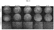
- the fundi categorized as abnormal were those in which the presence of autofluorescent pseudo-drusenoid lesions was observed ( Fig. 11 ; arrows) or with regions of focal atrophy ( Fig. 11 ; arrowheads).
- the percentage of abnormal fundi in the WT mice exposed to light was 20%, and in the untreated KI (KI control) it was 90%. Meanwhile, this percentage was reduced to 30% in mice treated with compound 2 (KI 2) and only to 60% in both mice treated with CYCLE (KI CYCLE) and RES (KI RES) (table 1).
- a Stratus OCT Zeiss
- a scanner adjusted to 6 lines of 3.45 mm each arranged radially.
- Six measurements were performed in each eye, upon which the retinal map was prepared ( Fig. 12 ) with the thickness of the retina quantified from the ganglion cell layer to the RPE.
- the thickness of the retina was significantly greater in the groups treated with RES and compound 2 when compared to the control KI group ( Fig. 12 ; C); these results show that treatment with SIRT1 activators prevented the production of retinal atrophy in KI mice after exposure to an intense light source.
- the c-wave is defined as a positive slow wave that appears after the b-wave and is used to measure the functional integrity of the RPE. Since the amplitude of this wave can be affected by the integrity of the photoreceptors, it is best to normalize the results with the amplitude of the b-wave, for which reason a quantification was performed of the proportion of the amplitude of the c-wave between the amplitude of the b-wave (c-/b-wave ratio) ( Fig. 13 ). The c-/b-wave ratio was significantly higher in the group treated with compound 2, which suggests that the functional integrity of the RPE was maintained in this group.
- the optomotor test was performed in order to assess visual acuity; this test consists in evaluating the optokinetic visual follow-up reflex that is stimulated by the rotating movement of different black and white serial horizontal bars. The response to said visual stimulus is manifested by a cervical tracking movement in the same direction of the rotation as the bars. Cervical movement is recorded with a camera, and the spatial frequency of the bars varies randomly, as does the direction of rotation of the bars.
- the optomotor test was evaluated at 6 different spatial frequencies (0.031, 0.061, 0.092, 0.103, 0.194, and 0.272 cycles/degree), and the spatial vision was measured by the number of positive responses.
- the SIRT1 activator (compound 2 ) has a protective effect on the retina of rd10 and Prpf31 A216P /+ mice.
- This retinoprotective effect suggests that this molecule could potentially be useful for the treatment of RP and other degenerative pathologies of the retina and RPE in which ROS, inflammation, and apoptosis occur, such as in AMD.
Landscapes
- Health & Medical Sciences (AREA)
- Life Sciences & Earth Sciences (AREA)
- Chemical & Material Sciences (AREA)
- Veterinary Medicine (AREA)
- Medicinal Chemistry (AREA)
- Pharmacology & Pharmacy (AREA)
- Animal Behavior & Ethology (AREA)
- General Health & Medical Sciences (AREA)
- Public Health (AREA)
- Epidemiology (AREA)
- Molecular Biology (AREA)
- Ophthalmology & Optometry (AREA)
- Engineering & Computer Science (AREA)
- Bioinformatics & Cheminformatics (AREA)
- Chemical Kinetics & Catalysis (AREA)
- General Chemical & Material Sciences (AREA)
- Nuclear Medicine, Radiotherapy & Molecular Imaging (AREA)
- Organic Chemistry (AREA)
- Inorganic Chemistry (AREA)
- Acyclic And Carbocyclic Compounds In Medicinal Compositions (AREA)
Abstract
Description
- The present invention relates to the therapeutic use of acylated piceid derivatives in ocular pathologies, particularly in retinal degeneration. The present invention can therefore be encompassed within the field of pharmaceuticals.
- Retinitis pigmentosa (RP) and age-related macular degeneration (AMD) are a cause of blindness in the adult population due to the degeneration of photoreceptors and of the retinal pigment epithelium (RPE). RP is a rare disease, but the prevalence of AMD is quite high, estimated at 8-16% of the population between 50 and 64 years of age. Like for RP, no treatment exists for the dry variety of AMD despite its high prevalence.
- Patent application
WO 2007020673 A1 discloses compounds derived from glycosylated hydroxystilbenes that might be useful for the treatment of some ocular diseases. Acylated piceid (3-glucosyl resveratrol) derivatives have been proposed for the treatment of inflammatory bowel diseases (ES 2362065 A1 - Nevertheless, there are currently no effective treatments that can prevent or slow down the degenerative process of photoreceptors or RPA in dry AMD or RP. Therefore, it would be beneficial to search for new molecules that are useful for the treatment or prevention of these and other ocular diseases involving retinal degeneration.
- The present invention provides compounds that are useful for the manufacture of a pharmaceutical composition for the treatment or prevention of ocular diseases. To this end, it is demonstrated in the examples how some acylated derivatives of resveratrol induce the protection of the photoreceptors in two animal models of retinal degeneration.
- In a first aspect, the present invention relates to the use of compounds of the general formula (I)

where: - R1 is a C1-C22 alkyl group or a C2-C22 alkenyl group, with R1 preferably being a C1-C22 alkyl group;
- for the preparation of a pharmaceutical composition for the prevention and/or treatment of ocular pathologies.
- In a preferred embodiment, R1 is a C2-C20 alkyl group, more preferably a C3-C17 alkyl group.
- In the present invention, the term "alkyl" refers to aliphatic, linear, or branched chains having from 1 to 22 carbon atoms, e.g., methyl, ethyl, n-propyl, i-propyl, n-butyl, tert-butyl, sec-butyl, n-pentyl, n-hexyl, etc. The alkyl group preferably has between 2 and 20 carbon atoms, and more preferably between 3 and 17 carbon atoms.
- In the present invention, the term "alkenyl" refers to unsaturated, linear, or branched aliphatic chains having from 2 to 22 carbon atoms and having between one and six unsaturations depending on the number of carbons, for example, without limitation to vinyl, allyl, oleyl, linoleyl, linolenyl, eicosapentaenoyl, docosahexaenoyl, etc.
- In a preferred embodiment, the compounds of the invention can be selected from among:


- The pharmaceutical composition of the present invention can be formulated for administration in a variety of manners that are known in the art. Examples of preparations include any solid composition (tablets, pills, capsules, granules, etc.) or liquid composition (solutions, suspensions, or emulsions) for oral, topical, or parenteral administration. The composition of the present invention can also be in the form of sustained-release drug formulations or any other conventional delivery system, and can thus be contained, without limitation thereto, in nanoparticles, liposomes, or nanospheres, in a polymeric material, in a biodegradable or non-biodegradable implant, or in biodegradable microparticles such as biodegradable microspheres, for example.
- Such a composition and/or its formulations can be administered to an animal, including a mammal and hence to a human, in a variety of manners, including but not limited to intraperitoneal, intravenous, intradermal, intraspinal, intrastromal, intraarticular, intrasynovial, intrathecal, intralesional, intraarterial, intramuscular, intranasal, intracranial, subcutaneous, intraorbital (sub-retinal, intravitreal), intracapsular, topical, or percutaneous administration, or by means of transdermal patches, nasal spray, surgical implant, internal surgical painting, or infusion pump.
- In an embodiment that is even more preferred, the composition of the invention is formulated for ophthalmic administration. The expression "formulated for ophthalmic administration" refers to a formulation that enables the composition of the invention to be administered ocularly - including, but without limitation thereto, topically or intraocularly - without such administration adversely affecting the properties, e.g., optical and/or physiological properties, of the eye.
- Examples of the composition of the invention that are formulated for ophthalmic administration include but are not limited to said composition in combination with water, salts, a polymeric liquid or semi-solid vehicle, a phosphate buffer, or any other ophthalmically acceptable liquid vehicle thereof that is known in the art.
- In a preferred embodiment, the pharmaceutical composition of the invention comprises a system for the controlled release of the compounds of the invention; more preferably, they comprise cyclodextrins and even more preferably 2-hydroxypropyl beta-cyclodextrin.
Therefore, another aspect of the present invention relates to an ophthalmological composition comprising at least one compound of the formula (I) as described above together with a controlled-release system, particularly a cyclodextrin, and more specifically 2- hydroxypropyl beta-cyclodextrin. - In another aspect, the present invention relates to a method for treating and/or preventing ocular pathology in a mammal, preferably a human, comprising the administration of a therapeutically effective amount of a composition of the general formula (I) as described above.
- The dosage for obtaining a therapeutically effective amount depends on a variety of factors, such as the age, weight, sex, or tolerance of the individual to whom the composition of the invention is to be administered, for example. In the meaning used in this description, the term "therapeutically effective amount" refers to the amount of the composition (I) calculated to produce the desired effect and, in general, will be determined inter alia by the characteristics of the composition, the age, condition, and history of the patient, the severity of the alteration or disorder, and the route and frequency of administration.
- The ocular pathologies of the present invention can be selected from among:
- 1. Hereditary degenerative pathologies of the retina, including retinitis pigmentosa and all those pathologies within the group of retinal dystrophies which include, but are not limited to: autosomal dominant retinitis pigmentosa, autosomal recessive retinitis pigmentosa, retinitis pigmentosa linked to the X chromosome, sporadic retinitis pigmentosa, retinitis pigmentosa associated with other syndromes, Cokayne syndrome, cone dystrophies, cone and rod degeneration, Leber's congenital amaurosis, retinitis punctata albescens, choroideremia, choroidal and retinal gyrate atrophy, choroidal generalized dystrophy, juvenile retinoschisis, Wagner's vitreoretinal degeneration, autosomal dominant vitreoretinal choroidopathy, fundus albipunctatus, Stargardt's disease, Best vitelliform macular dystrophy, Usher syndrome, Bardet-Biedl syndrome, fenestrated macular dystrophy, Sorsby pseudoinflammatory macular dystrophy, dominant drusen, among other retinal dystrophies involving degeneration of photoreceptors and/or EPR.
- 2. Early- and/or late-onset age-related (wet or neovascular) and/or non-exudative (dry or atrophic) macular degeneration; as well as other macular diseases such as: epiretinal membrane, macular hole, and macular edema, etc.
- 3. Degenerative inflammatory pathologies of the retina and the optic nerve of infectious, immune, vascular, traumatic, post-surgical, or teratogenic origin such as: ocular toxoplasmosis, cryptococcosis, lupus, Behcet's disease, diabetic retinopathy, ischemic optic neuropathy, optic neuritis, detachment of the retina, vitrectomy, internal limiting membrane peeling, surgery, retinoblastoma, etc.
- 4. Ocular pathologies that increase intraocular pressure and that involve degeneration of the ganglion cells of the retina and atrophy of the optic nerve such as: open-angle or closed-angle glaucoma.
- 5. Neurological pathologies that are accompanied by degenerative lesions of the retinal or optic nerve such as: amyotrophic lateral sclerosis, multiple sclerosis, Parkinson's disease, Alzheimer's disease, Graefe syndrome, Hallgren syndrome, Hallen Vorden Spatz syndrome, autosomal dominant cerebellar ataxia, and pigmentary degeneration of the retina.
- 6. Other ocular pathologies that occur with inflammation of the cornea, choroid, ciliary body of any etiology such as: uveitis, scleritis, keratoconjunctivitis, corneal transplant surgery, cataract surgery.
- 7. Other ocular or neurodegenerative pathologies that involve infiltration of microglia or shortening of telomeres due to cellular aging or oxidative stress.
- In one preferred embodiment, the ocular pathologies are selected from among retinitis pigmentosa and age-related macular degeneration.
- Throughout the description and the claims, the expression "comprises" and variants thereof are not intended to exclude other technical features, additives, components, or steps. For those skilled in the art, other objects, advantages, and features of the invention will be apparent in part from the description and in part from the practicing of the invention. The following examples and figures are provided for the sake of illustration and are not intended to limit the scope of the present invention.
-
-
Fig. 1 . Cell viability in neuroblastoma SH-SY5Y after damage with H2O2 and treatment with the different compounds 1-4. The controls are -H2O2 = 1% DMSO solution; +H2O2= H2O2 in 1% DMSO;RES 10 µM + H2O2=resveratrol 10 µM in H2O2 in 1% DMSO. -
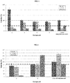
Fig. 2 . Cell viability in RAW macrophages after inflammation produced with LPS and treatment with the different compounds 1-2. The controls are -LPS = 1% DMSO solution; +LPS= 1% DMSO solution with LPS;RES 10 µM+LPS =resveratrol 10 µM in 1% DMSO solution with LPS. -
Fig. 3 . Concentration of TNF-alpha in culture medium after inflammation by LPS in RAW macrophages and treatment withcompound 2 and RES control. The controls are LPS alone = 1% DMSO solution with LPS; DMSO only = 1% DMSO solution. -
Fig. 4 . Concentration of NO in culture medium after inflammation by LPS in RAW macrophages and treatment withcompound 2 and RES control. The controls are LPS alone = 1% DMSO solution with LPS; DMSO only = 1% DMSO solution. -
Fig. 5 . Concentration of IL6 in culture medium after inflammation by LPS in RAW macrophages and treatment withcompound 2 and RES control. The controls are LPS alone = 1% DMSO solution with LPS; DMSO only = 1% DMSO solution. -
Fig. 6 . AChE activity with respect to the control ofcompound 2 and the RES control. -
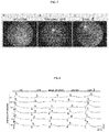
Fig. 7 . Fundus of normal c57/bI6 mouse (WT) (A), untreated rd10 mouse (B), and injected with 5 mM of compound 2 (C) at 15 days after subretinal administration of the compounds. The absence of pigment in the fundus of the mouse treated withcompound 2 is observed. -
Fig. 8 . Electroretinogram of normal c57/bI6 mouse (WT), untreated rd10 mouse,DMSO vehicle 5%, 5 mM resveratrol (RES) and 5mM compound 2 at 15 days after subretinal injection. The amplitude of the a- and b-waves was preserved in the group treated withcompound 2 in a greater proportion than in the rd10 mice that were untreated or treated with RES or with 5% DMSO vehicle. -
Fig. 9 . Amplitude of the b-wave in untreated rd10 mice, rd10 mice treated with 5% DMSO vehicle, with 5 mM RES, and with 5mM compound 2 at 15 days after administration of the subretinal injections. Each bar represents the average of the b-wave in 6 mice per group ± the standard error of the mean. The amplitude of the b-wave was significantly increased in the mice treated withcompound 2 in the dark-adapted ERG with flash intensities of 0.2, 1, 3 and 10 cd xs/m2. The statistical analysis was performed by one-way ANOVA with post hoc DMS test *p< 0.05; #p< 0.01. -
Fig. 10 . Immunofluorescence of normal c57/bI6 mouse (WT) (A, G, M), untreated rd10 mouse (B, H, N), or rd10 mice treated with vehicle (C, I, O), with 5 mM RES (D, J, P) and with 5 mM compound 2 (F, L, R) at 15 days after administration of the subretinal injections. The sections of the retina were immunostained with anti-rhodopsin antibodies (red) to label the rods and anti-opsin (green) to label the cones. The nuclei were stained with DAPI. In all of the groups that were treated with SIRT1 activators, more labeling of rhodopsin and opsin was observed than in the untreated group, but the signal of this immunolabeling was more intense in the group treated withcompound 2. Likewise, the thickness of the outer nuclear layer (ONL) was greater than in the group treated with RES. -
Fig. 11 . Fundus and autofluorescence of the retina in normal mice (WT control), untreated Prpf31 A216P/+ mice (KI control), and Prpf31 A216P/+ mice treated with eye drops administered daily for two and a half months containing 2 mM resveratrol (KI RES) or 2mM compound 2 diluted in a 13% hydroxypropyl-beta-cyclodextrin vehicle in PBS (KI 2) or only with the 13% hydroxypropyl-beta-cyclodextrin vehicle in PBS (KI CYCLE). Both WT and KI mice that did not receive any treatment (WT control and KI control) were used as controls. All the groups were subjected to 5,000 lux for 3 h and the evaluations were made 15 days after inducing light damage, the eye drops having been continued during this 2-week period. In the KI control, KI CYCLE, and KI RES groups, areas with autofluorescent punctiform yellow-white lesions (arrows) were observed which can be seen clearly in the enlargement of the image in the lower row. Moreover, areas of focal atrophy (arrowheads) were observed in these mice. Fundi with this type of characteristic were considered abnormal. In the majority of the fundi of the WT control andKI 2 groups, this type of lesion was not observed, and these fundi were classified as normal. -
Fig. 12 . Thickness of the retina quantified by means of OCT in normal mice (WT control), untreated Prpf31 A216P/+ mice (KI control), and Prpf31 A216P/+ mice treated with eye drops administered daily for two and a half months containing 2 mM resveratrol (KI RES) or 2mM compound 2 diluted in a 13% hydroxypropyl-beta-cyclodextrin vehicle in PBS (KI 2) or only with the 13% hydroxypropyl-beta-cyclodextrin vehicle in PBS (KI CYCLE). Both WT and KI mice that did not receive any treatment (WT control and KI control) were used as controls. All the groups were subjected to 5,000 lux for 3 h and the evaluations were made 15 days after inducing light damage, the eye drops having been continued during this 2-week period. The retinal maps (A) show the thickness of the retina in 9 concentric areas at 1, 2.22, and 3.45 mm and represented by a colorimetric scale (B). The data were quantified and are shown in (C). The bars represent averages of retinal thickness measured in µm +/- the standard error (WT control n = 5, rest of the groups n = 10). The normality of the data was confirmed by the Kolmogorov-Smirnov test (P = 0.98); to determine differences in the means of the groups, one-way ANOVA (P <0.001) and post hoc LSD tests were performed. *P<0.001 KI control vs. KI RES or KI control vsKI 2. -
Fig. 13 . Evaluation of the amplitude of the c-wave in normal mice (WT control), untreated Prpf31 A216P/+ mice (KI control), and Prpf31 A216P/+ mice treated with eye drops administered daily for two and a half months containing 2 mM resveratrol (KI RES) or 2mM compound 2 diluted in a 13% hydroxypropyl-beta-cyclodextrin vehicle in PBS (KI 2) or only with the 13% hydroxypropyl-beta-cyclodextrin vehicle in PBS (KI CYCLE). Both WT and KI mice that did not receive any treatment (WT control and KI control) were used as controls. All the groups were subjected to 5,000 lux for 3 h and the evaluations were made 15 days after inducing light damage, the eye drops having been continued during this 2-week period. The amplitude of the c-wave was measured by ERG (A) and then the amplitude of the b-wave was quantified (A), and the c-/b-wave ratio (C) was also estimated. The bars represent averages of the c-/b-wave ratio +/- the standard error (WT control n = 5, rest of the groups n = 10). The normality of the data was confirmed by the Kolmogorov-Smirnov test (P = 0.43); to determine differences in the means of the groups, one-way ANOVA (P <0.05) and post hoc LSD tests were performed. *P<0.01 KI control vsKI 2. -
Fig. 14 . Optomotor test showing the visual acuity curve in normal mice (WT control), untreated Prpf31 A216P/+ mice (KI control), and Prpf31 A216P/+ mice treated with eye drops administered daily for two and a half months containing 2 mM resveratrol (KI RES) or 2mM compound 2 diluted in a 13% hydroxypropyl-beta-cyclodextrin vehicle in PBS (KI 2) or only with the 13% hydroxypropyl-beta-cyclodextrin vehicle in PBS (KI CYCLE). Both WT and KI mice that did not receive any treatment (WT control and KI control) were used as controls. All the groups were subjected to 5,000 lux for 3 h and the evaluations were made 15 days after inducing light damage, the eye drops having been continued during this 2-week period. The optomotor test data were quantified and are shown in the chart. Each point represents averages of the number of positive responses +/- the standard error (WT control n = 5, rest of the groups n = 10). The normality of the data was confirmed by the Kolmogorov-Smirnov test in all spatial frequencies except for 0.031 and 0.061. To determine differences in the means of the groups, two-way ANOVA was performed, correlating both the different frequencies and groups (P <0.001) and the post hoc LSD test. *P<0.05 KI control vs. KI RES or **< 0.001 KI control vsKI 2. - The invention will be illustrated below by means of tests conducted by the inventors that demonstrate the effectiveness of the product of the invention.
- The SH-SY5Y neuroblastoma cell line was cultured in petri dishes pre-treated with collagen (100 µg/ml) with F12 medium supplemented with penicillin/streptomycin and 10% inactivated fetal bovine serum.
- Cell viability assays with neurons were prepared in 96-well plates pre-treated with collagen by seeding 20,000 cells/well in a volume of 100 µl and incubating the cells for 24 h before the addition of the compounds. The compounds to be tested were dissolved in DMSO and added in three different concentrations (1, 10 and 100 µM) in order to determine their toxicity. The final percentage of DMSO in each well was adjusted to 1%. The cell viability was evaluated 24 h after the addition of the compounds by means of the MTT assay according to the manufacturer's method. Mean values and standard deviations were calculated from at least 8 different measurements from several independent experiments.
- Neuroprotection assays. The neurons were cultured and seeded in the same manner as for the cell viability assay. The compounds to be tested were dissolved in DMSO and added to three different concentrations (1, 10 and 100 µM) and, after 10-minute incubation, hydrogen peroxide (100 µM) was added to the medium. The final percentage of DMSO in each well was adjusted to 1%. The cell viability was evaluated 24 h after the addition of the compounds by means of the MTT assay according to the manufacturer's method. Mean values and standard deviations were calculated from at least 8 different measurements from several independent experiments. Neuronal recovery was calculated by normalizing the results of the neuronal viability experiments after the addition of our compounds and H2O2 to the positive control of each experiment (neurons + H2O2).
- It is observed that the
RES 10 µM control recovers up to 50% of cell viability (indicated by the arrow inFig. 1 ). In contrast, acylated piceid derivatives (1, 2, 3 and 4) recover between 70 and 200% of viability at concentrations between 1 and 10 µM. All of those at 100 µM appear to exhibit toxicity. - RAW 264.7 macrophages were cultured in P75 with high-glucose DMEm supplemented with penicillin/streptomycin and 10% inactivated fetal bovine serum.
- The cell viability assays with RAW macrophages were prepared in 96-well plates by seeding 25,000 cells/well in a volume of 100 µl and incubating the cells for 4 h before the addition of the compounds. The compounds to be tested were dissolved in DMSO and added in three different concentrations (1, 10 and 100 µM) in order to determine their toxicity. The final percentage of DMSO in each well was adjusted to 1%. The cell viability was evaluated 24 h after the addition of the compounds by means of the MTT assay according to the manufacturer's method. Mean values and standard deviations were calculated from at least 8 different measurements from several independent experiments.
- Testing of mitigation of damage caused by the addition of LPS. The RAW 264.7 macrophages were cultured according to the procedure described above. The compounds to be tested were dissolved in DMSO and added to three different concentrations (1, 10 and 100 µM) and, after 10-minute incubation, LPS (100 ng/ml) was added to the medium. The final percentage of DMSO in each well was adjusted to 1%. The cell viability was evaluated 24 h after the addition of the compounds by means of the MTT assay according to the manufacturer's method. Mean values and standard deviations were calculated from at least 8 different measurements from several independent experiments.
- It is observed that the
RES 10 µM control recovers up to 82% of cell viability (indicated by the arrow inFig. 2 ).Compound 2 recovers between 80 and 140% of viability at concentrations between 1 and 100 µM. - To determine the production of cytokines, 5 × 10⁵ RAW 264.7 macrophages were seeded in 24-well plates (0.5 ml per well). The compounds to be tested were then added (10 µM), and the macrophages were either stimulated or not through the addition of LPS (1 µg/ml) to the culture medium. After 24 hours, the levels of IL-6 and TNF-a were measured in the supernatants by ELISA using the capture and biotinylated antibodies from BD PharMingen and PrepoTech (Gonzalez-Rey et al., Gut. 2009; 58:929-939; Sanchez et al., Stem Cells. 2011; 29:251-262). The levels of NO in the supernatants at 24 hours were measured indirectly by determining the nitrite concentration in the medium using the Griess reagent (Anderson et al., Gut. 2013; 62:1131-1141). A minimum of two independent experiments and three replicates per experiment were performed for each measured value. The values are expressed as the mean ± standard deviation.
- In the previous assay, the levels of various inflammation parameters were measured (TNF-α, NO, and IL-6) by ELISA after treatment with RES or with compound 2 (
Figs. 3, 4 , and5 ). - It is observed that the control of
RES 10 µM significantly decreases inflammatory parameters (TNF-α, NO, and IL-6) (indicated by the bar in bold).Compound 2 decreases TNF-α and NOT even more than RES and similarly to IL-6. - The objective of this assay was to analyze the protective effect of
compound 2 and of the resveratrol (RES) control in a model of neurotoxicity induced by the neurotoxin pentylenetetrazole (PTZ). As an experimental model, the zebrafish larva (Danio rerio) was used to study the effect of the compounds on acetylcholinesterase activity (AChE) in larvae at 5 days post-fertilization (dpf). - Studies of the central nervous system (CNS) in zebrafish (Kimmel et al., Dev Dyn, 1995 Jul; 203(3):253-310), show that, at 24 hours of development, the brain of the embryo has already segmented and some structures such as the neural tube, the notochord and the somites (muscle, and bone precursors). At 5 days post-fertilization (5 dpf), the animal has formed sensory organs such as eyes and otoliths. In addition, the heart, liver, kidneys, and pancreas, as well as the circulatory, digestive, and nervous systems are fully functional. At this time, the animal is able to respond to visual, olfactory, and mechanical stimuli and begins the search for food.
- AChE is an enzyme that degrades through its hydrolytic activity the neurotransmitter acetylcholine in choline and an acetate group. It is found chiefly in neuromuscular junctions and in the cholinergic nervous system, where its activity has the function of ending synaptic transmission (Behra et al., Nat Neurosci. 2002 Feb; 5(2): 111-8.). Acetylcholine is a neurotransmitter involved in the control of movement and is an important modulator of cognitive functions such as learning and memory (Hasselmo et al., Neuropsychopharmacology. 2011 Jan; 36(1): 52-73). Adequate levels of acetylcholinesterase thus reflect a healthy neuronal state.
- Zebrafish embryos were seeded in 50 ml of dilution water (AD) in a Petri dish and grown to 5 dpf (larval stage). Only those larvae that did not exhibit any type of external anomaly were used to perform the assay. Next, the larvae were transferred using a Pasteur pipette to a 24-well microplate, so that each well contained five larvae, making ten replicates per condition. First, the pre-treatment of the 5 dpf larvae was performed. For this, the larvae were incubated at 26 ± 1 °C for 1 hour in a volume of 2 ml of AD for the two control groups (Control and Control + PTZ), of physostigmine (Phys) 20 µM, which is a commercial inhibitor of the enzyme AChE for the Phys group, and of the test compounds at a concentration of 10 µM. A medium exchange was then carried out, and the larvae were incubated with the compounds in combination with 5 mM PTZ for 6 hours at 26 ± 1 °C. After this incubation period, all of the larvae were examined, and it was determined that the general state of the larvae was totally normal, without any visible anomaly or anomalous behavior. Finally, the larvae were processed for the analysis of the AChE activity.
- Determination of AChE levels. Once the experimental period was completed, larvae processing was carried out for the determination of AChE according to the technical study protocol (Measurement of acetylcholinesterase activity (AChE) in cell cultures and zebrafish larvae). The larvae were homogenized mechanically, and the samples were centrifuged to obtain the supernatant, which were used to determine the levels of the AChE enzyme as a function of the treatments administered. In addition, the determination of total protein of each experimental group was carried out according to the technical study protocol (Quantification of protein by BCA, as a normalization process). Finally, the AChE levels determined in the control group were taken as a reference measurement and deemed to be 100%.
-
Compound 2 significantly prevents the decrease in AChE activity induced by PTZ in the 5dpf larvae, showing a clear neuroprotective effect that is greater than that observed for RES (seeFig. 6 ). - Twenty zebrafish embryos were incubated for each concentration of compound to be tested (1 to 4). The concentrations used of each compound are 0.1 µM, 1 µM, 10 µM, 100 µM and 1 mM. The incubation period goes from 0 to 96 hours post fertilization (hpf). A negative control of 1% DMSO and a positive control of N,N-diethylaminobenzaldehyde (DEAB) are included. After the incubation period, the LC50 (lethal dose for 50% of the animals) is calculated.
- The zebrafish embryo toxicity assay was carried out for
compound 2 and the resveratrol (RES) control, and both exhibited acceptable toxicity (LC50 <140 uM) and (LC50 <1000 uM), respectively. - The rd10 mouse is a model of retinitis pigmentosa due to a mutation in the Pde6b gene that results in the degeneration of the photoreceptors that begins on postnatal day 15-16 (P15-16) and is completed within one month (P30). In this model, the degeneration begins in the rods and then continues with cones and finally the retinal pigment epithelium (RPE). The degeneration in this model occurs as a consequence of the loss of the function of Pde6b, which ultimately leads to a continuous entry of Ca++ in the cones with a significant production of reactive oxygen species (ROS), infiltration of macrophages, and activation of pro-apoptotic proteins. At the histological level, a loss of nuclei is observed in the outer nuclear layer of the retina (the layer in which photoreceptor nuclei are located), as well as the loss of the specific markers of rods (rhodopsin) and cones (opsin). The electroretinogram (ERG), which measures the electrical response of the retina to visual stimuli, is markedly affected in rd10 mice, with progressive loss of the amplitude of the a- and b-waves of the ERG. Finally, the fundus used to evaluate the macroscopic morphology of the bottom of the eye exhibits changes at one month of age in the rd10 mice, with accumulation of dark pigments in the periphery due to the degeneration of the RPE. To evaluate the protective effect of our compounds on the retina, we first used the rd10 model. Rd10 homozygous mice were injected in the subretinal space at P13 (two to three days before the onset of degeneration) with 1 µl of vehicle (5% DMSO in PBS) or resveratrol RES (5 mM), as well as compound 2 (5 mM). Then, 15 days after having been injected, fundus, ERG, and histological changes were evaluated in the untreated mice, those treated with the vehicle, and those treated with RES and
compound 2. Evaluation of the fundus revealed a lower amount of pigment accumulation in the retinas of mice treated withcompound 2 compared to the untreated mice (Fig. 7 ). The ERG in the treated mice was also evaluated 15 days after the injections. The amplitude of the a- and b-waves in the ERGs were quantified, and the statistical analysis was performed using two-way ANOVA and post hoc DMS. A value of p <0.05 was considered significant. The b-wave amplitude in the ERG of dark-adapted mice (flash intensities of 0.2, 1, 3, and 10 × cd s/m2) underwent a significant increase in the group of mice treated withcompound 2 compared to the controls (Figs. 8 and9 ). Finally, the immunofluorescence study was performed in order to observe the presence of markers of rods (rhodopsin) and cones (opsin) (Fig. 10 ). The number of photoreceptor nuclei present in the outer nuclear layer (ONL), as well as the immunostaining of rhodopsin, was much greater in rd10 mice treated withcompound 2 than in those treated with RES and in the controls. - Prpf31 A216P/+ mice are a useful model for assessing the degeneration of the RPE. This model is characterized by an increase in the thickness of the Bruch membrane and accumulation of esterified cholesterol between said membrane and the RPE produced by a defect in the splicing of the mRNA of relevant genes of the visual cycle, such as RPE65 and RDH12. The accumulation of toxic products derived from retinol leads to an increase in ROS, infiltration of macrophages, and primary degeneration of the RPE. The degeneration of the RPE occurs late, at approximately 8 months of age. The phenotypic characteristics of this model can be exacerbated by exposure to intense light and are characterized primarily by accumulation of pseudo-drusenoid lesions and regions of atrophy observed in the fundus, with a loss of visual acuity and perception of contrast being observed, as well as a decrease in the amplitude of the c-wave of the ERG and a decrease in the thickness of the retina.
- To evaluate the protective effect of the administration of our compounds in eye drops, we used
heterozygous 6 month-old Prpf31 A216P/+ (KI) mice that were treated topically with 20 µl of the studied compounds applied directly on the corneal surface daily (L-V) for two and a half months. The compounds used were RES andcompound 2, which were diluted in a solution of 13% hydroxypropyl-beta-cyclodextrin (CYCLO) in order to obtain a final concentration of 2 mM of each compound. After two and a half months of application of the eye drops with CICLO, RES, orcompound 2, the mutant KI mice were exposed to a light source of 5,000 lux for 3 hours. Normal (WT) and KI mice were also exposed to intense light as test control without receiving any treatment. Subsequently, the fundus of the animals was evaluated, in addition to the visual acuity by means of the optomotor test, the thickness of the retina by means of optical coherence tomography (OCT), and the amplitude of the c-wave by ERG. - Two categories were used to quantify the macroscopic characteristics of the fundus: abnormal and normal fundus. The fundi categorized as abnormal were those in which the presence of autofluorescent pseudo-drusenoid lesions was observed (
Fig. 11 ; arrows) or with regions of focal atrophy (Fig. 11 ; arrowheads). The percentage of abnormal fundi in the WT mice exposed to light (WT control) was 20%, and in the untreated KI (KI control) it was 90%. Meanwhile, this percentage was reduced to 30% in mice treated with compound 2 (KI 2) and only to 60% in both mice treated with CYCLE (KI CYCLE) and RES (KI RES) (table 1). When performing the statistical analysis for each dichotomous variable, we observed that there is a statistically significant relationship between the presence of normal fundi and mice treated withcompound 2 when compared with the control KI group (chi-square = 7.5; P <0.01, Fisher's exact test P <0.05) and with a probability of obtaining a normal fundus in the group treated withcompound 2 of seven times greater than in the control KI (normal RR = 7, IC95% = 1.044-46.949). These values were similar to those observed when comparing WT control and KI control (chi-square = 7.35, P <0.01, Fisher's exact test P <0.05) and with a probability of obtaining a normal fundus in the WT control group of eight times greater than in the control KI (normal RR = 8; IC95% = 1.184-54.043). In both the group treated with CICLO and that treated with RES, no statistically significant association was observed when comparing each one to the control KI group. The conclusion of this experiment is that treatment withcompound 2 prevented the formation of autofluorescent pseudo-drusenoid lesions and regions of focal atrophy in the Prpf31 A216P/+ mouse.Table 1. Quantification of the number of abnormal and normal fundi in WT and Prpf31 A216/+ (KI) mice without treatment or when treated with 13% CYCLE, RES, and compound 2.Fundus/Groups Abnormal (%) Normal (%) Total WT control 1 (20) 4 (80)# 5 KI control 9 (90) 1 (10) 10 KI CYCLE 6 (60) 4 (40) 10 KI RES 6 (60) 4 (40) 10 KI 23 (30) 7 (70)* 10 Total (%) 25 (55.6) 20 (44.4) 45 * KI 2 vs. KI control (chi-square= 7.5; P < 0.01; Fisher's exact test P < 0.05; RR normal= 7; IC95%= 1.044-46.949). # WT control vs KI control (chi-square = 7.35, P <0.01, Fisher's exact test P <0.05, normal RR = 8, 95% CI = 1.184-54.043). - To evaluate the thickness of the retina in vivo, a Stratus OCT (Zeiss) was used with a scanner adjusted to 6 lines of 3.45 mm each arranged radially. Six measurements were performed in each eye, upon which the retinal map was prepared (
Fig. 12 ) with the thickness of the retina quantified from the ganglion cell layer to the RPE. The thickness of the retina was significantly greater in the groups treated with RES andcompound 2 when compared to the control KI group (Fig. 12 ; C); these results show that treatment with SIRT1 activators prevented the production of retinal atrophy in KI mice after exposure to an intense light source. - To evaluate the functional integrity of the RPE, the amplitude of the c-wave in the ERG was measured. The c-wave is defined as a positive slow wave that appears after the b-wave and is used to measure the functional integrity of the RPE. Since the amplitude of this wave can be affected by the integrity of the photoreceptors, it is best to normalize the results with the amplitude of the b-wave, for which reason a quantification was performed of the proportion of the amplitude of the c-wave between the amplitude of the b-wave (c-/b-wave ratio) (
Fig. 13 ). The c-/b-wave ratio was significantly higher in the group treated withcompound 2, which suggests that the functional integrity of the RPE was maintained in this group. - Finally, the optomotor test was performed in order to assess visual acuity; this test consists in evaluating the optokinetic visual follow-up reflex that is stimulated by the rotating movement of different black and white serial horizontal bars. The response to said visual stimulus is manifested by a cervical tracking movement in the same direction of the rotation as the bars. Cervical movement is recorded with a camera, and the spatial frequency of the bars varies randomly, as does the direction of rotation of the bars. The optomotor test was evaluated at 6 different spatial frequencies (0.031, 0.061, 0.092, 0.103, 0.194, and 0.272 cycles/degree), and the spatial vision was measured by the number of positive responses. In the group treated with
compound 2, the number of positive responses was significantly higher than in control KI groups in the majority of frequencies studied (0.092, 0.103, 0.194, and 0.272 cycles/degree), and KI RES was only significant at 0.194 cycles/degree (Fig. 14 ). - In conclusion, in the experiments described in examples 6 and 7, we have demonstrated that the SIRT1 activator (compound 2) has a protective effect on the retina of rd10 and Prpf31 A216P/+ mice. This retinoprotective effect suggests that this molecule could potentially be useful for the treatment of RP and other degenerative pathologies of the retina and RPE in which ROS, inflammation, and apoptosis occur, such as in AMD.
Claims (15)
- A use of a compound of the general formula (I)

where:R1 is a C1-C22 alkyl group or a C2-C22 alkenyl group,for the preparation of a pharmaceutical composition for the prevention and/or treatment of ocular pathologies. - The use as set forth in claim 1, wherein R1 is a C2-C20 alkyl group.
- The use as set forth in any one of claims 1 or 2, wherein the compounds are selected from among: trans-resveratrol-3-O-(6'-O-butanoyl)-β-D-glucopyranoside, trans-resveratrol-3-O-(6'-O-octanoyl)-β-D-glucopyranoside, trans-resveratrol-3-O-(6'-O-hexadecanoyl)-β-D-glucopyranoside, trans-resveratrol-3-O-(6'-O-octadecanoyl)-β-D-glucopyranoside, and any combination thereof.
- The use as set forth in claim 3, wherein the compound is trans-resveratrol-3-O-(6'-O-octanoyl)-β-D-glucopyranoside.
- The use as set forth in any one of claims 1 to 4, wherein the pathologies are hereditary degenerative pathologies of the retina selected from among retinitis pigmentosa and pathologies within the group of retinal dystrophies selected from among autosomal dominant retinitis pigmentosa, autosomal recessive retinitis pigmentosa, retinitis pigmentosa linked to the X chromosome, sporadic retinitis pigmentosa, retinitis pigmentosa associated with other syndromes, Cokayne syndrome, cone dystrophies, cone and rod degeneration, Leber's congenital amaurosis, retinitis punctata albescens, choroideremia, choroidal and retinal gyrate atrophy, choroidal generalized dystrophy, juvenile retinoschisis, Wagner's vitreoretinal degeneration, autosomal dominant vitreoretinal choroidopathy, fundus albipunctatus, Stargardt's disease, Best vitelliform macular dystrophy, Usher syndrome, Bardet-Biedl syndrome, fenestrated macular dystrophy, Sorsby pseudoinflammatory macular dystrophy, and/or dominant drusen.
- The use as set forth in any one of claims 1 to 4, wherein the ocular pathologies are age-related macular degeneration.
- The use as set forth in any one of claims 1 to 4, wherein the ocular pathologies are degenerative inflammatory pathologies of the retina and of the optic nerve.
- The use as set forth in any one of claims 1 to 4, wherein the ocular pathologies are ocular pathologies that increase intraocular pressure and that involve degeneration of the ganglion cells of the retina and atrophy of the optic nerve selected from among open-angle and closed-angle glaucoma.
- The use as set forth in any one of claims 1 to 4, wherein the ocular pathologies are neurological pathologies that are accompanied by degenerative lesions of the retinal or optic nerve selected from among amyotrophic lateral sclerosis, multiple sclerosis, Parkinson's disease, Alzheimer's disease, Graefe syndrome, Hallgren syndrome, Hallen Vorden Spatz syndrome, autosomal dominant cerebellar ataxia, and pigmentary degeneration of the retina.
- The use as set forth in any one of claims 1 to 9, wherein the pharmaceutical composition further comprises a controlled-release system.
- The use as set forth in claim 10, wherein the controlled-release system is a cyclodextrin.
- The use as set forth in claim 11, wherein the cyclodextrin is 2-hydroxypropyl β-cyclodextrin.
- A pharmaceutical composition comprising at least one compound of the general formula (I):
 where: R1 is a C1-C22 alkyl group or a C2-C22 alkenyl group;and a cyclodextrin.
 - The composition as set forth in claim 13, wherein the compound of formula (I) is selected from among trans-resveratrol-3-O-(6'-O-butanoyl)-β-D-glucopyranoside, trans-resveratrol-3-O-(6'-O-octanoyl)-β-D-glucopyranoside, trans-resveratrol-3-O-(6'-O-hexadecanoyl)-β-D-glucopyranoside, trans-resveratrol-3-O-(6'-O-octadecanoyl)-β-D-glucopyranoside, and any combination thereof.
- The composition as set forth in any one of claims 13 or 14, wherein the cyclodextrin is 2-hydroxypropyl β-cyclodextrin.
Applications Claiming Priority (2)
| Application Number | Priority Date | Filing Date | Title |
|---|---|---|---|
| ES201631503A ES2673942B1 (en) | 2016-11-23 | 2016-11-23 | ACRYLIC COMPOUNDS FOR THE TREATMENT OF EYE PATHOLOGIES |
| PCT/ES2017/070760 WO2018096196A1 (en) | 2016-11-23 | 2017-11-17 | Acylated compounds for the treatment of ocular pathologies |
Publications (2)
| Publication Number | Publication Date |
|---|---|
| EP3545959A1 true EP3545959A1 (en) | 2019-10-02 |
| EP3545959A4 EP3545959A4 (en) | 2020-06-24 |
Family
ID=62194826
Family Applications (1)
| Application Number | Title | Priority Date | Filing Date |
|---|---|---|---|
| EP17875009.7A Pending EP3545959A4 (en) | 2016-11-23 | 2017-11-17 | Acylated compounds for the treatment of ocular pathologies |
Country Status (4)
| Country | Link |
|---|---|
| US (2) | US11213536B2 (en) |
| EP (1) | EP3545959A4 (en) |
| ES (1) | ES2673942B1 (en) |
| WO (1) | WO2018096196A1 (en) |
Families Citing this family (2)
| Publication number | Priority date | Publication date | Assignee | Title |
|---|---|---|---|---|
| JP2025520705A (en) | 2022-06-23 | 2025-07-03 | ミラムーン、ファーマ、ソシエダッド、リミターダ | Triazoles for use in treating eye diseases |
| ES3031706A1 (en) * | 2024-01-10 | 2025-07-10 | Consejo Superior Investigacion | Isoquercetin derivatives for the treatment and/or prevention of eye diseases (Machine-translation by Google Translate, not legally binding) |
Family Cites Families (12)
| Publication number | Priority date | Publication date | Assignee | Title |
|---|---|---|---|---|
| US6414037B1 (en) | 1998-01-09 | 2002-07-02 | Pharmascience | Pharmaceutical formulations of resveratrol and methods of use thereof |
| US6197834B1 (en) | 1998-09-01 | 2001-03-06 | Northeastern Ohio Universities College Of Medicine | Method of inhibiting formation of infectious herpes virus particles |
| US6573299B1 (en) | 1999-09-20 | 2003-06-03 | Advanced Medical Instruments | Method and compositions for treatment of the aging eye |
| US6878751B1 (en) | 2000-10-19 | 2005-04-12 | Imperial College Of Science Technology And Medicine | Administration of resveratrol to treat inflammatory respiratory disorders |
| US7037945B2 (en) | 2000-12-11 | 2006-05-02 | Northeastern Ohio Universities College Of Medicine | Method of inhibiting formation of Neisseria gonorrhea and Neisseria meningiditis |
| US7998974B2 (en) * | 2005-03-03 | 2011-08-16 | Sirtris Pharmaceuticals, Inc. | Fused heterocyclic compounds and their use as sirtuin modulators |
| US20070014833A1 (en) * | 2005-03-30 | 2007-01-18 | Sirtris Pharmaceuticals, Inc. | Treatment of eye disorders with sirtuin modulators |
| ITRM20050446A1 (en) | 2005-08-19 | 2007-02-20 | Tubilux Pharma S P A | USE OF HYDROXYLATED AND GLUCOSIDATED STYLBENES FOR THE PREVENTION AND TREATMENT OF EYE PATHOLOGIES. |
| ITFI20080019A1 (en) | 2008-02-11 | 2009-08-12 | Glures S R L | FORMULATIONS INCLUDING PICEID AND RESVERATROL ACTION TO PREVENT AND INHIBIT LIPID PEROXIDATION. |
| ES2362035B2 (en) | 2009-12-04 | 2011-11-15 | Universidad De Granada | BACTERIAN CEPA CECT7624, USES AND XEROPROTECTOR PRODUCT PRODUCED BY THE SAME. |
| ES2362065B1 (en) | 2009-12-15 | 2012-05-18 | Consejo Superior De Investigaciones Cient�?Ficas (Csic) | COMPOUNDS WITH ANTI-INFLAMMATORY ACTIVITY. |
| CN105503970B (en) * | 2015-11-27 | 2018-07-13 | 淮阴工学院 | A kind of polydatin ester derivative and its preparation method and application |
-
2016
- 2016-11-23 ES ES201631503A patent/ES2673942B1/en active Active
-
2017
- 2017-11-17 WO PCT/ES2017/070760 patent/WO2018096196A1/en not_active Ceased
- 2017-11-17 EP EP17875009.7A patent/EP3545959A4/en active Pending
- 2017-11-17 US US16/463,786 patent/US11213536B2/en active Active
-
2021
- 2021-11-30 US US17/539,012 patent/US20220079965A1/en not_active Abandoned
Also Published As
| Publication number | Publication date |
|---|---|
| ES2673942A1 (en) | 2018-06-26 |
| US20220079965A1 (en) | 2022-03-17 |
| US20190275068A1 (en) | 2019-09-12 |
| US11213536B2 (en) | 2022-01-04 |
| WO2018096196A1 (en) | 2018-05-31 |
| EP3545959A4 (en) | 2020-06-24 |
| ES2673942B1 (en) | 2019-04-09 |
Similar Documents
| Publication | Publication Date | Title |
|---|---|---|
| Sivak | The aging eye: common degenerative mechanisms between the Alzheimer's brain and retinal disease | |
| Tweedie et al. | Tumor necrosis factor-α synthesis inhibitor 3, 6′-dithiothalidomide attenuates markers of inflammation, Alzheimer pathology and behavioral deficits in animal models of neuroinflammation and Alzheimer’s disease | |
| Cao et al. | Protection of the retinal ganglion cells: intravitreal injection of resveratrol in mouse model of ocular hypertension | |
| Nagai et al. | A nanoparticle formulation of disulfiram prolongs corneal residence time of the drug and reduces intraocular pressure | |
| JP7296472B2 (en) | Treatment of mitochondria-related diseases and disorders (including their symptoms) with pridopidine | |
| CN108135867A (en) | Aldehyde conjugates and their uses | |
| Thanos et al. | Evidence for baseline retinal pigment epithelium pathology in the Trp1-Cre mouse | |
| US20220079965A1 (en) | Acylated compounds for the treatment of ocular pathologies | |
| JP2024144702A (en) | Pharmaceutical compositions for intraocular or oral administration for the treatment of retinal diseases | |
| Fontaine et al. | Systemic administration of the di-apocarotenoid norbixin (BIO201) is neuroprotective, preserves photoreceptor function and inhibits A2E and lipofuscin accumulation in animal models of age-related macular degeneration and Stargardt disease | |
| US20230372311A1 (en) | Compositions and methods of treating age-related retinal dysfunction | |
| US9561234B2 (en) | Methods for treating diseases of the retina | |
| EP4556013A1 (en) | Pharmaceutical composition for treatment of eye diseases comprising retinal organoid-derived exosomes | |
| Sheng et al. | MicroRNA-loaded antioxidant nanoplatforms for prevention and treatment of experimental acute and chronic uveitis | |
| JP2023521155A (en) | Methods and compositions for preventing and treating retinal neuropathy | |
| KR20190101424A (en) | Increased longevity and improvement of gait activity in patients with late childhood infant neuronal serotolipocytosis by gemfibrozil | |
| EP3634451A1 (en) | Reprogramming metabolism by inhibiting vhl for treatment of neurodegeneration | |
| Morales et al. | Acylated compounds for the treatment of ocular pathologies | |
| Kolahdouzmohammadi et al. | The role of iPSC modeling toward projection of autophagy pathway in disease pathogenesis: Leader or follower | |
| Fradot et al. | Rodent models of retinal degeneration: From purified cells in culture to living animals | |
| CN104780917A (en) | Baclofen and acamprosate based therapy of macular degeneration disorders | |
| US20180228811A1 (en) | Gdnf induction for the treatment of retinal disorders | |
| JP2022529742A (en) | Nerve repair method | |
| US12605358B2 (en) | Composition for preventing or treating nervous system diseases comprising compound that inhibits mTOR signaling pathway as active ingredient | |
| RU2768588C1 (en) | Method for detecting an active destructive process in the retina in an experiment |
Legal Events
| Date | Code | Title | Description |
|---|---|---|---|
| STAA | Information on the status of an ep patent application or granted ep patent |
Free format text: STATUS: THE INTERNATIONAL PUBLICATION HAS BEEN MADE |
|
| PUAI | Public reference made under article 153(3) epc to a published international application that has entered the european phase |
Free format text: ORIGINAL CODE: 0009012 |
|
| STAA | Information on the status of an ep patent application or granted ep patent |
Free format text: STATUS: REQUEST FOR EXAMINATION WAS MADE |
|
| 17P | Request for examination filed |
Effective date: 20190624 |
|
| AK | Designated contracting states |
Kind code of ref document: A1 Designated state(s): AL AT BE BG CH CY CZ DE DK EE ES FI FR GB GR HR HU IE IS IT LI LT LU LV MC MK MT NL NO PL PT RO RS SE SI SK SM TR |
|
| AX | Request for extension of the european patent |
Extension state: BA ME |
|
| DAV | Request for validation of the european patent (deleted) | ||
| DAX | Request for extension of the european patent (deleted) | ||
| A4 | Supplementary search report drawn up and despatched |
Effective date: 20200527 |
|
| RIC1 | Information provided on ipc code assigned before grant |
Ipc: A61K 31/7034 20060101ALI20200519BHEP Ipc: A61P 27/02 20060101ALI20200519BHEP Ipc: A61K 31/7024 20060101AFI20200519BHEP |
|
| STAA | Information on the status of an ep patent application or granted ep patent |
Free format text: STATUS: EXAMINATION IS IN PROGRESS |
|
| 17Q | First examination report despatched |
Effective date: 20220516 |
|
| P01 | Opt-out of the competence of the unified patent court (upc) registered |
Effective date: 20230518 |