EP3543952A1 - System, method and program for estimating acute cerebral infarction onset time - Google Patents
System, method and program for estimating acute cerebral infarction onset time Download PDFInfo
- Publication number
- EP3543952A1 EP3543952A1 EP17871182.6A EP17871182A EP3543952A1 EP 3543952 A1 EP3543952 A1 EP 3543952A1 EP 17871182 A EP17871182 A EP 17871182A EP 3543952 A1 EP3543952 A1 EP 3543952A1
- Authority
- EP
- European Patent Office
- Prior art keywords
- image
- infarction
- feature information
- region
- patient
- Prior art date
- Legal status (The legal status is an assumption and is not a legal conclusion. Google has not performed a legal analysis and makes no representation as to the accuracy of the status listed.)
- Granted
Links
Images





Classifications
-
- G—PHYSICS
- G06—COMPUTING OR CALCULATING; COUNTING
- G06T—IMAGE DATA PROCESSING OR GENERATION, IN GENERAL
- G06T7/00—Image analysis
- G06T7/0002—Inspection of images, e.g. flaw detection
- G06T7/0012—Biomedical image inspection
- G06T7/0014—Biomedical image inspection using an image reference approach
-
- A—HUMAN NECESSITIES
- A61—MEDICAL OR VETERINARY SCIENCE; HYGIENE
- A61B—DIAGNOSIS; SURGERY; IDENTIFICATION
- A61B5/00—Measuring for diagnostic purposes; Identification of persons
-
- A—HUMAN NECESSITIES
- A61—MEDICAL OR VETERINARY SCIENCE; HYGIENE
- A61B—DIAGNOSIS; SURGERY; IDENTIFICATION
- A61B5/00—Measuring for diagnostic purposes; Identification of persons
- A61B5/0033—Features or image-related aspects of imaging apparatus, e.g. for MRI, optical tomography or impedance tomography apparatus; Arrangements of imaging apparatus in a room
- A61B5/004—Features or image-related aspects of imaging apparatus, e.g. for MRI, optical tomography or impedance tomography apparatus; Arrangements of imaging apparatus in a room adapted for image acquisition of a particular organ or body part
- A61B5/0042—Features or image-related aspects of imaging apparatus, e.g. for MRI, optical tomography or impedance tomography apparatus; Arrangements of imaging apparatus in a room adapted for image acquisition of a particular organ or body part for the brain
-
- A—HUMAN NECESSITIES
- A61—MEDICAL OR VETERINARY SCIENCE; HYGIENE
- A61B—DIAGNOSIS; SURGERY; IDENTIFICATION
- A61B5/00—Measuring for diagnostic purposes; Identification of persons
- A61B5/05—Detecting, measuring or recording for diagnosis by means of electric currents or magnetic fields; Measuring using microwaves or radio waves
- A61B5/055—Detecting, measuring or recording for diagnosis by means of electric currents or magnetic fields; Measuring using microwaves or radio waves involving electronic [EMR] or nuclear [NMR] magnetic resonance, e.g. magnetic resonance imaging
-
- A—HUMAN NECESSITIES
- A61—MEDICAL OR VETERINARY SCIENCE; HYGIENE
- A61B—DIAGNOSIS; SURGERY; IDENTIFICATION
- A61B5/00—Measuring for diagnostic purposes; Identification of persons
- A61B5/40—Detecting, measuring or recording for evaluating the nervous system
- A61B5/4058—Detecting, measuring or recording for evaluating the nervous system for evaluating the central nervous system
- A61B5/4064—Evaluating the brain
-
- G—PHYSICS
- G06—COMPUTING OR CALCULATING; COUNTING
- G06T—IMAGE DATA PROCESSING OR GENERATION, IN GENERAL
- G06T7/00—Image analysis
-
- G—PHYSICS
- G06—COMPUTING OR CALCULATING; COUNTING
- G06T—IMAGE DATA PROCESSING OR GENERATION, IN GENERAL
- G06T7/00—Image analysis
- G06T7/0002—Inspection of images, e.g. flaw detection
- G06T7/0004—Industrial image inspection
- G06T7/0008—Industrial image inspection checking presence/absence
-
- G—PHYSICS
- G06—COMPUTING OR CALCULATING; COUNTING
- G06T—IMAGE DATA PROCESSING OR GENERATION, IN GENERAL
- G06T7/00—Image analysis
- G06T7/10—Segmentation; Edge detection
- G06T7/11—Region-based segmentation
-
- G—PHYSICS
- G06—COMPUTING OR CALCULATING; COUNTING
- G06T—IMAGE DATA PROCESSING OR GENERATION, IN GENERAL
- G06T7/00—Image analysis
- G06T7/30—Determination of transform parameters for the alignment of images, i.e. image registration
- G06T7/33—Determination of transform parameters for the alignment of images, i.e. image registration using feature-based methods
-
- G—PHYSICS
- G06—COMPUTING OR CALCULATING; COUNTING
- G06T—IMAGE DATA PROCESSING OR GENERATION, IN GENERAL
- G06T7/00—Image analysis
- G06T7/30—Determination of transform parameters for the alignment of images, i.e. image registration
- G06T7/33—Determination of transform parameters for the alignment of images, i.e. image registration using feature-based methods
- G06T7/337—Determination of transform parameters for the alignment of images, i.e. image registration using feature-based methods involving reference images or patches
-
- G—PHYSICS
- G06—COMPUTING OR CALCULATING; COUNTING
- G06F—ELECTRIC DIGITAL DATA PROCESSING
- G06F2218/00—Aspects of pattern recognition specially adapted for signal processing
- G06F2218/08—Feature extraction
-
- G—PHYSICS
- G06—COMPUTING OR CALCULATING; COUNTING
- G06T—IMAGE DATA PROCESSING OR GENERATION, IN GENERAL
- G06T2207/00—Indexing scheme for image analysis or image enhancement
- G06T2207/10—Image acquisition modality
- G06T2207/10072—Tomographic images
- G06T2207/10088—Magnetic resonance imaging [MRI]
-
- G—PHYSICS
- G06—COMPUTING OR CALCULATING; COUNTING
- G06T—IMAGE DATA PROCESSING OR GENERATION, IN GENERAL
- G06T2207/00—Indexing scheme for image analysis or image enhancement
- G06T2207/20—Special algorithmic details
- G06T2207/20081—Training; Learning
-
- G—PHYSICS
- G06—COMPUTING OR CALCULATING; COUNTING
- G06T—IMAGE DATA PROCESSING OR GENERATION, IN GENERAL
- G06T2207/00—Indexing scheme for image analysis or image enhancement
- G06T2207/30—Subject of image; Context of image processing
- G06T2207/30004—Biomedical image processing
- G06T2207/30016—Brain
Definitions
- Embodiments of the inventive concept relate to an acute cerebral infarction onset time estimation system, method and program. More specifically, embodiments of the inventive concept relate to a system, a method, and a program for estimating an amount of time that has elapsed time since an onset time of acute cerebral infarction of a patient whose acute cerebral infarction onset time has not been identified, by using magnetic resonance imaging (MRI).
- MRI magnetic resonance imaging
- Thrombolysis which prevents damages to brain cells by helping blood-communication in occluded cerebral blood vessels due to thrombus or embolus, is widely known as a method for treating patients with acute cerebral infarction. Thrombolysis should be performed as soon as possible from the start point of cerebral infarction, since the thrombolysis must be done promptly before the brain cells are damaged.
- Embodiments of the inventive concept are to provide an acute cerebral infarction onset time estimation system, method and program, by which an infarction region is automatically analyzed using characteristics of a diffusion-weighted image and a fluid-attenuated inversion recovery image of Magnetic Resonance Image (MRI) of the acute cerebral infarction patient, then, an amount of time that has elapsed since starting the acute cerebral infarction is estimated based on feature information in the infarction region, and, then, the estimated amount of time is provided to the clinician to quickly determine whether the acute cerebral infarction of the patient can be treated.
- MRI Magnetic Resonance Image
- a method for estimating an onset time of acute cerebral infarction includes: receiving, by a computer, a first image and a second image of a specific first patient whose acute cerebral infarction onset time is not identified, the first image is a fluid-attenuated inversion recovery image, and the second image is a diffusion-weighted image; extracting, by the computer, an infarction region image from the second image; aligning, by the computer, the second image with the first image to define an infarction region in the first image, based on a result of the alignment of the second image with the first image; extracting, by the computer, feature information of the first patient, from the infarction region in the first image; and comparing, by the computer, the extracted feature information with reference data to calculate an amount of time that has elapsed since the acute cerebral infarction onset time, based on a result of the comparison of the extracted feature information with the reference data.
- the acute cerebral infarction onset time can be estimated from a patient whose acute cerebral infarction onset time has not been identified. This may reduce the number of patients who miss the timing of thrombolysis due to the inability to determine whether the acute cerebral infarction is capable of be cured using thrombolysis.
- the medical staff may figure out the elapsed duration after the patient's acute cerebral infarction starts, and may determine immediately whether to start the thrombolysis for the patient. Therefore, a time required for the medical staff to determine whether or not the thrombolysis is performed using the MR image reading may be reduced. This can increase the percentage of patients whose acute cerebral infarction can be treated.
- FAIR fluid-attenuated inversion recovery
- a method for estimating an onset time of acute cerebral infarction includes: receiving, by a computer, a first image and a second image of a specific first patient whose acute cerebral infarction onset time is not identified, the first image is a fluid-attenuated inversion recovery image, and the second image is a diffusion-weighted image; extracting, by the computer, an infarction region image from the second image; aligning, by the computer, the second image with the first image to define an infarction region in the first image, based on a result of the alignment of the second image with the first image; extracting, by the computer, feature information of the first patient, from the infarction region in the first image; and comparing, by the computer, the extracted feature information with reference data an amount of time that has elapsed since the acute cerebral infarction onset time, based on a result of the comparison of the extracted feature information with the reference data.
- the reference data may include feature information contained in an infarction region of a first image of the at least one second patient.
- the second patient is a patient whose acute cerebral infarction onset time is identified.
- the reference data further includes information on an amount of time that has elapsed since an acute cerebral infarction onset time of the at least one second patient.
- the extracting of the infarction region image from the second image may include normalizing an intensity of an apparent diffusion coefficient map of the second image; and extracting the infarction region image via an absolute reference value search in the normalized apparent diffusion coefficient map.
- the method may further include comparing the infarction region with a symmetric region located in a brain of the first patient and being symmetrical to the infarction region to extract a relative signal amount of the infarction region, based on a result of the comparison of the infarction region with the symmetric region.
- the method may further include generating a ratio map that includes a visual expression of a relative signal amount between the symmetric region and the infarction region.
- the feature information may include: first feature information extracted from the infarction region in the first image; and second feature information extracted from an infarction region in the ratio map for the first image.
- the method may further include determining whether the elapsed amount of time is within a reference duration; and notifying the determination result.
- the defining of the infarction region in the first image may include aligning of the second image with the first image by applying at least one of an enlargement, a shrinkage, a symmetrical movement, and a rotation, to the first image or the second image, based on a skeleton position in the first image or the second image.
- the extracting of the feature information may include comprises extracting the feature information by applying at least one of a gray level co-occurrence matrix, a run-length matrix and a local binary pattern to signal intensity and gradient of the infarction region.
- the method may further include generating the reference data as a support vector machine, based on first and second images of a plurality of second patients.
- the generating of the reference data may include: classifying the plurality of second patients into a training group, a validating group, and a testing group; extracting feature information of each of second patients classified into the training group, from a first image of the each of the second patients classified into the training group, by performing an alignment of the first image of the each of the second patients classified into the training group with a second image of the each of the second patients classified into the training group; training the support vector machine by using the feature information of the second patients classified into the training group; determining a type of the support vector machine by using the validating group; and applying the testing group to the determined support vector machine type to determine whether the determined support vector machine type is to be defined as the reference data.
- the reference data may be classified into detailed groups according to gender or age information of the second patients.
- the calculating of the elapsed amount of time may include defining detailed groups to be compared with the feature information of the first patient based on age information or gender information of the first patient.
- a system for estimation of an acute cerebral infarction onset time includes: an image storage module that stores a first image and a second image of a specific first patient whose acute cerebral infarction onset time is not identified, the first image is a fluid-attenuated inversion recovery image, and the second image is a diffusion-weighted image; an infarction region image extraction module that extracts an infarction region image from the second image; an image alignment module that aligns the second image with the first image; an infarction region defining module that defines an infarction region in the first image, based on a result of the alignment of the second image with the first image; a feature information extracting module that extracts feature information of the first patient, from the infarction region of the first image; and an elapsed amount of time calculator that compares the extracted feature information with reference data, and an elapsed duration calculation module that calculates an amount of time that has elapsed since the acute cerebral infarction onset time, based on a result of the
- the reference data may include feature information of at least one second patient, contained in an infarction region of a first image of the at least one second patient, the second patient is a patient whose acute cerebral infarction onset time is identified; and information on an amount of time that has elapsed since an acute cerebral infarction onset time of the at least one second patient.
- the system may further include: a reference data generation module that generates the reference data as a support vector machine, based on first and second images of a plurality of second patients.
- the reference data generation module may be configured to: classify the plurality of second patients into a training group, a validating group, and a testing group; extract feature information of each of second patients classified into the training group from a first image of the each of the second patients classified into the training group, by performing an alignment of the first image of the each of the second patients classified into the training group with a second image of the each of the second patients classified into the training group; train the support vector machine by using the feature information of the second patients classified into the training group; determine a type of the support vector machine by using the validating group; and apply the testing group to the determined support vector machine type to determine whether the determined support vector machine type is to be defined as the reference data.
- the system may further include a ratio map generation module configured to: compare the infarction region with a symmetric region located in a brain of the first patient and being symmetrical to the infarction region; extract a relative signal amount of the infarction region, based on a result of the comparison of the infarction region with the symmetric region; and generate a ratio map that includes a visual expression of a relative signal amount between the symmetric region and the infarction region.
- the feature information includes: first feature information extracted from the infarction region in the first image; and second feature information extracted from an infarction region in the ratio map for the first image.
- a computer program for estimation of an acute cerebral infarction onset time is stored in a computer readable medium.
- the program When the program is coupled to and executed by the computer, the program enables the computer to perform a method for estimating an onset time of acute cerebral infarction, as described above.
- first image 10 is referring to a fluid-attenuated inversion recovery image (FLAIR image).
- second image 20 is referring to a diffusion-weighted image (DWI image).
- first patient is referring to a patient whose acute cerebral infarction onset time is not identified.
- first patient is referring to a patient whose acute cerebral infarction onset time should be identified by the computer.
- second patient is referring to a patient whose acute cerebral infarction onset time is identified.
- the computer includes all of the various devices that can perform computational processing and visually present the processing results to the user.
- the computer may include not only a desktop PC, a note book, but also a smart phone, a tablet PC, a cellular phone, a personal communication service phone (PCS phone), a mobile terminal based on a synchronous/asynchronous IMT-2000 (International Mobile Telecommunication-2000), a Palm personal computer, a personal digital assistant (PDA), etc.
- the computer may also include medical equipment that acquires or views an angiographic image.
- FIG. 1 is a flowchart of the acute cerebral infarction onset time estimation method according to one embodiment of the inventive concept.
- FIG. 2 shows an example of a process of applying the acute cerebral infarction onset time estimation method to a first image and a second image according to one embodiment of the inventive concept.
- the acute cerebral infarction onset time estimation method may include a step S200 of receiving, by a computer, a first image 10 and a second image 20 of a specific first patient whose acute cerebral infarction onset time is not identified; a step S400 of extracting, by the computer, an infarction region image from the second image 20; a step S600 of aligning, by the computer, the second image with the first image 10 to define an infarction region in the first image, based on a result of the alignment of the second image with the first image; a step S1000 of extracting feature information of the first patient, from the infarction region in the first image 10; and a step S1200 of comparing, by the computer, the extracted feature information with reference data to calculate an amount of time that has elapsed since the acute cerebral infarction onset time.
- a step S200 of receiving, by a computer, a first image 10 and a second image 20 of a specific first patient whose acute cerebral infarction onset time is not identified may include a
- the diffusion-weighted image shows an acute cerebral infarction area within a short time after the acute cerebral infarction occurs and thus a person who views the diffusion-weighted image would be able to immediately identify an acute cerebral infarction range.
- the acute cerebral infarction area is gradually darkened over time after the acute cerebral infarction onset time.
- an expression level of the infarction region in the acute cerebral infarction may vary based on an elapsed duration since the acute cerebral infarction onset time. Therefore, the inventive concept may identify an amount of time that has elapsed since the acute cerebral infarction onset time based on the discrepancy between the DWI image and the FLAIR image.
- the computer receives the first image 10 and the second image 20 for a specific first patient whose acute cerebral infarction onset time is not identified (S200; image receiving operation).
- the computer may directly receive medical image data taken from a magnetic resonance imaging (MRI) device.
- the computer may download medical image data from another computer or an external server and store the received image data in the computer.
- the computer may receive a MR image of the patient directly from the magnetic resonance imaging (MRI) device.
- the computer extracts the infarction region image from the second image 20 (S400).
- the computer identifies a boundary of the infarction region within the second image 20 (i.e., DWI image) and extracts the infarction region image based on the identified boundary.
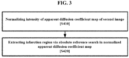
- the step S400 of infarction region image extracting operation may include normalizing an intensity of an apparent diffusion coefficient map of the second image 20 (S410); and a step S420 of extracting an infarction region via an absolute reference search in the normalized apparent diffusion coefficient map. Because the infarction region looks as a dark region in the apparent diffusion coefficient (ADC) map, the computer determines a dark pixel below or equal to a certain threshold value as the infarction region.
- ADC apparent diffusion coefficient
- the computer performs a process of normalizing the intensity of the signal within the ADC image map, since there occur differences between brightness distributions of the ADC image maps between individuals. Thereafter, the computer sets, as an absolute value, a threshold to act as a reference value for the infarction region in a normalized ADC image map.
- the computer aligns the second image 20 with the first image 10, and in the step S600, the computer defines the infarction region in the first image 10, based on a result of the alignment of the second image 20 with the first image 10.
- the computer aligns the second image 20 to the first image 10.
- the computer performs the alignment based on a position of a skeleton in the first image 10 and the second image 20.
- a brain skeleton structure of the same patient is the same even when there is a difference between manners in which MR images are taken.
- the computer performs the alignment based on the skeleton location or structure in the first image 10 and second image 20.
- the computer performs the alignment by applying at least one of an enlargement, a shrinkage, a symmetrical movement, and a rotation, to the first image 10 or second image 20.
- the computer performs the alignment by applying at least one of an enlargement, a shrinkage, a symmetrical movement, and a rotation, to the first image 10 or second image 20.
- the computer then defines the infarction region in the first image 10, based on a result of the alignment of the second image with the first image (based on the infarction region in the second image 20).
- a boundary of the infarction region within the diffusion-weighted image in which the infarction region of the acute cerebral infarction patient appears within a short period of time since the onset time is superimposed on the fluid-attenuated inversion recovery image (i.e., FLAIR image), thereby to define an infarction region in the FLAIR image.
- the computer extracts, in the step S1000, feature information of the first patient, from the infarction region of the first image 10.
- the feature information includes at least one feature element extracted from within a specific region of the image data (i.e., MR image).
- the step S1000 of extracting the feature information is performed by applying at least one of Gray Level Cooccurrence Matrix (GLCM), Run-Length Matrix (especially, Gray Level Run-Length Matrix (GLRLM)), and Local Binary Pattern (LBP), to the signal intensity and gradient of the infarction region.
- GLCM Gray Level Cooccurrence Matrix
- GLRLM Gray Level Run-Length Matrix
- LBP Local Binary Pattern
- the feature information includes various information that may be acquired from the infarction region in the second image 20.
- the feature information includes size or volume information of the infarction region.
- the feature information includes a mean, a standard deviation, a skewness, and kurtosis calculated for a signal intensity, a signal gradient, and a local binary pattern (LBP) map in the infarction region.
- the feature information includes various feature elements obtained by applying a Gray Level Cooccurrence Matrix or Run-Length Matrix. For example, when the computer applies the Gray Level Cooccurrence Matrix or Run-Length Matrix, all or some of the feature elements as shown in [Table 1] are acquired and used as feature information.
- the computer compares the extracted feature information with the reference data to calculate the amount of time that has elapsed since the acute cerebral infarction onset time. That is, the computer calculates the elapsed amount of time after the acute cerebral infarction onset of the first patient, based on the feature information in the infarction region, identified by the second image 20 (i.e., DWI image), in the first image 10 (i.e., FLAIR image).
- the feature information in the infarction region may contain information about a degree to which the infarction region is darkened in the FLAIR image, or information about an image shape that appears in the infarction region.
- the reference data includes feature information of at least one second patients whose acute cerebral infarction onset time is identified, contained in the infarction region of the first image 10 and information on an amount of time that has elapsed since an acute cerebral infarction onset time of the at least one second patient .
- the processor searches the reference data for the feature information of the second patients corresponding to the feature information of the first patient.
- the computer determines the elapsed amount of time after onset of the second patients' acute cerebral infarction as the elapsed amount of time after onset of the acute cerebral infarction for the first patient.
- the computer generates the reference data as a Support Vector Machine (SVM) as described below.
- SVM Support Vector Machine
- the computer applies, to the SVM, the elapsed duration since onset of the acute cerebral infarction of the first patient whose acute cerebral infarction onset time should be identified, thereby to calculate the elapsed duration since the onset of the acute cerebral infarction of the first patient.
- the method further comprises a step S1300 (not shown in the drawings), of determining (S1300) whether or not the elapsed duration is within a reference duration and notifying the determination result.
- the acute cerebral infarction treatment performance plan may vary depending on whether or not the elapsed duration is within the reference duration. Therefore, when the computer informs the medical staff quickly whether a certain time (e.g., 4.5 hours or 6 hours) has elapsed after the acute cerebral infarction has been started, the medical staff may promptly establish the treatment plan.
- the acute cerebral infarction patients have different treatment options depending on the elapsed durations from the onset times of the acute cerebral infarction of the patients.
- An intravenous recombinant tissue plasminogen activator (rt-PA)-based treatment may be available for a patient whose elapsed duration since the onset of the acute cerebral infarction is up to 4.5 hours.
- An intra-arterial mechanical thrombectomy may be effective for a patient whose elapsed duration since the onset of the acute cerebral infarction is up to 6 hours. Therefore, in order for the medical staff to quickly determine the treatment scheme and proceed with the determined treatment scheme, the computer informs the medical staff quickly of whether or not the elapsed amount of time since the onset of the acute cerebral infarction of the first patient is within the reference duration.
- the method includes a step S100 of generating the reference data in a form of a support vector machine (SVM), based on first and second images 10 and 20 of a plurality of second patients. That is, the computer generates a SVM for performing machine learning based on feature information about a plurality of second patients as constructed as big data.
- the feature information for the second patients is obtained in the same manner as in acquiring the feature information for the first patient. That is, the computer aligns the first image 10 and the second image 20 for each second patient with another second patient, defines an infarction region by applying an infarction region identified in the second image 20 to the first image 10, and extracts feature information from the defined infarction region.
- SVM support vector machine
- the step S100 may include, as shown in FIG. 5 , a step S110 of classifying the plurality of second patients into a training group, a validating group, and a testing group; a step S120 of extracting feature information of each of second patients classified into the training group, from a first image 10 of the each of the second patients classified into the training group, by performing an alignment of the first image of the each of the second patients classified into the training group with a second image of the each of the second patients classified into the training group; a step S130 of training the support vector machine by using the feature information of the second patients classified into the training group; a step S140 of determining a type of the support vector machine by using the validating group; and a step S150 of applying the testing group to the determined support vector machine type to determine whether the determined support vector machine type is to be defined as the reference data.
- the computer may classify a plurality of second patients into the training group (e.g., 60% of the entire second patients), the validating group (for example, 20% of the entire second patients), and the testing group (for example, 20% of the entire second patients). Then, a particular type of SVM is selected by applying the information of the second patients classified into the validating group to SVMs computed via training using the training group. Then, feature information of the second patients classified into the testing group is applied to the selected type of SVM to evaluate whether the selected type of SVM corresponds to an optimal SVM.
- the training group e.g. 60% of the entire second patients
- the validating group for example, 20% of the entire second patients
- the testing group for example, 20% of the entire second patients
- the reference data is classified into detailed groups based on the gender or age information of the second patients.
- the computer may classify a plurality of second patients into a plurality of detailed groups based on the gender or age, and may generate data for elapsed amount of time calculation for each detailed group.
- the computer may generate an SVM for each detailed group of the second patients.
- the step S1200 (the elapsed amount of time calculation operation) may include defining detailed groups to be compared with the feature information of the first patient, based on age information or gender information of the first patient.
- the method may further include a step S700 of comparing the infarction region with a symmetric region located in a brain of the first patient and being symmetrical to the infarction region to extract a relative signal amount of the infarction region, based on a result of the comparison of the infarction region with the symmetric region. Since the MR image is represented using a relative signal amount within an image to be captured, it is possible that the signal amount of the infarction region is not an absolute signal amount.
- the computer may compare the infarction region with a symmetric region located in a brain of the first patient and being symmetrical to the infarction region in the first image 10 to extract a relative signal amount of the infarction region. Therefore, as the relative signal amount of the infarction region in the first image 10 is calculated, the feature information extracted from the infarction region in the first image 10 of the first patient may be compared with the feature information extracted from the infarction region of the first image 10 of the second patient.
- the method may further include a step S800 of generating a ratio map 30 that includes a visual expression of a relative signal amount between the symmetric region and the infarction region.
- the feature information may include first feature information extracted from the infarction region in the first image 10; and second feature information extracted from an infarction region in the ratio map 30 for the first image 10. That is, the computer uses at least one feature element extracted from the first image 10 itself and from the ratio map 30 for the first image 10 as feature information.
- FIG. 8 is a schematic view illustrating a configuration of an acute cerebral infarction onset time estimation system, according to one embodiment of the inventive concept.
- the acute cerebral infarction onset time estimation system in accordance with another embodiment of the inventive concept may include at least one computer for estimating or calculating the acute cerebral infarction onset time.
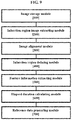
- the acute cerebral infarction onset time estimation system in accordance with another embodiment of the inventive concept may include an image storage module 100; an infarction region image extracting module 200; an image alignment module 300; an infarction region defining module 400; a feature information extracting module 500; and an elapsed duration calculating module 600.
- the image storage module 100 stores the first image 10 and second image 20 for a first patient whose acute cerebral infarction onset time is not identified. That is, the image storage module 100 receives and stores the first image 10 and the second image 20 of the first patient from the MR image capture device.
- the image storage module 100 may embodied as a memory present in the computer.
- the memory may include at least one storage media such as a flash memory type, a hard disk type, a multimedia card micro type, a card type memory (for example, SD or XD memory), a random access memory (RAM), SRAM (static random access memory), read-only memory (ROM), EEPROM (electrically erasable programmable read-only memory), PROM (programmable read-only memory), magnetic memory, magnetic disk, or optical disk.
- storage media such as a flash memory type, a hard disk type, a multimedia card micro type, a card type memory (for example, SD or XD memory), a random access memory (RAM), SRAM (static random access memory), read-only memory (ROM), EEPROM (electrically erasable programmable read-only memory), PROM (programmable read-only memory), magnetic memory, magnetic disk, or optical disk.
- the infarction region image extracting module 200 extracts the infarction region image from the second image 20. That is, the infarction region image extracting module 200 extracts the infarction region image by grasping the boundary of the infarction region in the second image 20 (i.e., DWI image).
- the image alignment module 300 aligns the second image 20 with the first image 10. In one embodiment, the image alignment module 300 performs the alignment based on a skeleton or brain shape in the first image 10 and second image 20.
- the infarction region defining module 400 defines the infarction region in the first image 10, based on a result of the alignment of the second image 20 with the first image 10. . That is, the infarction region defining module 400 may superimpose a boundary of the infarction region within the diffusion-weighted image in which the infarction region of the acute cerebral infarction patient appears within a short period of time since the onset time on the fluid-attenuated inversion recovery image (i.e., FLAIR image), thereby to define an infarction region in the FLAIR image.
- FLAIR image fluid-attenuated inversion recovery image
- the feature information extracting module 500 extracts feature information of the first patient, from the infarction region of the first image 10.
- the feature information may be at least one feature element extracted from the specific region of the image data, that is, the MR image.
- the feature information extracting module 500 may apply at least one of Gray Level Cooccurrence Matrix (GLCM), Run-Length Matrix (especially, Gray Level Run-Length Matrix (GLRLM)), and Local Binary Pattern (LBP), to the signal intensity and gradient of the infarction region, to extract the feature information.
- GLCM Gray Level Cooccurrence Matrix
- GLRLM Gray Level Run-Length Matrix
- LBP Local Binary Pattern
- the elapsed duration calculating module 600 compares the extracted feature information with the reference data to calculate the elapsed amount of time since the acute cerebral infarction onset time, based on a result of the comparison of the extracted feature information with reference data. That is, the elapsed duration calculating module 600 calculates the elapsed amount of time since the onset of the acute cerebral infarction of the first patient based on the feature information in the infarction region of the second image 20 (i.e., DWI image) within the first image 10 (i.e., FLAIR image).
- Each of the infarction region image extracting module 200, image alignment module 300, infarction region defining module 400, feature information extracting module 500, and elapsed duration calculating module 600 is contained in a particular computer, in software or hardware manner.
- an acute cerebral infarction onset time estimation system further includes a reference data generating module 700.
- the reference data generating module 700 generates the reference data as the support vector machine, based on the first image 10 and the second image 20 of each of a plurality of second patients.
- the reference data generating module 700 may perform following operations: classifying the plurality of second patients into a training group, a validating group, and a testing group; extracting feature information of each of second patients classified into the training group, from a first image 10 of the each of the second patient classified into the training group, by performing an alignment of the first image of the each of the second patients classified into the training group with a second image of the each of the second patients classified into the training group; training the support vector machine by using the feature information of the second patients classified into the training group; determining a type of the support vector machine by using the validating group; and applying the testing group to the determined support vector machine type to determine whether the determined support vector machine type is to be defined as the reference data.
- an acute cerebral infarction onset time estimation system further includes a ratio map generating module 800.
- the ratio map generating module 800 may be configured to compare the infarction region with a symmetric region located in a brain of the first patient and being symmetrical to the infarction region, extract a relative signal amount of the infarction region, based on a result of the comparison of the infarction region with the symmetric region; and generate the ratio map 30 that includes a visual expression of a relative signal amount between the symmetric region and the infarction region. Since the MR image is represented using a relative signal amount within an image to be captured, the signal amount of the infarction region may not employ an absolute signal amount.
- the ratio map generating module 800 may compare the infarction region with a symmetric region located in a brain of the first patient and being symmetrical to the infarction region in the first image 10 to extract a relative signal amount of the infarction region. Therefore, as the relative signal amount of the infarction region in the first image 10 is calculated, the feature information extracted from the infarction region in the first image 10 of the first patient may be compared with the feature information extracted from the infarction region of the first image 10 of the second patient.
- the feature information may include: first feature information extracted from the infarction region in the first image 10; and second feature information extracted from an infarction region in the ratio map 30 for the first image 10.
- the acute cerebral infarction onset time estimation method may be implemented using a program (or application) to be executed in combination with a computer as hardware, which may be stored in a medium.
- the above-mentioned program may include codes encoded in a computer language such as C, C ++, JAVA, or machine language which a central processing unit (CPU) of a computer can read via a device interface of the computer.
- the program may perform the method.
- codes may include functional codes related to functions that define necessary functions to execute the method, or may include executable procedure-related control codes necessary for the processor of the computer to execute the functions in accordance with a predetermined procedure.
- these codes may further include memory reference-related code that indicates where additional information or media needed for the execution of the functions by the computer's processor is addressed to any location (address) in the computer's internal or external memory.
- the codes may further include communication related codes indicating, for example, how to communicate with any other computer or server remotely using a communication module of the computer, and what information or media as transmitted or received therebetween during communication.
- the storage medium is not a medium for storing data for a short time such as a register, a cache, a memory, etc., but means a medium that semi-permanently stores data and is capable of being read by a device.
- examples of the storage medium include, but are not limited to, ROM, RAM, CD-ROM, magnetic tape, floppy disk, optical data storage, and the like. That is, the program may be stored on various recording media on various servers that the computer can access, or on various recording media on the user's computer. Further, the medium may store therein computer readable codes as distributed over a networked computer system and as read by the computer in a distributed manner.
- the acute cerebral infarction onset time can be estimated from a patient whose acute cerebral infarction onset time is not identified. This may reduce the number of patients who miss the timing of thrombolysis due to the inability to determine whether the acute cerebral infarction is capable of being cured using thrombolysis.
- the medical staff may figure out the elapsed duration after the patient's acute cerebral infarction starts, and may determine immediately whether to start the thrombolysis for the patient. Therefore, a time required for the medical staff to determine whether or not the thrombolysis is performed using the MR image reading may be reduced. This can increase the percentage of patients whose acute cerebral infarction can be treated.
Landscapes
- Health & Medical Sciences (AREA)
- Engineering & Computer Science (AREA)
- Life Sciences & Earth Sciences (AREA)
- Physics & Mathematics (AREA)
- Theoretical Computer Science (AREA)
- General Physics & Mathematics (AREA)
- Computer Vision & Pattern Recognition (AREA)
- Medical Informatics (AREA)
- General Health & Medical Sciences (AREA)
- Nuclear Medicine, Radiotherapy & Molecular Imaging (AREA)
- Radiology & Medical Imaging (AREA)
- Biophysics (AREA)
- Pathology (AREA)
- Molecular Biology (AREA)
- Surgery (AREA)
- Animal Behavior & Ethology (AREA)
- Biomedical Technology (AREA)
- Veterinary Medicine (AREA)
- Public Health (AREA)
- Heart & Thoracic Surgery (AREA)
- Neurology (AREA)
- Quality & Reliability (AREA)
- High Energy & Nuclear Physics (AREA)
- Physiology (AREA)
- Neurosurgery (AREA)
- Psychology (AREA)
- Magnetic Resonance Imaging Apparatus (AREA)
Abstract
Description
- Embodiments of the inventive concept relate to an acute cerebral infarction onset time estimation system, method and program. More specifically, embodiments of the inventive concept relate to a system, a method, and a program for estimating an amount of time that has elapsed time since an onset time of acute cerebral infarction of a patient whose acute cerebral infarction onset time has not been identified, by using magnetic resonance imaging (MRI).
- Thrombolysis, which prevents damages to brain cells by helping blood-communication in occluded cerebral blood vessels due to thrombus or embolus, is widely known as a method for treating patients with acute cerebral infarction. Thrombolysis should be performed as soon as possible from the start point of cerebral infarction, since the thrombolysis must be done promptly before the brain cells are damaged.
- However, the majority of patients who come to hospitals have no information about an onset time of acute cerebral infarction thereof. In other words, after a patient falls down, the patient is found by another person and transferred to a hospital. Thus, it may not be accurately checked when the acute cerebral infarction started before the patient was discovered by another person. In this case, there is a shortage of specialists who can judge whether the patient would be able to be subjected to thrombolysis only based on reading medical images (i.e., MR images).
- In addition, in emergency rooms where patients with acute cerebral infarction were transferred, the number of medical staffs experienced in the acute cerebral infarction is small.
- Therefore, there is a need for an invention that provides information that allows clinicians to safely and quickly determine the feasibility of treatment for patients with acute cerebral infarction.
- Embodiments of the inventive concept are to provide an acute cerebral infarction onset time estimation system, method and program, by which an infarction region is automatically analyzed using characteristics of a diffusion-weighted image and a fluid-attenuated inversion recovery image of Magnetic Resonance Image (MRI) of the acute cerebral infarction patient, then, an amount of time that has elapsed since starting the acute cerebral infarction is estimated based on feature information in the infarction region, and, then, the estimated amount of time is provided to the clinician to quickly determine whether the acute cerebral infarction of the patient can be treated.
- Purposes presented by the embodiments of the inventive concept are not limited to the purpose set forth above, and other purposes not mentioned may be clearly understood by one of ordinary skill in the art from the following description.
- According to an exemplary embodiment, a method for estimating an onset time of acute cerebral infarction includes: receiving, by a computer, a first image and a second image of a specific first patient whose acute cerebral infarction onset time is not identified, the first image is a fluid-attenuated inversion recovery image, and the second image is a diffusion-weighted image; extracting, by the computer, an infarction region image from the second image; aligning, by the computer, the second image with the first image to define an infarction region in the first image, based on a result of the alignment of the second image with the first image; extracting, by the computer, feature information of the first patient, from the infarction region in the first image; and comparing, by the computer, the extracted feature information with reference data to calculate an amount of time that has elapsed since the acute cerebral infarction onset time, based on a result of the comparison of the extracted feature information with the reference data.
- According to the inventive concept as described above, the acute cerebral infarction onset time can be estimated from a patient whose acute cerebral infarction onset time has not been identified. This may reduce the number of patients who miss the timing of thrombolysis due to the inability to determine whether the acute cerebral infarction is capable of be cured using thrombolysis.
- Further, because the a computer quickly performs the elapsed time amount calculation of the acute cerebral infarction automatically based on the fluid-attenuated inversion recovery (FAIR) image and the diffusion-weighted image, the medical staff may figure out the elapsed duration after the patient's acute cerebral infarction starts, and may determine immediately whether to start the thrombolysis for the patient. Therefore, a time required for the medical staff to determine whether or not the thrombolysis is performed using the MR image reading may be reduced. This can increase the percentage of patients whose acute cerebral infarction can be treated.
- The above and other objects and features will become apparent from the following description with reference to the following figures, wherein like reference numerals refer to like parts throughout the various figures unless otherwise specified, and wherein:
-
FIG. 1 is a flowchart of an acute cerebral infarction onset time estimation method according to one embodiment of the inventive concept; -
FIG. 2 shows an example of a process for applying the acute cerebral infarction onset time estimation method to a first image and a second image, according to one embodiment of the inventive concept; -
FIG. 3 is a flowchart of an infarction region image extraction process, according to one embodiment of the inventive concept; -
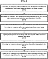
FIG. 4 is a flowchart of the acute cerebral infarction onset time estimation method that further includes a reference data generation operation, according to one embodiment of the inventive concept; -
FIG. 5 is a flowchart showing a process of generating reference data in a form of Support Vector Machine (SVM), according to one embodiment of the inventive concept; -
FIG. 6 is a flow chart of the acute cerebral infarction onset time estimation method which further includes a relative signal amount extracting operation of the first image according to one embodiment of the inventive concept; -
FIG. 7 is a flowchart of the acute cerebral infarction onset time estimation method that further includes a ratio map generation operation for the first image, according to one embodiment of the inventive concept; -
FIG. 8 is a schematic view illustrating a configuration of an acute cerebral infarction onset time estimation system, according to one embodiment of the inventive concept; -
FIG. 9 is a schematic view illustrating a configuration of an acute cerebral infarction onset time estimation system, according to another embodiment of the inventive concept; and -
FIG. 10 shows a schematic view illustrating a configuration of an acute cerebral infarction onset time estimation system, according to still another embodiment of the inventive concept. - According to an exemplary embodiment, a method for estimating an onset time of acute cerebral infarction includes: receiving, by a computer, a first image and a second image of a specific first patient whose acute cerebral infarction onset time is not identified, the first image is a fluid-attenuated inversion recovery image, and the second image is a diffusion-weighted image; extracting, by the computer, an infarction region image from the second image; aligning, by the computer, the second image with the first image to define an infarction region in the first image, based on a result of the alignment of the second image with the first image; extracting, by the computer, feature information of the first patient, from the infarction region in the first image; and comparing, by the computer, the extracted feature information with reference data an amount of time that has elapsed since the acute cerebral infarction onset time, based on a result of the comparison of the extracted feature information with the reference data.
- Further, according to an exemplary embodiment, the reference data may include feature information contained in an infarction region of a first image of the at least one second patient. The second patient is a patient whose acute cerebral infarction onset time is identified. The reference data further includes information on an amount of time that has elapsed since an acute cerebral infarction onset time of the at least one second patient.
- Further, according to an exemplary embodiment, the extracting of the infarction region image from the second image may include normalizing an intensity of an apparent diffusion coefficient map of the second image; and extracting the infarction region image via an absolute reference value search in the normalized apparent diffusion coefficient map.
- Further, according to an exemplary embodiment, the method may further include comparing the infarction region with a symmetric region located in a brain of the first patient and being symmetrical to the infarction region to extract a relative signal amount of the infarction region, based on a result of the comparison of the infarction region with the symmetric region.
- Further, according to an exemplary embodiment, the method may further include generating a ratio map that includes a visual expression of a relative signal amount between the symmetric region and the infarction region.
- Further, according to an exemplary embodiment, the feature information may include: first feature information extracted from the infarction region in the first image; and second feature information extracted from an infarction region in the ratio map for the first image.
- Further, according to an exemplary embodiment, the method may further include determining whether the elapsed amount of time is within a reference duration; and notifying the determination result.
- Further, according to an exemplary embodiment, the defining of the infarction region in the first image may include aligning of the second image with the first image by applying at least one of an enlargement, a shrinkage, a symmetrical movement, and a rotation, to the first image or the second image, based on a skeleton position in the first image or the second image.
- Further, according to an exemplary embodiment, the extracting of the feature information may include comprises extracting the feature information by applying at least one of a gray level co-occurrence matrix, a run-length matrix and a local binary pattern to signal intensity and gradient of the infarction region.
- Further, according to an exemplary embodiment, the method may further include generating the reference data as a support vector machine, based on first and second images of a plurality of second patients. The generating of the reference data may include: classifying the plurality of second patients into a training group, a validating group, and a testing group; extracting feature information of each of second patients classified into the training group, from a first image of the each of the second patients classified into the training group, by performing an alignment of the first image of the each of the second patients classified into the training group with a second image of the each of the second patients classified into the training group; training the support vector machine by using the feature information of the second patients classified into the training group; determining a type of the support vector machine by using the validating group; and applying the testing group to the determined support vector machine type to determine whether the determined support vector machine type is to be defined as the reference data.
- Further, according to an exemplary embodiment, the reference data may be classified into detailed groups according to gender or age information of the second patients. The calculating of the elapsed amount of time may include defining detailed groups to be compared with the feature information of the first patient based on age information or gender information of the first patient.
- According to an exemplary embodiment, a system for estimation of an acute cerebral infarction onset time includes: an image storage module that stores a first image and a second image of a specific first patient whose acute cerebral infarction onset time is not identified, the first image is a fluid-attenuated inversion recovery image, and the second image is a diffusion-weighted image; an infarction region image extraction module that extracts an infarction region image from the second image; an image alignment module that aligns the second image with the first image; an infarction region defining module that defines an infarction region in the first image, based on a result of the alignment of the second image with the first image; a feature information extracting module that extracts feature information of the first patient, from the infarction region of the first image; and an elapsed amount of time calculator that compares the extracted feature information with reference data, and an elapsed duration calculation module that calculates an amount of time that has elapsed since the acute cerebral infarction onset time, based on a result of the comparison of the extracted feature information with reference data.
- Further, according to an exemplary embodiment, the reference data may include feature information of at least one second patient, contained in an infarction region of a first image of the at least one second patient, the second patient is a patient whose acute cerebral infarction onset time is identified; and information on an amount of time that has elapsed since an acute cerebral infarction onset time of the at least one second patient..
- Further, according to an exemplary embodiment, the system may further include: a reference data generation module that generates the reference data as a support vector machine, based on first and second images of a plurality of second patients. The reference data generation module may be configured to: classify the plurality of second patients into a training group, a validating group, and a testing group; extract feature information of each of second patients classified into the training group from a first image of the each of the second patients classified into the training group, by performing an alignment of the first image of the each of the second patients classified into the training group with a second image of the each of the second patients classified into the training group; train the support vector machine by using the feature information of the second patients classified into the training group; determine a type of the support vector machine by using the validating group; and apply the testing group to the determined support vector machine type to determine whether the determined support vector machine type is to be defined as the reference data.
- Further, according to an exemplary embodiment, the system may further include a ratio map generation module configured to: compare the infarction region with a symmetric region located in a brain of the first patient and being symmetrical to the infarction region; extract a relative signal amount of the infarction region, based on a result of the comparison of the infarction region with the symmetric region; and generate a ratio map that includes a visual expression of a relative signal amount between the symmetric region and the infarction region. The feature information includes: first feature information extracted from the infarction region in the first image; and second feature information extracted from an infarction region in the ratio map for the first image.
- According to an exemplary embodiment, a computer program for estimation of an acute cerebral infarction onset time is stored in a computer readable medium. When the program is coupled to and executed by the computer, the program enables the computer to perform a method for estimating an onset time of acute cerebral infarction, as described above.
- Hereinafter, preferred embodiments of the inventive concept will be described in detail with reference to the accompanying drawings. Advantages and features of the inventive concept, and methods of accomplishing the same, will become apparent with reference to the embodiments described in detail below with reference to the accompanying drawings. However, the inventive concept is not limited to the embodiments disclosed below, but may be embodied in various forms. These embodiments are provided so that the disclosure of the inventive concept is fully explained, and that it is believed that the disclosure is intended to be sufficiently understood by one of ordinary skill in the art which the inventive concept belongs. The inventive concept is only defined by the scope of the claim. Like reference numerals refer to like elements throughout the specification.
- Unless otherwise defined, all terms including technical and scientific terms used herein have the same meaning as commonly understood by one of ordinary skill in the art to which this inventive concept belongs. It will be further understood that terms, such as those defined in commonly used dictionaries, should be interpreted as having a meaning that is consistent with their meaning in the context of the relevant art and will not be interpreted in an idealized or overly formal sense unless expressly so defined herein.
- The terminology used herein is for the purpose of describing particular embodiments only and is not intended to be limiting of the present disclosure. As used herein, the singular forms "a" and "an" are intended to include the plural forms as well, unless the context clearly indicates otherwise. It will be further understood that the terms "comprises", "comprising", "includes", and "including" when used in this specification, specify the presence of the stated features, integers, operations, elements, and/or components, but do not preclude the presence or addition of one or more other features, integers, operations, elements, components, and/or portions thereof.
- As used herein, "
first image 10" is referring to a fluid-attenuated inversion recovery image (FLAIR image). - As used herein, "
second image 20" is referring to a diffusion-weighted image (DWI image). - As used herein, "first patient" is referring to a patient whose acute cerebral infarction onset time is not identified. In other words, the "first patient" is referring to a patient whose acute cerebral infarction onset time should be identified by the computer. By contrast, "second patient" is referring to a patient whose acute cerebral infarction onset time is identified.
- As used herein, "computer" includes all of the various devices that can perform computational processing and visually present the processing results to the user. For example, the computer may include not only a desktop PC, a note book, but also a smart phone, a tablet PC, a cellular phone, a personal communication service phone (PCS phone), a mobile terminal based on a synchronous/asynchronous IMT-2000 (International Mobile Telecommunication-2000), a Palm personal computer, a personal digital assistant (PDA), etc. Further, the computer may also include medical equipment that acquires or views an angiographic image.
- Hereinafter, each of an acute cerebral infarction onset time estimation system, method and program, according to embodiments of the inventive concept will be described in detail with reference to the drawings.
-
FIG. 1 is a flowchart of the acute cerebral infarction onset time estimation method according to one embodiment of the inventive concept. -
FIG. 2 shows an example of a process of applying the acute cerebral infarction onset time estimation method to a first image and a second image according to one embodiment of the inventive concept. - Referring to
FIG. 1 , the acute cerebral infarction onset time estimation method according to one embodiment of the inventive concept may include a step S200 of receiving, by a computer, afirst image 10 and asecond image 20 of a specific first patient whose acute cerebral infarction onset time is not identified; a step S400 of extracting, by the computer, an infarction region image from thesecond image 20; a step S600 of aligning, by the computer, the second image with thefirst image 10 to define an infarction region in the first image, based on a result of the alignment of the second image with the first image; a step S1000 of extracting feature information of the first patient, from the infarction region in thefirst image 10; and a step S1200 of comparing, by the computer, the extracted feature information with reference data to calculate an amount of time that has elapsed since the acute cerebral infarction onset time. Hereinafter, a detailed description of each operation will be described. - The diffusion-weighted image shows an acute cerebral infarction area within a short time after the acute cerebral infarction occurs and thus a person who views the diffusion-weighted image would be able to immediately identify an acute cerebral infarction range. On the other hand, in the fluid-attenuated inversion recovery image, the acute cerebral infarction area is gradually darkened over time after the acute cerebral infarction onset time. In other words, in the fluid-attenuated inversion recovery image (i.e., FLAIR image), an expression level of the infarction region in the acute cerebral infarction may vary based on an elapsed duration since the acute cerebral infarction onset time. Therefore, the inventive concept may identify an amount of time that has elapsed since the acute cerebral infarction onset time based on the discrepancy between the DWI image and the FLAIR image.
- The computer receives the
first image 10 and thesecond image 20 for a specific first patient whose acute cerebral infarction onset time is not identified (S200; image receiving operation). The computer may directly receive medical image data taken from a magnetic resonance imaging (MRI) device. Alternatively, the computer may download medical image data from another computer or an external server and store the received image data in the computer. For example, when the medical staff determines the amount of the time that has elapsed since the acute cerebral infarction of a patient entering the emergency room due to the acute cerebral infarction and intends to make an emergency treatment plan, the computer may receive a MR image of the patient directly from the magnetic resonance imaging (MRI) device. - The computer extracts the infarction region image from the second image 20 (S400). In other words, the computer identifies a boundary of the infarction region within the second image 20 (i.e., DWI image) and extracts the infarction region image based on the identified boundary. In one embodiment, as in
FIG. 3 , the step S400 of infarction region image extracting operation may include normalizing an intensity of an apparent diffusion coefficient map of the second image 20 (S410); and a step S420 of extracting an infarction region via an absolute reference search in the normalized apparent diffusion coefficient map. Because the infarction region looks as a dark region in the apparent diffusion coefficient (ADC) map, the computer determines a dark pixel below or equal to a certain threshold value as the infarction region. The computer performs a process of normalizing the intensity of the signal within the ADC image map, since there occur differences between brightness distributions of the ADC image maps between individuals. Thereafter, the computer sets, as an absolute value, a threshold to act as a reference value for the infarction region in a normalized ADC image map. - The computer aligns the
second image 20 with thefirst image 10, and in the step S600, the computer defines the infarction region in thefirst image 10, based on a result of the alignment of thesecond image 20 with thefirst image 10. - First, the computer aligns the
second image 20 to thefirst image 10. In one embodiment, the computer performs the alignment based on a position of a skeleton in thefirst image 10 and thesecond image 20. A brain skeleton structure of the same patient is the same even when there is a difference between manners in which MR images are taken. Thus, the computer performs the alignment based on the skeleton location or structure in thefirst image 10 andsecond image 20. The computer performs the alignment by applying at least one of an enlargement, a shrinkage, a symmetrical movement, and a rotation, to thefirst image 10 orsecond image 20. Because there is a difference between brain shapes in thefirst image 10 andsecond image 20 when there is a change in a patient posture at the time of capturing thefirst image 10 andsecond image 20, the computer performs the alignment by applying at least one of an enlargement, a shrinkage, a symmetrical movement, and a rotation, to thefirst image 10 orsecond image 20. - The computer then defines the infarction region in the
first image 10, based on a result of the alignment of the second image with the first image (based on the infarction region in the second image 20). A boundary of the infarction region within the diffusion-weighted image in which the infarction region of the acute cerebral infarction patient appears within a short period of time since the onset time is superimposed on the fluid-attenuated inversion recovery image (i.e., FLAIR image), thereby to define an infarction region in the FLAIR image. - The computer extracts, in the step S1000, feature information of the first patient, from the infarction region of the
first image 10. The feature information includes at least one feature element extracted from within a specific region of the image data (i.e., MR image). In one embodiment, the step S1000 of extracting the feature information is performed by applying at least one of Gray Level Cooccurrence Matrix (GLCM), Run-Length Matrix (especially, Gray Level Run-Length Matrix (GLRLM)), and Local Binary Pattern (LBP), to the signal intensity and gradient of the infarction region. - Specifically, the feature information includes various information that may be acquired from the infarction region in the
second image 20. In one embodiment, the feature information includes size or volume information of the infarction region. Further, in another embodiment, the feature information includes a mean, a standard deviation, a skewness, and kurtosis calculated for a signal intensity, a signal gradient, and a local binary pattern (LBP) map in the infarction region. Further, in another embodiment, the feature information includes various feature elements obtained by applying a Gray Level Cooccurrence Matrix or Run-Length Matrix. For example, when the computer applies the Gray Level Cooccurrence Matrix or Run-Length Matrix, all or some of the feature elements as shown in [Table 1] are acquired and used as feature information.[Table 1] Extraction scheme Available feature elements GLCM energy, entropy, dissimilarity, contrast, inverse difference, correlation, homogeneity, autocorrelation, cluster shade, cluster prominence, maximum probability, sum of squares, sum average, sum variance, sum entropy, difference variance, difference entropy, information of correlation1 (IOC1), information of correlation2 (IOC2), inverse difference normalized (INN), inverse difference moment normalized (IDN) GLRLM short run emphasis (SRE), long run emphasis (LRE), gray-level nonuniformity (GLN), run-length nonuniformity (RLN), run percentage (RP), low gray run emphasis (LGRE), high gray run emphasis (HGRE), short run low gray emphasis (SRLGE), short run high gray emphasis (SRHGE), long run low gray emphasis (LRLGE), long run high gray emphasis (LRHGE) - Thereafter, the computer, in the step S1200, compares the extracted feature information with the reference data to calculate the amount of time that has elapsed since the acute cerebral infarction onset time. That is, the computer calculates the elapsed amount of time after the acute cerebral infarction onset of the first patient, based on the feature information in the infarction region, identified by the second image 20 (i.e., DWI image), in the first image 10 (i.e., FLAIR image). For example, the feature information in the infarction region may contain information about a degree to which the infarction region is darkened in the FLAIR image, or information about an image shape that appears in the infarction region. The reference data includes feature information of at least one second patients whose acute cerebral infarction onset time is identified, contained in the infarction region of the
first image 10 and information on an amount of time that has elapsed since an acute cerebral infarction onset time of the at least one second patient . The processor searches the reference data for the feature information of the second patients corresponding to the feature information of the first patient. The computer determines the elapsed amount of time after onset of the second patients' acute cerebral infarction as the elapsed amount of time after onset of the acute cerebral infarction for the first patient. In one embodiment, the computer generates the reference data as a Support Vector Machine (SVM) as described below. The computer applies, to the SVM, the elapsed duration since onset of the acute cerebral infarction of the first patient whose acute cerebral infarction onset time should be identified, thereby to calculate the elapsed duration since the onset of the acute cerebral infarction of the first patient. - Further, in another embodiment of the inventive concept, the method further comprises a step S1300 (not shown in the drawings), of determining (S1300) whether or not the elapsed duration is within a reference duration and notifying the determination result. The acute cerebral infarction treatment performance plan may vary depending on whether or not the elapsed duration is within the reference duration. Therefore, when the computer informs the medical staff quickly whether a certain time (e.g., 4.5 hours or 6 hours) has elapsed after the acute cerebral infarction has been started, the medical staff may promptly establish the treatment plan. The acute cerebral infarction patients have different treatment options depending on the elapsed durations from the onset times of the acute cerebral infarction of the patients. An intravenous recombinant tissue plasminogen activator (rt-PA)-based treatment may be available for a patient whose elapsed duration since the onset of the acute cerebral infarction is up to 4.5 hours. An intra-arterial mechanical thrombectomy may be effective for a patient whose elapsed duration since the onset of the acute cerebral infarction is up to 6 hours. Therefore, in order for the medical staff to quickly determine the treatment scheme and proceed with the determined treatment scheme, the computer informs the medical staff quickly of whether or not the elapsed amount of time since the onset of the acute cerebral infarction of the first patient is within the reference duration.
- Further, in another embodiment of the inventive concept, as shown in
FIG. 4 , the method includes a step S100 of generating the reference data in a form of a support vector machine (SVM), based on first andsecond images first image 10 and thesecond image 20 for each second patient with another second patient, defines an infarction region by applying an infarction region identified in thesecond image 20 to thefirst image 10, and extracts feature information from the defined infarction region. - In one embodiment, the step S100 (the reference data generation operation) may include, as shown in
FIG. 5 , a step S110 of classifying the plurality of second patients into a training group, a validating group, and a testing group; a step S120 of extracting feature information of each of second patients classified into the training group, from afirst image 10 of the each of the second patients classified into the training group, by performing an alignment of the first image of the each of the second patients classified into the training group with a second image of the each of the second patients classified into the training group; a step S130 of training the support vector machine by using the feature information of the second patients classified into the training group; a step S140 of determining a type of the support vector machine by using the validating group; and a step S150 of applying the testing group to the determined support vector machine type to determine whether the determined support vector machine type is to be defined as the reference data. That is, when the feature information of the second patient to be subjected to training to generate the SVM is used for the SVM selection and verification, it is possible that the SVM appropriateness examination is not performed properly. Thus, the computer may classify a plurality of second patients into the training group (e.g., 60% of the entire second patients), the validating group (for example, 20% of the entire second patients), and the testing group (for example, 20% of the entire second patients). Then, a particular type of SVM is selected by applying the information of the second patients classified into the validating group to SVMs computed via training using the training group. Then, feature information of the second patients classified into the testing group is applied to the selected type of SVM to evaluate whether the selected type of SVM corresponds to an optimal SVM. - Further, in another embodiment of the inventive concept, the reference data is classified into detailed groups based on the gender or age information of the second patients. There occur differences between state changes over time after onset time of acute cerebral infarction based on gender or age of patient. Therefore, in order to accurately calculate the elapsed amount of time, based on the age and gender of the first patient, the computer may classify a plurality of second patients into a plurality of detailed groups based on the gender or age, and may generate data for elapsed amount of time calculation for each detailed group. For example, the computer may generate an SVM for each detailed group of the second patients. In this connection, the step S1200 (the elapsed amount of time calculation operation) may include defining detailed groups to be compared with the feature information of the first patient, based on age information or gender information of the first patient.
- Further, as shown in
FIG. 6 , in another embodiment of the inventive concept, the method may further include a step S700 of comparing the infarction region with a symmetric region located in a brain of the first patient and being symmetrical to the infarction region to extract a relative signal amount of the infarction region, based on a result of the comparison of the infarction region with the symmetric region. Since the MR image is represented using a relative signal amount within an image to be captured, it is possible that the signal amount of the infarction region is not an absolute signal amount. Therefore, using characteristics of the brain that the brain is symmetrically configured based on a specific boundary line, the computer may compare the infarction region with a symmetric region located in a brain of the first patient and being symmetrical to the infarction region in thefirst image 10 to extract a relative signal amount of the infarction region. Therefore, as the relative signal amount of the infarction region in thefirst image 10 is calculated, the feature information extracted from the infarction region in thefirst image 10 of the first patient may be compared with the feature information extracted from the infarction region of thefirst image 10 of the second patient. - Further, as shown in
FIG. 7 , in another embodiment of the inventive concept, the method may further include a step S800 of generating aratio map 30 that includes a visual expression of a relative signal amount between the symmetric region and the infarction region. - Further, in another embodiment, when the computer generates the
ratio map 30 based on thefirst image 10, the feature information may include first feature information extracted from the infarction region in thefirst image 10; and second feature information extracted from an infarction region in theratio map 30 for thefirst image 10. That is, the computer uses at least one feature element extracted from thefirst image 10 itself and from theratio map 30 for thefirst image 10 as feature information. -
FIG. 8 is a schematic view illustrating a configuration of an acute cerebral infarction onset time estimation system, according to one embodiment of the inventive concept. - The acute cerebral infarction onset time estimation system in accordance with another embodiment of the inventive concept may include at least one computer for estimating or calculating the acute cerebral infarction onset time. Referring to
FIG. 8 , the acute cerebral infarction onset time estimation system in accordance with another embodiment of the inventive concept may include animage storage module 100; an infarction regionimage extracting module 200; animage alignment module 300; an infarctionregion defining module 400; a featureinformation extracting module 500; and an elapsedduration calculating module 600. - The
image storage module 100 stores thefirst image 10 andsecond image 20 for a first patient whose acute cerebral infarction onset time is not identified. That is, theimage storage module 100 receives and stores thefirst image 10 and thesecond image 20 of the first patient from the MR image capture device. - The
image storage module 100 may embodied as a memory present in the computer. The memory may include at least one storage media such as a flash memory type, a hard disk type, a multimedia card micro type, a card type memory (for example, SD or XD memory), a random access memory (RAM), SRAM (static random access memory), read-only memory (ROM), EEPROM (electrically erasable programmable read-only memory), PROM (programmable read-only memory), magnetic memory, magnetic disk, or optical disk. - The infarction region
image extracting module 200 extracts the infarction region image from thesecond image 20. That is, the infarction regionimage extracting module 200 extracts the infarction region image by grasping the boundary of the infarction region in the second image 20 (i.e., DWI image). - The
image alignment module 300 aligns thesecond image 20 with thefirst image 10. In one embodiment, theimage alignment module 300 performs the alignment based on a skeleton or brain shape in thefirst image 10 andsecond image 20. - The infarction
region defining module 400 defines the infarction region in thefirst image 10, based on a result of the alignment of thesecond image 20 with thefirst image 10. . That is, the infarctionregion defining module 400 may superimpose a boundary of the infarction region within the diffusion-weighted image in which the infarction region of the acute cerebral infarction patient appears within a short period of time since the onset time on the fluid-attenuated inversion recovery image (i.e., FLAIR image), thereby to define an infarction region in the FLAIR image. - The feature
information extracting module 500 extracts feature information of the first patient, from the infarction region of thefirst image 10. The feature information may be at least one feature element extracted from the specific region of the image data, that is, the MR image. In one embodiment, the featureinformation extracting module 500 may apply at least one of Gray Level Cooccurrence Matrix (GLCM), Run-Length Matrix (especially, Gray Level Run-Length Matrix (GLRLM)), and Local Binary Pattern (LBP), to the signal intensity and gradient of the infarction region, to extract the feature information. - The elapsed
duration calculating module 600 compares the extracted feature information with the reference data to calculate the elapsed amount of time since the acute cerebral infarction onset time, based on a result of the comparison of the extracted feature information with reference data. That is, the elapsedduration calculating module 600 calculates the elapsed amount of time since the onset of the acute cerebral infarction of the first patient based on the feature information in the infarction region of the second image 20 (i.e., DWI image) within the first image 10 (i.e., FLAIR image). - Each of the infarction region
image extracting module 200,image alignment module 300, infarctionregion defining module 400, featureinformation extracting module 500, and elapsedduration calculating module 600 is contained in a particular computer, in software or hardware manner. - Further, as shown in
FIG. 9 , an acute cerebral infarction onset time estimation system according to still another embodiment of the inventive concept further includes a referencedata generating module 700. The referencedata generating module 700 generates the reference data as the support vector machine, based on thefirst image 10 and thesecond image 20 of each of a plurality of second patients. In one embodiment, the referencedata generating module 700 may perform following operations: classifying the plurality of second patients into a training group, a validating group, and a testing group; extracting feature information of each of second patients classified into the training group, from afirst image 10 of the each of the second patient classified into the training group, by performing an alignment of the first image of the each of the second patients classified into the training group with a second image of the each of the second patients classified into the training group; training the support vector machine by using the feature information of the second patients classified into the training group; determining a type of the support vector machine by using the validating group; and applying the testing group to the determined support vector machine type to determine whether the determined support vector machine type is to be defined as the reference data. - Further, as shown in
FIG. 10 , an acute cerebral infarction onset time estimation system according to still yet another embodiment of the inventive concept further includes a ratiomap generating module 800. The ratiomap generating module 800 may be configured to compare the infarction region with a symmetric region located in a brain of the first patient and being symmetrical to the infarction region, extract a relative signal amount of the infarction region, based on a result of the comparison of the infarction region with the symmetric region; and generate theratio map 30 that includes a visual expression of a relative signal amount between the symmetric region and the infarction region. Since the MR image is represented using a relative signal amount within an image to be captured, the signal amount of the infarction region may not employ an absolute signal amount. Therefore, using characteristics of the brain that the brain is symmetrically configured based on a specific boundary line, the ratiomap generating module 800 may compare the infarction region with a symmetric region located in a brain of the first patient and being symmetrical to the infarction region in thefirst image 10 to extract a relative signal amount of the infarction region. Therefore, as the relative signal amount of the infarction region in thefirst image 10 is calculated, the feature information extracted from the infarction region in thefirst image 10 of the first patient may be compared with the feature information extracted from the infarction region of thefirst image 10 of the second patient. In this connection, the feature information may include: first feature information extracted from the infarction region in thefirst image 10; and second feature information extracted from an infarction region in theratio map 30 for thefirst image 10. - The acute cerebral infarction onset time estimation method according to an embodiment of the inventive concept as described above may be implemented using a program (or application) to be executed in combination with a computer as hardware, which may be stored in a medium.
- The above-mentioned program may include codes encoded in a computer language such as C, C ++, JAVA, or machine language which a central processing unit (CPU) of a computer can read via a device interface of the computer. Thus, when the computer reads and executes the program, the program may perform the method. These codes may include functional codes related to functions that define necessary functions to execute the method, or may include executable procedure-related control codes necessary for the processor of the computer to execute the functions in accordance with a predetermined procedure. Further, these codes may further include memory reference-related code that indicates where additional information or media needed for the execution of the functions by the computer's processor is addressed to any location (address) in the computer's internal or external memory. Further, when the computer's processor needs to communicate with any other computer or server at a remote location to execute the functions, the codes may further include communication related codes indicating, for example, how to communicate with any other computer or server remotely using a communication module of the computer, and what information or media as transmitted or received therebetween during communication.
- The storage medium is not a medium for storing data for a short time such as a register, a cache, a memory, etc., but means a medium that semi-permanently stores data and is capable of being read by a device. Specifically, examples of the storage medium include, but are not limited to, ROM, RAM, CD-ROM, magnetic tape, floppy disk, optical data storage, and the like. That is, the program may be stored on various recording media on various servers that the computer can access, or on various recording media on the user's computer. Further, the medium may store therein computer readable codes as distributed over a networked computer system and as read by the computer in a distributed manner.
- According to the inventive concept as described above, the acute cerebral infarction onset time can be estimated from a patient whose acute cerebral infarction onset time is not identified. This may reduce the number of patients who miss the timing of thrombolysis due to the inability to determine whether the acute cerebral infarction is capable of being cured using thrombolysis.
- Further, because the computer quickly performs the elapsed duration calculation of the acute cerebral infarction automatically based on the fluid-attenuated inversion recovery image (i.e., FLAIR image) and the diffusion-weighted image (i.e., DWI image), the medical staff may figure out the elapsed duration after the patient's acute cerebral infarction starts, and may determine immediately whether to start the thrombolysis for the patient. Therefore, a time required for the medical staff to determine whether or not the thrombolysis is performed using the MR image reading may be reduced. This can increase the percentage of patients whose acute cerebral infarction can be treated.
- While the inventive concept has been described with reference to exemplary embodiments, it will be apparent to those skilled in the art that various changes and modifications may be made without departing from the spirit and scope of the inventive concept. Therefore, it should be understood that the above embodiments are not limiting, but illustrative.
Claims (16)
- A method for estimating an onset time of acute cerebral infarction, the method comprising:receiving, by a computer, a first image and a second image of a specific first patient whose acute cerebral infarction onset time is not identified, the first image is a fluid-attenuated inversion recovery image, and the second image is a diffusion-weighted image;extracting, by the computer, an infarction region image from the second image;aligning, by the computer, the second image with the first image to define an infarction region in the first image, based on a result of the alignment of the second image with the first image;extracting, by the computer, feature information of the first patient, from the infarction region in the first image; andcomparing, by the computer, the extracted feature information with reference data to calculate an amount of time that has elapsed since the acute cerebral infarction onset time, based on a result of the comparison of the extracted feature information with the reference data.
- The method of claim 1, wherein the reference data includes:feature information of at least one second patient, contained in an infarction region of a first image of the at least one second patient, the second patient is a patient whose acute cerebral infarction onset time is identified; andinformation on an amount of time that has elapsed since an acute cerebral infarction onset time of the at least one second patient.
- The method of claim 1, wherein the extracting of the infarction region image from the second image comprises:normalizing an intensity of an apparent diffusion coefficient map of the second image; andextracting the infarction region image via an absolute reference value search in the normalized apparent diffusion coefficient map.
- The method of claim 1, further comprising:
comparing the infarction region with a symmetric region located in a brain of the first patient and being symmetrical to the infarction region to extract a relative signal amount of the infarction region, based on a result of the comparison of the infarction region with the symmetric region. - The method of claim 4, further comprising
generating a ratio map that includes a visual expression of a relative signal amount between the symmetric region and the infarction region. - The method of claim 5, wherein the feature information includes:first feature information extracted from the infarction region in the first image; andsecond feature information extracted from an infarction region in the ratio map for the first image.
- The method of claim 1, further comprising:determining whether the elapsed amount of time is within a reference duration; andnotifying the determination result.
- The method of claim 1, wherein the aligning of the second image with the first image comprises
aligning of the second image with the first image by applying at least one of an enlargement, a shrinkage, a symmetrical movement, and a rotation, to the first image or the second image, based on a skeleton position in the first image or the second image. - The method of claim 1, wherein the extracting of the feature information comprises
extracting the feature information by applying at least one of a gray level co-occurrence matrix, a run-length matrix and a local binary pattern, to signal intensity and gradient of the infarction region. - The method of claim 2, further comprising:generating the reference data as a support vector machine, based on first and second images of a plurality of second patients,wherein the generating of the reference data comprises:classifying the plurality of second patients into a training group, a validating group, and a testing group;extracting feature information of each of second patients classified into the training group, from a first image of the each of the second patients classified into the training group, by performing an alignment of the first image of the each of the second patients classified into the training group with a second image of the each of the second patients classified into the training group;training the support vector machine by using the feature information of the second patients classified into the training group;determining a type of the support vector machine by using the validating group; andapplying the testing group to the determined support vector machine type to determine whether the type of the support vector machine is to be defined as the reference data.
- The method of claim 2, wherein the reference data is classified into detailed groups according to gender or age information of second patients, and
wherein the calculating of the elapsed amount of time comprises defining detailed groups to be compared with the feature information of the first patient, based on age information or gender information of the first patient. - A system for estimation of an acute cerebral infarction onset time, the system comprising:an image storage module that stores a first image and a second image of a specific first patient whose acute cerebral infarction onset time is not identified, the first image is a fluid-attenuated inversion recovery image, and the second image is a diffusion-weighted image;an infarction region image extraction module that extracts an infarction region image from the second image;an image alignment module that aligns the second image with the first image;an infarction region defining module that defines an infarction region in the first image, based on a result of the alignment of the second image with the first image;a feature information extracting module that extracts feature information of the first patient, from the infarction region of the first image; andan elapsed duration calculation module that compares the extracted feature information with reference data, and calculates an amount of time that has elapsed since the acute cerebral infarction onset time, based on a result of the comparison of the extracted feature information with reference data.
- The system of claim 12, wherein the reference data includes:feature information of at least one second patient, contained in an infarction region of a first image of the at least one second patient, the second patient is a patient whose acute cerebral infarction onset time is identified; andinformation on an amount of time that has elapsed since an acute cerebral infarction onset time of the at least one second patient.
- The system of claim 12, further comprising:a reference data generation module that generates the reference data as a support vector machine, based on first and second images of a plurality of second patients,wherein the reference data generation module is configured to:classify the plurality of second patients into a training group, a validating group, and a testing group;extracts feature information of each of second patients classified into the training group, from a first image of the each of the second patients classified into the training group, by performing an alignment of the first image of the each of the second patients classified into the training group with a second image of the each of the second patients classified into the training group;train the support vector machine by using the feature information of the second patients classified into the training group;determine a type of the support vector machine by using the validating group; andapply the testing group to the determined support vector machine type to determine whether the determined support vector machine type is to be defined as the reference data.
- The system of claim 12, further comprising:a ratio map generation module configured to:compare the infarction region with a symmetric region located in a brain of the first patient and being symmetrical to the infarction region;extract a relative signal amount of the infarction region, based on a result of the comparison of the infarction region with the symmetric region; andgenerate a ratio map that includes a visual expression of a relative signal amount between the symmetric region and the infarction region,wherein the feature information includes:first feature information extracted from the infarction region in the first image; andsecond feature information extracted from an infarction region in the ratio map for the first image.
- A computer program for estimation of an acute cerebral infarction onset time, wherein the program is stored in a computer readable medium, wherein when the program is coupled to and executed by the computer, the program enables the computer to perform a method for estimating an onset time of acute cerebral infarction, according to any one of claims 1 to 11.
Applications Claiming Priority (2)
| Application Number | Priority Date | Filing Date | Title |
|---|---|---|---|
| KR1020160154688A KR101865110B1 (en) | 2016-11-21 | 2016-11-21 | System, method and program for estimating onset time of acute ischemic stroke |
| PCT/KR2017/013073 WO2018093189A1 (en) | 2016-11-21 | 2017-11-17 | System, method and program for estimating acute cerebral infarction onset time |
Publications (3)
| Publication Number | Publication Date |
|---|---|
| EP3543952A1 true EP3543952A1 (en) | 2019-09-25 |
| EP3543952A4 EP3543952A4 (en) | 2020-01-08 |
| EP3543952B1 EP3543952B1 (en) | 2021-03-24 |
Family
ID=62146667
Family Applications (1)
| Application Number | Title | Priority Date | Filing Date |
|---|---|---|---|
| EP17871182.6A Active EP3543952B1 (en) | 2016-11-21 | 2017-11-17 | System, method and program for estimating acute cerebral infarction onset time |
Country Status (4)
| Country | Link |
|---|---|
| US (1) | US11026620B2 (en) |
| EP (1) | EP3543952B1 (en) |
| KR (1) | KR101865110B1 (en) |
| WO (1) | WO2018093189A1 (en) |
Families Citing this family (6)
| Publication number | Priority date | Publication date | Assignee | Title |
|---|---|---|---|---|
| US10977811B2 (en) * | 2017-12-20 | 2021-04-13 | AI Analysis, Inc. | Methods and systems that normalize images, generate quantitative enhancement maps, and generate synthetically enhanced images |
| CN108852308B (en) * | 2018-06-22 | 2021-03-30 | 苏州高新区人民医院 | Time-out window AIS clinical reperfusion method based on multi-parameter image technology optimization detection system |
| JP7129869B2 (en) * | 2018-10-01 | 2022-09-02 | 富士フイルム株式会社 | Disease area extraction device, method and program |
| WO2020262683A1 (en) * | 2019-06-28 | 2020-12-30 | 富士フイルム株式会社 | Medical image processing device, method, and program |
| EP3798971A1 (en) * | 2019-09-30 | 2021-03-31 | Siemens Healthcare GmbH | Method and data processing system for providing a stroke information |
| JP2025542452A (en) * | 2022-12-26 | 2025-12-25 | ニュナプス インコーポレイテッド | Apparatus, method, system, and program for predicting the occurrence time of acute cerebral infarction |
Family Cites Families (26)
| Publication number | Priority date | Publication date | Assignee | Title |
|---|---|---|---|---|
| US6658287B1 (en) * | 1998-08-24 | 2003-12-02 | Georgia Tech Research Corporation | Method and apparatus for predicting the onset of seizures based on features derived from signals indicative of brain activity |
| EP1255484B1 (en) * | 2000-02-02 | 2009-08-05 | The General Hospital Corporation | Method for evaluating novel stroke treatments using a tissue risk map |
| US8724865B2 (en) * | 2001-11-07 | 2014-05-13 | Medical Metrics, Inc. | Method, computer software, and system for tracking, stabilizing, and reporting motion between vertebrae |
| US7076473B2 (en) * | 2002-04-19 | 2006-07-11 | Mitsubishi Electric Research Labs, Inc. | Classification with boosted dyadic kernel discriminants |
| US7529737B2 (en) * | 2004-05-14 | 2009-05-05 | Yin Aphinyanaphongs | Content and quality assessment method and apparatus for biomedical information retrieval |
| US7548642B2 (en) * | 2004-10-28 | 2009-06-16 | Siemens Medical Solutions Usa, Inc. | System and method for detection of ground glass objects and nodules |
| US20060201504A1 (en) * | 2005-03-08 | 2006-09-14 | Singhal Aneesh B | High-flow oxygen delivery system and methods of use thereof |
| EP1952340B1 (en) * | 2005-11-21 | 2012-10-24 | Agency for Science, Technology and Research | Superimposing brain atlas images and brain images with delineation of infarct and penumbra for stroke diagnosis |
| WO2008002275A1 (en) * | 2006-06-28 | 2008-01-03 | Agency For Science, Technology And Research | Registering brain images by aligning reference ellipses |
| WO2008000973A2 (en) * | 2006-06-29 | 2008-01-03 | Centre National De La Recherche Scientifique - Cnrs | Method for estimating the growth potential of cerebral infarcts |
| US20100221180A1 (en) * | 2007-08-31 | 2010-09-02 | Yanming Wang | In vivo imaging of myelination |
| EP2232443A4 (en) * | 2008-01-10 | 2012-07-04 | Agency Science Tech & Res | DIFFERENTIATION OF ARTEFACTS INFARCTUS IN IRM SCAN DATA |
| US20100014755A1 (en) * | 2008-07-21 | 2010-01-21 | Charles Lee Wilson | System and method for grid-based image segmentation and matching |
| TWI578977B (en) * | 2011-04-07 | 2017-04-21 | 香港中文大學 | Device for retinal image analysis |
| BR112015009056A2 (en) * | 2012-10-25 | 2017-07-04 | Koninklijke Philips Nv | apparatus and method for calculating a patient's thrombosis risk estimate value based on patient-specific input characteristics, and, computer program product |
| US20140146155A1 (en) * | 2012-11-24 | 2014-05-29 | Novarad Corporation | Image Processing |
| US9639933B2 (en) * | 2013-02-14 | 2017-05-02 | The Research Foundation For The State University Of New York | Method for adaptive computer-aided detection of pulmonary nodules in thoracic computed tomography images using hierarchical vector quantization and apparatus for same |
| KR101611489B1 (en) * | 2013-09-13 | 2016-04-11 | 재단법인 아산사회복지재단 | Method for estimating onset time of infarct region based on brain image |
| US20150206069A1 (en) * | 2014-01-17 | 2015-07-23 | Matthew BEERS | Machine learning-based patent quality metric |
| WO2016001825A1 (en) * | 2014-06-30 | 2016-01-07 | Universität Bern | Method for segmenting and predicting tissue regions in patients with acute cerebral ischemia |
| US9092691B1 (en) * | 2014-07-18 | 2015-07-28 | Median Technologies | System for computing quantitative biomarkers of texture features in tomographic images |
| AU2016221435B2 (en) * | 2015-02-16 | 2019-10-03 | Nathan Intrator | Systems and methods for brain activity interpretation |
| US10593035B2 (en) * | 2015-03-18 | 2020-03-17 | University Of South Florida | Image-based automated measurement model to predict pelvic organ prolapse |
| US20160335432A1 (en) * | 2015-05-17 | 2016-11-17 | Bitdefender IPR Management Ltd. | Cascading Classifiers For Computer Security Applications |
| JP6940101B2 (en) * | 2016-01-15 | 2021-09-22 | 国立大学法人富山大学 | Recruitment of pluripotent stem cells for ischemic stroke |
| EP3463062B1 (en) * | 2016-06-07 | 2024-10-16 | Neurosteer Ltd. | System for analyzing brain activity |
-
2016
- 2016-11-21 KR KR1020160154688A patent/KR101865110B1/en active Active
-
2017
- 2017-11-17 WO PCT/KR2017/013073 patent/WO2018093189A1/en not_active Ceased
- 2017-11-17 EP EP17871182.6A patent/EP3543952B1/en active Active
-
2019
- 2019-05-20 US US16/417,159 patent/US11026620B2/en active Active
Also Published As
| Publication number | Publication date |
|---|---|
| KR101865110B1 (en) | 2018-06-07 |
| EP3543952A4 (en) | 2020-01-08 |
| EP3543952B1 (en) | 2021-03-24 |
| KR20180056852A (en) | 2018-05-30 |
| US11026620B2 (en) | 2021-06-08 |
| US20190269359A1 (en) | 2019-09-05 |
| WO2018093189A1 (en) | 2018-05-24 |
Similar Documents
| Publication | Publication Date | Title |
|---|---|---|
| US11026620B2 (en) | System and method for estimating acute cerebral infarction onset time | |
| CN110705507B (en) | Identity recognition method and device | |
| RU2697646C1 (en) | Method of biometric authentication of a user and a computing device implementing said method | |
| US20130308834A1 (en) | Authentication apparatus, authentication program, and authentication method | |
| KR20180014624A (en) | System and Method for Iris Authentication | |
| US20140089000A1 (en) | Similar case searching apparatus, relevance database generating apparatus, similar case searching method, and relevance database generating method | |
| EP3666177A1 (en) | Electronic device and method for determining degree of conjunctival hyperemia by using same | |
| EP3276942B1 (en) | Electronic device and method for controlling the same | |
| CN114078120B (en) | Method, apparatus and medium for detecting scoliosis | |
| US20230298759A1 (en) | Information processing apparatus, information processing method, and program | |
| CN107784268A (en) | Make to use it to the method and electronic equipment for measuring heart rate based on infrared ray sensor | |
| KR20180031552A (en) | Appratus, system and method for facial recognition | |
| US9152867B2 (en) | Use of relatively permanent pigmented or vascular skin mark patterns in images for personal identification | |
| CN116523914B (en) | Aneurysm classification recognition device, method, equipment and storage medium | |
| CN111820863A (en) | Method for analyzing iris image and retina image by artificial intelligence technology | |
| KR20170014919A (en) | Electronic apparatus and method for detecting skin condition in electronic apparatus | |
| US12561798B2 (en) | Concurrent display of hemodynamic parameters and damaged brain tissue | |
| CN106557752A (en) | A kind of security protection control system and its method based on iris identification | |
| CN111985275A (en) | Health detection method, device and equipment based on iris recognition equipment | |
| Fukuta et al. | Personal identification based on blood vessels of retinal fundus images | |
| JP6466032B2 (en) | Patient information providing system, patient information providing method and program | |
| CN110781473B (en) | Method for recognizing and preprocessing face picture | |
| TWI547882B (en) | Biometric recognition system, recognition method, storage medium and biometric recognition processing chip | |
| US20250318772A1 (en) | Method, device, system, and program for predicting occurrence time of acute cerebral infrarction | |
| KR102344848B1 (en) | Apparatus and Method for identifying patient using face recognition |
Legal Events
| Date | Code | Title | Description |
|---|---|---|---|
| STAA | Information on the status of an ep patent application or granted ep patent |
Free format text: STATUS: THE INTERNATIONAL PUBLICATION HAS BEEN MADE |
|
| PUAI | Public reference made under article 153(3) epc to a published international application that has entered the european phase |
Free format text: ORIGINAL CODE: 0009012 |
|
| STAA | Information on the status of an ep patent application or granted ep patent |
Free format text: STATUS: REQUEST FOR EXAMINATION WAS MADE |
|
| 17P | Request for examination filed |
Effective date: 20190614 |
|
| AK | Designated contracting states |
Kind code of ref document: A1 Designated state(s): AL AT BE BG CH CY CZ DE DK EE ES FI FR GB GR HR HU IE IS IT LI LT LU LV MC MK MT NL NO PL PT RO RS SE SI SK SM TR |
|
| AX | Request for extension of the european patent |
Extension state: BA ME |
|
| A4 | Supplementary search report drawn up and despatched |
Effective date: 20191210 |
|
| RIC1 | Information provided on ipc code assigned before grant |
Ipc: G06T 7/00 20170101AFI20191204BHEP Ipc: A61B 5/00 20060101ALI20191204BHEP Ipc: G06T 7/33 20170101ALI20191204BHEP Ipc: G06K 9/00 20060101ALI20191204BHEP Ipc: G06T 7/11 20170101ALI20191204BHEP |
|
| DAV | Request for validation of the european patent (deleted) | ||
| DAX | Request for extension of the european patent (deleted) | ||
| STAA | Information on the status of an ep patent application or granted ep patent |
Free format text: STATUS: EXAMINATION IS IN PROGRESS |
|
| 17Q | First examination report despatched |
Effective date: 20200708 |
|
| GRAP | Despatch of communication of intention to grant a patent |
Free format text: ORIGINAL CODE: EPIDOSNIGR1 |
|
| STAA | Information on the status of an ep patent application or granted ep patent |
Free format text: STATUS: GRANT OF PATENT IS INTENDED |
|
| INTG | Intention to grant announced |
Effective date: 20201029 |
|
| GRAS | Grant fee paid |
Free format text: ORIGINAL CODE: EPIDOSNIGR3 |
|
| GRAA | (expected) grant |
Free format text: ORIGINAL CODE: 0009210 |
|
| STAA | Information on the status of an ep patent application or granted ep patent |
Free format text: STATUS: THE PATENT HAS BEEN GRANTED |
|
| AK | Designated contracting states |
Kind code of ref document: B1 Designated state(s): AL AT BE BG CH CY CZ DE DK EE ES FI FR GB GR HR HU IE IS IT LI LT LU LV MC MK MT NL NO PL PT RO RS SE SI SK SM TR |
|
| REG | Reference to a national code |
Ref country code: GB Ref legal event code: FG4D |
|
| REG | Reference to a national code |
Ref country code: CH Ref legal event code: EP |
|
| REG | Reference to a national code |
Ref country code: IE Ref legal event code: FG4D |
|
| REG | Reference to a national code |
Ref country code: AT Ref legal event code: REF Ref document number: 1375251 Country of ref document: AT Kind code of ref document: T Effective date: 20210415 Ref country code: DE Ref legal event code: R096 Ref document number: 602017035457 Country of ref document: DE |
|
| REG | Reference to a national code |
Ref country code: LT Ref legal event code: MG9D |
|
| PG25 | Lapsed in a contracting state [announced via postgrant information from national office to epo] |
Ref country code: FI Free format text: LAPSE BECAUSE OF FAILURE TO SUBMIT A TRANSLATION OF THE DESCRIPTION OR TO PAY THE FEE WITHIN THE PRESCRIBED TIME-LIMIT Effective date: 20210324 Ref country code: HR Free format text: LAPSE BECAUSE OF FAILURE TO SUBMIT A TRANSLATION OF THE DESCRIPTION OR TO PAY THE FEE WITHIN THE PRESCRIBED TIME-LIMIT Effective date: 20210324 Ref country code: GR Free format text: LAPSE BECAUSE OF FAILURE TO SUBMIT A TRANSLATION OF THE DESCRIPTION OR TO PAY THE FEE WITHIN THE PRESCRIBED TIME-LIMIT Effective date: 20210625 Ref country code: NO Free format text: LAPSE BECAUSE OF FAILURE TO SUBMIT A TRANSLATION OF THE DESCRIPTION OR TO PAY THE FEE WITHIN THE PRESCRIBED TIME-LIMIT Effective date: 20210624 Ref country code: BG Free format text: LAPSE BECAUSE OF FAILURE TO SUBMIT A TRANSLATION OF THE DESCRIPTION OR TO PAY THE FEE WITHIN THE PRESCRIBED TIME-LIMIT Effective date: 20210624 |
|
| PG25 | Lapsed in a contracting state [announced via postgrant information from national office to epo] |
Ref country code: SE Free format text: LAPSE BECAUSE OF FAILURE TO SUBMIT A TRANSLATION OF THE DESCRIPTION OR TO PAY THE FEE WITHIN THE PRESCRIBED TIME-LIMIT Effective date: 20210324 Ref country code: LV Free format text: LAPSE BECAUSE OF FAILURE TO SUBMIT A TRANSLATION OF THE DESCRIPTION OR TO PAY THE FEE WITHIN THE PRESCRIBED TIME-LIMIT Effective date: 20210324 Ref country code: RS Free format text: LAPSE BECAUSE OF FAILURE TO SUBMIT A TRANSLATION OF THE DESCRIPTION OR TO PAY THE FEE WITHIN THE PRESCRIBED TIME-LIMIT Effective date: 20210324 |
|
| REG | Reference to a national code |
Ref country code: NL Ref legal event code: MP Effective date: 20210324 |
|
| REG | Reference to a national code |
Ref country code: AT Ref legal event code: MK05 Ref document number: 1375251 Country of ref document: AT Kind code of ref document: T Effective date: 20210324 |
|
| PG25 | Lapsed in a contracting state [announced via postgrant information from national office to epo] |
Ref country code: NL Free format text: LAPSE BECAUSE OF FAILURE TO SUBMIT A TRANSLATION OF THE DESCRIPTION OR TO PAY THE FEE WITHIN THE PRESCRIBED TIME-LIMIT Effective date: 20210324 |
|
| PG25 | Lapsed in a contracting state [announced via postgrant information from national office to epo] |
Ref country code: EE Free format text: LAPSE BECAUSE OF FAILURE TO SUBMIT A TRANSLATION OF THE DESCRIPTION OR TO PAY THE FEE WITHIN THE PRESCRIBED TIME-LIMIT Effective date: 20210324 Ref country code: CZ Free format text: LAPSE BECAUSE OF FAILURE TO SUBMIT A TRANSLATION OF THE DESCRIPTION OR TO PAY THE FEE WITHIN THE PRESCRIBED TIME-LIMIT Effective date: 20210324 Ref country code: LT Free format text: LAPSE BECAUSE OF FAILURE TO SUBMIT A TRANSLATION OF THE DESCRIPTION OR TO PAY THE FEE WITHIN THE PRESCRIBED TIME-LIMIT Effective date: 20210324 Ref country code: SM Free format text: LAPSE BECAUSE OF FAILURE TO SUBMIT A TRANSLATION OF THE DESCRIPTION OR TO PAY THE FEE WITHIN THE PRESCRIBED TIME-LIMIT Effective date: 20210324 Ref country code: AT Free format text: LAPSE BECAUSE OF FAILURE TO SUBMIT A TRANSLATION OF THE DESCRIPTION OR TO PAY THE FEE WITHIN THE PRESCRIBED TIME-LIMIT Effective date: 20210324 |
|
| PG25 | Lapsed in a contracting state [announced via postgrant information from national office to epo] |
Ref country code: SK Free format text: LAPSE BECAUSE OF FAILURE TO SUBMIT A TRANSLATION OF THE DESCRIPTION OR TO PAY THE FEE WITHIN THE PRESCRIBED TIME-LIMIT Effective date: 20210324 Ref country code: RO Free format text: LAPSE BECAUSE OF FAILURE TO SUBMIT A TRANSLATION OF THE DESCRIPTION OR TO PAY THE FEE WITHIN THE PRESCRIBED TIME-LIMIT Effective date: 20210324 Ref country code: PL Free format text: LAPSE BECAUSE OF FAILURE TO SUBMIT A TRANSLATION OF THE DESCRIPTION OR TO PAY THE FEE WITHIN THE PRESCRIBED TIME-LIMIT Effective date: 20210324 Ref country code: PT Free format text: LAPSE BECAUSE OF FAILURE TO SUBMIT A TRANSLATION OF THE DESCRIPTION OR TO PAY THE FEE WITHIN THE PRESCRIBED TIME-LIMIT Effective date: 20210726 Ref country code: IS Free format text: LAPSE BECAUSE OF FAILURE TO SUBMIT A TRANSLATION OF THE DESCRIPTION OR TO PAY THE FEE WITHIN THE PRESCRIBED TIME-LIMIT Effective date: 20210724 |
|
| REG | Reference to a national code |
Ref country code: DE Ref legal event code: R097 Ref document number: 602017035457 Country of ref document: DE |
|
| PG25 | Lapsed in a contracting state [announced via postgrant information from national office to epo] |
Ref country code: DK Free format text: LAPSE BECAUSE OF FAILURE TO SUBMIT A TRANSLATION OF THE DESCRIPTION OR TO PAY THE FEE WITHIN THE PRESCRIBED TIME-LIMIT Effective date: 20210324 Ref country code: AL Free format text: LAPSE BECAUSE OF FAILURE TO SUBMIT A TRANSLATION OF THE DESCRIPTION OR TO PAY THE FEE WITHIN THE PRESCRIBED TIME-LIMIT Effective date: 20210324 Ref country code: ES Free format text: LAPSE BECAUSE OF FAILURE TO SUBMIT A TRANSLATION OF THE DESCRIPTION OR TO PAY THE FEE WITHIN THE PRESCRIBED TIME-LIMIT Effective date: 20210324 |
|
| PLBE | No opposition filed within time limit |
Free format text: ORIGINAL CODE: 0009261 |
|
| STAA | Information on the status of an ep patent application or granted ep patent |
Free format text: STATUS: NO OPPOSITION FILED WITHIN TIME LIMIT |
|
| PG25 | Lapsed in a contracting state [announced via postgrant information from national office to epo] |
Ref country code: SI Free format text: LAPSE BECAUSE OF FAILURE TO SUBMIT A TRANSLATION OF THE DESCRIPTION OR TO PAY THE FEE WITHIN THE PRESCRIBED TIME-LIMIT Effective date: 20210324 |
|
| 26N | No opposition filed |
Effective date: 20220104 |
|
| PG25 | Lapsed in a contracting state [announced via postgrant information from national office to epo] |
Ref country code: IS Free format text: LAPSE BECAUSE OF FAILURE TO SUBMIT A TRANSLATION OF THE DESCRIPTION OR TO PAY THE FEE WITHIN THE PRESCRIBED TIME-LIMIT Effective date: 20210724 |
|
| PG25 | Lapsed in a contracting state [announced via postgrant information from national office to epo] |
Ref country code: MC Free format text: LAPSE BECAUSE OF FAILURE TO SUBMIT A TRANSLATION OF THE DESCRIPTION OR TO PAY THE FEE WITHIN THE PRESCRIBED TIME-LIMIT Effective date: 20210324 |
|
| REG | Reference to a national code |
Ref country code: CH Ref legal event code: PL |
|
| PG25 | Lapsed in a contracting state [announced via postgrant information from national office to epo] |
Ref country code: LU Free format text: LAPSE BECAUSE OF NON-PAYMENT OF DUE FEES Effective date: 20211117 Ref country code: BE Free format text: LAPSE BECAUSE OF NON-PAYMENT OF DUE FEES Effective date: 20211130 |
|
| REG | Reference to a national code |
Ref country code: BE Ref legal event code: MM Effective date: 20211130 |
|
| PG25 | Lapsed in a contracting state [announced via postgrant information from national office to epo] |
Ref country code: IE Free format text: LAPSE BECAUSE OF NON-PAYMENT OF DUE FEES Effective date: 20211117 |
|
| PG25 | Lapsed in a contracting state [announced via postgrant information from national office to epo] |
Ref country code: FR Free format text: LAPSE BECAUSE OF NON-PAYMENT OF DUE FEES Effective date: 20211130 |
|
| PG25 | Lapsed in a contracting state [announced via postgrant information from national office to epo] |
Ref country code: IT Free format text: LAPSE BECAUSE OF FAILURE TO SUBMIT A TRANSLATION OF THE DESCRIPTION OR TO PAY THE FEE WITHIN THE PRESCRIBED TIME-LIMIT Effective date: 20210324 |
|
| PG25 | Lapsed in a contracting state [announced via postgrant information from national office to epo] |
Ref country code: CY Free format text: LAPSE BECAUSE OF FAILURE TO SUBMIT A TRANSLATION OF THE DESCRIPTION OR TO PAY THE FEE WITHIN THE PRESCRIBED TIME-LIMIT Effective date: 20210324 |
|
| PG25 | Lapsed in a contracting state [announced via postgrant information from national office to epo] |
Ref country code: LI Free format text: LAPSE BECAUSE OF NON-PAYMENT OF DUE FEES Effective date: 20220630 Ref country code: HU Free format text: LAPSE BECAUSE OF FAILURE TO SUBMIT A TRANSLATION OF THE DESCRIPTION OR TO PAY THE FEE WITHIN THE PRESCRIBED TIME-LIMIT; INVALID AB INITIO Effective date: 20171117 Ref country code: CH Free format text: LAPSE BECAUSE OF NON-PAYMENT OF DUE FEES Effective date: 20220630 |
|
| PG25 | Lapsed in a contracting state [announced via postgrant information from national office to epo] |
Ref country code: MK Free format text: LAPSE BECAUSE OF FAILURE TO SUBMIT A TRANSLATION OF THE DESCRIPTION OR TO PAY THE FEE WITHIN THE PRESCRIBED TIME-LIMIT Effective date: 20210324 |
|
| PG25 | Lapsed in a contracting state [announced via postgrant information from national office to epo] |
Ref country code: MT Free format text: LAPSE BECAUSE OF FAILURE TO SUBMIT A TRANSLATION OF THE DESCRIPTION OR TO PAY THE FEE WITHIN THE PRESCRIBED TIME-LIMIT Effective date: 20210324 |
|
| REG | Reference to a national code |
Ref country code: DE Ref legal event code: R082 Ref document number: 602017035457 Country of ref document: DE Representative=s name: DOMPATENT VON KREISLER SELTING WERNER - PARTNE, DE Ref country code: DE Ref legal event code: R082 Ref document number: 602017035457 Country of ref document: DE Representative=s name: DOMPATENT - PARTNERSCHAFT VON PATENTANWAELTEN , DE |
|
| REG | Reference to a national code |
Ref country code: DE Ref legal event code: R081 Ref document number: 602017035457 Country of ref document: DE Owner name: NUNAPS INC., KR Free format text: FORMER OWNERS: THE ASAN FOUNDATION, SEOUL/SOUL, KR; UNIVERSITY OF ULSAN FOUNDATION FOR INDUSTRY COOPERATION, ULSAN, KR |
|
| REG | Reference to a national code |
Ref country code: GB Ref legal event code: 732E Free format text: REGISTERED BETWEEN 20250424 AND 20250430 |
|
| PG25 | Lapsed in a contracting state [announced via postgrant information from national office to epo] |
Ref country code: TR Free format text: LAPSE BECAUSE OF FAILURE TO SUBMIT A TRANSLATION OF THE DESCRIPTION OR TO PAY THE FEE WITHIN THE PRESCRIBED TIME-LIMIT Effective date: 20210324 |
|
| PGFP | Annual fee paid to national office [announced via postgrant information from national office to epo] |
Ref country code: DE Payment date: 20251029 Year of fee payment: 9 |
|
| PGFP | Annual fee paid to national office [announced via postgrant information from national office to epo] |
Ref country code: GB Payment date: 20251120 Year of fee payment: 9 |