EP3543705A1 - Mitochondrial ph probe - Google Patents
Mitochondrial ph probe Download PDFInfo
- Publication number
- EP3543705A1 EP3543705A1 EP18163205.0A EP18163205A EP3543705A1 EP 3543705 A1 EP3543705 A1 EP 3543705A1 EP 18163205 A EP18163205 A EP 18163205A EP 3543705 A1 EP3543705 A1 EP 3543705A1
- Authority
- EP
- European Patent Office
- Prior art keywords
- probe
- mitochondrial
- fluorophore
- dependent
- alkyl
- Prior art date
- Legal status (The legal status is an assumption and is not a legal conclusion. Google has not performed a legal analysis and makes no representation as to the accuracy of the status listed.)
- Ceased
Links
- 239000000523 sample Substances 0.000 title claims abstract description 182
- 230000002438 mitochondrial effect Effects 0.000 title claims abstract description 147
- 230000009977 dual effect Effects 0.000 claims abstract description 11
- 238000004519 manufacturing process Methods 0.000 claims abstract description 4
- 210000004027 cell Anatomy 0.000 claims description 126
- 210000003470 mitochondria Anatomy 0.000 claims description 72
- 230000001419 dependent effect Effects 0.000 claims description 56
- 230000003647 oxidation Effects 0.000 claims description 47
- 238000007254 oxidation reaction Methods 0.000 claims description 47
- -1 polymethines Chemical class 0.000 claims description 31
- 229940079593 drug Drugs 0.000 claims description 30
- 239000003814 drug Substances 0.000 claims description 30
- 125000000217 alkyl group Chemical group 0.000 claims description 27
- BMZRVOVNUMQTIN-UHFFFAOYSA-N Carbonyl Cyanide para-Trifluoromethoxyphenylhydrazone Chemical compound FC(F)(F)OC1=CC=C(NN=C(C#N)C#N)C=C1 BMZRVOVNUMQTIN-UHFFFAOYSA-N 0.000 claims description 22
- 125000000623 heterocyclic group Chemical group 0.000 claims description 20
- 238000000034 method Methods 0.000 claims description 20
- 125000003118 aryl group Chemical group 0.000 claims description 18
- 125000006239 protecting group Chemical group 0.000 claims description 18
- 239000000872 buffer Substances 0.000 claims description 17
- 125000004448 alkyl carbonyl group Chemical group 0.000 claims description 15
- 125000004404 heteroalkyl group Chemical group 0.000 claims description 15
- 125000001072 heteroaryl group Chemical group 0.000 claims description 13
- 125000002915 carbonyl group Chemical group [*:2]C([*:1])=O 0.000 claims description 12
- 125000003342 alkenyl group Chemical group 0.000 claims description 11
- 125000000304 alkynyl group Chemical group 0.000 claims description 11
- 125000000753 cycloalkyl group Chemical group 0.000 claims description 11
- 229910052760 oxygen Inorganic materials 0.000 claims description 10
- 108090000371 Esterases Proteins 0.000 claims description 9
- 125000005842 heteroatom Chemical group 0.000 claims description 8
- 230000008859 change Effects 0.000 claims description 7
- GLUUGHFHXGJENI-UHFFFAOYSA-N Piperazine Chemical compound C1CNCCN1 GLUUGHFHXGJENI-UHFFFAOYSA-N 0.000 claims description 6
- 239000003795 chemical substances by application Substances 0.000 claims description 6
- 239000001301 oxygen Substances 0.000 claims description 6
- 239000000126 substance Substances 0.000 claims description 6
- 230000001268 conjugating effect Effects 0.000 claims description 5
- 125000002252 acyl group Chemical group 0.000 claims description 4
- 229910052717 sulfur Inorganic materials 0.000 claims description 4
- ZYGHJZDHTFUPRJ-UHFFFAOYSA-N benzo-alpha-pyrone Chemical group C1=CC=C2OC(=O)C=CC2=C1 ZYGHJZDHTFUPRJ-UHFFFAOYSA-N 0.000 claims description 3
- 235000001671 coumarin Nutrition 0.000 claims description 3
- 125000000332 coumarinyl group Chemical group O1C(=O)C(=CC2=CC=CC=C12)* 0.000 claims description 3
- JIHQDMXYYFUGFV-UHFFFAOYSA-N 1,3,5-triazine Chemical compound C1=NC=NC=N1 JIHQDMXYYFUGFV-UHFFFAOYSA-N 0.000 claims description 2
- YQTCQNIPQMJNTI-UHFFFAOYSA-N 2,2-dimethylpropan-1-one Chemical group CC(C)(C)[C]=O YQTCQNIPQMJNTI-UHFFFAOYSA-N 0.000 claims description 2
- OALHHIHQOFIMEF-UHFFFAOYSA-N 3',6'-dihydroxy-2',4',5',7'-tetraiodo-3h-spiro[2-benzofuran-1,9'-xanthene]-3-one Chemical group O1C(=O)C2=CC=CC=C2C21C1=CC(I)=C(O)C(I)=C1OC1=C(I)C(O)=C(I)C=C21 OALHHIHQOFIMEF-UHFFFAOYSA-N 0.000 claims description 2
- 125000000641 acridinyl group Chemical class C1(=CC=CC2=NC3=CC=CC=C3C=C12)* 0.000 claims description 2
- 125000003236 benzoyl group Chemical group [H]C1=C([H])C([H])=C(C([H])=C1[H])C(*)=O 0.000 claims description 2
- 125000004063 butyryl group Chemical group O=C([*])C([H])([H])C([H])([H])C([H])([H])[H] 0.000 claims description 2
- 125000003917 carbamoyl group Chemical group [H]N([H])C(*)=O 0.000 claims description 2
- 238000011067 equilibration Methods 0.000 claims description 2
- 150000008377 fluorones Chemical class 0.000 claims description 2
- 238000000338 in vitro Methods 0.000 claims description 2
- 125000001501 propionyl group Chemical group O=C([*])C([H])([H])C([H])([H])[H] 0.000 claims description 2
- 150000003220 pyrenes Chemical class 0.000 claims description 2
- 125000002813 thiocarbonyl group Chemical group *C(*)=S 0.000 claims description 2
- 238000012604 3D cell culture Methods 0.000 claims 1
- MYMOFIZGZYHOMD-UHFFFAOYSA-N Dioxygen Chemical compound O=O MYMOFIZGZYHOMD-UHFFFAOYSA-N 0.000 claims 1
- 238000001727 in vivo Methods 0.000 claims 1
- 238000010186 staining Methods 0.000 description 24
- 108091006146 Channels Proteins 0.000 description 22
- 230000001413 cellular effect Effects 0.000 description 19
- 238000009825 accumulation Methods 0.000 description 17
- GNBHRKFJIUUOQI-UHFFFAOYSA-N fluorescein Chemical compound O1C(=O)C2=CC=CC=C2C21C1=CC=C(O)C=C1OC1=CC(O)=CC=C21 GNBHRKFJIUUOQI-UHFFFAOYSA-N 0.000 description 16
- RTZKZFJDLAIYFH-UHFFFAOYSA-N Diethyl ether Chemical compound CCOCC RTZKZFJDLAIYFH-UHFFFAOYSA-N 0.000 description 15
- 239000012528 membrane Substances 0.000 description 15
- 239000011159 matrix material Substances 0.000 description 14
- 239000002609 medium Substances 0.000 description 13
- 238000002474 experimental method Methods 0.000 description 12
- DANUORFCFTYTSZ-UHFFFAOYSA-N epinigericin Natural products O1C2(C(CC(C)(O2)C2OC(C)(CC2)C2C(CC(O2)C2C(CC(C)C(O)(CO)O2)C)C)C)C(C)C(OC)CC1CC1CCC(C)C(C(C)C(O)=O)O1 DANUORFCFTYTSZ-UHFFFAOYSA-N 0.000 description 11
- DANUORFCFTYTSZ-BIBFWWMMSA-N nigericin Chemical compound C([C@@H]1C[C@H]([C@H]([C@]2([C@@H](C[C@](C)(O2)C2O[C@@](C)(CC2)C2[C@H](CC(O2)[C@@H]2[C@H](C[C@@H](C)[C@](O)(CO)O2)C)C)C)O1)C)OC)[C@H]1CC[C@H](C)C([C@@H](C)C(O)=O)O1 DANUORFCFTYTSZ-BIBFWWMMSA-N 0.000 description 11
- 210000000170 cell membrane Anatomy 0.000 description 10
- 230000000694 effects Effects 0.000 description 10
- LFQSCWFLJHTTHZ-UHFFFAOYSA-N Ethanol Chemical compound CCO LFQSCWFLJHTTHZ-UHFFFAOYSA-N 0.000 description 9
- 210000001700 mitochondrial membrane Anatomy 0.000 description 9
- 239000000243 solution Substances 0.000 description 9
- HEDRZPFGACZZDS-UHFFFAOYSA-N Chloroform Chemical compound ClC(Cl)Cl HEDRZPFGACZZDS-UHFFFAOYSA-N 0.000 description 8
- 238000013459 approach Methods 0.000 description 8
- 238000005259 measurement Methods 0.000 description 8
- 238000000386 microscopy Methods 0.000 description 8
- 239000000203 mixture Substances 0.000 description 8
- 210000003463 organelle Anatomy 0.000 description 8
- 239000002244 precipitate Substances 0.000 description 8
- 230000004044 response Effects 0.000 description 8
- MNULEGDCPYONBU-WMBHJXFZSA-N (1r,4s,5e,5'r,6'r,7e,10s,11r,12s,14r,15s,16s,18r,19s,20r,21e,25s,26r,27s,29s)-4-ethyl-11,12,15,19-tetrahydroxy-6'-[(2s)-2-hydroxypropyl]-5',10,12,14,16,18,20,26,29-nonamethylspiro[24,28-dioxabicyclo[23.3.1]nonacosa-5,7,21-triene-27,2'-oxane]-13,17,23-trio Polymers O([C@@H]1CC[C@@H](/C=C/C=C/C[C@H](C)[C@@H](O)[C@](C)(O)C(=O)[C@H](C)[C@@H](O)[C@H](C)C(=O)[C@H](C)[C@@H](O)[C@H](C)/C=C/C(=O)O[C@H]([C@H]2C)[C@H]1C)CC)[C@]12CC[C@@H](C)[C@@H](C[C@H](C)O)O1 MNULEGDCPYONBU-WMBHJXFZSA-N 0.000 description 7
- MNULEGDCPYONBU-DJRUDOHVSA-N (1s,4r,5z,5'r,6'r,7e,10s,11r,12s,14r,15s,18r,19r,20s,21e,26r,27s)-4-ethyl-11,12,15,19-tetrahydroxy-6'-(2-hydroxypropyl)-5',10,12,14,16,18,20,26,29-nonamethylspiro[24,28-dioxabicyclo[23.3.1]nonacosa-5,7,21-triene-27,2'-oxane]-13,17,23-trione Polymers O([C@H]1CC[C@H](\C=C/C=C/C[C@H](C)[C@@H](O)[C@](C)(O)C(=O)[C@H](C)[C@@H](O)C(C)C(=O)[C@H](C)[C@H](O)[C@@H](C)/C=C/C(=O)OC([C@H]2C)C1C)CC)[C@]12CC[C@@H](C)[C@@H](CC(C)O)O1 MNULEGDCPYONBU-DJRUDOHVSA-N 0.000 description 7
- MNULEGDCPYONBU-YNZHUHFTSA-N (4Z,18Z,20Z)-22-ethyl-7,11,14,15-tetrahydroxy-6'-(2-hydroxypropyl)-5',6,8,10,12,14,16,28,29-nonamethylspiro[2,26-dioxabicyclo[23.3.1]nonacosa-4,18,20-triene-27,2'-oxane]-3,9,13-trione Polymers CC1C(C2C)OC(=O)\C=C/C(C)C(O)C(C)C(=O)C(C)C(O)C(C)C(=O)C(C)(O)C(O)C(C)C\C=C/C=C\C(CC)CCC2OC21CCC(C)C(CC(C)O)O2 MNULEGDCPYONBU-YNZHUHFTSA-N 0.000 description 7
- MNULEGDCPYONBU-VVXVDZGXSA-N (5e,5'r,7e,10s,11r,12s,14s,15r,16r,18r,19s,20r,21e,26r,29s)-4-ethyl-11,12,15,19-tetrahydroxy-6'-[(2s)-2-hydroxypropyl]-5',10,12,14,16,18,20,26,29-nonamethylspiro[24,28-dioxabicyclo[23.3.1]nonacosa-5,7,21-triene-27,2'-oxane]-13,17,23-trione Polymers C([C@H](C)[C@@H](O)[C@](C)(O)C(=O)[C@@H](C)[C@H](O)[C@@H](C)C(=O)[C@H](C)[C@@H](O)[C@H](C)/C=C/C(=O)OC([C@H]1C)[C@H]2C)\C=C\C=C\C(CC)CCC2OC21CC[C@@H](C)C(C[C@H](C)O)O2 MNULEGDCPYONBU-VVXVDZGXSA-N 0.000 description 7
- MNULEGDCPYONBU-UHFFFAOYSA-N 4-ethyl-11,12,15,19-tetrahydroxy-6'-(2-hydroxypropyl)-5',10,12,14,16,18,20,26,29-nonamethylspiro[24,28-dioxabicyclo[23.3.1]nonacosa-5,7,21-triene-27,2'-oxane]-13,17,23-trione Polymers CC1C(C2C)OC(=O)C=CC(C)C(O)C(C)C(=O)C(C)C(O)C(C)C(=O)C(C)(O)C(O)C(C)CC=CC=CC(CC)CCC2OC21CCC(C)C(CC(C)O)O2 MNULEGDCPYONBU-UHFFFAOYSA-N 0.000 description 7
- 239000006144 Dulbecco’s modified Eagle's medium Substances 0.000 description 7
- MHABMANUFPZXEB-UHFFFAOYSA-N O-demethyl-aloesaponarin I Natural products O=C1C2=CC=CC(O)=C2C(=O)C2=C1C=C(O)C(C(O)=O)=C2C MHABMANUFPZXEB-UHFFFAOYSA-N 0.000 description 7
- 230000002411 adverse Effects 0.000 description 7
- 238000006243 chemical reaction Methods 0.000 description 7
- 238000003384 imaging method Methods 0.000 description 7
- 230000004941 influx Effects 0.000 description 7
- 229930191479 oligomycin Natural products 0.000 description 7
- MNULEGDCPYONBU-AWJDAWNUSA-N oligomycin A Polymers O([C@H]1CC[C@H](/C=C/C=C/C[C@@H](C)[C@H](O)[C@@](C)(O)C(=O)[C@@H](C)[C@H](O)[C@@H](C)C(=O)[C@@H](C)[C@H](O)[C@@H](C)/C=C/C(=O)O[C@@H]([C@@H]2C)[C@@H]1C)CC)[C@@]12CC[C@H](C)[C@H](C[C@@H](C)O)O1 MNULEGDCPYONBU-AWJDAWNUSA-N 0.000 description 7
- 238000001139 pH measurement Methods 0.000 description 7
- 238000009987 spinning Methods 0.000 description 7
- 238000011282 treatment Methods 0.000 description 7
- MCSXGCZMEPXKIW-UHFFFAOYSA-N 3-hydroxy-4-[(4-methyl-2-nitrophenyl)diazenyl]-N-(3-nitrophenyl)naphthalene-2-carboxamide Chemical compound Cc1ccc(N=Nc2c(O)c(cc3ccccc23)C(=O)Nc2cccc(c2)[N+]([O-])=O)c(c1)[N+]([O-])=O MCSXGCZMEPXKIW-UHFFFAOYSA-N 0.000 description 6
- ZKHQWZAMYRWXGA-KQYNXXCUSA-J ATP(4-) Chemical compound C1=NC=2C(N)=NC=NC=2N1[C@@H]1O[C@H](COP([O-])(=O)OP([O-])(=O)OP([O-])([O-])=O)[C@@H](O)[C@H]1O ZKHQWZAMYRWXGA-KQYNXXCUSA-J 0.000 description 6
- ZKHQWZAMYRWXGA-UHFFFAOYSA-N Adenosine triphosphate Natural products C1=NC=2C(N)=NC=NC=2N1C1OC(COP(O)(=O)OP(O)(=O)OP(O)(O)=O)C(O)C1O ZKHQWZAMYRWXGA-UHFFFAOYSA-N 0.000 description 6
- IAZDPXIOMUYVGZ-WFGJKAKNSA-N Dimethyl sulfoxide Chemical compound [2H]C([2H])([2H])S(=O)C([2H])([2H])[2H] IAZDPXIOMUYVGZ-WFGJKAKNSA-N 0.000 description 6
- 238000005481 NMR spectroscopy Methods 0.000 description 6
- QVGXLLKOCUKJST-UHFFFAOYSA-N atomic oxygen Chemical compound [O] QVGXLLKOCUKJST-UHFFFAOYSA-N 0.000 description 6
- 238000011088 calibration curve Methods 0.000 description 6
- 125000004432 carbon atom Chemical group C* 0.000 description 6
- 238000005119 centrifugation Methods 0.000 description 6
- 150000001875 compounds Chemical class 0.000 description 6
- 210000000805 cytoplasm Anatomy 0.000 description 6
- 230000006677 mitochondrial metabolism Effects 0.000 description 6
- 239000000047 product Substances 0.000 description 6
- 230000003595 spectral effect Effects 0.000 description 6
- 229940125659 RAPTA-T Drugs 0.000 description 5
- 239000002246 antineoplastic agent Substances 0.000 description 5
- 229940041181 antineoplastic drug Drugs 0.000 description 5
- 230000015572 biosynthetic process Effects 0.000 description 5
- DQLATGHUWYMOKM-UHFFFAOYSA-L cisplatin Chemical compound N[Pt](N)(Cl)Cl DQLATGHUWYMOKM-UHFFFAOYSA-L 0.000 description 5
- 229960004316 cisplatin Drugs 0.000 description 5
- 229960004132 diethyl ether Drugs 0.000 description 5
- BNJOZDZCRHCODO-UHFFFAOYSA-N dimethyloxalylglycine Chemical compound COC(=O)CNC(=O)C(=O)OC BNJOZDZCRHCODO-UHFFFAOYSA-N 0.000 description 5
- 239000000975 dye Substances 0.000 description 5
- 230000005284 excitation Effects 0.000 description 5
- 230000007246 mechanism Effects 0.000 description 5
- 230000002503 metabolic effect Effects 0.000 description 5
- 239000011541 reaction mixture Substances 0.000 description 5
- 229920006395 saturated elastomer Polymers 0.000 description 5
- 230000035945 sensitivity Effects 0.000 description 5
- 238000005406 washing Methods 0.000 description 5
- IJGRMHOSHXDMSA-UHFFFAOYSA-N Atomic nitrogen Chemical compound N#N IJGRMHOSHXDMSA-UHFFFAOYSA-N 0.000 description 4
- 101100028791 Caenorhabditis elegans pbs-5 gene Proteins 0.000 description 4
- OKTJSMMVPCPJKN-UHFFFAOYSA-N Carbon Chemical group [C] OKTJSMMVPCPJKN-UHFFFAOYSA-N 0.000 description 4
- 102000013379 Mitochondrial Proton-Translocating ATPases Human genes 0.000 description 4
- 108010026155 Mitochondrial Proton-Translocating ATPases Proteins 0.000 description 4
- 206010033128 Ovarian cancer Diseases 0.000 description 4
- 206010061535 Ovarian neoplasm Diseases 0.000 description 4
- 230000008901 benefit Effects 0.000 description 4
- 125000002091 cationic group Chemical group 0.000 description 4
- 230000008569 process Effects 0.000 description 4
- 125000004400 (C1-C12) alkyl group Chemical group 0.000 description 3
- 125000006710 (C2-C12) alkenyl group Chemical group 0.000 description 3
- 125000006711 (C2-C12) alkynyl group Chemical group 0.000 description 3
- 238000005160 1H NMR spectroscopy Methods 0.000 description 3
- 101150084229 ATXN1 gene Proteins 0.000 description 3
- CSCPPACGZOOCGX-UHFFFAOYSA-N Acetone Chemical compound CC(C)=O CSCPPACGZOOCGX-UHFFFAOYSA-N 0.000 description 3
- UGTJLJZQQFGTJD-UHFFFAOYSA-N Carbonylcyanide-3-chlorophenylhydrazone Chemical compound ClC1=CC=CC(NN=C(C#N)C#N)=C1 UGTJLJZQQFGTJD-UHFFFAOYSA-N 0.000 description 3
- QMMFVYPAHWMCMS-UHFFFAOYSA-N Dimethyl sulfide Chemical compound CSC QMMFVYPAHWMCMS-UHFFFAOYSA-N 0.000 description 3
- IAZDPXIOMUYVGZ-UHFFFAOYSA-N Dimethylsulphoxide Chemical compound CS(C)=O IAZDPXIOMUYVGZ-UHFFFAOYSA-N 0.000 description 3
- 108010002386 Interleukin-3 Proteins 0.000 description 3
- 108090001005 Interleukin-6 Proteins 0.000 description 3
- 125000005090 alkenylcarbonyl group Chemical group 0.000 description 3
- 125000005087 alkynylcarbonyl group Chemical group 0.000 description 3
- 230000004075 alteration Effects 0.000 description 3
- 239000007640 basal medium Substances 0.000 description 3
- 239000012472 biological sample Substances 0.000 description 3
- 230000008045 co-localization Effects 0.000 description 3
- 230000003013 cytotoxicity Effects 0.000 description 3
- 231100000135 cytotoxicity Toxicity 0.000 description 3
- 230000004069 differentiation Effects 0.000 description 3
- UAOMVDZJSHZZME-UHFFFAOYSA-N diisopropylamine Chemical compound CC(C)NC(C)C UAOMVDZJSHZZME-UHFFFAOYSA-N 0.000 description 3
- 125000000524 functional group Chemical group 0.000 description 3
- 239000011521 glass Substances 0.000 description 3
- 230000005764 inhibitory process Effects 0.000 description 3
- 230000003834 intracellular effect Effects 0.000 description 3
- 238000004895 liquid chromatography mass spectrometry Methods 0.000 description 3
- ISWSIDIOOBJBQZ-UHFFFAOYSA-N phenol group Chemical group C1(=CC=CC=C1)O ISWSIDIOOBJBQZ-UHFFFAOYSA-N 0.000 description 3
- 102000004169 proteins and genes Human genes 0.000 description 3
- 108090000623 proteins and genes Proteins 0.000 description 3
- 125000006413 ring segment Chemical group 0.000 description 3
- 201000003624 spinocerebellar ataxia type 1 Diseases 0.000 description 3
- 125000001424 substituent group Chemical group 0.000 description 3
- 238000003786 synthesis reaction Methods 0.000 description 3
- 230000008685 targeting Effects 0.000 description 3
- PMBXCGGQNSVESQ-UHFFFAOYSA-N 1-Hexanethiol Chemical compound CCCCCCS PMBXCGGQNSVESQ-UHFFFAOYSA-N 0.000 description 2
- OJVAMHKKJGICOG-UHFFFAOYSA-N 2,5-hexanedione Chemical compound CC(=O)CCC(C)=O OJVAMHKKJGICOG-UHFFFAOYSA-N 0.000 description 2
- HZAXFHJVJLSVMW-UHFFFAOYSA-N 2-Aminoethan-1-ol Chemical compound NCCO HZAXFHJVJLSVMW-UHFFFAOYSA-N 0.000 description 2
- ZWEHNKRNPOVVGH-UHFFFAOYSA-N 2-Butanone Chemical compound CCC(C)=O ZWEHNKRNPOVVGH-UHFFFAOYSA-N 0.000 description 2
- QQZOPKMRPOGIEB-UHFFFAOYSA-N 2-Oxohexane Chemical compound CCCCC(C)=O QQZOPKMRPOGIEB-UHFFFAOYSA-N 0.000 description 2
- SZNYYWIUQFZLLT-UHFFFAOYSA-N 2-methyl-1-(2-methylpropoxy)propane Chemical compound CC(C)COCC(C)C SZNYYWIUQFZLLT-UHFFFAOYSA-N 0.000 description 2
- NJBCRXCAPCODGX-UHFFFAOYSA-N 2-methyl-n-(2-methylpropyl)propan-1-amine Chemical compound CC(C)CNCC(C)C NJBCRXCAPCODGX-UHFFFAOYSA-N 0.000 description 2
- KDSNLYIMUZNERS-UHFFFAOYSA-N 2-methylpropanamine Chemical compound CC(C)CN KDSNLYIMUZNERS-UHFFFAOYSA-N 0.000 description 2
- BDFAOUQQXJIZDG-UHFFFAOYSA-N 2-methylpropane-1-thiol Chemical compound CC(C)CS BDFAOUQQXJIZDG-UHFFFAOYSA-N 0.000 description 2
- SYBYTAAJFKOIEJ-UHFFFAOYSA-N 3-Methylbutan-2-one Chemical compound CC(C)C(C)=O SYBYTAAJFKOIEJ-UHFFFAOYSA-N 0.000 description 2
- PFCHFHIRKBAQGU-UHFFFAOYSA-N 3-hexanone Chemical compound CCCC(=O)CC PFCHFHIRKBAQGU-UHFFFAOYSA-N 0.000 description 2
- YGHRJJRRZDOVPD-UHFFFAOYSA-N 3-methylbutanal Chemical compound CC(C)CC=O YGHRJJRRZDOVPD-UHFFFAOYSA-N 0.000 description 2
- KEHNRUNQZGRQHU-UHFFFAOYSA-N 4-oxopentanal Chemical compound CC(=O)CCC=O KEHNRUNQZGRQHU-UHFFFAOYSA-N 0.000 description 2
- PNJWIWWMYCMZRO-UHFFFAOYSA-N 4-penten-2-one Chemical compound CC(=O)CC=C PNJWIWWMYCMZRO-UHFFFAOYSA-N 0.000 description 2
- UCAQDIQSNVAWBD-UHFFFAOYSA-N 5-Oxohexanal Chemical compound CC(=O)CCCC=O UCAQDIQSNVAWBD-UHFFFAOYSA-N 0.000 description 2
- QTBSBXVTEAMEQO-UHFFFAOYSA-N Acetic acid Chemical compound CC(O)=O QTBSBXVTEAMEQO-UHFFFAOYSA-N 0.000 description 2
- 108090000695 Cytokines Proteins 0.000 description 2
- 102000004127 Cytokines Human genes 0.000 description 2
- LCGLNKUTAGEVQW-UHFFFAOYSA-N Dimethyl ether Chemical compound COC LCGLNKUTAGEVQW-UHFFFAOYSA-N 0.000 description 2
- ROSDSFDQCJNGOL-UHFFFAOYSA-N Dimethylamine Chemical compound CNC ROSDSFDQCJNGOL-UHFFFAOYSA-N 0.000 description 2
- QUSNBJAOOMFDIB-UHFFFAOYSA-N Ethylamine Chemical compound CCN QUSNBJAOOMFDIB-UHFFFAOYSA-N 0.000 description 2
- 102100031573 Hematopoietic progenitor cell antigen CD34 Human genes 0.000 description 2
- 101000777663 Homo sapiens Hematopoietic progenitor cell antigen CD34 Proteins 0.000 description 2
- KFZMGEQAYNKOFK-UHFFFAOYSA-N Isopropanol Chemical compound CC(C)O KFZMGEQAYNKOFK-UHFFFAOYSA-N 0.000 description 2
- 241001465754 Metazoa Species 0.000 description 2
- JGFZNNIVVJXRND-UHFFFAOYSA-N N,N-Diisopropylethylamine (DIPEA) Chemical compound CCN(C(C)C)C(C)C JGFZNNIVVJXRND-UHFFFAOYSA-N 0.000 description 2
- LRHPLDYGYMQRHN-UHFFFAOYSA-N N-Butanol Chemical compound CCCCO LRHPLDYGYMQRHN-UHFFFAOYSA-N 0.000 description 2
- AMQJEAYHLZJPGS-UHFFFAOYSA-N N-Pentanol Chemical compound CCCCCO AMQJEAYHLZJPGS-UHFFFAOYSA-N 0.000 description 2
- NBBJYMSMWIIQGU-UHFFFAOYSA-N Propionic aldehyde Chemical compound CCC=O NBBJYMSMWIIQGU-UHFFFAOYSA-N 0.000 description 2
- YRKCREAYFQTBPV-UHFFFAOYSA-N acetylacetone Chemical compound CC(=O)CC(C)=O YRKCREAYFQTBPV-UHFFFAOYSA-N 0.000 description 2
- 230000010933 acylation Effects 0.000 description 2
- 238000005917 acylation reaction Methods 0.000 description 2
- 230000004103 aerobic respiration Effects 0.000 description 2
- 125000006241 alcohol protecting group Chemical group 0.000 description 2
- 150000001408 amides Chemical class 0.000 description 2
- 125000006242 amine protecting group Chemical group 0.000 description 2
- 238000004458 analytical method Methods 0.000 description 2
- 230000006907 apoptotic process Effects 0.000 description 2
- BTANRVKWQNVYAZ-UHFFFAOYSA-N butan-2-ol Chemical compound CCC(C)O BTANRVKWQNVYAZ-UHFFFAOYSA-N 0.000 description 2
- 150000004657 carbamic acid derivatives Chemical class 0.000 description 2
- 125000003178 carboxy group Chemical group [H]OC(*)=O 0.000 description 2
- 125000006244 carboxylic acid protecting group Chemical group 0.000 description 2
- 230000001086 cytosolic effect Effects 0.000 description 2
- 231100000433 cytotoxic Toxicity 0.000 description 2
- 230000001472 cytotoxic effect Effects 0.000 description 2
- NNBZCPXTIHJBJL-UHFFFAOYSA-N decalin Chemical compound C1CCCC2CCCCC21 NNBZCPXTIHJBJL-UHFFFAOYSA-N 0.000 description 2
- 230000003247 decreasing effect Effects 0.000 description 2
- 238000010511 deprotection reaction Methods 0.000 description 2
- JQVDAXLFBXTEQA-UHFFFAOYSA-N dibutylamine Chemical compound CCCCNCCCC JQVDAXLFBXTEQA-UHFFFAOYSA-N 0.000 description 2
- LJSQFQKUNVCTIA-UHFFFAOYSA-N diethyl sulfide Chemical compound CCSCC LJSQFQKUNVCTIA-UHFFFAOYSA-N 0.000 description 2
- 125000004852 dihydrofuranyl group Chemical group O1C(CC=C1)* 0.000 description 2
- ZUOUZKKEUPVFJK-UHFFFAOYSA-N diphenyl Chemical compound C1=CC=CC=C1C1=CC=CC=C1 ZUOUZKKEUPVFJK-UHFFFAOYSA-N 0.000 description 2
- WEHWNAOGRSTTBQ-UHFFFAOYSA-N dipropylamine Chemical compound CCCNCCC WEHWNAOGRSTTBQ-UHFFFAOYSA-N 0.000 description 2
- 150000002148 esters Chemical class 0.000 description 2
- DNJIEGIFACGWOD-UHFFFAOYSA-N ethanethiol Chemical compound CCS DNJIEGIFACGWOD-UHFFFAOYSA-N 0.000 description 2
- 238000005755 formation reaction Methods 0.000 description 2
- LEQAOMBKQFMDFZ-UHFFFAOYSA-N glyoxal Chemical compound O=CC=O LEQAOMBKQFMDFZ-UHFFFAOYSA-N 0.000 description 2
- 229910052736 halogen Inorganic materials 0.000 description 2
- 150000002367 halogens Chemical class 0.000 description 2
- 210000003958 hematopoietic stem cell Anatomy 0.000 description 2
- ZSIAUFGUXNUGDI-UHFFFAOYSA-N hexan-1-ol Chemical compound CCCCCCO ZSIAUFGUXNUGDI-UHFFFAOYSA-N 0.000 description 2
- JARKCYVAAOWBJS-UHFFFAOYSA-N hexanal Chemical compound CCCCCC=O JARKCYVAAOWBJS-UHFFFAOYSA-N 0.000 description 2
- NAQMVNRVTILPCV-UHFFFAOYSA-N hexane-1,6-diamine Chemical compound NCCCCCCN NAQMVNRVTILPCV-UHFFFAOYSA-N 0.000 description 2
- MWVFCEVNXHTDNF-UHFFFAOYSA-N hexane-2,3-dione Chemical compound CCCC(=O)C(C)=O MWVFCEVNXHTDNF-UHFFFAOYSA-N 0.000 description 2
- 230000003284 homeostatic effect Effects 0.000 description 2
- 125000004435 hydrogen atom Chemical group [H]* 0.000 description 2
- 230000006698 induction Effects 0.000 description 2
- 239000002555 ionophore Substances 0.000 description 2
- 230000000236 ionophoric effect Effects 0.000 description 2
- ZXEKIIBDNHEJCQ-UHFFFAOYSA-N isobutanol Chemical compound CC(C)CO ZXEKIIBDNHEJCQ-UHFFFAOYSA-N 0.000 description 2
- 230000000670 limiting effect Effects 0.000 description 2
- 230000004048 modification Effects 0.000 description 2
- 238000012986 modification Methods 0.000 description 2
- 229910052757 nitrogen Inorganic materials 0.000 description 2
- 235000015097 nutrients Nutrition 0.000 description 2
- 230000003287 optical effect Effects 0.000 description 2
- 239000007800 oxidant agent Substances 0.000 description 2
- 230000001590 oxidative effect Effects 0.000 description 2
- DPBLXKKOBLCELK-UHFFFAOYSA-N pentan-1-amine Chemical compound CCCCCN DPBLXKKOBLCELK-UHFFFAOYSA-N 0.000 description 2
- XNLICIUVMPYHGG-UHFFFAOYSA-N pentan-2-one Chemical compound CCCC(C)=O XNLICIUVMPYHGG-UHFFFAOYSA-N 0.000 description 2
- FDPIMTJIUBPUKL-UHFFFAOYSA-N pentan-3-one Chemical compound CCC(=O)CC FDPIMTJIUBPUKL-UHFFFAOYSA-N 0.000 description 2
- HGBOYTHUEUWSSQ-UHFFFAOYSA-N pentanal Chemical compound CCCCC=O HGBOYTHUEUWSSQ-UHFFFAOYSA-N 0.000 description 2
- SUVIGLJNEAMWEG-UHFFFAOYSA-N propane-1-thiol Chemical compound CCCS SUVIGLJNEAMWEG-UHFFFAOYSA-N 0.000 description 2
- WGYKZJWCGVVSQN-UHFFFAOYSA-N propylamine Chemical compound CCCN WGYKZJWCGVVSQN-UHFFFAOYSA-N 0.000 description 2
- 230000002441 reversible effect Effects 0.000 description 2
- BHRZNVHARXXAHW-UHFFFAOYSA-N sec-butylamine Chemical compound CCC(C)N BHRZNVHARXXAHW-UHFFFAOYSA-N 0.000 description 2
- 210000000130 stem cell Anatomy 0.000 description 2
- 238000012360 testing method Methods 0.000 description 2
- XLYOFNOQVPJJNP-UHFFFAOYSA-N water Substances O XLYOFNOQVPJJNP-UHFFFAOYSA-N 0.000 description 2
- FEWIGMWODIRUJM-HWKANZROSA-N (E)-4-hexen-3-one Chemical compound CCC(=O)\C=C\C FEWIGMWODIRUJM-HWKANZROSA-N 0.000 description 1
- RUIHHSRBHSWDAL-ZZXKWVIFSA-N (e)-3-methylpent-3-enal Chemical compound C\C=C(/C)CC=O RUIHHSRBHSWDAL-ZZXKWVIFSA-N 0.000 description 1
- 125000005943 1,2,3,6-tetrahydropyridyl group Chemical group 0.000 description 1
- JPRPJUMQRZTTED-UHFFFAOYSA-N 1,3-dioxolanyl Chemical group [CH]1OCCO1 JPRPJUMQRZTTED-UHFFFAOYSA-N 0.000 description 1
- ZRKMQKLGEQPLNS-UHFFFAOYSA-N 1-Pentanethiol Chemical compound CCCCCS ZRKMQKLGEQPLNS-UHFFFAOYSA-N 0.000 description 1
- DURPTKYDGMDSBL-UHFFFAOYSA-N 1-butoxybutane Chemical compound CCCCOCCCC DURPTKYDGMDSBL-UHFFFAOYSA-N 0.000 description 1
- BMVXCPBXGZKUPN-UHFFFAOYSA-N 1-hexanamine Chemical compound CCCCCCN BMVXCPBXGZKUPN-UHFFFAOYSA-N 0.000 description 1
- BPIUIOXAFBGMNB-UHFFFAOYSA-N 1-hexoxyhexane Chemical compound CCCCCCOCCCCCC BPIUIOXAFBGMNB-UHFFFAOYSA-N 0.000 description 1
- LHNRHYOMDUJLLM-UHFFFAOYSA-N 1-hexylsulfanylhexane Chemical compound CCCCCCSCCCCCC LHNRHYOMDUJLLM-UHFFFAOYSA-N 0.000 description 1
- AOPDRZXCEAKHHW-UHFFFAOYSA-N 1-pentoxypentane Chemical compound CCCCCOCCCCC AOPDRZXCEAKHHW-UHFFFAOYSA-N 0.000 description 1
- JOZDADPMWLVEJK-UHFFFAOYSA-N 1-pentylsulfanylpentane Chemical compound CCCCCSCCCCC JOZDADPMWLVEJK-UHFFFAOYSA-N 0.000 description 1
- 238000001644 13C nuclear magnetic resonance spectroscopy Methods 0.000 description 1
- JKMHFZQWWAIEOD-UHFFFAOYSA-N 2-[4-(2-hydroxyethyl)piperazin-1-yl]ethanesulfonic acid Chemical compound OCC[NH+]1CCN(CCS([O-])(=O)=O)CC1 JKMHFZQWWAIEOD-UHFFFAOYSA-N 0.000 description 1
- HHBZZTKMMLDNDN-UHFFFAOYSA-N 2-butan-2-yloxybutane Chemical compound CCC(C)OC(C)CC HHBZZTKMMLDNDN-UHFFFAOYSA-N 0.000 description 1
- IEBAOPMEYUWQPD-UHFFFAOYSA-N 2-butan-2-ylsulfanylbutane Chemical compound CCC(C)SC(C)CC IEBAOPMEYUWQPD-UHFFFAOYSA-N 0.000 description 1
- CMWSRWTXVQLHNX-UHFFFAOYSA-N 2-methyl-1-(2-methylpropylsulfanyl)propane Chemical compound CC(C)CSCC(C)C CMWSRWTXVQLHNX-UHFFFAOYSA-N 0.000 description 1
- 125000001698 2H-pyranyl group Chemical group O1C(C=CC=C1)* 0.000 description 1
- FPQQSJJWHUJYPU-UHFFFAOYSA-N 3-(dimethylamino)propyliminomethylidene-ethylazanium;chloride Chemical compound Cl.CCN=C=NCCCN(C)C FPQQSJJWHUJYPU-UHFFFAOYSA-N 0.000 description 1
- IYGAMTQMILRCCI-UHFFFAOYSA-N 3-aminopropane-1-thiol Chemical compound NCCCS IYGAMTQMILRCCI-UHFFFAOYSA-N 0.000 description 1
- 125000004364 3-pyrrolinyl group Chemical group [H]C1=C([H])C([H])([H])N(*)C1([H])[H] 0.000 description 1
- RIRRYXTXJAZPMP-UHFFFAOYSA-N 4-aminobutane-1-thiol Chemical compound NCCCCS RIRRYXTXJAZPMP-UHFFFAOYSA-N 0.000 description 1
- FFFKKTPZOJSYJN-UHFFFAOYSA-N 4-ethyldec-5-yn-2-one Chemical compound C(#CCCCC)C(CC(C)=O)CC FFFKKTPZOJSYJN-UHFFFAOYSA-N 0.000 description 1
- KKOFYQBBUSZDKJ-UHFFFAOYSA-N 4-oxohexanal Chemical compound CCC(=O)CCC=O KKOFYQBBUSZDKJ-UHFFFAOYSA-N 0.000 description 1
- GZAJOEGTZDUSKS-UHFFFAOYSA-N 5-aminofluorescein Chemical compound C12=CC=C(O)C=C2OC2=CC(O)=CC=C2C21OC(=O)C1=CC(N)=CC=C21 GZAJOEGTZDUSKS-UHFFFAOYSA-N 0.000 description 1
- DXEUCMHRAHWVEF-UHFFFAOYSA-N 5-aminopentane-1-thiol Chemical compound NCCCCCS DXEUCMHRAHWVEF-UHFFFAOYSA-N 0.000 description 1
- SHDOHVMHEAVMNK-UHFFFAOYSA-N 6-[(2e)-3,3-dimethyl-2-[(2e,4e)-5-(1,3,3-trimethylindol-1-ium-2-yl)penta-2,4-dienylidene]indol-1-yl]hexanoic acid;chloride Chemical compound [Cl-].CC1(C)C2=CC=CC=C2[N+](C)=C1\C=C\C=C\C=C\1C(C)(C)C2=CC=CC=C2N/1CCCCCC(O)=O SHDOHVMHEAVMNK-UHFFFAOYSA-N 0.000 description 1
- LREQLEBVOXIEOM-UHFFFAOYSA-N 6-amino-2-methyl-2-heptanol Chemical compound CC(N)CCCC(C)(C)O LREQLEBVOXIEOM-UHFFFAOYSA-N 0.000 description 1
- SUTWPJHCRAITLU-UHFFFAOYSA-N 6-aminohexan-1-ol Chemical compound NCCCCCCO SUTWPJHCRAITLU-UHFFFAOYSA-N 0.000 description 1
- WYYXDSQOPIGZPU-UHFFFAOYSA-N 6-aminohexane-1-thiol Chemical compound NCCCCCCS WYYXDSQOPIGZPU-UHFFFAOYSA-N 0.000 description 1
- 229940126565 ATP-synthase inhibitor Drugs 0.000 description 1
- UMHJEEQLYBKSAN-UHFFFAOYSA-N Adipaldehyde Chemical compound O=CCCCCC=O UMHJEEQLYBKSAN-UHFFFAOYSA-N 0.000 description 1
- HJCMDXDYPOUFDY-WHFBIAKZSA-N Ala-Gln Chemical compound C[C@H](N)C(=O)N[C@H](C(O)=O)CCC(N)=O HJCMDXDYPOUFDY-WHFBIAKZSA-N 0.000 description 1
- 229930182536 Antimycin Natural products 0.000 description 1
- ZTQSAGDEMFDKMZ-UHFFFAOYSA-N Butyraldehyde Chemical compound CCCC=O ZTQSAGDEMFDKMZ-UHFFFAOYSA-N 0.000 description 1
- RWWOYRJWVRYASO-QVDQXJPCSA-N CC(C)NC(C)C[C@@H](C)Sc1ncnc(NC)n1 Chemical compound CC(C)NC(C)C[C@@H](C)Sc1ncnc(NC)n1 RWWOYRJWVRYASO-QVDQXJPCSA-N 0.000 description 1
- OZGOGWAWGKDQEL-NTPQCHGPSA-N CCC(C)CC/C=C/C=C1/N(C)c2cc(C)ccc2C1(C)C Chemical compound CCC(C)CC/C=C/C=C1/N(C)c2cc(C)ccc2C1(C)C OZGOGWAWGKDQEL-NTPQCHGPSA-N 0.000 description 1
- 0 CCC(N1CCN(*)CC1)=O Chemical compound CCC(N1CCN(*)CC1)=O 0.000 description 1
- JIAIZZUOINAANE-UHFFFAOYSA-N CCc1nc(N(CC2)CCN2C(C)(C)C)ncn1 Chemical compound CCc1nc(N(CC2)CCN2C(C)(C)C)ncn1 JIAIZZUOINAANE-UHFFFAOYSA-N 0.000 description 1
- 108010078791 Carrier Proteins Proteins 0.000 description 1
- 102000014914 Carrier Proteins Human genes 0.000 description 1
- VEXZGXHMUGYJMC-UHFFFAOYSA-M Chloride anion Chemical compound [Cl-] VEXZGXHMUGYJMC-UHFFFAOYSA-M 0.000 description 1
- VGCXGMAHQTYDJK-UHFFFAOYSA-N Chloroacetyl chloride Chemical compound ClCC(Cl)=O VGCXGMAHQTYDJK-UHFFFAOYSA-N 0.000 description 1
- HTIRHQRTDBPHNZ-UHFFFAOYSA-N Dibutyl sulfide Chemical compound CCCCSCCCC HTIRHQRTDBPHNZ-UHFFFAOYSA-N 0.000 description 1
- ZAFNJMIOTHYJRJ-UHFFFAOYSA-N Diisopropyl ether Chemical compound CC(C)OC(C)C ZAFNJMIOTHYJRJ-UHFFFAOYSA-N 0.000 description 1
- ZERULLAPCVRMCO-UHFFFAOYSA-N Dipropyl sulfide Chemical compound CCCSCCC ZERULLAPCVRMCO-UHFFFAOYSA-N 0.000 description 1
- 241000196324 Embryophyta Species 0.000 description 1
- PIICEJLVQHRZGT-UHFFFAOYSA-N Ethylenediamine Chemical compound NCCN PIICEJLVQHRZGT-UHFFFAOYSA-N 0.000 description 1
- 241000206602 Eukaryota Species 0.000 description 1
- 241000233866 Fungi Species 0.000 description 1
- SXRSQZLOMIGNAQ-UHFFFAOYSA-N Glutaraldehyde Chemical compound O=CCCCC=O SXRSQZLOMIGNAQ-UHFFFAOYSA-N 0.000 description 1
- 239000007995 HEPES buffer Substances 0.000 description 1
- 206010021143 Hypoxia Diseases 0.000 description 1
- 238000012404 In vitro experiment Methods 0.000 description 1
- AMIMRNSIRUDHCM-UHFFFAOYSA-N Isopropylaldehyde Chemical compound CC(C)C=O AMIMRNSIRUDHCM-UHFFFAOYSA-N 0.000 description 1
- WSMYVTOQOOLQHP-UHFFFAOYSA-N Malondialdehyde Chemical compound O=CCC=O WSMYVTOQOOLQHP-UHFFFAOYSA-N 0.000 description 1
- 102000029749 Microtubule Human genes 0.000 description 1
- 108091022875 Microtubule Proteins 0.000 description 1
- 101100335081 Mus musculus Flt3 gene Proteins 0.000 description 1
- 241000699670 Mus sp. Species 0.000 description 1
- HQABUPZFAYXKJW-UHFFFAOYSA-N N-butylamine Natural products CCCCN HQABUPZFAYXKJW-UHFFFAOYSA-N 0.000 description 1
- 206010028980 Neoplasm Diseases 0.000 description 1
- 108010013381 Porins Proteins 0.000 description 1
- 239000012979 RPMI medium Substances 0.000 description 1
- OZBDFBJXRJWNAV-UHFFFAOYSA-N Rimantadine hydrochloride Chemical compound Cl.C1C(C2)CC3CC2CC1(C(N)C)C3 OZBDFBJXRJWNAV-UHFFFAOYSA-N 0.000 description 1
- PCSMJKASWLYICJ-UHFFFAOYSA-N Succinic aldehyde Chemical compound O=CCCC=O PCSMJKASWLYICJ-UHFFFAOYSA-N 0.000 description 1
- 108010067973 Valinomycin Proteins 0.000 description 1
- 238000010521 absorption reaction Methods 0.000 description 1
- IKHGUXGNUITLKF-XPULMUKRSA-N acetaldehyde Chemical compound [14CH]([14CH3])=O IKHGUXGNUITLKF-XPULMUKRSA-N 0.000 description 1
- 125000002777 acetyl group Chemical group [H]C([H])([H])C(*)=O 0.000 description 1
- 230000002378 acidificating effect Effects 0.000 description 1
- 230000009471 action Effects 0.000 description 1
- 125000003158 alcohol group Chemical group 0.000 description 1
- 125000003368 amide group Chemical group 0.000 description 1
- 125000003277 amino group Chemical group 0.000 description 1
- CBTVGIZVANVGBH-UHFFFAOYSA-N aminomethyl propanol Chemical compound CC(C)(N)CO CBTVGIZVANVGBH-UHFFFAOYSA-N 0.000 description 1
- 230000004099 anaerobic respiration Effects 0.000 description 1
- 238000000540 analysis of variance Methods 0.000 description 1
- 150000008064 anhydrides Chemical class 0.000 description 1
- 125000005428 anthryl group Chemical group [H]C1=C([H])C([H])=C2C([H])=C3C(*)=C([H])C([H])=C([H])C3=C([H])C2=C1[H] 0.000 description 1
- CQIUKKVOEOPUDV-IYSWYEEDSA-N antimycin Chemical compound OC1=C(C(O)=O)C(=O)C(C)=C2[C@H](C)[C@@H](C)OC=C21 CQIUKKVOEOPUDV-IYSWYEEDSA-N 0.000 description 1
- 125000004429 atom Chemical group 0.000 description 1
- 125000002393 azetidinyl group Chemical group 0.000 description 1
- 125000003785 benzimidazolyl group Chemical group N1=C(NC2=C1C=CC=C2)* 0.000 description 1
- 125000000499 benzofuranyl group Chemical group O1C(=CC2=C1C=CC=C2)* 0.000 description 1
- 125000004601 benzofurazanyl group Chemical group N1=C2C(=NO1)C(=CC=C2)* 0.000 description 1
- 125000001164 benzothiazolyl group Chemical group S1C(=NC2=C1C=CC=C2)* 0.000 description 1
- 125000004196 benzothienyl group Chemical group S1C(=CC2=C1C=CC=C2)* 0.000 description 1
- 125000004541 benzoxazolyl group Chemical group O1C(=NC2=C1C=CC=C2)* 0.000 description 1
- 230000031018 biological processes and functions Effects 0.000 description 1
- 239000004305 biphenyl Substances 0.000 description 1
- 235000010290 biphenyl Nutrition 0.000 description 1
- 210000001185 bone marrow Anatomy 0.000 description 1
- 125000001246 bromo group Chemical group Br* 0.000 description 1
- OGQNOIVDVXRWRE-UHFFFAOYSA-N but-3-ynal Chemical compound O=CCC#C OGQNOIVDVXRWRE-UHFFFAOYSA-N 0.000 description 1
- GHWVXCQZPNWFRO-UHFFFAOYSA-N butane-2,3-diamine Chemical compound CC(N)C(C)N GHWVXCQZPNWFRO-UHFFFAOYSA-N 0.000 description 1
- LOCHFZBWPCLPAN-UHFFFAOYSA-N butane-2-thiol Chemical compound CCC(C)S LOCHFZBWPCLPAN-UHFFFAOYSA-N 0.000 description 1
- WQAQPCDUOCURKW-UHFFFAOYSA-N butanethiol Chemical compound CCCCS WQAQPCDUOCURKW-UHFFFAOYSA-N 0.000 description 1
- 125000000484 butyl group Chemical group [H]C([*])([H])C([H])([H])C([H])([H])C([H])([H])[H] 0.000 description 1
- 201000011510 cancer Diseases 0.000 description 1
- 230000007248 cellular mechanism Effects 0.000 description 1
- 125000001309 chloro group Chemical group Cl* 0.000 description 1
- 125000000259 cinnolinyl group Chemical group N1=NC(=CC2=CC=CC=C12)* 0.000 description 1
- FCFNRCROJUBPLU-UHFFFAOYSA-N compound M126 Natural products CC(C)C1NC(=O)C(C)OC(=O)C(C(C)C)NC(=O)C(C(C)C)OC(=O)C(C(C)C)NC(=O)C(C)OC(=O)C(C(C)C)NC(=O)C(C(C)C)OC(=O)C(C(C)C)NC(=O)C(C)OC(=O)C(C(C)C)NC(=O)C(C(C)C)OC1=O FCFNRCROJUBPLU-UHFFFAOYSA-N 0.000 description 1
- 239000013068 control sample Substances 0.000 description 1
- 229960000956 coumarin Drugs 0.000 description 1
- 239000012043 crude product Substances 0.000 description 1
- 125000004122 cyclic group Chemical group 0.000 description 1
- 125000001995 cyclobutyl group Chemical group [H]C1([H])C([H])([H])C([H])(*)C1([H])[H] 0.000 description 1
- 125000000582 cycloheptyl group Chemical group [H]C1([H])C([H])([H])C([H])([H])C([H])([H])C([H])(*)C([H])([H])C1([H])[H] 0.000 description 1
- 125000000113 cyclohexyl group Chemical group [H]C1([H])C([H])([H])C([H])([H])C([H])(*)C([H])([H])C1([H])[H] 0.000 description 1
- WVIIMZNLDWSIRH-UHFFFAOYSA-N cyclohexylcyclohexane Chemical compound C1CCCCC1C1CCCCC1 WVIIMZNLDWSIRH-UHFFFAOYSA-N 0.000 description 1
- 125000001511 cyclopentyl group Chemical group [H]C1([H])C([H])([H])C([H])([H])C([H])(*)C1([H])[H] 0.000 description 1
- 125000001559 cyclopropyl group Chemical group [H]C1([H])C([H])([H])C1([H])* 0.000 description 1
- UFULAYFCSOUIOV-UHFFFAOYSA-N cysteamine Chemical compound NCCS UFULAYFCSOUIOV-UHFFFAOYSA-N 0.000 description 1
- 230000005595 deprotonation Effects 0.000 description 1
- 238000010537 deprotonation reaction Methods 0.000 description 1
- 238000011161 development Methods 0.000 description 1
- 230000018109 developmental process Effects 0.000 description 1
- XYWDPYKBIRQXQS-UHFFFAOYSA-N di-isopropyl sulphide Natural products CC(C)SC(C)C XYWDPYKBIRQXQS-UHFFFAOYSA-N 0.000 description 1
- 206010012601 diabetes mellitus Diseases 0.000 description 1
- 238000010586 diagram Methods 0.000 description 1
- 125000002576 diazepinyl group Chemical group N1N=C(C=CC=C1)* 0.000 description 1
- HPNMFZURTQLUMO-UHFFFAOYSA-N diethylamine Chemical compound CCNCC HPNMFZURTQLUMO-UHFFFAOYSA-N 0.000 description 1
- 125000005043 dihydropyranyl group Chemical group O1C(CCC=C1)* 0.000 description 1
- 125000005057 dihydrothienyl group Chemical group S1C(CC=C1)* 0.000 description 1
- 229940043279 diisopropylamine Drugs 0.000 description 1
- 125000000532 dioxanyl group Chemical group 0.000 description 1
- POLCUAVZOMRGSN-UHFFFAOYSA-N dipropyl ether Chemical compound CCCOCCC POLCUAVZOMRGSN-UHFFFAOYSA-N 0.000 description 1
- 201000010099 disease Diseases 0.000 description 1
- 208000037265 diseases, disorders, signs and symptoms Diseases 0.000 description 1
- 125000005883 dithianyl group Chemical group 0.000 description 1
- 125000005411 dithiolanyl group Chemical group S1SC(CC1)* 0.000 description 1
- 238000005421 electrostatic potential Methods 0.000 description 1
- 238000011156 evaluation Methods 0.000 description 1
- 210000001723 extracellular space Anatomy 0.000 description 1
- 238000001914 filtration Methods 0.000 description 1
- 238000000684 flow cytometry Methods 0.000 description 1
- MHMNJMPURVTYEJ-UHFFFAOYSA-N fluorescein-5-isothiocyanate Chemical compound O1C(=O)C2=CC(N=C=S)=CC=C2C21C1=CC=C(O)C=C1OC1=CC(O)=CC=C21 MHMNJMPURVTYEJ-UHFFFAOYSA-N 0.000 description 1
- 238000001943 fluorescence-activated cell sorting Methods 0.000 description 1
- 125000001153 fluoro group Chemical group F* 0.000 description 1
- 125000003838 furazanyl group Chemical group 0.000 description 1
- 125000002541 furyl group Chemical group 0.000 description 1
- 229940015043 glyoxal Drugs 0.000 description 1
- 210000004524 haematopoietic cell Anatomy 0.000 description 1
- 230000003394 haemopoietic effect Effects 0.000 description 1
- 125000005843 halogen group Chemical group 0.000 description 1
- 229960005402 heptaminol Drugs 0.000 description 1
- 125000003187 heptyl group Chemical group [H]C([*])([H])C([H])([H])C([H])([H])C([H])([H])C([H])([H])C([H])([H])C([H])([H])[H] 0.000 description 1
- FEWIGMWODIRUJM-UHFFFAOYSA-N hex-4-en-3-one Natural products CCC(=O)C=CC FEWIGMWODIRUJM-UHFFFAOYSA-N 0.000 description 1
- NDOGLIPWGGRQCO-UHFFFAOYSA-N hexane-2,4-dione Chemical compound CCC(=O)CC(C)=O NDOGLIPWGGRQCO-UHFFFAOYSA-N 0.000 description 1
- 125000004051 hexyl group Chemical group [H]C([H])([H])C([H])([H])C([H])([H])C([H])([H])C([H])([H])C([H])([H])* 0.000 description 1
- 150000002430 hydrocarbons Chemical group 0.000 description 1
- 229910052739 hydrogen Inorganic materials 0.000 description 1
- 239000001257 hydrogen Substances 0.000 description 1
- 125000002887 hydroxy group Chemical group [H]O* 0.000 description 1
- 230000007954 hypoxia Effects 0.000 description 1
- JBFYUZGYRGXSFL-UHFFFAOYSA-N imidazolide Chemical compound C1=C[N-]C=N1 JBFYUZGYRGXSFL-UHFFFAOYSA-N 0.000 description 1
- 125000002632 imidazolidinyl group Chemical group 0.000 description 1
- 125000002636 imidazolinyl group Chemical group 0.000 description 1
- 230000003116 impacting effect Effects 0.000 description 1
- 238000011534 incubation Methods 0.000 description 1
- 125000003453 indazolyl group Chemical group N1N=C(C2=C1C=CC=C2)* 0.000 description 1
- 125000003387 indolinyl group Chemical group N1(CCC2=CC=CC=C12)* 0.000 description 1
- 125000003406 indolizinyl group Chemical group C=1(C=CN2C=CC=CC12)* 0.000 description 1
- 125000001041 indolyl group Chemical group 0.000 description 1
- 125000002346 iodo group Chemical group I* 0.000 description 1
- 238000005342 ion exchange Methods 0.000 description 1
- 150000002500 ions Chemical class 0.000 description 1
- 125000000959 isobutyl group Chemical group [H]C([H])([H])C([H])(C([H])([H])[H])C([H])([H])* 0.000 description 1
- 125000000904 isoindolyl group Chemical group C=1(NC=C2C=CC=CC12)* 0.000 description 1
- 125000001972 isopentyl group Chemical group [H]C([H])([H])C([H])(C([H])([H])[H])C([H])([H])C([H])([H])* 0.000 description 1
- 125000001449 isopropyl group Chemical group [H]C([H])([H])C([H])(*)C([H])([H])[H] 0.000 description 1
- JJWLVOIRVHMVIS-UHFFFAOYSA-N isopropylamine Chemical compound CC(C)N JJWLVOIRVHMVIS-UHFFFAOYSA-N 0.000 description 1
- 125000005956 isoquinolyl group Chemical group 0.000 description 1
- 125000001786 isothiazolyl group Chemical group 0.000 description 1
- 125000000842 isoxazolyl group Chemical group 0.000 description 1
- 150000002632 lipids Chemical class 0.000 description 1
- 239000007788 liquid Substances 0.000 description 1
- 210000005229 liver cell Anatomy 0.000 description 1
- 230000004807 localization Effects 0.000 description 1
- 229940118019 malondialdehyde Drugs 0.000 description 1
- 239000000463 material Substances 0.000 description 1
- 230000010534 mechanism of action Effects 0.000 description 1
- 230000008384 membrane barrier Effects 0.000 description 1
- RTWNYYOXLSILQN-UHFFFAOYSA-N methanediamine Chemical compound NCN RTWNYYOXLSILQN-UHFFFAOYSA-N 0.000 description 1
- 210000004688 microtubule Anatomy 0.000 description 1
- 230000005787 mitochondrial ATP synthesis coupled electron transport Effects 0.000 description 1
- 230000004065 mitochondrial dysfunction Effects 0.000 description 1
- 230000006705 mitochondrial oxidative phosphorylation Effects 0.000 description 1
- 125000002757 morpholinyl group Chemical group 0.000 description 1
- PXSXRABJBXYMFT-UHFFFAOYSA-N n-hexylhexan-1-amine Chemical compound CCCCCCNCCCCCC PXSXRABJBXYMFT-UHFFFAOYSA-N 0.000 description 1
- 125000001624 naphthyl group Chemical group 0.000 description 1
- 125000004593 naphthyridinyl group Chemical group N1=C(C=CC2=CC=CN=C12)* 0.000 description 1
- 239000013642 negative control Substances 0.000 description 1
- 230000004770 neurodegeneration Effects 0.000 description 1
- 208000015122 neurodegenerative disease Diseases 0.000 description 1
- 230000007935 neutral effect Effects 0.000 description 1
- 125000004433 nitrogen atom Chemical group N* 0.000 description 1
- 238000010606 normalization Methods 0.000 description 1
- 238000000655 nuclear magnetic resonance spectrum Methods 0.000 description 1
- 125000002347 octyl group Chemical group [H]C([*])([H])C([H])([H])C([H])([H])C([H])([H])C([H])([H])C([H])([H])C([H])([H])C([H])([H])[H] 0.000 description 1
- 125000001715 oxadiazolyl group Chemical group 0.000 description 1
- 125000003551 oxepanyl group Chemical group 0.000 description 1
- 125000003566 oxetanyl group Chemical group 0.000 description 1
- 230000010627 oxidative phosphorylation Effects 0.000 description 1
- 125000004430 oxygen atom Chemical group O* 0.000 description 1
- 239000006174 pH buffer Substances 0.000 description 1
- GPCKFIWBUTWTDH-UHFFFAOYSA-N pentane-3,3-diamine Chemical compound CCC(N)(N)CC GPCKFIWBUTWTDH-UHFFFAOYSA-N 0.000 description 1
- 125000001147 pentyl group Chemical group C(CCCC)* 0.000 description 1
- 229940100684 pentylamine Drugs 0.000 description 1
- 230000035699 permeability Effects 0.000 description 1
- 238000011458 pharmacological treatment Methods 0.000 description 1
- 125000005561 phenanthryl group Chemical group 0.000 description 1
- PSBAIJVSCTZDDB-UHFFFAOYSA-N phenyl acetylsalicylate Chemical compound CC(=O)OC1=CC=CC=C1C(=O)OC1=CC=CC=C1 PSBAIJVSCTZDDB-UHFFFAOYSA-N 0.000 description 1
- 125000001997 phenyl group Chemical group [H]C1=C([H])C([H])=C(*)C([H])=C1[H] 0.000 description 1
- 125000004592 phthalazinyl group Chemical group C1(=NN=CC2=CC=CC=C12)* 0.000 description 1
- 125000004193 piperazinyl group Chemical group 0.000 description 1
- 125000005936 piperidyl group Chemical group 0.000 description 1
- 229920000729 poly(L-lysine) polymer Polymers 0.000 description 1
- 102000007739 porin activity proteins Human genes 0.000 description 1
- 238000013105 post hoc analysis Methods 0.000 description 1
- 230000002062 proliferating effect Effects 0.000 description 1
- 230000035755 proliferation Effects 0.000 description 1
- BDERNNFJNOPAEC-UHFFFAOYSA-N propan-1-ol Chemical compound CCCO BDERNNFJNOPAEC-UHFFFAOYSA-N 0.000 description 1
- ZNZJJSYHZBXQSM-UHFFFAOYSA-N propane-2,2-diamine Chemical compound CC(C)(N)N ZNZJJSYHZBXQSM-UHFFFAOYSA-N 0.000 description 1
- KJRCEJOSASVSRA-UHFFFAOYSA-N propane-2-thiol Chemical compound CC(C)S KJRCEJOSASVSRA-UHFFFAOYSA-N 0.000 description 1
- 125000001436 propyl group Chemical group [H]C([*])([H])C([H])([H])C([H])([H])[H] 0.000 description 1
- 230000005588 protonation Effects 0.000 description 1
- 125000001042 pteridinyl group Chemical group N1=C(N=CC2=NC=CN=C12)* 0.000 description 1
- 125000000561 purinyl group Chemical group N1=C(N=C2N=CNC2=C1)* 0.000 description 1
- 125000003373 pyrazinyl group Chemical group 0.000 description 1
- 125000003072 pyrazolidinyl group Chemical group 0.000 description 1
- 125000002755 pyrazolinyl group Chemical group 0.000 description 1
- 125000003226 pyrazolyl group Chemical group 0.000 description 1
- 125000004076 pyridyl group Chemical group 0.000 description 1
- 125000000714 pyrimidinyl group Chemical group 0.000 description 1
- 125000000719 pyrrolidinyl group Chemical group 0.000 description 1
- 239000003642 reactive oxygen metabolite Substances 0.000 description 1
- 230000035806 respiratory chain Effects 0.000 description 1
- 230000000717 retained effect Effects 0.000 description 1
- 125000002914 sec-butyl group Chemical group [H]C([H])([H])C([H])([H])C([H])(*)C([H])([H])[H] 0.000 description 1
- 230000009758 senescence Effects 0.000 description 1
- 239000002002 slurry Substances 0.000 description 1
- 238000007619 statistical method Methods 0.000 description 1
- 230000029306 stem cell fate determination Effects 0.000 description 1
- 230000035882 stress Effects 0.000 description 1
- 125000004434 sulfur atom Chemical group 0.000 description 1
- 239000000725 suspension Substances 0.000 description 1
- 125000003718 tetrahydrofuranyl group Chemical group 0.000 description 1
- 125000003831 tetrazolyl group Chemical group 0.000 description 1
- 125000001113 thiadiazolyl group Chemical group 0.000 description 1
- 125000005308 thiazepinyl group Chemical group S1N=C(C=CC=C1)* 0.000 description 1
- 125000000335 thiazolyl group Chemical group 0.000 description 1
- 125000001583 thiepanyl group Chemical group 0.000 description 1
- 125000002053 thietanyl group Chemical group 0.000 description 1
- 150000007970 thio esters Chemical class 0.000 description 1
- 150000003558 thiocarbamic acid derivatives Chemical class 0.000 description 1
- 125000004568 thiomorpholinyl group Chemical group 0.000 description 1
- 125000005503 thioxanyl group Chemical group 0.000 description 1
- 125000001425 triazolyl group Chemical group 0.000 description 1
- FCFNRCROJUBPLU-DNDCDFAISA-N valinomycin Chemical compound CC(C)[C@@H]1NC(=O)[C@H](C)OC(=O)[C@@H](C(C)C)NC(=O)[C@@H](C(C)C)OC(=O)[C@H](C(C)C)NC(=O)[C@H](C)OC(=O)[C@@H](C(C)C)NC(=O)[C@@H](C(C)C)OC(=O)[C@H](C(C)C)NC(=O)[C@H](C)OC(=O)[C@@H](C(C)C)NC(=O)[C@@H](C(C)C)OC1=O FCFNRCROJUBPLU-DNDCDFAISA-N 0.000 description 1
- PXXNTAGJWPJAGM-UHFFFAOYSA-N vertaline Natural products C1C2C=3C=C(OC)C(OC)=CC=3OC(C=C3)=CC=C3CCC(=O)OC1CC1N2CCCC1 PXXNTAGJWPJAGM-UHFFFAOYSA-N 0.000 description 1
Images



Classifications
-
- G—PHYSICS
- G01—MEASURING; TESTING
- G01N—INVESTIGATING OR ANALYSING MATERIALS BY DETERMINING THEIR CHEMICAL OR PHYSICAL PROPERTIES
- G01N33/00—Investigating or analysing materials by specific methods not covered by groups G01N1/00 - G01N31/00
- G01N33/48—Biological material, e.g. blood, urine; Haemocytometers
- G01N33/50—Chemical analysis of biological material, e.g. blood, urine; Testing involving biospecific ligand binding methods; Immunological testing
- G01N33/84—Chemical analysis of biological material, e.g. blood, urine; Testing involving biospecific ligand binding methods; Immunological testing involving inorganic compounds or pH
-
- C—CHEMISTRY; METALLURGY
- C07—ORGANIC CHEMISTRY
- C07D—HETEROCYCLIC COMPOUNDS
- C07D493/00—Heterocyclic compounds containing oxygen atoms as the only ring hetero atoms in the condensed system
- C07D493/02—Heterocyclic compounds containing oxygen atoms as the only ring hetero atoms in the condensed system in which the condensed system contains two hetero rings
- C07D493/10—Spiro-condensed systems
-
- G—PHYSICS
- G01—MEASURING; TESTING
- G01N—INVESTIGATING OR ANALYSING MATERIALS BY DETERMINING THEIR CHEMICAL OR PHYSICAL PROPERTIES
- G01N31/00—Investigating or analysing non-biological materials by the use of the chemical methods specified in the subgroup; Apparatus specially adapted for such methods
- G01N31/22—Investigating or analysing non-biological materials by the use of the chemical methods specified in the subgroup; Apparatus specially adapted for such methods using chemical indicators
- G01N31/221—Investigating or analysing non-biological materials by the use of the chemical methods specified in the subgroup; Apparatus specially adapted for such methods using chemical indicators for investigating pH value
Definitions
- the present invention relates to mitochondrial pH probes, their uses and processes to produce them.
- Mitochondria are rod-shaped organelles in a cell that convert nutrients into adenosine triphosphate (ATP), using oxygen as an oxidant, thereby providing energy for the cell to drive the cell's metabolic activities. This process is known as aerobic respiration and is the reason animals breathe oxygen. Without mitochondria, cells would only be able to obtain energy from anaerobic respiration which is around 15 times less efficient than aerobic respiration. As well as energy generation, mitochondria regulate cellular redox states, generate most of the cellular reactive oxygen species, and initiate cellular apoptosis.
- ATP adenosine triphosphate
- Mitochondria are found in nearly all eukaryotes, including plants, animals, fungi, and protists, and are large enough to be observed with light microscopy.
- the specific pH value of the mitochondria directly influences its biological processes and changes to mitochondrial pH can lead to mitochondrial dysfunction.
- the ability to accurately and sensitively measure mitochondrial pH within living cells can provide important insights into mitochondrial biology, and the dynamics of the living cell as a whole.
- a number of fluorescence based mitochondrial pH probes have been developed to measure mitochondrial pH but there remains a need to develop a probe which can rapidly enter the mitochondria and provide reproducible and sensitive measurements without impacting the mitochondrial environment.
- the present inventors have found that by protecting the pH dependent fluorophore of a dual fluorophore ratiometric probe into a positive oxidation state, the probe is efficiently transported into the mitochondria of the cell and is converted by the cell into an active form where the oxidation state of the pH dependent fluorophore can vary enabling sensitive and rapid evaluation of mitochondrial pH.
- a method of measuring mitochondrial pH comprising the steps of:
- a method of measuring the impact of drugs or other conditions on the mitochondrial pH comprising applying the drug or other condition to a cell and measuring the change in mitochondrial pH using a dual fluorophore ratiometric mitochondrial pH probe as described herein.
- Mitochondrial pH probe means a molecule which is capable of relaying information about the pH inside a mitochondrial matrix.
- Mitochondria are rod-shaped organelles in a cell that convert nutrients into adenosine triphosphate (ATP), using oxygen as an oxidant, thereby providing energy for the cell to drive the cell's metabolic activities.
- Two specialized membranes encircle each mitochondrion present in the cell.
- the membranes divide the organelle into a narrow intermembrane space and a much larger internal matrix, each of which contains highly specialized proteins.
- the outer membrane of a mitochondrion contains many channels formed by the proteins like porin and acts like a sieve, filtering out molecules that are too big.
- the inner membrane which is highly convoluted so that a large number of infoldings called cristae are formed, also allows only certain molecules to pass through it and is much more selective than the outer membrane. To make certain that only those materials essential to the matrix are allowed into it, the inner membrane utilizes a group of transport proteins that will only transport the correct molecules.
- Mitochondria are generally oblong organelles, which range in size between 0.5 and 10 micrometers in length, and occur in numbers that directly correlate with the cell's level of metabolic activity.
- the organelles are quite flexible, however, and time-lapse studies of living cells have demonstrated that mitochondria change shape rapidly and move about in the cell almost constantly. Movements of the organelles appear to be linked in some way to the microtubules present in the cell, and are probably transported along the network with motor proteins. Consequently, mitochondria may be organized into lengthy traveling chains, packed tightly into relatively stable groups, or appear in many other formations based upon the particular needs of the cell and the characteristics of its microtubular network.
- FIG. 1 An example of mitochondria is shown in Figure 1 which shows the outer membrane 1, the inner membrane 2 demonstrating the infolding leading to formation of cristae 3, and the mitochondrial matrix 4.
- the probes of the present invention are able to measure the pH of the matrix 4.
- Fluorophore means a fluorescent chemical compound that can re-emit light upon light excitation, emitted light being of a different wavelength than excitation light.
- Double fluorophore means a molecule that contains two or more covalently linked fluorophores that are either directly connected together or are tethered together by a linker. Upon light excitation, each fluorophore can re-emit light.
- pH independent fluorophore means a fluorophore which emits light at a specific wavelength largely irrespective of the pH of the environment it is in. A pH independent fluorophore therefore exhibits consistent fluorescence over variable pH conditions, for example both low and high pH.
- pH dependent fluorophore means a fluorophore whose fluorescence changes depending on the pH of the environment it is in.
- a pH dependent fluorophore is sensitive to the pH environment and exhibits variable fluorescence depending on pH.
- Ratiometric means a method of measuring the fluorescence of the flurophores and determining the fluorescence ratio. Applying a ratiometric approach avoids flurophore concentration effects from influencing the pH measurement. If a probe comprises a single pH dependent fluorophore, the intensity of the emission will be dependent not only on pH but also on the fluorophore concentration. This makes it difficult to compare results between different biological samples. By applying a ratiometric approach, internal normalisation of the fluorescence readings is possible. While the absolute fluorescence intensities between different samples may vary depending on the fluorophore concentrations, the fluorescence ratio will remain independent of concentration and will only vary depending on pH.
- Linker means a sub-unit of a molecule which serves to covalently link two other subunits of said molecule together.
- the linker may be absent whereby the two units are directly linked to each other, or may be present and provide the link between the units.
- a linker should not adversely affect the function of the units.
- a linker should not prevent the individual fluorophores from fluorescing in a pH dependent or pH independent manner as required. For example, a linker should not conjugate into the fluorescence causing region of the flurophores, or should not do so in a way that adversely changes the properties of the flurophore(s).
- Inactive form means the molecule has been modified such that it exists in a positive oxidation state. In some embodiments, in inactive form, the molecule does not fluoresce in a pH dependent manner. The molecule may be in inactive form via protection of one or more functional groups. When in inactive form, the molecule is configured to facilitate transportation into the target location, for example the cell and/or mitochondria.
- Active form means the molecule is configured to produce its desired effect e.g. measure mitochondrial pH. In active form, the molecule will fluoresce in a pH dependent manner to enable mitochondrial pH measurement. The molecule may be converted into active form via removal of the protecting group(s).
- Protecting group means a group which has been introduced into a molecule by modification of a functional group which prevents said functional group from undergoing further changes, (e.g. reactions) until the protecting group has been removed.
- Suitable protecting groups can be deprotected intracellularly. Suitable protecting groups include alcohol protecting groups, amine protecting groups, carboxylic acid protecting groups, and the like.
- alcohol protecting groups include esters, formed with saturated and aromatic carboxylic acids, organic carbonate esters.
- amine protecting groups include amides, formed with saturated and aromatic carboxylic acids, carbamates, thiocarbamates.
- carboxylic acid protecting groups include acetoxymethyl ester, anhydrides, formed with saturated and aromatic carboxylic acids.
- Oxidation state means the overall charge of an atom, part of a molecule or a molecule.
- a single positive charge in a molecule corresponds to an oxidation state of +1 and a single negative charge corresponds to an oxidation state of -1.
- the oxidation state of a molecule or part of a molecule can be condition dependent or independent. For example, a nitrogen with four alkyl bonds exists in a +1 oxidation state irrespective of the surrounding conditions (e.g. pH).
- an alcohol molecule ROH exemplifies a variable oxidation state as it can exist at multiple oxidation states depending on the pH of its surroundings.
- Converted intracellularly means a process whereby a molecue is converted from one form to another (e.g. from an inactive form to an active form) via cellular machinery inside a cell.
- a molecule in protected form may be deprotected intracellularly by esterases to convert it from an inactive form into an active form.
- a protected molecule may therefore be deprotected intracellular.
- Probe calibration means the measurement of probe response in known conditions in order to produce quantifiable condition/response data, which can be used in combination with experimental probe data to determine the conditions present during use of the probe.
- “Measuring the impact of drugs or other conditions” means using the probes of the present invention to determine the impact of drug(s) or other situations on mitochondrial pH.
- the probes of the present invention may be used to determine how mitochondrial pH changes in response to the application of a drug into a cellular system and/or the removal of a drug from a cellular system.
- the probe of the present invention may also be used to determine how mitochondrial pH changes in response to other changes to cellular conditions, for example changes to temperature or the like.
- the probes of the present invention may be used to monitor real time changes to mitochondrial pH in response to any stimulus or condition being applied to the cell.
- the present invention relates to dual fluorophore ratiometric mitochondrial pH probes of general formula I as defined above.
- the groups can be selected in accordance with the following guidance:
- Alkyl means groups which may be branched or unbranched, and preferably have from 1 to about 12 carbon atoms. One more preferred class of alkyl groups has from 1 to about 8 carbon atoms. Even more preferred are alkyl groups having 1, 2, 3, 4, 5, 6, 7 or 8 carbon atoms. Propyl including isopropyl, butyl including sec-butyl and isobutyl, pentyl, including isopentyl, hexyl, heptyl and octyl are particularly preferred alkyl groups in the compounds of the present invention. Alkyl groups according to the present invention may be unsubstituted or substituted.
- Alkenyl and alkynyl mean groups which may be branched or unbranched, have one or more unsaturated linkages and from 2 to about 12 carbon atoms. One more preferred class of alkenyl and alkynyl groups has from about 2 to about 8 carbon atoms.. Even more preferred are alkenyl and alkynyl groups having 2, 3, 4, 5, 6, 7 or 8 carbon atoms. Alkenyl and alkynyl groups according to the present invention may be unsubstituted or substituted.
- Cycloalkyl means a cyclic saturated monovalent hydrocarbon group including single or multiple ring structures, including multiple ring groups that contain separate and/or fused alkyl rings. Typical cycloalkyl groups contain from 1 to 3 separate and/or fused rings and from 3 to about 18 carbon ring atoms. Preferably cycloalkyl groups contain from 6 to about 12 carbon ring atoms. Specially preferred cycloalkyl groups include cyclopropyl, cyclobutyl, cyclopentyl, cyclohexyl, cycloheptyl, bicyclohexane and decalin. Cycloalkyl groups according to the present invention may be unsubstituted or substituted.
- Heteroalkyl means an alkyl group, as defined above, having a substituent containing at least one heteroatom selected from NR', O, S, where R' may be a suitable group such as H, C 1 -C 12 alkyl, C 2 -C 12 alkenyl, C 2 -C 12 alkynyl, aryl, and heterocyclic group.
- R' groups according to the present invention may themselves be unsubstituted or substituted.
- These heteroatoms may optionally terminate the alkyl chain with a general formula of RNR', RO or RS or may be within the group with a general formula of RNR'R", ROR” or RSR".
- Preferred heteroalkyls include the following groups (note: when used in a linker the groups have one or more hydrogen atoms removed to form a bond with a flurophore(s) and/or another portion of the linker) ethylamine, propylamine, isopropylamine, butylamine, sec-butylamine, isobutylamine, pentylamine, hexylamine, ethanol, propanol, isopropanol, butanol, sec-butanol, isobutanol, pentanol, hexanol, ethanethiol, propanethiol, isopropanethiol, butanethiol, sec-butanethiol, isobutanethiol, pentanethiol, hexanethiol, methylenediamine, diaminoethane, diaminopropane, diaminobutane, diaminopen
- Halo means fluoro (F), chloro (CI), bromo (Br) or iodo (I).
- Aryl means an aromatic system including single and multiple ring compounds, including multiple ring compounds that contain separate and/or fused aryl groups. Typical aryl groups contain from 1 to 3 separated or fused rings and from 6 to about 18 carbon ring atoms. Preferably aryl groups contain from 6 to about 12 carbon ring atoms. Specially preferred aryl groups include phenyl, naphthyl, biphenyl, unsubstituted phenanthryl and anthryl. Aryl groups according to the present invention may be unsubstituted or substituted.
- Heterocyclic includes aromatic (heteroaromatic) or saturated or non-aromatic unsaturated (both heteroalicyclic) rings containing from 1 to 3 separated or fused rings and about 5 to 18 ring atoms.
- Preferably heterocyclic rings contain from 5 to about 10 ring atoms, preferably 5, 6 or 7 ring atoms.
- Suitable heterocyclic groups in the compounds of the present invention contain one, two or three heteroatoms selected from N, O or S atoms.
- Suitable heterocyclic groups include 8-quinolyl, isoquinolyl, pyridyl, pyrazinyl, pyrazolyl, pyrimidinyl, furyl, pyrrolyl,thienyl, thiazolyl, isothiazolyl, triazolyl, tetrazolyl, isoxazolyl, oxazolyl,imidazolyl, indolyl, isoindolyl, indazolyl, indolizinyl, phthalazinyl,pteridinyl, purinyl, oxadiazolyl, thiadiazolyl, furazanyl, pyridazinyl,triazinyl, cinnolinyl, benzimidazolyl, benzofuranyl, benzofurazanyl, benzothiophenyl, benzothiazolyl, benzoxazolyl, quinazolinyl,quinoxalinyl
- Alkylcarbonyl means an alkyl, alkenyl or alkynyl chain as described above which includes one or more carbonyl groups. These groups may terminate the alkyl chain or may be within its structure. If the carbonyl group terminates the alkyl chain it may optionally be bonded to a heteroatom selected from NR', O, S to form an amide, ester or thioester bond respectively, where R' is defined as above.
- Preferred alkylcarbonyls, alkenylcarbonyls and alkynylcarbonyls include acetaldehyde, propanal, butanal, isobutanal, pentanal, isovaleraldehyde, hexanal, acetone, butanone, methylisopropylketone, pentan-2-one, pentan-3-one, hexan-2-one, hexan-3-one, glyoxal, malondialdehyde, succinaldehyde, glutaraldehyde, hexanedial, oxobutanal, 4-oxopentanal, 4-oxohexanal, 5-oxohexanal, 2,4-pentanedione, 2,3-hexanedione, 2,4-hexanedione, 2,5-hexanedione, 4-penten-2-one, trans -3-pentanal, 3-methyl-3-
- the mitochondrial pH probes of the present invention are protected such that they exist in a positive oxidation state.
- the probe is in a fixed oxidation state of +1.
- the probe By fixing the probe into a positive oxidation state, the probe will preferentially accumulate within the mitochondria. Additionally, in the inactive form the probe is much more lypophilic, thus permeating the cellular membranes much more rapidly.
- Mitochondria are distinguished from other cellular organelles by its inner mitochondrial membrane having a high electrochemical potential. This electrostatic potential pulls the positively charged probe into the mitochondria leading to a concentration of probe in the mitochondria several orders of magnitude higher than the rest of the cell. The positive charge on the probe also helps with the initial delivery of the probe into the cell since the cell plasma membrane also has an electrochemical potential of the same polarity of the mitochondria, albeit at a much lower magnitude.
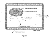
- mitochondrial accumulation is influenced by three main factors as shown in Figure 2 , namely:
- FIG. 2 shows a schematic representation of a cell (10) including a plasma membrane (11) and cytoplasm (12).
- the cell includes a mitochondrion organelle (13) which comprises an outer mitochondrial membrane (14) and an inner mitochondrial membrane (15).
- Mitochondrial pH probe molecules according to the present invention are shown as dots (16).
- the pH probe is initially outside the cell in extracellular space but the probe travels through the plasma membrane (11). The positive oxidation state of the probe and its suitable lipophilicity encourages influx into the cell.
- the probe is attracted into the mitochondria, again due to its positive oxidation state.
- the probe then accumulates inside the mitochondria (13) in the mitochondrial matrix.
- the probe is converted via cellular machinery (for example esterases) into active form. While this conversion will take place within the whole cell, the preferential accumulation into the mitochondria due to the positive probe charge leads to an overall accumulation inside the mitochondria.
- the diagram shows drug efflux which has the effect of removing probe from the cell.
- the relative ratio of probe influx and probe efflux is determined by the difference between the speed of the probe crossing the plasma membrane and the ability of the cell to remove probe by cell defensive mechanisms. Having a probe which can efficiently cross the cell membrane can increase the ratio of accumulation relative to efflux and therefore probes with high lipophilicity are generally preferred.
- protecting the probe according to the present invention can improve both the speed at which the probe accumulates within the mitochondria and also the overall concentration of probe contained within the mitochondria relative to the probe contained within the cell's cytoplasm and extracellularly.
- the probe achieves this by fixing a positive oxidation state to pull the probe into the mitochondria; and/or by increasing the lipophilicity of the probe. Through these mechanisms, the influx of the probe into the mitochondria outweighs the efflux out of the cell.
- the mitochondrial pH probe of the present invention is converted intracellularly into an active form by removal of the protecting groups and therefore converting the probe from a positive oxidation state into an active variable oxidation state.
- the active form of the probe is much more hydrophilic in comparison to the inactive form, thus has a very low membrane permeability and gets trapped inside the mitochondrial matrix.
- the oxidation state of the active probe varies between an oxidation state of 0 and -2. More preferably, the active probe has a variable oxidation state between 0 and -1. Alternatively, the active probe has a variable oxidation state between -1 and -2.
- Converting the probe into a lower oxidation state when in active form has a number of advantages. Firstly, while the positive oxidation state facilitates transportation into the mitochondria, converting the probe into a neutral or negative oxidation state reduces the possibility that the accumulation of positively charged probe depolarises the mitochondria and therefore negatively impacts the cell and the results obtained. Otherwise, the high accumulation of positively charged probes may influence mitochondrial activity due to depolarisation of the mitochondrial matrix thereby influencing or invalidating the results obtained.
- the mitochondrial pH probes of the present invention are configured to rapidly accumulate in the mitochondria relative to the cell's cytoplasm or extracellularly, and to convert intracellularly into an active form which does not depolarise the mitochondria thereby affecting the function of the mitochondria. This leads to a number of advantages.
- the preferential accumulation of probe in the mitochondria enables accurate and sensitive measurements.
- By having a high accumulation of active probe in the mitochondria it is possible to sensitively determine the pH of the mitochondrial matrix and the readings are not unduly influenced by probe readings in other parts of the cell or extracellularly. This allows for sensitive readings able to measure small changes in mitochondrial pH.
- the rapid accumulation enables fast pH measurements to be taken. It is possible to measure mitochondrial pH in real time and to rapidly follow the impact of external forces, for example drugs, on mitochondrial pH. Through these rapid real time measurements, it is possible to track mitochondrial pH changes and see the influence of drugs or other external factors on cellular mechanisms.
- the probes of the present invention are ratiometric. By covalently linking a pH dependent fluorophore with a pH independent fluorophore, it is possible to take concentration independent pH measurements. This allows for results to be normalised and compared between biological samples with variable nature.
- the two fluorophores have spectrally separated fluorescence bands and/or absorption bands. This improves the sensitivity of the readings and allows for more accurate measurements to be taken.
- the probes of the present invention comprise two fluorophores covalently linked together.
- Covalently linking fluorophores leads to advantageous properties. Were two non-covalently linked fluorophores to be used, the ratiometric approach would rely on the same concentration of each probe being present in the mitochondria at the same time. In practice, this is difficult to achieve because different probes will influx and efflux at different rates.
- the concentration ratio between them is inherently constant, because it is defined by the stoichiometry of the molecule.
- the probe is converted from an inactive protected form into an active deprotected form intracellularly.
- esterases within the cell deprotect the probe.
- the probe and the linker are configured so that said intracellular deprotection does not otherwise adversely affect the probe, for example by not only cleaving the protecting groups but also by cleaving other elements of the probe, for example the linker.
- the probes according to the present invention may incorporate any suitable fluorophores.
- the fluorophores may be selected such that they fluoresce or absorb light at spectrally distinct regions.
- the fluorophores are also selected so that in protected form they have an overall positive oxidation, but can be cleaved intracellularly via cellular machinery (for example esterases) into an active form that has a variable oxidation state so that the probe is sensitive to changes to pH.
- the pH independent fluorophore A allows for a ratiometric approach to be applied and provides an internal ratiometric standard.
- the pH independent fluorophore A is a cationic fluorophore.
- the pH independent fluorophore A is a lipophilic fluorophore.
- the pH independent fluorophore A is a cationic lipophilic fluorophore.
- the pH independent fluorophore A is selected from a fluorophore containing a positively charged N ion; a fluorophore derived from fluorones, polymethines, pyrenes or acridines; or a fluorophore derived from rosamines or cyanines.
- pH independent fluorophore A includes: or wherein X denotes the attachment site of L.
- the attachment site X may be positioned in a different part of the fluorophore provided the position does not adversely affect the ability of the fluorophore to fluoresce in a pH independent manner.
- pH independent fluorophore A is Cy5 and Cy3:
- the attachment site X denoting the attachment site of L is shown at the carbonyl group at the end of the alkyl chain. Within the scope of the present invention it is envisaged that the attachment site could vary. Equally, groups that do not affect the fluorescence properties of the fluorophore can be modified, for example the carbonyl and alkyl groups and remain within the scope of the invention.
- the pH dependent fluorophore B is capable of being protected such that the probe overall has an overall positive oxidation state.
- the fluorophore in inactive form must be capable of being converted into active form intracellularly. This may be achieved, for example, via esterases.
- the pH dependent fluorophore B comprises oxygen groups which can be protected to lock or cage the molecule into a positive oxidation state.
- the pH dependent fluorophore B in inactive form comprises one or more protected oxygen(s).
- any suitable fluorophore can be used as pH dependent fluorophore B that allows for protection and cellular deprotection.
- suitable fluorophores include fluoresceins or coumarins.
- the pH dependent fluorophore B is a fluorescein.
- the pH dependent fluorophore B is a coumarin.
- the pH dependent fluorophore B in inactive form may be selected from: wherein X denotes the attachment site of L and PG denotes a protecting group.
- the pH dependent fluorophore B is:
- the attachment group X denotes the attachment site of L and is shown attached to the amine group. Variations to the attacment site are encompassed within the scope of the invention provided they do not adversely impact the ability of the fluorophore to function as a pH dependent fluorophore.
- Particularly preferred mitochondrial pH probes include: wherein PG and L are as defined herein.
- An especially preferred probe is the flourescin-Cy5 and flourescin-Cy3 based probes:
- the linker L is selected from any suitable group that can covalently link the two fluorophores together without adversely affecting the fluorescence performance of either probe.
- the linker positioning and selection is designed to minimise steric hindrance for the rotation of the two fluorophores around the linker relative to each other, that may adversely affect the ability of the probe to travel through the cellular plasma membrane and mitochondrial membranes.
- the linker is a covalent bond between the two fluorophores.
- the linker L may be selected from one or more of: alkyl, alkenyl, alkynyl, cycloalkyl, aryl, heteroaryl, heterocyclic, heteroalkyl, carbonyl, alkylcarbonyl; and/or combinations thereof optionally joined directly or by an alkyl, carbonyl, aminocarbonyl, thiocarbonyl group or by a heteroatom.
- the linker L may be of Formula II: L 1 -(L 3 )-L 2 (Formula II), wherein L 1 and L 2 are independently selected from alkyl, alkenyl, alkynyl, cycloalkyl, aryl, heteroaryl, heterocyclic, heteroalkyl, carbonyl, and alkylcarbonyl; and L 3 is a bond convalently linking L 1 and L 2 together; or L 3 is selected from heteroatom, alkyl, alkenyl, alkynyl, cycloalkyl, aryl, heteroaryl, heterocyclic, heteroalkyl, carbonyl, and alkylcarbonyl.
- L 1 may be selected from: alkyl, heteroaryl, heterocyclic, heteroalkyl and alkylcarbonyl. In a further preferred embodiment L 1 may be selected from heteroaryl or alkylcarbonyl; more preferably 1,3,5-triazine or C 1-n alkyl-1-carbonyl; wherein n is 2 to 12.
- L 2 may be selected from alkyl, heteroaryl, heterocyclic, heteroalkyl and alkylcarbonyl. In a yet further preferred embodiment, L 2 may be selected from alkyl, heterocyclic or heteroalkyl; more preferably piperazine, 1-amino-n-thio-C 1-n alkyl, or 1,n-diamino-C 1-n alkyl; wherein n is 2 to 12.
- L 3 may be absent or may be selected from: NH or S. In a particularly preferred embodiment, L 3 is absent.
- Especially preferred linkers include:
- the mitochondrial pH probe according to the present invention in an inactive form is protected into an overall positive oxidation state.
- the protection is removed intracellularly to convert the probe into an active form with a variable oxidation state.
- the probe is protected by one or more protecting groups (PG).
- Any suitable protecting group can be used that is able to both achieve a positive oxidation state and also be removed intracellularly.
- Examples of possible protecting groups PG include acyl, propionyl, butyryl, isobutyryl, pivaloyl or benzoyl.
- An especially preferred protecting group is acyl.
- protecting groups include organic carbonate esters, thioesters and carbamates.
- the mitochondrial pH probe in protected form comprises the molecule 5-FA-PIP-Cy5-DA (Formula III shown below).
- 5-FA-PIP-Cy5-DA comprises the fluorophore 5-FA linked to the fluorophore Cy5, via a PIP (piperazyl) linker.
- the 5-FA unit has been diacetylated, leading to the designation DA:
- 5-FA-PIP-Cy5-DA (and any other probes according to the present invention) is delivered to the into the cells in a non-active caged form, when the phenolic groups of fluorescein are acylated. This quenches the fluorescein's fluorescence and also removes the pH dependence of the probe. Once delivered to the cell, however, the acetyl groups are cleaved by the cellular (mostly mitochondrial) esterases and the probe is converted to the active form. This is shown schematically below which shows the mechanism of cellular deprotecting and conversion of the probe from a positive fixed inactive state into an active form which has pH dependent fluorescence.
- fluorescein's fluorescence is pH dependent due to the reversible protonation/deprotonation of its phenolic group, when in a more acidic environment the non-fluorescent closed monoanionic spiro-form of fluorescein is prevalent, while in more basic environment the equilibrium is shifted to the opened fluorescent dianionic form of fluorescein.
- the Cy5 fluorophore of the probe is always fluorescent and thus acts as the pH independent ratiometric internal standard.
- the mitochondrial pH probes of the present invention can be used to measure mitochondrial pH.
- a method of measuring mitochondrial pH comprising the steps of:
- the probes of the present invention are configured to more easily pass through lipid membranes and to target the mitochondria. This leads to an accumulation of the probe in the mitochondria. As already discussed above, this accumulation is defined by the rate of influx into the cell and mitochondria and the rate of efflux by the cell's defence mechanisms to remove the probe. By ensuring rapid influx and effective mitochondrial targeting, the probe will accumulate in the mitochondria and influx will override efflux.
- the cell converts the probe into an active form, for example by using esterases. Esterases are present throughout the cell but because of the rapid accumulation of the probe within the mitochondria, conversion into active form is also predominantly occurring within the mitochondria leading to an accumulation of probe in active form within the mitochondria.
- the advantageous properties of the probes of the present invention are such in some embodiments, sufficient accumulation of active probe in the mitochondria can take place within 15-20 minutes. This enables rapid mitochondrial pH measurements to take place. The sensitivity and update speed of the probe gives the ability to monitor real time changes to mitochondrial pH in response to any stimulus or condition being applied to the cell.
- the cells are washed with the medium, not containing the probe. All the accumulated probe inside the mitochondria of the cells is converted into active form almost instantaneously. It is retained inside the mitochondria due to its ability to be trapped because of the membrane impermeability of the active form.
- the cells are then ready to be analyzed by fluorescent light microscopy or flow cytometry. The ratiometric fluorescent signal is acquired and quantified. Other methods, including but not limited plate-reading bulk analysis, can be used to analyze the stained cells. Overall, the staining and washing procedures can be performed within 30 minutes, after which the cells are ready for analysis.
- FCCP carbonylcyanide-4-trifluoromethoxyphenylhydrazone
- the cells initially are stained with the mitochondrial pH probe in the same conditions, as used in the main experiments. Then the medium, containing the probe, is washed away and substituted with the corresponding series of defined pH buffers, containing the means to equilibrate the mitochondrial pH and the buffer pH, preferably FCCP or CCCP.
- the series of control cells then analyzed after 15 minutes with the same method, as used for the main experiments, to quantify the ratiometric fluorescent signal, corresponding to the exact mitochondrial pH value, defined by the buffer pH in the case of the control experiment.
- the dependence of ratiometric signal over the pH value is then analytically approximated with the mathematical equation, preferably linear equation. This equation is later used to convert the value of ratiometric fluorescent signal, obtained in the main experiments, to the mitochondrial pH value, observed experimentally.
- Non-limiting examples of the application of the mitochondrial pH probes of the present invention include
- the probes of the present invention may be made by any suitable means. According to one embodiment, there is provided a method of manufacturing a dual fluorophore ratiometric mitochondrial pH probe of Formula I: A-L-B (Formula I); wherein:
- 5-FA-PIP-Cy5-DA comprises the fluorophore5-FA (fluorescein) linked to the fluorophore Cy5, joined via a PIP (piperazyl) linker.
- the 5-FA unit has been diacetylated, leading to the designation DA.
- 5-FA-PIP-Cy5-DA was synthesised from commercially available 5-aminofluoresin 17 according to the synthetic scheme below.
- the reaction mixture was diluted with 1 ml of chloroform and excess 5-aminofluoresceinopiperazine extracted 10 times with 0.5 ml of 0.5 M HCI, until the water layer was transparent.
- the remaining slurry of the chloroform layer was transferred to a 1.5 ml Eppendorf tube and the product was precipitated with 1 ml of diethyl ether. This precipitate was separated by centrifugation and the liquid layer was discarded.
- 0.3 ml of THF was added and the mixture sonicated for 2 minutes. The precipitate was separated by centrifugation again and the sonication-centrifugation was repeated 7 times.
- A2780 ovarian cancer cells were cultured in 24-well microscopy glass bottom plates in a CO2 incubator. Prior to imaging, the cells were stained with 2uM 5FA-PIP-Cy5-DA, and/or TMRM (20nM) for 30 min in bicarbonate-free DMEM medium to avoid extracellular pH fluctuations, then washed with the same medium (without 5FA-PIP-Cy5-DA) and imaged with confocal spinning disk microscopy at 60x magnification When TMRM was used, this dye was also present in the bicarbonate-free DMEM medium during the imaging of the cells.
- Typical pH variations in mitochondria of A2780 ovarian cancer cell line are represented at Figure 4 .
- Typical staining procedure is described further. Cells were stained for 30 min with 2 ⁇ M 5FA-PIP-Cy5-DA solution, the probe was washed away with the bicarbonate-free DMEM medium, and the cells were imaged after 10 min with confocal spinning disk microscope. All images are maximum intensity projection.
- Figure 4A represents the 5FA-PIP-Cy5-DA pH-independent (Cy5) channel, shown in red on the figure. The mitochondrial staining on Figure 4A is very homogeneous.
- Figure 4B represents the 5FA-PIP-Cy5-DA pH-dependent (5Fluo) channel, shown in blue on the figure.
- FIG. 4B The mitochondrial staining on Figure 4B is very heterogeneous, unlike the Figure 4A.
- Figure 4C represents the overlap image of Figure 4A and Figure 4B .
- Bluish hint Figure 4C corresponds to relatively high mitochondrial pH, reddish to relatively low mitochondrial pH.
- Figure 4D represents bright field image.
- the mitochondria in different A2780 cells are highly heterogeneous in the pH, which is reflected in the highly heterogeneous staining in pH dependent channel on Figure 4B .
- the ability of the synthesised probe to distinguish mitochondrial pH changes in live cells was tested. This test was performed via the comparison of the fluorescein/Cy5 fluorescence ratio between control conditions and cells which had been treated with K + /H + exchanging ionophore nigericin and a protonphore FCCP, which reduce mitochondrial pH by acidifying the mitochondrial matrix.
- FCCP and nigericin were commercially purchased. After the of A2780 ovarian cancer cells were stained as described above, the cells were washed with the bicarbonate-free DMEM medium, either containing DMSO vehicle for control sample or 300 nM FCCP or 2uM nigericin for the samples, where the mitochondrial pH was expected to be decreased. Confocal images were acquired for control, FCCP- and nigericin-treated cell samples, and the ratio between fluorescein and Cy5 channel fluorescence was quantified.
- the stained cells were treated with FCCP in high K + buffers in a pH range of 6.73 to 8.36.
- FCCP treatment can therefore be used as a practical way to calibrate a cell for exact pH measurements.
- TMRM tetramethylrhodamine methyl ester
- Figure 8 represents typical image of mitochondria, stained with both dyes.
- Figure 8A represents 5FA-PIP-Cy5-DA pH independent (Cy5) channel, shown in red.
- Figure 8B represents 5FA-PIP-Cy5-DA pH-dependent (5Fluo) channel, shown in blue.
- Figure 8C represents TMRM channel, shown in magenta.
- Figure 8D represents bright field image. Spinning disk confocal images of TMRM and 5FA-PIP-Cy5-DA costained A2780 cells were analyzed for pixel florescence intensity correlation between the Cy5 channel of 5FA-PIP-Cy5-DA and the TMRM channel ( Figure 8 ).
- the Cy5 channel of 5FA-PIP-Cy5-DA is the most appropriate for co-localization studies, because it is pH-independent and therefore clearly reveals the localization of the 5FA-PIP-Cy5-DA molecule independent of the pH.
- high degree of colocalization with a well-known mitochondrial selective probe TMRM further confirms mitochondrial selectivity of 5FA-PIP-Cy5-DA.
- the calibration curve for the ratio of 5Fluoro/Cy5 fluorescence was obtained using FCCP and a PBS-5% BSA buffer with MPP cells in a manner analogous to the method described in Example 2 (see Figure 10 ).
- the cells were cultured in Hematopoietic Stemline II (Sigma) medium with addition of SCF and Flt-3 cytokines. For appropriate experiments, additionally 5 uM DMOG or IL-3 20 ng/mL and IL-6 100ng/mL cytokines were added.
- the cells Prior to imaging, the cells were stained with the appropriate concentration of 5FA-PIP-Cy5-DA for 30 min in bicarbonate-free DMEM medium to avoid the extracellular pH fluctuation outside of the CO 2 incubator, then washed, transferred to poly-L-lysine coated 24-well microscopy glass bottom plates and imaged with confocal spinning disk microscopy at 60x magnification. Separate fluorescent images in green and deep red spectral channels were acquired. The florescence ratio was quantified by a specifically designed batch ImageJ plugin as described above.
- LT-HSC showed a much lower mitochondrial pH level compared to ST-HSC and MPP (see Figure 11 ).
- IL-3, IL-6 - treated LT-HSC had the highest overall average mitochondrial pH value, significantly above LT-HSC, both when cultured in basal medium, and with addition of DMOG, and ST-HSC and MPP.
- 5FA-PIP-Cy5-DA performs well as a probe for mitochondrial pH.
- the probe was able to demonstrate that differentiation induction in LT-HSC causes the burst in mitochondrial pH to a level not observed even in highly active, proliferative ST-HSC and MPP cells.
- 5FA-PIP-Cy5-DA significant differences in mitochondrial pH were observed through the range of conditions, favouring different stem cell fate outcomes, and demonstrating that mitochondrial pH alteration is associated with hematopoietic stem cell fate determination as a result.
- the calibration curve for the ratio of 5Fluoro/Cy5 fluorescence was obtained using FCCP and a PBS-5% BSA buffer with A2780 cells in a manner analogous to the method described in Example 2 (see Figure 12 ).
- the quantifying of mitochondrial pH in the presence of the drug and after the drug was washed away allowed the distinction between interference with mitochondrial metabolism and general cytotoxicity. If the effect of the drug was primarily to affect mitochondrial metabolism it was expected that the mitochondrial pH would quickly return to the non-treated cell level following removal of the drug, providing the drug had no other significant cytotoxic effects. Conversely, if the effect of the drug were not reversed by the washing step, an observed alteration of mitochondrial pH could be interpreted at least partially as a downstream consequence of the general cytotoxicity of the drug.
- A2780 cells were cultured in 24-well microscopy glass bottom plates in RPMI medium, containing Glutamax with 10% FBS addition. The cells were treated with appropriate 800uM RAPTA-T or 5uM cisplatin. Prior to the imaging the cells were stained with 2uM 5FA-PIP-Cy5-DA for 30 min in a bicarbonate-free DMEM medium to avoid the extracellular pH fluctuation outside of the CO 2 incubator, then washed and imaged with confocal spinning disk microscopy at 60x magnification. Cells were treated with either drug for one day and mitochondrial pH was quantified using 5FA-PIP-Cy5-DA in the presence of the drug and after the drug was washed away.
- Figure 13 also demonstrates the use of 5FA-PIP-Cy5-DA to assess the functional state of mitochondrial ATP synthase of A2780 cells by measuring mitochondrial pH in the presence and absence of ATP synthase inhibitor oligomycin. It can be seen that the addition of 10 ⁇ m oligomycin caused slight increase in mitochondrial pH from 8.4 to around 8.5.
- Oligomycin inhibits proton conductance by ATP synthase, removing its contribution to the proton gradient. If mitochondrial ATP synthase acts in the normal mode, inhibition with oligomycin would increase mitochondrial pH, while in the reversed mode it would decrease mitochondrial pH. Finally, in the case of pre-existing natural inhibition of ATP synthase, inhibition with oligomycin would not noticeably change mitochondrial pH.
Landscapes
- Life Sciences & Earth Sciences (AREA)
- Chemical & Material Sciences (AREA)
- Health & Medical Sciences (AREA)
- Immunology (AREA)
- Organic Chemistry (AREA)
- Engineering & Computer Science (AREA)
- Molecular Biology (AREA)
- General Health & Medical Sciences (AREA)
- Analytical Chemistry (AREA)
- Pathology (AREA)
- General Physics & Mathematics (AREA)
- Biomedical Technology (AREA)
- Urology & Nephrology (AREA)
- Biochemistry (AREA)
- Hematology (AREA)
- Physics & Mathematics (AREA)
- Medicinal Chemistry (AREA)
- Food Science & Technology (AREA)
- Biotechnology (AREA)
- Microbiology (AREA)
- Cell Biology (AREA)
- Inorganic Chemistry (AREA)
- Biophysics (AREA)
- Investigating Or Analysing Materials By The Use Of Chemical Reactions (AREA)
Abstract
The present invention relates to dual fluorophore ratiometric mitochondrial pH probes, their uses and their manufacture.
Description
- The present invention relates to mitochondrial pH probes, their uses and processes to produce them.
- Mitochondria are rod-shaped organelles in a cell that convert nutrients into adenosine triphosphate (ATP), using oxygen as an oxidant, thereby providing energy for the cell to drive the cell's metabolic activities. This process is known as aerobic respiration and is the reason animals breathe oxygen. Without mitochondria, cells would only be able to obtain energy from anaerobic respiration which is around 15 times less efficient than aerobic respiration. As well as energy generation, mitochondria regulate cellular redox states, generate most of the cellular reactive oxygen species, and initiate cellular apoptosis.
- The number of mitochondria present in a cell depends upon the metabolic requirements of that cell, and may range from a single large mitochondrion to thousands of the organelles. Mitochondria are found in nearly all eukaryotes, including plants, animals, fungi, and protists, and are large enough to be observed with light microscopy.
- The specific pH value of the mitochondria directly influences its biological processes and changes to mitochondrial pH can lead to mitochondrial dysfunction. The ability to accurately and sensitively measure mitochondrial pH within living cells can provide important insights into mitochondrial biology, and the dynamics of the living cell as a whole.
- A number of fluorescence based mitochondrial pH probes have been developed to measure mitochondrial pH but there remains a need to develop a probe which can rapidly enter the mitochondria and provide reproducible and sensitive measurements without impacting the mitochondrial environment.
- In a first aspect of the present invention there is provided a dual fluorophore ratiometric mitochondrial pH probe of Formula I:
A-L-B (Formula I);
wherein: - A is a pH independent fluorophore;
- B is a pH dependent fluorophore; and
- L is a chemical linker, conjugating the pH independent fluorophore A to the pH dependent fluorophore B, or L is a covalent bond;
- The present inventors have found that by protecting the pH dependent fluorophore of a dual fluorophore ratiometric probe into a positive oxidation state, the probe is efficiently transported into the mitochondria of the cell and is converted by the cell into an active form where the oxidation state of the pH dependent fluorophore can vary enabling sensitive and rapid evaluation of mitochondrial pH.
- In a further aspect there is provided a method of measuring mitochondrial pH comprising the steps of:
- a) contacting a cell with a mitochondrial pH probe in an inactive form as described herein;
- b) allowing the probe to accumulate in the mitochondria of the cell, and to convert into an active form;
- c) determining the ratio of fluorescence between the pH independent fluorophore A and the pH dependent fluorophore B; and
- d) using this ratio to determine the mitochondrial pH.
- In a yet further aspect there is provided a method of measuring the impact of drugs or other conditions on the mitochondrial pH comprising applying the drug or other condition to a cell and measuring the change in mitochondrial pH using a dual fluorophore ratiometric mitochondrial pH probe as described herein.
- In a yet further aspect there is provided a method of manufacturing a dual fluorophore ratiometric mitochondrial pH probe of Formula I:
A-L-B (Formula I);
wherein: - A is a pH independent fluorophore;
- B is a pH dependent fluorophore; and
- L is a chemical linker, conjugating the pH independent fluorophore A to the pH dependent fluorophore B, or L is a covalent bond;
-
-
Figure 1 is a schematic view of a mitochondria organelle -
Figure 2 is a schematic view of a cell showing the membrane barriers, mitochondria and pH probe -
Figure 3 is a plot of fluorescein channel pH-dependent fluorescence in PBS-5% BSA buffer for 5-FA-PIP-Cy5 (the unprotected form of the probe, which is active in the intracellular environment) -
Figure 4 is a series of images showing typical pH variations in mitochondria of A2780 ovarian cancer cell line are represented.Figure 4A represents the 5FA-PIP-Cy5-DA pH-independent (Cy5) channel.Figure 4B represents the 5FA-PIP-Cy5-DA pH-dependent (5Fluo) channel.Figure 4C represents the overlap image ofFigure 4A and Figure 4B. Figure 4D represents the bright field image -
Figure 5 is the fluorescein/Cy5 ratio measured in homeostatic conditions and upon nigericin and FCCP treatment -
Figure 6 is a plot of the ratio of fluorescein/Cy5 at a range of pH values when the mitochondrial proton gradient is collapsed with FCCP -
Figure 7 is a plot of the ratio of fluorescein/Cy5 at a range of pH values in the presence of nigericin -
Figure 8 is a series of typical images of mitochondria, stained with both 5FA-PIP-Cy5-DA.Figure 8A represents 5FA-PIP-Cy5-DA pH independent (Cy5) channel, shown in red.Figure 8B represents 5FA-PIP-Cy5-DA pH-dependent (5Fluo) channel, shown in blue.Figure 8C represents TMRM channel, shown in magenta.Figure 8D represents bright field image -
Figure 9 is a series of images which show the advantage of the caging approach through the staining of A2780 cells with either 5FA-PIP-Cy5-DA or 5FA-PIP-Cy5 in identical conditions.Figure 9A shows the successful staining of mitochondria in the case of inactive form 5FA-PIP-Cy5-DA.Figure 9B shows unsuccessful staining of mitochondria in the case of active form 5FA-PIP-Cy5.Figure 9C shows negative control cells with no staining agent present -
Figure 10 is a plot of a 5FA-PIP-Cy5-DA pH dependence calibration curve, obtained with MPP cells in the presence of FCCP -
Figure 11 is a plot to show the estimated mitochondrial pH (single haematopoietic cells) in different phenotypes and culture conditions as measured with 5-FA-PIP-Cy5-DA -
Figure 12 is a graph to show the 5FA-PIP-Cy5-DA pH dependence calibration curve obtained with A2780 cells in the presence of FCCP -
Figure 13 is a plot of estimated mitochondrial pH (average per field of view) of A2780 cells in the presence of anti-cancer drugs and following the drugs' removal - In the present application, a number of general terms and phrases are used, which should be interpreted as follows.
- "Mitochondrial pH probe" means a molecule which is capable of relaying information about the pH inside a mitochondrial matrix.
- "Mitochondria" are rod-shaped organelles in a cell that convert nutrients into adenosine triphosphate (ATP), using oxygen as an oxidant, thereby providing energy for the cell to drive the cell's metabolic activities. Two specialized membranes encircle each mitochondrion present in the cell. The membranes divide the organelle into a narrow intermembrane space and a much larger internal matrix, each of which contains highly specialized proteins. The outer membrane of a mitochondrion contains many channels formed by the proteins like porin and acts like a sieve, filtering out molecules that are too big. Similarly, the inner membrane, which is highly convoluted so that a large number of infoldings called cristae are formed, also allows only certain molecules to pass through it and is much more selective than the outer membrane. To make certain that only those materials essential to the matrix are allowed into it, the inner membrane utilizes a group of transport proteins that will only transport the correct molecules.
- Mitochondria are generally oblong organelles, which range in size between 0.5 and 10 micrometers in length, and occur in numbers that directly correlate with the cell's level of metabolic activity. The organelles are quite flexible, however, and time-lapse studies of living cells have demonstrated that mitochondria change shape rapidly and move about in the cell almost constantly. Movements of the organelles appear to be linked in some way to the microtubules present in the cell, and are probably transported along the network with motor proteins. Consequently, mitochondria may be organized into lengthy traveling chains, packed tightly into relatively stable groups, or appear in many other formations based upon the particular needs of the cell and the characteristics of its microtubular network.
- An example of mitochondria is shown in
Figure 1 which shows theouter membrane 1, the inner membrane 2 demonstrating the infolding leading to formation of cristae 3, and the mitochondrial matrix 4. The probes of the present invention are able to measure the pH of the matrix 4. - "Fluorophore" means a fluorescent chemical compound that can re-emit light upon light excitation, emitted light being of a different wavelength than excitation light.
- "Dual fluorophore" means a molecule that contains two or more covalently linked fluorophores that are either directly connected together or are tethered together by a linker. Upon light excitation, each fluorophore can re-emit light.
- "pH independent fluorophore" means a fluorophore which emits light at a specific wavelength largely irrespective of the pH of the environment it is in. A pH independent fluorophore therefore exhibits consistent fluorescence over variable pH conditions, for example both low and high pH.
- "pH dependent fluorophore" means a fluorophore whose fluorescence changes depending on the pH of the environment it is in. A pH dependent fluorophore is sensitive to the pH environment and exhibits variable fluorescence depending on pH.
- "Ratiometric" means a method of measuring the fluorescence of the flurophores and determining the fluorescence ratio. Applying a ratiometric approach avoids flurophore concentration effects from influencing the pH measurement. If a probe comprises a single pH dependent fluorophore, the intensity of the emission will be dependent not only on pH but also on the fluorophore concentration. This makes it difficult to compare results between different biological samples. By applying a ratiometric approach, internal normalisation of the fluorescence readings is possible. While the absolute fluorescence intensities between different samples may vary depending on the fluorophore concentrations, the fluorescence ratio will remain independent of concentration and will only vary depending on pH.
- "Linker" means a sub-unit of a molecule which serves to covalently link two other subunits of said molecule together. In the present invention, the linker may be absent whereby the two units are directly linked to each other, or may be present and provide the link between the units. A linker should not adversely affect the function of the units. A linker should not prevent the individual fluorophores from fluorescing in a pH dependent or pH independent manner as required. For example, a linker should not conjugate into the fluorescence causing region of the flurophores, or should not do so in a way that adversely changes the properties of the flurophore(s).
- "Inactive form" means the molecule has been modified such that it exists in a positive oxidation state. In some embodiments, in inactive form, the molecule does not fluoresce in a pH dependent manner. The molecule may be in inactive form via protection of one or more functional groups. When in inactive form, the molecule is configured to facilitate transportation into the target location, for example the cell and/or mitochondria.
- "Active form" means the molecule is configured to produce its desired effect e.g. measure mitochondrial pH. In active form, the molecule will fluoresce in a pH dependent manner to enable mitochondrial pH measurement. The molecule may be converted into active form via removal of the protecting group(s).
- "Protecting group" means a group which has been introduced into a molecule by modification of a functional group which prevents said functional group from undergoing further changes, (e.g. reactions) until the protecting group has been removed. Suitable protecting groups can be deprotected intracellularly. Suitable protecting groups include alcohol protecting groups, amine protecting groups, carboxylic acid protecting groups, and the like.
- Examples of alcohol protecting groups include esters, formed with saturated and aromatic carboxylic acids, organic carbonate esters.
- Examples of amine protecting groups include amides, formed with saturated and aromatic carboxylic acids, carbamates, thiocarbamates.
- Examples of carboxylic acid protecting groups include acetoxymethyl ester, anhydrides, formed with saturated and aromatic carboxylic acids.
- "Oxidation state" means the overall charge of an atom, part of a molecule or a molecule. A single positive charge in a molecule corresponds to an oxidation state of +1 and a single negative charge corresponds to an oxidation state of -1. The oxidation state of a molecule or part of a molecule can be condition dependent or independent. For example, a nitrogen with four alkyl bonds exists in a +1 oxidation state irrespective of the surrounding conditions (e.g. pH). By comparison, an alcohol molecule ROH exemplifies a variable oxidation state as it can exist at multiple oxidation states depending on the pH of its surroundings. At low pH the oxygen will bond to H+, leaving the alcohol ROH2 + in a +1 oxidation state. Alternatively, at a high pH, H+ may be lost, leading to a molecule in a -1 oxidation state as RO-. As a further example a molecule with the fixed oxidation state nitrogen and the variable oxidation state alcohol both present would display a variable oxidation state, moving from +1 in inactive form of the molecule to 0 for the active form of the molecule at low pH to -1 for the active form of the molecule at high pH.
- "Converted intracellularly" means a process whereby a molecue is converted from one form to another (e.g. from an inactive form to an active form) via cellular machinery inside a cell. For example, a molecule in protected form may be deprotected intracellularly by esterases to convert it from an inactive form into an active form. A protected molecule may therefore be deprotected intracellular.
- "Probe calibration" means the measurement of probe response in known conditions in order to produce quantifiable condition/response data, which can be used in combination with experimental probe data to determine the conditions present during use of the probe.
- "Measuring the impact of drugs or other conditions" means using the probes of the present invention to determine the impact of drug(s) or other situations on mitochondrial pH. For example, the probes of the present invention may be used to determine how mitochondrial pH changes in response to the application of a drug into a cellular system and/or the removal of a drug from a cellular system. The probe of the present invention may also be used to determine how mitochondrial pH changes in response to other changes to cellular conditions, for example changes to temperature or the like. The probes of the present invention may be used to monitor real time changes to mitochondrial pH in response to any stimulus or condition being applied to the cell.
- The present invention relates to dual fluorophore ratiometric mitochondrial pH probes of general formula I as defined above.
- In the molecules of the present invention, the groups can be selected in accordance with the following guidance:
- "Alkyl" means groups which may be branched or unbranched, and preferably have from 1 to about 12 carbon atoms. One more preferred class of alkyl groups has from 1 to about 8 carbon atoms. Even more preferred are alkyl groups having 1, 2, 3, 4, 5, 6, 7 or 8 carbon atoms. Propyl including isopropyl, butyl including sec-butyl and isobutyl, pentyl, including isopentyl, hexyl, heptyl and octyl are particularly preferred alkyl groups in the compounds of the present invention. Alkyl groups according to the present invention may be unsubstituted or substituted.
- "Alkenyl" and "alkynyl" mean groups which may be branched or unbranched, have one or more unsaturated linkages and from 2 to about 12 carbon atoms. One more preferred class of alkenyl and alkynyl groups has from about 2 to about 8 carbon atoms.. Even more preferred are alkenyl and alkynyl groups having 2, 3, 4, 5, 6, 7 or 8 carbon atoms. Alkenyl and alkynyl groups according to the present invention may be unsubstituted or substituted.
- "Cycloalkyl" means a cyclic saturated monovalent hydrocarbon group including single or multiple ring structures, including multiple ring groups that contain separate and/or fused alkyl rings. Typical cycloalkyl groups contain from 1 to 3 separate and/or fused rings and from 3 to about 18 carbon ring atoms. Preferably cycloalkyl groups contain from 6 to about 12 carbon ring atoms. Specially preferred cycloalkyl groups include cyclopropyl, cyclobutyl, cyclopentyl, cyclohexyl, cycloheptyl, bicyclohexane and decalin. Cycloalkyl groups according to the present invention may be unsubstituted or substituted.
- "Heteroalkyl" means an alkyl group, as defined above, having a substituent containing at least one heteroatom selected from NR', O, S, where R' may be a suitable group such as H, C1-C12 alkyl, C2-C12 alkenyl, C2-C12 alkynyl, aryl, and heterocyclic group. The R' groups according to the present invention may themselves be unsubstituted or substituted. These heteroatoms may optionally terminate the alkyl chain with a general formula of RNR', RO or RS or may be within the group with a general formula of RNR'R", ROR" or RSR". Preferred heteroalkyls include the following groups (note: when used in a linker the groups have one or more hydrogen atoms removed to form a bond with a flurophore(s) and/or another portion of the linker) ethylamine, propylamine, isopropylamine, butylamine, sec-butylamine, isobutylamine, pentylamine, hexylamine, ethanol, propanol, isopropanol, butanol, sec-butanol, isobutanol, pentanol, hexanol, ethanethiol, propanethiol, isopropanethiol, butanethiol, sec-butanethiol, isobutanethiol, pentanethiol, hexanethiol, methylenediamine, diaminoethane, diaminopropane, diaminobutane, diaminopentane, hexamethylenediamine, ethanolamine, aminomethylpropanol, heptaminol, 6-amino-1-hexanol, 2-aminoethanethiol, 3-aminopropanethiol, 4-aminobutanethiol, 5-aminopentanethiol, 6-aminohexanethiol, dimethylamine, diethylamine, dipropylamine, diisopropylamine, dibutylamine, disecbutylamine, diisobutylamine, dipropylamine, dihexylamine, dimethylether, diethylether, dipropylether, diisopropylether dibutylether, disec-butylether, diisobutylether, dipentylether, dihexylether, dimethylsulfide, diethylsulfide, dipropylsulfide, diisopropylsulfide, dibutylsulfide, disec-butylsulfide, diisobutylsulfide, dipentylsulfide or dihexylsulfide. Heteroalkyl groups according to the present invention may be unsubstituted or substituted.
- "Halo" means fluoro (F), chloro (CI), bromo (Br) or iodo (I).
- "Aryl" means an aromatic system including single and multiple ring compounds, including multiple ring compounds that contain separate and/or fused aryl groups. Typical aryl groups contain from 1 to 3 separated or fused rings and from 6 to about 18 carbon ring atoms. Preferably aryl groups contain from 6 to about 12 carbon ring atoms. Specially preferred aryl groups include phenyl, naphthyl, biphenyl, unsubstituted phenanthryl and anthryl. Aryl groups according to the present invention may be unsubstituted or substituted.
- "Heterocyclic" includes aromatic (heteroaromatic) or saturated or non-aromatic unsaturated (both heteroalicyclic) rings containing from 1 to 3 separated or fused rings and about 5 to 18 ring atoms. Preferably heterocyclic rings contain from 5 to about 10 ring atoms, preferably 5, 6 or 7 ring atoms. Suitable heterocyclic groups in the compounds of the present invention contain one, two or three heteroatoms selected from N, O or S atoms. Suitable heterocyclic groups include 8-quinolyl, isoquinolyl, pyridyl, pyrazinyl, pyrazolyl, pyrimidinyl, furyl, pyrrolyl,thienyl, thiazolyl, isothiazolyl, triazolyl, tetrazolyl, isoxazolyl, oxazolyl,imidazolyl, indolyl, isoindolyl, indazolyl, indolizinyl, phthalazinyl,pteridinyl, purinyl, oxadiazolyl, thiadiazolyl, furazanyl, pyridazinyl,triazinyl, cinnolinyl, benzimidazolyl, benzofuranyl, benzofurazanyl, benzothiophenyl, benzothiazolyl, benzoxazolyl, quinazolinyl,quinoxalinyl, naphthyridinyl, furopyridyl, pyrrolidinyl, tetrahydrofuranyl, dihydrofuranyl, tetrahydrothienyl,tetrahydrothiopyranyl, piperidyl, morpholinyl, thiomorpholinyl, thioxanyl, piperazinyl, azetidinyl, oxetanyl, thietanyl, homopiperidyl, oxepanyl, thiepanyl, oxazepinyl, diazepinyl, thiazepinyl, 1,2,3,6-tetrahydropyridyl, 2-pyrrolinyl, 3-pyrrolinyl, indolinyl, 2H-pyranyl, 4Hpyranyl, dioxanyl, 1,3-dioxolanyl, pyrazolinyl, dithianyl, dithiolanyl,dihydropyranyl, dihydrothienyl, dihydrofuranyl, pyrazolidinyl, imidazolinyl, imidazolidinyl, 3-azabicyclo[3.1.0]hexyl, 3-azabicyclo[4.1.0]heptyl, 3H-indolyl, and quinolizinyl. Heterocyclic groups according to the present invention may be unsubstituted or substituted.
- "Alkylcarbonyl", "alkenylcarbonyl", "alkynylcarbonyl" mean an alkyl, alkenyl or alkynyl chain as described above which includes one or more carbonyl groups. These groups may terminate the alkyl chain or may be within its structure. If the carbonyl group terminates the alkyl chain it may optionally be bonded to a heteroatom selected from NR', O, S to form an amide, ester or thioester bond respectively, where R' is defined as above. Preferred alkylcarbonyls, alkenylcarbonyls and alkynylcarbonyls include acetaldehyde, propanal, butanal, isobutanal, pentanal, isovaleraldehyde, hexanal, acetone, butanone, methylisopropylketone, pentan-2-one, pentan-3-one, hexan-2-one, hexan-3-one, glyoxal, malondialdehyde, succinaldehyde, glutaraldehyde, hexanedial, oxobutanal, 4-oxopentanal, 4-oxohexanal, 5-oxohexanal, 2,4-pentanedione, 2,3-hexanedione, 2,4-hexanedione, 2,5-hexanedione, 4-penten-2-one, trans-3-pentanal, 3-methyl-3-pentenal, 4-hexen-3-one, 3-butynaldehyde, 3-pentynylaldehyde, 3-hexynylaldehyde, 4-hexynynlaldehyde, 4-hexynylhexan-2-one. Alkylcarbonyls, alkenylcarbonyls and alkynylcarbonyls groups according to the present invention may be unsubstituted or substituted.
- The groups above mentioned may be substituted at one or more available positions by one or more suitable groups such as OR', =O, SR', SOR', SO2R', NO2, NHR', NR'R', =N-R', NHCOR', N(COR')2, NHSO2R', NR'C(=NR')NR'R', CN, halogen, COR', COOR', OCOR', OCONHR', OCONR'R', CONHR', CONR'R', protected OH, C1-C12 alkyl, C2-C12 alkenyl, C2-C12 alkynyl, aryl, and heterocyclic group, wherein each of the R' groups is independently selected from the group consisting of hydrogen, OH, NO2, NH2, SH, CN, halogen, COH, COalkyl, CO2H, C1-C12 alkyl, C2-C12 alkenyl, C2-C12 alkynyl, aryl, and heterocyclic group. The optional substituents described in this paragraph may themselves be substituted. Where such groups are themselves substituted, the substituents may be chosen from the foregoing list.
- In an inactive form, the mitochondrial pH probes of the present invention are protected such that they exist in a positive oxidation state. In a preferred embodiment, the probe is in a fixed oxidation state of +1.
- By fixing the probe into a positive oxidation state, the probe will preferentially accumulate within the mitochondria. Additionally, in the inactive form the probe is much more lypophilic, thus permeating the cellular membranes much more rapidly. Mitochondria are distinguished from other cellular organelles by its inner mitochondrial membrane having a high electrochemical potential. This electrostatic potential pulls the positively charged probe into the mitochondria leading to a concentration of probe in the mitochondria several orders of magnitude higher than the rest of the cell. The positive charge on the probe also helps with the initial delivery of the probe into the cell since the cell plasma membrane also has an electrochemical potential of the same polarity of the mitochondria, albeit at a much lower magnitude.
- In fact, mitochondrial accumulation is influenced by three main factors as shown in
Figure 2 , namely: - 1. initial accumulation in the cellular cytoplasm, dependent on plasma membrane potential;
- 2. secondary preferential accumulation in the mitochondria, dependent on the mitochondrial membrane potential; and
- 3. active cytoplasmic probe efflux by the cell removing the probe (which is highly cell dependent).
-
Figure 2 shows a schematic representation of a cell (10) including a plasma membrane (11) and cytoplasm (12). The cell includes a mitochondrion organelle (13) which comprises an outer mitochondrial membrane (14) and an inner mitochondrial membrane (15). Mitochondrial pH probe molecules according to the present invention are shown as dots (16). The pH probe is initially outside the cell in extracellular space but the probe travels through the plasma membrane (11). The positive oxidation state of the probe and its suitable lipophilicity encourages influx into the cell. - Once in the cytoplasm (12), the probe is attracted into the mitochondria, again due to its positive oxidation state. The probe then accumulates inside the mitochondria (13) in the mitochondrial matrix. The probe is converted via cellular machinery (for example esterases) into active form. While this conversion will take place within the whole cell, the preferential accumulation into the mitochondria due to the positive probe charge leads to an overall accumulation inside the mitochondria.
- The diagram shows drug efflux which has the effect of removing probe from the cell. The relative ratio of probe influx and probe efflux is determined by the difference between the speed of the probe crossing the plasma membrane and the ability of the cell to remove probe by cell defensive mechanisms. Having a probe which can efficiently cross the cell membrane can increase the ratio of accumulation relative to efflux and therefore probes with high lipophilicity are generally preferred.
- By protecting the probe, for example by acylating phenolic groups on the pH dependent flurophore, it is possible to increase the lipophilicity of the probe and therefore improve the ability for the probe to travel through both the cell's plasma membrane and also through the inner and outer mitochondrial membranes.
- Thus, protecting the probe according to the present invention can improve both the speed at which the probe accumulates within the mitochondria and also the overall concentration of probe contained within the mitochondria relative to the probe contained within the cell's cytoplasm and extracellularly. The probe achieves this by fixing a positive oxidation state to pull the probe into the mitochondria; and/or by increasing the lipophilicity of the probe. Through these mechanisms, the influx of the probe into the mitochondria outweighs the efflux out of the cell.
- The mitochondrial pH probe of the present invention is converted intracellularly into an active form by removal of the protecting groups and therefore converting the probe from a positive oxidation state into an active variable oxidation state. Additionally, the active form of the probe is much more hydrophilic in comparison to the inactive form, thus has a very low membrane permeability and gets trapped inside the mitochondrial matrix. Preferably, the oxidation state of the active probe varies between an oxidation state of 0 and -2. More preferably, the active probe has a variable oxidation state between 0 and -1. Alternatively, the active probe has a variable oxidation state between -1 and -2.
- Converting the probe into a lower oxidation state when in active form has a number of advantages. Firstly, while the positive oxidation state facilitates transportation into the mitochondria, converting the probe into a neutral or negative oxidation state reduces the possibility that the accumulation of positively charged probe depolarises the mitochondria and therefore negatively impacts the cell and the results obtained. Otherwise, the high accumulation of positively charged probes may influence mitochondrial activity due to depolarisation of the mitochondrial matrix thereby influencing or invalidating the results obtained.
- Secondly, having crossed into the mitochondrial matrix, converting into an active form reduces the lipophilicity of the probe and therefore makes it more difficult for the cellular machinery to efflux the probe. For example, removing protection from an otherwise hydrophilic group will significantly increase the hydrophilicity of the probe and therefore greatly reduce lipophilicity. This effectively traps the probe inside the lipophilic membrane. The inner mitochondrial membrane is particularly tight which means that lipophilicity is important to enable the probe to cross the membrane. Once converted into active form, the change in lipophilicity helps to trap the probe inside the inner membrane and in the mitochondrial matrix.
- Thus, the mitochondrial pH probes of the present invention are configured to rapidly accumulate in the mitochondria relative to the cell's cytoplasm or extracellularly, and to convert intracellularly into an active form which does not depolarise the mitochondria thereby affecting the function of the mitochondria. This leads to a number of advantages.
- The preferential accumulation of probe in the mitochondria enables accurate and sensitive measurements. By having a high accumulation of active probe in the mitochondria, it is possible to sensitively determine the pH of the mitochondrial matrix and the readings are not unduly influenced by probe readings in other parts of the cell or extracellularly. This allows for sensitive readings able to measure small changes in mitochondrial pH.
- The rapid accumulation enables fast pH measurements to be taken. It is possible to measure mitochondrial pH in real time and to rapidly follow the impact of external forces, for example drugs, on mitochondrial pH. Through these rapid real time measurements, it is possible to track mitochondrial pH changes and see the influence of drugs or other external factors on cellular mechanisms.
- The probes of the present invention are ratiometric. By covalently linking a pH dependent fluorophore with a pH independent fluorophore, it is possible to take concentration independent pH measurements. This allows for results to be normalised and compared between biological samples with variable nature.
- With a single fluorophore approach, there is just a single wavelength region of a pH-dependent fluorophore, where fluorescence intensity is measured. Different biological samples can only be compared between each other if the concentrations of the fluorophore in these samples are equal. With the ratiometric probes of the present invention, while the absolute fluorescent intensities of different samples may vary dependent on the corresponding fluorophore concentrations, the fluorescence ratio remains independent of the concentration and only responds to the change in pH.
- According to a preferred embodiment, the two fluorophores have spectrally separated fluorescence bands and/or absorption bands. This improves the sensitivity of the readings and allows for more accurate measurements to be taken.
- When two emission or excitation spectral bands used for the ratiometric approach correspond to the same single fluorophore, these bands are usually highly overlapping spectrally. This makes the pH-dependent fluorescence ratio change relatively small and thus the probe sensitivity low. While it is possible to use specially designed optical filters to increase probe sensitivity, this requires modification of the microscopes and thus causes impracticality. In contrast, if the two emission/excitation bands are completely spectrally separated, and are within the standard optical filter wavelength range (in this case green (GFP/FITC) and deep red (Cy5) ranges), then the sensitivity of the probe is greatly increased and can be used with any standard imaging system.
- The probes of the present invention comprise two fluorophores covalently linked together. Covalently linking fluorophores leads to advantageous properties. Were two non-covalently linked fluorophores to be used, the ratiometric approach would rely on the same concentration of each probe being present in the mitochondria at the same time. In practice, this is difficult to achieve because different probes will influx and efflux at different rates. By covalently linking the fluorophores together, the concentration ratio between them is inherently constant, because it is defined by the stoichiometry of the molecule.
- The probe is converted from an inactive protected form into an active deprotected form intracellularly. In a preferred embodiment, esterases within the cell (mostly within mitochondria) deprotect the probe. The probe and the linker are configured so that said intracellular deprotection does not otherwise adversely affect the probe, for example by not only cleaving the protecting groups but also by cleaving other elements of the probe, for example the linker.
- The probes according to the present invention may incorporate any suitable fluorophores. The fluorophores may be selected such that they fluoresce or absorb light at spectrally distinct regions. The fluorophores are also selected so that in protected form they have an overall positive oxidation, but can be cleaved intracellularly via cellular machinery (for example esterases) into an active form that has a variable oxidation state so that the probe is sensitive to changes to pH.
- The pH independent fluorophore A allows for a ratiometric approach to be applied and provides an internal ratiometric standard. In a preferred embodiment, the pH independent fluorophore A is a cationic fluorophore. In a further preferred embodiment, the pH independent fluorophore A is a lipophilic fluorophore. In an especially preferred embodiment, the the pH independent fluorophore A is a cationic lipophilic fluorophore. By using a cationic and/or lipophilic flurophore, the probe can more easily pass through the plasma and mitochondrial membranes and the positive charge allows the probe to target the mitochondria. In a preferred embodiment, the pH independent fluorophore A is a mitochondrial targeting moiety.
- In a preferred embodiment, the pH independent fluorophore A is selected from a fluorophore containing a positively charged N ion; a fluorophore derived from fluorones, polymethines, pyrenes or acridines; or a fluorophore derived from rosamines or cyanines. Particular non-limiting examples of pH independent fluorophore A includes:

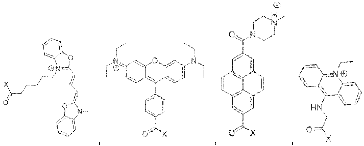

- Particularly preferred as pH independent fluorophore A is Cy5 and Cy3:

- The attachment site X denoting the attachment site of L is shown at the carbonyl group at the end of the alkyl chain. Within the scope of the present invention it is envisaged that the attachment site could vary. Equally, groups that do not affect the fluorescence properties of the fluorophore can be modified, for example the carbonyl and alkyl groups and remain within the scope of the invention.
- The pH dependent fluorophore B according to the present invention is capable of being protected such that the probe overall has an overall positive oxidation state. The fluorophore in inactive form must be capable of being converted into active form intracellularly. This may be achieved, for example, via esterases. In an embodiment, the pH dependent fluorophore B comprises oxygen groups which can be protected to lock or cage the molecule into a positive oxidation state. As such, in an embodiment, the pH dependent fluorophore B in inactive form comprises one or more protected oxygen(s).
- Any suitable fluorophore can be used as pH dependent fluorophore B that allows for protection and cellular deprotection. Non-limiting examples of suitable fluorophores include fluoresceins or coumarins. In a preferred embodiment, the pH dependent fluorophore B is a fluorescein. In a further preferred embodiment, the pH dependent fluorophore B is a coumarin. In an embodiment, the pH dependent fluorophore B in inactive form may be selected from:


- The attachment group X denotes the attachment site of L and is shown attached to the amine group. Variations to the attacment site are encompassed within the scope of the invention provided they do not adversely impact the ability of the fluorophore to function as a pH dependent fluorophore.
- Particularly preferred mitochondrial pH probes include:


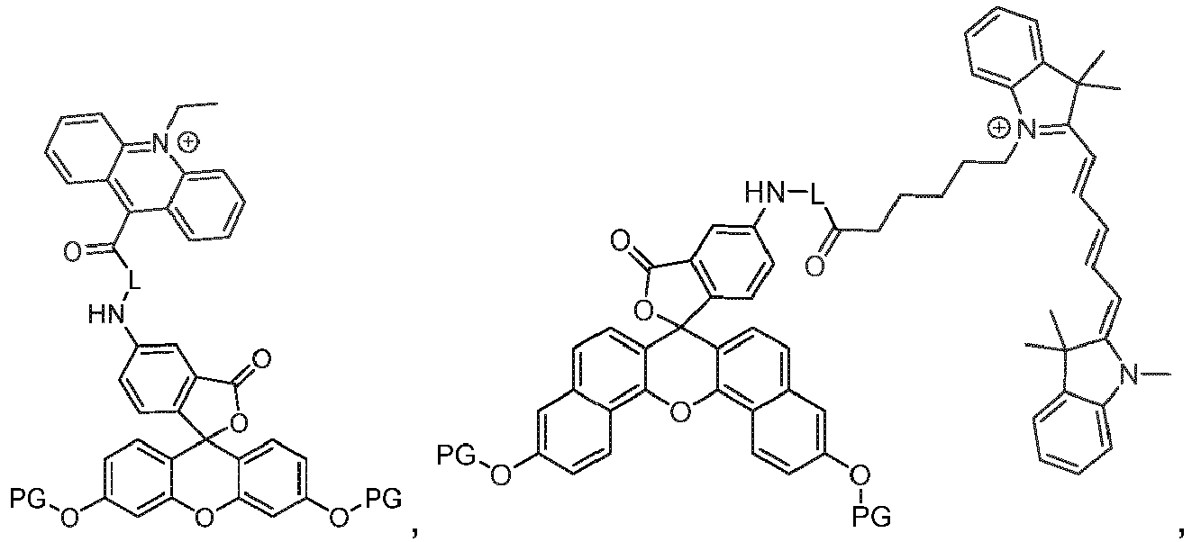

- An especially preferred probe is the flourescin-Cy5 and flourescin-Cy3 based probes:

- The linker L is selected from any suitable group that can covalently link the two fluorophores together without adversely affecting the fluorescence performance of either probe. In a preferred embodiment, the linker positioning and selection is designed to minimise steric hindrance for the rotation of the two fluorophores around the linker relative to each other, that may adversely affect the ability of the probe to travel through the cellular plasma membrane and mitochondrial membranes. In a yet further embodiment, the linker is a covalent bond between the two fluorophores.
- In an embodiment, the linker L may be selected from one or more of: alkyl, alkenyl, alkynyl, cycloalkyl, aryl, heteroaryl, heterocyclic, heteroalkyl, carbonyl, alkylcarbonyl; and/or combinations thereof optionally joined directly or by an alkyl, carbonyl, aminocarbonyl, thiocarbonyl group or by a heteroatom.
- In a further embodiment, the linker L may be of Formula II: L1-(L3)-L2 (Formula II), wherein L1 and L2 are independently selected from alkyl, alkenyl, alkynyl, cycloalkyl, aryl, heteroaryl, heterocyclic, heteroalkyl, carbonyl, and alkylcarbonyl; and L3 is a bond convalently linking L1 and L2 together; or L3 is selected from heteroatom, alkyl, alkenyl, alkynyl, cycloalkyl, aryl, heteroaryl, heterocyclic, heteroalkyl, carbonyl, and alkylcarbonyl.
- In a preferred embodiment, L1 may be selected from: alkyl, heteroaryl, heterocyclic, heteroalkyl and alkylcarbonyl. In a further preferred embodiment L1 may be selected from heteroaryl or alkylcarbonyl; more preferably 1,3,5-triazine or C1-nalkyl-1-carbonyl; wherein n is 2 to 12.
- In a further preferred embodiment, L2 may be selected from alkyl, heteroaryl, heterocyclic, heteroalkyl and alkylcarbonyl. In a yet further preferred embodiment, L2 may be selected from alkyl, heterocyclic or heteroalkyl; more preferably piperazine, 1-amino-n-thio-C1-nalkyl, or 1,n-diamino-C1-nalkyl; wherein n is 2 to 12.
- In a yet further preferred embodiment, L3 may be absent or may be selected from: NH or S. In a particularly preferred embodiment, L3 is absent.
- Especially preferred linkers include:


- The mitochondrial pH probe according to the present invention in an inactive form is protected into an overall positive oxidation state. The protection is removed intracellularly to convert the probe into an active form with a variable oxidation state. As such, in an inactive form, the probe is protected by one or more protecting groups (PG). Any suitable protecting group can be used that is able to both achieve a positive oxidation state and also be removed intracellularly. Examples of possible protecting groups PG include acyl, propionyl, butyryl, isobutyryl, pivaloyl or benzoyl. An especially preferred protecting group is acyl.
- Further possible preferred protecting groups include organic carbonate esters, thioesters and carbamates.
- According to a preferred embodiment of the present invention, the mitochondrial pH probe in protected form comprises the molecule 5-FA-PIP-Cy5-DA (Formula III shown below). 5-FA-PIP-Cy5-DA comprises the fluorophore 5-FA linked to the fluorophore Cy5, via a PIP (piperazyl) linker. The 5-FA unit has been diacetylated, leading to the designation DA:


- 5-FA-PIP-Cy5-DA (and any other probes according to the present invention) is delivered to the into the cells in a non-active caged form, when the phenolic groups of fluorescein are acylated. This quenches the fluorescein's fluorescence and also removes the pH dependence of the probe. Once delivered to the cell, however, the acetyl groups are cleaved by the cellular (mostly mitochondrial) esterases and the probe is converted to the active form. This is shown schematically below which shows the mechanism of cellular deprotecting and conversion of the probe from a positive fixed inactive state into an active form which has pH dependent fluorescence.


- In the active form of the probe fluorescein's fluorescence is pH dependent due to the reversible protonation/deprotonation of its phenolic group, when in a more acidic environment the non-fluorescent closed monoanionic spiro-form of fluorescein is prevalent, while in more basic environment the equilibrium is shifted to the opened fluorescent dianionic form of fluorescein. The Cy5 fluorophore of the probe is always fluorescent and thus acts as the pH independent ratiometric internal standard.
- The mitochondrial pH probes of the present invention can be used to measure mitochondrial pH. In an embodiment, there is provided a method of measuring mitochondrial pH comprising the steps of:
- a) contacting a cell with a mitochondrial pH probe in an inactive form as defined herein;
- b) allowing the probe to accumulate in the mitochondria of the cell, and to convert into an active form;
- c) determining the ratio of fluorescence between the pH independent fluorophore A and the pH dependent fluorophore B; and
- d) using this ratio to determine the mitochondrial pH.
- When in inactive form, the probes of the present invention are configured to more easily pass through lipid membranes and to target the mitochondria. This leads to an accumulation of the probe in the mitochondria. As already discussed above, this accumulation is defined by the rate of influx into the cell and mitochondria and the rate of efflux by the cell's defence mechanisms to remove the probe. By ensuring rapid influx and effective mitochondrial targeting, the probe will accumulate in the mitochondria and influx will override efflux. The cell converts the probe into an active form, for example by using esterases. Esterases are present throughout the cell but because of the rapid accumulation of the probe within the mitochondria, conversion into active form is also predominantly occurring within the mitochondria leading to an accumulation of probe in active form within the mitochondria.
- The advantageous properties of the probes of the present invention are such in some embodiments, sufficient accumulation of active probe in the mitochondria can take place within 15-20 minutes. This enables rapid mitochondrial pH measurements to take place. The sensitivity and update speed of the probe gives the ability to monitor real time changes to mitochondrial pH in response to any stimulus or condition being applied to the cell.
- Once sufficient accumulation has taken place, it is possible to begin measuring the fluorescence of the mitochondrial pH probe. After initial incubation time with the medium, containing probe in the inactive form, the cells are washed with the medium, not containing the probe. All the accumulated probe inside the mitochondria of the cells is converted into active form almost instantaneously. It is retained inside the mitochondria due to its ability to be trapped because of the membrane impermeability of the active form. The cells are then ready to be analyzed by fluorescent light microscopy or flow cytometry. The ratiometric fluorescent signal is acquired and quantified. Other methods, including but not limited plate-reading bulk analysis, can be used to analyze the stained cells. Overall, the staining and washing procedures can be performed within 30 minutes, after which the cells are ready for analysis.
- Prior to taking a pH measurement, it may be necessary to calibrate the probe to align the fluorescence ratiometric readings to a specific pH value. This may be undertaken by analysing the fluorescence ratio in the set of control experiments, including staining the control cell samples in the set of buffers with defined pH. These buffers must contain the protonophore agents, allowing the equilibration of the mitochondrial matrix pH with the pH of the surrounding buffer. Such agents might include:
carbonylcyanide-4-trifluoromethoxyphenylhydrazone (FCCP) as described in To, M. S. et al. Mitochondrial uncoupler FCCP activates proton conductance but does not block store-operated Ca2+ current in liver cells. Arch. Biochem. Biophys. 495, 152-158 (2010); and/or carbonylcyanide-3-chlorophenylhydrazone (CCCP) as described in Ruas, J. S. et al. Underestimation of the maximal capacity of the mitochondrial electron transport system in oligomycin-treated cells. ); and/or other mitochondrial protonophore and/or ionophore and/or ion-exchanging agent, such as valinomycin, nigericin antimycin and others, for example Lou, P.-H. et al. Mitochondrial uncouplers with an extraordinary dynamic range. Biochem. J. 407, 129-140 (2007); Kadenbach, B. Intrinsic and extrinsic uncoupling of oxidative phosphorylation. Biochim. Biophys. Acta - Bioenerg. 1604, 77-94 (2003). - The cells initially are stained with the mitochondrial pH probe in the same conditions, as used in the main experiments. Then the medium, containing the probe, is washed away and substituted with the corresponding series of defined pH buffers, containing the means to equilibrate the mitochondrial pH and the buffer pH, preferably FCCP or CCCP. The series of control cells then analyzed after 15 minutes with the same method, as used for the main experiments, to quantify the ratiometric fluorescent signal, corresponding to the exact mitochondrial pH value, defined by the buffer pH in the case of the control experiment. The dependence of ratiometric signal over the pH value is then analytically approximated with the mathematical equation, preferably linear equation. This equation is later used to convert the value of ratiometric fluorescent signal, obtained in the main experiments, to the mitochondrial pH value, observed experimentally.
- Through the ability to rapidly and sensitively measure pH changes within mitochondria, it is possible to evaluate how changes to the cell (for example the addition of a drug or drugs, changes in temperature or other external factors or the like) influence mitochondrial pH. It is equally possible to consider the reverse and investigate how changing mitochondrial pH can impact cellular activity. Non-limiting examples of the application of the mitochondrial pH probes of the present invention include
- Defining homeostatic mitochondrial pH values in various cell types;
- Defining changes in mitochondrial pH during the specific changes in particular cell types upon certain cellular activities, including but not limited proliferation, stress, apoptosis induction, quiescence, division and differentiation;
- Defining changes in mitochondrial pH upon pharmacological treatment;
- Defining changes in mitochondrial pH, characteristic for certain cell types upon onset of diseases, including but not limited cancer, diabetes and neurodegenerative diseases, or senescence;
- Defining changes in mitochondrial pH upon supplying various metabolic cues.
- The probes of the present invention may be made by any suitable means. According to one embodiment, there is provided a method of manufacturing a dual fluorophore ratiometric mitochondrial pH probe of Formula I:
A-L-B (Formula I);
wherein: - A is a pH independent fluorophore;
- B is a pH dependent fluorophore; and
- L is a chemical linker, conjugating the pH independent fluorophore A to the pH dependent fluorophore B, or L is a covalent bond;
- The invention will now be described by way of the following non-limiting examples.
- In the following examples the compound having the following formula III is referred to as 5-FA-PIP-Cy5-DA. 5-FA-PIP-Cy5-DA comprises the fluorophore5-FA (fluorescein) linked to the fluorophore Cy5, joined via a PIP (piperazyl) linker. The 5-FA unit has been diacetylated, leading to the designation DA.

- 5-FA-PIP-Cy5-DA was synthesised from commercially available 5-aminofluoresin 17 according to the synthetic scheme below.


- To a solution of 100 mg (0.288 mmol, 1 eq.) of 5-aminofluorescein 17 in 3 ml of DMAC a solution of 37 mg of chloroacetyl chloride (0.331 mmol, 1.15 eq.) in 1 ml of DMAC was added. The mixture was stirred for 10 min at room temperature and then a solution of 313 mg of piperazine (2.882 mmol, 10eq.) in 1 ml of ethanol was added. The mixture was stirred for 1 h. NMR spectra showed that the reaction was completed. The reaction mixture was then transferred to a falcon tube and 15 ml of diethyl ether was added to precipitate the crude product, which was separated by centrifugation. Next, 7 ml of ethanol was added to the separated precipitate and the resulting suspension sonicated for 2 minutes. The procedure was repeated 5 times and the purity of the product was controlled by NMR. Yield: 100 mg (81%) of 5-aminofluoresceinopiperazine 18.
1H NMR (800 MHz, DMSO-d6) δ 10.17 (s, 1H), 8.36 (d, J = 1.7 Hz, 1H), 7.94 (dd, J = 8.3, 2.0 Hz, 1H), 7.20 (d, J = 8.3 Hz, 1H), 6.67 - 6.63 (m, 2H), 6.59 (d, J = 8.7 Hz, 2H), 6.53 (dd, J = 8.7, 2.3 Hz, 2H), 3.14 (s, 2H), 2.77 (t, J = 4.8 Hz, 4H), 2.47 - 2.42 (m, 4H).
HRMS (LC-MS) found: 474.1631, calculated for [M-H+]: 474.1660. - 5 mg of Cy5-COOH chloride (9.634 µmol, 1 eq.) was dissolved in 1 ml of DMAC in a 2 ml Eppendorf tube. To the resulting solution of 3.1 mg of carboxydiimidazole (19.268 µmol, 2 eq.) was added in 0.5 ml of DMAC. The mixture was stirred for 1 h. The completion of conversion of Cy5-acid to imidazolide 19 was controlled by NMR. Next, to the reaction mixture a solution of 5-aminofluoresceinopiperazine 18 (14.451 µmol, 1.5 eq.) in 0.5 ml of DMAC was added. The reaction mixture was stirred overnight at room temperature. After completion of the reaction (confirmed by NMR) the reaction mixture was diluted with 1 ml of chloroform and excess 5-aminofluoresceinopiperazine extracted 10 times with 0.5 ml of 0.5 M HCI, until the water layer was transparent. The remaining slurry of the chloroform layer was transferred to a 1.5 ml Eppendorf tube and the product was precipitated with 1 ml of diethyl ether. This precipitate was separated by centrifugation and the liquid layer was discarded. Next, to the remaining precipitate 0.3 ml of THF was added and the mixture sonicated for 2 minutes. The precipitate was separated by centrifugation again and the sonication-centrifugation was repeated 7 times. The precipitate was dried in the open Eppendorf tube in the darkness. The purity of the product was controlled by NMR. Yield: 9.7 mg (90%) of 5FA-PIP-Cy5, calculated for the dichloride form.
1H NMR (500 MHz, DMSO-d6) δ 10.24 (s, 1H), 10.14 (s, 2H), 8.41 - 8.30 (m, 3H), 7.94 (s, 1H), 7.63 (t, J = 6.6 Hz, 2H), 7.42 (q, J = 7.8 Hz, 4H), 7.26 (t, J = 7.0 Hz, 3H), 6.69 (d, J = 2.0 Hz, 2H), 6.65 - 6.53 (m, 6H), 6.30 (dd, J = 24.6, 13.8 Hz, 2H), 4.12 (t, J = 6.8 Hz, 2H), 3.62 - 3.60 (m, 4H), 3.51 (s, 3H), 2.32 (s, 2H), 1.75-1.71 (m, 2H), 1.70 (s, 6H), 1.69 (s, 6H), 1.60-1.54 (m, 2H), 1.43-1.36 (m, 2H).
HRMS (LC-MS) found: 938.4008, calculated for [M-H+]: 938.4487. - To the solution of 5-FA-PIP-Cy5 ((9.039 µmol, 1 eq.) in 1 ml of DMAC, a solution of 6.1 mg EDCI hydrochloride (24.118 µmol, 3eq.), 4.8 mg of AcOH (80.393 µmol, 10 eq.), and 3.1 mg of DIPEA (24.118 µmol, 3eq.) in 0.5 ml of DMAC was added. The mixture was stirred overnight at room temperature and the degree of acylation was controlled by NMR. Upon completion of acylation, the reaction mixture was transferred to 15 ml falcon tube and diluted with 2 ml of chloroform. The resulting mixture was extracted with 2 ml of 0.5 M HCI 5 times. The water layers were discarded and the remaining chloroform layer was transferred to a 2 ml Eppendorf tube and precipitated with 1 ml of diethyl ether to separate the product. The formed precipitate was separated by centrifugation. Next, to the precipitate 0.4 ml of THF was added and the mixture was sonicated for 2 minutes. To this
mixture 1 ml of diethyl ether was added to crystallize back the dissolved product and the resulting precipitate was separated by centrifugation again. This procedure was repeated 5 times. The purity of the product was controlled by NMR. Yield: 8.2 mg (93%) of 5FA-PIP-Cy5-DA, calculated for the dichloride form.
1H NMR (500 MHz, DMSO-d 6) δ 10.27 (s, 1H), 8.44 (d, J = 1.5 Hz, 1H), 8.33 (t, J = 13.1 Hz, 2H), 8.01 - 7.94 (m, 1H), 7.61 (t, J = 7.5 Hz, 2H), 7.44 - 7.36 (m, 4H), 7.34 (d, J = 8.4 Hz, 1H), 7.28 (d, J = 2.1 Hz, 2H), 7.26 - 7.22 (m, 2H), 6.95 (dd, J = 8.7, 2.1 Hz, 2H), 6.91 (d, J = 8.6 Hz, 2H), 6.56 (t, J = 12.3 Hz, 1H), 6.28 (dd, J = 25.5, 13.8 Hz, 2H), 4.10 (t, J = 7.2 Hz, 2H), 3.60 (s, J = 1.8 Hz, 3H), 3.54-3.45 (m, 4H), 3.23 (s, 2H), 2.33 - 2.30 (m, 2H), 2.29 (s, 6H), 1.71 - 1.73 (m, 2H), 1.68 (s, 6H), 1.67 (s, 6H), 1.55 (p, J = 7.3 Hz, 2H), 1.39 (p, J = 7.9 Hz, 2H).
13C NMR (201 MHz, DMSO-d6) δ 173.75, 173.03, 170.84, 169.45, 169.34, 168.79, 154.50, 154.48, 152.52, 151.32, 147.12, 143.24, 142.55, 141.58, 141.49, 141.13, 129.65, 128.91, 128.83, 127.48, 126.72, 125.81, 125.21, 125.12, 124.97, 122.92, 122.77, 119.07, 116.71, 114.64, 111.56, 111.52, 110.94, 103.76, 103.54, 81.47, 61.89, 53.38, 52.88, 49.35, 49.33, 45.30, 43.77, 41.39, 32.39, 31.56, 27.67, 27.47, 27.29, 26.30, 25.00, 21.34.
HRMS (LC-MS) found: 1022.4530, calculated for [M-H+]: 1022.4699. - A2780 ovarian cancer cells were cultured in 24-well microscopy glass bottom plates in a CO2 incubator. Prior to imaging, the cells were stained with 2uM 5FA-PIP-Cy5-DA, and/or TMRM (20nM) for 30 min in bicarbonate-free DMEM medium to avoid extracellular pH fluctuations, then washed with the same medium (without 5FA-PIP-Cy5-DA) and imaged with confocal spinning disk microscopy at 60x magnification When TMRM was used, this dye was also present in the bicarbonate-free DMEM medium during the imaging of the cells. Separate fluorescent images in green (488/520 nm) and deep red (640/660 nm) spectral channels were acquired on Visitron W1 spinning disk confocal microscope. The florescence ratio was quantified using a specifically designed batch ImageJ plugin. This software thresholds the pH-independent Cy5 channel, quantifies the fluorescence intensity in the thresholded image areas, and uses the thresholded mask to quantify the intensity of pH-dependent 5Fluo channel. The intensity ratio is then used to estimate the mitochondrial pH of the cells. Calibration of the intensity ratio was achieved through the staining of A2780 cells in a series of PBS - 5% BSA buffers with defined pH, containing 20mM HEPES and 5uM FCCP, to equilibrate the mitochondrial pH with the pH of the buffer.
- The pH dependence for fluorescence at 520 nm of 5FA-PIP-Cy5, the active form of 5FA-PIP-Cy5-DA was measured with 5FA-PIP-Cy5 dissolved in a PBS buffer with 5% BSA. (see
Figure 3 ). From this study the pKa of the active form was estimated to be 6.88. This value is within expectable range for the fluorescein moiety containing electron-donor amide group and is within the physiologically relevant range of 6.5 - 8.5. - Typical pH variations in mitochondria of A2780 ovarian cancer cell line are represented at
Figure 4 . Typical staining procedure is described further. Cells were stained for 30 min with 2 µM 5FA-PIP-Cy5-DA solution, the probe was washed away with the bicarbonate-free DMEM medium, and the cells were imaged after 10 min with confocal spinning disk microscope. All images are maximum intensity projection.Figure 4A represents the 5FA-PIP-Cy5-DA pH-independent (Cy5) channel, shown in red on the figure. The mitochondrial staining onFigure 4A is very homogeneous.Figure 4B represents the 5FA-PIP-Cy5-DA pH-dependent (5Fluo) channel, shown in blue on the figure. The mitochondrial staining onFigure 4B is very heterogeneous, unlike theFigure 4A. Figure 4C represents the overlap image ofFigure 4A and Figure 4B . Bluish hintFigure 4C corresponds to relatively high mitochondrial pH, reddish to relatively low mitochondrial pH.Figure 4D represents bright field image. On theFigure 4C it can be clearly seen, that the mitochondria in different A2780 cells are highly heterogeneous in the pH, which is reflected in the highly heterogeneous staining in pH dependent channel onFigure 4B . - The ability of the synthesised probe to distinguish mitochondrial pH changes in live cells was tested. This test was performed via the comparison of the fluorescein/Cy5 fluorescence ratio between control conditions and cells which had been treated with K+/H+ exchanging ionophore nigericin and a protonphore FCCP, which reduce mitochondrial pH by acidifying the mitochondrial matrix. FCCP and nigericin were commercially purchased. After the of A2780 ovarian cancer cells were stained as described above, the cells were washed with the bicarbonate-free DMEM medium, either containing DMSO vehicle for control sample or 300 nM FCCP or 2uM nigericin for the samples, where the mitochondrial pH was expected to be decreased. Confocal images were acquired for control, FCCP- and nigericin-treated cell samples, and the ratio between fluorescein and Cy5 channel fluorescence was quantified.
- It can be seen from
Figure 5 that treatment with both nigericin and FCCP resulted in a clear measurable decrease in the 5Fluo-Cy5 ratio. This validates that 5-FA-PIP-Cy5-DA taken into the cells and mitochondria, that it is converted into its active form 5FA-PIP-Cy5, and that the resultant probe is sensitive to mitochondrial pH changes. - Having validated that the probe is sensitive to mitochondrial pH changes, an experiment was performed to calibrate the probe.
- The stained cells were treated with FCCP in high K+ buffers in a pH range of 6.73 to 8.36.
- The results of the experiment can be seen in
Figure 6 . In FCCP treated cells within this pH range the fluorescein/Cy5 ratio showed a good linear response. - Treatment with FCCP alone caused leakage of the probe from the mitochondria into the cytoplasm, but the addition of 5% BSA to the calibration buffers greatly alleviated this problem, making the calibration with FCCP well-suited for mitochondrial pH calibration with the probe. In fact, treatment with FCCP caused full collapse of the mitochondrial pH gradient, ensuring that the pH to which the probe is exposed within the mitochondria is equal to the pH of the buffer.
- Because the pH of the buffer can be directly measured, it was therefore possible to also know the exact cellular pH. As such, specific pH values can be attributed to the fluorescein/Cy5 ratios. FCCP treatment can therefore be used as a practical way to calibrate a cell for exact pH measurements.
- The experiment was repeated using nigericin and the results can be seen in
Figure 7 . Again, a strong linear response is seen validating the reliability of the probe in measuring pH. However, no full gradient collapse was seen with nigericin, even at high concentrations. As such, the mitochondrial pH is not the same as the buffer and nigericin is not well suitable as an agent to prepare an accurate calibration curve. - The selectivity of mitochondrial staining of 5FA-PIP-Cy5-DA was verified by co-staining with another well-known mitochondria targeting dye, tetramethylrhodamine methyl ester (TMRM). As 5FA-PIP-Cy5-DA is fluorescent in the deep red and green spectral regions, and TMRM is fluorescent in red spectral region, these dyes are spectrally compatible. A2780 cells were stained for 30 min with 2 µM 5FA-PIP-Cy5-DA and 20nM solution, the probe was washed away with the bicarbonate-free DMEM medium, containing 20nM TMRM, and the cells were imaged after 10 min with confocal spinning disk microscope.
Figure 8 represents typical image of mitochondria, stained with both dyes.Figure 8A represents 5FA-PIP-Cy5-DA pH independent (Cy5) channel, shown in red.Figure 8B represents 5FA-PIP-Cy5-DA pH-dependent (5Fluo) channel, shown in blue.Figure 8C represents TMRM channel, shown in magenta.Figure 8D represents bright field image. Spinning disk confocal images of TMRM and 5FA-PIP-Cy5-DA costained A2780 cells were analyzed for pixel florescence intensity correlation between the Cy5 channel of 5FA-PIP-Cy5-DA and the TMRM channel (Figure 8 ). The Cy5 channel of 5FA-PIP-Cy5-DA is the most appropriate for co-localization studies, because it is pH-independent and therefore clearly reveals the localization of the 5FA-PIP-Cy5-DA molecule independent of the pH. The high degree of co-localization of the two dyes was confirmed by high values of Manders' coefficients (M1 = 0.94, M2 = 0.982) and Pearson's coefficient (r = 0.905). Importantly, it is consistently observed that very little cytoplasmic background is present during the staining with 5FA-PIP-Cy5-DA, demonstrating the highly selective mitochondrial staining by 5FA-PIP-Cy5-DA. Furthermore, high degree of colocalization with a well-known mitochondrial selective probe TMRM further confirms mitochondrial selectivity of 5FA-PIP-Cy5-DA. - The advantage of the caging approach, ensuring the high lypophilicity and fixed positive oxidation state was tested by comparing the typical staining of the cells with the inactive form with the staining of the same cells in the same conditions directly with the active form. The result of such experiments are represented in
Figure 9 . Staining A2780 cells with 2uM of either 5FA-PIP-Cy5-DA or 5FA-PIP-Cy5 in identical conditions resulted in normal staining of mitochondria in the case of inactive form 5FA-PIP-Cy5-DA (Figure 9A ) and absolutely no staining of mitochondria in the case of active form 5FA-PIP-Cy5 (Figure 9B ), proving that the much more hydrophilic 5FA-PIP-Cy5 is not capable of permeating the cell membrane due to its non-cationic nature and higher hydrophilicity. In fact, there was no difference between the cells, stained with 5FA-PIP-Cy5 and non-stained control cells (Figure 9C ). - In order to demonstrate the applicability of the probes of the current invention in live cells, in vitro experiments were devised in which the mitochondrial pH differences in a series of live cells (LT-HSC, ST-HSC and MPP) was measured using through 5FA-PIP-Cy5-DA staining and fluorescence measurements. For LT-HSC cells, three different culture conditions (basal medium, DMOG-induced chemical hypoxia and IL-3, IL-6 - induced differentiation) were examined.
- The calibration curve for the ratio of 5Fluoro/Cy5 fluorescence was obtained using FCCP and a PBS-5% BSA buffer with MPP cells in a manner analogous to the method described in Example 2 (see
Figure 10 ). - Fresh cells, extracted from bone marrow of 8-12 week old male mice were stained on ice for surface cell markers and sorted by FACS, according to the following phenotypes: LT-HSC Lin- cKit+ Sca1+ CD150+ CD34- CD48-; ST-HSC Lin- cKit+ Sca1 + CD150+ CD34+; MPP Lin- cKit+ Sca1 + CD150- CD34+. The cells were cultured in Hematopoietic Stemline II (Sigma) medium with addition of SCF and Flt-3 cytokines. For appropriate experiments, additionally 5 uM DMOG or IL-3 20 ng/mL and IL-6 100ng/mL cytokines were added.
- Prior to imaging, the cells were stained with the appropriate concentration of 5FA-PIP-Cy5-DA for 30 min in bicarbonate-free DMEM medium to avoid the extracellular pH fluctuation outside of the CO2 incubator, then washed, transferred to poly-L-lysine coated 24-well microscopy glass bottom plates and imaged with confocal spinning disk microscopy at 60x magnification. Separate fluorescent images in green and deep red spectral channels were acquired. The florescence ratio was quantified by a specifically designed batch ImageJ plugin as described above.
- LT-HSC showed a much lower mitochondrial pH level compared to ST-HSC and MPP (see
Figure 11 ). LT-HSC, cultured in the basal medium, and LT-HSC, cultured in the presence of DMOG, had the same level of average mitochondrial pH, though DMOG-treated cells had less heterogeneity within the population, lacking the individual cells with high mitochondrial pH. Significantly, IL-3, IL-6 - treated LT-HSC had the highest overall average mitochondrial pH value, significantly above LT-HSC, both when cultured in basal medium, and with addition of DMOG, and ST-HSC and MPP. - This demonstrated that 5FA-PIP-Cy5-DA performs well as a probe for mitochondrial pH. The probe was able to demonstrate that differentiation induction in LT-HSC causes the burst in mitochondrial pH to a level not observed even in highly active, proliferative ST-HSC and MPP cells. Furthermore, through the use of 5FA-PIP-Cy5-DA, significant differences in mitochondrial pH were observed through the range of conditions, favouring different stem cell fate outcomes, and demonstrating that mitochondrial pH alteration is associated with hematopoietic stem cell fate determination as a result.
- In vitro studies were performed in which 5FA-PIP-Cy5-DA staining could be used to distinguish between direct drug interference with mitochondrial metabolism and general cytotoxicity from anti-cancer drugs RAPTA-T and cisplatin.
- The calibration curve for the ratio of 5Fluoro/Cy5 fluorescence was obtained using FCCP and a PBS-5% BSA buffer with A2780 cells in a manner analogous to the method described in Example 2 (see
Figure 12 ). - The quantifying of mitochondrial pH in the presence of the drug and after the drug was washed away allowed the distinction between interference with mitochondrial metabolism and general cytotoxicity. If the effect of the drug was primarily to affect mitochondrial metabolism it was expected that the mitochondrial pH would quickly return to the non-treated cell level following removal of the drug, providing the drug had no other significant cytotoxic effects. Conversely, if the effect of the drug were not reversed by the washing step, an observed alteration of mitochondrial pH could be interpreted at least partially as a downstream consequence of the general cytotoxicity of the drug.
- A2780 cells were cultured in 24-well microscopy glass bottom plates in RPMI medium, containing Glutamax with 10% FBS addition. The cells were treated with appropriate 800uM RAPTA-T or 5uM cisplatin. Prior to the imaging the cells were stained with 2uM 5FA-PIP-Cy5-DA for 30 min in a bicarbonate-free DMEM medium to avoid the extracellular pH fluctuation outside of the CO2 incubator, then washed and imaged with confocal spinning disk microscopy at 60x magnification. Cells were treated with either drug for one day and mitochondrial pH was quantified using 5FA-PIP-Cy5-DA in the presence of the drug and after the drug was washed away. For some cells the drugs were washed away prior to staining with 5FA-PIP-Cy5-DA. If the drug was not washed away prior to staining, it remained present in the medium through the whole imaging time. Separate fluorescent images in green and deep red spectral channels were acquired. The florescence ratio was quantified by a specifically designed batch ImageJ plugin as described above. Statistical analysis was performed using 1-way ANOVA with Bonferroni-Holm post-hoc analysis.
- The results are shown in
Figure 13 which clearly showed that cisplatin and RAPTA-T both drastically decreased mitochondrial pH in A2780 cells in comparison to non-treated cells. The introduction of RAPTA-T saw the pH drop to 8.0 compared to pH 8.4 seen with the control cells. Upon washing, the value rose again to pH 8.4, fully restoring the mitochondrial pH. By comparison, cisplatin dropped the pH to an average of 7.8, which increased to 8.0 after washing, with mitochondrial pH only partially restored by the washing step. This indicated that RAPTA-T has a much more direct impact on mitochondrial metabolism in comparison to cisplatin. - Again, it can be seen that the use of 5FA-PIP-Cy5-DA, enabled a rapid and direct measurement of mitochondrial pH allowing real time measurements of the effects of drugs on mitochondrial pH and to distinguish the differences of mechanisms of actions behind the alteration of mitochondrial pH by the anti-cancer drug treatment.
-
Figure 13 also demonstrates the use of 5FA-PIP-Cy5-DA to assess the functional state of mitochondrial ATP synthase of A2780 cells by measuring mitochondrial pH in the presence and absence of ATP synthase inhibitor oligomycin. It can be seen that the addition of 10µm oligomycin caused slight increase in mitochondrial pH from 8.4 to around 8.5. - Oligomycin inhibits proton conductance by ATP synthase, removing its contribution to the proton gradient. If mitochondrial ATP synthase acts in the normal mode, inhibition with oligomycin would increase mitochondrial pH, while in the reversed mode it would decrease mitochondrial pH. Finally, in the case of pre-existing natural inhibition of ATP synthase, inhibition with oligomycin would not noticeably change mitochondrial pH.
- It can be seen from
figure 13 that oligomycin caused a noticeable increase in mitochondrial pH demonstrating that the mitochondrial ATP synthase in the tested cells was acting in the normal mode. This suggests that the cytotoxic effects of the tested anticancer drugs in these cells were related to the disruption of mitochondrial oxidative phosphorylation: as during the normal functional state of mitochondrial ATP synthase, alkalinisation of mitochondrial matrix is achieved through the activity of respiratory chain, the following drug-induced drop in mitochondrial pH can be attributed to the disruption of this process. - In conclusion, these experiments demonstrate the potential and wide range of applications for 5-FA-PIP-Cy5-DA. The experimental data shows that due to the simplicity of the experiments, the impact on mitochondrial metabolism can be reliably and quickly assessed in real time. The data shows that 5-FA-PIP-Cy5-DA can be used to determine the specificity of mechanism of action of the various drugs which purport to have an effect on mitochondrial metabolism.
Claims (15)
- A dual fluorophore ratiometric mitochondrial pH probe of Formula I:
A-L-B (Formula I);
wherein:A is a pH independent fluorophore;B is a pH dependent fluorophore; andL is a chemical linker, conjugating the pH independent fluorophore A to the pH dependent fluorophore B, or L is a covalent bond;wherein in an inactive form, the probe is protected in an overall positive oxidation state; and wherein the probe may be deprotected intracellularly into an active form with a variable oxidation state. - The mitochondrial pH probe of claim 1, wherein in the inactive form, the probe has an oxidation state of +1.
- The mitochondrial pH probe of claim 1 or claim 2, wherein in the active form, the probe has a variable oxidation state of between 0 and -2, preferably between 0 and -1 or between -1 and -2.
- The mitochondrial pH probe of any preceding claim, wherein the pH independent fluorophore A is selected from:a. a fluorophore containing a positively charged N ion;b. a fluorophore derived from fluorones, polymethines, pyrenes or acridines; orc. a fluorophore derived from rosamines or cyanines;wherein the pH independent fluorophore A is preferably selected from:


most preferably wherein the pH independent fluorophore A is Cy5:
- The mitochondrial pH probe of any preceding claim, wherein the pH dependent fluorophore B in inactive form is a fluorophore comprising one or more protected oxygen(s).
- The mitochondrial pH probe of any preceding claim, wherein the pH dependent fluorophore B is selected from fluoresceins or coumarins;
preferably wherein the pH dependent fluorophore B in inactive form is selected from:

- The mitochondrial pH probe of claim 1, wherein the probe is selected from:


most preferably wherein the probe is:
- The mitochondrial pH probe of any preceding claim, wherein L is a linker selected from:alkyl, alkenyl, alkynyl, cycloalkyl, aryl, heteroaryl, heterocyclic, heteroalkyl, carbonyl, alkylcarbonyl; and/orcombinations of the above, optionally joined directly or by an alkyl, carbonyl, aminocarbonyl, thiocarbonyl group or by a heteroatom.
- The mitochondrial pH probe according to claim 12, wherein L is of formula II:
L1-(L3)-L2 (Formula II)
wherein L1 and L2 are independently selected from alkyl, alkenyl, alkynyl, cycloalkyl, aryl, heteroaryl, heterocyclic, heteroalkyl, carbonyl, and alkylcarbonyl; andL3 is a bond convalently linking L1 and L2 together; or L3 is selected from heteroatom, alkyl, alkenyl, alkynyl, cycloalkyl, aryl, heteroaryl, heterocyclic, heteroalkyl, carbonyl, and alkylcarbonyl;wherein L1 may optionally be is selected from: alkyl, heteroaryl, heterocyclic, heteroalkyl and alkylcarbonyl, preferably heteroaryl or alkylcarbonyl; more preferably 1,3,5-triazine or C1-nalkyl-1-carbonyl; wherein n is 2 to 12; and/orwherein L2 may optionally be selected from: alkyl, heteroaryl, heterocyclic, heteroalkyl and alkylcarbonyl, preferably, alkyl, heterocyclic or heteroalkyl; more preferably piperazine, 1-amino-n-thio-C1-nalkyl, or 1,n-diamino-C1-nalkyl; wherein n is 2 to 12; and/orwherein L3 may optionally be selected from: S, NH, or is not present;preferably, wherein the linker L is selected from:
 wherein n = 1-6.
 - The mitochondrial pH probe of any preceding claim, wherein the protecting group PG is selected from acyl, propionyl, butyryl, isobutyryl, pivaloyl or benzoyl; preferably acyl.
- A method of measuring mitochondrial pH comprising the steps of:a) contacting a cell with a mitochondrial pH probe in an inactive form as claimed in any one of claims 1 to 10;b) allowing the probe to accumulate in the mitochondria of the cell, and to convert into an active form;c) determining the ratio of fluorescence between the pH independent fluorophore A and the pH dependent fluorophore B; andd) using this ratio to determine the mitochondrial pH;wherein step a) may optionally comprise contacting the mitochondrial pH probe to a cell in vivo within a living organism or in vitro, for example within a 3D-cell culture model; and/or
wherein in step b) the probe may optionally accumulate in the mitochondria within 15 minutes in sufficient quantity to determine mitochondrial pH. - The method of claim 11, wherein in step b) the probe is converted into an active form by esterases.
- The method of claim 11 or claim 12, wherein the probe is calibrated using FCCP or other protonophore or similar agent, allowing equilibration of the mitochondrial pH with the pH of the buffer.
- A method of measuring the impact of drugs or other conditions on the mitochondrial pH comprising applying the drug or other condition to a cell and measuring the change in mitochondrial pH using a dual fluorophore ratiometric mitochondrial pH probe as claimed in any one of claims 1 to 10.
- A method of manufacturing a dual fluorophore ratiometric mitochondrial pH probe of Formula I:
A-L-B (Formula I);
wherein:A is a pH independent fluorophore;B is a pH dependent fluorophore; andL is a chemical linker, conjugating the pH independent fluorophore A tothe pH dependent fluorophore B, or L is a covalent bond;as claimed in any one of claims 1 to 10, comprising protecting the pH dependent fluorophore B such that the mitochondrial pH probe of Formula I is put into a positive oxidation state.
Priority Applications (2)
| Application Number | Priority Date | Filing Date | Title |
|---|---|---|---|
| EP18163205.0A EP3543705A1 (en) | 2018-03-21 | 2018-03-21 | Mitochondrial ph probe |
| PCT/EP2019/057012 WO2019180105A1 (en) | 2018-03-21 | 2019-03-20 | Mitochondrial ph probe |
Applications Claiming Priority (1)
| Application Number | Priority Date | Filing Date | Title |
|---|---|---|---|
| EP18163205.0A EP3543705A1 (en) | 2018-03-21 | 2018-03-21 | Mitochondrial ph probe |
Publications (1)
| Publication Number | Publication Date |
|---|---|
| EP3543705A1 true EP3543705A1 (en) | 2019-09-25 |
Family
ID=61868151
Family Applications (1)
| Application Number | Title | Priority Date | Filing Date |
|---|---|---|---|
| EP18163205.0A Ceased EP3543705A1 (en) | 2018-03-21 | 2018-03-21 | Mitochondrial ph probe |
Country Status (2)
| Country | Link |
|---|---|
| EP (1) | EP3543705A1 (en) |
| WO (1) | WO2019180105A1 (en) |
Cited By (2)
| Publication number | Priority date | Publication date | Assignee | Title |
|---|---|---|---|---|
| CN110776458A (en) * | 2019-11-01 | 2020-02-11 | 济南大学 | Fluorescent probe for detecting mitochondrial membrane potential, preparation method and application thereof |
| DE102019124795A1 (en) * | 2019-09-16 | 2021-03-18 | Abberior GmbH | Optical pH sensor |
Families Citing this family (2)
| Publication number | Priority date | Publication date | Assignee | Title |
|---|---|---|---|---|
| GB202104582D0 (en) | 2021-03-31 | 2021-05-12 | Vestlandets Innovasjonsselskap As | Mitochondrial ph probes and uses thereof |
| CN118373812B (en) * | 2024-04-09 | 2024-11-22 | 西南大学 | A mitochondrial fluorescent marker and labeling method |
Citations (1)
| Publication number | Priority date | Publication date | Assignee | Title |
|---|---|---|---|---|
| KR20140135483A (en) * | 2013-05-16 | 2014-11-26 | 고려대학교 산학협력단 | WIDE pH RANGE RESPONSIBLE RATIOMETRIC pH PROBE |
-
2018
- 2018-03-21 EP EP18163205.0A patent/EP3543705A1/en not_active Ceased
-
2019
- 2019-03-20 WO PCT/EP2019/057012 patent/WO2019180105A1/en not_active Ceased
Patent Citations (1)
| Publication number | Priority date | Publication date | Assignee | Title |
|---|---|---|---|---|
| KR20140135483A (en) * | 2013-05-16 | 2014-11-26 | 고려대학교 산학협력단 | WIDE pH RANGE RESPONSIBLE RATIOMETRIC pH PROBE |
Non-Patent Citations (8)
| Title |
|---|
| JUNYAN HAN ET AL: "Fluorescent Indicators for Intracellular pH", CHEMICAL REVIEWS, vol. 110, no. 5, 12 May 2010 (2010-05-12), pages 2709 - 2728, XP055078676, ISSN: 0009-2665, DOI: 10.1021/cr900249z * |
| KADENBACH, B.: "Intrinsic and extrinsic uncoupling of oxidative phosphorylation", BIOCHIM. BIOPHYS. ACTA - BIOENERG, vol. 1604, 2003, pages 77 - 94, XP004426697, DOI: doi:10.1016/S0005-2728(03)00027-6 |
| LOU, P.-H. ET AL.: "Mitochondrial uncouplers with an extraordinary dynamic range", BIOCHEM. J., vol. 407, 2007, pages 129 - 140 |
| RUAS, J. S. ET AL.: "Underestimation of the maximal capacity of the mitochondrial electron transport system in oligomycin-treated cells", PLOS ONE, vol. 11, 2016, pages 1 - 20 |
| TO, M. S. ET AL.: "Mitochondrial uncoupler FCCP activates proton conductance but does not block store-operated Ca2+ current in liver cells", ARCH. BIOCHEM. BIOPHYS., vol. 495, 2010, pages 152 - 158, XP026924524, DOI: doi:10.1016/j.abb.2010.01.004 |
| YAN FANYONG ET AL: "Fluorescein applications as fluorescent probes for the detection of analytes", TRAC TRENDS IN ANALYTICAL CHEMISTRY, vol. 97, 26 August 2017 (2017-08-26), pages 15 - 35, XP085268074, ISSN: 0165-9936, DOI: 10.1016/J.TRAC.2017.08.013 * |
| YONG-XIANG WU ET AL: "Bispyrene-Fluorescein Hybrid Based FRET Cassette: A Convenient Platform toward Ratiometric Time-Resolved Probe for Bioanalytical Applications", ANALYTICAL CHEMISTRY, vol. 86, no. 20, 21 October 2014 (2014-10-21), US, pages 10389 - 10396, XP055473968, ISSN: 0003-2700, DOI: 10.1021/ac502863m * |
| YUNCONG CHEN ET AL: "Ratiometric detection of pH fluctuation in mitochondria with a new fluorescein/cyanine hybrid sensor", CHEMICAL SCIENCE, vol. 6, no. 5, 1 January 2015 (2015-01-01), United Kingdom, pages 3187 - 3194, XP055473963, ISSN: 2041-6520, DOI: 10.1039/C4SC04021J * |
Cited By (3)
| Publication number | Priority date | Publication date | Assignee | Title |
|---|---|---|---|---|
| DE102019124795A1 (en) * | 2019-09-16 | 2021-03-18 | Abberior GmbH | Optical pH sensor |
| CN110776458A (en) * | 2019-11-01 | 2020-02-11 | 济南大学 | Fluorescent probe for detecting mitochondrial membrane potential, preparation method and application thereof |
| CN110776458B (en) * | 2019-11-01 | 2021-10-26 | 济南大学 | Fluorescent probe for detecting mitochondrial membrane potential and preparation method and application thereof |
Also Published As
| Publication number | Publication date |
|---|---|
| WO2019180105A1 (en) | 2019-09-26 |
Similar Documents
| Publication | Publication Date | Title |
|---|---|---|
| Meng et al. | Simultaneous visualization of lipid droplets and lysosomes using a single fluorescent probe | |
| Song et al. | A highly sensitive near-infrared ratiometric fluorescent probe for imaging of mitochondrial hydrazine in cells and in mice models | |
| Chen et al. | Ratiometric detection of pH fluctuation in mitochondria with a new fluorescein/cyanine hybrid sensor | |
| Wang et al. | Based ‘successive’nucleophilic substitution mitochondrial-targeted H2S red light emissive fluorescent probe and its imaging in mice | |
| Yang et al. | Dual-site fluorescent sensor for tracking lysosomal ATP and H2S during liver injury | |
| Fan et al. | A ratiometric lysosomal pH chemosensor based on fluorescence resonance energy transfer | |
| WO2019180105A1 (en) | Mitochondrial ph probe | |
| Chang et al. | Imaging beyond the proteome | |
| Chung et al. | Two-photon imaging of hydrogen polysulfides in living cells and hippocampal tissues | |
| Li et al. | A julolidine-chalcone-based fluorescent probe for detection of Al3+ in real water sample and cell imaging | |
| Lu et al. | Lysozyme-targeted ratiometric fluorescent probe for SO2 in living cells | |
| Hu et al. | A novel turn-on colorimetric and fluorescent sensor for Fe3+ and its application in living cells | |
| Tan et al. | A novel “off–on” colorimetric and fluorescent rhodamine-based pH chemosensor for extreme acidity | |
| Liu et al. | De Novo-Designed landmine warfare strategy luminophore for super-resolution imaging reveal ONOO–evolution in living cells | |
| Kritchenkov et al. | Biocompatible Ir (III) complexes as oxygen sensors for phosphorescence lifetime imaging | |
| Cheng et al. | Development of a one-step synthesized red emission fluorescent probe for sensitive detection of viscosity in vitro and in vivo | |
| Yang et al. | Rational design of a new near-infrared fluorophore and apply to the detection and imaging study of cysteine and thiophenol | |
| Yao et al. | Molecular engineering of fluorescent dyes for long-term specific visualization of the plasma membrane based on alkyl-chain-regulated cell permeability | |
| CN115772096A (en) | A two-photon fluorescent probe for dual-channel detection and its preparation method and use | |
| Rao et al. | Real-time monitoring apoptosis and autophagy among multiple organelles by adjusting the slight structure | |
| Yao et al. | Visualizing hydrogen sulfide in HepG2 cells exposed to OGD/R and in the liver of zebrafish with a hepatocyte-targeting fluorescent probe | |
| Chen et al. | Imaging pulmonary fibrosis with a practical probe for the detection of peroxynitrite in living cells and mice | |
| Xu et al. | Real-time monitoring norepinephrine exocytosis by high K+ via an endoplasmic reticulum-targeting fluorescent probe | |
| Sun et al. | Development of a ratiometric fluorescent probe for the detection of peroxynitrite | |
| Wang et al. | Photostable fluorescent probes for 3D imaging and monitoring the metabolism of lipid droplets |
Legal Events
| Date | Code | Title | Description |
|---|---|---|---|
| PUAI | Public reference made under article 153(3) epc to a published international application that has entered the european phase |
Free format text: ORIGINAL CODE: 0009012 |
|
| AK | Designated contracting states |
Kind code of ref document: A1 Designated state(s): AL AT BE BG CH CY CZ DE DK EE ES FI FR GB GR HR HU IE IS IT LI LT LU LV MC MK MT NL NO PL PT RO RS SE SI SK SM TR |
|
| AX | Request for extension of the european patent |
Extension state: BA ME |
|
| STAA | Information on the status of an ep patent application or granted ep patent |
Free format text: STATUS: THE APPLICATION HAS BEEN REFUSED |
|
| 18R | Application refused |
Effective date: 20191012 |