EP3538546B1 - Compositions, methods, and therapeutic uses related to fusogenic protein minion - Google Patents
Compositions, methods, and therapeutic uses related to fusogenic protein minion Download PDFInfo
- Publication number
- EP3538546B1 EP3538546B1 EP17808174.1A EP17808174A EP3538546B1 EP 3538546 B1 EP3538546 B1 EP 3538546B1 EP 17808174 A EP17808174 A EP 17808174A EP 3538546 B1 EP3538546 B1 EP 3538546B1
- Authority
- EP
- European Patent Office
- Prior art keywords
- minion
- polypeptide
- seq
- cell
- myomaker
- Prior art date
- Legal status (The legal status is an assumption and is not a legal conclusion. Google has not performed a legal analysis and makes no representation as to the accuracy of the status listed.)
- Active
Links
Images





















Classifications
-
- C—CHEMISTRY; METALLURGY
- C07—ORGANIC CHEMISTRY
- C07K—PEPTIDES
- C07K16/00—Immunoglobulins [IG], e.g. monoclonal or polyclonal antibodies
- C07K16/18—Immunoglobulins [IG], e.g. monoclonal or polyclonal antibodies against material from animals or humans
-
- A—HUMAN NECESSITIES
- A61—MEDICAL OR VETERINARY SCIENCE; HYGIENE
- A61K—PREPARATIONS FOR MEDICAL, DENTAL OR TOILETRY PURPOSES
- A61K39/00—Medicinal preparations containing antigens or antibodies
- A61K39/0005—Vertebrate antigens
- A61K39/0011—Cancer antigens
- A61K39/001102—Receptors, cell surface antigens or cell surface determinants
-
- A—HUMAN NECESSITIES
- A61—MEDICAL OR VETERINARY SCIENCE; HYGIENE
- A61K—PREPARATIONS FOR MEDICAL, DENTAL OR TOILETRY PURPOSES
- A61K9/00—Medicinal preparations characterised by special physical form
- A61K9/10—Dispersions; Emulsions
- A61K9/127—Synthetic bilayered vehicles, e.g. liposomes or liposomes with cholesterol as the only non-phosphatidyl surfactant
-
- A—HUMAN NECESSITIES
- A61—MEDICAL OR VETERINARY SCIENCE; HYGIENE
- A61P—SPECIFIC THERAPEUTIC ACTIVITY OF CHEMICAL COMPOUNDS OR MEDICINAL PREPARATIONS
- A61P21/00—Drugs for disorders of the muscular or neuromuscular system
-
- A—HUMAN NECESSITIES
- A61—MEDICAL OR VETERINARY SCIENCE; HYGIENE
- A61P—SPECIFIC THERAPEUTIC ACTIVITY OF CHEMICAL COMPOUNDS OR MEDICINAL PREPARATIONS
- A61P25/00—Drugs for disorders of the nervous system
-
- A—HUMAN NECESSITIES
- A61—MEDICAL OR VETERINARY SCIENCE; HYGIENE
- A61P—SPECIFIC THERAPEUTIC ACTIVITY OF CHEMICAL COMPOUNDS OR MEDICINAL PREPARATIONS
- A61P25/00—Drugs for disorders of the nervous system
- A61P25/02—Drugs for disorders of the nervous system for peripheral neuropathies
-
- A—HUMAN NECESSITIES
- A61—MEDICAL OR VETERINARY SCIENCE; HYGIENE
- A61P—SPECIFIC THERAPEUTIC ACTIVITY OF CHEMICAL COMPOUNDS OR MEDICINAL PREPARATIONS
- A61P25/00—Drugs for disorders of the nervous system
- A61P25/08—Antiepileptics; Anticonvulsants
-
- A—HUMAN NECESSITIES
- A61—MEDICAL OR VETERINARY SCIENCE; HYGIENE
- A61P—SPECIFIC THERAPEUTIC ACTIVITY OF CHEMICAL COMPOUNDS OR MEDICINAL PREPARATIONS
- A61P27/00—Drugs for disorders of the senses
- A61P27/02—Ophthalmic agents
-
- A—HUMAN NECESSITIES
- A61—MEDICAL OR VETERINARY SCIENCE; HYGIENE
- A61P—SPECIFIC THERAPEUTIC ACTIVITY OF CHEMICAL COMPOUNDS OR MEDICINAL PREPARATIONS
- A61P3/00—Drugs for disorders of the metabolism
-
- A—HUMAN NECESSITIES
- A61—MEDICAL OR VETERINARY SCIENCE; HYGIENE
- A61P—SPECIFIC THERAPEUTIC ACTIVITY OF CHEMICAL COMPOUNDS OR MEDICINAL PREPARATIONS
- A61P35/00—Antineoplastic agents
-
- A—HUMAN NECESSITIES
- A61—MEDICAL OR VETERINARY SCIENCE; HYGIENE
- A61P—SPECIFIC THERAPEUTIC ACTIVITY OF CHEMICAL COMPOUNDS OR MEDICINAL PREPARATIONS
- A61P35/00—Antineoplastic agents
- A61P35/02—Antineoplastic agents specific for leukemia
-
- A—HUMAN NECESSITIES
- A61—MEDICAL OR VETERINARY SCIENCE; HYGIENE
- A61P—SPECIFIC THERAPEUTIC ACTIVITY OF CHEMICAL COMPOUNDS OR MEDICINAL PREPARATIONS
- A61P9/00—Drugs for disorders of the cardiovascular system
-
- C—CHEMISTRY; METALLURGY
- C07—ORGANIC CHEMISTRY
- C07K—PEPTIDES
- C07K14/00—Peptides having more than 20 amino acids; Gastrins; Somatostatins; Melanotropins; Derivatives thereof
- C07K14/435—Peptides having more than 20 amino acids; Gastrins; Somatostatins; Melanotropins; Derivatives thereof from animals; from humans
- C07K14/46—Peptides having more than 20 amino acids; Gastrins; Somatostatins; Melanotropins; Derivatives thereof from animals; from humans from vertebrates
- C07K14/47—Peptides having more than 20 amino acids; Gastrins; Somatostatins; Melanotropins; Derivatives thereof from animals; from humans from vertebrates from mammals
-
- C—CHEMISTRY; METALLURGY
- C12—BIOCHEMISTRY; BEER; SPIRITS; WINE; VINEGAR; MICROBIOLOGY; ENZYMOLOGY; MUTATION OR GENETIC ENGINEERING
- C12N—MICROORGANISMS OR ENZYMES; COMPOSITIONS THEREOF; PROPAGATING, PRESERVING, OR MAINTAINING MICROORGANISMS; MUTATION OR GENETIC ENGINEERING; CULTURE MEDIA
- C12N15/00—Mutation or genetic engineering; DNA or RNA concerning genetic engineering, vectors, e.g. plasmids, or their isolation, preparation or purification; Use of hosts therefor
- C12N15/02—Preparation of hybrid cells by fusion of two or more cells, e.g. protoplast fusion
-
- C—CHEMISTRY; METALLURGY
- C12—BIOCHEMISTRY; BEER; SPIRITS; WINE; VINEGAR; MICROBIOLOGY; ENZYMOLOGY; MUTATION OR GENETIC ENGINEERING
- C12N—MICROORGANISMS OR ENZYMES; COMPOSITIONS THEREOF; PROPAGATING, PRESERVING, OR MAINTAINING MICROORGANISMS; MUTATION OR GENETIC ENGINEERING; CULTURE MEDIA
- C12N15/00—Mutation or genetic engineering; DNA or RNA concerning genetic engineering, vectors, e.g. plasmids, or their isolation, preparation or purification; Use of hosts therefor
- C12N15/09—Recombinant DNA-technology
- C12N15/11—DNA or RNA fragments; Modified forms thereof; Non-coding nucleic acids having a biological activity
- C12N15/113—Non-coding nucleic acids modulating the expression of genes, e.g. antisense oligonucleotides; Antisense DNA or RNA; Triplex- forming oligonucleotides; Catalytic nucleic acids, e.g. ribozymes; Nucleic acids used in co-suppression or gene silencing
-
- C—CHEMISTRY; METALLURGY
- C12—BIOCHEMISTRY; BEER; SPIRITS; WINE; VINEGAR; MICROBIOLOGY; ENZYMOLOGY; MUTATION OR GENETIC ENGINEERING
- C12N—MICROORGANISMS OR ENZYMES; COMPOSITIONS THEREOF; PROPAGATING, PRESERVING, OR MAINTAINING MICROORGANISMS; MUTATION OR GENETIC ENGINEERING; CULTURE MEDIA
- C12N15/00—Mutation or genetic engineering; DNA or RNA concerning genetic engineering, vectors, e.g. plasmids, or their isolation, preparation or purification; Use of hosts therefor
- C12N15/09—Recombinant DNA-technology
- C12N15/63—Introduction of foreign genetic material using vectors; Vectors; Use of hosts therefor; Regulation of expression
- C12N15/65—Introduction of foreign genetic material using vectors; Vectors; Use of hosts therefor; Regulation of expression using markers
-
- C—CHEMISTRY; METALLURGY
- C12—BIOCHEMISTRY; BEER; SPIRITS; WINE; VINEGAR; MICROBIOLOGY; ENZYMOLOGY; MUTATION OR GENETIC ENGINEERING
- C12N—MICROORGANISMS OR ENZYMES; COMPOSITIONS THEREOF; PROPAGATING, PRESERVING, OR MAINTAINING MICROORGANISMS; MUTATION OR GENETIC ENGINEERING; CULTURE MEDIA
- C12N15/00—Mutation or genetic engineering; DNA or RNA concerning genetic engineering, vectors, e.g. plasmids, or their isolation, preparation or purification; Use of hosts therefor
- C12N15/09—Recombinant DNA-technology
- C12N15/63—Introduction of foreign genetic material using vectors; Vectors; Use of hosts therefor; Regulation of expression
- C12N15/79—Vectors or expression systems specially adapted for eukaryotic hosts
- C12N15/85—Vectors or expression systems specially adapted for eukaryotic hosts for animal cells
- C12N15/86—Viral vectors
-
- C—CHEMISTRY; METALLURGY
- C12—BIOCHEMISTRY; BEER; SPIRITS; WINE; VINEGAR; MICROBIOLOGY; ENZYMOLOGY; MUTATION OR GENETIC ENGINEERING
- C12N—MICROORGANISMS OR ENZYMES; COMPOSITIONS THEREOF; PROPAGATING, PRESERVING, OR MAINTAINING MICROORGANISMS; MUTATION OR GENETIC ENGINEERING; CULTURE MEDIA
- C12N5/00—Undifferentiated human, animal or plant cells, e.g. cell lines; Tissues; Cultivation or maintenance thereof; Culture media therefor
- C12N5/06—Animal cells or tissues; Human cells or tissues
- C12N5/0602—Vertebrate cells
- C12N5/0652—Cells of skeletal and connective tissues; Mesenchyme
- C12N5/0658—Skeletal muscle cells, e.g. myocytes, myotubes, myoblasts
-
- C—CHEMISTRY; METALLURGY
- C12—BIOCHEMISTRY; BEER; SPIRITS; WINE; VINEGAR; MICROBIOLOGY; ENZYMOLOGY; MUTATION OR GENETIC ENGINEERING
- C12N—MICROORGANISMS OR ENZYMES; COMPOSITIONS THEREOF; PROPAGATING, PRESERVING, OR MAINTAINING MICROORGANISMS; MUTATION OR GENETIC ENGINEERING; CULTURE MEDIA
- C12N5/00—Undifferentiated human, animal or plant cells, e.g. cell lines; Tissues; Cultivation or maintenance thereof; Culture media therefor
- C12N5/10—Cells modified by introduction of foreign genetic material
- C12N5/12—Fused cells, e.g. hybridomas
- C12N5/14—Plant cells
-
- A—HUMAN NECESSITIES
- A61—MEDICAL OR VETERINARY SCIENCE; HYGIENE
- A61K—PREPARATIONS FOR MEDICAL, DENTAL OR TOILETRY PURPOSES
- A61K39/00—Medicinal preparations containing antigens or antibodies
- A61K2039/80—Vaccine for a specifically defined cancer
- A61K2039/892—Reproductive system [uterus, ovaries, cervix, testes]
-
- A—HUMAN NECESSITIES
- A61—MEDICAL OR VETERINARY SCIENCE; HYGIENE
- A61K—PREPARATIONS FOR MEDICAL, DENTAL OR TOILETRY PURPOSES
- A61K38/00—Medicinal preparations containing peptides
-
- C—CHEMISTRY; METALLURGY
- C07—ORGANIC CHEMISTRY
- C07K—PEPTIDES
- C07K2319/00—Fusion polypeptide
- C07K2319/20—Fusion polypeptide containing a tag with affinity for a non-protein ligand
-
- C—CHEMISTRY; METALLURGY
- C07—ORGANIC CHEMISTRY
- C07K—PEPTIDES
- C07K2319/00—Fusion polypeptide
- C07K2319/40—Fusion polypeptide containing a tag for immunodetection, or an epitope for immunisation
-
- C—CHEMISTRY; METALLURGY
- C07—ORGANIC CHEMISTRY
- C07K—PEPTIDES
- C07K2319/00—Fusion polypeptide
- C07K2319/60—Fusion polypeptide containing spectroscopic/fluorescent detection, e.g. green fluorescent protein [GFP]
-
- C—CHEMISTRY; METALLURGY
- C07—ORGANIC CHEMISTRY
- C07K—PEPTIDES
- C07K2319/00—Fusion polypeptide
- C07K2319/61—Fusion polypeptide containing an enzyme fusion for detection (lacZ, luciferase)
-
- C—CHEMISTRY; METALLURGY
- C12—BIOCHEMISTRY; BEER; SPIRITS; WINE; VINEGAR; MICROBIOLOGY; ENZYMOLOGY; MUTATION OR GENETIC ENGINEERING
- C12N—MICROORGANISMS OR ENZYMES; COMPOSITIONS THEREOF; PROPAGATING, PRESERVING, OR MAINTAINING MICROORGANISMS; MUTATION OR GENETIC ENGINEERING; CULTURE MEDIA
- C12N2740/00—Reverse transcribing RNA viruses
- C12N2740/00011—Details
- C12N2740/10011—Retroviridae
- C12N2740/10041—Use of virus, viral particle or viral elements as a vector
- C12N2740/10043—Use of virus, viral particle or viral elements as a vector viral genome or elements thereof as genetic vector
-
- Y—GENERAL TAGGING OF NEW TECHNOLOGICAL DEVELOPMENTS; GENERAL TAGGING OF CROSS-SECTIONAL TECHNOLOGIES SPANNING OVER SEVERAL SECTIONS OF THE IPC; TECHNICAL SUBJECTS COVERED BY FORMER USPC CROSS-REFERENCE ART COLLECTIONS [XRACs] AND DIGESTS
- Y02—TECHNOLOGIES OR APPLICATIONS FOR MITIGATION OR ADAPTATION AGAINST CLIMATE CHANGE
- Y02A—TECHNOLOGIES FOR ADAPTATION TO CLIMATE CHANGE
- Y02A50/00—TECHNOLOGIES FOR ADAPTATION TO CLIMATE CHANGE in human health protection, e.g. against extreme weather
- Y02A50/30—Against vector-borne diseases, e.g. mosquito-borne, fly-borne, tick-borne or waterborne diseases whose impact is exacerbated by climate change
Definitions
- the present invention provides compositions, methods, and therapeutic uses related to fusogenic protein MINION (microprotein inducer of fu sion ) .
- Mouse MINION (microprotein inducer of fus ion ) protein was also known as embryonic stem cell and germ cell specific protein (ESGP), and was initially identified as a downstream gene regulated by the transcription factor Oct-4, which plays an important role in stem cell pluripotency maintenance ( Chen Y et al., Acta Biochimica et Biophysica Sinica 2005, 37(12): 789-796 ).
- Mouse MINION was detected in fertilized oocyte, from 3.5 day postcoital (dpc) blastocyst to 17.5 dpc embryo, and in testis and ovary tissues in adult mice, but is dispensible for mouse embryonic stem cell self-renewal and differentiation. (Chen Y et al., 2005).
- mouse MINION protein is unknown.
- UniProt accession no. A0A1B0GTQ4 describes the sequence of Protein myomixer.
- WO2007128984 describes breast cancer markers.
- DOUGLAS P. MILLAY ET AL, Nature, (20130701), vol. 499, no. 7458, doi:10.1038/nature12343, ISSN 0028-0836, pages 301 - 305 describes myomaker as a membrane activator of myoblast fusion and muscle formation.
- the invention provides an expression vector comprising a nucleic acid i) encoding a Minion polypeptide comprising an amino acid sequence selected from any of SEQ ID NO: 1, 3, 5, 7, 9, 11, 13, 15, or a polypeptide having about 85%, 90%, 91%, 92%, 93%, 94%, 95%, 96%, 97%, 98% or 99% identity thereto and further comprising a second nucleic acid ii) encoding a Myomaker polypeptide comprising an amino acid sequence selected from any of SEQ ID NO: 16, 18, 20, 22, 24, 26, 28, or 30 or a polypeptide having about 85%, 90%, 91%, 92%, 93%, 94%, 95%, 96%, 97%, 98% or 99% sequence identity thereto.
- the invention further provides a cell comprising:
- the invention further provides a liposome comprising a Minion polypeptide comprising an amino acid sequence selected from any of SEQ ID NO: 1, 3, 5, 7, 9, 11, 13, 15, or a polypeptide having about 85%, 90%, 91%, 92%, 93%, 94%, 95%, 96%, 97%, 98% or 99% sequence identity thereto, and further comprising a Myomaker polypeptide comprising an amino acid sequence selected from any of SEQ ID NO: 16, 18, 20, 22, 24, 26, 28, or 30 or a polypeptide having about 85%, 90%, 91%, 92%, 93%, 94%, 95%, 96%, 97%, 98% or 99% sequence identity thereto.
- the invention further provides a method of delivering a therapeutic agent to a cell wherein the method is performed in vitro, the method comprising contacting the liposome as defined in the claims with the cell, wherein the liposome fuses with the cell and thereby delivers the therapeutic agent to the cell.
- the invention further provides a method of fusing a first cell to a second cell, the method comprising:
- the invention further provides an oncolytic virus comprising a nucleic acid encoding a Minion polypeptide comprising an amino acid sequence selected from any of SEQ ID NO: 1, 3, 5, 7, 9, 11, 13, 15, or a polypeptide having about 85%, 90%, 91%, 92%, 93%, 94%, 95%, 96%, 97%, 98% or 99% sequence identity thereto, and a nucleic acid encoding a Myomaker polypeptide comprising an amino acid sequence selected from any of SEQ ID NO: 16, 18, 20, 22, 24, 26, 28, or 30 or a polypeptide having about 85%, 90%, 91%, 92%, 93%, 94%, 95%, 96%, 97%, 98% or 99% sequence identity thereto for use in a method of treating cancer in a subj ect.
- the present invention is based, at least in part, on the findings that microprotein MINION is required for fusion of muscle progenitor cells and skeletal muscle development, and that heterologous co-expression of MINION and Myomaker is sufficient to induce rapid and homogeneous cellular fusion even in non-muscle cells.
- the human ortholog of mouse MINION has not been reported before.
- the present inventors identified the amino acid and cDNA sequences of human ortholog of MINION and found human MINION protein also has fusogenic activity, suggesting an evolutionarily conserved role for MINION protein in cell fusion. Accordingly, provided herein are compositions, methods, and therapeutic uses related to fusogenic protein MINION.
- expression vectors comprising a nucleic acid i) encoding a MINION polypeptide comprising an amino acid sequence selected from any of SEQ ID NO: 1, 3, 5, 7, 9, 11, 13, 15, or a polypeptide having about 85%, 90%, 91%, 92%, 93%, 94%, 95%, 96%, 97%, 98% or 99% identity thereto and further comprising a second nucleic acid ii) encoding a Myomaker polypeptide comprising an amino acid sequence selected from any of SEQ ID NO: 16, 18, 20, 22, 24, 26, 28, or 30 or a polypeptide having about 85%, 90%, 91%, 92%, 93%, 94%, 95%, 96%, 97%, 98% or 99% sequence identity thereto.
- expression vectors comprising a nucleic acid i) encoding a polypeptide comprising an amino acid sequence selected from any of SEQ ID NO: 1, 3, 9, 11, 13, 15, or a variant thereof as defined in the claims.
- the expression vector comprises a nucleic acid i) that encodes a polypeptide comprising SEQ ID NO: 1 with 1-4 mutations.
- the expression vector comprises a nucleic acid i) that encodes a polypeptide comprising SEQ ID NO: 3 with 1-4 mutations.
- the expression vector comprises a nucleic acid i) that encodes a polypeptide consisting of SEQ ID NO: 1.
- the expression vector comprises a nucleic acid i) that encodes a polypeptide consisting of SEQ ID NO: 3.
- the expression vector comprises a nucleic acid i) comprising SEQ ID NO: 2 or SEQ ID NO: 4.
- the expression vector comrises a nucleic acid i) consisting of SEQ ID NO: 2 or SEQ ID NO: 4.
- the expression vector comprises a nucleic acid i) that encodes a polypeptide comprising SEQ ID NO: 1 or SEQ ID NO: 3 or a variant thereof as defined in the claims, fused to a tag.
- the tag can be selected from a HA-tag (YPYDVPDYA, SEQ ID NO: 32), a Myc-tag (EQKLISEEDL, SEQ ID NO: 33), a FLAG-tag (DYKDDDDK, SEQ ID NO: 34), a His-tag (HHHHHH, SEQ ID NO: 35), an E-tag (GAPVPYPDPLEPR, SEQ ID NO: 36), a V5-tag (GKPIPNPLLGLDST, SEQ ID NO: 37), a VSV tag (YTDIEMNRI,GK, SEQ ID NO: 38), a polyglutamate tag (EEEEEE, SEQ ID NO: 39), an AviTag (GLNDIFEAQKIEWHE, SEQ ID NO: 40), a SBP tag (
- the expression vectors further comprise a promoter, e.g., a constitutive promoter, an inducible promoter, or a tissue-specific promoter, which regulates the expression of the MINION polypeptide.
- a promoter e.g., a constitutive promoter, an inducible promoter, or a tissue-specific promoter, which regulates the expression of the MINION polypeptide.
- the expression vectors also comprise a polyadenylation signal. In some embodiments, the expression vectors also comprise a selectable marker.
- the second nucleic acid ii) encodes a Myomaker polypeptide comprising an amino acid sequence selected from SEQ ID NO: 16, 18, 20, 22, 24, 26, 28, or 30, or a variant thereof as defined in the claims. In some embodiments, the second nucleic acid ii) encodes a Myomaker polypeptide consisting of an amino acid sequence selected from SEQ ID NO: 16, 18, 20, 22, 24, 26, 28, or 30, or a variant thereof as defined in the claims. In some embodiments, the second nucleic acid ii) encodes a Myomaker polypeptide comprising SEQ ID NO: 16. In some embodiments, the second nucleic acid ii) encodes a Myomaker polypeptide consisting of SEQ ID NO: 16. In some embodiments, the second nucleic acid ii) comprises SEQ ID NO: 17. In some embodiments, the second nucleic acid ii) consists of SEQ ID NO: 17.
- the expression vector further comprises an internal ribosome entry site or a 2A sequence upstream of the second nucleic acid.
- the expression vector can include a 2A sequence encoding a 2A oligopeptide selected from any one of SEQ ID NOs: 52-66.
- the expression vector further comprises a second promoter (e.g., a constitutive promoter, an inducible promoter, or a tissue-specific promoter) upstream of the second nucleic acid, which regulates the expression of the second nucleic acid.
- the expression vector can be a plasmid, a cosmid, an RNA, or a viral vector.
- the expression vector is a viral vector based on any of the following viruses: adenovirus, adeno-associated virus, Herpes Simplex Virus (HSV), parvovirus, retrovirus, lentivirus, vaccinia virus, Sinbis virus, influenza virus, reovirus, Newcastle disease virus (NDV), measles virus, vesicular stomatitis virus (VSV), poliovirus, poxvirus, Seneca Valley virus, coxsackievirus, enterovirus, myxoma virus, or maraba virus.
- viruses adenovirus, adeno-associated virus, Herpes Simplex Virus (HSV), parvovirus, retrovirus, lentivirus, vaccinia virus, Sinbis virus, influenza virus, reovirus, Newcastle disease virus (NDV), measles virus, vesicular stomatitis virus (V
- cells comprising any of the expression vectors as defined in the claims.
- the invention relates to such cells comprising an expression vector for expressing a MINION polypeptide (e.g., a human MINION polypeptide).
- such cells comprise an expression vector for expressing both a MINION polypeptide (e.g., a human MINION polypeptide) and a Myomaker polypeptide (e.g., a human Myomaker polypeptide).
- such cells comprise an expression vector for expressing a MINION polypeptide (e.g., a human MINION polypeptide) and a second expression vector for expressing a Myomaker polypeptide (e.g., a human Myomaker polypeptide).
- Such cells can be a host cell or a therapeutic cell.
- a cell is a human cell, e.g., a muscle cell, a fibroblast, a bone marrow cell, a blood cell, a hepatocyte, a stem cell, an epithelial cell, an endothelial cell, or a tumor cell.
- a cell expresses a detectable marker.
- liposomes comprising a MINION polypeptide comprising an amino acid sequence selected from any of SEQ ID NO: 1, 3, 5, 7, 9, 11, 13, 15, or a polypeptide having about 85%, 90%, 91%, 92%, 93%, 94%, 95%, 96%, 97%, 98% or 99% identity thereto and further comprising a Myomaker polypeptide comprising an amino acid sequence selected from any of SEQ ID NO: 16, 18, 20, 22, 24, 26, 28, or 30 or a polypeptide having about 85%, 90%, 91%, 92%, 93%, 94%, 95%, 96%, 97%, 98% or 99% sequence identity thereto.
- liposomes comprising a polypeptide comprising an amino acid sequence selected from SEQ ID NO: 1, 3, 5, 7, 9, 11, 13, or 15, or a variant thereof as defined in the claims.
- liposomes comprising a polypeptide consisting of an amino acid sequence selected from SEQ ID NO: 1, 3, 5, 7, 9, 11, 13, or 15, or a variant thereof as defined in the claims.
- liposomes comprising a polypeptide consisting of SEQ ID NO: 1 or a variant thereof as defined in the claims.
- liposomes comprising a polypeptide consisting of SEQ ID NO: 3 or a variant thereof as defined in the claims.
- the liposomes described herein also include a Myomaker polypeptide comprising an amino acid sequence selected from any of SEQ ID NO: 16, 18, 20, 22, 24, 26, 28, or 30 or a polypeptide having about 85%, 90%, 91%, 92%, 93%, 94%, 95%, 96%, 97%, 98% or 99% sequence identity thereto.
- the liposomes described herein include a Myomaker polypeptide comprising an amino acid sequence selected from SEQ ID NO: 16, 18, 20, 22, 24, 26, 28, or 30, or a variant thereof as defined in the claims.
- the liposomes described herein include a Myomaker polypeptide consisting of an amino acid sequence selected from SEQ ID NO: 16, 18, 20, 22, 24, 26, 28, or 30, or a variant thereof as defined in the claims. In some embodiments, the liposomes described herein include a Myomaker polypeptide consisting of SEQ ID NO: 16 or a variant thereof as defined in the claims.
- the liposomes described herein further include a therapeutic agent, wherein the therapeutic agent is encapsulated in the liposome.
- the therapeutic agent can be selected from a nucleic acid, a protein, a low molecular weight compound, or a peptide nucleic acid.
- the invention relates to methods of delivering a therapeutic agent to a cell using any of the liposomes described herein.
- such methods comprise contacting a cell with a liposome comprising a MINION polypeptide comprising an amino acid sequence selected from any of SEQ ID NO: 1, 3, 5, 7, 9, 11, 13, 15, or a polypeptide having about 85%, 90%, 91%, 92%, 93%, 94%, 95%, 96%, 97%, 98% or 99% identity thereto and further comprising a Myomaker polypeptide comprising an amino acid sequence selected from any of SEQ ID NO: 16, 18, 20, 22, 24, 26, 28, or 30 or a polypeptide having about 85%, 90%, 91%, 92%, 93%, 94%, 95%, 96%, 97%, 98% or 99% sequence identity thereto and a therapeutic agent, wherein the liposome fuses with the cell and thereby delivers the therapeutic agent to the cell.
- the method is performed in vitro.
- the cell is a human cell.
- the invention relates to methods of fusing a first cell to a second cell.
- the method comprises the following steps: (a) providing a first cell expressing a MINION polypeptide comprising an amino acid sequence selected from any of SEQ ID NO: 1, 3, 5, 7, 9, 11, 13, 15, or a polypeptide having about 85%, 90%, 91%, 92%, 93%, 94%, 95%, 96%, 97%, 98% or 99% identity thereto and a Myomaker polypeptide comprising an amino acid sequence selected from any of SEQ ID NO: 16, 18, 20, 22, 24, 26, 28, or 30 or a polypeptide having about 85%, 90%, 91%, 92%, 93%, 94%, 95%, 96%, 97%, 98% or 99% sequence identity thereto; and (b) contacting the first cell with a second cell expressing a Myomaker polypeptide, wherein the first cell fuses with the second cell, wherein step (b) of the method is performed in vitro and wherein the cells are non-muscle cells.
- the first and the second cells are human cells. In some embodiments, the first and the second cells are selected from a fibroblast, a bone marrow cell, a blood cell, a hepatocyte, a stem cell, an epithelial cell, an endothelial cell, a dendritic cell, or a tumor cell. In some embodiments, the first cell is selected from a fibroblast, a bone marrow cell, a blood cell, a hepatocyte, a stem cell, an epithelial cell, an endothelial cell, or a dendritic cell, and the second cell is a tumor cell or a dysfunctional cell. In some embodiments, the first cell expresses a detectable marker.
- the first cell further comprises a gene of interest, and the fusion of the cells delivers the gene of interest to the second cell.
- the gene of interest corrects a pathologic phenotype of the second cell.
- the second cell further comprises a gene of interest, and the fusion of the cell delivers the gene of interest to the first cell.
- the gene of interest corrects a pathologic phenotype of the first cell.
- the gene of interest is a gene of the mitochondrial DNA and the pathologic phenotype is selected from mitochondrial DNA depletion; mitochondrial myopathy; Myoclonic Epilepsy with Ragged Red Fibers (MERRF); mitochondrial myopathy, encephalomyopathy, lactic acidosis, stroke-like symptoms (MELAS); Kearns-Sayre syndrome (KSS); Leigh syndrome (subacute necrotizing encephalomyopathy) and maternally inherited Leigh syndrome (MILS); Mitochondrial neurogastrointestinal encephalomyopathy (MNGIE); Myoclonus epilepsy with ragged red fibers (MERRF); Neuropathy, ataxia and retinitis pigmentosa (NARP); or Pearson syndrome.
- an oncolytic virus comprising a nucleic acid encoding a MINION polypeptide comprising an amino acid sequence selected from any of SEQ ID NO: 1, 3, 5, 7, 9, 11, 13, 15, or a polypeptide having about 85%, 90%, 91%, 92%, 93%, 94%, 95%, 96%, 97%, 98% or 99% sequence identity thereto, and a nucleic acid encoding a Myomaker polypeptide comprising an amino acid sequence selected from any of SEQ ID NO: 16, 18, 20, 22, 24, 26, 28, or 30 or a polypeptide having about 85%, 90%, 91%, 92%, 93%, 94%, 95%, 96%, 97%, 98% or 99% sequence identity thereto for use in treating cancer.
- the cancer can be a head and neck cancer, skin cancer, breast cancer, prostate cancer, ovarian cancer, cervical cancer, lung cancer, liver cancer, renal cancer, pancreatic cancer, colorectal cancer, brain cancer, neuroblastoma, glioma, sarcoma, lymphoma, or leukemia.
- the cancer is a sarcoma selected from angiosarcoma, dermatofibrosarcoma, epithelioid sarcoma, Ewing's sarcoma, fibrosarcoma, gastrointestinal stromal tumors (GISTs), Kaposi sarcoma, Leiomyosarcoma, liposarcoma, malignant fibrous histiocytoma, neurofibrosarcoma, rhabdomyosarcoma, undifferentiated pleomorphic sarcoma, or synovial sarcoma.
- angiosarcoma selected from angiosarcoma, dermatofibrosarcoma, epithelioid sarcoma, Ewing's sarcoma, fibrosarcoma, gastrointestinal stromal tumors (GISTs), Kaposi sarcoma, Leiomyosarcoma, liposarcoma, malignant fibrous histiocytoma, neuro
- the oncolytic virus can be administered to the subject intratumorally, transdermally, transmucosally, orally, intranasally, subcutaneously, intra-arterially, intravenously, intramuscularly, intrathecally, or intraperitoneally, or via pulmonary administration.
- the oncolytic virus can be selected from any of the following viruses: oncolytic adenovirus, oncolytic adeno-associated virus, oncolytic Herpes Simplex Virus (HSV), oncolytic parvovirus, oncolytic retrovirus, oncolytic lentivirus, oncolytic vaccinia virus, oncolytic Sinbis virus, oncolytic influenza virus, oncolytic reovirus, oncolytic Newcastle disease virus (NDV), oncolytic measles virus, oncolytic vesicular stomatitis virus (VSV), oncolytic poliovirus, oncolytic poxvirus, oncolytic Seneca Valley virus, oncolytic coxsackievirus, oncolytic enterovirus, oncolytic myxoma virus, or oncolytic maraba virus.
- the oncolytic virus specifically targets cancer cells.
- the oncolytic virus selectively replicates in cancer cells.
- the oncolytic virus expresses a detectable marker.
- the MINION polypeptide described in any of the above uses comprises an amino acid sequence selected from SEQ ID NO: 1, 3, 5, 7, 9, 11, 13, or 15, or a variant thereof as defined in the claims.
- the MINION polypeptide consists of an amino acid sequence selected from SEQ ID NO: 1, 3, 5, 7, 9, 11, 13, or 15, or a variant thereof as defined in the claims.
- the MINION polypeptide comprises SEQ ID NO: 1 or a variant thereof as defined in the claims.
- the MINION polypeptide comprises SEQ ID NO: 3 or a variant thereof as defined in the claims.
- the MINION polypeptide consists of SEQ ID NO: 1 or a variant thereof as defined in the claims.
- the MINION polypeptide consists of SEQ ID NO: 3 or a variant thereof as defined in the claims.
- the Myomaker polypeptide described in any of the above uses comprises an amino acid sequence selected from SEQ ID NO: 16, 18, 20, 22, 24, 26, 28, or 30, or a variant thereof as defined in the claims.
- the Myomaker polypeptide consists of an amino acid sequence selected from SEQ ID NO: 16, 18, 20, 22, 24, 26, 28, or 30, or a variant thereof as defined in the claims.
- the Myomaker polypeptide comprises SEQ ID NO: 16 or a variant thereof as defined in the claims.
- the Myomaker polypeptide consists of SEQ ID NO: 16 or a variant thereof as defined in the claims.
- the present invention is based, at least in part, on the findings that microprotein MINION is required for fusion of muscle progenitor cells and skeletal muscle development, and that heterologous co-expression of MINION and Myomaker is sufficient to induce rapid and homogeneous cellular fusion even in non-muscle cells.
- the human ortholog of mouse MINION has not been reported before.
- the present inventors identified the amino acid and cDNA sequences of human ortholog of MINION and found human MINION protein also has fusogenic activity, suggesting an evolutionarily conserved role for MINION protein in cell fusion. Accordingly, provided herein are compositions, methods, and therapeutic uses related to fusogenic protein MINION. Any references herein to methods of treatment are intended to refer to products for use in said methods.
- human MINION protein and "human MINION microprotein” are used interchangeably herein (also known as microprotein inducer of fusion, Gm7325, ESGP, embryonic stem cell and germ cell specific protein, or EG653016) and refer to a protein having the amino acid sequence of SEQ ID NO: 1 (isoform 1) or SEQ ID NO: 3 (isoform 2).
- a "human MINION polypeptide” refers to a naturally occurring human MINION microprotein (e.g., the protein of SEQ ID NO: 1 or SEQ ID NO: 3) or a variant thereof.
- the protein and cDNA sequences of human MINION isoform 1 or isoform 2 are provided in Table 1 below.
- Mus musculus MINION isoform 2 cDNA 8 (GenBank Accession No. NM_001177470.1; ORF in uppercase) Rattus norvegicus MINION amino acid 9
- Rattus norvegicus MINION cDNA 10 (ORF in uppercase) Felis catus MINION amino acid 11
- Felis catus MINION cDNA 12 (ORF in uppercase)
- Sus scrofa MINION amino acid 13 Sus scrofa MINION cDNA 14 (ORF in uppercase) Macaca mulatta MINION amino acid 15
- mouse MINION polypeptide refers to a naturally occurring mouse MINION protein or a variant thereof.
- Two naturally occurring mouse MINION protein isforms (SEQ ID NO: 5 and SEQ ID NO: 7) have been reported.
- the amino acid and nucleotide sequences of mouse MINION isoforms are shown in Table 1.
- Table 1 also provides MINION sequences of other species, e.g., rat (R. norvegicus), monkey (M. mulatta), cat (F. catus), and pig (S. scrofa).
- a MINION polypeptide is used to refer collectively to all naturally occurring isoforms of a MINION protein of any species, or a variant thereof.
- a "MINION polypeptide” can be any MINION polypeptide listed in Table 1 or a variant thereof.
- Myomaker (also known as Tmem8c or Tmem226) is a transmembrane protein specifically expressed in muscle cells.
- a "human Myomaker polypeptide” refers to a naturally occurring human Myomaker protein (e.g., the protein of SEQ ID NO: 16) or a variant thereof. Human Myomaker protein and mRNA sequences are provided in Table 2.
- XP 006939684.1 Felis catus Myomaker isoform 1 cDNA 25 (GenBank Accession No. XM 006939622.2) Felis catus Myomaker isoform 2 amino acid 26 (GenBank Accession No. XP 011287016.1) Felis catus Myomaker isoform 2 cDNA 27 (GenBank Accession No. XM 011288714.1) Sus scrofa Myomaker amino acid 28 (GenBank Accession No. XP 003353750.1) Sus scrofa Myomaker cDNA 29 (GenBank Accession No. XM 003353702.3) Macaca mulatta Myomaker amino acid 30 Macaca mulatta Myomaker cDNA 31 (GenBank Accession No. XM 015116431.1)
- mouse Myomaker polypeptide refers to a naturally occurring mouse Myomaker protein (e.g., the protein of SEQ ID NO: 18 or 20) or a variant thereof.
- the amino acid and cDNA sequences of mouse Myomaker isofroms can be found in Table 2.
- Table 2 also includes Myomaker sequences of other species, e.g., rat (R. norvegicus), monkey (M. mulatta), cat (F. catus), and pig (S. scrofa).
- a Myomaker polypeptide is used to refer collectively to all naturally occurring isoforms of a Myomaker protein of any species, or a variant thereof.
- a "Myomaker polypeptide” can be any Myomaker polypeptide listed in Table 2 or a variant thereof.
- a cell includes a plurality of cells, including mixtures thereof.
- cDNA refers to complementary DNA, e.g., mRNA molecules present in a cell or organism made into cDNA with an enzyme such as reverse transcriptase.
- a "cDNA library” is a collection of all of the mRNA molecules present in a cell or organism, all turned into cDNA molecules with the enzyme reverse transcriptase, then inserted into “vectors” (other DNA molecules that can continue to replicate after addition of foreign DNA).
- Example vectors for libraries include bacteriophage (also known as "phage"), viruses that infect bacteria, for example, lambda phage. The library can then be probed for the specific cDNA (and thus mRNA) of interest.
- combination refers to either a fixed combination in one dosage unit form, or a combined administration where a compound of the present invention and a combination partner (e.g. another drug as explained below, also referred to as “therapeutic agent” or “coagent”) may be administered independently at the same time or separately within time intervals, especially where these time intervals allow that the combination partners show a cooperative, e.g. synergistic effect.
- a combination partner e.g. another drug as explained below, also referred to as “therapeutic agent” or “coagent”
- the single components may be packaged in a kit or separately.
- One or both of the components e.g., powders or liquids
- co-administration or “combined administration” or the like as utilized herein are meant to encompass administration of the selected combination partner to a single subject in need thereof (e.g. a patient), and are intended to include treatment regimens in which the agents are not necessarily administered by the same route of administration or at the same time.
- pharmaceutical combination as used herein means a product that results from the mixing or combining of more than one active ingredient and includes both fixed and non-fixed combinations of the active ingredients.
- fixed combination means that the active ingredients, e.g. a compound of the present invention and a combination partner, are both administered to a patient simultaneously in the form of a single entity or dosage.
- non-fixed combination means that the active ingredients, e.g.
- a compound of the present invention and a combination partner are both administered to a patient as separate entities either simultaneously, concurrently or sequentially with no specific time limits, wherein such administration provides therapeutically effective levels of the two compounds in the body of the patient.
- cocktail therapy e.g. the administration of three or more active ingredients.
- an “effective amount” refers to an amount sufficient to effect beneficial or desired results.
- a therapeutic amount is one that achieves the desired therapeutic effect. This amount can be the same or different from a prophylactically effective amount, which is an amount necessary to prevent onset of disease or disease symptoms.
- An effective amount can be administered in one or more administrations, applications or dosages.
- a "therapeutically effective amount" of a therapeutic compound i.e., an effective dosage) depends on the therapeutic compounds selected.
- the compositions can be administered from one or more times per day to one or more times per week; including once every other day.
- treatment of a subject with a therapeutically effective amount of the therapeutic compounds described herein can include a single treatment or a series of treatments.
- variant refers to a polypeptide that has a substantially identical amino acid sequence to a reference polypeptide, or is encoded by a substantially identical nucleotide sequence, and is capable of having one or more activities of the reference polypeptide.
- a variant can have about 85%, 90%, 91%, 92%, 93%, 94%, 95%, 96%, 97%, 98%, 99% or higher sequence identity to a reference polypeptide, while retain one or more activities of the reference polypeptide.
- a “gene” refers to a polynucleotide containing at least one open reading frame (ORF) that is capable of encoding a particular polypeptide or protein after being transcribed and translated.
- ORF open reading frame
- a polynucleotide sequence can be used to identify larger fragments or full-length coding sequences of the gene with which they are associated. Methods of isolating larger fragment sequences are known to those of skill in the art.
- isolated refers to altered or removed from the natural state.
- a nucleic acid or a peptide naturally present in a living animal is not “isolated,” but the same nucleic acid or peptide partially or completely separated from the coexisting materials of its natural state is “isolated.”
- An isolated nucleic acid or protein can exist in substantially purified form, or can exist in a non-native environment such as, for example, a host cell.
- nucleic acid refers to deoxyribonucleic acids (DNA) or ribonucleic acids (RNA) and polymers thereof in either single- or double-stranded form. Unless specifically limited, the term encompasses nucleic acids containing known analogues of natural nucleotides that have similar binding properties as the reference nucleic acid and are metabolized in a manner similar to naturally occurring nucleotides. Unless otherwise indicated, a particular nucleic acid sequence also implicitly encompasses conservatively modified variants thereof (e.g., degenerate codon substitutions), alleles, orthologs, SNPs, and complementary sequences as well as the sequence explicitly indicated.
- DNA deoxyribonucleic acids
- RNA ribonucleic acids
- degenerate codon substitutions may be achieved by generating sequences in which the third position of one or more selected (or all) codons is substituted with mixed-base and/or deoxyinosine residues ( Batzer et al., Nucleic Acid Res. 19:5081 (1991 ); Ohtsuka et al., J. Biol. Chem. 260:2605-2608 (1985 ); and Rossolini et al., Mol. Cell. Probes 8:91-98 (1994 )).
- peptide refers to a compound comprised of amino acid residues covalently linked by peptide bonds.
- a protein or peptide must contain at least two amino acids, and no limitation is placed on the maximum number of amino acids that can comprise a protein's or peptide's sequence.
- Polypeptides include any peptide or protein comprising two or more amino acids joined to each other by peptide bonds.
- the term refers to both short chains, which also commonly are referred to in the art as peptides, oligopeptides and oligomers, for example, and to longer chains, which generally are referred to in the art as proteins, of which there are many types.
- Polypeptides include, for example, biologically active fragments, substantially homologous polypeptides, oligopeptides, homodimers, heterodimers, variants of polypeptides, modified polypeptides, derivatives, analogs, fusion proteins, among others.
- a polypeptide includes a natural peptide, a recombinant peptide, or a combination thereof.
- a "microprotein” or “micropeptide” refers to a protein or polypeptide that is less than 100 amino acids long.
- homologous refers to the subunit sequence identity between two polymeric molecules, e.g., between two nucleic acid molecules, such as, two DNA molecules or two RNA molecules, or between two polypeptide molecules.
- two nucleic acid molecules such as, two DNA molecules or two RNA molecules
- polypeptide molecules between two polypeptide molecules.
- a subunit position in both of the two molecules is occupied by the same monomeric subunit; e.g., if a position in each of two DNA molecules is occupied by adenine, then they are homologous or identical at that position.
- the homology between two sequences is a direct function of the number of matching or homologous positions; e.g., if half (e.g., five positions in a polymer ten subunits in length) of the positions in two sequences are homologous, the two sequences are 50% homologous; if 90% of the positions (e.g., 9 of 10), are matched or homologous, the two sequences are 90% homologous.
- Percentage of "sequence identity” can be determined by comparing two optimally aligned sequences over a comparison window, where the fragment of the amino acid sequence in the comparison window may comprise additions or deletions (e.g., gaps or overhangs) as compared to the reference sequence (which does not comprise additions or deletions) for optimal alignment of the two sequences.
- the percentage can be calculated by determining the number of positions at which the identical amino acid residue occurs in both sequences to yield the number of matched positions, dividing the number of matched positions by the total number of positions in the window of comparison, and multiplying the result by 100 to yield the percentage of sequence identity.
- the output is the percent identity of the subject sequence with respect to the query sequence.
- subject refers to an animal, human or non-human, to whom treatment according to the methods of the present invention is provided.
- Veterinary and non-veterinary applications are contemplated.
- the term includes, but is not limited to, mammals, e.g., humans, other primates, pigs, rodents such as mice and rats, rabbits, guinea pigs, hamsters, cows, horses, cats, dogs, sheep and goats.
- Typical subjects include humans, farm animals, and domestic pets such as cats and dogs.
- beneficial or desired clinical results include, but are not limited to, alleviation of symptoms, diminishment of extent of disease, stabilized (i.e., not worsening) state of disease, delay or slowing of disease progression, amelioration or palliation of the disease state, and remission (whether partial or total), whether detectable or undetectable.
- Treatment can also mean prolonging survival as compared to expected survival if not receiving treatment.
- Statistical significance can be determined by any art known method. Exemplary statistical tests include: the Students T -test, Mann Whitney U non-parametric test, and Wilcoxon non-parametric statistical test. Some statistically significant relationships have a P value of less than 0.05 or 0.02. Particular binding proteins may show a difference, e.g., in specificity or binding that are statistically significant (e.g., P value ⁇ 0.05 or 0.02).
- the terms “induce”, “inhibit”, “potentiate”, “elevate”, “increase”, “decrease” or the like, e.g., which denote distinguishable qualitative or quantitative differences between two states, may refer to a difference, e.g., a statistically significant difference, between the two states.
- Mouse MINION (ESGP) protein was encoded by a single open reading frame (ORF) ( Chen Y et al., Acta Biochimica et Biophysica Sinica 2005, 37(12): 789-796 ).
- the 84 amino acid protein described in Chen et al. has an amino acid sequence of SEQ ID NO: 5 (isoform 1).
- a longer isoform of mouse MINION (isoform 2) was also described, which has the amino acid sequence of SEQ ID NO: 7, and a distinct N-terminus compared to isoform 1.
- the function of mouse MINION isoforms was unknown.
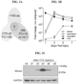
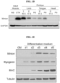
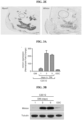
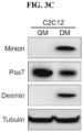
- mouse MINION is a skeletal muscle-specific microprotein highly expressed during muscle development and regeneration (see Example 2), and is required for fusion of muscle progenitors and skeletal muscle development (see Examples 3 and 4).
- forced heterologous expression of MINION, along with another protein Myomaker is found to be sufficient to induce cell fusion even in non-muscle cells (e.g., fibroblasts) (see Example 5). This is surprising since it was reported that Myomaker alone cannot promote fibroblast-fibroblast fusion although it was capable of promoting fusion of fibroblasts to myoblasts ( Millay et al., Nature. 2013;499(7458):301-5 ).
- the human ortholog of mouse MINION has not been reported before. As described herein, the present inventors found human ortholog of MINION has an amino acid sequence of SEQ ID NO: 1 or SEQ ID NO: 3, and a cDNA sequence of SEQ ID NO: 2 or SEQ ID NO: 4. The present inventors found that human MINION protein also has fusogenic activity, suggesting an evolutionarily conserved role for the MINION protein in cell fusion.
- MINION amino acid and nucleotide sequences of other species e.g., rat (R. norvegicus), monkey (M. mulatta), cat (F. catus), and pig (S. scrofa) are also provided in Table 1.
- a MINION polypeptide can be a variant of a naturally occurring MINION protein as defined in the claims (e.g., any MINION protein listed in Table 1).
- a human MINION polypeptide can be a variant of a naturally occurring human MINION protein as defined in the claims (e.g., the protein of SEQ ID NO: 1 or SEQ ID NO: 3, or another naturally occurring isoform thereof).
- a mouse MINION polypeptide can be a variant of a naturally occurring mouse MINION protein as defined in the claims (e.g., a protein of SEQ ID NO: 5 or SEQ ID NO: 7, or another naturally occurring isoform thereof).
- a MINION variant as defined in the claims can differ from a naturally occurring MINION protein by, for example, a modification (e.g., substitution, deletion, or insertion) of one or more amino acid residues in the naturally occurring MINION protein, but retains the biological activities of MINION, e.g., fusogenic activity.
- the MINION variant as defined in the claims can have one or more conservative or nonconservative amino acid substitution.
- a "conservative amino acid substitution” is one in which the amino acid residue is replaced with an amino acid residue having a similar side chain. Families of amino acid residues having similar side chains have been defined in the art.
- amino acids with basic side chains e.g., lysine, arginine, histidine
- acidic side chains e.g., aspartic acid, glutamic acid
- uncharged polar side chains e.g., glycine, asparagine, glutamine, serine, threonine, tyrosine, cysteine
- nonpolar side chains e.g., alanine, valine, leucine, isoleucine, proline, phenylalanine, methionine, tryptophan
- beta-branched side chains e.g., threonine, valine, isoleucine
- aromatic side chains e.g., tyrosine, phenylalanine, tryptophan, histidine
- the MINION variant as defined in the claims includes one or more mutations (e.g., substitutions (e.g., conservative substitutions or substitutions), insertions, or deletions) of non-essential amino acids relative to a naturally occurring MINION protein.
- a "non-essential" amino acid residue is a residue that can be altered from the wild-type sequence of MINION protein without abolishing or more preferably, without substantially altering a biological activity, such as the fusogenic activity, whereas changing an "essential" amino acid residue results in a substantial loss of biological activity.
- a MINION variant as defined in the claims may have at least one, two, three, or four, and no more than 10, 9, 8, 7, 6, or 5 mutations (e.g., substitutions (e.g., conservative substitutions or substitutions of non-essential amino acids), insertions, or deletions) relative to a naturally occurring MINION protein. Whether or not a particular substitution will be tolerated, i.e., will not adversely affect biological properties, such as fusogenic activity, can be predicted, e.g., by evaluating whether the mutation is conservative or by an activity assay.
- substitutions e.g., conservative substitutions or substitutions of non-essential amino acids
- a MINION variant can have about 85%, 90%, 91%, 92%, 93%, 94%, 95%, 96%, 97%, 98%, 99% or higher sequence identity to a naturally occurring MINION protein as defined in the claims.
- a human MINION variant can have about 85%, 90%, 91%, 92%, 93%, 94%, 95%, 96%, 97%, 98%, 99% or higher sequence identity to SEQ ID NO: 1 or SEQ ID NO: 3.
- a mouse MINION variant can have about 85%, 90%, 91%, 92%, 93%, 94%, 95%, 96%, 97%, 98%, 99% or higher sequence identity to SEQ ID NO: 5 or SEQ ID NO: 7.
- amino acid or nucleic acid “identity” is equivalent to amino acid or nucleic acid “homology”).
- the percent identity between the two sequences is a function of the number of identical positions shared by the sequences.
- nucleic acid segments hybridize under selective hybridization conditions (e.g., highly stringent hybridization conditions), to the complement of the strand.
- the nucleic acids may be present in whole cells, in a cell lysate, or in a partially purified or substantially pure form.
- the MINION variant as defined in the claims can comprise at least one non-naturally encoded amino acid.
- a “non-naturally encoded amino acid” refers to an amino acid that is not one of the common amino acids or pyrolysine or selenocysteine.
- Other terms that may be used synonymously with the term “non-naturally encoded amino acid” are “non-natural amino acid,” “unnatural amino acid,” “non-naturally-occurring amino acid,” and variously hyphenated and non-hyphenated versions thereof.
- the term “non-naturally encoded amino acid” also includes, but is not limited to, amino acids that occur by modification (e.g.
- a naturally encoded amino acid including but not limited to, the 20 common amino acids or pyrrolysine and selenocysteine
- non-naturally-occurring amino acids include, but are not limited to, N-acetylglucosaminyl-L-serine, N-acetylglucosaminyl-L-threonine, and O-phosphotyrosine.
- a non-naturally encoded amino acid is typically any structure having any substituent side chain other than one used in the twenty natural amino acids. Because the non-naturally encoded amino acids typically differ from the natural amino acids only in the structure of the side chain, the non-naturally encoded amino acids form amide bonds with other amino acids, including but not limited to, natural or non-naturally encoded, in the same manner in which they are formed in naturally occurring polypeptides. However, the non-naturally encoded amino acids have side chain groups that distinguish them from the natural amino acids.
- R optionally comprises an alkyl-, aryl-, acyl-, keto-, azido-, hydroxyl-, hydrazine, cyano-, halo-, hydrazide, alkenyl, alkynl, ether, thiol, seleno-, sulfonyl-, borate, boronate, phospho, phosphono, phosphine, heterocyclic, enone, imine, aldehyde, ester, thioacid, hydroxylamine, amino group, or the like or any combination thereof.
- Non-naturally occurring amino acids of interest include, but are not limited to, amino acids comprising a photoactivatable cross-linker, spin-labeled amino acids, fluorescent amino acids, metal binding amino acids, metal-containing amino acids, radioactive amino acids, amino acids with novel functional groups, amino acids that covalently or noncovalently interact with other molecules, photocaged and/or photoisomerizable amino acids, amino acids comprising biotin or a biotin analogue, glycosylated amino acids such as a sugar substituted serine, other carbohydrate modified amino acids, keto-containing amino acids, amino acids comprising polyethylene glycol or polyether, heavy atom substituted amino acids, chemically cleavable and/or photocleavable amino acids, amino acids with an elongated side chains as compared to natural amino acids, including but not limited to, polyethers or long chain hydrocarbons, including but not limited to, greater than about 5 or greater than about 10 carbons, carbon-linked sugar-containing amino acids,
- non-naturally encoded amino acids that may be suitable for use in the present invention and that are useful for reactions with water soluble polymers include, but are not limited to, those with carbonyl, aminooxy, hydrazine, hydrazide, semicarbazide, azide and alkyne reactive groups.
- non-naturally encoded amino acids comprise a saccharide moiety.
- amino acids examples include N-acetyl-L-glucosaminyl-L-serine, N-acetyl-L-galactosaminyl-L-serine, N-acetyl-L-glucosaminyl-L-threonine, N-acetyl-L-glucosaminyl-L-asparagine and O-mannosaminyl-L-serine.
- amino acids also include examples where the naturally-occurring N- or O-linkage between the amino acid and the saccharide is replaced by a covalent linkage not commonly found in nature--including but not limited to, an alkene, an oxime, a thioether, an amide and the like.
- amino acids also include saccharides that are not commonly found in naturally-occurring proteins such as 2-deoxy-glucose, 2-deoxygalactose and the like.
- a Lysine residue in the MINION sequence is conjugated to PEG directly or through a linker.
- linker can be, for example, a Glu residue or an acyl residue containing a thiol functional group for linkage to the appropriately modified PEG chain.
- An alternative method for introducing a PEG chain is to first introduce a Cys residue at the C-terminus or at solvent exposed residues such as replacements for Arg or Lys residues. This Cys residue is then site-specifically attached to a PEG chain containing, for example, a maleimide function.
- Methods for incorporating PEG or long chain polymers of PEG are well known in the art (described, for example, in Veronese, F. M., et al., Drug Disc. Today 10: 1451-8 (2005 ); Greenwald, R. B., et al., Adv. Drug Deliv. Rev. 55: 217-50 (2003 ); Roberts, M. J., et al., Adv. Drug Deliv.
- p-azidophenylalanine can be incorporated into the MINION polypeptides and then reacted with a PEG polymer having an acetylene moiety in the presence of a reducing agent and copper ions to facilitate an organic reaction known as "Huisgen [3+2]cycloaddition.”
- a PEG polymer having an acetylene moiety in the presence of a reducing agent and copper ions to facilitate an organic reaction known as "Huisgen [3+2]cycloaddition.”
- a variant of the MINION polypeptide as defined in the claims can comprise MINION, fused to a heterologous peptide such as a tag or a fusion domain. Fusions may be constructed such that the heterologous peptide (e.g., a tag or a fusion domain) is fused at the amino terminus or at the carboxy terminus of MINION polypeptide.
- a heterologous peptide e.g., a tag or a fusion domain
- tags or fusion domains include, but are not limited to, a HA-tag (YPYDVPDYA, SEQ ID NO: 32), a Myc-tag (EQKLISEEDL, SEQ ID NO: 33), a FLAG-tag (DYKDDDDK, SEQ ID NO: 34), a His-tag (HHHHHH, SEQ ID NO: 35), an E-tag (GAPVPYPDPLEPR, SEQ ID NO: 36), a V5-tag (GKPIPNPLLGLDST, SEQ ID NO: 37), a VSV tag (YTDIEMNRI,GK, SEQ ID NO: 38), a polyglutamate tag (EEEEEE, SEQ ID NO: 39), an AviTag (GLNDIFEAQKIEWHE, SEQ ID NO: 40), a SBP tag (MDEKTTGWRGGHVVEGLAGELEQLRARLEHHPQGQREP, SEQ ID NO: 41), a Strep-tag (WSHPQFEK, SEQ ID NO:
- a fusion domain or a tag may be selected so as to confer a desired property.
- some fusion domains are particularly useful for isolation of the fusion proteins by affinity chromatography.
- relevant matrices for affinity chromatography such as glutathione-, amylase-, and nickel- or cobalt-conjugated resins are used. Many of such matrices are available in "kit” form, such as the Pharmacia GST purification system and the QLAexpress TM system (Qiagen) useful with a His6 tag.
- a fusion domain may be selected so as to facilitate detection of the MINION proteins.
- detection domains include the various fluorescent proteins (e.g., GFP), as well as "epitope tags," which are usually short peptide sequences for which a specific antibody is available.
- epitope tags for which specific monoclonal antibodies are readily available include a HA-tag, a Myc-tag, a FLAG-tag.
- tags include an E-tag, a V5-tag, a VSV tag, a polyglutamate tag, an AviTag, a SBP tag, a Strep-tag, an Xpress tag, an S-tag, a Softag 1, a Softag 3, a Calmodulin-tag, a TC tag, an Isopeptag, a SpyTag, a SnoopTag, as described above.
- the tags or fusion domains can have a protease cleavage site, which allows the relevant protease to partially digest the fusion proteins and thereby liberate the MINION polypeptide from the fusion protein.
- an MINION polypeptide as defined in the claims can be fused with a domain that stabilizes the protein in vivo (e.g., a "stabilizer” domain).
- stabilizing is meant anything that increases serum half life, regardless of whether this is because of decreased destruction, decreased clearance by the kidney, or other pharmacokinetic effect. Fusions with the Fc domain of an immunoglobulin are known to confer desirable pharmacokinetic properties on a wide range of proteins.
- fusions to human serum albumin can confer desirable properties.
- Other types of fusion domains that may be selected include multimerizing (e.g., dimerizing, tetramerizing) domains and functional domains (that confer an additional biological function, as desired).
- fragments of a MINION polypeptide which retain the biological function of a native MINION polypeptide, such as fusogenic activity, interaction with Myomaker, etc.
- some instances described herein provide fragments of a MINION polypeptide that contain an "AxLyCxL" domain, which has been shown to be required for the fusogenic activity of MINION ( Shi et al., Requirement of the fusogenic micropeptide myomixer for muscle formation in zebrafish. PNAS 114, 11950 (2017 )).
- the fragments of a MINION polypeptide comprise amino acids 48-54 of SEQ ID NO: 1.
- the fragments of a MINION polypeptide comprise amino acids 48-54 of SEQ ID NO: 3. In some instances described herein, the fragments of a MINION polypeptide comprise amino acids 48-54 of SEQ ID NO: 5. In some instances described herein, the fragments of a MINION polypeptide comprise amino acids 72-78 of SEQ ID NO: 7.
- Some instances described herein provide fragments of a MINION polypeptide that contain the N-terminal hydrophobic region (amino acids 5-25 of human MINION), and positively charged Arg residues within the first ⁇ -helix following the N-terminal hydrophobic region the disruption of which resulted in a loss of fusogenic activity and perturbed interaction with Myomaker ( Bi P, Ramirez-Martinez A, Li H, Cannavino J, McAnally JR, Shelton JM, Sanchez-Ortiz E, Bassel-Duby R, Olson EN: Control of muscle formation by the fusogenic micropeptide myomixer. Science 356, 323 (2017 )).
- the fragments of a MINION polypeptide comprise amino acids 5-46 of SEQ ID NO: 1. In some instances described herein, the fragments of a MINION polypeptide comprise amino acids 5-46 of SEQ ID NO: 3. In some instances described herein, the fragments of a MINION polypeptide comprise amino acids 5-46 of SEQ ID NO: 5. In some instances described herein, the fragments of a MINION polypeptide comprise amino acids 29-70 of SEQ ID NO: 7. The fragment of a MINION polypeptide can have a variety of lengths.
- the fragment of a MINION polypeptide can have a length that is, is about, is at least, is more than, 7 amino acids, 8 amino acids, 9 amino acids, 10 amino acids, 11 amino acids, 12 amino acids, 13 amino acids, 14 amino acids, 15 amino acids, 16 amino acids, 17 amino acids, 18 amino acids, 19 amino acids, 20 amino acids, 25 amino acids, 30 amino acids, 35 amino acids, 40 amino acids, 45 amino acids, 50 amino acids, or a range that is between any two of the above values.
- a MINION peptide as defined in the claims may comprise one or more post-translational modifications, e.g., lipidation, glycosylation, phosphorylation, etc. Examples are as follows.
- Phosphorylation sites in Mus musculus MINION as predicted using GPS include: serine 12, serine 15, serine 41, serine 42, serine 51, serine 58, serine 65, serine 69, and serine 74. Palmitoylation is predicted in Mus musculus MINION using GPS-Lipid (http://gps.biocuckoo.org/) on cysteine 52.
- O-glycosylation of Mus musculus MINION is predicted using NetOGlyc 4.0 (http://www.cbs.dtu.dk/services/NetOGlyc/) on serine 69 and serine 74.
- Myomaker (also known as Tmem8c or Tmem226) is a membrane protein expressed specifically in skeletal muscle during embryogenesis and adult muscle regeneration ( Millay et al., Nature. 2013 Jul 18;499(7458):301-5 ). Myomaker has been reported to be both necessary and sufficient to promote myoblast fusion in vivo and in vitro (Millay et al., 2013). Millay et al. also reported that Myomaker can promote fusion of fibroblasts to myoblasts but cannot promote fibroblast-fibroblast fusion. Methods of fusing a non-muslce cell to a muscle cell by expressing exogenous Myomaker protein in the non-muscle cell have been described in WO2014/210448 .
- a Myomaker polypeptide can be a variant of a naturally occurring Myomaker protein as defined in the claims (e.g., any Myomaker protein listed in Table 2).
- a human Myomaker polypeptide can be a variant of a naturally occurring human Myomaker protein as defined in the claims (e.g., a protein of SEQ ID NO: 16, or a naturally occurring isoform thereof).
- a mouse Myomaker polypeptide can be a variant of a naturally occurring mouse Myomaker protein as defined in the claims (e.g., a protein of SEQ ID NO: 18 or SEQ ID NO: 20, or another naturally occurring isoform thereof).
- Table 2 provides Myomaker amino acid and nucleotide sequences of human, mouse, rat, monkey, cat, and pig.
- a Myomaker variant as defined in the claims can differ from a naturally occurring Myomaker protein by, for example, a modification (e.g., substitution, deletion, or insertion) of one or more amino acid residues in the naturally occurring Myomaker protein, but retains the biological activities of Myomaker, e.g., fusogenic activity.
- the Myomaker variant as defined in the claims can have one or more conservative or nonconservative amino acid substitution.
- a "conservative amino acid substitution” is one in which the amino acid residue is replaced with an amino acid residue having a similar side chain. Families of amino acid residues having similar side chains have been defined in the art.
- amino acids with basic side chains e.g., lysine, arginine, histidine
- acidic side chains e.g., aspartic acid, glutamic acid
- uncharged polar side chains e.g., glycine, asparagine, glutamine, serine, threonine, tyrosine, cysteine
- nonpolar side chains e.g., alanine, valine, leucine, isoleucine, proline, phenylalanine, methionine, tryptophan
- beta-branched side chains e.g., threonine, valine, isoleucine
- aromatic side chains e.g., tyrosine, phenylalanine, tryptophan, histidine
- the Myomaker variant as defined in the claims includes one or more mutations (e.g., substitutions (e.g., conservative substitutions or substitutions), insertions, or deletions) of non-essential amino acids relative to a naturally occurring Myomaker protein.
- a "non-essential" amino acid residue is a residue that can be altered from the wild-type sequence of Myomaker protein without abolishing or more preferably, without substantially altering a biological activity, such as fusogenic activity, whereas changing an "essential" amino acid residue results in a substantial loss of biological activity.
- a Myomaker variant as defined in the claims may have at least one, two, three, or four, and no more than 10, 9, 8, 7, 6, or 5 mutations (e.g., substitutions (e.g., conservative substitutions or substitutions of non-essential amino acids), insertions, or deletions) relative to a naturally occurring Myomaker protein. Whether or not a particular substitution will be tolerated, i.e., will not adversely affect biological properties, such as fusogenic activity, can be predicted, e.g., by evaluating whether the mutation is conservative or by an activity assay.
- substitutions e.g., conservative substitutions or substitutions of non-essential amino acids
- a Myomaker variant can have about 85%, 90%, 91%, 92%, 93%, 94%, 95%, 96%, 97%, 98%, 99% or higher sequence identity to a naturally occurring Myomaker protein as defined in the claims.
- a human Myomaker variant can have about 85%, 90%, 91%, 92%, 93%, 94%, 95%, 96%, 97%, 98%, 99% or higher sequence identity to SEQ ID NO: 16.
- a mouse Myomaker variant can have about 85%, 90%, 91%, 92%, 93%, 94%, 95%, 96%, 97%, 98%, 99% or higher sequence identity to SEQ ID NO: 18 or SEQ ID NO: 20.
- amino acid or nucleic acid “identity” is equivalent to amino acid or nucleic acid “homology”).
- the percent identity between the two sequences is a function of the number of identical positions shared by the sequences.
- nucleic acid segments hybridize under selective hybridization conditions (e.g., highly stringent hybridization conditions), to the complement of the strand.
- the nucleic acids may be present in whole cells, in a cell lysate, or in a partially purified or substantially pure form.
- the Myomaker variant as defined in the claims can comprise at least one non-naturally encoded amino acid.
- the Myomaker variant as defined in the claims can be a PEGylated Myomaker with extended in vivo half-life.
- a variant of the Myomaker polypeptide as defined in the claims can comprise Myomaker, fused to a tag or a fusion domain. Exemplary non-naturally encoded amino acids, PEGylation methods, and fusion partners described above for MINION variants are also applicable to Myomaker variants.
- the invention relates to nucleic acids encoding a MINION polypeptide (e.g., a human MINION polypeptide), expression vectors for expression of a MINION polypeptide (e.g., a human MINION polypeptide), and cells containing such expression vectors.
- a polynucleotide encoding a MINION polypeptide (e.g., a human MINION polypeptide), and expression vectors and host cells comprising such a polynucleotide.
- expression vectors as defined by the claims that can be employed to express a MINION polypeptide (e.g., a human MINION polypeptide).
- expression vector refers to a carrier nucleic acid molecule into which a desired coding sequence can be inserted for introduction into a cell where it can be expressed.
- Expression vector can be a plasmid, a cosmid, an RNA, a viral vector, or artificial chromosomes (see, e.g., Harrington et al., Nat Genet 15:345, 1997 ).
- nonviral vectors useful for expression of a polypeptide in mammalian (e.g., human) cells include pThioHis A, B & C, pcDNA3. 1/His, pEBVHis A, B & C (Invitrogen, San Diego, CA), MPSV vectors, and numerous other vectors known in the art for expressing other proteins.
- Useful viral vectors include, but are not limited to, vectors based on any of the following viruses: adenovirus, adeno-associated virus, Herpes Simplex Virus (HSV), parvovirus, retrovirus, lentivirus, vaccinia virus, Sinbis virus, influenza virus, reovirus, Newcastle disease virus (NDV), measles virus, vesicular stomatitis virus (VSV), poliovirus, poxvirus, Seneca Valley virus, coxsackievirus, enterovirus, myxoma virus, or maraba virus.
- the expression vector can be capable of autonomous replication or it can integrate into a host DNA.
- the expression vector as defined in the claims can be a recombinant DNA molecule containing a MINION polypeptide-coding sequence (e.g., a human MINION polypeptide-coding sequence).
- the recombinant expression vector typically includes one or more regulatory sequences operatively linked to the nucleic acid sequence to be expressed.
- the term "regulatory sequence” includes promoters, enhancers and other expression control elements (e.g., polyadenylation signals). Regulatory sequences include those which direct constitutive expression of a nucleotide sequence, as well as tissue-specific regulatory and/or inducible sequences.
- Expression vectors as defined in the claims can also include elements designed to optimize messenger RNA stability and translatability in host cells, and/or drug selection markers for establishing permanent, stable cell clones expressing a human MINION polypeptide.
- the design of the expression vector can depend on such factors as the choice of the host cell to be transformed, the level of expression of protein desired, and the like. General methods for generating such recombinant expression vectors can be found in Sambrook and Russell eds. (2001) Molecular Cloning: A Laboratory Manual, 3rd editi on; the series Ausubel et al. eds. (2007 with updated through 2010) Current Protocols in Molecular Biology , among others known in the art.
- a “promoter” is a control sequence that is a region of a nucleic acid sequence at which initiation and rate of transcription are controlled. It may contain genetic elements at which regulatory proteins and molecules may bind such as RNA polymerase and other transcription factors.
- the phrases "operatively positioned,” “operatively linked,” “under control,” and “under transcriptional control” mean that a promoter is in a correct functional location and/or orientation in relation to a nucleic acid sequence to control transcriptional initiation and/or expression of that sequence.
- a promoter may or may not be used in conjunction with an “enhancer,” which refers to a cis-acting regulatory sequence involved in the transcriptional activation of a nucleic acid sequence.
- a promoter may be one naturally-associated with a gene or sequence, as may be obtained by isolating the 5' non-coding sequences located upstream of the coding segment and/or exon. Such a promoter can be referred to as "endogenous.”
- an enhancer may be one naturally associated with a nucleic acid sequence, located either downstream or upstream of that sequence.
- certain advantages will be gained by positioning the coding nucleic acid segment under the control of a recombinant or heterologous promoter, which refers to a promoter that is not normally associated with a nucleic acid sequence in its natural environment.
- a recombinant or heterologous enhancer refers also to an enhancer not normally associated with a nucleic acid sequence in its natural environment.
- Such promoters or enhancers may include promoters or enhancers of other genes, and promoters or enhancers isolated from any other prokaryotic, viral, or eukaryotic cell, and promoters or enhancers not "naturally-occurring," i.e., containing different elements of different transcriptional regulatory regions, and/or mutations that alter expression.
- sequences may be produced using recombinant cloning and/or nucleic acid amplification technology, including PCR TM , in connection with the compositions disclosed herein (see US 4683202 , US 5928906 ).
- the control sequences that direct transcription and/or expression of sequences within non-nuclear organelles such as mitochondria, chloroplasts, and the like, can be employed as well.
- the promoters employed can be constitutive, inducible, tissue-specific, and/or useful under the appropriate conditions to direct high level expression of the introduced DNA segment, such as is advantageous in the large-scale production of recombinant proteins and/or peptides.
- a constitutive promoter is employed to provide constant expression of a MINION polypeptide as defined by the claims (e.g., a human MINION polypeptide).
- an inducible promoter is employed to prevent expression of a MINION polypeptide as defined in the claims (e.g., a human MINION polypeptide) except under inducing conditions.
- Inducible promoters include, e.g., arabinose, lacZ, a metallothionein promoter, a glucocorticoid promoters or a heat shock promoter.
- other regulatory elements may also be incorporated to improve expression of a nucleic acid encoding a MINION polypeptide as defined in the claims (e.g., a human MINION polypeptide), e.g., enhancers, ribosomal binding site, transcription termination sequences, and the like.
- a tissue-specific promoter is employed to provide expression of a MINION polypeptide as defined in the claims (e.g., a human MINION polypeptide) only in specific tissues.
- tissue-specific promoters or elements as well as assays to characterize their activities, is well known to those of skill in the art.
- regions include the human LINM2 gene ( Nomoto et al. 1999, Gene, 236(2):259-271 ), the somatostatin receptor 2 gene ( Kraus et al., 1998, FEES Lett., 428(3): 165-170 ), murine epididymal retinoic acid-binding gene ( Lareyre et al., 1999, J. Biol. Chem., 274(12):8282-8290 ), human CD4 ( Zhao-Emonet et al., 1998, Biochirn. Biophys.
- a specific initiation signal also may be required for efficient translation of coding sequences. These signals include the ATG initiation codon or adjacent sequences. Exogenous translational control signals, including the ATG initiation codon, may need to be provided. One of ordinary skill in the art would readily be capable of determining this and providing the necessary signals. It is well known that the initiation codon must be "in-frame" with the reading frame of the desired coding sequence to ensure translation of the entire insert. The exogenous translational control signals and initiation codons can be either natural or synthetic. The efficiency of expression may be enhanced by the inclusion of appropriate transcription enhancer elements.
- Expression can employ any appropriate host cells known in the art, for example, mammalian host cells, bacterial host cells, yeast host cells, insect host cells, etc. Both prokaryotic and eukaryotic expression systems are widely available.
- the expression system is a mammalian cell expression, such as a CHO cell expression system.
- a nucleic acid may be codon-optimized to facilitate expression in a desired host cell. It will be important to employ a promoter and/or enhancer that effectively directs the expression of the DNA segment in the cell type, organelle, and organism chosen for expression. Those of skill in the art of molecular biology generally know the use of promoters, enhancers, and cell type combinations for protein expression, for example, see Sambrook et al. (2001).
- RNA molecules will undergo RNA splicing to remove introns from the primary transcripts.
- Vectors containing genomic eukaryotic sequences may require donor and/or acceptor splicing sites to ensure proper processing of the transcript for protein expression (see Chandler et al., 1997, Proc. Natl. Acad. Sci. USA, 94(8):3 596-601 ).
- the vectors or constructs of the present disclosure will generally comprise at least one termination signal.
- a “termination signal” or “terminator” is comprised of the DNA sequences involved in specific termination of an RNA transcript by an RNA polymerase. Thus, in certain embodiments a termination signal that ends the production of an RNA transcript is contemplated.
- a terminator may be necessary in vivo to achieve desirable message levels. In eukaryotic systems, the terminator region may also comprise specific DNA sequences that permit site-specific cleavage of the new transcript so as to expose a polyadenylation site. This signals a specialized endogenous polymerase to add a stretch of about 200 A residues (poly A) to the 3' end of the transcript.
- RNA molecules modified with this polyA tail appear to more stable and are translated more efficiently.
- that terminator comprises a signal for the cleavage of the RNA, and it is more preferred that the terminator signal promotes polyadenylation of the message.
- the terminator and/or polyadenylation site elements can serve to enhance message levels and/or to minimize read through from the cassette into other sequences.
- Terminators contemplated for use in the disclosure include any known terminator of transcription described herein or known to one of ordinary skill in the art, including but not limited to, for example, the termination sequences of genes, such as for example the bovine growth hormone terminator or viral termination sequences, such as for example the SV40 terminator.
- the termination signal may be a lack of transcribable or translatable sequence, such as due to a sequence truncation.
- polyadenylation signal In expression, particularly eukaryotic expression, one will typically include a polyadenylation signal to effect proper polyadenylation of the transcript.
- the nature of the polyadenylation signal is not believed to be crucial to the successful practice of the disclosure, and/or any such sequence may be employed.
- Preferred embodiments include the SV40 polyadenylation signal and/or the bovine growth hormone polyadenylation signal, convenient and/or known to function well in various target cells. Polyadenylation may increase the stability of the transcript or may facilitate cytoplasmic transport.
- a vector in a host cell may contain one or more origins of replication sites (often termed "ori"), which is a specific nucleic acid sequence at which replication is initiated.
- ori origins of replication sites
- ARS autonomously replicating sequence
- cells containing a nucleic acid construct of the present disclosure may be identified in vitro or in vivo by including a marker in the expression vector.
- markers would confer an identifiable change to the cell permitting easy identification of cells containing the expression vector.
- a selectable marker is one that confers a property that allows for selection.
- a positive selectable marker is one in which the presence of the marker allows for its selection, while a negative selectable marker is one in which its presence prevents its selection.
- An example of a positive selectable marker is a drug resistance marker.
- a drug selection marker aids in the cloning and identification of transformants
- genes that confer resistance to neomycin, puromycin, hygromycin, DHFR, GPT, zeocin and histidinol are useful selectable markers.
- markers conferring a phenotype that allows for the discrimination of transformants based on the implementation of conditions other types of markers including screenable markers such as GFP, whose basis is colorimetric analysis, are also contemplated.
- screenable enzymes such as herpes simplex virus thymidine kinase (tk) or chloramphenicol acetyltransferase (CAT) may be utilized.
- provided herein are expression vectors as defined in the claims comprising a nucleic acid i) encoding a MINION polypeptide or a variant thereof as defined in the claims. In some embodiments, provided herein are expression vectors comprising a nucleic acid i) encoding a MINION polypeptide of Table 1 or a variant thereof as defined in the claims. In some embodiments, provided herein are expression vectors comprising a nucleic acid i) encoding a MINION polypeptide comprising an amino acid sequence selected from any of SEQ ID NOs: 1, 3, 5, 7, 9, 11, 13, or 15, or a variant thereof as defined in the claims.
- expression vectors comprising a nucleic acid i) encoding a MINION polypeptide consisting of an amino acid sequence selected from any of SEQ ID Nos: 1, 3, 5, 7, 9, 11, 13, or 15, or a variant thereof as defined in the claims.
- expression vectors comprising a nucleic acid i) selected from any of SEQ ID NOs: 2, 4, 6, 8, 10, 12, or 14.
- expression vectors comprising a nucleic acid i) encoding a polypeptide comprising SEQ ID NO: 1 or SEQ ID NO: 3 or a variant thereof as defined in the claims.
- the expression vector comprises a nucleic acid i) encoding a polypeptide comprising SEQ ID NO: 1 with 1-4 mutations.
- the expression vector comprises a nucleic acid i) encoding a polypeptide consisting of SEQ ID NO: 1.
- the expression vector comprises a nucleic acid i) encoding a polypeptide comprising SEQ ID NO: 3 with 1-4 mutations.
- the expression vector comprises a nucleic acid i) encoding a polypeptide consisting of SEQ ID NO: 1. In some embodiments, the expression vector comprises a nucleic acid i) encoding a polypeptide consisting of SEQ ID NO: 3. In some embodiments, the expression vector comprises a nucleic acid i) comprising SEQ ID NO: 2. In some embodiments, the expression vector comprises a nucleic acid i) comprising SEQ ID NO: 4. In some embodiments, the expression vector comprises a nucleic acid i) consisting of SEQ ID NO: 2. In some embodiments, the expression vector comprises a nucleic acid i) consisting of SEQ ID NO: 4.
- the expression vector comprises a promoter, e.g., a constitutive promoter, an inducible promoter, or a tissue-specific promoter. In some embodiments, the expression vector comprises a polyadenylation signal. In some embodiments, the expression vector comprises a selectable marker.
- the expression vector comprises a nucleic acid i) encoding a polypeptide comprising SEQ ID NO: 1 or SEQ ID NO: 3, fused to a tag.
- the tag can be selected from a HA-tag (YPYDVPDYA, SEQ ID NO: 32), a Myc-tag (EQKLISEEDL, SEQ ID NO: 33), a FLAG-tag (DYKDDDDK, SEQ ID NO: 34), a His-tag (HHHHHH, SEQ ID NO: 35), an E-tag (GAPVPYPDPLEPR, SEQ ID NO: 36), a V5-tag (GKPIPNPLLGLDST, SEQ ID NO: 37), a VSV tag (YTDIEMNRI,GK, SEQ ID NO: 38), a polyglutamate tag (EEEEEE, SEQ ID NO: 39), an AviTag (GLNDIFEAQKIEWHE, SEQ ID NO: 40), a SBP tag (MDEKTTGWRGGHVVEGLAGE
- the expression vectors further comprise a second nucleic acid ii) encoding a Myomaker polypeptide or a variant thereof as defined in the claims.
- the expression vector comprises a second nucleic acid ii) encoding a Myomaker polypeptide of Table 2 or a variant thereof as defined in the claims.
- the expression vector comprises a second nucleic acid ii) encoding a polypeptide comprising an amino acid sequence selected from any of SEQ ID NOs: 16, 18, 20, 22, 24, 26, 28, or 30, or a variant thereof as defined in the claims.
- the expression vector comprises a second nucleic acid ii) encoding a polypeptide consisting of an amino acid sequence selected from any of SEQ ID NOs: 16, 18, 20, 22, 24, 26, 28, or 30, or a variant thereof as defined in the claims.
- the expression vector comprises a second nucleic acid ii) selected from any of SEQ ID NOs: 17, 19, 21, 23, 25, 27, 29, or 31, or a variant thereof as defined in the claims.
- the expression vector comprises a second nucleic acid ii) encoding a polypeptide comprising SEQ ID NO: 16. In some embodiments, the expression vector comprises a second nucleic acid ii) encoding a polypeptide consisting of SEQ ID NO: 16. In some embodiments, the expression vector comprises a second nucleic acid ii) comprising SEQ ID NO: 17. In some embodiments, the expression vector comprises a second nucleic acid ii) consisting of SEQ ID NO: 17. In some embodiments, the expression vectors further comprise a second promoter upstream of the second nucleic acid. The second promoter can be a constitutive promoter, an inducible promoter, or a tissue-specific promoter.
- the expression vectors for expressing both a MINION polypeptide and a Myomaker polypeptide as defined in the claims comprise an internal ribosome entry site (IRES) upstream of the Myomaker-coding sequence.
- IRES elements are able to bypass the ribosome scanning model of 5'-methylated Cap dependent translation and begin translation at internal sites ( Pelletier and Sonenberg, 1988, Nature, 334:320-325 ).
- IRES elements from two members of the picornavirus family polio and encephalomyocarditis
- IRES elements from two members of the picornavirus family have been described (Pelletier and Sonenberg, 1988), as well an IRES from a mammalian message ( Macejak and Sarnow, 1991, Nature, 353:90-94, 1991 ).
- IRES elements can be linked to heterologous open reading frames. Multiple open reading frames can be transcribed together, each separated by an IRES, creating polycistronic messages. By virtue of the IRES element, each open reading frame is accessible to ribosomes for efficient translation. Multiple genes can be efficiently expressed using a single promoter/enhancer to transcribe a single message (see U.S. Patents 5925565 and 5935819 ).
- the expression vectors for expressing both a MINION polypeptide and a Myomaker polypeptide as defined in the claims comprise a 2A sequence upstream of the Myomaker-coding sequence.
- the 2A oligopeptide sequence was first characterized from the positive-stranded RNA picornavirus Foot-and-Mouth Disease Virus (FMDV); and FMDV 2A or F2A was shown to mediate a co-translational 'cleavage' between the upstream (capsid proteins) and downstream (RNA replication proteins) domains of the FMDV polyprotein ( Ryan MD, EMBO J 1994;134: 928-933 ; Ryan MD, J Gen Virol 1991; 72: 2727-2732 ; Donnelly MLL, J Gen Virol 1997; 78:13-21 ; Donnelly MLL, J Gen Virol 2001; 82:1013-1025 ).
- FMDV positive-stranded RNA picornavirus Foot-and-Mouth Disease Virus
- Active 2A sequences were characterized in the genomes of viruses from other genera of the picornaviruses, plus ⁇ 2A-like' sequences found within the genomes of a range of different RNA viruses and non-LTR retrotransposons ( Donnelly MLL, J Gen Virol 2001; 82:1027-1041 ; Heras SR, Cell Mol Life Sci 2006; 63:1449-1460 ; Luke GA, J Gen Virol 2008; 89:1036-1042 ; Odon V, Mol Biol Evol 2013; 30:1955-1965 ; Luke GA, Mob Gen Elements 2014; 3:e27525 ).
- a "2A sequence” refers to any nucleic acid sequence encoding a 2A or "2A-like" oligopeptide serving as a linker between two proteins, allowing autonomous intraribosomal self-processing of polyproteins (See e.g., de Felipe. Genetic Vaccines and Ther. 2:13 (2004 ); deFelipe et al. Traffic 5:616-626 (2004 )). These oligopeptides allow co-expression of multiple proteins from a single vector. Many 2A elements are known in the art. For example, viral 2A sequences have been described in US Patent Nos. 9175311 , 8865881 , 7939059 , 7947493 ,.
- a viral 2A sequence can be a picornaviral, a tetraviral 2A sequence, or a combination thereof.
- a picornaviral 2A sequence can be selected from any one of the Enteroviral 2A sequences, Rhinoviral 2A sequences, Cardioviral 2A sequences, Aphthoviral 2A sequences, Hepatoviral 2A sequences, Erboviral 2A sequences, Kobuviral 2A sequences, Teschoviral 2A sequences, and the Parechoviral 2A sequences.
- a tetraviral 2A sequences can be selected from any of the Betatetraviral 2A sequences or Omegatetraviral 2A sequences.
- Examples of 2A sequences that can be used in the methods and system disclosed herein, without limitation, include 2A sequences from the foot-and-mouth disease virus (F2A), equine rhinitis A virus (E2A), Thosea asigna virus (T2A), and porcine teschovirus-1 (P2A).
- F2A foot-and-mouth disease virus
- E2A equine rhinitis A virus
- T2A Thosea asigna virus
- P2A porcine teschovirus-1
- a 2A sequence encodes a viral 2A oligopeptide selected from T2A (EGRGSLLTCGDVEENPGP (SEQ ID NO: 52) or GSGEGRGSLLTCGDVEENPGP (SEQ ID NO: 53)), P2A (ATNFSLLKQAGDVEENPGP (SEQ ID NO: 54) or GSGATNFSLLKQAGDVEENPGP (SEQ ID NO: 55)), E2A (QCTNYALLKLAGDVESNPGP (SEQ ID NO: 56) or GSGQCTNYALLKLAGDVESNPGP (SEQ ID NO: 57)), or F2A (VKQTLNFDLLKLAGDVESNPGP (SEQ ID NO: 58) or GSGVKQTLNFDLLKLAGDVESNPGP (SEQ ID NO: 59)).
- Non-viral 2A sequence has been described in US Patent No. 8945876 .
- a non-viral 2A sequence can be a sea urchin (Strongylocentrotus purpuratus) 2A sequence (DGFCILYLLLILLMRSGDVETNPGP) (SEQ ID NO: 60); a sponge (Amphimedon queenslandica) 2A sequence (LLCFMLLLLLSGDVELNPGP (SEQ ID NO: 61) or HHFMFLLLLLAGDIELNPGP (SEQ ID NO: 62)); an acorn worm (Saccoglossus kowalevskii) 2A sequence (WFLVLLSFILSGDIEVNPGP (SEQ ID NO: 63)); or an amphioxus (Branchiostoma floridae) 2A sequence (KNCAMYMLLLSGDVETNPGP (SEQ ID NO: 64) or MVISQLMLKLAGDVEENPGP (SEQ ID NO: 65)).
- the expression vector comprises a 2A sequence encoding a 2A oligopeptide selected from any one of SEQ ID NOs: 52-66.
- a nucleic acid encoding a human MINION polypeptide may also include a sequence encoding a secretion signal sequence so that the polypeptide is secreted from the host cell.
- a sequence can be provided by the vector, or as part of the MINION nucleic acid that is present in the vector.
- Generation of an expression vector can utilize a vector that includes a multiple cloning site (MCS), which is a nucleic acid region that contains multiple restriction enzyme sites, any of which can be used in conjunction with standard recombinant technology to digest the vector.
- MCS multiple cloning site
- Restriction enzyme digestion refers to catalytic cleavage of a nucleic acid molecule with an enzyme that functions only at specific locations in a nucleic acid molecule. Many of these restriction enzymes are commercially available. Use of such enzymes is widely understood by those of skill in the art.
- a vector is linearized or fragmented using a restriction enzyme that cuts within the MCS to enable exogenous sequences to be ligated to the vector.
- "Ligation” refers to the process of forming phosphodiester bonds between two nucleic acid fragments, which may or may not be contiguous with each other. Techniques involving restriction enzymes and ligation reactions are well known to those of skill in the art of recombinant technology.
- Methods for introducing expression vectors containing the polynucleotide sequences of interest vary depending on the type of cellular host. For example, calcium chloride transfection is commonly utilized for prokaryotic cells, whereas calcium phosphate treatment or electroporation may be used for other cellular hosts (see generally Sambrook et al., supra). Other methods include, e.g., electroporation, calcium phosphate treatment, liposome-mediated transformation, injection and microinjection, ballistic methods, virosomes, immunoliposomes, polycation: nucleic acid conjugates, naked DNA, artificial virions, fusion to the herpes virus structural protein VP22, agent-enhanced uptake of DNA, and ex vivo transduction.
- cell lines which stably express polypeptides of the invention can be prepared using expression vectors of the invention which contain viral origins of replication or endogenous expression elements and a selectable marker gene.
- the invention relates to cells comprising an expression vector for expressing a MINION polypeptide (e.g., a human MINION polypeptide).
- a MINION polypeptide e.g., a human MINION polypeptide
- such cells comprise an expression vector for expressing both a MINION polypeptide (e.g., a human MINION polypeptide) and a Myomaker polypeptide (e.g., a human Myomaker polypeptide).
- such cells comprise an expression vector for expressing a MINION polypeptide (e.g., a human MINION polypeptide) and a second expression vector for expressing a Myomaker polypeptide (e.g., a human Myomaker polypeptide).
- Such cells can be a host cell or a therapeutic cell.
- the disclosure features a host cell that includes a nucleic acid molecule described herein.
- a host cell can be used to produce or express a human MINION polypeptide described herein.
- the terms "host cell” and “recombinant host cell” are used interchangeably herein, which refer to not only to the particular subject cell but to the progeny or potential progeny of such a cell. Because certain modifications may occur in succeeding generations due to either mutation or environmental influences, such progeny may not, in fact, be identical to the parent cell, but are still included within the scope of the term as used herein.
- a host cell can be any prokaryotic or eukaryotic cell.
- a protein can be expressed in bacterial cells (such as E.
- coli coli
- insect cells such as Chinese hamster ovary cells (CHO) or COS cells e.g., COS-7 cells, CV-1 origin SV40 cells; Gluzman (1981) Cell23:175-182 ).
- mammalian cells such as Chinese hamster ovary cells (CHO) or COS cells e.g., COS-7 cells, CV-1 origin SV40 cells; Gluzman (1981) Cell23:175-182 ).
- COS-7 cells e.g., COS-7 cells, CV-1 origin SV40 cells; Gluzman (1981) Cell23:175-182 .
- Other suitable host cells are known to those skilled in the art.
- a host cell can be used to produce or express a human MINION polypeptide described herein. Accordingly, the disclosure also features methods for producing a human MINION polypeptide using a host cell.
- the method includes culturing the host cell (into which a recombinant expression vector encoding a protein has been introduced) in a suitable medium, such that a human MINION polypeptide is produced.
- the method further includes isolating a human MINION polypeptide from the medium or the host cell.
- the disclosure features a therapeutic cell that includes a nucleic acid molecule as defined in the claims.
- therapeutic cell refers to a cell that has been genetically engineered to express a MINION polypeptide, as defined in the claims, that is capable of fusing to a dysfunctional cell or tumor cell.
- Such a therapeutic cell can be a human cell, e.g., a human muscle cell such as a myoblast, a cardiac muscle cell, a skeletal muscle cell, or a smooth muscle cell; or a human non-muscle cell such as a fibroblast, a bone marrow cell, a blood cell, a hepatocyte, a stem cell, an epithelial cell, an endothelial cell, a dendritic cell, or a tumor cell.
- the therapeutic cells comprise an expression vector expressing a MINION polypeptide as defined in the claims.
- the therapeutic cells further comprise an expression vector expressing a Myomaker polypeptide as defined in the claims.
- such a therapeutic cell expresses a detectable marker, e.g., a fluorescent molecule (e.g., fluorescein, Texas red, rhodamine, green fluorescent protein, and the like), an enzyme (e.g., horse radish peroxidase, alkaline phosphatase), a luminescent molecule (e.g., luciferase), a radioactive molecule (e.g., 3 H, 125 I, 35 S, 14 C, or 32 P), or calorimetric labels such as colloidal gold or colored beads.
- a detectable marker e.g., a fluorescent molecule (e.g., fluorescein, Texas red, rhodamine, green fluorescent protein, and the like), an enzyme (e.g., horse radish peroxidase, alkaline phosphatase), a luminescent molecule (e.g., luciferase), a radioactive molecule (e.g., 3 H,
- the therapeutic cells may be able to restore the function of the dysfunctional cell by fusing to the dysfunctional cell.
- a "dysfunctional cell” refers to a cell that has partially or completely lost its normal function, for example, due to a genetic abnormality, e.g., a genetic mutation.
- Non-limiting examples of dysfunctional cells include: a muscle cell having a mutation in the dystrophin gene resulting in cell death which is the underlying cause for Duchenne muscular dystrophy (DMD) and Becker muscular dystrophy (BMD); a muscle cell having a mutation in the Dysferlin gene resulting in Miyoshi myopathy (MM), Limb-girdle muscular dystrophy type 2B (LGMD2B), and Distal Myopathy (DM), a cell having a mutation in the gene encoding fumaryl acetoacetate hydrolase (Fah), which leads to Hereditary Tyrosinemia type I, etc.
- DMD Duchenne muscular dystrophy
- BMD Becker muscular dystrophy
- MM Miyoshi myopathy
- LGMD2B Limb-girdle muscular dystrophy type 2B
- DM Distal Myopathy
- Fah fumaryl acetoacetate hydrolase
- the therapeutic cells may restore the function of the dysfunctional cell by introducing a gene of interest, e.g., a correct version of the gene that is mutated in the dysfunctional cell, to the dysfunctional cell, and thereby correct the genetic abnormality in the dysfunctional cell.
- a gene of interest e.g., a correct version of the gene that is mutated in the dysfunctional cell
- the therapeutic cells may contain a correct version of the dystrophin gene, a correct version of the gene encoding Fah, etc.
- the therapeutic cells may be autologous or allogeneic. In embodiments where the therapeutic cells are autologous, the genetic abnormality of the therapeutic cells may be corrected by a variety of gene editing systems known in the art.
- the term "gene editing system” refers to a system comprising one or more DNA-binding domains or components and one or more DNA-modifying domains or components, or isolated nucleic acids, e.g., one or more vectors, encoding said DNA-binding and DNA-modifying domains or components.
- Gene editing systems are used for modifying the nucleic acid of a target gene and/or for modulating the expression of a target gene.
- the one or more DNA-binding domains or components are associated with the one or more DNA-modifying domains or components, such that the one or more DNA-binding domains target the one or more DNA-modifying domains or components to a specific nucleic acid site.
- Gene editing systems include but are not limited to, zinc finger nucleases, transcription activator-like effector nucleases (TALENs); clustered regularly interspaced short palindromic repeats (CRISPR)/Cas systems, and meganuclease systems.
- TALENs transcription activator-like effector nucleases
- CRISPR clustered regularly interspaced short palindromic repeats
- meganuclease systems include but are not limited to, zinc finger nucleases, transcription activator-like effector nucleases (TALENs); clustered regularly interspaced short palindromic repeats (CRISPR)/Cas systems, and meganuclease systems.
- CRISPR clustered regularly interspaced short palindromic repeats
- meganuclease systems may exhibit unwanted DNA-modifying activity which is detrimental to their utility in therapeutic applications.
- CRISPR refers to a set of clustered regularly interspaced short palindromic repeats, or a system comprising such a set of repeats.
- Cas refers to a CRISPR-associated protein.
- the diverse CRISPR-Cas systems can be divided into two classes according to the configuration of their effector modules: class 1 CRISPR systems utilize several Cas proteins and the crRNA to form an effector complex, whereas class 2 CRISPR systems employ a large single-component Cas protein in conjunction with crRNAs to mediate interference.
- class 2 CRISPR-Cas system employs Cpf1 (CRISPR from Prevotella and Francisella 1). See, e.g., Zetsche et al., Cell 163:759-771 (2015 ).
- Cpf1 as used herein includes all orthologs, and variants that can be used in a CRISPR system.
- the therapeutic cells may be the same or different cell type from the dysfunctional cell or tumor cell.
- the therapeutic cell may be a muscle cell such as a myoblast, a cardiac muscle cell, a skeletal muscle cell, or a smooth muscle cell; or a non-muscle cell such as a fibroblast, a bone marrow cell, a blood cell, a hepatocyte, a stem cell, an epithelial cell, an endothelial cell, a dendritic cell, etc.
- Liposomes Comprising MINION Polypeptide and Methods of Delivering Therapeutic Agents
- the invention relates to liposomes comprising a MINION polypeptide described herein, e.g., a MINION polypeptide of Table 1.
- the invention provides a liposome comprising a Minion polypeptide as defined in the claims and further comprising a Myomaker polypeptide as defined in the claims.
- Liposomes are closed lipid carriers characterized by lipid bilayers encapsulating an aqueous core. Liposomes can be used as carriers for a therapeutic agent.
- the MINION polypeptide as defined in the claims expresses on the surface of the liposomes.
- the fusogenic activity of the MINION polypeptide can induce fusion of the liposome lipids with a cell membrane, thereby delivering the liposomal content directly into the cytoplasm, bypassing the degradative endocytic entry pathway.
- Liposomes comprising a MINION polypeptide can fuse to a broad range of cell types, making them compatible with many therapeutic agents such as a nucleic acid (e.g., e.g., an antisense oligonucleotide, plasmid, siRNA, shRNA, aptamer), a protein or polypeptide (e.g., an antibody, antigen, enzyme, or therapeutic polypeptide), a peptide nucleic acid (PNA), or a low molecular weight compound.
- a nucleic acid e.g., e.g., an antisense oligonucleotide, plasmid, siRNA, shRNA, aptamer
- a protein or polypeptide e.g
- the liposomes described herein include a MINION polypeptide comprising an amino acid sequence selected from SEQ ID NO: 1, 3, 5, 7, 9, 11, 13, or 15, or a variant thereof as defined in the claims.
- the liposomes described herein include a MINION polypeptide consisting of an amino acid sequence selected from SEQ ID NO: 1, 3, 5, 7, 9, 11, 13, or 15, or a variant thereof as defined in the claims.
- the liposomes described herein include a polypeptide consisting of SEQ ID NO: 1.
- the liposomes described herein include a polypeptide consisting of SEQ ID NO: 3.
- the liposomes described herein include a polypeptide consisting of SEQ ID NO: 5.
- the liposomes described herein include a polypeptide consisting of SEQ ID NO: 7.
- the liposomes described herein also include a Myomaker polypeptide, e.g., a Myomaker polypeptide of Table 2, as defined in the claims.
- the liposomes described herein include a Myomaker polypeptide comprising an amino acid sequence selected from SEQ ID NO: 16, 18, 20, 22, 24, 26, 28, or 30, or a variant thereof as defined in the claims.
- the liposomes described herein include a Myomaker polypeptide consisting of an amino acid sequence selected from SEQ ID NO: 16, 18, 20, 22, 24, 26, 28, or 30, or a variant thereof as defined in the claims.
- the liposomes described herein include a Myomaker polypeptide consisting of SEQ ID NO: 16.
- the liposomes described herein include a Myomaker polypeptide consisting of SEQ ID NO: 18.
- the liposomes described herein include a Myomaker polypeptide consisting of SEQ ID NO: 20.
- liposomes as defined in the claims further comprise a therapeutic agent encapsulated in the liposome.
- the therapeutic agent can be a nucleic acid (e.g., an antisense oligonucleotide, plasmid, siRNA, shRNA, aptamer), a protein or polypeptide (e.g., an antibody, antigen, enzyme, or therapeutic polypeptide), a peptide nucleic acid (PNA), or a low molecular weight compound.
- Such liposomes are fusogenic and can be used to deliver the therapeutic agent into a target cell, e.g., a human cell, e.g., a dysfunctional cell or a tumor cell, through the fusion of the liposome lipids into the cell membrane.
- a target cell e.g., a human cell, e.g., a dysfunctional cell or a tumor cell
- a MINION polypeptide can be inserted into the liposomal membrane by the detergent depletion method derived from the concepts outlined in publications such as Rigaud et al., 1995, Biochim Biophys Acta 1231(3):223-46 ; Rigaud and Levy, 2003, Methods Enzylmol., 372:65-86 ; and Top et al., 2005, EMBO J, 24(17):2980-8 .
- the purified MINION polypeptide can be reconstituted into the liposomes by mixing the detergent-suspended MINION polypeptide with liposomes presaturated with detergent, followed by removal of the detergent.
- n-octyl ⁇ -D-glucopyranoside OG
- an optimal concentration of OG should be determined by incrementally adding OG to a solution of liposomal lipids across a 0-2.0% final volume concentration spectrum. Absorbance can be detected at ⁇ 600nm and a plot derived of absorbance versus OG%. The optimal concentration is just below the critical OG concentration which causes dissolution of the liposomes.
- a MINION polypeptide as defined in the claims can be associated with the liposome through an electrostatic or hydrophobic interaction.
- electrostatic interactions between the positively charged amino acids (Glu or Asp) of the MINION polypeptide as defined in the claims and the negatively charged phospholipid membrane of the liposome can result in an association between them.
- Neutral or anionic liposomes, and cationic lipids are known to facilitate delivery of substance(s) to a host cell. These compounds are readily available to one skilled in the art; for example, see Liposomes: A Practical Approach, RCP New Ed, IRL press (1990 ).
- the liposomal lipids can include cationic lipids, synthetic lipids, glycolipids, phospholipids, cholesterol or derivatives thereof, and equivalent molecules known to those of skill in the art.
- Anionic and neutral liposomes are well-known in the art (see, e.
- Phospholipids can comprise preferably phosphatidylcholine, sphingomyelin, phosphatidylethanolamine, phosphatidylserine, phosphatidylglycerol, phosphatidic acid, cardiolipin and phosphatidylinositol with varying fatty acyl compositions.
- Cationic lipids are also known in the art and are commonly used for drug or gene delivery.
- Such lipids include Lipofectin TM also known as DOTMA (N-[1-(2,3-dioleyloxy) propyls-N,N,N-trimethylammonium chloride), DOTAP (1,2-bis(oleyloxy)-3-(trimethylammonio) propane), DDAB (dimethyldioctadecylammonium bromide), DOGS (dioctadecylamidologlycyl spermine) and cholesterol derivatives such as DC-Chol (3 beta-(N--(N',N'-dimethyl aminomethane)-carbamoyl) cholesterol).
- DC-Chol beta-(N--(N',N'-dimethyl aminomethane)-carbamoyl) cholesterol
- Cationic lipids for delivery of polynucleotides are preferably used in association with a neutral lipid such as DOPE (dioleyl phosphatidylethanolamine), as described in WO 90/11092 as an example.
- Formulations containing cationic liposomes may optionally contain other transfection-facilitating compounds.
- WO 93/18759 A number of them are described in WO 93/18759 , WO 93/19768 , WO 94/25608 , and WO 95/02397 . They include spermine derivatives useful for facilitating the transport of DNA through the nuclear membrane (see, for example, WO 93/18759 ) and membrane-permeabilizing compounds such as GALA, Gramicidine S, and cationic bile salts (see, for example, WO 93/19768 ).
- Unilamellar liposomes can be prepared from multilamellar lipid vesicles.
- a mixture of DOPC:COPE:cholesterol:DC-cholesterol in a molar ratio of 60:30:4:6 can be used to prepare a multilamellar lipid vesicle suspension.
- lipid vesicles containing PE-PEG2000 and/or DOTAP may be required in certain circumstances.
- the multilamellar lipid vesicles can be prepared by a number of different methods, which will be known to a person skilled in the art, such as the thin film method described by Fenske and Cullis, 2005, Methods Enzymol. 391:7-40 .
- Unilamellar liposomes can be extruded from the multilamellar lipid vesicle suspension by manual extrusion through a polycarbonate membrane of defined pore size, using gas-tight, glass syringes, such as the LiposoFast-Basic (Avestin Inc., Ottawa, ON, Canada). In this procedure, the sample is passed through the membrane by pushing the sample back and forth between two syringes. In order to achieve a specific diameter of liposome, the multilamellar lipid vesicle suspension is passed through a filter having a specific diameter of pore size.
- liposomes or liposome compositions include those described in US Patent Nos. 4789633 ; 4902512 ; 4925661 ; 4983397 ; 5013556 ; 5292524 ; 5534241 ; 5593622 ; 5648478 ; 5676971 ; 5756069 ; 5834012 ; 5846458 ; 5891468 ; 5945122 ; 6046225 ; 6057299 ; 6056973 ; 6077834 ; 6126966 ; 6153596 ; 6197333 ; 6228391 ; 6316024 ; 6387397 ; 6417326 ; 6476068 ; 6586559 ; 6627218 ; 6723338 ; 6897196 ; 6936272 ; 7384923 ; US Pat.
- liposomes that were modified by the incorporation of polyethylene glycol or other hydrophilic polymers (e.g., a PEG liposome where one or more of the constituent lipids was modified by attachment of PEG). PEG-modified liposomes were also often referred to as "shielded" liposomes.
- Doxil.TM. doxorubicin HCl liposome injection
- PEG adjunct polyethylene glycol
- liposomes that aimed to specifically target particular cell types by incorporating targeting factors (also referred to as targeting ligands) for particular cell types.
- targeting factors also referred to as targeting ligands
- targeting factors/ligands include asialoglycoprotein, folate, transferrin, antibodies, etc.
- one or more of the constituent lipids could be modified by the attachment of a targeting factor.
- lipid compositions including targeting factors include U.S. Pat. Nos. 5049390 ; 5780052 ; 5786214 ; 6316024 ; 6056973 ; 6245427 ; 6524613 ; 6749,863 ; 6177059 ; 6530944 ; 7829113 ; 8758810 ; U.S. Pat. App. Publication. Nos. 2004/0022842; 2003/0224037; 2003/143742; 2003/0228285; 2002/0198164; 2003/0220284; 2003/0165934; 2003/0027779; International Patent Application Nos.
- a cell e.g., a human cell, e.g., a dysfunctional cell or a tumor cell
- the invention relates to methods of fusing a first cell to a second cell using a MINION polypeptide, e.g., a MINION polypeptide of Table 1.
- the methods comprise the steps of (a) providing a first cell expressing a MINION polypeptide as defined in the claims and a Myomaker polypeptide as defined in the claims; and (b) contacting the first cell with a second cell expressing Myomaker, wherein the first cell fuses with the second cell, wherein step (b) of the method is performed in vitro and wherein the cells are non-muscle cells.
- the MINION polypeptide comprises an amino acid sequence selected from SEQ ID NO: 1, 3, 5, 7, 9, 11, 13, or 15, or a variant thereof as defined in the claims. In some embodiments, the MINION polypeptide consists of an amino acid sequence selected from SEQ ID NO: 1, 3, 5, 7, 9, 11, 13, or 15, or a variant thereof as defined in the claims. In some embodiments, the MINION polypeptide comprises SEQ ID NO: 1 or a variant thereof as defined in the claims. In some embodiments, the MINION polypeptide comprises SEQ ID NO: 3 or a variant thereof as defined in the claims. In some embodiments, the MINION polypeptide consists of SEQ ID NO: 1 or a variant thereof as defined in the claims. In some embodiments, the MINION polypeptide consists of SEQ ID NO: 3 or a variant thereof as defined in the claims.
- the Myomaker polypeptide comprises an amino acid sequence selected from SEQ ID NO: 16, 18, 20, 22, 24, 26, 28, or 30, or a variant thereof as defined in the claims. In some embodiments, the Myomaker polypeptide consists of an amino acid sequence selected from SEQ ID NO: 16, 18, 20, 22, 24, 26, 28, or 30, or a variant thereof as defined in the claims. In some embodiments, the Myomaker polypeptide comprises SEQ ID NO: 16 or a variant thereof as defined in the claims. In some embodiments, the Myomaker polypeptide consists of SEQ ID NO: 16 or a variant thereof as defined in the claims.
- the first and second cells can be selected from a fibroblast, a bone marrow cell, a blood cell, a hepatocyte, a stem cell, an epithelial cell, an endothelial cell, a dendritic cell, or a tumor cell.
- the methods are used to fuse a non-muscle cell (e.g., a fibroblast, a bone marrow cell, a blood cell, a hepatocyte, a stem cell, an epithelial cell, an endothelial cell, or a dendritic cell) with a tumor cell or a dysfunctional cell.
- a non-muscle cell e.g., a fibroblast, a bone marrow cell, a blood cell, a hepatocyte, a stem cell, an epithelial cell, an endothelial cell, or a dendritic cell
- the first cell can express a detectable marker, e.g., a fluorescent molecule (e.g., fluorescein, Texas red, rhodamine, green fluorescent protein, and the like), an enzyme (e.g., horse radish peroxidase, alkaline phosphatase), a luminescent molecule (e.g., luciferase), a radioactive molecule (e.g., 3 H, 125 I, 35 S, 14 C, or 32 P), or calorimetric labels such as colloidal gold or colored beads.
- a detectable marker e.g., a fluorescent molecule (e.g., fluorescein, Texas red, rhodamine, green fluorescent protein, and the like), an enzyme (e.g., horse radish peroxidase, alkaline phosphatase), a luminescent molecule (e.g., luciferase), a radioactive molecule (e.g., 3 H, 125 I
- the first cell can include a gene of interest, and the fusion of the first and the second cells delivers the gene of interest to the second cell.
- the gene of interest corrects a pathologic phenotype of the second cell.
- the second cell can include a gene of interest, and the fusion of the first and the second cells delivers the gene of interest to the first cell.
- the gene of interest corrects a pathologic phenotype of the first cell.
- the gene of interest is a gene of the mitochondrial DNA.
- the pathologic phenotype can be selected from mitochondrial DNA depletion; mitochondrial myopathy; Myoclonic Epilepsy with Ragged Red Fibers (MERRF); mitochondrial myopathy, encephalomyopathy, lactic acidosis, stroke-like symptoms (MELAS); Kearns-Sayre syndrome (KSS); Leigh syndrome (subacute necrotizing encephalomyopathy) and maternally inherited Leigh syndrome (MILS); Mitochondrial neurogastrointestinal encephalomyopathy (MNGIE); Myoclonus epilepsy with ragged red fibers (MERRF); Neuropathy, ataxia and retinitis pigmentosa (NARP); or Pearson syndrome.
- MERRF Myoclonic Epilepsy with Ragged Red Fibers
- MELAS stroke-like symptoms
- KSS Kearns-Sayre syndrome
- MILS maternally inherited Leigh syndrome
- MNGIE Mitochondrial neurogastrointestinal encephalomy
- the gene of interest is a gene encoding fumaryl acetoacetate hydrolase (Fah). In some embodiments, the gene of interest is a gene encoding Dystrophin. In some embodiments, the gene of interest is a gene encoding Dysferlin.
- compositions described herein can be used to treat a cancer, e.g., head and neck cancer, skin cancer, breast cancer, prostate cancer, ovarian cancer, cervical cancer, lung cancer, liver cancer, renal cancer, pancreatic cancer, colorectal cancer, brain cancer, neuroblastoma, glioma, sarcoma, lymphoma, or leukemia.
- a cancer e.g., head and neck cancer, skin cancer, breast cancer, prostate cancer, ovarian cancer, cervical cancer, lung cancer, liver cancer, renal cancer, pancreatic cancer, colorectal cancer, brain cancer, neuroblastoma, glioma, sarcoma, lymphoma, or leukemia.
- compositions described herein can be used to treat a sarcoma selected from angiosarcoma, dermatofibrosarcoma, epithelioid sarcoma, Ewing's sarcoma, fibrosarcoma, gastrointestinal stromal tumors (GISTs), Kaposi sarcoma, Leiomyosarcoma, liposarcoma, malignant fibrous histiocytoma, neurofibrosarcoma, rhabdomyosarcoma, undifferentiated pleomorphic sarcoma, or synovial sarcoma.
- a sarcoma selected from angiosarcoma, dermatofibrosarcoma, epithelioid sarcoma, Ewing's sarcoma, fibrosarcoma, gastrointestinal stromal tumors (GISTs), Kaposi sarcoma, Leiomyosarcoma, liposarcoma, malignant fibrous his
- an oncolytic virus comprising a nucleic acid encoding a MINION polypeptide as defined in the claims and a nucleic acid encoding a Myomaker polypeptide as defined in the claims for use in treating cancer.
- Infection of a cell with a virus encoding a fusogenic protein can cause extensive multinucleated syncytial formation, effectively increasing the area and number of cells transduced by the virus ( Wong, PLOS ONE, DOI:10.1371/journal.pone.0151516, March 17, 2016 ).
- Mixing of cytoplasmic contents of fusing cells means that any gene product encoded within the vector is efficiently transferred to all cells fused into the syncytium.
- fusogenic proteins can act as effective single-agent therapeutics, as the process of fusion ultimately compromises cell function and induces cell death.
- oncolytic virus in addition to normal tumor cell lysis resulting in release of virus, an infected cell can spread its infectious payload through direct cell-cell fusion ( Wong, PLOS ONE, DOI:10.1371/journal.pone.0151516, March 17, 2016 ).
- the MINION polypeptide comprises an amino acid sequence selected from SEQ ID NO: 1, 3, 5, 7, 9, 11, 13, or 15, or a variant thereof as defined in the claims. In some embodiments, the MINION polypeptide consists of an amino acid sequence selected from SEQ ID NO: 1, 3, 5, 7, 9, 11, 13, or 15, or a variant thereof as defined in the claims. In some embodiments, the MINION polypeptide comprises SEQ ID NO: 1 or a variant thereof as defined in the claims. In some embodiments, the MINION polypeptide comprises SEQ ID NO: 3 or a variant thereof as defined in the claims. In some embodiments, the MINION polypeptide consists of SEQ ID NO: 1 or a variant thereof as defined in the claims. In some embodiments, the MINION polypeptide consists of SEQ ID NO: 3 or a variant thereof as defined in the claims.
- the Myomaker polypeptide comprises an amino acid sequence selected from SEQ ID NO: 16, 18, 20, 22, 24, 26, 28, or 30, or a variant thereof as defined in the claims. In some embodiments, the Myomaker polypeptide consists of an amino acid sequence selected from SEQ ID NO: 16, 18, 20, 22, 24, 26, 28, or 30, or a variant thereof as defined in the claims. In some embodiments, the Myomaker polypeptide comprises SEQ ID NO: 16 or a variant thereof as defined in the claims. In some embodiments, the Myomaker polypeptide consists of SEQ ID NO: 16 or a variant thereof as defined in the claims.
- Suitable oncolytic viruses are known in the art, e.g., those described in Kaufman, Nat Rev Drug Discov. 2015; 14(9):642-662 .
- the oncolytic virus specifically targets cancer cells, e.g., the oncolytic virus has no effect or a minimal effect on non-cancer cells.
- the oncolytic virus selectively replicates in cancer cells.
- the oncolytic virus is capable of selectively replicating in and triggering the death of or slowing the growth of a cancer cell.
- An oncolytic virus includes but is not limited to an oncolytic adenovirus, oncolytic adeno-associated virus, oncolytic Herpes Simplex Virus (HSV), oncolytic parvovirus, oncolytic retrovirus, oncolytic lentivirus, oncolytic vaccinia virus, oncolytic Sinbis virus, oncolytic influenza virus, oncolytic reovirus, oncolytic Newcastle disease virus (NDV), oncolytic measles virus, oncolytic vesicular stomatitis virus (VSV), oncolytic poliovirus, oncolytic poxvirus, oncolytic Seneca Valley virus, oncolytic coxsackievirus, oncolytic enterovirus, oncolytic myxoma virus, or oncolytic maraba virus.
- the oncolytic virus is a recombinant oncolytic virus, such as those described in US2010/0178684 .
- a recombinant oncolytic virus comprises a nucleic acid sequence (e.g., heterologous nucleic acid sequence) encoding an inhibitor of an immune or inflammatory response, e.g., as described in US2010/0178684 .
- the recombinant oncolytic virus e.g., oncolytic NDV
- a pro-apoptotic protein e.g., apoptin
- a cytokine e.g., GM-CSF, CSF, interferon-gamma, interleukin-2 (IL-2), tumor necrosis
- the oncolytic virus is a chimeric oncolytic NDV described in US 8591881 B2 , US 2012/0122185 A1 , or US 2014/0271677 A1 .
- the oncolytic virus comprises a conditionally replicative adenovirus (CRAd), which is designed to replicate exclusively in cancer cells. See, e.g., Alemany et al. Nature Biotechnol. 18(2000):723-27 .
- CRAd conditionally replicative adenovirus
- an oncolytic adenovirus comprises one described in Table 1 on page 725 of Alemany et al.,.
- Exemplary oncolytic viruses include but are not limited to the following:
- the oncolytic virus can express a detectable marker, e.g., a fluorescent molecule (e.g., fluorescein, Texas red, rhodamine, green fluorescent protein, and the like), an enzyme (e.g., horse radish peroxidase, alkaline phosphatase), a luminescent molecule (e.g., luciferase), a radioactive molecule (e.g., 3 H, 125 I, 35 S, 14 C, or 32 P), or calorimetric labels such as colloidal gold or colored beads.
- a detectable marker e.g., a fluorescent molecule (e.g., fluorescein, Texas red, rhodamine, green fluorescent protein, and the like), an enzyme (e.g., horse radish peroxidase, alkaline phosphatase), a luminescent molecule (e.g., luciferase), a radioactive molecule (e.g., 3 H,
- an oncolytic virus described herein is administering by injection, e.g., subcutaneous, intra-arterial, intravenous, intramuscular, intrathecal, or intraperitoneal injection.
- an oncolytic virus described herein is administered intratumorally, transdermally, transmucosally, orally, intranasally, subcutaneously, intra-arterially, intravenously, intramuscularly, intrathecally, or intraperitoneally, or via pulmonary administration.
- the methods and compositions disclosed herein may be further used to study cell fate and reprogramming, e.g., identifying genes that are expressed or inhibited during cell specialization.
- somatic cell nuclear reprogramming relied on chemical methods such as PEG treatment to induce heterokaryon formation ( Blau et al., Cytoplasmic activation of human nuclear genes in stable heterocaryons. Cell 1983, 32:1171-1180 ). It is contemplated that transient expression of Myomaker and MINION via conditional/inducible systems will allow far greater control of fusion, with resulting insights into the temporal dynamics of nuclear reprogramming.
- the methods and compositions disclosed herein may be used to fuse a first cell and a second cell according to the claims to generate a stable heterokaryon, wherein the first cell is reprogrammed by the second cell, e.g., the expression of one or more genes in the first cell are modulated by the second cell.
- the first and second cells can be selected from a fibroblast, a bone marrow cell, a blood cell, a hepatocyte, a stem cell, an epithelial cell, an endothelial cell, a dendritic cell, or a tumor cell.
- the first cell and the second cell are from different species, such as human, monkey, mouse, rat, pig, etc.
- the methods may comprise identifying the one or more genes that are modulated, for example, by comparing the gene expression profile of the first cell before fusion to that of the stable heterokaryon.
- compositions comprising one or more MINION polypeptides (e.g., one or more human MINION polypeptides), one or more nucleic acids encoding a MINION polypeptide (e.g., a human MINION polypeptide), one or more liposomes comprising a MINION polypeptide (e.g., a human MINION polypeptide), one or more therapeutic cells comprising a MINION polypeptide (e.g., a human MINION polypeptide), one or more oncolytic viruses comprising a MINION polypeptide (e.g., a human MINION polypeptide), or one or more cancer vaccines comprising a MINION polypeptide (e.g., a human MINION polypeptide) described herein.
- one or more MINION polypeptides e.g., one or more human MINION polypeptides
- nucleic acids encoding a MINION polypeptide
- liposomes comprising a MINION polypeptide
- therapeutic cells comprising a
- compositions typically include a pharmaceutically acceptable carrier.
- pharmaceutically acceptable carrier includes saline, solvents, dispersion media, coatings, antibacterial and antifungal agents, isotonic and absorption delaying agents, and the like, compatible with pharmaceutical administration.
- Pharmaceutical compositions are typically formulated to be compatible with its intended route of administration. Examples of routes of administration include parenteral (e.g., intravenous, intraarterial, intraperitoneal), oral, intracranial, intrathecal, intranasal (e.g., inhalation), intradermal, subcutaneous, or transmucosal administration.
- the pharmaceutical compositions comprise one or more pharmaceutically acceptable carriers, including, e.g., ion exchangers, alumina, aluminum stearate, lecithin, serum proteins, such as human serum albumin, buffer substances such as phosphates, glycine, sorbic acid, potassium sorbate, partial glyceride mixtures of saturated vegetable fatty acids, water, salts or electrolytes, such as protamine sulfate, disodium hydrogen phosphate, potassium hydrogen phosphate, sodium chloride, zinc salts, colloidal silica, magnesium trisilicate, polyvinyl pyrrolidone, cellulose-based substances, polyethylene glycol, sodium carboxymethylcellulose, polyacrylates, waxes, polyethylene-polyoxypropylene-block polymers, polyethylene glycol, and wool fat.
- pharmaceutically acceptable carriers including, e.g., ion exchangers, alumina, aluminum stearate, lecithin, serum proteins, such as human serum albumin, buffer substances such
- solutions or suspensions used for parenteral, intradermal, intrathecal, or subcutaneous application can include the following components: a sterile diluent such as water for injection, saline solution, fixed oils, polyethylene glycols, glycerine, propylene glycol or other synthetic solvents; antibacterial agents such as benzyl alcohol or methyl parabens; antioxidants such as ascorbic acid or sodium bisulfite; chelating agents such as ethylenediaminetetraacetic acid; buffers such as acetates, citrates or phosphates and agents for the adjustment of tonicity such as sodium chloride or dextrose. pH can be adjusted with acids or bases, such as hydrochloric acid or sodium hydroxide.
- the parenteral preparation can be enclosed in ampoules, disposable syringes or multiple dose vials made of glass or plastic.
- compositions suitable for injectable use can include sterile aqueous solutions (where water soluble) or dispersions and sterile powders, for the extemporaneous preparation of sterile injectable solutions or dispersion.
- suitable carriers include physiological saline, bacteriostatic water, Cremophor EL TM (BASF, Parsippany, NJ) or phosphate buffered saline (PBS).
- the composition must be sterile and should be fluid to the extent that easy syringability exists. It should be stable under the conditions of manufacture and storage and must be preserved against the contaminating action of microorganisms such as bacteria and fungi.
- the carrier can be a solvent or dispersion medium containing, for example, water, ethanol, polyol (for example, glycerol, propylene glycol, and liquid polyetheylene glycol, and the like), and suitable mixtures thereof.
- the proper fluidity can be maintained, for example, by the use of a coating such as lecithin, by the maintenance of the required particle size in the case of dispersion and by the use of surfactants.
- Prevention of the action of microorganisms can be achieved by various antibacterial and antifungal agents, for example, parabens, chlorobutanol, phenol, ascorbic acid, thimerosal, and the like.
- isotonic agents for example, sugars, polyalcohols such as mannitol, sorbitol, sodium chloride in the composition.
- Prolonged absorption of the injectable compositions can be brought about by including in the composition an agent that delays absorption, for example, aluminum monostearate and gelatin.
- Sterile injectable solutions can be prepared by incorporating the active compound in the required amount in an appropriate solvent with one or a combination of ingredients enumerated above, as required, followed by filtered sterilization.
- dispersions are prepared by incorporating the active compound into a sterile vehicle, which contains a basic dispersion medium and the required other ingredients from those enumerated above.
- a sterile vehicle which contains a basic dispersion medium and the required other ingredients from those enumerated above.
- the preferred methods of preparation are vacuum drying and freeze-drying, which yield a powder of the active ingredient plus any additional desired ingredient from a previously sterile-filtered solution thereof.
- Parenteral formulations can be a single bolus dose, an infusion or a loading bolus dose followed with a maintenance dose. These compositions can be administered at specific fixed or variable intervals, e.g., once a day, or on an "as needed" basis.
- a suitable pharmaceutical composition for injection can comprise a buffer (e.g., acetate, phosphate or citrate buffer), a surfactant (e.g., polysorbate), optionally a stabilizer agent (e.g., human albumin), etc.
- a buffer e.g., acetate, phosphate or citrate buffer
- a surfactant e.g., polysorbate
- optionally a stabilizer agent e.g., human albumin
- Preparations for peripheral administration include sterile aqueous or non-aqueous solutions, suspensions, and emulsions.
- non-aqueous solvents are propylene glycol, polyethylene glycol, vegetable oils such as olive oil, and injectable organic esters such as ethyl oleate.
- Aqueous carriers include, e.g., water, alcoholic/aqueous solutions, emulsions or suspensions, including saline and buffered media.
- the pharmaceutical composition comprises 0.01-0.1 M phosphate buffer or 0.8% saline.
- Other common parenteral vehicles include sodium phosphate solutions, Ringer's dextrose, dextrose and sodium chloride, lactated Ringer's, or fixed oils.
- Intravenous vehicles include fluid and nutrient replenishers, electrolyte replenishers, such as those based on Ringer's dextrose, and the like. Preservatives and other additives can also be present such as, for example, antimicrobials, antioxidants, chelating agents, and inert gases and the like.
- Oral compositions generally include an inert diluent or an edible carrier.
- the active compound can be incorporated with excipients and used in the form of tablets, troches, or capsules, e.g., gelatin capsules.
- Oral compositions can also be prepared using a fluid carrier for use as a mouthwash.
- Pharmaceutically compatible binding agents, and/or adjuvant materials can be included as part of the composition.
- the tablets, pills, capsules, troches and the like can contain any of the following ingredients, or compounds of a similar nature: a binder such as microcrystalline cellulose, gum tragacanth or gelatin; an excipient such as starch or lactose, a disintegrating agent such as alginic acid, Primogel, or corn starch; a lubricant such as magnesium stearate or Sterotes; a glidant such as colloidal silicon dioxide; a sweetening agent such as sucrose or saccharin; or a flavoring agent such as peppermint, methyl salicylate, or orange flavoring.
- a binder such as microcrystalline cellulose, gum tragacanth or gelatin
- an excipient such as starch or lactose, a disintegrating agent such as alginic acid, Primogel, or corn starch
- a lubricant such as magnesium stearate or Sterotes
- a glidant such as colloidal silicon dioxide
- the compounds can be delivered in the form of an aerosol spray from a pressured container or dispenser that contains a suitable propellant, e.g., a gas such as carbon dioxide, or a nebulizer.
- a suitable propellant e.g., a gas such as carbon dioxide, or a nebulizer.
- a suitable propellant e.g., a gas such as carbon dioxide, or a nebulizer.
- a suitable propellant e.g., a gas such as carbon dioxide, or a nebulizer.
- suitable propellant e.g., a gas such as carbon dioxide
- a nebulizer e.g., a gas such as carbon dioxide
- transmucosal or transdermal administration penetrants appropriate to the barrier to be permeated are used in the formulation.
- penetrants are generally known in the art, and include, for example, for transmucosal administration, detergents, bile salts, and fus
- the therapeutic compounds are prepared with carriers that will protect the therapeutic compounds against rapid elimination from the body, such as a controlled release formulation, including implants and microencapsulated delivery systems.
- compositions can be included in a container, pack, or dispenser together with instructions for administration.
- the pharmaceutical composition containing at least one pharmaceutical agent is formulated as a liquid (e.g., a thermosetting liquid), as a component of a solid (e.g., a powder or a biodegradable biocompatible polymer (e.g., a cationic biodegradable biocompatible polymer)), or as a component of a gel (e.g., a biodegradable biocompatible polymer).
- a liquid e.g., a thermosetting liquid
- a component of a solid e.g., a powder or a biodegradable biocompatible polymer (e.g., a cationic biodegradable biocompatible polymer)
- a gel e.g., a biodegradable biocompatible polymer
- the at least composition containing at least one pharmaceutical agent is formulated as a gel selected from the group of an alginate gel (e.g., sodium alginate), a cellulose-based gel (e.g., carboxymethyl cellulose or carboxyethyl cellulose), or a chitosan-based gel (e.g., chitosan glycerophosphate).
- an alginate gel e.g., sodium alginate
- a cellulose-based gel e.g., carboxymethyl cellulose or carboxyethyl cellulose
- a chitosan-based gel e.g., chitosan glycerophosphate
- drug-eluting polymers that can be used to formulate any of the pharmaceutical compositions described herein include, carrageenan, carboxymethylcellulose, hydroxypropylcellulose, dextran in combination with polyvinyl alcohol, dextran in combination with polyacrylic acid, polygalacturonic acid, galacturonic polysaccharide, polysalactic acid, polyglycolic acid, tamarind gum, xanthum gum, cellulose gum, guar gum (carboxymethyl guar), pectin, polyacrylic acid, polymethacrylic acid, N-isopropylpolyacrylomide, polyoxyethylene, polyoxypropylene, pluronic acid, polylactic acid, cyclodextrin, cycloamylose, resilin, polybutadiene, N-(2-Hydroxypropyl)methacrylamide (HP MA) copolymer, maleic anhydrate - alkyl vinyl ether, polydepsipeptide, polyhydroxybutadiene, N-
- an "effective amount” is an amount sufficient to effect beneficial or desired results.
- a therapeutic amount is one that achieves the desired therapeutic effect. This amount can be the same or different from a prophylactically effective amount, which is an amount necessary to prevent onset of disease or disease symptoms.
- An effective amount can be administered in one or more administrations, applications or dosages.
- a therapeutically effective amount of a therapeutic compound i.e., an effective dosage
- the compositions can be administered one from one or more times per day to one or more times per week; including once every other day.
- treatment of a subject with a therapeutically effective amount of the therapeutic compounds described herein can include a single treatment or a series of treatments.
- Dosage, toxicity and therapeutic efficacy of the therapeutic compounds can be determined by standard pharmaceutical procedures in cell cultures or experimental animals, e.g., for determining the LD50 (the dose lethal to 50% of the population), and the ED50 (the dose therapeutically effective in 50% of the population).
- the dose ratio between toxic and therapeutic effects is the therapeutic index and it can be expressed as the ratio LD50/ED50.
- Compounds which exhibit high therapeutic indices are preferred. While compounds that exhibit toxic side effects may be used, care should be taken to design a delivery system that targets such compounds to the site of affected tissue in order to minimize potential damage to uninfected cells and, thereby, reduce side effects.
- the data obtained from cell culture assays and animal studies can be used in formulating a range of dosage for use in humans.
- the dosage of such compounds lies preferably within a range of circulating concentrations that include the ED50 with little or no toxicity.
- the dosage may vary within this range depending upon the dosage form employed and the route of administration utilized.
- the therapeutically effective dose can be estimated initially from cell culture assays.
- a dose may be formulated in animal models to achieve a circulating plasma concentration range that includes the IC50 (i.e., the concentration of the test compound which achieves a half-maximal inhibition of symptoms) as determined in cell culture.
- IC50 i.e., the concentration of the test compound which achieves a half-maximal inhibition of symptoms
- levels in plasma may be measured, for example, by high performance liquid chromatography.
- kits including one or more MINION polypeptides (e.g., one or more human MINION polypeptides), one or more nucleic acids encoding a MINION polypeptide (e.g., a human MINION polypeptide), one or more liposomes comprising a MINION polypeptide (e.g., a human MINION polypeptide), one or more cells comprising a MINION polypeptide (e.g., a human MINION polypeptide), one or more oncolytic viruses comprising a MINION polypeptide (e.g., a human MINION polypeptide), or one or more cancer vacines comprising a MINION polypeptide (e.g., a human MINION polypeptide) described herein, and instructions for use.
- a MINION polypeptides e.g., one or more human MINION polypeptides
- nucleic acids encoding a MINION polypeptide
- liposomes comprising a MINION polypeptide
- cells e.
- Kits as described herein can be used in accordance with any of the methods described herein. Those skilled in the art will be aware of other suitable uses for kits described herein, and will be able to employ the kits for such uses. Kits as described herein can also include a mailer (e.g., a postage paid envelope or mailing pack) that can be used to return the sample for analysis, e.g., to a laboratory.
- the kit can include one or more containers for the sample, or the sample can be in a standard blood collection vial.
- the kit can also include one or more of an informed consent form, a test requisition form, and instructions on how to use the kit in a method described herein. Methods for using such kits are also included herein.
- One or more of the forms (e.g., the test requisition form) and the container holding the sample can be coded, for example, with a bar code for identifying the subject who provided the sample.
- CTX injury model is a well-established model to study mouse skeletal muscle regeneration.
- CTX from Naja mossambica mossambica (Sigma C9759) was dissolved in normal saline (0.9% w/v of NaCl) to make a 10 ⁇ M working solution which was aliquoted and stored at -20°C.
- RNA samples (12 8-10-week C57BL/6 mice were injected with CTX into the TA muscle as described above, and their TA muscles were collected at 1, 3, 5, 7 days after CTX injection respectively with 3 mice for each time point. TA muscles from three 8-10-week uninjured mice were also collected.
- Total RNA from each muscle sample was isolated by TRIzol Reagent (Thermo Fisher 15596026) according to manufacturer's instructions and purified by Qiagen RNeasy columns. The RNA samples (3 replicates for each time point) were submitted to the in-house Sequencing and Expression Analysis Core for quality checking, library preparation, and next-generation single-read sequencing.
- RPKM transcript per million mapped reads
- mice Four week old female C57BL/6J mice were superovulated by intraperitoneal injection of 5 IU pregnant mare's serum gonadotropin (PMSG) followed 47 hours later by 5 IU of human chorionic gonadotropin (HCG).
- PMSG pregnant mare's serum gonadotropin
- HCG human chorionic gonadotropin
- Female mice were mated 1:1 immediately after HCG injection to C57BL/6J male mice. The following morning, the females were checked for copulatory plugs and zygotes collected from the oviducts of plugged females. In vitro transcribed Cas9 mRNA (100 ng/ ⁇ L) and two gRNAs (50 ng/ ⁇ L) were coinjected into the pronuclei of fertilized zygotes.
- Zygotes surviving the injection procedure were transferred into a single oviduct, (50-60 embryos/oviduct) of pseudopregnant ICR recipient females. Mice produced from injected embryos were genotyped and sequenced (see Assay for genome modification below) to determine the presence of mutations within the genomic region of MINION. Mutant founder animals were then bred to C57BL/6J mice and offspring were analyzed for germline transmission.
- a 10bp spacer and the T7 promoter were added to the Streptococcus pyogenes Cas9 coding region by PCR amplification from a construct (pCR-Blunt II-TOPO-NLS-Cas9-NLS) made in house, and the amplified gel-purified Cas9 PCR product was used as the template for in vitro transcription using mMESSAGE mMACHINE T7 ULTRA kit (Life Technologies).
- IDT in vitro transcribed gRNA
- the T7 promoter with an additional "G” at the 5' end was added to the gRNA.
- the amplified gel-purified gRNA PCR product was used as the template for in vitro transcription using a MEGAshortscript T7 kit (Life Technologies). Both the Cas9 mRNA and the gRNAs were purified using a MEGAclear kit (Life Technologies) and eluted into RNase-free water.
- gRNA protospacer sequences targeting the mouse MINION ORF region were as follows: sgRNA 1: 5' GGACCGGGCCGTCGTGGAGG (SEQ ID NO: 69); sgRNA 2: 5' CCAGAGTGGACCACTCCCAG (SEQ ID NO: 70) for generation of the 135 bp deletion allele; sgRNA3: 5' GGCGGGCAACAGGCAGCAGC (SEQ ID NO: 104); and sgRNA4: 5' CTGGGAGTGGTCCACTCTGG (SEQ ID NO: 105) for generation of the 155 bp deletion allele.
- PCR was performed using primers flanking the targeted region: Seq-F: 5' GAGTGAACTCCTTAACCAGCTTTC (SEQ ID NO: 71); Seq-R: 5' GCGTTGCTGTTTCCAGGACCCGTG (SEQ ID NO: 72).
- the PCR products were used in a surveyor assay according to manufacturer's instructions.
- the PCR products were then analyzed by agarose gel electrophoresis and selected products were cloned into a pCR-blunt-cloning vector and sequenced. Mice containing mutations in the target region were bred to confirm germline transmission.
- one with a 135bp in-frame deletion within the MINION ORF was selected for further analysis.
- a primer set flanking the targeted region F0: 5'-CAAAGGGAGGGAGGGATTAAAG-3' (SEQ ID NO: 73); R0: 5'-CAGAGAGGAAG GGTCAATCAAC-3' (SEQ ID NO: 74) was used to amplify genomic DNA, generating a ⁇ 760bp product from the unmodified allele and a ⁇ 625bp product from the mutated allele respectively.
- a different primer set was used to amplify genomic DNA: Fwd-2: 5' AACACAATCTGTAGCCTGCTAGGAG (SEQ ID NO: 106); and Rev-2: 5' TATAAGCTGAAGGGAGGACTCCAC (SEQ ID NO: 107), generating a -555 bp product from the unmodified allele and a ⁇ 400 bp product from the mutant allele, respectively.
- the PCR products were separated by gel electrophoresis using 2% agarose (Sigma A9539). Wild-type mice demonstrate a single band of the larger size, while homozygotes containing the deletion demonstrate a single band of the smaller size, and heterozygous mice demonstrate both bands.
- mice For mouse embryonic myoblast isolation, pregnant C57BL/6J female mice were humanely euthanized, embryos from were rapidly but gently dissected and placed into dissection buffer containing Ham's F10 nutrient mix (Gibco 11550043) with 1X antibiotic-antimycotic (Gibco 15240062).
- dissection buffer containing Ham's F10 nutrient mix (Gibco 11550043) with 1X antibiotic-antimycotic (Gibco 15240062).
- the tail was kept in a numbered tube for genotyping, while all four limbs were skinned, dissected, and placed into a 2ml numbered Eppendorf tube containing ⁇ 1.5 ml dissection buffer including 4 mg/ml Collagenase type II (Worthington LS004176; freshly made and filtered before use).
- the samples were rotated on a platform rocker at 80 rpm and 37°C for 30-45 min, until the muscles were mostly digested and only bones and soft tissues were left. In general, earlier stage embryos required shorter incubation times. Cell suspensions were checked microscopically after each step to avoid over-digestion. After allowing the unwanted tissues to settle at room temperature, the supernatant was transferred to a 50ml conical tube.
- the suspension in the 2ml tube was mixed with that in the 50ml tube. Then after adding ⁇ 4-5 times wash medium (Ham's F10 and 10% horse serum, filtered), the digested mixture was passed through a 10ml 20gauge needle slowly and gently approximately 4 times, while scrupulously avoiding bubbles. More wash medium was then added to bring the final volume to 30ml. This suspension was filtered through a prewashed 40 ⁇ m Nylon Mesh filter on top of a new 50ml conical tube, and the filter was rinsed with 10ml wash medium into the same tube.
- wash medium Hybril F10 and 10% horse serum
- the isolated cell mixture from each embryo was first plated into a regular 150mm TC-treated dish for 30min at 37°C (preplate I) and replated into another 150mm dish for 30min at 37°C (preplate II) in order to eliminate fibroblasts, and then the supernatant containing mostly myoblasts was transferred into two 100mm collagen-coated dishes. Cells were examined the next day to determine necessity for passaging. Occasionally the preplate II dish also contained a some amount of myoblasts, and these were kept and expanded in addition to those in the collagen dishes. 0.05% trypsin was used for dissociating the cells from dishes.
- myoblast growth medium (1:1 mixture of DMEM low glucose and Ham's F-10 Nutrient Mix; 20% (v/v) FBS; freshly added 2.5 ng/ml rhFGF) were seeded into each well of a 384-well Collagen-coated PerkinElmer CellCarrier TM plate (6007550) for imaging purposes.
- the next day differentiation medium (DMEM high glucose (Gibco ® 11995073) with 3% to 5% horse serum) was added to the cells (DM day 0). Differentiation medium was replaced daily. The cells were fixed at DM day 3 and day 4 for immunofluorescence staining.
- C2C12 cells For C2C12 cells, ⁇ 1500-2000 cells were seeded into each well of a 384-well plate (DMEM high glucose with 10%FBS) and around 2 ⁇ 10 5 cells were seeded into each well of a 6-well plate, using C2C12 growth medium (DMEM high glucose with 10%FBS). On the next day, differentiation medium (DMEM high glucose with 2% horse serum) was added to the cells (DM day 0), and differentiation medium was likewise replaced daily. The cells were collected or fixed at different time points as described.
- DMEM high glucose with 10%FBS C2C12 growth medium
- differentiation medium DMEM high glucose with 2% horse serum
- mice embryos E14.5 and later were decapitated and the tails were collected in numbered tubes for genotyping.
- embryos were skinned in the area to be studied. Embryos were fixed overnight using 4% paraformaldehyde (PFA; Electron Microscopy Sciences #15714) in PBS at 4°C with gentle rotation. Following two quick rinses with PBS, embryos were placed into 70% ethanol for dehydration and long-term storage. For the tissues/organs to be studied, the appropriate portions were cut and submitted for paraffin embedding and sectioning using routine protocols.
- PFA paraformaldehyde
- skeletal muscle samples were dissected and partially embedded in gum tragacanth (Sigma G1128; 10% w/v in PBS) on a wooden dowel, and frozen in 2-methylbutane in a glass beaker cooled in liquid nitrogen.
- the fresh frozen muscle samples were then sectioned at 10 ⁇ m thickness using a cryostat cooled to - 20°C.
- These fresh frozen muscle sections were then fixed in 1% PFA diluted in PBS at room temperature for 5 min before subsequent staining procedures. Both cryosections and paraffin sections were stained with Hematoxylin and Eosin (H&E) following routine protocols.
- H&E Hematoxylin and Eosin
- the sections were incubated with the nuclear stain DAPI (Molecular Probes D1306; 5 mg/ml stock) at a 1 :20,000 dilution in PBS for 5 min, and slides were mounted and sealed using ImmuMount (Shandon) and glass coverslips.
- DAPI nuclear stain DAPI
- the deparaffinized and rehydrated slides were permeabilized with 0.2%Triton X-100 in PBS for 10 min and washed again with PBS.
- the sections were then blocked at room temperature for 1 hour using a freshly-made and filtered solution containing 5% heat inactivated normal goat serum in PBS. After blocking, similar procedures were performed as mentioned above for cryosections.
- the high-affinity F-actin probe Alexa Fluor 546-conjugated phalloidin (Invitrogen, A22283) was used according to manufacturer's instructions. Briefly, 384-well PerkinElmer CellCarrier plates were again used. Cells were fixed with 4% PFA in PBS for 8-10 min and quickly washed twice with PBS before permeabilization with 0.2% Triton X-100 in PBS for 10 min. After one wash with PBS, cells were blocked with freshly made and filtered 5% heat inactivated normal goat serum in PBS for 1 hour, and were incubated with primary antibodies overnight at 4°C.
- Mouse anti-MHC (MY32 clone, Sigma M4276, 1:400 dilution); Mouse anti-Desmin (D33 clone, DAKO M0760, 1:300 dilution). All secondary antibodies (Invitrogen Alexa-Fluor) were used at 1:250 dilutions, and the host species was either donkey or goat. Only secondary antibodies from the same host species were used together for co-staining.
- the Invitrogen EVOS FL Auto Imaging System was used for routine examination of immunofluorescence staining, GFP virus infection, and cell labeling.
- the Hamamatsu NanoZoomer and Aperio VERSA scanners were used to obtain whole-slide images using a 20x objective.
- the IXM confocal high-content imaging system was used with 10x and 20x objectives.
- the Ultra VIEW VoX 3D live cell imaging system (PerkinElmer) spinning disk confocal microscope system was used with 40x and 60x objectives. All pictures of whole mouse embryos were taken using iPhone 5S in combination with Leica KL200 LED dissection microscope.
- filtered myoblast growth medium (1:1 mixture of DMEM low glucose and Ham's F-10 Nutrient Mix; 20% FBS) with freshly added 2.5 ng/ml rhFGF was used.
- rhFGF 2.5 ng/ml rhFGF
- Myoblasts typically went through a crisis period after the removal of most fibroblasts, and could be seeded more densely at this point.
- Primary myoblasts in culture were monitored every day with fresh medium replacement as needed.
- filtered C2C12 growth medium (DMEM high glucose with 10%FBS) was used. Approximately 1-1.5 ⁇ 10 5 cells were seeded into a 100mm tissue culture-treated dish, and cells were split every 2 days. 0.25% trypsin was used for cell dissociation.
- filtered fibroblast growth medium (DMEM high glucose with 15%FBS) was used. Approximately 1 ⁇ 10 5 cells were seeded into a 100mm tissue culture-treated dish, and the cells were split once every 3 days. 0.25% trypsin was used for cell dissociation.
- filtered growth medium containing DMEM high glucose with 10%FBS was used for the culture of immortalized RAW264.7 macrophage line.
- DMEM high glucose with 10%FBS was used for the culture of immortalized RAW264.7 macrophage line.
- Around 2-3 ⁇ 10 6 cells were seeded into a 175 cm 2 flask. The cells were split once every 2 to 3 days, when they were ⁇ 60-75% confluent. To ensure cell lifting and reduce cell death, 0.25% trypsin and a cell scraper were used in combination.
- 50 ng/ml sRANKL (Peprotech, 174aa) was incubated with the cells for 3 days.
- ESGRO Complete PLUS medium Millipore SF001-500P
- FBS FBS
- 3 inhibitors GSK3 ⁇ inhibitor which comes with the medium
- MEK inhibitor PD184352 0.8 ⁇ M final
- FGFR inhibitor PD173074 0.1 ⁇ M final.
- RNA and protein isolation were seeded into gelatin-coated dishes without the fibroblast feeder layer. Approximately 1 ⁇ 10 6 cells were seeded into each 100mm dish. The medium was replaced every day.
- Cells were split once every two days at a ratio of 1:5 to 1:10, depending on experimental need. 0.05% trypsin was used for cell dissociation. All cell culture media contained 100 units/ml of penicillin and 100 ⁇ g/ml of streptomycin, unless otherwise specified.
- Detergents were then added to the lysates to a final concentration of 0.1% SDS, 0.1% sodium deoxycholate and 1% Triton ⁇ 100, and the samples were rotated at 4°C for 2-4 hours. Lysates were then transferred to new tubes and spun down at 15000-21000g for 10 min at 4°C. For organs containing significant amount of lipids, the supernatant was transferred and spun down again at 15000-21000g for 10min at 4°C.
- buffer from Alfa Aesar (J60423) was generally used (50mM Tris-HCl pH7.5, 150mM NaCl, 5%Glycerol, 0.1% SDS, 0.5% sodium deoxycholate and 1% Triton ⁇ 100, with the above mentioned protease and phosphatase inhibitor cocktails).
- Cells were quickly rinsed with DPBS and then ⁇ 300 ⁇ L ice-cold lysis buffer was added to each well of a 6-well plate. After incubation on ice for 5 min, the cells were pipetted up and down and transferred to 1.5 ml Eppendorf tubes and incubated on ice for 30min, with 1-second of vortexing every 10 min.
- C2C12 cells incubated with differentiation medium for 3 days were used for the subcellular fractionation studies using the Qproteome Cell Compartment system (Qiagen, 37502) following the manufacturer's instructions.
- the cells were dissociated from the dishes first before adding the first buffer in the kit.
- the cytosolic/membrane/nuclear/cytoskeletal fractions were extracted from the cells, and around 10 ⁇ g protein lysates from each fraction was loaded for Western blot.
- Antibody and ECL substrate information for Western blots Primary antibody Dilution Primary antibody incubation conditions
- Mouse anti-GAPDH EMD Millipore MAB374
- Goat antimouse Thermo Fisher A16078
- SuperSignal West Pico Thermo Fisher 34080
- Mouse anti-alpha-Tubulin (Sigma T9026) 1:20,000 RT 1-2 hours Same as above 1:20,000 RT 1-2 hours SuperSignal West Pico; Diluted with equal volume of water.
- Mouse anti-MHC MY32 (Sigma M4276) 1:1000 4 °C overnight Same as above 1:5000 RT 1-2 hours Luminata Forte; Diluted with equal volume of water.
- An Applied Biosystems 7900HT thermocycler was used with the following primers: MINION (5'-GGACCACTCCCAGAGGAAGGA-3' (SEQ ID NO: 75) and 5'-GGACCGACGCCTGGACTAAC-3' (SEQ ID NO: 76) and GAPDH (5'-AGGTCGGTGTGAACGGATTTG-3' (SEQ ID NO: 77) and 5'-TGTAGACCATGTAGTTGAGGT-3' (SEQ ID NO: 78).
- Relative quantification was performed using the comparative CT method.
- the CT value of MINION gene was normalized to that of the reference gene GAPDH in the same sample using the formula: 2 ⁇ CT .
- Lung flotation assay was adapted from previously described methods ( Z. Jakus et al., Lymphatic function is required prenatally for lung inflation at birth. The Journal of experimental medicine 211, 815, 2014 ; and M. Borensztein et al., Myod and H19-Igf2 locus interactions are required for diaphragm formation in the mouse. Development 140, 1231, 2013 ). E18.5 embryos from MINION ⁇ /+ ⁇ MINION ⁇ /+ crosses were quickly isolated by cesarean section from humanely sacrificed pregnant females, and were placed on dry Kimwipes. To maintain body temperature, these newborns were incubated by hand and subsequently in a 37°C chamber.
- shRNA constructs 19-21 nucleotide target sequences were selected using both BLOCK-iT RNAi Designer (Thermo Fisher) and in-house optimized algorithms.
- BLOCK-iT RNAi Designer Thermo Fisher
- shRNA target sequences were chosen initially, two targeting the coding sequence and two targeting the 3' UTR.
- a control sequence was used targeting the firefly ( Photinus pyralis ) luciferase gene, which exists in the pGL3 luciferase reporter vector but which lacks similar sequence in the mouse transcriptome.
- shRNA two 55-59 nt oligonucleotides were designed as shown below and synthesized (IDT).
- oligonucleotides each containing sense and antisense target sequences, a 9 nt intervening hairpin loop, and TTTG at the 5'ends with GATC at 3' ends for cohesive-end cloning, were annealed. These were then ligated with BbsI/SpeI-digested pGWL-si2/U6 vector. Subsequently using Gateway LR Clonase II Enzyme Mix, these shRNA cassettes were cloned into the vector pLentiLox3.7-GW (pLL3.7-GW), a 3rd generation lentiviral gateway vector that expresses shRNAs under the mouse U6 promoter.
- a CMV-EGFP reporter cassette was included in the vector to monitor expression.
- Table 4. shRNA target sequence and oligonucleotide design information
- shRNA construct name Target sequence Oligonucleotide names (S: Sense; A: Antisense) Oligonucleotide sequence mMINION -U1 GCTAAGAGGTGG TATTTAA (SEQ ID NO: 79) 3'UTR shRNA_1S 3'UTR shRNA_1A mMINION -U2 GCAGCAGGTAGT CAATAAACG (SEQ ID NO: 80) 3'UTR shRNA_2S 3'UTR shRNA_2A mMINION -C1 GCTGTCTGCTCTT TGTCCT (SEQ ID NO: 81) CDS shRNA_1S CDS shRNA_1A mMINION -C2 GTGGACCACTCC CAGAGGA (SEQ ID NO: 82) CDS shRNA_2S CDS shRNA_2A Control (Ctrl) GAC
- the coding sequences (CDS) of mouse MINION for the 84 aa isoform
- human MINION ortholog for the 84 aa isoform
- mouse tmem8c myomaker
- nanoluc control A
- luciferase control B
- mouse MINION with 1bp insertion for the 84 aa isoform
- human MINION with 1bp deletion for the 84 aa isoform
- mouse MINION truncation 39 aa form
- pCIGAR vector is an MSCV-based bicistronic retroviral vector modified to permit Gateway-mediated insertional recombination of transgenes immediately upstream of IRES-eGFP.
- empty pCIGAR vector was used as another control vector (control C).
- Lentiviral particles were produced in HEK 293T cells using a 3 rd generation lentiviral packaging system and FuGENE 6 transfection reagent (Promega). Fresh medium was replaced one day after transfection and the supernatant medium was collected on the following day. The medium was briefly centrifuged to remove dead cells, and neat virus was used for QC infection by reverse infection method on 293T cells with 8 ⁇ g/ml polybrene (overnight incubation without spin infection). Analysis of GFP expression by FACS 3 days after infection generally demonstrated a titer of approximately 1 ⁇ 10 6 vp/mL. Neat virus was further concentrated ⁇ 100 fold using a 100kDa centrifugal filter unit (Amicon), aliquoted, and stored at -80°C.
- Amicon 100kDa centrifugal filter unit
- shRNA-encoding lentiviruses were used to infect C2C12 cells.
- Viruses were diluted in growth medium containing 8 ⁇ g/ml polybrene, and after a brief incubation at 37°C with C2C12 cells, one round of spin infection was performed at 1100g for 1-1.5 hours and 32°C, using either 24-well or 12-well plates. Based on an estimated viral titer of 1 ⁇ 10 8 vp/ml (as measured on 293T cells), a virus amount equivalent to MOI30 on 293T cells was used on C2C12 cells. Fresh medium was replaced the next day and GFP + cells were sorted by FACS two days later (see gating strategy in Fig.
- the U1-infected, GFP + , sorted cells were reinfected with U2 shRNA virus and resorted by GFP signal (top 30-50%).
- wild-type C2C12 cells were infected with the control shGL3 virus (Ctrl) in two rounds and sorted twice to generate the Ctrl KD cells.
- These MINION KD and Ctrl KD C2C12 cells were expanded and used for in vitro differentiation assay in 384-well and 6-well plates.
- the MINION KD cells were also used as the background for in vitro reconstitution assay with cDNA retroviruses.
- the cDNA-encoding retroviruses were made by co-transfecting HEK 293 cells with pCIGAR retroviral vectors and pCL-Eco or pCL-10A1 packaging vectors using FuGENE 6 transfection reagent (Promega). Media was replaced one day after transfection and the supernatant medium was collected on the following day. After a brief centrifugation to eliminate dead cells, the neat virus was used for QC infection in 384-well plate by reverse infection method on NIH-3T3 cells (for pCL-Eco packaged virus) or 293T cells (for pCL-10A1 packaged virus) with 8 ⁇ g/ml polybrene (overnight incubation without spin infection). GFP expression was analyzed by FACS 3 days after infection, usually giving a titer of 0.3-1.2 ⁇ 10 6 vp/ml.
- a viral amount equivalent to MOI 3-6 on 3T3 or 293T cells was used on MINION KD C2C12 cells, and the infected cells were expanded and used for in vitro reconstitution assay in differentiation medium.
- a viral amount equivalent to MOI 2-4 on 3T3 or 293T cells was used.
- the viruses made with the same packaging plasmid and at similar MOI were used for infection. The experiments were repeated with different types of control viruses (Luciferase, NanoLuc-FLAG, empty vector).
- cells were infected with one type of cDNA retrovirus first using the 2-round spin infection protocol described above, expanded for several days, then reinfected with a second cDNA-encoding retrovirus using the same method. On the day following the final infection, the cells were labeled with different dyes, mixed, and seeded into 384-well or 24-well plates as described below.
- the CellTrace Violet dye exhibited a strong enrichment in nuclei which had been originally labeled with the dye, and did not diffuse into other non-violet-labeled nuclei, while the CellTracker Deep Red dye demonstrated perinuclear enrichment and was helpful in recognizing the cell boundary.
- Cell labeling was performed according to manufacturer's instructions with slight modifications. Cells were trypsinized, centrifuged, washed once with PBS, transferred to a 2 ml Eppendorf tube, centrifuged, and resuspended in PBS at a concentration of 0.8-1 ⁇ 10 6 cells/ml. 2x cell labeling solution was prepared separately in PBS with fluorescent dyes (Deep Red dye at 1:125 and Violet dye at 1:250) and was mixed well by brief vortexing. Equal volumes of the 2x labeling solution and the cell suspension (usually 250 ⁇ L each) were mixed and incubated at 37°C for 40-45 min with occasional mixing.
- the labeled cells were then mixed with 1 ml fresh medium, incubated at 37°C for 5 min and centrifuged at 150g. At this point, 3 more washes of the labeled cells was perfomed using fresh medium, incubation at 37°C for 10-15 min, and centrifugation at 150Xg. After the last wash, the cells were counted and diluted to the concentration needed for the final mixing experiment in a 384-well or 24-well plate, and were incubated at 37°C for 30 min. These additional washes and incubation steps were used to eliminate remaining unbound dye in the cell suspension and on the cell surface, which was critical for cell mixing experiments performed on the same day.
- cells labeled with Deep Red dye were mixed with those labeled with Violet dye at a 1:1 ratio, and were seeded into 384-well or 24-well plates, with a range of concentrations tested from 800-4000 cells per well.
- Cells in 384-well plates were fixed at different time points (24-48 hours) with 4% PFA for 10 min at room temperature, and were washed with PBS before imaging.
- Growth medium containing 10%-15% FBS was used for all experiments in both 10T1/2 and C2C12 cells.
- the differentiation index shown in C2C12 ( Fig. 12F ) and adult primary myoblasts ( Fig. 15B ) was calculated as the fraction of nuclei contained within all MHC + cells, including both mononuclear and multinuclear cells, as compared with the number of total nuclei within each 20x image acquired by IXM confocal high-content imaging. At least 6 separate fields from independent replicate wells were quantified for each genotype.
- fibroblasts still existed in these early-passage primary cultures, only the total nuclei in Desmin + cells were included for quantification.
- Another index used to examine the fusion efficiency in C2C12 cells ( Fig. 12H ) and adult primary myoblasts ( Fig. 15D ) was focused on myotubes.
- Myotubes were divided into 3 to 4 subgroups by the number nuclei number contained within each tube: 2 nuclei, 3-5 nuclei, 6-10 nuclei, or more than 10 nuclei ( Fig. 12H ). The fraction of each subgroup was calculated for each genotype in comparison to total myotube number. At least 6 separate fields from independent replicate wells were quantified for each genotype.
- the first fusion index ( Fig. 21E ) was based on the experiment shown in Fig. 22 , and was calculated as the percentage of nuclei within GFP + syncytia containing 4 or more nuclei, as compared with the number of total nuclei. At least four separate 10x IXM images from different replicate wells were used for quantification of each genotype.
- a second index was used ( Fig.
- the fraction of Deep Red + syncytia (containing 4 or more nuclei) containing one or more Violet dye-labeled nucleus was compared with the total number of Deep Red + syncytia on each 10x IXM image. At least 12 separate fields from independent replicate wells were quantified for each combination.
- Example 2 MINION is a skeletal muscle-specific microprotein highly expressed during muscle regeneration, development, and differentiation.
- Muscle development requires temporally regulated stem cell activation and differentiation, fusion of progenitors to form syncytial myotubes, and maturation of myotubes to generate contractile myofibers. While the early and late stages of this process have been intensively studied ( Comai, et al., Molecular and cellular regulation of skeletal myogenesis. Current Topics In Developmental Biology 110: 1, 2014 ; Buckingham, et al., Gene regulatory networks and transcriptional mechanisms that control myogenesis. Developmental Cell 28: 225, 2014 ), our understanding of the mechanisms and regulatory factors controlling cell fusion remains incomplete, particularly in mammals ( Hindi, et al., Signaling mechanisms in mammalian myoblast fusion.
- RNA-seq analysis of uninjured and regenerating muscle was performed to identify novel microproteins that play an important role in skeletal muscle development and regeneration ( Bentzinger, et al., Building muscle: molecular regulation of myogenesis. Cold Spring Harbor perspectives in biology 4, 2012 ).
- the predicted gene 7325 ( Gm7325 ) was the only gene meeting all criteria, encoding a putative 84 aa microprotein with possible expression in ES cells but no known function ( Chen, et al., Molecular cloning and functional analysis of ESGP, an embryonic stem cell and germ cell specific protein. Acta biochimica et biophysica Sinica 37: 789, 2005 ). For reasons described below, we named this gene MINION (microprotein inducer of fusion ) . The temporal pattern of MINION expression was very similar to that of Myomaker, but distinct from that of two other smORFs, ccl3 or sarcolipin ( Fig. 1B ).
- MINION Developmental expression of MINION was seen in mouse limb and tongue (both containing skeletal muscle), but not in embryonic or neonatal heart muscle ( FIG. 1D , Fig. 2E ). Both mRNA and protein levels of MINION increased rapidly during in vitro myoblast differentiation ( Fig. 1D and Figs. 3A-3C ). Although the full-length protein is predicted to contain an N-terminal signal sequence and predominant alpha-helical secondary structure ( Fig. 1E ), overexpression and supernatant concentration demonstrated no evidence of protein secretion (data not shown). Subcellular fractionation did however confirm significant enrichment within the membrane-associated fraction (including plasma membrane, ER, and Golgi), suggesting insertion into or association with a membrane compartment ( Fig. 3D ).
- TBLASTN search revealed a putative human MINION homolog with an intact ORF of 84 codons ( Fig. 4A ), despite prior annotation of the transcript as a long noncoding RNA (RP1-302G2.5; LOC101929726). Evolutionary conservation was seen across mammalian species ( Fig. 1F ), but no convincing sequence homolog was found in Drosophila or other invertebrates. No sequence similarity was seen to sarcolipin, phospholamban, or the recently reported microprotein DWORF ( Nelson et al., A peptide encoded by a transcript annotated as long noncoding RNA enhances SERCA activity in muscle. Science 351, 271, 2016 ).
- MINION is a membrane associated microprotein specifically expressed in developing and regenerating skeletal muscle, at a time of rapid expansion and fusion of stem cell-derived myoblasts.
- Example 3 The MINION microprotein is required for skeletal muscle development.
- Late stage MINION ⁇ / ⁇ embryos were distinguishable by their decreased size and weight, reduced limb diameter, spinal curvature, and atony, as well as by characteristic dorsal and nuchal subcutaneous edema at earlier stages ( Figs. 8A-8C ).
- Analysis of MINION ⁇ / ⁇ E17.5 embryos and P0 neonatal pups demonstrated diminutive appearance of the forelimb musculature ( Figs. 5D , 8B ), and decreased total size of muscle groups, despite no obvious impairment at early embryonic stages ( Figs. 9A-9D ).
- Example 4 The MINION microprotein is required for fusion of muscle progenitors.
- Immunofluorescence staining of both wild type and Minion KD myoblasts demonstrated endogenous expression of Minion protein in both differentiating mononuclear myoblasts and nascent multinuclear myotubes ( Figs. 27A-27B ). This expression pattern was further confirmed by immunofluorescence staining of longitudinal regenerating limb muscle sections at 3 days post cardiotoxin (CTX) injection ( Fig. 27C ). Analysis of Myogenin, MyoD, Desmin, and MHC expression confirmed both the absence of any molecular differentiation defect in MINION KD cells ( Figs. 12C , 12E , Fig. 14C ), as well as the presence of a severe block to myoblast fusion ( Figs.
- Example 5 Coexpression of MINION and Myomaker is sufficient to induce cell fusion in a heterologous system.
- MINION is required for Myomaker to mediate cell fusion with differentiating myoblasts, and that MINION and Myomaker together represent a minimal program for induction of fusion in both muscle cells as well as non-fusogenic lineages such as non-muscle cells, e.g., fibroblasts.
- Fig. 30A Treatment with two different actin-polymerization inhibitors, which disrupt cytoskeleton remodelling, blocked both actin reorganization and cell fusion in this minimal two-factor system ( Fig. 30B ).
- Fig. 30B Treatment with two different actin-polymerization inhibitors, which disrupt cytoskeleton remodelling, blocked both actin reorganization and cell fusion in this minimal two-factor system ( Fig. 30B ).
- Minion is the previously unknown factor required for Myomaker to mediate fusion of cells into differentiating skeletal muscle, and that Minion and Myomaker can together function as a minimal programme for the induction of cytoskeletal rearrangements leading to fusion ( Fig. 30C ).
- Table 5 Proteins significantly enriched in complex with Minion following affinity purification-mass spectrometry.
- the data provided herein uncover an evolutionarily conserved pathway for cell fusion mediated by the microprotein MINION and the transmembrane protein Myomaker.
- Our studies revealed an unanticipated polarity within the fusion pair, in which both cells must express Myomaker but only one cell need express MINION in order to drive cell fusion. This suggests that vertebrate muscle formation has previously unrecognized similarities with invertebrates, in which fusion occurs between distinct populations of founder cells and fusion competent myoblasts ( Chen, et al., Unveiling the mechanisms of cell-cell fusion. Science 308: 369, 2005 ).
- fusion involves the coordinated action of a transmembrane protein (Myomaker) which may induce apposition or adhesion of cell membranes, and a fusogenic protein, MINION, which may serve as the alpha-helical, hydrophobic fusion peptide to drive pore formation.
- Myomaker transmembrane protein
- MINION fusogenic protein
Landscapes
- Health & Medical Sciences (AREA)
- Life Sciences & Earth Sciences (AREA)
- Chemical & Material Sciences (AREA)
- Organic Chemistry (AREA)
- Engineering & Computer Science (AREA)
- Genetics & Genomics (AREA)
- General Health & Medical Sciences (AREA)
- Bioinformatics & Cheminformatics (AREA)
- Biomedical Technology (AREA)
- Medicinal Chemistry (AREA)
- Zoology (AREA)
- Pharmacology & Pharmacy (AREA)
- Animal Behavior & Ethology (AREA)
- Public Health (AREA)
- Veterinary Medicine (AREA)
- Biotechnology (AREA)
- Wood Science & Technology (AREA)
- General Engineering & Computer Science (AREA)
- Nuclear Medicine, Radiotherapy & Molecular Imaging (AREA)
- Chemical Kinetics & Catalysis (AREA)
- General Chemical & Material Sciences (AREA)
- Biochemistry (AREA)
- Microbiology (AREA)
- Molecular Biology (AREA)
- Biophysics (AREA)
- Cell Biology (AREA)
- Neurology (AREA)
- Plant Pathology (AREA)
- Physics & Mathematics (AREA)
- Epidemiology (AREA)
- Immunology (AREA)
- Proteomics, Peptides & Aminoacids (AREA)
- Oncology (AREA)
- Neurosurgery (AREA)
- Rheumatology (AREA)
- Dispersion Chemistry (AREA)
- Toxicology (AREA)
- Mycology (AREA)
- Gastroenterology & Hepatology (AREA)
- Orthopedic Medicine & Surgery (AREA)
Description
- The present invention provides compositions, methods, and therapeutic uses related to fusogenic protein MINION (microprotein inducer of fusion).
- Mouse MINION (microprotein inducer of fusion) protein was also known as embryonic stem cell and germ cell specific protein (ESGP), and was initially identified as a downstream gene regulated by the transcription factor Oct-4, which plays an important role in stem cell pluripotency maintenance (Chen Y et al., Acta Biochimica et Biophysica Sinica 2005, 37(12): 789-796). Mouse MINION was detected in fertilized oocyte, from 3.5 day postcoital (dpc) blastocyst to 17.5 dpc embryo, and in testis and ovary tissues in adult mice, but is dispensible for mouse embryonic stem cell self-renewal and differentiation. (Chen Y et al., 2005). Thus, the function of mouse MINION protein is unknown.
UniProt accession no. A0A1B0GTQ4 describes the sequence of Protein myomixer.WO2007128984 describes breast cancer markers. DOUGLAS P. MILLAY ET AL, Nature, (20130701), vol. 499, no. 7458, doi:10.1038/nature12343, ISSN 0028-0836, pages 301 - 305 describes myomaker as a membrane activator of myoblast fusion and muscle formation. - The invention is defined by the appended claims.
- The invention provides an expression vector comprising a nucleic acid i) encoding a Minion polypeptide comprising an amino acid sequence selected from any of SEQ ID NO: 1, 3, 5, 7, 9, 11, 13, 15, or a polypeptide having about 85%, 90%, 91%, 92%, 93%, 94%, 95%, 96%, 97%, 98% or 99% identity thereto and further comprising a second nucleic acid ii) encoding a Myomaker polypeptide comprising an amino acid sequence selected from any of SEQ ID NO: 16, 18, 20, 22, 24, 26, 28, or 30 or a polypeptide having about 85%, 90%, 91%, 92%, 93%, 94%, 95%, 96%, 97%, 98% or 99% sequence identity thereto.
- The invention further provides a cell comprising:
- a) an expression vector for expressing a MINION polypeptide wherein the expression vector comprises a nucleic acid i) encoding a Minion polypeptide comprising an amino acid sequence selected from any of SEQ ID NO: 1, 3, 5, 7, 9, 11, 13, 15, or a polypeptide having about 85%, 90%, 91%, 92%, 93%, 94%, 95%, 96%, 97%, 98% or 99% sequence identity thereto; and
- b) a second expression vector for expressing a Myomaker polypeptide wherein the expression vector comprises a second nucleic acid ii) encoding a Myomaker polypeptide comprising an amino acid sequence selected from any of SEQ ID NO: 16, 18, 20, 22, 24, 26, 28, or 30 or a polypeptide having about 85%, 90%, 91%, 92%, 93%, 94%, 95%, 96%, 97%, 98% or 99% sequence identity thereto.
- The invention further provides a liposome comprising a Minion polypeptide comprising an amino acid sequence selected from any of SEQ ID NO: 1, 3, 5, 7, 9, 11, 13, 15, or a polypeptide having about 85%, 90%, 91%, 92%, 93%, 94%, 95%, 96%, 97%, 98% or 99% sequence identity thereto, and further comprising a Myomaker polypeptide comprising an amino acid sequence selected from any of SEQ ID NO: 16, 18, 20, 22, 24, 26, 28, or 30 or a polypeptide having about 85%, 90%, 91%, 92%, 93%, 94%, 95%, 96%, 97%, 98% or 99% sequence identity thereto.
- The invention further provides a method of delivering a therapeutic agent to a cell wherein the method is performed in vitro, the method comprising contacting the liposome as defined in the claims with the cell, wherein the liposome fuses with the cell and thereby delivers the therapeutic agent to the cell.
- The invention further provides a method of fusing a first cell to a second cell, the method comprising:
- (a) providing a first cell expressing a Minion polypeptide comprising an amino acid sequence selected from any of SEQ ID NO: 1, 3, 5, 7, 9, 11, 13, 15, or a polypeptide having about 85%, 90%, 91%, 92%, 93%, 94%, 95%, 96%, 97%, 98% or 99% sequence identity thereto and a Myomaker polypeptide comprising an amino acid sequence selected from any of SEQ ID NO: 16, 18, 20, 22, 24, 26, 28, or 30 or a polypeptide having about 85%, 90%, 91%, 92%, 93%, 94%, 95%, 96%, 97%, 98% or 99% sequence identity thereto; and
- (b) contacting the first cell with a second cell expressing a Myomaker polypeptide, wherein the first cell fuses with the second cell, wherein step (b) is performed in vitro and wherein the cells are non-muscle cells.
- The invention further provides an oncolytic virus comprising a nucleic acid encoding a Minion polypeptide comprising an amino acid sequence selected from any of SEQ ID NO: 1, 3, 5, 7, 9, 11, 13, 15, or a polypeptide having about 85%, 90%, 91%, 92%, 93%, 94%, 95%, 96%, 97%, 98% or 99% sequence identity thereto, and a nucleic acid encoding a Myomaker polypeptide comprising an amino acid sequence selected from any of SEQ ID NO: 16, 18, 20, 22, 24, 26, 28, or 30 or a polypeptide having about 85%, 90%, 91%, 92%, 93%, 94%, 95%, 96%, 97%, 98% or 99% sequence identity thereto for use in a method of treating cancer in a subj ect.
- The present invention is based, at least in part, on the findings that microprotein MINION is required for fusion of muscle progenitor cells and skeletal muscle development, and that heterologous co-expression of MINION and Myomaker is sufficient to induce rapid and homogeneous cellular fusion even in non-muscle cells. The human ortholog of mouse MINION has not been reported before. As described herein, the present inventors identified the amino acid and cDNA sequences of human ortholog of MINION and found human MINION protein also has fusogenic activity, suggesting an evolutionarily conserved role for MINION protein in cell fusion. Accordingly, provided herein are compositions, methods, and therapeutic uses related to fusogenic protein MINION.
- In one aspect, provided herein are expression vectors comprising a nucleic acid i) encoding a MINION polypeptide comprising an amino acid sequence selected from any of SEQ ID NO: 1, 3, 5, 7, 9, 11, 13, 15, or a polypeptide having about 85%, 90%, 91%, 92%, 93%, 94%, 95%, 96%, 97%, 98% or 99% identity thereto and further comprising a second nucleic acid ii) encoding a Myomaker polypeptide comprising an amino acid sequence selected from any of SEQ ID NO: 16, 18, 20, 22, 24, 26, 28, or 30 or a polypeptide having about 85%, 90%, 91%, 92%, 93%, 94%, 95%, 96%, 97%, 98% or 99% sequence identity thereto. In some embodiments, provided herein are expression vectors comprising a nucleic acid i) encoding a polypeptide comprising an amino acid sequence selected from any of SEQ ID NO: 1, 3, 9, 11, 13, 15, or a variant thereof as defined in the claims. In some embodiments, the expression vector comprises a nucleic acid i) that encodes a polypeptide comprising SEQ ID NO: 1 with 1-4 mutations. In some embodiments, the expression vector comprises a nucleic acid i) that encodes a polypeptide comprising SEQ ID NO: 3 with 1-4 mutations. In some embodiments, the expression vector comprises a nucleic acid i) that encodes a polypeptide consisting of SEQ ID NO: 1. In some embodiments, the expression vector comprises a nucleic acid i) that encodes a polypeptide consisting of SEQ ID NO: 3. In some embodiments, the expression vector comprises a nucleic acid i) comprising SEQ ID NO: 2 or SEQ ID NO: 4. In some embodiments, the expression vector comrises a nucleic acid i) consisting of SEQ ID NO: 2 or SEQ ID NO: 4.
- In some embodiments, the expression vector comprises a nucleic acid i) that encodes a polypeptide comprising SEQ ID NO: 1 or SEQ ID NO: 3 or a variant thereof as defined in the claims, fused to a tag. The tag can be selected from a HA-tag (YPYDVPDYA, SEQ ID NO: 32), a Myc-tag (EQKLISEEDL, SEQ ID NO: 33), a FLAG-tag (DYKDDDDK, SEQ ID NO: 34), a His-tag (HHHHHH, SEQ ID NO: 35), an E-tag (GAPVPYPDPLEPR, SEQ ID NO: 36), a V5-tag (GKPIPNPLLGLDST, SEQ ID NO: 37), a VSV tag (YTDIEMNRI,GK, SEQ ID NO: 38), a polyglutamate tag (EEEEEE, SEQ ID NO: 39), an AviTag (GLNDIFEAQKIEWHE, SEQ ID NO: 40), a SBP tag (MDEKTTGWRGGHVVEGLAGELEQLRARLEHHPQGQREP, SEQ ID NO: 41), a Strep-tag (WSHPQFEK, SEQ ID NO: 42), an Xpress tag (DLYDDDDK, SEQ ID NO: 43), an S-tag (KETAAAKFERQHMDS, SEQ ID NO: 44), a Softag 1 (SLAELLNAGLGGS, SEQ ID NO: 45), a Softag 3 (TQDPSRVG, SEQ ID NO: 46), a Calmodulin-tag (KRRWKKNFIAVSAANRFKKISSSGAL, SEQ ID NO: 47), a TC tag (CCPGCC, SEQ ID NO: 48), an Isopeptag (TDKDMTITFTNKKDAE, SEQ ID NO: 49), a SpyTag (AHIVMVDAYKPTK, SEQ ID NO: 50), a SnoopTag (KLGDIEFIKVNK, SEQ ID NO: 51), a Glutathione-S-Transferase (GST)-tag, a fluorescent protein-tag (e.g., a GFP tag), a Maltose binding protein (MBP)-tag, a Halo-tag, or a thioredoxin-tag.
- In some embodiments, the expression vectors further comprise a promoter, e.g., a constitutive promoter, an inducible promoter, or a tissue-specific promoter, which regulates the expression of the MINION polypeptide.
- In some embodiments, the expression vectors also comprise a polyadenylation signal. In some embodiments, the expression vectors also comprise a selectable marker.
- In some embodiments, the second nucleic acid ii) encodes a Myomaker polypeptide comprising an amino acid sequence selected from SEQ ID NO: 16, 18, 20, 22, 24, 26, 28, or 30, or a variant thereof as defined in the claims. In some embodiments, the second nucleic acid ii) encodes a Myomaker polypeptide consisting of an amino acid sequence selected from SEQ ID NO: 16, 18, 20, 22, 24, 26, 28, or 30, or a variant thereof as defined in the claims. In some embodiments, the second nucleic acid ii) encodes a Myomaker polypeptide comprising SEQ ID NO: 16. In some embodiments, the second nucleic acid ii) encodes a Myomaker polypeptide consisting of SEQ ID NO: 16. In some embodiments, the second nucleic acid ii) comprises SEQ ID NO: 17. In some embodiments, the second nucleic acid ii) consists of SEQ ID NO: 17.
- In some embodiments, the expression vector further comprises an internal ribosome entry site or a 2A sequence upstream of the second nucleic acid. For example, the expression vector can include a 2A sequence encoding a 2A oligopeptide selected from any one of SEQ ID NOs: 52-66. In some embodiments, the expression vector further comprises a second promoter (e.g., a constitutive promoter, an inducible promoter, or a tissue-specific promoter) upstream of the second nucleic acid, which regulates the expression of the second nucleic acid.
- The expression vector can be a plasmid, a cosmid, an RNA, or a viral vector. In some embodiments, the expression vector is a viral vector based on any of the following viruses: adenovirus, adeno-associated virus, Herpes Simplex Virus (HSV), parvovirus, retrovirus, lentivirus, vaccinia virus, Sinbis virus, influenza virus, reovirus, Newcastle disease virus (NDV), measles virus, vesicular stomatitis virus (VSV), poliovirus, poxvirus, Seneca Valley virus, coxsackievirus, enterovirus, myxoma virus, or maraba virus.
- In another aspect, provided herein are cells comprising any of the expression vectors as defined in the claims. The invention relates to such cells comprising an expression vector for expressing a MINION polypeptide (e.g., a human MINION polypeptide). In some embodiments, such cells comprise an expression vector for expressing both a MINION polypeptide (e.g., a human MINION polypeptide) and a Myomaker polypeptide (e.g., a human Myomaker polypeptide). In some embodiments, such cells comprise an expression vector for expressing a MINION polypeptide (e.g., a human MINION polypeptide) and a second expression vector for expressing a Myomaker polypeptide (e.g., a human Myomaker polypeptide). Such cells can be a host cell or a therapeutic cell. In some embodiments, such a cell is a human cell, e.g., a muscle cell, a fibroblast, a bone marrow cell, a blood cell, a hepatocyte, a stem cell, an epithelial cell, an endothelial cell, or a tumor cell. In some embodiments, such a cell expresses a detectable marker.
- In another aspect, provided herein are liposomes comprising a MINION polypeptide comprising an amino acid sequence selected from any of SEQ ID NO: 1, 3, 5, 7, 9, 11, 13, 15, or a polypeptide having about 85%, 90%, 91%, 92%, 93%, 94%, 95%, 96%, 97%, 98% or 99% identity thereto and further comprising a Myomaker polypeptide comprising an amino acid sequence selected from any of SEQ ID NO: 16, 18, 20, 22, 24, 26, 28, or 30 or a polypeptide having about 85%, 90%, 91%, 92%, 93%, 94%, 95%, 96%, 97%, 98% or 99% sequence identity thereto. In some embodiments, provided herein are liposomes comprising a polypeptide comprising an amino acid sequence selected from SEQ ID NO: 1, 3, 5, 7, 9, 11, 13, or 15, or a variant thereof as defined in the claims. In some embodiments, provided herein are liposomes comprising a polypeptide consisting of an amino acid sequence selected from SEQ ID NO: 1, 3, 5, 7, 9, 11, 13, or 15, or a variant thereof as defined in the claims. In some embodiments, provided herein are liposomes comprising a polypeptide consisting of SEQ ID NO: 1 or a variant thereof as defined in the claims. In some embodiments, provided herein are liposomes comprising a polypeptide consisting of SEQ ID NO: 3 or a variant thereof as defined in the claims.
- In the invention, the liposomes described herein also include a Myomaker polypeptide comprising an amino acid sequence selected from any of SEQ ID NO: 16, 18, 20, 22, 24, 26, 28, or 30 or a polypeptide having about 85%, 90%, 91%, 92%, 93%, 94%, 95%, 96%, 97%, 98% or 99% sequence identity thereto. In some embodiments, the liposomes described herein include a Myomaker polypeptide comprising an amino acid sequence selected from SEQ ID NO: 16, 18, 20, 22, 24, 26, 28, or 30, or a variant thereof as defined in the claims. In some embodiments, the liposomes described herein include a Myomaker polypeptide consisting of an amino acid sequence selected from SEQ ID NO: 16, 18, 20, 22, 24, 26, 28, or 30, or a variant thereof as defined in the claims. In some embodiments, the liposomes described herein include a Myomaker polypeptide consisting of SEQ ID NO: 16 or a variant thereof as defined in the claims.
- In some embodiments, the liposomes described herein further include a therapeutic agent, wherein the therapeutic agent is encapsulated in the liposome. The therapeutic agent can be selected from a nucleic acid, a protein, a low molecular weight compound, or a peptide nucleic acid.
- The invention relates to methods of delivering a therapeutic agent to a cell using any of the liposomes described herein. In the invention, such methods comprise contacting a cell with a liposome comprising a MINION polypeptide comprising an amino acid sequence selected from any of SEQ ID NO: 1, 3, 5, 7, 9, 11, 13, 15, or a polypeptide having about 85%, 90%, 91%, 92%, 93%, 94%, 95%, 96%, 97%, 98% or 99% identity thereto and further comprising a Myomaker polypeptide comprising an amino acid sequence selected from any of SEQ ID NO: 16, 18, 20, 22, 24, 26, 28, or 30 or a polypeptide having about 85%, 90%, 91%, 92%, 93%, 94%, 95%, 96%, 97%, 98% or 99% sequence identity thereto and a therapeutic agent, wherein the liposome fuses with the cell and thereby delivers the therapeutic agent to the cell. In the invention, the method is performed in vitro. In some embodiments, the cell is a human cell. In some embodiments, the cell is a tumor cell or a dysfunctional cell.
- The invention relates to methods of fusing a first cell to a second cell. In the invention, the methodcomprises the following steps: (a) providing a first cell expressing a MINION polypeptide comprising an amino acid sequence selected from any of SEQ ID NO: 1, 3, 5, 7, 9, 11, 13, 15, or a polypeptide having about 85%, 90%, 91%, 92%, 93%, 94%, 95%, 96%, 97%, 98% or 99% identity thereto and a Myomaker polypeptide comprising an amino acid sequence selected from any of SEQ ID NO: 16, 18, 20, 22, 24, 26, 28, or 30 or a polypeptide having about 85%, 90%, 91%, 92%, 93%, 94%, 95%, 96%, 97%, 98% or 99% sequence identity thereto; and (b) contacting the first cell with a second cell expressing a Myomaker polypeptide, wherein the first cell fuses with the second cell, wherein step (b) of the method is performed in vitro and wherein the cells are non-muscle cells.
- In some embodiments, the first and the second cells are human cells. In some embodiments, the first and the second cells are selected from a fibroblast, a bone marrow cell, a blood cell, a hepatocyte, a stem cell, an epithelial cell, an endothelial cell, a dendritic cell, or a tumor cell. In some embodiments, the first cell is selected from a fibroblast, a bone marrow cell, a blood cell, a hepatocyte, a stem cell, an epithelial cell, an endothelial cell, or a dendritic cell, and the second cell is a tumor cell or a dysfunctional cell. In some embodiments, the first cell expresses a detectable marker.
- In some embodiments, the first cell further comprises a gene of interest, and the fusion of the cells delivers the gene of interest to the second cell. In some embodiments, the gene of interest corrects a pathologic phenotype of the second cell. In some embodiments, the second cell further comprises a gene of interest, and the fusion of the cell delivers the gene of interest to the first cell. In some embodiments, the gene of interest corrects a pathologic phenotype of the first cell.
- In some embodiments, the gene of interest is a gene of the mitochondrial DNA and the pathologic phenotype is selected from mitochondrial DNA depletion; mitochondrial myopathy; Myoclonic Epilepsy with Ragged Red Fibers (MERRF); mitochondrial myopathy, encephalomyopathy, lactic acidosis, stroke-like symptoms (MELAS); Kearns-Sayre syndrome (KSS); Leigh syndrome (subacute necrotizing encephalomyopathy) and maternally inherited Leigh syndrome (MILS); Mitochondrial neurogastrointestinal encephalomyopathy (MNGIE); Myoclonus epilepsy with ragged red fibers (MERRF); Neuropathy, ataxia and retinitis pigmentosa (NARP); or Pearson syndrome.
- In a further aspect, provided herein is an oncolytic virus, comprising a nucleic acid encoding a MINION polypeptide comprising an amino acid sequence selected from any of SEQ ID NO: 1, 3, 5, 7, 9, 11, 13, 15, or a polypeptide having about 85%, 90%, 91%, 92%, 93%, 94%, 95%, 96%, 97%, 98% or 99% sequence identity thereto, and a nucleic acid encoding a Myomaker polypeptide comprising an amino acid sequence selected from any of SEQ ID NO: 16, 18, 20, 22, 24, 26, 28, or 30 or a polypeptide having about 85%, 90%, 91%, 92%, 93%, 94%, 95%, 96%, 97%, 98% or 99% sequence identity thereto for use in treating cancer. The cancer can be a head and neck cancer, skin cancer, breast cancer, prostate cancer, ovarian cancer, cervical cancer, lung cancer, liver cancer, renal cancer, pancreatic cancer, colorectal cancer, brain cancer, neuroblastoma, glioma, sarcoma, lymphoma, or leukemia. In some embodiments, the cancer is a sarcoma selected from angiosarcoma, dermatofibrosarcoma, epithelioid sarcoma, Ewing's sarcoma, fibrosarcoma, gastrointestinal stromal tumors (GISTs), Kaposi sarcoma, Leiomyosarcoma, liposarcoma, malignant fibrous histiocytoma, neurofibrosarcoma, rhabdomyosarcoma, undifferentiated pleomorphic sarcoma, or synovial sarcoma. The oncolytic virus can be administered to the subject intratumorally, transdermally, transmucosally, orally, intranasally, subcutaneously, intra-arterially, intravenously, intramuscularly, intrathecally, or intraperitoneally, or via pulmonary administration.
- The oncolytic virus can be selected from any of the following viruses: oncolytic adenovirus, oncolytic adeno-associated virus, oncolytic Herpes Simplex Virus (HSV), oncolytic parvovirus, oncolytic retrovirus, oncolytic lentivirus, oncolytic vaccinia virus, oncolytic Sinbis virus, oncolytic influenza virus, oncolytic reovirus, oncolytic Newcastle disease virus (NDV), oncolytic measles virus, oncolytic vesicular stomatitis virus (VSV), oncolytic poliovirus, oncolytic poxvirus, oncolytic Seneca Valley virus, oncolytic coxsackievirus, oncolytic enterovirus, oncolytic myxoma virus, or oncolytic maraba virus. In some embodiments, the oncolytic virus specifically targets cancer cells. In some embodiments, the oncolytic virus selectively replicates in cancer cells. In some embodiments, the oncolytic virus expresses a detectable marker.
- The MINION polypeptide described in any of the above uses comprises an amino acid sequence selected from SEQ ID NO: 1, 3, 5, 7, 9, 11, 13, or 15, or a variant thereof as defined in the claims. In some embodiments, the MINION polypeptide consists of an amino acid sequence selected from SEQ ID NO: 1, 3, 5, 7, 9, 11, 13, or 15, or a variant thereof as defined in the claims. In some embodiments, the MINION polypeptide comprises SEQ ID NO: 1 or a variant thereof as defined in the claims. In some embodiments, the MINION polypeptide comprises SEQ ID NO: 3 or a variant thereof as defined in the claims. In some embodiments, the MINION polypeptide consists of SEQ ID NO: 1 or a variant thereof as defined in the claims. In some embodiments, the MINION polypeptide consists of SEQ ID NO: 3 or a variant thereof as defined in the claims.
- The Myomaker polypeptide described in any of the above uses comprises an amino acid sequence selected from SEQ ID NO: 16, 18, 20, 22, 24, 26, 28, or 30, or a variant thereof as defined in the claims. In some embodiments, the Myomaker polypeptide consists of an amino acid sequence selected from SEQ ID NO: 16, 18, 20, 22, 24, 26, 28, or 30, or a variant thereof as defined in the claims. In some embodiments, the Myomaker polypeptide comprises SEQ ID NO: 16 or a variant thereof as defined in the claims. In some embodiments, the Myomaker polypeptide consists of SEQ ID NO: 16 or a variant thereof as defined in the claims.
-
-
FIGs. 1A-1F show that MINION is a skeletal muscle-specific microprotein highly expressed during muscle development and regeneration.FIG. 1A is a Venn diagram showing overlap of RNA-seq of microprotein expression in regenerating adult mouse tibialis anterior (TA) muscle and differentiating C2C12 myoblasts (MB). CTX, cardiotoxin; FC: fold change compared to uninjured muscle (bottom left) or undifferentiated myoblasts (bottom right).FIG. 1B is a line graph depicting fold change of reads per kilobase per million mapped reads (RPKM) for selected genes upregulated after CTX injury. Values are normalized to uninjured muscle. Mean ± SD of fold change, n=3 per time point.FIG. 1C shows representative Western blot for detecting MINION expression in control uninjured (Ctl) and CTX-injured regenerating adult TA muscle. GAPDH (glyceraldehyde-3-phosphate dehydrogenase) served as loading controls.FIG. 1D shows representative Western blot for detecting MINION expression in embryonic muscle samples.Day 4 post-CTX in adult TA muscle was used as positive control while normal saline (NS) injection was used as negative control. E, embryonic day; P: post-natal. GAPDH served as loading controls.FIG. 1E shows representative Western blot for detecting expression of MINION, Myogenin, or myosin heavy chain (MHC) in C2C12 myoblasts under differentiating conditions (DM) for the indicated number of days. MHC, myosin heavy chain. Tubulin served as loading controls.FIG. 1F shows amino acid sequence alignment of mouse MINION protein (M. musculus, SEQ ID NO: 5) with putative orthologs from other mammalian species, e.g., rat MINION (R. norvegicus, SEQ ID NO: 9), human MINION (H. sapiens, SEQ ID NO: 1), monkey MINION (M. mulatta, SEQ ID NO: 15), cat MINION (F. catus, SEQ ID NO: 11), and pig MINION (S. scrofa, SEQ ID NO: 13). -
FIGs. 2A-2E show MINION is specifically expressed in regenerating adult skeletal muscle but not cardiac muscle.FIG. 2A shows immunofluorescence staining of MINION on transverse sections prepared from uninjured (left) or regenerating (right, 3 to 4 days post CTX injection) TA muscle. DAPI was used to counterstain nuclei. Scale bars: 20 µm.FIG. 2B shows representative Western blot for detecting MINION expression in various adult skeletal muscle groups and of cardiac muscle using indicated antibodies. 6-8 weeks old C57BL/6J mice were used. Ctl: uninjured TA muscle; CTX: regenerating TA muscle atday 4 post CTX injection; TA: tibialis anterior; Gastr: gastrocnemius; Quad: quadriceps femoris; Diaphr: diaphragm.FIG. 2C shows representative Western blot for detecting MINION expression in various adult tissues using indicated antibodies. 6-8 weeks old C57BL/6J mice were used.FIG. 2D shows expression of mouse Minion during embryonic development as detected by RNA-seq (EMBL-EBI Expression Atlas; http://www.ebi.ac.uk/gxa) (Mohun et al. Deciphering the Mechanisms of Developmental Disorders (DMDD): a new programme for phenotyping embryonic lethal mice. Disease models & ; Petryszak et al. Expression Atlas update--a database of gene and transcript expression from microarray- and sequencing-based functional genomics experiments. Nucleic acids research 42, D926-932, 2014). Numbers indicate RNA expression level (FPKM).FIG. 2E shows expression of mouse myod1 (left) and Minion (right) in non-somitic muscle as detected by in situ hybridization at E14.5 (Eurexpress; http://www.eurexpress.org/ee/intro.html) (Diez-Roux et al. A high-resolution anatomical atlas of the transcriptome in the mouse embryo. ). Arrows indicate overlapping expression in extraocular and facial muscle. -
FIGs. 3A-3E show that MINION is highly expressed in differentiating muscle cells and is membrane associated.FIG. 3A is a bar graph showing RT-qPCR quantification of MINION mRNA levels in C2C12 myoblasts under growth (GM) or differentiation (DM) conditions. Mouse embryonic stem cells (ESC) served as a positive control.FIG. 3B shows representative Western blot for detecting MINION expression in differentiating C2C12 myoblasts (at day1, 3, 5 in DM), and of embryonic stem cell (ESC) using the indicated antibodies. Tubulin served as a loading control.FIG. 3C shows representative Western blot for detecting MINION expression in C2C12 myoblasts under growth (GM) or differentiation conditions (DM; day 3), using the indicated antibodies. Pax7 is a transcription factor high in undifferentiated cells, while Desmin is an intermediate filament protein high in differentiated muscle cells. Tubulin served as a loading control.FIG. 3D shows representative Western blot analysis of concentrated cell culture supernatant (Sup) fromday 3 differentiating C2C12 myoblasts expressing control vector or C-terminally 3×FLAG-1×HA-tagged Minion. Both tagged (*) and endogenous (**) Minion are detected in whole cell extract (WCE). TGFβ and cleaved N-Cadherin (++) are positive controls,whereas Gapdh and intact N-Cadherin (+) are non-secreted negative controls. n=3.FIG. 3E shows representative Western blot for detecting MINION subcellular location. Western blot analysis was performed using the indicated antibodies. Four fractions were examined: Cyto., cytosolic; Mem., membrane; Nuc., nuclear; Cytosk., cytoskeletal. The membrane fraction contains both plasma membrane as well as intracellular membranes, including ER, Golgi, mitochondria, endosome, lysosome, etc. GAPDH: cytosolic fraction marker; N-Cadherin: plasma membrane marker; Calnexin: ER membrane marker; Histone H2B: nuclear fraction marker; Vimentin: cytoskeletal fraction marker. -
FIGs. 4A-4B show MINION promoter structure is consistent with regulation by the muscle regulatory factors MyoD and Myogenin.FIG. 4A shows sequence alignment of the genomic regions surrounding mouse gm7325/MINION gene (SEQ ID NO: 96) and its human ortholog RPI-302G2.5 (SEQ ID NO: 97). The MINION ORF is highlighted in black. Seven conserved E-box motifs (CANNTG and CANNTT) within the promoter and 5'UTR region are highlighted in grey.FIG. 4B shows ENCODE MyoD and Myogenin ChIP-seq data from C2C12 myoblasts under growth conditions (GM) and at three time points under differentiation conditions (DM) examined surround the MINION genomic locus. Black bars indicate the first four conserved E-box motifs within the promoter region inFIG. 4A . -
FIGs. 5A-5G show the MINION microprotein is required for skeletal muscle development.FIG. 5A is a diagram showing the strategy for CRISPR/Cas9-mediated mutagenesis of the MINION locus using a dual sgRNA approach. Gray box, MINION ORF; white box, non-coding exons; sgRNA, single guide RNA; Fwd and Rev, forward and reverse genotyping primers.FIG. 5B shows representative genotyping PCR results of MINION wild type (+/+) and heterozygous (Δ/+) mice carrying the 135-bp deletion depicted inFIG. 5A .FIG. 5C shows representative sequence traces from wild type (WT) allele (SEQ ID NO: 98) and knockout (KO) allele (SEQ ID NO: 99) bearing the 135-bp deletion. Black line indicates 5' boundary of the deleted interval.FIG. 5D is a set of photographs of skinned MINION +/+ and MINION Δ/Δ P0 mice. Arrows and asterisks indicate forelimb and intercostal musculature, respectively. Scale bars: 1 mm.FIG. 5E shows histological and immunofluorescence analysis of embryonic tongue skeletal muscle. Top row: hematoxylin and eosin (H&E) staining of sagittal tongue sections. Inset demonstrates the area and orientation of sections depicted. Bottom row: immunofluorescence staining for muscle marker MHC and counterstained with DAPI (4',6-diamidino-2-phenylindole). Scale bars: 100 µm.FIG. 5F is a set of immunofluorescence images of sagittal sections of diaphragm muscle stained for the muscle marker Desmin and counterstained with DAPI. Scale bars: 200 µm.FIG. 5G shows quantification (bar graph, left) and representative images (right) of lung flotation assay using E18.5 mouse embryos following 1 hour exposure to room air. -
FIGs. 6A-6D show perinatal lethality of MINION-deficient knockout mice.FIG. 6A is a diagram showing the strategy for generation of MINION-deficient animals by CRISPR/Cas9 mediated genome editing. A 135bp in-frame deletion (marked in light gray) within the MINION ORF (dark gray) was induced using a double sgRNA approach. The genomic target sequences of the gRNAs are indicated with straight lines. Fwd: forward PCR genotyping primer; Rev: reverse PCR genotyping primer.Fig. 6A discloses SEQ ID NOS 100-101, respectively, in order of appearance.FIG. 6B shows protein sequence alignment of the 84AA full-length mouse MINION (SEQ ID NO: 102) and the predicted 39AA truncated form (MINIONΔ) (SEQ ID NO: 103). The truncated form is predicted to contain only the N-terminal 26AA and C-terminal 12AA of the original microprotein.FIG. 6C shows representative agarose gel image of typical genotyping PCR results of E18.5 embryos from MINION Δ/+ × MINION Δ/+ crosses (Het×Het). +/+: wild type (MINION +/+ ); Δ/+: heterozygote (MINION Δ/+ ); Δ/Δ: MINION knockout homozygote (MINION Δ/Δ).FIG. 6D is a table summarizing genotyping results of both late-stage embryos and adult mice from MINION Δ/+ × MINION Δ/+ crosses. -
FIGs. 7A-7B show loss of MINION protein expression in MINION-deficient animals.FIG. 7A shows representative Western blot for detecting MINION expression in limbs and tongues from E16.5/E17.5 embryos and P0 mice with indicated genotypes. Adult TA muscle at 4 days post cardiotoxin injection (CTX 4dpi) was used as a positive control. Embryos from the same litter were used for each comparison. The full-length MINION was not observed in the MINION Δ/Δ embryos, and the predicted 39AA truncated protein was not observed either using the same anti-MINION antibody.FIG. 7B shows representative Western blot for detecting MINION expression in forelimbs from MINION +/+ and. MINION Δ/+ E18.5 embryos of the same litter using indicated antibodies. -
FIGs. 8A-8D show developmental abnormalities in MINION-deficient animals.FIG. 8A is a photo of MINION Δ/+ and MINION Δ/Δ E16 embryos. The arrow indicates accretion of dorsal and nuchal subcutaneous edema. Scale bar: 1mm.FIG. 8B is a set of photos of MINION +/+ (left) and MINION Δ/Δ (right) E17.5, either unskinned (top) or skinned (bottom). Arrow indicates expected location of forelimb musculature. Scale bars: 1mm.FIG. 8C is a set of photos of E18.5 embryos of the indicated genotypes following air breathing after delivery by cesarean section. The MINION Δ/Δ embryos were atonic and exhibited an abnormal spinal curvature and became cyanotic and dead soon after delivery.FIG. 8D is a bar graph showing quantification of E18.5 embryo weight after delivery by cesarean section. Double asterisk: p<0.001; ns = not significant. -
FIGs. 9A-9D show MINION deficiency affects later stages of skeletal muscle formation, causing a reduction of muscle group size.FIG. 9A is a set of histological images of hematoxylin and eosin (H&E) stained forelimb transverse sections of E14.5 embryos with indicated genotypes. No significant difference was observed between genotypes with respect to muscle group number, position or size. Scale bars: 100µm.FIG. 9B is a set of histological images of H&E-stained E16.5 forelimb transverse sections of indicated genotypes. Inset demonstrates magnification of the region marked with dotted lines. Scale bars in main panels: 100µm; scale bars in the inset: 10µm.FIG. 9C is a set of histological images of H&E-stained forelimb transverse sections from E19.5 embryos with indicated genotypes. Inset demonstrates magnification of the region marked with dotted lines. Scale bars in main panels: 100µm; scale bars in the inset:10µm.FIG. 9D is a set of Desmin immunofluorescence images of forelimb transverse sections from E19.5 embryos with indicated genotypes. Inset demonstrates magnification of the region marked with dotted lines. Desmin marks all differentiating myoblasts and myotubes and muscle fibers in the embryos. Nuclei were labeled by DAPI. Scale bars in main panels: 100µm; scale bars in the inset: 10µm. -
FIGs. 10A-10C show MINION deficiency leads to defects in limb and intercostal skeletal muscle formation.FIG. 10A is a set of histological images of H&E-stained E19.5 forelimb longitudinal sections of indicated genotypes. Scale bars: 100µm.FIG. 10B is a set of immunofluorescence images showing MHC expression in forelimb longitudinal sections for E19.5 embryos with indicated genotypes. Nuclei were stained with DAPI. Scale bars: 100µm.FIG. 10C is a set of immunofluorescence images showing Desmin expression in intercostal muscle sagittal sections from E19.5 embryos with indicated genotypes. Nuclei were counterstained with DAPI. Scale bars: 100µm. -
FIGs. 11A-11C show loss of MINION does not affect differentiation of skeletal muscle progenitors in vivo but blocks formation of multinucleated fibers.FIG. 11A is a set of Desmin immunofluorescence images of tongue sections of E16.5 tongue transverse sections with indicated genotypes. Black box in histological image at left demonstrates the area shown at right in fluorescence images. Nuclei were counterstained with DAPI. Scale bars: 200µm.FIG. 11B is a set of Desmin immunofluorescence images of E19.5 tongue transverse sections with indicated genotypes. Black box in histological image at left demonstrates the area shown at right in fluorescence images. Nuclei were counterstained with DAPI. Scale bars: 200µm.FIG. 11C is the magnified view of the white boxed areas shown inFIG. 11B . The E19.5 MINION Δ/+ tongue exhibited a great reduction of long myotubes. Scale bars: 200µm. -
FIGs. 12A-H show MINION is specifically required for fusion of muscle progenitors.FIG. 12A is a set of Desmin immunofluorescence images of primary embryonic myoblasts isolated from E18.5 MINION +/ +, MINION Δ/+ and MINION Δ/Δ embryos, following 3 days in differentiation medium. White arrowheads: myotubes; white arrows: elongating myoblast.FIG. 12B is a bar graph showing fusion index of myoblasts inFIG. 12A , calculated as %nuclei in Desmin+ myotubes ( ≥ 3 nuclei) of total nuclei in Desmin+ cells . Asterisk: P - <0.05.
FIG. 12C shows representative Western blot for detecting MINION, Myogenin, and MHC expression in C2C12 myoblasts lentivirally infected with either control luciferase targeting shRNA (Ctrl) or serially with two shRNA targeting the MINION 3'UTR (MINION KD) and cultured in growth medium (GM) or differentiation medium (DM) for the indicated number of days. Tubulin was used as a loading control.FIG. 12D is a set of MHC immunofluorescence images of control (Ctrl) and MINIONKD myofibers following 5 days in differentiation medium. Nuclei were counterstained with DAPI. Scale bars: 100 µm.FIG. 12E is a bar graph showing differentiation index ofFIG. 12D , calculated as % nuclei in MHC+ cells of total nuclei. NS = not significant.FIG. 12F is a bar graph showing fusion index ofFIG. 12D , calculated as the fraction of nuclei contained within MHC+ myotubes which had two or more nuclei, as compared to the number of total nuclei within each 20x image taken by IXM imaging. Double asterisk: p<0.001.FIG. 12G is a bar graph showing quantification of nuclei per myotube forFIG. 12D . Myotubes were binned into subgroups by total nuclear number as indicated.FIG. 12H is a set of MHC immunofluorescence images of MINIONKD cells expressing either control protein (NanoLuc) or human Minion ortholog, after 5 days in differentiation medium. Nuclei were counterstained with DAPI. Scale bars: 100 µm. -
FIG. 13 is a set of MHC immunofluorescence images of E17.5 primary embryonic myoblasts of indicated genotypes following 3 days in differentiation medium, which shows genetic loss of MINION blocks embryonic myoblast fusion in vitro. Nuclei were counterstained with DAPI. Scale bar: 100µm. -
FIGs. 14A-14C show generation and validation of lentiviral shRNA constructs targeting mouse MINION.FIG. 14A is a schematic diagram showing the mouse MINION genomic locus with the target regions of four shRNA constructs underlined. Solid black bar indicated the MINION ORF, and white bar indicated untranslated regions (UTRs). C1 and C2: shRNA constructs targeting the coding sequence; U1 and U2: shRNA constructs targeting the 3'UTR.FIG. 14B shows representative Western blot for detecting MINION expression in C2C12 myoblasts transduced with the indicated lentiviral shRNA constructs. An shRNA construct targeting the firefly (Photinus pyralis) luciferase gene was used as a negative control (Ctrl). After lentiviral infection and GFP sorting, the cells were expanded and kept in differentiation medium for 5 days. The two shRNA constructs targeting the MINION 3'UTR (U1/U2) were found to reduce MINION expression most efficiently.FIG. 14C shows representative Western blot for detecting MyoD1 and Desmine expression in control (Ctrl) or MINION KD C2C12 cells cultured in either growth medium (GM), or in differentiation medium (DM) for indicated number of days with indicated antibodies. -
FIGs. 15A-15D show knockdown of MINION in primary myoblasts blocks myoblast fusion and multinucleated myotube formation.FIG. 15A is a set of MHC immunofluorescence images of control (Ctrl) and MINIONKD primary myoblast-derived myofibers, formed after 4 days in differentiation medium. Nuclei were counterstained with DAPI. Scale bar: 100µm.FIG. 15B is a bar graph showing quantification of differentiation index forFIG. 15A , calculated as the percentage of nuclei within MHC+ cells among total nuclei in each field. ns = not significant.FIG. 15C is a bar graph showing quantification of fusion index forFIG. 15A , calculated as the percentage of nuclei within MHC+ myotubes containing ≥ 2 nuclei among total nuclei in each field. Double asterisk: p<0.001.FIG. 15D is a bar graph showing quantification of the percentage of myotube numbers for the experiment done inFIG. 15A . Myotubes were binned by nuclear number as indicated, and the percentage of myotubes within each subgroup was calculated. -
FIGs. 16A-16C show exogenous expression of mouse MINION or its human ortholog rescues the fusion defect of MINIONKD myoblasts.FIGs. 16A-16B are representative Western blots for detecting MINION expression in MINIONKD cells after retroviral expression of untagged or C-terminally tagged MINION and culture in differentiation medium for 5 days. Retroviral vectors carrying full-length mouse MINION CDS (the coding sequence), C-terminally 3XFLAG-1XHA-tagged mouse MINION CDS and C-terminally 3XFLAG-1XHA-tagged human MINION ortholog CDS were used for reconstitution. A C-terminally 1XFLAG-tagged Nanoluc retroviral vector was used as negative control. The anti-FLAG antibody recognizes three tagged proteins at the correct size (FIG. 16A ), while the anti-MINION antibody not only recognizes the mouse MINION protein (tagged and untagged) but also weakly recognizes the human MINION ortholog (FIG. 16B ). Note: the pCIGAR gateway retroviral vectors carrying tagged human and mouse MINION CDS inherited an extra start codon at the 5' end of and in-frame with the genuine start codon, giving rise to an extra band of slightly larger size (asterisk).FIG. 16C is a set of MHC immunofluorescence images of MINIONKD C2C12 cells with exogenous expression of tagged NanoLuc, untagged mouse MINION, tagged mouse MINION and tagged human MINION ortholog after 5 days in differentiation medium. Nuclei were counterstained with DAPI. Scale bars: 100µm. -
FIGs. 17A-17B show mouse and human MINION transcripts do not function as lncRNAs (long non-coding RNA).FIG. 17A is a schematic diagram showing four retroviral vectors containing either intact mouse/human MINION CDS, or those with 1bp frameshift (FS) mutations within the start codons. These single base pair mutations are predicted to disrupt the expression of full-length mouse/human MINION proteins without significantly altering RNA sequence.FIG. 17B is a set of MHC immunofluorescence images of MINIONKD C2C12 cells with exogenous expression of constructs indicated inFIG. 17A , and following 5 days in differentiation medium. Nuclei were counterstained with DAPI. Scale bars: 100µm. -
FIG. 18 is a set of MHC immunofluorescence images of MINIONKD C2C12 cells transduced with retroviral constructs encoding either empty vector (negative control), NanoLuc (negative control), full-length mouse MINION, or the truncated 39AA MINION mutant (predicted from the MINION Δ knockout allele containing the 135bp in-frame deletion), showing the MINIONΔ truncation mutant predicted from the MINION Δ knockout allele does not rescue the fusion defect observed in MINIONKD C2C12 cells. Cells were cultured in differentiation medium for 5 days. Nuclei were counterstained with DAPI. Scale bars: 100µm. -
FIGs. 19A-19B show MINION is undetectable in two models of non-myogenic cell-cell fusion.FIG. 19A is representative Western blot showing MINION is undetectable in placenta and periplacental tissue from MINION Δ/+ and MINION Δ/Δ embryos. TA muscles with normal saline injection (NS; day 4) and cardiotoxin injection (CTX; day 4) were used as negative and positive controls, respectively.FIG. 19B is representative Western blot showing MINION is undetectable in soluble RANKL ligand-induced cell-cell fusion and osteoclast formation in the macrophage line Raw264.7. C2C12 cells cultured either in growth medium (GM), or in differentiation medium for 5 days (DM) were used as positive and negative controls respectively. -
FIG. 20 is representative Western blot showing an anti-human Myomaker antibody recognizes endogenous mouse Myomaker. Myomaker expression in C2C12 myoblasts cultured under either growth conditions (GM), or in differentiation conditions for 3 days (DM); and of uninjured or cardiotoxin injured and regenerating TA muscle (CTX) at different time points (day -
FIGs. 21A-21G show coexpression of MINION and Myomaker is sufficient to induce cell fusion in a heterologous system.FIG. 21A shows representative Western blot for detecting Myomaker expression in wild-type C2C12 myoblasts in GM, or in control (Ctrl) or MINIONKD C2C12 myoblasts cultured under differentiation conditions (DM) for 3 days.FIG. 21B is a set of MHC immunofluorescence images of control (Ctrl) and MINIONKD C2C12 myoblasts expressing Luciferase (Ctrl), MINION, or myomaker, after 5 days in differentiation medium. Nuclei were counterstained with DAPI. Scale bars: 100 µm.FIG. 21C shows representative Western blot for detecting Myomaker and MINION expression in cell lines shown inFIG. 21B. FIG. 21D shows representative Western blots for detecting Myomaker and MINION expression in 10T1/2 fibroblasts expressing Luciferase (Ctrl), Myomaker or MINION.FIG. 21E is a bar graph showing quantification of GFP+ syncytia (defined as containing 4 or more nuclei) in fibroblasts expressing different combinations of proteins as indicated. Syncytia formation was scored 24 hours after seeding.FIG. 21F is a set of fluorescence images from cell mixing experiments using differentially labeled 10T1/2 fibroblasts. Cells were serially infected with retroviruses encoding the indicated combinations of MINION, Myomaker, or control (label omitted for simplicity). CellTrace Violet and CellTracker Deep Red dyes were used for labeling. The arrowheads indicate syncytia containing both Deep Red+ cells and Violet+ nuclei. The arrows indicate syncytia derived from Deep Red+ cells only. Scale bars: 50 µm.FIG. 21G is a bar graph showing quantification of fusion inFIG. 21F (bottom panels), measured as % Deep Red+ syncytia (≥ 4 nuclei) containing ≥ 1 Violet+ nucleus. -
FIG. 22 is a set of immunofluorescent images of 10T1/2 fibroblasts infected with retroviral vectors encoding Luciferase, Myomaker, or MINION as indicated, showing MINION and Myomaker together are sufficient to induce multinuclear syncytia formation in 10T1/2 fibroblasts. All retroviral vectors contain IRES-GFP downstream of the gene of interest, causing infected cells to uniformly express GFP. Split-channel grayscale images for GFP and DNA are included. Quantification of the data was shown inFIG. 21E . Fusion index was calculated as the percentage of nuclei found within GFP-positive syncytia containing 4 or more nuclei. Scale bars: 100µm. -
FIGs. 23A-23E are sets of split-channel fluorescence images of the cell mixing experiments described inFIG. 21F , showing MINION and Myomaker are sufficient to induce cell-cell fusion in fibroblasts, with MINION required only on one side of the fusion pair. Fluorescence images are shown from the cell mixing experiments using fibroblasts expressing the indicated combinations of proteins and labeled with either CellTrace Violet and CellTracker Deep Red dyes. 10T1/2 fibroblasts were serially infected with retroviruses encoding either MINION, myomaker or control vectors (omitted in the labeling for simplicity). All vectors contain IRES-GFP downstream of the gene of interest, causing infected cells to express GFP. Relevant cell color and proteins expressed are indicated above each image. The arrowheads indicate syncytia derived from Deep Red+ cells co-expressing MINION and Myomaker that also contain Violet+ nuclei from the second cell type expressing either Myomaker only, or MINION and Myomaker together. The arrows indicate syncytia derived from Deep Red+ cells co-expressing MINION and Myomaker that do not contain Violet+ nuclei from the second cell type expressing MINION only. Scale bars: 50µm. -
FIGs. 24A-24B show gating strategy for identification and sorting of Ctrl and MinionKD myoblasts. -
FIGs. 25A-25E show generation of a second Minion knockout allele by CRISPR/Cas9 gene editing.FIG 25A shows a strategy for CRISPR/Cas9 mutagenesis of the gm7325/Minion locus using a dual sgRNA approach with two different sgRNAs (gRNA3 (SEQ ID NO: 104) and gRNA4 (SEQ ID NO: 105)). Gray box, Minion ORF; white box, non-coding exons; gRNA, single guide RNA; light gray box, new coding region after the 155bp deletion; Fwd-2 (SEQ ID NO: 106) and Rev-2 (SEQ ID NO: 107),forward and reverse genotyping primers to detect the 155bp deletion.FIG 25B is a representative agarose gel picture of typical genotyping PCR results using E16.5 to E18.5 embryos from Minion Δ/+ × Minion Δ/+ crosses. +/+: wild type (Minion +/+ ); Δ/+: heterozygote (Minion Δ/+ ); Δ/Δ: Minion knockout homozygote (Minion Δ/Δ). n=5 (26 E16.5-E18.5 embryos total).FIG 25C shows representative photographs of unskinned Minion Δ/+ and Minion Δ/Δ E18.5 embryos. n=3. Scale bars: 1mm.FIG 25D shows a protein sequence alignment of the 84 aa full-length mouse Minion (Minion-FL) (SEQ ID NO: 102) and the predicted 74 aa truncated form (Minion-155KO) (SEQ ID NO: 108). The truncated form is predicted to contain only the N-terminal 18 aa of the original protein (in bold).FIG 25E shows a summary of genotyping results of both late-stage embryos (E16.5 to E18.5) and adult mice from Minion Δ/+ × Minion Δ/+ crosses. -
FIGs. 26A-26B show Minion deficiency blocks non-somitic skeletal muscle formation.FIG. 26A shows representative immunofluorescence images of non-somitic facial musculature from sagittal sections of E18.5 embryos with the indicated genotypes. Desmin staining is shown. Black box in histological image at left demonstrates the area shown at right in fluorescence images.FIG. 26B shows representative immunofluorescence images of non-somitic jaw and facial musculature on transverse sections from E19.5 embryos with the indicated genotypes. MHC and DAPI staining are shown. Two litters of embryos were examined, and 1-2 embryos of each genotype and in the same litter were compared in each experimental repeat. Scale bars, 1 mm for H&E images and 500 µm for immunofluorescence images. -
FIGs. 27A-27C show that Minion is expressed in differentiating myoblasts and nascent myotubes.FIG. 27A shows representative immunofluorescence images of Ctrl and Minion KD C2C12 myoblasts and myotubes after 3.5 days in DM. Left: Minion, MHC and DAPI are shown; Middle: grayscale images for Minion only; Right: grayscale images for control IgG. Insets marked by white dotted lines are magnified inFIG. 27B . n=2.FIG. 27B shows enlarged images of the insets inFIG. 27A . White arrowheads indicate the fused multinuclear myotubes and yellow arrows indicate unfused differentiating elongating myoblasts, both of which express Minion protein.FIG. 27C shows representative immunofluorescence staining of longitudinal sections of regenerating adult (10 week old)TA muscle 3 days post CTX injection. Left: Minion, Laminin and DAPI; Middle: Minion only (grayscale); Right: DAPI (grayscale). White arrowheads indicate nascent myotubes and yellow arrows indicate unfused differentiating elongating myoblasts, both of which express Minion protein. n=3. Scale bars: (FIGS. 27A-27B ) 100 µm and (FIG. 27C ) 30 µm. -
FIGs. 28A-28B show that Minion is required for Myomaker-mediated fusion.FIG. 28A shows representative immunofluorescence images of cell-mixing between 10T1/2 fibroblasts and wild-type C2C12 myoblasts (1:2 ratio) after 3 days in differentiation medium. Differentiating myoblasts and myotubes are marked by MHC. 10T1/2 fibroblasts were infected with retrovirus expressing GFP and proteins of interest (left, NanoLuc control; middle, mouse Myomaker; right, mouse Minion). DAPI marks nuclei. Fibroblasts expressing Myomaker fused with wild-type differentiating myoblasts and myotubes to become large thick myotubes (white arrowheads), while fibroblasts expressing Minion failed to do so. White arrows indicate MHC-positive myotubes that are not fused to fibroblasts. n=2 (eight technical replicates each). 0.7 mm × 0.7 mm fields are shown.FIG. 28B shows representative immunofluorescence images of cell mixing between 10T1/2 fibroblasts expressing Myomaker and C2C12 myoblasts after 3 days in DM. 0.7 mm × 0.7 mm fields at × 20 magnification are shown. Control and Minion KD myoblasts were used. Differentiating myoblasts and myotubes are marked by MHC. 10T1/2 fibroblasts expressing Myomaker were labelled with CellTrace Violet dye before mixing. Fibroblasts expressing Myomaker fused with differentiating control myoblasts (white arrowheads), but failed to fuse to Minion KD myoblasts. n=2 (eight technical replicates each). -
FIGs. 29A-29B show results for identification of Minion-interacting proteins.FIG. 29A shows that Minion-associated proteins were identified by affinity purification-MS analysis from day 3.5 differentiating C2C12 myoblasts expressing FLAG-tagged Minion (see Table 5), and were grouped into protein classes using Panther (Mi et al. PANTHER version 11: expanded annotation data from Gene Ontology and Reactome pathways, and data analysis tool enhancements. ). The number of significantly enriched proteins in each class is indicated. n=3.FIG. 29B shows a representative Western blot confirmation of an example hit. Minion and Gapdh serve as positive and negative controls respectively. -
FIGs. 30A-30C show that Minion and Myomaker-induced fusion requires cytoskeleton reorganization.FIG. 30A shows representative fluorescence images of 10T1/2 fibroblasts co-overexpressing Minion and Myomaker. F-actin (Alexa546-Phalloidin) and DAPI staining are shown. White arrowheads point to the boundaries of multinuclear cells. n=2 (six technical replicates each, five fields each).FIG. 30B shows representative fluorescence images of 10T1/2 fibroblasts co-overexpressing Minion and Myomaker and treated for 24 h with DMSO control or the actin polymerization inhibitors latrunculin B (0.1 µM) or cytochalasin D (0.3 µM) (Bothe & Baylies Drosophila myogenesis. Curr. Biol. 26, R786-R791, 2016). n=2 (six technical replicates each, five fields each). Scale bars, 100 µm (FIGS. 30A-30B). FIG. 30C shows a proposed model for Minion and Myomaker-induced cell-cell fusion. We suggest that Minion and Myomaker have separable roles in the fusion process; Myomaker mediates pre-fusion pore events such as cell-cell recognition and/or, membrane apposition, whereas Minion mediates later fusion pore formation, at least in part via induction of cytoskeletal rearrangements. - The present invention is based, at least in part, on the findings that microprotein MINION is required for fusion of muscle progenitor cells and skeletal muscle development, and that heterologous co-expression of MINION and Myomaker is sufficient to induce rapid and homogeneous cellular fusion even in non-muscle cells. The human ortholog of mouse MINION has not been reported before. As described herein, the present inventors identified the amino acid and cDNA sequences of human ortholog of MINION and found human MINION protein also has fusogenic activity, suggesting an evolutionarily conserved role for MINION protein in cell fusion. Accordingly, provided herein are compositions, methods, and therapeutic uses related to fusogenic protein MINION.
Any references herein to methods of treatment are intended to refer to products for use in said methods. - The terms "human MINION protein" and "human MINION microprotein" are used interchangeably herein (also known as microprotein inducer of fusion, Gm7325, ESGP, embryonic stem cell and germ cell specific protein, or EG653016) and refer to a protein having the amino acid sequence of SEQ ID NO: 1 (isoform 1) or SEQ ID NO: 3 (isoform 2). A "human MINION polypeptide" refers to a naturally occurring human MINION microprotein (e.g., the protein of SEQ ID NO: 1 or SEQ ID NO: 3) or a variant thereof. The protein and cDNA sequences of
human MINION isoform 1 orisoform 2 are provided in Table 1 below.Table 1 MINION amino acid and nucleotide sequences SPECIES ISOFORM SEQ ID NO SEQUENCE Homo sapiens MINION isoform 1 amino acid 1 
Homo sapiens MINION isoform 1 cDNA 2 (ORF in uppercase)
Homo sapiens MINION isoform 2 amino acid 3 
Homo sapiens MINION isoform 2 cDNA 4 (GenBank Accession No. NM_001315494.1; ORF in uppercase)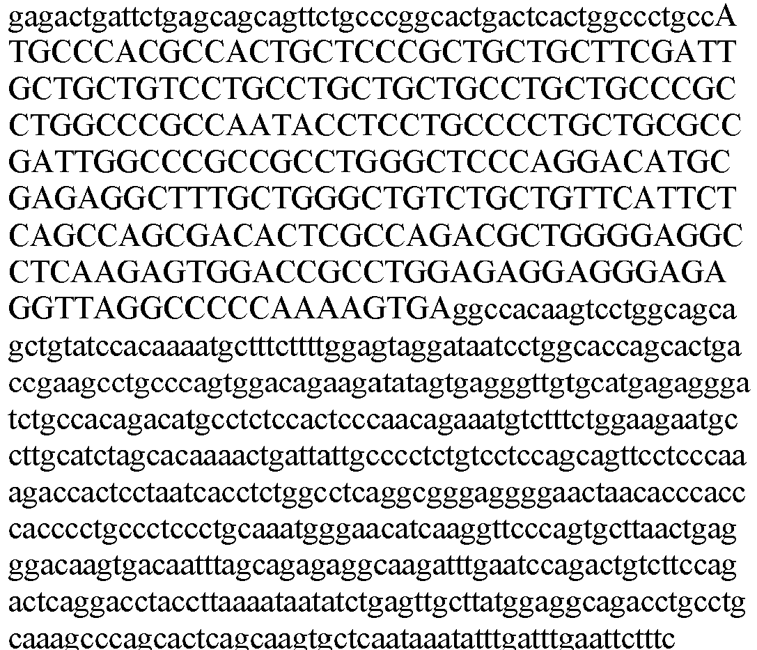
Mus musculus MINION isoform 1 amino acid 5 
Mus musculus MINION isoform 1 cDNA 6 (GenBank Accession No. NM_001177468.1; ORF in uppercase)
Mus musculus MINION isoform 2 amino acid 7 (GenBank Accession No. NP 001170941.1)
Mus musculus MINION isoform 2 cDNA8 
(GenBank Accession No. NM_001177470.1; ORF in uppercase) Rattus norvegicus MINION amino acid 9 
Rattus norvegicus MINION cDNA 10 (ORF in uppercase)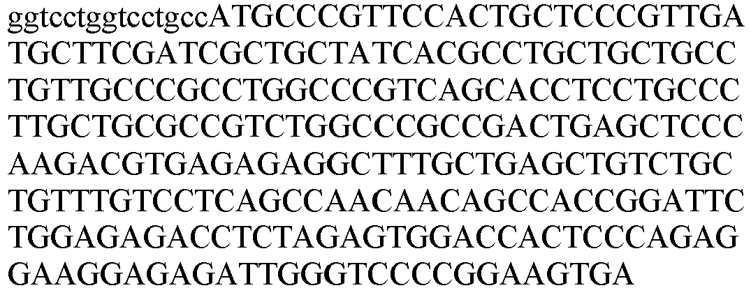
Felis catus MINION amino acid 11 
Felis catus MINION cDNA 12 (ORF in uppercase)
Sus scrofa MINION amino acid 13 
Sus scrofa MINION cDNA 14 (ORF in uppercase)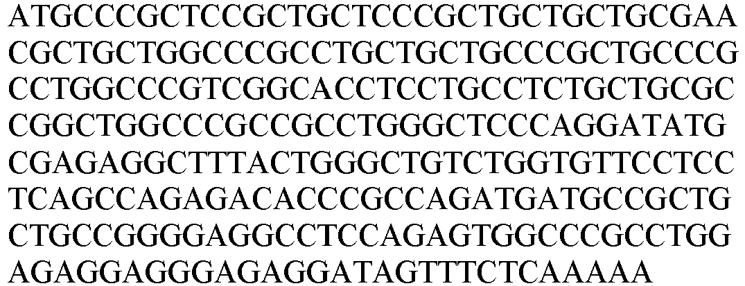
Macaca mulatta MINION amino acid 15 
- A "mouse MINION polypeptide" refers to a naturally occurring mouse MINION protein or a variant thereof. Two naturally occurring mouse MINION protein isforms (SEQ ID NO: 5 and SEQ ID NO: 7) have been reported. The amino acid and nucleotide sequences of mouse MINION isoforms are shown in Table 1. Table 1 also provides MINION sequences of other species, e.g., rat (R. norvegicus), monkey (M. mulatta), cat (F. catus), and pig (S. scrofa).
- The term "a MINION polypeptide" is used to refer collectively to all naturally occurring isoforms of a MINION protein of any species, or a variant thereof. For example, a "MINION polypeptide" can be any MINION polypeptide listed in Table 1 or a variant thereof.
- Myomaker (also known as Tmem8c or Tmem226) is a transmembrane protein specifically expressed in muscle cells. A "human Myomaker polypeptide" refers to a naturally occurring human Myomaker protein (e.g., the protein of SEQ ID NO: 16) or a variant thereof. Human Myomaker protein and mRNA sequences are provided in Table 2.
Table 2 Myomaker amino acid and nucleotide sequences SPECIES ISOFORM SEQ ID NO SEQUENCE Homo sapiens Myomaker amino acid 16 
Homo sapiens Myomaker cDNA 17 
Mus musculus Myomaker isoform 1 amino acid 18 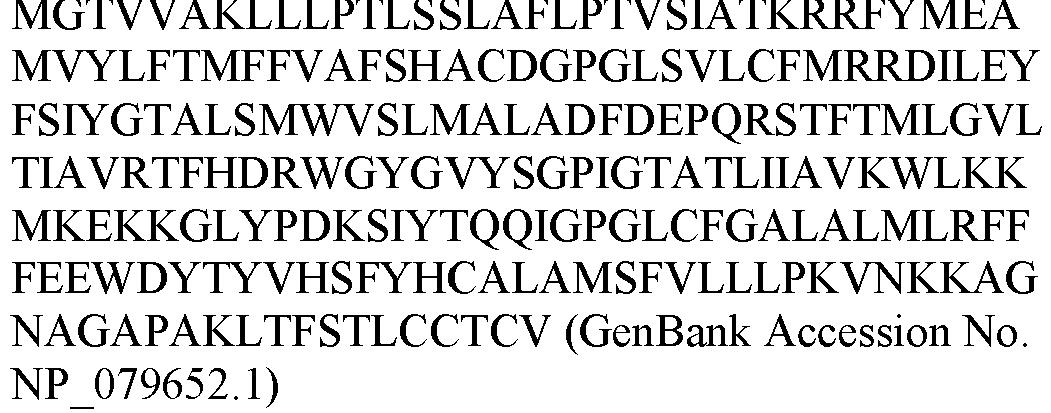
Mus musculus Myomaker isoform 1 cDNA 19 
Mus musculus Myomaker isoform 2 amino acid 20 
Mus musculus Myomaker isoform 2 cDNA 21 

Rattus norvegicus Myomaker amino acid 22 
Rattus norvegicus Myomaker cDNA 23 

Felis catus Myomaker isoform 1 amino acid 24 (GenBank Accession No. XP 006939684.1)
Felis catus Myomaker isoform 1 cDNA 25 
 (GenBank Accession No. XM 006939622.2)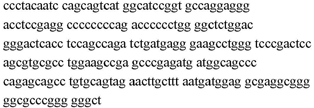
Felis catus Myomaker isoform 2 amino acid 26 (GenBank Accession No. XP 011287016.1)
Felis catus Myomaker isoform 2 cDNA 27 
 (GenBank Accession No. XM 011288714.1)
Sus scrofa Myomaker amino acid 28 (GenBank Accession No. XP 003353750.1)
Sus scrofa Myomaker cDNA 29  (GenBank Accession No. XM 003353702.3)
Macaca mulatta Myomaker amino acid 30 
Macaca mulatta Myomaker cDNA 31  (GenBank Accession No. XM 015116431.1)
- A "mouse Myomaker polypeptide" refers to a naturally occurring mouse Myomaker protein (e.g., the protein of SEQ ID NO: 18 or 20) or a variant thereof. The amino acid and cDNA sequences of mouse Myomaker isofroms can be found in Table 2. Table 2 also includes Myomaker sequences of other species, e.g., rat (R. norvegicus), monkey (M. mulatta), cat (F. catus), and pig (S. scrofa).
- The term "a Myomaker polypeptide" is used to refer collectively to all naturally occurring isoforms of a Myomaker protein of any species, or a variant thereof. For example, a "Myomaker polypeptide" can be any Myomaker polypeptide listed in Table 2 or a variant thereof.
- As used in the specification and claims, the singular form "a", "an" and "the" include plural references unless the context clearly dictates otherwise. For example, the term "a cell" includes a plurality of cells, including mixtures thereof.
- All numerical designations, e.g., pH, temperature, time, concentration, and molecular weight, including ranges, are approximations which are varied (+) or (-) by increments of 0.1. It is to be understood, although not always explicitly stated that all numerical designations are preceded by the term "about." It also is to be understood, although not always explicitly stated, that the reagents described herein are merely examples and that equivalents of such are known in the art.
- The term "cDNA" refers to complementary DNA, e.g., mRNA molecules present in a cell or organism made into cDNA with an enzyme such as reverse transcriptase. A "cDNA library" is a collection of all of the mRNA molecules present in a cell or organism, all turned into cDNA molecules with the enzyme reverse transcriptase, then inserted into "vectors" (other DNA molecules that can continue to replicate after addition of foreign DNA). Example vectors for libraries include bacteriophage (also known as "phage"), viruses that infect bacteria, for example, lambda phage. The library can then be probed for the specific cDNA (and thus mRNA) of interest.
- The term "combination" refers to either a fixed combination in one dosage unit form, or a combined administration where a compound of the present invention and a combination partner (e.g. another drug as explained below, also referred to as "therapeutic agent" or "coagent") may be administered independently at the same time or separately within time intervals, especially where these time intervals allow that the combination partners show a cooperative, e.g. synergistic effect. The single components may be packaged in a kit or separately. One or both of the components (e.g., powders or liquids) may be reconstituted or diluted to a desired dose prior to administration. The terms "co-administration" or "combined administration" or the like as utilized herein are meant to encompass administration of the selected combination partner to a single subject in need thereof (e.g. a patient), and are intended to include treatment regimens in which the agents are not necessarily administered by the same route of administration or at the same time. The term "pharmaceutical combination" as used herein means a product that results from the mixing or combining of more than one active ingredient and includes both fixed and non-fixed combinations of the active ingredients. The term "fixed combination" means that the active ingredients, e.g. a compound of the present invention and a combination partner, are both administered to a patient simultaneously in the form of a single entity or dosage. The term "non-fixed combination" means that the active ingredients, e.g. a compound of the present invention and a combination partner, are both administered to a patient as separate entities either simultaneously, concurrently or sequentially with no specific time limits, wherein such administration provides therapeutically effective levels of the two compounds in the body of the patient. The latter also applies to cocktail therapy, e.g. the administration of three or more active ingredients.
- An "effective amount" refers to an amount sufficient to effect beneficial or desired results. For example, a therapeutic amount is one that achieves the desired therapeutic effect. This amount can be the same or different from a prophylactically effective amount, which is an amount necessary to prevent onset of disease or disease symptoms. An effective amount can be administered in one or more administrations, applications or dosages. A "therapeutically effective amount" of a therapeutic compound (i.e., an effective dosage) depends on the therapeutic compounds selected. The compositions can be administered from one or more times per day to one or more times per week; including once every other day. The skilled artisan will appreciate that certain factors may influence the dosage and timing required to effectively treat a subject, including but are not limited to the severity of the disease or disorder, previous treatments, the general health and/or age of the subject, and other diseases present. Moreover, treatment of a subject with a therapeutically effective amount of the therapeutic compounds described herein can include a single treatment or a series of treatments.
- The term "variant" refers to a polypeptide that has a substantially identical amino acid sequence to a reference polypeptide, or is encoded by a substantially identical nucleotide sequence, and is capable of having one or more activities of the reference polypeptide. For example, a variant can have about 85%, 90%, 91%, 92%, 93%, 94%, 95%, 96%, 97%, 98%, 99% or higher sequence identity to a reference polypeptide, while retain one or more activities of the reference polypeptide.
- A "gene" refers to a polynucleotide containing at least one open reading frame (ORF) that is capable of encoding a particular polypeptide or protein after being transcribed and translated. A polynucleotide sequence can be used to identify larger fragments or full-length coding sequences of the gene with which they are associated. Methods of isolating larger fragment sequences are known to those of skill in the art.
- The term "isolated" refers to altered or removed from the natural state. For example, a nucleic acid or a peptide naturally present in a living animal is not "isolated," but the same nucleic acid or peptide partially or completely separated from the coexisting materials of its natural state is "isolated." An isolated nucleic acid or protein can exist in substantially purified form, or can exist in a non-native environment such as, for example, a host cell.
- The term "nucleic acid" or "polynucleotide" refers to deoxyribonucleic acids (DNA) or ribonucleic acids (RNA) and polymers thereof in either single- or double-stranded form. Unless specifically limited, the term encompasses nucleic acids containing known analogues of natural nucleotides that have similar binding properties as the reference nucleic acid and are metabolized in a manner similar to naturally occurring nucleotides. Unless otherwise indicated, a particular nucleic acid sequence also implicitly encompasses conservatively modified variants thereof (e.g., degenerate codon substitutions), alleles, orthologs, SNPs, and complementary sequences as well as the sequence explicitly indicated. Specifically, degenerate codon substitutions may be achieved by generating sequences in which the third position of one or more selected (or all) codons is substituted with mixed-base and/or deoxyinosine residues (Batzer et al., Nucleic Acid Res. 19:5081 (1991); Ohtsuka et al., J. Biol. Chem. 260:2605-2608 (1985); and Rossolini et al., Mol. Cell. Probes 8:91-98 (1994)).
- The terms "peptide," "polypeptide," and "protein" are used interchangeably, and refer to a compound comprised of amino acid residues covalently linked by peptide bonds. A protein or peptide must contain at least two amino acids, and no limitation is placed on the maximum number of amino acids that can comprise a protein's or peptide's sequence. Polypeptides include any peptide or protein comprising two or more amino acids joined to each other by peptide bonds. As used herein, the term refers to both short chains, which also commonly are referred to in the art as peptides, oligopeptides and oligomers, for example, and to longer chains, which generally are referred to in the art as proteins, of which there are many types. "Polypeptides" include, for example, biologically active fragments, substantially homologous polypeptides, oligopeptides, homodimers, heterodimers, variants of polypeptides, modified polypeptides, derivatives, analogs, fusion proteins, among others. A polypeptide includes a natural peptide, a recombinant peptide, or a combination thereof. A "microprotein" or "micropeptide" refers to a protein or polypeptide that is less than 100 amino acids long.
- The term "homologous" or "identity" refers to the subunit sequence identity between two polymeric molecules, e.g., between two nucleic acid molecules, such as, two DNA molecules or two RNA molecules, or between two polypeptide molecules. When a subunit position in both of the two molecules is occupied by the same monomeric subunit; e.g., if a position in each of two DNA molecules is occupied by adenine, then they are homologous or identical at that position. The homology between two sequences is a direct function of the number of matching or homologous positions; e.g., if half (e.g., five positions in a polymer ten subunits in length) of the positions in two sequences are homologous, the two sequences are 50% homologous; if 90% of the positions (e.g., 9 of 10), are matched or homologous, the two sequences are 90% homologous. Percentage of "sequence identity" can be determined by comparing two optimally aligned sequences over a comparison window, where the fragment of the amino acid sequence in the comparison window may comprise additions or deletions (e.g., gaps or overhangs) as compared to the reference sequence (which does not comprise additions or deletions) for optimal alignment of the two sequences. The percentage can be calculated by determining the number of positions at which the identical amino acid residue occurs in both sequences to yield the number of matched positions, dividing the number of matched positions by the total number of positions in the window of comparison, and multiplying the result by 100 to yield the percentage of sequence identity. The output is the percent identity of the subject sequence with respect to the query sequence.
- The term "subject" refers to an animal, human or non-human, to whom treatment according to the methods of the present invention is provided. Veterinary and non-veterinary applications are contemplated. The term includes, but is not limited to, mammals, e.g., humans, other primates, pigs, rodents such as mice and rats, rabbits, guinea pigs, hamsters, cows, horses, cats, dogs, sheep and goats. Typical subjects include humans, farm animals, and domestic pets such as cats and dogs.
- The term "treat" or "treatment" refers to both therapeutic treatment and prophylactic or preventive measures, wherein the object is to prevent or slow down an undesired physiological change or disorder. For purpose of this invention, beneficial or desired clinical results include, but are not limited to, alleviation of symptoms, diminishment of extent of disease, stabilized (i.e., not worsening) state of disease, delay or slowing of disease progression, amelioration or palliation of the disease state, and remission (whether partial or total), whether detectable or undetectable. "Treatment" can also mean prolonging survival as compared to expected survival if not receiving treatment.
- Statistical significance can be determined by any art known method. Exemplary statistical tests include: the Students T -test, Mann Whitney U non-parametric test, and Wilcoxon non-parametric statistical test. Some statistically significant relationships have a P value of less than 0.05 or 0.02. Particular binding proteins may show a difference, e.g., in specificity or binding that are statistically significant (e.g., P value< 0.05 or 0.02). The terms "induce", "inhibit", "potentiate", "elevate", "increase", "decrease" or the like, e.g., which denote distinguishable qualitative or quantitative differences between two states, may refer to a difference, e.g., a statistically significant difference, between the two states.
- Mouse MINION (ESGP) protein was encoded by a single open reading frame (ORF) (Chen Y et al., Acta Biochimica et Biophysica Sinica 2005, 37(12): 789-796). The 84 amino acid protein described in Chen et al., has an amino acid sequence of SEQ ID NO: 5 (isoform 1). A longer isoform of mouse MINION (isoform 2) was also described, which has the amino acid sequence of SEQ ID NO: 7, and a distinct N-terminus compared to
isoform 1. The function of mouse MINION isoforms was unknown. - The present inventors found that mouse MINION is a skeletal muscle-specific microprotein highly expressed during muscle development and regeneration (see Example 2), and is required for fusion of muscle progenitors and skeletal muscle development (see Examples 3 and 4). Moreover, forced heterologous expression of MINION, along with another protein Myomaker, is found to be sufficient to induce cell fusion even in non-muscle cells (e.g., fibroblasts) (see Example 5). This is surprising since it was reported that Myomaker alone cannot promote fibroblast-fibroblast fusion although it was capable of promoting fusion of fibroblasts to myoblasts (Millay et al., Nature. 2013;499(7458):301-5).
- The human ortholog of mouse MINION has not been reported before. As described herein, the present inventors found human ortholog of MINION has an amino acid sequence of SEQ ID NO: 1 or SEQ ID NO: 3, and a cDNA sequence of SEQ ID NO: 2 or SEQ ID NO: 4. The present inventors found that human MINION protein also has fusogenic activity, suggesting an evolutionarily conserved role for the MINION protein in cell fusion. MINION amino acid and nucleotide sequences of other species, e.g., rat (R. norvegicus), monkey (M. mulatta), cat (F. catus), and pig (S. scrofa) are also provided in Table 1.
- A MINION polypeptide can be a variant of a naturally occurring MINION protein as defined in the claims (e.g., any MINION protein listed in Table 1). For example, a human MINION polypeptide can be a variant of a naturally occurring human MINION protein as defined in the claims (e.g., the protein of SEQ ID NO: 1 or SEQ ID NO: 3, or another naturally occurring isoform thereof). A mouse MINION polypeptide can be a variant of a naturally occurring mouse MINION protein as defined in the claims (e.g., a protein of SEQ ID NO: 5 or SEQ ID NO: 7, or another naturally occurring isoform thereof).
- A MINION variant as defined in the claims can differ from a naturally occurring MINION protein by, for example, a modification (e.g., substitution, deletion, or insertion) of one or more amino acid residues in the naturally occurring MINION protein, but retains the biological activities of MINION, e.g., fusogenic activity. The MINION variant as defined in the claims can have one or more conservative or nonconservative amino acid substitution. A "conservative amino acid substitution" is one in which the amino acid residue is replaced with an amino acid residue having a similar side chain. Families of amino acid residues having similar side chains have been defined in the art. These families include amino acids with basic side chains (e.g., lysine, arginine, histidine), acidic side chains (e.g., aspartic acid, glutamic acid), uncharged polar side chains (e.g., glycine, asparagine, glutamine, serine, threonine, tyrosine, cysteine), nonpolar side chains (e.g., alanine, valine, leucine, isoleucine, proline, phenylalanine, methionine, tryptophan), beta-branched side chains(e.g., threonine, valine, isoleucine) and aromatic side chains (e.g., tyrosine, phenylalanine, tryptophan, histidine).
- In some embodiments, the MINION variant as defined in the claims includes one or more mutations (e.g., substitutions (e.g., conservative substitutions or substitutions), insertions, or deletions) of non-essential amino acids relative to a naturally occurring MINION protein. A "non-essential" amino acid residue is a residue that can be altered from the wild-type sequence of MINION protein without abolishing or more preferably, without substantially altering a biological activity, such as the fusogenic activity, whereas changing an "essential" amino acid residue results in a substantial loss of biological activity.
- A MINION variant as defined in the claims may have at least one, two, three, or four, and no more than 10, 9, 8, 7, 6, or 5 mutations (e.g., substitutions (e.g., conservative substitutions or substitutions of non-essential amino acids), insertions, or deletions) relative to a naturally occurring MINION protein. Whether or not a particular substitution will be tolerated, i.e., will not adversely affect biological properties, such as fusogenic activity, can be predicted, e.g., by evaluating whether the mutation is conservative or by an activity assay.
- In some embodiments, a MINION variant can have about 85%, 90%, 91%, 92%, 93%, 94%, 95%, 96%, 97%, 98%, 99% or higher sequence identity to a naturally occurring MINION protein as defined in the claims. For example, a human MINION variant can have about 85%, 90%, 91%, 92%, 93%, 94%, 95%, 96%, 97%, 98%, 99% or higher sequence identity to SEQ ID NO: 1 or SEQ ID NO: 3. A mouse MINION variant can have about 85%, 90%, 91%, 92%, 93%, 94%, 95%, 96%, 97%, 98%, 99% or higher sequence identity to SEQ ID NO: 5 or SEQ ID NO: 7.
- Calculations of "homology" or "sequence identity" between two sequences (the terms are used interchangeably herein) can be performed as follows. The sequences are aligned for optimal comparison purposes (e.g., gaps can be introduced in one or both of a first and a second amino acid or nucleic acid sequence for optimal alignment and non-homologous sequences can be disregarded for comparison purposes). The optimal alignment is determined as the best score using the GAP program in the GCG software package with a
Blossum 62 scoring matrix with a gap penalty of 12, a gap extend penalty of 4, and a frameshift gap penalty of 5. The amino acid residues or nucleotides at corresponding amino acid positions or nucleotide positions are then compared. When a position in the first sequence is occupied by the same amino acid residue or nucleotide as the corresponding position in the second sequence, then the molecules are identical at that position (as used herein amino acid or nucleic acid "identity" is equivalent to amino acid or nucleic acid "homology"). The percent identity between the two sequences is a function of the number of identical positions shared by the sequences. - In addition, substantial identity exists when the nucleic acid segments hybridize under selective hybridization conditions (e.g., highly stringent hybridization conditions), to the complement of the strand. The nucleic acids may be present in whole cells, in a cell lysate, or in a partially purified or substantially pure form.
- In some embodiments, the MINION variant as defined in the claims can comprise at least one non-naturally encoded amino acid. A "non-naturally encoded amino acid" refers to an amino acid that is not one of the common amino acids or pyrolysine or selenocysteine. Other terms that may be used synonymously with the term "non-naturally encoded amino acid" are "non-natural amino acid," "unnatural amino acid," "non-naturally-occurring amino acid," and variously hyphenated and non-hyphenated versions thereof. The term "non-naturally encoded amino acid" also includes, but is not limited to, amino acids that occur by modification (e.g. post-translational modifications) of a naturally encoded amino acid (including but not limited to, the 20 common amino acids or pyrrolysine and selenocysteine) but are not themselves naturally incorporated into a growing polypeptide chain by the translation complex. Examples of such non-naturally-occurring amino acids include, but are not limited to, N-acetylglucosaminyl-L-serine, N-acetylglucosaminyl-L-threonine, and O-phosphotyrosine.
- A non-naturally encoded amino acid is typically any structure having any substituent side chain other than one used in the twenty natural amino acids. Because the non-naturally encoded amino acids typically differ from the natural amino acids only in the structure of the side chain, the non-naturally encoded amino acids form amide bonds with other amino acids, including but not limited to, natural or non-naturally encoded, in the same manner in which they are formed in naturally occurring polypeptides. However, the non-naturally encoded amino acids have side chain groups that distinguish them from the natural amino acids. For example, R optionally comprises an alkyl-, aryl-, acyl-, keto-, azido-, hydroxyl-, hydrazine, cyano-, halo-, hydrazide, alkenyl, alkynl, ether, thiol, seleno-, sulfonyl-, borate, boronate, phospho, phosphono, phosphine, heterocyclic, enone, imine, aldehyde, ester, thioacid, hydroxylamine, amino group, or the like or any combination thereof. Other non-naturally occurring amino acids of interest that may be suitable for use in the present invention include, but are not limited to, amino acids comprising a photoactivatable cross-linker, spin-labeled amino acids, fluorescent amino acids, metal binding amino acids, metal-containing amino acids, radioactive amino acids, amino acids with novel functional groups, amino acids that covalently or noncovalently interact with other molecules, photocaged and/or photoisomerizable amino acids, amino acids comprising biotin or a biotin analogue, glycosylated amino acids such as a sugar substituted serine, other carbohydrate modified amino acids, keto-containing amino acids, amino acids comprising polyethylene glycol or polyether, heavy atom substituted amino acids, chemically cleavable and/or photocleavable amino acids, amino acids with an elongated side chains as compared to natural amino acids, including but not limited to, polyethers or long chain hydrocarbons, including but not limited to, greater than about 5 or greater than about 10 carbons, carbon-linked sugar-containing amino acids, redox-active amino acids, amino thioacid containing amino acids, and amino acids comprising one or more toxic moiety.
- Exemplary non-naturally encoded amino acids that may be suitable for use in the present invention and that are useful for reactions with water soluble polymers include, but are not limited to, those with carbonyl, aminooxy, hydrazine, hydrazide, semicarbazide, azide and alkyne reactive groups. In some embodiments, non-naturally encoded amino acids comprise a saccharide moiety. Examples of such amino acids include N-acetyl-L-glucosaminyl-L-serine, N-acetyl-L-galactosaminyl-L-serine, N-acetyl-L-glucosaminyl-L-threonine, N-acetyl-L-glucosaminyl-L-asparagine and O-mannosaminyl-L-serine. Examples of such amino acids also include examples where the naturally-occurring N- or O-linkage between the amino acid and the saccharide is replaced by a covalent linkage not commonly found in nature--including but not limited to, an alkene, an oxime, a thioether, an amide and the like. Examples of such amino acids also include saccharides that are not commonly found in naturally-occurring proteins such as 2-deoxy-glucose, 2-deoxygalactose and the like.
- Methods of making and introducing a non-naturally-occurring amino acid into a protein are known. See, e.g.,
US Patent Nos. 7,083,970 , and7,524,647 . The general principles for the production of orthogonal translation systems that are suitable for making proteins that comprise one or more desired unnatural amino acid are known in the art, as are the general methods for producing orthogonal translation systems. For example, see International Publication NumbersWO2002/086075 ;WO2002/085923 ;WO2004/094593 ;WO2005/007870 ;WO2005/007624 ;WO2006/110182 ; andWO2007/103490 . For discussion of orthogonal translation systems that incorporate unnatural amino acids, and methods for their production and use, see also, Wang and Schultz, (2005) "Expanding the Genetic Code." Angewandte Chemie Int Ed 44: 34-66; Xie and Schultz, (2005) "An Expanding Genetic Code." Methods 36: 227-238; Xie and Schultz, (2005) "Adding Amino Acids to the Genetic Repertoire." Curr Opinion in Chemical Biology 9: 548-554; and Wang, et al., (2006) "Expanding the Genetic Code." Annu Rev Biophys Biomol Struct 35: 225-249; Deiters, et al, (2005) "In vivo incorporation of an alkyne into proteins in Escherichia coli." Bioorganic & Medicinal Chemistry Letters 15:1521-1524; Chin, et al., (2002) "Addition of p-Azido-L-phenylalanine to the Genetic Code of Escherichia coli." J Am Chem Soc 124: 9026-9027; and International Publication No.WO2006/034332, filed on Sep. 20, 2005 . Additional details are found inU.S. Pat. No. 7,045,337 ;No. 7,083,970 ;No. 7,238,510 ;No. 7,129,333 ;No. 7,262,040 ;No. 7,183,082 ; No.7,199,222 ; and No.7,217,809 . - Another type of modification that can optionally be introduced into a MINION polypeptide (e.g., within the polypeptide chain or at either the N- or C-terminal), e.g., to extend in vivo half-life, is PEGylation or incorporation of long-chain polyethylene glycol polymers (PEG). Introduction of PEG or long chain polymers of PEG increases the effective molecular weight of the present polypeptides, for example, to prevent rapid filtration into the urine. In some embodiments, a Lysine residue in the MINION sequence is conjugated to PEG directly or through a linker. Such linker can be, for example, a Glu residue or an acyl residue containing a thiol functional group for linkage to the appropriately modified PEG chain. An alternative method for introducing a PEG chain is to first introduce a Cys residue at the C-terminus or at solvent exposed residues such as replacements for Arg or Lys residues. This Cys residue is then site-specifically attached to a PEG chain containing, for example, a maleimide function. Methods for incorporating PEG or long chain polymers of PEG are well known in the art (described, for example, in Veronese, F. M., et al., Drug Disc. Today 10: 1451-8 (2005); Greenwald, R. B., et al., Adv. Drug Deliv. Rev. 55: 217-50 (2003); Roberts, M. J., et al., Adv. Drug Deliv. Rev., 54: 459-76 (2002)). Other methods of polymer conjugations known in the art can also be used in the present invention, e.g.,
WO2008/098930 ; Lewis, et al., Bioconjug Chem., 19: 2144-55 (2008); Deiters, A, et al. (2004). Bio-org. Med. Chem. Lett. 14, 5743-5. In some embodiments, a phosphorylcholine-containing polymer conjugate with the MINION polypeptide can be used. In some embodiments, p-azidophenylalanine can be incorporated into the MINION polypeptides and then reacted with a PEG polymer having an acetylene moiety in the presence of a reducing agent and copper ions to facilitate an organic reaction known as "Huisgen [3+2]cycloaddition." A person of skill would readily recognize that other biocompatible polymer conjugates can be utilized. - In some embodiments, a variant of the MINION polypeptide as defined in the claims can comprise MINION, fused to a heterologous peptide such as a tag or a fusion domain. Fusions may be constructed such that the heterologous peptide (e.g., a tag or a fusion domain) is fused at the amino terminus or at the carboxy terminus of MINION polypeptide. Examples of tags or fusion domains include, but are not limited to, a HA-tag (YPYDVPDYA, SEQ ID NO: 32), a Myc-tag (EQKLISEEDL, SEQ ID NO: 33), a FLAG-tag (DYKDDDDK, SEQ ID NO: 34), a His-tag (HHHHHH, SEQ ID NO: 35), an E-tag (GAPVPYPDPLEPR, SEQ ID NO: 36), a V5-tag (GKPIPNPLLGLDST, SEQ ID NO: 37), a VSV tag (YTDIEMNRI,GK, SEQ ID NO: 38), a polyglutamate tag (EEEEEE, SEQ ID NO: 39), an AviTag (GLNDIFEAQKIEWHE, SEQ ID NO: 40), a SBP tag (MDEKTTGWRGGHVVEGLAGELEQLRARLEHHPQGQREP, SEQ ID NO: 41), a Strep-tag (WSHPQFEK, SEQ ID NO: 42), an Xpress tag (DLYDDDDK, SEQ ID NO: 43), an S-tag (KETAAAKFERQHMDS, SEQ ID NO: 44), a Softag 1 (SLAELLNAGLGGS, SEQ ID NO: 45), a Softag 3 (TQDPSRVG, SEQ ID NO: 46), a Calmodulin-tag (KRRWKKNFIAVSAANRFKKISSSGAL, SEQ ID NO: 47), a TC tag (CCPGCC, SEQ ID NO: 48), an Isopeptag (TDKDMTITFTNKKDAE, SEQ ID NO: 49), a SpyTag (AHIVMVDAYKPTK, SEQ ID NO: 50), a SnoopTag (KLGDIEFIKVNK, SEQ ID NO: 51), a Glutathione-S-Transferase (GST)-tag, a fluorescent protein-tag (e.g., a GFP tag), a Maltose binding protein (MBP)-tag, a Halo-tag, or a thioredoxin-tag.
- A fusion domain or a tag may be selected so as to confer a desired property. For example, some fusion domains are particularly useful for isolation of the fusion proteins by affinity chromatography. For the purpose of affinity purification, relevant matrices for affinity chromatography, such as glutathione-, amylase-, and nickel- or cobalt-conjugated resins are used. Many of such matrices are available in "kit" form, such as the Pharmacia GST purification system and the QLAexpress™ system (Qiagen) useful with a His6 tag. As another example, a fusion domain may be selected so as to facilitate detection of the MINION proteins. Examples of such detection domains include the various fluorescent proteins (e.g., GFP), as well as "epitope tags," which are usually short peptide sequences for which a specific antibody is available. Well known epitope tags for which specific monoclonal antibodies are readily available include a HA-tag, a Myc-tag, a FLAG-tag. Other known tags include an E-tag, a V5-tag, a VSV tag, a polyglutamate tag, an AviTag, a SBP tag, a Strep-tag, an Xpress tag, an S-tag, a
Softag 1, aSoftag 3, a Calmodulin-tag, a TC tag, an Isopeptag, a SpyTag, a SnoopTag, as described above. - In some cases, the tags or fusion domains can have a protease cleavage site, which allows the relevant protease to partially digest the fusion proteins and thereby liberate the MINION polypeptide from the fusion protein. In certain embodiments, an MINION polypeptide as defined in the claims can be fused with a domain that stabilizes the protein in vivo (e.g., a "stabilizer" domain). By "stabilizing" is meant anything that increases serum half life, regardless of whether this is because of decreased destruction, decreased clearance by the kidney, or other pharmacokinetic effect. Fusions with the Fc domain of an immunoglobulin are known to confer desirable pharmacokinetic properties on a wide range of proteins. Likewise, fusions to human serum albumin (HSA) can confer desirable properties. Other types of fusion domains that may be selected include multimerizing (e.g., dimerizing, tetramerizing) domains and functional domains (that confer an additional biological function, as desired).
- Also described herein are fragments of a MINION polypeptide which retain the biological function of a native MINION polypeptide, such as fusogenic activity, interaction with Myomaker, etc. For example, some instances described herein provide fragments of a MINION polypeptide that contain an "AxLyCxL" domain, which has been shown to be required for the fusogenic activity of MINION (Shi et al., Requirement of the fusogenic micropeptide myomixer for muscle formation in zebrafish. PNAS 114, 11950 (2017)). In some instances described herein, the fragments of a MINION polypeptide comprise amino acids 48-54 of SEQ ID NO: 1. In some instances described herein, the fragments of a MINION polypeptide comprise amino acids 48-54 of SEQ ID NO: 3. In some instances described herein, the fragments of a MINION polypeptide comprise amino acids 48-54 of SEQ ID NO: 5. In some instances described herein, the fragments of a MINION polypeptide comprise amino acids 72-78 of SEQ ID NO: 7. Some instances described herein provide fragments of a MINION polypeptide that contain the N-terminal hydrophobic region (amino acids 5-25 of human MINION), and positively charged Arg residues within the first α-helix following the N-terminal hydrophobic region the disruption of which resulted in a loss of fusogenic activity and perturbed interaction with Myomaker (Bi P, Ramirez-Martinez A, Li H, Cannavino J, McAnally JR, Shelton JM, Sanchez-Ortiz E, Bassel-Duby R, Olson EN: Control of muscle formation by the fusogenic micropeptide myomixer. Science 356, 323 (2017)). In some instances described herein, the fragments of a MINION polypeptide comprise amino acids 5-46 of SEQ ID NO: 1. In some instances described herein, the fragments of a MINION polypeptide comprise amino acids 5-46 of SEQ ID NO: 3. In some instances described herein, the fragments of a MINION polypeptide comprise amino acids 5-46 of SEQ ID NO: 5. In some instances described herein, the fragments of a MINION polypeptide comprise amino acids 29-70 of SEQ ID NO: 7. The fragment of a MINION polypeptide can have a variety of lengths. For example, the fragment of a MINION polypeptide can have a length that is, is about, is at least, is more than, 7 amino acids, 8 amino acids, 9 amino acids, 10 amino acids, 11 amino acids, 12 amino acids, 13 amino acids, 14 amino acids, 15 amino acids, 16 amino acids, 17 amino acids, 18 amino acids, 19 amino acids, 20 amino acids, 25 amino acids, 30 amino acids, 35 amino acids, 40 amino acids, 45 amino acids, 50 amino acids, or a range that is between any two of the above values.
- In some embodiments, a MINION peptide as defined in the claims may comprise one or more post-translational modifications, e.g., lipidation, glycosylation, phosphorylation, etc. Examples are as follows. Phosphorylation sites in Mus musculus MINION as predicted using GPS (http://gps.biocuckoo.org/) include:
serine 12,serine 15, serine 41, serine 42,serine 51, serine 58, serine 65,serine 69, and serine 74. Palmitoylation is predicted in Mus musculus MINION using GPS-Lipid (http://gps.biocuckoo.org/) on cysteine 52. O-glycosylation of Mus musculus MINION is predicted using NetOGlyc 4.0 (http://www.cbs.dtu.dk/services/NetOGlyc/) onserine 69 and serine 74. - Myomaker (also known as Tmem8c or Tmem226) is a membrane protein expressed specifically in skeletal muscle during embryogenesis and adult muscle regeneration (Millay et al., Nature. 2013 ). Myomaker has been reported to be both necessary and sufficient to promote myoblast fusion in vivo and in vitro (Millay et al., 2013). Millay et al. also reported that Myomaker can promote fusion of fibroblasts to myoblasts but cannot promote fibroblast-fibroblast fusion. Methods of fusing a non-muslce cell to a muscle cell by expressing exogenous Myomaker protein in the non-muscle cell have been described in
WO2014/210448 . - A Myomaker polypeptide can be a variant of a naturally occurring Myomaker protein as defined in the claims (e.g., any Myomaker protein listed in Table 2). For example, a human Myomaker polypeptide can be a variant of a naturally occurring human Myomaker protein as defined in the claims (e.g., a protein of SEQ ID NO: 16, or a naturally occurring isoform thereof). A mouse Myomaker polypeptide can be a variant of a naturally occurring mouse Myomaker protein as defined in the claims (e.g., a protein of SEQ ID NO: 18 or SEQ ID NO: 20, or another naturally occurring isoform thereof). Table 2 provides Myomaker amino acid and nucleotide sequences of human, mouse, rat, monkey, cat, and pig.
- A Myomaker variant as defined in the claims can differ from a naturally occurring Myomaker protein by, for example, a modification (e.g., substitution, deletion, or insertion) of one or more amino acid residues in the naturally occurring Myomaker protein, but retains the biological activities of Myomaker, e.g., fusogenic activity. The Myomaker variant as defined in the claims can have one or more conservative or nonconservative amino acid substitution. A "conservative amino acid substitution" is one in which the amino acid residue is replaced with an amino acid residue having a similar side chain. Families of amino acid residues having similar side chains have been defined in the art. These families include amino acids with basic side chains (e.g., lysine, arginine, histidine), acidic side chains (e.g., aspartic acid, glutamic acid), uncharged polar side chains (e.g., glycine, asparagine, glutamine, serine, threonine, tyrosine, cysteine), nonpolar side chains (e.g., alanine, valine, leucine, isoleucine, proline, phenylalanine, methionine, tryptophan), beta-branched side chains(e.g., threonine, valine, isoleucine) and aromatic side chains (e.g., tyrosine, phenylalanine, tryptophan, histidine).
- In some embodiments, the Myomaker variant as defined in the claims includes one or more mutations (e.g., substitutions (e.g., conservative substitutions or substitutions), insertions, or deletions) of non-essential amino acids relative to a naturally occurring Myomaker protein. A "non-essential" amino acid residue is a residue that can be altered from the wild-type sequence of Myomaker protein without abolishing or more preferably, without substantially altering a biological activity, such as fusogenic activity, whereas changing an "essential" amino acid residue results in a substantial loss of biological activity.
- A Myomaker variant as defined in the claims may have at least one, two, three, or four, and no more than 10, 9, 8, 7, 6, or 5 mutations (e.g., substitutions (e.g., conservative substitutions or substitutions of non-essential amino acids), insertions, or deletions) relative to a naturally occurring Myomaker protein. Whether or not a particular substitution will be tolerated, i.e., will not adversely affect biological properties, such as fusogenic activity, can be predicted, e.g., by evaluating whether the mutation is conservative or by an activity assay.
- In some embodiments, a Myomaker variant can have about 85%, 90%, 91%, 92%, 93%, 94%, 95%, 96%, 97%, 98%, 99% or higher sequence identity to a naturally occurring Myomaker protein as defined in the claims. For example, a human Myomaker variant can have about 85%, 90%, 91%, 92%, 93%, 94%, 95%, 96%, 97%, 98%, 99% or higher sequence identity to SEQ ID NO: 16. A mouse Myomaker variant can have about 85%, 90%, 91%, 92%, 93%, 94%, 95%, 96%, 97%, 98%, 99% or higher sequence identity to SEQ ID NO: 18 or SEQ ID NO: 20.
- Calculations of "homology" or "sequence identity" between two sequences (the terms are used interchangeably herein) can be performed as follows. The sequences are aligned for optimal comparison purposes (e.g., gaps can be introduced in one or both of a first and a second amino acid or nucleic acid sequence for optimal alignment and non-homologous sequences can be disregarded for comparison purposes). The optimal alignment is determined as the best score using the GAP program in the GCG software package with a
Blossum 62 scoring matrix with a gap penalty of 12, a gap extend penalty of 4, and a frameshift gap penalty of 5. The amino acid residues or nucleotides at corresponding amino acid positions or nucleotide positions are then compared. When a position in the first sequence is occupied by the same amino acid residue or nucleotide as the corresponding position in the second sequence, then the molecules are identical at that position (as used herein amino acid or nucleic acid "identity" is equivalent to amino acid or nucleic acid "homology"). The percent identity between the two sequences is a function of the number of identical positions shared by the sequences. - In addition, substantial identity exists when the nucleic acid segments hybridize under selective hybridization conditions (e.g., highly stringent hybridization conditions), to the complement of the strand. The nucleic acids may be present in whole cells, in a cell lysate, or in a partially purified or substantially pure form.
- In some embodiments, the Myomaker variant as defined in the claims can comprise at least one non-naturally encoded amino acid. In some embodiments, the Myomaker variant as defined in the claims can be a PEGylated Myomaker with extended in vivo half-life. In some embodiments, a variant of the Myomaker polypeptide as defined in the claims can comprise Myomaker, fused to a tag or a fusion domain. Exemplary non-naturally encoded amino acids, PEGylation methods, and fusion partners described above for MINION variants are also applicable to Myomaker variants.
- The invention relates to nucleic acids encoding a MINION polypeptide (e.g., a human MINION polypeptide), expression vectors for expression of a MINION polypeptide (e.g., a human MINION polypeptide), and cells containing such expression vectors. In other aspects, the invention relates to a polynucleotide encoding a MINION polypeptide (e.g., a human MINION polypeptide), and expression vectors and host cells comprising such a polynucleotide.
- Provided herein are expression vectors as defined by the claims that can be employed to express a MINION polypeptide (e.g., a human MINION polypeptide). The term "expression vector" refers to a carrier nucleic acid molecule into which a desired coding sequence can be inserted for introduction into a cell where it can be expressed. Expression vector can be a plasmid, a cosmid, an RNA, a viral vector, or artificial chromosomes (see, e.g., Harrington et al., Nat Genet 15:345, 1997). For example, nonviral vectors useful for expression of a polypeptide in mammalian (e.g., human) cells include pThioHis A, B & C, pcDNA3. 1/His, pEBVHis A, B & C (Invitrogen, San Diego, CA), MPSV vectors, and numerous other vectors known in the art for expressing other proteins. Useful viral vectors include, but are not limited to, vectors based on any of the following viruses: adenovirus, adeno-associated virus, Herpes Simplex Virus (HSV), parvovirus, retrovirus, lentivirus, vaccinia virus, Sinbis virus, influenza virus, reovirus, Newcastle disease virus (NDV), measles virus, vesicular stomatitis virus (VSV), poliovirus, poxvirus, Seneca Valley virus, coxsackievirus, enterovirus, myxoma virus, or maraba virus. In some embodiments, the expression vector can be capable of autonomous replication or it can integrate into a host DNA.
- In some embodiments, the expression vector as defined in the claims can be a recombinant DNA molecule containing a MINION polypeptide-coding sequence ( e.g., a human MINION polypeptide-coding sequence). The recombinant expression vector typically includes one or more regulatory sequences operatively linked to the nucleic acid sequence to be expressed. The term "regulatory sequence" includes promoters, enhancers and other expression control elements (e.g., polyadenylation signals). Regulatory sequences include those which direct constitutive expression of a nucleotide sequence, as well as tissue-specific regulatory and/or inducible sequences. Expression vectors as defined in the claims can also include elements designed to optimize messenger RNA stability and translatability in host cells, and/or drug selection markers for establishing permanent, stable cell clones expressing a human MINION polypeptide. The design of the expression vector can depend on such factors as the choice of the host cell to be transformed, the level of expression of protein desired, and the like. General methods for generating such recombinant expression vectors can be found in Sambrook and Russell eds. (2001) Molecular Cloning: A Laboratory Manual, 3rd edition; the series Ausubel et al. eds. (2007 with updated through 2010) Current Protocols in Molecular Biology, among others known in the art.
- A "promoter" is a control sequence that is a region of a nucleic acid sequence at which initiation and rate of transcription are controlled. It may contain genetic elements at which regulatory proteins and molecules may bind such as RNA polymerase and other transcription factors. The phrases "operatively positioned," "operatively linked," "under control," and "under transcriptional control" mean that a promoter is in a correct functional location and/or orientation in relation to a nucleic acid sequence to control transcriptional initiation and/or expression of that sequence. A promoter may or may not be used in conjunction with an "enhancer," which refers to a cis-acting regulatory sequence involved in the transcriptional activation of a nucleic acid sequence.
- A promoter may be one naturally-associated with a gene or sequence, as may be obtained by isolating the 5' non-coding sequences located upstream of the coding segment and/or exon. Such a promoter can be referred to as "endogenous." Similarly, an enhancer may be one naturally associated with a nucleic acid sequence, located either downstream or upstream of that sequence. Alternatively, certain advantages will be gained by positioning the coding nucleic acid segment under the control of a recombinant or heterologous promoter, which refers to a promoter that is not normally associated with a nucleic acid sequence in its natural environment. A recombinant or heterologous enhancer refers also to an enhancer not normally associated with a nucleic acid sequence in its natural environment. Such promoters or enhancers may include promoters or enhancers of other genes, and promoters or enhancers isolated from any other prokaryotic, viral, or eukaryotic cell, and promoters or enhancers not "naturally-occurring," i.e., containing different elements of different transcriptional regulatory regions, and/or mutations that alter expression. In addition to producing nucleic acid sequences of promoters and enhancers synthetically, sequences may be produced using recombinant cloning and/or nucleic acid amplification technology, including PCR™, in connection with the compositions disclosed herein (see
US 4683202 ,US 5928906 ). Furthermore, it is contemplated the control sequences that direct transcription and/or expression of sequences within non-nuclear organelles such as mitochondria, chloroplasts, and the like, can be employed as well. - The promoters employed can be constitutive, inducible, tissue-specific, and/or useful under the appropriate conditions to direct high level expression of the introduced DNA segment, such as is advantageous in the large-scale production of recombinant proteins and/or peptides. In some embodiments, a constitutive promoter is employed to provide constant expression of a MINION polypeptide as defined by the claims (e.g., a human MINION polypeptide). In some embodiments, an inducible promoter is employed to prevent expression of a MINION polypeptide as defined in the claims (e.g., a human MINION polypeptide) except under inducing conditions. Inducible promoters include, e.g., arabinose, lacZ, a metallothionein promoter, a glucocorticoid promoters or a heat shock promoter. In addition, other regulatory elements may also be incorporated to improve expression of a nucleic acid encoding a MINION polypeptide as defined in the claims (e.g., a human MINION polypeptide), e.g., enhancers, ribosomal binding site, transcription termination sequences, and the like. In some embodiments, a tissue-specific promoter is employed to provide expression of a MINION polypeptide as defined in the claims (e.g., a human MINION polypeptide) only in specific tissues. The identity of tissue-specific promoters or elements, as well as assays to characterize their activities, is well known to those of skill in the art. Examples of such regions include the human LINM2 gene (Nomoto et al. 1999, Gene, 236(2):259-271), the
somatostatin receptor 2 gene (Kraus et al., 1998, FEES Lett., 428(3): 165-170), murine epididymal retinoic acid-binding gene (Lareyre et al., 1999, J. Biol. Chem., 274(12):8282-8290), human CD4 (Zhao-Emonet et al., 1998, Biochirn. Biophys. Acta, 1442(2-3): 109-119), mouse alpha2 (XI) collagen (Tsumaki, et al., 1998, J. Biol. Chem., 273(36):22861-22864), D1A dopamine receptor gene (Lee, et al., 1997, J. Auton. Nerv. Syst., 74(2-3):86-90), insulin-like growth factor II (Wu et al., 1997, Biochem. Biophys. Res. Commun., 233(1):221-226), human platelet endothelial cell adhesion molecule-1 (Almendro et al., 1996, J. Immunol., 157(12):5411-5421), muscle creatine kinase (MCK) promoter (Wang et al., Gene Ther. 2008 Nov;15(22):1489-99). - A specific initiation signal also may be required for efficient translation of coding sequences. These signals include the ATG initiation codon or adjacent sequences. Exogenous translational control signals, including the ATG initiation codon, may need to be provided. One of ordinary skill in the art would readily be capable of determining this and providing the necessary signals. It is well known that the initiation codon must be "in-frame" with the reading frame of the desired coding sequence to ensure translation of the entire insert. The exogenous translational control signals and initiation codons can be either natural or synthetic. The efficiency of expression may be enhanced by the inclusion of appropriate transcription enhancer elements.
- Expression can employ any appropriate host cells known in the art, for example, mammalian host cells, bacterial host cells, yeast host cells, insect host cells, etc. Both prokaryotic and eukaryotic expression systems are widely available. In some embodiments, the expression system is a mammalian cell expression, such as a CHO cell expression system. In some embodiments, a nucleic acid may be codon-optimized to facilitate expression in a desired host cell. It will be important to employ a promoter and/or enhancer that effectively directs the expression of the DNA segment in the cell type, organelle, and organism chosen for expression. Those of skill in the art of molecular biology generally know the use of promoters, enhancers, and cell type combinations for protein expression, for example, see Sambrook et al. (2001).
- Most transcribed eukaryotic RNA molecules will undergo RNA splicing to remove introns from the primary transcripts. Vectors containing genomic eukaryotic sequences may require donor and/or acceptor splicing sites to ensure proper processing of the transcript for protein expression (see Chandler et al., 1997, Proc. Natl. Acad. Sci. USA, 94(8):3 596-601).
- The vectors or constructs of the present disclosure will generally comprise at least one termination signal. A "termination signal" or "terminator" is comprised of the DNA sequences involved in specific termination of an RNA transcript by an RNA polymerase. Thus, in certain embodiments a termination signal that ends the production of an RNA transcript is contemplated. A terminator may be necessary in vivo to achieve desirable message levels. In eukaryotic systems, the terminator region may also comprise specific DNA sequences that permit site-specific cleavage of the new transcript so as to expose a polyadenylation site. This signals a specialized endogenous polymerase to add a stretch of about 200 A residues (poly A) to the 3' end of the transcript. RNA molecules modified with this polyA tail appear to more stable and are translated more efficiently. Thus, in other embodiments involving eukaryotes, it is preferred that that terminator comprises a signal for the cleavage of the RNA, and it is more preferred that the terminator signal promotes polyadenylation of the message. The terminator and/or polyadenylation site elements can serve to enhance message levels and/or to minimize read through from the cassette into other sequences. Terminators contemplated for use in the disclosure include any known terminator of transcription described herein or known to one of ordinary skill in the art, including but not limited to, for example, the termination sequences of genes, such as for example the bovine growth hormone terminator or viral termination sequences, such as for example the SV40 terminator. In certain embodiments, the termination signal may be a lack of transcribable or translatable sequence, such as due to a sequence truncation.
- In expression, particularly eukaryotic expression, one will typically include a polyadenylation signal to effect proper polyadenylation of the transcript. The nature of the polyadenylation signal is not believed to be crucial to the successful practice of the disclosure, and/or any such sequence may be employed. Preferred embodiments include the SV40 polyadenylation signal and/or the bovine growth hormone polyadenylation signal, convenient and/or known to function well in various target cells. Polyadenylation may increase the stability of the transcript or may facilitate cytoplasmic transport.
- To propagate a vector in a host cell, it may contain one or more origins of replication sites (often termed "ori"), which is a specific nucleic acid sequence at which replication is initiated. Alternatively an autonomously replicating sequence (ARS) can be employed if the host cell is yeast.
- In certain embodiments of the disclosure, cells containing a nucleic acid construct of the present disclosure may be identified in vitro or in vivo by including a marker in the expression vector. Such markers would confer an identifiable change to the cell permitting easy identification of cells containing the expression vector. Generally, a selectable marker is one that confers a property that allows for selection. A positive selectable marker is one in which the presence of the marker allows for its selection, while a negative selectable marker is one in which its presence prevents its selection. An example of a positive selectable marker is a drug resistance marker.
- Usually the inclusion of a drug selection marker aids in the cloning and identification of transformants, for example, genes that confer resistance to neomycin, puromycin, hygromycin, DHFR, GPT, zeocin and histidinol are useful selectable markers. In addition to markers conferring a phenotype that allows for the discrimination of transformants based on the implementation of conditions, other types of markers including screenable markers such as GFP, whose basis is colorimetric analysis, are also contemplated. Alternatively, screenable enzymes such as herpes simplex virus thymidine kinase (tk) or chloramphenicol acetyltransferase (CAT) may be utilized. One of skill in the art would also know how to employ immunologic markers, possibly in conjunction with FACS analysis. The marker used is not believed to be important, so long as it is capable of being expressed simultaneously with the nucleic acid encoding a gene product. Further examples of selectable and screenable markers are well known to one of skill in the art.
- In some embodiments, provided herein are expression vectors as defined in the claims comprising a nucleic acid i) encoding a MINION polypeptide or a variant thereof as defined in the claims. In some embodiments, provided herein are expression vectors comprising a nucleic acid i) encoding a MINION polypeptide of Table 1 or a variant thereof as defined in the claims. In some embodiments, provided herein are expression vectors comprising a nucleic acid i) encoding a MINION polypeptide comprising an amino acid sequence selected from any of SEQ ID NOs: 1, 3, 5, 7, 9, 11, 13, or 15, or a variant thereof as defined in the claims. In some embodiments, provided herein are expression vectors comprising a nucleic acid i) encoding a MINION polypeptide consisting of an amino acid sequence selected from any of SEQ ID Nos: 1, 3, 5, 7, 9, 11, 13, or 15, or a variant thereof as defined in the claims. In some embodiments, provided herein are expression vectors comprising a nucleic acid i) selected from any of SEQ ID NOs: 2, 4, 6, 8, 10, 12, or 14.
- In some embodiments, provided herein are expression vectors comprising a nucleic acid i) encoding a polypeptide comprising SEQ ID NO: 1 or SEQ ID NO: 3 or a variant thereof as defined in the claims. In some embodiments, the expression vector comprises a nucleic acid i) encoding a polypeptide comprising SEQ ID NO: 1 with 1-4 mutations. In some embodiments, the expression vector comprises a nucleic acid i) encoding a polypeptide consisting of SEQ ID NO: 1. In some embodiments, the expression vector comprises a nucleic acid i) encoding a polypeptide comprising SEQ ID NO: 3 with 1-4 mutations. In some embodiments, the expression vector comprises a nucleic acid i) encoding a polypeptide consisting of SEQ ID NO: 1. In some embodiments, the expression vector comprises a nucleic acid i) encoding a polypeptide consisting of SEQ ID NO: 3. In some embodiments, the expression vector comprises a nucleic acid i) comprising SEQ ID NO: 2. In some embodiments, the expression vector comprises a nucleic acid i) comprising SEQ ID NO: 4. In some embodiments, the expression vector comprises a nucleic acid i) consisting of SEQ ID NO: 2. In some embodiments, the expression vector comprises a nucleic acid i) consisting of SEQ ID NO: 4.
- In some embodiments, the expression vector comprises a promoter, e.g., a constitutive promoter, an inducible promoter, or a tissue-specific promoter. In some embodiments, the expression vector comprises a polyadenylation signal. In some embodiments, the expression vector comprises a selectable marker.
- In some embodiments, the expression vector comprises a nucleic acid i) encoding a polypeptide comprising SEQ ID NO: 1 or SEQ ID NO: 3, fused to a tag. The tag can be selected from a HA-tag (YPYDVPDYA, SEQ ID NO: 32), a Myc-tag (EQKLISEEDL, SEQ ID NO: 33), a FLAG-tag (DYKDDDDK, SEQ ID NO: 34), a His-tag (HHHHHH, SEQ ID NO: 35), an E-tag (GAPVPYPDPLEPR, SEQ ID NO: 36), a V5-tag (GKPIPNPLLGLDST, SEQ ID NO: 37), a VSV tag (YTDIEMNRI,GK, SEQ ID NO: 38), a polyglutamate tag (EEEEEE, SEQ ID NO: 39), an AviTag (GLNDIFEAQKIEWHE, SEQ ID NO: 40), a SBP tag (MDEKTTGWRGGHVVEGLAGELEQLRARLEHHPQGQREP, SEQ ID NO: 41), a Strep-tag (WSHPQFEK, SEQ ID NO: 42), an Xpress tag (DLYDDDDK, SEQ ID NO: 43), an S-tag (KETAAAKFERQHMDS, SEQ ID NO: 44), a Softag 1 (SLAELLNAGLGGS, SEQ ID NO: 45), a Softag 3 (TQDPSRVG, SEQ ID NO: 46), a Calmodulin-tag (KRRWKKNFIAVSAANRFKKISSSGAL, SEQ ID NO: 47), a TC tag (CCPGCC, SEQ ID NO: 48), an Isopeptag (TDKDMTITFTNKKDAE, SEQ ID NO: 49), a SpyTag (AHIVMVDAYKPTK, SEQ ID NO: 50), a SnoopTag (KLGDIEFIKVNK, SEQ ID NO: 51), a Glutathione-S-Transferase (GST)-tag, a fluorescent protein-tag (e.g., a GFP tag), a Maltose binding protein (MBP)-tag, a Halo-tag, or a thioredoxin-tag.
- In some embodiments, the expression vectors further comprise a second nucleic acid ii) encoding a Myomaker polypeptide or a variant thereof as defined in the claims. Thus, provided herein are expression vectors for expressing both a MINION polypeptide and a Myomaker polypeptide as defined in the claims. In some embodiments, the expression vector comprises a second nucleic acid ii) encoding a Myomaker polypeptide of Table 2 or a variant thereof as defined in the claims. In some embodiments, the expression vector comprises a second nucleic acid ii) encoding a polypeptide comprising an amino acid sequence selected from any of SEQ ID NOs: 16, 18, 20, 22, 24, 26, 28, or 30, or a variant thereof as defined in the claims. In some embodiments, the expression vector comprises a second nucleic acid ii) encoding a polypeptide consisting of an amino acid sequence selected from any of SEQ ID NOs: 16, 18, 20, 22, 24, 26, 28, or 30, or a variant thereof as defined in the claims. In some embodiments, the expression vector comprises a second nucleic acid ii) selected from any of SEQ ID NOs: 17, 19, 21, 23, 25, 27, 29, or 31, or a variant thereof as defined in the claims.
- In some embodiments, the expression vector comprises a second nucleic acid ii) encoding a polypeptide comprising SEQ ID NO: 16. In some embodiments, the expression vector comprises a second nucleic acid ii) encoding a polypeptide consisting of SEQ ID NO: 16. In some embodiments, the expression vector comprises a second nucleic acid ii) comprising SEQ ID NO: 17. In some embodiments, the expression vector comprises a second nucleic acid ii) consisting of SEQ ID NO: 17. In some embodiments, the expression vectors further comprise a second promoter upstream of the second nucleic acid. The second promoter can be a constitutive promoter, an inducible promoter, or a tissue-specific promoter.
- In some embodiments, the expression vectors for expressing both a MINION polypeptide and a Myomaker polypeptide as defined in the claims comprise an internal ribosome entry site (IRES) upstream of the Myomaker-coding sequence. IRES elements are able to bypass the ribosome scanning model of 5'-methylated Cap dependent translation and begin translation at internal sites (Pelletier and Sonenberg, 1988, Nature, 334:320-325). IRES elements from two members of the picornavirus family (polio and encephalomyocarditis) have been described (Pelletier and Sonenberg, 1988), as well an IRES from a mammalian message (Macejak and Sarnow, 1991, Nature, 353:90-94, 1991). IRES elements can be linked to heterologous open reading frames. Multiple open reading frames can be transcribed together, each separated by an IRES, creating polycistronic messages. By virtue of the IRES element, each open reading frame is accessible to ribosomes for efficient translation. Multiple genes can be efficiently expressed using a single promoter/enhancer to transcribe a single message (see
U.S. Patents 5925565 and5935819 ). - In some embodiments, the expression vectors for expressing both a MINION polypeptide and a Myomaker polypeptide as defined in the claims comprise a 2A sequence upstream of the Myomaker-coding sequence. The 2A oligopeptide sequence was first characterized from the positive-stranded RNA picornavirus Foot-and-Mouth Disease Virus (FMDV); and FMDV 2A or F2A was shown to mediate a co-translational 'cleavage' between the upstream (capsid proteins) and downstream (RNA replication proteins) domains of the FMDV polyprotein (Ryan MD, EMBO J 1994;134: 928-933; Ryan MD, J Gen Virol 1991; 72: 2727-2732; Donnelly MLL, J Gen Virol 1997; 78:13-21; Donnelly MLL, J Gen Virol 2001; 82:1013-1025). Active 2A sequences were characterized in the genomes of viruses from other genera of the picornaviruses, plus `2A-like' sequences found within the genomes of a range of different RNA viruses and non-LTR retrotransposons (Donnelly MLL, J Gen Virol 2001; 82:1027-1041; Heras SR, Cell Mol Life Sci 2006; 63:1449-1460; Luke GA, J Gen Virol 2008; 89:1036-1042; Odon V, Mol Biol Evol 2013; 30:1955-1965; Luke GA, Mob Gen Elements 2014; 3:e27525). These 2A or "2A-like" oligopeptide sequences were shown to mediate a translational `recoding' event referred-to as `ribosome skipping', `stop carry-on' or 'stop-go' translation (Atkins JF, RNA 2007; 13:1-8).
- As used herein, a "2A sequence" refers to any nucleic acid sequence encoding a 2A or "2A-like" oligopeptide serving as a linker between two proteins, allowing autonomous intraribosomal self-processing of polyproteins (See e.g., de Felipe. Genetic Vaccines and Ther. 2:13 (2004); deFelipe et al. Traffic 5:616-626 (2004)). These oligopeptides allow co-expression of multiple proteins from a single vector. Many 2A elements are known in the art. For example, viral 2A sequences have been described in
US Patent Nos. 9175311 8865881 ,7939059 ,7947493 ,. For example, a viral 2A sequence can be a picornaviral, a tetraviral 2A sequence, or a combination thereof. A picornaviral 2A sequence can be selected from any one of the Enteroviral 2A sequences, Rhinoviral 2A sequences, Cardioviral 2A sequences, Aphthoviral 2A sequences, Hepatoviral 2A sequences, Erboviral 2A sequences, Kobuviral 2A sequences, Teschoviral 2A sequences, and the Parechoviral 2A sequences. A tetraviral 2A sequences can be selected from any of the Betatetraviral 2A sequences or Omegatetraviral 2A sequences. Examples of 2A sequences that can be used in the methods and system disclosed herein, without limitation, include 2A sequences from the foot-and-mouth disease virus (F2A), equine rhinitis A virus (E2A), Thosea asigna virus (T2A), and porcine teschovirus-1 (P2A). In some embodiments, a 2A sequence encodes a viral 2A oligopeptide selected from T2A (EGRGSLLTCGDVEENPGP (SEQ ID NO: 52) or GSGEGRGSLLTCGDVEENPGP (SEQ ID NO: 53)), P2A (ATNFSLLKQAGDVEENPGP (SEQ ID NO: 54) or GSGATNFSLLKQAGDVEENPGP (SEQ ID NO: 55)), E2A (QCTNYALLKLAGDVESNPGP (SEQ ID NO: 56) or GSGQCTNYALLKLAGDVESNPGP (SEQ ID NO: 57)), or F2A (VKQTLNFDLLKLAGDVESNPGP (SEQ ID NO: 58) or GSGVKQTLNFDLLKLAGDVESNPGP (SEQ ID NO: 59)). - Non-viral 2A sequence has been described in
US Patent No. 8945876 . For example, a non-viral 2A sequence can be a sea urchin (Strongylocentrotus purpuratus) 2A sequence (DGFCILYLLLILLMRSGDVETNPGP) (SEQ ID NO: 60); a sponge (Amphimedon queenslandica) 2A sequence (LLCFMLLLLLSGDVELNPGP (SEQ ID NO: 61) or HHFMFLLLLLAGDIELNPGP (SEQ ID NO: 62)); an acorn worm (Saccoglossus kowalevskii) 2A sequence (WFLVLLSFILSGDIEVNPGP (SEQ ID NO: 63)); or an amphioxus (Branchiostoma floridae) 2A sequence (KNCAMYMLLLSGDVETNPGP (SEQ ID NO: 64) or MVISQLMLKLAGDVEENPGP (SEQ ID NO: 65)). In some embodiments, the 2A sequence is a naturally occurring or synthetic sequence that includes the 2A consensus sequence D-X-E-X-NPGP (SEQ ID NO: 66), in which X is any amino acid residue. - In some embodiments, the expression vector comprises a 2A sequence encoding a 2A oligopeptide selected from any one of SEQ ID NOs: 52-66.
- In some embodiments, a nucleic acid encoding a human MINION polypeptide may also include a sequence encoding a secretion signal sequence so that the polypeptide is secreted from the host cell. Such a sequence can be provided by the vector, or as part of the MINION nucleic acid that is present in the vector.
- Generation of an expression vector can utilize a vector that includes a multiple cloning site (MCS), which is a nucleic acid region that contains multiple restriction enzyme sites, any of which can be used in conjunction with standard recombinant technology to digest the vector. See Carbonelli et al., 1999, Levenson et al., 1998, and Cocea, 1997. "Restriction enzyme digestion" refers to catalytic cleavage of a nucleic acid molecule with an enzyme that functions only at specific locations in a nucleic acid molecule. Many of these restriction enzymes are commercially available. Use of such enzymes is widely understood by those of skill in the art. Frequently, a vector is linearized or fragmented using a restriction enzyme that cuts within the MCS to enable exogenous sequences to be ligated to the vector. "Ligation" refers to the process of forming phosphodiester bonds between two nucleic acid fragments, which may or may not be contiguous with each other. Techniques involving restriction enzymes and ligation reactions are well known to those of skill in the art of recombinant technology.
- Methods for introducing expression vectors containing the polynucleotide sequences of interest vary depending on the type of cellular host. For example, calcium chloride transfection is commonly utilized for prokaryotic cells, whereas calcium phosphate treatment or electroporation may be used for other cellular hosts (see generally Sambrook et al., supra). Other methods include, e.g., electroporation, calcium phosphate treatment, liposome-mediated transformation, injection and microinjection, ballistic methods, virosomes, immunoliposomes, polycation: nucleic acid conjugates, naked DNA, artificial virions, fusion to the herpes virus structural protein VP22, agent-enhanced uptake of DNA, and ex vivo transduction. For long-term, high- yield production of recombinant proteins, stable expression will often be desired. For example, cell lines which stably express polypeptides of the invention can be prepared using expression vectors of the invention which contain viral origins of replication or endogenous expression elements and a selectable marker gene.
- Also provided herein are cells that include any of the expression vectors as defined in the claims. The invention relates to cells comprising an expression vector for expressing a MINION polypeptide (e.g., a human MINION polypeptide). In some embodiments, such cells comprise an expression vector for expressing both a MINION polypeptide (e.g., a human MINION polypeptide) and a Myomaker polypeptide (e.g., a human Myomaker polypeptide). In some embodiments, such cells comprise an expression vector for expressing a MINION polypeptide (e.g., a human MINION polypeptide) and a second expression vector for expressing a Myomaker polypeptide (e.g., a human Myomaker polypeptide). Such cells can be a host cell or a therapeutic cell.
- In some embodiments, the disclosure features a host cell that includes a nucleic acid molecule described herein. A host cell can be used to produce or express a human MINION polypeptide described herein. The terms "host cell" and "recombinant host cell" are used interchangeably herein, which refer to not only to the particular subject cell but to the progeny or potential progeny of such a cell. Because certain modifications may occur in succeeding generations due to either mutation or environmental influences, such progeny may not, in fact, be identical to the parent cell, but are still included within the scope of the term as used herein. A host cell can be any prokaryotic or eukaryotic cell. For example, a protein can be expressed in bacterial cells (such as E. coli), insect cells, yeast cells, or mammalian cells (such as Chinese hamster ovary cells (CHO) or COS cells e.g., COS-7 cells, CV-1 origin SV40 cells; Gluzman (1981) Cell23:175-182). Other suitable host cells are known to those skilled in the art.
- A host cell can be used to produce or express a human MINION polypeptide described herein. Accordingly, the disclosure also features methods for producing a human MINION polypeptide using a host cell. In one embodiment, the method includes culturing the host cell (into which a recombinant expression vector encoding a protein has been introduced) in a suitable medium, such that a human MINION polypeptide is produced. In another embodiment, the method further includes isolating a human MINION polypeptide from the medium or the host cell.
- In some embodiments, the disclosure features a therapeutic cell that includes a nucleic acid molecule as defined in the claims. As used herein, the term "therapeutic cell" refers to a cell that has been genetically engineered to express a MINION polypeptide, as defined in the claims, that is capable of fusing to a dysfunctional cell or tumor cell. Such a therapeutic cell can be a human cell, e.g., a human muscle cell such as a myoblast, a cardiac muscle cell, a skeletal muscle cell, or a smooth muscle cell; or a human non-muscle cell such as a fibroblast, a bone marrow cell, a blood cell, a hepatocyte, a stem cell, an epithelial cell, an endothelial cell, a dendritic cell, or a tumor cell. In the invention, the therapeutic cells comprise an expression vector expressing a MINION polypeptide as defined in the claims. In some the invention, the therapeutic cells further comprise an expression vector expressing a Myomaker polypeptide as defined in the claims. In some embodiments, such a therapeutic cell expresses a detectable marker, e.g., a fluorescent molecule (e.g., fluorescein, Texas red, rhodamine, green fluorescent protein, and the like), an enzyme (e.g., horse radish peroxidase, alkaline phosphatase), a luminescent molecule (e.g., luciferase), a radioactive molecule (e.g., 3H, 125I, 35S, 14C, or 32P), or calorimetric labels such as colloidal gold or colored beads. Cells expressing a detectable marker can be traced or visualized by appropriate detection methods such as microscopy, autoradiography, and/or other imaging methods known in the art.
- In some embodiments, the therapeutic cells may be able to restore the function of the dysfunctional cell by fusing to the dysfunctional cell. As used herein, a "dysfunctional cell" refers to a cell that has partially or completely lost its normal function, for example, due to a genetic abnormality, e.g., a genetic mutation. Non-limiting examples of dysfunctional cells include: a muscle cell having a mutation in the dystrophin gene resulting in cell death which is the underlying cause for Duchenne muscular dystrophy (DMD) and Becker muscular dystrophy (BMD); a muscle cell having a mutation in the Dysferlin gene resulting in Miyoshi myopathy (MM), Limb-girdle muscular dystrophy type 2B (LGMD2B), and Distal Myopathy (DM), a cell having a mutation in the gene encoding fumaryl acetoacetate hydrolase (Fah), which leads to Hereditary Tyrosinemia type I, etc.
- In some embodiments, the therapeutic cells may restore the function of the dysfunctional cell by introducing a gene of interest, e.g., a correct version of the gene that is mutated in the dysfunctional cell, to the dysfunctional cell, and thereby correct the genetic abnormality in the dysfunctional cell. For example, the therapeutic cells may contain a correct version of the dystrophin gene, a correct version of the gene encoding Fah, etc. The therapeutic cells may be autologous or allogeneic. In embodiments where the therapeutic cells are autologous, the genetic abnormality of the therapeutic cells may be corrected by a variety of gene editing systems known in the art. As used herein, the term "gene editing system" refers to a system comprising one or more DNA-binding domains or components and one or more DNA-modifying domains or components, or isolated nucleic acids, e.g., one or more vectors, encoding said DNA-binding and DNA-modifying domains or components. Gene editing systems are used for modifying the nucleic acid of a target gene and/or for modulating the expression of a target gene. In known gene editing systems, for example, the one or more DNA-binding domains or components are associated with the one or more DNA-modifying domains or components, such that the one or more DNA-binding domains target the one or more DNA-modifying domains or components to a specific nucleic acid site.
- Gene editing systems are known in the art, and include but are not limited to, zinc finger nucleases, transcription activator-like effector nucleases (TALENs); clustered regularly interspaced short palindromic repeats (CRISPR)/Cas systems, and meganuclease systems. Without wishing to be bound by theory, it is believed that the known gene editing systems may exhibit unwanted DNA-modifying activity which is detrimental to their utility in therapeutic applications. These concerns are particularly apparent in the use of gene editing systems for in vivo modification of genes or gene expression, e.g., where cells are engineered to constitutively express components of a gene editing system, such as through lentiviral or adenoviral vector transfection.
- "CRISPR" as used herein refers to a set of clustered regularly interspaced short palindromic repeats, or a system comprising such a set of repeats. "Cas," as used herein, refers to a CRISPR-associated protein. The diverse CRISPR-Cas systems can be divided into two classes according to the configuration of their effector modules:
class 1 CRISPR systems utilize several Cas proteins and the crRNA to form an effector complex, whereasclass 2 CRISPR systems employ a large single-component Cas protein in conjunction with crRNAs to mediate interference. One example ofclass 2 CRISPR-Cas system employs Cpf1 (CRISPR from Prevotella and Francisella 1). See, e.g., Zetsche et al., Cell 163:759-771 (2015). The term "Cpf1" as used herein includes all orthologs, and variants that can be used in a CRISPR system. - It is contemplated that the therapeutic cells may be the same or different cell type from the dysfunctional cell or tumor cell. For example, in embodiments where the dysfunctional cell is a muscle cell, the therapeutic cell may be a muscle cell such as a myoblast, a cardiac muscle cell, a skeletal muscle cell, or a smooth muscle cell; or a non-muscle cell such as a fibroblast, a bone marrow cell, a blood cell, a hepatocyte, a stem cell, an epithelial cell, an endothelial cell, a dendritic cell, etc.
- The invention relates to liposomes comprising a MINION polypeptide described herein, e.g., a MINION polypeptide of Table 1. The invention provides a liposome comprising a Minion polypeptide as defined in the claims and further comprising a Myomaker polypeptide as defined in the claims. Liposomes are closed lipid carriers characterized by lipid bilayers encapsulating an aqueous core. Liposomes can be used as carriers for a therapeutic agent. In some embodiments, the MINION polypeptide as defined in the claims expresses on the surface of the liposomes. The fusogenic activity of the MINION polypeptide can induce fusion of the liposome lipids with a cell membrane, thereby delivering the liposomal content directly into the cytoplasm, bypassing the degradative endocytic entry pathway. Liposomes comprising a MINION polypeptide can fuse to a broad range of cell types, making them compatible with many therapeutic agents such as a nucleic acid (e.g., e.g., an antisense oligonucleotide, plasmid, siRNA, shRNA, aptamer), a protein or polypeptide (e.g., an antibody, antigen, enzyme, or therapeutic polypeptide), a peptide nucleic acid (PNA), or a low molecular weight compound.
- The liposomes described herein include a MINION polypeptide comprising an amino acid sequence selected from SEQ ID NO: 1, 3, 5, 7, 9, 11, 13, or 15, or a variant thereof as defined in the claims. In some embodiments, the liposomes described herein include a MINION polypeptide consisting of an amino acid sequence selected from SEQ ID NO: 1, 3, 5, 7, 9, 11, 13, or 15, or a variant thereof as defined in the claims. In some embodiments, the liposomes described herein include a polypeptide consisting of SEQ ID NO: 1. In some embodiments, the liposomes described herein include a polypeptide consisting of SEQ ID NO: 3. In some embodiments, the liposomes described herein include a polypeptide consisting of SEQ ID NO: 5. In some embodiments, the liposomes described herein include a polypeptide consisting of SEQ ID NO: 7.
- In the invention, the liposomes described herein also include a Myomaker polypeptide, e.g., a Myomaker polypeptide of Table 2, as defined in the claims. The liposomes described herein include a Myomaker polypeptide comprising an amino acid sequence selected from SEQ ID NO: 16, 18, 20, 22, 24, 26, 28, or 30, or a variant thereof as defined in the claims. In some embodiments, the liposomes described herein include a Myomaker polypeptide consisting of an amino acid sequence selected from SEQ ID NO: 16, 18, 20, 22, 24, 26, 28, or 30, or a variant thereof as defined in the claims. In some embodiments, the liposomes described herein include a Myomaker polypeptide consisting of SEQ ID NO: 16. In some embodiments, the liposomes described herein include a Myomaker polypeptide consisting of SEQ ID NO: 18. In some embodiments, the liposomes described herein include a Myomaker polypeptide consisting of SEQ ID NO: 20.
- In some embodiments, liposomes as defined in the claims further comprise a therapeutic agent encapsulated in the liposome. The therapeutic agent can be a nucleic acid (e.g., an antisense oligonucleotide, plasmid, siRNA, shRNA, aptamer), a protein or polypeptide (e.g., an antibody, antigen, enzyme, or therapeutic polypeptide), a peptide nucleic acid (PNA), or a low molecular weight compound. Such liposomes are fusogenic and can be used to deliver the therapeutic agent into a target cell, e.g., a human cell, e.g., a dysfunctional cell or a tumor cell, through the fusion of the liposome lipids into the cell membrane.
- Methods of making liposomes comprising a fusogenic protein have been described in US patent application publications, e.g.,
US 20140314831 ,US 20140134232 ,US 20090280163 ,US 20090142267 . For example, a MINION polypeptide can be inserted into the liposomal membrane by the detergent depletion method derived from the concepts outlined in publications such as Rigaud et al., 1995, Biochim Biophys Acta 1231(3):223-46; Rigaud and Levy, 2003, Methods Enzylmol., 372:65-86; and Top et al., 2005, EMBO J, 24(17):2980-8. Briefly, the purified MINION polypeptide can be reconstituted into the liposomes by mixing the detergent-suspended MINION polypeptide with liposomes presaturated with detergent, followed by removal of the detergent. To assist in the process of inserting the MINION polypeptide into the liposome membrane, n-octyl β-D-glucopyranoside (OG) can be used in the detergent. Based on the size and type of liposomes used, an optimal concentration of OG should be determined by incrementally adding OG to a solution of liposomal lipids across a 0-2.0% final volume concentration spectrum. Absorbance can be detected at λ600nm and a plot derived of absorbance versus OG%. The optimal concentration is just below the critical OG concentration which causes dissolution of the liposomes. - In some embodiments, a MINION polypeptide as defined in the claims can be associated with the liposome through an electrostatic or hydrophobic interaction. For example, electrostatic interactions between the positively charged amino acids (Glu or Asp) of the MINION polypeptide as defined in the claims and the negatively charged phospholipid membrane of the liposome can result in an association between them.
- Neutral or anionic liposomes, and cationic lipids are known to facilitate delivery of substance(s) to a host cell. These compounds are readily available to one skilled in the art; for example, see Liposomes: A Practical Approach, RCP New Ed, IRL press (1990). The liposomal lipids can include cationic lipids, synthetic lipids, glycolipids, phospholipids, cholesterol or derivatives thereof, and equivalent molecules known to those of skill in the art. Anionic and neutral liposomes are well-known in the art (see, e. g., Liposomes: A Practical Approach, RPC New Ed, IRL press (1990), for a detailed description of methods for making liposomes) and are useful for delivering a large range of therapeutic agents. Phospholipids can comprise preferably phosphatidylcholine, sphingomyelin, phosphatidylethanolamine, phosphatidylserine, phosphatidylglycerol, phosphatidic acid, cardiolipin and phosphatidylinositol with varying fatty acyl compositions.
- Cationic lipids are also known in the art and are commonly used for drug or gene delivery. Such lipids include Lipofectin™ also known as DOTMA (N-[1-(2,3-dioleyloxy) propyls-N,N,N-trimethylammonium chloride), DOTAP (1,2-bis(oleyloxy)-3-(trimethylammonio) propane), DDAB (dimethyldioctadecylammonium bromide), DOGS (dioctadecylamidologlycyl spermine) and cholesterol derivatives such as DC-Chol (3 beta-(N--(N',N'-dimethyl aminomethane)-carbamoyl) cholesterol). A description of these cationic lipids can be found in
EP 187,702 WO 90/11092 U.S. Pat. No. 5,283,185 ,WO 91/15501 WO 95/26356 U.S. Pat. No. 5,527,928 . Cationic lipids for delivery of polynucleotides are preferably used in association with a neutral lipid such as DOPE (dioleyl phosphatidylethanolamine), as described inWO 90/11092 WO 93/18759 WO 93/19768 WO 94/25608 WO 95/02397 WO 93/18759 WO 93/19768 - Unilamellar liposomes can be prepared from multilamellar lipid vesicles. For example, a mixture of DOPC:COPE:cholesterol:DC-cholesterol in a molar ratio of 60:30:4:6 can be used to prepare a multilamellar lipid vesicle suspension. However, lipid vesicles containing PE-PEG2000 and/or DOTAP may be required in certain circumstances. The multilamellar lipid vesicles can be prepared by a number of different methods, which will be known to a person skilled in the art, such as the thin film method described by Fenske and Cullis, 2005, Methods Enzymol. 391:7-40.
- Unilamellar liposomes can be extruded from the multilamellar lipid vesicle suspension by manual extrusion through a polycarbonate membrane of defined pore size, using gas-tight, glass syringes, such as the LiposoFast-Basic (Avestin Inc., Ottawa, ON, Canada). In this procedure, the sample is passed through the membrane by pushing the sample back and forth between two syringes. In order to achieve a specific diameter of liposome, the multilamellar lipid vesicle suspension is passed through a filter having a specific diameter of pore size.
- Examples of liposomes or liposome compositions include those described in
US Patent Nos. 4789633 ;4902512 ;4925661 ;4983397 ;5013556 ;5292524 ;5534241 ;5593622 ;5648478 ;5676971 ;5756069 ;5834012 ;5846458 ;5891468 ;5945122 ;6046225 ;6057299 ;6056973 ;6077834 ;6126966 ;6153596 ;6197333 ;6228391 ;6316024 ;6387397 ;6417326 ;6476068 ;6586559 ;6627218 ;6723338 ;6897196 ;6936272 ;7384923 ;US Pat. App. Publication. Nos. 2001/0033860 ;2001/0038851 ;2002/0136707 ;2002/0034537 ;2003/0224037 ;2003/0113262 ;2003/0072794 ;2003/0082228 ;2003/0212031 ;2003/0203865 ;2003/0175205 ;2003/0162748 ;2003/0130190 ;2003/0059461 ;2003/0215490 ;2003/0166601 ;2004/0022842 ;2004/0142025 ;2004/0071768 ;2004/0234588 ;2004/0213833 ;2004/0029210 ;2005/0271588 ;2005/0136064 ; andInternational Patent Applications WO 99/30686 WO 02/41870 WO 00/74646 WO 96/13250 WO 98/33481 - In an effort to prolong the circulatory half-life of liposomes and avoid uptake by the reticuloendothelial system, researchers developed liposomes that were modified by the incorporation of polyethylene glycol or other hydrophilic polymers (e.g., a PEG liposome where one or more of the constituent lipids was modified by attachment of PEG). PEG-modified liposomes were also often referred to as "shielded" liposomes. Doxil.TM. (doxorubicin HCl liposome injection) is a liposome-enclosed doxorubicin, with adjunct polyethylene glycol (PEG) utilized to avoid the reticuloendothelial system (RES) and prolong drug circulation time. See Vail D M, Amantea M A, Colbern G T, et al., "Pegylated Liposomal Doxorubicin: Proof of Principle Using Preclinical Animal Models and Pharmacokinetic Studies." Semin Oncol. (2004) 31 (Suppl 13): 16-35. However, adverse effects were also caused by prolonged blood retention (e.g., hand-foot syndrome, an adverse effect of Doxil.RTM. on the peripheral system, etc.) became recognized as a problem.
- In addition to PEG-modified liposomes, researchers developed a variety of other derivatized lipids. These derivatized lipids could also be incorporated into liposomes. See, for example: International Patent Application
WO 93/01828 - In addition to modification of liposomes with PEG and other hydrophilic polymers, researchers also developed liposomes that aimed to specifically target particular cell types by incorporating targeting factors (also referred to as targeting ligands) for particular cell types. Examples of targeting factors/ligands include asialoglycoprotein, folate, transferrin, antibodies, etc. In some cases one or more of the constituent lipids could be modified by the attachment of a targeting factor.
- Examples of lipid compositions including targeting factors include
U.S. Pat. Nos. 5049390 ;5780052 ;5786214 ;6316024 ;6056973 ;6245427 ;6524613 ;6749,863 ;6177059 ;6530944 ;7829113 ;8758810 ; U.S. Pat. App. Publication. Nos. 2004/0022842; 2003/0224037; 2003/143742; 2003/0228285; 2002/0198164; 2003/0220284; 2003/0165934; 2003/0027779;International Patent Application Nos. WO 95/33841 WO 95/19434 WO 2001037807 ;WO 96/33698 WO 2001/49266 WO 9940789 WO 9925320 WO 9104014 WO 92/07959 EP 1369132 ;JP 2001002592 - Also provided herein are methods of delivering a therapeutic agent to a cell (e.g., a human cell, e.g., a dysfunctional cell or a tumor cell) by contacting a liposome comprising a MINION polypeptide as defined in the claims and a Myomaker polypeptide as defined in the claims with the cell, wherein the liposome fuses with the cell and thereby delivers the therapeutic agent to the cell, wherein the method is performed in vitro.
- The invention relates to methods of fusing a first cell to a second cell using a MINION polypeptide, e.g., a MINION polypeptide of Table 1. In the invention, the methods comprise the steps of (a) providing a first cell expressing a MINION polypeptide as defined in the claims and a Myomaker polypeptide as defined in the claims; and (b) contacting the first cell with a second cell expressing Myomaker, wherein the first cell fuses with the second cell, wherein step (b) of the method is performed in vitro and wherein the cells are non-muscle cells.
- The MINION polypeptide comprises an amino acid sequence selected from SEQ ID NO: 1, 3, 5, 7, 9, 11, 13, or 15, or a variant thereof as defined in the claims. In some embodiments, the MINION polypeptide consists of an amino acid sequence selected from SEQ ID NO: 1, 3, 5, 7, 9, 11, 13, or 15, or a variant thereof as defined in the claims. In some embodiments, the MINION polypeptide comprises SEQ ID NO: 1 or a variant thereof as defined in the claims. In some embodiments, the MINION polypeptide comprises SEQ ID NO: 3 or a variant thereof as defined in the claims. In some embodiments, the MINION polypeptide consists of SEQ ID NO: 1 or a variant thereof as defined in the claims. In some embodiments, the MINION polypeptide consists of SEQ ID NO: 3 or a variant thereof as defined in the claims.
- The Myomaker polypeptide comprises an amino acid sequence selected from SEQ ID NO: 16, 18, 20, 22, 24, 26, 28, or 30, or a variant thereof as defined in the claims. In some embodiments, the Myomaker polypeptide consists of an amino acid sequence selected from SEQ ID NO: 16, 18, 20, 22, 24, 26, 28, or 30, or a variant thereof as defined in the claims. In some embodiments, the Myomaker polypeptide comprises SEQ ID NO: 16 or a variant thereof as defined in the claims. In some embodiments, the Myomaker polypeptide consists of SEQ ID NO: 16 or a variant thereof as defined in the claims.
- These methods can be used to fuse any types of non-muscle cells, e.g., human non-muscle cells. In some embodiments, the first and second cells can be selected from a fibroblast, a bone marrow cell, a blood cell, a hepatocyte, a stem cell, an epithelial cell, an endothelial cell, a dendritic cell, or a tumor cell. In some embodiments, the methods are used to fuse a non-muscle cell (e.g., a fibroblast, a bone marrow cell, a blood cell, a hepatocyte, a stem cell, an epithelial cell, an endothelial cell, or a dendritic cell) with a tumor cell or a dysfunctional cell.
- In some embodiments, the first cell can express a detectable marker, e.g., a fluorescent molecule (e.g., fluorescein, Texas red, rhodamine, green fluorescent protein, and the like), an enzyme (e.g., horse radish peroxidase, alkaline phosphatase), a luminescent molecule (e.g., luciferase), a radioactive molecule (e.g., 3H, 125I, 35S, 14C, or 32P), or calorimetric labels such as colloidal gold or colored beads. Cells expressing a detectable marker can be traced or visualized by appropriate detection methods such as microscopy, autoradiography, and/or other imaging methods.
- In some embodiments, the first cell can include a gene of interest, and the fusion of the first and the second cells delivers the gene of interest to the second cell. In some embodiments, the gene of interest corrects a pathologic phenotype of the second cell. In some embodiments, the second cell can include a gene of interest, and the fusion of the first and the second cells delivers the gene of interest to the first cell. In some embodiments, the gene of interest corrects a pathologic phenotype of the first cell.
- In some embodiments, the gene of interest is a gene of the mitochondrial DNA. For example, the pathologic phenotype can be selected from mitochondrial DNA depletion; mitochondrial myopathy; Myoclonic Epilepsy with Ragged Red Fibers (MERRF); mitochondrial myopathy, encephalomyopathy, lactic acidosis, stroke-like symptoms (MELAS); Kearns-Sayre syndrome (KSS); Leigh syndrome (subacute necrotizing encephalomyopathy) and maternally inherited Leigh syndrome (MILS); Mitochondrial neurogastrointestinal encephalomyopathy (MNGIE); Myoclonus epilepsy with ragged red fibers (MERRF); Neuropathy, ataxia and retinitis pigmentosa (NARP); or Pearson syndrome.
- In some embodiments, the gene of interest is a gene encoding fumaryl acetoacetate hydrolase (Fah). In some embodiments, the gene of interest is a gene encoding Dystrophin. In some embodiments, the gene of interest is a gene encoding Dysferlin.
- In some instances described herein, the compositions described herein can be used to treat a cancer, e.g., head and neck cancer, skin cancer, breast cancer, prostate cancer, ovarian cancer, cervical cancer, lung cancer, liver cancer, renal cancer, pancreatic cancer, colorectal cancer, brain cancer, neuroblastoma, glioma, sarcoma, lymphoma, or leukemia. In some instances described herein, the compositions described herein can be used to treat a sarcoma selected from angiosarcoma, dermatofibrosarcoma, epithelioid sarcoma, Ewing's sarcoma, fibrosarcoma, gastrointestinal stromal tumors (GISTs), Kaposi sarcoma, Leiomyosarcoma, liposarcoma, malignant fibrous histiocytoma, neurofibrosarcoma, rhabdomyosarcoma, undifferentiated pleomorphic sarcoma, or synovial sarcoma.
- Any references herein to methods of treatment are intended to refer to products for use in said methods.
- Provided herein is an oncolytic virus, comprising a nucleic acid encoding a MINION polypeptide as defined in the claims and a nucleic acid encoding a Myomaker polypeptide as defined in the claims for use in treating cancer. Infection of a cell with a virus encoding a fusogenic protein can cause extensive multinucleated syncytial formation, effectively increasing the area and number of cells transduced by the virus (Wong, PLOS ONE, DOI:10.1371/journal.pone.0151516, March 17, 2016). Mixing of cytoplasmic contents of fusing cells means that any gene product encoded within the vector is efficiently transferred to all cells fused into the syncytium. Moreover, fusogenic proteins can act as effective single-agent therapeutics, as the process of fusion ultimately compromises cell function and induces cell death. In the context of oncolytic virus, in addition to normal tumor cell lysis resulting in release of virus, an infected cell can spread its infectious payload through direct cell-cell fusion (Wong, PLOS ONE, DOI:10.1371/journal.pone.0151516, March 17, 2016).
- The MINION polypeptide comprises an amino acid sequence selected from SEQ ID NO: 1, 3, 5, 7, 9, 11, 13, or 15, or a variant thereof as defined in the claims. In some embodiments, the MINION polypeptide consists of an amino acid sequence selected from SEQ ID NO: 1, 3, 5, 7, 9, 11, 13, or 15, or a variant thereof as defined in the claims. In some embodiments, the MINION polypeptide comprises SEQ ID NO: 1 or a variant thereof as defined in the claims. In some embodiments, the MINION polypeptide comprises SEQ ID NO: 3 or a variant thereof as defined in the claims. In some embodiments, the MINION polypeptide consists of SEQ ID NO: 1 or a variant thereof as defined in the claims. In some embodiments, the MINION polypeptide consists of SEQ ID NO: 3 or a variant thereof as defined in the claims.
- The Myomaker polypeptide comprises an amino acid sequence selected from SEQ ID NO: 16, 18, 20, 22, 24, 26, 28, or 30, or a variant thereof as defined in the claims. In some embodiments, the Myomaker polypeptide consists of an amino acid sequence selected from SEQ ID NO: 16, 18, 20, 22, 24, 26, 28, or 30, or a variant thereof as defined in the claims. In some embodiments, the Myomaker polypeptide comprises SEQ ID NO: 16 or a variant thereof as defined in the claims. In some embodiments, the Myomaker polypeptide consists of SEQ ID NO: 16 or a variant thereof as defined in the claims.
- Suitable oncolytic viruses are known in the art, e.g., those described in Kaufman, Nat Rev Drug Discov. 2015; 14(9):642-662. In some embodiments, the oncolytic virus specifically targets cancer cells, e.g., the oncolytic virus has no effect or a minimal effect on non-cancer cells. In some embodiments, the oncolytic virus selectively replicates in cancer cells. In embodiments, the oncolytic virus is capable of selectively replicating in and triggering the death of or slowing the growth of a cancer cell. An oncolytic virus includes but is not limited to an oncolytic adenovirus, oncolytic adeno-associated virus, oncolytic Herpes Simplex Virus (HSV), oncolytic parvovirus, oncolytic retrovirus, oncolytic lentivirus, oncolytic vaccinia virus, oncolytic Sinbis virus, oncolytic influenza virus, oncolytic reovirus, oncolytic Newcastle disease virus (NDV), oncolytic measles virus, oncolytic vesicular stomatitis virus (VSV), oncolytic poliovirus, oncolytic poxvirus, oncolytic Seneca Valley virus, oncolytic coxsackievirus, oncolytic enterovirus, oncolytic myxoma virus, or oncolytic maraba virus.
- In some embodiments, the oncolytic virus is a recombinant oncolytic virus, such as those described in
US2010/0178684 . In some embodiments, a recombinant oncolytic virus comprises a nucleic acid sequence (e.g., heterologous nucleic acid sequence) encoding an inhibitor of an immune or inflammatory response, e.g., as described inUS2010/0178684 . In embodiments, the recombinant oncolytic virus, e.g., oncolytic NDV, comprises a pro-apoptotic protein (e.g., apoptin), a cytokine (e.g., GM-CSF, CSF, interferon-gamma, interleukin-2 (IL-2), tumor necrosis factor-alpha), an immunoglobulin (e.g., an antibody against ED-B firbonectin), tumor associated antigen, a bispecific adapter protein (e.g., bispecific antibody or antibody fragment directed against NDV HN protein and a T cell co-stimulatory receptor, such as CD3 or CD28; or fusion protein between human IL-2 and single chain antibody directed against NDV HN protein). See, e.g., Zamarin et al. Future Microbiol. 7.3(2012):347-67. In some embodiments, the oncolytic virus is a chimeric oncolytic NDV described inUS 8591881 B2 ,US 2012/0122185 A1 , orUS 2014/0271677 A1 . - In some embodiments, the oncolytic virus comprises a conditionally replicative adenovirus (CRAd), which is designed to replicate exclusively in cancer cells. See, e.g., Alemany et al. Nature Biotechnol. 18(2000):723-27. In some embodiments, an oncolytic adenovirus comprises one described in Table 1 on page 725 of Alemany et al.,.
- Exemplary oncolytic viruses include but are not limited to the following:
- Group B Oncolytic Adenovirus (ColoAd1) (PsiOxus Therapeutics Ltd.) (see, e.g., Clinical Trial Identifier: NCT02053220);
- ONCOS-102 (previously called CGTG-102), which is an adenovirus comprising granulocyte-macrophage colony stimulating factor (GM-CSF) (Oncos Therapeutics) (see, e.g., Clinical Trial Identifier: NCT01598129);
- VCN-01, which is a genetically modified oncolytic human adenovirus encoding human PH20 hyaluronidase (VCN Biosciences, S.L.) (see, e.g., Clinical Trial Identifiers: NCT02045602 and NCT02045589);
- Conditionally Replicative Adenovirus ICOVIR-5, which is a virus derived from wild-type human adenovirus serotype 5 (Had5) that has been modified to selectively replicate in cancer cells with a deregulated retinoblastoma/E2F pathway (Institut Català d'Oncologia) (see, e.g., Clinical Trial Identifier: NCT01864759);
- Celyvir, which comprises bone marrow-derived autologous mesenchymal stem cells (MSCs) infected with ICOVIR5, an oncolytic adenovirus (Hospital Infantil Universitario Niño Jesús, Madrid, Spain/ Ramon Alemany) (see, e.g., Clinical Trial Identifier: NCT01844661);
- CG0070, which is a conditionally replicating
oncolytic serotype 5 adenovirus (Ad5) in which human E2F-1 promoter drives expression of the essential E1a viral genes, thereby restricting viral replication and cytotoxicity to Rb pathway-defective tumor cells (Cold Genesys, Inc.) (see, e.g., Clinical Trial Identifier: NCT02143804); or - DNX-2401 (formerly named Delta-24-RGD), which is an adenovirus that has been engineered to replicate selectively in retinoblastoma (Rb)-pathway deficient cells and to infect cells that express certain RGD-binding integrins more efficiently (Clinica Universidad de Navarra, Universidad de Navarra/ DNAtrix, Inc.) (see, e.g., Clinical Trial Identifier: NCT01956734).
- In some embodiments, the oncolytic virus can express a detectable marker, e.g., a fluorescent molecule (e.g., fluorescein, Texas red, rhodamine, green fluorescent protein, and the like), an enzyme (e.g., horse radish peroxidase, alkaline phosphatase), a luminescent molecule (e.g., luciferase), a radioactive molecule (e.g., 3H, 125I, 35S, 14C, or 32P), or calorimetric labels such as colloidal gold or colored beads.
- In some embodiments, an oncolytic virus described herein is administering by injection, e.g., subcutaneous, intra-arterial, intravenous, intramuscular, intrathecal, or intraperitoneal injection. In some embodiments, an oncolytic virus described herein is administered intratumorally, transdermally, transmucosally, orally, intranasally, subcutaneously, intra-arterially, intravenously, intramuscularly, intrathecally, or intraperitoneally, or via pulmonary administration.
- The methods and compositions disclosed herein may be further used to study cell fate and reprogramming, e.g., identifying genes that are expressed or inhibited during cell specialization. Early studies on somatic cell nuclear reprogramming relied on chemical methods such as PEG treatment to induce heterokaryon formation (Blau et al., Cytoplasmic activation of human nuclear genes in stable heterocaryons. Cell 1983, 32:1171-1180). It is contemplated that transient expression of Myomaker and MINION via conditional/inducible systems will allow far greater control of fusion, with resulting insights into the temporal dynamics of nuclear reprogramming.
- Accordingly, the methods and compositions disclosed herein may be used to fuse a first cell and a second cell according to the claims to generate a stable heterokaryon, wherein the first cell is reprogrammed by the second cell, e.g., the expression of one or more genes in the first cell are modulated by the second cell. In some embodiments, the first and second cells can be selected from a fibroblast, a bone marrow cell, a blood cell, a hepatocyte, a stem cell, an epithelial cell, an endothelial cell, a dendritic cell, or a tumor cell. In some embodiments, the first cell and the second cell are from different species, such as human, monkey, mouse, rat, pig, etc. In some embodiments, the methods may comprise identifying the one or more genes that are modulated, for example, by comparing the gene expression profile of the first cell before fusion to that of the stable heterokaryon.
- Also described herein are compositions, e.g., pharmaceutical compositions, comprising one or more MINION polypeptides (e.g., one or more human MINION polypeptides), one or more nucleic acids encoding a MINION polypeptide (e.g., a human MINION polypeptide), one or more liposomes comprising a MINION polypeptide (e.g., a human MINION polypeptide), one or more therapeutic cells comprising a MINION polypeptide (e.g., a human MINION polypeptide), one or more oncolytic viruses comprising a MINION polypeptide (e.g., a human MINION polypeptide), or one or more cancer vaccines comprising a MINION polypeptide (e.g., a human MINION polypeptide) described herein. Pharmaceutical compositions typically include a pharmaceutically acceptable carrier. As used herein the language "pharmaceutically acceptable carrier" includes saline, solvents, dispersion media, coatings, antibacterial and antifungal agents, isotonic and absorption delaying agents, and the like, compatible with pharmaceutical administration. Pharmaceutical compositions are typically formulated to be compatible with its intended route of administration. Examples of routes of administration include parenteral (e.g., intravenous, intraarterial, intraperitoneal), oral, intracranial, intrathecal, intranasal (e.g., inhalation), intradermal, subcutaneous, or transmucosal administration.
- In some embodiments, the pharmaceutical compositions comprise one or more pharmaceutically acceptable carriers, including, e.g., ion exchangers, alumina, aluminum stearate, lecithin, serum proteins, such as human serum albumin, buffer substances such as phosphates, glycine, sorbic acid, potassium sorbate, partial glyceride mixtures of saturated vegetable fatty acids, water, salts or electrolytes, such as protamine sulfate, disodium hydrogen phosphate, potassium hydrogen phosphate, sodium chloride, zinc salts, colloidal silica, magnesium trisilicate, polyvinyl pyrrolidone, cellulose-based substances, polyethylene glycol, sodium carboxymethylcellulose, polyacrylates, waxes, polyethylene-polyoxypropylene-block polymers, polyethylene glycol, and wool fat.
- Methods of formulating suitable pharmaceutical compositions are known in the art, see, e.g., Remington: The Science and Practice of Pharmacy. 21st ed., 2005; and the books in the series Drugs and the Pharmaceutical Sciences: a Series of Textbooks and Monographs (Dekker, NY). For example, solutions or suspensions used for parenteral, intradermal, intrathecal, or subcutaneous application can include the following components: a sterile diluent such as water for injection, saline solution, fixed oils, polyethylene glycols, glycerine, propylene glycol or other synthetic solvents; antibacterial agents such as benzyl alcohol or methyl parabens; antioxidants such as ascorbic acid or sodium bisulfite; chelating agents such as ethylenediaminetetraacetic acid; buffers such as acetates, citrates or phosphates and agents for the adjustment of tonicity such as sodium chloride or dextrose. pH can be adjusted with acids or bases, such as hydrochloric acid or sodium hydroxide. The parenteral preparation can be enclosed in ampoules, disposable syringes or multiple dose vials made of glass or plastic.
- Pharmaceutical compositions suitable for injectable use can include sterile aqueous solutions (where water soluble) or dispersions and sterile powders, for the extemporaneous preparation of sterile injectable solutions or dispersion. For intravenous administration, suitable carriers include physiological saline, bacteriostatic water, Cremophor EL™ (BASF, Parsippany, NJ) or phosphate buffered saline (PBS). In all cases, the composition must be sterile and should be fluid to the extent that easy syringability exists. It should be stable under the conditions of manufacture and storage and must be preserved against the contaminating action of microorganisms such as bacteria and fungi. The carrier can be a solvent or dispersion medium containing, for example, water, ethanol, polyol (for example, glycerol, propylene glycol, and liquid polyetheylene glycol, and the like), and suitable mixtures thereof. The proper fluidity can be maintained, for example, by the use of a coating such as lecithin, by the maintenance of the required particle size in the case of dispersion and by the use of surfactants. Prevention of the action of microorganisms can be achieved by various antibacterial and antifungal agents, for example, parabens, chlorobutanol, phenol, ascorbic acid, thimerosal, and the like. In many cases, it will be preferable to include isotonic agents, for example, sugars, polyalcohols such as mannitol, sorbitol, sodium chloride in the composition. Prolonged absorption of the injectable compositions can be brought about by including in the composition an agent that delays absorption, for example, aluminum monostearate and gelatin.
- Sterile injectable solutions can be prepared by incorporating the active compound in the required amount in an appropriate solvent with one or a combination of ingredients enumerated above, as required, followed by filtered sterilization.
- Generally, dispersions are prepared by incorporating the active compound into a sterile vehicle, which contains a basic dispersion medium and the required other ingredients from those enumerated above. In the case of sterile powders for the preparation of sterile injectable solutions, the preferred methods of preparation are vacuum drying and freeze-drying, which yield a powder of the active ingredient plus any additional desired ingredient from a previously sterile-filtered solution thereof.
- Parenteral formulations can be a single bolus dose, an infusion or a loading bolus dose followed with a maintenance dose. These compositions can be administered at specific fixed or variable intervals, e.g., once a day, or on an "as needed" basis.
- A suitable pharmaceutical composition for injection can comprise a buffer (e.g., acetate, phosphate or citrate buffer), a surfactant (e.g., polysorbate), optionally a stabilizer agent (e.g., human albumin), etc. Preparations for peripheral administration include sterile aqueous or non-aqueous solutions, suspensions, and emulsions. Examples of non-aqueous solvents are propylene glycol, polyethylene glycol, vegetable oils such as olive oil, and injectable organic esters such as ethyl oleate. Aqueous carriers include, e.g., water, alcoholic/aqueous solutions, emulsions or suspensions, including saline and buffered media. In some embodiments, the pharmaceutical composition comprises 0.01-0.1 M phosphate buffer or 0.8% saline. Other common parenteral vehicles include sodium phosphate solutions, Ringer's dextrose, dextrose and sodium chloride, lactated Ringer's, or fixed oils. Intravenous vehicles include fluid and nutrient replenishers, electrolyte replenishers, such as those based on Ringer's dextrose, and the like. Preservatives and other additives can also be present such as, for example, antimicrobials, antioxidants, chelating agents, and inert gases and the like.
- Oral compositions generally include an inert diluent or an edible carrier. For the purpose of oral therapeutic administration, the active compound can be incorporated with excipients and used in the form of tablets, troches, or capsules, e.g., gelatin capsules. Oral compositions can also be prepared using a fluid carrier for use as a mouthwash. Pharmaceutically compatible binding agents, and/or adjuvant materials can be included as part of the composition. The tablets, pills, capsules, troches and the like can contain any of the following ingredients, or compounds of a similar nature: a binder such as microcrystalline cellulose, gum tragacanth or gelatin; an excipient such as starch or lactose, a disintegrating agent such as alginic acid, Primogel, or corn starch; a lubricant such as magnesium stearate or Sterotes; a glidant such as colloidal silicon dioxide; a sweetening agent such as sucrose or saccharin; or a flavoring agent such as peppermint, methyl salicylate, or orange flavoring.
- For administration by inhalation, the compounds can be delivered in the form of an aerosol spray from a pressured container or dispenser that contains a suitable propellant, e.g., a gas such as carbon dioxide, or a nebulizer. Such methods include those described in
U.S. Patent No. 6,468,798 . Systemic administration of a therapeutic compound as described herein can also be by transmucosal or transdermal means. For transmucosal or transdermal administration, penetrants appropriate to the barrier to be permeated are used in the formulation. Such penetrants are generally known in the art, and include, for example, for transmucosal administration, detergents, bile salts, and fusidic acid derivatives. Transmucosal administration can be accomplished through the use of nasal sprays or suppositories. For transdermal administration, the active compounds are formulated into ointments, salves, gels, or creams as generally known in the art. - In one embodiment, the therapeutic compounds are prepared with carriers that will protect the therapeutic compounds against rapid elimination from the body, such as a controlled release formulation, including implants and microencapsulated delivery systems.
- The pharmaceutical compositions can be included in a container, pack, or dispenser together with instructions for administration.
- In non-limiting examples, the pharmaceutical composition containing at least one pharmaceutical agent is formulated as a liquid (e.g., a thermosetting liquid), as a component of a solid (e.g., a powder or a biodegradable biocompatible polymer (e.g., a cationic biodegradable biocompatible polymer)), or as a component of a gel (e.g., a biodegradable biocompatible polymer). In some embodiments, the at least composition containing at least one pharmaceutical agent is formulated as a gel selected from the group of an alginate gel (e.g., sodium alginate), a cellulose-based gel (e.g., carboxymethyl cellulose or carboxyethyl cellulose), or a chitosan-based gel (e.g., chitosan glycerophosphate). Additional, non-limiting examples of drug-eluting polymers that can be used to formulate any of the pharmaceutical compositions described herein include, carrageenan, carboxymethylcellulose, hydroxypropylcellulose, dextran in combination with polyvinyl alcohol, dextran in combination with polyacrylic acid, polygalacturonic acid, galacturonic polysaccharide, polysalactic acid, polyglycolic acid, tamarind gum, xanthum gum, cellulose gum, guar gum (carboxymethyl guar), pectin, polyacrylic acid, polymethacrylic acid, N-isopropylpolyacrylomide, polyoxyethylene, polyoxypropylene, pluronic acid, polylactic acid, cyclodextrin, cycloamylose, resilin, polybutadiene, N-(2-Hydroxypropyl)methacrylamide (HP MA) copolymer, maleic anhydrate - alkyl vinyl ether, polydepsipeptide, polyhydroxybutyrate, polycaprolactone, polydioxanone, polyethylene glycol, polyorganophosphazene, polyortho ester, polyvinylpyrrolidone, polylactic-co-glycolic acid (PLGA), polyanhydrides, polysilamine, poly N-vinyl caprolactam, and gellan.
- An "effective amount" is an amount sufficient to effect beneficial or desired results. For example, a therapeutic amount is one that achieves the desired therapeutic effect. This amount can be the same or different from a prophylactically effective amount, which is an amount necessary to prevent onset of disease or disease symptoms. An effective amount can be administered in one or more administrations, applications or dosages. A therapeutically effective amount of a therapeutic compound (i.e., an effective dosage) depends on the therapeutic compounds selected. The compositions can be administered one from one or more times per day to one or more times per week; including once every other day. The skilled artisan will appreciate that certain factors may influence the dosage and timing required to effectively treat a subject, including but are not limited to the severity of the disease or disorder, previous treatments, the general health and/or age of the subject, and other diseases present. Moreover, treatment of a subject with a therapeutically effective amount of the therapeutic compounds described herein can include a single treatment or a series of treatments.
- Dosage, toxicity and therapeutic efficacy of the therapeutic compounds can be determined by standard pharmaceutical procedures in cell cultures or experimental animals, e.g., for determining the LD50 (the dose lethal to 50% of the population), and the ED50 (the dose therapeutically effective in 50% of the population). The dose ratio between toxic and therapeutic effects is the therapeutic index and it can be expressed as the ratio LD50/ED50. Compounds which exhibit high therapeutic indices are preferred. While compounds that exhibit toxic side effects may be used, care should be taken to design a delivery system that targets such compounds to the site of affected tissue in order to minimize potential damage to uninfected cells and, thereby, reduce side effects.
- The data obtained from cell culture assays and animal studies can be used in formulating a range of dosage for use in humans. The dosage of such compounds lies preferably within a range of circulating concentrations that include the ED50 with little or no toxicity. The dosage may vary within this range depending upon the dosage form employed and the route of administration utilized. For any compound used in the method of the invention, the therapeutically effective dose can be estimated initially from cell culture assays. A dose may be formulated in animal models to achieve a circulating plasma concentration range that includes the IC50 (i.e., the concentration of the test compound which achieves a half-maximal inhibition of symptoms) as determined in cell culture. Such information can be used to more accurately determine useful doses in humans. Levels in plasma may be measured, for example, by high performance liquid chromatography.
- Also described herein are kits including one or more MINION polypeptides (e.g., one or more human MINION polypeptides), one or more nucleic acids encoding a MINION polypeptide (e.g., a human MINION polypeptide), one or more liposomes comprising a MINION polypeptide (e.g., a human MINION polypeptide), one or more cells comprising a MINION polypeptide (e.g., a human MINION polypeptide), one or more oncolytic viruses comprising a MINION polypeptide (e.g., a human MINION polypeptide), or one or more cancer vacines comprising a MINION polypeptide (e.g., a human MINION polypeptide) described herein, and instructions for use. Instructions for use can include instructions for diagnosis or treatment of a disease. Kits as described herein can be used in accordance with any of the methods described herein. Those skilled in the art will be aware of other suitable uses for kits described herein, and will be able to employ the kits for such uses. Kits as described herein can also include a mailer (e.g., a postage paid envelope or mailing pack) that can be used to return the sample for analysis, e.g., to a laboratory. The kit can include one or more containers for the sample, or the sample can be in a standard blood collection vial. The kit can also include one or more of an informed consent form, a test requisition form, and instructions on how to use the kit in a method described herein. Methods for using such kits are also included herein. One or more of the forms (e.g., the test requisition form) and the container holding the sample can be coded, for example, with a bar code for identifying the subject who provided the sample.
- The following materials and methods are used in Examples 2-6.
- All animal experiments were approved by the GNF IACUC, and carried out in accordance with approved guidelines. C57BL/6J mice were obtained through in-house breeding. Please see the "Generation of MINION-knockout mice by CRISPR/Cas9-mediated gene editing" section below for details regarding genetically engineered mice generated in-house.
- The cardiotoxin (CTX) injury model is a well-established model to study mouse skeletal muscle regeneration. CTX from Naja mossambica mossambica (Sigma C9759) was dissolved in normal saline (0.9% w/v of NaCl) to make a 10 µM working solution which was aliquoted and stored at -20°C. After anesthesia of the mouse with isoflurane (1.5%-2% in oxygen), the anterior aspect of the adult mouse hindlimb (8-10-week C57BL/6) was sterilized with 70% ethanol, shaved to expose the skin, and approximately 50 µL of CTX solution was injected into the midbelly of the tibialis anterior (TA) muscle using a 0.3ml U100 BD insulin syringe. TA muscles were collected and examined at different time points after CTX. Adult mice at a similar age without CTX injection or with equal volume normal saline injection were used as controls.
- Twelve 8-10-week C57BL/6 mice were injected with CTX into the TA muscle as described above, and their TA muscles were collected at 1, 3, 5, 7 days after CTX injection respectively with 3 mice for each time point. TA muscles from three 8-10-week uninjured mice were also collected. Total RNA from each muscle sample was isolated by TRIzol Reagent (Thermo Fisher 15596026) according to manufacturer's instructions and purified by Qiagen RNeasy columns. The RNA samples (3 replicates for each time point) were submitted to the in-house Sequencing and Expression Analysis Core for quality checking, library preparation, and next-generation single-read sequencing. One microgram of total RNA was used to make Illumina-compatible sequencing libraries, and the libraries were sequenced using 50bp single reads on an
Illumina HiSeq 1000. Reads were aligned to the mouse transcriptome (Refseq mouse transcripts as of March 2013) using BWA (H. Li, et al., Fast and accurate short read alignment with Burrows-Wheeler transform. ). An average of 36 million reads per sample mapped to the mouse transcriptome. To analyze the raw data, reads per kilobase of transcript per million mapped reads (RPKM) were calculated for each gene at each time point, the RPKM of each gene atCTX day CTX day 3 muscles compared to uninjured muscles were selected; 2) genes predicted to contain an open reading frame (ORF) of less than 100 codons were selected. Genes meeting both criteria were then selected and further examined against in-house RNA-Seq data from undifferentiated and differentiated primary myoblasts and C2C12 immortalized myoblasts. sMINION was the only small ORF that was not only dynamically expressed and but also exhibited greater than 10-fold change between undifferentiated and differentiated myoblast samples. - Four week old female C57BL/6J mice were superovulated by intraperitoneal injection of 5 IU pregnant mare's serum gonadotropin (PMSG) followed 47 hours later by 5 IU of human chorionic gonadotropin (HCG). Female mice were mated 1:1 immediately after HCG injection to C57BL/6J male mice. The following morning, the females were checked for copulatory plugs and zygotes collected from the oviducts of plugged females. In vitro transcribed Cas9 mRNA (100 ng/µL) and two gRNAs (50 ng/µL) were coinjected into the pronuclei of fertilized zygotes. Zygotes surviving the injection procedure were transferred into a single oviduct, (50-60 embryos/oviduct) of pseudopregnant ICR recipient females. Mice produced from injected embryos were genotyped and sequenced (see Assay for genome modification below) to determine the presence of mutations within the genomic region of MINION. Mutant founder animals were then bred to C57BL/6J mice and offspring were analyzed for germline transmission.
- In order to generate in vitro transcribed Cas9 mRNA, a 10bp spacer and the T7 promoter were added to the Streptococcus pyogenes Cas9 coding region by PCR amplification from a construct (pCR-Blunt II-TOPO-NLS-Cas9-NLS) made in house, and the amplified gel-purified Cas9 PCR product was used as the template for in vitro transcription using mMESSAGE mMACHINE T7 ULTRA kit (Life Technologies). In order to generate in vitro transcribed gRNA, two oligonucleotides were first synthesized (IDT):
- Oligonucleotide 1:

- Oligonucleotide 2:

- After oligonucleotide annealing followed by PCR amplification, the T7 promoter with an additional "G" at the 5' end (see oligonucleotide 1) was added to the gRNA. The amplified gel-purified gRNA PCR product was used as the template for in vitro transcription using a MEGAshortscript T7 kit (Life Technologies). Both the Cas9 mRNA and the gRNAs were purified using a MEGAclear kit (Life Technologies) and eluted into RNase-free water. The gRNA protospacer sequences targeting the mouse MINION ORF region were as follows: sgRNA 1: 5' GGACCGGGCCGTCGTGGAGG (SEQ ID NO: 69); sgRNA 2: 5' CCAGAGTGGACCACTCCCAG (SEQ ID NO: 70) for generation of the 135 bp deletion allele; sgRNA3: 5' GGCGGGCAACAGGCAGCAGC (SEQ ID NO: 104); and sgRNA4: 5' CTGGGAGTGGTCCACTCTGG (SEQ ID NO: 105) for generation of the 155 bp deletion allele.
- To detect mutations in the mice arising from injected embryos, PCR was performed using primers flanking the targeted region: Seq-F: 5' GAGTGAACTCCTTAACCAGCTTTC (SEQ ID NO: 71); Seq-R: 5' GCGTTGCTGTTTCCAGGACCCGTG (SEQ ID NO: 72). The PCR products were used in a surveyor assay according to manufacturer's instructions. The PCR products were then analyzed by agarose gel electrophoresis and selected products were cloned into a pCR-blunt-cloning vector and sequenced. Mice containing mutations in the target region were bred to confirm germline transmission. Among the mouse strains with mutations, one with a 135bp in-frame deletion within the MINION ORF was selected for further analysis.
- For genotyping of the subsequent progeny with the 135bp deletion, a primer set flanking the targeted region: F0: 5'-CAAAGGGAGGGAGGGATTAAAG-3' (SEQ ID NO: 73); R0: 5'-CAGAGAGGAAG GGTCAATCAAC-3' (SEQ ID NO: 74) was used to amplify genomic DNA, generating a ~760bp product from the unmodified allele and a ~625bp product from the mutated allele respectively. For genotyping of the progeny carrying the 155 bp deletion allele, a different primer set was used to amplify genomic DNA: Fwd-2: 5' AACACAATCTGTAGCCTGCTAGGAG (SEQ ID NO: 106); and Rev-2: 5' TATAAGCTGAAGGGAGGACTCCAC (SEQ ID NO: 107), generating a -555 bp product from the unmodified allele and a ~400 bp product from the mutant allele, respectively. The PCR products were separated by gel electrophoresis using 2% agarose (Sigma A9539). Wild-type mice demonstrate a single band of the larger size, while homozygotes containing the deletion demonstrate a single band of the smaller size, and heterozygous mice demonstrate both bands.
- For mouse embryonic myoblast isolation, pregnant C57BL/6J female mice were humanely euthanized, embryos from were rapidly but gently dissected and placed into dissection buffer containing Ham's F10 nutrient mix (Gibco 11550043) with 1X antibiotic-antimycotic (Gibco 15240062). For each embryo, the tail was kept in a numbered tube for genotyping, while all four limbs were skinned, dissected, and placed into a 2ml numbered Eppendorf tube containing ~1.5 ml dissection buffer including 4 mg/ml Collagenase type II (Worthington LS004176; freshly made and filtered before use). The samples were rotated on a platform rocker at 80 rpm and 37°C for 30-45 min, until the muscles were mostly digested and only bones and soft tissues were left. In general, earlier stage embryos required shorter incubation times. Cell suspensions were checked microscopically after each step to avoid over-digestion. After allowing the unwanted tissues to settle at room temperature, the supernatant was transferred to a 50ml conical tube. Then Dispase II (Gibco 17105041) was added at a final concentration of >=1.2 mg/ml (>0.6 U/ml) to both tubes: (1) For the supernatant in the 50ml tubes, cells were incubated at 37°C for 20-30 min with occasional mixing; (2) For the remaining tissues in the 2ml tubes, 1.5ml freshly made and filtered dissection buffer with 4 mg/ml Collagenase type II and >=1.2 mg/ml Dispase II, and the tubes were rotated again on the rocker at 80rpm and 37°C for 20-30 min to allow further digestion and dissociation.
- After dissociation, the suspension in the 2ml tube was mixed with that in the 50ml tube. Then after adding ~4-5 times wash medium (Ham's F10 and 10% horse serum, filtered), the digested mixture was passed through a 10ml 20gauge needle slowly and gently approximately 4 times, while scrupulously avoiding bubbles. More wash medium was then added to bring the final volume to 30ml. This suspension was filtered through a prewashed 40µm Nylon Mesh filter on top of a new 50ml conical tube, and the filter was rinsed with 10ml wash medium into the same tube. All of the 50ml tubes were then centrifuged at 125g for 5min at room temperature, and the supernatant was transferred and spun down again at 125g for 5min. The pellets from two centrifugations were resuspended and mixed in 2ml myoblast isolation medium followed by and additional 20ml of media containing a 1:1 mixture of DMEM low glucose (Gibco 11885084) and Ham's F-10 Nutrient Mix (Gibco 11550043); 20% (v/v) FBS; 1x antibiotic-antimycotic; and freshly added 2.5 ng/ml rhFGF (Promega G5071). Medium lacking DMEM but containing the remaining items above also produced similar results.
- The isolated cell mixture from each embryo was first plated into a regular 150mm TC-treated dish for 30min at 37°C (preplate I) and replated into another 150mm dish for 30min at 37°C (preplate II) in order to eliminate fibroblasts, and then the supernatant containing mostly myoblasts was transferred into two 100mm collagen-coated dishes. Cells were examined the next day to determine necessity for passaging. Occasionally the preplate II dish also contained a some amount of myoblasts, and these were kept and expanded in addition to those in the collagen dishes. 0.05% trypsin was used for dissociating the cells from dishes. After a few passages, 1X antibiotic-antimycotic was replaced by 1× penicillin-streptomycin (Gibco®). As the fibroblast number decreases in culture, the embryonic myoblasts may start to proliferate very slowly and they should be seeded more densely to recover from the slow growth.
- Adult mouse myoblasts were isolated similarly but with a few modifications: the muscles were removed from bones and minced with small scissors; 15ml conical tubes were used instead of 2ml tubes, with twice the volume of digestion buffer; and longer digestion and dissociation times were used.
- For primary myoblasts derived from both embryos and adult mice, around 3000 cells in 50 µl myoblast growth medium (1:1 mixture of DMEM low glucose and Ham's F-10 Nutrient Mix; 20% (v/v) FBS; freshly added 2.5 ng/ml rhFGF) were seeded into each well of a 384-well Collagen-coated PerkinElmer CellCarrier™ plate (6007550) for imaging purposes. The next day, differentiation medium (DMEM high glucose (Gibco® 11995073) with 3% to 5% horse serum) was added to the cells (DM day 0). Differentiation medium was replaced daily. The cells were fixed at
DM day 3 andday 4 for immunofluorescence staining. For C2C12 cells, ~ 1500-2000 cells were seeded into each well of a 384-well plate (DMEM high glucose with 10%FBS) and around 2×10⁵ cells were seeded into each well of a 6-well plate, using C2C12 growth medium (DMEM high glucose with 10%FBS). On the next day, differentiation medium (DMEM high glucose with 2% horse serum) was added to the cells (DM day 0), and differentiation medium was likewise replaced daily. The cells were collected or fixed at different time points as described. - For paraffin sections with embryonic samples, mouse embryos (E14.5 and later) were decapitated and the tails were collected in numbered tubes for genotyping. To enhance fixation in later-stage embryos (E17.5 and later), embryos were skinned in the area to be studied. Embryos were fixed overnight using 4% paraformaldehyde (PFA; Electron Microscopy Sciences #15714) in PBS at 4°C with gentle rotation. Following two quick rinses with PBS, embryos were placed into 70% ethanol for dehydration and long-term storage. For the tissues/organs to be studied, the appropriate portions were cut and submitted for paraffin embedding and sectioning using routine protocols.
- For cryosections of adult mouse tissue, skeletal muscle samples were dissected and partially embedded in gum tragacanth (Sigma G1128; 10% w/v in PBS) on a wooden dowel, and frozen in 2-methylbutane in a glass beaker cooled in liquid nitrogen. The fresh frozen muscle samples were then sectioned at 10 µm thickness using a cryostat cooled to - 20°C. These fresh frozen muscle sections were then fixed in 1% PFA diluted in PBS at room temperature for 5 min before subsequent staining procedures. Both cryosections and paraffin sections were stained with Hematoxylin and Eosin (H&E) following routine protocols.
- For muscle cryosections, after fixation with 1% PFA/PBS as described above, slides were washed with PBS and permeabilized with 0.2% Triton X-100 diluted in PBS at room temperature for 10 min, and were then washed again with PBS. Sections were blocked at room temperature for 1 hour using a freshly-prepared and filtered solution containing 1% heat-inactivated donkey serum, 1% BSA, 0.025% Tween20 in PBS. After blocking, sections were incubated with primary antibody at 4°C overnight, washed with PBS, and then incubated with secondary antibody for 2 hours at room temperature. After a 5min wash with PBS, the sections were incubated with the nuclear stain DAPI (Molecular Probes D1306; 5 mg/ml stock) at a 1 :20,000 dilution in PBS for 5 min, and slides were mounted and sealed using ImmuMount (Shandon) and glass coverslips. For paraffin sections with embryonic tissues, the deparaffinized and rehydrated slides were permeabilized with 0.2%Triton X-100 in PBS for 10 min and washed again with PBS. The sections were then blocked at room temperature for 1 hour using a freshly-made and filtered solution containing 5% heat inactivated normal goat serum in PBS. After blocking, similar procedures were performed as mentioned above for cryosections.
- Primary antibodies used for immunofluorescence were: Mouse anti-MHC (MY32 clone, Sigma M4276, 1:300 dilution on paraffin sections and 1:500 dilution on cryosections); Mouse anti-Desmin (D33 clone, DAKO M0760, 1:300 dilution); Sheep anti-MINION (R&D systems AF4580; 1:200 dilution). All secondary antibodies (Invitrogen Alexa-Fluor) were used at 1:250 dilution, and the host species was either donkey or goat. Only secondary antibodies from the same host species were used together for co-staining.
- For immunofluorescence staining of actin filaments in fibroblasts and myoblasts, the high-affinity F-actin probe Alexa Fluor 546-conjugated phalloidin (Invitrogen, A22283) was used according to manufacturer's instructions. Briefly, 384-well PerkinElmer CellCarrier plates were again used. Cells were fixed with 4% PFA in PBS for 8-10 min and quickly washed twice with PBS before permeabilization with 0.2% Triton X-100 in PBS for 10 min. After one wash with PBS, cells were blocked with freshly made and filtered 5% heat inactivated normal goat serum in PBS for 1 hour, and were incubated with primary antibodies overnight at 4°C. The next day, after two quick washes with PBS, cells were incubated with secondary antibodies for 1-2 hours at room temperature. After three quick washes with PBS, the cells were incubated with DAPI (5 mg/ml stock, 1:20,000 dilution in PBS) for 10 min. The 384-well plate was then imaged using either Ultra VIEW confocal or ImageXpress Micro (IXM; Molecular Devices) confocal imaging systems (see the Microscopy part below).
- Primary antibodies used were: Mouse anti-MHC (MY32 clone, Sigma M4276, 1:400 dilution); Mouse anti-Desmin (D33 clone, DAKO M0760, 1:300 dilution). All secondary antibodies (Invitrogen Alexa-Fluor) were used at 1:250 dilutions, and the host species was either donkey or goat. Only secondary antibodies from the same host species were used together for co-staining.
- The Invitrogen EVOS FL Auto Imaging System was used for routine examination of immunofluorescence staining, GFP virus infection, and cell labeling. For imaging of the H&E and immunostained tissue sections on glass slides, the Hamamatsu NanoZoomer and Aperio VERSA scanners were used to obtain whole-slide images using a 20x objective. For imaging of the immunofuorescence cell samples in 384-well plates, the IXM confocal high-content imaging system was used with 10x and 20x objectives. In order to acquire higher resolution images for tissue sections and cell samples, the Ultra VIEW VoX 3D live cell imaging system (PerkinElmer) spinning disk confocal microscope system was used with 40x and 60x objectives. All pictures of whole mouse embryos were taken using iPhone 5S in combination with Leica KL200 LED dissection microscope.
- For culture of primary myoblasts isolated from later-stage mouse embryos and adult mice, filtered myoblast growth medium (1:1 mixture of DMEM low glucose and Ham's F-10 Nutrient Mix; 20% FBS) with freshly added 2.5 ng/ml rhFGF was used. In general, around 2-4×10⁵ cells were seeded into a 100mm collagen-coated dish, and the cells were split once every 2 to 3 days at a ratio of 1:2 to 1:4, depending on proliferation speed. 0.05% trypsin was used for dissociating cells from dishes. Myoblasts typically went through a crisis period after the removal of most fibroblasts, and could be seeded more densely at this point. Primary myoblasts in culture were monitored every day with fresh medium replacement as needed. For the culture of immortalized C2C12 myoblast cells (ATCC), filtered C2C12 growth medium (DMEM high glucose with 10%FBS) was used. Approximately 1-1.5×10⁵ cells were seeded into a 100mm tissue culture-treated dish, and cells were split every 2 days. 0.25% trypsin was used for cell dissociation. For the culture of the immortalized 10T1/2 fibroblast line, filtered fibroblast growth medium (DMEM high glucose with 15%FBS) was used. Approximately 1×10⁵ cells were seeded into a 100mm tissue culture-treated dish, and the cells were split once every 3 days. 0.25% trypsin was used for cell dissociation. For the culture of immortalized RAW264.7 macrophage line, filtered growth medium containing DMEM high glucose with 10%FBS was used. Around 2-3×10⁶ cells were seeded into a 175 cm2 flask. The cells were split once every 2 to 3 days, when they were ~60-75% confluent. To ensure cell lifting and reduce cell death, 0.25% trypsin and a cell scraper were used in combination. To induce the formation of multinuclear osteoclast-like cells, 50 ng/ml sRANKL (Peprotech, 174aa) was incubated with the cells for 3 days. For the culture of CJ7 embryonic stem cells from 129 mice, freshly made and filtered growth medium was used, consisting of ESGRO Complete PLUS medium (Millipore SF001-500P) with 15% FBS and 3 inhibitors: GSK3β inhibitor which comes with the medium; MEK inhibitor PD184352 (0.8 µM final); and FGFR inhibitor PD173074 (0.1 µM final). Normally the cells were co-cultured with mouse embryonic fibroblasts according to standard procedures, but for the purpose of RNA and protein isolation were seeded into gelatin-coated dishes without the fibroblast feeder layer. Approximately 1×10⁶ cells were seeded into each 100mm dish. The medium was replaced every day. Cells were split once every two days at a ratio of 1:5 to 1:10, depending on experimental need. 0.05% trypsin was used for cell dissociation. All cell culture media contained 100 units/ml of penicillin and 100 µg/ml of streptomycin, unless otherwise specified.
- Both embryonic and adult mouse tissue samples were weighed, snap-frozen in liquid nitrogen, and stored at -80°C until use. For preparation of protein lysates, eight volumes of ice-cold lysis buffer (50mM Tris-HCl pH7.5, 150mM NaCl, 1mM EDTA, 10% glycerol, with freshly added 2x Halt protease inhibitor cocktail and 1× Roche PhosSTOP phosphatase inhibitor cocktail) and 1-2 3mm tungsten carbide beads (Qiagen) were added to each sample in a 1.5 ml or 2 ml Eppendorf tube. These were then homogenized at 30cycles/s for 3-8 min at 4°C using a TissueLyser II. Detergents were then added to the lysates to a final concentration of 0.1% SDS, 0.1% sodium deoxycholate and 1% Triton × 100, and the samples were rotated at 4°C for 2-4 hours. Lysates were then transferred to new tubes and spun down at 15000-21000g for 10 min at 4°C. For organs containing significant amount of lipids, the supernatant was transferred and spun down again at 15000-21000g for 10min at 4°C.
- For cell samples, buffer from Alfa Aesar (J60423) was generally used (50mM Tris-HCl pH7.5, 150mM NaCl, 5%Glycerol, 0.1% SDS, 0.5% sodium deoxycholate and 1% Triton × 100, with the above mentioned protease and phosphatase inhibitor cocktails). Cells were quickly rinsed with DPBS and then ~ 300 µL ice-cold lysis buffer was added to each well of a 6-well plate. After incubation on ice for 5 min, the cells were pipetted up and down and transferred to 1.5 ml Eppendorf tubes and incubated on ice for 30min, with 1-second of vortexing every 10 min. The samples were then spun down at 15000-21000g for 10 min at 4°C. A second buffer without ionic detergents was used for some of experiments (50mM Tris-HCl pH7.5, 150mM NaCl, 10%Glycerol, 1mM EDTA, and 1% Triton × 100, with the above mentioned protease and phosphatase inhibitor cocktails), with similar results. Bio-Rad DC protein assay with BSA standard was performed to measure protein concentration of supernatants. Lysates were mixed with NuPAGE LDS Sample Buffer (NP0007) and dithiothreitol (100mM final), and boiled at 94 degree for 10 min prior to SDS-PAGE.
- C2C12 cells incubated with differentiation medium for 3 days were used for the subcellular fractionation studies using the Qproteome Cell Compartment system (Qiagen, 37502) following the manufacturer's instructions. The cells were dissociated from the dishes first before adding the first buffer in the kit. The cytosolic/membrane/nuclear/cytoskeletal fractions were extracted from the cells, and around 10 µg protein lysates from each fraction was loaded for Western blot.
- Approximately 10-30 µg cell lysates and 40-60 µg tissue lysates were loaded per well. The NuPAGE Novex gel electrophoresis system was used for the separation of proteins. NuPAGE MES SDS Running Buffer (NP0002) and 4-12% NuPAGE Novex Bis-Tris gels were used. Proteins were transferred to PVDF or nitrocellulose membranes using the iBlot transfer system (Thermo Fisher). Freshly prepared 5% milk in TBST (137 mM NaCl, 20 mM Tris, 0.1% Tween-20, pH7.6) was generally used as the blocking buffer with both PVDF and nitrocellulose membranes. However, for the detection of MINION using primary antibody raised in sheep, freshly made and filtered 10% donkey serum in TBST was used as the blocking buffer with PVDF membrane (Millipore Immobilon-PSQ, 0.2 µm pore size). The information of primary and secondary antibodies used in western blots is listed in the table below (Table 3). Two ECL substrates with different sensitivity were used as indicated. We found that the anti-human TMEM8C antibody recognized both endogenous and overexpressed Myomaker protein in both mouse primary muscle and cultured cell lysates (
Figs. 20 ,21C-21D ), but required extended antibody incubation and exposure times.Table 3. Antibody and ECL substrate information for Western blots Primary antibody Dilution Primary antibody incubation conditions HRP conjugated secondary antibody Dilution Secondary antibody incubation conditions ECL substrate used most Sheep anti-MINION (R&D AF4580) 1:1000 (1ug/ml) 4 °C overnight Donkey anti-sheep (Thermo Fisher A16047) 1:5000 Room temperature (RT) for 1-2 hours Luminata Forte (EMD Millipore WBLUF0100; increased sensitivity). Diluted with equal volume of water. Mouse anti-GAPDH (EMD Millipore MAB374) 1:20,000 RT 30 min-1 hourGoat antimouse (Thermo Fisher A16078) 1:20,000 RT 1 hourSuperSignal West Pico (Thermo Fisher 34080). Diluted with equal volume of water. Mouse anti-alpha-Tubulin (Sigma T9026) 1:20,000 RT 1-2 hours Same as above 1:20,000 RT 1-2 hours SuperSignal West Pico; Diluted with equal volume of water. Mouse anti-MHC MY32 (Sigma M4276) 1:1000 4 °C overnight Same as above 1:5000 RT 1-2 hours Luminata Forte; Diluted with equal volume of water. Mouse anti-Myogenin (DSHB F5Dc) 1:200 4 °C overnight Same as above 1:5000 RT 1-2 hours Luminata Forte Mouse anti-Pax7 (sc-81648) 1:100 4 °C overnight Same as above 1:5000 RT 1-2 hours Luminata Forte Mouse anti-Desmin clone D33 (DAKO M0760) 1:200 4 °C overnight Same as above 1:5000 RT 1-2 hours Luminata Forte Rabbit anti-MyoD1 (Novus Biologicals NBP1-54153) 1:5000 4 °C overnight Goat anti-rabbit (Cell Signaling #7074) 1:2000 RT 1-2 hours Luminata Forte Rabbit anti-N-Cadherin(Cell signaling #4061) 1:1000 4 °C overnight Same as above 1:2000 RT 1-2 hours Luminata Forte Rabbit anti-Calnexin (Novus Biologicals NB100-1965SS) 1:1000 4 °C overnight Same as above 1:2000 RT 1-2 hours SuperSignal West Pico Rabbit anti-H2B (Cell signaling #8135) 1:1000 4 °C overnight Same as above 1:2000 RT 1-2 hours Luminata Forte Rabbit anti-Vimentin [EPR3776] (Abeam ab92547) 1:10,000 4°C overnight Same as above 1:2000 RT 1-2 hours SuperSignal West Pico Rabbit anti-Human TMEM8C (Myomaker) (Abeam ab 188300) 1:200 4 °C at least two days Same as above 1:500-1:800 4 °C overnight plus RT 3 hours; or 4 °C two days Luminata Forte HRP conjugated mouse anti-FLAG M2 (Sigma A8592) 1:10,000 4 °C overnight -- -- -- SuperSignal West Pico; Diluted with equal volume of water. - Total RNA was isolated from different cell line samples using TRIzol Reagent (Thermo Fisher 15596026) according to manufacturer's instructions. First-strand cDNA synthesis was performed using qScript cDNA SuperMix (Quanta BioSciences) according to manufacturer's instructions. For PCR, cDNA from ~5 ng RNA was used in a 12.5 µl reaction with Power SYBR Green PCR Master Mix (Thermo Fisher 4367659). Reactions with RNA only were prepared as negative controls. An Applied Biosystems 7900HT thermocycler was used with the following primers: MINION (5'-GGACCACTCCCAGAGGAAGGA-3' (SEQ ID NO: 75) and 5'-GGACCGACGCCTGGACTAAC-3' (SEQ ID NO: 76) and GAPDH (5'-AGGTCGGTGTGAACGGATTTG-3' (SEQ ID NO: 77) and 5'-TGTAGACCATGTAGTTGAGGT-3' (SEQ ID NO: 78). Relative quantification was performed using the comparative CT method. The CT value of MINION gene was normalized to that of the reference gene GAPDH in the same sample using the formula: 2ΔΔCT.
- Lung flotation assay was adapted from previously described methods (Z. Jakus et al., Lymphatic function is required prenatally for lung inflation at birth. The Journal of experimental medicine 211, 815, 2014; and M. Borensztein et al., Myod and H19-Igf2 locus interactions are required for diaphragm formation in the mouse. Development 140, 1231, 2013). E18.5 embryos from MINIONΔ/+ × MINIONΔ/+ crosses were quickly isolated by cesarean section from humanely sacrificed pregnant females, and were placed on dry Kimwipes. To maintain body temperature, these newborns were incubated by hand and subsequently in a 37°C chamber. Pups were exposed to normal room air following delivery, and were monitored for at least 1 hour. MINIONΔ/Δ newborns were uniformly atonic, apneic, and became cyanotic almost immediately after delivery. The majority of MINION+/+ and MINIONΔ/+ mice exhibited normal breathing and demonstrated pink body color indicative of adequate perfusion. After at least 1 hour of air breathing, pups were anesthetized, weighed, tailed for genotyping, decapitated, and the lungs were dissected and placed into PBS in 15 ml conical tubes or 2 ml Eppendorf tubes for flotation assay. The lungs were then monitored for more than 15 min, after which they were scored as either floating or sinking. Approximately 50 E18.5 preterm newborns were examined.
- For the cloning of shRNA constructs, 19-21 nucleotide target sequences were selected using both BLOCK-iT RNAi Designer (Thermo Fisher) and in-house optimized algorithms. For the mouse MINION gene, four shRNA target sequences were chosen initially, two targeting the coding sequence and two targeting the 3' UTR. A control sequence was used targeting the firefly (Photinus pyralis) luciferase gene, which exists in the pGL3 luciferase reporter vector but which lacks similar sequence in the mouse transcriptome. For each shRNA, two 55-59 nt oligonucleotides were designed as shown below and synthesized (IDT). The oligonucleotides, each containing sense and antisense target sequences, a 9 nt intervening hairpin loop, and TTTG at the 5'ends with GATC at 3' ends for cohesive-end cloning, were annealed. These were then ligated with BbsI/SpeI-digested pGWL-si2/U6 vector. Subsequently using Gateway LR Clonase II Enzyme Mix, these shRNA cassettes were cloned into the vector pLentiLox3.7-GW (pLL3.7-GW), a 3rd generation lentiviral gateway vector that expresses shRNAs under the mouse U6 promoter. A CMV-EGFP reporter cassette was included in the vector to monitor expression.
Table 4. shRNA target sequence and oligonucleotide design information shRNA construct name Target sequence Oligonucleotide names (S: Sense; A: Antisense) Oligonucleotide sequence mMINION -U1 GCTAAGAGGTGG TATTTAA (SEQ ID NO: 79) 3'UTR shRNA_1S 
3'UTR shRNA_1A 
mMINION -U2 GCAGCAGGTAGT CAATAAACG (SEQ ID NO: 80) 3'UTR shRNA_2S 
3'UTR shRNA_2A 
mMINION -C1 GCTGTCTGCTCTT TGTCCT (SEQ ID NO: 81) CDS shRNA_1S 
CDS shRNA_1A 
mMINION -C2 GTGGACCACTCC CAGAGGA (SEQ ID NO: 82) CDS shRNA_2S 
CDS shRNA_2A 
Control (Ctrl) GACGAACACTTC TTCATCG (SEQ ID NO: 83) Ctrl shGL3_S 
Ctrl shGL3_A 
- For the cloning of cDNA constructs, the coding sequences (CDS) of mouse MINION (for the 84 aa isoform), human MINION ortholog, mouse tmem8c (myomaker), nanoluc (control A), luciferase (control B), mouse MINION with 1bp insertion, human MINION with 1bp deletion, and mouse MINION truncation (39 aa form) were synthesized (IDT) with attB sites at both ends and a consensus Kozak sequence (5' GCCACC (SEQ ID NO: 94)) before the start codon. For mouse and human MINION CDS, both untagged and C-terminally 3×FLAG-1×HA-tagged (3F1H) versions were generated. For the nanoluc CDS, a C-terminal 1×FLAG tag was added. Using Gateway BP Clonase II Enzyme mix (Thermo Fisher 11789020), the synthesized DNA sequences were cloned into the pDONR221 vector and the sequence-confirmed entry vectors were subsequently cloned into the pCIGAR gateway retroviral vector using Gateway LR Clonase II Enzyme mix. The pCIGAR vector is an MSCV-based bicistronic retroviral vector modified to permit Gateway-mediated insertional recombination of transgenes immediately upstream of IRES-eGFP. In addition, the empty pCIGAR vector (MCS version) was used as another control vector (control C). After expression testing of the two 3F1H-tagged mouse and human MINION constructs, it was noticed that two bands were detected by Western blot with anti-FLAG antibody (
Fig. 16 ). This reflects the presence of an extra start codon in-frame with the actual start codon, inherited at the 5' end of the pCIGAR vectors, giving rise to a product with an additional N-terminal 16 aa, indicated by an asterisk. As a 6-nt consensus Kozak sequence was added before the actual start codon, the intensity of the intended product is much stronger. All other cDNA vectors therefore contained an extra T nucleotide immediately prior to the Kozak sequence, in order to avoid upstream translational initiation. - Lentiviral particles were produced in HEK 293T cells using a 3rd generation lentiviral packaging system and
FuGENE 6 transfection reagent (Promega). Fresh medium was replaced one day after transfection and the supernatant medium was collected on the following day. The medium was briefly centrifuged to remove dead cells, and neat virus was used for QC infection by reverse infection method on 293T cells with 8 µg/ml polybrene (overnight incubation without spin infection). Analysis of GFP expression byFACS 3 days after infection generally demonstrated a titer of approximately 1×10⁶ vp/mL. Neat virus was further concentrated ~100 fold using a 100kDa centrifugal filter unit (Amicon), aliquoted, and stored at -80°C. - To examine the knockdown efficiency of the mouse MINION transcript, shRNA-encoding lentiviruses were used to infect C2C12 cells. Viruses were diluted in growth medium containing 8 µg/ml polybrene, and after a brief incubation at 37°C with C2C12 cells, one round of spin infection was performed at 1100g for 1-1.5 hours and 32°C, using either 24-well or 12-well plates. Based on an estimated viral titer of 1×10⁸ vp/ml (as measured on 293T cells), a virus amount equivalent to MOI30 on 293T cells was used on C2C12 cells. Fresh medium was replaced the next day and GFP+ cells were sorted by FACS two days later (see gating strategy in
Fig. 24 ). Infection efficiency of 70-85% was routinely achieved as judged by FACS analysis, though GFP signal in undifferentiated C2C12 cells was barely discernable by EVOS fluorescence microscopy. Sorted, GFP+ C2C12 cells were recovered, expanded and seeded into 6-well plates for in vitro differentiation assay. At DM day 4-6, cell lysates were collected and the expression of MINION protein was examined by Western blot. The two shRNA constructs targeting the MINION 3'UTR (U1/U2) were found to reduce MINION expression most efficiently (Fig. 14B ), and for subsequent experiments in C2C12 and primary myoblasts, cells were infected with U1 and U2 shRNA viruses only. In order to produce the MINIONKD C2C12 cells, the U1-infected, GFP+, sorted cells were reinfected with U2 shRNA virus and resorted by GFP signal (top 30-50%). Similarly, wild-type C2C12 cells were infected with the control shGL3 virus (Ctrl) in two rounds and sorted twice to generate the CtrlKD cells. These MINIONKD and CtrlKD C2C12 cells were expanded and used for in vitro differentiation assay in 384-well and 6-well plates. The MINIONKD cells were also used as the background for in vitro reconstitution assay with cDNA retroviruses. - Similar infection steps were followed in order to knock down MINION in primary myoblasts derived from adult mice, except that 6 µg/ml polybrene was used and fresh medium lacking polybrene was added by the end of the day instead of on the next day, as primary myoblasts appeared more sensitive to polybrene treatment. Due to the difficulty in expanding these infected and sorted primary cells, one round of virus infection was performed using either Ctrl virus or 1: 1 ratio mixture of U1 and U2 viruses, and the cells were sorted by GFP signal after 2-3 days. Around one-third or one-half the amount of virus used on C2C12 cells was used on primary myoblasts. The sorted primary myoblasts were recovered for 3 days and then seeded into 384-well plates for in vitro differentiation assay.
- The cDNA-encoding retroviruses were made by co-transfecting HEK 293 cells with pCIGAR retroviral vectors and pCL-Eco or pCL-10A1 packaging
vectors using FuGENE 6 transfection reagent (Promega). Media was replaced one day after transfection and the supernatant medium was collected on the following day. After a brief centrifugation to eliminate dead cells, the neat virus was used for QC infection in 384-well plate by reverse infection method on NIH-3T3 cells (for pCL-Eco packaged virus) or 293T cells (for pCL-10A1 packaged virus) with 8 µg/ml polybrene (overnight incubation without spin infection). GFP expression was analyzed byFACS 3 days after infection, usually giving a titer of 0.3-1.2×10⁶ vp/ml. - In order to infect C2C12 cells and 10T1/2 cells, neat virus was incubated with 8 µg/ml polybrene at 37°C for 10 min and then added to the cells, which were then incubated for 15 min at 37°C. One round of spin infection was performed using 24-well or 12-well plates at 1100g for 1-1.5 hours and 32°C. After 4-6 hours, fresh neat virus with polybrene was added and a second round of spin infection was performed. Fresh medium was added to the infected cells to dilute the polybrene. Media was replaced the following day, and 2-3 days later, the infected cells usually exhibited very strong GFP fluorescence by microscopy. Infection efficiency of this two-round infection method was generally >95% over a large range of viral concentrations, therefore FACS was not generally necessary. This strong GFP signal was used to mark the boundary of cells, in addition to phase contrast imaging and nuclear markers (
Figs. 22 , 23A-23F); moreover, in cells that were fixed with 4% PFA but not permeabilized with detergents, the GFP signal was higher in the nuclear region, allowing unambiguous delineation of both the cell boundary and nuclei. - For each round of infection, a viral amount equivalent to MOI 3-6 on 3T3 or 293T cells was used on MINIONKD C2C12 cells, and the infected cells were expanded and used for in vitro reconstitution assay in differentiation medium. For co-expression and cell-mixing experiments on wild type 10T1/2 cells and C2C12 cells, a viral amount equivalent to MOI 2-4 on 3T3 or 293T cells was used. For each comparison, the viruses made with the same packaging plasmid and at similar MOI were used for infection. The experiments were repeated with different types of control viruses (Luciferase, NanoLuc-FLAG, empty vector). For co-expression and mixing experiments, cells were infected with one type of cDNA retrovirus first using the 2-round spin infection protocol described above, expanded for several days, then reinfected with a second cDNA-encoding retrovirus using the same method. On the day following the final infection, the cells were labeled with different dyes, mixed, and seeded into 384-well or 24-well plates as described below.
- The performance of a series of cell-permeant fluorescent dyes was tested on 10T1/2 fibroblasts and C2C12 myoblasts over multiple dilutions and using different labeling methods, with signal strength and pattern monitored continuously by microscopy for at least four days. CellTracker Deep Red dye (Thermo Fisher C34565, 1:250 final) and CellTrace Violet dye (Thermo Fisher C34571, 1:500 final) were selected for subsequent experiments, and were observed to label the cytoplasmic and nuclear regions of mononuclear cells. However, in fused multinuclear cells (
Figs. 21F , 23A-23F), the CellTrace Violet dye exhibited a strong enrichment in nuclei which had been originally labeled with the dye, and did not diffuse into other non-violet-labeled nuclei, while the CellTracker Deep Red dye demonstrated perinuclear enrichment and was helpful in recognizing the cell boundary. These features allowed facile quantification of fusion efficiency (Fig. 21G ). - Cell labeling was performed according to manufacturer's instructions with slight modifications. Cells were trypsinized, centrifuged, washed once with PBS, transferred to a 2 ml Eppendorf tube, centrifuged, and resuspended in PBS at a concentration of 0.8-1×10⁶ cells/ml. 2x cell labeling solution was prepared separately in PBS with fluorescent dyes (Deep Red dye at 1:125 and Violet dye at 1:250) and was mixed well by brief vortexing. Equal volumes of the 2x labeling solution and the cell suspension (usually 250 µL each) were mixed and incubated at 37°C for 40-45 min with occasional mixing. The labeled cells were then mixed with 1 ml fresh medium, incubated at 37°C for 5 min and centrifuged at 150g. At this point, 3 more washes of the labeled cells was perfomed using fresh medium, incubation at 37°C for 10-15 min, and centrifugation at 150Xg. After the last wash, the cells were counted and diluted to the concentration needed for the final mixing experiment in a 384-well or 24-well plate, and were incubated at 37°C for 30 min. These additional washes and incubation steps were used to eliminate remaining unbound dye in the cell suspension and on the cell surface, which was critical for cell mixing experiments performed on the same day. After incubation, cells labeled with Deep Red dye were mixed with those labeled with Violet dye at a 1:1 ratio, and were seeded into 384-well or 24-well plates, with a range of concentrations tested from 800-4000 cells per well. Cells in 384-well plates were fixed at different time points (24-48 hours) with 4% PFA for 10 min at room temperature, and were washed with PBS before imaging. Growth medium containing 10%-15% FBS was used for all experiments in both 10T1/2 and C2C12 cells.
- Several indices were used to examine the fusion efficiency in C2C12 cells as well as adult and embryonic primary myoblasts during in vitro differentiation. The differentiation index shown in C2C12 (
Fig. 12F ) and adult primary myoblasts (Fig. 15B ) was calculated as the fraction of nuclei contained within all MHC+ cells, including both mononuclear and multinuclear cells, as compared with the number of total nuclei within each 20x image acquired by IXM confocal high-content imaging. At least 6 separate fields from independent replicate wells were quantified for each genotype. The fusion index shown in C2C12 (Fig. 12G ) and adult primary myoblasts (Fig. 15C ) was calculated as the fraction of nuclei contained within MHC+ myotubes which had two or more nuclei, as compared to the number of total nuclei within each 20x image taken by IXM imaging. At least 6 separate fields from independent replicate wells were quantified for each genotype. The fusion index shown for embryonic primary myoblasts (Fig. 12C ) was calculated as the fraction of nuclei contained within Desmin+ myotubes having three or more nuclei, as compared to the number of nuclei within all Desmin+ cells on each 20x IXM image. At least 6 separate fields from independent replicate wells were quantified for each genotype. Since fibroblasts still existed in these early-passage primary cultures, only the total nuclei in Desmin+ cells were included for quantification. Another index used to examine the fusion efficiency in C2C12 cells (Fig. 12H ) and adult primary myoblasts (Fig. 15D ) was focused on myotubes. Myotubes were divided into 3 to 4 subgroups by the number nuclei number contained within each tube: 2 nuclei, 3-5 nuclei, 6-10 nuclei, or more than 10 nuclei (Fig. 12H ). The fraction of each subgroup was calculated for each genotype in comparison to total myotube number. At least 6 separate fields from independent replicate wells were quantified for each genotype. - For co-expression experiments in 10T1/2 cells, two indices were used to examine fusion efficiency. The first fusion index (
Fig. 21E ) was based on the experiment shown inFig. 22 , and was calculated as the percentage of nuclei within GFP+ syncytia containing 4 or more nuclei, as compared with the number of total nuclei. At least four separate 10x IXM images from different replicate wells were used for quantification of each genotype. In order to examine the ability of MINION-only or Myomaker-only expressing cells to fuse with MINION and Myomaker co-expressing cells, a second index was used (Fig. 21G ) to calculate the fusion efficiency between Deep Red dye-labeled cells and Violet dye-labeled cells in four different combinations (Figs. 21F , 23A-23F). In these combinations, all of the Deep Red dye-labeled cells had co-expression of Myomaker and MINION, no matter which protein was expressed first by infection, and these cells were able to fuse to themselves, becoming Deep Red+ syncytia. The Violet dye-labeled cells expressed either Myomaker and MINION (either Myomaker or MINION was expressed first); Myomaker only (together with empty vector control); or MINION only (together with the Luciferase control). The fraction of Deep Red+ syncytia (containing 4 or more nuclei) containing one or more Violet dye-labeled nucleus was compared with the total number of Deep Red+ syncytia on each 10x IXM image. At least 12 separate fields from independent replicate wells were quantified for each combination. - Muscle development requires requires temporally regulated stem cell activation and differentiation, fusion of progenitors to form syncytial myotubes, and maturation of myotubes to generate contractile myofibers. While the early and late stages of this process have been intensively studied (Comai, et al., Molecular and cellular regulation of skeletal myogenesis. Current Topics In Developmental Biology 110: 1, 2014; Buckingham, et al., Gene regulatory networks and transcriptional mechanisms that control myogenesis. Developmental Cell 28: 225, 2014), our understanding of the mechanisms and regulatory factors controlling cell fusion remains incomplete, particularly in mammals (Hindi, et al., Signaling mechanisms in mammalian myoblast fusion. Science signaling 6, re2, 2013; Hochreiter-Hufford et al., Phosphatidylserine receptor BAI1 and apoptotic cells as new promoters of myoblast fusion. Nature 497, 263, 2013). Recently, the transmembrane protein Tmem8c/Myomaker was identified and was shown to be necessary for myoblast fusion and sufficient for fusion of non-muscle cells to differentiating myoblasts (Millay et al., Myomaker is a membrane activator of myoblast fusion and muscle formation. Nature 499: 301, 2013). Importantly however, Myomaker expression was insufficient to drive fusion of a non-muscle cell to another non-muscle cell.
- In addition to canonically defined protein coding genes, recent studies have indicated that existence of a new class of regulatory factors in muscle. These small ORFs (smORFs) are transcribed and translated but are largely bioinformatically silent by virtue of their size, typically encoding microproteins <100 amino acids (aa) in length. Although estimates vary widely, the human and mouse genomes are thought to contain at least several thousand of these "hidden" genes (Saghatelian et al., Discovery and characterization of smORF-encoded bioactive polypeptides. ). Intriguingly, of the small number of mammalian microproteins reported to date, several have been identified in muscle, all of which encode regulatory factors for the sarco/endoplasmic reticulum Ca2+-ATPase (SERCA), with structural similarity to known SERCA-regulatory proteins such as sarcolipin and phospholamban (Nelson et al., A peptide encoded by a transcript annotated as long noncoding RNA enhances SERCA activity in muscle. Science 351, 271, 2016; Anderson et al., A micropeptide encoded by a putative long noncoding RNA regulates muscle performance. Cell 160, 595, 2015; Magny et al., Conserved regulation of cardiac calcium uptake by peptides encoded in small open reading frames. Science 341, 1116, 2013).
- Whole transcriptome RNA-seq analysis of uninjured and regenerating muscle was performed to identify novel microproteins that play an important role in skeletal muscle development and regeneration (Bentzinger, et al., Building muscle: molecular regulation of myogenesis. Cold Spring Harbor perspectives in ). We searched for transcripts demonstrating strong temporal regulation (upregulation at
day 3 following injury), annotated ORF length of less than 100 codons, and a similarly dynamic pattern of regulation during mouse myoblast differentiation in vitro (Fig. 1A ). The predicted gene 7325 (Gm7325) was the only gene meeting all criteria, encoding a putative 84 aa microprotein with possible expression in ES cells but no known function (Chen, et al., Molecular cloning and functional analysis of ESGP, an embryonic stem cell and germ cell specific protein. Acta biochimica et biophysica Sinica 37: 789, 2005). For reasons described below, we named this gene MINION (microprotein inducer of fusion). The temporal pattern of MINION expression was very similar to that of Myomaker, but distinct from that of two other smORFs, ccl3 or sarcolipin (Fig. 1B ). - Western blot confirmed that MINION transcript is translated; MINION protein was absent in uninjured tibialis anterior (TA) muscle but strongly induced during regeneration, peaking on day 3-4 following injury (
Fig. 1C ). Immunofluorescence analysis demonstrated MINION expression within nascent regenerating myofibers (Fig. 2A ), whereas MINION protein was not detectable in uninjured adult muscle (Fig. 2B ) nor in other non-muscle tissues (Fig. 2C ). RNA-seq analysis of early embryonic development revealed Minion expression which was detectable as early assomite stage 15 but greatly increased bysomite stage 36, following limb and tail bud formation (Fig.
2D). Developmental expression of MINION was seen in mouse limb and tongue (both containing skeletal muscle), but not in embryonic or neonatal heart muscle (Fig. 1D ,Fig. 2E ). Both mRNA and protein levels of MINION increased rapidly during in vitro myoblast differentiation (Fig. 1D andFigs. 3A-3C ). Although the full-length protein is predicted to contain an N-terminal signal sequence and predominant alpha-helical secondary structure (Fig. 1E ), overexpression and supernatant concentration demonstrated no evidence of protein secretion (data not shown). Subcellular fractionation did however confirm significant enrichment within the membrane-associated fraction (including plasma membrane, ER, and Golgi), suggesting insertion into or association with a membrane compartment (Fig. 3D ). - TBLASTN search revealed a putative human MINION homolog with an intact ORF of 84 codons (
Fig. 4A ), despite prior annotation of the transcript as a long noncoding RNA (RP1-302G2.5; LOC101929726). Evolutionary conservation was seen across mammalian species (Fig. 1F ), but no convincing sequence homolog was found in Drosophila or other invertebrates. No sequence similarity was seen to sarcolipin, phospholamban, or the recently reported microprotein DWORF (Nelson et al., A peptide encoded by a transcript annotated as long noncoding RNA enhances SERCA activity in muscle. Science 351, 271, 2016). Minion expression slightly trailed that of the basic helix-loop-helix transcription factor Myogenin (Fig. 1E ), suggesting control by canonical muscle regulatory factors (MRFs, e.g. MyoD and Myogenin). Indeed, analysis of the upstream regulatory regions of human and mouse MINION loci revealed evolutionarily conserved E-box binding sites for MRFs (Fig. 4A ). Both MyoD and Myogenin specifically bound these sites in differentiating myoblasts, as shown by ENCODE whole genome ChIP-seq (Fig. 4B ) (see also Sloan et al., ENCODE data at the ENCODE portal. ). - In summary, these data show MINION is a membrane associated microprotein specifically expressed in developing and regenerating skeletal muscle, at a time of rapid expansion and fusion of stem cell-derived myoblasts.
- The spatial and temporal expression pattern of MINION and the presence of functional MRF-binding E-boxes in MINION regulatory sequences suggested a role in skeletal muscle development. To test this, CRISPR/Cas9 genome editing was used to generate MINION-deficient mice (
Fig. 5A ). Two guide RNAs (gRNAs) targeting the single coding exon were coinjected into embryos, and F0 pups were screened for mutations (Figs. 5A &6 ,Fig. 25 ). Small insertion/deletion mutations as well as larger deletions between gRNA target sites were identified (Fig. 6A ), and two founder lines containing either a 135 bp in-frame deletion (MINION Δ/Δ;Figs. 5A-5C ,Fig. 6B ) or a 155 bp frameshift deletion (Figs. 25A, 25B ,25D ) were characterized further. Subsequent experiments are with the 135 bp deletion allele unless otherwise mentioned. Although heterozygous MINION Δ/+ animals were viable and recovered at expected Mendelian ratios, and late-stage homozygous mutant MINION Δ/Δ embryos were also obtained, no live homozygous MINION Δ/Δ animals were recovered after birth (Figs. 6C-6D ), consistent with perinatal lethality. Loss of MINION protein was confirmed in embryonic and perinatal limb and tongue skeletal muscle from MINION deficient animals (Figs. 7A-7B ). - Late stage MINION Δ/Δ embryos were distinguishable by their decreased size and weight, reduced limb diameter, spinal curvature, and atony, as well as by characteristic dorsal and nuchal subcutaneous edema at earlier stages (
Figs. 8A-8C ). Analysis of MINION Δ/Δ E17.5 embryos and P0 neonatal pups demonstrated diminutive appearance of the forelimb musculature (Figs. 5D ,8B ), and decreased total size of muscle groups, despite no obvious impairment at early embryonic stages (Figs. 9A-9D ). Clear abnormality in skeletal muscle formation was seen at E18.5-19.5 by both histology and immunofluorescence staining for muscle cell markers (MHC and Desmin); whereas control tongue skeletal muscle contained abundant elongated multinucleated myotubes, MINION Δ/Δ muscle demonstrated marked reduction in fused fibers, with accumulation of both short nascent fibers as well as unfused mononucleated cells (Fig. 2D ,Figs. 9A-9D ). Similar defects were present in MINION Δ/Δ forelimb, diaphragm, and intercostal musculature (Figs. 10A and10B ,Fig. 26 ,Fig. 5F ,Fig. 9D ,Fig. 10C ), with the latter observation suggesting that MINION Δ/Δ perinatal lethality could reflect absence of respiratory function. Late stage fetuses were therefore delivered by cesarean section, exposed to room air, and monitored for 1 hour, after which lungs were dissected and subjected to flotation testing. E18.5 MINION Δ/Δ embryos demonstrated cardiac contraction and reflex reactions (data not shown), but died soon after exposure to air. In keeping with the dramatic decrease in diaphragm and intercostal muscle formation, lungs from MINION Δ/Δ but not control animals failed to float, indicating absence of lung inflation after birth (Fig. 5G ) and likely perinatal lethality related to skeletal muscle insufficiency. - Cumulatively, these data suggest that MINIONΔ/Δ embryos die at perinatal stages due to sequela of skeletal muscle insufficiency.
- The absence of multinucleated myofibers in MINIONΔ/Δ muscle suggests that MINION may be specifically required for myoblast fusion. Indeed, induction of differentiation and fusion in primary embryonic myoblasts derived from MINIONΔ/Δ or control muscle demonstrated near complete failure to form multinucleated myotubes (≥3 nuclei) specifically in MINION deficient cells (
Figs. 12A-12B ,Fig. 13 ). Importantly, markers of myogenic commitment and terminal differentiation were induced normally in MINION Δ/Δ myoblasts both in vivo and in vitro (Fig. 6E,Figs. 10A-10B ,Figs. 11A-11C ,Fig. 13 ), suggesting that the muscle formation defect did not result from blocked progenitor differentiation per se. We further confirmed this using loss-of-function in immortalized mouse C2C12 myoblasts via stable lentiviral transduction with shRNAs targeting the MINION coding sequence and 3' untranslated region (UTR) (Fig. 14A ). Near complete suppression of MINION expression was achieved using individual shRNAs (Fig. 14B ), and a combination of the two most active shRNAs resulted in undetectable protein levels in differentiating cells (MINION KD;Fig. 12C ). Immunofluorescence staining of both wild type and Minion KD myoblasts demonstrated endogenous expression of Minion protein in both differentiating mononuclear myoblasts and nascent multinuclear myotubes (Figs. 27A-27B ). This expression pattern was further confirmed by immunofluorescence staining of longitudinal regenerating limb muscle sections at 3 days post cardiotoxin (CTX) injection (Fig. 27C ). Analysis of Myogenin, MyoD, Desmin, and MHC expression confirmed both the absence of any molecular differentiation defect in MINION KD cells (Figs. 12C ,12E ,Fig. 14C ), as well as the presence of a severe block to myoblast fusion (Figs. 12D, 12F and 12G ). Interestingly, MINION deficient myoblasts elongated and aligned normally despite failing to fuse (Fig. 12D ), suggesting that myoblast adhesion was not affected. Similar results were obtained using lentiviral shRNA transduction of primary, non-immortalized adult mouse myoblasts (Figs. 15A-15D ). - Since the shRNAs used to target MINION transcript recognize the 3' UTR, we tested the ability of various cDNAs to complement the MINION KD cell fusion defect (
Fig. 16A-16B ). Both full-length C-terminally tagged and untagged mouse MINION robustly rescued fusion (Fig. 16C ), demonstrating that the cell fusion defect observed in MINION KD cells was not the result of off-target effects. The putative human Minion ortholog, previously annotated as a long noncoding RNA (GRCh37 genome assembly), was then tested in a similar complementation assay, demonstrating that both untagged and C-terminally epitope-tagged human Minion ORFs strongly reconstituted cell fusion in MINION KD cells (Fig. 12H ,Fig. 16C ). To definitively establish that these ORFs function via protein coding, single nucleotide insertion or deletion were introduced into the untagged mouse and human Minion cDNA, respectively. These frameshift point mutants failed to complement the fusion defect (Figs. 17A and 17B ), confirming that the transcripts function as microproteins and not as non-coding RNAs. Reconstitution of MINION KD cells with cDNA mimicking the 135bp-deletion allele found in the MINION Δ/Δ mice likewise failed to rescue the fusion defect (Fig. 18 ), confirming that this represents a true loss-of-function allele. - The above results demonstrate that MINION protein is required for cell fusion in developing muscle. Interestingly, this effect appears to be muscle-specific, as MINION expression was not detectable in other settings of physiologic cell fusion, such as the placenta or fusing macrophage lineage cells (
Figs. 19A-19B ). - To better understand the mechanism of MINION-associated cell fusion, and the functional relationship between MINION and Myomaker was investigated. Myomaker protein was readily detectable in regenerating muscle and differentiating mouse myoblasts (
Fig. 20 ), when MINION levels are likewise high (Figs. 1B &1E ). - It was previously reported that Myomaker is sufficient to induce the fusion of fibroblasts to differentiating myoblasts (Anderson et al. Widespread control of calcium signaling by a family of SERCA-inhibiting micropeptides. Sci. Signal. 9, ra119, 2016), and we therefore examined whether Minion exhibited a similar phenotype when expressed in fibroblasts. C3H 10T1/2 fibroblasts were infected with retroviruses encoding GFP and either control, Myomaker or Minion; GFP-positive fibroblasts were then mixed with wild-type C2C12 myoblasts and kept in differentiation medium for 3-4 days. Differentiating myoblasts and myotubes were marked by MHC expression, and fusion of GFP-positive fibroblasts to myotubes was readily detected when Myomaker, but not Minion or control, was expressed (
Fig. 28A ). However, Myomaker-expressing fibroblasts failed to fuse to differentiating Minion KD myoblasts (Fig. 28B ). Loss of MINION expression did not impair Myomaker expression in differentiating myoblasts (Fig. 21A ), and myomaker overexpression was incapable of rescuing the fusion defect in MINION KD cells (Figs. 21B-21C ). Co-immunoprecipitation revealed no detectable physical interaction (data not shown). - Previous studies have demonstrated that myomaker alone is incapable of inducing fusion between non-myogenic cells (Millay et al., Nature 499, 301, 2013). When expressed heterologously in 10T1/2 fibroblasts, neither Myomaker nor Minion alone (
Fig. 21D ) was sufficient to drive cell fusion between fibroblasts (Fig. 21F ,Fig. 22 , and data not shown). Remarkably, however, co-expression of MINION and Myomaker drove rapid and uniform fusion of transduced fibroblasts with one another to form large multinuclear syncytia (Fig. 21E andFig. 22 ). Similar results were observed in myoblasts cultured under growth conditions (data not shown). Cell mixing experiments using cell populations expressing MINION and/or Myomaker and differentially labeled with fluorescent dyes demonstrated that this represented true cell fusion and not incomplete cytokinesis, and that cell fusion under these conditions requires MINION expression on only one side of the fusion pair, whereas Myomaker expression is required within both fusing cells (Figs. 21F-21G ,Figs. 23A-23E ). - Therefore, MINION is required for Myomaker to mediate cell fusion with differentiating myoblasts, and that MINION and Myomaker together represent a minimal program for induction of fusion in both muscle cells as well as non-fusogenic lineages such as non-muscle cells, e.g., fibroblasts.
- Mechanistically, the small size and lack of functional domains within microproteins has led to the suggestion that they function primarily via protein-protein interactions (Ma J. et al.. Improved identification and analysis of small open reading frame encoded polypeptides. Anal. Chem. 88, 3967-3975, 2016). While the simplest model for Minion function is physical interaction with Myomaker, such a model would not explain the differences in loss of function phenotype (lack of alignment and presence of alignment, respectively). In fact, extensive attempts at co-immunoprecipitation using both tagged and untagged versions of Minion with endogenous and overexpressed Myomaker revealed no detectable physical interaction between the two proteins in differentiating muscle cells, as determined by western blot (data not shown). We therefore performed affinity purification followed by mass spectrometry (AP-MS) analysis using FLAG-tagged Minion expressed in differentiating myoblasts. Whereas Myomaker was again not recovered as a specific interacting protein, several classes of highly enriched interacting proteins were identified (
Figs. 29A-29B ; Table 5), including cytoskeletal proteins. Indeed, we observed that multinuclear fibroblasts induced by co-expression of Minion and Myomaker exhibited dramatic cytoskeletal rearrangement, with formation of an actin wall at the cell periphery (Duan R. & Gallagher P. J. Dependence of myoblast fusion on a cortical actin wall and nonmuscle myosin IIA. Dev. Biol. 325, 374-385 2009) (Fig. 30A ). Treatment with two different actin-polymerization inhibitors, which disrupt cytoskeleton remodelling, blocked both actin reorganization and cell fusion in this minimal two-factor system (Fig. 30B ). We conclude that Minion is the previously unknown factor required for Myomaker to mediate fusion of cells into differentiating skeletal muscle, and that Minion and Myomaker can together function as a minimal programme for the induction of cytoskeletal rearrangements leading to fusion (Fig. 30C ).Table 5. Proteins significantly enriched in complex with Minion following affinity purification-mass spectrometry. Description Accession Number Molecula r Weight Fisher's Exact Test (p-value) *(p<0.00414 ) Numbe r of Spectra Uniqu e Peptid e Sequenc e Coverag e (%) Embryonic stem cell-and germ cell-specific protein ESGP OS=Mus musculus GN=Gm7325 PE=2 SV=1 Q2Q5T5 9.6 kDa < 0.00010 33 3 50 Dysferlin OS=Mus musculus GN=Dysf PE=1 SV=3 DYSF_MOUSE 238 kDa < 0.00010 23 17 13 Aldehyde dehydrogenase family 3 member B1 OS=Mus musculus GN=Aldh3b1 PE=2 SV=1 AL3B1_MOUSE 52 kDa < 0.00010 58 15 46 ATPase family AAA domain-containing protein 1 OS=Mus musculus GN=Atad1 PE=1 SV=1 ATAD1 MOUSE 41 kDa <0.00010 15 7 30 Signal recognition particle 68 kDa protein OS=Mus musculus GN=Srp68 PE=2 SV=2 SRP68 MOUSE 71 kDa < 0.00010 19 11 25 Calsequestrin-2 OS=Mus musculus GN=Casq2 PE=2 SV=3 CASQ2_MOUSE 48 kDa < 0.00010 35 9 24 Calcium uniporter protein, mitochondrial OS=Mus musculus GN=Mcu PE=2 SV=2 MCU MOUSE 40 kDa < 0.00010 34 12 38 Inositol 1,4,5-trisphosphate receptor type 3 OS=Mus musculus GN=Itpr3 PE=1 SV=3 ITPR3 MOUSE 304 kDa 0.0027 14 11 6 Synaptopodin 2-like protein OS=Mus musculus GN=Synpo2l PE=2 SV=1 SYP2L MOUSE 103 kDa 0.0024 9 5 8 DnaJ homolog subfamily C member 16 OS=Mus musculus GN=Dnajc16 PE=1 SV=2 DJC16_MOUSE 89 kDa 0.00032 12 9 19 Syntaxin-8 OS=Mus musculus GN=Stx8 PE=1 SV=1 STX8_MOUSE 27 kDa 0.00062 11 4 19 NADH-ubiquinone oxidoreductase 75 kDa subunit, mitochondrial OS=Mus musculus GN=Ndufs1 PE=1 SV=2 NDUS1_MOUSE 80 kDa 0.00010 19 9 20 Adenylate kinase isoenzyme 4, mitochondrial OS=Mus musculus GN=Ak4 PE=2 SV=1 KAD4_MOUSE 25 kDa 0.00010 15 6 52 Sorbin and SH3 domain-containing protein 2 OS=Mus musculus GN=Sorbs2 PE=1 SV=2 SRBS2 MOUSE 132 kDa 0.0024 9 6 7 Vacuolar protein sorting-associated protein 45 OS=Mus musculus GN=Vps45 PE=1 SV=1 VPS45 MOUSE 65 kDa 0.0012 10 7 15 NADH dehydrogenase [ubiquinone] flavoprotein 1, mitochondrial OS=Mus musculus GN=Ndufv1 PE=1 SV=1 NDUV1_MOUS E 51 kDa 0.0022 12 7 24 B-cell receptor-associated protein 31 OS=Mus musculus GN=Bcap31 PE=1 SV=3 BAP31 MOUSE 28 kDa 0.00032 12 7 27 NADH dehydrogenase [ubiquinone] 1 alpha subcomplex subunit 10, mitochondrial OS=Mus musculus GN=Ndufa10 PE=1 SV=1 NDUAA_MOUS E 41 kDa 0.0040 12 7 24 Cluster of Guanine nucleotide-binding protein G(s) subunit alpha isoforms short OS=Mus musculus GN=Gnas PE=1 SV=1 (GNAS2 MOUSE) GNAS2_MOUSE [7] 46 kDa 0.00012 22 9 34 Acetolactate synthase-like protein OS=Mus musculus GN=Ilvbl PE=2 SV=1 IL VBL MOUSE 68 kDa 0.0011 22 10 32 Cytoskeleton-associated protein 5 OS=Mus musculus GN=Ckap5 PE=2 SV=1 CKAP5 MOUSE 226 kDa 0.0038 23 13 8 Voltage-dependent anion-selective channel protein 1 OS=Mus musculus GN=Vdac1 PE=1 SV=3 VDAC1_MOUS E 32 kDa 0.00094 28 10 15 Apolipoprotein O-like OS=Mus musculus GN=Apool PE=2 SV=1 APOOL_MOUS E 29 kDa 0.0039 28 9 49 Junctional sarcoplasmic reticulum protein 1 OS=Mus musculus GN=Jsrp1 PE=1 SV=2 JSPR1 MOUSE 36 kDa 0.0023 31 8 53 Tripartite motif-containing protein 72 OS=Mus musculus GN=Trim72 PE=1 SV=1 TRI72 MOUSE 53 kDa 0.00048 40 16 55 Cytochrome b-cl complex subunit 1, mitochondrial OS=Mus musculus GN=Uqcrc1 PE=1 SV=2 QCR1_MOUSE 53 kDa 0.0010 38 13 41 Eukaryotic initiation factor 4A-II OS=Mus musculus GN=Eif4a2 PE=2 SV=2 IF4A2 MOUSE 46 kDa 0.0023 45 9 53 T-complex protein 1 subunit theta OS=Mus musculus GN=Cct8 PE=1 SV=3 TCPQ_MOUSE 60 kDa < 0.00010 57 22 48 Isovaleryl-CoA dehydrogenase, mitochondrial OS=Mus musculus GN=Ivd PE=1 SV=1 IVD_MOUSE 46 kDa 0.0012 50 16 49 T-complex protein 1 subunit beta OS=Mus musculus GN=Cct2 PE=1 SV=4 TCPB_MOUSE 57 kDa 0.00076 53 20 55 Nascent polypeptide-associated complex subunit alpha, muscle-specific form OS=Mus musculus GN=Naca PE=1 SV=2 NACAM_MOUS E 220 kDa 0.0031 50 23 18 Coproporphyrinoge n-III oxidase, mitochondrial OS=Mus musculus GN=Cpox PE=1 SV=2 HEM6 MOUSE 50 kDa 0.0014 57 19 52 T-complex protein 1 subunit alpha OS=Mus musculus GN=Tcp1 PE=1 SV=3 TCPA_MOUSE 60 kDa 0.0035 65 20 52 Very long-chain specific acyl-CoA dehydrogenase, mitochondrial OS=Mus musculus GN=Acadvl PE=1 SV=3 ACADV_MOUS E 71 kDa 0.0017 72 26 57 Trifunctional enzyme subunit beta, mitochondrial OS=Mus musculus GN=Hadhb PE=1 SV=1 ECHB_MOUSE 51 kDa < 0.00010 93 22 68 Trifunctional enzyme subunit alpha, mitochondrial OS=Mus musculus GN=Hadha PE=1 SV=1 ECHA_MOUSE 83 kDa < 0.00010 201 33 66 ATP synthase subunit beta, mitochondrial OS=Mus musculus GN=Atp5b PE=1 SV=2 ATPB_MOUSE 56 kDa < 0.00010 231 26 77 Cytoplasmic dynein 1 heavy chain 1 OS=Mus musculus GN=Dync1h1 PE=1 SV=2 DYHC1_MOUS E 532 kDa < 0.00010 444 157 46 Cluster of Myc box-dependent-interacting protein 1 OS=Mus musculus GN=Bin1 PE=1 SV=1 (BIN1_MOUSE) BIN1_MOUSE [3] 64 kDa 0.003 75 16 42 - The data provided herein uncover an evolutionarily conserved pathway for cell fusion mediated by the microprotein MINION and the transmembrane protein Myomaker. Our studies revealed an unanticipated polarity within the fusion pair, in which both cells must express Myomaker but only one cell need express MINION in order to drive cell fusion. This suggests that vertebrate muscle formation has previously unrecognized similarities with invertebrates, in which fusion occurs between distinct populations of founder cells and fusion competent myoblasts (Chen, et al., Unveiling the mechanisms of cell-cell fusion. Science 308: 369, 2005). Based on this asymmetry, we propose a model for vertebrate muscle development, in which fusion involves the coordinated action of a transmembrane protein (Myomaker) which may induce apposition or adhesion of cell membranes, and a fusogenic protein, MINION, which may serve as the alpha-helical, hydrophobic fusion peptide to drive pore formation.
- Beyond the implications for muscle biology, the current description of a defined two-component system opens the door to programmable and potentially targeted cell fusion, which may find a variety of therapeutic and research applications, including oncolytic fusion of cancer cells (see Nakamura et al., Antibody-targeted cell fusion. ), fusion of dendritic cells to cancer cells in immunotherapy (see Avigan, et al., Dendritic/tumor fusion cells as cancer vaccines. Seminars in oncology 39: 287, 2012), uses in regenerative medicine (see Dittmar, et al., Cell fusion in health and disease. Volume II: cell fusion in disease. Introduction. Advances in experimental medicine and biology 714: 1, 2011), and for heterokaryon-based studies of nuclear reprogramming (see Blau et al., Plasticity of the differentiated state. Science 230: 758, 1985). Finally and importantly, our studies constitute the first report, to our knowledge, of an essential mammalian microprotein-MINION. As such, they represent the strongest evidence to date that this class of small, little studied, and mostly bioinformatically silent proteins has critical and unexplored functions in vertebrate biology.
- Unless defined otherwise, the technical and scientific terms used herein have the same meaning as that usually understood by a specialist familiar with the field to which the disclosure belongs.
- Unless indicated otherwise, all methods, steps, techniques and manipulations that are not specifically described in detail can be performed and have been performed in a manner known per se, as will be clear to the skilled person. Reference is for example again made to the standard handbooks and the general background art mentioned herein and to the further references cited therein.
Claims (21)
- An expression vector comprising a nucleic acid i) encoding a Minion polypeptide comprising an amino acid sequence selected from any of SEQ ID NO: 1, 3, 5, 7, 9, 11, 13, 15, or a polypeptide having about 85%, 90%, 91%, 92%, 93%, 94%, 95%, 96%, 97%, 98% or 99% identity thereto and further comprising a second nucleic acid ii) encoding a Myomaker polypeptide comprising an amino acid sequence selected from any of SEQ ID NO: 16, 18, 20, 22, 24, 26, 28, or 30 or a polypeptide having about 85%, 90%, 91%, 92%, 93%, 94%, 95%, 96%, 97%, 98% or 99% sequence identity thereto.
- The vector of claim 1, wherein the nucleic acid i)a) encodes a polypeptide comprising SEQ ID NO: 1 or SEQ ID NO: 3 or a polypeptide having about 85%, 90%, 91%, 92%, 93%, 94%, 95%, 96%, 97%, 98% or 99% sequence identity thereto,b) encodes a polypeptide comprising SEQ ID NO: 1 with 1-4 mutations,c) encodes a polypeptide consisting of SEQ ID NO: 1,d) encodes a polypeptide comprising SEQ ID NO: 3 with 1-4 mutations,e) encodes a polypeptide consisting of SEQ ID NO: 3,f) encodes a polypeptide comprising SEQ ID NO: 1 or SEQ ID NO: 3 fused to a tag,g) comprises SEQ ID NO: 2,h) comprises SEQ ID NO: 4,i) consists of SEQ ID NO: 2, orj) consists of SEQ ID NO: 4.
- The vector of claim 1 or 2, wherein the nucleic acid i) encodes a polypeptide comprising SEQ ID NO: 1 or SEQ ID NO: 3.
- The vector of any one of claims 1 to 3, wherein the second nucleic acid ii)a) encodes a polypeptide comprising SEQ ID NO: 16,b) encodes a polypeptide consisting of SEQ ID NO: 16,c) comprises SEQ ID NO: 17, ord) consists of SEQ ID NO: 17.
- The vector of any one of claims 1 to 4, wherein the second nucleic acid ii) encodes a polypeptide comprising SEQ ID NO: 16.
- A cell comprising the vector of any of claims 1-5.
- A cell comprisinga) an expression vector for expressing a MINION polypeptide wherein the expression vector comprises a nucleic acid i) encoding a Minion polypeptide comprising an amino acid sequence selected from any of SEQ ID NO: 1, 3, 5, 7, 9, 11, 13, 15, or a polypeptide having about 85%, 90%, 91%, 92%, 93%, 94%, 95%, 96%, 97%, 98% or 99% sequence identity thereto; andb) a second expression vector for expressing a Myomaker polypeptide wherein the expression vector comprises a second nucleic acid ii) encoding a Myomaker polypeptide comprising an amino acid sequence selected from any of SEQ ID NO: 16, 18, 20, 22, 24, 26, 28, or 30 or a polypeptide having about 85%, 90%, 91%, 92%, 93%, 94%, 95%, 96%, 97%, 98% or 99% sequence identity thereto.
- The cell of any one of claims 6 or 7, wherein the cell is selected from a muscle cell, a fibroblast, a bone marrow cell, a blood cell, a hepatocyte, a stem cell, an epithelial cell, an endothelial cell, or a tumor cell.
- A liposome comprising a Minion polypeptide comprising an amino acid sequence selected from any of SEQ ID NO: 1, 3, 5, 7, 9, 11, 13, 15, or a polypeptide having about 85%, 90%, 91%, 92%, 93%, 94%, 95%, 96%, 97%, 98% or 99% sequence identity thereto, and further comprising a Myomaker polypeptide, wherein the Myomaker polypeptide comprises an amino acid sequence selected from SEQ ID NO: 16, 18, 20, 22, 24, 26, 28, or 30, or a polypeptide having about 85%, 90%, 91%, 92%, 93%, 94%, 95%, 96%, 97%, 98% or 99% sequence identity thereto.
- The liposome of claim 9, wherein the Minion polypeptide consists of SEQ ID NO: 1 or SEQ ID NO: 3 and the Myomaker polypeptide comprises SEQ ID NO: 16.
- The liposome of claim 9 or claim 10, further comprising a therapeutic agent, wherein the therapeutic agent is encapsulated in the liposome.
- The liposome of claim 11, wherein the therapeutic agent is selected from a nucleic acid, a protein, a low molecular weight compound, or a peptide nucleic acid.
- A method of delivering a therapeutic agent to a cell wherein the method is performed in vitro, the method comprising contacting the liposome of claim 11 or claim 12 with the cell, wherein the liposome fuses with the cell and thereby delivers the therapeutic agent to the cell.
- A method of fusing a first cell to a second cell, the method comprising:(a) providing a first cell expressing a Minion polypeptide comprising an amino acid sequence selected from any of SEQ ID NO: 1, 3, 5, 7, 9, 11, 13, 15, or a polypeptide having about 85%, 90%, 91%, 92%, 93%, 94%, 95%, 96%, 97%, 98% or 99% sequence identity thereto and a Myomaker polypeptide comprising an amino acid sequence selected from any of SEQ ID NO: 16, 18, 20, 22, 24, 26, 28, or 30 or a polypeptide having about 85%, 90%, 91%, 92%, 93%, 94%, 95%, 96%, 97%, 98% or 99% sequence identity thereto; and(b) contacting the first cell with a second cell expressing a Myomaker polypeptide,wherein the first cell fuses with the second cell, wherein step (b) is performed in vitro and wherein the cells are non-muscle cells.
- The method of claim 14, wherein the cells are fibroblasts.
- The method of claim 14 or claim 15, wherein the first cell further comprises a gene of interest, and the fusion of the cells delivers the gene of interest to the second cell.
- The method of any one of claims 14 to 16, wherein the second cell further comprises a gene of interest, and the fusion of the cell delivers the gene of interest to the first cell.
- An oncolytic virus comprising a nucleic acid encoding a Minion polypeptide comprising an amino acid sequence selected from any of SEQ ID NO: 1, 3, 5, 7, 9, 11, 13, 15, or a polypeptide having about 85%, 90%, 91%, 92%, 93%, 94%, 95%, 96%, 97%, 98% or 99% sequence identity thereto, and a nucleic acid encoding a Myomaker polypeptide comprising an amino acid sequence selected from any of SEQ ID NO: 16, 18, 20, 22, 24, 26, 28, or 30 or a polypeptide having about 85%, 90%, 91%, 92%, 93%, 94%, 95%, 96%, 97%, 98% or 99% sequence identity thereto for use in a method of treating cancer in a subject.
- The oncolytic virus for use according to claim 18 comprising a nucleic acid i) encoding a Minion polypeptide as defined in claim 1 or 3.
- The oncolytic virus for use according to claim 18 or claim 19 comprising a nucleic acid ii) encoding a Myomaker polypeptide which comprises SEQ ID NO: 16.
- The oncolytic virus for use according to any one of claims 18 - 20, wherein the cancer is selected from head and neck cancer, skin cancer, breast cancer, prostate cancer, ovarian cancer, cervical cancer, lung cancer, liver cancer, renal cancer, pancreatic cancer, colorectal cancer, brain cancer, neuroblastoma, glioma, sarcoma, lymphoma, or leukemia.
Applications Claiming Priority (2)
| Application Number | Priority Date | Filing Date | Title |
|---|---|---|---|
| US201662421514P | 2016-11-14 | 2016-11-14 | |
| PCT/IB2017/057060 WO2018087720A1 (en) | 2016-11-14 | 2017-11-10 | Compositions, methods, and therapeutic uses related to fusogenic protein minion |
Publications (2)
| Publication Number | Publication Date |
|---|---|
| EP3538546A1 EP3538546A1 (en) | 2019-09-18 |
| EP3538546B1 true EP3538546B1 (en) | 2025-01-08 |
Family
ID=60543617
Family Applications (1)
| Application Number | Title | Priority Date | Filing Date |
|---|---|---|---|
| EP17808174.1A Active EP3538546B1 (en) | 2016-11-14 | 2017-11-10 | Compositions, methods, and therapeutic uses related to fusogenic protein minion |
Country Status (5)
| Country | Link |
|---|---|
| US (1) | US11339209B2 (en) |
| EP (1) | EP3538546B1 (en) |
| JP (1) | JP2020500020A (en) |
| CN (1) | CN109996809A (en) |
| WO (1) | WO2018087720A1 (en) |
Families Citing this family (9)
| Publication number | Priority date | Publication date | Assignee | Title |
|---|---|---|---|---|
| EP4417686A3 (en) * | 2017-02-14 | 2024-11-20 | Children's Hospital Medical Center | Polypeptides, nucleic acid molecules, cells, and related methods |
| AU2018225436B2 (en) * | 2017-02-24 | 2023-07-13 | The Board Of Regents Of The University Of Texas System | Compositions and methods relating to myomixer-promoted muscle cell fusion |
| EP3833765B1 (en) * | 2017-08-09 | 2026-01-14 | Ohio State Innovation Foundation | Oncolytic virus carrying e-cadherin and uses thereof |
| EP3807304A1 (en) * | 2018-06-15 | 2021-04-21 | Children's Hospital Medical Center | Polypeptides, nucleic acid molecules, compositions, and related methods |
| CN109679927B (en) * | 2019-02-25 | 2021-04-09 | 中国农业科学院兰州兽医研究所 | Swine senna valley virus, preparation method of pig senna valley virus inactivated vaccine, pig senna valley virus inactivated vaccine and application |
| US20220378841A1 (en) * | 2019-07-10 | 2022-12-01 | Children's Hospital Medical Center | Modified cells and related methods |
| WO2023235206A1 (en) * | 2022-06-02 | 2023-12-07 | Rowan University | Room air monitoring device |
| CN116284224B (en) * | 2023-05-12 | 2023-07-25 | 中国农业大学 | Cyclic peptide combined with Claudin18.2 and application thereof |
| CN118291538A (en) * | 2024-04-01 | 2024-07-05 | 华南农业大学 | Application of host cell OCLN gene in regulating reovirus replication |
Family Cites Families (165)
| Publication number | Priority date | Publication date | Assignee | Title |
|---|---|---|---|---|
| CA1247080A (en) | 1983-03-08 | 1988-12-20 | Commonwealth Serum Laboratories Commission | Antigenically active amino acid sequences |
| US4816567A (en) | 1983-04-08 | 1989-03-28 | Genentech, Inc. | Recombinant immunoglobin preparations |
| US4789633A (en) | 1984-04-19 | 1988-12-06 | University Of Tennessee Research Corporation | Fused liposome and acid induced method for liposome fusion |
| US4925661A (en) | 1984-04-19 | 1990-05-15 | Leaf Huang | Target-specific cytotoxic liposomes |
| CA1288774C (en) | 1985-01-07 | 1991-09-10 | Deborah A. Eppstein | 1,2-dialkoxy-w-trialkylammonium cationic surfactants |
| US4683202A (en) | 1985-03-28 | 1987-07-28 | Cetus Corporation | Process for amplifying nucleic acid sequences |
| US4983397A (en) | 1985-08-19 | 1991-01-08 | Board Of Regents, University Of Texas System | Pharmaceutical compositions consisting of acylated phospholipids |
| US5225539A (en) | 1986-03-27 | 1993-07-06 | Medical Research Council | Recombinant altered antibodies and methods of making altered antibodies |
| JPS63182029A (en) | 1987-01-22 | 1988-07-27 | Agency Of Ind Science & Technol | Liposome |
| EP0307434B2 (en) | 1987-03-18 | 1998-07-29 | Scotgen Biopharmaceuticals, Inc. | Altered antibodies |
| US5049390A (en) | 1987-09-02 | 1991-09-17 | Allergy Immuno Technologies, Inc. | Liposome containing immunotherapy agents for treating IgE mediated allergies |
| US5677425A (en) | 1987-09-04 | 1997-10-14 | Celltech Therapeutics Limited | Recombinant antibody |
| US5476996A (en) | 1988-06-14 | 1995-12-19 | Lidak Pharmaceuticals | Human immune system in non-human animal |
| JPH0720857B2 (en) | 1988-08-11 | 1995-03-08 | テルモ株式会社 | Liposome and its manufacturing method |
| US5223409A (en) | 1988-09-02 | 1993-06-29 | Protein Engineering Corp. | Directed evolution of novel binding proteins |
| GB8823869D0 (en) | 1988-10-12 | 1988-11-16 | Medical Res Council | Production of antibodies |
| US5530101A (en) | 1988-12-28 | 1996-06-25 | Protein Design Labs, Inc. | Humanized immunoglobulins |
| EP0737750B1 (en) | 1989-03-21 | 2003-05-14 | Vical, Inc. | Expression of exogenous polynucleotide sequences in a vertebrate |
| ATE233100T1 (en) | 1989-08-02 | 2003-03-15 | Mitra Medical Technology Ab | SYSTEM FOR USE IN A PROCESS OF THERAPEUTIC AND DIAGNOSTIC TREATMENT |
| CA2025907A1 (en) | 1989-09-21 | 1991-03-22 | Franklin D. Collins | Method of transporting compositions across the blood brain barrier |
| US5013556A (en) | 1989-10-20 | 1991-05-07 | Liposome Technology, Inc. | Liposomes with enhanced circulation time |
| US6075181A (en) | 1990-01-12 | 2000-06-13 | Abgenix, Inc. | Human antibodies derived from immunized xenomice |
| US6673986B1 (en) | 1990-01-12 | 2004-01-06 | Abgenix, Inc. | Generation of xenogeneic antibodies |
| US6150584A (en) | 1990-01-12 | 2000-11-21 | Abgenix, Inc. | Human antibodies derived from immunized xenomice |
| ES2284161T3 (en) | 1990-01-12 | 2007-11-01 | Amgen Fremont Inc. | GENERATION OF XENOGENIC ANTIBODIES. |
| US5279833A (en) | 1990-04-04 | 1994-01-18 | Yale University | Liposomal transfection of nucleic acids into animal cells |
| US5427908A (en) | 1990-05-01 | 1995-06-27 | Affymax Technologies N.V. | Recombinant library screening methods |
| GB9015198D0 (en) | 1990-07-10 | 1990-08-29 | Brien Caroline J O | Binding substance |
| US6172197B1 (en) | 1991-07-10 | 2001-01-09 | Medical Research Council | Methods for producing members of specific binding pairs |
| US6255458B1 (en) | 1990-08-29 | 2001-07-03 | Genpharm International | High affinity human antibodies and human antibodies against digoxin |
| US5633425A (en) | 1990-08-29 | 1997-05-27 | Genpharm International, Inc. | Transgenic non-human animals capable of producing heterologous antibodies |
| US5545806A (en) | 1990-08-29 | 1996-08-13 | Genpharm International, Inc. | Ransgenic non-human animals for producing heterologous antibodies |
| US6300129B1 (en) | 1990-08-29 | 2001-10-09 | Genpharm International | Transgenic non-human animals for producing heterologous antibodies |
| US5874299A (en) | 1990-08-29 | 1999-02-23 | Genpharm International, Inc. | Transgenic non-human animals capable of producing heterologous antibodies |
| US5661016A (en) | 1990-08-29 | 1997-08-26 | Genpharm International Inc. | Transgenic non-human animals capable of producing heterologous antibodies of various isotypes |
| US5877397A (en) | 1990-08-29 | 1999-03-02 | Genpharm International Inc. | Transgenic non-human animals capable of producing heterologous antibodies of various isotypes |
| AU8507191A (en) | 1990-08-29 | 1992-03-30 | Genpharm International, Inc. | Transgenic non-human animals capable of producing heterologous antibodies |
| US5625126A (en) | 1990-08-29 | 1997-04-29 | Genpharm International, Inc. | Transgenic non-human animals for producing heterologous antibodies |
| US5814318A (en) | 1990-08-29 | 1998-09-29 | Genpharm International Inc. | Transgenic non-human animals for producing heterologous antibodies |
| US5770429A (en) | 1990-08-29 | 1998-06-23 | Genpharm International, Inc. | Transgenic non-human animals capable of producing heterologous antibodies |
| US5789650A (en) | 1990-08-29 | 1998-08-04 | Genpharm International, Inc. | Transgenic non-human animals for producing heterologous antibodies |
| WO1992007959A1 (en) | 1990-11-05 | 1992-05-14 | Biowhittaker Inc. | Homogeneous membrane lytic immunoassay method utilizing contact sensitive liposome formulations |
| US5554728A (en) | 1991-07-23 | 1996-09-10 | Nexstar Pharmaceuticals, Inc. | Lipid conjugates of therapeutic peptides and protease inhibitors |
| US5283185A (en) | 1991-08-28 | 1994-02-01 | University Of Tennessee Research Corporation | Method for delivering nucleic acids into cells |
| US6509032B1 (en) | 1991-08-28 | 2003-01-21 | Mcmaster University | Cationic amphiphiles |
| US5292524A (en) | 1991-09-06 | 1994-03-08 | California Institute Of Technology | Blood platelet loaded with diagnostic or therapeutic-containing liposome or reconstituted Sendai virus |
| PT1696031E (en) | 1991-12-02 | 2010-06-25 | Medical Res Council | Production of anti-self antibodies from antibody segment repertoires and displayed on phage |
| AU3328493A (en) | 1991-12-17 | 1993-07-19 | Genpharm International, Inc. | Transgenic non-human animals capable of producing heterologous antibodies |
| US5858784A (en) | 1991-12-17 | 1999-01-12 | The Regents Of The University Of California | Expression of cloned genes in the lung by aerosol- and liposome-based delivery |
| US5714350A (en) | 1992-03-09 | 1998-02-03 | Protein Design Labs, Inc. | Increasing antibody affinity by altering glycosylation in the immunoglobulin variable region |
| AU671450B2 (en) | 1992-03-20 | 1996-08-29 | Baylor College Of Medicine | A DNA transporter system and method of use |
| DE69333434T2 (en) | 1992-04-03 | 2005-03-03 | The Regents Of The University Of California, Oakland | SELF-ORGANIZING SYSTEM FOR THE ADMINISTRATION OF POLYNUCLEOTIDES CONTAINING AN AMPHIPHATIC PEPTIDE |
| CA2118508A1 (en) | 1992-04-24 | 1993-11-11 | Elizabeth S. Ward | Recombinant production of immunoglobulin-like domains in prokaryotic cells |
| DE4228457A1 (en) | 1992-08-27 | 1994-04-28 | Beiersdorf Ag | Production of heterodimeric PDGF-AB using a bicistronic vector system in mammalian cells |
| AU6819494A (en) | 1993-04-26 | 1994-11-21 | Genpharm International, Inc. | Transgenic non-human animals capable of producing heterologous antibodies |
| SG54115A1 (en) | 1993-04-27 | 1998-11-16 | Gerber Scient Products Inc | Thermal printing apparatus with improved power supply |
| JPH08511420A (en) | 1993-06-16 | 1996-12-03 | セルテック・セラピューテイクス・リミテッド | Body |
| AU681735C (en) | 1993-07-14 | 2002-02-21 | Regents Of The University Of California, The | Self-assembling polynucleotide delivery system comprising dendrimer polycations |
| US5534241A (en) | 1993-07-23 | 1996-07-09 | Torchilin; Vladimir P. | Amphipathic polychelating compounds and methods of use |
| EP0734438A4 (en) | 1993-12-17 | 1998-12-23 | Spinal Cord Society | Method for inducing dna synthesis in neurons |
| US5830686A (en) | 1994-01-13 | 1998-11-03 | Calydon | Tissue-specific enhancer active in prostate |
| US6057299A (en) | 1994-01-13 | 2000-05-02 | Calydon, Inc. | Tissue-specific enhancer active in prostate |
| US5651981A (en) | 1994-03-29 | 1997-07-29 | Northwestern University | Cationic phospholipids for transfection |
| WO1995031183A1 (en) * | 1994-05-16 | 1995-11-23 | Washington University | Cell membrane fusion composition and method |
| US6110666A (en) | 1994-06-09 | 2000-08-29 | Medical Research Council | Locus control subregions conferring integration-site independent transgene expression abstract of the disclosure |
| FR2722208B1 (en) | 1994-07-05 | 1996-10-04 | Inst Nat Sante Rech Med | NEW INTERNAL RIBOSOME ENTRY SITE, VECTOR CONTAINING SAME AND THERAPEUTIC USE |
| US5527928A (en) | 1994-09-30 | 1996-06-18 | Nantz; Michael H. | Cationic transport reagents |
| WO1996013250A1 (en) | 1994-10-27 | 1996-05-09 | Amgem Inc. | Compositions for increased bioavailability of orally delivered therapeutic agents |
| US5786214A (en) | 1994-12-15 | 1998-07-28 | Spinal Cord Society | pH-sensitive immunoliposomes and method of gene delivery to the mammalian central nervous system |
| US6008202A (en) | 1995-01-23 | 1999-12-28 | University Of Pittsburgh | Stable lipid-comprising drug delivery complexes and methods for their production |
| US5641870A (en) | 1995-04-20 | 1997-06-24 | Genentech, Inc. | Low pH hydrophobic interaction chromatography for antibody purification |
| US6121022A (en) | 1995-04-14 | 2000-09-19 | Genentech, Inc. | Altered polypeptides with increased half-life |
| US5869046A (en) | 1995-04-14 | 1999-02-09 | Genentech, Inc. | Altered polypeptides with increased half-life |
| US5780052A (en) | 1995-04-24 | 1998-07-14 | Northeastern University | Compositions and methods useful for inhibiting cell death and for delivering an agent into a cell |
| US5834012A (en) | 1995-05-03 | 1998-11-10 | Roman Perez-Soler | Lipid complexed topoisomerase I inhibitors |
| JP3735921B2 (en) | 1996-02-07 | 2006-01-18 | 三菱ウェルファーマ株式会社 | GPIb / lipid complex and uses thereof |
| US6077834A (en) | 1996-02-09 | 2000-06-20 | Cheng; Pi-Wan | Receptor ligand-facilitated delivery of biologically active molecules |
| WO1997035561A1 (en) | 1996-03-28 | 1997-10-02 | The Board Of Trustees Of The University Of Illinois | Materials and methods for making improved liposome compositions |
| ATE273320T1 (en) | 1996-04-11 | 2004-08-15 | Univ British Columbia | FUSOGENIC LIPOSOMES |
| US6228391B1 (en) | 1996-05-02 | 2001-05-08 | Terumo Kabushiki Kaisha | Amidine derivatives and drug carriers comprising the same |
| US5928906A (en) | 1996-05-09 | 1999-07-27 | Sequenom, Inc. | Process for direct sequencing during template amplification |
| DE69725747T2 (en) | 1996-08-23 | 2004-07-29 | Sequus Pharmaceuticals, Inc., Menlo Park | LIPOSOME CONTAINING CISPLATIN |
| US7384923B2 (en) | 1999-05-14 | 2008-06-10 | Lipoxen Technologies Limited | Liposomes |
| TW520297B (en) | 1996-10-11 | 2003-02-11 | Sequus Pharm Inc | Fusogenic liposome composition and method |
| US6056973A (en) | 1996-10-11 | 2000-05-02 | Sequus Pharmaceuticals, Inc. | Therapeutic liposome composition and method of preparation |
| WO1998027209A1 (en) | 1996-12-18 | 1998-06-25 | Emory University | Polycationic oligomers |
| IL131008A (en) | 1997-02-05 | 2003-06-24 | Upjohn Co | Lipid complexes and liposomes of highly insoluble platinum complex dicarboxylate |
| US6277375B1 (en) | 1997-03-03 | 2001-08-21 | Board Of Regents, The University Of Texas System | Immunoglobulin-like domains with increased half-lives |
| US6060082A (en) | 1997-04-18 | 2000-05-09 | Massachusetts Institute Of Technology | Polymerized liposomes targeted to M cells and useful for oral or mucosal drug delivery |
| US6524613B1 (en) | 1997-04-30 | 2003-02-25 | Regents Of The University Of Minnesota | Hepatocellular chimeraplasty |
| WO1998052976A1 (en) | 1997-05-21 | 1998-11-26 | Biovation Limited | Method for the production of non-immunogenic proteins |
| JP3614866B2 (en) | 1997-06-12 | 2005-01-26 | リサーチ コーポレイション テクノロジーズ,インコーポレイティド | Artificial antibody polypeptide |
| AU8692598A (en) | 1997-08-04 | 1999-02-22 | Calydon, Inc. | A human glandular kallikrein enhancer, vectors comprising the enhancer and methods of use thereof |
| DE69830249T2 (en) | 1997-11-19 | 2006-05-04 | Georgetown University | TARGETED LIPOSOMES FOR THE ADMINISTRATION OF GENES |
| US6749863B1 (en) | 1997-11-19 | 2004-06-15 | Georgetown University | Targeted liposome gene delivery |
| AU1656799A (en) | 1997-12-12 | 1999-07-05 | Inex Pharmaceuticals Corp. | Cationic drugs encapsulated in anionic liposomes |
| US6320017B1 (en) | 1997-12-23 | 2001-11-20 | Inex Pharmaceuticals Corp. | Polyamide oligomers |
| AU2669599A (en) | 1998-02-12 | 1999-08-30 | Regents Of The University Of California, The | Compositions for receptor/liposome mediated transfection and methods of using same |
| US6194551B1 (en) | 1998-04-02 | 2001-02-27 | Genentech, Inc. | Polypeptide variants |
| AU3657899A (en) | 1998-04-20 | 1999-11-08 | James E. Bailey | Glycosylation engineering of antibodies for improving antibody-dependent cellular cytotoxicity |
| US6245427B1 (en) | 1998-07-06 | 2001-06-12 | DüZGüNES NEJAT | Non-ligand polypeptide and liposome complexes as intracellular delivery vehicles |
| KR100940380B1 (en) | 1999-01-15 | 2010-02-02 | 제넨테크, 인크. | Polypeptide Variants with Altered Effector Function |
| US7311924B2 (en) | 1999-04-01 | 2007-12-25 | Hana Biosciences, Inc. | Compositions and methods for treating cancer |
| US6723338B1 (en) | 1999-04-01 | 2004-04-20 | Inex Pharmaceuticals Corporation | Compositions and methods for treating lymphoma |
| PT1176195E (en) | 1999-04-09 | 2013-07-18 | Kyowa Hakko Kirin Co Ltd | Method for controlling the activity of immunologically functional molecule |
| DE19925143A1 (en) | 1999-06-02 | 2000-12-07 | Aventis Pharma Gmbh | New liposomal vector complexes and their use for gene therapy |
| JP2001002592A (en) | 1999-06-18 | 2001-01-09 | Dai Ichi Seiyaku Co Ltd | Composition for gene-transducing |
| CN102766210B (en) | 1999-08-24 | 2015-07-08 | E.R.施贵宝&圣斯有限责任公司 | Human CTLA-4 antibodies and uses thereof |
| CA2391606A1 (en) | 1999-11-24 | 2001-05-31 | The Liposome Company, Inc. | Modular targeted liposomal delivery system |
| CN101041079A (en) | 1999-12-30 | 2007-09-26 | 诺瓦提斯公司 | Novel colloid synthetic vectors for gene therapy |
| EP1246606A2 (en) | 1999-12-30 | 2002-10-09 | Judith K. Gwathmey | Iron chelator delivery system |
| US6530944B2 (en) | 2000-02-08 | 2003-03-11 | Rice University | Optically-active nanoparticles for use in therapeutic and diagnostic methods |
| US6926884B2 (en) | 2000-03-02 | 2005-08-09 | Mitsubishi Pharma Corporation | GPIb-lipid bond construct and use thereof |
| ES2295149T3 (en) | 2000-04-12 | 2008-04-16 | Liplasome Pharma A/S | SUPPLY SYSTEMS OF PHARMACOS AGAINST PARASITIC INFECTIONS. |
| AU6627201A (en) | 2000-05-03 | 2001-11-12 | Mbt Munich Biotechnology Gmbh | Cationic diagnostic, imaging and therapeutic agents associated with activated vascular sites |
| US20030059461A1 (en) | 2000-06-06 | 2003-03-27 | Sibtech, Inc. | Molecular delivery vehicle for delivery of selected compounds to targets |
| US20030072794A1 (en) | 2000-06-09 | 2003-04-17 | Teni Boulikas | Encapsulation of plasmid DNA (lipogenes™) and therapeutic agents with nuclear localization signal/fusogenic peptide conjugates into targeted liposome complexes |
| US20040234588A1 (en) | 2000-09-21 | 2004-11-25 | University Of Georgia Research Foundation, Inc. | Artificial lipoprotein carrier system for bioactive materials |
| EP1209469A1 (en) | 2000-11-22 | 2002-05-29 | Vectron Therapeutics AG | Production and use of a targeted diagnostic system |
| IL155977A0 (en) | 2000-11-30 | 2003-12-23 | Medarex Inc | Transgenic transchromosomal rodents for making human antibodies |
| EP1342063A4 (en) | 2000-12-12 | 2004-07-14 | Australian Inst Marine Science | DOSAGE OF PARALYZING TOXIN PRESENT IN SEAFOOD |
| US6897196B1 (en) | 2001-02-07 | 2005-05-24 | The Regents Of The University Of California | pH sensitive lipids based on ortho ester linkers, composition and method |
| FI113840B (en) | 2001-03-26 | 2004-06-30 | Ctt Cancer Targeting Tech Oy | Use of matrix metalloproteinase inhibitors in targeting liposomes |
| IL158418A0 (en) | 2001-04-19 | 2004-05-12 | Scripps Research Inst | In vivo incorporation of unnatural amino acids |
| AU2002256398A2 (en) | 2001-04-30 | 2002-11-11 | Targeted Genetics Corporation | Lipid-comprising drug delivery complexes and methods for their production |
| US20030082228A1 (en) | 2001-05-09 | 2003-05-01 | Inex Pharmaceuticals Corporation | Anti-angiogenic therapy using liposome-encapsulated chemotherapeutic agents |
| WO2002092780A2 (en) | 2001-05-17 | 2002-11-21 | Diversa Corporation | Novel antigen binding molecules for therapeutic, diagnostic, prophylactic, enzymatic, industrial, and agricultural applications, and methods for generating and screening thereof |
| US20040142325A1 (en) * | 2001-09-14 | 2004-07-22 | Liat Mintz | Methods and systems for annotating biomolecular sequences |
| WO2003028640A2 (en) | 2001-10-03 | 2003-04-10 | Vanderbilt University | In vivo panning for ligands to radiation-induced molecules |
| KR100988949B1 (en) | 2001-10-25 | 2010-10-20 | 제넨테크, 인크. | Glycoprotein Composition |
| AU2002346446A1 (en) | 2001-11-20 | 2003-06-10 | Baylor College Of Medicine | Methods and compositions in checkpoint signaling |
| US6476068B1 (en) | 2001-12-06 | 2002-11-05 | Pharmacia Italia, S.P.A. | Platinum derivative pharmaceutical formulations |
| US7939059B2 (en) | 2001-12-10 | 2011-05-10 | California Institute Of Technology | Method for the generation of antigen-specific lymphocytes |
| US20030220284A1 (en) | 2002-02-22 | 2003-11-27 | Patricia Yotnda | Delivery of adenoviral DNA in a liposomal formulation for treatment of disease |
| AU2003217912A1 (en) | 2002-03-01 | 2003-09-16 | Xencor | Antibody optimization |
| US20030228285A1 (en) | 2002-05-03 | 2003-12-11 | Mien-Chie Hung | Bipartite T-cell factor (Tcf)-responsive promoter |
| US20040022842A1 (en) | 2002-06-03 | 2004-02-05 | Mebiopharm Co., Ltd. | Liposome preparations containing oxaliplatin |
| JP3415131B1 (en) | 2002-06-03 | 2003-06-09 | メビオファーム株式会社 | Liposome preparation |
| EP1519714B1 (en) | 2002-06-28 | 2010-10-20 | Protiva Biotherapeutics Inc. | Method and apparatus for producing liposomes |
| WO2004035743A2 (en) | 2002-10-16 | 2004-04-29 | The Scripps Research Institute | Site specific incorporation of keto amino acids into proteins |
| MXPA05003978A (en) | 2002-10-16 | 2005-06-22 | Scripps Research Inst | Glycoprotein synthesis. |
| PL377161A1 (en) | 2002-11-21 | 2006-01-23 | Pevion Biotech Ltd. | High-efficiency fusogenic vesicles, methods of producing them, and pharmaceutical compositions containing them |
| CN103289960B (en) | 2003-04-17 | 2017-04-26 | 斯克利普斯研究院 | Expanding the eukaryotic genetic code |
| WO2005007624A2 (en) | 2003-07-07 | 2005-01-27 | The Scripps Research Institute | Compositions of orthogonal glutamyl-trna and aminoacyl trna synthetase pairs and uses thereof |
| US20060160175A1 (en) | 2003-07-07 | 2006-07-20 | The Scripps Research Institute | Compositions of orthogonal leucyl-trna and aminoacyl-trna synthetase pairs and uses thereof |
| US7947493B2 (en) | 2003-07-09 | 2011-05-24 | Wisconsin Alumni Research Foundation | Compositions and methods for regulating mRNA transcription and translation |
| JP4782700B2 (en) | 2004-01-20 | 2011-09-28 | カロバイオス ファーマシューティカルズ インコーポレイティッド | Transfer of antibody specificity using minimally required binding determinants |
| US7718410B2 (en) | 2004-09-21 | 2010-05-18 | The Scripps Research Institute | In vivo incorporation of alkynyl amino acids into proteins in eubacteria |
| EP2383340B1 (en) | 2004-10-27 | 2014-07-16 | The Scripps Research Institute | Orthogonal translation components for the in vivo incorporation of unnatural amino acids |
| JP2006248978A (en) | 2005-03-10 | 2006-09-21 | Mebiopharm Co Ltd | New liposome preparation |
| MX2008007056A (en) | 2005-12-02 | 2008-11-12 | Sinai School Medicine | Chimeric viruses presenting non-native surface proteins and uses thereof. |
| WO2007103490A2 (en) | 2006-03-09 | 2007-09-13 | The Scripps Research Institute | System for the expression of orthogonal translation components in eubacterial host cells |
| GB0606954D0 (en) * | 2006-04-06 | 2006-05-17 | Randox Lab Ltd | Method |
| EP2020988B1 (en) | 2006-04-28 | 2017-08-16 | Children's Hospital Medical Center | Compositions comprising fusogenic proteins or polypeptides derived from prosaposin for application in transmembrane drug delivery systems |
| WO2008140621A2 (en) | 2006-12-21 | 2008-11-20 | Mount Sinai School Of Medicine Of New York University | Transgenic oncolytic viruses and uses thereof |
| WO2008098930A1 (en) | 2007-02-14 | 2008-08-21 | Biocompatibes Uk Limited | Derivatisation of biological molecules |
| US20100150889A1 (en) | 2008-12-17 | 2010-06-17 | The Uab Research Foundation | Polycistronic Vector For Human Induced Pluripotent Stem Cell Production |
| WO2010091262A1 (en) | 2009-02-05 | 2010-08-12 | Mount Sinai School Of Medicine Of New York University | Chimeric newcastle disease viruses and uses thereof |
| FR2945538B1 (en) | 2009-05-12 | 2014-12-26 | Sanofi Aventis | HUMANIZED ANTIBODIES SPECIFIC TO THE PROTOFIBRILLARY FORM OF THE BETA-AMYLOID PEPTIDE. |
| WO2012040825A1 (en) | 2010-09-29 | 2012-04-05 | Innovascreen Inc. | Recombinant polypeptides for membrane fusion and uses thereof |
| US8865881B2 (en) | 2011-02-22 | 2014-10-21 | California Institute Of Technology | Delivery of proteins using adeno-associated virus (AAV) vectors |
| US8945876B2 (en) | 2011-11-23 | 2015-02-03 | University Of Hawaii | Auto-processing domains for polypeptide expression |
| AU2013213563B2 (en) * | 2012-01-27 | 2018-04-19 | Katholieke Universiteit Leuven, K.U.Leuven R&D | Monocyte biomarkers for cancer detection |
| KR102222157B1 (en) | 2013-03-14 | 2021-03-03 | 이칸 스쿨 오브 메디슨 엣 마운트 시나이 | Newcastle disease viruses and uses thereof |
| WO2014210448A1 (en) * | 2013-06-27 | 2014-12-31 | The Board Of Regents Of The University Of Texas System | Compositions and methods relating to myomaker-induced muscle cell fusion |
-
2017
- 2017-11-10 EP EP17808174.1A patent/EP3538546B1/en active Active
- 2017-11-10 WO PCT/IB2017/057060 patent/WO2018087720A1/en not_active Ceased
- 2017-11-10 US US16/349,553 patent/US11339209B2/en active Active
- 2017-11-10 CN CN201780070035.4A patent/CN109996809A/en active Pending
- 2017-11-10 JP JP2019524896A patent/JP2020500020A/en not_active Ceased
Non-Patent Citations (2)
| Title |
|---|
| ACTA BIOCHIMICA ET BIOPHYSICA SINICA, vol. 37, no. 12, December 2005 (2005-12-01), pages 789 - 796, ISSN: 1672-9145(print), DOI: 10.1111/J.1745-7270.2005.00120.X * |
| DATABASE BIOSIS [online] BIOSCIENCES INFORMATION SERVICE, PHILADELPHIA, PA, US; December 2005 (2005-12-01), CHEN YAN-MEI ET AL: "Molecular cloning and functional analysis of ESGP, an embryonic stem cell and germ cell specific protein", XP002777333, Database accession no. PREV200600147114 * |
Also Published As
| Publication number | Publication date |
|---|---|
| EP3538546A1 (en) | 2019-09-18 |
| US11339209B2 (en) | 2022-05-24 |
| US20190263899A1 (en) | 2019-08-29 |
| WO2018087720A1 (en) | 2018-05-17 |
| JP2020500020A (en) | 2020-01-09 |
| CN109996809A (en) | 2019-07-09 |
Similar Documents
| Publication | Publication Date | Title |
|---|---|---|
| Zhang et al. | The microprotein Minion controls cell fusion and muscle formation | |
| US11697806B2 (en) | Polynucleotides, compositions, and methods for genome editing | |
| JP7395355B2 (en) | Compositions and uses thereof for promoting membrane fusion | |
| US11339209B2 (en) | Compositions, methods, and therapeutic uses related to fusogenic protein minion | |
| Zickler et al. | Novel endogenous engineering platform for robust loading and delivery of functional mRNA by extracellular vesicles | |
| US20210206818A1 (en) | Messenger ribonucleic acids for the production of intracellular binding polypeptides and methods of use thereof | |
| JP2017119719A (en) | Engineered nucleic acid and method of use thereof for non-human vertebrate | |
| CN105209617B (en) | Microvesicles and methods for their manufacture | |
| KR20200067921A (en) | Methods and products for expressing proteins in cells | |
| KR20220004649A (en) | Polynucleotides, compositions and methods for polypeptide expression | |
| JP2016534125A (en) | Methods and compositions for RNA-guided treatment of HIV infection | |
| BR112021010925A2 (en) | ANELOSOMES FOR DELIVERY OF THERAPEUTIC MODALITIES OF PROTEIN REPLACEMENT | |
| Fisher et al. | Comparative processing and function of human and ferret cystic fibrosis transmembrane conductance regulator | |
| CA3236236A1 (en) | Compositions and systems for rna-programmable cell editing and methods of making and using same | |
| JP2023546133A (en) | Pan-RAS mRNA cancer vaccine | |
| US9181225B2 (en) | Targeted delivery using tissue-specific peptidomimetic ligands | |
| KR20230041686A (en) | Methods for Identifying and Characterizing Anelloviruses and Uses Thereof | |
| US20220313782A1 (en) | Methods and compositions involving tert activating therapies | |
| JP7109040B2 (en) | fibrosis inhibitor | |
| CN102355911A (en) | Use of vgii3 activity modulator for the modulation of adipogenesis | |
| Liu et al. | Mini‐Catalytically Inactive Cas13X‐Derived RNA Base Editing of β‐Catenin Attenuates Pulmonary Damage in a Murine Acute Lung Injury Model | |
| Luo et al. | Developing High-Yield and Safe Therapeutic EVs by Ablating Tissue Factor-Mediated Toxicity | |
| WO2025080866A1 (en) | Trna therapeutics for hemophilia | |
| WO2026097104A2 (en) | Chimeric protein compositions for cancer therapy and methods of use thereof | |
| CN118660966A (en) | Compositions and systems for RNA programmable cell editing and methods of making and using the same |
Legal Events
| Date | Code | Title | Description |
|---|---|---|---|
| STAA | Information on the status of an ep patent application or granted ep patent |
Free format text: STATUS: UNKNOWN |
|
| STAA | Information on the status of an ep patent application or granted ep patent |
Free format text: STATUS: THE INTERNATIONAL PUBLICATION HAS BEEN MADE |
|
| PUAI | Public reference made under article 153(3) epc to a published international application that has entered the european phase |
Free format text: ORIGINAL CODE: 0009012 |
|
| STAA | Information on the status of an ep patent application or granted ep patent |
Free format text: STATUS: REQUEST FOR EXAMINATION WAS MADE |
|
| 17P | Request for examination filed |
Effective date: 20190614 |
|
| AK | Designated contracting states |
Kind code of ref document: A1 Designated state(s): AL AT BE BG CH CY CZ DE DK EE ES FI FR GB GR HR HU IE IS IT LI LT LU LV MC MK MT NL NO PL PT RO RS SE SI SK SM TR |
|
| AX | Request for extension of the european patent |
Extension state: BA ME |
|
| DAV | Request for validation of the european patent (deleted) | ||
| DAX | Request for extension of the european patent (deleted) | ||
| STAA | Information on the status of an ep patent application or granted ep patent |
Free format text: STATUS: EXAMINATION IS IN PROGRESS |
|
| 17Q | First examination report despatched |
Effective date: 20200319 |
|
| GRAP | Despatch of communication of intention to grant a patent |
Free format text: ORIGINAL CODE: EPIDOSNIGR1 |
|
| STAA | Information on the status of an ep patent application or granted ep patent |
Free format text: STATUS: GRANT OF PATENT IS INTENDED |
|
| INTG | Intention to grant announced |
Effective date: 20240806 |
|
| P01 | Opt-out of the competence of the unified patent court (upc) registered |
Free format text: CASE NUMBER: APP_51288/2024 Effective date: 20240911 |
|
| GRAS | Grant fee paid |
Free format text: ORIGINAL CODE: EPIDOSNIGR3 |
|
| GRAA | (expected) grant |
Free format text: ORIGINAL CODE: 0009210 |
|
| STAA | Information on the status of an ep patent application or granted ep patent |
Free format text: STATUS: THE PATENT HAS BEEN GRANTED |
|
| AK | Designated contracting states |
Kind code of ref document: B1 Designated state(s): AL AT BE BG CH CY CZ DE DK EE ES FI FR GB GR HR HU IE IS IT LI LT LU LV MC MK MT NL NO PL PT RO RS SE SI SK SM TR |
|
| REG | Reference to a national code |
Ref country code: GB Ref legal event code: FG4D |
|
| REG | Reference to a national code |
Ref country code: CH Ref legal event code: EP |
|
| REG | Reference to a national code |
Ref country code: DE Ref legal event code: R096 Ref document number: 602017087262 Country of ref document: DE |
|
| REG | Reference to a national code |
Ref country code: IE Ref legal event code: FG4D |
|
| REG | Reference to a national code |
Ref country code: LT Ref legal event code: MG9D |
|
| REG | Reference to a national code |
Ref country code: NL Ref legal event code: MP Effective date: 20250108 |
|
| REG | Reference to a national code |
Ref country code: AT Ref legal event code: MK05 Ref document number: 1758275 Country of ref document: AT Kind code of ref document: T Effective date: 20250108 |
|
| PG25 | Lapsed in a contracting state [announced via postgrant information from national office to epo] |
Ref country code: NL Free format text: LAPSE BECAUSE OF FAILURE TO SUBMIT A TRANSLATION OF THE DESCRIPTION OR TO PAY THE FEE WITHIN THE PRESCRIBED TIME-LIMIT Effective date: 20250108 |
|
| PG25 | Lapsed in a contracting state [announced via postgrant information from national office to epo] |
Ref country code: RS Free format text: LAPSE BECAUSE OF FAILURE TO SUBMIT A TRANSLATION OF THE DESCRIPTION OR TO PAY THE FEE WITHIN THE PRESCRIBED TIME-LIMIT Effective date: 20250408 |
|
| PG25 | Lapsed in a contracting state [announced via postgrant information from national office to epo] |
Ref country code: FI Free format text: LAPSE BECAUSE OF FAILURE TO SUBMIT A TRANSLATION OF THE DESCRIPTION OR TO PAY THE FEE WITHIN THE PRESCRIBED TIME-LIMIT Effective date: 20250108 |
|
| PG25 | Lapsed in a contracting state [announced via postgrant information from national office to epo] |
Ref country code: PL Free format text: LAPSE BECAUSE OF FAILURE TO SUBMIT A TRANSLATION OF THE DESCRIPTION OR TO PAY THE FEE WITHIN THE PRESCRIBED TIME-LIMIT Effective date: 20250108 |
|
| PG25 | Lapsed in a contracting state [announced via postgrant information from national office to epo] |
Ref country code: ES Free format text: LAPSE BECAUSE OF FAILURE TO SUBMIT A TRANSLATION OF THE DESCRIPTION OR TO PAY THE FEE WITHIN THE PRESCRIBED TIME-LIMIT Effective date: 20250108 |
|
| PG25 | Lapsed in a contracting state [announced via postgrant information from national office to epo] |
Ref country code: IS Free format text: LAPSE BECAUSE OF FAILURE TO SUBMIT A TRANSLATION OF THE DESCRIPTION OR TO PAY THE FEE WITHIN THE PRESCRIBED TIME-LIMIT Effective date: 20250508 Ref country code: NO Free format text: LAPSE BECAUSE OF FAILURE TO SUBMIT A TRANSLATION OF THE DESCRIPTION OR TO PAY THE FEE WITHIN THE PRESCRIBED TIME-LIMIT Effective date: 20250408 |
|
| PG25 | Lapsed in a contracting state [announced via postgrant information from national office to epo] |
Ref country code: HR Free format text: LAPSE BECAUSE OF FAILURE TO SUBMIT A TRANSLATION OF THE DESCRIPTION OR TO PAY THE FEE WITHIN THE PRESCRIBED TIME-LIMIT Effective date: 20250108 |
|
| PG25 | Lapsed in a contracting state [announced via postgrant information from national office to epo] |
Ref country code: LV Free format text: LAPSE BECAUSE OF FAILURE TO SUBMIT A TRANSLATION OF THE DESCRIPTION OR TO PAY THE FEE WITHIN THE PRESCRIBED TIME-LIMIT Effective date: 20250108 Ref country code: PT Free format text: LAPSE BECAUSE OF FAILURE TO SUBMIT A TRANSLATION OF THE DESCRIPTION OR TO PAY THE FEE WITHIN THE PRESCRIBED TIME-LIMIT Effective date: 20250508 |
|
| PG25 | Lapsed in a contracting state [announced via postgrant information from national office to epo] |
Ref country code: BG Free format text: LAPSE BECAUSE OF FAILURE TO SUBMIT A TRANSLATION OF THE DESCRIPTION OR TO PAY THE FEE WITHIN THE PRESCRIBED TIME-LIMIT Effective date: 20250108 Ref country code: GR Free format text: LAPSE BECAUSE OF FAILURE TO SUBMIT A TRANSLATION OF THE DESCRIPTION OR TO PAY THE FEE WITHIN THE PRESCRIBED TIME-LIMIT Effective date: 20250409 |
|
| PG25 | Lapsed in a contracting state [announced via postgrant information from national office to epo] |
Ref country code: AT Free format text: LAPSE BECAUSE OF FAILURE TO SUBMIT A TRANSLATION OF THE DESCRIPTION OR TO PAY THE FEE WITHIN THE PRESCRIBED TIME-LIMIT Effective date: 20250108 |
|
| PG25 | Lapsed in a contracting state [announced via postgrant information from national office to epo] |
Ref country code: SE Free format text: LAPSE BECAUSE OF FAILURE TO SUBMIT A TRANSLATION OF THE DESCRIPTION OR TO PAY THE FEE WITHIN THE PRESCRIBED TIME-LIMIT Effective date: 20250108 |
|
| PG25 | Lapsed in a contracting state [announced via postgrant information from national office to epo] |
Ref country code: SM Free format text: LAPSE BECAUSE OF FAILURE TO SUBMIT A TRANSLATION OF THE DESCRIPTION OR TO PAY THE FEE WITHIN THE PRESCRIBED TIME-LIMIT Effective date: 20250108 |
|
| REG | Reference to a national code |
Ref country code: DE Ref legal event code: R097 Ref document number: 602017087262 Country of ref document: DE |
|
| PG25 | Lapsed in a contracting state [announced via postgrant information from national office to epo] |
Ref country code: DK Free format text: LAPSE BECAUSE OF FAILURE TO SUBMIT A TRANSLATION OF THE DESCRIPTION OR TO PAY THE FEE WITHIN THE PRESCRIBED TIME-LIMIT Effective date: 20250108 |
|
| PG25 | Lapsed in a contracting state [announced via postgrant information from national office to epo] |
Ref country code: EE Free format text: LAPSE BECAUSE OF FAILURE TO SUBMIT A TRANSLATION OF THE DESCRIPTION OR TO PAY THE FEE WITHIN THE PRESCRIBED TIME-LIMIT Effective date: 20250108 Ref country code: CZ Free format text: LAPSE BECAUSE OF FAILURE TO SUBMIT A TRANSLATION OF THE DESCRIPTION OR TO PAY THE FEE WITHIN THE PRESCRIBED TIME-LIMIT Effective date: 20250108 |
|
| PG25 | Lapsed in a contracting state [announced via postgrant information from national office to epo] |
Ref country code: RO Free format text: LAPSE BECAUSE OF FAILURE TO SUBMIT A TRANSLATION OF THE DESCRIPTION OR TO PAY THE FEE WITHIN THE PRESCRIBED TIME-LIMIT Effective date: 20250108 |
|
| PG25 | Lapsed in a contracting state [announced via postgrant information from national office to epo] |
Ref country code: SK Free format text: LAPSE BECAUSE OF FAILURE TO SUBMIT A TRANSLATION OF THE DESCRIPTION OR TO PAY THE FEE WITHIN THE PRESCRIBED TIME-LIMIT Effective date: 20250108 |
|
| PLBE | No opposition filed within time limit |
Free format text: ORIGINAL CODE: 0009261 |
|
| STAA | Information on the status of an ep patent application or granted ep patent |
Free format text: STATUS: NO OPPOSITION FILED WITHIN TIME LIMIT |
|
| 26N | No opposition filed |
Effective date: 20251009 |
|
| PGFP | Annual fee paid to national office [announced via postgrant information from national office to epo] |
Ref country code: DE Payment date: 20251022 Year of fee payment: 9 |
|
| PGFP | Annual fee paid to national office [announced via postgrant information from national office to epo] |
Ref country code: GB Payment date: 20251016 Year of fee payment: 9 |
|
| PGFP | Annual fee paid to national office [announced via postgrant information from national office to epo] |
Ref country code: FR Payment date: 20251023 Year of fee payment: 9 |
|
| PG25 | Lapsed in a contracting state [announced via postgrant information from national office to epo] |
Ref country code: IT Free format text: LAPSE BECAUSE OF FAILURE TO SUBMIT A TRANSLATION OF THE DESCRIPTION OR TO PAY THE FEE WITHIN THE PRESCRIBED TIME-LIMIT Effective date: 20250108 |