EP3530732A1 - Ovarian cancer organoid culture - Google Patents

Ovarian cancer organoid culture Download PDF

Info

Publication number
EP3530732A1
EP3530732A1 EP18158377.4A EP18158377A EP3530732A1 EP 3530732 A1 EP3530732 A1 EP 3530732A1 EP 18158377 A EP18158377 A EP 18158377A EP 3530732 A1 EP3530732 A1 EP 3530732A1
Authority
EP
European Patent Office
Prior art keywords
ovarian cancer
compound
culture
cancer
organoid
Prior art date
Legal status (The legal status is an assumption and is not a legal conclusion. Google has not performed a legal analysis and makes no representation as to the accuracy of the status listed.)
Withdrawn
Application number
EP18158377.4A
Other languages
German (de)
French (fr)
Inventor
Thomas F. Meyer
Mirjana Kessler
Current Assignee (The listed assignees may be inaccurate. Google has not performed a legal analysis and makes no representation or warranty as to the accuracy of the list.)
Max Planck Gesellschaft zur Foerderung der Wissenschaften eV
Original Assignee
Max Planck Gesellschaft zur Foerderung der Wissenschaften eV
Priority date (The priority date is an assumption and is not a legal conclusion. Google has not performed a legal analysis and makes no representation as to the accuracy of the date listed.)
Filing date
Publication date
Application filed by Max Planck Gesellschaft zur Foerderung der Wissenschaften eV filed Critical Max Planck Gesellschaft zur Foerderung der Wissenschaften eV
Priority to EP18158377.4A priority Critical patent/EP3530732A1/en
Priority to EP19705543.7A priority patent/EP3755791B1/en
Priority to PCT/EP2019/054468 priority patent/WO2019162453A1/en
Priority to US16/967,548 priority patent/US20210230555A1/en
Publication of EP3530732A1 publication Critical patent/EP3530732A1/en
Withdrawn legal-status Critical Current

Links

Images

Classifications

    • CCHEMISTRY; METALLURGY
    • C12BIOCHEMISTRY; BEER; SPIRITS; WINE; VINEGAR; MICROBIOLOGY; ENZYMOLOGY; MUTATION OR GENETIC ENGINEERING
    • C12NMICROORGANISMS OR ENZYMES; COMPOSITIONS THEREOF; PROPAGATING, PRESERVING, OR MAINTAINING MICROORGANISMS; MUTATION OR GENETIC ENGINEERING; CULTURE MEDIA
    • C12N5/00Undifferentiated human, animal or plant cells, e.g. cell lines; Tissues; Cultivation or maintenance thereof; Culture media therefor
    • C12N5/06Animal cells or tissues; Human cells or tissues
    • C12N5/0602Vertebrate cells
    • C12N5/0693Tumour cells; Cancer cells
    • GPHYSICS
    • G01MEASURING; TESTING
    • G01NINVESTIGATING OR ANALYSING MATERIALS BY DETERMINING THEIR CHEMICAL OR PHYSICAL PROPERTIES
    • G01N33/00Investigating or analysing materials by specific methods not covered by groups G01N1/00 - G01N31/00
    • G01N33/48Biological material, e.g. blood, urine; Haemocytometers
    • G01N33/50Chemical analysis of biological material, e.g. blood, urine; Testing involving biospecific ligand binding methods; Immunological testing
    • G01N33/5005Chemical analysis of biological material, e.g. blood, urine; Testing involving biospecific ligand binding methods; Immunological testing involving human or animal cells
    • G01N33/5008Chemical analysis of biological material, e.g. blood, urine; Testing involving biospecific ligand binding methods; Immunological testing involving human or animal cells for testing or evaluating the effect of chemical or biological compounds, e.g. drugs, cosmetics
    • G01N33/5011Chemical analysis of biological material, e.g. blood, urine; Testing involving biospecific ligand binding methods; Immunological testing involving human or animal cells for testing or evaluating the effect of chemical or biological compounds, e.g. drugs, cosmetics for testing antineoplastic activity
    • CCHEMISTRY; METALLURGY
    • C12BIOCHEMISTRY; BEER; SPIRITS; WINE; VINEGAR; MICROBIOLOGY; ENZYMOLOGY; MUTATION OR GENETIC ENGINEERING
    • C12NMICROORGANISMS OR ENZYMES; COMPOSITIONS THEREOF; PROPAGATING, PRESERVING, OR MAINTAINING MICROORGANISMS; MUTATION OR GENETIC ENGINEERING; CULTURE MEDIA
    • C12N2501/00Active agents used in cell culture processes, e.g. differentation
    • C12N2501/10Growth factors
    • C12N2501/11Epidermal growth factor [EGF]
    • CCHEMISTRY; METALLURGY
    • C12BIOCHEMISTRY; BEER; SPIRITS; WINE; VINEGAR; MICROBIOLOGY; ENZYMOLOGY; MUTATION OR GENETIC ENGINEERING
    • C12NMICROORGANISMS OR ENZYMES; COMPOSITIONS THEREOF; PROPAGATING, PRESERVING, OR MAINTAINING MICROORGANISMS; MUTATION OR GENETIC ENGINEERING; CULTURE MEDIA
    • C12N2501/00Active agents used in cell culture processes, e.g. differentation
    • C12N2501/10Growth factors
    • C12N2501/155Bone morphogenic proteins [BMP]; Osteogenins; Osteogenic factor; Bone inducing factor
    • CCHEMISTRY; METALLURGY
    • C12BIOCHEMISTRY; BEER; SPIRITS; WINE; VINEGAR; MICROBIOLOGY; ENZYMOLOGY; MUTATION OR GENETIC ENGINEERING
    • C12NMICROORGANISMS OR ENZYMES; COMPOSITIONS THEREOF; PROPAGATING, PRESERVING, OR MAINTAINING MICROORGANISMS; MUTATION OR GENETIC ENGINEERING; CULTURE MEDIA
    • C12N2501/00Active agents used in cell culture processes, e.g. differentation
    • C12N2501/40Regulators of development
    • C12N2501/415Wnt; Frizzeled
    • CCHEMISTRY; METALLURGY
    • C12BIOCHEMISTRY; BEER; SPIRITS; WINE; VINEGAR; MICROBIOLOGY; ENZYMOLOGY; MUTATION OR GENETIC ENGINEERING
    • C12NMICROORGANISMS OR ENZYMES; COMPOSITIONS THEREOF; PROPAGATING, PRESERVING, OR MAINTAINING MICROORGANISMS; MUTATION OR GENETIC ENGINEERING; CULTURE MEDIA
    • C12N2501/00Active agents used in cell culture processes, e.g. differentation
    • C12N2501/70Enzymes
    • C12N2501/72Transferases [EC 2.]
    • C12N2501/727Kinases (EC 2.7.)
    • CCHEMISTRY; METALLURGY
    • C12BIOCHEMISTRY; BEER; SPIRITS; WINE; VINEGAR; MICROBIOLOGY; ENZYMOLOGY; MUTATION OR GENETIC ENGINEERING
    • C12NMICROORGANISMS OR ENZYMES; COMPOSITIONS THEREOF; PROPAGATING, PRESERVING, OR MAINTAINING MICROORGANISMS; MUTATION OR GENETIC ENGINEERING; CULTURE MEDIA
    • C12N2503/00Use of cells in diagnostics
    • C12N2503/02Drug screening
    • CCHEMISTRY; METALLURGY
    • C12BIOCHEMISTRY; BEER; SPIRITS; WINE; VINEGAR; MICROBIOLOGY; ENZYMOLOGY; MUTATION OR GENETIC ENGINEERING
    • C12NMICROORGANISMS OR ENZYMES; COMPOSITIONS THEREOF; PROPAGATING, PRESERVING, OR MAINTAINING MICROORGANISMS; MUTATION OR GENETIC ENGINEERING; CULTURE MEDIA
    • C12N2511/00Cells for large scale production
    • GPHYSICS
    • G01MEASURING; TESTING
    • G01NINVESTIGATING OR ANALYSING MATERIALS BY DETERMINING THEIR CHEMICAL OR PHYSICAL PROPERTIES
    • G01N2800/00Detection or diagnosis of diseases
    • G01N2800/10Musculoskeletal or connective tissue disorders
    • G01N2800/101Diffuse connective tissue disease, e.g. Sjögren, Wegener's granulomatosis

Definitions

  • the present invention relates to a method for the production of a culture, e.g. an organoid culture of ovarian cancer or cancer precursor cells, particularly of high grade serous ovarian carcinoma cells.
  • a culture e.g. an organoid culture of ovarian cancer or cancer precursor cells, particularly of high grade serous ovarian carcinoma cells.
  • an organoid culture of ovarian cancer or cancer precursor cells and a biobank comprising a plurality of different organoid cultures of ovarian cancer or cancer precursor cells may be generated.
  • a culture medium suitable for the long-term culture of ovarian cancer or cancer precursor cells is provided.
  • use of the organoid culture and the biobank for medical applications, e.g. in the field of diagnostics, and therapy and in the field of drug screening is described.
  • the present inventors have now provided a method for producing an ovarian cancer cell organoid culture, particularly a human ovarian cancer cell organoid culture, more particularly from primary patient-derived ovarian cancer cells.
  • This method allows production of stable organoid cultures which closely resemble in mutational profile or/and cellular phenotype to the parental cancer tissue. Contrary to healthy epithelium, the ovarian cancer cell organoid cultures do not need the inhibition of BMP signaling by antagonists, but even benefit form BMP signaling.
  • Ovarian cancer organoids also do not require exogenous Wnt, e.g. Wnt 3a supplementation. Some patient isolates, however, were found to benefit from the presence of Rspondin.
  • a first aspect of the invention relates to a method for the production of an ovarian cancer or cancer precursor cell culture, e.g. a cancer or cancer precursor cell organoid culture.
  • ovarian cancer or cancer precursor cells particularly primary ovarian cancer or cancer precursor cells may be used.
  • Ovarian cancer cells include cells from solid tumor deposits, ascites (fluid from the peritoneum) and circulating tumor cells present in blood.
  • Cancer precursor cells include cells from premalignant lesions e.g. lesions in the fallopian tube, which already contain a mutation in at least one driver gene, such as p53, PTEN, BRCA, RB or/and NF.
  • the ovarian cancer or cancer precursor cells may be derived from any mammalian species, e.g. human or other primates or rodents such as mouse or rat. More particularly, primary ovarian cancer or cancer precursor cells derived from human patients are used as a starting material. Surprisingly it was found that cancer cells as well as cancer precursor cells have altered niche requirements for cultivation in comparison to healthy tissue.
  • the method may comprise a two-dimensional cell culture and in particular a three-dimensional cell culture, wherein an organoid culture may be produced.
  • the cultivation is carried out in a cell culture medium which is capable of BMP signaling, e.g. in a medium which comprises at least one compound which stimulates BMP signaling or/and which is essentially free of a compound which inhibits BMP signaling.
  • a further aspect of the invention is an ovarian cancer or cancer precursor cell organoid culture.
  • This culture is defined by the presence of ovarian cancer or cancer precursor cells in the form of organoids which may have an average size of about 200-500 ⁇ m.
  • the cancer or cancer precursor cells are preferably characterized by the presence of cellular markers such as nuclear p53, EpCAM or/and PAX8, as well as a high proliferation index (as indicated e.g. by Ki-67 positive cells).
  • ovarian cancer or cancer precursor organoids In contrast to the polarized epithelial monolayer of cells from a healthy fallopian tube, ovarian cancer or cancer precursor organoids lack a central cavity, which is a consequence of loss of tissue architecture. A high degree of phenotypic similarity between generated organoid lines and parental tissues has been confirmed by comparison of standard set of markers as well as histological features.
  • the organoid culture is a stable culture, i.e. it can be propagated for a time of at least six months or longer in a suitable cell culture medium as described herein.
  • the organoid culture may be subjected to freezing/thawing procedures, and expansion e.g. into a multi-well format, suitable for high throughput set ups and drug testing.
  • cells of the organoid culture may be subjected to genetic modification, e.g. by genome editing.
  • a further aspect of the invention is a biobank comprising a plurality of different organoid cultures.
  • the biobank may comprise the organoid cultures in the form of individual propagating organoid cultures or/and frozen organoid cultures.
  • a still further aspect of the invention relates to the use of a cell culture medium for the production of an organoid culture from ovarian cancer or cancer precursor cells.
  • the cell culture medium may be selected from any suitable eukaryotic cell culture medium comprising salts, vitamins and trace elements.
  • the cell culture medium is supplemented with HEPES, glutamine, transferrin or/and insulin and optionally with albumin or/and serum such as fetal calf serum.
  • suitable supplements include, for example, a B27 supplement, nicotine amide, a Rock inhibitor such as a Y-27632, an N-2 supplement, and a TGF beta inhibitor such as SB-431542.
  • a suitable eukaryotic cell culture medium may be the Advanced DMEM/F12 medium (Gibco®) optionally supplemented as indicated above.
  • the medium can be used for long term in vitro expansion and differentiation of ovarian cancer cells or/and cancer precursors. All cell culture systems may be used which are supportive of maintenance of stem cells such as feeders based cultures, low attachment systems, air liquid interface etc.
  • the cell culture medium is capable of BMP signaling. This may be achieved by adding mitogenic growth factors capable of stimulating bone morphogenetic protein (BMP) signaling such as epidermal growth factor (EGF), fibroblast growth factor (FGF) and/or a Rspondin, such as Rspondin 1, e.g. human Rspondin 1 (UniProt Q2MKA7).
  • BMP bone morphogenetic protein
  • EGF epidermal growth factor
  • FGF fibroblast growth factor
  • Rspondin such as Rspondin 1, e.g. human Rspondin 1 (UniProt Q2MKA7).
  • exogenous BMP such as BMP2, e.g. human BMP2 (RefSeq NP_001191) is added to the medium.
  • inhibitors of BMP signaling such as Noggin, a.g. human Noggin (RefSeq NP_005441) are omitted from the medium, i.e.
  • Wnt proteins such as Wnt 3a, e.g. human Wnt 3a (RefSeq NP_149122) are omitted from the medium, i.e. are not provided exogenously to the medium.
  • the cell culture medium of the organoid culture may also comprise a suitable 3D cell culture matrix supporting the growth of organoids such as a basement membrane-like matrix, e.g. a Matrigel®, BME® or EHS® matrix.
  • a suitable matrix such as Matrigel and providing a suitable composition of paracrine niche factors in the medium, stem cells from the primary isolate will expand and differentiate into compact structures, i.e. cancer organoids, which can grow indefinitely.
  • establishment of the organoid culture may involve two parallel cultivation set-ups, one without Rspondin supplementation and one with Rspondin supplementation.
  • one set-up may be carried out with a medium comprising EGF as mitogenic signal and BMP2 together with general organoid culture supplements as additional support and the other one with a medium which includes FGF and Rspondin supplementation.
  • Still a further aspect of the present invention relates to the use of the ovarian cancer or cancer precursor cell organoid culture or/and the biobank for medical applications, and drug screening.
  • the use may comprise determining the efficacy of a therapeutic compound in the treatment of ovarian cancer, e.g. for personalized medicine, or/and for the identification of a therapeutic compound suitable in the treatment of ovarian cancer, e.g. for drug screening.
  • Another example for a medical application is the use of the ovarian cancer or cancer precursor cell organoid culture or/and the biobank as a test system for cancer immunotherapy, e.g. for testing the efficacy of immune cells, e.g. T cells, in cancer immunotherapy, e.g. the testing of immune cells, e.g. T cells, obtained from the patient to be treated or obtained from a heterologous source, e.g. by determining antigen-specific immune responses, or for identifying novel neoantigens.
  • the present invention provides a therapeutic compound suitable for the treatment of an individual specimen of ovarian cancer, e.g. a specimen of ovarian cancer from an individual patient wherein efficacy of the compound has been tested against an organoid culture, particularly against an organoid culture of ovarian cancer or cancer precursor cells derived from said individual patient.
  • the compound may be used for the treatment of ovarian cancer in a patient from which an ovarian cancer or cancer precursor cell organoid culture has been generated and tested with at least one therapeutic compound.
  • the patient is a chemotherapy na ⁇ ve patient, particularly a patient who has been previously subjected to debulking surgery.
  • the present invention is defined by the following items:
  • HGSC high grade serous ovarian cancer
  • Step 1 Isolation and 2D cultivation of cancer cells
  • Step 2 Seeding cancer cells in 3D culture
  • Step 3 Splitting of the cancer organoid culture
  • HGSC organoid culture is dependent on active BMP signaling. This may be achieved by omission of the Noggin from the media (see medium A). As Noggin is a potent chelating agent for endogenous BMPs which are produced in the organoids this leads to the activation of downstream BMP signaling. Interestingly, none of the tested cancer tissues was able to give rise to an organoid culture in standard 3D medium (SM). Organoids could not form at all, or experienced growth arrest during cultivation ( Figure 1 ).
  • SM standard 3D medium
  • Table 1 Basic components (present in all media) Components of standard medium for healthy FT (SM) Components of medium A Components of medium B Advance F12++, B27 2%, Nicotinamide 1mM, N2 1%, Y-27632 9 ⁇ M, TGF ⁇ RI 0,5 ⁇ M inhibitor EGF 10ng/ml, FGF 100 ng/ml, Noggin 100 ng/ml, Rspondin 10 % conditioned medium Wnt 25 % conditioned medium EGF 10 ng/ml, BMP2 10 ng/ml EGF 10 ng/ml, FGF 100 ng/ml, Rspondin, 10 % conditioned medium
  • each patient sample should be subjected to parallel testing under two different conditions in the presence and absence of Rspondin: for example, medium A and (general organoid culture supplements + BMP2 + EGF) and medium B (general organoid culture supplements + EGF + FGF + Rspondin1).
  • Patient-derived organoid cultures from ovarian cancer can be propagated stably in a long term culture (>6 months) with regular splitting intervals of e.g. 3-4 weeks.
  • the cultures can be routinely frozen and thawed which is a key prerequisite for biobanking storage.
  • organoids match native tumor tissue in phenotype, e.g. PAX8 confirming Mullerean lineage, EpCAM, p53 and p16 ( Figure 4 ). All tested organoid cultures were matched in phenotype to the fragment of native tumor processed and stained in standard procedure which was used for clinical diagnosis. Ovarian cancer cancer organoids mostly lack central cavities in contrast to fallopian tube organoids which are built of polarized epithelial monolayers. This is expected as histological atypia and stratification are present already in the early stages of the carcinogenesis in the fallopian tube ( Shaw et al 2009, Modern Pathology 22, 1135-1138 ).
  • ovarian cancer organoids fatefully reproduce tumor cells in vitro and thus represent an adequate biological model which opens a potential to advance treatment approaches in several major categories: 1) in vitro testing of drug response (complementary to the in silico predictions based on NGS data); 2) drug screening of new drug candidates; 3) in vitro study of resistance mechanisms; 4) identification of new biomarkers, as tissue based approaches are limited by a high background due to complexity of in vivo system; 5) identification of novel neo-antigens for improving efficiency of immunotherapies; 6) development of assays to study antigen specific immune responses in vitro such are direct cytotoxicity assays (CTL assays) by using patient cancer organoids; 7) investigating mechanisms to improve efficiency of the immunotherapy by genetically modifying cancer organoids.
  • CTL assays direct cytotoxicity assays
  • organoid culture represents an attractive model to test in advance the therapeutic response of each patient to different classes of approved drugs and provide the clinician with valuable information which could improve decision making process at the late stages.
  • This type of application is a promising methodology for improvement of personalized therapy.

Landscapes

  • Health & Medical Sciences (AREA)
  • Engineering & Computer Science (AREA)
  • Life Sciences & Earth Sciences (AREA)
  • Biomedical Technology (AREA)
  • Chemical & Material Sciences (AREA)
  • Biotechnology (AREA)
  • Bioinformatics & Cheminformatics (AREA)
  • Organic Chemistry (AREA)
  • Zoology (AREA)
  • Wood Science & Technology (AREA)
  • Genetics & Genomics (AREA)
  • Biochemistry (AREA)
  • Cell Biology (AREA)
  • Microbiology (AREA)
  • General Health & Medical Sciences (AREA)
  • Immunology (AREA)
  • Oncology (AREA)
  • General Engineering & Computer Science (AREA)
  • Urology & Nephrology (AREA)
  • Hematology (AREA)
  • Molecular Biology (AREA)
  • Food Science & Technology (AREA)
  • Pathology (AREA)
  • General Physics & Mathematics (AREA)
  • Physics & Mathematics (AREA)
  • Medicinal Chemistry (AREA)
  • Toxicology (AREA)
  • Analytical Chemistry (AREA)
  • Tropical Medicine & Parasitology (AREA)
  • Measuring Or Testing Involving Enzymes Or Micro-Organisms (AREA)
  • Micro-Organisms Or Cultivation Processes Thereof (AREA)

Abstract

The present invention relates to a method for the production of a culture, e.g. an organoid culture of ovarian cancer or cancer precursor cells, particularly of high grade serous ovarian carcinoma cells. By means of this method, an organoid culture of ovarian cancer or cancer precursor cells and a biobank comprising a plurality of different organoid cultures of ovarian cancer or cancer precursor cells may be generated. Further, a culture medium suitable for the long-term culture of ovarian cancer or cancer precursor cells is provided. Furthermore, use of the organoid culture and the biobank for medical applications, e.g. in the field of diagnostics, and therapy and in the field of drug screening is described.

Description

  • The present invention relates to a method for the production of a culture, e.g. an organoid culture of ovarian cancer or cancer precursor cells, particularly of high grade serous ovarian carcinoma cells. By means of this method, an organoid culture of ovarian cancer or cancer precursor cells and a biobank comprising a plurality of different organoid cultures of ovarian cancer or cancer precursor cells may be generated. Further, a culture medium suitable for the long-term culture of ovarian cancer or cancer precursor cells is provided. Furthermore, use of the organoid culture and the biobank for medical applications, e.g. in the field of diagnostics, and therapy and in the field of drug screening is described.
  • High grade serous ovarian cancer, the deadliest gynecological malignancy, with a 10 year survival rate of 30% remains a considerable challenge for clinicians who have only limited treatment options. Although initial response rate to standard platinum based chemotherapy is high (around 80 percent), almost all patients eventually relapse. Thus, despite some improvements in the lines of therapy, surgical debulking remains by far the most reliable determining factor of the long term outcome.
  • Due to the absence of suitable diagnostic tools which would improve detection rate at an early stage of the disease, and thereby improve prognosis, the development of new treatment options is hampered by the great genetic heterogeneity of this tumor type. While the vast majority of cases of high grade serous ovarian cancer harbors a mutation in the p53 gene (approx. 96 %) all other somatic mutations in driver genes (PTEN, BRCA, Rb, NF etc) are found to exhibit relatively low prevalance. On the other hand, the genome of ovarian cancer cells is abundant with genomic rearrangements, amplifications and deletions as well as changes in the epigenetic regulation (TCGA study 2011, Nature 474, 609-615). Thus, it has been extremely difficult to classify patients into distinct subgroups solely based on the analysis of sequencing data, e.g. data obtained by next generation sequencing (NGS).
  • Also, so far it has been impossible to generate patient derived primary cultures with sufficent efficiency, as tumor isolates undergo senescence and growth arrest in vitro in standard cell culture systems. Therefore all current biobanks of ovarian cancer samples are comprised of "non viable" material, e.g. frozen tissue and paraffine blocks.
  • Adult stem cells have been identified in the epithelial surfaces of the body (Barker et al. 2007, Nature 449, 1003-1007). These cells can be propagated indefinitely in vitro in a long term 3D organoid culture within a Matrigel matrix give rise to continuously differentiating progeny ( US 2012/0028355 ). Recently, our research group has established in vitro conditions for successful cultivation of human fallopian tube organoids, which provided framework for new concepts to study biology of the oviduct (Kessler et al 2015, Nature Communications 6, Article number: 8989). Despite minor differences in quantities of the individual components of the culture, the general principle for growing fallopian tube organoids resembles niche requirements from the gastrointestinal tract: Exogenous supplementation of Wnt pathway agonists Wnt and Rspondin, EGF and FGF10 as well as inhibition of the BMP signaling by Noggin.
  • Effective inhibition of BMP signaling is also essential for the cultivation of colon cancer organoids, currently the best characterized organoid culture from patient derived tumor material (Van de Wetering et al. 2015, Cell 161, 933-945). Since more than 90 % of colon cancers harbor mutations which lead to constitutive activation of Wnt pathway, colon cancer tumor organoids do not require exogenous Wnt3A supplementation, although Rspondin1 is still added to the medium. A systematic follow-up analysis revealed that at later stages of colon cancer progression an additional relaxation of niche requirements for organoid growth occurs but changes are highly patient dependent and diverse (Fujii et al 2016, Cell Stem Cell 18, 827-838).
  • For gynecological malignancies, the generation of a stable long-term cultivation model for solid tumor deposits has been challenging. Specific features of ovarian cancer, such as relative inaccessibility for exploratory imaging techniques and lack of knowledge about exact mechanism of early transformation in particular in comparison to intensely studied transition of polyps into colorectal cancer, have represented an objective hurdle in the development of advanced cell culture models.
  • The present inventors have now provided a method for producing an ovarian cancer cell organoid culture, particularly a human ovarian cancer cell organoid culture, more particularly from primary patient-derived ovarian cancer cells. This method allows production of stable organoid cultures which closely resemble in mutational profile or/and cellular phenotype to the parental cancer tissue. Contrary to healthy epithelium, the ovarian cancer cell organoid cultures do not need the inhibition of BMP signaling by antagonists, but even benefit form BMP signaling. Ovarian cancer organoids also do not require exogenous Wnt, e.g. Wnt 3a supplementation. Some patient isolates, however, were found to benefit from the presence of Rspondin.
  • Thus, a first aspect of the invention relates to a method for the production of an ovarian cancer or cancer precursor cell culture, e.g. a cancer or cancer precursor cell organoid culture. As a starting material, ovarian cancer or cancer precursor cells, particularly primary ovarian cancer or cancer precursor cells may be used. Ovarian cancer cells include cells from solid tumor deposits, ascites (fluid from the peritoneum) and circulating tumor cells present in blood. Cancer precursor cells include cells from premalignant lesions e.g. lesions in the fallopian tube, which already contain a mutation in at least one driver gene, such as p53, PTEN, BRCA, RB or/and NF. The ovarian cancer or cancer precursor cells may be derived from any mammalian species, e.g. human or other primates or rodents such as mouse or rat. More particularly, primary ovarian cancer or cancer precursor cells derived from human patients are used as a starting material. Surprisingly it was found that cancer cells as well as cancer precursor cells have altered niche requirements for cultivation in comparison to healthy tissue.
  • According to the invention, all cell culture systems may be used which are supportive of maintenance of stem cells such as feeders based cultures, low attachment systems, air liquid interface etc. Thus, the method may comprise a two-dimensional cell culture and in particular a three-dimensional cell culture, wherein an organoid culture may be produced.
  • The cultivation is carried out in a cell culture medium which is capable of BMP signaling, e.g. in a medium which comprises at least one compound which stimulates BMP signaling or/and which is essentially free of a compound which inhibits BMP signaling.
  • A further aspect of the invention is an ovarian cancer or cancer precursor cell organoid culture. This culture is defined by the presence of ovarian cancer or cancer precursor cells in the form of organoids which may have an average size of about 200-500 µm. The cancer or cancer precursor cells are preferably characterized by the presence of cellular markers such as nuclear p53, EpCAM or/and PAX8, as well as a high proliferation index (as indicated e.g. by Ki-67 positive cells). In contrast to the polarized epithelial monolayer of cells from a healthy fallopian tube, ovarian cancer or cancer precursor organoids lack a central cavity, which is a consequence of loss of tissue architecture. A high degree of phenotypic similarity between generated organoid lines and parental tissues has been confirmed by comparison of standard set of markers as well as histological features.
  • The organoid culture is a stable culture, i.e. it can be propagated for a time of at least six months or longer in a suitable cell culture medium as described herein. The organoid culture may be subjected to freezing/thawing procedures, and expansion e.g. into a multi-well format, suitable for high throughput set ups and drug testing. If desired, cells of the organoid culture may be subjected to genetic modification, e.g. by genome editing.
  • A further aspect of the invention is a biobank comprising a plurality of different organoid cultures. The biobank may comprise the organoid cultures in the form of individual propagating organoid cultures or/and frozen organoid cultures.
  • A still further aspect of the invention relates to the use of a cell culture medium for the production of an organoid culture from ovarian cancer or cancer precursor cells. The cell culture medium may be selected from any suitable eukaryotic cell culture medium comprising salts, vitamins and trace elements. Preferably, the cell culture medium is supplemented with HEPES, glutamine, transferrin or/and insulin and optionally with albumin or/and serum such as fetal calf serum. Further, suitable supplements include, for example, a B27 supplement, nicotine amide, a Rock inhibitor such as a Y-27632, an N-2 supplement, and a TGF beta inhibitor such as SB-431542. For example, a suitable eukaryotic cell culture medium may be the Advanced DMEM/F12 medium (Gibco®) optionally supplemented as indicated above.
  • The medium can be used for long term in vitro expansion and differentiation of ovarian cancer cells or/and cancer precursors. all cell culture systems may be used which are supportive of maintenance of stem cells such as feeders based cultures, low attachment systems, air liquid interface etc.
  • The cell culture medium is capable of BMP signaling. This may be achieved by adding mitogenic growth factors capable of stimulating bone morphogenetic protein (BMP) signaling such as epidermal growth factor (EGF), fibroblast growth factor (FGF) and/or a Rspondin, such as Rspondin 1, e.g. human Rspondin 1 (UniProt Q2MKA7). Preferably, exogenous BMP, such as BMP2, e.g. human BMP2 (RefSeq NP_001191) is added to the medium. Further, it is preferred that inhibitors of BMP signaling, such as Noggin, a.g. human Noggin (RefSeq NP_005441) are omitted from the medium, i.e. not provided exogenously to the medium. Further, it is preferred that Wnt proteins such as Wnt 3a, e.g. human Wnt 3a (RefSeq NP_149122) are omitted from the medium, i.e. are not provided exogenously to the medium.
  • Two specific examples of suitable culture media are
    1. (i) a medium supplemented with BMP2, e.g. in a concentration of about 1-50 ng/ml, particularly about 5-20 ng/ml and more particularly of about 10 ng/ml, and EGF, e.g. in a concentration of about 1-200 ng/ml, particularly about 10-100 ng/ml, and more particularly of about 10 ng/ml, and
    2. (ii) a medium supplemented with EGF as indicated above, FGF, e.g. in a concentration of about 20-500 ng/ml, particularly about 50-200 ng/ml and more particularly of about 100 ng/ml, and Rspondin 1, e.g. in a concentration of about 50-100 ng/ml, particularly of about 100-700 ng/ml, or for example as 10 % of a conditioned medium, harvested from the cell line producing the protein.
  • In addition to the above-mentioned components, the cell culture medium of the organoid culture may also comprise a suitable 3D cell culture matrix supporting the growth of organoids such as a basement membrane-like matrix, e.g. a Matrigel®, BME® or EHS® matrix. By embedding in a suitable matrix such as Matrigel and providing a suitable composition of paracrine niche factors in the medium, stem cells from the primary isolate will expand and differentiate into compact structures, i.e. cancer organoids, which can grow indefinitely.
  • In a particular embodiment, establishment of the organoid culture may involve two parallel cultivation set-ups, one without Rspondin supplementation and one with Rspondin supplementation. For example, one set-up may be carried out with a medium comprising EGF as mitogenic signal and BMP2 together with general organoid culture supplements as additional support and the other one with a medium which includes FGF and Rspondin supplementation.
  • Still a further aspect of the present invention relates to the use of the ovarian cancer or cancer precursor cell organoid culture or/and the biobank for medical applications, and drug screening. For example, the use may comprise determining the efficacy of a therapeutic compound in the treatment of ovarian cancer, e.g. for personalized medicine, or/and for the identification of a therapeutic compound suitable in the treatment of ovarian cancer, e.g. for drug screening.
  • Another example for a medical application is the use of the ovarian cancer or cancer precursor cell organoid culture or/and the biobank as a test system for cancer immunotherapy, e.g. for testing the efficacy of immune cells, e.g. T cells, in cancer immunotherapy, e.g. the testing of immune cells, e.g. T cells, obtained from the patient to be treated or obtained from a heterologous source, e.g. by determining antigen-specific immune responses, or for identifying novel neoantigens.
  • Furthermore, the present invention provides a therapeutic compound suitable for the treatment of an individual specimen of ovarian cancer, e.g. a specimen of ovarian cancer from an individual patient wherein efficacy of the compound has been tested against an organoid culture, particularly against an organoid culture of ovarian cancer or cancer precursor cells derived from said individual patient. Thus, the compound may be used for the treatment of ovarian cancer in a patient from which an ovarian cancer or cancer precursor cell organoid culture has been generated and tested with at least one therapeutic compound. In some embodiments, the patient is a chemotherapy naïve patient, particularly a patient who has been previously subjected to debulking surgery.
  • In preferred embodiments, the present invention is defined by the following items:
    1. 1. A method for the production of an ovarian cancer or cancer precursor cell culture, e.g. from primary cancer cells including cells from tumor tissue and circulating tumor cancer cells, or from primary cancer precursor cells including cells from the fallopian tube, which harbor cancer driving mutations, said method comprising the steps:
      1. (a) cultivating ovarian cancer or cancer precursor cells in a suitable cell culture medium which is capable of BMP signaling.
      2. (b) optionally obtaining a cell culture from the cultivation step (a).
    2. 2. The method of item 1, wherein the cell culture is an organoid culture.
    3. 3. The method of item 1 or 2, wherein the culture medium comprises at least one compound which stimulates BMP signaling selected from mitogenic growth factors such as EGF or/and FGF, a BMP, e.g. BMP2, or/and a Rspondin, e.g. Rspondin1, wherein the medium particularly comprises
      1. (i) EGF or/and a BMP
      2. (ii) EGF, FGF or/and a Rspondin.
    4. 4. The method of any one of items 1 to 3, wherein the culture medium is free from exogenously added Wnt protein, e.g. from an exogenously added Wnt3a protein.
    5. 5. The method of any one of items 1 to 4, wherein the culture medium is free of an exogenously added compound which inhibits BMP signaling, such as Noggin.
    6. 6. The method of any one of the items 1 to 5, wherein the culture medium comprises at least one compound selected from the group consisting of a B27 supplement, nicotine amide, a Rock inhibitor such as Y-27632, an N-2 supplement, and a TGF beta inhibitor such as SB-431542.
    7. 7. The method of any one of the items 1 to 6, wherein the ovarian cancer or cancer precursor cells are primary ovarian cancer or cancer precursor cells, more particularly primary ovarian cancer or cancer precursor cells derived from a human patient.
    8. 8. The method of any one of the items 1 to 7, further comprising the step of determination of
      1. (i) the cellular phenotype,
      2. (ii) the mutational profile, or/and
      3. (iii) protein expression profile
      of (a) the ovarian cancer or cancer precursor cells, or/and (b) the culture, e.g. the organoid culture.
    9. 9. The method of any one of the items 1 to 8, wherein the ovarian cancer is a high-grade serous ovarian carcinoma.
    10. 10.An ovarian cancer or cancer precursor cell organoid culture which can be stably propagated.
    11. 11.The organoid culture of item 10, as obtainable by the method of any one of the items 1 to 9.
    12. 12.A biobank, comprising a plurality of different organoid cultures of item 10 or 11.
    13. 13. Use of an organoid culture of item 10 or 11, or a biobank of item 12, for the determination of the efficacy of a compound in the treatment of ovarian cancer.
    14. 14. Use of an organoid culture of item 10 or 11, or of a biobank of item 12, for the identification of a compound suitable in the treatment of ovarian cancer.
    15. 15. Use of a cell culture medium which is capable of BMP signaling, for the production of a culture from ovarian cancer or cancer precursor cells.
    16. 16. The use of item 15 for the production of an organoid culture.
    17. 17. The use of item 15 or 16, wherein the cell culture medium comprises at least one compound which stimulates BMP signaling selected from mitogenic growth factors such as EGF, FGF, a BMP, e.g. BMP2, or/and a Rspondin, e.g. Rspondin1, wherein the medium particularly comprises
      1. (i) EGF or/and BMP2, or
      2. (ii) EGF, FGF or/and a Rspondin.
    18. 18. The use of any one of items 15 to 17, wherein the ovarian cancer cells are primary ovarian cancer or cancer precursor cells, more particularly primary ovarian cancer or cancer precursor cells derived from a human patient.
    19. 19. The use of any one of claims 15 to 18, wherein the culture medium is free from exogenously added Wnt protein, e.g. from an exogenously added Wnt3a protein.
    20. 20. The use of any one of items 15 to 19, wherein the cell culture medium is free of an exogenously added compound which inhibits BMP signaling, such as Noggin.
    21. 21.The use of any one of the items 15 to 20, wherein the cell culture medium comprises at least one compound selected from the group consisting of a B27 supplement, nicotine amide, a ROCK inhibitor such as Y-27632, an N-2 supplement, and a TGF beta inhibitor such as SB-431542.
    22. 22.A method for the determination of the efficacy of a compound in the treatment of ovarian cancer, particularly of ovarian cancer in an individual patient, comprising the steps
      1. (a) providing an ovarian cancer or cancer precursor cell organoid culture,
      2. (b) contacting at least one compound with the organoid culture provided in step (a), said at least one compound being selected from therapeutically active agents suitable for the treatment of ovarian cancer,
      3. (c) determining growth or/and propagation of the organoid culture after being contacted with the at least one compound, and
      4. (d) selecting at least one compound which inhibits growth or/and propagation of the organoid culture, as determined in step (c).
    23. 23. The method of item 22, wherein the organoid culture is as defined in item 10 or 11, or obtained from a biobank of item 12.
    24. 24. The method of any one of the items 22 to 23, wherein the organoid culture has been produced from a patient-derived sample of the ovarian cancer or cancer precursor cells from an individual patient to be treated.
    25. 25. The method of any one of the items 22 to 24, wherein the at least one compound in step (b) is selected from the group consisting of chemotherapeutic platinum compounds such as cis-platin, carboplatin or oxaliplatin.
    26. 26.A screening method for the identification of a compound suitable in the treatment of ovarian cancer, particularly of ovarian cancer in a plurality of different patients, comprising the steps
      1. (a) providing at least one ovarian cancer or cancer precursor cell organoid culture,
      2. (b) contacting at least one compound with the at least one organoid culture provided in step (a), said at least one compound being selected from candidate active agents presumed to be suitable for the treatment of ovarian cancer,
      3. (c) determining growth or/and propagation of the at least one organoid culture after being contacted with the at least one compound, and
      4. (d) selecting at least one compound which inhibits growth or/and propagation of at least one organoid culture, as determined in step (c).
    27. 27. The method of item 26, wherein the organoid culture is as defined in item 10 or 11, or obtained from a biobank of item 12.
    28. 28. The method of any one of the items 26 to 27, wherein a plurality of organoid cultures, obtained from different ovarian cancer or cancer precursor cells, particularly from a plurality of different patients, is analyzed.
    29. 29.A compound selected from active agents suitable for the treatment of ovarian cancer, for use in the treatment of a patient suffering from ovarian cancer, said treatment comprising the steps.
      1. (a) providing an ovarian cancer or cancer precursor cell organoid culture from said patient,
      2. (b) contacting at least one compound with the organoid culture provided in step (a), said at least one compound being selected from active agents suitable for the treatment of ovarian cancer,
      3. (c) determining growth or/and propagation of the organoid after being contacted with the at least one compound,
      4. (d) selecting a compound which inhibits growth or/and propagation of the organoid culture, as determined in step (c), and
      5. (e) treating the patient with compound selected in step (d).
    30. 30. The compound for use of item 29, wherein the organoid is obtained by the method of any one of the items 1 to 9.
    31. 31. The compound for use of item 29 or 30, wherein the organoid culture is an organoid culture as defined in item 10 or 11, or obtained from a biobank of item 12.
    32. 32. The compound for use of any one of the items 29 to 31, wherein the organoid culture is produced from a sample of the ovarian cancer to be treated.
    33. 33. The compound for use of any one of the items 29 to 32, wherein the at least one compound in step (b) is selected from the group consisting of chemotherapeutic platinum compounds such as cis-platin, carboplatin and oxaliplatin.
    34. 34.A method of treating ovarian cancer in a patient suffering from ovarian cancer, comprising the steps
      1. (a) providing an ovarian cancer or cancer precursor cell organoid culture from said patient,
      2. (b) contacting at least one compound with the organoid culture provided in step (a), said at least one compound being selected from active agents suitable for the treatment of ovarian cancer,
      3. (c) determining growth or/and propagation of the organoid culture after being contacted with the at least one compound,
      4. (d) selecting a compound which inhibits growth or/and propagation of the organoid culture, as determined in step (c), and
      5. (e) treating the patient with compound selected in step (d).
    35. 35. Use of an organoid culture of ovarian cancer or cancer precursor cells for determining the efficacy of immune cells in the treatment of ovarian cancer, wherein the immune cells are derived from the patient to be treated or from a heterologous source.
  • Further, the present invention shall be explained in more detail by the following Figures and Examples.
  • Figure Legends
    • Figure 1 : Example of an ovarian cancer cell organoid culture line (OVK ORG 11) which grows in medium A
      Although some cell aggregates were initially formed in the SM standard medium, the culture underwent growth arrest in the next passage. Organoids cultivated in medium A reached much bigger size (-200-500 µm) and could be propagated stably.
    • Figure 2 : Addition of soluble BMP2 improves organoid growth in high-grade serous carcinoma (HGSC) 3D cultures
      The plot is representative of formed organoid counts per field view from 3 different patient isolates which were cultivated in parallel in medium -/+ BMP2.
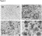
    • Figure 3 : Patient-derived organoid cell line (OVK ORG 2) shows established stable long term growth in medium B
      The upper part shows the absence of organoid formation from cancer cells in standard SM medium, while healthy fallopian tube cells from the same patient gave rise to a stable organoid culture. The lower part shows the formation of organoids from cancer cells in medium B.
    • Figure 4 : Confocal images of a patient derived organoid cell line (OVK ORG 11) showing strong nuclear accumulation of p53 which is characteristic in cells harboring p53.
      Organoids are positive for p53, EpCAM and PAX8 and have highly polymorphic nuclei which is hallmark of HGSC.
    • Figure 5 : Individual response curve to carboplatin treatment in organoid cell lines ORG1 and ORG2
      Treatment of organoid cancer lines with carboplatin, a standard component of the chemotherapy treatment for HGS patients revealed differences in responsivness and kinetics. Cell vialiability (y axis) was determined proportional to the strength of luminiscence signal (Cell titer R Glow 3D Promega). The test also showed only minor technical variability among triplicates (bars of different color) which makes it suitable for quantitative studies.
    Example Methods
  • All samples processed in the study were obtained as fragments of solid tumor deposits from chemotherapy naïve high grade serous ovarian cancer (HGSC) patients undergoing debulking surgery. Tumor samples were removed during the surgery. Initial assessment of the tumor content was performed by the operating clinician. Samples were transported on ice within 2 hours of the procedure and subjected to a cell isolation procedure.
  • The following steps were performed to ensure proper quality analysis (phenotypic and genotypic) of the processed tissue: - a 3 mm3 fragment was shock frozen in liquid nitrogen and stored at -80°C; a further 3 mm3 fragment was fixed at room temperature (RT) for 24 h in 3.7 % paraformaldehyde (PFA) followed by paraffin embedding. The remaining tissue was used for cell isolation.
  • In the following, an experimental procedure including all the steps required for establishment of long term organoid culture from HGSC patients tissue is described. As Matrigel® is highly selective and supports growth of epithelial cells no absolute exclusion of stroma cells is necessary to obtain pure organoid lines which are 100% EpCam positive.
  • Step 1: Isolation and 2D cultivation of cancer cells
    • Adv. +++: Advanced F12 Medium + HEPES (6 ml) + Glutamax (5 ml)+ 5% fetal calf serum (FCS)
    • Adv. ++: Advanced F12 Medium + HEPES (6 ml) + Glutamax (5 ml)
      • Cut tissue with a scissor in small pieces and then mince further with scalpel for 3-5 minutes
      • Collect all fragments, incubate with 2 ml Collagenase Type 1 + 2 ml Collagenase type II and transfer to a 6 well plate,
      • Incubate fragments for 45 min at 37°C 5% CO2,
      • Add 5 ml Adv +++ and transfer in to a 50 ml Falcon tube,
      • Vortex vigorously for 30 seconds,
      • Let pieces settle down, take supernatant and transfer in to a fresh tube,
      • Add again 5 ml Adv +++, mix with a pipette,
      • Let pieces settle down, take supernatant and transfer in to a fresh tube,
      • Centrifuge for 5 min at 400 rpm,
      • Discard supernatant,
      • Prepare 10 ml Adv +++, add 30 µl Y-27632 and 10 µl hEGF (final conc. 10 ng/ml),
      • Resuspend cell pellet in medium,
      • Seed cell in a flask,
      • Incubate at 5% CO2 at 37°C and grow cells 3-5 days prior to transfer to 3D culture.
    Step 2: Seeding cancer cells in 3D culture
    • Remove Medium from the flask and wash one time with PBS,
    • Add 1 ml TrypLE and incubate 10 min until cells are completely detached,
    • Remove cells with 5 to 10 ml of Adv ++ and transfer in a 15 ml Falcon tube,
    • Centrifuge at 1200 rpm for 5 min,
    • Remove supernatant,
    • Add 1 ml add Adv ++ and count cells,
    • Keep cells on ice during this time;
    Seeding of cells:
    • For a 24 well plate - 20.000 cells per 50 µl Matrigel
    • For a 48 well plate - 10.000 cells per 25 µl Matrigel
    • Seed cells in prewarmed plate,
    • Let Matrigel solidify for 30 min in the incubator,
    • Add growth medium,
    • For a 24 well plate - 500µl,
    • For a 48 well plate - 250 µl,
    • Change Medium twice times per week.
    Step 3: Splitting of the cancer organoid culture
  • Splitting should be carried out about once per 3 weeks.
    • Remove medium from all wells,
    • Add 1 ml ice cold Adv +++ to the Matrigel droplet,
    • Scrape with pipet tip on the bottom of well,
    • As Matrigel dissolves transfer organoids in the medium to 15 ml Falcon tube,
    • Wash the splitting well with 500 µl cold medium and put the medium thereafter into the same Falcon tube,
    • Fill up to 5 ml with Adv ++ (cold) and spin down (1000 rpm, 5 min),
    • Remove supernatant and add 1 ml TrypLE,
    • Incubate for 10 min at 37 C, vortex shortly 2-3 times for 20 seconds,
    • Fill up to 5 ml with cold Adv ++,
    • Centrifuge at 4°C for 5 min at 1200 rpm,
    • Remove the supernatant,
    • If impossible, wash the pellet again,
    • For 1 well dissolve cells in 50 µl Matrigel, seed as an drop per well,
    • Incubate for 30 min at 37°C,
    • Add culture medium,
    • Change medium every 3-4 days.
    Results
  • We report culture conditions and procedures for establishing a stable long term organoid culture from primary tumor deposits of high grade serous ovarian cancer.
  • There are substantial changes in the niche conditions which need to be provided to ensure in vitro cultivation of patient tumor samples, compared to the already described niche conditions for adult stem cells from the healthy epithelium (Kessler et al., 2015, supra). Significantly, we found that exogenous supplementation of Wnt 3a is detrimental for ovarian cancer stem cells as all of the established lines grew in Wnt3a-negative media. However, some individual samples continued to benefit from supplementation of Wnt agonist.
  • In contrast to the healthy oviductal epithelium, and other healthy mucosal models, we found that a HGSC organoid culture is dependent on active BMP signaling. This may be achieved by omission of the Noggin from the media (see medium A). As Noggin is a potent chelating agent for endogenous BMPs which are produced in the organoids this leads to the activation of downstream BMP signaling. Interestingly, none of the tested cancer tissues was able to give rise to an organoid culture in standard 3D medium (SM). Organoids could not form at all, or experienced growth arrest during cultivation (Figure 1).
  • Cancer cells seeded in medium A, containing only basic medium components plus EGF and BMP2, gave rise to a stable and expandable long term culture. Although endogenous activation of the BMP pathway occurs as direct consequence of the absence of Noggin in the medium it was tested if increasing the amount of BMP confers further growth advantage to the organoids. Indeed, organoid formation efficiency in the HGSC organoid cultures was clearly improved by exogenous supplementation of soluble BMP2 (final concentration 10 ng/ml) to the growth medium (Figure 2). Thus, BMP2 was included in the standard composition of the medium A.
  • We found stable long term propagation of HGSC organoids in basic medium (see table) supplemented solely with EGF and BMP2 (designated medium A). However two of the generated cultures required additional growth support which was provided by Rspondin 1 and FGF (medium B) in the medium. Both media lack Noggin and thus have activated BMP signaling contrary to the requirements of a healthy organoid culture. As a control comparison, a Fallopian tube organoid culture from the same patient efficiently cultivated in SM medium, showing that new properties are restricted to the malignant tissue. Table 1
    Basic components (present in all media) Components of standard medium for healthy FT (SM) Components of medium A Components of medium B
    Advance F12++, B27 2%, Nicotinamide 1mM, N2 1%, Y-27632 9 µM, TGFβ RI 0,5 µM inhibitor EGF 10ng/ml, FGF 100 ng/ml, Noggin 100 ng/ml, Rspondin 10 % conditioned medium Wnt 25 % conditioned medium EGF 10 ng/ml, BMP2 10 ng/ml EGF 10 ng/ml, FGF 100 ng/ml, Rspondin, 10 % conditioned medium
  • In order to find out whether Rspondin is required for achieving stable long term cultivation, each patient sample should be subjected to parallel testing under two different conditions in the presence and absence of Rspondin: for example, medium A and (general organoid culture supplements + BMP2 + EGF) and medium B (general organoid culture supplements + EGF + FGF + Rspondin1).
  • Patient-derived organoid cultures from ovarian cancer can be propagated stably in a long term culture (>6 months) with regular splitting intervals of e.g. 3-4 weeks. The cultures can be routinely frozen and thawed which is a key prerequisite for biobanking storage.
  • Immunofluorescence analysis of the key structural and functional biomarkers confirmed that organoids match native tumor tissue in phenotype, e.g. PAX8 confirming Mullerean lineage, EpCAM, p53 and p16 (Figure 4). All tested organoid cultures were matched in phenotype to the fragment of native tumor processed and stained in standard procedure which was used for clinical diagnosis. Ovarian cancer cancer organoids mostly lack central cavities in contrast to fallopian tube organoids which are built of polarized epithelial monolayers. This is expected as histological atypia and stratification are present already in the early stages of the carcinogenesis in the fallopian tube (Shaw et al 2009, Modern Pathology 22, 1135-1138). Panel sequencing of paired organoid culture and tissue confirmed that the tested organoids recapitulate cancer tissue composition also on the genomic level. Importantly cultures can be expanded to multi-well format to enable high throughput experimental set up. Pilot drug test experiments with carboplatin, first line compulsory agent in the treatment of the HGSC patients revealed that organoids show individual patient profiles in their response to the drug which can be determined by precise quantitative measurements.
  • Therefore it can be concluded that ovarian cancer organoids fatefully reproduce tumor cells in vitro and thus represent an adequate biological model which opens a potential to advance treatment approaches in several major categories: 1) in vitro testing of drug response (complementary to the in silico predictions based on NGS data); 2) drug screening of new drug candidates; 3) in vitro study of resistance mechanisms; 4) identification of new biomarkers, as tissue based approaches are limited by a high background due to complexity of in vivo system; 5) identification of novel neo-antigens for improving efficiency of immunotherapies; 6) development of assays to study antigen specific immune responses in vitro such are direct cytotoxicity assays (CTL assays) by using patient cancer organoids; 7) investigating mechanisms to improve efficiency of the immunotherapy by genetically modifying cancer organoids.
  • Within this study we have so far generated 15 patient organoid lines. The production method could be further improved by optimizing clinical sampling during surgery as most of the patients undergo surgery at FIGO (International Federation of Gynecology and Obstetrics) stage III where metastatic peritoneum deposits are abundant and any large tumor mass is likely heterogeneous. The procedure described herein sets a major milestone which enables creation of live tumor biobanks for high number of patients.
  • Taking into account very narrow therapeutic options which are currently available to ovarian cancer patients, and wide spread platinum resistence which ultimately develops in all patients, the organoid culture represents an attractive model to test in advance the therapeutic response of each patient to different classes of approved drugs and provide the clinician with valuable information which could improve decision making process at the late stages. This type of application is a promising methodology for improvement of personalized therapy.

Claims (15)

  1. A method for the production of an ovarian cancer or cancer precursor cell culture, said method comprising the steps:
    (a) cultivating ovarian cancer or cancer precursor cells in a suitable cell culture medium which is capable of BMP signaling, and
    (b) optionally obtaining a cell culture from the cultivation step (a).
  2. The method of claim 1, wherein the cell culture is an organoid culture.
  3. The method of claim 1 or 2, wherein the culture medium comprises at least one compound which stimulates BMP signaling selected from mitogenic growth factors such as EGF or/and FGF, a BMP, e.g. BMP2, or/and a Rspondin, e.g. Rspondin1, wherein the medium particularly comprises
    (iv) EGF or/and a BMP, or
    (v) EGF, FGF or/and a Rspondin.
  4. The method of any one of claims 1 to 3, wherein the culture medium is free from exogenously added Wnt protein, e.g. from an exogenously added Wnt3a protein.
  5. The method of any one of claims 1 to 4, wherein the culture medium is free of an exogenously added compound which inhibits BMP signaling, such as Noggin.
  6. The method of any one of the claims 1 to 5, wherein the ovarian cancer cells are primary ovarian cancer or cancer precursor cells, more particularly primary ovarian cancer or cancer precursor cells derived from a human patient.
  7. The method of any one of the claims 1 to 4, wherein the ovarian cancer is a high-grade serous ovarian carcinoma.
  8. An ovarian cancer or cancer precursor cell organoid culture which can be stably propagated.
  9. The organoid culture of claim 8, as obtainable by the method of any one of claims 1 to 7.
  10. A biobank, comprising a plurality of different organoid cultures of claim 8 or 9.
  11. Use of an organoid culture of claim 8 or 9, or a biobank of claim 10,
    (i) for the determination of the efficacy of a compound in the treatment of ovarian cancer, or
    (ii)for the identification of a compound suitable in the treatment of ovarian cancer.
  12. Use of a cell culture medium which is capable of BMP signaling, for the production of a culture, e.g. an organoid culture from ovarian cancer or cancer precursor cells.
  13. A method for the determination of the efficacy of a compound in the treatment of ovarian cancer, particularly of ovarian cancer in an individual patient, comprising the steps
    (a) providing an ovarian cancer or cancer precursor cell organoid culture,
    (b) contacting at least one compound with the organoid culture provided in step (a), said at least one compound being selected from therapeutically active agents suitable for the treatment of ovarian cancer,
    (c) determining growth or/and propagation of the organoid culture after being contacted with the at least one compound, and
    (d) selecting at least one compound which inhibits growth or/and propagation of the organoid culture, as determined in step (c).
  14. A screening method for the identification of a compound suitable in the treatment of ovarian cancer, particularly of ovarian cancer in a plurality of different patients, comprising the steps
    (a) providing at least one ovarian cancer or cancer precursor cell organoid culture,
    (b) contacting at least one compound with the at least one organoid culture provided in step (a), said at least one compound being selected from candidate active agents presumed to be suitable for the treatment of ovarian cancer,
    (c) determining growth or/and propagation of the at least one organoid culture after being contacted with the at least one compound, and
    (d) selecting at least one compound which inhibits growth or/and propagation of at least one organoid culture, as determined in step (c).
  15. A compound selected from active agents suitable for the treatment of ovarian cancer, for use in the treatment of a patient suffering from ovarian cancer, said treatment comprising the steps
    (a) providing an ovarian cancer or cancer precursor cell organoid culture from said patient,
    (b) contacting at least one compound with the organoid culture provided in step (a), said at least one compound being selected from active agents suitable for the treatment of ovarian cancer,
    (c) determining growth or/and propagation of the organoid after being contacted with the at least one compound,
    (d) selecting a compound which inhibits growth or/and propagation of the organoid culture, as determined in step (c), and
    (e) treating the patient with compound selected in step (d).
EP18158377.4A 2018-02-23 2018-02-23 Ovarian cancer organoid culture Withdrawn EP3530732A1 (en)

Priority Applications (4)

Application Number Priority Date Filing Date Title
EP18158377.4A EP3530732A1 (en) 2018-02-23 2018-02-23 Ovarian cancer organoid culture
EP19705543.7A EP3755791B1 (en) 2018-02-23 2019-02-22 Ovarian cancer organoid culture
PCT/EP2019/054468 WO2019162453A1 (en) 2018-02-23 2019-02-22 Ovarian cancer organoid culture
US16/967,548 US20210230555A1 (en) 2018-02-23 2019-02-22 Ovarian cancer organoid culture

Applications Claiming Priority (1)

Application Number Priority Date Filing Date Title
EP18158377.4A EP3530732A1 (en) 2018-02-23 2018-02-23 Ovarian cancer organoid culture

Publications (1)

Publication Number Publication Date
EP3530732A1 true EP3530732A1 (en) 2019-08-28

Family

ID=61274173

Family Applications (2)

Application Number Title Priority Date Filing Date
EP18158377.4A Withdrawn EP3530732A1 (en) 2018-02-23 2018-02-23 Ovarian cancer organoid culture
EP19705543.7A Active EP3755791B1 (en) 2018-02-23 2019-02-22 Ovarian cancer organoid culture

Family Applications After (1)

Application Number Title Priority Date Filing Date
EP19705543.7A Active EP3755791B1 (en) 2018-02-23 2019-02-22 Ovarian cancer organoid culture

Country Status (3)

Country Link
US (1) US20210230555A1 (en)
EP (2) EP3530732A1 (en)
WO (1) WO2019162453A1 (en)

Cited By (3)

* Cited by examiner, † Cited by third party
Publication number Priority date Publication date Assignee Title
CN111040996A (en) * 2019-12-06 2020-04-21 北京科途医学科技有限公司 Method for preparing ovarian cancer organoid
CN112391348A (en) * 2020-10-16 2021-02-23 中国人民解放军空军军医大学 Ovarian cancer organoid 3D culture method
CN113493763A (en) * 2020-03-18 2021-10-12 四川大学华西医院 Mouse ovary primary cell culture medium applicable to gene editing and in-vitro culture method

Families Citing this family (2)

* Cited by examiner, † Cited by third party
Publication number Priority date Publication date Assignee Title
CN114921414B (en) * 2022-07-22 2022-09-27 北京和沛生物科技有限公司 Method for in vitro separation culture of circulating tumor cells
CN116716236B (en) * 2023-06-29 2024-10-29 重庆大学附属肿瘤医院 A method for constructing cervical cancer organoids and its application

Citations (1)

* Cited by examiner, † Cited by third party
Publication number Priority date Publication date Assignee Title
US20120028355A1 (en) 2009-02-03 2012-02-02 Koninklijke Nederlandse Akademie Van Wetenschappen Culture medium for epithelial stem cells and organoids comprising said stem cells

Family Cites Families (1)

* Cited by examiner, † Cited by third party
Publication number Priority date Publication date Assignee Title
US20160237400A1 (en) * 2013-03-15 2016-08-18 The Jackson Laboratory Isolation of non-embryonic stem cells and uses thereof

Patent Citations (1)

* Cited by examiner, † Cited by third party
Publication number Priority date Publication date Assignee Title
US20120028355A1 (en) 2009-02-03 2012-02-02 Koninklijke Nederlandse Akademie Van Wetenschappen Culture medium for epithelial stem cells and organoids comprising said stem cells

Non-Patent Citations (13)

* Cited by examiner, † Cited by third party
Title
"TCGA study", NATURE, vol. 474, 2011, pages 609 - 615
BARKER ET AL., NATURE, vol. 449, 2007, pages 1003 - 1007
COFFMAN L G ET AL: "Human carcinoma-associated mesenchymal stem cells promote ovarian cancer chemotherapy resistance via a BMP4/HH signaling loop", ONCOTARGET, vol. 7, no. 6, 9 February 2016 (2016-02-09), pages 6916 - 6932, XP055496128, ISSN: 1949-2553, DOI: 10.18632/oncotarget.6870 *
FUJII ET AL., CELL STEM CELL, vol. 18, 2016, pages 827 - 838
FUJII M ET AL: "A Colorectal Tumor Organoid Library Demonstrates Progressive Loss of Niche Factor Requirements during Tumorigenesis", CELL STEM CELL, vol. 18, no. 6, 19 May 2016 (2016-05-19), pages 827 - 838, XP029567586, ISSN: 1934-5909, DOI: 10.1016/J.STEM.2016.04.003 *
JABS J ET AL: "Screening drug effects in patient-derived cancer cells links organoid responses to genome alterations", MOLECULAR SYSTEMS BIOLOGY, vol. 13, 955, 27 November 2017 (2017-11-27), XP055496120, ISSN: 1744-4292, DOI: 10.15252/msb.20177697 *
KESSLER ET AL., NATURE COMMUNICATIONS, vol. 6, 2015
KESSLER M ET AL: "The Notch and Wnt pathways regulate stemness and differentiation in human fallopian tube organoids", NATURE COMMUNICATIONS, vol. 6, 8989, 8 December 2015 (2015-12-08), XP055496132, DOI: 10.1038/ncomms9989 *
NOLL E M ET AL: "CYP3A5 mediates basal and acquired therapy resistance in different subtypes of pancreatic ductal adenocarcinoma", NATURE MEDICINE, vol. 22, no. 3, 8 February 2016 (2016-02-08), pages 278 - 287, XP055496124, ISSN: 1078-8956, DOI: 10.1038/nm.4038 *
PENG J ET AL: "The BMP Signaling Pathway Leads to Enhanced Proliferation in Serous Ovarian Cancer-A Potential Therapeutic Target", MOLECULAR CARCINOGENESIS, vol. 55, 7 February 2016 (2016-02-07), pages 335 - 345, XP055496129, DOI: 10.1002/mc.22283 *
SHAW ET AL., MODERN PATHOLOGY, vol. 22, 2009, pages 1135 - 1138
VAN DE WETERING ET AL., CELL, vol. 161, 2015, pages 933 - 945
VAN DE WETERING M ET AL: "Prospective Derivation of a Living Organoid Biobank of Colorectal Cancer Patients", CELL, vol. 161, no. 4, 7 May 2015 (2015-05-07), pages 933 - 945, XP029224300, ISSN: 0092-8674, DOI: 10.1016/J.CELL.2015.03.053 *

Cited By (3)

* Cited by examiner, † Cited by third party
Publication number Priority date Publication date Assignee Title
CN111040996A (en) * 2019-12-06 2020-04-21 北京科途医学科技有限公司 Method for preparing ovarian cancer organoid
CN113493763A (en) * 2020-03-18 2021-10-12 四川大学华西医院 Mouse ovary primary cell culture medium applicable to gene editing and in-vitro culture method
CN112391348A (en) * 2020-10-16 2021-02-23 中国人民解放军空军军医大学 Ovarian cancer organoid 3D culture method

Also Published As

Publication number Publication date
EP3755791A1 (en) 2020-12-30
EP3755791B1 (en) 2024-08-21
WO2019162453A1 (en) 2019-08-29
US20210230555A1 (en) 2021-07-29

Similar Documents

Publication Publication Date Title
EP3755791B1 (en) Ovarian cancer organoid culture
Qu et al. Patient-derived organoids in human cancer: a platform for fundamental research and precision medicine
US20230272489A1 (en) Tumor organoid culture compositions, systems, and methods
JP7461674B2 (en) Culture medium for esophageal squamous cell carcinoma epithelial cells, culture method, and use thereof
Pastuschek et al. Molecular characteristics of established trophoblast-derived cell lines
Wang et al. The potential of organoids in urological cancer research
Lao et al. Improved methods to generate spheroid cultures from tumor cells, tumor cells & fibroblasts or tumor-fragments: microenvironment, microvesicles and MiRNA
Sun et al. Prediction of clinical precision chemotherapy by patient‐derived 3D bioprinting models of colorectal cancer and its liver metastases
Tomaszewski et al. Adipose-derived stem cell-secreted factors promote early stage follicle development in a biomimetic matrix
Xu et al. Organoids in lung cancer: A teenager with infinite growth potential
Jin et al. Tumor organoids to study gastroesophageal cancer: a primer
Marr et al. WERF Endometriosis Phenome and Biobanking Harmonisation Project for Experimental Models in Endometriosis Research (EPHect-EM-Organoids): endometrial organoids as an emerging technology for endometriosis research
CN115975939B (en) Cervical cancer organoid culture medium, and culture method and application thereof
CN114075539A (en) Method for constructing in-situ primary bladder cancer animal model
Aguilar-Cázarez et al. A protocol for explant cultures of IDH1-mutant diffuse low-grade gliomas
Baker et al. Generation and culture of tumor and metastatic organoids from murine models of pancreatic ductal adenocarcinoma
Krbal et al. Derivation and basic characterization of colorectal carcinoma primary cell lines
CN115960816B (en) Culture medium, culture method and application of lung cancer organoids
Teriyapirom et al. Acquired epithelial WNT secretion drives niche independence of developing gastric cancer
WO2023060764A1 (en) Culture medium for primary cell of gastric carcinoma, and culture method therefor
WO2023039999A1 (en) Culture medium and culture method for lung cancer epithelial cells, and application thereof
CN115975937A (en) Culture medium and culture method of liver cancer suspension organoids
CN115975910B (en) Culture medium and culture method for gastric cancer organoids
CN115595308A (en) Culture medium for lung cancer epithelial cells, culture method and application thereof
Constant et al. Advanced human in vitro models for the discovery and development of lung cancer therapies

Legal Events

Date Code Title Description
PUAI Public reference made under article 153(3) epc to a published international application that has entered the european phase

Free format text: ORIGINAL CODE: 0009012

AK Designated contracting states

Kind code of ref document: A1

Designated state(s): AL AT BE BG CH CY CZ DE DK EE ES FI FR GB GR HR HU IE IS IT LI LT LU LV MC MK MT NL NO PL PT RO RS SE SI SK SM TR

AX Request for extension of the european patent

Extension state: BA ME

STAA Information on the status of an ep patent application or granted ep patent

Free format text: STATUS: THE APPLICATION IS DEEMED TO BE WITHDRAWN

18D Application deemed to be withdrawn

Effective date: 20200229