EP3527176A1 - Crimping heart valve with nitinol braid - Google Patents
Crimping heart valve with nitinol braid Download PDFInfo
- Publication number
- EP3527176A1 EP3527176A1 EP18204261.4A EP18204261A EP3527176A1 EP 3527176 A1 EP3527176 A1 EP 3527176A1 EP 18204261 A EP18204261 A EP 18204261A EP 3527176 A1 EP3527176 A1 EP 3527176A1
- Authority
- EP
- European Patent Office
- Prior art keywords
- sheath
- valve
- heart valve
- stent
- attachment area
- Prior art date
- Legal status (The legal status is an assumption and is not a legal conclusion. Google has not performed a legal analysis and makes no representation as to the accuracy of the status listed.)
- Withdrawn
Links
Images




Classifications
-
- A—HUMAN NECESSITIES
- A61—MEDICAL OR VETERINARY SCIENCE; HYGIENE
- A61F—FILTERS IMPLANTABLE INTO BLOOD VESSELS; PROSTHESES; DEVICES PROVIDING PATENCY TO, OR PREVENTING COLLAPSING OF, TUBULAR STRUCTURES OF THE BODY, e.g. STENTS; ORTHOPAEDIC, NURSING OR CONTRACEPTIVE DEVICES; FOMENTATION; TREATMENT OR PROTECTION OF EYES OR EARS; BANDAGES, DRESSINGS OR ABSORBENT PADS; FIRST-AID KITS
- A61F2/00—Filters implantable into blood vessels; Prostheses, i.e. artificial substitutes or replacements for parts of the body; Appliances for connecting them with the body; Devices providing patency to, or preventing collapsing of, tubular structures of the body, e.g. stents
- A61F2/02—Prostheses implantable into the body
- A61F2/24—Heart valves ; Vascular valves, e.g. venous valves; Heart implants, e.g. passive devices for improving the function of the native valve or the heart muscle; Transmyocardial revascularisation [TMR] devices; Valves implantable in the body
- A61F2/2427—Devices for manipulating or deploying heart valves during implantation
- A61F2/2436—Deployment by retracting a sheath
-
- A—HUMAN NECESSITIES
- A61—MEDICAL OR VETERINARY SCIENCE; HYGIENE
- A61F—FILTERS IMPLANTABLE INTO BLOOD VESSELS; PROSTHESES; DEVICES PROVIDING PATENCY TO, OR PREVENTING COLLAPSING OF, TUBULAR STRUCTURES OF THE BODY, e.g. STENTS; ORTHOPAEDIC, NURSING OR CONTRACEPTIVE DEVICES; FOMENTATION; TREATMENT OR PROTECTION OF EYES OR EARS; BANDAGES, DRESSINGS OR ABSORBENT PADS; FIRST-AID KITS
- A61F2/00—Filters implantable into blood vessels; Prostheses, i.e. artificial substitutes or replacements for parts of the body; Appliances for connecting them with the body; Devices providing patency to, or preventing collapsing of, tubular structures of the body, e.g. stents
- A61F2/02—Prostheses implantable into the body
- A61F2/24—Heart valves ; Vascular valves, e.g. venous valves; Heart implants, e.g. passive devices for improving the function of the native valve or the heart muscle; Transmyocardial revascularisation [TMR] devices; Valves implantable in the body
- A61F2/2412—Heart valves ; Vascular valves, e.g. venous valves; Heart implants, e.g. passive devices for improving the function of the native valve or the heart muscle; Transmyocardial revascularisation [TMR] devices; Valves implantable in the body with soft flexible valve members, e.g. tissue valves shaped like natural valves
- A61F2/2418—Scaffolds therefor, e.g. support stents
-
- A—HUMAN NECESSITIES
- A61—MEDICAL OR VETERINARY SCIENCE; HYGIENE
- A61F—FILTERS IMPLANTABLE INTO BLOOD VESSELS; PROSTHESES; DEVICES PROVIDING PATENCY TO, OR PREVENTING COLLAPSING OF, TUBULAR STRUCTURES OF THE BODY, e.g. STENTS; ORTHOPAEDIC, NURSING OR CONTRACEPTIVE DEVICES; FOMENTATION; TREATMENT OR PROTECTION OF EYES OR EARS; BANDAGES, DRESSINGS OR ABSORBENT PADS; FIRST-AID KITS
- A61F2/00—Filters implantable into blood vessels; Prostheses, i.e. artificial substitutes or replacements for parts of the body; Appliances for connecting them with the body; Devices providing patency to, or preventing collapsing of, tubular structures of the body, e.g. stents
- A61F2/02—Prostheses implantable into the body
- A61F2/24—Heart valves ; Vascular valves, e.g. venous valves; Heart implants, e.g. passive devices for improving the function of the native valve or the heart muscle; Transmyocardial revascularisation [TMR] devices; Valves implantable in the body
- A61F2/2427—Devices for manipulating or deploying heart valves during implantation
-
- A—HUMAN NECESSITIES
- A61—MEDICAL OR VETERINARY SCIENCE; HYGIENE
- A61F—FILTERS IMPLANTABLE INTO BLOOD VESSELS; PROSTHESES; DEVICES PROVIDING PATENCY TO, OR PREVENTING COLLAPSING OF, TUBULAR STRUCTURES OF THE BODY, e.g. STENTS; ORTHOPAEDIC, NURSING OR CONTRACEPTIVE DEVICES; FOMENTATION; TREATMENT OR PROTECTION OF EYES OR EARS; BANDAGES, DRESSINGS OR ABSORBENT PADS; FIRST-AID KITS
- A61F2/00—Filters implantable into blood vessels; Prostheses, i.e. artificial substitutes or replacements for parts of the body; Appliances for connecting them with the body; Devices providing patency to, or preventing collapsing of, tubular structures of the body, e.g. stents
- A61F2/95—Instruments specially adapted for placement or removal of stents or stent-grafts
-
- A—HUMAN NECESSITIES
- A61—MEDICAL OR VETERINARY SCIENCE; HYGIENE
- A61F—FILTERS IMPLANTABLE INTO BLOOD VESSELS; PROSTHESES; DEVICES PROVIDING PATENCY TO, OR PREVENTING COLLAPSING OF, TUBULAR STRUCTURES OF THE BODY, e.g. STENTS; ORTHOPAEDIC, NURSING OR CONTRACEPTIVE DEVICES; FOMENTATION; TREATMENT OR PROTECTION OF EYES OR EARS; BANDAGES, DRESSINGS OR ABSORBENT PADS; FIRST-AID KITS
- A61F2/00—Filters implantable into blood vessels; Prostheses, i.e. artificial substitutes or replacements for parts of the body; Appliances for connecting them with the body; Devices providing patency to, or preventing collapsing of, tubular structures of the body, e.g. stents
- A61F2/95—Instruments specially adapted for placement or removal of stents or stent-grafts
- A61F2/9522—Means for mounting a stent or stent-graft onto or into a placement instrument
-
- A—HUMAN NECESSITIES
- A61—MEDICAL OR VETERINARY SCIENCE; HYGIENE
- A61F—FILTERS IMPLANTABLE INTO BLOOD VESSELS; PROSTHESES; DEVICES PROVIDING PATENCY TO, OR PREVENTING COLLAPSING OF, TUBULAR STRUCTURES OF THE BODY, e.g. STENTS; ORTHOPAEDIC, NURSING OR CONTRACEPTIVE DEVICES; FOMENTATION; TREATMENT OR PROTECTION OF EYES OR EARS; BANDAGES, DRESSINGS OR ABSORBENT PADS; FIRST-AID KITS
- A61F2/00—Filters implantable into blood vessels; Prostheses, i.e. artificial substitutes or replacements for parts of the body; Appliances for connecting them with the body; Devices providing patency to, or preventing collapsing of, tubular structures of the body, e.g. stents
- A61F2/02—Prostheses implantable into the body
- A61F2/24—Heart valves ; Vascular valves, e.g. venous valves; Heart implants, e.g. passive devices for improving the function of the native valve or the heart muscle; Transmyocardial revascularisation [TMR] devices; Valves implantable in the body
- A61F2/2427—Devices for manipulating or deploying heart valves during implantation
- A61F2/2439—Expansion controlled by filaments
-
- A—HUMAN NECESSITIES
- A61—MEDICAL OR VETERINARY SCIENCE; HYGIENE
- A61F—FILTERS IMPLANTABLE INTO BLOOD VESSELS; PROSTHESES; DEVICES PROVIDING PATENCY TO, OR PREVENTING COLLAPSING OF, TUBULAR STRUCTURES OF THE BODY, e.g. STENTS; ORTHOPAEDIC, NURSING OR CONTRACEPTIVE DEVICES; FOMENTATION; TREATMENT OR PROTECTION OF EYES OR EARS; BANDAGES, DRESSINGS OR ABSORBENT PADS; FIRST-AID KITS
- A61F2/00—Filters implantable into blood vessels; Prostheses, i.e. artificial substitutes or replacements for parts of the body; Appliances for connecting them with the body; Devices providing patency to, or preventing collapsing of, tubular structures of the body, e.g. stents
- A61F2/95—Instruments specially adapted for placement or removal of stents or stent-grafts
- A61F2002/9528—Instruments specially adapted for placement or removal of stents or stent-grafts for retrieval of stents
-
- A—HUMAN NECESSITIES
- A61—MEDICAL OR VETERINARY SCIENCE; HYGIENE
- A61F—FILTERS IMPLANTABLE INTO BLOOD VESSELS; PROSTHESES; DEVICES PROVIDING PATENCY TO, OR PREVENTING COLLAPSING OF, TUBULAR STRUCTURES OF THE BODY, e.g. STENTS; ORTHOPAEDIC, NURSING OR CONTRACEPTIVE DEVICES; FOMENTATION; TREATMENT OR PROTECTION OF EYES OR EARS; BANDAGES, DRESSINGS OR ABSORBENT PADS; FIRST-AID KITS
- A61F2/00—Filters implantable into blood vessels; Prostheses, i.e. artificial substitutes or replacements for parts of the body; Appliances for connecting them with the body; Devices providing patency to, or preventing collapsing of, tubular structures of the body, e.g. stents
- A61F2/95—Instruments specially adapted for placement or removal of stents or stent-grafts
- A61F2002/9534—Instruments specially adapted for placement or removal of stents or stent-grafts for repositioning of stents
-
- A—HUMAN NECESSITIES
- A61—MEDICAL OR VETERINARY SCIENCE; HYGIENE
- A61F—FILTERS IMPLANTABLE INTO BLOOD VESSELS; PROSTHESES; DEVICES PROVIDING PATENCY TO, OR PREVENTING COLLAPSING OF, TUBULAR STRUCTURES OF THE BODY, e.g. STENTS; ORTHOPAEDIC, NURSING OR CONTRACEPTIVE DEVICES; FOMENTATION; TREATMENT OR PROTECTION OF EYES OR EARS; BANDAGES, DRESSINGS OR ABSORBENT PADS; FIRST-AID KITS
- A61F2210/00—Particular material properties of prostheses classified in groups A61F2/00 - A61F2/26 or A61F2/82 or A61F9/00 or A61F11/00 or subgroups thereof
- A61F2210/0014—Particular material properties of prostheses classified in groups A61F2/00 - A61F2/26 or A61F2/82 or A61F9/00 or A61F11/00 or subgroups thereof using shape memory or superelastic materials, e.g. nitinol
-
- A—HUMAN NECESSITIES
- A61—MEDICAL OR VETERINARY SCIENCE; HYGIENE
- A61F—FILTERS IMPLANTABLE INTO BLOOD VESSELS; PROSTHESES; DEVICES PROVIDING PATENCY TO, OR PREVENTING COLLAPSING OF, TUBULAR STRUCTURES OF THE BODY, e.g. STENTS; ORTHOPAEDIC, NURSING OR CONTRACEPTIVE DEVICES; FOMENTATION; TREATMENT OR PROTECTION OF EYES OR EARS; BANDAGES, DRESSINGS OR ABSORBENT PADS; FIRST-AID KITS
- A61F2220/00—Fixations or connections for prostheses classified in groups A61F2/00 - A61F2/26 or A61F2/82 or A61F9/00 or A61F11/00 or subgroups thereof
- A61F2220/0025—Connections or couplings between prosthetic parts, e.g. between modular parts; Connecting elements
-
- A—HUMAN NECESSITIES
- A61—MEDICAL OR VETERINARY SCIENCE; HYGIENE
- A61F—FILTERS IMPLANTABLE INTO BLOOD VESSELS; PROSTHESES; DEVICES PROVIDING PATENCY TO, OR PREVENTING COLLAPSING OF, TUBULAR STRUCTURES OF THE BODY, e.g. STENTS; ORTHOPAEDIC, NURSING OR CONTRACEPTIVE DEVICES; FOMENTATION; TREATMENT OR PROTECTION OF EYES OR EARS; BANDAGES, DRESSINGS OR ABSORBENT PADS; FIRST-AID KITS
- A61F2220/00—Fixations or connections for prostheses classified in groups A61F2/00 - A61F2/26 or A61F2/82 or A61F9/00 or A61F11/00 or subgroups thereof
- A61F2220/0025—Connections or couplings between prosthetic parts, e.g. between modular parts; Connecting elements
- A61F2220/0075—Connections or couplings between prosthetic parts, e.g. between modular parts; Connecting elements sutured, ligatured or stitched, retained or tied with a rope, string, thread, wire or cable
-
- A—HUMAN NECESSITIES
- A61—MEDICAL OR VETERINARY SCIENCE; HYGIENE
- A61F—FILTERS IMPLANTABLE INTO BLOOD VESSELS; PROSTHESES; DEVICES PROVIDING PATENCY TO, OR PREVENTING COLLAPSING OF, TUBULAR STRUCTURES OF THE BODY, e.g. STENTS; ORTHOPAEDIC, NURSING OR CONTRACEPTIVE DEVICES; FOMENTATION; TREATMENT OR PROTECTION OF EYES OR EARS; BANDAGES, DRESSINGS OR ABSORBENT PADS; FIRST-AID KITS
- A61F2220/00—Fixations or connections for prostheses classified in groups A61F2/00 - A61F2/26 or A61F2/82 or A61F9/00 or A61F11/00 or subgroups thereof
- A61F2220/0025—Connections or couplings between prosthetic parts, e.g. between modular parts; Connecting elements
- A61F2220/0083—Connections or couplings between prosthetic parts, e.g. between modular parts; Connecting elements using hook and loop-type fasteners
Definitions
- the present invention relates to collapsible prosthetic heart valve implantation, and more particularly, to apparatus and methods for collapsing prosthetic heart valves during an implantation procedure.
- Prosthetic heart valves that are collapsible to a relatively small circumferential size can be delivered into a patient less invasively than valves that are not collapsible.
- a collapsible valve may be delivered into a patient via a tube-like delivery apparatus such as a catheter, a trocar, a laparoscopic instrument, or the like. This collapsibility can avoid the need for a more invasive procedure such as full open-chest, open-heart surgery.
- Collapsible prosthetic heart valves typically take the form of a valve structure mounted on a stent.
- a stent There are two types of stents on which the valve structures are ordinarily mounted: a self-expanding stent and a balloon-expandable stent.
- a self-expanding stent To place such valves into a delivery apparatus and ultimately into a patient, the valve must first be collapsed or crimped to reduce its circumferential size.
- the prosthetic valve When a collapsed prosthetic valve has reached the desired implant site in the patient (e.g., at or near the annulus of the patient's heart valve that is to be replaced by the prosthetic valve), the prosthetic valve can be deployed or released from the delivery apparatus and re-expanded to its full operating size.
- the desired implant site in the patient e.g., at or near the annulus of the patient's heart valve that is to be replaced by the prosthetic valve
- the prosthetic valve can be deployed or released from the delivery apparatus and re-expanded to its full operating size.
- balloon-expandable valves this generally involves releasing the entire valve, assuring its proper location, and then expanding a balloon positioned within the valve stent.
- the stent automatically expands as the stent is withdrawn from the delivery apparatus.
- the annulus end of the valve is typically unsheathed (withdrawn from the delivery apparatus) and expanded first, to test the basic functionality of the valve, while the aortic end of the valve remains sheathed.
- a user such as an interventional cardiologist or a physician
- the user may re-sheath the annulus end, thereby crimping the valve for repositioning.
- the user can again deploy the valve.
- the present invention addresses these needs by providing a system for re-crimping the valve after it has been fully deployed.
- a system for crimping a heart valve during an implantation procedure is provided.
- the system advantageously allows for crimping of the valve after it has been fully deployed, and thus, allows a physician to replace the valve if it is not functioning properly and/or reposition the valve if proper fitment has not been achieved.
- One embodiment of the system includes a delivery tube extending in a longitudinal direction and defining a hollow lumen for receiving the heart valve, a deployment device including a first member and a second member moveably secured to the first member, and a sheath configured to at least partially surround a radially collapsible stent.
- the attachment device of the first member is attachable to a first attachment area of the sheath and the attachment device of the second member is attachable to a second attachment area of the sheath such that movement of the first member relative to the second member provides a tensioning force on the sheath for crimping the heart valve.
- the sheath may be formed of nitinol and be at least partially coated with a lubricant.
- the first attachment area of the sheath is provided adjacent a proximal end of the sheath and the second attachment area of the sheath is provided adjacent a distal end of the sheath. Furthermore, the first attachment area and second attachment area may include at least one loop for cooperating with the at least one of a hook, a clamp, and a tether provided on at least one of the first member and the second member.
- the tether may be a breakable tether.
- the second member is telescopingly arranged within the first member for transitioning the deployment device between a first condition, in which the first member and the second member define a first length, and a second condition, in which the first member and the second member define a second length greater than the first length.
- the heart valve includes a stent formed of a plurality of struts.
- the stent is transitionable between a radially collapsed state and a radially expanded state, such that in the expanded state, a diameter of the stent is larger than a diameter of the lumen of the delivery tube.
- the stent also includes a valve including a cuff and at least one leaflet. When the deployment device is transitioned from the first condition to the second condition, tension is applied to the sheath for radially compressing at least a portion of the stent.
- the system further includes a first wire and a second wire opposingly secured within the deployment device for guiding delivery of the heart valve.
- the method includes moving a first member connected to a sheath at a first location away from a second member connected to the sheath at a second location when the sheath at least partially encloses a stent configured to transition between a radially collapsed state and a radially expanded state. This movement applies a tensioning force to the sheath to radially engage the stent and transition the stent from the expanded state to the collapsed state.
- moving the first member away from the second member includes a telescoping movement.
- only one of the first member and the second member is disconnected from the sheath.
- the sheath remains in the body after withdrawal of the delivery device and in other aspects of the invention the sheath is removed from the body along with the delivery device.
- proximal when used in connection with a prosthetic heart valve, refers to the end of the heart valve closest to the heart when the heart valve is implanted in a patient
- distal when used in connection with a prosthetic heart valve, refers to the end of the heart valve farthest from the heart when the heart valve is implanted in a patient.
- the terms “trailing” and “leading” are to be taken as relative to the user of the delivery devices. "Trailing” is to be understood as relatively close to the operator, and “leading” is to be understood as relatively farther away from the operator.
- FIG. 1 shows a collapsible prosthetic heart valve 10 according to an embodiment of the present disclosure.
- the prosthetic heart valve 10 is designed to replace the function of the native aortic valve of a patient.
- Examples of collapsible prosthetic heart valves are described in U.S. Pat. No. 7,018,406 and U.S. Pat. No. 9,241,794 , the disclosures of both of which are hereby incorporated herein by reference.
- Prosthetic heart valve 10 includes an expandable stent 12 which may be formed from biocompatible materials that are capable of self-expansion, for example, shape memory alloys such as nitinol.
- Stent 12 extends from a distal or annulus end 14 to a proximal or aortic end 16, and includes an annulus section 18 adjacent distal end 14 and an aortic section 20 adjacent proximal end 16.
- Annulus section 18 has a relatively small cross-section in the expanded state compared to aortic section 20 in the expanded state.
- annulus section 18 is in the form of a cylinder having a substantially constant diameter along its length.
- a transition section 22 tapers outwardly from annulus section 18 to aortic section 20.
- Stent 12 may include one or more retaining elements 28 provided on aortic section 20 and sized and shaped to cooperate with a delivery device. Engagement of retaining elements 28 with a corresponding retaining structure on the delivery device maintains prosthetic heart valve 10 in the crimped position within the delivery device, minimizes longitudinal movement of the prosthetic heart valve relative to the delivery device during unsheathing or resheathing procedures, and prevents rotation of the prosthetic heart valve relative to the delivery device as the delivery device is advanced to the target location and during deployment.
- Stent 12 may also include a plurality of commissure features 30 for attaching the commissure between two adjacent leaflets to stent 12.
- commissure features 30 may lie at the intersection of four cells 24, two of the cells being adjacent one another in the same annular row, and the other two cells being in different annular rows and lying in an end-to-end relationship.
- commissure features 30 are positioned entirely within annulus section 18 or at the juncture of annulus section 18 and transition section 22.
- Commissure features 30 may include one or more eyelets 32 which facilitate the suturing of the leaflet commissure to stent 12.
- Prosthetic heart valve 10 includes a valve assembly 34 preferably positioned in annulus section 18. Valve assembly 34 may be secured to stent 12 by suturing valve assembly 34 to struts 26 and/or to commissure features 30. Valve assembly 34 includes a cuff 36 and a plurality of leaflets 38 which open and close collectively to function as a one-way valve. Since FIG. 1 illustrates a prosthetic heart valve for replacing a native tricuspid valve, such as the aortic valve, prosthetic heart valve 10 is illustrated with three leaflets 38, as well as three commissure features 30. However, it will be appreciated that prosthetic heart valves according to this aspect of the invention may have a greater or lesser number of leaflets 38 and/or commissure features 30.
- cuff 36 is illustrated in FIG. 1 as being disposed on the luminal or inner surface of annulus section 18, it is contemplated that cuff 36 may be disposed on the abluminal or outer surface of annulus section 18, or may cover all or part of either or both of the luminal and abluminal surfaces of annulus section 18.
- Both cuff 36 and leaflets 38 may be wholly or partly formed of any suitable biological material, such as bovine or porcine pericardium, or polymers, such as PTFE, urethanes and the like.
- Prosthetic heart valve 10 described above may be used to replace a native heart valve, such as the aortic valve, a surgical heart valve, or a heart valve that has undergone a surgical procedure.
- Prosthetic heart valve 10 may be delivered to the desired site (e.g., near a native aortic annulus) using the delivery device described in detail below.
- prosthetic heart valve 10 is disposed inside the delivery device in the collapsed condition.
- the delivery device may be introduced into a patient using a transfemoral, transapical, transseptal approach or another approaoch. Once the delivery device has reached the target site, the user may deploy prosthetic heart valve 10.
- the prosthetic heart valve Upon deployment, the prosthetic heart valve expands into secure engagement within the native anatomic structure such as the aortic annulus. When the prosthetic heart valve is properly positioned inside the patient, it works as a one-way valve, allowing blood to flow in one direction and preventing blood from flowing in the opposite direction.
- FIG. 2 illustrates a human heart H and two different approaches of delivering prosthetic heart valve 10 to its intended target at aortic valve AV.
- heart H includes an aorta A, an aortic arch AA and a left ventricle LV. Two separate paths are shown for introducing prosthetic heart valve 10 to aortic valve AV.
- a transfemoral approach of the prosthetic heart valve is indicated by the dashed arrow and the label "AV1".
- prosthetic heart valve 10 is inserted into the femoral artery, tracked through the vasculature and then introduced to the target site via the aortic arch AA. Echocardiography and other means may be used to help guide the delivery device through this approach.
- a second dashed arrow, labeled "AV2,” indicates a transapical approach of the prosthetic heart valve.
- transapical delivery a small incision is made between the ribs and into the apex of left ventricle LV to deliver the prosthetic heart valve to the target site.
- FIG. 3 illustrates a conventional device 100 for transfemoral delivery of a prosthetic heart valve.
- the leading end of a delivery tube 102 points in the distal direction of the heart.
- valve 10 Prior to deployment, valve 10 is disposed in delivery tube 102 with annulus section 18 closest to leading end 104 of delivery tube 102.
- the transfemoral approach first unsheathes annulus end 18 of heart valve 10, allowing annulus section 18 to expand prior to full deployment of valve 10.
- delivery tube 102 may be retracted toward its trailing end 106 while internal components (not shown) of delivery tube 102 hold heart valve 10 stationary.
- valve assembly 34 and specifically the function of leaflets 38, may be preliminarily tested in this approach without fully deploying heart valve 10.
- AV2 transapical approach
- AV1 transfemoral approach
- FIGS. 4A and 4B illustrate a conventional delivery device 200 used for a transapical approach.
- Delivery device 200 includes a delivery tube 202 having a leading end 204 connected to a trailing end 206 via a support core 208.
- Delivery tube 202 defines a compartment 210 for housing prosthetic heart valve 10.
- delivery device 200 is advanced in a forward direction through the apex of the heart and into the aorta until annulus end 14 of valve 10 is disposed at or near the annulus of the native valve and leading end 204 of delivery device 200 projects through the native valve into the aorta. While trailing end 206 of delivery device 200 is held stationary, leading end 204 is translated in a forward direction (depicted by arrow "S") away from trailing end 206 to remove prosthetic heart valve 10 from compartment 210. In this configuration, aortic section 20 of valve 10 remains disposed in delivery tube 202 while annulus section 18 expands after it has been unsheathed. Thus, trailing-end-first deployment deploys annulus section 18 before aortic section 20. The deployment process can be reversed before full deployments, by reversing the movement of leading end 204 of delivery device 200 relative to stent 10.
- FIG. 5 illustrates one embodiment of system 300, which allows prosthetic heart valve 10 to be crimped, and thus repositioned, after valve 10 has been fully deployed.
- full deployment means the entire heart valve has been withdrawn from the delivery device and transitioned to its expanded state.
- Delivery device 300 includes delivery tube 302 having a lumen 304, a deployment device 306 for controlling the crimping of prosthetic heart valve 10, and a tensionable, braided sheath 308 for compressing heart valve 10 after full deployment (the braided portion of sheath 308 has been omitted from FIG. 5 for clarity).
- Deployment device 306 is extendable through delivery tube 302 and includes a flexible, first or outer shaft 310 extending in a longitudinal direction and a flexible, second or inner shaft 312 extending in the longitudinal direction.
- second shaft 312 is telescopically arranged within first shaft 310, such that second shaft 312 is slidable within and relative to first shaft 310.
- One or more attachment devices 314 are provided on outer shaft 310, at a location marked by X1 ( FIG. 6A and 6B ), for coupling outer shaft 310 to a first attachment location 316 provided on a proximal end of sheath 308, and on inner shaft 312, at a location marked by X2 ( FIG. 6A and 6B ), for coupling inner shaft 312 to a second attachment location 318 provided on a distal end of sheath 308.
- sheath 308 is an open ended tube or sock-like structure and may be formed of nitinol braid, fabric, or any other strong, yet pliable material that permits blood to flow therethrough. Sheath 308 at least partially surrounds valve 10 when coupled to outer shaft 310 at location X1, and inner shaft 312 at location X2, such that valve 10 will be compressed or crimped when tension is applied to sheath 308. In a first or un-tensioned condition ( FIG. 6A ), in which no tension or little tension is applied to sheath 308, the distance between X1 and X2 defines a length L1.
- a user slides inner shaft 312 distally relative to outer shaft 310, to a second or tensioned position ( FIG. 6B ) in which the distance between X1 and X2 defines a second length L2 greater than length L1, thereby applying a tensioning force to sheath 308.
- second shaft 312 is described as telescoping within first shaft 310, it will be understood that second shaft 312 may also telescope about first shaft 310, or may be configured to move relative thereto in any manner that increases the length between X1 and X2 in order to apply a tensioning force to sheath 308.
- valve 10 may be crimped.
- delivery device 302 may be advanced over stent 12 to recapture valve 10 for withdrawal.
- inner shaft 312 may be rotated about its longitudinal axis.
- the steering mechanism provided by wires 320, 322 reduces tissue trauma as the physician can guide valve 10 through the delivery path as opposed to relying on the tissue to guide the valve 10.
- FIG. 7 depicts the attachment, schematically represented in FIG. 5 , between deployment device 306 and sheath 308.
- a plurality of loops 326 are provided at the proximal end and the distal end of sheath 308 for removably securing sheath 308 to deployment device 306.
- Each of loops 326 on the proximal end of sheath 308 is configured to receive a corresponding hook 328 tethered to outer shaft 310 at first attachment location 316 and each of loops 326 on the distal end of sheath 308 is configured to receive a corresponding hook 328 tethered to inner shaft 312 at second attachment location 318.
- each hook 328 When a slight tension is applied to tethers 330, each hook 328 will engage its corresponding loop 326, thereby coupling delivery device 306 and sheath 308. After tension is released from tethers 330, hooks 328 will be able to disengage from loops 326.
- deployment device 306 and delivery device 302 may be withdrawn from the body of the patient. In this embodiment, sheath 308 remains sandwiched between stent 12 and the surrounding tissue after delivery device 302 and deployment device 306 have been withdrawn. This aids in sealing valve 10 to the native annulus, thereby reducing paravalvular leak (PVL).
- FIG. 7 illustrates a hook and loop connection, any removeable connection, such as clamps or breakable tethers may be used.
- sheath 308 only partially surrounds stent 12, preferably aortic section 20, which may help in reducing PVL and/or anchoring stent 12 in the native anatomy.
- loops 326 are provided on the distal end of sheath 308 for receiving a corresponding hook 328 tethered to inner shaft 312.
- the proximal end of sheath 308 is non-removeably connected to outer shaft 310, for example, via tethers 330. When a slight tension is applied to tethers 330, each hook 328 engages its corresponding loop 326, thereby coupling delivery device 306 and sheath 308.
- sheath 308 is pulled from between stent 312 and the surrounding heart tissue without dislodging valve 10 from its desired position.
- An inner surface and/or outer surface of sheath 308 may be lubricated in order to assist sheath 308 in sliding from between the tissue and stent 12 without dislodging valve 10 from its desired position.
- a physician places stent 12 around deployment device 306, sheath 308 at least partially around stent 12, and attaches sheath 308 to outer shaft 310 and inner shaft 312. Sheath 308 may then be tensioned by sliding inner shaft 312 distally relative to outer shaft 310 to crimp stent 12 for loading valve 10 into lumen 304 of delivery tube 302. Alternatively, valve 10 and the surrounding sheath 308 may be crimped and loaded into delivery tube 302 using conventional methods.
- a physician may steer heart valve 10 via wires 320, 322 through a delivery path to a desired location (e.g., within the aortic valve annulus). Once in position, valve 10 may be fully deployed so that the physician can comprehensively analyze valve function and fitment of valve 10 while a slight tension is maintained on tethers 330 so that hooks 328 remain engaged with loops 326. If the physician deems that valve 10 is not properly positioned, the physician may slide inner member 312 away from outer member 310, thereby applying a tensioning force to sheath 308, for crimping stent 12 as previously described.
- Stent 12 may then be repositioned using guide wires 320, 322.
- delivery tube 302 may be slide over crimped valve 10 to recapture the valve within bore 304 for withdrawal.
- system 300 provides the physician with the ability to correct improper function or placement of valve 10, after full deployment, such that the valves true function and final placement may be analyzed.
- one aspect of the present disclosure is directed to a device for crimping a heart valve
- a device for crimping a heart valve comprising a delivery tube extending in a longitudinal direction and having a lumen configured to receive a heart valve; a deployment device including a first member at least partially received within the lumen of the delivery tube and a second member slidable relative to the first member; and a sheath configured to at least partially surround a heart valve, the sheath having a first attachment area connected to the first member and a second attachment area connected to the second member, and wherein movement of the first member relative to the second member tensions the sheath; and/or wherein the first attachment area is connected to the first member at a first location, the second attachment area is connected to the second member at a second location, and the second member is telescopingly arranged within the first member for transitioning the deployment device between a first condition in which there is a first length between the first location and the second location, and a second condition in which there is a
Landscapes
- Health & Medical Sciences (AREA)
- Cardiology (AREA)
- Engineering & Computer Science (AREA)
- Biomedical Technology (AREA)
- Heart & Thoracic Surgery (AREA)
- Transplantation (AREA)
- Oral & Maxillofacial Surgery (AREA)
- Vascular Medicine (AREA)
- Life Sciences & Earth Sciences (AREA)
- Animal Behavior & Ethology (AREA)
- General Health & Medical Sciences (AREA)
- Public Health (AREA)
- Veterinary Medicine (AREA)
- Prostheses (AREA)
Abstract
Description
- This application claims the benefit of the filing date of
U.S. Provisional Patent Application No. 62/632,479 filed February 20, 2018 - The present invention relates to collapsible prosthetic heart valve implantation, and more particularly, to apparatus and methods for collapsing prosthetic heart valves during an implantation procedure.
- Prosthetic heart valves that are collapsible to a relatively small circumferential size can be delivered into a patient less invasively than valves that are not collapsible. For example, a collapsible valve may be delivered into a patient via a tube-like delivery apparatus such as a catheter, a trocar, a laparoscopic instrument, or the like. This collapsibility can avoid the need for a more invasive procedure such as full open-chest, open-heart surgery.
- Collapsible prosthetic heart valves typically take the form of a valve structure mounted on a stent. There are two types of stents on which the valve structures are ordinarily mounted: a self-expanding stent and a balloon-expandable stent. To place such valves into a delivery apparatus and ultimately into a patient, the valve must first be collapsed or crimped to reduce its circumferential size.
- When a collapsed prosthetic valve has reached the desired implant site in the patient (e.g., at or near the annulus of the patient's heart valve that is to be replaced by the prosthetic valve), the prosthetic valve can be deployed or released from the delivery apparatus and re-expanded to its full operating size. For balloon-expandable valves, this generally involves releasing the entire valve, assuring its proper location, and then expanding a balloon positioned within the valve stent. For self-expanding valves, on the other hand, the stent automatically expands as the stent is withdrawn from the delivery apparatus.
- In conventional delivery systems for self-expanding aortic valves, the annulus end of the valve is typically unsheathed (withdrawn from the delivery apparatus) and expanded first, to test the basic functionality of the valve, while the aortic end of the valve remains sheathed. Afterwards, a user (such as an interventional cardiologist or a physician) may desire to withdraw or reposition the stent within a patient's aortic annulus. To accomplish this, the user may re-sheath the annulus end, thereby crimping the valve for repositioning. After the valve has been repositioned, the user can again deploy the valve.
- In conventional systems, once a self-expanding valve has been fully deployed, or fully unsheathed, it expands to a diameter larger than that of the delivery apparatus that previously contained the valve in the collapsed condition, making re-sheathing impossible, or difficult at best. Thus, conventional delivery systems allow for re-sheathing only after partial deployment, which limits the user's ability to test valve function and fitment.
- It is not currently possible, using conventional delivery devices, to determine whether a valve assembly will function as intended without full deployment of the heart valve. Moreover, it has been observed that full deployment sometimes causes the valve to "jump" or reposition when the aortic end engages with tissue. In these instances, where the valve includes a malfunction or has moved to an improper position after the valve has been fully deployed, the heart valve would need to be entirely removed from the patient. Removing a fully deployed heart valve requires surgery and greatly increases the risk of damaging the surrounding tissue of an already at risk patient.
- There therefore is a need for further improvements to the systems and methods for transcatheter delivery of collapsible prosthetic heart valves, and in particular, self-expanding prosthetic heart valves. Among other advantages, the present invention addresses these needs by providing a system for re-crimping the valve after it has been fully deployed.
- In accordance with a first aspect of the present invention, a system for crimping a heart valve during an implantation procedure is provided. The system advantageously allows for crimping of the valve after it has been fully deployed, and thus, allows a physician to replace the valve if it is not functioning properly and/or reposition the valve if proper fitment has not been achieved.
- One embodiment of the system includes a delivery tube extending in a longitudinal direction and defining a hollow lumen for receiving the heart valve, a deployment device including a first member and a second member moveably secured to the first member, and a sheath configured to at least partially surround a radially collapsible stent. The attachment device of the first member is attachable to a first attachment area of the sheath and the attachment device of the second member is attachable to a second attachment area of the sheath such that movement of the first member relative to the second member provides a tensioning force on the sheath for crimping the heart valve. The sheath may be formed of nitinol and be at least partially coated with a lubricant.
- The first attachment area of the sheath is provided adjacent a proximal end of the sheath and the second attachment area of the sheath is provided adjacent a distal end of the sheath. Furthermore, the first attachment area and second attachment area may include at least one loop for cooperating with the at least one of a hook, a clamp, and a tether provided on at least one of the first member and the second member. The tether may be a breakable tether.
- In a preferred embodiment, the second member is telescopingly arranged within the first member for transitioning the deployment device between a first condition, in which the first member and the second member define a first length, and a second condition, in which the first member and the second member define a second length greater than the first length.
- The heart valve includes a stent formed of a plurality of struts. The stent is transitionable between a radially collapsed state and a radially expanded state, such that in the expanded state, a diameter of the stent is larger than a diameter of the lumen of the delivery tube. The stent also includes a valve including a cuff and at least one leaflet. When the deployment device is transitioned from the first condition to the second condition, tension is applied to the sheath for radially compressing at least a portion of the stent.
- The system further includes a first wire and a second wire opposingly secured within the deployment device for guiding delivery of the heart valve.
- Also provided herein is a method for crimping a heart valve after the heart valve has been fully deployed from a delivery tube. The method includes moving a first member connected to a sheath at a first location away from a second member connected to the sheath at a second location when the sheath at least partially encloses a stent configured to transition between a radially collapsed state and a radially expanded state. This movement applies a tensioning force to the sheath to radially engage the stent and transition the stent from the expanded state to the collapsed state. In a preferred embodiment, moving the first member away from the second member includes a telescoping movement.
- In the collapsed state, a user may manipulate at least one of a plurality of wires disposed within the first and second members to reposition the heart valve to a new location. Additionally, or alternatively, after at least a portion of the heart valve is crimped to a diameter that is smaller than a diameter of a lumen of the delivery tube, the valve may be recaptured within the lumen of the delivery tube. At least one of the first member and the second member may then be disconnected from the sheath and the first member, second member, and the delivery device may be withdrawn from the body of a patient.
- In certain aspects of the invention, only one of the first member and the second member is disconnected from the sheath. Thus, in one aspect of the invention, the sheath remains in the body after withdrawal of the delivery device and in other aspects of the invention the sheath is removed from the body along with the delivery device.
- Various embodiments of a delivery system are disclosed herein with reference to the drawings, wherein:
-
FIG. 1 is a partial side elevational view of a collapsible prosthetic heart valve; -
FIG. 2 is a highly schematic cutaway view of the human heart, showing two approaches for delivering a prosthetic aortic heart valve; -
FIG. 3 is a fragmentary side elevational view showing partial deployment of a collapsible prosthetic heart valve from a delivery device using a transfemoral approach; -
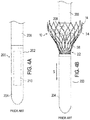
FIG. 4A is a fragmentary side elevational view of a conventional delivery device for use with a transapical approach; -
FIG. 4B is a fragmentary side elevational view of the delivery device ofFIG. 4A showing partial deployment of a collapsible prosthetic heart valve using the transapical approach; -
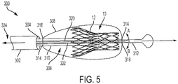
FIG. 5 is a fragmentary side-view of a crimping system in accordance with an embodiment of the present invention including a fully deployed prosthetic heart valve; -
FIG. 6A is a fragmentary, schematic depiction of the system ofFIG. 5 in an un-tensioned state; -
FIG. 6B is a fragmentary, schematic depiction of the system ofFIG. 5 in a tensioned state; -
FIG. 7 is a fragmentary side view of an exemplary attachment between a sheath and a delivery device of the system ofFIG. 5 ; and -
FIG. 8 is a fragmentary side view of another exemplary attachment between a sheath and a delivery device of the system ofFIG. 5 . - As used herein, the term "proximal," when used in connection with a prosthetic heart valve, refers to the end of the heart valve closest to the heart when the heart valve is implanted in a patient, whereas the term "distal," when used in connection with a prosthetic heart valve, refers to the end of the heart valve farthest from the heart when the heart valve is implanted in a patient. When used in connection with devices for delivering a prosthetic heart valve into a patient, the terms "trailing" and "leading" are to be taken as relative to the user of the delivery devices. "Trailing" is to be understood as relatively close to the operator, and "leading" is to be understood as relatively farther away from the operator.
-
FIG. 1 shows a collapsibleprosthetic heart valve 10 according to an embodiment of the present disclosure. Theprosthetic heart valve 10 is designed to replace the function of the native aortic valve of a patient. Examples of collapsible prosthetic heart valves are described inU.S. Pat. No. 7,018,406 andU.S. Pat. No. 9,241,794 -
Prosthetic heart valve 10 includes anexpandable stent 12 which may be formed from biocompatible materials that are capable of self-expansion, for example, shape memory alloys such as nitinol.Stent 12 extends from a distal orannulus end 14 to a proximal oraortic end 16, and includes anannulus section 18 adjacentdistal end 14 and anaortic section 20 adjacentproximal end 16.Annulus section 18 has a relatively small cross-section in the expanded state compared toaortic section 20 in the expanded state. Preferably,annulus section 18 is in the form of a cylinder having a substantially constant diameter along its length. Atransition section 22 tapers outwardly fromannulus section 18 toaortic section 20. Each of the sections ofstent 12 includes a plurality ofcells 24 formed of a plurality ofstruts 26 connected to one another to form one or more annular rows around the stent. In one particular embodiment, shown inFIG. 1 ,annulus section 18 may have two annular rows of complete cells andaortic section 20 andtransition section 22 may each have one or more annular rows of partial cells.Cells 24 inaortic section 20 may be larger thancells 24 inannulus section 18. The larger cells in theaortic section 20 facilitate positioning ofprosthetic valve 10 withoutstent 12 interfering with blood flow to the coronary arteries. -
Stent 12 may include one or more retaining elements 28 provided onaortic section 20 and sized and shaped to cooperate with a delivery device. Engagement of retaining elements 28 with a corresponding retaining structure on the delivery device maintainsprosthetic heart valve 10 in the crimped position within the delivery device, minimizes longitudinal movement of the prosthetic heart valve relative to the delivery device during unsheathing or resheathing procedures, and prevents rotation of the prosthetic heart valve relative to the delivery device as the delivery device is advanced to the target location and during deployment. -
Stent 12 may also include a plurality of commissure features 30 for attaching the commissure between two adjacent leaflets tostent 12. As is shown inFIG. 1 , commissure features 30 may lie at the intersection of fourcells 24, two of the cells being adjacent one another in the same annular row, and the other two cells being in different annular rows and lying in an end-to-end relationship. Preferably, commissure features 30 are positioned entirely withinannulus section 18 or at the juncture ofannulus section 18 andtransition section 22. Commissure features 30 may include one ormore eyelets 32 which facilitate the suturing of the leaflet commissure tostent 12. -
Prosthetic heart valve 10 includes avalve assembly 34 preferably positioned inannulus section 18.Valve assembly 34 may be secured tostent 12 by suturingvalve assembly 34 tostruts 26 and/or to commissure features 30.Valve assembly 34 includes acuff 36 and a plurality ofleaflets 38 which open and close collectively to function as a one-way valve. SinceFIG. 1 illustrates a prosthetic heart valve for replacing a native tricuspid valve, such as the aortic valve,prosthetic heart valve 10 is illustrated with threeleaflets 38, as well as three commissure features 30. However, it will be appreciated that prosthetic heart valves according to this aspect of the invention may have a greater or lesser number ofleaflets 38 and/or commissure features 30. - Although
cuff 36 is illustrated inFIG. 1 as being disposed on the luminal or inner surface ofannulus section 18, it is contemplated thatcuff 36 may be disposed on the abluminal or outer surface ofannulus section 18, or may cover all or part of either or both of the luminal and abluminal surfaces ofannulus section 18. Bothcuff 36 andleaflets 38 may be wholly or partly formed of any suitable biological material, such as bovine or porcine pericardium, or polymers, such as PTFE, urethanes and the like. - As is shown in
FIG. 1 , the entirety ofvalve assembly 34, including the leaflet commissures, is positioned inannulus section 18 ofstent 12. That is, theentire valve assembly 34 is substantially positioned betweendistal end 14 ofstent 12 and commissure features 30, such that none ofvalve assembly 34 is positioned between commissure features 30 andproximal end 16 ofstent 12. As is explained in further detail hereinafter, this configuration is particularly advantageous during conventional delivery (illustrated inFIGS. 3 and4B ) for enabling the deployment ofheart valve 10 by an amount sufficient forvalve leaflets 38 to operate, whileproximal end 16 ofstent 12 remains collapsed within the delivery device. Since the present invention provides a device for recapturingheart valve 10 after it has been fully deployed, the location ofvalve assembly 34 need not be limited exclusively toannulus section 18. For example,valve assembly 34 may extend intotransition section 22. -
Prosthetic heart valve 10 described above may be used to replace a native heart valve, such as the aortic valve, a surgical heart valve, or a heart valve that has undergone a surgical procedure.Prosthetic heart valve 10 may be delivered to the desired site (e.g., near a native aortic annulus) using the delivery device described in detail below. During delivery,prosthetic heart valve 10 is disposed inside the delivery device in the collapsed condition. The delivery device may be introduced into a patient using a transfemoral, transapical, transseptal approach or another approaoch. Once the delivery device has reached the target site, the user may deployprosthetic heart valve 10. Upon deployment, the prosthetic heart valve expands into secure engagement within the native anatomic structure such as the aortic annulus. When the prosthetic heart valve is properly positioned inside the patient, it works as a one-way valve, allowing blood to flow in one direction and preventing blood from flowing in the opposite direction. -
FIG. 2 illustrates a human heart H and two different approaches of deliveringprosthetic heart valve 10 to its intended target at aortic valve AV. As illustrated inFIG. 2 , heart H includes an aorta A, an aortic arch AA and a left ventricle LV. Two separate paths are shown for introducingprosthetic heart valve 10 to aortic valve AV. - A transfemoral approach of the prosthetic heart valve is indicated by the dashed arrow and the label "AV1". In this method,
prosthetic heart valve 10 is inserted into the femoral artery, tracked through the vasculature and then introduced to the target site via the aortic arch AA. Echocardiography and other means may be used to help guide the delivery device through this approach. - A second dashed arrow, labeled "AV2," indicates a transapical approach of the prosthetic heart valve. In transapical delivery, a small incision is made between the ribs and into the apex of left ventricle LV to deliver the prosthetic heart valve to the target site.
-
FIG. 3 illustrates aconventional device 100 for transfemoral delivery of a prosthetic heart valve. In the transfemoral approach, the leading end of adelivery tube 102 points in the distal direction of the heart. Prior to deployment,valve 10 is disposed indelivery tube 102 withannulus section 18 closest to leadingend 104 ofdelivery tube 102. As seen inFIG. 3 , the transfemoral approach first unsheathes annulus end 18 ofheart valve 10, allowingannulus section 18 to expand prior to full deployment ofvalve 10. For example,delivery tube 102 may be retracted toward its trailingend 106 while internal components (not shown) ofdelivery tube 102hold heart valve 10 stationary. More specifically, proximal oraortic end 16 ofvalve 10, includingaortic section 20, remains partially sheathed and coupled todelivery tube 102, while distal or annulus end 14 of the valve, includingannulus section 18, expands. It will be appreciated thatvalve assembly 34, and specifically the function ofleaflets 38, may be preliminarily tested in this approach without fully deployingheart valve 10. - In other circumstances, it may be desirable to use a transapical approach (shown as "AV2" in
FIG. 2 ) as opposed to the transfemoral approach (shown as "AV1" inFIG. 2 ). For example, calcification in arteries may render tracking of the delivery device using a transfemoral approach difficult and make a transapical approach the preferable route. -
FIGS. 4A and 4B illustrate aconventional delivery device 200 used for a transapical approach.Delivery device 200 includes adelivery tube 202 having aleading end 204 connected to a trailingend 206 via asupport core 208.Delivery tube 202 defines acompartment 210 for housingprosthetic heart valve 10. - During delivery,
delivery device 200 is advanced in a forward direction through the apex of the heart and into the aorta untilannulus end 14 ofvalve 10 is disposed at or near the annulus of the native valve andleading end 204 ofdelivery device 200 projects through the native valve into the aorta. While trailingend 206 ofdelivery device 200 is held stationary, leadingend 204 is translated in a forward direction (depicted by arrow "S") away from trailingend 206 to removeprosthetic heart valve 10 fromcompartment 210. In this configuration,aortic section 20 ofvalve 10 remains disposed indelivery tube 202 whileannulus section 18 expands after it has been unsheathed. Thus, trailing-end-first deployment deploysannulus section 18 beforeaortic section 20. The deployment process can be reversed before full deployments, by reversing the movement of leadingend 204 ofdelivery device 200 relative tostent 10. - Irrespective of whether a transfemoral or transapical approach is utilized, conventional delivery requires that
aortic section 20 remain at least partially sheathed bydelivery tube valve 10 to be subsequently re-crimped. However, to testvalve assembly 34 in a fully expanded position, and becauseheart valve 10 can "jump" or reposition upon full deployment, which can alter fitment and lead to PV leak, it would be desirable to be able to re-crimpheart valve 10 for repositioning or withdrawing the valve afterstent 12 has been fully deployed. -
FIG. 5 illustrates one embodiment ofsystem 300, which allowsprosthetic heart valve 10 to be crimped, and thus repositioned, aftervalve 10 has been fully deployed. As used herein, the term "full deployment" means the entire heart valve has been withdrawn from the delivery device and transitioned to its expanded state. -
Delivery device 300 includesdelivery tube 302 having alumen 304, adeployment device 306 for controlling the crimping ofprosthetic heart valve 10, and a tensionable, braidedsheath 308 for compressingheart valve 10 after full deployment (the braided portion ofsheath 308 has been omitted fromFIG. 5 for clarity). -
Delivery tube 302 may be any tube-like delivery device such as a catheter, a trocar, a laparoscopic instrument, or the like, configured to houseprosthetic heart valve 10 in a crimped state. In one embodiment hereof, female retaining structures (not shown) are disposed withindelivery tube 302 for cooperating with retaining elements 28 ofstent 12 to minimize longitudinal movement ofprosthetic heart valve 10 relative todelivery device 300 during unsheathing and to prevent rotation ofprosthetic heart valve 10 relative todelivery tube 302 asdelivery device 300 is advanced toward the target location. -
Deployment device 306 is extendable throughdelivery tube 302 and includes a flexible, first orouter shaft 310 extending in a longitudinal direction and a flexible, second orinner shaft 312 extending in the longitudinal direction. In one preferred embodiment,second shaft 312 is telescopically arranged withinfirst shaft 310, such thatsecond shaft 312 is slidable within and relative tofirst shaft 310. One ormore attachment devices 314 are provided onouter shaft 310, at a location marked by X1 (FIG. 6A and 6B ), for couplingouter shaft 310 to afirst attachment location 316 provided on a proximal end ofsheath 308, and oninner shaft 312, at a location marked by X2 (FIG. 6A and 6B ), for couplinginner shaft 312 to asecond attachment location 318 provided on a distal end ofsheath 308. - As is illustrated in
FIGS. 6A and 6B ,sheath 308 is an open ended tube or sock-like structure and may be formed of nitinol braid, fabric, or any other strong, yet pliable material that permits blood to flow therethrough.Sheath 308 at least partially surroundsvalve 10 when coupled toouter shaft 310 at location X1, andinner shaft 312 at location X2, such thatvalve 10 will be compressed or crimped when tension is applied tosheath 308. In a first or un-tensioned condition (FIG. 6A ), in which no tension or little tension is applied tosheath 308, the distance between X1 and X2 defines a length L1. To compress or crimpheart valve 10, a user slidesinner shaft 312 distally relative toouter shaft 310, to a second or tensioned position (FIG. 6B ) in which the distance between X1 and X2 defines a second length L2 greater than length L1, thereby applying a tensioning force tosheath 308. Althoughsecond shaft 312 is described as telescoping withinfirst shaft 310, it will be understood thatsecond shaft 312 may also telescope aboutfirst shaft 310, or may be configured to move relative thereto in any manner that increases the length between X1 and X2 in order to apply a tensioning force tosheath 308. Accordingly, after full deployment, if the physician deems thatvalve 10 is not functioning properly, has jumped, or is otherwise incorrectly positioned, the physician maytension sheath 308, as previously described, to at least partially crimpstent 12 and repositionvalve 10. Moreover, in the crimped state,delivery device 302 may be advanced overstent 12 to recapturevalve 10 for withdrawal. -
Device 300 may optionally include first andsecond wires deployment device 306, as shown inFIG. 5 , for aiding the user in deliveringheart valve 10.Wires inner shaft 312 in an opposing configuration, and preferably, at or near a distal end ofinner shaft 312. Accordingly, relative movement betweenwires inner shaft 312 and flexibleouter shaft 310, and ineffect heart valve 10, in a desired direction. For example, pullingwire 322 toward trailingend 324 relative to wire 320 will cause flexibleinner shaft 312 to turn in the direction indicated by arrow B. Conversely, pullingwire 320 toward trailingend 324, relative towire 322, will causeinner shaft 312 to turn in the direction indicated by arrow A. In order to steer the delivery ofvalve 10 in another plane,inner shaft 312 may be rotated about its longitudinal axis. The steering mechanism provided bywires valve 10 through the delivery path as opposed to relying on the tissue to guide thevalve 10. -
FIG. 7 depicts the attachment, schematically represented inFIG. 5 , betweendeployment device 306 andsheath 308. As shown inFIG. 7 , a plurality ofloops 326 are provided at the proximal end and the distal end ofsheath 308 for removably securingsheath 308 todeployment device 306. Each ofloops 326 on the proximal end ofsheath 308 is configured to receive acorresponding hook 328 tethered toouter shaft 310 atfirst attachment location 316 and each ofloops 326 on the distal end ofsheath 308 is configured to receive acorresponding hook 328 tethered toinner shaft 312 atsecond attachment location 318. When a slight tension is applied totethers 330, eachhook 328 will engage itscorresponding loop 326, thereby couplingdelivery device 306 andsheath 308. After tension is released fromtethers 330, hooks 328 will be able to disengage fromloops 326. After disconnection,deployment device 306 anddelivery device 302 may be withdrawn from the body of the patient. In this embodiment,sheath 308 remains sandwiched betweenstent 12 and the surrounding tissue afterdelivery device 302 anddeployment device 306 have been withdrawn. This aids in sealingvalve 10 to the native annulus, thereby reducing paravalvular leak (PVL). AlthoughFIG. 7 illustrates a hook and loop connection, any removeable connection, such as clamps or breakable tethers may be used. - In an alternative embodiment, illustrated by
FIG. 8 ,sheath 308 only partially surroundsstent 12, preferablyaortic section 20, which may help in reducing PVL and/or anchoringstent 12 in the native anatomy. As shown inFIG. 8 ,loops 326 are provided on the distal end ofsheath 308 for receiving acorresponding hook 328 tethered toinner shaft 312. In contrast to the embodiment illustrated inFIG. 7 , the proximal end ofsheath 308 is non-removeably connected toouter shaft 310, for example, viatethers 330. When a slight tension is applied totethers 330, eachhook 328 engages itscorresponding loop 326, thereby couplingdelivery device 306 andsheath 308. However, after tension is released fromtethers 330, hooks 328 will disengage fromloops 326, thereby disconnectinginner shaft 312 andsheath 308 whiletethers 330 maintain a connection betweenouter shaft 310 andsheath 308. Thus, whenannulus section 18 is securely engaged in the valve annulus and whendeployment device 306 is withdrawn from the body of the patient,sheath 308 is pulled from betweenstent 312 and the surrounding heart tissue without dislodgingvalve 10 from its desired position. An inner surface and/or outer surface ofsheath 308 may be lubricated in order to assistsheath 308 in sliding from between the tissue andstent 12 without dislodgingvalve 10 from its desired position. - To use
device 300, a physician placesstent 12 arounddeployment device 306,sheath 308 at least partially aroundstent 12, and attachessheath 308 toouter shaft 310 andinner shaft 312.Sheath 308 may then be tensioned by slidinginner shaft 312 distally relative toouter shaft 310 to crimpstent 12 for loadingvalve 10 intolumen 304 ofdelivery tube 302. Alternatively,valve 10 and the surroundingsheath 308 may be crimped and loaded intodelivery tube 302 using conventional methods. - After
delivery device 300 has been loaded, using either a transfemoral or transapical approach, a physician may steerheart valve 10 viawires valve 10 may be fully deployed so that the physician can comprehensively analyze valve function and fitment ofvalve 10 while a slight tension is maintained ontethers 330 so that hooks 328 remain engaged withloops 326. If the physician deems thatvalve 10 is not properly positioned, the physician may slideinner member 312 away fromouter member 310, thereby applying a tensioning force tosheath 308, for crimpingstent 12 as previously described.Stent 12 may then be repositioned usingguide wires valve 10 is not functioning properly,delivery tube 302 may be slide over crimpedvalve 10 to recapture the valve withinbore 304 for withdrawal. Advantageously,system 300 provides the physician with the ability to correct improper function or placement ofvalve 10, after full deployment, such that the valves true function and final placement may be analyzed. - To summarize the foregoing, one aspect of the present disclosure is directed to a device for crimping a heart valve comprising a delivery tube extending in a longitudinal direction and having a lumen configured to receive a heart valve; a deployment device including a first member at least partially received within the lumen of the delivery tube and a second member slidable relative to the first member; and a sheath configured to at least partially surround a heart valve, the sheath having a first attachment area connected to the first member and a second attachment area connected to the second member, and wherein movement of the first member relative to the second member tensions the sheath; and/or wherein the first attachment area is connected to the first member at a first location, the second attachment area is connected to the second member at a second location, and the second member is telescopingly arranged within the first member for transitioning the deployment device between a first condition in which there is a first length between the first location and the second location, and a second condition in which there is a second length greater than the first length between the first location and the second location; and/or further comprising a heart valve including a stent formed of a plurality of struts, the stent being transitionable between a radially collapsed state and a radially expanded state, wherein in the expanded state a diameter of the stent is larger than a diameter of the lumen of the delivery tube; and/or wherein the stent comprises a valve including a cuff and at least one leaflet; and/or wherein the deployment device in the first condition exerts a first tension on the sheath and the deployment device in the second condition exerts a second tension on the sheath greater than the first tension, the second tension being sufficient to radially compress at least a portion of the stent; and/or wherein at least one of the first attachment area and second attachment area comprises at least one loop; and/or wherein at least one of the first member or the second member includes at least one connector selected from the group of a hook, a clamp, and a tether; and/or wherein the tether comprise a breakable braid; and/or further comprising a first wire and a second wire opposingly secured within the deployment device for guiding delivery of the heart valve; and/or wherein the first attachment area of the sheath is adjacent a proximal end of the sheath and the second attachment area of the sheath is adjacent a distal end of the sheath; and/or wherein at least a portion of the sheath is lubricated; and/or wherein the sheath is formed of nitinol.
- Although the invention herein has been described with reference to particular embodiments, it is to be understood that these embodiments are merely illustrative of the principles and applications of the present invention. It is therefore to be understood that numerous modifications may be made to the illustrative embodiments and that other arrangements may be devised without departing from the spirit and scope of the present invention as defined by the appended claims.
Claims (12)
- A device for crimping a heart valve, comprising:a delivery tube extending in a longitudinal direction and having a lumen configured to receive a heart valve;a deployment device including a first member at least partially received within the lumen of the delivery tube and a second member slidable relative to the first member; anda sheath configured to at least partially surround a heart valve, the sheath having a first attachment area connected to the first member and a second attachment area connected to the second member, andwherein movement of the first member relative to the second member tensions the sheath.
- The system of claim 1, wherein the first attachment area is connected to the first member at a first location, the second attachment area is connected to the second member at a second location, and the second member is telescopingly arranged within the first member for transitioning the deployment device between a first condition in which there is a first length between the first location and the second location, and a second condition in which there is a second length greater than the first length between the first location and the second location.
- The system of claim 2, further comprising a heart valve including a stent formed of a plurality of struts, the stent being transitionable between a radially collapsed state and a radially expanded state, wherein in the expanded state a diameter of the stent is larger than a diameter of the lumen of the delivery tube.
- The system of claim 3, wherein the stent comprises a valve including a cuff and at least one leaflet.
- The system of claim 3, wherein the deployment device in the first condition exerts a first tension on the sheath and the deployment device in the second condition exerts a second tension on the sheath greater than the first tension, the second tension being sufficient to radially compress at least a portion of the stent.
- The system of claim 1, wherein at least one of the first attachment area and second attachment area comprises at least one loop.
- The system of claim 1, wherein at least one of the first member or the second member includes at least one connector selected from the group of a hook, a clamp, and a tether.
- The system of claim 7, wherein the tether comprise a breakable braid.
- The system of claim 1, further comprising a first wire and a second wire opposingly secured within the deployment device for guiding delivery of the heart valve.
- The system of claim 1, wherein the first attachment area of the sheath is adjacent a proximal end of the sheath and the second attachment area of the sheath is adjacent a distal end of the sheath.
- The system of claim 1, wherein at least a portion of the sheath is lubricated.
- The system of claim 1, wherein the sheath is formed of nitinol.
Applications Claiming Priority (1)
| Application Number | Priority Date | Filing Date | Title |
|---|---|---|---|
| US201862632479P | 2018-02-20 | 2018-02-20 |
Publications (1)
| Publication Number | Publication Date |
|---|---|
| EP3527176A1 true EP3527176A1 (en) | 2019-08-21 |
Family
ID=64172334
Family Applications (1)
| Application Number | Title | Priority Date | Filing Date |
|---|---|---|---|
| EP18204261.4A Withdrawn EP3527176A1 (en) | 2018-02-20 | 2018-11-05 | Crimping heart valve with nitinol braid |
Country Status (2)
| Country | Link |
|---|---|
| US (1) | US10898326B2 (en) |
| EP (1) | EP3527176A1 (en) |
Families Citing this family (4)
| Publication number | Priority date | Publication date | Assignee | Title |
|---|---|---|---|---|
| CN109498215B (en) * | 2018-12-14 | 2023-05-02 | 宁波健世科技股份有限公司 | Separated release aortic valve stent |
| US12127939B2 (en) * | 2020-04-30 | 2024-10-29 | Cephea Valve Technologies, Inc. | Catheter lumen lubricant |
| CN116847902B (en) * | 2020-12-23 | 2026-04-28 | 波士顿科学国际有限公司 | High-friction woven fabric facilitates device delivery |
| CN114767335B (en) * | 2022-04-25 | 2026-01-23 | 金仕生物科技(常熟)有限公司 | Valve delivery system |
Citations (7)
| Publication number | Priority date | Publication date | Assignee | Title |
|---|---|---|---|---|
| US7018406B2 (en) | 1999-11-17 | 2006-03-28 | Corevalve Sa | Prosthetic valve for transluminal delivery |
| US20130178888A1 (en) * | 2011-10-17 | 2013-07-11 | W. L. Gore & Associates, Inc. | Endoluminal device retrieval devices and related systems and methods |
| US20150282821A1 (en) * | 2014-04-08 | 2015-10-08 | Incuvate, Llc | Systems and methods for management of thrombosis |
| US9241794B2 (en) | 2007-09-26 | 2016-01-26 | St. Jude Medical, Inc. | Collapsible prosthetic heart valves |
| US20160206426A1 (en) * | 2013-08-30 | 2016-07-21 | Cedars-Sinai Medical Center | Devices and methods for transcatheter retrieval of mechanical heart valve leaflets |
| US20160324633A1 (en) * | 2011-08-05 | 2016-11-10 | Mitraltech Ltd. | Techniques for percutaneous mitral valve replacement and sealing |
| US20170325830A1 (en) * | 2007-12-26 | 2017-11-16 | Covidien Lp | Retrieval systems and methods for use thereof |
Family Cites Families (72)
| Publication number | Priority date | Publication date | Assignee | Title |
|---|---|---|---|---|
| US3657744A (en) | 1970-05-08 | 1972-04-25 | Univ Minnesota | Method for fixing prosthetic implants in a living body |
| US4423730A (en) | 1982-03-01 | 1984-01-03 | Shelhigh Inc. | Atriotomy button and implantation device |
| US5190546A (en) | 1983-10-14 | 1993-03-02 | Raychem Corporation | Medical devices incorporating SIM alloy elements |
| US5078720A (en) | 1990-05-02 | 1992-01-07 | American Medical Systems, Inc. | Stent placement instrument and method |
| ATE137656T1 (en) | 1992-10-31 | 1996-05-15 | Schneider Europ Ag | ARRANGEMENT FOR IMPLANTING SELF-EXPANDING ENDPROSTHESES |
| US5797960A (en) | 1993-02-22 | 1998-08-25 | Stevens; John H. | Method and apparatus for thoracoscopic intracardiac procedures |
| US5843167A (en) | 1993-04-22 | 1998-12-01 | C. R. Bard, Inc. | Method and apparatus for recapture of hooked endoprosthesis |
| EP0657147B1 (en) | 1993-11-04 | 1999-08-04 | C.R. Bard, Inc. | Non-migrating vascular prosthesis |
| AU691854B2 (en) | 1993-12-03 | 1998-05-28 | Edwards Lifesciences Ag | Cardiopulmonary bypass system for closed-chest intervention |
| US5415664A (en) | 1994-03-30 | 1995-05-16 | Corvita Corporation | Method and apparatus for introducing a stent or a stent-graft |
| US5824041A (en) | 1994-06-08 | 1998-10-20 | Medtronic, Inc. | Apparatus and methods for placement and repositioning of intraluminal prostheses |
| US5702418A (en) | 1995-09-12 | 1997-12-30 | Boston Scientific Corporation | Stent delivery system |
| US5968068A (en) | 1996-09-12 | 1999-10-19 | Baxter International Inc. | Endovascular delivery system |
| EP2133030A1 (en) | 1997-06-27 | 2009-12-16 | The Trustees of Columbia University of the City of New York | Method and apparatus for circulatory valve repair |
| US5980533A (en) | 1998-06-09 | 1999-11-09 | Scimed Life Systems, Inc. | Stent delivery system |
| US20070043435A1 (en) | 1999-11-17 | 2007-02-22 | Jacques Seguin | Non-cylindrical prosthetic valve system for transluminal delivery |
| US8016877B2 (en) | 1999-11-17 | 2011-09-13 | Medtronic Corevalve Llc | Prosthetic valve for transluminal delivery |
| FR2800984B1 (en) | 1999-11-17 | 2001-12-14 | Jacques Seguin | DEVICE FOR REPLACING A HEART VALVE PERCUTANEOUSLY |
| US8579966B2 (en) | 1999-11-17 | 2013-11-12 | Medtronic Corevalve Llc | Prosthetic valve for transluminal delivery |
| US6391050B1 (en) | 2000-02-29 | 2002-05-21 | Scimed Life Systems, Inc. | Self-expanding stent delivery system |
| US6264683B1 (en) | 2000-03-17 | 2001-07-24 | Advanced Cardiovascular Systems, Inc. | Stent delivery catheter with bumpers for improved retention of balloon expandable stents |
| DE10026307A1 (en) | 2000-05-26 | 2001-11-29 | Variomed Ag Balzers | Stent, positioning element and insertion catheter |
| US20060142848A1 (en) | 2000-09-12 | 2006-06-29 | Shlomo Gabbay | Extra-anatomic aortic valve placement |
| US7510572B2 (en) | 2000-09-12 | 2009-03-31 | Shlomo Gabbay | Implantation system for delivery of a heart valve prosthesis |
| US6623518B2 (en) | 2001-02-26 | 2003-09-23 | Ev3 Peripheral, Inc. | Implant delivery system with interlock |
| WO2002067782A2 (en) | 2001-02-26 | 2002-09-06 | Ev3 Peripheral, Inc. | Implant delivery system with interlock |
| US7556646B2 (en) | 2001-09-13 | 2009-07-07 | Edwards Lifesciences Corporation | Methods and apparatuses for deploying minimally-invasive heart valves |
| FR2828091B1 (en) | 2001-07-31 | 2003-11-21 | Seguin Jacques | ASSEMBLY ALLOWING THE PLACEMENT OF A PROTHETIC VALVE IN A BODY DUCT |
| US20060106415A1 (en) | 2004-11-12 | 2006-05-18 | Shlomo Gabbay | Apparatus to facilitate implantation |
| US6814746B2 (en) | 2002-11-01 | 2004-11-09 | Ev3 Peripheral, Inc. | Implant delivery system with marker interlock |
| US20040215315A1 (en) * | 2003-04-25 | 2004-10-28 | Jones Ryan A. | Drug-eluting stent with sheath and balloon deployment assembly |
| US10219899B2 (en) | 2004-04-23 | 2019-03-05 | Medtronic 3F Therapeutics, Inc. | Cardiac valve replacement systems |
| US20050137686A1 (en) | 2003-12-23 | 2005-06-23 | Sadra Medical, A Delaware Corporation | Externally expandable heart valve anchor and method |
| US8182528B2 (en) | 2003-12-23 | 2012-05-22 | Sadra Medical, Inc. | Locking heart valve anchor |
| US8343213B2 (en) | 2003-12-23 | 2013-01-01 | Sadra Medical, Inc. | Leaflet engagement elements and methods for use thereof |
| US8840663B2 (en) | 2003-12-23 | 2014-09-23 | Sadra Medical, Inc. | Repositionable heart valve method |
| US8828078B2 (en) | 2003-12-23 | 2014-09-09 | Sadra Medical, Inc. | Methods and apparatus for endovascular heart valve replacement comprising tissue grasping elements |
| US9005273B2 (en) | 2003-12-23 | 2015-04-14 | Sadra Medical, Inc. | Assessing the location and performance of replacement heart valves |
| US20090054975A1 (en) | 2004-02-06 | 2009-02-26 | Children's Medical Center Corporation | Deployment device for cardiac surgery |
| US7311730B2 (en) | 2004-02-13 | 2007-12-25 | Shlomo Gabbay | Support apparatus and heart valve prosthesis for sutureless implantation |
| CA3050938C (en) | 2004-10-02 | 2021-10-19 | Edwards Lifesciences Cardiaq Llc | Methods and devices for repair or replacement of heart valves or adjacent tissue without the need for full cardiopulmonary support |
| US20070162100A1 (en) | 2006-01-10 | 2007-07-12 | Shlomo Gabbay | System and method for loading implanter with prosthesis |
| US20060167468A1 (en) | 2004-11-12 | 2006-07-27 | Shlomo Gabbay | Implantation system and method for loading an implanter with a prosthesis |
| DE102005003632A1 (en) | 2005-01-20 | 2006-08-17 | Fraunhofer-Gesellschaft zur Förderung der angewandten Forschung e.V. | Catheter for the transvascular implantation of heart valve prostheses |
| EP3266414B1 (en) | 2005-05-12 | 2024-07-17 | Covidien LP | Implant delivery system with interlocked rx port orientation |
| US8790396B2 (en) | 2005-07-27 | 2014-07-29 | Medtronic 3F Therapeutics, Inc. | Methods and systems for cardiac valve delivery |
| US8702777B2 (en) | 2005-08-22 | 2014-04-22 | Incept, Llc | Steep-taper flared stents and apparatus and methods for delivering them |
| US20070073391A1 (en) | 2005-09-28 | 2007-03-29 | Henry Bourang | System and method for delivering a mitral valve repair device |
| US8167932B2 (en) | 2005-10-18 | 2012-05-01 | Edwards Lifesciences Corporation | Heart valve delivery system with valve catheter |
| US8764820B2 (en) | 2005-11-16 | 2014-07-01 | Edwards Lifesciences Corporation | Transapical heart valve delivery system and method |
| MX2008008068A (en) | 2005-12-22 | 2008-09-10 | Symetis Sa | Stent-valves for valve replacement and associated methods and systems for surgery. |
| US20070168013A1 (en) | 2006-01-19 | 2007-07-19 | Myles Douglas | Vascular graft and deployment system |
| US8147541B2 (en) | 2006-02-27 | 2012-04-03 | Aortx, Inc. | Methods and devices for delivery of prosthetic heart valves and other prosthetics |
| US20110224678A1 (en) | 2006-03-23 | 2011-09-15 | Shlomo Gabbay | Method and implantation system for implanting a cardiovascular prosthesis |
| US20070239271A1 (en) | 2006-04-10 | 2007-10-11 | Than Nguyen | Systems and methods for loading a prosthesis onto a minimally invasive delivery system |
| US8876895B2 (en) | 2006-09-19 | 2014-11-04 | Medtronic Ventor Technologies Ltd. | Valve fixation member having engagement arms |
| US8747459B2 (en) | 2006-12-06 | 2014-06-10 | Medtronic Corevalve Llc | System and method for transapical delivery of an annulus anchored self-expanding valve |
| US8070799B2 (en) | 2006-12-19 | 2011-12-06 | Sorin Biomedica Cardio S.R.L. | Instrument and method for in situ deployment of cardiac valve prostheses |
| US20090171456A1 (en) * | 2007-12-28 | 2009-07-02 | Kveen Graig L | Percutaneous heart valve, system, and method |
| AU2009205739B2 (en) | 2008-01-16 | 2014-09-25 | St. Jude Medical, Inc. | Delivery and retrieval systems for collapsible/expandable prosthetic heart valves |
| WO2010051025A1 (en) | 2008-10-30 | 2010-05-06 | St. Jude Medical, Inc. | Collapsible/expandable prosthetic heart valve delivery system and methods |
| US10278728B2 (en) | 2009-01-30 | 2019-05-07 | St. Jude Medical, Llc | Transapical mini-introducer hemostasis valve and punch |
| AU2010236288A1 (en) | 2009-04-15 | 2011-10-20 | Cardiaq Valve Technologies, Inc. | Vascular implant and delivery system |
| WO2011094527A1 (en) * | 2010-01-29 | 2011-08-04 | Cook Medical Technologies Llc | Mechanically expandable delivery and dilation systems |
| US9414914B2 (en) | 2010-02-24 | 2016-08-16 | Medtronic Ventor Technologies Ltd. | Catheter assembly with valve crimping accessories |
| US8562663B2 (en) | 2010-10-26 | 2013-10-22 | Medtronic Ventor Technologies Ltd. | Devices and methods for loading a prosthesis onto a delivery system |
| US10188515B2 (en) | 2013-05-13 | 2019-01-29 | Medtronic Vascular Inc. | Devices and methods for crimping a medical device |
| US9414917B2 (en) | 2013-09-17 | 2016-08-16 | Medtronic, Inc. | Systems and methods for loading a valve prosthesis onto a catheter |
| CN104586542B (en) | 2013-10-31 | 2017-01-04 | 上海微创心通医疗科技有限公司 | An apparatus and method for loading an implant into a delivery system |
| WO2015127283A1 (en) * | 2014-02-21 | 2015-08-27 | Cardiaq Valve Technologies, Inc. | Delivery device for controlled deployement of a replacement valve |
| AU2016370459B2 (en) * | 2015-12-14 | 2019-06-06 | Medtronic Vascular Inc. | Devices and methods for transcatheter valve loading and implantation |
| EP3432835B1 (en) * | 2016-03-24 | 2025-09-17 | Edwards Lifesciences Corporation | Delivery system for prosthetic heart valve |
-
2018
- 2018-10-31 US US16/176,437 patent/US10898326B2/en active Active
- 2018-11-05 EP EP18204261.4A patent/EP3527176A1/en not_active Withdrawn
Patent Citations (7)
| Publication number | Priority date | Publication date | Assignee | Title |
|---|---|---|---|---|
| US7018406B2 (en) | 1999-11-17 | 2006-03-28 | Corevalve Sa | Prosthetic valve for transluminal delivery |
| US9241794B2 (en) | 2007-09-26 | 2016-01-26 | St. Jude Medical, Inc. | Collapsible prosthetic heart valves |
| US20170325830A1 (en) * | 2007-12-26 | 2017-11-16 | Covidien Lp | Retrieval systems and methods for use thereof |
| US20160324633A1 (en) * | 2011-08-05 | 2016-11-10 | Mitraltech Ltd. | Techniques for percutaneous mitral valve replacement and sealing |
| US20130178888A1 (en) * | 2011-10-17 | 2013-07-11 | W. L. Gore & Associates, Inc. | Endoluminal device retrieval devices and related systems and methods |
| US20160206426A1 (en) * | 2013-08-30 | 2016-07-21 | Cedars-Sinai Medical Center | Devices and methods for transcatheter retrieval of mechanical heart valve leaflets |
| US20150282821A1 (en) * | 2014-04-08 | 2015-10-08 | Incuvate, Llc | Systems and methods for management of thrombosis |
Also Published As
| Publication number | Publication date |
|---|---|
| US20190254819A1 (en) | 2019-08-22 |
| US10898326B2 (en) | 2021-01-26 |
Similar Documents
| Publication | Publication Date | Title |
|---|---|---|
| US12496191B2 (en) | Transcatheter delivery system and method with controlled expansion and contraction of prosthetic heart valve | |
| US11701226B2 (en) | Prosthetic heart valves and apparatus and methods for delivery of same | |
| CN111759368B (en) | Transcatheter valve delivery system with septum aperture closed end assembly | |
| US10864076B2 (en) | Transapical mitral valve replacement | |
| US10524909B2 (en) | Retaining cage to permit resheathing of a tavi aortic-first transapical system | |
| US9149358B2 (en) | Delivery systems for prosthetic heart valves | |
| EP2538883B1 (en) | Catheter assembly with valve crimping accessories | |
| US20080215144A1 (en) | Replacement prosthetic heart valves and methods of implantation | |
| WO2018085059A1 (en) | Valve delivery system | |
| CN102811682A (en) | Transcatheter prosthetic heart valve delivery system with recapturing feature and method | |
| US11992406B2 (en) | Multi-step deployment to improve TAVR implant stability | |
| CN113164254B (en) | Prosthetic valve delivery assembly | |
| US10898326B2 (en) | Crimping heart valve with nitinol braid | |
| EP4085875B1 (en) | Method of implant loading | |
| US12138163B2 (en) | Fully deployable and recapturable prosthetic heart valve |
Legal Events
| Date | Code | Title | Description |
|---|---|---|---|
| PUAI | Public reference made under article 153(3) epc to a published international application that has entered the european phase |
Free format text: ORIGINAL CODE: 0009012 |
|
| STAA | Information on the status of an ep patent application or granted ep patent |
Free format text: STATUS: THE APPLICATION HAS BEEN PUBLISHED |
|
| AK | Designated contracting states |
Kind code of ref document: A1 Designated state(s): AL AT BE BG CH CY CZ DE DK EE ES FI FR GB GR HR HU IE IS IT LI LT LU LV MC MK MT NL NO PL PT RO RS SE SI SK SM TR |
|
| AX | Request for extension of the european patent |
Extension state: BA ME |
|
| STAA | Information on the status of an ep patent application or granted ep patent |
Free format text: STATUS: REQUEST FOR EXAMINATION WAS MADE |
|
| 17P | Request for examination filed |
Effective date: 20191230 |
|
| R17P | Request for examination filed (corrected) |
Effective date: 20191230 |
|
| STAA | Information on the status of an ep patent application or granted ep patent |
Free format text: STATUS: THE APPLICATION IS DEEMED TO BE WITHDRAWN |
|
| 18D | Application deemed to be withdrawn |
Effective date: 20210501 |