EP3518972B1 - Antigen binding molecules and methods of use thereof - Google Patents
Antigen binding molecules and methods of use thereof Download PDFInfo
- Publication number
- EP3518972B1 EP3518972B1 EP17857353.1A EP17857353A EP3518972B1 EP 3518972 B1 EP3518972 B1 EP 3518972B1 EP 17857353 A EP17857353 A EP 17857353A EP 3518972 B1 EP3518972 B1 EP 3518972B1
- Authority
- EP
- European Patent Office
- Prior art keywords
- seq
- antigen binding
- binding molecule
- amino acid
- sequence
- Prior art date
- Legal status (The legal status is an assumption and is not a legal conclusion. Google has not performed a legal analysis and makes no representation as to the accuracy of the status listed.)
- Active
Links
Images


Classifications
-
- C—CHEMISTRY; METALLURGY
- C07—ORGANIC CHEMISTRY
- C07K—PEPTIDES
- C07K16/00—Immunoglobulins [IG], e.g. monoclonal or polyclonal antibodies
- C07K16/42—Immunoglobulins [IG], e.g. monoclonal or polyclonal antibodies against immunoglobulins
- C07K16/4208—Immunoglobulins [IG], e.g. monoclonal or polyclonal antibodies against immunoglobulins against an idiotypic determinant on Ig
- C07K16/4241—Immunoglobulins [IG], e.g. monoclonal or polyclonal antibodies against immunoglobulins against an idiotypic determinant on Ig against anti-human or anti-animal Ig
- C07K16/4258—Immunoglobulins [IG], e.g. monoclonal or polyclonal antibodies against immunoglobulins against an idiotypic determinant on Ig against anti-human or anti-animal Ig against anti-receptor Ig
-
- A—HUMAN NECESSITIES
- A61—MEDICAL OR VETERINARY SCIENCE; HYGIENE
- A61K—PREPARATIONS FOR MEDICAL, DENTAL OR TOILETRY PURPOSES
- A61K35/00—Medicinal preparations containing materials or reaction products thereof with undetermined constitution
- A61K35/12—Materials from mammals; Compositions comprising non-specified tissues or cells; Compositions comprising non-embryonic stem cells; Genetically modified cells
- A61K35/14—Blood; Artificial blood
- A61K35/17—Lymphocytes; B-cells; T-cells; Natural killer cells; Interferon-activated or cytokine-activated lymphocytes
-
- A—HUMAN NECESSITIES
- A61—MEDICAL OR VETERINARY SCIENCE; HYGIENE
- A61K—PREPARATIONS FOR MEDICAL, DENTAL OR TOILETRY PURPOSES
- A61K39/00—Medicinal preparations containing antigens or antibodies
- A61K39/0005—Vertebrate antigens
- A61K39/0011—Cancer antigens
-
- A—HUMAN NECESSITIES
- A61—MEDICAL OR VETERINARY SCIENCE; HYGIENE
- A61K—PREPARATIONS FOR MEDICAL, DENTAL OR TOILETRY PURPOSES
- A61K39/00—Medicinal preparations containing antigens or antibodies
- A61K39/395—Antibodies; Immunoglobulins; Immune serum, e.g. antilymphocytic serum
- A61K39/39533—Antibodies; Immunoglobulins; Immune serum, e.g. antilymphocytic serum against materials from animals
- A61K39/3955—Antibodies; Immunoglobulins; Immune serum, e.g. antilymphocytic serum against materials from animals against proteinaceous materials, e.g. enzymes, hormones, lymphokines
-
- C—CHEMISTRY; METALLURGY
- C07—ORGANIC CHEMISTRY
- C07K—PEPTIDES
- C07K14/00—Peptides having more than 20 amino acids; Gastrins; Somatostatins; Melanotropins; Derivatives thereof
- C07K14/435—Peptides having more than 20 amino acids; Gastrins; Somatostatins; Melanotropins; Derivatives thereof from animals; from humans
- C07K14/705—Receptors; Cell surface antigens; Cell surface determinants
- C07K14/70503—Immunoglobulin superfamily
- C07K14/7051—T-cell receptor (TcR)-CD3 complex
-
- C—CHEMISTRY; METALLURGY
- C07—ORGANIC CHEMISTRY
- C07K—PEPTIDES
- C07K16/00—Immunoglobulins [IG], e.g. monoclonal or polyclonal antibodies
- C07K16/18—Immunoglobulins [IG], e.g. monoclonal or polyclonal antibodies against material from animals or humans
- C07K16/28—Immunoglobulins [IG], e.g. monoclonal or polyclonal antibodies against material from animals or humans against receptors, cell surface antigens or cell surface determinants
- C07K16/2803—Immunoglobulins [IG], e.g. monoclonal or polyclonal antibodies against material from animals or humans against receptors, cell surface antigens or cell surface determinants against the immunoglobulin superfamily
-
- C—CHEMISTRY; METALLURGY
- C07—ORGANIC CHEMISTRY
- C07K—PEPTIDES
- C07K16/00—Immunoglobulins [IG], e.g. monoclonal or polyclonal antibodies
- C07K16/42—Immunoglobulins [IG], e.g. monoclonal or polyclonal antibodies against immunoglobulins
- C07K16/4208—Immunoglobulins [IG], e.g. monoclonal or polyclonal antibodies against immunoglobulins against an idiotypic determinant on Ig
-
- G—PHYSICS
- G01—MEASURING; TESTING
- G01N—INVESTIGATING OR ANALYSING MATERIALS BY DETERMINING THEIR CHEMICAL OR PHYSICAL PROPERTIES
- G01N33/00—Investigating or analysing materials by specific methods not covered by groups G01N1/00 - G01N31/00
- G01N33/48—Biological material, e.g. blood, urine; Haemocytometers
- G01N33/50—Chemical analysis of biological material, e.g. blood, urine; Testing involving biospecific ligand binding methods; Immunological testing
- G01N33/53—Immunoassay; Biospecific binding assay; Materials therefor
- G01N33/569—Immunoassay; Biospecific binding assay; Materials therefor for microorganisms, e.g. protozoa, bacteria, viruses
- G01N33/56966—Animal cells
- G01N33/56972—White blood cells
-
- G—PHYSICS
- G01—MEASURING; TESTING
- G01N—INVESTIGATING OR ANALYSING MATERIALS BY DETERMINING THEIR CHEMICAL OR PHYSICAL PROPERTIES
- G01N33/00—Investigating or analysing materials by specific methods not covered by groups G01N1/00 - G01N31/00
- G01N33/48—Biological material, e.g. blood, urine; Haemocytometers
- G01N33/50—Chemical analysis of biological material, e.g. blood, urine; Testing involving biospecific ligand binding methods; Immunological testing
- G01N33/68—Chemical analysis of biological material, e.g. blood, urine; Testing involving biospecific ligand binding methods; Immunological testing involving proteins, peptides or amino acids
- G01N33/6854—Immunoglobulins
- G01N33/686—Anti-idiotype
-
- A—HUMAN NECESSITIES
- A61—MEDICAL OR VETERINARY SCIENCE; HYGIENE
- A61K—PREPARATIONS FOR MEDICAL, DENTAL OR TOILETRY PURPOSES
- A61K39/00—Medicinal preparations containing antigens or antibodies
- A61K2039/51—Medicinal preparations containing antigens or antibodies comprising whole cells, viruses or DNA/RNA
- A61K2039/515—Animal cells
- A61K2039/5156—Animal cells expressing foreign proteins
-
- A—HUMAN NECESSITIES
- A61—MEDICAL OR VETERINARY SCIENCE; HYGIENE
- A61K—PREPARATIONS FOR MEDICAL, DENTAL OR TOILETRY PURPOSES
- A61K39/00—Medicinal preparations containing antigens or antibodies
- A61K2039/51—Medicinal preparations containing antigens or antibodies comprising whole cells, viruses or DNA/RNA
- A61K2039/515—Animal cells
- A61K2039/5158—Antigen-pulsed cells, e.g. T-cells
-
- A—HUMAN NECESSITIES
- A61—MEDICAL OR VETERINARY SCIENCE; HYGIENE
- A61P—SPECIFIC THERAPEUTIC ACTIVITY OF CHEMICAL COMPOUNDS OR MEDICINAL PREPARATIONS
- A61P35/00—Antineoplastic agents
- A61P35/02—Antineoplastic agents specific for leukemia
-
- C—CHEMISTRY; METALLURGY
- C07—ORGANIC CHEMISTRY
- C07K—PEPTIDES
- C07K16/00—Immunoglobulins [IG], e.g. monoclonal or polyclonal antibodies
- C07K16/42—Immunoglobulins [IG], e.g. monoclonal or polyclonal antibodies against immunoglobulins
- C07K16/4208—Immunoglobulins [IG], e.g. monoclonal or polyclonal antibodies against immunoglobulins against an idiotypic determinant on Ig
- C07K16/4241—Immunoglobulins [IG], e.g. monoclonal or polyclonal antibodies against immunoglobulins against an idiotypic determinant on Ig against anti-human or anti-animal Ig
-
- C—CHEMISTRY; METALLURGY
- C07—ORGANIC CHEMISTRY
- C07K—PEPTIDES
- C07K2317/00—Immunoglobulins specific features
- C07K2317/20—Immunoglobulins specific features characterized by taxonomic origin
-
- C—CHEMISTRY; METALLURGY
- C07—ORGANIC CHEMISTRY
- C07K—PEPTIDES
- C07K2317/00—Immunoglobulins specific features
- C07K2317/20—Immunoglobulins specific features characterized by taxonomic origin
- C07K2317/24—Immunoglobulins specific features characterized by taxonomic origin containing regions, domains or residues from different species, e.g. chimeric, humanized or veneered
-
- C—CHEMISTRY; METALLURGY
- C07—ORGANIC CHEMISTRY
- C07K—PEPTIDES
- C07K2317/00—Immunoglobulins specific features
- C07K2317/50—Immunoglobulins specific features characterized by immunoglobulin fragments
- C07K2317/51—Complete heavy chain or Fd fragment, i.e. VH + CH1
-
- C—CHEMISTRY; METALLURGY
- C07—ORGANIC CHEMISTRY
- C07K—PEPTIDES
- C07K2317/00—Immunoglobulins specific features
- C07K2317/50—Immunoglobulins specific features characterized by immunoglobulin fragments
- C07K2317/515—Complete light chain, i.e. VL + CL
-
- C—CHEMISTRY; METALLURGY
- C07—ORGANIC CHEMISTRY
- C07K—PEPTIDES
- C07K2317/00—Immunoglobulins specific features
- C07K2317/50—Immunoglobulins specific features characterized by immunoglobulin fragments
- C07K2317/56—Immunoglobulins specific features characterized by immunoglobulin fragments variable (Fv) region, i.e. VH and/or VL
- C07K2317/565—Complementarity determining region [CDR]
-
- C—CHEMISTRY; METALLURGY
- C07—ORGANIC CHEMISTRY
- C07K—PEPTIDES
- C07K2317/00—Immunoglobulins specific features
- C07K2317/60—Immunoglobulins specific features characterized by non-natural combinations of immunoglobulin fragments
- C07K2317/62—Immunoglobulins specific features characterized by non-natural combinations of immunoglobulin fragments comprising only variable region components
- C07K2317/622—Single chain antibody (scFv)
-
- C—CHEMISTRY; METALLURGY
- C07—ORGANIC CHEMISTRY
- C07K—PEPTIDES
- C07K2319/00—Fusion polypeptide
- C07K2319/01—Fusion polypeptide containing a localisation/targetting motif
- C07K2319/03—Fusion polypeptide containing a localisation/targetting motif containing a transmembrane segment
-
- C—CHEMISTRY; METALLURGY
- C07—ORGANIC CHEMISTRY
- C07K—PEPTIDES
- C07K2319/00—Fusion polypeptide
- C07K2319/33—Fusion polypeptide fusions for targeting to specific cell types, e.g. tissue specific targeting, targeting of a bacterial subspecies
-
- C—CHEMISTRY; METALLURGY
- C07—ORGANIC CHEMISTRY
- C07K—PEPTIDES
- C07K2319/00—Fusion polypeptide
- C07K2319/70—Fusion polypeptide containing domain for protein-protein interaction
- C07K2319/74—Fusion polypeptide containing domain for protein-protein interaction containing a fusion for binding to a cell surface receptor
Definitions
- antigen binding molecules such as antibodies, which specifically bind to the anti-CD19 scFv FMC63, as well as molecules comprising these sequences and cells presenting such molecules, polynucleotides encoding such antigen binding molecules, as well as humanized forms of the antigen binding molecules; methods of using the antigen binding molecules are also disclosed.
- Antigen binding molecules including antibodies, and fragments such as Fabs, F(ab') 2 , scFvs, etc, are used in immunotherapy and solid phase-based applications such as biosensors, affinity chromatography, and immunoassays. These antibodies and other antigen binding molecules gain their utility by virtue of their ability to specifically bind their targets.
- Anti-idiotypic antibodies are a subset of antibodies, and are antibodies raised against immunizing antibodies. These anti-idiotypic antibodies demonstrated specific binding against the idiotopes (unique antigenic determinants on the surface of the antibodies) of the immunizing antibodies.
- Anti-idiotypic antibodies can be generally classified into three distinct groups: (1) antibodies are those that recognize idiotopes distinct from the antigen-binding site (ABS) on immunizing antibodies; (2) antibodies that recognize epitopes within the ABS and mimic the structure, and forming the so-called "internal image," of the nominal antigen; and (3) antibodies that recognize epitopes within the ABS without the structural resemblance of the nominal antigen (see, e.g., Pan et al., (1995) FASEB J 9:43-49 ).
- ABS antigen-binding site
- FMC63 is an IgG2a mouse monoclonal antibody that recognizes CD19, which is expressed on the surface of B cells ( Zola et al., (1991) Immunol Cell Biol 69:411-22 ).
- Single chain variable fragments (scFv) formed from FMC63 comprise the targeting component of some chimeric antigen receptors (CARs) ( Kochenderfer et al., (2009) J Immunother 32(7):689-702 ), and the scFv of FMC63 has previously been used to generate anti-FMC63 antibodies (see e.g. WO2014/190273 and Jena et al., (2013) PLoS ONE 8(3):e57838 ).
- CARs chimeric antigen receptors
- rabbit antigen binding molecules including antibodies, that specifically bind to the anti-CD19 scFv FMC63 (SEQ ID NO: 1), as well as molecules comprising these sequences and cells presenting such molecules.
- Humanized forms of the disclosed rabbit antigen binding molecules also form as aspect of the disclosure. Applications and uses of these antigen binding molecules are also disclosed.
- the present invention relates to an isolated antigen binding molecule that specifically binds to the amino acid sequence according to SEQ ID NO: 1 of a molecule comprising said amino acid sequence, wherein the antigen binding molecule comprises a heavy chain variable region (VH) and a light chain variable region (VL)" said VH comprising a VH CDR1 comprising the amino acid sequence of SEQ ID NO: 23, a VH CDR2 comprising the amino acid sequence of SEQ ID NO: 24 and a VH CDR3 comprising the amino acid sequence of SEQ ID NO: 25; and said VL comprising a VL CDR1 comprising the amino acid sequence of SEQ ID NO: 29, a VL CDR2 comprising the amino acid sequence of SEQ ID NO: 30 and a VL CDR3 comprising the amino acid sequence of SEQ ID NO: 31.
- VH heavy chain variable region
- VL light chain variable region
- the antigen binding molecule of the present invention comprises a heavy chain variable region (VH) sequence according to SEQ ID NO: 21.
- VH heavy chain variable region
- the antigen binding molecule of the present invention comprises a light chain variable region (VL) sequence according to SEQ ID NO: 27.
- VL light chain variable region
- the antigen binding molecule of the present invention further comprises a detectable label selected from the group consisting of a fluorescent label, a photochromic compound, a proteinaceous fluorescent label, a magnetic label, a radiolabel, and a hapten.
- the present invention also relates to a polynucleotide encoding the antigen binding molecule of the present invention.
- the present invention also relates to a vector comprising the polynucleotide of the present invention.
- the present invention also relates to a cell comprising the polynucleotide of the present invention or the vector of the present invention.
- the present invention also relates to a composition comprising the antigen binding molecule of the present invention, polynucleotides encoding the antigen binding molecule of the present invention, or cells comprising polynucleotides encoding the antigen binding molecule of the present invention.
- an isolated antigen binding molecule that specifically binds a molecule comprising SEQ ID NO: 1 is provided.
- the antigen binding molecule specifically binds a molecule comprising one or more peptides (e.g., complementarity determining regions (CDRs)) selected from the group consisting of SEQ ID NOs:74-82.
- CDRs complementarity determining regions
- the antigen binding molecule is selected from the group consisting of an antibody, an scFv, a Fab, a Fab', a Fv, a F(ab') 2 , a dAb, a human antibody, a humanized antibody, a chimeric antibody, a monoclonal antibody, a polyclonal antibody, a recombinant antibody, an IgE antibody, an IgD antibody, an IgM antibody, an IgG1 antibody, an IgG1 antibody having at least one mutation in the hinge region, an IgG2 antibody an IgG2 antibody having at least one mutation in the hinge region, an IgG3 antibody, an IgG3 antibody having at least one mutation in the hinge region, an IgG4 antibody, an IgG4 antibody having at least one mutation in the hinge region, an antibody comprising at least one non-naturally occurring amino acid, and any combination thereof.
- the antigen binding molecule comprises a heavy chain (HC) and in further embodiments the HC comprises a heavy chain variable region (VH) sequence selected from the group consisting of SEQ ID NOs: 3, 15, 21, 33, 39 and 51.
- the variable region (VH) of the antigen binding molecule comprises one or more of (a) a CDR1, (b) a CDR2, and (c) a CDR3.
- the antigen binding molecule comprises a heavy chain CDR1 selected from the group consisting of SEQ ID NOs: 5, 23, 41 and 53.
- the antigen binding molecule comprises a heavy chain CDR2 selected from the group consisting of SEQ ID NOs: 6, 24, 42 and 54.
- the antigen binding molecule comprises a heavy chain CDR3 selected from the group consisting of SEQ ID NOs: 7, 25, 43 and 55.
- the antigen binding molecule comprises a heavy chain comprising a heavy chain CDR1, a heavy chain CDR2, and a heavy chain CDR3, each CDR comprising an amino acid sequence shown in FIGURES 5-21 .
- an antigen binding molecule which comprises a VH amino acid sequence that is at least about 70%, at least about 75%, at least about 80%, at least about 85%, at least about 90%, at least about 95%, at least about 96%, at least about 97%, at least about 98%, at least about 99%, or about 100% identical to a VH of an antigen binding molecule provided herein.
- the antigen binding molecule comprises a light chain (LC) and in further embodiments the LC comprises a light chain variable region (VL) sequence selected from the group consisting of SEQ ID NOs: 8, 18, 27, 36, 45 and 57.
- the variable region (VL) and comprises one or more of (a) a CDR1, (b) a CDR2, and (c) a CDR3.
- the antigen binding molecule comprises a light chain CDR1 selected from the group consisting of SEQ ID NOs: 11, 29, 47 and 59.
- the antigen binding molecule comprises a light chain CDR2 selected from the group consisting of SEQ ID NOs: 12, 30, 48 and 60.
- the antigen binding molecule comprises a light chain CDR3 selected from the group consisting of SEQ ID NOs: 13, 31, 49 and 61.
- the light chain comprises a light chain CDR1, a light chain CDR2, and a light chain CDR3, each CDR comprising an amino acid sequence shown in one of FIGURES 5-21 .
- an antigen binding molecule which comprises a VL amino acid sequence that is at least about 70%, at least about 75%, at least about 80%, at least about 85%, at least about 90%, at least about 95%, at least about 96%, at least about 97%, at least about 98%, at least about 99%, or about 100% identical to a VL of an antigen binding molecule provided herein.
- an antigen binding molecule comprises (a) a VH comprising the amino acid sequence of SEQ ID NO: 3; and (b) a VL comprising the amino acid sequence of SEQ ID NO: 9.
- an antigen binding molecule comprises: (a) a VH CDR1 region comprising the amino acid sequence of SEQ ID NO: 5; (b) a VH CDR2 region comprising the amino acid sequence of SEQ ID NO: 6; (c) a VH CDR3 region comprising the amino acid sequence of SEQ ID NO: 7; (d) a VL CDR1 region comprising the amino acid sequence of SEQ ID NO: 11; (e) a VL CDR2 region comprising the amino acid sequence of SEQ ID NO: 12; and (f) a VL CDR3 region comprising the amino acid sequence of SEQ ID NO: 13.
- an antigen binding molecule comprises (a) a VH comprising the amino acid sequence of SEQ ID NO: 15; and (b) a VL comprising the amino acid sequence of SEQ ID NO: 18.
- an antigen binding molecule comprises (a) a VH CDR1 region comprising the amino acid sequence of SEQ ID NO: 5; (b) a VH CDR2 region comprising the amino acid sequence of SEQ ID NO: 6; (c) a VH CDR3 region comprising the amino acid sequence of SEQ ID NO: 7; (d) a VL CDR1 region comprising the amino acid sequence of SEQ ID NO: 11; (e) a VL CDR2 region comprising the amino acid sequence of SEQ ID NO: 12; and (f) a VL CDR3 region comprising the amino acid sequence of SEQ ID NO: 13.
- an antigen binding molecule comprises (a) a VH comprising the amino acid sequence of SEQ ID NO: 21; and (b) a VL comprising the amino acid sequence of SEQ ID NO: 27.
- an antigen binding molecule comprises (a) a VH CDR1 region comprising the amino acid sequence of SEQ ID NO: 23; (b) a VH CDR2 region comprising the amino acid sequence of SEQ ID NO: 24; (c) a VH CDR3 region comprising the amino acid sequence of SEQ ID NO: 25; (d) a VL CDR1 region comprising the amino acid sequence of SEQ ID NO: 29; (e) a VL CDR2 region comprising the amino acid sequence of SEQ ID NO: 30; and (f) a VL CDR3 region comprising the amino acid sequence of SEQ ID NO: 31.
- an antigen binding molecule comprises (a) a VH comprising the amino acid sequence of SEQ ID NO: 33; and (b) a VL comprising the amino acid sequence of SEQ ID NO: 36.
- an antigen binding molecule comprises (a) a VH CDR1 region comprising the amino acid sequence of SEQ ID NO: 23; (b) a VH CDR2 region comprising the amino acid sequence of SEQ ID NO: 24; (c) a VH CDR3 region comprising the amino acid sequence of SEQ ID NO: 25; (d) a VL CDR1 region comprising the amino acid sequence of SEQ ID NO: 29; (e) a VL CDR2 region comprising the amino acid sequence of SEQ ID NO: 30; and (f) a VL CDR3 region comprising the amino acid sequence of SEQ ID NO: 31.
- an antigen binding molecule comprises (a) a VH comprising the amino acid sequence of SEQ ID NO: 39; and (b) a VL comprising the amino acid sequence of SEQ ID NO: 45.
- an antigen binding molecule comprises (a) a VH CDR1 region comprising the amino acid sequence of SEQ ID NO: 41; (b) a VH CDR2 region comprising the amino acid sequence of SEQ ID NO: 42; (c) a VH CDR3 region comprising the amino acid sequence of SEQ ID NO: 43; (d) VL CDR1 region comprising the amino acid sequence of SEQ ID NO: 47; (e) a VL CDR2 region comprising the amino acid sequence of SEQ ID NO: 48; and (f) a VL CDR3 region comprising the amino acid sequence of SEQ ID NO: 49.
- an antigen binding molecule comprises (a) a VH comprising the amino acid sequence of SEQ ID NO: 51; and (b) a VL comprising the amino acid sequence of SEQ ID NO: 57.
- an antigen binding molecule comprises (a) a VH CDR1 region comprising the amino acid sequence of SEQ ID NO: 53; (b) a VH CDR2 region comprising the amino acid sequence of SEQ ID NO: 54; (c) a VH CDR3 region comprising the amino acid sequence of SEQ ID NO: 55; (d) a VL CDR1 region comprising the amino acid sequence of SEQ ID NO: 59; (e) a VL CDR2 region comprising the amino acid sequence of SEQ ID NO: 60; and (f) a VL CDR3 region comprising the amino acid sequence of SEQ ID NO: 61.
- an antigen binding molecule provided herein further comprises a detectable label, and can be selected from the group consisting of a fluorescent label, a photochromic compound, a proteinaceous fluorescent label, a magnetic label, a radiolabel, and a hapten.
- a fluorescent label is selected from the group consisting of an Atto dye, an Alexafluor dye, quantum dots, Hydroxycoumarin, Aminocouramin, Methoxycourmarin, Cascade Blue, Pacific Blue, Pacific Orange Lucifer Yellow, NBD, R-Phycoerythrin (PE), PE-Cy5 conjugates, PE-Cy7 conjugates, Red 613, PerCP, TruRed, FluorX, Fluorescein, BODIPY-FL, Cy2, Cy3, Cy3B, Cy3.5, CyS, Cy5.5, Cy7, TRITC, X-Rhodamine, Lissamine Rhocamine B, Texas Red, Allophycocyanin (APC), APC-Cy7 conjugates, Indo-1, Fluo-3, Fluo-4, DCFH, DHR, SNARF, GFP (Y66H mutation), GFP (Y66F mutation), EBFP, EBFP2, Azurite, GFPuv, T-Sapphire, Cerulean, m
- composition comprising an antigen binding molecule disclosed herein.
- a polynucleotide encoding the heavy chain of an antigen binding molecule disclosed herein, and a polynucleotide encoding the light chain of an antigen binding molecule disclosed herein.
- a vector comprising the polynucleotides is also disclosed.
- a cell comprising one or more such vectors is disclosed.
- the cell comprises a cell selected from the group consisting of a CHO cell, a Sp2/0 cell, a rabbit cell and an E. coli cell.
- a method of making an antigen binding molecule disclosed herein comprising incubating a cell disclosed herein under suitable conditions is provided.
- a method of administering a dose of a medicament to a subject comprising a preselected number of cells presenting a therapeutic molecule comprising SEQ ID NO: 1, is provided.
- the method comprises (a) providing a sample of known volume comprising a population comprising a known number of cells, which cells are known or suspected to be presenting a molecule comprising SEQ ID NO: 1; (b) providing an aliquot of the sample comprising a population of cells presenting a therapeutic molecule comprising SEQ ID NO: 1; (c) providing an antigen binding molecule that specifically binds the SEQ ID NO: 1, the antigen binding molecule further comprising a detectable label; (d) contacting the aliquot of (b) with the antigen binding molecule of (c) under conditions that permit the formation of a binding complex comprising a cell present in the sample and the antigen binding molecule; (e) determining the fraction of cells present in a binding complex of (d) in the aliquot; (f) determining the concentration of cells presenting a molecule comprising SEQ ID NO: 1 in the sample, based on the fraction of cells determined in (e); (g) determining the volume of the sample that comprises the selected
- the molecule comprising SEQ ID NO: 1 is a CAR; and (b) the cell is an immune cell selected from the group consisting of CD8+ T cells, CD4+ T cells, tumor infiltrating lymphocytes (TILs), NK cells, TCR-expressing cells, dendritic cells, and NK-T cells.
- TILs tumor infiltrating lymphocytes
- the CAR further comprises a molecule, or a fragment thereof, selected from the group consisting of CD28, OX-40, 4-1BB/CD137, CD2, CD7, CD27, CD30, CD40, Programmed Death-1 (PD-1), inducible T cell co-stimulator (ICOS), lymphocyte function-associated antigen-1 (LFA-1, CDl-la/CD18), CD3 gamma, CD3 delta, CD3 epsilon, CD3 zeta, CD247, CD276 (B7-H3), LIGHT, (TNFSF14), NKG2C, Ig alpha (CD79a), DAP-10, Fc gamma receptor, MHC class 1 molecule, TNF receptor proteins, an Immunoglobulin protein, cytokine receptor, integrins, Signaling Lymphocytic Activation Molecules (SLAM proteins), activating NK cell receptors, BTLA, a Toll ligand receptor, ICAM-1, B7-H3,
- the detectable label is selected from the group consisting of a fluorescent label, a photochromic compound, a proteinaceous fluorescent label, a magnetic label, a radiolabel, and a hapten.
- the fluorescent label is selected from the group consisting of an Atto dye, an Alexafluor dye, quantum dots, Hydroxycoumarin, Aminocouramin, Methoxycourmarin, Cascade Blue, Pacific Blue, Pacific Orange Lucifer Yellow, NBD, R-Phycoerythrin (PE), PE-Cy5 conjugates, PE-Cy7 conjugates, Red 613, PerCP, TruRed, FluorX, Fluorescein, BODIPY-FL, Cy2, Cy3, Cy3B, Cy3.5, Cy5, Cy5.5, Cy7, TRITC, X-Rhodamine, Lissamine Rhocamine B, Texas Red, Allophycocyanin (APC), APC-Cy7 conjugates, Indo-1, Fluo-3, Fluo-4, DCFH, D
- the immune cell is a T cell and in still further embodiments the T cell is disposed in vitro or the T cell is disposed in vivo. In other embodiments, the T cell is in one of blood, extracted tissue, tissue grown ex vivo, and cell culture media. In some embodiments, the T cell is an autologous T cell, and in other embodiments the T cell is an allogenic T cell. In an embodiment, the dose is 1.0 ⁇ 10 6 cells per kilogram of the subject. Additionally, in various embodiments of the disclosed method the antigen binding molecule comprises an antigen binding molecule disclosed herein, and humanized forms thereof.
- a method of administering a dose of a medicament to a subject comprising a preselected number of cells presenting a therapeutic molecule comprising CDR sequences according to any one of SEQ ID Nos: 74-82, is provided.
- the method comprises (a) providing a sample of known volume comprising a population comprising a known number of cells, which cells are known or suspected to be presenting a molecule comprising CDR sequences according to any one of SEQ ID Nos: 74-82; (b) providing an aliquot of the sample comprising a population of cells presenting a therapeutic molecule comprising CDR sequences according to any one of SEQ ID Nos: 74-82; (c) providing an antigen binding molecule that specifically binds a molecule comprising CDR sequences according to any one of SEQ ID Nos: 74-82, the antigen binding molecule further comprising a detectable label; (d) contacting the aliquot of (b) with the antigen binding molecule of (c) under conditions that permit the formation of a binding complex comprising a cell present in the sample and the antigen binding molecule; (e) determining the fraction of cells present in a binding complex of (d) in the aliquot; (f) determining the concentration
- the molecule comprising CDR sequences according to any one of SEQ ID Nos: 74-82 is a CAR; and (b) the cell is an immune cell selected from the group consisting of CD8+ T cells, CD4+ T cells, tumor infiltrating lymphocytes (TILs), NK cells, TCR-expressing cells, dendritic cells, and NK-T cells.
- TILs tumor infiltrating lymphocytes
- the CAR further comprises a molecule, or a fragment thereof, selected from the group consisting of CD28, OX-40, 4-1BB/CD137, CD2, CD7, CD27, CD30, CD40, Programmed Death-1 (PD-1), inducible T cell co-stimulator (ICOS), lymphocyte function-associated antigen-1 (LFA-1, CDl-la/CD18), CD3 gamma, CD3 delta, CD3 epsilon, CD3 zeta, CD247, CD276 (B7-H3), LIGHT, (TNFSF14), NKG2C, Ig alpha (CD79a), DAP-10, Fc gamma receptor, MHC class 1 molecule, TNF receptor proteins, an Immunoglobulin protein, cytokine receptor, integrins, Signaling Lymphocytic Activation Molecules (SLAM proteins), activating NK cell receptors, BTLA, a Toll ligand receptor, ICAM-1, B7-H
- the detectable label is selected from the group consisting of a fluorescent label, a photochromic compound, a proteinaceous fluorescent label, a magnetic label, a radiolabel, and a hapten.
- the fluorescent label is selected from the group consisting of an Atto dye, an Alexafluor dye, quantum dots, Hydroxycoumarin, Aminocouramin, Methoxycourmarin, Cascade Blue, Pacific Blue, Pacific Orange Lucifer Yellow, NBD, R-Phycoerythrin (PE), PE-Cy5 conjugates, PE-Cy7 conjugates, Red 613, PerCP, TruRed, FluorX, Fluorescein, BODIPY-FL, Cy2, Cy3, Cy3B, Cy3.5, Cy5, Cy5.5, Cy7, TRITC, X-Rhodamine, Lissamine Rhocamine B, Texas Red, Allophycocyanin (APC), APC-Cy7 conjugates, Indo-1, Fluo-3, Fluo-4, DCFH, D
- the immune cell is a T cell and in still further embodiments the T cell is disposed in vitro or the T cell is disposed in vivo. In other embodiments, the T cell is in one of blood, extracted tissue, tissue grown ex vivo, and cell culture media. In some embodiments, the T cell is an autologous T cell, and in other embodiments the T cell is an allogenic T cell. In an embodiment, the dose is 1.0 ⁇ 10 6 cells per kilogram of the subject. Additionally, in various embodiments of the disclosed method the antigen binding molecule comprises an antigen binding molecule disclosed herein, and humanized forms thereof.
- a method of determining a number of cells presenting a molecule comprising SEQ ID NO: 1 in a sample comprises (a) providing a sample comprising cells known or suspected to be presenting a molecule comprising SEQ ID NO: 1; (b) contacting the sample of (a) with an antigen binding molecule that specifically binds the molecule comprising SEQ ID NO: 1, the antigen binding molecule further comprising a detectable label, under conditions that permit the formation of a binding complex comprising a cell present in the sample and the antigen binding molecule; and (c) determining the number of cells present in a binding complex of (b) in the sample.
- the molecule comprising SEQ ID NO: 1 is a CAR; and (b) the cell is an immune cell selected from the group consisting of CD8+ T cells, CD4+ T cells, tumor infiltrating lymphocytes (TILs), NK cells, TCR-expressing cells, dendritic cells, and NK-T cells.
- TILs tumor infiltrating lymphocytes
- the CAR further comprises a molecule, or a fragment thereof, selected from the group consisting of CD28, OX-40, 4-1BB/CD137, CD2, CD7, CD27, CD30, CD40, Programmed Death-1 (PD-1), inducible T cell co-stimulator (ICOS), lymphocyte function-associated antigen-1 (LFA-1, CDl-la/CD18), CD3 gamma, CD3 delta, CD3 epsilon, CD3 zeta, CD247, CD276 (B7-H3), LIGHT, (TNFSF14), NKG2C, Ig alpha (CD79a), DAP-10, Fc gamma receptor, MHC class 1 molecule, TNF receptor proteins, an Immunoglobulin protein, cytokine receptor, integrins, Signaling Lymphocytic Activation Molecules (SLAM proteins), activating NK cell receptors, BTLA, a Toll ligand receptor, ICAM-1, B7-H
- the detectable label is selected from the group consisting of a fluorescent label, a photochromic compound, a proteinaceous fluorescent label, a magnetic label, a radiolabel, and a hapten.
- the fluorescent label is selected from the group consisting of an Atto dye, an Alexafluor dye, quantum dots, Hydroxycoumarin, Aminocouramin, Methoxycourmarin, Cascade Blue, Pacific Blue, Pacific Orange Lucifer Yellow, NBD, R-Phycoerythrin (PE), PE-Cy5 conjugates, PE-Cy7 conjugates, Red 613, PerCP, TruRed, FluorX, Fluorescein, BODIPY-FL, Cy2, Cy3, Cy3B, Cy3.5, Cy5, Cy5.5, Cy7, TRITC, X-Rhodamine, Lissamine Rhocamine B, Texas Red, Allophycocyanin (APC), APC-Cy7 conjugates, Indo-1, Fluo-3, Fluo-4, DCFH, D
- the immune cell is a T cell and in still further embodiments the T cell is disposed in vitro or the T cell is disposed in vivo. In other embodiments, the T cell is in one of blood, extracted tissue, tissue grown ex vivo, and cell culture media. In some embodiments, the T cell is an autologous T cell, and in other embodiments the T cell is an allogenic T cell. Additionally, in various embodiments of the disclosed method, the antigen binding molecule comprises an antigen binding molecule disclosed herein, and humanized forms thereof.
- a method of determining a number of cells presenting a molecule comprising CDR sequences according to any one of SEQ ID Nos: 74-82 in a sample comprises (a) providing a sample comprising cells known or suspected to be presenting a molecule comprising CDR sequences according to any one of SEQ ID Nos: 74-82; (b) contacting the sample of (a) with an antigen binding molecule that specifically binds the molecule comprising CDR sequences according to any one of SEQ ID Nos: 74-82 is provided, the antigen binding molecule further comprising a detectable label, under conditions that permit the formation of a binding complex comprising a cell present in the sample and the antigen binding molecule; and (c) determining the number of cells present in a binding complex of (b) in the sample.
- the molecule comprising CDR sequences according to any one of SEQ ID Nos: 74-82 is a CAR; and (b) the cell is an immune cell selected from the group consisting of CD8+ T cells, CD4+ T cells, tumor infiltrating lymphocytes (TILs), NK cells, TCR-expressing cells, dendritic cells, and NK-T cells.
- TILs tumor infiltrating lymphocytes
- the CAR further comprises a molecule, or a fragment thereof, selected from the group consisting of CD28, OX-40, 4-1BB/CD137, CD2, CD7, CD27, CD30, CD40, Programmed Death-1 (PD-1), inducible T cell co-stimulator (ICOS), lymphocyte function-associated antigen-1 (LFA-1, CDl-la/CD18), CD3 gamma, CD3 delta, CD3 epsilon, CD3 zeta, CD247, CD276 (B7-H3), LIGHT, (TNFSF14), NKG2C, Ig alpha (CD79a), DAP-10, Fc gamma receptor, MHC class 1 molecule, TNF receptor proteins, an Immunoglobulin protein, cytokine receptor, integrins, Signaling Lymphocytic Activation Molecules (SLAM proteins), activating NK cell receptors, BTLA, a Toll ligand receptor, ICAM-1, B7-H
- the detectable label is selected from the group consisting of a fluorescent label, a photochromic compound, a proteinaceous fluorescent label, a magnetic label, a radiolabel, and a hapten.
- the fluorescent label is selected from the group consisting of an Atto dye, an Alexafluor dye, quantum dots, Hydroxycoumarin, Aminocouramin, Methoxycourmarin, Cascade Blue, Pacific Blue, Pacific Orange Lucifer Yellow, NBD, R-Phycoerythrin (PE), PE-Cy5 conjugates, PE-Cy7 conjugates, Red 613, PerCP, TruRed, FluorX, Fluorescein, BODIPY-FL, Cy2, Cy3, Cy3B, Cy3.5, Cy5, Cy5.5, Cy7, TRITC, X-Rhodamine, Lissamine Rhocamine B, Texas Red, Allophycocyanin (APC), APC-Cy7 conjugates, Indo-1, Fluo-3, Fluo-4, DCFH, D
- the immune cell is a T cell and in still further embodiments the T cell is disposed in vitro or the T cell is disposed in vivo. In other embodiments, the T cell is in one of blood, extracted tissue, tissue grown ex vivo, and cell culture media. In some embodiments, the T cell is an autologous T cell, and in other embodiments the T cell is an allogenic T cell. Additionally, in various embodiments of the disclosed method the antigen binding molecule comprises an antigen binding molecule disclosed herein, and humanized forms thereof.
- a method of isolating a molecule comprising SEQ ID NO: 1 comprises (a) providing a sample known or suspected to comprise a molecule comprising SEQ ID NO: 1; (b) providing an antigen binding molecule that specifically binds a molecule comprising SEQ ID NO: 1, optionally comprising a detectable label; (c) contacting the sample with the antigen binding molecule, under conditions that permit the formation of a binding complex comprising the molecule comprising SEQ ID NO: 1 and the antigen binding molecule; (d) separating any molecules not part of a binding complex from formed binding complexes; and (e) separating a formed binding complex into: (a) a molecule comprising SEQ ID NO: 1, and (b) an antigen binding molecule.
- the molecule comprising SEQ ID NO: 1 is a CAR.
- the CAR further comprises a molecule, or a fragment thereof, selected from the group consisting of CD28, OX-40, 4-1BB/CD137, CD2, CD7, CD27, CD30, CD40, Programmed Death-1 (PD-1), inducible T cell co-stimulator (ICOS), lymphocyte function-associated antigen-1 (LFA-1, CDl-la/CD18), CD3 gamma, CD3 delta, CD3 epsilon, CD3 zeta, CD247, CD276 (B7-H3), LIGHT, (TNFSF14), NKG2C, Ig alpha (CD79a), DAP-10, Fc gamma receptor, MHC class 1 molecule, TNF receptor proteins, an Immunoglobulin protein, cytokine receptor, integrins, Signaling Lymphocytic Activation Molecules (SLAM proteins), activating NK cell
- SLAM proteins Signaling Ly
- the antigen binding molecule is disposed on a surface selected from the group consisting of an agarose bead, a magnetic bead, a plastic welled plate, a glass welled plate, a ceramic welled plate and a cell culture bag.
- the detectable label is selected from the group consisting of a fluorescent label, a photochromic compound, a proteinaceous fluorescent label, a magnetic label, a radiolabel, and a hapten.
- the fluorescent label is selected from the group consisting of an Atto dye, an Alexafluor dye, quantum dots, Hydroxycoumarin, Aminocouramin, Methoxycourmarin, Cascade Blue, Pacific Blue, Pacific Orange Lucifer Yellow, NBD, R-Phycoerythrin (PE), PE-Cy5 conjugates, PE-Cy7 conjugates, Red 613, PerCP, TruRed, FluorX, Fluorescein, BODIPY-FL, Cy2, Cy3, Cy3B, Cy3.5, CyS, Cy5.5, Cy7, TRITC, X-Rhodamine, Lissamine Rhocamine B, Texas Red, Allophycocyanin (APC), APC-Cy7 conjugates, Indo-1, Fluo-3, Fluo-4, DCFH, DHR, SNARF, GFP (Y66H mutation), GFP (Y66F mutation), EBFP, EBFP2, Azurite, GFPuv, T-Sapphire, Cerulean, mCF
- a method of isolating a molecule comprising CDR sequences according to any one of SEQ ID Nos: 74-82 comprises (a) providing a sample known or suspected to comprise a molecule comprising CDR sequences according to any one of SEQ ID Nos: 74-82; (b) providing an antigen binding molecule that specifically binds a molecule comprising CDR sequences according to any one of SEQ ID Nos: 74-82, optionally comprising a detectable label; (c) contacting the sample with the antigen binding molecule, under conditions that permit the formation of a binding complex comprising the molecule comprising CDR sequences according to any one of SEQ ID Nos: 74-82 and the antigen binding molecule; (d) separating any molecules not part of a binding complex from formed binding complexes; and (e) separating a formed binding complex into: (a) a molecule comprising CDR sequences according to any one of SEQ ID Nos:
- the molecule comprising CDR sequences according to any one of SEQ ID Nos: 74-82 is a CAR.
- the CAR further comprises a molecule, or a fragment thereof, selected from the group consisting of CD28, OX-40, 4-1BB/CD137, CD2, CD7, CD27, CD30, CD40, Programmed Death-1 (PD-1), inducible T cell co-stimulator (ICOS), lymphocyte function-associated antigen-1 (LFA-1, CDl-la/CD18), CD3 gamma, CD3 delta, CD3 epsilon, CD3 zeta, CD247, CD276 (B7-H3), LIGHT, (TNFSF14), NKG2C, Ig alpha (CD79a), DAP-10, Fc gamma receptor, MHC class 1 molecule, TNF receptor proteins, an Immunoglobulin protein, cytokine receptor, integrins, Signaling Lymphocytic Activation Mol
- the antigen binding molecule is disposed on a surface selected from the group consisting of an agarose bead, a magnetic bead, a plastic welled plate, a glass welled plate, a ceramic welled plate and a cell culture bag.
- the detectable label is selected from the group consisting of a fluorescent label, a photochromic compound, a proteinaceous fluorescent label, a magnetic label, a radiolabel, and a hapten.
- the fluorescent label is selected from the group consisting of an Atto dye, an Alexafluor dye, quantum dots, Hydroxycoumarin, Aminocouramin, Methoxycourmarin, Cascade Blue, Pacific Blue, Pacific Orange Lucifer Yellow, NBD, R-Phycoerythrin (PE), PE-Cy5 conjugates, PE-Cy7 conjugates, Red 613, PerCP, TruRed, FluorX, Fluorescein, BODIPY-FL, Cy2, Cy3, Cy3B, Cy3.5, Cy5, Cy5.5, Cy7, TRITC, X-Rhodamine, Lissamine Rhocamine B, Texas Red, Allophycocyanin (APC), APC-Cy7 conjugates, Indo-1, Fluo-3, Fluo-4, DCFH, DHR, SNARF, GFP (Y66H mutation), GFP (Y66F mutation), EBFP, EBFP2, Azurite, GFPuv, T-Sapphire, Cerulean, mCFP
- a method of determining the presence or absence of a molecule comprising SEQ ID NO: 1 in a sample comprises (a) providing a sample known or suspected to comprise a molecule comprising SEQ ID NO: 1; (b) providing an antigen binding molecule comprising a detectable label that specifically binds a molecule comprising SEQ ID NO: 1; (c) contacting the sample with the antigen binding molecule under conditions that permit the formation of a binding complex; (d) separating any molecules not part of a binding complex from formed binding complexes; and (e) detecting the presence or absence of a binding complex.
- the molecule comprising SEQ ID NO: 1 is a CAR, and in further embodiments, the CAR further comprises a molecule, or a fragment thereof, selected from the group consisting of CD28, OX-40, 4-1BB/CD137, CD2, CD7, CD27, CD30, CD40, Programmed Death-1 (PD-1), inducible T cell co-stimulator (ICOS), lymphocyte function-associated antigen-1 (LFA-1, CDl-la/CD18), CD3 gamma, CD3 delta, CD3 epsilon, CD3 zeta, CD247, CD276 (B7-H3), LIGHT, (TNFSF14), NKG2C, Ig alpha (CD79a), DAP-10, Fc gamma receptor, MHC class 1 molecule, TNF receptor proteins, an Immunoglobulin protein, cytokine receptor, integrins, Signaling Lymphocytic Activation Molecules (SLAM proteins), activating NK cell receptor
- the antigen binding molecule is disposed on a surface selected from the group consisting of an agarose bead, a magnetic bead, a plastic welled plate, a glass welled plate, a ceramic welled plate and a cell culture bag.
- the detectable label is selected from the group consisting of a fluorescent label, a photochromic compound, a proteinaceous fluorescent label, a magnetic label, a radiolabel, and a hapten.
- the fluorescent label is selected from the group consisting of an Atto dye, an Alexafluor dye, quantum dots, Hydroxycoumarin, Aminocouramin, Methoxycourmarin, Cascade Blue, Pacific Blue, Pacific Orange Lucifer Yellow, NBD, R-Phycoerythrin (PE), PE-Cy5 conjugates, PE-Cy7 conjugates, Red 613, PerCP, TruRed, FluorX, Fluorescein, BODIPY-FL, Cy2, Cy3, Cy3B, Cy3.5, Cy5, Cy5.5, Cy7, TRITC, X-Rhodamine, Lissamine Rhocamine B, Texas Red, Allophycocyanin (APC), APC-Cy7 conjugates, Indo-1, Fluo-3, Fluo-4, DCFH, DHR, SNARF, GFP (Y66H mutation), GFP (Y66F mutation), EBFP, EBFP2, Azurite, GFPuv, T-Sapphire, Cerulean, mCF
- a method of determining the presence or absence of a molecule comprising CDR sequences according to any one of SEQ ID Nos: 74-82 in a sample comprises (a) providing a sample known or suspected to comprise a molecule comprising CDR sequences according to any one of SEQ ID Nos: 74-82; (b) providing an antigen binding molecule comprising a detectable label that specifically binds a molecule comprising CDR sequences according to any one of SEQ ID Nos: 74-82; (c) contacting the sample with the antigen binding molecule under conditions that permit the formation of a binding complex; (d) separating any molecules not part of a binding complex from formed binding complexes; and (e) detecting the presence or absence of a binding complex.
- the molecule comprising CDR sequences according to any one of SEQ ID Nos: 74-82 is a CAR, and in further embodiments, the CAR further comprises a molecule, or a fragment thereof, selected from the group consisting of CD28, OX-40, 4-1BB/CD137, CD2, CD7, CD27, CD30, CD40, Programmed Death-1 (PD-1), inducible T cell co-stimulator (ICOS), lymphocyte function-associated antigen-1 (LFA-1, CDl-la/CD18), CD3 gamma, CD3 delta, CD3 epsilon, CD3 zeta, CD247, CD276 (B7-H3), LIGHT, (TNFSF14), NKG2C, Ig alpha (CD79a), DAP-10, Fc gamma receptor, MHC class 1 molecule, TNF receptor proteins, an Immunoglobulin protein, cytokine receptor, integrins, Signaling Lymphocytic Activation Molecul
- the antigen binding molecule is disposed on a surface selected from the group consisting of an agarose bead, a magnetic bead, a plastic welled plate, a glass welled plate, a ceramic welled plate and a cell culture bag.
- the detectable label is selected from the group consisting of a fluorescent label, a photochromic compound, a proteinaceous fluorescent label, a magnetic label, a radiolabel, and a hapten.
- the fluorescent label is selected from the group consisting of an Atto dye, an Alexafluor dye, quantum dots, Hydroxycoumarin, Aminocouramin, Methoxycourmarin, Cascade Blue, Pacific Blue, Pacific Orange Lucifer Yellow, NBD, R-Phycoerythrin (PE), PE-Cy5 conjugates, PE-Cy7 conjugates, Red 613, PerCP, TruRed, FluorX, Fluorescein, BODIPY-FL, Cy2, Cy3, Cy3B, Cy3.5, Cy5, Cy5.5, Cy7, TRITC, X-Rhodamine, Lissamine Rhocamine B, Texas Red, Allophycocyanin (APC), APC-Cy7 conjugates, Indo-1, Fluo-3, Fluo-4, DCFH, DHR, SNARF, GFP (Y66H mutation), GFP (Y66F mutation), EBFP, EBFP2, Azurite, GFPuv, T-Sapphire, Cerulean, mCF
- a method of increasing the concentration of cells presenting a molecule comprising SEQ ID NO: 1 comprises (a) providing a sample comprising a cell known or suspected to present a molecule comprising SEQ ID NO: 1; (b) providing an antigen binding molecule that specifically binds a molecule comprising SEQ ID NO: 1, optionally comprising a detectable label; (c) contacting the sample with the antigen binding molecule under conditions that permit the formation of a binding complex comprising the molecule comprising SEQ ID NO: 1 and the antigen binding molecule; (d) removing any components not part of a binding complex; and (e) repeating steps (a)-(d) a desired number of times.
- the molecule comprising SEQ ID NO: 1 is a CAR; and (b) the cell is an immune cell selected from the group consisting of CD8+ T cells, CD4+ T cells, tumor infiltrating lymphocytes (TILs), NK cells, TCR-expressing cells, dendritic cells, and NK-T cells.
- TILs tumor infiltrating lymphocytes
- the CAR further comprises a molecule, or a fragment thereof, selected from the group consisting of CD28, OX-40, 4-1BB/CD137, CD2, CD7, CD27, CD30, CD40, Programmed Death-1 (PD-1), inducible T cell co-stimulator (ICOS), lymphocyte function-associated antigen-1 (LFA-1, CDl-la/CD18), CD3 gamma, CD3 delta, CD3 epsilon, CD3 zeta, CD247, CD276 (B7-H3), LIGHT, (TNFSF14), NKG2C, Ig alpha (CD79a), DAP-10, Fc gamma receptor, MHC class 1 molecule, TNF receptor proteins, an Immunoglobulin protein, cytokine receptor, integrins, Signaling Lymphocytic Activation Molecules (SLAM proteins), activating NK cell receptors, BTLA, a Toll ligand receptor, ICAM-1, B7-H
- the immune cell is a T cell and in still further embodiments the T cell is disposed in vitro or the T cell is disposed in vivo. In other embodiments, the T cell is in one of blood, extracted tissue, tissue grown ex vivo, and cell culture media. In some embodiments, the T cell is an autologous T cell, and in other embodiments the T cell is an allogenic T cell. Additionally, in various embodiments of the disclosed method the antigen binding molecule comprises an antigen binding molecule disclosed herein, and humanized forms thereof. In further embodiments, the detectable label is selected from the group consisting of a fluorescent label, a photochromic compound, a proteinaceous fluorescent label, a magnetic label, a radiolabel, and a hapten.
- the fluorescent label is selected from the group consisting of an Atto dye, an Alexafluor dye, quantum dots, Hydroxycoumarin, Aminocouramin, Methoxycourmarin, Cascade Blue, Pacific Blue, Pacific Orange Lucifer Yellow, NBD, R-Phycoerythrin (PE), PE-Cy5 conjugates, PE-Cy7 conjugates, Red 613, PerCP, TruRed, FluorX, Fluorescein, BODIPY-FL, Cy2, Cy3, Cy3B, Cy3.5, Cy5, Cy5.5, Cy7, TRITC, X-Rhodamine, Lissamine Rhocamine B, Texas Red, Allophycocyanin (APC), APC-Cy7 conjugates, Indo-1, Fluo-3, Fluo-4, DCFH, DHR, SNARF, GFP (Y66H mutation), GFP (Y66F mutation), EBFP, EBFP2, Azurite, GFPuv, T-Sapphire, Cerulean, mCFP,
- a method of increasing the concentration of cells presenting a molecule comprising CDR sequences according to any one of SEQ ID Nos: 74-82 comprises (a) providing a sample comprising a cell known or suspected to present a molecule comprising CDR sequences according to any one of SEQ ID Nos: 74-82; (b) providing an antigen binding molecule that specifically binds a molecule comprising CDR sequences according to any one of SEQ ID Nos: 74-82, optionally comprising a detectable label; (c) contacting the sample with the antigen binding molecule under conditions that permit the formation of a binding complex comprising the molecule comprising CDR sequences according to any one of SEQ ID Nos: 74-82 and the antigen binding molecule; (d) removing any components not part of a binding complex; and (e) repeating steps (a)-(d) a desired number of times.
- the molecule comprising CDR sequences according to any one of SEQ ID Nos: 74-82 is a CAR; and (b) the cell is an immune cell selected from the group consisting of CD8+ T cells, CD4+ T cells, tumor infiltrating lymphocytes (TILs), NK cells, TCR-expressing cells, dendritic cells, and NK-T cells.
- TILs tumor infiltrating lymphocytes
- the CAR further comprises a molecule, or a fragment thereof, selected from the group consisting of CD28, OX-40, 4-1BB/CD137, CD2, CD7, CD27, CD30, CD40, Programmed Death-1 (PD-1), inducible T cell co-stimulator (ICOS), lymphocyte function-associated antigen-1 (LFA-1, CDl-la/CD18), CD3 gamma, CD3 delta, CD3 epsilon, CD3 zeta, CD247, CD276 (B7-H3), LIGHT, (TNFSF14), NKG2C, Ig alpha (CD79a), DAP-10, Fc gamma receptor, MHC class 1 molecule, TNF receptor proteins, an Immunoglobulin protein, cytokine receptor, integrins, Signaling Lymphocytic Activation Molecules (SLAM proteins), activating NK cell receptors, BTLA, a Toll ligand receptor, ICAM-1, B7-H
- the immune cell is a T cell and in still further embodiments the T cell is disposed in vitro or the T cell is disposed in vivo. In other embodiments, the T cell is in one of blood, extracted tissue, tissue grown ex vivo, and cell culture media. In some embodiments, the T cell is an autologous T cell, and in other embodiments the T cell is an allogenic T cell. Additionally, in various embodiments of the disclosed method the antigen binding molecule comprises an antigen binding molecule disclosed herein, and humanized forms thereof. In further embodiments, the detectable label is selected from the group consisting of a fluorescent label, a photochromic compound, a proteinaceous fluorescent label, a magnetic label, a radiolabel, and a hapten.
- the fluorescent label is selected from the group consisting of an Atto dye, an Alexafluor dye, quantum dots, Hydroxycoumarin, Aminocouramin, Methoxycourmarin, Cascade Blue, Pacific Blue, Pacific Orange Lucifer Yellow, NBD, R-Phycoerythrin (PE), PE-Cy5 conjugates, PE-Cy7 conjugates, Red 613, PerCP, TruRed, FluorX, Fluorescein, BODIPY-FL, Cy2, Cy3, Cy3B, Cy3.5, Cy5, Cy5.5, Cy7, TRITC, X-Rhodamine, Lissamine Rhocamine B, Texas Red, Allophycocyanin (APC), APC-Cy7 conjugates, Indo-1, Fluo-3, Fluo-4, DCFH, DHR, SNARF, GFP (Y66H mutation), GFP (Y66F mutation), EBFP, EBFP2, Azurite, GFPuv, T-Sapphire, Cerulean, mCFP,
- a method of depleting a population of immune cells presenting a molecule comprising SEQ ID NO: 1 comprises (a) providing a population of immune cells to be depleted, wherein the immune cells are known or suspected to be presenting a molecule comprising SEQ ID NO: 1; and (b) contacting the immune cells with an antigen binding molecule that specifically binds to (a) the molecule comprising SEQ ID NO: 1, and (b) an activating molecule expressed on the surface of the an immune cell not presenting the molecule comprising SEQ ID NO:1, under conditions that permit the formation of a ternary binding complex comprising the molecule comprising SEQ ID NO: 1, the activating molecule and the antigen binding molecule.
- the molecule comprising SEQ ID NO: 1 is a CAR; and (b) the immune cell selected from the group consisting of CD8+ T cells, CD4+ T cells, tumor infiltrating lymphocytes (TILs), NK cells, TCR-expressing cells, dendritic cells, and NK-T cells.
- the immune cell selected from the group consisting of CD8+ T cells, CD4+ T cells, tumor infiltrating lymphocytes (TILs), NK cells, TCR-expressing cells, dendritic cells, and NK-T cells.
- the CAR further comprises a molecule, or a fragment thereof, selected from the group consisting of CD28, OX-40, 4-1BB/CD137, CD2, CD7, CD27, CD30, CD40, Programmed Death-1 (PD-1), inducible T cell co-stimulator (ICOS), lymphocyte function-associated antigen-1 (LFA-1, CDl-la/CD18), CD3 gamma, CD3 delta, CD3 epsilon, CD3 zeta, CD247, CD276 (B7-H3), LIGHT, (TNFSF14), NKG2C, Ig alpha (CD79a), DAP-10, Fc gamma receptor, MHC class 1 molecule, TNF receptor proteins, an Immunoglobulin protein, cytokine receptor, integrins, Signaling Lymphocytic Activation Molecules (SLAM proteins), activating NK cell receptors, BTLA, a Toll ligand receptor, ICAM-1, B7-
- the immune cell is a T cell and in still further embodiments the T cell is disposed in vitro or the T cell is disposed in vivo. In other embodiments, the T cell is in one of blood, extracted tissue, tissue grown ex vivo, and cell culture media. In some embodiments, the T cell is an autologous T cell, and in other embodiments the T cell is an allogenic T cell. Additionally, in various embodiments of the disclosed method the antigen binding molecule comprises an antigen binding molecule disclosed herein, and humanized forms thereof.
- a method of depleting a population of immune cells presenting a molecule comprising CDR sequences according to any one of SEQ ID Nos: 74-82 comprises (a) providing a population of immune cells to be depleted, wherein the immune cells are known or suspected to be presenting a molecule comprising CDR sequences according to any one of SEQ ID Nos: 74-82; and (b) contacting the immune cells with an antigen binding molecule that specifically binds to (a) the molecule comprising CDR sequences according to any one of SEQ ID Nos: 74-82, and (b) an activating molecule expressed on the surface of the an immune cell not presenting the molecule comprising CDR sequences according to any one of SEQ ID Nos: 74-82, under conditions that permit the formation of a ternary binding complex comprising the molecule comprising CDR sequences according to any one of SEQ ID Nos: 74-82, the activating molecule and the anti
- the molecule comprising CDR sequences according to any one of SEQ ID Nos: 74-82 is a CAR; and (b) the immune cell selected from the group consisting of CD8+ T cells, CD4+ T cells, tumor infiltrating lymphocytes (TILs), NK cells, TCR-expressing cells, dendritic cells, and NK-T cells.
- the immune cell selected from the group consisting of CD8+ T cells, CD4+ T cells, tumor infiltrating lymphocytes (TILs), NK cells, TCR-expressing cells, dendritic cells, and NK-T cells.
- the CAR further comprises a molecule, or a fragment thereof, selected from the group consisting of CD28, OX-40, 4-1BB/CD137, CD2, CD7, CD27, CD30, CD40, Programmed Death-1 (PD-1), inducible T cell co-stimulator (ICOS), lymphocyte function-associated antigen-1 (LFA-1, CDl-la/CD18), CD3 gamma, CD3 delta, CD3 epsilon, CD3 zeta, CD247, CD276 (B7-H3), LIGHT, (TNFSF14), NKG2C, Ig alpha (CD79a), DAP-10, Fc gamma receptor, MHC class 1 molecule, TNF receptor proteins, an Immunoglobulin protein, cytokine receptor, integrins, Signaling Lymphocytic Activation Molecules (SLAM proteins), activating NK cell receptors, BTLA, a Toll ligand receptor, ICAM-1, B7-
- the immune cell is a T cell and in still further embodiments the T cell is disposed in vitro or the T cell is disposed in vivo. In other embodiments, the T cell is in one of blood, extracted tissue, tissue grown ex vivo, and cell culture media. In some embodiments, the T cell is an autologous T cell, and in other embodiments the T cell is an allogenic T cell. Additionally, in various embodiments of the disclosed method the antigen binding molecule comprises an antigen binding molecule disclosed herein, and humanized forms thereof.
- the present teaching relates to anti-idiotypic antigen binding molecules, including antibodies, which specifically bind to antigen binding molecules that specifically bind the amino acid sequence of the anti-CD19 scFv FMC63 (see, Nicholson et al., (1997) Mol Immunol 34(16-17):1157-65).
- the anti-CD19 scFv FMC63 has the amino acid sequence:
- Humanized forms of the antigen binding molecules molecules comprising the anti-CD19 scFv FMC63 and cells presenting a molecule comprising the anti-CD19 scFv FMC63 are also provided. Additionally, polynucleotides encoding the antigen binding molecules, as well as vectors comprising the polynucleotides, and in vitro cells comprising the polynucleotides and vectors, are also disclosed.
- the antigen binding molecules, polynucleotides, vectors, in vitro cells and methods described herein can be used in a range of applications, e.g., as reagents to detect the presence of moieties comprising the anti-CD19 scFv FMC63, as well as molecules comprising this sequence and cells presenting such molecules, quantifying the amount of a moiety comprising anti-CD19 scFv FMC63, as well as molecules comprising this sequence and cells presenting such molecules, screening for moieties comprising the anti-CD19 scFv FMC63, as well as molecules comprising this sequence and cells presenting such molecules, purifying moieties comprising the anti-CD19 scFv FMC63, as well as molecules comprising this sequence and cells presenting such molecules, and biomarker studies focused on moieties comprising the anti-CD19 scFv FMC63, as well as molecules comprising this sequence and cells presenting such molecules.
- Therapeutic uses are also provided, for example applications in which the biological activity of a moiety comprising the anti-CD19 scFv FMC63, as well as cells presenting such molecules, is modulated (enhanced or repressed), as well as dose ranging studies related to therapeutics comprising the anti-CD19 scFv FMC63, as well as molecules comprising this sequence and cells presenting such molecules, and cells presenting such molecules.
- antigen binding molecules disclosed herein were generated from hybridomas generated using B-cells of rabbit origin, but can be readily humanized using standard methods known to those of skill in the art, as well as those described herein. Representative humanized forms of the disclosed antigen binding molecules can be generated as described herein.
- the twenty conventional (e.g., naturally occurring) amino acids and their abbreviations follow conventional usage. See, e.g., Immunology - A Synthesis (2nd Edition), Golub and Green, eds., Sinauer Assoc., Sunderland, Mass. (1991 ). Stereoisomers (e.g., D-amino acids) of the twenty conventional amino acids, unnatural amino acids such as alpha-, alpha-disubstituted amino acids, N-alkyl amino acids, lactic acid, and other unconventional amino acids can also be suitable components for polypeptides of the present teaching.
- Examples of unconventional amino acids include: 4-hydroxyproline, gamma-carboxyglutamate, epsilon-N,N,N-trimethyllysine, e-N-acetyllysine, O-phosphoserine, N-acetylserine, N-formylmethionine, 3-methylhistidine, 5-hydroxylysine, sigma-N-methylarginine, and other similar amino acids and imino acids (e.g., 4-hydroxyproline).
- the left-hand direction is the amino terminal direction and the right-hand direction is the carboxy-terminal direction, in accordance with standard usage and convention.
- the term “about” refers to a value or composition that is within an acceptable error range for the particular value or composition as determined by one of ordinary skill in the art, which will depend in part on how the value or composition is measured or determined, i.e., the limitations of the measurement system.
- “about” or “comprising essentially of” can mean within one or more than one standard deviation per the practice in the art.
- “about” or “comprising essentially of” can mean a range of up to 10% ( i.e., ⁇ 10%).
- about 5mg can include any number between 4.5 mg and 5.5 mg.
- the terms can mean up to an order of magnitude or up to 5-fold of a value.
- any concentration range, percentage range, ratio range or integer range is to be understood to be inclusive of the value of any integer within the recited range and, when appropriate, fractions thereof (such as one-tenth and one-hundredth of an integer), unless otherwise indicated.
- the term “and/or” is to be understood as specific disclosure of each of the two specified features or components with or without the other.
- the term “and/or” as used in a phrase such as “A and/or B” herein is intended to include “A and B,” “A or B,” “A” (alone), and “B” (alone).
- the term “and/or,” as used in a phrase such as 'A, B, and/or C” is intended to encompass each of the following aspects: A, B, and C; A, B, or C; A or C; A or B; B or C; A and C; A and B; B and C; A (alone); B (alone); and C (alone).
- allogeneic refers to any material derived from one individual which is then introduced to another individual of the same species, e.g ., allogeneic T cell transplantation.
- an antibody includes, without limitation, a glycoprotein immunoglobulin which binds specifically to an antigen.
- an antibody can comprise at least two heavy (HC) chains and two light (LC) chains interconnected by disulfide bonds, or an antigen binding molecule thereof.
- Each HC chain comprises a heavy chain variable region (abbreviated herein as VH) and a heavy chain constant region.
- the heavy chain constant region comprises three constant domains, CH1, CH2 and CH3.
- Each LC chain comprises a light chain variable region (abbreviated herein as VL) and a light chain constant region.
- the light chain constant region comprises one constant domain, CL.
- antibody also encompasses an intact immunoglobulin or an antigen binding portion thereof that competes with the intact antibody for specific binding, unless otherwise specified.
- Antigen binding portions can be produced by recombinant DNA techniques or by enzymatic or chemical cleavage of intact antibodies.
- Antigen binding portions include, inter alia, Fab, Fab', F(ab') 2 , Fv, domain antibodies (dAbs), fragments including complementarity determining regions (CDRs), single-chain antibodies (scFv), chimeric antibodies, diabodies, triabodies, tetrabodies, and polypeptides that contain at least a portion of an immunoglobulin that is sufficient to confer specific antigen binding to the polypeptide.
- antibody includes, both naturally occurring and non-naturally occurring (recombinantly-produced) antibodies, human and non-human antibodies, monospecific antibodies, multispecific antibodies (including bispecific antibodies), immunoglobulins, synthetic antibodies, tetrameric antibodies comprising two heavy chain and two light chain molecules, an antibody light chain monomer, an antibody heavy chain monomer, an antibody light chain dimer, an antibody heavy chain dimer, an antibody light chain-antibody heavy chain pair, intrabodies (see, e.g., Stocks, (2004) Drug Discovery Today 9(22):960-66 ), antibody fusions (which term encompasses antibody-drug conjugates) and which are sometimes referred to herein as "antibody conjugates”), heteroconjugate antibodies, single domain antibodies, monovalent antibodies, single chain antibodies or single-chain Fvs (scFv), camelized antibodies, affybodies, Fab fragments, F(ab') 2 fragments, disulfide-linked Fvs (sdFv), anti-idiotypic (anti-
- a non-human antibody can be humanized using recombinant methods to reduce its immunogenicity in humans, as disclosed herein, with respect to antibodies that specifically bind the anti-CD19 scFv FMC63, as well as molecules comprising this sequence and cells presenting such molecules.
- antibody also includes an antigen-binding fragment of an antigen binding molecule of any of the aforementioned immunoglobulins, and includes a monovalent and a divalent fragment or portion, and a single chain antibody (i.e., a scFv).
- an antibody specifically binds the anti-CD19 scFv FMC63 (SEQ ID NO: 1), as well as molecules comprising this sequence and cells presenting such molecules.
- the antibody specifically binds to a CAR (or component thereof) comprising SEQ ID NO: 1, as well as molecules comprising this sequence, and cells presenting such molecules; cells presenting SEQ ID NO:1 can, but need not be, an immune cell, such as a T cell.
- the term "antigen" means any molecule that provokes an immune response or is capable of being bound by an antibody or other antigen binding molecule.
- the immune response can involve either antibody production, or the activation of specific immunologically-competent cells, or both.
- any macromolecule including virtually all proteins or peptides (including the anti-CD19 scFv FMC63; SEQ ID NO: 1), as well as molecules comprising this sequence and cells presenting such molecules), can serve as an antigen.
- an antigen can be endogenously expressed, i.e. expressed by genomic DNA, or it can be recombinantly expressed, or it can be chemically synthesized.
- an antigen comprises all or a portion of the anti-CD19 scFv FMC63, as well as molecules comprising this sequence, which is optionally conjugated to an adjuvant such as keyhole limpet hemocyanin (KLH), or to an Fc to facilitate screening.
- KLH keyhole limpet hemocyanin
- the term "antigen binding molecule” means a protein comprising a portion that binds to an antigen or target protein and, optionally, a scaffold or framework portion that allows the antigen binding portion to adopt a conformation that promotes binding of the antigen binding molecule to the antigen.
- antigen binding molecules examples include a scFv, a human, mouse or rabbit antibody; a humanized antibody; a chimeric antibody; a recombinant antibody; a single chain antibody; a diabody; a triabody; a tetrabody; a Fab fragment; a F(ab')2 fragment; an IgD antibody; an IgE antibody; an IgM antibody; an IgGl antibody; an IgG2 anti-body; an IgG3 antibody; or an IgG4 antibody, and fragments thereof.
- An antigen binding molecule can comprise, for example, an alternative protein scaffold or artificial scaffold with grafted complementarity determining regions (CDRs) or CDR derivatives.
- CDRs complementarity determining regions
- Such scaffolds include, but are not limited to, antibody-derived scaffolds comprising mutations introduced to, for example, stabilize the three-dimensional structure of the antigen binding molecule as well as wholly synthetic scaffolds comprising, for example, a biocompatible polymer. See, e.g., Korndorfer et al., 2003, Proteins: Structure, Function, and Bioinformatics, 53(1):121-129 (2003 ); Roque et al., Biotechnol. Prog. 20:639-654 (2004 ).
- peptide antibody mimetics can be used, as well as scaffolds based on antibody mimetics utilizing various components (e.g., fibronectin) as a scaffold.
- An antigen binding molecule can have, for example, the structure of a naturally occurring immunoglobulin.
- An antigen binding molecule can have one or more binding sites. If there is more than one binding site, the binding sites can be identical to one another or they can be different. For example, a naturally occurring human immunoglobulin typically has two identical binding sites, while a "bispecific” or “bifunctional” antibody has two different binding sites, and is capable of specifically binding two different antigens (e.g., the anti-CD19 scFv FMC63 and a cell surface activator molecule).
- an antigen binding molecule is an antibody or fragment thereof, including one or more of the complementarity determining regions (CDRs) disclosed herein and shown in FIGURES 5-21 , which specifically bind the anti-CD19 scFv FMC63, as well as molecules comprising the anti-CD19 scFv FMC63, and cells presenting such molecules.
- the antigen binding molecule binds to a CAR comprising the anti-CD19 scFv FMC63, as well as molecules comprising the anti-CD19 scFv FMC63, and can be expressed on an immune cell, such as a T cell.
- autologous refers to any material derived from the same individual to which it is later to be re-introduced.
- eACT TM engineered autologous cell therapy
- binding affinity means the strength of the sum total of non-covalent interactions between a single binding site of a molecule (e.g., an antigen binding molecule such as an antibody) and its binding partner (e.g., an antigen). Unless indicated otherwise, as used herein, "binding affinity” refers to intrinsic binding affinity which reflects a 1:1 interaction between members of a binding pair (e.g., antibody and antigen).
- the affinity of a molecule X for its partner Y can generally be represented by the dissociation constant (K D ). Affinity can be measured and/or expressed in a number of ways known in the art, including, but not limited to, equilibrium dissociation constant (K D ), and equilibrium association constant (K A ).
- K D is calculated from the quotient of k off /k on
- K A is calculated from the quotient of k on /k off
- k on refers to the association rate constant of, e.g., an antibody to an antigen
- k off refers to the dissociation of, e.g., an antibody to an antigen.
- the k on and k off can be determined by standard techniques known to one of ordinary skill in the art, such as BIAcore ® or KinExA or surface plasmon resonance.
- CDR complementarity determining region
- Framework regions can aid in maintaining the proper confirmation of the CDRs to promote binding between the antigen binding molecule and an antigen.
- a number of definitions of the CDRs are commonly in use: Kabat numbering, Chothia numbering, AbM numbering, or contact numbering.
- the AbM definition is a compromise between the Kabat and Chothia systems, and is used by Oxford Molecular's AbM antibody modelling software. Table A defines CDRs using each numbering system. The contact definition is based on an analysis of the available complex crystal structures.
- Kabat numbering and like terms are recognized in the art and refer to a system of numbering amino acid residues in the heavy and light chain variable regions of an antibody, or an antigen binding molecule thereof.
- the CDRs of an antibody can be determined according to the Kabat numbering system (see, e.g., Kabat et al. in Sequences of Proteins of Immunological Interest, 5th Ed., NIH Publication 91-3242, Bethesda MD 1991 ).
- CDRs within an antibody heavy chain molecule are typically present at amino acid positions 31 to 35, which optionally can include one or two additional amino acids, following 35 (referred to in the Kabat numbering scheme as 35A and 35B) (CDR1), amino acid positions 50 to 65 (CDR2), and amino acid positions 95 to 102 (CDR3).
- CDRs within an antibody light chain molecule are typically present at amino acid positions 24 to 34 (CDR1), amino acid positions 50 to 56 (CDR2), and amino acid positions 89 to 97 (CDR3).
- the CDRs of the antibodies described herein can be described according to the Kabat numbering scheme, as shown in Figure 6 (although they can readily be construed in other numbering systems using Table A above).
- the CDRs of an antibody can be determined according to the Chothia numbering scheme, which refers to the location of immunoglobulin structural loops (see, e.g., Chothia C & Lesk AM, (1987), J Mol Biol 196: 901-917 ; Al-Lazikani B et al., (1997) J Mol Biol 273: 927-948 ; Chothia C et al., (1992) J Mol Biol 227: 799-817 ; Tramontano A et al., (1990) J Mol Biol 215(1): 175-82 ; and U.S. Patent No. 7,709,226 ).
- Chothia numbering scheme refers to the location of immunoglobulin structural loops
- the Chothia CDR-H1 loop is present at heavy chain amino acids 26 to 32, 33, or 34
- the Chothia CDR-H2 loop is present at heavy chain amino acids 52 to 56
- the Chothia CDR-H3 loop is present at heavy chain amino acids 95 to 102
- the Chothia CDR-L1 loop is present at light chain amino acids 24 to 34
- the Chothia CDR-L2 loop is present at light chain amino acids 50 to 56
- the Chothia CDR-L3 loop is present at light chain amino acids 89 to 97.
- the end of the Chothia CDR-HI loop when numbered using the Kabat numbering convention varies between H32 and H34 depending on the length of the loop (this is because the Kabat numbering scheme places the insertions at H35A and H35B; if neither 35A nor 35B is present, the loop ends at 32; if only 35A is present, the loop ends at 33; if both 35A and 35B are present, the loop ends at 34). See Table A.
- the CDRs of the antibodies described herein have been determined according to the Chothia numbering scheme, as shown in FIGURE 20 .
- a "conservative amino acid substitution” is one in which the amino acid residue is replaced with an amino acid residue having a similar side chain.
- Families of amino acid residues having side chains have been defined in the art. These families include amino acids with basic side chains (e.g., lysine, arginine, histidine), acidic side chains (e.g., aspartic acid, glutamic acid), uncharged polar side chains (e.g., glycine, asparagine, glutamine, serine, threonine, tyrosine, cysteine, tryptophan), nonpolar side chains (e.g., alanine, valine, leucine, isoleucine, proline, phenylalanine, methionine), beta-branched side chains (e.g., threonine, valine, isoleucine) and aromatic side chains (e.g., tyrosine, phenylalanine, tryptophan, histidine).
- amino acid residues which are typically incorporated by chemical peptide synthesis rather than by synthesis in biological systems. These include peptidomimetics and other reversed or inverted forms of amino acid moieties. Naturally occurring residues can be divided into classes based on common side chain properties:
- Non-conservative substitutions can involve the exchange of a member of one of these classes for a member from another class. Such substituted residues can be introduced, for example, into regions of a human antibody that are homologous with non-human antibodies, or into the non-homologous regions of the molecule. Exemplary conservative amino acid substitutions are set forth in Table B below.
- constant region and “constant domain” are interchangeable and have a meaning common in the art.
- the constant region is an antibody portion, e.g., a carboxyl terminal portion of a light and/or heavy chain which is not directly involved in binding of an antibody to antigen but which can exhibit various effector functions, such as interaction with the Fc receptor.
- the constant region of an immunoglobulin molecule generally has a more conserved amino acid sequence relative to an immunoglobulin variable domain.
- cross competes means the situation in which the interaction between an antigen and a first antigen binding molecule or binding fragment thereof blocks, limits, inhibits, or otherwise reduces the ability of a reference antigen binding molecule or binding fragment thereof to interact with the antigen.
- Cross competition can be complete, e.g ., binding of the binding molecule to the antigen completely blocks the ability of the reference binding molecule to bind the antigen, or it can be partial, e.g ., binding of the binding molecule to the antigen reduces the ability of the reference binding molecule to bind the antigen.
- an antigen binding molecule that cross competes with a reference antigen binding molecule binds the same or an overlapping epitope as the reference antigen binding molecule. In other embodiments, the antigen binding molecule that cross competes with a reference antigen binding molecule binds a different epitope than the reference antigen binding molecule.
- derivative refers to a molecule that includes a chemical modification other than an insertion, deletion, or substitution of amino acids (or nucleic acids).
- derivatives comprise covalent modifications, including, but not limited to, chemical bonding with polymers, lipids, or other organic or inorganic moieties.
- a chemically modified antigen binding molecule (a derivative) can have a greater circulating half-life than an antigen binding molecule that is not chemically modified.
- a derivative antigen binding molecule is covalently modified to include one or more water soluble polymer attachments, including, but not limited to, polyethylene glycol, polyoxyethylene glycol, or polypropylene glycol.
- the term "diabody” or dAB means bivalent antibodies comprising two polypeptide chains, wherein each polypeptide chain comprises VH and VL domains joined by a linker that is too short to allow for pairing between two domains on the same chain, thus allowing each domain to pair with a complementary domain on another polypeptide chain (see, e.g., Holliger et al., (1993) Proc Natl Acad Sci U.S.A. 90:6444-48 , Poljak et al., (1994) Structure 2: 1121-23 , and Perisic et al., (1994) Strucure 2(12): 1217-26 ).
- polypeptide chains having different sequences can be used to make a diabody with two different antigen binding sites.
- tribodies and tetrabodies are antibodies comprising three and four polypeptide chains, respectively, and forming three and four antigen binding sites, respectively, which can be the same or different.
- an epitope is a term in the art and refers to a localized region of an antigen to which an antibody can specifically bind.
- An epitope can be, for example, contiguous amino acids of a polypeptide (linear or contiguous epitope) or an epitope can, for example, come together from two or more non-contiguous regions of a polypeptide or polypeptides (conformational, non-linear, discontinuous, or non-contiguous epitope).
- the epitope to which an antibody binds can be determined by, e.g., NMR spectroscopy, X-ray diffraction crystallography studies, ELISA assays, hydrogen/deuterium exchange coupled with mass spectrometry (e.g., liquid chromatography electro spray mass spectrometry), array-based oligo-peptide scanning assays, and/or mutagenesis mapping (e.g., site-directed mutagenesis mapping).
- crystallization may be accomplished using any of the known methods in the art (e.g., Giege et al., (1994) Acta Crystallogr D Biol Crystallogr 50(Pt 4): 339-350 ; McPherson, (1990) Eur J Biochem 189: 1-23 ; Chayen, (1997) Structure 5: 1269-1274 ; McPherson, (1976) J Biol Chem 251: 6300-6303 ).
- Giege et al. (1994) Acta Crystallogr D Biol Crystallogr 50(Pt 4): 339-350 ; McPherson, (1990) Eur J Biochem 189: 1-23 ; Chayen, (1997) Structure 5: 1269-1274 ; McPherson, (1976) J Biol Chem 251: 6300-6303 ).
- Antibody:antigen crystals can be studied using well known X-ray diffraction techniques and may be refined using computer software such as X-PLOR (Yale University, 1992, distributed by Molecular Simulations, Inc.; see, e.g., Meth Enzymol (1985) Vols 114 & 115, eds Wyckoff et al., ), and BUSTER ( Bricogne, (1993) Acta Crystallogr D Biol Crystallogr 49(Pt 1): 37-60 ; Bricogne, (1997) Meth Enzymol 276A: 361-423, ed. Carter ; Roversi et al., (2000) Acta Crystallogr D Biol Crystallogr 56(Pt 10): 1316-1323 ).
- Mutagenesis mapping studies can be accomplished using any method known to one of skill in the art. See, e.g., Champe et al., (1995) J Biol Chem 270: 1388-94 and Cunningham & Wells, (1989) Science 244: 1081-85 for a description of mutagenesis techniques, including alanine and arginine scanning mutagenesis techniques.
- Fab fragment means is a monovalent fragment having the VL, VH, CL and CH domains; a "F(ab') 2 fragment” is a bivalent fragment having two Fab fragments linked by a disulfide bridge at the hinge region; a "Fv fragment” has the VH and VL domains of a single arm of an antibody; and a “dAb fragment” has a VH domain, a VL domain, or an antigen-binding fragment of a VH or VL domain.
- immunospecifically binds As used herein, the terms “immunospecifically binds,” “immunospecifically recognizes,” “specifically binds,” and “specifically recognizes” are analogous terms and are used interchangeably in the context of antigen binding molecules, and means that a given molecule preferentially binds to an antigen (e.g., epitope or immune complex) as such binding is understood by one skilled in the art.
- an antigen e.g., epitope or immune complex
- an antigen binding molecule that specifically binds to an antigen may bind to other peptides or polypeptides, but with a comparatively lower affinity as determined by, e.g., immunoassays, BIAcore ® , KinExA 3000 instrument (Sapidyne Instruments, Boise, ID), or other assays known in the art.
- molecules that specifically bind to an antigen bind to the antigen with a K A that is at least 2 logs, 2.5 logs, 3 logs, 4 logs or greater than the K A when the molecules bind to another antigen.
- molecules that specifically bind to an antigen bind with a dissociation constant (K d ) of about 1 x 10 -7 M.
- the antigen binding molecule specifically binds an antigen (e.g., the anti-CD19 scFv FMC63, as well as molecules comprising this sequence and cells presenting such molecules) with "high affinity" when the K d is about 1 x 10 -9 M to about 5 x 10 -9 M.
- the antigen binding molecule specifically binds an antigen (e.g., the anti-CD19 scFv FMC63, as well as molecules comprising this sequence and cells presenting such molecules) with "very high affinity" when the K d is 1 x 10 -10 M to about 5 x 10 -10 M.
- an antigen e.g., the anti-CD19 scFv FMC63, as well as molecules comprising this sequence and cells presenting such molecules
- molecules that specifically bind to an antigen do not cross react with other proteins under similar binding conditions.
- molecules that specifically bind to an antigen do not cross react with other proteins that do not comprise the anti-CD19 scFv FMC63, molecules comprising this sequence and cells presenting such molecules.
- an antibody or fragment thereof that binds to the anti-CD19 scFv FMC63, as well as molecules comprising this sequence and cells presenting such molecules, with higher affinity than to another unrelated antigen.
- an antigen binding molecule e.g., an antibody
- an antigen binding molecule or fragment thereof that binds to the anti-CD19 scFv FMC63, as well as molecules comprising this sequence and cells presenting such molecules, with a 20%, 25%, 30%, 35%, 40%, 45%, 50%, 55%, 60%, 65%, 70%, 75%, 80%, 85%, 90%, 95% or higher affinity than to another, unrelated antigen as measured by, e.g., a radioimmunoassay, surface plasmon resonance, or kinetic exclusion assay.
- the extent of binding of an antigen binding molecule, antibody or antigen binding fragment thereof that specifically binds the anti-CD19 scFv FMC63, as well as molecules comprising this sequence and cells presenting such molecules, described herein compared to an unrelated protein which does not comprise the anti-CD19 scFv FMC63, as well as molecules comprising this sequence and cells presenting such molecules is less than 10%, 15%, or 20% of the binding of the antibody to linker fragment protein as measured by, e.g., a radioimmunoassay.
- the term "heavy chain” when used in reference to an antibody can refer to any distinct type, e.g., alpha ( ⁇ ), delta ( ⁇ ), epsilon ( ⁇ ), gamma ( ⁇ ) and mu ( ⁇ ), based on the amino acid sequence of the constant domain, which give rise to IgA, IgD, IgE, IgG and IgM classes of antibodies, respectively, including subclasses of IgG, e.g., IgG 1 , IgG 2 , IgG 3 and IgG 4 .
- immunoglobulin means an immune molecule from any of the commonly known isotypes, including but not limited to IgA, secretory IgA, IgG and IgM.
- IgG subclasses are also well known to those in the art and include but are not limited to human IgG1, IgG2, IgG3 and IgG4. Many of the molecules described herein are immunoglobulins.
- isotype means the antibody class or subclass (e.g., IgM or IgG1) that is encoded by the heavy chain constant region genes.
- An immunoglobulin is a tetrameric molecule, normally composed of two identical pairs of polypeptide chains, each pair having one "light” (about 25 kDa) and one "heavy" chain (about 50-70 kDa).
- the amino-terminal portion of each chain includes a variable region of about 100 to 130 or more amino acids primarily responsible for antigen recognition.
- the carboxy-terminal portion of each chain defines a constant region primarily responsible for effector function.
- Human light chains are classified as kappa and lambda light chains.
- Heavy chains are classified as mu, delta, gamma, alpha, or epsilon, and define the antibody's isotype as IgM, IgD, IgG, IgA, or IgE, respectively.
- variable and constant regions are joined by a "J" region of about 12 or more amino acids, with the heavy chain also including a "D” region of about 10 more amino acids.
- the variable regions of each light/heavy chain pair form the antibody binding site such that an intact immunoglobulin has two primary binding sites.
- Naturally occurring immunoglobulin chains exhibit the same general structure of relatively conserved framework regions (FR) joined by three hypervariable regions, also called complementarity determining regions or "CDRs.” From N-terminus to C-terminus, both light and heavy chains comprise the domains FR1, CDRl, FR2, CDR2, FR3, CDR3 and FR4. The assignment of amino acids to each domain can be done in accordance with the definitions of Kabat (see, e.g., Kabat et al.
- in vitro cell refers to any cell that is cultured ex vivo.
- An in vitro cell can include a human cell such as a T cell or dendritic cell, or it can include CHO, sP2/0, rabbit and other non-human cells.
- the term "light chain” when used in reference to an antibody can refer to any distinct type, e.g., kappa ( ⁇ ) or lambda ( ⁇ ) based on the amino acid sequence of the constant domains. Light chain amino acid sequences are known in the art. In specific embodiments, the light chain is a human light chain.
- neutralizing refers to an antigen binding molecule, scFv, antibody, or a fragment thereof, that binds to a ligand (e.g., a moiety comprising the anti-CD19 scFv FMC63, as well as molecules comprising this sequence and cells presenting such molecules) and prevents or reduces the biological effect of that ligand.
- a ligand e.g., a moiety comprising the anti-CD19 scFv FMC63, as well as molecules comprising this sequence and cells presenting such molecules
- the antigen binding molecule, scFv, antibody, or a fragment thereof directly blocking a binding site on the ligand or otherwise alters the ligand's ability to bind through indirect means (such as structural or energetic alterations in the ligand).
- the antigen binding molecule, scFv, antibody, or a fragment thereof prevents the protein to which it is bound from performing a biological function.
- the term "patient” means any human who is being treated for an abnormal physiological condition, such as cancer or has been formally diagnosed with a disorder, those without formally recognized disorders, those receiving medical attention, those at risk of developing the disorders, etc.
- the terms "subject” and “patient” are used interchangeably herein and include both human and non-human animal subjects.
- peptide As used herein, the terms “peptide,” “polypeptide,” and “protein” are used interchangeably herein, and mean a compound comprising amino acid residues covalently linked by peptide bonds.
- a protein or peptide must contain at least two amino acids, but no limitation is placed on the maximum number of amino acids that can comprise a protein's or peptide's sequence.
- polypeptide encompasses any peptide or protein comprising two or more amino acids joined to each other by peptide bonds.
- the term refers to both short chains, which also commonly are referred to as peptides, oligopeptides and oligomers, and to longer chains, which generally are referred to as proteins.
- Polypeptides include, for example, biologically active fragments, substantially homologous polypeptides, oligopeptides, homodimers, heterodimers, variants of polypeptides, modified polypeptides, derivatives, analogs, fusion proteins, among others.
- polypeptide includes natural peptides, recombinant peptides, synthetic peptides, or a combination thereof.
- polypeptides and/or proteins have deletions from, additions to, and/or substitutions of one or more amino acids of antigen binding molecule.
- Useful polypeptide fragments may include immunologically functional fragments of antigen binding molecules, including not limited to one or more CDR regions, variable domains of a heavy and/or light chain, a portion of other portions of an antibody chain, and the like.
- Moieties that can be substituted for one or more amino acids of an antigen binding molecule include, e.g., D or L forms of amino acids, an amino acid different from the amino acid normally found in the same position of an antigen binding molecule (relative to SEQ ID NOs: 2-73), deletions, non-naturally occurring amino acids, and chemical analogs of amino acids.
- Peptide analogs are commonly used in the pharmaceutical industry as non-peptide drugs with properties analogous to those of the template peptide and form an aspect of the instant disclosure. These types of non-peptide compound are termed "peptide mimetics” or "peptidomimetics.” See, e.g., Fauchere, (1986) Adv. Drug Res. (Testa, ed.) 15:29-69 ; Veber & Freidinger, (1985) TINS, p.392 ; and Evans et al., (1987) J. Med. Chem, 30:1229-39 .
- Polypeptides, peptides, proteins and analogous molecules comprising the anti-CD19 scFv FMC63, as well as molecules comprising this sequence and cells presenting such molecules, are specifically encompassed by the terms.
- percent identity means the percent of identical residues between the amino acids or nucleotides in the compared molecules. For these calculations, gaps in alignments (if any) must be addressed by a particular mathematical model or computer program (i.e., an "algorithm”).
- the sequences being compared are aligned in a way that gives the largest match between the sequences.
- the computer program used to determine percent identity can be, e.g., MOE (Chemical Computing Group) or DNASTAR (University of Wisconsin, Madison, WI).
- the computer algorithm GAP can be used to align the two polypeptides or polynucleotides for which the percent sequence identity is to be determined.
- the sequences are aligned for optimal matching of their respective amino acid or nucleotide (the "matched span,” as determined by the algorithm).
- a gap opening penalty (which is calculated as 3x the average diagonal, wherein the "average diagonal” is the average of the diagonal of the comparison matrix being used; the “diagonal” is the score or number assigned to each perfect amino acid match by the particular comparison matrix) and a gap extension penalty (which is usually 1/10 times the gap opening penalty), as well as a comparison matrix such as PAM 250 or BLOSUM 62 are used in conjunction with the algorithm.
- a standard comparison matrix (see, e.g., Dayhoff et al., (1978) Atlas of Protein Sequence and Structure 5:345-352 for the PAM 250 comparison matrix; Henikoff et al., (1992) Proc. Natl. Acad. Sci. U.S.A. 89: 10915-10919 for the BLOSUM 62 comparison matrix) is also used by the algorithm.
- Certain alignment schemes for aligning two amino acid sequences can result in matching of only a short region of the two sequences, and this small aligned region can have very high sequence identity even though there is no significant relationship between the two full-length sequences. Accordingly, the selected alignment method (e.g., the GAP program) can be adjusted if desired to result in an alignment that spans at least 50 contiguous amino acids of the target polypeptide.
- the selected alignment method e.g., the GAP program
- single-chain antibody and “single chain fragment variable (scFv)" are used interchangeably and mean an antigen binding molecule in which a VL and a VH region are joined via a linker to form a continuous protein chain wherein the linker is long enough to allow the protein chain to fold back on itself and form a monovalent antigen binding site (see, e.g., Bird etal., (1988) Science 242:423-26 and Huston etal., (1988) Proc. Natl. Acad. Sci. U.S.A. 85:5879-83 (1988 ).
- FMC63 is a specific example of a scFv.
- a “therapeutically effective amount,” “effective dose,” “effective amount,” or “therapeutically effective dosage” of a therapeutic agent, is any amount that, when used alone or in combination with another therapeutic agent, protects a subject against the onset of a disease or promotes disease regression evidenced by a decrease in severity of disease symptoms, an increase in frequency and duration of disease symptom-free periods, or a prevention of impairment or disability due to the disease affliction.
- the ability of a therapeutic agent to promote disease regression can be evaluated using a variety of methods known to the skilled practitioner, such as in human subjects during clinical trials, in animal model systems predictive of efficacy in humans, or by assaying the activity of the agent in in vitro assays.
- the vector is a retroviral vector, a DNA vector, a RNA vector, an adenoviral vector, a baculoviral vector, an Epstein Barr viral vector, a papovaviral vector, a vaccinia viral vector, a herpes simplex viral vector, an adenovirus associated vector, a lentiviral vector, or any combination thereof.
- variable region or “variable domain” are used interchangeably and mean a portion of an antibody, generally, a portion of a light or heavy chain, typically about the amino-terminal end of the antibody and comprising about 100-130 amino acids in the heavy chain and about 90 to 115 amino acids in the light chain, which differ extensively in sequence among antibodies and are used in the binding and specificity of a particular antibody for its particular antigen.
- the variability in sequence is concentrated in those regions called complementarity determining regions (CDRs) while the more highly conserved regions in the variable domain are called framework regions (FR).
- CDRs complementarity determining regions
- FR framework regions
- variable region of an antigen binding molecule is a human variable region.
- the variable region comprises rodent, human or murine CDRs and human framework regions (FRs).
- the variable region is a primate (e.g., a non-human primate) variable region.
- the variable region is a rabbit variable region.
- the variable region comprises human CDRs and non-human (e.g., rabbit, murine, rat or non-human primate) framework regions (FRs).
- the variable region comprises non-human (e.g., rabbit, murine, rat or non-human primate) CDRs and human framework regions (FRs).
- VH VH domain
- VH chain VH chain
- VL VL domain
- VL chain VL chain
- the present disclosure is directed to antigen binding molecules, including antibodies, that specifically bind the anti-CD19 scFv FMC63 (SEQ ID NO: 1), as well as molecules comprising this sequence and cells presenting such molecules, and/or those which cross compete with one or more antigen binding molecules described herein (i.e., one or more of those described in FIGURES 5-21 and/or disclosed in the appended Sequence Listing).
- Polynucleotides encoding the antigen binding molecules are also provided, and form an aspect of the instant disclosure.
- an antibody or antigen binding molecule encoded of the present teaching can be single chained or double chained.
- the antibody or antigen binding molecule is single chained.
- the antigen binding molecule is selected from the group consisting of an scFv, a Fab, a Fab', a Fv, a F(ab') 2 , a dAb, and any combination thereof.
- the antibody or antigen binding molecule comprises an scFv.
- an antigen binding molecule such as an antibody comprises a single chain, wherein the heavy chain variable region and the light chain variable region are connected by a linker (e.g., an scFv).
- a linker e.g., an scFv.
- the VH is located at the N terminus of the linker and the VL is located at the C terminus of the linker.
- the VL is located at the N terminus of the linker and the VH is located at the C terminus of the linker.
- the linker comprises at least about 5, at least about 8, at least about 10, at least about 13, at least about 15, at least about 18, at least about 20, at least about 25, at least about 30, at least about 35, at least about 40, at least about 45, at least about 50, at least about 60, at least about 70, at least about 80, at least about 90, or at least about 100 amino acids. In some embodiments, the linker comprises between about 8 amino acids and about 18 amino acids (e.g., 10 amino acids).
- the antigen binding molecules of the present teaching specifically bind to the anti-CD19 scFv FMC63, as well as molecules comprising this sequence and cells presenting such molecules.
- an antigen binding molecule of the present disclosure specifically binds the anti-CD19 scFv FMC63, as well as molecules comprising this sequence and cells presenting such molecules with a K D of less than 1 x 10 -6 M, less than 1 x 10 -7 M, less than 1 x 10 -8 M, or less than 1 x 10 -9 M.
- an antigen binding molecule specifically binds to the anti-CD19 scFv FMC63, as well as molecules comprising this sequence and cells presenting such molecules, with a Ko of less than 1 x 10 -7 M.
- an antigen binding molecule specifically binds the anti-CD19 scFv FMC63, as well as molecules comprising this sequence and cells presenting such molecules, with a K D of less than 1 x 10 -8 M.
- an antigen binding molecule binds the anti-CD19 scFv FMC63, as well as molecules comprising this sequence and cells presenting such molecules, with a K D of about 1 x 10 -7 M, about 2 x 10 -7 M, about 3 x 10 -7 M, about 4 x 10 -7 M, about 5 x 10 -7 M, about 6 x 10 -7 M, about 7 x 10 -7 M, about 8 x 10 -7 M, about 9 x 10 -7 M, about 1 x 10 -8 M, about 2 x 10 -8 M, about 3 x 10 -8 M, about 4 x 10 -8 M, about 5 x 10 -8 M, about 6 x 10 -8 M, about 7 x 10 -8 M, about 8 x 10 -8 M, about 9 x 10 -8 M, about 1 x 10 -9 M, about 2 x 10 -9 M, about 3 x 10 -9 M, about 4 x 10 -9 M, about 5 x 10 -9 M, about 5
- an antigen binding molecule of the instant disclosure is an antibody identified herein as Clone 7, Clone 13, Clone 14-1, Clone 14-7, Clone 15, or Clone 17, and each comprises the following heavy and light chain amino acid, coding, variable, and CDR sequences, as provided and labeled:
- the antigen binding molecules of the present disclosure are antibodies and antigen binding fragments thereof.
- the antibodies of the present disclosure comprise at least one CDR set forth in Figures 5-21 .
- the present disclosure provides hybridomas capable of producing the antibodies disclosed herein, and also methods of producing antibodies from hybridomas, as described herein and as known in the art.
- a humanized monoclonal antibody comprises the variable domain of a murine or rabbit antibody (or all or part of the antigen binding site thereof) and a constant domain derived from a human antibody.
- a humanized antibody fragment may comprise an antigen binding site of a murine or rabbit monoclonal antibody and a variable domain fragment (lacking the antigen binding site) derived from a human antibody.
- Procedures for the production of engineered monoclonal antibodies include those described in Riechmann etal., (1988) Nature 332:323 , Liu etal., (1987) Proc. Nat. Acad. Sci.
- the chimeric antibody is a CDR grafted antibody.
- Techniques for humanizing antibodies are discussed in, e.g., U.S. Pat. Nos. 5,869,619 ; 5,225,539 ; 5,821,337 ; 5,859,205 ; 6,881,557 ; Padlan et al., (1995) FASEB J. 9:133-39 ; Tamura et al., (2000) J. Immunol. 164:1432-41 ; Zhang et al., (2005) Mol. Immunol.
- An antigen binding molecule of the present teaching can also be a fully human monoclonal antibody.
- Fully human monoclonal antibodies can be generated by any number of techniques with which those having ordinary skill in the art will be familiar. Such methods include, but are not limited to, Epstein Barr Virus (EBV) transformation of human peripheral blood cells (e.g ., containing B lymphocytes), in vitro immunization of human B-cells, fusion of spleen cells from immunized transgenic mice carrying inserted human immunoglobulin genes, isolation from human immunoglobulin V region phage libraries, or other procedures as known in the art and based on the disclosure herein.
- EBV Epstein Barr Virus
- mice in which one or more endogenous immunoglobulin genes have been inactivated by various means have been prepared.
- Human immunoglobulin genes have been introduced into the mice to replace the inactivated mouse genes.
- elements of the human heavy and light chain locus are introduced into strains of mice derived from embryonic stem cell lines that contain targeted disruptions of the endogenous heavy chain and light chain loci (see also Bruggemann et al., (1997) Curr. Opin. Biotechnol. 8:455-58 ).
- phage display An additional method for obtaining antigen binding molecules of the teaching is by the use of phage display, which is well-established for this purpose. See, e.g., Winter et al., (1994) Ann. Rev. Immunol. 12:433-55 ; Burton et al., (1994) Adv. Immunol 57:191-280 .
- Human or murine immunoglobulin variable region gene combinatorial libraries can be created in phage vectors that can be screened to select Ig fragments (Fab, Fv, sFv, or multimers thereof) that bind the anti-CD19 scFv FMC63, as well as molecules comprising this sequence and cells presenting such molecules. See, e.g., U.S. Pat. No.
- a library containing a plurality of polynucleotide sequences encoding Ig variable region fragments can be inserted into the genome of a filamentous bacteriophage, such as M13 or lambda phage ( ⁇ ImmunoZap TM (H) and ⁇ ImmunoZap TM (L) vectors (Stratagene, La Jolla, Calif) can also be used in this approach) or a variant thereof, in frame with the sequence encoding a phage coat protein.
- a filamentous bacteriophage such as M13 or lambda phage ( ⁇ ImmunoZap TM (H) and ⁇ ImmunoZap TM (L) vectors (Stratagene, La Jolla, Calif) can also be used in this approach) or a variant thereof, in frame with the sequence encoding a phage coat protein.
- mRNA is isolated from a B-cell population, and used to create heavy and light chain immunoglobulin cDNA expression libraries in the ⁇ ImmunoZap TM (H) and ⁇ ImmunoZap TM (L) vectors. These vectors can be screened individually or co-expressed to form Fab fragments or antibodies. Positive plaques can subsequently be converted to a non-lytic plasmid that allows high level expression of monoclonal antibody fragments from E . coli.
- variable regions of a gene expressing a monoclonal antibody of interest are amplified using nucleotide primers.
- These primers can be synthesized by one of ordinary skill in the art, or can be purchased from commercial sources, which also sell primers for mouse and human variable regions including, among others, primers for V Ha , V Hb , V Hc , V Hd , C H1 , V L and C L regions).
- These primers can be used to amplify heavy or light chain variable regions, which can then be inserted into vectors such as ⁇ ImmunoZap TM (H) and ⁇ ImmunoZap TM (L) (Stratagene), respectively.
- vectors can then be introduced into E. coli, yeast, or mammalian-based systems for expression. Large amounts of a single-chain protein containing a fusion of the V H and V L domains can be produced using these methods.
- the specific antibody genes can be cloned by isolating and amplifying DNA or mRNA therefrom according to standard procedures as described herein.
- the antibodies produced therefrom can be sequenced and the CDRs identified and the DNA coding for the CDRs can be manipulated as described previously to generate other antibodies according to the teaching.
- An alternative method for production of a murine monoclonal antibody is to inject the hybridoma cells into the peritoneal cavity of a syngeneic mouse, for example, a mouse that has been treated ( e.g ., pristane-primed) to promote formation of ascites fluid containing the monoclonal antibody.
- Monoclonal antibodies can be isolated and purified by a variety of well-established techniques. Such isolation techniques include affinity chromatography with Protein-A Sepharose, size-exclusion chromatography, and ion-exchange chromatography ( see, e.g., Baines and Thorpe, (1992) in Methods in Molecular Biology, 10:79-104 (The Humana Press ).
- Monoclonal antibodies can be purified by affinity chromatography using an appropriate ligand selected based on particular properties of the antibody (e.g ., heavy or light chain isotype, binding specificity, etc.).
- an appropriate ligand selected based on particular properties of the antibody (e.g ., heavy or light chain isotype, binding specificity, etc.).
- a suitable ligand, immobilized on a solid support include Protein A, Protein G, an anti-constant region (light chain or heavy chain) antibody, and an anti-idiotype antibody.
- antigen binding molecules were produced in a rabbit system, human, partially human, or humanized antibodies may be suitable for many applications, particularly those involving administration of the antibody to a human subject, other types of antigen binding molecules will be suitable for certain applications.
- Such antibodies can be prepared as described herein and form an aspect of the instant disclosure.
- the instant disclosure provides antigen binding molecules that specifically bind to the anti-CD19 scFv FMC63 (SEQ ID NO: 1), molecules comprising this sequence and cells presenting such molecules.
- Antigen binding molecules that cross compete with the disclosed antigen binding molecules disclosed herein for an aspect of the disclosure.
- the antigen binding molecule cross competes with a reference antibody comprising an amino acid sequence selected from the group consisting of SEQ ID NOs: 1-60.
- the antigen binding molecule cross competes with a reference antibody, wherein the reference antibody comprises a VH CDR1 comprising an amino acid sequence of SEQ ID NOs: 5, 23, 41 and 53.
- the antigen binding molecule cross competes with a reference antibody, wherein the reference antibody comprises a VH CDR2 comprising an amino acid sequence of SEQ ID NOs: 6, 24, 42 and 54. In certain embodiments, the antigen binding molecule cross competes with a reference antibody, wherein the reference antibody comprises a VH CDR3 comprising an amino acid sequence of SEQ ID NOs: 7, 25, 43 and 55. In certain embodiments, the antigen binding molecule cross competes with a reference antibody, wherein the reference antibody comprises a VL CDR1 comprising an amino acid sequence of SEQ ID NOs: 11, 29, 47 and 59.
- the antigen binding molecule cross competes with a reference antibody, wherein the reference antibody comprises a VL CDR2 comprising an amino acid sequence of SEQ ID NOs: 12, 30, 48 and 60. In certain embodiments, the antigen binding molecule cross competes with a reference antibody, wherein the reference antibody comprises a VL CDR3 comprising an amino acid sequence of SEQ ID NOs: 13, 31, 49 and 61.
- the polynucleotides of the present teaching encodes an antibody or antigen binding molecule that specifically binds the anti-CD19 scFv FMC63 (SEQ ID NO: 1), as well as molecules comprising these sequences and cells presenting such molecules, wherein the antibody or antigen binding molecule binds the same or an overlapping epitope as a reference antibody disclosed herein ( e.g. , those comprising sequences presented in Figures 5-21 ). In certain embodiments, the antibody or antigen binding molecule binds the same or an overlapping epitope as a reference antibody.
- an antigen binding molecule or antibody that specifically binds to the anti-CD19 scFv FMC63 comprises a VH CDR1 comprising, consisting of, or consisting essentially of the amino acid sequence GFSFNN (SEQ ID NO: 5).
- an antigen binding molecule or antibody that specifically binds to the anti-CD19 scFv FMC63 comprises a VH CDR2 comprising, consisting of, or consisting essentially of the amino acid sequence YVGSSD (SEQ ID NO: 6).
- an antigen binding molecule or antibody that specifically binds to the anti-CD19 scFv FMC63 comprises a VH CDR3 comprising, consisting of, or consisting essentially of the amino acid sequence NLGL (SEQ ID NO: 7).
- an antigen binding molecule or antibody that specifically binds to the anti-CD19 scFv FMC63 comprises a heavy chain VH comprising: (a) a VH CDR1 comprising, consisting of, or consisting essentially of the amino acid sequence GFSFNN (SEQ ID NO: 5); and/or (b) a VH CDR2 comprising, consisting of, or consisting essentially of the amino acid sequence YVGSSD (SEQ ID NO: 6); and/or (c) a VH CDR3 comprising, consisting of, or consisting essentially of the amino acid sequence NLGL (SEQ ID NO: 7).
- the antigen binding molecule or antibody that specifically binds to the anti-CD19 scFv FMC63 (SEQ ID NO: 1), molecules comprising this sequence and cells presenting this sequence comprises a VH CDR1, a VH CDR2, and VH CDR3, wherein the VH CDR1, VH CDR2, and VH CDR3 comprise the amino acid sequence of the VH CDR1, VH CDR2, and VH CDR3 sequences presented in FIGURES 5 , 19 , 20 and 21 .
- the VH CDRs are those presented in FIGURE 5 .
- the antigen binding molecule or antibody that specifically binds to the anti-CD19 scFv FMC63 comprises a heavy chain variable region sequence comprising an amino acid sequence of FIGURE 5 (SEQ ID NO: 3).
- the antigen binding molecule or antibody that specifically binds to the anti-CD19 scFv FMC63 (SEQ ID NO: 1), molecules comprising this sequence and cells presenting this sequence comprises the VH framework regions (FRs) described herein.
- the antibody or antigen binding molecule comprises the VH FRs as set forth in, or derivable from, the sequences presented in FIGURE 5 (e.g., one, two, three, or four of the FRs in one sequence of FIGURE 5 ).
- the antigen binding molecule or antibody that specifically binds to the anti-CD19 scFv FMC63 (SEQ ID NO: 1), molecules comprising this sequence and cells presenting this sequence comprises a heavy chain sequence disclosed herein (e.g ., SEQ ID NO: 4 in FIGURE 5 ).
- the antibody or antigen binding molecule comprises a heavy chain variable region comprising the amino acid sequence of SEQ ID NO: 3.
- the heavy chain variable region is 80%, 85%, 90%, 91%, 92%, 93%, 94%, 95%, 96%, 97%, 98% or 99% identical to the heavy chain variable region sequence of SEQ ID NO:3.
- an antigen binding molecule or antibody that specifically binds to the anti-CD19 scFv FMC63 comprises a VL CDR1 comprising, consisting of, or consisting essentially of the amino acid sequence QASESVYNSDWLA (SEQ ID NO: 11).
- an antigen binding molecule or antibody that specifically binds to the anti-CD19 scFv FMC63 comprises a VL CDR2 comprising, consisting of, or consisting essentially of the amino acid sequence AASTLAS (SEQ ID NO: 12).
- an antigen binding molecule or antibody that specifically binds to the anti-CD19 scFv FMC63 comprises a VL CDR3 comprising, consisting of, or consisting essentially of the amino acid sequence AGYKSSSTDGIA (SEQ ID NO: 13).
- an antigen binding molecule or antibody that specifically binds to the anti-CD19 scFv FMC63 comprises a light chain VL comprising: (a) a VL CDR1 comprising, consisting of, or consisting essentially of the amino acid sequence QASESVYNSDWLA (SEQ ID NO: 11); and/or (b) a VL CDR2 comprising, consisting of, or consisting essentially of the amino acid sequence AASTLAS (SEQ ID NO: 12); and/or (c) a VL CDR3 comprising, consisting of, or consisting essentially of the amino acid sequence AGYKSSSTDGIA (SEQ ID NO: 13).
- the antigen binding molecule or antibody that specifically binds to the anti-CD19 scFv FMC63 (SEQ ID NO: 1), molecules comprising this sequence and cells presenting this sequence comprises a VL CDR1, a VL CDR2, and VL CDR3, wherein the VL CDR1, VL CDR2, and VL CDR3 comprise the amino acid sequence of the VL CDR1, VL CDR2, and VL CDR3 sequences presented in FIGURES 6 , 19 , 20 and 21 .
- the VL CDRs are those presented in FIGURE 6 .
- the antigen binding molecule or antibody that specifically binds to the anti-CD19 scFv FMC63 (SEQ ID NO: 1), molecules comprising this sequence and cells presenting this sequence comprises a light chain variable region sequence comprising an amino acid sequence of FIGURE 6 (SEQ ID NO: 9)
- the antigen binding molecule or antibody that specifically binds to the anti-CD19 scFv FMC63 (SEQ ID NO: 1), molecules comprising this sequence and cells presenting this sequence comprises the VL framework regions (FRs) described herein.
- the antibody or antigen binding molecule comprises the VL FRs as set forth in, or derivable from, the sequences presented in FIGURE 6 (e.g., one, two, three, or four of the FRs in one sequence of FIGURE 6 ).
- the antigen binding molecule or antibody that specifically binds to the anti-CD19 scFv FMC63 (SEQ ID NO: 1), molecules comprising this sequence and cells presenting this sequence comprises a light chain sequence disclosed herein (e.g., SEQ ID NO: 10 in FIGURE 6 ).
- the antibody or antigen binding molecule comprises a light chain variable region comprising the amino acid sequence of SEQ ID NO: 9.
- the light chain variable region is 80%, 85%, 90%, 91%, 92%, 93%, 94%, 95%, 96%, 97%, 98% or 99% identical to the light chain variable region sequence of SEQ ID NO: 9.
- the antibody or antigen binding molecule that specifically binds to the anti-CD19 scFv FMC63 (SEQ ID NO: 1), molecules comprising this sequence and cells presenting this sequence comprises any one, two, and/or three VH CDR sequences disclosed herein.
- the antibody or antigen binding molecule comprises a VH CDR1, a VH CDR2, and a VH CDR3 having the amino acid sequence of any VH CDR1, VH CDR2, and VH CDR3 disclosed herein, respectively.
- the antibody or antigen binding molecule comprises any one, two, and/or three VL CDR sequences disclosed herein.
- the antibody or antigen binding molecule comprises a VL CDR1, a VL CDR2, and a VL CDR3 having the amino acid sequence of any VL CDR1, VL CDR2, and VL CDR3 disclosed herein, respectively.
- the antibody or antigen binding molecule that specifically binds to the anti-CD19 scFv FMC63 comprises: (a) a VH CDR1 region comprising the amino acid sequence of SEQ ID NO: 5; (b) a VH CDR2 region comprising the amino acid sequence of SEQ ID NO: 6; (c) a VH CDR3 region comprising the amino acid sequence of SEQ ID NO: 7; (d) a VL CDR1 region comprising the amino acid sequence of SEQ ID NO: 11; (e) a VL CDR2 region comprising the amino acid sequence of SEQ ID NO: 12; and (f) a VL CDR3 region comprising the amino acid sequence of SEQ ID NO: 13.
- the antibody or antigen binding molecule that specifically binds to the anti-CD19 scFv FMC63 comprises: (a) a VH CDR1 region; (b) a VH CDR2 region; (c) a VH CDR3 region; (d) a VL CDR1 region; (e) a VL CDR2 region; and (f) a VL CDR3 region, wherein the VH and VL CDRs are shown in FIGURES 5 , 6 , respectively, and in FIGURES 19 , 20 and 21 .
- the antibody or antigen binding molecule that specifically binds to the anti-CD19 scFv FMC63 (SEQ ID NO: 1), molecules comprising this sequence and cells presenting this sequence comprises a heavy chain variable region sequence disclosed herein ( e.g ., in FIGURE 5 ) and a light chain variable region sequence disclosed herein ( e.g ., in FIGURE 6 ).
- the antibody or antigen binding molecule comprises: (a) a heavy chain variable region comprising the amino acid sequence of SEQ ID NO: 3; and (b) a light chain variable region comprising the amino acid sequence of SEQ ID NO: 9. Nucleotide sequences encoding the heavy chain variable region and the light chain variable region are provided in FIGURES 5 and 6 , respectively.
- the antibody or antigen binding molecule that specifically binds to the anti-CD19 scFv FMC63 (SEQ ID NO: 1), molecules comprising this sequence and cells presenting this sequence comprises a heavy chain sequence disclosed herein (e.g., in FIGURE 5 ) and a light chain sequence disclosed herein (e.g., in FIGURE 6 ).
- the antibody or antigen binding molecule comprises: (a) a heavy chain comprising the amino acid sequence of SEQ ID NO: 4; and (b) a light chain comprising the amino acid sequence of SEQ ID NO: 10.
- the antibody or antigen binding molecule comprises: (a) a heavy chain comprising an amino acid sequence that is 80%, 85%, 90%, 91%, 92%, 93%, 94%, 95%, 96%, 97%, 98% or 99% identical to the amino acid sequence of SEQ ID NO: 4; and (b) a light chain comprising an amino acid sequence that is 80%, 85%, 90%, 91%, 92%, 93%, 94%, 95%, 96%, 97%, 98% or 99% identical to the amino acid sequence of SEQ ID NO: 10.
- an antigen binding molecule or antibody that specifically binds to the anti-CD19 scFv FMC63 comprises a VH CDR1 comprising, consisting of, or consisting essentially of the amino acid sequence GFSFNN (SEQ ID NO: 5).
- an antigen binding molecule or antibody that specifically binds to the anti-CD19 scFv FMC63 comprises a VH CDR2 comprising, consisting of, or consisting essentially of the amino acid sequence YVGSSD (SEQ ID NO: 6).
- an antigen binding molecule or antibody that specifically binds to the anti-CD19 scFv FMC63 comprises a VH CDR3 comprising, consisting of, or consisting essentially of the amino acid sequence NLGL (SEQ ID NO: 7).
- an antigen binding molecule or antibody that specifically binds to the anti-CD19 scFv FMC63 comprises a heavy chain VH comprising: (a) a VH CDR1 comprising, consisting of, or consisting essentially of the amino acid sequence GFSFNN (SEQ ID NO: 5); and/or (b) a VH CDR2 comprising, consisting of, or consisting essentially of the amino acid sequence YVGSSD (SEQ ID NO: 6); and/or (c) a VH CDR3 comprising, consisting of, or consisting essentially of the amino acid sequence NLGL (SEQ ID NO: 7).
- the antigen binding molecule or antibody that specifically binds to the anti-CD19 scFv FMC63 (SEQ ID NO: 1), molecules comprising this sequence and cells presenting this sequence comprises a VH CDR1, a VH CDR2, and VH CDR3, wherein the VH CDR1, VH CDR2, and VH CDR3 comprise the amino acid sequence of the VH CDR1, VH CDR2, and VH CDR3 sequences presented in FIGURES 7 , 19 , 20 and 21 .
- the VH CDRs are those presented in FIGURE 7 .
- the antigen binding molecule or antibody that specifically binds to the anti-CD19 scFv FMC63 comprises a heavy chain variable region sequence comprising an amino acid sequence of FIGURE 7 (SEQ ID NO: 15).
- the antigen binding molecule or antibody that specifically binds to the anti-CD19 scFv FMC63 (SEQ ID NO: 1), molecules comprising this sequence and cells presenting this sequence comprises the VH framework regions (FRs) described herein.
- the antibody or antigen binding molecule comprises the VH FRs as set forth in, or derivable from, the sequences presented in FIGURE 7 (e.g., one, two, three, or four of the FRs in one sequence of FIGURE 7 ).
- the antigen binding molecule or antibody that specifically binds to the anti-CD19 scFv FMC63 (SEQ ID NO: 1), molecules comprising this sequence and cells presenting this sequence comprises a heavy chain sequence disclosed herein (e.g., SEQ ID NO: 16 in FIGURE 7 ).
- the antibody or antigen binding molecule comprises a heavy chain variable region comprising the amino acid sequence of SEQ ID NO: 15.
- the heavy chain variable region is 80%, 85%, 90%, 91%, 92%, 93%, 94%, 95%, 96%, 97%, 98% or 99% identical to the heavy chain variable region sequence of SEQ ID NO:15.
- an antigen binding molecule or antibody that specifically binds to the anti-CD19 scFv FMC63 comprises a VL CDR1 comprising, consisting of, or consisting essentially of the amino acid sequence QASESVYNSDWLA (SEQ ID NO: 11).
- an antigen binding molecule or antibody that specifically binds to the anti-CD19 scFv FMC63 comprises a VL CDR2 comprising, consisting of, or consisting essentially of the amino acid sequence AASTLAS (SEQ ID NO: 12).
- an antigen binding molecule or antibody that specifically binds to the anti-CD19 scFv FMC63 comprises a VL CDR3 comprising, consisting of, or consisting essentially of the amino acid sequence AGYKSSSTDGIA (SEQ ID NO: 13).
- an antigen binding molecule or antibody that specifically binds to the anti-CD19 scFv FMC63 comprises a light chain VL comprising: (a) a VL CDR1 comprising, consisting of, or consisting essentially of the amino acid sequence QASESVYNSDWLA (SEQ ID NO: 11); and/or (b) a VL CDR2 comprising, consisting of, or consisting essentially of the amino acid sequence AASTLAS (SEQ ID NO: 12); and/or (c) a VL CDR3 comprising, consisting of, or consisting essentially of the amino acid sequence AGYKSSSTDGIA (SEQ ID NO: 13).
- the antigen binding molecule or antibody that specifically binds to the anti-CD19 scFv FMC63 (SEQ ID NO: 1), molecules comprising this sequence and cells presenting this sequence comprises a VL CDR1, a VL CDR2, and VL CDR3, wherein the VL CDR1, VL CDR2, and VL CDR3 comprise the amino acid sequence of the VL CDR1, VL CDR2, and VL CDR3 sequences presented in FIGURES 8 , 19 , 20 and 21 .
- the VL CDRs are those presented in FIGURE 8 .
- the antigen binding molecule or antibody that specifically binds to the anti-CD19 scFv FMC63 comprises a light chain variable region sequence comprising an amino acid sequence of FIGURE 8 (SEQ ID NO: 18)
- the antigen binding molecule or antibody that specifically binds to the anti-CD19 scFv FMC63 (SEQ ID NO: 1), molecules comprising this sequence and cells presenting this sequence comprises the VL framework regions (FRs) described herein.
- the antibody or antigen binding molecule comprises the VL FRs as set forth in, or derivable from, the sequences presented in FIGURE 8 (e.g., one, two, three, or four of the FRs in one sequence of FIGURE 8 ).
- the antigen binding molecule or antibody that specifically binds to the anti-CD19 scFv FMC63 (SEQ ID NO: 1), molecules comprising this sequence and cells presenting this sequence comprises a light chain sequence disclosed herein (e.g., SEQ ID NO: 19 in FIGURE 8 ).
- the antibody or antigen binding molecule comprises a light chain variable region comprising the amino acid sequence of SEQ ID NO: 18.
- the light chain variable region is 80%, 85%, 90%, 91%, 92%, 93%, 94%, 95%, 96%, 97%, 98% or 99% identical to the light chain variable region sequence of SEQ ID NO: 18.
- the antibody or antigen binding molecule that specifically binds to the anti-CD19 scFv FMC63 (SEQ ID NO: 1), molecules comprising this sequence and cells presenting this sequence comprises any one, two, and/or three VH CDR sequences disclosed herein.
- the antibody or antigen binding molecule comprises a VH CDR1, a VH CDR2, and a VH CDR3 having the amino acid sequence of any VH CDR1, VH CDR2, and VH CDR3 disclosed herein, respectively.
- the antibody or antigen binding molecule comprises any one, two, and/or three VL CDR sequences disclosed herein.
- the antibody or antigen binding molecule comprises a VL CDR1, a VL CDR2, and a VL CDR3 having the amino acid sequence of any VL CDR1, VL CDR2, and VL CDR3 disclosed herein, respectively.
- the antibody or antigen binding molecule that specifically binds to the anti-CD19 scFv FMC63 comprises: (a) a VH CDR1 region comprising the amino acid sequence of SEQ ID NO: 5; (b) a VH CDR2 region comprising the amino acid sequence of SEQ ID NO: 6; (c) a VH CDR3 region comprising the amino acid sequence of SEQ ID NO: 7; (d) a VL CDR1 region comprising the amino acid sequence of SEQ ID NO: 11; (e) a VL CDR2 region comprising the amino acid sequence of SEQ ID NO: 12; and (f) a VL CDR3 region comprising the amino acid sequence of SEQ ID NO: 13.
- the antibody or antigen binding molecule that specifically binds to the anti-CD19 scFv FMC63 comprises: (a) a VH CDR1 region; (b) a VH CDR2 region; (c) a VH CDR3 region; (d) a VL CDR1 region; (e) a VL CDR2 region; and (f) a VL CDR3 region, wherein the VH and VL CDRs are shown in FIGURES 7 , 8 , respectively, and in FIGURES 19 , 20 and 21 .
- the antibody or antigen binding molecule that specifically binds to the anti-CD19 scFv FMC63 (SEQ ID NO: 1), molecules comprising this sequence and cells presenting this sequence comprises a heavy chain variable region sequence disclosed herein ( e.g ., in FIGURE 7 ) and a light chain variable region sequence disclosed herein (e.g., in FIGURE 8 ).
- the antibody or antigen binding molecule comprises: (a) a heavy chain variable region comprising the amino acid sequence of SEQ ID NO: 15; and (b) a light chain variable region comprising the amino acid sequence of SEQ ID NO: 18. Nucleotide sequences encoding the heavy chain variable region and the light chain variable region are provided in FIGURES 7 and 8 , respectively.
- the antibody or antigen binding molecule that specifically binds to the anti-CD19 scFv FMC63 (SEQ ID NO: 1), molecules comprising this sequence and cells presenting this sequence comprises a heavy chain sequence disclosed herein (e.g., in FIGURE 7 ) and a light chain sequence disclosed herein (e.g., in FIGURE 8 ).
- the antibody or antigen binding molecule comprises: (a) a heavy chain comprising the amino acid sequence of SEQ ID NO: 16; and (b) a light chain comprising the amino acid sequence of SEQ ID NO: 19.
- the antibody or antigen binding molecule comprises: (a) a heavy chain comprising an amino acid sequence that is 80%, 85%, 90%, 91%, 92%, 93%, 94%, 95%, 96%, 97%, 98% or 99% identical to the amino acid sequence of SEQ ID NO: 16; and (b) a light chain comprising an amino acid sequence that is 80%, 85%, 90%, 91%, 92%, 93%, 94%, 95%, 96%, 97%, 98% or 99% identical to the amino acid sequence of SEQ ID NO: 19.
- an antigen binding molecule or antibody that specifically binds to the anti-CD19 scFv FMC63 comprises a VH CDR1 comprising, consisting of, or consisting essentially of the amino acid sequence GFDFSINY (SEQ ID NO: 23).
- an antigen binding molecule or antibody that specifically binds to the anti-CD19 scFv FMC63 comprises a VH CDR2 comprising, consisting of, or consisting essentially of the amino acid sequence YTGDD (SEQ ID NO: 24).
- an antigen binding molecule or antibody that specifically binds to the anti-CD19 scFv FMC63 comprises a VH CDR3 comprising, consisting of, or consisting essentially of the amino acid sequence GLYSGSINNL (SEQ ID NO: 25).
- an antigen binding molecule or antibody that specifically binds to the anti-CD19 scFv FMC63 comprises a heavy chain VH comprising: (a) a VH CDR1 comprising, consisting of, or consisting essentially of the amino acid sequence GFDFSINY (SEQ ID NO: 23); and/or (b) a VH CDR2 comprising, consisting of, or consisting essentially of the amino acid sequence YTGDD (SEQ ID NO: 24); and/or (c) a VH CDR3 comprising, consisting of, or consisting essentially of the amino acid sequence GLYSGSINNL (SEQ ID NO: 25).
- the antigen binding molecule or antibody that specifically binds to the anti-CD19 scFv FMC63 (SEQ ID NO: 1), molecules comprising this sequence and cells presenting this sequence comprises a VH CDR1, a VH CDR2, and VH CDR3, wherein the VH CDR1, VH CDR2, and VH CDR3 comprise the amino acid sequence of the VH CDR1, VH CDR2, and VH CDR3 sequences presented in FIGURES 19 , 20 , and 21 .
- the VH CDRs are those presented in FIGURE 9 .
- the antigen binding molecule or antibody that specifically binds to the anti-CD19 scFv FMC63 comprises a heavy chain variable region sequence comprising an amino acid sequence of FIGURE 9 (SEQ ID NO: 21).
- the antigen binding molecule or antibody that specifically binds to the anti-CD19 scFv FMC63 (SEQ ID NO: 1), molecules comprising this sequence and cells presenting this sequence comprises the VH framework regions (FRs) described herein.
- the antibody or antigen binding molecule comprises the VH FRs as set forth in, or derivable from, the sequences presented in FIGURE 9 (e.g., one, two, three, or four of the FRs in one sequence of FIGURE 9 ).
- the antigen binding molecule or antibody that specifically binds to the anti-CD19 scFv FMC63 (SEQ ID NO: 1), molecules comprising this sequence and cells presenting this sequence comprises a heavy chain sequence disclosed herein (e.g., SEQ ID NO: 22 in FIGURE 9 ).
- the antibody or antigen binding molecule comprises a heavy chain variable region comprising the amino acid sequence of SEQ ID NO: 21.
- the heavy chain variable region is 80%, 85%, 90%, 91%, 92%, 93%, 94%, 95%, 96%, 97%, 98% or 99% identical to the heavy chain variable region sequence of SEQ ID NO:21.
- an antigen binding molecule or antibody that specifically binds to the anti-CD19 scFv FMC63 comprises a VL CDR1 comprising, consisting of, or consisting essentially of the amino acid sequence QASQSVYNNDYLS (SEQ ID NO: 29).
- an antigen binding molecule or antibody that specifically binds to the anti-CD19 scFv FMC63 comprises a VL CDR2 comprising, consisting of, or consisting essentially of the amino acid sequence YASTLAS (SEQ ID NO: 30).
- an antigen binding molecule or antibody that specifically binds to the anti-CD19 scFv FMC63 comprises a VL CDR3 comprising, consisting of, or consisting essentially of the amino acid sequence AGVKGYSNDNNG (SEQ ID NO: 31).
- an antigen binding molecule or antibody that specifically binds to the anti-CD19 scFv FMC63 comprises a light chain VL comprising: (a) a VL CDR1 comprising, consisting of, or consisting essentially of the amino acid sequence QASQSVYNNDYLS (SEQ ID NO: 29); and/or (b) a VL CDR2 comprising, consisting of, or consisting essentially of the amino acid sequence YASTLAS (SEQ ID NO: 30); and/or (c) a VL CDR3 comprising, consisting of, or consisting essentially of the amino acid sequence AGVKGYSNDNNG (SEQ ID NO: 31).
- the antigen binding molecule or antibody that specifically binds to the anti-CD19 scFv FMC63 (SEQ ID NO: 1), molecules comprising this sequence and cells presenting this sequence comprises a VL CDR1, a VL CDR2, and VL CDR3, wherein the VL CDR1, VL CDR2, and VL CDR3 comprise the amino acid sequence of the VL CDR1, VL CDR2, and VL CDR3 sequences presented in FIGURES 19 , 20 and 21 .
- the VL CDRs are those presented in FIGURE 10 .
- the antigen binding molecule or antibody that specifically binds to the anti-CD19 scFv FMC63 comprises a light chain variable region sequence comprising an amino acid sequence of FIGURE 10 (SEQ ID NO: 27).
- the antigen binding molecule or antibody that specifically binds to the anti-CD19 scFv FMC63 (SEQ ID NO: 1), molecules comprising this sequence and cells presenting this sequence comprises the VL framework regions (FRs) described herein.
- the antibody or antigen binding molecule comprises the VL FRs as set forth in, or derivable from, the sequences presented in FIGURE 10 (e.g., one, two, three, or four of the FRs in one sequence of FIGURE 10 ).
- the antigen binding molecule or antibody that specifically binds to the anti-CD19 scFv FMC63 (SEQ ID NO: 1), molecules comprising this sequence and cells presenting this sequence comprises a light chain sequence disclosed herein (e.g ., SEQ ID NO: 28 in FIGURE 10 ).
- the antibody or antigen binding molecule comprises a light chain variable region comprising the amino acid sequence of SEQ ID NO: 27.
- the light chain variable region is 80%, 85%, 90%, 91%, 92%, 93%, 94%, 95%, 96%, 97%, 98% or 99% identical to the light chain variable region sequence of SEQ ID NO: 27.
- the antibody or antigen binding molecule that specifically binds to the anti-CD19 scFv FMC63 (SEQ ID NO: 1), molecules comprising this sequence and cells presenting this sequence comprises any one, two, and/or three VH CDR sequences disclosed herein.
- the antibody or antigen binding molecule comprises a VH CDR1, a VH CDR2, and a VH CDR3 having the amino acid sequence of any VH CDR1, VH CDR2, and VH CDR3 disclosed herein, respectively.
- the antibody or antigen binding molecule comprises any one, two, and/or three VL CDR sequences disclosed herein.
- the antibody or antigen binding molecule comprises a VL CDR1, a VL CDR2, and a VL CDR3 having the amino acid sequence of any VL CDR1, VL CDR2, and VL CDR3 disclosed herein, respectively.
- the antibody or antigen binding molecule that specifically binds to the anti-CD19 scFv FMC63 comprises: (a) a VH CDR1 region comprising the amino acid sequence of SEQ ID NO: 23; (b) a VH CDR2 region comprising the amino acid sequence of SEQ ID NO: 24; (c) a VH CDR3 region comprising the amino acid sequence of SEQ ID NO: 25; (d) a VL CDR1 region comprising the amino acid sequence of SEQ ID NO: 29; (e) a VL CDR2 region comprising the amino acid sequence of SEQ ID NO: 30; and (f) a VL CDR3 region comprising the amino acid sequence of SEQ ID NO: 31.
- the antibody or antigen binding molecule that specifically binds to the anti-CD19 scFv FMC63 comprises: (a) a VH CDR1 region; (b) a VH CDR2 region; (c) a VH CDR3 region; (d) a VL CDR1 region; (e) a VL CDR2 region; and (f) a VL CDR3 region, wherein the VH and VL CDRs are shown in FIGURES 9 , 10 , respectively, and in FIGURES 19 , 20 and 21 .
- the antibody or antigen binding molecule that specifically binds to the anti-CD19 scFv FMC63 (SEQ ID NO: 1), molecules comprising this sequence and cells presenting this sequence comprises a heavy chain variable region sequence disclosed herein ( e.g ., in FIGURE 9 ) and a light chain variable region sequence disclosed herein (e.g., in FIGURE 10 ).
- the antibody or antigen binding molecule comprises: (a) a heavy chain variable region comprising the amino acid sequence of SEQ ID NO: 22; and (b) a light chain variable region comprising the amino acid sequence of SEQ ID NO: 28. Nucleotide sequences encoding the heavy chain variable region and the light chain variable region are provided in FIGURES 9 and 10 , respectively.
- the antibody or antigen binding molecule that specifically binds to the anti-CD19 scFv FMC63 (SEQ ID NO: 1), molecules comprising this sequence and cells presenting this sequence comprises a heavy chain sequence disclosed herein (e.g., in FIGURE 9 ) and a light chain sequence disclosed herein (e.g., in FIGURE 10 ).
- the antibody or antigen binding molecule comprises: (a) a heavy chain comprising the amino acid sequence of SEQ ID NO: 22; and (b) a light chain comprising the amino acid sequence of SEQ ID NO: 28.
- the antibody or antigen binding molecule comprises: (a) a heavy chain comprising an amino acid sequence that is 80%, 85%, 90%, 91%, 92%, 93%, 94%, 95%, 96%, 97%, 98% or 99% identical to the amino acid sequence of SEQ ID NO: 22; and (b) a light chain comprising an amino acid sequence that is 80%, 85%, 90%, 91%, 92%, 93%, 94%, 95%, 96%, 97%, 98% or 99% identical to the amino acid sequence of SEQ ID NO: 28.
- an antigen binding molecule or antibody that specifically binds to the anti-CD19 scFv FMC63 comprises a VH CDR1 comprising, consisting of, or consisting essentially of the amino acid sequence GFDFSINY (SEQ ID NO: 23).
- an antigen binding molecule or antibody that specifically binds to the anti-CD19 scFv FMC63 comprises a VH CDR2 comprising, consisting of, or consisting essentially of the amino acid sequence YTGDD (SEQ ID NO: 24).
- an antigen binding molecule or antibody that specifically binds to the anti-CD19 scFv FMC63 comprises a VH CDR3 comprising, consisting of, or consisting essentially of the amino acid sequence GLYSGSINNL (SEQ ID NO: 25).
- an antigen binding molecule or antibody that specifically binds to the anti-CD19 scFv FMC63 comprises a heavy chain VH comprising: (a) a VH CDR1 comprising, consisting of, or consisting essentially of the amino acid sequence GFDFSINY (SEQ ID NO: 23); and/or (b) a VH CDR2 comprising, consisting of, or consisting essentially of the amino acid sequence YTGDD (SEQ ID NO: 24); and/or (c) a VH CDR3 comprising, consisting of, or consisting essentially of the amino acid sequence GLYSGSINNL (SEQ ID NO: 25).
- the antigen binding molecule or antibody that specifically binds to the anti-CD19 scFv FMC63 (SEQ ID NO: 1), molecules comprising this sequence and cells presenting this sequence comprises a VH CDR1, a VH CDR2, and VH CDR3, wherein the VH CDR1, VH CDR2, and VH CDR3 comprise the amino acid sequence of the VH CDR1, VH CDR2, and VH CDR3 sequences presented in FIGURES 19 , 20 and 21 .
- the VH CDRs are those presented in FIGURE 11 .
- the antigen binding molecule or antibody that specifically binds to the anti-CD19 scFv FMC63 comprises a heavy chain variable region sequence comprising an amino acid sequence of FIGURE 11 (SEQ ID NO: 33).
- the antigen binding molecule or antibody that specifically binds to the anti-CD19 scFv FMC63 (SEQ ID NO: 1), molecules comprising this sequence and cells presenting this sequence comprises the VH framework regions (FRs) described herein.
- the antibody or antigen binding molecule comprises the VH FRs as set forth in, or derivable from, the sequences presented in FIGURE 11 ( e.g., one, two, three, or four of the FRs in one sequence of FIGURE 11 ).
- the antigen binding molecule or antibody that specifically binds to the anti-CD19 scFv FMC63 (SEQ ID NO: 1), molecules comprising this sequence and cells presenting this sequence comprises a heavy chain sequence disclosed herein (e.g., in FIGURE 11 ).
- the antibody or antigen binding molecule comprises a heavy chain variable region comprising the amino acid sequence of SEQ ID NO: 33.
- the heavy chain variable region is 80%, 85%, 90%, 91%, 92%, 93%, 94%, 95%, 96%, 97%, 98% or 99% identical to the heavy chain variable region sequence of SEQ ID NO: 33.
- an antigen binding molecule or antibody that specifically binds to the anti-CD19 scFv FMC63 comprises a VL CDR1 comprising, consisting of, or consisting essentially of the amino acid sequence QASQSVYNNDYLS (SEQ ID NO: 29).
- an antigen binding molecule or antibody that specifically binds to the anti-CD19 scFv FMC63 comprises a VL CDR2 comprising, consisting of, or consisting essentially of the amino acid sequence YASTLAS (SEQ ID NO: 30).
- an antigen binding molecule or antibody that specifically binds to the anti-CD19 scFv FMC63 comprises a VL CDR3 comprising, consisting of, or consisting essentially of the amino acid sequence AGVKGYSNDNNG (SEQ ID NO: 31).
- an antigen binding molecule or antibody that specifically binds to the anti-CD19 scFv FMC63 comprises a light chain VL comprising: (a) a VL CDR1 comprising, consisting of, or consisting essentially of the amino acid sequence QASQSVYNNDYLS (SEQ ID NO: 29); and/or (b) a VL CDR2 comprising, consisting of, or consisting essentially of the amino acid sequence YASTLAS (SEQ ID NO: 30); and/or (c) a VL CDR3 comprising, consisting of, or consisting essentially of the amino acid sequence AGVKGYSNDNNG (SEQ ID NO: 31).
- the antigen binding molecule or antibody that specifically binds to the anti-CD19 scFv FMC63 (SEQ ID NO: 1), molecules comprising this sequence and cells presenting this sequence comprises a VL CDR1, a VL CDR2, and VL CDR3, wherein the VL CDR1, VL CDR2, and VL CDR3 comprise the amino acid sequence of the VL CDR1, VL CDR2, and VL CDR3 sequences presented in FIGURES 20 , 21 and 22 .
- the VL CDRs are those presented in FIGURE 12 .
- the antigen binding molecule or antibody that specifically binds to the anti-CD19 scFv FMC63 comprises a light chain variable region sequence comprising an amino acid sequence of FIGURE 12 (SEQ ID NO: 36).
- the antigen binding molecule or antibody that specifically binds to the anti-CD19 scFv FMC63 (SEQ ID NO: 1), molecules comprising this sequence and cells presenting this sequence comprises the VL framework regions (FRs) described herein.
- the antibody or antigen binding molecule comprises the VL FRs as set forth in, or derivable from, the sequences presented in FIGURE 12 (e.g., one, two, three, or four of the FRs in one sequence of FIGURE 12 ).
- the antigen binding molecule or antibody that specifically binds to the anti-CD19 scFv FMC63 (SEQ ID NO: 1), molecules comprising this sequence and cells presenting this sequence comprises a light chain sequence disclosed herein (e.g ., SEQ ID NO: 37 in FIGURE 12 ).
- the antibody or antigen binding molecule comprises a light chain variable region comprising the amino acid sequence of SEQ ID NO: 36.
- the light chain variable region is 80%, 85%, 90%, 91%, 92%, 93%, 94%, 95%, 96%, 97%, 98% or 99% identical to the light chain variable region sequence of SEQ ID NO: 36.
- the antibody or antigen binding molecule that specifically binds to the anti-CD19 scFv FMC63 (SEQ ID NO: 1), molecules comprising this sequence and cells presenting this sequence comprises any one, two, and/or three VH CDR sequences disclosed herein.
- the antibody or antigen binding molecule comprises a VH CDR1, a VH CDR2, and a VH CDR3 having the amino acid sequence of any VH CDR1, VH CDR2, and VH CDR3 disclosed herein, respectively.
- the antibody or antigen binding molecule comprises any one, two, and/or three VL CDR sequences disclosed herein.
- the antibody or antigen binding molecule comprises a VL CDR1, a VL CDR2, and a VL CDR3 having the amino acid sequence of any VL CDR1, VL CDR2, and VL CDR3 disclosed herein, respectively.
- the antibody or antigen binding molecule that specifically binds to the anti-CD19 scFv FMC63 comprises: (a) a VH CDR1 region comprising the amino acid sequence of SEQ ID NO: 23; (b) a VH CDR2 region comprising the amino acid sequence of SEQ ID NO: 24; (c) a VH CDR3 region comprising the amino acid sequence of SEQ ID NO: 25; (d) a VL CDR1 region comprising the amino acid sequence of SEQ ID NO: 29; (e) a VL CDR2 region comprising the amino acid sequence of SEQ ID NO: 30; and (f) a VL CDR3 region comprising the amino acid sequence of SEQ ID NO: 31.
- the antibody or antigen binding molecule that specifically binds to the anti-CD19 scFv FMC63 comprises: (a) a VH CDR1 region; (b) a VH CDR2 region; (c) a VH CDR3 region; (d) a VL CDR1 region; (e) a VL CDR2 region; and (f) a VL CDR3 region, wherein the VH and VL CDRs are shown in FIGURES 11 , 12 , respectively, and in FIGURES 19 , 20 and 21 .
- the antibody or antigen binding molecule that specifically binds to the anti-CD19 scFv FMC63 (SEQ ID NO: 1), molecules comprising this sequence and cells presenting this sequence comprises a heavy chain variable region sequence disclosed herein ( e.g ., in FIGURE 11 ) and a light chain variable region sequence disclosed herein (e.g., in FIGURE 12 ).
- the antibody or antigen binding molecule comprises: (a) a heavy chain variable region comprising the amino acid sequence of SEQ ID NO: 33; and (b) a light chain variable region comprising the amino acid sequence of SEQ ID NO: 36. Nucleotide sequences encoding the heavy chain variable region and the light chain variable region are provided in FIGURES 11 and 12 , respectively.
- the antibody or antigen binding molecule that specifically binds to the anti-CD19 scFv FMC63 (SEQ ID NO: 1), molecules comprising this sequence and cells presenting this sequence comprises a heavy chain sequence disclosed herein (e.g., in FIGURE 11 ) and a light chain sequence disclosed herein (e.g., in FIGURE 12 ).
- the antibody or antigen binding molecule comprises: (a) a heavy chain comprising the amino acid sequence of SEQ ID NO: 34; and (b) a light chain comprising the amino acid sequence of SEQ ID NO: 37.
- the antibody or antigen binding molecule comprises: (a) a heavy chain comprising an amino acid sequence that is 80%, 85%, 90%, 91%, 92%, 93%, 94%, 95%, 96%, 97%, 98% or 99% identical to the amino acid sequence of SEQ ID NO: 34; and (b) a light chain comprising an amino acid sequence that is 80%, 85%, 90%, 91%, 92%, 93%, 94%, 95%, 96%, 97%, 98% or 99% identical to the amino acid sequence of SEQ ID NO: 37.
- an antigen binding molecule or antibody that specifically binds to the anti-CD19 scFv FMC63 comprises a VH CDR1 comprising, consisting of, or consisting essentially of the amino acid sequence GFSFTSNY (SEQ ID NO: 41).
- an antigen binding molecule or antibody that specifically binds to the anti-CD19 scFv FMC63 comprises a VH CDR2 comprising, consisting of, or consisting essentially of the amino acid sequence FLGSSG (SEQ ID NO: 42).
- an antigen binding molecule or antibody that specifically binds to the anti-CD19 scFv FMC63 comprises a VH CDR3 comprising, consisting of, or consisting essentially of the amino acid sequence DYVNGYDYFNL (SEQ ID NO: 43).
- an antigen binding molecule or antibody that specifically binds to the anti-CD19 scFv FMC63 comprises a heavy chain VH comprising: (a) a VH CDR1 comprising, consisting of, or consisting essentially of the amino acid sequence GFSFTSNY (SEQ ID NO: 41); and/or (b) a VH CDR2 comprising, consisting of, or consisting essentially of the amino acid sequence FLGSSG (SEQ ID NO: 42); and/or (c) a VH CDR3 comprising, consisting of, or consisting essentially of the amino acid sequence DYVNGYDYFNL (SEQ ID NO: 43).
- the antigen binding molecule or antibody that specifically binds to the anti-CD19 scFv FMC63 (SEQ ID NO: 1), molecules comprising this sequence and cells presenting this sequence comprises a VH CDR1, a VH CDR2, and VH CDR3, wherein the VH CDR1, VH CDR2, and VH CDR3 comprise the amino acid sequence of the VH CDR1, VH CDR2, and VH CDR3 sequences presented in FIGURES 19 , 20 and 21 .
- the VH CDRs are those presented in FIGURE 13 .
- the antigen binding molecule or antibody that specifically binds to the anti-CD19 scFv FMC63 comprises a heavy chain variable region sequence comprising an amino acid sequence of FIGURE 13 (SEQ ID NO: 39).
- the antigen binding molecule or antibody that specifically binds to the anti-CD19 scFv FMC63 (SEQ ID NO: 1), molecules comprising this sequence and cells presenting this sequence comprises the VH framework regions (FRs) described herein.
- the antibody or antigen binding molecule comprises the VH FRs as set forth in, or derivable from, the sequences presented in FIGURE 13 (e.g., one, two, three, or four of the FRs in one sequence of FIGURE 13 ).
- the antigen binding molecule or antibody that specifically binds to the anti-CD19 scFv FMC63 (SEQ ID NO: 1), molecules comprising this sequence and cells presenting this sequence comprises a heavy chain sequence disclosed herein (e.g.,SEQ ID NO: 40 in FIGURE 13 ).
- the antibody or antigen binding molecule comprises a heavy chain variable region comprising the amino acid sequence of SEQ ID NO: 39.
- the heavy chain variable region is 80%, 85%, 90%, 91%, 92%, 93%, 94%, 95%, 96%, 97%, 98% or 99% identical to the heavy chain variable region sequence of SEQ ID NO:39.
- an antigen binding molecule or antibody that specifically binds to the anti-CD19 scFv FMC63 comprises a VL CDR1 comprising, consisting of, or consisting essentially of the amino acid sequence QSSQSVYNKNLA (SEQ ID NO: 47).
- an antigen binding molecule or antibody that specifically binds to the anti-CD19 scFv FMC63 comprises a VL CDR2 comprising, consisting of, or consisting essentially of the amino acid sequence STSTLDS (SEQ ID NO: 48).
- an antigen binding molecule or antibody that specifically binds to the anti-CD19 scFv FMC63 comprises a VL CDR3 comprising, consisting of, or consisting essentially of the amino acid sequence LGSYDCSSADCNA (SEQ ID NO: 49).
- an antigen binding molecule or antibody that specifically binds to the anti-CD19 scFv FMC63 comprises a light chain VL comprising: (a) a VL CDR1 comprising, consisting of, or consisting essentially of the amino acid sequence QSSQSVYNKNLA (SEQ ID NO: 47); and/or (b) a VL CDR2 comprising, consisting of, or consisting essentially of the amino acid sequence STSTLDS (SEQ ID NO: 48); and/or (c) a VL CDR3 comprising, consisting of, or consisting essentially of the amino acid sequence LGSYDCSSADCNA (SEQ ID NO: 49).
- the antigen binding molecule or antibody that specifically binds to the anti-CD19 scFv FMC63 (SEQ ID NO: 1), molecules comprising this sequence and cells presenting this sequence comprises a VL CDR1, a VL CDR2, and VL CDR3, wherein the VL CDR1, VL CDR2, and VL CDR3 comprise the amino acid sequence of the VL CDR1, VL CDR2, and VL CDR3 sequences presented in FIGURES 19 , 20 and 21 .
- the VL CDRs are those presented in FIGURE 14 .
- the antigen binding molecule or antibody that specifically binds to the anti-CD19 scFv FMC63 comprises a light chain variable region sequence comprising an amino acid sequence of FIGURE 14 (SEQ ID NO: 45).
- the antigen binding molecule or antibody that specifically binds to the anti-CD19 scFv FMC63 (SEQ ID NO: 1), molecules comprising this sequence and cells presenting this sequence comprises the VL framework regions (FRs) described herein.
- the antibody or antigen binding molecule comprises the VL FRs as set forth in, or derivable from, the sequences presented in FIGURE 14 (e.g., one, two, three, or four of the FRs in one sequence of FIGURE 14 ).
- the antigen binding molecule or antibody that specifically binds to the anti-CD19 scFv FMC63 (SEQ ID NO: 1), molecules comprising this sequence and cells presenting this sequence comprises a light chain sequence disclosed herein (e.g., SEQ ID NO: 46 in FIGURE 14 ).
- the antibody or antigen binding molecule comprises a light chain variable region comprising the amino acid sequence of SEQ ID NO: 45.
- the light chain variable region is 80%, 85%, 90%, 91%, 92%, 93%, 94%, 95%, 96%, 97%, 98% or 99% identical to the light chain variable region sequence of SEQ ID NO: 45.
- the antibody or antigen binding molecule that specifically binds to the anti-CD19 scFv FMC63 (SEQ ID NO: 1), molecules comprising this sequence and cells presenting this sequence comprises any one, two, and/or three VH CDR sequences disclosed herein.
- the antibody or antigen binding molecule comprises a VH CDR1, a VH CDR2, and a VH CDR3 having the amino acid sequence of any VH CDR1, VH CDR2, and VH CDR3 disclosed herein, respectively.
- the antibody or antigen binding molecule comprises any one, two, and/or three VL CDR sequences disclosed herein.
- the antibody or antigen binding molecule comprises a VL CDR1, a VL CDR2, and a VL CDR3 having the amino acid sequence of any VL CDR1, VL CDR2, and VL CDR3 disclosed herein, respectively.
- the antibody or antigen binding molecule that specifically binds to the anti-CD19 scFv FMC63 comprises: (a) a VH CDR1 region comprising the amino acid sequence of SEQ ID NO: 41; (b) a VH CDR2 region comprising the amino acid sequence of SEQ ID NO: 42; (c) a VH CDR3 region comprising the amino acid sequence of SEQ ID NO: 43; (d) a VL CDR1 region comprising the amino acid sequence of SEQ ID NO: 47; (e) a VL CDR2 region comprising the amino acid sequence of SEQ ID NO: 48; and (f) a VL CDR3 region comprising the amino acid sequence of SEQ ID NO: 49.
- the antibody or antigen binding molecule that specifically binds to the anti-CD19 scFv FMC63 comprises: (a) a VH CDR1 region; (b) a VH CDR2 region; (c) a VH CDR3 region; (d) a VL CDR1 region; (e) a VL CDR2 region; and (f) a VL CDR3 region, wherein the VH and VL CDRs are shown in FIGURES 13 , 14 , respectively, and in FIGURES 19 , 20 and 21 .
- the antibody or antigen binding molecule that specifically binds to the anti-CD19 scFv FMC63 (SEQ ID NO: 1), molecules comprising this sequence and cells presenting this sequence comprises a heavy chain variable region sequence disclosed herein ( e.g ., in FIGURE 13 ) and a light chain variable region sequence disclosed herein ( e.g ., in FIGURE 14 ).
- the antibody or antigen binding molecule comprises: (a) a heavy chain variable region comprising the amino acid sequence of SEQ ID NO: 39; and (b) a light chain variable region comprising the amino acid sequence of SEQ ID NO: 45. Nucleotide sequences encoding the heavy chain variable region and the light chain variable region are provided in FIGURES 13 and 14 , respectively.
- the antibody or antigen binding molecule that specifically binds to the anti-CD19 scFv FMC63 (SEQ ID NO: 1), molecules comprising this sequence and cells presenting this sequence comprises a heavy chain sequence disclosed herein (e.g., in FIGURE 13 ) and a light chain sequence disclosed herein (e.g., in FIGURE 14 ).
- the antibody or antigen binding molecule comprises: (a) a heavy chain comprising the amino acid sequence of SEQ ID NO: 40; and (b) a light chain comprising the amino acid sequence of SEQ ID NO: 46.
- the antibody or antigen binding molecule comprises: (a) a heavy chain comprising an amino acid sequence that is 80%, 85%, 90%, 91%, 92%, 93%, 94%, 95%, 96%, 97%, 98% or 99% identical to the amino acid sequence of SEQ ID NO: 40; and (b) a light chain comprising an amino acid sequence that is 80%, 85%, 90%, 91%, 92%, 93%, 94%, 95%, 96%, 97%, 98% or 99% identical to the amino acid sequence of SEQ ID NO: 46.
- an antigen binding molecule or antibody that specifically binds to the anti-CD19 scFv FMC63 comprises a VH CDR1 comprising, consisting of, or consisting essentially of the amino acid sequence GFSFSDSW (SEQ ID NO: 53).
- an antigen binding molecule or antibody that specifically binds to the anti-CD19 scFv FMC63 comprises a VH CDR2 comprising, consisting of, or consisting essentially of the amino acid sequence YTGDG (SEQ ID NO: 54).
- an antigen binding molecule or antibody that specifically binds to the anti-CD19 scFv FMC63 comprises a VH CDR3 comprising, consisting of, or consisting essentially of the amino acid sequence GAQFYL (SEQ ID NO: 55).
- an antigen binding molecule or antibody that specifically binds to the anti-CD19 scFv FMC63 comprises a heavy chain VH comprising: (a) a VH CDR1 comprising, consisting of, or consisting essentially of the amino acid sequence GFSFTSNY (SEQ ID NO: 53); and/or (b) a VH CDR2 comprising, consisting of, or consisting essentially of the amino acid sequence FLGSSG (SEQ ID NO: 54); and/or (c) a VH CDR3 comprising, consisting of, or consisting essentially of the amino acid sequence DYVNGYDYFNL (SEQ ID NO: 55).
- the antigen binding molecule or antibody that specifically binds to the anti-CD19 scFv FMC63 (SEQ ID NO: 1), molecules comprising this sequence and cells presenting this sequence comprises a VH CDR1, a VH CDR2, and VH CDR3, wherein the VH CDR1, VH CDR2, and VH CDR3 comprise the amino acid sequence of the VH CDR1, VH CDR2, and VH CDR3 sequences presented in FIGURES 19 , 20 and 21 .
- the VH CDRs are those presented in FIGURE 15 .
- the antigen binding molecule or antibody that specifically binds to the anti-CD19 scFv FMC63 comprises a heavy chain variable region sequence comprising an amino acid sequence of FIGURE 15 (SEQ ID NO: 51).
- the antigen binding molecule or antibody that specifically binds to the anti-CD19 scFv FMC63 (SEQ ID NO: 1), molecules comprising this sequence and cells presenting this sequence comprises the VH framework regions (FRs) described herein.
- the antibody or antigen binding molecule comprises the VH FRs as set forth in, or derivable from, the sequences presented in FIGURE 15 (e.g., one, two, three, or four of the FRs in one sequence of FIGURE 15 ).
- the antigen binding molecule or antibody that specifically binds to the anti-CD19 scFv FMC63 (SEQ ID NO: 1), molecules comprising this sequence and cells presenting this sequence comprises a heavy chain sequence disclosed herein (e.g ., SEQ ID NO: 52 in FIGURE 15 ).
- the antibody or antigen binding molecule comprises a heavy chain variable region comprising the amino acid sequence of SEQ ID NO: 51.
- the heavy chain variable region is 80%, 85%, 90%, 91%, 92%, 93%, 94%, 95%, 96%, 97%, 98% or 99% identical to the heavy chain variable region sequence of SEQ ID NO:51.
- an antigen binding molecule or antibody that specifically binds to the anti-CD19 scFv FMC63 comprises a VL CDR1 comprising, consisting of, or consisting essentially of the amino acid sequence QSSQSVYANTYLS (SEQ ID NO: 59).
- an antigen binding molecule or antibody that specifically binds to the anti-CD19 scFv FMC63 comprises a VL CDR2 comprising, consisting of, or consisting essentially of the amino acid sequence SASSLAS (SEQ ID NO: 60).
- an antigen binding molecule or antibody that specifically binds to the anti-CD19 scFv FMC63 comprises a VL CDR3 comprising, consisting of, or consisting essentially of the amino acid sequence LGRYSCGLADCAA (SEQ ID NO: 61).
- an antigen binding molecule or antibody that specifically binds to the anti-CD19 scFv FMC63 comprises a light chain VL comprising: (a) a VL CDR1 comprising, consisting of, or consisting essentially of the amino acid sequence QSSQSVYANTYLS (SEQ ID NO: 59); and/or (b) a VL CDR2 comprising, consisting of, or consisting essentially of the amino acid sequence SASSLAS (SEQ ID NO: 60); and/or (c) a VL CDR3 comprising, consisting of, or consisting essentially of the amino acid sequence LGRYSCGLADCAA (SEQ ID NO: 61).
- the antigen binding molecule or antibody that specifically binds to the anti-CD19 scFv FMC63 (SEQ ID NO: 1), molecules comprising this sequence and cells presenting this sequence comprises a VL CDR1, a VL CDR2, and VL CDR3, wherein the VL CDR1, VL CDR2, and VL CDR3 comprise the amino acid sequence of the VL CDR1, VL CDR2, and VL CDR3 sequences presented in FIGURES 20 , 21 and 22 .
- the VL CDRs are those presented in FIGURE 21 .
- the antigen binding molecule or antibody that specifically binds to the anti-CD19 scFv FMC63 (SEQ ID NO: 1), molecules comprising this sequence and cells presenting this sequence comprises a light chain variable region sequence comprising an amino acid sequence of FIGURE 16 (SEQ ID NO: 57).
- the antigen binding molecule or antibody that specifically binds to the anti-CD19 scFv FMC63 (SEQ ID NO: 1), molecules comprising this sequence and cells presenting this sequence comprises the VL framework regions (FRs) described herein.
- the antibody or antigen binding molecule comprises the VL FRs as set forth in, or derivable from, the sequences presented in FIGURE 16 (e.g., one, two, three, or four of the FRs in one sequence of FIGURE 16 ).
- the antigen binding molecule or antibody that specifically binds to the anti-CD19 scFv FMC63 (SEQ ID NO: 1), molecules comprising this sequence and cells presenting this sequence comprises a light chain sequence disclosed herein (e.g ., SEQ ID NO: 58 in FIGURE 16 ).
- the antibody or antigen binding molecule comprises a light chain variable region comprising the amino acid sequence of SEQ ID NO: 57.
- the light chain variable region is 80%, 85%, 90%, 91%, 92%, 93%, 94%, 95%, 96%, 97%, 98% or 99% identical to the light chain variable region sequence of SEQ ID NO:57.
- the antibody or antigen binding molecule that specifically binds to the anti-CD19 scFv FMC63 (SEQ ID NO: 1), molecules comprising this sequence and cells presenting this sequence comprises any one, two, and/or three VH CDR sequences disclosed herein.
- the antibody or antigen binding molecule comprises a VH CDR1, a VH CDR2, and a VH CDR3 having the amino acid sequence of any VH CDR1, VH CDR2, and VH CDR3 disclosed herein, respectively.
- the antibody or antigen binding molecule comprises any one, two, and/or three VL CDR sequences disclosed herein.
- the antibody or antigen binding molecule comprises a VL CDR1, a VL CDR2, and a VL CDR3 having the amino acid sequence of any VL CDR1, VL CDR2, and VL CDR3 disclosed herein, respectively.
- the antibody or antigen binding molecule that specifically binds to the anti-CD19 scFv FMC63 comprises: (a) a VH CDR1 region comprising the amino acid sequence of SEQ ID NO: 53; (b) a VH CDR2 region comprising the amino acid sequence of SEQ ID NO: 54; (c) a VH CDR3 region comprising the amino acid sequence of SEQ ID NO: 55; (d) a VL CDR1 region comprising the amino acid sequence of SEQ ID NO: 59; (e) a VL CDR2 region comprising the amino acid sequence of SEQ ID NO: 60; and (f) a VL CDR3 region comprising the amino acid sequence of SEQ ID NO: 61.
- the antibody or antigen binding molecule that specifically binds to the anti-CD19 scFv FMC63 comprises: (a) a VH CDR1 region; (b) a VH CDR2 region; (c) a VH CDR3 region; (d) a VL CDR1 region; (e) a VL CDR2 region; and (f) a VL CDR3 region, wherein the VH and VL CDRs are shown in FIGURES 15 , 16 , respectively, and in FIGURES 19 , 20 and 21 .
- the antibody or antigen binding molecule that specifically binds to the anti-CD19 scFv FMC63 (SEQ ID NO: 1), molecules comprising this sequence and cells presenting this sequence comprises a heavy chain variable region sequence disclosed herein ( e.g ., in FIGURE 15 ) and a light chain variable region sequence disclosed herein ( e.g ., in FIGURE 16 ).
- the antibody or antigen binding molecule comprises: (a) a heavy chain variable region comprising the amino acid sequence of SEQ ID NO: 45; and (b) a light chain variable region comprising the amino acid sequence of SEQ ID NO: 57. Nucleotide sequences encoding the heavy chain variable region and the light chain variable region are provided in FIGURES 15 and 16 , respectively.
- the antibody or antigen binding molecule that specifically binds to the anti-CD19 scFv FMC63 (SEQ ID NO: 1), molecules comprising this sequence and cells presenting this sequence comprises a heavy chain sequence disclosed herein (e.g., in FIGURE 15 ) and a light chain sequence disclosed herein (e.g., in FIGURE 16 ).
- the antibody or antigen binding molecule comprises: (a) a heavy chain comprising the amino acid sequence of SEQ ID NO: 52; and (b) a light chain comprising the amino acid sequence of SEQ ID NO: 58.
- the antibody or antigen binding molecule comprises: (a) a heavy chain comprising an amino acid sequence that is 80%, 85%, 90%, 91%, 92%, 93%, 94%, 95%, 96%, 97%, 98% or 99% identical to the amino acid sequence of SEQ ID NO: 52; and (b) a light chain comprising an amino acid sequence that is 80%, 85%, 90%, 91%, 92%, 93%, 94%, 95%, 96%, 97%, 98% or 99% identical to the amino acid sequence of SEQ ID NO: 58.
- the present teaching is also directed to polynucleotides encoding antibodies and other antigen binding molecules that specifically bind to the anti-CD19 scFv FMC63 (SEQ ID NO: 1), molecules comprising this sequence and cells presenting this sequence.
- a polynucleotide of the present teaching encodes an antigen binding molecule, wherein the antigen binding molecule comprises a heavy chain variable region amino acid sequence that is at least about 75%, at least about 85%, at least about 85%, at least about 90%, at least about 95%, at least about 96%, at least about 97%, at least about 98%, at least about 99%, or 100% identical to a heavy chain variable region amino acid sequence selected from the group consisting of SEQ ID NOs: 3, 15, 21, 33, 39 and 51.
- a polynucleotide of the present teaching encodes antigen binding molecule, wherein the antigen binding molecule comprises a light chain variable amino acid sequence that is at least about 75%, at least about 85%, at least about 85%, at least about 90%, at least about 95%, at least about 96%, at least about 97%, at least about 98%, at least about 99%, or 100% identical to a light chain variable region amino acid sequence selected from the group consisting of SEQ ID NOs: 8, 18, 27, 36, 45 and 57.
- the polynucleotide comprises a heavy chain coding sequence selected from the group consisting of SEQ ID NO: 2, SEQ ID NO: 14, SEQ ID NO: 20, SEQ ID NO: 32, SEQ ID NO: 38 and SEQ ID NO: 50.
- the polynucleotide comprises a light chain coding sequence selected from the group consisting of SEQ ID NO: 8, SEQ ID NO: 17, SEQ ID NO: 26, SEQ ID NO: 35, SEQ ID NO: 44 and SEQ ID NO: 56.
- vectors comprising a polynucleotide disclosed herein.
- the present teaching is directed to a vector or a set of vectors comprising a polynucleotide(s) encoding an amino acid sequence of an antibody or antigen binding molecule that specifically binds to the anti-CD19 scFv FMC63 (SEQ ID NO: 1), molecules comprising this sequence and cells presenting this sequence, as described herein.
- the vector is a viral vector.
- the vector is a retroviral vector, a DNA vector, a murine leukemia virus vector, an SFG vector, a plasmid, a RNA vector, an adenoviral vector, a baculoviral vector, an Epstein Barr viral vector, a papovaviral vector, a vaccinia viral vector, a herpes simplex viral vector, an adenovirus associated vector (AAV), a lentiviral vector, or any combination thereof.
- AAV adenovirus associated vector
- cells comprising a polynucleotide or a vector of the present teaching.
- the present teaching is directed to cells, in vitro cells, comprising a polynucleotide encoding an antigen binding molecule, as described herein.
- the present teaching is directed to cells, e.g., in vitro cells, comprising a polynucleotide encoding an antibody or an antigen binding molecule thereof that specifically binds to the anti-CD19 scFv FMC63 (SEQ ID NO: 1), molecules comprising this sequence and cells presenting this sequence, as disclosed herein.
- a host cell can be a prokaryotic cell, fungal cell, yeast cell, or higher eukaryotic cells such as a mammalian cell.
- Suitable prokaryotic cells include, without limitation, eubacteria, such as Gram-negative or Gram-positive organisms, for example, Enterobactehaceae such as Escherichia, e.g., E.
- a host cell is a human cell.
- a host cell is a CHO cell and in other embodiments a host cell is a sP2/0 or other murine cell.
- a host cell of the present teaching can be obtained through any source known in the art.
- compositions comprising a polynucleotide described herein, a vector described herein, an antibody and/or an antigen binding molecule described herein, or an in vitro cell described herein.
- the composition comprises a pharmaceutically acceptable carrier, diluent, solubilizer, emulsifier, preservative and/or adjuvant.
- the composition comprises an excipient.
- the composition comprises a polynucleotide encoding an antibody or antigen binding molecule that specifically binds to that specifically binds to the anti-CD19 scFv FMC63 (SEQ ID NO: 1), molecules comprising this sequence and cells presenting this sequence.
- the composition comprises an antigen binding molecule encoded by a polynucleotide of the present teaching, wherein the antigen binding molecule specifically binds to the anti-CD19 scFv FMC63 (SEQ ID NO: 1), molecules comprising this sequence and cells presenting this sequence, as disclosed herein.
- the composition comprises an in vitro cell comprising a polynucleotide encoding an antibody or an antigen binding molecule thereof encoded by a polynucleotide of the present teaching.
- the composition comprises one antibody or antigen binding molecule that specifically binds to the anti-CD19 scFv FMC63 (SEQ ID NO: 1), molecules comprising this sequence and cells presenting this sequence, as disclosed herein.
- the composition comprises more than one antibody or antigen binding molecule that specifically binds to the anti-CD19 scFv FMC63 (SEQ ID NO: 1), molecules comprising this sequence and cells presenting this sequence, as disclosed herein, wherein the antibodies or antigen binding molecules bind more than one epitope.
- the antibodies or antigen binding molecules will not compete with one another for binding to that epitope.
- two or more of the antibodies or antigen binding molecules provided herein are combined together in a pharmaceutical composition.
- a composition will be suitable for administration to a subject, including a human.
- the antigen binding molecule is selected from the group consisting of an antibody, an scFv, a Fab, a Fab', a Fv, a F(ab') 2 , a dAb, a human antibody, a humanized antibody, a chimeric antibody, a monoclonal antibody, a polyclonal antibody, a recombinant antibody, an IgE antibody, an IgD antibody, an IgM antibody, an IgG1 antibody, an IgG1 antibody having at least one mutation in the hinge region, an IgG2 antibody an IgG2 antibody having at least one mutation in the hinge region, an IgG3 antibody, an IgG1 antibody having at least one mutation in the hinge region, an IgG4 antibody, an IgG4 antibody having at least one mutation in the hinge region, an antibody comprising at least one non-naturally occurring amino acid, and any combination thereof.
- T cells can be employed.
- T cells can come from any source known in the art.
- T cells can be differentiated in vitro from a hematopoietic stem cell population, or T cells can be obtained from a subject.
- T cells can be obtained from, e.g., peripheral blood mononuclear cells (PBMCs), bone marrow, lymph node tissue, cord blood, thymus tissue, tissue from a site of infection, ascites, pleural effusion, spleen tissue, and tumors.
- PBMCs peripheral blood mononuclear cells
- the T cells can be derived from one or more T cell lines available in the art.
- T cells can also be obtained from a unit of blood collected from a subject using any number of techniques known to the skilled artisan, such as FICOLL TM separation and/or apheresis. Additional methods of isolating T cells for a T cell therapy are disclosed in U.S. Patent Publication No. 2013/0287748 .
- an antigen binding molecule specifically binds to the anti-CD19 scFv FMC63 (SEQ ID NO: 1), molecules comprising this sequence and cells presenting this sequence, as disclosed herein.
- the antigen binding molecule comprises one or more of (a) a light chain CDR1, (b) a light chain CDR2, (c) a light chain CDR3, (d) a heavy chain CDR1, (e) a heavy chain CDR2, and (f) a heavy chain CDR3.
- an antigen binding molecule comprises a heavy chain CDR3 comprising one of SEQ ID NOs: 7, 25, 43, 55, or a light chain CDR3 comprising one of SEQ ID NOs: 13, 31, 49, 61, or both the heavy and light chain CDR3s.
- the antigen binding molecule comprises a heavy chain CDR1 comprising an amino acid sequence comprising one of SEQ ID NOs: 5, 23, 41 and 53, or a heavy chain CDR2 comprising the amino acid sequence of one of SEQ ID NOs: 6, 24, 42 and 54, or a light chain CDR1 comprising the amino acid sequence of one of SEQ ID NOs: 11, 29, 47 and 59 or a light chain CDR2 comprising the amino acid sequence of one of SEQ ID NOs: 12, 30, 48 and 60.
- the antigen binding molecule comprises a heavy chain CDR1, a heavy chain CDR2, a heavy chain CDR3, a light chain CDR1, a light chain CDR2, and a light chain CDR3, each CDR comprising an amino acid sequence shown in FIGURES 5-21 .
- an antigen binding molecule comprises a heavy chain (HC), and the HC comprises a heavy chain variable region (VH) sequence comprising one of SEQ ID NOs: 3, 15, 21, 33, 39 and 51.
- the heavy chain comprises a heavy chain CDR1, a heavy chain CDR2, and a heavy chain CDR3, each CDR comprising an amino acid sequence shown in FIGURES 5-21 .
- an antigen binding molecule can be employed which comprises a VH amino acid sequence that is at least about 70%, at least about 75%, at least about 80%, at least about 85%, at least about 90%, at least about 95%, at least about 96%, at least about 97%, at least about 98%, at least about 99%, or about 100% identical to a VH of an antigen binding molecule of claim disclosed herein (e.g ., an antigen binding molecules comprising a variable region (VH) sequence comprising one of SEQ ID NOs: 3, 15, 21, 33, 39 and 51).
- VH variable region
- an antigen binding molecule comprises a light chain (LC), and the LC can comprise a heavy chain variable region (VL) sequence comprising one of SEQ ID NOs: 9, 18, 27, 36, 44 and 57.
- VL heavy chain variable region
- the light chain comprises a light chain CDR1, a light chain CDR2, and a light chain CDR3, each CDR comprising an amino acid sequence shown in FIGURES 5-21 .
- an antigen binding molecule can be employed which comprises a VL amino acid sequence that is at least about 70%, at least about 75%, at least about 80%, at least about 85%, at least about 90%, at least about 95%, at least about 96%, at least about 97%, at least about 98%, at least about 99%, or about 100% identical to a VL of an antigen binding molecule of claim disclosed herein (e.g ., an antigen binding molecules comprising a variable region (VL) sequence comprising SEQ ID NO: 9, 18, 27, 36, 44 and 57).
- VL variable region
- a method of administering a dose of a medicament to a subject comprising a preselected number of cells presenting a therapeutic molecule comprising the anti-CD19 scFv FMC63 (SEQ ID NO: 1), is provided.
- the dose comprises 0.5 ⁇ 10 6 cells per kilogram of the subject, 1.0 ⁇ 10 6 cells per kilogram of the subject, 2.0 ⁇ 10 6 cells per kilogram of the subject, 3.0 ⁇ 10 6 cells per kilogram of the subject, 4.0 ⁇ 10 6 cells per kilogram of the subject, or 5.0 ⁇ 10 6 cells per kilogram of the subject, although the method can be employed using any dose.
- 1.0 ⁇ 10 6 cells per kilogram of the subject is a preferred dose.
- a subject is a human or non-human subject.
- the subject can be, e.g., any human who is being treated for an abnormal physiological condition, such as cancer or has been formally diagnosed with a disorder, those without formally recognized disorders, those receiving medical attention, those at risk of developing the disorders, those being studied for the presence or absence of a disorder, etc.
- a sample of known volume comprising a population comprising a known number of cells, which cells are known or suspected to be presenting a molecule comprising SEQ ID NO: 1, is provided.
- the number of cells can be determined using any known method.
- the cells are counted using an automated apparatus, such as a cell sorter (e.g., a FACS), however traditional non-automated cell counting methods can also be employed.
- a cell sorter e.g., a FACS
- the cells of the method can comprise any type of cell, with immune cells (e.g., B lymphocytes, monocytes, dendritic cells, Langerhans cells, keratinocytes, endothelial cells, astrocytes, fibroblasts, and oligodendrocytes).
- immune cells e.g., B lymphocytes, monocytes, dendritic cells, Langerhans cells, keratinocytes, endothelial cells, astrocytes, fibroblasts, and oligodendrocytes.
- T cells including T cytotoxic, T helper and Treg cells
- the cells are T cells, which can be obtained as described herein and by methods known in the art. Any type of cell can be employed in the method, and the cell can be a human or non-human cell (including both prokaryotic and eukaryotic cells).
- Exemplary cells include, but are not limited to immune cells such as T cells, tumor infiltrating lymphocytes (TILs), NK cells, TCR-expressing cells, dendritic cells, and NK-T cells.
- TILs tumor infiltrating lymphocytes
- NK cells TCR-expressing cells
- dendritic cells dendritic cells
- NK-T cells NK cells
- the T cells can be autologous, allogeneic, or heterologous.
- the cells are T cells presenting a CAR.
- the T cells can be CD4+ T cells or CD8+ T cells.
- the T cell can be an in vivo T cell or an in vitro T cell.
- the cells can be disposed in, or isolated from, any environment capable of maintaining the cells in a viable form, such as blood, tissue or any other sample obtained from a subject, cell culture media, tissue grown ex vivo, etc. Gradient purification, cell culture selection and/or cell sorting can also be employed in obtaining T cells.
- the therapeutic molecule expressed by the cell can comprise any molecule known or suspected to provide a therapeutic benefit to a subject to which is it administered.
- a therapeutic molecule can be a peptide or polypeptide of any structure or design.
- the SEQ ID NO: 1 component is expressed or disposed, at least in part, extracellularly, i.e., to a degree that it can be recognized by an extracellular interaction partner such as the antigen binding molecules of the instant disclosure.
- the therapeutic molecule is a CAR.
- the therapeutic molecule can comprise a molecule, or fragment thereof, selected from the group consisting of CD28, OX-40, 4-1BB/CD137, CD2, CD7, CD27, CD30, CD40, Programmed Death-1 (PD-1), inducible T cell co-stimulator (ICOS), lymphocyte function-associated antigen-1 (LFA-1, CDl-la/CD18), CD3 gamma, CD3 delta, CD3 epsilon, CD3 zeta, CD247, CD276 (B7-H3), LIGHT, (TNFSF14), NKG2C, Ig alpha (CD79a), DAP-10, Fc gamma receptor, MHC class 1 molecule, TNF receptor proteins, an Immunoglobulin protein, cytokine receptor, integrins, Signaling Lymphocytic Activation Molecules (SLAM proteins), activating NK cell receptors, BTLA,
- SLAM proteins Signaling Lymp
- an aliquot of the sample comprising a population of cells presenting a molecule comprising the anti-CD19 scFv FMC63 sequence (SEQ ID NO: 1) is provided.
- the aliquot can be obtained using any convenient means, such as by a cell sorter, by a simply pipetting of material out of the sample, etc.
- an antigen binding molecule that specifically binds the anti-CD19 scFv FMC63 (SEQ ID NO: 1) further comprising a detectable label is provided.
- the antigen binding molecule is preferably an antigen binding molecule disclosed herein, e.g., in the figures, sequence listing or the instant disclosure. Any detectable label can be employed in the method, as described herein, and suitable labels can be selected using a desired set of criteria. Examples of types of detectable labels include fluorescent labels ( e .
- an Atto dye an Alexafluor dye, quantum dots, Hydroxycoumarin, Aminocoumarin, Methoxycoumarin, Cascade Blue, Pacific Blue, Pacific Orange, Lucifer yellow, NBD, R-Phycoerythrin (PE), PE-Cy5 conjugates, PE-Cy7 conjugates, Red 613, PerCP, TruRed, FluorX, Fluorescein, BODIPY-FL, Cy2, Cy3, Cy3B, Cy3.5, Cy5, Cy5.5, Cy7, TRITC, X-Rhodamine, Lissamine Rhodamine B, Texas Red, Allophycocyanin (APC), APC-Cy7 conjugates, Indo-1, Fluo-3, Fluo-4, DCFH, DHR, SNARF, GFP (Y66H mutation), GFP (Y66F mutation), EBFP, EBFP2, Azurite, GFPuv, T-Sapphire, Cerulean, mCFP, mTurquoise2, EC
- Suitable optical dyes including fluoro-phores, are described in Johnson, Molecular Probes Handbook:A Guide to Fluorescent Probes and Labeling Techniques, 11th Edition, Life Technologies, (2010 ), radiolabels (e . g ., isotope markers such as 3 H, 11 C, 14 C, 15 N, 18 F, 35 S, 64 CU, 90 Y, 99 Tc, 111 In, 124 I, 125 I, 131 I), photochromic compounds, a Halo-tag, Atto dyes, Tracy dyes, proteinaceous fluorescent labels ( e .
- proteinaceous fluorescent labels also include, but are not limited to, green fluorescent protein, including a Renilla, Ptilosarcus, or Aequorea species of GFP ( Chalfie et al., (1994) Science 263:802-805 ), EGFP (Clon-tech Labs., Inc., Genbank Accession Number U55762), blue fluorescent protein (BFP, Quantum Biotechnologies, Inc; Stauber, (1998) Biotechniques 24:462-471 ; Heim et al., (1996) Curr. Biol. 6: 178-182 ), enhanced yellow fluorescent protein (Clontech Labs., Inc.), luciferase ( Ichiki et al., (1993) J. Immunol. 150:5408-5417 ), magnetic labels ( e . g ., DYNABEADS), etc.
- Strategies for the labeling of proteins are known in the art and can be employed in the disclosed method.
- the label can be associated with the antigen binding molecule at any position in the molecule, although it is preferable to associate the label with the molecule at a position (or positions, if multiple labels are employed) at a point such that the binding properties of the molecule are not modified (unless such modified binding activity is desired).
- Any antigen binding molecule that specifically binds SEQ ID NO: 1 (or fragment thereof) can be employed. Multiple examples of suitable antigen binding molecules are provided herein, e.g., those having one or more of the CDRs shown in FIGURES 5-21 .
- the antigen binding molecule can be disposed on any surface, or no surface at all.
- the antigen binding molecule can be present in a buffer and the buffer-antigen binding molecule can be contacted with the sample.
- the antigen binding molecule can be associated with a surface. Suitable surfaces include agarose beads, magnetic beads such as DYNABEADS, or a plastic, glass or ceramic plate such as a welled plate, a bag such as a cell culture bag, etc.
- the surface can itself be disposed in another structure, such as a column.
- the aliquot of the sample is contacted with the antigen binding molecule under conditions that permit the formation of a binding complex comprising a cell present in the sample and the antigen binding molecule.
- the result of this step of the method is the formation of a binding complex in which the antigen binding molecule, with which a detectable label is associated, is bound to the cell expressing the therapeutic molecule, which comprises the anti-CD19 scFv FMC63 sequence (SEQ ID NO: 1).
- the binding complex itself is detectable.
- Conditions that permit the formation of a binding complex will be dependent on a variety of factors, however generally aqueous buffers at physiological pH and ionic strength, such as in phosphate-buffered saline (PBS), will favor formation of binding complexes and are preferred in the disclosed method.
- PBS phosphate-buffered saline
- the fraction of cells present in a binding complex in the aliquot is then determined. This calculation can be performed by comparing the number of cells bearing the detectable label to those that do not, and can be represented as percentage.
- the number of cells in binding complexes can be determined.
- the specific method employed to determine the number of cells present in a binding complex will be dependent on the nature of the label selected. For example, FACS can be employed when a fluorescent label is selected; when an isotope label is selected mass spectrometry, NMR or other technique can be employed; magnetic-based cell sorting can be employed when a magnetic label is chosen; microscopy can also be employed.
- the number of cells in the sample is known ab initio and thus the fraction of cells present in a binding complex can be easily determined.
- the concentration of cells in the initial sample expressing a molecule comprising the anti-CD19 scFv FMC63 is determined; the determination is based on the fraction of cells determined to be present in the binding complex, and thus expressing the therapeutic protein bearing a detectable label.
- the fraction of cells presenting the therapeutic protein is known, and the volume of the aliquot is known; thus a simple comparison of the number of cells in the sample from which the aliquot was taken that are expressing the therapeutic molecule to the volume of the larger sample provides the fraction of the cells in the sample bearing the therapeutic molecule on a therapeutic molecule/volume basis ( i . e ., the concentration of cells bearing the therapeutic molecule in the larger sample).
- the volume of the sample that comprises the selected number of cells is then determined, by extrapolation based on the concentration of cells bearing therapeutic molecule present in the sample.
- the volume of sample comprising the desired number of cells is administered to the subject.
- the administration can comprise an aspect of a therapeutic regimen based on the therapeutic molecule present in the sample and expressed by the cells in the sample.
- an advantage of the method is that by administering a dose comprising the preselected number of cells, which number of cells will be determined based on a known or expected efficacy, unnecessary administration of cells presenting the therapeutic molecule is avoided; i.e., the subject receives the correct number of cells to provide a desired therapeutic benefit and is not overdosed with cells.
- Vb Method of Determining a Number of Cells Presenting a Molecule of Interest
- the number of cells present in a sample may be desirable to determine the number of cells present in a sample. For example, it may be desirable to determine the number of immune cells present a sample obtained from a subject. Or it may be desirable to determine the number of cells transfected and expressing a construct, which can be used as a measure of the level of efficiency of the transfection.
- the disclosed method can be employed in these and other applications in which it is desirable to determine the number of cells present in a sample.
- a method of determining a number of cells presenting a molecule in a sample wherein the molecule comprising the anti-CD19 scFv FMC63 (SEQ ID NO: 1) is provided.
- a sample comprising cells known or suspected to be expressing a molecule comprising the amino acid sequence of the anti-CD19 scFv FMC63 (SEQ ID NO: 1) is provided.
- the cell can be of any type, and can be human or non-human (e . g ., mouse, rate, rabbit, hamster, etc).
- the cell is an immune cell.
- An immune cell of the method can be any type of immune cell (e . g ., B lymphocytes, monocytes, dendritic cells, Langerhans cells, keratinocytes, endothelial cells, astrocytes, fibroblasts, and oligodendrocytes).
- T cells including T cytotoxic, T helper and Treg cells
- the cells are T cells, which can be obtained as described herein and by methods known in the art.
- the cell can be a human or non-human cell (including both prokaryotic and eukaryotic cells).
- exemplary cells include, but are not limited to immune cells such as T cells, tumor infiltrating lymphocytes (TILs), NK cells, TCR-expressing cells, dendritic cells, and NK-T cells.
- TILs tumor infiltrating lymphocytes
- NK cells TCR-expressing cells
- dendritic cells dendritic cells
- NK-T cells eukaryotic cells
- the T cells can be autologous, allogeneic, or heterologous.
- the cells are T cells presenting a CAR.
- the T cells can be CD4+ T cells or CD8+ T cells.
- the T cell can be an in vivo T cell or an in vitro T cell.
- the cells can be disposed in, or isolated from, any environment capable of maintaining the cells in a viable form, such as blood, tissue or any other sample obtained from a subject, cell culture media, tissue grown
- the molecule comprising the anti-CD19 scFv FMC63 is a CAR.
- the molecule can comprise a molecule, or fragment thereof, selected from the group consisting of CD28, OX-40, 4-1BB/CD137, CD2, CD7, CD27, CD30, CD40, Programmed Death-1 (PD-1), inducible T cell co-stimulator (ICOS), lymphocyte function-associated antigen-1 (LFA-1, CDl-la/CD18), CD3 gamma, CD3 delta, CD3 epsilon, CD3 zeta, CD247, CD276 (B7-H3), LIGHT, (TNFSF14), NKG2C, Ig alpha (CD79a), DAP-10, Fc gamma receptor, MHC class 1 molecule, TNF receptor proteins, an Immunoglobulin protein, cytokine receptor, integrins, Signaling Lymphocytic Activation Molecul
- the sample is then contacted with an antigen binding molecule that specifically binds SEQ ID NO: 1 and comprises a detectable label, under conditions that permit the formation of a binding complex comprising a cell present in the sample and the antigen binding molecule.
- the antigen binding molecule is preferably an antigen binding molecule (or fragment thereof) disclosed herein, e.g., in the figures, sequence listing or the instant section of the disclosure. Any antigen binding molecule that specifically binds SEQ ID NO: 1 can be employed in the disclosed method. Multiple examples of suitable antigen binding molecules are provided herein, e . g ., those having one or more of the CDRs shown in FIGURE 5-21 .
- any detectable label can be employed in the method, as described herein, and suitable labels can be selected using a desired set of criteria.
- detectable labels include fluorescent labels (e.g ., fluorescein, rhodamine, tetramethylrhodamine, eosin, erythrosin, coumarin, methyl-coumarins, pyrene, Malachite green, stilbene, Lucifer Yellow, Cascade Blue, Texas Red, IAEDANS, EDANS, BODIPY FL, LC Red 640, Cy 5, Cy 5.5, LC Red 705, Oregon green, the Alexa-Fluor dyes (Alexa Fluor 350, Alexa Fluor 430, Alexa Fluor 488, Alexa Fluor 546, Alexa Fluor 568, Alexa Fluor 594, Alexa Fluor 633, Alexa Fluor 647, Alexa Fluor 660, Alexa Fluor 680), Cascade Blue, Cas-cade Yellow and R-phycoerythrin (
- Suitable optical dyes including fluoro-phores, are described in Johnson, Molecular Probes Handbook:A Guide to Fluorescent Probes and Labeling Techniques, 11th Edition, Life Technologies, (2010 ), radiolabels (e . g ., isotope markers such as 3 H, 11 C, 14 C, 15 N, 18 F, 35 S, 64 CU, 90 Y, 99 Tc, 111 In, 124 I, 125 I, 131 I), photochromic compounds, a Halo-tag, Atto dyes, Tracy dyes, proteinaceous fluorescent labels ( e .
- proteinaceous fluorescent labels also include, but are not limited to, green fluorescent protein, including a Renilla, Ptilosarcus, or Aequorea species of GFP ( Chalfie et al., (1994) Science 263:802-805 ), EGFP (Clon-tech Labs., Inc., Genbank Accession Number U55762), blue fluorescent protein (BFP, Quantum Biotechnologies, Inc; Stauber, (1998) Biotechniques 24:462-471 ; Heim et al., (1996) Curr. Biol. 6: 178-182 ), enhanced yellow fluorescent protein (Clontech Labs., Inc.), luciferase ( Ichiki et al., (1993) J. Immunol.
- the label can be associated with the antigen binding molecule at any position in the molecule, although it is preferable to associate the label with the molecule at a position (or positions, if multiple labels are employed) at a point such that the binding properties of the molecule are not modified (unless such modified binding activity is desired).
- Any antigen binding molecule that specifically binds the anti-CD19 scFv FMC63 sequence (SEQ ID NO: 1) (or fragment thereof) can be employed, such as those disclosed herein, e . g ., those having one or more of the CDRs shown in FIGURES 5-21 .
- the antigen binding molecule can be disposed on any surface, or no surface at all.
- the antigen binding molecule can be present in a buffer and the buffer-antigen binding molecule can be contacted with the sample.
- the antigen binding molecule can be associated with a surface. Suitable surfaces include agarose beads, magnetic beads such as DYNABEADS, or a plastic, glass or ceramic plate such as a welled plate, a bag such as a cell culture bag, etc.
- the surface can itself be disposed in another structure, such as a column.
- Conditions that permit the formation of a binding complex will be dependent on a variety of factors, however generally aqueous buffers at physiological pH and ionic strength, such as in phosphate-buffered saline (PBS), will favor formation of binding complexes and are preferred in the disclosed method.
- PBS phosphate-buffered saline
- the number of cells present in a binding complex in the sample is determined.
- the specific method employed to determine the number of cells present in a binding complex will be dependent on the nature of the label selected. For example, FACS can be employed when a fluorescent label is selected; when an isotope label is selected mass spectrometry, NMR or other technique can be employed; magnetic-based cell sorting can be employed when a magnetic label is chosen; microscopy can also be employed.
- the output of these detection methods can be in the form of a number of cells or the output can be of a form that allows the calculation of the number of cells based on the output.
- a method of isolating a molecule comprising the anti-CD19 scFv FMC63 is provided.
- the method comprises providing a sample known or suspected to comprise a molecule comprising the anti-CD19 scFv FMC63 (SEQ ID NO: 1).
- the molecule comprising the anti-CD19 scFv FMC63 is a CAR.
- the molecule can comprise a molecule, or fragment thereof, selected from the group consisting of CD28, OX-40, 4-1BB/CD137, CD2, CD7, CD27, CD30, CD40, Programmed Death-1 (PD-1), inducible T cell co-stimulator (ICOS), lymphocyte function-associated antigen-1 (LFA-1, CDl-la/CD18), CD3 gamma, CD3 delta, CD3 epsilon, CD3 zeta, CD247, CD276 (B7-H3), LIGHT, (TNFSF14), NKG2C, Ig alpha (CD79a), DAP-10, Fc gamma receptor, MHC class 1 molecule, TNF receptor proteins, an Immunoglobulin protein, cytokine receptor, integrins, Signaling Lymphocytic Activation Molecul
- An antigen binding molecule that specifically binds the anti-CD19 scFv FMC63 sequence (SEQ ID NO: 1) and optionally comprises a detectable label is provided.
- any detectable label can be employed in the method, as described herein, and suitable labels can be selected using a desired set of criteria.
- detectable labels examples include fluorescent labels (e.g., fluorescein, rhodamine, tetramethylrhodamine, eosin, erythrosin, coumarin, methyl-coumarins, pyrene, Malachite green, stilbene, Lucifer Yellow, Cascade Blue, Texas Red, IAEDANS, EDANS, BODIPY FL, LC Red 640, Cy 5, Cy 5.5, LC Red 705, Oregon green, the Alexa-Fluor dyes (Alexa Fluor 350, Alexa Fluor 430, Alexa Fluor 488, Alexa Fluor 546, Alexa Fluor 568, Alexa Fluor 594, Alexa Fluor 633, Alexa Fluor 647, Alexa Fluor 660, Alexa Fluor 680), Cascade Blue, Cas-cade Yellow and R-phycoerythrin (PE) (Molecular Probes), FITC, Rhodamine, and Texas Red (Pierce), Cy5, Cy5.5, Cy7 (
- Suitable optical dyes including fluorophores, are described in Johnson, Molecular Probes Handbook:A Guide to Fluorescent Probes and Labeling Techniques, 11th Edition, Life Technologies, (2010 ), radiolabels (e . g ., isotope markers such as 3 H, 11 C, 14 C, 15 N, 18 F, 35 S, 64 CU, 90 Y, 99 Tc, 111 In, 124 I, 125 I, 131 I).
- Photochromic compounds, a Halo-tag, Atto dyes, Tracy dyes, proteinaceous fluorescent labels e .
- proteinaceous fluorescent labels also include, but are not limited to, green fluorescent protein, including a Renilla, Ptilosarcus, or Aequorea species of GFP ( Chalfie et al., (1994) Science 263:802-805 ), EGFP (Clon-tech Labs., Inc., Genbank Accession Number U55762), blue fluorescent protein (BFP, Quantum Biotechnologies, Inc; Stauber, (1998) Biotechniques 24:462-471 ; Heim et al., (1996) Curr. Biol. 6: 178-182 ), enhanced yellow fluorescent protein (Clontech Labs., Inc.), luciferase ( Ichiki et al., (1993) J. Immunol.
- magnetic labels e.g., DYNABEADS
- Strategies for the labeling of proteins are well known in the art and can be employed in the disclosed method. See, e.g., Obermaier et al., (2015) Methods Mol Biol 1295:153-65 ; Strack (2016) Nature Methods 13:33 ; Site-Specific Protein Labeling: Methods and Protocols, (Gautier and Hinner, eds.) 2015, Springer .
- the label can be associated with the antigen binding molecule at any position in the molecule, although it is preferable to associate the label with the molecule at a position (or positions, if multiple labels are employed) at a point such that the binding properties of the molecule are not modified (unless such modified binding activity is desired).
- Any antigen binding molecule that specifically binds anti-CD19 scFv FMC63 sequence (SEQ ID NO: 1) (or fragment thereof) can be employed, such as those disclosed herein, e . g ., those having one or more of the CDRs shown in FIGURES 5-21 .
- the antigen binding molecule can be disposed on any surface, or no surface at all.
- the antigen binding molecule can be present in a buffer and the buffer-antigen binding molecule can be contacted with the sample.
- the antigen binding molecule can be associated with a surface. Suitable surfaces include agarose beads, magnetic beads such as DYNABEADS, or a plastic, glass or ceramic plate such as a welled plate, a bag such as a cell culture bag, etc.
- the surface can itself be disposed in another structure, such as a column.
- binding complex Conditions that permit the formation of a binding complex will be dependent on a variety of factors, however generally aqueous buffers at physiological pH and ionic strength, such as in phosphate-buffered saline (PBS), will favor formation of binding complexes and are preferred in the disclosed method. Since the component parts of a binding complex can be disposed on surfaces as described herein, formed binding complexes can also be disposed on surfaces.
- PBS phosphate-buffered saline
- binding complexes may have formed, or a plurality of binding complexes comprising one or more antigen binding molecules bound to a molecule comprising SEQ ID NO: 1 (or one or more molecules comprising SEQ ID NO: 1 bound to an antigen binding molecule) may have formed. Unbound molecules comprising SEQ ID NO: 1 and/or unbound antigen binding molecules may also be present in the local environment of any formed binding complexes.
- Any molecules not part of a binding complex are then separated from any formed binding complexes.
- the method of the removal will depend on the structure and/or local environment of the binding complexes. For example, if the antigen binding molecule is disposed on a bead, plate or bag the unbound components of the reaction mixture can be washed away using a solution that leaves formed binding complexes intact. If a binding complex is disposed on a bead, the bead itself may be situated in a column or other structure and the same approach can be used.
- the solution used to induce the formation of binding complexes can be used, for example, as a wash solution to remove unbound components. Any suitable buffer or solution that does not disrupt formed binding complexes can also be used. Typically, buffers having high salt concentrations, non-physiological pH, containing chaotropes or denaturants, are preferably avoided when performing this step of the method.
- a formed binding complex is then separated into (a) a molecule comprising SEQ ID NO: 1, and (b) an antigen binding molecule.
- the separation can be achieved using standard methodologies known to those of skill in the art. For example, a solution of suitable pH and composition can be washed over the complexes. A solution that is commonly employed for this purpose is 0.1 M glycine HCl, pH 2.5-3.0, and this solution can be employed to achieve the separation.
- the molecule comprising SEQ ID NO: 1 is of primary interest it can be collected; alternatively, if the antigen binding molecule is of primary interest it can be collected.
- a molecule comprising the anti-CD19 scFv FMC63 sequence (SEQ ID NO: 1), as provided herein.
- simply knowing whether a molecule comprising SEQ ID NO: 1 provided herein is present or absent from a sample is enough information.
- the qualitative determination of the presence or absence of a molecule comprising the anti-CD19 scFv FMC63 sequence (SEQ ID NO: 1) of the instant disclosure can be useful in multiple applications.
- a method of determining the presence or absence of a molecule comprising SEQ ID NO: 1 in a sample is provided.
- the method comprises providing a sample known or suspected to comprise a molecule comprising SEQ ID NO: 1.
- the molecule comprising SEQ ID NO: 1 is a CAR.
- the molecule can comprise a molecule, or fragment thereof, selected from the group consisting of CD28, OX-40, 4-1BB/CD137, CD2, CD7, CD27, CD30, CD40, Programmed Death-1 (PD-1), inducible T cell co-stimulator (ICOS), lymphocyte function-associated antigen-1 (LFA-1, CDl-la/CD18), CD3 gamma, CD3 delta, CD3 epsilon, CD3 zeta, CD247, CD276 (B7-H3), LIGHT, (TNFSF14), NKG2C, Ig alpha (CD79a), DAP-10, Fc gamma receptor, MHC class 1 molecule, TNF receptor proteins, an Immunoglobulin protein, cytokine receptor, integrins, Signaling Lymphocytic Activation Molecules (SLAM proteins), activating NK cell receptors
- SLAM proteins Signaling Ly
- An antigen binding molecule comprising a detectable label that specifically binds the anti-CD19 scFv FMC63 (SEQ ID NO: 1) is provided.
- Suitable labels can be selected using a desired set of criteria.
- types of detectable labels include fluorescent labels (e.g., fluorescein, rhodamine, tetramethylrhodamine, eosin, erythrosin, coumarin, methyl-coumarins, pyrene, Malachite green, stilbene, Lucifer Yellow, Cascade Blue, Texas Red, IAEDANS, EDANS, BODIPY FL, LC Red 640, Cy 5, Cy 5.5, LC Red 705, Oregon green, the Alexa-Fluor dyes (Alexa Fluor 350, Alexa Fluor 430, Alexa Fluor 488, Alexa Fluor 546, Alexa Fluor 568, Alexa Fluor 594, Alexa Fluor 633, Alexa Fluor 647, Alexa Fluor 660, Alex
- Suitable optical dyes including fluoro-phores, are described in Johnson, Molecular Probes Handbook:A Guide to Fluorescent Probes and Labeling Techniques, 11th Edition, Life Technologies, (2010 ), radiolabels (e . g ., isotope markers such as 3 H, 11 C, 14 C, 15 N, 18 F, 35 S, 64 CU, 90 Y, 99 Tc, 111 In, 124 I, 125 I, 131 I), photochromic compounds, a Halo-tag, Atto dyes, Tracy dyes, proteinaceous fluorescent labels ( e .
- proteinaceous fluorescent labels also include, but are not limited to, green fluorescent protein, including a Renilla, Ptilosarcus, or Aequorea species of GFP ( Chalfie et al., (1994) Science 263:802-805 ), EGFP (Clon-tech Labs., Inc., Genbank Accession Number U55762), blue fluorescent protein (BFP, Quantum Biotechnologies, Inc; Stauber, (1998) Biotechniques 24:462-471 ; Heim et al., (1996) Curr. Biol. 6: 178-182 ), enhanced yellow fluorescent protein (Clontech Labs., Inc.), luciferase ( Ichiki et al., (1993) J. Immunol.
- the label can be associated with the antigen binding molecule at any position in the molecule, although it is preferable to associate the label with the molecule at a position (or positions, if multiple labels are employed) at a point such that the binding properties of the molecule are not modified (unless such modified binding activity is desired).
- Any antigen binding molecule that specifically binds the anti-CD19 scFv FMC63 sequence can be employed, such as those disclosed herein, e . g ., those having one or more of the CDRs shown in FIGURES 5-21 .
- the sample is contacted with the antigen binding molecule under conditions that permit the formation of a binding complex comprising a molecule comprising SEQ ID NO: 1 (which can be presented on a cell) present in the sample and the antigen binding molecule.
- the antigen binding molecule can be disposed on any surface, or no surface at all.
- the antigen binding molecule can be present in a buffer and the buffer-antigen binding molecule can be contacted with the sample.
- the antigen binding molecule can be associated with a surface. Suitable surfaces include agarose beads, magnetic beads such as DYNABEADS, or a plastic, glass or ceramic plate such as a welled plate, a bag such as a cell culture bag, etc.
- the surface can itself be disposed in another structure, such as a column.
- binding complex Conditions that permit the formation of a binding complex will be dependent on a variety of factors, however generally aqueous buffers at physiological pH and ionic strength, such as in phosphate-buffered saline (PBS), will favor formation of binding complexes and are preferred in the disclosed method. Since the component parts of a binding complex can be disposed on surfaces as described herein, formed binding complexes can also be disposed on surfaces.
- PBS phosphate-buffered saline
- binding complexes may have formed, or a plurality of binding complexes comprising one or more antigen binding molecules bound to a molecule comprising the anti-CD19 scFv FMC63 sequence (SEQ ID NO: 1) (or one or more molecules comprising SEQ ID NO: 1 bound to an antigen binding molecule) may have formed. Unbound molecules comprising SEQ ID NO: 1 and/or unbound antigen binding molecules may also be present in the local environment of any formed binding complexes.
- Any molecules not part of a binding complex are then separated from any formed binding complexes.
- the method of the removal will depend on the structure and/or local environment of the binding complexes. For example, if the antigen binding molecule is disposed on a bead, plate or bag the unbound components of the reaction mixture can be washed away using a solution that leaves formed binding complexes intact. If a binding complex is disposed on a bead, the bead itself may be situated in a column or other structure and the same approach can be used.
- the solution used to induce the formation of binding complexes can be used, for example, as a wash solution to remove unbound components. Any suitable buffer or solution that does not disrupt formed binding complexes can also be used. Typically, buffers having high salt concentrations, non-physiological pH, containing chaotropes or denaturants, should be avoided when performing this step of the method.
- a binding complex-- which will comprise a molecule comprising SEQ ID NO: 1 and an antigen binding molecule-is detected.
- the specific method employed to detect the presence or absence of a binding complex will be dependent on the nature of the label selected. For example, flow cytometry or FACS can be employed when a fluorescent label is selected; when an isotope label is selected mass spectrometry, NMR or other technique can be employed; magnetic-based cell sorting can be employed when a magnetic label is chosen; microscopy can also be employed.
- the end result of the method is a qualitative assessment of the presence or absence of the antigen binding molecule comprising the detectable label, and thus, the presence or absence of its binding partner, the molecule comprising SEQ ID NO: 1.
- the molecule comprising the anti-CD19 scFv FMC63 sequence can be disposed in any environment.
- the molecule comprising SEQ ID NO: 1 is expressed on the surface of a cell.
- the cell can be of any type, and can be human or non-human ( e . g ., mouse, rate, rabbit, hamster, etc).
- the cell is an immune cell.
- An immune cell of the method can be any type of immune cell (e.g., B lymphocytes, monocytes, dendritic cells, Langerhans cells, keratinocytes, endothelial cells, astrocytes, fibroblasts, and oligodendrocytes).
- T cells including T cytotoxic, T helper and Treg cells
- the cells are T cells, which can be obtained as described herein and by methods known in the art.
- Any type of immune cell can be employed in this embodiment of the disclosed method, and the cell can be a human or non-human cell (including both prokaryotic and eukaryotic cells).
- Exemplary cells include, but are not limited to immune cells such as T cells, tumor infiltrating lymphocytes (TILs), NK cells, TCR-expressing cells, dendritic cells, and NK-T cells.
- TILs tumor infiltrating lymphocytes
- NK cells TCR-expressing cells
- dendritic cells dendritic cells
- NK-T cells NK cells
- the T cells can be autologous, allogeneic, or heterologous.
- the cells are T cells presenting a CAR.
- the T cells can be CD4+ T cells or CD8+ T cells.
- the T cell can be an in vivo T cell or an in vitro T cell.
- the cell can be disposed in, or isolated from, any environment capable of maintaining the cell in a viable form, such as blood, tissue or any other sample obtained from a subject, cell culture media, tissue grown ex vivo, a suitable buffer, etc.
- a molecule of interest is present in a sample in lower-than-desired levels. For example, when a cell is transfected with a foreign gene expression levels of the protein(s) encoded by the foreign gene are sometimes low. The same can be true for molecules secreted from a cell; such molecules are often present in low quantities (but can still be detected using the methods provided herein, if the molecule comprises the anti-CD19 scFv FMC63 sequence (SEQ ID NO: 1).
- One solution to the problem of low expression levels is to increase the concentration of the molecule of interest, which can be free in solution, or expressed on the surface of a cell. The concentration of intracellularly-expressed molecules of interest can also be enhanced, however the cells must first be lysed to release the molecule. To address this problem, a method of increasing the concentration of cells presenting a molecule comprising the anti-CD19 scFv FMC63 sequence (SEQ ID NO: 1) is provided.
- the method comprises providing a sample comprising cells known or suspected to present a molecule comprising the anti-CD19 scFv FMC63 sequence (SEQ ID NO: 1).
- the molecule comprising the sequence SEQ ID NO: 1 is a CAR.
- the molecule can comprise a molecule, or fragment thereof, selected from the group consisting of CD28, OX-40, 4-1BB/CD137, CD2, CD7, CD27, CD30, CD40, Programmed Death-1 (PD-1), inducible T cell co-stimulator (ICOS), lymphocyte function-associated antigen-1 (LFA-1, CDl-la/CD18), CD3 gamma, CD3 delta, CD3 epsilon, CD3 zeta, CD247, CD276 (B7-H3), LIGHT, (TNFSF14), NKG2C, Ig alpha (CD79a), DAP-10, Fc gamma receptor, MHC class 1 molecule, TNF receptor proteins, an Immunoglobulin protein, cytokine receptor, integrins, Signaling Lymphocytic Activation Molecules (SLAM proteins), activating NK cell
- SLAM proteins Signaling Ly
- An antigen binding molecule that specifically binds SEQ ID NO: 1, and optionally comprises a detectable label is provided.
- any detectable label can be employed in the method, as described herein, and suitable labels can be selected using a desired set of criteria.
- Examples of types of detectable labels include fluorescent labels ( e .
- Suitable optical dyes including fluoro-phores, are described in Johnson, Molecular Probes Handbook:A Guide to Fluorescent Probes and Labeling Techniques, 11th Edition, Life Technologies, (2010 ), radiolabels (e . g ., isotope markers such as 3 H, 11 C, 14 C, 15 N, 18 F, 35 S, 64 CU, 90 Y, 99 Tc, 111 In, 124 I, 125 I, 131 I), photochromic compounds, a Halo-tag, Atto dyes, Tracy dyes, proteinaceous fluorescent labels ( e .
- proteinaceous fluorescent labels also include, but are not limited to, green fluorescent protein, including a Renilla, Ptilosarcus, or Aequorea species of GFP ( Chalfie et al., (1994) Science 263:802-805 ), EGFP (Clon-tech Labs., Inc., Genbank Accession Number U55762), blue fluorescent protein (BFP, Quantum Biotechnologies, Inc; Stauber, (1998) Biotechniques 24:462-471 ; Heim et al., (1996) Curr. Biol. 6: 178-182 ), enhanced yellow fluorescent protein (Clontech Labs., Inc.), luciferase ( Ichiki et al., (1993) J. Immunol.
- the label can be associated with the antigen binding molecule at any position in the molecule, although it is preferable to associate the label with the molecule at a position (or positions, if multiple labels are employed) at a point such that the binding properties of the molecule are not modified (unless such modified binding activity is desired).
- Any antigen binding molecule that specifically binds the anti-CD19 scFv FMC63 sequence (SEQ ID NO: 1) (or fragment thereof) can be employed, such as those disclosed herein, e . g ., those having one or more of the CDRs shown in FIGURES 5-21 .
- the antigen binding molecule can be disposed on any surface, or no surface at all.
- the antigen binding molecule can be present in a buffer and the buffer-antigen binding molecule can be contacted with the sample.
- the antigen binding molecule can be associated with a surface. Suitable surfaces include agarose beads, magnetic beads such as DYNABEADS, or a plastic, glass or ceramic plate such as a welled plate, a bag such as a cell culture bag, etc.
- the surface can itself be disposed in another structure, such as a column.
- a cell presenting a molecule comprising SEQ ID NO: 1 can be of any type, and can be human or non-human (e . g ., mouse, rate, rabbit, hamster, etc).
- the cell is an immune cell.
- An immune cell of the method can be any type of immune cell (e.g., B lymphocytes, monocytes, dendritic cells, Langerhans cells, keratinocytes, endothelial cells, astrocytes, fibroblasts, and oligodendrocytes).
- T cells including T cytotoxic, T helper and Treg cells are especially preferred.
- the cells are T cells, which can be obtained as described herein and by methods known in the art.
- the cell can be a human or non-human cell (including both prokaryotic and eukaryotic cells).
- exemplary cells include, but are not limited to immune cells such as T cells, tumor infiltrating lymphocytes (TILs), NK cells, TCR-expressing cells, dendritic cells, and NK-T cells.
- TILs tumor infiltrating lymphocytes
- NK cells TCR-expressing cells
- dendritic cells dendritic cells
- NK-T cells eukaryotic cells
- the T cells can be autologous, allogeneic, or heterologous.
- the cells are T cells presenting a CAR.
- the T cells can be CD4+ T cells or CD8+ T cells.
- the T cell can be an in vivo T cell or an in vitro T cell.
- the cells can be disposed in, or isolated from, any environment capable of maintaining the cells in a viable form, such as blood, tissue or any other sample obtained from a subject, cell culture media, tissue grown
- the sample comprising cells is contacted with the antigen binding molecule, under conditions that permit the formation of a binding complex comprising a molecule comprising anti-CD19 scFv FMC63 sequence (SEQ ID NO: 1) and the antigen binding molecule.
- Conditions that permit the formation of a binding complex will be dependent on a variety of factors, however generally aqueous buffers at physiological pH and ionic strength, such as in phosphate-buffered saline (PBS), will favor formation of binding complexes and are preferred in the disclosed method. Since the component parts of a binding complex can be disposed on surfaces as described herein, formed binding complexes can also be disposed on surfaces.
- binding complexes may have formed, or a plurality of binding complexes comprising one or more antigen binding molecules bound to a molecule comprising the anti-CD19 scFv FMC63 sequence (SEQ ID NO: 1) (or one or more molecules comprising SEQ ID NO: 1 bound to an antigen binding molecule) may have formed. Unbound molecules comprising SEQ ID NO: 1 and/or unbound antigen binding molecules may also be present in the local environment of any formed binding complexes.
- Any molecules or cells not part of a binding complex are then separated from any formed binding complexes.
- the method of the removal will depend on the structure and/or local environment of the binding complexes. For example, if the antigen binding molecule is disposed on a bead, plate or bag the unbound components of the reaction mixture can be washed away using a solution that leaves formed binding complexes intact. If a binding complex is disposed on a bead, the bead itself may be situated in a column or other structure and the same approach can be used.
- the solution used to induce the formation of binding complexes can be used, for example, as a wash solution to remove unbound components. Any suitable buffer or solution that does not disrupt formed binding complexes can also be used. Typically, buffers having high salt concentrations, non-physiological pH, containing chaotropes or denaturants, should be avoided when performing this step of the method.
- a population of cells presenting a molecule comprising the SEQ ID NO: 1 will be present. If a detectable label was employed, the concentration of the cells can be easily determined, consistent with the nature of the label. Cells not expressing the molecule comprising SEQ ID NO: 1 will be absent, and thus the population (or concentration) of cells presenting a molecule comprising SEQ ID NO: 1 will be increased compared to the levels prior to performing the method.
- the above steps can be repeated a desired number of times.
- a desired number of times can also be zero, if the desired concentration of cells is already present.
- a subject When a subject has an immune cell-mediated condition, it can be of significant importance that the condition be controlled in a timely fashion so as to prevent harm to the subject. For example, when a subject has an autoimmune reaction it may be desirable to suppress an immune cell-mediated response by depleting a population of immune cells, in an effort to prevent harm. In another example, a subject receiving immunotherapy may react too strongly to the therapy and be at risk of harm; depleting the population of immune cells administered to the subject may be an effective approach to mitigating the subject's reaction to the immunotherapy.
- a method of depleting a population of immune cells presenting a molecule comprising the anti-CD19 scFv FMC63 sequence (SEQ ID NO: 1) is provided.
- An antigen binding molecule that specifically recognizes SEQ ID NO: 1, e.g., those having one or more of the CDRs shown in Figure 6 can be employed in the method.
- the method comprises providing a population of immune cells to be depleted, wherein the cells are known or suspected to be expressing a molecule comprising the anti-CD19 scFv FMC63 sequence (SEQ ID NO: 1).
- the molecule comprising SEQ ID NO: 1 is a CAR.
- the molecule can comprise a molecule, or fragment thereof, selected from the group consisting of CD28, OX-40, 4-1BB/CD137, CD2, CD7, CD27, CD30, CD40, Programmed Death-1 (PD-1), inducible T cell co-stimulator (ICOS), lymphocyte function-associated antigen-1 (LFA-1, CDl-la/CD18), CD3 gamma, CD3 delta, CD3 epsilon, CD3 zeta, CD247, CD276 (B7-H3), LIGHT, (TNFSF14), NKG2C, Ig alpha (CD79a), DAP-10, Fc gamma receptor, MHC class 1 molecule, TNF receptor proteins, an Immunoglobulin protein, cytokine receptor, integrins, Signaling Lymphocytic Activation Molecules (SLAM proteins), activating NK cell receptors
- SLAM proteins Signaling Ly
- An immune cell expressing a molecule comprising the anti-CD19 scFv FMC63 (SEQ ID NO: 1) sequence can be of any type, and can be human or non-human ( e . g ., mouse, rate, rabbit, hamster, etc).
- An immune cell of the method can be any type of immune cell (e.g., B lymphocytes, monocytes, dendritic cells, Langerhans cells, keratinocytes, endothelial cells, astrocytes, fibroblasts, and oligodendrocytes).
- T cells including T cytotoxic, T helper and Treg cells
- the cells are T cells, which can be obtained as described herein and by methods known in the art.
- the cell can be a human or non-human cell (including both prokaryotic and eukaryotic cells).
- exemplary cells include, but are not limited to immune cells such as T cells, tumor infiltrating lymphocytes (TILs), NK cells, TCR-expressing cells, dendritic cells, and NK-T cells.
- TILs tumor infiltrating lymphocytes
- NK cells TCR-expressing cells
- dendritic cells and NK-T cells.
- the T cells can be autologous, allogeneic, or heterologous.
- the cells are T cells presenting a CAR.
- the T cells can be CD4+ T cells or CD8+ T cells.
- the T cell can be an in vivo T cell or an in vitro T cell.
- the cells can be disposed in, or isolated from, any environment capable of maintaining the cells in a viable form, such as blood, tissue or any other sample obtained from a subject, cell culture media, tissue grown ex vivo, a suitable buffer, etc.
- a viable form such as blood, tissue or any other sample obtained from a subject, cell culture media, tissue grown ex vivo, a suitable buffer, etc.
- the population of immune cells are disposed in a subject, and more preferably a human subject.
- the immune cells are contacted with an antigen binding molecule that specifically binds to (a) the molecule comprising SEQ ID NO: 1, and (b) an activating molecule expressed on the surface of the immune cell not expressing the molecule comprising SEQ ID NO: 1, under conditions that permit the formation of a ternary binding complex comprising the molecule comprising SEQ ID NO: 1, the activating molecule and the antigen binding molecule.
- the antigen binding molecule can be disposed on any surface, or no surface at all.
- the antigen binding molecule (which can also comprise the population of immune cells to be depleted and/or can be present in a buffer) and the buffer-antigen binding molecule can be contacted with the sample.
- the antigen binding molecule can be associated with a surface.
- Suitable surfaces include agarose beads, magnetic beads such as DYNABEADS, or a plastic, glass or ceramic plate such as a welled plate, a bag such as a cell culture bag, etc.
- the surface can itself be disposed in another structure, such as a column.
- the immune cells are contacted with the antigen binding molecule, under conditions that permit the formation of a ternary binding complex comprising a molecule comprising the anti-CD19 scFv FMC63 (SEQ ID NO: 1) sequence, the antigen binding molecule and an activating molecule expressed on the surface of an immune cell not expressing the molecule comprising SEQ ID NO: 1.
- Conditions that permit the formation of a binding complex will be dependent on a variety of factors, however generally aqueous buffers at physiological pH and ionic strength, such as in phosphate-buffered saline (PBS), will favor formation of binding complexes and are preferred in the disclosed method. Since the component parts of a binding complex can be disposed on surfaces as described herein, formed binding complexes can also be disposed on surfaces.
- the contacting is performed by administering the antigen binding molecule directly to a subject.
- the subject will already have a population of cells to be depleted, wherein the cells express a molecule comprising the anti-CD19 scFv FMC63 (SEQ ID NO: 1) sequence.
- these cells, as well as cells presenting an activating molecule will be present in the subject prior to the administration of the antigen binding molecule to the subject.
- the human blood, lymph and tissue environment will permit the formation of ternary binding complexes.
- the binding of the antigen binding molecule with the molecule comprising the anti-CD19 scFv FMC63 (SEQ ID NO: 1) sequence serves to "tag" those cells presenting the molecule comprising SEQ ID NO: 1 ( i.e., the cells to be depleted).
- This binding event may or may not lead to depletion on its own.
- this binding event brings both cells ( i.e., the cell expressing the molecule comprising SEQ ID NO: 1, and the cell expressing the activating molecule) together into proximity.
- the physiological result of the binding event is the killing of the cell expressing the molecule comprising SEQ ID NO: 1.
- the instant disclosure comprises a number of nucleic acid and polypeptide sequences.
- the table below correlates each sequence with its appropriate description and SEQ ID NO.
- SEQ ID NO Description Sequence 1 Anti-CD19 scFv FMC63 2 Clone 7 VH 3 Clone 7 VH 4 Clone 7 HC 5 Clone 7 VH CDR1 6 Clone 7 VH CDR2 7 Clone 7 VH CDR3 8 Clone 7 VL 9 Clone 7 VL 10 Clone 7 LC 11 Clone 7 VL CDR1 QASESVYNSDWLA 12 Clone 7 VL CDR2 AASTLAS 13 Clone 7 VL CDR3 AGYKSSSTDGIA 14 Clone 13 VH 15 Clone 13 VH 16 Clone 13 HC 5 Clone 13 VH CDR1 GFSFSNN 6 Clone 13 VH CDR2 YVGSSD 7 Clone 13 VH CDR3 NLGL 17 Clone 13 VL 18 Clone 13 VL 19 Clone 13 LC 11 Clone 13 VL CDR1 QASESV
- Monoclonal antibodies were generated through immunization of rabbits using the anti-CD19 scFv FMC63 (SEQ ID NO: 1), conjugated to Fc as immunogen. Titer was determined via screening polyclonal sera by using Fc as a screen. A secondary screen was performed using CAR T cells assayed via flow cytometry. Once titer was achieved, the immunized rabbits were sacrificed and monoclonals were derived using standard hybridoma generation and subcloning techniques. The final screening of the hybridoma subclones was accomplished via additional rounds of flow cytometry and immunohistochemistry (IHC) of proliferating CAR T cells or fixed cell pellets derived from CAR T cells, respectively. The sequences of the final two subclones selected were determined by standard Sanger sequencing of the hybridomas subclones.
- IHC immunohistochemistry
- PBMCs were isolated from healthy donor leukopaks (Hemacare TM ) using Ficoll-Paque density centrifugation per manufacturer's instructions. PBMCs were stimulated using OKT3 (50ng/ml, Miltenyi Biotec TM ) in R10 media + IL-2 (300IU/ml, Proleukin ® , Prometheus ® Therapeutics and Diagnostics). Two days after stimulation, CAR T cells presenting the anti-CD19 scFv FMC63 (SEQ ID NO: 1) were generated through viral transduction of these activated primary human T cells. Transduction was performed using either a retro- (pMSVG vector) or lentivirus (pGAR vector) depending upon the origin of the CARs used in the screening. Confirmation of CAR construct expression and viral transduction efficiency was determined using Protein L conjugated to phycoerythrin (PE) or fluorescein isothiocyanate (FITC). Results are shown in Figures 1A-1B and 2 .
- PE
- the antigen binding molecules disclosed herein are anti-idiotypic antigen binding molecules, and recognize an epitope on the anti-CD19 scFv FMC63.
- An antigen binding molecule e.g., an antibody
- An antigen binding molecule disclosed herein can thus be used to purify a molecule, such as the anti-CD19 scFv FMC63, macromolecule, polymer, cell, material, etc., displaying an epitope that is recognized by the antigen binding molecules disclosed herein.
- an antigen binding molecule disclosed herein e.g., Clones 7, 13, 14-1, 14-7, 15 and/or 17 and fragments thereof
- a resin which can be disposed in a column or other structure.
- a sample comprising a molecule comprising all or a fragment of anti-CD19 scFv FMC63 can then be contacted with the beads, resin, etc to which the antigen binding molecule was attached or with which an antigen binding molecule was associated. This allows the formation of an association or binding complex comprising the antigen binding molecule and the molecule comprising all or a fragment of the anti-CD19 scFv FMC63.
- the beads or resin can then be washed with a suitable solution, such as a buffer solution (e.g., PBS, HEPES, MOPS, Tris, Tricine, etc) having a pH selected to maintain the stability of the molecule comprising all or a fragment of the anti-CD19 scFv FMC63.
- a buffer solution e.g., PBS, HEPES, MOPS, Tris, Tricine, etc
- the washing can remove unwanted and unbound components of the sample.
- the molecule comprising all or a fragment of the anti-CD19 scFv FMC63 can then be eluted from the antigen binding molecules using an elution buffer and conditions selected to disrupt any association or binding complexes formed.
- elution buffers include 0.1M glycine, pH 2.5-3.0, and 0.1M citric acid, pH 3.0, 50-100mM triethylamine or triethanolamine, pH 11.5, 3.5-4.0M magnesium chloride, pH 7.0 in 10mM Tris, 2-6M guanidine, and 2-8M urea.
- eluted molecules, cells and moieties of interest comprising all or a fragment of the anti-CD19 scFv FMC63 is collected, and purity can be subsequently checked by running a sample on an SDS polyacrylamide gel.
- an antigen binding molecule can be disposed in solution with any molecular entity displaying the epitope, and purified from a mixed population of molecules, cells, etc. and eluted from the beads, resin, or free antibody by washing with 300-500 mM sodium chloride or lowering the pH and neutralizing with 1 M Tris, for proteins, or phosphate buffer. Subsequently, dialysis can be used to return materials to desired buffer conditions.
- cells displaying a molecule comprising all or a fragment of the anti-CD19 scFv FMC63 can be incubated with magnetic beads (e.g., DYNABEADS) with which an antigen binding molecule disclosed herein has been associated.
- magnetic beads e.g., DYNABEADS
- the incubation is performed under conditions that both allow for the formation of binding complexes/associations, such as under physiological conditions, in the presence of a media selected for this purpose (e.g., RPMI-1640).
- Cells bound by the beads are then separated from cells not displaying a molecule comprising the anti-CD19 scFv FMC63 or fragment thereof.
- the beads can be washed with media, such as RPMI-1640 supplemented with 10% FBS, in the presence of a magnet.
- Selected cells i.e., those presenting molecules that comprise the anti-CD19 scFv FMC63 can then be separated from the beads: First, selected cells are grown out in media. After growing out cells for 48 hours, the magnetic beads can be separated from cells in solution and discarded, leaving a pure population of cells expressing desired molecule.
- the beads are not magnetic, and in this embodiment the above steps can also be followed and adapted to maintain cell integrity, but also to allow separation of bead-bound cells from non-bead bound cells.
- an antigen binding molecule disclosed herein e . g ., Clones 7, 13, 14-1, 14-7, 15 and/or 17 and fragments thereof
- His-tagged i.e., labeled with a short polyhistidine sequence
- the antigen binding molecules can then be incubated with cells known or suspected to be expressing SEQ ID NO: 1 under conditions suitable for the formation of complexes comprising the cells and the antigen binding molecules.
- the cells are contacted with the resin, which can be disposed in a solid structure such as a welled plate, column or other structure.
- the antigen binding molecule-cell complexes can then be separated from one another by washing with imidazole, which will be of a higher concentration than any imidazole included in any solutions used in the formation of the binding complexes.
- Eluted cells can then be spun down, washed in RPMI or other suitable media, and then resuspended in media.
- Also provided is a method of activating CAR-positive T cells presenting a molecule comprising a specific idiotope recognized by a specific antigen binding molecule e . g ., an antigen binding molecule that recognizes the anti-CD19 scFv FMC63 (SEQ ID NO: 1), such as those disclosed herein: Clone 7, 13, 14-1, 14-7, 15 and/or 17, and fragments thereof).
- This method may be adapted for any antibody recognizing a protein of interest on a T-cell containing an activation domain, such as a chimeric antigen receptor (CAR) comprising SEQ ID NO: 1.
- Activation can be achieved using plate-bound, bead-bound, polymer-bound, or other form of the antibody that specifically recognizes an extracellular component of the CAR or similar molecule.
- the method can be performed as follows: first, a 12-well tissue culture treated plate is coated with 1 mL of a 1.5 ⁇ g/mL solution of an anti-CD19 scFv FMC63 antigen binding molecule disclosed herein, which has been diluted in HBSS or other phosphate buffer, and placed in an incubator at 37 C for 2 hours. Next, the plate is washed three times with HBSS or other phosphate buffer having a suitable pH, ionic strength, etc. Continuing, CAR-positive T-cells in OpTmizer media (with supplements) or RPMI-1640 media with 10% FBS are added to the tissue culture treated plate. The cells are then grown at 37 C with 5% CO 2 .
- the cells are examined to determine any increase in the percent CAR-positive cells. This determination can be made by identifying any increase in the expression of any cell-surface and/or internal markers, including, but not limited to 4-1BB, CD69, CD25, PD-1, and Ki-67.
- the Molecular Operating Environment (MOE) software developed by Chemical Computing Group (CCG) can be used to generate alignments between the rabbit antibody Clones 7, 13, 14-1, 14-7, 15 and 17 and pairs of variable light and heavy chains, VL and VH, respectively from two databases:
- Humanized FMC63 CAR constructs were identified comprising conserved CDR loops (e.g., comprising amino acid sequences defined in Table C and D) and bind to CD19.
- Jurkat cells expressing FMC63 and humanized FMC63 CAR constructs were generated by transduction with lentivirus carrying the respective constructs.
- Monoclonal antibodies 14-1, 15, and 17-4 were incubated with CAR-expressing Jurkat cells and detected with a FITC-conjugated goat anti-rabbit F(ab')2 antibody (Thermo). An antibody reactive against all of these CAR constructs was used as a control to verify expression ( Figure 22 , top row).
- Clone 15 binds to the common amino acids in the CDR loops of FMC63 and all the humanized constructs, but shows no reactivity against mock-transduced Jurkat cells. ( Figure 22 ). Secondary antibody only and mock-transduced controls show no background staining. Table C .
- CDRs for the light chain variable (VL) region of the humanized FMC63 anti-CD19 Sequence (Convention) CDR1 SEQ ID NO CDR2 SEQ ID NO CDR3 SEQ ID NO VL (Chothia) 74 HTSRLHS 75 QQGNTLPYT 76 VL (Kabat) 74 HTSRLHS 75 QQGNTLPYT 76 VL (IMGT) 74 HTSRLHS 75 QQGNTLPYT 76 Table D .
Landscapes
- Health & Medical Sciences (AREA)
- Life Sciences & Earth Sciences (AREA)
- Immunology (AREA)
- Chemical & Material Sciences (AREA)
- Medicinal Chemistry (AREA)
- General Health & Medical Sciences (AREA)
- Engineering & Computer Science (AREA)
- Molecular Biology (AREA)
- Cell Biology (AREA)
- Organic Chemistry (AREA)
- Hematology (AREA)
- Biochemistry (AREA)
- Biomedical Technology (AREA)
- Proteomics, Peptides & Aminoacids (AREA)
- Urology & Nephrology (AREA)
- Microbiology (AREA)
- Biophysics (AREA)
- Genetics & Genomics (AREA)
- Biotechnology (AREA)
- Pharmacology & Pharmacy (AREA)
- Epidemiology (AREA)
- Veterinary Medicine (AREA)
- Public Health (AREA)
- Animal Behavior & Ethology (AREA)
- Zoology (AREA)
- Virology (AREA)
- Food Science & Technology (AREA)
- Analytical Chemistry (AREA)
- General Physics & Mathematics (AREA)
- Pathology (AREA)
- Physics & Mathematics (AREA)
- Mycology (AREA)
- Developmental Biology & Embryology (AREA)
- Tropical Medicine & Parasitology (AREA)
- Oncology (AREA)
- Bioinformatics & Cheminformatics (AREA)
- Endocrinology (AREA)
- Toxicology (AREA)
- Gastroenterology & Hepatology (AREA)
- Peptides Or Proteins (AREA)
Description
- This disclosure relates to antigen binding molecules, such as antibodies, which specifically bind to the anti-CD19 scFv FMC63, as well as molecules comprising these sequences and cells presenting such molecules, polynucleotides encoding such antigen binding molecules, as well as humanized forms of the antigen binding molecules; methods of using the antigen binding molecules are also disclosed.
- Antigen binding molecules, including antibodies, and fragments such as Fabs, F(ab')2, scFvs, etc, are used in immunotherapy and solid phase-based applications such as biosensors, affinity chromatography, and immunoassays. These antibodies and other antigen binding molecules gain their utility by virtue of their ability to specifically bind their targets. Anti-idiotypic antibodies are a subset of antibodies, and are antibodies raised against immunizing antibodies. These anti-idiotypic antibodies demonstrated specific binding against the idiotopes (unique antigenic determinants on the surface of the antibodies) of the immunizing antibodies. Anti-idiotypic antibodies can be generally classified into three distinct groups: (1) antibodies are those that recognize idiotopes distinct from the antigen-binding site (ABS) on immunizing antibodies; (2) antibodies that recognize epitopes within the ABS and mimic the structure, and forming the so-called "internal image," of the nominal antigen; and (3) antibodies that recognize epitopes within the ABS without the structural resemblance of the nominal antigen (see, e.g., Pan et al., (1995) FASEB J 9:43-49).
- FMC63 is an IgG2a mouse monoclonal antibody that recognizes CD19, which is expressed on the surface of B cells (Zola et al., (1991) Immunol Cell Biol 69:411-22). Single chain variable fragments (scFv) formed from FMC63 comprise the targeting component of some chimeric antigen receptors (CARs) (Kochenderfer et al., (2009) J Immunother 32(7):689-702), and the scFv of FMC63 has previously been used to generate anti-FMC63 antibodies (see e.g.
WO2014/190273 and Jena et al., (2013) PLoS ONE 8(3):e57838). - Disclosed herein are rabbit antigen binding molecules, including antibodies, that specifically bind to the anti-CD19 scFv FMC63 (SEQ ID NO: 1), as well as molecules comprising these sequences and cells presenting such molecules. Humanized forms of the disclosed rabbit antigen binding molecules also form as aspect of the disclosure. Applications and uses of these antigen binding molecules are also disclosed.
- The present invention relates to an isolated antigen binding molecule that specifically binds to the amino acid sequence according to SEQ ID NO: 1 of a molecule comprising said amino acid sequence, wherein the antigen binding molecule comprises a heavy chain variable region (VH) and a light chain variable region (VL)" said VH comprising a VH CDR1 comprising the amino acid sequence of SEQ ID NO: 23, a VH CDR2 comprising the amino acid sequence of SEQ ID NO: 24 and a VH CDR3 comprising the amino acid sequence of SEQ ID NO: 25; and said VL comprising a VL CDR1 comprising the amino acid sequence of SEQ ID NO: 29, a VL CDR2 comprising the amino acid sequence of SEQ ID NO: 30 and a VL CDR3 comprising the amino acid sequence of SEQ ID NO: 31.
- In one embodiment, the antigen binding molecule of the present invention comprises a heavy chain variable region (VH) sequence according to SEQ ID NO: 21.
- In one embodiment, the antigen binding molecule of the present invention comprises a light chain variable region (VL) sequence according to SEQ ID NO: 27.
- In one embodiment, the antigen binding molecule of the present invention further comprises a detectable label selected from the group consisting of a fluorescent label, a photochromic compound, a proteinaceous fluorescent label, a magnetic label, a radiolabel, and a hapten.
- The present invention also relates to a polynucleotide encoding the antigen binding molecule of the present invention.
- The present invention also relates to a vector comprising the polynucleotide of the present invention.
- The present invention also relates to a cell comprising the polynucleotide of the present invention or the vector of the present invention.
- Finally, the present invention also relates to a composition comprising the antigen binding molecule of the present invention, polynucleotides encoding the antigen binding molecule of the present invention, or cells comprising polynucleotides encoding the antigen binding molecule of the present invention.
- The present invention is defined by the terms of the appended claims. The following explanations are meant to explain the general concept of the teaching and its various aspects and embodiments. The general teaching describes aspects which in some respects go beyond the claimed invention as such, which is defined exclusively by the appended claims. The general teaching is provided to place the actual invention in a broader technical context and to illustrate possible related technical developments. Such additional technical information which does not fall within the scope of the appended claims is not part of the invention. In particular, the terms "embodiment", "invention", and "aspect" are not to be construed as necessarily referring to the claimed invention, unless the subject-matter in question falls within the scope of the claims.
- In one aspect, an isolated antigen binding molecule that specifically binds a molecule comprising SEQ ID NO: 1 is provided. In some embodiments, the antigen binding molecule specifically binds a molecule comprising one or more peptides (e.g., complementarity determining regions (CDRs)) selected from the group consisting of SEQ ID NOs:74-82. In some embodiments, the antigen binding molecule is selected from the group consisting of an antibody, an scFv, a Fab, a Fab', a Fv, a F(ab')2, a dAb, a human antibody, a humanized antibody, a chimeric antibody, a monoclonal antibody, a polyclonal antibody, a recombinant antibody, an IgE antibody, an IgD antibody, an IgM antibody, an IgG1 antibody, an IgG1 antibody having at least one mutation in the hinge region, an IgG2 antibody an IgG2 antibody having at least one mutation in the hinge region, an IgG3 antibody, an IgG3 antibody having at least one mutation in the hinge region, an IgG4 antibody, an IgG4 antibody having at least one mutation in the hinge region, an antibody comprising at least one non-naturally occurring amino acid, and any combination thereof.
- In some embodiments, the antigen binding molecule comprises a heavy chain (HC) and in further embodiments the HC comprises a heavy chain variable region (VH) sequence selected from the group consisting of SEQ ID NOs: 3, 15, 21, 33, 39 and 51. In still other embodiments, the variable region (VH) of the antigen binding molecule comprises one or more of (a) a CDR1, (b) a CDR2, and (c) a CDR3. In some embodiments, the antigen binding molecule comprises a heavy chain CDR1 selected from the group consisting of SEQ ID NOs: 5, 23, 41 and 53. In some embodiments, the antigen binding molecule comprises a heavy chain CDR2 selected from the group consisting of SEQ ID NOs: 6, 24, 42 and 54. In some embodiments the antigen binding molecule comprises a heavy chain CDR3 selected from the group consisting of SEQ ID NOs: 7, 25, 43 and 55. In additional embodiments, the antigen binding molecule comprises a heavy chain comprising a heavy chain CDR1, a heavy chain CDR2, and a heavy chain CDR3, each CDR comprising an amino acid sequence shown in
FIGURES 5-21 . In still further embodiments, an antigen binding molecule which comprises a VH amino acid sequence that is at least about 70%, at least about 75%, at least about 80%, at least about 85%, at least about 90%, at least about 95%, at least about 96%, at least about 97%, at least about 98%, at least about 99%, or about 100% identical to a VH of an antigen binding molecule provided herein. - In some embodiments, the antigen binding molecule comprises a light chain (LC) and in further embodiments the LC comprises a light chain variable region (VL) sequence selected from the group consisting of SEQ ID NOs: 8, 18, 27, 36, 45 and 57. In additional embodiments, the variable region (VL) and comprises one or more of (a) a CDR1, (b) a CDR2, and (c) a CDR3. In some embodiments, the antigen binding molecule comprises a light chain CDR1 selected from the group consisting of SEQ ID NOs: 11, 29, 47 and 59. In some embodiments, the antigen binding molecule comprises a light chain CDR2 selected from the group consisting of SEQ ID NOs: 12, 30, 48 and 60. In some embodiments, the antigen binding molecule comprises a light chain CDR3 selected from the group consisting of SEQ ID NOs: 13, 31, 49 and 61. In some embodiments, the light chain comprises a light chain CDR1, a light chain CDR2, and a light chain CDR3, each CDR comprising an amino acid sequence shown in one of
FIGURES 5-21 . In still further embodiments, an antigen binding molecule which comprises a VL amino acid sequence that is at least about 70%, at least about 75%, at least about 80%, at least about 85%, at least about 90%, at least about 95%, at least about 96%, at least about 97%, at least about 98%, at least about 99%, or about 100% identical to a VL of an antigen binding molecule provided herein. - In some embodiments, an antigen binding molecule comprises (a) a VH comprising the amino acid sequence of SEQ ID NO: 3; and (b) a VL comprising the amino acid sequence of SEQ ID NO: 9. In some embodiments, an antigen binding molecule comprises: (a) a VH CDR1 region comprising the amino acid sequence of SEQ ID NO: 5; (b) a VH CDR2 region comprising the amino acid sequence of SEQ ID NO: 6; (c) a VH CDR3 region comprising the amino acid sequence of SEQ ID NO: 7; (d) a VL CDR1 region comprising the amino acid sequence of SEQ ID NO: 11; (e) a VL CDR2 region comprising the amino acid sequence of SEQ ID NO: 12; and (f) a VL CDR3 region comprising the amino acid sequence of SEQ ID NO: 13.
- In some embodiments, an antigen binding molecule comprises (a) a VH comprising the amino acid sequence of SEQ ID NO: 15; and (b) a VL comprising the amino acid sequence of SEQ ID NO: 18. In some embodiments, an antigen binding molecule comprises (a) a VH CDR1 region comprising the amino acid sequence of SEQ ID NO: 5; (b) a VH CDR2 region comprising the amino acid sequence of SEQ ID NO: 6; (c) a VH CDR3 region comprising the amino acid sequence of SEQ ID NO: 7; (d) a VL CDR1 region comprising the amino acid sequence of SEQ ID NO: 11; (e) a VL CDR2 region comprising the amino acid sequence of SEQ ID NO: 12; and (f) a VL CDR3 region comprising the amino acid sequence of SEQ ID NO: 13.
- In some embodiments, an antigen binding molecule comprises (a) a VH comprising the amino acid sequence of SEQ ID NO: 21; and (b) a VL comprising the amino acid sequence of SEQ ID NO: 27. In some embodiments, an antigen binding molecule comprises (a) a VH CDR1 region comprising the amino acid sequence of SEQ ID NO: 23; (b) a VH CDR2 region comprising the amino acid sequence of SEQ ID NO: 24; (c) a VH CDR3 region comprising the amino acid sequence of SEQ ID NO: 25; (d) a VL CDR1 region comprising the amino acid sequence of SEQ ID NO: 29; (e) a VL CDR2 region comprising the amino acid sequence of SEQ ID NO: 30; and (f) a VL CDR3 region comprising the amino acid sequence of SEQ ID NO: 31.
- In some embodiments, an antigen binding molecule comprises (a) a VH comprising the amino acid sequence of SEQ ID NO: 33; and (b) a VL comprising the amino acid sequence of SEQ ID NO: 36. In some embodiments, an antigen binding molecule comprises (a) a VH CDR1 region comprising the amino acid sequence of SEQ ID NO: 23; (b) a VH CDR2 region comprising the amino acid sequence of SEQ ID NO: 24; (c) a VH CDR3 region comprising the amino acid sequence of SEQ ID NO: 25; (d) a VL CDR1 region comprising the amino acid sequence of SEQ ID NO: 29; (e) a VL CDR2 region comprising the amino acid sequence of SEQ ID NO: 30; and (f) a VL CDR3 region comprising the amino acid sequence of SEQ ID NO: 31.
- In some embodiments, an antigen binding molecule comprises (a) a VH comprising the amino acid sequence of SEQ ID NO: 39; and (b) a VL comprising the amino acid sequence of SEQ ID NO: 45. In some embodiments, an antigen binding molecule comprises (a) a VH CDR1 region comprising the amino acid sequence of SEQ ID NO: 41; (b) a VH CDR2 region comprising the amino acid sequence of SEQ ID NO: 42; (c) a VH CDR3 region comprising the amino acid sequence of SEQ ID NO: 43; (d) VL CDR1 region comprising the amino acid sequence of SEQ ID NO: 47; (e) a VL CDR2 region comprising the amino acid sequence of SEQ ID NO: 48; and (f) a VL CDR3 region comprising the amino acid sequence of SEQ ID NO: 49.
- In a specific embodiment, an antigen binding molecule comprises (a) a VH comprising the amino acid sequence of SEQ ID NO: 51; and (b) a VL comprising the amino acid sequence of SEQ ID NO: 57. In some embodiments, an antigen binding molecule comprises (a) a VH CDR1 region comprising the amino acid sequence of SEQ ID NO: 53; (b) a VH CDR2 region comprising the amino acid sequence of SEQ ID NO: 54; (c) a VH CDR3 region comprising the amino acid sequence of SEQ ID NO: 55; (d) a VL CDR1 region comprising the amino acid sequence of SEQ ID NO: 59; (e) a VL CDR2 region comprising the amino acid sequence of SEQ ID NO: 60; and (f) a VL CDR3 region comprising the amino acid sequence of SEQ ID NO: 61.
- In various embodiments, an antigen binding molecule provided herein further comprises a detectable label, and can be selected from the group consisting of a fluorescent label, a photochromic compound, a proteinaceous fluorescent label, a magnetic label, a radiolabel, and a hapten. In various embodiments, a fluorescent label is selected from the group consisting of an Atto dye, an Alexafluor dye, quantum dots, Hydroxycoumarin, Aminocouramin, Methoxycourmarin, Cascade Blue, Pacific Blue, Pacific Orange Lucifer Yellow, NBD, R-Phycoerythrin (PE), PE-Cy5 conjugates, PE-Cy7 conjugates, Red 613, PerCP, TruRed, FluorX, Fluorescein, BODIPY-FL, Cy2, Cy3, Cy3B, Cy3.5, CyS, Cy5.5, Cy7, TRITC, X-Rhodamine, Lissamine Rhocamine B, Texas Red, Allophycocyanin (APC), APC-Cy7 conjugates, Indo-1, Fluo-3, Fluo-4, DCFH, DHR, SNARF, GFP (Y66H mutation), GFP (Y66F mutation), EBFP, EBFP2, Azurite, GFPuv, T-Sapphire, Cerulean, mCFP, mTurquoise2, ECFP, CyPet, GFP (Y66W mutation), mKeima-Red, TagCFP, AmCyan1, mTFP1, GFP (S65A mutation), Midorishi Cyan, Wild Type GFP, GFP (S65C mutation), TurboGFP, TagGFP, GFP (S65L mutation), Emerald, GFP (S65T mutation), EGFP, Azami Green, ZsGreen1, TagYFP, EYFP,Topaz, Venus, mCitrine, YPet, TurboYFP, ZsYellowl, Kusabira Orange, mOrange, Allophycocyanin (APC), mKO, TurboRFP, tdTomato, TagRFP, DsRed monomer, DsRed2 ("RFP"), mStrawberry, TurboFP602, AsRed2, mRFP1, J-Red, R-phycoerythrin (RPE), B-phycoeryhring (BPE), mCherry, HcRedl, Katusha, P3, Peridinin Chlorophyll (PerCP), mKate (TagFP635), TurboFP635, mPlum, and mRaspberry.
- In another aspect, a composition comprising an antigen binding molecule disclosed herein is provided. Also provided is a polynucleotide encoding the heavy chain of an antigen binding molecule disclosed herein, and a polynucleotide encoding the light chain of an antigen binding molecule disclosed herein. A vector comprising the polynucleotides is also disclosed. Further, a cell comprising one or more such vectors is disclosed. In various embodiments, the cell comprises a cell selected from the group consisting of a CHO cell, a Sp2/0 cell, a rabbit cell and an E. coli cell. A method of making an antigen binding molecule disclosed herein comprising incubating a cell disclosed herein under suitable conditions is provided.
- Any reference to a method of treatment or diagnosis carried out on the human or animal body is to be interpreted as substances and compositions for use in such methods. In another aspect, a method of administering a dose of a medicament to a subject, the dose comprising a preselected number of cells presenting a therapeutic molecule comprising SEQ ID NO: 1, is provided. In some embodiments the method comprises (a) providing a sample of known volume comprising a population comprising a known number of cells, which cells are known or suspected to be presenting a molecule comprising SEQ ID NO: 1; (b) providing an aliquot of the sample comprising a population of cells presenting a therapeutic molecule comprising SEQ ID NO: 1; (c) providing an antigen binding molecule that specifically binds the SEQ ID NO: 1, the antigen binding molecule further comprising a detectable label; (d) contacting the aliquot of (b) with the antigen binding molecule of (c) under conditions that permit the formation of a binding complex comprising a cell present in the sample and the antigen binding molecule; (e) determining the fraction of cells present in a binding complex of (d) in the aliquot; (f) determining the concentration of cells presenting a molecule comprising SEQ ID NO: 1 in the sample, based on the fraction of cells determined in (e); (g) determining the volume of the sample that comprises the selected number of cells; and (h) administering the volume of the sample determined in (g) to the subject.
- In some embodiments of the method, (a) the molecule comprising SEQ ID NO: 1 is a CAR; and (b) the cell is an immune cell selected from the group consisting of CD8+ T cells, CD4+ T cells, tumor infiltrating lymphocytes (TILs), NK cells, TCR-expressing cells, dendritic cells, and NK-T cells. In another embodiment, the CAR further comprises a molecule, or a fragment thereof, selected from the group consisting of CD28, OX-40, 4-1BB/CD137, CD2, CD7, CD27, CD30, CD40, Programmed Death-1 (PD-1), inducible T cell co-stimulator (ICOS), lymphocyte function-associated antigen-1 (LFA-1, CDl-la/CD18), CD3 gamma, CD3 delta, CD3 epsilon, CD3 zeta, CD247, CD276 (B7-H3), LIGHT, (TNFSF14), NKG2C, Ig alpha (CD79a), DAP-10, Fc gamma receptor, MHC class 1 molecule, TNF receptor proteins, an Immunoglobulin protein, cytokine receptor, integrins, Signaling Lymphocytic Activation Molecules (SLAM proteins), activating NK cell receptors, BTLA, a Toll ligand receptor, ICAM-1, B7-H3, CDS, ICAM-1, GITR, BAFFR, LIGHT, HVEM (LIGHTR), KIRDS2, SLAMF7, NKp80 (KLRF1), NKp44, NKp30, NKp46, CD19, CD4, CD8alpha, CD8beta, IL-2R beta, IL-2R gamma, IL-7R alpha, ITGA4, VLA1, CD49a, ITGA4, IA4, CD49D, ITGA6, VLA-6, CD49f, ITGAD, CDl ld, ITGAE, CD103, ITGAL, CDl la, LFA-1, ITGAM, CDl lb, ITGAX, CDl lc, ITGB1, CD29, ITGB2, CD18, LFA-1, ITGB7, NKG2D, TNFR2, TRANCE/RANKL, DNAM1 (CD226), SLAMF4 (CD244, 2B4), CD84, CD96 (Tactile), CEACAM1, CRT AM, Ly9 (CD229), CD160 (BY55), PSGL1, CD100 (SEMA4D), CD69, SLAMF6 (NTB-A, Lyl08), SLAM (SLAMF1, CD150, IPO-3), BLAME (SLAMF8), SELPLG (CD162), LTBR, LAT, GADS, SLP-76, PAG/Cbp, CD19a, a ligand that specifically binds with CD83, and combinations thereof. In an additional embodiment, the detectable label is selected from the group consisting of a fluorescent label, a photochromic compound, a proteinaceous fluorescent label, a magnetic label, a radiolabel, and a hapten. In further embodiments, the fluorescent label is selected from the group consisting of an Atto dye, an Alexafluor dye, quantum dots, Hydroxycoumarin, Aminocouramin, Methoxycourmarin, Cascade Blue, Pacific Blue, Pacific Orange Lucifer Yellow, NBD, R-Phycoerythrin (PE), PE-Cy5 conjugates, PE-Cy7 conjugates, Red 613, PerCP, TruRed, FluorX, Fluorescein, BODIPY-FL, Cy2, Cy3, Cy3B, Cy3.5, Cy5, Cy5.5, Cy7, TRITC, X-Rhodamine, Lissamine Rhocamine B, Texas Red, Allophycocyanin (APC), APC-Cy7 conjugates, Indo-1, Fluo-3, Fluo-4, DCFH, DHR, SNARF, GFP (Y66H mutation), GFP (Y66F mutation), EBFP, EBFP2, Azurite, GFPuv, T-Sapphire, Cerulean, mCFP, mTurquoise2, ECFP, CyPet, GFP (Y66W mutation), mKeima-Red, TagCFP, AmCyan1, mTFP1, GFP (S65A mutation), Midorishi Cyan, Wild Type GFP, GFP (S65C mutation), TurboGFP, TagGFP, GFP (S65L mutation), Emerald, GFP (S65T mutation), EGFP, Azami Green, ZsGreen1, TagYFP, EYFP,Topaz, Venus, mCitrine, YPet, TurboYFP, ZsYellowl, Kusabira Orange, mOrange, Allophycocyanin (APC), mKO, TurboRFP, tdTomato, TagRFP, DsRed monomer, DsRed2 ("RFP"), mStrawberry, TurboFP602, AsRed2, mRFP1, J-Red, R-phycoerythrin (RPE), B-phycoeryhring (BPE), mCherry, HcRedl, Katusha, P3, Peridinin Chlorophyll (PerCP), mKate (TagFP635), TurboFP635, mPlum, and mRaspberry. In additional embodiments, the immune cell is a T cell and in still further embodiments the T cell is disposed in vitro or the T cell is disposed in vivo. In other embodiments, the T cell is in one of blood, extracted tissue, tissue grown ex vivo, and cell culture media. In some embodiments, the T cell is an autologous T cell, and in other embodiments the T cell is an allogenic T cell. In an embodiment, the dose is 1.0×10⁶ cells per kilogram of the subject. Additionally, in various embodiments of the disclosed method the antigen binding molecule comprises an antigen binding molecule disclosed herein, and humanized forms thereof.
- In another aspect, a method of administering a dose of a medicament to a subject, the dose comprising a preselected number of cells presenting a therapeutic molecule comprising CDR sequences according to any one of SEQ ID Nos: 74-82, is provided. In some embodiments, the method comprises (a) providing a sample of known volume comprising a population comprising a known number of cells, which cells are known or suspected to be presenting a molecule comprising CDR sequences according to any one of SEQ ID Nos: 74-82; (b) providing an aliquot of the sample comprising a population of cells presenting a therapeutic molecule comprising CDR sequences according to any one of SEQ ID Nos: 74-82; (c) providing an antigen binding molecule that specifically binds a molecule comprising CDR sequences according to any one of SEQ ID Nos: 74-82, the antigen binding molecule further comprising a detectable label; (d) contacting the aliquot of (b) with the antigen binding molecule of (c) under conditions that permit the formation of a binding complex comprising a cell present in the sample and the antigen binding molecule; (e) determining the fraction of cells present in a binding complex of (d) in the aliquot; (f) determining the concentration of cells presenting a molecule comprising CDR sequences according to any one of SEQ ID Nos: 74-82 in the sample, based on the fraction of cells determined in (e); (g) determining the volume of the sample that comprises the selected number of cells; and (h) administering the volume of the sample determined in (g) to the subject.
- In some embodiments of the method, (a) the molecule comprising CDR sequences according to any one of SEQ ID Nos: 74-82 is a CAR; and (b) the cell is an immune cell selected from the group consisting of CD8+ T cells, CD4+ T cells, tumor infiltrating lymphocytes (TILs), NK cells, TCR-expressing cells, dendritic cells, and NK-T cells. In some embodiments, the CAR further comprises a molecule, or a fragment thereof, selected from the group consisting of CD28, OX-40, 4-1BB/CD137, CD2, CD7, CD27, CD30, CD40, Programmed Death-1 (PD-1), inducible T cell co-stimulator (ICOS), lymphocyte function-associated antigen-1 (LFA-1, CDl-la/CD18), CD3 gamma, CD3 delta, CD3 epsilon, CD3 zeta, CD247, CD276 (B7-H3), LIGHT, (TNFSF14), NKG2C, Ig alpha (CD79a), DAP-10, Fc gamma receptor, MHC class 1 molecule, TNF receptor proteins, an Immunoglobulin protein, cytokine receptor, integrins, Signaling Lymphocytic Activation Molecules (SLAM proteins), activating NK cell receptors, BTLA, a Toll ligand receptor, ICAM-1, B7-H3, CDS, ICAM-1, GITR, BAFFR, LIGHT, HVEM (LIGHTR), KIRDS2, SLAMF7, NKp80 (KLRF1), NKp44, NKp30, NKp46, CD19, CD4, CD8alpha, CD8beta, IL-2R beta, IL-2R gamma, IL-7R alpha, ITGA4, VLA1, CD49a, ITGA4, IA4, CD49D, ITGA6, VLA-6, CD49f, ITGAD, CDl ld, ITGAE, CD103, ITGAL, CDl la, LFA-1, ITGAM, CDl lb, ITGAX, CDl lc, ITGB1, CD29, ITGB2, CD18, LFA-1, ITGB7, NKG2D, TNFR2, TRANCE/RANKL, DNAM1 (CD226), SLAMF4 (CD244, 2B4), CD84, CD96 (Tactile), CEACAM1, CRT AM, Ly9 (CD229), CD160 (BY55), PSGL1, CD100 (SEMA4D), CD69, SLAMF6 (NTB-A, Lyl08), SLAM (SLAMF1, CD150, IPO-3), BLAME (SLAMF8), SELPLG (CD162), LTBR, LAT, GADS, SLP-76, PAG/Cbp, CD19a, a ligand that specifically binds with CD83, and combinations thereof. In some embodiments, the detectable label is selected from the group consisting of a fluorescent label, a photochromic compound, a proteinaceous fluorescent label, a magnetic label, a radiolabel, and a hapten. In further embodiments, the fluorescent label is selected from the group consisting of an Atto dye, an Alexafluor dye, quantum dots, Hydroxycoumarin, Aminocouramin, Methoxycourmarin, Cascade Blue, Pacific Blue, Pacific Orange Lucifer Yellow, NBD, R-Phycoerythrin (PE), PE-Cy5 conjugates, PE-Cy7 conjugates, Red 613, PerCP, TruRed, FluorX, Fluorescein, BODIPY-FL, Cy2, Cy3, Cy3B, Cy3.5, Cy5, Cy5.5, Cy7, TRITC, X-Rhodamine, Lissamine Rhocamine B, Texas Red, Allophycocyanin (APC), APC-Cy7 conjugates, Indo-1, Fluo-3, Fluo-4, DCFH, DHR, SNARF, GFP (Y66H mutation), GFP (Y66F mutation), EBFP, EBFP2, Azurite, GFPuv, T-Sapphire, Cerulean, mCFP, mTurquoise2, ECFP, CyPet, GFP (Y66W mutation), mKeima-Red, TagCFP, AmCyan1, mTFP1, GFP (S65A mutation), Midorishi Cyan, Wild Type GFP, GFP (S65C mutation), TurboGFP, TagGFP, GFP (S65L mutation), Emerald, GFP (S65T mutation), EGFP, Azami Green, ZsGreen1, TagYFP, EYFP,Topaz, Venus, mCitrine, YPet, TurboYFP, ZsYellowl, Kusabira Orange, mOrange, Allophycocyanin (APC), mKO, TurboRFP, tdTomato, TagRFP, DsRed monomer, DsRed2 ("RFP"), mStrawberry, TurboFP602, AsRed2, mRFP1, J-Red, R-phycoerythrin (RPE), B-phycoeryhring (BPE), mCherry, HcRedl, Katusha, P3, Peridinin Chlorophyll (PerCP), mKate (TagFP635), TurboFP635, mPlum, and mRaspberry. In additional embodiments, the immune cell is a T cell and in still further embodiments the T cell is disposed in vitro or the T cell is disposed in vivo. In other embodiments, the T cell is in one of blood, extracted tissue, tissue grown ex vivo, and cell culture media. In some embodiments, the T cell is an autologous T cell, and in other embodiments the T cell is an allogenic T cell. In an embodiment, the dose is 1.0×10⁶ cells per kilogram of the subject. Additionally, in various embodiments of the disclosed method the antigen binding molecule comprises an antigen binding molecule disclosed herein, and humanized forms thereof.
- In another aspect, a method of determining a number of cells presenting a molecule comprising SEQ ID NO: 1 in a sample is provided. In an embodiment, the method comprises (a) providing a sample comprising cells known or suspected to be presenting a molecule comprising SEQ ID NO: 1; (b) contacting the sample of (a) with an antigen binding molecule that specifically binds the molecule comprising SEQ ID NO: 1, the antigen binding molecule further comprising a detectable label, under conditions that permit the formation of a binding complex comprising a cell present in the sample and the antigen binding molecule; and (c) determining the number of cells present in a binding complex of (b) in the sample.
- In some embodiments of the disclosed method, (a) the molecule comprising SEQ ID NO: 1 is a CAR; and (b) the cell is an immune cell selected from the group consisting of CD8+ T cells, CD4+ T cells, tumor infiltrating lymphocytes (TILs), NK cells, TCR-expressing cells, dendritic cells, and NK-T cells. In some embodiments, the CAR further comprises a molecule, or a fragment thereof, selected from the group consisting of CD28, OX-40, 4-1BB/CD137, CD2, CD7, CD27, CD30, CD40, Programmed Death-1 (PD-1), inducible T cell co-stimulator (ICOS), lymphocyte function-associated antigen-1 (LFA-1, CDl-la/CD18), CD3 gamma, CD3 delta, CD3 epsilon, CD3 zeta, CD247, CD276 (B7-H3), LIGHT, (TNFSF14), NKG2C, Ig alpha (CD79a), DAP-10, Fc gamma receptor, MHC class 1 molecule, TNF receptor proteins, an Immunoglobulin protein, cytokine receptor, integrins, Signaling Lymphocytic Activation Molecules (SLAM proteins), activating NK cell receptors, BTLA, a Toll ligand receptor, ICAM-1, B7-H3, CDS, ICAM-1, GITR, BAFFR, LIGHT, HVEM (LIGHTR), KIRDS2, SLAMF7, NKp80 (KLRF1), NKp44, NKp30, NKp46, CD19, CD4, CD8alpha, CD8beta, IL-2R beta, IL-2R gamma, IL-7R alpha, ITGA4, VLA1, CD49a, ITGA4, IA4, CD49D, ITGA6, VLA-6, CD49f, ITGAD, CDl ld, ITGAE, CD103, ITGAL, CDl la, LFA-1, ITGAM, CDl lb, ITGAX, CDl lc, ITGB1, CD29, ITGB2, CD18, LFA-1, ITGB7, NKG2D, TNFR2, TRANCE/RANKL, DNAM1 (CD226), SLAMF4 (CD244, 2B4), CD84, CD96 (Tactile), CEACAM1, CRT AM, Ly9 (CD229), CD160 (BY55), PSGL1, CD100 (SEMA4D), CD69, SLAMF6 (NTB-A, Lyl08), SLAM (SLAMF1, CD150, IPO-3), BLAME (SLAMF8), SELPLG (CD162), LTBR, LAT, GADS, SLP-76, PAG/Cbp, CD19a, a ligand that specifically binds with CD83, and combinations thereof. In some embodiments, the detectable label is selected from the group consisting of a fluorescent label, a photochromic compound, a proteinaceous fluorescent label, a magnetic label, a radiolabel, and a hapten. In some embodiments, the fluorescent label is selected from the group consisting of an Atto dye, an Alexafluor dye, quantum dots, Hydroxycoumarin, Aminocouramin, Methoxycourmarin, Cascade Blue, Pacific Blue, Pacific Orange Lucifer Yellow, NBD, R-Phycoerythrin (PE), PE-Cy5 conjugates, PE-Cy7 conjugates, Red 613, PerCP, TruRed, FluorX, Fluorescein, BODIPY-FL, Cy2, Cy3, Cy3B, Cy3.5, Cy5, Cy5.5, Cy7, TRITC, X-Rhodamine, Lissamine Rhocamine B, Texas Red, Allophycocyanin (APC), APC-Cy7 conjugates, Indo-1, Fluo-3, Fluo-4, DCFH, DHR, SNARF, GFP (Y66H mutation), GFP (Y66F mutation), EBFP, EBFP2, Azurite, GFPuv, T-Sapphire, Cerulean, mCFP, mTurquoise2, ECFP, CyPet, GFP (Y66W mutation), mKeima-Red, TagCFP, AmCyan1, mTFP1, GFP (S65A mutation), Midorishi Cyan, Wild Type GFP, GFP (S65C mutation), TurboGFP, TagGFP, GFP (S65L mutation), Emerald, GFP (S65T mutation), EGFP, Azami Green, ZsGreen1, TagYFP, EYFP,Topaz, Venus, mCitrine, YPet, TurboYFP, ZsYellowl, Kusabira Orange, mOrange, Allophycocyanin (APC), mKO, TurboRFP, tdTomato, TagRFP, DsRed monomer, DsRed2 ("RFP"), mStrawberry, TurboFP602, AsRed2, mRFP1, J-Red, R-phycoerythrin (RPE), B-phycoeryhring (BPE), mCherry, HcRedl, Katusha, P3, Peridinin Chlorophyll (PerCP), mKate (TagFP635), TurboFP635, mPlum, and mRaspberry. In additional embodiments, the immune cell is a T cell and in still further embodiments the T cell is disposed in vitro or the T cell is disposed in vivo. In other embodiments, the T cell is in one of blood, extracted tissue, tissue grown ex vivo, and cell culture media. In some embodiments, the T cell is an autologous T cell, and in other embodiments the T cell is an allogenic T cell. Additionally, in various embodiments of the disclosed method, the antigen binding molecule comprises an antigen binding molecule disclosed herein, and humanized forms thereof.
- In another aspect, a method of determining a number of cells presenting a molecule comprising CDR sequences according to any one of SEQ ID Nos: 74-82 in a sample is provided. In some embodiments, the method comprises (a) providing a sample comprising cells known or suspected to be presenting a molecule comprising CDR sequences according to any one of SEQ ID Nos: 74-82; (b) contacting the sample of (a) with an antigen binding molecule that specifically binds the molecule comprising CDR sequences according to any one of SEQ ID Nos: 74-82 is provided, the antigen binding molecule further comprising a detectable label, under conditions that permit the formation of a binding complex comprising a cell present in the sample and the antigen binding molecule; and (c) determining the number of cells present in a binding complex of (b) in the sample.
- In some embodiments of the disclosed method, (a) the molecule comprising CDR sequences according to any one of SEQ ID Nos: 74-82 is a CAR; and (b) the cell is an immune cell selected from the group consisting of CD8+ T cells, CD4+ T cells, tumor infiltrating lymphocytes (TILs), NK cells, TCR-expressing cells, dendritic cells, and NK-T cells. In some embodiments, the CAR further comprises a molecule, or a fragment thereof, selected from the group consisting of CD28, OX-40, 4-1BB/CD137, CD2, CD7, CD27, CD30, CD40, Programmed Death-1 (PD-1), inducible T cell co-stimulator (ICOS), lymphocyte function-associated antigen-1 (LFA-1, CDl-la/CD18), CD3 gamma, CD3 delta, CD3 epsilon, CD3 zeta, CD247, CD276 (B7-H3), LIGHT, (TNFSF14), NKG2C, Ig alpha (CD79a), DAP-10, Fc gamma receptor, MHC class 1 molecule, TNF receptor proteins, an Immunoglobulin protein, cytokine receptor, integrins, Signaling Lymphocytic Activation Molecules (SLAM proteins), activating NK cell receptors, BTLA, a Toll ligand receptor, ICAM-1, B7-H3, CDS, ICAM-1, GITR, BAFFR, LIGHT, HVEM (LIGHTR), KIRDS2, SLAMF7, NKp80 (KLRF1), NKp44, NKp30, NKp46, CD19, CD4, CD8alpha, CD8beta, IL-2R beta, IL-2R gamma, IL-7R alpha, ITGA4, VLA1, CD49a, ITGA4, IA4, CD49D, ITGA6, VLA-6, CD49f, ITGAD, CDl ld, ITGAE, CD103, ITGAL, CDl la, LFA-1, ITGAM, CDl lb, ITGAX, CD1 lc, ITGBl, CD29, ITGB2, CD18, LFA-1, ITGB7, NKG2D, TNFR2, TRANCE/RANKL, DNAM1 (CD226), SLAMF4 (CD244, 2B4), CD84, CD96 (Tactile), CEACAM1, CRT AM, Ly9 (CD229), CD160 (BY55), PSGL1, CD100 (SEMA4D), CD69, SLAMF6 (NTB-A, Ly108), SLAM (SLAMF1, CD150, IPO-3), BLAME (SLAMF8), SELPLG (CD162), LTBR, LAT, GADS, SLP-76, PAG/Cbp, CD19a, a ligand that specifically binds with CD83, and combinations thereof. In some embodiments, the detectable label is selected from the group consisting of a fluorescent label, a photochromic compound, a proteinaceous fluorescent label, a magnetic label, a radiolabel, and a hapten. In some embodiments, the fluorescent label is selected from the group consisting of an Atto dye, an Alexafluor dye, quantum dots, Hydroxycoumarin, Aminocouramin, Methoxycourmarin, Cascade Blue, Pacific Blue, Pacific Orange Lucifer Yellow, NBD, R-Phycoerythrin (PE), PE-Cy5 conjugates, PE-Cy7 conjugates, Red 613, PerCP, TruRed, FluorX, Fluorescein, BODIPY-FL, Cy2, Cy3, Cy3B, Cy3.5, Cy5, Cy5.5, Cy7, TRITC, X-Rhodamine, Lissamine Rhocamine B, Texas Red, Allophycocyanin (APC), APC-Cy7 conjugates, Indo-1, Fluo-3, Fluo-4, DCFH, DHR, SNARF, GFP (Y66H mutation), GFP (Y66F mutation), EBFP, EBFP2, Azurite, GFPuv, T-Sapphire, Cerulean, mCFP, mTurquoise2, ECFP, CyPet, GFP (Y66W mutation), mKeima-Red, TagCFP, AmCyan1, mTFP1, GFP (S65A mutation), Midorishi Cyan, Wild Type GFP, GFP (S65C mutation), TurboGFP, TagGFP, GFP (S65L mutation), Emerald, GFP (S65T mutation), EGFP, Azami Green, ZsGreen1, TagYFP, EYFP, Topaz, Venus, mCitrine, YPet, TurboYFP, ZsYellowl, Kusabira Orange, mOrange, Allophycocyanin (APC), mKO, TurboRFP, tdTomato, TagRFP, DsRed monomer, DsRed2 ("RFP"), mStrawberry, TurboFP602, AsRed2, mRFP1, J-Red, R-phycoerythrin (RPE), B-phycoeryhring (BPE), mCherry, HcRedl, Katusha, P3, Peridinin Chlorophyll (PerCP), mKate (TagFP635), TurboFP635, mPlum, and mRaspberry. In additional embodiments, the immune cell is a T cell and in still further embodiments the T cell is disposed in vitro or the T cell is disposed in vivo. In other embodiments, the T cell is in one of blood, extracted tissue, tissue grown ex vivo, and cell culture media. In some embodiments, the T cell is an autologous T cell, and in other embodiments the T cell is an allogenic T cell. Additionally, in various embodiments of the disclosed method the antigen binding molecule comprises an antigen binding molecule disclosed herein, and humanized forms thereof.
- In another aspect, a method of isolating a molecule comprising SEQ ID NO: 1, is provided. In an embodiment, the method comprises (a) providing a sample known or suspected to comprise a molecule comprising SEQ ID NO: 1; (b) providing an antigen binding molecule that specifically binds a molecule comprising SEQ ID NO: 1, optionally comprising a detectable label; (c) contacting the sample with the antigen binding molecule, under conditions that permit the formation of a binding complex comprising the molecule comprising SEQ ID NO: 1 and the antigen binding molecule; (d) separating any molecules not part of a binding complex from formed binding complexes; and (e) separating a formed binding complex into: (a) a molecule comprising SEQ ID NO: 1, and (b) an antigen binding molecule.
- In embodiments of the disclosed method, the molecule comprising SEQ ID NO: 1 is a CAR. In embodiments, the CAR further comprises a molecule, or a fragment thereof, selected from the group consisting of CD28, OX-40, 4-1BB/CD137, CD2, CD7, CD27, CD30, CD40, Programmed Death-1 (PD-1), inducible T cell co-stimulator (ICOS), lymphocyte function-associated antigen-1 (LFA-1, CDl-la/CD18), CD3 gamma, CD3 delta, CD3 epsilon, CD3 zeta, CD247, CD276 (B7-H3), LIGHT, (TNFSF14), NKG2C, Ig alpha (CD79a), DAP-10, Fc gamma receptor, MHC class 1 molecule, TNF receptor proteins, an Immunoglobulin protein, cytokine receptor, integrins, Signaling Lymphocytic Activation Molecules (SLAM proteins), activating NK cell receptors, BTLA, a Toll ligand receptor, ICAM-1, B7-H3, CDS, ICAM-1, GITR, BAFFR, LIGHT, HVEM (LIGHTR), KIRDS2, SLAMF7, NKp80 (KLRF1), NKp44, NKp30, NKp46, CD19, CD4, CD8alpha, CD8beta, IL-2R beta, IL-2R gamma, IL-7R alpha, ITGA4, VLA1, CD49a, ITGA4, IA4, CD49D, ITGA6, VLA-6, CD49f, ITGAD, CDl ld, ITGAE, CD103, ITGAL, CDl la, LFA-1, ITGAM, CDl lb, ITGAX, CD1 lc, ITGBl, CD29, ITGB2, CD18, LFA-1, ITGB7, NKG2D, TNFR2, TRANCE/RANKL, DNAM1 (CD226), SLAMF4 (CD244, 2B4), CD84, CD96 (Tactile), CEACAM1, CRT AM, Ly9 (CD229), CD160 (BY55), PSGL1, CD100 (SEMA4D), CD69, SLAMF6 (NTB-A, Lyl08), SLAM (SLAMF1, CD150, IPO-3), BLAME (SLAMF8), SELPLG (CD162), LTBR, LAT, GADS, SLP-76, PAG/Cbp, CD19a, a ligand that specifically binds with CD83, and combinations thereof. In some embodiments, the antigen binding molecule is disposed on a surface selected from the group consisting of an agarose bead, a magnetic bead, a plastic welled plate, a glass welled plate, a ceramic welled plate and a cell culture bag. some embodiments, the detectable label is selected from the group consisting of a fluorescent label, a photochromic compound, a proteinaceous fluorescent label, a magnetic label, a radiolabel, and a hapten. In some embodiments, the fluorescent label is selected from the group consisting of an Atto dye, an Alexafluor dye, quantum dots, Hydroxycoumarin, Aminocouramin, Methoxycourmarin, Cascade Blue, Pacific Blue, Pacific Orange Lucifer Yellow, NBD, R-Phycoerythrin (PE), PE-Cy5 conjugates, PE-Cy7 conjugates, Red 613, PerCP, TruRed, FluorX, Fluorescein, BODIPY-FL, Cy2, Cy3, Cy3B, Cy3.5, CyS, Cy5.5, Cy7, TRITC, X-Rhodamine, Lissamine Rhocamine B, Texas Red, Allophycocyanin (APC), APC-Cy7 conjugates, Indo-1, Fluo-3, Fluo-4, DCFH, DHR, SNARF, GFP (Y66H mutation), GFP (Y66F mutation), EBFP, EBFP2, Azurite, GFPuv, T-Sapphire, Cerulean, mCFP, mTurquoise2, ECFP, CyPet, GFP (Y66W mutation), mKeima-Red, TagCFP, AmCyan1, mTFP1, GFP (S65A mutation), Midorishi Cyan, Wild Type GFP, GFP (S65C mutation), TurboGFP, TagGFP, GFP (S65L mutation), Emerald, GFP (S65T mutation), EGFP, Azami Green, ZsGreen1, TagYFP, EYFP,Topaz, Venus, mCitrine, YPet, TurboYFP, ZsYellowl, Kusabira Orange, mOrange, Allophycocyanin (APC), mKO, TurboRFP, tdTomato, TagRFP, DsRed monomer, DsRed2 ("RFP"), mStrawberry, TurboFP602, AsRed2, mRFP1, J-Red, R-phycoerythrin (RPE), B-phycoeryhring (BPE), mCherry, HcRedl, Katusha, P3, Peridinin Chlorophyll (PerCP), mKate (TagFP635), TurboFP635, mPlum, and mRaspberry. Additionally, in various embodiments of the disclosed method the antigen binding molecule comprises an antigen binding molecule disclosed herein, and humanized forms thereof.
- In another aspect, a method of isolating a molecule comprising CDR sequences according to any one of SEQ ID Nos: 74-82, is provided. In an embodiment, the method comprises (a) providing a sample known or suspected to comprise a molecule comprising CDR sequences according to any one of SEQ ID Nos: 74-82; (b) providing an antigen binding molecule that specifically binds a molecule comprising CDR sequences according to any one of SEQ ID Nos: 74-82, optionally comprising a detectable label; (c) contacting the sample with the antigen binding molecule, under conditions that permit the formation of a binding complex comprising the molecule comprising CDR sequences according to any one of SEQ ID Nos: 74-82 and the antigen binding molecule; (d) separating any molecules not part of a binding complex from formed binding complexes; and (e) separating a formed binding complex into: (a) a molecule comprising CDR sequences according to any one of SEQ ID Nos: 74-82, and (b) an antigen binding molecule.
- In some embodiments of the disclosed method, the molecule comprising CDR sequences according to any one of SEQ ID Nos: 74-82 is a CAR. In embodiments, the CAR further comprises a molecule, or a fragment thereof, selected from the group consisting of CD28, OX-40, 4-1BB/CD137, CD2, CD7, CD27, CD30, CD40, Programmed Death-1 (PD-1), inducible T cell co-stimulator (ICOS), lymphocyte function-associated antigen-1 (LFA-1, CDl-la/CD18), CD3 gamma, CD3 delta, CD3 epsilon, CD3 zeta, CD247, CD276 (B7-H3), LIGHT, (TNFSF14), NKG2C, Ig alpha (CD79a), DAP-10, Fc gamma receptor, MHC class 1 molecule, TNF receptor proteins, an Immunoglobulin protein, cytokine receptor, integrins, Signaling Lymphocytic Activation Molecules (SLAM proteins), activating NK cell receptors, BTLA, a Toll ligand receptor, ICAM-1, B7-H3, CDS, ICAM-1, GITR, BAFFR, LIGHT, HVEM (LIGHTR), KIRDS2, SLAMF7, NKp80 (KLRF1), NKp44, NKp30, NKp46, CD19, CD4, CD8alpha, CD8beta, IL-2R beta, IL-2R gamma, IL-7R alpha, ITGA4, VLA1, CD49a, ITGA4, IA4, CD49D, ITGA6, VLA-6, CD49f, ITGAD, CDl ld, ITGAE, CD103, ITGAL, CDl la, LFA-1, ITGAM, CDl lb, ITGAX, CDl lc, ITGB1, CD29, ITGB2, CD18, LFA-1, ITGB7, NKG2D, TNFR2, TRANCE/RANKL, DNAM1 (CD226), SLAMF4 (CD244, 2B4), CD84, CD96 (Tactile), CEACAM1, CRT AM, Ly9 (CD229), CD160 (BY55), PSGL1, CD100 (SEMA4D), CD69, SLAMF6 (NTB-A, Lyl08), SLAM (SLAMF1, CD150, IPO-3), BLAME (SLAMF8), SELPLG (CD162), LTBR, LAT, GADS, SLP-76, PAG/Cbp, CD19a, a ligand that specifically binds with CD83, and combinations thereof. In some embodiments, the antigen binding molecule is disposed on a surface selected from the group consisting of an agarose bead, a magnetic bead, a plastic welled plate, a glass welled plate, a ceramic welled plate and a cell culture bag. In some embodiments, the detectable label is selected from the group consisting of a fluorescent label, a photochromic compound, a proteinaceous fluorescent label, a magnetic label, a radiolabel, and a hapten. In some embodiments, the fluorescent label is selected from the group consisting of an Atto dye, an Alexafluor dye, quantum dots, Hydroxycoumarin, Aminocouramin, Methoxycourmarin, Cascade Blue, Pacific Blue, Pacific Orange Lucifer Yellow, NBD, R-Phycoerythrin (PE), PE-Cy5 conjugates, PE-Cy7 conjugates, Red 613, PerCP, TruRed, FluorX, Fluorescein, BODIPY-FL, Cy2, Cy3, Cy3B, Cy3.5, Cy5, Cy5.5, Cy7, TRITC, X-Rhodamine, Lissamine Rhocamine B, Texas Red, Allophycocyanin (APC), APC-Cy7 conjugates, Indo-1, Fluo-3, Fluo-4, DCFH, DHR, SNARF, GFP (Y66H mutation), GFP (Y66F mutation), EBFP, EBFP2, Azurite, GFPuv, T-Sapphire, Cerulean, mCFP, mTurquoise2, ECFP, CyPet, GFP (Y66W mutation), mKeima-Red, TagCFP, AmCyan1, mTFP1, GFP (S65A mutation), Midorishi Cyan, Wild Type GFP, GFP (S65C mutation), TurboGFP, TagGFP, GFP (S65L mutation), Emerald, GFP (S65T mutation), EGFP, Azami Green, ZsGreen1, TagYFP, EYFP,Topaz, Venus, mCitrine, YPet, TurboYFP, ZsYellowl, Kusabira Orange, mOrange, Allophycocyanin (APC), mKO, TurboRFP, tdTomato, TagRFP, DsRed monomer, DsRed2 ("RFP"), mStrawberry, TurboFP602, AsRed2, mRFP1, J-Red, R-phycoerythrin (RPE), B-phycoeryhring (BPE), mCherry, HcRedl, Katusha, P3, Peridinin Chlorophyll (PerCP), mKate (TagFP635), TurboFP635, mPlum, and mRaspberry. Additionally, in various embodiments of the disclosed method the antigen binding molecule comprises an antigen binding molecule disclosed herein, and humanized forms thereof.
- In another aspect, a method of determining the presence or absence of a molecule comprising SEQ ID NO: 1 in a sample. In some embodiments, the method comprises (a) providing a sample known or suspected to comprise a molecule comprising SEQ ID NO: 1; (b) providing an antigen binding molecule comprising a detectable label that specifically binds a molecule comprising SEQ ID NO: 1; (c) contacting the sample with the antigen binding molecule under conditions that permit the formation of a binding complex; (d) separating any molecules not part of a binding complex from formed binding complexes; and (e) detecting the presence or absence of a binding complex.
- In some embodiments, the molecule comprising SEQ ID NO: 1 is a CAR, and in further embodiments, the CAR further comprises a molecule, or a fragment thereof, selected from the group consisting of CD28, OX-40, 4-1BB/CD137, CD2, CD7, CD27, CD30, CD40, Programmed Death-1 (PD-1), inducible T cell co-stimulator (ICOS), lymphocyte function-associated antigen-1 (LFA-1, CDl-la/CD18), CD3 gamma, CD3 delta, CD3 epsilon, CD3 zeta, CD247, CD276 (B7-H3), LIGHT, (TNFSF14), NKG2C, Ig alpha (CD79a), DAP-10, Fc gamma receptor, MHC class 1 molecule, TNF receptor proteins, an Immunoglobulin protein, cytokine receptor, integrins, Signaling Lymphocytic Activation Molecules (SLAM proteins), activating NK cell receptors, BTLA, a Toll ligand receptor, ICAM-1, B7-H3, CDS, ICAM-1, GITR, BAFFR, LIGHT, HVEM (LIGHTR), KIRDS2, SLAMF7, NKp80 (KLRF1), NKp44, NKp30, NKp46, CD19, CD4, CD8alpha, CD8beta, IL-2R beta, IL-2R gamma, IL-7R alpha, ITGA4, VLA1, CD49a, ITGA4, IA4, CD49D, ITGA6, VLA-6, CD49f, ITGAD, CDl ld, ITGAE, CD103, ITGAL, CDl la, LFA-1, ITGAM, CDl lb, ITGAX, CD1 lc, ITGBl, CD29, ITGB2, CD18, LFA-1, ITGB7, NKG2D, TNFR2, TRANCE/RANKL, DNAM1 (CD226), SLAMF4 (CD244, 2B4), CD84, CD96 (Tactile), CEACAM1, CRT AM, Ly9 (CD229), CD160 (BY55), PSGL1, CD100 (SEMA4D), CD69, SLAMF6 (NTB-A, Lyl08), SLAM (SLAMF1, CD150, IPO-3), BLAME (SLAMF8), SELPLG (CD162), LTBR, LAT, GADS, SLP-76, PAG/Cbp, CD19a, a ligand that specifically binds with CD83, and combinations thereof. In additional embodiments, the antigen binding molecule is disposed on a surface selected from the group consisting of an agarose bead, a magnetic bead, a plastic welled plate, a glass welled plate, a ceramic welled plate and a cell culture bag. In further embodiments, the detectable label is selected from the group consisting of a fluorescent label, a photochromic compound, a proteinaceous fluorescent label, a magnetic label, a radiolabel, and a hapten. In still further embodiments, the fluorescent label is selected from the group consisting of an Atto dye, an Alexafluor dye, quantum dots, Hydroxycoumarin, Aminocouramin, Methoxycourmarin, Cascade Blue, Pacific Blue, Pacific Orange Lucifer Yellow, NBD, R-Phycoerythrin (PE), PE-Cy5 conjugates, PE-Cy7 conjugates, Red 613, PerCP, TruRed, FluorX, Fluorescein, BODIPY-FL, Cy2, Cy3, Cy3B, Cy3.5, Cy5, Cy5.5, Cy7, TRITC, X-Rhodamine, Lissamine Rhocamine B, Texas Red, Allophycocyanin (APC), APC-Cy7 conjugates, Indo-1, Fluo-3, Fluo-4, DCFH, DHR, SNARF, GFP (Y66H mutation), GFP (Y66F mutation), EBFP, EBFP2, Azurite, GFPuv, T-Sapphire, Cerulean, mCFP, mTurquoise2, ECFP, CyPet, GFP (Y66W mutation), mKeima-Red, TagCFP, AmCyan1, mTFP1, GFP (S65A mutation), Midorishi Cyan, Wild Type GFP, GFP (S65C mutation), TurboGFP, TagGFP, GFP (S65L mutation), Emerald, GFP (S65T mutation), EGFP, Azami Green, ZsGreen1, TagYFP, EYFP,Topaz, Venus, mCitrine, YPet, TurboYFP, ZsYellowl, Kusabira Orange, mOrange, Allophycocyanin (APC), mKO, TurboRFP, tdTomato, TagRFP, DsRed monomer, DsRed2 ("RFP"), mStrawberry, TurboFP602, AsRed2, mRFP1, J-Red, R-phycoerythrin (RPE), B-phycoeryhring (BPE), mCherry, HcRedl, Katusha, P3, Peridinin Chlorophyll (PerCP), mKate (TagFP635), TurboFP635, mPlum, and mRaspberry. Additionally, in various embodiments of the disclosed method the antigen binding molecule comprises an antigen binding molecule disclosed herein, and humanized forms thereof.
- In another aspect, a method of determining the presence or absence of a molecule comprising CDR sequences according to any one of SEQ ID Nos: 74-82 in a sample. In some embodiments, the method comprises (a) providing a sample known or suspected to comprise a molecule comprising CDR sequences according to any one of SEQ ID Nos: 74-82; (b) providing an antigen binding molecule comprising a detectable label that specifically binds a molecule comprising CDR sequences according to any one of SEQ ID Nos: 74-82; (c) contacting the sample with the antigen binding molecule under conditions that permit the formation of a binding complex; (d) separating any molecules not part of a binding complex from formed binding complexes; and (e) detecting the presence or absence of a binding complex.
- In some embodiments, the molecule comprising CDR sequences according to any one of SEQ ID Nos: 74-82 is a CAR, and in further embodiments, the CAR further comprises a molecule, or a fragment thereof, selected from the group consisting of CD28, OX-40, 4-1BB/CD137, CD2, CD7, CD27, CD30, CD40, Programmed Death-1 (PD-1), inducible T cell co-stimulator (ICOS), lymphocyte function-associated antigen-1 (LFA-1, CDl-la/CD18), CD3 gamma, CD3 delta, CD3 epsilon, CD3 zeta, CD247, CD276 (B7-H3), LIGHT, (TNFSF14), NKG2C, Ig alpha (CD79a), DAP-10, Fc gamma receptor, MHC class 1 molecule, TNF receptor proteins, an Immunoglobulin protein, cytokine receptor, integrins, Signaling Lymphocytic Activation Molecules (SLAM proteins), activating NK cell receptors, BTLA, a Toll ligand receptor, ICAM-1, B7-H3, CDS, ICAM-1, GITR, BAFFR, LIGHT, HVEM (LIGHTR), KIRDS2, SLAMF7, NKp80 (KLRF1), NKp44, NKp30, NKp46, CD19, CD4, CD8alpha, CD8beta, IL-2R beta, IL-2R gamma, IL-7R alpha, ITGA4, VLA1, CD49a, ITGA4, IA4, CD49D, ITGA6, VLA-6, CD49f, ITGAD, CDl ld, ITGAE, CD103, ITGAL, CDl la, LFA-1, ITGAM, CDl lb, ITGAX, CDl lc, ITGB1, CD29, ITGB2, CD18, LFA-1, ITGB7, NKG2D, TNFR2, TRANCE/RANKL, DNAM1 (CD226), SLAMF4 (CD244, 2B4), CD84, CD96 (Tactile), CEACAM1, CRT AM, Ly9 (CD229), CD160 (BY55), PSGL1, CD100 (SEMA4D), CD69, SLAMF6 (NTB-A, Lyl08), SLAM (SLAMF1, CD150, IPO-3), BLAME (SLAMF8), SELPLG (CD162), LTBR, LAT, GADS, SLP-76, PAG/Cbp, CD19a, a ligand that specifically binds with CD83, and combinations thereof. In additional embodiments, the antigen binding molecule is disposed on a surface selected from the group consisting of an agarose bead, a magnetic bead, a plastic welled plate, a glass welled plate, a ceramic welled plate and a cell culture bag. In further embodiments, the detectable label is selected from the group consisting of a fluorescent label, a photochromic compound, a proteinaceous fluorescent label, a magnetic label, a radiolabel, and a hapten. In still further embodiments, the fluorescent label is selected from the group consisting of an Atto dye, an Alexafluor dye, quantum dots, Hydroxycoumarin, Aminocouramin, Methoxycourmarin, Cascade Blue, Pacific Blue, Pacific Orange Lucifer Yellow, NBD, R-Phycoerythrin (PE), PE-Cy5 conjugates, PE-Cy7 conjugates, Red 613, PerCP, TruRed, FluorX, Fluorescein, BODIPY-FL, Cy2, Cy3, Cy3B, Cy3.5, Cy5, Cy5.5, Cy7, TRITC, X-Rhodamine, Lissamine Rhocamine B, Texas Red, Allophycocyanin (APC), APC-Cy7 conjugates, Indo-1, Fluo-3, Fluo-4, DCFH, DHR, SNARF, GFP (Y66H mutation), GFP (Y66F mutation), EBFP, EBFP2, Azurite, GFPuv, T-Sapphire, Cerulean, mCFP, mTurquoise2, ECFP, CyPet, GFP (Y66W mutation), mKeima-Red, TagCFP, AmCyan1, mTFP1, GFP (S65A mutation), Midorishi Cyan, Wild Type GFP, GFP (S65C mutation), TurboGFP, TagGFP, GFP (S65L mutation), Emerald, GFP (S65T mutation), EGFP, Azami Green, ZsGreen1, TagYFP, EYFP,Topaz, Venus, mCitrine, YPet, TurboYFP, ZsYellowl, Kusabira Orange, mOrange, Allophycocyanin (APC), mKO, TurboRFP, tdTomato, TagRFP, DsRed monomer, DsRed2 ("RFP"), mStrawberry, TurboFP602, AsRed2, mRFP1, J-Red, R-phycoerythrin (RPE), B-phycoeryhring (BPE), mCherry, HcRedl, Katusha, P3, Peridinin Chlorophyll (PerCP), mKate (TagFP635), TurboFP635, mPlum, and mRaspberry. Additionally, in various embodiments of the disclosed method the antigen binding molecule comprises an antigen binding molecule disclosed herein, and humanized forms thereof.
- In yet another aspect, a method of increasing the concentration of cells presenting a molecule comprising SEQ ID NO: 1 is provided. In some embodiments, the method comprises (a) providing a sample comprising a cell known or suspected to present a molecule comprising SEQ ID NO: 1; (b) providing an antigen binding molecule that specifically binds a molecule comprising SEQ ID NO: 1, optionally comprising a detectable label; (c) contacting the sample with the antigen binding molecule under conditions that permit the formation of a binding complex comprising the molecule comprising SEQ ID NO: 1 and the antigen binding molecule; (d) removing any components not part of a binding complex; and (e) repeating steps (a)-(d) a desired number of times.
- In an embodiment, (a) the molecule comprising SEQ ID NO: 1 is a CAR; and (b) the cell is an immune cell selected from the group consisting of CD8+ T cells, CD4+ T cells, tumor infiltrating lymphocytes (TILs), NK cells, TCR-expressing cells, dendritic cells, and NK-T cells. In other embodiments, the CAR further comprises a molecule, or a fragment thereof, selected from the group consisting of CD28, OX-40, 4-1BB/CD137, CD2, CD7, CD27, CD30, CD40, Programmed Death-1 (PD-1), inducible T cell co-stimulator (ICOS), lymphocyte function-associated antigen-1 (LFA-1, CDl-la/CD18), CD3 gamma, CD3 delta, CD3 epsilon, CD3 zeta, CD247, CD276 (B7-H3), LIGHT, (TNFSF14), NKG2C, Ig alpha (CD79a), DAP-10, Fc gamma receptor, MHC class 1 molecule, TNF receptor proteins, an Immunoglobulin protein, cytokine receptor, integrins, Signaling Lymphocytic Activation Molecules (SLAM proteins), activating NK cell receptors, BTLA, a Toll ligand receptor, ICAM-1, B7-H3, CDS, ICAM-1, GITR, BAFFR, LIGHT, HVEM (LIGHTR), KIRDS2, SLAMF7, NKp80 (KLRF1), NKp44, NKp30, NKp46, CD19, CD4, CD8alpha, CD8beta, IL-2R beta, IL-2R gamma, IL-7R alpha, ITGA4, VLA1, CD49a, ITGA4, IA4, CD49D, ITGA6, VLA-6, CD49f, ITGAD, CDl ld, ITGAE, CD103, ITGAL, CDl la, LFA-1, ITGAM, CDl lb, ITGAX, CD1 lc, ITGBl, CD29, ITGB2, CD18, LFA-1, ITGB7, NKG2D, TNFR2, TRANCE/RANKL, DNAM1 (CD226), SLAMF4 (CD244, 2B4), CD84, CD96 (Tactile), CEACAM1, CRT AM, Ly9 (CD229), CD160 (BY55), PSGL1, CD100 (SEMA4D), CD69, SLAMF6 (NTB-A, Lyl08), SLAM (SLAMF1, CD150, IPO-3), BLAME (SLAMF8), SELPLG (CD162), LTBR, LAT, GADS, SLP-76, PAG/Cbp, CD19a, a ligand that specifically binds with CD83, and combinations thereof. In additional embodiments, the immune cell is a T cell and in still further embodiments the T cell is disposed in vitro or the T cell is disposed in vivo. In other embodiments, the T cell is in one of blood, extracted tissue, tissue grown ex vivo, and cell culture media. In some embodiments, the T cell is an autologous T cell, and in other embodiments the T cell is an allogenic T cell. Additionally, in various embodiments of the disclosed method the antigen binding molecule comprises an antigen binding molecule disclosed herein, and humanized forms thereof. In further embodiments, the detectable label is selected from the group consisting of a fluorescent label, a photochromic compound, a proteinaceous fluorescent label, a magnetic label, a radiolabel, and a hapten. In embodiments, the fluorescent label is selected from the group consisting of an Atto dye, an Alexafluor dye, quantum dots, Hydroxycoumarin, Aminocouramin, Methoxycourmarin, Cascade Blue, Pacific Blue, Pacific Orange Lucifer Yellow, NBD, R-Phycoerythrin (PE), PE-Cy5 conjugates, PE-Cy7 conjugates, Red 613, PerCP, TruRed, FluorX, Fluorescein, BODIPY-FL, Cy2, Cy3, Cy3B, Cy3.5, Cy5, Cy5.5, Cy7, TRITC, X-Rhodamine, Lissamine Rhocamine B, Texas Red, Allophycocyanin (APC), APC-Cy7 conjugates, Indo-1, Fluo-3, Fluo-4, DCFH, DHR, SNARF, GFP (Y66H mutation), GFP (Y66F mutation), EBFP, EBFP2, Azurite, GFPuv, T-Sapphire, Cerulean, mCFP, mTurquoise2, ECFP, CyPet, GFP (Y66W mutation), mKeima-Red, TagCFP, AmCyan1, mTFP1, GFP (S65A mutation), Midorishi Cyan, Wild Type GFP, GFP (S65C mutation), TurboGFP, TagGFP, GFP (S65L mutation), Emerald, GFP (S65T mutation), EGFP, Azami Green, ZsGreen1, TagYFP, EYFP,Topaz, Venus, mCitrine, YPet, TurboYFP, ZsYellowl, Kusabira Orange, mOrange, Allophycocyanin (APC), mKO, TurboRFP, tdTomato, TagRFP, DsRed monomer, DsRed2 ("RFP"), mStrawberry, TurboFP602, AsRed2, mRFP1, J-Red, R-phycoerythrin (RPE), B-phycoeryhring (BPE), mCherry, HcRedl, Katusha, P3, Peridinin Chlorophyll (PerCP), mKate (TagFP635), TurboFP635, mPlum, and mRaspberry.
- In yet another aspect, a method of increasing the concentration of cells presenting a molecule comprising CDR sequences according to any one of SEQ ID Nos: 74-82 is provided. In an embodiment, the method comprises (a) providing a sample comprising a cell known or suspected to present a molecule comprising CDR sequences according to any one of SEQ ID Nos: 74-82; (b) providing an antigen binding molecule that specifically binds a molecule comprising CDR sequences according to any one of SEQ ID Nos: 74-82, optionally comprising a detectable label; (c) contacting the sample with the antigen binding molecule under conditions that permit the formation of a binding complex comprising the molecule comprising CDR sequences according to any one of SEQ ID Nos: 74-82 and the antigen binding molecule; (d) removing any components not part of a binding complex; and (e) repeating steps (a)-(d) a desired number of times.
- In an embodiment, (a) the molecule comprising CDR sequences according to any one of SEQ ID Nos: 74-82 is a CAR; and (b) the cell is an immune cell selected from the group consisting of CD8+ T cells, CD4+ T cells, tumor infiltrating lymphocytes (TILs), NK cells, TCR-expressing cells, dendritic cells, and NK-T cells. In other embodiments, the CAR further comprises a molecule, or a fragment thereof, selected from the group consisting of CD28, OX-40, 4-1BB/CD137, CD2, CD7, CD27, CD30, CD40, Programmed Death-1 (PD-1), inducible T cell co-stimulator (ICOS), lymphocyte function-associated antigen-1 (LFA-1, CDl-la/CD18), CD3 gamma, CD3 delta, CD3 epsilon, CD3 zeta, CD247, CD276 (B7-H3), LIGHT, (TNFSF14), NKG2C, Ig alpha (CD79a), DAP-10, Fc gamma receptor, MHC class 1 molecule, TNF receptor proteins, an Immunoglobulin protein, cytokine receptor, integrins, Signaling Lymphocytic Activation Molecules (SLAM proteins), activating NK cell receptors, BTLA, a Toll ligand receptor, ICAM-1, B7-H3, CDS, ICAM-1, GITR, BAFFR, LIGHT, HVEM (LIGHTR), KIRDS2, SLAMF7, NKp80 (KLRF1), NKp44, NKp30, NKp46, CD19, CD4, CD8alpha, CD8beta, IL-2R beta, IL-2R gamma, IL-7R alpha, ITGA4, VLA1, CD49a, ITGA4, IA4, CD49D, ITGA6, VLA-6, CD49f, ITGAD, CDl ld, ITGAE, CD103, ITGAL, CDl la, LFA-1, ITGAM, CDl lb, ITGAX, CDl lc, ITGB1, CD29, ITGB2, CD18, LFA-1, ITGB7, NKG2D, TNFR2, TRANCE/RANKL, DNAM1 (CD226), SLAMF4 (CD244, 2B4), CD84, CD96 (Tactile), CEACAM1, CRT AM, Ly9 (CD229), CD160 (BY55), PSGL1, CD100 (SEMA4D), CD69, SLAMF6 (NTB-A, Lyl08), SLAM (SLAMF1, CD150, IPO-3), BLAME (SLAMF8), SELPLG (CD162), LTBR, LAT, GADS, SLP-76, PAG/Cbp, CD19a, a ligand that specifically binds with CD83, and combinations thereof. In additional embodiments, the immune cell is a T cell and in still further embodiments the T cell is disposed in vitro or the T cell is disposed in vivo. In other embodiments, the T cell is in one of blood, extracted tissue, tissue grown ex vivo, and cell culture media. In some embodiments, the T cell is an autologous T cell, and in other embodiments the T cell is an allogenic T cell. Additionally, in various embodiments of the disclosed method the antigen binding molecule comprises an antigen binding molecule disclosed herein, and humanized forms thereof. In further embodiments, the detectable label is selected from the group consisting of a fluorescent label, a photochromic compound, a proteinaceous fluorescent label, a magnetic label, a radiolabel, and a hapten. In embodiments, the fluorescent label is selected from the group consisting of an Atto dye, an Alexafluor dye, quantum dots, Hydroxycoumarin, Aminocouramin, Methoxycourmarin, Cascade Blue, Pacific Blue, Pacific Orange Lucifer Yellow, NBD, R-Phycoerythrin (PE), PE-Cy5 conjugates, PE-Cy7 conjugates, Red 613, PerCP, TruRed, FluorX, Fluorescein, BODIPY-FL, Cy2, Cy3, Cy3B, Cy3.5, Cy5, Cy5.5, Cy7, TRITC, X-Rhodamine, Lissamine Rhocamine B, Texas Red, Allophycocyanin (APC), APC-Cy7 conjugates, Indo-1, Fluo-3, Fluo-4, DCFH, DHR, SNARF, GFP (Y66H mutation), GFP (Y66F mutation), EBFP, EBFP2, Azurite, GFPuv, T-Sapphire, Cerulean, mCFP, mTurquoise2, ECFP, CyPet, GFP (Y66W mutation), mKeima-Red, TagCFP, AmCyan1, mTFP1, GFP (S65A mutation), Midorishi Cyan, Wild Type GFP, GFP (S65C mutation), TurboGFP, TagGFP, GFP (S65L mutation), Emerald, GFP (S65T mutation), EGFP, Azami Green, ZsGreen1, TagYFP, EYFP,Topaz, Venus, mCitrine, YPet, TurboYFP, ZsYellowl, Kusabira Orange, mOrange, Allophycocyanin (APC), mKO, TurboRFP, tdTomato, TagRFP, DsRed monomer, DsRed2 ("RFP"), mStrawberry, TurboFP602, AsRed2, mRFP1, J-Red, R-phycoerythrin (RPE), B-phycoeryhring (BPE), mCherry, HcRedl, Katusha, P3, Peridinin Chlorophyll (PerCP), mKate (TagFP635), TurboFP635, mPlum, and mRaspberry.
- In another aspect, a method of depleting a population of immune cells presenting a molecule comprising SEQ ID NO: 1 is provided. In some embodiments, the method comprises (a) providing a population of immune cells to be depleted, wherein the immune cells are known or suspected to be presenting a molecule comprising SEQ ID NO: 1; and (b) contacting the immune cells with an antigen binding molecule that specifically binds to (a) the molecule comprising SEQ ID NO: 1, and (b) an activating molecule expressed on the surface of the an immune cell not presenting the molecule comprising SEQ ID NO:1, under conditions that permit the formation of a ternary binding complex comprising the molecule comprising SEQ ID NO: 1, the activating molecule and the antigen binding molecule. In further embodiments, (a) the molecule comprising SEQ ID NO: 1 is a CAR; and (b) the immune cell selected from the group consisting of CD8+ T cells, CD4+ T cells, tumor infiltrating lymphocytes (TILs), NK cells, TCR-expressing cells, dendritic cells, and NK-T cells. In still further embodiments, the CAR further comprises a molecule, or a fragment thereof, selected from the group consisting of CD28, OX-40, 4-1BB/CD137, CD2, CD7, CD27, CD30, CD40, Programmed Death-1 (PD-1), inducible T cell co-stimulator (ICOS), lymphocyte function-associated antigen-1 (LFA-1, CDl-la/CD18), CD3 gamma, CD3 delta, CD3 epsilon, CD3 zeta, CD247, CD276 (B7-H3), LIGHT, (TNFSF14), NKG2C, Ig alpha (CD79a), DAP-10, Fc gamma receptor, MHC class 1 molecule, TNF receptor proteins, an Immunoglobulin protein, cytokine receptor, integrins, Signaling Lymphocytic Activation Molecules (SLAM proteins), activating NK cell receptors, BTLA, a Toll ligand receptor, ICAM-1, B7-H3, CDS, ICAM-1, GITR, BAFFR, LIGHT, HVEM (LIGHTR), KIRDS2, SLAMF7, NKp80 (KLRF1), NKp44, NKp30, NKp46, CD19, CD4, CD8alpha, CD8beta, IL-2R beta, IL-2R gamma, IL-7R alpha, ITGA4, VLA1, CD49a, ITGA4, IA4, CD49D, ITGA6, VLA-6, CD49f, ITGAD, CDl ld, ITGAE, CD103, ITGAL, CDl la, LFA-1, ITGAM, CDl lb, ITGAX, CDl lc, ITGBl, CD29, ITGB2, CD18, LFA-1, ITGB7, NKG2D, TNFR2, TRANCE/RANKL, DNAM1 (CD226), SLAMF4 (CD244, 2B4), CD84, CD96 (Tactile), CEACAM1, CRT AM, Ly9 (CD229), CD160 (BY55), PSGL1, CD100 (SEMA4D), CD69, SLAMF6 (NTB-A, Lyl08), SLAM (SLAMF1, CD150, IPO-3), BLAME (SLAMF8), SELPLG (CD162), LTBR, LAT, GADS, SLP-76, PAG/Cbp, CD19a, a ligand that specifically binds with CD83, and combinations thereof. In additional embodiments, the immune cell is a T cell and in still further embodiments the T cell is disposed in vitro or the T cell is disposed in vivo. In other embodiments, the T cell is in one of blood, extracted tissue, tissue grown ex vivo, and cell culture media. In some embodiments, the T cell is an autologous T cell, and in other embodiments the T cell is an allogenic T cell. Additionally, in various embodiments of the disclosed method the antigen binding molecule comprises an antigen binding molecule disclosed herein, and humanized forms thereof.
- In another aspect, a method of depleting a population of immune cells presenting a molecule comprising CDR sequences according to any one of SEQ ID Nos: 74-82 is provided. In some embodiments, the method comprises (a) providing a population of immune cells to be depleted, wherein the immune cells are known or suspected to be presenting a molecule comprising CDR sequences according to any one of SEQ ID Nos: 74-82; and (b) contacting the immune cells with an antigen binding molecule that specifically binds to (a) the molecule comprising CDR sequences according to any one of SEQ ID Nos: 74-82, and (b) an activating molecule expressed on the surface of the an immune cell not presenting the molecule comprising CDR sequences according to any one of SEQ ID Nos: 74-82, under conditions that permit the formation of a ternary binding complex comprising the molecule comprising CDR sequences according to any one of SEQ ID Nos: 74-82, the activating molecule and the antigen binding molecule.
- In further embodiments, (a) the molecule comprising CDR sequences according to any one of SEQ ID Nos: 74-82 is a CAR; and (b) the immune cell selected from the group consisting of CD8+ T cells, CD4+ T cells, tumor infiltrating lymphocytes (TILs), NK cells, TCR-expressing cells, dendritic cells, and NK-T cells. In still further embodiments, the CAR further comprises a molecule, or a fragment thereof, selected from the group consisting of CD28, OX-40, 4-1BB/CD137, CD2, CD7, CD27, CD30, CD40, Programmed Death-1 (PD-1), inducible T cell co-stimulator (ICOS), lymphocyte function-associated antigen-1 (LFA-1, CDl-la/CD18), CD3 gamma, CD3 delta, CD3 epsilon, CD3 zeta, CD247, CD276 (B7-H3), LIGHT, (TNFSF14), NKG2C, Ig alpha (CD79a), DAP-10, Fc gamma receptor, MHC class 1 molecule, TNF receptor proteins, an Immunoglobulin protein, cytokine receptor, integrins, Signaling Lymphocytic Activation Molecules (SLAM proteins), activating NK cell receptors, BTLA, a Toll ligand receptor, ICAM-1, B7-H3, CDS, ICAM-1, GITR, BAFFR, LIGHT, HVEM (LIGHTR), KIRDS2, SLAMF7, NKp80 (KLRF1), NKp44, NKp30, NKp46, CD19, CD4, CD8alpha, CD8beta, IL-2R beta, IL-2R gamma, IL-7R alpha, ITGA4, VLA1, CD49a, ITGA4, IA4, CD49D, ITGA6, VLA-6, CD49f, ITGAD, CDl ld, ITGAE, CD103, ITGAL, CDl la, LFA-1, ITGAM, CDl lb, ITGAX, CDl lc, ITGB1, CD29, ITGB2, CD18, LFA-1, ITGB7, NKG2D, TNFR2, TRANCE/RANKL, DNAM1 (CD226), SLAMF4 (CD244, 2B4), CD84, CD96 (Tactile), CEACAM1, CRT AM, Ly9 (CD229), CD160 (BY55), PSGL1, CD100 (SEMA4D), CD69, SLAMF6 (NTB-A, Lyl08), SLAM (SLAMF1, CD150, IPO-3), BLAME (SLAMF8), SELPLG (CD162), LTBR, LAT, GADS, SLP-76, PAG/Cbp, CD19a, a ligand that specifically binds with CD83, and combinations thereof. In additional embodiments, the immune cell is a T cell and in still further embodiments the T cell is disposed in vitro or the T cell is disposed in vivo. In other embodiments, the T cell is in one of blood, extracted tissue, tissue grown ex vivo, and cell culture media. In some embodiments, the T cell is an autologous T cell, and in other embodiments the T cell is an allogenic T cell. Additionally, in various embodiments of the disclosed method the antigen binding molecule comprises an antigen binding molecule disclosed herein, and humanized forms thereof.
-
-
Figure 1 is a series of plots showing the results of flow cytometry experiments performed using untransduced cells (Figure 1A ) and cells transduced with a construct encoding a CAR comprising the anti-CD19 scFv FMC63 (Figure 1B ) and then contacted with anti-scFv antibodies corresponding to three different parental clones (Clone 14,Clone 15 and Clone 17); the plots demonstrate specific binding of the antibodies to the expressed CAR. -
Figure 2 is a series of plots showing the results of flow cytometry experiments performed using cells transduced with a construct encoding a CAR comprising the anti-CD19 scFv FMC63 and then contacted with anti-scFv antibodies corresponding to five different clones (Clone 14-1, Clone 14-7,Clone 15, Clone 17-1 and Clone 17-4); the plots demonstrate specific binding of the antibodies to the expressed CAR. -
Figure 3 is a series of images depicting the results of immunohistochemistry (IHC) studies performed using cells presenting a CAR comprising the anti-CD19 scFv FMC63; the left figure demonstrates the specific binding ofantibody Clone 7 to the CAR, while the right figure demonstrates the specific binding ofantibody Clone 13 to the CAR. -
Figure 4 is a series of photographs depicting the results of immunohistochemistry (IHC) studies performed using cells presenting a CAR comprising the anti-CD19 scFv FMC63;Figure 4A demonstrates the specific binding ofantibody Clones Figure 4B demonstrates the results for both antibodies using a PBMC control. -
Figure 5 is a series of sequences showing the coding and amino acid sequences for the VH region ofClone 7, the full length heavy chain amino acid sequence, and the heavy chain CDR1,CDR 2 andCDR 3 amino acid sequences for this clone. -
Figure 6 is a series of sequences showing the coding and amino acid sequences for the VL region ofClone 7, the full length light chain amino acid sequence, and the light chain CDR1,CDR 2 andCDR 3 amino acid sequences for this clone. -
Figure 7 is a series of sequences showing the coding and amino acid sequences for the VH region ofClone 13, the full length heavy chain amino acid sequence and the heavy chain CDR1,CDR 2 andCDR 3 amino acid sequences for this clone. -
Figure 8 is a series of sequences showing the coding and amino acid sequences for the VL region ofClone 13, the full length light chain amino acid sequence and the light chain CDR1,CDR 2 andCDR 3 amino acid sequences for this clone. -
Figure 9 is a series of sequences showing the coding and amino acid sequences for the VH region of Clone 14-1, the full length heavy chain amino acid sequence and the heavy chain CDR1,CDR 2 andCDR 3 amino acid sequences for this clone. -
Figure 10 is a series of sequences showing the coding and amino acid sequences for the VL region of Clone 14-1, the full length light chain amino acid sequence and the light chain CDR1,CDR 2 andCDR 3 amino acid sequences for this clone. -
Figure 11 is a series of sequences showing the coding and amino acid sequences for the VH region of Clone 14-7, the full length heavy chain amino acid sequence and the heavy chain CDR1,CDR 2 andCDR 3 amino acid sequences for this clone. -
Figure 12 is a series of sequences showing the coding and amino acid sequences for the VL region of Clone 14-7, the full length light chain amino acid sequence and the light chain CDR1,CDR 2 andCDR 3 amino acid sequences for this clone. -
Figure 13 is a series of sequences showing the coding and amino acid sequences for the VH region ofClone 15, the full length heavy chain amino acid sequence and the heavy chain CDR1,CDR 2 andCDR 3 amino acid sequences for this clone. -
Figure 14 is a series of sequences showing the coding and amino acid sequences for the VL region ofClone 15, the full length light chain amino acid sequence and the light chain CDR1,CDR 2 andCDR 3 amino acid sequences for this clone. -
Figure 15 is a series of sequences showing the coding and amino acid sequences for the VH region ofClone 17, the full length heavy chain amino acid sequence and the heavy chain CDR1,CDR 2 andCDR 3 amino acid sequences for this clone. -
Figure 16 is a series of sequences showing the coding and amino acid sequences for the VL region ofClone 17, the full length light chain amino acid sequence and the light chain CDR1,CDR 2 andCDR 3 amino acid sequences for this clone. -
Figure 17 is an alignment showing the variable heavy chain sequences of the six distinct antibodies identified (Figure 17A ), and a clading diagram (Figure 17B ) showing the relationship of the sequences to one another. -
Figure 18 is an alignment showing the variable light chain sequences of the six distinct antibodies identified (Figure 18A ), and a clading diagram (Figure 18B ) showing the relationship of the sequences to one another. -
Figure 19 is a table showing the CDR1, CDR2 and CDR3 sequences of the heavy and light chains of the six distinct antibodies identified, assigned based on the Kabat numbering scheme. -
Figure 20 is a table showing the CDR1, CDR2 and CDR3 sequences of the heavy and light chains of the six distinct antibodies identified, assigned based on the Chothia numbering scheme. -
Figure 21 is a table showing the CDR1, CDR2 and CDR3 sequences of the heavy and light chains of the six distinct antibodies identified, assigned based on the IMGT numbering scheme. -
Figure 22 shows flow cytometry plots used to determine binding of clones 14-1, 15, and 17-4 to FMC63 and humanized variants thereof (SS, JS, AS, NS). - The present teaching relates to anti-idiotypic antigen binding molecules, including antibodies, which specifically bind to antigen binding molecules that specifically bind the amino acid sequence of the anti-CD19 scFv FMC63 (see, Nicholson et al., (1997) Mol Immunol 34(16-17):1157-65).
- The anti-CD19 scFv FMC63 has the amino acid sequence:

- Humanized forms of the antigen binding molecules, molecules comprising the anti-CD19 scFv FMC63 and cells presenting a molecule comprising the anti-CD19 scFv FMC63 are also provided. Additionally, polynucleotides encoding the antigen binding molecules, as well as vectors comprising the polynucleotides, and in vitro cells comprising the polynucleotides and vectors, are also disclosed.
- Methods of using the disclosed antigen binding molecules are provided. The antigen binding molecules, polynucleotides, vectors, in vitro cells and methods described herein can be used in a range of applications, e.g., as reagents to detect the presence of moieties comprising the anti-CD19 scFv FMC63, as well as molecules comprising this sequence and cells presenting such molecules, quantifying the amount of a moiety comprising anti-CD19 scFv FMC63, as well as molecules comprising this sequence and cells presenting such molecules, screening for moieties comprising the anti-CD19 scFv FMC63, as well as molecules comprising this sequence and cells presenting such molecules, purifying moieties comprising the anti-CD19 scFv FMC63, as well as molecules comprising this sequence and cells presenting such molecules, and biomarker studies focused on moieties comprising the anti-CD19 scFv FMC63, as well as molecules comprising this sequence and cells presenting such molecules. Therapeutic uses are also provided, for example applications in which the biological activity of a moiety comprising the anti-CD19 scFv FMC63, as well as cells presenting such molecules, is modulated (enhanced or repressed), as well as dose ranging studies related to therapeutics comprising the anti-CD19 scFv FMC63, as well as molecules comprising this sequence and cells presenting such molecules, and cells presenting such molecules.
- The antigen binding molecules (antibodies) disclosed herein were generated from hybridomas generated using B-cells of rabbit origin, but can be readily humanized using standard methods known to those of skill in the art, as well as those described herein. Representative humanized forms of the disclosed antigen binding molecules can be generated as described herein.
- In order that the present disclosure may be more readily understood, certain terms are first defined. As used in this application, except as otherwise expressly provided herein, each of the following terms shall have the meaning set forth below. Additional definitions are set forth throughout the application. The headings provided herein are not limitations of the various aspects of the disclosure, which aspects should be understood by reference to the specification as a whole.
- It is understood that, wherever aspects are described herein with the language "comprising," otherwise analogous aspects described in terms of "consisting of" and/or "consisting essentially of" are also provided.
- Units, prefixes, and symbols used herein are provided using their Système International de Unites (SI) accepted form. Numeric ranges are inclusive of the numbers defining the range.
- Unless defined otherwise, all technical and scientific terms used herein have the same meaning as commonly understood by one of ordinary skill in the art to which this disclosure is related. For example, Juo, The Concise Dictionary of Biomedicine and Molecular Biology, 2nd ed., (2001), CRC Press; The Dictionary of Cell & Molecular Biology, 5th ed., (2013), Academic Press; and The Oxford Dictionary Of Biochemistry And Molecular Biology, Cammack et al. eds., 2nd ed, (2006), Oxford University Press, provide those of skill in the art with a general dictionary for many of the terms used in this disclosure.
- As used herein, the twenty conventional (e.g., naturally occurring) amino acids and their abbreviations follow conventional usage. See, e.g., Immunology - A Synthesis (2nd Edition), Golub and Green, eds., Sinauer Assoc., Sunderland, Mass. (1991). Stereoisomers (e.g., D-amino acids) of the twenty conventional amino acids, unnatural amino acids such as alpha-, alpha-disubstituted amino acids, N-alkyl amino acids, lactic acid, and other unconventional amino acids can also be suitable components for polypeptides of the present teaching. Examples of unconventional amino acids include: 4-hydroxyproline, gamma-carboxyglutamate, epsilon-N,N,N-trimethyllysine, e-N-acetyllysine, O-phosphoserine, N-acetylserine, N-formylmethionine, 3-methylhistidine, 5-hydroxylysine, sigma-N-methylarginine, and other similar amino acids and imino acids (e.g., 4-hydroxyproline). In the polypeptide notation used herein, the left-hand direction is the amino terminal direction and the right-hand direction is the carboxy-terminal direction, in accordance with standard usage and convention.
- As used herein, the term the terms "a" and "an" are used per standard convention and mean one or more, unless context dictates otherwise.
- As used herein, the term "about" refers to a value or composition that is within an acceptable error range for the particular value or composition as determined by one of ordinary skill in the art, which will depend in part on how the value or composition is measured or determined, i.e., the limitations of the measurement system. For example, "about" or "comprising essentially of" can mean within one or more than one standard deviation per the practice in the art. Alternatively, "about" or "comprising essentially of" can mean a range of up to 10% (i.e., ±10%). For example, about 5mg can include any number between 4.5 mg and 5.5 mg. Furthermore, particularly with respect to biological systems or processes, the terms can mean up to an order of magnitude or up to 5-fold of a value. When particular values or compositions are provided in the instant disclosure, unless otherwise stated, the meaning of "about" or "comprising essentially of" should be assumed to be within an acceptable error range for that particular value or composition.
- As described herein, any concentration range, percentage range, ratio range or integer range is to be understood to be inclusive of the value of any integer within the recited range and, when appropriate, fractions thereof (such as one-tenth and one-hundredth of an integer), unless otherwise indicated.
- As used herein, the term "and/or" is to be understood as specific disclosure of each of the two specified features or components with or without the other. Thus, the term "and/or" as used in a phrase such as "A and/or B" herein is intended to include "A and B," "A or B," "A" (alone), and "B" (alone). Likewise, the term "and/or," as used in a phrase such as 'A, B, and/or C" is intended to encompass each of the following aspects: A, B, and C; A, B, or C; A or C; A or B; B or C; A and C; A and B; B and C; A (alone); B (alone); and C (alone).
- As used herein, the term the use of the alternative (e.g., "or") should be understood to mean either one, both, or any combination thereof of the alternatives.
- As used herein, the term "allogeneic" refers to any material derived from one individual which is then introduced to another individual of the same species, e.g., allogeneic T cell transplantation.
- As used herein, the term "antibody" (Ab) includes, without limitation, a glycoprotein immunoglobulin which binds specifically to an antigen. In general, an antibody can comprise at least two heavy (HC) chains and two light (LC) chains interconnected by disulfide bonds, or an antigen binding molecule thereof. Each HC chain comprises a heavy chain variable region (abbreviated herein as VH) and a heavy chain constant region. The heavy chain constant region comprises three constant domains, CH1, CH2 and CH3. Each LC chain comprises a light chain variable region (abbreviated herein as VL) and a light chain constant region. The light chain constant region comprises one constant domain, CL. The VH and VL regions can be further subdivided into regions of hypervariability, termed complementarity determining regions (CDRs), interspersed with regions that are more conserved, termed framework regions (FR). Each VH and VL comprises three CDRs and four FRs, arranged from amino-terminus to carboxy-terminus in the following order: FR1, CDR1, FR2, CDR2, FR3, CDR3, FR4. The variable regions of the heavy and light chains contain a binding domain that interacts with an antigen. The constant regions of the Abs may mediate the binding of the immunoglobulin to host tissues or factors, including various cells of the immune system (e.g., effector cells) and the first component of the classical complement system (C1q). The term "antibody" also encompasses an intact immunoglobulin or an antigen binding portion thereof that competes with the intact antibody for specific binding, unless otherwise specified. Antigen binding portions can be produced by recombinant DNA techniques or by enzymatic or chemical cleavage of intact antibodies. Antigen binding portions include, inter alia, Fab, Fab', F(ab')2, Fv, domain antibodies (dAbs), fragments including complementarity determining regions (CDRs), single-chain antibodies (scFv), chimeric antibodies, diabodies, triabodies, tetrabodies, and polypeptides that contain at least a portion of an immunoglobulin that is sufficient to confer specific antigen binding to the polypeptide.
- The term "antibody" includes, both naturally occurring and non-naturally occurring (recombinantly-produced) antibodies, human and non-human antibodies, monospecific antibodies, multispecific antibodies (including bispecific antibodies), immunoglobulins, synthetic antibodies, tetrameric antibodies comprising two heavy chain and two light chain molecules, an antibody light chain monomer, an antibody heavy chain monomer, an antibody light chain dimer, an antibody heavy chain dimer, an antibody light chain-antibody heavy chain pair, intrabodies (see, e.g., Stocks, (2004) Drug Discovery Today 9(22):960-66), antibody fusions (which term encompasses antibody-drug conjugates) and which are sometimes referred to herein as "antibody conjugates"), heteroconjugate antibodies, single domain antibodies, monovalent antibodies, single chain antibodies or single-chain Fvs (scFv), camelized antibodies, affybodies, Fab fragments, F(ab')2 fragments, disulfide-linked Fvs (sdFv), anti-idiotypic (anti-Id) antibodies (including, e.g., anti-anti-Id antibodies), minibodies, domain antibodies, synthetic antibodies (sometimes referred to herein as "antibody mimetics"), and antigen-binding fragments thereof. In certain embodiments, antibodies described herein refer to polyclonal antibody populations.
- A non-human antibody can be humanized using recombinant methods to reduce its immunogenicity in humans, as disclosed herein, with respect to antibodies that specifically bind the anti-CD19 scFv FMC63, as well as molecules comprising this sequence and cells presenting such molecules. Where not expressly stated, and unless the context indicates otherwise, the term "antibody" also includes an antigen-binding fragment of an antigen binding molecule of any of the aforementioned immunoglobulins, and includes a monovalent and a divalent fragment or portion, and a single chain antibody (i.e., a scFv).
- In various embodiments, an antibody specifically binds the anti-CD19 scFv FMC63 (SEQ ID NO: 1), as well as molecules comprising this sequence and cells presenting such molecules. In some embodiments, the antibody specifically binds to a CAR (or component thereof) comprising SEQ ID NO: 1, as well as molecules comprising this sequence, and cells presenting such molecules; cells presenting SEQ ID NO:1 can, but need not be, an immune cell, such as a T cell.
- As used herein, the term "antigen" means any molecule that provokes an immune response or is capable of being bound by an antibody or other antigen binding molecule. The immune response can involve either antibody production, or the activation of specific immunologically-competent cells, or both. Those of skill in the art will readily understand that any macromolecule, including virtually all proteins or peptides (including the anti-CD19 scFv FMC63; SEQ ID NO: 1), as well as molecules comprising this sequence and cells presenting such molecules), can serve as an antigen. Generally, an antigen can be endogenously expressed, i.e. expressed by genomic DNA, or it can be recombinantly expressed, or it can be chemically synthesized. In one particular embodiment, an antigen comprises all or a portion of the anti-CD19 scFv FMC63, as well as molecules comprising this sequence, which is optionally conjugated to an adjuvant such as keyhole limpet hemocyanin (KLH), or to an Fc to facilitate screening.
- As used herein, the term "antigen binding molecule" means a protein comprising a portion that binds to an antigen or target protein and, optionally, a scaffold or framework portion that allows the antigen binding portion to adopt a conformation that promotes binding of the antigen binding molecule to the antigen. Examples of the representative types of antigen binding molecules include a scFv, a human, mouse or rabbit antibody; a humanized antibody; a chimeric antibody; a recombinant antibody; a single chain antibody; a diabody; a triabody; a tetrabody; a Fab fragment; a F(ab')2 fragment; an IgD antibody; an IgE antibody; an IgM antibody; an IgGl antibody; an IgG2 anti-body; an IgG3 antibody; or an IgG4 antibody, and fragments thereof.
- An antigen binding molecule can comprise, for example, an alternative protein scaffold or artificial scaffold with grafted complementarity determining regions (CDRs) or CDR derivatives. Such scaffolds include, but are not limited to, antibody-derived scaffolds comprising mutations introduced to, for example, stabilize the three-dimensional structure of the antigen binding molecule as well as wholly synthetic scaffolds comprising, for example, a biocompatible polymer. See, e.g., Korndorfer et al., 2003, Proteins: Structure, Function, and Bioinformatics, 53(1):121-129 (2003); Roque et al., Biotechnol. Prog. 20:639-654 (2004). In addition, peptide antibody mimetics ("PAMs") can be used, as well as scaffolds based on antibody mimetics utilizing various components (e.g., fibronectin) as a scaffold. An antigen binding molecule can have, for example, the structure of a naturally occurring immunoglobulin.
- An antigen binding molecule can have one or more binding sites. If there is more than one binding site, the binding sites can be identical to one another or they can be different. For example, a naturally occurring human immunoglobulin typically has two identical binding sites, while a "bispecific" or "bifunctional" antibody has two different binding sites, and is capable of specifically binding two different antigens (e.g., the anti-CD19 scFv FMC63 and a cell surface activator molecule).
- In various embodiments, an antigen binding molecule is an antibody or fragment thereof, including one or more of the complementarity determining regions (CDRs) disclosed herein and shown in
FIGURES 5-21 , which specifically bind the anti-CD19 scFv FMC63, as well as molecules comprising the anti-CD19 scFv FMC63, and cells presenting such molecules. In further embodiments, the antigen binding molecule binds to a CAR comprising the anti-CD19 scFv FMC63, as well as molecules comprising the anti-CD19 scFv FMC63, and can be expressed on an immune cell, such as a T cell. - The term "autologous" refers to any material derived from the same individual to which it is later to be re-introduced. For example, the engineered autologous cell therapy (eACT™) methods described herein involve collection of lymphocytes from a patient, which are then engineered to express a construct, e.g., a CAR construct, and then administered back to the same patient.
- As used herein, the term "binding affinity" means the strength of the sum total of non-covalent interactions between a single binding site of a molecule (e.g., an antigen binding molecule such as an antibody) and its binding partner (e.g., an antigen). Unless indicated otherwise, as used herein, "binding affinity" refers to intrinsic binding affinity which reflects a 1:1 interaction between members of a binding pair (e.g., antibody and antigen). The affinity of a molecule X for its partner Y can generally be represented by the dissociation constant (KD). Affinity can be measured and/or expressed in a number of ways known in the art, including, but not limited to, equilibrium dissociation constant (KD), and equilibrium association constant (KA). The KD is calculated from the quotient of koff/kon, whereas KA is calculated from the quotient of kon/koff. kon refers to the association rate constant of, e.g., an antibody to an antigen, and koff refers to the dissociation of, e.g., an antibody to an antigen. The kon and koff can be determined by standard techniques known to one of ordinary skill in the art, such as BIAcore® or KinExA or surface plasmon resonance.
- As used herein, the term "complementarity determining region" or "CDR" means an amino acid sequence that contributes to antigen binding specificity and affinity. Framework regions can aid in maintaining the proper confirmation of the CDRs to promote binding between the antigen binding molecule and an antigen. A number of definitions of the CDRs are commonly in use: Kabat numbering, Chothia numbering, AbM numbering, or contact numbering. The AbM definition is a compromise between the Kabat and Chothia systems, and is used by Oxford Molecular's AbM antibody modelling software. Table A defines CDRs using each numbering system. The contact definition is based on an analysis of the available complex crystal structures.
Table A Loop Kabat AbM Chothia Contact L1 L24--L34 L24--L34 L24--L34 L30--L36 L2 L50--L56 L50--L56 L50--L56 L46--L55 L3 L89--L97 L89--L97 L89--L97 L89--L96 H1 H31--H35B H26--H35B H26--H32..34 H30--H35B H1 H31--H35 H26--H35 H26--H32 H30--H35 H2 H50--H65 H50--H58 H52--H56 H47--H58 H3 H95--H102 H95--H102 H95--H102 H93--H101 - The term "Kabat numbering" and like terms are recognized in the art and refer to a system of numbering amino acid residues in the heavy and light chain variable regions of an antibody, or an antigen binding molecule thereof. In certain aspects, the CDRs of an antibody can be determined according to the Kabat numbering system (see, e.g., Kabat et al. in Sequences of Proteins of Immunological Interest, 5th Ed., NIH Publication 91-3242, Bethesda MD 1991). Using the Kabat numbering system, CDRs within an antibody heavy chain molecule are typically present at amino acid positions 31 to 35, which optionally can include one or two additional amino acids, following 35 (referred to in the Kabat numbering scheme as 35A and 35B) (CDR1), amino acid positions 50 to 65 (CDR2), and amino acid positions 95 to 102 (CDR3). Using the Kabat numbering system, CDRs within an antibody light chain molecule are typically present at amino acid positions 24 to 34 (CDR1), amino acid positions 50 to 56 (CDR2), and amino acid positions 89 to 97 (CDR3). In some embodiments, the CDRs of the antibodies described herein can be described according to the Kabat numbering scheme, as shown in
Figure 6 (although they can readily be construed in other numbering systems using Table A above). - In certain aspects, the CDRs of an antibody can be determined according to the Chothia numbering scheme, which refers to the location of immunoglobulin structural loops (see, e.g., Chothia C & Lesk AM, (1987), J Mol Biol 196: 901-917; Al-Lazikani B et al., (1997) J Mol Biol 273: 927-948; Chothia C et al., (1992) J Mol Biol 227: 799-817; Tramontano A et al., (1990) J Mol Biol 215(1): 175-82; and
U.S. Patent No. 7,709,226 ). Typically, when using the Kabat numbering convention, the Chothia CDR-H1 loop is present at heavychain amino acids 26 to 32, 33, or 34, the Chothia CDR-H2 loop is present at heavychain amino acids 52 to 56, and the Chothia CDR-H3 loop is present at heavy chain amino acids 95 to 102, while the Chothia CDR-L1 loop is present at lightchain amino acids 24 to 34, the Chothia CDR-L2 loop is present at lightchain amino acids 50 to 56, and the Chothia CDR-L3 loop is present at light chain amino acids 89 to 97. The end of the Chothia CDR-HI loop when numbered using the Kabat numbering convention varies between H32 and H34 depending on the length of the loop (this is because the Kabat numbering scheme places the insertions at H35A and H35B; if neither 35A nor 35B is present, the loop ends at 32; if only 35A is present, the loop ends at 33; if both 35A and 35B are present, the loop ends at 34). See Table A. In some embodiments, the CDRs of the antibodies described herein have been determined according to the Chothia numbering scheme, as shown inFIGURE 20 . - As used herein, a "conservative amino acid substitution" is one in which the amino acid residue is replaced with an amino acid residue having a similar side chain. Families of amino acid residues having side chains have been defined in the art. These families include amino acids with basic side chains (e.g., lysine, arginine, histidine), acidic side chains (e.g., aspartic acid, glutamic acid), uncharged polar side chains (e.g., glycine, asparagine, glutamine, serine, threonine, tyrosine, cysteine, tryptophan), nonpolar side chains (e.g., alanine, valine, leucine, isoleucine, proline, phenylalanine, methionine), beta-branched side chains (e.g., threonine, valine, isoleucine) and aromatic side chains (e.g., tyrosine, phenylalanine, tryptophan, histidine). In certain embodiments, one or more amino acid residues within a CDR(s) or within a framework region(s) of an antibody or antigen binding molecule provided herein (or fragment thereof) can be replaced with an amino acid residue with a similar side chain.
- Conservative amino acid substitutions, which are encompassed by the present disclosure, can encompass non-naturally occurring amino acid residues, which are typically incorporated by chemical peptide synthesis rather than by synthesis in biological systems. These include peptidomimetics and other reversed or inverted forms of amino acid moieties. Naturally occurring residues can be divided into classes based on common side chain properties:
- hydrophobic: norleucine, Met, Ala, Val, Leu, Ile;
- neutral hydrophilic: Cys, Ser, Thr, Asn, Gln;
- acidic: Asp, Glu;
- basic: His, Lys, Arg;
- residues that influence chain orientation: Gly, Pro; and
- aromatic: Trp, Tyr, Phe.
- Non-conservative substitutions can involve the exchange of a member of one of these classes for a member from another class. Such substituted residues can be introduced, for example, into regions of a human antibody that are homologous with non-human antibodies, or into the non-homologous regions of the molecule. Exemplary conservative amino acid substitutions are set forth in Table B below.
Table B Original Residues Exemplary Substitutions Preferred Substitutions Ala Val, Leu, Ile Val Arg Lys, Gln, Asn Lys Asn Gln Gln Asp Glu Glu Cys Ser, Ala Ser Gln Asn Asn Glu Asp Asp Gly Pro, Ala Ala His Asn, Gln, Lys, Arg Arg Ile Leu, Val, Met, Ala, Phe, Norleucine Leu Leu Norleucine, Ile, Val, Met, Ala, Phe Ile Lys Arg, 1,4 Diamino-butyric acid, Gln, Asn Arg Met Leu, Phe, Ile Leu Phe Leu, Val, Ile, Ala,Tyr Leu Pro Ala Gly Ser Thr, Ala, Cys Thr Thr Ser Ser Trp Tyr, Phe Tyr Tyr Trp, Phe, Thr, Ser Phe Val Ile, Met, Leu, Phe, Ala, Norleucine Leu - As used herein, the terms "constant region" and "constant domain" are interchangeable and have a meaning common in the art. The constant region is an antibody portion, e.g., a carboxyl terminal portion of a light and/or heavy chain which is not directly involved in binding of an antibody to antigen but which can exhibit various effector functions, such as interaction with the Fc receptor. The constant region of an immunoglobulin molecule generally has a more conserved amino acid sequence relative to an immunoglobulin variable domain.
- As used herein, the term "cross competes" means the situation in which the interaction between an antigen and a first antigen binding molecule or binding fragment thereof blocks, limits, inhibits, or otherwise reduces the ability of a reference antigen binding molecule or binding fragment thereof to interact with the antigen. Cross competition can be complete, e.g., binding of the binding molecule to the antigen completely blocks the ability of the reference binding molecule to bind the antigen, or it can be partial, e.g., binding of the binding molecule to the antigen reduces the ability of the reference binding molecule to bind the antigen. In certain embodiments, an antigen binding molecule that cross competes with a reference antigen binding molecule binds the same or an overlapping epitope as the reference antigen binding molecule. In other embodiments, the antigen binding molecule that cross competes with a reference antigen binding molecule binds a different epitope than the reference antigen binding molecule. Numerous types of competitive binding assays can be used to determine if one antigen binding molecule competes with another, for example: solid phase direct or indirect radioimmunoassay (RIA); solid phase direct or indirect enzyme immunoassay (EIA); sandwich competition assay (Stahli et al., (1983) Method Enzymol 9:242-53); solid phase direct biotin-avidin EIA (Kirkland et al., (1986) J Immunol 137:3614-19); solid phase direct labeled assay, solid phase direct labeled sandwich assay (Harlow and Lane, 1988, Antibodies, A Laboratory Manual, Cold Spring Harbor Press); solid phase direct label RIA using I125 label (Morel et al., (1988) Molec Immunol 25:7-15); solid phase direct biotin-avidin EIA (Cheung et al., (1990) Virology 176:546-52); and direct labeled RIA (Moldenhauer et al., (1990) Scand J Immunol 32:77-82).
- The term "derivative" refers to a molecule that includes a chemical modification other than an insertion, deletion, or substitution of amino acids (or nucleic acids). In certain embodiments, derivatives comprise covalent modifications, including, but not limited to, chemical bonding with polymers, lipids, or other organic or inorganic moieties. In certain embodiments, a chemically modified antigen binding molecule (a derivative) can have a greater circulating half-life than an antigen binding molecule that is not chemically modified. In some embodiments, a derivative antigen binding molecule is covalently modified to include one or more water soluble polymer attachments, including, but not limited to, polyethylene glycol, polyoxyethylene glycol, or polypropylene glycol.
- As used herein, the term "diabody" or dAB means bivalent antibodies comprising two polypeptide chains, wherein each polypeptide chain comprises VH and VL domains joined by a linker that is too short to allow for pairing between two domains on the same chain, thus allowing each domain to pair with a complementary domain on another polypeptide chain (see, e.g., Holliger et al., (1993) Proc Natl Acad Sci U.S.A. 90:6444-48, Poljak et al., (1994) Structure 2: 1121-23, and Perisic et al., (1994) Strucure 2(12): 1217-26). If the two polypeptide chains of a diabody are identical, then a diabody resulting from their pairing will have two identical antigen binding sites. Polypeptide chains having different sequences can be used to make a diabody with two different antigen binding sites. Similarly, tribodies and tetrabodies are antibodies comprising three and four polypeptide chains, respectively, and forming three and four antigen binding sites, respectively, which can be the same or different.
- As used herein, an "epitope" is a term in the art and refers to a localized region of an antigen to which an antibody can specifically bind. An epitope can be, for example, contiguous amino acids of a polypeptide (linear or contiguous epitope) or an epitope can, for example, come together from two or more non-contiguous regions of a polypeptide or polypeptides (conformational, non-linear, discontinuous, or non-contiguous epitope). In certain embodiments, the epitope to which an antibody binds can be determined by, e.g., NMR spectroscopy, X-ray diffraction crystallography studies, ELISA assays, hydrogen/deuterium exchange coupled with mass spectrometry (e.g., liquid chromatography electro spray mass spectrometry), array-based oligo-peptide scanning assays, and/or mutagenesis mapping (e.g., site-directed mutagenesis mapping). For X-ray crystallography, crystallization may be accomplished using any of the known methods in the art (e.g., Giege et al., (1994) Acta Crystallogr D Biol Crystallogr 50(Pt 4): 339-350; McPherson, (1990) Eur J Biochem 189: 1-23; Chayen, (1997) Structure 5: 1269-1274; McPherson, (1976) J Biol Chem 251: 6300-6303). Antibody:antigen crystals can be studied using well known X-ray diffraction techniques and may be refined using computer software such as X-PLOR (Yale University, 1992, distributed by Molecular Simulations, Inc.; see, e.g., Meth Enzymol (1985) Vols 114 & 115, eds Wyckoff et al.,), and BUSTER (Bricogne, (1993) Acta Crystallogr D Biol Crystallogr 49(Pt 1): 37-60; Bricogne, (1997) Meth Enzymol 276A: 361-423, ed. Carter; Roversi et al., (2000) Acta Crystallogr D Biol Crystallogr 56(Pt 10): 1316-1323). Mutagenesis mapping studies can be accomplished using any method known to one of skill in the art. See, e.g., Champe et al., (1995) J Biol Chem 270: 1388-94 and Cunningham & Wells, (1989) Science 244: 1081-85 for a description of mutagenesis techniques, including alanine and arginine scanning mutagenesis techniques.
- As used herein, the term "Fab fragment" means is a monovalent fragment having the VL, VH, CL and CH domains; a "F(ab')2 fragment" is a bivalent fragment having two Fab fragments linked by a disulfide bridge at the hinge region; a "Fv fragment" has the VH and VL domains of a single arm of an antibody; and a "dAb fragment" has a VH domain, a VL domain, or an antigen-binding fragment of a VH or VL domain.
- As used herein, the terms "immunospecifically binds," "immunospecifically recognizes," "specifically binds," and "specifically recognizes" are analogous terms and are used interchangeably in the context of antigen binding molecules, and means that a given molecule preferentially binds to an antigen (e.g., epitope or immune complex) as such binding is understood by one skilled in the art. For example, an antigen binding molecule that specifically binds to an antigen may bind to other peptides or polypeptides, but with a comparatively lower affinity as determined by, e.g., immunoassays, BIAcore®, KinExA 3000 instrument (Sapidyne Instruments, Boise, ID), or other assays known in the art. In some embodiments, molecules that specifically bind to an antigen bind to the antigen with a KA that is at least 2 logs, 2.5 logs, 3 logs, 4 logs or greater than the KA when the molecules bind to another antigen.
- In another embodiment, molecules that specifically bind to an antigen (e.g., the anti-CD19 scFv FMC63; SEQ ID NO: 1), as well as molecules comprising this sequence and cells presenting such molecules) bind with a dissociation constant (Kd) of about 1 x 10⁻⁷ M. In some embodiments, the antigen binding molecule specifically binds an antigen (e.g., the anti-CD19 scFv FMC63, as well as molecules comprising this sequence and cells presenting such molecules) with "high affinity" when the Kd is about 1 x 10⁻⁹ M to about 5 x 10⁻⁹ M. In some embodiments, the antigen binding molecule specifically binds an antigen (e.g., the anti-CD19 scFv FMC63, as well as molecules comprising this sequence and cells presenting such molecules) with "very high affinity" when the Kd is 1 x 10⁻¹⁰ M to about 5 x 10⁻¹⁰ M.
- In still another embodiment, molecules that specifically bind to an antigen (e.g., the anti-CD19 scFv FMC63, as well as molecules comprising this sequence and cells presenting such molecules) do not cross react with other proteins under similar binding conditions. In some embodiments, molecules that specifically bind to an antigen (e.g., the anti-CD19 scFv FMC63, as well as molecules comprising this sequence and cells presenting such molecules) do not cross react with other proteins that do not comprise the anti-CD19 scFv FMC63, molecules comprising this sequence and cells presenting such molecules. In some embodiments, provided herein is an antibody or fragment thereof that binds to the anti-CD19 scFv FMC63, as well as molecules comprising this sequence and cells presenting such molecules, with higher affinity than to another unrelated antigen. In certain embodiments, provided herein is an antigen binding molecule (e.g., an antibody) or fragment thereof that binds to the anti-CD19 scFv FMC63, as well as molecules comprising this sequence and cells presenting such molecules, with a 20%, 25%, 30%, 35%, 40%, 45%, 50%, 55%, 60%, 65%, 70%, 75%, 80%, 85%, 90%, 95% or higher affinity than to another, unrelated antigen as measured by, e.g., a radioimmunoassay, surface plasmon resonance, or kinetic exclusion assay. In some embodiments, the extent of binding of an antigen binding molecule, antibody or antigen binding fragment thereof that specifically binds the anti-CD19 scFv FMC63, as well as molecules comprising this sequence and cells presenting such molecules, described herein compared to an unrelated protein which does not comprise the anti-CD19 scFv FMC63, as well as molecules comprising this sequence and cells presenting such molecules, is less than 10%, 15%, or 20% of the binding of the antibody to linker fragment protein as measured by, e.g., a radioimmunoassay.
- As used herein, the term "heavy chain" when used in reference to an antibody can refer to any distinct type, e.g., alpha (α), delta (δ), epsilon (ε), gamma (γ) and mu (µ), based on the amino acid sequence of the constant domain, which give rise to IgA, IgD, IgE, IgG and IgM classes of antibodies, respectively, including subclasses of IgG, e.g., IgG1, IgG2, IgG3 and IgG4.
- As used herein, the term "immunoglobulin" means an immune molecule from any of the commonly known isotypes, including but not limited to IgA, secretory IgA, IgG and IgM. IgG subclasses are also well known to those in the art and include but are not limited to human IgG1, IgG2, IgG3 and IgG4. Many of the molecules described herein are immunoglobulins. As used herein, "isotype" means the antibody class or subclass (e.g., IgM or IgG1) that is encoded by the heavy chain constant region genes.
- An immunoglobulin is a tetrameric molecule, normally composed of two identical pairs of polypeptide chains, each pair having one "light" (about 25 kDa) and one "heavy" chain (about 50-70 kDa). The amino-terminal portion of each chain includes a variable region of about 100 to 130 or more amino acids primarily responsible for antigen recognition. The carboxy-terminal portion of each chain defines a constant region primarily responsible for effector function. Human light chains are classified as kappa and lambda light chains. Heavy chains are classified as mu, delta, gamma, alpha, or epsilon, and define the antibody's isotype as IgM, IgD, IgG, IgA, or IgE, respectively. Within light and heavy chains, the variable and constant regions are joined by a "J" region of about 12 or more amino acids, with the heavy chain also including a "D" region of about 10 more amino acids. See generally, Berzofsky & Berkower, Ch. 7 in Fundamental Immunology (Paul, W., ed., Lippincott Williams & Wilkins (2012). The variable regions of each light/heavy chain pair form the antibody binding site such that an intact immunoglobulin has two primary binding sites.
- Naturally occurring immunoglobulin chains exhibit the same general structure of relatively conserved framework regions (FR) joined by three hypervariable regions, also called complementarity determining regions or "CDRs." From N-terminus to C-terminus, both light and heavy chains comprise the domains FR1, CDRl, FR2, CDR2, FR3, CDR3 and FR4. The assignment of amino acids to each domain can be done in accordance with the definitions of Kabat (see, e.g., Kabat et al. in Sequences of Proteins of Immunological Interest, 5th Ed., NIH Publication 91-3242, Bethesda MD (1991)) or Chothia (Chothia, used herein, (see, e.g., Chothia & Lesk (1987), J. Mol. Biol. 196:901-917; Chothia et al., 1989, Nature 342:878-883 or Honegger & Pluckthun (2001), J Mol Biol 309:657-670). The Kabat, Chothia, IGMT and Abm (Oxford Molecular) numbering systems are described more fully herein.
- As used herein, the term "in vitro cell" refers to any cell that is cultured ex vivo. An in vitro cell can include a human cell such as a T cell or dendritic cell, or it can include CHO, sP2/0, rabbit and other non-human cells.
- As used herein, the term "light chain" when used in reference to an antibody can refer to any distinct type, e.g., kappa (κ) or lambda (λ) based on the amino acid sequence of the constant domains. Light chain amino acid sequences are known in the art. In specific embodiments, the light chain is a human light chain.
- The term "neutralizing" refers to an antigen binding molecule, scFv, antibody, or a fragment thereof, that binds to a ligand (e.g., a moiety comprising the anti-CD19 scFv FMC63, as well as molecules comprising this sequence and cells presenting such molecules) and prevents or reduces the biological effect of that ligand. In some embodiments, the antigen binding molecule, scFv, antibody, or a fragment thereof, directly blocking a binding site on the ligand or otherwise alters the ligand's ability to bind through indirect means (such as structural or energetic alterations in the ligand). In some embodiments, the antigen binding molecule, scFv, antibody, or a fragment thereof prevents the protein to which it is bound from performing a biological function.
- As used herein, the term "patient" means any human who is being treated for an abnormal physiological condition, such as cancer or has been formally diagnosed with a disorder, those without formally recognized disorders, those receiving medical attention, those at risk of developing the disorders, etc. The terms "subject" and "patient" are used interchangeably herein and include both human and non-human animal subjects.
- As used herein, the terms "peptide," "polypeptide," and "protein" are used interchangeably herein, and mean a compound comprising amino acid residues covalently linked by peptide bonds. A protein or peptide must contain at least two amino acids, but no limitation is placed on the maximum number of amino acids that can comprise a protein's or peptide's sequence. The term polypeptide encompasses any peptide or protein comprising two or more amino acids joined to each other by peptide bonds. As used herein, the term refers to both short chains, which also commonly are referred to as peptides, oligopeptides and oligomers, and to longer chains, which generally are referred to as proteins. "Polypeptides" include, for example, biologically active fragments, substantially homologous polypeptides, oligopeptides, homodimers, heterodimers, variants of polypeptides, modified polypeptides, derivatives, analogs, fusion proteins, among others. The term "polypeptide" includes natural peptides, recombinant peptides, synthetic peptides, or a combination thereof.
- In some aspects, the polypeptides and/or proteins have deletions from, additions to, and/or substitutions of one or more amino acids of antigen binding molecule. Useful polypeptide fragments may include immunologically functional fragments of antigen binding molecules, including not limited to one or more CDR regions, variable domains of a heavy and/or light chain, a portion of other portions of an antibody chain, and the like. Moieties that can be substituted for one or more amino acids of an antigen binding molecule include, e.g., D or L forms of amino acids, an amino acid different from the amino acid normally found in the same position of an antigen binding molecule (relative to SEQ ID NOs: 2-73), deletions, non-naturally occurring amino acids, and chemical analogs of amino acids.
- Peptide analogs are commonly used in the pharmaceutical industry as non-peptide drugs with properties analogous to those of the template peptide and form an aspect of the instant disclosure. These types of non-peptide compound are termed "peptide mimetics" or "peptidomimetics." See, e.g., Fauchere, (1986) Adv. Drug Res. (Testa, ed.) 15:29-69; Veber & Freidinger, (1985) TINS, p.392; and Evans et al., (1987) J. Med. Chem, 30:1229-39.
- Polypeptides, peptides, proteins and analogous molecules comprising the anti-CD19 scFv FMC63, as well as molecules comprising this sequence and cells presenting such molecules, are specifically encompassed by the terms.
- As used herein, the term "percent identity" means the percent of identical residues between the amino acids or nucleotides in the compared molecules. For these calculations, gaps in alignments (if any) must be addressed by a particular mathematical model or computer program (i.e., an "algorithm"). Methods that can be used to calculate the identity of the aligned nucleic acids or polypeptides include those described in Computational Molecular Biology, (Lesk, ed.), (1988) New York: Oxford University Press; Biocomputing Informatics and Genome Projects, (Smith, ed.), 1993, New York: Academic Press; Computer Analysis of Sequence Data, Part I, (Griffin and Griffin, eds.), 1994, New Jersey: Humana Press; von Heinje, (1987) Sequence Analysis in Molecular Biology, New York: Academic Press; Sequence Analysis Primer, (Gribskov and Devereux, eds.), 1991, New York: M. Stockton Press; and Carillo et al., (1988) J. Applied Math. 48:1073.
- In calculating percent identity, the sequences being compared are aligned in a way that gives the largest match between the sequences. The computer program used to determine percent identity can be, e.g., MOE (Chemical Computing Group) or DNASTAR (University of Wisconsin, Madison, WI). The computer algorithm GAP can be used to align the two polypeptides or polynucleotides for which the percent sequence identity is to be determined. The sequences are aligned for optimal matching of their respective amino acid or nucleotide (the "matched span," as determined by the algorithm). A gap opening penalty (which is calculated as 3x the average diagonal, wherein the "average diagonal" is the average of the diagonal of the comparison matrix being used; the "diagonal" is the score or number assigned to each perfect amino acid match by the particular comparison matrix) and a gap extension penalty (which is usually 1/10 times the gap opening penalty), as well as a comparison matrix such as PAM 250 or
BLOSUM 62 are used in conjunction with the algorithm. In certain embodiments, a standard comparison matrix (see, e.g., Dayhoff et al., (1978) Atlas of Protein Sequence and Structure 5:345-352 for the PAM 250 comparison matrix; Henikoff et al., (1992) Proc. Natl. Acad. Sci. U.S.A. 89: 10915-10919 for theBLOSUM 62 comparison matrix) is also used by the algorithm. - Certain alignment schemes for aligning two amino acid sequences can result in matching of only a short region of the two sequences, and this small aligned region can have very high sequence identity even though there is no significant relationship between the two full-length sequences. Accordingly, the selected alignment method (e.g., the GAP program) can be adjusted if desired to result in an alignment that spans at least 50 contiguous amino acids of the target polypeptide.
- As used herein, the terms "single-chain antibody" and "single chain fragment variable (scFv)" are used interchangeably and mean an antigen binding molecule in which a VL and a VH region are joined via a linker to form a continuous protein chain wherein the linker is long enough to allow the protein chain to fold back on itself and form a monovalent antigen binding site (see, e.g., Bird etal., (1988) Science 242:423-26 and Huston etal., (1988) Proc. Natl. Acad. Sci. U.S.A. 85:5879-83 (1988). FMC63 is a specific example of a scFv.
- A "therapeutically effective amount," "effective dose," "effective amount," or "therapeutically effective dosage" of a therapeutic agent, (e.g., a moiety comprising the anti-CD19 scFv FMC63, as well as molecules comprising this sequence and cells presenting such molecules), is any amount that, when used alone or in combination with another therapeutic agent, protects a subject against the onset of a disease or promotes disease regression evidenced by a decrease in severity of disease symptoms, an increase in frequency and duration of disease symptom-free periods, or a prevention of impairment or disability due to the disease affliction. The ability of a therapeutic agent to promote disease regression can be evaluated using a variety of methods known to the skilled practitioner, such as in human subjects during clinical trials, in animal model systems predictive of efficacy in humans, or by assaying the activity of the agent in in vitro assays.
- The terms "transduction" and "transduced" refer to the process whereby foreign DNA is introduced into a cell via viral vector (see Hartl and Jones (1997) "Genetics: Principles and Analysis," 4th ed, Jones & Bartlett). In some embodiments, the vector is a retroviral vector, a DNA vector, a RNA vector, an adenoviral vector, a baculoviral vector, an Epstein Barr viral vector, a papovaviral vector, a vaccinia viral vector, a herpes simplex viral vector, an adenovirus associated vector, a lentiviral vector, or any combination thereof.
- As used herein, the terms "variable region" or "variable domain" are used interchangeably and mean a portion of an antibody, generally, a portion of a light or heavy chain, typically about the amino-terminal end of the antibody and comprising about 100-130 amino acids in the heavy chain and about 90 to 115 amino acids in the light chain, which differ extensively in sequence among antibodies and are used in the binding and specificity of a particular antibody for its particular antigen. The variability in sequence is concentrated in those regions called complementarity determining regions (CDRs) while the more highly conserved regions in the variable domain are called framework regions (FR). The CDRs of the light and heavy chains are primarily responsible for the interaction and specificity of the antibody with antigen.
- In certain embodiments, the variable region of an antigen binding molecule is a human variable region. In further embodiments, the variable region comprises rodent, human or murine CDRs and human framework regions (FRs). In further embodiments, the variable region is a primate (e.g., a non-human primate) variable region. In yet further embodiments, the variable region is a rabbit variable region. In other embodiments, the variable region comprises human CDRs and non-human (e.g., rabbit, murine, rat or non-human primate) framework regions (FRs). In other embodiments, the variable region comprises non-human (e.g., rabbit, murine, rat or non-human primate) CDRs and human framework regions (FRs).
- The terms "VH," "VH domain" and "VH chain" are used interchangeably and mean the heavy chain variable region of an antigen binding molecule, antibody or an antigen binding fragment thereof.
- The terms "VL," "VL domain" and "VL chain" are used interchangeably and mean the light chain variable region of an antigen binding molecule, antibody or an antigen binding fragment thereof.
- Various aspects of the teaching are described in further detail in the following subsections.
- The present disclosure is directed to antigen binding molecules, including antibodies, that specifically bind the anti-CD19 scFv FMC63 (SEQ ID NO: 1), as well as molecules comprising this sequence and cells presenting such molecules, and/or those which cross compete with one or more antigen binding molecules described herein (i.e., one or more of those described in
FIGURES 5-21 and/or disclosed in the appended Sequence Listing). Polynucleotides encoding the antigen binding molecules are also provided, and form an aspect of the instant disclosure. - An antibody or antigen binding molecule encoded of the present teaching can be single chained or double chained. In some embodiments, the antibody or antigen binding molecule is single chained. In certain embodiments, the antigen binding molecule is selected from the group consisting of an scFv, a Fab, a Fab', a Fv, a F(ab')2, a dAb, and any combination thereof. In one particular embodiment, the antibody or antigen binding molecule comprises an scFv.
- In certain embodiments, an antigen binding molecule such as an antibody comprises a single chain, wherein the heavy chain variable region and the light chain variable region are connected by a linker (e.g., an scFv). In some embodiments, the VH is located at the N terminus of the linker and the VL is located at the C terminus of the linker. In other embodiments, the VL is located at the N terminus of the linker and the VH is located at the C terminus of the linker. In some embodiments, the linker comprises at least about 5, at least about 8, at least about 10, at least about 13, at least about 15, at least about 18, at least about 20, at least about 25, at least about 30, at least about 35, at least about 40, at least about 45, at least about 50, at least about 60, at least about 70, at least about 80, at least about 90, or at least about 100 amino acids. In some embodiments, the linker comprises between about 8 amino acids and about 18 amino acids (e.g., 10 amino acids).
- In some embodiments, the antigen binding molecules of the present teaching specifically bind to the anti-CD19 scFv FMC63, as well as molecules comprising this sequence and cells presenting such molecules. In certain embodiments, an antigen binding molecule of the present disclosure specifically binds the anti-CD19 scFv FMC63, as well as molecules comprising this sequence and cells presenting such molecules with a KD of less than 1 x 10⁻⁶ M, less than 1 x 10⁻⁷ M, less than 1 x 10⁻⁸ M, or less than 1 x 10⁻⁹ M. In one particular embodiment, an antigen binding molecule specifically binds to the anti-CD19 scFv FMC63, as well as molecules comprising this sequence and cells presenting such molecules, with a Ko of less than 1 x 10⁻⁷ M. In another embodiment, an antigen binding molecule specifically binds the anti-CD19 scFv FMC63, as well as molecules comprising this sequence and cells presenting such molecules, with a KD of less than 1 x 10⁻⁸ M. In some embodiments, an antigen binding molecule binds the anti-CD19 scFv FMC63, as well as molecules comprising this sequence and cells presenting such molecules, with a KD of about 1 x 10⁻⁷ M, about 2 x 10⁻⁷ M, about 3 x 10⁻⁷ M, about 4 x 10⁻⁷ M, about 5 x 10⁻⁷ M, about 6 x 10⁻⁷ M, about 7 x 10⁻⁷ M, about 8 x 10⁻⁷ M, about 9 x 10⁻⁷ M, about 1 x 10⁻⁸ M, about 2 x 10⁻⁸ M, about 3 x 10⁻⁸ M, about 4 x 10⁻⁸ M, about 5 x 10⁻⁸ M, about 6 x 10⁻⁸ M, about 7 x 10⁻⁸ M, about 8 x 10⁻⁸ M, about 9 x 10⁻⁸ M, about 1 x 10⁻⁹ M, about 2 x 10⁻⁹ M, about 3 x 10⁻⁹ M, about 4 x 10⁻⁹ M, about 5 x 10⁻⁹ M, about 6 x 10⁻⁹ M, about 7 x 10⁻⁹ M, about 8 x 10⁻⁹ M, about 9 x 10⁻⁹ M, about 1 x 10⁻¹⁰ M, or about 5 x 10⁻¹⁰ M. KD can be calculated using standard methodologies, as described herein.
- In specific embodiments, an antigen binding molecule of the instant disclosure is an antibody identified herein as
Clone 7,Clone 13, Clone 14-1, Clone 14-7,Clone 15, orClone 17, and each comprises the following heavy and light chain amino acid, coding, variable, and CDR sequences, as provided and labeled: -
Clone 7 VH DNA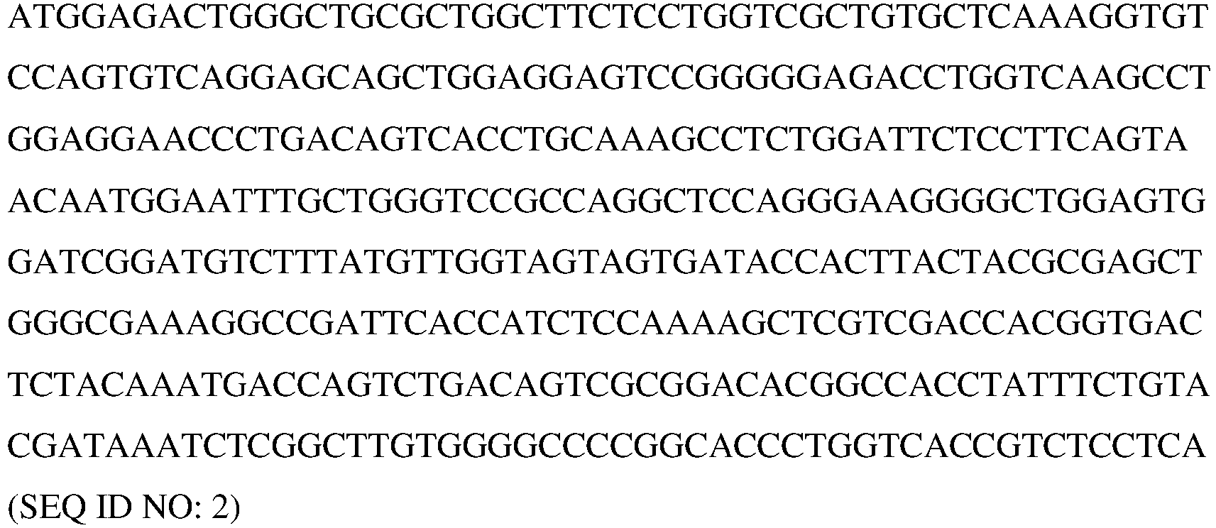
-
Clone 7 VH AA (CDRs underlined)
-
Clone 7 HC AA (CDRs underlined)
-
Clone 7 VH CDR1 AA
GFSFSNN (SEQ ID NO: 5) -
Clone 7 VH CDR2 AA
YVGSSD (SEQ ID NO: 6) -
Clone 7 VH CDR3 AA
NLGL (SEQ ID NO: 7) -
Clone 7 VL DNA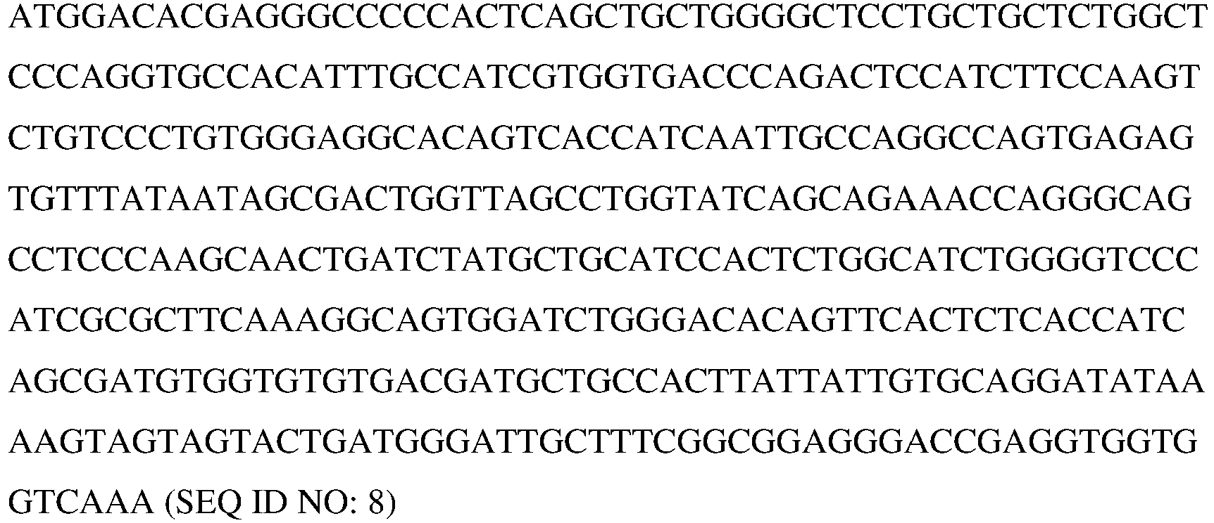
-
Clone 7 VL AA (CDRs underlined)
-
Clone 7 LC AA (CDRs underlined)
-
Clone 7 VL CDR1 AA
QASESVYNSDWLA (SEQ ID NO: 11) -
Clone 7 VL CDR2 AA
AASTLAS (SEQ ID NO: 12) -
Clone 7 VL CDR3 AA
AGYKSSSTDGIA (SEQ ID NO: 13) -
Clone 13 VH DNA
-
Clone 13 VH AA (CDRs underlined)
-
Clone 13 HC AA (CDRs underlined)
-
Clone 13 VH CDR1 AA
GFSFSNN (SEQ ID NO: 5) -
Clone 13 VH CDR2 AA
YVGSSD (SEQ ID NO: 6) -
Clone 13 VH CDR3 AA -
Clone 13 VL DNA
-
Clone 13 VL AA (CDRs underlined)
-
Clone 13 LC AA (CDRs underlined)
-
Clone 13 VL CDR1 AA
QASESVYNSDWLA (SEQ ID NO: 11) -
Clone 13 VL CDR2 AA
AASTLAS (SEQ ID NO: 12) -
Clone 13 VL CDR3 AA
AGYKSSSTDGIA (SEQ ID NO: 13) - Clone 14-1 VH DNA


- Clone 14-1 VH AA (CDRs underlined)

- Clone 14-1 HC AA (CDRs underlined) ID NO: 22)

- Clone 14-1 VH CDR1 AA
GFDFSINY (SEQ ID NO: 23) - Clone 14-1 VH CDR2 AA
YTGDD (SEQ ID NO: 24) - Clone 14-1 VH CDR3 AA
GLYSGSINNL (SEQ ID NO: 25) - Clone 14-1 VL DNA


- Clone 14-1 VL AA (CDRs underlined)

- Clone 14-1 LC AA (CDRs underlined)

- Clone 14-1 VLCDR1 AA
QASQSVYNNDYLS (SEQ ID NO: 29) - Clone 14-1 VL CDR2 AA
YASTLAS (SEQ ID NO: 30) - Clone 14-1 VL CDR3 AA
AGVKGYSNDNNG (SEQ ID NO: 31) - Clone 14-7 VH DNA


- Clone 14-7 VH AA (CDRs underlined)

- Clone 14-7 HC AA (CDRs underlined)

- Clone 14-7 VH CDR1 AA
GFDFSINY (SEQ ID NO: 23) - Clone 14-7 VH CDR2 AA
YTGDD (SEQ ID NO: 24) - Clone 14-7 VH CDR3 AA
GLYSGSINNL (SEQ ID NO: 25) - Clone 14-7 VL DNA

- Clone 14-7 VL AA (CDRs underlined)

- Clone 14-7 LC AA (CDRs underlined)

- Clone 14-7 VL CDR1 AA
QASQSVYNNDYLS (SEQ ID NO: 29) - Clone 14-7 VL CDR2 AA
YASTLAS (SEQ ID NO: 30) - Clone 14-7 VL CDR3 AA
AGVKGYSNDNNG (SEQ ID NO: 31) -
Clone 15 VH DNA
-
Clone 15 VH AA (CDRs underlined)
-
Clone 15 HC AA (CDRs underlined)
-
Clone 15 VH CDR1 AA
GFSFTSNY (SEQ ID NO: 41) -
Clone 15 VH CDR2 AA
FLGSSG (SEQ ID NO: 42) -
Clone 15 VH CDR3 AA
DYVNGYDYFNL (SEQ ID NO: 43) -
Clone 15 VL DNA
-
Clone 15 VL AA (CDRs underlined)
-
Clone 15 LC AA (CDRs underlined)
-
Clone 15 VL CDR1 AA
QSSQSVYNKNLA (SEQ ID NO: 47) -
Clone 15 VL CDR2 AA
STSTLDS (SEQ ID NO: 48) -
Clone 15 VL CDR3 AA
LGSYDCSSADCNA (SEQ ID NO: 49) -
Clone 17 VH DNA
-
Clone 17 VH AA (CDRs underlined)
-
Clone 17 HC AA (CDRs underlined)
-
Clone 17 VH CDR1 AA
GFSFSDSW (SEQ ID NO: 53) -
Clone 17 VH CDR2 AA
YTGDG (SEQ ID NO: 54) -
Clone 17 VH CDR3 AA
GAQFYL (SEQ ID NO: 55) -
Clone 17 VL DNA
-
Clone 17 VL AA (CDRs underlined)
-
Clone 17 LC AA (CDRs underlined)
-
Clone 17 VL CDR1 AA
QSSQSVYANTYLS (SEQ ID NO: 59) -
Clone 17 VL CDR2 AA
SASSLAS (SEQ ID NO: 60) -
Clone 17 VL CDR3 AA
LGRYSCGLADCAA (SEQ ID NO: 61) - In some embodiments, the antigen binding molecules of the present disclosure are antibodies and antigen binding fragments thereof. In some embodiments, the antibodies of the present disclosure comprise at least one CDR set forth in
Figures 5-21 . In another aspect, the present disclosure provides hybridomas capable of producing the antibodies disclosed herein, and also methods of producing antibodies from hybridomas, as described herein and as known in the art. - Humanized antibodies are described herein and may be prepared by known techniques. In some embodiments, a humanized monoclonal antibody comprises the variable domain of a murine or rabbit antibody (or all or part of the antigen binding site thereof) and a constant domain derived from a human antibody. Alternatively, a humanized antibody fragment may comprise an antigen binding site of a murine or rabbit monoclonal antibody and a variable domain fragment (lacking the antigen binding site) derived from a human antibody. Procedures for the production of engineered monoclonal antibodies include those described in Riechmann etal., (1988) Nature 332:323, Liu etal., (1987) Proc. Nat. Acad. Sci. USA 84:3439, Larrick et al., (1989) Bio/Technology 7:934, and Winter et al., (1993) TIPS 14:139. In some embodiments, the chimeric antibody is a CDR grafted antibody. Techniques for humanizing antibodies are discussed in, e.g.,
U.S. Pat. Nos. 5,869,619 ;5,225,539 ;5,821,337 ;5,859,205 ;6,881,557 ; Padlan et al., (1995) FASEB J. 9:133-39; Tamura et al., (2000) J. Immunol. 164:1432-41; Zhang et al., (2005) Mol. Immunol. 42(12):1445-1451; Hwang et al., Methods. (2005) 36(1):35-42; Dall'Acqua et al., (2005) Methods 36(1):43-60; and Clark, (2000) Immunology Today 21(8):397-402. - An antigen binding molecule of the present teaching can also be a fully human monoclonal antibody. Fully human monoclonal antibodies can be generated by any number of techniques with which those having ordinary skill in the art will be familiar. Such methods include, but are not limited to, Epstein Barr Virus (EBV) transformation of human peripheral blood cells (e.g., containing B lymphocytes), in vitro immunization of human B-cells, fusion of spleen cells from immunized transgenic mice carrying inserted human immunoglobulin genes, isolation from human immunoglobulin V region phage libraries, or other procedures as known in the art and based on the disclosure herein.
- Procedures have been developed for generating human monoclonal antibodies in non-human animals. For example, mice in which one or more endogenous immunoglobulin genes have been inactivated by various means have been prepared. Human immunoglobulin genes have been introduced into the mice to replace the inactivated mouse genes. In this technique, elements of the human heavy and light chain locus are introduced into strains of mice derived from embryonic stem cell lines that contain targeted disruptions of the endogenous heavy chain and light chain loci (see also Bruggemann et al., (1997) Curr. Opin. Biotechnol. 8:455-58).
- Examples of techniques for production and use of transgenic animals for the production of human or partially human antibodies are described in
U.S. Pat. Nos. 5,814,318 ,5,569,825 , and5,545,806 ; Davis et al., Antibody Engineering: Methods and Protocols, (Lo, ed) Humana Press, NJ, 191-200 (2003); Kellermann et al., (2002) Curr Opin Biotechnol. 13:593-97; Russel etal., (2000) Infect Immun. 68:1820-26; Gallo etal., (2000) Eur J. Immun. 30:534-40; Davis etal., (1999) Cancer Metastasis Rev. 18:421-25; Green, (1999) J Immunol Methods 231:11-23; Jakobovits, (1998) Advanced Drug Delivery Reviews 31:33-42; Green et al., (1998) J Exp Med. 188:483-95; Jakobovits, (1998) Exp. Opin. Invest. Drugs. 7:607-14; Tsuda et al., (1997) Genomics, 42:413-21; Mendez et al., (1997) Nat. Genet. 15:146-56; Jakobovits, (1994) Curr Biol. 4:761-63; Arbones et al., (1994) Immunity 1:247-60; Green et al., (1994) Nat. Genet. 7:13-21; Jakobovits et al., (1993) Nature 362:255-58; Jakobovits et al., (1993) Proc Natl Acad Sci USA 90:2551-55; Chen et al., (1993) Intl Immunol 5:647-656; Choi et al., (1993) Nature Genetics 4:117-23; Fishwild et al., (1996) Nature Biotechnology 14:845-51; Lonberg et al., (1994) Nature 368: 856-59; Lonberg, (1994) Handbook of Experimental Pharmacology 113: 49-101; Neuberger, (1996) Nature Biotech 14:826; Taylor et al., (1992) Nucleic Acids Research 20:6287-95; Taylor et al., (1994) Intl Immunol 6:579-91; Tomizuka et al., (1997) Nature Genetics 16:133-43; Tomizuka et al., (2000) Proc Nat Acad Sci USA 97:722-27; Tuaillon et al., (1993) Proc Nat Acad Sci USA 90:3720-24; Tuaillon et al., (1994) J Immunol 152:2912-20.; Lonberg et al., (1994) Nature 368:856; Taylor et al., (1994) Intl Immunol 6:579;U.S. Pat. No. 5,877,397 ; Bruggemann et al., (1997) Curr. Opin. Biotechnol. 8:455-58; Jakobovits et al., (1995) Ann. N.Y. Acad. Sci. 764:525-35. - An additional method for obtaining antigen binding molecules of the teaching is by the use of phage display, which is well-established for this purpose. See, e.g., Winter et al., (1994) Ann. Rev. Immunol. 12:433-55; Burton et al., (1994) Adv. Immunol 57:191-280. Human or murine immunoglobulin variable region gene combinatorial libraries can be created in phage vectors that can be screened to select Ig fragments (Fab, Fv, sFv, or multimers thereof) that bind the anti-CD19 scFv FMC63, as well as molecules comprising this sequence and cells presenting such molecules. See, e.g.,
U.S. Pat. No. 5,223,409 ; Huse et al., (1989) Science 246:1275-81; Sastry et al., (1989) Proc. Natl. Acad. Sci. USA 86:5728-32; Alting-Mees et al., (1990) Strategies in Molecular Biology 3:1-9; Kang et al., (1991) Proc. Natl. Acad. Sci. USA 88:4363-66; Hoogenboom et al., (1992) J. Mol. Biol. 227:381-388; Schlebusch et al., (1997) Hybridoma 16:47-52 and references cited therein. For example, a library containing a plurality of polynucleotide sequences encoding Ig variable region fragments can be inserted into the genome of a filamentous bacteriophage, such as M13 or lambda phage (λImmunoZap™(H) and λImmunoZap™(L) vectors (Stratagene, La Jolla, Calif) can also be used in this approach) or a variant thereof, in frame with the sequence encoding a phage coat protein. - Briefly, mRNA is isolated from a B-cell population, and used to create heavy and light chain immunoglobulin cDNA expression libraries in the λImmunoZap™(H) and λImmunoZap™(L) vectors. These vectors can be screened individually or co-expressed to form Fab fragments or antibodies. Positive plaques can subsequently be converted to a non-lytic plasmid that allows high level expression of monoclonal antibody fragments from E. coli.
- In some embodiments, in a hybridoma the variable regions of a gene expressing a monoclonal antibody of interest are amplified using nucleotide primers. These primers can be synthesized by one of ordinary skill in the art, or can be purchased from commercial sources, which also sell primers for mouse and human variable regions including, among others, primers for VHa, VHb, VHc, VHd, CH1, VL and CL regions). These primers can be used to amplify heavy or light chain variable regions, which can then be inserted into vectors such as λImmunoZap™(H) and λImmunoZap™(L) (Stratagene), respectively. These vectors can then be introduced into E. coli, yeast, or mammalian-based systems for expression. Large amounts of a single-chain protein containing a fusion of the VH and VL domains can be produced using these methods.
- Once cells producing the antigen binding molecules provided herein have been obtained using any of the above-described immunization and other techniques, the specific antibody genes can be cloned by isolating and amplifying DNA or mRNA therefrom according to standard procedures as described herein. The antibodies produced therefrom can be sequenced and the CDRs identified and the DNA coding for the CDRs can be manipulated as described previously to generate other antibodies according to the teaching.
- It will be understood by one skilled in the art that some proteins, such as antibodies, can undergo a variety of posttranslational modifications. The type and extent of these modifications often depends on the host cell line used to express the protein as well as the culture conditions. Such modifications can include variations in glycosylation, methionine oxidation, diketopiperizine formation, aspartate isomerization and asparagine deamidation. A frequent modification is the loss of a carboxy-terminal basic residue (such as lysine or arginine) due to the action of carboxypeptidases (as described in Harris, (1995) J Chromatog 705:129-34.
- An alternative method for production of a murine monoclonal antibody (from which FMC63 can be derived) is to inject the hybridoma cells into the peritoneal cavity of a syngeneic mouse, for example, a mouse that has been treated (e.g., pristane-primed) to promote formation of ascites fluid containing the monoclonal antibody. Monoclonal antibodies can be isolated and purified by a variety of well-established techniques. Such isolation techniques include affinity chromatography with Protein-A Sepharose, size-exclusion chromatography, and ion-exchange chromatography (see, e.g., Baines and Thorpe, (1992) in Methods in Molecular Biology, 10:79-104 (The Humana Press). Monoclonal antibodies can be purified by affinity chromatography using an appropriate ligand selected based on particular properties of the antibody (e.g., heavy or light chain isotype, binding specificity, etc.). Examples of a suitable ligand, immobilized on a solid support, include Protein A, Protein G, an anti-constant region (light chain or heavy chain) antibody, and an anti-idiotype antibody.
- Although the disclosed antigen binding molecules were produced in a rabbit system, human, partially human, or humanized antibodies may be suitable for many applications, particularly those involving administration of the antibody to a human subject, other types of antigen binding molecules will be suitable for certain applications. Such antibodies can be prepared as described herein and form an aspect of the instant disclosure.
- The instant disclosure provides antigen binding molecules that specifically bind to the anti-CD19 scFv FMC63 (SEQ ID NO: 1), molecules comprising this sequence and cells presenting such molecules. Antigen binding molecules that cross compete with the disclosed antigen binding molecules disclosed herein for an aspect of the disclosure. In certain embodiments, the antigen binding molecule cross competes with a reference antibody comprising an amino acid sequence selected from the group consisting of SEQ ID NOs: 1-60. In certain embodiments, the antigen binding molecule cross competes with a reference antibody, wherein the reference antibody comprises a VH CDR1 comprising an amino acid sequence of SEQ ID NOs: 5, 23, 41 and 53. In certain embodiments, the antigen binding molecule cross competes with a reference antibody, wherein the reference antibody comprises a VH CDR2 comprising an amino acid sequence of SEQ ID NOs: 6, 24, 42 and 54. In certain embodiments, the antigen binding molecule cross competes with a reference antibody, wherein the reference antibody comprises a VH CDR3 comprising an amino acid sequence of SEQ ID NOs: 7, 25, 43 and 55. In certain embodiments, the antigen binding molecule cross competes with a reference antibody, wherein the reference antibody comprises a VL CDR1 comprising an amino acid sequence of SEQ ID NOs: 11, 29, 47 and 59. In certain embodiments, the antigen binding molecule cross competes with a reference antibody, wherein the reference antibody comprises a VL CDR2 comprising an amino acid sequence of SEQ ID NOs: 12, 30, 48 and 60. In certain embodiments, the antigen binding molecule cross competes with a reference antibody, wherein the reference antibody comprises a VL CDR3 comprising an amino acid sequence of SEQ ID NOs: 13, 31, 49 and 61.
- In some embodiments, the polynucleotides of the present teaching encodes an antibody or antigen binding molecule that specifically binds the anti-CD19 scFv FMC63 (SEQ ID NO: 1), as well as molecules comprising these sequences and cells presenting such molecules, wherein the antibody or antigen binding molecule binds the same or an overlapping epitope as a reference antibody disclosed herein (e.g., those comprising sequences presented in
Figures 5-21 ). In certain embodiments, the antibody or antigen binding molecule binds the same or an overlapping epitope as a reference antibody. - In some embodiments, an antigen binding molecule or antibody that specifically binds to the anti-CD19 scFv FMC63 (SEQ ID NO: 1), molecules comprising this sequence and cells presenting such molecules, comprises a VH CDR1 comprising, consisting of, or consisting essentially of the amino acid sequence GFSFNN (SEQ ID NO: 5).
- In some embodiments, an antigen binding molecule or antibody that specifically binds to the anti-CD19 scFv FMC63 (SEQ ID NO: 1), molecules comprising this sequence and cells presenting such molecules, comprises a VH CDR2 comprising, consisting of, or consisting essentially of the amino acid sequence YVGSSD (SEQ ID NO: 6).
- In some embodiments, an antigen binding molecule or antibody that specifically binds to the anti-CD19 scFv FMC63 (SEQ ID NO: 1), molecules comprising this sequence and cells presenting such molecules, comprises a VH CDR3 comprising, consisting of, or consisting essentially of the amino acid sequence NLGL (SEQ ID NO: 7).
- In some embodiments, an antigen binding molecule or antibody that specifically binds to the anti-CD19 scFv FMC63 (SEQ ID NO: 1), molecules comprising this sequence and cells presenting such molecules, comprises a heavy chain VH comprising: (a) a VH CDR1 comprising, consisting of, or consisting essentially of the amino acid sequence GFSFNN (SEQ ID NO: 5); and/or (b) a VH CDR2 comprising, consisting of, or consisting essentially of the amino acid sequence YVGSSD (SEQ ID NO: 6); and/or (c) a VH CDR3 comprising, consisting of, or consisting essentially of the amino acid sequence NLGL (SEQ ID NO: 7).
- In some embodiments, the antigen binding molecule or antibody that specifically binds to the anti-CD19 scFv FMC63 (SEQ ID NO: 1), molecules comprising this sequence and cells presenting this sequence, comprises a VH CDR1, a VH CDR2, and VH CDR3, wherein the VH CDR1, VH CDR2, and VH CDR3 comprise the amino acid sequence of the VH CDR1, VH CDR2, and VH CDR3 sequences presented in
FIGURES 5 ,19 ,20 and21 . In a particular embodiment, the VH CDRs are those presented inFIGURE 5 . - In some embodiments, the antigen binding molecule or antibody that specifically binds to the anti-CD19 scFv FMC63 (SEQ ID NO: 1), molecules comprising this sequence and cells presenting this sequence, comprises a heavy chain variable region sequence comprising an amino acid sequence of
FIGURE 5 (SEQ ID NO: 3). - In some embodiments, the antigen binding molecule or antibody that specifically binds to the anti-CD19 scFv FMC63 (SEQ ID NO: 1), molecules comprising this sequence and cells presenting this sequence, comprises the VH framework regions (FRs) described herein. In specific embodiments, the antibody or antigen binding molecule comprises the VH FRs as set forth in, or derivable from, the sequences presented in
FIGURE 5 (e.g., one, two, three, or four of the FRs in one sequence ofFIGURE 5 ). - In some embodiments, the antigen binding molecule or antibody that specifically binds to the anti-CD19 scFv FMC63 (SEQ ID NO: 1), molecules comprising this sequence and cells presenting this sequence, comprises a heavy chain sequence disclosed herein (e.g., SEQ ID NO: 4 in
FIGURE 5 ). In some embodiments, the antibody or antigen binding molecule comprises a heavy chain variable region comprising the amino acid sequence of SEQ ID NO: 3. - In various embodiments, the heavy chain variable region is 80%, 85%, 90%, 91%, 92%, 93%, 94%, 95%, 96%, 97%, 98% or 99% identical to the heavy chain variable region sequence of SEQ ID NO:3.
- In some embodiments, an antigen binding molecule or antibody that specifically binds to the anti-CD19 scFv FMC63 (SEQ ID NO: 1), molecules comprising this sequence and cells presenting such molecules, comprises a VL CDR1 comprising, consisting of, or consisting essentially of the amino acid sequence QASESVYNSDWLA (SEQ ID NO: 11).
- In some embodiments, an antigen binding molecule or antibody that specifically binds to the anti-CD19 scFv FMC63 (SEQ ID NO: 1), molecules comprising this sequence and cells presenting such molecules, comprises a VL CDR2 comprising, consisting of, or consisting essentially of the amino acid sequence AASTLAS (SEQ ID NO: 12).
- In some embodiments, an antigen binding molecule or antibody that specifically binds to the anti-CD19 scFv FMC63 (SEQ ID NO: 1), molecules comprising this sequence and cells presenting such molecules, comprises a VL CDR3 comprising, consisting of, or consisting essentially of the amino acid sequence AGYKSSSTDGIA (SEQ ID NO: 13).
- In some embodiments, an antigen binding molecule or antibody that specifically binds to the anti-CD19 scFv FMC63 (SEQ ID NO: 1), molecules comprising this sequence and cells presenting such molecules, comprises a light chain VL comprising: (a) a VL CDR1 comprising, consisting of, or consisting essentially of the amino acid sequence QASESVYNSDWLA (SEQ ID NO: 11); and/or (b) a VL CDR2 comprising, consisting of, or consisting essentially of the amino acid sequence AASTLAS (SEQ ID NO: 12); and/or (c) a VL CDR3 comprising, consisting of, or consisting essentially of the amino acid sequence AGYKSSSTDGIA (SEQ ID NO: 13).
- In some embodiments, the antigen binding molecule or antibody that specifically binds to the anti-CD19 scFv FMC63 (SEQ ID NO: 1), molecules comprising this sequence and cells presenting this sequence, comprises a VL CDR1, a VL CDR2, and VL CDR3, wherein the VL CDR1, VL CDR2, and VL CDR3 comprise the amino acid sequence of the VL CDR1, VL CDR2, and VL CDR3 sequences presented in
FIGURES 6 ,19 ,20 and21 . In a particular embodiment, the VL CDRs are those presented inFIGURE 6 . - In some embodiments, the antigen binding molecule or antibody that specifically binds to the anti-CD19 scFv FMC63 (SEQ ID NO: 1), molecules comprising this sequence and cells presenting this sequence, comprises a light chain variable region sequence comprising an amino acid sequence of
FIGURE 6 (SEQ ID NO: 9) - In some embodiments, the antigen binding molecule or antibody that specifically binds to the anti-CD19 scFv FMC63 (SEQ ID NO: 1), molecules comprising this sequence and cells presenting this sequence, comprises the VL framework regions (FRs) described herein. In specific embodiments, the antibody or antigen binding molecule comprises the VL FRs as set forth in, or derivable from, the sequences presented in
FIGURE 6 (e.g., one, two, three, or four of the FRs in one sequence ofFIGURE 6 ). - In some embodiments, the antigen binding molecule or antibody that specifically binds to the anti-CD19 scFv FMC63 (SEQ ID NO: 1), molecules comprising this sequence and cells presenting this sequence, comprises a light chain sequence disclosed herein (e.g., SEQ ID NO: 10 in
FIGURE 6 ). In some embodiments, the antibody or antigen binding molecule comprises a light chain variable region comprising the amino acid sequence of SEQ ID NO: 9. - In various embodiments, the light chain variable region is 80%, 85%, 90%, 91%, 92%, 93%, 94%, 95%, 96%, 97%, 98% or 99% identical to the light chain variable region sequence of SEQ ID NO: 9.
- In some embodiments, the antibody or antigen binding molecule that specifically binds to the anti-CD19 scFv FMC63 (SEQ ID NO: 1), molecules comprising this sequence and cells presenting this sequence, comprises any one, two, and/or three VH CDR sequences disclosed herein. In certain embodiments, the antibody or antigen binding molecule comprises a VH CDR1, a VH CDR2, and a VH CDR3 having the amino acid sequence of any VH CDR1, VH CDR2, and VH CDR3 disclosed herein, respectively. In some embodiments, the antibody or antigen binding molecule comprises any one, two, and/or three VL CDR sequences disclosed herein. In certain embodiments, the antibody or antigen binding molecule comprises a VL CDR1, a VL CDR2, and a VL CDR3 having the amino acid sequence of any VL CDR1, VL CDR2, and VL CDR3 disclosed herein, respectively.
- In some embodiments, the antibody or antigen binding molecule that specifically binds to the anti-CD19 scFv FMC63 (SEQ ID NO: 1), molecules comprising this sequence and cells presenting this sequence, comprises: (a) a VH CDR1 region comprising the amino acid sequence of SEQ ID NO: 5; (b) a VH CDR2 region comprising the amino acid sequence of SEQ ID NO: 6; (c) a VH CDR3 region comprising the amino acid sequence of SEQ ID NO: 7; (d) a VL CDR1 region comprising the amino acid sequence of SEQ ID NO: 11; (e) a VL CDR2 region comprising the amino acid sequence of SEQ ID NO: 12; and (f) a VL CDR3 region comprising the amino acid sequence of SEQ ID NO: 13.
- In some embodiments, the antibody or antigen binding molecule that specifically binds to the anti-CD19 scFv FMC63 (SEQ ID NO: 1), molecules comprising this sequence and cells presenting this sequence, comprises: (a) a VH CDR1 region; (b) a VH CDR2 region; (c) a VH CDR3 region; (d) a VL CDR1 region; (e) a VL CDR2 region; and (f) a VL CDR3 region, wherein the VH and VL CDRs are shown in
FIGURES 5 ,6 , respectively, and inFIGURES 19 ,20 and21 . - In some embodiments, the antibody or antigen binding molecule that specifically binds to the anti-CD19 scFv FMC63 (SEQ ID NO: 1), molecules comprising this sequence and cells presenting this sequence, comprises a heavy chain variable region sequence disclosed herein (e.g., in
FIGURE 5 ) and a light chain variable region sequence disclosed herein (e.g., inFIGURE 6 ). - In some embodiments, the antibody or antigen binding molecule comprises: (a) a heavy chain variable region comprising the amino acid sequence of SEQ ID NO: 3; and (b) a light chain variable region comprising the amino acid sequence of SEQ ID NO: 9. Nucleotide sequences encoding the heavy chain variable region and the light chain variable region are provided in
FIGURES 5 and6 , respectively. - In some embodiments, the antibody or antigen binding molecule that specifically binds to the anti-CD19 scFv FMC63 (SEQ ID NO: 1), molecules comprising this sequence and cells presenting this sequence, comprises a heavy chain sequence disclosed herein (e.g., in
FIGURE 5 ) and a light chain sequence disclosed herein (e.g., inFIGURE 6 ). - In some embodiments, the antibody or antigen binding molecule comprises: (a) a heavy chain comprising the amino acid sequence of SEQ ID NO: 4; and (b) a light chain comprising the amino acid sequence of SEQ ID NO: 10.
- In some embodiments, the antibody or antigen binding molecule comprises: (a) a heavy chain comprising an amino acid sequence that is 80%, 85%, 90%, 91%, 92%, 93%, 94%, 95%, 96%, 97%, 98% or 99% identical to the amino acid sequence of SEQ ID NO: 4; and (b) a light chain comprising an amino acid sequence that is 80%, 85%, 90%, 91%, 92%, 93%, 94%, 95%, 96%, 97%, 98% or 99% identical to the amino acid sequence of SEQ ID NO: 10.
- In some embodiments, an antigen binding molecule or antibody that specifically binds to the anti-CD19 scFv FMC63 (SEQ ID NO: 1), molecules comprising this sequence and cells presenting such molecules, comprises a VH CDR1 comprising, consisting of, or consisting essentially of the amino acid sequence GFSFNN (SEQ ID NO: 5).
- In some embodiments, an antigen binding molecule or antibody that specifically binds to the anti-CD19 scFv FMC63 (SEQ ID NO: 1), molecules comprising this sequence and cells presenting such molecules, comprises a VH CDR2 comprising, consisting of, or consisting essentially of the amino acid sequence YVGSSD (SEQ ID NO: 6).
- In some embodiments, an antigen binding molecule or antibody that specifically binds to the anti-CD19 scFv FMC63 (SEQ ID NO: 1), molecules comprising this sequence and cells presenting such molecules, comprises a VH CDR3 comprising, consisting of, or consisting essentially of the amino acid sequence NLGL (SEQ ID NO: 7).
- In some embodiments, an antigen binding molecule or antibody that specifically binds to the anti-CD19 scFv FMC63 (SEQ ID NO: 1), molecules comprising this sequence and cells presenting such molecules, comprises a heavy chain VH comprising: (a) a VH CDR1 comprising, consisting of, or consisting essentially of the amino acid sequence GFSFNN (SEQ ID NO: 5); and/or (b) a VH CDR2 comprising, consisting of, or consisting essentially of the amino acid sequence YVGSSD (SEQ ID NO: 6); and/or (c) a VH CDR3 comprising, consisting of, or consisting essentially of the amino acid sequence NLGL (SEQ ID NO: 7).
- In some embodiments, the antigen binding molecule or antibody that specifically binds to the anti-CD19 scFv FMC63 (SEQ ID NO: 1), molecules comprising this sequence and cells presenting this sequence, comprises a VH CDR1, a VH CDR2, and VH CDR3, wherein the VH CDR1, VH CDR2, and VH CDR3 comprise the amino acid sequence of the VH CDR1, VH CDR2, and VH CDR3 sequences presented in
FIGURES 7 ,19 ,20 and21 . In a particular embodiment, the VH CDRs are those presented inFIGURE 7 . - In some embodiments, the antigen binding molecule or antibody that specifically binds to the anti-CD19 scFv FMC63 (SEQ ID NO: 1), molecules comprising this sequence and cells presenting this sequence, comprises a heavy chain variable region sequence comprising an amino acid sequence of
FIGURE 7 (SEQ ID NO: 15). - In some embodiments, the antigen binding molecule or antibody that specifically binds to the anti-CD19 scFv FMC63 (SEQ ID NO: 1), molecules comprising this sequence and cells presenting this sequence, comprises the VH framework regions (FRs) described herein. In specific embodiments, the antibody or antigen binding molecule comprises the VH FRs as set forth in, or derivable from, the sequences presented in
FIGURE 7 (e.g., one, two, three, or four of the FRs in one sequence ofFIGURE 7 ). - In some embodiments, the antigen binding molecule or antibody that specifically binds to the anti-CD19 scFv FMC63 (SEQ ID NO: 1), molecules comprising this sequence and cells presenting this sequence, comprises a heavy chain sequence disclosed herein (e.g., SEQ ID NO: 16 in
FIGURE 7 ). In some embodiments, the antibody or antigen binding molecule comprises a heavy chain variable region comprising the amino acid sequence of SEQ ID NO: 15. - In various embodiments, the heavy chain variable region is 80%, 85%, 90%, 91%, 92%, 93%, 94%, 95%, 96%, 97%, 98% or 99% identical to the heavy chain variable region sequence of SEQ ID NO:15.
- In some embodiments, an antigen binding molecule or antibody that specifically binds to the anti-CD19 scFv FMC63 (SEQ ID NO: 1), molecules comprising this sequence and cells presenting such molecules, comprises a VL CDR1 comprising, consisting of, or consisting essentially of the amino acid sequence QASESVYNSDWLA (SEQ ID NO: 11).
- In some embodiments, an antigen binding molecule or antibody that specifically binds to the anti-CD19 scFv FMC63 (SEQ ID NO: 1), molecules comprising this sequence and cells presenting such molecules, comprises a VL CDR2 comprising, consisting of, or consisting essentially of the amino acid sequence AASTLAS (SEQ ID NO: 12).
- In some embodiments, an antigen binding molecule or antibody that specifically binds to the anti-CD19 scFv FMC63 (SEQ ID NO: 1), molecules comprising this sequence and cells presenting such molecules, comprises a VL CDR3 comprising, consisting of, or consisting essentially of the amino acid sequence AGYKSSSTDGIA (SEQ ID NO: 13).
- In some embodiments, an antigen binding molecule or antibody that specifically binds to the anti-CD19 scFv FMC63 (SEQ ID NO: 1), molecules comprising this sequence and cells presenting such molecules, comprises a light chain VL comprising: (a) a VL CDR1 comprising, consisting of, or consisting essentially of the amino acid sequence QASESVYNSDWLA (SEQ ID NO: 11); and/or (b) a VL CDR2 comprising, consisting of, or consisting essentially of the amino acid sequence AASTLAS (SEQ ID NO: 12); and/or (c) a VL CDR3 comprising, consisting of, or consisting essentially of the amino acid sequence AGYKSSSTDGIA (SEQ ID NO: 13).
- In some embodiments, the antigen binding molecule or antibody that specifically binds to the anti-CD19 scFv FMC63 (SEQ ID NO: 1), molecules comprising this sequence and cells presenting this sequence, comprises a VL CDR1, a VL CDR2, and VL CDR3, wherein the VL CDR1, VL CDR2, and VL CDR3 comprise the amino acid sequence of the VL CDR1, VL CDR2, and VL CDR3 sequences presented in
FIGURES 8 ,19 ,20 and21 . In a particular embodiment, the VL CDRs are those presented inFIGURE 8 . - In some embodiments, the antigen binding molecule or antibody that specifically binds to the anti-CD19 scFv FMC63 (SEQ ID NO: 1), molecules comprising this sequence and cells presenting this sequence, comprises a light chain variable region sequence comprising an amino acid sequence of
FIGURE 8 (SEQ ID NO: 18) - In some embodiments, the antigen binding molecule or antibody that specifically binds to the anti-CD19 scFv FMC63 (SEQ ID NO: 1), molecules comprising this sequence and cells presenting this sequence, comprises the VL framework regions (FRs) described herein. In specific embodiments, the antibody or antigen binding molecule comprises the VL FRs as set forth in, or derivable from, the sequences presented in
FIGURE 8 (e.g., one, two, three, or four of the FRs in one sequence ofFIGURE 8 ). - In some embodiments, the antigen binding molecule or antibody that specifically binds to the anti-CD19 scFv FMC63 (SEQ ID NO: 1), molecules comprising this sequence and cells presenting this sequence, comprises a light chain sequence disclosed herein (e.g., SEQ ID NO: 19 in
FIGURE 8 ). In some embodiments, the antibody or antigen binding molecule comprises a light chain variable region comprising the amino acid sequence of SEQ ID NO: 18. - In various embodiments, the light chain variable region is 80%, 85%, 90%, 91%, 92%, 93%, 94%, 95%, 96%, 97%, 98% or 99% identical to the light chain variable region sequence of SEQ ID NO: 18.
- In some embodiments, the antibody or antigen binding molecule that specifically binds to the anti-CD19 scFv FMC63 (SEQ ID NO: 1), molecules comprising this sequence and cells presenting this sequence, comprises any one, two, and/or three VH CDR sequences disclosed herein. In certain embodiments, the antibody or antigen binding molecule comprises a VH CDR1, a VH CDR2, and a VH CDR3 having the amino acid sequence of any VH CDR1, VH CDR2, and VH CDR3 disclosed herein, respectively. In some embodiments, the antibody or antigen binding molecule comprises any one, two, and/or three VL CDR sequences disclosed herein. In certain embodiments, the antibody or antigen binding molecule comprises a VL CDR1, a VL CDR2, and a VL CDR3 having the amino acid sequence of any VL CDR1, VL CDR2, and VL CDR3 disclosed herein, respectively.
- In some embodiments, the antibody or antigen binding molecule that specifically binds to the anti-CD19 scFv FMC63 (SEQ ID NO: 1), molecules comprising this sequence and cells presenting this sequence, comprises: (a) a VH CDR1 region comprising the amino acid sequence of SEQ ID NO: 5; (b) a VH CDR2 region comprising the amino acid sequence of SEQ ID NO: 6; (c) a VH CDR3 region comprising the amino acid sequence of SEQ ID NO: 7; (d) a VL CDR1 region comprising the amino acid sequence of SEQ ID NO: 11; (e) a VL CDR2 region comprising the amino acid sequence of SEQ ID NO: 12; and (f) a VL CDR3 region comprising the amino acid sequence of SEQ ID NO: 13.
- In some embodiments, the antibody or antigen binding molecule that specifically binds to the anti-CD19 scFv FMC63 (SEQ ID NO: 1), molecules comprising this sequence and cells presenting this sequence, comprises: (a) a VH CDR1 region; (b) a VH CDR2 region; (c) a VH CDR3 region; (d) a VL CDR1 region; (e) a VL CDR2 region; and (f) a VL CDR3 region, wherein the VH and VL CDRs are shown in
FIGURES 7 ,8 , respectively, and inFIGURES 19 ,20 and21 . - In some embodiments, the antibody or antigen binding molecule that specifically binds to the anti-CD19 scFv FMC63 (SEQ ID NO: 1), molecules comprising this sequence and cells presenting this sequence, comprises a heavy chain variable region sequence disclosed herein (e.g., in
FIGURE 7 ) and a light chain variable region sequence disclosed herein (e.g., inFIGURE 8 ). - In some embodiments, the antibody or antigen binding molecule comprises: (a) a heavy chain variable region comprising the amino acid sequence of SEQ ID NO: 15; and (b) a light chain variable region comprising the amino acid sequence of SEQ ID NO: 18. Nucleotide sequences encoding the heavy chain variable region and the light chain variable region are provided in
FIGURES 7 and8 , respectively. - In some embodiments, the antibody or antigen binding molecule that specifically binds to the anti-CD19 scFv FMC63 (SEQ ID NO: 1), molecules comprising this sequence and cells presenting this sequence, comprises a heavy chain sequence disclosed herein (e.g., in
FIGURE 7 ) and a light chain sequence disclosed herein (e.g., inFIGURE 8 ). - In some embodiments, the antibody or antigen binding molecule comprises: (a) a heavy chain comprising the amino acid sequence of SEQ ID NO: 16; and (b) a light chain comprising the amino acid sequence of SEQ ID NO: 19.
- In some embodiments, the antibody or antigen binding molecule comprises: (a) a heavy chain comprising an amino acid sequence that is 80%, 85%, 90%, 91%, 92%, 93%, 94%, 95%, 96%, 97%, 98% or 99% identical to the amino acid sequence of SEQ ID NO: 16; and (b) a light chain comprising an amino acid sequence that is 80%, 85%, 90%, 91%, 92%, 93%, 94%, 95%, 96%, 97%, 98% or 99% identical to the amino acid sequence of SEQ ID NO: 19.
- In some embodiments, an antigen binding molecule or antibody that specifically binds to the anti-CD19 scFv FMC63 (SEQ ID NO: 1), molecules comprising this sequence and cells presenting such molecules, comprises a VH CDR1 comprising, consisting of, or consisting essentially of the amino acid sequence GFDFSINY (SEQ ID NO: 23).
- In some embodiments, an antigen binding molecule or antibody that specifically binds to the anti-CD19 scFv FMC63 (SEQ ID NO: 1), molecules comprising this sequence and cells presenting such molecules, comprises a VH CDR2 comprising, consisting of, or consisting essentially of the amino acid sequence YTGDD (SEQ ID NO: 24).
- In some embodiments, an antigen binding molecule or antibody that specifically binds to the anti-CD19 scFv FMC63 (SEQ ID NO: 1), molecules comprising this sequence and cells presenting such molecules, comprises a VH CDR3 comprising, consisting of, or consisting essentially of the amino acid sequence GLYSGSINNL (SEQ ID NO: 25).
- In some embodiments, an antigen binding molecule or antibody that specifically binds to the anti-CD19 scFv FMC63 (SEQ ID NO: 1), molecules comprising this sequence and cells presenting such molecules, comprises a heavy chain VH comprising: (a) a VH CDR1 comprising, consisting of, or consisting essentially of the amino acid sequence GFDFSINY (SEQ ID NO: 23); and/or (b) a VH CDR2 comprising, consisting of, or consisting essentially of the amino acid sequence YTGDD (SEQ ID NO: 24); and/or (c) a VH CDR3 comprising, consisting of, or consisting essentially of the amino acid sequence GLYSGSINNL (SEQ ID NO: 25).
- In some embodiments, the antigen binding molecule or antibody that specifically binds to the anti-CD19 scFv FMC63 (SEQ ID NO: 1), molecules comprising this sequence and cells presenting this sequence, comprises a VH CDR1, a VH CDR2, and VH CDR3, wherein the VH CDR1, VH CDR2, and VH CDR3 comprise the amino acid sequence of the VH CDR1, VH CDR2, and VH CDR3 sequences presented in
FIGURES 19 ,20 , and21 . In a particular embodiment, the VH CDRs are those presented inFIGURE 9 . - In some embodiments, the antigen binding molecule or antibody that specifically binds to the anti-CD19 scFv FMC63 (SEQ ID NO: 1), molecules comprising this sequence and cells presenting this sequence, comprises a heavy chain variable region sequence comprising an amino acid sequence of
FIGURE 9 (SEQ ID NO: 21). - In some embodiments, the antigen binding molecule or antibody that specifically binds to the anti-CD19 scFv FMC63 (SEQ ID NO: 1), molecules comprising this sequence and cells presenting this sequence, comprises the VH framework regions (FRs) described herein. In specific embodiments, the antibody or antigen binding molecule comprises the VH FRs as set forth in, or derivable from, the sequences presented in
FIGURE 9 (e.g., one, two, three, or four of the FRs in one sequence ofFIGURE 9 ). - In some embodiments, the antigen binding molecule or antibody that specifically binds to the anti-CD19 scFv FMC63 (SEQ ID NO: 1), molecules comprising this sequence and cells presenting this sequence, comprises a heavy chain sequence disclosed herein (e.g., SEQ ID NO: 22 in
FIGURE 9 ). In some embodiments, the antibody or antigen binding molecule comprises a heavy chain variable region comprising the amino acid sequence of SEQ ID NO: 21. - In various embodiments, the heavy chain variable region is 80%, 85%, 90%, 91%, 92%, 93%, 94%, 95%, 96%, 97%, 98% or 99% identical to the heavy chain variable region sequence of SEQ ID NO:21.
- In some embodiments, an antigen binding molecule or antibody that specifically binds to the anti-CD19 scFv FMC63 (SEQ ID NO: 1), molecules comprising this sequence and cells presenting such molecules, comprises a VL CDR1 comprising, consisting of, or consisting essentially of the amino acid sequence QASQSVYNNDYLS (SEQ ID NO: 29).
- In some embodiments, an antigen binding molecule or antibody that specifically binds to the anti-CD19 scFv FMC63 (SEQ ID NO: 1), molecules comprising this sequence and cells presenting such molecules, comprises a VL CDR2 comprising, consisting of, or consisting essentially of the amino acid sequence YASTLAS (SEQ ID NO: 30).
- In some embodiments, an antigen binding molecule or antibody that specifically binds to the anti-CD19 scFv FMC63 (SEQ ID NO: 1), molecules comprising this sequence and cells presenting such molecules, comprises a VL CDR3 comprising, consisting of, or consisting essentially of the amino acid sequence AGVKGYSNDNNG (SEQ ID NO: 31).
- In some embodiments, an antigen binding molecule or antibody that specifically binds to the anti-CD19 scFv FMC63 (SEQ ID NO: 1), molecules comprising this sequence and cells presenting such molecules, comprises a light chain VL comprising: (a) a VL CDR1 comprising, consisting of, or consisting essentially of the amino acid sequence QASQSVYNNDYLS (SEQ ID NO: 29); and/or (b) a VL CDR2 comprising, consisting of, or consisting essentially of the amino acid sequence YASTLAS (SEQ ID NO: 30); and/or (c) a VL CDR3 comprising, consisting of, or consisting essentially of the amino acid sequence AGVKGYSNDNNG (SEQ ID NO: 31).
- In some embodiments, the antigen binding molecule or antibody that specifically binds to the anti-CD19 scFv FMC63 (SEQ ID NO: 1), molecules comprising this sequence and cells presenting this sequence, comprises a VL CDR1, a VL CDR2, and VL CDR3, wherein the VL CDR1, VL CDR2, and VL CDR3 comprise the amino acid sequence of the VL CDR1, VL CDR2, and VL CDR3 sequences presented in
FIGURES 19 ,20 and21 . In a particular embodiment, the VL CDRs are those presented inFIGURE 10 . - In some embodiments, the antigen binding molecule or antibody that specifically binds to the anti-CD19 scFv FMC63 (SEQ ID NO: 1), molecules comprising this sequence and cells presenting this sequence, comprises a light chain variable region sequence comprising an amino acid sequence of
FIGURE 10 (SEQ ID NO: 27). - In some embodiments, the antigen binding molecule or antibody that specifically binds to the anti-CD19 scFv FMC63 (SEQ ID NO: 1), molecules comprising this sequence and cells presenting this sequence, comprises the VL framework regions (FRs) described herein. In specific embodiments, the antibody or antigen binding molecule comprises the VL FRs as set forth in, or derivable from, the sequences presented in
FIGURE 10 (e.g., one, two, three, or four of the FRs in one sequence ofFIGURE 10 ). - In some embodiments, the antigen binding molecule or antibody that specifically binds to the anti-CD19 scFv FMC63 (SEQ ID NO: 1), molecules comprising this sequence and cells presenting this sequence, comprises a light chain sequence disclosed herein (e.g., SEQ ID NO: 28 in
FIGURE 10 ). In some embodiments, the antibody or antigen binding molecule comprises a light chain variable region comprising the amino acid sequence of SEQ ID NO: 27. - In various embodiments, the light chain variable region is 80%, 85%, 90%, 91%, 92%, 93%, 94%, 95%, 96%, 97%, 98% or 99% identical to the light chain variable region sequence of SEQ ID NO: 27.
- In some embodiments, the antibody or antigen binding molecule that specifically binds to the anti-CD19 scFv FMC63 (SEQ ID NO: 1), molecules comprising this sequence and cells presenting this sequence, comprises any one, two, and/or three VH CDR sequences disclosed herein. In certain embodiments, the antibody or antigen binding molecule comprises a VH CDR1, a VH CDR2, and a VH CDR3 having the amino acid sequence of any VH CDR1, VH CDR2, and VH CDR3 disclosed herein, respectively. In some embodiments, the antibody or antigen binding molecule comprises any one, two, and/or three VL CDR sequences disclosed herein. In certain embodiments, the antibody or antigen binding molecule comprises a VL CDR1, a VL CDR2, and a VL CDR3 having the amino acid sequence of any VL CDR1, VL CDR2, and VL CDR3 disclosed herein, respectively.
- In some embodiments, the antibody or antigen binding molecule that specifically binds to the anti-CD19 scFv FMC63 (SEQ ID NO: 1), molecules comprising this sequence and cells presenting this sequence, comprises: (a) a VH CDR1 region comprising the amino acid sequence of SEQ ID NO: 23; (b) a VH CDR2 region comprising the amino acid sequence of SEQ ID NO: 24; (c) a VH CDR3 region comprising the amino acid sequence of SEQ ID NO: 25; (d) a VL CDR1 region comprising the amino acid sequence of SEQ ID NO: 29; (e) a VL CDR2 region comprising the amino acid sequence of SEQ ID NO: 30; and (f) a VL CDR3 region comprising the amino acid sequence of SEQ ID NO: 31.
- In some embodiments, the antibody or antigen binding molecule that specifically binds to the anti-CD19 scFv FMC63 (SEQ ID NO: 1), molecules comprising this sequence and cells presenting this sequence, comprises: (a) a VH CDR1 region; (b) a VH CDR2 region; (c) a VH CDR3 region; (d) a VL CDR1 region; (e) a VL CDR2 region; and (f) a VL CDR3 region, wherein the VH and VL CDRs are shown in
FIGURES 9 ,10 , respectively, and inFIGURES 19 ,20 and21 . - In some embodiments, the antibody or antigen binding molecule that specifically binds to the anti-CD19 scFv FMC63 (SEQ ID NO: 1), molecules comprising this sequence and cells presenting this sequence, comprises a heavy chain variable region sequence disclosed herein (e.g., in
FIGURE 9 ) and a light chain variable region sequence disclosed herein (e.g., inFIGURE 10 ). - In some embodiments, the antibody or antigen binding molecule comprises: (a) a heavy chain variable region comprising the amino acid sequence of SEQ ID NO: 22; and (b) a light chain variable region comprising the amino acid sequence of SEQ ID NO: 28. Nucleotide sequences encoding the heavy chain variable region and the light chain variable region are provided in
FIGURES 9 and10 , respectively. - In some embodiments, the antibody or antigen binding molecule that specifically binds to the anti-CD19 scFv FMC63 (SEQ ID NO: 1), molecules comprising this sequence and cells presenting this sequence, comprises a heavy chain sequence disclosed herein (e.g., in
FIGURE 9 ) and a light chain sequence disclosed herein (e.g., inFIGURE 10 ). - In some embodiments, the antibody or antigen binding molecule comprises: (a) a heavy chain comprising the amino acid sequence of SEQ ID NO: 22; and (b) a light chain comprising the amino acid sequence of SEQ ID NO: 28.
- In some embodiments, the antibody or antigen binding molecule comprises: (a) a heavy chain comprising an amino acid sequence that is 80%, 85%, 90%, 91%, 92%, 93%, 94%, 95%, 96%, 97%, 98% or 99% identical to the amino acid sequence of SEQ ID NO: 22; and (b) a light chain comprising an amino acid sequence that is 80%, 85%, 90%, 91%, 92%, 93%, 94%, 95%, 96%, 97%, 98% or 99% identical to the amino acid sequence of SEQ ID NO: 28.
- In some embodiments, an antigen binding molecule or antibody that specifically binds to the anti-CD19 scFv FMC63 (SEQ ID NO: 1), molecules comprising this sequence and cells presenting such molecules, comprises a VH CDR1 comprising, consisting of, or consisting essentially of the amino acid sequence GFDFSINY (SEQ ID NO: 23).
- In some embodiments, an antigen binding molecule or antibody that specifically binds to the anti-CD19 scFv FMC63 (SEQ ID NO: 1), molecules comprising this sequence and cells presenting such molecules, comprises a VH CDR2 comprising, consisting of, or consisting essentially of the amino acid sequence YTGDD (SEQ ID NO: 24).
- In some embodiments, an antigen binding molecule or antibody that specifically binds to the anti-CD19 scFv FMC63 (SEQ ID NO: 1), molecules comprising this sequence and cells presenting such molecules, comprises a VH CDR3 comprising, consisting of, or consisting essentially of the amino acid sequence GLYSGSINNL (SEQ ID NO: 25).
- In some embodiments, an antigen binding molecule or antibody that specifically binds to the anti-CD19 scFv FMC63 (SEQ ID NO: 1), molecules comprising this sequence and cells presenting such molecules, comprises a heavy chain VH comprising: (a) a VH CDR1 comprising, consisting of, or consisting essentially of the amino acid sequence GFDFSINY (SEQ ID NO: 23); and/or (b) a VH CDR2 comprising, consisting of, or consisting essentially of the amino acid sequence YTGDD (SEQ ID NO: 24); and/or (c) a VH CDR3 comprising, consisting of, or consisting essentially of the amino acid sequence GLYSGSINNL (SEQ ID NO: 25).
- In some embodiments, the antigen binding molecule or antibody that specifically binds to the anti-CD19 scFv FMC63 (SEQ ID NO: 1), molecules comprising this sequence and cells presenting this sequence, comprises a VH CDR1, a VH CDR2, and VH CDR3, wherein the VH CDR1, VH CDR2, and VH CDR3 comprise the amino acid sequence of the VH CDR1, VH CDR2, and VH CDR3 sequences presented in
FIGURES 19 ,20 and21 . In a particular embodiment, the VH CDRs are those presented inFIGURE 11 . - In some embodiments, the antigen binding molecule or antibody that specifically binds to the anti-CD19 scFv FMC63 (SEQ ID NO: 1), molecules comprising this sequence and cells presenting this sequence, comprises a heavy chain variable region sequence comprising an amino acid sequence of
FIGURE 11 (SEQ ID NO: 33). - In some embodiments, the antigen binding molecule or antibody that specifically binds to the anti-CD19 scFv FMC63 (SEQ ID NO: 1), molecules comprising this sequence and cells presenting this sequence, comprises the VH framework regions (FRs) described herein. In specific embodiments, the antibody or antigen binding molecule comprises the VH FRs as set forth in, or derivable from, the sequences presented in
FIGURE 11 (e.g., one, two, three, or four of the FRs in one sequence ofFIGURE 11 ). - In some embodiments, the antigen binding molecule or antibody that specifically binds to the anti-CD19 scFv FMC63 (SEQ ID NO: 1), molecules comprising this sequence and cells presenting this sequence, comprises a heavy chain sequence disclosed herein (e.g., in
FIGURE 11 ). In some embodiments, the antibody or antigen binding molecule comprises a heavy chain variable region comprising the amino acid sequence of SEQ ID NO: 33. - In various embodiments, the heavy chain variable region is 80%, 85%, 90%, 91%, 92%, 93%, 94%, 95%, 96%, 97%, 98% or 99% identical to the heavy chain variable region sequence of SEQ ID NO: 33.
- In some embodiments, an antigen binding molecule or antibody that specifically binds to the anti-CD19 scFv FMC63 (SEQ ID NO: 1), molecules comprising this sequence and cells presenting such molecules, comprises a VL CDR1 comprising, consisting of, or consisting essentially of the amino acid sequence QASQSVYNNDYLS (SEQ ID NO: 29).
- In some embodiments, an antigen binding molecule or antibody that specifically binds to the anti-CD19 scFv FMC63 (SEQ ID NO: 1), molecules comprising this sequence and cells presenting such molecules, comprises a VL CDR2 comprising, consisting of, or consisting essentially of the amino acid sequence YASTLAS (SEQ ID NO: 30).
- In some embodiments, an antigen binding molecule or antibody that specifically binds to the anti-CD19 scFv FMC63 (SEQ ID NO: 1), molecules comprising this sequence and cells presenting such molecules, comprises a VL CDR3 comprising, consisting of, or consisting essentially of the amino acid sequence AGVKGYSNDNNG (SEQ ID NO: 31).
- In some embodiments, an antigen binding molecule or antibody that specifically binds to the anti-CD19 scFv FMC63 (SEQ ID NO: 1), molecules comprising this sequence and cells presenting such molecules, comprises a light chain VL comprising: (a) a VL CDR1 comprising, consisting of, or consisting essentially of the amino acid sequence QASQSVYNNDYLS (SEQ ID NO: 29); and/or (b) a VL CDR2 comprising, consisting of, or consisting essentially of the amino acid sequence YASTLAS (SEQ ID NO: 30); and/or (c) a VL CDR3 comprising, consisting of, or consisting essentially of the amino acid sequence AGVKGYSNDNNG (SEQ ID NO: 31).
- In some embodiments, the antigen binding molecule or antibody that specifically binds to the anti-CD19 scFv FMC63 (SEQ ID NO: 1), molecules comprising this sequence and cells presenting this sequence, comprises a VL CDR1, a VL CDR2, and VL CDR3, wherein the VL CDR1, VL CDR2, and VL CDR3 comprise the amino acid sequence of the VL CDR1, VL CDR2, and VL CDR3 sequences presented in
FIGURES 20 ,21 and22 . In a particular embodiment, the VL CDRs are those presented inFIGURE 12 . - In some embodiments, the antigen binding molecule or antibody that specifically binds to the anti-CD19 scFv FMC63 (SEQ ID NO: 1), molecules comprising this sequence and cells presenting this sequence, comprises a light chain variable region sequence comprising an amino acid sequence of
FIGURE 12 (SEQ ID NO: 36). - In some embodiments, the antigen binding molecule or antibody that specifically binds to the anti-CD19 scFv FMC63 (SEQ ID NO: 1), molecules comprising this sequence and cells presenting this sequence, comprises the VL framework regions (FRs) described herein. In specific embodiments, the antibody or antigen binding molecule comprises the VL FRs as set forth in, or derivable from, the sequences presented in
FIGURE 12 (e.g., one, two, three, or four of the FRs in one sequence ofFIGURE 12 ). - In some embodiments, the antigen binding molecule or antibody that specifically binds to the anti-CD19 scFv FMC63 (SEQ ID NO: 1), molecules comprising this sequence and cells presenting this sequence, comprises a light chain sequence disclosed herein (e.g., SEQ ID NO: 37 in
FIGURE 12 ). In some embodiments, the antibody or antigen binding molecule comprises a light chain variable region comprising the amino acid sequence of SEQ ID NO: 36. - In various embodiments, the light chain variable region is 80%, 85%, 90%, 91%, 92%, 93%, 94%, 95%, 96%, 97%, 98% or 99% identical to the light chain variable region sequence of SEQ ID NO: 36.
- In some embodiments, the antibody or antigen binding molecule that specifically binds to the anti-CD19 scFv FMC63 (SEQ ID NO: 1), molecules comprising this sequence and cells presenting this sequence, comprises any one, two, and/or three VH CDR sequences disclosed herein. In certain embodiments, the antibody or antigen binding molecule comprises a VH CDR1, a VH CDR2, and a VH CDR3 having the amino acid sequence of any VH CDR1, VH CDR2, and VH CDR3 disclosed herein, respectively. In some embodiments, the antibody or antigen binding molecule comprises any one, two, and/or three VL CDR sequences disclosed herein. In certain embodiments, the antibody or antigen binding molecule comprises a VL CDR1, a VL CDR2, and a VL CDR3 having the amino acid sequence of any VL CDR1, VL CDR2, and VL CDR3 disclosed herein, respectively.
- In some embodiments, the antibody or antigen binding molecule that specifically binds to the anti-CD19 scFv FMC63 (SEQ ID NO: 1), molecules comprising this sequence and cells presenting this sequence, comprises: (a) a VH CDR1 region comprising the amino acid sequence of SEQ ID NO: 23; (b) a VH CDR2 region comprising the amino acid sequence of SEQ ID NO: 24; (c) a VH CDR3 region comprising the amino acid sequence of SEQ ID NO: 25; (d) a VL CDR1 region comprising the amino acid sequence of SEQ ID NO: 29; (e) a VL CDR2 region comprising the amino acid sequence of SEQ ID NO: 30; and (f) a VL CDR3 region comprising the amino acid sequence of SEQ ID NO: 31.
- In some embodiments, the antibody or antigen binding molecule that specifically binds to the anti-CD19 scFv FMC63 (SEQ ID NO: 1), molecules comprising this sequence and cells presenting this sequence, comprises: (a) a VH CDR1 region; (b) a VH CDR2 region; (c) a VH CDR3 region; (d) a VL CDR1 region; (e) a VL CDR2 region; and (f) a VL CDR3 region, wherein the VH and VL CDRs are shown in
FIGURES 11 ,12 , respectively, and inFIGURES 19 ,20 and21 . - In some embodiments, the antibody or antigen binding molecule that specifically binds to the anti-CD19 scFv FMC63 (SEQ ID NO: 1), molecules comprising this sequence and cells presenting this sequence, comprises a heavy chain variable region sequence disclosed herein (e.g., in
FIGURE 11 ) and a light chain variable region sequence disclosed herein (e.g., inFIGURE 12 ). - In some embodiments, the antibody or antigen binding molecule comprises: (a) a heavy chain variable region comprising the amino acid sequence of SEQ ID NO: 33; and (b) a light chain variable region comprising the amino acid sequence of SEQ ID NO: 36. Nucleotide sequences encoding the heavy chain variable region and the light chain variable region are provided in
FIGURES 11 and12 , respectively. - In some embodiments, the antibody or antigen binding molecule that specifically binds to the anti-CD19 scFv FMC63 (SEQ ID NO: 1), molecules comprising this sequence and cells presenting this sequence, comprises a heavy chain sequence disclosed herein (e.g., in
FIGURE 11 ) and a light chain sequence disclosed herein (e.g., inFIGURE 12 ). - In some embodiments, the antibody or antigen binding molecule comprises: (a) a heavy chain comprising the amino acid sequence of SEQ ID NO: 34; and (b) a light chain comprising the amino acid sequence of SEQ ID NO: 37.
- In some embodiments, the antibody or antigen binding molecule comprises: (a) a heavy chain comprising an amino acid sequence that is 80%, 85%, 90%, 91%, 92%, 93%, 94%, 95%, 96%, 97%, 98% or 99% identical to the amino acid sequence of SEQ ID NO: 34; and (b) a light chain comprising an amino acid sequence that is 80%, 85%, 90%, 91%, 92%, 93%, 94%, 95%, 96%, 97%, 98% or 99% identical to the amino acid sequence of SEQ ID NO: 37.
- In some embodiments, an antigen binding molecule or antibody that specifically binds to the anti-CD19 scFv FMC63 (SEQ ID NO: 1), molecules comprising this sequence and cells presenting such molecules, comprises a VH CDR1 comprising, consisting of, or consisting essentially of the amino acid sequence GFSFTSNY (SEQ ID NO: 41).
- In some embodiments, an antigen binding molecule or antibody that specifically binds to the anti-CD19 scFv FMC63 (SEQ ID NO: 1), molecules comprising this sequence and cells presenting such molecules, comprises a VH CDR2 comprising, consisting of, or consisting essentially of the amino acid sequence FLGSSG (SEQ ID NO: 42).
- In some embodiments, an antigen binding molecule or antibody that specifically binds to the anti-CD19 scFv FMC63 (SEQ ID NO: 1), molecules comprising this sequence and cells presenting such molecules, comprises a VH CDR3 comprising, consisting of, or consisting essentially of the amino acid sequence DYVNGYDYFNL (SEQ ID NO: 43).
- In some embodiments, an antigen binding molecule or antibody that specifically binds to the anti-CD19 scFv FMC63 (SEQ ID NO: 1), molecules comprising this sequence and cells presenting such molecules, comprises a heavy chain VH comprising: (a) a VH CDR1 comprising, consisting of, or consisting essentially of the amino acid sequence GFSFTSNY (SEQ ID NO: 41); and/or (b) a VH CDR2 comprising, consisting of, or consisting essentially of the amino acid sequence FLGSSG (SEQ ID NO: 42); and/or (c) a VH CDR3 comprising, consisting of, or consisting essentially of the amino acid sequence DYVNGYDYFNL (SEQ ID NO: 43).
- In some embodiments, the antigen binding molecule or antibody that specifically binds to the anti-CD19 scFv FMC63 (SEQ ID NO: 1), molecules comprising this sequence and cells presenting this sequence, comprises a VH CDR1, a VH CDR2, and VH CDR3, wherein the VH CDR1, VH CDR2, and VH CDR3 comprise the amino acid sequence of the VH CDR1, VH CDR2, and VH CDR3 sequences presented in
FIGURES 19 ,20 and21 . In a particular embodiment, the VH CDRs are those presented inFIGURE 13 . - In some embodiments, the antigen binding molecule or antibody that specifically binds to the anti-CD19 scFv FMC63 (SEQ ID NO: 1), molecules comprising this sequence and cells presenting this sequence, comprises a heavy chain variable region sequence comprising an amino acid sequence of
FIGURE 13 (SEQ ID NO: 39). - In some embodiments, the antigen binding molecule or antibody that specifically binds to the anti-CD19 scFv FMC63 (SEQ ID NO: 1), molecules comprising this sequence and cells presenting this sequence, comprises the VH framework regions (FRs) described herein. In specific embodiments, the antibody or antigen binding molecule comprises the VH FRs as set forth in, or derivable from, the sequences presented in
FIGURE 13 (e.g., one, two, three, or four of the FRs in one sequence ofFIGURE 13 ). - In some embodiments, the antigen binding molecule or antibody that specifically binds to the anti-CD19 scFv FMC63 (SEQ ID NO: 1), molecules comprising this sequence and cells presenting this sequence, comprises a heavy chain sequence disclosed herein (e.g.,SEQ ID NO: 40 in
FIGURE 13 ). In some embodiments, the antibody or antigen binding molecule comprises a heavy chain variable region comprising the amino acid sequence of SEQ ID NO: 39. - In various embodiments, the heavy chain variable region is 80%, 85%, 90%, 91%, 92%, 93%, 94%, 95%, 96%, 97%, 98% or 99% identical to the heavy chain variable region sequence of SEQ ID NO:39.
- In some embodiments, an antigen binding molecule or antibody that specifically binds to the anti-CD19 scFv FMC63 (SEQ ID NO: 1), molecules comprising this sequence and cells presenting such molecules, comprises a VL CDR1 comprising, consisting of, or consisting essentially of the amino acid sequence QSSQSVYNKNLA (SEQ ID NO: 47).
- In some embodiments, an antigen binding molecule or antibody that specifically binds to the anti-CD19 scFv FMC63 (SEQ ID NO: 1), molecules comprising this sequence and cells presenting such molecules, comprises a VL CDR2 comprising, consisting of, or consisting essentially of the amino acid sequence STSTLDS (SEQ ID NO: 48).
- In some embodiments, an antigen binding molecule or antibody that specifically binds to the anti-CD19 scFv FMC63 (SEQ ID NO: 1), molecules comprising this sequence and cells presenting such molecules, comprises a VL CDR3 comprising, consisting of, or consisting essentially of the amino acid sequence LGSYDCSSADCNA (SEQ ID NO: 49).
- In some embodiments, an antigen binding molecule or antibody that specifically binds to the anti-CD19 scFv FMC63 (SEQ ID NO: 1), molecules comprising this sequence and cells presenting such molecules, comprises a light chain VL comprising: (a) a VL CDR1 comprising, consisting of, or consisting essentially of the amino acid sequence QSSQSVYNKNLA (SEQ ID NO: 47); and/or (b) a VL CDR2 comprising, consisting of, or consisting essentially of the amino acid sequence STSTLDS (SEQ ID NO: 48); and/or (c) a VL CDR3 comprising, consisting of, or consisting essentially of the amino acid sequence LGSYDCSSADCNA (SEQ ID NO: 49).
- In some embodiments, the antigen binding molecule or antibody that specifically binds to the anti-CD19 scFv FMC63 (SEQ ID NO: 1), molecules comprising this sequence and cells presenting this sequence, comprises a VL CDR1, a VL CDR2, and VL CDR3, wherein the VL CDR1, VL CDR2, and VL CDR3 comprise the amino acid sequence of the VL CDR1, VL CDR2, and VL CDR3 sequences presented in
FIGURES 19 ,20 and21 . In a particular embodiment, the VL CDRs are those presented inFIGURE 14 . - In some embodiments, the antigen binding molecule or antibody that specifically binds to the anti-CD19 scFv FMC63 (SEQ ID NO: 1), molecules comprising this sequence and cells presenting this sequence, comprises a light chain variable region sequence comprising an amino acid sequence of
FIGURE 14 (SEQ ID NO: 45). - In some embodiments, the antigen binding molecule or antibody that specifically binds to the anti-CD19 scFv FMC63 (SEQ ID NO: 1), molecules comprising this sequence and cells presenting this sequence, comprises the VL framework regions (FRs) described herein. In specific embodiments, the antibody or antigen binding molecule comprises the VL FRs as set forth in, or derivable from, the sequences presented in
FIGURE 14 (e.g., one, two, three, or four of the FRs in one sequence ofFIGURE 14 ). - In some embodiments, the antigen binding molecule or antibody that specifically binds to the anti-CD19 scFv FMC63 (SEQ ID NO: 1), molecules comprising this sequence and cells presenting this sequence, comprises a light chain sequence disclosed herein (e.g., SEQ ID NO: 46 in
FIGURE 14 ). In some embodiments, the antibody or antigen binding molecule comprises a light chain variable region comprising the amino acid sequence of SEQ ID NO: 45. - In various embodiments, the light chain variable region is 80%, 85%, 90%, 91%, 92%, 93%, 94%, 95%, 96%, 97%, 98% or 99% identical to the light chain variable region sequence of SEQ ID NO: 45.
- In some embodiments, the antibody or antigen binding molecule that specifically binds to the anti-CD19 scFv FMC63 (SEQ ID NO: 1), molecules comprising this sequence and cells presenting this sequence, comprises any one, two, and/or three VH CDR sequences disclosed herein. In certain embodiments, the antibody or antigen binding molecule comprises a VH CDR1, a VH CDR2, and a VH CDR3 having the amino acid sequence of any VH CDR1, VH CDR2, and VH CDR3 disclosed herein, respectively. In some embodiments, the antibody or antigen binding molecule comprises any one, two, and/or three VL CDR sequences disclosed herein. In certain embodiments, the antibody or antigen binding molecule comprises a VL CDR1, a VL CDR2, and a VL CDR3 having the amino acid sequence of any VL CDR1, VL CDR2, and VL CDR3 disclosed herein, respectively.
- In some embodiments, the antibody or antigen binding molecule that specifically binds to the anti-CD19 scFv FMC63 (SEQ ID NO: 1), molecules comprising this sequence and cells presenting this sequence, comprises: (a) a VH CDR1 region comprising the amino acid sequence of SEQ ID NO: 41; (b) a VH CDR2 region comprising the amino acid sequence of SEQ ID NO: 42; (c) a VH CDR3 region comprising the amino acid sequence of SEQ ID NO: 43; (d) a VL CDR1 region comprising the amino acid sequence of SEQ ID NO: 47; (e) a VL CDR2 region comprising the amino acid sequence of SEQ ID NO: 48; and (f) a VL CDR3 region comprising the amino acid sequence of SEQ ID NO: 49.
- In some embodiments, the antibody or antigen binding molecule that specifically binds to the anti-CD19 scFv FMC63 (SEQ ID NO: 1), molecules comprising this sequence and cells presenting this sequence, comprises: (a) a VH CDR1 region; (b) a VH CDR2 region; (c) a VH CDR3 region; (d) a VL CDR1 region; (e) a VL CDR2 region; and (f) a VL CDR3 region, wherein the VH and VL CDRs are shown in
FIGURES 13 ,14 , respectively, and inFIGURES 19 ,20 and21 . - In some embodiments, the antibody or antigen binding molecule that specifically binds to the anti-CD19 scFv FMC63 (SEQ ID NO: 1), molecules comprising this sequence and cells presenting this sequence, comprises a heavy chain variable region sequence disclosed herein (e.g., in
FIGURE 13 ) and a light chain variable region sequence disclosed herein (e.g., inFIGURE 14 ). - In some embodiments, the antibody or antigen binding molecule comprises: (a) a heavy chain variable region comprising the amino acid sequence of SEQ ID NO: 39; and (b) a light chain variable region comprising the amino acid sequence of SEQ ID NO: 45. Nucleotide sequences encoding the heavy chain variable region and the light chain variable region are provided in
FIGURES 13 and14 , respectively. - In some embodiments, the antibody or antigen binding molecule that specifically binds to the anti-CD19 scFv FMC63 (SEQ ID NO: 1), molecules comprising this sequence and cells presenting this sequence, comprises a heavy chain sequence disclosed herein (e.g., in
FIGURE 13 ) and a light chain sequence disclosed herein (e.g., inFIGURE 14 ). - In some embodiments, the antibody or antigen binding molecule comprises: (a) a heavy chain comprising the amino acid sequence of SEQ ID NO: 40; and (b) a light chain comprising the amino acid sequence of SEQ ID NO: 46.
- In some embodiments, the antibody or antigen binding molecule comprises: (a) a heavy chain comprising an amino acid sequence that is 80%, 85%, 90%, 91%, 92%, 93%, 94%, 95%, 96%, 97%, 98% or 99% identical to the amino acid sequence of SEQ ID NO: 40; and (b) a light chain comprising an amino acid sequence that is 80%, 85%, 90%, 91%, 92%, 93%, 94%, 95%, 96%, 97%, 98% or 99% identical to the amino acid sequence of SEQ ID NO: 46.
- In some embodiments, an antigen binding molecule or antibody that specifically binds to the anti-CD19 scFv FMC63 (SEQ ID NO: 1), molecules comprising this sequence and cells presenting such molecules, comprises a VH CDR1 comprising, consisting of, or consisting essentially of the amino acid sequence GFSFSDSW (SEQ ID NO: 53).
- In some embodiments, an antigen binding molecule or antibody that specifically binds to the anti-CD19 scFv FMC63 (SEQ ID NO: 1), molecules comprising this sequence and cells presenting such molecules, comprises a VH CDR2 comprising, consisting of, or consisting essentially of the amino acid sequence YTGDG (SEQ ID NO: 54).
- In some embodiments, an antigen binding molecule or antibody that specifically binds to the anti-CD19 scFv FMC63 (SEQ ID NO: 1), molecules comprising this sequence and cells presenting such molecules, comprises a VH CDR3 comprising, consisting of, or consisting essentially of the amino acid sequence GAQFYL (SEQ ID NO: 55).
- In some embodiments, an antigen binding molecule or antibody that specifically binds to the anti-CD19 scFv FMC63 (SEQ ID NO: 1), molecules comprising this sequence and cells presenting such molecules, comprises a heavy chain VH comprising: (a) a VH CDR1 comprising, consisting of, or consisting essentially of the amino acid sequence GFSFTSNY (SEQ ID NO: 53); and/or (b) a VH CDR2 comprising, consisting of, or consisting essentially of the amino acid sequence FLGSSG (SEQ ID NO: 54); and/or (c) a VH CDR3 comprising, consisting of, or consisting essentially of the amino acid sequence DYVNGYDYFNL (SEQ ID NO: 55).
- In some embodiments, the antigen binding molecule or antibody that specifically binds to the anti-CD19 scFv FMC63 (SEQ ID NO: 1), molecules comprising this sequence and cells presenting this sequence, comprises a VH CDR1, a VH CDR2, and VH CDR3, wherein the VH CDR1, VH CDR2, and VH CDR3 comprise the amino acid sequence of the VH CDR1, VH CDR2, and VH CDR3 sequences presented in
FIGURES 19 ,20 and21 . In a particular embodiment, the VH CDRs are those presented inFIGURE 15 . - In some embodiments, the antigen binding molecule or antibody that specifically binds to the anti-CD19 scFv FMC63 (SEQ ID NO: 1), molecules comprising this sequence and cells presenting this sequence, comprises a heavy chain variable region sequence comprising an amino acid sequence of
FIGURE 15 (SEQ ID NO: 51). - In some embodiments, the antigen binding molecule or antibody that specifically binds to the anti-CD19 scFv FMC63 (SEQ ID NO: 1), molecules comprising this sequence and cells presenting this sequence, comprises the VH framework regions (FRs) described herein. In specific embodiments, the antibody or antigen binding molecule comprises the VH FRs as set forth in, or derivable from, the sequences presented in
FIGURE 15 (e.g., one, two, three, or four of the FRs in one sequence ofFIGURE 15 ). - In some embodiments, the antigen binding molecule or antibody that specifically binds to the anti-CD19 scFv FMC63 (SEQ ID NO: 1), molecules comprising this sequence and cells presenting this sequence, comprises a heavy chain sequence disclosed herein (e.g., SEQ ID NO: 52 in
FIGURE 15 ). In some embodiments, the antibody or antigen binding molecule comprises a heavy chain variable region comprising the amino acid sequence of SEQ ID NO: 51. - In various embodiments, the heavy chain variable region is 80%, 85%, 90%, 91%, 92%, 93%, 94%, 95%, 96%, 97%, 98% or 99% identical to the heavy chain variable region sequence of SEQ ID NO:51.
- In some embodiments, an antigen binding molecule or antibody that specifically binds to the anti-CD19 scFv FMC63 (SEQ ID NO: 1), molecules comprising this sequence and cells presenting such molecules, comprises a VL CDR1 comprising, consisting of, or consisting essentially of the amino acid sequence QSSQSVYANTYLS (SEQ ID NO: 59).
- In some embodiments, an antigen binding molecule or antibody that specifically binds to the anti-CD19 scFv FMC63 (SEQ ID NO: 1), molecules comprising this sequence and cells presenting such molecules, comprises a VL CDR2 comprising, consisting of, or consisting essentially of the amino acid sequence SASSLAS (SEQ ID NO: 60).
- In some embodiments, an antigen binding molecule or antibody that specifically binds to the anti-CD19 scFv FMC63 (SEQ ID NO: 1), molecules comprising this sequence and cells presenting such molecules, comprises a VL CDR3 comprising, consisting of, or consisting essentially of the amino acid sequence LGRYSCGLADCAA (SEQ ID NO: 61).
- In some embodiments, an antigen binding molecule or antibody that specifically binds to the anti-CD19 scFv FMC63 (SEQ ID NO: 1), molecules comprising this sequence and cells presenting such molecules, comprises a light chain VL comprising: (a) a VL CDR1 comprising, consisting of, or consisting essentially of the amino acid sequence QSSQSVYANTYLS (SEQ ID NO: 59); and/or (b) a VL CDR2 comprising, consisting of, or consisting essentially of the amino acid sequence SASSLAS (SEQ ID NO: 60); and/or (c) a VL CDR3 comprising, consisting of, or consisting essentially of the amino acid sequence LGRYSCGLADCAA (SEQ ID NO: 61).
- In some embodiments, the antigen binding molecule or antibody that specifically binds to the anti-CD19 scFv FMC63 (SEQ ID NO: 1), molecules comprising this sequence and cells presenting this sequence, comprises a VL CDR1, a VL CDR2, and VL CDR3, wherein the VL CDR1, VL CDR2, and VL CDR3 comprise the amino acid sequence of the VL CDR1, VL CDR2, and VL CDR3 sequences presented in
FIGURES 20 ,21 and22 . In a particular embodiment, the VL CDRs are those presented inFIGURE 21 . - In some embodiments, the antigen binding molecule or antibody that specifically binds to the anti-CD19 scFv FMC63 (SEQ ID NO: 1), molecules comprising this sequence and cells presenting this sequence, comprises a light chain variable region sequence comprising an amino acid sequence of
FIGURE 16 (SEQ ID NO: 57). - In some embodiments, the antigen binding molecule or antibody that specifically binds to the anti-CD19 scFv FMC63 (SEQ ID NO: 1), molecules comprising this sequence and cells presenting this sequence, comprises the VL framework regions (FRs) described herein. In specific embodiments, the antibody or antigen binding molecule comprises the VL FRs as set forth in, or derivable from, the sequences presented in
FIGURE 16 (e.g., one, two, three, or four of the FRs in one sequence ofFIGURE 16 ). - In some embodiments, the antigen binding molecule or antibody that specifically binds to the anti-CD19 scFv FMC63 (SEQ ID NO: 1), molecules comprising this sequence and cells presenting this sequence, comprises a light chain sequence disclosed herein (e.g., SEQ ID NO: 58 in
FIGURE 16 ). In some embodiments, the antibody or antigen binding molecule comprises a light chain variable region comprising the amino acid sequence of SEQ ID NO: 57. - In various embodiments, the light chain variable region is 80%, 85%, 90%, 91%, 92%, 93%, 94%, 95%, 96%, 97%, 98% or 99% identical to the light chain variable region sequence of SEQ ID NO:57.
- In some embodiments, the antibody or antigen binding molecule that specifically binds to the anti-CD19 scFv FMC63 (SEQ ID NO: 1), molecules comprising this sequence and cells presenting this sequence, comprises any one, two, and/or three VH CDR sequences disclosed herein. In certain embodiments, the antibody or antigen binding molecule comprises a VH CDR1, a VH CDR2, and a VH CDR3 having the amino acid sequence of any VH CDR1, VH CDR2, and VH CDR3 disclosed herein, respectively. In some embodiments, the antibody or antigen binding molecule comprises any one, two, and/or three VL CDR sequences disclosed herein. In certain embodiments, the antibody or antigen binding molecule comprises a VL CDR1, a VL CDR2, and a VL CDR3 having the amino acid sequence of any VL CDR1, VL CDR2, and VL CDR3 disclosed herein, respectively.
- In some embodiments, the antibody or antigen binding molecule that specifically binds to the anti-CD19 scFv FMC63 (SEQ ID NO: 1), molecules comprising this sequence and cells presenting this sequence, comprises: (a) a VH CDR1 region comprising the amino acid sequence of SEQ ID NO: 53; (b) a VH CDR2 region comprising the amino acid sequence of SEQ ID NO: 54; (c) a VH CDR3 region comprising the amino acid sequence of SEQ ID NO: 55; (d) a VL CDR1 region comprising the amino acid sequence of SEQ ID NO: 59; (e) a VL CDR2 region comprising the amino acid sequence of SEQ ID NO: 60; and (f) a VL CDR3 region comprising the amino acid sequence of SEQ ID NO: 61.
- In some embodiments, the antibody or antigen binding molecule that specifically binds to the anti-CD19 scFv FMC63 (SEQ ID NO: 1), molecules comprising this sequence and cells presenting this sequence, comprises: (a) a VH CDR1 region; (b) a VH CDR2 region; (c) a VH CDR3 region; (d) a VL CDR1 region; (e) a VL CDR2 region; and (f) a VL CDR3 region, wherein the VH and VL CDRs are shown in
FIGURES 15 ,16 , respectively, and inFIGURES 19 ,20 and21 . - In some embodiments, the antibody or antigen binding molecule that specifically binds to the anti-CD19 scFv FMC63 (SEQ ID NO: 1), molecules comprising this sequence and cells presenting this sequence, comprises a heavy chain variable region sequence disclosed herein (e.g., in
FIGURE 15 ) and a light chain variable region sequence disclosed herein (e.g., inFIGURE 16 ). - In some embodiments, the antibody or antigen binding molecule comprises: (a) a heavy chain variable region comprising the amino acid sequence of SEQ ID NO: 45; and (b) a light chain variable region comprising the amino acid sequence of SEQ ID NO: 57. Nucleotide sequences encoding the heavy chain variable region and the light chain variable region are provided in
FIGURES 15 and16 , respectively. - In some embodiments, the antibody or antigen binding molecule that specifically binds to the anti-CD19 scFv FMC63 (SEQ ID NO: 1), molecules comprising this sequence and cells presenting this sequence, comprises a heavy chain sequence disclosed herein (e.g., in
FIGURE 15 ) and a light chain sequence disclosed herein (e.g., inFIGURE 16 ). - In some embodiments, the antibody or antigen binding molecule comprises: (a) a heavy chain comprising the amino acid sequence of SEQ ID NO: 52; and (b) a light chain comprising the amino acid sequence of SEQ ID NO: 58.
- In some embodiments, the antibody or antigen binding molecule comprises: (a) a heavy chain comprising an amino acid sequence that is 80%, 85%, 90%, 91%, 92%, 93%, 94%, 95%, 96%, 97%, 98% or 99% identical to the amino acid sequence of SEQ ID NO: 52; and (b) a light chain comprising an amino acid sequence that is 80%, 85%, 90%, 91%, 92%, 93%, 94%, 95%, 96%, 97%, 98% or 99% identical to the amino acid sequence of SEQ ID NO: 58.
- The present teaching is also directed to polynucleotides encoding antibodies and other antigen binding molecules that specifically bind to the anti-CD19 scFv FMC63 (SEQ ID NO: 1), molecules comprising this sequence and cells presenting this sequence.
- In some embodiments, a polynucleotide of the present teaching encodes an antigen binding molecule, wherein the antigen binding molecule comprises a heavy chain variable region amino acid sequence that is at least about 75%, at least about 85%, at least about 85%, at least about 90%, at least about 95%, at least about 96%, at least about 97%, at least about 98%, at least about 99%, or 100% identical to a heavy chain variable region amino acid sequence selected from the group consisting of SEQ ID NOs: 3, 15, 21, 33, 39 and 51.
- In some embodiments, a polynucleotide of the present teaching encodes antigen binding molecule, wherein the antigen binding molecule comprises a light chain variable amino acid sequence that is at least about 75%, at least about 85%, at least about 85%, at least about 90%, at least about 95%, at least about 96%, at least about 97%, at least about 98%, at least about 99%, or 100% identical to a light chain variable region amino acid sequence selected from the group consisting of SEQ ID NOs: 8, 18, 27, 36, 45 and 57.
- In certain embodiments, the polynucleotide comprises a heavy chain coding sequence selected from the group consisting of SEQ ID NO: 2, SEQ ID NO: 14, SEQ ID NO: 20, SEQ ID NO: 32, SEQ ID NO: 38 and SEQ ID NO: 50. In another embodiment, the polynucleotide comprises a light chain coding sequence selected from the group consisting of SEQ ID NO: 8, SEQ ID NO: 17, SEQ ID NO: 26, SEQ ID NO: 35, SEQ ID NO: 44 and SEQ ID NO: 56.
- As will be appreciated by those of skill in the art, variations of the disclosed polynucleotide sequences are possible due to the degeneracy of the genetic code. Such variants of the disclose polynucleotide sequences thus form an aspect of the instant disclosure.
- In certain aspects, provided herein are vectors comprising a polynucleotide disclosed herein. In some embodiments, the present teaching is directed to a vector or a set of vectors comprising a polynucleotide(s) encoding an amino acid sequence of an antibody or antigen binding molecule that specifically binds to the anti-CD19 scFv FMC63 (SEQ ID NO: 1), molecules comprising this sequence and cells presenting this sequence, as described herein.
- Any vector known in the art can be suitable for expressing the antibodies and other antigen binding molecules of the present teaching. In some embodiments, the vector is a viral vector. In some embodiments, the vector is a retroviral vector, a DNA vector, a murine leukemia virus vector, an SFG vector, a plasmid, a RNA vector, an adenoviral vector, a baculoviral vector, an Epstein Barr viral vector, a papovaviral vector, a vaccinia viral vector, a herpes simplex viral vector, an adenovirus associated vector (AAV), a lentiviral vector, or any combination thereof.
- In other aspects, provided herein are cells comprising a polynucleotide or a vector of the present teaching. In some embodiments, the present teaching is directed to cells, in vitro cells, comprising a polynucleotide encoding an antigen binding molecule, as described herein. In some embodiments, the present teaching is directed to cells, e.g., in vitro cells, comprising a polynucleotide encoding an antibody or an antigen binding molecule thereof that specifically binds to the anti-CD19 scFv FMC63 (SEQ ID NO: 1), molecules comprising this sequence and cells presenting this sequence, as disclosed herein.
- Any cell can be used as a host cell for the polynucleotides and vectors encoding all or a fragment of the antibodies and other antigen binding molecules of the present teaching. In some embodiments, a host cell can be a prokaryotic cell, fungal cell, yeast cell, or higher eukaryotic cells such as a mammalian cell. Suitable prokaryotic cells include, without limitation, eubacteria, such as Gram-negative or Gram-positive organisms, for example, Enterobactehaceae such as Escherichia, e.g., E. coli; Enterobacter; Erwinia; Klebsiella; Proteus; Salmonella, e.g., Salmonella typhimurium; Serratia, e.g., Serratia marcescans, and Shigella; Bacilli such as B. subtilis and B. licheniformis; Pseudomonas such as P. aeruginosa; and Streptomyces. In some embodiments, a host cell is a human cell. In some embodiments, a host cell is a CHO cell and in other embodiments a host cell is a sP2/0 or other murine cell. A host cell of the present teaching can be obtained through any source known in the art.
- Other aspects of the present teaching are directed to compositions comprising a polynucleotide described herein, a vector described herein, an antibody and/or an antigen binding molecule described herein, or an in vitro cell described herein. In some embodiments, the composition comprises a pharmaceutically acceptable carrier, diluent, solubilizer, emulsifier, preservative and/or adjuvant. In some embodiments, the composition comprises an excipient. In some embodiments, the composition comprises a polynucleotide encoding an antibody or antigen binding molecule that specifically binds to that specifically binds to the anti-CD19 scFv FMC63 (SEQ ID NO: 1), molecules comprising this sequence and cells presenting this sequence. In another embodiment, the composition comprises an antigen binding molecule encoded by a polynucleotide of the present teaching, wherein the antigen binding molecule specifically binds to the anti-CD19 scFv FMC63 (SEQ ID NO: 1), molecules comprising this sequence and cells presenting this sequence, as disclosed herein. In another embodiment, the composition comprises an in vitro cell comprising a polynucleotide encoding an antibody or an antigen binding molecule thereof encoded by a polynucleotide of the present teaching.
- In some embodiments, the composition comprises one antibody or antigen binding molecule that specifically binds to the anti-CD19 scFv FMC63 (SEQ ID NO: 1), molecules comprising this sequence and cells presenting this sequence, as disclosed herein. In some embodiments, the composition comprises more than one antibody or antigen binding molecule that specifically binds to the anti-CD19 scFv FMC63 (SEQ ID NO: 1), molecules comprising this sequence and cells presenting this sequence, as disclosed herein, wherein the antibodies or antigen binding molecules bind more than one epitope. In some embodiments, the antibodies or antigen binding molecules will not compete with one another for binding to that epitope. In some embodiments, two or more of the antibodies or antigen binding molecules provided herein are combined together in a pharmaceutical composition. Preferably such a composition will be suitable for administration to a subject, including a human.
- The following section describes various exemplary methods of using the disclosed antigen binding molecules herein. Any antigen binding molecule disclosed herein can be employed in the disclosed methods.
- In various embodiments of the disclosed methods, the antigen binding molecule is selected from the group consisting of an antibody, an scFv, a Fab, a Fab', a Fv, a F(ab')2, a dAb, a human antibody, a humanized antibody, a chimeric antibody, a monoclonal antibody, a polyclonal antibody, a recombinant antibody, an IgE antibody, an IgD antibody, an IgM antibody, an IgG1 antibody, an IgG1 antibody having at least one mutation in the hinge region, an IgG2 antibody an IgG2 antibody having at least one mutation in the hinge region, an IgG3 antibody, an IgG1 antibody having at least one mutation in the hinge region, an IgG4 antibody, an IgG4 antibody having at least one mutation in the hinge region, an antibody comprising at least one non-naturally occurring amino acid, and any combination thereof.
- In some of the disclosed methods T cells can be employed. Such T cells can come from any source known in the art. For example, T cells can be differentiated in vitro from a hematopoietic stem cell population, or T cells can be obtained from a subject. T cells can be obtained from, e.g., peripheral blood mononuclear cells (PBMCs), bone marrow, lymph node tissue, cord blood, thymus tissue, tissue from a site of infection, ascites, pleural effusion, spleen tissue, and tumors. In addition, the T cells can be derived from one or more T cell lines available in the art. T cells can also be obtained from a unit of blood collected from a subject using any number of techniques known to the skilled artisan, such as FICOLL™ separation and/or apheresis. Additional methods of isolating T cells for a T cell therapy are disclosed in
U.S. Patent Publication No. 2013/0287748 . - In various embodiments of the disclosed methods, an antigen binding molecule specifically binds to the anti-CD19 scFv FMC63 (SEQ ID NO: 1), molecules comprising this sequence and cells presenting this sequence, as disclosed herein. In further embodiments of the disclosed methods, the antigen binding molecule comprises one or more of (a) a light chain CDR1, (b) a light chain CDR2, (c) a light chain CDR3, (d) a heavy chain CDR1, (e) a heavy chain CDR2, and (f) a heavy chain CDR3. In additional embodiments of the disclosed methods, an antigen binding molecule comprises a heavy chain CDR3 comprising one of SEQ ID NOs: 7, 25, 43, 55, or a light chain CDR3 comprising one of SEQ ID NOs: 13, 31, 49, 61, or both the heavy and light chain CDR3s. In other embodiments of the disclosed methods, the antigen binding molecule comprises a heavy chain CDR1 comprising an amino acid sequence comprising one of SEQ ID NOs: 5, 23, 41 and 53, or a heavy chain CDR2 comprising the amino acid sequence of one of SEQ ID NOs: 6, 24, 42 and 54, or a light chain CDR1 comprising the amino acid sequence of one of SEQ ID NOs: 11, 29, 47 and 59 or a light chain CDR2 comprising the amino acid sequence of one of SEQ ID NOs: 12, 30, 48 and 60. Referring to the Figures, in various embodiments of the disclosed methods, the antigen binding molecule comprises a heavy chain CDR1, a heavy chain CDR2, a heavy chain CDR3, a light chain CDR1, a light chain CDR2, and a light chain CDR3, each CDR comprising an amino acid sequence shown in
FIGURES 5-21 . - In various embodiments of the disclosed methods, an antigen binding molecule comprises a heavy chain (HC), and the HC comprises a heavy chain variable region (VH) sequence comprising one of SEQ ID NOs: 3, 15, 21, 33, 39 and 51. Referring to the figures, in various embodiments of the disclosed methods the heavy chain comprises a heavy chain CDR1, a heavy chain CDR2, and a heavy chain CDR3, each CDR comprising an amino acid sequence shown in
FIGURES 5-21 . Moreover, in embodiments of the disclosed methods, an antigen binding molecule can be employed which comprises a VH amino acid sequence that is at least about 70%, at least about 75%, at least about 80%, at least about 85%, at least about 90%, at least about 95%, at least about 96%, at least about 97%, at least about 98%, at least about 99%, or about 100% identical to a VH of an antigen binding molecule of claim disclosed herein (e.g., an antigen binding molecules comprising a variable region (VH) sequence comprising one of SEQ ID NOs: 3, 15, 21, 33, 39 and 51). - In various embodiments of the disclosed methods, an antigen binding molecule comprises a light chain (LC), and the LC can comprise a heavy chain variable region (VL) sequence comprising one of SEQ ID NOs: 9, 18, 27, 36, 44 and 57. Referring to the figures, in various embodiments of the disclosed methods the light chain comprises a light chain CDR1, a light chain CDR2, and a light chain CDR3, each CDR comprising an amino acid sequence shown in
FIGURES 5-21 . Moreover, in embodiments of the disclosed methods, an antigen binding molecule can be employed which comprises a VL amino acid sequence that is at least about 70%, at least about 75%, at least about 80%, at least about 85%, at least about 90%, at least about 95%, at least about 96%, at least about 97%, at least about 98%, at least about 99%, or about 100% identical to a VL of an antigen binding molecule of claim disclosed herein (e.g., an antigen binding molecules comprising a variable region (VL) sequence comprising SEQ ID NO: 9, 18, 27, 36, 44 and 57). - In view of the above description of antigen binding molecules that can be employed in the disclosed methods, representative methods will now be discussed in more detail.
- In one aspect, a method of administering a dose of a medicament to a subject, the dose comprising a preselected number of cells presenting a therapeutic molecule comprising the anti-CD19 scFv FMC63 (SEQ ID NO: 1), is provided.
- In specific embodiments, the dose comprises 0.5 ×10⁶ cells per kilogram of the subject, 1.0×10⁶ cells per kilogram of the subject, 2.0×10⁶ cells per kilogram of the subject, 3.0×10⁶ cells per kilogram of the subject, 4.0×10⁶ cells per kilogram of the subject, or 5.0×10⁶ cells per kilogram of the subject, although the method can be employed using any dose. 1.0×10⁶ cells per kilogram of the subject is a preferred dose.
- Consistent with the definition provided herein, in various embodiments, a subject is a human or non-human subject. When the subject is a human, the subject can be, e.g., any human who is being treated for an abnormal physiological condition, such as cancer or has been formally diagnosed with a disorder, those without formally recognized disorders, those receiving medical attention, those at risk of developing the disorders, those being studied for the presence or absence of a disorder, etc.
- Initially, a sample of known volume comprising a population comprising a known number of cells, which cells are known or suspected to be presenting a molecule comprising SEQ ID NO: 1, is provided. In the disclosed method, the number of cells can be determined using any known method. In preferred embodiments the cells are counted using an automated apparatus, such as a cell sorter (e.g., a FACS), however traditional non-automated cell counting methods can also be employed.
- The cells of the method can comprise any type of cell, with immune cells (e.g., B lymphocytes, monocytes, dendritic cells, Langerhans cells, keratinocytes, endothelial cells, astrocytes, fibroblasts, and oligodendrocytes). T cells (including T cytotoxic, T helper and Treg cells) are especially preferred. In specific embodiments, the cells are T cells, which can be obtained as described herein and by methods known in the art. Any type of cell can be employed in the method, and the cell can be a human or non-human cell (including both prokaryotic and eukaryotic cells). Exemplary cells include, but are not limited to immune cells such as T cells, tumor infiltrating lymphocytes (TILs), NK cells, TCR-expressing cells, dendritic cells, and NK-T cells. The T cells can be autologous, allogeneic, or heterologous. In additional embodiments, the cells are T cells presenting a CAR. The T cells can be CD4+ T cells or CD8+ T cells. When a T cell is employed in the disclosed methods, the T cell can be an in vivo T cell or an in vitro T cell. Moreover, the cells can be disposed in, or isolated from, any environment capable of maintaining the cells in a viable form, such as blood, tissue or any other sample obtained from a subject, cell culture media, tissue grown ex vivo, etc. Gradient purification, cell culture selection and/or cell sorting can also be employed in obtaining T cells.
- The therapeutic molecule expressed by the cell can comprise any molecule known or suspected to provide a therapeutic benefit to a subject to which is it administered. Thus, a therapeutic molecule can be a peptide or polypeptide of any structure or design. Preferably the SEQ ID NO: 1 component is expressed or disposed, at least in part, extracellularly, i.e., to a degree that it can be recognized by an extracellular interaction partner such as the antigen binding molecules of the instant disclosure.
- In specific embodiments, the therapeutic molecule is a CAR. When the therapeutic molecule is a CAR it can comprise a molecule, or fragment thereof, selected from the group consisting of CD28, OX-40, 4-1BB/CD137, CD2, CD7, CD27, CD30, CD40, Programmed Death-1 (PD-1), inducible T cell co-stimulator (ICOS), lymphocyte function-associated antigen-1 (LFA-1, CDl-la/CD18), CD3 gamma, CD3 delta, CD3 epsilon, CD3 zeta, CD247, CD276 (B7-H3), LIGHT, (TNFSF14), NKG2C, Ig alpha (CD79a), DAP-10, Fc gamma receptor, MHC class 1 molecule, TNF receptor proteins, an Immunoglobulin protein, cytokine receptor, integrins, Signaling Lymphocytic Activation Molecules (SLAM proteins), activating NK cell receptors, BTLA, a Toll ligand receptor, ICAM-1, B7-H3, CDS, ICAM-1, GITR, BAFFR, LIGHT, HVEM (LIGHTR), KIRDS2, SLAMF7, NKp80 (KLRF1), NKp44, NKp30, NKp46, CD19, CD4, CD8alpha, CD8beta, IL-2R beta, IL-2R gamma, IL-7R alpha, ITGA4, VLA1, CD49a, ITGA4, IA4, CD49D, ITGA6, VLA-6, CD49f, ITGAD, CDl ld, ITGAE, CD103, ITGAL, CDl la, LFA-1, ITGAM, CDl lb, ITGAX, CDl lc, ITGB1, CD29, ITGB2, CD18, LFA-1, ITGB7, NKG2D, TNFR2, TRANCE/RANKL, DNAM1 (CD226), SLAMF4 (CD244, 2B4), CD84, CD96 (Tactile), CEACAM1, CRT AM, Ly9 (CD229), CD160 (BY55), PSGL1, CD100 (SEMA4D), CD69, SLAMF6 (NTB-A, Ly108), SLAM (SLAMF1, CD150, IPO-3), BLAME (SLAMF8), SELPLG (CD162), LTBR, LAT, GADS, SLP-76, PAG/Cbp, CD19a, a ligand that specifically binds with CD83, and combinations thereof.
- Continuing, an aliquot of the sample comprising a population of cells presenting a molecule comprising the anti-CD19 scFv FMC63 sequence (SEQ ID NO: 1) is provided. The aliquot can be obtained using any convenient means, such as by a cell sorter, by a simply pipetting of material out of the sample, etc.
- Additionally, an antigen binding molecule that specifically binds the anti-CD19 scFv FMC63 (SEQ ID NO: 1) further comprising a detectable label is provided. The antigen binding molecule is preferably an antigen binding molecule disclosed herein, e.g., in the figures, sequence listing or the instant disclosure. Any detectable label can be employed in the method, as described herein, and suitable labels can be selected using a desired set of criteria. Examples of types of detectable labels include fluorescent labels (e.g., an Atto dye, an Alexafluor dye, quantum dots, Hydroxycoumarin, Aminocoumarin, Methoxycoumarin, Cascade Blue, Pacific Blue, Pacific Orange, Lucifer yellow, NBD, R-Phycoerythrin (PE), PE-Cy5 conjugates, PE-Cy7 conjugates, Red 613, PerCP, TruRed, FluorX, Fluorescein, BODIPY-FL, Cy2, Cy3, Cy3B, Cy3.5, Cy5, Cy5.5, Cy7, TRITC, X-Rhodamine, Lissamine Rhodamine B, Texas Red, Allophycocyanin (APC), APC-Cy7 conjugates, Indo-1, Fluo-3, Fluo-4, DCFH, DHR, SNARF, GFP (Y66H mutation), GFP (Y66F mutation), EBFP, EBFP2, Azurite, GFPuv, T-Sapphire, Cerulean, mCFP, mTurquoise2, ECFP, CyPet, GFP (Y66W mutation), mKeima-Red, TagCFP, AmCyan1, mTFP1, GFP (S65A mutation), Midoriishi Cyan, Wild Type GFP, GFP (S65C mutation), TurboGFP, TagGFP, GFP (S65L mutation), Emerald, GFP (S65T mutation), EGFP, Azami Green, ZsGreen1, TagYFP, EYFP,Topaz, Venus, mCitrine, YPet, TurboYFP, ZsYellow1, Kusabira Orange, mOrange, Allophycocyanin (APC), mKO, TurboRFP, tdTomato, TagRFP, DsRed monomer, DsRed2 ("RFP"), mStrawberry, TurboFP602, AsRed2, mRFP1, J-Red, R-phycoerythrin (RPE), B-phycoerythrin (BPE), mCherry, HcRed1, Katusha, P3, Peridinin Chlorophyll (PerCP), mKate (TagFP635), TurboFP635, mPlum, and mRaspberry). Suitable optical dyes, including fluoro-phores, are described in Johnson, Molecular Probes Handbook:A Guide to Fluorescent Probes and Labeling Techniques, 11th Edition, Life Technologies, (2010), radiolabels (e.g., isotope markers such as 3H, 11C, 14C, 15N, 18F, 35S, 64CU, 90Y, 99Tc, 111In, 124I, 125I, 131I), photochromic compounds, a Halo-tag, Atto dyes, Tracy dyes, proteinaceous fluorescent labels (e.g., proteinaceous fluorescent labels also include, but are not limited to, green fluorescent protein, including a Renilla, Ptilosarcus, or Aequorea species of GFP (Chalfie et al., (1994) Science 263:802-805), EGFP (Clon-tech Labs., Inc., Genbank Accession Number U55762), blue fluorescent protein (BFP, Quantum Biotechnologies, Inc; Stauber, (1998) Biotechniques 24:462-471; Heim et al., (1996) Curr. Biol. 6: 178-182), enhanced yellow fluorescent protein (Clontech Labs., Inc.), luciferase (Ichiki et al., (1993) J. Immunol. 150:5408-5417), magnetic labels (e.g., DYNABEADS), etc. Strategies for the labeling of proteins are known in the art and can be employed in the disclosed method.
- The label can be associated with the antigen binding molecule at any position in the molecule, although it is preferable to associate the label with the molecule at a position (or positions, if multiple labels are employed) at a point such that the binding properties of the molecule are not modified (unless such modified binding activity is desired). Any antigen binding molecule that specifically binds SEQ ID NO: 1 (or fragment thereof) can be employed. Multiple examples of suitable antigen binding molecules are provided herein, e.g., those having one or more of the CDRs shown in
FIGURES 5-21 . - The antigen binding molecule can be disposed on any surface, or no surface at all. For example, the antigen binding molecule can be present in a buffer and the buffer-antigen binding molecule can be contacted with the sample. Alternatively, the antigen binding molecule can be associated with a surface. Suitable surfaces include agarose beads, magnetic beads such as DYNABEADS, or a plastic, glass or ceramic plate such as a welled plate, a bag such as a cell culture bag, etc. The surface can itself be disposed in another structure, such as a column.
- Continuing, the aliquot of the sample is contacted with the antigen binding molecule under conditions that permit the formation of a binding complex comprising a cell present in the sample and the antigen binding molecule. Thus, the result of this step of the method is the formation of a binding complex in which the antigen binding molecule, with which a detectable label is associated, is bound to the cell expressing the therapeutic molecule, which comprises the anti-CD19 scFv FMC63 sequence (SEQ ID NO: 1). Thus, the binding complex itself is detectable. Conditions that permit the formation of a binding complex will be dependent on a variety of factors, however generally aqueous buffers at physiological pH and ionic strength, such as in phosphate-buffered saline (PBS), will favor formation of binding complexes and are preferred in the disclosed method.
- The fraction of cells present in a binding complex in the aliquot is then determined. This calculation can be performed by comparing the number of cells bearing the detectable label to those that do not, and can be represented as percentage. The number of cells in binding complexes can be determined. The specific method employed to determine the number of cells present in a binding complex will be dependent on the nature of the label selected. For example, FACS can be employed when a fluorescent label is selected; when an isotope label is selected mass spectrometry, NMR or other technique can be employed; magnetic-based cell sorting can be employed when a magnetic label is chosen; microscopy can also be employed. The number of cells in the sample is known ab initio and thus the fraction of cells present in a binding complex can be easily determined.
- Continuing, the concentration of cells in the initial sample expressing a molecule comprising the anti-CD19 scFv FMC63 (SEQ ID NO: 1) is determined; the determination is based on the fraction of cells determined to be present in the binding complex, and thus expressing the therapeutic protein bearing a detectable label.
- The fraction of cells presenting the therapeutic protein is known, and the volume of the aliquot is known; thus a simple comparison of the number of cells in the sample from which the aliquot was taken that are expressing the therapeutic molecule to the volume of the larger sample provides the fraction of the cells in the sample bearing the therapeutic molecule on a therapeutic molecule/volume basis (i.e., the concentration of cells bearing the therapeutic molecule in the larger sample).
- The volume of the sample that comprises the selected number of cells is then determined, by extrapolation based on the concentration of cells bearing therapeutic molecule present in the sample.
- Finally, the volume of sample comprising the desired number of cells is administered to the subject. The administration can comprise an aspect of a therapeutic regimen based on the therapeutic molecule present in the sample and expressed by the cells in the sample.
- Although the administration can be performed one time or more than one time, an advantage of the method is that by administering a dose comprising the preselected number of cells, which number of cells will be determined based on a known or expected efficacy, unnecessary administration of cells presenting the therapeutic molecule is avoided; i.e., the subject receives the correct number of cells to provide a desired therapeutic benefit and is not overdosed with cells.
- There are situations in which it may be desirable to determine the number of cells present in a sample. For example, it may be desirable to determine the number of immune cells present a sample obtained from a subject. Or it may be desirable to determine the number of cells transfected and expressing a construct, which can be used as a measure of the level of efficiency of the transfection. The disclosed method can be employed in these and other applications in which it is desirable to determine the number of cells present in a sample.
- Thus, a method of determining a number of cells presenting a molecule in a sample wherein the molecule comprising the anti-CD19 scFv FMC63 (SEQ ID NO: 1) is provided.
- In one embodiment, a sample comprising cells known or suspected to be expressing a molecule comprising the amino acid sequence of the anti-CD19 scFv FMC63 (SEQ ID NO: 1) is provided.
- The cell can be of any type, and can be human or non-human (e.g., mouse, rate, rabbit, hamster, etc). In one preferred embodiment, the cell is an immune cell. An immune cell of the method can be any type of immune cell (e.g., B lymphocytes, monocytes, dendritic cells, Langerhans cells, keratinocytes, endothelial cells, astrocytes, fibroblasts, and oligodendrocytes). T cells (including T cytotoxic, T helper and Treg cells) are especially preferred. In specific embodiments, the cells are T cells, which can be obtained as described herein and by methods known in the art. Any type of immune cell can be employed in this embodiment of the disclosed method, and the cell can be a human or non-human cell (including both prokaryotic and eukaryotic cells). Exemplary cells include, but are not limited to immune cells such as T cells, tumor infiltrating lymphocytes (TILs), NK cells, TCR-expressing cells, dendritic cells, and NK-T cells. The T cells can be autologous, allogeneic, or heterologous. In additional embodiments, the cells are T cells presenting a CAR. The T cells can be CD4+ T cells or CD8+ T cells. When a T cell is employed in the disclosed methods, the T cell can be an in vivo T cell or an in vitro T cell. Moreover, the cells can be disposed in, or isolated from, any environment capable of maintaining the cells in a viable form, such as blood, tissue or any other sample obtained from a subject, cell culture media, tissue grown ex vivo, a suitable buffer, etc.
- In specific embodiments, the molecule comprising the anti-CD19 scFv FMC63 (SEQ ID NO: 1) is a CAR. When the molecule is a CAR it can comprise a molecule, or fragment thereof, selected from the group consisting of CD28, OX-40, 4-1BB/CD137, CD2, CD7, CD27, CD30, CD40, Programmed Death-1 (PD-1), inducible T cell co-stimulator (ICOS), lymphocyte function-associated antigen-1 (LFA-1, CDl-la/CD18), CD3 gamma, CD3 delta, CD3 epsilon, CD3 zeta, CD247, CD276 (B7-H3), LIGHT, (TNFSF14), NKG2C, Ig alpha (CD79a), DAP-10, Fc gamma receptor, MHC class 1 molecule, TNF receptor proteins, an Immunoglobulin protein, cytokine receptor, integrins, Signaling Lymphocytic Activation Molecules (SLAM proteins), activating NK cell receptors, BTLA, a Toll ligand receptor, ICAM-1, B7-H3, CDS, ICAM-1, GITR, BAFFR, LIGHT, HVEM (LIGHTR), KIRDS2, SLAMF7, NKp80 (KLRF1), NKp44, NKp30, NKp46, CD19, CD4, CD8alpha, CD8beta, IL-2R beta, IL-2R gamma, IL-7R alpha, ITGA4, VLA1, CD49a, ITGA4, IA4, CD49D, ITGA6, VLA-6, CD49f, ITGAD, CDl ld, ITGAE, CD103, ITGAL, CDl la, LFA-1, ITGAM, CDl lb, ITGAX, CDl lc, ITGBl, CD29, ITGB2, CD18, LFA-1, ITGB7, NKG2D, TNFR2, TRANCE/RANKL, DNAM1 (CD226), SLAMF4 (CD244, 2B4), CD84, CD96 (Tactile), CEACAM1, CRT AM, Ly9 (CD229), CD160 (BY55), PSGL1, CD100 (SEMA4D), CD69, SLAMF6 (NTB-A, Lyl08), SLAM (SLAMF1, CD150, IPO-3), BLAME (SLAMF8), SELPLG (CD162), LTBR, LAT, GADS, SLP-76, PAG/Cbp, CD19a, a ligand that specifically binds with CD83, and combinations thereof.
- The sample is then contacted with an antigen binding molecule that specifically binds SEQ ID NO: 1 and comprises a detectable label, under conditions that permit the formation of a binding complex comprising a cell present in the sample and the antigen binding molecule. The antigen binding molecule is preferably an antigen binding molecule (or fragment thereof) disclosed herein, e.g., in the figures, sequence listing or the instant section of the disclosure. Any antigen binding molecule that specifically binds SEQ ID NO: 1 can be employed in the disclosed method. Multiple examples of suitable antigen binding molecules are provided herein, e.g., those having one or more of the CDRs shown in
FIGURE 5-21 . - Any detectable label can be employed in the method, as described herein, and suitable labels can be selected using a desired set of criteria. Examples of types of detectable labels include fluorescent labels (e.g., fluorescein, rhodamine, tetramethylrhodamine, eosin, erythrosin, coumarin, methyl-coumarins, pyrene, Malachite green, stilbene, Lucifer Yellow, Cascade Blue, Texas Red, IAEDANS, EDANS, BODIPY FL, LC Red 640,
Cy 5, Cy 5.5, LC Red 705, Oregon green, the Alexa-Fluor dyes (Alexa Fluor 350, Alexa Fluor 430, Alexa Fluor 488, Alexa Fluor 546, Alexa Fluor 568, Alexa Fluor 594, Alexa Fluor 633, Alexa Fluor 647, Alexa Fluor 660, Alexa Fluor 680), Cascade Blue, Cas-cade Yellow and R-phycoerythrin (PE) (Molecular Probes), FITC, Rhodamine, and Texas Red (Pierce), Cy5, Cy5.5, Cy7 (Amersham Life Science). Suitable optical dyes, including fluoro-phores, are described in Johnson, Molecular Probes Handbook:A Guide to Fluorescent Probes and Labeling Techniques, 11th Edition, Life Technologies, (2010), radiolabels (e.g., isotope markers such as 3H, 11C, 14C, 15N, 18F, 35S, 64CU, 90Y, 99Tc, 111In, 124I, 125I, 131I), photochromic compounds, a Halo-tag, Atto dyes, Tracy dyes, proteinaceous fluorescent labels (e.g., proteinaceous fluorescent labels also include, but are not limited to, green fluorescent protein, including a Renilla, Ptilosarcus, or Aequorea species of GFP (Chalfie et al., (1994) Science 263:802-805), EGFP (Clon-tech Labs., Inc., Genbank Accession Number U55762), blue fluorescent protein (BFP, Quantum Biotechnologies, Inc; Stauber, (1998) Biotechniques 24:462-471; Heim et al., (1996) Curr. Biol. 6: 178-182), enhanced yellow fluorescent protein (Clontech Labs., Inc.), luciferase (Ichiki et al., (1993) J. Immunol. 150:5408-5417), magnetic labels (e.g., DYNABEADS), etc. Strategies for the labeling of proteins are well known in the art and can be employed in the disclosed method. See, e.g., Obermaier et al., (2015) Methods Mol Biol 1295:153-65; Strack (2016) Nature Methods 13:33; Site-Specific Protein Labeling: Methods and Protocols, (Gautier and Hinner, eds.) 2015, Springer. - The label can be associated with the antigen binding molecule at any position in the molecule, although it is preferable to associate the label with the molecule at a position (or positions, if multiple labels are employed) at a point such that the binding properties of the molecule are not modified (unless such modified binding activity is desired). Any antigen binding molecule that specifically binds the anti-CD19 scFv FMC63 sequence (SEQ ID NO: 1) (or fragment thereof) can be employed, such as those disclosed herein, e.g., those having one or more of the CDRs shown in
FIGURES 5-21 . - The antigen binding molecule can be disposed on any surface, or no surface at all. For example, the antigen binding molecule can be present in a buffer and the buffer-antigen binding molecule can be contacted with the sample. Alternatively, the antigen binding molecule can be associated with a surface. Suitable surfaces include agarose beads, magnetic beads such as DYNABEADS, or a plastic, glass or ceramic plate such as a welled plate, a bag such as a cell culture bag, etc. The surface can itself be disposed in another structure, such as a column.
- Conditions that permit the formation of a binding complex will be dependent on a variety of factors, however generally aqueous buffers at physiological pH and ionic strength, such as in phosphate-buffered saline (PBS), will favor formation of binding complexes and are preferred in the disclosed method.
- Continuing, the number of cells present in a binding complex in the sample is determined. The specific method employed to determine the number of cells present in a binding complex will be dependent on the nature of the label selected. For example, FACS can be employed when a fluorescent label is selected; when an isotope label is selected mass spectrometry, NMR or other technique can be employed; magnetic-based cell sorting can be employed when a magnetic label is chosen; microscopy can also be employed. The output of these detection methods can be in the form of a number of cells or the output can be of a form that allows the calculation of the number of cells based on the output.
- It is of tremendous value to have the ability to separate different populations of molecules, and particularly biologically-relevant molecules, from one another. Using the antigen binding molecules provided herein, such separation can be achieved and employed in a range of biotechnological, biopharmaceutical and therapeutic applications. Thus, in one aspect of the instant disclosure, a method of isolating a molecule comprising the anti-CD19 scFv FMC63 (SEQ ID NO: 1) is provided.
- In some embodiments, the method comprises providing a sample known or suspected to comprise a molecule comprising the anti-CD19 scFv FMC63 (SEQ ID NO: 1).
- In specific embodiments, the molecule comprising the anti-CD19 scFv FMC63 (SEQ ID NO: 1) is a CAR. When the molecule is a CAR it can comprise a molecule, or fragment thereof, selected from the group consisting of CD28, OX-40, 4-1BB/CD137, CD2, CD7, CD27, CD30, CD40, Programmed Death-1 (PD-1), inducible T cell co-stimulator (ICOS), lymphocyte function-associated antigen-1 (LFA-1, CDl-la/CD18), CD3 gamma, CD3 delta, CD3 epsilon, CD3 zeta, CD247, CD276 (B7-H3), LIGHT, (TNFSF14), NKG2C, Ig alpha (CD79a), DAP-10, Fc gamma receptor, MHC class 1 molecule, TNF receptor proteins, an Immunoglobulin protein, cytokine receptor, integrins, Signaling Lymphocytic Activation Molecules (SLAM proteins), activating NK cell receptors, BTLA, a Toll ligand receptor, ICAM-1, B7-H3, CDS, ICAM-1, GITR, BAFFR, LIGHT, HVEM (LIGHTR), KIRDS2, SLAMF7, NKp80 (KLRF1), NKp44, NKp30, NKp46, CD19, CD4, CD8alpha, CD8beta, IL-2R beta, IL-2R gamma, IL-7R alpha, ITGA4, VLA1, CD49a, ITGA4, IA4, CD49D, ITGA6, VLA-6, CD49f, ITGAD, CDl ld, ITGAE, CD103, ITGAL, CDl la, LFA-1, ITGAM, CDl lb, ITGAX, CDl lc, ITGBl, CD29, ITGB2, CD18, LFA-1, ITGB7, NKG2D, TNFR2, TRANCE/RANKL, DNAM1 (CD226), SLAMF4 (CD244, 2B4), CD84, CD96 (Tactile), CEACAM1, CRT AM, Ly9 (CD229), CD160 (BY55), PSGL1, CD100 (SEMA4D), CD69, SLAMF6 (NTB-A, Lyl08), SLAM (SLAMF1, CD150, IPO-3), BLAME (SLAMF8), SELPLG (CD162), LTBR, LAT, GADS, SLP-76, PAG/Cbp, CD19a, a ligand that specifically binds with CD83, and combinations thereof.
- An antigen binding molecule that specifically binds the anti-CD19 scFv FMC63 sequence (SEQ ID NO: 1) and optionally comprises a detectable label is provided. When it is decided to employ a detectable label, any detectable label can be employed in the method, as described herein, and suitable labels can be selected using a desired set of criteria. Examples of types of detectable labels include fluorescent labels (e.g., fluorescein, rhodamine, tetramethylrhodamine, eosin, erythrosin, coumarin, methyl-coumarins, pyrene, Malachite green, stilbene, Lucifer Yellow, Cascade Blue, Texas Red, IAEDANS, EDANS, BODIPY FL, LC Red 640,
Cy 5, Cy 5.5, LC Red 705, Oregon green, the Alexa-Fluor dyes (Alexa Fluor 350, Alexa Fluor 430, Alexa Fluor 488, Alexa Fluor 546, Alexa Fluor 568, Alexa Fluor 594, Alexa Fluor 633, Alexa Fluor 647, Alexa Fluor 660, Alexa Fluor 680), Cascade Blue, Cas-cade Yellow and R-phycoerythrin (PE) (Molecular Probes), FITC, Rhodamine, and Texas Red (Pierce), Cy5, Cy5.5, Cy7 (Amersham Life Science)). Suitable optical dyes, including fluorophores, are described in Johnson, Molecular Probes Handbook:A Guide to Fluorescent Probes and Labeling Techniques, 11th Edition, Life Technologies, (2010), radiolabels (e.g., isotope markers such as 3H, 11C, 14C, 15N, 18F, 35S, 64CU, 90Y, 99Tc, 111In, 124I, 125I, 131I). Photochromic compounds, a Halo-tag, Atto dyes, Tracy dyes, proteinaceous fluorescent labels (e.g., proteinaceous fluorescent labels also include, but are not limited to, green fluorescent protein, including a Renilla, Ptilosarcus, or Aequorea species of GFP (Chalfie et al., (1994) Science 263:802-805), EGFP (Clon-tech Labs., Inc., Genbank Accession Number U55762), blue fluorescent protein (BFP, Quantum Biotechnologies, Inc; Stauber, (1998) Biotechniques 24:462-471; Heim et al., (1996) Curr. Biol. 6: 178-182), enhanced yellow fluorescent protein (Clontech Labs., Inc.), luciferase (Ichiki et al., (1993) J. Immunol. 150:5408-5417), magnetic labels (e.g., DYNABEADS), etc can also be employed. Strategies for the labeling of proteins are well known in the art and can be employed in the disclosed method. See, e.g., Obermaier et al., (2015) Methods Mol Biol 1295:153-65; Strack (2016) Nature Methods 13:33; Site-Specific Protein Labeling: Methods and Protocols, (Gautier and Hinner, eds.) 2015, Springer. - The label can be associated with the antigen binding molecule at any position in the molecule, although it is preferable to associate the label with the molecule at a position (or positions, if multiple labels are employed) at a point such that the binding properties of the molecule are not modified (unless such modified binding activity is desired). Any antigen binding molecule that specifically binds anti-CD19 scFv FMC63 sequence (SEQ ID NO: 1) (or fragment thereof) can be employed, such as those disclosed herein, e.g., those having one or more of the CDRs shown in
FIGURES 5-21 . - The antigen binding molecule can be disposed on any surface, or no surface at all. For example, the antigen binding molecule can be present in a buffer and the buffer-antigen binding molecule can be contacted with the sample. Alternatively, the antigen binding molecule can be associated with a surface. Suitable surfaces include agarose beads, magnetic beads such as DYNABEADS, or a plastic, glass or ceramic plate such as a welled plate, a bag such as a cell culture bag, etc. The surface can itself be disposed in another structure, such as a column.
- Conditions that permit the formation of a binding complex will be dependent on a variety of factors, however generally aqueous buffers at physiological pH and ionic strength, such as in phosphate-buffered saline (PBS), will favor formation of binding complexes and are preferred in the disclosed method. Since the component parts of a binding complex can be disposed on surfaces as described herein, formed binding complexes can also be disposed on surfaces.
- At this stage, no binding complexes may have formed, or a plurality of binding complexes comprising one or more antigen binding molecules bound to a molecule comprising SEQ ID NO: 1 (or one or more molecules comprising SEQ ID NO: 1 bound to an antigen binding molecule) may have formed. Unbound molecules comprising SEQ ID NO: 1 and/or unbound antigen binding molecules may also be present in the local environment of any formed binding complexes.
- Any molecules not part of a binding complex are then separated from any formed binding complexes. The method of the removal will depend on the structure and/or local environment of the binding complexes. For example, if the antigen binding molecule is disposed on a bead, plate or bag the unbound components of the reaction mixture can be washed away using a solution that leaves formed binding complexes intact. If a binding complex is disposed on a bead, the bead itself may be situated in a column or other structure and the same approach can be used.
- The solution used to induce the formation of binding complexes can be used, for example, as a wash solution to remove unbound components. Any suitable buffer or solution that does not disrupt formed binding complexes can also be used. Typically, buffers having high salt concentrations, non-physiological pH, containing chaotropes or denaturants, are preferably avoided when performing this step of the method.
- A formed binding complex is then separated into (a) a molecule comprising SEQ ID NO: 1, and (b) an antigen binding molecule. The separation can be achieved using standard methodologies known to those of skill in the art. For example, a solution of suitable pH and composition can be washed over the complexes. A solution that is commonly employed for this purpose is 0.1 M glycine HCl, pH 2.5-3.0, and this solution can be employed to achieve the separation. Other solutions that can be employed include 100 mM citric acid, pH 3.0, 50-100 mM triethylamine or triethanolamine, pH 11.5; 150 mM ammonium hydroxide, pH 10.5; 0.1 M glycine•NaOH, pH 10.0; 5 M lithium chloride, 3.5 M magnesium or potassium chloride, 3.0 M potassium chloride, 2.5 M sodium or potassium iodide, 0.2-3.0 M sodium thiocyanate, 0.1 M Tris-acetate with 2.0 M NaCl, pH 7.7; 2-6 M guanidine HCl, 2-8 M urea, 1.0 M ammonium thiocyanate, 1
% sodium deoxycholate 1% SDS; and 10% dioxane 50% ethylene glycol, pH 8-11.5. - Following the separation, if the molecule comprising SEQ ID NO: 1 is of primary interest it can be collected; alternatively, if the antigen binding molecule is of primary interest it can be collected.
- As disclosed herein, it may sometimes be desirable to isolate a molecule comprising the anti-CD19 scFv FMC63 sequence (SEQ ID NO: 1), as provided herein. In other cases, simply knowing whether a molecule comprising SEQ ID NO: 1 provided herein is present or absent from a sample is enough information. For example, it may be beneficial to know that such a molecule is being expressed, regardless of the level of expression. In other cases, it may be desirable to know if a purification process or step designed to remove such a molecule has been effectively performed. Thus, the qualitative determination of the presence or absence of a molecule comprising the anti-CD19 scFv FMC63 sequence (SEQ ID NO: 1) of the instant disclosure can be useful in multiple applications. In view thereof, a method of determining the presence or absence of a molecule comprising SEQ ID NO: 1 in a sample is provided.
- In some embodiments, the method comprises providing a sample known or suspected to comprise a molecule comprising SEQ ID NO: 1.
- In some embodiments, the molecule comprising SEQ ID NO: 1 is a CAR. When the molecule is a CAR it can comprise a molecule, or fragment thereof, selected from the group consisting of CD28, OX-40, 4-1BB/CD137, CD2, CD7, CD27, CD30, CD40, Programmed Death-1 (PD-1), inducible T cell co-stimulator (ICOS), lymphocyte function-associated antigen-1 (LFA-1, CDl-la/CD18), CD3 gamma, CD3 delta, CD3 epsilon, CD3 zeta, CD247, CD276 (B7-H3), LIGHT, (TNFSF14), NKG2C, Ig alpha (CD79a), DAP-10, Fc gamma receptor, MHC class 1 molecule, TNF receptor proteins, an Immunoglobulin protein, cytokine receptor, integrins, Signaling Lymphocytic Activation Molecules (SLAM proteins), activating NK cell receptors, BTLA, a Toll ligand receptor, ICAM-1, B7-H3, CDS, ICAM-1, GITR, BAFFR, LIGHT, HVEM (LIGHTR), KIRDS2, SLAMF7, NKp80 (KLRF1), NKp44, NKp30, NKp46, CD19, CD4, CD8alpha, CD8beta, IL-2R beta, IL-2R gamma, IL-7R alpha, ITGA4, VLA1, CD49a, ITGA4, IA4, CD49D, ITGA6, VLA-6, CD49f, ITGAD, CDl ld, ITGAE, CD103, ITGAL, CDl la, LFA-1, ITGAM, CDl lb, ITGAX, CDl lc, ITGBl, CD29, ITGB2, CD18, LFA-1, ITGB7, NKG2D, TNFR2, TRANCE/RANKL, DNAM1 (CD226), SLAMF4 (CD244, 2B4), CD84, CD96 (Tactile), CEACAM1, CRT AM, Ly9 (CD229), CD160 (BY55), PSGL1, CD100 (SEMA4D), CD69, SLAMF6 (NTB-A, Ly108), SLAM (SLAMF1, CD150, IPO-3), BLAME (SLAMF8), SELPLG (CD162), LTBR, LAT, GADS, SLP-76, PAG/Cbp, CD19a, a ligand that specifically binds with CD83, and combinations thereof.
- An antigen binding molecule comprising a detectable label that specifically binds the anti-CD19 scFv FMC63 (SEQ ID NO: 1) is provided. Suitable labels can be selected using a desired set of criteria. Examples of types of detectable labels include fluorescent labels (e.g., fluorescein, rhodamine, tetramethylrhodamine, eosin, erythrosin, coumarin, methyl-coumarins, pyrene, Malachite green, stilbene, Lucifer Yellow, Cascade Blue, Texas Red, IAEDANS, EDANS, BODIPY FL, LC Red 640,
Cy 5, Cy 5.5, LC Red 705, Oregon green, the Alexa-Fluor dyes (Alexa Fluor 350, Alexa Fluor 430, Alexa Fluor 488, Alexa Fluor 546, Alexa Fluor 568, Alexa Fluor 594, Alexa Fluor 633, Alexa Fluor 647, Alexa Fluor 660, Alexa Fluor 680), Cascade Blue, Cas-cade Yellow and R-phycoerythrin (PE) (Molecular Probes), FITC, Rhodamine, and Texas Red (Pierce), Cy5, Cy5.5, Cy7 (Amersham Life Science). Suitable optical dyes, including fluoro-phores, are described in Johnson, Molecular Probes Handbook:A Guide to Fluorescent Probes and Labeling Techniques, 11th Edition, Life Technologies, (2010), radiolabels (e.g., isotope markers such as 3H, 11C, 14C, 15N, 18F, 35S, 64CU, 90Y, 99Tc, 111In, 124I, 125I, 131I), photochromic compounds, a Halo-tag, Atto dyes, Tracy dyes, proteinaceous fluorescent labels (e.g., proteinaceous fluorescent labels also include, but are not limited to, green fluorescent protein, including a Renilla, Ptilosarcus, or Aequorea species of GFP (Chalfie et al., (1994) Science 263:802-805), EGFP (Clon-tech Labs., Inc., Genbank Accession Number U55762), blue fluorescent protein (BFP, Quantum Biotechnologies, Inc; Stauber, (1998) Biotechniques 24:462-471; Heim et al., (1996) Curr. Biol. 6: 178-182), enhanced yellow fluorescent protein (Clontech Labs., Inc.), luciferase (Ichiki et al., (1993) J. Immunol. 150:5408-5417), magnetic labels (e.g., DYNABEADS), etc. Strategies for the labeling of proteins are well known in the art and can be employed in the disclosed method. See, e.g., Obermaier et al., (2015) Methods Mol Biol 1295:153-65; Strack (2016) Nature Methods 13:33; Site-Specific Protein Labeling: Methods and Protocols, (Gautier and Hinner, eds.) 2015, Springer. - The label can be associated with the antigen binding molecule at any position in the molecule, although it is preferable to associate the label with the molecule at a position (or positions, if multiple labels are employed) at a point such that the binding properties of the molecule are not modified (unless such modified binding activity is desired). Any antigen binding molecule that specifically binds the anti-CD19 scFv FMC63 sequence (SEQ ID NO: 1 or fragment thereof) can be employed, such as those disclosed herein, e.g., those having one or more of the CDRs shown in
FIGURES 5-21 . - Continuing, the sample is contacted with the antigen binding molecule under conditions that permit the formation of a binding complex comprising a molecule comprising SEQ ID NO: 1 (which can be presented on a cell) present in the sample and the antigen binding molecule. The antigen binding molecule can be disposed on any surface, or no surface at all. For example, the antigen binding molecule can be present in a buffer and the buffer-antigen binding molecule can be contacted with the sample. Alternatively, the antigen binding molecule can be associated with a surface. Suitable surfaces include agarose beads, magnetic beads such as DYNABEADS, or a plastic, glass or ceramic plate such as a welled plate, a bag such as a cell culture bag, etc. The surface can itself be disposed in another structure, such as a column.
- Conditions that permit the formation of a binding complex will be dependent on a variety of factors, however generally aqueous buffers at physiological pH and ionic strength, such as in phosphate-buffered saline (PBS), will favor formation of binding complexes and are preferred in the disclosed method. Since the component parts of a binding complex can be disposed on surfaces as described herein, formed binding complexes can also be disposed on surfaces.
- At this stage, no binding complexes may have formed, or a plurality of binding complexes comprising one or more antigen binding molecules bound to a molecule comprising the anti-CD19 scFv FMC63 sequence (SEQ ID NO: 1) (or one or more molecules comprising SEQ ID NO: 1 bound to an antigen binding molecule) may have formed. Unbound molecules comprising SEQ ID NO: 1 and/or unbound antigen binding molecules may also be present in the local environment of any formed binding complexes.
- Any molecules not part of a binding complex are then separated from any formed binding complexes. The method of the removal will depend on the structure and/or local environment of the binding complexes. For example, if the antigen binding molecule is disposed on a bead, plate or bag the unbound components of the reaction mixture can be washed away using a solution that leaves formed binding complexes intact. If a binding complex is disposed on a bead, the bead itself may be situated in a column or other structure and the same approach can be used.
- The solution used to induce the formation of binding complexes can be used, for example, as a wash solution to remove unbound components. Any suitable buffer or solution that does not disrupt formed binding complexes can also be used. Typically, buffers having high salt concentrations, non-physiological pH, containing chaotropes or denaturants, should be avoided when performing this step of the method.
- Lastly, the presence or absence of a binding complex--which will comprise a molecule comprising SEQ ID NO: 1 and an antigen binding molecule-is detected. The specific method employed to detect the presence or absence of a binding complex will be dependent on the nature of the label selected. For example, flow cytometry or FACS can be employed when a fluorescent label is selected; when an isotope label is selected mass spectrometry, NMR or other technique can be employed; magnetic-based cell sorting can be employed when a magnetic label is chosen; microscopy can also be employed. The end result of the method is a qualitative assessment of the presence or absence of the antigen binding molecule comprising the detectable label, and thus, the presence or absence of its binding partner, the molecule comprising SEQ ID NO: 1.
- As is the case with all of the disclosed methods, the molecule comprising the anti-CD19 scFv FMC63 sequence (SEQ ID NO: 1) can be disposed in any environment. In preferred embodiments, the molecule comprising SEQ ID NO: 1 is expressed on the surface of a cell. In this embodiment, the cell can be of any type, and can be human or non-human (e.g., mouse, rate, rabbit, hamster, etc). In one preferred embodiment, the cell is an immune cell. An immune cell of the method can be any type of immune cell (e.g., B lymphocytes, monocytes, dendritic cells, Langerhans cells, keratinocytes, endothelial cells, astrocytes, fibroblasts, and oligodendrocytes). T cells (including T cytotoxic, T helper and Treg cells) are especially preferred. In specific embodiments, the cells are T cells, which can be obtained as described herein and by methods known in the art. Any type of immune cell can be employed in this embodiment of the disclosed method, and the cell can be a human or non-human cell (including both prokaryotic and eukaryotic cells). Exemplary cells include, but are not limited to immune cells such as T cells, tumor infiltrating lymphocytes (TILs), NK cells, TCR-expressing cells, dendritic cells, and NK-T cells. The T cells can be autologous, allogeneic, or heterologous. In additional embodiments, the cells are T cells presenting a CAR. The T cells can be CD4+ T cells or CD8+ T cells. When a T cell is employed in the disclosed methods, the T cell can be an in vivo T cell or an in vitro T cell.
- In an additional embodiment, the cell can be disposed in, or isolated from, any environment capable of maintaining the cell in a viable form, such as blood, tissue or any other sample obtained from a subject, cell culture media, tissue grown ex vivo, a suitable buffer, etc.
- Very often a molecule of interest is present in a sample in lower-than-desired levels. For example, when a cell is transfected with a foreign gene expression levels of the protein(s) encoded by the foreign gene are sometimes low. The same can be true for molecules secreted from a cell; such molecules are often present in low quantities (but can still be detected using the methods provided herein, if the molecule comprises the anti-CD19 scFv FMC63 sequence (SEQ ID NO: 1). One solution to the problem of low expression levels is to increase the concentration of the molecule of interest, which can be free in solution, or expressed on the surface of a cell. The concentration of intracellularly-expressed molecules of interest can also be enhanced, however the cells must first be lysed to release the molecule. To address this problem, a method of increasing the concentration of cells presenting a molecule comprising the anti-CD19 scFv FMC63 sequence (SEQ ID NO: 1) is provided.
- In some embodiments, the method comprises providing a sample comprising cells known or suspected to present a molecule comprising the anti-CD19 scFv FMC63 sequence (SEQ ID NO: 1).
- In specific embodiments, the molecule comprising the sequence SEQ ID NO: 1 is a CAR. When the molecule is a CAR it can comprise a molecule, or fragment thereof, selected from the group consisting of CD28, OX-40, 4-1BB/CD137, CD2, CD7, CD27, CD30, CD40, Programmed Death-1 (PD-1), inducible T cell co-stimulator (ICOS), lymphocyte function-associated antigen-1 (LFA-1, CDl-la/CD18), CD3 gamma, CD3 delta, CD3 epsilon, CD3 zeta, CD247, CD276 (B7-H3), LIGHT, (TNFSF14), NKG2C, Ig alpha (CD79a), DAP-10, Fc gamma receptor, MHC class 1 molecule, TNF receptor proteins, an Immunoglobulin protein, cytokine receptor, integrins, Signaling Lymphocytic Activation Molecules (SLAM proteins), activating NK cell receptors, BTLA, a Toll ligand receptor, ICAM-1, B7-H3, CDS, ICAM-1, GITR, BAFFR, LIGHT, HVEM (LIGHTR), KIRDS2, SLAMF7, NKp80 (KLRF1), NKp44, NKp30, NKp46, CD19, CD4, CD8alpha, CD8beta, IL-2R beta, IL-2R gamma, IL-7R alpha, ITGA4, VLA1, CD49a, ITGA4, IA4, CD49D, ITGA6, VLA-6, CD49f, ITGAD, CDl ld, ITGAE, CD103, ITGAL, CDl la, LFA-1, ITGAM, CDl lb, ITGAX, CD1 lc, ITGBl, CD29, ITGB2, CD18, LFA-1, ITGB7, NKG2D, TNFR2, TRANCE/RANKL, DNAM1 (CD226), SLAMF4 (CD244, 2B4), CD84, CD96 (Tactile), CEACAM1, CRT AM, Ly9 (CD229), CD160 (BY55), PSGL1, CD100 (SEMA4D), CD69, SLAMF6 (NTB-A, Ly108), SLAM (SLAMF1, CD150, IPO-3), BLAME (SLAMF8), SELPLG (CD162), LTBR, LAT, GADS, SLP-76, PAG/Cbp, CD19a, a ligand that specifically binds with CD83, and combinations thereof.
- An antigen binding molecule that specifically binds SEQ ID NO: 1, and optionally comprises a detectable label, is provided. When it is decided to employ a detectable label, any detectable label can be employed in the method, as described herein, and suitable labels can be selected using a desired set of criteria. Examples of types of detectable labels include fluorescent labels (e.g., fluorescein, rhodamine, tetramethylrhodamine, eosin, erythrosin, coumarin, methyl-coumarins, pyrene, Malachite green, stilbene, Lucifer Yellow, Cascade Blue, Texas Red, IAEDANS, EDANS, BODIPY FL, LC Red 640,
Cy 5, Cy 5.5, LC Red 705, Oregon green, the Alexa-Fluor dyes (Alexa Fluor 350, Alexa Fluor 430, Alexa Fluor 488, Alexa Fluor 546, Alexa Fluor 568, Alexa Fluor 594, Alexa Fluor 633, Alexa Fluor 647, Alexa Fluor 660, Alexa Fluor 680), Cascade Blue, Cas-cade Yellow and R-phycoerythrin (PE) (Molecular Probes), FITC, Rhodamine, and Texas Red (Pierce), Cy5, Cy5.5, Cy7 (Amersham Life Science). Suitable optical dyes, including fluoro-phores, are described in Johnson, Molecular Probes Handbook:A Guide to Fluorescent Probes and Labeling Techniques, 11th Edition, Life Technologies, (2010), radiolabels (e.g., isotope markers such as 3H, 11C, 14C, 15N, 18F, 35S, 64CU, 90Y, 99Tc, 111In, 124I, 125I, 131I), photochromic compounds, a Halo-tag, Atto dyes, Tracy dyes, proteinaceous fluorescent labels (e.g., proteinaceous fluorescent labels also include, but are not limited to, green fluorescent protein, including a Renilla, Ptilosarcus, or Aequorea species of GFP (Chalfie et al., (1994) Science 263:802-805), EGFP (Clon-tech Labs., Inc., Genbank Accession Number U55762), blue fluorescent protein (BFP, Quantum Biotechnologies, Inc; Stauber, (1998) Biotechniques 24:462-471; Heim et al., (1996) Curr. Biol. 6: 178-182), enhanced yellow fluorescent protein (Clontech Labs., Inc.), luciferase (Ichiki et al., (1993) J. Immunol. 150:5408-5417), magnetic labels (e.g., DYNABEADS), etc. Strategies for the labeling of proteins are well known in the art and can be employed in the disclosed method. See, e.g., Obermaier et al., (2015) Methods Mol Biol 1295:153-65; Strack (2016) Nature Methods 13:33; Site-Specific Protein Labeling: Methods and Protocols, (Gautier and Hinner, eds.) 2015, Springer. - The label can be associated with the antigen binding molecule at any position in the molecule, although it is preferable to associate the label with the molecule at a position (or positions, if multiple labels are employed) at a point such that the binding properties of the molecule are not modified (unless such modified binding activity is desired). Any antigen binding molecule that specifically binds the anti-CD19 scFv FMC63 sequence (SEQ ID NO: 1) (or fragment thereof) can be employed, such as those disclosed herein, e.g., those having one or more of the CDRs shown in
FIGURES 5-21 . - The antigen binding molecule can be disposed on any surface, or no surface at all. For example, the antigen binding molecule can be present in a buffer and the buffer-antigen binding molecule can be contacted with the sample. Alternatively, the antigen binding molecule can be associated with a surface. Suitable surfaces include agarose beads, magnetic beads such as DYNABEADS, or a plastic, glass or ceramic plate such as a welled plate, a bag such as a cell culture bag, etc. The surface can itself be disposed in another structure, such as a column.
- A cell presenting a molecule comprising SEQ ID NO: 1 can be of any type, and can be human or non-human (e.g., mouse, rate, rabbit, hamster, etc). In a preferred embodiment, the cell is an immune cell. An immune cell of the method can be any type of immune cell (e.g., B lymphocytes, monocytes, dendritic cells, Langerhans cells, keratinocytes, endothelial cells, astrocytes, fibroblasts, and oligodendrocytes). T cells (including T cytotoxic, T helper and Treg cells) are especially preferred. In specific embodiments, the cells are T cells, which can be obtained as described herein and by methods known in the art. Any type of immune cell can be employed in this embodiment of the disclosed method, and the cell can be a human or non-human cell (including both prokaryotic and eukaryotic cells). Exemplary cells include, but are not limited to immune cells such as T cells, tumor infiltrating lymphocytes (TILs), NK cells, TCR-expressing cells, dendritic cells, and NK-T cells. The T cells can be autologous, allogeneic, or heterologous. In additional embodiments, the cells are T cells presenting a CAR. The T cells can be CD4+ T cells or CD8+ T cells. When a T cell is employed in the disclosed methods, the T cell can be an in vivo T cell or an in vitro T cell. Moreover, the cells can be disposed in, or isolated from, any environment capable of maintaining the cells in a viable form, such as blood, tissue or any other sample obtained from a subject, cell culture media, tissue grown ex vivo, a suitable buffer, etc.
- The sample comprising cells is contacted with the antigen binding molecule, under conditions that permit the formation of a binding complex comprising a molecule comprising anti-CD19 scFv FMC63 sequence (SEQ ID NO: 1) and the antigen binding molecule. Conditions that permit the formation of a binding complex will be dependent on a variety of factors, however generally aqueous buffers at physiological pH and ionic strength, such as in phosphate-buffered saline (PBS), will favor formation of binding complexes and are preferred in the disclosed method. Since the component parts of a binding complex can be disposed on surfaces as described herein, formed binding complexes can also be disposed on surfaces.
- At this stage, no binding complexes may have formed, or a plurality of binding complexes comprising one or more antigen binding molecules bound to a molecule comprising the anti-CD19 scFv FMC63 sequence (SEQ ID NO: 1) (or one or more molecules comprising SEQ ID NO: 1 bound to an antigen binding molecule) may have formed. Unbound molecules comprising SEQ ID NO: 1 and/or unbound antigen binding molecules may also be present in the local environment of any formed binding complexes.
- Any molecules or cells not part of a binding complex are then separated from any formed binding complexes. The method of the removal will depend on the structure and/or local environment of the binding complexes. For example, if the antigen binding molecule is disposed on a bead, plate or bag the unbound components of the reaction mixture can be washed away using a solution that leaves formed binding complexes intact. If a binding complex is disposed on a bead, the bead itself may be situated in a column or other structure and the same approach can be used.
- The solution used to induce the formation of binding complexes can be used, for example, as a wash solution to remove unbound components. Any suitable buffer or solution that does not disrupt formed binding complexes can also be used. Typically, buffers having high salt concentrations, non-physiological pH, containing chaotropes or denaturants, should be avoided when performing this step of the method.
- At this stage of the method, a population of cells presenting a molecule comprising the SEQ ID NO: 1 will be present. If a detectable label was employed, the concentration of the cells can be easily determined, consistent with the nature of the label. Cells not expressing the molecule comprising SEQ ID NO: 1 will be absent, and thus the population (or concentration) of cells presenting a molecule comprising SEQ ID NO: 1 will be increased compared to the levels prior to performing the method.
- If the concentration of the molecule comprising the anti-CD19 scFv FMC63 sequence (SEQ ID NO: 1) is not at a desired level, the above steps can be repeated a desired number of times. In the context of this step of the method, a desired number of times can also be zero, if the desired concentration of cells is already present.
- When a subject has an immune cell-mediated condition, it can be of significant importance that the condition be controlled in a timely fashion so as to prevent harm to the subject. For example, when a subject has an autoimmune reaction it may be desirable to suppress an immune cell-mediated response by depleting a population of immune cells, in an effort to prevent harm. In another example, a subject receiving immunotherapy may react too strongly to the therapy and be at risk of harm; depleting the population of immune cells administered to the subject may be an effective approach to mitigating the subject's reaction to the immunotherapy. In view of the need for a method of controlling a subject's immune cell-mediated response, a method of depleting a population of immune cells presenting a molecule comprising the anti-CD19 scFv FMC63 sequence (SEQ ID NO: 1) is provided. An antigen binding molecule that specifically recognizes SEQ ID NO: 1, e.g., those having one or more of the CDRs shown in
Figure 6 , can be employed in the method. - In some embodiments, the method comprises providing a population of immune cells to be depleted, wherein the cells are known or suspected to be expressing a molecule comprising the anti-CD19 scFv FMC63 sequence (SEQ ID NO: 1).
- In specific embodiments, the molecule comprising SEQ ID NO: 1 is a CAR. When the molecule is a CAR it can comprise a molecule, or fragment thereof, selected from the group consisting of CD28, OX-40, 4-1BB/CD137, CD2, CD7, CD27, CD30, CD40, Programmed Death-1 (PD-1), inducible T cell co-stimulator (ICOS), lymphocyte function-associated antigen-1 (LFA-1, CDl-la/CD18), CD3 gamma, CD3 delta, CD3 epsilon, CD3 zeta, CD247, CD276 (B7-H3), LIGHT, (TNFSF14), NKG2C, Ig alpha (CD79a), DAP-10, Fc gamma receptor, MHC class 1 molecule, TNF receptor proteins, an Immunoglobulin protein, cytokine receptor, integrins, Signaling Lymphocytic Activation Molecules (SLAM proteins), activating NK cell receptors, BTLA, a Toll ligand receptor, ICAM-1, B7-H3, CDS, ICAM-1, GITR, BAFFR, LIGHT, HVEM (LIGHTR), KIRDS2, SLAMF7, NKp80 (KLRF1), NKp44, NKp30, NKp46, CD19, CD4, CD8alpha, CD8beta, IL-2R beta, IL-2R gamma, IL-7R alpha, ITGA4, VLA1, CD49a, ITGA4, IA4, CD49D, ITGA6, VLA-6, CD49f, ITGAD, CDl ld, ITGAE, CD103, ITGAL, CDl la, LFA-1, ITGAM, CDl lb, ITGAX, CDl lc, ITGB1, CD29, ITGB2, CD18, LFA-1, ITGB7, NKG2D, TNFR2, TRANCE/RANKL, DNAM1 (CD226), SLAMF4 (CD244, 2B4), CD84, CD96 (Tactile), CEACAM1, CRT AM, Ly9 (CD229), CD160 (BY55), PSGL1, CD100 (SEMA4D), CD69, SLAMF6 (NTB-A, Lyl08), SLAM (SLAMF1, CD150, IPO-3), BLAME (SLAMF8), SELPLG (CD162), LTBR, LAT, GADS, SLP-76, PAG/Cbp, CD19a, a ligand that specifically binds with CD83, and combinations thereof.
- An immune cell expressing a molecule comprising the anti-CD19 scFv FMC63 (SEQ ID NO: 1) sequence can be of any type, and can be human or non-human (e.g., mouse, rate, rabbit, hamster, etc). An immune cell of the method can be any type of immune cell (e.g., B lymphocytes, monocytes, dendritic cells, Langerhans cells, keratinocytes, endothelial cells, astrocytes, fibroblasts, and oligodendrocytes). T cells (including T cytotoxic, T helper and Treg cells) are especially preferred. In specific embodiments, the cells are T cells, which can be obtained as described herein and by methods known in the art. Any type of immune cell can be employed in this embodiment of the disclosed method, and the cell can be a human or non-human cell (including both prokaryotic and eukaryotic cells). Exemplary cells include, but are not limited to immune cells such as T cells, tumor infiltrating lymphocytes (TILs), NK cells, TCR-expressing cells, dendritic cells, and NK-T cells. The T cells can be autologous, allogeneic, or heterologous. In additional embodiments, the cells are T cells presenting a CAR. The T cells can be CD4+ T cells or CD8+ T cells. When a T cell is employed in the disclosed methods, the T cell can be an in vivo T cell or an in vitro T cell. Moreover, the cells can be disposed in, or isolated from, any environment capable of maintaining the cells in a viable form, such as blood, tissue or any other sample obtained from a subject, cell culture media, tissue grown ex vivo, a suitable buffer, etc. As the disclosed method can be employed in therapeutic settings, in preferred embodiments the population of immune cells are disposed in a subject, and more preferably a human subject.
- Continuing, the immune cells are contacted with an antigen binding molecule that specifically binds to (a) the molecule comprising SEQ ID NO: 1, and (b) an activating molecule expressed on the surface of the immune cell not expressing the molecule comprising SEQ ID NO: 1, under conditions that permit the formation of a ternary binding complex comprising the molecule comprising SEQ ID NO: 1, the activating molecule and the antigen binding molecule. The antigen binding molecule can be disposed on any surface, or no surface at all. For example, the antigen binding molecule (which can also comprise the population of immune cells to be depleted and/or can be present in a buffer) and the buffer-antigen binding molecule can be contacted with the sample. Alternatively, the antigen binding molecule can be associated with a surface. Suitable surfaces include agarose beads, magnetic beads such as DYNABEADS, or a plastic, glass or ceramic plate such as a welled plate, a bag such as a cell culture bag, etc. The surface can itself be disposed in another structure, such as a column.
- The immune cells are contacted with the antigen binding molecule, under conditions that permit the formation of a ternary binding complex comprising a molecule comprising the anti-CD19 scFv FMC63 (SEQ ID NO: 1) sequence, the antigen binding molecule and an activating molecule expressed on the surface of an immune cell not expressing the molecule comprising SEQ ID NO: 1. Conditions that permit the formation of a binding complex will be dependent on a variety of factors, however generally aqueous buffers at physiological pH and ionic strength, such as in phosphate-buffered saline (PBS), will favor formation of binding complexes and are preferred in the disclosed method. Since the component parts of a binding complex can be disposed on surfaces as described herein, formed binding complexes can also be disposed on surfaces.
- In preferred embodiments, the contacting is performed by administering the antigen binding molecule directly to a subject. In this embodiment, the subject will already have a population of cells to be depleted, wherein the cells express a molecule comprising the anti-CD19 scFv FMC63 (SEQ ID NO: 1) sequence. Thus, these cells, as well as cells presenting an activating molecule, will be present in the subject prior to the administration of the antigen binding molecule to the subject. The human blood, lymph and tissue environment will permit the formation of ternary binding complexes. The binding of the antigen binding molecule with the molecule comprising the anti-CD19 scFv FMC63 (SEQ ID NO: 1) sequence serves to "tag" those cells presenting the molecule comprising SEQ ID NO: 1 (i.e., the cells to be depleted). This binding event may or may not lead to depletion on its own. When the antigen binding molecule binds the activating molecule to form the ternary binding complex, however, this binding event brings both cells (i.e., the cell expressing the molecule comprising SEQ ID NO: 1, and the cell expressing the activating molecule) together into proximity. The physiological result of the binding event is the killing of the cell expressing the molecule comprising SEQ ID NO: 1. Thus, with multiple binding events occurring throughout the subject the population of immune cells bearing the molecule comprising SEQ ID NO: 1 are depleted and the risk of harm to the subject decreases.
- The instant disclosure comprises a number of nucleic acid and polypeptide sequences. For convenience, the table below correlates each sequence with its appropriate description and SEQ ID NO.
SEQ ID NO Description Sequence 1 Anti-CD19 scFv FMC63 
2 Clone 7 VH 
3 Clone 7 VH 
4 Clone 7 HC 
5 Clone 7 VH CDR1 
6 Clone 7 VH CDR2 
7 Clone 7 VH CDR3 
8 Clone 7 VL 

9 Clone 7 VL 
10 Clone 7 LC 
11 Clone 7 VL CDR1 QASESVYNSDWLA 12 Clone 7 VL CDR2 AASTLAS 13 Clone 7 VL CDR3 AGYKSSSTDGIA 14 Clone 13 VH 
15 Clone 13 VH 
16 Clone 13 HC 
5 Clone 13 VH CDR1 GFSFSNN 6 Clone 13 VH CDR2 YVGSSD 7 Clone 13 VH CDR3 NLGL 17 Clone 13 VL 
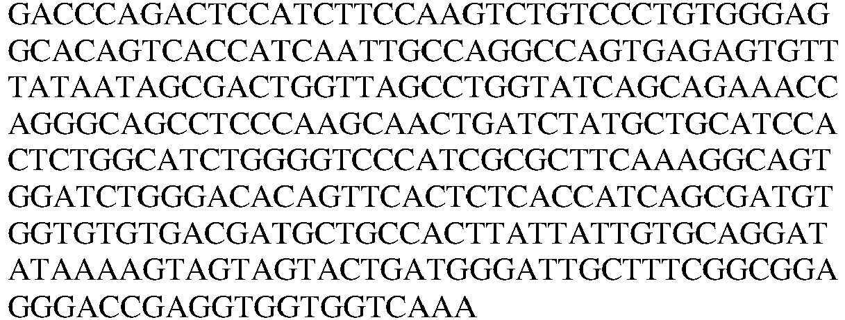
18 Clone 13 VL 
19 Clone 13 LC 
11 Clone 13 VL CDR1 QASESVYNSDWLA 12 Clone 13 VL CDR2 AASTLAS 13 Clone 13 VL CDR3 AGYKSSSTDGIA 20 Clone 14-1 VH 
21 Clone 14-1VH 
22 Clone 14-1HC 
23 Clone 14-1VH CDR1 GFDFSINY 24 Clone 14-1VH CDR2 YTGDD 25 Clone 14-1VH CDR3 GLYSGSINNL 26 Clone 14-1VL 
27 Clone 14-1VL 
28 Clone 14-1LC 
29 Clone 14-1 VL CDR1 QASQSVYNNDYLS 30 Clone 14-1 VL CDR2 YASTLAS 31 Clone 14-1 VL CDR3 AGVKGYSNDNNG 32 Clone 14-7 VH 
33 Clone 14-7 VH 
34 Clone 14-7 HC 

23 Clone 14-7 VH CDR1 GFDFSINY 24 Clone 14-7 VH CDR2 YTGDD 25 Clone 14-7 VH CDR3 GLYSGSINNL 35 Clone 14-7 VL 
36 Clone 14-7 VL 
37 Clone 14-7 LC 
29 Clone 14-7 VL CDR1 QASQSVYNNDYLS 30 Clone 14-7 VL CDR2 YASTLAS 31 Clone 14-7 VL CDR3 AGVKGYSNDNNG 38 Clone 15 VH 

39 Clone 15 VH 
40 Clone 15 HC 
41 Clone 15 VH CDR1 GFSFTSNY 42 Clone 15 VH CDR2 FLGSSG 43 Clone 15 VH CDR3 DYVNGYDYFNL 44 Clone 15 VL 
45 Clone 15 VL 
46 Clone 15 LC 
47 Clone 15 VL CDR1 QSSQSVYNKNLA 48 Clone 15 VL CDR2 STSTLDS 49 Clone 15 VL CDR3 LGSYDCSSADCNA 50 Clone 17 VH 

51 Clone 17 VH 
52 Clone 17 HC 
53 Clone 17 VH CDR1 GFSFSDSW 54 Clone 17 VH CDR2 YTGDG 55 Clone 17 VH CDR3 GAQFYL 56 Clone 17 VL 
57 Clone 17 VL 
58 Clone 17 LC 
59 CDR1 QSSQSVYANTYLS 60 Clone 17 VL CDR2 SASSLAS 61 Clone 17 VL CDR3 LGRYSCGLADCAA 11 7 VL CDR1 Kabat QASESVYNSDWLA 12 7 VL CDR2 Kabat AASTLAS 13 7 VL CDR3 Kabat AGYKSSSTDGIA 11 13 VL CDR1 Kabat QASESVYNSDWLA 12 13 VL CDR2 Kabat AASTLAS 13 13 VL CDR3 Kabat AGYKSSSTDGIA 29 14-1 VL CDR1 Kabat QASQSVYNNDYLS 30 14-1 VL CDR2 Kabat YASTLAS 31 14-1 VL CDR3 Kabat AGVKGYSNDNNG 29 14-7 VL CDR1 Kabat QASQSVYNNDYLS 30 14-7 VL CDR2 Kabat YASTLAS 31 14-7 VL CDR3 Kabat AGVKGYSNDNNG 47 15 VL CDR1 Kabat QSSQSVYNKNLA 48 15 VL CDR2 Kabat STSTLDS 49 15 VL CDR3 Kabat LGSYDCSSADCNA 59 17 VL CDR1 Kabat QSSQSVYANTYLS 60 17 VL CDR2 Kabat SASSLAS 61 17 VL CDR3 Kabat LGRYSCGLADCAA 62 7 VH CDR1 Kabat NNGIC 63 7 VH CDR2 Kabat CLYVGSSDTTYYASWAK 7 7 VH CDR3 Kabat NLGL 62 13 VH CDR1 Kabat NNGIC 63 13 VH CDR2 Kabat CLYVGSSDTTYYASWAK 7 13 VH CDR3 Kabat NLGL 64 14-1 VH CDR1 Kabat INYYMC 65 14-1 VH CDR2 Kabat CIYTGDDDTFYASWAK 25 14-1 VH CDR3 Kabat GLYSGSINNL 64 14-7 VH CDR1 Kabat INYYMC 65 14-7 VH CDR2 Kabat CIYTGDDDTFYASWAK 25 14-7 VH CDR3 Kabat GLYSGSINNL 66 15 VH CDR1 Kabat SNYYMC 67 15 VH CDR2 Kabat CIFLGSSGNTVYANWAK 43 15 VH CDR3 Kabat DYVNGYDYFNL 68 17 VH CDR1 Kabat DSWYLC 69 17 VH CDR2 Kabat CIYTGDGDTYYATWAK 55 17 VH CDR3 Kabat GAQFYL 11 7 VL CDR1 Chothia QASESVYNSDWLA 12 7 VL CDR2 Chothia AASTLAS 13 7 VL CDR3 Chothia AGYKSSSTDGIA 11 13 VL CDR1 Chothia QASESVYNSDWLA 12 13 VL CDR2 Chothia AASTLAS 13 13 VL CDR3 Chothia AGYKSSSTDGIA 29 14-1 VL CDR1 Chothia QASQSVYNNDYLS 30 14-1 VL CDR2 Chothia YASTLAS 31 14-1 VL CDR3 Chothia AGVKGYSNDNNG 29 14-7 VL CDR1 Chothia QASQSVYNNDYLS 30 14-7 VL CDR2 Chothia YASTLAS 31 14-7 VL CDR3 Chothia AGVKGYSNDNNG 47 15 VL CDR1 Chothia QSSQSVYNKNLA 48 15 VL CDR2 Chothia STSTLDS 49 15 VL CDR3 Chothia LGSYDCSSADCNA 59 17 VL CDR1 Chothia QSSQSVYANTYLS 60 17 VL CDR2 Chothia SASSLAS 61 17 VL CDR3 Chothia LGRYSCGLADCAA 5 7 VH CDR1 Chothia GFSFSNN 6 7 VH CDR2 Chothia YVGSSD 7 7 VH CDR3 Chothia NLGL 5 13VH CDR1 Chothia GFSFSNN 6 13 VH CDR2 Chothia YVGSSD 7 13 VH CDR3 Chothia NLGL 23 14-1 VH CDR1 Chothia GFDFSINY 24 14-1 VH CDR2 Chothia YTGDD 25 14-1 VH CDR3 Chothia GLYSGSINNL 23 14-7 VH CDR1 Chothia GFDFSINY 24 14-7 VH CDR2 Chothia YTGDD 25 14-7 VH CDR3 Chothia GLYSGSINNL 41 15 VH CDR1 Chothia GFSFTSNY 42 15 VH CDR2 Chothia FLGSSG 43 15 VH CDR3 Chothia DYVNGYDYFNL 53 17 VH CDR1 Chothia GFSFSDSW 54 17 VH CDR2 Chothia YTGDG 55 17 VH CDR3 Chothia GAQFYL 11 7 VL CDR1 IMGT QASESVYNSDWLA 12 7 VL CDR2 IMGT AASTLAS 13 7 VL CDR3 IMGT AGYKSSSTDGIA 11 13 VL CDR1 IMGT QASESVYNSDWLA 12 13 VL CDR2 IMGT AASTLAS 13 13 VL CDR3 IMGT AGYKSSSTDGIA 29 14-1 VL CDR1 IMGT QASQSVYNNDYLS 30 14-1 VL CDR2 IMGT YASTLAS 31 14-1 VL CDR3 IMGT AGVKGYSNDNNG 29 14-7 VL CDR1 IMGT QASQSVYNNDYLS 30 14-7 VL CDR2 IMGT YASTLAS 31 14-7 VL CDR3 IMGT AGVKGYSNDNNG 47 15 VL CDR1 IMGT QSSQSVYNKNLA 48 15 VL CDR2 IMGT STSTLDS 49 15 VL CDR3 IMGT LGSYDCSSADCNA 59 17 VL CDR1 IMGT QSSQSVYANTYLS 60 17 VL CDR2 IMGT SASSLAS 61 17 VL CDR3 IMGT LGRYSCGLADCAA 70 7 VH CDR 1 IMGT GFSFSNNGIC 63 7 VH CDR 2 IMGT CLYVGSSDTTYYASWAK 7 7 VH CDR 3 IMGT NLGL 70 13 VH CDR 1 IMGT GFSFSNNGIC 63 13 VH CDR 2 IMGT CLYVGSSDTTYYASWAK 7 13 VH CDR 3 IMGT NLGL 71 14-1 VH CDR 1 IMGT GFDFSINYYMC 65 14-1 VH CDR 2 IMGT CIYTGDDDTFYASWAK 25 14-1 VH CDR 3 IMGT GLYSGSINNL 71 14-7 VH CDR 1 IMGT GFDFSINYYMC 65 14-7 VH CDR 2 IMGT CIYTGDDDTFYASWAK 25 14-7 VH CDR 3 IMGT GLYSGSINNL 72 15 VH CDR 1 IMGT GFSFTSNYYMC 67 15 VH CDR 2 IMGT CIFLGSSGNTVYANWAK 43 15 VH CDR 3 IMGT DYVNGYDYFNL 73 17 VH CDR 1 IMGT GFSFSDSWYLC 69 17 VH CDR 2 IMGT CIYTGDGDTYYATWAK 55 17 VH CDR 3 IMGT GAQFYL 74 HUMANIZED FMC 63 VL CDR1 RASQDISKYLN 75 HUMANIZED FMC 63 VL CDR2 HTSRLHS 76 HUMANIZED FMC 63 VL CDR3 QQGNTLPYT 77 HUMANIZED FMC 63 VH CDR1 Clothia GVSLPDY 78 HUMANIZED FMC 63 VH CDR2 Clothia WGSET 79 HUMANIZED FMC 63 VH CDR3 HYYYGGSYAMDY 80 HUMANIZED FMC 63 VH CDR1 Kabat DYGVS 81 HUMANIZED FMC 63 VH CDR2 Kabat/IMGT VIWGSETTYYNSALKS 82 HUMANIZED FMC 63 VH CDR1 IMGT GVSLPDYGVS - The present invention is further illustrated by the following examples, which should not be construed as further limiting.
- Monoclonal antibodies were generated through immunization of rabbits using the anti-CD19 scFv FMC63 (SEQ ID NO: 1), conjugated to Fc as immunogen. Titer was determined via screening polyclonal sera by using Fc as a screen. A secondary screen was performed using CAR T cells assayed via flow cytometry. Once titer was achieved, the immunized rabbits were sacrificed and monoclonals were derived using standard hybridoma generation and subcloning techniques. The final screening of the hybridoma subclones was accomplished via additional rounds of flow cytometry and immunohistochemistry (IHC) of proliferating CAR T cells or fixed cell pellets derived from CAR T cells, respectively. The sequences of the final two subclones selected were determined by standard Sanger sequencing of the hybridomas subclones.
- PBMCs were isolated from healthy donor leukopaks (Hemacare™) using Ficoll-Paque density centrifugation per manufacturer's instructions. PBMCs were stimulated using OKT3 (50ng/ml, Miltenyi Biotec™) in R10 media + IL-2 (300IU/ml, Proleukin®, Prometheus® Therapeutics and Diagnostics). Two days after stimulation, CAR T cells presenting the anti-CD19 scFv FMC63 (SEQ ID NO: 1) were generated through viral transduction of these activated primary human T cells. Transduction was performed using either a retro- (pMSVG vector) or lentivirus (pGAR vector) depending upon the origin of the CARs used in the screening. Confirmation of CAR construct expression and viral transduction efficiency was determined using Protein L conjugated to phycoerythrin (PE) or fluorescein isothiocyanate (FITC). Results are shown in
Figures 1A-1B and2 . - Several antibodies disclosed herein, (
Clones 7 and 13) were assessed with respect to their ability to function as reagents in immunohistochemistry (IHC) studies. To create the fixed cell pellets for IHC staining, ~2e6 CAR T cells presenting the anti-CD19 scFv FMC63 (SEQ ID NO: 1) were centrifuged and washed with PBS. The cells were resuspended in PBS containing 0.45% paraformaldehyde (PFA) and incubated on a shaking platform for 2 hours at room temperature. After the incubation, the cells were washed once more with PBS and resuspended in PBS with 5% BSA. The results of the IHC experiments are shown inFigures 3 and4A (Figure 4B shows controls), and demonstrate that atleast clones - The antigen binding molecules disclosed herein are anti-idiotypic antigen binding molecules, and recognize an epitope on the anti-CD19 scFv FMC63. An antigen binding molecule (e.g., an antibody) disclosed herein can thus be used to purify a molecule, such as the anti-CD19 scFv FMC63, macromolecule, polymer, cell, material, etc., displaying an epitope that is recognized by the antigen binding molecules disclosed herein.
- In some embodiments, an antigen binding molecule disclosed herein (e.g.,
Clones - In another embodiment, an antigen binding molecule can be disposed in solution with any molecular entity displaying the epitope, and purified from a mixed population of molecules, cells, etc. and eluted from the beads, resin, or free antibody by washing with 300-500 mM sodium chloride or lowering the pH and neutralizing with 1 M Tris, for proteins, or phosphate buffer. Subsequently, dialysis can be used to return materials to desired buffer conditions.
- In some embodiments, cells displaying a molecule comprising all or a fragment of the anti-CD19 scFv FMC63 can be incubated with magnetic beads (e.g., DYNABEADS) with which an antigen binding molecule disclosed herein has been associated. Preferably the incubation is performed under conditions that both allow for the formation of binding complexes/associations, such as under physiological conditions, in the presence of a media selected for this purpose (e.g., RPMI-1640).
- Cells bound by the beads (which will be presenting molecules comprising the anti-CD19 scFv FMC63) are then separated from cells not displaying a molecule comprising the anti-CD19 scFv FMC63 or fragment thereof. In some embodiments, the beads can be washed with media, such as RPMI-1640 supplemented with 10% FBS, in the presence of a magnet.
- Selected cells, i.e., those presenting molecules that comprise the anti-CD19 scFv FMC63 can then be separated from the beads: First, selected cells are grown out in media. After growing out cells for 48 hours, the magnetic beads can be separated from cells in solution and discarded, leaving a pure population of cells expressing desired molecule.
- In an alternative embodiment, the beads are not magnetic, and in this embodiment the above steps can also be followed and adapted to maintain cell integrity, but also to allow separation of bead-bound cells from non-bead bound cells.
- In another alternative embodiment, an antigen binding molecule disclosed herein (e.g.,
Clones - Also provided is a method of activating CAR-positive T cells presenting a molecule comprising a specific idiotope recognized by a specific antigen binding molecule (e.g., an antigen binding molecule that recognizes the anti-CD19 scFv FMC63 (SEQ ID NO: 1), such as those disclosed herein:
Clone - In some embodiments, the method can be performed as follows: first, a 12-well tissue culture treated plate is coated with 1 mL of a 1.5 µg/mL solution of an anti-CD19 scFv FMC63 antigen binding molecule disclosed herein, which has been diluted in HBSS or other phosphate buffer, and placed in an incubator at 37 C for 2 hours. Next, the plate is washed three times with HBSS or other phosphate buffer having a suitable pH, ionic strength, etc. Continuing, CAR-positive T-cells in OpTmizer media (with supplements) or RPMI-1640 media with 10% FBS are added to the tissue culture treated plate. The cells are then grown at 37 C with 5% CO2.
- After 2 days, the cells are examined to determine any increase in the percent CAR-positive cells. This determination can be made by identifying any increase in the expression of any cell-surface and/or internal markers, including, but not limited to 4-1BB, CD69, CD25, PD-1, and Ki-67.
- The Molecular Operating Environment (MOE) software developed by Chemical Computing Group (CCG) can be used to generate alignments between the
rabbit antibody Clones - (1) The Abysis human database: a database of about 2000 known human VL/VH sequence pairs from IMGT-LigM DB; and
- (2) A human germline database: a database of germline sequences.
- Humanized models show the best sequence alignments (highest identity to both the VL and VH domains) with fewest gaps. The top 100 antibody pairs from each human database can be exported and clustered using kClust (Hauser, Mayer, & Soding, (2013) BMC Bioinformatics, 248). Tables for VL and VH sequences for each of the antibodies, can be constructed, with sequences from each of the two databases clustered at 90% and 95%.
- Humanized FMC63 CAR constructs were identified comprising conserved CDR loops (e.g., comprising amino acid sequences defined in Table C and D) and bind to CD19. Jurkat cells expressing FMC63 and humanized FMC63 CAR constructs were generated by transduction with lentivirus carrying the respective constructs. Monoclonal antibodies 14-1, 15, and 17-4 were incubated with CAR-expressing Jurkat cells and detected with a FITC-conjugated goat anti-rabbit F(ab')2 antibody (Thermo). An antibody reactive against all of these CAR constructs was used as a control to verify expression (
Figure 22 , top row).Clone 15 binds to the common amino acids in the CDR loops of FMC63 and all the humanized constructs, but shows no reactivity against mock-transduced Jurkat cells. (Figure 22 ). Secondary antibody only and mock-transduced controls show no background staining.Table C. CDRs for the light chain variable (VL) region of the humanized FMC63 anti-CD19 Sequence (Convention) CDR1 SEQ ID NO CDR2 SEQ ID NO CDR3 SEQ ID NO VL (Chothia) 
74 HTSRLHS 75 QQGNTLPYT 76 VL (Kabat) 
74 HTSRLHS 75 QQGNTLPYT 76 VL (IMGT) 
74 HTSRLHS 75 QQGNTLPYT 76 Table D. CDRs for the heavy chain variable (VH) region of the humanized FMC63 anti-CD19 Sequence (Convention) CDR1 SEQ ID NO CDR2 SEQ ID NO CDR3 SEQ ID NO VH (Chothia) GVSLPDY 77 WGSET 78 
79 VH (Kabat) DYGVS 80 
81 
79 VH (IMGT) 
82 
81 
79
Claims (8)
- An isolated antigen binding molecule that specifically binds to the amino acid sequence according to SEQ ID NO: 1 of a molecule comprising said amino acid sequence, wherein the antigen binding molecule comprises a heavy chain variable region (VH) and a light chain variable region (VL),said VH comprising a VH CDR1 comprising the amino acid sequence of SEQ ID NO: 23, a VH CDR2 comprising the amino acid sequence of SEQ ID NO: 24 and a VH CDR3 comprising the amino acid sequence of SEQ ID NO: 25; andsaid VL comprising a VL CDR1 comprising the amino acid sequence of SEQ ID NO: 29, a VL CDR2 comprising the amino acid sequence of SEQ ID NO: 30 and a VL CDR3 comprising the amino acid sequence of SEQ ID NO: 31.
- The antigen binding molecule of claim 1, comprising a heavy chain variable region (VH) sequence according to SEQ ID NO: 21.
- The antigen binding molecule of claim 1, comprising a light chain variable region (VL) sequence according to SEQ ID NO: 27.
- The antigen binding molecule of any of claims 1-3, further comprising a detectable label selected from the group consisting of a fluorescent label, a photochromic compound, a proteinaceous fluorescent label, a magnetic label, a radiolabel, and a hapten.
- A polynucleotide encoding the antigen binding molecule of any one of claims 1 to 3.
- A vector comprising the polynucleotide of claim 5.
- A cell comprising the polynucleotide of claim 5 or the vector of claim 6.
- A composition comprising the antigen binding molecule of any one of claims 1 to 4, polynucleotides encoding the antigen binding molecule of any one of claims 1 to 3, or cells comprising polynucleotides encoding the antigen binding molecule of any one of claims 1 to 3.
Priority Applications (1)
| Application Number | Priority Date | Filing Date | Title |
|---|---|---|---|
| EP24223050.6A EP4538295A3 (en) | 2016-09-28 | 2017-09-27 | Antigen binding molecules and methods of use thereof |
Applications Claiming Priority (2)
| Application Number | Priority Date | Filing Date | Title |
|---|---|---|---|
| US201662401007P | 2016-09-28 | 2016-09-28 | |
| PCT/US2017/053790 WO2018064205A1 (en) | 2016-09-28 | 2017-09-27 | Antigen binding molecules and methods of use thereof |
Related Child Applications (1)
| Application Number | Title | Priority Date | Filing Date |
|---|---|---|---|
| EP24223050.6A Division EP4538295A3 (en) | 2016-09-28 | 2017-09-27 | Antigen binding molecules and methods of use thereof |
Publications (4)
| Publication Number | Publication Date |
|---|---|
| EP3518972A1 EP3518972A1 (en) | 2019-08-07 |
| EP3518972A4 EP3518972A4 (en) | 2020-07-22 |
| EP3518972B1 true EP3518972B1 (en) | 2024-12-25 |
| EP3518972C0 EP3518972C0 (en) | 2024-12-25 |
Family
ID=61687622
Family Applications (2)
| Application Number | Title | Priority Date | Filing Date |
|---|---|---|---|
| EP24223050.6A Pending EP4538295A3 (en) | 2016-09-28 | 2017-09-27 | Antigen binding molecules and methods of use thereof |
| EP17857353.1A Active EP3518972B1 (en) | 2016-09-28 | 2017-09-27 | Antigen binding molecules and methods of use thereof |
Family Applications Before (1)
| Application Number | Title | Priority Date | Filing Date |
|---|---|---|---|
| EP24223050.6A Pending EP4538295A3 (en) | 2016-09-28 | 2017-09-27 | Antigen binding molecules and methods of use thereof |
Country Status (6)
| Country | Link |
|---|---|
| US (4) | US10626187B2 (en) |
| EP (2) | EP4538295A3 (en) |
| ES (1) | ES3013602T3 (en) |
| PL (1) | PL3518972T3 (en) |
| TW (4) | TWI779479B (en) |
| WO (1) | WO2018064205A1 (en) |
Families Citing this family (23)
| Publication number | Priority date | Publication date | Assignee | Title |
|---|---|---|---|---|
| SG10201808811QA (en) | 2014-04-10 | 2018-11-29 | Seattle Childrens Hospital Dba Seattle Childrens Res Inst | Method and compositions for cellular immunotherapy |
| MX2018001568A (en) | 2015-08-07 | 2019-04-25 | Seattle Children´S Hospital Dba Seattle Children´S Res Institute | Bispecific car t-cells for solid tumor targeting. |
| ES2960835T3 (en) | 2016-07-12 | 2024-03-06 | Kite Pharma Inc | Antigen binding molecules and methods of use thereof |
| US10626187B2 (en) | 2016-09-28 | 2020-04-21 | Kite Pharma, Inc. | Antigen binding molecules specific for an anti-CD19 scFv |
| WO2019133902A2 (en) | 2017-12-28 | 2019-07-04 | Astute Medical, Inc. | Antibodies and assays for ccl14 |
| CN119656304A (en) | 2018-04-12 | 2025-03-21 | 凯德药业股份有限公司 | Chimeric receptor T cell therapy using features of tumor microenvironment |
| KR102734592B1 (en) | 2018-06-01 | 2024-11-27 | 카이트 파마 인코포레이티드 | Chimeric antigen receptor t cell therapy |
| EP3806902A4 (en) | 2018-06-14 | 2022-05-04 | Beth Israel Deaconess Medical Center, Inc. | COMPOSITIONS AND METHODS FOR PREVENTING OR REVERSING T LYMPHOCYTE DEPLETION BY TARGET CYTOSIS MEDIATED BY ECTONUCLEOTIDASE-INHIBITING ANTIBODIES |
| CN110850068B (en) * | 2018-08-21 | 2023-08-15 | 上海恒润达生生物科技股份有限公司 | Chimeric antigen receptor affinity detection method |
| EP3861018A4 (en) | 2018-10-05 | 2022-10-26 | Seattle Children's Hospital d/b/a Seattle Children's Research Institute | NEWBORN SCREENING FOR PRIMARY IMMUNODEFICIENCIES, CYSTINOSIS AND WILSON'S DISEASE |
| CN113226340A (en) | 2018-11-26 | 2021-08-06 | 恩卡尔塔公司 | Methods of simultaneously expanding multiple immune cell types, related compositions, and uses thereof in cancer immunotherapy |
| CN111349161B (en) * | 2018-12-20 | 2022-02-08 | 上海星湾生物技术有限公司 | Monoclonal antibody of anti-CD 19 antibody and application thereof |
| KR20210137085A (en) | 2019-03-05 | 2021-11-17 | 엔카르타, 인크. | CD19 induced chimeric antigen receptor and use thereof in immunotherapy |
| BR112021026832A2 (en) | 2019-07-02 | 2022-05-10 | Hutchinson Fred Cancer Res | Recombinant ad35 vectors and related gene therapy enhancements |
| CN114341178B (en) * | 2019-07-02 | 2024-05-07 | 机敏医药股份有限公司 | Antibodies and assays for CCL14 |
| CN112390894A (en) * | 2019-08-12 | 2021-02-23 | 广东东阳光药业有限公司 | Chimeric antigen receptor and uses thereof |
| CN110894238B (en) * | 2019-11-25 | 2021-01-19 | 华道(上海)生物医药有限公司 | Monoclonal antibody for detecting CAR-T cells, kit and application |
| CN115279795A (en) * | 2020-02-11 | 2022-11-01 | 克里斯珀医疗股份公司 | Anti-idiotypic antibodies targeting anti-CD 19 chimeric antigen receptor |
| WO2021202807A1 (en) * | 2020-03-31 | 2021-10-07 | Seattle Children's Hospital D/B/A Seattle Children's Research Institute | Proteomic screening for lysosomal storage diseases |
| WO2021203031A2 (en) * | 2020-04-02 | 2021-10-07 | Seattle Children's Hospital D/B/A Seattle Children's Research Institute | Antibodies that specifically bind peptides associated with the primary immunodeficiencies: wiskott-aldrich syndrome and x-linked agammaglobulinemia |
| WO2021216667A2 (en) * | 2020-04-21 | 2021-10-28 | The Board Of Regents Of The University Of Texas System | Antibodies to chitinase 3-like-1 and methods of use therefor |
| CN117820478B (en) * | 2023-12-29 | 2024-10-29 | 北京贝来药业有限公司 | Tandem molecules of nanobodies and their use in the treatment of diseases |
| WO2026064719A1 (en) * | 2024-09-22 | 2026-03-26 | Allogene Therapeutics, Inc. | Cd19 anti-idiotypic antibodies |
Family Cites Families (19)
| Publication number | Priority date | Publication date | Assignee | Title |
|---|---|---|---|---|
| US5225539A (en) | 1986-03-27 | 1993-07-06 | Medical Research Council | Recombinant altered antibodies and methods of making altered antibodies |
| US5223409A (en) | 1988-09-02 | 1993-06-29 | Protein Engineering Corp. | Directed evolution of novel binding proteins |
| US5859205A (en) | 1989-12-21 | 1999-01-12 | Celltech Limited | Humanised antibodies |
| US5814318A (en) | 1990-08-29 | 1998-09-29 | Genpharm International Inc. | Transgenic non-human animals for producing heterologous antibodies |
| US5545806A (en) | 1990-08-29 | 1996-08-13 | Genpharm International, Inc. | Ransgenic non-human animals for producing heterologous antibodies |
| US5877397A (en) | 1990-08-29 | 1999-03-02 | Genpharm International Inc. | Transgenic non-human animals capable of producing heterologous antibodies of various isotypes |
| ES2246502T3 (en) | 1990-08-29 | 2006-02-16 | Genpharm International, Inc. | TRANSGENIC NON-HUMAN ANIMALS ABLE TO PRODUCE HETEROLOGICAL ANTIBODIES. |
| EP0590058B1 (en) | 1991-06-14 | 2003-11-26 | Genentech, Inc. | HUMANIZED Heregulin ANTIBODy |
| US5869619A (en) | 1991-12-13 | 1999-02-09 | Xoma Corporation | Modified antibody variable domains |
| AU2002368077B2 (en) | 2001-07-12 | 2010-03-04 | Jefferson Foote | Super humanized antibodies |
| WO2008133722A2 (en) * | 2006-11-10 | 2008-11-06 | Ucb Pharma S.A. | Anti human sclerostin antibodies |
| PH12013501201A1 (en) | 2010-12-09 | 2013-07-29 | Univ Pennsylvania | Use of chimeric antigen receptor-modified t cells to treat cancer |
| KR102037432B1 (en) * | 2011-04-29 | 2019-11-27 | 아펙시젠, 인코포레이티드 | Anti-cd40 antibodies and methods of use |
| CA2913052A1 (en) * | 2013-05-24 | 2014-11-27 | Board Of Regents, The University Of Texas System | Chimeric antigen receptor-targeting monoclonal antibodies |
| WO2016019300A1 (en) | 2014-07-31 | 2016-02-04 | Novartis Ag | Subset-optimized chimeric antigen receptor-containing t-cells |
| ES2960835T3 (en) | 2016-07-12 | 2024-03-06 | Kite Pharma Inc | Antigen binding molecules and methods of use thereof |
| MA45784A (en) * | 2016-07-29 | 2019-06-05 | Juno Therapeutics Inc | ANTI-BODY ANTI-IDIOTYPES DIRECTED AGAINST ANTI-CD19 ANTIBODY |
| US10626187B2 (en) | 2016-09-28 | 2020-04-21 | Kite Pharma, Inc. | Antigen binding molecules specific for an anti-CD19 scFv |
| JP6918465B2 (en) * | 2016-10-14 | 2021-08-11 | 株式会社明治 | Composition for suppressing reduction of lactic acid bacteria of the genus Lactobacillus in the intestinal tract |
-
2017
- 2017-09-27 US US15/717,691 patent/US10626187B2/en active Active
- 2017-09-27 TW TW110104217A patent/TWI779479B/en active
- 2017-09-27 TW TW111136483A patent/TWI828334B/en active
- 2017-09-27 WO PCT/US2017/053790 patent/WO2018064205A1/en not_active Ceased
- 2017-09-27 TW TW106133121A patent/TWI695010B/en active
- 2017-09-27 EP EP24223050.6A patent/EP4538295A3/en active Pending
- 2017-09-27 EP EP17857353.1A patent/EP3518972B1/en active Active
- 2017-09-27 ES ES17857353T patent/ES3013602T3/en active Active
- 2017-09-27 PL PL17857353.1T patent/PL3518972T3/en unknown
- 2017-09-27 TW TW109114726A patent/TWI719894B/en active
-
2020
- 2020-03-10 US US16/814,661 patent/US11384155B2/en active Active
-
2022
- 2022-06-09 US US17/836,862 patent/US12202909B2/en active Active
-
2024
- 2024-12-10 US US18/975,713 patent/US20250243295A1/en active Pending
Also Published As
| Publication number | Publication date |
|---|---|
| US20200354475A1 (en) | 2020-11-12 |
| TW202124424A (en) | 2021-07-01 |
| TWI779479B (en) | 2022-10-01 |
| WO2018064205A1 (en) | 2018-04-05 |
| TWI719894B (en) | 2021-02-21 |
| PL3518972T3 (en) | 2025-04-07 |
| US10626187B2 (en) | 2020-04-21 |
| US11384155B2 (en) | 2022-07-12 |
| EP3518972A1 (en) | 2019-08-07 |
| EP4538295A3 (en) | 2025-06-18 |
| TWI695010B (en) | 2020-06-01 |
| EP3518972A4 (en) | 2020-07-22 |
| TW202033545A (en) | 2020-09-16 |
| US20250243295A1 (en) | 2025-07-31 |
| US20180086846A1 (en) | 2018-03-29 |
| US20220403050A1 (en) | 2022-12-22 |
| TWI828334B (en) | 2024-01-01 |
| EP3518972C0 (en) | 2024-12-25 |
| US12202909B2 (en) | 2025-01-21 |
| TW201827457A (en) | 2018-08-01 |
| ES3013602T3 (en) | 2025-04-14 |
| EP4538295A2 (en) | 2025-04-16 |
| TW202304966A (en) | 2023-02-01 |
Similar Documents
| Publication | Publication Date | Title |
|---|---|---|
| US12202909B2 (en) | Antigen binding molecules specific for an anti-CD19 scFV | |
| AU2020210684B2 (en) | Antigen binding molecules and methods of use thereof | |
| EP4253959A2 (en) | Anti-idiotypic antibodies directed to the antigen-binding portion of an bcma-binding molecule | |
| US20240150750A1 (en) | Antigen binding molecules and methods of use thereof | |
| US20220396628A1 (en) | Anti-idiotypic antigen binding molecules and methods of use thereof | |
| US20240343820A1 (en) | Antigen binding molecules and methods of use | |
| HK40102075A (en) | Antigen binding molecules and methods of use thereof | |
| HK40012838B (en) | Antigen binding molecules and methods of use thereof | |
| HK40012838A (en) | Antigen binding molecules and methods of use thereof | |
| HK40008698A (en) | Antigen binding molecules and methods of use thereof | |
| HK40008698B (en) | Antigen binding molecules and methods of use thereof | |
| WO2026064719A1 (en) | Cd19 anti-idiotypic antibodies |
Legal Events
| Date | Code | Title | Description |
|---|---|---|---|
| STAA | Information on the status of an ep patent application or granted ep patent |
Free format text: STATUS: THE INTERNATIONAL PUBLICATION HAS BEEN MADE |
|
| PUAI | Public reference made under article 153(3) epc to a published international application that has entered the european phase |
Free format text: ORIGINAL CODE: 0009012 |
|
| STAA | Information on the status of an ep patent application or granted ep patent |
Free format text: STATUS: REQUEST FOR EXAMINATION WAS MADE |
|
| 17P | Request for examination filed |
Effective date: 20190328 |
|
| AK | Designated contracting states |
Kind code of ref document: A1 Designated state(s): AL AT BE BG CH CY CZ DE DK EE ES FI FR GB GR HR HU IE IS IT LI LT LU LV MC MK MT NL NO PL PT RO RS SE SI SK SM TR |
|
| AX | Request for extension of the european patent |
Extension state: BA ME |
|
| DAV | Request for validation of the european patent (deleted) | ||
| DAX | Request for extension of the european patent (deleted) | ||
| REG | Reference to a national code |
Ref country code: DE Ref legal event code: R079 Free format text: PREVIOUS MAIN CLASS: A61K0039395000 Ipc: A61K0039000000 Ref document number: 602017086994 Country of ref document: DE |
|
| A4 | Supplementary search report drawn up and despatched |
Effective date: 20200618 |
|
| RIC1 | Information provided on ipc code assigned before grant |
Ipc: C07K 16/42 20060101ALI20200612BHEP Ipc: C07K 16/46 20060101ALI20200612BHEP Ipc: C07K 14/705 20060101ALI20200612BHEP Ipc: A61P 35/02 20060101ALI20200612BHEP Ipc: A61K 39/00 20060101AFI20200612BHEP Ipc: C07K 16/28 20060101ALI20200612BHEP |
|
| REG | Reference to a national code |
Ref country code: HK Ref legal event code: DE Ref document number: 40012838 Country of ref document: HK |
|
| STAA | Information on the status of an ep patent application or granted ep patent |
Free format text: STATUS: EXAMINATION IS IN PROGRESS |
|
| 17Q | First examination report despatched |
Effective date: 20210512 |
|
| GRAP | Despatch of communication of intention to grant a patent |
Free format text: ORIGINAL CODE: EPIDOSNIGR1 |
|
| STAA | Information on the status of an ep patent application or granted ep patent |
Free format text: STATUS: GRANT OF PATENT IS INTENDED |
|
| INTG | Intention to grant announced |
Effective date: 20240722 |
|
| GRAS | Grant fee paid |
Free format text: ORIGINAL CODE: EPIDOSNIGR3 |
|
| GRAA | (expected) grant |
Free format text: ORIGINAL CODE: 0009210 |
|
| STAA | Information on the status of an ep patent application or granted ep patent |
Free format text: STATUS: THE PATENT HAS BEEN GRANTED |
|
| AK | Designated contracting states |
Kind code of ref document: B1 Designated state(s): AL AT BE BG CH CY CZ DE DK EE ES FI FR GB GR HR HU IE IS IT LI LT LU LV MC MK MT NL NO PL PT RO RS SE SI SK SM TR |
|
| REG | Reference to a national code |
Ref country code: GB Ref legal event code: FG4D |
|
| REG | Reference to a national code |
Ref country code: CH Ref legal event code: EP |
|
| REG | Reference to a national code |
Ref country code: DE Ref legal event code: R096 Ref document number: 602017086994 Country of ref document: DE |
|
| REG | Reference to a national code |
Ref country code: IE Ref legal event code: FG4D |
|
| U01 | Request for unitary effect filed |
Effective date: 20250115 |
|
| U07 | Unitary effect registered |
Designated state(s): AT BE BG DE DK EE FI FR IT LT LU LV MT NL PT RO SE SI Effective date: 20250121 |
|
| PG25 | Lapsed in a contracting state [announced via postgrant information from national office to epo] |
Ref country code: HR Free format text: LAPSE BECAUSE OF FAILURE TO SUBMIT A TRANSLATION OF THE DESCRIPTION OR TO PAY THE FEE WITHIN THE PRESCRIBED TIME-LIMIT Effective date: 20241225 |
|
| REG | Reference to a national code |
Ref country code: ES Ref legal event code: FG2A Ref document number: 3013602 Country of ref document: ES Kind code of ref document: T3 Effective date: 20250414 |
|
| PG25 | Lapsed in a contracting state [announced via postgrant information from national office to epo] |
Ref country code: RS Free format text: LAPSE BECAUSE OF FAILURE TO SUBMIT A TRANSLATION OF THE DESCRIPTION OR TO PAY THE FEE WITHIN THE PRESCRIBED TIME-LIMIT Effective date: 20250325 |
|
| REG | Reference to a national code |
Ref country code: GR Ref legal event code: EP Ref document number: 20250400504 Country of ref document: GR Effective date: 20250409 |
|
| REG | Reference to a national code |
Ref country code: SK Ref legal event code: T3 Ref document number: E 46077 Country of ref document: SK |
|
| PG25 | Lapsed in a contracting state [announced via postgrant information from national office to epo] |
Ref country code: SM Free format text: LAPSE BECAUSE OF FAILURE TO SUBMIT A TRANSLATION OF THE DESCRIPTION OR TO PAY THE FEE WITHIN THE PRESCRIBED TIME-LIMIT Effective date: 20241225 |
|
| PG25 | Lapsed in a contracting state [announced via postgrant information from national office to epo] |
Ref country code: IS Free format text: LAPSE BECAUSE OF FAILURE TO SUBMIT A TRANSLATION OF THE DESCRIPTION OR TO PAY THE FEE WITHIN THE PRESCRIBED TIME-LIMIT Effective date: 20250425 |
|
| U20 | Renewal fee for the european patent with unitary effect paid |
Year of fee payment: 9 Effective date: 20250807 |
|
| REG | Reference to a national code |
Ref country code: CH Ref legal event code: U11 Free format text: ST27 STATUS EVENT CODE: U-0-0-U10-U11 (AS PROVIDED BY THE NATIONAL OFFICE) Effective date: 20251001 |
|
| PGFP | Annual fee paid to national office [announced via postgrant information from national office to epo] |
Ref country code: GR Payment date: 20250818 Year of fee payment: 9 Ref country code: NO Payment date: 20250909 Year of fee payment: 9 |
|
| PGFP | Annual fee paid to national office [announced via postgrant information from national office to epo] |
Ref country code: PL Payment date: 20250707 Year of fee payment: 9 Ref country code: TR Payment date: 20250922 Year of fee payment: 9 |
|
| PGFP | Annual fee paid to national office [announced via postgrant information from national office to epo] |
Ref country code: GB Payment date: 20250703 Year of fee payment: 9 |
|
| PGFP | Annual fee paid to national office [announced via postgrant information from national office to epo] |
Ref country code: CZ Payment date: 20250922 Year of fee payment: 9 Ref country code: IE Payment date: 20250702 Year of fee payment: 9 |
|
| PGFP | Annual fee paid to national office [announced via postgrant information from national office to epo] |
Ref country code: SK Payment date: 20250812 Year of fee payment: 9 |
|
| PLBE | No opposition filed within time limit |
Free format text: ORIGINAL CODE: 0009261 |
|
| STAA | Information on the status of an ep patent application or granted ep patent |
Free format text: STATUS: NO OPPOSITION FILED WITHIN TIME LIMIT |
|
| 26N | No opposition filed |
Effective date: 20250926 |
|
| PGFP | Annual fee paid to national office [announced via postgrant information from national office to epo] |
Ref country code: CH Payment date: 20251001 Year of fee payment: 9 |
|
| PGFP | Annual fee paid to national office [announced via postgrant information from national office to epo] |
Ref country code: ES Payment date: 20251014 Year of fee payment: 9 |
|
| PG25 | Lapsed in a contracting state [announced via postgrant information from national office to epo] |
Ref country code: MC Free format text: LAPSE BECAUSE OF FAILURE TO SUBMIT A TRANSLATION OF THE DESCRIPTION OR TO PAY THE FEE WITHIN THE PRESCRIBED TIME-LIMIT Effective date: 20241225 |