EP3510143B1 - In vitro neonatal biomimetic (nmimic) model and methods of using same - Google Patents
In vitro neonatal biomimetic (nmimic) model and methods of using same Download PDFInfo
- Publication number
- EP3510143B1 EP3510143B1 EP17768611.0A EP17768611A EP3510143B1 EP 3510143 B1 EP3510143 B1 EP 3510143B1 EP 17768611 A EP17768611 A EP 17768611A EP 3510143 B1 EP3510143 B1 EP 3510143B1
- Authority
- EP
- European Patent Office
- Prior art keywords
- cells
- vaccine
- cell culture
- culture
- cord blood
- Prior art date
- Legal status (The legal status is an assumption and is not a legal conclusion. Google has not performed a legal analysis and makes no representation as to the accuracy of the status listed.)
- Active
Links
Images







Classifications
-
- G—PHYSICS
- G01—MEASURING; TESTING
- G01N—INVESTIGATING OR ANALYSING MATERIALS BY DETERMINING THEIR CHEMICAL OR PHYSICAL PROPERTIES
- G01N33/00—Investigating or analysing materials by specific methods not covered by groups G01N1/00 - G01N31/00
- G01N33/48—Biological material, e.g. blood, urine; Haemocytometers
- G01N33/50—Chemical analysis of biological material, e.g. blood, urine; Testing involving biospecific ligand binding methods; Immunological testing
- G01N33/5005—Chemical analysis of biological material, e.g. blood, urine; Testing involving biospecific ligand binding methods; Immunological testing involving human or animal cells
- G01N33/5008—Chemical analysis of biological material, e.g. blood, urine; Testing involving biospecific ligand binding methods; Immunological testing involving human or animal cells for testing or evaluating the effect of chemical or biological compounds, e.g. drugs, cosmetics
- G01N33/5044—Chemical analysis of biological material, e.g. blood, urine; Testing involving biospecific ligand binding methods; Immunological testing involving human or animal cells for testing or evaluating the effect of chemical or biological compounds, e.g. drugs, cosmetics involving specific cell types
- G01N33/5047—Cells of the immune system
- G01N33/505—Cells of the immune system involving T-cells
-
- C—CHEMISTRY; METALLURGY
- C12—BIOCHEMISTRY; BEER; SPIRITS; WINE; VINEGAR; MICROBIOLOGY; ENZYMOLOGY; MUTATION OR GENETIC ENGINEERING
- C12N—MICROORGANISMS OR ENZYMES; COMPOSITIONS THEREOF; PROPAGATING, PRESERVING, OR MAINTAINING MICROORGANISMS; MUTATION OR GENETIC ENGINEERING; CULTURE MEDIA
- C12N5/00—Undifferentiated human, animal or plant cells, e.g. cell lines; Tissues; Cultivation or maintenance thereof; Culture media therefor
- C12N5/06—Animal cells or tissues; Human cells or tissues
- C12N5/0602—Vertebrate cells
- C12N5/0634—Cells from the blood or the immune system
- C12N5/0635—B lymphocytes
-
- C—CHEMISTRY; METALLURGY
- C12—BIOCHEMISTRY; BEER; SPIRITS; WINE; VINEGAR; MICROBIOLOGY; ENZYMOLOGY; MUTATION OR GENETIC ENGINEERING
- C12N—MICROORGANISMS OR ENZYMES; COMPOSITIONS THEREOF; PROPAGATING, PRESERVING, OR MAINTAINING MICROORGANISMS; MUTATION OR GENETIC ENGINEERING; CULTURE MEDIA
- C12N5/00—Undifferentiated human, animal or plant cells, e.g. cell lines; Tissues; Cultivation or maintenance thereof; Culture media therefor
- C12N5/06—Animal cells or tissues; Human cells or tissues
- C12N5/0602—Vertebrate cells
- C12N5/0634—Cells from the blood or the immune system
- C12N5/0636—T lymphocytes
-
- C—CHEMISTRY; METALLURGY
- C12—BIOCHEMISTRY; BEER; SPIRITS; WINE; VINEGAR; MICROBIOLOGY; ENZYMOLOGY; MUTATION OR GENETIC ENGINEERING
- C12N—MICROORGANISMS OR ENZYMES; COMPOSITIONS THEREOF; PROPAGATING, PRESERVING, OR MAINTAINING MICROORGANISMS; MUTATION OR GENETIC ENGINEERING; CULTURE MEDIA
- C12N5/00—Undifferentiated human, animal or plant cells, e.g. cell lines; Tissues; Cultivation or maintenance thereof; Culture media therefor
- C12N5/06—Animal cells or tissues; Human cells or tissues
- C12N5/0602—Vertebrate cells
- C12N5/0634—Cells from the blood or the immune system
- C12N5/0639—Dendritic cells, e.g. Langherhans cells in the epidermis
-
- G—PHYSICS
- G01—MEASURING; TESTING
- G01N—INVESTIGATING OR ANALYSING MATERIALS BY DETERMINING THEIR CHEMICAL OR PHYSICAL PROPERTIES
- G01N33/00—Investigating or analysing materials by specific methods not covered by groups G01N1/00 - G01N31/00
- G01N33/48—Biological material, e.g. blood, urine; Haemocytometers
- G01N33/50—Chemical analysis of biological material, e.g. blood, urine; Testing involving biospecific ligand binding methods; Immunological testing
- G01N33/5005—Chemical analysis of biological material, e.g. blood, urine; Testing involving biospecific ligand binding methods; Immunological testing involving human or animal cells
- G01N33/5008—Chemical analysis of biological material, e.g. blood, urine; Testing involving biospecific ligand binding methods; Immunological testing involving human or animal cells for testing or evaluating the effect of chemical or biological compounds, e.g. drugs, cosmetics
- G01N33/5044—Chemical analysis of biological material, e.g. blood, urine; Testing involving biospecific ligand binding methods; Immunological testing involving human or animal cells for testing or evaluating the effect of chemical or biological compounds, e.g. drugs, cosmetics involving specific cell types
- G01N33/5047—Cells of the immune system
- G01N33/5052—Cells of the immune system involving B-cells
-
- G—PHYSICS
- G01—MEASURING; TESTING
- G01N—INVESTIGATING OR ANALYSING MATERIALS BY DETERMINING THEIR CHEMICAL OR PHYSICAL PROPERTIES
- G01N33/00—Investigating or analysing materials by specific methods not covered by groups G01N1/00 - G01N31/00
- G01N33/48—Biological material, e.g. blood, urine; Haemocytometers
- G01N33/50—Chemical analysis of biological material, e.g. blood, urine; Testing involving biospecific ligand binding methods; Immunological testing
- G01N33/5005—Chemical analysis of biological material, e.g. blood, urine; Testing involving biospecific ligand binding methods; Immunological testing involving human or animal cells
- G01N33/5008—Chemical analysis of biological material, e.g. blood, urine; Testing involving biospecific ligand binding methods; Immunological testing involving human or animal cells for testing or evaluating the effect of chemical or biological compounds, e.g. drugs, cosmetics
- G01N33/5082—Supracellular entities, e.g. tissue, organisms
- G01N33/5088—Supracellular entities, e.g. tissue, organisms of vertebrates
-
- C—CHEMISTRY; METALLURGY
- C12—BIOCHEMISTRY; BEER; SPIRITS; WINE; VINEGAR; MICROBIOLOGY; ENZYMOLOGY; MUTATION OR GENETIC ENGINEERING
- C12N—MICROORGANISMS OR ENZYMES; COMPOSITIONS THEREOF; PROPAGATING, PRESERVING, OR MAINTAINING MICROORGANISMS; MUTATION OR GENETIC ENGINEERING; CULTURE MEDIA
- C12N2500/00—Specific components of cell culture medium
- C12N2500/90—Serum-free medium, which may still contain naturally-sourced components
- C12N2500/92—Medium free of human- or animal-derived components
-
- C—CHEMISTRY; METALLURGY
- C12—BIOCHEMISTRY; BEER; SPIRITS; WINE; VINEGAR; MICROBIOLOGY; ENZYMOLOGY; MUTATION OR GENETIC ENGINEERING
- C12N—MICROORGANISMS OR ENZYMES; COMPOSITIONS THEREOF; PROPAGATING, PRESERVING, OR MAINTAINING MICROORGANISMS; MUTATION OR GENETIC ENGINEERING; CULTURE MEDIA
- C12N2500/00—Specific components of cell culture medium
- C12N2500/99—Serum-free medium
-
- C—CHEMISTRY; METALLURGY
- C12—BIOCHEMISTRY; BEER; SPIRITS; WINE; VINEGAR; MICROBIOLOGY; ENZYMOLOGY; MUTATION OR GENETIC ENGINEERING
- C12N—MICROORGANISMS OR ENZYMES; COMPOSITIONS THEREOF; PROPAGATING, PRESERVING, OR MAINTAINING MICROORGANISMS; MUTATION OR GENETIC ENGINEERING; CULTURE MEDIA
- C12N2501/00—Active agents used in cell culture processes, e.g. differentation
- C12N2501/20—Cytokines; Chemokines
- C12N2501/21—Chemokines, e.g. MIP-1, MIP-2, RANTES, MCP, PF-4
-
- C—CHEMISTRY; METALLURGY
- C12—BIOCHEMISTRY; BEER; SPIRITS; WINE; VINEGAR; MICROBIOLOGY; ENZYMOLOGY; MUTATION OR GENETIC ENGINEERING
- C12N—MICROORGANISMS OR ENZYMES; COMPOSITIONS THEREOF; PROPAGATING, PRESERVING, OR MAINTAINING MICROORGANISMS; MUTATION OR GENETIC ENGINEERING; CULTURE MEDIA
- C12N2501/00—Active agents used in cell culture processes, e.g. differentation
- C12N2501/20—Cytokines; Chemokines
- C12N2501/23—Interleukins [IL]
- C12N2501/2306—Interleukin-6 (IL-6)
-
- C—CHEMISTRY; METALLURGY
- C12—BIOCHEMISTRY; BEER; SPIRITS; WINE; VINEGAR; MICROBIOLOGY; ENZYMOLOGY; MUTATION OR GENETIC ENGINEERING
- C12N—MICROORGANISMS OR ENZYMES; COMPOSITIONS THEREOF; PROPAGATING, PRESERVING, OR MAINTAINING MICROORGANISMS; MUTATION OR GENETIC ENGINEERING; CULTURE MEDIA
- C12N2501/00—Active agents used in cell culture processes, e.g. differentation
- C12N2501/20—Cytokines; Chemokines
- C12N2501/23—Interleukins [IL]
- C12N2501/2312—Interleukin-12 (IL-12)
-
- C—CHEMISTRY; METALLURGY
- C12—BIOCHEMISTRY; BEER; SPIRITS; WINE; VINEGAR; MICROBIOLOGY; ENZYMOLOGY; MUTATION OR GENETIC ENGINEERING
- C12N—MICROORGANISMS OR ENZYMES; COMPOSITIONS THEREOF; PROPAGATING, PRESERVING, OR MAINTAINING MICROORGANISMS; MUTATION OR GENETIC ENGINEERING; CULTURE MEDIA
- C12N2501/00—Active agents used in cell culture processes, e.g. differentation
- C12N2501/20—Cytokines; Chemokines
- C12N2501/23—Interleukins [IL]
- C12N2501/2321—Interleukin-21 (IL-21)
-
- C—CHEMISTRY; METALLURGY
- C12—BIOCHEMISTRY; BEER; SPIRITS; WINE; VINEGAR; MICROBIOLOGY; ENZYMOLOGY; MUTATION OR GENETIC ENGINEERING
- C12N—MICROORGANISMS OR ENZYMES; COMPOSITIONS THEREOF; PROPAGATING, PRESERVING, OR MAINTAINING MICROORGANISMS; MUTATION OR GENETIC ENGINEERING; CULTURE MEDIA
- C12N2501/00—Active agents used in cell culture processes, e.g. differentation
- C12N2501/50—Cell markers; Cell surface determinants
- C12N2501/51—B7 molecules, e.g. CD80, CD86, CD28 (ligand), CD152 (ligand)
-
- C—CHEMISTRY; METALLURGY
- C12—BIOCHEMISTRY; BEER; SPIRITS; WINE; VINEGAR; MICROBIOLOGY; ENZYMOLOGY; MUTATION OR GENETIC ENGINEERING
- C12N—MICROORGANISMS OR ENZYMES; COMPOSITIONS THEREOF; PROPAGATING, PRESERVING, OR MAINTAINING MICROORGANISMS; MUTATION OR GENETIC ENGINEERING; CULTURE MEDIA
- C12N2502/00—Coculture with; Conditioned medium produced by
- C12N2502/11—Coculture with; Conditioned medium produced by blood or immune system cells
- C12N2502/1107—B cells
-
- C—CHEMISTRY; METALLURGY
- C12—BIOCHEMISTRY; BEER; SPIRITS; WINE; VINEGAR; MICROBIOLOGY; ENZYMOLOGY; MUTATION OR GENETIC ENGINEERING
- C12N—MICROORGANISMS OR ENZYMES; COMPOSITIONS THEREOF; PROPAGATING, PRESERVING, OR MAINTAINING MICROORGANISMS; MUTATION OR GENETIC ENGINEERING; CULTURE MEDIA
- C12N2502/00—Coculture with; Conditioned medium produced by
- C12N2502/11—Coculture with; Conditioned medium produced by blood or immune system cells
- C12N2502/1114—T cells
-
- C—CHEMISTRY; METALLURGY
- C12—BIOCHEMISTRY; BEER; SPIRITS; WINE; VINEGAR; MICROBIOLOGY; ENZYMOLOGY; MUTATION OR GENETIC ENGINEERING
- C12N—MICROORGANISMS OR ENZYMES; COMPOSITIONS THEREOF; PROPAGATING, PRESERVING, OR MAINTAINING MICROORGANISMS; MUTATION OR GENETIC ENGINEERING; CULTURE MEDIA
- C12N2502/00—Coculture with; Conditioned medium produced by
- C12N2502/11—Coculture with; Conditioned medium produced by blood or immune system cells
- C12N2502/1121—Dendritic cells
-
- C—CHEMISTRY; METALLURGY
- C12—BIOCHEMISTRY; BEER; SPIRITS; WINE; VINEGAR; MICROBIOLOGY; ENZYMOLOGY; MUTATION OR GENETIC ENGINEERING
- C12N—MICROORGANISMS OR ENZYMES; COMPOSITIONS THEREOF; PROPAGATING, PRESERVING, OR MAINTAINING MICROORGANISMS; MUTATION OR GENETIC ENGINEERING; CULTURE MEDIA
- C12N2533/00—Supports or coatings for cell culture, characterised by material
- C12N2533/50—Proteins
Definitions
- Vaccination is the best defense against infection, and the first few months of life provide an important period in which vaccines can induce robust protection against disease.
- clinical development and improvement of vaccines and immunotherapies is stymied by the lack of appropriate in vitro assays for testing immune responses in immature immune systems.
- Development of in vitro models of neonatal and infant immunity would be of great benefit in advancing research into vaccines that can be administered following birth.
- the invention described herein is directed to this and other important goals.
- nMIMIC mammalian umbilical cord blood-based neonatal modular immune in vitro construct
- nMIMIC models used in methods of the invention recapitulate aspects of early life naive immunity.
- the models disclosed herein emulate the immune response of neonates and infants observed in the clinic.
- These nMIMIC models offer the ability to utilize components of the mammalian neonatal immune system (e.g., T, B, and dendritic cells (DCs)) from cord blood and present them in culture at ratios similar to that observed at physiological concentrations in vivo.
- T, B, and dendritic cells (DCs) components of the mammalian neonatal immune system
- T FH T follicular helper cells
- nMIMIC models Two nMIMIC models are disclosed, the nMIMIC DC/T cell model and the nN ⁇ MIC B cell/T FH cell model, as well as methods of using these models to provide information about cellular responses of components of the mammalian neonatal immune system to antigens, and in assessing candidate vaccines. Methods of the invention concern the nN ⁇ MIC B cell/T FH cell model.
- the nMIMIC B cell/T FH cell model primarily comprises cord-blood derived T follicular helper (T FH ) cells and cord blood B cells, and it can be used, for example, to assay the effects of selected agents on antigen presentation and B cell response.
- the nMIMIC B cell/T FH cell model can be used in a number of different manners, including as a non-limiting example an in vitro method of assessing the response of cells of the neonatal immune system to a selected vaccine.
- the present invention is directed to a method of screening a vaccine for activity comprising:
- the nMIMIC B cell/T FH cell model can be exemplified as an in vitro method of assessing the potential efficacy of a vaccine via in vitro testing.
- the present invention is directed to a method of assessing efficacy of a vaccine comprising:
- the nN ⁇ MIC B cell/T FH cell model can be exemplified as an in vitro method of predicting in vivo efficacy of a vaccine using an in vitro cell culture.
- the present invention is directed to a method of predicting in vivo efficacy of a vaccine using an in vitro cell culture comprising:
- the first cell culture is maintained for a period of about 3, 4, 5, 6, 7, 8, 9, 10, 11, 12, 13, 14, 15 or more days. In a particular aspect, the first cell culture is maintained for a period of about 8-12 days.
- additional vaccine is added to the first cell culture at least once, twice, thrice, or more.
- the additional vaccine is added to the first cell culture at least once.
- Irradiated feeder cells may be added to the first culture upon one or more of the repetitions of vaccine addition.
- the irradiated feeder cells may be, but are not limited to, cord blood mononuclear cells (CBMCs) grown in a serum-free culture media.
- the first cell culture is maintained for a period of about 3, 4, 5, 6, 7, 8, 9, 10, 11, 12, 13, 14, 15 or more days, additional vaccine is added on day 2, 3 or 4 after the initial vaccine addition, and additional vaccine is added on day 5, 6 or 7 after the initial vaccine addition.
- the first cell culture is maintained for a period of about 8-12 days and additional vaccine is added on one of day 2, 3 or 4, and added on one of day 5, 6 or 7 after the initial vaccine addition.
- Irradiated feeder cells may be added to the first culture upon repetition of one or more of the vaccine additions.
- the irradiated feeder cells may be, but are not limited to, cord blood mononuclear cells (CBMCs) grown in a serum-free culture media.
- the vaccine-specific antigen added in (c) is added to the second cell culture of (b) shortly after the second cell culture is prepared, i.e. within about one hour after preparation of the second cell culture of (b).
- the culture of (c) is maintained for a period of about 5, 6, 7, 8, 9, 10, 11, 12, 13, 14, 15, 16, 17, 18, 19, 20 or more days. In a particular aspect, the culture of (c) is maintained for a period of about 8-12 days.
- IL-21 may be added to the culture of (c), typically at a concentration ranging from about 10 to 500 ng/ml.
- IL-21 is added at one or more of the following time points: (i) when the vaccine-specific antigen is added to the second cell culture of (b), (ii) on day 2, 3 or 4 after preparation of the second cell culture, and (iii) on day 5, 6 or 7 after preparation of the second cell culture.
- the culture of (c) is maintained for a period of about 5, 6, 7, 8, 9, 10, 11, 12, 13, 14, 15 or more days, IL-21 is added (i) when the vaccine-specific antigen is added to the second cell culture of (b), (ii) on day 2, 3 or 4 after preparation of the second cell culture, and (iii) on day 5, 6 or 7 after preparation of the second cell culture.
- the cellular response is one or more of production of a cytokine by the B cells, expression of a marker by the B cells, and production of an antibody with binding specificity for the vaccine-specific antigen by the B cells.
- the cytokine may be, but is not limited to, is one or more of TNF ⁇ , IFN ⁇ , IL-2, IL-4, IL-6, IL-8, IL-10, IL-17, and IL-21.
- the marker may be, but is not limited to, is one or more of CD 10, CD19, CD20, CD24, CD27, CD38, CD40, CD86, CD138, IgD, IgG, and IgM.
- the vaccine may be, but is not limited to, an inactivated, attenuated, toxoid, subunit, conjugate, valence and heterotypic vaccine, or a protein, peptide, polynucleotide, oligonucleotide, polysaccharide, virus, virion, bacteria, fungi, or fragment thereof, or antigen-primed DCs.
- the vaccine-specific antigen may be, but is not limited to, a portion of the vaccine or a component of the vaccine.
- the one or more cellular activator is selected from the group consisting of an anti-CD28 antibody, IL-2, IL-6, IL-12, IL-21, and IL-23.
- the cord blood B cells are autologous to the cord blood CD4+ T cells (i.e., from the same organism).
- the cord blood B cells may be treated with CpG2006 prior to addition of the cord blood B cells to the second cell culture.
- the ratio of T FH cells to cord blood B cells in the second cell culture ranges from about 1:1 to about 1:100.
- the second cell culture may be formed in a three-dimensional matrix.
- the matrix may be a gel matrix.
- the present invention is directed to a method of screening a vaccine for activity in vitro comprising:
- the present invention is directed to a method of assessing in vitro efficacy of a vaccine comprising:
- the present invention is directed to a method of predicting in vivo efficacy of a vaccine using an in vitro cell culture comprising:
- the irradiated feeder cells may be, but are not limited to, cord blood mononuclear cells (CBMCs) grown in a serum-free culture media.
- CBMCs cord blood mononuclear cells
- the vaccine may be, but is not limited to, an inactivated, attenuated, toxoid, subunit, conjugate, valence and heterotypic vaccine, or a protein, peptide, polynucleotide, oligonucleotide, polysaccharide, virus, virion, bacteria, fungi, or fragment thereof, or antigen-primed DCs.
- the vaccine-specific antigen may be, but is not limited to, a portion of the vaccine or a component of the vaccine.
- the cord blood B cells may be treated with CpG2006 prior to addition of the cord blood B cells to the second cell culture.
- the matrix may be a gel matrix.
- “about” refers to a numeric value, including, for example, whole numbers, fractions, and percentages, whether or not explicitly indicated.
- the term “about” generally refers to a range of numerical values (e.g., +/- 5-10% of the recited value) that one of ordinary skill in the art would consider equivalent to the recited value (e.g., having the same function or result). In some instances, the term “about” may include numerical values that are rounded to the nearest significant figure.
- an "antigen” refers to an agent (e.g., protein, polypeptide, polysaccharide, glycolipid, etc.) that elicits an immune response; and/or an agent that is bound by a T cell receptor (e.g., when presented by an MHC molecule), Fc receptor (e.g., activating Fc receptors expressed in high levels by dendritic cells and macrophages), B cell receptor or to an antibody (e.g., produced by a B cell) when exposed or administered to an organism.
- an antigen elicits a humoral response (e.g., including production of antigen-specific antibodies) in an organism.
- an antigen elicits a cellular response (e.g., involving T-cells whose receptors specifically interact with the antigen) in an organism.
- a "vaccine” refers to an administerable, prophylactic compound, composition, or substance that is capable of stimulating an immune response (e.g., containing an antigen), which confers protection against infection (or ameliorates the symptoms of infection) by a causative agent of a disease (e.g., virus, bacterium, fungus, etc.).
- an immune response e.g., containing an antigen
- a causative agent of a disease e.g., virus, bacterium, fungus, etc.
- an “adjuvant” refers to a substance or vehicle that non-specifically enhances the immune response to an antigen.
- Adjuvants can include a suspension of minerals (alum, aluminum hydroxide, or phosphate) on which antigen is adsorbed; or water-in-oil emulsions in which an antigen solution is emulsified in mineral oil (for example, Freund's incomplete adjuvant), sometimes with the inclusion of killed mycobacteria (Freund's complete adjuvant) to further enhance antigenicity, and oil-in-water emulsions (e.g., MF59, AS03).
- mineral oil for example, Freund's incomplete adjuvant
- killed mycobacteria Flreund's complete adjuvant
- Immunostimulatory oligonucleotides can also be used as adjuvants.
- Adjuvants also include biological molecules, such as costimulatory molecules.
- Exemplary biological adjuvants include IL-2, RANTES, GM-CSF, TNF- ⁇ , IFN- ⁇ , G-CSF, LFA-3, CD72, B7-1, B7-2, OX-40L and 41 BBL.
- the present invention provides methods using cell culture constructs that serve as in vitro models of the mammalian neonatal immune system. Because the constructs are cell cultures comprising cord blood and cord blood-derived immune cells, the constructs are termed neonatal modular immune in vitro constructs (nMIMIC). These nMIMIC models can be used to predict early life immune responses to selected agents, such as antigens, and they can also serve as primary screens in the pre-clinical evaluation of infant and toddler vaccines.
- nMIMIC neonatal modular immune in vitro constructs
- CBMCs Cord blood mononuclear cells
- nMIMIC models can serve as in vitro biomimetic systems to assess immunogenicity of various vaccines, such as bacterial conjugate formulations.
- the nMIMIC model system not only encompasses a very practical and achievable approach, it provides a powerful tool for assessing immunological responses in a human infant immune system without the need to employ the human component, i.e., infants. This provides for decisional outcomes on the formulation and use of new or modified vaccines prior to conducting a complex clinical trial.
- the nMIMIC models described herein offer the ability to utilize components of the mammalian neonatal immune system (e.g., T, B, and dendritic cells) from cord blood and present them in culture at ratios similar to that observed under physiological conditions in vivo.
- the inventors have been able to (1) generate cell-mediated responses using cord blood derived-dendritic cells (DCs) and CD4+ T cells, and (2) generate antigen-specific antibody responses to childhood vaccines using cord blood-derived T FH cells and naive B cells.
- DCs cord blood derived-dendritic cells
- CD4+ T cells CD4+ T cells
- the present disclosure provides two nN ⁇ MIC models, the nMIMIC DC/T cell model and the nMIMIC B cell/T FH cell model, and methods of using these models to provide information about cellular responses of components of the mammalian neonatal immune system to antigens and in assessing candidate vaccines.
- the present invention is directed to methods using the nMIMIC B cell/T FH cell model.
- the nMIMIC DC/T cell model is disclosed herein but is not a model of the present invention.
- This model primarily comprises cord-blood derived dendritic cells (DCs) and cord blood CD4+ T cells, and it can be used, for example, to assay the effects of selected agents on neonatal antigen presentation and CD4+ T cell response. Because neonatal immune systems are immature, the responses of such systems to antigens and vaccines can differ from those seen in juvenile and adult immune systems. Therefore, this model can be used to study the intricacies of the neonatal immune system as well as to obtain important information regarding the likely response of an infant to a selected antigen or vaccine.
- the nMIMIC DC/T cell model is utilized by (i) preparing or obtaining a population of cord blood-derived dendritic cells (DCs), (ii) exposing the DCs to a selected agent and permitting uptake, (iii) culturing these primed DCs with cord blood CD4+ T cells, and (iv) assessing the responses of the cells.
- DCs cord blood-derived dendritic cells
- exposing the DCs to a selected agent and permitting uptake
- culturing these primed DCs with cord blood CD4+ T cells and assessing the responses of the cells.
- nMIMIC DC/T cell model can be used in a number of different manners, it can be generally characterized as an in vitro method of assessing the response of cells of the neonatal immune system to a selected agent.
- the cellular response to a selected agent may be assessed by a method comprising:
- the nMIMIC DC/T cell model can also be characterized as an in vitro method of screening selected agents (e.g., antigen, vaccine) for their activity on components of the mammalian neonatal immune system.
- selected agents e.g., antigen, vaccine
- the model may be used to screen a selected agent for activity in a method comprising:
- the first cell culture is maintained for a period of time that will vary depending on the culture conditions and identity of the antigen, among other important factors. However, the first cell culture will generally be maintained for between about 1 and 48 hours prior to formation of the second cell culture. Suitable periods of time include about 1, 2, 3, 4, 5, 6, 7, 8, 9, 10, 11, 12, 13, 14, 15, 16, 17, 18, 19, 20, 21, 22, 23, 24, 25, 26, 27, 28, 29, 30, 31, 32, 33, 34, 35, 36, 37, 38, 39, 40, 41, 42, 43, 44, 45, 46, 47, 48 or more hours prior to formation of the second cell culture, and about 12-36 hours, about 15-33 hours, about 18-30 hours, and about 21-27 hours.
- the second cell culture is maintained for a period of time that will also vary depending on the culture conditions and identity of the antigen, among other important factors. However, the second cell culture will generally be maintained for between about 1 and 20 days prior to adding the second population of primed DCs. Suitable periods of time include about 1, 2, 3, 4, 5, 6, 7, 8, 9, 10, 11, 12, 13, 14, 15, 16, 17, 18, 19, 20 or more days prior to adding the second population of primed DCs, and about 10-20 days, about 12-20 days, about 14-10 days, about 16-20 days, about 12-16 days, about 14-16 days, about 4-10 days, about 5-10 days, and about 6-8 days.
- the culture of (c) is maintained for a period of time that will again vary depending on the culture conditions and identity of the antigen, among other important factors. However, the culture of (c) will generally be maintained for between about 1 and 10 hours prior to the analyzing of (d). Suitable periods of time include about 1, 2, 3, 4, 5, 6, 7, 8, 9, 10, 11, 12 or more hours prior to the analyzing of (d), and about 6-12 hours, about 8-12 hours, about 10-12 hours, about 8-10 hours, about 2-8 hours, about 3-7 hours, and about 4-6 hours.
- the T cell response that is analyzed may be production of a cytokine or a marker, for example.
- cytokines include, but are not limited to, one or more of TNF ⁇ , IFN ⁇ , IL-2, IL-4, IL-5, IL-6, IL-8, IL-9, IL-10, IL-13, IL-17A, IL-17F, IL-18, IL-21, IL-22, and IL-26.
- Exemplary markers include, but are not limited to, one or more of CD25, CD26, CD27, CD28, CD30, CD44, CD69, CD71, CD134, andCD154.
- Suitable agents can be generally defined as antigens and vaccines, and they can be further defined as proteins, peptides, polynucleotides, oligonucleotides, and polysaccharides, as well as viruses, virions, bacteria, fungi, and fragments thereof.
- the immune cells that can be used in the nMIMIC DC/T cell model can be from a single cord blood source or two or more sources, depending on the manner in which the model is being used.
- the cord blood T cells can be autologous to the cord blood-derived DCs (i.e., from the same organism), or allogeneic to the cord blood-derived DCs (i.e., from another organism of the same species).
- the ratio of primed DCs to cord blood T cells in the culture of (b) can vary depending on such factors as the manner in which the model is used, the identity of the selected agent, and the other components included in the culture.
- the ratio of DCs to T cells may range from about 1:5 to about 1:100.
- the ratio may range from about 1:20 to about 1:80 or about 1:30 to about 1:50, although acceptable ranges also include from about 1:10 to about 1:50, about 1:20 to about 1:60, about 1:15 to about 1:35, and about 1:30 to about 1:55.
- the ratio of cells in the second population of primed DCs added to the culture of (c) to the cord blood T cells in the culture can vary depending on such factors as the manner in which the model is used, the identity of the selected agent, and the other components included in the culture.
- the ratio of DCs to T cells may range from about 1:5 to about 1:100.
- the ratio may range from about 1:5 to about 1:50 or from about 1:5 to about 1:15, although acceptable ranges also include from about 1:5 to about 1:20, about 1:10 to about 1:25, about 1:15 to about 1:30, and about 1:20 to about 1:40.
- the cord blood-derived DCs may be characterized as expressing one or more of the following markers: CD14, CD16, CD25, CD80, CD86, and HLA-DR.
- the cord blood-derived DCs that are used in the nMIMIC DC/T cell model can be prepared in conjunction with the use of the model or obtained from another, e.g., commercial source.
- the DCs can be obtained from an active cell culture, or prepared from a frozen aliquot of the cells.
- the cord blood-derived DCs may be prepared by a method comprising:
- the porous membrane may be a polycarbonate membrane or a membrane comprised of polytetrafluoroethylene, polyethylene terephthalate, or polyester (PET).
- the porous membrane will typically have pores that allow cells, e.g., monocytes and immature dendritic cells, to pass through. Such pores range in diameters of between about 1 and 15 ⁇ m, however the pores will generally be between 0.4, 0.5, 0.6, 0.7, 0.8, 0.9, 1, 2, 3, 4, 5, 6, 7, 8, 9, 10, 11, 12, 13, 14 and 15 ⁇ m in diameter.
- a permeable support may be used to provide the upper chamber of the well, the porous membrane, and the lower chamber of the well. An example of such a permeable support is the Corning Inc. Transwell ® permeable supports.
- the lower chamber of the well may comprise extracellular matrix (ECM) material.
- ECM material may comprise a material selected from, but not limited to, gelatin, collagen, synthetic ECM materials, poly(lactic-co-glycolic acid) (PLGA), polyglycolic acid (PGA), natural ECM materials, chitosan, protosan, fibronectin, vitronectin, collagen I, collagen IV, laminin, and mixtures thereof.
- the lower chamber of the well may further comprise one or more of fibroblasts, support cells, and stromal cells.
- the endothelial cells may be, but are not limited to, human umbilical vein endothelial cells (HUVECs) and human somatic cell hybrid EA.hy926 (ATCC ® CRL-2922 TM ; ATCC, Manassas, VA).
- the endothelial cells may be a transformed endothelial cell line.
- the endothelial cells may be cultured on the both sides of the porous membrane. Further, the endothelial cells may be cultured to confluency prior to adding the CBMCs. Alternatively, the endothelial cells may be cultured until multilayer cell growth is achieved and prior to adding the CBMCs.
- a layer of ECM material may be on the upper surface of the porous membrane and the endothelial cells are cultured on the ECM material layer.
- a layer of ECM material may be on the upper surface and the lower surface of the porous membrane and the endothelial cells are cultured on both layers of ECM material.
- the ECM material may comprise a material selected from, but not limited to, gelatin, collagen, synthetic ECM materials, PLGA, PGA, natural ECM materials, chitosan, protosan, fibronectin, vitronectin, collagen I, collagen IV, laminin, and mixtures thereof.
- the lower chamber of the well that is used to prepared the dendritic cells may also serve as the first cell culture of (a).
- the first cell culture of (a) can be is a separate culture to which the DCs are transferred.
- the nMIMIC B cell/T FH cell model is the nMIMIC model used in methods of the present invention.
- This model comprises cord-blood derived T follicular helper (T FH ) cells and cord blood B cells, and it can be used, for example, to assay the effects of selected agents on neonatal antigen presentation and B cell response.
- T FH cord-blood derived T follicular helper
- this model can also be used to study the intricacies of the neonatal immune system as well as to obtain important information regarding the likely response of an infant to a selected antigen or vaccine.
- the nMIMIC B cell/T FH cell model is utilized by (i) preparing a population of cord blood-derived T follicular helper (T FH ) cells from cord blood CD4+ T cells in the presence of a selected agent, (ii) preparing a second cell culture comprising these T FH cells and naive cord blood B cells, (iii) adding additional agent or a fragment thereof to the culture, and (iv) assessing the responses of the cells.
- T FH cord blood-derived T follicular helper
- nMIMIC B cell/T FH cell model can be used in a number of different manners, it can be generally characterized as an in vitro method of assessing the response of cells of the neonatal immune system to a selected vaccine.
- the invention is directed to a method of screening a vaccine for activity comprising:
- the nMIMIC B cell/T FH cell model can also be characterized as an in vitro method of assessing the potential efficacy of a vaccine via in vitro testing.
- the invention is also directed to a method of assessing efficacy of a vaccine comprising:
- the nMIMIC B cell/T FH cell model can further be characterized as an in vitro method of predicting in vivo efficacy of a vaccine using an in vitro cell culture.
- the invention is also directed to a method of predicting in vivo efficacy of a vaccine using an in vitro cell culture comprising:
- the nMIMIC B cell/T FH cell model can be used in in vitro methods of assessing the immunogenicity of an antigen.
- the immunogenicity of an antigen may be assessed in a method comprising:
- the first cell culture is maintained for a period of time that will vary depending on the culture conditions and identity of the vaccine/antigen, among other important factors. However, the first cell culture will generally be maintained for between about 1 and 20 days. Suitable periods of time include about 1, 2, 3, 4, 5, 6, 7, 8, 9, 10, 11, 12, 13, 14, 15, 16, 17, 18, 19, 20 or more days, and about 7-13 days, about 8-12 days, and about 9-11 days.
- additional vaccine/antigen may be added to the first cell culture including at least once, twice, thrice, or more.
- the additional vaccine/antigen may be identical to the initial vaccine/antigen added to the first cell culture.
- the additional vaccine/antigen may be a different vaccine/antigen that stimulates an immune response against the same pathogen or disease to which the initial vaccine/antigen is directed or derived.
- the initial vaccine/antigen may comprise one or more antigens from a particular virus or bacteria, and the additional vaccine/antigen may comprise one or more different antigens from the same virus or bacteria.
- the additional vaccine/antigen may also be a different formulation of the initial vaccine/antigen.
- the initial vaccine/antigen may be adjuvanted, and the additional vaccine/antigen may be unadjuvanted.
- the additional vaccine/antigen is added to the first cell culture at least once.
- Irradiated feeder cells may be added to the first culture upon one or more of the repetitions of vaccine/antigen addition.
- the irradiated feeder cells may be, but are not limited to, cord blood mononuclear cells (CBMCs).
- additional vaccine/antigen When additional vaccine/antigen is added, it may be added on day 2, 3 or 4 after the initial vaccine/antigen addition, and/or additional vaccine/antigen is added on day 5, 6 or 7 after the initial vaccine/antigen addition.
- the first cell culture is maintained for a period of about 8-12 days and additional vaccine/antigen is added on one of day 2, 3, or 4 and added on one of day 5, 6 or 7 after the initial vaccine/antigen addition.
- the antigen or vaccine-specific antigen added in (c) is added shortly after the cell culture of (b) is prepared, i.e., within about 1, 2, 3, 4, 5, 6 or more hours after preparation of the cell culture of (b).
- the identity of the vaccine-specific antigen used in this model will vary widely based on the identity of the vaccine, but such antigens are typically a portion of the vaccine or a component of the vaccine.
- the antigen or vaccine-specific antigen added in (c) may be identical to the initial vaccine/antigen added in (a). Alternatively, the antigen or vaccine-specific antigen added in (c) may be different from (i.e., not present in) the vaccine/antigen added in (a).
- the antigen or vaccine-specific antigen added in (c) may be different from the vaccine/antigen added in (a), but may be capable of stimulating an immune response against the same pathogen or disease to which the vaccine/antigen of step (a) is directed.
- the vaccine/antigen added in step (a) may comprise one or more antigens from a particular virus or bacteria (e.g. a polysaccharide), and the additional vaccine/antigen may comprise one or more different antigens from the same virus or bacteria (e.g., a polypeptide).
- the culture of (c) is also maintained for a period of time that will vary depending on the culture conditions and identity of the vaccine or antigen, among other important factors. However, the culture of (c) will generally be maintained for between about 1 and 25 days. Suitable periods of time include about 1, 2, 3, 4, 5, 6, 7, 8, 9, 10, 11, 12, 13, 14, 15, 16, 17, 18, 19, 20, 21, 22, 23, 24, 25 or more days, and about 10-20 days, about 12-18 days, about 14-16 days, about 7-13 days, about 8-12 days, and about 9-11 days.
- IL-21 may be added to the culture of (c).
- the amount added may vary, but the final concentration in the culture media will generally range from about 1 to 500 ng/ml. Suitable concentration ranges include from about 10 to 500 ng/ml, 1 to 100 ng/ml, 50 to 350 ng/ml, 50 to 150 ng/ml, 100 to 400 ng/ml, 150 to 500 ng/ml, and 200 to 500 ng/ml.
- IL-21 When IL-21 is added, IL-21 may be added at one or more of the following time points: (i) when the antigen or vaccine-specific antigen is added to the second cell culture of (b), (ii) on day 2, 3 or 4 after preparation of the second cell culture, and (iii) on day 5, 6 or 7 after preparation of the second cell culture.
- the culture of (c) is maintained for about 8-12 10 days and IL-21 is added upon addition of the antigen or vaccine-specific antigen to the second cell culture, on one of three days 2, 3 or 4 after preparation of the second cell culture, and on one of six days 5, 6 or 7 after preparation of the second cell culture.
- the cellular response is one or more of production of a cytokine by the T FH cells, expression of a marker by the T FH cells, production of a cytokine by the B cells, expression of a marker by the B cells, and production of an antibody with binding specificity for the antigen or vaccine-specific antigen by the B cells.
- the cellular response is one or more of production of a cytokine by the B cells, expression of a marker by the B cells, and production of an antibody with binding specificity for the vaccine-specific antigen by the B cells.
- the cytokine may be, but is not limited to, is one or more of CXCL13, TNF ⁇ , IFN ⁇ , IL-2, IL-4, IL-6, IL-8, IL-10, IL-13, IL-17, and IL-21.
- the cytokine may be one or more of TNF ⁇ , IFN ⁇ , IL-2, IL-4, IL-6, IL-8, IL-10, IL-17, and IL-21.
- the marker may be, but is not limited to, is one or more of CD10, CD19, CD20, CD24, CD27, CD38, CD40, CD86, CD138, IgD, IgG, and IgM.
- Suitable vaccines can be generally defined as inactivated, attenuated, toxoid, subunit, conjugate, valence and heterotypic vaccines, and they can be further defined as proteins, peptides, polynucleotides, oligonucleotides, and polysaccharides, as well as microorganisms such as viruses, virions, bacteria, and fungi, and fragments thereof, and antigen-primed DCs.
- the one or more cellular activator used in this model may be, but is not limited to, an anti-CD28 antibody, IL-2, IL-6, IL-12, IL-21, and IL-23.
- the one or more cellular activator is selected from the group consisting of an anti-CD28 antibody, IL-2, IL-6, IL-12, IL-21, and IL-23.
- the immune cells that can be used in the nMIMIC B cell/T FH cell model can be from a single cord blood source or two or more sources, depending on the manner in which the model is being used.
- the cord blood B cells can be autologous to the cord blood CD4+ T from which the T FH cells are derived (i.e., from the same organism).
- the cord blood B cells can even be heterologous to the cord blood CD4+ T cells (i.e., from an organism of a different species).
- the cord blood B cells may be treated with CpG2006 (ODN 2006; InvivoGen, San Diego, CA) prior to addition of the cord blood B cells to the second cell culture of (b).
- CpG2006 ODN 2006; InvivoGen, San Diego, CA
- the B cells are treated with the activator for about 1-48 hours before forming the second cell culture of (b) at a concentration of between about 0.1 to 5 ug/ml.
- the ratio of T FH cells to cord blood B cells in the second cell culture can vary depending on such factors as the manner in which the model is used, the identity of the selected agent (vaccine or antigen), and the other components included in the culture. For most applications, the ratio may range from about 1:1 to about 1:100.
- the second cell culture may be formed in a three-dimensional matrix.
- the matrix may be, but is not limited to, a gel matrix, such as a hydrogel for 3D cell culture.
- the matrix may contain one or more extracellular matrix components, including, but not limited to, one or more of collagen I, collagen IV, vitronectin, fibronectin, and laminin.
- the invention is directed to a method of screening a vaccine for activity in vitro comprising:
- the invention is directed to a method of assessing in vitro efficacy of a vaccine comprising:
- the invention is directed to a method of predicting in vivo efficacy of a vaccine using an in vitro cell culture comprising:
- the irradiated feeder cells may be, but are not limited to, cord blood mononuclear cells (CBMCs).
- CBMCs cord blood mononuclear cells
- Suitable vaccines can be generally defined as inactivated, attenuated, toxoid, subunit, conjugate, valence and heterotypic vaccines, and they can be further defined as proteins, peptides, polynucleotides, oligonucleotides, and polysaccharides, as well as microorganisms such as viruses, virions, bacteria, and fungi, and fragments thereof, and antigen-primed DCs.
- the vaccine-specific antigen used in this model will vary widely based on the identity of the vaccine, but such antigens are typically a portion of the vaccine or a component of the vaccine.
- the cord blood B cells may be treated with CpG2006 prior to addition of the cord blood B cells to the second cell culture.
- CpG2006 is used, the B cells are treated with the activator for about 1-48 hours before forming the second cell culture of (b) at a concentration of between about 0.1 to 5 ug/ml.
- the matrix may be, but is not limited to, a gel matrix, such as a hydrogel for 3D cell culture.
- cytokine/chemokine magnetic bead panel e.g., Millipore's MILLIPLEX MAP TM kit
- ELISA electrospray adsorbent assay
- the analysis of cells such as T FH cells, T cells and B cells, for production and/or expression of cytokines, cell surface markers and other factors may be conducted via means readily known to and understood by the skilled artisan that include, but are not limited to, flow cytometry using one or more antibodies that bind to selected molecules with specificity.
- the analysis of supernatants and cell culture media for the production of antibodies may be conducted via means readily known to and understood by the skilled artisan that include, but are not limited to, a magnetic bead panel, an ELISA or MSD multi-array technology.
- the analysis of B cells for the production of antibodies may be conducted via means readily known to and understood by the skilled artisan that include, but are not limited to, a magnetic bead panel, an ELISA or MSD multi-array technology.
- culture conditions promoting a cellular response include culture conditions typically associated with the culture of immune cells in vitro, such as 5% CO 2 , 95% relative humidity, and 37°C.
- conditions promoting development of vaccine-specific T follicular helper (T FH ) cells include culture conditions typical associated with the culture of immune cells in vitro, such as 5% CO 2 , 95% relative humidity, and 37°C.
- the source of the cord blood cells may be a human, a non-human primate, bird, horse, cow, goat, sheep, a companion animal, such as a dog, cat or rodent, or other mammal.
- nMIMIC DC cell/T cell model Use of the nMIMIC DC cell/T cell model is detailed below and shown schematically in Figure 1 .
- EA.hy926 cells human somatic cell hybrid EA.hy926 cells.
- EA.hy926 cells were plated in 24-well transwell inserts that had a pore range diameter between about 1 and 15 ⁇ m at a density of between 1 ⁇ 10 4 and 1 ⁇ 10 5 cells in M199 media with 20% FCS (fetal calf serum).
- FCS fetal calf serum
- CBMCs cord blood mononuclear cells
- a second set of EA.hy926 plated 24-well transwell plates were prepared in M199 media with 20% FCS and cultured for 7 days with media exchange to a serum-free culture media on day 13.
- transwell inserts of cultures where CBMCs were applied were removed and replaced with a fresh transwell layered with EA.hy926 cells prepared on day 0.
- DCs had migrated to the bottom of the 24-well dish through the transwell and other cell types still present in transwell insert are removed.
- Experimental cultures were then treated with antigen of interest at predetermined concentrations to the lower chamber, while control cultures received vehicle alone.
- the selected antigen was a Haemophilus b tetanus toxoid conjugate vaccine (Hib-TT (Sanofi Pasteur).
- DCs treated in the presence and absence of antigen were harvested and co-cultured with isolated cord blood CD4+ T cells at ratios between 1:5 and 1:100 (DC:T cells) and incubated for 7 days.
- Cord blood CD4+ T cells were isolated using negative selection by magnetic bead isolation.
- CBMCs are added to the second set of EA.hy926 transwell plates (prepared on day 7) overnight, with antigen treatments added on day 15 after exchange with fresh EA.hy926 transwells as described above.
- the first DC/CD4+ T cell co-culture was collected and a second co-culture was prepared with the second set of treated and non-treated transwell-generated DC cells.
- the first DC/CD4+ T cell co-culture is plated at between 5 ⁇ 10 4 and 1 ⁇ 10 6 with the second set of harvested DCs plated at between 5 ⁇ 10 3 and 1 ⁇ 10 5 .
- Re-stimulation with the second set of transwell-generated DCs was done for a period of between 2-10 hours prior to intracellular cytokine analysis by flow cytometry.
- Intracellular cytokine staining was performed to analyze accumulation of both Th1 (IFN ⁇ , TNF ⁇ , IL-2) and Th2 (IL-4, IL-6, IL-10, IL-13) cytokine subsets in response to antigen by flow cytometry using the BD Fortessa ® Cytometer with blue, red, and violet lasers using the BD FACSDiva ® Software using the following panel for CD4+ T cell analysis: Fluorochrome Marker 1 LDA LDA 2 BV421 OX40 3 BV605 TNF ⁇ 5 BV650 IL-10 4 BV711 IL-13 6 BV785 IL-4 7 FITC IL-6 8 PE CD4 9 PE-CF594 IL-2 10 PerCP-eFluor710 IL-8 11 PE-Cy7 IFN ⁇ 12 Alexa Fluor 647 IL-21 13 Alexa Fluor 700 IL-17A 14 APC-Cy7 CD154
- nMIMIC B cell/T FH cell model Use of the nMIMIC B cell/T FH cell model is detailed below and shown schematically in Figure 4 .
- CBMCs human cord blood mononuclear cells
- a first group of wells was treated with a T FH differentiation cocktail containing: a selected antigen (concentration optimized for the selected antigen), anti-CD28 antibodies (0.1 ug/ml to 10 ug/ml), IL-6 (1 ng/ml to 500 ng/ml), and IL-12 (1 ng/ml to 500 ng/ml); a second group of wells was left untreated to serve as feeder cells in later steps; a third group of cells was left untreated to serve as a negative control.
- the selected vaccines were a meningococcal polysaccharide groups A, C, Y and W-135 diphtheria conjugate vaccine (MenACYW-DT) (Sanofi Pasteur) and Hib-TT(control).
- CBMCs were harvested from selected wells by vigorous pipetting and/or scraping cells into conical tubes, followed by a rinse with a serum-free culture media.
- CD4+ T cells were isolated from the harvested CBMC using negative selection by magnetic bead isolation. The CD4+ T cells were then plated in a serum-free culture media at a concentration of 1 ⁇ 10 5 and 10 ⁇ 10 6 cells per well in 96-well culture plates. Between 1 ⁇ 10 4 and 5 ⁇ 10 5 irradiated CBMC feeder cells and additional T FH differentiation cocktail was added to selected wells.
- T FH were harvested from the wells and between 1 ⁇ 10 4 and 5 ⁇ 10 5 cells were co-cultured with 1 ⁇ 10 5 to 10 ⁇ 10 6 isolated cord blood B cells (negative magnetic bead selection) in a hydrogel for 3D cell culture.
- IL-21 was added (1 ng/ml to 500 ng/ml) to the T FH cell/B cell co-culture.
- IL-21 was again added to the T FH cell/B cell co-culture.
- IgM and IgG antigen-specific antibody production were analyzed using Meso Scale Discovery 96-well plates coated with all four meningococcal serotype antigens (A, C, Y, W-135). MSDs IgM and IgG secondary antibodies were used to detect antibodies in supernatants.
- MenACYW-DT-induced antibody responses can be produced using the nN ⁇ MIC B cell/T FH model.
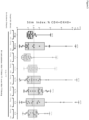
- MenACYW-DT-induced antigen-specific IgM antibody responses to all four meningococcal serotypes were observed in several donors. Increased fold changes over background are shown in Figure 5 .
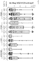
- nMIMIC B cell/T FH model can also be used to show MenACYW-DT-induced antigen-specific IgG antibody responses to each of the meningococcal serotypes, with several donors showing increased fold changes over background for serotype C.
- nMIMIC B cell/T FH cell model Use of the nMIMIC B cell/T FH cell model is detailed below and shown schematically in Figure 4 .
- CBMCs human cord blood mononuclear cells
- a first group of wells was treated with a T FH differentiation cocktail containing: a selected antigen (concentration optimized for the selected antigen), anti-CD28 antibodies (0.1 ⁇ g/ml to 10 ⁇ g/ml), IL-6 (1 ng/ml to 500 ng/ml), and IL-12 (1 ng/ml to 500 ng/ml); a second group of wells was left untreated to serve as feeder cells in later steps; a third group of cells was left untreated to serve as a negative control.
- the selected vaccines were the Dengue Fever Virus vaccine consisting of serotypes 1, 2, 3, and 4 (Sanofi Pasteur) and Hib-TT (control).
- CBMCs were harvested from selected wells by vigorous pipetting and/or gentle scraping of cells into conical tubes, followed by a rinse with a serum-free culture media.
- CD4+ T cells were isolated from the harvested CBMC using negative selection by magnetic bead isolation. The CD4+ T cells were then plated in a serum-free culture media at a concentration of 1 ⁇ 10 5 and 10 ⁇ 10 6 cells per well in 96-well culture plates. Between 1 ⁇ 10 4 and 5 ⁇ 10 5 irradiated CBMC feeder cells and additional T FH differentiation cocktail was added to selected wells.
- T FH were harvested from the wells and between 1 ⁇ 10 4 and 5 ⁇ 10 5 cells were co-cultured with 1 ⁇ 10 5 to 10 ⁇ 10 6 isolated cord blood B cells (negative magnetic bead selection) in a hydrogel for 3D cell culture.
- IL-21 was added (1 ng/ml to 500 ng/ml) to the T FH cell/B cell co-culture.
- IL-21 was again added to the T FH cell/B cell co-culture.
- IgM and IgG antigen-specific antibody production were analyzed using Meso Scale Discovery 96-well plates coated with all four Dengue Virus serotype antigens (1, 2, 3, and 4). MSDs IgM and IgG secondary antibodies were used to detect antibodies in supernatants.
- Dengue Virus-induced antibody responses can be produced using the nN ⁇ MIC B cell/T FH model.
- Dengue Virus-induced antigen-specific IgM antibody responses to all four Dengue serotypes (1, 2, 3, and 4) were observed in several donors. Increased fold changes over background are shown in Figure 7 .
- nMIMIC B cell/T FH cell model Use of the nMIMIC B cell/T FH cell model is detailed below and shown schematically in Figure 4 .
- CBMCs human cord blood mononuclear cells
- a first group of wells was treated with a T FH differentiation cocktail containing: a selected antigen (concentration optimized for the selected antigen), anti-CD28 antibodies (0.1 ⁇ g/ml to 10 ⁇ g/ml), IL-6 (1 ng/ml to 500 ng/ml), and IL-12 (1 ng/ml to 500 ng/ml); a second group of wells was left untreated to serve as feeder cells in later steps; a third group of cells was left untreated to serve as a negative control.
- the selected vaccine was a 17D Yellow fever virus vaccine (Sanofi Pasteur) and a Dengue Vaccine (control).
- CBMCs were harvested from selected wells by vigorous pipetting and/or scraping cells into conical tubes, followed by a rinse with a serum-free culture media.
- CD4+ T cells were isolated from the harvested CBMC using negative selection by magnetic bead isolation. The CD4+ T cells were then plated in a serum-free culture media at a concentration of 1 ⁇ 10 5 and 10 ⁇ 10 6 cells per well in 96-well culture plates. Between 1 ⁇ 10 4 and 5 ⁇ 10 5 irradiated CBMC feeder cells and additional T FH differentiation cocktail was added to selected wells.
- T FH were harvested from the wells and between 1 ⁇ 10 4 and 5 ⁇ 10 5 cells were co-cultured with 1 ⁇ 10 5 to 10 ⁇ 10 6 isolated cord blood B cells (negative magnetic bead selection) in a hydrogel for 3D cell culture.
- IL-21 was added (1 ng/ml to 500 ng/ml) to the T FH cell/B cell co-culture.
- IL-21 was again added to the T FH cell/B cell co-culture.
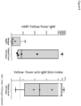
- IgM and IgG antigen-specific antibody production were analyzed using Meso Scale Discovery 96-well plates coated with 17D Yellow fever virus vaccine. MSDs IgM and IgG secondary antibodies were used to detect antibodies in supernatants.
- nMIMIC B cell/T FH cell model Use of the nMIMIC B cell/T FH cell model is detailed below and shown schematically in Figure 4 .
- CBMCs human cord blood mononuclear cells
- the selected vaccines were a meningococcal polysaccharide groups A, C, Y and W-135 diphtheria conjugate vaccine (MenACYW-DT) and groups A, C, Y and W-135 tetanus toxoid conjugate vaccine and Hib-TT (control).
- CBMCs were harvested from selected wells by vigorous pipetting and/or scraping cells into conical tubes, followed by a rinse with a serum-free culture media.
- CD4+ T cells were isolated from the harvested CBMC using negative selection by magnetic bead isolation. The CD4+ T cells were then plated in a serum-free culture media at a concentration of 1 ⁇ 10 5 and 10 ⁇ 10 6 cells per well in 96-well culture plates. Between 1 ⁇ 10 4 and 5 ⁇ 10 5 irradiated CBMC feeder cells and additional T FH differentiation cocktail was added to selected wells.
- T FH were harvested from the wells and between 1 ⁇ 10 4 and 5 ⁇ 10 5 cells were co-cultured with 1 ⁇ 10 5 to 10 ⁇ 10 6 isolated cord blood B cells (negative magnetic bead selection) in a hydrogel for 3D cell culture.
- a range of between 1:1 and 1:100 T FH cell to B cell co-culture ratio was prepared.
- the same antigen that was present in the T FH differentiation cocktail was added to the T FH cell/B cells co-culture.
- IL-21 was added (1 ng/ml to 500 ng/ml) to the T FH cell/B cell co-culture.
- IL-21 was again added to the T FH cell/B cell co-culture.
- IgM and IgG antigen-specific antibody production were analyzed using Meso Scale Discovery 96-well plates coated with all four meningococcal serotype antigens (A, C, Y, W-135). MSDs IgM and IgG secondary antibodies were used to detect antibodies in supernatants.
- MenACYW-DT and TT induced antibody responses can be produced using the nN ⁇ MIC B cell/T FH model.
- MenACYW-DT and TT induced antigen-specific IgM antibody responses to all four meningococcal serotypes were observed in several donors. Increased fold changes over background are shown in Figure 9 .
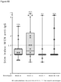
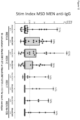
- Figure 10 also demonstrates that these vaccines also showed a stimulation index (SI) of greater than 1.5 for most serotypes for the IgM antibodies.
- SI stimulation index
- Figure 11 shows all four serotypes of MenACYW-DT and TT both show positive antibody, IgG responses of cord blood donors in the nMIMIC over our irrelevant non-meningococcal Act-HIB control.
- Figure 12 shows the Stimulation Index (SI) of IgG antibody data.
Landscapes
- Health & Medical Sciences (AREA)
- Life Sciences & Earth Sciences (AREA)
- Engineering & Computer Science (AREA)
- Immunology (AREA)
- Biomedical Technology (AREA)
- Chemical & Material Sciences (AREA)
- Cell Biology (AREA)
- Biotechnology (AREA)
- Bioinformatics & Cheminformatics (AREA)
- Hematology (AREA)
- Organic Chemistry (AREA)
- Zoology (AREA)
- Wood Science & Technology (AREA)
- Genetics & Genomics (AREA)
- General Health & Medical Sciences (AREA)
- Microbiology (AREA)
- Biochemistry (AREA)
- Molecular Biology (AREA)
- Urology & Nephrology (AREA)
- General Engineering & Computer Science (AREA)
- Tropical Medicine & Parasitology (AREA)
- Toxicology (AREA)
- Pathology (AREA)
- Analytical Chemistry (AREA)
- Physics & Mathematics (AREA)
- Medicinal Chemistry (AREA)
- Food Science & Technology (AREA)
- General Physics & Mathematics (AREA)
- Measuring Or Testing Involving Enzymes Or Micro-Organisms (AREA)
- Micro-Organisms Or Cultivation Processes Thereof (AREA)
- Peptides Or Proteins (AREA)
- Investigating Or Analysing Biological Materials (AREA)
- Silicon Compounds (AREA)
- Silicon Polymers (AREA)
- Steroid Compounds (AREA)
Description
- Vaccination is the best defense against infection, and the first few months of life provide an important period in which vaccines can induce robust protection against disease. However, the clinical development and improvement of vaccines and immunotherapies is stymied by the lack of appropriate in vitro assays for testing immune responses in immature immune systems. Development of in vitro models of neonatal and infant immunity would be of great benefit in advancing research into vaccines that can be administered following birth. The invention described herein is directed to this and other important goals.
- Immune responses in neonates are reviewed in Basha et al. (2014, Expert Rev. Clin. Immunol., 10(9), p1171-1184). Higbee et al, 2009 (ATLA, Vol. 37, Suppl. 1, 19-27) and Drake et al. (2012, Disrup. Sci. Tech., 1(1), p28-40) teach in vitro models to mimic the human immune system comprising different modules that represent different features of the immune system.
WO2013/134464 describes artificial tissue constructs for vaccine evaluation using endothelial cells and mononuclear cells.US2009/117581 also describes artificial immune systems comprising T cells, B cells and antigen-primed dendritic cells. - Provided herein are cell culture constructs and methodologies that together serve as in vitro models of the mammalian neonatal immune system. These mammalian umbilical cord blood-based neonatal modular immune in vitro construct (nMIMIC) models can be used to predict early life immune responses to selected antigens and can serve as primary screens in the pre-clinical evaluation of infant and toddler vaccines.
- The nMIMIC models used in methods of the invention recapitulate aspects of early life naive immunity. Using nascent cord blood-derived immunocytes in the nMIMIC models, the models disclosed herein emulate the immune response of neonates and infants observed in the clinic. These nMIMIC models offer the ability to utilize components of the mammalian neonatal immune system (e.g., T, B, and dendritic cells (DCs)) from cord blood and present them in culture at ratios similar to that observed at physiological concentrations in vivo. With these models, the inventors have been able to (1) apply antigen-treated, cord blood derived-DCs to CD4+ T cells to generate cell-mediated responses, and (2) differentiate cord blood CD4+ T cells into a specialized class of T lymphocytes, T follicular helper cells (TFH), which are essential for B cell help and the humoral response, and apply these TFH cells to naive B cells to generate antigen-specific antibody responses to childhood vaccines.
- Two nMIMIC models are disclosed, the nMIMIC DC/T cell model and the nNΠMIC B cell/TFH cell model, as well as methods of using these models to provide information about cellular responses of components of the mammalian neonatal immune system to antigens, and in assessing candidate vaccines. Methods of the invention concern the nNΠMIC B cell/TFH cell model.
- The nMIMIC B cell/TFH cell model primarily comprises cord-blood derived T follicular helper (TFH) cells and cord blood B cells, and it can be used, for example, to assay the effects of selected agents on antigen presentation and B cell response. The nMIMIC B cell/TFH cell model can be used in a number of different manners, including as a non-limiting example an in vitro method of assessing the response of cells of the neonatal immune system to a selected vaccine. Thus, and in a first embodiment, the present invention is directed to a method of screening a vaccine for activity comprising:
- (a) adding a vaccine and one or more cellular activator to a first in vitro cell culture comprising cord blood CD4+ T cells and maintaining the cell culture under conditions promoting development of vaccine-specific T follicular helper (TFH) cells,
- (b) forming a second in vitro cell culture comprising TFH cells developed in (a) and cord blood B cells,
- (c) adding a vaccine-specific antigen to the cell culture of (b) and maintaining the culture under conditions promoting a cellular response, and
- (d) analyzing supernatant and/or cells from the cell culture of (c) for a cellular response, thereby screening a vaccine for activity,
- wherein the one or more cellular activator is selected from the group consisting of an anti-CD28 antibody, IL-2, IL-6, IL-12, IL-21, and IL-23; and
- wherein the cellular response is one or more of production of a cytokine by the B cells, expression of a marker by the B cells, and production of an antibody with binding specificity for the vaccine-specific antigen by the B cells.
- In another non-limiting example, the nMIMIC B cell/TFH cell model can be exemplified as an in vitro method of assessing the potential efficacy of a vaccine via in vitro testing. In particular, and in a second embodiment, the present invention is directed to a method of assessing efficacy of a vaccine comprising:
- (a) adding a vaccine and one or more cellular activator to a first in vitro cell culture comprising cord blood CD4+ T cells and maintaining the cell culture under conditions promoting development of vaccine-specific T follicular helper (TFH) cells,
- (b) forming a second in vitro cell culture comprising TFH cells developed in (a) and cord blood B cells,
- (c) adding a vaccine-specific antigen to the cell culture of (b) and maintaining the culture under conditions promoting a cellular response, and
- (d) analyzing supernatant and/or cells from the cell culture of (c) for a cellular response, thereby assessing efficacy of a vaccine,
- wherein the one or more cellular activator is selected from the group consisting of an anti-CD28 antibody, IL-2, IL-6, IL-12, IL-21, and IL-23; and
- wherein the cellular response is one or more of production of a cytokine by the B cells, expression of a marker by the B cells, and production of an antibody with binding specificity for the vaccine-specific antigen by the B cells.
- In a further non-limiting example, the nNΠMIC B cell/TFH cell model can be exemplified as an in vitro method of predicting in vivo efficacy of a vaccine using an in vitro cell culture. In particular, and in a third embodiment, the present invention is directed to a method of predicting in vivo efficacy of a vaccine using an in vitro cell culture comprising:
- (a) adding a vaccine and one or more cellular activator to a first in vitro cell culture comprising cord blood CD4+ T cells and maintaining the cell culture under conditions promoting development of vaccine-specific T follicular helper (TFH) cells,
- (b) forming a second in vitro cell culture comprising TFH cells developed in (a) and cord blood B cells,
- (c) adding a vaccine-specific antigen to the cell culture of (b) and maintaining the culture under conditions promoting a cellular response, and
- (d) analyzing supernatant and/or cells from the cell culture of (c) for a cellular response, wherein when a cellular response is found the vaccine is predicted to have in vivo efficacy,
- wherein the one or more cellular activator is selected from the group consisting of an anti-CD28 antibody, IL-2, IL-6, IL-12, IL-21, and IL-23; and
- wherein the cellular response is one or more of production of a cytokine by the B cells, expression of a marker by the B cells, and production of an antibody with binding specificity for the vaccine-specific antigen by the B cells.
- In aspects of these first, second and third embodiments, the first cell culture is maintained for a period of about 3, 4, 5, 6, 7, 8, 9, 10, 11, 12, 13, 14, 15 or more days. In a particular aspect, the first cell culture is maintained for a period of about 8-12 days.
- In aspects of these first, second and third embodiments, additional vaccine is added to the first cell culture at least once, twice, thrice, or more. In a particular aspect, the additional vaccine is added to the first cell culture at least once. Irradiated feeder cells may be added to the first culture upon one or more of the repetitions of vaccine addition. The irradiated feeder cells may be, but are not limited to, cord blood mononuclear cells (CBMCs) grown in a serum-free culture media.
- In aspects of these first, second and third embodiments, the first cell culture is maintained for a period of about 3, 4, 5, 6, 7, 8, 9, 10, 11, 12, 13, 14, 15 or more days, additional vaccine is added on
day day day day - In aspects of these first, second and third embodiments, the vaccine-specific antigen added in (c) is added to the second cell culture of (b) shortly after the second cell culture is prepared, i.e. within about one hour after preparation of the second cell culture of (b).
- In aspects of these embodiments, the culture of (c) is maintained for a period of about 5, 6, 7, 8, 9, 10, 11, 12, 13, 14, 15, 16, 17, 18, 19, 20 or more days. In a particular aspect, the culture of (c) is maintained for a period of about 8-12 days.
- In aspects of these first, second and third embodiments, IL-21 may be added to the culture of (c), typically at a concentration ranging from about 10 to 500 ng/ml. When added, IL-21 is added at one or more of the following time points: (i) when the vaccine-specific antigen is added to the second cell culture of (b), (ii) on
day day - In aspects of these first, second and third embodiments, the culture of (c) is maintained for a period of about 5, 6, 7, 8, 9, 10, 11, 12, 13, 14, 15 or more days, IL-21 is added (i) when the vaccine-specific antigen is added to the second cell culture of (b), (ii) on
day day - In these first, second and third embodiments, the cellular response is one or more of production of a cytokine by the B cells, expression of a marker by the B cells, and production of an antibody with binding specificity for the vaccine-specific antigen by the B cells. The cytokine may be, but is not limited to, is one or more of TNFα, IFNγ, IL-2, IL-4, IL-6, IL-8, IL-10, IL-17, and IL-21. The marker may be, but is not limited to, is one or more of
CD 10, CD19, CD20, CD24, CD27, CD38, CD40, CD86, CD138, IgD, IgG, and IgM. - In aspects of these first, second and third embodiments, the vaccine may be, but is not limited to, an inactivated, attenuated, toxoid, subunit, conjugate, valence and heterotypic vaccine, or a protein, peptide, polynucleotide, oligonucleotide, polysaccharide, virus, virion, bacteria, fungi, or fragment thereof, or antigen-primed DCs.
- In aspects of these first, second and third embodiments, the vaccine-specific antigen may be, but is not limited to, a portion of the vaccine or a component of the vaccine.
- In these first, second and third embodiments, the one or more cellular activator is selected from the group consisting of an anti-CD28 antibody, IL-2, IL-6, IL-12, IL-21, and IL-23.
- In aspects of these first, second and third embodiments, the cord blood B cells are autologous to the cord blood CD4+ T cells (i.e., from the same organism).
- In aspects of these first, second and third embodiments, the cord blood B cells may be treated with CpG2006 prior to addition of the cord blood B cells to the second cell culture.
- In aspects of these first, second and third embodiments, the ratio of TFH cells to cord blood B cells in the second cell culture ranges from about 1:1 to about 1:100.
- In aspects of these first, second and third embodiments, the second cell culture may be formed in a three-dimensional matrix. The matrix may be a gel matrix.
- In a particular example, and in a fourth embodiment of the invention, the present invention is directed to a method of screening a vaccine for activity in vitro comprising:
- (a) adding a vaccine and cellular activators to a first in vitro cell culture comprising cord blood CD4+ T cells and maintaining the cell culture for about 8-12 days under conditions promoting development of vaccine-specific T follicular helper (TFH) cells,
- wherein the cellular activators are one or more of an anti-CD28 antibody, IL-2, IL-6, IL-12, IL-21, and IL-23, and
- wherein irradiated feeder cells and additional vaccine are added on one of
day day
- (b) forming a second in vitro cell culture comprising TFH cells developed in (a) and autologous cord blood B cells in a three-dimensional matrix,
wherein the ratio of TFH cells to cord blood B cells in the second cell culture ranges from about 1:1 to about 1:100; - (c) adding a vaccine-specific antigen to the cell culture of (b) and maintaining the culture for about 1-25 days under conditions promoting a cellular response,
wherein IL-21 is added to the cell culture of (b) upon addition of the antigen, on one ofday day - (d) analyzing supernatant and/or cells from the cell culture of (c) for production of an antibody with binding specificity for the vaccine-specific antigen by the B cells, thereby screening a vaccine for activity in vitro.
- In another example, and in a fifth embodiment, the present invention is directed to a method of assessing in vitro efficacy of a vaccine comprising:
- (a) adding a vaccine and cellular activators to a first in vitro cell culture comprising cord blood CD4+ T cells and maintaining the cell culture for about 8-12 days under conditions promoting development of vaccine-specific T follicular helper (TFH) cells,
- wherein the cellular activators are one or more of an anti-CD28 antibody, IL-2, IL-6, IL-12, IL-21, and IL-23, and
- wherein irradiated feeder cells and additional vaccine are added on one of
day day
- (b) forming a second in vitro cell culture comprising TFH cells developed in (a) and autologous cord blood B cells in a three-dimensional matrix,
wherein the ratio of TFH cells to cord blood B cells in the second cell culture ranges from about 1:1 to about 1:100; - (c) adding a vaccine-specific antigen to the cell culture of (b) and maintaining the culture for about 1-25 days under conditions promoting a cellular response,
wherein IL-21 is added to the cell culture of (b) upon addition of the antigen, on one ofday day - (d) analyzing supernatant and/or cells from the cell culture of (c) for production of an antibody with binding specificity for the vaccine-specific antigen by the B cells, thereby assessing in vitro efficacy of a vaccine.
- In a further example, and in a sixth embodiment, the present invention is directed to a method of predicting in vivo efficacy of a vaccine using an in vitro cell culture comprising:
- (a) adding a vaccine and cellular activators to a first in vitro cell culture comprising cord blood CD4+ T cells and maintaining the cell culture for about 8-12 days under conditions promoting development of vaccine-specific T follicular helper (TFH) cells,
- wherein the cellular activators are one or more of an anti-CD28 antibody, IL-2, IL-6, IL-12, IL-21, and IL-23, and
- wherein irradiated feeder cells and additional vaccine are added on one of
day day
- (b) forming a second in vitro cell culture comprising TFH cells developed in (a) and autologous cord blood B cells in a three-dimensional matrix,
wherein the ratio of TFH cells to cord blood B cells in the second cell culture ranges from about 1:1 to about 1:100; - (c) adding a vaccine-specific antigen to the cell culture of (b) and maintaining the culture for about 1-25 days under conditions promoting a cellular response,
wherein IL-21 is added to the cell culture of (b) upon addition of the antigen, on one ofday day - (d) analyzing supernatant and/or cells from the cell culture of (c) for production of an antibody with binding specificity for the vaccine-specific antigen by the B cells, wherein when such antibody is found the vaccine is predicted to have in vivo efficacy.
- In aspects of these fourth, fifth and sixth embodiments, the irradiated feeder cells may be, but are not limited to, cord blood mononuclear cells (CBMCs) grown in a serum-free culture media.
- In aspects of these fourth, fifth and sixth embodiments, the vaccine may be, but is not limited to, an inactivated, attenuated, toxoid, subunit, conjugate, valence and heterotypic vaccine, or a protein, peptide, polynucleotide, oligonucleotide, polysaccharide, virus, virion, bacteria, fungi, or fragment thereof, or antigen-primed DCs.
- In aspects of these fourth, fifth and sixth embodiments, the vaccine-specific antigen may be, but is not limited to, a portion of the vaccine or a component of the vaccine.
- In aspects of these fourth, fifth and sixth embodiments, the cord blood B cells may be treated with CpG2006 prior to addition of the cord blood B cells to the second cell culture.
- In aspects of these fourth, fifth and sixth embodiments, the matrix may be a gel matrix.
- The foregoing has outlined rather broadly the features and technical advantages of the present invention (and related disclosures) in order that the detailed description of the invention that follows may be better understood. Additional features and advantages of the invention will be described herein, which form the subject of the claims of the invention. It is to be expressly understood, that any description, figure, example, etc. is provided for the purpose of illustration and description only and is by no means intended to define the limits the invention.
-
-
Figure 1 . nMIMIC DC/CD4+ T Cell Model for Evaluating a Selected Antigen. Neonate DCs that are pre-treated with a selected antigen for 24 hrs (1) are co-cultured with CD4+ T cells (2) for 7 days. The co-culture is then re-challenged with antigen-treated DCs (3) for 5-7 hours prior to analysis (4). Analysis may include assaying cytokine expression via such means as flow cytometry, intracellular cytokine staining (ICCS). -
Figure 2 . nMIMIC DC/CD4+ T Cell Model: Stimulation of nNΠMIC with Hib-TT ("Hib") leads to activation of antigen-specific CD4+ T cells in neonates via flow cytometry. Multiple intracellular cytokines were increased in response to Hib-TT in the nMIMIC T cell assay. Cord blood T cells that received antigen-treated DCs were co-cultured for 7 days and either re-stimulated or not with target DCs prior to intracellular cytokine analysis. Second bar in each pair represents those cultures stimulated and re-stimulated with Hib-TT. Data for each cytokine is shown as %CD4+OX40+cytokine+ cells. The data in the graph is represented as a box and whisker plot where the first and third quartiles are at the ends of the box, the median is the line in the interior of the box, and the maximum and minimum of the data set are indicated by the whiskers. -
Figure 3 . nMIMIC DC/CD4+ T Cell Model: Stimulation of nNΠMIC with Hib-TT ("Hib") leads to activation of CD4+ T cells in neonates via flow cytometry. % above each bar represents # of donors where the stimulation index (SI) or fold change over background was ≥ 1.5. Stimulation index was calculated as those cultures treated and re-stimulated with Hib-TT (Hib-TT/Hib-T) over cultures treated but not re-stimulated with Hib-TT (Hib-TT/NoAg). Multiple intracellular cytokines are increased in response to Hib-TT in the nMIMIC T cell assay as demonstrated by SI. -
Figure 4 . nMIMIC naive TFH/B cell Model for Evaluating a Selected Antigen. A selected antigen is used to generate antigen-specific cord blood TFH cells. The TFH cells are then co-cultured with B cells, and the combined culture is optionally re-stimulated with antigen before supernatant collection and antibody analyses. -
Figures 5A-5B . MenACYW-DT-induced IgM antibody responses were observed in the nMIMIC model. MenACYW-DT-induced antigen-specific IgM antibody responses in the nMIMIC to all four meningococcal serotypes (A, C, Y, W-135), with several donors showing increased fold changes over background. In these experiments Hib-TT ("Hib") serves as an irrelevant non-meningococcal background control. InFig. 5A , data illustrates IgM antibody magnitude (represented in MSD units) while inFig. 5B , data shows stimulation index (SI) or fold change over background Hib-TT control. % above each bar represents # of donors where the SI was ≥ 1.5. -
Figures 6A-6B . MenACYW-DT-induced IgG antibody responses were observed in the nMIMIC model. MenACYW-DT-induced antigen-specific IgG antibody responses in the nMIMIC to all four meningococcal serotypes (A, C, Y, W-135), with several donors showing increased fold changes over background for serotype C. In these experiments Hib-TT ("Hib") serves as an irrelevant non-meningococcal background control. InFig. 6A , data illustrates IgG antibody magnitude (represented in MSD units) while inFig. 6B , data shows stimulation index (SI) or fold change over background Hib-TT control. % above each bar represents # of donors where the SI was ≥ 1.5. -
Figure 7 . Dengue Virus vaccine-induced IgM antibody responses were observed in the nMIMIC model. Dengue vaccine-induced antigen-specific IgM antibody responses in the nMIMIC model to all four Dengue vaccine serotypes (1, 2, 3, 4), with several donors showing increased fold changes over background forserotype -
Figure 8 . Yellow Fever Virus vaccine-induced IgG antibody responses were observed in the nMIMIC model. Yellow Fever vaccine-induced antigen-specific IgM antibody responses in the nMIMIC model to the 17D Yellow fever virus vaccine, with several donors showing increased fold changes over background. In these experiments Dengue virus ("Dengue") serves as an irrelevant non-Yellow Fever virus background control. Antibody magnitude is shown on the left of the graph and stimulation index on the right of the graph. The stimulation index (SI) was calculated by dividing Yellow Fever antibody responses over Dengue Fever control. An SI equal to or greater than 1.5 is considered a positive response. -
Figure 9 . Men ACWY-DT and ACWY-TT both stimulate meningococcal anti-IgM antibody production of all four serotypes: Ab magnitude. Meningococcal vaccine-induced IgM antibody responses were observed in the nMIMIC model. Both vaccines show positive IgM antibody responses of cord blood donors in the nNΠMIC over our irrelevant non-meningococcal Act-HIB control. -
Figure 10 . Men ACWY-DT and ACWY-TT both stimulate meningococcal anti-IgM antibody production of all four serotypes: Stim Index. Meningococcal ACWY-DT and ACWY-TT stimulation index of IgM data are presented. An SI equal to or greater than 1.5 is considered a positive response. Percent, %, refers to number of donors that had an SI 1.5 or greater. -
Figure 11 . Men ACWY-DT and ACWY-TT both stimulate meningococcal anti-IgG antibody production of all four serotypes: Ab magnitude. Meningococcal vaccine-induced IgG antibody responses were observed in the nMIMIC model. Men ACWY-DT and ACWY-TT both show positive IgG antibody responses of cord blood donors in the nNΠMIC over our irrelevant non-meningococcal Act-HIB control. -
Figure 12 . Men ACWY-DT and ACWY-TT both stimulate meningococcal anti-IgM antibody production of all four serotypes: Stim Index. Meningococcal ACWY-DT and ACWY-TT stimulation index of IgG data are presented. An SI equal to or greater than 1.5 is considered a positive response. Percent, %, refers to number of donors that had an SI 1.5 or greater. - As used herein, "a" or "an" may mean one or more. As used herein when used in conjunction with the word "comprising," the words "a" or "an" may mean one or more than one. As used herein "another" may mean at least a second or more. Furthermore, unless otherwise required by context, singular terms include pluralities and plural terms include the singular.
- As used herein, "about" refers to a numeric value, including, for example, whole numbers, fractions, and percentages, whether or not explicitly indicated. The term "about" generally refers to a range of numerical values (e.g., +/- 5-10% of the recited value) that one of ordinary skill in the art would consider equivalent to the recited value (e.g., having the same function or result). In some instances, the term "about" may include numerical values that are rounded to the nearest significant figure.
- As used herein, an "antigen" refers to an agent (e.g., protein, polypeptide, polysaccharide, glycolipid, etc.) that elicits an immune response; and/or an agent that is bound by a T cell receptor (e.g., when presented by an MHC molecule), Fc receptor (e.g., activating Fc receptors expressed in high levels by dendritic cells and macrophages), B cell receptor or to an antibody (e.g., produced by a B cell) when exposed or administered to an organism. In some examples, an antigen elicits a humoral response (e.g., including production of antigen-specific antibodies) in an organism. Alternatively or additionally, in some examples, an antigen elicits a cellular response (e.g., involving T-cells whose receptors specifically interact with the antigen) in an organism.
- As used herein, a "vaccine" refers to an administerable, prophylactic compound, composition, or substance that is capable of stimulating an immune response (e.g., containing an antigen), which confers protection against infection (or ameliorates the symptoms of infection) by a causative agent of a disease (e.g., virus, bacterium, fungus, etc.).
- As used herein, an "adjuvant" refers to a substance or vehicle that non-specifically enhances the immune response to an antigen. Adjuvants can include a suspension of minerals (alum, aluminum hydroxide, or phosphate) on which antigen is adsorbed; or water-in-oil emulsions in which an antigen solution is emulsified in mineral oil (for example, Freund's incomplete adjuvant), sometimes with the inclusion of killed mycobacteria (Freund's complete adjuvant) to further enhance antigenicity, and oil-in-water emulsions (e.g., MF59, AS03). Immunostimulatory oligonucleotides (such as those including a CpG motif) can also be used as adjuvants. Adjuvants also include biological molecules, such as costimulatory molecules. Exemplary biological adjuvants include IL-2, RANTES, GM-CSF, TNF-α, IFN-γ, G-CSF, LFA-3, CD72, B7-1, B7-2, OX-40L and 41 BBL.
- As summarized above, the present invention provides methods using cell culture constructs that serve as in vitro models of the mammalian neonatal immune system. Because the constructs are cell cultures comprising cord blood and cord blood-derived immune cells, the constructs are termed neonatal modular immune in vitro constructs (nMIMIC). These nMIMIC models can be used to predict early life immune responses to selected agents, such as antigens, and they can also serve as primary screens in the pre-clinical evaluation of infant and toddler vaccines.
- The nMIMIC models utilize cord blood immune cells. Use of such cells is the only practical approach to studying early life immunity in vitro due to the practicality and ethical issues associated with obtaining large enough volumes of blood from neonates, infants, and toddlers for cell studies. Cord blood mononuclear cells (CBMCs), for example, may be obtained from blood banks across the United States.
- While some groups have utilized cord blood to assess immune responses, as seen in the published literature, none offers a system where the balance of cellular components of the mammalian immune system are considered, such as the ratios of types and concentration of cells, multicellular interactions, and spatiotemporal cellular kinetics. With the nMIMIC models presented herein, human neonatal and infant immune response profiles are replicated. Importantly, statistically significant neonatal immune response to vaccination can be achieved in the nMIMIC models with much fewer donor samples (20-40 CBMC samples) than is required for a clinical trial where hundreds (
phase 2 trials) or even thousands (phase 3 trials) of infant participants would be necessary. These assays can be performed at a fraction of the cost it would take to run even a small clinical trial. Experimental data produced to date provides strong evidence that the nMIMIC models can serve as in vitro biomimetic systems to assess immunogenicity of various vaccines, such as bacterial conjugate formulations. The nMIMIC model system not only encompasses a very practical and achievable approach, it provides a powerful tool for assessing immunological responses in a human infant immune system without the need to employ the human component, i.e., infants. This provides for decisional outcomes on the formulation and use of new or modified vaccines prior to conducting a complex clinical trial. - As also summarized above, the nMIMIC models described herein offer the ability to utilize components of the mammalian neonatal immune system (e.g., T, B, and dendritic cells) from cord blood and present them in culture at ratios similar to that observed under physiological conditions in vivo. With these models, the inventors have been able to (1) generate cell-mediated responses using cord blood derived-dendritic cells (DCs) and CD4+ T cells, and (2) generate antigen-specific antibody responses to childhood vaccines using cord blood-derived TFH cells and naive B cells.
- The present disclosure provides two nNΠMIC models, the nMIMIC DC/T cell model and the nMIMIC B cell/TFH cell model, and methods of using these models to provide information about cellular responses of components of the mammalian neonatal immune system to antigens and in assessing candidate vaccines. The present invention is directed to methods using the nMIMIC B cell/TFH cell model.
- The nMIMIC DC/T cell model is disclosed herein but is not a model of the present invention. This model primarily comprises cord-blood derived dendritic cells (DCs) and cord blood CD4+ T cells, and it can be used, for example, to assay the effects of selected agents on neonatal antigen presentation and CD4+ T cell response. Because neonatal immune systems are immature, the responses of such systems to antigens and vaccines can differ from those seen in juvenile and adult immune systems. Therefore, this model can be used to study the intricacies of the neonatal immune system as well as to obtain important information regarding the likely response of an infant to a selected antigen or vaccine.
- In the broadest sense, the nMIMIC DC/T cell model is utilized by (i) preparing or obtaining a population of cord blood-derived dendritic cells (DCs), (ii) exposing the DCs to a selected agent and permitting uptake, (iii) culturing these primed DCs with cord blood CD4+ T cells, and (iv) assessing the responses of the cells. Use of this model thus allows one to study such disparate functionalities as agent uptake and presentation by neonatal DCs, interactions between primed DCs and CD4+ T cells, and the cellular response of CD4+ T cells when activated by primed DCs.
- While it will be readily apparent that the nMIMIC DC/T cell model can be used in a number of different manners, it can be generally characterized as an in vitro method of assessing the response of cells of the neonatal immune system to a selected agent. Thus, using this model, the cellular response to a selected agent may be assessed by a method comprising:
- (a) adding a selected agent to a first in vitro cell culture comprising cord blood-derived dendritic cells (DCs) and maintaining the cell culture under conditions promoting uptake of the selected agent by the DCs to form a first population of primed DCs,
- (b) forming a second in vitro cell culture comprising primed DCs developed in (a) and cord blood CD4+ T cells and maintaining the cell culture under conditions promoting T cell activation,
- (c) adding a second population of primed DCs to the cell culture of (b) and maintaining the culture under conditions promoting a T cell response, wherein the second population of primed DCs is prepared in the same manner as the first population of primed DCs, and
- (d) analyzing supernatant and/or T cells from the cell culture of (c) for a T cell response, thereby assessing a cellular response to a selected agent.
- The nMIMIC DC/T cell model can also be characterized as an in vitro method of screening selected agents (e.g., antigen, vaccine) for their activity on components of the mammalian neonatal immune system. Thus, the model may be used to screen a selected agent for activity in a method comprising:
- (a) adding a selected agent to a first in vitro cell culture comprising cord blood-derived dendritic cells (DCs) and maintaining the cell culture under conditions promoting the selected agent uptake by the DCs to form a first population of primed DCs,
- (b) forming a second in vitro cell culture comprising primed DCs developed in (a) and cord blood CD4+ T cells and maintaining the cell culture under conditions promoting T cell activation,
- (c) adding a second population of primed DCs to the cell culture of (b) and maintaining the culture under conditions promoting a T cell response, wherein the second population of primed DCs is prepared in the same manner as the first population of primed DCs, and
- (d) analyzing supernatant and/or T cells from the cell culture of (c) for a T cell response, thereby screening a selected agent for activity.
- When this model is used, the first cell culture is maintained for a period of time that will vary depending on the culture conditions and identity of the antigen, among other important factors. However, the first cell culture will generally be maintained for between about 1 and 48 hours prior to formation of the second cell culture. Suitable periods of time include about 1, 2, 3, 4, 5, 6, 7, 8, 9, 10, 11, 12, 13, 14, 15, 16, 17, 18, 19, 20, 21, 22, 23, 24, 25, 26, 27, 28, 29, 30, 31, 32, 33, 34, 35, 36, 37, 38, 39, 40, 41, 42, 43, 44, 45, 46, 47, 48 or more hours prior to formation of the second cell culture, and about 12-36 hours, about 15-33 hours, about 18-30 hours, and about 21-27 hours.
- When this model is used, the second cell culture is maintained for a period of time that will also vary depending on the culture conditions and identity of the antigen, among other important factors. However, the second cell culture will generally be maintained for between about 1 and 20 days prior to adding the second population of primed DCs. Suitable periods of time include about 1, 2, 3, 4, 5, 6, 7, 8, 9, 10, 11, 12, 13, 14, 15, 16, 17, 18, 19, 20 or more days prior to adding the second population of primed DCs, and about 10-20 days, about 12-20 days, about 14-10 days, about 16-20 days, about 12-16 days, about 14-16 days, about 4-10 days, about 5-10 days, and about 6-8 days.
- When this model is used, the culture of (c) is maintained for a period of time that will again vary depending on the culture conditions and identity of the antigen, among other important factors. However, the culture of (c) will generally be maintained for between about 1 and 10 hours prior to the analyzing of (d). Suitable periods of time include about 1, 2, 3, 4, 5, 6, 7, 8, 9, 10, 11, 12 or more hours prior to the analyzing of (d), and about 6-12 hours, about 8-12 hours, about 10-12 hours, about 8-10 hours, about 2-8 hours, about 3-7 hours, and about 4-6 hours.
- When this model is used, the T cell response that is analyzed may be production of a cytokine or a marker, for example. Exemplary cytokines include, but are not limited to, one or more of TNFα, IFNγ, IL-2, IL-4, IL-5, IL-6, IL-8, IL-9, IL-10, IL-13, IL-17A, IL-17F, IL-18, IL-21, IL-22, and IL-26. Exemplary markers include, but are not limited to, one or more of CD25, CD26, CD27, CD28, CD30, CD44, CD69, CD71, CD134, andCD154.
- The skilled artisan will understand that the selected agent that may be used in this model or any of the other models described herein can vary widely and it is only limited by the ability to be taken up and displayed by the DCs. Suitable agents can be generally defined as antigens and vaccines, and they can be further defined as proteins, peptides, polynucleotides, oligonucleotides, and polysaccharides, as well as viruses, virions, bacteria, fungi, and fragments thereof.
- The immune cells that can be used in the nMIMIC DC/T cell model can be from a single cord blood source or two or more sources, depending on the manner in which the model is being used. Thus, for example, the cord blood T cells can be autologous to the cord blood-derived DCs (i.e., from the same organism), or allogeneic to the cord blood-derived DCs (i.e., from another organism of the same species).
- The ratio of primed DCs to cord blood T cells in the culture of (b) can vary depending on such factors as the manner in which the model is used, the identity of the selected agent, and the other components included in the culture. The ratio of DCs to T cells may range from about 1:5 to about 1:100. For most applications, the ratio may range from about 1:20 to about 1:80 or about 1:30 to about 1:50, although acceptable ranges also include from about 1:10 to about 1:50, about 1:20 to about 1:60, about 1:15 to about 1:35, and about 1:30 to about 1:55.
- In a similar fashion, the ratio of cells in the second population of primed DCs added to the culture of (c) to the cord blood T cells in the culture can vary depending on such factors as the manner in which the model is used, the identity of the selected agent, and the other components included in the culture. The ratio of DCs to T cells may range from about 1:5 to about 1:100. For most applications, the ratio may range from about 1:5 to about 1:50 or from about 1:5 to about 1:15, although acceptable ranges also include from about 1:5 to about 1:20, about 1:10 to about 1:25, about 1:15 to about 1:30, and about 1:20 to about 1:40.
- In these methods, the cord blood-derived DCs may be characterized as expressing one or more of the following markers: CD14, CD16, CD25, CD80, CD86, and HLA-DR.
- The cord blood-derived DCs that are used in the nMIMIC DC/T cell model can be prepared in conjunction with the use of the model or obtained from another, e.g., commercial source. The DCs can be obtained from an active cell culture, or prepared from a frozen aliquot of the cells. The cord blood-derived DCs may be prepared by a method comprising:
- (a) culturing endothelial cells on an upper surface of a porous membrane, wherein said porous membrane is housed in an upper chamber of a cell culture well that is suspended over, and is separable from, a lower chamber of the well;
- (b) applying cord blood mononuclear cells (CBMCs) to the endothelial cells on the porous membrane of (a);
- (c) removing the upper chamber housing the porous membrane and endothelial cells from the well about 48 hours after application of the CBMCs; and
- (d) isolating dendritic cells from the lower chamber of the well.
- In the noted method of preparing the cord blood-derived DCs, the porous membrane may be a polycarbonate membrane or a membrane comprised of polytetrafluoroethylene, polyethylene terephthalate, or polyester (PET). The porous membrane will typically have pores that allow cells, e.g., monocytes and immature dendritic cells, to pass through. Such pores range in diameters of between about 1 and 15 µm, however the pores will generally be between 0.4, 0.5, 0.6, 0.7, 0.8, 0.9, 1, 2, 3, 4, 5, 6, 7, 8, 9, 10, 11, 12, 13, 14 and 15 µm in diameter. A permeable support may be used to provide the upper chamber of the well, the porous membrane, and the lower chamber of the well. An example of such a permeable support is the Corning Inc. Transwell® permeable supports.
- In the methods of preparing the cord blood-derived DCs, the lower chamber of the well may comprise extracellular matrix (ECM) material. The ECM material may comprise a material selected from, but not limited to, gelatin, collagen, synthetic ECM materials, poly(lactic-co-glycolic acid) (PLGA), polyglycolic acid (PGA), natural ECM materials, chitosan, protosan, fibronectin, vitronectin, collagen I, collagen IV, laminin, and mixtures thereof. The lower chamber of the well may further comprise one or more of fibroblasts, support cells, and stromal cells.
- In the noted method of preparing the cord blood-derived DCs, the endothelial cells may be, but are not limited to, human umbilical vein endothelial cells (HUVECs) and human somatic cell hybrid EA.hy926 (ATCC® CRL-2922™; ATCC, Manassas, VA). In additional examples, the endothelial cells may be a transformed endothelial cell line.
- In the noted method of preparing the cord blood-derived DCs, the endothelial cells may be cultured on the both sides of the porous membrane. Further, the endothelial cells may be cultured to confluency prior to adding the CBMCs. Alternatively, the endothelial cells may be cultured until multilayer cell growth is achieved and prior to adding the CBMCs.
- In the noted method of preparing the cord blood-derived DCs, a layer of ECM material may be on the upper surface of the porous membrane and the endothelial cells are cultured on the ECM material layer. Alternatively, a layer of ECM material may be on the upper surface and the lower surface of the porous membrane and the endothelial cells are cultured on both layers of ECM material. The ECM material may comprise a material selected from, but not limited to, gelatin, collagen, synthetic ECM materials, PLGA, PGA, natural ECM materials, chitosan, protosan, fibronectin, vitronectin, collagen I, collagen IV, laminin, and mixtures thereof.
- When the nMIMIC DC/T cell model is used in conjunction with the noted method of preparing the cord blood-derived DCs, the lower chamber of the well that is used to prepared the dendritic cells may also serve as the first cell culture of (a). Alternatively, the first cell culture of (a) can be is a separate culture to which the DCs are transferred.
- The nMIMIC B cell/TFH cell model is the nMIMIC model used in methods of the present invention. This model comprises cord-blood derived T follicular helper (TFH) cells and cord blood B cells, and it can be used, for example, to assay the effects of selected agents on neonatal antigen presentation and B cell response. As with the nMIMIC DC/T cell model, this model can also be used to study the intricacies of the neonatal immune system as well as to obtain important information regarding the likely response of an infant to a selected antigen or vaccine.
- In the broadest sense, the nMIMIC B cell/TFH cell model is utilized by (i) preparing a population of cord blood-derived T follicular helper (TFH) cells from cord blood CD4+ T cells in the presence of a selected agent, (ii) preparing a second cell culture comprising these TFH cells and naive cord blood B cells, (iii) adding additional agent or a fragment thereof to the culture, and (iv) assessing the responses of the cells. Use of this model thus allows one to study such disparate functionalities as TFH cell maturation, agent uptake and presentation by TFH cells, interactions between primed TFH cells and B cells, and the cellular response of TFH cells and B cells.
- While it will be readily apparent that the nMIMIC B cell/TFH cell model can be used in a number of different manners, it can be generally characterized as an in vitro method of assessing the response of cells of the neonatal immune system to a selected vaccine. Thus, the invention is directed to a method of screening a vaccine for activity comprising:
- (a) adding a vaccine and one or more cellular activator to a first in vitro cell culture comprising cord blood CD4+ T cells and maintaining the cell culture under conditions promoting development of vaccine-specific T follicular helper (TFH) cells,
- (b) forming a second in vitro cell culture comprising TFH cells developed in (a) and cord blood B cells,
- (c) adding a vaccine-specific antigen to the cell culture of (b) and maintaining the culture under conditions promoting a cellular response, and
- (d) analyzing supernatant and/or cells from the cell culture of (c) for a cellular response, thereby screening a vaccine for activity,
- wherein the one or more cellular activator is selected from the group consisting of an anti-CD28 antibody, IL-2, IL-6, IL-12, IL-21, and IL-23; and
- wherein the cellular response is one or more of production of a cytokine by the B cells, expression of a marker by the B cells, and production of an antibody with binding specificity for the vaccine-specific antigen by the B cells.
- The nMIMIC B cell/TFH cell model can also be characterized as an in vitro method of assessing the potential efficacy of a vaccine via in vitro testing. Thus, the invention is also directed to a method of assessing efficacy of a vaccine comprising:
- (a) adding a vaccine and one or more cellular activator to a first in vitro cell culture comprising cord blood CD4+ T cells and maintaining the cell culture under conditions promoting development of vaccine-specific T follicular helper (TFH) cells,
- (b) forming a second in vitro cell culture comprising TFH cells developed in (a) and cord blood B cells,
- (c) adding a vaccine-specific antigen to the cell culture of (b) and maintaining the culture under conditions promoting a cellular response, and
- (d) analyzing supernatant and/or cells from the cell culture of (c) for a cellular response, thereby assessing efficacy of a vaccine,
- wherein the one or more cellular activator is selected from the group consisting of an anti-CD28 antibody, IL-2, IL-6, IL-12, IL-21, and IL-23; and
- wherein the cellular response is one or more of production of a cytokine by the B cells, expression of a marker by the B cells, and production of an antibody with binding specificity for the vaccine-specific antigen by the B cells.
- The nMIMIC B cell/TFH cell model can further be characterized as an in vitro method of predicting in vivo efficacy of a vaccine using an in vitro cell culture. Thus, the invention is also directed to a method of predicting in vivo efficacy of a vaccine using an in vitro cell culture comprising:
- (a) adding a vaccine and one or more cellular activator to a first in vitro cell culture comprising cord blood CD4+ T cells and maintaining the cell culture under conditions promoting development of vaccine-specific T follicular helper (TFH) cells,
- (b) forming a second in vitro cell culture comprising TFH cells developed in (a) and cord blood B cells,
- (c) adding a vaccine-specific antigen to the cell culture of (b) and maintaining the culture under conditions promoting a cellular response, and
- (d) analyzing supernatant and/or cells from the cell culture of (c) for a cellular response, wherein when a cellular response is found the vaccine is predicted to have in vivo efficacy,
- wherein the one or more cellular activator is selected from the group consisting of an anti-CD28 antibody, IL-2, IL-6, IL-12, IL-21, and IL-23; and
- wherein the cellular response is one or more of production of a cytokine by the B cells, expression of a marker by the B cells, and production of an antibody with binding specificity for the vaccine-specific antigen by the B cells.
- Although not an embodiment of the invention, the nMIMIC B cell/TFH cell model can be used in in vitro methods of assessing the immunogenicity of an antigen. In particular, the immunogenicity of an antigen may be assessed in a method comprising:
- (a) adding an antigen and one or more cellular activator to a first in vitro cell culture comprising cord blood CD4+ T cells and maintaining the cell culture under conditions promoting development of antigen-specific T follicular helper (TFH) cells,
- (b) forming a second in vitro cell culture comprising TFH cells developed in (a) and cord blood B cells,
- (c) adding the antigen to the cell culture of (b) and maintaining the culture under conditions promoting a cellular response, and
- (d) analyzing supernatant and/or cells from the cell culture of (c) for a cellular response, thereby assessing immunogenicity of an antigen.
- When this model is used, the first cell culture is maintained for a period of time that will vary depending on the culture conditions and identity of the vaccine/antigen, among other important factors. However, the first cell culture will generally be maintained for between about 1 and 20 days. Suitable periods of time include about 1, 2, 3, 4, 5, 6, 7, 8, 9, 10, 11, 12, 13, 14, 15, 16, 17, 18, 19, 20 or more days, and about 7-13 days, about 8-12 days, and about 9-11 days.
- When this model is used, additional vaccine/antigen may be added to the first cell culture including at least once, twice, thrice, or more. The additional vaccine/antigen may be identical to the initial vaccine/antigen added to the first cell culture. Alternatively, the additional vaccine/antigen may be a different vaccine/antigen that stimulates an immune response against the same pathogen or disease to which the initial vaccine/antigen is directed or derived. For example, the initial vaccine/antigen may comprise one or more antigens from a particular virus or bacteria, and the additional vaccine/antigen may comprise one or more different antigens from the same virus or bacteria. The additional vaccine/antigen may also be a different formulation of the initial vaccine/antigen. For example, the initial vaccine/antigen may be adjuvanted, and the additional vaccine/antigen may be unadjuvanted. In a particular aspect, the additional vaccine/antigen is added to the first cell culture at least once. Irradiated feeder cells may be added to the first culture upon one or more of the repetitions of vaccine/antigen addition. The irradiated feeder cells may be, but are not limited to, cord blood mononuclear cells (CBMCs).
- When additional vaccine/antigen is added, it may be added on
day day day day - When this model is used, the antigen or vaccine-specific antigen added in (c) is added shortly after the cell culture of (b) is prepared, i.e., within about 1, 2, 3, 4, 5, 6 or more hours after preparation of the cell culture of (b). The identity of the vaccine-specific antigen used in this model will vary widely based on the identity of the vaccine, but such antigens are typically a portion of the vaccine or a component of the vaccine. The antigen or vaccine-specific antigen added in (c) may be identical to the initial vaccine/antigen added in (a). Alternatively, the antigen or vaccine-specific antigen added in (c) may be different from (i.e., not present in) the vaccine/antigen added in (a). Alternatively, the antigen or vaccine-specific antigen added in (c) may be different from the vaccine/antigen added in (a), but may be capable of stimulating an immune response against the same pathogen or disease to which the vaccine/antigen of step (a) is directed. For example, the vaccine/antigen added in step (a) may comprise one or more antigens from a particular virus or bacteria (e.g. a polysaccharide), and the additional vaccine/antigen may comprise one or more different antigens from the same virus or bacteria (e.g., a polypeptide).
- When this model is used, the culture of (c) is also maintained for a period of time that will vary depending on the culture conditions and identity of the vaccine or antigen, among other important factors. However, the culture of (c) will generally be maintained for between about 1 and 25 days. Suitable periods of time include about 1, 2, 3, 4, 5, 6, 7, 8, 9, 10, 11, 12, 13, 14, 15, 16, 17, 18, 19, 20, 21, 22, 23, 24, 25 or more days, and about 10-20 days, about 12-18 days, about 14-16 days, about 7-13 days, about 8-12 days, and about 9-11 days.
- IL-21 may be added to the culture of (c). The amount added may vary, but the final concentration in the culture media will generally range from about 1 to 500 ng/ml. Suitable concentration ranges include from about 10 to 500 ng/ml, 1 to 100 ng/ml, 50 to 350 ng/ml, 50 to 150 ng/ml, 100 to 400 ng/ml, 150 to 500 ng/ml, and 200 to 500 ng/ml.
- When IL-21 is added, IL-21 may be added at one or more of the following time points: (i) when the antigen or vaccine-specific antigen is added to the second cell culture of (b), (ii) on
day day days days - When this model is used, the cellular response is one or more of production of a cytokine by the TFH cells, expression of a marker by the TFH cells, production of a cytokine by the B cells, expression of a marker by the B cells, and production of an antibody with binding specificity for the antigen or vaccine-specific antigen by the B cells. In methods of the invention the cellular response is one or more of production of a cytokine by the B cells, expression of a marker by the B cells, and production of an antibody with binding specificity for the vaccine-specific antigen by the B cells. The cytokine may be, but is not limited to, is one or more of CXCL13, TNFα, IFNγ, IL-2, IL-4, IL-6, IL-8, IL-10, IL-13, IL-17, and IL-21. In methods of the invention the cytokine may be one or more of TNFα, IFNγ, IL-2, IL-4, IL-6, IL-8, IL-10, IL-17, and IL-21. The marker may be, but is not limited to, is one or more of CD10, CD19, CD20, CD24, CD27, CD38, CD40, CD86, CD138, IgD, IgG, and IgM.
- The skilled artisan will understand that the vaccine or antigen that may be used in this model can vary widely and it only limited by the ability to be processed by TFH cells and presented to B cells. Suitable vaccines can be generally defined as inactivated, attenuated, toxoid, subunit, conjugate, valence and heterotypic vaccines, and they can be further defined as proteins, peptides, polynucleotides, oligonucleotides, and polysaccharides, as well as microorganisms such as viruses, virions, bacteria, and fungi, and fragments thereof, and antigen-primed DCs.
- The one or more cellular activator used in this model may be, but is not limited to, an anti-CD28 antibody, IL-2, IL-6, IL-12, IL-21, and IL-23. In methods of the invention the one or more cellular activator is selected from the group consisting of an anti-CD28 antibody, IL-2, IL-6, IL-12, IL-21, and IL-23.
- The immune cells that can be used in the nMIMIC B cell/TFH cell model can be from a single cord blood source or two or more sources, depending on the manner in which the model is being used. Thus, for example, the cord blood B cells can be autologous to the cord blood CD4+ T from which the TFH cells are derived (i.e., from the same organism). The cord blood B cells can even be heterologous to the cord blood CD4+ T cells (i.e., from an organism of a different species).
- When this model is used, the cord blood B cells may be treated with CpG2006 (ODN 2006; InvivoGen, San Diego, CA) prior to addition of the cord blood B cells to the second cell culture of (b). When CpG2006 is used, the B cells are treated with the activator for about 1-48 hours before forming the second cell culture of (b) at a concentration of between about 0.1 to 5 ug/ml.
- The ratio of TFH cells to cord blood B cells in the second cell culture can vary depending on such factors as the manner in which the model is used, the identity of the selected agent (vaccine or antigen), and the other components included in the culture. For most applications, the ratio may range from about 1:1 to about 1:100.
- When this model is used, the second cell culture may be formed in a three-dimensional matrix. The matrix may be, but is not limited to, a gel matrix, such as a hydrogel for 3D cell culture. The matrix may contain one or more extracellular matrix components, including, but not limited to, one or more of collagen I, collagen IV, vitronectin, fibronectin, and laminin.
- In a particular example, the invention is directed to a method of screening a vaccine for activity in vitro comprising:
- (a) adding a vaccine and cellular activators to a first in vitro cell culture comprising cord blood CD4+ T cells and maintaining the cell culture for about 8-12 days under conditions promoting development of vaccine-specific T follicular helper (TFH) cells,
- wherein the cellular activators are one or more of an anti-CD28 antibody, IL-2, IL-6, IL-12, IL-21, and IL-23, and
- wherein irradiated feeder cells and additional vaccine are added on one of
day day
- (b) forming a second in vitro cell culture comprising TFH cells developed in (a) and autologous cord blood B cells in a three-dimensional matrix,
wherein the ratio of TFH cells to cord blood B cells in the second cell culture ranges from about 1:1 to about 1:100; - (c) adding a vaccine-specific antigen to the cell culture of (b) and maintaining the culture for about 1-25 days under conditions promoting a cellular response,
wherein IL-21 is added to the cell culture of (b) upon addition of the antigen, on one ofday day - (d) analyzing supernatant and/or cells from the cell culture of (c) for production of an antibody with binding specificity for the vaccine-specific antigen by the B cells, thereby screening a vaccine for activity in vitro.
- In another example, the invention is directed to a method of assessing in vitro efficacy of a vaccine comprising:
- (a) adding a vaccine and cellular activators to a first in vitro cell culture comprising cord blood CD4+ T cells and maintaining the cell culture for about 8-12 days under conditions promoting development of vaccine-specific T follicular helper (TFH) cells,
- wherein the cellular activators are one or more of an anti-CD28 antibody, IL-2, IL-6, IL-12, IL-21, and IL-23, and
- wherein irradiated feeder cells and additional vaccine are added on one of
day day
- (b) forming a second in vitro cell culture comprising TFH cells developed in (a) and autologous cord blood B cells in a three-dimensional matrix,
wherein the ratio of TFH cells to cord blood B cells in the second cell culture ranges from about 1:1 to about 1:100; - (c) adding a vaccine-specific antigen to the cell culture of (b) and maintaining the culture for about 1-25 days under conditions promoting a cellular response,
wherein IL-21 is added to the cell culture of (b) upon addition of the antigen, on one ofday day - (d) analyzing supernatant and/or cells from the cell culture of (c) for production of an antibody with binding specificity for the vaccine-specific antigen by the B cells, thereby assessing in vitro efficacy of a vaccine.
- In a further example, the invention is directed to a method of predicting in vivo efficacy of a vaccine using an in vitro cell culture comprising:
- (a) adding a vaccine and cellular activators to a first in vitro cell culture comprising cord blood CD4+ T cells and maintaining the cell culture for about 8-12 days under conditions promoting development of vaccine-specific T follicular helper (TFH) cells,
- wherein the cellular activators are one or more of an anti-CD28 antibody, IL-2, IL-6, IL-12, IL-21, and IL-23, and
- wherein irradiated feeder cells and additional vaccine are added on one of
day day
- (b) forming a second in vitro cell culture comprising TFH cells developed in (a) and autologous cord blood B cells in a three-dimensional matrix,
wherein the ratio of TFH cells to cord blood B cells in the second cell culture ranges from about 1:1 to about 1:100; - (c) adding a vaccine-specific antigen to the cell culture of (b) and maintaining the culture for about 1-25 days under conditions promoting a cellular response,
wherein IL-21 is added to the cell culture of (b) upon addition of the antigen, on one ofday day - (d) analyzing supernatant and/or cells from the cell culture of (c) for production of an antibody with binding specificity for the vaccine-specific antigen by the B cells, wherein when such antibody is found the vaccine is predicted to have in vivo efficacy.
- In these examples, the irradiated feeder cells may be, but are not limited to, cord blood mononuclear cells (CBMCs).
- The skilled artisan will understand that the vaccine that may be used in this model can vary widely and it only limited by the ability to be processed by TFH cells and presented to B cells. Suitable vaccines can be generally defined as inactivated, attenuated, toxoid, subunit, conjugate, valence and heterotypic vaccines, and they can be further defined as proteins, peptides, polynucleotides, oligonucleotides, and polysaccharides, as well as microorganisms such as viruses, virions, bacteria, and fungi, and fragments thereof, and antigen-primed DCs.
- The vaccine-specific antigen used in this model will vary widely based on the identity of the vaccine, but such antigens are typically a portion of the vaccine or a component of the vaccine.
- The cord blood B cells may be treated with CpG2006 prior to addition of the cord blood B cells to the second cell culture. When CpG2006 is used, the B cells are treated with the activator for about 1-48 hours before forming the second cell culture of (b) at a concentration of between about 0.1 to 5 ug/ml.
- The matrix may be, but is not limited to, a gel matrix, such as a hydrogel for 3D cell culture.
- In each of the described methods, including methods of the invention, the analysis of supernatants and cell culture media for the production and/or presence of cytokines, markers and other factors may be conducted by via means readily known to and understood by the skilled artisan that include, but are not limited to, a cytokine/chemokine magnetic bead panel (e.g., Millipore's MILLIPLEX MAP™ kit), an ELISA or MSD (mesoscale scale discovery) multi-array technology.
- In each of the described methods, including methods of the invention, the analysis of cells, such as TFH cells, T cells and B cells, for production and/or expression of cytokines, cell surface markers and other factors may be conducted via means readily known to and understood by the skilled artisan that include, but are not limited to, flow cytometry using one or more antibodies that bind to selected molecules with specificity.
- In each of the described methods, including methods of the invention, the analysis of supernatants and cell culture media for the production of antibodies may be conducted via means readily known to and understood by the skilled artisan that include, but are not limited to, a magnetic bead panel, an ELISA or MSD multi-array technology. Similarly, the analysis of B cells for the production of antibodies may be conducted via means readily known to and understood by the skilled artisan that include, but are not limited to, a magnetic bead panel, an ELISA or MSD multi-array technology.
- In each of the described methods, including methods of the invention, culture conditions promoting a cellular response, such as a TFH cell, T cell, and/or B cell response, include culture conditions typically associated with the culture of immune cells in vitro, such as 5% CO2, 95% relative humidity, and 37°C.
- In each of the described methods, including methods of the invention, conditions promoting development of vaccine-specific T follicular helper (TFH) cells include culture conditions typical associated with the culture of immune cells in vitro, such as 5% CO2, 95% relative humidity, and 37°C.
- In each of the described methods, including methods of the invention, the source of the cord blood cells may be a human, a non-human primate, bird, horse, cow, goat, sheep, a companion animal, such as a dog, cat or rodent, or other mammal.
- Use of the nMIMIC DC cell/T cell model is detailed below and shown schematically in
Figure 1 . - Using a peripheral tissue equivalent (PTE) method, human cord blood DCs were generated in a transwell system containing an endothelial cell layer (human somatic cell hybrid EA.hy926 cells). On
day 0, EA.hy926 cells were plated in 24-well transwell inserts that had a pore range diameter between about 1 and 15 µm at a density of between 1 × 10⁴ and 1 × 10⁵ cells in M199 media with 20% FCS (fetal calf serum). Onday 6, M199 media was replaced with a serum-free culture media. Onday 7, cord blood mononuclear cells (CBMCs) were collected and between 1 × 10⁶ and 50 × 106 cells in a serum-free culture media were plated onto the 24-well transwell inserts containing the EA.hy926 layer. As DCs migrate through this layer they differentiated into mature cells with the ability to present antigen. - Also, on
day 7, a second set of EA.hy926 plated 24-well transwell plates were prepared in M199 media with 20% FCS and cultured for 7 days with media exchange to a serum-free culture media on day 13. - On
day 8, transwell inserts of cultures where CBMCs were applied were removed and replaced with a fresh transwell layered with EA.hy926 cells prepared onday 0. At this point DCs had migrated to the bottom of the 24-well dish through the transwell and other cell types still present in transwell insert are removed. Experimental cultures were then treated with antigen of interest at predetermined concentrations to the lower chamber, while control cultures received vehicle alone. In this experiment, the selected antigen was a Haemophilus b tetanus toxoid conjugate vaccine (Hib-TT (Sanofi Pasteur). - On day 9, DCs treated in the presence and absence of antigen were harvested and co-cultured with isolated cord blood CD4+ T cells at ratios between 1:5 and 1:100 (DC:T cells) and incubated for 7 days. Cord blood CD4+ T cells were isolated using negative selection by magnetic bead isolation. On
day 14, CBMCs are added to the second set of EA.hy926 transwell plates (prepared on day 7) overnight, with antigen treatments added onday 15 after exchange with fresh EA.hy926 transwells as described above. - On day 16, the first DC/CD4+ T cell co-culture was collected and a second co-culture was prepared with the second set of treated and non-treated transwell-generated DC cells. The first DC/CD4+ T cell co-culture is plated at between 5 × 10⁴ and 1 × 10⁶ with the second set of harvested DCs plated at between 5 × 10³ and 1 × 10⁵. Re-stimulation with the second set of transwell-generated DCs was done for a period of between 2-10 hours prior to intracellular cytokine analysis by flow cytometry.
- Intracellular cytokine staining was performed to analyze accumulation of both Th1 (IFNγ, TNFα, IL-2) and Th2 (IL-4, IL-6, IL-10, IL-13) cytokine subsets in response to antigen by flow cytometry using the BD Fortessa® Cytometer with blue, red, and violet lasers using the BD FACSDiva® Software using the following panel for CD4+ T cell analysis:
Fluorochrome Marker 1 LDA LDA 2 BV421 OX40 3 BV605 TNFα 5 BV650 IL-10 4 BV711 IL-13 6 BV785 IL-4 7 FITC IL-6 8 PE CD4 9 PE-CF594 IL-2 10 PerCP-eFluor710 IL-8 11 PE-Cy7 IFNγ 12 Alexa Fluor 647 IL-21 13 Alexa Fluor 700 IL- 17A 14 APC-Cy7 CD154 - The analysis indicated that stimulation of the nMIMIC DC/T cell model with Hib-TT leads to activation of antigen-specific CD4+ T cells in neonates. As shown in
Figure 2 , multiple intracellular cytokines were increased in response to Hib-TT in the nMIMIC T cell assay as assayed via flow cytometry. Cord blood T cells that received antigen-treated DCs were co-cultured for 7 days and either re-stimulated or not with target DCs prior to intracellular cytokine analysis. Second bar in each colored pair represents those cultured stimulated and re-stimulated with Hib-TT. - As shown in
Figure 3 , production of IFNγ, TNFα, IL-2, IL-21, IL-17, IL-13, IL-6 and IL-8 was also increased, as assayed via flow cytometry. InFigure 3 , % represents the number of donors where the stimulation index (SI) or fold change over background was ≥ 1.5. Multiple intracellular cytokines were found to be increased in response to Hib-TT in the model as demonstrated by SI. These results indicate that in response to stimulation with Hib-TT, cord blood CD4+ T cells were activated showing Th1 (TNFα and IL-8), Th2 (IL-13, IL-6), as well as Th17 (IL-17) responses as demonstrated by cytokine evaluations via flow cytometry. - Use of the nMIMIC B cell/TFH cell model is detailed below and shown schematically in
Figure 4 . - On
day 0 where human cord blood mononuclear cells (CBMCs) were collected and between 1 × 10⁶ and 50 × 106 cells in a serum-free culture media were plated in wells of a 24-well culture plate. A first group of wells was treated with a TFH differentiation cocktail containing: a selected antigen (concentration optimized for the selected antigen), anti-CD28 antibodies (0.1 ug/ml to 10 ug/ml), IL-6 (1 ng/ml to 500 ng/ml), and IL-12 (1 ng/ml to 500 ng/ml); a second group of wells was left untreated to serve as feeder cells in later steps; a third group of cells was left untreated to serve as a negative control. The selected vaccines were a meningococcal polysaccharide groups A, C, Y and W-135 diphtheria conjugate vaccine (MenACYW-DT) (Sanofi Pasteur) and Hib-TT(control). - On
day 3, CBMCs were harvested from selected wells by vigorous pipetting and/or scraping cells into conical tubes, followed by a rinse with a serum-free culture media. CD4+ T cells were isolated from the harvested CBMC using negative selection by magnetic bead isolation. The CD4+ T cells were then plated in a serum-free culture media at a concentration of 1 × 10⁵ and 10 × 106 cells per well in 96-well culture plates. Between 1 × 10⁴ and 5 × 10⁵ irradiated CBMC feeder cells and additional TFH differentiation cocktail was added to selected wells. - On
day 6, additional irradiated feeder cells (1 × 10³ to 1 × 10⁵) and additional TFH differentiation cocktail was added to selected wells. - On
day 10, TFH were harvested from the wells and between 1 × 10⁴ and 5 × 10⁵ cells were co-cultured with 1 × 10⁵ to 10 × 106 isolated cord blood B cells (negative magnetic bead selection) in a hydrogel for 3D cell culture. A range of between 1:1 and 1: 100 TFH cell to B cell co-culture ratio was prepared. The same antigen that was present in the TFH differentiation cocktail was added to the TFH cell/B cells co-culture. - On day 12, IL-21 was added (1 ng/ml to 500 ng/ml) to the TFH cell/B cell co-culture. On
day 14, IL-21 was again added to the TFH cell/B cell co-culture. - On
day 20, B cells were harvested from the culture and culture media was collected. IgM and IgG antigen-specific antibody production was analyzed using Meso Scale Discovery 96-well plates coated with all four meningococcal serotype antigens (A, C, Y, W-135). MSDs IgM and IgG secondary antibodies were used to detect antibodies in supernatants. - The analysis demonstrated that MenACYW-DT-induced antibody responses can be produced using the nNΠMIC B cell/TFH model. As seen in
Figure 5 , MenACYW-DT-induced antigen-specific IgM antibody responses to all four meningococcal serotypes (A, C, Y, W-135) were observed in several donors. Increased fold changes over background are shown inFigure 5 . - The results presented in
Figure 6 demonstrate that the nMIMIC B cell/TFH model can also be used to show MenACYW-DT-induced antigen-specific IgG antibody responses to each of the meningococcal serotypes, with several donors showing increased fold changes over background for serotype C. - Use of the nMIMIC B cell/TFH cell model is detailed below and shown schematically in
Figure 4 . - On
day 0 where human cord blood mononuclear cells (CBMCs) were collected and between 1 × 10⁶ and 50 × 106 cells in a serum-free culture media were plated in wells of a 24-well culture plate. A first group of wells was treated with a TFH differentiation cocktail containing: a selected antigen (concentration optimized for the selected antigen), anti-CD28 antibodies (0.1 µg/ml to 10 µg/ml), IL-6 (1 ng/ml to 500 ng/ml), and IL-12 (1 ng/ml to 500 ng/ml); a second group of wells was left untreated to serve as feeder cells in later steps; a third group of cells was left untreated to serve as a negative control. The selected vaccines were the Dengue Fever Virus vaccine consisting ofserotypes - On
day 3, CBMCs were harvested from selected wells by vigorous pipetting and/or gentle scraping of cells into conical tubes, followed by a rinse with a serum-free culture media. CD4+ T cells were isolated from the harvested CBMC using negative selection by magnetic bead isolation. The CD4+ T cells were then plated in a serum-free culture media at a concentration of 1 × 10⁵ and 10 × 106 cells per well in 96-well culture plates. Between 1 × 10⁴ and 5 × 10⁵ irradiated CBMC feeder cells and additional TFH differentiation cocktail was added to selected wells. - On
day 6, additional irradiated feeder cells (1 × 10³ to 1 × 10⁵) and additional TFH differentiation cocktail was added to selected wells. - On
day 10, TFH were harvested from the wells and between 1 × 10⁴ and 5 × 10⁵ cells were co-cultured with 1 × 10⁵ to 10 × 106 isolated cord blood B cells (negative magnetic bead selection) in a hydrogel for 3D cell culture. A range of between 1:1 and 1: 100 TFH cell to B cell co-culture ratio was prepared. The same antigen that was present in the TFH differentiation cocktail was added to the TFH cell/B cells co-culture. - On day 12, IL-21 was added (1 ng/ml to 500 ng/ml) to the TFH cell/B cell co-culture. On
day 14, IL-21 was again added to the TFH cell/B cell co-culture. - On
day 20, B cells were harvested from the culture and culture media was collected. IgM and IgG antigen-specific antibody production was analyzed using Meso Scale Discovery 96-well plates coated with all four Dengue Virus serotype antigens (1, 2, 3, and 4). MSDs IgM and IgG secondary antibodies were used to detect antibodies in supernatants. - The analysis demonstrated that Dengue Virus-induced antibody responses can be produced using the nNΠMIC B cell/TFH model. As seen in
Figure 7 , Dengue Virus-induced antigen-specific IgM antibody responses to all four Dengue serotypes (1, 2, 3, and 4) were observed in several donors. Increased fold changes over background are shown inFigure 7 . - The results presented in
Figure 7 demonstrate that the nMIMIC B cell/TFH model can also be used to show Dengue Virus-induced antigen-specific IgM antibody responses to each of the Dengue serotypes, with several donors showing increased fold changes over background for serotypes. - Use of the nMIMIC B cell/TFH cell model is detailed below and shown schematically in
Figure 4 . - On
day 0 where human cord blood mononuclear cells (CBMCs) were collected and between 1 × 10⁶ and 50 × 106 cells in a serum-free culture media were plated in wells of a 24-well culture plate. A first group of wells was treated with a TFH differentiation cocktail containing: a selected antigen (concentration optimized for the selected antigen), anti-CD28 antibodies (0.1 µg/ml to 10 µg/ml), IL-6 (1 ng/ml to 500 ng/ml), and IL-12 (1 ng/ml to 500 ng/ml); a second group of wells was left untreated to serve as feeder cells in later steps; a third group of cells was left untreated to serve as a negative control. The selected vaccine was a 17D Yellow fever virus vaccine (Sanofi Pasteur) and a Dengue Vaccine (control). - On
day 3, CBMCs were harvested from selected wells by vigorous pipetting and/or scraping cells into conical tubes, followed by a rinse with a serum-free culture media. CD4+ T cells were isolated from the harvested CBMC using negative selection by magnetic bead isolation. The CD4+ T cells were then plated in a serum-free culture media at a concentration of 1 × 10⁵ and 10 × 106 cells per well in 96-well culture plates. Between 1 × 10⁴ and 5 × 10⁵ irradiated CBMC feeder cells and additional TFH differentiation cocktail was added to selected wells. - On
day 6, additional irradiated feeder cells (1 × 10³ to 1 × 10⁵) and additional TFH differentiation cocktail was added to selected wells. - On
day 10, TFH were harvested from the wells and between 1 × 10⁴ and 5 × 10⁵ cells were co-cultured with 1 × 10⁵ to 10 × 106 isolated cord blood B cells (negative magnetic bead selection) in a hydrogel for 3D cell culture. A range of between 1:1 and 1: 100 TFH cell to B cell co-culture ratio was prepared. The same antigen that was present in the TFH differentiation cocktail was added to the TFH cell/B cells co-culture. - On day 12, IL-21 was added (1 ng/ml to 500 ng/ml) to the TFH cell/B cell co-culture. On
day 14, IL-21 was again added to the TFH cell/B cell co-culture. - On
day 20, B cells were harvested from the culture and culture media was collected. IgM and IgG antigen-specific antibody production was analyzed using Meso Scale Discovery 96-well plates coated with 17D Yellow fever virus vaccine. MSDs IgM and IgG secondary antibodies were used to detect antibodies in supernatants. - The analysis demonstrated that the 17D Yellow fever virus vaccine-induced antibody responses can be produced using the nMIMIC B cell/TFH model. As seen in
Figure 8 , the 17D Yellow fever virus vaccine-induced antigen-specific IgM antibody responses were observed in several donors. Increased fold changes over background are shown inFigure 8 to the right. - The results presented in
Figure 8 demonstrate that the nMIMIC B cell/TFH model can also be used to show the 17D Yellow fever virus vaccine-induced antigen-specific IgM antibody responses to the vaccine, with several donors showing increased fold changes over background. - Use of the nMIMIC B cell/TFH cell model is detailed below and shown schematically in
Figure 4 . - On
day 0 where human cord blood mononuclear cells (CBMCs) were collected and between 1 × 10⁶ and 50 × 106 cells in a serum-free culture media were plated in wells of a 24-well culture plate. A first group of wells was treated with a TFH differentiation cocktail containing: a selected antigen (concentration optimized for the selected antigen), anti-CD28 antibodies (0.1 µg/ml to 10 µg/ml), IL-6 (1 ng/ml to 500 ng/ml), and IL-12 (1 ng/ml to 500 ng/ml); a second group of wells was left untreated to serve as feeder cells in later steps; a third group of cells was left untreated to serve as a negative control. The selected vaccines were a meningococcal polysaccharide groups A, C, Y and W-135 diphtheria conjugate vaccine (MenACYW-DT) and groups A, C, Y and W-135 tetanus toxoid conjugate vaccine and Hib-TT (control). - On
day 3, CBMCs were harvested from selected wells by vigorous pipetting and/or scraping cells into conical tubes, followed by a rinse with a serum-free culture media. CD4+ T cells were isolated from the harvested CBMC using negative selection by magnetic bead isolation. The CD4+ T cells were then plated in a serum-free culture media at a concentration of 1 × 10⁵ and 10 × 106 cells per well in 96-well culture plates. Between 1 × 10⁴ and 5 × 10⁵ irradiated CBMC feeder cells and additional TFH differentiation cocktail was added to selected wells. - On
day 6, additional irradiated feeder cells (1 × 10³ to 1 × 10⁵) and additional TFH differentiation cocktail was added to selected wells. - On
day 10, TFH were harvested from the wells and between 1 × 10⁴ and 5 × 10⁵ cells were co-cultured with 1 × 10⁵ to 10 × 106 isolated cord blood B cells (negative magnetic bead selection) in a hydrogel for 3D cell culture. A range of between 1:1 and 1:100 TFH cell to B cell co-culture ratio was prepared. The same antigen that was present in the TFH differentiation cocktail was added to the TFH cell/B cells co-culture. - On day 12, IL-21 was added (1 ng/ml to 500 ng/ml) to the TFH cell/B cell co-culture. On
day 14, IL-21 was again added to the TFH cell/B cell co-culture. - On
day 20, B cells were harvested from the culture and culture media was collected. IgM and IgG antigen-specific antibody production was analyzed using Meso Scale Discovery 96-well plates coated with all four meningococcal serotype antigens (A, C, Y, W-135). MSDs IgM and IgG secondary antibodies were used to detect antibodies in supernatants. - The analysis demonstrated that MenACYW-DT and TT induced antibody responses can be produced using the nNΠMIC B cell/TFH model. As seen in
Figure 9 , MenACYW-DT and TT induced antigen-specific IgM antibody responses to all four meningococcal serotypes (A, C, Y, W-135) were observed in several donors. Increased fold changes over background are shown inFigure 9 .Figure 10 also demonstrates that these vaccines also showed a stimulation index (SI) of greater than 1.5 for most serotypes for the IgM antibodies. -
Figure 11 shows all four serotypes of MenACYW-DT and TT both show positive antibody, IgG responses of cord blood donors in the nMIMIC over our irrelevant non-meningococcal Act-HIB control.Figure 12 shows the Stimulation Index (SI) of IgG antibody data. - The results presented in
Figures 9-12 demonstrate that the nMIMIC B cell/TFH model can also be used to show MenACYW-DT and Men ACYW-TT vaccines induced antigen-specific IgG and IgM antibody responses to each of the meningococcal serotypes, with several donors showing increased fold changes over background.
Claims (15)
- A method of screening a vaccine for activity, assessing efficacy of a vaccine or predicting in vivo efficacy of a vaccine, comprising:(a) adding a vaccine and one or more cellular activator to a first in vitro cell culture comprising cord blood CD4+ T cells and maintaining the cell culture under conditions promoting development of vaccine-specific T follicular helper (TFH) cells,(b) forming a second in vitro cell culture comprising TFH cells developed in (a) and cord blood B cells,(c) adding a vaccine-specific antigen to the cell culture of (b) and maintaining the culture under conditions promoting a cellular response, and(d) analyzing supernatant and/or cells from the cell culture of (c) for a cellular response, thereby screening a vaccine for activity, thereby assessing efficacy of a vaccine or wherein when a cellular response is found the vaccine is predicted to have in vivo efficacy,wherein the one or more cellular activator is selected from the group consisting of an anti-CD28 antibody, IL-2, IL-6, IL-12, IL-21, and IL-23; andwherein the cellular response is one or more of production of a cytokine by the B cells, expression of a marker by the B cells, and production of an antibody with binding specificity for the vaccine-specific antigen by the B cells.
- The method of claim 1 wherein the first cell culture is maintained for a period of about 8-12 days.
- The method of claim 1 or 2 whereini) additional vaccine is added to the first cell culture at least once; and/orii) the first cell culture is maintained for a period of about 8-12 days and additional vaccine is added on one of day 2, 3 or 4 after the initial vaccine addition and on one of day 5, 6 or 7 after the initial vaccine addition.
- The method of claim 3 wherein irradiated feeder cells are added to the first culture upon repetition of one or more of the vaccine additions.
- The method of claim 4 wherein the irradiated feeder cells are cord blood mononuclear cells (CBMCs) grown in a serum-free culture media.
- The method of any one of claims 1-5 whereini) the culture of (c) is maintained for about 8-12 days; and/orii) IL-21 is added to the culture of (c).
- The method of any one of claims 1-6 wherein the culture of (c) is maintained for about 1-25 days and IL-21 is added upon addition of the vaccine-specific antigen to the second cell culture, on one of day 2, 3 or 4 after preparation of the second cell culture, and on one of day 5, 6 or 7 after preparation of the second cell culture.
- The method of any one of claims 1 to 7 whereini) the cytokine is one or more of TNFα, IFNγ, IL-2, IL-4, IL-6, IL-8, IL-10, IL-17, or IL-21; orii) the marker is one or more of CD10, CD19, CD20, CD24, CD27, CD38, CD40, CD86, CD138, IgD, IgG, and IgM.
- The method of any one of claims 1 to 8 whereini) the vaccine is an inactivated, attenuated, toxoid, subunit, conjugate, valence and heterotypic vaccine, or a protein, peptide, polynucleotide, oligonucleotide, polysaccharide, virus, virion, bacteria, fungi, or fragment thereof, or antigen-primed DCs; and/orii) the vaccine-specific antigen is a portion of the vaccine or a component of the vaccine.
- The method of any one of claims 1 to 9 wherein the cord blood B cells are autologous to the cord blood CD4+ T cells.
- The method of any one of claims 1 to 10 whereini) the cord blood B cells are treated with CpG2006 prior to addition of the cord blood B cells to the second cell culture;ii) the ratio of TFH cells to cord blood B cells in the second cell culture ranges from about 1:1 to about 1:100; and/oriii) the second cell culture is formed in a three-dimensional matrix.
- The method of claim 11 wherein the matrix is a gel matrix.
- A method of screening a vaccine for activity, assessing efficacy of a vaccine or predicting in vitro efficacy of a vaccine as claimed in claim 1 comprising:(a) adding a vaccine and cellular activators to a first cell culture comprising cord blood CD4+ T cells and maintaining the cell culture for about 8-12 days under conditions promoting development of vaccine-specific T follicular helper (TFH) cells,wherein the cellular activators are one or more of an anti-CD28 antibody, IL-2, IL-6, IL-12, IL-21, and IL-23, andwherein irradiated feeder cells and additional vaccine are added on one of day 2, 3 or 4 after the initial vaccine addition, and on one of day 5, 6 or 7 after the initial vaccine addition;(b) forming a second cell culture comprising TFH cells developed in (a) and autologous cord blood B cells in a three-dimensional matrix,
wherein the ratio of TFH cells to cord blood B cells in the second cell culture ranges from about 1:1 to about 1:100;(c) adding a vaccine-specific antigen to the cell culture of (b) and maintaining the culture for about 1-25 days under conditions promoting a cellular response,
wherein IL-21 is added to the cell culture of (b) upon addition of the antigen, on one of day 2, 3 or 4 after preparation of the second cell culture, and on one of day 5, 6 or 7 after preparation of the second cell culture; and(d) analyzing supernatant and/or cells from the cell culture of (c) for production of an antibody with binding specificity for the vaccine-specific antigen by the B cells, thereby screening a vaccine for activity, thereby assessing efficacy or wherein when such antibody is found the vaccine is predicted to have in vivo efficacy. - The method of claim 13 whereini) the irradiated feeder cells are cord blood mononuclear cells (CBMCs) grown in a serum-free culture media; and/orii) the vaccine is an inactivated, attenuated, toxoid, subunit, conjugate, valence and heterotypic vaccine, or a protein, peptide, polynucleotide, oligonucleotide, polysaccharide, virus, virion, bacteria, fungi, or fragment thereof, or antigen-primed DCs.
- The method of claim 13 or 14 whereini) the vaccine-specific antigen is a portion of the vaccine or a component of the vaccine; and/orii) the cord blood B cells are treated with CpG2006 prior to addition of the cord blood B cells to the second cell culture; and/oriii) the matrix is a gel matrix.
Applications Claiming Priority (2)
| Application Number | Priority Date | Filing Date | Title |
|---|---|---|---|
| US201662384897P | 2016-09-08 | 2016-09-08 | |
| PCT/US2017/050408 WO2018048988A1 (en) | 2016-09-08 | 2017-09-07 | In vitro neonatal biomimetic (nmimic) model and methods of using same |
Publications (2)
| Publication Number | Publication Date |
|---|---|
| EP3510143A1 EP3510143A1 (en) | 2019-07-17 |
| EP3510143B1 true EP3510143B1 (en) | 2023-03-01 |
Family
ID=59895429
Family Applications (1)
| Application Number | Title | Priority Date | Filing Date |
|---|---|---|---|
| EP17768611.0A Active EP3510143B1 (en) | 2016-09-08 | 2017-09-07 | In vitro neonatal biomimetic (nmimic) model and methods of using same |
Country Status (7)
| Country | Link |
|---|---|
| US (1) | US11156601B2 (en) |
| EP (1) | EP3510143B1 (en) |
| JP (2) | JP2019531717A (en) |
| AU (2) | AU2017325009B2 (en) |
| CA (1) | CA3035059C (en) |
| IL (1) | IL265209B2 (en) |
| WO (1) | WO2018048988A1 (en) |
Family Cites Families (4)
| Publication number | Priority date | Publication date | Assignee | Title |
|---|---|---|---|---|
| WO2005104755A2 (en) | 2004-04-28 | 2005-11-10 | Vaxdesign Corporation | Artificial immune system: methods for making and use |
| US7785806B2 (en) | 2004-04-28 | 2010-08-31 | Vaxdesign Corporation | Method for determining the immunogenicity of an antigen |
| EP2199384B1 (en) * | 2005-12-21 | 2011-11-02 | Sanofi Pasteur VaxDesign Corporation | A porous membrane device that promotes the differentiation of monocytes into dendritic cells |
| EP2822578A4 (en) * | 2012-03-07 | 2015-10-07 | Childrens Medical Center | TISSUE CONSTRUCTS AND USES THEREOF |
-
2017
- 2017-09-07 JP JP2019513400A patent/JP2019531717A/en active Pending
- 2017-09-07 WO PCT/US2017/050408 patent/WO2018048988A1/en not_active Ceased
- 2017-09-07 CA CA3035059A patent/CA3035059C/en active Active
- 2017-09-07 AU AU2017325009A patent/AU2017325009B2/en active Active
- 2017-09-07 US US16/330,152 patent/US11156601B2/en active Active
- 2017-09-07 IL IL265209A patent/IL265209B2/en unknown
- 2017-09-07 EP EP17768611.0A patent/EP3510143B1/en active Active
-
2021
- 2021-08-11 JP JP2021131082A patent/JP7274540B2/en active Active
-
2023
- 2023-08-24 AU AU2023219905A patent/AU2023219905B2/en active Active
Also Published As
| Publication number | Publication date |
|---|---|
| CA3035059C (en) | 2023-07-11 |
| AU2023219905B2 (en) | 2026-02-05 |
| EP3510143A1 (en) | 2019-07-17 |
| CA3035059A1 (en) | 2018-03-15 |
| JP2021180673A (en) | 2021-11-25 |
| US11156601B2 (en) | 2021-10-26 |
| IL265209B1 (en) | 2023-11-01 |
| AU2023219905A1 (en) | 2023-09-14 |
| JP7274540B2 (en) | 2023-05-16 |
| WO2018048988A1 (en) | 2018-03-15 |
| US20190187127A1 (en) | 2019-06-20 |
| IL265209A (en) | 2019-05-30 |
| JP2019531717A (en) | 2019-11-07 |
| AU2017325009A1 (en) | 2019-03-21 |
| AU2017325009B2 (en) | 2023-07-20 |
| IL265209B2 (en) | 2024-03-01 |
Similar Documents
| Publication | Publication Date | Title |
|---|---|---|
| EP2570477B1 (en) | Co-culture lymphoid tissue equivalent (LTE) for an artificial immune system (AIS) | |
| JP5795699B2 (en) | Artificial immune system: production and use | |
| US8080416B2 (en) | Method for determining the immunogenicity of an antigen | |
| US8030070B2 (en) | Artificial lymphoid tissue equivalent | |
| JP2015509382A (en) | Organization structure and use thereof | |
| US11156601B2 (en) | In vitro neonatal biomimetic (nMIMIC) model and methods of using same | |
| AU2007258279B2 (en) | Co-culture lymphoid tissue equivalent (LTE) for an artificial immune system (AIS) | |
| AU2006344474B2 (en) | Co-culture lymphoid tissue equivalent (LTE) for an artificial immune system (AIS) |
Legal Events
| Date | Code | Title | Description |
|---|---|---|---|
| STAA | Information on the status of an ep patent application or granted ep patent |
Free format text: STATUS: UNKNOWN |
|
| STAA | Information on the status of an ep patent application or granted ep patent |
Free format text: STATUS: THE INTERNATIONAL PUBLICATION HAS BEEN MADE |
|
| PUAI | Public reference made under article 153(3) epc to a published international application that has entered the european phase |
Free format text: ORIGINAL CODE: 0009012 |
|
| STAA | Information on the status of an ep patent application or granted ep patent |
Free format text: STATUS: REQUEST FOR EXAMINATION WAS MADE |
|
| 17P | Request for examination filed |
Effective date: 20190403 |
|
| AK | Designated contracting states |
Kind code of ref document: A1 Designated state(s): AL AT BE BG CH CY CZ DE DK EE ES FI FR GB GR HR HU IE IS IT LI LT LU LV MC MK MT NL NO PL PT RO RS SE SI SK SM TR |
|
| AX | Request for extension of the european patent |
Extension state: BA ME |
|
| STAA | Information on the status of an ep patent application or granted ep patent |
Free format text: STATUS: EXAMINATION IS IN PROGRESS |
|
| DAV | Request for validation of the european patent (deleted) | ||
| DAX | Request for extension of the european patent (deleted) | ||
| 17Q | First examination report despatched |
Effective date: 20191213 |
|
| GRAP | Despatch of communication of intention to grant a patent |
Free format text: ORIGINAL CODE: EPIDOSNIGR1 |
|
| STAA | Information on the status of an ep patent application or granted ep patent |
Free format text: STATUS: GRANT OF PATENT IS INTENDED |
|
| INTG | Intention to grant announced |
Effective date: 20220819 |
|
| GRAS | Grant fee paid |
Free format text: ORIGINAL CODE: EPIDOSNIGR3 |
|
| GRAA | (expected) grant |
Free format text: ORIGINAL CODE: 0009210 |
|
| STAA | Information on the status of an ep patent application or granted ep patent |
Free format text: STATUS: THE PATENT HAS BEEN GRANTED |
|
| AK | Designated contracting states |
Kind code of ref document: B1 Designated state(s): AL AT BE BG CH CY CZ DE DK EE ES FI FR GB GR HR HU IE IS IT LI LT LU LV MC MK MT NL NO PL PT RO RS SE SI SK SM TR |
|
| REG | Reference to a national code |
Ref country code: GB Ref legal event code: FG4D |
|
| REG | Reference to a national code |
Ref country code: CH Ref legal event code: EP Ref country code: AT Ref legal event code: REF Ref document number: 1551003 Country of ref document: AT Kind code of ref document: T Effective date: 20230315 |
|
| REG | Reference to a national code |
Ref country code: DE Ref legal event code: R096 Ref document number: 602017066427 Country of ref document: DE |
|
| REG | Reference to a national code |
Ref country code: IE Ref legal event code: FG4D |
|
| REG | Reference to a national code |
Ref country code: LT Ref legal event code: MG9D |
|
| REG | Reference to a national code |
Ref country code: NL Ref legal event code: MP Effective date: 20230301 |
|
| PG25 | Lapsed in a contracting state [announced via postgrant information from national office to epo] |
Ref country code: RS Free format text: LAPSE BECAUSE OF FAILURE TO SUBMIT A TRANSLATION OF THE DESCRIPTION OR TO PAY THE FEE WITHIN THE PRESCRIBED TIME-LIMIT Effective date: 20230301 Ref country code: NO Free format text: LAPSE BECAUSE OF FAILURE TO SUBMIT A TRANSLATION OF THE DESCRIPTION OR TO PAY THE FEE WITHIN THE PRESCRIBED TIME-LIMIT Effective date: 20230601 Ref country code: LV Free format text: LAPSE BECAUSE OF FAILURE TO SUBMIT A TRANSLATION OF THE DESCRIPTION OR TO PAY THE FEE WITHIN THE PRESCRIBED TIME-LIMIT Effective date: 20230301 Ref country code: LT Free format text: LAPSE BECAUSE OF FAILURE TO SUBMIT A TRANSLATION OF THE DESCRIPTION OR TO PAY THE FEE WITHIN THE PRESCRIBED TIME-LIMIT Effective date: 20230301 Ref country code: HR Free format text: LAPSE BECAUSE OF FAILURE TO SUBMIT A TRANSLATION OF THE DESCRIPTION OR TO PAY THE FEE WITHIN THE PRESCRIBED TIME-LIMIT Effective date: 20230301 Ref country code: ES Free format text: LAPSE BECAUSE OF FAILURE TO SUBMIT A TRANSLATION OF THE DESCRIPTION OR TO PAY THE FEE WITHIN THE PRESCRIBED TIME-LIMIT Effective date: 20230301 |
|
| REG | Reference to a national code |
Ref country code: AT Ref legal event code: MK05 Ref document number: 1551003 Country of ref document: AT Kind code of ref document: T Effective date: 20230301 |
|
| PG25 | Lapsed in a contracting state [announced via postgrant information from national office to epo] |
Ref country code: SE Free format text: LAPSE BECAUSE OF FAILURE TO SUBMIT A TRANSLATION OF THE DESCRIPTION OR TO PAY THE FEE WITHIN THE PRESCRIBED TIME-LIMIT Effective date: 20230301 Ref country code: PL Free format text: LAPSE BECAUSE OF FAILURE TO SUBMIT A TRANSLATION OF THE DESCRIPTION OR TO PAY THE FEE WITHIN THE PRESCRIBED TIME-LIMIT Effective date: 20230301 Ref country code: NL Free format text: LAPSE BECAUSE OF FAILURE TO SUBMIT A TRANSLATION OF THE DESCRIPTION OR TO PAY THE FEE WITHIN THE PRESCRIBED TIME-LIMIT Effective date: 20230301 Ref country code: GR Free format text: LAPSE BECAUSE OF FAILURE TO SUBMIT A TRANSLATION OF THE DESCRIPTION OR TO PAY THE FEE WITHIN THE PRESCRIBED TIME-LIMIT Effective date: 20230602 Ref country code: FI Free format text: LAPSE BECAUSE OF FAILURE TO SUBMIT A TRANSLATION OF THE DESCRIPTION OR TO PAY THE FEE WITHIN THE PRESCRIBED TIME-LIMIT Effective date: 20230301 |
|
| PG25 | Lapsed in a contracting state [announced via postgrant information from national office to epo] |
Ref country code: SM Free format text: LAPSE BECAUSE OF FAILURE TO SUBMIT A TRANSLATION OF THE DESCRIPTION OR TO PAY THE FEE WITHIN THE PRESCRIBED TIME-LIMIT Effective date: 20230301 Ref country code: RO Free format text: LAPSE BECAUSE OF FAILURE TO SUBMIT A TRANSLATION OF THE DESCRIPTION OR TO PAY THE FEE WITHIN THE PRESCRIBED TIME-LIMIT Effective date: 20230301 Ref country code: PT Free format text: LAPSE BECAUSE OF FAILURE TO SUBMIT A TRANSLATION OF THE DESCRIPTION OR TO PAY THE FEE WITHIN THE PRESCRIBED TIME-LIMIT Effective date: 20230703 Ref country code: EE Free format text: LAPSE BECAUSE OF FAILURE TO SUBMIT A TRANSLATION OF THE DESCRIPTION OR TO PAY THE FEE WITHIN THE PRESCRIBED TIME-LIMIT Effective date: 20230301 Ref country code: CZ Free format text: LAPSE BECAUSE OF FAILURE TO SUBMIT A TRANSLATION OF THE DESCRIPTION OR TO PAY THE FEE WITHIN THE PRESCRIBED TIME-LIMIT Effective date: 20230301 Ref country code: AT Free format text: LAPSE BECAUSE OF FAILURE TO SUBMIT A TRANSLATION OF THE DESCRIPTION OR TO PAY THE FEE WITHIN THE PRESCRIBED TIME-LIMIT Effective date: 20230301 |
|
| PG25 | Lapsed in a contracting state [announced via postgrant information from national office to epo] |
Ref country code: SK Free format text: LAPSE BECAUSE OF FAILURE TO SUBMIT A TRANSLATION OF THE DESCRIPTION OR TO PAY THE FEE WITHIN THE PRESCRIBED TIME-LIMIT Effective date: 20230301 Ref country code: IS Free format text: LAPSE BECAUSE OF FAILURE TO SUBMIT A TRANSLATION OF THE DESCRIPTION OR TO PAY THE FEE WITHIN THE PRESCRIBED TIME-LIMIT Effective date: 20230701 |
|
| REG | Reference to a national code |
Ref country code: DE Ref legal event code: R097 Ref document number: 602017066427 Country of ref document: DE |
|
| PLBE | No opposition filed within time limit |
Free format text: ORIGINAL CODE: 0009261 |
|
| STAA | Information on the status of an ep patent application or granted ep patent |
Free format text: STATUS: NO OPPOSITION FILED WITHIN TIME LIMIT |
|
| PG25 | Lapsed in a contracting state [announced via postgrant information from national office to epo] |
Ref country code: SI Free format text: LAPSE BECAUSE OF FAILURE TO SUBMIT A TRANSLATION OF THE DESCRIPTION OR TO PAY THE FEE WITHIN THE PRESCRIBED TIME-LIMIT Effective date: 20230301 Ref country code: DK Free format text: LAPSE BECAUSE OF FAILURE TO SUBMIT A TRANSLATION OF THE DESCRIPTION OR TO PAY THE FEE WITHIN THE PRESCRIBED TIME-LIMIT Effective date: 20230301 |
|
| 26N | No opposition filed |
Effective date: 20231204 |
|
| REG | Reference to a national code |
Ref country code: CH Ref legal event code: PL |
|
| PG25 | Lapsed in a contracting state [announced via postgrant information from national office to epo] |
Ref country code: LU Free format text: LAPSE BECAUSE OF NON-PAYMENT OF DUE FEES Effective date: 20230907 |
|
| REG | Reference to a national code |
Ref country code: BE Ref legal event code: MM Effective date: 20230930 |
|
| PG25 | Lapsed in a contracting state [announced via postgrant information from national office to epo] |
Ref country code: LU Free format text: LAPSE BECAUSE OF NON-PAYMENT OF DUE FEES Effective date: 20230907 Ref country code: IT Free format text: LAPSE BECAUSE OF FAILURE TO SUBMIT A TRANSLATION OF THE DESCRIPTION OR TO PAY THE FEE WITHIN THE PRESCRIBED TIME-LIMIT Effective date: 20230301 Ref country code: MC Free format text: LAPSE BECAUSE OF FAILURE TO SUBMIT A TRANSLATION OF THE DESCRIPTION OR TO PAY THE FEE WITHIN THE PRESCRIBED TIME-LIMIT Effective date: 20230301 |
|
| REG | Reference to a national code |
Ref country code: IE Ref legal event code: MM4A |
|
| PG25 | Lapsed in a contracting state [announced via postgrant information from national office to epo] |
Ref country code: IE Free format text: LAPSE BECAUSE OF NON-PAYMENT OF DUE FEES Effective date: 20230907 |
|
| PG25 | Lapsed in a contracting state [announced via postgrant information from national office to epo] |
Ref country code: CH Free format text: LAPSE BECAUSE OF NON-PAYMENT OF DUE FEES Effective date: 20230930 |
|
| PG25 | Lapsed in a contracting state [announced via postgrant information from national office to epo] |
Ref country code: IE Free format text: LAPSE BECAUSE OF NON-PAYMENT OF DUE FEES Effective date: 20230907 Ref country code: CH Free format text: LAPSE BECAUSE OF NON-PAYMENT OF DUE FEES Effective date: 20230930 |
|
| PG25 | Lapsed in a contracting state [announced via postgrant information from national office to epo] |
Ref country code: BE Free format text: LAPSE BECAUSE OF NON-PAYMENT OF DUE FEES Effective date: 20230930 |
|
| PG25 | Lapsed in a contracting state [announced via postgrant information from national office to epo] |
Ref country code: BG Free format text: LAPSE BECAUSE OF FAILURE TO SUBMIT A TRANSLATION OF THE DESCRIPTION OR TO PAY THE FEE WITHIN THE PRESCRIBED TIME-LIMIT Effective date: 20230301 |
|
| PG25 | Lapsed in a contracting state [announced via postgrant information from national office to epo] |
Ref country code: BG Free format text: LAPSE BECAUSE OF FAILURE TO SUBMIT A TRANSLATION OF THE DESCRIPTION OR TO PAY THE FEE WITHIN THE PRESCRIBED TIME-LIMIT Effective date: 20230301 |
|
| PG25 | Lapsed in a contracting state [announced via postgrant information from national office to epo] |
Ref country code: CY Free format text: LAPSE BECAUSE OF FAILURE TO SUBMIT A TRANSLATION OF THE DESCRIPTION OR TO PAY THE FEE WITHIN THE PRESCRIBED TIME-LIMIT; INVALID AB INITIO Effective date: 20170907 |
|
| PG25 | Lapsed in a contracting state [announced via postgrant information from national office to epo] |
Ref country code: HU Free format text: LAPSE BECAUSE OF FAILURE TO SUBMIT A TRANSLATION OF THE DESCRIPTION OR TO PAY THE FEE WITHIN THE PRESCRIBED TIME-LIMIT; INVALID AB INITIO Effective date: 20170907 |
|
| PGFP | Annual fee paid to national office [announced via postgrant information from national office to epo] |
Ref country code: DE Payment date: 20250702 Year of fee payment: 9 |
|
| PGFP | Annual fee paid to national office [announced via postgrant information from national office to epo] |
Ref country code: GB Payment date: 20250703 Year of fee payment: 9 |
|
| PGFP | Annual fee paid to national office [announced via postgrant information from national office to epo] |
Ref country code: FR Payment date: 20250703 Year of fee payment: 9 |
|
| PG25 | Lapsed in a contracting state [announced via postgrant information from national office to epo] |
Ref country code: TR Free format text: LAPSE BECAUSE OF FAILURE TO SUBMIT A TRANSLATION OF THE DESCRIPTION OR TO PAY THE FEE WITHIN THE PRESCRIBED TIME-LIMIT Effective date: 20230301 |