EP3508219A1 - Self-replicating rna prime - protein boost vaccines - Google Patents
Self-replicating rna prime - protein boost vaccines Download PDFInfo
- Publication number
- EP3508219A1 EP3508219A1 EP18209088.6A EP18209088A EP3508219A1 EP 3508219 A1 EP3508219 A1 EP 3508219A1 EP 18209088 A EP18209088 A EP 18209088A EP 3508219 A1 EP3508219 A1 EP 3508219A1
- Authority
- EP
- European Patent Office
- Prior art keywords
- antigen
- rna
- polypeptide
- epitope
- rsv
- Prior art date
- Legal status (The legal status is an assumption and is not a legal conclusion. Google has not performed a legal analysis and makes no representation as to the accuracy of the status listed.)
- Pending
Links
- 108090000623 proteins and genes Proteins 0.000 title description 127
- 102000004169 proteins and genes Human genes 0.000 title description 104
- 229960005486 vaccine Drugs 0.000 title description 52
- 108090000765 processed proteins & peptides Proteins 0.000 claims abstract description 296
- 102000004196 processed proteins & peptides Human genes 0.000 claims abstract description 275
- 229920001184 polypeptide Polymers 0.000 claims abstract description 265
- 239000000203 mixture Substances 0.000 claims abstract description 258
- 244000052769 pathogen Species 0.000 claims abstract description 88
- 230000001717 pathogenic effect Effects 0.000 claims abstract description 84
- 230000002163 immunogen Effects 0.000 claims abstract description 63
- 230000037452 priming Effects 0.000 claims abstract description 51
- 230000028993 immune response Effects 0.000 claims abstract description 44
- 230000001939 inductive effect Effects 0.000 claims abstract description 15
- 108091007433 antigens Proteins 0.000 claims description 412
- 102000036639 antigens Human genes 0.000 claims description 412
- 239000000427 antigen Substances 0.000 claims description 402
- 238000000034 method Methods 0.000 claims description 94
- 239000002671 adjuvant Substances 0.000 claims description 86
- 241000701022 Cytomegalovirus Species 0.000 claims description 70
- -1 cationic lipid Chemical class 0.000 claims description 58
- 239000002245 particle Substances 0.000 claims description 46
- 239000002502 liposome Substances 0.000 claims description 42
- 241000700605 Viruses Species 0.000 claims description 40
- 125000002091 cationic group Chemical group 0.000 claims description 40
- 241000710929 Alphavirus Species 0.000 claims description 39
- 230000004927 fusion Effects 0.000 claims description 34
- 230000003612 virological effect Effects 0.000 claims description 34
- 241000725643 Respiratory syncytial virus Species 0.000 claims description 33
- 239000011859 microparticle Substances 0.000 claims description 32
- 125000003729 nucleotide group Chemical group 0.000 claims description 27
- 208000015181 infectious disease Diseases 0.000 claims description 25
- 125000003275 alpha amino acid group Chemical group 0.000 claims description 24
- 239000007764 o/w emulsion Substances 0.000 claims description 24
- 239000008194 pharmaceutical composition Substances 0.000 claims description 22
- 239000012528 membrane Substances 0.000 claims description 18
- YYGNTYWPHWGJRM-AAJYLUCBSA-N squalene Chemical compound CC(C)=CCC\C(C)=C\CC\C(C)=C\CC\C=C(/C)CC\C=C(/C)CCC=C(C)C YYGNTYWPHWGJRM-AAJYLUCBSA-N 0.000 claims description 16
- 239000000277 virosome Substances 0.000 claims description 12
- 208000035473 Communicable disease Diseases 0.000 claims description 10
- 239000004005 microsphere Substances 0.000 claims description 10
- 239000003937 drug carrier Substances 0.000 claims description 9
- 239000007908 nanoemulsion Substances 0.000 claims description 8
- 239000002077 nanosphere Substances 0.000 claims description 7
- 239000003981 vehicle Substances 0.000 claims description 7
- 239000007762 w/o emulsion Substances 0.000 claims description 7
- 230000000890 antigenic effect Effects 0.000 abstract description 6
- 108091032973 (ribonucleotides)n+m Proteins 0.000 description 357
- 235000018102 proteins Nutrition 0.000 description 100
- 210000004027 cell Anatomy 0.000 description 90
- 150000002632 lipids Chemical class 0.000 description 65
- 235000001014 amino acid Nutrition 0.000 description 61
- 150000001413 amino acids Chemical group 0.000 description 60
- 239000000839 emulsion Substances 0.000 description 55
- LFQSCWFLJHTTHZ-UHFFFAOYSA-N Ethanol Chemical compound CCO LFQSCWFLJHTTHZ-UHFFFAOYSA-N 0.000 description 45
- 238000009472 formulation Methods 0.000 description 36
- 238000004519 manufacturing process Methods 0.000 description 32
- 230000014509 gene expression Effects 0.000 description 31
- 239000000243 solution Substances 0.000 description 27
- IJGRMHOSHXDMSA-UHFFFAOYSA-N nitrogen Substances N#N IJGRMHOSHXDMSA-UHFFFAOYSA-N 0.000 description 25
- 150000007523 nucleic acids Chemical class 0.000 description 25
- 239000003921 oil Substances 0.000 description 25
- 235000019198 oils Nutrition 0.000 description 25
- 239000000872 buffer Substances 0.000 description 24
- 229910019142 PO4 Inorganic materials 0.000 description 23
- 102000039446 nucleic acids Human genes 0.000 description 23
- 108020004707 nucleic acids Proteins 0.000 description 23
- 235000021317 phosphate Nutrition 0.000 description 23
- XLYOFNOQVPJJNP-UHFFFAOYSA-N water Substances O XLYOFNOQVPJJNP-UHFFFAOYSA-N 0.000 description 23
- PRAKJMSDJKAYCZ-UHFFFAOYSA-N dodecahydrosqualene Natural products CC(C)CCCC(C)CCCC(C)CCCCC(C)CCCC(C)CCCC(C)C PRAKJMSDJKAYCZ-UHFFFAOYSA-N 0.000 description 22
- 210000002966 serum Anatomy 0.000 description 22
- 239000012634 fragment Substances 0.000 description 21
- HVYWMOMLDIMFJA-DPAQBDIFSA-N cholesterol Chemical compound C1C=C2C[C@@H](O)CC[C@]2(C)[C@@H]2[C@@H]1[C@@H]1CC[C@H]([C@H](C)CCCC(C)C)[C@@]1(C)CC2 HVYWMOMLDIMFJA-DPAQBDIFSA-N 0.000 description 20
- YYGNTYWPHWGJRM-UHFFFAOYSA-N (6E,10E,14E,18E)-2,6,10,15,19,23-hexamethyltetracosa-2,6,10,14,18,22-hexaene Chemical compound CC(C)=CCCC(C)=CCCC(C)=CCCC=C(C)CCC=C(C)CCC=C(C)C YYGNTYWPHWGJRM-UHFFFAOYSA-N 0.000 description 19
- 108020004414 DNA Proteins 0.000 description 19
- 239000000047 product Substances 0.000 description 19
- TUHBEKDERLKLEC-UHFFFAOYSA-N squalene Natural products CC(=CCCC(=CCCC(=CCCC=C(/C)CCC=C(/C)CC=C(C)C)C)C)C TUHBEKDERLKLEC-UHFFFAOYSA-N 0.000 description 19
- 241000124008 Mammalia Species 0.000 description 18
- BHEOSNUKNHRBNM-UHFFFAOYSA-N Tetramethylsqualene Natural products CC(=C)C(C)CCC(=C)C(C)CCC(C)=CCCC=C(C)CCC(C)C(=C)CCC(C)C(C)=C BHEOSNUKNHRBNM-UHFFFAOYSA-N 0.000 description 18
- 230000003053 immunization Effects 0.000 description 18
- 239000010452 phosphate Substances 0.000 description 18
- 239000002953 phosphate buffered saline Substances 0.000 description 18
- 102000040430 polynucleotide Human genes 0.000 description 18
- 108091033319 polynucleotide Proteins 0.000 description 18
- 239000002157 polynucleotide Substances 0.000 description 18
- 235000002639 sodium chloride Nutrition 0.000 description 18
- 229940031439 squalene Drugs 0.000 description 18
- 239000007979 citrate buffer Substances 0.000 description 17
- NBIIXXVUZAFLBC-UHFFFAOYSA-K phosphate Chemical compound [O-]P([O-])([O-])=O NBIIXXVUZAFLBC-UHFFFAOYSA-K 0.000 description 17
- 239000013638 trimer Substances 0.000 description 17
- 210000001744 T-lymphocyte Anatomy 0.000 description 16
- 238000002649 immunization Methods 0.000 description 16
- 229910052757 nitrogen Inorganic materials 0.000 description 16
- 101000941029 Homo sapiens Endoplasmic reticulum junction formation protein lunapark Proteins 0.000 description 15
- 101000991410 Homo sapiens Nucleolar and spindle-associated protein 1 Proteins 0.000 description 15
- 101100427486 Human cytomegalovirus (strain AD169) UL131 gene Proteins 0.000 description 15
- 102100030991 Nucleolar and spindle-associated protein 1 Human genes 0.000 description 15
- 238000003556 assay Methods 0.000 description 15
- 235000010482 polyoxyethylene sorbitan monooleate Nutrition 0.000 description 15
- 229920000053 polysorbate 80 Polymers 0.000 description 15
- 239000004094 surface-active agent Substances 0.000 description 15
- 241000251539 Vertebrata <Metazoa> Species 0.000 description 14
- 230000035772 mutation Effects 0.000 description 14
- 238000002255 vaccination Methods 0.000 description 14
- 241001465754 Metazoa Species 0.000 description 13
- FAPWRFPIFSIZLT-UHFFFAOYSA-M Sodium chloride Chemical compound [Na+].[Cl-] FAPWRFPIFSIZLT-UHFFFAOYSA-M 0.000 description 13
- 230000000694 effects Effects 0.000 description 13
- PEDCQBHIVMGVHV-UHFFFAOYSA-N glycerol Substances OCC(O)CO PEDCQBHIVMGVHV-UHFFFAOYSA-N 0.000 description 13
- 229930182490 saponin Natural products 0.000 description 13
- 150000007949 saponins Chemical class 0.000 description 13
- 235000017709 saponins Nutrition 0.000 description 13
- 239000008279 sol Substances 0.000 description 13
- 108090000288 Glycoproteins Proteins 0.000 description 12
- 210000003719 b-lymphocyte Anatomy 0.000 description 12
- 235000012000 cholesterol Nutrition 0.000 description 12
- 239000003814 drug Substances 0.000 description 12
- 229920001223 polyethylene glycol Polymers 0.000 description 12
- 229920000642 polymer Polymers 0.000 description 12
- 239000011550 stock solution Substances 0.000 description 12
- 102000003886 Glycoproteins Human genes 0.000 description 11
- 125000000129 anionic group Chemical group 0.000 description 11
- 238000012217 deletion Methods 0.000 description 11
- 230000037430 deletion Effects 0.000 description 11
- 201000010099 disease Diseases 0.000 description 11
- 208000037265 diseases, disorders, signs and symptoms Diseases 0.000 description 11
- 239000000499 gel Substances 0.000 description 11
- 238000000338 in vitro Methods 0.000 description 11
- 238000001727 in vivo Methods 0.000 description 11
- 238000003756 stirring Methods 0.000 description 11
- NRJAVPSFFCBXDT-HUESYALOSA-N 1,2-distearoyl-sn-glycero-3-phosphocholine Chemical compound CCCCCCCCCCCCCCCCCC(=O)OC[C@H](COP([O-])(=O)OCC[N+](C)(C)C)OC(=O)CCCCCCCCCCCCCCCCC NRJAVPSFFCBXDT-HUESYALOSA-N 0.000 description 10
- 102000004127 Cytokines Human genes 0.000 description 10
- 108090000695 Cytokines Proteins 0.000 description 10
- 239000004812 Fluorinated ethylene propylene Substances 0.000 description 10
- 241000238631 Hexapoda Species 0.000 description 10
- 229940022005 RNA vaccine Drugs 0.000 description 10
- PRXRUNOAOLTIEF-ADSICKODSA-N Sorbitan trioleate Chemical compound CCCCCCCC\C=C/CCCCCCCC(=O)OC[C@@H](OC(=O)CCCCCCC\C=C/CCCCCCCC)[C@H]1OC[C@H](O)[C@H]1OC(=O)CCCCCCC\C=C/CCCCCCCC PRXRUNOAOLTIEF-ADSICKODSA-N 0.000 description 10
- 241000710959 Venezuelan equine encephalitis virus Species 0.000 description 10
- 108010067390 Viral Proteins Proteins 0.000 description 10
- 239000013566 allergen Substances 0.000 description 10
- 230000002458 infectious effect Effects 0.000 description 10
- JMUHBNWAORSSBD-WKYWBUFDSA-N mifamurtide Chemical compound CCCCCCCCCCCCCCCC(=O)OC[C@@H](OC(=O)CCCCCCCCCCCCCCC)COP(O)(=O)OCCNC(=O)[C@H](C)NC(=O)CC[C@H](C(N)=O)NC(=O)[C@H](C)NC(=O)[C@@H](C)O[C@H]1[C@H](O)[C@@H](CO)OC(O)[C@@H]1NC(C)=O JMUHBNWAORSSBD-WKYWBUFDSA-N 0.000 description 10
- 229960005225 mifamurtide Drugs 0.000 description 10
- 230000003472 neutralizing effect Effects 0.000 description 10
- 229920009441 perflouroethylene propylene Polymers 0.000 description 10
- 229920000136 polysorbate Polymers 0.000 description 10
- 150000003839 salts Chemical class 0.000 description 10
- 102000040650 (ribonucleotides)n+m Human genes 0.000 description 9
- 206010009944 Colon cancer Diseases 0.000 description 9
- 241000725303 Human immunodeficiency virus Species 0.000 description 9
- OKKJLVBELUTLKV-UHFFFAOYSA-N Methanol Chemical compound OC OKKJLVBELUTLKV-UHFFFAOYSA-N 0.000 description 9
- 241000699670 Mus sp. Species 0.000 description 9
- 108091034117 Oligonucleotide Proteins 0.000 description 9
- 102000006382 Ribonucleases Human genes 0.000 description 9
- 108010083644 Ribonucleases Proteins 0.000 description 9
- 241000710960 Sindbis virus Species 0.000 description 9
- HEMHJVSKTPXQMS-UHFFFAOYSA-M Sodium hydroxide Chemical compound [OH-].[Na+] HEMHJVSKTPXQMS-UHFFFAOYSA-M 0.000 description 9
- WNROFYMDJYEPJX-UHFFFAOYSA-K aluminium hydroxide Chemical compound [OH-].[OH-].[OH-].[Al+3] WNROFYMDJYEPJX-UHFFFAOYSA-K 0.000 description 9
- 239000011521 glass Substances 0.000 description 9
- 230000003308 immunostimulating effect Effects 0.000 description 9
- 229920002113 octoxynol Polymers 0.000 description 9
- 239000001397 quillaja saponaria molina bark Substances 0.000 description 9
- 230000010076 replication Effects 0.000 description 9
- 239000000523 sample Substances 0.000 description 9
- 229940031626 subunit vaccine Drugs 0.000 description 9
- 239000013598 vector Substances 0.000 description 9
- 229940021995 DNA vaccine Drugs 0.000 description 8
- 241000588724 Escherichia coli Species 0.000 description 8
- 108010076504 Protein Sorting Signals Proteins 0.000 description 8
- 230000005867 T cell response Effects 0.000 description 8
- 229960004784 allergens Drugs 0.000 description 8
- 230000001580 bacterial effect Effects 0.000 description 8
- 230000008901 benefit Effects 0.000 description 8
- 230000015572 biosynthetic process Effects 0.000 description 8
- 239000003795 chemical substances by application Substances 0.000 description 8
- 239000007924 injection Substances 0.000 description 8
- 238000002347 injection Methods 0.000 description 8
- 210000004962 mammalian cell Anatomy 0.000 description 8
- 201000001441 melanoma Diseases 0.000 description 8
- 238000006386 neutralization reaction Methods 0.000 description 8
- 239000002773 nucleotide Substances 0.000 description 8
- 230000003071 parasitic effect Effects 0.000 description 8
- 150000003904 phospholipids Chemical class 0.000 description 8
- 241000712461 unidentified influenza virus Species 0.000 description 8
- GVJHHUAWPYXKBD-IEOSBIPESA-N α-tocopherol Chemical compound OC1=C(C)C(C)=C2O[C@@](CCC[C@H](C)CCC[C@H](C)CCCC(C)C)(C)CCC2=C1C GVJHHUAWPYXKBD-IEOSBIPESA-N 0.000 description 8
- 230000024932 T cell mediated immunity Effects 0.000 description 7
- ISXSJGHXHUZXNF-LXZPIJOJSA-N [(3s,8s,9s,10r,13r,14s,17r)-10,13-dimethyl-17-[(2r)-6-methylheptan-2-yl]-2,3,4,7,8,9,11,12,14,15,16,17-dodecahydro-1h-cyclopenta[a]phenanthren-3-yl] n-[2-(dimethylamino)ethyl]carbamate;hydrochloride Chemical compound Cl.C1C=C2C[C@@H](OC(=O)NCCN(C)C)CC[C@]2(C)[C@@H]2[C@@H]1[C@@H]1CC[C@H]([C@H](C)CCCC(C)C)[C@@]1(C)CC2 ISXSJGHXHUZXNF-LXZPIJOJSA-N 0.000 description 7
- 229940037003 alum Drugs 0.000 description 7
- 229910052782 aluminium Inorganic materials 0.000 description 7
- XAGFODPZIPBFFR-UHFFFAOYSA-N aluminium Chemical compound [Al] XAGFODPZIPBFFR-UHFFFAOYSA-N 0.000 description 7
- ILRRQNADMUWWFW-UHFFFAOYSA-K aluminium phosphate Chemical compound O1[Al]2OP1(=O)O2 ILRRQNADMUWWFW-UHFFFAOYSA-K 0.000 description 7
- 244000052616 bacterial pathogen Species 0.000 description 7
- 238000003776 cleavage reaction Methods 0.000 description 7
- 150000001875 compounds Chemical class 0.000 description 7
- 238000009295 crossflow filtration Methods 0.000 description 7
- ATUOYWHBWRKTHZ-UHFFFAOYSA-N dimethylmethane Natural products CCC ATUOYWHBWRKTHZ-UHFFFAOYSA-N 0.000 description 7
- 238000009826 distribution Methods 0.000 description 7
- 230000002708 enhancing effect Effects 0.000 description 7
- 244000053095 fungal pathogen Species 0.000 description 7
- 108010026228 mRNA guanylyltransferase Proteins 0.000 description 7
- 230000004048 modification Effects 0.000 description 7
- 238000012986 modification Methods 0.000 description 7
- 239000002777 nucleoside Substances 0.000 description 7
- 239000000244 polyoxyethylene sorbitan monooleate Substances 0.000 description 7
- 244000079416 protozoan pathogen Species 0.000 description 7
- 238000000746 purification Methods 0.000 description 7
- 239000011780 sodium chloride Substances 0.000 description 7
- 238000013518 transcription Methods 0.000 description 7
- 230000035897 transcription Effects 0.000 description 7
- 238000013519 translation Methods 0.000 description 7
- 244000052613 viral pathogen Species 0.000 description 7
- RTZKZFJDLAIYFH-UHFFFAOYSA-N Diethyl ether Chemical compound CCOCC RTZKZFJDLAIYFH-UHFFFAOYSA-N 0.000 description 6
- 241000287828 Gallus gallus Species 0.000 description 6
- 108010043121 Green Fluorescent Proteins Proteins 0.000 description 6
- 102000004144 Green Fluorescent Proteins Human genes 0.000 description 6
- 108010028921 Lipopeptides Proteins 0.000 description 6
- 206010028980 Neoplasm Diseases 0.000 description 6
- 240000004808 Saccharomyces cerevisiae Species 0.000 description 6
- 241000710961 Semliki Forest virus Species 0.000 description 6
- 102000002689 Toll-like receptor Human genes 0.000 description 6
- 108020000411 Toll-like receptor Proteins 0.000 description 6
- JLCPHMBAVCMARE-UHFFFAOYSA-N [3-[[3-[[3-[[3-[[3-[[3-[[3-[[3-[[3-[[3-[[3-[[5-(2-amino-6-oxo-1H-purin-9-yl)-3-[[3-[[3-[[3-[[3-[[3-[[5-(2-amino-6-oxo-1H-purin-9-yl)-3-[[5-(2-amino-6-oxo-1H-purin-9-yl)-3-hydroxyoxolan-2-yl]methoxy-hydroxyphosphoryl]oxyoxolan-2-yl]methoxy-hydroxyphosphoryl]oxy-5-(5-methyl-2,4-dioxopyrimidin-1-yl)oxolan-2-yl]methoxy-hydroxyphosphoryl]oxy-5-(6-aminopurin-9-yl)oxolan-2-yl]methoxy-hydroxyphosphoryl]oxy-5-(6-aminopurin-9-yl)oxolan-2-yl]methoxy-hydroxyphosphoryl]oxy-5-(6-aminopurin-9-yl)oxolan-2-yl]methoxy-hydroxyphosphoryl]oxy-5-(6-aminopurin-9-yl)oxolan-2-yl]methoxy-hydroxyphosphoryl]oxyoxolan-2-yl]methoxy-hydroxyphosphoryl]oxy-5-(5-methyl-2,4-dioxopyrimidin-1-yl)oxolan-2-yl]methoxy-hydroxyphosphoryl]oxy-5-(4-amino-2-oxopyrimidin-1-yl)oxolan-2-yl]methoxy-hydroxyphosphoryl]oxy-5-(5-methyl-2,4-dioxopyrimidin-1-yl)oxolan-2-yl]methoxy-hydroxyphosphoryl]oxy-5-(5-methyl-2,4-dioxopyrimidin-1-yl)oxolan-2-yl]methoxy-hydroxyphosphoryl]oxy-5-(6-aminopurin-9-yl)oxolan-2-yl]methoxy-hydroxyphosphoryl]oxy-5-(6-aminopurin-9-yl)oxolan-2-yl]methoxy-hydroxyphosphoryl]oxy-5-(4-amino-2-oxopyrimidin-1-yl)oxolan-2-yl]methoxy-hydroxyphosphoryl]oxy-5-(4-amino-2-oxopyrimidin-1-yl)oxolan-2-yl]methoxy-hydroxyphosphoryl]oxy-5-(4-amino-2-oxopyrimidin-1-yl)oxolan-2-yl]methoxy-hydroxyphosphoryl]oxy-5-(6-aminopurin-9-yl)oxolan-2-yl]methoxy-hydroxyphosphoryl]oxy-5-(4-amino-2-oxopyrimidin-1-yl)oxolan-2-yl]methyl [5-(6-aminopurin-9-yl)-2-(hydroxymethyl)oxolan-3-yl] hydrogen phosphate Polymers Cc1cn(C2CC(OP(O)(=O)OCC3OC(CC3OP(O)(=O)OCC3OC(CC3O)n3cnc4c3nc(N)[nH]c4=O)n3cnc4c3nc(N)[nH]c4=O)C(COP(O)(=O)OC3CC(OC3COP(O)(=O)OC3CC(OC3COP(O)(=O)OC3CC(OC3COP(O)(=O)OC3CC(OC3COP(O)(=O)OC3CC(OC3COP(O)(=O)OC3CC(OC3COP(O)(=O)OC3CC(OC3COP(O)(=O)OC3CC(OC3COP(O)(=O)OC3CC(OC3COP(O)(=O)OC3CC(OC3COP(O)(=O)OC3CC(OC3COP(O)(=O)OC3CC(OC3COP(O)(=O)OC3CC(OC3COP(O)(=O)OC3CC(OC3COP(O)(=O)OC3CC(OC3COP(O)(=O)OC3CC(OC3COP(O)(=O)OC3CC(OC3CO)n3cnc4c(N)ncnc34)n3ccc(N)nc3=O)n3cnc4c(N)ncnc34)n3ccc(N)nc3=O)n3ccc(N)nc3=O)n3ccc(N)nc3=O)n3cnc4c(N)ncnc34)n3cnc4c(N)ncnc34)n3cc(C)c(=O)[nH]c3=O)n3cc(C)c(=O)[nH]c3=O)n3ccc(N)nc3=O)n3cc(C)c(=O)[nH]c3=O)n3cnc4c3nc(N)[nH]c4=O)n3cnc4c(N)ncnc34)n3cnc4c(N)ncnc34)n3cnc4c(N)ncnc34)n3cnc4c(N)ncnc34)O2)c(=O)[nH]c1=O JLCPHMBAVCMARE-UHFFFAOYSA-N 0.000 description 6
- 244000309466 calf Species 0.000 description 6
- 235000013330 chicken meat Nutrition 0.000 description 6
- GVJHHUAWPYXKBD-UHFFFAOYSA-N d-alpha-tocopherol Natural products OC1=C(C)C(C)=C2OC(CCCC(C)CCCC(C)CCCC(C)C)(C)CCC2=C1C GVJHHUAWPYXKBD-UHFFFAOYSA-N 0.000 description 6
- 239000000975 dye Substances 0.000 description 6
- UYTPUPDQBNUYGX-UHFFFAOYSA-N guanine Chemical class O=C1NC(N)=NC2=C1N=CN2 UYTPUPDQBNUYGX-UHFFFAOYSA-N 0.000 description 6
- 239000012510 hollow fiber Substances 0.000 description 6
- 229910052500 inorganic mineral Inorganic materials 0.000 description 6
- 238000007918 intramuscular administration Methods 0.000 description 6
- 239000007788 liquid Substances 0.000 description 6
- 239000000463 material Substances 0.000 description 6
- 235000010755 mineral Nutrition 0.000 description 6
- 239000011707 mineral Substances 0.000 description 6
- 238000002156 mixing Methods 0.000 description 6
- 239000000178 monomer Substances 0.000 description 6
- 229940035032 monophosphoryl lipid a Drugs 0.000 description 6
- 231100000252 nontoxic Toxicity 0.000 description 6
- 230000003000 nontoxic effect Effects 0.000 description 6
- 150000003013 phosphoric acid derivatives Chemical class 0.000 description 6
- 210000001519 tissue Anatomy 0.000 description 6
- 239000012224 working solution Substances 0.000 description 6
- DRHZYJAUECRAJM-DWSYSWFDSA-N (2s,3s,4s,5r,6r)-6-[[(3s,4s,4ar,6ar,6bs,8r,8ar,12as,14ar,14br)-8a-[(2s,3r,4s,5r,6r)-3-[(2s,3r,4s,5r,6s)-5-[(2s,3r,4s,5r)-4-[(2s,3r,4r)-3,4-dihydroxy-4-(hydroxymethyl)oxolan-2-yl]oxy-3,5-dihydroxyoxan-2-yl]oxy-3,4-dihydroxy-6-methyloxan-2-yl]oxy-5-[(3s,5s, Chemical compound O([C@H]1[C@H](O)[C@H](O[C@H]([C@@H]1O[C@H]1[C@@H]([C@@H](O)[C@@H](O)[C@@H](CO)O1)O)O[C@H]1CC[C@]2(C)[C@H]3CC=C4[C@@H]5CC(C)(C)CC[C@@]5([C@@H](C[C@@]4(C)[C@]3(C)CC[C@H]2[C@@]1(C=O)C)O)C(=O)O[C@@H]1O[C@H](C)[C@@H]([C@@H]([C@H]1O[C@H]1[C@@H]([C@H](O)[C@@H](O[C@H]2[C@@H]([C@@H](O[C@H]3[C@@H]([C@@](O)(CO)CO3)O)[C@H](O)CO2)O)[C@H](C)O1)O)O)OC(=O)C[C@@H](O)C[C@H](OC(=O)C[C@@H](O)C[C@@H]([C@@H](C)CC)O[C@H]1[C@@H]([C@@H](O)[C@H](CO)O1)O)[C@@H](C)CC)C(O)=O)[C@@H]1OC[C@@H](O)[C@H](O)[C@H]1O DRHZYJAUECRAJM-DWSYSWFDSA-N 0.000 description 5
- 241000283690 Bos taurus Species 0.000 description 5
- 208000001333 Colorectal Neoplasms Diseases 0.000 description 5
- 108010041986 DNA Vaccines Proteins 0.000 description 5
- 108090000626 DNA-directed RNA polymerases Proteins 0.000 description 5
- 102000004163 DNA-directed RNA polymerases Human genes 0.000 description 5
- 238000002965 ELISA Methods 0.000 description 5
- NYHBQMYGNKIUIF-UUOKFMHZSA-N Guanosine Natural products C1=NC=2C(=O)NC(N)=NC=2N1[C@@H]1O[C@H](CO)[C@@H](O)[C@H]1O NYHBQMYGNKIUIF-UUOKFMHZSA-N 0.000 description 5
- 241000701806 Human papillomavirus Species 0.000 description 5
- 108700026244 Open Reading Frames Proteins 0.000 description 5
- 229920003171 Poly (ethylene oxide) Polymers 0.000 description 5
- 235000014680 Saccharomyces cerevisiae Nutrition 0.000 description 5
- 241000144282 Sigmodon Species 0.000 description 5
- 101710172711 Structural protein Proteins 0.000 description 5
- 108091027544 Subgenomic mRNA Proteins 0.000 description 5
- 229920004890 Triton X-100 Polymers 0.000 description 5
- 239000004480 active ingredient Substances 0.000 description 5
- 238000013459 approach Methods 0.000 description 5
- 239000008346 aqueous phase Substances 0.000 description 5
- 239000007864 aqueous solution Substances 0.000 description 5
- 230000002238 attenuated effect Effects 0.000 description 5
- 238000011260 co-administration Methods 0.000 description 5
- 230000000875 corresponding effect Effects 0.000 description 5
- 239000003085 diluting agent Substances 0.000 description 5
- 238000010790 dilution Methods 0.000 description 5
- 239000012895 dilution Substances 0.000 description 5
- 229940079593 drug Drugs 0.000 description 5
- 238000002474 experimental method Methods 0.000 description 5
- 230000005847 immunogenicity Effects 0.000 description 5
- GZQKNULLWNGMCW-PWQABINMSA-N lipid A (E. coli) Chemical class O1[C@H](CO)[C@@H](OP(O)(O)=O)[C@H](OC(=O)C[C@@H](CCCCCCCCCCC)OC(=O)CCCCCCCCCCCCC)[C@@H](NC(=O)C[C@@H](CCCCCCCCCCC)OC(=O)CCCCCCCCCCC)[C@@H]1OC[C@@H]1[C@@H](O)[C@H](OC(=O)C[C@H](O)CCCCCCCCCCC)[C@@H](NC(=O)C[C@H](O)CCCCCCCCCCC)[C@@H](OP(O)(O)=O)O1 GZQKNULLWNGMCW-PWQABINMSA-N 0.000 description 5
- 208000020816 lung neoplasm Diseases 0.000 description 5
- 108700021021 mRNA Vaccine Proteins 0.000 description 5
- 238000005259 measurement Methods 0.000 description 5
- 125000001446 muramyl group Chemical group N[C@@H](C=O)[C@@H](O[C@@H](C(=O)*)C)[C@H](O)[C@H](O)CO 0.000 description 5
- NFQBIAXADRDUGK-KWXKLSQISA-N n,n-dimethyl-2,3-bis[(9z,12z)-octadeca-9,12-dienoxy]propan-1-amine Chemical compound CCCCC\C=C/C\C=C/CCCCCCCCOCC(CN(C)C)OCCCCCCCC\C=C/C\C=C/CCCCC NFQBIAXADRDUGK-KWXKLSQISA-N 0.000 description 5
- WTJKGGKOPKCXLL-RRHRGVEJSA-N phosphatidylcholine Chemical compound CCCCCCCCCCCCCCCC(=O)OC[C@H](COP([O-])(=O)OCC[N+](C)(C)C)OC(=O)CCCCCCCC=CCCCCCCCC WTJKGGKOPKCXLL-RRHRGVEJSA-N 0.000 description 5
- 239000013612 plasmid Substances 0.000 description 5
- 230000009467 reduction Effects 0.000 description 5
- 229920006395 saturated elastomer Polymers 0.000 description 5
- 238000006467 substitution reaction Methods 0.000 description 5
- 238000003786 synthesis reaction Methods 0.000 description 5
- 238000012360 testing method Methods 0.000 description 5
- 230000001225 therapeutic effect Effects 0.000 description 5
- 229960000984 tocofersolan Drugs 0.000 description 5
- 239000003053 toxin Substances 0.000 description 5
- 231100000765 toxin Toxicity 0.000 description 5
- 108700012359 toxins Proteins 0.000 description 5
- 238000011282 treatment Methods 0.000 description 5
- 241000701447 unidentified baculovirus Species 0.000 description 5
- 235000015112 vegetable and seed oil Nutrition 0.000 description 5
- PORPENFLTBBHSG-MGBGTMOVSA-N 1,2-dihexadecanoyl-sn-glycerol-3-phosphate Chemical group CCCCCCCCCCCCCCCC(=O)OC[C@H](COP(O)(O)=O)OC(=O)CCCCCCCCCCCCCCC PORPENFLTBBHSG-MGBGTMOVSA-N 0.000 description 4
- 241000251468 Actinopterygii Species 0.000 description 4
- 241000271566 Aves Species 0.000 description 4
- 241000894006 Bacteria Species 0.000 description 4
- 208000026310 Breast neoplasm Diseases 0.000 description 4
- 241000282472 Canis lupus familiaris Species 0.000 description 4
- 108090000565 Capsid Proteins Proteins 0.000 description 4
- MIKUYHXYGGJMLM-GIMIYPNGSA-N Crotonoside Natural products C1=NC2=C(N)NC(=O)N=C2N1[C@H]1O[C@@H](CO)[C@H](O)[C@@H]1O MIKUYHXYGGJMLM-GIMIYPNGSA-N 0.000 description 4
- 241000282326 Felis catus Species 0.000 description 4
- 102000004961 Furin Human genes 0.000 description 4
- 108090001126 Furin Proteins 0.000 description 4
- 108010010803 Gelatin Proteins 0.000 description 4
- 108010002616 Interleukin-5 Proteins 0.000 description 4
- 206010058467 Lung neoplasm malignant Diseases 0.000 description 4
- 108060004795 Methyltransferase Proteins 0.000 description 4
- 108091028043 Nucleic acid sequence Proteins 0.000 description 4
- 239000002202 Polyethylene glycol Substances 0.000 description 4
- 101500027983 Rattus norvegicus Octadecaneuropeptide Proteins 0.000 description 4
- 108010008281 Recombinant Fusion Proteins Proteins 0.000 description 4
- 102000007056 Recombinant Fusion Proteins Human genes 0.000 description 4
- 241000710942 Ross River virus Species 0.000 description 4
- 239000004147 Sorbitan trioleate Substances 0.000 description 4
- 239000013504 Triton X-100 Substances 0.000 description 4
- 230000005875 antibody response Effects 0.000 description 4
- 210000004369 blood Anatomy 0.000 description 4
- 239000008280 blood Substances 0.000 description 4
- 210000000481 breast Anatomy 0.000 description 4
- 238000004364 calculation method Methods 0.000 description 4
- 238000013270 controlled release Methods 0.000 description 4
- 210000005220 cytoplasmic tail Anatomy 0.000 description 4
- 238000013461 design Methods 0.000 description 4
- 238000001514 detection method Methods 0.000 description 4
- 230000029087 digestion Effects 0.000 description 4
- 238000005538 encapsulation Methods 0.000 description 4
- 239000002158 endotoxin Substances 0.000 description 4
- 238000001914 filtration Methods 0.000 description 4
- 235000019688 fish Nutrition 0.000 description 4
- 239000008273 gelatin Substances 0.000 description 4
- 229920000159 gelatin Polymers 0.000 description 4
- 235000019322 gelatine Nutrition 0.000 description 4
- 235000011852 gelatine desserts Nutrition 0.000 description 4
- 239000005090 green fluorescent protein Substances 0.000 description 4
- 230000036541 health Effects 0.000 description 4
- 238000011065 in-situ storage Methods 0.000 description 4
- 230000006698 induction Effects 0.000 description 4
- 210000003734 kidney Anatomy 0.000 description 4
- KWGKDLIKAYFUFQ-UHFFFAOYSA-M lithium chloride Chemical compound [Li+].[Cl-] KWGKDLIKAYFUFQ-UHFFFAOYSA-M 0.000 description 4
- 201000005202 lung cancer Diseases 0.000 description 4
- 238000012544 monitoring process Methods 0.000 description 4
- 150000003833 nucleoside derivatives Chemical class 0.000 description 4
- 125000003835 nucleoside group Chemical group 0.000 description 4
- 229940066429 octoxynol Drugs 0.000 description 4
- 229940046166 oligodeoxynucleotide Drugs 0.000 description 4
- 239000012071 phase Substances 0.000 description 4
- 229920000056 polyoxyethylene ether Polymers 0.000 description 4
- 229940068968 polysorbate 80 Drugs 0.000 description 4
- 239000001294 propane Substances 0.000 description 4
- 235000019337 sorbitan trioleate Nutrition 0.000 description 4
- 229960000391 sorbitan trioleate Drugs 0.000 description 4
- 238000001179 sorption measurement Methods 0.000 description 4
- 241000894007 species Species 0.000 description 4
- 208000024891 symptom Diseases 0.000 description 4
- RWQNBRDOKXIBIV-UHFFFAOYSA-N thymine Chemical compound CC1=CNC(=O)NC1=O RWQNBRDOKXIBIV-UHFFFAOYSA-N 0.000 description 4
- 239000011732 tocopherol Substances 0.000 description 4
- 229930003799 tocopherol Natural products 0.000 description 4
- 210000002845 virion Anatomy 0.000 description 4
- CITHEXJVPOWHKC-UUWRZZSWSA-N 1,2-di-O-myristoyl-sn-glycero-3-phosphocholine Chemical compound CCCCCCCCCCCCCC(=O)OC[C@H](COP([O-])(=O)OCC[N+](C)(C)C)OC(=O)CCCCCCCCCCCCC CITHEXJVPOWHKC-UUWRZZSWSA-N 0.000 description 3
- KILNVBDSWZSGLL-KXQOOQHDSA-N 1,2-dihexadecanoyl-sn-glycero-3-phosphocholine Chemical compound CCCCCCCCCCCCCCCC(=O)OC[C@H](COP([O-])(=O)OCC[N+](C)(C)C)OC(=O)CCCCCCCCCCCCCCC KILNVBDSWZSGLL-KXQOOQHDSA-N 0.000 description 3
- SNKAWJBJQDLSFF-NVKMUCNASA-N 1,2-dioleoyl-sn-glycero-3-phosphocholine Chemical compound CCCCCCCC\C=C/CCCCCCCC(=O)OC[C@H](COP([O-])(=O)OCC[N+](C)(C)C)OC(=O)CCCCCCC\C=C/CCCCCCCC SNKAWJBJQDLSFF-NVKMUCNASA-N 0.000 description 3
- PZNPLUBHRSSFHT-RRHRGVEJSA-N 1-hexadecanoyl-2-octadecanoyl-sn-glycero-3-phosphocholine Chemical compound CCCCCCCCCCCCCCCCCC(=O)O[C@@H](COP([O-])(=O)OCC[N+](C)(C)C)COC(=O)CCCCCCCCCCCCCCC PZNPLUBHRSSFHT-RRHRGVEJSA-N 0.000 description 3
- GJTBSTBJLVYKAU-XVFCMESISA-N 2-thiouridine Chemical compound O[C@@H]1[C@H](O)[C@@H](CO)O[C@H]1N1C(=S)NC(=O)C=C1 GJTBSTBJLVYKAU-XVFCMESISA-N 0.000 description 3
- FWMNVWWHGCHHJJ-SKKKGAJSSA-N 4-amino-1-[(2r)-6-amino-2-[[(2r)-2-[[(2r)-2-[[(2r)-2-amino-3-phenylpropanoyl]amino]-3-phenylpropanoyl]amino]-4-methylpentanoyl]amino]hexanoyl]piperidine-4-carboxylic acid Chemical compound C([C@H](C(=O)N[C@H](CC(C)C)C(=O)N[C@H](CCCCN)C(=O)N1CCC(N)(CC1)C(O)=O)NC(=O)[C@H](N)CC=1C=CC=CC=1)C1=CC=CC=C1 FWMNVWWHGCHHJJ-SKKKGAJSSA-N 0.000 description 3
- 108010068327 4-hydroxyphenylpyruvate dioxygenase Proteins 0.000 description 3
- 241000972773 Aulopiformes Species 0.000 description 3
- 238000011725 BALB/c mouse Methods 0.000 description 3
- 241000711895 Bovine orthopneumovirus Species 0.000 description 3
- 206010006187 Breast cancer Diseases 0.000 description 3
- 102100023321 Ceruloplasmin Human genes 0.000 description 3
- LZZYPRNAOMGNLH-UHFFFAOYSA-M Cetrimonium bromide Chemical compound [Br-].CCCCCCCCCCCCCCCC[N+](C)(C)C LZZYPRNAOMGNLH-UHFFFAOYSA-M 0.000 description 3
- 101710094648 Coat protein Proteins 0.000 description 3
- 241000699800 Cricetinae Species 0.000 description 3
- NYHBQMYGNKIUIF-UHFFFAOYSA-N D-guanosine Natural products C1=2NC(N)=NC(=O)C=2N=CN1C1OC(CO)C(O)C1O NYHBQMYGNKIUIF-UHFFFAOYSA-N 0.000 description 3
- 235000001815 DL-alpha-tocopherol Nutrition 0.000 description 3
- 239000011627 DL-alpha-tocopherol Substances 0.000 description 3
- 206010066919 Epidemic polyarthritis Diseases 0.000 description 3
- 241000283073 Equus caballus Species 0.000 description 3
- WSFSSNUMVMOOMR-UHFFFAOYSA-N Formaldehyde Chemical compound O=C WSFSSNUMVMOOMR-UHFFFAOYSA-N 0.000 description 3
- 241000606768 Haemophilus influenzae Species 0.000 description 3
- 101710154606 Hemagglutinin Proteins 0.000 description 3
- 241000711549 Hepacivirus C Species 0.000 description 3
- 241000700721 Hepatitis B virus Species 0.000 description 3
- 241000257303 Hymenoptera Species 0.000 description 3
- 229920002884 Laureth 4 Polymers 0.000 description 3
- 240000007472 Leucaena leucocephala Species 0.000 description 3
- 235000010643 Leucaena leucocephala Nutrition 0.000 description 3
- 241000712079 Measles morbillivirus Species 0.000 description 3
- 241000699666 Mus <mouse, genus> Species 0.000 description 3
- 108091061960 Naked DNA Proteins 0.000 description 3
- 108700001237 Nucleic Acid-Based Vaccines Proteins 0.000 description 3
- 101710093908 Outer capsid protein VP4 Proteins 0.000 description 3
- 101710135467 Outer capsid protein sigma-1 Proteins 0.000 description 3
- 102000035195 Peptidases Human genes 0.000 description 3
- 108091005804 Peptidases Proteins 0.000 description 3
- 229920001213 Polysorbate 20 Polymers 0.000 description 3
- 101710176177 Protein A56 Proteins 0.000 description 3
- 229930182558 Sterol Natural products 0.000 description 3
- 229930006000 Sucrose Natural products 0.000 description 3
- CZMRCDWAGMRECN-UGDNZRGBSA-N Sucrose Chemical compound O[C@H]1[C@H](O)[C@@H](CO)O[C@@]1(CO)O[C@@H]1[C@H](O)[C@@H](O)[C@H](O)[C@@H](CO)O1 CZMRCDWAGMRECN-UGDNZRGBSA-N 0.000 description 3
- 239000007984 Tris EDTA buffer Substances 0.000 description 3
- VQQVWGVXDIPORV-UHFFFAOYSA-N Tryptanthrine Chemical class C1=CC=C2C(=O)N3C4=CC=CC=C4C(=O)C3=NC2=C1 VQQVWGVXDIPORV-UHFFFAOYSA-N 0.000 description 3
- 108060008682 Tumor Necrosis Factor Proteins 0.000 description 3
- 102000000852 Tumor Necrosis Factor-alpha Human genes 0.000 description 3
- 230000010530 Virus Neutralization Effects 0.000 description 3
- 239000002253 acid Substances 0.000 description 3
- 125000002252 acyl group Chemical group 0.000 description 3
- OIRDTQYFTABQOQ-KQYNXXCUSA-N adenosine group Chemical group [C@@H]1([C@H](O)[C@H](O)[C@@H](CO)O1)N1C=NC=2C(N)=NC=NC12 OIRDTQYFTABQOQ-KQYNXXCUSA-N 0.000 description 3
- 230000000240 adjuvant effect Effects 0.000 description 3
- AZDRQVAHHNSJOQ-UHFFFAOYSA-N alumane Chemical class [AlH3] AZDRQVAHHNSJOQ-UHFFFAOYSA-N 0.000 description 3
- 150000003863 ammonium salts Chemical class 0.000 description 3
- 210000000612 antigen-presenting cell Anatomy 0.000 description 3
- BJVCSICIEDHBNI-UHFFFAOYSA-N benzo[b][1,8]naphthyridine Chemical class N1=CC=CC2=CC3=CC=CC=C3N=C21 BJVCSICIEDHBNI-UHFFFAOYSA-N 0.000 description 3
- MVCRZALXJBDOKF-JPZHCBQBSA-N beta-hydroxywybutosine 5'-monophosphate Chemical compound C1=NC=2C(=O)N3C(CC(O)[C@H](NC(=O)OC)C(=O)OC)=C(C)N=C3N(C)C=2N1[C@@H]1O[C@H](COP(O)(O)=O)[C@@H](O)[C@H]1O MVCRZALXJBDOKF-JPZHCBQBSA-N 0.000 description 3
- 239000000227 bioadhesive Substances 0.000 description 3
- 229920002988 biodegradable polymer Polymers 0.000 description 3
- 239000004621 biodegradable polymer Substances 0.000 description 3
- YMKDRGPMQRFJGP-UHFFFAOYSA-M cetylpyridinium chloride Chemical compound [Cl-].CCCCCCCCCCCCCCCC[N+]1=CC=CC=C1 YMKDRGPMQRFJGP-UHFFFAOYSA-M 0.000 description 3
- 229960001927 cetylpyridinium chloride Drugs 0.000 description 3
- 238000007385 chemical modification Methods 0.000 description 3
- 238000006243 chemical reaction Methods 0.000 description 3
- 238000004587 chromatography analysis Methods 0.000 description 3
- 229920001577 copolymer Polymers 0.000 description 3
- OPTASPLRGRRNAP-UHFFFAOYSA-N cytosine Chemical class NC=1C=CNC(=O)N=1 OPTASPLRGRRNAP-UHFFFAOYSA-N 0.000 description 3
- 230000001086 cytosolic effect Effects 0.000 description 3
- 210000001151 cytotoxic T lymphocyte Anatomy 0.000 description 3
- 239000003599 detergent Substances 0.000 description 3
- 241001493065 dsRNA viruses Species 0.000 description 3
- 238000005516 engineering process Methods 0.000 description 3
- 150000002148 esters Chemical class 0.000 description 3
- HQQADJVZYDDRJT-UHFFFAOYSA-N ethene;prop-1-ene Chemical group C=C.CC=C HQQADJVZYDDRJT-UHFFFAOYSA-N 0.000 description 3
- 239000012467 final product Substances 0.000 description 3
- 229940029575 guanosine Drugs 0.000 description 3
- 210000002443 helper t lymphocyte Anatomy 0.000 description 3
- 239000000185 hemagglutinin Substances 0.000 description 3
- HNDVDQJCIGZPNO-UHFFFAOYSA-N histidine Natural products OC(=O)C(N)CC1=CN=CN1 HNDVDQJCIGZPNO-UHFFFAOYSA-N 0.000 description 3
- 239000008240 homogeneous mixture Substances 0.000 description 3
- 210000000987 immune system Anatomy 0.000 description 3
- 239000002955 immunomodulating agent Substances 0.000 description 3
- 229940121354 immunomodulator Drugs 0.000 description 3
- 230000004957 immunoregulator effect Effects 0.000 description 3
- 238000001990 intravenous administration Methods 0.000 description 3
- 229940062711 laureth-9 Drugs 0.000 description 3
- 229920006008 lipopolysaccharide Polymers 0.000 description 3
- 210000004072 lung Anatomy 0.000 description 3
- 108020004999 messenger RNA Proteins 0.000 description 3
- 125000000956 methoxy group Chemical group [H]C([H])([H])O* 0.000 description 3
- 125000002496 methyl group Chemical group [H]C([H])([H])* 0.000 description 3
- 230000003232 mucoadhesive effect Effects 0.000 description 3
- 239000002105 nanoparticle Substances 0.000 description 3
- QJGQUHMNIGDVPM-UHFFFAOYSA-N nitrogen group Chemical group [N] QJGQUHMNIGDVPM-UHFFFAOYSA-N 0.000 description 3
- 230000003287 optical effect Effects 0.000 description 3
- 239000008363 phosphate buffer Substances 0.000 description 3
- ONJQDTZCDSESIW-UHFFFAOYSA-N polidocanol Chemical compound CCCCCCCCCCCCOCCOCCOCCOCCOCCOCCOCCOCCOCCO ONJQDTZCDSESIW-UHFFFAOYSA-N 0.000 description 3
- 235000010486 polyoxyethylene sorbitan monolaurate Nutrition 0.000 description 3
- 239000000256 polyoxyethylene sorbitan monolaurate Substances 0.000 description 3
- 230000003389 potentiating effect Effects 0.000 description 3
- 239000003755 preservative agent Substances 0.000 description 3
- 238000012545 processing Methods 0.000 description 3
- 230000035755 proliferation Effects 0.000 description 3
- 230000000069 prophylactic effect Effects 0.000 description 3
- 230000001681 protective effect Effects 0.000 description 3
- 229940023143 protein vaccine Drugs 0.000 description 3
- 102000005962 receptors Human genes 0.000 description 3
- 108020003175 receptors Proteins 0.000 description 3
- 230000003362 replicative effect Effects 0.000 description 3
- 230000004044 response Effects 0.000 description 3
- 238000004007 reversed phase HPLC Methods 0.000 description 3
- 125000002652 ribonucleotide group Chemical group 0.000 description 3
- 235000019515 salmon Nutrition 0.000 description 3
- 230000007017 scission Effects 0.000 description 3
- 238000012216 screening Methods 0.000 description 3
- 238000013207 serial dilution Methods 0.000 description 3
- 239000007787 solid Substances 0.000 description 3
- 235000003702 sterols Nutrition 0.000 description 3
- 230000000638 stimulation Effects 0.000 description 3
- 239000000126 substance Substances 0.000 description 3
- 239000000758 substrate Substances 0.000 description 3
- 239000005720 sucrose Substances 0.000 description 3
- 239000000725 suspension Substances 0.000 description 3
- 239000003826 tablet Substances 0.000 description 3
- 230000008685 targeting Effects 0.000 description 3
- 150000003584 thiosemicarbazones Chemical class 0.000 description 3
- 241001430294 unidentified retrovirus Species 0.000 description 3
- 239000002435 venom Substances 0.000 description 3
- 210000001048 venom Anatomy 0.000 description 3
- 231100000611 venom Toxicity 0.000 description 3
- 238000005303 weighing Methods 0.000 description 3
- 235000004835 α-tocopherol Nutrition 0.000 description 3
- NEZDNQCXEZDCBI-WJOKGBTCSA-N (2-aminoethoxy)[(2r)-2,3-bis(tetradecanoyloxy)propoxy]phosphinic acid Chemical compound CCCCCCCCCCCCCC(=O)OC[C@H](COP(O)(=O)OCCN)OC(=O)CCCCCCCCCCCCC NEZDNQCXEZDCBI-WJOKGBTCSA-N 0.000 description 2
- JNYAEWCLZODPBN-JGWLITMVSA-N (2r,3r,4s)-2-[(1r)-1,2-dihydroxyethyl]oxolane-3,4-diol Chemical class OC[C@@H](O)[C@H]1OC[C@H](O)[C@H]1O JNYAEWCLZODPBN-JGWLITMVSA-N 0.000 description 2
- OPCHFPHZPIURNA-MFERNQICSA-N (2s)-2,5-bis(3-aminopropylamino)-n-[2-(dioctadecylamino)acetyl]pentanamide Chemical compound CCCCCCCCCCCCCCCCCCN(CC(=O)NC(=O)[C@H](CCCNCCCN)NCCCN)CCCCCCCCCCCCCCCCCC OPCHFPHZPIURNA-MFERNQICSA-N 0.000 description 2
- MZOFCQQQCNRIBI-VMXHOPILSA-N (3s)-4-[[(2s)-1-[[(2s)-1-[[(1s)-1-carboxy-2-hydroxyethyl]amino]-4-methyl-1-oxopentan-2-yl]amino]-5-(diaminomethylideneamino)-1-oxopentan-2-yl]amino]-3-[[2-[[(2s)-2,6-diaminohexanoyl]amino]acetyl]amino]-4-oxobutanoic acid Chemical compound OC[C@@H](C(O)=O)NC(=O)[C@H](CC(C)C)NC(=O)[C@H](CCCN=C(N)N)NC(=O)[C@H](CC(O)=O)NC(=O)CNC(=O)[C@@H](N)CCCCN MZOFCQQQCNRIBI-VMXHOPILSA-N 0.000 description 2
- RKDVKSZUMVYZHH-UHFFFAOYSA-N 1,4-dioxane-2,5-dione Chemical compound O=C1COC(=O)CO1 RKDVKSZUMVYZHH-UHFFFAOYSA-N 0.000 description 2
- HXVKEKIORVUWDR-FDDDBJFASA-N 1-[(2r,3r,4s,5r)-3,4-dihydroxy-5-(hydroxymethyl)oxolan-2-yl]-5-(methylaminomethyl)-2-sulfanylidenepyrimidin-4-one Chemical compound S=C1NC(=O)C(CNC)=CN1[C@H]1[C@H](O)[C@H](O)[C@@H](CO)O1 HXVKEKIORVUWDR-FDDDBJFASA-N 0.000 description 2
- IIZPXYDJLKNOIY-JXPKJXOSSA-N 1-palmitoyl-2-arachidonoyl-sn-glycero-3-phosphocholine Chemical compound CCCCCCCCCCCCCCCC(=O)OC[C@H](COP([O-])(=O)OCC[N+](C)(C)C)OC(=O)CCC\C=C/C\C=C/C\C=C/C\C=C/CCCCC IIZPXYDJLKNOIY-JXPKJXOSSA-N 0.000 description 2
- KWVJHCQQUFDPLU-YEUCEMRASA-N 2,3-bis[[(z)-octadec-9-enoyl]oxy]propyl-trimethylazanium Chemical compound CCCCCCCC\C=C/CCCCCCCC(=O)OCC(C[N+](C)(C)C)OC(=O)CCCCCCC\C=C/CCCCCCCC KWVJHCQQUFDPLU-YEUCEMRASA-N 0.000 description 2
- HNLXNOZHXNSSPN-UHFFFAOYSA-N 2-[2-[2-[2-[2-[2-[2-[4-(2,4,4-trimethylpentan-2-yl)phenoxy]ethoxy]ethoxy]ethoxy]ethoxy]ethoxy]ethoxy]ethanol Chemical compound CC(C)(C)CC(C)(C)C1=CC=C(OCCOCCOCCOCCOCCOCCOCCO)C=C1 HNLXNOZHXNSSPN-UHFFFAOYSA-N 0.000 description 2
- ZLGYVWRJIZPQMM-HHHXNRCGSA-N 2-azaniumylethyl [(2r)-2,3-di(dodecanoyloxy)propyl] phosphate Chemical compound CCCCCCCCCCCC(=O)OC[C@H](COP(O)(=O)OCCN)OC(=O)CCCCCCCCCCC ZLGYVWRJIZPQMM-HHHXNRCGSA-N 0.000 description 2
- 125000001731 2-cyanoethyl group Chemical group [H]C([H])(*)C([H])([H])C#N 0.000 description 2
- ASJSAQIRZKANQN-CRCLSJGQSA-N 2-deoxy-D-ribose Chemical compound OC[C@@H](O)[C@@H](O)CC=O ASJSAQIRZKANQN-CRCLSJGQSA-N 0.000 description 2
- VZQXUWKZDSEQRR-SDBHATRESA-N 2-methylthio-N(6)-(Delta(2)-isopentenyl)adenosine Chemical compound C12=NC(SC)=NC(NCC=C(C)C)=C2N=CN1[C@@H]1O[C@H](CO)[C@@H](O)[C@H]1O VZQXUWKZDSEQRR-SDBHATRESA-N 0.000 description 2
- OVONXEQGWXGFJD-UHFFFAOYSA-N 4-sulfanylidene-1h-pyrimidin-2-one Chemical compound SC=1C=CNC(=O)N=1 OVONXEQGWXGFJD-UHFFFAOYSA-N 0.000 description 2
- OIVLITBTBDPEFK-UHFFFAOYSA-N 5,6-dihydrouracil Chemical compound O=C1CCNC(=O)N1 OIVLITBTBDPEFK-UHFFFAOYSA-N 0.000 description 2
- 102100030310 5,6-dihydroxyindole-2-carboxylic acid oxidase Human genes 0.000 description 2
- VSCNRXVDHRNJOA-PNHWDRBUSA-N 5-(carboxymethylaminomethyl)uridine Chemical compound O[C@@H]1[C@H](O)[C@@H](CO)O[C@H]1N1C(=O)NC(=O)C(CNCC(O)=O)=C1 VSCNRXVDHRNJOA-PNHWDRBUSA-N 0.000 description 2
- ZAYHVCMSTBRABG-UHFFFAOYSA-N 5-Methylcytidine Natural products O=C1N=C(N)C(C)=CN1C1C(O)C(O)C(CO)O1 ZAYHVCMSTBRABG-UHFFFAOYSA-N 0.000 description 2
- VKLFQTYNHLDMDP-PNHWDRBUSA-N 5-carboxymethylaminomethyl-2-thiouridine Chemical compound O[C@@H]1[C@H](O)[C@@H](CO)O[C@H]1N1C(=S)NC(=O)C(CNCC(O)=O)=C1 VKLFQTYNHLDMDP-PNHWDRBUSA-N 0.000 description 2
- QXDXBKZJFLRLCM-UAKXSSHOSA-N 5-hydroxyuridine Chemical compound O[C@@H]1[C@H](O)[C@@H](CO)O[C@H]1N1C(=O)NC(=O)C(O)=C1 QXDXBKZJFLRLCM-UAKXSSHOSA-N 0.000 description 2
- HLZXTFWTDIBXDF-PNHWDRBUSA-N 5-methoxycarbonylmethyl-2-thiouridine Chemical compound S=C1NC(=O)C(CC(=O)OC)=CN1[C@H]1[C@H](O)[C@H](O)[C@@H](CO)O1 HLZXTFWTDIBXDF-PNHWDRBUSA-N 0.000 description 2
- YIZYCHKPHCPKHZ-PNHWDRBUSA-N 5-methoxycarbonylmethyluridine Chemical compound O=C1NC(=O)C(CC(=O)OC)=CN1[C@H]1[C@H](O)[C@H](O)[C@@H](CO)O1 YIZYCHKPHCPKHZ-PNHWDRBUSA-N 0.000 description 2
- SNNBPMAXGYBMHM-JXOAFFINSA-N 5-methyl-2-thiouridine Chemical compound S=C1NC(=O)C(C)=CN1[C@H]1[C@H](O)[C@H](O)[C@@H](CO)O1 SNNBPMAXGYBMHM-JXOAFFINSA-N 0.000 description 2
- ZAYHVCMSTBRABG-JXOAFFINSA-N 5-methylcytidine Chemical compound O=C1N=C(N)C(C)=CN1[C@H]1[C@H](O)[C@H](O)[C@@H](CO)O1 ZAYHVCMSTBRABG-JXOAFFINSA-N 0.000 description 2
- PNWOYKVCNDZOLS-UHFFFAOYSA-N 6-amino-5-chloro-1h-pyrimidin-2-one Chemical compound NC=1NC(=O)N=CC=1Cl PNWOYKVCNDZOLS-UHFFFAOYSA-N 0.000 description 2
- CLGFIVUFZRGQRP-UHFFFAOYSA-N 7,8-dihydro-8-oxoguanine Chemical class O=C1NC(N)=NC2=C1NC(=O)N2 CLGFIVUFZRGQRP-UHFFFAOYSA-N 0.000 description 2
- OGHAROSJZRTIOK-KQYNXXCUSA-O 7-methylguanosine Chemical compound C1=2N=C(N)NC(=O)C=2[N+](C)=CN1[C@@H]1O[C@H](CO)[C@@H](O)[C@H]1O OGHAROSJZRTIOK-KQYNXXCUSA-O 0.000 description 2
- RGKBRPAAQSHTED-UHFFFAOYSA-N 8-oxoadenine Chemical compound NC1=NC=NC2=C1NC(=O)N2 RGKBRPAAQSHTED-UHFFFAOYSA-N 0.000 description 2
- 229920000936 Agarose Polymers 0.000 description 2
- 102100027211 Albumin Human genes 0.000 description 2
- 108010088751 Albumins Proteins 0.000 description 2
- 241000272525 Anas platyrhynchos Species 0.000 description 2
- 241000272517 Anseriformes Species 0.000 description 2
- 101710145634 Antigen 1 Proteins 0.000 description 2
- 241000416162 Astragalus gummifer Species 0.000 description 2
- 208000032791 BCR-ABL1 positive chronic myelogenous leukemia Diseases 0.000 description 2
- DWRXFEITVBNRMK-UHFFFAOYSA-N Beta-D-1-Arabinofuranosylthymine Natural products O=C1NC(=O)C(C)=CN1C1C(O)C(O)C(CO)O1 DWRXFEITVBNRMK-UHFFFAOYSA-N 0.000 description 2
- 241001674044 Blattodea Species 0.000 description 2
- BTBUEUYNUDRHOZ-UHFFFAOYSA-N Borate Chemical compound [O-]B([O-])[O-] BTBUEUYNUDRHOZ-UHFFFAOYSA-N 0.000 description 2
- CPELXLSAUQHCOX-UHFFFAOYSA-M Bromide Chemical compound [Br-] CPELXLSAUQHCOX-UHFFFAOYSA-M 0.000 description 2
- 241000222122 Candida albicans Species 0.000 description 2
- 241000283707 Capra Species 0.000 description 2
- 101710132601 Capsid protein Proteins 0.000 description 2
- 241000726768 Carpinus Species 0.000 description 2
- 241000255930 Chironomidae Species 0.000 description 2
- 229920001661 Chitosan Polymers 0.000 description 2
- HEDRZPFGACZZDS-UHFFFAOYSA-N Chloroform Chemical compound ClC(Cl)Cl HEDRZPFGACZZDS-UHFFFAOYSA-N 0.000 description 2
- 208000010833 Chronic myeloid leukaemia Diseases 0.000 description 2
- 108091026890 Coding region Proteins 0.000 description 2
- 108700010070 Codon Usage Proteins 0.000 description 2
- 241000711573 Coronaviridae Species 0.000 description 2
- ROSDSFDQCJNGOL-UHFFFAOYSA-N Dimethylamine Chemical compound CNC ROSDSFDQCJNGOL-UHFFFAOYSA-N 0.000 description 2
- 241000710945 Eastern equine encephalitis virus Species 0.000 description 2
- 102100038132 Endogenous retrovirus group K member 6 Pro protein Human genes 0.000 description 2
- 241000709661 Enterovirus Species 0.000 description 2
- 101710121417 Envelope glycoprotein Proteins 0.000 description 2
- IAYPIBMASNFSPL-UHFFFAOYSA-N Ethylene oxide Chemical compound C1CO1 IAYPIBMASNFSPL-UHFFFAOYSA-N 0.000 description 2
- 241000710831 Flavivirus Species 0.000 description 2
- 241000724709 Hepatitis delta virus Species 0.000 description 2
- 101001105486 Homo sapiens Proteasome subunit alpha type-7 Proteins 0.000 description 2
- 108010001336 Horseradish Peroxidase Proteins 0.000 description 2
- 241000701085 Human alphaherpesvirus 3 Species 0.000 description 2
- 241000713772 Human immunodeficiency virus 1 Species 0.000 description 2
- 241000700723 Ictalurid herpesvirus 1 Species 0.000 description 2
- 241000711804 Infectious hematopoietic necrosis virus Species 0.000 description 2
- 241000710921 Infectious pancreatic necrosis virus Species 0.000 description 2
- 241000546112 Infectious salmon anemia virus Species 0.000 description 2
- UGQMRVRMYYASKQ-KQYNXXCUSA-N Inosine Chemical compound O[C@@H]1[C@H](O)[C@@H](CO)O[C@H]1N1C2=NC=NC(O)=C2N=C1 UGQMRVRMYYASKQ-KQYNXXCUSA-N 0.000 description 2
- 229930010555 Inosine Natural products 0.000 description 2
- 108010050904 Interferons Proteins 0.000 description 2
- 102000014150 Interferons Human genes 0.000 description 2
- 108010002350 Interleukin-2 Proteins 0.000 description 2
- 108090000978 Interleukin-4 Proteins 0.000 description 2
- 108090001005 Interleukin-6 Proteins 0.000 description 2
- 108020004684 Internal Ribosome Entry Sites Proteins 0.000 description 2
- 102100031413 L-dopachrome tautomerase Human genes 0.000 description 2
- 241001505329 Lymphocystis disease virus 1 Species 0.000 description 2
- 239000004472 Lysine Substances 0.000 description 2
- 102100034216 Melanocyte-stimulating hormone receptor Human genes 0.000 description 2
- 102000018697 Membrane Proteins Human genes 0.000 description 2
- 108010052285 Membrane Proteins Proteins 0.000 description 2
- 241000588655 Moraxella catarrhalis Species 0.000 description 2
- 102100034256 Mucin-1 Human genes 0.000 description 2
- 241000711386 Mumps virus Species 0.000 description 2
- 208000033761 Myelogenous Chronic BCR-ABL Positive Leukemia Diseases 0.000 description 2
- VQAYFKKCNSOZKM-IOSLPCCCSA-N N(6)-methyladenosine Chemical compound C1=NC=2C(NC)=NC=NC=2N1[C@@H]1O[C@H](CO)[C@@H](O)[C@H]1O VQAYFKKCNSOZKM-IOSLPCCCSA-N 0.000 description 2
- 108700020354 N-acetylmuramyl-threonyl-isoglutamine Proteins 0.000 description 2
- VQAYFKKCNSOZKM-UHFFFAOYSA-N NSC 29409 Natural products C1=NC=2C(NC)=NC=NC=2N1C1OC(CO)C(O)C1O VQAYFKKCNSOZKM-UHFFFAOYSA-N 0.000 description 2
- 241000244206 Nematoda Species 0.000 description 2
- 101710163270 Nuclease Proteins 0.000 description 2
- 206010033128 Ovarian cancer Diseases 0.000 description 2
- 206010061535 Ovarian neoplasm Diseases 0.000 description 2
- 206010061902 Pancreatic neoplasm Diseases 0.000 description 2
- 241001494479 Pecora Species 0.000 description 2
- 108010013639 Peptidoglycan Proteins 0.000 description 2
- ISWSIDIOOBJBQZ-UHFFFAOYSA-N Phenol Chemical compound OC1=CC=CC=C1 ISWSIDIOOBJBQZ-UHFFFAOYSA-N 0.000 description 2
- 241000209504 Poaceae Species 0.000 description 2
- 229920012266 Poly(ether sulfone) PES Polymers 0.000 description 2
- 229920002732 Polyanhydride Polymers 0.000 description 2
- 239000004695 Polyether sulfone Substances 0.000 description 2
- 229920001710 Polyorthoester Polymers 0.000 description 2
- WCUXLLCKKVVCTQ-UHFFFAOYSA-M Potassium chloride Chemical compound [Cl-].[K+] WCUXLLCKKVVCTQ-UHFFFAOYSA-M 0.000 description 2
- GOOHAUXETOMSMM-UHFFFAOYSA-N Propylene oxide Chemical compound CC1CO1 GOOHAUXETOMSMM-UHFFFAOYSA-N 0.000 description 2
- 239000004365 Protease Substances 0.000 description 2
- 102100021201 Proteasome subunit alpha type-7 Human genes 0.000 description 2
- KDCGOANMDULRCW-UHFFFAOYSA-N Purine Natural products N1=CNC2=NC=NC2=C1 KDCGOANMDULRCW-UHFFFAOYSA-N 0.000 description 2
- 230000006819 RNA synthesis Effects 0.000 description 2
- 241000700159 Rattus Species 0.000 description 2
- 102100030086 Receptor tyrosine-protein kinase erbB-2 Human genes 0.000 description 2
- 108700008625 Reporter Genes Proteins 0.000 description 2
- 108091028664 Ribonucleotide Proteins 0.000 description 2
- PYMYPHUHKUWMLA-LMVFSUKVSA-N Ribose Natural products OC[C@@H](O)[C@@H](O)[C@@H](O)C=O PYMYPHUHKUWMLA-LMVFSUKVSA-N 0.000 description 2
- 241000702670 Rotavirus Species 0.000 description 2
- 241000710801 Rubivirus Species 0.000 description 2
- 101100269369 Saccharomyces cerevisiae (strain ATCC 204508 / S288c) AGE1 gene Proteins 0.000 description 2
- 241000277263 Salmo Species 0.000 description 2
- 241000219287 Saponaria Species 0.000 description 2
- 241000209056 Secale Species 0.000 description 2
- 101710200413 Small hydrophobic protein Proteins 0.000 description 2
- VMHLLURERBWHNL-UHFFFAOYSA-M Sodium acetate Chemical compound [Na+].CC([O-])=O VMHLLURERBWHNL-UHFFFAOYSA-M 0.000 description 2
- 241000194017 Streptococcus Species 0.000 description 2
- WYURNTSHIVDZCO-UHFFFAOYSA-N Tetrahydrofuran Chemical compound C1CCOC1 WYURNTSHIVDZCO-UHFFFAOYSA-N 0.000 description 2
- 108010060818 Toll-Like Receptor 9 Proteins 0.000 description 2
- 102100033117 Toll-like receptor 9 Human genes 0.000 description 2
- 229920001615 Tragacanth Polymers 0.000 description 2
- 241000255993 Trichoplusia ni Species 0.000 description 2
- 239000007983 Tris buffer Substances 0.000 description 2
- ISAKRJDGNUQOIC-UHFFFAOYSA-N Uracil Chemical compound O=C1C=CNC(=O)N1 ISAKRJDGNUQOIC-UHFFFAOYSA-N 0.000 description 2
- 208000007097 Urinary Bladder Neoplasms Diseases 0.000 description 2
- 208000002687 Venezuelan Equine Encephalomyelitis Diseases 0.000 description 2
- 108700005077 Viral Genes Proteins 0.000 description 2
- 241000711825 Viral hemorrhagic septicemia virus Species 0.000 description 2
- YXNIEZJFCGTDKV-UHFFFAOYSA-N X-Nucleosid Natural products O=C1N(CCC(N)C(O)=O)C(=O)C=CN1C1C(O)C(O)C(CO)O1 YXNIEZJFCGTDKV-UHFFFAOYSA-N 0.000 description 2
- 230000002378 acidificating effect Effects 0.000 description 2
- 239000000443 aerosol Substances 0.000 description 2
- HMFHBZSHGGEWLO-UHFFFAOYSA-N alpha-D-Furanose-Ribose Natural products OCC1OC(O)C(O)C1O HMFHBZSHGGEWLO-UHFFFAOYSA-N 0.000 description 2
- 125000003277 amino group Chemical group 0.000 description 2
- 230000000692 anti-sense effect Effects 0.000 description 2
- 230000006907 apoptotic process Effects 0.000 description 2
- ALSPKRWQCLSJLV-UHFFFAOYSA-N azanium;acetic acid;acetate Chemical compound [NH4+].CC(O)=O.CC([O-])=O ALSPKRWQCLSJLV-UHFFFAOYSA-N 0.000 description 2
- 229960000686 benzalkonium chloride Drugs 0.000 description 2
- CADWTSSKOVRVJC-UHFFFAOYSA-N benzyl(dimethyl)azanium;chloride Chemical compound [Cl-].C[NH+](C)CC1=CC=CC=C1 CADWTSSKOVRVJC-UHFFFAOYSA-N 0.000 description 2
- 239000006172 buffering agent Substances 0.000 description 2
- 229940095731 candida albicans Drugs 0.000 description 2
- 210000000234 capsid Anatomy 0.000 description 2
- 239000002775 capsule Substances 0.000 description 2
- 229910052799 carbon Inorganic materials 0.000 description 2
- 239000000969 carrier Substances 0.000 description 2
- 230000015556 catabolic process Effects 0.000 description 2
- 230000001413 cellular effect Effects 0.000 description 2
- 235000013339 cereals Nutrition 0.000 description 2
- WOWHHFRSBJGXCM-UHFFFAOYSA-M cetyltrimethylammonium chloride Chemical compound [Cl-].CCCCCCCCCCCCCCCC[N+](C)(C)C WOWHHFRSBJGXCM-UHFFFAOYSA-M 0.000 description 2
- 150000001841 cholesterols Chemical class 0.000 description 2
- 229960001231 choline Drugs 0.000 description 2
- 238000010367 cloning Methods 0.000 description 2
- 239000000084 colloidal system Substances 0.000 description 2
- 208000029742 colonic neoplasm Diseases 0.000 description 2
- 230000021615 conjugation Effects 0.000 description 2
- 238000007796 conventional method Methods 0.000 description 2
- 238000006731 degradation reaction Methods 0.000 description 2
- 230000001419 dependent effect Effects 0.000 description 2
- 238000011161 development Methods 0.000 description 2
- 230000018109 developmental process Effects 0.000 description 2
- 238000000502 dialysis Methods 0.000 description 2
- PSLWZOIUBRXAQW-UHFFFAOYSA-M dimethyl(dioctadecyl)azanium;bromide Chemical compound [Br-].CCCCCCCCCCCCCCCCCC[N+](C)(C)CCCCCCCCCCCCCCCCCC PSLWZOIUBRXAQW-UHFFFAOYSA-M 0.000 description 2
- BPHQZTVXXXJVHI-UHFFFAOYSA-N dimyristoyl phosphatidylglycerol Chemical compound CCCCCCCCCCCCCC(=O)OCC(COP(O)(=O)OCC(O)CO)OC(=O)CCCCCCCCCCCCC BPHQZTVXXXJVHI-UHFFFAOYSA-N 0.000 description 2
- MWRBNPKJOOWZPW-CLFAGFIQSA-N dioleoyl phosphatidylethanolamine Chemical compound CCCCCCCC\C=C/CCCCCCCC(=O)OCC(COP(O)(=O)OCCN)OC(=O)CCCCCCC\C=C/CCCCCCCC MWRBNPKJOOWZPW-CLFAGFIQSA-N 0.000 description 2
- 108010051081 dopachrome isomerase Proteins 0.000 description 2
- 238000011156 evaluation Methods 0.000 description 2
- 239000013604 expression vector Substances 0.000 description 2
- 229940013317 fish oils Drugs 0.000 description 2
- 239000000796 flavoring agent Substances 0.000 description 2
- 238000001943 fluorescence-activated cell sorting Methods 0.000 description 2
- 229920001002 functional polymer Polymers 0.000 description 2
- 108020001507 fusion proteins Proteins 0.000 description 2
- 102000037865 fusion proteins Human genes 0.000 description 2
- 150000004676 glycans Chemical class 0.000 description 2
- 235000011187 glycerol Nutrition 0.000 description 2
- 229930182470 glycoside Natural products 0.000 description 2
- 239000008187 granular material Substances 0.000 description 2
- 201000010536 head and neck cancer Diseases 0.000 description 2
- 208000014829 head and neck neoplasm Diseases 0.000 description 2
- 235000008216 herbs Nutrition 0.000 description 2
- 230000005745 host immune response Effects 0.000 description 2
- 210000005260 human cell Anatomy 0.000 description 2
- 230000028996 humoral immune response Effects 0.000 description 2
- 229930195733 hydrocarbon Natural products 0.000 description 2
- 150000002430 hydrocarbons Chemical class 0.000 description 2
- 230000002209 hydrophobic effect Effects 0.000 description 2
- FDGQSTZJBFJUBT-UHFFFAOYSA-N hypoxanthine Chemical compound O=C1NC=NC2=C1NC=N2 FDGQSTZJBFJUBT-UHFFFAOYSA-N 0.000 description 2
- 230000002519 immonomodulatory effect Effects 0.000 description 2
- 238000003364 immunohistochemistry Methods 0.000 description 2
- 238000012744 immunostaining Methods 0.000 description 2
- 239000003022 immunostimulating agent Substances 0.000 description 2
- 238000010348 incorporation Methods 0.000 description 2
- 238000011534 incubation Methods 0.000 description 2
- 206010022000 influenza Diseases 0.000 description 2
- 229960003971 influenza vaccine Drugs 0.000 description 2
- 229960003786 inosine Drugs 0.000 description 2
- 230000003993 interaction Effects 0.000 description 2
- 230000003834 intracellular effect Effects 0.000 description 2
- PHTQWCKDNZKARW-UHFFFAOYSA-N isoamylol Chemical compound CC(C)CCO PHTQWCKDNZKARW-UHFFFAOYSA-N 0.000 description 2
- JJTUDXZGHPGLLC-UHFFFAOYSA-N lactide Chemical compound CC1OC(=O)C(C)OC1=O JJTUDXZGHPGLLC-UHFFFAOYSA-N 0.000 description 2
- 235000010445 lecithin Nutrition 0.000 description 2
- 239000000787 lecithin Substances 0.000 description 2
- 229940067606 lecithin Drugs 0.000 description 2
- 238000011068 loading method Methods 0.000 description 2
- 238000004020 luminiscence type Methods 0.000 description 2
- 210000004698 lymphocyte Anatomy 0.000 description 2
- 229920002521 macromolecule Polymers 0.000 description 2
- HQKMJHAJHXVSDF-UHFFFAOYSA-L magnesium stearate Chemical compound [Mg+2].CCCCCCCCCCCCCCCCCC([O-])=O.CCCCCCCCCCCCCCCCCC([O-])=O HQKMJHAJHXVSDF-UHFFFAOYSA-L 0.000 description 2
- 208000015486 malignant pancreatic neoplasm Diseases 0.000 description 2
- 230000000813 microbial effect Effects 0.000 description 2
- 239000003094 microcapsule Substances 0.000 description 2
- 230000000869 mutational effect Effects 0.000 description 2
- JXTPJDDICSTXJX-UHFFFAOYSA-N n-Triacontane Natural products CCCCCCCCCCCCCCCCCCCCCCCCCCCCCC JXTPJDDICSTXJX-UHFFFAOYSA-N 0.000 description 2
- 239000002736 nonionic surfactant Substances 0.000 description 2
- 229940023146 nucleic acid vaccine Drugs 0.000 description 2
- 235000019488 nut oil Nutrition 0.000 description 2
- 229920002114 octoxynol-9 Polymers 0.000 description 2
- 239000004006 olive oil Substances 0.000 description 2
- 235000008390 olive oil Nutrition 0.000 description 2
- 239000003960 organic solvent Substances 0.000 description 2
- 238000004806 packaging method and process Methods 0.000 description 2
- 201000002528 pancreatic cancer Diseases 0.000 description 2
- 208000008443 pancreatic carcinoma Diseases 0.000 description 2
- UNEIHNMKASENIG-UHFFFAOYSA-N para-chlorophenylpiperazine Chemical compound C1=CC(Cl)=CC=C1N1CCNCC1 UNEIHNMKASENIG-UHFFFAOYSA-N 0.000 description 2
- 239000012188 paraffin wax Substances 0.000 description 2
- 244000045947 parasite Species 0.000 description 2
- 230000037361 pathway Effects 0.000 description 2
- 210000003819 peripheral blood mononuclear cell Anatomy 0.000 description 2
- 239000000546 pharmaceutical excipient Substances 0.000 description 2
- 150000008104 phosphatidylethanolamines Chemical class 0.000 description 2
- 230000004962 physiological condition Effects 0.000 description 2
- 229920003023 plastic Polymers 0.000 description 2
- 239000004033 plastic Substances 0.000 description 2
- 239000013573 pollen allergen Substances 0.000 description 2
- 229920001610 polycaprolactone Polymers 0.000 description 2
- 229920006393 polyether sulfone Polymers 0.000 description 2
- 210000004896 polypeptide structure Anatomy 0.000 description 2
- 229920001282 polysaccharide Polymers 0.000 description 2
- 239000005017 polysaccharide Substances 0.000 description 2
- 239000011148 porous material Substances 0.000 description 2
- 230000002516 postimmunization Effects 0.000 description 2
- 238000001556 precipitation Methods 0.000 description 2
- 238000002360 preparation method Methods 0.000 description 2
- 150000003212 purines Chemical class 0.000 description 2
- 230000006798 recombination Effects 0.000 description 2
- 238000005215 recombination Methods 0.000 description 2
- 238000011160 research Methods 0.000 description 2
- 239000002336 ribonucleotide Substances 0.000 description 2
- DWRXFEITVBNRMK-JXOAFFINSA-N ribothymidine Chemical compound O=C1NC(=O)C(C)=CN1[C@H]1[C@H](O)[C@H](O)[C@@H](CO)O1 DWRXFEITVBNRMK-JXOAFFINSA-N 0.000 description 2
- RHFUOMFWUGWKKO-UHFFFAOYSA-N s2C Natural products S=C1N=C(N)C=CN1C1C(O)C(O)C(CO)O1 RHFUOMFWUGWKKO-UHFFFAOYSA-N 0.000 description 2
- 230000003248 secreting effect Effects 0.000 description 2
- 230000035945 sensitivity Effects 0.000 description 2
- 238000000926 separation method Methods 0.000 description 2
- 239000010686 shark liver oil Substances 0.000 description 2
- 238000001542 size-exclusion chromatography Methods 0.000 description 2
- 239000001632 sodium acetate Substances 0.000 description 2
- 235000017281 sodium acetate Nutrition 0.000 description 2
- 159000000000 sodium salts Chemical class 0.000 description 2
- 239000002904 solvent Substances 0.000 description 2
- 238000001228 spectrum Methods 0.000 description 2
- 210000000952 spleen Anatomy 0.000 description 2
- 210000004988 splenocyte Anatomy 0.000 description 2
- 229940032094 squalane Drugs 0.000 description 2
- 238000010561 standard procedure Methods 0.000 description 2
- 238000004659 sterilization and disinfection Methods 0.000 description 2
- 150000003432 sterols Chemical class 0.000 description 2
- 239000007929 subcutaneous injection Substances 0.000 description 2
- 238000010254 subcutaneous injection Methods 0.000 description 2
- 239000008362 succinate buffer Substances 0.000 description 2
- 239000000829 suppository Substances 0.000 description 2
- 239000002511 suppository base Substances 0.000 description 2
- 238000010189 synthetic method Methods 0.000 description 2
- 150000003505 terpenes Chemical class 0.000 description 2
- RYYWUUFWQRZTIU-UHFFFAOYSA-K thiophosphate Chemical compound [O-]P([O-])([O-])=S RYYWUUFWQRZTIU-UHFFFAOYSA-K 0.000 description 2
- WYWHKKSPHMUBEB-UHFFFAOYSA-N tioguanine Chemical class N1C(N)=NC(=S)C2=C1N=CN2 WYWHKKSPHMUBEB-UHFFFAOYSA-N 0.000 description 2
- 235000010384 tocopherol Nutrition 0.000 description 2
- 229960001295 tocopherol Drugs 0.000 description 2
- 125000002640 tocopherol group Chemical class 0.000 description 2
- 235000019149 tocopherols Nutrition 0.000 description 2
- 235000010487 tragacanth Nutrition 0.000 description 2
- 239000000196 tragacanth Substances 0.000 description 2
- 229940116362 tragacanth Drugs 0.000 description 2
- 230000014616 translation Effects 0.000 description 2
- 150000003626 triacylglycerols Chemical class 0.000 description 2
- LENZDBCJOHFCAS-UHFFFAOYSA-N tris Chemical compound OCC(N)(CO)CO LENZDBCJOHFCAS-UHFFFAOYSA-N 0.000 description 2
- 108010014402 tyrosinase-related protein-1 Proteins 0.000 description 2
- 241001515965 unidentified phage Species 0.000 description 2
- 239000002691 unilamellar liposome Substances 0.000 description 2
- 229920002554 vinyl polymer Polymers 0.000 description 2
- 239000013603 viral vector Substances 0.000 description 2
- 238000003260 vortexing Methods 0.000 description 2
- 238000005406 washing Methods 0.000 description 2
- 239000002076 α-tocopherol Substances 0.000 description 2
- KIUKXJAPPMFGSW-DNGZLQJQSA-N (2S,3S,4S,5R,6R)-6-[(2S,3R,4R,5S,6R)-3-Acetamido-2-[(2S,3S,4R,5R,6R)-6-[(2R,3R,4R,5S,6R)-3-acetamido-2,5-dihydroxy-6-(hydroxymethyl)oxan-4-yl]oxy-2-carboxy-4,5-dihydroxyoxan-3-yl]oxy-5-hydroxy-6-(hydroxymethyl)oxan-4-yl]oxy-3,4,5-trihydroxyoxane-2-carboxylic acid Chemical compound CC(=O)N[C@H]1[C@H](O)O[C@H](CO)[C@@H](O)[C@@H]1O[C@H]1[C@H](O)[C@@H](O)[C@H](O[C@H]2[C@@H]([C@@H](O[C@H]3[C@@H]([C@@H](O)[C@H](O)[C@H](O3)C(O)=O)O)[C@H](O)[C@@H](CO)O2)NC(C)=O)[C@@H](C(O)=O)O1 KIUKXJAPPMFGSW-DNGZLQJQSA-N 0.000 description 1
- PHFMCMDFWSZKGD-IOSLPCCCSA-N (2r,3s,4r,5r)-2-(hydroxymethyl)-5-[6-(methylamino)-2-methylsulfanylpurin-9-yl]oxolane-3,4-diol Chemical compound C1=NC=2C(NC)=NC(SC)=NC=2N1[C@@H]1O[C@H](CO)[C@@H](O)[C@H]1O PHFMCMDFWSZKGD-IOSLPCCCSA-N 0.000 description 1
- OFBLZCXWVROESG-PKPIPKONSA-N (2s)-1,2,3-trihydroxyheptan-4-one Chemical compound CCCC(=O)C(O)[C@@H](O)CO OFBLZCXWVROESG-PKPIPKONSA-N 0.000 description 1
- MYUOTPIQBPUQQU-CKTDUXNWSA-N (2s,3r)-2-amino-n-[[9-[(2r,3r,4s,5r)-3,4-dihydroxy-5-(hydroxymethyl)oxolan-2-yl]-2-methylsulfanylpurin-6-yl]carbamoyl]-3-hydroxybutanamide Chemical compound C12=NC(SC)=NC(NC(=O)NC(=O)[C@@H](N)[C@@H](C)O)=C2N=CN1[C@@H]1O[C@H](CO)[C@@H](O)[C@H]1O MYUOTPIQBPUQQU-CKTDUXNWSA-N 0.000 description 1
- GPTUGCGYEMEAOC-IBZYUGMLSA-N (2s,3r)-2-amino-n-[[9-[(2r,3r,4s,5r)-3,4-dihydroxy-5-(hydroxymethyl)oxolan-2-yl]purin-6-yl]-methylcarbamoyl]-3-hydroxybutanamide Chemical compound C1=NC=2C(N(C)C(=O)NC(=O)[C@@H](N)[C@H](O)C)=NC=NC=2N1[C@@H]1O[C@H](CO)[C@@H](O)[C@H]1O GPTUGCGYEMEAOC-IBZYUGMLSA-N 0.000 description 1
- JZSSTKLEXRQFEA-HEIFUQTGSA-N (2s,3r,4s,5r)-2-(6-aminopurin-9-yl)-3,4-dihydroxy-5-(hydroxymethyl)oxolane-2-carboxamide Chemical compound C1=NC2=C(N)N=CN=C2N1[C@]1(C(=O)N)O[C@H](CO)[C@@H](O)[C@H]1O JZSSTKLEXRQFEA-HEIFUQTGSA-N 0.000 description 1
- ASWBNKHCZGQVJV-UHFFFAOYSA-N (3-hexadecanoyloxy-2-hydroxypropyl) 2-(trimethylazaniumyl)ethyl phosphate Chemical compound CCCCCCCCCCCCCCCC(=O)OCC(O)COP([O-])(=O)OCC[N+](C)(C)C ASWBNKHCZGQVJV-UHFFFAOYSA-N 0.000 description 1
- XBBQCOKPWNZHFX-TYASJMOZSA-N (3r,4s,5r)-2-[(2r,3r,4r,5r)-2-(6-aminopurin-9-yl)-4-hydroxy-5-(hydroxymethyl)oxolan-3-yl]oxy-5-(hydroxymethyl)oxolane-3,4-diol Chemical compound O([C@@H]1[C@H](O)[C@@H](CO)O[C@H]1N1C=2N=CN=C(C=2N=C1)N)C1O[C@H](CO)[C@@H](O)[C@H]1O XBBQCOKPWNZHFX-TYASJMOZSA-N 0.000 description 1
- YHQZWWDVLJPRIF-JLHRHDQISA-N (4R)-4-[[(2S,3R)-2-[acetyl-[(3R,4R,5S,6R)-3-amino-4-[(1R)-1-carboxyethoxy]-5-hydroxy-6-(hydroxymethyl)oxan-2-yl]amino]-3-hydroxybutanoyl]amino]-5-amino-5-oxopentanoic acid Chemical compound C(C)(=O)N([C@@H]([C@H](O)C)C(=O)N[C@H](CCC(=O)O)C(N)=O)C1[C@H](N)[C@@H](O[C@@H](C(=O)O)C)[C@H](O)[C@H](O1)CO YHQZWWDVLJPRIF-JLHRHDQISA-N 0.000 description 1
- DNIAPMSPPWPWGF-GSVOUGTGSA-N (R)-(-)-Propylene glycol Chemical compound C[C@@H](O)CO DNIAPMSPPWPWGF-GSVOUGTGSA-N 0.000 description 1
- LVNGJLRDBYCPGB-LDLOPFEMSA-N (R)-1,2-distearoylphosphatidylethanolamine Chemical compound CCCCCCCCCCCCCCCCCC(=O)OC[C@H](COP([O-])(=O)OCC[NH3+])OC(=O)CCCCCCCCCCCCCCCCC LVNGJLRDBYCPGB-LDLOPFEMSA-N 0.000 description 1
- UKAUYVFTDYCKQA-UHFFFAOYSA-N -2-Amino-4-hydroxybutanoic acid Natural products OC(=O)C(N)CCO UKAUYVFTDYCKQA-UHFFFAOYSA-N 0.000 description 1
- MWRBNPKJOOWZPW-GPADLTIESA-N 1,2-di-[(9E)-octadecenoyl]-sn-glycero-3-phosphoethanolamine Chemical compound CCCCCCCC\C=C\CCCCCCCC(=O)OC[C@H](COP(O)(=O)OCCN)OC(=O)CCCCCCC\C=C\CCCCCCCC MWRBNPKJOOWZPW-GPADLTIESA-N 0.000 description 1
- FVXDQWZBHIXIEJ-LNDKUQBDSA-N 1,2-di-[(9Z,12Z)-octadecadienoyl]-sn-glycero-3-phosphocholine Chemical compound CCCCC\C=C/C\C=C/CCCCCCCC(=O)OC[C@H](COP([O-])(=O)OCC[N+](C)(C)C)OC(=O)CCCCCCC\C=C/C\C=C/CCCCC FVXDQWZBHIXIEJ-LNDKUQBDSA-N 0.000 description 1
- MLKLDGSYMHFAOC-AREMUKBSSA-N 1,2-dicapryl-sn-glycero-3-phosphocholine Chemical compound CCCCCCCCCC(=O)OC[C@H](COP([O-])(=O)OCC[N+](C)(C)C)OC(=O)CCCCCCCCC MLKLDGSYMHFAOC-AREMUKBSSA-N 0.000 description 1
- SLKDGVPOSSLUAI-PGUFJCEWSA-N 1,2-dihexadecanoyl-sn-glycero-3-phosphoethanolamine zwitterion Chemical compound CCCCCCCCCCCCCCCC(=O)OC[C@H](COP(O)(=O)OCCN)OC(=O)CCCCCCCCCCCCCCC SLKDGVPOSSLUAI-PGUFJCEWSA-N 0.000 description 1
- KLFKZIQAIPDJCW-GPOMZPHUSA-N 1,2-dihexadecanoyl-sn-glycero-3-phosphoserine Chemical compound CCCCCCCCCCCCCCCC(=O)OC[C@H](COP(O)(=O)OC[C@H](N)C(O)=O)OC(=O)CCCCCCCCCCCCCCC KLFKZIQAIPDJCW-GPOMZPHUSA-N 0.000 description 1
- OKLASJZQBDJAPH-RUZDIDTESA-N 1,2-dilauroyl-sn-glycero-3-phosphate Chemical compound CCCCCCCCCCCC(=O)OC[C@H](COP(O)(O)=O)OC(=O)CCCCCCCCCCC OKLASJZQBDJAPH-RUZDIDTESA-N 0.000 description 1
- YFWHNAWEOZTIPI-DIPNUNPCSA-N 1,2-dioctadecanoyl-sn-glycerol-3-phosphate Chemical compound CCCCCCCCCCCCCCCCCC(=O)OC[C@H](COP(O)(O)=O)OC(=O)CCCCCCCCCCCCCCCCC YFWHNAWEOZTIPI-DIPNUNPCSA-N 0.000 description 1
- DSNRWDQKZIEDDB-SQYFZQSCSA-N 1,2-dioleoyl-sn-glycero-3-phospho-(1'-sn-glycerol) Chemical compound CCCCCCCC\C=C/CCCCCCCC(=O)OC[C@H](COP(O)(=O)OC[C@@H](O)CO)OC(=O)CCCCCCC\C=C/CCCCCCCC DSNRWDQKZIEDDB-SQYFZQSCSA-N 0.000 description 1
- WTBFLCSPLLEDEM-JIDRGYQWSA-N 1,2-dioleoyl-sn-glycero-3-phospho-L-serine Chemical compound CCCCCCCC\C=C/CCCCCCCC(=O)OC[C@H](COP(O)(=O)OC[C@H](N)C(O)=O)OC(=O)CCCCCCC\C=C/CCCCCCCC WTBFLCSPLLEDEM-JIDRGYQWSA-N 0.000 description 1
- MHUWZNTUIIFHAS-DSSVUWSHSA-N 1,2-dioleoyl-sn-glycerol-3-phosphate Chemical compound CCCCCCCC\C=C/CCCCCCCC(=O)OC[C@H](COP(O)(O)=O)OC(=O)CCCCCCC\C=C/CCCCCCCC MHUWZNTUIIFHAS-DSSVUWSHSA-N 0.000 description 1
- XIJAZGMFHRTBFY-FDDDBJFASA-N 1-[(2r,3r,4s,5r)-3,4-dihydroxy-5-(hydroxymethyl)oxolan-2-yl]-2-$l^{1}-selanyl-5-(methylaminomethyl)pyrimidin-4-one Chemical compound [Se]C1=NC(=O)C(CNC)=CN1[C@H]1[C@H](O)[C@H](O)[C@@H](CO)O1 XIJAZGMFHRTBFY-FDDDBJFASA-N 0.000 description 1
- UTQUILVPBZEHTK-ZOQUXTDFSA-N 1-[(2r,3r,4s,5r)-3,4-dihydroxy-5-(hydroxymethyl)oxolan-2-yl]-3-methylpyrimidine-2,4-dione Chemical compound O=C1N(C)C(=O)C=CN1[C@H]1[C@H](O)[C@H](O)[C@@H](CO)O1 UTQUILVPBZEHTK-ZOQUXTDFSA-N 0.000 description 1
- BTFXIEGOSDSOGN-KWCDMSRLSA-N 1-[(2r,3r,4s,5r)-3,4-dihydroxy-5-(hydroxymethyl)oxolan-2-yl]-5-methyl-1,3-diazinane-2,4-dione Chemical compound O=C1NC(=O)C(C)CN1[C@H]1[C@H](O)[C@H](O)[C@@H](CO)O1 BTFXIEGOSDSOGN-KWCDMSRLSA-N 0.000 description 1
- QAQSNXHKHKONNS-UHFFFAOYSA-N 1-ethyl-2-hydroxy-4-methyl-6-oxopyridine-3-carboxamide Chemical compound CCN1C(O)=C(C(N)=O)C(C)=CC1=O QAQSNXHKHKONNS-UHFFFAOYSA-N 0.000 description 1
- GFYLSDSUCHVORB-IOSLPCCCSA-N 1-methyladenosine Chemical compound C1=NC=2C(=N)N(C)C=NC=2N1[C@@H]1O[C@H](CO)[C@@H](O)[C@H]1O GFYLSDSUCHVORB-IOSLPCCCSA-N 0.000 description 1
- UTAIYTHAJQNQDW-KQYNXXCUSA-N 1-methylguanosine Chemical compound C1=NC=2C(=O)N(C)C(N)=NC=2N1[C@@H]1O[C@H](CO)[C@@H](O)[C@H]1O UTAIYTHAJQNQDW-KQYNXXCUSA-N 0.000 description 1
- WJNGQIYEQLPJMN-IOSLPCCCSA-N 1-methylinosine Chemical compound C1=NC=2C(=O)N(C)C=NC=2N1[C@@H]1O[C@H](CO)[C@@H](O)[C@H]1O WJNGQIYEQLPJMN-IOSLPCCCSA-N 0.000 description 1
- RFVFQQWKPSOBED-PSXMRANNSA-N 1-myristoyl-2-palmitoyl-sn-glycero-3-phosphocholine Chemical compound CCCCCCCCCCCCCCCC(=O)O[C@@H](COP([O-])(=O)OCC[N+](C)(C)C)COC(=O)CCCCCCCCCCCCC RFVFQQWKPSOBED-PSXMRANNSA-N 0.000 description 1
- TYAQXZHDAGZOEO-KXQOOQHDSA-N 1-myristoyl-2-stearoyl-sn-glycero-3-phosphocholine Chemical compound CCCCCCCCCCCCCCCCCC(=O)O[C@@H](COP([O-])(=O)OCC[N+](C)(C)C)COC(=O)CCCCCCCCCCCCC TYAQXZHDAGZOEO-KXQOOQHDSA-N 0.000 description 1
- VXUOFDJKYGDUJI-OAQYLSRUSA-N 1-myristoyl-sn-glycero-3-phosphocholine Chemical compound CCCCCCCCCCCCCC(=O)OC[C@@H](O)COP([O-])(=O)OCC[N+](C)(C)C VXUOFDJKYGDUJI-OAQYLSRUSA-N 0.000 description 1
- PAZGBAOHGQRCBP-DDDNOICHSA-N 1-palmitoyl-2-oleoyl-sn-glycero-3-phosphoglycerol Chemical compound CCCCCCCCCCCCCCCC(=O)OC[C@H](COP(O)(=O)OCC(O)CO)OC(=O)CCCCCCC\C=C/CCCCCCCC PAZGBAOHGQRCBP-DDDNOICHSA-N 0.000 description 1
- MZWGYEJOZNRLQE-KXQOOQHDSA-N 1-stearoyl-2-myristoyl-sn-glycero-3-phosphocholine Chemical compound CCCCCCCCCCCCCCCCCC(=O)OC[C@H](COP([O-])(=O)OCC[N+](C)(C)C)OC(=O)CCCCCCCCCCCCC MZWGYEJOZNRLQE-KXQOOQHDSA-N 0.000 description 1
- ATHVAWFAEPLPPQ-VRDBWYNSSA-N 1-stearoyl-2-oleoyl-sn-glycero-3-phosphocholine Chemical compound CCCCCCCCCCCCCCCCCC(=O)OC[C@H](COP([O-])(=O)OCC[N+](C)(C)C)OC(=O)CCCCCCC\C=C/CCCCCCCC ATHVAWFAEPLPPQ-VRDBWYNSSA-N 0.000 description 1
- OVYNGSFVYRPRCG-UHFFFAOYSA-N 2'-O-Methylguanosine Natural products COC1C(O)C(CO)OC1N1C(NC(N)=NC2=O)=C2N=C1 OVYNGSFVYRPRCG-UHFFFAOYSA-N 0.000 description 1
- SXUXMRMBWZCMEN-UHFFFAOYSA-N 2'-O-methyl uridine Natural products COC1C(O)C(CO)OC1N1C(=O)NC(=O)C=C1 SXUXMRMBWZCMEN-UHFFFAOYSA-N 0.000 description 1
- OVYNGSFVYRPRCG-KQYNXXCUSA-N 2'-O-methylguanosine Chemical compound CO[C@@H]1[C@H](O)[C@@H](CO)O[C@H]1N1C(N=C(N)NC2=O)=C2N=C1 OVYNGSFVYRPRCG-KQYNXXCUSA-N 0.000 description 1
- HPHXOIULGYVAKW-IOSLPCCCSA-N 2'-O-methylinosine Chemical compound CO[C@@H]1[C@H](O)[C@@H](CO)O[C@H]1N1C(N=CNC2=O)=C2N=C1 HPHXOIULGYVAKW-IOSLPCCCSA-N 0.000 description 1
- HPHXOIULGYVAKW-UHFFFAOYSA-N 2'-O-methylinosine Natural products COC1C(O)C(CO)OC1N1C(NC=NC2=O)=C2N=C1 HPHXOIULGYVAKW-UHFFFAOYSA-N 0.000 description 1
- 101800001779 2'-O-methyltransferase Proteins 0.000 description 1
- SXUXMRMBWZCMEN-ZOQUXTDFSA-N 2'-O-methyluridine Chemical compound CO[C@@H]1[C@H](O)[C@@H](CO)O[C@H]1N1C(=O)NC(=O)C=C1 SXUXMRMBWZCMEN-ZOQUXTDFSA-N 0.000 description 1
- LDGWQMRUWMSZIU-LQDDAWAPSA-M 2,3-bis[(z)-octadec-9-enoxy]propyl-trimethylazanium;chloride Chemical compound [Cl-].CCCCCCCC\C=C/CCCCCCCCOCC(C[N+](C)(C)C)OCCCCCCCC\C=C/CCCCCCCC LDGWQMRUWMSZIU-LQDDAWAPSA-M 0.000 description 1
- IJFVSSZAOYLHEE-UHFFFAOYSA-N 2,3-di(dodecanoyloxy)propyl 2-(trimethylazaniumyl)ethyl phosphate Chemical compound CCCCCCCCCCCC(=O)OCC(COP([O-])(=O)OCC[N+](C)(C)C)OC(=O)CCCCCCCCCCC IJFVSSZAOYLHEE-UHFFFAOYSA-N 0.000 description 1
- TXLHNFOLHRXMAU-UHFFFAOYSA-N 2-(4-benzylphenoxy)-n,n-diethylethanamine;hydron;chloride Chemical compound Cl.C1=CC(OCCN(CC)CC)=CC=C1CC1=CC=CC=C1 TXLHNFOLHRXMAU-UHFFFAOYSA-N 0.000 description 1
- YUCFXTKBZFABID-WOUKDFQISA-N 2-(dimethylamino)-9-[(2r,3r,4r,5r)-4-hydroxy-5-(hydroxymethyl)-3-methoxyoxolan-2-yl]-3h-purin-6-one Chemical compound CO[C@@H]1[C@H](O)[C@@H](CO)O[C@H]1N1C(NC(=NC2=O)N(C)C)=C2N=C1 YUCFXTKBZFABID-WOUKDFQISA-N 0.000 description 1
- IQZWKGWOBPJWMX-UHFFFAOYSA-N 2-Methyladenosine Natural products C12=NC(C)=NC(N)=C2N=CN1C1OC(CO)C(O)C1O IQZWKGWOBPJWMX-UHFFFAOYSA-N 0.000 description 1
- KRQUFUKTQHISJB-YYADALCUSA-N 2-[(E)-N-[2-(4-chlorophenoxy)propoxy]-C-propylcarbonimidoyl]-3-hydroxy-5-(thian-3-yl)cyclohex-2-en-1-one Chemical compound CCC\C(=N/OCC(C)OC1=CC=C(Cl)C=C1)C1=C(O)CC(CC1=O)C1CCCSC1 KRQUFUKTQHISJB-YYADALCUSA-N 0.000 description 1
- VHXUHQJRMXUOST-PNHWDRBUSA-N 2-[1-[(2r,3r,4r,5r)-4-hydroxy-5-(hydroxymethyl)-3-methoxyoxolan-2-yl]-2,4-dioxopyrimidin-5-yl]acetamide Chemical compound CO[C@@H]1[C@H](O)[C@@H](CO)O[C@H]1N1C(=O)NC(=O)C(CC(N)=O)=C1 VHXUHQJRMXUOST-PNHWDRBUSA-N 0.000 description 1
- FKMHSNTVILORFA-UHFFFAOYSA-N 2-[2-(2-dodecoxyethoxy)ethoxy]ethanol Chemical compound CCCCCCCCCCCCOCCOCCOCCO FKMHSNTVILORFA-UHFFFAOYSA-N 0.000 description 1
- SOEYIPCQNRSIAV-IOSLPCCCSA-N 2-amino-5-(aminomethyl)-7-[(2r,3r,4s,5r)-3,4-dihydroxy-5-(hydroxymethyl)oxolan-2-yl]-1h-pyrrolo[2,3-d]pyrimidin-4-one Chemical compound C1=2NC(N)=NC(=O)C=2C(CN)=CN1[C@@H]1O[C@H](CO)[C@@H](O)[C@H]1O SOEYIPCQNRSIAV-IOSLPCCCSA-N 0.000 description 1
- BIRQNXWAXWLATA-IOSLPCCCSA-N 2-amino-7-[(2r,3r,4s,5r)-3,4-dihydroxy-5-(hydroxymethyl)oxolan-2-yl]-4-oxo-1h-pyrrolo[2,3-d]pyrimidine-5-carbonitrile Chemical compound C1=C(C#N)C=2C(=O)NC(N)=NC=2N1[C@@H]1O[C@H](CO)[C@@H](O)[C@H]1O BIRQNXWAXWLATA-IOSLPCCCSA-N 0.000 description 1
- PFCLMNDDPTZJHQ-XLPZGREQSA-N 2-amino-7-[(2r,4s,5r)-4-hydroxy-5-(hydroxymethyl)oxolan-2-yl]-1h-pyrrolo[2,3-d]pyrimidin-4-one Chemical compound C1=CC=2C(=O)NC(N)=NC=2N1[C@H]1C[C@H](O)[C@@H](CO)O1 PFCLMNDDPTZJHQ-XLPZGREQSA-N 0.000 description 1
- NTYZLKZZBRSAPT-DBINCYRJSA-N 2-amino-9-[(2r,3r,4r,5r)-3-[(3r,4s,5r)-3,4-dihydroxy-5-(hydroxymethyl)oxolan-2-yl]oxy-4-hydroxy-5-(hydroxymethyl)oxolan-2-yl]-3h-purin-6-one Chemical compound O([C@@H]1[C@H](O)[C@@H](CO)O[C@H]1N1C=NC=2C(=O)N=C(NC=21)N)C1O[C@H](CO)[C@@H](O)[C@H]1O NTYZLKZZBRSAPT-DBINCYRJSA-N 0.000 description 1
- JLYURAYAEKVGQJ-IOSLPCCCSA-N 2-amino-9-[(2r,3r,4r,5r)-4-hydroxy-5-(hydroxymethyl)-3-methoxyoxolan-2-yl]-1-methylpurin-6-one Chemical compound CO[C@@H]1[C@H](O)[C@@H](CO)O[C@H]1N1C(N=C(N)N(C)C2=O)=C2N=C1 JLYURAYAEKVGQJ-IOSLPCCCSA-N 0.000 description 1
- QNIZHKITBISILC-RPKMEZRRSA-N 2-amino-9-[(2r,3r,4s,5r)-3,4-dihydroxy-5-(hydroxymethyl)-2-methyloxolan-2-yl]-3h-purin-6-one Chemical compound C1=NC(C(NC(N)=N2)=O)=C2N1[C@]1(C)O[C@H](CO)[C@@H](O)[C@H]1O QNIZHKITBISILC-RPKMEZRRSA-N 0.000 description 1
- PBFLIOAJBULBHI-JJNLEZRASA-N 2-amino-n-[[9-[(2r,3r,4s,5r)-3,4-dihydroxy-5-(hydroxymethyl)oxolan-2-yl]purin-6-yl]carbamoyl]acetamide Chemical compound C1=NC=2C(NC(=O)NC(=O)CN)=NC=NC=2N1[C@@H]1O[C@H](CO)[C@@H](O)[C@H]1O PBFLIOAJBULBHI-JJNLEZRASA-N 0.000 description 1
- MWBWWFOAEOYUST-UHFFFAOYSA-N 2-aminopurine Chemical class NC1=NC=C2N=CNC2=N1 MWBWWFOAEOYUST-UHFFFAOYSA-N 0.000 description 1
- XLPHMKQBBCKEFO-UHFFFAOYSA-N 2-azaniumylethyl 2,3-bis(3,7,11,15-tetramethylhexadecanoyloxy)propyl phosphate Chemical compound CC(C)CCCC(C)CCCC(C)CCCC(C)CC(=O)OCC(COP(O)(=O)OCCN)OC(=O)CC(C)CCCC(C)CCCC(C)CCCC(C)C XLPHMKQBBCKEFO-UHFFFAOYSA-N 0.000 description 1
- XLPHMKQBBCKEFO-DHYROEPTSA-N 2-azaniumylethyl [(2r)-2,3-bis(3,7,11,15-tetramethylhexadecanoyloxy)propyl] phosphate Chemical compound CC(C)CCCC(C)CCCC(C)CCCC(C)CC(=O)OC[C@H](COP(O)(=O)OCCN)OC(=O)CC(C)CCCC(C)CCCC(C)CCCC(C)C XLPHMKQBBCKEFO-DHYROEPTSA-N 0.000 description 1
- MPNXSZJPSVBLHP-UHFFFAOYSA-N 2-chloro-n-phenylpyridine-3-carboxamide Chemical compound ClC1=NC=CC=C1C(=O)NC1=CC=CC=C1 MPNXSZJPSVBLHP-UHFFFAOYSA-N 0.000 description 1
- KMEMIMRPZGDOMG-UHFFFAOYSA-N 2-cyanoethoxyphosphonamidous acid Chemical compound NP(O)OCCC#N KMEMIMRPZGDOMG-UHFFFAOYSA-N 0.000 description 1
- VWSLLSXLURJCDF-UHFFFAOYSA-N 2-methyl-4,5-dihydro-1h-imidazole Chemical compound CC1=NCCN1 VWSLLSXLURJCDF-UHFFFAOYSA-N 0.000 description 1
- IQZWKGWOBPJWMX-IOSLPCCCSA-N 2-methyladenosine Chemical compound C12=NC(C)=NC(N)=C2N=CN1[C@@H]1O[C@H](CO)[C@@H](O)[C@H]1O IQZWKGWOBPJWMX-IOSLPCCCSA-N 0.000 description 1
- QEWSGVMSLPHELX-UHFFFAOYSA-N 2-methylthio-N6-(cis-hydroxyisopentenyl) adenosine Chemical compound C12=NC(SC)=NC(NCC=C(C)CO)=C2N=CN1C1OC(CO)C(O)C1O QEWSGVMSLPHELX-UHFFFAOYSA-N 0.000 description 1
- QCDWFXQBSFUVSP-UHFFFAOYSA-N 2-phenoxyethanol Chemical compound OCCOC1=CC=CC=C1 QCDWFXQBSFUVSP-UHFFFAOYSA-N 0.000 description 1
- RHFUOMFWUGWKKO-XVFCMESISA-N 2-thiocytidine Chemical compound S=C1N=C(N)C=CN1[C@H]1[C@H](O)[C@H](O)[C@@H](CO)O1 RHFUOMFWUGWKKO-XVFCMESISA-N 0.000 description 1
- GIIGHSIIKVOWKZ-UHFFFAOYSA-N 2h-triazolo[4,5-d]pyrimidine Chemical class N1=CN=CC2=NNN=C21 GIIGHSIIKVOWKZ-UHFFFAOYSA-N 0.000 description 1
- 108020005345 3' Untranslated Regions Proteins 0.000 description 1
- YXNIEZJFCGTDKV-JANFQQFMSA-N 3-(3-amino-3-carboxypropyl)uridine Chemical compound O=C1N(CCC(N)C(O)=O)C(=O)C=CN1[C@H]1[C@H](O)[C@H](O)[C@@H](CO)O1 YXNIEZJFCGTDKV-JANFQQFMSA-N 0.000 description 1
- LKKMLIBUAXYLOY-UHFFFAOYSA-N 3-Amino-1-methyl-5H-pyrido[4,3-b]indole Chemical compound N1C2=CC=CC=C2C2=C1C=C(N)N=C2C LKKMLIBUAXYLOY-UHFFFAOYSA-N 0.000 description 1
- RDPUKVRQKWBSPK-UHFFFAOYSA-N 3-Methylcytidine Natural products O=C1N(C)C(=N)C=CN1C1C(O)C(O)C(CO)O1 RDPUKVRQKWBSPK-UHFFFAOYSA-N 0.000 description 1
- UTQUILVPBZEHTK-UHFFFAOYSA-N 3-Methyluridine Natural products O=C1N(C)C(=O)C=CN1C1C(O)C(O)C(CO)O1 UTQUILVPBZEHTK-UHFFFAOYSA-N 0.000 description 1
- BINGDNLMMYSZFR-QYVSTXNMSA-N 3-[(2r,3r,4s,5r)-3,4-dihydroxy-5-(hydroxymethyl)oxolan-2-yl]-6,7-dimethyl-5h-imidazo[1,2-a]purin-9-one Chemical compound C1=NC=2C(=O)N3C(C)=C(C)N=C3NC=2N1[C@@H]1O[C@H](CO)[C@@H](O)[C@H]1O BINGDNLMMYSZFR-QYVSTXNMSA-N 0.000 description 1
- WEVYNIUIFUYDGI-UHFFFAOYSA-N 3-[6-[4-(trifluoromethoxy)anilino]-4-pyrimidinyl]benzamide Chemical compound NC(=O)C1=CC=CC(C=2N=CN=C(NC=3C=CC(OC(F)(F)F)=CC=3)C=2)=C1 WEVYNIUIFUYDGI-UHFFFAOYSA-N 0.000 description 1
- RDPUKVRQKWBSPK-ZOQUXTDFSA-N 3-methylcytidine Chemical compound O=C1N(C)C(=N)C=CN1[C@H]1[C@H](O)[C@H](O)[C@@H](CO)O1 RDPUKVRQKWBSPK-ZOQUXTDFSA-N 0.000 description 1
- RHKWIGHJGOEUSM-UHFFFAOYSA-N 3h-imidazo[4,5-h]quinoline Chemical class C1=CN=C2C(N=CN3)=C3C=CC2=C1 RHKWIGHJGOEUSM-UHFFFAOYSA-N 0.000 description 1
- CYDQOEWLBCCFJZ-UHFFFAOYSA-N 4-(4-fluorophenyl)oxane-4-carboxylic acid Chemical compound C=1C=C(F)C=CC=1C1(C(=O)O)CCOCC1 CYDQOEWLBCCFJZ-UHFFFAOYSA-N 0.000 description 1
- ZLOIGESWDJYCTF-UHFFFAOYSA-N 4-Thiouridine Natural products OC1C(O)C(CO)OC1N1C(=O)NC(=S)C=C1 ZLOIGESWDJYCTF-UHFFFAOYSA-N 0.000 description 1
- YBBDRHCNZBVLGT-FDDDBJFASA-N 4-amino-1-[(2r,3r,4r,5r)-4-hydroxy-5-(hydroxymethyl)-3-methoxyoxolan-2-yl]-2-oxopyrimidine-5-carbaldehyde Chemical compound CO[C@@H]1[C@H](O)[C@@H](CO)O[C@H]1N1C(=O)N=C(N)C(C=O)=C1 YBBDRHCNZBVLGT-FDDDBJFASA-N 0.000 description 1
- QUZQVVNSDQCAOL-WOUKDFQISA-N 4-demethylwyosine Chemical compound N1C(C)=CN(C(C=2N=C3)=O)C1=NC=2N3[C@@H]1O[C@H](CO)[C@@H](O)[C@H]1O QUZQVVNSDQCAOL-WOUKDFQISA-N 0.000 description 1
- ZLOIGESWDJYCTF-XVFCMESISA-N 4-thiouridine Chemical compound O[C@@H]1[C@H](O)[C@@H](CO)O[C@H]1N1C(=O)NC(=S)C=C1 ZLOIGESWDJYCTF-XVFCMESISA-N 0.000 description 1
- JJTUDXZGHPGLLC-IMJSIDKUSA-N 4511-42-6 Chemical compound C[C@@H]1OC(=O)[C@H](C)OC1=O JJTUDXZGHPGLLC-IMJSIDKUSA-N 0.000 description 1
- YHRRPHCORALGKQ-UHFFFAOYSA-N 5,2'-O-dimethyluridine Chemical compound COC1C(O)C(CO)OC1N1C(=O)NC(=O)C(C)=C1 YHRRPHCORALGKQ-UHFFFAOYSA-N 0.000 description 1
- UVGCZRPOXXYZKH-QADQDURISA-N 5-(carboxyhydroxymethyl)uridine Chemical compound O[C@@H]1[C@H](O)[C@@H](CO)O[C@H]1N1C(=O)NC(=O)C(C(O)C(O)=O)=C1 UVGCZRPOXXYZKH-QADQDURISA-N 0.000 description 1
- FAWQJBLSWXIJLA-VPCXQMTMSA-N 5-(carboxymethyl)uridine Chemical compound O[C@@H]1[C@H](O)[C@@H](CO)O[C@H]1N1C(=O)NC(=O)C(CC(O)=O)=C1 FAWQJBLSWXIJLA-VPCXQMTMSA-N 0.000 description 1
- NFEXJLMYXXIWPI-JXOAFFINSA-N 5-Hydroxymethylcytidine Chemical compound C1=C(CO)C(N)=NC(=O)N1[C@H]1[C@H](O)[C@H](O)[C@@H](CO)O1 NFEXJLMYXXIWPI-JXOAFFINSA-N 0.000 description 1
- ZYEWPVTXYBLWRT-UHFFFAOYSA-N 5-Uridinacetamid Natural products O=C1NC(=O)C(CC(=O)N)=CN1C1C(O)C(O)C(CO)O1 ZYEWPVTXYBLWRT-UHFFFAOYSA-N 0.000 description 1
- BISHACNKZIBDFM-UHFFFAOYSA-N 5-amino-1h-pyrimidine-2,4-dione Chemical compound NC1=CNC(=O)NC1=O BISHACNKZIBDFM-UHFFFAOYSA-N 0.000 description 1
- LOEDKMLIGFMQKR-JXOAFFINSA-N 5-aminomethyl-2-thiouridine Chemical compound S=C1NC(=O)C(CN)=CN1[C@H]1[C@H](O)[C@H](O)[C@@H](CO)O1 LOEDKMLIGFMQKR-JXOAFFINSA-N 0.000 description 1
- LQLQRFGHAALLLE-UHFFFAOYSA-N 5-bromouracil Chemical compound BrC1=CNC(=O)NC1=O LQLQRFGHAALLLE-UHFFFAOYSA-N 0.000 description 1
- ZYEWPVTXYBLWRT-VPCXQMTMSA-N 5-carbamoylmethyluridine Chemical compound O=C1NC(=O)C(CC(=O)N)=CN1[C@H]1[C@H](O)[C@H](O)[C@@H](CO)O1 ZYEWPVTXYBLWRT-VPCXQMTMSA-N 0.000 description 1
- ZFTBZKVVGZNMJR-UHFFFAOYSA-N 5-chlorouracil Chemical compound ClC1=CNC(=O)NC1=O ZFTBZKVVGZNMJR-UHFFFAOYSA-N 0.000 description 1
- 101710163573 5-hydroxyisourate hydrolase Proteins 0.000 description 1
- JDBGXEHEIRGOBU-UHFFFAOYSA-N 5-hydroxymethyluracil Chemical compound OCC1=CNC(=O)NC1=O JDBGXEHEIRGOBU-UHFFFAOYSA-N 0.000 description 1
- ZXIATBNUWJBBGT-JXOAFFINSA-N 5-methoxyuridine Chemical compound O=C1NC(=O)C(OC)=CN1[C@H]1[C@H](O)[C@H](O)[C@@H](CO)O1 ZXIATBNUWJBBGT-JXOAFFINSA-N 0.000 description 1
- HXVKEKIORVUWDR-UHFFFAOYSA-N 5-methylaminomethyl-2-thiouridine Natural products S=C1NC(=O)C(CNC)=CN1C1C(O)C(O)C(CO)O1 HXVKEKIORVUWDR-UHFFFAOYSA-N 0.000 description 1
- ZXQHKBUIXRFZBV-FDDDBJFASA-N 5-methylaminomethyluridine Chemical compound O=C1NC(=O)C(CNC)=CN1[C@H]1[C@H](O)[C@H](O)[C@@H](CO)O1 ZXQHKBUIXRFZBV-FDDDBJFASA-N 0.000 description 1
- LRSASMSXMSNRBT-UHFFFAOYSA-N 5-methylcytosine Chemical compound CC1=CNC(=O)N=C1N LRSASMSXMSNRBT-UHFFFAOYSA-N 0.000 description 1
- USVMJSALORZVDV-UHFFFAOYSA-N 6-(gamma,gamma-dimethylallylamino)purine riboside Natural products C1=NC=2C(NCC=C(C)C)=NC=NC=2N1C1OC(CO)C(O)C1O USVMJSALORZVDV-UHFFFAOYSA-N 0.000 description 1
- QFVKLKDEXOWFSL-UHFFFAOYSA-N 6-amino-5-bromo-1h-pyrimidin-2-one Chemical compound NC=1NC(=O)N=CC=1Br QFVKLKDEXOWFSL-UHFFFAOYSA-N 0.000 description 1
- NLLCDONDZDHLCI-UHFFFAOYSA-N 6-amino-5-hydroxy-1h-pyrimidin-2-one Chemical compound NC=1NC(=O)N=CC=1O NLLCDONDZDHLCI-UHFFFAOYSA-N 0.000 description 1
- XZIIFPSPUDAGJM-UHFFFAOYSA-N 6-chloro-2-n,2-n-diethylpyrimidine-2,4-diamine Chemical compound CCN(CC)C1=NC(N)=CC(Cl)=N1 XZIIFPSPUDAGJM-UHFFFAOYSA-N 0.000 description 1
- RYYIULNRIVUMTQ-UHFFFAOYSA-N 6-chloroguanine Chemical class NC1=NC(Cl)=C2N=CNC2=N1 RYYIULNRIVUMTQ-UHFFFAOYSA-N 0.000 description 1
- 101710187795 60S ribosomal protein L15 Proteins 0.000 description 1
- LOSIULRWFAEMFL-UHFFFAOYSA-N 7-deazaguanine Chemical compound O=C1NC(N)=NC2=C1CC=N2 LOSIULRWFAEMFL-UHFFFAOYSA-N 0.000 description 1
- LPXQRXLUHJKZIE-UHFFFAOYSA-N 8-azaguanine Chemical compound NC1=NC(O)=C2NN=NC2=N1 LPXQRXLUHJKZIE-UHFFFAOYSA-N 0.000 description 1
- 229960005508 8-azaguanine Drugs 0.000 description 1
- JSRIPIORIMCGTG-WOUKDFQISA-N 9-[(2R,3R,4R,5R)-4-hydroxy-5-(hydroxymethyl)-3-methoxyoxolan-2-yl]-1-methylpurin-6-one Chemical compound CO[C@@H]1[C@H](O)[C@@H](CO)O[C@H]1N1C(N=CN(C)C2=O)=C2N=C1 JSRIPIORIMCGTG-WOUKDFQISA-N 0.000 description 1
- OJTAZBNWKTYVFJ-IOSLPCCCSA-N 9-[(2r,3r,4r,5r)-4-hydroxy-5-(hydroxymethyl)-3-methoxyoxolan-2-yl]-2-(methylamino)-3h-purin-6-one Chemical compound C1=2NC(NC)=NC(=O)C=2N=CN1[C@@H]1O[C@H](CO)[C@@H](O)[C@H]1OC OJTAZBNWKTYVFJ-IOSLPCCCSA-N 0.000 description 1
- MSSXOMSJDRHRMC-UHFFFAOYSA-N 9H-purine-2,6-diamine Chemical class NC1=NC(N)=C2NC=NC2=N1 MSSXOMSJDRHRMC-UHFFFAOYSA-N 0.000 description 1
- 102100030840 AT-rich interactive domain-containing protein 4B Human genes 0.000 description 1
- 208000035657 Abasia Diseases 0.000 description 1
- 229930024421 Adenine Natural products 0.000 description 1
- GFFGJBXGBJISGV-UHFFFAOYSA-N Adenine Chemical compound NC1=NC=NC2=C1N=CN2 GFFGJBXGBJISGV-UHFFFAOYSA-N 0.000 description 1
- 241000256118 Aedes aegypti Species 0.000 description 1
- 239000012103 Alexa Fluor 488 Substances 0.000 description 1
- 108020004774 Alkaline Phosphatase Proteins 0.000 description 1
- 102000002260 Alkaline Phosphatase Human genes 0.000 description 1
- 241000219496 Alnus Species 0.000 description 1
- GUBGYTABKSRVRQ-XLOQQCSPSA-N Alpha-Lactose Chemical compound O[C@@H]1[C@@H](O)[C@@H](O)[C@@H](CO)O[C@H]1O[C@@H]1[C@@H](CO)O[C@H](O)[C@H](O)[C@H]1O GUBGYTABKSRVRQ-XLOQQCSPSA-N 0.000 description 1
- 102100023635 Alpha-fetoprotein Human genes 0.000 description 1
- NLXLAEXVIDQMFP-UHFFFAOYSA-N Ammonium chloride Substances [NH4+].[Cl-] NLXLAEXVIDQMFP-UHFFFAOYSA-N 0.000 description 1
- VHUUQVKOLVNVRT-UHFFFAOYSA-N Ammonium hydroxide Chemical compound [NH4+].[OH-] VHUUQVKOLVNVRT-UHFFFAOYSA-N 0.000 description 1
- 206010002091 Anaesthesia Diseases 0.000 description 1
- 241000605281 Anaplasma phagocytophilum Species 0.000 description 1
- 108700042778 Antimicrobial Peptides Proteins 0.000 description 1
- 102000044503 Antimicrobial Peptides Human genes 0.000 description 1
- 102100037435 Antiviral innate immune response receptor RIG-I Human genes 0.000 description 1
- 101710127675 Antiviral innate immune response receptor RIG-I Proteins 0.000 description 1
- 241000256837 Apidae Species 0.000 description 1
- 101001073212 Arabidopsis thaliana Peroxidase 33 Proteins 0.000 description 1
- 241000239223 Arachnida Species 0.000 description 1
- 241000203069 Archaea Species 0.000 description 1
- PEMQXWCOMFJRLS-UHFFFAOYSA-N Archaeosine Natural products C1=2NC(N)=NC(=O)C=2C(C(=N)N)=CN1C1OC(CO)C(O)C1O PEMQXWCOMFJRLS-UHFFFAOYSA-N 0.000 description 1
- 241000712891 Arenavirus Species 0.000 description 1
- IYMAXBFPHPZYIK-BQBZGAKWSA-N Arg-Gly-Asp Chemical compound NC(N)=NCCC[C@H](N)C(=O)NCC(=O)N[C@@H](CC(O)=O)C(O)=O IYMAXBFPHPZYIK-BQBZGAKWSA-N 0.000 description 1
- 241000238421 Arthropoda Species 0.000 description 1
- 241000228197 Aspergillus flavus Species 0.000 description 1
- 241001225321 Aspergillus fumigatus Species 0.000 description 1
- 241000351920 Aspergillus nidulans Species 0.000 description 1
- 241000228245 Aspergillus niger Species 0.000 description 1
- 241001465318 Aspergillus terreus Species 0.000 description 1
- 241000208837 Asterales Species 0.000 description 1
- 241001203868 Autographa californica Species 0.000 description 1
- 244000075850 Avena orientalis Species 0.000 description 1
- 235000007319 Avena orientalis Nutrition 0.000 description 1
- 108091008875 B cell receptors Proteins 0.000 description 1
- 102100035526 B melanoma antigen 1 Human genes 0.000 description 1
- 208000003950 B-cell lymphoma Diseases 0.000 description 1
- 241000193830 Bacillus <bacterium> Species 0.000 description 1
- 241000193738 Bacillus anthracis Species 0.000 description 1
- 244000063299 Bacillus subtilis Species 0.000 description 1
- 235000014469 Bacillus subtilis Nutrition 0.000 description 1
- 108020000946 Bacterial DNA Proteins 0.000 description 1
- 241000608319 Bebaru virus Species 0.000 description 1
- 108060000903 Beta-catenin Proteins 0.000 description 1
- 102000015735 Beta-catenin Human genes 0.000 description 1
- 241000219429 Betula Species 0.000 description 1
- 235000003932 Betula Nutrition 0.000 description 1
- 235000018185 Betula X alpestris Nutrition 0.000 description 1
- 235000018212 Betula X uliginosa Nutrition 0.000 description 1
- 241000219495 Betulaceae Species 0.000 description 1
- 241000157302 Bison bison athabascae Species 0.000 description 1
- 206010005003 Bladder cancer Diseases 0.000 description 1
- 241000238658 Blattella Species 0.000 description 1
- 241000255789 Bombyx mori Species 0.000 description 1
- 241000588807 Bordetella Species 0.000 description 1
- 241000588779 Bordetella bronchiseptica Species 0.000 description 1
- 241000588780 Bordetella parapertussis Species 0.000 description 1
- 241000588832 Bordetella pertussis Species 0.000 description 1
- 241000589968 Borrelia Species 0.000 description 1
- 241000589978 Borrelia hermsii Species 0.000 description 1
- 241001148604 Borreliella afzelii Species 0.000 description 1
- 241000142472 Borreliella andersonii Species 0.000 description 1
- 241000589969 Borreliella burgdorferi Species 0.000 description 1
- 241001148605 Borreliella garinii Species 0.000 description 1
- 241000710780 Bovine viral diarrhea virus 1 Species 0.000 description 1
- 241000339490 Brachyachne Species 0.000 description 1
- 241000589893 Brachyspira hyodysenteriae Species 0.000 description 1
- FERIUCNNQQJTOY-UHFFFAOYSA-M Butyrate Chemical compound CCCC([O-])=O FERIUCNNQQJTOY-UHFFFAOYSA-M 0.000 description 1
- FERIUCNNQQJTOY-UHFFFAOYSA-N Butyric acid Chemical class CCCC(O)=O FERIUCNNQQJTOY-UHFFFAOYSA-N 0.000 description 1
- 101710155857 C-C motif chemokine 2 Proteins 0.000 description 1
- 102100021943 C-C motif chemokine 2 Human genes 0.000 description 1
- 239000002126 C01EB10 - Adenosine Substances 0.000 description 1
- QOFRNSMLZCPQKL-KTIIIUPTSA-N CCN(CC)CC.CCCCCCCCCCCCCC(=O)O[C@H](CCCCCCCCCCC)CC(=O)NCCO[C@@H]1O[C@H](CO)[C@@H](OP(O)(O)=O)[C@H](OC(=O)C[C@@H](CCCCCCCCCCC)OC(=O)CCCCCCCCCCCCC)[C@H]1NC(=O)C[C@@H](CCCCCCCCCCC)OC(=O)CCCCCCCCCCCCC Chemical compound CCN(CC)CC.CCCCCCCCCCCCCC(=O)O[C@H](CCCCCCCCCCC)CC(=O)NCCO[C@@H]1O[C@H](CO)[C@@H](OP(O)(O)=O)[C@H](OC(=O)C[C@@H](CCCCCCCCCCC)OC(=O)CCCCCCCCCCCCC)[C@H]1NC(=O)C[C@@H](CCCCCCCCCCC)OC(=O)CCCCCCCCCCCCC QOFRNSMLZCPQKL-KTIIIUPTSA-N 0.000 description 1
- QCMYYKRYFNMIEC-UHFFFAOYSA-N COP(O)=O Chemical class COP(O)=O QCMYYKRYFNMIEC-UHFFFAOYSA-N 0.000 description 1
- 101150072608 CVC1 gene Proteins 0.000 description 1
- 241000232908 Cabassous Species 0.000 description 1
- 101100438971 Caenorhabditis elegans mat-1 gene Proteins 0.000 description 1
- UXVMQQNJUSDDNG-UHFFFAOYSA-L Calcium chloride Chemical compound [Cl-].[Cl-].[Ca+2] UXVMQQNJUSDDNG-UHFFFAOYSA-L 0.000 description 1
- 241001247232 Caligidae Species 0.000 description 1
- 241001611011 Caligus Species 0.000 description 1
- 241001611004 Caligus rogercresseyi Species 0.000 description 1
- 241000589876 Campylobacter Species 0.000 description 1
- 241000589877 Campylobacter coli Species 0.000 description 1
- 241000589875 Campylobacter jejuni Species 0.000 description 1
- 102100025570 Cancer/testis antigen 1 Human genes 0.000 description 1
- 241000222128 Candida maltosa Species 0.000 description 1
- 241000282465 Canis Species 0.000 description 1
- 108090000209 Carbonic anhydrases Proteins 0.000 description 1
- 102000003846 Carbonic anhydrases Human genes 0.000 description 1
- 102100027668 Carboxy-terminal domain RNA polymerase II polypeptide A small phosphatase 1 Human genes 0.000 description 1
- 101710134395 Carboxy-terminal domain RNA polymerase II polypeptide A small phosphatase 1 Proteins 0.000 description 1
- 229920002134 Carboxymethyl cellulose Polymers 0.000 description 1
- 108010022366 Carcinoembryonic Antigen Proteins 0.000 description 1
- 102100025475 Carcinoembryonic antigen-related cell adhesion molecule 5 Human genes 0.000 description 1
- 102100026548 Caspase-8 Human genes 0.000 description 1
- 108090000538 Caspase-8 Proteins 0.000 description 1
- 102000053642 Catalytic RNA Human genes 0.000 description 1
- 108090000994 Catalytic RNA Proteins 0.000 description 1
- 241000218645 Cedrus Species 0.000 description 1
- 102000000844 Cell Surface Receptors Human genes 0.000 description 1
- 108010001857 Cell Surface Receptors Proteins 0.000 description 1
- 241000282994 Cervidae Species 0.000 description 1
- 241000242722 Cestoda Species 0.000 description 1
- CXRFDZFCGOPDTD-UHFFFAOYSA-M Cetrimide Chemical compound [Br-].CCCCCCCCCCCCCC[N+](C)(C)C CXRFDZFCGOPDTD-UHFFFAOYSA-M 0.000 description 1
- 241001502567 Chikungunya virus Species 0.000 description 1
- 241000256128 Chironomus <genus> Species 0.000 description 1
- 241000606161 Chlamydia Species 0.000 description 1
- 241001647378 Chlamydia psittaci Species 0.000 description 1
- 241000606153 Chlamydia trachomatis Species 0.000 description 1
- VEXZGXHMUGYJMC-UHFFFAOYSA-M Chloride anion Chemical compound [Cl-] VEXZGXHMUGYJMC-UHFFFAOYSA-M 0.000 description 1
- 206010008631 Cholera Diseases 0.000 description 1
- 208000017667 Chronic Disease Diseases 0.000 description 1
- KRKNYBCHXYNGOX-UHFFFAOYSA-K Citrate Chemical compound [O-]C(=O)CC(O)(CC([O-])=O)C([O-])=O KRKNYBCHXYNGOX-UHFFFAOYSA-K 0.000 description 1
- 241000193163 Clostridioides difficile Species 0.000 description 1
- 241000193403 Clostridium Species 0.000 description 1
- 241000193155 Clostridium botulinum Species 0.000 description 1
- 241000193449 Clostridium tetani Species 0.000 description 1
- 241000223205 Coccidioides immitis Species 0.000 description 1
- 241001522757 Coccidioides posadasii Species 0.000 description 1
- 108020004705 Codon Proteins 0.000 description 1
- 108010060123 Conjugate Vaccines Proteins 0.000 description 1
- 229920002261 Corn starch Polymers 0.000 description 1
- 241000723382 Corylus Species 0.000 description 1
- 235000001543 Corylus americana Nutrition 0.000 description 1
- 240000007582 Corylus avellana Species 0.000 description 1
- 235000007466 Corylus avellana Nutrition 0.000 description 1
- 241000186216 Corynebacterium Species 0.000 description 1
- 241000186227 Corynebacterium diphtheriae Species 0.000 description 1
- 229920000742 Cotton Polymers 0.000 description 1
- 241000699802 Cricetulus griseus Species 0.000 description 1
- 229920002785 Croscarmellose sodium Polymers 0.000 description 1
- MIKUYHXYGGJMLM-UUOKFMHZSA-N Crotonoside Chemical compound C1=NC2=C(N)NC(=O)N=C2N1[C@@H]1O[C@H](CO)[C@@H](O)[C@H]1O MIKUYHXYGGJMLM-UUOKFMHZSA-N 0.000 description 1
- 201000007336 Cryptococcosis Diseases 0.000 description 1
- 241000221204 Cryptococcus neoformans Species 0.000 description 1
- 240000005109 Cryptomeria japonica Species 0.000 description 1
- 108010069514 Cyclic Peptides Proteins 0.000 description 1
- 102000001189 Cyclic Peptides Human genes 0.000 description 1
- 102000013701 Cyclin-Dependent Kinase 4 Human genes 0.000 description 1
- 108010025464 Cyclin-Dependent Kinase 4 Proteins 0.000 description 1
- 108010072210 Cyclophilin C Proteins 0.000 description 1
- 241001051708 Cyprinid herpesvirus 3 Species 0.000 description 1
- FBPFZTCFMRRESA-FSIIMWSLSA-N D-Glucitol Natural products OC[C@H](O)[C@H](O)[C@@H](O)[C@H](O)CO FBPFZTCFMRRESA-FSIIMWSLSA-N 0.000 description 1
- FBPFZTCFMRRESA-KVTDHHQDSA-N D-Mannitol Chemical compound OC[C@@H](O)[C@@H](O)[C@H](O)[C@H](O)CO FBPFZTCFMRRESA-KVTDHHQDSA-N 0.000 description 1
- FBPFZTCFMRRESA-JGWLITMVSA-N D-glucitol Chemical compound OC[C@H](O)[C@@H](O)[C@H](O)[C@H](O)CO FBPFZTCFMRRESA-JGWLITMVSA-N 0.000 description 1
- HMFHBZSHGGEWLO-SOOFDHNKSA-N D-ribofuranose Chemical compound OC[C@H]1OC(O)[C@H](O)[C@@H]1O HMFHBZSHGGEWLO-SOOFDHNKSA-N 0.000 description 1
- 102000053602 DNA Human genes 0.000 description 1
- 241000450599 DNA viruses Species 0.000 description 1
- 229940032024 DPT vaccine Drugs 0.000 description 1
- 241000209210 Dactylis Species 0.000 description 1
- 208000001840 Dandruff Diseases 0.000 description 1
- 241000725619 Dengue virus Species 0.000 description 1
- 102000016911 Deoxyribonucleases Human genes 0.000 description 1
- 108010053770 Deoxyribonucleases Proteins 0.000 description 1
- 241000238710 Dermatophagoides Species 0.000 description 1
- 206010012735 Diarrhoea Diseases 0.000 description 1
- 239000004338 Dichlorodifluoromethane Substances 0.000 description 1
- 208000002699 Digestive System Neoplasms Diseases 0.000 description 1
- GZDFHIJNHHMENY-UHFFFAOYSA-N Dimethyl dicarbonate Chemical compound COC(=O)OC(=O)OC GZDFHIJNHHMENY-UHFFFAOYSA-N 0.000 description 1
- 101800000620 Disintegrin-like Proteins 0.000 description 1
- 241000255601 Drosophila melanogaster Species 0.000 description 1
- 239000006144 Dulbecco’s modified Eagle's medium Substances 0.000 description 1
- 229920005682 EO-PO block copolymer Polymers 0.000 description 1
- 239000006145 Eagle's minimal essential medium Substances 0.000 description 1
- 241001115402 Ebolavirus Species 0.000 description 1
- 241000605314 Ehrlichia Species 0.000 description 1
- 241000196324 Embryophyta Species 0.000 description 1
- 206010014611 Encephalitis venezuelan equine Diseases 0.000 description 1
- 241000305071 Enterobacterales Species 0.000 description 1
- 241000194033 Enterococcus Species 0.000 description 1
- 241000194032 Enterococcus faecalis Species 0.000 description 1
- 241000194031 Enterococcus faecium Species 0.000 description 1
- 101710146739 Enterotoxin Proteins 0.000 description 1
- 102000004190 Enzymes Human genes 0.000 description 1
- 108090000790 Enzymes Proteins 0.000 description 1
- 102000018651 Epithelial Cell Adhesion Molecule Human genes 0.000 description 1
- 108010066687 Epithelial Cell Adhesion Molecule Proteins 0.000 description 1
- 241000283086 Equidae Species 0.000 description 1
- 101100121779 Equine herpesvirus 1 (strain Kentucky A) gK gene Proteins 0.000 description 1
- 244000140063 Eragrostis abyssinica Species 0.000 description 1
- 235000014966 Eragrostis abyssinica Nutrition 0.000 description 1
- 241000588722 Escherichia Species 0.000 description 1
- 241000206602 Eukaryota Species 0.000 description 1
- 241000238739 Euroglyphus Species 0.000 description 1
- 108010074860 Factor Xa Proteins 0.000 description 1
- 241000219427 Fagales Species 0.000 description 1
- 241000272186 Falco columbarius Species 0.000 description 1
- 102100028073 Fibroblast growth factor 5 Human genes 0.000 description 1
- 108090000380 Fibroblast growth factor 5 Proteins 0.000 description 1
- 241000711950 Filoviridae Species 0.000 description 1
- GHASVSINZRGABV-UHFFFAOYSA-N Fluorouracil Chemical compound FC1=CNC(=O)NC1=O GHASVSINZRGABV-UHFFFAOYSA-N 0.000 description 1
- 241000710198 Foot-and-mouth disease virus Species 0.000 description 1
- 235000016623 Fragaria vesca Nutrition 0.000 description 1
- 240000009088 Fragaria x ananassa Species 0.000 description 1
- 235000011363 Fragaria x ananassa Nutrition 0.000 description 1
- 102000001390 Fructose-Bisphosphate Aldolase Human genes 0.000 description 1
- 108010068561 Fructose-Bisphosphate Aldolase Proteins 0.000 description 1
- 241000233866 Fungi Species 0.000 description 1
- 102100039717 G antigen 1 Human genes 0.000 description 1
- 101710113436 GTPase KRas Proteins 0.000 description 1
- 102000000805 Galectin 4 Human genes 0.000 description 1
- 108010001515 Galectin 4 Proteins 0.000 description 1
- 102100040510 Galectin-3-binding protein Human genes 0.000 description 1
- 101710197901 Galectin-3-binding protein Proteins 0.000 description 1
- 102100031351 Galectin-9 Human genes 0.000 description 1
- 101710121810 Galectin-9 Proteins 0.000 description 1
- 102400000921 Gastrin Human genes 0.000 description 1
- 108010052343 Gastrins Proteins 0.000 description 1
- 241000608297 Getah virus Species 0.000 description 1
- 108010061711 Gliadin Proteins 0.000 description 1
- WQZGKKKJIJFFOK-GASJEMHNSA-N Glucose Chemical compound OC[C@H]1OC(O)[C@H](O)[C@@H](O)[C@@H]1O WQZGKKKJIJFFOK-GASJEMHNSA-N 0.000 description 1
- 102100041003 Glutamate carboxypeptidase 2 Human genes 0.000 description 1
- 108010068370 Glutens Proteins 0.000 description 1
- JZNWSCPGTDBMEW-UHFFFAOYSA-N Glycerophosphorylethanolamin Natural products NCCOP(O)(=O)OCC(O)CO JZNWSCPGTDBMEW-UHFFFAOYSA-N 0.000 description 1
- 241001510533 Glycyphagus Species 0.000 description 1
- 102100021181 Golgi phosphoprotein 3 Human genes 0.000 description 1
- 108091006067 Goα proteins Proteins 0.000 description 1
- 108060003393 Granulin Proteins 0.000 description 1
- 241000190708 Guanarito mammarenavirus Species 0.000 description 1
- 206010069767 H1N1 influenza Diseases 0.000 description 1
- 101150064935 HELI gene Proteins 0.000 description 1
- 241000606790 Haemophilus Species 0.000 description 1
- 241000589989 Helicobacter Species 0.000 description 1
- 241000711557 Hepacivirus Species 0.000 description 1
- 229940124872 Hepatitis B virus vaccine Drugs 0.000 description 1
- 208000005176 Hepatitis C Diseases 0.000 description 1
- 241000724675 Hepatitis E virus Species 0.000 description 1
- 208000037262 Hepatitis delta Diseases 0.000 description 1
- 108091080980 Hepatitis delta virus ribozyme Proteins 0.000 description 1
- 241000709721 Hepatovirus A Species 0.000 description 1
- 102100036284 Hepcidin Human genes 0.000 description 1
- 241000175212 Herpesvirales Species 0.000 description 1
- 241000228404 Histoplasma capsulatum Species 0.000 description 1
- 208000017604 Hodgkin disease Diseases 0.000 description 1
- 208000010747 Hodgkins lymphoma Diseases 0.000 description 1
- 241000744855 Holcus Species 0.000 description 1
- 241000282412 Homo Species 0.000 description 1
- 101000792935 Homo sapiens AT-rich interactive domain-containing protein 4B Proteins 0.000 description 1
- 101000874316 Homo sapiens B melanoma antigen 1 Proteins 0.000 description 1
- 101000856237 Homo sapiens Cancer/testis antigen 1 Proteins 0.000 description 1
- 101000886137 Homo sapiens G antigen 1 Proteins 0.000 description 1
- 101000892862 Homo sapiens Glutamate carboxypeptidase 2 Proteins 0.000 description 1
- 101001021253 Homo sapiens Hepcidin Proteins 0.000 description 1
- 101001011441 Homo sapiens Interferon regulatory factor 4 Proteins 0.000 description 1
- 101001064302 Homo sapiens Lipase member I Proteins 0.000 description 1
- 101000934372 Homo sapiens Macrosialin Proteins 0.000 description 1
- 101001134060 Homo sapiens Melanocyte-stimulating hormone receptor Proteins 0.000 description 1
- 101000578784 Homo sapiens Melanoma antigen recognized by T-cells 1 Proteins 0.000 description 1
- 101001133056 Homo sapiens Mucin-1 Proteins 0.000 description 1
- 101001131670 Homo sapiens PWWP domain-containing DNA repair factor 3A Proteins 0.000 description 1
- 101001123325 Homo sapiens Peroxisome proliferator-activated receptor gamma coactivator 1-beta Proteins 0.000 description 1
- 101001136592 Homo sapiens Prostate stem cell antigen Proteins 0.000 description 1
- 101000880770 Homo sapiens Protein SSX2 Proteins 0.000 description 1
- 101001012157 Homo sapiens Receptor tyrosine-protein kinase erbB-2 Proteins 0.000 description 1
- 101001062222 Homo sapiens Receptor-binding cancer antigen expressed on SiSo cells Proteins 0.000 description 1
- 101000973629 Homo sapiens Ribosome quality control complex subunit NEMF Proteins 0.000 description 1
- 101000831567 Homo sapiens Toll-like receptor 2 Proteins 0.000 description 1
- 101000669402 Homo sapiens Toll-like receptor 7 Proteins 0.000 description 1
- 101000800483 Homo sapiens Toll-like receptor 8 Proteins 0.000 description 1
- 101000671653 Homo sapiens U3 small nucleolar RNA-associated protein 14 homolog A Proteins 0.000 description 1
- 241000598436 Human T-cell lymphotropic virus Species 0.000 description 1
- 241001135569 Human adenovirus 5 Species 0.000 description 1
- 206010071038 Human anaplasmosis Diseases 0.000 description 1
- 101100048372 Human cytomegalovirus (strain AD169) H301 gene Proteins 0.000 description 1
- 101100195396 Human cytomegalovirus (strain Merlin) RL11 gene Proteins 0.000 description 1
- 101100249083 Human cytomegalovirus (strain Merlin) RL12 gene Proteins 0.000 description 1
- 101100141719 Human cytomegalovirus (strain Merlin) RL13 gene Proteins 0.000 description 1
- 101100090376 Human cytomegalovirus (strain Merlin) RL5A gene Proteins 0.000 description 1
- 101100483324 Human cytomegalovirus (strain Merlin) UL147A gene Proteins 0.000 description 1
- 101100483325 Human cytomegalovirus (strain Merlin) UL148A gene Proteins 0.000 description 1
- 101100483326 Human cytomegalovirus (strain Merlin) UL148B gene Proteins 0.000 description 1
- 101100483327 Human cytomegalovirus (strain Merlin) UL148C gene Proteins 0.000 description 1
- 101100483328 Human cytomegalovirus (strain Merlin) UL148D gene Proteins 0.000 description 1
- 101100048362 Human cytomegalovirus (strain Merlin) UL150 gene Proteins 0.000 description 1
- 101100048373 Human cytomegalovirus (strain Merlin) UL18 gene Proteins 0.000 description 1
- 241000701044 Human gammaherpesvirus 4 Species 0.000 description 1
- VEXZGXHMUGYJMC-UHFFFAOYSA-N Hydrochloric acid Chemical compound Cl VEXZGXHMUGYJMC-UHFFFAOYSA-N 0.000 description 1
- UFHFLCQGNIYNRP-UHFFFAOYSA-N Hydrogen Chemical class [H][H] UFHFLCQGNIYNRP-UHFFFAOYSA-N 0.000 description 1
- 108010052919 Hydroxyethylthiazole kinase Proteins 0.000 description 1
- 108010027436 Hydroxymethylpyrimidine kinase Proteins 0.000 description 1
- UGQMRVRMYYASKQ-UHFFFAOYSA-N Hypoxanthine nucleoside Natural products OC1C(O)C(CO)OC1N1C(NC=NC2=O)=C2N=C1 UGQMRVRMYYASKQ-UHFFFAOYSA-N 0.000 description 1
- 108060003951 Immunoglobulin Proteins 0.000 description 1
- 108010043496 Immunoglobulin Idiotypes Proteins 0.000 description 1
- 108010030506 Integrin alpha6beta4 Proteins 0.000 description 1
- 102100037850 Interferon gamma Human genes 0.000 description 1
- 102100030126 Interferon regulatory factor 4 Human genes 0.000 description 1
- 102000006992 Interferon-alpha Human genes 0.000 description 1
- 108010047761 Interferon-alpha Proteins 0.000 description 1
- 108010074328 Interferon-gamma Proteins 0.000 description 1
- 108010002352 Interleukin-1 Proteins 0.000 description 1
- 108010065805 Interleukin-12 Proteins 0.000 description 1
- 108090000176 Interleukin-13 Proteins 0.000 description 1
- 108010002586 Interleukin-7 Proteins 0.000 description 1
- 108010063738 Interleukins Proteins 0.000 description 1
- 102000015696 Interleukins Human genes 0.000 description 1
- 108091092195 Intron Proteins 0.000 description 1
- PIWKPBJCKXDKJR-UHFFFAOYSA-N Isoflurane Chemical compound FC(F)OC(Cl)C(F)(F)F PIWKPBJCKXDKJR-UHFFFAOYSA-N 0.000 description 1
- 241000712890 Junin mammarenavirus Species 0.000 description 1
- 241000721662 Juniperus Species 0.000 description 1
- 208000008839 Kidney Neoplasms Diseases 0.000 description 1
- 244000285963 Kluyveromyces fragilis Species 0.000 description 1
- 235000014663 Kluyveromyces fragilis Nutrition 0.000 description 1
- 241001138401 Kluyveromyces lactis Species 0.000 description 1
- 241000235058 Komagataella pastoris Species 0.000 description 1
- WTDRDQBEARUVNC-UHFFFAOYSA-N L-Dopa Natural products OC(=O)C(N)CC1=CC=C(O)C(O)=C1 WTDRDQBEARUVNC-UHFFFAOYSA-N 0.000 description 1
- UKAUYVFTDYCKQA-VKHMYHEASA-N L-homoserine Chemical compound OC(=O)[C@@H](N)CCO UKAUYVFTDYCKQA-VKHMYHEASA-N 0.000 description 1
- FFEARJCKVFRZRR-BYPYZUCNSA-N L-methionine Chemical compound CSCC[C@H](N)C(O)=O FFEARJCKVFRZRR-BYPYZUCNSA-N 0.000 description 1
- QEFRNWWLZKMPFJ-ZXPFJRLXSA-N L-methionine (R)-S-oxide Chemical compound C[S@@](=O)CC[C@H]([NH3+])C([O-])=O QEFRNWWLZKMPFJ-ZXPFJRLXSA-N 0.000 description 1
- QEFRNWWLZKMPFJ-UHFFFAOYSA-N L-methionine sulphoxide Natural products CS(=O)CCC(N)C(O)=O QEFRNWWLZKMPFJ-UHFFFAOYSA-N 0.000 description 1
- LRQKBLKVPFOOQJ-YFKPBYRVSA-N L-norleucine Chemical compound CCCC[C@H]([NH3+])C([O-])=O LRQKBLKVPFOOQJ-YFKPBYRVSA-N 0.000 description 1
- COLNVLDHVKWLRT-QMMMGPOBSA-N L-phenylalanine Chemical compound OC(=O)[C@@H](N)CC1=CC=CC=C1 COLNVLDHVKWLRT-QMMMGPOBSA-N 0.000 description 1
- OUYCCCASQSFEME-QMMMGPOBSA-N L-tyrosine Chemical compound OC(=O)[C@@H](N)CC1=CC=C(O)C=C1 OUYCCCASQSFEME-QMMMGPOBSA-N 0.000 description 1
- GUBGYTABKSRVRQ-QKKXKWKRSA-N Lactose Natural products OC[C@H]1O[C@@H](O[C@H]2[C@H](O)[C@@H](O)C(O)O[C@@H]2CO)[C@H](O)[C@@H](O)[C@H]1O GUBGYTABKSRVRQ-QKKXKWKRSA-N 0.000 description 1
- 241000712902 Lassa mammarenavirus Species 0.000 description 1
- 241000589248 Legionella Species 0.000 description 1
- 241000589242 Legionella pneumophila Species 0.000 description 1
- 208000007764 Legionnaires' Disease Diseases 0.000 description 1
- 241000713666 Lentivirus Species 0.000 description 1
- 241001247233 Lepeophtheirus Species 0.000 description 1
- 241001247234 Lepeophtheirus salmonis Species 0.000 description 1
- 241000589929 Leptospira interrogans Species 0.000 description 1
- 102100030659 Lipase member I Human genes 0.000 description 1
- 241000186781 Listeria Species 0.000 description 1
- 241000186779 Listeria monocytogenes Species 0.000 description 1
- 241000209082 Lolium Species 0.000 description 1
- 208000016604 Lyme disease Diseases 0.000 description 1
- 241000712899 Lymphocytic choriomeningitis mammarenavirus Species 0.000 description 1
- 108010000410 MSH receptor Proteins 0.000 description 1
- 241000712898 Machupo mammarenavirus Species 0.000 description 1
- 102000007651 Macrophage Colony-Stimulating Factor Human genes 0.000 description 1
- 108010046938 Macrophage Colony-Stimulating Factor Proteins 0.000 description 1
- 102100025136 Macrosialin Human genes 0.000 description 1
- 101710125418 Major capsid protein Proteins 0.000 description 1
- 229930195725 Mannitol Natural products 0.000 description 1
- 241000608292 Mayaro virus Species 0.000 description 1
- 102100022430 Melanocyte protein PMEL Human genes 0.000 description 1
- 102100028389 Melanoma antigen recognized by T-cells 1 Human genes 0.000 description 1
- 108010090054 Membrane Glycoproteins Proteins 0.000 description 1
- 102000012750 Membrane Glycoproteins Human genes 0.000 description 1
- 201000009906 Meningitis Diseases 0.000 description 1
- QWZLBLDNRUUYQI-UHFFFAOYSA-M Methylbenzethonium chloride Chemical compound [Cl-].CC1=CC(C(C)(C)CC(C)(C)C)=CC=C1OCCOCC[N+](C)(C)CC1=CC=CC=C1 QWZLBLDNRUUYQI-UHFFFAOYSA-M 0.000 description 1
- 229920000168 Microcrystalline cellulose Polymers 0.000 description 1
- 241001092142 Molina Species 0.000 description 1
- 241000700560 Molluscum contagiosum virus Species 0.000 description 1
- 241000588621 Moraxella Species 0.000 description 1
- 241000588622 Moraxella bovis Species 0.000 description 1
- 241000712045 Morbillivirus Species 0.000 description 1
- 241000868135 Mucambo virus Species 0.000 description 1
- 108010008707 Mucin-1 Proteins 0.000 description 1
- 102000016943 Muramidase Human genes 0.000 description 1
- 108010014251 Muramidase Proteins 0.000 description 1
- 241001529936 Murinae Species 0.000 description 1
- 241000714177 Murine leukemia virus Species 0.000 description 1
- 241000186359 Mycobacterium Species 0.000 description 1
- 241000186367 Mycobacterium avium Species 0.000 description 1
- 241000187482 Mycobacterium avium subsp. paratuberculosis Species 0.000 description 1
- 241000186362 Mycobacterium leprae Species 0.000 description 1
- 241000187480 Mycobacterium smegmatis Species 0.000 description 1
- IYYIBFCJILKPCO-WOUKDFQISA-O N(2),N(2),N(7)-trimethylguanosine Chemical compound C1=2NC(N(C)C)=NC(=O)C=2N(C)C=[N+]1[C@@H]1O[C@H](CO)[C@@H](O)[C@H]1O IYYIBFCJILKPCO-WOUKDFQISA-O 0.000 description 1
- RSPURTUNRHNVGF-IOSLPCCCSA-N N(2),N(2)-dimethylguanosine Chemical compound C1=NC=2C(=O)NC(N(C)C)=NC=2N1[C@@H]1O[C@H](CO)[C@@H](O)[C@H]1O RSPURTUNRHNVGF-IOSLPCCCSA-N 0.000 description 1
- ZBYRSRLCXTUFLJ-IOSLPCCCSA-O N(2),N(7)-dimethylguanosine Chemical compound CNC=1NC(C=2[N+](=CN([C@H]3[C@H](O)[C@H](O)[C@@H](CO)O3)C=2N=1)C)=O ZBYRSRLCXTUFLJ-IOSLPCCCSA-O 0.000 description 1
- SLEHROROQDYRAW-KQYNXXCUSA-N N(2)-methylguanosine Chemical compound C1=NC=2C(=O)NC(NC)=NC=2N1[C@@H]1O[C@H](CO)[C@@H](O)[C@H]1O SLEHROROQDYRAW-KQYNXXCUSA-N 0.000 description 1
- NIDVTARKFBZMOT-PEBGCTIMSA-N N(4)-acetylcytidine Chemical compound O=C1N=C(NC(=O)C)C=CN1[C@H]1[C@H](O)[C@H](O)[C@@H](CO)O1 NIDVTARKFBZMOT-PEBGCTIMSA-N 0.000 description 1
- WVGPGNPCZPYCLK-WOUKDFQISA-N N(6),N(6)-dimethyladenosine Chemical compound C1=NC=2C(N(C)C)=NC=NC=2N1[C@@H]1O[C@H](CO)[C@@H](O)[C@H]1O WVGPGNPCZPYCLK-WOUKDFQISA-N 0.000 description 1
- USVMJSALORZVDV-SDBHATRESA-N N(6)-(Delta(2)-isopentenyl)adenosine Chemical compound C1=NC=2C(NCC=C(C)C)=NC=NC=2N1[C@@H]1O[C@H](CO)[C@@H](O)[C@H]1O USVMJSALORZVDV-SDBHATRESA-N 0.000 description 1
- 108010062010 N-Acetylmuramoyl-L-alanine Amidase Proteins 0.000 description 1
- WVGPGNPCZPYCLK-UHFFFAOYSA-N N-Dimethyladenosine Natural products C1=NC=2C(N(C)C)=NC=NC=2N1C1OC(CO)C(O)C1O WVGPGNPCZPYCLK-UHFFFAOYSA-N 0.000 description 1
- UNUYMBPXEFMLNW-DWVDDHQFSA-N N-[(9-beta-D-ribofuranosylpurin-6-yl)carbamoyl]threonine Chemical compound C1=NC=2C(NC(=O)N[C@@H]([C@H](O)C)C(O)=O)=NC=NC=2N1[C@@H]1O[C@H](CO)[C@@H](O)[C@H]1O UNUYMBPXEFMLNW-DWVDDHQFSA-N 0.000 description 1
- SLLVJTURCPWLTP-UHFFFAOYSA-N N-[9-[3,4-dihydroxy-5-(hydroxymethyl)oxolan-2-yl]purin-6-yl]acetamide Chemical compound C1=NC=2C(NC(=O)C)=NC=NC=2N1C1OC(CO)C(O)C1O SLLVJTURCPWLTP-UHFFFAOYSA-N 0.000 description 1
- MNLRQHMNZILYPY-MDMHTWEWSA-N N-acetyl-alpha-D-muramic acid Chemical compound OC(=O)[C@@H](C)O[C@H]1[C@H](O)[C@@H](CO)O[C@H](O)[C@@H]1NC(C)=O MNLRQHMNZILYPY-MDMHTWEWSA-N 0.000 description 1
- SXZWBNWTCVLZJN-NMIJJABPSA-N N-tricosanoylsphing-4-enine-1-phosphocholine Chemical compound CCCCCCCCCCCCCCCCCCCCCCC(=O)N[C@@H](COP([O-])(=O)OCC[N+](C)(C)C)[C@H](O)\C=C\CCCCCCCCCCCCC SXZWBNWTCVLZJN-NMIJJABPSA-N 0.000 description 1
- LZCNWAXLJWBRJE-ZOQUXTDFSA-N N4-Methylcytidine Chemical compound O=C1N=C(NC)C=CN1[C@H]1[C@H](O)[C@H](O)[C@@H](CO)O1 LZCNWAXLJWBRJE-ZOQUXTDFSA-N 0.000 description 1
- GOSWTRUMMSCNCW-UHFFFAOYSA-N N6-(cis-hydroxyisopentenyl)adenosine Chemical compound C1=NC=2C(NCC=C(CO)C)=NC=NC=2N1C1OC(CO)C(O)C1O GOSWTRUMMSCNCW-UHFFFAOYSA-N 0.000 description 1
- 241000588653 Neisseria Species 0.000 description 1
- 241000588652 Neisseria gonorrhoeae Species 0.000 description 1
- 241001263478 Norovirus Species 0.000 description 1
- 241000714209 Norwalk virus Species 0.000 description 1
- 108091005461 Nucleic proteins Proteins 0.000 description 1
- 108090001074 Nucleocapsid Proteins Proteins 0.000 description 1
- 101710141454 Nucleoprotein Proteins 0.000 description 1
- VZQXUWKZDSEQRR-UHFFFAOYSA-N Nucleosid Natural products C12=NC(SC)=NC(NCC=C(C)C)=C2N=CN1C1OC(CO)C(O)C1O VZQXUWKZDSEQRR-UHFFFAOYSA-N 0.000 description 1
- JXNORPPTKDEAIZ-QOCRDCMYSA-N O-4''-alpha-D-mannosylqueuosine Chemical compound NC(N1)=NC(N([C@@H]([C@@H]2O)O[C@H](CO)[C@H]2O)C=C2CN[C@H]([C@H]3O)C=C[C@@H]3O[C@H]([C@H]([C@H]3O)O)O[C@H](CO)[C@H]3O)=C2C1=O JXNORPPTKDEAIZ-QOCRDCMYSA-N 0.000 description 1
- 241000207836 Olea <angiosperm> Species 0.000 description 1
- 241000795633 Olea <sea slug> Species 0.000 description 1
- 240000007817 Olea europaea Species 0.000 description 1
- 241000277338 Oncorhynchus kisutch Species 0.000 description 1
- 241000283973 Oryctolagus cuniculus Species 0.000 description 1
- 240000007594 Oryza sativa Species 0.000 description 1
- 235000007164 Oryza sativa Nutrition 0.000 description 1
- 108060006580 PRAME Proteins 0.000 description 1
- 102000036673 PRAME Human genes 0.000 description 1
- 208000002193 Pain Diseases 0.000 description 1
- 108020002230 Pancreatic Ribonuclease Proteins 0.000 description 1
- 208000016222 Pancreatic disease Diseases 0.000 description 1
- 102000005891 Pancreatic ribonuclease Human genes 0.000 description 1
- 241000711504 Paramyxoviridae Species 0.000 description 1
- 208000002606 Paramyxoviridae Infections Diseases 0.000 description 1
- 235000019483 Peanut oil Nutrition 0.000 description 1
- 108010033276 Peptide Fragments Proteins 0.000 description 1
- 102000007079 Peptide Fragments Human genes 0.000 description 1
- 102100024968 Peptidyl-prolyl cis-trans isomerase C Human genes 0.000 description 1
- 241000238661 Periplaneta Species 0.000 description 1
- 102100028961 Peroxisome proliferator-activated receptor gamma coactivator 1-beta Human genes 0.000 description 1
- 208000037581 Persistent Infection Diseases 0.000 description 1
- 201000005702 Pertussis Diseases 0.000 description 1
- 241000710778 Pestivirus Species 0.000 description 1
- 241000745991 Phalaris Species 0.000 description 1
- 235000010627 Phaseolus vulgaris Nutrition 0.000 description 1
- 244000046052 Phaseolus vulgaris Species 0.000 description 1
- 241000286209 Phasianidae Species 0.000 description 1
- 241000746981 Phleum Species 0.000 description 1
- 108090000608 Phosphoric Monoester Hydrolases Proteins 0.000 description 1
- 102000004160 Phosphoric Monoester Hydrolases Human genes 0.000 description 1
- NBIIXXVUZAFLBC-UHFFFAOYSA-N Phosphoric acid Chemical compound OP(O)(O)=O NBIIXXVUZAFLBC-UHFFFAOYSA-N 0.000 description 1
- 241001674048 Phthiraptera Species 0.000 description 1
- 240000009188 Phyllostachys vivax Species 0.000 description 1
- 241000235648 Pichia Species 0.000 description 1
- 241000709664 Picornaviridae Species 0.000 description 1
- 241000218633 Pinidae Species 0.000 description 1
- 241000868134 Pixuna virus Species 0.000 description 1
- 206010035226 Plasma cell myeloma Diseases 0.000 description 1
- 241000224016 Plasmodium Species 0.000 description 1
- 241000223960 Plasmodium falciparum Species 0.000 description 1
- 206010035500 Plasmodium falciparum infection Diseases 0.000 description 1
- 241000223821 Plasmodium malariae Species 0.000 description 1
- 206010035501 Plasmodium malariae infection Diseases 0.000 description 1
- 241001505293 Plasmodium ovale Species 0.000 description 1
- 206010035502 Plasmodium ovale infection Diseases 0.000 description 1
- 206010035503 Plasmodium vivax infection Diseases 0.000 description 1
- 241000209464 Platanaceae Species 0.000 description 1
- 241000209466 Platanus Species 0.000 description 1
- 235000006485 Platanus occidentalis Nutrition 0.000 description 1
- 244000268528 Platanus occidentalis Species 0.000 description 1
- 241000142787 Pneumocystis jirovecii Species 0.000 description 1
- 241000711902 Pneumovirus Species 0.000 description 1
- 241000209048 Poa Species 0.000 description 1
- 241001536628 Poales Species 0.000 description 1
- 108091036407 Polyadenylation Proteins 0.000 description 1
- 229920002565 Polyethylene Glycol 400 Polymers 0.000 description 1
- 108010076039 Polyproteins Proteins 0.000 description 1
- 239000004372 Polyvinyl alcohol Substances 0.000 description 1
- 241000288906 Primates Species 0.000 description 1
- 101710083689 Probable capsid protein Proteins 0.000 description 1
- 101710186352 Probable membrane antigen 3 Proteins 0.000 description 1
- 101710181078 Probable membrane antigen 75 Proteins 0.000 description 1
- 206010060862 Prostate cancer Diseases 0.000 description 1
- 102100036735 Prostate stem cell antigen Human genes 0.000 description 1
- 208000000236 Prostatic Neoplasms Diseases 0.000 description 1
- 108010029485 Protein Isoforms Proteins 0.000 description 1
- 102000001708 Protein Isoforms Human genes 0.000 description 1
- 102100037686 Protein SSX2 Human genes 0.000 description 1
- 108010001267 Protein Subunits Proteins 0.000 description 1
- 102000002067 Protein Subunits Human genes 0.000 description 1
- 241000125945 Protoparvovirus Species 0.000 description 1
- 241000589516 Pseudomonas Species 0.000 description 1
- 229930185560 Pseudouridine Natural products 0.000 description 1
- PTJWIQPHWPFNBW-UHFFFAOYSA-N Pseudouridine C Natural products OC1C(O)C(CO)OC1C1=CNC(=O)NC1=O PTJWIQPHWPFNBW-UHFFFAOYSA-N 0.000 description 1
- 241000238711 Pyroglyphidae Species 0.000 description 1
- 101150030723 RIR2 gene Proteins 0.000 description 1
- 101150100512 RL6 gene Proteins 0.000 description 1
- 208000009341 RNA Virus Infections Diseases 0.000 description 1
- 108010065868 RNA polymerase SP6 Proteins 0.000 description 1
- 239000013614 RNA sample Substances 0.000 description 1
- 101800001758 RNA-directed RNA polymerase nsP4 Proteins 0.000 description 1
- 241000711798 Rabies lyssavirus Species 0.000 description 1
- 101001000212 Rattus norvegicus Decorin Proteins 0.000 description 1
- 101710100968 Receptor tyrosine-protein kinase erbB-2 Proteins 0.000 description 1
- 102100029165 Receptor-binding cancer antigen expressed on SiSo cells Human genes 0.000 description 1
- 108020004511 Recombinant DNA Proteins 0.000 description 1
- 206010038389 Renal cancer Diseases 0.000 description 1
- 208000006265 Renal cell carcinoma Diseases 0.000 description 1
- 102100022213 Ribosome quality control complex subunit NEMF Human genes 0.000 description 1
- 241000606701 Rickettsia Species 0.000 description 1
- 241000606695 Rickettsia rickettsii Species 0.000 description 1
- 241000283984 Rodentia Species 0.000 description 1
- 241000220221 Rosales Species 0.000 description 1
- 241000710799 Rubella virus Species 0.000 description 1
- 241001533467 Rubulavirus Species 0.000 description 1
- 235000019485 Safflower oil Nutrition 0.000 description 1
- 241000607142 Salmonella Species 0.000 description 1
- 241001138501 Salmonella enterica Species 0.000 description 1
- 241001354013 Salmonella enterica subsp. enterica serovar Enteritidis Species 0.000 description 1
- 241000531795 Salmonella enterica subsp. enterica serovar Paratyphi A Species 0.000 description 1
- 241000293871 Salmonella enterica subsp. enterica serovar Typhi Species 0.000 description 1
- 241000277331 Salmonidae Species 0.000 description 1
- 241000235347 Schizosaccharomyces pombe Species 0.000 description 1
- 235000007238 Secale cereale Nutrition 0.000 description 1
- 241000872198 Serjania polyphylla Species 0.000 description 1
- 235000003434 Sesamum indicum Nutrition 0.000 description 1
- 244000040738 Sesamum orientale Species 0.000 description 1
- 241000607768 Shigella Species 0.000 description 1
- 241000607764 Shigella dysenteriae Species 0.000 description 1
- 241000607760 Shigella sonnei Species 0.000 description 1
- VYPSYNLAJGMNEJ-UHFFFAOYSA-N Silicium dioxide Chemical compound O=[Si]=O VYPSYNLAJGMNEJ-UHFFFAOYSA-N 0.000 description 1
- 244000044822 Simmondsia californica Species 0.000 description 1
- 235000004433 Simmondsia californica Nutrition 0.000 description 1
- 241000700584 Simplexvirus Species 0.000 description 1
- 101500008203 Sindbis virus Capsid protein Proteins 0.000 description 1
- 241000258242 Siphonaptera Species 0.000 description 1
- 235000008981 Smilax officinalis Nutrition 0.000 description 1
- 240000002493 Smilax officinalis Species 0.000 description 1
- 229920002125 Sokalan® Polymers 0.000 description 1
- 235000011684 Sorghum saccharatum Nutrition 0.000 description 1
- 244000062793 Sorghum vulgare Species 0.000 description 1
- 241000256251 Spodoptera frugiperda Species 0.000 description 1
- 241000191940 Staphylococcus Species 0.000 description 1
- 241000191963 Staphylococcus epidermidis Species 0.000 description 1
- 235000021355 Stearic acid Nutrition 0.000 description 1
- 240000006694 Stellaria media Species 0.000 description 1
- 208000005718 Stomach Neoplasms Diseases 0.000 description 1
- 241000193985 Streptococcus agalactiae Species 0.000 description 1
- 241000194019 Streptococcus mutans Species 0.000 description 1
- 241000193996 Streptococcus pyogenes Species 0.000 description 1
- 241000244177 Strongyloides stercoralis Species 0.000 description 1
- 241000282887 Suidae Species 0.000 description 1
- 101800001271 Surface protein Proteins 0.000 description 1
- 108091008874 T cell receptors Proteins 0.000 description 1
- 102000016266 T-Cell Antigen Receptors Human genes 0.000 description 1
- 208000027585 T-cell non-Hodgkin lymphoma Diseases 0.000 description 1
- 230000029662 T-helper 1 type immune response Effects 0.000 description 1
- 101710137500 T7 RNA polymerase Proteins 0.000 description 1
- 101150031162 TM4SF1 gene Proteins 0.000 description 1
- 101150110861 TRM2 gene Proteins 0.000 description 1
- 101150102071 TRX1 gene Proteins 0.000 description 1
- 101150037769 TRX2 gene Proteins 0.000 description 1
- 101710178472 Tegument protein Proteins 0.000 description 1
- 108010017842 Telomerase Proteins 0.000 description 1
- WPMWEFXCIYCJSA-UHFFFAOYSA-N Tetraethylene glycol monododecyl ether Chemical compound CCCCCCCCCCCCOCCOCCOCCOCCO WPMWEFXCIYCJSA-UHFFFAOYSA-N 0.000 description 1
- 241000223892 Tetrahymena Species 0.000 description 1
- 241000248384 Tetrahymena thermophila Species 0.000 description 1
- RYYWUUFWQRZTIU-UHFFFAOYSA-N Thiophosphoric acid Chemical class OP(O)(S)=O RYYWUUFWQRZTIU-UHFFFAOYSA-N 0.000 description 1
- 241000710924 Togaviridae Species 0.000 description 1
- 102100024333 Toll-like receptor 2 Human genes 0.000 description 1
- 102100039390 Toll-like receptor 7 Human genes 0.000 description 1
- 102100033110 Toll-like receptor 8 Human genes 0.000 description 1
- 241000223997 Toxoplasma gondii Species 0.000 description 1
- 102100034902 Transmembrane 4 L6 family member 1 Human genes 0.000 description 1
- 241000242541 Trematoda Species 0.000 description 1
- 241000869417 Trematodes Species 0.000 description 1
- 241000589886 Treponema Species 0.000 description 1
- 241000589892 Treponema denticola Species 0.000 description 1
- 241000589884 Treponema pallidum Species 0.000 description 1
- 102000005924 Triose-Phosphate Isomerase Human genes 0.000 description 1
- 108700015934 Triose-phosphate isomerases Proteins 0.000 description 1
- 108030003004 Triphosphatases Proteins 0.000 description 1
- 235000019714 Triticale Nutrition 0.000 description 1
- 235000021307 Triticum Nutrition 0.000 description 1
- 244000098338 Triticum aestivum Species 0.000 description 1
- 241000132125 Tyrophagus Species 0.000 description 1
- 241000261594 Tyrophagus longior Species 0.000 description 1
- 102000003425 Tyrosinase Human genes 0.000 description 1
- 108060008724 Tyrosinase Proteins 0.000 description 1
- 102100040099 U3 small nucleolar RNA-associated protein 14 homolog A Human genes 0.000 description 1
- 101150111135 UL1 gene Proteins 0.000 description 1
- 101150018115 UL10 gene Proteins 0.000 description 1
- 101150087840 UL11 gene Proteins 0.000 description 1
- 101150053422 UL116 gene Proteins 0.000 description 1
- 101150036407 UL14 gene Proteins 0.000 description 1
- 101150012954 UL15A gene Proteins 0.000 description 1
- 101150042088 UL16 gene Proteins 0.000 description 1
- 101150104047 UL17 gene Proteins 0.000 description 1
- 101150044932 UL2 gene Proteins 0.000 description 1
- 101150097466 UL22A gene Proteins 0.000 description 1
- 101150017804 UL33 gene Proteins 0.000 description 1
- 101150090946 UL38 gene Proteins 0.000 description 1
- 101150042941 UL4 gene Proteins 0.000 description 1
- 101150100826 UL40 gene Proteins 0.000 description 1
- 101150097825 UL41A gene Proteins 0.000 description 1
- 101150099321 UL42 gene Proteins 0.000 description 1
- 101150099617 UL5 gene Proteins 0.000 description 1
- 101150033660 UL6 gene Proteins 0.000 description 1
- 101150095805 UL7 gene Proteins 0.000 description 1
- 101150079038 UL78 gene Proteins 0.000 description 1
- 101150033561 UL8 gene Proteins 0.000 description 1
- 101150026859 UL9 gene Proteins 0.000 description 1
- 101150013568 US16 gene Proteins 0.000 description 1
- 101150071882 US17 gene Proteins 0.000 description 1
- 101150044878 US18 gene Proteins 0.000 description 1
- 101150110932 US19 gene Proteins 0.000 description 1
- 101150049278 US20 gene Proteins 0.000 description 1
- 101150114976 US21 gene Proteins 0.000 description 1
- 101150097212 US27 gene Proteins 0.000 description 1
- 101150044134 US28 gene Proteins 0.000 description 1
- 101150016895 US29 gene Proteins 0.000 description 1
- 101150077766 US30 gene Proteins 0.000 description 1
- 101150110034 US34A gene Proteins 0.000 description 1
- 241000700618 Vaccinia virus Species 0.000 description 1
- 206010046865 Vaccinia virus infection Diseases 0.000 description 1
- 241000700647 Variola virus Species 0.000 description 1
- 201000009145 Venezuelan equine encephalitis Diseases 0.000 description 1
- 241000256856 Vespidae Species 0.000 description 1
- 241000607598 Vibrio Species 0.000 description 1
- 241000607626 Vibrio cholerae Species 0.000 description 1
- 108020005202 Viral DNA Proteins 0.000 description 1
- 108700022715 Viral Proteases Proteins 0.000 description 1
- 108020000999 Viral RNA Proteins 0.000 description 1
- 108700002693 Viral Replicase Complex Proteins Proteins 0.000 description 1
- 108010087302 Viral Structural Proteins Proteins 0.000 description 1
- 208000005466 Western Equine Encephalomyelitis Diseases 0.000 description 1
- 201000005806 Western equine encephalitis Diseases 0.000 description 1
- JCZSFCLRSONYLH-UHFFFAOYSA-N Wyosine Natural products N=1C(C)=CN(C(C=2N=C3)=O)C=1N(C)C=2N3C1OC(CO)C(O)C1O JCZSFCLRSONYLH-UHFFFAOYSA-N 0.000 description 1
- 241000235015 Yarrowia lipolytica Species 0.000 description 1
- 241000710772 Yellow fever virus Species 0.000 description 1
- 241000607734 Yersinia <bacteria> Species 0.000 description 1
- 241000607447 Yersinia enterocolitica Species 0.000 description 1
- 241000607479 Yersinia pestis Species 0.000 description 1
- 241000607477 Yersinia pseudotuberculosis Species 0.000 description 1
- PORPENFLTBBHSG-MGBGTMOVSA-M [(2r)-2,3-di(hexadecanoyloxy)propyl] hydrogen phosphate Chemical compound CCCCCCCCCCCCCCCC(=O)OC[C@H](COP(O)([O-])=O)OC(=O)CCCCCCCCCCCCCCC PORPENFLTBBHSG-MGBGTMOVSA-M 0.000 description 1
- ZBNRGEMZNWHCGA-PDKVEDEMSA-N [(2r)-2-[(2r,3r,4s)-3,4-bis[[(z)-octadec-9-enoyl]oxy]oxolan-2-yl]-2-hydroxyethyl] (z)-octadec-9-enoate Chemical compound CCCCCCCC\C=C/CCCCCCCC(=O)OC[C@@H](O)[C@H]1OC[C@H](OC(=O)CCCCCCC\C=C/CCCCCCCC)[C@H]1OC(=O)CCCCCCC\C=C/CCCCCCCC ZBNRGEMZNWHCGA-PDKVEDEMSA-N 0.000 description 1
- TVGUROHJABCRTB-MHJQXXNXSA-N [(2r,3s,4r,5s)-5-[(2r,3r,4r,5r)-2-(2-amino-6-oxo-3h-purin-9-yl)-4-hydroxy-5-(hydroxymethyl)oxolan-3-yl]oxy-3,4-dihydroxyoxolan-2-yl]methyl dihydrogen phosphate Chemical compound O([C@@H]1[C@H](O)[C@@H](CO)O[C@H]1N1C=NC=2C(=O)N=C(NC=21)N)[C@@H]1O[C@H](COP(O)(O)=O)[C@@H](O)[C@H]1O TVGUROHJABCRTB-MHJQXXNXSA-N 0.000 description 1
- UZQJVUCHXGYFLQ-AYDHOLPZSA-N [(2s,3r,4s,5r,6r)-4-[(2s,3r,4s,5r,6r)-4-[(2r,3r,4s,5r,6r)-4-[(2s,3r,4s,5r,6r)-3,5-dihydroxy-6-(hydroxymethyl)-4-[(2s,3r,4s,5s,6r)-3,4,5-trihydroxy-6-(hydroxymethyl)oxan-2-yl]oxyoxan-2-yl]oxy-3,5-dihydroxy-6-(hydroxymethyl)oxan-2-yl]oxy-3,5-dihydroxy-6-(hy Chemical compound O([C@H]1[C@H](O)[C@@H](CO)O[C@H]([C@@H]1O)O[C@H]1[C@H](O)[C@@H](CO)O[C@H]([C@@H]1O)O[C@H]1CC[C@]2(C)[C@H]3CC=C4[C@@]([C@@]3(CC[C@H]2[C@@]1(C=O)C)C)(C)CC(O)[C@]1(CCC(CC14)(C)C)C(=O)O[C@H]1[C@@H]([C@@H](O[C@H]2[C@@H]([C@@H](O[C@H]3[C@@H]([C@@H](O[C@H]4[C@@H]([C@@H](O[C@H]5[C@@H]([C@@H](O)[C@H](O)[C@@H](CO)O5)O)[C@H](O)[C@@H](CO)O4)O)[C@H](O)[C@@H](CO)O3)O)[C@H](O)[C@@H](CO)O2)O)[C@H](O)[C@@H](CO)O1)O)[C@@H]1O[C@H](CO)[C@@H](O)[C@H](O)[C@H]1O UZQJVUCHXGYFLQ-AYDHOLPZSA-N 0.000 description 1
- HIHOWBSBBDRPDW-PTHRTHQKSA-N [(3s,8s,9s,10r,13r,14s,17r)-10,13-dimethyl-17-[(2r)-6-methylheptan-2-yl]-2,3,4,7,8,9,11,12,14,15,16,17-dodecahydro-1h-cyclopenta[a]phenanthren-3-yl] n-[2-(dimethylamino)ethyl]carbamate Chemical compound C1C=C2C[C@@H](OC(=O)NCCN(C)C)CC[C@]2(C)[C@@H]2[C@@H]1[C@@H]1CC[C@H]([C@H](C)CCCC(C)C)[C@@]1(C)CC2 HIHOWBSBBDRPDW-PTHRTHQKSA-N 0.000 description 1
- QWXOJIDBSHLIFI-UHFFFAOYSA-N [3-(1-chloro-3'-methoxyspiro[adamantane-4,4'-dioxetane]-3'-yl)phenyl] dihydrogen phosphate Chemical compound O1OC2(C3CC4CC2CC(Cl)(C4)C3)C1(OC)C1=CC=CC(OP(O)(O)=O)=C1 QWXOJIDBSHLIFI-UHFFFAOYSA-N 0.000 description 1
- NYDLOCKCVISJKK-WRBBJXAJSA-N [3-(dimethylamino)-2-[(z)-octadec-9-enoyl]oxypropyl] (z)-octadec-9-enoate Chemical compound CCCCCCCC\C=C/CCCCCCCC(=O)OCC(CN(C)C)OC(=O)CCCCCCC\C=C/CCCCCCCC NYDLOCKCVISJKK-WRBBJXAJSA-N 0.000 description 1
- 241000606834 [Haemophilus] ducreyi Species 0.000 description 1
- PQIHYNWPAJABTB-QCNRFFRDSA-N [O-]S(CCNC[S+]=C(N1)N([C@@H]([C@@H]2O)O[C@H](CO)[C@H]2O)C=CC1=O)(=O)=O Chemical compound [O-]S(CCNC[S+]=C(N1)N([C@@H]([C@@H]2O)O[C@H](CO)[C@H]2O)C=CC1=O)(=O)=O PQIHYNWPAJABTB-QCNRFFRDSA-N 0.000 description 1
- SORGEQQSQGNZFI-UHFFFAOYSA-N [azido(phenoxy)phosphoryl]oxybenzene Chemical compound C=1C=CC=CC=1OP(=O)(N=[N+]=[N-])OC1=CC=CC=C1 SORGEQQSQGNZFI-UHFFFAOYSA-N 0.000 description 1
- 238000002835 absorbance Methods 0.000 description 1
- DPXJVFZANSGRMM-UHFFFAOYSA-N acetic acid;2,3,4,5,6-pentahydroxyhexanal;sodium Chemical compound [Na].CC(O)=O.OCC(O)C(O)C(O)C(O)C=O DPXJVFZANSGRMM-UHFFFAOYSA-N 0.000 description 1
- 238000005903 acid hydrolysis reaction Methods 0.000 description 1
- 150000007513 acids Chemical class 0.000 description 1
- 230000010933 acylation Effects 0.000 description 1
- 238000005917 acylation reaction Methods 0.000 description 1
- 230000033289 adaptive immune response Effects 0.000 description 1
- 229960000643 adenine Drugs 0.000 description 1
- 229960005305 adenosine Drugs 0.000 description 1
- 230000002411 adverse Effects 0.000 description 1
- 238000001042 affinity chromatography Methods 0.000 description 1
- 238000000246 agarose gel electrophoresis Methods 0.000 description 1
- 239000000556 agonist Substances 0.000 description 1
- 125000003282 alkyl amino group Chemical group 0.000 description 1
- 150000005215 alkyl ethers Chemical class 0.000 description 1
- 125000005211 alkyl trimethyl ammonium group Chemical group 0.000 description 1
- 230000029936 alkylation Effects 0.000 description 1
- 238000005804 alkylation reaction Methods 0.000 description 1
- 229940087168 alpha tocopherol Drugs 0.000 description 1
- 108010026331 alpha-Fetoproteins Proteins 0.000 description 1
- 229910021502 aluminium hydroxide Inorganic materials 0.000 description 1
- 229940047712 aluminum hydroxyphosphate Drugs 0.000 description 1
- 150000001412 amines Chemical group 0.000 description 1
- 150000003862 amino acid derivatives Chemical class 0.000 description 1
- 125000000539 amino acid group Chemical group 0.000 description 1
- 235000019270 ammonium chloride Nutrition 0.000 description 1
- 239000000908 ammonium hydroxide Substances 0.000 description 1
- 230000003321 amplification Effects 0.000 description 1
- 230000037005 anaesthesia Effects 0.000 description 1
- 238000004458 analytical method Methods 0.000 description 1
- 238000010171 animal model Methods 0.000 description 1
- 238000005349 anion exchange Methods 0.000 description 1
- 230000030741 antigen processing and presentation Effects 0.000 description 1
- 239000003963 antioxidant agent Substances 0.000 description 1
- 235000006708 antioxidants Nutrition 0.000 description 1
- 239000012062 aqueous buffer Substances 0.000 description 1
- 239000008365 aqueous carrier Substances 0.000 description 1
- PEMQXWCOMFJRLS-RPKMEZRRSA-N archaeosine Chemical compound C1=2NC(N)=NC(=O)C=2C(C(=N)N)=CN1[C@@H]1O[C@H](CO)[C@@H](O)[C@H]1O PEMQXWCOMFJRLS-RPKMEZRRSA-N 0.000 description 1
- 108010072041 arginyl-glycyl-aspartic acid Proteins 0.000 description 1
- XKNKHVGWJDPIRJ-UHFFFAOYSA-N arsanilic acid Chemical class NC1=CC=C([As](O)(O)=O)C=C1 XKNKHVGWJDPIRJ-UHFFFAOYSA-N 0.000 description 1
- 229940091771 aspergillus fumigatus Drugs 0.000 description 1
- 239000012131 assay buffer Substances 0.000 description 1
- 239000011324 bead Substances 0.000 description 1
- UREZNYTWGJKWBI-UHFFFAOYSA-M benzethonium chloride Chemical compound [Cl-].C1=CC(C(C)(C)CC(C)(C)C)=CC=C1OCCOCC[N+](C)(C)CC1=CC=CC=C1 UREZNYTWGJKWBI-UHFFFAOYSA-M 0.000 description 1
- 229960001950 benzethonium chloride Drugs 0.000 description 1
- JBIROUFYLSSYDX-UHFFFAOYSA-M benzododecinium chloride Chemical compound [Cl-].CCCCCCCCCCCC[N+](C)(C)CC1=CC=CC=C1 JBIROUFYLSSYDX-UHFFFAOYSA-M 0.000 description 1
- SMTUJUHULKBTBS-UHFFFAOYSA-N benzyl(trimethyl)azanium;methanolate Chemical compound [O-]C.C[N+](C)(C)CC1=CC=CC=C1 SMTUJUHULKBTBS-UHFFFAOYSA-N 0.000 description 1
- WGDUUQDYDIIBKT-UHFFFAOYSA-N beta-Pseudouridine Natural products OC1OC(CN2C=CC(=O)NC2=O)C(O)C1O WGDUUQDYDIIBKT-UHFFFAOYSA-N 0.000 description 1
- 239000011230 binding agent Substances 0.000 description 1
- 230000004071 biological effect Effects 0.000 description 1
- 239000012620 biological material Substances 0.000 description 1
- 229920001222 biopolymer Polymers 0.000 description 1
- 238000001574 biopsy Methods 0.000 description 1
- 229940031416 bivalent vaccine Drugs 0.000 description 1
- 230000000903 blocking effect Effects 0.000 description 1
- 230000037396 body weight Effects 0.000 description 1
- 210000001185 bone marrow Anatomy 0.000 description 1
- 239000000337 buffer salt Substances 0.000 description 1
- 210000004899 c-terminal region Anatomy 0.000 description 1
- 239000001110 calcium chloride Substances 0.000 description 1
- 229910001628 calcium chloride Inorganic materials 0.000 description 1
- 235000011148 calcium chloride Nutrition 0.000 description 1
- 239000001506 calcium phosphate Substances 0.000 description 1
- 235000011010 calcium phosphates Nutrition 0.000 description 1
- 159000000007 calcium salts Chemical class 0.000 description 1
- 201000011510 cancer Diseases 0.000 description 1
- 150000004657 carbamic acid derivatives Chemical class 0.000 description 1
- 230000021235 carbamoylation Effects 0.000 description 1
- 150000001720 carbohydrates Chemical group 0.000 description 1
- 125000002915 carbonyl group Chemical group [*:2]C([*:1])=O 0.000 description 1
- 235000010948 carboxy methyl cellulose Nutrition 0.000 description 1
- 239000001768 carboxy methyl cellulose Substances 0.000 description 1
- 239000008112 carboxymethyl-cellulose Substances 0.000 description 1
- 230000003197 catalytic effect Effects 0.000 description 1
- 239000003093 cationic surfactant Substances 0.000 description 1
- 230000024245 cell differentiation Effects 0.000 description 1
- 210000002421 cell wall Anatomy 0.000 description 1
- 230000036755 cellular response Effects 0.000 description 1
- 230000004700 cellular uptake Effects 0.000 description 1
- 239000004464 cereal grain Substances 0.000 description 1
- SXPWTBGAZSPLHA-UHFFFAOYSA-M cetalkonium chloride Chemical compound [Cl-].CCCCCCCCCCCCCCCC[N+](C)(C)CC1=CC=CC=C1 SXPWTBGAZSPLHA-UHFFFAOYSA-M 0.000 description 1
- 229960000228 cetalkonium chloride Drugs 0.000 description 1
- 229940115457 cetyldimethylethylammonium bromide Drugs 0.000 description 1
- 230000008859 change Effects 0.000 description 1
- AOXOCDRNSPFDPE-UKEONUMOSA-N chembl413654 Chemical compound C([C@H](C(=O)NCC(=O)N[C@H](CC=1C2=CC=CC=C2NC=1)C(=O)N[C@H](CCSC)C(=O)N[C@H](CC(O)=O)C(=O)N[C@H](CC=1C=CC=CC=1)C(N)=O)NC(=O)[C@@H](C)NC(=O)[C@@H](CCC(O)=O)NC(=O)[C@@H](CCC(O)=O)NC(=O)[C@@H](CCC(O)=O)NC(=O)[C@H](CCC(O)=O)NC(=O)[C@H](CCC(O)=O)NC(=O)[C@H](CC(C)C)NC(=O)[C@H](CC=1C2=CC=CC=C2NC=1)NC(=O)[C@H]1N(CCC1)C(=O)CNC(=O)[C@@H](N)CCC(O)=O)C1=CC=C(O)C=C1 AOXOCDRNSPFDPE-UKEONUMOSA-N 0.000 description 1
- 239000003153 chemical reaction reagent Substances 0.000 description 1
- 210000004978 chinese hamster ovary cell Anatomy 0.000 description 1
- 150000003841 chloride salts Chemical class 0.000 description 1
- 230000004186 co-expression Effects 0.000 description 1
- 239000003240 coconut oil Substances 0.000 description 1
- 235000019864 coconut oil Nutrition 0.000 description 1
- 235000012716 cod liver oil Nutrition 0.000 description 1
- 239000003026 cod liver oil Substances 0.000 description 1
- 229940075614 colloidal silicon dioxide Drugs 0.000 description 1
- 210000001072 colon Anatomy 0.000 description 1
- 239000003086 colorant Substances 0.000 description 1
- 210000003022 colostrum Anatomy 0.000 description 1
- 235000021277 colostrum Nutrition 0.000 description 1
- 230000000295 complement effect Effects 0.000 description 1
- 239000002299 complementary DNA Substances 0.000 description 1
- 230000000536 complexating effect Effects 0.000 description 1
- 238000010668 complexation reaction Methods 0.000 description 1
- 239000003636 conditioned culture medium Substances 0.000 description 1
- 229940031670 conjugate vaccine Drugs 0.000 description 1
- 238000010276 construction Methods 0.000 description 1
- 238000001816 cooling Methods 0.000 description 1
- 239000002285 corn oil Substances 0.000 description 1
- 235000005687 corn oil Nutrition 0.000 description 1
- 239000008120 corn starch Substances 0.000 description 1
- 208000014446 corneal intraepithelial dyskeratosis-palmoplantar hyperkeratosis-laryngeal dyskeratosis syndrome Diseases 0.000 description 1
- 229940099112 cornstarch Drugs 0.000 description 1
- 235000012343 cottonseed oil Nutrition 0.000 description 1
- 239000002385 cottonseed oil Substances 0.000 description 1
- 230000008878 coupling Effects 0.000 description 1
- 238000010168 coupling process Methods 0.000 description 1
- 238000005859 coupling reaction Methods 0.000 description 1
- 229960001681 croscarmellose sodium Drugs 0.000 description 1
- 235000010947 crosslinked sodium carboxy methyl cellulose Nutrition 0.000 description 1
- 239000012531 culture fluid Substances 0.000 description 1
- 125000000151 cysteine group Chemical group N[C@@H](CS)C(=O)* 0.000 description 1
- 229940104302 cytosine Drugs 0.000 description 1
- 230000034994 death Effects 0.000 description 1
- 230000003247 decreasing effect Effects 0.000 description 1
- 230000007123 defense Effects 0.000 description 1
- 230000002950 deficient Effects 0.000 description 1
- 230000003111 delayed effect Effects 0.000 description 1
- 208000033921 delayed sleep phase type circadian rhythm sleep disease Diseases 0.000 description 1
- 238000003936 denaturing gel electrophoresis Methods 0.000 description 1
- 210000004443 dendritic cell Anatomy 0.000 description 1
- PXBRQCKWGAHEHS-UHFFFAOYSA-N dichlorodifluoromethane Chemical compound FC(F)(Cl)Cl PXBRQCKWGAHEHS-UHFFFAOYSA-N 0.000 description 1
- 235000019404 dichlorodifluoromethane Nutrition 0.000 description 1
- 235000014113 dietary fatty acids Nutrition 0.000 description 1
- 230000004069 differentiation Effects 0.000 description 1
- ZPTBLXKRQACLCR-XVFCMESISA-N dihydrouridine Chemical compound O[C@@H]1[C@H](O)[C@@H](CO)O[C@H]1N1C(=O)NC(=O)CC1 ZPTBLXKRQACLCR-XVFCMESISA-N 0.000 description 1
- 238000007865 diluting Methods 0.000 description 1
- 239000013024 dilution buffer Substances 0.000 description 1
- 239000000539 dimer Substances 0.000 description 1
- OGQYPPBGSLZBEG-UHFFFAOYSA-N dimethyl(dioctadecyl)azanium Chemical compound CCCCCCCCCCCCCCCCCC[N+](C)(C)CCCCCCCCCCCCCCCCCC OGQYPPBGSLZBEG-UHFFFAOYSA-N 0.000 description 1
- GZWGEAAWWHKLDR-JDVCJPALSA-M dimethyl-bis[(z)-octadec-9-enoyl]azanium;chloride Chemical compound [Cl-].CCCCCCCC\C=C/CCCCCCCC(=O)[N+](C)(C)C(=O)CCCCCCC\C=C/CCCCCCCC GZWGEAAWWHKLDR-JDVCJPALSA-M 0.000 description 1
- MHUWZNTUIIFHAS-CLFAGFIQSA-N dioleoyl phosphatidic acid Chemical compound CCCCCCCC\C=C/CCCCCCCC(=O)OCC(COP(O)(O)=O)OC(=O)CCCCCCC\C=C/CCCCCCCC MHUWZNTUIIFHAS-CLFAGFIQSA-N 0.000 description 1
- BIABMEZBCHDPBV-UHFFFAOYSA-N dipalmitoyl phosphatidylglycerol Chemical compound CCCCCCCCCCCCCCCC(=O)OCC(COP(O)(=O)OCC(O)CO)OC(=O)CCCCCCCCCCCCCCC BIABMEZBCHDPBV-UHFFFAOYSA-N 0.000 description 1
- 229960005097 diphtheria vaccines Drugs 0.000 description 1
- LOKCTEFSRHRXRJ-UHFFFAOYSA-I dipotassium trisodium dihydrogen phosphate hydrogen phosphate dichloride Chemical compound P(=O)(O)(O)[O-].[K+].P(=O)(O)([O-])[O-].[Na+].[Na+].[Cl-].[K+].[Cl-].[Na+] LOKCTEFSRHRXRJ-UHFFFAOYSA-I 0.000 description 1
- 229940042396 direct acting antivirals thiosemicarbazones Drugs 0.000 description 1
- FVJZSBGHRPJMMA-UHFFFAOYSA-N distearoyl phosphatidylglycerol Chemical compound CCCCCCCCCCCCCCCCCC(=O)OCC(COP(O)(=O)OCC(O)CO)OC(=O)CCCCCCCCCCCCCCCCC FVJZSBGHRPJMMA-UHFFFAOYSA-N 0.000 description 1
- NAGJZTKCGNOGPW-UHFFFAOYSA-N dithiophosphoric acid Chemical class OP(O)(S)=S NAGJZTKCGNOGPW-UHFFFAOYSA-N 0.000 description 1
- 125000003438 dodecyl group Chemical group [H]C([H])([H])C([H])([H])C([H])([H])C([H])([H])C([H])([H])C([H])([H])C([H])([H])C([H])([H])C([H])([H])C([H])([H])C([H])([H])C([H])([H])* 0.000 description 1
- QBHFVMDLPTZDOI-UHFFFAOYSA-N dodecylphosphocholine Chemical compound CCCCCCCCCCCCOP([O-])(=O)OCC[N+](C)(C)C QBHFVMDLPTZDOI-UHFFFAOYSA-N 0.000 description 1
- XJWSAJYUBXQQDR-UHFFFAOYSA-M dodecyltrimethylammonium bromide Chemical compound [Br-].CCCCCCCCCCCC[N+](C)(C)C XJWSAJYUBXQQDR-UHFFFAOYSA-M 0.000 description 1
- 238000012377 drug delivery Methods 0.000 description 1
- 238000002296 dynamic light scattering Methods 0.000 description 1
- 239000012636 effector Substances 0.000 description 1
- 239000003792 electrolyte Substances 0.000 description 1
- 238000001493 electron microscopy Methods 0.000 description 1
- 238000004520 electroporation Methods 0.000 description 1
- 210000001671 embryonic stem cell Anatomy 0.000 description 1
- 210000002472 endoplasmic reticulum Anatomy 0.000 description 1
- 239000003623 enhancer Substances 0.000 description 1
- 230000000369 enteropathogenic effect Effects 0.000 description 1
- 231100000249 enterotoxic Toxicity 0.000 description 1
- 230000002242 enterotoxic effect Effects 0.000 description 1
- 239000000147 enterotoxin Substances 0.000 description 1
- 231100000655 enterotoxin Toxicity 0.000 description 1
- 230000007071 enzymatic hydrolysis Effects 0.000 description 1
- 238000006047 enzymatic hydrolysis reaction Methods 0.000 description 1
- 229940088598 enzyme Drugs 0.000 description 1
- RRCFLRBBBFZLSB-XIFYLAFSSA-N epoxyqueuosine Chemical compound C1=C(CN[C@@H]2[C@H]([C@@H](O)[C@@H]3O[C@@H]32)O)C=2C(=O)NC(N)=NC=2N1[C@@H]1O[C@H](CO)[C@@H](O)[C@H]1O RRCFLRBBBFZLSB-XIFYLAFSSA-N 0.000 description 1
- 238000011067 equilibration Methods 0.000 description 1
- 230000032050 esterification Effects 0.000 description 1
- 238000005886 esterification reaction Methods 0.000 description 1
- 125000001495 ethyl group Chemical group [H]C([H])([H])C([H])([H])* 0.000 description 1
- VUFOSBDICLTFMS-UHFFFAOYSA-M ethyl-hexadecyl-dimethylazanium;bromide Chemical compound [Br-].CCCCCCCCCCCCCCCC[N+](C)(C)CC VUFOSBDICLTFMS-UHFFFAOYSA-M 0.000 description 1
- 210000003527 eukaryotic cell Anatomy 0.000 description 1
- 230000007717 exclusion Effects 0.000 description 1
- 239000003925 fat Substances 0.000 description 1
- 235000019197 fats Nutrition 0.000 description 1
- 239000000194 fatty acid Substances 0.000 description 1
- 229930195729 fatty acid Natural products 0.000 description 1
- 150000002195 fatty ethers Chemical class 0.000 description 1
- 230000001605 fetal effect Effects 0.000 description 1
- 210000002950 fibroblast Anatomy 0.000 description 1
- 239000000945 filler Substances 0.000 description 1
- 235000019634 flavors Nutrition 0.000 description 1
- 238000000684 flow cytometry Methods 0.000 description 1
- XRECTZIEBJDKEO-UHFFFAOYSA-N flucytosine Chemical compound NC1=NC(=O)NC=C1F XRECTZIEBJDKEO-UHFFFAOYSA-N 0.000 description 1
- 229960004413 flucytosine Drugs 0.000 description 1
- 239000012530 fluid Substances 0.000 description 1
- 239000007850 fluorescent dye Substances 0.000 description 1
- 229960002949 fluorouracil Drugs 0.000 description 1
- 239000011888 foil Substances 0.000 description 1
- 239000013568 food allergen Substances 0.000 description 1
- 235000013355 food flavoring agent Nutrition 0.000 description 1
- 238000013467 fragmentation Methods 0.000 description 1
- 238000006062 fragmentation reaction Methods 0.000 description 1
- 230000002538 fungal effect Effects 0.000 description 1
- 101150015940 gL gene Proteins 0.000 description 1
- 101150040331 gM gene Proteins 0.000 description 1
- 206010017758 gastric cancer Diseases 0.000 description 1
- 230000002496 gastric effect Effects 0.000 description 1
- 210000001035 gastrointestinal tract Anatomy 0.000 description 1
- 238000001502 gel electrophoresis Methods 0.000 description 1
- 230000002068 genetic effect Effects 0.000 description 1
- 238000010353 genetic engineering Methods 0.000 description 1
- 210000004602 germ cell Anatomy 0.000 description 1
- 229910001679 gibbsite Inorganic materials 0.000 description 1
- 235000021312 gluten Nutrition 0.000 description 1
- 230000013595 glycosylation Effects 0.000 description 1
- 238000006206 glycosylation reaction Methods 0.000 description 1
- PCHJSUWPFVWCPO-UHFFFAOYSA-N gold Chemical compound [Au] PCHJSUWPFVWCPO-UHFFFAOYSA-N 0.000 description 1
- 239000010931 gold Substances 0.000 description 1
- 229910052737 gold Inorganic materials 0.000 description 1
- 239000013574 grass pollen allergen Substances 0.000 description 1
- 244000144993 groups of animals Species 0.000 description 1
- 108010064833 guanylyltransferase Proteins 0.000 description 1
- 238000010438 heat treatment Methods 0.000 description 1
- 210000003958 hematopoietic stem cell Anatomy 0.000 description 1
- 208000002672 hepatitis B Diseases 0.000 description 1
- 208000029570 hepatitis D virus infection Diseases 0.000 description 1
- 206010073071 hepatocellular carcinoma Diseases 0.000 description 1
- KLHSDMQFUVANEB-MELZOAELSA-L hexadecyl-[(2r,3r)-4-[hexadecyl(dimethyl)azaniumyl]-2,3-dimethoxybutyl]-dimethylazanium;dibromide Chemical compound [Br-].[Br-].CCCCCCCCCCCCCCCC[N+](C)(C)C[C@@H](OC)[C@H](OC)C[N+](C)(C)CCCCCCCCCCCCCCCC KLHSDMQFUVANEB-MELZOAELSA-L 0.000 description 1
- 238000000265 homogenisation Methods 0.000 description 1
- 229940046533 house dust mites Drugs 0.000 description 1
- 201000009163 human granulocytic anaplasmosis Diseases 0.000 description 1
- 208000022340 human granulocytic ehrlichiosis Diseases 0.000 description 1
- 230000004727 humoral immunity Effects 0.000 description 1
- 229920002674 hyaluronan Polymers 0.000 description 1
- 229960003160 hyaluronic acid Drugs 0.000 description 1
- 239000001257 hydrogen Chemical class 0.000 description 1
- 229910052739 hydrogen Inorganic materials 0.000 description 1
- 230000007062 hydrolysis Effects 0.000 description 1
- 238000006460 hydrolysis reaction Methods 0.000 description 1
- XLYOFNOQVPJJNP-UHFFFAOYSA-M hydroxide Chemical compound [OH-] XLYOFNOQVPJJNP-UHFFFAOYSA-M 0.000 description 1
- 150000004679 hydroxides Chemical class 0.000 description 1
- FAHBNUUHRFUEAI-UHFFFAOYSA-M hydroxidooxidoaluminium Chemical compound O[Al]=O FAHBNUUHRFUEAI-UHFFFAOYSA-M 0.000 description 1
- 125000002887 hydroxy group Chemical group [H]O* 0.000 description 1
- 238000003384 imaging method Methods 0.000 description 1
- HOPZBJPSUKPLDT-UHFFFAOYSA-N imidazo[4,5-h]quinolin-2-one Chemical class C1=CN=C2C3=NC(=O)N=C3C=CC2=C1 HOPZBJPSUKPLDT-UHFFFAOYSA-N 0.000 description 1
- 229960002751 imiquimod Drugs 0.000 description 1
- DOUYETYNHWVLEO-UHFFFAOYSA-N imiquimod Chemical compound C1=CC=CC2=C3N(CC(C)C)C=NC3=C(N)N=C21 DOUYETYNHWVLEO-UHFFFAOYSA-N 0.000 description 1
- 230000036039 immunity Effects 0.000 description 1
- 230000001571 immunoadjuvant effect Effects 0.000 description 1
- 238000003119 immunoblot Methods 0.000 description 1
- 102000018358 immunoglobulin Human genes 0.000 description 1
- 239000000568 immunological adjuvant Substances 0.000 description 1
- 230000002434 immunopotentiative effect Effects 0.000 description 1
- 238000002513 implantation Methods 0.000 description 1
- 229940029583 inactivated polio vaccine Drugs 0.000 description 1
- 230000002779 inactivation Effects 0.000 description 1
- 230000001524 infective effect Effects 0.000 description 1
- 230000002401 inhibitory effect Effects 0.000 description 1
- 230000015788 innate immune response Effects 0.000 description 1
- 230000010354 integration Effects 0.000 description 1
- 108010044426 integrins Proteins 0.000 description 1
- 102000006495 integrins Human genes 0.000 description 1
- 229940079322 interferon Drugs 0.000 description 1
- 229940047124 interferons Drugs 0.000 description 1
- 229940047122 interleukins Drugs 0.000 description 1
- 239000000543 intermediate Substances 0.000 description 1
- 238000001361 intraarterial administration Methods 0.000 description 1
- 239000007927 intramuscular injection Substances 0.000 description 1
- 238000010255 intramuscular injection Methods 0.000 description 1
- 238000007912 intraperitoneal administration Methods 0.000 description 1
- 239000007928 intraperitoneal injection Substances 0.000 description 1
- 230000026045 iodination Effects 0.000 description 1
- 238000006192 iodination reaction Methods 0.000 description 1
- 238000005342 ion exchange Methods 0.000 description 1
- 230000002427 irreversible effect Effects 0.000 description 1
- 229960002725 isoflurane Drugs 0.000 description 1
- 229940119170 jojoba wax Drugs 0.000 description 1
- 201000010982 kidney cancer Diseases 0.000 description 1
- 210000003292 kidney cell Anatomy 0.000 description 1
- 239000008101 lactose Substances 0.000 description 1
- 208000032839 leukemia Diseases 0.000 description 1
- 230000021633 leukocyte mediated immunity Effects 0.000 description 1
- 229960004502 levodopa Drugs 0.000 description 1
- 238000001638 lipofection Methods 0.000 description 1
- 239000006193 liquid solution Substances 0.000 description 1
- 230000007774 longterm Effects 0.000 description 1
- 239000007937 lozenge Substances 0.000 description 1
- 210000005265 lung cell Anatomy 0.000 description 1
- 125000003588 lysine group Chemical group [H]N([H])C([H])([H])C([H])([H])C([H])([H])C([H])([H])C([H])(N([H])[H])C(*)=O 0.000 description 1
- 239000004325 lysozyme Substances 0.000 description 1
- 229960000274 lysozyme Drugs 0.000 description 1
- 235000010335 lysozyme Nutrition 0.000 description 1
- 235000019359 magnesium stearate Nutrition 0.000 description 1
- 201000004792 malaria Diseases 0.000 description 1
- 239000000594 mannitol Substances 0.000 description 1
- 235000010355 mannitol Nutrition 0.000 description 1
- 239000003550 marker Substances 0.000 description 1
- HLZXTFWTDIBXDF-UHFFFAOYSA-N mcm5sU Natural products COC(=O)Cc1cn(C2OC(CO)C(O)C2O)c(=S)[nH]c1=O HLZXTFWTDIBXDF-UHFFFAOYSA-N 0.000 description 1
- 229940041323 measles vaccine Drugs 0.000 description 1
- 230000007246 mechanism Effects 0.000 description 1
- 230000001404 mediated effect Effects 0.000 description 1
- PSGAAPLEWMOORI-PEINSRQWSA-N medroxyprogesterone acetate Chemical compound C([C@@]12C)CC(=O)C=C1[C@@H](C)C[C@@H]1[C@@H]2CC[C@]2(C)[C@@](OC(C)=O)(C(C)=O)CC[C@H]21 PSGAAPLEWMOORI-PEINSRQWSA-N 0.000 description 1
- 230000002503 metabolic effect Effects 0.000 description 1
- 229910052751 metal Inorganic materials 0.000 description 1
- 239000002184 metal Substances 0.000 description 1
- 229930182817 methionine Natural products 0.000 description 1
- GWKIZNPISGBQGY-GNLDREGESA-N methyl (2S)-4-[4,6-dimethyl-9-oxo-3-[(2R,3R,4S,5R)-2,3,4-trihydroxy-5-(hydroxymethyl)oxolan-2-yl]imidazo[1,2-a]purin-7-yl]-2-(methoxycarbonylamino)butanoate Chemical class O[C@@]1([C@H](O)[C@H](O)[C@@H](CO)O1)N1C=NC=2C(=O)N3C(CC[C@@H](C(=O)OC)NC(=O)OC)=C(C)N=C3N(C)C21 GWKIZNPISGBQGY-GNLDREGESA-N 0.000 description 1
- XOTXNXXJZCFUOA-UGKPPGOTSA-N methyl 2-[1-[(2r,3r,4r,5r)-4-hydroxy-5-(hydroxymethyl)-3-methoxyoxolan-2-yl]-2,4-dioxopyrimidin-5-yl]acetate Chemical compound CO[C@@H]1[C@H](O)[C@@H](CO)O[C@H]1N1C(=O)NC(=O)C(CC(=O)OC)=C1 XOTXNXXJZCFUOA-UGKPPGOTSA-N 0.000 description 1
- JNVLKTZUCGRYNN-LQGIRWEJSA-N methyl 2-[1-[(2r,3r,4s,5r)-3,4-dihydroxy-5-(hydroxymethyl)oxolan-2-yl]-2,4-dioxopyrimidin-5-yl]-2-hydroxyacetate Chemical compound O=C1NC(=O)C(C(O)C(=O)OC)=CN1[C@H]1[C@H](O)[C@H](O)[C@@H](CO)O1 JNVLKTZUCGRYNN-LQGIRWEJSA-N 0.000 description 1
- WCNMEQDMUYVWMJ-UHFFFAOYSA-N methyl 4-[3-[3,4-dihydroxy-5-(hydroxymethyl)oxolan-2-yl]-4,6-dimethyl-9-oxoimidazo[1,2-a]purin-7-yl]-3-hydroperoxy-2-(methoxycarbonylamino)butanoate Chemical compound C1=NC=2C(=O)N3C(CC(C(NC(=O)OC)C(=O)OC)OO)=C(C)N=C3N(C)C=2N1C1OC(CO)C(O)C1O WCNMEQDMUYVWMJ-UHFFFAOYSA-N 0.000 description 1
- 229920000609 methyl cellulose Polymers 0.000 description 1
- WZRYXYRWFAPPBJ-PNHWDRBUSA-N methyl uridin-5-yloxyacetate Chemical compound O=C1NC(=O)C(OCC(=O)OC)=CN1[C@H]1[C@H](O)[C@H](O)[C@@H](CO)O1 WZRYXYRWFAPPBJ-PNHWDRBUSA-N 0.000 description 1
- 229960002285 methylbenzethonium chloride Drugs 0.000 description 1
- 235000010981 methylcellulose Nutrition 0.000 description 1
- 239000001923 methylcellulose Substances 0.000 description 1
- YACKEPLHDIMKIO-UHFFFAOYSA-N methylphosphonic acid Chemical compound CP(O)(O)=O YACKEPLHDIMKIO-UHFFFAOYSA-N 0.000 description 1
- LSDPWZHWYPCBBB-UHFFFAOYSA-O methylsulfide anion Chemical compound [SH2+]C LSDPWZHWYPCBBB-UHFFFAOYSA-O 0.000 description 1
- XKBGEWXEAPTVCK-UHFFFAOYSA-M methyltrioctylammonium chloride Chemical compound [Cl-].CCCCCCCC[N+](C)(CCCCCCCC)CCCCCCCC XKBGEWXEAPTVCK-UHFFFAOYSA-M 0.000 description 1
- 238000002493 microarray Methods 0.000 description 1
- 235000019813 microcrystalline cellulose Nutrition 0.000 description 1
- 239000008108 microcrystalline cellulose Substances 0.000 description 1
- 229940016286 microcrystalline cellulose Drugs 0.000 description 1
- 238000000520 microinjection Methods 0.000 description 1
- 235000013336 milk Nutrition 0.000 description 1
- 239000008267 milk Substances 0.000 description 1
- 210000004080 milk Anatomy 0.000 description 1
- 101150084874 mimG gene Proteins 0.000 description 1
- 238000010369 molecular cloning Methods 0.000 description 1
- 150000004712 monophosphates Chemical class 0.000 description 1
- DNIAPMSPPWPWGF-UHFFFAOYSA-N monopropylene glycol Natural products CC(O)CO DNIAPMSPPWPWGF-UHFFFAOYSA-N 0.000 description 1
- 125000004573 morpholin-4-yl group Chemical group N1(CCOCC1)* 0.000 description 1
- 238000000465 moulding Methods 0.000 description 1
- 238000010172 mouse model Methods 0.000 description 1
- 210000003097 mucus Anatomy 0.000 description 1
- 229940031348 multivalent vaccine Drugs 0.000 description 1
- 229940095293 mumps vaccine Drugs 0.000 description 1
- 210000003205 muscle Anatomy 0.000 description 1
- 201000000050 myeloid neoplasm Diseases 0.000 description 1
- UXDAWVUDZLBBAM-UHFFFAOYSA-N n,n-diethylbenzeneacetamide Chemical compound CCN(CC)C(=O)CC1=CC=CC=C1 UXDAWVUDZLBBAM-UHFFFAOYSA-N 0.000 description 1
- KHGRPHJXYWLEFQ-HKTUAWPASA-N n,n-dimethyl-2,3-bis[(9z,12z,15z)-octadeca-9,12,15-trienoxy]propan-1-amine Chemical compound CC\C=C/C\C=C/C\C=C/CCCCCCCCOCC(CN(C)C)OCCCCCCCC\C=C/C\C=C/C\C=C/CC KHGRPHJXYWLEFQ-HKTUAWPASA-N 0.000 description 1
- GLGLUQVVDHRLQK-WRBBJXAJSA-N n,n-dimethyl-2,3-bis[(z)-octadec-9-enoxy]propan-1-amine Chemical compound CCCCCCCC\C=C/CCCCCCCCOCC(CN(C)C)OCCCCCCCC\C=C/CCCCCCCC GLGLUQVVDHRLQK-WRBBJXAJSA-N 0.000 description 1
- JQRHOXPYDFZULQ-UHFFFAOYSA-N n,n-dimethyl-2,3-dioctadecoxypropan-1-amine Chemical compound CCCCCCCCCCCCCCCCCCOCC(CN(C)C)OCCCCCCCCCCCCCCCCCC JQRHOXPYDFZULQ-UHFFFAOYSA-N 0.000 description 1
- DDBRXOJCLVGHLX-UHFFFAOYSA-N n,n-dimethylmethanamine;propane Chemical compound CCC.CN(C)C DDBRXOJCLVGHLX-UHFFFAOYSA-N 0.000 description 1
- ZUHZZVMEUAUWHY-UHFFFAOYSA-N n,n-dimethylpropan-1-amine Chemical compound CCCN(C)C ZUHZZVMEUAUWHY-UHFFFAOYSA-N 0.000 description 1
- PUPNJSIFIXXJCH-UHFFFAOYSA-N n-(4-hydroxyphenyl)-2-(1,1,3-trioxo-1,2-benzothiazol-2-yl)acetamide Chemical compound C1=CC(O)=CC=C1NC(=O)CN1S(=O)(=O)C2=CC=CC=C2C1=O PUPNJSIFIXXJCH-UHFFFAOYSA-N 0.000 description 1
- OHDXDNUPVVYWOV-UHFFFAOYSA-N n-methyl-1-(2-naphthalen-1-ylsulfanylphenyl)methanamine Chemical compound CNCC1=CC=CC=C1SC1=CC=CC2=CC=CC=C12 OHDXDNUPVVYWOV-UHFFFAOYSA-N 0.000 description 1
- AEMBWNDIEFEPTH-UHFFFAOYSA-N n-tert-butyl-n-ethylnitrous amide Chemical compound CCN(N=O)C(C)(C)C AEMBWNDIEFEPTH-UHFFFAOYSA-N 0.000 description 1
- 229940022007 naked DNA vaccine Drugs 0.000 description 1
- 239000002539 nanocarrier Substances 0.000 description 1
- 230000007935 neutral effect Effects 0.000 description 1
- 150000002829 nitrogen Chemical class 0.000 description 1
- 125000004433 nitrogen atom Chemical group N* 0.000 description 1
- 208000002154 non-small cell lung carcinoma Diseases 0.000 description 1
- 238000003199 nucleic acid amplification method Methods 0.000 description 1
- 238000001668 nucleic acid synthesis Methods 0.000 description 1
- 239000010466 nut oil Substances 0.000 description 1
- 235000014571 nuts Nutrition 0.000 description 1
- QIQXTHQIDYTFRH-UHFFFAOYSA-N octadecanoic acid Chemical compound CCCCCCCCCCCCCCCCCC(O)=O QIQXTHQIDYTFRH-UHFFFAOYSA-N 0.000 description 1
- OQCDKBAXFALNLD-UHFFFAOYSA-N octadecanoic acid Natural products CCCCCCCC(C)CCCCCCCCC(O)=O OQCDKBAXFALNLD-UHFFFAOYSA-N 0.000 description 1
- 229940098514 octoxynol-9 Drugs 0.000 description 1
- 238000006384 oligomerization reaction Methods 0.000 description 1
- 238000005457 optimization Methods 0.000 description 1
- 210000000056 organ Anatomy 0.000 description 1
- 229960005030 other vaccine in atc Drugs 0.000 description 1
- 210000001672 ovary Anatomy 0.000 description 1
- 230000002018 overexpression Effects 0.000 description 1
- 239000003002 pH adjusting agent Substances 0.000 description 1
- 125000000913 palmityl group Chemical group [H]C([*])([H])C([H])([H])C([H])([H])C([H])([H])C([H])([H])C([H])([H])C([H])([H])C([H])([H])C([H])([H])C([H])([H])C([H])([H])C([H])([H])C([H])([H])C([H])([H])C([H])([H])C([H])([H])[H] 0.000 description 1
- 238000004091 panning Methods 0.000 description 1
- 238000007911 parenteral administration Methods 0.000 description 1
- 235000010603 pastilles Nutrition 0.000 description 1
- 239000013610 patient sample Substances 0.000 description 1
- 102000007863 pattern recognition receptors Human genes 0.000 description 1
- 108010089193 pattern recognition receptors Proteins 0.000 description 1
- 239000000312 peanut oil Substances 0.000 description 1
- 229940023041 peptide vaccine Drugs 0.000 description 1
- 230000002093 peripheral effect Effects 0.000 description 1
- 108040007629 peroxidase activity proteins Proteins 0.000 description 1
- 102000013415 peroxidase activity proteins Human genes 0.000 description 1
- 235000020030 perry Nutrition 0.000 description 1
- 230000002688 persistence Effects 0.000 description 1
- 229940066827 pertussis vaccine Drugs 0.000 description 1
- 229960005323 phenoxyethanol Drugs 0.000 description 1
- 125000002467 phosphate group Chemical group [H]OP(=O)(O[H])O[*] 0.000 description 1
- 150000008105 phosphatidylcholines Chemical class 0.000 description 1
- 229940067605 phosphatidylethanolamines Drugs 0.000 description 1
- 150000008106 phosphatidylserines Chemical class 0.000 description 1
- PTMHPRAIXMAOOB-UHFFFAOYSA-L phosphoramidate Chemical compound NP([O-])([O-])=O PTMHPRAIXMAOOB-UHFFFAOYSA-L 0.000 description 1
- 229910052698 phosphorus Inorganic materials 0.000 description 1
- 229920001983 poloxamer Polymers 0.000 description 1
- 229920000729 poly(L-lysine) polymer Polymers 0.000 description 1
- 229920001606 poly(lactic acid-co-glycolic acid) Polymers 0.000 description 1
- 239000002745 poly(ortho ester) Substances 0.000 description 1
- 229920002463 poly(p-dioxanone) polymer Polymers 0.000 description 1
- 229920002627 poly(phosphazenes) Polymers 0.000 description 1
- 229920001481 poly(stearyl methacrylate) Polymers 0.000 description 1
- 229920002946 poly[2-(methacryloxy)ethyl phosphorylcholine] polymer Polymers 0.000 description 1
- 239000004584 polyacrylic acid Substances 0.000 description 1
- 239000004632 polycaprolactone Substances 0.000 description 1
- 239000004417 polycarbonate Substances 0.000 description 1
- 229920000515 polycarbonate Polymers 0.000 description 1
- 229920002721 polycyanoacrylate Polymers 0.000 description 1
- 239000000622 polydioxanone Substances 0.000 description 1
- 229920006149 polyester-amide block copolymer Polymers 0.000 description 1
- 229940051841 polyoxyethylene ether Drugs 0.000 description 1
- 239000003910 polypeptide antibiotic agent Substances 0.000 description 1
- 229940068977 polysorbate 20 Drugs 0.000 description 1
- 229920002451 polyvinyl alcohol Polymers 0.000 description 1
- 239000013641 positive control Substances 0.000 description 1
- 230000001323 posttranslational effect Effects 0.000 description 1
- GRLPQNLYRHEGIJ-UHFFFAOYSA-J potassium aluminium sulfate Chemical compound [Al+3].[K+].[O-]S([O-])(=O)=O.[O-]S([O-])(=O)=O GRLPQNLYRHEGIJ-UHFFFAOYSA-J 0.000 description 1
- 239000001103 potassium chloride Substances 0.000 description 1
- 235000011164 potassium chloride Nutrition 0.000 description 1
- 229920001592 potato starch Polymers 0.000 description 1
- 229940116317 potato starch Drugs 0.000 description 1
- 239000000843 powder Substances 0.000 description 1
- 230000002265 prevention Effects 0.000 description 1
- 150000003141 primary amines Chemical class 0.000 description 1
- 230000008569 process Effects 0.000 description 1
- 230000000770 proinflammatory effect Effects 0.000 description 1
- 239000003380 propellant Substances 0.000 description 1
- 238000011321 prophylaxis Methods 0.000 description 1
- 235000013772 propylene glycol Nutrition 0.000 description 1
- 210000002307 prostate Anatomy 0.000 description 1
- 235000019833 protease Nutrition 0.000 description 1
- 235000019419 proteases Nutrition 0.000 description 1
- 125000006239 protecting group Chemical group 0.000 description 1
- 235000004252 protein component Nutrition 0.000 description 1
- 230000030788 protein refolding Effects 0.000 description 1
- PTJWIQPHWPFNBW-GBNDHIKLSA-N pseudouridine Chemical compound O[C@@H]1[C@H](O)[C@@H](CO)O[C@H]1C1=CNC(=O)NC1=O PTJWIQPHWPFNBW-GBNDHIKLSA-N 0.000 description 1
- PSHHQIGKVLIVBD-UHFFFAOYSA-N purine-2,4-diamine Chemical class C1=NC(N)=NC2(N)N=CN=C21 PSHHQIGKVLIVBD-UHFFFAOYSA-N 0.000 description 1
- 230000001698 pyrogenic effect Effects 0.000 description 1
- HNJBEVLQSNELDL-UHFFFAOYSA-N pyrrolidin-2-one Chemical compound O=C1CCCN1 HNJBEVLQSNELDL-UHFFFAOYSA-N 0.000 description 1
- 210000003314 quadriceps muscle Anatomy 0.000 description 1
- QQXQGKSPIMGUIZ-AEZJAUAXSA-N queuosine Chemical compound C1=2C(=O)NC(N)=NC=2N([C@H]2[C@@H]([C@H](O)[C@@H](CO)O2)O)C=C1CN[C@H]1C=C[C@H](O)[C@@H]1O QQXQGKSPIMGUIZ-AEZJAUAXSA-N 0.000 description 1
- 108010014186 ras Proteins Proteins 0.000 description 1
- 239000011535 reaction buffer Substances 0.000 description 1
- 230000010837 receptor-mediated endocytosis Effects 0.000 description 1
- 238000010188 recombinant method Methods 0.000 description 1
- 230000002829 reductive effect Effects 0.000 description 1
- 230000022532 regulation of transcription, DNA-dependent Effects 0.000 description 1
- 230000001105 regulatory effect Effects 0.000 description 1
- 108091008146 restriction endonucleases Proteins 0.000 description 1
- 238000012552 review Methods 0.000 description 1
- 239000002342 ribonucleoside Substances 0.000 description 1
- 150000003290 ribose derivatives Chemical class 0.000 description 1
- 108020004418 ribosomal RNA Proteins 0.000 description 1
- 125000000548 ribosyl group Chemical group C1([C@H](O)[C@H](O)[C@H](O1)CO)* 0.000 description 1
- 108091092562 ribozyme Proteins 0.000 description 1
- 235000009566 rice Nutrition 0.000 description 1
- 229960003131 rubella vaccine Drugs 0.000 description 1
- 239000012146 running buffer Substances 0.000 description 1
- 239000003813 safflower oil Substances 0.000 description 1
- 235000005713 safflower oil Nutrition 0.000 description 1
- 210000003296 saliva Anatomy 0.000 description 1
- 238000007127 saponification reaction Methods 0.000 description 1
- 238000007789 sealing Methods 0.000 description 1
- 150000003335 secondary amines Chemical class 0.000 description 1
- 229940069764 shark liver oil Drugs 0.000 description 1
- 238000002741 site-directed mutagenesis Methods 0.000 description 1
- 238000004513 sizing Methods 0.000 description 1
- 239000000344 soap Substances 0.000 description 1
- 239000001509 sodium citrate Substances 0.000 description 1
- NLJMYIDDQXHKNR-UHFFFAOYSA-K sodium citrate Chemical compound O.O.[Na+].[Na+].[Na+].[O-]C(=O)CC(O)(CC([O-])=O)C([O-])=O NLJMYIDDQXHKNR-UHFFFAOYSA-K 0.000 description 1
- 239000001540 sodium lactate Substances 0.000 description 1
- 235000011088 sodium lactate Nutrition 0.000 description 1
- 229940005581 sodium lactate Drugs 0.000 description 1
- 239000001488 sodium phosphate Substances 0.000 description 1
- 229910000162 sodium phosphate Inorganic materials 0.000 description 1
- QBSSGICBQYAHPS-OUPRKWGVSA-M sodium;(2s)-2-azaniumyl-3-[[(2r)-2,3-di(dodecanoyloxy)propoxy]-oxidophosphoryl]oxypropanoate Chemical compound [Na+].CCCCCCCCCCCC(=O)OC[C@H](COP([O-])(=O)OC[C@H]([NH3+])C([O-])=O)OC(=O)CCCCCCCCCCC QBSSGICBQYAHPS-OUPRKWGVSA-M 0.000 description 1
- FGGPAWQCCGEWTJ-UHFFFAOYSA-M sodium;2,3-bis(sulfanyl)propane-1-sulfonate Chemical compound [Na+].[O-]S(=O)(=O)CC(S)CS FGGPAWQCCGEWTJ-UHFFFAOYSA-M 0.000 description 1
- ALPWRKFXEOAUDR-GKEJWYBXSA-M sodium;[(2r)-2,3-di(octadecanoyloxy)propyl] hydrogen phosphate Chemical compound [Na+].CCCCCCCCCCCCCCCCCC(=O)OC[C@H](COP(O)([O-])=O)OC(=O)CCCCCCCCCCCCCCCCC ALPWRKFXEOAUDR-GKEJWYBXSA-M 0.000 description 1
- 229940035044 sorbitan monolaurate Drugs 0.000 description 1
- 239000000600 sorbitol Substances 0.000 description 1
- 235000010356 sorbitol Nutrition 0.000 description 1
- 239000003549 soybean oil Substances 0.000 description 1
- 235000012424 soybean oil Nutrition 0.000 description 1
- 229940084106 spermaceti Drugs 0.000 description 1
- 239000012177 spermaceti Substances 0.000 description 1
- 210000004989 spleen cell Anatomy 0.000 description 1
- 229940038774 squalene oil Drugs 0.000 description 1
- 241000114864 ssRNA viruses Species 0.000 description 1
- 230000006641 stabilisation Effects 0.000 description 1
- 239000003381 stabilizer Substances 0.000 description 1
- 238000010186 staining Methods 0.000 description 1
- 239000008117 stearic acid Substances 0.000 description 1
- 125000004079 stearyl group Chemical group [H]C([*])([H])C([H])([H])C([H])([H])C([H])([H])C([H])([H])C([H])([H])C([H])([H])C([H])([H])C([H])([H])C([H])([H])C([H])([H])C([H])([H])C([H])([H])C([H])([H])C([H])([H])C([H])([H])C([H])([H])C([H])([H])[H] 0.000 description 1
- 230000001954 sterilising effect Effects 0.000 description 1
- 201000011549 stomach cancer Diseases 0.000 description 1
- 238000003860 storage Methods 0.000 description 1
- 238000007920 subcutaneous administration Methods 0.000 description 1
- 150000003467 sulfuric acid derivatives Chemical class 0.000 description 1
- 235000020238 sunflower seed Nutrition 0.000 description 1
- 239000000375 suspending agent Substances 0.000 description 1
- 230000002459 sustained effect Effects 0.000 description 1
- 201000010740 swine influenza Diseases 0.000 description 1
- 230000017960 syncytium formation Effects 0.000 description 1
- 101150047061 tag-72 gene Proteins 0.000 description 1
- 239000000454 talc Substances 0.000 description 1
- 229910052623 talc Inorganic materials 0.000 description 1
- 235000012222 talc Nutrition 0.000 description 1
- 150000003512 tertiary amines Chemical class 0.000 description 1
- 229960002766 tetanus vaccines Drugs 0.000 description 1
- 238000002560 therapeutic procedure Methods 0.000 description 1
- 239000002562 thickening agent Substances 0.000 description 1
- RTKIYNMVFMVABJ-UHFFFAOYSA-L thimerosal Chemical compound [Na+].CC[Hg]SC1=CC=CC=C1C([O-])=O RTKIYNMVFMVABJ-UHFFFAOYSA-L 0.000 description 1
- 238000004809 thin layer chromatography Methods 0.000 description 1
- 229960004906 thiomersal Drugs 0.000 description 1
- 229940113082 thymine Drugs 0.000 description 1
- 229960003087 tioguanine Drugs 0.000 description 1
- 238000004448 titration Methods 0.000 description 1
- 238000001890 transfection Methods 0.000 description 1
- 230000009466 transformation Effects 0.000 description 1
- 108010020589 trehalose-6-phosphate synthase Proteins 0.000 description 1
- 125000005208 trialkylammonium group Chemical group 0.000 description 1
- QORWJWZARLRLPR-UHFFFAOYSA-H tricalcium bis(phosphate) Chemical class [Ca+2].[Ca+2].[Ca+2].[O-]P([O-])([O-])=O.[O-]P([O-])([O-])=O QORWJWZARLRLPR-UHFFFAOYSA-H 0.000 description 1
- KMVDECFGXJKYHV-UHFFFAOYSA-L trimethyl-[10-(trimethylazaniumyl)decyl]azanium;dichloride Chemical compound [Cl-].[Cl-].C[N+](C)(C)CCCCCCCCCC[N+](C)(C)C KMVDECFGXJKYHV-UHFFFAOYSA-L 0.000 description 1
- GETQZCLCWQTVFV-UHFFFAOYSA-N trimethylamine Chemical compound CN(C)C GETQZCLCWQTVFV-UHFFFAOYSA-N 0.000 description 1
- 239000001226 triphosphate Substances 0.000 description 1
- 235000011178 triphosphate Nutrition 0.000 description 1
- RYFMWSXOAZQYPI-UHFFFAOYSA-K trisodium phosphate Chemical compound [Na+].[Na+].[Na+].[O-]P([O-])([O-])=O RYFMWSXOAZQYPI-UHFFFAOYSA-K 0.000 description 1
- 201000008827 tuberculosis Diseases 0.000 description 1
- 108010087967 type I signal peptidase Proteins 0.000 description 1
- OUYCCCASQSFEME-UHFFFAOYSA-N tyrosine Natural products OC(=O)C(N)CC1=CC=C(O)C=C1 OUYCCCASQSFEME-UHFFFAOYSA-N 0.000 description 1
- 238000000825 ultraviolet detection Methods 0.000 description 1
- 241000701161 unidentified adenovirus Species 0.000 description 1
- 241001529453 unidentified herpesvirus Species 0.000 description 1
- 238000011144 upstream manufacturing Methods 0.000 description 1
- 229940035893 uracil Drugs 0.000 description 1
- RVCNQQGZJWVLIP-VPCXQMTMSA-N uridin-5-yloxyacetic acid Chemical compound O[C@@H]1[C@H](O)[C@@H](CO)O[C@H]1N1C(=O)NC(=O)C(OCC(O)=O)=C1 RVCNQQGZJWVLIP-VPCXQMTMSA-N 0.000 description 1
- YIZYCHKPHCPKHZ-UHFFFAOYSA-N uridine-5-acetic acid methyl ester Natural products COC(=O)Cc1cn(C2OC(CO)C(O)C2O)c(=O)[nH]c1=O YIZYCHKPHCPKHZ-UHFFFAOYSA-N 0.000 description 1
- 201000005112 urinary bladder cancer Diseases 0.000 description 1
- 239000012646 vaccine adjuvant Substances 0.000 description 1
- 208000007089 vaccinia Diseases 0.000 description 1
- 108010027510 vaccinia virus capping enzyme Proteins 0.000 description 1
- 239000006200 vaporizer Substances 0.000 description 1
- 229940021648 varicella vaccine Drugs 0.000 description 1
- 239000008158 vegetable oil Substances 0.000 description 1
- 235000013311 vegetables Nutrition 0.000 description 1
- 210000003501 vero cell Anatomy 0.000 description 1
- 239000010698 whale oil Substances 0.000 description 1
- QAOHCFGKCWTBGC-QHOAOGIMSA-N wybutosine Chemical compound C1=NC=2C(=O)N3C(CC[C@H](NC(=O)OC)C(=O)OC)=C(C)N=C3N(C)C=2N1[C@@H]1O[C@H](CO)[C@@H](O)[C@H]1O QAOHCFGKCWTBGC-QHOAOGIMSA-N 0.000 description 1
- QAOHCFGKCWTBGC-UHFFFAOYSA-N wybutosine Natural products C1=NC=2C(=O)N3C(CCC(NC(=O)OC)C(=O)OC)=C(C)N=C3N(C)C=2N1C1OC(CO)C(O)C1O QAOHCFGKCWTBGC-UHFFFAOYSA-N 0.000 description 1
- JCZSFCLRSONYLH-QYVSTXNMSA-N wyosin Chemical compound N=1C(C)=CN(C(C=2N=C3)=O)C=1N(C)C=2N3[C@@H]1O[C@H](CO)[C@@H](O)[C@H]1O JCZSFCLRSONYLH-QYVSTXNMSA-N 0.000 description 1
- 241000228158 x Triticosecale Species 0.000 description 1
- 210000005253 yeast cell Anatomy 0.000 description 1
- 229940051021 yellow-fever virus Drugs 0.000 description 1
- 239000002888 zwitterionic surfactant Substances 0.000 description 1
- 150000003772 α-tocopherols Chemical class 0.000 description 1
- PAPBSGBWRJIAAV-UHFFFAOYSA-N ε-Caprolactone Chemical compound O=C1CCCCCO1 PAPBSGBWRJIAAV-UHFFFAOYSA-N 0.000 description 1
Images
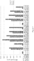
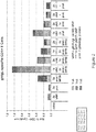
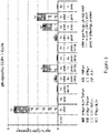
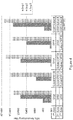
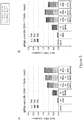

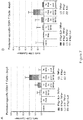


Classifications
-
- A—HUMAN NECESSITIES
- A61—MEDICAL OR VETERINARY SCIENCE; HYGIENE
- A61K—PREPARATIONS FOR MEDICAL, DENTAL OR TOILETRY PURPOSES
- A61K39/00—Medicinal preparations containing antigens or antibodies
- A61K39/12—Viral antigens
- A61K39/155—Paramyxoviridae, e.g. parainfluenza virus
-
- A—HUMAN NECESSITIES
- A61—MEDICAL OR VETERINARY SCIENCE; HYGIENE
- A61K—PREPARATIONS FOR MEDICAL, DENTAL OR TOILETRY PURPOSES
- A61K39/00—Medicinal preparations containing antigens or antibodies
- A61K39/12—Viral antigens
-
- A—HUMAN NECESSITIES
- A61—MEDICAL OR VETERINARY SCIENCE; HYGIENE
- A61K—PREPARATIONS FOR MEDICAL, DENTAL OR TOILETRY PURPOSES
- A61K39/00—Medicinal preparations containing antigens or antibodies
- A61K39/12—Viral antigens
- A61K39/245—Herpetoviridae, e.g. herpes simplex virus
-
- A—HUMAN NECESSITIES
- A61—MEDICAL OR VETERINARY SCIENCE; HYGIENE
- A61P—SPECIFIC THERAPEUTIC ACTIVITY OF CHEMICAL COMPOUNDS OR MEDICINAL PREPARATIONS
- A61P31/00—Antiinfectives, i.e. antibiotics, antiseptics, chemotherapeutics
- A61P31/12—Antivirals
- A61P31/14—Antivirals for RNA viruses
-
- A—HUMAN NECESSITIES
- A61—MEDICAL OR VETERINARY SCIENCE; HYGIENE
- A61P—SPECIFIC THERAPEUTIC ACTIVITY OF CHEMICAL COMPOUNDS OR MEDICINAL PREPARATIONS
- A61P31/00—Antiinfectives, i.e. antibiotics, antiseptics, chemotherapeutics
- A61P31/12—Antivirals
- A61P31/20—Antivirals for DNA viruses
-
- A—HUMAN NECESSITIES
- A61—MEDICAL OR VETERINARY SCIENCE; HYGIENE
- A61K—PREPARATIONS FOR MEDICAL, DENTAL OR TOILETRY PURPOSES
- A61K39/00—Medicinal preparations containing antigens or antibodies
- A61K2039/51—Medicinal preparations containing antigens or antibodies comprising whole cells, viruses or DNA/RNA
- A61K2039/53—DNA (RNA) vaccination
-
- A—HUMAN NECESSITIES
- A61—MEDICAL OR VETERINARY SCIENCE; HYGIENE
- A61K—PREPARATIONS FOR MEDICAL, DENTAL OR TOILETRY PURPOSES
- A61K39/00—Medicinal preparations containing antigens or antibodies
- A61K2039/545—Medicinal preparations containing antigens or antibodies characterised by the dose, timing or administration schedule
-
- A—HUMAN NECESSITIES
- A61—MEDICAL OR VETERINARY SCIENCE; HYGIENE
- A61K—PREPARATIONS FOR MEDICAL, DENTAL OR TOILETRY PURPOSES
- A61K39/00—Medicinal preparations containing antigens or antibodies
- A61K2039/555—Medicinal preparations containing antigens or antibodies characterised by a specific combination antigen/adjuvant
- A61K2039/55505—Inorganic adjuvants
-
- A—HUMAN NECESSITIES
- A61—MEDICAL OR VETERINARY SCIENCE; HYGIENE
- A61K—PREPARATIONS FOR MEDICAL, DENTAL OR TOILETRY PURPOSES
- A61K39/00—Medicinal preparations containing antigens or antibodies
- A61K2039/555—Medicinal preparations containing antigens or antibodies characterised by a specific combination antigen/adjuvant
- A61K2039/55511—Organic adjuvants
- A61K2039/55555—Liposomes; Vesicles, e.g. nanoparticles; Spheres, e.g. nanospheres; Polymers
-
- A—HUMAN NECESSITIES
- A61—MEDICAL OR VETERINARY SCIENCE; HYGIENE
- A61K—PREPARATIONS FOR MEDICAL, DENTAL OR TOILETRY PURPOSES
- A61K39/00—Medicinal preparations containing antigens or antibodies
- A61K2039/555—Medicinal preparations containing antigens or antibodies characterised by a specific combination antigen/adjuvant
- A61K2039/55511—Organic adjuvants
- A61K2039/55566—Emulsions, e.g. Freund's adjuvant, MF59
-
- A—HUMAN NECESSITIES
- A61—MEDICAL OR VETERINARY SCIENCE; HYGIENE
- A61K—PREPARATIONS FOR MEDICAL, DENTAL OR TOILETRY PURPOSES
- A61K39/00—Medicinal preparations containing antigens or antibodies
- A61K2039/555—Medicinal preparations containing antigens or antibodies characterised by a specific combination antigen/adjuvant
- A61K2039/55588—Adjuvants of undefined constitution
-
- C—CHEMISTRY; METALLURGY
- C12—BIOCHEMISTRY; BEER; SPIRITS; WINE; VINEGAR; MICROBIOLOGY; ENZYMOLOGY; MUTATION OR GENETIC ENGINEERING
- C12N—MICROORGANISMS OR ENZYMES; COMPOSITIONS THEREOF; PROPAGATING, PRESERVING, OR MAINTAINING MICROORGANISMS; MUTATION OR GENETIC ENGINEERING; CULTURE MEDIA
- C12N2710/00—MICROORGANISMS OR ENZYMES; COMPOSITIONS THEREOF; PROPAGATING, PRESERVING, OR MAINTAINING MICROORGANISMS; MUTATION OR GENETIC ENGINEERING; CULTURE MEDIA dsDNA viruses
- C12N2710/00011—Details
- C12N2710/16011—Herpesviridae
- C12N2710/16111—Cytomegalovirus, e.g. human herpesvirus 5
- C12N2710/16134—Use of virus or viral component as vaccine, e.g. live-attenuated or inactivated virus, VLP, viral protein
-
- C—CHEMISTRY; METALLURGY
- C12—BIOCHEMISTRY; BEER; SPIRITS; WINE; VINEGAR; MICROBIOLOGY; ENZYMOLOGY; MUTATION OR GENETIC ENGINEERING
- C12N—MICROORGANISMS OR ENZYMES; COMPOSITIONS THEREOF; PROPAGATING, PRESERVING, OR MAINTAINING MICROORGANISMS; MUTATION OR GENETIC ENGINEERING; CULTURE MEDIA
- C12N2760/00—MICROORGANISMS OR ENZYMES; COMPOSITIONS THEREOF; PROPAGATING, PRESERVING, OR MAINTAINING MICROORGANISMS; MUTATION OR GENETIC ENGINEERING; CULTURE MEDIA ssRNA viruses negative-sense
- C12N2760/00011—Details
- C12N2760/18011—Paramyxoviridae
- C12N2760/18511—Pneumovirus, e.g. human respiratory syncytial virus
- C12N2760/18534—Use of virus or viral component as vaccine, e.g. live-attenuated or inactivated virus, VLP, viral protein
-
- C—CHEMISTRY; METALLURGY
- C12—BIOCHEMISTRY; BEER; SPIRITS; WINE; VINEGAR; MICROBIOLOGY; ENZYMOLOGY; MUTATION OR GENETIC ENGINEERING
- C12N—MICROORGANISMS OR ENZYMES; COMPOSITIONS THEREOF; PROPAGATING, PRESERVING, OR MAINTAINING MICROORGANISMS; MUTATION OR GENETIC ENGINEERING; CULTURE MEDIA
- C12N2770/00—MICROORGANISMS OR ENZYMES; COMPOSITIONS THEREOF; PROPAGATING, PRESERVING, OR MAINTAINING MICROORGANISMS; MUTATION OR GENETIC ENGINEERING; CULTURE MEDIA ssRNA viruses positive-sense
- C12N2770/00011—Details
- C12N2770/36011—Togaviridae
- C12N2770/36111—Alphavirus, e.g. Sindbis virus, VEE, EEE, WEE, Semliki
- C12N2770/36141—Use of virus, viral particle or viral elements as a vector
- C12N2770/36143—Use of virus, viral particle or viral elements as a vector viral genome or elements thereof as genetic vector
Definitions
- Nucleic acid based vaccines are an attractive approach to vaccination.
- intramuscular (IM) immunization of plasmid DNA encoding an antigen can induce cellular and humoral immune responses and protect against challenge.
- DNA vaccines offer certain advantages over traditional vaccines using protein antigens, or attenuated pathogens.
- DNA vaccines can be more effective in producing a properly folded antigen in its native conformation, and in generating a cellular immune response.
- DNA vaccines also do not have some of the safety problems associated with killed or attenuated pathogens.
- a killed viral preparation may contain residual live viruses, and an attenuated virus may mutate and revert to a pathogenic phenotype.
- DNA vaccines are generally effective in generating cell mediated immunity (such as interferon-y secreting antigen-specific T-cells and antigen-specific cytotoxic T-cells), but less effective in generating antibodies against the encoded and expressed antigen.
- WO 99/30733 discloses a method of enhancing immune response to a nucleic acid vaccine by simultaneous administration of the protein that is encoded by the nucleic acid.
- the two components do not need to be administered in the same composition. Both components need to be administered during the induction phase of the immune response, with the protein preferably being masked or held back until after the nucleic acid has primed the immune system.
- the vaccine comprised naked DNA and naked protein antigen in physical admixture.
- the protein antigen was formulated for delayed release in a biodegradable polymer-alum formulation admixed with naked DNA.
- WO 97/28818 discloses a vaccine that delivers a nucleic acid and a protein antigen to antigen presenting cells.
- the nucleic acid may encode the same protein as the protein antigen.
- the nucleic acid and protein are "complexed," e.g., by covalent conjugation.
- the complex may be formulated as a synthetic virus-like particle. It is also suggested that liposomal systems may be used.
- U.S. Pat. No. 7,604,803 discloses the co-delivery of nucleic acid and its encoded protein to the same cell using a liposomal system.
- the DNA molecule and its encoded protein are entrapped within the same liposomal vehicle, such that the two entities arrive at antigen-presenting cells together, resulting in the processing and presentation of the protein form of the antigen, together with the expression of the DNA-encoded form of the antigen in the same cell.
- WO 2009/156852 discloses a prime/boost approach to raise an immune response in a subject.
- the method comprises: (i) administering at least one dose of a priming immunogenic composition to the subject, to elicit a primary immune response against a pathogen; and (ii) administering a boosting immunogenic composition to the subject, to elicit, within 21 days of its administration or sooner, a protective anamnestic immune response against the pathogen.
- WO 2009/074861 discloses a vaccine comprising (i) a nucleic acid sequence encoding at least one influenza virus antigen coated onto carrier particles, and (ii) an assistor protein for sequential or concomitant administration.
- the assistor protein and the antigen encoded by the nucleic acid molecule share at least one common epitope.
- WO 2006/061643 discloses a prime-boost vaccination method using herpes viral vectors, in particular, a heterologous prime-boost regimen using two different non-replicating viral vectors, one of which is a herpes virus vector.
- the two vectors share a common epitope that is associated with a target antigen or disease.
- WO 2010/036948 discloses the use of a priming composition and a boosting composition to prime and boost an immune response.
- the priming composition comprises a DNA plasmid that comprises a nucleic acid molecule encoding an influenza virus hemagglutinin (HA) or an epitope-bearing domain thereof.
- the boosting composition comprises an influenza vaccine.
- non-coding plasmid DNA has an immuno-adjuvant action when co-entrapped with peptides in liposomal vesicles ( Gursel, M. et al. Vaccine (1999) 17: 1376-1383 ) and that DNA with CpG motifs has an adjuvant effect on naked DNA and peptide vaccines ( Klinman, D. M. et al. Vaccine (1999) 17: 19-25 ).
- DNA-based vaccines have been raised regarding the safety of DNA-based vaccines.
- the introduced DNA molecules could potentially integrate into the host genome or, due to their distribution to various tissues, could lead to undesirable sustained expression of antigens.
- certain DNA viruses have also been used to deliver DNA molecules. Because of their infectious properties, such viruses achieve a very high transfection rate.
- the viruses used are genetically modified to prevent the formation of functional infectious particles in the transfected cell. Despite these precautions, however, it is not possible to rule out the risk of uncontrolled propagation of the introduced gene and viral genes, for example due to potential recombination events. This also entails the risk of the DNA being inserted into an intact gene of the host cell's genome by e.g.
- RNA molecules encoding an antigen or a derivative thereof may also be used as vaccines.
- RNA vaccines offer certain advantages as compared to DNA vaccines. However, compared with DNA-based vaccines, relatively minor attention has been given to RNA-based vaccines. RNAs are highly susceptible to degradation by nucleases when administered as a therapeutic or vaccine. Additionally, RNAs are not actively transported into cells. See, e.g., Vajdy, M., et al., Mucosal adjuvants and delivery systems for protein-, DNA- and RNA-based vaccines, Immunol Cell Biol, 2004. 82(6): p. 617-27 .
- TLRs Toll-like receptors
- PAMPS pathogen-associated molecular patterns
- Many TLRs have been identified in humans, mice, and other mammalian species.
- DNA molecules such as bacterial or viral DNA
- RNA molecules such as single stranded viral RNA
- U.S. Pat. No. 7,862,829 discloses a method of producing an immune response by administering an antigen and an alphavirus-based adjuvant.
- the method is based on the discovery that alphavirus, a (+)ssRNA virus, can act as an adjuvant to enhance an immune response against an antigen, even though the antigen is not presented on or expressed by the virus.
- the alphavirus particles may be delivered by liposomal system.
- This invention generally relates to immunogenic compositions that comprise an RNA component and a polypeptide component.
- Immunogenic compositions that deliver antigenic epitopes in two different forms - a first epitope from a pathogen, in RNA-coded form; and a second epitope from the same pathogen, in polypeptide form - can enhance the immune response to the pathogen, as compared to immunization with RNA alone, or polypeptide alone.
- the first epitope and the second epitope are the same epitope.
- the invention also relates to a kit comprising an RNA-based priming composition and a polypeptide-based boosting composition for sequential administration.
- the kit is suitable for, for example, a "RNA prime, protein boost" immunization regimen to generate an immune response to a pathogen.
- the invention also relates to methods for treating or preventing an infectious disease, methods for inducing an immune response, or methods of vaccinating a subject, by co-delivery of an RNA molecule and a polypeptide molecule (co-administration).
- the invention also relates to methods for treating or preventing an infectious disease, methods for inducing an immune response, or methods of vaccinating a subject, by sequential administration of an RNA molecule and a polypeptide molecule (prime-boost).
- the invention provides an immunogenic composition comprising: (i) a self-replicating RNA molecule that encodes a first polypeptide antigen comprising a first epitope; and (ii) a second polypeptide antigen comprising a second epitope; wherein the first and second epitope are epitopes from the same pathogen.
- the first and and second epitopes can be the same epitope.
- the first and second epitopes can be different epitopes.
- the first polypeptide antigen and the second polypeptide antigen are substantially the same.
- the first polypeptide antigen can be a soluble or membrane anchored polypeptide and the second polypeptide antigen can be a soluble polypeptide.
- the first polypeptide antigen is a fusion polypeptide that further comprises a third epitope from a different pathogen.
- the second polypeptide antigen is a fusion polypeptide that further comprises a third epitope from a different pathogen.
- the first and second epitopes can be epitopes from the same subspecies of the pathogen.
- the self-replicating RNA can be an alphavirus-derived RNA replicon.
- the self-replicating RNA molecule can comprise one or more modified nucleotides.
- the immunogenic composition further comprises a cationic lipid, a liposome, a cochleate, a virosome, an immune-stimulating complex, a microparticle, a microsphere, a nanosphere, a unilamellar vesicle, a multilamellar vesicle, an oil-in-water emulsion, a water-in-oil emulsion, an emulsome, a polycationic peptide, or a cationic nanoemulsion.
- the RNA molecule is encapsulated in, bound to or adsorbed on a cationic lipid, a liposome, a cochleate, a virosome, an immune-stimulating complex, a microparticle, a microsphere, a nanosphere, a unilamellar vesicle, a multilamellar vesicle, an oil-in-water emulsion, a water-in-oil emulsion, an emulsome, a polycationic peptide, a cationic nanoemulsion or combinations thereof.
- the pathogen is a virus
- the first polypeptide antigen and second polypeptide antigen are viral antigens.
- the viral antigens can be RSV-F antigens.
- the RSV-F antigens comprise an amino acid sequence selected from SEQ ID NOs:25-40.
- the viral antigens can be from Cytomegalovirus (CMV).
- CMV antigens are independently selected from the group consisting of a gB antigen, a gH antigen, a gL antigen, a gM antigen, a gN antigen, a gO antigen, a UL128 antigen, a UL129 antigen, and a UL130 antigen.
- the immunogenic composition further comprises an adjuvant.
- the adjuvant can be MF59.
- the invention also relates to pharmaceutical compositions that comprise an immunogenic composition as described herein and a pharmaceutically acceptable carrier and/or a pharmaceutically acceptable vehicle.
- the invention also relates to methods for treating or preventing an infectious disease comprising administering to a subject in need thereof a therapeautically effective amount of a composition as described herein.
- the invention also relates to methods of inducing an immune response in a subject comprising administering to a subject in need thereof a therapeutically effective amount of a composition as described herein.
- the invention also relates to methods of vaccinating a subject, comprising administering to a subject in need thereof a composition as described herein.
- kits comprising: (i) a priming composition comprising a self-replicating RNA molecule that encodes a first polypeptide antigen that comprises a first epitope from a pathogen; and (ii) a boosting composition comprising a second polypeptide antigen that comprises a second epitope from the pathogen.
- the first and second polypeptide antigens can be substantially the same.
- the first polypeptide antigen can be a soluble or membrane anchored polypeptide, and the second polypeptide antigen can be a soluble polypeptide.
- the first polypeptide antigen can be a fusion polypeptide.
- the second polypeptide antigen can be a fusion polypeptide.
- the self-replicating RNA can be an alphavirus-derived RNA replicon.
- the self-replicating RNA molecule can comprise one or more modified nucleotides.
- the priming composition of the kit further comprises a cationic lipid, a liposome, a cochleate, a virosome, an immune-stimulating complex, a microparticle, a microsphere, a nanosphere, a unilamellar vesicle, a multilamellar vesicle, an oil-in-water emulsion, a water-in-oil emulsion, an emulsome, a polycationic peptide, or a cationic nanoemulsion.
- the RNA molecule of the kit is encapsulated in, bound to or adsorbed on a cationic lipid, a liposome, a cochleate, a virosome, an immune-stimulating complex, a microparticle, a microsphere, a nanosphere, a unilamellar vesicle, a multilamellar vesicle, an oil-in-water emulsion, a water-in-oil emulsion, an emulsome, a polycationic peptide, a cationic nanoemulsion or combinations thereof.
- the pathogen of the kit is a virus
- the first polypeptide antigen and second polypeptide antigen are viral antigens.
- the viral antigens can be from respiratory syncytial virus (RSV).
- the viral antigens can be a RSV-F antigen.
- the RSV-F antigens comprise an amino acid sequence selected from SEQ ID NOs:25-40.
- the viral antigens are from Cytomegalovirus (CMV).
- CMV antigens are independently selected from the group consisting of a gB antigen, a gH antigen, a gL antigen, a gM antigen, a gN antigen, a gO antigen, a UL128 antigen, a UL129 antigen, and a UL130 antigen.
- the priming composition of the kit, the boosting composition of the kit, or both comprise an adjuvant.
- the adjuvant can be MF59.
- the priming composition, the boosting composition, or both can comprise a pharmaceutically acceptable carrier and/or a pharmaceutically acceptable vehicle.
- the invention also relates to methods for treating or preventing an infectious disease comprising: (i) administering to a subject in need thereof at least once a therapeutically effective amount of a priming composition comprising a self-replicating RNA molecule that encodes a first polypeptide antigen that comprises a first epitope from a pathogen; and subsequently administering the subject at least once a therapeutically effective amount of a boosting composition comprising a second polypeptide antigen that comprises a second epitope from said pathogen; wherein said first epitope and second epitope are the same epitope.
- the invention also relates to methods for inducing an immune response in a subject comprising (i) administering to a subject in need thereof at least once a therapeutically effective amount of a priming composition comprising a self-replicating RNA molecule that encodes a first polypeptide antigen that comprises a first epitope from a pathogen; and subsequently administering the subject at least once a therapeutically effective amount of a boosting composition comprising a second polypeptide antigen that comprises a second epitope from said pathogen; wherein said first epitope and second epitope are the same epitope.
- the invention also relates to methods for vaccinating a subject, comprising administering to a subject in need thereof at least once a therapeutically effective amount of a priming composition comprising a self-replicating RNA molecule that encodes a first polypeptide antigen that comprises a first epitope from a pathogen; and subsequently administering the subject at least once a therapeutically effective amount of a boosting composition comprising a second polypeptide antigen that comprises a second epitope from said pathogen; wherein said first epitope and second epitope are the same epitope.
- a priming composition comprising a self-replicating RNA molecule that encodes a first polypeptide antigen that comprises a first epitope from a pathogen
- a boosting composition comprising a second polypeptide antigen that comprises a second epitope from said pathogen
- the first polypeptide antigen and the second polypeptide antigen are substantially the same.
- the first polypeptide antigen can be a soluble or membrane anchored polypeptide and the second polypeptide antigen can be a soluble polypeptide.
- the first polypeptide antigen is a fusion polypeptide that further comprises a third epitope from a different pathogen.
- the second polypeptide antigen is a fusion polypeptide that further comprises a third epitope from a different pathogen.
- the first and second epitopes can be epitopes from the same subspecies of the pathogen.
- the self-replicating RNA can be an alphavirus-derived RNA replicon.
- the self-replicating RNA molecule can comprise one or more modified nucleotides.
- the immunogenic composition further comprises a cationic lipid, a liposome, a cochleate, a virosome, an immune-stimulating complex, a microparticle, a microsphere, a nanosphere, a unilamellar vesicle, a multilamellar vesicle, an oil-in-water emulsion, a water-in-oil emulsion, an emulsome, a polycationic peptide, or a cationic nanoemulsion.
- the RNA molecule is encapsulated in, bound to or adsorbed on a cationic lipid, a liposome, a cochleate, a virosome, an immune-stimulating complex, a microparticle, a microsphere, a nanosphere, a unilamellar vesicle, a multilamellar vesicle, an oil-in-water emulsion, a water-in-oil emulsion, an emulsome, a polycationic peptide, a cationic nanoemulsion or combinations thereof.
- the pathogen is a virus
- the first polypeptide antigen and second polypeptide antigen are viral antigens.
- the viral antigens can be RSV-F antigens.
- the RSV-F antigens comprise an amino acid sequence selected from SEQ ID NOs:25-40.
- the viral antigens can be from Cytomegalovirus (CMV).
- CMV antigens are independently selected from the group consisting of a gB antigen, a gH antigen, a gL antigen, a gM antigen, a gN antigen, a gO antigen, a UL128 antigen, a UL129 antigen, and a UL130 antigen.
- the immunogenic composition further comprises an adjuvant.
- the adjuvant can be MF59.
- the invention also relates to pharmaceutical compositions that comprise an immunogenic composition as described herein and a pharmaceutically acceptable carrier and/or a pharmaceutically acceptable vehicle.
- RNA molecules are self-adjuvanting.
- RNA molecules formulated in liposomes
- cytokines including IFN- ⁇ , IP-10 (CXCL-10), IL-6, KC (CXCL1), IL-5, IL-13, MCP-1, and MIP- ⁇
- the cytokines can enhance the host immune response to the protein antigen that was encoded by the RNA molecule.
- RNA molecules and a polypeptide molecule e.g., administering an immunogenic composition that has an RNA component and a protein component; or sequential administration regimens such as "RNA prime, protein boost"
- the polypeptide molecule can enhance total antibody titers in the host, while the RNA molecule can enhance the production of antibodies that recognize an antigen in its native structure.
- the combination can induce an antibody response with an enhanced ratio of functional antibodies (e.g., neutralizing antibodies) to total antibodies.
- RNA molecules promote type 1 T helper responses (Th1, IFN- ⁇ hi , IL-4 lo ), whereas protein molecules promote type 2 T helper responses.
- RNA molecules may be delivered to cells using delivery systems such as liposomes or oil-in-water emulsions. Liposomes and oil-in-water emulsions are also known to have adjuvant activities. Thus, the adjuvant activity of the RNA together with adjuvant activity of the delivery system can act synergistically to enhance the immune response to an antigen.
- multivalency may be achieved by combining a polypeptide antigen with an RNA that encodes a different antigen from the same pathogen.
- the invention relates to immunogenic compositions that comprise an RNA component and a polypeptide component.
- Immunogenic compositions that deliver antigenic epitopes in two different forms - a first epitope from a pathogen, in RNA-coded form; and a second epitope from the same pathogen, in polypeptide form - can enhance the immune response to the pathogen.
- the first epitope and the second epitope are the same epitope (i.e., the first antigen, in RNA-coded form, and the second antigen, in polypeptide form, share at least one common epitope).
- the RNA component of the immunogenic composition can encode a protein that is substantially the same as the polypeptide component of the immunogenic composition (e.g., the amino acid sequence encoded by the RNA molecule and the polypeptide component of the immunogenic composition share at least about 90% sequence identity across the length of the shorter antigen).
- the two antigens have the same epitope, such as the same immunodominant epitope(s).
- RNA molecule that encodes a viral antigen As described herein, the inventors have evaluated the efficacies of immunogenic compositions that comprise (i) a self-replicating RNA molecule that encodes a viral antigen, and (ii) the viral antigen in polypeptide form.
- Viral antigens that were used in these studies include HIV gp140 and RSV-F antigens. The results demonstrated that coadministering an RNA molecule that encodes a viral antigen, together with the viral antigen in polypeptide form, potentiated the immune response to the antigen, resulting in higher antibody titers as compared to administering the RNA molecule alone.
- the immunogenic compositions described herein can be formulated as a vaccine to induce or enhance the host immune response to a pathogen. Also provided herein are methods of using the immunogenic compositions of the invention to induce or enhance an immune response in a subject in need thereof.
- the invention in another aspect, relates to a kit comprising: (i) a priming composition comprising a self-replicating RNA molecule that encodes a first polypeptide antigen that comprises a first epitope, and (ii) a boosting composition comprising a second polypeptide antigen that comprises a second epitope; wherein said first epitope and second epitope are the same epitope ( i.e., the first antigen, in RNA-coded form, and the second antigen, in polypeptide form, share at least one common epitope).
- the kit may be used for sequential administration of the priming and the boosting compositions.
- the invention in another aspect, relates to a method for treating or preventing an infectious disease, a method for inducing an immune response in a subject, or a method of vaccinating a subject, comprising: (i) administering to a subject in need thereof at least once a therapeutically effective amount of a priming composition comprising a self-replicating RNA molecule that encodes a first polypeptide antigen that comprises a first epitope, and (ii) subsequently administering the subject at least once a therapeutically effective amount of a boosting composition comprising a second polypeptide antigen that comprises a second epitope; wherein said first epitope and second epitope are the same epitope ( i.e., the first antigen, in RNA-coded form, and the second antigen, in polypeptide form, share at least one common epitope).
- RNA prime, protein boost vaccination strategies As described herein, the inventors have evaluated RNA prime, protein boost vaccination strategies. These studies demonstrate several benefits of the RNA prime, protein boost strategy, as compared to a protein prime, protein boost strategy, including, for example, increased antibody titers, a more balanced IgG 1 :IgG 2a subtype profile, induction of T H 1 type, CD4+ T cell-mediated immune response that was similar to that of viral particles, and reduced production of non-neutralizing antibodies.
- the RNA molecule in the priming composition encodes a protein that is substantially the same as the polypeptide molecule in the boosting composition (e.g., the amino acid sequence encoded by the RNA molecule in the priming composition and the polypeptide in the boosting composition share at least about 90% sequence identity across the length of the shorter antigen).
- the two antigens have the same epitope, such as the same immunodominant epitope(s).
- the priming and boosting compositions described herein can be formulated as a vaccine to induce or enhance the immune response to a pathogen. Also provided herein are methods of using the priming and boosting compositions of the invention to induce or enhance an immune response in a subject in need thereof.
- the invention also relates to immunogenic compositions, pharmaceutical compositions, or kits as described herein for use in therapy, and to the use of immunogenic compositions, pharmaceutical compositions, or kits as described herein for the manufacture of a medicament for enhancing or generating an immune response.
- the invention provides an immunogenic composition comprising an RNA component and a polypeptide component.
- the immunogenic composition comprises: (i) a self-replicating RNA molecule that encodes a first polypeptide antigen comprising a first epitope (the RNA component); and (ii) a second polypeptide antigen comprising a second epitope (the polypeptide component); wherein said first epitope and second epitope are epitopes from the same pathogen.
- the first epitope and second epitope can be the same epitope, or different epitopes if desired.
- the first epitope and second epitope can be from the same polypeptide of the pathogen, or different polypeptides of the pathogen.
- the first epitope and second epitope can also be epitopes which are highly conserved between different strains or subspecies of the pathogen, such as those epitopes with limited or no mutational variations.
- the first epitope and the second epitope can be different epitopes from the same pathogen (e.g., the first epitope is from RSV F and the second epitope is from RSV G).
- the first polypeptide antigen and the second polypeptide antigen are derived from the same protein from the pathogen.
- the RNA molecule may encode a first polypeptide antigen comprising a full-length protein from a pathogen (e.g., a viral protein), or an antigenic portion thereof, optionally fused with a heterologous sequence that may facilitate the expression, production, purification or detection of the viral protein encoded by the RNA.
- the second polypeptide antigen may be a recombinant protein comprising the full-length protein, or an antigenic portion thereof, optionally fused with a heterologous sequence (e.g., His-tag) that may facilitate the expression, production, purification or detection of the second polypeptide antigen.
- the first polypeptide antigen, the second polypeptide antigen, or both may comprise a mutation variant of a protein from a pathogen (e.g., a viral protein having amino acid substitution(s), addition(s), or deletion(s)).
- a pathogen e.g., a viral protein having amino acid substitution(s), addition(s), or deletion(s)
- the amino acid sequence identity between the first polypeptide antigen and the second polypeptide antigen is at least about 40%, least about 50%, least about 60%, least about 65%, least about 70%, at least about 75%, at least about 80%, at least about 85%, at least about 90%, at least about 95%, at least about 96%, at least about 97%, at least about 98%, or at least about 99%.
- the first polypeptide antigen and the second polypeptide antigen are the same antigen.
- the first polypeptide antigen and the polypeptide second antigen share at least 1, at least 2, at least 3, at least 4, or at least 5 common B-cell or T-cell epitopes. In certain embodiments, the first polypeptide antigen and the second polypeptide antigen have at least one common immunodominant epitope. In certain embodiments, the first polypeptide antigen and the second polypeptide antigen have the same immunodominant epitope(s), or the same primary immunodominant epitope.
- the first polypeptide antigen is a soluble or membrane anchored polypeptide
- the second polypeptide antigen is a soluble polypeptide.
- the wild type viral protein is a transmembrane surface protein
- the RNA molecule may comprise the full-length coding sequence to produce the first (membrane-anchored) antigen, while the transmembrane region of the viral protein may be deleted to produce the second polypeptide antigen (which is soluble).
- the first antigen or the second antigen is a fusion polypeptide further comprising a third epitope.
- the third epitope may be from a different pathogen, or from a different antigen of the same pathogen.
- Antigens suitable for inclusion in the immunogenic compositions described herein may be derived from any pathogen (e.g., a bacterial pathogen, a viral pathogen, a fungal pathogen, a protozoan pathogen, or a multi-cellular parasitic pathogen), allergen or tumor.
- pathogen e.g., a bacterial pathogen, a viral pathogen, a fungal pathogen, a protozoan pathogen, or a multi-cellular parasitic pathogen
- the first and second antigens are derived from a viral pathogen.
- viral pathogens include, e.g., respiratory syncytial virus (RSV), hepatitis B virus (HBV), hepatitis C virus (HCV), Dengue virus, herpes simplex virus (HSV; e.g., HSV-I, HSV-II), molluscum contagiosum virus, vaccinia virus, variola virus, lentivirus, human immunodeficiency virus (HIV), human papilloma virus (HPV), cytomegalovirus (CMV), varicella zoster virus (VZV), rhinovirus, enterovirus, adenovirus, coronavirus (e.g., SARS), influenza virus (flu), para-influenza virus, mumps virus, measles virus, papovavirus, hepadnavirus, flavivirus, retrovirus, arenavirus (e.g., Lymphocytic Chor
- the first and second antigens are derived from a virus which infects fish, such as: infectious salmon anemia virus (ISAV), salmon pancreatic disease virus (SPDV), infectious pancreatic necrosis virus (IPNV), channel catfish virus (CCV), fish lymphocystis disease virus (FLDV), infectious hematopoietic necrosis virus (IHNV), koi herpesvirus, salmon picorna-like virus (also known as picorna-like virus of atlantic salmon), landlocked salmon virus (LSV), atlantic salmon rotavirus (ASR), trout strawberry disease virus (TSD), coho salmon tumor virus (CSTV), or viral hemorrhagic septicemia virus (VHSV).
- infectious salmon anemia virus ISAV
- SPDV salmon pancreatic disease virus
- IPNV infectious pancreatic necrosis virus
- CCV channel catfish virus
- FLDV fish lymphocystis disease virus
- IHNV infectious hematopoietic necrosis virus
- the first and second antigens are derived from a parasite from the Plasmodium genus, such as P.falciparum, P.vivax, P.malariae or P.ovale.
- the invention may be used for immunising against malaria.
- the first and second antigens are derived from a parasite from the Caligidae family, particularly those from the Lepeophtheirus and Caligus genera e.g. sea lice such as Lepeophtheirus salmonis or Caligus rogercresseyi.
- first and second antigens are derived from a bacterial pathogen.
- bacterial pathogens include, e.g., Neisseria spp, including N. gonorrhea and N. meningitides; Streptococcus spp, including S. pneumoniae, S. pyogenes, S. agalactiae, S. mutans; Haemophilus spp, including H. influenzae type B, non typeable H. influenzae, H. ducreyi; Moraxella spp, including M. catarrhalis, also known as Branhamella catarrhalis; Bordetella spp, including B. pertussis, B. parapertussis and B.
- bronchiseptica Mycobacterium spp., including M. tuberculosis, M. bovis, M. leprae, M. avium, M. paratuberculosis, M. smegmatis; Legionella spp, including L. pneumophila; Escherichia spp, including enterotoxic E. coli, enterohemorragic E. coli, enteropathogenic E. coli; Vibrio spp, including V. cholera, Shigella spp, including S. sonnei, S. dysenteriae, S. flexnerii; Yersinia spp, including Y. enterocolitica, Y. pestis, Y.
- pseudotuberculosis Campylobacter spp, including C. jejuni and C. coli
- Salmonella spp including S. typhi, S. paratyphi, S. choleraesuis, S. enteritidis
- Listeria spp. including L. monocytogenes
- Helicobacter spp including H pylori
- Pseudomonas spp including P. aeruginosa
- Staphylococcus spp. including S. aureus, S. epidermidis
- Enterococcus spp. including E. faecalis, E. faecium
- Clostridium spp. including C.
- first and second antigens are derived from a fungal pathogen (e.g., a yeast or mold pathogen).
- exemplary fungal pathogens include, e.g., Aspergillus fumigatus, A. flavus, A. niger, A. terreus, A. nidulans, Coccidioides immitis, Coccidioides posadasii, Cryptococcus neoformans, Histoplasma capsulatum, Candida albicans, and Pneumocystis jirovecii.
- first and second antigens are derived from a protozoan pathogen.
- protozoan pathogens include, e.g., Toxoplasma gondii and Strongyloides stercoralis.
- the first and second antigens are derived from a multi-cellular parasitic pathogen.
- exemplary multicellular parasitic pathogens include, e.g., trematodes (flukes), cestodes (tapeworms), nematodes (roundworms), and arthropods.
- the first and second antigens are derived from an allergen, such as pollen allergens (tree-, herb, weed-, and grass pollen allergens); insect or arachnid allergens (inhalant, saliva and venom allergens, e.g. mite allergens, cockroach and midges allergens, hymenopthera venom allergens); animal hair and dandruff allergens (from e.g. dog, cat, horse, rat, mouse, etc .); and food allergens (e.g. a gliadin).
- pollen allergens tree-, herb, weed-, and grass pollen allergens
- insect or arachnid allergens inhalant, saliva and venom allergens, e.g. mite allergens, cockroach and midges allergens, hymenopthera venom allergens
- animal hair and dandruff allergens from e.g
- Important pollen allergens from trees, grasses and herbs are such originating from the taxonomic orders of Fagales, Oleales, Pinales and platanaceae including, but not limited to, birch (Betula), alder (Alnus), hazel (Corylus), hornbeam (Carpinus) and olive (Olea), cedar (Cryptomeria and Juniperus), plane tree (Platanus), the order of Poales including grasses of the genera Lolium, Phleum, Poa, Cynodon, Dactylis, Holcus, Phalaris, Secale, and Sorghum, the orders of Asterales and Urticales including herbs of the genera Ambrosia, Artemisia, and Parietaria.
- venom allergens including such originating from stinging or biting insects such as those from the taxonomic order of Hymenoptera including bees ( Apidae ), wasps ( Vespidea ) , and ants ( Formicoidae ).
- the first and second antigens are derived from a tumor antigen selected from: (a) cancer-testis antigens such as NY-ESO-1, SSX2, SCP1 as well as RAGE, BAGE, GAGE and MAGE family polypeptides, for example, GAGE-1, GAGE-2, MAGE-1, MAGE-2, MAGE-3, MAGE-4, MAGE-5, MAGE-6, and MAGE-12 (which can be used, for example, to address melanoma, lung, head and neck, NSCLC, breast, gastrointestinal, and bladder tumors; (b) mutated antigens, for example, p53 (associated with various solid tumors, e.g., colorectal, lung, head and neck cancer), p21/Ras (associated with, e.g., melanoma, pancreatic cancer and colorectal cancer), CDK4 (associated with, e.g., melanoma), MUM1 (associated with, e.g., melanoma),
- tumor immunogens include, but are not limited to, p15, Hom/Mel-40, H-Ras, E2A-PRL, H4-RET, IGH-IGK, MYL-RAR, Epstein Barr virus antigens, EBNA, human papillomavirus (HPV) antigens, including E6 and E7, hepatitis B and C virus antigens, human T-cell lymphotropic virus antigens, TSP-180, p185erbB2, p180erbB-3, c-met, mn-23H1, TAG-72-4, CA 19-9, CA 72-4, CAM 17.1, NuMa, K-ras, p16, TAGE, PSCA, CT7, 43-9F, 5T4, 791 Tgp72, beta-HCG, BCA225, BTAA, CA 125, CA 15-3 (CA 27.29 ⁇ BCAA), CA 195, CA 242, CA-50, CAM43, CD68 ⁇ KP1,
- the pathogen is RSV.
- RSV is an enveloped non-segmented negative-strand RNA virus in the family Paramyxoviridae, genus Pneumovirus.
- paramyxoviruses such as RSV, like other enveloped viruses such as influenza virus and HIV, require fusion of the viral membrane with a host cell's membrane.
- the conserved fusion protein (RSV-F glycoprotein) fuses the viral and cellular membranes by coupling irreversible protein refolding with juxtaposition of the membranes. In current models based on paramyxovirus studies, the RSV-F protein initially folds into a metastable "pre-fusion" conformation.
- pre-fusion conformation undergoes refolding and conformational changes to its stable "post-fusion” conformation. See, also, Swanson et al., PNAS USA 108(23):9619-9624 (2011 ) regarding pre-fusion and post-fusion RSV-F structures.
- the first and second antigens are from RSV.
- the first and second antigens can independently be derived from the RSV surface glycoproteins Fusion (F), Glycoprotein (G), Small Hydrophobic protein (SH), the matrix proteins M and M2, the nucleocapsid proteins N, P and L, and the nonstructural proteins NS1 and NS2.
- the first and second antigens are each an RSV-F antigen.
- the F glycoprotein of RSV is a type I single-pass integral membrane protein having four general domains: N-terminal ER-translocating signal sequence (SS), ectodomain (ED), transmembrane domain (TM), and a cytoplasmic tail (CT).
- SS N-terminal ER-translocating signal sequence
- ED ectodomain
- TM transmembrane domain
- CT cytoplasmic tail
- the RSV-F glycoprotein is translated from mRNA into an approximately 574 amino acid protein designated F 0 .
- Post-translational processing of F 0 includes removal of an N-terminal signal peptide by a signal peptidase in the endoplasmic reticulum.
- F 0 is also cleaved at two sites (approximately 109/110 and approximately 136/137) by cellular proteases (in particular furin) in the trans-Golgi.
- F 1 contains a hydrophobic fusion peptide at its N-terminus and also two amphipathic heptad-repeat regions (HRA and HRB). HRA is near the fusion peptide and HRB is near the transmembrane domain.
- HRA is near the fusion peptide and HRB is near the transmembrane domain.
- Three F 1 -F 2 heterodimers are assembled as homotrimers of F 1 -F 2 in the virion.
- RSV-F antigens suitable for inclusion in the immunogenic compositions described herein, either in RNA enconded form or as polypeptides include RSV-F glycoprotein and RSV-F glycoprotein variants.
- Suitable RSV-F glycoprotein variants include, for example, full length F protein and truncated variants such as soluble ecto-domains, each optionally containing one or more mutations, such as furin-cleavage mutations, trypsin-cleavage mutations, fusion peptide mutations (e.g., deletions in whole or in part), mutations that stabilize the HRB trimer, and mutations that destabilize the HRA trimer.
- sequence of amino acid residue 100-150 of the RSV-F glycoprotein such as SEQ ID NO:1 or SEQ ID NO:2 disclosed in WO2011/008974 , or the soluble ecto domains thereof, is:
- sequence of the RSV-F antigen comprises:
- the sequences presented above contain a signal peptide and a HIS tag.
- the RSV-F protein used in the invention can contain any of the amino acid sequences shown above, with or without the signal peptide and/or HIS tag.
- RSV F antigens examples include SEQ ID NOS: 6, 8 and 10 as disclosed in WO2009/079796 at pages 58-60, which are incorporated herein by reference.
- the RSV-F polypeptide suitable for inclusion in the immunogenic compositions described herein may be in any desired form and conformation, including any desired mixture of forms and conformations.
- the RSV-F polypeptide can be a monomer, or can be a trimer comprising three monomer polypeptides. Trimers can be monodispersed or can be in the form of a rosette, for example, due to interactions between the fusion peptides of individual timers.
- the immunogenic compositions may comprise RSV-F polypeptides that are monomers, trimers, a combination of monomers and trimers (e.g., in dynamic equilibrium), rosettes of trimers, and any combination of the foregoing.
- RSV-F forms and conformations of RSV-F, including monomers, trimers, a combination of monomers and trimers (e.g., in dynamic equilibrium), rosettes of trimers, cleaved and uncleaved forms, and pre-fusion and post-fusion forms are well known in the art, and are disclosed for example in WO2011/008974 , the disclosure of which is incorporated herein by reference in its entirety.
- the skilled addressee is directed WO2011/008974 , in particular at page 24, line 10 through page 26 line 27 and Examples 2-7, which RSV-F proteins in a variety of forms and conformations and methods for making them.
- the pathogen is CMV
- the first and second antigens are independently derived from CMV.
- the first and second antigens are derived from a capsid protein, an envelope glycoprotein (such as gB, gH, gL, gM, gN), or a tegument protein.
- the first and second antigens are derived from one or more of the following proteins: pp65, IE1, gB, gD, gH, gL, gM, gN, gO, UL128, UL129, gUL130, UL150, UL131, UL33, UL78, US27, US28, RL5A, RL6, RL10, RL11, RL12, RL13, UL1, UL2, UL4, UL5, UL6, UL7, UL8, UL9, UL10, UL11, UL14, UL15A, UL16, UL17, UL18, UL22A, UL38, UL40, UL41A, UL42, UL116, UL119, UL120, UL121, UL124, UL132, UL147A, UL148, UL142, UL144, UL141, UL140, UL135, UL136, UL138
- the CMV antigen may also be a fusion polypeptide of one or more CMV proteins, such as pp65/IE1 ( Reap et al., Vaccine (2007) 25:7441-7449 ), gH/gL ( Chowdary et al., Nature Structural & Molecular Biology, 17, 882-888 (2010 )).
- CMV proteins such as pp65/IE1 ( Reap et al., Vaccine (2007) 25:7441-7449 ), gH/gL ( Chowdary et al., Nature Structural & Molecular Biology, 17, 882-888 (2010 )).
- Suitable CMV antigens include gB, gH, gL, gO, and can be from any CMV strain.
- CMV proteins can be from Merlin, AD169, VR1814, Towne, Toledo, TR, PH, TB40, or Fix strains of CMV. Exemplary sequences of CMV proteins that may be used for the invention are shown in Table 1.
- the first antigen, the second antigen, or both may be a gB antigen.
- a gB antigen can be full length gB protein or can omit one or more regions of the protein. Alternatively, fragments of a gB protein can be used.
- gB amino acids are numbered according to the full-length gB amino acid sequence (CMV gB FL) shown in SEQ ID NO: 2, which is 907 amino acids long.
- Suitable regions of a gB protein which can be excluded from the full-length protein or included as fragments include: the signal sequence (amino acids 1-24), a gB-DLD disintegrin-like domain (amino acids 57-146), a furin cleavage site (amino acids 459-460), a heptad repeat region (679-693), a membrane spanning domain (amino acids 751-771), and a cytoplasmic domain from amino acids 771-906.
- a gB antigen includes amino acids 67-86 (Neutralizing Epitope AD2) and/or amino acids 532-635 (Immunodominant Epitope AD1).
- gB antigens include "gB sol 692,” which includes the first 692 amino acids of gB, and "gB sol 750,” which includes the first 750 amino acids of gB.
- the signal sequence, amino acids 1-24, can be present or absent from gB sol 692 and gB sol 750 as desired.
- the gB antigen is a gB fragment of 10 amino acids or longer.
- the number of amino acids in the fragment can comprise 10, 15, 20, 30, 40, 50, 60, 70, 80, 90, 100, 125, 150, 175, 200, 225, 250, 275, 300, 325, 350, 375, 400, 425, 450, 475, 500, 525, 550, 575, 600, 625, 650, 675, 700, 725, 750, 775, 800, 825, 850, or 875 amino acids.
- the invention may also use a gB antigen comprising an amino acid sequence that is at least 75% identical to SEQ ID NO: 2 ( e.g., at least 80%, at least 85%, at least 90%, at least 95%, at least 97%, at least 98%, at least 99%, or 100% identical to SEQ ID NO: 2 ).
- the first antigen, the second antigen, or both may be a gH antigen.
- a gH antigen can be a full-length gH protein (CMV gH FL, SEQ ID NO:8, for example, which is a 743 amino acid protein).
- gH has a membrane spanning domain and a cytoplasmic domain starting at position 716 to position 743. Removing amino acids from 717 to 743 provides a soluble gH (e.g., CMV gH sol, SEQ ID NO: 10).
- the gH antigen is a gH fragment of 10 amino acids or longer.
- the number of amino acids in the fragment can comprise 10, 15, 20, 30, 40, 50, 60, 70, 80, 90, 100, 125, 150, 175, 200, 225, 250, 275, 300, 325, 350, 375, 400, 425, 450, 475, 500, 525, 550, 575, 600, 625, 650, 675, 700, or 725 amino acids.
- the invention may also use a gH antigen comprising an amino acid sequence that is at least 75% identical to SEQ ID NO: 8 or 10 (e.g., at least 80%, at least 85%, at least 90%, at least 95%, at least 97%, at least 98%, at least 99%, or 100% identical to SEQ ID NO: 8 or 10).
- a gH antigen comprising an amino acid sequence that is at least 75% identical to SEQ ID NO: 8 or 10 (e.g., at least 80%, at least 85%, at least 90%, at least 95%, at least 97%, at least 98%, at least 99%, or 100% identical to SEQ ID NO: 8 or 10).
- the first antigen, the second antigen, or both may be a gL antigen.
- a gL antigen can be a full-length gL protein (CMV gL FL, SEQ ID NO: 12, for example, which is a 278 amino acid protein).
- CMV gL FL full-length gL protein
- SEQ ID NO: 12 for example, which is a 278 amino acid protein
- a gL fragment can be used.
- the number of amino acids in the fragment can comprise 10, 15, 20, 30, 40, 50, 60, 70, 80, 90, 100, 125, 150, 175, 200, 225, or 250 amino acids.
- the invention may also use a gL antigen comprising an amino acid sequence that is at least 75% identical to SEQ ID NO: 12 ( e.g., at least 80%, at least 85%, at least 90%, at least 95%, at least 97%, at least 98%, at least 99%, or 100% identical to SEQ ID NO: 12).
- the first antigen, the second antigen, or both may be a gO antigen.
- a gO antigen can be a full-length gO protein (CMV gO FL, SEQ ID NO: 18, for example, which is a 472 amino acid protein).
- the gO antigen can be a gO fragment of 10 amino acids or longer.
- the number of amino acids in the fragment can comprise 10, 15, 20, 30, 40, 50, 60, 70, 80, 90, 100, 125, 150, 175, 200, 225, 250, 275, 300, 325, 350, 375, 400, 425, or 450 amino acids.
- the invention may also use a gO antigen comprising an amino acid sequence that is at least 75% identical to SEQ ID NO: 18 ( e.g., at least 80%, at least 85%, at least 90%, at least 95%, at least 97%, at least 98%, at least 99%, or 100% identical to SEQ ID NO: 18).
- the first antigen, the second antigen, or both may be a gM antigen.
- a gM antigen can be a full-length gM protein (CMV gM FL, SEQ ID NO: 14, for example, which is a 371 amino acid protein).
- the gM antigen can be a gM fragment of 10 amino acids or longer.
- the number of amino acids in the fragment can comprise 10, 15, 20, 30, 40, 50, 60, 70, 80, 90, 100, 125, 150, 175, 200, 225, 250, 275, 300, 325, or 350 amino acids.
- the invention may also use a gM antigen comprising an amino acid sequence that is at least 75% identical to SEQ ID NO: 14 ( e.g., at least 80%, at least 85%, at least 90%, at least 95%, at least 97%, at least 98%, at least 99%, or 100% identical to SEQ ID NO: 14).
- the first antigen, the second antigen, or both may be a gN antigen.
- a gN antigen can be a full-length gN protein (CMV gN FL, SEQ ID NO:16, for example, which is a 135 amino acid protein).
- the gN antigen can be a gN fragment of 10 amino acids or longer.
- the number of amino acids in the fragment can comprise 10, 15, 20, 30, 40, 50, 60, 70, 80, 90, 100, or 125 amino acids.
- the invention may also use a gN antigen comprising an amino acid sequence that is at least 75% identical to SEQ ID NO: 16 ( e.g., at least 80%, at least 85%, at least 90%, at least 95%, at least 97%, at least 98%, at least 99%, or 100% identical to SEQ ID NO: 16).
- the first antigen, the second antigen, or both may be a UL128 antigen.
- a UL128 antigen can be a full-length UL128 protein (CMV UL128 FL, SEQ ID NO:20, for example, which is a 171 amino acid protein).
- the UL128 antigen can be a UL128 fragment of 10 amino acids or longer.
- the number of amino acids in the fragment can comprise 10, 15, 20, 30, 40, 50, 60, 70, 80, 90, 100, 125, or 150 amino acids.
- the invention may also use a UL128 antigen comprising an amino acid sequence that is at least 75% identical to SEQ ID NO: 20 ( e.g., at least 80%, at least 85%, at least 90%, at least 95%, at least 97%, at least 98%, at least 99%, or 100% identical to SEQ ID NO: 20).
- the first antigen, the second antigen, or both may be a UL130 antigen.
- a UL130 antigen can be a full-length UL130 protein (CMV UL130 FL, SEQ ID NO:22, for example, which is a 214 amino acid protein).
- the UL130 antigen can be a UL130 fragment of 10 amino acids or longer.
- the number of amino acids in the fragment can comprise 10, 15, 20, 30, 40, 50, 60, 70, 80, 90, 100, 125, 150, 175, or 200 amino acids.
- the invention may also use a UL130 antigen comprising an amino acid sequence that is at least 75% identical to SEQ ID NO: 22 ( e.g., at least 80%, at least 85%, at least 90%, at least 95%, at least 97%, at least 98%, at least 99%, or 100% identical to SEQ ID NO: 22).
- the first antigen, the second antigen, or both may be a UL131 antigen.
- a UL131 antigen can be a full-length UL131 protein (CMV UL131, SEQ ID NO:24, for example, which is a 129 amino acid protein).
- the UL131 antigen can be a UL131 fragment of 10 amino acids or longer.
- the number of amino acids in the fragment can comprise 10, 15, 20, 30, 40, 50, 60, 70, 80, 90, 100, 125, 150, 175, or 200 amino acids.
- the invention may also use a UL131 antigen comprising an amino acid sequence that is at least 75% identical to SEQ ID NO: 24 ( e.g., at least 80%, at least 85%, at least 90%, at least 95%, at least 97%, at least 98%, at least 99%, or 100% identical to SEQ ID NO: 24).
- the CMV antigen may be a fusion polypeptide.
- the antigen may comprise a first domain and a second domain, wherein (i) the first domain comprises a first CMV antigen and (ii) the second domain comprises a second CMV antigen.
- the first CMV antigen and the second CMV antigen are independently selected from a gB, gH, gL, gO, gM, gN, UL128, UL130, or UL131 antigen described above.
- Two or more CMV antigens may also be co-delivered in the form of a complex, or in a form that can form a complex in vivo (e.g., gH/gL complex, gM/gN complex, gH/gL/UL128/UL130/UL131 pentameric compelx).
- the immunogenic composition may comprise an RNA molecule that encode two or more separate proteins, e.g, gH and gL.
- the immunogenic composition may also comprise two or more polypeptide antigens, e.g., gH and gL.
- the immunogenic composition described herein comprises an RNA component and a polypeptide component.
- the RNA is a self-replicating RNA.
- the composition can contain more than one RNA molecule encoding an antigen, e.g., two, three, five, ten or more RNA molecules.
- one RNA molecule may also encode more than one antigen, e.g., a bicistronic, or tricistronic RNA molecule that encodes different or identical antigens.
- the sequence of the RNA molecule may be codon optimized or deoptimized for expression in a desired host, such as a human cell.
- the sequence of the RNA molecule may be modified if desired, for example to increase the efficacy of expression or replication of the RNA, or to provide additional stability or resistance to degradation.
- the RNA sequence can be modified with respect to its codon usage, for example, to increase translation efficacy and half-life of the RNA.
- a poly A tail e.g., of about 30 adenosine residues or more
- the 5' end of the RNA may be capped with a modified ribonucleotide with the structure m7G (5') ppp (5') N (cap 0 structure) or a derivative thereof, which can be incorporated during RNA synthesis or can be enzymatically engineered after RNA transcription (e.g., by using Vaccinia Virus Capping Enzyme (VCE) consisting of mRNA triphosphatase, guanylyl- transferase and guanine-7-methytransferase, which catalyzes the construction of N7-monomethylated cap 0 structures).
- VCE Vaccinia Virus Capping Enzyme
- Cap 0 structure plays an important role in maintaining the stability and translational efficacy of the RNA molecule.
- the 5' cap of the RNA molecule may be further modified by a 2'-O-Methyltransferase which results in the generation of a cap 1 structure (m7Gppp [m2 '-O] N), which may further increases translation efficacy.
- the RNA molecule can comprise one or more modified nucleotides in addition to any 5' cap structure.
- modified nucleoside modifications found on mammalian RNA. See, e.g., Limbach et al., Nucleic Acids Research, 22(12):2183-2196 (1994 ).
- the preparation of nucleotides and modified nucleotides and nucleosides are well-known in the art, e.g.
- Modified nucleobases which can be incorporated into modified nucleosides and nucleotides and be present in the RNA molecules include: m5C (5-methylcytidine), m5U (5-methyluridine), m6A (N6-methyladenosine), s2U (2-thiouridine), Um (2'-O-methyluridine), m1A (1-methyladenosine); m2A (2-methyladenosine); Am (2-1-0-methyladenosine); ms2m6A (2-methylthio-N6-methyladenosine); i6A (N6-isopentenyladenosine); ms2i6A (2-methylthio-N6isopentenyladenosine); io6A (N6-(cis-hydroxyisopentenyl)adenosine); ms2io6A (2-methylthio-N6-(cis-hydroxyisopentenyl) adenosine
- the RNA molecule can contain phosphoramidate, phosphorothioate, and/or methylphosphonate linkages.
- the RNA molecule does not include modified nucleotides, e.g., does not include modified nucleobases, and all of the nucleotides in the RNA molecule are conventional standard ribonucleotides A, U, G and C, with the exception of an optional 5' cap that may include, for example, 7-methylguanosine.
- the RNA may include a 5' cap comprising a 7'-methylguanosine, and the first 1, 2 or 3 5' ribonucleotides may be methylated at the 2' position of the ribose.
- the cationic oil in water emulsion contains a self-replicating RNA molecule.
- the self-replicating RNA molecule is derived from or based on an RNA virus or a retrovirus. In certain embodiments, the self-replicating RNA molecule is derived from or based on an alphavirus.
- Self-replicating RNA molecules are well known in the art and can be produced by using replication elements derived from, e.g., alphaviruses, and substituting the structural viral proteins with a nucleotide sequence encoding a protein of interest.
- Cells transfected with self-replicating RNA briefly produce of antigen before undergoing apoptotic death. This death is a likely result of requisite double-stranded (ds) RNA intermediates, which also have been shown to super-activate Dendritic Cells.
- the enhanced immunogenicity of self-replicating RNA may be a result of the production of pro-inflammatory dsRNA, which mimics an RNA-virus infection of host cells.
- the cell's machinery is used by self-replicating RNA molecules to generate an exponential increase of encoded gene products, such as proteins or antigens, which can accumulate in the cells or be secreted from the cells.
- Overexpression of proteins or antigens by self-replicating RNA molecules takes advantage of the immunostimulatory adjuvant effects, including stimulation of toll-like receptors (TLR) 3, 7 and 8 and non TLR pathways (e.g, RIG-1, MD-5) by the products of RNA replication and amplification, and translation which induces apoptosis of the transfected cell.
- TLR toll-like receptors
- RIG-1 non TLR pathways
- the self-replicating RNA generally contains at least one or more genes selected from the group consisting of viral replicases, viral proteases, viral helicases and other nonstructural viral proteins, and also comprise 5'- and 3'-end cis -active replication sequences, and if desired, a heterologous sequences that encode a desired amino acid sequences (e.g., an antigen of interest).
- a subgenomic promoter that directs expression of the heterologous sequence can be included in the self-replicating RNA.
- the heterologous sequence e.g., an antigen of interest
- the self-replicating RNA molecule is not encapsulated in a virus-like particle.
- Self-replicating RNA molecules of the invention can be designed so that the self-replicating RNA molecule cannot induce production of infectious viral particles. This can be achieved, for example, by omitting one or more viral genes encoding structural proteins that are necessary for the production of viral particles in the self-replicating RNA.
- an alpha virus such as Sinebis virus (SIN), Semliki forest virus and Venezuelan equine encephalitis virus (VEE)
- one or more genes encoding viral structural proteins, such as capsid and/or envelope glycoproteins can be omitted.
- self-replicating RNA molecules of the invention can also be designed to induce production of infectious viral particles that are attenuated or virulent, or to produce viral particles that are capable of a single round of subsequent infection.
- RNA molecules When delivered to a vertebrate cell, a self-replicating RNA molecule can lead to the production of multiple daughter RNAs by transcription from itself (or from an antisense copy of itself).
- the self-replicating RNA can be directly translated after delivery to a cell, and this translation provides a RNA-dependent RNA polymerase which then produces transcripts from the delivered RNA.
- the delivered RNA leads to the production of multiple daughter RNAs.
- These transcripts are antisense relative to the delivered RNA and may be translated themselves to provide in situ expression of a gene product, or may be transcribed to provide further transcripts with the same sense as the delivered RNA which are translated to provide in situ expression of the gene product.
- Alphaviruses comprise a set of genetically, structurally, and serologically related arthropod-bome viruses of the Togaviridae family. Twenty-six known viruses and virus subtypes have been classified within the alphavirus genus, including, Sindbis virus, Semliki Forest virus, Ross River virus, and Venezuelan equine encephalitis virus.
- the self-replicating RNA of the invention may incorporate a RNA replicase derived from semliki forest virus (SFV), Sindbis virus (SIN), Venezuelan equine encephalitis virus (VEE), Ross-River virus (RRV), or other viruses belonging to the alphavirus family.
- SFV semliki forest virus
- SI Sindbis virus
- VEE Venezuelan equine encephalitis virus
- RRV Ross-River virus
- Replicon vectors can be utilized in several formats, including DNA, RNA, and recombinant replicon particles.
- Such replicon vectors have been derived from alphaviruses that include, for example, Sindbis virus ( Xiong et al. (1989) Science 243:1188-1191 ; Dubensky et al., (1996) J. Virol. 70:508-519 ; Hariharan et al. (1998) J. Virol. 72:950-958 ; Polo et al.
- Alphaviruses-derived replicons are generally quite similar in overall characteristics (e.g., structure, replication), individual alphaviruses may exhibit some particular property (e.g., receptor binding, interferon sensitivity, and disease profile) that is unique. Therefore, chimeric alphavirus replicons made from divergent virus families may also be useful.
- Alphavirus-based replicons are (+)-stranded replicons that can be translated after delivery to a cell to give of a replicase (or replicase-transcriptase).
- the replicase is translated as a polyprotein which auto-cleaves to provide a replication complex which creates genomic (-)-strand copies of the +-strand delivered RNA.
- These (-)-strand transcripts can themselves be transcribed to give further copies of the (+)-stranded parent RNA and also to give a subgenomic transcript which encodes the desired gene product. Translation of the subgenomic transcript thus leads to in situ expression of the desired gene product by the infected cell.
- Suitable alphavirus replicons can use a replicase from a Sindbis virus, a semliki forest virus, an eastern equine encephalitis virus, a venezuelan equine encephalitis virus, etc.
- a preferred self-replicating RNA molecule thus encodes (i) a RNA-dependent RNA polymerase which can transcribe RNA from the self-replicating RNA molecule and (ii) a polypeptide antigen.
- the polymerase can be an alphavirus replicase e.g. comprising alphavirus protein nsP4.
- an alphavirus based self-replicating RNA molecule of the invention does not encode alphavirus structural proteins.
- the self-replicating RNA can lead to the production of genomic RNA copies of itself in a cell, but not to the production of RNA-containing alphavirus virions.
- the inability to produce these virions means that, unlike a wild-type alphavirus, the self-replicating RNA molecule cannot perpetuate itself in infectious form.
- alphavirus structural proteins which are necessary for perpetuation in wild-type viruses are absent from self-replicating RNAs of the invention and their place is taken by gene(s) encoding the desired gene product, such that the subgenomic transcript encodes the desired gene product rather than the structural alphavirus virion proteins.
- a self-replicating RNA molecule useful with the invention may have two open reading frames.
- the first (5') open reading frame encodes a replicase; the second (3') open reading frame encodes a polypeptide antigen.
- the RNA may have additional (downstream) open reading frames e.g. that encode another desired gene products.
- a self-replicating RNA molecule can have a 5' sequence which is compatible with the encoded replicase.
- the self-replicating RNA molecule is derived from or based on a virus other than an alphavirus, preferably, a positive-stranded RNA virus, and more preferably a picornavirus, flavivirus, rubivirus, pestivirus, hepacivirus, calicivirus, or coronavirus.
- a virus other than an alphavirus preferably, a positive-stranded RNA virus, and more preferably a picornavirus, flavivirus, rubivirus, pestivirus, hepacivirus, calicivirus, or coronavirus.
- Suitable wild-type alphavirus sequences are well-known and are available from sequence depositories, such as the American Type Culture Collection, Rockville, Md.
- alphaviruses include Aura (ATCC VR-368), Bebaru virus (ATCC VR-600, ATCC VR-1240), Cabassou (ATCC VR-922), Chikungunya virus (ATCC VR-64, ATCC VR-1241), Eastern equine encephalomyelitis virus (ATCC VR-65, ATCC VR-1242), Fort Morgan (ATCC VR-924), Getah virus (ATCC VR-369, ATCC VR-1243), Kyzylagach (ATCC VR-927), Mayaro (ATCC VR-66), Mayaro virus (ATCC VR-1277), Middleburg (ATCC VR-370), Mucambo virus (ATCC VR-580, ATCC VR-1244), Ndumu (ATCC VR-371), Pixuna virus (ATCC VR-372, ATCC VR-1245), Ross River virus (ATCC VR-373, ATCC VR-1246), Semliki Forest (ATCC VR-67, ATCC VR-1247), Sindbis virus (ATCC VR-68, ATCC VR-12, ATCC
- the self-replicating RNA molecules of the invention are larger than other types of RNA (e.g. mRNA).
- the self-replicating RNA molecules of the invention contain at least about 4kb.
- the self-replicating RNA can contain at least about 5kb, at least about 6kb, at least about 7kb, at least about 8kb, at least about 9kb, at least about 10kb, at least about 11kb, at least about 12kb or more than 12kb.
- the self-replicating RNA is about 4kb to about 12kb, about 5kb to about 12kb, about 6kb to about 12kb, about 7kb to about 12kb, about 8kb to about 12kb, about 9kb to about 12kb, about 10kb to about 12kb, about 11kb to about 12kb, about 5kb to about 11kb, about 5kb to about 10kb, about 5kb to about 9kb, about 5kb to about 8kb, about 5kb to about 7kb, about 5kb to about 6kb, about 6kb to about 12kb, about 6kb to about 11kb, about 6kb to about 10kb, about 6kb to about 9kb, about 6kb to about 8kb, about 6kb to about 7kb, about 7kb to about 11kb, about 7kb to about 10kb, about 7kb to about 9kb, about 7kb to about 10kb, about 7kb to
- the self-replicating RNA molecules of the invention may comprise one or more modified nucleotides (e.g., pseudouridine, N6-methyladenosine, 5-methylcytidine, 5-methyluridine).
- modified nucleotides e.g., pseudouridine, N6-methyladenosine, 5-methylcytidine, 5-methyluridine.
- the self-replicating RNA molecule may encode a single polypeptide antigen or, optionally, two or more of polypeptide antigens linked together in a way that each of the sequences retains its identity ( e.g., linked in series) when expressed as an amino acid sequence.
- the polypeptides generated from the self-replicating RNA may then be produced as a fusion polypeptide or engineered in such a manner to result in separate polypeptide or peptide sequences.
- the self-replicating RNA of the invention may encode one or more polypeptide antigens that contain a range of epitopes.
- epitopes capable of eliciting either a helper T-cell response or a cytotoxic T-cell response or both.
- the self-replicating RNA molecules described herein may be engineered to express multiple nucleotide sequences, from two or more open reading frames, thereby allowing co-expression of proteins, such as a two or more antigens together with cytokines or other immunomodulators, which can enhance the generation of an immune response.
- proteins such as a two or more antigens together with cytokines or other immunomodulators, which can enhance the generation of an immune response.
- Such a self-replicating RNA molecule might be particularly useful, for example, in the production of various gene products (e.g., proteins) at the same time, for example, as a bivalent or multivalent vaccine.
- the self-replicating RNA molecules of the invention can be prepared using any suitable method.
- suitable methods are known in the art for producing RNA molecules that contain modified nucleotides.
- a self-replicating RNA molecule that contains modified nucleotides can be prepared by transcribing (e.g., in vitro transcription) a DNA that encodes the self-replicating RNA molecule using a suitable DNA-dependent RNA polymerase, such as T7 phage RNA polymerase, SP6 phage RNA polymerase, T3 phage RNA polymerase, and the like, or mutants of these polymerases which allow efficient incorporation of modified nucleotides into RNA molecules.
- the transcription reaction will contain nucleotides and modified nucleotides, and other components that support the activity of the selected polymerase, such as a suitable buffer, and suitable salts.
- nucleotide analogs into a self-replicating RNA may be engineered, for example, to alter the stability of such RNA molecules, to increase resistance against RNases, to establish replication after introduction into appropriate host cells ("infectivity" of the RNA), and/or to induce or reduce innate and adaptive immune responses.
- Suitable synthetic methods can be used alone, or in combination with one or more other methods (e.g., recombinant DNA or RNA technology), to produce a self-replicating RNA molecule of the invention.
- Suitable methods for de novo synthesis are well-known in the art and can be adapted for particular applications. Exemplary methods include, for example, chemical synthesis using suitable protecting groups such as CEM ( Masuda et al., (2007) Nucleic Acids Symposium Series 51:3-4 ), the ⁇ -cyanoethyl phosphoramidite method ( Beaucage S L et al. (1981) Tetrahedron Lett 22:1859 ); nucleoside H-phosphonate method ( Garegg P et al.
- Nucleic acid synthesis can also be performed using suitable recombinant methods that are well-known and conventional in the art, including cloning, processing, and/or expression of polynucleotides and gene products encoded by such polynucleotides.
- DNA shuffling by random fragmentation and PCR reassembly of gene fragments and synthetic polynucleotides are examples of known techniques that can be used to design and engineer polynucleotide sequences.
- Site-directed mutagenesis can be used to alter nucleic acids and the encoded proteins, for example, to insert new restriction sites, alter glycosylation patterns, change codon preference, produce splice variants, introduce mutations and the like.
- a self-replicating RNA can be digested to monophosphates (e.g., using nuclease PI) and dephosphorylated (e.g., using a suitable phosphatase such as CIAP), and the resulting nucleosides analyzed by reversed phase HPLC (e.g., usings a YMC Pack ODS-AQ column (5 micron, 4.6 X 250 mm) and elute using a gradient, 30% B (0-5 min) to 100 % B (5 - 13 min) and at 100 % B (13-40) min, flow Rate (0.7 ml/min), UV detection (wavelength: 260 nm), column temperature (30°C). Buffer A (20mM acetic acid - ammonium acetate pH 3.5), buffer B (20mM acetic acid - ammonium acetate
- the self-replicating RNA molecules of the invention may include one or more modified nucleotides so that the self-replicating RNA molecule will have less immunomodulatory activity upon introduction or entry into a host cell (e.g., a human cell) in comparison to the corresponding self-replicating RNA molecule that does not contain modified nucleotides.
- a host cell e.g., a human cell
- the self-replicating RNA molecules can be screened or analyzed to confirm their therapeutic and prophylactic properties using various in vitro or in vivo testing methods that are known to those of skill in the art.
- vaccines comprising self-replicating RNA molecule can be tested for their effect on induction of proliferation or effector function of the particular lymphocyte type of interest, e.g., B cells, T cells, T cell lines, and T cell clones.
- lymphocyte type of interest e.g., B cells, T cells, T cell lines, and T cell clones.
- spleen cells from immunized mice can be isolated and the capacity of cytotoxic T lymphocytes to lyse autologous target cells that contain a self replicating RNA molecule that encodes a polypeptide antigen.
- T helper cell differentiation can be analyzed by measuring proliferation or production of TH1 (IL-2 and IFN- ⁇ ) and /or TH2 (IL-4 and IL-5) cytokines by ELISA or directly in CD4+ T cells by cytoplasmic cytokine staining and flow cytometry.
- TH1 IL-2 and IFN- ⁇
- TH2 IL-4 and IL-5
- Self-replicating RNA molecules that encode a polypeptide antigen can also be tested for ability to induce humoral immune responses, as evidenced, for example, by induction of B cell production of antibodies specific for an antigen of interest.
- These assays can be conducted using, for example, peripheral B lymphocytes from immunized individuals. Such assay methods are known to those of skill in the art.
- Other assays that can be used to characterize the self-replicating RNA molecules of the invention can involve detecting expression of the encoded antigen by the target cells.
- FACS can be used to detect antigen expression on the cell surface or intracellularly. Another advantage of FACS selection is that one can sort for different levels of expression; sometimes-lower expression may be desired.
- Other suitable method for identifying cells which express a particular antigen involve panning using monoclonal antibodies on a plate or capture using magnetic beads coated with monoclonal antibodies.
- the self-replicating RNA of the invention may be delivered by a variety of methods, such as naked RNA delivery or in combination with lipids, polymers or other compounds that facilitate entry into the cells.
- the RNA molecules of the present invention can be introduced into target cells or subjects using any suitable technique, e.g., by direct injection, microinjection, electroporation, lipofection, biolystics, and the like.
- the immunogenic composition described herein comprises a polypeptide component and an RNA component.
- the polypeptide component encompasses multi-chain polypeptide structures, such as a polypeptide complex (e.g., a complex formed by two or more proteins), or a large polypeptide structure, such as VLP.
- Suitable antigens that can be used as the polypeptide component (the "second polypeptide antigen") of the immunogenic composition include proteins and peptides from any pathogen, such as a bacterial pathogen, a viral pathogen, a fungal pathogen, a protozoan pathogen, or a multi-cellular parasitic pathogen.
- Exemplary antigens include any one of the antigens described above, such as an antigen derived from RSV, HIV, or CMV.
- the composition can contain more than one polypeptide antigen.
- the polypeptide may also be a fusion polypeptide comprising two or more epitopes from two different proteins of the same pathogen, or two or more epitopes from two different pathogens.
- the polypeptide antigen may include additional sequences, such as a sequence to facilitate expression, production, purification or detection (e.g., a poly-His sequence).
- polypeptide antigen will usually be isolated or purified. Thus, they will not be associated with molecules with which they are normally, if applicable, found in nature.
- Polypeptides will usually be prepared by expression in a recombinant host system. Generally, they are produced by expression of recombinant constructs that encode the ecto-domains in suitable recombinant host cells, although any suitable methods can be used.
- Suitable recombinant host cells include, for example, insect cells (e.g., Aedes aegypti, Autographa californica, Bombyx mori, Drosophila melanogaster, Spodoptera frugiperda, and Trichoplusia ni), mammalian cells (e.g., human, non-human primate, horse, cow, sheep, dog, cat, and rodent ( e.g., hamster), avian cells (e.g., chicken, duck, and geese), bacteria (e.g., E.
- insect cells e.g., Aedes aegypti, Autographa californica, Bombyx mori, Drosophila melanogaster, Spodoptera frugiperda, and Trichoplusia ni
- mammalian cells e.g., human, non-human primate, horse, cow, sheep, dog, cat, and rodent (e.g.,
- yeast cells e.g., Saccharomyces cerevisiae, Candida albicans, Candida maltosa, Hansenual polymorpha, Kluyveromyces fragilis, Kluyveromyces lactis, Pichia guillerimondii, Pichia pastoris, Schizosaccharomyces pombe and Yarrowia lipolytica
- Tetrahymena cells e.g., Tetrahymena thermophila ) or combinations thereof.
- suitable insect cells and mammalian cells are well-known in the art.
- Suitable insect cells include, for example, Sf9 cells, Sf21 cells, Tn5 cells, Schneider S2 cells, and High Five cells (a clonal isolate derived from the parental Trichoplusia ni BTI-TN-5B1-4 cell line (Invitrogen)).
- Suitable mammalian cells include, for example, Chinese hamster ovary (CHO) cells, human embryonic kidney cells (HEK293 cells, typically transformed by sheared adenovirus type 5 DNA), NIH-3T3 cells, 293-T cells, Vero cells, HeLa cells, PERC.6 cells (ECACC deposit number 96022940), Hep G2 cells, MRC-5 (ATCC CCL-171), WI-38 (ATCC CCL-75), fetal rhesus lung cells (ATCC CL-160), Madin-Darby bovine kidney (“MDBK”) cells, Madin-Darby canine kidney (“MDCK”) cells (e.g., MDCK (NBL2), ATCC CCL34; or MDCK 33016, DSM ACC 2219), baby hamster kidney (BHK) cells, such as BHK21-F, HKCC cells, and the like.
- CHO Chinese hamster ovary
- HEK293 cells human embryonic kidney cells
- Suitable avian cells include, for example, chicken embryonic stem cells (e.g., EBx® cells), chicken embryonic fibroblasts, chicken embryonic germ cells, duck cells (e.g., AGE1.CR and AGE1.CR.pIX cell lines (ProBioGen) which are described, for example, in Vaccine 27:4975-4982 (2009 ) and WO2005/042728 ), EB66 cells, and the like.
- chicken embryonic stem cells e.g., EBx® cells
- chicken embryonic fibroblasts e.g., chicken embryonic germ cells
- duck cells e.g., AGE1.CR and AGE1.CR.pIX cell lines (ProBioGen) which are described, for example, in Vaccine 27:4975-4982 (2009 ) and WO2005/042728
- EB66 cells e.g., EB66 cells, and the like.
- Suitable insect cell expression systems such as baculovirus systems, are known to those of skill in the art and described in, e.g., Summers and Smith, Texas Agricultural Experiment Station Bulletin No. 1555 (1987). Materials and methods for baculovirus/insert cell expression systems are commercially available in kit form from, inter alia, Invitrogen, San Diego CA. Avian cell expression systems are also known to those of skill in the art and described in, e.g., U.S. Patent Nos. 5,340,740 ; 5,656,479 ; 5,830,510 ; 6,114,168 ; and 6,500,668 ; European Patent No. EP 0787180B ; European Patent Application No.
- EP03291813.8 ; WO 03/043415 ; and WO 03/076601 are also known in the art and described in, e.g., Yeast Genetic Engineering (Barr et al., eds., 1989) Butterworths, Lond on.
- Recombinant constructs encoding a polypeptide can be prepared in suitable vectors using conventional methods.
- a number of suitable vectors for expression of recombinant proteins in insect or mammalian cells are well-known and conventional in the art.
- Suitable vectors can contain a number of components, including, but not limited to one or more of the following: an origin of replication; a selectable marker gene; one or more expression control elements, such as a transcriptional control element (e.g., a promoter, an enhancer, a terminator), and/or one or more translation signals; and a signal sequence or leader sequence for targeting to the secretory pathway in a selected host cell (e.g., of mammalian origin or from a heterologous mammalian or non-mammalian species).
- a transcriptional control element e.g., a promoter, an enhancer, a terminator
- a signal sequence or leader sequence for targeting to the secretory pathway in a selected host cell (e.g.,
- baculovirus expression vector such as pFastBac (Invitrogen)
- pFastBac Invitrogen
- the baculovirus particles are amplified and used to infect insect cells to express recombinant protein.
- a vector that will drive expression of the construct in the desired mammalian host cell e.g., Chinese hamster ovary cells
- Polypeptides can be purified using any suitable methods. For example, methods for purifying polypeptides by immunoaffinity chromatography are known in the art. Ruiz-Arguello et al., J. Gen. Virol., 85:3677-3687 (2004 ). Suitable methods for purifying desired proteins including precipitation and various types of chromatography, such as hydrophobic interaction, ion exchange, affinity, chelating and size exclusion are well-known in the art. Suitable purification schemes can be created using two or more of these or other suitable methods. If desired, the polypeptides can include a "tag" that facilitates purification, such as an epitope tag or a HIS tag. Such tagged polypeptides can conveniently be purified, for example from conditioned media, by chelating chromatography or affinity chromatography.
- RNA component In addition to the protein compontent and the RNA component, additional components, such as lipids, polymers or other compounds may be optionally included in the immunogenic composition as described herein to facilitate the entry of RNA into target cells.
- additional components such as lipids, polymers or other compounds may be optionally included in the immunogenic composition as described herein to facilitate the entry of RNA into target cells.
- RNA can be delivered as naked RNA (e.g. merely as an aqueous solution of RNA), to enhance entry into cells and also subsequent intercellular effects, the RNA molecule is preferably administered in combination with a delivery system, such as a particulate or emulsion delivery system.
- a delivery system such as a particulate or emulsion delivery system.
- the RNA molecule may be introduced into cells by way of receptor-mediated endocytosis. See e.g., U.S. Pat. No. 6,090,619 ; Wu and Wu, J. Biol. Chem., 263:14621 (1988 ); and Curiel et al., Proc. Natl. Acad. Sci. USA, 88:8850 (1991 ).
- U.S. Pat. No. 6,090,619 Wu and Wu, J. Biol. Chem., 263:14621 (1988 ); and Curiel et al., Proc. Natl. Acad. Sci. USA, 88:8850 (1991 ).
- 6,083,741 discloses introducing an exogenous nucleic acid into mammalian cells by associating the nucleic acid to a polycation moiety (e.g., poly-L-lysine having 3-100 lysine residues), which is itself coupled to an integrin receptor-binding moiety (e.g., a cyclic peptide having the sequence Arg-Gly-Asp).
- a polycation moiety e.g., poly-L-lysine having 3-100 lysine residues
- an integrin receptor-binding moiety e.g., a cyclic peptide having the sequence Arg-Gly-Asp
- RNA molecule of the present invention can be delivered into cells via amphiphiles. See e.g., U.S. Pat. No. 6,071,890 .
- a nucleic acid molecule may form a complex with the cationic amphiphile. Mammalian cells contacted with the complex can readily take it up.
- Three particularly useful delivery systems are (i) liposomes (ii) non-toxic and biodegradable polymer microparticles (iii) cationic submicron oil-in-water emulsions.
- RNA-containing aqueous core can have an anionic, cationic or zwitterionic hydrophilic head group. Formation of liposomes from anionic phospholipids dates back to the 1960s, and cationic liposome-forming lipids have been studied since the 1990s. Some phospholipids are anionic whereas other are zwitterionic.
- Suitable classes of phospholipid include, but are not limited to, phosphatidylethanolamines, phosphatidylcholines, phosphatidylserines, and phosphatidylglycerols, and some useful phospholipids are listed in Table 2.
- Useful cationic lipids include, but are not limited to, dioleoyl trimethylammonium propane (DOTAP), 1,2-distearyloxy-N,N-dimethyl-3-aminopropane (DSDMA), 1,2-dioleyloxy-N,Ndimethyl-3-aminopropane (DODMA), 1,2-dilinoleyloxy-N,N-dimethyl-3-aminopropane (DLinDMA), 1,2-dilinolenyloxy-N,N-dimethyl-3-aminopropane (DLenDMA).
- Zwitterionic lipids include, but are not limited to, acyl zwitterionic lipids and ether zwitterionic lipids.
- Examples of useful zwitterionic lipids are DPPC, DOPC and dodecylphosphocholine.
- the lipids can be saturated or unsaturated. Table 2.
- Phospholipids DDPC 1,2-Didecanoyl-sn-Glycero-3-phosphatidylcholine
- DEPA 1,2-Dierucoyl-sn-Glycero-3-Phosphate
- DEPC 1,2-Erucoyl-sn-Glycero-3-phosphatidylcholine
- DEPE 1,2-Dierucoyl-sn-Glycero-3-phosphatidylethanolamine
- DEPG 1,2-Dierucoyl-sn-Glycero-3[Phosphatidyl-rac-(1-glycerol%)
- DLOPC 1,2-Linoleoyl-sn-Glycero-3-phosphatidylcholine
- DLPA 1,2-Dilauroyl-sn-Glycero-3-
- Liposomes can be formed from a single lipid or from a mixture of lipids.
- a mixture may comprise (i) a mixture of anionic lipids (ii) a mixture of cationic lipids (iii) a mixture of zwitterionic lipids (iv) a mixture of anionic lipids and cationic lipids (v) a mixture of anionic lipids and zwitterionic lipids (vi) a mixture of zwitterionic lipids and cationic lipids or (vii) a mixture of anionic lipids, cationic lipids and zwitterionic lipids.
- a mixture may comprise both saturated and unsaturated lipids.
- a mixture may comprise DSPC (zwitterionic, saturated), DlinDMA (cationic, unsaturated), and/or DMPG (anionic, saturated).
- DSPC zwitterionic, saturated
- DlinDMA cationic, unsaturated
- DMPG anionic, saturated
- the hydrophilic portion of a lipid can be PEGylated ( i.e. modified by covalent attachment of a polyethylene glycol). This modification can increase stability and prevent non-specific adsorption of the liposomes.
- lipids can be conjugated to PEG using techniques such as those disclosed in Heyes et al. (2005) J Controlled Release 107:276-87 ..
- a mixture of DSPC, DlinDMA, PEG-DMPG and cholesterol is used in the examples.
- a separate aspect of the invention is a liposome comprising DSPC, DlinDMA, PEG-DMG and cholesterol.
- This liposome preferably encapsulates RNA, such as a self-replicating RNA e.g. encoding an immunogen.
- Liposomes are usually divided into three groups: multilamellar vesicles (MLV); small unilamellar vesicles (SUV); and large unilamellar vesicles (LUV).
- MLVs have multiple bilayers in each vesicle, forming several separate aqueous compartments.
- SUVs and LUVs have a single bilayer encapsulating an aqueous core; SUVs typically have a diameter ⁇ 50nm, and LUVs have a diameter >50nm.
- Liposomes useful with of the invention are ideally LUVs with a diameter in the range of 50-220nm.
- compositions comprising a population of LUVs with different diameters: (i) at least 80% by number should have diameters in the range of 20-220nm, (ii) the average diameter (Zav, by intensity) of the population is ideally in the range of 40-200nm, and/or (iii) the diameters should have a polydispersity index ⁇ 0.2.
- Liposomes Methods and Protocols, Volume 1: Pharmaceutical Nanocarriers: Methods and Protocols. (ed. Weissig). Humana Press, 2009. ISBN 160327359X ; Liposome Technology, volumes I, II & III. (ed. Gregoriadis). Informa Healthcare, 2006 ; and Functional Polymer Colloids and Microparticles volume 4 (Microspheres, microcapsules & liposomes). (eds. Arshady & Guyot). Citus Books, 2002 .
- One useful method involves mixing (i) an ethanolic solution of the lipids (ii) an aqueous solution of the nucleic acid and (iii) buffer, followed by mixing, equilibration, dilution and purification ( Heyes et al. (2005) J Controlled Release 107:276-87 .).
- RNA is preferably encapsulated within the liposomes, and so the liposome forms a outer layer around an aqueous RNA-containing core. This encapsulation has been found to protect RNA from RNase digestion.
- the liposomes can include some external RNA ( e.g. on the surface of the liposomes), but at least half of the RNA (and ideally all of it) is encapsulated.
- RNA molecules can form microparticles to encapsulate or adsorb RNA.
- the use of a substantially non-toxic polymer means that a recipient can safely receive the particles, and the use of a biodegradable polymer means that the particles can be metabolised after delivery to avoid long-term persistence.
- Useful polymers are also sterilisable, to assist in preparing pharmaceutical grade formulations.
- Suitable non-toxic and biodegradable polymers include, but are not limited to, poly( ⁇ -hydroxy acids), polyhydroxy butyric acids, polylactones (including polycaprolactones), polydioxanones, polyvalerolactone, polyorthoesters, polyanhydrides, polycyanoacrylates, tyrosine-derived polycarbonates, polyvinyl-pyrrolidinones or polyesteramides, and combinations thereof.
- the microparticles are formed from poly( ⁇ -hydroxy acids), such as a poly(lactides) (“PLA”), copolymers of lactide and glycolide such as a poly(D,L-lactide-co-glycolide) (“PLG”), and copolymers of D,L-lactide and caprolactone.
- PLG polymers include those having a lactide/glycolide molar ratio ranging, for example, from 20:80 to 80:20 e.g. 25:75, 40:60, 45:55, 55:45, 60:40, 75:25.
- Useful PLG polymers include those having a molecular weight between, for example, 5,000-200,000 Da e.g. between 10,000-100,000, 20,000-70,000, 40,000-50,000 Da.
- microparticles ideally have a diameter in the range of 0.02 ⁇ m to 8 ⁇ m.
- a composition comprising a population of microparticles with different diameters at least 80% by number should have diameters in the range of 0.03-7 ⁇ m.
- a microparticle may include a cationic surfactant and/or lipid e.g.
- Microparticles of the invention can have a zeta potential of between 40-100 mV.
- RNA can be adsorbed to the microparticles, and adsorption is facilitated by including cationic materials (e.g. cationic lipids) in the microparticle.
- cationic materials e.g. cationic lipids
- Oil-in-water emulsions are known for adjuvanting influenza vaccines e.g. the MF59TM adjuvant in the FLUADTM product, and the AS03 adjuvant in the PREPANDRIXTM product.
- RNA delivery according to the present invention can utilise an oil-in-water emulsion, provided that the emulsion includes one or more cationic molecules.
- a cationic lipid can be included in the emulsion to provide a positive droplet surface to which negatively-charged RNA can attach.
- the emulsion comprises one or more oils.
- Suitable oil(s) include those from, for example, an animal (such as fish) or a vegetable source.
- the oil is ideally biodegradable (metabolisable) and biocompatible.
- Sources for vegetable oils include nuts, seeds and grains. Peanut oil, soybean oil, coconut oil, and olive oil, the most commonly available, exemplify the nut oils.
- Jojoba oil can be used e.g. obtained from the jojoba bean.
- Seed oils include safflower oil, cottonseed oil, sunflower seed oil, sesame seed oil and the like.
- corn oil is the most readily available, but the oil of other cereal grains such as wheat, oats, rye, rice, teff, triticale and the like may also be used.
- 6-10 carbon fatty acid esters of glycerol and 1,2-propanediol, while not occurring naturally in seed oils, may be prepared by hydrolysis, separation and esterification of the appropriate materials starting from the nut and seed oils. Fats and oils from mammalian milk are metabolizable and so may be used. The procedures for separation, purification, saponification and other means necessary for obtaining pure oils from animal sources are well known in the art.
- cod liver oil cod liver oil, shark liver oils, and whale oil such as spermaceti exemplify several of the fish oils which may be used herein.
- a number of branched chain oils are synthesized biochemically in 5-carbon isoprene units and are generally referred to as terpenoids.
- Squalene can also be obtained from yeast or other suitable microbes. In some embodiments, Squalene is preferably obtained from non-animal sources, such as from olives, olive oil or yeast. Squalane, the saturated analog to squalene, can also be used.
- Fish oils, including squalene and squalane are readily available from commercial sources or may be obtained by methods known in the art.
- oils are the tocopherols, particularly in combination with squalene.
- the oil phase of an emulsion includes a tocopherol
- any of the ⁇ , ⁇ , ⁇ , ⁇ , ⁇ or ⁇ tocopherols can be used, but ⁇ -tocopherols are preferred.
- D- ⁇ -tocopherol and DL- ⁇ -tocopherol can both be used.
- a preferred ⁇ -tocopherol is DL- ⁇ -tocopherol.
- An oil combination comprising squalene and a tocopherol (e.g. DL- ⁇ -tocopherol) can be used.
- the oil in the emulsion may comprise a combination of oils e.g. squalene and at least one further oil.
- the aqueous component of the emulsion can be plain water (e.g. w.f.i.) or can include further components e.g. solutes. For instance, it may include salts to form a buffer e.g. citrate or phosphate salts, such as sodium salts.
- Typical buffers include: a phosphate buffer; a Tris buffer; a borate buffer; a succinate buffer; a histidine buffer; or a citrate buffer.
- a buffered aqueous phase is preferred, and buffers will typically be included in the 5-20mM range.
- the emulsion also includes a cationic lipid.
- this lipid is a surfactant so that it can facilitate formation and stabilisation of the emulsion.
- Useful cationic lipids generally contains a nitrogen atom that is positively charged under physiological conditions e.g. as a tertiary or quaternary amine. This nitrogen can be in the hydrophilic head group of an amphiphilic surfactant.
- Useful cationic lipids include, but are not limited to: 1,2-dioleoyloxy-3-(trimethylammonio)propane (DOTAP), 3'-[N-(N',N'-Dimethylaminoethane)-carbamoyl]Cholesterol (DC Cholesterol), dimethyldioctadecyl-ammonium (DDA e.g.
- DOTAP 1,2-dioleoyloxy-3-(trimethylammonio)propane
- DC Cholesterol dimethyldioctadecyl-ammonium
- DMTAP 1,2-Dimyristoyl-3-Trimethyl-AmmoniumPropane
- DPTAP dipalmitoyl(C16:0)trimethyl ammonium propane
- DSTAP distearoyltrimethylammonium propane
- DMA N-[1-(2, 3-dioleyloxy)propyl]-N,N,N-trimethylammonium chloride
- DODAC N,N-dioleoyl-N,N-dimethylammonium chloride
- DOEPC 1,2-dioleoyl-sn-glycero-3-ethylphosphocholine
- DODAP 1,2-dioleoyl-3-dimethylammonium-propane
- DDAMA 1,2-dilinoleyloxy-3-dimethylaminopropane
- benzalkonium chloride BAK
- benzethonium chloride cetramide (which contains tetradecyltrimethylammonium bromide and possibly small amounts of dedecyltrimethylammonium bromide and hexadecyltrimethyl ammonium bromide)
- cetylpyridinium chloride CPC
- cetyl trimethylammonium chloride CAC
- primary amines secondary amines, tertiary amines, including but not limited to N,N',N'-polyoxyethylene (10)-N-tallow-1,3 -diaminopropane
- other quaternary amine salts including but not limited to dodecyltrimethylammonium bromide, hexadecyltrimethyl-ammonium bromide, mixed alkyl-trimethyl-ammonium bromide, benzyldimethyldodecylammonium chloride, benzyldimethyldodecy
- cetylpyridinium bromide and cetylpyridinium chloride N-alkylpiperidinium salts, dicationic bolaform electrolytes (C 12 Me 6 ; C 12 Bu 6 ), dialkylglycetylphosphorylcholine, lysolecithin, L- ⁇ dioleoylphosphatidylethanolamine, cholesterol hemisuccinate choline ester, lipopolyamines, including but not limited to dioctadecylamidoglycylspermine (DOGS), dipalmitoyl phosphatidylethanol-amidospermine (DPPES), lipopoly-L (or D)-lysine (LPLL, LPDL), poly (L (or D)-lysine conjugated to N-glutarylphosphatidylethanolamine, didodecyl glutamate ester with pendant amino group (C 12 GluPhC n N + ), ditetradecyl glutamate ester with pendant amino group (
- the cationic lipid is preferably biodegradable (metabolisable) and biocompatible.
- an emulsion can include a non-ionic surfactant and/or a zwitterionic surfactant.
- surfactants include, but are not limited to: the polyoxyethylene sorbitan esters surfactants (commonly referred to as the Tweens), especially polysorbate 20 and polysorbate 80; copolymers of ethylene oxide (EO), propylene oxide (PO), and/or butylene oxide (BO), sold under the DOWFAXTM tradename, such as linear EO/PO block copolymers; octoxynols, which can vary in the number of repeating ethoxy (oxy-1,2-ethanediyl) groups, with octoxynol-9 (Triton X-100, or t-octylphenoxypolyethoxyethanol) being of particular interest; (octylphenoxy)polyethoxyethanol (IGEPAL CA-630/NP-40); phospholipids such
- ком ⁇ онент can be included in the emulsion e.g. Tween 80/Span 85 mixtures, or Tween 80/Triton-X100 mixtures.
- a combination of a polyoxyethylene sorbitan ester such as polyoxyethylene sorbitan monooleate (Tween 80) and an octoxynol such as t-octylphenoxy-polyethoxyethanol (Triton X-100) is also suitable.
- Another useful combination comprises laureth 9 plus a polyoxyethylene sorbitan ester and/or an octoxynol.
- Useful mixtures can comprise a surfactant with a HLB value in the range of 10- 20 (e.g. polysorbate 80, with a HLB of 15.0) and a surfactant with a HLB value in the range of 1-10 (e.g. sorbitan trioleate, with a HLB of 1.8).
- Preferred amounts of oil (% by volume) in the final emulsion are between 2-20% e.g. 5-15%, 6-14%, 7-13%, 8-12%.
- a squalene content of about 4-6% or about 9-11% is particularly useful.
- Preferred amounts of surfactants (% by weight) in the final emulsion are between 0.001% and 8%.
- polyoxyethylene sorbitan esters such as polysorbate 80
- polysorbate 80 0.2 to 4%, in particular between 0.4-0.6%, between 0.45-0.55%, about 0.5% or between 1.5-2%, between 1.8-2.2%, between 1.9-2.1%, about 2%, or 0.85-0.95%, or about 1%
- sorbitan esters such as sorbitan trioleate
- 0.02 to 2% in particular about 0.5% or about 1%
- octyl- or nonylphenoxy polyoxyethanols such as Triton X-100
- polyoxyethylene ethers such as laureth 9 0.1 to 8%, preferably 0.1 to 10% and in particular 0.1 to 1% or about 0.5%.
- the absolute amounts of oil and surfactant, and their ratio, can be varied within wide limits while still forming an emulsion.
- a skilled person can easily vary the relative proportions of the components to obtain a desired emulsion, but a weight ratio of between 4:1 and 5:1 for oil and surfactant is typical (excess oil).
- the oil droplet size (diameter).
- the most effective emulsions have a droplet size in the submicron range.
- the droplet sizes will be in the range 50-750nm.
- the average droplet size is less than 250nm e.g. less than 200nm, less than 150nm.
- the average droplet size is usefully in the range of 80-180nm.
- at least 80% (by number) of the emulsion's oil droplets are less than 250 nm in diameter, and preferably at least 90%. Apparatuses for determining the average droplet size in an emulsion, and the size distribution, are commercially available.
- the distribution of droplet sizes has only one maximum i.e. there is a single population of droplets distributed around an average (mode), rather than having two maxima.
- Preferred emulsions have a polydispersity of ⁇ 0.4 e.g. 0.3, 0.2, or less.
- Suitable emulsions with submicron droplets and a narrow size distribution can be obtained by the use of microfluidisation.
- This technique reduces average oil droplet size by propelling streams of input components through geometrically fixed channels at high pressure and high velocity. These streams contact channel walls, chamber walls and each other. The results shear, impact and cavitation forces cause a reduction in droplet size. Repeated steps of microfluidisation can be performed until an emulsion with a desired droplet size average and distribution are achieved.
- thermal methods can be used to cause phase inversion. These methods can also provide a submicron emulsion with a tight particle size distribution.
- Preferred emulsions can be filter sterilised i.e. their droplets can pass through a 220nm filter. As well as providing a sterilisation, this procedure also removes any large droplets in the emulsion.
- the cationic lipid in the emulsion is DOTAP.
- the cationic oil-in-water emulsion may comprise from about 0.5 mg/ml to about 25 mg/ml DOTAP.
- the cationic oil-in-water emulsion may comprise DOTAP at from about 0.5 mg/ml to about 25 mg/ml, from about 0.6 mg/ml to about 25 mg/ml, from about 0.7 mg/ml to about 25 mg/ml, from about 0.8 mg/ml to about 25 mg/ml, from about 0.9 mg/ml to about 25 mg/ml, from about 1.0 mg/ml to about 25 mg/ml, from about 1.1 mg/ml to about 25 mg/ml, from about 1.2 mg/ml to about 25 mg/ml, from about 1.3 mg/ml to about 25 mg/ml, from about 1.4 mg/ml to about 25 mg/ml, from about 1.5 mg/ml to about 25 mg/ml, from about
- the cationic oil-in-water emulsion comprises from about 0.8 mg/ml to about 1.6 mg/ml DOTAP, such as 0.8 mg/ml, 1.2 mg/ml, 1.4 mg/ml or 1.6 mg/ml.
- the cationic lipid is DC Cholesterol.
- the cationic oil-in-water emulsion may comprise DC Cholesterol at from about 0.1 mg/ml to about 5 mg/ml DC Cholesterol.
- the cationic oil-in-water emulsion may comprise DC Cholesterol from about 0.1 mg/ml to about 5 mg/ml, from about 0.2 mg/ml to about 5 mg/ml, from about 0.3 mg/ml to about 5 mg/ml, from about 0.4 mg/ml to about 5 mg/ml, from about 0.5 mg/ml to about 5 mg/ml, from about 0.62 mg/ml to about 5 mg/ml, from about 1 mg/ml to about 5 mg/ml, from about 1.5 mg/ml to about 5 mg/ml, from about 2 mg/ml to about 5 mg/ml, from about 2.46 mg/ml to about 5 mg/ml, from about 3 mg/ml to about 5 mg/ml,
- the cationic lipid is DDA.
- the cationic oil-in-water emulsion may comprise from about 0.1 mg/ml to about 5 mg/ml DDA.
- the cationic oil-in-water emulsion may comprise DDA at from about 0.1 mg/ml to about 5 mg/ml, from about 0.1 mg/ml to about 4.5 mg/ml, from about 0.1 mg/ml to about 4 mg/ml, from about 0.1 mg/ml to about 3.5 mg/ml, from about 0.1 mg/ml to about 3 mg/ml, from about 0.1 mg/ml to about 2.5 mg/ml, from about 0.1 mg/ml to about 2 mg/ml, from about 0.1 mg/ml to about 1.5 mg/ml, from about 0.1 mg/ml to about 1.45 mg/ml, from about 0.2 mg/ml to about 5 mg/ml, from about 0.3 mg/ml to about 5 mg/ml, from about 0.4 mg/m
- the cationic oil-in-water emulsion may comprise DDA at about 20 mg/ml, about 21 mg/ml, about 21.5 mg/ml, about 21.6 mg/ml, about 25 mg/ml.
- the cationic oil-in-water emulsion comprises from about 0.73 mg/ml to about 1.45 mg/ml DDA, such as 1.45 mg/ml.
- RNA molecules of the invention can also be delivered to cells ex vivo, such as cells explanted from an individual patient (e.g., lymphocytes, bone marrow aspirates, tissue biopsy) or universal donor hematopoietic stem cells, followed by re-implantation of the cells into a patient, usually after selection for cells which have been transfected with the RNA molecule.
- cells ex vivo such as cells explanted from an individual patient (e.g., lymphocytes, bone marrow aspirates, tissue biopsy) or universal donor hematopoietic stem cells, followed by re-implantation of the cells into a patient, usually after selection for cells which have been transfected with the RNA molecule.
- the appropriate amount of cells to deliver to a patient will vary with patient conditions, and desired effect, which can be determined by a skilled artisan. See e.g., U.S. Pat. Nos. 6,054,288 ; 6,048,524 ; and 6,048,729 .
- the cells used are
- the immunogenic compositions provided herein include or optionally include one or more immunoregulatory agents such as adjuvants.
- immunoregulatory agents such as adjuvants.
- exemplary adjuvants include, but are not limited to, a TH1 adjuvant and/or a TH2 adjuvant, further discussed below.
- the adjuvants used in the immunogenic compositions provide herein include, but are not limited to:
- Mineral containing compositions suitable for use as adjuvants include mineral salts, such as aluminum salts and calcium salts.
- the immunogenic composition may include mineral salts such as hydroxides (e.g., oxyhydroxides), phosphates (e.g., hydroxyphosphates, orthophosphates), sulfates, etc. (see, e.g., VACCINE DESIGN: THE SUBUNIT AND ADJUVANT APPROACH (Powell, M.F. and Newman, MJ. eds.) (New York: Plenum Press) 1995, Chapters 8 and 9), or mixtures of different mineral compounds (e.g.
- the mineral containing compositions may also be formulated as a particle of metal salt ( WO 00/23105 ).
- Aluminum salts may be included in vaccines of the invention such that the dose of Al 3+ is between 0.2 and 1.0 mg per dose.
- the aluminum based adjuvant is alum (aluminum potassium sulfate (AlK(SO 4 ) 2 ), or an alum derivative, such as that formed in-situ by mixing an antigen in phosphate buffer with alum, followed by titration and precipitation with a base such as ammonium hydroxide or sodium hydroxide.
- Aluminum-based adjuvant suitable for use in vaccine formulations is aluminum hydroxide adjuvant (Al(OH) 3 ) or crystalline aluminum oxyhydroxide (AlOOH), which is an excellent adsorbant, having a surface area of approximately 500m 2 /g.
- the aluminum based adjuvant can be aluminum phosphate adjuvant (AlPO 4 ) or aluminum hydroxyphosphate, which contains phosphate groups in place of some or all of the hydroxyl groups of aluminum hydroxide adjuvant.
- Preferred aluminum phosphate adjuvants provided herein are amorphous and soluble in acidic, basic and neutral media.
- the adjuvant comprises both aluminum phosphate and aluminum hydroxide.
- the adjuvant has a greater amount of aluminum phosphate than aluminum hydroxide, such as a ratio of 2:1, 3:1, 4:1, 5:1, 6:1, 7:1, 8:1, 9:1 or greater than 9:1, by weight aluminum phosphate to aluminum hydroxide.
- aluminum salts in the vaccine are present at 0.4 to 1.0 mg per vaccine dose, or 0.4 to 0.8 mg per vaccine dose, or 0.5 to 0.7 mg per vaccine dose, or about 0.6 mg per vaccine dose.
- the preferred aluminum-based adjuvant(s), or ratio of multiple aluminum-based adjuvants, such as aluminum phosphate to aluminum hydroxide is selected by optimization of electrostatic attraction between molecules such that the antigen carries an opposite charge as the adjuvant at the desired pH.
- pretreatment of aluminum hydroxide with phosphate lowers its isoelectric point, making it a preferred adjuvant for more basic antigens.
- Oil-emulsion compositions and formulations suitable for use as adjuvants include squalene-water emulsions, such as MF59 (5% Squalene, 0.5% Tween 80, and 0.5% Span 85, formulated into submicron particles using a microfluidizer). See WO 90/14837 . See also, Podda (2001) VACCINE 19: 2673-2680 ; Frey et al. (2003) Vaccine 21:4234-4237 . MF59 is used as the adjuvant in the FLUADTM influenza virus trivalent subunit vaccine.
- Particularly preferred oil-emulsion adjuvants for use in the compositions are submicron oil-in-water emulsions.
- Preferred submicron oil-in-water emulsions for use herein are squalene/water emulsions optionally containing varying amounts of MTP-PE, such as a submicron oil-in-water emulsion containing 4-5% w/v squalene, 0.25-1.0% w/v Tween 80TM (polyoxyethylenesorbitan monooleate), and/or 0.25-1.0% Span 85TM (sorbitan trioleate), and, optionally, N-acetylmuramyl-L-alanyl-D-isogluatminyl-L-alanine-2-(1'-2'- dipalmitoyl-SM-glycero-3 -huydroxyphosphophoryloxy)-ethylamine (MTP-PE), for example, the submicron oil-in-water
- Patent No. 6,299,884 U.S. Patent No. 6,451,325 ; and Ott et al., "MF59 - Design and Evaluation of a Safe and Potent Adjuvant for Human Vaccines" in Vaccine Design: The Subunit and Adjuvant Approach (Powell, M.F. and Newman, MJ. eds.) (New York: Plenum Press) 1995, pp. 277-296 ).
- MF59 contains 4-5% w/v Squalene (e.g.
- MTP-PE may be present in an amount of about 0-500 ⁇ g/dose, more preferably 0-250 ⁇ g/dose and most preferably, 0-100 ⁇ g/dose.
- MF59-0 refers to the above submicron oil-in-water emulsion lacking MTP-PE, while the term MF59-MTP denotes a formulation that contains MTP-PE.
- MF59-100 contains 100 ⁇ g MTP-PE per dose, and so on.
- MF69 another submicron oil-in-water emulsion for use herein, contains 4.3% w/v squalene, 0.25% w/v Tween 80TM, and 0.75% w/v Span 85TM and optionally MTP-PE.
- MF75 also known as SAF, containing 10% squalene, 0.4% Tween 80TM, 5% pluronic - blocked polymer L121, and thr-MDP, also microfluidized into a submicron emulsion.
- MF75-MTP denotes an MF75 formulation that includes MTP, such as from 100-400 ⁇ g MTP-PE per dose.
- Submicron oil-in-water emulsions methods of making the same and immunostimulating agents, such as muramyl peptides, for use in the compositions, are described in detail in WO 90/14837 ; U.S. Patent No. 6,299,884 ; and U.S. Patent No. 6,451,325 .
- CFA Complete Freund's adjuvant
- IFA incomplete Freund's adjuvant
- Saponins are a heterologous group of sterol glycosides and triterpenoid glycosides that are found in the bark, leaves, stems, roots and even flowers of a wide range of plant species. Saponins isolated from the bark of the Quillaia saponaria Molina tree have been widely studied as adjuvants. Saponins can also be commercially obtained from Smilax ornata (sarsaprilla), Gypsophilla paniculata (brides veil), and Saponaria officianalis (soap root). Saponin adjuvant formulations include purified formulations, such as QS21, as well as lipid formulations, such as ISCOMs. Saponin adjuvant formulations include STIMULON® adjuvant (Antigenics, Inc., Lexington, MA).
- Saponin compositions have been purified using High Performance Thin Layer Chromatography (HP-TLC) and Reversed Phase High Performance Liquid Chromatography (RP-HPLC). Specific purified fractions using these techniques have been identified, including QS7, QS 17, QS 18, QS21, QH-A, QH-B and QH-C.
- the saponin is QS21.
- a method of production of QS21 is disclosed in U.S. Patent No. 5,057,540 .
- Saponin formulations may also comprise a sterol, such as cholesterol (see WO 96/33739 ).
- Saponin formulations may include sterols, cholesterols and lipid formulations. Combinations of saponins and cholesterols can be used to form unique particles called Immunostimulating Complexes (ISCOMs).
- ISCOMs typically also include a phospholipid such as phosphatidylethanolamine or phosphatidylcholine. Any known saponin can be used in ISCOMs.
- the ISCOM includes one or more of Quil A, QHA and QHC.
- ISCOMs are further described in EP 0 109 942 , WO 96/11711 and WO 96/33739 .
- the ISCOMS may be devoid of (an) additional detergent(s). See WO 00/07621 .
- Virosomes and Virus Like Particles generally contain one or more proteins from a virus optionally combined or formulated with a phospholipid. They are generally non-pathogenic, non-replicating and generally do not contain any of the native viral genome. The viral proteins may be recombinantly produced or isolated from whole viruses.
- viral proteins suitable for use in virosomes or VLPs include proteins derived from influenza virus (such as HA or NA), Hepatitis B virus (such as core or capsid proteins), Hepatitis E virus, measles virus, Sindbis virus, Rotavirus, Foot-and-Mouth Disease virus, Retrovirus, Norwalk virus, human Papilloma virus, HIV, RNA-phages, Q ⁇ -phage (such as coat proteins), GA-phage, fr-phage, AP205 phage, and Ty (such as retrotransposon Ty protein pi).
- influenza virus such as HA or NA
- Hepatitis B virus such as core or capsid proteins
- Hepatitis E virus measles virus
- Sindbis virus Rotavirus
- Foot-and-Mouth Disease virus Retrovirus
- Norwalk virus Norwalk virus
- human Papilloma virus HIV
- RNA-phages Q ⁇ -phage (such as coat proteins)
- Virosomes are discussed further in, for example, Gluck et al. (2002) VACCINE 20:B10-B16 .
- Immunopotentiating reconstituted influenza virosomes are used as the subunit antigen delivery system in the intranasal trivalent INFLEXALTM product ( Mischler and Metcalfe (2002) VACCINE 20 Suppl 5:B17-B23 ) and the INFLUVAC PLUSTM product.
- Bacterial or microbial derivatives suitable for use as adjuvants include, but are not limited to:
- the CpG sequence may be directed to TLR9, such as the motif GTCGTT or TTCGTT. See Kandimalla et al. (2003) Biochem. Soc. Trans. 31 (part 3):654-658 .
- the CpG sequence may be specific for inducing a ThI immune response, such as a CpG-A ODN, or it may be more specific for inducing a B cell response, such a CpG-B ODN.
- CpG-A and CpG-B ODNs are discussed in Blackwell et al. (2003) J. Immunol. 170(8):4061-4068 ; Krieg (2002) TRENDS Immunol. 23(2): 64-65 ; and WO 01/95935 .
- the CpG is a CpG-A ODN.
- the CpG oligonucleotide is constructed so that the 5' end is accessible for receptor recognition.
- two CpG oligonucleotide sequences may be attached at their 3' ends to form "immunomers". See, for example, Kandimalla et al. (2003) BBRC 306:948-953 ; Kandimalla et al. (2003) Biochem. Soc. Trans. 31 (part 3):664-658 ' Bhagat et al. (2003) BBRC 300:853-861 ; and WO03/035836 .
- Immunostimulatory oligonucleotides and polymeric molecules also include alternative polymer backbone structures such as, but not limited to, polyvinyl backbones ( Pitha et al. (1970) Biochem. Biophys. Acta 204(1):39-48 ; Pitha et al. (1970) Biopolymers 9(8):965-977 ), and morpholino backbones ( U.S. Patent No. 5,142,047 ; U.S. Patent No. 5,185,444 ). A variety of other charged and uncharged polynucleotide analogs are known in the art.
- Adjuvant IC31 Intercell AG, Vienna, Austria, is a synthetic formulation that contains an antimicrobial peptide, KLK, and an immunostimulatory oligonucleotide, ODNIa.
- the two component solution may be simply mixed with antigens (e.g., particles in accordance with the invention with an associated antigen), with no conjugation required.
- ADP-ribosylating toxins and detoxified derivatives thereof Bacterial ADP- ribosylating toxins and detoxified derivatives thereof may be used as adjuvants in the invention.
- the protein is derived from E. coli (i.e., E. coli heat labile enterotoxin "LT"), cholera ("CT"), or pertussis ("PT").
- E. coli heat labile enterotoxin
- CT cholera
- PT pertussis
- the use of detoxified ADP- ribosylating toxins as mucosal adjuvants is described in WO 95/17211 and as parenteral adjuvants in WO 98/42375 .
- the adjuvant is a detoxified LT mutant such as LT-K63, LT-R72, and LTR192G.
- LT-K63 a detoxified LT mutant
- LTR192G a detoxified LT mutant
- ADP-ribosylating toxins and detoxified derivatives thereof, particularly LT-K63 and LT-R72, as adjuvants can be found in the following references: Beignon et al. (2002) Infect. Immun. 70(6):3012-3019 ; Pizza et al. (2001) Vaccine 19:2534-2541 ; Pizza et al. (2000) J. Med. Microbiol. 290(4-5):455- 461 ; Scharton-Kersten et al. (2000) Infect. Immun. 68(9):5306-5313 ' Ryan et al.
- Bioadhesives and mucoadhesives may also be used as adjuvants.
- Suitable bioadhesives include esterified hyaluronic acid microspheres ( Singh et al. (2001) J. Cont. Release 70:267-276 ) or mucoadhesives such as cross-linked derivatives of polyacrylic acid, polyvinyl alcohol, polyvinyl pyrollidone, polysaccharides and carboxymethylcellulose. Chitosan and derivatives thereof may also be used as adjuvants in the invention (see WO 99/27960 ).
- liposome formulations suitable for use as adjuvants are described in U.S. Patent No. 6,090,406 ; U.S. Patent No. 5,916,588 ; and EP Patent Publication No. EP 0 626 169 .
- Adjuvants suitable for use in the invention include polyoxyethylene ethers and polyoxyethylene esters (see, e.g., WO 99/52549 ). Such formulations further include polyoxyethylene sorbitan ester surfactants in combination with an octoxynol ( WO 01/21207 ) as well as polyoxyethylene alkyl ethers or ester surfactants in combination with at least one additional non-ionic surfactant such as an octoxynol ( WO 01/21152 ).
- Preferred polyoxyethylene ethers are selected from the following group: polyoxyethylene-9-lauryl ether (laureth 9), polyoxyethylene-9-steoryl ether, polyoxytheylene-8-steoryl ether, polyoxyethylene-4-lauryl ether, polyoxyethylene-35- lauryl ether, and polyoxyethylene-23-lauryl ether.
- PCPP formulations suitable for use as adjuvants are described, for example, in Andrianov et al. (1998) Biomaterials 19(1-3): 109-115 ; and Payne et al. (1998) Adv. Drug Del. Rev. 31(3): 185-196 .
- muramyl peptides suitable for use as adjuvants include N-acetyl- muramyl-L-threonyl-D-isoglutamine (thr-MDP), N-acetyl-normuramyl-1-alanyl-d-isoglutamine (nor-MDP), and N-acetylmuramyl-1-alanyl-d-isoglutaminyl-1-alanine-2-(1'-2'-dipalmitoyl-sn-glycero-3-hydroxyphosphoryloxy)-ethylamine MTP-PE).
- thr-MDP N-acetyl- muramyl-L-threonyl-D-isoglutamine
- nor-MDP N-acetyl-normuramyl-1-alanyl-d-isoglutamine
- imidazoquinoline compounds suitable for use as adjuvants include Imiquimod and its analogues, which are described further in Stanley (2002) Clin. Exp. Dermatol. 27(7):571-577 ; Jones (2003) Curr. Opin. Investig. Drugs 4(2):214-218 ; and U.S. Patent Nos. 4,689,338 ; 5,389,640 ; 5,268,376 ; 4,929,624 ; 5,266,575 ; 5,352,784 ; 5,494,916 ; 5,482,936 ; 5,346,905 ; 5,395,937 ; 5,238,944 ; and 5,525,612 .
- thiosemicarbazone compounds suitable for use as adjuvants, as well as methods of formulating, manufacturing, and screening for such compounds, include those described in WO 04/60308 .
- the thiosemicarbazones are particularly effective in the stimulation of human peripheral blood mononuclear cells for the production of cytokines, such as TNF- ⁇ .
- tryptanthrin compounds suitable for use as adjuvants include those described in WO 04/64759 .
- the tryptanthrin compounds are particularly effective in the stimulation of human peripheral blood mononuclear cells for the production of cytokines, such as TNF- ⁇ .
- examples of benzonaphthyridine compounds suitable for use as adjuvants include:
- benzonaphthyridine compounds suitable for use as adjuvants include those described in WO 2009/111337 .
- Lipopeptides suitable for use as adjuvants are described above.
- Other exemplary lipopeptides include, e.g., LP 40, which is an agonist of TLR2. See, e.g., Akdis, et al, EUR. J. IMMUNOLOGY, 33: 2717-26 (2003 ).
- Murein lipopeptides are lipopeptides derived from E. coli. See, Hantke, et al., Eur. J. Biochem., 34: 284-296 (1973 ).
- Murein lipopeptides comprise a peptide linked to N-acetyl muramic acid, and are thus related to Muramyl peptides, which are described in Baschang, et al., Tetrahedron, 45(20): 6331-6360 (1989 ).
- the human immunomodulators suitable for use as adjuvants include, but are not limited to, cytokines, such as, by way of example only, interleukins (IL-1, IL-2, IL-4, IL-5, IL-6, IL-7, IL-12), interferons (such as, by way of example only, interferon-y), macrophage colony stimulating factor, and tumor necrosis factor.
- cytokines such as, by way of example only, interleukins (IL-1, IL-2, IL-4, IL-5, IL-6, IL-7, IL-12
- interferons such as, by way of example only, interferon-y
- macrophage colony stimulating factor such as, by way of example only, macrophage colony stimulating factor, and tumor necrosis factor.
- Microparticles suitable for use as adjuvants include, but are not limited to, microparticles formed from materials that are biodegradable and non-toxic (e.g. a poly(.alpha.-hydroxy acid), a polyhydroxybutyric acid, a polyorthoester, a polyanhydride, a polycaprolactone, etc.), with poly(lactide-co-glycolide).
- such microparticles are treated to have a negatively-charged surface (e.g. with SDS) or a positively-charged surface (e.g. with a cationic detergent, such as CTAB).
- microparticles suitable for use as adjuvants have a particle diameter of about 100 nm to about 150 ⁇ m in diameter. In certain embodiments, the particle diameter is about 200 nm to about 30 ⁇ m, and in other embodiments the particle diameter is about 500 nm to 10 ⁇ m.
- kits wherein an RNA molecule encoding a first polypeptide antigen (the RNA component); and a second polypeptide antigen (the polypeptide component), are in separate containers.
- the kit can contain a first container comprising a composition comprising an RNA molecule encoding a first polypeptide antigen, and a second container comprising a composition comprising a second polypeptide antigen.
- the polypeptide or the RNA molecule can be in liquid form or can be in solid form (e.g., lyophilized).
- kits described may be used for co-delivery of the RNA component and the polypeptide component of the immunogenic compositions described herein (e.g ., the RNA component and the polypeptide component are mixed prior to administration for simultaneous delivery, e.g., mixed within about 72 hours, about 48 hours, about 24 hours, about 12 hours, about 10 hours, about 9 hours, about 8 hours, about 7 hours, about 6 hours, about 5 hours, about 4 hours, about 3 hours, about 2 hours, about 1 hour, about 45 minutes, about 30 minutes, about 15 minutes, about 10 minutes, or about 5 minutes prior to administration).
- the RNA component and the polypeptide component are mixed prior to administration for simultaneous delivery, e.g., mixed within about 72 hours, about 48 hours, about 24 hours, about 12 hours, about 10 hours, about 9 hours, about 8 hours, about 7 hours, about 6 hours, about 5 hours, about 4 hours, about 3 hours, about 2 hours, about 1 hour, about 45 minutes, about 30 minutes, about 15 minutes, about 10 minutes, or about 5 minutes prior to administration).
- the invention provides a kit comprising: (i) a priming composition comprising a self-replicating RNA molecule that encodes a first polypeptide antigen that comprises a first epitope; and (ii) a boosting composition comprising a second polypeptide antigen that comprises a second epitope; wherein said first epitope and second epitope are the same epitope.
- a priming composition comprising a self-replicating RNA molecule that encodes a first polypeptide antigen that comprises a first epitope
- a boosting composition comprising a second polypeptide antigen that comprises a second epitope
- RNA-coded antigen for the priming composition
- polypeptide antigen for the boosting composition
- proteins and peptides from any pathogen such as a bacterial pathogen, a viral pathogen, a fungal pathogen, a protozoan pathogen, or a multi-cellular parasitic pathogen.
- exemplary antigens include any one of the antigens described above, such as an antigen derived from RSV, HIV, or CMV.
- the RNA molecule of the priming composition can be delivered as naked RNA (e.g. merely as an aqueous solution of RNA).
- the priming composition may optionally comprise a delivery system (such as a particulate or emulsion delivery system), so that the RNA molecule is administered in combination with the delivery system. Exemplary delivery systems are described above.
- the delivery system may be in the same container as the RNA molecule (e.g., pre-formulated), or in a different container from the RNA (e.g., the RNA and the delivery system are separately packaged, and may be combined, e.g., within about 72 hours, about 48 hours, about 24 hours, about 12 hours, about 10 hours, about 9 hours, about 8 hours, about 7 hours, about 6 hours, about 5 hours, about 4 hours, about 3 hours, about 2 hours, about 1 hour, about 45 minutes, about 30 minutes, about 15 minutes, about 10 minutes, or about 5 minutes prior to administration).
- the priming composition, the boosting composition, or both may optionally include one or more immunoregulatory agents such as adjuvants, as described herein.
- the immunoregulatory agent may be in the same container as the priming or boosting composition, or in a separate contained that can be combined with the priming or boosting composition prior to administration.
- the priming composition comprising the RNA molecule or the boosting composition comprising the polypeptide can be in liquid form or can be in solid form (e.g., lyophilized).
- Suitable containers include, for example, bottles, vials, syringes, and test tubes.
- Containers can be formed from a variety of materials, including glass or plastic.
- a container may have a sterile access port (for example, the container may be an intravenous solution bag or a vial having a stopper pierceable by a hypodermic injection needle).
- the kit can further comprise a third container comprising a pharmaceutically-acceptable buffer, such as phosphate-buffered saline, Ringer's solution, or dextrose solution. It can also contain other materials useful to the end-user, including other pharmaceutically acceptable formulating solutions such as buffers, diluents, filters, needles, and syringes or other delivery device.
- a pharmaceutically-acceptable buffer such as phosphate-buffered saline, Ringer's solution, or dextrose solution. It can also contain other materials useful to the end-user, including other pharmaceutically acceptable formulating solutions such as buffers, diluents, filters, needles, and syringes or other delivery device.
- the kit may further include a fourth container comprising an adjuvant (such as an aluminum containing adjuvant or MF59).
- the kit can also comprise a package insert containing written instructions for methods of inducing immunity or for treating infections.
- the package insert can be an unapproved draft package insert or can be a package insert approved by the Food and Drug Administration (FDA) or other regulatory body.
- FDA Food and Drug Administration
- the invention also provides a delivery device pre-filled with the immunogenic compositions, the priming compositions, or the boosting compositions described above.
- the invention relates to pharmaceutical compositions comprising an RNA component and a polypeptide component.
- the pharmaceutical composition comprises: (i) a self-replicating RNA molecule that encodes a first polypeptide antigen comprising a first epitope (the RNA component); and (ii) a second polypeptide antigen comprising a second epitope (the polypeptide component); wherein said first epitope and second epitope are epitopes from the same pathogen; and (iii) a pharmaceutically acceptable carrier and/or a pharmaceutically acceptable vehicle.
- the invention in another aspect, relates to a kit comprising: (i) a priming composition comprising a self-replicating RNA molecule that encodes a first polypeptide antigen that comprises a first epitope; and (ii) a boosting composition comprising a second polypeptide antigen that comprises a second epitope; wherein said first epitope and second epitope are the same epitope; and wherein the priming composition, the boosting composition, or both, comprise(s) a pharmaceutically acceptable carrier and/or a pharmaceutically acceptable vehicle.
- the pharmaceutical compositions typically include a pharmaceutically acceptable carrier and/or a suitable delivery system as described herein (such as liposomes, nanoemulsions, PLG micro- and nanoparticles, lipoplexes, chitosan micro- and nanoparticles and other polyplexes for RNA delivery). If desired other pharmaceutically acceptable components can be included, such as excipients and adjuvants. These pharmaceutical compositions can be used as vaccines.
- Pharmaceutically acceptable carriers are determined in part by the particular composition being administered, as well as by the particular method used to administer the composition. Accordingly, there is a wide variety of suitable formulations of pharmaceutical compositions of the present invention. A variety of aqueous carriers can be used. Suitable pharmaceutically acceptable carriers for use in the pharmaceutical compositions include plain water (e.g. w.f.i.) or a buffer e.g. a phosphate buffer, a Tris buffer, a borate buffer, a succinate buffer, a histidine buffer, or a citrate buffer. Buffer salts will typically be included in the 5-20mM range.
- compositions are preferably sterile, and may be sterilized by conventional sterilization techniques.
- compositions may contain pharmaceutically acceptable auxiliary substances as required to approximate physiological conditions such as pH adjusting and buffering agents, and tonicity adjusting agents and the like, for example, sodium acetate, sodium chloride, potassium chloride, calcium chloride, sodium lactate and the like.
- the pharmaceutical compositions of the invention may have a pH between 5.0 and 9.5, e.g. between 6.0 and 8.0.
- compositions of the invention may include sodium salts (e.g. sodium chloride) to give tonicity.
- sodium salts e.g. sodium chloride
- a concentration of 10 ⁇ 2 mg/ml NaCl is typical e.g. about 9 mg/ml.
- compositions of the invention may have an osmolarity of between 200 mOsm/kg and 400 mOsm/kg, e.g. between 240-360 mOsm/kg, or between 290-310 mOsm/kg.
- compositions of the invention may include one or more preservatives, such as thiomersal or 2-phenoxyethanol.
- preservatives such as thiomersal or 2-phenoxyethanol.
- Mercury-free compositions are preferred, and preservative-free vaccines can be prepared.
- compositions of the invention are preferably non-pyrogenic e.g. containing ⁇ 1 EU (endotoxin unit, a standard measure) per dose, and preferably ⁇ 0.1 EU per dose.
- Pharmaceutical compositions of the invention are preferably gluten free.
- the concentrations of the polypeptide molecule and/or the RNA molecule in the pharmaceutical compositions can vary, and will be selected based on fluid volumes, viscosities, body weight and other considerations in accordance with the particular mode of administration selected and the intended recipient's needs.
- the pharmaceutical compositions are formulated to provide an effective amount of RNA + polypeptide (either administered simultaneously, or administered sequentially, such as RNA prime, protein boost), such as an amount (either in a single dose or as part of a series) that is effective for treatment or prevention. This amount varies depending upon the health and physical condition of the individual to be treated, age, the taxonomic group of individual to be treated (e.g.
- RNA content of compositions will generally be expressed in terms of the amount of RNA per dose.
- a preferred dose has ⁇ 200 ⁇ g, ⁇ 100 ⁇ g, ⁇ 50 ⁇ g, or ⁇ 10 ⁇ g RNA, and expression can be seen at much lower levels e.g. ⁇ 1 ⁇ g/dose, ⁇ 100ng/dose, ⁇ 10ng/dose, ⁇ 1ng/dose, etc.
- the amount of polypeptide in each dose will generally comprise from about 0.1 to about 100 ⁇ g of polypeptide, with from about 5 to about 50 ⁇ g being preferred and from about 5 to about 25 ⁇ g/dose being alternatively preferred.
- the amount of adjuvant if any, will be an amount that will induce an immunomodulating response without significant adverse side effect.
- An optional amount for a particular vaccine can be ascertained by standard studies involving observation of a vaccine's antibody titers and their virus neutralization capabilities.
- the amount of adjuvant will be from about 1 to about 100 ⁇ g/dose, with from about 5 to about 50 ⁇ g/dose being preferred, and from about 20 to about 50 ⁇ g/dose being alternatively preferred.
- Formulations suitable for parenteral administration include aqueous and non-aqueous, isotonic sterile injection solutions, which can contain antioxidants, buffers, bacteriostats, and solutes that render the formulation isotonic with the blood of the intended recipient, and aqueous and non-aqueous sterile suspensions that can include suspending agents, solubilizers, thickening agents, stabilizers, and preservatives.
- the formulations can be presented in unit-dose or multi-dose sealed containers, such as ampoules and vials.
- Injection solutions and suspensions can be prepared from sterile powders, granules, and tablets. Cells transduced by the RNA molecules can also be administered intravenously or parenterally.
- Formulations suitable for oral administration can consist of (a) liquid solutions, such as an effective amount of the packaged nucleic acid suspended in diluents, such as water, saline or PEG 400; (b) capsules, sachets or tablets, each containing a predetermined amount of the active ingredient, as liquids, solids, granules or gelatin; (c) suspensions in an appropriate liquid; and (d) suitable emulsions.
- liquid solutions such as an effective amount of the packaged nucleic acid suspended in diluents, such as water, saline or PEG 400
- capsules, sachets or tablets each containing a predetermined amount of the active ingredient, as liquids, solids, granules or gelatin
- suspensions in an appropriate liquid such as water, saline or PEG 400
- Tablet forms can include one or more of lactose, sucrose, mannitol, sorbitol, calcium phosphates, corn starch, potato starch, tragacanth, microcrystalline cellulose, acacia, gelatin, colloidal silicon dioxide, croscarmellose sodium, talc, magnesium stearate, stearic acid, and other excipients, colorants, fillers, binders, diluents, buffering agents, moistening agents, preservatives, flavoring agents, dyes, disintegrating agents, and pharmaceutically compatible carriers.
- Lozenge forms can comprise the active ingredient in a flavor, usually sucrose and acacia or tragacanth, as well as pastilles comprising the active ingredient in an inert base, such as gelatin and glycerin or sucrose and acacia emulsions, gels, and the like containing, in addition to the active ingredient, carriers known in the art.
- a flavor usually sucrose and acacia or tragacanth
- pastilles comprising the active ingredient in an inert base, such as gelatin and glycerin or sucrose and acacia emulsions, gels, and the like containing, in addition to the active ingredient, carriers known in the art.
- polypeptide and RNA molecules when administered orally, must be protected from digestion. Protection of polypeptide and RNA molecules can typically be accomplished either by complexing the RNA molecule or the polypeptide molecule with a composition to render the RNA/polypeptide resistant to acidic and enzymatic hydrolysis, or by packaging the RNA molecule or the polypeptide molecule in an appropriately resistant carrier such as a liposome. Means of protecting nucleic acids (such as RNA molecules) and polypeptides from digestion are well known in the art.
- the pharmaceutical compositions can be encapsulated, e.g., in liposomes, or in a formulation that provides for slow release of the active ingredient.
- the RNA molecule may be formulated as liposomes, then administered as a priming composition.
- liposome-formulated RNA may be mixed with the polypeptide molecule to produce the RNA + polypeptide immunogenic composition of the invention.
- the RNA molecule and the polypeptide molecule can be co-encapsulated in liposomes.
- compositions described herein can be made into aerosol formulations (e.g., they can be "nebulized") to be administered via inhalation. Aerosol formulations can be placed into pressurized acceptable propellants, such as dichlorodifluoromethane, propane, nitrogen, and the like.
- Suitable suppository formulations may contain the RNA, the polypeptide, or the polypeptide and RNA combination as described herein, and a suppository base.
- Suitable suppository bases include natural or synthetic triglycerides or paraffin hydrocarbons. It is also possible to use gelatin rectal capsules filled with the polypeptide and RNA molecules as described herein, and a suitable base, for example, liquid triglycerides, polyethylene glycols, and paraffin hydrocarbons.
- the invention provides a method for inducing, generating or enhancing an immune response in a subject in need thereof, such as a vertebrate, preferably a mammal, comprising administering an effective amount of an immunogenic composition comprising an RNA component and a polypeptide component.
- the composition comprises: (i) a self-replicating RNA molecule that encodes a first polypeptide antigen comprising a first epitope (the RNA component); and (ii) a second polypeptide antigen comprising a second epitope (the polypeptide component); wherein said first epitope and second epitope are epitopes from the same pathogen.
- the immune response is preferably protective and preferably involves antibodies and/or cell-mediated immunity. The method may be used to induce a primary immune response and/or to boost an immune response.
- the immunogenic compositions disclosed herein may be used in the manufacture of a medicament for inducing, generating, or enhancing an immune response in a subject in need thereof, such as a vertebrate, preferably a mammal.
- the invention provides a method for treating or preventing an infectious disease in a subject (such as a vertebrate, preferably a mammal) in need thereof, comprising administering an effective amount of an immunogenic composition comprising an RNA component and a polypeptide component.
- the composition comprises: (i) a self-replicating RNA molecule that encodes a first polypeptide antigen comprising a first epitope(the RNA component); and (ii) a second polypeptide antigen comprising a second epitope (the polypeptide component); wherein said first epitope and second epitope are epitopes from the same pathogen.
- compositions disclosed herein may be used in the manufacture of a medicament for treating or preventing an infectious disease in a subject in need thereof, such as a vertebrate, preferably a mammal.
- the invention provides a method for vaccinating a subject, such as a vertebrate, preferably a mammal, or immunizing a subject against a pathogen (e.g., a bacterial pathogen, a viral pathogen, a fungal pathogen, a protozoan pathogen, or a multi-cellular parasitic pathogen), comprising administering to a subject in need thereof an effective amount of an immunogenic composition comprising an RNA component and a polypeptide component.
- a pathogen e.g., a bacterial pathogen, a viral pathogen, a fungal pathogen, a protozoan pathogen, or a multi-cellular parasitic pathogen
- composition comprises: (i) a self-replicating RNA molecule that encodes a first polypeptide antigen comprising a first epitope (the RNA component); and (ii) a second polypeptide antigen comprising a second epitope (the polypeptide component); wherein said first epitope and second epitope are epitopes from the same pathogen.
- compositions disclosed herein may be used in the manufacture of a medicament for vaccinating a subject in need thereof, such as a vertebrate, preferably a mammal.
- RNA molecule and the polypeptide molecule are co-administered, it may still be desirable to package the polypeptide molecule and RNA molecule separately.
- the two components may be combined, e.g., within about 72 hours, about 48 hours, about 24 hours, about 12 hours, about 10 hours, about 9 hours, about 8 hours, about 7 hours, about 6 hours, about 5 hours, about 4 hours, about 3 hours, about 2 hours, about 1 hour, about 45 minutes, about 30 minutes, about 15 minutes, about 10 minutes, or about 5 minutes prior to administration.
- the polypeptide molecule and RNA molecule can be combined at a patient's bedside.
- One aspect of the invention relates to the "prime and boost" immunization regimes in which the immune response induced by a priming composition is boosted by a boosting composition.
- a boosting composition comprising substantially the same antigen in the same form (e.g., protein prime, protein boost; RNA prime, RNA boost; etc.), substantially the same antigen in a different form (e.g., RNA prime, protein boost; in which the RNA and the protein are directed to the same target antigen), or a different antigen in the same or a different form (e.g., RNA prime targeting antigen 1, protein boost targeting antigen 2, wherein antigen 1 and antigen 2 are different but share a common epitope), may be administered to boost the immune response in the primed host.
- an antigen e.g., a polypeptide antigen, an RNA-coded antigen, an attenuated pathogen, or a combination thereof
- a boosting composition comprising substantially the same antigen in the same form (e.
- the invention provides a method for inducing, generating or enhancing an immune response in a subject in need thereof, such as a vertebrate, preferably a mammal, comprising: (i) administering to a subject in need thereof at least once a therapeutically effective amount of a priming composition comprising a self-replicating RNA molecule that encodes a first polypeptide antigen that comprises a first epitope; and (ii) subsequently administering the subject at least once a therapeutically effective amount of a boosting composition comprising a second polypeptide antigen that comprises a second epitope; wherein said first epitope and second epitope are the same epitope.
- the immune response is preferably protective and preferably involves antibodies and/or cell-mediated immunity.
- the priming and boosting compositions disclosed herein may be used in the manufacture of a medicament for inducing, generating, or enhancing an immune response in a subject in need thereof, such as a vertebrate, preferably a mammal.
- the invention provides a method for treating or preventing an infectious disease in a subject (such as a vertebrate, preferably a mammal) in need thereof, comprising: (i) administering to a subject in need thereof at least once a therapeutically effective amount of a priming composition comprising a self-replicating RNA molecule that encodes a first polypeptide antigen that comprises a first epitope; and (ii) subsequently administering the subject at least once a therapeutically effective amount of a boosting composition comprising a second polypeptide antigen that comprises a second epitope; wherein said first epitope and second epitope are the same epitope.
- a priming composition comprising a self-replicating RNA molecule that encodes a first polypeptide antigen that comprises a first epitope
- a boosting composition comprising a second polypeptide antigen that comprises a second epitope
- the priming and boosting compositions disclosed herein may be used in the manufacture of a medicament for treating or preventing an infectious disease in a subject in need thereof, such as a vertebrate, preferably a mammal.
- the invention provides a method for vaccinating a subject, such as a vertebrate, preferably a mammal, or immunizing a subject against a pathogen (e.g., a bacterial pathogen, a viral pathogen, a fungal pathogen, a protozoan pathogen, or a multicellular parasitic pathogen), comprising: (i) administering to a subject in need thereof at least once a therapeutically effective amount of a priming composition comprising a self-replicating RNA molecule that encodes a first polypeptide antigen that comprises a first epitope; and (ii) subsequently administering the subject at least once a therapeutically effective amount of a boosting composition comprising a second polypeptide antigen that comprises a second epitope; wherein said first epitope and second epitope are the same epitope.
- a pathogen e.g., a bacterial pathogen, a viral pathogen, a fungal pathogen, a protozoan path
- the priming and boosting compositions disclosed herein may be used in the manufacture of a medicament for vaccinating a subject in need thereof, such as a vertebrate, preferably a mammal.
- the priming composition and the boosting composition may be substantially the same (e.g., RNA + protein prime, RNA + protein boost), or may be different (e.g., RNA + protein prime, protein boost).
- the antigens (either in polypeptide form or in RNA-coded form) to be included in the priming and boosting compositions need not be identical, but should share at least one common epitope (e.g., the priming composition comprising an RNA molecule that encodes a first polypeptide antigen that comprises a first epitope; the boosting composition comprising a second polypeptide antigen that comprises a second epitope; wherein said first epitope and second epitope are the same epitope).
- RNA prime, protein boost immunization strategy. Following priming (at least once) with an RNA molecule, a polypeptide molecule is subsequently administered to boost the immune response in the primed host.
- RNA+protein prime, protein boost Another embodiment of the invention uses an "RNA+protein prime, protein boost" strategy. Following priming (at least once) with an immunogenic composition comprising an RNA molecule and a polypeptide molecule, a polypeptide molecule is subsequently administered to boost the immune response in the primed host.
- the subject may be primed and/or boosted more than once.
- the immunization strategy can be prime, prime, boost; or prime, boost, boost.
- the priming composition is administered as least twice, at least 3 times, at least 4 times, or at least 5 times.
- the boost composition is administered as least twice, at least 3 times, at least 4 times, or at least 5 times.
- Administration of the boosting composition is generally weeks or months after administration of the priming composition, such as about 1 week, about 2 weeks, about 3 weeks, about 4 weeks, about 8 weeks, about 12 weeks, about 16 weeks, about 20 weeks, about 24 weeks, about 28 weeks, about 32 weeks, about 36 weeks, about 40 weeks, about 44 weeks, about 48 weeks, about 52 weeks, about 1 month, about 2 months, about 3 months, about 4 months, about 5 months, about 6 months, about 7 months, about 8 months, about 9 months, about 10 months, about 11 months, about 12 months, about 18 months, about 2 years, about 3 years, about 4 years, about 5 years, about 6 years, about 7 years, about 8 years, about 9 years, or about 10 years after the priming composition is administered.
- Suitable animal subjects for administration of the compositions disclosed herein include, for example, fish, birds, cattle, pigs, horses, deer, sheep, goats, bison, rabbits, cats, dogs, chickens, ducks, turkeys, and the like.
- the mammal is preferably a human.
- the human is preferably a child ( e.g., a toddler or infant), a teenager, or an adult; where the vaccine is for therapeutic use, the human is preferably a teenager or an adult.
- a vaccine intended for children may also be administered to adults, e.g., to assess safety, dosage, immunogenicity, etc.
- One way of checking efficacy of therapeutic treatment involves monitoring pathogen infection after administration of the compositions or vaccines disclosed herein.
- One way of checking efficacy of prophylactic treatment involves monitoring immune responses, systemically (such as monitoring the level of IgG1 and IgG2a production) and/or mucosally (such as monitoring the level of IgA production), against the antigen.
- immune responses systemically (such as monitoring the level of IgG1 and IgG2a production) and/or mucosally (such as monitoring the level of IgA production)
- antigen-specific serum antibody responses are determined post-immunization but pre-challenge whereas antigen-specific mucosal antibody responses are determined post-immunization and post-challenge.
- nucleic acid molecule e.g., the RNA
- the nucleic acid molecule encodes a protein antigen
- Another way of assessing the immunogenicity of the compositions or vaccines disclosed herein where the nucleic acid molecule (e.g., the RNA) encodes a protein antigen is to express the protein antigen recombinantly for screening patient sera or mucosal secretions by immunoblot and/or microarrays. A positive reaction between the protein and the patient sample indicates that the patient has mounted an immune response to the protein in question. This method may also be used to identify immunodominant antigens and/or epitopes within protein antigens.
- compositions can also be determined in vivo by challenging appropriate animal models of the pathogen of interest infection.
- Dosage can be by a single dose schedule or a multiple dose schedule. Multiple doses may be used in a primary immunization schedule and/or in a booster immunization schedule. In a multiple dose schedule the various doses may be given by the same or different routes, e.g., a parenteral prime and mucosal boost, a mucosal prime and parenteral boost, etc. Multiple doses will typically be administered at least 1 week apart ( e.g., about 2 weeks, about 3 weeks, about 4 weeks, about 6 weeks, about 8 weeks, about 10 weeks, about 12 weeks, about 16 weeks, etc.).
- compositions disclosed herein that include one or more antigens or are used in conjunction with one or more antigens may be used to treat both children and adults.
- a human subject may be less than 1 year old, 1-5 years old, 5-15 years old, 15-55 years old, or at least 55 years old.
- Preferred subjects for receiving the compositions are the elderly (e.g., >50 years old, >60 years old, and preferably >65 years), the young (e.g., ⁇ 5 years old), hospitalized patients, healthcare workers, armed service and military personnel, pregnant women, the chronically ill, or immunodeficient patients.
- the compositions are not suitable solely for these groups, however, and may be used more generally in a population.
- Preferred routes of administration include, but are not limited to, intramuscular, intraperitoneal, intradermal, subcutaneous, intravenous, intraarterial, and intraoccular injection. Oral and transdermal administration, as well as administration by inhalation or suppository is also contemplated. Particularly preferred routes of administration include intramuscular, intradermal and subcutaneous injection. According to some embodiments of the present invention, the composition is administered to a host animal using a needleless injection device, which are well-known and widely available.
- a vaccine that targets a particular target cell type (e.g., an antigen presenting cell or an antigen processing cell).
- a particular target cell type e.g., an antigen presenting cell or an antigen processing cell.
- RNA molecules of the invention can also be introduced directly into a tissue, such as muscle. See, e.g., U.S. Pat. No. 5,580,859 .
- RNA into cells of a mammal Other methods such as "biolistic” or particle-mediated transformation (see, e.g., Sanford et al., U.S. Pat. No. 4,945,050 ; U.S. Pat. No. 5,036,006 ) are also suitable for introduction of RNA into cells of a mammal. These methods are useful not only for in vivo introduction of RNA into a mammal, but also for ex vivo modification of cells for reintroduction into a mammal.
- the present invention includes the use of suitable delivery systems, such as liposomes, polymer microparticles or submicron emulsion microparticles with encapsulated or adsorbed RNA, or RNA + polypeptide, to deliver the RNA, or RNA + polypeptide, to elicit an immune response.
- suitable delivery systems such as liposomes, polymer microparticles or submicron emulsion microparticles with encapsulated or adsorbed RNA, or RNA + polypeptide, to deliver the RNA, or RNA + polypeptide, to elicit an immune response.
- the invention includes liposomes, microparticles, submicron emulsions, or combinations thereof, with adsorbed and/or encapsulated RNA, or RNA + polypeptide.
- compositions disclosed herein that include one or more antigens, or are used in conjunction with one or more antigens may be administered to patients at substantially the same time as ( e.g., during the same medical consultation or visit to a healthcare professional or vaccination centre) other vaccines, e.g., at substantially the same time as a measles vaccine, a mumps vaccine, a rubella vaccine, a MMR vaccine, a varicella vaccine, a MMRV vaccine, a diphtheria vaccine, a tetanus vaccine, a pertussis vaccine, a DTP vaccine, a conjugated H.
- other vaccines e.g., at substantially the same time as a measles vaccine, a mumps vaccine, a rubella vaccine, a MMR vaccine, a varicella vaccine, a MMRV vaccine, a diphtheria vaccine, a tetanus vaccine, a pertussis vaccine, a DTP vaccine, a conjugated H.
- influenzae type b vaccine an inactivated poliovirus vaccine, a hepatitis B virus vaccine, a meningococcal conjugate vaccine (such as a tetravalent A C W135 Y vaccine), a respiratory syncytial virus vaccine, etc.
- an “antigen” refers to a molecule containing one or more epitopes (either linear, conformational or both), that elicits an immunological response.
- An “epitope” is a portion of an antigen that is recognized by the immune system (e.g., by an antibody, an immunoglobulin receptor, a B cell receptor, or a T cell receptor).
- An epitope can be linear or conformational. Commonly, an epitope is a polypeptide or polysaccharide in a naturally occurring antigen. In artificial antigens it can be a low molecular weight substance such as an arsanilic acid derivative.
- T-cells and B-cells recognize antigens in different ways.
- T-cells recognize peptide fragments of proteins that are embedded in class-II or class-I MHC molecules at the surface of cells
- B-cells recognize surface features of an unprocessed antigen, via immunoglobulin-like cell surface receptors.
- the difference in antigen recognition mechanisms of T-cells and B-cells are reflected in the different natures of their epitopes.
- B-cells recognize surface features of an antigen or a pathogen
- T-cell epitopes (which comprise peptides of about 8-12 amino acids in length) can be "internal" as well as "surface” when viewed in the context of the three-dimensional structure of the antigen.
- a B-cell epitope is preferably exposed on the surface of the antigen or pathogen, and can be linear or conformational, whereas a T-cell epitope is typically linear but is not required to be available or on the surface of the antigen.
- a B-cell epitope will include at least about 5 amino acids but can be as small as 3-4 amino acids.
- a T-cell epitope, such as a CTL epitope will typically include at least about 7-9 amino acids, and a helper T-cell epitope will typically include at least about 12-20 amino acids.
- an individual When an individual is immunized with a polypeptide antigen having multiple epitopes, in many instances the majority of responding T lymphocytes will be specific for one or a few linear epitopes from that antigen and/or a majority of the responding B lymphocytes will be specific for one or a few linear or conformational epitopes from that antigen.
- Such epitopes are typically referred to as "immunodominant epitopes.”
- an antigen having several immunodominant epitopes a single epitope may be most dominant, and is typically referred to as the "primary" immunodominant epitope. The remaining immunodominant epitopes are typically referred to as "secondary" immunodominant epitope(s).
- fusion polypeptide refers to a single polypeptide in which the amino acid sequence is derived from at least two different naturally occurring proteins or polypeptide chains.
- naked refers to nucleic acids that are substantially free of other macromolecules, such as lipids, polymers, and proteins.
- a "naked” nucleic acid such as a self-replicating RNA, is not formulated with other macromolecules to improve cellular uptake. Accordingly, a naked nucleic acid is not encapsulated in, absorbed on, or bound to a liposome, a microparticle or nanoparticle, a cationic emulsion, and the like.
- nucleotide analog or “modified nucleotide” refers to a nucleotide that contains one or more chemical modifications (e.g., substitutions) in or on the nitrogenous base of the nucleoside (e.g., cytosine (C), thymine (T) or uracil (U)), adenine (A) or guanine (G)).
- a nucleotide analog can contain further chemical modifications in or on the sugar moiety of the nucleoside (e.g., ribose, deoxyribose, modified ribose, modified deoxyribose, six-membered sugar analog, or open-chain sugar analog), or the phosphate.
- pathogen refers to a virus, eukaryote, prokaryote or archaea that is capable of proliferation, and causes a disease or illness in a host organism, such as a vertebrate (e.g., a mammal).
- a pathogen can be a viral, bacterial, protozoan, or fungal species, as well as a multi-cellular parasitic species.
- two epitopes are from the same pathogen when the two epitopes are from the same pathogen species, but not necessarily from the same strain, serotype, clade, etc.
- the two epitopes can be from two different subspecies, strains, or serotypes of the same pathogen (e.g., one epitope from H1N1 influenza virus, the other epitope from H3N2 influenza virus; one epitope from HIV-1 Clade B, the other epitope from HIV-1 Clade C; etc.).
- one epitope from H1N1 influenza virus, the other epitope from H3N2 influenza virus; one epitope from HIV-1 Clade B, the other epitope from HIV-1 Clade C; etc. e.g., one epitope from H1N1 influenza virus, the other epitope from H3N2 influenza virus; one epitope from HIV-1 Clade B, the other epitope from HIV-1 Clade C; etc.
- polypeptide antigen refers to a polypeptide comprising one or more epitopes (either linear, conformational or both), that elicits an immunological response.
- Polypeptide antigens include, for example, a naturally-occurring protein, a mutational variant of a naturally-occurring protein (e.g., a protein that has amino acid substitution(s), addition(s), or deletion(s)), a truncated form of a naturally-occurring protein (e.g., an intracellular domain or extracellular domain of a membrane-anchored protein), as well as a fusion protein (a protein that is derived from at least two different naturally occurring proteins or polypeptide chains).
- polypeptide antigens also encompass polypeptides that comprise one or more amino acid stereoisomers, derivatives, or analogues.
- amino acid derivatives include, e.g., chemical modifications of amino acids such as alkylation, acylation, carbamylation, iodination, etc.
- Amino acid analogues include, e.g., compounds that have the same basic chemical structure as a naturally occurring amino acid, such as homoserine, norleucine, methionine sulfoxide, methionine methyl sulfonium.
- Polypeptide antigens also encompass polypeptides that are modified post-translationally (such as acetylated, phosphorylated, or glycosylated polypeptides). Therefore, an epitope of a polypeptide antigen is not limited to a peptide.
- an epitope of a glycosylated polypeptide may be a saccharide group that is attached to the polypeptide chain.
- Two protein antigens are "substantially the same” if the amino acid sequence identify between the two antigens is at least about 90%, at least about 95%, at least about 96%, at least about 97%, at least about 98%, or at least about 99%, across the length of the shorter antigen.
- treat include alleviating, abating or ameliorating disease or condition symptoms, preventing additional symptoms, ameliorating or preventing the underlying metabolic causes of symptoms, inhibiting the disease or condition, e.g., arresting the development of the disease or condition, relieving the disease or condition, causing regression of the disease or condition, relieving a condition caused by the disease or condition, or stopping the symptoms of the disease or condition.
- treatment include, but are not limited to, prophylactic and/or therapeutic treatments
- VRP viral replicon particle
- virus-like particle refers to a structure formed by viral coat proteins (e.g., a capsid) and optionally an evelope, but having no genetic material.
- a VLP resembles a viral particle.
- Plasmid DNA encoding alphavirus replicons (see sequences, vA317, vA17, vA336, vA160, vA322, vA311, vA306, vA142, vA526, vA527, vA318, vA140, vA318, vA372, vA368, vA369) served as a template for synthesis of RNA in vitro.
- Replicons contain the genetic elements required for RNA replication but lack those encoding gene products necessary for particle assembly; the structural genes of the alphavirus genome are replaced by sequences encoding a heterologous protein.
- the positive-stranded RNA is translated to produce four non-structural proteins, which together replicate the genomic RNA and transcribe abundant subgenomic mRNAs encoding the heterologous gene product. Due to the lack of expression of the alphavirus structural proteins, replicons are incapable of inducing the generation of infectious particles.
- a bacteriophage (T7 or SP6) promoter upstream of the alphavirus cDNA facilitates the synthesis of the replicon RNA in vitro and the hepatitis delta virus (HDV) ribozyme immediately downstream of the poly(A)-tail generates the correct 3'-end through its self-cleaving activity.
- HDV hepatitis delta virus
- run-off transcripts were synthesized in vitro using T7 or SP6 bacteriophage derived DNA-dependent RNA polymerase. Transcriptions were performed for 2 hours at 37°C in the presence of 7.5 mM (T7 RNA polymerase) or 5 mM (SP6 RNA polymerase) of each of the nucleoside triphosphates (ATP, CTP, GTP and UTP) following the instructions provided by the manufacturer (Ambion, Austin, TX). Following transcription, the template DNA was digested with TURBO DNase (Ambion, Austin, TX).
- RNA was precipitated with LiCl and reconstituted in nuclease-free water.
- Uncapped RNA was capped post-transcripionally with Vaccinia Capping Enzyme (VCE) using the ScriptCap m 7 G Capping System (Epicentre Biotechnologies, Madison, WI) as outlined in the user manual.
- Post-transcriptionally capped RNA was precipitated with LiCl and reconstituted in nuclease-free water.
- concentration of the RNA samples was determined by measuring the optical density at 260 nm. Integrity of the in vitro transcripts was confirmed by denaturing agarose gel electrophoresis.
- 1,2-dilinoleyloxy-N,N-dimethyl-3-aminopropane (DlinDMA) was synthesized using a previously published procedure [ Heyes, J., Palmer, L., Bremner, K., MacLachlan, I. Cationic lipid saturation influences intracellular delivery of encapsulated nucleic acids. Journal of Controlled Release, 107: 276-287 (2005 )].
- 1,2-Diastearoyl- sn- glycero-3-phosphocholine (DSPC) was purchased from Genzyme. Cholesterol was obtained from Sigma-Aldrich (St. Lois, MO).
- DOTAP 1,2-dioleoyl-3-trimethylammonium-propane
- DC-chol 3 ⁇ -[N-(N',N'-dimethylaminoethane)-carbamoyl]cholesterol hydrochloride
- Lipid RV02 can be prepared according to known methods including the methods disclosed in WO 2011/057020 . For example by substituting the alpha-amino and carbonyl of NorArgenine with a long chain alkenoyl and a long chain alkylamino, respectively. RV02 has the structure
- LNPs were formulated using three methods:
- Fresh lipid stock solutions in ethanol were prepared. 37 mg of DlinDMA, 11.8 mg of DSPC, 27.8 mg of Cholesterol and 8.07 mg of PEG DMG 2000 were weighed and dissolved in 7.55 mL of ethanol. The freshly prepared lipid stock solution was gently rocked at 37 °C for about 15 min to form a homogenous mixture. Then, 120.9 ⁇ L of the stock was added to 1.879 mL ethanol to make a working lipid stock solution of 2 mL. This amount of lipids was used to form LNPs with 40 ⁇ g RNA at a 8:1 N:P (Nitrogen to Phosphate) ratio.
- the protonatable nitrogen on DlinDMA (the cationic lipid) and phosphates on the RNA are used for this calculation.
- Each ⁇ g of self-replicating RNA molecule was assumed to contain 3 nmoles of anionic phosphate, each ⁇ g of DlinDMA was assumed to contains 1.6 nmoles of cationic nitrogen.
- a 2 mL working solution of RNA was also prepared from a stock solution of ⁇ 1 ⁇ g/ ⁇ L in 100 mM citrate buffer (pH 6) (Teknova).
- RNA and the lipids were connected to a T mixer (PEEKTM 500 ⁇ m ID junction) using FEP tubing([fluorinated ethylene-propylene] 2mm ID x 3mm OD, Idex Health Science, Oak Harbor, WA).
- the outlet from the T mixer was also FEP tubing (2mm ID x 3mm).
- the third syringe containing the citrate buffer was connected to a separate piece of tubing (2mm ID x 3mm OD). All syringes were then driven at a flow rate of 7 mL/min using a syringe pump (from kdScientific, model no. KDS-220).
- the tube outlets were positioned to collect the mixtures in a 20 mL glass vial (while stirring).
- LNPs were loaded into Pierce Slide-A-Lyzer Dialysis Cassettes (Thermo Scientific, extra strength, 0.5-3 mL capacity) and dialyzed against 400-500 mL of 1X PBS (diluted from 10X AccuGENE PBS, from Lonza) overnight at 4 °C in an autoclaved plastic container before recovering the final product.
- 1X PBS diluted from 10X AccuGENE PBS, from Lonza
- pKas referred to herein are measured in water at standard temperature and pressure. Also, unless otherwise indicated, all references to pKa are references to pKa measured using the following technique. 2mM solution of lipid in ethanol are prepared by weighing the lipid and then dissolving in ethanol. 0.3mM solution of fluorescent probe TNS in ethanol:methanol 9:1 is prepared by first making 3mM solution of TNS in methanol and then diluting to 0.3mM with ethanol.
- aqueous buffer containing sodium phosphate, sodium citrate, sodium acetate and sodium chloride, at the concentrations 20mM, 25mM, 20mM and 150 mM, respectively, is prepared.
- the buffer is split into eight parts and the pH adjusted either with 12N HCl or 6N NaOH to 4.44-4.52, 5.27, 6.15-6.21, 6.57, 7.10-7.20, 7.72-7.80, 8.27-8.33 and 10.47-11.12. 400uL of 2mM lipid solution and 800uL of 0.3mM TNS solution are mixed.
- probe/lipid mix are added to 242.5uL of buffer in a 1mL 96well plate (model NUNC 260252, Nalgae Nunc International). This is done with all eight buffers.
- the background fluorescence value of an empty well on the 96 well plate is subtracted from each probe/lipid/buffer mixture.
- the fluorescence intensity values are then normalized to the value at lowest pH.
- the normalized fluorescence intensity vs. pH chart is then plotted in the Microsoft Excel software. The eight points are connected with a smooth line.
- the point on the line at which the normalized fluorescence intensity is equal to 0.5 is found.
- the pH corresponding to normalized fluorescence intensity equal to 0.5 is found and is considered the pKa of the lipid.
- Fresh lipid stock solutions in ethanol were prepared. 37 mg of DlinDMA, 11.8 mg of DSPC, 27.8 mg of Cholesterol and 8.07 mg of PEG DMG 2000 were weighed and dissolved in 7.55 mL of ethanol. The freshly prepared lipid stock solution was gently rocked at 37 °C for about 15 min to form a homogenous mixture. Then, 226.7 ⁇ L of the stock was added to 1.773 mL ethanol to make a working lipid stock solution of 2 mL. This amount of lipids was used to form LNPs with 75 ⁇ g RNA at a 8:1 N:P (Nitrogen to Phosphate) ratio.
- the protonatable nitrogen on DlinDMA (the cationic lipid) and phosphates on the RNA are used for this calculation.
- Each ⁇ g of self-replicating RNA molecule was assumed to contain 3 nmoles of anionic phosphate, each ⁇ g of DlinDMA was assumed to contains 1.6 nmoles of cationic nitrogen.
- a 2 mL working solution of RNA was also prepared from a stock solution of ⁇ 1 ⁇ g/ ⁇ L in 100 mM citrate buffer (pH 6) (Teknova).
- RNA and the lipids were connected to a T mixer (PEEKTM 500 ⁇ m ID junction) using FEP tubing([fluorinated ethylene-propylene] 2mm ID x 3mm OD, Idex Health Science, Oak Harbor, WA).
- the outlet from the T mixer was also FEP tubing (2mm ID x 3mm).
- the third syringe containing the citrate buffer was connected to a separate piece of tubing (2mm ID x 3mm OD). All syringes were then driven at a flow rate of 7 mL/min using a syringe pump (from kdScientific, model no. KDS-220).
- the tube outlets were positioned to collect the mixtures in a 20 mL glass vial (while stirring).
- the stir bar was taken out and the ethanol/aqueous solution was allowed to equilibrate to room temperature for 1 h.
- the mixture was loaded in a 5 cc syringe (BD Medical), which was fitted to a piece of FEP tubing (2mm ID x 3mm OD) and in another 5 cc syringe with equal length of FEP tubing, an equal volume of 100 mM citrate buffer (pH 6) was loaded.
- the two syringes were driven at 7mL/min flow rate using a syringe pump and the final mixture collected in a 20 mL glass vial (while stirring).
- LNPs were concentrated to 2 mL and dialyzed against 10-15 volumes of 1X PBS (from Teknova) using the Tangential Flow Filtration (TFF) system before recovering the final product.
- TFF Tangential Flow Filtration
- the TFF system and hollow fiber filtration membranes were purchased from Spectrum Labs and were used according to the manufacturer's guidelines.
- Polyethersulfone (PES) hollow fiber filtration membranes (part number P-C1-100E-100-01N) with a 100 kD pore size cutoff and 20 cm 2 surface area were used.
- PES Polyethersulfone
- formulations were diluted to the required RNA concentration with 1X PBS (from Teknova).
- Fresh lipid stock solutions in ethanol were prepared. 37 mg of DlinDMA, 11.8 mg of DSPC, 27.8 mg of Cholesterol and 8.07 mg of PEG DMG 2000 were weighed and dissolved in 7.55 mL of ethanol. The freshly prepared lipid stock solution was gently rocked at 37 °C for about 15 min to form a homogenous mixture. Then, 226.7 ⁇ L of the stock was added to 1.773 mL ethanol to make a working lipid stock solution of 2 mL. This amount of lipids was used to form LNPs with 75 ⁇ g RNA at a 8:1 N:P (Nitrogen to Phosphate) ratio.
- the protonatable nitrogen on DlinDMA (the cationic lipid) and phosphates on the RNA are used for this calculation.
- Each ⁇ g of self-replicating RNA molecule was assumed to contain 3 nmoles of anionic phosphate, each ⁇ g of DlinDMA was assumed to contains 1.6 nmoles of cationic nitrogen.
- a 2 mL working solution of RNA was also prepared from a stock solution of ⁇ 1 ⁇ g/ ⁇ L in 100 mM citrate buffer (pH 6) (Teknova).
- RNA and the lipids were connected to a T mixer (PEEKTM 500 ⁇ m ID junction) using FEP tubing([fluorinated ethylene-propylene] 2mm ID x 3mm OD, Idex Health Science, Oak Harbor, WA).
- the outlet from the T mixer was also FEP tubing (2mm ID x 3mm).
- the third syringe containing the citrate buffer was connected to a separate piece of tubing (2mm ID x 3mm OD). All syringes were then driven at a flow rate of 7 mL/min using a syringe pump (from kdScientific, model no. KDS-220).
- the tube outlets were positioned to collect the mixtures in a 20 mL glass vial (while stirring).
- the stir bar was taken out and the ethanol/aqueous solution was allowed to equilibrate to room temperature for 1 h.
- the mixture was loaded in a 5 cc syringe (BD Medical), which was fitted to a piece of FEP tubing (2mm ID x 3mm OD) and in another 5 cc syringe with equal length of FEP tubing, an equal volume of 100 mM citrate buffer (pH 6) was loaded.
- the two syringes were driven at 7mL/min flow rate using a syringe pump and the final mixture collected in a 20 mL glass vial (while stirring).
- the mixture collected from the second mixing step were passed through Mustang Q membrane (an anion-exchange support that binds and removes anionic molecules, obtained from Pall Corporation, AnnArbor, MI, USA).
- Mustang Q membrane an anion-exchange support that binds and removes anionic molecules, obtained from Pall Corporation, AnnArbor, MI, USA.
- 4 mL of 1 M NaOH, 4 mL of 1 M NaCl and 10 mL of 100 mM citrate buffer (pH 6) were successively passed through the Mustang membrane.
- LNPs were warmed for 10 min at 37 °C before passing through the mustang filter.
- LNPs were concentrated to 2 mL and dialyzed against 10-15 volumes of 1X PBS (from Teknova) using the Tangential Flow Filtration (TFF) system before recovering the final product.
- TFF system and hollow fiber filtration membranes were purchased from Spectrum Labs and were used according to the manufacturer's guidelines.
- Polyethersulfone (PES) hollow fiber filtration membranes (part number P-C1-100E-100-01N) with a 100 kD pore size cutoff and 20 cm 2 surface area were used.
- PES Polyethersulfone
- CNEs were prepared similar to charged MF59 as previously described ( Ott et al., Journal of Controlled Release, volume 79, pages 1-5, 2002 ), with one major modification for CMF34.
- DOTAP was dissolved in the squalene directly, and no organic solvent was used. It was discovered that inclusion of a solvent in emulsions that contained greater than 1.6 mg/ml DOTAP produced a foamy feedstock that could not be microfluidized to produce an emulsion. Heating squalene to 37°C allowed DOTAP to be directly dissolved in squalene, and then the oil phase could be successfully dispersed in the aqueous phase (e.g., by homogenization) to produce an emulsion.
- the number of nitrogens in solution was calculated from the cationic lipid concentration, DOTAP for example has 1 nitrogen that can be protonated per molecule.
- the RNA concentration was used to calculate the amount of phosphate in solution using an estimate of 3 nmols of phosphate per microgram of RNA.
- the N/P ratio can be modified.
- RNA was complexed to the CNEs in a range of nitrogen / phosphate ratios (N/P). Calculation of the N/P ratio was done by calculating the number of moles of protonatable nitrogens in the emulsion per milliliter. To calculate the number of phosphates, a constant of 3 nmols of phosphate per microgram of RNA was used.
- the appropriate ratio of the emulsion was added to the RNA.
- the RNA was diluted to the appropriate concentration and added directly into an equal volume of emulsion while vortexing lightly. The solution was allowed to sit at room temperature for approximately 2 hours. Once complexed the resulting solution was diluted to the appropriate concentration and used within 1 hour.
- Particle size was measured using a Zetasizer Nano ZS (Malvern Instruments, Worcestershire, UK) according to the manufacturer's instructions. Particle sizes are reported as the Z average with the polydispersity index (pdi). Liposomes were diluted in 1X PBS before measurement.
- RNA and RNA concentration were determined by Quant-iT RiboGreen RNA reagent kit (Invitrogen). Manufacturer's instructions were followed in the assay. The ribosomal RNA standard provided in the kit was used to generate a standard curve. LNPs were diluted ten fold or one hundred fold in 1X TE buffer (from kit), before addition of the dye. Separately, LNPs were diluted ten or 100 fold in 1X TE buffer containing 0.5% Triton X (Sigma-Aldrich), before addition of the dye.
- Triton X was used to disrupt the LNPs, providing a fluorescence reading corresponding to the total RNA amount and the sample without Triton X provided fluorescence corresponding to the unencapsulated RNA.
- Denaturing gel electrophoresis was performed to evaluate the integrity of the RNA after the formulation process and to assess the RNAse protection of the encapsulated RNA.
- the gel was cast as follows: 0.4g of agarose (Bio-Rad, Hercules, CA) was added to 36 ml of DEPC treated water and heated in a microwave until dissolved and then cooled until warm. 4 ml of 10x denaturing gel buffer (Ambion, Austin, TX), was then added to the agarose solution. The gel was poured and was allowed to set for at least 30 minutes at room temperature. The gel was then placed in a gel tank, and 1x Northernmax running buffer (Ambion, Austin, TX) was added to cover the gel by a few millimeters.
- RNase digestion was achieved by incubation with 3.8mAU of RNase A per microgram of RNA (Ambion, Hercules, and CA) for 30 minutes at room temperature.
- RNase was inactivated with Protenase K (Novagen, Darmstadt, Germany) by incubating the sample at 55°C for 10 minutes.
- Post RNase inactivation a 1:1 v/v mixture of sample to 25:24:1 v/v/v, phenol: chloroform: isoamyl alcohol was added to extract the RNA from the lipids into the aqueous phase. Samples were mixed by vortexing for a few seconds and then placed on a centrifuge for 15 minutes at 12k RPM.
- the aqueous phase (containing the RNA) was removed and used to analyze the RNA. Prior to loading (400 ng RNA per well) all the samples were incubated with formaldehyde loading dye, denatured for 10 minutes at 65°C and cooled to room temperature. Ambion Millennium markers were used to approximate the molecular weight of the RNA construct. The gel was run at 90 V. The gel was stained using 0.1% SYBR gold according to the manufacturer's guidelines (Invitrogen, Carlsbad, CA) in water by rocking at room temperature for 1 hour. Gel images were taken on a Bio-Rad Chemidoc XRS imaging system (Hercules, CA).
- SEAP Secreted alkaline phosphatase
- RNA replicon encoding for SEAP was administered with and without formulation to mice via intramuscularly injection.
- Groups of 5 female BALB/c mice aged 8-10 weeks and weighing about 20g were immunized with liposomes encapsulating RNA encoding for SEAP. Naked RNA was administered in RNase free 1X PBS.
- viral replicon particles (VRPs) at a dose of 5X10 5 infectious units (IU) were also sometimes administered.
- a 100 ⁇ l dose was administered to each mouse (50 ⁇ l per site) in the quadriceps muscle. Blood samples were taken 1, 3, and 6 days post injection. Serum was separated from the blood immediately after collection, and stored at -30°C until use.
- a chemiluminescent SEAP assay Phospha-Light System (Applied Biosystems, Bedford, MA) was used to analyze the serum.
- Mouse sera were diluted 1:4 in 1X Phospha-Light dilution buffer.
- Samples were placed in a water bath sealed with aluminum sealing foil and heat inactivated for 30 minutes at 65°C. After cooling on ice for 3 minutes, and equilibrating to room temperature, 50 ⁇ L of Phospha-Light assay buffer was added to the wells and the samples were left at room temperature for 5 minutes. Then, 50 ⁇ L of reaction buffer containing 1:20 CSPD® (chemiluminescent alkaline phosphate substrate) substrate was added, and the luminescence was measured after 20 minutes of incubation at room temperature. Luminescence was measured on a Berthold Centro LB 960 luminometer (Oak Ridge, TN) with a 1 second integration per well. The activity of SEAP in each sample was measured in duplicate and the mean of these two measurements taken.
- VRP Viral replicon particles
- VRPs v iral r eplicon p articles
- the antigen (or reporter gene) replicons consisted of alphavirus chimeric replicons (VCR) derived from the genome of Venezuelan equine encephalitis virus (VEEV) engineered to contain the 3' terminal sequences (3' UTR) of Sindbis virus and a Sindbis virus packaging signal (PS) (see Fig. 2 of Perri et al). These replicons were packaged into VRPs by co-electroporating them into baby hamster kidney (BHK) cells along with defective helper RNAs encoding the Sindbis virus capsid and glycoprotein genes (see Fig. 2 of Perri et al., J. Virol. 77: 10394-10403 (2003 )). The VRPs were then harvested and titrated by standard methods and inoculated into animals in culture fluid or other isotonic buffers.
- VCR alphavirus chimeric replicons
- VEEV Venezuelan equine encephalitis virus
- PS Sindbis virus packaging signal
- the RSV F trimer is a recombinant protein comprising the ectodomain of RSV F with a deletion of the fusion peptide region preventing association with other trimers.
- the resulting construct forms a homogeneous trimer, as observed by size exclusion chromatography, and has an expected phenotype consistent with a postfusion F conformation as observed by electron microscopy.
- the protein was expressed in insect cells and purified by virtue of a HIS-tagged in fusion with the construct's C-terminus followed by size exclusion chromatography using conventional techniques. The resulting protein sample exhibits greater than 95% purity.
- trimer protein was adsorbed on 2 mg/mL alum using 10 mM Histidine buffer, pH 6.3 and isotonicity adjusted with sodium chloride to 150 mM.
- F-subunit protein was adsorbed on alum overnight with gentle stirring at 2-8 °C.
- mice Groups of 10 female BALB/c mice aged 8-10 weeks and weighing about 20 g were immunized at day 0 and day 21 with bleeds taken at days 14, 35 and 49. All animals were injected in the quadriceps in the two hind legs each getting an equivalent volume (50 ⁇ l per site). When measurement of T cell responses was required, spleens were harvested at day 35 or 49.
- mice Female cotton rats ( Sigmodon hispidis ) were obtained from Harlan Laboratories. All experiments were approved and performed according to Novartis Animal Care and Use Committee. Groups of animals were immunized intramuscularly (i.m., 100 ⁇ l) with the indicated vaccines on days 0 and 21. Serum samples were collected 2 weeks after each immunization. Immunized or unvaccinated control animals were challenged intranasally (i.n.) with 1x10 5 PFU RSV 4 weeks after the final immunization. Blood collection and RSV challenge were performed under anesthesia with 3% isoflurane using a precision vaporizer.
- HRP horse radish peroxidase
- OD optical density
- GraphPad Prism For each serum sample, a plot of optical density (OD) versus logarithm of the reciprocal serum dilution was generated by nonlinear regression (GraphPad Prism). Titers were defined as the reciprocal serum dilution at an OD of approximately 0.5 (normalized to standard, pooled sera from RSV-infected cotton rats with a defined titer of 1:2500, that was included on every plate).
- Serum samples were tested for the presence of neutralizing antibodies by a plaque reduction neutralization test (PRNT).
- PRNT plaque reduction neutralization test
- Two-fold serial dilutions of HI-serum (in PBS with 5% HI-FBS) were added to an equal volume of RSV Long previously titered to give approximately 115 PFU/25 ⁇ l.
- Serum/virus mixtures were incubated for 2 hours at 37°C and 5% CO2, to allow virus neutralization to occur, and then 25 ⁇ l of this mixture (containing approximately 115 PFU) was inoculated on duplicate wells of HEp-2 cells in 96 well plates.
- the cells were overlayed with 0.75% Methyl Cellulose/ EMEM 5% HI-FBS and incubated for 42 hours.
- the number of infectious virus particles was determined by detection of syncytia formation by immunostaining followed by automated counting.
- the neutralization titer is defined as the reciprocal of the serum dilution producing at least a 60% reduction in number of synctia per well, relative to controls (no serum).
- Serum samples were tested for the presence of neutralizing antibodies by an infection reduction neutralization test. Two-fold serial dilutions of HI-serum (in DMEM with 10% HI FBS) were added to an equal volume of CMV (strain TB40 or clinical isolate 8819) previously titered to give approximately 200 IU/50 ⁇ l. Serum/virus mixtures were incubated for 2 hours at 37°C and 5% CO2, to allow virus neutralization to occur, and then 50 ⁇ l of this mixture (containing approximately 200 IU) was inoculated on duplicate wells of ARPE-19 cells in 96 half well plates. Plates were incubated for 40-44 hours.
- the number of positive infected foci was determined by immunostaining with an AlexaFluor 488 conjugated IE1 CMV monoclonal antibody followed by automated counting.
- the neutralization titer is defined as the reciprocal of the serum dilution producing a 50% reduction in number of positive virus foci per well, relative to controls (no serum).
- Seronegative calves were defined as those having titers of ⁇ 1:4 titer to BRSV in a constant virus, decreasing serum neutralization (SN) assay with 50-500 Tissue Culture Infective Dose 50 (TCID 50 ) BRSV. Calves were also screened prior to acquisition, for persistent infection with Bovine Virus Diarrhea Virus (BVDV). Immunohistochemistry (IHC) testing of ear notch samples was performed with all negative results.
- SN serum neutralization
- TCID 50 Tissue Culture Infective Dose 50
- RNA and PBS vaccines were administered as 1.0 mL split doses on each side of the neck (2 mL total dose).
- the Triangle 4 product was administered as labeled (2.0 mL dose on one side of the neck).
- the RSV-F subunit protein vaccine (15 ⁇ g) adjvuanted with MF59 was administered as 1.0 mL split doses on each side of the neck (2 mL total dose).
- Serum was collected for antibody analysis on days 0, 14, 21, 35, 42, 56, 63, 86, 100, 107, 114, 121, 128, 135, 146, 160, 167, 174, 181, 188, 195, and 202.
- RNA molecule encoding the RSV-F or an RNA molecule encoding the GFP protein, was co-administered with an RSV-F antigen in polypeptide form.
- the effects of RNA on its "cognate” antigen and "non-cognate” antigen were assessed.
- the RSV-F antigen is a "non-cognate” antigen of the GFP-coding RNA because the F antigen does not share sequence homology to, and does not immunologically cross-react with the polypeptide encoded by the RNA molecule (GFP).
- RNAs were used for this study: the vA317 replicon that expresses the surface fusion glycoprotein of RSV (RSV-F); the vA17 replicon that expresses green fluorescent protein (GFP); and the vA336 replicon that is replication-defective and encodes GFP.
- RSV-F surface fusion glycoprotein of RSV
- GFP green fluorescent protein
- vA336 replicon that is replication-defective and encodes GFP.
- BALB/c mice 5 animals per group, were given bilateral intramuscular vaccinations (50 ⁇ L per leg) on days 0 and 21. Spleens were harvested at day 49 for T cell analysis. Animals, 70 total, were divided into 14 groups (5 animals per group):
- F-specific serum IgG titers are shown in tables I-1&2 (day 14) and I-3&4 (day35).
- F-specific serum IgG1 titers are shown in tables I-5&6 (day35) and F-specific serum IgG2a titers are shown in tables 1-7 - I-9 (day 35).
- RSV serum neutralizing antibody titers are shown in table I-10 (days 35 and 49).
- Average net F-specific cytokine-positive T cell frequencies (CD4+ or CD8+) are shown in Tables I-11 and I-12.
- RNA formulated with LNP was a novel, potent adjuvant for recombinant protein (RSV F).
- LNP was tested without and with RNA (encoding GFP) as adjuvants for F protein. Serum antibodies and splenic T cells specific for F antigen were measured.
- the LNP RV01 (without RNA) was an adjuvant for F protein.
- RV01 formulated with GFP RNA was even more effective, indicating that both the LNP and the RNA components contribute to the adjuvanticity.
- RNA was needed to induce IgG2a, but not IgG1, indicating that LNP was not a "Th1" type adjuvant.
- F RNA was compared to GFP RNA and to no RNA (LNP present in all). For antibody responses, there was relatively little difference between F RNA, GFP RNA, and no RNA, except for IgG2a, as noted above. However, F RNA induced superior CD8 T cell responses compared to GFP RNA or no RNA Table I-1.
- F-specific serum IgG titers of BALB/c mice 5 animals per group, 14 days after intramuscular vaccination. Data are represented as titers for individual mice and the geometric mean titers of 5 individual mice per group. If an individual animal had a titer of ⁇ 25 (limit of detection), it was assigned a titer of 5.
- F-specific serum IgG titers of BALB/c mice 5 animals per group, intramuscular vaccinations on days 0 and 21. Serum was collected for antibody analysis on day 35 (2wp2). Data are represented as titers for individual mice and the geometric mean titers of 5 individual mice per group.
- F-specific serum IgG1 titers of BALB/c mice 5 animals per group, intramuscular vaccinations on days 0 and 21. Serum was collected for antibody analysis on day 35 (2wp2). Data are represented as titers for individual mice and the geometric mean titers of 5 individual mice per group.
- F-specific serum IgG2a titers of BALB/c mice 5 animals per group, intramuscular vaccinations on days 0 and 21. Serum was collected for antibody analysis on day 35 (2wp2). Data are represented as titers for individual mice and the geometric mean titers of 5 individual mice per group. If an individual animal had a titer of ⁇ 25 (limit of detection), it was assigned a titer of 5.
- RSV serum neutralization titers of BALB/c mice 5 animals per group, after intramuscular vaccinations on days 0 and 21. Serum was collected for analysis on days 35 (2wp2) and 49 (4wp2). Data are represented as 60% plaque reduction neutralization titers of pools of 5 mice, 1 pool per group.
- CD4+CD8- splenic T cells % cytokine-positive and specific for RSV peptides F51-66, F164-178, F309-323 dose/vaccine IFNg+ IL2+ IL5+ TNFa+ 1E6 IU VRP-VCR2.1-RSV-F-full 0.10 ⁇ 0.03 0.12 ⁇ 0.05 -0.01 ⁇ 0.02 0.13 ⁇ 0.05 5 ⁇ g RSV F 0.00 ⁇ 0.01 0.03 ⁇ 0.02 0.01 ⁇ 0.01 0.00 ⁇ 0.02 5 ⁇ g RSV F/25 ⁇ g TLR7A/Alum 0.00 ⁇ 0.01 0.04 ⁇ 0.02 0.00 ⁇ 0.01 0.02 ⁇ 0.02 5 ⁇ g RSV F + 1 ⁇ g vA317u/CNE17 0.03 ⁇ 0.01 0.10 ⁇ 0.03 0.01 ⁇ 0.01 0.06 ⁇ 0.03 5 ⁇ g RSV F + 1 ⁇ g vA317u/RV01(36)
- CD8+CD4- splenic T cells % cytokine-positive and specific for RSV F peptides F85-93 and F249-258 dose/vaccine IFNg+ IL2+ IL5+ TNFa+ 1E6 IU VRP-VCR2.1-RSV-F-full 1.51 ⁇ 0.16 0.59 ⁇ 0.10 0.00 ⁇ 0.02 1.11 ⁇ 0.14 5 ⁇ g RSV F -0.01 ⁇ 0.06 0.03 ⁇ 0.03 0.00 ⁇ 0.01 0.00 ⁇ 0.03 5 ⁇ g RSV F/25 ⁇ g TLR7A/Alum 0.21 ⁇ 0.07 0.16 ⁇ 0.06 0.00 ⁇ 0.02 0.22 ⁇ 0.07 5 ⁇ g RSV F + 1 ⁇ g vA317u/CNE17 1.40 ⁇ 0.15 0.74 ⁇ 0.11 0.00 ⁇ 0.03 1.03 ⁇ 0.13 5 ⁇ g RSV F + 1 ⁇ g vA317u/RV01(36) 5.60 ⁇
- the vA142 replicon was used for this study. This construct expresses the full-length wild type surface fusion glycoprotein of RSV with the fusion peptide deleted and the 3' end of the replicon is formed by ribozyme-mediated cleavage. BALB/c mice, 116 total, were divided into 11 groups (4-22 animals per group).
- Group 1 (8 animals) were given bilateral intramuscular vaccinations (50 ⁇ L per leg) on days 0, 21 with unadjuvanted RSV-F subunit protein vaccine (3 ⁇ g) and all animals were sacrificed at day 42.
- Group 2 (22 animals) were given bilateral intramuscular vaccinations (50 ⁇ L per leg) on days 0, 21 with the RSV-F subunit protein vaccine (3 ⁇ g) adjuvanted with alum. 4 animals were sacrificed at day 42. 6 mice were given a third vaccination at day 42 with the RSV-F subunit protein vaccine (3 ⁇ g) adjuvanted with alum and the animals were sacrificed at day 63. 6 mice were given a third vaccination at day 42 with RNA (vA142, 1 ⁇ g) formulated with CNE17 (10:1 N:P ratio) and the animals were sacrificed at day 63.
- RNA vA142, 1 ⁇ g
- mice were given a third vaccination at day 42 with RNA (vA142, 1 ⁇ g) formulated in LNP (RV01(39) (composition: DlinDMA 40%, DSPC- 10%, Chol- 48%, PEG DMG 5000-2% at an N:P ratio of 8:1, made using Method A, 175 ⁇ g RNA batch size) and the animals were sacrificed at day 63.
- RNA vA142, 1 ⁇ g
- LNP RV01(39) (composition: DlinDMA 40%, DSPC- 10%, Chol- 48%, PEG DMG 5000-2% at an N:P ratio of 8:1, made using Method A, 175 ⁇ g RNA batch size) and the animals were sacrificed at day 63.
- Group 3 (8 animals) were given bilateral intramuscular vaccinations (50 ⁇ L per leg) on days 0, 21 with the RSV-F subunit protein vaccine (3 ⁇ g) adjuvanted with alum and a small molecule TLR7 agonist (TLR7A), all animals were sacrificed at day 42.
- Group 4 (8 animals) were given bilateral intramuscular vaccinations (50 ⁇ L per leg) on days 0, 21 with the RSV-F subunit protein vaccine (3 ⁇ g) adjuvanted with MF59 (equivalent squalene dose to CNE17) and all animals were sacrificed at day 42.
- Group 5 (8 animals) were given bilateral intramuscular vaccinations (50 ⁇ L per leg) on days 0, 21 with the RSV-F subunit protein vaccine (3 ⁇ g) adjuvanted with CNE17 and animals were sacrificed at day 42.
- mice (16 animals) were given bilateral intramuscular vaccinations (50 ⁇ L per leg) on days 0, 21 with RNA (vA142, 1 ⁇ g) formulated with CNE17. 4 animals were sacrificed at day 42. 6 mice were given a third vaccination at day 42 with the RSV-F subunit protein vaccine (3 ⁇ g) adjuvanted with alum and the animals were sacrificed at day 63. 6 mice were given a third vaccination at day 42 with RNA (vA142, 1 ⁇ g) formulated with CNE17 and the animals were sacrificed at day 63.
- Group 7 (16 animals) were given bilateral intramuscular vaccinations (50 ⁇ L per leg) on days 0, 21 with RNA (vA142, 1 ⁇ g) formulated with LNP (RV01(39)) (composition: DlinDMA 40%, DSPC- 10%, Chol- 48%, PEG DMG 5000-2% at an N:P ratio of 8:1, made using Method A, 175 ⁇ g RNA batch size). 4 animals were sacrificed at day 42. 6 mice were given a third vaccination at day 42 with the RSV-F subunit protein vaccine (3 ⁇ g) adjuvanted with alum and the animals were sacrificed at day 63.
- mice were given a third vaccination at day 42 with RNA (vA142, 1 ⁇ g) formulated with LNP (RV01(39)) (composition: DlinDMA 40%, DSPC- 10%, Chol- 48%, PEG DMG 5000-2% at an N:P ratio of 8:1, made using Method A, 175 ⁇ g RNA batch size) and the animals were sacrificed at day 63.
- RNA vA142, 1 ⁇ g
- LNP RV01(39)
- composition composition: DlinDMA 40%, DSPC- 10%, Chol- 48%, PEG DMG 5000-2% at an N:P ratio of 8:1, made using Method A, 175 ⁇ g RNA batch size
- Group 8 (8 animals) were given bilateral intramuscular vaccinations (50 ⁇ L per leg) on days 0, 21 with RNA (vA142, 1 ⁇ g) and the RSV-F protein subunit (no alum or MF59) formulated with CNE17. All animals were sacrificed at day 42.
- Group 9 (8 animals) were given bilateral intramuscular vaccinations (50 ⁇ L per leg) on days 0, 21 with RNA (vA142, 1 ⁇ g) formulated in LNP (RV01(39)) (composition: DlinDMA 40%, DSPC- 10%, Chol- 48%, PEG DMG 5000-2% at an N:P ratio of 8:1, made using Method A, 175 ⁇ g RNA batch size) and mixed with the RSV-F protein subunit (no alum or MF59). All animals were sacrificed at day 42.
- Group 10 (10 animals) were given bilateral intramuscular vaccinations (50 ⁇ L per leg) on days 0, 21 with VRPs (1x10 6 IU) expressing the full-length wild type surface fusion glycoprotein of RSV with the fusion peptide deleted. 4 animals were sacrificed at day 42. 6 mice were given a third vaccination at day 42 with the RSV-F subunit protein vaccine (3 ⁇ g) adjuvanted with alum and the animals were sacrificed at day 63.
- Group 11 (4 Animals) Na ⁇ ve control. Serum was collected for antibody analysis on days 0, 20, 41 and 63. Spleens were harvested on days 42 and 63 for T-cell analysis.
- F-specific serum IgG titers are shown in table II-1 (day 20), II-2 (day 41) and II-7 (day 63).
- F-specific serum IgG1 titers are shown in tables II-3 (day 41) and II-8 (day 63)
- F-specific serum IgG2a titers are shown in tables II-4 (day 41) and II-9 (day 63).
- RSV serum neutralizing antibody titers are shown in tables II-5 (day 41) and II-10 (day 63).
- Average net F-specific cytokine-positive T cell frequencies (CD4+ or CD8+) are shown in tables II-6 (day 42) and II-11 (day 63).
- CNE17 is a potent adjuvant for the F subunit, as the F-specific IgG titers induced by vaccination with F subunit + CNE17 were greater than those induced by unadjuvanted F subunit, and equal to those induced by F subunit + alum or F subunit + MF59.
- F subunit + CNE17, unadjuvanted F subunit, F subunit + alum, F subunit + MF59 the F-specific IgG response was primarily IgG1 (very little/no IgG2a).
- Addition of a TLR7 agonist (TLR7A) to the F + alum vaccine increased the magnitude of the total F-specific IgG response by approximately two-fold, and induced class switching to IgG2a.
- vA142 + CNE17 and vA142 + RV01 vaccines each induced F-specific IgG class switching to IgG2a (F-specific IgG2a titers were at least ten-fold greater than F-specific IgG1 titers when measured after the second vaccination), although the magnitude of the response to vA142 + RV01 was approximately ten-fold greater response than the response to vA142 + CNE17.
- vA142 + CNE17 + F subunit without alum or MF59 induced F-specific IgG titers that were 24-fold those induced by vA142 + CNE17 (without F subunit), but were approximately equal to those induced by F subunit + CNE17 (without vA142).
- the F-specific IgG isotype induced by vA142 + CNE17 + F subunit was primarily IgG1, matching the response to F subunit + CNE17 (primarily IgG1) but not the response to vA142 + CNE17 (primarily IgG2a).
- vA142 + RV01 + F subunit (without alum or MF59) induced a F-specific IgG titer that was approximately equal in magnitude to that induced by vA142 + CNE17 (without F subunit).
- the F-specific IgG isotype induced by the vA142 + RV01 + F subunit vaccine was primarily IgG2a, matching the response to vA142 + CNE17.
- the RSV neutralization titers at the two weeks post second vaccination time point correlated well with the total F-specific IgG titers.
- Splenic T cell responses (CD4 and CD8) to all subunit vaccines that did not contain replicon were low.
- Replicon vaccines induced a robust CD8 response, and a moderate CD4 response.
- Both T cell subsets produced primarily type 1 cytokines including IFN ⁇ , TNF ⁇ , and IL2.
- the magnitude of the CD8 T cell response to vA142 + RV01 was approximately equal to the response induced by VRP, and about five-fold greater than the response induced by vA142 + CNE17.
- vA142 + CNE17 + F subunit induced a robust CD8 T cell response that was greater in magnitude than the sum of the responses induced by F subunit + CNE17 and vA142 + CNE17 vaccines.
- vA142 + RV01 + F subunit induced a robust CD8 response that was equal in magnitude to that induced by vA142 + RV01.
- the data collected after the third vaccination of BALB/c mice show the following: A third vA142 + CNE17 vaccination boosted the F-specific IgG and RSV neutralization titers three fold. A third vA142 + RV01 vaccination boosted the F-specific IgG titer two fold and the RSV neutralization titer four fold.
- F-specific and RSV neutralization titers were greatly enhanced after F subunit + alum vaccination of mice previously vaccinated twice with vA142 + CNE17 (increased IgG titers by 46 fold and RSV neutralization titers by 62 fold). F-specific and RSV neutralization titers were also enhanced after F subunit + alum vaccination of mice previously vaccinated twice with vA142 + RV01 (increased IgG titers by 5 fold and RSV neutralization titers by 6 fold). The magnitude of the RSV neutralization titers after two replicon vaccinations followed by one F subunit + alum vaccination were equal to or greater than that induced by three subunit vaccinations.
- the F-specific IgG isotype induced by F subunit + alum was primarily IgG1
- the isoytpe induced by vA142 + CNE17 or vA142 + RV01 was primarily IgG2a.
- This isoptype balance set by the priming vaccinations was maintained after a third vaccination with a homologous or heterologous vaccine.
- the dominant F-specific IgG isotype was IgG1 for mice vaccinated three times with F subunit + alum, vaccinated twice with F subunit + alum and then once with vA142 + CNE17, or vaccinated twice with F subunit + alum and then once with vA142 + RV01.
- the dominant F-specific IgG isotype was IgG2a for mice vaccinated three times with vA142 + CNE17, mice vaccinated three times with vA142 + RV01, mice vaccinated twice with vA142 + CNE17 and then once with F subunit + alum, and mice vaccinated twice with vA142 + RV01 and then once with F subunit + alum.
- IgG1 is often associated with a T helper type 1 (Th1)
- IgG2a is often associated with a T helper type 2 (Th2) response.
- Table II-1 F-specific serum IgG titers of BALB/c mice, 2-22 animals per group, 20 days ( ⁇ 3wp1) after intramuscular vaccination with the indicated vaccines. Data are represented as titers for individual animals and the geometric mean titer (bottom row in table) of each group. If an individual animal had a titer of ⁇ 25 (limit of detection) it was assigned a titer of 5.
- Group RSV serum neutralization titers ⁇ 3wp2 (day 41) pool 1 pool 2 pool 3 pool 4 pool 5 pool 6 pool 7 GMT 3 ug F trimer subunit 10 10 3 ug F trimer subunit + alum 159 221 655 562 396 198 303 315 3 ug F trimer subunit + alum + 25 ug TLR7A 595 214 357 3 ug F trimer subunit + 1/3 dose MF59 509 71 190 3 ug F trimer subunit + CNE17 106 149 126 1 ug vA142 + CNE17 29 38 34 31 41 34 1 ug vA142 + RV01 (39) 344 1995 311 148 331 402 3 ug F trimer subunit + 1 ug vA142 + CNE17 73 352 160 3 ug F trimer subunit + 1 ug vA142 + RV01 (39) 116 262 174 1E6 IU VRP (A142) 82
- F-specific serum IgG titers of BALB/c mice 2-6 animals per group, after intramuscular vaccinations with the indicated 1 st , 2 nd , and 3 rd vaccines (administered on days 0, 21, and 42, respectively). Serum was collected for antibody analysis on day 63 (3wp3). Data are represented as titers for individual animals and the geometric mean titer for each group. If an individual animal had a titer of ⁇ 25 (limit of detection) it was assigned a titer of 5.
- F-specific serum IgG1 titers of BALB/c mice 2-6 animals per group, after intramuscular vaccinations with the indicated 1 st , 2 nd , and 3 rd vaccines (administered on days 0, 21, and 42, respectively). Serum was collected for antibody analysis on day 63 (3wp3). Data are represented as titers for individual animals and the geometric mean titer for each group. If an individual animal had a titer of ⁇ 125 (limit of detection) it was assigned a titer of 25.
- F-specific serum IgG2a titers of BALB/c mice 2-6 animals per group, after intramuscular vaccinations with the indicated 1 st , 2 nd , and 3 rd vaccines (administered on days 0, 21, and 42, respectively). Serum was collected for antibody analysis on day 63 (3wp3). Data are represented as titers for individual animals and the geometric mean titer for each group. If an individual animal had a titer of ⁇ 125 (limit of detection) it was assigned a titer of 25.
- Group 3wp2 (day 63) splenic F-specific T cell response CD4+CD8- CD4-CD8+ 1 st and 2 nd vaccination 3 rd vaccination IFNg + TNFa + IL-2 + IL-5+ IFNg + TNFa + IL-2+ IL-5+ 3 ug F trimer subunit + alum 3 ug F trimer subunit + alum 0.00 0.01 0.02 0.02 0.01 0.01 0.03 0.00 3 ug F trimer subunit + alum 1 ug vA142 + CNE17 0.00 0.03 0.07 0.02 0.14 0.23 0.26 0.01 3 ug F trimer subunit + alum 1 ug vA142 + RV01 (39) 0.00 0.04 0.07 0.01 0.09 0.19 0.18 0.00 1 ug vA142 + CNE17 3 ug F trimer subunit + alum 0.07 0.29 0.34 0.01 0.37 0.49 0.25 0.01 1 ug vA142 + C
- vA317 replicon which expresses the full-length wild type surface fusion glycoprotein of RSV (RSV-F); the vA318 replicon, which expresses the truncated (transmembrane and cytoplasmic tail removed) surface fusion glycoprotein of RSV; and the vA142 replicon, which expresses the full-length wild type surface fusion glycoprotein of RSV with the fusion peptide deleted.
- Control groups received the RSV-F subunit protein vaccine (5 ⁇ g) adjuvanted with alum (8 animals/group) and VRP's expressing full-length RSV-F (1x10 6 IU, 8 animals/group). All the LNP, subunit and VRP groups received a third vaccination (day 56) with RSV-F subunit protein vaccine (5 ⁇ g) adjuvanted with alum. In addition there was a naive control (4 animals/group). Serum was collected for antibody analysis on days 0, 21, 35, 56, 70.
- F-specific serum IgG titers are shown in table III-1 (day 21), III-2 (day 35), III-3 (day 56), III-4 (day 70) for the groups that received the third vaccination with the RSV-F subunit vaccine.
- RSV serum neutralizing antibody titers on days 21, 35, 56 and 70 are shown in table III-5.
- F-specific serum IgG titers on days 14, 21, 28, 35, 42, 56, 70 are shown in table III-6 for the two groups that did not receive the third vaccination with the RSV-F subunit vaccine.
- RSV serum neutralizing antibody titers on days 14, 21, 28, 35, 42, 56, 70 for these two groups are shown in table III-7.
- Cotton rats vaccinated with replicon, VRP, or subunit on days 0 and 21 were all vaccinated with 5 ⁇ g RSV-F trimer subunit + alum on day 56 (five weeks after the second vaccination).
- This third vaccination did not boost antibody titers in cotton rats previously vaccinated with F trimer subunit + alum, but it provided a large boost to titers in cotton rats previously vaccinated with replicon (both RV01 and CNE17 formulations).
- the RSV serum neutralization titers after two replicon vaccinations followed by an F subunit + alum boost were equal to or greater than titers induced by two or three sequential F subunit + alum vaccinations.
- a ratio of F-specific IgG titer to RSV neutralizing titer can be calculated, and is one indication of the quality of the antibody response.
- a lower ratio suggests that a larger fraction of the vaccine-induced antibody is capable of neutralizing RSV, and is therefore functional.
- the data from this study show that the F-specific IgG: RSV neutralizing antibody titer ratio was lower after vaccination with replicon or VRP than after vaccination with F subunit + alum.
- the lower F-specific IgG:RSV neutralizing antibody titer ratio set by two replicon or VRP vaccinations was maintained after a F subunit + alum boost.
- F-specific serum IgG and RSV neutralization titers induced by a single vaccination reached their peak around day 21 and were maintained through at least day 56 (50-70% drop in F-specific IgG titer, little change in RSV neutralization titer).
- a homologous second vaccination was given to these animals on day 56, and boosted antibody titers to a level at least equal to that achieved when the second vaccination was administered on day 21.
- Table III-1 F-specific serum IgG titers of cotton rats, 2-8 animals per group, 21 days after intramuscular vaccination with the indicated vaccines.
- F-specific serum IgG titers of cotton rats 2-8 animals per group, after intramuscular vaccination on days 0 and 21 with the indicated vaccines. All cotton rats (except those in naive group) were also vaccinated with 5 ⁇ g of F trimer subunit/alum on day 56. Serum was collected for antibody analysis on day 70 (2wp3). Data are represented as titers for individual animals and the geometric mean titer for each group. If an individual animal had a titer of ⁇ 25 (limit of detection) it was assigned a titer of 5.
- the vA142 replicon which expresses the full-length wild type surface fusion glycoprotein of RSV with the fusion peptide deleted, was used for this experiment.
- RNA Group 1 self-replicating RNA (vA142, 1 ⁇ g, RSV-F) formulated in LNPs (RV01(37).
- the LNP had the following composition: 40% DlinDMA, 10% DSPC, 48% Chol, 2% PEG DMG 2000 and an N:P ratio of 8:1. They were made using Method B, except a 175 ⁇ g RNA batch size was used.
- RNA Group 2 self-replicating RNA (vA142, 0.1 ⁇ g, RSV-F) formulated in LNPs (RV01(37).
- the LNP had the following composition: 40% DlinDMA, 10% DSPC, 48% Chol, 2% PEG DMG 2000 and an N:P ratio of 8:1. They were made using Method B, except a 175 ⁇ g RNA batch size was used.
- RNA Group 3 self-replicating RNA (vA142, 1 ⁇ g, RSV-F) formulated in LNPs (RV17(10).
- the LNP had the following composition: NVP-LJC305-NX-2 40%, DSPC- 10%, Chol- 49.5%, PEG DMG 5000-0.5% and an N:P ratio of 8:1. They were made using Method A, except a 200 ⁇ g RNA batch size was used.
- RNA Group 4 self-replicating RNA (vA142, 0.1 ⁇ g, RSV-F) formulated in LNPs (RV17(10).
- the LNP had the following composition: NVP-LJC305-NX-2 40%, DSPC- 10%, Chol- 49.5%, PEG DMG 5000-0.5% and an N:P ratio of 8:1. They were made using Method A, except a 200 ⁇ g RNA batch size was used.
- RNA Group 5 self-replicating RNA (vA142, 1 ⁇ g, RSV-F) formulated in LNPs (RV05(11).
- the LNP had the following composition: NVP-LGB046-NX-1 40%, 18:2 PE (DLoPE)- 30%, Chol- 28%, PEG DMG 2000-2% and an N:P ratio of 8:1. They were made using Method A, except a 200 ⁇ g RNA batch size was used.
- RNA Group 6 self-replicating RNA (vA142, 0.1 ⁇ g, RSV-F) formulated in LNPs (RV05(11).
- the LNP had the following composition: NVP-LGB046-NX-1 40%, 18:2 PE (DLoPE)- 30%, Chol- 28%, PEG DMG 2000-2% and an N:P ratio of 8:1. They were made using Method A, except a 200 ⁇ g RNA batch size was used.
- Group 8 self-replicating RNA (vA142, 0.1 ⁇ g, RSV-F) formulated with CNE13 at an N:P ratio of 10:1.
- Group 9 self-replicating RNA (vA142, 1.0 ⁇ g, RSV-F) formulated with CNE17 at an N:P ratio of 10:1.
- VRPs (1x10 6 IU) expressing the full-length wild type surface fusion glycoprotein of RSV.
- Group 12 RSV-F subunit protein vaccine (5 ⁇ g) adjuvanted with alum.
- Group 13 a naive control (3 animals).
- F-specific serum IgG titers are shown in table IV-1 (day 21), IV-2 (day35), IV-3 (day 49), IV-4 (day 64).
- RSV serum neutralizing antibody titers on days 21, 35, 49 and 64 are shown in table IV-5.
- RSV neutralization titers were approximately equal for RV01, RV05, RV17 and CNE17, whereas titers with CNE13 were lower (35-68% of the RSV neutralization titer induced by a matched dose of vA142 + RV01). Titers in all groups were boosted by a homologous second vaccination given on day 21. After the second replicon vaccination, F-specific serum IgG titers were again highest with RV01, followed closely by RV05, RV17, and CNE17, and lower with CNE13 (25-33% of the F-specific IgG titer induced by a matched dose of vA142 + RV01).
- RSV neutralization titers generally followed this same trend, although exhibited more variability within the groups.
- RSV serum neutralization titers two weeks after the second vaccination were at least three fold lower in replicon groups than F subunit + alum or VRP groups.
- a ratio of F-specific IgG titer to RSV neutralizing titer can be calculated, and is one indication of the quality of the antibody response.
- a lower ratio suggests that a larger fraction of the vaccine-induced antibody is capable of neutralizing RSV, and is therefore functional.
- the data from this study show that the F-specific IgG: RSV neutralizing antibody titer ratio is lower after vaccination with replicon or VRP than after vaccination with F subunit + alum.
- the lower F-specific IgG:RSV neutralizing antibody titer ratio set by two replicon or VRP vaccinations was maintained after a F subunit + alum boost. Table IV-1.
- F-specific serum IgG titers of cotton rats 3-8 animals per group, after intramuscular vaccination on days 0 and 21 with the indicated vaccines. All cotton rats (except those in naive group) were also vaccinated with 5 ⁇ g of F trimer subunit/alum on day 49 (4wp2). Serum was collected for antibody analysis on day 64 ( ⁇ 2wp3). Data are represented as titers for individual animals and the geometric mean titer for each group. If an individual animal had a titer of ⁇ 25 (limit of detection) it was assigned a titer of 5.
- RSV serum neutralization titers of cotton rats 2-8 animals per group (2 per group for naive) after intramuscular vaccinations with the indicated vaccines on days 0 and 21 (3wp1). All animals (except naive) were then vaccinated with 5 ⁇ g F trimer subunit/alum on day 49 (4wp2). Serum was collected for analysis on days 21 (3wp1), 35 (2wp2), 49 (4wp2), and 64 ( ⁇ 2wp3). Data are represented as 60% RSV neutralization titers for 2 pools of 3-4 animals per group and geometric mean titers of these 2 pools per group (one pool of 3 animals for naive group).
- the vA317 replicon that expresses the surface fusion glycoprotein of RSV was used for this experiment.
- Calves (5 per group) were given intramuscular vaccinations on days 0, 21, 86 and 146.
- RNA and PBS vaccines were administered as 1.0 mL split doses on each side of the neck (2 mL total dose).
- the Triangle 4 product (a vaccine for prevention of disease caused by bovine respiratory syncytial virus as well as other pathogens; Ft. Dodge Animal Health) was administered as labeled (2.0 mL dose on one side of the neck).
- the RSV-F subunit protein vaccine (15 ⁇ g) adjvuanted with MF59 was administered as 1.0 mL split doses on each side of the neck (2 mL total dose).
- Self-replicating RNA (vA317, 60 ⁇ g) was formulated in LNP (RV01(01), with the following composition: 40% DlinDMA, 10% DSPC, 48% Chol, 2% PEG DMG 2000, N:P ratio of 8:1 and was made using Method C, except a 1.5 mg RNA batch size was used.
- Self replicating RNA (vA317, 60 ⁇ g) was formulated with CNE17 at an N:P ratio of 10:1.
- RNA/CNE RNA/LNP
- a commercial RSV vaccine Triangle 4
- PBS PBS
- RNA vaccines in this study encoded human RSV F while the Triangle 4 contained bovine RSV F (the RSV F protein is highly conserved between BRSV and HRSV). All calves were vaccinated a fourth time with RSV-F subunit/MF59. Serum was collected for antibody analysis on days 0, 14, 21, 35, 42, 56, 63, 86, 100, 107, 114, 121, 128, 135, 146, 160, 167, 174, 181, 188, 195, and 202.
- F-specific serum IgG titers on days 21, 35, 56, 86, 100, 121, 146, 160, 181 and 202 are shown in table V-1.
- RSV serum neutralizing antibody titers on days 35, 56, 100, 107, 114, 146, 160, 167, 174 are shown in table V-2.
- RNA replicon RSV vaccines in large animals, with two of the five RNA/CNE-vaccinated calves demonstrating good neutralizing antibody titers after the third vaccination as measured by the complement-independent HRSV neutralization assay.
- all vaccinated calves had good neutralizing antibody titers after the second RNA vaccination regardless of the formulation.
- both RNA/CNE and RNA/LNP elicited F-specific serum IgG titers that were detected in a few calves after the second vaccination and in all calves after the third vaccination.
- RNA replicon vaccines in large animals is particularly important in light of the loss in potency observed previously with DNA-based vaccines when moving from small animal models to larger animals and humans.
- a typical dose for DNA in cows is 0.5 -1.0 mg (Taylor 2005, Boxus 2007), so it is very encouraging that immune responses were induced with these 1 st generation RNA vaccines at a 60 ⁇ g dose.
- the RNA/LNP vaccine was also shown to be superior to DNA in the mouse model experiments.
- Serum was collected for antibody analysis prior to vaccination (pre-immune) and on days 21, 35, 56, 86, 100, 121,146, 160, 181, and 202. Data are represented as titers for individual cows and the geometric mean titers of 5 individual cows per group. If an individual animal had a titer of ⁇ 25 (limit of detection) it was assigned a titer of 5. * Cow #18 was vaccinated with vA317/CNE17 vaccine at D86 (instead of Triangle 4), and therefore its titers are not included in the GMT from day 100 onwards.
- VRPs expressing the gH/gL complex VRPs expressing green fluorescent protein (VRP-G), purified gH/gL subunit with or without MF59, different sequences of VRP or VRP-G followed by subunit in PBS or MF59, VRPs expressing the gH/gL complex mixed with purified gH/gL subunit, or VRP-G mixed with purified gH/gL subunit (see Table VI-1).
- a group of control mice did not receive any vaccine, and additional control groups included GFP VRPs to determine whether any effects from VRP prime were specific to the encoded antigen.
- Sera were harvested three weeks after each immunization and used for ELISA to determine binding antibody titers, using the same purified gH/gL antigen in the assay as in the subunit vaccine.
- the sera were also used for HCMV microneutralization assay using TB40 infection of ARPE-19 epithelial cells.
- spleens were extracted from sacrificed mice. Spleen cells were stimulated in vitro with purified gH/gL protein or a pool of 15-mer peptides (overlapping by 11 amino acids) corresponding approximately to the c-terminal half of the gH protein, stained for CD4, CD8, and cytokine expression, and analyzed using flow cytometry.
- Vaccination with the gH/gL VRPs elicited strongly neutralizing antibody responses ( Figure 1 ).
- Purified gH/gL subunit elicited modest responses without adjuvant.
- MF59 potently neutralizing antibody responses that were at least as strong as those elicited by gH/gL VRPs were elicted.
- Different VRP prime, subunit/MF59 boost regimens did not increase the neutralizing titer compared to subunit.
- Substituting the gH/gL VRP for a GFP-expressing GFP in the prime-boost regimens had the expected result of decreasing the magnitude of the neutralizing response.
- VRP prime followed by unadjuvanted protein boost did not increase neutralizing titers compared to subunit/MF59 alone.
- the titers for any given prime boost regimen were comparable when the subunit boost was delivered with or without MF59.
- Vaccination with VRPs or subunit without adjuvant elicited weak CD4+ T cell responses ( Figure 2 ).
- the only combination of VRPs and subunit (prime-boost or mixed modality) that elicited significantly more CD4 cells than subunit/MF59 was one dose of VRPs followed by two doses of subunit/MF59. These cells had a Th1/Th0 phenotype; all groups that received a gH/gL-expressing VRP elicited cells with this phenotype.
- priming or co-immunizing with GFP-expressing VRPs did not change the T cell phenotype compared to subunit/MF59.
- mice were immunized three times, three weeks apart with a SAM vaccine, which is a self replicating RNA as described herein, encoding the CMV pentameric complex (gH/gL/UL128/UL130/UL131), purified pentameric subunit adjuvanted with MF59, different sequences of SAM followed by subunit in MF59, or a combination of the two (Table VII-1).
- the SAM vaccine was encapsulated in synthetic LNPs for non-viral delivery. A group of control mice did not receive any vaccine.
- Doses Antigen Formulation Dose 1 4 - - - 1 2 8 3 SAM vaccine encoding pentameric complex (Penta SAM) Lipid nanoparticle (LNP) 1 microgram 3 8 3 Purified pentameric complex (Penta subunit) MF59 1 microgram 4 8 3 1st Penta SAM vaccine 2nd and 3rd Penta subunit LNP MF59 1 microgram 5 8 3 1st and 2nd Penta SAM vaccine 3rd Penta subunit LNP MF59 1 microgram 6 8 3 Penta SAM vaccine + Penta subunit (mixed) LNP 1 microgram + 1 microgram
- Sera were harvested three weeks after each immunization and used for ELISA to determine binding antibody titers, using the same purified antigen in the assay as in the subunit vaccine. The sera were also used for HCMV microneutralization assay using TB40 or VR1814 infection of ARPE-19 epithelial cells. Three or four weeks after the third immunization, spleens were extracted from sacrificed mice. Spleen cells were stimulated in vitro with purified protein or a pool of 15-mer peptides (overlapping by 11 amino acids) corresponding to the c-terminal half of the gH protein, stained for cytokine expression, and analyzed using flow cytometry.
- the pentameric subunit in MF59 did not respond as well as pentameric SAM vaccine to the first and second dose of vaccine, but titers elicited by subunit/MF59 surpassed titers elicited by SAM vaccine after the third dose.
- One SAM vaccine prime followed by a single dose of subunit/MF59 elicited stronger neutralizing responses than two doses of subunit/MF59, but was equal to SAM vaccine alone.
- a second subunit/MF59 boost administered to these animals raised neutralizing responses to a level that exceeded those seen after three doses of subunit/MF59 or SAM vaccine.
- CD4+ T cell responses to the vaccinations using purified gH/gL and pentameric subunits were analyzed.
- SAM vaccine prime protein boost and mixed SAM vaccine + subunit elicited more CD4+ T cells responding to gH/gL re-stimulation than SAM vaccine or subunit/MF59 alone ( Figure 5 ).
- CD4+ responses to RNA alone were insignificant, whereas responses to subunit/MF59 alone were Th2/Th0 phenotype.
- the phenotype of the responding cells from mice immunized combinations of SAM vaccine and subunit was primarily Th1/Th0. Similar trends were seen when re-stimulating cells with purified pentameric complex, although the responses were generally stronger ( Figure 6 ).
- CD8+ T cell responses to the vaccinations using purified pentameric subunit or a pool of peptides to gH were also analyzed.
- Cells from these animals also showed the strongest responses when re-stimulated with gH peptides ( Figure 8 ).
- RSV-F RNA prime (as a dose range), and a subunit boost (3 ⁇ g, different formulations) were administered to mice to evaluate the ability of subunit, with different formulations as indicated in Table VIII-1, to boost RNA-induced response.
- Each mouse received a constant 3 ⁇ g subunit dose, with variable RNA doses and different subunit adjuvants.
- Mice were vaccinated on days 0, 21 and 42 intramuscularly with a 100 ⁇ l dose, split between the two hind legs. Serum was sampled on day 20 for F-specific IgG, and on days 35 and 56 for IgG, IgG1, IgG2a and neutralizing antibody. Spleens were harvested on day 56.
- Table VIII-1 Group n 1st & 2nd Vacc.
- RNA prime, subunit boost is an effective regimen for inducing high titer of RSV neutralizing antibodies ( Figure 9 ).
- the boost works with alum-adjuvanted, MF59-adjuvanted, or even unadjuvanted F subunit boost ( Figure 11 ).
- RNA vaccine set a Th1-biased response (IgG2a, Th1 cytokines) that was maintained after the subunit boost, irrespective of the boost adjuvant, and even when the subunit with or without adjuvant vaccine induced more of a Th2-biased response on its own (without an RNA prime). Very low doses of the RNA vaccine, even those without a detectable immune response, were sufficient to set the Th1-biased response that was maintained after the subunit boost. ( Figure 10 )
Abstract
Description
- This application claims the benefit of
U.S. Provisional Application No. 61/505,105 filed on July 6, 2011 - The instant application contains a Sequence Listing which has been submitted in ASCII format via EFS-Web and is hereby incorporated by reference in its entirety. Said ASCII copy, created on July 5, 2012, is named PAT54514.txt and is 92,631 bytes in size.
- Nucleic acid based vaccines are an attractive approach to vaccination. For example, intramuscular (IM) immunization of plasmid DNA encoding an antigen can induce cellular and humoral immune responses and protect against challenge. DNA vaccines offer certain advantages over traditional vaccines using protein antigens, or attenuated pathogens. For example, as compared to protein vaccines, DNA vaccines can be more effective in producing a properly folded antigen in its native conformation, and in generating a cellular immune response. DNA vaccines also do not have some of the safety problems associated with killed or attenuated pathogens. For example, a killed viral preparation may contain residual live viruses, and an attenuated virus may mutate and revert to a pathogenic phenotype. DNA vaccines are generally effective in generating cell mediated immunity (such as interferon-y secreting antigen-specific T-cells and antigen-specific cytotoxic T-cells), but less effective in generating antibodies against the encoded and expressed antigen.
-
WO 99/30733 -
WO 97/28818 -
U.S. Pat. No. 7,604,803 discloses the co-delivery of nucleic acid and its encoded protein to the same cell using a liposomal system. The DNA molecule and its encoded protein are entrapped within the same liposomal vehicle, such that the two entities arrive at antigen-presenting cells together, resulting in the processing and presentation of the protein form of the antigen, together with the expression of the DNA-encoded form of the antigen in the same cell. -
WO 2009/156852 discloses a prime/boost approach to raise an immune response in a subject. The method comprises: (i) administering at least one dose of a priming immunogenic composition to the subject, to elicit a primary immune response against a pathogen; and (ii) administering a boosting immunogenic composition to the subject, to elicit, within 21 days of its administration or sooner, a protective anamnestic immune response against the pathogen. -
WO 2009/074861 discloses a vaccine comprising (i) a nucleic acid sequence encoding at least one influenza virus antigen coated onto carrier particles, and (ii) an assistor protein for sequential or concomitant administration. The assistor protein and the antigen encoded by the nucleic acid molecule share at least one common epitope. -
WO 2006/061643 discloses a prime-boost vaccination method using herpes viral vectors, in particular, a heterologous prime-boost regimen using two different non-replicating viral vectors, one of which is a herpes virus vector. The two vectors share a common epitope that is associated with a target antigen or disease. -
WO 2010/036948 discloses the use of a priming composition and a boosting composition to prime and boost an immune response. The priming composition comprises a DNA plasmid that comprises a nucleic acid molecule encoding an influenza virus hemagglutinin (HA) or an epitope-bearing domain thereof. The boosting composition comprises an influenza vaccine. - It is known that non-coding plasmid DNA has an immuno-adjuvant action when co-entrapped with peptides in liposomal vesicles (Gursel, M. et al. Vaccine (1999) 17: 1376-1383) and that DNA with CpG motifs has an adjuvant effect on naked DNA and peptide vaccines (Klinman, D. M. et al. Vaccine (1999) 17: 19-25).
- Concerns have been raised regarding the safety of DNA-based vaccines. The introduced DNA molecules could potentially integrate into the host genome or, due to their distribution to various tissues, could lead to undesirable sustained expression of antigens. In addition, certain DNA viruses have also been used to deliver DNA molecules. Because of their infectious properties, such viruses achieve a very high transfection rate. The viruses used are genetically modified to prevent the formation of functional infectious particles in the transfected cell. Despite these precautions, however, it is not possible to rule out the risk of uncontrolled propagation of the introduced gene and viral genes, for example due to potential recombination events. This also entails the risk of the DNA being inserted into an intact gene of the host cell's genome by e.g. recombination, with the consequence that the host gene may be mutated and thus completely or partially inactivated or may give rise to misinformation. In other words, synthesis of a host gene product which is vital to the cell may be completely suppressed or, alternatively, a modified or incorrect gene product is expressed.
- RNA molecules encoding an antigen or a derivative thereof may also be used as vaccines. RNA vaccines offer certain advantages as compared to DNA vaccines. However, compared with DNA-based vaccines, relatively minor attention has been given to RNA-based vaccines. RNAs are highly susceptible to degradation by nucleases when administered as a therapeutic or vaccine. Additionally, RNAs are not actively transported into cells. See, e.g., Vajdy, M., et al., Mucosal adjuvants and delivery systems for protein-, DNA- and RNA-based vaccines, Immunol Cell Biol, 2004. 82(6): p. 617-27.
- Toll-like receptors (TLRs) are a group of pattern recognition receptors which bind to pathogen-associated molecular patterns (PAMPS) from bacteria, fungi, protozoa and viruses, and act as a first line of defense against invading pathogens. Many TLRs have been identified in humans, mice, and other mammalian species. DNA molecules (such as bacterial or viral DNA) are recognized by TLR9, whereas RNA molecules (such as single stranded viral RNA) are recognized by TLR7 or TLR8.
-
U.S. Pat. No. 7,862,829 discloses a method of producing an immune response by administering an antigen and an alphavirus-based adjuvant. The method is based on the discovery that alphavirus, a (+)ssRNA virus, can act as an adjuvant to enhance an immune response against an antigen, even though the antigen is not presented on or expressed by the virus. The alphavirus particles may be delivered by liposomal system. - There is a need to improve the efficacy of protein subunit vaccines and nucleic acid vaccines such as RNA vaccines.
- Certain terms that are used to describe the invention in this are defined and explained herein in Section 6.
- This invention generally relates to immunogenic compositions that comprise an RNA component and a polypeptide component. Immunogenic compositions that deliver antigenic epitopes in two different forms - a first epitope from a pathogen, in RNA-coded form; and a second epitope from the same pathogen, in polypeptide form - can enhance the immune response to the pathogen, as compared to immunization with RNA alone, or polypeptide alone. Preferably, the first epitope and the second epitope are the same epitope.
- The invention also relates to a kit comprising an RNA-based priming composition and a polypeptide-based boosting composition for sequential administration. The kit is suitable for, for example, a "RNA prime, protein boost" immunization regimen to generate an immune response to a pathogen.
- The invention also relates to methods for treating or preventing an infectious disease, methods for inducing an immune response, or methods of vaccinating a subject, by co-delivery of an RNA molecule and a polypeptide molecule (co-administration).
- The invention also relates to methods for treating or preventing an infectious disease, methods for inducing an immune response, or methods of vaccinating a subject, by sequential administration of an RNA molecule and a polypeptide molecule (prime-boost).
- In one aspect, the invention provides an immunogenic composition comprising: (i) a self-replicating RNA molecule that encodes a first polypeptide antigen comprising a first epitope; and (ii) a second polypeptide antigen comprising a second epitope; wherein the first and second epitope are epitopes from the same pathogen. The first and and second epitopes can be the same epitope. The first and second epitopes can be different epitopes.
- In some embodiments, the first polypeptide antigen and the second polypeptide antigen are substantially the same.
- The first polypeptide antigen can be a soluble or membrane anchored polypeptide and the second polypeptide antigen can be a soluble polypeptide.
- In some embodiments, the first polypeptide antigen is a fusion polypeptide that further comprises a third epitope from a different pathogen. In some embodiments, the second polypeptide antigen is a fusion polypeptide that further comprises a third epitope from a different pathogen. The first and second epitopes can be epitopes from the same subspecies of the pathogen.
- The self-replicating RNA can be an alphavirus-derived RNA replicon. The self-replicating RNA molecule can comprise one or more modified nucleotides.
- In some embodiments, the immunogenic composition further comprises a cationic lipid, a liposome, a cochleate, a virosome, an immune-stimulating complex, a microparticle, a microsphere, a nanosphere, a unilamellar vesicle, a multilamellar vesicle, an oil-in-water emulsion, a water-in-oil emulsion, an emulsome, a polycationic peptide, or a cationic nanoemulsion.
- In some embodiments, the RNA molecule is encapsulated in, bound to or adsorbed on a cationic lipid, a liposome, a cochleate, a virosome, an immune-stimulating complex, a microparticle, a microsphere, a nanosphere, a unilamellar vesicle, a multilamellar vesicle, an oil-in-water emulsion, a water-in-oil emulsion, an emulsome, a polycationic peptide, a cationic nanoemulsion or combinations thereof.
- In some embodiments, the pathogen is a virus, and the first polypeptide antigen and second polypeptide antigen are viral antigens. The viral antigens can be RSV-F antigens. Preferably, the RSV-F antigens comprise an amino acid sequence selected from SEQ ID NOs:25-40.
- The viral antigens can be from Cytomegalovirus (CMV). In some embodiments, the CMV antigens are independently selected from the group consisting of a gB antigen, a gH antigen, a gL antigen, a gM antigen, a gN antigen, a gO antigen, a UL128 antigen, a UL129 antigen, and a UL130 antigen.
- In some embodiments, the immunogenic composition further comprises an adjuvant. The adjuvant can be MF59.
- The invention also relates to pharmaceutical compositions that comprise an immunogenic composition as described herein and a pharmaceutically acceptable carrier and/or a pharmaceutically acceptable vehicle.
- The invention also relates to methods for treating or preventing an infectious disease comprising administering to a subject in need thereof a therapeautically effective amount of a composition as described herein.
- The invention also relates to methods of inducing an immune response in a subject comprising administering to a subject in need thereof a therapeutically effective amount of a composition as described herein.
- The invention also relates to methods of vaccinating a subject, comprising administering to a subject in need thereof a composition as described herein.
- The invention also relates to kits comprising: (i) a priming composition comprising a self-replicating RNA molecule that encodes a first polypeptide antigen that comprises a first epitope from a pathogen; and (ii) a boosting composition comprising a second polypeptide antigen that comprises a second epitope from the pathogen. The first and second polypeptide antigens can be substantially the same. The first polypeptide antigen can be a soluble or membrane anchored polypeptide, and the second polypeptide antigen can be a soluble polypeptide. The first polypeptide antigen can be a fusion polypeptide. The second polypeptide antigen can be a fusion polypeptide. The self-replicating RNA can be an alphavirus-derived RNA replicon. The self-replicating RNA molecule can comprise one or more modified nucleotides.
- In some embodiments, the priming composition of the kit further comprises a cationic lipid, a liposome, a cochleate, a virosome, an immune-stimulating complex, a microparticle, a microsphere, a nanosphere, a unilamellar vesicle, a multilamellar vesicle, an oil-in-water emulsion, a water-in-oil emulsion, an emulsome, a polycationic peptide, or a cationic nanoemulsion.
- In some embodiments, the RNA molecule of the kit is encapsulated in, bound to or adsorbed on a cationic lipid, a liposome, a cochleate, a virosome, an immune-stimulating complex, a microparticle, a microsphere, a nanosphere, a unilamellar vesicle, a multilamellar vesicle, an oil-in-water emulsion, a water-in-oil emulsion, an emulsome, a polycationic peptide, a cationic nanoemulsion or combinations thereof.
- In some embodiments, the pathogen of the kit is a virus, and the first polypeptide antigen and second polypeptide antigen are viral antigens. The viral antigens can be from respiratory syncytial virus (RSV). The viral antigens can be a RSV-F antigen. Preferably, the RSV-F antigens comprise an amino acid sequence selected from SEQ ID NOs:25-40.
- In some embodiments, the viral antigens are from Cytomegalovirus (CMV). In some embodiments, the CMV antigens are independently selected from the group consisting of a gB antigen, a gH antigen, a gL antigen, a gM antigen, a gN antigen, a gO antigen, a UL128 antigen, a UL129 antigen, and a UL130 antigen.
- In some embodiments, the priming composition of the kit, the boosting composition of the kit, or both, comprise an adjuvant. The adjuvant can be MF59. The priming composition, the boosting composition, or both can comprise a pharmaceutically acceptable carrier and/or a pharmaceutically acceptable vehicle.
- The invention also relates to methods for treating or preventing an infectious disease comprising: (i) administering to a subject in need thereof at least once a therapeutically effective amount of a priming composition comprising a self-replicating RNA molecule that encodes a first polypeptide antigen that comprises a first epitope from a pathogen; and subsequently administering the subject at least once a therapeutically effective amount of a boosting composition comprising a second polypeptide antigen that comprises a second epitope from said pathogen; wherein said first epitope and second epitope are the same epitope.
- The invention also relates to methods for inducing an immune response in a subject comprising (i) administering to a subject in need thereof at least once a therapeutically effective amount of a priming composition comprising a self-replicating RNA molecule that encodes a first polypeptide antigen that comprises a first epitope from a pathogen; and subsequently administering the subject at least once a therapeutically effective amount of a boosting composition comprising a second polypeptide antigen that comprises a second epitope from said pathogen; wherein said first epitope and second epitope are the same epitope.
- The invention also relates to methods for vaccinating a subject, comprising administering to a subject in need thereof at least once a therapeutically effective amount of a priming composition comprising a self-replicating RNA molecule that encodes a first polypeptide antigen that comprises a first epitope from a pathogen; and subsequently administering the subject at least once a therapeutically effective amount of a boosting composition comprising a second polypeptide antigen that comprises a second epitope from said pathogen; wherein said first epitope and second epitope are the same epitope.
- In some embodiments, the first polypeptide antigen and the second polypeptide antigen are substantially the same.
- The first polypeptide antigen can be a soluble or membrane anchored polypeptide and the second polypeptide antigen can be a soluble polypeptide.
- In some embodiments, the first polypeptide antigen is a fusion polypeptide that further comprises a third epitope from a different pathogen. In some embodiments, the second polypeptide antigen is a fusion polypeptide that further comprises a third epitope from a different pathogen. The first and second epitopes can be epitopes from the same subspecies of the pathogen.
- The self-replicating RNA can be an alphavirus-derived RNA replicon. The self-replicating RNA molecule can comprise one or more modified nucleotides.
- In some embodiments, the immunogenic composition further comprises a cationic lipid, a liposome, a cochleate, a virosome, an immune-stimulating complex, a microparticle, a microsphere, a nanosphere, a unilamellar vesicle, a multilamellar vesicle, an oil-in-water emulsion, a water-in-oil emulsion, an emulsome, a polycationic peptide, or a cationic nanoemulsion.
- In some embodiments, the RNA molecule is encapsulated in, bound to or adsorbed on a cationic lipid, a liposome, a cochleate, a virosome, an immune-stimulating complex, a microparticle, a microsphere, a nanosphere, a unilamellar vesicle, a multilamellar vesicle, an oil-in-water emulsion, a water-in-oil emulsion, an emulsome, a polycationic peptide, a cationic nanoemulsion or combinations thereof.
- In some embodiments, the pathogen is a virus, and the first polypeptide antigen and second polypeptide antigen are viral antigens. The viral antigens can be RSV-F antigens. Preferably, the RSV-F antigens comprise an amino acid sequence selected from SEQ ID NOs:25-40.
- The viral antigens can be from Cytomegalovirus (CMV). In some embodiments, the CMV antigens are independently selected from the group consisting of a gB antigen, a gH antigen, a gL antigen, a gM antigen, a gN antigen, a gO antigen, a UL128 antigen, a UL129 antigen, and a UL130 antigen.
- In some embodiments, the immunogenic composition further comprises an adjuvant. The adjuvant can be MF59.
- The invention also relates to pharmaceutical compositions that comprise an immunogenic composition as described herein and a pharmaceutically acceptable carrier and/or a pharmaceutically acceptable vehicle.
-
-
Figure 1 is a graph showing neutralizing antibody tires elicited by a three dose immunization regimen in which VRPs that express gH/gL, VRP that express green fluorescent protein (VRP-G), and purified gH/gL administered that were administered in various orders or were mixed and coadministered. (see Example VI, Table VI-1) Key: VRP = gH/gL expressingVRP, VRP-G = green fluorescent protein expressing VRP, PBS = gHsol/gL subunit in PBS, MF59 = gHsol/gL in MF59, Mix = gH/gL VRP + gHsol/gL (no adjuvant), Mix-G = VRP-G + gHsol/gL (no adjuvant). -
Figure 2 is a graph showing the percentage of cytokine positive antigen-specifc CD4 T cells at 4wp3 (day 69) after immunization using the dose immunization regimen described inFigure 1 and Example VI, Table VI-1. Splenocytes were restimulated in vitro with gHsol/gL protein. -
Figure 3 is a graph showing the percentage of cytokine positive antigen-specific CD8 T cells at 4wp3 (day 69) after immunization using the dose immunization regimen described inFigure 1 and Example VI, Table VI-1. Splenocytes were restimulated in vitro withgH peptide pool 2. -
Figure 4 is a graph showing self amplifying RNA and subunit, alone or in combination, elicit high neutralizing antibody titers. The self amplifying RNA was encapsulated in LNPs. Self amplifying RNA and subunit dose was 1µg, mixed dose was 1+1 µg. Neutralizing assay: VR1814 infection of ARPE-19 cells in presence of complement. -
Figures 5A and 5B are graphs showing CD4+ T cell responses to the vaccinations using purified gH/gL and pentameric subunits at (A) 3wp3 (day 64) and (B) 4wp3 (day 71). -
Figures 6A and 6B are graphs showing CD4+ T cell responses to the vaccinations using purified pentameric complex at (A) 3wp3 (day 64) and (B) 4wp3 (day 71). -
Figures 7A and 7B are graphs showing CD8+ T cell responses to the vaccinations using purified pentameric complex at (A) 3wp3 (day 64) and (B) 4wp3 (day 71). -
Figures 7A and 7B are graphs showing CD8+ T cell responses to the vaccinations using purified pentameric complex at (A) 3wp3 (day 64) and (B) 4wp3 (day 71). -
Figures 8A and 8B are graphs showing CD8+ T cell responses to the vaccinations usinggH peptide pool 2 at (A) 3wp3 (day 64) and (B) 4wp3 (day 71). -
Figure 9 is a graph showing high neutralizing antibody titers in Balb/c mice that were administered RSV-F-RNA vaccine prime, followed by RSV-F subunit vaccine boost. Mice were administered the vaccine intramuscularly ondays days -
Figure 10 is a graph showing the F-specific IgG2a:IgG1 ratio in Balb/c mice that were administered the RSV-F-RNA vaccine. The F-RNA vaccine is potent at setting a Th1 immune response, that is maintained after a F subunit vaccine boost. Mice were administered vaccinations intramuscularly ondays days day 57. -
Figures 11A-11C are graphs showing the effect of adjuvant on subunit boost. Even though a very low dose of RNA (300 pg) does not induce a measurable RSV-specific antibody response in BALB/c mice (A), it is sufficient to set a Th1-biased phenotype after a subunit vaccine boost (IgG2a-biased, no significant IL-5) (B and C). Subunit vaccine adjuvant (none, alum or MF59) has little impact on the magnitude of the neutralizing antibody titer induced by RNA vaccine prime, subunit vaccine boost. - One particular advantage of an RNA vaccine is that RNA molecules are self-adjuvanting. For example, the inventors observed that RNA molecules (formulated in liposomes) induced several serum cytokines, including IFN-α, IP-10 (CXCL-10), IL-6, KC (CXCL1), IL-5, IL-13, MCP-1, and MIP-α, within 24 hours of intramuscular injection into a mouse model. The cytokines can enhance the host immune response to the protein antigen that was encoded by the RNA molecule.
- Vaccination strategies that combine an RNA molecule and a polypeptide molecule (e.g., administering an immunogenic composition that has an RNA component and a protein component; or sequential administration regimens such as "RNA prime, protein boost") provide several benefits. For example, the polypeptide molecule can enhance total antibody titers in the host, while the RNA molecule can enhance the production of antibodies that recognize an antigen in its native structure. Thus the combination can induce an antibody response with an enhanced ratio of functional antibodies (e.g., neutralizing antibodies) to total antibodies. Furthermore, RNA molecules promote type 1 T helper responses (Th1, IFN-γhi, IL-4lo), whereas protein molecules promote type 2 T helper responses. Thus, combining an RNA molecule and a polypeptide molecule can promote both T cell-mediated immunity as well as humoral immunity. In addition, RNA molecules may be delivered to cells using delivery systems such as liposomes or oil-in-water emulsions. Liposomes and oil-in-water emulsions are also known to have adjuvant activities. Thus, the adjuvant activity of the RNA together with adjuvant activity of the delivery system can act synergistically to enhance the immune response to an antigen. Finally, multivalency may be achieved by combining a polypeptide antigen with an RNA that encodes a different antigen from the same pathogen.
- In one aspect, the invention relates to immunogenic compositions that comprise an RNA component and a polypeptide component. Immunogenic compositions that deliver antigenic epitopes in two different forms - a first epitope from a pathogen, in RNA-coded form; and a second epitope from the same pathogen, in polypeptide form - can enhance the immune response to the pathogen.
- Preferably, the first epitope and the second epitope are the same epitope (i.e., the first antigen, in RNA-coded form, and the second antigen, in polypeptide form, share at least one common epitope). For example, the RNA component of the immunogenic composition can encode a protein that is substantially the same as the polypeptide component of the immunogenic composition (e.g., the amino acid sequence encoded by the RNA molecule and the polypeptide component of the immunogenic composition share at least about 90% sequence identity across the length of the shorter antigen). Alternatively, the two antigens have the same epitope, such as the same immunodominant epitope(s).
- As described herein, the inventors have evaluated the efficacies of immunogenic compositions that comprise (i) a self-replicating RNA molecule that encodes a viral antigen, and (ii) the viral antigen in polypeptide form. Viral antigens that were used in these studies include HIV gp140 and RSV-F antigens. The results demonstrated that coadministering an RNA molecule that encodes a viral antigen, together with the viral antigen in polypeptide form, potentiated the immune response to the antigen, resulting in higher antibody titers as compared to administering the RNA molecule alone. In addition, coadministering a viral antigen in RNA-coded form and in polypeptide form enhanced isotype switching from IgG1 to IgG2a, producing a more balanced IgG1:IgG2a subtype profile as compared to administering the polypeptide antigen alone. Finally, the studies disclosed herein also show that administrating an antigen in RNA-coded form and polypeptide from can enhance CD4+ and CD8+ T cell-mediated immunity.
- The immunogenic compositions described herein can be formulated as a vaccine to induce or enhance the host immune response to a pathogen. Also provided herein are methods of using the immunogenic compositions of the invention to induce or enhance an immune response in a subject in need thereof.
- In another aspect, the invention relates to a kit comprising: (i) a priming composition comprising a self-replicating RNA molecule that encodes a first polypeptide antigen that comprises a first epitope, and (ii) a boosting composition comprising a second polypeptide antigen that comprises a second epitope; wherein said first epitope and second epitope are the same epitope (i.e., the first antigen, in RNA-coded form, and the second antigen, in polypeptide form, share at least one common epitope). The kit may be used for sequential administration of the priming and the boosting compositions.
- In another aspect, the invention relates to a method for treating or preventing an infectious disease, a method for inducing an immune response in a subject, or a method of vaccinating a subject, comprising: (i) administering to a subject in need thereof at least once a therapeutically effective amount of a priming composition comprising a self-replicating RNA molecule that encodes a first polypeptide antigen that comprises a first epitope, and (ii) subsequently administering the subject at least once a therapeutically effective amount of a boosting composition comprising a second polypeptide antigen that comprises a second epitope; wherein said first epitope and second epitope are the same epitope (i.e., the first antigen, in RNA-coded form, and the second antigen, in polypeptide form, share at least one common epitope).
- As described herein, the inventors have evaluated RNA prime, protein boost vaccination strategies. These studies demonstrate several benefits of the RNA prime, protein boost strategy, as compared to a protein prime, protein boost strategy, including, for example, increased antibody titers, a more balanced IgG1:IgG2a subtype profile, induction of
T H1 type, CD4+ T cell-mediated immune response that was similar to that of viral particles, and reduced production of non-neutralizing antibodies. - Preferably, the RNA molecule in the priming composition encodes a protein that is substantially the same as the polypeptide molecule in the boosting composition (e.g., the amino acid sequence encoded by the RNA molecule in the priming composition and the polypeptide in the boosting composition share at least about 90% sequence identity across the length of the shorter antigen). Alternatively, the two antigens have the same epitope, such as the same immunodominant epitope(s).
- The priming and boosting compositions described herein can be formulated as a vaccine to induce or enhance the immune response to a pathogen. Also provided herein are methods of using the priming and boosting compositions of the invention to induce or enhance an immune response in a subject in need thereof.
- The invention also relates to immunogenic compositions, pharmaceutical compositions, or kits as described herein for use in therapy, and to the use of immunogenic compositions, pharmaceutical compositions, or kits as described herein for the manufacture of a medicament for enhancing or generating an immune response.
- In one aspect, the invention provides an immunogenic composition comprising an RNA component and a polypeptide component. The immunogenic composition comprises: (i) a self-replicating RNA molecule that encodes a first polypeptide antigen comprising a first epitope (the RNA component); and (ii) a second polypeptide antigen comprising a second epitope (the polypeptide component); wherein said first epitope and second epitope are epitopes from the same pathogen.
- The first epitope and second epitope can be the same epitope, or different epitopes if desired. The first epitope and second epitope can be from the same polypeptide of the pathogen, or different polypeptides of the pathogen. The first epitope and second epitope can also be epitopes which are highly conserved between different strains or subspecies of the pathogen, such as those epitopes with limited or no mutational variations. For example, the first epitope and the second epitope can be different epitopes from the same pathogen (e.g., the first epitope is from RSV F and the second epitope is from RSV G).
- In certain embodiments, the first polypeptide antigen and the second polypeptide antigen are derived from the same protein from the pathogen. For example, the RNA molecule may encode a first polypeptide antigen comprising a full-length protein from a pathogen (e.g., a viral protein), or an antigenic portion thereof, optionally fused with a heterologous sequence that may facilitate the expression, production, purification or detection of the viral protein encoded by the RNA. The second polypeptide antigen may be a recombinant protein comprising the full-length protein, or an antigenic portion thereof, optionally fused with a heterologous sequence (e.g., His-tag) that may facilitate the expression, production, purification or detection of the second polypeptide antigen. Alternatively, the first polypeptide antigen, the second polypeptide antigen, or both, may comprise a mutation variant of a protein from a pathogen (e.g., a viral protein having amino acid substitution(s), addition(s), or deletion(s)).
- Preferably, the amino acid sequence identity between the first polypeptide antigen and the second polypeptide antigen is at least about 40%, least about 50%, least about 60%, least about 65%, least about 70%, at least about 75%, at least about 80%, at least about 85%, at least about 90%, at least about 95%, at least about 96%, at least about 97%, at least about 98%, or at least about 99%. In certain embodiments, the first polypeptide antigen and the second polypeptide antigen are the same antigen.
- In certain embodiments, the first polypeptide antigen and the polypeptide second antigen share at least 1, at least 2, at least 3, at least 4, or at least 5 common B-cell or T-cell epitopes. In certain embodiments, the first polypeptide antigen and the second polypeptide antigen have at least one common immunodominant epitope. In certain embodiments, the first polypeptide antigen and the second polypeptide antigen have the same immunodominant epitope(s), or the same primary immunodominant epitope.
- In certain embodiments, the first polypeptide antigen is a soluble or membrane anchored polypeptide, and the second polypeptide antigen is a soluble polypeptide. For example, if the wild type viral protein is a transmembrane surface protein, the RNA molecule may comprise the full-length coding sequence to produce the first (membrane-anchored) antigen, while the transmembrane region of the viral protein may be deleted to produce the second polypeptide antigen (which is soluble).
- In certain embodiment, the first antigen or the second antigen is a fusion polypeptide further comprising a third epitope. The third epitope may be from a different pathogen, or from a different antigen of the same pathogen.
- Antigens suitable for inclusion in the immunogenic compositions described herein (either in RNA-coded form or in polypeptide form) may be derived from any pathogen (e.g., a bacterial pathogen, a viral pathogen, a fungal pathogen, a protozoan pathogen, or a multi-cellular parasitic pathogen), allergen or tumor.
- In certain embodiments, the first and second antigens are derived from a viral pathogen. Exemplary viral pathogens include, e.g., respiratory syncytial virus (RSV), hepatitis B virus (HBV), hepatitis C virus (HCV), Dengue virus, herpes simplex virus (HSV; e.g., HSV-I, HSV-II), molluscum contagiosum virus, vaccinia virus, variola virus, lentivirus, human immunodeficiency virus (HIV), human papilloma virus (HPV), cytomegalovirus (CMV), varicella zoster virus (VZV), rhinovirus, enterovirus, adenovirus, coronavirus (e.g., SARS), influenza virus (flu), para-influenza virus, mumps virus, measles virus, papovavirus, hepadnavirus, flavivirus, retrovirus, arenavirus (e.g., Lymphocytic Choriomeningitis Virus, Junin virus, Machupo virus, Guanarito virus, or Lassa virus), norovirus, yellow fever virus, rabies virus, filovirus (e.g., Ebola virus or marbug virus), hepatitis C virus, hepatitis B virus, hepatitis A virus, Morbilliviruses (e.g., measles virus), Rubulaviruses (e.g., mumps virus), Rubiviruses (e.g., rubella virus), bovine viral diarrhea virus. For example, the antigen can be CMV glycoprotein gH, or gL; Parvovirus; HIV glycoprotein gp120 or gp140, HIV p55 gag, pol; or RSV-F antigen, etc.
- In some embodiments, the first and second antigens are derived from a virus which infects fish, such as: infectious salmon anemia virus (ISAV), salmon pancreatic disease virus (SPDV), infectious pancreatic necrosis virus (IPNV), channel catfish virus (CCV), fish lymphocystis disease virus (FLDV), infectious hematopoietic necrosis virus (IHNV), koi herpesvirus, salmon picorna-like virus (also known as picorna-like virus of atlantic salmon), landlocked salmon virus (LSV), atlantic salmon rotavirus (ASR), trout strawberry disease virus (TSD), coho salmon tumor virus (CSTV), or viral hemorrhagic septicemia virus (VHSV).
- In some embodiments the first and second antigens are derived from a parasite from the Plasmodium genus, such as P.falciparum, P.vivax, P.malariae or P.ovale. Thus the invention may be used for immunising against malaria. In some embodiments the first and second antigens are derived from a parasite from the Caligidae family, particularly those from the Lepeophtheirus and Caligus genera e.g. sea lice such as Lepeophtheirus salmonis or Caligus rogercresseyi.
- In certain embodiments, first and second antigens are derived from a bacterial pathogen. Exemplary bacterial pathogens include, e.g., Neisseria spp, including N. gonorrhea and N. meningitides; Streptococcus spp, including S. pneumoniae, S. pyogenes, S. agalactiae, S. mutans; Haemophilus spp, including H. influenzae type B, non typeable H. influenzae, H. ducreyi; Moraxella spp, including M. catarrhalis, also known as Branhamella catarrhalis; Bordetella spp, including B. pertussis, B. parapertussis and B. bronchiseptica; Mycobacterium spp., including M. tuberculosis, M. bovis, M. leprae, M. avium, M. paratuberculosis, M. smegmatis; Legionella spp, including L. pneumophila; Escherichia spp, including enterotoxic E. coli, enterohemorragic E. coli, enteropathogenic E. coli; Vibrio spp, including V. cholera, Shigella spp, including S. sonnei, S. dysenteriae, S. flexnerii; Yersinia spp, including Y. enterocolitica, Y. pestis, Y. pseudotuberculosis, Campylobacter spp, including C. jejuni and C. coli; Salmonella spp, including S. typhi, S. paratyphi, S. choleraesuis, S. enteritidis; Listeria spp., including L. monocytogenes; Helicobacter spp, including H pylori; Pseudomonas spp, including P. aeruginosa, Staphylococcus spp., including S. aureus, S. epidermidis; Enterococcus spp., including E. faecalis, E. faecium; Clostridium spp., including C. tetani, C. botulinum, C. difficile; Bacillus spp., including B. anthracis; Corynebacterium spp., including C. diphtheriae; Borrelia spp., including B. burgdorferi, B. garinii, B. afzelii, B. andersonii, B. hermsii; Ehrlichia spp., including E. equi and the agent of the Human Granulocytic Ehrlichiosis; Rickettsia spp, including R. rickettsii; Chlamydia spp., including C. trachomatis, C. neumoniae, C. psittaci; Leptsira spp., including L. interrogans; Treponema spp., including T. pallidum, T. denticola, T. hyodysenteriae.
- In certain embodiments, first and second antigens are derived from a fungal pathogen (e.g., a yeast or mold pathogen). Exemplary fungal pathogens include, e.g., Aspergillus fumigatus, A. flavus, A. niger, A. terreus, A. nidulans, Coccidioides immitis, Coccidioides posadasii, Cryptococcus neoformans, Histoplasma capsulatum, Candida albicans, and Pneumocystis jirovecii.
- In certain embodiments, first and second antigens are derived from a protozoan pathogen. Exemplary protozoan pathogens include, e.g., Toxoplasma gondii and Strongyloides stercoralis.
- In certain embodiments, the first and second antigens are derived from a multi-cellular parasitic pathogen. Exemplary multicellular parasitic pathogens include, e.g., trematodes (flukes), cestodes (tapeworms), nematodes (roundworms), and arthropods.
- In some embodiments, the first and second antigens are derived from an allergen, such as pollen allergens (tree-, herb, weed-, and grass pollen allergens); insect or arachnid allergens (inhalant, saliva and venom allergens, e.g. mite allergens, cockroach and midges allergens, hymenopthera venom allergens); animal hair and dandruff allergens (from e.g. dog, cat, horse, rat, mouse, etc.); and food allergens (e.g. a gliadin). Important pollen allergens from trees, grasses and herbs are such originating from the taxonomic orders of Fagales, Oleales, Pinales and platanaceae including, but not limited to, birch (Betula), alder (Alnus), hazel (Corylus), hornbeam (Carpinus) and olive (Olea), cedar (Cryptomeria and Juniperus), plane tree (Platanus), the order of Poales including grasses of the genera Lolium, Phleum, Poa, Cynodon, Dactylis, Holcus, Phalaris, Secale, and Sorghum, the orders of Asterales and Urticales including herbs of the genera Ambrosia, Artemisia, and Parietaria. Other important inhalation allergens are those from house dust mites of the genus Dermatophagoides and Euroglyphus, storage mite e.g. Lepidoglyphys, Glycyphagus and Tyrophagus, those from cockroaches, midges and fleas e.g. Blatella, Periplaneta, Chironomus and Ctenocepphalides, and those from mammals such as cat, dog and horse, venom allergens including such originating from stinging or biting insects such as those from the taxonomic order of Hymenoptera including bees (Apidae), wasps (Vespidea), and ants (Formicoidae).
- In some embodiments, the first and second antigens are derived from a tumor antigen selected from: (a) cancer-testis antigens such as NY-ESO-1, SSX2, SCP1 as well as RAGE, BAGE, GAGE and MAGE family polypeptides, for example, GAGE-1, GAGE-2, MAGE-1, MAGE-2, MAGE-3, MAGE-4, MAGE-5, MAGE-6, and MAGE-12 (which can be used, for example, to address melanoma, lung, head and neck, NSCLC, breast, gastrointestinal, and bladder tumors; (b) mutated antigens, for example, p53 (associated with various solid tumors, e.g., colorectal, lung, head and neck cancer), p21/Ras (associated with, e.g., melanoma, pancreatic cancer and colorectal cancer), CDK4 (associated with, e.g., melanoma), MUM1 (associated with, e.g., melanoma), caspase-8 (associated with, e.g., head and neck cancer), CIA 0205 (associated with, e.g., bladder cancer), HLA-A2-R1701, beta catenin (associated with, e.g., melanoma), TCR (associated with, e.g., T-cell non-Hodgkins lymphoma), BCR-abl (associated with, e.g., chronic myelogenous leukemia), triosephosphate isomerase, KIA 0205, CDC-27, and LDLR-FUT; (c) over-expressed antigens, for example, Galectin 4 (associated with, e.g., colorectal cancer), Galectin 9 (associated with, e.g., Hodgkin's disease), proteinase 3 (associated with, e.g., chronic myelogenous leukemia), WT 1 (associated with, e.g., various leukemias), carbonic anhydrase (associated with, e.g., renal cancer), aldolase A (associated with, e.g., lung cancer), PRAME (associated with, e.g., melanoma), HER-2/neu (associated with, e.g., breast, colon, lung and ovarian cancer), mammaglobin, alpha-fetoprotein (associated with, e.g., hepatoma), KSA (associated with, e.g., colorectal cancer), gastrin (associated with, e.g., pancreatic and gastric cancer), telomerase catalytic protein, MUC-1 (associated with, e.g., breast and ovarian cancer), G-250 (associated with, e.g., renal cell carcinoma), p53 (associated with, e.g., breast, colon cancer), and carcinoembryonic antigen (associated with, e.g., breast cancer, lung cancer, and cancers of the gastrointestinal tract such as colorectal cancer); (d) shared antigens, for example, melanoma-melanocyte differentiation antigens such as MART-1/Melan A, gp100, MC1R, melanocyte-stimulating hormone receptor, tyrosinase, tyrosinase related protein-1/TRP1 and tyrosinase related protein-2/TRP2 (associated with, e.g., melanoma); (e) prostate associated antigens such as PAP, PSA, PSMA, PSH-P1, PSM-P1, PSM-P2, associated with e.g., prostate cancer; (f) immunoglobulin idiotypes (associated with myeloma and B cell lymphomas, for example). In certain embodiments, tumor immunogens include, but are not limited to, p15, Hom/Mel-40, H-Ras, E2A-PRL, H4-RET, IGH-IGK, MYL-RAR, Epstein Barr virus antigens, EBNA, human papillomavirus (HPV) antigens, including E6 and E7, hepatitis B and C virus antigens, human T-cell lymphotropic virus antigens, TSP-180, p185erbB2, p180erbB-3, c-met, mn-23H1, TAG-72-4, CA 19-9, CA 72-4, CAM 17.1, NuMa, K-ras, p16, TAGE, PSCA, CT7, 43-9F, 5T4, 791 Tgp72, beta-HCG, BCA225, BTAA, CA 125, CA 15-3 (CA 27.29\BCAA), CA 195, CA 242, CA-50, CAM43, CD68\KP1, CO-029, FGF-5, Ga733 (EpCAM), HTgp-175, M344, MA-50, MG7-Ag, MOV18, NB/70K, NY-CO-1, RCAS1, SDCCAG16, TA-90 (Mac-2 binding protein/cyclophilin C-associated protein), TAAL6, TAG72, TLP, TPS, and the like.
- In some aspects, the pathogen is RSV. RSV is an enveloped non-segmented negative-strand RNA virus in the family Paramyxoviridae, genus Pneumovirus. To infect a host cell, paramyxoviruses such as RSV, like other enveloped viruses such as influenza virus and HIV, require fusion of the viral membrane with a host cell's membrane. For RSV, the conserved fusion protein (RSV-F glycoprotein) fuses the viral and cellular membranes by coupling irreversible protein refolding with juxtaposition of the membranes. In current models based on paramyxovirus studies, the RSV-F protein initially folds into a metastable "pre-fusion" conformation. During cell entry, the pre-fusion conformation undergoes refolding and conformational changes to its stable "post-fusion" conformation. See, also, Swanson et al., PNAS USA 108(23):9619-9624 (2011) regarding pre-fusion and post-fusion RSV-F structures.
- In certain embodiments, the first and second antigens are from RSV. For example, the first and second antigens can independently be derived from the RSV surface glycoproteins Fusion (F), Glycoprotein (G), Small Hydrophobic protein (SH), the matrix proteins M and M2, the nucleocapsid proteins N, P and L, and the nonstructural proteins NS1 and NS2. In certain preferred embodiments, the first and second antigens are each an RSV-F antigen.
- The F glycoprotein of RSV is a type I single-pass integral membrane protein having four general domains: N-terminal ER-translocating signal sequence (SS), ectodomain (ED), transmembrane domain (TM), and a cytoplasmic tail (CT). CT contains a single palmitoylated cysteine residue. The sequence of F protein is highly conserved among RSV isolates, but is constantly evolving (Kim et al. (2007) J Med Virol 79: 820-828). Unlike most paramyxoviruses, the F protein in RSV can mediate entry and syncytium formation independent of the other viral proteins (HN is usually necessary in addition to F in other paramyxoviruses).
- The RSV-F glycoprotein is translated from mRNA into an approximately 574 amino acid protein designated F0. Post-translational processing of F0 includes removal of an N-terminal signal peptide by a signal peptidase in the endoplasmic reticulum. F0 is also cleaved at two sites (approximately 109/110 and approximately 136/137) by cellular proteases (in particular furin) in the trans-Golgi. This cleavage results in the removal of a short intervening sequence and generates two subunits designated F1 (∼50 kDa; C-terminal; approximately residues 137-574) and F2 (∼20 kDa; N- terminal; approximately residues 1-109) that remain associated with each other. F1 contains a hydrophobic fusion peptide at its N-terminus and also two amphipathic heptad-repeat regions (HRA and HRB). HRA is near the fusion peptide and HRB is near the transmembrane domain. Three F1-F2 heterodimers are assembled as homotrimers of F1-F2 in the virion.
- RSV-F antigens suitable for inclusion in the immunogenic compositions described herein, either in RNA enconded form or as polypeptides, include RSV-F glycoprotein and RSV-F glycoprotein variants. Suitable RSV-F glycoprotein variants include, for example, full length F protein and truncated variants such as soluble ecto-domains, each optionally containing one or more mutations, such as furin-cleavage mutations, trypsin-cleavage mutations, fusion peptide mutations (e.g., deletions in whole or in part), mutations that stabilize the HRB trimer, and mutations that destabilize the HRA trimer.
- Full length and truncated RSV-F glycoprotiens, including those with one or more such mutations in a variety of combinations are well known in the art and are disclosed for example in
WO2011/008974 , the disclosure of which is incorporated herein by reference in its entirety. - The skilled addressee is directed to the following sections of
WO2011/008974 which disclose exemplary RSV-F antigens that can be used, in RNA form or as polypeptides, in the immunogenic compositions: (i)page 15,line 20 through page 16,line 27, which describes RSV-F, its amino acid sequence and domain structure; (ii) page 16,line 28 through page 18, line 11, which describes soluble ectodomains of RSV-F; (iii) page 18,line 14 throughpage 20,line 15, which describes furin-cleavage mutations, trypsin-cleavage mutations, fusion peptide mutations; (iv)page 20, line 16 throughpage 21,line 8, andpage 26,line 29 through page 30,line 14, which describe optional oligomerization sequences; (v)page 20, lines 9-24, which describe introduced protease cleavage sites; (vi) and page 30, line 18 throughpage 32, line 18, which describe mutations that stabilize the HRB trimer, destabilize the HRA trimer and other mutations that can be included. - In particular embodiments, the sequence of amino acid residue 100-150 of the RSV-F glycoprotein, such as SEQ ID NO:1 or SEQ ID NO:2 disclosed in
WO2011/008974 , or the soluble ecto domains thereof, is: - (Furmet) TPATNNRARKELPRFMNYTLNNAKKTNVTLSKKRKKKFLGFLLGVGSAIAS (SEQ ID NO:25)
- (Furdel) TPATNNRARQELPRFMNYTLNNAKKTNVTLSKK---RFLGFLLGVGSAIAS (SEQ ID NO:26)
- (OFurex) TPATNNQAQNELPQFMNYTLNNANNTNVTLSQNQNQNFLGFLLGVGSAIAS (SEQ ID NO:27)
- (NFurex) TPATNNQAQNELPQFMNYTLNNAQQTNVTLSQNQNQNFLGFLLGVGSAIAS (SEQ ID NO:28)
- (Dp21Furex) TPATNNQAQN---------------------QNQNQNFLGFLLGVGSAIAS (SEQ ID NO:29)
- (Dp23Fures)TPATNNQAQN-----------------------QNQNFLGFLLGVGSAIAS (SEQ ID NO:30)
- (Dp23furdel) TPATNNRARQ-----------------------QQQRFLGFLLGVGSAIAS (SEQ ID NO:31)
- (Nterm Furin) TPATNNRARRELPQFMNYTLNNAQQTNVTLSQNQNQNFLGFLLGVGSAIAS (SEQ ID NO:32)
- (Cterm Furin)TPATNNQAQNELPQFMNYTLNNAQQTNVTLSKKRKRRFLGFLLGVGSAIAS (SEQ ID NO:33)
- (Fusion peptide deletion 1) TPATNNRARRELPRFMNYTLNNAKKTNVTLSKKRKRR-SAIAS (SEQ ID NO:34),
- (Fusion peptide deletion 2) TPATNNRARRELPRFMNYTLNNAKKTNVTLSKKRKRR-GVGSAIAS (SEQ ID NO:35),
- (Fusion peptide deletion 3) TPATNNRARRELPRFMNYTLNNAKKTNVTLSKKRKRR-ASAIAS (SEQ ID NO:36), or
- (Factor Xa) TPATNNIEGRELPRFMNYTLNNAKKTNVTLSKKIEGRFLGFLLGVGSAIAS (SEQ ID NO:37).
- In particular embodiments, the sequence of the RSV-F antigen comprises:
- RSV-
F Fusion Deletion 1 Truncated HIS
- RSV-
F Fusion Deletion 2 Truncated HIS
- RSV-
F Fusion Deletion 3 Truncated HIS

- The sequences presented above contain a signal peptide and a HIS tag. The RSV-F protein used in the invention can contain any of the amino acid sequences shown above, with or without the signal peptide and/or HIS tag.
- Examples of additional RSV F antigens that can be used in the invention include SEQ ID NOS: 6, 8 and 10 as disclosed in
WO2009/079796 at pages 58-60, which are incorporated herein by reference. - The RSV-F polypeptide suitable for inclusion in the immunogenic compositions described herein may be in any desired form and conformation, including any desired mixture of forms and conformations. For example, the RSV-F polypeptide can be a monomer, or can be a trimer comprising three monomer polypeptides. Trimers can be monodispersed or can be in the form of a rosette, for example, due to interactions between the fusion peptides of individual timers. The immunogenic compositions may comprise RSV-F polypeptides that are monomers, trimers, a combination of monomers and trimers (e.g., in dynamic equilibrium), rosettes of trimers, and any combination of the foregoing. The forms and conformations of RSV-F, including monomers, trimers, a combination of monomers and trimers (e.g., in dynamic equilibrium), rosettes of trimers, cleaved and uncleaved forms, and pre-fusion and post-fusion forms are well known in the art, and are disclosed for example in
WO2011/008974 , the disclosure of which is incorporated herein by reference in its entirety. The skilled addressee is directedWO2011/008974 , in particular atpage 24,line 10 throughpage 26line 27 and Examples 2-7, which RSV-F proteins in a variety of forms and conformations and methods for making them. - In some aspects, the pathogen is CMV, and the first and second antigens are independently derived from CMV. In certain embodiments, the first and second antigens are derived from a capsid protein, an envelope glycoprotein (such as gB, gH, gL, gM, gN), or a tegument protein. In certain embodiments, the first and second antigens are derived from one or more of the following proteins: pp65, IE1, gB, gD, gH, gL, gM, gN, gO, UL128, UL129, gUL130, UL150, UL131, UL33, UL78, US27, US28, RL5A, RL6, RL10, RL11, RL12, RL13, UL1, UL2, UL4, UL5, UL6, UL7, UL8, UL9, UL10, UL11, UL14, UL15A, UL16, UL17, UL18, UL22A, UL38, UL40, UL41A, UL42, UL116, UL119, UL120, UL121, UL124, UL132, UL147A, UL148, UL142, UL144, UL141, UL140, UL135, UL136, UL138, UL139, UL133, UL135, UL148A, UL148B, UL148C, UL148D, US2, US3, US6, US7, US8, US9, US10, US11, US12, US13, US14, US15, US16, US17, US18, US19, US20, US21, US29, US30, or US34A.
- The CMV antigen may also be a fusion polypeptide of one or more CMV proteins, such as pp65/IE1 (Reap et al., Vaccine (2007) 25:7441-7449), gH/gL (Chowdary et al., Nature Structural & Molecular Biology, 17, 882-888 (2010)).
- Suitable CMV antigens include gB, gH, gL, gO, and can be from any CMV strain. For example, CMV proteins can be from Merlin, AD169, VR1814, Towne, Toledo, TR, PH, TB40, or Fix strains of CMV. Exemplary sequences of CMV proteins that may be used for the invention are shown in Table 1.
Table 1 Full length gH polynucleotide (CMV gH FL) SEQ ID NO:7 Full length gH polypeptide (CMV gH FL) SEQ ID NO:8 Full length gL polynucleotide (CMV gL FL) SEQ ID NO:11 Full length gL polypeptide (CMV gL FL) SEQ ID NO:12 Full length gO polynucleotide (CMV gO FL) SEQ ID NO:17 Full length gO polypeptide (CMV gO FL) SEQ ID NO:18 gH sol polynucleotide (CMV gH sol) SEQ ID NO:9 gH sol polypeptide (CMV gH sol) SEQ ID NO:10 Full length UL128 polynucleotide (CMV UL128 FL) SEQ ID NO:19 Full length UL128 polypeptide (CMV UL128 FL) SEQ ID NO:20 Full length UL130 polynucleotide (CMV UL130 FL) SEQ ID NO:21 Full length UL130 polypeptide (CMV UL130 FL) SEQ ID NO:22 Full length UL131 polynucleotide (CMV UL131 FL) SEQ ID NO:23 Full length UL131 polypeptide (CMV UL131 FL) SEQ ID NO:24 Full length gB polynucleotide (CMV gB FL) SEQ ID NO: 1 Full length gB polypeptide (CMV gB FL) SEQ ID NO:2 gB sol 750 polynucleotide (CMV gB 750) SEQ ID NO:3 gB sol 750 polypeptide (CMV gB 750) SEQ ID NO:4 gB sol 692 polynucleotide (CMV gB 692) SEQ ID NO:5 gB sol 692 polypeptide (CMV gB 692) SEQ ID NO:6 Full length gM polynucleotide (CMV gM FL) SEQ ID NO:13 Full length gM polypeptide (CMV gM FL) SEQ ID NO:14 Full length gN polynucleotide (CMV gN FL) SEQ ID NO:15 Full length gN polypeptide (CMV gN FL) SEQ ID NO:16 - In certain embodiments, the first antigen, the second antigen, or both, may be a gB antigen. A gB antigen can be full length gB protein or can omit one or more regions of the protein. Alternatively, fragments of a gB protein can be used. gB amino acids are numbered according to the full-length gB amino acid sequence (CMV gB FL) shown in SEQ ID NO: 2, which is 907 amino acids long. Suitable regions of a gB protein, which can be excluded from the full-length protein or included as fragments include: the signal sequence (amino acids 1-24), a gB-DLD disintegrin-like domain (amino acids 57-146), a furin cleavage site (amino acids 459-460), a heptad repeat region (679-693), a membrane spanning domain (amino acids 751-771), and a cytoplasmic domain from amino acids 771-906. In some embodiments, a gB antigen includes amino acids 67-86 (Neutralizing Epitope AD2) and/or amino acids 532-635 (Immunodominant Epitope AD1). Specific examples of gB antigens include "gB sol 692," which includes the first 692 amino acids of gB, and "gB sol 750," which includes the first 750 amino acids of gB. The signal sequence, amino acids 1-24, can be present or absent from gB sol 692 and gB sol 750 as desired.
- In some embodiments, the gB antigen is a gB fragment of 10 amino acids or longer. For example, the number of amino acids in the fragment can comprise 10, 15, 20, 30, 40, 50, 60, 70, 80, 90, 100, 125, 150, 175, 200, 225, 250, 275, 300, 325, 350, 375, 400, 425, 450, 475, 500, 525, 550, 575, 600, 625, 650, 675, 700, 725, 750, 775, 800, 825, 850, or 875 amino acids.
- The invention may also use a gB antigen comprising an amino acid sequence that is at least 75% identical to SEQ ID NO: 2 (e.g., at least 80%, at least 85%, at least 90%, at least 95%, at least 97%, at least 98%, at least 99%, or 100% identical to SEQ ID NO: 2).
- In certain embodiments, the first antigen, the second antigen, or both, may be a gH antigen. A gH antigen can be a full-length gH protein (CMV gH FL, SEQ ID NO:8, for example, which is a 743 amino acid protein). gH has a membrane spanning domain and a cytoplasmic domain starting at position 716 to position 743. Removing amino acids from 717 to 743 provides a soluble gH (e.g., CMV gH sol, SEQ ID NO: 10).
- In some embodiments, the gH antigen is a gH fragment of 10 amino acids or longer. For example, the number of amino acids in the fragment can comprise 10, 15, 20, 30, 40, 50, 60, 70, 80, 90, 100, 125, 150, 175, 200, 225, 250, 275, 300, 325, 350, 375, 400, 425, 450, 475, 500, 525, 550, 575, 600, 625, 650, 675, 700, or 725 amino acids.
- The invention may also use a gH antigen comprising an amino acid sequence that is at least 75% identical to SEQ ID NO: 8 or 10 (e.g., at least 80%, at least 85%, at least 90%, at least 95%, at least 97%, at least 98%, at least 99%, or 100% identical to SEQ ID NO: 8 or 10).
- In certain embodiments, the first antigen, the second antigen, or both, may be a gL antigen. A gL antigen can be a full-length gL protein (CMV gL FL, SEQ ID NO: 12, for example, which is a 278 amino acid protein). Alternatively, a gL fragment can be used. For example, the number of amino acids in the fragment can comprise 10, 15, 20, 30, 40, 50, 60, 70, 80, 90, 100, 125, 150, 175, 200, 225, or 250 amino acids.
- The invention may also use a gL antigen comprising an amino acid sequence that is at least 75% identical to SEQ ID NO: 12 (e.g., at least 80%, at least 85%, at least 90%, at least 95%, at least 97%, at least 98%, at least 99%, or 100% identical to SEQ ID NO: 12).
- In certain embodiments, the first antigen, the second antigen, or both, may be a gO antigen. A gO antigen can be a full-length gO protein (CMV gO FL, SEQ ID NO: 18, for example, which is a 472 amino acid protein). Alternatively, the gO antigen can be a gO fragment of 10 amino acids or longer. For example, the number of amino acids in the fragment can comprise 10, 15, 20, 30, 40, 50, 60, 70, 80, 90, 100, 125, 150, 175, 200, 225, 250, 275, 300, 325, 350, 375, 400, 425, or 450 amino acids.
- The invention may also use a gO antigen comprising an amino acid sequence that is at least 75% identical to SEQ ID NO: 18 (e.g., at least 80%, at least 85%, at least 90%, at least 95%, at least 97%, at least 98%, at least 99%, or 100% identical to SEQ ID NO: 18).
- In certain embodiments, the first antigen, the second antigen, or both, may be a gM antigen. A gM antigen can be a full-length gM protein (CMV gM FL, SEQ ID NO: 14, for example, which is a 371 amino acid protein). Alternatively, the gM antigen can be a gM fragment of 10 amino acids or longer. For example, the number of amino acids in the fragment can comprise 10, 15, 20, 30, 40, 50, 60, 70, 80, 90, 100, 125, 150, 175, 200, 225, 250, 275, 300, 325, or 350 amino acids.
- The invention may also use a gM antigen comprising an amino acid sequence that is at least 75% identical to SEQ ID NO: 14 (e.g., at least 80%, at least 85%, at least 90%, at least 95%, at least 97%, at least 98%, at least 99%, or 100% identical to SEQ ID NO: 14).
- In certain embodiments, the first antigen, the second antigen, or both, may be a gN antigen. A gN antigen can be a full-length gN protein (CMV gN FL, SEQ ID NO:16, for example, which is a 135 amino acid protein). Alternatively, the gN antigen can be a gN fragment of 10 amino acids or longer. For example, the number of amino acids in the fragment can comprise 10, 15, 20, 30, 40, 50, 60, 70, 80, 90, 100, or 125 amino acids.
- The invention may also use a gN antigen comprising an amino acid sequence that is at least 75% identical to SEQ ID NO: 16 (e.g., at least 80%, at least 85%, at least 90%, at least 95%, at least 97%, at least 98%, at least 99%, or 100% identical to SEQ ID NO: 16).
- In certain embodiments, the first antigen, the second antigen, or both, may be a UL128 antigen. A UL128 antigen can be a full-length UL128 protein (CMV UL128 FL, SEQ ID NO:20, for example, which is a 171 amino acid protein). Alternatively, the UL128 antigen can be a UL128 fragment of 10 amino acids or longer. For example, the number of amino acids in the fragment can comprise 10, 15, 20, 30, 40, 50, 60, 70, 80, 90, 100, 125, or 150 amino acids.
- The invention may also use a UL128 antigen comprising an amino acid sequence that is at least 75% identical to SEQ ID NO: 20 (e.g., at least 80%, at least 85%, at least 90%, at least 95%, at least 97%, at least 98%, at least 99%, or 100% identical to SEQ ID NO: 20).
- In certain embodiments, the first antigen, the second antigen, or both, may be a UL130 antigen. A UL130 antigen can be a full-length UL130 protein (CMV UL130 FL, SEQ ID NO:22, for example, which is a 214 amino acid protein). Alternatively, the UL130 antigen can be a UL130 fragment of 10 amino acids or longer. For example, the number of amino acids in the fragment can comprise 10, 15, 20, 30, 40, 50, 60, 70, 80, 90, 100, 125, 150, 175, or 200 amino acids.
- The invention may also use a UL130 antigen comprising an amino acid sequence that is at least 75% identical to SEQ ID NO: 22 (e.g., at least 80%, at least 85%, at least 90%, at least 95%, at least 97%, at least 98%, at least 99%, or 100% identical to SEQ ID NO: 22).
- In certain embodiments, the first antigen, the second antigen, or both, may be a UL131 antigen. A UL131 antigen can be a full-length UL131 protein (CMV UL131, SEQ ID NO:24, for example, which is a 129 amino acid protein). Alternatively, the UL131 antigen can be a UL131 fragment of 10 amino acids or longer. For example, the number of amino acids in the fragment can comprise 10, 15, 20, 30, 40, 50, 60, 70, 80, 90, 100, 125, 150, 175, or 200 amino acids.
- The invention may also use a UL131 antigen comprising an amino acid sequence that is at least 75% identical to SEQ ID NO: 24 (e.g., at least 80%, at least 85%, at least 90%, at least 95%, at least 97%, at least 98%, at least 99%, or 100% identical to SEQ ID NO: 24).
- The CMV antigen may be a fusion polypeptide. For example, the antigen may comprise a first domain and a second domain, wherein (i) the first domain comprises a first CMV antigen and (ii) the second domain comprises a second CMV antigen. The first CMV antigen and the second CMV antigen are independently selected from a gB, gH, gL, gO, gM, gN, UL128, UL130, or UL131 antigen described above.
- Two or more CMV antigens may also be co-delivered in the form of a complex, or in a form that can form a complex in vivo (e.g., gH/gL complex, gM/gN complex, gH/gL/UL128/UL130/UL131 pentameric compelx). For example, the immunogenic composition may comprise an RNA molecule that encode two or more separate proteins, e.g, gH and gL. The immunogenic composition may also comprise two or more polypeptide antigens, e.g., gH and gL.
- The immunogenic composition described herein comprises an RNA component and a polypeptide component. Preferably, the RNA is a self-replicating RNA.
- The composition can contain more than one RNA molecule encoding an antigen, e.g., two, three, five, ten or more RNA molecules. Alternatively or in addition, one RNA molecule may also encode more than one antigen, e.g., a bicistronic, or tricistronic RNA molecule that encodes different or identical antigens.
- The sequence of the RNA molecule may be codon optimized or deoptimized for expression in a desired host, such as a human cell.
- The sequence of the RNA molecule may be modified if desired, for example to increase the efficacy of expression or replication of the RNA, or to provide additional stability or resistance to degradation. For example, the RNA sequence can be modified with respect to its codon usage, for example, to increase translation efficacy and half-life of the RNA. A poly A tail (e.g., of about 30 adenosine residues or more) may be attached to the 3' end of the RNA to increase its half-life. The 5' end of the RNA may be capped with a modified ribonucleotide with the structure m7G (5') ppp (5') N (
cap 0 structure) or a derivative thereof, which can be incorporated during RNA synthesis or can be enzymatically engineered after RNA transcription (e.g., by using Vaccinia Virus Capping Enzyme (VCE) consisting of mRNA triphosphatase, guanylyl- transferase and guanine-7-methytransferase, which catalyzes the construction of N7-monomethylated cap 0 structures).Cap 0 structure plays an important role in maintaining the stability and translational efficacy of the RNA molecule. The 5' cap of the RNA molecule may be further modified by a 2'-O-Methyltransferase which results in the generation of acap 1 structure (m7Gppp [m2 '-O] N), which may further increases translation efficacy. - If desired, the RNA molecule can comprise one or more modified nucleotides in addition to any 5' cap structure. There are more than 96 naturally occurring nucleoside modifications found on mammalian RNA. See, e.g., Limbach et al., Nucleic Acids Research, 22(12):2183-2196 (1994). The preparation of nucleotides and modified nucleotides and nucleosides are well-known in the art, e.g. from
US Patent Numbers 4373071 ,4458066 ,4500707 ,4668777 ,4973679 ,5047524 ,5132418 ,5153319 ,5262530 ,5700642 all of which are incorporated by reference in their entirety herein, and many modified nucleosides and modified nucleotides are commercially available. - Modified nucleobases which can be incorporated into modified nucleosides and nucleotides and be present in the RNA molecules include: m5C (5-methylcytidine), m5U (5-methyluridine), m6A (N6-methyladenosine), s2U (2-thiouridine), Um (2'-O-methyluridine), m1A (1-methyladenosine); m2A (2-methyladenosine); Am (2-1-0-methyladenosine); ms2m6A (2-methylthio-N6-methyladenosine); i6A (N6-isopentenyladenosine); ms2i6A (2-methylthio-N6isopentenyladenosine); io6A (N6-(cis-hydroxyisopentenyl)adenosine); ms2io6A (2-methylthio-N6-(cis-hydroxyisopentenyl) adenosine); g6A (N6-glycinylcarbamoyladenosine); t6A (N6-threonyl carbamoyladenosine); ms2t6A (2-methylthio-N6-threonyl carbamoyladenosine); m6t6A (N6-methyl-N6-threonylcarbamoyladenosine); hn6A(N6-hydroxynorvalylcarbamoyl adenosine); ms2hn6A (2-methylthio-N6-hydroxynorvalyl carbamoyladenosine); Ar(p) (2'-O-ribosyladenosine (phosphate)); I (inosine); m1I (1-methylinosine); m'Im (1,2'-O-dimethylinosine); m3C (3-methylcytidine); Cm (2T-O-methylcytidine); s2C (2-thiocytidine); ac4C (N4-acetylcytidine); f5C (5-fonnylcytidine); m5Cm (5,2-O-dimethylcytidine); ac4Cm (N4acetyl2TOmethylcytidine); k2C (lysidine); m1G (1-methylguanosine); m2G (N2-methylguanosine); m7G (7-methylguanosine); Gm (2'-O-methylguanosine); m22G (N2,N2-dimethylguanosine); m2Gm (N2,2'-O-dimethylguanosine); m22Gm (N2,N2,2'-O-trimethylguanosine); Gr(p) (2'-O-ribosylguanosine (phosphate)); yW (wybutosine); o2yW (peroxywybutosine); OHyW (hydroxywybutosine); OHyW* (undermodified hydroxywybutosine); imG (wyosine); mimG (methylguanosine); Q (queuosine); oQ (epoxyqueuosine); galQ (galtactosyl-queuosine); manQ (mannosyl-queuosine); preQo (7-cyano-7-deazaguanosine); preQi (7-aminomethyl-7-deazaguanosine); G* (archaeosine); D (dihydrouridine); m5Um (5,2'-O-dimethyluridine); s4U (4-thiouridine); m5s2U (5-methyl-2-thiouridine); s2Um (2-thio-2'-O-methyluridine); acp3U (3-(3-amino-3-carboxypropyl)uridine); ho5U (5-hydroxyuridine); mo5U (5-methoxyuridine); cmo5U (uridine 5-oxyacetic acid); mcmo5U (uridine 5-oxyacetic acid methyl ester); chm5U (5-(carboxyhydroxymethyl)uridine)); mchm5U (5-(carboxyhydroxymethyl)uridine methyl ester); mcm5U (5-methoxycarbonyl methyluridine); mcm5Um (S-methoxycarbonylmethyl-2-O-methyluridine); mcm5s2U (5-methoxycarbonylmethyl-2-thiouridine); nm5s2U (5-aminomethyl-2-thiouridine); mnm5U (5-methylaminomethyluridine); mnm5s2U (5-methylaminomethyl-2-thiouridine); mnm5se2U (5-methylaminomethyl-2-selenouridine); ncm5U (5-carbamoylmethyl uridine); ncm5Um (5-carbamoylmethyl-2'-O-methyluridine); cmnm5U (5-carboxymethylaminomethyluridine); cnmm5Um (5-carboxymethylaminomethyl-2-L-Omethyluridine); cmnm5s2U (5-carboxymethylaminomethyl-2-thiouridine); m62A (N6,N6-dimethyladenosine); Tm (2'-O-methylinosine); m4C (N4-methylcytidine); m4Cm (N4,2-O-dimethylcytidine); hm5C (5-hydroxymethylcytidine); m3U (3-methyluridine); cm5U (5-carboxymethyluridine); m6Am (N6,T-O-dimethyladenosine); rn62Am (N6,N6,O-2-trimethyladenosine); m2'7G (N2,7-dimethylguanosine); m2'2'7G (N2,N2,7-trimethylguanosine); m3Um (3,2T-O-dimethyluridine); m5D (5-methyldihydrouridine); f5Cm (5-formyl-2'-O-methylcytidine); m1Gm (1,2'-O-dimethylguanosine); m'Am (1,2-O-dimethyl adenosine) irinomethyluridine); tm5s2U (S-taurinomethyl-2-thiouridine)); imG-14 (4-demethyl guanosine); imG2 (isoguanosine); ac6A (N6-acetyladenosine), hypoxanthine, inosine, 8-oxo-adenine, 7-substituted derivatives thereof, dihydrouracil, pseudouracil, 2-thiouracil, 4-thiouracil, 5-aminouracil, 5-(C1-C6)-alkyluracil, 5-methyluracil, 5-(C2-C6)-alkenyluracil, 5-(C2-C6)-alkynyluracil, 5-(hydroxymethyl)uracil, 5-chlorouracil, 5-fluorouracil, 5-bromouracil, 5-hydroxycytosine, 5-(C1-C6)-alkylcytosine, 5-methylcytosine, 5-(C2-C6)-alkenylcytosine, 5-(C2-C6)-alkynylcytosine, 5-chlorocytosine, 5-fluorocytosine, 5-bromocytosine, N2-dimethylguanine, 7-deazaguanine, 8-azaguanine, 7-deaza-7-substituted guanine, 7-deaza-7-(C2-C6)alkynylguanine, 7-deaza-8-substituted guanine, 8-hydroxyguanine, 6-thioguanine, 8-oxoguanine, 2-aminopurine, 2-amino-6-chloropurine, 2,4-diaminopurine, 2,6-diaminopurine, 8-azapurine, substituted 7-deazapurine, 7-deaza-7-substituted purine, 7-deaza-8-substituted purine, hydrogen (abasic residue), m5C, m5U, m6A, s2U, W, or 2'-O-methyl-U. Many of these modified nucleobases and their corresponding ribonucleosides are available from commercial suppliers. See, e.g.,
WO 2011/005799 which is incorporated herein by reference. - If desired, the RNA molecule can contain phosphoramidate, phosphorothioate, and/or methylphosphonate linkages.
- In some embodiments, the RNA molecule does not include modified nucleotides, e.g., does not include modified nucleobases, and all of the nucleotides in the RNA molecule are conventional standard ribonucleotides A, U, G and C, with the exception of an optional 5' cap that may include, for example, 7-methylguanosine. In other embodiments, the RNA may include a 5' cap comprising a 7'-methylguanosine, and the first 1, 2 or 3 5' ribonucleotides may be methylated at the 2' position of the ribose.
- In some aspects, the cationic oil in water emulsion contains a self-replicating RNA molecule. In certain embodiments, the self-replicating RNA molecule is derived from or based on an RNA virus or a retrovirus. In certain embodiments, the self-replicating RNA molecule is derived from or based on an alphavirus.
- Self-replicating RNA molecules are well known in the art and can be produced by using replication elements derived from, e.g., alphaviruses, and substituting the structural viral proteins with a nucleotide sequence encoding a protein of interest. Cells transfected with self-replicating RNA briefly produce of antigen before undergoing apoptotic death. This death is a likely result of requisite double-stranded (ds) RNA intermediates, which also have been shown to super-activate Dendritic Cells. Thus, the enhanced immunogenicity of self-replicating RNA may be a result of the production of pro-inflammatory dsRNA, which mimics an RNA-virus infection of host cells.
- Advantageously, the cell's machinery is used by self-replicating RNA molecules to generate an exponential increase of encoded gene products, such as proteins or antigens, which can accumulate in the cells or be secreted from the cells. Overexpression of proteins or antigens by self-replicating RNA molecules takes advantage of the immunostimulatory adjuvant effects, including stimulation of toll-like receptors (TLR) 3, 7 and 8 and non TLR pathways (e.g, RIG-1, MD-5) by the products of RNA replication and amplification, and translation which induces apoptosis of the transfected cell.
- The self-replicating RNA generally contains at least one or more genes selected from the group consisting of viral replicases, viral proteases, viral helicases and other nonstructural viral proteins, and also comprise 5'- and 3'-end cis-active replication sequences, and if desired, a heterologous sequences that encode a desired amino acid sequences (e.g., an antigen of interest). A subgenomic promoter that directs expression of the heterologous sequence can be included in the self-replicating RNA. If desired, the heterologous sequence (e.g., an antigen of interest) may be fused in frame to other coding regions in the self-replicating RNA and/or may be under the control of an internal ribosome entry site (IRES).
- In certain embodiments, the self-replicating RNA molecule is not encapsulated in a virus-like particle. Self-replicating RNA molecules of the invention can be designed so that the self-replicating RNA molecule cannot induce production of infectious viral particles. This can be achieved, for example, by omitting one or more viral genes encoding structural proteins that are necessary for the production of viral particles in the self-replicating RNA. For example, when the self-replicating RNA molecule is based on an alpha virus, such as Sinebis virus (SIN), Semliki forest virus and Venezuelan equine encephalitis virus (VEE), one or more genes encoding viral structural proteins, such as capsid and/or envelope glycoproteins, can be omitted.
- If desired, self-replicating RNA molecules of the invention can also be designed to induce production of infectious viral particles that are attenuated or virulent, or to produce viral particles that are capable of a single round of subsequent infection.
- When delivered to a vertebrate cell, a self-replicating RNA molecule can lead to the production of multiple daughter RNAs by transcription from itself (or from an antisense copy of itself). The self-replicating RNA can be directly translated after delivery to a cell, and this translation provides a RNA-dependent RNA polymerase which then produces transcripts from the delivered RNA. Thus the delivered RNA leads to the production of multiple daughter RNAs. These transcripts are antisense relative to the delivered RNA and may be translated themselves to provide in situ expression of a gene product, or may be transcribed to provide further transcripts with the same sense as the delivered RNA which are translated to provide in situ expression of the gene product.
- One suitable system for achieving self-replication is to use an alphavirus-based RNA replicon. Alphaviruses comprise a set of genetically, structurally, and serologically related arthropod-bome viruses of the Togaviridae family. Twenty-six known viruses and virus subtypes have been classified within the alphavirus genus, including, Sindbis virus, Semliki Forest virus, Ross River virus, and Venezuelan equine encephalitis virus. As such, the self-replicating RNA of the invention may incorporate a RNA replicase derived from semliki forest virus (SFV), sindbis virus (SIN), Venezuelan equine encephalitis virus (VEE), Ross-River virus (RRV), or other viruses belonging to the alphavirus family.
- An alphavirus-based "replicon" expression vectors can be used in the invention. Replicon vectors may be utilized in several formats, including DNA, RNA, and recombinant replicon particles. Such replicon vectors have been derived from alphaviruses that include, for example, Sindbis virus (Xiong et al. (1989) Science 243:1188-1191; Dubensky et al., (1996) J. Virol. 70:508-519; Hariharan et al. (1998) J. Virol. 72:950-958; Polo et al. (1999) PNAS 96:4598-4603), Semliki Forest virus (Liljestrom (1991) Bio/Technology 9:1356-1361; Berglund et al. (1998) Nat. Biotech. 16:562-565), and Venezuelan equine encephalitis virus (Pushko et al. (1997) Virology 239:389-401). Alphaviruses-derived replicons are generally quite similar in overall characteristics (e.g., structure, replication), individual alphaviruses may exhibit some particular property (e.g., receptor binding, interferon sensitivity, and disease profile) that is unique. Therefore, chimeric alphavirus replicons made from divergent virus families may also be useful.
- Alphavirus-based replicons are (+)-stranded replicons that can be translated after delivery to a cell to give of a replicase (or replicase-transcriptase). The replicase is translated as a polyprotein which auto-cleaves to provide a replication complex which creates genomic (-)-strand copies of the +-strand delivered RNA. These (-)-strand transcripts can themselves be transcribed to give further copies of the (+)-stranded parent RNA and also to give a subgenomic transcript which encodes the desired gene product. Translation of the subgenomic transcript thus leads to in situ expression of the desired gene product by the infected cell. Suitable alphavirus replicons can use a replicase from a sindbis virus, a semliki forest virus, an eastern equine encephalitis virus, a venezuelan equine encephalitis virus, etc.
- A preferred self-replicating RNA molecule thus encodes (i) a RNA-dependent RNA polymerase which can transcribe RNA from the self-replicating RNA molecule and (ii) a polypeptide antigen. The polymerase can be an alphavirus replicase e.g. comprising alphavirus protein nsP4.
- Whereas natural alphavirus genomes encode structural virion proteins in addition to the non-structural replicase, it is preferred that an alphavirus based self-replicating RNA molecule of the invention does not encode alphavirus structural proteins. Thus the self-replicating RNA can lead to the production of genomic RNA copies of itself in a cell, but not to the production of RNA-containing alphavirus virions. The inability to produce these virions means that, unlike a wild-type alphavirus, the self-replicating RNA molecule cannot perpetuate itself in infectious form. The alphavirus structural proteins which are necessary for perpetuation in wild-type viruses are absent from self-replicating RNAs of the invention and their place is taken by gene(s) encoding the desired gene product, such that the subgenomic transcript encodes the desired gene product rather than the structural alphavirus virion proteins.
- Thus a self-replicating RNA molecule useful with the invention may have two open reading frames. The first (5') open reading frame encodes a replicase; the second (3') open reading frame encodes a polypeptide antigen. In some embodiments the RNA may have additional (downstream) open reading frames e.g. that encode another desired gene products. A self-replicating RNA molecule can have a 5' sequence which is compatible with the encoded replicase.
- In other aspects, the self-replicating RNA molecule is derived from or based on a virus other than an alphavirus, preferably, a positive-stranded RNA virus, and more preferably a picornavirus, flavivirus, rubivirus, pestivirus, hepacivirus, calicivirus, or coronavirus. Suitable wild-type alphavirus sequences are well-known and are available from sequence depositories, such as the American Type Culture Collection, Rockville, Md. Representative examples of suitable alphaviruses include Aura (ATCC VR-368), Bebaru virus (ATCC VR-600, ATCC VR-1240), Cabassou (ATCC VR-922), Chikungunya virus (ATCC VR-64, ATCC VR-1241), Eastern equine encephalomyelitis virus (ATCC VR-65, ATCC VR-1242), Fort Morgan (ATCC VR-924), Getah virus (ATCC VR-369, ATCC VR-1243), Kyzylagach (ATCC VR-927), Mayaro (ATCC VR-66), Mayaro virus (ATCC VR-1277), Middleburg (ATCC VR-370), Mucambo virus (ATCC VR-580, ATCC VR-1244), Ndumu (ATCC VR-371), Pixuna virus (ATCC VR-372, ATCC VR-1245), Ross River virus (ATCC VR-373, ATCC VR-1246), Semliki Forest (ATCC VR-67, ATCC VR-1247), Sindbis virus (ATCC VR-68, ATCC VR-1248), Tonate (ATCC VR-925), Triniti (ATCC VR-469), Una (ATCC VR-374), Venezuelan equine encephalomyelitis (ATCC VR-69, ATCC VR-923, ATCC VR-1250 ATCC VR-1249, ATCC VR-532), Western equine encephalomyelitis (ATCC VR-70, ATCC VR-1251, ATCC VR-622, ATCC VR-1252), Whataroa (ATCC VR-926), and Y-62-33 (ATCC VR-375).
- The self-replicating RNA molecules of the invention are larger than other types of RNA (e.g. mRNA). Typically, the self-replicating RNA molecules of the invention contain at least about 4kb. For example, the self-replicating RNA can contain at least about 5kb, at least about 6kb, at least about 7kb, at least about 8kb, at least about 9kb, at least about 10kb, at least about 11kb, at least about 12kb or more than 12kb. In certain examples, the self-replicating RNA is about 4kb to about 12kb, about 5kb to about 12kb, about 6kb to about 12kb, about 7kb to about 12kb, about 8kb to about 12kb, about 9kb to about 12kb, about 10kb to about 12kb, about 11kb to about 12kb, about 5kb to about 11kb, about 5kb to about 10kb, about 5kb to about 9kb, about 5kb to about 8kb, about 5kb to about 7kb, about 5kb to about 6kb, about 6kb to about 12kb, about 6kb to about 11kb, about 6kb to about 10kb, about 6kb to about 9kb, about 6kb to about 8kb, about 6kb to about 7kb, about 7kb to about 11kb, about 7kb to about 10kb, about 7kb to about 9kb, about 7kb to about 8kb, about 8kb to about 11kb, about 8kb to about 10kb, about 8kb to about 9kb, about 9kb to about 11kb, about 9kb to about 10kb, or about 10kb to about 11kb.
- The self-replicating RNA molecules of the invention may comprise one or more modified nucleotides (e.g., pseudouridine, N6-methyladenosine, 5-methylcytidine, 5-methyluridine).
- The self-replicating RNA molecule may encode a single polypeptide antigen or, optionally, two or more of polypeptide antigens linked together in a way that each of the sequences retains its identity (e.g., linked in series) when expressed as an amino acid sequence. The polypeptides generated from the self-replicating RNA may then be produced as a fusion polypeptide or engineered in such a manner to result in separate polypeptide or peptide sequences.
- The self-replicating RNA of the invention may encode one or more polypeptide antigens that contain a range of epitopes. Preferably epitopes capable of eliciting either a helper T-cell response or a cytotoxic T-cell response or both.
- The self-replicating RNA molecules described herein may be engineered to express multiple nucleotide sequences, from two or more open reading frames, thereby allowing co-expression of proteins, such as a two or more antigens together with cytokines or other immunomodulators, which can enhance the generation of an immune response. Such a self-replicating RNA molecule might be particularly useful, for example, in the production of various gene products (e.g., proteins) at the same time, for example, as a bivalent or multivalent vaccine.
- The self-replicating RNA molecules of the invention can be prepared using any suitable method. Several suitable methods are known in the art for producing RNA molecules that contain modified nucleotides. For example, a self-replicating RNA molecule that contains modified nucleotides can be prepared by transcribing (e.g., in vitro transcription) a DNA that encodes the self-replicating RNA molecule using a suitable DNA-dependent RNA polymerase, such as T7 phage RNA polymerase, SP6 phage RNA polymerase, T3 phage RNA polymerase, and the like, or mutants of these polymerases which allow efficient incorporation of modified nucleotides into RNA molecules. The transcription reaction will contain nucleotides and modified nucleotides, and other components that support the activity of the selected polymerase, such as a suitable buffer, and suitable salts. The incorporation of nucleotide analogs into a self-replicating RNA may be engineered, for example, to alter the stability of such RNA molecules, to increase resistance against RNases, to establish replication after introduction into appropriate host cells ("infectivity" of the RNA), and/or to induce or reduce innate and adaptive immune responses.
- Suitable synthetic methods can be used alone, or in combination with one or more other methods (e.g., recombinant DNA or RNA technology), to produce a self-replicating RNA molecule of the invention. Suitable methods for de novo synthesis are well-known in the art and can be adapted for particular applications. Exemplary methods include, for example, chemical synthesis using suitable protecting groups such as CEM (Masuda et al., (2007) Nucleic Acids Symposium Series 51:3-4), the β-cyanoethyl phosphoramidite method (Beaucage S L et al. (1981) Tetrahedron Lett 22:1859); nucleoside H-phosphonate method (Garegg P et al. (1986) Tetrahedron Lett 27:4051-4; Froehler B C et al. (1986) Nucl Acid Res 14:5399-407; Garegg P et al. (1986) Tetrahedron Lett 27:4055-8; Gaffney B L et al. (1988) Tetrahedron Lett 29:2619-22). These chemistries can be performed or adapted for use with automated nucleic acid synthesizers that are commercially available. Additional suitable synthetic methods are disclosed in Uhlmann et al. (1990) Chem Rev 90:544-84, and Goodchild J (1990) Bioconjugate Chem 1: 165. Nucleic acid synthesis can also be performed using suitable recombinant methods that are well-known and conventional in the art, including cloning, processing, and/or expression of polynucleotides and gene products encoded by such polynucleotides. DNA shuffling by random fragmentation and PCR reassembly of gene fragments and synthetic polynucleotides are examples of known techniques that can be used to design and engineer polynucleotide sequences. Site-directed mutagenesis can be used to alter nucleic acids and the encoded proteins, for example, to insert new restriction sites, alter glycosylation patterns, change codon preference, produce splice variants, introduce mutations and the like. Suitable methods for transcription, translation and expression of nucleic acid sequences are known and conventional in the art. (See generally, Current Protocols in Molecular Biology, Vol. 2, Ed. Ausubel, et al., Greene Publish. Assoc. & Wiley Interscience, Ch. 13, 1988; Glover, DNA Cloning, Vol. II, IRL Press, Wash., D.C., Ch. 3, 1986; Bitter, et al., in Methods in Enzymology 153:516-544 (1987); The Molecular Biology of the Yeast Saccharomyces, Eds. Strathern et al., Cold Spring Harbor Press, Vols. I and II, 1982; and Sambrook et al., Molecular Cloning: A Laboratory Manual, Cold Spring Harbor Press, 1989.)
- The presence and/or quantity of one or more modified nucleotides in a self-replicating RNA molecule can be determined using any suitable method. For example, a self-replicating RNA can be digested to monophosphates (e.g., using nuclease PI) and dephosphorylated (e.g., using a suitable phosphatase such as CIAP), and the resulting nucleosides analyzed by reversed phase HPLC (e.g., usings a YMC Pack ODS-AQ column (5 micron, 4.6 X 250 mm) and elute using a gradient, 30% B (0-5 min) to 100 % B (5 - 13 min) and at 100 % B (13-40) min, flow Rate (0.7 ml/min), UV detection (wavelength: 260 nm), column temperature (30°C). Buffer A (20mM acetic acid - ammonium acetate pH 3.5), buffer B (20mM acetic acid - ammonium acetate pH 3.5 / methanol [90/10])).
- Optionally, the self-replicating RNA molecules of the invention may include one or more modified nucleotides so that the self-replicating RNA molecule will have less immunomodulatory activity upon introduction or entry into a host cell (e.g., a human cell) in comparison to the corresponding self-replicating RNA molecule that does not contain modified nucleotides.
- If desired, the self-replicating RNA molecules can be screened or analyzed to confirm their therapeutic and prophylactic properties using various in vitro or in vivo testing methods that are known to those of skill in the art. For example, vaccines comprising self-replicating RNA molecule can be tested for their effect on induction of proliferation or effector function of the particular lymphocyte type of interest, e.g., B cells, T cells, T cell lines, and T cell clones. For example, spleen cells from immunized mice can be isolated and the capacity of cytotoxic T lymphocytes to lyse autologous target cells that contain a self replicating RNA molecule that encodes a polypeptide antigen. In addition, T helper cell differentiation can be analyzed by measuring proliferation or production of TH1 (IL-2 and IFN-γ) and /or TH2 (IL-4 and IL-5) cytokines by ELISA or directly in CD4+ T cells by cytoplasmic cytokine staining and flow cytometry.
- Self-replicating RNA molecules that encode a polypeptide antigen can also be tested for ability to induce humoral immune responses, as evidenced, for example, by induction of B cell production of antibodies specific for an antigen of interest. These assays can be conducted using, for example, peripheral B lymphocytes from immunized individuals. Such assay methods are known to those of skill in the art. Other assays that can be used to characterize the self-replicating RNA molecules of the invention can involve detecting expression of the encoded antigen by the target cells. For example, FACS can be used to detect antigen expression on the cell surface or intracellularly. Another advantage of FACS selection is that one can sort for different levels of expression; sometimes-lower expression may be desired. Other suitable method for identifying cells which express a particular antigen involve panning using monoclonal antibodies on a plate or capture using magnetic beads coated with monoclonal antibodies.
- The self-replicating RNA of the invention may be delivered by a variety of methods, such as naked RNA delivery or in combination with lipids, polymers or other compounds that facilitate entry into the cells. The RNA molecules of the present invention can be introduced into target cells or subjects using any suitable technique, e.g., by direct injection, microinjection, electroporation, lipofection, biolystics, and the like.
- The immunogenic composition described herein comprises a polypeptide component and an RNA component. The polypeptide component encompasses multi-chain polypeptide structures, such as a polypeptide complex (e.g., a complex formed by two or more proteins), or a large polypeptide structure, such as VLP.
- Suitable antigens that can be used as the polypeptide component (the "second polypeptide antigen") of the immunogenic composition include proteins and peptides from any pathogen, such as a bacterial pathogen, a viral pathogen, a fungal pathogen, a protozoan pathogen, or a multi-cellular parasitic pathogen. Exemplary antigens include any one of the antigens described above, such as an antigen derived from RSV, HIV, or CMV. The composition can contain more than one polypeptide antigen. Alternatively or in addition, the polypeptide may also be a fusion polypeptide comprising two or more epitopes from two different proteins of the same pathogen, or two or more epitopes from two different pathogens.
- The polypeptide antigen may include additional sequences, such as a sequence to facilitate expression, production, purification or detection (e.g., a poly-His sequence).
- The polypeptide antigen will usually be isolated or purified. Thus, they will not be associated with molecules with which they are normally, if applicable, found in nature.
- Polypeptides will usually be prepared by expression in a recombinant host system. Generally, they are produced by expression of recombinant constructs that encode the ecto-domains in suitable recombinant host cells, although any suitable methods can be used. Suitable recombinant host cells include, for example, insect cells (e.g., Aedes aegypti, Autographa californica, Bombyx mori, Drosophila melanogaster, Spodoptera frugiperda, and Trichoplusia ni), mammalian cells (e.g., human, non-human primate, horse, cow, sheep, dog, cat, and rodent (e.g., hamster), avian cells (e.g., chicken, duck, and geese), bacteria (e.g., E. coli, Bacillus subtilis, and Streptococcus spp.), yeast cells (e.g., Saccharomyces cerevisiae, Candida albicans, Candida maltosa, Hansenual polymorpha, Kluyveromyces fragilis, Kluyveromyces lactis, Pichia guillerimondii, Pichia pastoris, Schizosaccharomyces pombe and Yarrowia lipolytica), Tetrahymena cells (e.g., Tetrahymena thermophila) or combinations thereof. Many suitable insect cells and mammalian cells are well-known in the art. Suitable insect cells include, for example, Sf9 cells, Sf21 cells, Tn5 cells, Schneider S2 cells, and High Five cells (a clonal isolate derived from the parental Trichoplusia ni BTI-TN-5B1-4 cell line (Invitrogen)). Suitable mammalian cells include, for example, Chinese hamster ovary (CHO) cells, human embryonic kidney cells (HEK293 cells, typically transformed by sheared
adenovirus type 5 DNA), NIH-3T3 cells, 293-T cells, Vero cells, HeLa cells, PERC.6 cells (ECACC deposit number 96022940), Hep G2 cells, MRC-5 (ATCC CCL-171), WI-38 (ATCC CCL-75), fetal rhesus lung cells (ATCC CL-160), Madin-Darby bovine kidney ("MDBK") cells, Madin-Darby canine kidney ("MDCK") cells (e.g., MDCK (NBL2), ATCC CCL34; or MDCK 33016, DSM ACC 2219), baby hamster kidney (BHK) cells, such as BHK21-F, HKCC cells, and the like. Suitable avian cells include, for example, chicken embryonic stem cells (e.g., EBx® cells), chicken embryonic fibroblasts, chicken embryonic germ cells, duck cells (e.g., AGE1.CR and AGE1.CR.pIX cell lines (ProBioGen) which are described, for example, in Vaccine 27:4975-4982 (2009) andWO2005/042728 ), EB66 cells, and the like. - Suitable insect cell expression systems, such as baculovirus systems, are known to those of skill in the art and described in, e.g., Summers and Smith, Texas Agricultural Experiment Station Bulletin No. 1555 (1987). Materials and methods for baculovirus/insert cell expression systems are commercially available in kit form from, inter alia, Invitrogen, San Diego CA. Avian cell expression systems are also known to those of skill in the art and described in, e.g.,
U.S. Patent Nos. 5,340,740 ;5,656,479 ;5,830,510 ;6,114,168 ; and6,500,668 ; European Patent No.EP 0787180B ; European Patent Application No.EP03291813.8 WO 03/043415 WO 03/076601 - Recombinant constructs encoding a polypeptide can be prepared in suitable vectors using conventional methods. A number of suitable vectors for expression of recombinant proteins in insect or mammalian cells are well-known and conventional in the art. Suitable vectors can contain a number of components, including, but not limited to one or more of the following: an origin of replication; a selectable marker gene; one or more expression control elements, such as a transcriptional control element (e.g., a promoter, an enhancer, a terminator), and/or one or more translation signals; and a signal sequence or leader sequence for targeting to the secretory pathway in a selected host cell (e.g., of mammalian origin or from a heterologous mammalian or non-mammalian species). For example, for expression in insect cells a suitable baculovirus expression vector, such as pFastBac (Invitrogen), is used to produce recombinant baculovirus particles. The baculovirus particles are amplified and used to infect insect cells to express recombinant protein. For expression in mammalian cells, a vector that will drive expression of the construct in the desired mammalian host cell (e.g., Chinese hamster ovary cells) is used.
- Polypeptides can be purified using any suitable methods. For example, methods for purifying polypeptides by immunoaffinity chromatography are known in the art. Ruiz-Arguello et al., J. Gen. Virol., 85:3677-3687 (2004). Suitable methods for purifying desired proteins including precipitation and various types of chromatography, such as hydrophobic interaction, ion exchange, affinity, chelating and size exclusion are well-known in the art. Suitable purification schemes can be created using two or more of these or other suitable methods. If desired, the polypeptides can include a "tag" that facilitates purification, such as an epitope tag or a HIS tag. Such tagged polypeptides can conveniently be purified, for example from conditioned media, by chelating chromatography or affinity chromatography.
- In addition to the protein compontent and the RNA component, additional components, such as lipids, polymers or other compounds may be optionally included in the immunogenic composition as described herein to facilitate the entry of RNA into target cells.
- Although RNA can be delivered as naked RNA (e.g. merely as an aqueous solution of RNA), to enhance entry into cells and also subsequent intercellular effects, the RNA molecule is preferably administered in combination with a delivery system, such as a particulate or emulsion delivery system. A large number of delivery systems are well known to those of skill in the art.
- For example, the RNA molecule may be introduced into cells by way of receptor-mediated endocytosis. See e.g.,
U.S. Pat. No. 6,090,619 ; Wu and Wu, J. Biol. Chem., 263:14621 (1988); and Curiel et al., Proc. Natl. Acad. Sci. USA, 88:8850 (1991). For example,U.S. Pat. No. 6,083,741 discloses introducing an exogenous nucleic acid into mammalian cells by associating the nucleic acid to a polycation moiety (e.g., poly-L-lysine having 3-100 lysine residues), which is itself coupled to an integrin receptor-binding moiety (e.g., a cyclic peptide having the sequence Arg-Gly-Asp). - The RNA molecule of the present invention can be delivered into cells via amphiphiles. See e.g.,
U.S. Pat. No. 6,071,890 . Typically, a nucleic acid molecule may form a complex with the cationic amphiphile. Mammalian cells contacted with the complex can readily take it up. - Three particularly useful delivery systems are (i) liposomes (ii) non-toxic and biodegradable polymer microparticles (iii) cationic submicron oil-in-water emulsions.
- Various amphiphilic lipids can form bilayers in an aqueous environment to encapsulate a RNA-containing aqueous core as a liposome. These lipids can have an anionic, cationic or zwitterionic hydrophilic head group. Formation of liposomes from anionic phospholipids dates back to the 1960s, and cationic liposome-forming lipids have been studied since the 1990s. Some phospholipids are anionic whereas other are zwitterionic. Suitable classes of phospholipid include, but are not limited to, phosphatidylethanolamines, phosphatidylcholines, phosphatidylserines, and phosphatidylglycerols, and some useful phospholipids are listed in Table 2. Useful cationic lipids include, but are not limited to, dioleoyl trimethylammonium propane (DOTAP), 1,2-distearyloxy-N,N-dimethyl-3-aminopropane (DSDMA), 1,2-dioleyloxy-N,Ndimethyl-3-aminopropane (DODMA), 1,2-dilinoleyloxy-N,N-dimethyl-3-aminopropane (DLinDMA), 1,2-dilinolenyloxy-N,N-dimethyl-3-aminopropane (DLenDMA). Zwitterionic lipids include, but are not limited to, acyl zwitterionic lipids and ether zwitterionic lipids. Examples of useful zwitterionic lipids are DPPC, DOPC and dodecylphosphocholine. The lipids can be saturated or unsaturated.
Table 2. Phospholipids DDPC 1,2-Didecanoyl-sn-Glycero-3- phosphatidylcholine DEPA 1,2-Dierucoyl-sn-Glycero-3- Phosphate DEPC 1,2-Erucoyl-sn-Glycero-3- phosphatidylcholine DEPE 1,2-Dierucoyl-sn-Glycero-3- phosphatidylethanolamine DEPG 1,2-Dierucoyl-sn-Glycero-3[Phosphatidyl-rac-(1-glycerol...) DLOPC 1,2-Linoleoyl-sn-Glycero-3- phosphatidylcholine DLPA 1,2-Dilauroyl-sn-Glycero-3- Phosphate DLPC 1,2-Dilauroyl-sn-Glycero-3- phosphatidylcholine DLPE 1,2-Dilauroyl-sn-Glycero-3- phosphatidylethanolamine DLPG 1,2-Dilauroyl-sn-Glycero-3[Phosphatidyl-rac-(1-glycerol...) DLPS 1,2-Dilauroyl-sn-Glycero-3- phosphatidylserine DMG 1,2-Dimyristoyl-sn-glycero-3- phosphoethanolamine DMPA 1,2-Dimyristoyl-sn-Glycero-3- Phosphate DMPC 1,2-Dimyristoyl-sn-Glycero-3- phosphatidylcholine DMPE 1,2-Dimyristoyl-sn-Glycero-3- phosphatidylethanolamine DMPG 1,2-Myristoyl-sn-Glycero-3[Phosphatidyl-rac-(1-glycerol...) DMPS 1,2-Dimyristoyl-sn-Glycero-3- phosphatidylserine DOPA 1,2-Dioleoyl-sn-Glycero-3- Phosphate DOPC 1,2-Dioleoyl-sn-Glycero-3- phosphatidylcholine DOPE 1,2-Dioleoyl-sn-Glycero-3- phosphatidylethanolamine DOPG 1,2-Dioleoyl-sn-Glycero-3[Phosphatidyl-rac-(1-glycerol...) DOPS 1,2-Dioleoyl-sn-Glycero-3- phosphatidylserine DPPA 1,2-Dipalmitoyl-sn-Glycero-3- Phosphate DPPC 1,2-Dipalmitoyl-sn-Glycero-3- phosphatidylcholine DPPE 1,2-Dipalmitoyl-sn-Glycero-3- phosphatidylethanolamine DPPG 1,2-Dipalmitoyl-sn-Glycero-3[Phosphatidyl-rac-(1-glycerol...) DPPS 1,2-Dipalmitoyl-sn-Glycero-3- phosphatidylserine DPyPE 1,2-diphytanoyl-sn-glycero-3- phosphoethanolamine DSPA 1,2-Distearoyl-sn-Glycero-3- Phosphate DSPC 1,2-Distearoyl-sn-Glycero-3- phosphatidylcholine DSPE 1,2-Diostearpyl-sn-Glycero-3- phosphatidylethanolamine DSPG 1,2-Distearoyl-sn-Glycero-3[Phosphatidyl-rac-(1-glycerol...) DSPS 1,2-Distearoyl-sn-Glycero-3-phosphatidylserine EPC Egg-PC HEPC Hydrogenated Egg PC HSPC High purity Hydrogenated Soy PC HSPC Hydrogenated Soy PC LYSOPC MYRISTIC 1-Myristoyl-sn-Glycero-3-phosphatidylcholine LYSOPC PALMITIC 1-Palmitoyl-sn-Glycero-3-phosphatidylcholine LYSOPC STEARIC 1-Stearoyl-sn-Glycero-3-phosphatidylcholine Milk Sphingomyelin MPPC 1-Myristoyl,2-palmitoyl-sn-Glycero 3-phosphatidylcholine MSPC 1-Myristoyl,2-stearoyl-sn-Glycero-3-phosphatidylcholine PMPC 1-Palmitoyl,2-myristoyl-sn-Glycero-3-phosphatidylcholine POPC 1-Palmitoyl,2-oleoyl-sn-Glycero-3-phosphatidylcholine POPE 1-Palmitoy1-2-oleoyl-sn-Glycero-3- phosphatidylethanolamine POPG 1,2-Dioleoyl-sn-Glycero-3[Phosphatidyl-rac-(1-glycerol)...] PSPC 1-Palmitoyl,2-stearoyl-sn-Glycero-3-phosphatidylcholine SMPC 1-Stearoyl,2-myristoyl-sn-Glycero-3-phosphatidylcholine SOPC 1-Stearoyl,2-oleoyl-sn-Glycero-3-phosphatidylcholine SPPC 1-Stearoyl,2-palmitoyl-sn-Glycero-3-phosphatidylcholine - Liposomes can be formed from a single lipid or from a mixture of lipids. A mixture may comprise (i) a mixture of anionic lipids (ii) a mixture of cationic lipids (iii) a mixture of zwitterionic lipids (iv) a mixture of anionic lipids and cationic lipids (v) a mixture of anionic lipids and zwitterionic lipids (vi) a mixture of zwitterionic lipids and cationic lipids or (vii) a mixture of anionic lipids, cationic lipids and zwitterionic lipids. Similarly, a mixture may comprise both saturated and unsaturated lipids. For example, a mixture may comprise DSPC (zwitterionic, saturated), DlinDMA (cationic, unsaturated), and/or DMPG (anionic, saturated). Where a mixture of lipids is used, not all of the component lipids in the mixture need to be amphiphilic e.g. one or more amphiphilic lipids can be mixed with cholesterol.
- The hydrophilic portion of a lipid can be PEGylated (i.e. modified by covalent attachment of a polyethylene glycol). This modification can increase stability and prevent non-specific adsorption of the liposomes. For instance, lipids can be conjugated to PEG using techniques such as those disclosed in Heyes et al. (2005) J Controlled Release 107:276-87..
- A mixture of DSPC, DlinDMA, PEG-DMPG and cholesterol is used in the examples. A separate aspect of the invention is a liposome comprising DSPC, DlinDMA, PEG-DMG and cholesterol. This liposome preferably encapsulates RNA, such as a self-replicating RNA e.g. encoding an immunogen.
- Liposomes are usually divided into three groups: multilamellar vesicles (MLV); small unilamellar vesicles (SUV); and large unilamellar vesicles (LUV). MLVs have multiple bilayers in each vesicle, forming several separate aqueous compartments. SUVs and LUVs have a single bilayer encapsulating an aqueous core; SUVs typically have a diameter ≤50nm, and LUVs have a diameter >50nm. Liposomes useful with of the invention are ideally LUVs with a diameter in the range of 50-220nm. For a composition comprising a population of LUVs with different diameters: (i) at least 80% by number should have diameters in the range of 20-220nm, (ii) the average diameter (Zav, by intensity) of the population is ideally in the range of 40-200nm, and/or (iii) the diameters should have a polydispersity index <0.2.
- Techniques for preparing suitable liposomes are well known in the art e.g. see Liposomes: Methods and Protocols, Volume 1: Pharmaceutical Nanocarriers: Methods and Protocols. (ed. Weissig). Humana Press, 2009. ISBN 160327359X; Liposome Technology, volumes I, II & III. (ed. Gregoriadis). Informa Healthcare, 2006; and Functional Polymer Colloids and Microparticles volume 4 (Microspheres, microcapsules & liposomes). (eds. Arshady & Guyot). Citus Books, 2002. One useful method involves mixing (i) an ethanolic solution of the lipids (ii) an aqueous solution of the nucleic acid and (iii) buffer, followed by mixing, equilibration, dilution and purification (Heyes et al. (2005) J Controlled Release 107:276-87.).
- RNA is preferably encapsulated within the liposomes, and so the liposome forms a outer layer around an aqueous RNA-containing core. This encapsulation has been found to protect RNA from RNase digestion.. The liposomes can include some external RNA (e.g. on the surface of the liposomes), but at least half of the RNA (and ideally all of it) is encapsulated.
- Various polymers can form microparticles to encapsulate or adsorb RNA. The use of a substantially non-toxic polymer means that a recipient can safely receive the particles, and the use of a biodegradable polymer means that the particles can be metabolised after delivery to avoid long-term persistence. Useful polymers are also sterilisable, to assist in preparing pharmaceutical grade formulations.
- Suitable non-toxic and biodegradable polymers include, but are not limited to, poly(α-hydroxy acids), polyhydroxy butyric acids, polylactones (including polycaprolactones), polydioxanones, polyvalerolactone, polyorthoesters, polyanhydrides, polycyanoacrylates, tyrosine-derived polycarbonates, polyvinyl-pyrrolidinones or polyesteramides, and combinations thereof.
- In some embodiments, the microparticles are formed from poly(α-hydroxy acids), such as a poly(lactides) ("PLA"), copolymers of lactide and glycolide such as a poly(D,L-lactide-co-glycolide) ("PLG"), and copolymers of D,L-lactide and caprolactone. Useful PLG polymers include those having a lactide/glycolide molar ratio ranging, for example, from 20:80 to 80:20 e.g. 25:75, 40:60, 45:55, 55:45, 60:40, 75:25. Useful PLG polymers include those having a molecular weight between, for example, 5,000-200,000 Da e.g. between 10,000-100,000, 20,000-70,000, 40,000-50,000 Da.
- The microparticles ideally have a diameter in the range of 0.02 µm to 8µm. For a composition comprising a population of microparticles with different diameters at least 80% by number should have diameters in the range of 0.03-7µm.
- Techniques for preparing suitable microparticles are well known in the art e.g. see Functional Polymer Colloids and Microparticles volume 4 (Microspheres, microcapsules & liposomes). (eds. Arshady & Guyot). Citus Books, 2002; Polymers in Drug Delivery. (eds. Uchegbu & Schatzlein). CRC Press, 2006. (in particular chapter 7) and Microparticulate Systems for the Delivery of Proteins and Vaccines. (eds. Cohen & Bernstein). CRC Press, 1996. To facilitate adsorption of RNA, a microparticle may include a cationic surfactant and/or lipid e.g. as disclosed in O'Hagan et al. (2001), J Virology 75:9037-9043; and Singh et al. (2003) Pharmaceutical Research 20: 247-251. An alternative way of making polymeric microparticles is by molding and curing e.g. as disclosed in
WO2009/132206 . - Microparticles of the invention can have a zeta potential of between 40-100 mV.
- RNA can be adsorbed to the microparticles, and adsorption is facilitated by including cationic materials (e.g. cationic lipids) in the microparticle.
- Oil-in-water emulsions are known for adjuvanting influenza vaccines e.g. the MF59™ adjuvant in the FLUAD™ product, and the AS03 adjuvant in the PREPANDRIX™ product. RNA delivery according to the present invention can utilise an oil-in-water emulsion, provided that the emulsion includes one or more cationic molecules. For instance, a cationic lipid can be included in the emulsion to provide a positive droplet surface to which negatively-charged RNA can attach.
- The emulsion comprises one or more oils. Suitable oil(s) include those from, for example, an animal (such as fish) or a vegetable source. The oil is ideally biodegradable (metabolisable) and biocompatible. Sources for vegetable oils include nuts, seeds and grains. Peanut oil, soybean oil, coconut oil, and olive oil, the most commonly available, exemplify the nut oils. Jojoba oil can be used e.g. obtained from the jojoba bean. Seed oils include safflower oil, cottonseed oil, sunflower seed oil, sesame seed oil and the like. In the grain group, corn oil is the most readily available, but the oil of other cereal grains such as wheat, oats, rye, rice, teff, triticale and the like may also be used. 6-10 carbon fatty acid esters of glycerol and 1,2-propanediol, while not occurring naturally in seed oils, may be prepared by hydrolysis, separation and esterification of the appropriate materials starting from the nut and seed oils. Fats and oils from mammalian milk are metabolizable and so may be used. The procedures for separation, purification, saponification and other means necessary for obtaining pure oils from animal sources are well known in the art.
- Most fish contain metabolizable oils which may be readily recovered. For example, cod liver oil, shark liver oils, and whale oil such as spermaceti exemplify several of the fish oils which may be used herein. A number of branched chain oils are synthesized biochemically in 5-carbon isoprene units and are generally referred to as terpenoids. Squalene can also be obtained from yeast or other suitable microbes. In some embodiments, Squalene is preferably obtained from non-animal sources, such as from olives, olive oil or yeast. Squalane, the saturated analog to squalene, can also be used. Fish oils, including squalene and squalane, are readily available from commercial sources or may be obtained by methods known in the art.
- Other useful oils are the tocopherols, particularly in combination with squalene. Where the oil phase of an emulsion includes a tocopherol, any of the α, β, γ, δ, ε or ξ tocopherols can be used, but α-tocopherols are preferred. D-α-tocopherol and DL-α-tocopherol can both be used. A preferred α-tocopherol is DL-α-tocopherol. An oil combination comprising squalene and a tocopherol (e.g. DL-α-tocopherol) can be used.
- Preferred emulsions comprise squalene, a shark liver oil which is a branched, unsaturated terpenoid (C30H50; [(CH3)2C[=CHCH2CH2C(CH3)]2=CHCH2-]2; 2,6,10,15,19,23-hexamethyl-2,6,10,14,18,22-tetracosahexaene; CAS RN 7683-64-9).
- The oil in the emulsion may comprise a combination of oils e.g. squalene and at least one further oil.
- The aqueous component of the emulsion can be plain water (e.g. w.f.i.) or can include further components e.g. solutes. For instance, it may include salts to form a buffer e.g. citrate or phosphate salts, such as sodium salts. Typical buffers include: a phosphate buffer; a Tris buffer; a borate buffer; a succinate buffer; a histidine buffer; or a citrate buffer. A buffered aqueous phase is preferred, and buffers will typically be included in the 5-20mM range.
- The emulsion also includes a cationic lipid. Preferably this lipid is a surfactant so that it can facilitate formation and stabilisation of the emulsion. Useful cationic lipids generally contains a nitrogen atom that is positively charged under physiological conditions e.g. as a tertiary or quaternary amine. This nitrogen can be in the hydrophilic head group of an amphiphilic surfactant. Useful cationic lipids include, but are not limited to: 1,2-dioleoyloxy-3-(trimethylammonio)propane (DOTAP), 3'-[N-(N',N'-Dimethylaminoethane)-carbamoyl]Cholesterol (DC Cholesterol), dimethyldioctadecyl-ammonium (DDA e.g. the bromide), 1,2-Dimyristoyl-3-Trimethyl-AmmoniumPropane (DMTAP), dipalmitoyl(C16:0)trimethyl ammonium propane (DPTAP), distearoyltrimethylammonium propane (DSTAP), N-[1-(2, 3-dioleyloxy)propyl]-N,N,N-trimethylammonium chloride (DOTMA), N,N-dioleoyl-N,N-dimethylammonium chloride (DODAC), 1,2-dioleoyl-sn-glycero-3-ethylphosphocholine (DOEPC), 1,2-dioleoyl-3-dimethylammonium-propane (DODAP), 1,2-dilinoleyloxy-3-dimethylaminopropane (DLinDMA). Other useful cationic lipids are: benzalkonium chloride (BAK), benzethonium chloride, cetramide (which contains tetradecyltrimethylammonium bromide and possibly small amounts of dedecyltrimethylammonium bromide and hexadecyltrimethyl ammonium bromide), cetylpyridinium chloride (CPC), cetyl trimethylammonium chloride (CTAC), primary amines, secondary amines, tertiary amines, including but not limited to N,N',N'-polyoxyethylene (10)-N-tallow-1,3 -diaminopropane, other quaternary amine salts, including but not limited to dodecyltrimethylammonium bromide, hexadecyltrimethyl-ammonium bromide, mixed alkyl-trimethyl-ammonium bromide, benzyldimethyldodecylammonium chloride, benzyldimethylhexadecyl-ammonium chloride, benzyltrimethylammonium methoxide, cetyldimethylethylammonium bromide, dimethyldioctadecyl ammonium bromide (DDAB), methylbenzethonium chloride, decamethonium chloride, methyl mixed trialkyl ammonium chloride, methyl trioctylammonium chloride), N,N-dimethyl-N-[2(2-methyl-4-(1,1,3,3tetramethylbutyl)-phenoxy]-ethoxy)ethyl]-benzenemetha-naminium chloride (DEBDA), dialkyldimetylammonium salts, [1-(2,3-dioleyloxy)-propyl]-N,N,N,trimethylammonium chloride, 1,2-diacyl-3-(trimethylammonio) propane (acyl group=dimyristoyl, dipalmitoyl, distearoyl, dioleoyl), 1,2-diacyl-3 (dimethylammonio)propane (acyl group=dimyristoyl, dipalmitoyl, distearoyl, dioleoyl), 1,2-dioleoyl-3-(4'-trimethyl-ammonio)butanoyl-sn-glycerol, 1,2-dioleoyl 3-succinyl-sn-glycerol choline ester, cholesteryl(4'-trimethylammonio)butanoate), N-alkyl pyridinium salts (e.g. cetylpyridinium bromide and cetylpyridinium chloride), N-alkylpiperidinium salts, dicationic bolaform electrolytes (C12Me6; C12Bu6), dialkylglycetylphosphorylcholine, lysolecithin, L-α dioleoylphosphatidylethanolamine, cholesterol hemisuccinate choline ester, lipopolyamines, including but not limited to dioctadecylamidoglycylspermine (DOGS), dipalmitoyl phosphatidylethanol-amidospermine (DPPES), lipopoly-L (or D)-lysine (LPLL, LPDL), poly (L (or D)-lysine conjugated to N-glutarylphosphatidylethanolamine, didodecyl glutamate ester with pendant amino group (C12GluPhCnN+), ditetradecyl glutamate ester with pendant amino group (C14GluCnN+), cationic derivatives of cholesterol, including but not limited to cholesteryl-3β-oxysuccinamidoethylenetrimethylammonium salt, cholesteryl-3β-oxysuccinamidoethylene dimethylamine, cholesteryl-3β-carboxyamidoethylenetrimethylammonium salt, cholesteryl-3β-carboxyamidoethylenedimethylamine. Other useful cationic lipids are described in
US 2008/0085870 andUS 2008/0057080 , which are incorporated herein by reference. - The cationic lipid is preferably biodegradable (metabolisable) and biocompatible.
- In addition to the oil and cationic lipid, an emulsion can include a non-ionic surfactant and/or a zwitterionic surfactant. Such surfactants include, but are not limited to: the polyoxyethylene sorbitan esters surfactants (commonly referred to as the Tweens), especially
polysorbate 20 andpolysorbate 80; copolymers of ethylene oxide (EO), propylene oxide (PO), and/or butylene oxide (BO), sold under the DOWFAX™ tradename, such as linear EO/PO block copolymers; octoxynols, which can vary in the number of repeating ethoxy (oxy-1,2-ethanediyl) groups, with octoxynol-9 (Triton X-100, or t-octylphenoxypolyethoxyethanol) being of particular interest; (octylphenoxy)polyethoxyethanol (IGEPAL CA-630/NP-40); phospholipids such as phosphatidylcholine (lecithin); polyoxyethylene fatty ethers derived from lauryl, cetyl, stearyl and oleyl alcohols (known as Brij surfactants), such as triethyleneglycol monolauryl ether (Brij 30); polyoxyethylene-9-lauryl ether; and sorbitan esters (commonly known as the Spans), such as sorbitan trioleate (Span 85) and sorbitan monolaurate. Preferred surfactants for including in the emulsion are polysorbate 80 (Tween 80; polyoxyethylene sorbitan monooleate), Span 85 (sorbitan trioleate), lecithin and Triton X-100. - Mixtures of these surfactants can be included in the
emulsion e.g. Tween 80/Span 85 mixtures, orTween 80/Triton-X100 mixtures. A combination of a polyoxyethylene sorbitan ester such as polyoxyethylene sorbitan monooleate (Tween 80) and an octoxynol such as t-octylphenoxy-polyethoxyethanol (Triton X-100) is also suitable. Another useful combination compriseslaureth 9 plus a polyoxyethylene sorbitan ester and/or an octoxynol. Useful mixtures can comprise a surfactant with a HLB value in the range of 10-20 (e.g.polysorbate 80, with a HLB of 15.0) and a surfactant with a HLB value in the range of 1-10 (e.g. sorbitan trioleate, with a HLB of 1.8). - Preferred amounts of oil (% by volume) in the final emulsion are between 2-20% e.g. 5-15%, 6-14%, 7-13%, 8-12%. A squalene content of about 4-6% or about 9-11% is particularly useful.
- Preferred amounts of surfactants (% by weight) in the final emulsion are between 0.001% and 8%. For example: polyoxyethylene sorbitan esters (such as polysorbate 80) 0.2 to 4%, in particular between 0.4-0.6%, between 0.45-0.55%, about 0.5% or between 1.5-2%, between 1.8-2.2%, between 1.9-2.1%, about 2%, or 0.85-0.95%, or about 1%; sorbitan esters (such as sorbitan trioleate) 0.02 to 2%, in particular about 0.5% or about 1%; octyl- or nonylphenoxy polyoxyethanols (such as Triton X-100) 0.001 to 0.1%, in particular 0.005 to 0.02%; polyoxyethylene ethers (such as laureth 9) 0.1 to 8%, preferably 0.1 to 10% and in particular 0.1 to 1% or about 0.5%.
- The absolute amounts of oil and surfactant, and their ratio, can be varied within wide limits while still forming an emulsion. A skilled person can easily vary the relative proportions of the components to obtain a desired emulsion, but a weight ratio of between 4:1 and 5:1 for oil and surfactant is typical (excess oil).
- An important parameter for ensuring immunostimulatory activity of an emulsion, particularly in large animals, is the oil droplet size (diameter). The most effective emulsions have a droplet size in the submicron range. Suitably the droplet sizes will be in the range 50-750nm. Most usefully the average droplet size is less than 250nm e.g. less than 200nm, less than 150nm. The average droplet size is usefully in the range of 80-180nm. Ideally, at least 80% (by number) of the emulsion's oil droplets are less than 250 nm in diameter, and preferably at least 90%. Apparatuses for determining the average droplet size in an emulsion, and the size distribution, are commercially available. These these typically use the techniques of dynamic light scattering and/or single-particle optical sensing e.g. the Accusizer™ and Nicomp™ series of instruments available from Particle Sizing Systems (Santa Barbara, USA), or the Zetasizer™ instruments from Malvern Instruments (UK), or the Particle Size Distribution Analyzer instruments from Horiba (Kyoto, Japan).
- Ideally, the distribution of droplet sizes (by number) has only one maximum i.e. there is a single population of droplets distributed around an average (mode), rather than having two maxima. Preferred emulsions have a polydispersity of <0.4 e.g. 0.3, 0.2, or less.
- Suitable emulsions with submicron droplets and a narrow size distribution can be obtained by the use of microfluidisation. This technique reduces average oil droplet size by propelling streams of input components through geometrically fixed channels at high pressure and high velocity. These streams contact channel walls, chamber walls and each other. The results shear, impact and cavitation forces cause a reduction in droplet size. Repeated steps of microfluidisation can be performed until an emulsion with a desired droplet size average and distribution are achieved.
- As an alternative to microfluidisation, thermal methods can be used to cause phase inversion. These methods can also provide a submicron emulsion with a tight particle size distribution.
- Preferred emulsions can be filter sterilised i.e. their droplets can pass through a 220nm filter. As well as providing a sterilisation, this procedure also removes any large droplets in the emulsion.
- In certain embodiments, the cationic lipid in the emulsion is DOTAP. The cationic oil-in-water emulsion may comprise from about 0.5 mg/ml to about 25 mg/ml DOTAP. For example, the cationic oil-in-water emulsion may comprise DOTAP at from about 0.5 mg/ml to about 25 mg/ml, from about 0.6 mg/ml to about 25 mg/ml, from about 0.7 mg/ml to about 25 mg/ml, from about 0.8 mg/ml to about 25 mg/ml, from about 0.9 mg/ml to about 25 mg/ml, from about 1.0 mg/ml to about 25 mg/ml, from about 1.1 mg/ml to about 25 mg/ml, from about 1.2 mg/ml to about 25 mg/ml, from about 1.3 mg/ml to about 25 mg/ml, from about 1.4 mg/ml to about 25 mg/ml, from about 1.5 mg/ml to about 25 mg/ml, from about 1.6 mg/ml to about 25 mg/ml, from about 1.7 mg/ml to about 25 mg/ml, from about 0.5 mg/ml to about 24 mg/ml, from about 0.5 mg/ml to about 22 mg/ml, from about 0.5 mg/ml to about 20 mg/ml, from about 0.5 mg/ml to about 18 mg/ml, from about 0.5 mg/ml to about 15 mg/ml, from about 0.5 mg/ml to about 12 mg/ml, from about 0.5 mg/ml to about 10 mg/ml, from about 0.5 mg/ml to about 5 mg/ml, from about 0.5 mg/ml to about 2 mg/ml, from about 0.5 mg/ml to about 1.9 mg/ml, from about 0.5 mg/ml to about 1.8 mg/ml, from about 0.5 mg/ml to about 1.7 mg/ml, from about 0.5 mg/ml to about 1.6 mg/ml, from about 0.6 mg/ml to about 1.6 mg/ml, from about 0.7 mg/ml to about 1.6 mg/ml, from about 0.8 mg/ml to about 1.6 mg/ml, about 0.5 mg/ml, about 0.6 mg/ml, about 0.7 mg/ml, about 0.8 mg/ml, about 0.9 mg/ml, about 1.0 mg/ml, about 1.1 mg/ml, about 1.2 mg/ml, about 1.3 mg/ml, about 1.4 mg/ml, about 1.5 mg/ml, about 1.6 mg/ml, about 12 mg/ml, about 18 mg/ml, about 20 mg/ml, about 21.8 mg/ml, about 24 mg/ml, etc. In an exemplary embodiment, the cationic oil-in-water emulsion comprises from about 0.8 mg/ml to about 1.6 mg/ml DOTAP, such as 0.8 mg/ml, 1.2 mg/ml, 1.4 mg/ml or 1.6 mg/ml.
- In certain embodiments, the cationic lipid is DC Cholesterol. The cationic oil-in-water emulsion may comprise DC Cholesterol at from about 0.1 mg/ml to about 5 mg/ml DC Cholesterol. For example, the cationic oil-in-water emulsion may comprise DC Cholesterol from about 0.1 mg/ml to about 5 mg/ml, from about 0.2 mg/ml to about 5 mg/ml, from about 0.3 mg/ml to about 5 mg/ml, from about 0.4 mg/ml to about 5 mg/ml, from about 0.5 mg/ml to about 5 mg/ml, from about 0.62 mg/ml to about 5 mg/ml, from about 1 mg/ml to about 5 mg/ml, from about 1.5 mg/ml to about 5 mg/ml, from about 2 mg/ml to about 5 mg/ml, from about 2.46 mg/ml to about 5 mg/ml, from about 3 mg/ml to about 5 mg/ml, from about 3.5 mg/ml to about 5 mg/ml, from about 4 mg/ml to about 5 mg/ml, from about 4.5 mg/ml to about 5 mg/ml, from about 0.1 mg/ml to about 4.92 mg/ml, from about 0.1 mg/ml to about 4.5 mg/ml, from about 0.1 mg/ml to about 4 mg/ml, from about 0.1 mg/ml to about 3.5 mg/ml, from about 0.1 mg/ml to about 3 mg/ml, from about 0.1 mg/ml to about 2.46 mg/ml, from about 0.1 mg/ml to about 2 mg/ml, from about 0.1 mg/ml to about 1.5 mg/ml, from about 0.1 mg/ml to about 1 mg/ml, from about 0.1 mg/ml to about 0.62 mg/ml, about 0.15 mg/ml, about 0.3 mg/ml, about 0.6 mg/ml, about 0.62 mg/ml, about 0.9 mg/ml, about 1.2 mg/ml, about 2.46 mg/ml, about 4.92 mg/ml, etc. In an exemplary embodiment, the cationic oil-in-water emulsion comprises from about 0.62 mg/ml to about 4.92 mg/ml DC Cholesterol, such as 2.46 mg/ml.
- In certain embodiments, the cationic lipid is DDA. The cationic oil-in-water emulsion may comprise from about 0.1 mg/ml to about 5 mg/ml DDA. For example, the cationic oil-in-water emulsion may comprise DDA at from about 0.1 mg/ml to about 5 mg/ml, from about 0.1 mg/ml to about 4.5 mg/ml, from about 0.1 mg/ml to about 4 mg/ml, from about 0.1 mg/ml to about 3.5 mg/ml, from about 0.1 mg/ml to about 3 mg/ml, from about 0.1 mg/ml to about 2.5 mg/ml, from about 0.1 mg/ml to about 2 mg/ml, from about 0.1 mg/ml to about 1.5 mg/ml, from about 0.1 mg/ml to about 1.45 mg/ml, from about 0.2 mg/ml to about 5 mg/ml, from about 0.3 mg/ml to about 5 mg/ml, from about 0.4 mg/ml to about 5 mg/ml, from about 0.5 mg/ml to about 5 mg/ml, from about 0.6 mg/ml to about 5 mg/ml, from about 0.73 mg/ml to about 5 mg/ml, from about 0.8 mg/ml to about 5 mg/ml, from about 0.9 mg/ml to about 5 mg/ml, from about 1.0 mg/ml to about 5 mg/ml, from about 1.2 mg/ml to about 5 mg/ml, from about 1.45 mg/ml to about 5 mg/ml, from about 2 mg/ml to about 5 mg/ml, from about 2.5 mg/ml to about 5 mg/ml, from about 3 mg/ml to about 5 mg/ml, from about 3.5 mg/ml to about 5 mg/ml, from about 4 mg/ml to about 5 mg/ml, from about 4.5 mg/ml to about 5 mg/ml, about 1.2 mg/ml, about 1.45 mg/ml, etc. Alternatively, the cationic oil-in-water emulsion may comprise DDA at about 20 mg/ml, about 21 mg/ml, about 21.5 mg/ml, about 21.6 mg/ml, about 25 mg/ml. In an exemplary embodiment, the cationic oil-in-water emulsion comprises from about 0.73 mg/ml to about 1.45 mg/ml DDA, such as 1.45 mg/ml.
- The RNA molecules of the invention can also be delivered to cells ex vivo, such as cells explanted from an individual patient (e.g., lymphocytes, bone marrow aspirates, tissue biopsy) or universal donor hematopoietic stem cells, followed by re-implantation of the cells into a patient, usually after selection for cells which have been transfected with the RNA molecule. The appropriate amount of cells to deliver to a patient will vary with patient conditions, and desired effect, which can be determined by a skilled artisan. See e.g.,
U.S. Pat. Nos. 6,054,288 ;6,048,524 ; and6,048,729 . Preferably, the cells used are autologous, i.e., cells obtained from the patient being treated. - In certain embodiments, the immunogenic compositions provided herein include or optionally include one or more immunoregulatory agents such as adjuvants. Exemplary adjuvants include, but are not limited to, a TH1 adjuvant and/or a TH2 adjuvant, further discussed below. In certain embodiments, the adjuvants used in the immunogenic compositions provide herein include, but are not limited to:
- 1. Mineral-Containing Compositions;
- 2. Oil Emulsions;
- 3. Saponin Formulations;
- 4. Virosomes and Virus-Like Particles;
- 5. Bacterial or Microbial Derivatives;
- 6. Bioadhesives and Mucoadhesives;
- 7. Liposomes;
- 8. Polyoxyethylene Ether and Polyoxyethylene Ester Formulations;
- 9. Polyphosphazene (PCPP);
- 10. Muramyl Peptides;
- 11. Imidazoquinolone Compounds;
- 12. Thiosemicarbazone Compounds;
- 13. Tryptanthrin Compounds;
- 14. Human Immunomodulators;
- 15. Lipopeptides;
- 16. Benzonaphthyridines;
- 17. Microparticles
- 18. Immunostimulatory polynucleotide (such as RNA or DNA; e.g., CpG-containing oligonucleotides)
- Mineral containing compositions suitable for use as adjuvants include mineral salts, such as aluminum salts and calcium salts. The immunogenic composition may include mineral salts such as hydroxides (e.g., oxyhydroxides), phosphates (e.g., hydroxyphosphates, orthophosphates), sulfates, etc. (see, e.g., VACCINE DESIGN: THE SUBUNIT AND ADJUVANT APPROACH (Powell, M.F. and Newman, MJ. eds.) (New York: Plenum Press) 1995,
Chapters 8 and 9), or mixtures of different mineral compounds (e.g. a mixture of a phosphate and a hydroxide adjuvant, optionally with an excess of the phosphate), with the compounds taking any suitable form (e.g. gel, crystalline, amorphous, etc.), and with adsorption to the salt(s) being preferred. The mineral containing compositions may also be formulated as a particle of metal salt (WO 00/23105 - Aluminum salts may be included in vaccines of the invention such that the dose of Al3+ is between 0.2 and 1.0 mg per dose.
- In certain embodiments, the aluminum based adjuvant is alum (aluminum potassium sulfate (AlK(SO4)2), or an alum derivative, such as that formed in-situ by mixing an antigen in phosphate buffer with alum, followed by titration and precipitation with a base such as ammonium hydroxide or sodium hydroxide.
- Another aluminum-based adjuvant suitable for use in vaccine formulations is aluminum hydroxide adjuvant (Al(OH)3) or crystalline aluminum oxyhydroxide (AlOOH), which is an excellent adsorbant, having a surface area of approximately 500m2/g. Alternatively, the aluminum based adjuvant can be aluminum phosphate adjuvant (AlPO4) or aluminum hydroxyphosphate, which contains phosphate groups in place of some or all of the hydroxyl groups of aluminum hydroxide adjuvant. Preferred aluminum phosphate adjuvants provided herein are amorphous and soluble in acidic, basic and neutral media.
- In certain embodiments, the adjuvant comprises both aluminum phosphate and aluminum hydroxide. In a more particular embodiment, the adjuvant has a greater amount of aluminum phosphate than aluminum hydroxide, such as a ratio of 2:1, 3:1, 4:1, 5:1, 6:1, 7:1, 8:1, 9:1 or greater than 9:1, by weight aluminum phosphate to aluminum hydroxide. In another embodiment, aluminum salts in the vaccine are present at 0.4 to 1.0 mg per vaccine dose, or 0.4 to 0.8 mg per vaccine dose, or 0.5 to 0.7 mg per vaccine dose, or about 0.6 mg per vaccine dose.
- Generally, the preferred aluminum-based adjuvant(s), or ratio of multiple aluminum-based adjuvants, such as aluminum phosphate to aluminum hydroxide is selected by optimization of electrostatic attraction between molecules such that the antigen carries an opposite charge as the adjuvant at the desired pH. For example, aluminum phosphate adjuvant (iep = 4) adsorbs lysozyme, but not albumin at pH 7.4. Should albumin be the target, aluminum hydroxide adjuvant would be selected (iep=4). Alternatively, pretreatment of aluminum hydroxide with phosphate lowers its isoelectric point, making it a preferred adjuvant for more basic antigens.
- Oil-emulsion compositions and formulations suitable for use as adjuvants (with or without other specific immunostimulating agents such as muramyl peptides or bacterial cell wall components) include squalene-water emulsions, such as MF59 (5% Squalene, 0.5
% Tween 80, and 0.5% Span 85, formulated into submicron particles using a microfluidizer). SeeWO 90/14837 - Particularly preferred oil-emulsion adjuvants for use in the compositions are submicron oil-in-water emulsions. Preferred submicron oil-in-water emulsions for use herein are squalene/water emulsions optionally containing varying amounts of MTP-PE, such as a submicron oil-in-water emulsion containing 4-5% w/v squalene, 0.25-1.0% w/
v Tween 80™ (polyoxyethylenesorbitan monooleate), and/or 0.25-1.0% Span 85™ (sorbitan trioleate), and, optionally, N-acetylmuramyl-L-alanyl-D-isogluatminyl-L-alanine-2-(1'-2'- dipalmitoyl-SM-glycero-3 -huydroxyphosphophoryloxy)-ethylamine (MTP-PE), for example, the submicron oil-in-water emulsion known as "MF59" (WO 90/14837 U.S. Patent No. 6,299,884 ;U.S. Patent No. 6,451,325 ; and Ott et al., "MF59 - Design and Evaluation of a Safe and Potent Adjuvant for Human Vaccines" in Vaccine Design: The Subunit and Adjuvant Approach (Powell, M.F. and Newman, MJ. eds.) (New York: Plenum Press) 1995, pp. 277-296). MF59 contains 4-5% w/v Squalene (e.g. 4.3%), 0.25- 0.5% w/v Tween 80™, and 0.5% w/v Span 85™ and optionally contains various amounts of MTP-PE, formulated into submicron particles using a microfluidizer such as Model 11OY microfluidizer (Microfluidics, Newton, MA). For example, MTP-PE may be present in an amount of about 0-500 µg/dose, more preferably 0-250 µg/dose and most preferably, 0-100 µg/dose. As used herein, the term "MF59-0" refers to the above submicron oil-in-water emulsion lacking MTP-PE, while the term MF59-MTP denotes a formulation that contains MTP-PE. For instance, "MF59-100" contains 100 µg MTP-PE per dose, and so on. MF69, another submicron oil-in-water emulsion for use herein, contains 4.3% w/v squalene, 0.25% w/v Tween 80™, and 0.75% w/v Span 85™ and optionally MTP-PE. Yet another submicron oil-in-water emulsion is MF75, also known as SAF, containing 10% squalene, 0.4% Tween 80™, 5% pluronic - blocked polymer L121, and thr-MDP, also microfluidized into a submicron emulsion. MF75-MTP denotes an MF75 formulation that includes MTP, such as from 100-400 µg MTP-PE per dose. - Submicron oil-in-water emulsions, methods of making the same and immunostimulating agents, such as muramyl peptides, for use in the compositions, are described in detail in
WO 90/14837 U.S. Patent No. 6,299,884 ; andU.S. Patent No. 6,451,325 . - Complete Freund's adjuvant (CFA) and incomplete Freund's adjuvant (IFA) may also be used as adjuvants in the invention.
- Saponins are a heterologous group of sterol glycosides and triterpenoid glycosides that are found in the bark, leaves, stems, roots and even flowers of a wide range of plant species. Saponins isolated from the bark of the Quillaia saponaria Molina tree have been widely studied as adjuvants. Saponins can also be commercially obtained from Smilax ornata (sarsaprilla), Gypsophilla paniculata (brides veil), and Saponaria officianalis (soap root). Saponin adjuvant formulations include purified formulations, such as QS21, as well as lipid formulations, such as ISCOMs. Saponin adjuvant formulations include STIMULON® adjuvant (Antigenics, Inc., Lexington, MA).
- Saponin compositions have been purified using High Performance Thin Layer Chromatography (HP-TLC) and Reversed Phase High Performance Liquid Chromatography (RP-HPLC). Specific purified fractions using these techniques have been identified, including QS7, QS 17, QS 18, QS21, QH-A, QH-B and QH-C. Preferably, the saponin is QS21. A method of production of QS21 is disclosed in
U.S. Patent No. 5,057,540 . Saponin formulations may also comprise a sterol, such as cholesterol (seeWO 96/33739 - Saponin formulations may include sterols, cholesterols and lipid formulations. Combinations of saponins and cholesterols can be used to form unique particles called Immunostimulating Complexes (ISCOMs). ISCOMs typically also include a phospholipid such as phosphatidylethanolamine or phosphatidylcholine. Any known saponin can be used in ISCOMs. Preferably, the ISCOM includes one or more of Quil A, QHA and QHC. ISCOMs are further described in
EP 0 109 942WO 96/11711 WO 96/33739 WO 00/07621 - A review of the development of saponin based adjuvants can be found in Barr et al. (1998) ADV. DRUG DEL. REV. 32:247-271. See also Sjolander et al. (1998) ADV. DRUG DEL. REV. 32:321-338.
- Virosomes and Virus Like Particles (VLPs) generally contain one or more proteins from a virus optionally combined or formulated with a phospholipid. They are generally non-pathogenic, non-replicating and generally do not contain any of the native viral genome. The viral proteins may be recombinantly produced or isolated from whole viruses. These viral proteins suitable for use in virosomes or VLPs include proteins derived from influenza virus (such as HA or NA), Hepatitis B virus (such as core or capsid proteins), Hepatitis E virus, measles virus, Sindbis virus, Rotavirus, Foot-and-Mouth Disease virus, Retrovirus, Norwalk virus, human Papilloma virus, HIV, RNA-phages, Qβ-phage (such as coat proteins), GA-phage, fr-phage, AP205 phage, and Ty (such as retrotransposon Ty protein pi). VLPs are discussed further in
WO 03/024480 WO 03/024481 - Bacterial or microbial derivatives suitable for use as adjuvants include, but are not limited to:
- (1) Non-toxic derivatives of enterobacterial lipopolysaccharide (LPS): Such derivatives include Monophosphoryl lipid A (MPL) and 3-O-deacylated MPL (3dMPL). 3dMPL is a mixture of 3 De-O-acylated monophosphoryl lipid A with 4, 5 or 6 acylated chains. A preferred "small particle" form of 3 De-O-acylated monophosphoryl lipid A is disclosed in
EP 0 689 454EP 0 689 454 - (2) Lipid A Derivatives: Lipid A derivatives include derivatives of lipid A from Escherichia coli such as OM- 174. OM- 174 is described for example in Meraldi et al. (2003) Vaccine 21 :2485-2491; and Pajak et al. (2003) Vaccine 21 :836-842. Another exemplary adjuvant is the synthetic phospholipid dimer, E6020 (Eisai Co. Ltd., Tokyo, Japan), which mimics the physicochemical and biological properties of many of the natural lipid A's derived from Gram-negative bacteria.
- (3) Immunostimulatory oligonucleotides: Immunostimulatory oligonucleotides or polymeric molecules suitable for use as adjuvants in the invention include nucleotide sequences containing a CpG motif (a sequence containing an unmethylated cytosine followed by guanosine and linked by a phosphate bond). Bacterial double stranded RNA or oligonucleotides containing palindromic or poly(dG) sequences have also been shown to be immunostimulatory. The CpG's can include nucleotide modifications/analogs such as phosphorothioate modifications and can be double-stranded or single-stranded. Optionally, the guanosine may be replaced with an analog such as 2'-deoxy-7-deazaguanosine. See Kandimalla et al. (2003) Nucl. Acids Res. 31(9): 2393-2400;
WO 02/26757 WO 99/62923 WO 98/40100 U.S. Patent No. 6,207,646 ;U.S. Patent No. 6,239,116 ; andU.S. Patent No. 6,429,199 . - The CpG sequence may be directed to TLR9, such as the motif GTCGTT or TTCGTT. See Kandimalla et al. (2003) Biochem. Soc. Trans. 31 (part 3):654-658. The CpG sequence may be specific for inducing a ThI immune response, such as a CpG-A ODN, or it may be more specific for inducing a B cell response, such a CpG-B ODN. CpG-A and CpG-B ODNs are discussed in Blackwell et al. (2003) J. Immunol. 170(8):4061-4068; Krieg (2002) TRENDS Immunol. 23(2): 64-65; and
WO 01/95935 - Preferably, the CpG oligonucleotide is constructed so that the 5' end is accessible for receptor recognition. Optionally, two CpG oligonucleotide sequences may be attached at their 3' ends to form "immunomers". See, for example, Kandimalla et al. (2003) BBRC 306:948-953; Kandimalla et al. (2003) Biochem. Soc. Trans. 31 (part 3):664-658' Bhagat et al. (2003) BBRC 300:853-861; and
WO03/035836 - Immunostimulatory oligonucleotides and polymeric molecules also include alternative polymer backbone structures such as, but not limited to, polyvinyl backbones (Pitha et al. (1970) Biochem. Biophys. Acta 204(1):39-48; Pitha et al. (1970) Biopolymers 9(8):965-977), and morpholino backbones (
U.S. Patent No. 5,142,047 ;U.S. Patent No. 5,185,444 ). A variety of other charged and uncharged polynucleotide analogs are known in the art. Numerous backbone modifications are known in the art, including, but not limited to, uncharged linkages (e.g., methyl phosphonates, phosphotriesters, phosphoamidates, and carbamates) and charged linkages (e.g., phosphorothioates and phosphorodithioates). - Adjuvant IC31, Intercell AG, Vienna, Austria, is a synthetic formulation that contains an antimicrobial peptide, KLK, and an immunostimulatory oligonucleotide, ODNIa. The two component solution may be simply mixed with antigens (e.g., particles in accordance with the invention with an associated antigen), with no conjugation required.
- ADP-ribosylating toxins and detoxified derivatives thereof: Bacterial ADP- ribosylating toxins and detoxified derivatives thereof may be used as adjuvants in the invention. Preferably, the protein is derived from E. coli (i.e., E. coli heat labile enterotoxin "LT"), cholera ("CT"), or pertussis ("PT"). The use of detoxified ADP- ribosylating toxins as mucosal adjuvants is described in
WO 95/17211 WO 98/42375 - Bioadhesives and mucoadhesives may also be used as adjuvants. Suitable bioadhesives include esterified hyaluronic acid microspheres (Singh et al. (2001) J. Cont. Release 70:267-276) or mucoadhesives such as cross-linked derivatives of polyacrylic acid, polyvinyl alcohol, polyvinyl pyrollidone, polysaccharides and carboxymethylcellulose. Chitosan and derivatives thereof may also be used as adjuvants in the invention (see
WO 99/27960 - Examples of liposome formulations suitable for use as adjuvants are described in
U.S. Patent No. 6,090,406 ;U.S. Patent No. 5,916,588 ; and EP Patent Publication No.EP 0 626 169 - Adjuvants suitable for use in the invention include polyoxyethylene ethers and polyoxyethylene esters (see, e.g.,
WO 99/52549 WO 01/21207 WO 01/21152 - PCPP formulations suitable for use as adjuvants are described, for example, in Andrianov et al. (1998) Biomaterials 19(1-3): 109-115; and Payne et al. (1998) Adv. Drug Del. Rev. 31(3): 185-196.
- Examples of muramyl peptides suitable for use as adjuvants include N-acetyl- muramyl-L-threonyl-D-isoglutamine (thr-MDP), N-acetyl-normuramyl-1-alanyl-d-isoglutamine (nor-MDP), and N-acetylmuramyl-1-alanyl-d-isoglutaminyl-1-alanine-2-(1'-2'-dipalmitoyl-sn-glycero-3-hydroxyphosphoryloxy)-ethylamine MTP-PE).
- Examples of imidazoquinoline compounds suitable for use as adjuvants include Imiquimod and its analogues, which are described further in Stanley (2002) Clin. Exp. Dermatol. 27(7):571-577; Jones (2003) Curr. Opin. Investig. Drugs 4(2):214-218; and
U.S. Patent Nos. 4,689,338 ;5,389,640 ;5,268,376 ;4,929,624 ;5,266,575 ;5,352,784 ;5,494,916 ;5,482,936 ;5,346,905 ;5,395,937 ;5,238,944 ; and5,525,612 . - Examples of thiosemicarbazone compounds suitable for use as adjuvants, as well as methods of formulating, manufacturing, and screening for such compounds, include those described in
WO 04/60308 - Examples of tryptanthrin compounds suitable for use as adjuvants, as well as methods of formulating, manufacturing, and screening for such compounds, include those described in
WO 04/64759 - Examples of benzonaphthyridine compounds suitable for use as adjuvants, as well as methods of formulating and manufacturing, include those described in
WO 2009/111337 . - Lipopeptides suitable for use as adjuvants are described above. Other exemplary lipopeptides include, e.g.,
LP 40, which is an agonist of TLR2. See, e.g., Akdis, et al, EUR. J. IMMUNOLOGY, 33: 2717-26 (2003). Murein lipopeptides are lipopeptides derived from E. coli. See, Hantke, et al., Eur. J. Biochem., 34: 284-296 (1973). Murein lipopeptides comprise a peptide linked to N-acetyl muramic acid, and are thus related to Muramyl peptides, which are described in Baschang, et al., Tetrahedron, 45(20): 6331-6360 (1989). - The human immunomodulators suitable for use as adjuvants include, but are not limited to, cytokines, such as, by way of example only, interleukins (IL-1, IL-2, IL-4, IL-5, IL-6, IL-7, IL-12), interferons (such as, by way of example only, interferon-y), macrophage colony stimulating factor, and tumor necrosis factor.
- Microparticles suitable for use as adjuvants include, but are not limited to, microparticles formed from materials that are biodegradable and non-toxic (e.g. a poly(.alpha.-hydroxy acid), a polyhydroxybutyric acid, a polyorthoester, a polyanhydride, a polycaprolactone, etc.), with poly(lactide-co-glycolide). In certain embodiments, such microparticles are treated to have a negatively-charged surface (e.g. with SDS) or a positively-charged surface (e.g. with a cationic detergent, such as CTAB). The microparticles suitable for use as adjuvants have a particle diameter of about 100 nm to about 150 µm in diameter. In certain embodiments, the particle diameter is about 200 nm to about 30 µm, and in other embodiments the particle diameter is about 500 nm to 10 µm.
- The invention also provides kits, wherein an RNA molecule encoding a first polypeptide antigen (the RNA component); and a second polypeptide antigen (the polypeptide component), are in separate containers. For example, the kit can contain a first container comprising a composition comprising an RNA molecule encoding a first polypeptide antigen, and a second container comprising a composition comprising a second polypeptide antigen. The polypeptide or the RNA molecule can be in liquid form or can be in solid form (e.g., lyophilized).
- The kits described may be used for co-delivery of the RNA component and the polypeptide component of the immunogenic compositions described herein (e.g., the RNA component and the polypeptide component are mixed prior to administration for simultaneous delivery, e.g., mixed within about 72 hours, about 48 hours, about 24 hours, about 12 hours, about 10 hours, about 9 hours, about 8 hours, about 7 hours, about 6 hours, about 5 hours, about 4 hours, about 3 hours, about 2 hours, about 1 hour, about 45 minutes, about 30 minutes, about 15 minutes, about 10 minutes, or about 5 minutes prior to administration).
- In another aspect, the invention provides a kit comprising: (i) a priming composition comprising a self-replicating RNA molecule that encodes a first polypeptide antigen that comprises a first epitope; and (ii) a boosting composition comprising a second polypeptide antigen that comprises a second epitope; wherein said first epitope and second epitope are the same epitope. The kits are suitable for sequential administration of the RNA and the polypeptide, such as a "RNA prime, protein boost" immunization regimen to generate an immune response to a pathogen.
- Suitable antigens that can be used as the RNA-coded antigen (the first polypeptide antigen) for the priming composition, or the polypeptide antigen (the second polypeptide antigen) for the boosting composition include proteins and peptides from any pathogen, such as a bacterial pathogen, a viral pathogen, a fungal pathogen, a protozoan pathogen, or a multi-cellular parasitic pathogen. Exemplary antigens include any one of the antigens described above, such as an antigen derived from RSV, HIV, or CMV.
- The RNA molecule of the priming composition can be delivered as naked RNA (e.g. merely as an aqueous solution of RNA). Alternatively, to enhance entry into cells and also subsequent intercellular effects, the priming composition may optionally comprise a delivery system (such as a particulate or emulsion delivery system), so that the RNA molecule is administered in combination with the delivery system. Exemplary delivery systems are described above. The delivery system may be in the same container as the RNA molecule (e.g., pre-formulated), or in a different container from the RNA (e.g., the RNA and the delivery system are separately packaged, and may be combined, e.g., within about 72 hours, about 48 hours, about 24 hours, about 12 hours, about 10 hours, about 9 hours, about 8 hours, about 7 hours, about 6 hours, about 5 hours, about 4 hours, about 3 hours, about 2 hours, about 1 hour, about 45 minutes, about 30 minutes, about 15 minutes, about 10 minutes, or about 5 minutes prior to administration).
- The priming composition, the boosting composition, or both, may optionally include one or more immunoregulatory agents such as adjuvants, as described herein. The immunoregulatory agent may be in the same container as the priming or boosting composition, or in a separate contained that can be combined with the priming or boosting composition prior to administration.
- The priming composition comprising the RNA molecule or the boosting composition comprising the polypeptide can be in liquid form or can be in solid form (e.g., lyophilized).
- Suitable containers include, for example, bottles, vials, syringes, and test tubes. Containers can be formed from a variety of materials, including glass or plastic. A container may have a sterile access port (for example, the container may be an intravenous solution bag or a vial having a stopper pierceable by a hypodermic injection needle).
- The kit can further comprise a third container comprising a pharmaceutically-acceptable buffer, such as phosphate-buffered saline, Ringer's solution, or dextrose solution. It can also contain other materials useful to the end-user, including other pharmaceutically acceptable formulating solutions such as buffers, diluents, filters, needles, and syringes or other delivery device. The kit may further include a fourth container comprising an adjuvant (such as an aluminum containing adjuvant or MF59).
- The kit can also comprise a package insert containing written instructions for methods of inducing immunity or for treating infections. The package insert can be an unapproved draft package insert or can be a package insert approved by the Food and Drug Administration (FDA) or other regulatory body.
- The invention also provides a delivery device pre-filled with the immunogenic compositions, the priming compositions, or the boosting compositions described above.
- In one aspect, the invention relates to pharmaceutical compositions comprising an RNA component and a polypeptide component. The pharmaceutical composition comprises: (i) a self-replicating RNA molecule that encodes a first polypeptide antigen comprising a first epitope (the RNA component); and (ii) a second polypeptide antigen comprising a second epitope (the polypeptide component); wherein said first epitope and second epitope are epitopes from the same pathogen; and (iii) a pharmaceutically acceptable carrier and/or a pharmaceutically acceptable vehicle.
- In another aspect, the invention relates to a kit comprising: (i) a priming composition comprising a self-replicating RNA molecule that encodes a first polypeptide antigen that comprises a first epitope; and (ii) a boosting composition comprising a second polypeptide antigen that comprises a second epitope; wherein said first epitope and second epitope are the same epitope; and wherein the priming composition, the boosting composition, or both, comprise(s) a pharmaceutically acceptable carrier and/or a pharmaceutically acceptable vehicle.
- The pharmaceutical compositions typically include a pharmaceutically acceptable carrier and/or a suitable delivery system as described herein (such as liposomes, nanoemulsions, PLG micro- and nanoparticles, lipoplexes, chitosan micro- and nanoparticles and other polyplexes for RNA delivery). If desired other pharmaceutically acceptable components can be included, such as excipients and adjuvants. These pharmaceutical compositions can be used as vaccines.
- Pharmaceutically acceptable carriers are determined in part by the particular composition being administered, as well as by the particular method used to administer the composition. Accordingly, there is a wide variety of suitable formulations of pharmaceutical compositions of the present invention. A variety of aqueous carriers can be used. Suitable pharmaceutically acceptable carriers for use in the pharmaceutical compositions include plain water (e.g. w.f.i.) or a buffer e.g. a phosphate buffer, a Tris buffer, a borate buffer, a succinate buffer, a histidine buffer, or a citrate buffer. Buffer salts will typically be included in the 5-20mM range.
- The pharmaceutical compositions are preferably sterile, and may be sterilized by conventional sterilization techniques.
- The compositions may contain pharmaceutically acceptable auxiliary substances as required to approximate physiological conditions such as pH adjusting and buffering agents, and tonicity adjusting agents and the like, for example, sodium acetate, sodium chloride, potassium chloride, calcium chloride, sodium lactate and the like.
- Preferably, the pharmaceutical compositions of the invention may have a pH between 5.0 and 9.5, e.g. between 6.0 and 8.0.
- Pharmaceutical compositions of the invention may include sodium salts (e.g. sodium chloride) to give tonicity. A concentration of 10±2 mg/ml NaCl is typical e.g. about 9 mg/ml.
- Pharmaceutical compositions of the invention may have an osmolarity of between 200 mOsm/kg and 400 mOsm/kg, e.g. between 240-360 mOsm/kg, or between 290-310 mOsm/kg.
- Pharmaceutical compositions of the invention may include one or more preservatives, such as thiomersal or 2-phenoxyethanol. Mercury-free compositions are preferred, and preservative-free vaccines can be prepared.
- Pharmaceutical compositions of the invention are preferably non-pyrogenic e.g. containing <1 EU (endotoxin unit, a standard measure) per dose, and preferably <0.1 EU per dose. Pharmaceutical compositions of the invention are preferably gluten free.
- The concentrations of the polypeptide molecule and/or the RNA molecule in the pharmaceutical compositions can vary, and will be selected based on fluid volumes, viscosities, body weight and other considerations in accordance with the particular mode of administration selected and the intended recipient's needs. However, the pharmaceutical compositions are formulated to provide an effective amount of RNA + polypeptide (either administered simultaneously, or administered sequentially, such as RNA prime, protein boost), such as an amount (either in a single dose or as part of a series) that is effective for treatment or prevention. This amount varies depending upon the health and physical condition of the individual to be treated, age, the taxonomic group of individual to be treated (e.g. non-human primate, primate, etc.), the capacity of the individual's immune system to react to the antigen encoded protein or peptide, the condition to be treated, and other relevant factors. It is expected that the amount will fall in a relatively broad range that can be determined through routine trials. The RNA content of compositions will generally be expressed in terms of the amount of RNA per dose. A preferred dose has ≤ 200µg, ≤ 100µg, ≤ 50µg, or ≤ 10µg RNA, and expression can be seen at much lower levels e.g. ≤ 1µg/dose, ≤ 100ng/dose, ≤ 10ng/dose, ≤ 1ng/dose, etc. The amount of polypeptide in each dose will generally comprise from about 0.1 to about 100 µg of polypeptide, with from about 5 to about 50 µg being preferred and from about 5 to about 25 µg/dose being alternatively preferred.
- The amount of adjuvant, if any, will be an amount that will induce an immunomodulating response without significant adverse side effect. An optional amount for a particular vaccine can be ascertained by standard studies involving observation of a vaccine's antibody titers and their virus neutralization capabilities. The amount of adjuvant will be from about 1 to about 100 µg/dose, with from about 5 to about 50 µg/dose being preferred, and from about 20 to about 50 µg/dose being alternatively preferred.
- Formulations suitable for parenteral administration, such as, for example, by intraarticular (in the joints), intravenous or intraperitoneal injection, and preferably by intramuscular, intradermal or subcutaneous injection, include aqueous and non-aqueous, isotonic sterile injection solutions, which can contain antioxidants, buffers, bacteriostats, and solutes that render the formulation isotonic with the blood of the intended recipient, and aqueous and non-aqueous sterile suspensions that can include suspending agents, solubilizers, thickening agents, stabilizers, and preservatives. The formulations can be presented in unit-dose or multi-dose sealed containers, such as ampoules and vials. Injection solutions and suspensions can be prepared from sterile powders, granules, and tablets. Cells transduced by the RNA molecules can also be administered intravenously or parenterally.
- Formulations suitable for oral administration can consist of (a) liquid solutions, such as an effective amount of the packaged nucleic acid suspended in diluents, such as water, saline or
PEG 400; (b) capsules, sachets or tablets, each containing a predetermined amount of the active ingredient, as liquids, solids, granules or gelatin; (c) suspensions in an appropriate liquid; and (d) suitable emulsions. Tablet forms can include one or more of lactose, sucrose, mannitol, sorbitol, calcium phosphates, corn starch, potato starch, tragacanth, microcrystalline cellulose, acacia, gelatin, colloidal silicon dioxide, croscarmellose sodium, talc, magnesium stearate, stearic acid, and other excipients, colorants, fillers, binders, diluents, buffering agents, moistening agents, preservatives, flavoring agents, dyes, disintegrating agents, and pharmaceutically compatible carriers. Lozenge forms can comprise the active ingredient in a flavor, usually sucrose and acacia or tragacanth, as well as pastilles comprising the active ingredient in an inert base, such as gelatin and glycerin or sucrose and acacia emulsions, gels, and the like containing, in addition to the active ingredient, carriers known in the art. - It is recognized that polypeptide and RNA molecules, when administered orally, must be protected from digestion. Protection of polypeptide and RNA molecules can typically be accomplished either by complexing the RNA molecule or the polypeptide molecule with a composition to render the RNA/polypeptide resistant to acidic and enzymatic hydrolysis, or by packaging the RNA molecule or the polypeptide molecule in an appropriately resistant carrier such as a liposome. Means of protecting nucleic acids (such as RNA molecules) and polypeptides from digestion are well known in the art.
- The pharmaceutical compositions can be encapsulated, e.g., in liposomes, or in a formulation that provides for slow release of the active ingredient. For example, the RNA molecule may be formulated as liposomes, then administered as a priming composition. Alternatively, liposome-formulated RNA may be mixed with the polypeptide molecule to produce the RNA + polypeptide immunogenic composition of the invention. Alternatively, the RNA molecule and the polypeptide molecule can be co-encapsulated in liposomes.
- The compositions described herein (priming compositions, boosting compositions, or immunogenic compositions comprising an RNA and a polypeptide), alone or in combination with other suitable components, can be made into aerosol formulations (e.g., they can be "nebulized") to be administered via inhalation. Aerosol formulations can be placed into pressurized acceptable propellants, such as dichlorodifluoromethane, propane, nitrogen, and the like.
- Suitable suppository formulations may contain the RNA, the polypeptide, or the polypeptide and RNA combination as described herein, and a suppository base. Suitable suppository bases include natural or synthetic triglycerides or paraffin hydrocarbons. It is also possible to use gelatin rectal capsules filled with the polypeptide and RNA molecules as described herein, and a suitable base, for example, liquid triglycerides, polyethylene glycols, and paraffin hydrocarbons.
- In another aspect, the invention provides a method for inducing, generating or enhancing an immune response in a subject in need thereof, such as a vertebrate, preferably a mammal, comprising administering an effective amount of an immunogenic composition comprising an RNA component and a polypeptide component. The composition comprises: (i) a self-replicating RNA molecule that encodes a first polypeptide antigen comprising a first epitope (the RNA component); and (ii) a second polypeptide antigen comprising a second epitope (the polypeptide component); wherein said first epitope and second epitope are epitopes from the same pathogen. The immune response is preferably protective and preferably involves antibodies and/or cell-mediated immunity. The method may be used to induce a primary immune response and/or to boost an immune response.
- In another aspect, the immunogenic compositions disclosed herein may be used in the manufacture of a medicament for inducing, generating, or enhancing an immune response in a subject in need thereof, such as a vertebrate, preferably a mammal.
- In another aspect, the invention provides a method for treating or preventing an infectious disease in a subject (such as a vertebrate, preferably a mammal) in need thereof, comprising administering an effective amount of an immunogenic composition comprising an RNA component and a polypeptide component. The composition comprises: (i) a self-replicating RNA molecule that encodes a first polypeptide antigen comprising a first epitope(the RNA component); and (ii) a second polypeptide antigen comprising a second epitope (the polypeptide component); wherein said first epitope and second epitope are epitopes from the same pathogen.
- In another aspect, the compositions disclosed herein may be used in the manufacture of a medicament for treating or preventing an infectious disease in a subject in need thereof, such as a vertebrate, preferably a mammal.
- In another aspect, the invention provides a method for vaccinating a subject, such as a vertebrate, preferably a mammal, or immunizing a subject against a pathogen (e.g., a bacterial pathogen, a viral pathogen, a fungal pathogen, a protozoan pathogen, or a multi-cellular parasitic pathogen), comprising administering to a subject in need thereof an effective amount of an immunogenic composition comprising an RNA component and a polypeptide component. The composition comprises: (i) a self-replicating RNA molecule that encodes a first polypeptide antigen comprising a first epitope (the RNA component); and (ii) a second polypeptide antigen comprising a second epitope (the polypeptide component); wherein said first epitope and second epitope are epitopes from the same pathogen.
- In another aspect, the compositions disclosed herein may be used in the manufacture of a medicament for vaccinating a subject in need thereof, such as a vertebrate, preferably a mammal.
- When the RNA molecule and the polypeptide molecule are co-administered, it may still be desirable to package the polypeptide molecule and RNA molecule separately. The two components may be combined, e.g., within about 72 hours, about 48 hours, about 24 hours, about 12 hours, about 10 hours, about 9 hours, about 8 hours, about 7 hours, about 6 hours, about 5 hours, about 4 hours, about 3 hours, about 2 hours, about 1 hour, about 45 minutes, about 30 minutes, about 15 minutes, about 10 minutes, or about 5 minutes prior to administration. For example, the polypeptide molecule and RNA molecule can be combined at a patient's bedside.
- One aspect of the invention relates to the "prime and boost" immunization regimes in which the immune response induced by a priming composition is boosted by a boosting composition. For example, following priming (at least once) with an antigen (e.g., a polypeptide antigen, an RNA-coded antigen, an attenuated pathogen, or a combination thereof), a boosting composition comprising substantially the same antigen in the same form (e.g., protein prime, protein boost; RNA prime, RNA boost; etc.), substantially the same antigen in a different form (e.g., RNA prime, protein boost; in which the RNA and the protein are directed to the same target antigen), or a different antigen in the same or a different form (e.g., RNA prime targeting
antigen 1, proteinboost targeting antigen 2, whereinantigen 1 andantigen 2 are different but share a common epitope), may be administered to boost the immune response in the primed host. - In another aspect, the invention provides a method for inducing, generating or enhancing an immune response in a subject in need thereof, such as a vertebrate, preferably a mammal, comprising: (i) administering to a subject in need thereof at least once a therapeutically effective amount of a priming composition comprising a self-replicating RNA molecule that encodes a first polypeptide antigen that comprises a first epitope; and (ii) subsequently administering the subject at least once a therapeutically effective amount of a boosting composition comprising a second polypeptide antigen that comprises a second epitope; wherein said first epitope and second epitope are the same epitope. The immune response is preferably protective and preferably involves antibodies and/or cell-mediated immunity.
- In another aspect, the priming and boosting compositions disclosed herein may be used in the manufacture of a medicament for inducing, generating, or enhancing an immune response in a subject in need thereof, such as a vertebrate, preferably a mammal.
- In another aspect, the invention provides a method for treating or preventing an infectious disease in a subject (such as a vertebrate, preferably a mammal) in need thereof, comprising: (i) administering to a subject in need thereof at least once a therapeutically effective amount of a priming composition comprising a self-replicating RNA molecule that encodes a first polypeptide antigen that comprises a first epitope; and (ii) subsequently administering the subject at least once a therapeutically effective amount of a boosting composition comprising a second polypeptide antigen that comprises a second epitope; wherein said first epitope and second epitope are the same epitope.
- In another aspect, the priming and boosting compositions disclosed herein may be used in the manufacture of a medicament for treating or preventing an infectious disease in a subject in need thereof, such as a vertebrate, preferably a mammal.
- In another aspect, the invention provides a method for vaccinating a subject, such as a vertebrate, preferably a mammal, or immunizing a subject against a pathogen (e.g., a bacterial pathogen, a viral pathogen, a fungal pathogen, a protozoan pathogen, or a multicellular parasitic pathogen), comprising: (i) administering to a subject in need thereof at least once a therapeutically effective amount of a priming composition comprising a self-replicating RNA molecule that encodes a first polypeptide antigen that comprises a first epitope; and (ii) subsequently administering the subject at least once a therapeutically effective amount of a boosting composition comprising a second polypeptide antigen that comprises a second epitope; wherein said first epitope and second epitope are the same epitope.
- In another aspect, the priming and boosting compositions disclosed herein may be used in the manufacture of a medicament for vaccinating a subject in need thereof, such as a vertebrate, preferably a mammal.
- The priming composition and the boosting composition may be substantially the same (e.g., RNA + protein prime, RNA + protein boost), or may be different (e.g., RNA + protein prime, protein boost).
- The antigens (either in polypeptide form or in RNA-coded form) to be included in the priming and boosting compositions need not be identical, but should share at least one common epitope (e.g., the priming composition comprising an RNA molecule that encodes a first polypeptide antigen that comprises a first epitope; the boosting composition comprising a second polypeptide antigen that comprises a second epitope; wherein said first epitope and second epitope are the same epitope).
- One embodiment of the invention uses an "RNA prime, protein boost" immunization strategy. Following priming (at least once) with an RNA molecule, a polypeptide molecule is subsequently administered to boost the immune response in the primed host.
- Another embodiment of the invention uses an "RNA+protein prime, protein boost" strategy. Following priming (at least once) with an immunogenic composition comprising an RNA molecule and a polypeptide molecule, a polypeptide molecule is subsequently administered to boost the immune response in the primed host.
- The subject may be primed and/or boosted more than once. For example, the immunization strategy can be prime, prime, boost; or prime, boost, boost. In certain embodiment, the priming composition is administered as least twice, at least 3 times, at least 4 times, or at least 5 times. In certain embodiment, the boost composition is administered as least twice, at least 3 times, at least 4 times, or at least 5 times.
- Administration of the boosting composition is generally weeks or months after administration of the priming composition, such as about 1 week, about 2 weeks, about 3 weeks, about 4 weeks, about 8 weeks, about 12 weeks, about 16 weeks, about 20 weeks, about 24 weeks, about 28 weeks, about 32 weeks, about 36 weeks, about 40 weeks, about 44 weeks, about 48 weeks, about 52 weeks, about 1 month, about 2 months, about 3 months, about 4 months, about 5 months, about 6 months, about 7 months, about 8 months, about 9 months, about 10 months, about 11 months, about 12 months, about 18 months, about 2 years, about 3 years, about 4 years, about 5 years, about 6 years, about 7 years, about 8 years, about 9 years, or about 10 years after the priming composition is administered.
- Suitable animal subjects for administration of the compositions disclosed herein include, for example, fish, birds, cattle, pigs, horses, deer, sheep, goats, bison, rabbits, cats, dogs, chickens, ducks, turkeys, and the like. The mammal is preferably a human. Where the vaccine is for prophylactic use, the human is preferably a child (e.g., a toddler or infant), a teenager, or an adult; where the vaccine is for therapeutic use, the human is preferably a teenager or an adult. A vaccine intended for children may also be administered to adults, e.g., to assess safety, dosage, immunogenicity, etc.
- One way of checking efficacy of therapeutic treatment involves monitoring pathogen infection after administration of the compositions or vaccines disclosed herein. One way of checking efficacy of prophylactic treatment involves monitoring immune responses, systemically (such as monitoring the level of IgG1 and IgG2a production) and/or mucosally (such as monitoring the level of IgA production), against the antigen. Typically, antigen-specific serum antibody responses are determined post-immunization but pre-challenge whereas antigen-specific mucosal antibody responses are determined post-immunization and post-challenge.
- Another way of assessing the immunogenicity of the compositions or vaccines disclosed herein where the nucleic acid molecule (e.g., the RNA) encodes a protein antigen is to express the protein antigen recombinantly for screening patient sera or mucosal secretions by immunoblot and/or microarrays. A positive reaction between the protein and the patient sample indicates that the patient has mounted an immune response to the protein in question. This method may also be used to identify immunodominant antigens and/or epitopes within protein antigens.
- The efficacy of the compositions can also be determined in vivo by challenging appropriate animal models of the pathogen of interest infection.
- Dosage can be by a single dose schedule or a multiple dose schedule. Multiple doses may be used in a primary immunization schedule and/or in a booster immunization schedule. In a multiple dose schedule the various doses may be given by the same or different routes, e.g., a parenteral prime and mucosal boost, a mucosal prime and parenteral boost, etc. Multiple doses will typically be administered at least 1 week apart (e.g., about 2 weeks, about 3 weeks, about 4 weeks, about 6 weeks, about 8 weeks, about 10 weeks, about 12 weeks, about 16 weeks, etc.).
- The compositions disclosed herein that include one or more antigens or are used in conjunction with one or more antigens may be used to treat both children and adults. Thus a human subject may be less than 1 year old, 1-5 years old, 5-15 years old, 15-55 years old, or at least 55 years old. Preferred subjects for receiving the compositions are the elderly (e.g., >50 years old, >60 years old, and preferably >65 years), the young (e.g., <5 years old), hospitalized patients, healthcare workers, armed service and military personnel, pregnant women, the chronically ill, or immunodeficient patients. The compositions are not suitable solely for these groups, however, and may be used more generally in a population.
- Preferred routes of administration include, but are not limited to, intramuscular, intraperitoneal, intradermal, subcutaneous, intravenous, intraarterial, and intraoccular injection. Oral and transdermal administration, as well as administration by inhalation or suppository is also contemplated. Particularly preferred routes of administration include intramuscular, intradermal and subcutaneous injection. According to some embodiments of the present invention, the composition is administered to a host animal using a needleless injection device, which are well-known and widely available.
- It is sometimes advantageous to employ a vaccine that targets a particular target cell type (e.g., an antigen presenting cell or an antigen processing cell).
- Catheters or like devices may be used to deliver the composition of the invention, as polypeptide + naked RNA, polypeptide + RNA formulated with a delivery system (e.g., RNA encapsulated in liposomes), RNA only, or polypeptide only into a target organ or tissue. Suitable catheters are disclosed in, e.g.,
U.S. Pat. Nos. 4,186,745 ;5,397,307 ;5,547,472 ;5,674,192 ; and6,129,705 , all of which are incorporated herein by reference. The RNA molecules of the invention can also be introduced directly into a tissue, such as muscle. See, e.g.,U.S. Pat. No. 5,580,859 . Other methods such as "biolistic" or particle-mediated transformation (see, e.g.,Sanford et al., U.S. Pat. No. 4,945,050 ;U.S. Pat. No. 5,036,006 ) are also suitable for introduction of RNA into cells of a mammal. These methods are useful not only for in vivo introduction of RNA into a mammal, but also for ex vivo modification of cells for reintroduction into a mammal. - The present invention includes the use of suitable delivery systems, such as liposomes, polymer microparticles or submicron emulsion microparticles with encapsulated or adsorbed RNA, or RNA + polypeptide, to deliver the RNA, or RNA + polypeptide, to elicit an immune response. The invention includes liposomes, microparticles, submicron emulsions, or combinations thereof, with adsorbed and/or encapsulated RNA, or RNA + polypeptide.
- The compositions disclosed herein that include one or more antigens, or are used in conjunction with one or more antigens, may be administered to patients at substantially the same time as (e.g., during the same medical consultation or visit to a healthcare professional or vaccination centre) other vaccines, e.g., at substantially the same time as a measles vaccine, a mumps vaccine, a rubella vaccine, a MMR vaccine, a varicella vaccine, a MMRV vaccine, a diphtheria vaccine, a tetanus vaccine, a pertussis vaccine, a DTP vaccine, a conjugated H. influenzae type b vaccine, an inactivated poliovirus vaccine, a hepatitis B virus vaccine, a meningococcal conjugate vaccine (such as a tetravalent A C W135 Y vaccine), a respiratory syncytial virus vaccine, etc.
- The term "about", as used here, refers to +/- 10% of a value.
- An "antigen" refers to a molecule containing one or more epitopes (either linear, conformational or both), that elicits an immunological response.
- An "epitope" is a portion of an antigen that is recognized by the immune system (e.g., by an antibody, an immunoglobulin receptor, a B cell receptor, or a T cell receptor). An epitope can be linear or conformational. Commonly, an epitope is a polypeptide or polysaccharide in a naturally occurring antigen. In artificial antigens it can be a low molecular weight substance such as an arsanilic acid derivative.
- T-cells and B-cells recognize antigens in different ways. T-cells recognize peptide fragments of proteins that are embedded in class-II or class-I MHC molecules at the surface of cells, whereas B-cells recognize surface features of an unprocessed antigen, via immunoglobulin-like cell surface receptors. The difference in antigen recognition mechanisms of T-cells and B-cells are reflected in the different natures of their epitopes. Thus, whereas B-cells recognize surface features of an antigen or a pathogen, T-cell epitopes (which comprise peptides of about 8-12 amino acids in length) can be "internal" as well as "surface" when viewed in the context of the three-dimensional structure of the antigen. Accordingly, a B-cell epitope is preferably exposed on the surface of the antigen or pathogen, and can be linear or conformational, whereas a T-cell epitope is typically linear but is not required to be available or on the surface of the antigen. Normally, a B-cell epitope will include at least about 5 amino acids but can be as small as 3-4 amino acids. A T-cell epitope, such as a CTL epitope, will typically include at least about 7-9 amino acids, and a helper T-cell epitope will typically include at least about 12-20 amino acids.
- When an individual is immunized with a polypeptide antigen having multiple epitopes, in many instances the majority of responding T lymphocytes will be specific for one or a few linear epitopes from that antigen and/or a majority of the responding B lymphocytes will be specific for one or a few linear or conformational epitopes from that antigen. Such epitopes are typically referred to as "immunodominant epitopes." In an antigen having several immunodominant epitopes, a single epitope may be most dominant, and is typically referred to as the "primary" immunodominant epitope. The remaining immunodominant epitopes are typically referred to as "secondary" immunodominant epitope(s).
- The term "fusion polypeptide" refers to a single polypeptide in which the amino acid sequence is derived from at least two different naturally occurring proteins or polypeptide chains.
- The term "naked" as used herein refers to nucleic acids that are substantially free of other macromolecules, such as lipids, polymers, and proteins. A "naked" nucleic acid, such as a self-replicating RNA, is not formulated with other macromolecules to improve cellular uptake. Accordingly, a naked nucleic acid is not encapsulated in, absorbed on, or bound to a liposome, a microparticle or nanoparticle, a cationic emulsion, and the like.
- As used herein, "nucleotide analog" or "modified nucleotide" refers to a nucleotide that contains one or more chemical modifications (e.g., substitutions) in or on the nitrogenous base of the nucleoside (e.g., cytosine (C), thymine (T) or uracil (U)), adenine (A) or guanine (G)). A nucleotide analog can contain further chemical modifications in or on the sugar moiety of the nucleoside (e.g., ribose, deoxyribose, modified ribose, modified deoxyribose, six-membered sugar analog, or open-chain sugar analog), or the phosphate.
- The term "pathogen" refers to a virus, eukaryote, prokaryote or archaea that is capable of proliferation, and causes a disease or illness in a host organism, such as a vertebrate (e.g., a mammal). A pathogen can be a viral, bacterial, protozoan, or fungal species, as well as a multi-cellular parasitic species. As used herein, two epitopes are from the same pathogen when the two epitopes are from the same pathogen species, but not necessarily from the same strain, serotype, clade, etc. Therefore, the two epitopes can be from two different subspecies, strains, or serotypes of the same pathogen (e.g., one epitope from H1N1 influenza virus, the other epitope from H3N2 influenza virus; one epitope from HIV-1 Clade B, the other epitope from HIV-1 Clade C; etc.).
- As used herein, a "polypeptide antigen" refers to a polypeptide comprising one or more epitopes (either linear, conformational or both), that elicits an immunological response. Polypeptide antigens include, for example, a naturally-occurring protein, a mutational variant of a naturally-occurring protein (e.g., a protein that has amino acid substitution(s), addition(s), or deletion(s)), a truncated form of a naturally-occurring protein (e.g., an intracellular domain or extracellular domain of a membrane-anchored protein), as well as a fusion protein (a protein that is derived from at least two different naturally occurring proteins or polypeptide chains). In addition, polypeptide antigens also encompass polypeptides that comprise one or more amino acid stereoisomers, derivatives, or analogues. For example, amino acid derivatives include, e.g., chemical modifications of amino acids such as alkylation, acylation, carbamylation, iodination, etc. Amino acid analogues include, e.g., compounds that have the same basic chemical structure as a naturally occurring amino acid, such as homoserine, norleucine, methionine sulfoxide, methionine methyl sulfonium. Polypeptide antigens also encompass polypeptides that are modified post-translationally (such as acetylated, phosphorylated, or glycosylated polypeptides). Therefore, an epitope of a polypeptide antigen is not limited to a peptide. For example, an epitope of a glycosylated polypeptide may be a saccharide group that is attached to the polypeptide chain.
- Two protein antigens are "substantially the same" if the amino acid sequence identify between the two antigens is at least about 90%, at least about 95%, at least about 96%, at least about 97%, at least about 98%, or at least about 99%, across the length of the shorter antigen.
- The terms "treat," "treating" or "treatment", as used herein, include alleviating, abating or ameliorating disease or condition symptoms, preventing additional symptoms, ameliorating or preventing the underlying metabolic causes of symptoms, inhibiting the disease or condition, e.g., arresting the development of the disease or condition, relieving the disease or condition, causing regression of the disease or condition, relieving a condition caused by the disease or condition, or stopping the symptoms of the disease or condition. The terms "treat," "treating" or "treatment", include, but are not limited to, prophylactic and/or therapeutic treatments
- The term "viral replicon particle" or "VRP" refers to recombinant infectious virions that cannot generate infectious progeny because of deletion of structural gene(s).
- The term "virus-like particle" or "VLP" refers to a structure formed by viral coat proteins (e.g., a capsid) and optionally an evelope, but having no genetic material. A VLP resembles a viral particle.
- The invention now being generally described, it will be more readily understood by reference to the following examples, which are included merely for purposes of illustration of certain aspects and embodiments of the present invention, and are not intended to limit the invention.
- Plasmid DNA encoding alphavirus replicons (see sequences, vA317, vA17, vA336, vA160, vA322, vA311, vA306, vA142, vA526, vA527, vA318, vA140, vA318, vA372, vA368, vA369) served as a template for synthesis of RNA in vitro. Replicons contain the genetic elements required for RNA replication but lack those encoding gene products necessary for particle assembly; the structural genes of the alphavirus genome are replaced by sequences encoding a heterologous protein. Upon delivery of the replicons to eukaryotic cells, the positive-stranded RNA is translated to produce four non-structural proteins, which together replicate the genomic RNA and transcribe abundant subgenomic mRNAs encoding the heterologous gene product. Due to the lack of expression of the alphavirus structural proteins, replicons are incapable of inducing the generation of infectious particles. A bacteriophage (T7 or SP6) promoter upstream of the alphavirus cDNA facilitates the synthesis of the replicon RNA in vitro and the hepatitis delta virus (HDV) ribozyme immediately downstream of the poly(A)-tail generates the correct 3'-end through its self-cleaving activity.
- Following linearization of the plasmid DNA downstream of the HDV ribozyme with a suitable restriction endonuclease, run-off transcripts were synthesized in vitro using T7 or SP6 bacteriophage derived DNA-dependent RNA polymerase. Transcriptions were performed for 2 hours at 37°C in the presence of 7.5 mM (T7 RNA polymerase) or 5 mM (SP6 RNA polymerase) of each of the nucleoside triphosphates (ATP, CTP, GTP and UTP) following the instructions provided by the manufacturer (Ambion, Austin, TX). Following transcription, the template DNA was digested with TURBO DNase (Ambion, Austin, TX). The replicon RNA was precipitated with LiCl and reconstituted in nuclease-free water. Uncapped RNA was capped post-transcripionally with Vaccinia Capping Enzyme (VCE) using the ScriptCap m7G Capping System (Epicentre Biotechnologies, Madison, WI) as outlined in the user manual. Post-transcriptionally capped RNA was precipitated with LiCl and reconstituted in nuclease-free water. The concentration of the RNA samples was determined by measuring the optical density at 260 nm. Integrity of the in vitro transcripts was confirmed by denaturing agarose gel electrophoresis.
- 1,2-dilinoleyloxy-N,N-dimethyl-3-aminopropane (DlinDMA) was synthesized using a previously published procedure [Heyes, J., Palmer, L., Bremner, K., MacLachlan, I. Cationic lipid saturation influences intracellular delivery of encapsulated nucleic acids. Journal of Controlled Release, 107: 276-287 (2005)]. 1,2-Diastearoyl-sn-glycero-3-phosphocholine (DSPC) was purchased from Genzyme. Cholesterol was obtained from Sigma-Aldrich (St. Lois, MO). 1, 2-dimyristoyl-sn-glycero-3-phosphoethanolamine-N-[methoxy(polyethylene glycol)-2000] (ammonium salt) (PEG DMG 2000), 1,2-dimyristoyl-sn-glycero-3-phosphoethanolamine-N-[methoxy(polyethylene glycol)-2000] (ammonium salt) (PEG DMG 1000) and 1, 2-dimyristoyl-sn-glycero-3-phosphoethanolamine-N-[methoxy(polyethylene glycol)-2000] (ammonium salt) (PEG DMG 3000) were obtained from Avanti Polar Lipids (Alabaster, AL). 1,2-dioleoyl-3-trimethylammonium-propane (chloride salt) (DOTAP) and 3β-[N-(N',N'-dimethylaminoethane)-carbamoyl]cholesterol hydrochloride (DC-chol) were obtained from Avanti Polar Lipids. Proprietary lipids RV03, RV04, RV05, RV06, RV07, RV08, RV09, RV10, RV11, RV12, RV15, were made as previously described in
WO 2011/076807 , which disclosed the lipids and methods for making them. Lipid RV02 can be prepared according to known methods including the methods disclosed inWO 2011/057020 . For example by substituting the alpha-amino and carbonyl of NorArgenine with a long chain alkenoyl and a long chain alkylamino, respectively. RV02 has the structure
- LNPs were formulated using three methods:
- Fresh lipid stock solutions in ethanol were prepared. 37 mg of DlinDMA, 11.8 mg of DSPC, 27.8 mg of Cholesterol and 8.07 mg of
PEG DMG 2000 were weighed and dissolved in 7.55 mL of ethanol. The freshly prepared lipid stock solution was gently rocked at 37 °C for about 15 min to form a homogenous mixture. Then, 120.9 µL of the stock was added to 1.879 mL ethanol to make a working lipid stock solution of 2 mL. This amount of lipids was used to form LNPs with 40 µg RNA at a 8:1 N:P (Nitrogen to Phosphate) ratio. The protonatable nitrogen on DlinDMA (the cationic lipid) and phosphates on the RNA are used for this calculation. Each µg of self-replicating RNA molecule was assumed to contain 3 nmoles of anionic phosphate, each µg of DlinDMA was assumed to contains 1.6 nmoles of cationic nitrogen. A 2 mL working solution of RNA was also prepared from a stock solution of ∼ 1µg/µL in 100 mM citrate buffer (pH 6) (Teknova). Three 20 mL glass vials (with stir bars) were rinsed with RNase Away solution (Molecular BioProducts) and washed with plenty of MilliQ water before use to decontaminate the vials of RNAses. One of the vials was used for the RNA working solution and the others for collecting the lipid and RNA mixes (as described later). The working lipid and RNA solutions were heated at 37 °C for 10 min before being loaded into 3cc luer-lok syringes (BD Medical). 2 mL of citrate buffer (pH 6) was loaded in another 3 cc syringe. Syringes containing RNA and the lipids were connected to a T mixer (PEEK™ 500 µm ID junction) using FEP tubing([fluorinated ethylene-propylene] 2mm ID x 3mm OD, Idex Health Science, Oak Harbor, WA). The outlet from the T mixer was also FEP tubing (2mm ID x 3mm). The third syringe containing the citrate buffer was connected to a separate piece of tubing (2mm ID x 3mm OD). All syringes were then driven at a flow rate of 7 mL/min using a syringe pump (from kdScientific, model no. KDS-220). The tube outlets were positioned to collect the mixtures in a 20 mL glass vial (while stirring). Next, LNPs were loaded into Pierce Slide-A-Lyzer Dialysis Cassettes (Thermo Scientific, extra strength, 0.5-3 mL capacity) and dialyzed against 400-500 mL of 1X PBS (diluted from 10X AccuGENE PBS, from Lonza) overnight at 4 °C in an autoclaved plastic container before recovering the final product. For in vitro and in vivo experiments, formulations were diluted to the required RNA concentration with 1X PBS (from Teknova). - Unless explicitly indicated otherwise, all pKas referred to herein are measured in water at standard temperature and pressure. Also, unless otherwise indicated, all references to pKa are references to pKa measured using the following technique. 2mM solution of lipid in ethanol are prepared by weighing the lipid and then dissolving in ethanol. 0.3mM solution of fluorescent probe TNS in ethanol:methanol 9:1 is prepared by first making 3mM solution of TNS in methanol and then diluting to 0.3mM with ethanol.
- An aqueous buffer containing sodium phosphate, sodium citrate, sodium acetate and sodium chloride, at the concentrations 20mM, 25mM, 20mM and 150 mM, respectively, is prepared. The buffer is split into eight parts and the pH adjusted either with 12N HCl or 6N NaOH to 4.44-4.52, 5.27, 6.15-6.21, 6.57, 7.10-7.20, 7.72-7.80, 8.27-8.33 and 10.47-11.12. 400uL of 2mM lipid solution and 800uL of 0.3mM TNS solution are mixed.
- Using the Tecan Genesis RSP150 high throughput liquid handler and Gemini Software, 7.5uL of probe/lipid mix are added to 242.5uL of buffer in a 1mL 96well plate (model NUNC 260252, Nalgae Nunc International). This is done with all eight buffers.
- After mixing in 1mL 96 well plate, 100uL of each probe/lipid/buffer mixture is transferred to a 250uL black with clear bottom 96 well plate (model COSTAR 3904, Corning). The fluorescence measurements are carried out on the SpectraMax M5 spectrophotometer using software SoftMax pro 5.2 and following parameters:
Read Mode: Fluorescence, Top read Wavelengths: Ex 322nm, Em 431nm, Auto Cutoff On 420nm Sensitivity: Readings 6, PMT: Auto Automix: Before: Off Autocalibrate: On Assay plate type: 96 Well Standard clrbtm Wells to read: Read entire plate Settling time: Off Column Wav. Priority: Column priority Carriage Speed: Normal Auto read: Off - After the measurement, the background fluorescence value of an empty well on the 96 well plate is subtracted from each probe/lipid/buffer mixture. The fluorescence intensity values are then normalized to the value at lowest pH. The normalized fluorescence intensity vs. pH chart is then plotted in the Microsoft Excel software. The eight points are connected with a smooth line.
- The point on the line at which the normalized fluorescence intensity is equal to 0.5 is found. The pH corresponding to normalized fluorescence intensity equal to 0.5 is found and is considered the pKa of the lipid.
- Fresh lipid stock solutions in ethanol were prepared. 37 mg of DlinDMA, 11.8 mg of DSPC, 27.8 mg of Cholesterol and 8.07 mg of
PEG DMG 2000 were weighed and dissolved in 7.55 mL of ethanol. The freshly prepared lipid stock solution was gently rocked at 37 °C for about 15 min to form a homogenous mixture. Then, 226.7 µL of the stock was added to 1.773 mL ethanol to make a working lipid stock solution of 2 mL. This amount of lipids was used to form LNPs with 75 µg RNA at a 8:1 N:P (Nitrogen to Phosphate) ratio. The protonatable nitrogen on DlinDMA (the cationic lipid) and phosphates on the RNA are used for this calculation. Each µg of self-replicating RNA molecule was assumed to contain 3 nmoles of anionic phosphate, each µg of DlinDMA was assumed to contains 1.6 nmoles of cationic nitrogen. A 2 mL working solution of RNA was also prepared from a stock solution of ∼ 1µg/µL in 100 mM citrate buffer (pH 6) (Teknova). Three 20 mL glass vials (with stir bars) were rinsed with RNase Away solution (Molecular BioProducts) and washed with plenty of MilliQ water before use to decontaminate the vials of RNAses. One of the vials was used for the RNA working solution and the others for collecting the lipid and RNA mixes (as described later). The working lipid and RNA solutions were heated at 37 °C for 10 min before being loaded into 3cc luer-lok syringes (BD Medical). 2 mL of citrate buffer (pH 6) was loaded in another 3 cc syringe. Syringes containing RNA and the lipids were connected to a T mixer (PEEK™ 500 µm ID junction) using FEP tubing([fluorinated ethylene-propylene] 2mm ID x 3mm OD, Idex Health Science, Oak Harbor, WA). The outlet from the T mixer was also FEP tubing (2mm ID x 3mm). The third syringe containing the citrate buffer was connected to a separate piece of tubing (2mm ID x 3mm OD). All syringes were then driven at a flow rate of 7 mL/min using a syringe pump (from kdScientific, model no. KDS-220). The tube outlets were positioned to collect the mixtures in a 20 mL glass vial (while stirring). The stir bar was taken out and the ethanol/aqueous solution was allowed to equilibrate to room temperature for 1 h. Then the mixture was loaded in a 5 cc syringe (BD Medical), which was fitted to a piece of FEP tubing (2mm ID x 3mm OD) and in another 5 cc syringe with equal length of FEP tubing, an equal volume of 100 mM citrate buffer (pH 6) was loaded. The two syringes were driven at 7mL/min flow rate using a syringe pump and the final mixture collected in a 20 mL glass vial (while stirring). Next, LNPs were concentrated to 2 mL and dialyzed against 10-15 volumes of 1X PBS (from Teknova) using the Tangential Flow Filtration (TFF) system before recovering the final product. The TFF system and hollow fiber filtration membranes were purchased from Spectrum Labs and were used according to the manufacturer's guidelines. Polyethersulfone (PES) hollow fiber filtration membranes (part number P-C1-100E-100-01N) with a 100 kD pore size cutoff and 20 cm2 surface area were used. For in vitro and in vivo experiments, formulations were diluted to the required RNA concentration with 1X PBS (from Teknova). - Fresh lipid stock solutions in ethanol were prepared. 37 mg of DlinDMA, 11.8 mg of DSPC, 27.8 mg of Cholesterol and 8.07 mg of
PEG DMG 2000 were weighed and dissolved in 7.55 mL of ethanol. The freshly prepared lipid stock solution was gently rocked at 37 °C for about 15 min to form a homogenous mixture. Then, 226.7 µL of the stock was added to 1.773 mL ethanol to make a working lipid stock solution of 2 mL. This amount of lipids was used to form LNPs with 75 µg RNA at a 8:1 N:P (Nitrogen to Phosphate) ratio. The protonatable nitrogen on DlinDMA (the cationic lipid) and phosphates on the RNA are used for this calculation. Each µg of self-replicating RNA molecule was assumed to contain 3 nmoles of anionic phosphate, each µg of DlinDMA was assumed to contains 1.6 nmoles of cationic nitrogen. A 2 mL working solution of RNA was also prepared from a stock solution of ∼ 1µg/µL in 100 mM citrate buffer (pH 6) (Teknova). Three 20 mL glass vials (with stir bars) were rinsed with RNase Away solution (Molecular BioProducts) and washed with plenty of MilliQ water before use to decontaminate the vials of RNAses. One of the vials was used for the RNA working solution and the others for collecting the lipid and RNA mixes (as described later). The working lipid and RNA solutions were heated at 37 °C for 10 min before being loaded into 3cc luer-lok syringes (BD Medical). 2 mL of citrate buffer (pH 6) was loaded in another 3 cc syringe. Syringes containing RNA and the lipids were connected to a T mixer (PEEK™ 500 µm ID junction) using FEP tubing([fluorinated ethylene-propylene] 2mm ID x 3mm OD, Idex Health Science, Oak Harbor, WA). The outlet from the T mixer was also FEP tubing (2mm ID x 3mm). The third syringe containing the citrate buffer was connected to a separate piece of tubing (2mm ID x 3mm OD). All syringes were then driven at a flow rate of 7 mL/min using a syringe pump (from kdScientific, model no. KDS-220). The tube outlets were positioned to collect the mixtures in a 20 mL glass vial (while stirring). The stir bar was taken out and the ethanol/aqueous solution was allowed to equilibrate to room temperature for 1 h. Then the mixture was loaded in a 5 cc syringe (BD Medical), which was fitted to a piece of FEP tubing (2mm ID x 3mm OD) and in another 5 cc syringe with equal length of FEP tubing, an equal volume of 100 mM citrate buffer (pH 6) was loaded. The two syringes were driven at 7mL/min flow rate using a syringe pump and the final mixture collected in a 20 mL glass vial (while stirring). Next, the mixture collected from the second mixing step (LNPs) were passed through Mustang Q membrane (an anion-exchange support that binds and removes anionic molecules, obtained from Pall Corporation, AnnArbor, MI, USA). Before passing the LNPs, 4 mL of 1 M NaOH, 4 mL of 1 M NaCl and 10 mL of 100 mM citrate buffer (pH 6) were successively passed through the Mustang membrane. LNPs were warmed for 10 min at 37 °C before passing through the mustang filter. Next, LNPs were concentrated to 2 mL and dialyzed against 10-15 volumes of 1X PBS (from Teknova) using the Tangential Flow Filtration (TFF) system before recovering the final product. The TFF system and hollow fiber filtration membranes were purchased from Spectrum Labs and were used according to the manufacturer's guidelines. Polyethersulfone (PES) hollow fiber filtration membranes (part number P-C1-100E-100-01N) with a 100 kD pore size cutoff and 20 cm2 surface area were used. For in vitro and in vivo experiments, formulations were diluted to the required RNA concentration with 1X PBS (from Teknova). - CNEs were prepared similar to charged MF59 as previously described (Ott et al., Journal of Controlled Release, volume 79, pages 1-5, 2002), with one major modification for CMF34. DOTAP was dissolved in the squalene directly, and no organic solvent was used. It was discovered that inclusion of a solvent in emulsions that contained greater than 1.6 mg/ml DOTAP produced a foamy feedstock that could not be microfluidized to produce an emulsion. Heating squalene to 37°C allowed DOTAP to be directly dissolved in squalene, and then the oil phase could be successfully dispersed in the aqueous phase (e.g., by homogenization) to produce an emulsion.
Table 3 CNE Cationic Lipid mg/mL Surfactant Squalene oil:Lipid ratio (mole: mole) Aqueous phase CNE13 DDA (in DCM) 1.45 0.5% SPAN 85 4.3% 10mM citrate buffer pH 6.5 DDA (in DCM) 0.5 % Tween 80CNE17 DOTAP (in DCM) 1.4 0.5% SPAN 85 4.3% 52.4:1 10mM citrate buffer pH 6.5 0.5 % Tween 80CMF34 DOTAP (no organic solvent) 4.4 0.5% SPAN 85 4.3% 16.7:1 10mM citrate buffer pH 6.5 0.5 % Tween 80 - The number of nitrogens in solution was calculated from the cationic lipid concentration, DOTAP for example has 1 nitrogen that can be protonated per molecule. The RNA concentration was used to calculate the amount of phosphate in solution using an estimate of 3 nmols of phosphate per microgram of RNA. By varying the amount of RNA : Lipid, the N/P ratio can be modified. RNA was complexed to the CNEs in a range of nitrogen / phosphate ratios (N/P). Calculation of the N/P ratio was done by calculating the number of moles of protonatable nitrogens in the emulsion per milliliter. To calculate the number of phosphates, a constant of 3 nmols of phosphate per microgram of RNA was used.
- After the values were determined, the appropriate ratio of the emulsion was added to the RNA. Using these values, the RNA was diluted to the appropriate concentration and added directly into an equal volume of emulsion while vortexing lightly. The solution was allowed to sit at room temperature for approximately 2 hours. Once complexed the resulting solution was diluted to the appropriate concentration and used within 1 hour.
- Particle size was measured using a Zetasizer Nano ZS (Malvern Instruments, Worcestershire, UK) according to the manufacturer's instructions. Particle sizes are reported as the Z average with the polydispersity index (pdi). Liposomes were diluted in 1X PBS before measurement.
- The percentage of encapsulated RNA and RNA concentration were determined by Quant-iT RiboGreen RNA reagent kit (Invitrogen). Manufacturer's instructions were followed in the assay. The ribosomal RNA standard provided in the kit was used to generate a standard curve. LNPs were diluted ten fold or one hundred fold in 1X TE buffer (from kit), before addition of the dye. Separately, LNPs were diluted ten or 100 fold in 1X TE buffer containing 0.5% Triton X (Sigma-Aldrich), before addition of the dye. Thereafter an equal amount of dye was added to each solution and then ∼ 180 µL of each solution after dye addition was loaded in duplicate into a 96 well tissue culture plate (obtained from VWR, catalog # 353072). The fluorescence (Ex 485 nm, Em 528 nm) was read on a microplate reader (from BioTek Instruments, Inc.).
- Triton X was used to disrupt the LNPs, providing a fluorescence reading corresponding to the total RNA amount and the sample without Triton X provided fluorescence corresponding to the unencapsulated RNA. % RNA encapsulation was determined as follows: LNP RNA Encapsulation (%) = [(Ft-Fi)/Ft]
X 100, where Ft is the fluorescence intensity of LNPs with triton X addition and Fi is the fluorescence intensity of the LNP solution without detergent addition. These values (Ft and Fi) were obtained after subtraction from blank (IX TE buffer) fluorescence intensity. The concentration of encapsulated RNA was obtained by comparing Ft-Fi with the standard curve generated. All LNP formulations were dosed in vivo based on the encapsulated dose. - Denaturing gel electrophoresis was performed to evaluate the integrity of the RNA after the formulation process and to assess the RNAse protection of the encapsulated RNA. The gel was cast as follows: 0.4g of agarose (Bio-Rad, Hercules, CA) was added to 36 ml of DEPC treated water and heated in a microwave until dissolved and then cooled until warm. 4 ml of 10x denaturing gel buffer (Ambion, Austin, TX), was then added to the agarose solution. The gel was poured and was allowed to set for at least 30 minutes at room temperature. The gel was then placed in a gel tank, and 1x Northernmax running buffer (Ambion, Austin, TX) was added to cover the gel by a few millimeters.
- RNase digestion was achieved by incubation with 3.8mAU of RNase A per microgram of RNA (Ambion, Hercules, and CA) for 30 minutes at room temperature. RNase was inactivated with Protenase K (Novagen, Darmstadt, Germany) by incubating the sample at 55°C for 10 minutes. Post RNase inactivation, a 1:1 v/v mixture of sample to 25:24:1 v/v/v, phenol: chloroform: isoamyl alcohol was added to extract the RNA from the lipids into the aqueous phase. Samples were mixed by vortexing for a few seconds and then placed on a centrifuge for 15 minutes at 12k RPM. The aqueous phase (containing the RNA) was removed and used to analyze the RNA. Prior to loading (400 ng RNA per well) all the samples were incubated with formaldehyde loading dye, denatured for 10 minutes at 65°C and cooled to room temperature. Ambion Millennium markers were used to approximate the molecular weight of the RNA construct. The gel was run at 90 V. The gel was stained using 0.1% SYBR gold according to the manufacturer's guidelines (Invitrogen, Carlsbad, CA) in water by rocking at room temperature for 1 hour. Gel images were taken on a Bio-Rad Chemidoc XRS imaging system (Hercules, CA).
- To assess the kinetics and amount of antigen production in vivo, an RNA replicon encoding for SEAP was administered with and without formulation to mice via intramuscularly injection. Groups of 5 female BALB/c mice aged 8-10 weeks and weighing about 20g were immunized with liposomes encapsulating RNA encoding for SEAP. Naked RNA was administered in RNase free 1X PBS. As a positive control, viral replicon particles (VRPs) at a dose of 5X10⁵ infectious units (IU) were also sometimes administered. A 100µl dose was administered to each mouse (50µl per site) in the quadriceps muscle. Blood samples were taken 1, 3, and 6 days post injection. Serum was separated from the blood immediately after collection, and stored at -30°C until use.
- A chemiluminescent SEAP assay Phospha-Light System (Applied Biosystems, Bedford, MA) was used to analyze the serum. Mouse sera were diluted 1:4 in 1X Phospha-Light dilution buffer. Samples were placed in a water bath sealed with aluminum sealing foil and heat inactivated for 30 minutes at 65°C. After cooling on ice for 3 minutes, and equilibrating to room temperature, 50 µL of Phospha-Light assay buffer was added to the wells and the samples were left at room temperature for 5 minutes. Then, 50 µL of reaction buffer containing 1:20 CSPD® (chemiluminescent alkaline phosphate substrate) substrate was added, and the luminescence was measured after 20 minutes of incubation at room temperature. Luminescence was measured on a Berthold Centro LB 960 luminometer (Oak Ridge, TN) with a 1 second integration per well. The activity of SEAP in each sample was measured in duplicate and the mean of these two measurements taken.
- To compare RNA vaccines to traditional RNA-vectored approaches for achieving in vivo expression of reporter genes or antigens, we utilized viral replicon particles (VRPs) produced in BHK cells by the methods described by Perri et al. (2003) An alphavirus replicon particle chimera derived from venezuelan equine encephalitis and sindbis viruses is a potent gene-based vaccine delivery vector. J Virol 77: 10394-10403. In this system, the antigen (or reporter gene) replicons consisted of alphavirus chimeric replicons (VCR) derived from the genome of Venezuelan equine encephalitis virus (VEEV) engineered to contain the 3' terminal sequences (3' UTR) of Sindbis virus and a Sindbis virus packaging signal (PS) (see
Fig. 2 of Perri et al). These replicons were packaged into VRPs by co-electroporating them into baby hamster kidney (BHK) cells along with defective helper RNAs encoding the Sindbis virus capsid and glycoprotein genes (seeFig. 2 of Perri et al., J. Virol. 77: 10394-10403 (2003)). The VRPs were then harvested and titrated by standard methods and inoculated into animals in culture fluid or other isotonic buffers. - The RSV F trimer is a recombinant protein comprising the ectodomain of RSV F with a deletion of the fusion peptide region preventing association with other trimers. The resulting construct forms a homogeneous trimer, as observed by size exclusion chromatography, and has an expected phenotype consistent with a postfusion F conformation as observed by electron microscopy. The protein was expressed in insect cells and purified by virtue of a HIS-tagged in fusion with the construct's C-terminus followed by size exclusion chromatography using conventional techniques. The resulting protein sample exhibits greater than 95% purity. For the in vivo evaluation of the F-subunit vaccine, 100 µg/mL trimer protein was adsorbed on 2 mg/mL alum using 10 mM Histidine buffer, pH 6.3 and isotonicity adjusted with sodium chloride to 150 mM. F-subunit protein was adsorbed on alum overnight with gentle stirring at 2-8 °C.
- Groups of 10 female BALB/c mice aged 8-10 weeks and weighing about 20 g were immunized at
day 0 andday 21 with bleeds taken atdays day 35 or 49. - Female cotton rats (Sigmodon hispidis) were obtained from Harlan Laboratories. All experiments were approved and performed according to Novartis Animal Care and Use Committee. Groups of animals were immunized intramuscularly (i.m., 100 µl) with the indicated vaccines on
days - Individual serum samples were assayed for the presence of RSV F-specific IgG by enzyme-linked immunosorbent assay (ELISA). ELISA plates (MaxiSorp 96-well, Nunc) were coated overnight at 4°C with 1 µg/ml purified RSV F in PBS. After washing (PBS with 0.1% Tween-20), plates were blocked with Superblock Blocking Buffer in PBS (Thermo Scientific) for at least 1.5 hr at 37°C. The plates were then washed, serial dilutions of serum in assay diluent (PBS with 0.1% Tween-20 and 5% goat serum) from experimental or control cotton rats were added, and plates were incubated for 2 hr at 37°C. After washing, plates were incubated with horse radish peroxidase (HRP)-conjugated chicken anti-cotton rat IgG (Immunology Consultants Laboratory, Inc, diluted 1:5,000 in assay diluent) for 1 hr at 37°C. Finally, plates were washed and 100 µl of TMB peroxidase substrate solution (Kirkegaard & Perry Laboratories, Inc) was added to each well. Reactions were stopped by addition of 100 µl of 1M H3PO4, and absorbance was read at 450 nm using a plate reader. For each serum sample, a plot of optical density (OD) versus logarithm of the reciprocal serum dilution was generated by nonlinear regression (GraphPad Prism). Titers were defined as the reciprocal serum dilution at an OD of approximately 0.5 (normalized to standard, pooled sera from RSV-infected cotton rats with a defined titer of 1:2500, that was included on every plate).
- Serum samples were tested for the presence of neutralizing antibodies by a plaque reduction neutralization test (PRNT). Two-fold serial dilutions of HI-serum (in PBS with 5% HI-FBS) were added to an equal volume of RSV Long previously titered to give approximately 115 PFU/25 µl. Serum/virus mixtures were incubated for 2 hours at 37°C and 5% CO2, to allow virus neutralization to occur, and then 25 µl of this mixture (containing approximately 115 PFU) was inoculated on duplicate wells of HEp-2 cells in 96 well plates. After 2 hr at 37°C and 5% CO2, the cells were overlayed with 0.75% Methyl Cellulose/
EMEM 5% HI-FBS and incubated for 42 hours. The number of infectious virus particles was determined by detection of syncytia formation by immunostaining followed by automated counting. The neutralization titer is defined as the reciprocal of the serum dilution producing at least a 60% reduction in number of synctia per well, relative to controls (no serum). - Serum samples were tested for the presence of neutralizing antibodies by an infection reduction neutralization test. Two-fold serial dilutions of HI-serum (in DMEM with 10% HI FBS) were added to an equal volume of CMV (strain TB40 or clinical isolate 8819) previously titered to give approximately 200 IU/50µl. Serum/virus mixtures were incubated for 2 hours at 37°C and 5% CO2, to allow virus neutralization to occur, and then 50 µl of this mixture (containing approximately 200 IU) was inoculated on duplicate wells of ARPE-19 cells in 96 half well plates. Plates were incubated for 40-44 hours. The number of positive infected foci was determined by immunostaining with an AlexaFluor 488 conjugated IE1 CMV monoclonal antibody followed by automated counting. The neutralization titer is defined as the reciprocal of the serum dilution producing a 50% reduction in number of positive virus foci per well, relative to controls (no serum).
- Twenty colostrum deprived Holstein/Holstein cross calves were used in the study. All calves were male. Animals were obtained from J & R, a local supplier and double ear-tagged for identification, as per site procedures. Initially, there was no intent to castrate and dehorn, however, when the study was extended to include 3rd and 4th vaccinations, these procedures were performed to help ensure animal and handler safety. The procedures were performed in accordance with RAC SOPs. The procedures were done at a time felt to have the least impact on the study results. All calves were prescreened prior to the beginning of the study, and were seronegative for BRSV via a serum neutralization assay. Seronegative calves were defined as those having titers of ≤ 1:4 titer to BRSV in a constant virus, decreasing serum neutralization (SN) assay with 50-500 Tissue Culture Infective Dose50 (TCID50) BRSV. Calves were also screened prior to acquisition, for persistent infection with Bovine Virus Diarrhea Virus (BVDV). Immunohistochemistry (IHC) testing of ear notch samples was performed with all negative results.
- Calves (5 per group) were given intramuscular vaccinations on
days Triangle 4 product was administered as labeled (2.0 mL dose on one side of the neck). The RSV-F subunit protein vaccine (15 µg) adjvuanted with MF59 was administered as 1.0 mL split doses on each side of the neck (2 mL total dose). Serum was collected for antibody analysis ondays - In this example, an RNA molecule encoding the RSV-F, or an RNA molecule encoding the GFP protein, was co-administered with an RSV-F antigen in polypeptide form. The effects of RNA on its "cognate" antigen and "non-cognate" antigen were assessed. The RSV-F antigen is a "non-cognate" antigen of the GFP-coding RNA because the F antigen does not share sequence homology to, and does not immunologically cross-react with the polypeptide encoded by the RNA molecule (GFP).
- Three RNAs were used for this study: the vA317 replicon that expresses the surface fusion glycoprotein of RSV (RSV-F); the vA17 replicon that expresses green fluorescent protein (GFP); and the vA336 replicon that is replication-defective and encodes GFP. BALB/c mice, 5 animals per group, were given bilateral intramuscular vaccinations (50 µL per leg) on
days -
Group 1 were given bilateral intramuscular vaccinations (50 µL per leg) ondays -
Group 2 were given bilateral intramuscular vaccinations (50 µL per leg) ondays -
Group 3 were given bilateral intramuscular vaccinations (50 µL per leg) ondays -
Group 4 were given bilateral intramuscular vaccinations (50 µL per leg) ondays -
Group 5 were given bilateral intramuscular vaccinations (50 µL per leg) ondays % PEG DMG 2000 and an N:P ratio of 8:1. They were made using Method A, except a 150 µg RNA batch size was used. - Group 6 were given bilateral intramuscular vaccinations (50 µL per leg) on
days group - Group 7 were given bilateral intramuscular vaccinations (50 µL per leg) on
days group -
Group 8 were given bilateral intramuscular vaccinations (50 µL per leg) ondays group 11 and 14 which contained 1.0 µg RNA. -
Group 9 were given bilateral intramuscular vaccinations (50 µL per leg) ondays groups 6 and 12. -
Group 10 were given bilateral intramuscular vaccinations (50 µL per leg) ondays groups 7 and 13. - Group 11 were given bilateral intramuscular vaccinations (50 µL per leg) on
days groups -
Group 12 were given bilateral intramuscular vaccinations (50 µL per leg) ondays groups 6 and 9. -
Group 13 were given bilateral intramuscular vaccinations (50 µL per leg) ondays groups 7 and 10. -
Group 14 were given bilateral intramuscular vaccinations (50 µL per leg) ondays groups 8 and 11. - F-specific serum IgG titers are shown in tables I-1&2 (day 14) and I-3&4 (day35). F-specific serum IgG1 titers are shown in tables I-5&6 (day35) and F-specific serum IgG2a titers are shown in tables 1-7 - I-9 (day 35). RSV serum neutralizing antibody titers are shown in table I-10 (
days 35 and 49). Average net F-specific cytokine-positive T cell frequencies (CD4+ or CD8+) are shown in Tables I-11 and I-12. - This study showed that RNA formulated with LNP was a novel, potent adjuvant for recombinant protein (RSV F). LNP was tested without and with RNA (encoding GFP) as adjuvants for F protein. Serum antibodies and splenic T cells specific for F antigen were measured. The LNP RV01 (without RNA) was an adjuvant for F protein. RV01 formulated with GFP RNA was even more effective, indicating that both the LNP and the RNA components contribute to the adjuvanticity. RNA was needed to induce IgG2a, but not IgG1, indicating that LNP was not a "Th1" type adjuvant.
- For the combination of protein (RSV F) and LNP/RNA, F RNA was compared to GFP RNA and to no RNA (LNP present in all). For antibody responses, there was relatively little difference between F RNA, GFP RNA, and no RNA, except for IgG2a, as noted above. However, F RNA induced superior CD8 T cell responses compared to GFP RNA or no RNA
Table I-1. F-specific serum IgG titers of BALB/c mice, 5 animals per group, 14 days after intramuscular vaccination. Data are represented as titers for individual mice and the geometric mean titers of 5 individual mice per group. If an individual animal had a titer of <25 (limit of detection), it was assigned a titer of 5. dose/vaccine 1E6 IU VRP-VCR2.1-RSV-F-full 5 µg RSV F 5 µg RSV F/25 µg TLR7A/ alum 5 µg RSV F + 1 µg vA317u/ CNE17 5 µg RSV F + 1 µg vA317u/RV01(36) F-specific serum IgG titers 1775 5 20850 1326 5464 1976 5 13877 1816 14434 3080 444 14239 2184 7587 1964 44 14590 1016 9180 2003 5 19155 614 10938 GMT 2117 19 16302 1268 9031 
Table I-3. F-specific serum IgG titers of BALB/c mice, 5 animals per group, intramuscular vaccinations on days dose/vaccine 1E6 IU VRP-VCR2.1-RSV-F-full 5 µg RSV F 5 µg RSV F/25 µg TLR7A/ Alum 5 µg RSV F + 1 µg vA317u/ CNE17 5 µg RSV F + 1 µg vA317u/ RV01(36) F-specific serum IgG titers 34973 1712 256096 106631 137187 27377 1528 233285 110416 195548 46627 9472 372299 193437 177449 47593 10788 200553 65757 225507 17089 7053 246051 106100 169730 GMT 32509 4519 255910 109704 178695 

Table I-5. F-specific serum IgG1 titers of BALB/c mice, 5 animals per group, intramuscular vaccinations on days dose/vaccine 1E6 IU VRP-VCR2.1-RSV-F-full 5 µg RSV F 5 µg RSV F/25 µg TLR7A/ alum 5 µg RSV F + 1 µg vA317u/ CNE17 5 µg RSV F + 1 µg vA317u/RV01(36) F-specific serum IgG1 titers 8215 1584 329326 159083 51705 4591 2910 440698 149037 76049 3224 17153 421048 250812 80830 7728 16701 272041 77892 90314 3722 12119 368395 163630 120507 GMT 5114 6932 360919 149942 80871 
Table I-7. F-specific serum IgG2a titers of BALB/c mice, 5 animals per group, intramuscular vaccinations on days dose/vaccine 1E6 IU VRP-VCR2.1-RSV-F-full 5 µg RSV F 5 µg RSV F/25 µg TLR7A/alum 5 µg RSV F + 1 µg vA317u/CNE17 5 µg RSV F + 1 µg vA317u/RV01(36) F-specific serum IgG2a titers 71930 5 79789 1489 299210 66120 5 40013 6038 390179 118752 539 236750 5253 316071 117259 457 60506 13828 354418 34487 5 51464 14135 396302 GMT 74428 31 74877 6209 349070 
Table I-9 ratio of F-specific serum IgG2a:IgG1 titers Formulations IgG2a:IgG1 ratio F protein (5 meg), benchmark 1 1:220 F protein/Alum/TLR7A (25 mcg), benchmark 2 1:4.8 VRP (1E6), benchmark 3 10:1 F protein (5 mcg) + 1 mcg F RNA/Liposome 5:1 F protein (5 mcg) + 1 mcg F RNA/CNE17 1:25 F protein (5 mcg) + 1 mcg GFP RNA/Liposome 1:1.7 F protein (5 mcg) + 1 mcg GFP RNA/CNE17 1:2.0 F protein (5 mcg) + 0.01 mcg equivalent of liposome (without RNA) 1:79 F protein (5 mcg) + 0.1 mcg equivalent of liposome (without RNA) 1:388 F protein (5 mcg) + 1.0 mcg equivalent of liposome (without RNA) 1:1204 Table I-10. RSV serum neutralization titers of BALB/c mice, 5 animals per group, after intramuscular vaccinations on days RSV serum neutralization titers dose/vaccine 2wp2 4wp2 1E6 IU VRP-VCR2.1-RSV-F-full 53 62 5 µg RSV F <20 20 5 µg RSV F/25 µg TLR7A/alum 1002 1703 5 µg RSV F + 1 µg vA317u/CNE17 531 352 5 µg RSV F + 1 µg vA317u/RV01(36) 333 291 5 µg RSV F + RV01(36) empty (0.01 µg) <20 24 5 µg RSV F + RV01(36) empty (0.1 µg) <20 30 5 µg RSV F + RV01(36) empty (1 µg) 74 108 5 µg RSV F + 0.01 µg vA17u/RV01(36) 27 34 5 µg RSV F + 0.1 µg vA17u/RV01(36) 61 70 5 µg RSV F + 1 µg vA17u/RV01(36) 133 113 5 µg RSV F + 0.01 µg vA336u/RV01(36) <20 <20 5 µg RSV F + 0.1 µg vA336u/RV01(36) 34 67 5 µg RSV F + 1 µg vA336u/RV01(36) 373 258 None <20 23 Table I-11. Frequencies of RSV F-specific CD4+ splenic T cells on day 49 (4wp2). Shown are net (antigen-specific) cytokine-positive frequency (%) ± 95% confidence half-interval. Net frequencies shown in bold indicate stimulated responses that were statistically significantly > 0. CD4+CD8- splenic T cells: % cytokine-positive and specific for RSV peptides F51-66, F164-178, F309-323 dose/vaccine IFNg+ IL2+ IL5+ TNFa+ 1E6 IU VRP-VCR2.1-RSV-F-full 0.10 ± 0.03 0.12 ± 0.05 -0.01 ± 0.02 0.13 ± 0.05 5 µg RSV F 0.00 ± 0.01 0.03 ± 0.02 0.01 ± 0.01 0.00 ± 0.02 5 µg RSV F/25 µg TLR7A/Alum 0.00 ± 0.01 0.04 ± 0.02 0.00 ± 0.01 0.02 ± 0.02 5 µg RSV F + 1 µg vA317u/CNE17 0.03 ± 0.01 0.10 ± 0.03 0.01 ± 0.01 0.06 ± 0.03 5 µg RSV F + 1 µg vA317u/RV01(36) 0.12 ± 0.03 0.31 ± 0.05 0.01 ± 0.01 0.31 ± 0.05 5 µg RSV F + RV01(36) empty (0.01 µg) 0.00 ± 0.01 0.02 ± 0.01 0.00 ± 0.01 0.02 ± 0.02 5 µg RSV F + RV01(36) empty (0.1 µg) 0.00 ± 0.01 0.04 ± 0.02 0.02 ± 0.01 0.01 ± 0.02 5 µg RSV F + RV01(36) empty (1 µg) 0.00 ± 0.01 0.06 ± 0.02 0.07 ± 0.02 0.01 ± 0.02 5 µg RSV F + 0.01 µg vA17u/RV01(36) 0.01 ± 0.01 0.06 ± 0.02 0.01 ± 0.01 0.01 ± 0.02 5 µg RSV F + 0.1 µg vA17u/RV01(36) 0.02 ± 0.01 0.09 ± 0.02 0.01 ± 0.01 0.06 ± 0.02 5 µg RSV F + 1 µg vA17u/RV01(36) 0.07 ± 0.02 0.21 ± 0.04 0.02 ± 0.02 0.18 ± 0.04 5 µg RSV F + 0.01 µg vA336u/RV01(36) 0.01 ± 0.01 0.02 ± 0.01 0.00 ± 0.01 0.01 ± 0.02 5 µg RSV F + 0.1 µg vA336u/RV01(36) 0.01 ± 0.01 0.06 ± 0.02 0.00 ± 0.01 0.04 ± 0.02 5 µg RSV F + 1 µg vA336u/RV01(36) 0.10 ± 0.03 0.22 ± 0.04 0.00 ± 0.01 0.22 ± 0.04 None 0.00 ± 0.01 0.00 ± 0.01 0.00 ± 0.01 -0.01 ± 0.02 Table I-12. Frequencies of RSV F-specific CD8+ splenic T cells on day 49 (4wp2). Shown are net (antigen-specific) cytokine-positive frequency (%) ± 95% confidence half-interval. Net frequencies shown in bold indicate stimulated responses that were statistically significantly > 0. CD8+CD4- splenic T cells: % cytokine-positive and specific for RSV F peptides F85-93 and F249-258 dose/vaccine IFNg+ IL2+ IL5+ TNFa+ 1E6 IU VRP-VCR2.1-RSV-F-full 1.51 ± 0.16 0.59 ± 0.10 0.00 ± 0.02 1.11 ± 0.14 5 µg RSV F -0.01 ± 0.06 0.03 ± 0.03 0.00 ± 0.01 0.00 ± 0.03 5 µg RSV F/25 µg TLR7A/Alum 0.21 ± 0.07 0.16 ± 0.06 0.00 ± 0.02 0.22 ± 0.07 5 µg RSV F + 1 µg vA317u/CNE17 1.40 ± 0.15 0.74 ± 0.11 0.00 ± 0.03 1.03 ± 0.13 5 µg RSV F + 1 µg vA317u/RV01(36) 5.60 ± 0.29 2.26 ± 0.18 0.00 ± 0.02 4.56 ± 0.26 5 µg RSV F + RV01(36) empty (0.01 µg) 0.11 ± 0.06 0.07 ± 0.04 -0.04 ± 0.03 0.07 ± 0.05 5 µg RSV F + RV01(36) empty (0.1 µg) 0.19 ± 0.07 0.20 ± 0.06 0.00 ± 0.02 0.20 ± 0.06 5 µg RSV F + RV01(36) empty (1 µg) 0.60 ± 0.10 0.66 ± 0.10 0.01 ± 0.02 0.66 ± 0.11 5 µg RSV F + 0.01 µg vA17u/RV01(36) 0.10 ± 0.05 0.06 ± 0.04 0.02 ± 0.03 0.07 ± 0.04 5 µg RSV F + 0.1 µg vA17u/RV01(36) 1.00 ± 0.13 0.51 ± 0.09 -0.03 ± 0.02 0.72 ± 0.11 5 µg RSV F + 1 µg vA17u/RV01(36) 2.18 ± 0.18 1.24 ± 0.14 0.00 ± 0.02 1.67 ± 0.16 5 µg RSV F + 0.01 µg vA336u/RV01(36) 0.12 ± 0.06 0.12 ± 0.04 0.01 ± 0.02 0.13 ± 0.05 5 µg RSV F + 0.1 µg vA336u/RV01(36) 1.01 ± 0.13 0.54 ± 0.10 0.01 ± 0.03 0.80 ± 0.12 5 µg RSV F + 1 µg vA336u/RV01(36) 2.39 ± 0.19 1.16 ± 0.14 0.02 ± 0.03 1.93 ± 0.17 None 0.00 ± 0.03 0.00 ± 0.02 -0.02 ± 0.03 0.02 ± 0.03 - The vA142 replicon was used for this study. This construct expresses the full-length wild type surface fusion glycoprotein of RSV with the fusion peptide deleted and the 3' end of the replicon is formed by ribozyme-mediated cleavage. BALB/c mice, 116 total, were divided into 11 groups (4-22 animals per group).
- Group 1 (8 animals) were given bilateral intramuscular vaccinations (50 µL per leg) on
days - Group 2 (22 animals) were given bilateral intramuscular vaccinations (50 µL per leg) on
days day 63. 6 mice were given a third vaccination at day 42 with RNA (vA142, 1 µg) formulated with CNE17 (10:1 N:P ratio) and the animals were sacrificed atday 63. 6 mice were given a third vaccination at day 42 with RNA (vA142, 1 µg) formulated in LNP (RV01(39) (composition:DlinDMA 40%, DSPC- 10%, Chol- 48%, PEG DMG 5000-2% at an N:P ratio of 8:1, made using Method A, 175 µg RNA batch size) and the animals were sacrificed atday 63. - Group 3 (8 animals) were given bilateral intramuscular vaccinations (50 µL per leg) on
days - Group 4 (8 animals) were given bilateral intramuscular vaccinations (50 µL per leg) on
days - Group 5 (8 animals) were given bilateral intramuscular vaccinations (50 µL per leg) on
days - Group 6 (16 animals) were given bilateral intramuscular vaccinations (50 µL per leg) on
days day 63. 6 mice were given a third vaccination at day 42 with RNA (vA142, 1 µg) formulated with CNE17 and the animals were sacrificed atday 63. - Group 7 (16 animals) were given bilateral intramuscular vaccinations (50 µL per leg) on
days DlinDMA 40%, DSPC- 10%, Chol- 48%, PEG DMG 5000-2% at an N:P ratio of 8:1, made using Method A, 175 µg RNA batch size). 4 animals were sacrificed at day 42. 6 mice were given a third vaccination at day 42 with the RSV-F subunit protein vaccine (3 µg) adjuvanted with alum and the animals were sacrificed atday 63. 6 mice were given a third vaccination at day 42 with RNA (vA142, 1 µg) formulated with LNP (RV01(39)) (composition:DlinDMA 40%, DSPC- 10%, Chol- 48%, PEG DMG 5000-2% at an N:P ratio of 8:1, made using Method A, 175 µg RNA batch size) and the animals were sacrificed atday 63. - Group 8 (8 animals) were given bilateral intramuscular vaccinations (50 µL per leg) on
days - Group 9 (8 animals) were given bilateral intramuscular vaccinations (50 µL per leg) on
days DlinDMA 40%, DSPC- 10%, Chol- 48%, PEG DMG 5000-2% at an N:P ratio of 8:1, made using Method A, 175 µg RNA batch size) and mixed with the RSV-F protein subunit (no alum or MF59). All animals were sacrificed at day 42. - Group 10 (10 animals) were given bilateral intramuscular vaccinations (50 µL per leg) on
days day 63. - Group 11 (4 Animals) Naïve control.
Serum was collected for antibody analysis ondays days 42 and 63 for T-cell analysis. - F-specific serum IgG titers are shown in table II-1 (day 20), II-2 (day 41) and II-7 (day 63). F-specific serum IgG1 titers are shown in tables II-3 (day 41) and II-8 (day 63), and F-specific serum IgG2a titers are shown in tables II-4 (day 41) and II-9 (day 63). RSV serum neutralizing antibody titers are shown in tables II-5 (day 41) and II-10 (day 63). Average net F-specific cytokine-positive T cell frequencies (CD4+ or CD8+) are shown in tables II-6 (day 42) and II-11 (day 63).
- The data collected after the second vaccination of BALB/c mice show the following:
CNE17 is a potent adjuvant for the F subunit, as the F-specific IgG titers induced by vaccination with F subunit + CNE17 were greater than those induced by unadjuvanted F subunit, and equal to those induced by F subunit + alum or F subunit + MF59. In each of these four groups (F subunit + CNE17, unadjuvanted F subunit, F subunit + alum, F subunit + MF59) the F-specific IgG response was primarily IgG1 (very little/no IgG2a). Addition of a TLR7 agonist (TLR7A) to the F + alum vaccine increased the magnitude of the total F-specific IgG response by approximately two-fold, and induced class switching to IgG2a. - vA142 + CNE17 and vA142 + RV01 vaccines each induced F-specific IgG class switching to IgG2a (F-specific IgG2a titers were at least ten-fold greater than F-specific IgG1 titers when measured after the second vaccination), although the magnitude of the response to vA142 + RV01 was approximately ten-fold greater response than the response to vA142 + CNE17.
- vA142 + CNE17 + F subunit (without alum or MF59) induced F-specific IgG titers that were 24-fold those induced by vA142 + CNE17 (without F subunit), but were approximately equal to those induced by F subunit + CNE17 (without vA142). The F-specific IgG isotype induced by vA142 + CNE17 + F subunit was primarily IgG1, matching the response to F subunit + CNE17 (primarily IgG1) but not the response to vA142 + CNE17 (primarily IgG2a).
- vA142 + RV01 + F subunit (without alum or MF59) induced a F-specific IgG titer that was approximately equal in magnitude to that induced by vA142 + CNE17 (without F subunit). The F-specific IgG isotype induced by the vA142 + RV01 + F subunit vaccine was primarily IgG2a, matching the response to vA142 + CNE17.
- The RSV neutralization titers at the two weeks post second vaccination time point correlated well with the total F-specific IgG titers.
- Splenic T cell responses (CD4 and CD8) to all subunit vaccines that did not contain replicon were low. Replicon vaccines induced a robust CD8 response, and a moderate CD4 response. Both T cell subsets produced primarily type 1 cytokines including IFNγ, TNFα, and IL2. The magnitude of the CD8 T cell response to vA142 + RV01 was approximately equal to the response induced by VRP, and about five-fold greater than the response induced by vA142 + CNE17.
- vA142 + CNE17 + F subunit induced a robust CD8 T cell response that was greater in magnitude than the sum of the responses induced by F subunit + CNE17 and vA142 + CNE17 vaccines. vA142 + RV01 + F subunit induced a robust CD8 response that was equal in magnitude to that induced by vA142 + RV01.
- The data collected after the third vaccination of BALB/c mice show the following:
A third vA142 + CNE17 vaccination boosted the F-specific IgG and RSV neutralization titers three fold. A third vA142 + RV01 vaccination boosted the F-specific IgG titer two fold and the RSV neutralization titer four fold. - F-specific and RSV neutralization titers were greatly enhanced after F subunit + alum vaccination of mice previously vaccinated twice with vA142 + CNE17 (increased IgG titers by 46 fold and RSV neutralization titers by 62 fold). F-specific and RSV neutralization titers were also enhanced after F subunit + alum vaccination of mice previously vaccinated twice with vA142 + RV01 (increased IgG titers by 5 fold and RSV neutralization titers by 6 fold). The magnitude of the RSV neutralization titers after two replicon vaccinations followed by one F subunit + alum vaccination were equal to or greater than that induced by three subunit vaccinations.
- After two vaccinations, the F-specific IgG isotype induced by F subunit + alum was primarily IgG1, whereas the isoytpe induced by vA142 + CNE17 or vA142 + RV01was primarily IgG2a. This isoptype balance set by the priming vaccinations was maintained after a third vaccination with a homologous or heterologous vaccine. For example, the dominant F-specific IgG isotype was IgG1 for mice vaccinated three times with F subunit + alum, vaccinated twice with F subunit + alum and then once with vA142 + CNE17, or vaccinated twice with F subunit + alum and then once with vA142 + RV01. The dominant F-specific IgG isotype was IgG2a for mice vaccinated three times with vA142 + CNE17, mice vaccinated three times with vA142 + RV01, mice vaccinated twice with vA142 + CNE17 and then once with F subunit + alum, and mice vaccinated twice with vA142 + RV01 and then once with F subunit + alum. IgG1 is often associated with a T helper type 1 (Th1) and IgG2a is often associated with a T helper type 2 (Th2) response. These data show that replicon vaccines induced a Th1-type response that was maintained even after a subunit + alum boost (that in itself induces a Th2-like response).
Table II-1. F-specific serum IgG titers of BALB/c mice, 2-22 animals per group, 20 days (∼3wp1) after intramuscular vaccination with the indicated vaccines. Data are represented as titers for individual animals and the geometric mean titer (bottom row in table) of each group. If an individual animal had a titer of <25 (limit of detection) it was assigned a titer of 5. ∼3wp1 (day 20) F-specific serum IgG titers Group 3 ug F trimer subunit 3 ugF trimer subunit + alum 3 ugF trimer subunit + alum + 25 ug TLR7A 3 ug F trimer subunit + 1/3 dose MF59 3 ug F trimer subunit + CNE17 1 ug vA142 + CNE17 1 ug vA142 + RV01 (39) 3 ug F trimer subunit + 1 ug vA142 + CNE17 3 ug F trimer subunit + 1 ug vA142 + RV01 (39) 1E6 IU VRP (A142) naive 5 2792 10085 2169 2753 1019 5078 2561 3622 5591 5 128 3337 14233 2046 1911 388 3995 1309 6320 3304 5 82 4533 19072 2817 3158 1167 11053 1639 5060 4378 1414 3182 6711 5209 1913 1977 6199 2778 6295 6363 46 4991 21158 1509 2247 731 6295 2058 5158 2349 370 2902 12073 1445 2404 897 4503 4365 5027 5251 114 3480 10915 1936 1615 500 8196 5393 6767 5419 384 3370 19626 28213 2130 885 5225 5894 7459 2965 4444 653 7301 3856 2567 854 4438 4493 3005 1335 5142 4352 9515 1197 3949 2875 967 5833 2775 1908 8050 3918 1676 2179 1654 388 3298 5078 5410 5950 5159 7033 3552 124 3862 13344 3063 2220 924 5289 2850 5591 4227 5 Table II-2. F-specific serum IgG titers of BALB/c mice, 2-22 animals per group, after intramuscular vaccination with the indicated vaccines on days ∼3wp2 (day 41) F-specific serum IgG titers Group 3 ug F timer subunit 3 ugF trimer subunit + alum 3 ug F trimer subunit + alum + 25 ug TLR7A 3 ug F trimer subunit + 1/3 dose MF59 3 ug F trimer subunit + CNE17 1 ug vA142 + CNE17 1 ug vA142 + RV01 (39) 3 ugF trimer subunit + 1 ug vA142 + CNE17 3 ug F trimer subunit + 1 ug vA142 + RV01 (39) 1E6 IU VRP (A142) naive 3333 127489 371967 143371 220170 6311 41898 191234 48414 45898 5 64917 126823 279675 113913 119981 13605 72032 155411 107539 55587 5 18031 272342 442396 125447 111939 3550 114060 140117 89808 80201 44805 163175 372514 226022 176715 9278 98383 195165 93729 24167 27313 205032 419241 166683 79338 7292 63614 181607 81754 26529 67566 153234 304957 140631 108340 13343 78069 193053 62075 24529 11077 89144 412571 213628 172781 2963 64790 250282 82313 32024 66825 208898 212292 363933 232211 15017 65534 212180 126066 35789 163304 12236 72860 17290 328763 18115 93245 31304 186277 16334 78355 254714 5446 35923 199791 1288 129992 70668 13656 133508 194242 11395 30533 187070 18314 67121 268138 1962 137072 205685 85988 333357 352752 26439 180136 343088 174095 143540 7879 71680 187277 83253 33886 5 Table II-3. F-specific serum IgG1 titers of BALB/c mice, 2-22 animals per group, after intramuscular vaccination with the indicated vaccines on days ∼3wp2 (day 41) F-specific serum IgG1 titers Group 3 ug F trimer subunit 3 ugF trimer subunit + alum 3 ugF trimer subunit + alum + 25 ug TLR7A 3 ug F trimer subunit + 1/3 dose MF59 3 ug F trimer subunit + CNE17 1 ug vA142 + CNE17 1 ug vA142 + RV01(39) F trimer subunit + 1 ug vA142 + CNE17 3 ug F trimer subunit + 1 ug vA142 + (39) 1E6 IU VRP (A142) naive 4751 225245 350032 266739 903304 1650 14319 451989 17066 13904 5 107732 238261 271960 178609 197261 4957 9623 418360 22116 9898 5 28126 650444 367427 219141 207527 1238 30034 279866 65528 5531 59285 350077 315508 581887 241267 936 14032 449010 21682 5097 36991 522278 360452 343140 229090 5016 8154 347235 32735 10355 86261 285165 288697 224815 191527 519 6857 408570 17214 3932 15630 132721 648766 531617 321951 7008 23195 446552 19802 4090 86314 366415 106856 506364 457173 3454 8078 561199 39707 5493 294116 4947 27901 972 940674 3960 25553 2990 333129 2330 17643 302960 552 6776 352372 504 57247 94693 1864 10066 304688 5340 7894 293133 5 10994 298757 180582 317663 109094 388756 609926 37208 300542 307342 325547 295168 1449 14154 412900 26423 5027 5 Table II-4. F-specific serum IgG2a titers of BALB/c mice, 2-22 animals per group, after intramuscular vaccination with the indicated vaccines on days ∼3wp2 (day 41) F-specific serum IgG2a titers Group 3 ug F trimer subunit 3 ug F trimer subunit + alum 3 ug F trimer subunit + alum + 25 ug TLR7A 3 ug F trimer subunit + 1/3 dose MF59 3 ug F trimer subunit + CNE17 1 ug vA142 + CNE17 1 ug vA142 + RV01 (39) 3 ug F trimer subunit + 1 ug vA142 + CNE17 3 ug F trimer subunit + 1 ug vA142 + RV01 (39) 1E6 IU VRP (A142) naive 25 25 96017 248 25 20432 87093 36243 109284 119958 25 25 25 18204 5412 25 20469 194006 2525 298231 153137 25 25 25 131509 1467 25 5716 414280 1440 113622 174041 1484 25 69704 25 25 42031 340224 529 179564 64698 479 25 138075 25 1660 26914 172298 17489 148624 64977 2326 25 79634 25 25 11979 240916 25 135295 64744 662 2631 71358 25 37663 27314 218653 25 179619 140264 8085 25 195149 319060 25 28250 256190 5480 268365 101520 25 35361 163792 61478 25 33064 248366 89519 25 16448 179106 25 4934 127192 25 49826 356910 318 27778 598570 410 34555 59459 25 6434 150814 25 25 25 25 25 1268 329 47 83891 354 105 20135 205134 1195 168328 96244 25 Table II-5. RSV serum neutralization titers of BALB/c mice after intramuscular vaccinations with the indicated vaccines on days Group RSV serum neutralization titers ∼3wp2 (day 41) pool 1pool 2pool 3pool 4pool 5pool 6 pool 7 GMT 3 ug F trimer subunit 10 10 3 ug F trimer subunit + alum 159 221 655 562 396 198 303 315 3 ug F trimer subunit + alum + 25 ug TLR7A 595 214 357 3 ug F trimer subunit + 1/3 dose MF59 509 71 190 3 ug F trimer subunit + CNE17 106 149 126 1 ug vA142 + CNE17 29 38 34 31 41 34 1 ug vA142 + RV01 (39) 344 1995 311 148 331 402 3 ug F trimer subunit + 1 ug vA142 + CNE17 73 352 160 3 ug F trimer subunit + 1 ug vA142 + RV01 (39) 116 262 174 1E6 IU VRP (A142) 82 61 40 58 Table II-6. Frequencies of RSV F-specific CD4+ or CD8+ splenic T cells of BALB/c mice, 4 animals per group (except 2 in naive group), after intramuscular vaccination with the indicated vaccines on days Group ∼3wp2 (day 41) splenic F-specific T cell response CD4+CD8- CD4-CD8+ IFNg+ TNFa+ IL-2+ IL-5+ IFNg+ TNFa+ IL-2+ IL-5+ 3 ug F trimer subunit 0.00 0.01 0.00 0.01 0.01 0.01 0.01 0.00 3 ug F trimer subunit + alum 0.00 0.00 0.02 0.00 0.01 0.00 0.04 0.00 3 ug F trimer subunit + alum + 25 ug TLR7A 0.01 0.02 0.03 0.01 0.02 0.04 0.03 0.01 3 ug F trimer subunit + 1/3 dose MF59 0.02 0.02 0.05 0.02 0.09 0.10 0.24 0.00 3 ug F trimer subunit + CNE17 0.01 0.02 0.04 0.05 0.02 0.10 0.14 0.00 1 ug vA142 + CNE17 0.05 0.08 0.16 0.01 0.57 0.59 0.37 0.00 1 ug vA142 + RV01 (39) 0.09 0.18 0.34 0.00 3.45 3.59 1.41 0.01 3 ug F trimer subunit + 1 ug vA142 + CNE17 0.04 0.08 0.19 0.09 2.42 2.55 0.99 0.01 3 ug F trimer subunit + 1 ug vA142 + RV01 (39) 0.13 0.26 0.50 0.00 3.35 3.32 1.92 0.01 1E6 IU VRP (A142) 0.10 0.18 0.30 0.01 2.68 2.93 1.55 0.00 Naïve 0.00 0.01 0.01 0.00 0.00 0.00 0.00 0.00 Table II-7. F-specific serum IgG titers of BALB/c mice, 2-6 animals per group, after intramuscular vaccinations with the indicated 1st, 2nd, and 3rd vaccines (administered on days Group 3wp3 (day 63) F-specific serum IgG titers GMT 1st and 2nd vaccination 3rd vaccination 3 ug F trimer subunit + alum 3 ug F trimer subunit + alum 397125 219348 234682 474062 325653 485166 339498 3 ug F trimer subunit + alum 1 ug vA142 + CNE17 295650 285307 301188 108903 191240 263752 227749 3 ug F trimer subunit + alum 1 ug vA142 + RV01 (39) 389432 220866 340951 263722 316715 579546 335242 1 ug vA142 + CNE17 3 ug F trimer subunit + alum 378695 368173 638358 580096 535790 747680 523925 1 ug vA142 + CNE17 1 ug vA142 + CNE17 28392 25370 20404 25687 28161 5515 19710 1 ug vA142 + RV01 3 ug F trimer subunit + alum 422179 446249 284067 287956 441567 506771 388691 1 ug vA142 + RV01 (39) 1 ug vA142 + RV01 (39) 124230 68942 135140 161575 81778 99206 107195 1E6 IU VRP (A142) 3 ug F trimer subunit + alum 560903 458740 254037 268794 491496 200132 346419 naive Naïve 5 5 5 Table II-8. F-specific serum IgG1 titers of BALB/c mice, 2-6 animals per group, after intramuscular vaccinations with the indicated 1st, 2nd, and 3rd vaccines (administered on days Group 3wp3 (day 63) F-specific serum IgG1 titers GMT 1st and 2nd vaccination 3rd vaccination 3 ug F trimer subunit + alum 3 ug F trimer subunit + alum 472727 245635 204730 475968 378693 569982 366974 3 ug F trimer subunit + alum 1 ug vA142 + CNE17 332385 331830 322523 118291 189876 261341 243571 3 ug F trimer subunit + alum 1 ug vA142 + RV01 (39) 466588 291761 417428 288690 350836 598125 388578 1 ug vA142 + CNE17 3 ug F trimer subunit + alum 110372 97254 502079 172198 242153 79623 161727 1 ug vA142 + CNE17 1 ug vA142 + CNE17 8395 1386 488 8881 12117 25 1575 1 ug vA142 + RV01 3 ug F trimer subunit + alum 54308 155078 153748 32589 193390 105775 97578 1 ug vA142 + RV01 (39) 1 ug vA142 + RV01 (39) 25565 7668 51279 8030 18916 25651 18428 1E6 IU VRP (A142) 3 ug F trimer subunit + alum 304345 235517 20501 53684 105506 18831 73427 naive Naïve 25 25 25 Table II-9. F-specific serum IgG2a titers of BALB/c mice, 2-6 animals per group, after intramuscular vaccinations with the indicated 1st, 2nd, and 3rd vaccines (administered on days Group 3wp3 (day 63) F-specific serum IgG2a titers GMT 1st and 2nd vaccination 3rd vaccination 3 ug F trimer subunit + alum 3 ug F trimer subunit + alum 25 25 5124 25 683 495 173 3 ug F trimer subunit + alum 1 ug vA142 + CNE17 25 678 25 1789 2090 52 209 3 ug F trimer subunit + alum 1 ug vA142 + RV01 (39) 7279 4583 8964 10947 3749 10965 7158 1 ug vA142 + CNE17 3 ug F trimer subunit + alum 544099 598814 601179 241979 529568 1529305 580802 1 ug vA142 + CNE17 1 ug vA142 + CNE17 29369 27933 38983 19630 27763 14863 25248 1 ug vA142 + RV01 (39) 3 ug F trimer subunit + alum 886323 471796 277106 837672 628141 852851 610941 1 ug vA142 + RV01 (39) 1 ug vA142 + RV01 (39) 210624 150131 198056 340986 122466 140132 182253 1E6 IU VRP (A142) 3 ug F tamer subunit + alum 621104 472535 674444 548436 824451 581684 611064 naive Naïve 25 25 25 Table II-10. RSV serum neutralization titers of BALB/c mice after intramuscular vaccinations with the indicated 1st, 2nd, and 3rd vaccines (administered on days Group RSV serum neutralization titers 3wp3 (day 63) 1st and 2nd vaccination 3rd vaccination pool 1 pool 2GMT 3 ug F trimer subunit + alum 3 ug F trimer subunit + alum 539 2982 1268 3 ug F trimer subunit + alum 1 ug vA142 + CNE17 1199 1507 1344 3 ug F trimer subunit + alum 1 ug vA142 + RV01 (39) 1943 4024 2796 1 ug vA142 + CNE17 3 ug F trimer subunit + alum 2283 2169 2225 1 ug vA142 + CNE17 1 ug vA142 + CNE17 144 96 118 1 ug vA142 + RV01 (39) 3 ug F trimer subunit + alum 4654 4882 4767 1 ug vA142 + RV01 (39) 1 ug vA142 + RV01 (39) 530 1240 868 1E6 IU VRP (A142) 3 ug F trimer subunit + alum 4672 2287 3269 naive Naïve 10 10 Table II-11. Frequencies of RSV F-specific CD4+ or CD8+ splenic T cells of BALB/c mice, 6 animals per group (except 2 in naive group), after intramuscular vaccinations with the indicated 1st, 2nd, and 3rd vaccines (administered on days Group 3wp2 (day 63) splenic F-specific T cell response CD4+CD8- CD4- CD8+ 1st and 2nd vaccination 3rd vaccination IFNg + TNFa + IL-2+ IL-5+ IFNg + TNFa + IL-2+ IL-5+ 3 ug F trimer subunit + alum 3 ug F trimer subunit + alum 0.00 0.01 0.02 0.02 0.01 0.01 0.03 0.00 3 ug F trimer subunit + alum 1 ug vA142 + CNE17 0.00 0.03 0.07 0.02 0.14 0.23 0.26 0.01 3 ug F trimer subunit + alum 1 ug vA142 + RV01 (39) 0.00 0.04 0.07 0.01 0.09 0.19 0.18 0.00 1 ug vA142 + CNE17 3 ug F trimer subunit + alum 0.07 0.29 0.34 0.01 0.37 0.49 0.25 0.01 1 ug vA142 + CNE17 1 ug vA142 + CNE17 0.02 0.16 0.19 0.00 0.29 0.35 0.28 0.01 1 ug vA142 + RV01 (39) 3 ug F trimer subunit + alum 0.03 0.18 0.19 0.01 0.97 1.47 0.51 0.00 1 ug vA142 + RV01 (39) 1 ug vA142 + RV01 (39) 0.05 0.37 0.35 0.00 0.99 1.47 0.58 0.00 1E6 IU VRP (A142) 3 ug F trimer subunit + alum 0.05 0.19 0.18 0.00 1.33 1.75 0.56 0.02 naive Naïve 0.00 0.00 0.00 0.01 0.01 0.01 0.02 0.02 - Three different replicons were used for this study: the vA317 replicon, which expresses the full-length wild type surface fusion glycoprotein of RSV (RSV-F); the vA318 replicon, which expresses the truncated (transmembrane and cytoplasmic tail removed) surface fusion glycoprotein of RSV; and the vA142 replicon, which expresses the full-length wild type surface fusion glycoprotein of RSV with the fusion peptide deleted. Cotton rats, 2-8 animals per group, were given intramuscular vaccinations (100 µL in one leg) on
days % PEG DMG 2000, N:P ratio of 8:1 and was made using Method B, except a 150 µg RNA batch size was used. Control groups received the RSV-F subunit protein vaccine (5 µg) adjuvanted with alum (8 animals/group) and VRP's expressing full-length RSV-F (1x10⁶ IU, 8 animals/group). All the LNP, subunit and VRP groups received a third vaccination (day 56) with RSV-F subunit protein vaccine (5 µg) adjuvanted with alum. In addition there was a naive control (4 animals/group). Serum was collected for antibody analysis ondays days 0 and 56 with RNA (vA317, 1 µg) formulated in LNPs (RV01(29) or CNE17 (8 animals /group). These groups did not receive a third vaccination with the subunit protein vaccine. Serum was collected for antibody analysis ondays - F-specific serum IgG titers are shown in table III-1 (day 21), III-2 (day 35), III-3 (day 56), III-4 (day 70) for the groups that received the third vaccination with the RSV-F subunit vaccine. RSV serum neutralizing antibody titers on
days days days - When formulated with RV01 or CNE17, all three replicons evaluated in this study (vA317, vA318, vA142) were immunogenic in cotton rats. Each elicited serum F-specific IgG and RSV neutralizing antibodies after the first vaccination, and a second vaccination effectively boosted the response. F-specific IgG titers after the second vaccination with 1.0 µg replicon were 1.5 to 4-fold higher than after the second vaccination with 0.1 µg replicon, regardless of the formulation. The three replicons evaluted in this study (vA317, vA318, vA142) elicited comparable antibody titers suggesting that full length RSV-F (with and without the fusion peptide) and truncated RSV-F are equally immunogenic in cotton rats. There was also little difference in antibody titer when the same replicon was formulated with RV01 or CNE17, suggesting that these two formulations are equally potent in cotton rats. RSV serum neutralization titers after a second replicon vaccination were at least ten-fold lower than after a second RSV-F subunit + alum or VRP vaccination.
- Cotton rats vaccinated with replicon, VRP, or subunit on
days - For each group and time point a ratio of F-specific IgG titer to RSV neutralizing titer can be calculated, and is one indication of the quality of the antibody response. A lower ratio suggests that a larger fraction of the vaccine-induced antibody is capable of neutralizing RSV, and is therefore functional. The data from this study show that the F-specific IgG: RSV neutralizing antibody titer ratio was lower after vaccination with replicon or VRP than after vaccination with F subunit + alum. The lower F-specific IgG:RSV neutralizing antibody titer ratio set by two replicon or VRP vaccinations was maintained after a F subunit + alum boost.
- This study also evaluated the kinetics of the antibody response to 1.0 µg vA317 formulated with RV01 or CNE17. F-specific serum IgG and RSV neutralization titers induced by a single vaccination reached their peak around
day 21 and were maintained through at least day 56 (50-70% drop in F-specific IgG titer, little change in RSV neutralization titer). A homologous second vaccination was given to these animals on day 56, and boosted antibody titers to a level at least equal to that achieved when the second vaccination was administered onday 21.Table III-1. F-specific serum IgG titers of cotton rats, 2-8 animals per group, 21 days after intramuscular vaccination with the indicated vaccines. Data are represented as titers for individual animals and the geometric mean titer for each group. If an individual animal had a titer of <25 (limit of detection) it was assigned a titer of 5. Group 3wp1 (day 21) F-specific serum IgG titers GMT 1 ug vA318/RV01 (29) 503 750 144 141 104 509 103 501 260 0.1 ug vA318/RV01 (29) 47 109 141 40 120 180 161 68 95 1 ug vA318/CNE17 446 1084 466 174 258 537 233 656 412 0.1 ug vA318/CNE17 696 906 327 242 418 173 438 63 316 1 ugvA142/RV01 (29) 592 379 1000 327 500 270 672 447 483 0.1 ugvA142/RV01 (29) 177 316 673 404 175 448 183 439 314 1 ug vA142/CNE17 423 439 201 567 229 625 992 315 419 0.1 ug vA142/CNE17 402 263 586 230 268 190 199 555 308 1 ugvA317/RV01 (29) 379 892 373 851 1062 1453 1512 1000 841 1E6 VRP (F-full) 2448 1627 2057 2767 1677 1879 3891 1238 2075 5 ug F trimer subunit/alum 8433 13720 5940 7794 27047 26852 10612 16237 12685 Naïve 5 5 5 Table III-2. F-specific serum IgG titers of cotton rats, 2-8 animals per group, after intramuscular vaccination with the indicated vaccines on days Group 2wp2 (day 35) F-specific serum IgG titers GMT 1 ug vA318/RV01 (29) 1500 2447 1358 586 322 1319 1152 864 1027 0.1 ug vA318/RV01 (29) 219 197 317 560 330 340 237 154 274 1 ug vA318/CNE17 4366 2334 2281 1273 2280 1178 422 2009 1693 0.1 ug vA318/CNE17 3268 1485 1161 826 772 919 2688 489 1201 1 ug vA142/RV01 (29) 5079 1013 2300 11178 2317 1388 2036 1485 1847 0.1 ug vA142/RV01 (29) 664 748 1085 2670 403 874 763 857 871 1 ug vA142/CNE17 2395 2025 1889 6150 1585 4536 3259 2791 0.1 ug vA142/CNE17 1446 298 3021 1745 2135 543 1152 1867 1242 1 ug vA317/RV01 (29) 2121 3478 1939 2833 7048 9963 7064 3475 4032 1E6 VRP (F-full) 3685 3953 5080 4517 1769 3153 3080 10074 3938 5 ug F trimer subunit/alum 49635 98715 45626 55704 63437 49799 36424 54526 Naïve 5 5 5 Table III-3. F-specific serum IgG titers of cotton rats, 2-8 animals per group, after intramuscular vaccination with the indicated vaccines on days Group 5wp2 (day 56) F-specific serum IgG titers GMT 1 ug vA318/RV01 (29) 672 559 421 136 135 313 399 407 332 0.1 ug vA318/RV01 (29) 95 166 163 81 192 313 120 122 144 1 ug vA318/CNE17 1239 1208 475 497 1398 754 247 1076 749 0.1 ug vA318/CNE17 1608 809 533 475 365 414 998 136 535 1 ug vA142/RV01 (29) 1361 652 2068 634 2811 772 1426 709 1124 0.1 ug vA142/RV01 (29) 213 435 663 1063 158 670 437 311 418 1 ug vA142/CNE17 824 841 615 2296 859 1522 1464 1094 0.1 ug vA142/CNE17 618 183 784 492 617 374 495 799 501 1 ug vA317/RV01 (29) 729 1822 709 1824 1523 2638 3088 925 1452 1E6 VRP (F-full) 1377 1493 1711 1716 1052 1244 1170 4569 1596 5 ug F trimer subunit/alum 23322 42848 25468 18597 27586 25058 23551 25846 Naïve 5 5 5 Table III-4. F-specific serum IgG titers of cotton rats, 2-8 animals per group, after intramuscular vaccination on days Group 2wp3 (day 70) F-specific serum IgG titers GMT 1 ug vA318/RV01 (29) 26193 19838 14953 6158 9324 12742 19583 15384 14263 0.1 ug vA318/RV01 (29) 922 2275 2385 962 2216 1674 2365 6482 2017 1 ug vA318/CNE17 14101 35083 19054 14272 35651 26929 8220 37314 21122 0.1 ug vA318/CNE17 36218 37967 22406 20415 12633 8447 35167 4677 18004 1 ug vA142/RV01 (29) 14004 8393 16986 16213 11520 5990 15827 6846 11168 0.1 ug vA142/RV01 (29) 5584 12724 14350 17334 6566 15322 12849 9538 11023 1 ug vA142/CNE17 11441 13405 17436 17696 18594 20947 22410 17016 0.1 ug vA142/CNE17 18417 24557 12250 25534 25210 15650 18176 21074 19554 1 ug vA317%RV01 (29) 7825 17564 11108 17105 10895 11840 25113 13684 13852 1E6 VRP (F-full) 7872 11490 17166 14129 15972 14840 28172 13892 14574 5 ug F trimer subunit/alum 36631 75674 51823 41784 59712 37095 50029 48864 Naïve 5 5 5 Table III-5. RSV serum neutralization titers of cotton rats, 2-8 animals per group (2 per group for naive) after intramuscular vaccinations on days group RSV serum neutralization titers 3wp1 (D21) 2wp2 (D35) 5wp2 (D56) 2wp3 (D70) pool 1 pool 2 GMT pool 1 pool 2 GMT pool 1 pool 2 GMT pool 1 pool 2 GMT 1 ug vA318/RVOl (29) 51 65 58 194 92 134 112 110 111 8446 3381 6344 0.1 ug vA318/RVO1 (29) 33 50 41 112 92 102 87 45 63 5514 8013 6647 1 ug vA318/CNE17 60 68 64 278 176 221 139 137 138 9640 6157 7704 0.1 ugvA318/CNE17 90 65 76 188 374 265 62 153 97 6214 4274 5154 1 ug vA142/RV01 (29) 69 86 77 243 476 340 113 360 202 3510 8390 5427 0.1 ug vA142/RV01 (29) 33 38 35 68 63 65 59 53 56 3123 1582 2223 1 ug vA142/CNE17 41 139 75 315 374 343 246 128 177 7802 8083 7941 0.1 ug vA142/CNE17 30 68 45 122 142 132 59 217 113 5011 3424 4142 1 ug vA317/RVOl (29) 38 10 19 220 381 290 166 240 200 3627 4837 4189 1E6 VRP (F-full) 77 141 104 996 2379 1539 521 598 558 1329 6222 2876 5 ug F trimer subunit/alum 294 683 448 4029 4931 4457 1613 1647 1630 4604 2863 3631 naive 10 10 10 10 10 10 Table III-6. F-specific serum IgG titers of cotton rats, 5 animals per group, after intramuscular vaccination on days 0 and 56 with the indicated vaccines. Serum was collected for antibody analysis on days 14 (2wp1), 21 (3wp1), 28 (4wp1), 35 (5wp1), 42 (6wp1), 56 (8wp1) and 70 (2wp2). Data are represented as titers for individual animals and the geometric mean titer for each group. If an individual animal had a titer of <25 (limit of detection) it was assigned a titer of 5.Group animal # F-specific serum IgG titers 2wp1 (D14) 3wp1 (D21) 4wp1 (D28) 5wp1 (D35) 6wp1 (D42) 8wp1 (D56) 2wp2 (D70) 1 ug vA317/RV01 (29) 666 346 537 488 420 339 383 5106 667 589 633 608 777 730 480 5340 641 577 969 986 1286 673 460 5241 669 385 760 562 409 386 251 2742 670 218 222 266 184 168 105 1520 GMT 397 561 535 501 405 295 3589 1 ug vA317/CNE17 671 292 319 307 305 186 171 1276 672 141 152 194 200 91 43 2379 673 161 516 440 389 236 150 5264 674 239 499 455 422 281 162 4442 675 292 240 157 129 72 43 2524 GMT 215 313 285 265 152 95 2823 Table III-7. RSV serum neutralization titers of cotton rats, 5 animals per group, after intramuscular vaccination on days 0 and 56 with the indicated vaccines. Serum was collected for antibody analysis on days 14 (2wp1), 21 (3wp1), 28 (4wp1), 35 (5wp1), 42 (6wp1), 56 (8wp1) and 70 (2wp2). Data are represented as 60% RSV neutralization titers for 1 pools of 5 animals per group. If an individual pool had a titer of <20 (limit of detection) it was assigned a titer of 10.Group RSV serum neutralization titers 2wp1 (D14) 3wp1 (D21) 4wp1 (D28) 5wp1 (D35) 6wp1 (D42) 8wp1 (D56) 2wp2 (D70) 1 ug vA317/RV01 (29) 52 82 90 106 80 101 1348 1 ug vA317/CNE17 34 38 36 33 25 36 143 - The vA142 replicon, which expresses the full-length wild type surface fusion glycoprotein of RSV with the fusion peptide deleted, was used for this experiment. Cotton rats, 4-8 animals per group, were given intramuscular vaccinations (100 µL in one leg) on
days -
Group 1 self-replicating RNA (vA142, 1 µg, RSV-F) formulated in LNPs (RV01(37). The LNP had the following composition: 40% DlinDMA, 10% DSPC, 48% Chol, 2% PEG DMG 2000 and an N:P ratio of 8:1. They were made using Method B, except a 175 µg RNA batch size was used. -
Group 2 self-replicating RNA (vA142, 0.1 µg, RSV-F) formulated in LNPs (RV01(37). The LNP had the following composition: 40% DlinDMA, 10% DSPC, 48% Chol, 2% PEG DMG 2000 and an N:P ratio of 8:1. They were made using Method B, except a 175 µg RNA batch size was used. -
Group 3 self-replicating RNA (vA142, 1 µg, RSV-F) formulated in LNPs (RV17(10). The LNP had the following composition: NVP-LJC305-NX-2 40%, DSPC- 10%, Chol- 49.5%, PEG DMG 5000-0.5% and an N:P ratio of 8:1. They were made using Method A, except a 200 µg RNA batch size was used. -
Group 4 self-replicating RNA (vA142, 0.1 µg, RSV-F) formulated in LNPs (RV17(10). The LNP had the following composition: NVP-LJC305-NX-2 40%, DSPC- 10%, Chol- 49.5%, PEG DMG 5000-0.5% and an N:P ratio of 8:1. They were made using Method A, except a 200 µg RNA batch size was used. -
Group 5 self-replicating RNA (vA142, 1 µg, RSV-F) formulated in LNPs (RV05(11). The LNP had the following composition: NVP-LGB046-NX-1 40%, 18:2 PE (DLoPE)- 30%, Chol- 28%, PEG DMG 2000-2% and an N:P ratio of 8:1. They were made using Method A, except a 200 µg RNA batch size was used. - Group 6 self-replicating RNA (vA142, 0.1 µg, RSV-F) formulated in LNPs (RV05(11). The LNP had the following composition: NVP-LGB046-NX-1 40%, 18:2 PE (DLoPE)- 30%, Chol- 28%, PEG DMG 2000-2% and an N:P ratio of 8:1. They were made using Method A, except a 200 µg RNA batch size was used.
- Group 7 self-replicating RNA (vA142, 1.0 µg, RSV-F) formulated with CNE13 at an N:P ratio of 10:1.
-
Group 8 self-replicating RNA (vA142, 0.1 µg, RSV-F) formulated with CNE13 at an N:P ratio of 10:1. -
Group 9 self-replicating RNA (vA142, 1.0 µg, RSV-F) formulated with CNE17 at an N:P ratio of 10:1. -
Group 10 self-replicating RNA (vA142, 0.1 µg, RSV-F) formulated with CNE17 at an N:P ratio of 10:1. - Group 11 VRPs (1x10⁶ IU) expressing the full-length wild type surface fusion glycoprotein of RSV.
-
Group 12 RSV-F subunit protein vaccine (5 µg) adjuvanted with alum. - Group 13 a naive control (3 animals).
- Serum was collected for antibody analysis on
days - F-specific serum IgG titers are shown in table IV-1 (day 21), IV-2 (day35), IV-3 (day 49), IV-4 (day 64). RSV serum neutralizing antibody titers on
days - In this study, cotton rats were vaccinated with vA142 replicon formulated RV01, RV17, RV05, CNE17, or CNE13. Each of these vaccines was given at two doses (1.0 and 0.1 µg). After the first replicon vaccination, F-specific serum IgG titers were highest with RV01, followed by RV05, RV17 and CNE17 (40-67% of the F-specific IgG titer induced by a matched dose of vA142 + RV01), and finally CNE13 (6-10% of the F-specific IgG titer induced by a matched dose of vA142 + RV01). After the first replicon vaccination, RSV neutralization titers were approximately equal for RV01, RV05, RV17 and CNE17, whereas titers with CNE13 were lower (35-68% of the RSV neutralization titer induced by a matched dose of vA142 + RV01). Titers in all groups were boosted by a homologous second vaccination given on
day 21. After the second replicon vaccination, F-specific serum IgG titers were again highest with RV01, followed closely by RV05, RV17, and CNE17, and lower with CNE13 (25-33% of the F-specific IgG titer induced by a matched dose of vA142 + RV01). Post second vaccination RSV neutralization titers generally followed this same trend, although exhibited more variability within the groups. RSV serum neutralization titers two weeks after the second vaccination were at least three fold lower in replicon groups than F subunit + alum or VRP groups. - All cotton rats in this study (except naive) were vaccinated with 5 µg F subunit + alum on day 49 (four weeks after the second vaccination). This third vaccination did not boost antibody titers in cotton rats previously vaccinated with subunit, but it provided a large boost to titers in cotton rats previously vaccinated with replicon, regardless of the formultion (RV01, RV17, RV05, CNE17, CNE13). In most cases the RSV serum neutralization titers after two replicon vaccinations followed by an F subunit + alum boost were equal to titers induced by two or three sequential F subunit + alum vaccinations.
- For each group and time point a ratio of F-specific IgG titer to RSV neutralizing titer can be calculated, and is one indication of the quality of the antibody response. A lower ratio suggests that a larger fraction of the vaccine-induced antibody is capable of neutralizing RSV, and is therefore functional. The data from this study show that the F-specific IgG: RSV neutralizing antibody titer ratio is lower after vaccination with replicon or VRP than after vaccination with F subunit + alum. The lower F-specific IgG:RSV neutralizing antibody titer ratio set by two replicon or VRP vaccinations was maintained after a F subunit + alum boost.
Table IV-1. F-specific serum IgG titers of cotton rats, 3-8 animals per group, 21 days after intramuscular vaccination with the indicated vaccines. Data are represented as titers for individual animals and the geometric mean titer for each group. If an individual animal had a titer of <25 (limit of detection) it was assigned a titer of 5. Group 3wp1 (day 21) F-specific serum IgG titers GMT 1 ug vA142/RV01 (37) 334 316 943 628 787 550 546 639 558 0.1 ug vA142/RV01 (37) 193 90 251 66 30 82 89 388 112 1 ug vA142/RV17 (10) 861 131 378 262 736 200 327 265 330 0.1 ug vA142/RV17 (10) 188 140 5 41 111 28 51 1 ug vA142/RV05 (11) 376 630 370 383 232 126 551 342 0.1 ug vA142/RV05 (11) 93 192 38 181 5 57 5 202 49 1 ug vA142/ CNE13 5 185 681 268 192 455 415 441 201 0.1 ug vA142/CNE13 164 49 5 66 44 243 207 184 76 1 ug vA142/ CNE17 43 214 46 5 5 33 48 129 35 0.1 ug vA142/ CNE17 5 5 5 5 53 186 5 5 11 1E6 VRP (F-full) 1624 1101 3111 3952 1944 1097 573 1271 1555 5 ug F trimer subunit/alum 1249 488 24687 17552 29911 22928 16624 8425 naive 5 5 5 5 Table IV-2. F-specific serum IgG titers of cotton rats, 3-8 animals per group, after intramuscular vaccination with the indicated vaccines on days Group 2wp2 (day 35) F-specific serum IgG titers GMT 1 ug vAI42/RV0I (37) 3608 1966 4220 2961 3896 4212 14834 2680 3938 0.1 ug vA142/RV01 (37) 1454 412 2127 934 1138 2750 2184 1839 1403 1 ug vA142/RV17 (10) 3488 1892 3568 1915 3094 1507 7845 3266 2927 0.1 ug vA142/RV17 (10) 599 1153 177 614 850 254 503 1 ug vA142/RV05 (11) 2735 2030 3331 4957 6336 1957 3067 3207 0.1 ug vA142/RV05 (11) 1833 1490 1997 584 1307 1328 82 2348 1008 1 ug vA142/CNE13 1971 3234 15961 1679 3006 4928 1850 2366 3203 0.1 ug vA142/CNE13 3380 1879 1348 4032 2423 1902 1311 2583 2195 1 ug vA142/CNE17 241 3321 838 522 452 986 1646 2759 958 0.1 ug vA142/CNE17 171 997 419 267 225 1270 368 983 459 1E6 VRP (F-full) 4944 5023 5603 12192 14184 5764 6392 10678 7448 5 ug F trimer subunit/alum 60431 34850 94259 116203 75778 108013 124309 81297 naive 5 5 5 5 Table IV-3. F-specific serum IgG titers of cotton rats, 3-8 animals per group, after intramuscular vaccination with the indicated vaccines on days Group 4wp2 (day 49) F-specific serum IgG titers GMT 1 ug vA142/RVO1 (37) 1681 961 2154 2220 2861 2710 9271 1870 2383 0.1 ug vA142/RV01 (37) 1271 254 935 671 870 1446 1485 1650 943 1 ug vA142/RV17 (10) 3595 1977 2836 1408 4314 1075 2652 1811 2239 0.1 ug vA142/RV17 (10) 745 787 137 468 752 570 503 1 ug vA142/RV05 (11) 2309 1028 1957 3162 2916 2343 2123 2151 0.1 ug vA142/RV05 (11) 1899 897 1234 855 482 488 5 2268 513 1 ug vA142/CNE13 922 2475 7883 964 1626 3663 1241 1124 1861 0.1 ug vA142/CNE13 1702 1234 1090 2190 1261 1066 878 1766 1341 1 ug vA142/CNE17 238 6470 884 879 530 1066 1186 4942 1188 0.1 ug vA142/CNE17 237 613 211 186 158 744 301 715 331 1E6 VRP (F-full) 2537 2594 1662 6465 10802 3442 3796 6875 4023 5 ug F trimer subunit/alum 36694 18062 65268 62018 55760 89468 110556 54776 naive 5 5 5 5 Table IV-4. F-specific serum IgG titers of cotton rats, 3-8 animals per group, after intramuscular vaccination on days Group ∼2wp3 (day 64) F-specific serum IgG titers GMT 1 ug vA142/RV01 (37) 15772 6101 14780 16995 17241 25359 27454 19522 16563 0.1 ug vA142/RV01 (37) 25676 13765 19597 11461 7832 16681 12694 20780 15123 1 ug vA142/RV17 (10) 20604 22426 56417 29296 53866 14992 19221 17081 25900 0.1 ug vA142/RV17 (10) 20857 24082 9670 15857 19641 53866 20821 1 ug vA142/RV05 (11) 32193 23171 22416 28063 19311 34924 16713 24494 0.1 ug vA142/RV05 (11) 38208 25998 17304 20634 14551 19200 1528 19917 15308 1 ug vA142/CNE13 10884 15381 52436 18745 22023 20262 24805 25415 21537 0.1 ug vA142/CNE13 17474 35952 37393 23943 39407 16807 17189 54958 27753 1 ug vA142/CNE17 7543 77979 42889 7306 14673 28679 37562 54571 25128 0.1 ug vA142/CNE17 21964 22163 35860 33383 23572 17133 39531 110957 31748 1E6 VRP (F-full) 17455 27475 15866 20471 34132 29351 22155 56379 25777 5 ug F trimer subunit/alum 83936 38397 100297 97227 84404 63790 159161 82911 naive 5 5 5 5 Table IV-5. RSV serum neutralization titers of cotton rats, 2-8 animals per group (2 per group for naive) after intramuscular vaccinations with the indicated vaccines on days 0 and 21 (3wp1). All animals (except naive) were then vaccinated with 5 µg F trimer subunit/alum on day 49 (4wp2). Serum was collected for analysis on days 21 (3wp1), 35 (2wp2), 49 (4wp2), and 64 (∼2wp3). Data are represented as 60% RSV neutralization titers for 2 pools of 3-4 animals per group and geometric mean titers of these 2 pools per group (one pool of 3 animals for naive group). If an individual pool had a titer of <20 (limit of detection) it was assigned a titer of 10.Group RSV serum neutralization titers 3wpl (D21) 2wp2 (D35) 4wp2 (D49) ∼2wp3 (D64) poo 11 pool 2GMT pool 1 pool 2GMT pool 1 pool 2GMT pool 1 pool 2GMT 1 ug vA142/RV01 (37) 61 72 66 525 1184 788 230 407 306 2742 2580 161 0.1 ug vA142/RV01 (37) 26 26 26 105 251 162 33 101 58 887 3542 1772 1 ug vA142/RV17 (10) 87 54 69 301 281 291 195 201 198 2177 3266 3221 0.1 ug vA142/RV17 (10) 26 23 24 128 40 72 49 38 43 2219 581 1135 1 ug vA142/RV05 (11) 70 80 75 468 428 448 178 228 201 3962 8295 5733 0.1 ug vA142/RV05 (11) 29 25 27 610 226 371 192 138 163 3893 1541 2449 1 ug vA142/CNE13 32 54 42 358 270 311 181 113 143 709 1987 1187 0.1 ug vA 142/CNE 13 38 39 38 523 118 248 77 131 100 2080 1553 1797 1 ug vA142/CNE17 21 25 23 568 142 284 420 156 256 4444 6149 5227 0.1 ug vA142/CNE17 10 31 18 65 56 60 37 52 44 3040 5984 4265 1E6 VRP (F-full) 172 109 137 247 2 3354 2879 734 1442 1029 1768 2085 1920 5 ug F trimer subunit/alum 248 379 307 203 1 3252 2570 607 2081 1124 2720 3085 2897 naive 10 10 10 10 - The vA317 replicon that expresses the surface fusion glycoprotein of RSV (RSV-F) was used for this experiment. Calves (5 per group) were given intramuscular vaccinations on
days PEG DMG 2000, N:P ratio of 8:1 and was made using Method C, except a 1.5 mg RNA batch size was used. Self replicating RNA (vA317, 60 µg) was formulated with CNE17 at an N:P ratio of 10:1. - Calves were vaccinated three times with RNA/CNE, RNA/LNP, a commercial RSV vaccine (Triangle 4), or PBS. It should be noted that the RNA vaccines in this study encoded human RSV F while the Triangle 4 contained bovine RSV F (the RSV F protein is highly conserved between BRSV and HRSV). All calves were vaccinated a fourth time with RSV-F subunit/MF59. Serum was collected for antibody analysis on
days - F-specific serum IgG titers on
days days - The data from this study provided proof of concept for RNA replicon RSV vaccines in large animals, with two of the five RNA/CNE-vaccinated calves demonstrating good neutralizing antibody titers after the third vaccination as measured by the complement-independent HRSV neutralization assay. In the complement-enhanced HRSV neutralization assay, all vaccinated calves had good neutralizing antibody titers after the second RNA vaccination regardless of the formulation. Furthermore, both RNA/CNE and RNA/LNP elicited F-specific serum IgG titers that were detected in a few calves after the second vaccination and in all calves after the third vaccination. Proof of concept for RNA replicon vaccines in large animals is particularly important in light of the loss in potency observed previously with DNA-based vaccines when moving from small animal models to larger animals and humans. A typical dose for DNA in cows is 0.5 -1.0 mg (Taylor 2005, Boxus 2007), so it is very encouraging that immune responses were induced with these 1st generation RNA vaccines at a 60 µg dose. The RNA/LNP vaccine was also shown to be superior to DNA in the mouse model experiments.
- These data also suggest that CNE-formulated RNA is more immunogenic than the LNP-formulated RNA in calves. This was evident in higher ELISA titers after the second vaccination, and higher neutralization titers after the third vaccination with the former formulation. RSV-F subunit/MF59 was able to boost the IgG response in all previously vaccinated calves, and boost complement-independent HRSV neutralization titers of calves previously vaccinated with RNA.
Table V-1. F-specific serum IgG titers of cows, 5 animals per group, after intramuscular vaccinations with the indicated vaccines on days days day 100 onwards.Group animal # F-specific serum IgG titers pre-immune 3wp1 D21 2wp2 D35 5wp2 D56 ∼9wp2 D86 2wp3 D100 5wp3 D121 7wp3 D146 2wp4 D160 5wp4 D181 8wp4 D202 PBS 2 5 5 5 5 5 5 5 5 36 49 139 5 5 5 5 5 5 5 5 5 76 179 154 6 5 5 5 5 5 5 5 5 331 368 322 11 5 5 5 5 5 5 20 5 5 5 5 5 5 5 5 5 29 73 GMT 5 5 5 5 5 5 5 5 46 98 150 60 ug vA317/ RV01 (01) 1 5 5 62 36 28 4182 1518 193 141475 28402 6864 7 5 5 5 5 5 510 328 32 1 1228 4560 1437 8 5 5 34 5 5 133 223 98 11078 3637 1228 10 5 5 5 30 86 1061 13 5 5 5 5 49 887 301 51 10583 5162 2531 GMT 5 5 12 11 20 768 428 74 20774 7022 2353 60 ug vA317/ CNE17 14 5 5 5 5 5 191 177 5 4609 2093 1121 17 5 5 1561 828 610 1730 1785 110 6357 2912 1611 19 5 5 243 167 228 3239 1604 411 86280 20052 10056 22 5 5 5 58 155 278 216 132 16082 8084 811 23 5 5 5 5 5 927 410 55 967 2697 2601 GMT 5 5 34 46 56 773 538 70 8297 4843 2073 Triangle 4 3 5 5 4014 3242 2920 24028 18303 961 25068 14047 4876 9 5 5 930 715 686 2552 7514 325 12088 4099 2143 15 5 5 1274 373 145 1363 1095 140 8976 2814 1673 18 5 5 413 97 109 74* 1058* 109* 37419* 11207* 4461* 24 5 5 9200 2319 1138 1611 400 292 11771 3209 1184 GMT 5 5 1784 721 514 3406 2786 336 13376 4775 2133 Table V-2. RSV serum neutralization titers of cows, 5 animals per group, after intramuscular vaccinations with the indicated vaccines on days days day 100 onwards.Group animal # RSV serum neutralization titers pre-immune 2wp2 D35 5wp2 D56 2wp3 D100 3wp3 D107 4wp3 D114 8wp3 D146 2wp4 D160 3wp4 D167 4wp4 D174 PBS 2 10 10 10 27 26 10 38 10 10 10 5 10 10 10 23 24 21 10 10 10 10 6 27 10 10 10 32 27 10 10 10 10 11 10 10 10 10 10 22 20 10 10 10 10 10 24 10 10 10 10 GMT 12 10 10 14 18 20 14 10 10 10 60 ug vA317/ RV01 (01) 1 31 10 10 30 35 39 10 310 156 78 7 10 10 10 21 10 35 10 25 10 10 8 10 10 10 22 10 10 10 29 28 10 10 10 10 10 10 10 10 13 10 10 10 26 10 10 27 21 10 27 GMT 13 10 10 20 13 17 13 47 26 21 60 ug vA317/ CNE17 14 10 10 10 10 10 21 10 10 10 10 17 10 10 10 99 93 123 10 145 110 75 19 10 10 10 173 179 132 48 42 0 379 233 22 10 10 32 10 35 39 10 82 52 58 23 10 10 10 10 28 28 10 22 28 10 GMT 10 10 13 28 44 52 14 64 57 40 Triangle 4 3 10 30 39 49 46 39 27 31 24 21 9 21 10 10 42 33 60 10 10 67 23 15 10 10 10 23 30 34 10 37 27 10 18 10 10 10 10 60* 62* 10* 67* 70* 62* 24 10 27 10 51 45 35 10 30 10 10 GMT 12 15 13 39 38 41 13 24 26 15 - Mice were immunized three times, three weeks apart with VRPs expressing the gH/gL complex, VRPs expressing green fluorescent protein (VRP-G), purified gH/gL subunit with or without MF59, different sequences of VRP or VRP-G followed by subunit in PBS or MF59, VRPs expressing the gH/gL complex mixed with purified gH/gL subunit, or VRP-G mixed with purified gH/gL subunit (see Table VI-1). A group of control mice did not receive any vaccine, and additional control groups included GFP VRPs to determine whether any effects from VRP prime were specific to the encoded antigen.
Table VI-1 Group Mice Vaccinations Antigen Formulation Dose 1 4 3 None - - 2 8 3 gH FL/gL VRP - 106 IU 3 8 3 gHsol/ gL subunit PBS 1 µg 4 8 3 gHsol/ gL subunit MF59 1 µg 5 8 3 1st dose VEE gH FL/gL VRP - 106 IU 2nd and 3rd dose gHsol/ gL subunit MF59 1 µg 6 8 3 1st and 2nd doses VEE gH FL/gL VRP - 106 IU 3rd dose gHsol/gL subunit MF59 1 µg 7 8 3 1st dose VEE GFP VRP (VRP-G) - 106 IU 2nd and 3rd dose gHsol/ gL subunit MF59 1 µg 8 8 3 1st and 2nd doses VRP-G - 106 IU 3rd dose gHsol/ gL subunit MF59 1 µg 9 8 3 1st dose gH FL/gL VRP - 106 IU 2nd and 3rd dose gHsol/ gL subunit PBS 1 µg 10 8 3 1st and 2nd doses gH FL/gL VRP - 10b IU 3rd dose gHsol/ gL subunit PBS 1 µg 11 8 3 gH FL/gL VRP + gHsol/gL subunit MIXED - 106 IU + 1 µg 12 8 3 GFP VRP + gHsol/gL subunit MIXED - 106 IU + 1 µg - Sera were harvested three weeks after each immunization and used for ELISA to determine binding antibody titers, using the same purified gH/gL antigen in the assay as in the subunit vaccine. The sera were also used for HCMV microneutralization assay using TB40 infection of ARPE-19 epithelial cells.
- Four weeks after the third immunization, spleens were extracted from sacrificed mice. Spleen cells were stimulated in vitro with purified gH/gL protein or a pool of 15-mer peptides (overlapping by 11 amino acids) corresponding approximately to the c-terminal half of the gH protein, stained for CD4, CD8, and cytokine expression, and analyzed using flow cytometry.
- Vaccination with the gH/gL VRPs elicited strongly neutralizing antibody responses (
Figure 1 ). Purified gH/gL subunit elicited modest responses without adjuvant. When formulated with MF59 potently neutralizing antibody responses that were at least as strong as those elicited by gH/gL VRPs were elicted. Different VRP prime, subunit/MF59 boost regimens did not increase the neutralizing titer compared to subunit. Substituting the gH/gL VRP for a GFP-expressing GFP in the prime-boost regimens had the expected result of decreasing the magnitude of the neutralizing response. VRP prime followed by unadjuvanted protein boost did not increase neutralizing titers compared to subunit/MF59 alone. The titers for any given prime boost regimen were comparable when the subunit boost was delivered with or without MF59. - Vaccination with VRPs or subunit without adjuvant elicited weak CD4+ T cell responses (
Figure 2 ). Subunit adjuvanted with MF59 elicited CD4 cells with a Th2/Th0 phenotype. The only combination of VRPs and subunit (prime-boost or mixed modality) that elicited significantly more CD4 cells than subunit/MF59 was one dose of VRPs followed by two doses of subunit/MF59. These cells had a Th1/Th0 phenotype; all groups that received a gH/gL-expressing VRP elicited cells with this phenotype. In contrast, priming or co-immunizing with GFP-expressing VRPs did not change the T cell phenotype compared to subunit/MF59. - Vaccination with VRPs expressing gH/gL elicited robust CD8+ T cell responses (
Figure 3 ). No prime boost combination tested or co-administration with subunit improved the responses. - The data from this study showed gH/gL prime followed by gH/gL subunit boost elicited strong neutralizing titers with a concomitant CD4 T cell response, which may affect durability of the responses. As a result, following VRP prime, there is no need to adjuvant the subunit boost with MF59. VRPs that expressed a heterologous antigen did not prime animals for subsequent subunit boost, and did not adjuvant responses to subunit when co-delivered.
- Mice were immunized three times, three weeks apart with a SAM vaccine, which is a self replicating RNA as described herein, encoding the CMV pentameric complex (gH/gL/UL128/UL130/UL131), purified pentameric subunit adjuvanted with MF59, different sequences of SAM followed by subunit in MF59, or a combination of the two (Table VII-1). The SAM vaccine was encapsulated in synthetic LNPs for non-viral delivery. A group of control mice did not receive any vaccine.
Table VII-1 Group No. Mice No. Doses Antigen Formulation Dose 1 4 - - - 1 2 8 3 SAM vaccine encoding pentameric complex (Penta SAM) Lipid nanoparticle (LNP) 1 microgram 3 8 3 Purified pentameric complex (Penta subunit) MF59 1 microgram 4 8 3 1st Penta SAM vaccine 2nd and 3rd Penta subunit LNP MF59 1 microgram 5 8 3 1st and 2nd Penta SAM vaccine 3rd Penta subunit LNP MF59 1 microgram 6 8 3 Penta SAM vaccine + Penta subunit (mixed) LNP 1 microgram + 1 microgram - Sera were harvested three weeks after each immunization and used for ELISA to determine binding antibody titers, using the same purified antigen in the assay as in the subunit vaccine. The sera were also used for HCMV microneutralization assay using TB40 or VR1814 infection of ARPE-19 epithelial cells. Three or four weeks after the third immunization, spleens were extracted from sacrificed mice. Spleen cells were stimulated in vitro with purified protein or a pool of 15-mer peptides (overlapping by 11 amino acids) corresponding to the c-terminal half of the gH protein, stained for cytokine expression, and analyzed using flow cytometry.
- The SAM vaccine and subunit/MF59 alone elicited potently neutralizing antibody responses after three doses (
Figure 4 ). The pentameric subunit in MF59 did not respond as well as pentameric SAM vaccine to the first and second dose of vaccine, but titers elicited by subunit/MF59 surpassed titers elicited by SAM vaccine after the third dose. One SAM vaccine prime followed by a single dose of subunit/MF59 elicited stronger neutralizing responses than two doses of subunit/MF59, but was equal to SAM vaccine alone. A second subunit/MF59 boost administered to these animals raised neutralizing responses to a level that exceeded those seen after three doses of subunit/MF59 or SAM vaccine. Two doses of SAM vaccine followed by a single dose of subunit/MF59 did not appear to benefit neutralizing responses compared to either subunit/MF or SAM vaccine alone. Mixing SAM vaccine with subunit, without MF59, elicited a strong response after the first dose, similar to RNA alone, and elicited strong neutralizing titers after two and three doses. - CD4+ T cell responses to the vaccinations using purified gH/gL and pentameric subunits were analyzed. SAM vaccine prime protein boost and mixed SAM vaccine + subunit elicited more CD4+ T cells responding to gH/gL re-stimulation than SAM vaccine or subunit/MF59 alone (
Figure 5 ). CD4+ responses to RNA alone were insignificant, whereas responses to subunit/MF59 alone were Th2/Th0 phenotype. The phenotype of the responding cells from mice immunized combinations of SAM vaccine and subunit was primarily Th1/Th0. Similar trends were seen when re-stimulating cells with purified pentameric complex, although the responses were generally stronger (Figure 6 ). - CD8+ T cell responses to the vaccinations using purified pentameric subunit or a pool of peptides to gH were also analyzed. The only significant CD8 responses seen when re-stimulating with pentameric subunit was in the mice immunized with two doses of SAM vaccine followed by one dose of subunit/MF59 (
Figure 7 ). Cells from these animals also showed the strongest responses when re-stimulated with gH peptides (Figure 8 ). Mice immunized with the SAM vaccine + subunit, with one dose of SAM vaccine followed by two doses of subunit/MF59, or with SAM vaccine alone, also showed significant responses to re-stimulation (Figure 8 ). - Conclusions: One dose of SAM vaccine followed by two doses of subunit/MF59, as well as SAM vaccine + subunit, elicited higher neutralizing titers than subunit/MF59 alone. The response to SAM vaccine + subunit did not require addition of MF59 adjuvant. The largest impact of SAM vaccine prime subunit/MF59 boost was on cellular immune responses. Any combination including the SAM vaccine produced primarily a Th1/Th0 CD4+ response. Moreover, two immunizations with SAM vaccine followed by one immunization with subunit/MF59 produced the strongest CD8+ responses. This study shows that a SAM vaccine prime protein boost regimen can be optimized to produce a desired immune response, i.e. cellular or humoral.
- RSV-F RNA prime (as a dose range), and a subunit boost (3 µg, different formulations) were administered to mice to evaluate the ability of subunit, with different formulations as indicated in Table VIII-1, to boost RNA-induced response. Each mouse received a constant 3 µg subunit dose, with variable RNA doses and different subunit adjuvants. Mice were vaccinated on
days day 20 for F-specific IgG, and ondays 35 and 56 for IgG, IgG1, IgG2a and neutralizing antibody. Spleens were harvested on day 56.Table VIII-1 Group n 1st & 2nd Vacc. 3rd Vacc. 1 7 1 µg RNA/ CMF34 3 µg F 2 7 0.01 µg RNA/ CMF34 3 7 0.3 ng RNA/ CMF34 4 7 3 µg F 5 7 1 µg RNA/ CMF34 3 µg F/alum 6 7 0.01 µg RNA/CMF34 7 7 0.3 ng RNA/ CMF34 8 7 3 µg F/ alum 9 7 1 µg RNA/ CMF34 3 µg F/ MF59 10 7 0.01 µg RNA/CMF34 11 7 0.3 ng RNA/ CMF34 12 7 3 µg F/ MF59 13 7 1 µg RNA/CMF34 (only 2nd) 1 µg RNA/CMF34 (T cell ctrl.) 14 7 0.01 µg RNA/CMF34 (only 2nd) 0.1 µg RNA/CMF34 (T cell ctrl) 15 7 0.3 ng RNA/CMF34 (only 2nd) 0.3 ng RNA/CMF34 (T cell ctrl) 16 7 PBS PBS - The data showed that RNA prime, subunit boost is an effective regimen for inducing high titer of RSV neutralizing antibodies (
Figure 9 ). The boost works with alum-adjuvanted, MF59-adjuvanted, or even unadjuvanted F subunit boost (Figure 11 ). - The RNA vaccine set a Th1-biased response (IgG2a, Th1 cytokines) that was maintained after the subunit boost, irrespective of the boost adjuvant, and even when the subunit with or without adjuvant vaccine induced more of a Th2-biased response on its own (without an RNA prime). Very low doses of the RNA vaccine, even those without a detectable immune response, were sufficient to set the Th1-biased response that was maintained after the subunit boost. (
Figure 10 ) - The specification is most thoroughly understood in light of the teachings of the references cited within the specification. The embodiments within the specification provide an illustration of embodiments of the invention and should not be construed to limit the scope of the invention. The skilled artisan readily recognizes that many other embodiments are encompassed by the invention. All publications and patents cited in this disclosure are incorporated by reference in their entirety. To the extent the material incorporated by reference contradicts or is inconsistent with this specification, the specification will supersede any such material. The citation of any references herein is not an admission that such references are prior art to the present invention.
- Those skilled in the art will recognize, or be able to ascertain using no more than routine experimentation, many equivalents to the specific embodiments of the invention described herein. Such equivalents are intended to be encompassed by the following embodiments.
-
-
CMV gB FL (SEQ ID NO:1) :


-
CMV gB FL (SEQ ID NO:2)

-
CMV gB sol 750 (SEQ ID NO:3):

-
Cmv gB sol 750 (SEQ ID NO:4)

-
CMV gB sol 692 (SEQ ID NO:5):

-
Cmv gB sol 692 (SEQ ID NO:6):

-
CMV gH FL (SEQ ID NO:7) :

-
Cmv gH FL (SEQ ID NO:8);

-
CMV gH sol (SEQ ID NO:9):


-
CMV gH sol (SEQ ID NO:10):

-
CMV gL fl (SEQ ID NO:11:

-
CMV gL FL (SEQ ID NO:12):

-
CMV gM FL (SEQ ID NO:13):

-
CMV gM FL (SEQ ID NO:14):
-
CMV gN FL (SEQ ID NO:15):

-
CMV gN FL (SEQ ID NO:16):

-
CMV gO FL (SEQ ID NO:17):

-
CMV gO FL (SEQ ID NO:18):

-
CMV UL128 FL (SEQ ID NO:19):

-
CMV UL128 FL (SEQ ID NO:20):

-
CMV UL130 FL (SEQ ID NO:21):

-
CMV UL130 FL (SEQ ID NO:22):

-
CMV UL131 FL (SEQ ID NO:23):

-
CMV UL131 FL (SEQ ID NO: 24):

-
- 1. An immunogenic composition comprising:
- (i) a self-replicating RNA molecule that encodes a first polypeptide antigen comprising a first epitope; and
- (ii) a second polypeptide antigen comprising a second epitope;
- 2. The immunogenic composition of
embodiment 1, wherein said first epitope and second epitope are the same epitope. - 3. The immunogenic composition of
embodiment 1, wherein said first epitope and second epitope are different epitopes. - 4. The immunogenic composition of
embodiment - 5. The immunogenic composition of any one of embodiments 1-4, wherein said first polypeptide antigen is a soluble or membrane anchored polypeptide, and said second polypeptide antigen is a soluble polypeptide.
- 6. The immunogenic composition of any one of embodiments 1-5, wherein said first polypeptide antigen is a fusion polypeptide further comprising a third epitope from a different pathogen.
- 7. The immunogenic composition of any one of embodiments 1-6, wherein said second polypeptide antigen is a fusion polypeptide further comprising a third epitope from a different pathogen.
- 8. The immunogenic composition of any one of embodiments 1-7, wherein said first epitope and second epitope are epitopes from the same subspecies of the pathogen.
- 9. The immunogenic composition of any one of embodiments 1-8, wherein the self-replicating RNA is an alphavirus-derived RNA replicon.
- 10. The immunogenic composition of any one of embodiments 1-9, wherein the self-replicating RNA molecule comprises one or more modified nucleotides.
- 11. The immunogenic composition of any one of embodiments 1-10, further comprising a cationic lipid, a liposome, a cochleate, a virosome, an immune-stimulating complex, a microparticle, a microsphere, a nanosphere, a unilamellar vesicle, a multilamellar vesicle, an oil-in-water emulsion, a water-in-oil emulsion, an emulsome, a polycationic peptide, or a cationic nanoemulsion.
- 12. The immunogenic composition of any one of embodiments 1-11, wherein the RNA molecule is encapsulated in, bound to or adsorbed on a cationic lipid, a liposome, a cochleate, a virosome, an immune-stimulating complex, a microparticle, a microsphere, a nanosphere, a unilamellar vesicle, a multilamellar vesicle, an oil-in-water emulsion, a water-in-oil emulsion, an emulsome, and a polycationic peptide, a cationic nanoemulsion or combinations thereof.
- 13. The immunogenic composition of any one of embodiments 1-12, wherein the pathogen is a virus, and the first polypeptide antigen and second polypeptide antigen are viral antigens.
- 14. The immunogenic composition of
embodiment 13, wherein the viral antigens are from respiratory syncytial virus (RSV). - 15. The immunogenic composition of
embodiment 14, wherein the viral antigens are RSV-F antigens. - 16. The immunogenic composition of
embodiment 15, wherein the RSV-F antigens comprise an amino acid sequence selected from SEQ ID NOs:25-40. - 17. The immunogenic composition of
embodiment 13, wherein the viral antigens are from Cytomegalovirus (CMV). - 18. The immunogenic composition of embodiment 17, wherein the CMV antigens are independently selected from the group consisting of a gB antigen, a gH antigen, a gL antigen, a gM antigen, a gN antigen, a gO antigen, a UL128 anigen, a UL129 antigen, and a UL130 antigen.
- 19. The immunogenic composition of any one of embodiments 1-18, further comprising an adjuvant.
- 20. A pharmaceutical composition comprising an immunogenic composition according to any one of embodiments 1-19 and a pharmaceutically acceptable carrier and/or a pharmaceutically acceptable vehicle.
- 21. A method for treating or preventing an infectious disease comprising administering to a subject in need thereof a therapeutically effective amount of a composition according to any one of embodiments 1-20.
- 22. A method for inducing an immune response in a subject comprising administering to a subject in need thereof a therapeutically effective amount of a composition according to any one of embodiments 1-20.
- 23. A method of vaccinating a subject, comprising administering to a subject in need thereof a composition according to any one of embodiments 1-20.
- 24. A kit comprising:
- (i) a priming composition comprising a self-replicating RNA molecule that encodes a first polypeptide antigen that comprises a first epitope from a pathogen; and
- (ii) a boosting composition comprising a second polypeptide antigen that comprises a second epitope from said pathogen;
- 25. The kit of
embodiment 24, wherein said first polypeptide antigen and second polypeptide antigen are substantially the same. - 26. The kit of
embodiment 24 or embodiment 25, wherein said first polypeptide antigen is a soluble or membrane anchored polypeptide, and said second polypeptide antigen is a soluble polypeptide. - 27. The kit of any one of embodiments 24-26, wherein said first polypeptide antigen is a fusion polypeptide.
- 28. The kit of any one of embodiments 24-27, wherein said second polypeptide antigen is a fusion polypeptide.
- 29. The kit of any one of embodiments 24-28, wherein the self-replicating RNA is an alphavirus-derived RNA replicon.
- 30. The kit of any one of embodiments 24-29, wherein the self-replicating RNA molecule comprises one or more modified nucleotides.
- 31. The kit of any one of embodiments 24-30, wherein the priming composition further comprises a cationic lipid, a liposome, a cochleate, a virosome, an immune-stimulating complex, a microparticle, a microsphere, a nanosphere, a unilamellar vesicle, a multilamellar vesicle, an oil-in-water emulsion, a water-in-oil emulsion, an emulsome, a polycationic peptide, or a cationic nanoemulsion.
- 32. The kit of any one of embodiments 24-31, wherein the RNA molecule is encapsulated in, bound to or adsorbed on a cationic lipid, a liposome, a cochleate, a virosome, an immune-stimulating complex, a microparticle, a microsphere, a nanosphere, a unilamellar vesicle, a multilamellar vesicle, an oil-in-water emulsion, a water-in-oil emulsion, an emulsome, a polycationic peptide, a cationic nanoemulsion or combinations thereof.
- 33. The kit of any one of embodiments 24-32, wherein the pathogen is a virus, and the first polypeptide antigen and second polypeptide antigen are viral antigens.
- 34. The kit of embodiment 33, wherein the viral antigens are from respiratory syncytial virus (RSV).
- 35. The kit of embodiment 34, wherein the viral antigens are an RSV-F antigen.
- 36. The kit of
embodiment 35, wherein the RSV-F antigens comprise an amino acid sequence selected from SEQ ID NOs:25-40. - 37. The kit of embodiment 30, wherein the viral antigens are from Cytomegalovirus (CMV).
- 38. The kit of
embodiment 37, wherein the CMV antigens are independently selected from the group consisting of a gB antigen, a gH antigen, a gL antigen, a gM antigen, a gN antigen, a gO antigen, a UL128 anigen, a UL129 antigen, and a UL130 antigen. - 39. The kit of any one of embodiments 24-38, wherein the priming composition, the boosting composition, or both, comprise(s) an adjuvant.
- 40. The kit of
embodiment 39, wherein said adjuvant is MF59. - 41. The kit of any one of embodiments 24-40, wherein the priming composition, the boosting composition, or both, comprise(s) a pharmaceutically acceptable carrier and/or a pharmaceutically acceptable vehicle.
- 42. A method for treating or preventing an infectious disease comprising:
- (i) administering to a subject in need thereof at least once a therapeutically effective amount of a priming composition comprising a self-replicating RNA molecule that encodes a first polypeptide antigen that comprises a first epitope from a pathogen; and
- (ii) subsequently administering the subject at least once a therapeutically effective amount of a boosting composition comprising a second polypeptide antigen that comprises a second epitope from said pathogen;
- 43. A method for inducing an immune response in a subject comprising:
- (i) administering to a subject in need thereof at least once a therapeutically effective amount of a priming composition comprising a self-replicating RNA molecule that encodes a first polypeptide antigen that comprises a first epitope from a pathogen; and
- (ii) subsequently administering the subject at least once a therapeutically effective amount of a boosting composition comprising a second polypeptide antigen that comprises a second epitope from said pathogen;
- 44. A method of vaccinating a subject, comprising:
- (i) administering to a subject in need thereof at least once a therapeutically effective amount of a priming composition comprising a self-replicating RNA molecule that encodes a first polypeptide antigen that comprises a first epitope from a pathogen; and
- (ii) subsequently administering the subject at least once a therapeutically effective amount of a boosting composition comprising a second polypeptide antigen that comprises a second epitope from said pathogen;
- 45. The method of any one of embodiments 42-44, wherein said first polypeptide antigen and second polypeptide antigen are substantially the same.
- 46. The method of any one of embodiments 42-45, wherein said first polypeptide antigen is a soluble or membrane anchored polypeptide, and said second polypeptide antigen is a soluble polypeptide.
- 47. The method of any one of embodiments 42-46, wherein said first polypeptide antigen is a fusion polypeptide.
- 48. The method of any one of embodiments 42-47, wherein said second polypeptide antigen is a fusion polypeptide.
- 49. The method of any one of embodiments 42-48, wherein the self-replicating RNA is an alphavirus-derived RNA replicon.
- 50. The method of any one of embodiments 42-49, wherein the self-replicating RNA molecule comprises one or more modified nucleotides.
- 51. The method of any one of embodiments 42-50, wherein the priming composition further comprises a cationic lipid, a liposome, a cochleate, a virosome, an immune-stimulating complex, a microparticle, a microsphere, a nanosphere, a unilamellar vesicle, a multilamellar vesicle, an oil-in-water emulsion, a water-in-oil emulsion, an emulsome, a polycationic peptide, or a cationic nanoemulsion.
- 52. The method of any one of embodiments 42-51, wherein the RNA molecule is encapsulated in, bound to or adsorbed on a cationic lipid, a liposome, a cochleate, a virosome, an immune-stimulating complex, a microparticle, a microsphere, a nanosphere, a unilamellar vesicle, a multilamellar vesicle, an oil-in-water emulsion, a water-in-oil emulsion, an emulsome, a polycationic peptide, a cationic nanoemulsion or combinations thereof.
- 53. The method of any one of embodiments 42-52, wherein the pathogen is a virus, and the first polypeptide antigen and second polypeptide antigen are viral antigens.
- 54. The method of embodiment 53, wherein the viral antigens are from respiratory syncytial virus (RSV).
- 55. The method of embodiment 54, wherein the viral antigens are RSV-F antigens.
- 56. The method of embodiment 55, wherein the RSV-F antigens comprise an amino acid sequence selected from SEQ ID NOs:25-40.
- 57. The method of embodiment 53, wherein the viral antigens are from Cytomegalovirus (CMV).
- 58. The method of
embodiment 57, wherein the CMV antigens are independently selected from the group consisting of a gB antigen, a gH antigen, a gL antigen, a gM antigen, a gN antigen, a gO antigen, a UL128 anigen, a UL129 antigen, and a UL130 antigen. - 59. The method of any one of embodiments 42-58, wherein the priming composition, the boosting composition, or both, comprise(s) an adjuvant.
- 60. The method of embodiment 59, wherein said adjuvant is MF59.
- 61. The method of any one of embodiments 42-60, wherein the priming composition, the boosting composition, or both, comprise(s) a pharmaceutically acceptable carrier and/or a pharmaceutically acceptable vehicle.


Claims (13)
- An immunogenic composition comprising:(i) a self-replicating RNA molecule that encodes a first polypeptide antigen comprising a first epitope, wherein said self-replicating RNA molecule(a) is not encapsulated by a virus-like particle, and(b) is encapsulated in, bound to or adsorbed on a cationic lipid, a liposome, a cochleate, a virosome, an immune-stimulating complex, a microparticle, a microsphere, a nanosphere, a unilamellar vesicle, a multilamellar vesicle, an oil-in-water emulsion, a water-in-oil emulsion, an emulsome, and a polycationic peptide, a cationic nanoemulsion or combinations thereof; and(ii) a second polypeptide antigen comprising a second epitope;wherein said first epitope and second epitope are different epitopes from the same pathogen.
- The immunogenic composition of claim 1, wherein said first polypeptide antigen is a soluble or membrane anchored polypeptide, and said second polypeptide antigen is a soluble polypeptide.
- The immunogenic composition of any one of claims 1-2, wherein said first polypeptide antigen is a fusion polypeptide further comprising a third epitope from a different pathogen and/or wherein said second polypeptide antigen is a fusion polypeptide further comprising a third epitope from a different pathogen.
- The immunogenic composition of any one of claims 1-3, wherein said first epitope and second epitope are epitopes from the same subspecies of the pathogen.
- The immunogenic composition of any one of claims 1-4, wherein the self-replicating RNA is an alphavirus-derived RNA replicon.
- The immunogenic composition of any one of claims 1-5, wherein the self-replicating RNA molecule comprises one or more modified nucleotides.
- The immunogenic composition of any one of claims 1-6, wherein the pathogen is a virus, and the first polypeptide antigen and second polypeptide antigen are viral antigens.
- The immunogenic composition of claim 7, wherein the viral antigens (i) are from respiratory syncytial virus (RSV); or (ii) are RSV-F antigens; or (iii) are RSV-F antigens comprising an amino acid sequence selected from SEQ ID NOs:25-40; or (iv) are from Cytomegalovirus (CMV); or (v) are CMV antigens independently selected from the group consisting of a gB antigen, a gH antigen, a gL antigen, a gM antigen, a gN antigen, a gO antigen, a UL128 antigen, a UL129 antigen, and a UL130 antigen.
- The immunogenic composition of any one of claims 1-8, further comprising an adjuvant.
- A pharmaceutical composition comprising an immunogenic composition according to any one of claims 1-9 and a pharmaceutically acceptable carrier and/or a pharmaceutically acceptable vehicle.
- A kit comprising the immunogenic composition of any preceding claim, wherein the self-replicating RNA molecule and the second polypeptide antigen are provided separately as:(i) a priming composition comprising the self-replicating RNA molecule that encodes the first polypeptide antigen that comprises the first epitope from a pathogen; and(ii) a boosting composition comprising the second polypeptide antigen that comprises the second epitope from said pathogen.
- The kit of claim 11, wherein the priming composition, the boosting composition, or both, comprise(s) an adjuvant, for example MF59.
- The immunogenic composition of any one of claims 1 to 10 for use in a method of treating or preventing an infectious disease, or inducing an immune response against the pathogen or vaccinating a subject against the pathogen, the method comprising administering the composition to the subject.
Applications Claiming Priority (3)
| Application Number | Priority Date | Filing Date | Title |
|---|---|---|---|
| US201161505105P | 2011-07-06 | 2011-07-06 | |
| PCT/US2012/045854 WO2013006842A2 (en) | 2011-07-06 | 2012-07-06 | Immunogenic compositions and uses thereof |
| EP12735421.5A EP2729168A2 (en) | 2011-07-06 | 2012-07-06 | Immunogenic compositions and uses thereof |
Related Parent Applications (1)
| Application Number | Title | Priority Date | Filing Date |
|---|---|---|---|
| EP12735421.5A Division EP2729168A2 (en) | 2011-07-06 | 2012-07-06 | Immunogenic compositions and uses thereof |
Publications (1)
| Publication Number | Publication Date |
|---|---|
| EP3508219A1 true EP3508219A1 (en) | 2019-07-10 |
Family
ID=46513884
Family Applications (2)
| Application Number | Title | Priority Date | Filing Date |
|---|---|---|---|
| EP18209088.6A Pending EP3508219A1 (en) | 2011-07-06 | 2012-07-06 | Self-replicating rna prime - protein boost vaccines |
| EP12735421.5A Withdrawn EP2729168A2 (en) | 2011-07-06 | 2012-07-06 | Immunogenic compositions and uses thereof |
Family Applications After (1)
| Application Number | Title | Priority Date | Filing Date |
|---|---|---|---|
| EP12735421.5A Withdrawn EP2729168A2 (en) | 2011-07-06 | 2012-07-06 | Immunogenic compositions and uses thereof |
Country Status (5)
| Country | Link |
|---|---|
| US (2) | US11058762B2 (en) |
| EP (2) | EP3508219A1 (en) |
| JP (2) | JP2014520807A (en) |
| CA (1) | CA2841047A1 (en) |
| WO (1) | WO2013006842A2 (en) |
Families Citing this family (87)
| Publication number | Priority date | Publication date | Assignee | Title |
|---|---|---|---|---|
| US11446374B2 (en) | 2008-12-09 | 2022-09-20 | Novavax, Inc. | Modified RSV F proteins and methods of their use |
| ES2582005T3 (en) | 2008-12-09 | 2016-09-08 | Novavax, Inc. | Modified RSV F proteins and methods of use |
| US10487332B2 (en) | 2010-07-06 | 2019-11-26 | Glaxosmithkline Biologicals Sa | Immunisation of large mammals with low doses of RNA |
| HUE047796T2 (en) | 2010-07-06 | 2020-05-28 | Glaxosmithkline Biologicals Sa | Delivery of rna to trigger multiple immune pathways |
| US20130171241A1 (en) | 2010-07-06 | 2013-07-04 | Novartis Ag | Liposomes with lipids having an advantageous pka-value for rna delivery |
| US8822663B2 (en) | 2010-08-06 | 2014-09-02 | Moderna Therapeutics, Inc. | Engineered nucleic acids and methods of use thereof |
| SI3970742T1 (en) | 2010-08-31 | 2022-08-31 | Glaxosmithkline Biologicals S.A. | Pegylated liposomes for delivery of immunogen-encoding rna |
| NZ608972A (en) | 2010-10-01 | 2015-09-25 | Moderna Therapeutics Inc | Engineered nucleic acids and methods of use thereof |
| MX363307B (en) | 2010-10-11 | 2019-03-20 | Novartis Ag Star | Antigen delivery platforms. |
| AU2012236099A1 (en) | 2011-03-31 | 2013-10-03 | Moderna Therapeutics, Inc. | Delivery and formulation of engineered nucleic acids |
| RS59037B1 (en) | 2011-06-08 | 2019-08-30 | Translate Bio Inc | Lipid nanoparticle compositions and methods for mrna delivery |
| CA2840989A1 (en) * | 2011-07-06 | 2013-01-10 | Novartis Ag | Immunogenic combination compositions and uses thereof |
| US9464124B2 (en) | 2011-09-12 | 2016-10-11 | Moderna Therapeutics, Inc. | Engineered nucleic acids and methods of use thereof |
| DE19216461T1 (en) | 2011-10-03 | 2021-10-07 | Modernatx, Inc. | MODIFIED NUCLEOSIDES, NUCLEOTIDES AND NUCLEIC ACIDS AND USES THEREOF |
| RU2014118727A (en) * | 2011-10-11 | 2015-11-20 | Новартис Аг | RECOMBINANT SELF-REPLICING POLYCISTRON RNA MOLECULES |
| US9040397B2 (en) * | 2011-10-21 | 2015-05-26 | LGS Innovations LLC | Method of making graphene layers, and articles made thereby |
| WO2013090648A1 (en) | 2011-12-16 | 2013-06-20 | modeRNA Therapeutics | Modified nucleoside, nucleotide, and nucleic acid compositions |
| US9283287B2 (en) | 2012-04-02 | 2016-03-15 | Moderna Therapeutics, Inc. | Modified polynucleotides for the production of nuclear proteins |
| US9878056B2 (en) | 2012-04-02 | 2018-01-30 | Modernatx, Inc. | Modified polynucleotides for the production of cosmetic proteins and peptides |
| AU2013243948A1 (en) | 2012-04-02 | 2014-10-30 | Moderna Therapeutics, Inc. | Modified polynucleotides for the production of proteins associated with human disease |
| US9572897B2 (en) | 2012-04-02 | 2017-02-21 | Modernatx, Inc. | Modified polynucleotides for the production of cytoplasmic and cytoskeletal proteins |
| EP2869842A1 (en) * | 2012-07-06 | 2015-05-13 | Novartis AG | Immunogenic compositions and uses thereof |
| CN104853771A (en) | 2012-07-06 | 2015-08-19 | 诺华股份有限公司 | Complexes of cytomegalovirus proteins |
| EP2885419A4 (en) | 2012-08-14 | 2016-05-25 | Moderna Therapeutics Inc | Enzymes and polymerases for the synthesis of rna |
| ES2921623T3 (en) | 2012-11-26 | 2022-08-30 | Modernatx Inc | terminally modified RNA |
| CA2897752A1 (en) | 2013-01-10 | 2014-07-17 | Novartis Ag | Influenza virus immunogenic compositions and uses thereof |
| US20160024181A1 (en) | 2013-03-13 | 2016-01-28 | Moderna Therapeutics, Inc. | Long-lived polynucleotide molecules |
| EP2971010B1 (en) | 2013-03-14 | 2020-06-10 | ModernaTX, Inc. | Formulation and delivery of modified nucleoside, nucleotide, and nucleic acid compositions |
| US8980864B2 (en) | 2013-03-15 | 2015-03-17 | Moderna Therapeutics, Inc. | Compositions and methods of altering cholesterol levels |
| WO2015048744A2 (en) | 2013-09-30 | 2015-04-02 | Moderna Therapeutics, Inc. | Polynucleotides encoding immune modulating polypeptides |
| EA201690675A1 (en) | 2013-10-03 | 2016-08-31 | Модерна Терапьютикс, Инк. | POLYNUCLEOTES ENCODING THE RECEPTOR OF LOW DENSITY LIPOPROTEINS |
| LT3134131T (en) | 2014-04-23 | 2022-02-10 | Modernatx, Inc. | Nucleic acid vaccines |
| EP3139953A1 (en) * | 2014-05-08 | 2017-03-15 | Pfizer Inc. | Means and methods for treating cmv |
| CA2951430A1 (en) * | 2014-06-13 | 2015-12-17 | Glaxosmithkline Biologicals Sa | Immunogenic combinations |
| JP2017524357A (en) | 2014-07-16 | 2017-08-31 | モデルナティエックス インコーポレイテッドModernaTX,Inc. | Chimeric polynucleotide |
| CA2975816A1 (en) | 2015-05-15 | 2016-11-24 | Curevac Ag | Prime-boost regimens involving administration of at least one mrna construct |
| AU2016275619B2 (en) * | 2015-06-12 | 2019-09-19 | Glaxosmithkline Biologicals Sa | Adenovirus polynucleotides and polypeptides |
| US11364292B2 (en) | 2015-07-21 | 2022-06-21 | Modernatx, Inc. | CHIKV RNA vaccines |
| EP3324979B1 (en) | 2015-07-21 | 2022-10-12 | ModernaTX, Inc. | Infectious disease vaccines |
| KR20180052124A (en) | 2015-09-03 | 2018-05-17 | 노바백스, 인코포레이티드 | Vaccine compositions having improved stability and immunogenicity |
| PT3350157T (en) | 2015-09-17 | 2022-03-18 | Modernatx Inc | Compounds and compositions for intracellular delivery of therapeutic agents |
| US10849920B2 (en) | 2015-10-05 | 2020-12-01 | Modernatx, Inc. | Methods for therapeutic administration of messenger ribonucleic acid drugs |
| JP6921833B2 (en) | 2015-10-22 | 2021-08-18 | モデルナティーエックス, インコーポレイテッド | Human cytomegalovirus vaccine |
| AU2016342371B2 (en) | 2015-10-22 | 2023-05-11 | Modernatx, Inc. | Nucleic acid vaccines for varicella zoster virus (VZV) |
| WO2017070624A1 (en) | 2015-10-22 | 2017-04-27 | Modernatx, Inc. | Tropical disease vaccines |
| WO2017070616A2 (en) | 2015-10-22 | 2017-04-27 | Modernatx, Inc. | Sexually transmitted disease vaccines |
| LT3386484T (en) | 2015-12-10 | 2022-06-10 | Modernatx, Inc. | Compositions and methods for delivery of therapeutic agents |
| US10799463B2 (en) | 2015-12-22 | 2020-10-13 | Modernatx, Inc. | Compounds and compositions for intracellular delivery of agents |
| JP2019511255A (en) | 2016-01-11 | 2019-04-25 | バーンダリ,インク. | Microneedle composition and method of using the same |
| US10611800B2 (en) | 2016-03-11 | 2020-04-07 | Pfizer Inc. | Human cytomegalovirus gB polypeptide |
| CA3041307A1 (en) | 2016-10-21 | 2018-04-26 | Giuseppe Ciaramella | Human cytomegalovirus vaccine |
| WO2018089540A1 (en) | 2016-11-08 | 2018-05-17 | Modernatx, Inc. | Stabilized formulations of lipid nanoparticles |
| WO2018089851A2 (en) | 2016-11-11 | 2018-05-17 | Modernatx, Inc. | Influenza vaccine |
| MA50335A (en) | 2016-12-08 | 2020-08-19 | Modernatx Inc | NUCLEIC ACID VACCINES AGAINST RESPIRATORY VIRUSES |
| US11203569B2 (en) | 2017-03-15 | 2021-12-21 | Modernatx, Inc. | Crystal forms of amino lipids |
| US11752206B2 (en) | 2017-03-15 | 2023-09-12 | Modernatx, Inc. | Herpes simplex virus vaccine |
| US11576961B2 (en) | 2017-03-15 | 2023-02-14 | Modernatx, Inc. | Broad spectrum influenza virus vaccine |
| WO2018170306A1 (en) | 2017-03-15 | 2018-09-20 | Modernatx, Inc. | Compounds and compositions for intracellular delivery of therapeutic agents |
| MA47787A (en) * | 2017-03-15 | 2020-01-22 | Modernatx Inc | RESPIRATORY SYNCYTIAL VIRUS VACCINE |
| WO2018170270A1 (en) | 2017-03-15 | 2018-09-20 | Modernatx, Inc. | Varicella zoster virus (vzv) vaccine |
| WO2018183901A1 (en) * | 2017-03-30 | 2018-10-04 | The Government Of The United States Of America As Represented By The Secretary Of The Army | Nucleic acid vaccine composition comprising a lipid formulation, and method of increasing the potency of nucleic acid vaccines |
| MX2019013259A (en) | 2017-05-08 | 2020-01-13 | Gritstone Oncology Inc | Alphavirus neoantigen vectors. |
| EP3638215A4 (en) | 2017-06-15 | 2021-03-24 | Modernatx, Inc. | Rna formulations |
| WO2019023566A1 (en) * | 2017-07-28 | 2019-01-31 | Janssen Vaccines & Prevention B.V. | Methods and compositions for heterologous reprna immunizations |
| JP7355731B2 (en) | 2017-08-16 | 2023-10-03 | アクイタス セラピューティクス インコーポレイテッド | Lipids for use in lipid nanoparticle formulations |
| US11744801B2 (en) | 2017-08-31 | 2023-09-05 | Modernatx, Inc. | Methods of making lipid nanoparticles |
| US10653767B2 (en) | 2017-09-14 | 2020-05-19 | Modernatx, Inc. | Zika virus MRNA vaccines |
| AU2018392884B2 (en) | 2017-12-20 | 2021-11-11 | Glaxosmithkline Biologicals Sa | Epstein-Barr Virus antigen constructs |
| CA3087537A1 (en) | 2018-01-04 | 2019-07-11 | Jan-willem THEUNISSEN | Anti-tissue factor antibodies, antibody-drug conjugates, and related methods |
| EP3746090A4 (en) | 2018-01-29 | 2021-11-17 | ModernaTX, Inc. | Rsv rna vaccines |
| WO2019183063A1 (en) | 2018-03-19 | 2019-09-26 | Novavax, Inc. | Multivalent influenza nanoparticle vaccines |
| JP2021534182A (en) * | 2018-08-17 | 2021-12-09 | グラクソスミスクライン バイオロジカルズ ソシエテ アノニム | Immunogenic compositions and their use |
| US11629172B2 (en) | 2018-12-21 | 2023-04-18 | Pfizer Inc. | Human cytomegalovirus gB polypeptide |
| TW202043256A (en) | 2019-01-10 | 2020-12-01 | 美商健生生物科技公司 | Prostate neoantigens and their uses |
| US11351242B1 (en) | 2019-02-12 | 2022-06-07 | Modernatx, Inc. | HMPV/hPIV3 mRNA vaccine composition |
| WO2020243719A1 (en) | 2019-05-30 | 2020-12-03 | Gritstone Oncology, Inc. | Modified adenoviruses |
| MA56535A (en) | 2019-06-20 | 2022-04-27 | Janssen Sciences Ireland Unlimited Co | SELF-REPLICATING RNA MOLECULES FOR HEPATITIS B VIRUS (HBV) VACCINES AND USES THEREOF |
| EP4031524A1 (en) | 2019-09-19 | 2022-07-27 | ModernaTX, Inc. | Branched tail lipid compounds and compositions for intracellular delivery of therapeutic agents |
| US11759515B2 (en) | 2020-03-09 | 2023-09-19 | Arcturus Therapeutics, Inc. | Compositions and methods for inducing immune responses |
| EP4126030A1 (en) * | 2020-04-01 | 2023-02-08 | University Of Florida Research Foundation, Incorporated | Multilamellar rna nanoparticle vaccine against sars-cov-2 |
| WO2021229448A1 (en) | 2020-05-11 | 2021-11-18 | Janssen Pharmaceuticals, Inc. | Rna replicon encoding a stabilized corona virus spike protein |
| TWI810589B (en) | 2020-06-21 | 2023-08-01 | 美商輝瑞股份有限公司 | Human cytomegalovirus gb polypeptide |
| CN116438308A (en) | 2020-08-06 | 2023-07-14 | 磨石生物公司 | Multi-epitope vaccine box |
| US11406703B2 (en) | 2020-08-25 | 2022-08-09 | Modernatx, Inc. | Human cytomegalovirus vaccine |
| WO2022103927A1 (en) * | 2020-11-12 | 2022-05-19 | Shape Biopharmaceuticals Inc. | Immunogenic compositions for coronaviruses |
| EP4267593A2 (en) * | 2020-12-23 | 2023-11-01 | GlaxoSmithKline Biologicals SA | Self-amplifying messenger rna |
| US11524023B2 (en) | 2021-02-19 | 2022-12-13 | Modernatx, Inc. | Lipid nanoparticle compositions and methods of formulating the same |
Citations (4)
| Publication number | Priority date | Publication date | Assignee | Title |
|---|---|---|---|---|
| WO2005007689A1 (en) * | 2003-07-11 | 2005-01-27 | Alphavax, Inc. | Alphavirus-based cytomegalovirus vaccines |
| WO2009042794A2 (en) * | 2007-09-26 | 2009-04-02 | Vanderbilt University | Venezuelan equine encephalitis replicons expressing paramyxovirus glycoproteins as vaccine |
| WO2011005799A2 (en) * | 2009-07-06 | 2011-01-13 | Novartis Ag | Self replicating rna molecules and uses thereof |
| WO2012030901A1 (en) * | 2010-08-31 | 2012-03-08 | Novartis Ag | Small liposomes for delivery of immunogen-encoding rna |
Family Cites Families (90)
| Publication number | Priority date | Publication date | Assignee | Title |
|---|---|---|---|---|
| US4186745A (en) | 1976-07-30 | 1980-02-05 | Kauzlarich James J | Porous catheters |
| US5132418A (en) | 1980-02-29 | 1992-07-21 | University Patents, Inc. | Process for preparing polynucleotides |
| US4500707A (en) | 1980-02-29 | 1985-02-19 | University Patents, Inc. | Nucleosides useful in the preparation of polynucleotides |
| US4458066A (en) | 1980-02-29 | 1984-07-03 | University Patents, Inc. | Process for preparing polynucleotides |
| US4973679A (en) | 1981-03-27 | 1990-11-27 | University Patents, Inc. | Process for oligonucleo tide synthesis using phosphormidite intermediates |
| US4668777A (en) | 1981-03-27 | 1987-05-26 | University Patents, Inc. | Phosphoramidite nucleoside compounds |
| US4373071A (en) | 1981-04-30 | 1983-02-08 | City Of Hope Research Institute | Solid-phase synthesis of polynucleotides |
| SE8205892D0 (en) | 1982-10-18 | 1982-10-18 | Bror Morein | IMMUNOGENT MEMBRANE PROTEIN COMPLEX, SET FOR PREPARATION AND USE THEREOF |
| IL73534A (en) | 1983-11-18 | 1990-12-23 | Riker Laboratories Inc | 1h-imidazo(4,5-c)quinoline-4-amines,their preparation and pharmaceutical compositions containing certain such compounds |
| US6090406A (en) | 1984-04-12 | 2000-07-18 | The Liposome Company, Inc. | Potentiation of immune responses with liposomal adjuvants |
| US5916588A (en) | 1984-04-12 | 1999-06-29 | The Liposome Company, Inc. | Peptide-containing liposomes, immunogenic liposomes and methods of preparation and use |
| US5036006A (en) | 1984-11-13 | 1991-07-30 | Cornell Research Foundation, Inc. | Method for transporting substances into living cells and tissues and apparatus therefor |
| US4945050A (en) | 1984-11-13 | 1990-07-31 | Cornell Research Foundation, Inc. | Method for transporting substances into living cells and tissues and apparatus therefor |
| ATE171185T1 (en) | 1985-03-15 | 1998-10-15 | Antivirals Inc | POLYNUCLEOTIDE IMMUNOTESTING AGENTS AND METHODS |
| US5185444A (en) | 1985-03-15 | 1993-02-09 | Anti-Gene Deveopment Group | Uncharged morpolino-based polymers having phosphorous containing chiral intersubunit linkages |
| US5153319A (en) | 1986-03-31 | 1992-10-06 | University Patents, Inc. | Process for preparing polynucleotides |
| US6048729A (en) | 1987-05-01 | 2000-04-11 | Transkaryotic Therapies, Inc. | In vivo protein production and delivery system for gene therapy |
| US5057540A (en) | 1987-05-29 | 1991-10-15 | Cambridge Biotech Corporation | Saponin adjuvant |
| JPH0731857B2 (en) | 1988-02-19 | 1995-04-10 | 三洋電機株式会社 | Reel base drive device in magnetic recording / reproducing apparatus |
| JPH04500203A (en) | 1988-08-25 | 1992-01-16 | ザ リポソーム カンパニー,インコーポレイテッド | Influenza vaccines and novel adjuvants |
| US5238944A (en) | 1988-12-15 | 1993-08-24 | Riker Laboratories, Inc. | Topical formulations and transdermal delivery systems containing 1-isobutyl-1H-imidazo[4,5-c]quinolin-4-amine |
| US5262530A (en) | 1988-12-21 | 1993-11-16 | Applied Biosystems, Inc. | Automated system for polynucleotide synthesis and purification |
| US5047524A (en) | 1988-12-21 | 1991-09-10 | Applied Biosystems, Inc. | Automated system for polynucleotide synthesis and purification |
| US5703055A (en) | 1989-03-21 | 1997-12-30 | Wisconsin Alumni Research Foundation | Generation of antibodies through lipid mediated DNA delivery |
| US4929624A (en) | 1989-03-23 | 1990-05-29 | Minnesota Mining And Manufacturing Company | Olefinic 1H-imidazo(4,5-c)quinolin-4-amines |
| CA2017507C (en) | 1989-05-25 | 1996-11-12 | Gary Van Nest | Adjuvant formulation comprising a submicron oil droplet emulsion |
| US5674192A (en) | 1990-12-28 | 1997-10-07 | Boston Scientific Corporation | Drug delivery |
| US5389640A (en) | 1991-03-01 | 1995-02-14 | Minnesota Mining And Manufacturing Company | 1-substituted, 2-substituted 1H-imidazo[4,5-c]quinolin-4-amines |
| US5268376A (en) | 1991-09-04 | 1993-12-07 | Minnesota Mining And Manufacturing Company | 1-substituted 1H-imidazo[4,5-c]quinolin-4-amines |
| PT101031B (en) | 1991-11-05 | 2002-07-31 | Transkaryotic Therapies Inc | PROCESS FOR THE SUPPLY OF PROTEINS BY GENETIC THERAPY |
| US6054288A (en) | 1991-11-05 | 2000-04-25 | Transkaryotic Therapies, Inc. | In vivo protein production and delivery system for gene therapy |
| US5266575A (en) | 1991-11-06 | 1993-11-30 | Minnesota Mining And Manufacturing Company | 2-ethyl 1H-imidazo[4,5-ciquinolin-4-amines |
| US5340740A (en) | 1992-05-15 | 1994-08-23 | North Carolina State University | Method of producing an avian embryonic stem cell culture and the avian embryonic stem cell culture produced by the process |
| US5395937A (en) | 1993-01-29 | 1995-03-07 | Minnesota Mining And Manufacturing Company | Process for preparing quinoline amines |
| JP4028593B2 (en) | 1993-03-23 | 2007-12-26 | グラクソスミスクライン・バイオロジカルス・ソシエテ・アノニム | 3-O deacylated monophosphoryl lipid A-containing vaccine composition |
| US5352784A (en) | 1993-07-15 | 1994-10-04 | Minnesota Mining And Manufacturing Company | Fused cycloalkylimidazopyridines |
| JPH09500128A (en) | 1993-07-15 | 1997-01-07 | ミネソタ マイニング アンド マニュファクチャリング カンパニー | Imidazo [4,5-c] pyridin-4-amine |
| US5397307A (en) | 1993-12-07 | 1995-03-14 | Schneider (Usa) Inc. | Drug delivery PTCA catheter and method for drug delivery |
| GB9326174D0 (en) | 1993-12-22 | 1994-02-23 | Biocine Sclavo | Mucosal adjuvant |
| JP3403233B2 (en) | 1994-01-20 | 2003-05-06 | テルモ株式会社 | Balloon catheter |
| US6429199B1 (en) | 1994-07-15 | 2002-08-06 | University Of Iowa Research Foundation | Immunostimulatory nucleic acid molecules for activating dendritic cells |
| US6207646B1 (en) | 1994-07-15 | 2001-03-27 | University Of Iowa Research Foundation | Immunostimulatory nucleic acid molecules |
| US6239116B1 (en) | 1994-07-15 | 2001-05-29 | University Of Iowa Research Foundation | Immunostimulatory nucleic acid molecules |
| AUPM873294A0 (en) | 1994-10-12 | 1994-11-03 | Csl Limited | Saponin preparations and use thereof in iscoms |
| FR2726003B1 (en) | 1994-10-21 | 2002-10-18 | Agronomique Inst Nat Rech | CULTURE MEDIUM OF AVIAN TOTIPOTENT EMBRYONIC CELLS, METHOD FOR CULTURING THESE CELLS, AND AVIAN TOTIPOTENT EMBRYONIC CELLS |
| AU3876695A (en) | 1994-11-17 | 1996-06-17 | Imperial College Of Science, Technology And Medicine | Internalisation of dna, using conjugates of poly-l-lysine and an integrin receptor ligand |
| US6071890A (en) | 1994-12-09 | 2000-06-06 | Genzyme Corporation | Organ-specific targeting of cationic amphiphile/DNA complexes for gene therapy |
| US5482936A (en) | 1995-01-12 | 1996-01-09 | Minnesota Mining And Manufacturing Company | Imidazo[4,5-C]quinoline amines |
| UA56132C2 (en) | 1995-04-25 | 2003-05-15 | Смітклайн Бічем Байолоджікалс С.А. | Vaccine composition (variants), method for stabilizing qs21 providing resistance against hydrolysis (variants), method for manufacturing vaccine |
| US5700642A (en) | 1995-05-22 | 1997-12-23 | Sri International | Oligonucleotide sizing using immobilized cleavable primers |
| EP0880360B1 (en) * | 1996-02-12 | 2002-10-09 | Cobra Therapeutics Limited | Novel methods of vaccination and vaccines therefore comprising a nucleic acid encoding a first epitope and a peptide containing a second epitope |
| WO1998040100A1 (en) | 1997-03-10 | 1998-09-17 | Ottawa Civic Loeb Research Institute | USE OF NUCLEIC ACIDS CONTAINING UNMETHYLATED CpG DINUCLEOTIDE AS AN ADJUVANT |
| US6818222B1 (en) | 1997-03-21 | 2004-11-16 | Chiron Corporation | Detoxified mutants of bacterial ADP-ribosylating toxins as parenteral adjuvants |
| US6090619A (en) | 1997-09-08 | 2000-07-18 | University Of Florida | Materials and methods for intracellular delivery of biologically active molecules |
| US6129705A (en) | 1997-10-01 | 2000-10-10 | Medtronic Ave, Inc. | Drug delivery and gene therapy delivery system |
| GB9725084D0 (en) | 1997-11-28 | 1998-01-28 | Medeva Europ Ltd | Vaccine compositions |
| GB9726555D0 (en) | 1997-12-16 | 1998-02-11 | Smithkline Beecham Plc | Vaccine |
| TR200002930T2 (en) | 1998-04-09 | 2000-12-21 | Smithkline Beecham Biologicals S.A. | Facilitating compositions |
| US6562798B1 (en) | 1998-06-05 | 2003-05-13 | Dynavax Technologies Corp. | Immunostimulatory oligonucleotides with modified bases and methods of use thereof |
| GB9817052D0 (en) | 1998-08-05 | 1998-09-30 | Smithkline Beecham Biolog | Vaccine |
| IL142395A0 (en) | 1998-10-16 | 2002-03-10 | Smithkline Beecham Biolog | Adjuvant systems and vaccines |
| BR0014282A (en) | 1999-09-24 | 2002-05-21 | Smithkline Beecham Biolog | Vaccines |
| BR0014285A (en) | 1999-09-24 | 2002-05-21 | Smithkline Beecham Biolog | Adjuvants comprising a polyoxyethylene alkyl ester or ether and at least one nonionic surfactant |
| WO2001095935A1 (en) | 2000-01-20 | 2001-12-20 | Ottawa Health Research Institute | Immunostimulatory nucleic acids for inducing a th2 immune response |
| WO2002009645A2 (en) | 2000-08-01 | 2002-02-07 | The Johns Hopkins University | Intercellular transport protein linked to an antigen as a molecular vaccine |
| JP2004509970A (en) | 2000-09-26 | 2004-04-02 | ハイブリドン・インコーポレイテッド | Regulation of immunostimulatory activity of immunostimulatory oligonucleotide analogues by positional chemical changes |
| DE60234375D1 (en) | 2001-09-14 | 2009-12-24 | Cytos Biotechnology Ag | PACKAGING IMMUNSTIMULATING CpG IN VIRUS LIKE PARTICLES: PREPARATION METHOD AND USE |
| AU2002347404A1 (en) | 2001-09-14 | 2003-04-01 | Cytos Biotechnology Ag | In vivo activation of antigen presenting cells for enhancement of immune responses induced by virus like particles |
| WO2003035836A2 (en) | 2001-10-24 | 2003-05-01 | Hybridon Inc. | Modulation of immunostimulatory properties of oligonucleotide-based compounds by optimal presentation of 5' ends |
| FR2832423B1 (en) | 2001-11-22 | 2004-10-08 | Vivalis | EXOGENOUS PROTEIN EXPRESSION SYSTEM IN AN AVIAN SYSTEM |
| FR2836924B1 (en) | 2002-03-08 | 2005-01-14 | Vivalis | AVIAN CELL LINES USEFUL FOR THE PRODUCTION OF INTEREST SUBSTANCES |
| ES2279127T3 (en) | 2002-07-05 | 2007-08-16 | Lipoxen Technologies Limited | PROCEDURE TO INCREASE AN IMMUNE RESPONSE OF NUCLEIC ACID VACCINATION. |
| CA2508228C (en) | 2002-12-23 | 2013-12-17 | Vical Incorporated | Codon-optimized polynucleotide-based vaccines against human cytomegalovirus infection |
| CA2511646A1 (en) | 2002-12-27 | 2004-07-22 | Chiron Corporation | Thiosemicarbazones as anti-virals and immunopotentiators |
| CA2513655C (en) | 2003-01-21 | 2011-11-22 | Chiron Corporation | Use of tryptanthrin compounds for immune potentiation |
| EP1528101A1 (en) | 2003-11-03 | 2005-05-04 | ProBioGen AG | Immortalized avian cell lines for virus production |
| JP5331340B2 (en) | 2004-05-18 | 2013-10-30 | バイカル インコーポレイテッド | Influenza virus vaccine composition and method of use thereof |
| AU2005327198B2 (en) | 2004-07-09 | 2011-03-31 | University Of North Carolina At Chapel Hill | Viral adjuvants |
| GB2421025A (en) | 2004-12-09 | 2006-06-14 | Oxxon Therapeutics Ltd | HSV vaccination vectors |
| WO2009074861A2 (en) | 2007-12-10 | 2009-06-18 | Powderject Research Limited | Improved vaccine |
| EP2222710B8 (en) | 2007-12-24 | 2016-10-12 | ID Biomedical Corporation of Quebec | Recombinant rsv antigens |
| NZ588191A (en) | 2008-03-03 | 2012-06-29 | Irm Llc | Compounds and compositions as tlr activity modulators |
| WO2009132206A1 (en) | 2008-04-25 | 2009-10-29 | Liquidia Technologies, Inc. | Compositions and methods for intracellular delivery and release of cargo |
| WO2009156852A1 (en) | 2008-06-25 | 2009-12-30 | Novartis Ag | Rapid responses to delayed booster immunisations |
| US20110177122A1 (en) | 2008-09-26 | 2011-07-21 | The United States Of America, As Represented By The Secretary, Dept. Of Health & Human Services | Dna prime/activated vaccine boost immunization to influenza virus |
| LT3178490T (en) | 2009-07-15 | 2022-07-25 | Glaxosmithkline Biologicals S.A. | Rsv f protein compositions and methods for making same |
| WO2011057020A1 (en) | 2009-11-04 | 2011-05-12 | Marina Biotech, Inc. | Activity generating delivery molecules |
| US20110200582A1 (en) | 2009-12-23 | 2011-08-18 | Novartis Ag | Lipids, lipid compositions, and methods of using them |
| EP2556151A1 (en) | 2010-04-07 | 2013-02-13 | Novartis AG | Method for generating a parvovirus b19 virus-like particle |
| US10487332B2 (en) | 2010-07-06 | 2019-11-26 | Glaxosmithkline Biologicals Sa | Immunisation of large mammals with low doses of RNA |
-
2012
- 2012-07-06 WO PCT/US2012/045854 patent/WO2013006842A2/en active Application Filing
- 2012-07-06 JP JP2014519091A patent/JP2014520807A/en active Pending
- 2012-07-06 CA CA2841047A patent/CA2841047A1/en not_active Abandoned
- 2012-07-06 US US14/130,869 patent/US11058762B2/en active Active
- 2012-07-06 EP EP18209088.6A patent/EP3508219A1/en active Pending
- 2012-07-06 EP EP12735421.5A patent/EP2729168A2/en not_active Withdrawn
-
2016
- 2016-10-27 JP JP2016210783A patent/JP2017025103A/en active Pending
-
2021
- 2021-05-26 US US17/330,826 patent/US20210290755A1/en active Pending
Patent Citations (4)
| Publication number | Priority date | Publication date | Assignee | Title |
|---|---|---|---|---|
| WO2005007689A1 (en) * | 2003-07-11 | 2005-01-27 | Alphavax, Inc. | Alphavirus-based cytomegalovirus vaccines |
| WO2009042794A2 (en) * | 2007-09-26 | 2009-04-02 | Vanderbilt University | Venezuelan equine encephalitis replicons expressing paramyxovirus glycoproteins as vaccine |
| WO2011005799A2 (en) * | 2009-07-06 | 2011-01-13 | Novartis Ag | Self replicating rna molecules and uses thereof |
| WO2012030901A1 (en) * | 2010-08-31 | 2012-03-08 | Novartis Ag | Small liposomes for delivery of immunogen-encoding rna |
Non-Patent Citations (18)
| Title |
|---|
| A. S. HIDMARK ET AL: "Humoral Responses against Coimmunized Protein Antigen but Not against Alphavirus-Encoded Antigens Require Alpha/Beta Interferon Signaling", JOURNAL OF VIROLOGY, vol. 80, no. 14, 15 July 2006 (2006-07-15), pages 7100 - 7110, XP055068342, ISSN: 0022-538X, DOI: 10.1128/JVI.02579-05 * |
| B. R. ANDERSON ET AL: "Incorporation of pseudouridine into mRNA enhances translation by diminishing PKR activation", NUCLEIC ACIDS RESEARCH, vol. 38, no. 17, 1 September 2010 (2010-09-01), pages 5884 - 5892, XP055041208, ISSN: 0305-1048, DOI: 10.1093/nar/gkq347 * |
| BARNEY S. GRAHAM: "Biological challenges and technological opportunities for respiratory syncytial virus vaccine development", IMMUNOLOGICAL REVIEWS, vol. 239, no. 1, 1 January 2011 (2011-01-01), pages 149 - 166, XP055008499, ISSN: 0105-2896, DOI: 10.1111/j.1600-065X.2010.00972.x * |
| CARINE L ET AL: "Vaccination of calves using the BRSV nucleocapsid protein in a DNA prime-protein boost strategy stimulates cell-mediated immunity and protects the lungs against BRSV replication and pathology", VACCINE, ELSEVIER LTD, GB, vol. 26, no. 37, 2 September 2008 (2008-09-02), pages 4840 - 4848, XP023980044, ISSN: 0264-410X, [retrieved on 20080721], DOI: 10.1016/J.VACCINE.2008.06.100 * |
| DEFANG G N ET AL: "Induction of neutralizing antibodies to Hendra and Nipah glycoproteins using a Venezuelan equine encephalitis virus in vivo expression system", VACCINE, ELSEVIER LTD, GB, vol. 29, no. 2, 16 December 2010 (2010-12-16), pages 212 - 220, XP027539081, ISSN: 0264-410X, [retrieved on 20101102] * |
| ELLIOTT ET AL: "Alphavirus replicon particles encoding the fusion or attachment glycoproteins of respiratory syncytial virus elicit protective immune responses in BALB/c mice and functional serum antibodies in rhesus macaques", VACCINE, ELSEVIER LTD, GB, vol. 25, no. 41, 27 September 2007 (2007-09-27), pages 7132 - 7144, XP022276667, ISSN: 0264-410X, DOI: 10.1016/J.VACCINE.2007.07.065 * |
| EMILIA GENINI ET AL: "Serum antibody response to the gH/gL/pUL128-131 five-protein complex of human cytomegalovirus (HCMV) in primary and reactivated HCMV infections", JOURNAL OF CLINICAL VIROLOGY, ELSEVIER, AMSTERDAM, NL, vol. 52, no. 2, 1 October 2011 (2011-10-01), pages 113 - 118, XP002672532, ISSN: 1386-6532, [retrieved on 20110804], DOI: 10.1016/J.JCV.2011.06.018 * |
| FRANCES MARIA SACCOCCIO: "Thesis: CMV VACCINE DEVELOPMENT BASED ON EPITHELIAL ENTRY MEDIATORS UL128, UL130, AND UL131", 3 June 2011 (2011-06-03), XP055108668, Retrieved from the Internet <URL:https://digarchive.library.vcu.edu/bitstream/handle/10156/3452/SACCOCCIO_FRANCES_PhD.pdf?sequence=1> [retrieved on 20140318] * |
| FROLOV I ET AL: "ALPHAVIRUS-BASED EXPRESSION VECTORS: STRATEGIES AND APPLICATIONS", PROCEEDINGS OF THE NATIONAL ACADEMY OF SCIENCES OF THE UNITED STATES OF AMERICA, NATIONAL ACADEMY OF SCIENCES, US, vol. 93, no. 21, 1 January 1996 (1996-01-01), pages 11371 - 11377, XP000910193, ISSN: 0027-8424, DOI: 10.1073/PNAS.93.21.11371 * |
| GRADA M. VAN BLEEK ET AL: "RSV 2010: Recent advances in research on respiratory syncytial virus and other pneumoviruses", VACCINE, vol. 29, no. 43, 1 October 2011 (2011-10-01), pages 7285 - 7291, XP055041333, ISSN: 0264-410X, DOI: 10.1016/j.vaccine.2011.07.114 * |
| MOK H ET AL: "Venezuelan equine encephalitis virus replicon particles encoding respiratory syncytial virus surface glycoproteins induce protective mucosal responses in mice and cotton rats", JOURNAL OF VIROLOGY, THE AMERICAN SOCIETY FOR MICROBIOLOGY, US, vol. 81, no. 24, 1 December 2007 (2007-12-01), pages 13710 - 13722, XP002510798, ISSN: 0022-538X, [retrieved on 20071010], DOI: 10.1128/JVI.01351-07 * |
| NÃ SLUND TANJA I ET AL: "Role of innate signalling pathways in the immunogenicity of alphaviral replicon-based vaccines", VIROLOGY JOURNAL, BIOMED CENTRAL, LONDON, GB, vol. 8, no. 1, 24 January 2011 (2011-01-24), pages 36, XP021089812, ISSN: 1743-422X, DOI: 10.1186/1743-422X-8-36 * |
| REAP ET AL: "Development and preclinical evaluation of an alphavirus replicon particle vaccine for cytomegalovirus", VACCINE, ELSEVIER LTD, GB, vol. 25, no. 42, 28 September 2007 (2007-09-28), pages 7441 - 7449, XP022277623, ISSN: 0264-410X, DOI: 10.1016/J.VACCINE.2007.08.016 * |
| RONG XU ET AL: "Characterization of immune Responses Elicited in Macaques Immunized Sequentially with Chimeric VEE/SIN Alphavirus Replicon Particles Expressing SIVGag and/or HIVEnv and with Recombinant HIVgp140Env Protein", AIDS RESEARCH AND HUMAN RETROVIRUSES, MARY ANN LIEBERT, US, vol. 22, no. 10, 1 October 2006 (2006-10-01), pages 1022 - 1030, XP002476924, ISSN: 0889-2229, DOI: 10.1089/AID.2006.22.1022 * |
| S. W. BARNETT ET AL: "Antibody-Mediated Protection against Mucosal Simian-Human Immunodeficiency Virus Challenge of Macaques Immunized with Alphavirus Replicon Particles and Boosted with Trimeric Envelope Glycoprotein in MF59 Adjuvant", JOURNAL OF VIROLOGY, vol. 84, no. 12, 14 April 2010 (2010-04-14), pages 5975 - 5985, XP055041222, ISSN: 0022-538X, DOI: 10.1128/JVI.02533-09 * |
| SAWAI KEISUKE ET AL: "A novel method of cell-specific mRNA transfection", MOLECULAR GENETICS AND METABOLISM, ACADEMIC PRESS, AMSTERDAM, NL, vol. 64, no. 1, 1 May 1998 (1998-05-01), pages 44 - 51, XP002245185, ISSN: 1096-7192, DOI: 10.1006/MGME.1998.2692 * |
| THOMPSON JOSEPH M ET AL: "Mucosal and systemic adjuvant activity of alphavirus replicon particles", PROCEEDINGS OF THE NATIONAL ACADEMY OF SCIENCES, NATIONAL ACADEMY OF SCIENCES, US, vol. 103, no. 10, 7 March 2006 (2006-03-07), pages 3722 - 3727, XP002558655, ISSN: 0027-8424, [retrieved on 20060227], DOI: 10.1073/PNAS.0600287103 * |
| TUBULEKAS I ET AL: "Alphavirus expression vectors and their use as recombinant vaccines: a minireview", GENE, ELSEVIER, AMSTERDAM, NL, vol. 190, no. 1, 29 April 1997 (1997-04-29), pages 191 - 195, XP004064401, ISSN: 0378-1119, DOI: 10.1016/S0378-1119(96)00679-8 * |
Also Published As
| Publication number | Publication date |
|---|---|
| US20210290755A1 (en) | 2021-09-23 |
| WO2013006842A3 (en) | 2014-06-19 |
| JP2014520807A (en) | 2014-08-25 |
| US11058762B2 (en) | 2021-07-13 |
| JP2017025103A (en) | 2017-02-02 |
| WO2013006842A2 (en) | 2013-01-10 |
| EP2729168A2 (en) | 2014-05-14 |
| US20140242152A1 (en) | 2014-08-28 |
| CA2841047A1 (en) | 2013-01-10 |
Similar Documents
| Publication | Publication Date | Title |
|---|---|---|
| US20210290755A1 (en) | Immunogenic compositions and uses thereof | |
| US20240082329A1 (en) | Immunogenic combination compositions and uses thereof | |
| US11813323B2 (en) | RSV immunization regimen | |
| US20190091329A1 (en) | Cationic oil-in-water emulsions | |
| US20150140068A1 (en) | Immunogenic compositions and uses thereof | |
| CA2804591C (en) | Cationic oil-in-water emulsions | |
| JP6305925B2 (en) | Recombinant self-replicating polycistronic RNA molecules |
Legal Events
| Date | Code | Title | Description |
|---|---|---|---|
| PUAI | Public reference made under article 153(3) epc to a published international application that has entered the european phase |
Free format text: ORIGINAL CODE: 0009012 |
|
| STAA | Information on the status of an ep patent application or granted ep patent |
Free format text: STATUS: REQUEST FOR EXAMINATION WAS MADE |
|
| 17P | Request for examination filed |
Effective date: 20181129 |
|
| AC | Divisional application: reference to earlier application |
Ref document number: 2729168 Country of ref document: EP Kind code of ref document: P |
|
| AK | Designated contracting states |
Kind code of ref document: A1 Designated state(s): AL AT BE BG CH CY CZ DE DK EE ES FI FR GB GR HR HU IE IS IT LI LT LU LV MC MK MT NL NO PL PT RO RS SE SI SK SM TR |
|
| STAA | Information on the status of an ep patent application or granted ep patent |
Free format text: STATUS: EXAMINATION IS IN PROGRESS |
|
| STAA | Information on the status of an ep patent application or granted ep patent |
Free format text: STATUS: EXAMINATION IS IN PROGRESS |
|
| 17Q | First examination report despatched |
Effective date: 20201202 |
|
| STAA | Information on the status of an ep patent application or granted ep patent |
Free format text: STATUS: EXAMINATION IS IN PROGRESS |
|
| P01 | Opt-out of the competence of the unified patent court (upc) registered |
Effective date: 20230428 |
|
| RAP3 | Party data changed (applicant data changed or rights of an application transferred) |
Owner name: GLAXOSMITHKLINE BIOLOGICALS SA |