EP3499236A1 - Cd8 t cell subsets as a biomarker for predicting non-fusion after spinal fusion surgery - Google Patents
Cd8 t cell subsets as a biomarker for predicting non-fusion after spinal fusion surgery Download PDFInfo
- Publication number
- EP3499236A1 EP3499236A1 EP17206698.7A EP17206698A EP3499236A1 EP 3499236 A1 EP3499236 A1 EP 3499236A1 EP 17206698 A EP17206698 A EP 17206698A EP 3499236 A1 EP3499236 A1 EP 3499236A1
- Authority
- EP
- European Patent Office
- Prior art keywords
- fusion
- spinal
- surgery
- sample
- patient
- Prior art date
- Legal status (The legal status is an assumption and is not a legal conclusion. Google has not performed a legal analysis and makes no representation as to the accuracy of the status listed.)
- Ceased
Links
- 230000004927 fusion Effects 0.000 title claims abstract description 129
- 238000001356 surgical procedure Methods 0.000 title claims description 53
- 239000000090 biomarker Substances 0.000 title description 2
- 210000000662 T-lymphocyte subset Anatomy 0.000 title 1
- 102100021260 Galactosylgalactosylxylosylprotein 3-beta-glucuronosyltransferase 1 Human genes 0.000 claims abstract description 36
- 101000894906 Homo sapiens Galactosylgalactosylxylosylprotein 3-beta-glucuronosyltransferase 1 Proteins 0.000 claims abstract description 36
- 238000000034 method Methods 0.000 claims abstract description 28
- 210000001266 CD8-positive T-lymphocyte Anatomy 0.000 claims abstract description 24
- 210000004027 cell Anatomy 0.000 claims description 35
- 210000000988 bone and bone Anatomy 0.000 claims description 13
- 230000001747 exhibiting effect Effects 0.000 claims description 12
- 108010007726 Bone Morphogenetic Proteins Proteins 0.000 claims description 10
- 102000007350 Bone Morphogenetic Proteins Human genes 0.000 claims description 10
- 210000001744 T-lymphocyte Anatomy 0.000 claims description 10
- 229940112869 bone morphogenetic protein Drugs 0.000 claims description 10
- 238000011282 treatment Methods 0.000 claims description 8
- 238000000684 flow cytometry Methods 0.000 claims description 7
- 210000004369 blood Anatomy 0.000 claims description 4
- 239000008280 blood Substances 0.000 claims description 4
- 238000012544 monitoring process Methods 0.000 claims description 4
- 102100024506 Bone morphogenetic protein 2 Human genes 0.000 claims description 2
- 102100022544 Bone morphogenetic protein 7 Human genes 0.000 claims description 2
- 101000762366 Homo sapiens Bone morphogenetic protein 2 Proteins 0.000 claims description 2
- 101000899361 Homo sapiens Bone morphogenetic protein 7 Proteins 0.000 claims description 2
- 101000946843 Homo sapiens T-cell surface glycoprotein CD8 alpha chain Proteins 0.000 description 52
- 102100034922 T-cell surface glycoprotein CD8 alpha chain Human genes 0.000 description 52
- 101000738771 Homo sapiens Receptor-type tyrosine-protein phosphatase C Proteins 0.000 description 25
- 102100037422 Receptor-type tyrosine-protein phosphatase C Human genes 0.000 description 25
- 239000003446 ligand Substances 0.000 description 21
- 239000003550 marker Substances 0.000 description 17
- 230000035876 healing Effects 0.000 description 11
- 241001529936 Murinae Species 0.000 description 8
- 238000001514 detection method Methods 0.000 description 8
- 230000035945 sensitivity Effects 0.000 description 7
- 101000914514 Homo sapiens T-cell-specific surface glycoprotein CD28 Proteins 0.000 description 6
- 102100027213 T-cell-specific surface glycoprotein CD28 Human genes 0.000 description 6
- 208000002607 Pseudarthrosis Diseases 0.000 description 5
- 238000004458 analytical method Methods 0.000 description 5
- 230000002255 enzymatic effect Effects 0.000 description 5
- 210000005259 peripheral blood Anatomy 0.000 description 5
- 239000011886 peripheral blood Substances 0.000 description 5
- 108010004469 allophycocyanin Proteins 0.000 description 4
- 230000036765 blood level Effects 0.000 description 4
- MHMNJMPURVTYEJ-UHFFFAOYSA-N fluorescein-5-isothiocyanate Chemical compound O1C(=O)C2=CC(N=C=S)=CC=C2C21C1=CC=C(O)C=C1OC1=CC(O)=CC=C21 MHMNJMPURVTYEJ-UHFFFAOYSA-N 0.000 description 4
- 102000008394 Immunoglobulin Fragments Human genes 0.000 description 3
- 108010021625 Immunoglobulin Fragments Proteins 0.000 description 3
- 238000000585 Mann–Whitney U test Methods 0.000 description 3
- 108091008874 T cell receptors Proteins 0.000 description 3
- 102000016266 T-Cell Antigen Receptors Human genes 0.000 description 3
- 239000012636 effector Substances 0.000 description 3
- 230000000694 effects Effects 0.000 description 3
- 210000002865 immune cell Anatomy 0.000 description 3
- 230000003287 optical effect Effects 0.000 description 3
- 108090000765 processed proteins & peptides Proteins 0.000 description 3
- 108010088751 Albumins Proteins 0.000 description 2
- 102000009027 Albumins Human genes 0.000 description 2
- 210000005236 CD8+ effector T cell Anatomy 0.000 description 2
- OYPRJOBELJOOCE-UHFFFAOYSA-N Calcium Chemical compound [Ca] OYPRJOBELJOOCE-UHFFFAOYSA-N 0.000 description 2
- 108090000695 Cytokines Proteins 0.000 description 2
- 102000004127 Cytokines Human genes 0.000 description 2
- WQZGKKKJIJFFOK-GASJEMHNSA-N Glucose Natural products OC[C@H]1OC(O)[C@H](O)[C@@H](O)[C@@H]1O WQZGKKKJIJFFOK-GASJEMHNSA-N 0.000 description 2
- DGAQECJNVWCQMB-PUAWFVPOSA-M Ilexoside XXIX Chemical compound C[C@@H]1CC[C@@]2(CC[C@@]3(C(=CC[C@H]4[C@]3(CC[C@@H]5[C@@]4(CC[C@@H](C5(C)C)OS(=O)(=O)[O-])C)C)[C@@H]2[C@]1(C)O)C)C(=O)O[C@H]6[C@@H]([C@H]([C@@H]([C@H](O6)CO)O)O)O.[Na+] DGAQECJNVWCQMB-PUAWFVPOSA-M 0.000 description 2
- 206010061218 Inflammation Diseases 0.000 description 2
- 208000008930 Low Back Pain Diseases 0.000 description 2
- 102000015636 Oligopeptides Human genes 0.000 description 2
- 108010038807 Oligopeptides Proteins 0.000 description 2
- 108010004729 Phycoerythrin Proteins 0.000 description 2
- ZLMJMSJWJFRBEC-UHFFFAOYSA-N Potassium Chemical compound [K] ZLMJMSJWJFRBEC-UHFFFAOYSA-N 0.000 description 2
- XSQUKJJJFZCRTK-UHFFFAOYSA-N Urea Chemical compound NC(N)=O XSQUKJJJFZCRTK-UHFFFAOYSA-N 0.000 description 2
- 150000001413 amino acids Chemical class 0.000 description 2
- 239000000427 antigen Substances 0.000 description 2
- 108091007433 antigens Proteins 0.000 description 2
- 102000036639 antigens Human genes 0.000 description 2
- 210000001772 blood platelet Anatomy 0.000 description 2
- 239000011575 calcium Substances 0.000 description 2
- 229910052791 calcium Inorganic materials 0.000 description 2
- 239000004202 carbamide Substances 0.000 description 2
- 230000001684 chronic effect Effects 0.000 description 2
- 230000007812 deficiency Effects 0.000 description 2
- 238000003745 diagnosis Methods 0.000 description 2
- 210000003743 erythrocyte Anatomy 0.000 description 2
- 239000007850 fluorescent dye Substances 0.000 description 2
- 239000008103 glucose Substances 0.000 description 2
- 230000002757 inflammatory effect Effects 0.000 description 2
- 230000004054 inflammatory process Effects 0.000 description 2
- 239000000463 material Substances 0.000 description 2
- 239000002773 nucleotide Substances 0.000 description 2
- 125000003729 nucleotide group Chemical group 0.000 description 2
- 239000011591 potassium Substances 0.000 description 2
- 229910052700 potassium Inorganic materials 0.000 description 2
- 239000011734 sodium Substances 0.000 description 2
- 229910052708 sodium Inorganic materials 0.000 description 2
- 230000001225 therapeutic effect Effects 0.000 description 2
- 230000036962 time dependent Effects 0.000 description 2
- 210000001519 tissue Anatomy 0.000 description 2
- 230000001960 triggered effect Effects 0.000 description 2
- 208000008035 Back Pain Diseases 0.000 description 1
- 238000002965 ELISA Methods 0.000 description 1
- 206010016654 Fibrosis Diseases 0.000 description 1
- 101000914484 Homo sapiens T-lymphocyte activation antigen CD80 Proteins 0.000 description 1
- 102100037850 Interferon gamma Human genes 0.000 description 1
- 108010074328 Interferon-gamma Proteins 0.000 description 1
- 208000004404 Intractable Pain Diseases 0.000 description 1
- 108091034117 Oligonucleotide Proteins 0.000 description 1
- 201000009859 Osteochondrosis Diseases 0.000 description 1
- 208000002193 Pain Diseases 0.000 description 1
- 208000004550 Postoperative Pain Diseases 0.000 description 1
- 239000004365 Protease Substances 0.000 description 1
- 102000007056 Recombinant Fusion Proteins Human genes 0.000 description 1
- 108010008281 Recombinant Fusion Proteins Proteins 0.000 description 1
- 206010041591 Spinal osteoarthritis Diseases 0.000 description 1
- 208000007103 Spondylolisthesis Diseases 0.000 description 1
- 102100027222 T-lymphocyte activation antigen CD80 Human genes 0.000 description 1
- 241000700605 Viruses Species 0.000 description 1
- JLCPHMBAVCMARE-UHFFFAOYSA-N [3-[[3-[[3-[[3-[[3-[[3-[[3-[[3-[[3-[[3-[[3-[[5-(2-amino-6-oxo-1H-purin-9-yl)-3-[[3-[[3-[[3-[[3-[[3-[[5-(2-amino-6-oxo-1H-purin-9-yl)-3-[[5-(2-amino-6-oxo-1H-purin-9-yl)-3-hydroxyoxolan-2-yl]methoxy-hydroxyphosphoryl]oxyoxolan-2-yl]methoxy-hydroxyphosphoryl]oxy-5-(5-methyl-2,4-dioxopyrimidin-1-yl)oxolan-2-yl]methoxy-hydroxyphosphoryl]oxy-5-(6-aminopurin-9-yl)oxolan-2-yl]methoxy-hydroxyphosphoryl]oxy-5-(6-aminopurin-9-yl)oxolan-2-yl]methoxy-hydroxyphosphoryl]oxy-5-(6-aminopurin-9-yl)oxolan-2-yl]methoxy-hydroxyphosphoryl]oxy-5-(6-aminopurin-9-yl)oxolan-2-yl]methoxy-hydroxyphosphoryl]oxyoxolan-2-yl]methoxy-hydroxyphosphoryl]oxy-5-(5-methyl-2,4-dioxopyrimidin-1-yl)oxolan-2-yl]methoxy-hydroxyphosphoryl]oxy-5-(4-amino-2-oxopyrimidin-1-yl)oxolan-2-yl]methoxy-hydroxyphosphoryl]oxy-5-(5-methyl-2,4-dioxopyrimidin-1-yl)oxolan-2-yl]methoxy-hydroxyphosphoryl]oxy-5-(5-methyl-2,4-dioxopyrimidin-1-yl)oxolan-2-yl]methoxy-hydroxyphosphoryl]oxy-5-(6-aminopurin-9-yl)oxolan-2-yl]methoxy-hydroxyphosphoryl]oxy-5-(6-aminopurin-9-yl)oxolan-2-yl]methoxy-hydroxyphosphoryl]oxy-5-(4-amino-2-oxopyrimidin-1-yl)oxolan-2-yl]methoxy-hydroxyphosphoryl]oxy-5-(4-amino-2-oxopyrimidin-1-yl)oxolan-2-yl]methoxy-hydroxyphosphoryl]oxy-5-(4-amino-2-oxopyrimidin-1-yl)oxolan-2-yl]methoxy-hydroxyphosphoryl]oxy-5-(6-aminopurin-9-yl)oxolan-2-yl]methoxy-hydroxyphosphoryl]oxy-5-(4-amino-2-oxopyrimidin-1-yl)oxolan-2-yl]methyl [5-(6-aminopurin-9-yl)-2-(hydroxymethyl)oxolan-3-yl] hydrogen phosphate Polymers Cc1cn(C2CC(OP(O)(=O)OCC3OC(CC3OP(O)(=O)OCC3OC(CC3O)n3cnc4c3nc(N)[nH]c4=O)n3cnc4c3nc(N)[nH]c4=O)C(COP(O)(=O)OC3CC(OC3COP(O)(=O)OC3CC(OC3COP(O)(=O)OC3CC(OC3COP(O)(=O)OC3CC(OC3COP(O)(=O)OC3CC(OC3COP(O)(=O)OC3CC(OC3COP(O)(=O)OC3CC(OC3COP(O)(=O)OC3CC(OC3COP(O)(=O)OC3CC(OC3COP(O)(=O)OC3CC(OC3COP(O)(=O)OC3CC(OC3COP(O)(=O)OC3CC(OC3COP(O)(=O)OC3CC(OC3COP(O)(=O)OC3CC(OC3COP(O)(=O)OC3CC(OC3COP(O)(=O)OC3CC(OC3COP(O)(=O)OC3CC(OC3CO)n3cnc4c(N)ncnc34)n3ccc(N)nc3=O)n3cnc4c(N)ncnc34)n3ccc(N)nc3=O)n3ccc(N)nc3=O)n3ccc(N)nc3=O)n3cnc4c(N)ncnc34)n3cnc4c(N)ncnc34)n3cc(C)c(=O)[nH]c3=O)n3cc(C)c(=O)[nH]c3=O)n3ccc(N)nc3=O)n3cc(C)c(=O)[nH]c3=O)n3cnc4c3nc(N)[nH]c4=O)n3cnc4c(N)ncnc34)n3cnc4c(N)ncnc34)n3cnc4c(N)ncnc34)n3cnc4c(N)ncnc34)O2)c(=O)[nH]c1=O JLCPHMBAVCMARE-UHFFFAOYSA-N 0.000 description 1
- 238000002835 absorbance Methods 0.000 description 1
- 230000004913 activation Effects 0.000 description 1
- 230000002411 adverse Effects 0.000 description 1
- 125000003275 alpha amino acid group Chemical group 0.000 description 1
- 208000037873 arthrodesis Diseases 0.000 description 1
- 230000000981 bystander Effects 0.000 description 1
- 238000012512 characterization method Methods 0.000 description 1
- 238000002591 computed tomography Methods 0.000 description 1
- 238000007596 consolidation process Methods 0.000 description 1
- 238000004132 cross linking Methods 0.000 description 1
- 238000007822 cytometric assay Methods 0.000 description 1
- 230000006378 damage Effects 0.000 description 1
- 230000007850 degeneration Effects 0.000 description 1
- 230000003111 delayed effect Effects 0.000 description 1
- 210000004443 dendritic cell Anatomy 0.000 description 1
- 238000010494 dissociation reaction Methods 0.000 description 1
- 230000005593 dissociations Effects 0.000 description 1
- 239000003814 drug Substances 0.000 description 1
- 230000004761 fibrosis Effects 0.000 description 1
- 238000001943 fluorescence-activated cell sorting Methods 0.000 description 1
- 238000007499 fusion processing Methods 0.000 description 1
- 108020001507 fusion proteins Proteins 0.000 description 1
- 102000037865 fusion proteins Human genes 0.000 description 1
- 239000003102 growth factor Substances 0.000 description 1
- 230000003308 immunostimulating effect Effects 0.000 description 1
- 230000001771 impaired effect Effects 0.000 description 1
- 239000007943 implant Substances 0.000 description 1
- 239000003112 inhibitor Substances 0.000 description 1
- 230000015788 innate immune response Effects 0.000 description 1
- 230000003993 interaction Effects 0.000 description 1
- 238000011835 investigation Methods 0.000 description 1
- 230000007774 longterm Effects 0.000 description 1
- 239000004325 lysozyme Substances 0.000 description 1
- 210000002540 macrophage Anatomy 0.000 description 1
- 238000005259 measurement Methods 0.000 description 1
- 239000000101 novel biomarker Substances 0.000 description 1
- 108091008104 nucleic acid aptamers Proteins 0.000 description 1
- 238000012829 orthopaedic surgery Methods 0.000 description 1
- 230000011164 ossification Effects 0.000 description 1
- 230000002085 persistent effect Effects 0.000 description 1
- 238000002823 phage display Methods 0.000 description 1
- 238000000554 physical therapy Methods 0.000 description 1
- 229920001184 polypeptide Polymers 0.000 description 1
- 230000002980 postoperative effect Effects 0.000 description 1
- 102000004196 processed proteins & peptides Human genes 0.000 description 1
- 108020003175 receptors Proteins 0.000 description 1
- 238000011160 research Methods 0.000 description 1
- 238000012552 review Methods 0.000 description 1
- 238000002702 ribosome display Methods 0.000 description 1
- 231100000241 scar Toxicity 0.000 description 1
- 208000005198 spinal stenosis Diseases 0.000 description 1
- 208000005801 spondylosis Diseases 0.000 description 1
- 230000009772 tissue formation Effects 0.000 description 1
- 238000013519 translation Methods 0.000 description 1
Images



Classifications
-
- G—PHYSICS
- G01—MEASURING; TESTING
- G01N—INVESTIGATING OR ANALYSING MATERIALS BY DETERMINING THEIR CHEMICAL OR PHYSICAL PROPERTIES
- G01N33/00—Investigating or analysing materials by specific methods not covered by groups G01N1/00 - G01N31/00
- G01N33/48—Biological material, e.g. blood, urine; Haemocytometers
- G01N33/50—Chemical analysis of biological material, e.g. blood, urine; Testing involving biospecific ligand binding methods; Immunological testing
- G01N33/53—Immunoassay; Biospecific binding assay; Materials therefor
- G01N33/569—Immunoassay; Biospecific binding assay; Materials therefor for microorganisms, e.g. protozoa, bacteria, viruses
- G01N33/56966—Animal cells
- G01N33/56972—White blood cells
-
- A—HUMAN NECESSITIES
- A61—MEDICAL OR VETERINARY SCIENCE; HYGIENE
- A61K—PREPARATIONS FOR MEDICAL, DENTAL OR TOILETRY PURPOSES
- A61K38/00—Medicinal preparations containing peptides
-
- C—CHEMISTRY; METALLURGY
- C12—BIOCHEMISTRY; BEER; SPIRITS; WINE; VINEGAR; MICROBIOLOGY; ENZYMOLOGY; MUTATION OR GENETIC ENGINEERING
- C12N—MICROORGANISMS OR ENZYMES; COMPOSITIONS THEREOF; PROPAGATING, PRESERVING, OR MAINTAINING MICROORGANISMS; MUTATION OR GENETIC ENGINEERING; CULTURE MEDIA
- C12N2501/00—Active agents used in cell culture processes, e.g. differentation
- C12N2501/10—Growth factors
- C12N2501/155—Bone morphogenic proteins [BMP]; Osteogenins; Osteogenic factor; Bone inducing factor
-
- G—PHYSICS
- G01—MEASURING; TESTING
- G01N—INVESTIGATING OR ANALYSING MATERIALS BY DETERMINING THEIR CHEMICAL OR PHYSICAL PROPERTIES
- G01N2333/00—Assays involving biological materials from specific organisms or of a specific nature
- G01N2333/435—Assays involving biological materials from specific organisms or of a specific nature from animals; from humans
- G01N2333/705—Assays involving receptors, cell surface antigens or cell surface determinants
- G01N2333/70503—Immunoglobulin superfamily, e.g. VCAMs, PECAM, LFA-3
- G01N2333/70517—CD8
-
- G—PHYSICS
- G01—MEASURING; TESTING
- G01N—INVESTIGATING OR ANALYSING MATERIALS BY DETERMINING THEIR CHEMICAL OR PHYSICAL PROPERTIES
- G01N2333/00—Assays involving biological materials from specific organisms or of a specific nature
- G01N2333/435—Assays involving biological materials from specific organisms or of a specific nature from animals; from humans
- G01N2333/705—Assays involving receptors, cell surface antigens or cell surface determinants
- G01N2333/70596—Molecules with a "CD"-designation not provided for elsewhere in G01N2333/705
-
- G—PHYSICS
- G01—MEASURING; TESTING
- G01N—INVESTIGATING OR ANALYSING MATERIALS BY DETERMINING THEIR CHEMICAL OR PHYSICAL PROPERTIES
- G01N2800/00—Detection or diagnosis of diseases
- G01N2800/10—Musculoskeletal or connective tissue disorders
-
- G—PHYSICS
- G01—MEASURING; TESTING
- G01N—INVESTIGATING OR ANALYSING MATERIALS BY DETERMINING THEIR CHEMICAL OR PHYSICAL PROPERTIES
- G01N2800/00—Detection or diagnosis of diseases
- G01N2800/52—Predicting or monitoring the response to treatment, e.g. for selection of therapy based on assay results in personalised medicine; Prognosis
Definitions
- the present invention relates to a method for predicting or detecting non-fusion, particularly before undergoing or after a spinal fusion surgery.
- Chronic low back pain are one of the major reasons for disability or early work retirement in the industrial world. There are multiple reasons for the lower back pain (e.g. biological, anatomical, degeneration) resulting in serious problems like spondylisthesis, spondylosis, osteochondrosis or spinal stenosis. After an intensive long term conservative treatment with physiotherapy most of these affected patients have to undergo a spinal bone fusion surgery in terms of arthrodesis to relieve intractable pain.
- Fusion rates following the surgical treatment for spinal fusion surgery shows a wide range of successful de novo bone formation from 46% to almost 100%. Based on the FDA guidelines, a delayed union is characterized "when healing has not advanced at the average rate for the location between 3-6 months". A non-fusion (pseudarthrosis) is defined by a "healing time of > 9 month with no radiographic progression for 3 month.” There are several factors that affect the success of fusion like patient co-morbidities, implant position and biologic deficiencies. Moreover, the various available bone graft materials can also affect the outcome of spinal fusion. Normally, the most used graft material comes from iliac crest bone.
- a first aspect of the invention relates to a method for predicting the probability of having or developing a non-fusion, particularly before undergoing a spinal fusion surgery or before revision surgery in failed spinal fusion or after a spinal fusion surgery.
- the method comprises determining the frequency of a subpopulation of CD8+ cells or CD8+CD3+CD45+ cells selected from CD8+CD57+, CD8+CD28- and CD8+CD57+CD28-in a sample obtained from a patient.
- CD8+ effector T cells expressing the phenotype CD45+CD3+CD8+CD28- or CD45+CD3+CD8+ CD57+ or CD45+CD3+CD8+CD57+CD28-(CD8+ TEMRA).
- CD45+CD3+CD8+CD28- or CD45+CD3+CD8+ CD57+ or CD45+CD3+CD8+CD57+CD28-(CD8+ TEMRA CD45+CD3+CD8+CD28- or CD45+CD3+CD8+CD57+CD28-(CD8+ TEMRA).
- Damage-associated molecular pattern (DAMPs) interaction can trigger the release of inflammatory cytokines (e.g. IFN-gamma) by these CD8+ TEMRA that support overwhelming inflammation and fibrosis.
- these CD8+ TEMRA do not need T-cell receptor (TCR)-crosslinking for activation.
- any cell population is designated "positive” with respect to a certain marker molecule herein, this designation shall particularly mean that said cell population can be stained by a common fluorescent-dye-labeled antibody against the marker molecule and will give a fluorescence signal of at least one, two or three log higher intensity compared to unlabeled cells or cells labelled with the same antibody but commonly known as not expressing said marker molecule or an isotype control antibody.
- any cell population that is designated "negative” with respect to certain marker molecule cannot be stained by a fluorescent-dye labelled antibody as described above against the marker molecule.
- a second aspect of the invention relates to a method for monitoring the course of treatment of a non-fusion, particularly after spinal fusion surgery.
- the method comprises determining the frequency of a subpopulation of CD8+ T cells or CD8+CD3+CD45+ cells selected from CD8+CD57+, CD8+CD28- and CD8+CD57+CD28- in a sample obtained from said patient.
- non-fusion or "nonunion” is used in its meaning known in the field of medicine, more particular in the field of orthopaedic surgery. It particularly refers to the absence of a consolidation of two bone segments within 9 months with no radiographic progression for 3 months. Nonunion or non-fusion is also referred to as pseudoarthrosis.
- low doses of, e.g., BMP can be administered to a patient with a confirmed high risk of having or developing non-fusion.
- the sample is a blood sample, particularly a sample from the peripheral blood.
- the sample is a sample that has been obtained from the patient before, during or after the spinal fusion surgery.
- a sample exhibiting an at least 1.5 higher frequency of the subpopulation of CD8+ T cells or CD8+CD3+CD45+ cells when compared to a standard value determined for a large population with normal fusion after spinal fusion surgery is assigned to a patient having an elevated probability for having or developing non-fusion after spinal fusion surgery.
- a sample exhibiting an at least 1.8 higher frequency of the subpopulation of CD8+ T cells or CD8+CD3+CD45+ cells when compared to a standard value determined for a large population with normal union after spinal fusion is assigned to a patient having an elevated probability for having or developing non-fusion after spinal fusion surgery.
- a sample exhibiting an at least two-fold higher frequency of the subpopulation of CD8+ T cells or CD8+CD3+CD45+ cells when compared to a standard value determined for a large population with normal union after spinal fusion is assigned to a patient having an elevated probability for having or developing non-fusion after spinal fusion surgery.
- a sample exhibiting a fraction of CD8+CD57+CD28- cells referred to the total sum of all CD8+ cell in the sample of at least 22.7 % more particular of at least 33.1 % is assigned to a patient having an elevated probability for having or developing non-fusion after spinal fusion surgery.
- a sample exhibiting a summed fraction of CD8+CD57 cells and CD8+CD28- cells referred to the total sum of all CD8+ cells in the sample of at least 85.6 % is assigned to a patient having an elevated probability for having or developing non-fusion after spinal fusion surgery.
- a sample exhibiting a fraction of CD8+CD28- cells referred to the total sum of all CD8+ cells in the sample of at least 28.0%, more particular of at least 42.9 %, is assigned to a patient having an elevated probability for having or developing non-fusion after spinal fusion surgery.
- a sample exhibiting a fraction of CD8+CD57+ cells referred to the total sum of all CD8+cells in the sample of at least 28.0%, more particular of at least 42.9%, is assigned to a patient having an elevated probability for having or developing non-fusion after spinal fusion.
- the method according to the first or second aspect of the invention is performed by contacting a sample with a first ligand specifically reactive to a marker molecule selected from a marker group comprised of CD8 (UniProt P01732, P10966), CD28 (UniProt P10747) and CD57 (UniProt Q9P2W7), and determining the frequency of a cell presenting the marker molecule marked with the first ligand or determining the frequency of the first ligand bound to the marker molecule.
- a marker group comprised of CD8 (UniProt P01732, P10966), CD28 (UniProt P10747) and CD57 (UniProt Q9P2W7)
- the first ligand is selected from the group comprised of an antibody, an antibody fragment, an antibody-like-molecule, an oligopeptide of 6 to 30 amino acids, and a nucleic acid aptamer molecule of 10 to 75 nucleotides in length, wherein the ligand is capable to bind to a member of the marker group described in the preceding paragraph with a dissociation constant of 10 -8 mol/l or smaller.
- the antibody fragment is the Fab domain of an antibody (the antigen binding region of an antibody) or a single chain antibody (scFv), a fusion protein consisting of the variable regions of light and heavy chains of an antibody connected by a peptide linker.
- An antibody fragment or an antibody-like molecule may be manufactured by suitable methods such as recombinant protein expression.
- Such first ligand may also be developed by evolutive methods such as phage display, ribosome display or SELEX, wherein polypeptides or oligonucleotides are selected according to their binding affinity to a target of interest. Additionally, higher affinity inhibitors may be identified by reiterative rounds of evolution and selection of the amino acid sequence or nucleotide sequence.
- the oligopeptide of 6 to 30 amino acids as referred to above is a peptide derived from the part of a ligand, which is recognized by a member of the marker group described above.
- the ligand that is recognized by a member of the marker group described above is selected from CD80 (Uniprot ID P33681) or CD86 (Uniprot ID P42081), which are ligands of CD28.
- the first ligand is an antibody reactive to CD8, CD28, or CD57 and further comprises a fluorescent moiety for optical detection, wherein a marker molecule according to the preceding paragraphs is bound to such antibody, and cells presenting that marker molecule can be counted by a fluorescence based flow cytometric method such as fluorescence activated cell sorting.
- the first ligand is specifically bound by a second ligand, wherein the second ligand comprises an enzymatic activity or a fluorescent moiety.
- a plurality of different marker molecules may be determined by use of a plurality of different first ligands, wherein each ligand specifically binds to a particular marker molecule.
- each first ligand of the plurality comprises a certain enzymatic activity or fluorescent moiety as described above that can be spectroscopically distinguished form the enzymatic activity or fluorescent moiety of each other first ligand of the plurality.
- each first ligand is bound be a specific second ligand having a certain enzymatic activity or fluorescent moiety that can be spectroscopically distinguished for the enzymatic activity or fluorescent moiety of each other second ligand.
- the first and second ligand or pluralities thereof are antibodies and used in an Enzyme-linked immunosorbent assay.
- the frequency of a subpopulation according to the preceding aspect of the invention is determined by counting cells that are marked with a fluorescent antibody directed to a marker molecule selected from group comprised of CD8, CD28 and CD57 in a flow cytometric assay.
- the antibodies according to the above aspects or embodiments of the invention are monoclonal antibodies of murine origin and comprise a fluorescent moiety for optical detection in the flow cytometry such as APC (allophycocyanin), FITC (fluorescein isothiocyanate) or PE (phycoerythrin).
- APC allophycocyanin
- FITC fluorescein isothiocyanate
- PE phycoerythrin
- the antibody is selected from the group comprised of APC-Cy7-labelled mouse anti-human CD8 IgG1, a murine monoclonal antibody conjugated with the tandem fluorophor APC-Cy7, FITC-labelled mouse anti-human CD57 IgM, a murine monoclonal antibody, and APC-H7 mouse anti-human CD28 IgG1, a murine monoclonal antibody conjugated with the tandem fluorophor APC-H7 being an analogue of APC-Cy7 and having the same spectroscopic properties.
- the use of a combination of antibodies for predicting the probability of having or developing a non-fusion, particularly after spinal fusion surgery comprises an anti-CD8-antibody and an anti-CD57-antibody, or an anti-CD8-antibody and an anti-CD28 antibody, or an anti-CD8-antibody, an anti-CD28-antibody and an anti-CD57-antibody, and wherein said antibodies are suitable for fluorescence based flow cytometry.
- the antibodies according to the above aspect of the invention are monoclonal antibodies of murine origin and comprise a fluorescent moiety for optical detection in the flow cytometry such as APC (allophycocyanin), FITC (fluorescein isothiocyanate) or PE (phycoerythrin).
- APC allophycocyanin
- FITC fluorescein isothiocyanate
- PE phycoerythrin
- the antibody is selected from the group comprised of APC-Cy7-labelled mouse anti-human CD8 IgG1, a murine monoclonal antibody conjugated with the tandem fluorophor APC-Cy7, FITC-labelled mouse anti-human CD57 IgM, a murine monoclonal antibody, and APC-H7 mouse anti-human CD28 IgG1, a murine monoclonal antibody conjugated with the tandem fluorophor APC-H7 being an analogue of APC-Cy7 and having the same spectroscopic properties.
- a system for predicting the probability of having or developing non-fusion, particularly before undergoing a spinal fusion surgery, or for monitoring the course of treatment of non-fusion, particularly after spinal fusion surgery, of a patient comprising
- the device is a flow cytometer, comprising a flow cell for transporting and aligning cells, a light source such as a laser and a detector suitable for measuring light or other biophysical parameters such as impedance.
- a flow cell for transporting and aligning cells
- a light source such as a laser
- a detector suitable for measuring light or other biophysical parameters such as impedance.
- Such device may be used for determining the frequency of subpopulations of CD8+ cells according to the above aspects and embodiments of the invention.
- the device is spectrophotometer or a plate reader, comprising a compartment holding the sample such as a cuvette or a microtiter plate, a light source and a UV/Vis detector suitable for measurement of absorbance or fluorescence, such as a diode array.
- the programmed microprocessor is integrated in the device described in the preceding paragraphs or is part of a control unit or a computer for operating the device.
- the device described above is configured to determine the frequency of the subpopulation of CD8+ T cells or CD8+CD3+CD45+ cells according to the above aspects and embodiments of the invention, particularly of CD8+CD57+, CD8+CD28- or CD8+CD57+CD28- T cells.
- a bone morphogenetic protein for use in a method for preventing or treating non-fusion after spinal fusion surgery, wherein the bone morphogenetic protein is administered during or after undergoing a spinal fusion surgery to a patient that has an elevated probability of developing or having a non-fusion after spinal bone fusion surgery.
- the bone morphogenetic protein is BMP2 or BMP7.
- the non-fusion patients showed differences in immune and inflammatory parameters in peripheral blood.
- the spinal non-fusion was strongly associated with a significantly enhanced frequency (>2 fold) of terminally differentiated CD8+ effector T cells expressing the phenotype CD45+CD3+CD8+CD28-and/or CD45+CD3+CD8+CD57+ (CD8+ TEMRA).
- patients with enhanced levels of CD8+ TEMRA cells because of a personal history of chronic immunostimulation (e.g. by persistent and frequently reactivating viruses like CMV/EBV), have a worse vertebral body fusion or quality following spinal fusion surgery as a result of an overwhelming inflammation that inhibits normal vertebral body fusion.
- chronic immunostimulation e.g. by persistent and frequently reactivating viruses like CMV/EBV
- the data presented herein demonstrate that the individual immune profile (frequency of CD8+ TEMRA) is a reliable biomarker for predicting impaired bone fusion patients allowing early interventions with known and established methods.
Landscapes
- Health & Medical Sciences (AREA)
- Life Sciences & Earth Sciences (AREA)
- Immunology (AREA)
- Engineering & Computer Science (AREA)
- Chemical & Material Sciences (AREA)
- Hematology (AREA)
- Cell Biology (AREA)
- Molecular Biology (AREA)
- General Health & Medical Sciences (AREA)
- Medicinal Chemistry (AREA)
- Biomedical Technology (AREA)
- Urology & Nephrology (AREA)
- Physics & Mathematics (AREA)
- General Physics & Mathematics (AREA)
- Biotechnology (AREA)
- Food Science & Technology (AREA)
- Zoology (AREA)
- Tropical Medicine & Parasitology (AREA)
- Analytical Chemistry (AREA)
- Biochemistry (AREA)
- Virology (AREA)
- Microbiology (AREA)
- Pathology (AREA)
- Bioinformatics & Cheminformatics (AREA)
- Pharmacology & Pharmacy (AREA)
- Proteomics, Peptides & Aminoacids (AREA)
- Epidemiology (AREA)
- Animal Behavior & Ethology (AREA)
- Public Health (AREA)
- Veterinary Medicine (AREA)
- Measuring Or Testing Involving Enzymes Or Micro-Organisms (AREA)
Abstract
The present invention relates to a method for predicting the probability of having or developing a non-fusion, wherein said method comprises determining the frequency of a subpopulation of CD8+ T cells selected from CD8+CD57+, CD8+CD28- and CD8+CD57+CD28- in a sample obtained from a patient. The invention further relates to a system for predicting the probability of having or developing a non-fusion.
Description
- The present invention relates to a method for predicting or detecting non-fusion, particularly before undergoing or after a spinal fusion surgery.
- Chronic low back pain are one of the major reasons for disability or early work retirement in the industrial world. There are multiple reasons for the lower back pain (e.g. biological, anatomical, degeneration) resulting in serious problems like spondylisthesis, spondylosis, osteochondrosis or spinal stenosis. After an intensive long term conservative treatment with physiotherapy most of these affected patients have to undergo a spinal bone fusion surgery in terms of arthrodesis to relieve intractable pain.
- Between 1998 and 2008, the annual number of spinal fusion surgeries in the US increased 2.4-fold (137%) from 174,223 to 413,171. [Rajaee SS et al.: Spinal fusion in the United States: analysis of trends from 1998 to 2008. 2012 Jan 1;37(1):67-76]. In 2011, 488,000 spinal fusion surgeries were reported in the United States of America, representing 3.1% (6th rank) all residential treatments. Moreover, with around 27,000$ per case, the surgical treatment of spinal fusion patient is most expensive[Agency for Healthcare Research and Quality (AHRQ)]. In Germany, a total of approx 230.000 spinal operations were conducted in 2011 representing an increase of 136% compared to 2008 [Deutsches Ärzteblatt]
- Fusion rates following the surgical treatment for spinal fusion surgery shows a wide range of successful de novo bone formation from 46% to almost 100%. Based on the FDA guidelines, a delayed union is characterized "when healing has not advanced at the average rate for the location between 3-6 months". A non-fusion (pseudarthrosis) is defined by a "healing time of > 9 month with no radiographic progression for 3 month." There are several factors that affect the success of fusion like patient co-morbidities, implant position and biologic deficiencies. Moreover, the various available bone graft materials can also affect the outcome of spinal fusion. Normally, the most used graft material comes from iliac crest bone.
- However, assessing the patients function and quality of life, high rates of post-operative pain and morbidity often result from this additional procedure. Together with the abovementioned limited fusion rates due the biologic deficiencies of the patient this represents a serious medical problem with a high socio-economic impact. The usage of bone morphogenetic protein (BMP-2), is so far authorized in a revision scenario only, due to high costs and different side effects. Chun et al. reported in 2015: "The best treatment for pseudarthrosis is to prevent it from occurring after the initial operation". [Chun DS et al.; Lumbar pseudarthrosis: a review of current diagnosis and treatment. Neurosurg Focus. 2015 Oct;39(4).]
- Therefore, there is an unmet need for predicting the outcome after spinal fusion surgery to personalize as early as possible the therapeutic use of supporting growth factors, like BMP's, which are costly and not free of adverse effects.
- It is therefore the objective of the present invention to provide means and methods for an accurate prediction or detection of non-fusion, particularly before or after a spinal fusion surgery.
- The above objective is attained by a method having the features of
claim 1 or 2 and by a system having the features of claim 7. Preferred embodiments are state in the sub claims and the description below. - According thereto, a first aspect of the invention relates to a method for predicting the probability of having or developing a non-fusion, particularly before undergoing a spinal fusion surgery or before revision surgery in failed spinal fusion or after a spinal fusion surgery. The method comprises determining the frequency of a subpopulation of CD8+ cells or CD8+CD3+CD45+ cells selected from CD8+CD57+, CD8+CD28- and CD8+CD57+CD28-in a sample obtained from a patient.
- During the course of investigation of non-fusion after spinal fusion, it was surprisingly found that the spinal non-fusion was accompanied by a significantly elevated frequency of terminally differentiated CD8+ effector T cells expressing the phenotype CD45+CD3+CD8+CD28- or CD45+CD3+CD8+ CD57+ or CD45+CD3+CD8+CD57+CD28-(CD8+ TEMRA). These cells are characterized by their tissue homing properties and strong bystander activity. They can be triggered independent on their T -cell receptor in an antigen-independent matter by cytokines delivered from cells of innate immunity (like macrophages or dendritic cells) triggered within the inflamed tissue as result of Toll Receptor (TLR). Damage-associated molecular pattern (DAMPs) interaction (e.g. IL-12, IL-18) can trigger the release of inflammatory cytokines (e.g. IFN-gamma) by these CD8+ TEMRA that support overwhelming inflammation and fibrosis. In contrast to other T cells, these CD8+ TEMRA do not need T-cell receptor (TCR)-crosslinking for activation.
- If any cell population is designated "positive" with respect to a certain marker molecule herein, this designation shall particularly mean that said cell population can be stained by a common fluorescent-dye-labeled antibody against the marker molecule and will give a fluorescence signal of at least one, two or three log higher intensity compared to unlabeled cells or cells labelled with the same antibody but commonly known as not expressing said marker molecule or an isotype control antibody. Vice versa, any cell population that is designated "negative" with respect to certain marker molecule cannot be stained by a fluorescent-dye labelled antibody as described above against the marker molecule.
- A second aspect of the invention relates to a method for monitoring the course of treatment of a non-fusion, particularly after spinal fusion surgery. The method comprises determining the frequency of a subpopulation of CD8+ T cells or CD8+CD3+CD45+ cells selected from CD8+CD57+, CD8+CD28- and CD8+CD57+CD28- in a sample obtained from said patient.
- The term "non-fusion" or "nonunion" is used in its meaning known in the field of medicine, more particular in the field of orthopaedic surgery. It particularly refers to the absence of a consolidation of two bone segments within 9 months with no radiographic progression for 3 months. Nonunion or non-fusion is also referred to as pseudoarthrosis.
- Advantageously in case of revision surgery in failed spinal fusion, low doses of, e.g., BMP can be administered to a patient with a confirmed high risk of having or developing non-fusion.
- In certain embodiments of the first or second aspect of the invention, the sample is a blood sample, particularly a sample from the peripheral blood.
- In certain embodiments of the first or second aspect of the invention, the sample is a sample that has been obtained from the patient before, during or after the spinal fusion surgery.
- In certain embodiments of the first or second aspect of the invention, a sample exhibiting an at least 1.5 higher frequency of the subpopulation of CD8+ T cells or CD8+CD3+CD45+ cells when compared to a standard value determined for a large population with normal fusion after spinal fusion surgery is assigned to a patient having an elevated probability for having or developing non-fusion after spinal fusion surgery.
- In certain embodiments of the first or second aspect of the invention, a sample exhibiting an at least 1.8 higher frequency of the subpopulation of CD8+ T cells or CD8+CD3+CD45+ cells when compared to a standard value determined for a large population with normal union after spinal fusion is assigned to a patient having an elevated probability for having or developing non-fusion after spinal fusion surgery.
- In certain embodiments of the first or second aspect of the invention, a sample exhibiting an at least two-fold higher frequency of the subpopulation of CD8+ T cells or CD8+CD3+CD45+ cells when compared to a standard value determined for a large population with normal union after spinal fusion is assigned to a patient having an elevated probability for having or developing non-fusion after spinal fusion surgery.
- In certain embodiments of the first or second aspect of the invention, a sample exhibiting a fraction of CD8+CD57+CD28- cells referred to the total sum of all CD8+ cell in the sample of at least 22.7 % more particular of at least 33.1 %, is assigned to a patient having an elevated probability for having or developing non-fusion after spinal fusion surgery.
- In certain embodiments of the first or second aspect of the invention, a sample exhibiting a summed fraction of CD8+CD57 cells and CD8+CD28- cells referred to the total sum of all CD8+ cells in the sample of at least 85.6 % is assigned to a patient having an elevated probability for having or developing non-fusion after spinal fusion surgery.
- In certain embodiments of the first or second aspect of the invention, a sample exhibiting a fraction of CD8+CD28- cells referred to the total sum of all CD8+ cells in the sample of at least 28.0%, more particular of at least 42.9 %, is assigned to a patient having an elevated probability for having or developing non-fusion after spinal fusion surgery.
- In certain embodiments of the first or second aspect of the invention, a sample exhibiting a fraction of CD8+CD57+ cells referred to the total sum of all CD8+cells in the sample of at least 28.0%, more particular of at least 42.9%, is assigned to a patient having an elevated probability for having or developing non-fusion after spinal fusion.
- In certain embodiments, the method according to the first or second aspect of the invention is performed by contacting a sample with a first ligand specifically reactive to a marker molecule selected from a marker group comprised of CD8 (UniProt P01732, P10966), CD28 (UniProt P10747) and CD57 (UniProt Q9P2W7), and determining the frequency of a cell presenting the marker molecule marked with the first ligand or determining the frequency of the first ligand bound to the marker molecule.
- In certain embodiments of the first or second aspect of the invention, the first ligand is selected from the group comprised of an antibody, an antibody fragment, an antibody-like-molecule, an oligopeptide of 6 to 30 amino acids, and a nucleic acid aptamer molecule of 10 to 75 nucleotides in length, wherein the ligand is capable to bind to a member of the marker group described in the preceding paragraph with a dissociation constant of 10-8 mol/l or smaller.
- In certain embodiments of the first or second aspect of the invention, the antibody fragment is the Fab domain of an antibody (the antigen binding region of an antibody) or a single chain antibody (scFv), a fusion protein consisting of the variable regions of light and heavy chains of an antibody connected by a peptide linker. An antibody fragment or an antibody-like molecule may be manufactured by suitable methods such as recombinant protein expression.
- Such first ligand may also be developed by evolutive methods such as phage display, ribosome display or SELEX, wherein polypeptides or oligonucleotides are selected according to their binding affinity to a target of interest. Additionally, higher affinity inhibitors may be identified by reiterative rounds of evolution and selection of the amino acid sequence or nucleotide sequence.
- In certain embodiments of the first or second aspect of the invention, the oligopeptide of 6 to 30 amino acids as referred to above is a peptide derived from the part of a ligand, which is recognized by a member of the marker group described above.
- In certain embodiments of the first or second aspect of the invention, the ligand that is recognized by a member of the marker group described above is selected from CD80 (Uniprot ID P33681) or CD86 (Uniprot ID P42081), which are ligands of CD28.
- In certain embodiments of the first or second aspect of the invention, the first ligand is an antibody reactive to CD8, CD28, or CD57 and further comprises a fluorescent moiety for optical detection, wherein a marker molecule according to the preceding paragraphs is bound to such antibody, and cells presenting that marker molecule can be counted by a fluorescence based flow cytometric method such as fluorescence activated cell sorting.
- In certain embodiments of the first or second aspect of the invention, the first ligand is specifically bound by a second ligand, wherein the second ligand comprises an enzymatic activity or a fluorescent moiety. A plurality of different marker molecules may be determined by use of a plurality of different first ligands, wherein each ligand specifically binds to a particular marker molecule.
- In certain embodiments of the first or second aspect of the invention, each first ligand of the plurality comprises a certain enzymatic activity or fluorescent moiety as described above that can be spectroscopically distinguished form the enzymatic activity or fluorescent moiety of each other first ligand of the plurality.
- In certain embodiments of the first or second aspect of the invention, each first ligand is bound be a specific second ligand having a certain enzymatic activity or fluorescent moiety that can be spectroscopically distinguished for the enzymatic activity or fluorescent moiety of each other second ligand.
- In certain embodiments of the first or second aspect of the invention, the first and second ligand or pluralities thereof are antibodies and used in an Enzyme-linked immunosorbent assay.
- In certain embodiments of the first or second aspect of the invention, the frequency of a subpopulation according to the preceding aspect of the invention, particularly of CD8+CD57+, CD8+CD28- or CD8+CD57+CD28- T cells, is determined by counting cells that are marked with a fluorescent antibody directed to a marker molecule selected from group comprised of CD8, CD28 and CD57 in a flow cytometric assay.
- In certain embodiments of the first or second aspect of the invention, the antibodies according to the above aspects or embodiments of the invention are monoclonal antibodies of murine origin and comprise a fluorescent moiety for optical detection in the flow cytometry such as APC (allophycocyanin), FITC (fluorescein isothiocyanate) or PE (phycoerythrin).
- In certain embodiments of the first or second aspect of the invention, the antibody is selected from the group comprised of APC-Cy7-labelled mouse anti-human CD8 IgG1, a murine monoclonal antibody conjugated with the tandem fluorophor APC-Cy7, FITC-labelled mouse anti-human CD57 IgM, a murine monoclonal antibody, and APC-H7 mouse anti-human CD28 IgG1, a murine monoclonal antibody conjugated with the tandem fluorophor APC-H7 being an analogue of APC-Cy7 and having the same spectroscopic properties.
- According to another aspect of the invention, the use of a combination of antibodies for predicting the probability of having or developing a non-fusion, particularly after spinal fusion surgery is provided, wherein the combination comprises an anti-CD8-antibody and an anti-CD57-antibody, or an anti-CD8-antibody and an anti-CD28 antibody, or an anti-CD8-antibody, an anti-CD28-antibody and an anti-CD57-antibody, and wherein said antibodies are suitable for fluorescence based flow cytometry.
- In certain embodiments of above aspect of the invention, the antibodies according to the above aspect of the invention are monoclonal antibodies of murine origin and comprise a fluorescent moiety for optical detection in the flow cytometry such as APC (allophycocyanin), FITC (fluorescein isothiocyanate) or PE (phycoerythrin).
- In certain embodiments of the above aspect of the invention, the antibody is selected from the group comprised of APC-Cy7-labelled mouse anti-human CD8 IgG1, a murine monoclonal antibody conjugated with the tandem fluorophor APC-Cy7, FITC-labelled mouse anti-human CD57 IgM, a murine monoclonal antibody, and APC-H7 mouse anti-human CD28 IgG1, a murine monoclonal antibody conjugated with the tandem fluorophor APC-H7 being an analogue of APC-Cy7 and having the same spectroscopic properties.
- According to a further aspect of the invention, a system for predicting the probability of having or developing non-fusion, particularly before undergoing a spinal fusion surgery, or for monitoring the course of treatment of non-fusion, particularly after spinal fusion surgery, of a patient is provided, comprising
- a device for determination the frequency of a cell population in a sample from a patient, and
- a programmed microprocessor,
- In certain embodiments, the device is a flow cytometer, comprising a flow cell for transporting and aligning cells, a light source such as a laser and a detector suitable for measuring light or other biophysical parameters such as impedance. Such device may be used for determining the frequency of subpopulations of CD8+ cells according to the above aspects and embodiments of the invention.
- In certain embodiments, the device is spectrophotometer or a plate reader, comprising a compartment holding the sample such as a cuvette or a microtiter plate, a light source and a UV/Vis detector suitable for measurement of absorbance or fluorescence, such as a diode array.
- In certain embodiments, the programmed microprocessor is integrated in the device described in the preceding paragraphs or is part of a control unit or a computer for operating the device.
- In certain embodiments, the device described above is configured to determine the frequency of the subpopulation of CD8+ T cells or CD8+CD3+CD45+ cells according to the above aspects and embodiments of the invention, particularly of CD8+CD57+, CD8+CD28- or CD8+CD57+CD28- T cells.
- According to yet another aspect of the invention, a bone morphogenetic protein for use in a method for preventing or treating non-fusion after spinal fusion surgery is provided, wherein the bone morphogenetic protein is administered during or after undergoing a spinal fusion surgery to a patient that has an elevated probability of developing or having a non-fusion after spinal bone fusion surgery.
- In certain embodiments, the bone morphogenetic protein is BMP2 or BMP7.
-
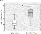
- Fig. 1
- shows the results of flow cytometric analyses of the circulating immune cell subset CD45+CD3+CD8+CD57+ T cells in the peripheral blood of patients with spinal non-fusion and normal fusion. Spinal non-fusion patients (healing class 2) showed a significantly higher frequency of CD45+CD3+CD8+CD57+ T cells (CD57+ in % of CD45+CD3+CD8+ T cells) compared to normal healing patients (healing class 1) indicates a dramatic increase of terminally differentiated CD8+ effector cells (TEMRA CD8+). * = P < 0.001; Mann-Whitney U test.
- Fig. 2
- Results of flow cytometric analyses of the circulating immune cell subset CD45+CD3+CD8+CD28- T cells in the peripheral blood of patients with spinal non-fusion and normal fusion. Spinal non-fusion patients (healing class 2) showed a significantly higher frequency of CD45+CD3+CD8+CD28- T cells (CD28- in % of CD45+CD3+CD8+ T cells) compared to normal healing patients (healing class 1) indicates a dramatic increase of terminally differentiated CD8+ effector cells (TEMRA CD8+). * = P < 0.001; Mann-Whitney U test.
- Fig. 3
- shows the results of flow cytometric analyses of the circulating immune cell subset CD45+CD3+CD8+CD57+28- T cells in the peripheral blood of patients with spinal non-fusion and normal fusion. Spinal non-fusion patients (healing class 2) showed a significantly higher frequency of CD45+CD3+CD8+CD57+28- T cells (CD57CD28-+ in % of CD45+CD3+CD8+ T cells) compared to normal healing patients (healing class 1) indicates a dramatic increase of terminally differentiated CD8+ effector cells (TEMRA CD8+). * = P < 0.001; Mann-Whitney U test.
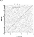
- Fig. 4
- illustrates the receiver operating characteristics (ROC) based on the summation of the expression of CD57+ in % of CD45+CD3+CD8+ T cells and CD28- in % of CD45+CD3+CD8+ T cells. The ROC analysis showed a high sensitivity (76%) as well as specificity (96%) in the detection of the affected patients based on their blood levels using a cut-off level of 85.6 % with an area under the curve (AUC): 0.886 and p<0.001.
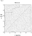
- Fig. 5
- illustrates the receiver operating characteristic (ROC) based on the expression of CD57+CD28- in % of CD45+CD3+CD8+ T cells with an area under the curve (AUC): 0.859 and p<0.001. The ROC analysis showed a high sensitivity (90%) as well as a specificity (71%) in the detection of the affected patients based on their blood levels using a cut-off level of 22.7%. A cut-off level of 33.1% showed a sensitivity of 71% with a higher specificity (96%)
- Fig. 6
- illustrates the receiver operating characteristics (ROC) based on the expression of CD57+ in % of CD45+CD3+CD8+ T cells with an area under the curve (AUC): 0.863 and p<0.001. The ROC analysis showed a high sensitivity (90%) as well as a specificity (61%) in the detection of the affected patients based on their blood levels using a cut-off level of 24.4%. A cut-off level of 37.6% showed a sensitivity of 71% with a higher specificity (96%)
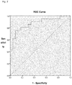
- Fig. 7
- illustrates the receiver operating characteristics (ROC) based on the expression of CD28- in % of CD45+CD3+CD8+ T cells with an area under the curve (AUC): 0.876 and p<0.001. The ROC analysis showed a high sensitivity (90%) as well as a specificity (66%) in the detection of the affected patients based on their blood levels using a cut-off level of 28.0%. A cut-off level of 42.9% showed a sensitivity of 76% with a higher specificity (91%)
- In order to define novel biomarkers for predicting the individual risk and to identify novel therapeutic targets, the inventors included 44 patients that received a spinal bone fusion surgery with secured vertebral body fusion results into a retrospective study. These patients were classified in patients with normal vertebral body fusion (n=23) and patients with pseudarthrosis/non-fusion (n=21), based on their CT-scan results.
Table 1 shows the characterization and blood parameters of the patients involved in the retrospective study (spinal fusion and spinal non-fusion patients) Parameter Spinal non-fusion (n=21) Spinal fusion (n=23) p-value Age (y) 70.7 ± 8.1 63.7 ± 10.1 0.01 Sex 6 male / 15 female 10 male / 13 female 0.3 (28.6%) / (71.4%) (43.5%) / (56.5%) Time to revision or revision free (month) 21.7±12.7 27.0±10.3 0.1 Minimum: 8 Minimum: 12 Maximum: 47 Maximum: 57 Height (m) 1.62±0.1 1.66±0.1 0.3 Weight (kg) 76.9±10.7 79.6±13.7 0.5 BMI 29.1±3.2 29.3±6.4 0.9 Leucocytes (/nL) 8.5±3.9 6.7±2.0 0.06 Haemoglobin (g/L) 13.2±1.9 14.2±1.3 0.06 Haematocrite (%) 0.39±0.05 0.41±0.03 0.07 Erythrocytes (/pL) 4.4±0.6 4.6±0.5 0.2 Thrombocytes (/nL) 271.5±77.8 253.0±53.0 0.3 Sodium (mmol/L) 139.0±3.9 139.0±2.9 0.9 Potassium (mmol/L) 4.2±0.4 4.1±0.3 0.6 Calcium (mmol/L) 2.4±0.1 2.3±0.1 0.4 Glucose (mg/dL) 102.3±24.1 109.1±41.8 0.5 Creatinin (mg/dL) 0.88±0.2 0.81±0.2 0.3 Urea (mg/dL) 32.8±9.2 32.4±11.3 0.9 Albumin (g/L) 42.9±9.3 44.5±2.7 0.4 GPT (U/L) 24.5±19.9 28.4±16.0 0.5 GOT (U/L) 26.5±14.2 31.0±28.8 0.5 CRP (mg/L) 0.9±1.1 4.0±6.0 0.03 Table 2: shows the blood parameters and time to diagnosis of spinal non-fusion at the initial and revision surgery for non-fusion patients. Parameter Spinal non-fusion Initial Surgery Spinal non-fusion Revision Surgery p-value Time to diagnose non-fusion (month) 8 month = n=2 = 9.5% 10 month = n= 2 = 9.5% 11 month = n= 2 = 9.5% 12 month = n=3 = 14.3% 13 month = n=1 = 4.8% 18 month = n= 1 = 4.8% 19 month =n= 1 = 4.8% 24 month =n= 1 = 4.8% 26 month =n= 1 = 4.8% 31 month =n= 1 = 4.8% 33 month =n= 1 = 4.8% 36 month =n= 2 = 9.5% 40 month =n= 2 = 9.5% 47 month =n= 1 = 4.8% Revision performed - 15/21 (71.4%) - Spongiosa + BMP-2 - 8/15(53.3%) - Spongiosa 7/15 (46.7%) - Leucocytes (/nL) 8.5±3.9 6.5±1.6 0.9 Haemoglobin (g/L) 13.2±1.9 13.9±1.9 0.6 Haematocrite (%) 0.39±0.05 0.41±0.06 0.8 Erythrocytes (/pL) 4.4±0.6 4.4±0.7 0.7 Thrombocytes (/nL) 271.5±77.8 261.4±68.1 0.5 Sodium (mmol/L) 139.0±3.9 137.7±4.4 0.4 Potassium (mmol/L) 4.2±0.4 4.2±0.4 0.7 Calcium (mmol/L) 2.4±0.1 2.4±0.1 0.5 Glucose (mg/dL) 102.3±24.1 106.4±23.8 0.3 Creatinin (mg/dL) 0.88±0.2 0.87±0.2 0.7 Urea (mg/dL) 32.8±9.2 36.5±9.1 0.3 Albumin (g/L) 42.9±9.3 43.8±2.9 0.4 GPT (U/L) 24.5±19.9 19.6±9.7 0.2 GOT (U/L) 26.5±14.2 22.7±4.9 0.2 CRP (mg/L) 0.9±1.1 4.4±9.5 0.01 - As established in literature and routinely used in clinics, there are time dependent and radiological criteria for the classification of a non-fusion. To fulfil the definition of a non-fusion process, the patients have to meet one or more of the following criteria.
- An incomplete vertebral body fusion after at least 24 post-operative weeks based on the presence of scar tissue formation in the intervertebral zone. (Zenya Ito et al., SPINE Volume 35, Number 21, pp E1101 - E1105; 2010)
-
- 1) no bridging of the trabecular bone connecting the two vertebral bodies,
- 2) angular motion in excess of 5 degrees
- 3) sagittal translation in excess of 3 mm
- 4) radiolucencies that involve more than half of the interfaces between the dowels and the host vertebral end plates
- Based on this former classification, the non-fusion patients showed differences in immune and inflammatory parameters in peripheral blood. Particularly, the spinal non-fusion was strongly associated with a significantly enhanced frequency (>2 fold) of terminally differentiated CD8+ effector T cells expressing the phenotype CD45+CD3+CD8+CD28-and/or CD45+CD3+CD8+CD57+ (CD8+ TEMRA).
- Accordingly, patients with enhanced levels of CD8+ TEMRA cells, because of a personal history of chronic immunostimulation (e.g. by persistent and frequently reactivating viruses like CMV/EBV), have a worse vertebral body fusion or quality following spinal fusion surgery as a result of an overwhelming inflammation that inhibits normal vertebral body fusion.
- The data presented herein demonstrate that the individual immune profile (frequency of CD8+ TEMRA) is a reliable biomarker for predicting impaired bone fusion patients allowing early interventions with known and established methods.
Claims (10)
- Method for predicting the probability of having or developing a non-fusion, particularly before undergoing a spinal fusion surgery or after a spinal fusion surgery, wherein said method comprises determining the frequency of a subpopulation of CD8+ T cells selected from CD8+CD57+, CD8+CD28- and CD8+CD57+CD28- in a sample obtained from a patient.
- Method for monitoring the course of treatment of a non-fusion in a patient, particularly after spinal fusion surgery, wherein said method comprises determining the frequency of a subpopulation of CD8+ T cells selected from CD8+CD57+, CD8+CD28- and CD8+CD57+CD28- in a sample obtained from said patient.
- The method according to claim 1 or 2, wherein said sample is a blood sample.
- The method according to claim to any one of the preceding claims, wherein said sample is a sample that has been obtained before, during or after spinal fusion surgery from said patient.
- The method according to any one of the preceding claims, wherein a sample exhibiting an at least two-fold higher frequency of said subpopulation of CD8+ T cell when compared to a standard value determined for a large population with normal fusion after bone fusion is assigned to a patient having an elevated probability for having or developing non-fusion after spinal fusion surgery.
- The method according any one of the preceding claims, wherein- a sample exhibiting a fraction of CD8+CD57+CD28- cells referred to the total sum of all CD8+ cell in said sample of at least 22.7 % more particular of at least 33.1 %, is assigned to a patient having an elevated probability for having or developing non-fusion after spinal fusion surgery, and/or- a sample exhibiting a summed fraction of CD8+CD57+ cells and CD8+CD28-cells referred to the total sum of all CD8+ cells in said sample of at least 85.6 % is assigned to a patient having an elevated probability for having or developing non-fusion after spinal fusion surgery, and/or- a sample exhibiting a fraction of CD8+CD57+ cells referred to the total sum of all CD8+ cells in said sample of at least 24.4%, more particular of at least 37.6 %, is assigned to a patient having an elevated probability for having or developing non-fusion after spinal fusion surgery, and/or- a sample exhibiting a fraction of CD8+CD28- cells referred to the total sum of all CD8+cells in said sample of at least 28.0%, more particular of at least 42.9%, is assigned to a patient having an elevated probability for having or developing non-fusion after spinal fusion .
- A system for predicting the probability of having or developing a non-fusion, particularly before undergoing a spinal fusion surgery or after a spinal fusion surgery, and/or for monitoring the course of treatment of a non-fusion of a patient, particularly after spinal fusion surgery, comprising- a device for determination the frequency of a cell population in a sample obtained from a patient- programmed microprocessor,wherein
said microprocessor is configured to run a method according to any one of the preceding claims. - Use of a combination of antibodies for predicting the probability of having or developing a non-fusion, particularly before undergoing a spinal fusion surgery after a spinal fusion surgery, wherein said combination comprises an anti-CD8-antibody and an anti-CD57-antibody, or an anti-CD8-antibody and an anti-CD28 antibody, or an anti-CD8-antibody, an anti-CD28-antibody and an anti-CD57-antibody, and wherein said antibodies are suitable for fluorescence based flow cytometry.
- A bone morphogenetic protein for use in a method for preventing or treating non-fusion after spinal bone fusion surgery, wherein said bone morphogenetic protein is administered during or after undergoing a spinal fusion surgery or a revision surgery after failed spinal fusion to a patient that has an elevated probability of developing or having a non-fusion after spinal bone fusion surgery.
- The bone morphogenetic protein for use in a method for treating non-fusion after spinal bone fusion surgery in a patient having an elevated probability of non-fusion after spinal bone fusion surgery according to claim 9, wherein said bone morphogenetic protein is selected from BMP2, or BMP7.
Priority Applications (6)
| Application Number | Priority Date | Filing Date | Title |
|---|---|---|---|
| EP17206698.7A EP3499236A1 (en) | 2017-12-12 | 2017-12-12 | Cd8 t cell subsets as a biomarker for predicting non-fusion after spinal fusion surgery |
| PCT/EP2018/064085 WO2018219959A1 (en) | 2017-05-29 | 2018-05-29 | Cd8 t cell subsets as a biomarker for predicting non-fusion after spinal fusion surgery |
| US16/618,118 US11802873B2 (en) | 2017-05-29 | 2018-05-29 | CD8 T cell subsets as a biomarker for predicting non-fusion after spinal fusion surgery |
| EP18725881.9A EP3631450B1 (en) | 2017-05-29 | 2018-05-29 | Cd8 t cell subsets as a biomarker for predicting non-fusion after spinal fusion surgery |
| JP2019565847A JP7229541B2 (en) | 2017-05-29 | 2018-05-29 | CD8 T cell subsets as biomarkers to predict non-fusion after spinal fusion surgery |
| CN201880048733.9A CN110945358B (en) | 2017-05-29 | 2018-05-29 | CD 8T cell subpopulations as markers for predicting post-spinal fusion non-fusion |
Applications Claiming Priority (1)
| Application Number | Priority Date | Filing Date | Title |
|---|---|---|---|
| EP17206698.7A EP3499236A1 (en) | 2017-12-12 | 2017-12-12 | Cd8 t cell subsets as a biomarker for predicting non-fusion after spinal fusion surgery |
Publications (1)
| Publication Number | Publication Date |
|---|---|
| EP3499236A1 true EP3499236A1 (en) | 2019-06-19 |
Family
ID=60888125
Family Applications (1)
| Application Number | Title | Priority Date | Filing Date |
|---|---|---|---|
| EP17206698.7A Ceased EP3499236A1 (en) | 2017-05-29 | 2017-12-12 | Cd8 t cell subsets as a biomarker for predicting non-fusion after spinal fusion surgery |
Country Status (1)
| Country | Link |
|---|---|
| EP (1) | EP3499236A1 (en) |
Citations (2)
| Publication number | Priority date | Publication date | Assignee | Title |
|---|---|---|---|---|
| US5492697A (en) * | 1990-03-05 | 1996-02-20 | Board Of Regents, Univ. Of Texas System | Biodegradable implant for fracture nonunions |
| EP2623978A1 (en) * | 2012-02-03 | 2013-08-07 | Charité - Universitätsmedizin Berlin | CD8+ T-cell subsets as markers for prediction of delayed fracture healing |
-
2017
- 2017-12-12 EP EP17206698.7A patent/EP3499236A1/en not_active Ceased
Patent Citations (2)
| Publication number | Priority date | Publication date | Assignee | Title |
|---|---|---|---|---|
| US5492697A (en) * | 1990-03-05 | 1996-02-20 | Board Of Regents, Univ. Of Texas System | Biodegradable implant for fracture nonunions |
| EP2623978A1 (en) * | 2012-02-03 | 2013-08-07 | Charité - Universitätsmedizin Berlin | CD8+ T-cell subsets as markers for prediction of delayed fracture healing |
Non-Patent Citations (7)
| Title |
|---|
| AXELRAD T W ET AL: "Bone morphogenetic proteins in orthopaedic surgery", CYTOKINE AND GROWTH FACTOR REVIEWS, ELSEVIER LTD, GB, vol. 20, no. 5-6, 1 October 2009 (2009-10-01), pages 481 - 488, XP026790619, ISSN: 1359-6101, [retrieved on 20091104] * |
| CHUN DS ET AL.: "Lumbar pseudarthrosis: a review of current diagnosis and treatment", NEUROSURG FOCUS, vol. 39, no. 4, October 2015 (2015-10-01) |
| CHUN ET AL., THE BEST TREATMENT FOR PSEUDARTHROSIS IS TO PREVENT IT FROM OCCURRING AFTER THE INITIAL OPERATION, 2015 |
| RAJAEE SS ET AL., SPINAL FUSION IN THE UNITED STATES: ANALYSIS OF TRENDS FROM 1998 TO 2008, vol. 37, no. 1, 1 January 2012 (2012-01-01), pages 67 - 76 |
| S. REINKE ET AL: "Terminally Differentiated CD8+ T Cells Negatively Affect Bone Regeneration in Humans", SCIENCE TRANSLATIONAL MEDICINE, vol. 5, no. 177, 20 March 2013 (2013-03-20), US, pages 177ra36 - 177ra36, XP055365176, ISSN: 1946-6234, DOI: 10.1126/scitranslmed.3004754 * |
| WERLE STEPHAN ET AL: "Bone morphogenetic protein 7 and autologous bone graft in revision surgery for non-union after lumbar interbody fusion", ARCHIVES OF ORTHOPAEDIC AND TRAUMA SURGERY, SPRINGER VERLAG, DE, vol. 136, no. 8, 20 June 2016 (2016-06-20), pages 1041 - 1049, XP036004744, ISSN: 0936-8051, [retrieved on 20160620], DOI: 10.1007/S00402-016-2485-X * |
| ZENYA ITO ET AL., SPINE, vol. 35, no. 21, 2010, pages E1101 - E1105 |
Similar Documents
| Publication | Publication Date | Title |
|---|---|---|
| Paiva et al. | Immune status of high-risk smoldering multiple myeloma patients and its therapeutic modulation under LenDex: a longitudinal analysis | |
| US20230033136A1 (en) | Cd8+t-cell subsets as markers for prediction of delayed fracture healing | |
| EP4055609A1 (en) | Classification of tumor microenvironments | |
| US11802873B2 (en) | CD8 T cell subsets as a biomarker for predicting non-fusion after spinal fusion surgery | |
| Wyatt et al. | Molecular expression patterns in the synovium and their association with advanced symptomatic knee osteoarthritis | |
| EP1700129A2 (en) | Method of assessing rheumatoid arthritis by measuring anti-ccp and interleukin 6 | |
| Nakazawa et al. | Distinct expression of coinhibitory molecules on alveolar T cells in patients with rheumatoid arthritis–associated and idiopathic inflammatory myopathy–associated interstitial lung disease | |
| EP1721162B1 (en) | Method of assessing rheumatoid arthritis by measuring anti-ccp and serum amyloid a | |
| EP1721163B1 (en) | Method of assessing rheumatoid arthritis by measuring rheumatoid factor and interleukin-6 | |
| Wen et al. | 1, 25-Dihydroxyvitamin D3 modulates T cell differentiation and impacts on the production of cytokines from Chinese Han patients with early rheumatoid arthritis | |
| van den Broek et al. | Long-term effect of hematopoietic cell transplantation on systemic inflammation in patients with mucopolysaccharidoses | |
| Šenolt et al. | Interleukin-20 is triggered by TLR ligands and associates with disease activity in patients with rheumatoid arthritis | |
| EP2148934B1 (en) | Matrix marker model and methods for assessing and treating rheumatoid arthritis | |
| El-Fetouh et al. | Serum interleukin-18 and interleukin-10 levels in systemic lupus erythematosus: correlation with SLEDAI score and disease activity parameters | |
| Niki et al. | Five types of inflammatory arthritis following total knee arthroplasty | |
| Mease | The potential roles for novel biomarkers in rheumatoid arthritis assessment | |
| Koutsonikoli et al. | Novel biomarkers for early targeted and individualized treatment in Juvenile Idiopathic Arthritis | |
| WO2024002914A1 (en) | Prediction of, and composition to improve, tendon healing | |
| HK1140797B (en) | Matrix marker model and methods for assessing and treating rheumatoid arthritis |
Legal Events
| Date | Code | Title | Description |
|---|---|---|---|
| PUAI | Public reference made under article 153(3) epc to a published international application that has entered the european phase |
Free format text: ORIGINAL CODE: 0009012 |
|
| AK | Designated contracting states |
Kind code of ref document: A1 Designated state(s): AL AT BE BG CH CY CZ DE DK EE ES FI FR GB GR HR HU IE IS IT LI LT LU LV MC MK MT NL NO PL PT RO RS SE SI SK SM TR |
|
| AX | Request for extension of the european patent |
Extension state: BA ME |
|
| STAA | Information on the status of an ep patent application or granted ep patent |
Free format text: STATUS: THE APPLICATION HAS BEEN REFUSED |
|
| 18R | Application refused |
Effective date: 20190720 |