EP3495817A1 - Molecular diagnostic screening assay - Google Patents
Molecular diagnostic screening assay Download PDFInfo
- Publication number
- EP3495817A1 EP3495817A1 EP18193992.7A EP18193992A EP3495817A1 EP 3495817 A1 EP3495817 A1 EP 3495817A1 EP 18193992 A EP18193992 A EP 18193992A EP 3495817 A1 EP3495817 A1 EP 3495817A1
- Authority
- EP
- European Patent Office
- Prior art keywords
- binders
- nucleic acid
- droplets
- sample
- chromosome
- Prior art date
- Legal status (The legal status is an assumption and is not a legal conclusion. Google has not performed a legal analysis and makes no representation as to the accuracy of the status listed.)
- Granted
Links
Images




Classifications
-
- C—CHEMISTRY; METALLURGY
- C12—BIOCHEMISTRY; BEER; SPIRITS; WINE; VINEGAR; MICROBIOLOGY; ENZYMOLOGY; MUTATION OR GENETIC ENGINEERING
- C12N—MICROORGANISMS OR ENZYMES; COMPOSITIONS THEREOF; PROPAGATING, PRESERVING, OR MAINTAINING MICROORGANISMS; MUTATION OR GENETIC ENGINEERING; CULTURE MEDIA
- C12N15/00—Mutation or genetic engineering; DNA or RNA concerning genetic engineering, vectors, e.g. plasmids, or their isolation, preparation or purification; Use of hosts therefor
- C12N15/09—Recombinant DNA-technology
- C12N15/10—Processes for the isolation, preparation or purification of DNA or RNA
- C12N15/1034—Isolating an individual clone by screening libraries
- C12N15/1072—Differential gene expression library synthesis, e.g. subtracted libraries, differential screening
-
- C—CHEMISTRY; METALLURGY
- C12—BIOCHEMISTRY; BEER; SPIRITS; WINE; VINEGAR; MICROBIOLOGY; ENZYMOLOGY; MUTATION OR GENETIC ENGINEERING
- C12Q—MEASURING OR TESTING PROCESSES INVOLVING ENZYMES, NUCLEIC ACIDS OR MICROORGANISMS; COMPOSITIONS OR TEST PAPERS THEREFOR; PROCESSES OF PREPARING SUCH COMPOSITIONS; CONDITION-RESPONSIVE CONTROL IN MICROBIOLOGICAL OR ENZYMOLOGICAL PROCESSES
- C12Q1/00—Measuring or testing processes involving enzymes, nucleic acids or microorganisms; Compositions therefor; Processes of preparing such compositions
- C12Q1/68—Measuring or testing processes involving enzymes, nucleic acids or microorganisms; Compositions therefor; Processes of preparing such compositions involving nucleic acids
- C12Q1/6813—Hybridisation assays
- C12Q1/6827—Hybridisation assays for detection of mutation or polymorphism
-
- C—CHEMISTRY; METALLURGY
- C12—BIOCHEMISTRY; BEER; SPIRITS; WINE; VINEGAR; MICROBIOLOGY; ENZYMOLOGY; MUTATION OR GENETIC ENGINEERING
- C12Q—MEASURING OR TESTING PROCESSES INVOLVING ENZYMES, NUCLEIC ACIDS OR MICROORGANISMS; COMPOSITIONS OR TEST PAPERS THEREFOR; PROCESSES OF PREPARING SUCH COMPOSITIONS; CONDITION-RESPONSIVE CONTROL IN MICROBIOLOGICAL OR ENZYMOLOGICAL PROCESSES
- C12Q1/00—Measuring or testing processes involving enzymes, nucleic acids or microorganisms; Compositions therefor; Processes of preparing such compositions
- C12Q1/68—Measuring or testing processes involving enzymes, nucleic acids or microorganisms; Compositions therefor; Processes of preparing such compositions involving nucleic acids
- C12Q1/6844—Nucleic acid amplification reactions
-
- C—CHEMISTRY; METALLURGY
- C12—BIOCHEMISTRY; BEER; SPIRITS; WINE; VINEGAR; MICROBIOLOGY; ENZYMOLOGY; MUTATION OR GENETIC ENGINEERING
- C12Q—MEASURING OR TESTING PROCESSES INVOLVING ENZYMES, NUCLEIC ACIDS OR MICROORGANISMS; COMPOSITIONS OR TEST PAPERS THEREFOR; PROCESSES OF PREPARING SUCH COMPOSITIONS; CONDITION-RESPONSIVE CONTROL IN MICROBIOLOGICAL OR ENZYMOLOGICAL PROCESSES
- C12Q1/00—Measuring or testing processes involving enzymes, nucleic acids or microorganisms; Compositions therefor; Processes of preparing such compositions
- C12Q1/68—Measuring or testing processes involving enzymes, nucleic acids or microorganisms; Compositions therefor; Processes of preparing such compositions involving nucleic acids
- C12Q1/6876—Nucleic acid products used in the analysis of nucleic acids, e.g. primers or probes
- C12Q1/6883—Nucleic acid products used in the analysis of nucleic acids, e.g. primers or probes for diseases caused by alterations of genetic material
-
- C—CHEMISTRY; METALLURGY
- C12—BIOCHEMISTRY; BEER; SPIRITS; WINE; VINEGAR; MICROBIOLOGY; ENZYMOLOGY; MUTATION OR GENETIC ENGINEERING
- C12Q—MEASURING OR TESTING PROCESSES INVOLVING ENZYMES, NUCLEIC ACIDS OR MICROORGANISMS; COMPOSITIONS OR TEST PAPERS THEREFOR; PROCESSES OF PREPARING SUCH COMPOSITIONS; CONDITION-RESPONSIVE CONTROL IN MICROBIOLOGICAL OR ENZYMOLOGICAL PROCESSES
- C12Q1/00—Measuring or testing processes involving enzymes, nucleic acids or microorganisms; Compositions therefor; Processes of preparing such compositions
- C12Q1/68—Measuring or testing processes involving enzymes, nucleic acids or microorganisms; Compositions therefor; Processes of preparing such compositions involving nucleic acids
- C12Q1/6876—Nucleic acid products used in the analysis of nucleic acids, e.g. primers or probes
- C12Q1/6883—Nucleic acid products used in the analysis of nucleic acids, e.g. primers or probes for diseases caused by alterations of genetic material
- C12Q1/6886—Nucleic acid products used in the analysis of nucleic acids, e.g. primers or probes for diseases caused by alterations of genetic material for cancer
-
- C—CHEMISTRY; METALLURGY
- C12—BIOCHEMISTRY; BEER; SPIRITS; WINE; VINEGAR; MICROBIOLOGY; ENZYMOLOGY; MUTATION OR GENETIC ENGINEERING
- C12Q—MEASURING OR TESTING PROCESSES INVOLVING ENZYMES, NUCLEIC ACIDS OR MICROORGANISMS; COMPOSITIONS OR TEST PAPERS THEREFOR; PROCESSES OF PREPARING SUCH COMPOSITIONS; CONDITION-RESPONSIVE CONTROL IN MICROBIOLOGICAL OR ENZYMOLOGICAL PROCESSES
- C12Q2600/00—Oligonucleotides characterized by their use
- C12Q2600/156—Polymorphic or mutational markers
Definitions
- the invention generally relates to method for screening for a condition in a subject.
- Assays have been developed that rely on analyzing nucleic acid molecules from bodily fluids for the presence of abnormalities, thus leading to early diagnosis of certain conditions such as cancer or fetal aneuploidy.
- a typical bodily fluid sample however, a majority of the nucleic acid is degraded, and any altered nucleic acids containing an abnormality of interest are present in small amounts (e.g., less than 1%) relative to a total amount of nucleic acids in the sample. This results in a failure to detect the small amount of abnormal nucleic acid.
- Amplification based approaches e.g., polymerase chain reaction (PCR), digital PCR, quantitative PCR
- PCR polymerase chain reaction
- digital PCR digital PCR
- quantitative PCR quantitative PCR
- next generation sequencing methods such as those commercially available from Roche (454 and SOLiD), Illumina/Solexa, Pacific Biosciences, and Life Technologies / Ion Torrent allow for the highly sensitive detection of the small population of abnormal nucleic acids in a sample, generally without the need for amplification of the nucleic acid in the sample.
- sequencing instruments are very expensive and these sequencing methods still require a large amount of data (for example, approximately 1,000,000 sequencing reads) to reliably identify an abnormal nucleic acid in a sample.
- sequencing is a very costly approach that still requires a significant amount of data in order to identify the presence of a population of abnormal nucleic acids in a sample.
- Methods of the invention are able to identify an altered nucleic acid containing an abnormality of interest that is present in small amounts (e.g., less than 1%) relative to a total amount of nucleic acids in a sample.
- Methods of the invention are accomplished by designing a plurality of binders (e.g., 10 binders, 100 binders, 1,000 binders) against the nucleic acid that includes the abnormality of interest (i.e., the target nuclei acid).
- the binders each include a region that hybridizes to a different location on the target nucleic acid, and thus, all of the binders hybridize to the target nucleic acid at once.
- the target nucleic acid is separated from the sample and the hybridized binders are analyzed (e.g., PCR, digital PCR, qPCR, sequencing, etc.).
- This approach effectively increases the number of counts and confidence in the number of counts by a factor given by the number of binders against the target nucleic acid. For example, assuming only 1,000 genome equivalents of the target nucleic acid in the sample are available, and there are 1,000 binders, each of which binds to a different location on the target, there are now 1,000,000 target readouts, which is enough to identify a rare abnormal nucleic acid in a sample.
- methods of the invention involve obtaining a pool of nucleic acids from a sample, incubating the nucleic acids with first and second sets of binders, in which the first set binds uniquely to different regions of a target nucleic acid in the pool, the second set binds uniquely to different regions of a reference nucleic acid in the pool, and the first and second sets include different detectable labels, removing unbound binders, detecting the labels, and screening for a condition based upon results of the detecting step.
- Detecting of the label may be accomplished by any analytical method known in the art.
- the detectable labels are barcode sequences and detecting the label includes sequencing the barcodes.
- screening for the condition may involve counting a number of barcodes from the first set, counting a number of barcodes from the second set, and determining whether a statistical difference exists between the number of barcodes from the first set and the number of barcodes from the second set.
- detecting the label may be accomplished by an amplification based technique, such as PCR, digital PCR, or qPCR.
- digital PCR is used to detect the labels.
- the method further includes compartmentalizing bound binders of the first and second set into compartmentalized portions, the compartmentalized portions including, on average, either the first set of binders or the second set of binders, and amplifying binders in the compartmentalized portions. Compartmentalizing may involve diluting the sample such that it may be dispensed into different wells of a multi-well plate in a manner such that each well includes, on average, either the first set of binders or the second set of binders.
- the compartmentalizing involves forming droplets and the compartmentalized portions are the droplets.
- An exemplary method for forming droplets involves flowing a stream of sample fluid including the amplicons such that it intersects two opposing streams of flowing carrier fluid.
- the carrier fluid is immiscible with the sample fluid. Intersection of the sample fluid with the two opposing streams of flowing carrier fluid results in partitioning of the sample fluid into individual sample droplets.
- the carrier fluid may be any fluid that is immiscible with the sample fluid.
- An exemplary carrier fluid is oil, particularly, a fluorinated oil.
- the carrier fluid includes a surfactant, such as a fluorosurfactant.
- the droplets may be flowed through channels.
- the binders of the first set include the same universal primer site and the binders of the second set include the same universal primer site, in which the primer sites of the first and second binders are different.
- Each compartmentalized portion further includes universal primers that bind the universal priming sites of the binders of the first set and universal primers that bind the universal priming sites of the binders of the second set.
- the compartmentalized portions further include probes that bind the detectable label of the first set of binders and probes that bind the detectable label of the second set of binders.
- An amplification reaction (e.g., PCR) is conducted in each compartmentalized portion, and the first probes are allowed to bind to the detectable label of the first set of binders, and the second probes are allowed to bind to the detectable label of the second set of binders.
- the probes are optically labeled probes and detecting the label includes optically detecting the bound probes.
- screening for the condition may involve counting a number of compartmentalized portions including the first detectable label, counting a number of compartmentalized portions comprising the second detectable label, and determining whether a statistical difference exists between the number of compartmentalized portions comprising the first detectable label and the number of compartmentalized portions comprising the second detectable label.
- Methods of the invention may be used to screen for any condition.
- the condition is fetal aneuploidy.
- the fetal aneuploidy is trisomy 21 (Down syndrome).
- any maternal sample that may include fetal cell-free circulating nucleic acid.
- Exemplary samples include blood, plasma, or serum.
- the target nucleic acid is nucleic acid of chromosome 21 and the first set of binders binds to the nucleic acid of chromosome 21 in the pool.
- the second set of binders binds nucleic acid of a reference chromosome in the pool.
- the detectable labels are barcode sequences and detecting the label includes sequencing the barcodes.
- screening for the condition may involve counting a number of barcodes from the first set, counting a number of barcodes from the second set, and determining whether a statistical difference exists between the number of barcodes from the first set and the number of barcodes from the second set.
- detecting the label may be accomplished by an amplification based technique, such as PCR, digital PCR, or qPCR. In specific embodiments, digital PCR is used to detect the labels.
- screening for the condition may involve counting a number of compartmentalized portions including the first detectable label, counting a number of compartmentalized portions comprising the second detectable label, and determining whether a statistical difference exists between the number of compartmentalized portions comprising the first detectable label and the number of compartmentalized portions comprising the second detectable label.
- methods of the invention are used to screen a subject for cancer generally.
- the first set of binders binds genomic regions of the nucleic acids associated with known mutations involved in different cancers and the second set of binders binds genomic regions of the nucleic acids that are not mutated.
- screening for the condition may involve counting a number of barcodes from the first set, counting a number of barcodes from the second set, and determining whether a statistical difference exists between the number of barcodes from the first set and the number of barcodes from the second set.
- detecting the label may be accomplished by an amplification based technique, such as PCR, digital PCR, or qPCR.
- digital PCR is used to detect the labels.
- screening for the condition may involve counting a number of compartmentalized portions including the first detectable label, counting a number of compartmentalized portions comprising the second detectable label, and determining whether a statistical difference exists between the number of compartmentalized portions comprising the first detectable label and the number of compartmentalized portions comprising the second detectable label.
- Another aspect of the invention provides methods for screening for a condition in a subject, that involve obtaining a pool of different nucleic acid from a sample, compartmentalizing the pool of nucleic acids into compartmentalized portions, the compartmentalized portions including, on average, either a first set of binders or a second set of binders, wherein the first and second sets comprise different detectable labels and the first and second sets bind to different nucleic acids in the pool, amplifying only binders in the compartmentalized portions that bound to the nucleic acid, detecting the labels, and screening for a condition based upon results of the detecting step.
- the invention generally relates to method for screening for a condition in a subject.
- methods of the invention involve obtaining a pool of nucleic acids from a sample, incubating the nucleic acids with first and second sets of binders, in which the first set binds uniquely to different regions of a target nucleic acid in the pool, the second set binds uniquely to different regions of a reference nucleic acid in the pool, and the first and second sets include different detectable labels, removing unbound binders, detecting the labels, and screening for a condition based upon results of the detecting step.
- the binders, and not the nucleic acid is analyzed for the purpose of screening for a condition.
- Nucleic acid is generally is acquired from a sample or a subject.
- Target molecules for labeling and/or detection according to the methods of the invention include, but are not limited to, genetic and proteomic material, such as DNA, genomic DNA, RNA, expressed RNA and/or chromosome(s). Methods of the invention are applicable to DNA from whole cells or to portions of genetic or proteomic material obtained from one or more cells.
- the sample may be obtained in any clinically acceptable manner, and the nucleic acid templates are extracted from the sample by methods known in the art. Nucleic acid templates can be obtained as described in U.S. Patent Application Publication Number US2002/0190663 A1, published Oct. 9, 2003 .
- nucleic acid can be extracted from a biological sample by a variety of techniques such as those described by Maniatis, et al. (Molecular Cloning: A Laboratory Manual, Cold Spring Harbor, N.Y., pp. 280-281, 1982 ), the contents of which are incorporated by reference herein in their entirety.
- Nucleic acid templates include deoxyribonucleic acid (DNA) and/or ribonucleic acid (RNA). Nucleic acid templates can be synthetic or derived from naturally occurring sources. In one embodiment, nucleic acid templates are isolated from a biological sample containing a variety of other components, such as proteins, lipids and non-template nucleic acids. Nucleic acid templates can be obtained from any cellular material, obtained from an animal, plant, bacterium, fungus, or any other cellular organism. Biological samples for use in the present invention include viral particles or preparations.
- Nucleic acid templates can be obtained directly from an organism or from a biological sample obtained from an organism, e.g., from blood, urine, cerebrospinal fluid, seminal fluid, saliva, sputum, stool and tissue.
- nucleic acid is obtained from fresh frozen plasma (FFP). Any tissue or body fluid specimen may be used as a source for nucleic acid for use in the invention.
- Nucleic acid templates can also be isolated from cultured cells, such as a primary cell culture or a cell line. The cells or tissues from which template nucleic acids are obtained can be infected with a virus or other intracellular pathogen.
- a sample can also be total RNA extracted from a biological specimen, a cDNA library, viral, or genomic DNA.
- nucleic acid obtained from biological samples is fragmented to produce suitable fragments for analysis.
- An advantage of methods of the invention is that they can be performed on nucleic acids that have not been fragmented.
- nucleic acids are fragmented prior to performing methods of the invention.
- nucleic acid from a biological sample is fragmented by sonication.
- individual nucleic acid template molecules can be from about 5 bases to about 20 kb.
- a biological sample as described herein may be homogenized or fractionated in the presence of a detergent or surfactant.
- concentration of the detergent in the buffer may be about 0.05% to about 10.0%.
- concentration of the detergent can be up to an amount where the detergent remains soluble in the solution. In a preferred embodiment, the concentration of the detergent is between 0.1% to about 2%.
- the detergent particularly a mild one that is nondenaturing, can act to solubilize the sample.
- Detergents may be ionic or nonionic.
- ionic detergents examples include deoxycholate, sodium dodecyl sulfate (SDS), N-lauroylsarcosine, and cetyltrimethylammoniumbromide (CTAB).
- a zwitterionic reagent may also be used in the purification schemes of the present invention, such as Chaps, zwitterion 3-14, and 3-[(3-cholamidopropyl)dimethylammonio]-1-propanesulf-onate. It is contemplated also that urea may be added with or without another detergent or surfactant.
- Lysis or homogenization solutions may further contain other agents, such as reducing agents.
- reducing agents include dithiothreitol (DTT), .beta.-mercaptoethanol, DTE, GSH, cysteine, cysteamine, tricarboxyethyl phosphine (TCEP), or salts of sulfurous acid.
- the nucleic acid is denatured by any method known in the art to produce single stranded nucleic acid templates and a pair of first and second oligonucleotides is hybridized to the single stranded nucleic acid template such that the first and second oligonucleotides flank a target region on the template.
- first and second sets of binders generally have the structure shown in Figure 1A and the second set of binders have a structure as shown in Figure 1B .
- Each binder includes a pair of universal forward and reverse primer sites (P1 and P1' and P2 and P2'). All of the binders of the first set include the same set of universal primer sites. All of the binders of the second set include the same set of universal primer sites. The primers sites of the first set are different than the primer sites of the second set, i.e., P1 and P1' are different than P2 and P2'.
- Each of the binders includes a detectable label code site. All of the binders of the first set include the same detectable label code site. All of the binders of the second set include the same detectable label code site. The detectable label code site of the first set is different than the detectable label code site of the second set.
- Each of the binders in the first set includes a target sequence portion.
- the target sequence portion for each of the binders of the first set is a sequence that binds to a region of the target nucleic acid.
- the target sequence portion of each binder of the first set is different, so that each binder of the first set may bind to a different location of the target nucleic acid.
- Each of the binders in the second set includes a reference sequence portion.
- the reference sequence portion for each of the binders of the second set is a sequence that binds to a region of the reference nucleic acid.
- the reference sequence portion of each binder of the second set is different, so that each binder of the second set may bind to a different location of the reference nucleic acid.
- the first set of binders generally have the structure shown in Figure 2A and the second set of binders have a structure as shown in Figure 2B .
- the first and second binders are constructed from an "a" portion and a "b" portion such that a complete binder is constructed by ligation or a fill and ligate process. This type of structure avoids the need to remove unbound binders, because only fully formed binders (i.e., those with an "a” and "b") portion can be analyzed.
- Figures 3A-B show complete binders.
- a probe such as a Taqman probe, would not be hydrolyzed exponentially as the PCR reaction proceeded. This is illustrated in Figures 4A-B .
- the type of detectable label code site in the first and second binders will depend on the binder detection technique to be employed. If the binder detection technique is sequencing, the detectable label code site will be a barcode sequence. In these embodiments, all of the binders of the first set will have the same barcode sequence and all of the binders of the second set will have the same barcode sequence. The barcode sequence of the first set of binders is different than the barcode sequence of the second set of binders.
- the detectable label code site will be a sequence that hybridizes with the probe.
- all of the binders of the first set will hybridize with a first probe and all of the binders of the second set will hybridize with a second probe.
- the first and second probe are different and include different labels.
- Oligonucleotides can also be obtained from commercial sources such as Operon Technologies, Amersham Pharmacia Biotech, Sigma, and Life Technologies.
- the oligonucleotides can have an identical melting temperature.
- the lengths of the oligonucleotides can be extended or shortened at the 5' end or the 3' end to produce oligonucleotides with desired melting temperatures.
- the annealing position of each oligonucleotide can be designed such that the sequence and length of the probe yield the desired melting temperature.
- Computer programs can also be used to design oligonucleotides, including but not limited to Array Designer Software (Arrayit Inc.), Oligonucleotide Probe Sequence Design Software for Genetic Analysis (Olympus Optical Co.), NetPrimer, and DNAsis from Hitachi Software Engineering.
- Array Designer Software Arrayit Inc.
- Oligonucleotide Probe Sequence Design Software for Genetic Analysis Olympus Optical Co.
- NetPrimer NetPrimer
- DNAsis from Hitachi Software Engineering.
- the TM (melting temperature) of each probe is calculated using software programs such as Oligo Design, available from Invitrogen Corp.
- reaction conditions of high stringency are used to ensure great specificity between the probes and the code sites on the binders.
- Nucleic acid hybridization may be affected by such conditions as salt concentration, temperature, or organic solvents, in addition to base composition, length of complementary strands, and number of nucleotide base mismatches between hybridizing nucleic acids, as is readily appreciated by those skilled in the art.
- Stringency of hybridization reactions is readily determinable by one of ordinary skill in the art, and generally is an empirical calculation dependent upon sequence length, washing temperature, and salt concentration. In general, longer sequences require higher temperatures for proper annealing, while shorter sequences need lower temperatures.
- Hybridization generally depends on the ability of denatured DNA to re-anneal when complementary strands are present in an environment below its melting temperature. The higher the degree of desired homology between the sequence and hybridizable sequence, the higher the relative temperature that can be used. As a result, it follows that higher relative temperatures would tend to make the reaction conditions more stringent, while lower temperatures less so.
- stringency of hybridization reactions see Ausubel et al., Current Protocols in Molecular Biology, Wiley Interscience Publishers, (1995 ), the contents of which are incorporated by reference herein in their entirety.
- Stringent conditions or high stringency conditions typically: (1) employ low ionic strength and high temperature for washing, for example 0.015 M sodium chloride/0.0015 M sodium citrate/0.1% sodium dodecyl sulfate at 50°C.; (2) employ during hybridization a denaturing agent, such as formamide, for example, 50% (v/v) formamide with 0.1% bovine serum albumin/0.1% Ficoll/0.1% polyvinylpyrrolidone/50 mM sodium phosphate buffer at pH 6.5 with 750 mM sodium chloride, 75 mM sodium citrate at 42°C.; or (3) employ 50% formamide, 5 x SSC (0.75 M NaCl, 0.075 M sodium citrate), 50 mM sodium phosphate (pH 6.8), 0.1% sodium pyrophosphate, 5 x Denhardt's solution, sonicated salmon sperm DNA (50 .mu.g/ml), 0.1% SDS, and 10% dextran sulfate at 42°C, with was
- reaction conditions of moderate stringency are used for hybridization of the first and second oligonucleotides to binding regions on the template nucleic acid.
- Moderately stringent conditions may be identified as described by Sambrook et al., Molecular Cloning: A Laboratory Manual, New York: Cold Spring Harbor Press, 1989 (the contents of which are incorporated by reference herein in their entirety and include the use of washing solution and hybridization conditions (e.g., temperature, ionic strength and % SDS) less stringent that those described above.
- moderately stringent conditions is overnight incubation at 37°C in a solution comprising: 20% formamide, 5 x SSC (150 mM NaCl, 15 mM trisodium citrate), 50 mM sodium phosphate (pH 7.6), 5 x Denhardt's solution, 10% dextran sulfate, and 20 mg/ml denatured sheared salmon sperm DNA, followed by washing the filters in 1 x SSC at about 37°C to 50°C.
- a solution comprising: 20% formamide, 5 x SSC (150 mM NaCl, 15 mM trisodium citrate), 50 mM sodium phosphate (pH 7.6), 5 x Denhardt's solution, 10% dextran sulfate, and 20 mg/ml denatured sheared salmon sperm DNA, followed by washing the filters in 1 x SSC at about 37°C to 50°C.
- 5 x SSC 150 mM NaCl, 15 mM tris
- Oligonucleotides suitable for use in the present invention include those formed from nucleic acids, such as RNA and/or DNA, nucleic acid analogs, locked nucleic acids, modified nucleic acids, and chimeric probes of a mixed class including a nucleic acid with another organic component such as peptide nucleic acids.
- Exemplary nucleotide analogs include phosphate esters of deoxyadenosine, deoxycytidine, deoxyguanosine, deoxythymidine, adenosine, cytidine, guanosine, and uridine.
- non-natural nucleotides include a xanthine or hypoxanthine; 5-bromouracil, 2-aminopurine, deoxyinosine, or methylated cytosine, such as 5-methylcytosine, and N4-methoxydeoxycytosine.
- bases of polynucleotide mimetics such as methylated nucleic acids, e.g., 2'-O-methRNA, peptide nucleic acids, modified peptide nucleic acids, and any other structural moiety that can act substantially like a nucleotide or base, for example, by exhibiting base-complementarity with one or more bases that occur in DNA or RNA.
- oligonucleotide probe is not critical, as long as the oligonucleotides are capable of hybridizing to the code sites of the binders.
- oligonucleotides may be of any length.
- oligonucleotides may be as few as 5 nucleotides, or as much as 5000 nucleotides.
- Exemplary oligonucleotides are 5-mers, 10-mers, 15-mers, 20-mers, 25-mers, 50-mers, 100-mers, 200-mers, 500-mers, 1000-mers, 3000-mers, or 5000-mers.
- Methods for determining an optimal oligonucleotides length are known in the art. See, e.g., Shuber (U.S.
- the first and second oligonucleotides do not have to be of the same length. In certain embodiments, the first and second oligonucleotides are the same length, while in other embodiments, the first and second oligonucleotides are of different lengths.
- reaction time will depend on the different factors discussed above, e.g., stringency conditions, probe length, probe design, etc.
- the probes will include a detectable label that is directly or indirectly detectable.
- Preferred labels include optically-detectable labels, such as fluorescent labels.
- fluorescent labels include, but are not limited to, 4-acetamido-4'-isothiocyanatostilbene-2,2'disulfonic acid; acridine and derivatives: acridine, acridine isothiocyanate; 5-(2'-aminoethyl)aminonaphthalene-1-sulfonic acid (EDANS); 4-amino-N-[3-vinylsulfonyl)phenyl]naphthalimide-3,5 disulfonate; N-(4-anilino-1-naphthyl)maleimide; anthranilamide; BODIPY; Brilliant Yellow; coumarin and derivatives; coumarin, 7-amino-4-methylcoumarin (AMC, Coumarin 120), 7-amino-4-trifluoromethylcoul
- the pool of nucleic acids is incubated with first and second sets of binders under conditions that allow the first set of binders to bind to target nucleic acids in the pool and the second set of binders to bind the reference nucleic acids in the pool.
- reaction conditions of high stringency are used to ensure great specificity between the probes and the code sites on the binders.
- Nucleic acid hybridization may be affected by such conditions as salt concentration, temperature, or organic solvents, in addition to base composition, length of complementary strands, and number of nucleotide base mismatches between hybridizing nucleic acids, as is readily appreciated by those skilled in the art.
- Stringency of hybridization reactions is readily determinable by one of ordinary skill in the art, and generally is an empirical calculation dependent upon sequence length, washing temperature, and salt concentration. In general, longer sequences require higher temperatures for proper annealing, while shorter sequences need lower temperatures.
- Hybridization generally depends on the ability of denatured DNA to re-anneal when complementary strands are present in an environment below its melting temperature. The higher the degree of desired homology between the sequence and hybridizable sequence, the higher the relative temperature that can be used. As a result, it follows that higher relative temperatures would tend to make the reaction conditions more stringent, while lower temperatures less so.
- stringency of hybridization reactions see Ausubel et al., Current Protocols in Molecular Biology, Wiley Interscience Publishers, (1995 ), the contents of which are incorporated by reference herein in their entirety.
- Stringent conditions or high stringency conditions typically: (1) employ low ionic strength and high temperature for washing, for example 0.015 M sodium chloride/0.0015 M sodium citrate/0.1% sodium dodecyl sulfate at 50°C.; (2) employ during hybridization a denaturing agent, such as formamide, for example, 50% (v/v) formamide with 0.1% bovine serum albumin/0.1% Ficoll/0.1% polyvinylpyrrolidone/50 mM sodium phosphate buffer at pH 6.5 with 750 mM sodium chloride, 75 mM sodium citrate at 42°C.; or (3) employ 50% formamide, 5 x SSC (0.75 M NaCl, 0.075 M sodium citrate), 50 mM sodium phosphate (pH 6.8), 0.1% sodium pyrophosphate, 5 x Denhardt's solution, sonicated salmon sperm DNA (50 .mu.g/ml), 0.1% SDS, and 10% dextran sulfate at 42°C, with was
- reaction conditions of moderate stringency are used for hybridization of the first and second oligonucleotides to binding regions on the template nucleic acid.
- Moderately stringent conditions may be identified as described by Sambrook et al., Molecular Cloning: A Laboratory Manual, New York: Cold Spring Harbor Press, 1989 (the contents of which are incorporated by reference herein in their entirety and include the use of washing solution and hybridization conditions (e.g., temperature, ionic strength and % SDS) less stringent that those described above.
- moderately stringent conditions is overnight incubation at 37°C in a solution comprising: 20% formamide, 5 x SSC (150 mM NaCl, 15 mM trisodium citrate), 50 mM sodium phosphate (pH 7.6), 5 x Denhardt's solution, 10% dextran sulfate, and 20 mg/ml denatured sheared salmon sperm DNA, followed by washing the filters in 1 x SSC at about 37°C to 50°C.
- a solution comprising: 20% formamide, 5 x SSC (150 mM NaCl, 15 mM trisodium citrate), 50 mM sodium phosphate (pH 7.6), 5 x Denhardt's solution, 10% dextran sulfate, and 20 mg/ml denatured sheared salmon sperm DNA, followed by washing the filters in 1 x SSC at about 37°C to 50°C.
- 5 x SSC 150 mM NaCl, 15 mM tris
- Oligonucleotides suitable for use in the present invention include those formed from nucleic acids, such as RNA and/or DNA, nucleic acid analogs, locked nucleic acids, modified nucleic acids, and chimeric probes of a mixed class including a nucleic acid with another organic component such as peptide nucleic acids.
- Exemplary nucleotide analogs include phosphate esters of deoxyadenosine, deoxycytidine, deoxyguanosine, deoxythymidine, adenosine, cytidine, guanosine, and uridine.
- non-natural nucleotides include a xanthine or hypoxanthine; 5-bromouracil, 2-aminopurine, deoxyinosine, or methylated cytosine, such as 5-methylcytosine, and N4-methoxydeoxycytosine.
- bases of polynucleotide mimetics such as methylated nucleic acids, e.g., 2'-O-methRNA, peptide nucleic acids, modified peptide nucleic acids, and any other structural moiety that can act substantially like a nucleotide or base, for example, by exhibiting base-complementarity with one or more bases that occur in DNA or RNA.
- reaction time will depend on the different factors discussed above, e.g., stringency conditions, probe length, probe design, etc.
- binders can be constructed such that a removal step is not necessary and methods of the invention can be conducted with or without the removing step, i.e., this is an optional step.
- the binders can be RNA binders and unbound binders can be removed by exonuclease digestion.
- the nucleic acid in the sample can be modified with a biotin tag prior to being incubated with the first and second binders.
- the incubated mixture can be exposed to a streptavidin coated surface, such as magnetic beads such that the nucleic acid in the sample hybridized with the binders binds to the streptavidin coated surface.
- a magnetic field can then be used to separate the unbound binders from nucleic acid having bound binders.
- attachment methods can be used to anchor or immobilize the nucleic acid molecule to the surface of a substrate.
- the immobilization can be achieved through direct or indirect bonding to the surface.
- the bonding can be by covalent linkage. See, Joos et al., Analytical Biochemistry 247:96-101, 1997 ; Oroskar et al., Clin. Chem. 42:1547-1555, 1996 ; and Khandjian, Mol. Bio. Rep. 11:107-115, 1986 .
- An example of an attachment is direct amine bonding of a terminal nucleotide of the template or the 5' end of the primer to an epoxide integrated on the surface.
- the bonding also can be through non-covalent linkage.
- biotin-streptavidin Taylor et al., J. Phys. D. Appl. Phys. 24:1443, 1991
- digoxigenin with anti-digoxigenin Smith et al., Science 253:1122, 1992 ) are common tools for anchoring nucleic acids to surfaces and parallels.
- sequencing is used to detect the code site. Sequencing may be by any method known in the art. DNA sequencing techniques include classic dideoxy sequencing reactions (Sanger method) using labeled terminators or primers and gel separation in slab or capillary, sequencing by synthesis using reversibly terminated labeled nucleotides, pyrosequencing, 454 sequencing, allele specific hybridization to a library of labeled oligonucleotide probes, sequencing by synthesis using allele specific hybridization to a library of labeled clones that is followed by ligation, real time monitoring of the incorporation of labeled nucleotides during a polymerization step, polony sequencing, and SOLiD sequencing. Sequencing of separated molecules has more recently been demonstrated by sequential or single extension reactions using polymerases or ligases as well as by single or sequential differential hybridizations with libraries of probes.
- a sequencing technique that can be used in the methods of the provided invention includes, for example, Helicos True Single Molecule Sequencing (tSMS) ( Harris T. D. et al. (2008) Science 320:106-109 ).
- tSMS Helicos True Single Molecule Sequencing
- a DNA sample is cleaved into strands of approximately 100 to 200 nucleotides, and a polyA sequence is added to the 3' end of each DNA strand.
- Each strand is labeled by the addition of a fluorescently labeled adenosine nucleotide.
- the DNA strands are then hybridized to a flow cell, which contains millions of oligo-T capture sites that are immobilized to the flow cell surface.
- the templates can be at a density of about 100 million templates/cm2.
- the flow cell is then loaded into an instrument, e.g., HeliScope.TM. sequencer, and a laser illuminates the surface of the flow cell, revealing the position of each template.
- a CCD camera can map the position of the templates on the flow cell surface.
- the template fluorescent label is then cleaved and washed away.
- the sequencing reaction begins by introducing a DNA polymerase and a fluorescently labeled nucleotide.
- the oligo-T nucleic acid serves as a primer.
- the polymerase incorporates the labeled nucleotides to the primer in a template directed manner. The polymerase and unincorporated nucleotides are removed.
- the templates that have directed incorporation of the fluorescently labeled nucleotide are detected by imaging the flow cell surface. After imaging, a cleavage step removes the fluorescent label, and the process is repeated with other fluorescently labeled nucleotides until the desired read length is achieved. Sequence information is collected with each nucleotide addition step. Further description of tSMS is shown for example in Lapidus et al. (U.S. patent number 7,169,560 ), Lapidus et al. (U.S. patent application number 2009/0191565 ), Quake et al. (U.S. patent number 6,818,395 ), Harris (U.S. patent number 7,282,337 ), Quake et al. (U.S. patent application number 2002/0164629 ), and Braslavsky, et al., PNAS (USA), 100: 3960-3964 (2003 ), the contents of each of these references is incorporated by reference herein in its entirety.
- 454 sequencing involves two steps. In the first step, DNA is sheared into fragments of approximately 300-800 base pairs, and the fragments are blunt ended. Oligonucleotide adaptors are then ligated to the ends of the fragments. The adaptors serve as primers for amplification and sequencing of the fragments.
- the fragments can be attached to DNA capture beads, e.g., streptavidin-coated beads using, e.g., Adaptor B, which contains 5'-biotin tag.
- the fragments attached to the beads are PCR amplified within droplets of an oil-water emulsion. The result is multiple copies of clonally amplified DNA fragments on each bead.
- the beads are captured in wells (pico-liter sized). Pyrosequencing is performed on each DNA fragment in parallel. Addition of one or more nucleotides generates a light signal that is recorded by a CCD camera in a sequencing instrument. The signal strength is proportional to the number of nucleotides incorporated. Pyrosequencing makes use of pyrophosphate (PPi) which is released upon nucleotide addition. PPi is converted to ATP by ATP sulfurylase in the presence of adenosine 5' phosphosulfate. Luciferase uses ATP to convert luciferin to oxyluciferin, and this reaction generates light that is detected and analyzed.
- PPi pyrophosphate
- SOLiD sequencing genomic DNA is sheared into fragments, and adaptors are attached to the 5' and 3' ends of the fragments to generate a fragment library.
- internal adaptors can be introduced by ligating adaptors to the 5' and 3' ends of the fragments, circularizing the fragments, digesting the circularized fragment to generate an internal adaptor, and attaching adaptors to the 5' and 3' ends of the resulting fragments to generate a mate-paired library.
- clonal bead populations are prepared in microreactors containing beads, primers, template, and PCR components.
- the templates are denatured and beads are enriched to separate the beads with extended templates. Templates on the selected beads are subjected to a 3' modification that permits bonding to a glass slide.
- the sequence can be determined by sequential hybridization and ligation of partially random oligonucleotides with a central determined base (or pair of bases) that is identified by a specific fluorophore. After a color is recorded, the ligated oligonucleotide is cleaved and removed and the process is then repeated.
- Ion Torrent sequencing U.S. patent application numbers 2009/0026082 , 2009/0127589 , 2010/0035252 , 2010/0137143 , 2010/0188073 , 2010/0197507 , 2010/0282617 , 2010/0300559 ), 2010/0300895 , 2010/0301398 , and 2010/0304982 ), the content of each of which is incorporated by reference herein in its entirety.
- Ion Torrent sequencing DNA is sheared into fragments of approximately 300-800 base pairs, and the fragments are blunt ended. Oligonucleotide adaptors are then ligated to the ends of the fragments.
- the adaptors serve as primers for amplification and sequencing of the fragments.
- the fragments can be attached to a surface and is attached at a resolution such that the fragments are individually resolvable. Addition of one or more nucleotides releases a proton (H+), which signal detected and recorded in a sequencing instrument. The signal strength is proportional to the number of nucleotides incorporated.
- Illumina sequencing is based on the amplification of DNA on a solid surface using fold-back PCR and anchored primers. Genomic DNA is fragmented, and adapters are added to the 5' and 3' ends of the fragments. DNA fragments that are attached to the surface of flow cell channels are extended and bridge amplified. The fragments become double stranded, and the double stranded molecules are denatured. Multiple cycles of the solid-phase amplification followed by denaturation can create several million clusters of approximately 1,000 copies of single-stranded DNA molecules of the same template in each channel of the flow cell.
- Primers DNA polymerase and four fluorophore-labeled, reversibly terminating nucleotides are used to perform sequential sequencing. After nucleotide incorporation, a laser is used to excite the fluorophores, and an image is captured and the identity of the first base is recorded. The 3' terminators and fluorophores from each incorporated base are removed and the incorporation, detection and identification steps are repeated.
- SMRT single molecule, real-time
- each of the four DNA bases is attached to one of four different fluorescent dyes. These dyes are phospholinked.
- a single DNA polymerase is immobilized with a single molecule of template single stranded DNA at the bottom of a zero-mode waveguide (ZMW).
- ZMW is a confinement structure which enables observation of incorporation of a single nucleotide by DNA polymerase against the background of fluorescent nucleotides that rapidly diffuse in an out of the ZMW (in microseconds). It takes several milliseconds to incorporate a nucleotide into a growing strand.
- the fluorescent label is excited and produces a fluorescent signal, and the fluorescent tag is cleaved off. Detection of the corresponding fluorescence of the dye indicates which base was incorporated. The process is repeated.
- a nanopore is a small hole, of the order of 1 nanometer in diameter. Immersion of a nanopore in a conducting fluid and application of a potential across it results in a slight electrical current due to conduction of ions through the nanopore. The amount of current which flows is sensitive to the size of the nanopore. As a DNA molecule passes through a nanopore, each nucleotide on the DNA molecule obstructs the nanopore to a different degree. Thus, the change in the current passing through the nanopore as the DNA molecule passes through the nanopore represents a reading of the DNA sequence.
- a sequencing technique that can be used in the methods of the provided invention involves using a chemical-sensitive field effect transistor (chemFET) array to sequence DNA (for example, as described in US Patent Application Publication No. 20090026082 ).
- chemFET chemical-sensitive field effect transistor
- DNA molecules can be placed into reaction chambers, and the template molecules can be hybridized to a sequencing primer bound to a polymerase.
- Incorporation of one or more triphosphates into a new nucleic acid strand at the 3' end of the sequencing primer can be detected by a change in current by a chemFET.
- An array can have multiple chemFET sensors.
- single nucleic acids can be attached to beads, and the nucleic acids can be amplified on the bead, and the individual beads can be transferred to individual reaction chambers on a chemFET array, with each chamber having a chemFET sensor, and the nucleic acids can be sequenced.
- Another example of a sequencing technique that can be used in the methods of the provided invention involves using an electron microscope ( Moudrianakis E. N. and Beer M. Proc Natl Acad Sci USA. 1965 March; 53:564-71 ).
- individual DNA molecules are labeled using metallic labels that are distinguishable using an electron microscope. These molecules are then stretched on a flat surface and imaged using an electron microscope to measure sequences.
- the sequencing is single-molecule sequencing-by-synthesis.
- Single-molecule sequencing is shown for example in Lapidus et al. (U.S. patent number 7,169,560 ), Quake et al. (U.S. patent number 6,818,395 ), Harris (U.S. patent number 7,282,337 ), Quake et al. (U.S. patent application number 2002/0164629 ), and Braslavsky, et al., PNAS (USA), 100: 3960-3964 (2003 ), the contents of each of these references is incorporated by reference herein in its entirety.
- a single-stranded nucleic acid e.g., DNA or cDNA
- oligonucleotides attached to a surface of a flow cell.
- the single-stranded nucleic acids may be captured by methods known in the art, such as those shown in Lapidus (U.S. patent number 7,666,593 ).
- the oligonucleotides may be covalently attached to the surface or various attachments other than covalent linking as known to those of ordinary skill in the art may be employed.
- the attachment may be indirect, e.g., via the polymerases of the invention directly or indirectly attached to the surface.
- the surface may be planar or otherwise, and/or may be porous or non-porous, or any other type of surface known to those of ordinary skill to be suitable for attachment.
- the nucleic acid is then sequenced by imaging the polymerase-mediated addition of fluorescently-labeled nucleotides incorporated into the growing strand surface oligonucleotide, at single molecule resolution.
- the invention encompasses methods wherein the nucleic acid sequencing reaction comprises hybridizing a sequencing primer to a single-stranded region of a linearized amplification product, sequentially incorporating one or more nucleotides into a polynucleotide strand complementary to the region of amplified template strand to be sequenced, identifying the base present in one or more of the incorporated nucleotide(s) and thereby determining the sequence of a region of the template strand.
- short reads are stitched together bioinformatically by finding overlaps and extending them.
- Such fragments can be detected during sequence assembly process by observing multiple possible ways to extend the fragment, one of which contains sequence specific to end marker. End markers can be chosen such that end marker sequence is not frequently found in DNA fragments of sample that is analyzed and probabilistic framework utilizing quality scores can be applied to decide whether a certain possible sequence extension way represents end maker and thus end of the fragment.
- Overlapping fragments may be computationally discarded since they no longer represent the same initial long molecule. This process allows to treat population of molecules resulting after amplification as a clonally amplified population of disjoint molecules with no significant overlap or homology, which enables sequencing errors to be corrected to achieve very high consensus accuracy and allows unambiguous reconstruction of long fragments. If overlaps are not discarded, then one has to assume that reads may be originating from fragments originating from two homologous chromosomes or overlapping regions of the same chromosome (in case of diploid organism) which makes error correction difficult and ambiguous.
- Computational removal of overlapping fragments obtained from both the 5' and the 3' directions also allows use of quality scores to resolve nearly-identical repeats.
- Resulting long fragments may be assembled into full genomes using any of the algorithms known in the art for genome sequence assembly that can utilize long reads.
- de-novo assembly fragments can be used to obtain phasing (assignment to homologous copies of chromosomes) of genomic variants, by observing that under conditions of experiment described in the preferred embodiment long fragments originate from either one of chromosomes, which enables to correlate and co-localize variants detected in overlapping fragments obtained from distinct partitioned portions.
- amplification based methods are used to detect the code site.
- Amplification refers to production of additional copies of a nucleic acid sequence and is generally carried out using polymerase chain reaction or other technologies well known in the art (e.g., Dieffenbach and Dveksler, PCR Primer, a Laboratory Manual, Cold Spring Harbor Press, Plainview, N.Y. [1995 ]).
- the amplification reaction may be any amplification reaction known in the art that amplifies nucleic acid molecules, such as polymerase chain reaction, nested polymerase chain reaction, polymerase chain reaction-single strand conformation polymorphism, ligase chain reaction ( Barany F. (1991) PNAS 88:189-193 ; Barany F.
- PCR Polymerase chain reaction
- K. B. Mullis U.S. patent numbers 4,683,195 and 4,683,202 , hereby incorporated by reference
- the process for amplifying the target sequence includes introducing an excess of primers (oligonucleotides) to a DNA mixture containing a desired target sequence, followed by a precise sequence of thermal cycling.
- the present invention includes, but is not limited to, various PCR strategies as are known in the art, for example QPCR, multiplex PCR, assymetric PCR, nested PCR, hotstart PCR, touchdown PCR, assembly PCR, digital PCR, allele specific PCR, methylation specific PCR, reverse transcription PCR, helicase dependent PCR, inverse PCR, intersequence specific PCR, ligation mediated PCR, mini primer PCR, and solid phase PCR, emulsion PCR, and PCR as performed in a thermocycler, droplets, microfluidic reaction chambers, flow cells and other microfluidic devices.
- various PCR strategies as are known in the art, for example QPCR, multiplex PCR, assymetric PCR, nested PCR, hotstart PCR, touchdown PCR, assembly PCR, digital PCR, allele specific PCR, methylation specific PCR, reverse transcription PCR, helicase dependent PCR, inverse PCR, intersequence specific PCR, lig
- digital PCR is used to detect the code sites.
- the pool is diluted so that the sample can be compartmentalized in a manner in which each compartment includes on a single nucleic acid.
- Any type of compartment generally used for digital PCR may be used with methods of the invention. Exemplary compartments include chambers, wells, droplets, reaction volumes, slugs.
- the sample concentration should be dilute enough that most of the compartments contain no more than a single nucleic acid with only a small statistical chance that a compartment will contain two or more molecules.
- the parameters which govern this relationship are the volume of the compartment and the concentration of nucleic acid in the sample solution.
- the compartmentalized portions are droplets and compartmentalizing involves forming the droplets.
- Sample droplets may be formed by any method known in the art.
- the droplets are aqueous droplets that are surrounded by an immiscible carrier fluid. Methods of forming droplets are shown for example in Link et al. (U.S. patent application numbers 2008/0014589 , 2008/0003142 , and 2010/0137163 ), Stone et al. (U.S. patent number 7,708,949 and U.S. patent application number 2010/0172803 ), Anderson et al. (U.S. patent number 7,041,481 and which reissued as RE41,780 ) and European publication number EP2047910 to Raindance Technologies Inc. The content of each of which is incorporated by reference herein in its entirety.
- Figures 5A-B show an exemplary embodiment of a device 100 for droplet formation.
- Device 100 includes an inlet channel 101 , and outlet channel 102 , and two carrier fluid channels 103 and 104 . Channels 101 , 102 , 103 , and 104 meet at a junction 105 .
- Inlet channel 101 flows sample fluid to the junction 105.
- Carrier fluid channels 103 and 104 flow a carrier fluid that is immiscible with the sample fluid to the junction 105.
- Inlet channel 101 narrows at its distal portion wherein it connects to junction 105 (See Figure 5B ).
- Inlet channel 101 is oriented to be perpendicular to carrier fluid channels 103 and 104 . Droplets are formed as sample fluid flows from inlet channel 101 to junction 105 , where the sample fluid interacts with flowing carrier fluid provided to the junction 105 by carrier fluid channels 103 and 104.
- Outlet channel 102 receives the droplets of sample fluid surrounded by carrier fluid.
- the sample fluid is typically an aqueous buffer solution, such as ultrapure water (e.g., 18 mega-ohm resistivity, obtained, for example by column chromatography), 10 mM Tris HCl and 1 mM EDTA (TE) buffer, phosphate buffer saline (PBS) or acetate buffer. Any liquid or buffer that is physiologically compatible with nucleic acid molecules can be used.
- the carrier fluid is one that is immiscible with the sample fluid.
- the carrier fluid can be a non-polar solvent, decane (e g., tetradecane or hexadecane), fluorocarbon oil, silicone oil or another oil (for example, mineral oil).
- the carrier fluid contains one or more additives, such as agents which reduce surface tensions (surfactants).
- Surfactants can include Tween, Span, fluorosurfactants, and other agents that are soluble in oil relative to water.
- performance is improved by adding a second surfactant to the sample fluid.
- Surfactants can aid in controlling or optimizing droplet size, flow and uniformity, for example by reducing the shear force needed to extrude or inject droplets into an intersecting channel. This can affect droplet volume and periodicity, or the rate or frequency at which droplets break off into an intersecting channel.
- the surfactant can serve to stabilize aqueous emulsions in fluorinated oils from coalescing.
- the droplets may be coated with a surfactant.
- surfactants that may be added to the carrier fluid include, but are not limited to, surfactants such as sorbitan-based carboxylic acid esters (e.g., the "Span” surfactants, Fluka Chemika), including sorbitan monolaurate (Span 20), sorbitan monopalmitate (Span 40), sorbitan monostearate (Span 60) and sorbitan monooleate (Span 80), and perfluorinated polyethers (e.g., DuPont Krytox 157 FSL, FSM, and/or FSH).
- sorbitan-based carboxylic acid esters e.g., the "Span” surfactants, Fluka Chemika
- sorbitan monolaurate sorbitan monopalmitate
- Span 60 sorbitan monostearate
- Span 80 sorbitan monooleate
- perfluorinated polyethers e.g., DuPont K
- non-ionic surfactants which may be used include polyoxyethylenated alkylphenols (for example, nonyl-, p-dodecyl-, and dinonylphenols), polyoxyethylenated straight chain alcohols, polyoxyethylenated polyoxypropylene glycols, polyoxyethylenated mercaptans, long chain carboxylic acid esters (for example, glyceryl and polyglyceryl esters of natural fatty acids, propylene glycol, sorbitol, polyoxyethylenated sorbitol esters, polyoxyethylene glycol esters, etc.) and alkanolamines (e.g., diethanolamine-fatty acid condensates and isopropanolamine-fatty acid condensates).
- alkylphenols for example, nonyl-, p-dodecyl-, and dinonylphenols
- polyoxyethylenated straight chain alcohols poly
- the carrier fluid may be caused to flow through the outlet channel so that the surfactant in the carrier fluid coats the channel walls.
- the fluorosurfactant can be prepared by reacting the perflourinated polyether DuPont Krytox 157 FSL, FSM, or FSH with aqueous ammonium hydroxide in a volatile fluorinated solvent. The solvent and residual water and ammonia can be removed with a rotary evaporator. The surfactant can then be dissolved (e.g., 2.5 wt %) in a fluorinated oil (e.g., Flourinert (3M)), which then serves as the carrier fluid.
- a fluorinated oil e.g., Flourinert (3M)
- the sample droplet may be pre-mixed with a primer or primers, or the primer or primers may be added to the droplet. Aong with the primers, reagents for a PCR reaction are also introduced to the droplets. Such reagents generally include Taq polymerase, deoxynucleotides of type A, C, G and T, magnesium chloride, all suspended within an aqueous buffer.
- reagents generally include Taq polymerase, deoxynucleotides of type A, C, G and T, magnesium chloride, all suspended within an aqueous buffer.
- the droplet also includes detectably labeled probes for detection of the amplified target nucleic acid, the details of which are discussed below.
- An exemplary method of introducing primers, PCR reagents, and probes to a sample droplet is as follows. After formation of the sample droplet from the first sample fluid, the droplet is contacted with a flow of a second sample fluid stream, which contains the primers for both the first and second binders. Contact between the droplet and the fluid stream results in a portion of the fluid stream integrating with the droplet to form a mixed droplet containing a nucleic having bound binders, primers, PCR reagents, and probes.
- Droplets of the first sample fluid flow through a first channel separated from each other by immiscible carrier fluid and suspended in the immiscible carrier fluid.
- the droplets are delivered to the merge area, i.e., junction of the first channel with the second channel, by a pressure-driven flow generated by a positive displacement pump. While droplet arrives at the merge area, a bolus of a second sample fluid is protruding from an opening of the second channel into the first channel.
- the intersection of the channels may be perpendicular. However, any angle that results in an intersection of the channels may be used, and methods of the invention are not limited to the orientation of the channels.
- the bolus of the second sample fluid stream continues to increase in size due to pumping action of a positive displacement pump connected to the second channel, which outputs a steady stream of the second sample fluid into the merge area.
- the flowing droplet containing the first sample fluid eventually contacts the bolus of the second sample fluid that is protruding into the first channel. Contact between the two sample fluids results in a portion of the second sample fluid being segmented from the second sample fluid stream and joining with the first sample fluid droplet 201 to form a mixed droplet.
- the interface separating the fluids must be ruptured.
- this rupture can be achieved through the application of an electric charge.
- the rupture will result from application of an electric field.
- the rupture will be achieved through non-electrical means, e.g. by hydrophobic/hydrophilic patterning of the surface contacting the fluids.
- Electric charge may be created in the first and second sample fluids within the carrier fluid using any suitable technique, for example, by placing the first and second sample fluids within an electric field (which may be AC, DC, etc.), and/or causing a reaction to occur that causes the first and second sample fluids to have an electric charge, for example, a chemical reaction, an ionic reaction, a photocatalyzed reaction, etc.
- an electric field which may be AC, DC, etc.
- the electric field in some embodiments, is generated from an electric field generator, i.e., a device or system able to create an electric field that can be applied to the fluid.
- the electric field generator may produce an AC field (i.e., one that varies periodically with respect to time, for example, sinusoidally, sawtooth, square, etc.), a DC field (i.e., one that is constant with respect to time), a pulsed field, etc.
- the electric field generator may be constructed and arranged to create an electric field within a fluid contained within a channel or a microfluidic channel.
- the electric field generator may be integral to or separate from the fluidic system containing the channel or microfluidic channel, according to some embodiments.
- an electric field is produced by applying voltage across a pair of electrodes, which may be positioned on or embedded within the fluidic system (for example, within a substrate defining the channel or microfluidic channel), and/or positioned proximate the fluid such that at least a portion of the electric field interacts with the fluid.
- the electrodes can be fashioned from any suitable electrode material or materials known to those of ordinary skill in the art, including, but not limited to, silver, gold, copper, carbon, platinum, tungsten, tin, cadmium, nickel, indium tin oxide (“ITO”), etc., as well as combinations thereof. In some cases, transparent or substantially transparent electrodes can be used.
- the electric field facilitates rupture of the interface separating the second sample fluid and the droplet. Rupturing the interface facilitates merging of the bolus of the second sample fluid and the first sample fluid droplet.
- the forming mixed droplet continues to increase in size until it a portion of the second sample fluid breaks free or segments from the second sample fluid stream prior to arrival and merging of the next droplet containing the first sample fluid.
- the segmenting of the portion of the second sample fluid from the second sample fluid stream occurs as soon as the force due to the shear and/or elongational flow that is exerted on the forming mixed droplet by the immiscible carrier fluid overcomes the surface tension whose action is to keep the segmenting portion of the second sample fluid connected with the second sample fluid stream.
- the now fully formed mixed droplet continues to flow through the first channel.
- Primers can be prepared by a variety of methods including but not limited to cloning of appropriate sequences and direct chemical synthesis using methods well known in the art ( Narang et al., Methods Enzymol., 68:90 (1979 ); Brown et al., Methods Enzymol., 68:109 (1979 )). Primers can also be obtained from commercial sources such as Operon Technologies, Amersham Pharmacia Biotech, Sigma, and Life Technologies. The primers can have an identical melting temperature. The lengths of the primers can be extended or shortened at the 5' end or the 3' end to produce primers with desired melting temperatures. Also, the annealing position of each primer pair can be designed such that the sequence and, length of the primer pairs yield the desired melting temperature.
- Computer programs can also be used to design primers, including but not limited to Array Designer Software (Arrayit Inc.), Oligonucleotide Probe Sequence Design Software for Genetic Analysis (Olympus Optical Co.), NetPrimer, and DNAsis from Hitachi Software Engineering.
- the TM (melting or annealing temperature) of each primer is calculated using software programs such as Oligo Design, available from Invitrogen Corp.
- the droplets are thermal cycled, resulting in amplification of the target nucleic acid in each droplet.
- the droplets are then heated to a temperature sufficient for dissociating the binders from the nucleic acids (e.g., 94o-100o Celsius).
- the droplets are maintained at that temperature for a sufficient time to allow dissociation (e.g., 2-5 minutes).
- the droplets are then cooled to a temperature sufficient for allowing one or more of the PCR reagents (e.g., primers) to anneal/hybridize to the binders (e.g., 50o-65o Celsius). This temperature is maintained r a sufficient time to allow annealing (e.g., 20-45 seconds).
- the droplets are then heated to a temperature sufficient for allowing extension of the primer (e.g., 68o-72o Celsius). The temperature is maintained for a sufficient time to allow extension of the primer ( ⁇ 1 min/kb). These cycles of denaturing, annealing and extension can be repeated for 20-45 additional cycles, resulting in amplification of the binder in each droplet.
- a temperature sufficient for allowing extension of the primer e.g., 68o-72o Celsius.
- the temperature is maintained for a sufficient time to allow extension of the primer ( ⁇ 1 min/kb).
- fluorescent signal is generated in a TaqMan assay by the enzymatic degradation of the fluorescently labeled probe.
- the probe contains a dye and quencher that are maintained in close proximity to one another by being attached to the same probe. When in close proximity, the dye is quenched by fluorescence resonance energy transfer to the quencher.
- Certain probes are designed that hybridize to the first binders, and other probes are designed that hybridize to the second binders. Probes that hybridize to the first binders have a different fluorophore attached than probes that hybridize to the second binders.
- the amplicon is denatured allowing the probe and PCR primers to hybridize.
- the PCR primer is extended by Taq polymerase replicating the alternative strand.
- the Taq polymerase encounters the probe which is also hybridized to the same strand and degrades it. This releases the dye and quencher from the probe which are then allowed to move away from each other. This eliminates the FRET between the two, allowing the dye to release its fluorescence. Through each cycle more fluorescence is released. The amount of fluorescence released depends on the efficiency of the PCR reaction and also the kinetics of the probe hybridization.
- the probe will not hybridize as efficiently and thus a fewer number of probes are degraded during each round of PCR and thus less fluorescent signal is generated. This difference in fluorescence per droplet can be detected and counted.
- the efficiency of hybridization can be affected by such things as probe concentration, probe ratios between competing probes, and the number of mismatches present in the probe.
- the analysis may be based on counting, i.e., determining a number of droplets or barcodes for the first binder, determining a number of droplets or barcode for the second binds, and then determining whether a statistical different exists between the first and second numbers.
- counting i.e., determining a number of droplets or barcodes for the first binder, determining a number of droplets or barcode for the second binds, and then determining whether a statistical different exists between the first and second numbers.
- Such methods are well known in the art. See, e.g., Lapidus et al. (U.S. patent numbers 5,670,325 and 5,928,870 ) and Shuber et al. (U.S. patent number 6,203,993 and 6,214,558 ), the content of each of which is incorporated by reference herein in its entirety.
- Fetal aneuploidy e.g., Down syndrome, Edward syndrome, and Patau syndrome
- chromosomal aberrations affect 9 of 1,000 live births ( Cunningham et al. in Williams Obstetrics, McGraw-Hill, New York, p. 942, 2002 ).
- Chromosomal abnormalities are generally diagnosed by karyotyping of fetal cells obtained by invasive procedures such as chorionic villus sampling or amniocentesis. Those procedures are associated with potentially significant risks to both the fetus and the mother.
- Noninvasive screening using maternal serum markers or ultrasound are available but have limited reliability ( Fan et al., PNAS, 105(42):16266-16271, 2008 ).
- Methods of the invention may be used to screen for fetal aneuploidy. Such methods involve obtaining a sample, e.g., a tissue or body fluid, that is suspected to include both maternal and fetal nucleic acids. Such samples may include saliva, urine, tear, vaginal secretion, amniotic fluid, breast fluid, breast milk, sweat, or tissue. In certain embodiments, this sample is drawn maternal blood, and circulating DNA is found in the blood plasma, rather than in cells. A preferred sample is maternal peripheral venous blood.
- approximately 10-20 mL of blood is drawn. That amount of blood allows one to obtain at least about 10,000 genome equivalents of total nucleic acid (sample size based on an estimate of fetal nucleic acid being present at roughly 25 genome equivalents/mL of maternal plasma in early pregnancy, and a fetal nucleic acid concentration of about 3.4% of total plasma nucleic acid). However, less blood may be drawn for a genetic screen where less statistical significance is required, or the nucleic acid sample is enriched for fetal nucleic acid.
- the amount of fetal nucleic acid in a maternal sample generally increases as a pregnancy progresses, less sample may be required as the pregnancy progresses in order to obtain the same or similar amount of fetal nucleic acid from a sample.
- the aneuploidy is trisomy of chromosome 21 (Down syndrome).
- the exemplified method herein can be sued for any fetal aneuploidy screen.
- the target nucleic acid is nucleic acid of chromosome 21 and the first set of binders binds to the nucleic acid of chromosome 21 in the pool, and the second set of binders binds nucleic acid of a reference chromosome in the pool, such as chromosome 1. This is exemplified in Figure 6 .
- first binders N and second binders M are in equal number and the Target region and Normalization region are in a fixed ratio to each other in the sample, i.e. 1:1 (nomal DNA) or 1:1.5 (trisomy 21) or 1:1.05 (blend of DNA from 90% normal cells and 10% cells with trisomy 21), then the number of first binders and second binders that bind to the sample DNA will be in the same ratio.
- a ratio that is not 1:1, e.g. 1:1.5 indicates trisomy 21 of the fetus.
- Methods of the invention may be used to generally screen for cancer.
- the first set of binders binds genomic regions of the nucleic acids associated with known mutations involved in different cancers and the second set of binders binds genomic regions of the nucleic acids that are not mutated.
- Biomarkers associated with development of brain cancers are shown in D'Andrea (US 2009/0081237 ), Murphy et al. (US 2006/0269558 ), Gibson et al. (US 2006/0281089 ), and Zetter et al. (US 2006/0160762 ).
- Biomarkers associated with development of renal cancer are shown in Patel (US 7,300,765 ), Soyupak et al. (US 7,482,129 ), Sahin et al. (US 7,527,933 ), Price et al.
- Biomarkers associated with development of hepatic cancers are shown in Home et al. (US 6,974,667 ), Yuan et al. (US 6,897,018 ), Hanausek-Walaszek et al. (US 5,310,653 ), and Liew et al. (US 2005/0152908 ).
- Biomarkers associated with development of gastric, gastrointestinal, and/or esophageal cancers are shown in Chang et al.
- Biomarkers associated with development of skin cancer are shown in Roberts et al. (US 6,316,208 ), Polsky (US 7,442,507 ), Price et al. (US 7,229,770 ), Genetta (US 7,078,180 ), Carson et al. (US 6,576,420 ), Moses et al. (US 2008/0286811 ), Moses et al. (US 2008/0268473 ), Dooley et al. (US 2003/0232356 ), Chang et al. (US 2008/0274908 ), Alani et al.
- Biomarkers associated with development of multiple myeloma are shown in Coignet ( US 7,449,303 ), Shaughnessy et al. (US 7,308,364 ), Seshi (US 7,049,072 ), and Shaughnessy et al. (US 2008/0293578 , US 2008/0234139 , and US 2008/0234138 ).
- Biomarkers associated with development of leukemia are shown in Ando et al. (US 7,479,371 ), Coignet (US 7,479,370 and US 7,449,303 ), Davi et al.
- Identification of an aneuploidy from DNA extracted from maternal blood using end point TAQMAN (hydrolysis probes, Invitrogen, Inc.) based digital PCR may be accomplished by conducting one assay against an affected chromosome and another assay against a normalization chromosome. The number of counts is then compared in order to identify a statistically relevant elevation of the number of counts. This is readily done in a multiplex fashion using a different color dye for each assay. Often there is either insufficient DNA or the fetal fraction of the DNA is insufficient to identify aneuploidies.
- One way of overcoming the limitation of insufficient material is to use multiple assays for each chromosome. That is a rather limited approach, if each assay were to use a different fluorescence wavelength.
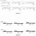
- FIG. 7 provides a schematic diagram of this embodiment.
- trisomy 21 is exemplified, however, this embodiment is not limited to trisomy 21 and may be used with other trisomies simultaneously or as separate assays.
- probes used in assays like TAQMAN (hydrolysis probes, Invitrogen, Inc.) will hybridize to only one region in the genome.
- an alternative strategy is to identify regions that are present at multiple sites throughout the genome. Those regions would ideally be widely distributed throughout the genome and be flanked by unique DNA sequences that would allow the design of sequence specific PCR primers that only amplify a single site in the genome which has a probe hybridized to it.
- the probe sequences could be spread throughout the entire genome or specific for only one chromosome. The greater the number of sites present in the genome and the more evenly spread out the probe sequence, the more universal the probe set will be.
- An advantage of this assay is that the probe concentration may be independent of the number of target sites.
- FIG 8 provides a schematic of such an assay.
- trisomy 21 is exemplified, however, this embodiment is not limited to trisomy 21 and may be used with other trisomies simultaneously or as separate assays.
- probes and PCR primer combinations are selected for chromosome 21 and for a reference chromosome, such as chromosome 1.
- the particular probe that is selected for chromosome 21 is present in chromosome 21 at multiple locations. However, the probe site could also be present in other chromosomes, even chromosome 1. This probe would be labeled with dye-1.
- PCR primers are designed that flank the probe sites but that selectively amplify only one region in chromosome 21 that contains a probe for each PCR primer pair.
- the number of PCR primer pairs that are used could be a subset of the probe sites on chromosome 21 or the entire set on chromosome 21.
- Signal is generated at each site in which a probe is targeted that is also targeted by the PCR primers.
- Other regions to which the probe can hybridize do not generate signal unless also targeted by the PCR primers. This way even though the probes hybridize to other chromosomes or genomic regions they cannot generate signal.
- a second probe which also hybridizes to multiple locations in the genome and in this case at least to some regions on chromosome 1 is labeled with dye-2.
- Sequence specific PCR primers that flank some of the probes on chromosome 1 are designed. As with the first probe, it may hybridize to multiple other locations in the genome other than chromosome 1. However, only the probes targeted to chromosome 1 that also included in the amplicons generated by the PCR primers will produce signal. In this way a single probe with a single dye can target multiple regions in a particular chromosome and generate a signal in digital PCR. The ability to target multiple regions gives more statistical power to identify small changes in relative copy numbers between chromosomes that allow the identification of chromosomal aneuploidy.
- the specific sequence of the probe is present on both chromosomes, thus making it impossible to identify if a specific target sequence is present in a reaction, only presence or absence of some portions of the chromosome can be determined, but not the target on the chromosome.
- two different probes with two different dyes are designed to distinguish between signals generated for different chromosomes.
- Multiple chromosomes can also be targeted with a single dye by varying the signal produced by each probe. Because these reactions are carried out in droplets with very precise volumes, assay signal intensities can be adjusted by varying such things as the concentration of probes in each droplet.
- Two different probes with the same dye can be tuned to generate different final intensities in a droplet. In this way a droplet positive for one chromosome can be distinguished from another droplet containing a different chromosome.
- Hybridization reactions were set up containing 50 ng of genomic DNA, 3.75 femtomole each of two or four hybridization tiles (Table 1) with a 1x concentration of MLPA buffer (MRC-Holland) in a total volume of 25 ⁇ L. Table 1. Hybridization Tile Sequences.
- DMDe8 Hyb B2 and DMDi3 B2 are represented in FIG. 10 as the "P1 - a tile”.
- DMDe8 Hyb A2 and DMDi3 A2 are represented in FIG. 10 as the "P1' - code 1 - b" tile.
- the DMDe8 Hyb A2 and DMDe8 Hyb B2 tile combination is represented by the Chr21-1 tile combination in FIG. 10 and the DMDi3 Hyb A2 and DMDi3 Hyb B2 tile combination is represented in FIG. 10 as the Chr21-2 tile combination.
- Each tile is made up of two parts.
- the first part (hyb A) consists of a sequence specific region that will hybridize to the denatured DNA.
- the 5' end of hyb A also contains the forward primer sequence and the probe sequence.
- the second part of the tile (hyb B) contains a sequence specific region and the sequence for the reverse primer.
- the genomic DNA was denatured at 98°C degrees for five minutes.
- the DNA was then added to a mixture of MLPA Buffer and 3.75 femtomol of tiles. Three separate hybridization reactions were set up:
- a ligation reaction was set up. Three ⁇ L each of Buffer A (MRC-Holland) and Buffer B (MRC-Holland) were mixed with one ⁇ L of Ligase (MRC-Holland). This mixture was brought to a final volume of 32 uL with water and mixed with the hybridization reaction. The samples were then cycled using the following conditions: 54°C for 15 minutes, 98°C degrees for 5 minutes and then a hold at 20°C until ready to continue.
- Buffer A MRC-Holland
- Buffer B MRC-Holland
- a PCR reaction mix was made consisting of 1X Genotyping master mix (Life Technologies 4371355 - TaqMan Genotyping Buffer), 0.9uM PCR primer, RDT stabilizer, 0.2uM FAM probe and 3ul ligated sample.
- the ligation reactions were processed by the RainDance Source instrument to generate approximately 5 million 5pL droplets that were thermal cycled using the following conditions: Temp Time 1 95 °C 10 min 2 95 °C 15 sec 3 58 °C 15 sec 4 60 °C 45 sec 5 go to step 2 and repeat 44 times 6 4 °C Long
- FIGS. 11-13 show the scatter plot results for each of the three reactions.
- the white box in FIGS. 11-13 contain the FAM fluorescent positive droplets which indicate the presence of a DNA fragment corresponding to the X chromosome which is the target chromosome in this case.
- FIGS. 11-13 contain the count of PCR positive droplets (FAM Drops). The number of FAM positive droplets for each reaction were:
Landscapes
- Chemical & Material Sciences (AREA)
- Life Sciences & Earth Sciences (AREA)
- Health & Medical Sciences (AREA)
- Organic Chemistry (AREA)
- Proteomics, Peptides & Aminoacids (AREA)
- Engineering & Computer Science (AREA)
- Zoology (AREA)
- Wood Science & Technology (AREA)
- Genetics & Genomics (AREA)
- Analytical Chemistry (AREA)
- Bioinformatics & Cheminformatics (AREA)
- Biotechnology (AREA)
- General Engineering & Computer Science (AREA)
- Immunology (AREA)
- General Health & Medical Sciences (AREA)
- Molecular Biology (AREA)
- Biophysics (AREA)
- Microbiology (AREA)
- Biochemistry (AREA)
- Physics & Mathematics (AREA)
- Pathology (AREA)
- Biomedical Technology (AREA)
- Hospice & Palliative Care (AREA)
- Oncology (AREA)
- Chemical Kinetics & Catalysis (AREA)
- Plant Pathology (AREA)
- Crystallography & Structural Chemistry (AREA)
- Bioinformatics & Computational Biology (AREA)
- Measuring Or Testing Involving Enzymes Or Micro-Organisms (AREA)
Abstract
Description
- The present application claims the benefit of and priority to
U.S. provisional application serial number 61/597,611, filed February 10, 2012 - The invention generally relates to method for screening for a condition in a subject.
- Assays have been developed that rely on analyzing nucleic acid molecules from bodily fluids for the presence of abnormalities, thus leading to early diagnosis of certain conditions such as cancer or fetal aneuploidy. In a typical bodily fluid sample however, a majority of the nucleic acid is degraded, and any altered nucleic acids containing an abnormality of interest are present in small amounts (e.g., less than 1%) relative to a total amount of nucleic acids in the sample. This results in a failure to detect the small amount of abnormal nucleic acid.
- Amplification based approaches (e.g., polymerase chain reaction (PCR), digital PCR, quantitative PCR) have previously been employed to attempt to detect these abnormalities. However, due to the stochastic nature of the amplification reaction, a population of molecules that is present in a small amount in the sample often is overlooked. In fact, if rare nucleic acid is not amplified in the first few rounds of amplification, it becomes increasingly unlikely that the rare event will ever be detected. Thus, the resulting biased post-amplification nucleic acid population does not represent the true condition of the sample from which it was obtained.
- The advent of next generation sequencing methods, such as those commercially available from Roche (454 and SOLiD), Illumina/Solexa, Pacific Biosciences, and Life Technologies / Ion Torrent allow for the highly sensitive detection of the small population of abnormal nucleic acids in a sample, generally without the need for amplification of the nucleic acid in the sample. However, sequencing instruments are very expensive and these sequencing methods still require a large amount of data (for example, approximately 1,000,000 sequencing reads) to reliably identify an abnormal nucleic acid in a sample. Thus, sequencing is a very costly approach that still requires a significant amount of data in order to identify the presence of a population of abnormal nucleic acids in a sample.
- Methods of the invention are able to identify an altered nucleic acid containing an abnormality of interest that is present in small amounts (e.g., less than 1%) relative to a total amount of nucleic acids in a sample. Methods of the invention are accomplished by designing a plurality of binders (e.g., 10 binders, 100 binders, 1,000 binders) against the nucleic acid that includes the abnormality of interest (i.e., the target nuclei acid). The binders each include a region that hybridizes to a different location on the target nucleic acid, and thus, all of the binders hybridize to the target nucleic acid at once. The target nucleic acid is separated from the sample and the hybridized binders are analyzed (e.g., PCR, digital PCR, qPCR, sequencing, etc.). This approach effectively increases the number of counts and confidence in the number of counts by a factor given by the number of binders against the target nucleic acid. For example, assuming only 1,000 genome equivalents of the target nucleic acid in the sample are available, and there are 1,000 binders, each of which binds to a different location on the target, there are now 1,000,000 target readouts, which is enough to identify a rare abnormal nucleic acid in a sample.
- In certain aspects, methods of the invention involve obtaining a pool of nucleic acids from a sample, incubating the nucleic acids with first and second sets of binders, in which the first set binds uniquely to different regions of a target nucleic acid in the pool, the second set binds uniquely to different regions of a reference nucleic acid in the pool, and the first and second sets include different detectable labels, removing unbound binders, detecting the labels, and screening for a condition based upon results of the detecting step.
- Detecting of the label may be accomplished by any analytical method known in the art. In certain embodiments, the detectable labels are barcode sequences and detecting the label includes sequencing the barcodes. In embodiments that using sequencing, screening for the condition may involve counting a number of barcodes from the first set, counting a number of barcodes from the second set, and determining whether a statistical difference exists between the number of barcodes from the first set and the number of barcodes from the second set.
- In other embodiments, detecting the label may be accomplished by an amplification based technique, such as PCR, digital PCR, or qPCR. In specific embodiments, digital PCR is used to detect the labels. In these embodiments, after the removing step, the method further includes compartmentalizing bound binders of the first and second set into compartmentalized portions, the compartmentalized portions including, on average, either the first set of binders or the second set of binders, and amplifying binders in the compartmentalized portions. Compartmentalizing may involve diluting the sample such that it may be dispensed into different wells of a multi-well plate in a manner such that each well includes, on average, either the first set of binders or the second set of binders. Other exemplary compartmentalizing techniques are shown for example in,
Griffiths et al. (U.S. patent number 7,968,287 ) andLink et al. (U.S. patent application number 2008/0014589 ), the content of each of which is incorporated by reference herein in its entirety. - In certain embodiments, the compartmentalizing involves forming droplets and the compartmentalized portions are the droplets. An exemplary method for forming droplets involves flowing a stream of sample fluid including the amplicons such that it intersects two opposing streams of flowing carrier fluid. The carrier fluid is immiscible with the sample fluid. Intersection of the sample fluid with the two opposing streams of flowing carrier fluid results in partitioning of the sample fluid into individual sample droplets. The carrier fluid may be any fluid that is immiscible with the sample fluid. An exemplary carrier fluid is oil, particularly, a fluorinated oil. In certain embodiments, the carrier fluid includes a surfactant, such as a fluorosurfactant. The droplets may be flowed through channels.
- Generally, the binders of the first set include the same universal primer site and the binders of the second set include the same universal primer site, in which the primer sites of the first and second binders are different. Each compartmentalized portion further includes universal primers that bind the universal priming sites of the binders of the first set and universal primers that bind the universal priming sites of the binders of the second set. The compartmentalized portions further include probes that bind the detectable label of the first set of binders and probes that bind the detectable label of the second set of binders. An amplification reaction (e.g., PCR) is conducted in each compartmentalized portion, and the first probes are allowed to bind to the detectable label of the first set of binders, and the second probes are allowed to bind to the detectable label of the second set of binders. In such methods, the probes are optically labeled probes and detecting the label includes optically detecting the bound probes.
- In such methods, screening for the condition may involve counting a number of compartmentalized portions including the first detectable label, counting a number of compartmentalized portions comprising the second detectable label, and determining whether a statistical difference exists between the number of compartmentalized portions comprising the first detectable label and the number of compartmentalized portions comprising the second detectable label.
- Methods of the invention may be used to screen for any condition. In certain embodiments, the condition is fetal aneuploidy. In particular embodiments, the fetal aneuploidy is trisomy 21 (Down syndrome). To look for fetal aneuploidy, one can use any maternal sample that may include fetal cell-free circulating nucleic acid. Exemplary samples include blood, plasma, or serum. In these embodiments, the target nucleic acid is nucleic acid of
chromosome 21 and the first set of binders binds to the nucleic acid ofchromosome 21 in the pool. The second set of binders binds nucleic acid of a reference chromosome in the pool. - In certain embodiments, the detectable labels are barcode sequences and detecting the label includes sequencing the barcodes. In embodiments that using sequencing, screening for the condition may involve counting a number of barcodes from the first set, counting a number of barcodes from the second set, and determining whether a statistical difference exists between the number of barcodes from the first set and the number of barcodes from the second set. In other embodiments, detecting the label may be accomplished by an amplification based technique, such as PCR, digital PCR, or qPCR. In specific embodiments, digital PCR is used to detect the labels. In such methods, screening for the condition may involve counting a number of compartmentalized portions including the first detectable label, counting a number of compartmentalized portions comprising the second detectable label, and determining whether a statistical difference exists between the number of compartmentalized portions comprising the first detectable label and the number of compartmentalized portions comprising the second detectable label.
- In other embodiments, methods of the invention are used to screen a subject for cancer generally. In these embodiments, the first set of binders binds genomic regions of the nucleic acids associated with known mutations involved in different cancers and the second set of binders binds genomic regions of the nucleic acids that are not mutated. In embodiments that using sequencing, screening for the condition may involve counting a number of barcodes from the first set, counting a number of barcodes from the second set, and determining whether a statistical difference exists between the number of barcodes from the first set and the number of barcodes from the second set. In other embodiments, detecting the label may be accomplished by an amplification based technique, such as PCR, digital PCR, or qPCR. In specific embodiments, digital PCR is used to detect the labels. In such methods, screening for the condition may involve counting a number of compartmentalized portions including the first detectable label, counting a number of compartmentalized portions comprising the second detectable label, and determining whether a statistical difference exists between the number of compartmentalized portions comprising the first detectable label and the number of compartmentalized portions comprising the second detectable label.
- Another aspect of the invention provides methods for screening for a condition in a subject, that involve obtaining a pool of different nucleic acid from a sample, compartmentalizing the pool of nucleic acids into compartmentalized portions, the compartmentalized portions including, on average, either a first set of binders or a second set of binders, wherein the first and second sets comprise different detectable labels and the first and second sets bind to different nucleic acids in the pool, amplifying only binders in the compartmentalized portions that bound to the nucleic acid, detecting the labels, and screening for a condition based upon results of the detecting step.
-
-
Figures 1A-B show one embodiment of first and second binders of the invention. -
Figures 2A-B show another embodiments of binders of the invention. -
Figures 3A-B shows the binders ofFigures 2A-B in circularized form. -
Figures 4A-B illustrate how circularized binders are amplified while non-circularized binders are not amplified. -
Figures 5A-B shows an exemplary embodiment of a device for droplet formation. -
Figure 6 shows an embodiment of using methods of the invention to screen for fetal aneuploidy, particularly trisomy 21 (Down syndrome). -
Figure 7 shows an embodiment of a multiple TAQMAN (hydrolysis probes, Invitrogen, Inc.) assay with assay specific probes. A first assay is conducted onchromosome 21. p21-1 to p21-n are all different target sequences onchromosome 21, but the same fluorophore. f21-1 to f21-n are the forward PCR primers and r21-1 to r21-n are the reverse PCR primers. Another assay is conducted on a normalization chromosome, in this case,chromosome 1. However, the second assay uses a different set of probes and a different color. p1-1 to p1-n are all different target sequences onchromosome 1, but the same fluorophore. f1-1 to f1-n are the forward PCR primers and r1-1 to r1-n are the reverse PCR primers. -
Figure 8 shows an embodiment of an assay that uses a set of probes that hybridize to multiple regions in the genome. For this embodiment, a set of primers is selected that flank a common probe site onchromosome 21. Additionally, a set of primers is selected that flank a different common probe sight onchromosome 1. As compared to the embodiment described inFigure 7 , the assay in this embodiment reduces the number of different probes that have to be in the mixture, thereby decreasing the amount of background fluorescence. In this embodiment, the specificity may primarily or exclusively come from the primers as the probes may hybridize to many locations throughout the genome. -
Figure 9 panels A-D show an embodiment of an assay that uses multiple primers to each ofchromosome 21 andchromosome 1, in which the primers have a chromosome specific probe location on the primer. Panel A showschromosome 21 with a set of forward primers (f21-1, f21-2, ... f21-n) and a set of reverse primers (r21-1, r21-2, ... r21-n). Each of the reverse primers has a universal probe annealing site (p'21) at the end. Panel B shows PCR product with probe annealing site on the end. p21 is a universal probe that hybridizes to all PCR amplicons forchromosome 21. f21-1 to f21-n are the forward PCR primers and r21-1 to r21-n are the reverse PCR primers. Panel C showschromosome 1 with a set of forward primers (f1-1, f1-2, ... f1-n) and a set of reverse primers (r1-1, r1-2, ... r1-n). Each of the reverse primers has a universal probe annealing site (p'1) at the end. Panel D shows PCR product with probe annealing site on the end. p1 is a universal probe that hybridizes to all PCR amplicons forchromosome 1. f1-1 to f1-n are the forward PCR primers and r1-1 to r1-n are the reverse PCR primers. -
Figure 10 shows a number of TAGS. Each TAG is constructed from an 'a' and a 'b' portion such that a complete TAG is constructed by ligation or a fill and ligate process.
It is then likely to be unnecessary to remove unbound tags. However, in some cases it may be desirable to remove unbound tags, in which case they can be removed by using 3' & 5' exonuclease and protected ends on the half TAGs.
In scenarios where there is a limiting amount of starting DNA, multiple TAGS can be generated from the same starting target by melting off of the complete TAG and then annealing 'a' and 'b' fragments. This would be a linear amplification of the number of complete TAGS constructed. -
Figure 11 shows a scatter plot for the reaction DMDi3 Hyb A2 + DMDi3 Hyb B2 . -
Figure 12 shows a scatter plot for the reaction DMDe8 Hyb A2 + DMDe8 Hyb B2 -
Figure 13 shows a scatter plot for the reaction DMDe8 Hyb A2 + DMDe8 Hyb B2 tile + DMDi3 Hyb A2 + DMDi3 Hyb B2. - The invention generally relates to method for screening for a condition in a subject. In certain embodiments, methods of the invention involve obtaining a pool of nucleic acids from a sample, incubating the nucleic acids with first and second sets of binders, in which the first set binds uniquely to different regions of a target nucleic acid in the pool, the second set binds uniquely to different regions of a reference nucleic acid in the pool, and the first and second sets include different detectable labels, removing unbound binders, detecting the labels, and screening for a condition based upon results of the detecting step. It is important to note that in methods of the invention, the binders, and not the nucleic acid, is analyzed for the purpose of screening for a condition.
- Nucleic acid is generally is acquired from a sample or a subject. Target molecules for labeling and/or detection according to the methods of the invention include, but are not limited to, genetic and proteomic material, such as DNA, genomic DNA, RNA, expressed RNA and/or chromosome(s). Methods of the invention are applicable to DNA from whole cells or to portions of genetic or proteomic material obtained from one or more cells. For a subject, the sample may be obtained in any clinically acceptable manner, and the nucleic acid templates are extracted from the sample by methods known in the art. Nucleic acid templates can be obtained as described in U.S. Patent Application Publication Number
US2002/0190663 A1, published Oct. 9, 2003 . Generally, nucleic acid can be extracted from a biological sample by a variety of techniques such as those described by Maniatis, et al. (Molecular Cloning: A Laboratory Manual, Cold Spring Harbor, N.Y., pp. 280-281, 1982), the contents of which are incorporated by reference herein in their entirety. - Nucleic acid templates include deoxyribonucleic acid (DNA) and/or ribonucleic acid (RNA). Nucleic acid templates can be synthetic or derived from naturally occurring sources. In one embodiment, nucleic acid templates are isolated from a biological sample containing a variety of other components, such as proteins, lipids and non-template nucleic acids. Nucleic acid templates can be obtained from any cellular material, obtained from an animal, plant, bacterium, fungus, or any other cellular organism. Biological samples for use in the present invention include viral particles or preparations. Nucleic acid templates can be obtained directly from an organism or from a biological sample obtained from an organism, e.g., from blood, urine, cerebrospinal fluid, seminal fluid, saliva, sputum, stool and tissue. In a particular embodiment, nucleic acid is obtained from fresh frozen plasma (FFP). Any tissue or body fluid specimen may be used as a source for nucleic acid for use in the invention. Nucleic acid templates can also be isolated from cultured cells, such as a primary cell culture or a cell line. The cells or tissues from which template nucleic acids are obtained can be infected with a virus or other intracellular pathogen. A sample can also be total RNA extracted from a biological specimen, a cDNA library, viral, or genomic DNA.
- Generally, nucleic acid obtained from biological samples is fragmented to produce suitable fragments for analysis. An advantage of methods of the invention is that they can be performed on nucleic acids that have not been fragmented.
- However, in certain embodiments, nucleic acids are fragmented prior to performing methods of the invention. In one embodiment, nucleic acid from a biological sample is fragmented by sonication. Generally, individual nucleic acid template molecules can be from about 5 bases to about 20 kb.
- A biological sample as described herein may be homogenized or fractionated in the presence of a detergent or surfactant. The concentration of the detergent in the buffer may be about 0.05% to about 10.0%. The concentration of the detergent can be up to an amount where the detergent remains soluble in the solution. In a preferred embodiment, the concentration of the detergent is between 0.1% to about 2%. The detergent, particularly a mild one that is nondenaturing, can act to solubilize the sample. Detergents may be ionic or nonionic. Examples of nonionic detergents include triton, such as the Triton® X series (Triton® X-100 t-Oct-C6H4-(OCH2-CH2)xOH, x=9-10, Triton® X-100R, Triton® X-114 x=7-8), octyl glucoside, polyoxyethylene(9)dodecyl ether, digitonin, IGEPAL® CA630 octylphenyl polyethylene glycol, n-octyl-beta-D-glucopyranoside (betaOG), n-dodecyl-beta, Tween® 20 polyethylene glycol sorbitan monolaurate, Tween® 80 polyethylene glycol sorbitan monooleate, polidocanol, n-dodecyl beta-D-maltoside (DDM), NP-40 nonylphenyl polyethylene glycol, C12E8 (octaethylene glycol n-dodecyl monoether), hexaethyleneglycol mono-n-tetradecyl ether (C14EO6), octyl-beta-thioglucopyranoside (octyl thioglucoside, OTG), Emulgen, and polyoxyethylene 10 lauryl ether (C12E10). Examples of ionic detergents (anionic or cationic) include deoxycholate, sodium dodecyl sulfate (SDS), N-lauroylsarcosine, and cetyltrimethylammoniumbromide (CTAB). A zwitterionic reagent may also be used in the purification schemes of the present invention, such as Chaps, zwitterion 3-14, and 3-[(3-cholamidopropyl)dimethylammonio]-1-propanesulf-onate. It is contemplated also that urea may be added with or without another detergent or surfactant.
- Lysis or homogenization solutions may further contain other agents, such as reducing agents. Examples of such reducing agents include dithiothreitol (DTT), .beta.-mercaptoethanol, DTE, GSH, cysteine, cysteamine, tricarboxyethyl phosphine (TCEP), or salts of sulfurous acid.
- Once obtained, the nucleic acid is denatured by any method known in the art to produce single stranded nucleic acid templates and a pair of first and second oligonucleotides is hybridized to the single stranded nucleic acid template such that the first and second oligonucleotides flank a target region on the template.
- Methods of the invention involve using first and second sets of binders. In certain embodiments, the first set of binders generally have the structure shown in
Figure 1A and the second set of binders have a structure as shown inFigure 1B . Each binder includes a pair of universal forward and reverse primer sites (P1 and P1' and P2 and P2'). All of the binders of the first set include the same set of universal primer sites. All of the binders of the second set include the same set of universal primer sites. The primers sites of the first set are different than the primer sites of the second set, i.e., P1 and P1' are different than P2 and P2'. Each of the binders includes a detectable label code site. All of the binders of the first set include the same detectable label code site. All of the binders of the second set include the same detectable label code site. The detectable label code site of the first set is different than the detectable label code site of the second set. - Each of the binders in the first set includes a target sequence portion. The target sequence portion for each of the binders of the first set is a sequence that binds to a region of the target nucleic acid. However, the target sequence portion of each binder of the first set is different, so that each binder of the first set may bind to a different location of the target nucleic acid.
- Each of the binders in the second set includes a reference sequence portion. The reference sequence portion for each of the binders of the second set is a sequence that binds to a region of the reference nucleic acid. However, the reference sequence portion of each binder of the second set is different, so that each binder of the second set may bind to a different location of the reference nucleic acid.
- In certain embodiments, the first set of binders generally have the structure shown in
Figure 2A and the second set of binders have a structure as shown inFigure 2B . In this embodiments, the first and second binders are constructed from an "a" portion and a "b" portion such that a complete binder is constructed by ligation or a fill and ligate process. This type of structure avoids the need to remove unbound binders, because only fully formed binders (i.e., those with an "a" and "b") portion can be analyzed.Figures 3A-B show complete binders. If the binder is constructed such that the digital PCR annealing region is only amplified by the primers when a circle is constructed, then a probe, such as a Taqman probe, would not be hydrolyzed exponentially as the PCR reaction proceeded. This is illustrated inFigures 4A-B . - The type of detectable label code site in the first and second binders will depend on the binder detection technique to be employed. If the binder detection technique is sequencing, the detectable label code site will be a barcode sequence. In these embodiments, all of the binders of the first set will have the same barcode sequence and all of the binders of the second set will have the same barcode sequence. The barcode sequence of the first set of binders is different than the barcode sequence of the second set of binders.
- If the binder detection technique is probe hybridization, the detectable label code site will be a sequence that hybridizes with the probe. In these embodiments, all of the binders of the first set will hybridize with a first probe and all of the binders of the second set will hybridize with a second probe. The first and second probe are different and include different labels.
- Methods of synthesizing oligonucleotide are known in the art. See, e.g., Sambrook et al. (DNA microarray: A Molecular Cloning Manual, Cold Spring Harbor, N.Y., 2003) or Maniatis, et al. (Molecular Cloning: A Laboratory Manual, Cold Spring Harbor, N.Y., 1982), the contents of each of which are incorporated by reference herein in their entirety. Suitable methods for synthesizing oligonucleotide are also described in Caruthers (Science 230:281-285, 1985), the contents of which are incorporated by reference. Oligonucleotides can also be obtained from commercial sources such as Operon Technologies, Amersham Pharmacia Biotech, Sigma, and Life Technologies. The oligonucleotides can have an identical melting temperature. The lengths of the oligonucleotides can be extended or shortened at the 5' end or the 3' end to produce oligonucleotides with desired melting temperatures. Also, the annealing position of each oligonucleotide can be designed such that the sequence and length of the probe yield the desired melting temperature. The simplest equation for determining the melting temperature of probes smaller than 25 base pairs is the Wallace Rule (Td=2(A+T)+4(G+C)). Computer programs can also be used to design oligonucleotides, including but not limited to Array Designer Software (Arrayit Inc.), Oligonucleotide Probe Sequence Design Software for Genetic Analysis (Olympus Optical Co.), NetPrimer, and DNAsis from Hitachi Software Engineering. The TM (melting temperature) of each probe is calculated using software programs such as Oligo Design, available from Invitrogen Corp.
- In certain embodiments, reaction conditions of high stringency are used to ensure great specificity between the probes and the code sites on the binders. Nucleic acid hybridization may be affected by such conditions as salt concentration, temperature, or organic solvents, in addition to base composition, length of complementary strands, and number of nucleotide base mismatches between hybridizing nucleic acids, as is readily appreciated by those skilled in the art. Stringency of hybridization reactions is readily determinable by one of ordinary skill in the art, and generally is an empirical calculation dependent upon sequence length, washing temperature, and salt concentration. In general, longer sequences require higher temperatures for proper annealing, while shorter sequences need lower temperatures. Hybridization generally depends on the ability of denatured DNA to re-anneal when complementary strands are present in an environment below its melting temperature. The higher the degree of desired homology between the sequence and hybridizable sequence, the higher the relative temperature that can be used. As a result, it follows that higher relative temperatures would tend to make the reaction conditions more stringent, while lower temperatures less so. For additional details and explanation of stringency of hybridization reactions, see Ausubel et al., Current Protocols in Molecular Biology, Wiley Interscience Publishers, (1995), the contents of which are incorporated by reference herein in their entirety.
- Stringent conditions or high stringency conditions typically: (1) employ low ionic strength and high temperature for washing, for example 0.015 M sodium chloride/0.0015 M sodium citrate/0.1% sodium dodecyl sulfate at 50°C.; (2) employ during hybridization a denaturing agent, such as formamide, for example, 50% (v/v) formamide with 0.1% bovine serum albumin/0.1% Ficoll/0.1% polyvinylpyrrolidone/50 mM sodium phosphate buffer at pH 6.5 with 750 mM sodium chloride, 75 mM sodium citrate at 42°C.; or (3) employ 50% formamide, 5 x SSC (0.75 M NaCl, 0.075 M sodium citrate), 50 mM sodium phosphate (pH 6.8), 0.1% sodium pyrophosphate, 5 x Denhardt's solution, sonicated salmon sperm DNA (50 .mu.g/ml), 0.1% SDS, and 10% dextran sulfate at 42°C, with washes at 42°C in 0.2 x SSC (sodium chloride/sodium citrate) and 50% formamide at 55°C, followed by a high-stringency wash consisting of 0.1 x SSC containing EDTA at 55°C.
- In other embodiments, reaction conditions of moderate stringency are used for hybridization of the first and second oligonucleotides to binding regions on the template nucleic acid. Moderately stringent conditions may be identified as described by Sambrook et al., Molecular Cloning: A Laboratory Manual, New York: Cold Spring Harbor Press, 1989 (the contents of which are incorporated by reference herein in their entirety and include the use of washing solution and hybridization conditions (e.g., temperature, ionic strength and % SDS) less stringent that those described above. An example of moderately stringent conditions is overnight incubation at 37°C in a solution comprising: 20% formamide, 5 x SSC (150 mM NaCl, 15 mM trisodium citrate), 50 mM sodium phosphate (pH 7.6), 5 x Denhardt's solution, 10% dextran sulfate, and 20 mg/ml denatured sheared salmon sperm DNA, followed by washing the filters in 1 x SSC at about 37°C to 50°C. The skilled artisan will recognize how to adjust the temperature, ionic strength, etc. as necessary to accommodate factors such as sequence length and the like.
- Oligonucleotides suitable for use in the present invention include those formed from nucleic acids, such as RNA and/or DNA, nucleic acid analogs, locked nucleic acids, modified nucleic acids, and chimeric probes of a mixed class including a nucleic acid with another organic component such as peptide nucleic acids. Exemplary nucleotide analogs include phosphate esters of deoxyadenosine, deoxycytidine, deoxyguanosine, deoxythymidine, adenosine, cytidine, guanosine, and uridine. Other examples of non-natural nucleotides include a xanthine or hypoxanthine; 5-bromouracil, 2-aminopurine, deoxyinosine, or methylated cytosine, such as 5-methylcytosine, and N4-methoxydeoxycytosine. Also included are bases of polynucleotide mimetics, such as methylated nucleic acids, e.g., 2'-O-methRNA, peptide nucleic acids, modified peptide nucleic acids, and any other structural moiety that can act substantially like a nucleotide or base, for example, by exhibiting base-complementarity with one or more bases that occur in DNA or RNA.
- The length of the oligonucleotide probe is not critical, as long as the oligonucleotides are capable of hybridizing to the code sites of the binders. In fact, oligonucleotides may be of any length. For example, oligonucleotides may be as few as 5 nucleotides, or as much as 5000 nucleotides. Exemplary oligonucleotides are 5-mers, 10-mers, 15-mers, 20-mers, 25-mers, 50-mers, 100-mers, 200-mers, 500-mers, 1000-mers, 3000-mers, or 5000-mers. Methods for determining an optimal oligonucleotides length are known in the art. See, e.g.,
Shuber (U.S. patent number 5,888,778 ). The first and second oligonucleotides do not have to be of the same length. In certain embodiments, the first and second oligonucleotides are the same length, while in other embodiments, the first and second oligonucleotides are of different lengths. - The reaction time will depend on the different factors discussed above, e.g., stringency conditions, probe length, probe design, etc.
- Generally, the probes will include a detectable label that is directly or indirectly detectable. Preferred labels include optically-detectable labels, such as fluorescent labels. Examples of fluorescent labels include, but are not limited to, 4-acetamido-4'-isothiocyanatostilbene-2,2'disulfonic acid; acridine and derivatives: acridine, acridine isothiocyanate; 5-(2'-aminoethyl)aminonaphthalene-1-sulfonic acid (EDANS); 4-amino-N-[3-vinylsulfonyl)phenyl]naphthalimide-3,5 disulfonate; N-(4-anilino-1-naphthyl)maleimide; anthranilamide; BODIPY; Brilliant Yellow; coumarin and derivatives; coumarin, 7-amino-4-methylcoumarin (AMC, Coumarin 120), 7-amino-4-trifluoromethylcouluarin (Coumaran 151); cyanine dyes; cyanosine; 4',6-diaminidino-2-phenylindole (DAPI); 5'5"-dibromopyrogallol-sulfonaphthalein (Bromopyrogallol Red); 7-diethylamino-3-(4'-isothiocyanatophenyl)-4-methylcoumarin; diethylenetriamine pentaacetate; 4,4'-diisothiocyanatodihydro-stilbene-2,2'-disulfonic acid; 4,4'-diisothiocyanatostilbene-2,2'-disulfonic acid; 5-[dimethylamino]naphthalene-1-sulfonyl chloride (DNS, dansylchloride); 4-dimethylaminophenylazophenyl-4'-isothiocyanate (DABITC); eosin and derivatives; eosin, eosin isothiocyanate, erythrosin and derivatives; erythrosin B, erythrosin, isothiocyanate; ethidium; fluorescein and derivatives; 5-carboxyfluorescein (FAM), 5-(4,6-dichlorotriazin-2-yl)aminofluorescein (DTAF), 2',7'-dimethoxy-4'5'-dichloro-6-carboxyfluorescein, fluorescein, fluorescein isothiocyanate, QFITC, (XRITC); fluorescamine; IR144; IR1446; Malachite Green isothiocyanate; 4-methylumbelliferoneortho cresolphthalein; nitrotyrosine; pararosaniline; Phenol Red; B-phycoerythrin; o-phthaldialdehyde; pyrene and derivatives: pyrene, pyrene butyrate, succinimidyl 1-pyrene; butyrate quantum dots; Reactive Red 4 (Cibacron.TM. Brilliant Red 3B-A) rhodamine and derivatives: 6-carboxy-X-rhodamine (ROX), 6-carboxyrhodamine (R6G), lissamine rhodamine B sulfonyl chloride rhodamine (Rhod), rhodamine B, rhodamine 123, rhodamine X isothiocyanate, sulforhodamine B,
sulforhodamine 101, sulfonyl chloride derivative of sulforhodamine 101 (Texas Red); N,N,N',N'tetramethyl-6-carboxyrhodamine (TAMRA); tetramethyl rhodamine; tetramethyl rhodamine isothiocyanate (TRITC); riboflavin; rosolic acid; terbium chelate derivatives; Cy3; Cy5; Cy5.5; Cy7; IRD 700; IRD 800; La Jolta Blue; phthalo cyanine; and naphthalo cyanine. Preferred fluorescent labels are cyanine-3 and cyanine-5. Labels other than fluorescent labels are contemplated by the invention, including other optically-detectable labels. - The pool of nucleic acids is incubated with first and second sets of binders under conditions that allow the first set of binders to bind to target nucleic acids in the pool and the second set of binders to bind the reference nucleic acids in the pool.
- In certain embodiments, reaction conditions of high stringency are used to ensure great specificity between the probes and the code sites on the binders. Nucleic acid hybridization may be affected by such conditions as salt concentration, temperature, or organic solvents, in addition to base composition, length of complementary strands, and number of nucleotide base mismatches between hybridizing nucleic acids, as is readily appreciated by those skilled in the art. Stringency of hybridization reactions is readily determinable by one of ordinary skill in the art, and generally is an empirical calculation dependent upon sequence length, washing temperature, and salt concentration. In general, longer sequences require higher temperatures for proper annealing, while shorter sequences need lower temperatures. Hybridization generally depends on the ability of denatured DNA to re-anneal when complementary strands are present in an environment below its melting temperature. The higher the degree of desired homology between the sequence and hybridizable sequence, the higher the relative temperature that can be used. As a result, it follows that higher relative temperatures would tend to make the reaction conditions more stringent, while lower temperatures less so. For additional details and explanation of stringency of hybridization reactions, see Ausubel et al., Current Protocols in Molecular Biology, Wiley Interscience Publishers, (1995), the contents of which are incorporated by reference herein in their entirety.
- Stringent conditions or high stringency conditions typically: (1) employ low ionic strength and high temperature for washing, for example 0.015 M sodium chloride/0.0015 M sodium citrate/0.1% sodium dodecyl sulfate at 50°C.; (2) employ during hybridization a denaturing agent, such as formamide, for example, 50% (v/v) formamide with 0.1% bovine serum albumin/0.1% Ficoll/0.1% polyvinylpyrrolidone/50 mM sodium phosphate buffer at pH 6.5 with 750 mM sodium chloride, 75 mM sodium citrate at 42°C.; or (3) employ 50% formamide, 5 x SSC (0.75 M NaCl, 0.075 M sodium citrate), 50 mM sodium phosphate (pH 6.8), 0.1% sodium pyrophosphate, 5 x Denhardt's solution, sonicated salmon sperm DNA (50 .mu.g/ml), 0.1% SDS, and 10% dextran sulfate at 42°C, with washes at 42°C in 0.2 x SSC (sodium chloride/sodium citrate) and 50% formamide at 55°C, followed by a high-stringency wash consisting of 0.1 x SSC containing EDTA at 55°C.
- In other embodiments, reaction conditions of moderate stringency are used for hybridization of the first and second oligonucleotides to binding regions on the template nucleic acid. Moderately stringent conditions may be identified as described by Sambrook et al., Molecular Cloning: A Laboratory Manual, New York: Cold Spring Harbor Press, 1989 (the contents of which are incorporated by reference herein in their entirety and include the use of washing solution and hybridization conditions (e.g., temperature, ionic strength and % SDS) less stringent that those described above. An example of moderately stringent conditions is overnight incubation at 37°C in a solution comprising: 20% formamide, 5 x SSC (150 mM NaCl, 15 mM trisodium citrate), 50 mM sodium phosphate (pH 7.6), 5 x Denhardt's solution, 10% dextran sulfate, and 20 mg/ml denatured sheared salmon sperm DNA, followed by washing the filters in 1 x SSC at about 37°C to 50°C. The skilled artisan will recognize how to adjust the temperature, ionic strength, etc. as necessary to accommodate factors such as sequence length and the like.
Oligonucleotides suitable for use in the present invention include those formed from nucleic acids, such as RNA and/or DNA, nucleic acid analogs, locked nucleic acids, modified nucleic acids, and chimeric probes of a mixed class including a nucleic acid with another organic component such as peptide nucleic acids. Exemplary nucleotide analogs include phosphate esters of deoxyadenosine, deoxycytidine, deoxyguanosine, deoxythymidine, adenosine, cytidine, guanosine, and uridine. Other examples of non-natural nucleotides include a xanthine or hypoxanthine; 5-bromouracil, 2-aminopurine, deoxyinosine, or methylated cytosine, such as 5-methylcytosine, and N4-methoxydeoxycytosine. Also included are bases of polynucleotide mimetics, such as methylated nucleic acids, e.g., 2'-O-methRNA, peptide nucleic acids, modified peptide nucleic acids, and any other structural moiety that can act substantially like a nucleotide or base, for example, by exhibiting base-complementarity with one or more bases that occur in DNA or RNA. - The reaction time will depend on the different factors discussed above, e.g., stringency conditions, probe length, probe design, etc.
- In certain embodiments, it is important to remove unbound binders. However, this is an optional step based upon the type of binders used with methods of the invention. As discussed above, binders can be constructed such that a removal step is not necessary and methods of the invention can be conducted with or without the removing step, i.e., this is an optional step.
- Any method known in the art may be used for removing unbound binders. For example, the binders can be RNA binders and unbound binders can be removed by exonuclease digestion. Alternatively, the nucleic acid in the sample can be modified with a biotin tag prior to being incubated with the first and second binders. The incubated mixture can be exposed to a streptavidin coated surface, such as magnetic beads such that the nucleic acid in the sample hybridized with the binders binds to the streptavidin coated surface. A magnetic field can then be used to separate the unbound binders from nucleic acid having bound binders. Methods of modifying nucleic acids with biotin and then attaching to a streptavidin coated surface are known in the art, see for example,
Lapidus et al. (U.S. patent number 7,169,560 ),Lapidus et al. (U.S. patent application number 2009/0191565 ),Quake et al. (U.S. patent number 6,818,395 ),Harris (U.S. patent number 7,282,337 ), andQuake et al. (U.S. patent application number 2002/0164629 ), the content of each of which is incorporated by reference herein in its entirety. - Various other attachment methods can be used to anchor or immobilize the nucleic acid molecule to the surface of a substrate. The immobilization can be achieved through direct or indirect bonding to the surface. The bonding can be by covalent linkage. See, Joos et al., Analytical Biochemistry 247:96-101, 1997; Oroskar et al., Clin. Chem. 42:1547-1555, 1996; and Khandjian, Mol. Bio. Rep. 11:107-115, 1986. An example of an attachment is direct amine bonding of a terminal nucleotide of the template or the 5' end of the primer to an epoxide integrated on the surface. The bonding also can be through non-covalent linkage. For example, biotin-streptavidin (Taylor et al., J. Phys. D. Appl. Phys. 24:1443, 1991) and digoxigenin with anti-digoxigenin (Smith et al., Science 253:1122, 1992) are common tools for anchoring nucleic acids to surfaces and parallels.
- In certain embodiments, sequencing is used to detect the code site. Sequencing may be by any method known in the art. DNA sequencing techniques include classic dideoxy sequencing reactions (Sanger method) using labeled terminators or primers and gel separation in slab or capillary, sequencing by synthesis using reversibly terminated labeled nucleotides, pyrosequencing, 454 sequencing, allele specific hybridization to a library of labeled oligonucleotide probes, sequencing by synthesis using allele specific hybridization to a library of labeled clones that is followed by ligation, real time monitoring of the incorporation of labeled nucleotides during a polymerization step, polony sequencing, and SOLiD sequencing. Sequencing of separated molecules has more recently been demonstrated by sequential or single extension reactions using polymerases or ligases as well as by single or sequential differential hybridizations with libraries of probes.
- A sequencing technique that can be used in the methods of the provided invention includes, for example, Helicos True Single Molecule Sequencing (tSMS) (Harris T. D. et al. (2008) Science 320:106-109). In the tSMS technique, a DNA sample is cleaved into strands of approximately 100 to 200 nucleotides, and a polyA sequence is added to the 3' end of each DNA strand. Each strand is labeled by the addition of a fluorescently labeled adenosine nucleotide. The DNA strands are then hybridized to a flow cell, which contains millions of oligo-T capture sites that are immobilized to the flow cell surface. The templates can be at a density of about 100 million templates/cm2. The flow cell is then loaded into an instrument, e.g., HeliScope.TM. sequencer, and a laser illuminates the surface of the flow cell, revealing the position of each template. A CCD camera can map the position of the templates on the flow cell surface. The template fluorescent label is then cleaved and washed away. The sequencing reaction begins by introducing a DNA polymerase and a fluorescently labeled nucleotide. The oligo-T nucleic acid serves as a primer. The polymerase incorporates the labeled nucleotides to the primer in a template directed manner. The polymerase and unincorporated nucleotides are removed. The templates that have directed incorporation of the fluorescently labeled nucleotide are detected by imaging the flow cell surface. After imaging, a cleavage step removes the fluorescent label, and the process is repeated with other fluorescently labeled nucleotides until the desired read length is achieved. Sequence information is collected with each nucleotide addition step. Further description of tSMS is shown for example in
Lapidus et al. (U.S. patent number 7,169,560 ),Lapidus et al. (U.S. patent application number 2009/0191565 ),Quake et al. (U.S. patent number 6,818,395 ),Harris (U.S. patent number 7,282,337 ),Quake et al. (U.S. patent application number 2002/0164629 ), and Braslavsky, et al., PNAS (USA), 100: 3960-3964 (2003), the contents of each of these references is incorporated by reference herein in its entirety. - Another example of a DNA sequencing technique that can be used in the methods of the provided invention is 454 sequencing (Roche) (Margulies, M et al. 2005, Nature, 437, 376-380). 454 sequencing involves two steps. In the first step, DNA is sheared into fragments of approximately 300-800 base pairs, and the fragments are blunt ended. Oligonucleotide adaptors are then ligated to the ends of the fragments. The adaptors serve as primers for amplification and sequencing of the fragments. The fragments can be attached to DNA capture beads, e.g., streptavidin-coated beads using, e.g., Adaptor B, which contains 5'-biotin tag. The fragments attached to the beads are PCR amplified within droplets of an oil-water emulsion. The result is multiple copies of clonally amplified DNA fragments on each bead. In the second step, the beads are captured in wells (pico-liter sized). Pyrosequencing is performed on each DNA fragment in parallel. Addition of one or more nucleotides generates a light signal that is recorded by a CCD camera in a sequencing instrument. The signal strength is proportional to the number of nucleotides incorporated. Pyrosequencing makes use of pyrophosphate (PPi) which is released upon nucleotide addition. PPi is converted to ATP by ATP sulfurylase in the presence of adenosine 5' phosphosulfate. Luciferase uses ATP to convert luciferin to oxyluciferin, and this reaction generates light that is detected and analyzed.
- Another example of a DNA sequencing technique that can be used in the methods of the provided invention is SOLiD technology (Applied Biosystems). In SOLiD sequencing, genomic DNA is sheared into fragments, and adaptors are attached to the 5' and 3' ends of the fragments to generate a fragment library. Alternatively, internal adaptors can be introduced by ligating adaptors to the 5' and 3' ends of the fragments, circularizing the fragments, digesting the circularized fragment to generate an internal adaptor, and attaching adaptors to the 5' and 3' ends of the resulting fragments to generate a mate-paired library. Next, clonal bead populations are prepared in microreactors containing beads, primers, template, and PCR components. Following PCR, the templates are denatured and beads are enriched to separate the beads with extended templates. Templates on the selected beads are subjected to a 3' modification that permits bonding to a glass slide. The sequence can be determined by sequential hybridization and ligation of partially random oligonucleotides with a central determined base (or pair of bases) that is identified by a specific fluorophore. After a color is recorded, the ligated oligonucleotide is cleaved and removed and the process is then repeated.
- Another example of a DNA sequencing technique that can be used in the methods of the provided invention is Ion Torrent sequencing (
U.S. patent application numbers 2009/0026082 ,2009/0127589 ,2010/0035252 ,2010/0137143 ,2010/0188073 ,2010/0197507 ,2010/0282617 ,2010/0300559 ),2010/0300895 ,2010/0301398 , and2010/0304982 ), the content of each of which is incorporated by reference herein in its entirety. In Ion Torrent sequencing, DNA is sheared into fragments of approximately 300-800 base pairs, and the fragments are blunt ended. Oligonucleotide adaptors are then ligated to the ends of the fragments. The adaptors serve as primers for amplification and sequencing of the fragments. The fragments can be attached to a surface and is attached at a resolution such that the fragments are individually resolvable. Addition of one or more nucleotides releases a proton (H+), which signal detected and recorded in a sequencing instrument. The signal strength is proportional to the number of nucleotides incorporated. - Another example of a sequencing technology that can be used in the methods of the provided invention is Illumina sequencing. Illumina sequencing is based on the amplification of DNA on a solid surface using fold-back PCR and anchored primers. Genomic DNA is fragmented, and adapters are added to the 5' and 3' ends of the fragments. DNA fragments that are attached to the surface of flow cell channels are extended and bridge amplified. The fragments become double stranded, and the double stranded molecules are denatured. Multiple cycles of the solid-phase amplification followed by denaturation can create several million clusters of approximately 1,000 copies of single-stranded DNA molecules of the same template in each channel of the flow cell. Primers, DNA polymerase and four fluorophore-labeled, reversibly terminating nucleotides are used to perform sequential sequencing. After nucleotide incorporation, a laser is used to excite the fluorophores, and an image is captured and the identity of the first base is recorded. The 3' terminators and fluorophores from each incorporated base are removed and the incorporation, detection and identification steps are repeated.
- Another example of a sequencing technology that can be used in the methods of the provided invention includes the single molecule, real-time (SMRT) technology of Pacific Biosciences. In SMRT, each of the four DNA bases is attached to one of four different fluorescent dyes. These dyes are phospholinked. A single DNA polymerase is immobilized with a single molecule of template single stranded DNA at the bottom of a zero-mode waveguide (ZMW). A ZMW is a confinement structure which enables observation of incorporation of a single nucleotide by DNA polymerase against the background of fluorescent nucleotides that rapidly diffuse in an out of the ZMW (in microseconds). It takes several milliseconds to incorporate a nucleotide into a growing strand. During this time, the fluorescent label is excited and produces a fluorescent signal, and the fluorescent tag is cleaved off. Detection of the corresponding fluorescence of the dye indicates which base was incorporated. The process is repeated.
- Another example of a sequencing technique that can be used in the methods of the provided invention is nanopore sequencing (Soni G V and Meller A. (2007) Clin Chem 53: 1996-2001). A nanopore is a small hole, of the order of 1 nanometer in diameter. Immersion of a nanopore in a conducting fluid and application of a potential across it results in a slight electrical current due to conduction of ions through the nanopore. The amount of current which flows is sensitive to the size of the nanopore. As a DNA molecule passes through a nanopore, each nucleotide on the DNA molecule obstructs the nanopore to a different degree. Thus, the change in the current passing through the nanopore as the DNA molecule passes through the nanopore represents a reading of the DNA sequence.
- Another example of a sequencing technique that can be used in the methods of the provided invention involves using a chemical-sensitive field effect transistor (chemFET) array to sequence DNA (for example, as described in
US Patent Application Publication No. 20090026082 ). In one example of the technique, DNA molecules can be placed into reaction chambers, and the template molecules can be hybridized to a sequencing primer bound to a polymerase. Incorporation of one or more triphosphates into a new nucleic acid strand at the 3' end of the sequencing primer can be detected by a change in current by a chemFET. An array can have multiple chemFET sensors. In another example, single nucleic acids can be attached to beads, and the nucleic acids can be amplified on the bead, and the individual beads can be transferred to individual reaction chambers on a chemFET array, with each chamber having a chemFET sensor, and the nucleic acids can be sequenced. - Another example of a sequencing technique that can be used in the methods of the provided invention involves using an electron microscope (Moudrianakis E. N. and Beer M. Proc Natl Acad Sci USA. 1965 March; 53:564-71). In one example of the technique, individual DNA molecules are labeled using metallic labels that are distinguishable using an electron microscope. These molecules are then stretched on a flat surface and imaged using an electron microscope to measure sequences.
- In a particular embodiment, the sequencing is single-molecule sequencing-by-synthesis. Single-molecule sequencing is shown for example in
Lapidus et al. (U.S. patent number 7,169,560 ),Quake et al. (U.S. patent number 6,818,395 ),Harris (U.S. patent number 7,282,337 ),Quake et al. (U.S. patent application number 2002/0164629 ), and Braslavsky, et al., PNAS (USA), 100: 3960-3964 (2003), the contents of each of these references is incorporated by reference herein in its entirety. - Briefly, a single-stranded nucleic acid (e.g., DNA or cDNA) is hybridized to oligonucleotides attached to a surface of a flow cell. The single-stranded nucleic acids may be captured by methods known in the art, such as those shown in
Lapidus (U.S. patent number 7,666,593 ). The oligonucleotides may be covalently attached to the surface or various attachments other than covalent linking as known to those of ordinary skill in the art may be employed. Moreover, the attachment may be indirect, e.g., via the polymerases of the invention directly or indirectly attached to the surface. The surface may be planar or otherwise, and/or may be porous or non-porous, or any other type of surface known to those of ordinary skill to be suitable for attachment. The nucleic acid is then sequenced by imaging the polymerase-mediated addition of fluorescently-labeled nucleotides incorporated into the growing strand surface oligonucleotide, at single molecule resolution. - Thus, the invention encompasses methods wherein the nucleic acid sequencing reaction comprises hybridizing a sequencing primer to a single-stranded region of a linearized amplification product, sequentially incorporating one or more nucleotides into a polynucleotide strand complementary to the region of amplified template strand to be sequenced, identifying the base present in one or more of the incorporated nucleotide(s) and thereby determining the sequence of a region of the template strand.
- For the sequence reconstruction process, short reads are stitched together bioinformatically by finding overlaps and extending them. To be able to do that unambiguously, one must ensure that long fragments that were amplified are distinct enough, and do not have similar stretches of DNA that will make assembly from short fragments ambiguous, which can occur, for example, if two molecules in a same well originated from overlapping positions on homologous chromosomes, overlapping positions of same chromosome, or genomic repeat. Such fragments can be detected during sequence assembly process by observing multiple possible ways to extend the fragment, one of which contains sequence specific to end marker. End markers can be chosen such that end marker sequence is not frequently found in DNA fragments of sample that is analyzed and probabilistic framework utilizing quality scores can be applied to decide whether a certain possible sequence extension way represents end maker and thus end of the fragment.
- Overlapping fragments may be computationally discarded since they no longer represent the same initial long molecule. This process allows to treat population of molecules resulting after amplification as a clonally amplified population of disjoint molecules with no significant overlap or homology, which enables sequencing errors to be corrected to achieve very high consensus accuracy and allows unambiguous reconstruction of long fragments. If overlaps are not discarded, then one has to assume that reads may be originating from fragments originating from two homologous chromosomes or overlapping regions of the same chromosome (in case of diploid organism) which makes error correction difficult and ambiguous.
- Computational removal of overlapping fragments obtained from both the 5' and the 3' directions also allows use of quality scores to resolve nearly-identical repeats. Resulting long fragments may be assembled into full genomes using any of the algorithms known in the art for genome sequence assembly that can utilize long reads.
- In addition to de-novo assembly fragments can be used to obtain phasing (assignment to homologous copies of chromosomes) of genomic variants, by observing that under conditions of experiment described in the preferred embodiment long fragments originate from either one of chromosomes, which enables to correlate and co-localize variants detected in overlapping fragments obtained from distinct partitioned portions.
- In certain embodiments, amplification based methods are used to detect the code site. Amplification refers to production of additional copies of a nucleic acid sequence and is generally carried out using polymerase chain reaction or other technologies well known in the art (e.g., Dieffenbach and Dveksler, PCR Primer, a Laboratory Manual, Cold Spring Harbor Press, Plainview, N.Y. [1995]). The amplification reaction may be any amplification reaction known in the art that amplifies nucleic acid molecules, such as polymerase chain reaction, nested polymerase chain reaction, polymerase chain reaction-single strand conformation polymorphism, ligase chain reaction (Barany F. (1991) PNAS 88:189-193; Barany F. (1991) PCR Methods and Applications 1:5-16), ligase detection reaction (Barany F. (1991) PNAS 88:189-193), strand displacement amplification and restriction fragments length polymorphism, transcription based amplification system, nucleic acid sequence-based amplification, rolling circle amplification, and hyper-branched rolling circle amplification.
- Polymerase chain reaction (PCR) refers to methods by
K. B. Mullis (U.S. patent numbers 4,683,195 and4,683,202 , hereby incorporated by reference) for increasing concentration of a segment of a target sequence in a mixture of genomic DNA without cloning or purification. The process for amplifying the target sequence includes introducing an excess of primers (oligonucleotides) to a DNA mixture containing a desired target sequence, followed by a precise sequence of thermal cycling. The present invention includes, but is not limited to, various PCR strategies as are known in the art, for example QPCR, multiplex PCR, assymetric PCR, nested PCR, hotstart PCR, touchdown PCR, assembly PCR, digital PCR, allele specific PCR, methylation specific PCR, reverse transcription PCR, helicase dependent PCR, inverse PCR, intersequence specific PCR, ligation mediated PCR, mini primer PCR, and solid phase PCR, emulsion PCR, and PCR as performed in a thermocycler, droplets, microfluidic reaction chambers, flow cells and other microfluidic devices. - In specific embodiments, digital PCR is used to detect the code sites. For digital PCR embodiments, after the first and second binders have been incubated with the pool of nucleic acids, the pool is diluted so that the sample can be compartmentalized in a manner in which each compartment includes on a single nucleic acid. Any type of compartment generally used for digital PCR may be used with methods of the invention. Exemplary compartments include chambers, wells, droplets, reaction volumes, slugs.
- Poisson statistics dictate the dilution requirements needed to insure that each compartment contains only a single nucleic acid. In particular, the sample concentration should be dilute enough that most of the compartments contain no more than a single nucleic acid with only a small statistical chance that a compartment will contain two or more molecules. The parameters which govern this relationship are the volume of the compartment and the concentration of nucleic acid in the sample solution. The probability that a compartment will contain two or more nucleic acid (NAT≤2) can be expressed as:

- In particular embodiments, the compartmentalized portions are droplets and compartmentalizing involves forming the droplets. Sample droplets may be formed by any method known in the art. The droplets are aqueous droplets that are surrounded by an immiscible carrier fluid. Methods of forming droplets are shown for example in
Link et al. (U.S. patent application numbers 2008/0014589 ,2008/0003142 , and2010/0137163 ),Stone et al. (U.S. patent number 7,708,949 andU.S. patent application number 2010/0172803 ),Anderson et al. (U.S. patent number 7,041,481 and which reissued asRE41,780 ) and European publication numberEP2047910 to Raindance Technologies Inc. The content of each of which is incorporated by reference herein in its entirety. -
Figures 5A-B show an exemplary embodiment of adevice 100 for droplet formation.Device 100 includes aninlet channel 101, andoutlet channel 102, and twocarrier fluid channels Channels junction 105.Inlet channel 101 flows sample fluid to thejunction 105.Carrier fluid channels junction 105.Inlet channel 101 narrows at its distal portion wherein it connects to junction 105 (SeeFigure 5B ).Inlet channel 101 is oriented to be perpendicular tocarrier fluid channels inlet channel 101 tojunction 105, where the sample fluid interacts with flowing carrier fluid provided to thejunction 105 bycarrier fluid channels Outlet channel 102 receives the droplets of sample fluid surrounded by carrier fluid. - The sample fluid is typically an aqueous buffer solution, such as ultrapure water (e.g., 18 mega-ohm resistivity, obtained, for example by column chromatography), 10 mM Tris HCl and 1 mM EDTA (TE) buffer, phosphate buffer saline (PBS) or acetate buffer. Any liquid or buffer that is physiologically compatible with nucleic acid molecules can be used. The carrier fluid is one that is immiscible with the sample fluid. The carrier fluid can be a non-polar solvent, decane (e g., tetradecane or hexadecane), fluorocarbon oil, silicone oil or another oil (for example, mineral oil).
- In certain embodiments, the carrier fluid contains one or more additives, such as agents which reduce surface tensions (surfactants). Surfactants can include Tween, Span, fluorosurfactants, and other agents that are soluble in oil relative to water. In some applications, performance is improved by adding a second surfactant to the sample fluid. Surfactants can aid in controlling or optimizing droplet size, flow and uniformity, for example by reducing the shear force needed to extrude or inject droplets into an intersecting channel. This can affect droplet volume and periodicity, or the rate or frequency at which droplets break off into an intersecting channel. Furthermore, the surfactant can serve to stabilize aqueous emulsions in fluorinated oils from coalescing.
- In certain embodiments, the droplets may be coated with a surfactant. Preferred surfactants that may be added to the carrier fluid include, but are not limited to, surfactants such as sorbitan-based carboxylic acid esters (e.g., the "Span" surfactants, Fluka Chemika), including sorbitan monolaurate (Span 20), sorbitan monopalmitate (Span 40), sorbitan monostearate (Span 60) and sorbitan monooleate (Span 80), and perfluorinated polyethers (e.g., DuPont Krytox 157 FSL, FSM, and/or FSH). Other non-limiting examples of non-ionic surfactants which may be used include polyoxyethylenated alkylphenols (for example, nonyl-, p-dodecyl-, and dinonylphenols), polyoxyethylenated straight chain alcohols, polyoxyethylenated polyoxypropylene glycols, polyoxyethylenated mercaptans, long chain carboxylic acid esters (for example, glyceryl and polyglyceryl esters of natural fatty acids, propylene glycol, sorbitol, polyoxyethylenated sorbitol esters, polyoxyethylene glycol esters, etc.) and alkanolamines (e.g., diethanolamine-fatty acid condensates and isopropanolamine-fatty acid condensates).
- In certain embodiments, the carrier fluid may be caused to flow through the outlet channel so that the surfactant in the carrier fluid coats the channel walls. In one embodiment, the fluorosurfactant can be prepared by reacting the perflourinated polyether DuPont Krytox 157 FSL, FSM, or FSH with aqueous ammonium hydroxide in a volatile fluorinated solvent. The solvent and residual water and ammonia can be removed with a rotary evaporator. The surfactant can then be dissolved (e.g., 2.5 wt %) in a fluorinated oil (e.g., Flourinert (3M)), which then serves as the carrier fluid.
- Methods for performing PCR in droplets are shown for example in
Link et al. (U.S. patent application numbers 2008/0014589 ,2008/0003142 , and2010/0137163 ),Anderson et al. (U.S. patent number 7,041,481 and which reissued asRE41,780 ) and European publication numberEP2047910 to Raindance Technologies Inc. The content of each of which is incorporated by reference herein in its entirety. - The sample droplet may be pre-mixed with a primer or primers, or the primer or primers may be added to the droplet. Aong with the primers, reagents for a PCR reaction are also introduced to the droplets. Such reagents generally include Taq polymerase, deoxynucleotides of type A, C, G and T, magnesium chloride, all suspended within an aqueous buffer. The droplet also includes detectably labeled probes for detection of the amplified target nucleic acid, the details of which are discussed below.
- An exemplary method of introducing primers, PCR reagents, and probes to a sample droplet is as follows. After formation of the sample droplet from the first sample fluid, the droplet is contacted with a flow of a second sample fluid stream, which contains the primers for both the first and second binders. Contact between the droplet and the fluid stream results in a portion of the fluid stream integrating with the droplet to form a mixed droplet containing a nucleic having bound binders, primers, PCR reagents, and probes.
- Droplets of the first sample fluid flow through a first channel separated from each other by immiscible carrier fluid and suspended in the immiscible carrier fluid. The droplets are delivered to the merge area, i.e., junction of the first channel with the second channel, by a pressure-driven flow generated by a positive displacement pump. While droplet arrives at the merge area, a bolus of a second sample fluid is protruding from an opening of the second channel into the first channel. The intersection of the channels may be perpendicular. However, any angle that results in an intersection of the channels may be used, and methods of the invention are not limited to the orientation of the channels.
- The bolus of the second sample fluid stream continues to increase in size due to pumping action of a positive displacement pump connected to the second channel, which outputs a steady stream of the second sample fluid into the merge area. The flowing droplet containing the first sample fluid eventually contacts the bolus of the second sample fluid that is protruding into the first channel. Contact between the two sample fluids results in a portion of the second sample fluid being segmented from the second sample fluid stream and joining with the first sample fluid droplet 201 to form a mixed droplet.
- In order to achieve the merge of the first and second sample fluids, the interface separating the fluids must be ruptured. In certain embodiments, this rupture can be achieved through the application of an electric charge. In certain embodiments, the rupture will result from application of an electric field. In certain embodiments, the rupture will be achieved through non-electrical means, e.g. by hydrophobic/hydrophilic patterning of the surface contacting the fluids.
- Description of applying electric charge to sample fluids is provided in
Link et al. (U.S. patent application number 2007/0003442 ) and European Patent NumberEP2004316 to Raindance Technologies Inc, the content of each of which is incorporated by reference herein in its entirety. Electric charge may be created in the first and second sample fluids within the carrier fluid using any suitable technique, for example, by placing the first and second sample fluids within an electric field (which may be AC, DC, etc.), and/or causing a reaction to occur that causes the first and second sample fluids to have an electric charge, for example, a chemical reaction, an ionic reaction, a photocatalyzed reaction, etc. - The electric field, in some embodiments, is generated from an electric field generator, i.e., a device or system able to create an electric field that can be applied to the fluid. The electric field generator may produce an AC field (i.e., one that varies periodically with respect to time, for example, sinusoidally, sawtooth, square, etc.), a DC field (i.e., one that is constant with respect to time), a pulsed field, etc. The electric field generator may be constructed and arranged to create an electric field within a fluid contained within a channel or a microfluidic channel. The electric field generator may be integral to or separate from the fluidic system containing the channel or microfluidic channel, according to some embodiments.
- Techniques for producing a suitable electric field (which may be AC, DC, etc.) are known to those of ordinary skill in the art. For example, in one embodiment, an electric field is produced by applying voltage across a pair of electrodes, which may be positioned on or embedded within the fluidic system (for example, within a substrate defining the channel or microfluidic channel), and/or positioned proximate the fluid such that at least a portion of the electric field interacts with the fluid. The electrodes can be fashioned from any suitable electrode material or materials known to those of ordinary skill in the art, including, but not limited to, silver, gold, copper, carbon, platinum, tungsten, tin, cadmium, nickel, indium tin oxide ("ITO"), etc., as well as combinations thereof. In some cases, transparent or substantially transparent electrodes can be used.
- The electric field facilitates rupture of the interface separating the second sample fluid and the droplet. Rupturing the interface facilitates merging of the bolus of the second sample fluid and the first sample fluid droplet. The forming mixed droplet continues to increase in size until it a portion of the second sample fluid breaks free or segments from the second sample fluid stream prior to arrival and merging of the next droplet containing the first sample fluid. The segmenting of the portion of the second sample fluid from the second sample fluid stream occurs as soon as the force due to the shear and/or elongational flow that is exerted on the forming mixed droplet by the immiscible carrier fluid overcomes the surface tension whose action is to keep the segmenting portion of the second sample fluid connected with the second sample fluid stream. The now fully formed mixed droplet continues to flow through the first channel.
- Primers can be prepared by a variety of methods including but not limited to cloning of appropriate sequences and direct chemical synthesis using methods well known in the art (Narang et al., Methods Enzymol., 68:90 (1979); Brown et al., Methods Enzymol., 68:109 (1979)). Primers can also be obtained from commercial sources such as Operon Technologies, Amersham Pharmacia Biotech, Sigma, and Life Technologies. The primers can have an identical melting temperature. The lengths of the primers can be extended or shortened at the 5' end or the 3' end to produce primers with desired melting temperatures. Also, the annealing position of each primer pair can be designed such that the sequence and, length of the primer pairs yield the desired melting temperature. The simplest equation for determining the melting temperature of primers smaller than 25 base pairs is the Wallace Rule (Td=2(A+T)+4(G+C)). Computer programs can also be used to design primers, including but not limited to Array Designer Software (Arrayit Inc.), Oligonucleotide Probe Sequence Design Software for Genetic Analysis (Olympus Optical Co.), NetPrimer, and DNAsis from Hitachi Software Engineering. The TM (melting or annealing temperature) of each primer is calculated using software programs such as Oligo Design, available from Invitrogen Corp.
- Once final droplets have been produced, the droplets are thermal cycled, resulting in amplification of the target nucleic acid in each droplet. The droplets are then heated to a temperature sufficient for dissociating the binders from the nucleic acids (e.g., 94º-100º Celsius). The droplets are maintained at that temperature for a sufficient time to allow dissociation (e.g., 2-5 minutes). The droplets are then cooled to a temperature sufficient for allowing one or more of the PCR reagents (e.g., primers) to anneal/hybridize to the binders (e.g., 50º-65º Celsius). This temperature is maintained r a sufficient time to allow annealing (e.g., 20-45 seconds). The droplets are then heated to a temperature sufficient for allowing extension of the primer (e.g., 68º-72º Celsius). The temperature is maintained for a sufficient time to allow extension of the primer (∼ 1 min/kb). These cycles of denaturing, annealing and extension can be repeated for 20-45 additional cycles, resulting in amplification of the binder in each droplet.
- During amplification, fluorescent signal is generated in a TaqMan assay by the enzymatic degradation of the fluorescently labeled probe. The probe contains a dye and quencher that are maintained in close proximity to one another by being attached to the same probe. When in close proximity, the dye is quenched by fluorescence resonance energy transfer to the quencher. Certain probes are designed that hybridize to the first binders, and other probes are designed that hybridize to the second binders. Probes that hybridize to the first binders have a different fluorophore attached than probes that hybridize to the second binders.
- During the PCR amplification, the amplicon is denatured allowing the probe and PCR primers to hybridize. The PCR primer is extended by Taq polymerase replicating the alternative strand. During the replication process the Taq polymerase encounters the probe which is also hybridized to the same strand and degrades it. This releases the dye and quencher from the probe which are then allowed to move away from each other. This eliminates the FRET between the two, allowing the dye to release its fluorescence. Through each cycle more fluorescence is released. The amount of fluorescence released depends on the efficiency of the PCR reaction and also the kinetics of the probe hybridization. If there is a single mismatch between the probe and the target sequence the probe will not hybridize as efficiently and thus a fewer number of probes are degraded during each round of PCR and thus less fluorescent signal is generated. This difference in fluorescence per droplet can be detected and counted. The efficiency of hybridization can be affected by such things as probe concentration, probe ratios between competing probes, and the number of mismatches present in the probe.
- Analysis is then performed on the binders. Regardless of the code detection method (e.g., sequencing or amplification based methods), the analysis may be based on counting, i.e., determining a number of droplets or barcodes for the first binder, determining a number of droplets or barcode for the second binds, and then determining whether a statistical different exists between the first and second numbers. Such methods are well known in the art. See, e.g.,
Lapidus et al. (U.S. patent numbers 5,670,325 and5,928,870 ) andShuber et al. (U.S. patent number 6,203,993 and6,214,558 ), the content of each of which is incorporated by reference herein in its entirety. - Fetal aneuploidy (e.g., Down syndrome, Edward syndrome, and Patau syndrome) and other chromosomal aberrations affect 9 of 1,000 live births (Cunningham et al. in Williams Obstetrics, McGraw-Hill, New York, p. 942, 2002). Chromosomal abnormalities are generally diagnosed by karyotyping of fetal cells obtained by invasive procedures such as chorionic villus sampling or amniocentesis. Those procedures are associated with potentially significant risks to both the fetus and the mother. Noninvasive screening using maternal serum markers or ultrasound are available but have limited reliability (Fan et al., PNAS, 105(42):16266-16271, 2008).
- Methods of the invention may be used to screen for fetal aneuploidy. Such methods involve obtaining a sample, e.g., a tissue or body fluid, that is suspected to include both maternal and fetal nucleic acids. Such samples may include saliva, urine, tear, vaginal secretion, amniotic fluid, breast fluid, breast milk, sweat, or tissue. In certain embodiments, this sample is drawn maternal blood, and circulating DNA is found in the blood plasma, rather than in cells. A preferred sample is maternal peripheral venous blood.
- In certain embodiments, approximately 10-20 mL of blood is drawn. That amount of blood allows one to obtain at least about 10,000 genome equivalents of total nucleic acid (sample size based on an estimate of fetal nucleic acid being present at roughly 25 genome equivalents/mL of maternal plasma in early pregnancy, and a fetal nucleic acid concentration of about 3.4% of total plasma nucleic acid). However, less blood may be drawn for a genetic screen where less statistical significance is required, or the nucleic acid sample is enriched for fetal nucleic acid.
- Because the amount of fetal nucleic acid in a maternal sample generally increases as a pregnancy progresses, less sample may be required as the pregnancy progresses in order to obtain the same or similar amount of fetal nucleic acid from a sample.
- In certain embodiments, the aneuploidy is trisomy of chromosome 21 (Down syndrome). However, the exemplified method herein can be sued for any fetal aneuploidy screen. In such embodiments, the target nucleic acid is nucleic acid of
chromosome 21 and the first set of binders binds to the nucleic acid ofchromosome 21 in the pool, and the second set of binders binds nucleic acid of a reference chromosome in the pool, such aschromosome 1. This is exemplified inFigure 6 . - The methods are then conducted as described above and either sequencing or digital PCR can be used to detect the code site of the first and second binders. The detected code sites are then counted. Under the assumptions that first binders N and second binders M are in equal number and the Target region and Normalization region are in a fixed ratio to each other in the sample, i.e. 1:1 (nomal DNA) or 1:1.5 (trisomy 21) or 1:1.05 (blend of DNA from 90% normal cells and 10% cells with trisomy 21), then the number of first binders and second binders that bind to the sample DNA will be in the same ratio. A ratio that is not 1:1, e.g. 1:1.5 indicates
trisomy 21 of the fetus. - Methods of the invention may be used to generally screen for cancer. In this embodiment, the first set of binders binds genomic regions of the nucleic acids associated with known mutations involved in different cancers and the second set of binders binds genomic regions of the nucleic acids that are not mutated.
- The methods are then conducted as described above and either sequencing or digital PCR can be used to detect the code site of the first and second binders. The detected code sites are then counted. How counting based methods can be used to screen for a cancer are known in the art. See, e.g.,
Lapidus et al. (U.S. patent numbers 5,670,325 and5,928,870 ) andShuber et al. (U.S. patent number 6,203,993 and6,214,558 ), the content of each of which is incorporated by reference herein in its entirety. - Mutations that are indicative of cancer are known in the art. See for example, Hesketh (The Oncogene Facts Book, Academic Press Limited, 1995). Biomarkers associated with development of breast cancer are shown in
Erlander et al. (US 7,504,214 ),Dai et al. (US 7,514 ,209 and7,171,311 ),Baker et al. (US 7,056,674 andUS 7,081,340 ),Erlander et al. (US 2009/0092973 ). The contents of the patent application and each of these patents are incorporated by reference herein in their entirety. Biomarkers associated with development of cervical cancer are shown inPatel (US 7,300,765 ),Pardee et al. (US 7,153,700 ),Kim (US 6,905,844 ),Roberts et al. (US 6,316,208 ),Schlegel (US 2008/0113340 ),Kwok et al. (US 2008/0044828 ),Fisher et al. (US 2005/0260566 ),Sastry et al. (US 2005/0048467 ),Lai (US 2008/0311570 ) andVan Der Zee et al. (US 2009/0023137 ). Biomarkers associated with development of vaginal cancer are shown inGiordano (US 5,840,506 ),Kruk (US 2008/0009005 ), Hellman et al. (Br J Cancer. 100(8):1303-1314, 2009). Biomarkers associated with development of brain cancers (e.g., glioma, cerebellum, medulloblastoma, astrocytoma, ependymoma, glioblastoma) are shown inD'Andrea (US 2009/0081237 ),Murphy et al. (US 2006/0269558 ),Gibson et al. (US 2006/0281089 ), andZetter et al. (US 2006/0160762 ). Biomarkers associated with development of renal cancer are shown inPatel (US 7,300,765 ),Soyupak et al. (US 7,482,129 ),Sahin et al. (US 7,527,933 ),Price et al. (US 7,229,770 ),Raitano (US 7,507,541 ), andBecker et al. (US 2007/0292869 ). Biomarkers associated with development of hepatic cancers (e.g., hepatocellular carcinoma) are shown inHome et al. (US 6,974,667 ),Yuan et al. (US 6,897,018 ),Hanausek-Walaszek et al. (US 5,310,653 ), andLiew et al. (US 2005/0152908 ). Biomarkers associated with development of gastric, gastrointestinal, and/or esophageal cancers are shown inChang et al. (US 7,507,532 ),Bae et al. (US 7,368,255 ),Muramatsu et al. (US 7,090,983 ),Sahin et al. (US 7,527,933 ),Chow et al. (US 2008/0138806 ),Waldman et al. (US 2005/0100895 ),Goldenring (US 2008/0057514 ),An et al. (US 2007/0259368 ),Guilford et al. (US 2007/0184439 ),Wirtz et al. (US 2004/0018525 ), Filella et al. (Acta Oncol. 33(7):747-751, 1994),Waldman et al. (US 6,767,704 ), and Lipkin et al. (Cancer Research, 48:235-245, 1988). Biomarkers associated with development of ovarian cancer are shown inPodust et al. (US 7,510,842 ),Wang (US 7,348,142 ),O'Brien et al. (US 7,291,462 ,6,942,978 ,6,316,213 ,6,294,344 , and6,268,165 ),Ganetta (US 7,078,180 ),Malinowski et al. (US 2009/0087849 ),Beyer et al. (US 2009/0081685 ),Fischer et al. (US 2009/0075307 ),Mansfield et al. (US 2009/0004687 ),Livingston et al. (US 2008/0286199 ),Farias-Eisner et al. (US 2008/0038754 ),Ahmed et al. (US 2007/0053896 ),Giordano (US 5,840,506 ), and Tchagang et al. (Mol Cancer Ther, 7:27-37, 2008). Biomarkers associated with development of head-and-neck and thyroid cancers are shown inSidransky et al. (US 7,378,233 ),Skolnick et al. (US 5,989,815 ),Budiman et al. (US 2009/0075265 ), Hasina et al. (Cancer Research, 63:555-559, 2003),Kebebew et al. (US 2008/0280302 ), and Ralhan (Mol Cell Proteomics, 7(6):1162-1173, 2008). The contents of each of the articles, patents, and patent applications are incorporated by reference herein in their entirety. Biomarkers associated with development of colorectal cancers are shown inRaitano et al. (US 7,507,541 ),Reinhard et al. (US 7,501,244 ),Waldman et al. (US 7,479,376 );Schleyer et al. (US 7,198,899 );Reed (US 7,163,801 ),Robbins et al. (US 7,022,472 ),Mack et al. (US 6,682,890 ),Tabiti et al. (US 5,888,746 ),Budiman et al. (US 2009/0098542 ),Karl (US 2009/0075311 ),Arjol et al. (US 2008/0286801 ),Lee et al. (US 2008/0206756 ),Mori et al. (US 2008/0081333 ),Wang et al. (US 2008/0058432 ),Belacel et al. (US 2008/0050723 ),Stedronsky et al. (US 2008/0020940 ),An et al. (US 2006/0234254 ),Eveleigh et al. (US 2004/0146921 ), andYeatman et al. (US 2006/0195269 ). Biomarkers associated with development of prostate cancer are shown inSidransky (US 7,524,633 ),Platica (US 7,510,707 ),Salceda et al. (US 7,432,064 andUS 7,364,862 ),Siegler et al. (US 7,361,474 ),Wang (US 7,348,142 ),Ali et al. (US 7,326,529 ),Price et al. (US 7,229,770 ),O'Brien et al. (US 7,291,462 ),Golub et al. (US 6,949,342 ),Ogden et al. (US 6,841,350 ),An et al. (US 6,171,796 ),Bergan et al. (US 2009/0124569 ),Bhowmick (US 2009/0017463 ),Srivastava et al. (US 2008/0269157 ),Chinnaiyan et al. (US 2008/0222741 ),Thaxton et al. (US 2008/0181850 ),Dahary et al. (US 2008/0014590 ),Diamandis et al. (US 2006/0269971 ),Rubin et al. (US 2006/0234259 ),Einstein et al. (US 2006/0115821 ),Paris et al. (US 2006/0110759 ),Condon-Cardo (US 2004/0053247 ), andRitchie et al. (US 2009/0127454 ). Biomarkers associated with development of pancreatic cancer are shown inSahin et al. (US 7,527,933 ),Rataino et al. (US 7,507,541 ),Schleyer et al. (US 7,476,506 ),Domon et al. (US 7,473,531 ),McCaffey et al. (US 7,358,231 ),Price et al. (US 7,229,770 ),Chan et al. (US 2005/0095611 ),Mitchl et al. (US 2006/0258841 ), and Faca et al. (PLoS Med 5(6):e123, 2008). Biomarkers associated with development of lung cancer are shown inSahin et al. (US 7,527,933 ),Hutteman (US 7,473,530 ),Bae et al. (US 7,368,255 ),Wang (US 7,348,142 ),Nacht et al. (US 7,332,590 ),Gure et al. (US 7,314,721 ),Patel (US 7,300,765 ),Price et al. (US 7,229,770 ),O'Brien et al. (US 7,291,462 andUS 6,316,213 ),Muramatsu et al. (US 7,090,983 ),Carson et al. (US 6,576,420 ),Giordano (US 5,840,506 ),Guo (US 2009/0062144 ),Tsao et al. (US 2008/0176236 ),Nakamura et al. (US 2008/0050378 ),Raponi et al. (US 2006/0252057 ),Yip et al. (US 2006/0223127 ),Pollock et al. (US 2006/0046257 ),Moon et al. (US 2003/0224509 ), andBudiman et al. (US 2009/0098543 ). Biomarkers associated with development of skin cancer (e.g., basal cell carcinoma, squamous cell carcinoma, and melanoma) are shown inRoberts et al. (US 6,316,208 ),Polsky (US 7,442,507 ),Price et al. (US 7,229,770 ),Genetta (US 7,078,180 ),Carson et al. (US 6,576,420 ),Moses et al. (US 2008/0286811 ),Moses et al. (US 2008/0268473 ),Dooley et al. (US 2003/0232356 ),Chang et al. (US 2008/0274908 ),Alani et al. (US 2008/0118462 ),Wang (US 2007/0154889 ), andZetter et al. (US 2008/0064047 ). Biomarkers associated with development of multiple myeloma are shown in Coignet (US 7,449,303 ),Shaughnessy et al. (US 7,308,364 ),Seshi (US 7,049,072 ), andShaughnessy et al. (US 2008/0293578 ,US 2008/0234139 , andUS 2008/0234138 ). Biomarkers associated with development of leukemia are shown inAndo et al. (US 7,479,371 ),Coignet (US 7,479,370 andUS 7,449,303 ),Davi et al. (US 7,416,851 ),Chiorazzi (US 7,316,906 ),Seshi (US 7,049,072 ),Van Baren et al. (US 6,130,052 ),Taniguchi (US 5,643,729 ),Insel et al. (US 2009/0131353 ), and Van Bockstaele et al. (Blood Rev. 23(1):25-47, 2009). Biomarkers associated with development of lymphoma are shown inAndo et al. (US 7,479,371 ),Levy et al. (US 7,332,280 ), andArnold (US 5,858,655 ). Biomarkers associated with development of bladder cancer are shown inPrice et al. (US 7,229,770 ),Orntoft (US 6,936,417 ),Haak-Frendscho et al. (US 6,008,003 ),Feinstein et al. (US 6,998,232 ),Elting et al. (US 2008/0311604 ), andWewer et al. (2009/0029372 ). The content of each of the above references is incorporated by reference herein in its entirety. - Identification of an aneuploidy from DNA extracted from maternal blood using end point TAQMAN (hydrolysis probes, Invitrogen, Inc.) based digital PCR may be accomplished by conducting one assay against an affected chromosome and another assay against a normalization chromosome. The number of counts is then compared in order to identify a statistically relevant elevation of the number of counts. This is readily done in a multiplex fashion using a different color dye for each assay. Often there is either insufficient DNA or the fetal fraction of the DNA is insufficient to identify aneuploidies. One way of overcoming the limitation of insufficient material is to use multiple assays for each chromosome. That is a rather limited approach, if each assay were to use a different fluorescence wavelength. However, by using the same dye for assays on the same chromosome, chromosomal identity is maintained even though the exact target may not be identified. For example, 10 assays for each chromosome are tuned to have roughly the same end point fluorescence intensity in order to indicate the presence of one of 10 possible targets on a chromosome. Doing so increases the number of amplifiable targets in a sample by a factor of 10.
Figure 7 provides a schematic diagram of this embodiment. InFigure 7 ,trisomy 21 is exemplified, however, this embodiment is not limited totrisomy 21 and may be used with other trisomies simultaneously or as separate assays. - Another approach is to use a set of probes that hybridize to multiple regions in the genome. Typically, great care is taken to verify that probes used in assays like TAQMAN (hydrolysis probes, Invitrogen, Inc.) will hybridize to only one region in the genome. However, an alternative strategy is to identify regions that are present at multiple sites throughout the genome. Those regions would ideally be widely distributed throughout the genome and be flanked by unique DNA sequences that would allow the design of sequence specific PCR primers that only amplify a single site in the genome which has a probe hybridized to it. The probe sequences could be spread throughout the entire genome or specific for only one chromosome. The greater the number of sites present in the genome and the more evenly spread out the probe sequence, the more universal the probe set will be. An advantage of this assay is that the probe concentration may be independent of the number of target sites.
-
Figure 8 provides a schematic of such an assay. InFigure 8 ,trisomy 21 is exemplified, however, this embodiment is not limited totrisomy 21 and may be used with other trisomies simultaneously or as separate assays. In this embodiment, to design a test fortrisomy 21, probes and PCR primer combinations are selected forchromosome 21 and for a reference chromosome, such aschromosome 1. The particular probe that is selected forchromosome 21 is present inchromosome 21 at multiple locations. However, the probe site could also be present in other chromosomes, evenchromosome 1. This probe would be labeled with dye-1. PCR primers are designed that flank the probe sites but that selectively amplify only one region inchromosome 21 that contains a probe for each PCR primer pair. The number of PCR primer pairs that are used could be a subset of the probe sites onchromosome 21 or the entire set onchromosome 21. Signal is generated at each site in which a probe is targeted that is also targeted by the PCR primers. Other regions to which the probe can hybridize do not generate signal unless also targeted by the PCR primers. This way even though the probes hybridize to other chromosomes or genomic regions they cannot generate signal. A second probe which also hybridizes to multiple locations in the genome and in this case at least to some regions onchromosome 1 is labeled with dye-2. Sequence specific PCR primers that flank some of the probes onchromosome 1 are designed. As with the first probe, it may hybridize to multiple other locations in the genome other thanchromosome 1. However, only the probes targeted tochromosome 1 that also included in the amplicons generated by the PCR primers will produce signal. In this way a single probe with a single dye can target multiple regions in a particular chromosome and generate a signal in digital PCR. The ability to target multiple regions gives more statistical power to identify small changes in relative copy numbers between chromosomes that allow the identification of chromosomal aneuploidy. In most cases, the specific sequence of the probe is present on both chromosomes, thus making it impossible to identify if a specific target sequence is present in a reaction, only presence or absence of some portions of the chromosome can be determined, but not the target on the chromosome. - An alternative approach is to have multiple primers to each chromosome in which the primers have a chromosome specific probe location on the primer. Such an approach is shown schematically in
Figure 9 . - In the examples above, two different probes with two different dyes are designed to distinguish between signals generated for different chromosomes. Multiple chromosomes can also be targeted with a single dye by varying the signal produced by each probe. Because these reactions are carried out in droplets with very precise volumes, assay signal intensities can be adjusted by varying such things as the concentration of probes in each droplet. Two different probes with the same dye can be tuned to generate different final intensities in a droplet. In this way a droplet positive for one chromosome can be distinguished from another droplet containing a different chromosome.
- In no way are these techniques restricted to use with droplets, and any form of partitioning the sample may be used.
- References and citations to other documents, such as patents, patent applications, patent publications, journals, books, papers, web contents, have been made throughout this disclosure. All such documents are hereby incorporated herein by reference in their entirety for all purposes.
- The invention may be embodied in other specific forms without departing from the spirit or essential characteristics thereof. The foregoing embodiments are therefore to be considered in all respects illustrative rather than limiting on the invention described herein.
- Hybridization reactions were set up containing 50 ng of genomic DNA, 3.75 femtomole each of two or four hybridization tiles (Table 1) with a 1x concentration of MLPA buffer (MRC-Holland) in a total volume of 25 µL.
Table 1. Hybridization Tile Sequences. Hybridization Tile Sequence DMDe8 Hyb A2 
DMDe8 Hyb B2 5-/5Phos/CAGCAACAGCAGCAGGAAGCAGTA TCATCGCCGGGAGATATACCCA-3 (SEQ ID NO: 2) DMDi3 Hyb A2 
DMDi3 Hyb B2 5-/5Phos/GGATTACTGCATGACCACCAATGACA GATCGCCGGGAGATATACCCA-3 (SEQ ID NO: 4) FIG. 10 as the "P1 - a tile". DMDe8 Hyb A2 and DMDi3 A2 are represented inFIG. 10 as the "P1' - code 1 - b" tile. The DMDe8 Hyb A2 and DMDe8 Hyb B2 tile combination is represented by the Chr21-1 tile combination inFIG. 10 and the DMDi3 Hyb A2 and DMDi3 Hyb B2 tile combination is represented inFIG. 10 as the Chr21-2 tile combination. Each tile is made up of two parts. The first part (hyb A) consists of a sequence specific region that will hybridize to the denatured DNA. The 5' end of hyb A also contains the forward primer sequence and the probe sequence. The second part of the tile (hyb B) contains a sequence specific region and the sequence for the reverse primer.
The genomic DNA was denatured at 98°C degrees for five minutes. The DNA was then added to a mixture of MLPA Buffer and 3.75 femtomol of tiles. Three separate hybridization reactions were set up: - 1. The DMDe8 Hyb A2 + DMDe8 Hyb B2 tile
- 2. DMDi3 Hyb A2 + DMDi3 Hyb B2 tile
- 3. DMDe8 Hyb A2 + DMDe8 Hyb B2 tile + DMDi3 Hyb A2 + DMDi3 Hyb B2 tile
- Following the hybridization step a ligation reaction was set up. Three µL each of Buffer A (MRC-Holland) and Buffer B (MRC-Holland) were mixed with one µL of Ligase (MRC-Holland). This mixture was brought to a final volume of 32 uL with water and mixed with the hybridization reaction. The samples were then cycled using the following conditions: 54°C for 15 minutes, 98°C degrees for 5 minutes and then a hold at 20°C until ready to continue.
- A PCR reaction mix was made consisting of 1X Genotyping master mix (Life Technologies 4371355 - TaqMan Genotyping Buffer), 0.9uM PCR primer, RDT stabilizer, 0.2uM FAM probe and 3ul ligated sample. The ligation reactions were processed by the RainDance Source instrument to generate approximately 5 million 5pL droplets that were thermal cycled using the following conditions:
Temp Time 1 95 °C 10 min 2 95 °C 15 sec 3 58 °C 15 sec 4 60 °C 45 sec 5 go to step 2 and repeat 44 times 6 4 °C Long - The primer and probe sequences used in the PCR master mix are shown in Tables 2 and 3.
Table 2. Primer Sequences Oligo/Probe Sequence Forward 5- GAT CGA CGA GAC ACT CTC G -3(SEQ ID NO: 5) Reverse 5-TGG GTATAT CTC CCG GCG AT -3(SEQ ID NO: 6) Table 3. Probe Sequences Oligo/Probe Sequence FAM Probe 5- /56-FAM/+CCT +G+CT +G+GT +GA/3IABkFQ/ -3(SEQ ID NO: 7) - Following thermal cycling the droplets were reinjected into a RainDance Sense instrument to detected the number of droplets with a positive FAM signal. The results are shown in
FIGS. 11-13. FIGS. 11-13 show the scatter plot results for each of the three reactions. The white box inFIGS. 11-13 contain the FAM fluorescent positive droplets which indicate the presence of a DNA fragment corresponding to the X chromosome which is the target chromosome in this case.FIGS. 11-13 contain the count of PCR positive droplets (FAM Drops). The number of FAM positive droplets for each reaction were: - DMDi3 - 239 -
FIG. 11 - DMDe8 - 169 -
FIG. 12 - DMDi3 + DMDe8 - 398 -
FIG. 13 - The reaction with both sets of tiles (DMDi3 + DMDe8) resulted in approximately the same number of FAM positive droplets as the combination of the FAM positive droplets for the DMDi3 and DMDe8 reactions run separately. This demonstrates that the combination of multiple tile sets for different regions of the same chromosome results in the generation of more positive droplets in a single sample which results in a greater statistical power to identify chromosomal copy number differences in samples with a small percentage of fetal DNA.
-
- 1. A method for disease screening, the method comprising:
- obtaining nucleic acid from a sample;
- incubating the nucleic acid with first and second sets of binders, wherein members of the first set bind to different regions of a target nucleic acid, members of the second set bind to different regions of a reference nucleic acid, and members of the first set comprise a detectable label that is different than members of the second set;
- removing unbound binders;
- detecting the labels; and
- identifying a positive screen based upon differential presence of label between said first and second sets.
- 2. The method according to
para 1, wherein the detectable labels are barcode sequences and detecting comprises sequencing the barcodes. - 3. The method according to para 2, wherein said identifying step comprises
counting a number of labels from the first set;
counting a number of labels from the second set; and
determining whether a statistical difference exists between the number of barcodes from the first set and the number of barcodes from the second set. - 4. The method according to
para 1, wherein after the removing step, the method further comprises:- compartmentalizing bound binders of the first and second set into compartmentalized portions, the compartmentalized portions comprising, on average, either the first set of binders or the second set of binders; and
- amplifying binders in the compartmentalized portions.
- 5. The method according to para 4, wherein the binders of the first set comprise the same universal primer site and the binders of the second set comprise the same universal primer site, wherein the primer sites of the first and second binders are different.
- 6. The method according to para 5, wherein the compartmentalized portions further comprise universal primers that bind the universal priming sites of the binders of the first set and universal primers that bind the universal priming sites of the binders of the second set.
- 7. The method according to para 6, wherein amplifying is the polymerase chain reaction.
- 8. The method according to para 6, wherein the compartmentalized portions further comprise probes that bind the detectable label of the first set of binders and probes that bind the detectable label of the second set of binders.
- 9. The method according to para 8, wherein detecting comprises optically detecting the bound probes.
- 10. The method according to para 9, wherein determining comprises:
- counting a number of compartmentalized portions comprising the first detectable label;
- counting a number of compartmentalized portions comprising the second detectable label; and
- determining whether a statistical difference exists between the number of compartmentalized portions comprising the first detectable label and the number of compartmentalized portions comprising the second detectable label.
- 11. The method according to para 4, wherein the compartmentalized portions are droplets and compartmentalizing comprises forming the droplets.
- 12. The method according to
para 11, wherein the droplets are aqueous droplets in an immiscible carrier fluid. - 13. The method according to para 12, wherein the immiscible carrier fluid is oil.
- 14. The method according to para 13, wherein the oil comprises a surfactant.
- 15. The method according to para 14, wherein the surfactant is a fluorosurfactant.
- 16. The method according to para 13, wherein the oil is a fluorinated oil.
- 17. The method according to
para 1, wherein the condition is fetal aneuploidy. - 18. The method according to para 17, wherein the fetal aneuploidy is trisomy 21 (Down syndrome).
- 19. The method according to para 18, wherein the sample is a maternal sample that comprises fetal cell-free circulating nucleic acid.
- 20. The method according to para 19, wherein the target nucleic acid is nucleic acid of
chromosome 21 and the first set of binders binds to the nucleic acid ofchromosome 21 in the pool, and the second set of binders binds nucleic acid of a reference chromosome in the pool. - 21. The method according to para 20, wherein the detectable labels are barcode sequences and detecting comprises sequencing the barcodes.
- 22. The method according to
para 21, wherein determining comprises:- counting a number of barcodes from the first set;
- counting a number of barcodes from the second set; and
- determining whether a statistical difference exists between the number of barcodes from the first set and the number of barcodes from the second set.
- 23. The method according to para 20, wherein after the removing step, the method further comprises:
- compartmentalizing bound binders of the first and second set into compartmentalized portions, the compartmentalized portions comprising, on average, either the first set of binders or the second set of binders; and
- amplifying binders in the compartmentalized portions.
- 24. The method according to para 23, wherein the binders of the first set comprise the same universal primer site and the binders of the second set comprise the same universal primer site, wherein the primer sites of the first and second binders are different.
- 25. The method according to para 24, wherein the compartmentalized portions further comprise universal primers that bind the universal priming sites of the binders of the first set and universal primers that bind the universal priming sites of the binders of the second set.
- 26. The method according to para 25, wherein amplifying is the polymerase chain reaction.
- 27. The method according to para 26, wherein the compartmentalized portions further comprise probes that bind the detectable label of the first set of binders and probes that bind the detectable label of the second set of binders.
- 28. The method according to para 27, wherein detecting comprises optically detecting the bound probes.
- 29. The method according to para 28, wherein determining comprises:
- counting a number of compartmentalized portions comprising the first detectable label;
- counting a number of compartmentalized portions comprising the second detectable label; and
- determining whether a statistical difference exists between the number of compartmentalized portions comprising the first detectable label and the number of compartmentalized portions comprising the second detectable label.
- 30. The method according to
para 1, wherein the condition is cancer. - 31. The method according to para 30, wherein the first set of binders binds genomic regions of the nucleic acids associated with known mutations involved in different cancers and the second set of binders binds genomic regions of the nucleic acids that are not mutated.
- 32. The method according to para 31, wherein the detectable labels are barcode sequences and detecting comprises sequencing the barcodes.
- 33. The method according to para 32, wherein determining comprises:
- counting a number of barcodes from the first set;
- counting a number of barcodes from the second set; and
- determining whether a statistical difference exists between the number of barcodes from the first set and the number of barcodes from the second set.
- 34. The method according to para 31, wherein after the removing step, the method further comprises:
- compartmentalizing bound binders of the first and second set into compartmentalized portions, the compartmentalized portions comprising, on average, either the first set of binders or the second set of binders; and
- amplifying binders in the compartmentalized portions.
- 35. The method according to para 34, wherein the binders of the first set comprise the same universal primer site and the binders of the second set comprise the same universal primer site, wherein the primer sites of the first and second binders are different.
- 36. The method according to para 35, wherein the compartmentalized portions further comprise universal primers that bind the universal priming sites of the binders of the first set and universal primers that bind the universal priming sites of the binders of the second set.
- 37. The method according to para 36, wherein amplifying is the polymerase chain reaction.
- 38. The method according to para 37, wherein the compartmentalized portions further comprise probes that bind the detectable label of the first set of binders and probes that bind the detectable label of the second set of binders.
- 39. The method according to para 38, wherein detecting comprises optically detecting the bound probes.
- 40. The method according to para 39, wherein determining comprises:
- counting a number of compartmentalized portions comprising the first detectable label;
- counting a number of compartmentalized portions comprising the second detectable label; and
- determining whether a statistical difference exists between the number of compartmentalized portions comprising the first detectable label and the number of compartmentalized portions comprising the second detectable label.
- 41. A method for screening for a condition in a subject, the method comprising:
- obtaining a pool of different nucleic acid from a sample;
- compartmentalizing the pool of nucleic acids into compartmentalized portions, the compartmentalized portions comprising, on average, either a first set of binders or a second set of binders, wherein the first and second sets comprise different detectable labels and the first and second sets bind to different nucleic acids in the pool;
- amplifying only binders in the compartmentalized portions that bound to the nucleic acid;
- detecting the labels; and
- screening for a condition based upon results of the detecting step.
- 42. The method according to para 41, wherein the binders of the first and second sets bind to a plurality of locations on the different nucleic acids.
- 43. The method according to para 42, wherein the binders of the first set comprise the same universal primer site and the binders of the second set comprise the same universal primer site, wherein the primer sites of the first and second binders are different.
- 44. The method according to para 43, wherein the compartmentalized portions further comprise universal primers that bind the universal priming sites of the binders of the first set and universal primers that bind the universal priming sites of the binders of the second set.
- 45. The method according to para 44, wherein amplifying is the polymerase chain reaction.
- 46. The method according to para 44, wherein the compartmentalized portions comprise probes that bind the detectable label of the first set of binders and probes that bind the detectable label of the second set of binders.
- 47. The method according to para 46, wherein detecting comprises optically detecting the bound probes.
- 48. The method according to para 47, wherein determining comprises:
- counting a number of compartmentalized portions comprising the first detectable label;
- counting a number of compartmentalized portions comprising the second detectable label; and
- determining whether a difference exists between the number of compartmentalized portions comprising the first detectable label and the number of compartmentalized portions comprising the second detectable label.
- 49. The method according to para 23, wherein the compartmentalized portions are droplets and compartmentalizing comprises forming the droplets.
- 50. The method according to para 32, wherein the droplets are aqueous droplets in an immiscible carrier fluid.


Claims (13)
- A method for detecting a target nucleic acid, the method comprising:providing a first set of oligonucleotide binders comprising a target-specific sequence, first and second universal primer sites, and a first code site; and a second set of oligonucleotide binders comprising a reference-specific sequence, first and second universal primer sites, and a second code site;exposing the sets of oligonucleotide binders to a sample comprising a target chromosome and a reference chromosome, wherein the first set binds to multiple loci on the target chromosome and the second set binds to multiple loci on the reference chromosome;compartmentalizing the oligonucleotide binders in a plurality of droplets comprising primer species that bind to the universal primer sites;amplifying the oligonucleotide binders in the droplets in the presence of first and second probe species, wherein the first probe species binds to the first code site and releases a first detectable moiety, and the second probe species binds to the second code site and releases a second detectable moiety, and wherein the first and second detectable moieties are different; anddetecting the released first and second detectable moieties in the droplets.
- The method of claim 1, wherein the oligonucleotide binders of the first set comprise a separation between a first portion of the target nucleic acid specific sequence and a remaining portion of the target nucleic acid specific sequence, and a sequence element linking the first and the second universal primer sites.
- The method of claim 2, wherein prior to the step of amplifying, the method further comprises the step of ligating the first portion of the target nucleic acid specific sequence to the remaining portion of the target nucleic acid specific sequence, wherein the ligation produces a circular oligonucleotide binder that binds to a template nucleic acid.
- The method of claim 1, wherein prior to the step of amplifying, the method further comprises removing the oligonucleotide binders that do not bind by exonuclease digestion.
- The method of claim 1, wherein the universal primer sites and the code sites do not hybridize with the target nucleic acid or the reference nucleic acid.
- The method of claim 1, wherein the first and second probe species each comprise a hydrolysis probe species.
- The method of claim 1, wherein the first and second detectable moieties comprise optically labeled probes.
- The method of claim 1, wherein the first and second sets of oligonucleotide binders are hybridized in the same reaction.
- The method of claim 1, wherein the first and a second universal primer sites of the first set of oligonucleotide binders are different than the a first and a second universal primer sites of the second set of oligonucleotide binders.
- The method of claim 1, wherein the code site of the first set of oligonucleotide binders is different than the code site of the second set of oligonucleotide binders.
- The method of claim 1, wherein the droplets are aqueous droplets in an immiscible fluid.
- The method of claim 11, wherein the immiscible fluid is an oil.
- The method of claim 12, wherein the oil comprises a surfactant.
Applications Claiming Priority (3)
| Application Number | Priority Date | Filing Date | Title |
|---|---|---|---|
| US201261597611P | 2012-02-10 | 2012-02-10 | |
| EP13746127.3A EP2823303A4 (en) | 2012-02-10 | 2013-02-11 | MOLECULAR DIAGNOSTIC SCREEN TYPE ASSAY |
| PCT/US2013/025608 WO2013120089A1 (en) | 2012-02-10 | 2013-02-11 | Molecular diagnostic screening assay |
Related Parent Applications (1)
| Application Number | Title | Priority Date | Filing Date |
|---|---|---|---|
| EP13746127.3A Division EP2823303A4 (en) | 2012-02-10 | 2013-02-11 | MOLECULAR DIAGNOSTIC SCREEN TYPE ASSAY |
Publications (2)
| Publication Number | Publication Date |
|---|---|
| EP3495817A1 true EP3495817A1 (en) | 2019-06-12 |
| EP3495817B1 EP3495817B1 (en) | 2024-10-16 |
Family
ID=48946079
Family Applications (2)
| Application Number | Title | Priority Date | Filing Date |
|---|---|---|---|
| EP18193992.7A Active EP3495817B1 (en) | 2012-02-10 | 2013-02-11 | Molecular diagnostic screening assay |
| EP13746127.3A Ceased EP2823303A4 (en) | 2012-02-10 | 2013-02-11 | MOLECULAR DIAGNOSTIC SCREEN TYPE ASSAY |
Family Applications After (1)
| Application Number | Title | Priority Date | Filing Date |
|---|---|---|---|
| EP13746127.3A Ceased EP2823303A4 (en) | 2012-02-10 | 2013-02-11 | MOLECULAR DIAGNOSTIC SCREEN TYPE ASSAY |
Country Status (3)
| Country | Link |
|---|---|
| US (2) | US20130210659A1 (en) |
| EP (2) | EP3495817B1 (en) |
| WO (1) | WO2013120089A1 (en) |
Families Citing this family (64)
| Publication number | Priority date | Publication date | Assignee | Title |
|---|---|---|---|---|
| US7968287B2 (en) | 2004-10-08 | 2011-06-28 | Medical Research Council Harvard University | In vitro evolution in microfluidic systems |
| WO2007081385A2 (en) | 2006-01-11 | 2007-07-19 | Raindance Technologies, Inc. | Microfluidic devices and methods of use in the formation and control of nanoreactors |
| WO2007133710A2 (en) | 2006-05-11 | 2007-11-22 | Raindance Technologies, Inc. | Microfluidic devices and methods of use thereof |
| US9562837B2 (en) | 2006-05-11 | 2017-02-07 | Raindance Technologies, Inc. | Systems for handling microfludic droplets |
| WO2008097559A2 (en) | 2007-02-06 | 2008-08-14 | Brandeis University | Manipulation of fluids and reactions in microfluidic systems |
| WO2008130623A1 (en) | 2007-04-19 | 2008-10-30 | Brandeis University | Manipulation of fluids, fluid components and reactions in microfluidic systems |
| US12038438B2 (en) | 2008-07-18 | 2024-07-16 | Bio-Rad Laboratories, Inc. | Enzyme quantification |
| EP4047367A1 (en) | 2008-07-18 | 2022-08-24 | Bio-Rad Laboratories, Inc. | Method for detecting target analytes with droplet libraries |
| WO2010111231A1 (en) | 2009-03-23 | 2010-09-30 | Raindance Technologies, Inc. | Manipulation of microfluidic droplets |
| US8835358B2 (en) | 2009-12-15 | 2014-09-16 | Cellular Research, Inc. | Digital counting of individual molecules by stochastic attachment of diverse labels |
| US9399797B2 (en) | 2010-02-12 | 2016-07-26 | Raindance Technologies, Inc. | Digital analyte analysis |
| EP4435111A1 (en) | 2010-02-12 | 2024-09-25 | Bio-Rad Laboratories, Inc. | Digital analyte analysis |
| WO2012045012A2 (en) | 2010-09-30 | 2012-04-05 | Raindance Technologies, Inc. | Sandwich assays in droplets |
| EP3859011A1 (en) | 2011-02-11 | 2021-08-04 | Bio-Rad Laboratories, Inc. | Methods for forming mixed droplets |
| WO2012112804A1 (en) | 2011-02-18 | 2012-08-23 | Raindance Technoligies, Inc. | Compositions and methods for molecular labeling |
| EP2714970B1 (en) | 2011-06-02 | 2017-04-19 | Raindance Technologies, Inc. | Enzyme quantification |
| US8658430B2 (en) | 2011-07-20 | 2014-02-25 | Raindance Technologies, Inc. | Manipulating droplet size |
| EP3321378B1 (en) | 2012-02-27 | 2021-11-10 | Becton, Dickinson and Company | Compositions for molecular counting |
| CN105705659B (en) | 2013-08-28 | 2019-11-29 | 贝克顿迪金森公司 | Massively parallel single-cell analysis |
| US11901041B2 (en) | 2013-10-04 | 2024-02-13 | Bio-Rad Laboratories, Inc. | Digital analysis of nucleic acid modification |
| CN105745528A (en) | 2013-10-07 | 2016-07-06 | 赛卢拉研究公司 | Methods and systems for digitally counting features on arrays |
| US9944977B2 (en) | 2013-12-12 | 2018-04-17 | Raindance Technologies, Inc. | Distinguishing rare variations in a nucleic acid sequence from a sample |
| EP3259371B1 (en) | 2015-02-19 | 2020-09-02 | Becton, Dickinson and Company | High-throughput single-cell analysis combining proteomic and genomic information |
| WO2016138496A1 (en) | 2015-02-27 | 2016-09-01 | Cellular Research, Inc. | Spatially addressable molecular barcoding |
| US20180073073A1 (en) * | 2015-03-18 | 2018-03-15 | Cellular Research, Inc. | Methods and compositions for labeling targets and haplotype phasing |
| CN107406888A (en) | 2015-03-30 | 2017-11-28 | 赛卢拉研究公司 | Methods and compositions for combining barcoding |
| EP3286326B1 (en) | 2015-04-23 | 2025-01-22 | Becton, Dickinson and Company | Method for whole transcriptome amplification |
| US11124823B2 (en) | 2015-06-01 | 2021-09-21 | Becton, Dickinson And Company | Methods for RNA quantification |
| WO2017044574A1 (en) | 2015-09-11 | 2017-03-16 | Cellular Research, Inc. | Methods and compositions for nucleic acid library normalization |
| EP3377647B1 (en) * | 2015-11-16 | 2020-03-11 | Progenity, Inc. | Nucleic acids and methods for detecting methylation status |
| EP3452614B1 (en) | 2016-05-02 | 2023-06-28 | Becton, Dickinson and Company | Accurate molecular barcoding |
| US10301677B2 (en) | 2016-05-25 | 2019-05-28 | Cellular Research, Inc. | Normalization of nucleic acid libraries |
| WO2017205691A1 (en) | 2016-05-26 | 2017-11-30 | Cellular Research, Inc. | Molecular label counting adjustment methods |
| US10640763B2 (en) | 2016-05-31 | 2020-05-05 | Cellular Research, Inc. | Molecular indexing of internal sequences |
| US10202641B2 (en) | 2016-05-31 | 2019-02-12 | Cellular Research, Inc. | Error correction in amplification of samples |
| IL265478B2 (en) | 2016-09-26 | 2024-10-01 | Becton Dickinson Co | Measurement of protein expression using reagents with barcoded oligonucleotide sequences |
| AU2017359048B2 (en) | 2016-11-08 | 2022-07-21 | Becton, Dickinson And Company | Methods for expression profile classification |
| CN109906274B (en) | 2016-11-08 | 2023-08-25 | 贝克顿迪金森公司 | Methods for cell marker classification |
| WO2018132610A1 (en) | 2017-01-13 | 2018-07-19 | Cellular Research, Inc. | Hydrophilic coating of fluidic channels |
| US11319583B2 (en) | 2017-02-01 | 2022-05-03 | Becton, Dickinson And Company | Selective amplification using blocking oligonucleotides |
| JP7536450B2 (en) | 2017-06-05 | 2024-08-20 | ベクトン・ディキンソン・アンド・カンパニー | Single cell sample indexing |
| CN111492068B (en) | 2017-12-19 | 2025-03-21 | 贝克顿迪金森公司 | Oligonucleotide-associated particles |
| CA3097976A1 (en) | 2018-05-03 | 2019-11-07 | Becton, Dickinson And Company | High throughput multiomics sample analysis |
| EP3788170B1 (en) | 2018-05-03 | 2025-01-01 | Becton, Dickinson and Company | Molecular barcoding on opposite transcript ends |
| EP3861134B1 (en) | 2018-10-01 | 2024-09-04 | Becton, Dickinson and Company | Determining 5' transcript sequences |
| EP3877520B1 (en) | 2018-11-08 | 2025-03-19 | Becton, Dickson And Company | Whole transcriptome analysis of single cells using random priming |
| EP3894552B1 (en) | 2018-12-13 | 2026-01-28 | Becton, Dickinson and Company | Selective extension in single cell whole transcriptome analysis |
| WO2020150356A1 (en) | 2019-01-16 | 2020-07-23 | Becton, Dickinson And Company | Polymerase chain reaction normalization through primer titration |
| EP3914728B1 (en) | 2019-01-23 | 2023-04-05 | Becton, Dickinson and Company | Oligonucleotides associated with antibodies |
| CN113454234B (en) | 2019-02-14 | 2025-03-18 | 贝克顿迪金森公司 | Heterozygote targeted and whole transcriptome amplification |
| US11965208B2 (en) | 2019-04-19 | 2024-04-23 | Becton, Dickinson And Company | Methods of associating phenotypical data and single cell sequencing data |
| WO2021016239A1 (en) | 2019-07-22 | 2021-01-28 | Becton, Dickinson And Company | Single cell chromatin immunoprecipitation sequencing assay |
| EP4055160B1 (en) | 2019-11-08 | 2024-04-10 | Becton Dickinson and Company | Using random priming to obtain full-length v(d)j information for immune repertoire sequencing |
| CN115279919A (en) | 2020-01-13 | 2022-11-01 | 贝克顿迪金森公司 | Cell capture using DU-containing oligonucleotides |
| CN115244184A (en) | 2020-01-13 | 2022-10-25 | 贝克顿迪金森公司 | Methods and compositions for quantification of protein and RNA |
| WO2021155057A1 (en) | 2020-01-29 | 2021-08-05 | Becton, Dickinson And Company | Barcoded wells for spatial mapping of single cells through sequencing |
| CN111254188A (en) * | 2020-02-06 | 2020-06-09 | 成都导胜生物技术有限公司 | Tissue fluid for direct PCR amplification, amplification system and kit |
| US12153043B2 (en) | 2020-02-25 | 2024-11-26 | Becton, Dickinson And Company | Bi-specific probes to enable the use of single-cell samples as single color compensation control |
| WO2021231779A1 (en) | 2020-05-14 | 2021-11-18 | Becton, Dickinson And Company | Primers for immune repertoire profiling |
| CN115803445A (en) | 2020-06-02 | 2023-03-14 | 贝克顿迪金森公司 | Oligonucleotides and beads for 5-prime gene expression assays |
| US11932901B2 (en) | 2020-07-13 | 2024-03-19 | Becton, Dickinson And Company | Target enrichment using nucleic acid probes for scRNAseq |
| WO2022026909A1 (en) | 2020-07-31 | 2022-02-03 | Becton, Dickinson And Company | Single cell assay for transposase-accessible chromatin |
| WO2022109343A1 (en) | 2020-11-20 | 2022-05-27 | Becton, Dickinson And Company | Profiling of highly expressed and lowly expressed proteins |
| CN116685850A (en) | 2020-12-15 | 2023-09-01 | 贝克顿迪金森公司 | Single-cell secretome analysis |
Citations (170)
| Publication number | Priority date | Publication date | Assignee | Title |
|---|---|---|---|---|
| US4683195A (en) | 1986-01-30 | 1987-07-28 | Cetus Corporation | Process for amplifying, detecting, and/or-cloning nucleic acid sequences |
| US4683202A (en) | 1985-03-28 | 1987-07-28 | Cetus Corporation | Process for amplifying nucleic acid sequences |
| US5310653A (en) | 1989-10-24 | 1994-05-10 | Board Of Regents, The University Of Texas System | Tumor marker protein and antibodies thereto for cancer risk assessment or diagnosis |
| US5643729A (en) | 1994-02-24 | 1997-07-01 | Boehringer Ingelheim International Gmbh | Methods for diagnosing cancer, precancerous state, or susceptibility to other forms of diseases by detecting an acceleration of exon skipping in IRF-1 mRNA |
| US5670325A (en) | 1996-08-14 | 1997-09-23 | Exact Laboratories, Inc. | Method for the detection of clonal populations of transformed cells in a genomically heterogeneous cellular sample |
| US5840506A (en) | 1996-06-05 | 1998-11-24 | Thomas Jefferson University | Methods for the diagnosis and prognosis of cancer |
| US5858655A (en) | 1991-03-11 | 1999-01-12 | The General Hospital Corporation | Method for diagnosing neoplasia by detecting expression of PRAD1 cyclin |
| US5888746A (en) | 1994-06-10 | 1999-03-30 | Institute Of Molecular And Cell Biology | Tumor diagnosis and prognosis |
| US5888778A (en) | 1997-06-16 | 1999-03-30 | Exact Laboratories, Inc. | High-throughput screening method for identification of genetic mutations or disease-causing microorganisms using segmented primers |
| WO1999028507A1 (en) * | 1997-12-04 | 1999-06-10 | Exact Laboratories, Inc. | Methods for the detection of chromosomal aberrations |
| US5928870A (en) | 1997-06-16 | 1999-07-27 | Exact Laboratories, Inc. | Methods for the detection of loss of heterozygosity |
| US5989815A (en) | 1994-03-18 | 1999-11-23 | University Of Utah Research Foundation | Methods for detecting predisposition to cancer at the MTS gene |
| US6008003A (en) | 1997-10-28 | 1999-12-28 | Promega Corporation | Non-invasive diagnostic method for interstitial cystitis and bladder cancer |
| US6130052A (en) | 1997-04-25 | 2000-10-10 | Ludwig Institute For Cancer Research | Leukemia associated genes |
| US6171796B1 (en) | 1998-01-21 | 2001-01-09 | Urocor, Inc. | Biomarkers and targets for diagnosis prognosis and management of prostate disease |
| US6203993B1 (en) | 1996-08-14 | 2001-03-20 | Exact Science Corp. | Methods for the detection of nucleic acids |
| US6268165B1 (en) | 1997-03-19 | 2001-07-31 | The Board Of Trustees Of The University Of Arkansas | Methods for the early diagnosis of ovarian cancer |
| US6294344B1 (en) | 1997-03-19 | 2001-09-25 | The Board Of Trustees Of The University Of Arkansas | Methods for the early diagnosis of ovarian cancer |
| US6316208B1 (en) | 1994-01-07 | 2001-11-13 | Memorial Sloan-Kettering Cancer Center | Methods for determining isolated p27 protein levels and uses thereof |
| US6316213B1 (en) | 1997-03-19 | 2001-11-13 | The Board Of Trustees Of The University Of Arkansas | Methods for the early diagnosis of ovarian, breast and lung cancer |
| US20020164629A1 (en) | 2001-03-12 | 2002-11-07 | California Institute Of Technology | Methods and apparatus for analyzing polynucleotide sequences by asynchronous base extension |
| US20020190663A1 (en) | 2000-07-17 | 2002-12-19 | Rasmussen Robert T. | Method and apparatuses for providing uniform electron beams from field emission displays |
| US6576420B1 (en) | 1998-06-23 | 2003-06-10 | Regents Of The University Of California | Method for early diagnosis of, and determination of prognosis in, cancer |
| US20030224509A1 (en) | 2000-11-29 | 2003-12-04 | Cangen International | DAP-kinase and HOXA9, two human genes associated with genesis, progression, and aggressiveness of non-small cell lung cancer |
| US20030232356A1 (en) | 2002-02-08 | 2003-12-18 | Dooley Thomas P. | Skin cell biomarkers and methods for identifying biomarkers using nucleic acid microarrays |
| US6682890B2 (en) | 2000-08-17 | 2004-01-27 | Protein Design Labs, Inc. | Methods of diagnosing and determining prognosis of colorectal cancer |
| US20040018525A1 (en) | 2002-05-21 | 2004-01-29 | Bayer Aktiengesellschaft | Methods and compositions for the prediction, diagnosis, prognosis, prevention and treatment of malignant neoplasma |
| US20040053247A1 (en) | 1997-12-01 | 2004-03-18 | Sloan-Kettering Institute For Cancer Research | Markers for prostate cancer |
| US6767704B2 (en) | 2000-03-27 | 2004-07-27 | Thomas Jefferson University | Methods of screening and diagnosing esophageal cancer by determining guanylin cyclase C expression |
| US20040146921A1 (en) | 2003-01-24 | 2004-07-29 | Bayer Pharmaceuticals Corporation | Expression profiles for colon cancer and methods of use |
| US6818395B1 (en) | 1999-06-28 | 2004-11-16 | California Institute Of Technology | Methods and apparatus for analyzing polynucleotide sequences |
| US6841350B2 (en) | 1999-02-20 | 2005-01-11 | The North West London Hospitals Nhs Trust Of Northwick Park Hospital | Methods of diagnosing prostate cancer through the detection of the presence or absence of Pax 2 mRNA |
| US20050048467A1 (en) | 2001-07-20 | 2005-03-03 | Sastry Jagannadha K. | Method and compositions relating to hpv-associated pre-cancerous and cancerous growths, including cin |
| US20050095611A1 (en) | 2003-05-02 | 2005-05-05 | Chan Daniel W. | Identification of biomarkers for detecting pancreatic cancer |
| US20050100895A1 (en) | 2000-09-18 | 2005-05-12 | Waldman Scott A. | Compositions and methods for identifying and targeting stomach and esophageal cancer cells |
| US6897018B1 (en) | 1998-02-25 | 2005-05-24 | The United States Of America As Represented By The Department Of Health And Human Services | DLC-1 gene deleted in cancers |
| US6905844B2 (en) | 2000-11-28 | 2005-06-14 | Kim Jin-Woo | Human cervical cancer 2 protooncogene and protein encoded therein |
| US20050152908A1 (en) | 2003-11-03 | 2005-07-14 | Genenews Inc. | Liver cancer biomarkers |
| US6936417B2 (en) | 1999-02-22 | 2005-08-30 | Aros Applied Biotechnology Aps | Gene expression in bladder tumors |
| US6942978B1 (en) | 1999-03-03 | 2005-09-13 | The Board Of Trustees Of The University Of Arkansas | Transmembrane serine protease overexpressed in ovarian carcinoma and uses thereof |
| US6949342B2 (en) | 2001-12-21 | 2005-09-27 | Whitehead Institute For Biomedical Research | Prostate cancer diagnosis and outcome prediction by expression analysis |
| US20050260566A1 (en) | 2004-03-24 | 2005-11-24 | Tripath Imaging, Inc. | Methods and compositions for the detection of cervical disease |
| US6974667B2 (en) | 2000-06-14 | 2005-12-13 | Gene Logic, Inc. | Gene expression profiles in liver cancer |
| US6998232B1 (en) | 1999-09-27 | 2006-02-14 | Quark Biotech, Inc. | Methods of diagnosing bladder cancer |
| US20060046257A1 (en) | 2004-01-27 | 2006-03-02 | Sarah Pollock | Novel nucleotide and amino acid sequences, and assays and methods of use thereof for diagnosis of lung cancer |
| US7022472B2 (en) | 1998-10-22 | 2006-04-04 | Diadexus, Inc. | Mutations in human MLH1 and human MSH2 genes useful in diagnosing colorectal cancer |
| US7041481B2 (en) | 2003-03-14 | 2006-05-09 | The Regents Of The University Of California | Chemical amplification based on fluid partitioning |
| US7049072B2 (en) | 2000-06-05 | 2006-05-23 | University Of South Florida | Gene expression analysis of pluri-differentiated mesenchymal progenitor cells and methods for diagnosing a leukemic disease state |
| US20060110759A1 (en) | 2004-11-05 | 2006-05-25 | Regents Of The University Of California | Biomarkers for prostate cancer metastasis |
| US20060115821A1 (en) | 2003-06-26 | 2006-06-01 | Richard Einstein | Prostate specific genes and the use thereof as targets for prostate cancer therapy |
| US7056674B2 (en) | 2003-06-24 | 2006-06-06 | Genomic Health, Inc. | Prediction of likelihood of cancer recurrence |
| US7078180B2 (en) | 2001-09-05 | 2006-07-18 | The Children's Hospital Of Philadelphia | Methods and compositions useful for diagnosis, staging, and treatment of cancers and tumors |
| US20060160762A1 (en) | 2004-12-13 | 2006-07-20 | Children's Medical Center Corporation | Methods for the treatment, diagnosis, and prognosis of cancer |
| US7081340B2 (en) | 2002-03-13 | 2006-07-25 | Genomic Health, Inc. | Gene expression profiling in biopsied tumor tissues |
| US7090983B1 (en) | 1999-09-10 | 2006-08-15 | Takashi Muramatsu | Methods for detecting early cancer |
| US20060195269A1 (en) | 2004-02-25 | 2006-08-31 | Yeatman Timothy J | Methods and systems for predicting cancer outcome |
| US20060223127A1 (en) | 2002-12-18 | 2006-10-05 | Ciphergen Biosystems, Inc. | Serum biomarkers in lung cancer |
| US20060234259A1 (en) | 2005-02-25 | 2006-10-19 | Rubin Mark A | Biomarkers for predicting prostate cancer progression |
| US20060234254A1 (en) | 2004-11-08 | 2006-10-19 | Sungwhan An | Colon cancer biomarker discovery |
| US20060252057A1 (en) | 2004-11-30 | 2006-11-09 | Mitch Raponi | Lung cancer prognostics |
| US20060258841A1 (en) | 2003-01-17 | 2006-11-16 | Josef Michl | Pancreatic cancer associated antigen, antibody thereto, and diagnostic and treatment methods |
| US20060269971A1 (en) | 2003-09-26 | 2006-11-30 | Mount Sinai Hospital | Methods for detecting prostate cancer |
| US20060269558A1 (en) | 1998-04-27 | 2006-11-30 | Murphy Gerald P | Nr-CAM gene, nucleic acids and nucleic acid products for therapeutic and diagnostic uses for tumors |
| US20060281089A1 (en) | 2003-06-12 | 2006-12-14 | University Of Manitoba | Methods for detecting cancer and monitoring cancer progression |
| US7153700B1 (en) | 1999-03-26 | 2006-12-26 | Dana-Farber Cancer Institute, Inc. | Methods and compositions for diagnosing and predicting the behavior of cancer |
| US20070003442A1 (en) | 2003-08-27 | 2007-01-04 | President And Fellows Of Harvard College | Electronic control of fluidic species |
| US7163801B2 (en) | 1999-09-01 | 2007-01-16 | The Burnham Institute | Methods for determining the prognosis for cancer patients using tucan |
| US7171311B2 (en) | 2001-06-18 | 2007-01-30 | Rosetta Inpharmatics Llc | Methods of assigning treatment to breast cancer patients |
| US7169560B2 (en) | 2003-11-12 | 2007-01-30 | Helicos Biosciences Corporation | Short cycle methods for sequencing polynucleotides |
| US20070053896A1 (en) | 2003-09-05 | 2007-03-08 | Royal Women's Hospital | Diagnostic marker for ovarian cancer |
| US7198899B2 (en) | 2002-06-03 | 2007-04-03 | Chiron Corporation | Use of NRG4, or inhibitors thereof, in the treatment of colon and pancreatic cancers |
| US7229770B1 (en) | 1998-10-01 | 2007-06-12 | The Regents Of The University Of California | YKL-40 as a marker and prognostic indicator for cancers |
| US20070154889A1 (en) | 2004-06-25 | 2007-07-05 | Veridex, Llc | Methods and reagents for the detection of melanoma |
| US20070184439A1 (en) | 2003-07-17 | 2007-08-09 | Guilford Parry J | Markers for detection of gastric cancer |
| EP1829964A1 (en) * | 2004-12-08 | 2007-09-05 | Takeshi Yamamoto | Method of examining gene sequence |
| US7282337B1 (en) | 2006-04-14 | 2007-10-16 | Helicos Biosciences Corporation | Methods for increasing accuracy of nucleic acid sequencing |
| US7291462B2 (en) | 1998-02-20 | 2007-11-06 | The University Of Arkansas For Medical Sciences | TADG-15: an extracellular serine protease overexpressed in carcinomas |
| US20070259368A1 (en) | 2006-05-03 | 2007-11-08 | Genomictree, Inc. | Gastric cancer biomarker discovery |
| US7300765B2 (en) | 2002-04-02 | 2007-11-27 | Ucb Pharma S.A. | SC6 for diagnosis of cancers |
| US7308364B2 (en) | 2001-11-07 | 2007-12-11 | The University Of Arkansas For Medical Sciences | Diagnosis of multiple myeloma on gene expression profiling |
| US20070292869A1 (en) | 2006-03-02 | 2007-12-20 | Ppd Biomarker Discovery Sciences, Llc | Compositions and Methods for Analyzing Renal Cancer |
| US7314721B2 (en) | 2000-01-21 | 2008-01-01 | Ludwig Institute For Cancer Research | Small cell lung cancer associated antigens and uses therefor |
| US7316906B2 (en) | 1999-10-08 | 2008-01-08 | The Feinstein Institute For Medical Research | CD38 as a prognostic indicator in B cell chronic lymphocytic leukemia |
| US20080009005A1 (en) | 2006-02-09 | 2008-01-10 | Kruk Patricia A | Detection of cancer by elevated levels of BCL-2 |
| US20080014590A1 (en) | 2004-01-27 | 2008-01-17 | Compugen Ltd. | Novel nucleotide and amino acid sequences, and assays and methods of use thereof for diagnosis of prostate cancer |
| US20080014589A1 (en) | 2006-05-11 | 2008-01-17 | Link Darren R | Microfluidic devices and methods of use thereof |
| US20080020940A1 (en) | 2006-07-24 | 2008-01-24 | Miraculins Inc. | Biomarkers for use in the diagnosis and treatment of colorectal cancer |
| US7326529B2 (en) | 1999-12-06 | 2008-02-05 | Diadexus, Inc. | Method of diagnosing, monitoring, staging, imaging and treating prostate cancer |
| US20080038754A1 (en) | 2004-07-14 | 2008-02-14 | The Regents Of The University Of California | Biomarkers for Early Detection of Ovarian Cancer |
| US7332590B2 (en) | 2001-08-16 | 2008-02-19 | The United States Of America As Represented By The Department Of Health And Human Services | Molecular characteristics of non-small cell lung cancer |
| US7332280B2 (en) | 2003-10-14 | 2008-02-19 | Ronald Levy | Classification of patients having diffuse large B-cell lymphoma based upon gene expression |
| US20080044828A1 (en) | 2004-02-04 | 2008-02-21 | Kwok Tim T | CUDR as biomarker for cancer progression and therapeutic response |
| US20080050378A1 (en) | 2004-03-23 | 2008-02-28 | Oncotherapy Science, Inc. | Method for Diagnosing Non-Small Cell Lung Cancer |
| US20080050723A1 (en) | 2006-08-23 | 2008-02-28 | Nabil Belacel | Molecular method for diagnosis of colon cancer |
| US20080058432A1 (en) | 2006-03-03 | 2008-03-06 | Yixin Wang | Molecular assay to predict recurrence of Duke's B colon cancer |
| US20080057514A1 (en) | 2006-09-06 | 2008-03-06 | Vanderbilt University | Methods of screening for gastrointestinal cancer |
| US20080064047A1 (en) | 2005-01-28 | 2008-03-13 | Zetter Bruce R | Methods for diagnosis and prognosis of epithelial cancers |
| US7348142B2 (en) | 2002-03-29 | 2008-03-25 | Veridex, Lcc | Cancer diagnostic panel |
| US20080081333A1 (en) | 2006-05-26 | 2008-04-03 | University Of Maryland, Baltimore | Methylated promoters as biomarkers of colon cancer |
| US7358231B1 (en) | 2005-12-01 | 2008-04-15 | Applera Corporation | Pancreatic cancer secreted targets and uses thereof |
| US7361474B2 (en) | 2003-02-24 | 2008-04-22 | United States Of America As Represented By The Department Of Veterans Affairs | Serum macrophage migration inhibitory factor (MIF) as marker for prostate cancer |
| US7364862B2 (en) | 1998-10-19 | 2008-04-29 | Diadexus, Inc. | Method of diagnosing, monitoring, staging, imaging and treating prostate cancer |
| US7368255B2 (en) | 2001-01-29 | 2008-05-06 | Suk-Chul Bae | RUNX3 gene showing anti-tumor activity and use thereof |
| US20080113340A1 (en) | 2003-07-18 | 2008-05-15 | Georgetown University | Diagnosis and treatment of cervical cancer |
| US20080118462A1 (en) | 2005-01-07 | 2008-05-22 | The John Hopkins University | Biomarkers for Melanoma |
| US7378233B2 (en) | 2003-04-12 | 2008-05-27 | The Johns Hopkins University | BRAF mutation T1796A in thyroid cancers |
| US20080138806A1 (en) | 2006-12-08 | 2008-06-12 | National Taiwan University | Biomarkers and detection methods for gastric diseases |
| US20080176236A1 (en) | 2006-11-15 | 2008-07-24 | Ming Sound Tsao | Materials and methods for prognosing lung cancer survival |
| US20080181850A1 (en) | 2007-01-29 | 2008-07-31 | Northwestern University | Biomarkers For Prostate Cancer |
| US7416851B2 (en) | 2004-11-08 | 2008-08-26 | Institut Pasteur | Method of diagnosis/prognosis of human chronic lymphocytic leukemia comprising the profiling of LPL/ADAM genes |
| US20080206756A1 (en) | 2003-07-18 | 2008-08-28 | California Pacific Medical Center | Biomarker panel for colorectal cancer |
| US20080222741A1 (en) | 2001-08-02 | 2008-09-11 | The Regents Of The University Of Michigan | Expression Profile Of Prostate Cancer |
| US20080234138A1 (en) | 2006-12-08 | 2008-09-25 | Shaughnessy John D | TP53 gene expression and uses thereof |
| US7432064B2 (en) | 1998-10-19 | 2008-10-07 | Diadexus, Inc. | Method of diagnosing, monitoring, staging, imaging and treating prostate cancer |
| US7442507B2 (en) | 2005-01-24 | 2008-10-28 | New York University School Of Medicine | Methods for detecting circulating mutant BRAF DNA |
| US20080269157A1 (en) | 2006-10-10 | 2008-10-30 | The Henry F. Jackson Foundation For Military Medicine | Prostate cancer-specific alterations in ERG gene expression and detection and treatment methods based on those alterations |
| US20080268473A1 (en) | 2005-02-17 | 2008-10-30 | Moses Marsha A | Adamts-7 as a Biomarker for Cancers of Epithelial Origin |
| US20080274908A1 (en) | 2007-05-04 | 2008-11-06 | Dermtech International | Diagnosis of melanoma by nucleic acid analysis |
| US7449303B2 (en) | 2003-05-02 | 2008-11-11 | Health Research, Inc. | Use of JAG2 expression in diagnosis of plasma cell disorders |
| US20080280302A1 (en) | 2007-05-09 | 2008-11-13 | The Regents Of The University Of California | Multigene diagnostic assay for malignant thyroid neoplasm |
| US20080286801A1 (en) | 2005-12-21 | 2008-11-20 | Carlos Buesa Arjol | Method for the analysis of differential expression in colorectal cancer |
| US20080286811A1 (en) | 2005-02-18 | 2008-11-20 | Moses Marsha A | Cyr61 as a Biomarker for Diagnosis and Prognosis of Cancers of Epithelial Origin |
| US20080286199A1 (en) | 2005-02-16 | 2008-11-20 | Livingston David M | Methods of Detecting Ovarian Cancer |
| US20080293578A1 (en) | 2001-11-07 | 2008-11-27 | Shaugnessy John D | Diagnosis, prognosis and identification of potential therapeutic targets of multiple myeloma based on gene expression profiling |
| US20080311604A1 (en) | 2005-11-02 | 2008-12-18 | Elting James J | Methods for Prediction and Prognosis of Cancer, and Monitoring Cancer Therapy |
| US20080311570A1 (en) | 2007-06-15 | 2008-12-18 | National Defense Medical Center | Cancer screening method |
| EP2004316A2 (en) | 2006-01-27 | 2008-12-24 | The President and Fellows of Harvard College | Fluidic droplet coalescence |
| US20090004687A1 (en) | 2007-06-29 | 2009-01-01 | Mansfield Brian C | Predictive markers for ovarian cancer |
| US7473530B2 (en) | 2005-05-04 | 2009-01-06 | Wayne State University | Method to detect lung cancer |
| US7473531B1 (en) | 2003-08-08 | 2009-01-06 | Colora Corporation | Pancreatic cancer targets and uses thereof |
| US20090017463A1 (en) | 2007-07-10 | 2009-01-15 | Vanderbilt University | Methods for predicting prostate cancer recurrence |
| US7479370B2 (en) | 2003-09-08 | 2009-01-20 | Health Research, Inc. | Detection of 13q14 chromosomal alterations |
| US7479371B2 (en) | 2002-04-09 | 2009-01-20 | Tokai University | Method of judging leukemia, pre-leukemia or aleukemic malignant blood disease and diagnostic therefor |
| US20090023137A1 (en) | 2004-07-16 | 2009-01-22 | Oncomethylome Sciences S.A. | ESR1 and Cervical Cancer |
| US7482129B2 (en) | 2004-05-04 | 2009-01-27 | Institute Of Virology, Slovak Academy Of Sciences | MN/CA IX/CA9 and Renal Cancer Prognosis |
| US20090029372A1 (en) | 2007-05-14 | 2009-01-29 | Kobenhavns Universitet | Adam12 as a biomarker for bladder cancer |
| US20090026082A1 (en) | 2006-12-14 | 2009-01-29 | Ion Torrent Systems Incorporated | Methods and apparatus for measuring analytes using large scale FET arrays |
| US20090062144A1 (en) | 2007-04-03 | 2009-03-05 | Nancy Lan Guo | Gene signature for prognosis and diagnosis of lung cancer |
| US7501244B2 (en) | 2001-02-21 | 2009-03-10 | Novartis Vaccines And Diagnostics, Inc. | Determining prognosis of colon or breast cancer by measuring TTK expression |
| US7504214B2 (en) | 2003-09-19 | 2009-03-17 | Biotheranostics, Inc. | Predicting outcome with tamoxifen in breast cancer |
| US20090075265A1 (en) | 2007-02-02 | 2009-03-19 | Orion Genomics Llc | Gene methylation in thyroid cancer diagnosis |
| US20090075311A1 (en) | 2005-12-21 | 2009-03-19 | Johann Karl | Assessing colorectal cancer by measuring hemoglobin and m2-pk in a stool sample |
| US20090075307A1 (en) | 2006-01-27 | 2009-03-19 | Tripath Imaging, Inc. | Methods for identifying patients with an increased likelihood of having ovarian cancer and compositions therefor |
| US7507541B2 (en) | 1999-10-28 | 2009-03-24 | Agensys, Inc. | 36P6D5: secreted tumor antigen |
| US7507532B2 (en) | 2004-03-08 | 2009-03-24 | Medigen Biotechnology Corporation | Cancer specific gene MH15 |
| US20090081237A1 (en) | 2007-03-12 | 2009-03-26 | Dana-Farber Cancer Institute | Prognostic, diagnostic, and cancer therapeutic uses of FANCI and FANCI modulating agents |
| US20090081685A1 (en) | 2004-07-09 | 2009-03-26 | Tripath Imaging, Inc. | Methods and compositions for the detection of ovarian disease |
| US7510842B2 (en) | 2005-03-11 | 2009-03-31 | Vermilllion, Inc. | Biomarker for ovarian and endometrial cancer: hepcidin |
| US7510707B2 (en) | 1999-12-20 | 2009-03-31 | New York University Mt. Sinai School Of Medicine | PAR, a novel marker gene for breast and prostate cancers |
| US20090087849A1 (en) | 2007-09-06 | 2009-04-02 | Tripath Imaging, Inc. | Nucleic acid-based methods and compositions for the detection of ovarian cancer |
| US7514209B2 (en) | 2001-06-18 | 2009-04-07 | Rosetta Inpharmatics Llc | Diagnosis and prognosis of breast cancer patients |
| US20090092973A1 (en) | 2001-12-21 | 2009-04-09 | Aviaradx, Inc. | Grading of Breast Cancer |
| US7524633B2 (en) | 2001-11-16 | 2009-04-28 | The Johns Hopkins University School Of Medicine | Method of detection of prostate cancer |
| US7527933B2 (en) | 2002-11-22 | 2009-05-05 | Ganymed Pharmaceuticals Ag | Genetic products differentially expressed in tumors and the use thereof |
| US20090124569A1 (en) | 2007-10-12 | 2009-05-14 | Northwestern University | Inhibition and treatment of prostate cancer metastasis |
| US20090131353A1 (en) | 2005-12-07 | 2009-05-21 | Insel Paul A | Diagnosis and Treatment of Chronic Lymphocytic Leukemia (CLL) |
| US20090127454A1 (en) | 2006-03-24 | 2009-05-21 | Phenomenome Discoveries Inc. | Biomarkers useful for diagnosing prostate cancer, and methods thereof |
| US20090127589A1 (en) | 2006-12-14 | 2009-05-21 | Ion Torrent Systems Incorporated | Methods and apparatus for measuring analytes using large scale FET arrays |
| US20100035252A1 (en) | 2008-08-08 | 2010-02-11 | Ion Torrent Systems Incorporated | Methods for sequencing individual nucleic acids under tension |
| US7666593B2 (en) | 2005-08-26 | 2010-02-23 | Helicos Biosciences Corporation | Single molecule sequencing of captured nucleic acids |
| US7708949B2 (en) | 2002-06-28 | 2010-05-04 | President And Fellows Of Harvard College | Method and apparatus for fluid dispersion |
| US20100137163A1 (en) | 2006-01-11 | 2010-06-03 | Link Darren R | Microfluidic Devices and Methods of Use in The Formation and Control of Nanoreactors |
| US20100137143A1 (en) | 2008-10-22 | 2010-06-03 | Ion Torrent Systems Incorporated | Methods and apparatus for measuring analytes |
| US20100282617A1 (en) | 2006-12-14 | 2010-11-11 | Ion Torrent Systems Incorporated | Methods and apparatus for detecting molecular interactions using fet arrays |
| US20100300559A1 (en) | 2008-10-22 | 2010-12-02 | Ion Torrent Systems, Inc. | Fluidics system for sequential delivery of reagents |
| US20100300895A1 (en) | 2009-05-29 | 2010-12-02 | Ion Torrent Systems, Inc. | Apparatus and methods for performing electrochemical reactions |
| US20100304982A1 (en) | 2009-05-29 | 2010-12-02 | Ion Torrent Systems, Inc. | Scaffolded nucleic acid polymer particles and methods of making and using |
| US20100301398A1 (en) | 2009-05-29 | 2010-12-02 | Ion Torrent Systems Incorporated | Methods and apparatus for measuring analytes |
| US7968287B2 (en) | 2004-10-08 | 2011-06-28 | Medical Research Council Harvard University | In vitro evolution in microfluidic systems |
| US20110159499A1 (en) * | 2009-11-25 | 2011-06-30 | Quantalife, Inc. | Methods and compositions for detecting genetic material |
Family Cites Families (26)
| Publication number | Priority date | Publication date | Assignee | Title |
|---|---|---|---|---|
| AU730633B2 (en) * | 1996-05-29 | 2001-03-08 | Phillip Belgrader | Detection of nucleic acid sequence differences using coupled ligase detection and polymerase chain reactions |
| IL156002A0 (en) * | 1997-02-12 | 2003-12-23 | Eugene Y Chan | Methods and products for analyzing polymers |
| US6974669B2 (en) * | 2000-03-28 | 2005-12-13 | Nanosphere, Inc. | Bio-barcodes based on oligonucleotide-modified nanoparticles |
| DK2278029T3 (en) * | 2000-04-10 | 2016-10-03 | Taxon Biosciences Inc | Methods of study and genetic analysis of populations |
| US7163790B2 (en) * | 2001-11-28 | 2007-01-16 | Bio-Rad Laboratories, Inc. | Parallel polymorphism scoring by amplification and error correction |
| WO2004031408A1 (en) * | 2002-09-30 | 2004-04-15 | F.Hoffmann-La Roche Ag | Oligonucleotides for genotyping thymidylate synthase gene |
| US7354706B2 (en) * | 2003-09-09 | 2008-04-08 | The Regents Of The University Of Colorado, A Body Corporate | Use of photopolymerization for amplification and detection of a molecular recognition event |
| WO2005047521A2 (en) * | 2003-11-10 | 2005-05-26 | Investigen, Inc. | Methods of preparing nucleic acid for detection |
| US20060078894A1 (en) * | 2004-10-12 | 2006-04-13 | Winkler Matthew M | Methods and compositions for analyzing nucleic acids |
| US20060234264A1 (en) * | 2005-03-14 | 2006-10-19 | Affymetrix, Inc. | Multiplex polynucleotide synthesis |
| US8407013B2 (en) * | 2005-06-07 | 2013-03-26 | Peter K. Rogan | AB initio generation of single copy genomic probes |
| EP1969137B1 (en) * | 2005-11-22 | 2011-10-05 | Stichting Dienst Landbouwkundig Onderzoek | Multiplex nucleic acid detection |
| US20110045462A1 (en) * | 2006-11-14 | 2011-02-24 | The Regents Of The University Of California | Digital analysis of gene expression |
| ES2380844T3 (en) * | 2007-09-07 | 2012-05-18 | Fluidigm Corporation | Determination of the variation in the number of copies, methods and systems |
| EP2329043B1 (en) * | 2008-08-26 | 2014-12-17 | Becton Dickinson and Company | Assay for chlamydia trachomatis by amplification and detection of chlamydia trachomatis cytotoxin gene |
| EP2230312A1 (en) * | 2009-03-19 | 2010-09-22 | Helmholtz-Zentrum für Infektionsforschung GmbH | Probe compound for detecting and isolating enzymes and means and methods using the same |
| FR2945545B1 (en) * | 2009-05-14 | 2011-08-05 | Univ Aix Marseille Ii | METHOD FOR DETECTION OF PROCARYOTE DNA EXTRACTED FROM A SAMPLE SAMPLE |
| BR112012004382A2 (en) * | 2009-09-01 | 2016-03-22 | Koninkl Philips Electronics Nv | device for specific target molecule selection, method for specifically targeting molecules and use of a device |
| EP2336354A1 (en) * | 2009-12-18 | 2011-06-22 | Roche Diagnostics GmbH | A method for the detection of a RNA molecule, a kit and a use related thereof |
| EP4435111A1 (en) * | 2010-02-12 | 2024-09-25 | Bio-Rad Laboratories, Inc. | Digital analyte analysis |
| US20110257031A1 (en) * | 2010-02-12 | 2011-10-20 | Life Technologies Corporation | Nucleic acid, biomolecule and polymer identifier codes |
| JP5901046B2 (en) * | 2010-02-19 | 2016-04-06 | 国立大学法人 千葉大学 | Novel alternative splicing variant of OATP1B3 mRNA |
| EP2596127A2 (en) * | 2010-07-23 | 2013-05-29 | Esoterix Genetic Laboratories, LLC | Identification of differentially represented fetal or maternal genomic regions and uses thereof |
| US20130225623A1 (en) * | 2010-10-27 | 2013-08-29 | Mount Sinai School Of Medicine | Methods of Treating Psychiatric or Neurological Disorders with MGLUR Antagonists |
| US9273349B2 (en) * | 2013-03-14 | 2016-03-01 | Affymetrix, Inc. | Detection of nucleic acids |
| WO2018044831A1 (en) * | 2016-08-30 | 2018-03-08 | Integrated Dna Technologies, Inc. | Cleavable hairpin primers |
-
2013
- 2013-02-11 US US13/764,459 patent/US20130210659A1/en not_active Abandoned
- 2013-02-11 EP EP18193992.7A patent/EP3495817B1/en active Active
- 2013-02-11 WO PCT/US2013/025608 patent/WO2013120089A1/en not_active Ceased
- 2013-02-11 EP EP13746127.3A patent/EP2823303A4/en not_active Ceased
-
2015
- 2015-01-14 US US14/596,949 patent/US20150126400A1/en not_active Abandoned
Patent Citations (184)
| Publication number | Priority date | Publication date | Assignee | Title |
|---|---|---|---|---|
| US4683202A (en) | 1985-03-28 | 1987-07-28 | Cetus Corporation | Process for amplifying nucleic acid sequences |
| US4683202B1 (en) | 1985-03-28 | 1990-11-27 | Cetus Corp | |
| US4683195A (en) | 1986-01-30 | 1987-07-28 | Cetus Corporation | Process for amplifying, detecting, and/or-cloning nucleic acid sequences |
| US4683195B1 (en) | 1986-01-30 | 1990-11-27 | Cetus Corp | |
| US5310653A (en) | 1989-10-24 | 1994-05-10 | Board Of Regents, The University Of Texas System | Tumor marker protein and antibodies thereto for cancer risk assessment or diagnosis |
| US5858655A (en) | 1991-03-11 | 1999-01-12 | The General Hospital Corporation | Method for diagnosing neoplasia by detecting expression of PRAD1 cyclin |
| US6316208B1 (en) | 1994-01-07 | 2001-11-13 | Memorial Sloan-Kettering Cancer Center | Methods for determining isolated p27 protein levels and uses thereof |
| US5643729A (en) | 1994-02-24 | 1997-07-01 | Boehringer Ingelheim International Gmbh | Methods for diagnosing cancer, precancerous state, or susceptibility to other forms of diseases by detecting an acceleration of exon skipping in IRF-1 mRNA |
| US5989815A (en) | 1994-03-18 | 1999-11-23 | University Of Utah Research Foundation | Methods for detecting predisposition to cancer at the MTS gene |
| US5888746A (en) | 1994-06-10 | 1999-03-30 | Institute Of Molecular And Cell Biology | Tumor diagnosis and prognosis |
| US5840506A (en) | 1996-06-05 | 1998-11-24 | Thomas Jefferson University | Methods for the diagnosis and prognosis of cancer |
| US5670325A (en) | 1996-08-14 | 1997-09-23 | Exact Laboratories, Inc. | Method for the detection of clonal populations of transformed cells in a genomically heterogeneous cellular sample |
| US6203993B1 (en) | 1996-08-14 | 2001-03-20 | Exact Science Corp. | Methods for the detection of nucleic acids |
| US6214558B1 (en) | 1996-08-14 | 2001-04-10 | Exact Laboratories, Inc. | Methods for the detection of chromosomal aberrations |
| US6316213B1 (en) | 1997-03-19 | 2001-11-13 | The Board Of Trustees Of The University Of Arkansas | Methods for the early diagnosis of ovarian, breast and lung cancer |
| US6294344B1 (en) | 1997-03-19 | 2001-09-25 | The Board Of Trustees Of The University Of Arkansas | Methods for the early diagnosis of ovarian cancer |
| US6268165B1 (en) | 1997-03-19 | 2001-07-31 | The Board Of Trustees Of The University Of Arkansas | Methods for the early diagnosis of ovarian cancer |
| US6130052A (en) | 1997-04-25 | 2000-10-10 | Ludwig Institute For Cancer Research | Leukemia associated genes |
| US5928870A (en) | 1997-06-16 | 1999-07-27 | Exact Laboratories, Inc. | Methods for the detection of loss of heterozygosity |
| US5888778A (en) | 1997-06-16 | 1999-03-30 | Exact Laboratories, Inc. | High-throughput screening method for identification of genetic mutations or disease-causing microorganisms using segmented primers |
| US6008003A (en) | 1997-10-28 | 1999-12-28 | Promega Corporation | Non-invasive diagnostic method for interstitial cystitis and bladder cancer |
| US20040053247A1 (en) | 1997-12-01 | 2004-03-18 | Sloan-Kettering Institute For Cancer Research | Markers for prostate cancer |
| WO1999028507A1 (en) * | 1997-12-04 | 1999-06-10 | Exact Laboratories, Inc. | Methods for the detection of chromosomal aberrations |
| US6171796B1 (en) | 1998-01-21 | 2001-01-09 | Urocor, Inc. | Biomarkers and targets for diagnosis prognosis and management of prostate disease |
| US7291462B2 (en) | 1998-02-20 | 2007-11-06 | The University Of Arkansas For Medical Sciences | TADG-15: an extracellular serine protease overexpressed in carcinomas |
| US6897018B1 (en) | 1998-02-25 | 2005-05-24 | The United States Of America As Represented By The Department Of Health And Human Services | DLC-1 gene deleted in cancers |
| US20060269558A1 (en) | 1998-04-27 | 2006-11-30 | Murphy Gerald P | Nr-CAM gene, nucleic acids and nucleic acid products for therapeutic and diagnostic uses for tumors |
| US6576420B1 (en) | 1998-06-23 | 2003-06-10 | Regents Of The University Of California | Method for early diagnosis of, and determination of prognosis in, cancer |
| US7229770B1 (en) | 1998-10-01 | 2007-06-12 | The Regents Of The University Of California | YKL-40 as a marker and prognostic indicator for cancers |
| US7432064B2 (en) | 1998-10-19 | 2008-10-07 | Diadexus, Inc. | Method of diagnosing, monitoring, staging, imaging and treating prostate cancer |
| US7364862B2 (en) | 1998-10-19 | 2008-04-29 | Diadexus, Inc. | Method of diagnosing, monitoring, staging, imaging and treating prostate cancer |
| US7022472B2 (en) | 1998-10-22 | 2006-04-04 | Diadexus, Inc. | Mutations in human MLH1 and human MSH2 genes useful in diagnosing colorectal cancer |
| US6841350B2 (en) | 1999-02-20 | 2005-01-11 | The North West London Hospitals Nhs Trust Of Northwick Park Hospital | Methods of diagnosing prostate cancer through the detection of the presence or absence of Pax 2 mRNA |
| US6936417B2 (en) | 1999-02-22 | 2005-08-30 | Aros Applied Biotechnology Aps | Gene expression in bladder tumors |
| US6942978B1 (en) | 1999-03-03 | 2005-09-13 | The Board Of Trustees Of The University Of Arkansas | Transmembrane serine protease overexpressed in ovarian carcinoma and uses thereof |
| US7153700B1 (en) | 1999-03-26 | 2006-12-26 | Dana-Farber Cancer Institute, Inc. | Methods and compositions for diagnosing and predicting the behavior of cancer |
| US6818395B1 (en) | 1999-06-28 | 2004-11-16 | California Institute Of Technology | Methods and apparatus for analyzing polynucleotide sequences |
| US7163801B2 (en) | 1999-09-01 | 2007-01-16 | The Burnham Institute | Methods for determining the prognosis for cancer patients using tucan |
| US7090983B1 (en) | 1999-09-10 | 2006-08-15 | Takashi Muramatsu | Methods for detecting early cancer |
| US6998232B1 (en) | 1999-09-27 | 2006-02-14 | Quark Biotech, Inc. | Methods of diagnosing bladder cancer |
| US7316906B2 (en) | 1999-10-08 | 2008-01-08 | The Feinstein Institute For Medical Research | CD38 as a prognostic indicator in B cell chronic lymphocytic leukemia |
| US7507541B2 (en) | 1999-10-28 | 2009-03-24 | Agensys, Inc. | 36P6D5: secreted tumor antigen |
| US7326529B2 (en) | 1999-12-06 | 2008-02-05 | Diadexus, Inc. | Method of diagnosing, monitoring, staging, imaging and treating prostate cancer |
| US7510707B2 (en) | 1999-12-20 | 2009-03-31 | New York University Mt. Sinai School Of Medicine | PAR, a novel marker gene for breast and prostate cancers |
| US7314721B2 (en) | 2000-01-21 | 2008-01-01 | Ludwig Institute For Cancer Research | Small cell lung cancer associated antigens and uses therefor |
| US6767704B2 (en) | 2000-03-27 | 2004-07-27 | Thomas Jefferson University | Methods of screening and diagnosing esophageal cancer by determining guanylin cyclase C expression |
| US7479376B2 (en) | 2000-03-27 | 2009-01-20 | Thomas Jefferson University | Compositions and methods for identifying and targeting cancer cells of alimentary canal origin |
| US7049072B2 (en) | 2000-06-05 | 2006-05-23 | University Of South Florida | Gene expression analysis of pluri-differentiated mesenchymal progenitor cells and methods for diagnosing a leukemic disease state |
| US6974667B2 (en) | 2000-06-14 | 2005-12-13 | Gene Logic, Inc. | Gene expression profiles in liver cancer |
| US20020190663A1 (en) | 2000-07-17 | 2002-12-19 | Rasmussen Robert T. | Method and apparatuses for providing uniform electron beams from field emission displays |
| US6682890B2 (en) | 2000-08-17 | 2004-01-27 | Protein Design Labs, Inc. | Methods of diagnosing and determining prognosis of colorectal cancer |
| US20050100895A1 (en) | 2000-09-18 | 2005-05-12 | Waldman Scott A. | Compositions and methods for identifying and targeting stomach and esophageal cancer cells |
| US6905844B2 (en) | 2000-11-28 | 2005-06-14 | Kim Jin-Woo | Human cervical cancer 2 protooncogene and protein encoded therein |
| US20030224509A1 (en) | 2000-11-29 | 2003-12-04 | Cangen International | DAP-kinase and HOXA9, two human genes associated with genesis, progression, and aggressiveness of non-small cell lung cancer |
| US7368255B2 (en) | 2001-01-29 | 2008-05-06 | Suk-Chul Bae | RUNX3 gene showing anti-tumor activity and use thereof |
| US7501244B2 (en) | 2001-02-21 | 2009-03-10 | Novartis Vaccines And Diagnostics, Inc. | Determining prognosis of colon or breast cancer by measuring TTK expression |
| US20020164629A1 (en) | 2001-03-12 | 2002-11-07 | California Institute Of Technology | Methods and apparatus for analyzing polynucleotide sequences by asynchronous base extension |
| US7514209B2 (en) | 2001-06-18 | 2009-04-07 | Rosetta Inpharmatics Llc | Diagnosis and prognosis of breast cancer patients |
| US7171311B2 (en) | 2001-06-18 | 2007-01-30 | Rosetta Inpharmatics Llc | Methods of assigning treatment to breast cancer patients |
| US20050048467A1 (en) | 2001-07-20 | 2005-03-03 | Sastry Jagannadha K. | Method and compositions relating to hpv-associated pre-cancerous and cancerous growths, including cin |
| US20080222741A1 (en) | 2001-08-02 | 2008-09-11 | The Regents Of The University Of Michigan | Expression Profile Of Prostate Cancer |
| US7332590B2 (en) | 2001-08-16 | 2008-02-19 | The United States Of America As Represented By The Department Of Health And Human Services | Molecular characteristics of non-small cell lung cancer |
| US7078180B2 (en) | 2001-09-05 | 2006-07-18 | The Children's Hospital Of Philadelphia | Methods and compositions useful for diagnosis, staging, and treatment of cancers and tumors |
| US20080234139A1 (en) | 2001-11-07 | 2008-09-25 | Shaughnessy John D | Diagnosis, prognosis and identification of potential therapeutic targets of multiple myeloma based on gene expression profiling |
| US20080293578A1 (en) | 2001-11-07 | 2008-11-27 | Shaugnessy John D | Diagnosis, prognosis and identification of potential therapeutic targets of multiple myeloma based on gene expression profiling |
| US7308364B2 (en) | 2001-11-07 | 2007-12-11 | The University Of Arkansas For Medical Sciences | Diagnosis of multiple myeloma on gene expression profiling |
| US7524633B2 (en) | 2001-11-16 | 2009-04-28 | The Johns Hopkins University School Of Medicine | Method of detection of prostate cancer |
| US20090092973A1 (en) | 2001-12-21 | 2009-04-09 | Aviaradx, Inc. | Grading of Breast Cancer |
| US6949342B2 (en) | 2001-12-21 | 2005-09-27 | Whitehead Institute For Biomedical Research | Prostate cancer diagnosis and outcome prediction by expression analysis |
| US20030232356A1 (en) | 2002-02-08 | 2003-12-18 | Dooley Thomas P. | Skin cell biomarkers and methods for identifying biomarkers using nucleic acid microarrays |
| US7081340B2 (en) | 2002-03-13 | 2006-07-25 | Genomic Health, Inc. | Gene expression profiling in biopsied tumor tissues |
| US7348142B2 (en) | 2002-03-29 | 2008-03-25 | Veridex, Lcc | Cancer diagnostic panel |
| US7300765B2 (en) | 2002-04-02 | 2007-11-27 | Ucb Pharma S.A. | SC6 for diagnosis of cancers |
| US7479371B2 (en) | 2002-04-09 | 2009-01-20 | Tokai University | Method of judging leukemia, pre-leukemia or aleukemic malignant blood disease and diagnostic therefor |
| US20040018525A1 (en) | 2002-05-21 | 2004-01-29 | Bayer Aktiengesellschaft | Methods and compositions for the prediction, diagnosis, prognosis, prevention and treatment of malignant neoplasma |
| US7198899B2 (en) | 2002-06-03 | 2007-04-03 | Chiron Corporation | Use of NRG4, or inhibitors thereof, in the treatment of colon and pancreatic cancers |
| US7476506B2 (en) | 2002-06-03 | 2009-01-13 | Novartis Vaccines And Diagnostics, Inc. | Use of NRG4, or inhibitors thereof, in the treatment of colon and pancreatic cancers |
| US7708949B2 (en) | 2002-06-28 | 2010-05-04 | President And Fellows Of Harvard College | Method and apparatus for fluid dispersion |
| US20100172803A1 (en) | 2002-06-28 | 2010-07-08 | President And Fellows Of Harvard College | Method and apparatus for fluid dispersion |
| US7527933B2 (en) | 2002-11-22 | 2009-05-05 | Ganymed Pharmaceuticals Ag | Genetic products differentially expressed in tumors and the use thereof |
| US20060223127A1 (en) | 2002-12-18 | 2006-10-05 | Ciphergen Biosystems, Inc. | Serum biomarkers in lung cancer |
| US20060258841A1 (en) | 2003-01-17 | 2006-11-16 | Josef Michl | Pancreatic cancer associated antigen, antibody thereto, and diagnostic and treatment methods |
| US20040146921A1 (en) | 2003-01-24 | 2004-07-29 | Bayer Pharmaceuticals Corporation | Expression profiles for colon cancer and methods of use |
| US7361474B2 (en) | 2003-02-24 | 2008-04-22 | United States Of America As Represented By The Department Of Veterans Affairs | Serum macrophage migration inhibitory factor (MIF) as marker for prostate cancer |
| US7041481B2 (en) | 2003-03-14 | 2006-05-09 | The Regents Of The University Of California | Chemical amplification based on fluid partitioning |
| USRE41780E1 (en) | 2003-03-14 | 2010-09-28 | Lawrence Livermore National Security, Llc | Chemical amplification based on fluid partitioning in an immiscible liquid |
| US7378233B2 (en) | 2003-04-12 | 2008-05-27 | The Johns Hopkins University | BRAF mutation T1796A in thyroid cancers |
| US7449303B2 (en) | 2003-05-02 | 2008-11-11 | Health Research, Inc. | Use of JAG2 expression in diagnosis of plasma cell disorders |
| US20050095611A1 (en) | 2003-05-02 | 2005-05-05 | Chan Daniel W. | Identification of biomarkers for detecting pancreatic cancer |
| US20060281089A1 (en) | 2003-06-12 | 2006-12-14 | University Of Manitoba | Methods for detecting cancer and monitoring cancer progression |
| US7056674B2 (en) | 2003-06-24 | 2006-06-06 | Genomic Health, Inc. | Prediction of likelihood of cancer recurrence |
| US20060115821A1 (en) | 2003-06-26 | 2006-06-01 | Richard Einstein | Prostate specific genes and the use thereof as targets for prostate cancer therapy |
| US20070184439A1 (en) | 2003-07-17 | 2007-08-09 | Guilford Parry J | Markers for detection of gastric cancer |
| US20080113340A1 (en) | 2003-07-18 | 2008-05-15 | Georgetown University | Diagnosis and treatment of cervical cancer |
| US20080206756A1 (en) | 2003-07-18 | 2008-08-28 | California Pacific Medical Center | Biomarker panel for colorectal cancer |
| US7473531B1 (en) | 2003-08-08 | 2009-01-06 | Colora Corporation | Pancreatic cancer targets and uses thereof |
| US20070003442A1 (en) | 2003-08-27 | 2007-01-04 | President And Fellows Of Harvard College | Electronic control of fluidic species |
| US20070053896A1 (en) | 2003-09-05 | 2007-03-08 | Royal Women's Hospital | Diagnostic marker for ovarian cancer |
| US7479370B2 (en) | 2003-09-08 | 2009-01-20 | Health Research, Inc. | Detection of 13q14 chromosomal alterations |
| US7504214B2 (en) | 2003-09-19 | 2009-03-17 | Biotheranostics, Inc. | Predicting outcome with tamoxifen in breast cancer |
| US20060269971A1 (en) | 2003-09-26 | 2006-11-30 | Mount Sinai Hospital | Methods for detecting prostate cancer |
| US7332280B2 (en) | 2003-10-14 | 2008-02-19 | Ronald Levy | Classification of patients having diffuse large B-cell lymphoma based upon gene expression |
| US20050152908A1 (en) | 2003-11-03 | 2005-07-14 | Genenews Inc. | Liver cancer biomarkers |
| US20090191565A1 (en) | 2003-11-12 | 2009-07-30 | Helicos Biosciences Corporation | Short cycle methods for sequencing polynucleotides |
| US7169560B2 (en) | 2003-11-12 | 2007-01-30 | Helicos Biosciences Corporation | Short cycle methods for sequencing polynucleotides |
| US20060046257A1 (en) | 2004-01-27 | 2006-03-02 | Sarah Pollock | Novel nucleotide and amino acid sequences, and assays and methods of use thereof for diagnosis of lung cancer |
| US20080014590A1 (en) | 2004-01-27 | 2008-01-17 | Compugen Ltd. | Novel nucleotide and amino acid sequences, and assays and methods of use thereof for diagnosis of prostate cancer |
| US20080044828A1 (en) | 2004-02-04 | 2008-02-21 | Kwok Tim T | CUDR as biomarker for cancer progression and therapeutic response |
| US20060195269A1 (en) | 2004-02-25 | 2006-08-31 | Yeatman Timothy J | Methods and systems for predicting cancer outcome |
| US7507532B2 (en) | 2004-03-08 | 2009-03-24 | Medigen Biotechnology Corporation | Cancer specific gene MH15 |
| US20080050378A1 (en) | 2004-03-23 | 2008-02-28 | Oncotherapy Science, Inc. | Method for Diagnosing Non-Small Cell Lung Cancer |
| US20050260566A1 (en) | 2004-03-24 | 2005-11-24 | Tripath Imaging, Inc. | Methods and compositions for the detection of cervical disease |
| US7482129B2 (en) | 2004-05-04 | 2009-01-27 | Institute Of Virology, Slovak Academy Of Sciences | MN/CA IX/CA9 and Renal Cancer Prognosis |
| US20070154889A1 (en) | 2004-06-25 | 2007-07-05 | Veridex, Llc | Methods and reagents for the detection of melanoma |
| US20090081685A1 (en) | 2004-07-09 | 2009-03-26 | Tripath Imaging, Inc. | Methods and compositions for the detection of ovarian disease |
| US20080038754A1 (en) | 2004-07-14 | 2008-02-14 | The Regents Of The University Of California | Biomarkers for Early Detection of Ovarian Cancer |
| US20090023137A1 (en) | 2004-07-16 | 2009-01-22 | Oncomethylome Sciences S.A. | ESR1 and Cervical Cancer |
| US7968287B2 (en) | 2004-10-08 | 2011-06-28 | Medical Research Council Harvard University | In vitro evolution in microfluidic systems |
| US20060110759A1 (en) | 2004-11-05 | 2006-05-25 | Regents Of The University Of California | Biomarkers for prostate cancer metastasis |
| US7416851B2 (en) | 2004-11-08 | 2008-08-26 | Institut Pasteur | Method of diagnosis/prognosis of human chronic lymphocytic leukemia comprising the profiling of LPL/ADAM genes |
| US20060234254A1 (en) | 2004-11-08 | 2006-10-19 | Sungwhan An | Colon cancer biomarker discovery |
| US20060252057A1 (en) | 2004-11-30 | 2006-11-09 | Mitch Raponi | Lung cancer prognostics |
| EP1829964A1 (en) * | 2004-12-08 | 2007-09-05 | Takeshi Yamamoto | Method of examining gene sequence |
| US20060160762A1 (en) | 2004-12-13 | 2006-07-20 | Children's Medical Center Corporation | Methods for the treatment, diagnosis, and prognosis of cancer |
| US20080118462A1 (en) | 2005-01-07 | 2008-05-22 | The John Hopkins University | Biomarkers for Melanoma |
| US7442507B2 (en) | 2005-01-24 | 2008-10-28 | New York University School Of Medicine | Methods for detecting circulating mutant BRAF DNA |
| US20080064047A1 (en) | 2005-01-28 | 2008-03-13 | Zetter Bruce R | Methods for diagnosis and prognosis of epithelial cancers |
| US20080286199A1 (en) | 2005-02-16 | 2008-11-20 | Livingston David M | Methods of Detecting Ovarian Cancer |
| US20080268473A1 (en) | 2005-02-17 | 2008-10-30 | Moses Marsha A | Adamts-7 as a Biomarker for Cancers of Epithelial Origin |
| US20080286811A1 (en) | 2005-02-18 | 2008-11-20 | Moses Marsha A | Cyr61 as a Biomarker for Diagnosis and Prognosis of Cancers of Epithelial Origin |
| US20060234259A1 (en) | 2005-02-25 | 2006-10-19 | Rubin Mark A | Biomarkers for predicting prostate cancer progression |
| US7510842B2 (en) | 2005-03-11 | 2009-03-31 | Vermilllion, Inc. | Biomarker for ovarian and endometrial cancer: hepcidin |
| US7473530B2 (en) | 2005-05-04 | 2009-01-06 | Wayne State University | Method to detect lung cancer |
| US7666593B2 (en) | 2005-08-26 | 2010-02-23 | Helicos Biosciences Corporation | Single molecule sequencing of captured nucleic acids |
| US20080311604A1 (en) | 2005-11-02 | 2008-12-18 | Elting James J | Methods for Prediction and Prognosis of Cancer, and Monitoring Cancer Therapy |
| US7358231B1 (en) | 2005-12-01 | 2008-04-15 | Applera Corporation | Pancreatic cancer secreted targets and uses thereof |
| US20090131353A1 (en) | 2005-12-07 | 2009-05-21 | Insel Paul A | Diagnosis and Treatment of Chronic Lymphocytic Leukemia (CLL) |
| US20080286801A1 (en) | 2005-12-21 | 2008-11-20 | Carlos Buesa Arjol | Method for the analysis of differential expression in colorectal cancer |
| US20090075311A1 (en) | 2005-12-21 | 2009-03-19 | Johann Karl | Assessing colorectal cancer by measuring hemoglobin and m2-pk in a stool sample |
| US20100137163A1 (en) | 2006-01-11 | 2010-06-03 | Link Darren R | Microfluidic Devices and Methods of Use in The Formation and Control of Nanoreactors |
| US20090075307A1 (en) | 2006-01-27 | 2009-03-19 | Tripath Imaging, Inc. | Methods for identifying patients with an increased likelihood of having ovarian cancer and compositions therefor |
| EP2004316A2 (en) | 2006-01-27 | 2008-12-24 | The President and Fellows of Harvard College | Fluidic droplet coalescence |
| US20080009005A1 (en) | 2006-02-09 | 2008-01-10 | Kruk Patricia A | Detection of cancer by elevated levels of BCL-2 |
| US20070292869A1 (en) | 2006-03-02 | 2007-12-20 | Ppd Biomarker Discovery Sciences, Llc | Compositions and Methods for Analyzing Renal Cancer |
| US20080058432A1 (en) | 2006-03-03 | 2008-03-06 | Yixin Wang | Molecular assay to predict recurrence of Duke's B colon cancer |
| US20090127454A1 (en) | 2006-03-24 | 2009-05-21 | Phenomenome Discoveries Inc. | Biomarkers useful for diagnosing prostate cancer, and methods thereof |
| US7282337B1 (en) | 2006-04-14 | 2007-10-16 | Helicos Biosciences Corporation | Methods for increasing accuracy of nucleic acid sequencing |
| US20070259368A1 (en) | 2006-05-03 | 2007-11-08 | Genomictree, Inc. | Gastric cancer biomarker discovery |
| EP2047910A2 (en) | 2006-05-11 | 2009-04-15 | Raindance Technologies, Inc. | Microfluidic devices |
| US20080014589A1 (en) | 2006-05-11 | 2008-01-17 | Link Darren R | Microfluidic devices and methods of use thereof |
| US20080081333A1 (en) | 2006-05-26 | 2008-04-03 | University Of Maryland, Baltimore | Methylated promoters as biomarkers of colon cancer |
| US20080020940A1 (en) | 2006-07-24 | 2008-01-24 | Miraculins Inc. | Biomarkers for use in the diagnosis and treatment of colorectal cancer |
| US20080050723A1 (en) | 2006-08-23 | 2008-02-28 | Nabil Belacel | Molecular method for diagnosis of colon cancer |
| US20080057514A1 (en) | 2006-09-06 | 2008-03-06 | Vanderbilt University | Methods of screening for gastrointestinal cancer |
| US20080269157A1 (en) | 2006-10-10 | 2008-10-30 | The Henry F. Jackson Foundation For Military Medicine | Prostate cancer-specific alterations in ERG gene expression and detection and treatment methods based on those alterations |
| US20080176236A1 (en) | 2006-11-15 | 2008-07-24 | Ming Sound Tsao | Materials and methods for prognosing lung cancer survival |
| US20080234138A1 (en) | 2006-12-08 | 2008-09-25 | Shaughnessy John D | TP53 gene expression and uses thereof |
| US20080138806A1 (en) | 2006-12-08 | 2008-06-12 | National Taiwan University | Biomarkers and detection methods for gastric diseases |
| US20100188073A1 (en) | 2006-12-14 | 2010-07-29 | Ion Torrent Systems Incorporated | Methods and apparatus for measuring analytes using large scale fet arrays |
| US20090127589A1 (en) | 2006-12-14 | 2009-05-21 | Ion Torrent Systems Incorporated | Methods and apparatus for measuring analytes using large scale FET arrays |
| US20100197507A1 (en) | 2006-12-14 | 2010-08-05 | Ion Torrent Systems Incorporated | Methods and apparatus for measuring analytes using large scale fet arrays |
| US20090026082A1 (en) | 2006-12-14 | 2009-01-29 | Ion Torrent Systems Incorporated | Methods and apparatus for measuring analytes using large scale FET arrays |
| US20100282617A1 (en) | 2006-12-14 | 2010-11-11 | Ion Torrent Systems Incorporated | Methods and apparatus for detecting molecular interactions using fet arrays |
| US20080181850A1 (en) | 2007-01-29 | 2008-07-31 | Northwestern University | Biomarkers For Prostate Cancer |
| US20090075265A1 (en) | 2007-02-02 | 2009-03-19 | Orion Genomics Llc | Gene methylation in thyroid cancer diagnosis |
| US20090098542A1 (en) | 2007-02-02 | 2009-04-16 | Orion Genomics Llc | Gene Methylation in Colon Cancer Diagnosis |
| US20090098543A1 (en) | 2007-02-02 | 2009-04-16 | Orion Genomics Llc | Gene methylation in lung cancer diagnosis |
| US20090081237A1 (en) | 2007-03-12 | 2009-03-26 | Dana-Farber Cancer Institute | Prognostic, diagnostic, and cancer therapeutic uses of FANCI and FANCI modulating agents |
| US20090062144A1 (en) | 2007-04-03 | 2009-03-05 | Nancy Lan Guo | Gene signature for prognosis and diagnosis of lung cancer |
| US20080274908A1 (en) | 2007-05-04 | 2008-11-06 | Dermtech International | Diagnosis of melanoma by nucleic acid analysis |
| US20080280302A1 (en) | 2007-05-09 | 2008-11-13 | The Regents Of The University Of California | Multigene diagnostic assay for malignant thyroid neoplasm |
| US20090029372A1 (en) | 2007-05-14 | 2009-01-29 | Kobenhavns Universitet | Adam12 as a biomarker for bladder cancer |
| US20080311570A1 (en) | 2007-06-15 | 2008-12-18 | National Defense Medical Center | Cancer screening method |
| US20090004687A1 (en) | 2007-06-29 | 2009-01-01 | Mansfield Brian C | Predictive markers for ovarian cancer |
| US20090017463A1 (en) | 2007-07-10 | 2009-01-15 | Vanderbilt University | Methods for predicting prostate cancer recurrence |
| US20090087849A1 (en) | 2007-09-06 | 2009-04-02 | Tripath Imaging, Inc. | Nucleic acid-based methods and compositions for the detection of ovarian cancer |
| US20090124569A1 (en) | 2007-10-12 | 2009-05-14 | Northwestern University | Inhibition and treatment of prostate cancer metastasis |
| US20100035252A1 (en) | 2008-08-08 | 2010-02-11 | Ion Torrent Systems Incorporated | Methods for sequencing individual nucleic acids under tension |
| US20100137143A1 (en) | 2008-10-22 | 2010-06-03 | Ion Torrent Systems Incorporated | Methods and apparatus for measuring analytes |
| US20100300559A1 (en) | 2008-10-22 | 2010-12-02 | Ion Torrent Systems, Inc. | Fluidics system for sequential delivery of reagents |
| US20100300895A1 (en) | 2009-05-29 | 2010-12-02 | Ion Torrent Systems, Inc. | Apparatus and methods for performing electrochemical reactions |
| US20100304982A1 (en) | 2009-05-29 | 2010-12-02 | Ion Torrent Systems, Inc. | Scaffolded nucleic acid polymer particles and methods of making and using |
| US20100301398A1 (en) | 2009-05-29 | 2010-12-02 | Ion Torrent Systems Incorporated | Methods and apparatus for measuring analytes |
| US20110159499A1 (en) * | 2009-11-25 | 2011-06-30 | Quantalife, Inc. | Methods and compositions for detecting genetic material |
Non-Patent Citations (35)
| Title |
|---|
| AUSUBEL ET AL.: "Current Protocols in Molecular Biology", 1995, WILEY INTERSCIENCE PUBLISHERS |
| BARANY F., PCR METHODS AND APPLICATIONS, vol. 1, 1991, pages 5 - 16 |
| BARANY F., PNAS, vol. 88, 1991, pages 189 - 193 |
| BRASLAVSKY ET AL., PNAS (USA, vol. 100, 2003, pages 3960 - 3964 |
| BROWN ET AL., METHODS ENZYMOL., vol. 68, 1979, pages 109 |
| CARUTHERS, SCIENCE, vol. 230, 1985, pages 281 - 285 |
| CHIU ROSSA W K ET AL: "Noninvasive prenatal diagnosis of fetal chromosomal aneuploidy by massively parallel genomic sequencing of DNA in maternal plasma", PROCEEDINGS OF THE NATIONAL ACADEMY OF SCIENCES, NATIONAL ACADEMY OF SCIENCES, US, vol. 105, no. 51, 23 December 2008 (2008-12-23), pages 20458 - 20463, XP002620454, ISSN: 0027-8424, [retrieved on 20081210], DOI: 10.1073/PNAS.0810641105 * |
| CUNNINGHAM ET AL.: "Williams Obstetrics", 2002, MCGRAW-HILL, pages: 942 |
| DIEFFENBACH; DVEKSLER: "PCR Primer, a Laboratory Manual", 1995, COLD SPRING HARBOR PRESS |
| EIJK-VAN OS PETRA G C ET AL: "Multiplex Ligation-dependent Probe Amplification (MLPA(R)) for the detection of copy number variation in genomic sequences", METHODS IN MOLECULAR BIOLOGY, HUMANA PRESS, INC, US, vol. 688, 1 January 2011 (2011-01-01), pages 97 - 126, XP009184854, ISSN: 1940-6029 * |
| FACA ET AL., PLOS MED, vol. 5, no. 6, 2008, pages e123 |
| FAN ET AL., PNAS, vol. 105, no. 42, 2008, pages 16266 - 16271 |
| FILELLA ET AL., ACTA ONCOL., vol. 33, no. 7, 1994, pages 747 - 751 |
| HARRIS T. D. ET AL., SCIENCE, vol. 320, 2008, pages 106 - 109 |
| HASINA ET AL., CANCER RESEARCH, vol. 63, 2003, pages 555 - 559 |
| HELLMAN ET AL., BR J CANCER, vol. 100, no. 8, 2009, pages 1303 - 1314 |
| HESKETH: "The Oncogene Facts Book", 1995, ACADEMIC PRESS LIMITED |
| JOOS ET AL., ANALYTICAL BIOCHEMISTRY, vol. 247, 1997, pages 96 - 101 |
| KHANDJIAN, MOL. BIO. REP., vol. 11, 1986, pages 107 - 115 |
| LIPKIN ET AL., CANCER RESEARCH, vol. 48, 1988, pages 235 - 245 |
| MANIATIS ET AL.: "Molecular Cloning: A Laboratory Manual", 1982, COLD SPRING HARBOR |
| MANIATIS ET AL.: "Molecular Cloning: A Laboratory Manual", 1982, COLD SPRING HARBOR, pages: 280 - 281 |
| MARGULIES, M ET AL., NATURE, vol. 437, 2005, pages 376 - 380 |
| MOUDRIANAKIS E. N.; BEER M., PROC NATL ACAD SCI USA, vol. 53, March 1965 (1965-03-01), pages 564 - 71 |
| NARANG ET AL., METHODS ENZYMOL., vol. 68, 1979, pages 90 |
| OROSKAR ET AL., CLIN. CHEM., vol. 42, 1996, pages 1547 - 1555 |
| Q. GUO ET AL: "Simultaneous Detection of Trisomies 13, 18, and 21 with Multiplex Ligation-Dependent Probe Amplification-Based Real-Time PCR", CLINICAL CHEMISTRY, vol. 56, no. 9, 1 September 2010 (2010-09-01), pages 1451 - 1459, XP055189889, ISSN: 0009-9147, DOI: 10.1373/clinchem.2010.146472 * |
| RALHAN, MOL CELL PROTEOMICS, vol. 7, no. 6, 2008, pages 1162 - 1173 |
| SAMBROOK ET AL.: "DNA microarray: A Molecular Cloning Manual", 2003, COLD SPRING HARBOR |
| SAMBROOK ET AL.: "Molecular Cloning: A Laboratory Manual", 1989, COLD SPRING HARBOR PRESS |
| SMITH ET AL., SCIENCE, vol. 253, 1992, pages 1122 |
| SONI G V; MELLER A., CLIN CHEM, vol. 53, 2007, pages 1996 - 2001 |
| TAYLOR ET AL., J. PHYS. D. APPL. PHYS., vol. 24, 1991, pages 1443 |
| TCHAGANG ET AL., MOL CANCER THER, vol. 7, 2008, pages 27 - 37 |
| VAN BOCKSTAELE ET AL., BLOOD, vol. 23, no. 1, 2009, pages 25 - 47 |
Also Published As
| Publication number | Publication date |
|---|---|
| EP2823303A1 (en) | 2015-01-14 |
| EP2823303A4 (en) | 2015-09-30 |
| US20150126400A1 (en) | 2015-05-07 |
| WO2013120089A1 (en) | 2013-08-15 |
| EP3495817B1 (en) | 2024-10-16 |
| US20130210659A1 (en) | 2013-08-15 |
Similar Documents
| Publication | Publication Date | Title |
|---|---|---|
| EP3495817B1 (en) | Molecular diagnostic screening assay | |
| US10808279B2 (en) | Digital analyte analysis | |
| US10202637B2 (en) | Methods for analyzing nucleic acid | |
| US20210395808A1 (en) | Distinguishing rare variations in a nucleic acid sequence from a sample | |
| EP3458597B1 (en) | Quantitative real time pcr amplification using an electrowetting-based device | |
| EP3378954B1 (en) | Quantification of a minority nucleic acid species | |
| EP2844768B1 (en) | Digital analyte analysis | |
| US8841071B2 (en) | Sample multiplexing | |
| US9494520B2 (en) | Digital analyte analysis | |
| US20140303005A1 (en) | Rare cell analysis after negative selection | |
| US20160251719A1 (en) | Methods for copy number determination | |
| US20210292821A1 (en) | Methods for detecting aneuploidy | |
| US8377657B1 (en) | Primers for analyzing methylated sequences and methods of use thereof | |
| US20130309667A1 (en) | Primers for analyzing methylated sequences and methods of use thereof | |
| EP2986762B1 (en) | Digital analyte analysis | |
| KR20250011954A (en) | Compositions and methods for oncological assays |
Legal Events
| Date | Code | Title | Description |
|---|---|---|---|
| PUAI | Public reference made under article 153(3) epc to a published international application that has entered the european phase |
Free format text: ORIGINAL CODE: 0009012 |
|
| STAA | Information on the status of an ep patent application or granted ep patent |
Free format text: STATUS: THE APPLICATION HAS BEEN PUBLISHED |
|
| AC | Divisional application: reference to earlier application |
Ref document number: 2823303 Country of ref document: EP Kind code of ref document: P |
|
| AK | Designated contracting states |
Kind code of ref document: A1 Designated state(s): AL AT BE BG CH CY CZ DE DK EE ES FI FR GB GR HR HU IE IS IT LI LT LU LV MC MK MT NL NO PL PT RO RS SE SI SK SM TR |
|
| RAP1 | Party data changed (applicant data changed or rights of an application transferred) |
Owner name: BIO-RAD LABORATORIES, INC. |
|
| STAA | Information on the status of an ep patent application or granted ep patent |
Free format text: STATUS: REQUEST FOR EXAMINATION WAS MADE |
|
| 17P | Request for examination filed |
Effective date: 20191211 |
|
| RBV | Designated contracting states (corrected) |
Designated state(s): AL AT BE BG CH CY CZ DE DK EE ES FI FR GB GR HR HU IE IS IT LI LT LU LV MC MK MT NL NO PL PT RO RS SE SI SK SM TR |
|
| STAA | Information on the status of an ep patent application or granted ep patent |
Free format text: STATUS: EXAMINATION IS IN PROGRESS |
|
| 17Q | First examination report despatched |
Effective date: 20200403 |
|
| RAP3 | Party data changed (applicant data changed or rights of an application transferred) |
Owner name: BIO-RAD LABORATORIES, INC. |
|
| REG | Reference to a national code |
Ref country code: DE Ref legal event code: R079 Free format text: PREVIOUS MAIN CLASS: G01N0033530000 Ipc: C12Q0001688300 Ref country code: DE Ref legal event code: R079 Ref document number: 602013086204 Country of ref document: DE Free format text: PREVIOUS MAIN CLASS: G01N0033530000 Ipc: C12Q0001688300 |
|
| GRAP | Despatch of communication of intention to grant a patent |
Free format text: ORIGINAL CODE: EPIDOSNIGR1 |
|
| STAA | Information on the status of an ep patent application or granted ep patent |
Free format text: STATUS: GRANT OF PATENT IS INTENDED |
|
| RIC1 | Information provided on ipc code assigned before grant |
Ipc: C12Q 1/6886 20180101ALI20240418BHEP Ipc: C12Q 1/6844 20180101ALI20240418BHEP Ipc: C12Q 1/6827 20180101ALI20240418BHEP Ipc: C12Q 1/6883 20180101AFI20240418BHEP |
|
| INTG | Intention to grant announced |
Effective date: 20240507 |
|
| GRAS | Grant fee paid |
Free format text: ORIGINAL CODE: EPIDOSNIGR3 |
|
| GRAA | (expected) grant |
Free format text: ORIGINAL CODE: 0009210 |
|
| STAA | Information on the status of an ep patent application or granted ep patent |
Free format text: STATUS: THE PATENT HAS BEEN GRANTED |
|
| AC | Divisional application: reference to earlier application |
Ref document number: 2823303 Country of ref document: EP Kind code of ref document: P |
|
| AK | Designated contracting states |
Kind code of ref document: B1 Designated state(s): AL AT BE BG CH CY CZ DE DK EE ES FI FR GB GR HR HU IE IS IT LI LT LU LV MC MK MT NL NO PL PT RO RS SE SI SK SM TR |
|
| REG | Reference to a national code |
Ref country code: GB Ref legal event code: FG4D |
|
| REG | Reference to a national code |
Ref country code: CH Ref legal event code: EP |
|
| REG | Reference to a national code |
Ref country code: IE Ref legal event code: FG4D |
|
| REG | Reference to a national code |
Ref country code: DE Ref legal event code: R096 Ref document number: 602013086204 Country of ref document: DE |
|
| REG | Reference to a national code |
Ref country code: LT Ref legal event code: MG9D |
|
| REG | Reference to a national code |
Ref country code: NL Ref legal event code: MP Effective date: 20241016 |
|
| REG | Reference to a national code |
Ref country code: AT Ref legal event code: MK05 Ref document number: 1732967 Country of ref document: AT Kind code of ref document: T Effective date: 20241016 |
|
| PG25 | Lapsed in a contracting state [announced via postgrant information from national office to epo] |
Ref country code: NL Free format text: LAPSE BECAUSE OF FAILURE TO SUBMIT A TRANSLATION OF THE DESCRIPTION OR TO PAY THE FEE WITHIN THE PRESCRIBED TIME-LIMIT Effective date: 20241016 |
|
| PG25 | Lapsed in a contracting state [announced via postgrant information from national office to epo] |
Ref country code: NL Free format text: LAPSE BECAUSE OF FAILURE TO SUBMIT A TRANSLATION OF THE DESCRIPTION OR TO PAY THE FEE WITHIN THE PRESCRIBED TIME-LIMIT Effective date: 20241016 |
|
| PG25 | Lapsed in a contracting state [announced via postgrant information from national office to epo] |
Ref country code: PT Free format text: LAPSE BECAUSE OF FAILURE TO SUBMIT A TRANSLATION OF THE DESCRIPTION OR TO PAY THE FEE WITHIN THE PRESCRIBED TIME-LIMIT Effective date: 20250217 Ref country code: HR Free format text: LAPSE BECAUSE OF FAILURE TO SUBMIT A TRANSLATION OF THE DESCRIPTION OR TO PAY THE FEE WITHIN THE PRESCRIBED TIME-LIMIT Effective date: 20241016 Ref country code: IS Free format text: LAPSE BECAUSE OF FAILURE TO SUBMIT A TRANSLATION OF THE DESCRIPTION OR TO PAY THE FEE WITHIN THE PRESCRIBED TIME-LIMIT Effective date: 20250216 |
|
| PG25 | Lapsed in a contracting state [announced via postgrant information from national office to epo] |
Ref country code: FI Free format text: LAPSE BECAUSE OF FAILURE TO SUBMIT A TRANSLATION OF THE DESCRIPTION OR TO PAY THE FEE WITHIN THE PRESCRIBED TIME-LIMIT Effective date: 20241016 |
|
| PG25 | Lapsed in a contracting state [announced via postgrant information from national office to epo] |
Ref country code: BG Free format text: LAPSE BECAUSE OF FAILURE TO SUBMIT A TRANSLATION OF THE DESCRIPTION OR TO PAY THE FEE WITHIN THE PRESCRIBED TIME-LIMIT Effective date: 20241016 |
|
| PG25 | Lapsed in a contracting state [announced via postgrant information from national office to epo] |
Ref country code: ES Free format text: LAPSE BECAUSE OF FAILURE TO SUBMIT A TRANSLATION OF THE DESCRIPTION OR TO PAY THE FEE WITHIN THE PRESCRIBED TIME-LIMIT Effective date: 20241016 |
|
| PG25 | Lapsed in a contracting state [announced via postgrant information from national office to epo] |
Ref country code: NO Free format text: LAPSE BECAUSE OF FAILURE TO SUBMIT A TRANSLATION OF THE DESCRIPTION OR TO PAY THE FEE WITHIN THE PRESCRIBED TIME-LIMIT Effective date: 20250116 |
|
| PG25 | Lapsed in a contracting state [announced via postgrant information from national office to epo] |
Ref country code: GR Free format text: LAPSE BECAUSE OF FAILURE TO SUBMIT A TRANSLATION OF THE DESCRIPTION OR TO PAY THE FEE WITHIN THE PRESCRIBED TIME-LIMIT Effective date: 20250117 Ref country code: AT Free format text: LAPSE BECAUSE OF FAILURE TO SUBMIT A TRANSLATION OF THE DESCRIPTION OR TO PAY THE FEE WITHIN THE PRESCRIBED TIME-LIMIT Effective date: 20241016 Ref country code: LV Free format text: LAPSE BECAUSE OF FAILURE TO SUBMIT A TRANSLATION OF THE DESCRIPTION OR TO PAY THE FEE WITHIN THE PRESCRIBED TIME-LIMIT Effective date: 20241016 |
|
| PG25 | Lapsed in a contracting state [announced via postgrant information from national office to epo] |
Ref country code: PL Free format text: LAPSE BECAUSE OF FAILURE TO SUBMIT A TRANSLATION OF THE DESCRIPTION OR TO PAY THE FEE WITHIN THE PRESCRIBED TIME-LIMIT Effective date: 20241016 |
|
| PG25 | Lapsed in a contracting state [announced via postgrant information from national office to epo] |
Ref country code: RS Free format text: LAPSE BECAUSE OF FAILURE TO SUBMIT A TRANSLATION OF THE DESCRIPTION OR TO PAY THE FEE WITHIN THE PRESCRIBED TIME-LIMIT Effective date: 20250116 |
|
| PG25 | Lapsed in a contracting state [announced via postgrant information from national office to epo] |
Ref country code: SM Free format text: LAPSE BECAUSE OF FAILURE TO SUBMIT A TRANSLATION OF THE DESCRIPTION OR TO PAY THE FEE WITHIN THE PRESCRIBED TIME-LIMIT Effective date: 20241016 |
|
| PG25 | Lapsed in a contracting state [announced via postgrant information from national office to epo] |
Ref country code: DK Free format text: LAPSE BECAUSE OF FAILURE TO SUBMIT A TRANSLATION OF THE DESCRIPTION OR TO PAY THE FEE WITHIN THE PRESCRIBED TIME-LIMIT Effective date: 20241016 |
|
| REG | Reference to a national code |
Ref country code: DE Ref legal event code: R097 Ref document number: 602013086204 Country of ref document: DE |
|
| PG25 | Lapsed in a contracting state [announced via postgrant information from national office to epo] |
Ref country code: EE Free format text: LAPSE BECAUSE OF FAILURE TO SUBMIT A TRANSLATION OF THE DESCRIPTION OR TO PAY THE FEE WITHIN THE PRESCRIBED TIME-LIMIT Effective date: 20241016 |
|
| PG25 | Lapsed in a contracting state [announced via postgrant information from national office to epo] |
Ref country code: RO Free format text: LAPSE BECAUSE OF FAILURE TO SUBMIT A TRANSLATION OF THE DESCRIPTION OR TO PAY THE FEE WITHIN THE PRESCRIBED TIME-LIMIT Effective date: 20241016 |
|
| PG25 | Lapsed in a contracting state [announced via postgrant information from national office to epo] |
Ref country code: SK Free format text: LAPSE BECAUSE OF FAILURE TO SUBMIT A TRANSLATION OF THE DESCRIPTION OR TO PAY THE FEE WITHIN THE PRESCRIBED TIME-LIMIT Effective date: 20241016 |
|
| PG25 | Lapsed in a contracting state [announced via postgrant information from national office to epo] |
Ref country code: CZ Free format text: LAPSE BECAUSE OF FAILURE TO SUBMIT A TRANSLATION OF THE DESCRIPTION OR TO PAY THE FEE WITHIN THE PRESCRIBED TIME-LIMIT Effective date: 20241016 |
|
| PG25 | Lapsed in a contracting state [announced via postgrant information from national office to epo] |
Ref country code: IT Free format text: LAPSE BECAUSE OF FAILURE TO SUBMIT A TRANSLATION OF THE DESCRIPTION OR TO PAY THE FEE WITHIN THE PRESCRIBED TIME-LIMIT Effective date: 20241016 |
|
| PLBE | No opposition filed within time limit |
Free format text: ORIGINAL CODE: 0009261 |
|
| STAA | Information on the status of an ep patent application or granted ep patent |
Free format text: STATUS: NO OPPOSITION FILED WITHIN TIME LIMIT |
|
| PG25 | Lapsed in a contracting state [announced via postgrant information from national office to epo] |
Ref country code: SE Free format text: LAPSE BECAUSE OF FAILURE TO SUBMIT A TRANSLATION OF THE DESCRIPTION OR TO PAY THE FEE WITHIN THE PRESCRIBED TIME-LIMIT Effective date: 20241016 |
|
| PG25 | Lapsed in a contracting state [announced via postgrant information from national office to epo] |
Ref country code: MC Free format text: LAPSE BECAUSE OF FAILURE TO SUBMIT A TRANSLATION OF THE DESCRIPTION OR TO PAY THE FEE WITHIN THE PRESCRIBED TIME-LIMIT Effective date: 20241016 |
|
| 26N | No opposition filed |
Effective date: 20250717 |
|
| REG | Reference to a national code |
Ref country code: CH Ref legal event code: PL |
|
| PG25 | Lapsed in a contracting state [announced via postgrant information from national office to epo] |
Ref country code: LU Free format text: LAPSE BECAUSE OF NON-PAYMENT OF DUE FEES Effective date: 20250211 |
|
| PG25 | Lapsed in a contracting state [announced via postgrant information from national office to epo] |
Ref country code: CH Free format text: LAPSE BECAUSE OF NON-PAYMENT OF DUE FEES Effective date: 20250228 |
|
| REG | Reference to a national code |
Ref country code: BE Ref legal event code: MM Effective date: 20250228 |
|
| PG25 | Lapsed in a contracting state [announced via postgrant information from national office to epo] |
Ref country code: BE Free format text: LAPSE BECAUSE OF NON-PAYMENT OF DUE FEES Effective date: 20250228 |
|
| PG25 | Lapsed in a contracting state [announced via postgrant information from national office to epo] |
Ref country code: IE Free format text: LAPSE BECAUSE OF NON-PAYMENT OF DUE FEES Effective date: 20250211 |
|
| PG25 | Lapsed in a contracting state [announced via postgrant information from national office to epo] |
Ref country code: CY Free format text: LAPSE BECAUSE OF FAILURE TO SUBMIT A TRANSLATION OF THE DESCRIPTION OR TO PAY THE FEE WITHIN THE PRESCRIBED TIME-LIMIT Effective date: 20241016 |
|
| PG25 | Lapsed in a contracting state [announced via postgrant information from national office to epo] |
Ref country code: LT Free format text: LAPSE BECAUSE OF FAILURE TO SUBMIT A TRANSLATION OF THE DESCRIPTION OR TO PAY THE FEE WITHIN THE PRESCRIBED TIME-LIMIT Effective date: 20241016 |
|
| PG25 | Lapsed in a contracting state [announced via postgrant information from national office to epo] |
Ref country code: TR Free format text: LAPSE BECAUSE OF FAILURE TO SUBMIT A TRANSLATION OF THE DESCRIPTION OR TO PAY THE FEE WITHIN THE PRESCRIBED TIME-LIMIT Effective date: 20241016 |
|
| PGFP | Annual fee paid to national office [announced via postgrant information from national office to epo] |
Ref country code: GB Payment date: 20260626 Year of fee payment: 14 |
|
| PGFP | Annual fee paid to national office [announced via postgrant information from national office to epo] |
Ref country code: DE Payment date: 20260427 Year of fee payment: 14 |
|
| PGFP | Annual fee paid to national office [announced via postgrant information from national office to epo] |
Ref country code: FR Payment date: 20260521 Year of fee payment: 14 |