EP3492583A1 - Cell laminate, method for producing cell laminate, method for evaluating epidermal proliferation or differentiation, method for evaluating test substance, and biological implantation material - Google Patents
Cell laminate, method for producing cell laminate, method for evaluating epidermal proliferation or differentiation, method for evaluating test substance, and biological implantation material Download PDFInfo
- Publication number
- EP3492583A1 EP3492583A1 EP17833979.2A EP17833979A EP3492583A1 EP 3492583 A1 EP3492583 A1 EP 3492583A1 EP 17833979 A EP17833979 A EP 17833979A EP 3492583 A1 EP3492583 A1 EP 3492583A1
- Authority
- EP
- European Patent Office
- Prior art keywords
- cell
- gene
- cell laminate
- epidermal
- deficient
- Prior art date
- Legal status (The legal status is an assumption and is not a legal conclusion. Google has not performed a legal analysis and makes no representation as to the accuracy of the status listed.)
- Withdrawn
Links
- 238000004519 manufacturing process Methods 0.000 title claims abstract description 33
- 238000000034 method Methods 0.000 title claims description 55
- 239000000126 substance Substances 0.000 title claims description 43
- 238000012360 testing method Methods 0.000 title claims description 35
- 230000004069 differentiation Effects 0.000 title claims description 22
- 230000035755 proliferation Effects 0.000 title claims description 19
- 239000000463 material Substances 0.000 title claims description 15
- 238000002513 implantation Methods 0.000 title claims description 14
- 210000004027 cell Anatomy 0.000 claims abstract description 314
- 108090000623 proteins and genes Proteins 0.000 claims abstract description 114
- 210000005175 epidermal keratinocyte Anatomy 0.000 claims abstract description 99
- 230000002950 deficient Effects 0.000 claims abstract description 96
- 238000012258 culturing Methods 0.000 claims abstract description 54
- 239000007788 liquid Substances 0.000 claims abstract description 47
- 210000002615 epidermis Anatomy 0.000 claims abstract description 36
- 101710184277 Insulin-like growth factor 1 receptor Proteins 0.000 claims description 73
- 102000008186 Collagen Human genes 0.000 claims description 30
- 108010035532 Collagen Proteins 0.000 claims description 30
- 229920001436 collagen Polymers 0.000 claims description 30
- 210000002901 mesenchymal stem cell Anatomy 0.000 claims description 18
- 101710163270 Nuclease Proteins 0.000 claims description 16
- 241000282414 Homo sapiens Species 0.000 claims description 13
- 210000000170 cell membrane Anatomy 0.000 claims description 10
- 108020004084 membrane receptors Proteins 0.000 claims description 10
- 239000002609 medium Substances 0.000 description 53
- 102100039688 Insulin-like growth factor 1 receptor Human genes 0.000 description 42
- 108020004414 DNA Proteins 0.000 description 21
- 210000003491 skin Anatomy 0.000 description 19
- 239000013612 plasmid Substances 0.000 description 18
- 239000000512 collagen gel Substances 0.000 description 14
- 238000010586 diagram Methods 0.000 description 11
- 108091033409 CRISPR Proteins 0.000 description 10
- LOKCTEFSRHRXRJ-UHFFFAOYSA-I dipotassium trisodium dihydrogen phosphate hydrogen phosphate dichloride Chemical compound P(=O)(O)(O)[O-].[K+].P(=O)(O)([O-])[O-].[Na+].[Na+].[Cl-].[K+].[Cl-].[Na+] LOKCTEFSRHRXRJ-UHFFFAOYSA-I 0.000 description 10
- 210000002950 fibroblast Anatomy 0.000 description 10
- 239000002953 phosphate buffered saline Substances 0.000 description 10
- UXVMQQNJUSDDNG-UHFFFAOYSA-L Calcium chloride Chemical compound [Cl-].[Cl-].[Ca+2] UXVMQQNJUSDDNG-UHFFFAOYSA-L 0.000 description 9
- 108020005004 Guide RNA Proteins 0.000 description 9
- 238000003556 assay Methods 0.000 description 9
- 239000001110 calcium chloride Substances 0.000 description 9
- 229910001628 calcium chloride Inorganic materials 0.000 description 9
- 239000000243 solution Substances 0.000 description 9
- 230000007812 deficiency Effects 0.000 description 8
- 230000035772 mutation Effects 0.000 description 8
- 238000001262 western blot Methods 0.000 description 8
- 238000010356 CRISPR-Cas9 genome editing Methods 0.000 description 7
- 238000012790 confirmation Methods 0.000 description 7
- LFQSCWFLJHTTHZ-UHFFFAOYSA-N Ethanol Chemical compound CCO LFQSCWFLJHTTHZ-UHFFFAOYSA-N 0.000 description 6
- 101001034652 Homo sapiens Insulin-like growth factor 1 receptor Proteins 0.000 description 6
- 150000000996 L-ascorbic acids Chemical class 0.000 description 6
- 238000004113 cell culture Methods 0.000 description 6
- 230000014509 gene expression Effects 0.000 description 6
- 102000012422 Collagen Type I Human genes 0.000 description 5
- 108010022452 Collagen Type I Proteins 0.000 description 5
- 241000588724 Escherichia coli Species 0.000 description 5
- 239000006185 dispersion Substances 0.000 description 5
- 230000002500 effect on skin Effects 0.000 description 5
- 239000013600 plasmid vector Substances 0.000 description 5
- 229920000515 polycarbonate Polymers 0.000 description 5
- 239000004417 polycarbonate Substances 0.000 description 5
- 102000004169 proteins and genes Human genes 0.000 description 5
- 208000017520 skin disease Diseases 0.000 description 5
- 239000013598 vector Substances 0.000 description 5
- 241000283690 Bos taurus Species 0.000 description 4
- 108091003079 Bovine Serum Albumin Proteins 0.000 description 4
- 239000006144 Dulbecco’s modified Eagle's medium Substances 0.000 description 4
- 102000004190 Enzymes Human genes 0.000 description 4
- 108090000790 Enzymes Proteins 0.000 description 4
- WZUVPPKBWHMQCE-UHFFFAOYSA-N Haematoxylin Chemical compound C12=CC(O)=C(O)C=C2CC2(O)C1C1=CC=C(O)C(O)=C1OC2 WZUVPPKBWHMQCE-UHFFFAOYSA-N 0.000 description 4
- 241000124008 Mammalia Species 0.000 description 4
- 229930040373 Paraformaldehyde Natural products 0.000 description 4
- 241000288906 Primates Species 0.000 description 4
- 238000010367 cloning Methods 0.000 description 4
- 239000002537 cosmetic Substances 0.000 description 4
- 239000012091 fetal bovine serum Substances 0.000 description 4
- 238000001943 fluorescence-activated cell sorting Methods 0.000 description 4
- 238000007490 hematoxylin and eosin (H&E) staining Methods 0.000 description 4
- 238000012744 immunostaining Methods 0.000 description 4
- 238000000338 in vitro Methods 0.000 description 4
- 229920002866 paraformaldehyde Polymers 0.000 description 4
- 239000000825 pharmaceutical preparation Substances 0.000 description 4
- 229940127557 pharmaceutical product Drugs 0.000 description 4
- -1 polyethylene terephthalate Polymers 0.000 description 4
- UCSJYZPVAKXKNQ-HZYVHMACSA-N streptomycin Chemical compound CN[C@H]1[C@H](O)[C@@H](O)[C@H](CO)O[C@H]1O[C@@H]1[C@](C=O)(O)[C@H](C)O[C@H]1O[C@@H]1[C@@H](NC(N)=N)[C@H](O)[C@@H](NC(N)=N)[C@H](O)[C@H]1O UCSJYZPVAKXKNQ-HZYVHMACSA-N 0.000 description 4
- AZKSAVLVSZKNRD-UHFFFAOYSA-M 3-(4,5-dimethylthiazol-2-yl)-2,5-diphenyltetrazolium bromide Chemical compound [Br-].S1C(C)=C(C)N=C1[N+]1=NC(C=2C=CC=CC=2)=NN1C1=CC=CC=C1 AZKSAVLVSZKNRD-UHFFFAOYSA-M 0.000 description 3
- 108091079001 CRISPR RNA Proteins 0.000 description 3
- 102100031780 Endonuclease Human genes 0.000 description 3
- 108010042407 Endonucleases Proteins 0.000 description 3
- 101710088660 Filaggrin Proteins 0.000 description 3
- 101150116870 LORICRIN gene Proteins 0.000 description 3
- 241000700159 Rattus Species 0.000 description 3
- 238000000137 annealing Methods 0.000 description 3
- 230000003115 biocidal effect Effects 0.000 description 3
- 210000001185 bone marrow Anatomy 0.000 description 3
- 238000003776 cleavage reaction Methods 0.000 description 3
- 230000000295 complement effect Effects 0.000 description 3
- 230000003247 decreasing effect Effects 0.000 description 3
- 201000010099 disease Diseases 0.000 description 3
- 208000037265 diseases, disorders, signs and symptoms Diseases 0.000 description 3
- 238000011156 evaluation Methods 0.000 description 3
- 238000002474 experimental method Methods 0.000 description 3
- 238000010362 genome editing Methods 0.000 description 3
- 239000012528 membrane Substances 0.000 description 3
- 239000006179 pH buffering agent Substances 0.000 description 3
- 230000007017 scission Effects 0.000 description 3
- 210000002966 serum Anatomy 0.000 description 3
- 238000010186 staining Methods 0.000 description 3
- 108700026220 vif Genes Proteins 0.000 description 3
- XLYOFNOQVPJJNP-UHFFFAOYSA-N water Chemical compound O XLYOFNOQVPJJNP-UHFFFAOYSA-N 0.000 description 3
- 108091032973 (ribonucleotides)n+m Proteins 0.000 description 2
- JKMHFZQWWAIEOD-UHFFFAOYSA-N 2-[4-(2-hydroxyethyl)piperazin-1-yl]ethanesulfonic acid Chemical compound OCC[NH+]1CCN(CCS([O-])(=O)=O)CC1 JKMHFZQWWAIEOD-UHFFFAOYSA-N 0.000 description 2
- DVLFYONBTKHTER-UHFFFAOYSA-N 3-(N-morpholino)propanesulfonic acid Chemical compound OS(=O)(=O)CCCN1CCOCC1 DVLFYONBTKHTER-UHFFFAOYSA-N 0.000 description 2
- CIWBSHSKHKDKBQ-JLAZNSOCSA-N Ascorbic acid Chemical compound OC[C@H](O)[C@H]1OC(=O)C(O)=C1O CIWBSHSKHKDKBQ-JLAZNSOCSA-N 0.000 description 2
- 238000010354 CRISPR gene editing Methods 0.000 description 2
- OYPRJOBELJOOCE-UHFFFAOYSA-N Calcium Chemical compound [Ca] OYPRJOBELJOOCE-UHFFFAOYSA-N 0.000 description 2
- 241000282472 Canis lupus familiaris Species 0.000 description 2
- 241000283707 Capra Species 0.000 description 2
- 108090001132 Caspase-14 Proteins 0.000 description 2
- 241000700198 Cavia Species 0.000 description 2
- 102000001187 Collagen Type III Human genes 0.000 description 2
- 108010069502 Collagen Type III Proteins 0.000 description 2
- 102000004266 Collagen Type IV Human genes 0.000 description 2
- 108010042086 Collagen Type IV Proteins 0.000 description 2
- 241000699800 Cricetinae Species 0.000 description 2
- 102000053602 DNA Human genes 0.000 description 2
- 102000001301 EGF receptor Human genes 0.000 description 2
- 108060006698 EGF receptor Proteins 0.000 description 2
- 238000002965 ELISA Methods 0.000 description 2
- 241000283086 Equidae Species 0.000 description 2
- 241000282326 Felis catus Species 0.000 description 2
- 206010064571 Gene mutation Diseases 0.000 description 2
- 108091027305 Heteroduplex Proteins 0.000 description 2
- 241000282412 Homo Species 0.000 description 2
- 101500025419 Homo sapiens Epidermal growth factor Proteins 0.000 description 2
- 101001027128 Homo sapiens Fibronectin Proteins 0.000 description 2
- 101000605520 Homo sapiens Kallikrein-14 Proteins 0.000 description 2
- 241000701806 Human papillomavirus Species 0.000 description 2
- 241000282560 Macaca mulatta Species 0.000 description 2
- 241000699670 Mus sp. Species 0.000 description 2
- 241000282339 Mustela Species 0.000 description 2
- CTQNGGLPUBDAKN-UHFFFAOYSA-N O-Xylene Chemical compound CC1=CC=CC=C1C CTQNGGLPUBDAKN-UHFFFAOYSA-N 0.000 description 2
- 241000283973 Oryctolagus cuniculus Species 0.000 description 2
- 241000282579 Pan Species 0.000 description 2
- 241001494479 Pecora Species 0.000 description 2
- 229930182555 Penicillin Natural products 0.000 description 2
- JGSARLDLIJGVTE-MBNYWOFBSA-N Penicillin G Chemical compound N([C@H]1[C@H]2SC([C@@H](N2C1=O)C(O)=O)(C)C)C(=O)CC1=CC=CC=C1 JGSARLDLIJGVTE-MBNYWOFBSA-N 0.000 description 2
- 241000282405 Pongo abelii Species 0.000 description 2
- 241000283984 Rodentia Species 0.000 description 2
- UIIMBOGNXHQVGW-UHFFFAOYSA-M Sodium bicarbonate Chemical compound [Na+].OC([O-])=O UIIMBOGNXHQVGW-UHFFFAOYSA-M 0.000 description 2
- 241000282887 Suidae Species 0.000 description 2
- 238000010459 TALEN Methods 0.000 description 2
- 108010017842 Telomerase Proteins 0.000 description 2
- 102100032938 Telomerase reverse transcriptase Human genes 0.000 description 2
- 108091028113 Trans-activating crRNA Proteins 0.000 description 2
- 108010043645 Transcription Activator-Like Effector Nucleases Proteins 0.000 description 2
- 102000013814 Wnt Human genes 0.000 description 2
- 108050003627 Wnt Proteins 0.000 description 2
- 108010017070 Zinc Finger Nucleases Proteins 0.000 description 2
- 238000004458 analytical method Methods 0.000 description 2
- 239000003242 anti bacterial agent Substances 0.000 description 2
- 239000000427 antigen Substances 0.000 description 2
- 108091007433 antigens Proteins 0.000 description 2
- 102000036639 antigens Human genes 0.000 description 2
- 230000000903 blocking effect Effects 0.000 description 2
- 239000011575 calcium Substances 0.000 description 2
- 229910052791 calcium Inorganic materials 0.000 description 2
- 230000024245 cell differentiation Effects 0.000 description 2
- 230000032823 cell division Effects 0.000 description 2
- 150000001875 compounds Chemical class 0.000 description 2
- 238000010276 construction Methods 0.000 description 2
- 238000007796 conventional method Methods 0.000 description 2
- 230000000694 effects Effects 0.000 description 2
- YQGOJNYOYNNSMM-UHFFFAOYSA-N eosin Chemical compound [Na+].OC(=O)C1=CC=CC=C1C1=C2C=C(Br)C(=O)C(Br)=C2OC2=C(Br)C(O)=C(Br)C=C21 YQGOJNYOYNNSMM-UHFFFAOYSA-N 0.000 description 2
- 230000005284 excitation Effects 0.000 description 2
- 102000013373 fibrillar collagen Human genes 0.000 description 2
- 108060002894 fibrillar collagen Proteins 0.000 description 2
- 239000000499 gel Substances 0.000 description 2
- 208000030536 genetic skin disease Diseases 0.000 description 2
- 230000013632 homeostatic process Effects 0.000 description 2
- 229940116978 human epidermal growth factor Drugs 0.000 description 2
- 210000000987 immune system Anatomy 0.000 description 2
- 238000010348 incorporation Methods 0.000 description 2
- 230000010354 integration Effects 0.000 description 2
- 241001515942 marmosets Species 0.000 description 2
- 210000002752 melanocyte Anatomy 0.000 description 2
- 238000012986 modification Methods 0.000 description 2
- 230000004048 modification Effects 0.000 description 2
- GVUGOAYIVIDWIO-UFWWTJHBSA-N nepidermin Chemical compound C([C@@H](C(=O)N[C@@H]([C@@H](C)CC)C(=O)NCC(=O)N[C@@H](CCC(O)=O)C(=O)N[C@@H](CCCNC(N)=N)C(=O)N[C@@H](CS)C(=O)N[C@@H](CCC(N)=O)C(=O)N[C@@H](CC=1C=CC(O)=CC=1)C(=O)N[C@@H](CCCNC(N)=N)C(=O)N[C@@H](CC(O)=O)C(=O)N[C@@H](CC(C)C)C(=O)N[C@@H](CCCCN)C(=O)N[C@@H](CC=1C2=CC=CC=C2NC=1)C(=O)N[C@@H](CC=1C2=CC=CC=C2NC=1)C(=O)N[C@@H](CCC(O)=O)C(=O)N[C@@H](CC(C)C)C(=O)N[C@@H](CCCNC(N)=N)C(O)=O)NC(=O)CNC(=O)[C@@H](NC(=O)[C@@H](NC(=O)[C@H](CS)NC(=O)[C@H](CC(N)=O)NC(=O)[C@H](CS)NC(=O)[C@H](C)NC(=O)[C@H](CC=1C=CC(O)=CC=1)NC(=O)[C@H](CCCCN)NC(=O)[C@H](CC(O)=O)NC(=O)[C@H](CC(C)C)NC(=O)[C@H](C)NC(=O)[C@H](CCC(O)=O)NC(=O)[C@@H](NC(=O)[C@H](CC=1C=CC(O)=CC=1)NC(=O)[C@H](CCSC)NC(=O)[C@H](CS)NC(=O)[C@@H](NC(=O)CNC(=O)[C@H](CC(O)=O)NC(=O)[C@H](CC=1NC=NC=1)NC(=O)[C@H](CC(C)C)NC(=O)[C@H](CS)NC(=O)[C@H](CC=1C=CC(O)=CC=1)NC(=O)CNC(=O)[C@H](CC(O)=O)NC(=O)[C@H](CC=1NC=NC=1)NC(=O)[C@H](CO)NC(=O)[C@H](CC(C)C)NC(=O)[C@H]1N(CCC1)C(=O)[C@H](CS)NC(=O)[C@H](CCC(O)=O)NC(=O)[C@H](CO)NC(=O)[C@H](CC(O)=O)NC(=O)[C@H](CO)NC(=O)[C@@H](N)CC(N)=O)C(C)C)[C@@H](C)CC)C(C)C)C(C)C)C1=CC=C(O)C=C1 GVUGOAYIVIDWIO-UFWWTJHBSA-N 0.000 description 2
- 230000003287 optical effect Effects 0.000 description 2
- 229940049954 penicillin Drugs 0.000 description 2
- 229920000139 polyethylene terephthalate Polymers 0.000 description 2
- 239000005020 polyethylene terephthalate Substances 0.000 description 2
- 229920001343 polytetrafluoroethylene Polymers 0.000 description 2
- 239000004810 polytetrafluoroethylene Substances 0.000 description 2
- 229920002981 polyvinylidene fluoride Polymers 0.000 description 2
- 239000013641 positive control Substances 0.000 description 2
- 238000002360 preparation method Methods 0.000 description 2
- 238000010814 radioimmunoprecipitation assay Methods 0.000 description 2
- 102000005962 receptors Human genes 0.000 description 2
- 108020003175 receptors Proteins 0.000 description 2
- 238000011084 recovery Methods 0.000 description 2
- BOLDJAUMGUJJKM-LSDHHAIUSA-N renifolin D Natural products CC(=C)[C@@H]1Cc2c(O)c(O)ccc2[C@H]1CC(=O)c3ccc(O)cc3O BOLDJAUMGUJJKM-LSDHHAIUSA-N 0.000 description 2
- 238000003757 reverse transcription PCR Methods 0.000 description 2
- 229960005322 streptomycin Drugs 0.000 description 2
- 238000013518 transcription Methods 0.000 description 2
- 230000035897 transcription Effects 0.000 description 2
- 239000008096 xylene Substances 0.000 description 2
- 101150090724 3 gene Proteins 0.000 description 1
- 101150033839 4 gene Proteins 0.000 description 1
- 229920001817 Agar Polymers 0.000 description 1
- APKFDSVGJQXUKY-KKGHZKTASA-N Amphotericin-B Natural products O[C@H]1[C@@H](N)[C@H](O)[C@@H](C)O[C@H]1O[C@H]1C=CC=CC=CC=CC=CC=CC=C[C@H](C)[C@@H](O)[C@@H](C)[C@H](C)OC(=O)C[C@H](O)C[C@H](O)CC[C@@H](O)[C@H](O)C[C@H](O)C[C@](O)(C[C@H](O)[C@H]2C(O)=O)O[C@H]2C1 APKFDSVGJQXUKY-KKGHZKTASA-N 0.000 description 1
- 238000009020 BCA Protein Assay Kit Methods 0.000 description 1
- BHPQYMZQTOCNFJ-UHFFFAOYSA-N Calcium cation Chemical compound [Ca+2] BHPQYMZQTOCNFJ-UHFFFAOYSA-N 0.000 description 1
- 102000000503 Collagen Type II Human genes 0.000 description 1
- 108010041390 Collagen Type II Proteins 0.000 description 1
- 102000012432 Collagen Type V Human genes 0.000 description 1
- 108010022514 Collagen Type V Proteins 0.000 description 1
- 102000009736 Collagen Type XI Human genes 0.000 description 1
- 108010034789 Collagen Type XI Proteins 0.000 description 1
- 102000004127 Cytokines Human genes 0.000 description 1
- 108090000695 Cytokines Proteins 0.000 description 1
- 102000012410 DNA Ligases Human genes 0.000 description 1
- 108010061982 DNA Ligases Proteins 0.000 description 1
- 101100317380 Danio rerio wnt4a gene Proteins 0.000 description 1
- 102100021202 Desmocollin-1 Human genes 0.000 description 1
- 101710157876 Desmocollin-1 Proteins 0.000 description 1
- KCXVZYZYPLLWCC-UHFFFAOYSA-N EDTA Chemical compound OC(=O)CN(CC(O)=O)CCN(CC(O)=O)CC(O)=O KCXVZYZYPLLWCC-UHFFFAOYSA-N 0.000 description 1
- 239000006145 Eagle's minimal essential medium Substances 0.000 description 1
- 241000283073 Equus caballus Species 0.000 description 1
- 102100037362 Fibronectin Human genes 0.000 description 1
- 102100028314 Filaggrin Human genes 0.000 description 1
- 102000003688 G-Protein-Coupled Receptors Human genes 0.000 description 1
- 108090000045 G-Protein-Coupled Receptors Proteins 0.000 description 1
- 229930182566 Gentamicin Natural products 0.000 description 1
- CEAZRRDELHUEMR-URQXQFDESA-N Gentamicin Chemical compound O1[C@H](C(C)NC)CC[C@@H](N)[C@H]1O[C@H]1[C@H](O)[C@@H](O[C@@H]2[C@@H]([C@@H](NC)[C@@](C)(O)CO2)O)[C@H](N)C[C@@H]1N CEAZRRDELHUEMR-URQXQFDESA-N 0.000 description 1
- 108010078321 Guanylate Cyclase Proteins 0.000 description 1
- 102000014469 Guanylate cyclase Human genes 0.000 description 1
- 239000007995 HEPES buffer Substances 0.000 description 1
- 101000954762 Homo sapiens Proto-oncogene Wnt-3 Proteins 0.000 description 1
- 102000004310 Ion Channels Human genes 0.000 description 1
- 102100038298 Kallikrein-14 Human genes 0.000 description 1
- MLSJBGYKDYSOAE-DCWMUDTNSA-N L-Ascorbic acid-2-glucoside Chemical compound OC[C@H](O)[C@H]1OC(=O)C(O[C@@H]2[C@@H]([C@@H](O)[C@H](O)[C@@H](CO)O2)O)=C1O MLSJBGYKDYSOAE-DCWMUDTNSA-N 0.000 description 1
- KIENGQUGHPTFGC-JLAZNSOCSA-N L-ascorbic acid 6-phosphate Chemical compound OP(=O)(O)OC[C@H](O)[C@H]1OC(=O)C(O)=C1O KIENGQUGHPTFGC-JLAZNSOCSA-N 0.000 description 1
- 102100031784 Loricrin Human genes 0.000 description 1
- 238000000134 MTT assay Methods 0.000 description 1
- 231100000002 MTT assay Toxicity 0.000 description 1
- 241000713333 Mouse mammary tumor virus Species 0.000 description 1
- 241000699666 Mus <mouse, genus> Species 0.000 description 1
- 101000605519 Mus musculus Kallikrein-14 Proteins 0.000 description 1
- 229930193140 Neomycin Natural products 0.000 description 1
- 206010028980 Neoplasm Diseases 0.000 description 1
- 101710199268 Periostin Proteins 0.000 description 1
- 239000004260 Potassium ascorbate Substances 0.000 description 1
- 102100038094 Protein-glutamine gamma-glutamyltransferase E Human genes 0.000 description 1
- 101710182788 Protein-glutamine gamma-glutamyltransferase E Proteins 0.000 description 1
- 239000012980 RPMI-1640 medium Substances 0.000 description 1
- 102000004278 Receptor Protein-Tyrosine Kinases Human genes 0.000 description 1
- 108090000873 Receptor Protein-Tyrosine Kinases Proteins 0.000 description 1
- 108010008281 Recombinant Fusion Proteins Proteins 0.000 description 1
- 102000007056 Recombinant Fusion Proteins Human genes 0.000 description 1
- 108700008625 Reporter Genes Proteins 0.000 description 1
- 241000282898 Sus scrofa Species 0.000 description 1
- 102100035936 Syntaxin-2 Human genes 0.000 description 1
- 108700038219 Syntaxin-2 Proteins 0.000 description 1
- 229920004890 Triton X-100 Polymers 0.000 description 1
- 241000700605 Viruses Species 0.000 description 1
- 101150010310 WNT-4 gene Proteins 0.000 description 1
- 102000052549 Wnt-3 Human genes 0.000 description 1
- 102000052548 Wnt-4 Human genes 0.000 description 1
- 108700020984 Wnt-4 Proteins 0.000 description 1
- 206010052428 Wound Diseases 0.000 description 1
- 208000027418 Wounds and injury Diseases 0.000 description 1
- 230000009471 action Effects 0.000 description 1
- 210000001789 adipocyte Anatomy 0.000 description 1
- 210000000577 adipose tissue Anatomy 0.000 description 1
- 239000008272 agar Substances 0.000 description 1
- 239000011543 agarose gel Substances 0.000 description 1
- 230000032683 aging Effects 0.000 description 1
- APKFDSVGJQXUKY-INPOYWNPSA-N amphotericin B Chemical compound O[C@H]1[C@@H](N)[C@H](O)[C@@H](C)O[C@H]1O[C@H]1/C=C/C=C/C=C/C=C/C=C/C=C/C=C/[C@H](C)[C@@H](O)[C@@H](C)[C@H](C)OC(=O)C[C@H](O)C[C@H](O)CC[C@@H](O)[C@H](O)C[C@H](O)C[C@](O)(C[C@H](O)[C@H]2C(O)=O)O[C@H]2C1 APKFDSVGJQXUKY-INPOYWNPSA-N 0.000 description 1
- 229960003942 amphotericin b Drugs 0.000 description 1
- 229960000723 ampicillin Drugs 0.000 description 1
- AVKUERGKIZMTKX-NJBDSQKTSA-N ampicillin Chemical compound C1([C@@H](N)C(=O)N[C@H]2[C@H]3SC([C@@H](N3C2=O)C(O)=O)(C)C)=CC=CC=C1 AVKUERGKIZMTKX-NJBDSQKTSA-N 0.000 description 1
- 235000010323 ascorbic acid Nutrition 0.000 description 1
- 239000011668 ascorbic acid Substances 0.000 description 1
- 229960005070 ascorbic acid Drugs 0.000 description 1
- 210000000270 basal cell Anatomy 0.000 description 1
- 238000004166 bioassay Methods 0.000 description 1
- 239000002981 blocking agent Substances 0.000 description 1
- 210000002449 bone cell Anatomy 0.000 description 1
- 239000000872 buffer Substances 0.000 description 1
- 235000010376 calcium ascorbate Nutrition 0.000 description 1
- 229940047036 calcium ascorbate Drugs 0.000 description 1
- 239000011692 calcium ascorbate Substances 0.000 description 1
- 235000011148 calcium chloride Nutrition 0.000 description 1
- 229910001424 calcium ion Inorganic materials 0.000 description 1
- BLORRZQTHNGFTI-ZZMNMWMASA-L calcium-L-ascorbate Chemical compound [Ca+2].OC[C@H](O)[C@H]1OC(=O)C(O)=C1[O-].OC[C@H](O)[C@H]1OC(=O)C(O)=C1[O-] BLORRZQTHNGFTI-ZZMNMWMASA-L 0.000 description 1
- 238000011088 calibration curve Methods 0.000 description 1
- 201000011510 cancer Diseases 0.000 description 1
- 210000004413 cardiac myocyte Anatomy 0.000 description 1
- 238000010370 cell cloning Methods 0.000 description 1
- 238000006243 chemical reaction Methods 0.000 description 1
- 239000003153 chemical reaction reagent Substances 0.000 description 1
- 210000001612 chondrocyte Anatomy 0.000 description 1
- 238000007398 colorimetric assay Methods 0.000 description 1
- 239000006059 cover glass Substances 0.000 description 1
- 230000018044 dehydration Effects 0.000 description 1
- 238000006297 dehydration reaction Methods 0.000 description 1
- 238000012217 deletion Methods 0.000 description 1
- 230000037430 deletion Effects 0.000 description 1
- 238000004925 denaturation Methods 0.000 description 1
- 230000036425 denaturation Effects 0.000 description 1
- 210000003074 dental pulp Anatomy 0.000 description 1
- 238000001514 detection method Methods 0.000 description 1
- 239000012153 distilled water Substances 0.000 description 1
- 238000001962 electrophoresis Methods 0.000 description 1
- 239000008393 encapsulating agent Substances 0.000 description 1
- 230000002255 enzymatic effect Effects 0.000 description 1
- 238000006911 enzymatic reaction Methods 0.000 description 1
- MHMNJMPURVTYEJ-UHFFFAOYSA-N fluorescein-5-isothiocyanate Chemical compound O1C(=O)C2=CC(N=C=S)=CC=C2C21C1=CC=C(O)C=C1OC1=CC(O)=CC=C21 MHMNJMPURVTYEJ-UHFFFAOYSA-N 0.000 description 1
- 102000034287 fluorescent proteins Human genes 0.000 description 1
- 108091006047 fluorescent proteins Proteins 0.000 description 1
- 210000003953 foreskin Anatomy 0.000 description 1
- 230000037433 frameshift Effects 0.000 description 1
- 229960002518 gentamicin Drugs 0.000 description 1
- 239000011521 glass Substances 0.000 description 1
- 239000003102 growth factor Substances 0.000 description 1
- XLYOFNOQVPJJNP-ZSJDYOACSA-N heavy water Substances [2H]O[2H] XLYOFNOQVPJJNP-ZSJDYOACSA-N 0.000 description 1
- 210000005260 human cell Anatomy 0.000 description 1
- 230000006872 improvement Effects 0.000 description 1
- 238000001727 in vivo Methods 0.000 description 1
- 238000011534 incubation Methods 0.000 description 1
- 230000005732 intercellular adhesion Effects 0.000 description 1
- 230000035990 intercellular signaling Effects 0.000 description 1
- 150000002500 ions Chemical class 0.000 description 1
- 229960000318 kanamycin Drugs 0.000 description 1
- 229930027917 kanamycin Natural products 0.000 description 1
- SBUJHOSQTJFQJX-NOAMYHISSA-N kanamycin Chemical compound O[C@@H]1[C@@H](O)[C@H](O)[C@@H](CN)O[C@@H]1O[C@H]1[C@H](O)[C@@H](O[C@@H]2[C@@H]([C@@H](N)[C@H](O)[C@@H](CO)O2)O)[C@H](N)C[C@@H]1N SBUJHOSQTJFQJX-NOAMYHISSA-N 0.000 description 1
- 229930182823 kanamycin A Natural products 0.000 description 1
- 210000002510 keratinocyte Anatomy 0.000 description 1
- 238000003475 lamination Methods 0.000 description 1
- 238000001638 lipofection Methods 0.000 description 1
- 229940078752 magnesium ascorbyl phosphate Drugs 0.000 description 1
- 238000012423 maintenance Methods 0.000 description 1
- 102000006240 membrane receptors Human genes 0.000 description 1
- 238000002493 microarray Methods 0.000 description 1
- 239000007758 minimum essential medium Substances 0.000 description 1
- 229910000403 monosodium phosphate Inorganic materials 0.000 description 1
- 235000019799 monosodium phosphate Nutrition 0.000 description 1
- 210000001074 muscle attachment cell Anatomy 0.000 description 1
- VMGAPWLDMVPYIA-HIDZBRGKSA-N n'-amino-n-iminomethanimidamide Chemical compound N\N=C\N=N VMGAPWLDMVPYIA-HIDZBRGKSA-N 0.000 description 1
- 229960004927 neomycin Drugs 0.000 description 1
- 108020004707 nucleic acids Proteins 0.000 description 1
- 102000039446 nucleic acids Human genes 0.000 description 1
- 150000007523 nucleic acids Chemical class 0.000 description 1
- 235000015097 nutrients Nutrition 0.000 description 1
- 239000012188 paraffin wax Substances 0.000 description 1
- 230000035699 permeability Effects 0.000 description 1
- 210000005059 placental tissue Anatomy 0.000 description 1
- 229920002401 polyacrylamide Polymers 0.000 description 1
- 235000019275 potassium ascorbate Nutrition 0.000 description 1
- 229940017794 potassium ascorbate Drugs 0.000 description 1
- CONVKSGEGAVTMB-RXSVEWSESA-M potassium-L-ascorbate Chemical compound [K+].OC[C@H](O)[C@H]1OC(=O)C(O)=C1[O-] CONVKSGEGAVTMB-RXSVEWSESA-M 0.000 description 1
- 230000008439 repair process Effects 0.000 description 1
- 230000010076 replication Effects 0.000 description 1
- 230000003248 secreting effect Effects 0.000 description 1
- 238000012163 sequencing technique Methods 0.000 description 1
- YRWWOAFMPXPHEJ-OFBPEYICSA-K sodium L-ascorbic acid 2-phosphate Chemical compound [Na+].[Na+].[Na+].OC[C@H](O)[C@H]1OC(=O)C(OP([O-])([O-])=O)=C1[O-] YRWWOAFMPXPHEJ-OFBPEYICSA-K 0.000 description 1
- 235000010378 sodium ascorbate Nutrition 0.000 description 1
- PPASLZSBLFJQEF-RKJRWTFHSA-M sodium ascorbate Substances [Na+].OC[C@@H](O)[C@H]1OC(=O)C(O)=C1[O-] PPASLZSBLFJQEF-RKJRWTFHSA-M 0.000 description 1
- 229960005055 sodium ascorbate Drugs 0.000 description 1
- 229940048058 sodium ascorbyl phosphate Drugs 0.000 description 1
- 235000017557 sodium bicarbonate Nutrition 0.000 description 1
- 229910000030 sodium bicarbonate Inorganic materials 0.000 description 1
- AJPJDKMHJJGVTQ-UHFFFAOYSA-M sodium dihydrogen phosphate Chemical compound [Na+].OP(O)([O-])=O AJPJDKMHJJGVTQ-UHFFFAOYSA-M 0.000 description 1
- PPASLZSBLFJQEF-RXSVEWSESA-M sodium-L-ascorbate Chemical compound [Na+].OC[C@H](O)[C@H]1OC(=O)C(O)=C1[O-] PPASLZSBLFJQEF-RXSVEWSESA-M 0.000 description 1
- 230000003068 static effect Effects 0.000 description 1
- 238000003756 stirring Methods 0.000 description 1
- 210000000437 stratum spinosum Anatomy 0.000 description 1
- 210000001519 tissue Anatomy 0.000 description 1
- 230000001988 toxicity Effects 0.000 description 1
- 231100000419 toxicity Toxicity 0.000 description 1
- 230000009466 transformation Effects 0.000 description 1
- 230000009261 transgenic effect Effects 0.000 description 1
- HTJNEBVCZXHBNJ-XCTPRCOBSA-H trimagnesium;(2r)-2-[(1s)-1,2-dihydroxyethyl]-3,4-dihydroxy-2h-furan-5-one;diphosphate Chemical compound [Mg+2].[Mg+2].[Mg+2].[O-]P([O-])([O-])=O.[O-]P([O-])([O-])=O.OC[C@H](O)[C@H]1OC(=O)C(O)=C1O HTJNEBVCZXHBNJ-XCTPRCOBSA-H 0.000 description 1
- 229910021642 ultra pure water Inorganic materials 0.000 description 1
- 239000012498 ultrapure water Substances 0.000 description 1
- 210000003954 umbilical cord Anatomy 0.000 description 1
- 238000011144 upstream manufacturing Methods 0.000 description 1
- 239000013603 viral vector Substances 0.000 description 1
Images



Classifications
-
- G—PHYSICS
- G01—MEASURING; TESTING
- G01N—INVESTIGATING OR ANALYSING MATERIALS BY DETERMINING THEIR CHEMICAL OR PHYSICAL PROPERTIES
- G01N33/00—Investigating or analysing materials by specific methods not covered by groups G01N1/00 - G01N31/00
- G01N33/48—Biological material, e.g. blood, urine; Haemocytometers
- G01N33/50—Chemical analysis of biological material, e.g. blood, urine; Testing involving biospecific ligand binding methods; Immunological testing
- G01N33/5005—Chemical analysis of biological material, e.g. blood, urine; Testing involving biospecific ligand binding methods; Immunological testing involving human or animal cells
- G01N33/5008—Chemical analysis of biological material, e.g. blood, urine; Testing involving biospecific ligand binding methods; Immunological testing involving human or animal cells for testing or evaluating the effect of chemical or biological compounds, e.g. drugs, cosmetics
- G01N33/5082—Supracellular entities, e.g. tissue, organisms
-
- A—HUMAN NECESSITIES
- A61—MEDICAL OR VETERINARY SCIENCE; HYGIENE
- A61K—PREPARATIONS FOR MEDICAL, DENTAL OR TOILETRY PURPOSES
- A61K35/00—Medicinal preparations containing materials or reaction products thereof with undetermined constitution
- A61K35/12—Materials from mammals; Compositions comprising non-specified tissues or cells; Compositions comprising non-embryonic stem cells; Genetically modified cells
- A61K35/36—Skin; Hair; Nails; Sebaceous glands; Cerumen; Epidermis; Epithelial cells; Keratinocytes; Langerhans cells; Ectodermal cells
-
- A—HUMAN NECESSITIES
- A61—MEDICAL OR VETERINARY SCIENCE; HYGIENE
- A61L—METHODS OR APPARATUS FOR STERILISING MATERIALS OR OBJECTS IN GENERAL; DISINFECTION, STERILISATION OR DEODORISATION OF AIR; CHEMICAL ASPECTS OF BANDAGES, DRESSINGS, ABSORBENT PADS OR SURGICAL ARTICLES; MATERIALS FOR BANDAGES, DRESSINGS, ABSORBENT PADS OR SURGICAL ARTICLES
- A61L27/00—Materials for grafts or prostheses or for coating grafts or prostheses
- A61L27/14—Macromolecular materials
- A61L27/22—Polypeptides or derivatives thereof, e.g. degradation products
- A61L27/24—Collagen
-
- A—HUMAN NECESSITIES
- A61—MEDICAL OR VETERINARY SCIENCE; HYGIENE
- A61L—METHODS OR APPARATUS FOR STERILISING MATERIALS OR OBJECTS IN GENERAL; DISINFECTION, STERILISATION OR DEODORISATION OF AIR; CHEMICAL ASPECTS OF BANDAGES, DRESSINGS, ABSORBENT PADS OR SURGICAL ARTICLES; MATERIALS FOR BANDAGES, DRESSINGS, ABSORBENT PADS OR SURGICAL ARTICLES
- A61L27/00—Materials for grafts or prostheses or for coating grafts or prostheses
- A61L27/36—Materials for grafts or prostheses or for coating grafts or prostheses containing ingredients of undetermined constitution or reaction products thereof, e.g. transplant tissue, natural bone, extracellular matrix
- A61L27/3604—Materials for grafts or prostheses or for coating grafts or prostheses containing ingredients of undetermined constitution or reaction products thereof, e.g. transplant tissue, natural bone, extracellular matrix characterised by the human or animal origin of the biological material, e.g. hair, fascia, fish scales, silk, shellac, pericardium, pleura, renal tissue, amniotic membrane, parenchymal tissue, fetal tissue, muscle tissue, fat tissue, enamel
- A61L27/362—Skin, e.g. dermal papillae
-
- A—HUMAN NECESSITIES
- A61—MEDICAL OR VETERINARY SCIENCE; HYGIENE
- A61L—METHODS OR APPARATUS FOR STERILISING MATERIALS OR OBJECTS IN GENERAL; DISINFECTION, STERILISATION OR DEODORISATION OF AIR; CHEMICAL ASPECTS OF BANDAGES, DRESSINGS, ABSORBENT PADS OR SURGICAL ARTICLES; MATERIALS FOR BANDAGES, DRESSINGS, ABSORBENT PADS OR SURGICAL ARTICLES
- A61L27/00—Materials for grafts or prostheses or for coating grafts or prostheses
- A61L27/36—Materials for grafts or prostheses or for coating grafts or prostheses containing ingredients of undetermined constitution or reaction products thereof, e.g. transplant tissue, natural bone, extracellular matrix
- A61L27/38—Materials for grafts or prostheses or for coating grafts or prostheses containing ingredients of undetermined constitution or reaction products thereof, e.g. transplant tissue, natural bone, extracellular matrix containing added animal cells
- A61L27/3804—Materials for grafts or prostheses or for coating grafts or prostheses containing ingredients of undetermined constitution or reaction products thereof, e.g. transplant tissue, natural bone, extracellular matrix containing added animal cells characterised by specific cells or progenitors thereof, e.g. fibroblasts, connective tissue cells, kidney cells
- A61L27/3813—Epithelial cells, e.g. keratinocytes, urothelial cells
-
- A—HUMAN NECESSITIES
- A61—MEDICAL OR VETERINARY SCIENCE; HYGIENE
- A61P—SPECIFIC THERAPEUTIC ACTIVITY OF CHEMICAL COMPOUNDS OR MEDICINAL PREPARATIONS
- A61P17/00—Drugs for dermatological disorders
-
- C—CHEMISTRY; METALLURGY
- C12—BIOCHEMISTRY; BEER; SPIRITS; WINE; VINEGAR; MICROBIOLOGY; ENZYMOLOGY; MUTATION OR GENETIC ENGINEERING
- C12N—MICROORGANISMS OR ENZYMES; COMPOSITIONS THEREOF; PROPAGATING, PRESERVING, OR MAINTAINING MICROORGANISMS; MUTATION OR GENETIC ENGINEERING; CULTURE MEDIA
- C12N5/00—Undifferentiated human, animal or plant cells, e.g. cell lines; Tissues; Cultivation or maintenance thereof; Culture media therefor
- C12N5/06—Animal cells or tissues; Human cells or tissues
- C12N5/0602—Vertebrate cells
- C12N5/0625—Epidermal cells, skin cells; Cells of the oral mucosa
- C12N5/0629—Keratinocytes; Whole skin
-
- C—CHEMISTRY; METALLURGY
- C12—BIOCHEMISTRY; BEER; SPIRITS; WINE; VINEGAR; MICROBIOLOGY; ENZYMOLOGY; MUTATION OR GENETIC ENGINEERING
- C12N—MICROORGANISMS OR ENZYMES; COMPOSITIONS THEREOF; PROPAGATING, PRESERVING, OR MAINTAINING MICROORGANISMS; MUTATION OR GENETIC ENGINEERING; CULTURE MEDIA
- C12N5/00—Undifferentiated human, animal or plant cells, e.g. cell lines; Tissues; Cultivation or maintenance thereof; Culture media therefor
- C12N5/06—Animal cells or tissues; Human cells or tissues
- C12N5/0602—Vertebrate cells
- C12N5/0652—Cells of skeletal and connective tissues; Mesenchyme
- C12N5/0662—Stem cells
- C12N5/0663—Bone marrow mesenchymal stem cells (BM-MSC)
-
- C—CHEMISTRY; METALLURGY
- C12—BIOCHEMISTRY; BEER; SPIRITS; WINE; VINEGAR; MICROBIOLOGY; ENZYMOLOGY; MUTATION OR GENETIC ENGINEERING
- C12N—MICROORGANISMS OR ENZYMES; COMPOSITIONS THEREOF; PROPAGATING, PRESERVING, OR MAINTAINING MICROORGANISMS; MUTATION OR GENETIC ENGINEERING; CULTURE MEDIA
- C12N5/00—Undifferentiated human, animal or plant cells, e.g. cell lines; Tissues; Cultivation or maintenance thereof; Culture media therefor
- C12N5/06—Animal cells or tissues; Human cells or tissues
- C12N5/0602—Vertebrate cells
- C12N5/0652—Cells of skeletal and connective tissues; Mesenchyme
- C12N5/0662—Stem cells
- C12N5/0666—Mesenchymal stem cells from hair follicles
-
- G—PHYSICS
- G01—MEASURING; TESTING
- G01N—INVESTIGATING OR ANALYSING MATERIALS BY DETERMINING THEIR CHEMICAL OR PHYSICAL PROPERTIES
- G01N33/00—Investigating or analysing materials by specific methods not covered by groups G01N1/00 - G01N31/00
- G01N33/48—Biological material, e.g. blood, urine; Haemocytometers
- G01N33/50—Chemical analysis of biological material, e.g. blood, urine; Testing involving biospecific ligand binding methods; Immunological testing
- G01N33/5005—Chemical analysis of biological material, e.g. blood, urine; Testing involving biospecific ligand binding methods; Immunological testing involving human or animal cells
- G01N33/5008—Chemical analysis of biological material, e.g. blood, urine; Testing involving biospecific ligand binding methods; Immunological testing involving human or animal cells for testing or evaluating the effect of chemical or biological compounds, e.g. drugs, cosmetics
-
- A—HUMAN NECESSITIES
- A61—MEDICAL OR VETERINARY SCIENCE; HYGIENE
- A61L—METHODS OR APPARATUS FOR STERILISING MATERIALS OR OBJECTS IN GENERAL; DISINFECTION, STERILISATION OR DEODORISATION OF AIR; CHEMICAL ASPECTS OF BANDAGES, DRESSINGS, ABSORBENT PADS OR SURGICAL ARTICLES; MATERIALS FOR BANDAGES, DRESSINGS, ABSORBENT PADS OR SURGICAL ARTICLES
- A61L27/00—Materials for grafts or prostheses or for coating grafts or prostheses
- A61L27/50—Materials characterised by their function or physical properties, e.g. injectable or lubricating compositions, shape-memory materials, surface modified materials
- A61L27/60—Materials for use in artificial skin
-
- C—CHEMISTRY; METALLURGY
- C12—BIOCHEMISTRY; BEER; SPIRITS; WINE; VINEGAR; MICROBIOLOGY; ENZYMOLOGY; MUTATION OR GENETIC ENGINEERING
- C12N—MICROORGANISMS OR ENZYMES; COMPOSITIONS THEREOF; PROPAGATING, PRESERVING, OR MAINTAINING MICROORGANISMS; MUTATION OR GENETIC ENGINEERING; CULTURE MEDIA
- C12N2502/00—Coculture with; Conditioned medium produced by
- C12N2502/13—Coculture with; Conditioned medium produced by connective tissue cells; generic mesenchyme cells, e.g. so-called "embryonic fibroblasts"
- C12N2502/1352—Mesenchymal stem cells
-
- C—CHEMISTRY; METALLURGY
- C12—BIOCHEMISTRY; BEER; SPIRITS; WINE; VINEGAR; MICROBIOLOGY; ENZYMOLOGY; MUTATION OR GENETIC ENGINEERING
- C12N—MICROORGANISMS OR ENZYMES; COMPOSITIONS THEREOF; PROPAGATING, PRESERVING, OR MAINTAINING MICROORGANISMS; MUTATION OR GENETIC ENGINEERING; CULTURE MEDIA
- C12N2510/00—Genetically modified cells
-
- C—CHEMISTRY; METALLURGY
- C12—BIOCHEMISTRY; BEER; SPIRITS; WINE; VINEGAR; MICROBIOLOGY; ENZYMOLOGY; MUTATION OR GENETIC ENGINEERING
- C12N—MICROORGANISMS OR ENZYMES; COMPOSITIONS THEREOF; PROPAGATING, PRESERVING, OR MAINTAINING MICROORGANISMS; MUTATION OR GENETIC ENGINEERING; CULTURE MEDIA
- C12N2533/00—Supports or coatings for cell culture, characterised by material
- C12N2533/50—Proteins
- C12N2533/54—Collagen; Gelatin
Definitions
- the present disclosure relates to a cell laminate, a method for producing a cell laminate, a method for evaluating epidermal proliferation or differentiation, a method for evaluating a test substance, and a biological implantation material.
- the reconstructed artificial skin is used as a biological implantation material for the treatment of, for example, burns or wounds.
- the reconstructed artificial skin is also used as an evaluation system for elucidating the structure or function of the skin, or an evaluation system for developing a pharmaceutical product, a cosmetic product, or the like, for which a variety of techniques have been proposed so far.
- JP5458259B discloses a cell laminate for use as a bioassay system, including a first cell layer, a layer formed of non-enzyme treated type IV collagen, and a second cell layer laminated in this order, in which the second cell layer is of a collagen gel containing fibroblasts and the second cell layer is of epidermal keratinocytes.
- JP5228246B discloses a three-dimensional disease skin reconstruct obtained by three-dimensionally culturing a transgenic epidermal basal cell, into which a nucleic acid encoding epimorphin is introduced in an expressible manner, on a support.
- JP5458259B did not pay attention to the endogenous gene of the skin.
- the three-dimensional disease skin reconstruct of JP5228246B is obtained by culturing a cell into which a foreign gene has been introduced, not focusing on endogenous genes of the skin.
- a novel cell laminate obtained by culturing specific gene-deficient epidermal keratinocytes and a method for producing the same. Further, according to the present disclosure, it is possible to provide a method for evaluating epidermal proliferation or differentiation, a method for evaluating a test substance, and a biological implantation material, to each of which a cell laminate and a method for producing the same are applied.
- the cell laminate the method for producing a cell laminate, the method for evaluating epidermal proliferation or differentiation, the method for evaluating a test substance, and the biological implantation material of the present disclosure will be described in detail.
- the present invention is not limited to the following embodiments at all and can be carried out with appropriate modification within the scope of the object of the present invention.
- the numerical range indicated by using “to” in the present specification means a range including numerical values described before and after “to” as a minimum value and a maximum value, respectively.
- the upper limit value or the lower limit value described in a certain numerical range may be replaced with the upper limit value or the lower limit value of the numerical range in other stepwise description.
- the upper limit value or the lower limit value described in a certain numerical range may be replaced with the values shown in the Examples.
- the concentration of each component in the present specification means the total concentration of a plurality of substances in the case where there are a plurality of substances corresponding to each component.
- layer in the present specification includes the case where a layer is formed only in a part of the region in addition to the case where a layer is formed in the whole region in the case of observing the region where the layer exists.
- step in the present specification includes not only an independent step but also a step in which the intended purpose of this step can be achieved even in the case where the step cannot be clearly distinguished from other steps.
- the cell laminate of the present disclosure includes an epidermal layer on which specific gene-deficient epidermal keratinocytes (hereinafter, also referred to as "gene-deficient epidermal keratinocytes") are laminated.
- the cell laminate of the present disclosure may have only an epidermal layer or may further include layers other than an epidermal layer such as a support layer described below.
- the epidermal layer is a layer in which at least gene-deficient epidermal keratinocytes are laminated.
- the "lamination” may be such that gene-deficient epidermal keratinocytes are piled up, and it is not required that the division of the layers can be clearly confirmed.
- epidermal keratinocytes include cells derived from mammals.
- mammals include primates such as humans, rhesus monkeys, marmosets, orangutans, and chimpanzees; rodents such as mice, rats, hamsters, and guinea pigs; and rabbits, pigs, cows, goats, horses, sheep, minks, dogs, and cats.
- primate epidermal keratinocytes are preferred, and human epidermal keratinocytes are more preferred.
- the type of the human epidermal keratinocyte is not particularly limited, and examples thereof include HaCaT cells ( J. Invest. Dermatol., 1999, 112 (3): 343-353 ), human dermal keratinocytes (HDKs) ( Cancer Sci., 2007, 98: 147-154 ), and normal human epidermal keratinocytes (NHEKs).
- HaCaT cells are preferable from the viewpoint of relatively uniform cell properties irrespective of the number of passages, easy handling, and normal cell differentiation state.
- the epidermal keratinocyte may be a non-immortalized cell or an immortalized cell.
- the non-immortalized cell refers to a cell whose proliferation stops due to a finite number of cell divisions.
- the immortalized cell refers to a cell that does not stop proliferation even in the case where cell division is repeated, that is, a cell that has acquired an infinite autonomous replication ability.
- Immortalization of a cell can be carried out by introducing an immortalizing gene such as a simian virus (SV) 40 T antigen gene, a telomerase reverse transcriptase (TERT) gene, or a human papilloma virus (HPV) E6-E7 gene into the cell.
- an immortalizing gene such as a simian virus (SV) 40 T antigen gene, a telomerase reverse transcriptase (TERT) gene, or a human papilloma virus (HPV) E6-E7 gene into the cell.
- a specific gene lacking in epidermal keratinocytes there is no particular limitation on the type of a specific gene lacking in epidermal keratinocytes.
- the specific gene include cell membrane receptor genes and skin disease causative genes.
- cell membrane receptors include a tyrosine kinase receptor such as insulin-like growth factor-1 receptor (IGF-1R) or epidermal growth factor receptor (EGF-R); a G protein-coupled receptor; a guanylate cyclase receptor; and an ion channel type receptor.
- IGF-1R insulin-like growth factor-1 receptor
- EGF-R epidermal growth factor receptor
- G protein-coupled receptor a G protein-coupled receptor
- guanylate cyclase receptor guanylate cyclase receptor
- an ion channel type receptor The specific genes lacking in epidermal keratinocytes may be of one type or of two or more types.
- the specific gene includes a cell membrane receptor gene (for example, an IGF-1R gene). Since it is thought that the cell membrane receptor gene plays an important role in the homeostasis of the epidermal layer of the living body, use of epidermal keratinocytes deficient in the cell membrane receptor gene makes it possible to obtain a novel cell laminate which is different from cell laminates in the related art. For example, as shown in the Examples which will be described later, a cell laminate in which the epidermal layer is thinned can be obtained by culturing IGF-1R gene-deficient epidermal keratinocytes by air-liquid interface culture.
- IGF-1R gene for example, an IGF-1R gene
- the specific gene includes a skin disease causative gene.
- a cell laminate produced using epidermal keratinocytes deficient in a skin disease causative gene can be used as a biological implantation material for the treatment of skin diseases.
- a gene-deficient epidermal keratinocyte can be obtained by deleting a specific gene of the epidermal keratinocyte.
- a method using a site-specific nuclease is preferable as a method of deleting a specific gene of the epidermal keratinocyte.
- the method using a site-specific nuclease include a CRISPR-Cas9 method, a transcription activator-like effector nucleases (TALENs: registered trademark) method, and a zinc finger nucleases (ZFNs) method.
- the gene-deficient epidermal keratinocyte is obtained by in vitro deletion of a specific gene of the epidermal keratinocyte.
- the CRISPR-Cas9 method is preferable from the viewpoint of convenience and the ability to simultaneously edit a plurality of sites on genomic DNA.
- the principle of the CRISPR-Cas9 method is as follows. That is, in the CRISPR-Cas9 method, Cas9 nuclease and guide RNA (gRNA) are introduced into a cell.
- the gRNA includes CRISPR RNA (crRNA) having a base sequence complementary to a target sequence (for example, a sequence on a gene to be deleted) and trans-activating crRNA (tracrRNA), and binds to genomic DNA to thereby induce cleavage of the target sequence by the Cas9 nuclease. Mutation of the target sequence is caused during the repair process of the cleaved genomic DNA, and as a result, it becomes possible to lose the gene.
- crRNA CRISPR RNA having a base sequence complementary to a target sequence (for example, a sequence on a gene to be deleted) and trans-activating
- the following methods (a) to (d) are known as the method for introducing Cas9 nuclease and gRNA into a cell.
- the method (a) is preferable from the viewpoint of convenience and introduction efficiency.
- the epidermal layer may contain one type of gene-deficient epidermal keratinocyte or may contain two or more types of gene-deficient epidermal keratinocytes.
- an aspect containing two or more types of gene-deficient epidermal keratinocytes for example, an aspect containing two or more types of gene-deficient epidermal keratinocytes which have the same cell type of epidermal keratinocyte but have different types of specific genes to be deleted; an aspect containing two or more types of gene-deficient epidermal keratinocytes which have the same type of the specific gene to be deleted but have different cell types of the epidermal keratinocytes; and an aspect containing two or more types of gene-deficient epidermal keratinocytes which have different cell types of the epidermal keratinocytes and different types of the specific genes to be deleted.
- the epidermal layer may contain cells other than the gene-deficient epidermal keratinocytes. Examples of other cells include melanocytes and cells of the immune system.
- the number of cells other than the gene-deficient epidermal keratinocytes may be 20% or less, 15% or less, or 10% or less with respect to the total number of cells in the epidermal layer.
- the epidermal layer may be a layer in which a four-layer structure of a basal layer, a stratum spinosum, a granular layer, and a horny cell layer is confirmed, or may be a layer in which only a part of the four layers is confirmed.
- the method of confirming each of the above layers is not particularly limited, and the layers can be confirmed by a known method. In general, each layer can be confirmed by HE staining of the epidermal layer and confirmation of characteristic structure and position information under an optical microscope.
- the cell laminate of the present disclosure may further include a support layer.
- the support layer is not particularly limited as long as it can support the epidermal layer and form a cell laminate.
- the support layer may be a membrane filter from the viewpoint that preparation and handling at the time of culture of gene-deficient epidermal keratinocytes by air-liquid interface culture are facilitated.
- the material for the membrane filter include polyethylene terephthalate (PET), polycarbonate, and polytetrafluoroethylene (PTFE).
- the support layer is preferably a layer containing collagen and at least one of mesenchymal cells or mesenchymal stem cells (hereinafter, also referred to as "collagen-containing layer").
- the collagen-containing layer corresponds to the dermal layer of the skin.
- intercellular signaling plays an important role in addition to growth factors and nutrients supplied from the dermal layer.
- the cell laminate of the present disclosure further includes a collagen-containing layer, so that the structure and function of the cell laminate can be brought closer to the skin of a living body.
- mesenchymal cells and mesenchymal stem cells contained in the collagen-containing layer include cells derived from mammals.
- mammals include primates such as humans, rhesus monkeys, marmosets, orangutans, and chimpanzees; rodents such as mice, rats, hamsters, and guinea pigs; and rabbits, pigs, cows, goats, horses, sheep, minks, dogs, and cats.
- primate cells are preferred and human cells are more preferred.
- the type of mesenchymal cells is not particularly limited, and examples thereof include fibroblasts, adipocytes, cardiomyocytes, bone cells, chondrocytes, and tenocytes. Of these, fibroblasts are preferable from the viewpoint of bringing the cell laminate closer to the skin of a living body.
- mesenchymal stem cells is not particularly limited and examples thereof include mesenchymal stem cells derived from bone marrow, adipose tissue, placental tissue, umbilical cord tissue, dental pulp, or the like. Among them, bone marrow-derived mesenchymal stem cells are preferable from the viewpoint of maintenance of undifferentiation.
- the fact that the mesenchymal stem cell is derived from bone marrow can be determined by analyzing the cluster of differentiation (CD) antigen of the cell by, for example, cell immunostaining, flow cytometer, or Western blotting.
- CD cluster of differentiation
- Mesenchymal cells and mesenchymal stem cells may be non-immortalized cells or immortalized cells. From the viewpoint of bringing the proliferation state and differentiation state of the epidermis closer to the epidermis of a living body, mesenchymal cells and mesenchymal stem cells are preferably non-immortalized cells.
- the collagen-containing layer may contain one type of mesenchymal cells or may contain two or more types of mesenchymal cells.
- the collagen-containing layer may contain one type of mesenchymal stem cells or may contain two or more types of mesenchymal stem cells.
- the collagen contained in the collagen-containing layer is not particularly limited, and for example, fibrillar collagen that gels can be mentioned.
- fibrillar collagen examples include type I collagen, type II collagen, type III collagen, type V collagen, and type XI collagen, among which type I collagen or type III collagen is preferable, and type I collagen is more preferable.
- the collagen may be acid-solubilized collagen. Acid-solubilized type I collagen is more preferable as the collagen.
- the origin of collagen is not particularly limited, and examples thereof include collagen derived from human, cow, horse, pig, mouse, rat, or the like.
- the collagen-containing layer may contain one type of collagen or may contain two or more types of collagen.
- the collagen-containing layer may be, for example, a collagen gel in which at least one of mesenchymal cells or mesenchymal stem cells is embedded.
- a collagen gel can be constructed, for example, by gelling a collagen solution in which collagen and at least one of mesenchymal cells or mesenchymal stem cells are added to a medium.
- the medium is not particularly limited and may be, for example, a medium for a cell laminate which is used in air-liquid interface culture that will be described later.
- the density of the collagen gel is preferably, for example, about 0.1 mg/mL to 100 mg/mL.
- the collagen-containing layer may contain cells other than mesenchymal cells and mesenchymal stem cells. Examples of other cells include cells of the immune system.
- the cell density per cm 2 of the bottom area of the collagen-containing layer is, for example, preferably 1 ⁇ 10 5 cells/cm 2 or more, more preferably 2.5 ⁇ 10 5 cells/cm 2 or more, still more preferably 5 ⁇ 10 5 cells/cm 2 or more, and particularly preferably 1 ⁇ 10 6 cells/cm 2 or more.
- the number of cells in the collagen-containing layer can be measured by a conventional method using a cell counter or a counting chamber. In addition, the number of cells can be estimated based on the calibration curve also by an MTT colorimetric assay.
- the MTT assay is a colorimetric determination method which measures the enzymatic activity of reducing 3-(4,5-dimethyl-thiazol-2-yl)-2,5-diphenyltetrazolium bromide (MTT) or a similar dye to a formazan dye (violet).
- the cell laminate of the present disclosure may further include layers other than the epidermal layer and the support layer.
- the cell laminate of the present disclosure may include a layer formed of non-enzyme treated type IV collagen (see JP5458259B ) between the epidermal layer and the support layer.
- the method for producing a cell laminate of the present disclosure includes a step of culturing specific gene-deficient epidermal keratinocytes (that is, the above-mentioned gene-deficient epidermal keratinocytes) by air-liquid interface culture (hereinafter, also referred to as "culturing step").
- the epidermal keratinocytes are preferably human epidermal keratinocytes.
- the method for producing a cell laminate of the present disclosure may further include a step of deleting a specific gene of an epidermal keratinocyte using a site-specific nuclease.
- a site-specific nuclease for example, the above-mentioned CRISPR-Cas9 method can be mentioned.
- the specific gene includes a cell membrane receptor gene (for example, an IGF-1R gene).
- the step of deleting a specific gene of an epidermal keratinocyte using a site-specific nuclease is carried out in vitro. As shown in the Examples which will be described later, a cell laminate in which the epidermal layer is thinned can be obtained by culturing IGF-1R gene-deficient epidermal keratinocytes by air-liquid interface culture.
- the medium for use in the culturing step may be, for example, a medium in which serum is added to a base medium.
- the medium for a cell laminate may further contain CaCl 2 , an ascorbic acid derivative, an antibiotic, and/or a pH buffering agent.
- the base medium examples include a Dulbecco's Modified Eagle Medium (DMEM), an Eagle's Minimum Essential Medium (EMEM), a Minimum Essential Medium Alpha modification (MEM ⁇ ), a Roswell Park Memorial Institute 1640 (RPMI 1640) medium, and a Ham's F12 (HamF12) medium.
- DMEM Dulbecco's Modified Eagle Medium
- EMEM Eagle's Minimum Essential Medium
- MEM ⁇ Minimum Essential Medium Alpha modification
- RPMI 1640 Roswell Park Memorial Institute 1640
- Ham's F12 Ham's F12
- the serum may be, for example, fetal bovine serum (FBS).
- FBS fetal bovine serum
- concentration of serum in the medium for a cell laminate is preferably, for example, 5 v/v% to 20 v/v%.
- the medium for a cell laminate preferably contains CaCl 2 .
- CaCl 2 By incorporation of CaCl 2 in the medium for a cell laminate, the differentiation of epidermal keratinocytes tends to be promoted. Further, calcium ions are also necessary ions for intercellular adhesion.
- the concentration of calcium in the medium for a cell laminate is preferably, for example, 0.5 mmol/L to 5 mmol/L and more preferably 1 mmol/L to 3 mmol/L.
- the medium for a cell laminate preferably contains an ascorbic acid derivative.
- an ascorbic acid derivative By incorporation of the ascorbic acid derivative in the medium for a cell laminate, epidermal keratinocytes are activated, resulting in a tendency to promote cell differentiation.
- ascorbic acid derivative examples include ascorbic acid, sodium ascorbate, potassium ascorbate, calcium ascorbate, L-ascorbic acid phosphate ester, magnesium ascorbyl phosphate, sodium ascorbyl phosphate, ascorbyl sulfate, ascorbyl sulfate disodium salt, and ascorbyl-2-glucoside.
- the concentration of the ascorbic acid derivative in the medium for a cell laminate is preferably, for example, 0.01 w/w% to 1 w/w%.
- the medium for a cell laminate may contain an antibiotic.
- antibiotics include penicillin, streptomycin, neomycin, amphotericin B, kanamycin, and gentamicin.
- the medium for a cell laminate may contain a pH buffering agent.
- the pH buffering agent include sodium hydrogen carbonate, calcium chloride, sodium dihydrogen phosphate, 2-[4-(2-hydroxyethyl)-1-piperazinyl]ethanesulfonic acid (HEPES), and 3-morpholinopropane-1-sulfonic acid (MOPS).
- the pH of the medium for a cell laminate is preferably, for example, pH 6.8 to pH 7.6 and more preferably pH 7.0 to pH 7.4.
- the method for culturing the gene-deficient epidermal keratinocytes by air-liquid interface culture to produce a cell laminate there is no particular limitation on the method for culturing the gene-deficient epidermal keratinocytes by air-liquid interface culture to produce a cell laminate.
- a known method for producing artificial skin using epidermal keratinocytes can be adopted.
- the air-liquid interface culture of gene-deficient epidermal keratinocytes may be carried out on a support such as a cell culture insert made of polycarbonate or may be carried out on the above-described support layer.
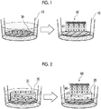
- FIG. 1 An example of the culturing step is shown in Fig. 1 .
- gene-deficient epidermal keratinocytes 30 are seeded on a support 10 and cultured to form a layer of gene-deficient epidermal keratinocytes.
- the medium on the layer of the gene-deficient epidermal keratinocytes is removed to form an air-liquid interface and the culture is continued, whereby it is possible to obtain a cell laminate 50 including an epidermal layer 40 in which gene-deficient epidermal keratinocytes are laminated.
- a collagen gel in which at least one of mesenchymal cells or mesenchymal stem cells is embedded is prepared as a support layer 20.
- gene-deficient epidermal keratinocytes 30 are seeded on the support layer 20 and cultured to form a layer of gene-deficient epidermal keratinocytes.
- the medium on the layer of the gene-deficient epidermal keratinocytes is removed to form an air-liquid interface, and the culture is continued, whereby it is possible to obtain a cell laminate 60 including the support layer 20 and the epidermal layer 40 in which gene-deficient epidermal keratinocytes are laminated.
- the number of passages of gene-deficient epidermal keratinocytes seeded on the support 10 or the support layer 20 is preferably, for example, 10 passages or less, more preferably 8 passages or less, and still more preferably 5 passages or less.
- Culture conditions for air-liquid interface culture For example, conditions of 37°C and 5 v/v% CO 2 can be adopted.
- the number of days of culture by air-liquid interface culture is preferably, for example, 10 days or more and more preferably 11 days or more. By setting the number of days of culture to 10 days or more, the state of differentiation of the epidermal layer tends to be sufficient.
- the upper limit of the number of days of culture is not particularly limited, and it is generally within 30 days and preferably within 25 days.
- the method for evaluating epidermal proliferation or differentiation of the present disclosure includes a step of culturing specific gene-deficient epidermal keratinocytes (that is, the above-mentioned gene-deficient epidermal keratinocytes) by air-liquid interface culture.
- the epidermal proliferation or differentiation can be evaluated by culturing the gene-deficient epidermal keratinocytes by air-liquid interface culture to produce a cell laminate.
- the function of a specific gene can also be evaluated from the epidermal proliferation state or differentiation state.
- the epidermal proliferation state or differentiation state can be evaluated by culturing gene-deficient epidermal keratinocytes by air-liquid interface culture to produce a cell laminate, and then staining the epidermal layer with HE and observing the stained epidermal layer with an optical microscope.
- the epidermal differentiation state can be evaluated by culturing gene-deficient epidermal keratinocytes by air-liquid interface culture to produce a cell laminate, and detecting gene expression in cells of the epidermal layer.
- genes which are indicators of epidermal differentiation state include a loricrin gene (LOR), a periostin gene (POSTN), a transglutaminase 3 gene (TGM3), a wingless-type mouse mammary tumor virus (MMTV) integration site family member 3 gene (WNT3), a wingless-type MMTV integration site family member 4 gene (WNT4), a filaggrin gene (FLG), a kallikrein-related peptidase 14 gene (KLK14), a desmocollin 1 gene (DSC1), a caspase 14 gene (CASP14), and a fibronectin 1 gene (FN1).
- LOR loricrin gene
- POSTN periostin gene
- TGM3 transglutaminase 3 gene
- WNT3 wingless-type mouse mammary tumor virus
- WNT4 wingless-type MMTV integration site family member 4 gene
- FLG filaggrin gene
- KLK14 kallik
- the method for detecting gene expression is not particularly limited.
- the expression of RNA corresponding to the above-mentioned gene can be detected by a reverse transcription polymerase chain reaction (RT-PCR) method or a microarray method.
- the expression of a protein corresponding to the above-mentioned gene can also be detected by enzyme-linked immunosorbent assay (ELISA) or Western blotting.
- the method for evaluating a test substance includes a step of culturing specific gene-deficient epidermal keratinocytes (that is, the above-mentioned gene-deficient epidermal keratinocytes) in the presence of a test substance by air-liquid interface culture. Effects of a test substance on the structure, function, and the like of a cell laminate can be evaluated by culturing the gene-deficient epidermal keratinocytes in the presence of the test substance by air-liquid interface culture to produce the cell laminate.
- specific gene-deficient epidermal keratinocytes that is, the above-mentioned gene-deficient epidermal keratinocytes
- step of culturing the gene-deficient epidermal keratinocytes by air-liquid interface culture is the same as the above-mentioned method for producing a cell laminate of the present disclosure, except that the cells are cultured in the presence of a test substance, a detailed description thereof will be omitted.
- test substance in the presence of a test substance is not particularly limited as long as the test substance can come into contact with the gene-deficient epidermal keratinocytes.
- a test substance may be added to the above-mentioned medium for a cell laminate.
- a test substance may be contained in the above-mentioned collagen-containing layer, or a cell secreting a test substance may be contained in the above-mentioned collagen-containing layer.
- test substance is not particularly limited, and may be a component contained in a pharmaceutical product or a cosmetic product, a bio-related substance such as cytokine, or another compound.
- the method for evaluating a test substance includes a step of bringing a test substance into contact with the above-mentioned cell laminate of the present disclosure.
- bringing the test substance into contact with the cell laminate it is possible to evaluate the effect of the toxicity or the like of the test substance on the cell laminate, the permeability of the test substance to the cell laminate, and the like.
- a cell laminate in which the epidermal layer is thinned can be obtained by culturing IGF-1R gene-deficient epidermal keratinocytes by air-liquid interface culture. Therefore, this cell laminate can be used as a thinning model or an aging model of skin for the evaluation of a test substance.
- the method of bringing the test substance into contact with the cell laminate is not particularly limited, and for example, a method of applying the test substance to the epidermal layer can be mentioned.
- test substance is not particularly limited.
- the test substance may be a pharmaceutical product or a cosmetic product, or may be a component contained in a pharmaceutical product or a cosmetic product, or may be another compound.
- the biological implantation material of the present disclosure includes the above-mentioned cell laminate of the present disclosure.
- the cell laminate can engraft and self-organize.
- the cell laminate is self-organized by the action of factors in vivo after engraftment, whereby the object of treatment can be achieved.
- a biological implantation material including a cell laminate produced using epidermal keratinocytes deficient in a skin disease causative gene can also be used for treating a patient having a genetic skin disease.
- IGF-1R gene-deficient HaCaT cells were constructed by using HaCaT cells as epidermal keratinocytes and deleting the IGF-1R gene of HaCaT cells by a CRISPR-Cas9 method.
- HaCaT cells were cultured in DMEM (available from Thermo Fisher Scientific Inc.) containing 10 v/v% FBS (available from Thermo Fisher Scientific Inc.) and penicillin and streptomycin (pen/strep, available from Thermo Fisher Scientific Inc.) as a medium in a humidified incubator under conditions of 37°C and 5 v/v% CO 2 .
- DMEM available from Thermo Fisher Scientific Inc.
- pen/strep penicillin and streptomycin
- the passage was carried out by recovering the cells using 0.25 w/v% trypsin-ethylenediamine tetraacetic acid (EDTA) (available from Thermo Fisher Scientific Inc.).
- EDTA 0.25 w/v% trypsin-ethylenediamine tetraacetic acid
- plasmid vector for genome editing (hereinafter, also simply referred to as "plasmid” or “vector”) was constructed as follows.
- the plasmid vector attached to this kit can express Cas9 nuclease and gRNA in a single vector and has an orange fluorescent protein (OFP) gene as a reporter gene.
- OFP orange fluorescent protein
- a target sequence on genomic DNA was selected using software such as CRISPRdirect (http://crispr.dbcls.jp/) or E-CRISP (http://www.e-crisp.org/E-CRISP/).
- CRISPRdirect http://crispr.dbcls.jp/
- E-CRISP http://www.e-crisp.org/E-CRISP/.
- PAM proto-spacer adjacent motif
- oligo DNAs (IGF1R. 1, IGF1R. 2, and IGF1R. 3) encoding crRNA complementary to each target sequence were designed.
- the base sequences of the designed oligo DNAs are shown in Table 1 below.
- Table 1 the PAM sequence adjacent to each base sequence, the number of mismatched bases with the target sequence, and the gene position of the target sequence are also shown.
- Base sequence (5' ⁇ 3') PAM sequence Number of mismatched bases Gene position SEQ ID NO IGF1R. 1 CTTCGAGATGACCAATCTCA AGG 0 chr15[98707824] 1 IGF1R. 2 CTCGGTAATGACCGTGAGCT TGG 0 chr15[98707694] 2 IGF1R. 3 TCGTTGCGGATGTCGATGCC TGG 0 chr15[98707570] 3
- an oligo DNA was constructed by adding an additional sequence for ligation to the base sequence of each oligo DNA shown in Table 1 and the complementary sequence thereof, which was followed by denaturation at 95°C and annealing at room temperature (25°C) to construct a double-stranded oligo DNA.
- the constructed double-stranded oligo DNA was ligated into a vector using T4 DNA ligase attached to the kit.
- E. coli One Shot TOP10 Chemically Competent E. coli
- the transformed E. coli was cultured on an agar medium supplemented with ampicillin.
- a part of colonized E. coli was recovered and the plasmid was purified using a plasmid recovery kit (available from QIAGEN, Inc.).
- the purified plasmid was confirmed to be a desired plasmid by PCR using the U6 forward primer attached to the kit.
- the E. coli having a desired plasmid was cultured again, and the plasmid was purified using a plasmid recovery kit (available from QIAGEN, Inc.).
- the sequence of the plasmid was confirmed using a commercially available kit (BigDye Terminator v3.1 Cycle Sequencing Kit, available from ABI, "BigDye” is a registered trademark), and was used for the subsequent experiment.
- a genome editing plasmid was introduced into HaCaT cells cultured to 70% to 80% occupancy relative to the bottom area of a 24-well plate (available from Becton, Dickinson and Company) by a lipofection method.
- Introduction of the plasmid was carried out using a Lipofectamine 3000 reagent (available from Thermo Fisher Scientific Inc., "Lipofectamine” is a registered trademark) according to the manufacturer's protocol.
- the medium was exchanged after 48 hours of introduction of the plasmid, and dead cells were removed.
- the OFP-positive cells were confirmed with a fluorescence microscope (excitation wavelength: 548 nm, fluorescence wavelength: 560 nm), the plasmid introduction rate was about 2% to 3%.
- HaCaT cells after introduction of the plasmid were recovered using 0.25 w/v% trypsin-EDTA (available from Thermo Fisher Scientific Inc.), and then OFP-positive cells were enriched with a cell sorter (BD FACSAria, available from Becton, Dickinson and Company, "FACSAria” is a registered trademark).
- BD FACSAria available from Becton, Dickinson and Company, "FACSAria” is a registered trademark.
- the plasmid introduction rate was about 100%.
- Cell assay was carried out using a special enzyme (Cell endonuclease) that recognizes and cleaves the mismatched portion of heteroduplex DNA.
- Cell endonuclease The principle of the Cell assay is as follows. The region containing the target sequence on the genomic DNA is amplified by PCR, and the amplified product is denatured and then re-annealed. In the case where a mutation is present in the amplified product, heteroduplex DNA is formed upon re-annealing, so the mismatched portion is cleaved by Cell endonuclease. The presence or absence of a gene mutation can be confirmed by confirming the presence or absence of this cleavage by electrophoresis.
- the Cell assay was carried out as follows using a commercially available kit (GeneArt Genomic Cleavage Detection Kit, available from Thermo Fisher Scientific Inc.).
- the base sequences of forward primer (hIGF-1R_E2_13F) and reverse primer (hIGF-1R_E2_401R) used for PCR are shown in Table 2 below.
- Base sequence (5' ⁇ 3') SEQ ID NO hIGF-1R_E2_13F GCATCGACATCCGCAACGAC 4 hIGF-1R_E2_401R GGACACCGCATCCAGGATCA 5
- Genomic DNA was extracted from FACS-enriched OFP-positive cells and the region containing the target sequence of the IGF-1R gene was amplified using the above-mentioned primers.
- the amplified product was denatured at 95°C and then re-annealed at room temperature (25°C) to obtain double-stranded DNA.
- Cell endonuclease was added to the double-stranded DNA after re-annealing, followed by incubation at 37°C for 1 hour, and then the enzyme was inactivated by heat. Then, the amplified product after the enzymatic reaction was electrophoresed on 4 w/v% agarose gel (available from Wako Pure Chemical Industries, Ltd.), and the electrophoretic pattern was confirmed. As a result, three bands were detected, thus confirming the introduction of mutation into the IGF-1R gene.
- FACS-enriched OFP-positive cells were diluted to a density of 0.5 cells/well to 1 cell/well and seeded in a 96-well plate. After culturing the cells for 2 weeks to 4 weeks, the genomic DNA of the proliferated cells was extracted and subjected to the Cell assay in the same manner as described above. As a result, mutation of the IGF-1R gene was confirmed for clones of 24 strains.
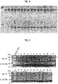
- Fig. 4 Apart of the electrophoretic pattern in the Cell assay is shown in Fig. 4 .
- the lanes marked with a circle correspond to clones in which mutation of the IGF-1R gene was confirmed.
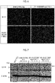
- the Cell assay-positive strain after cloning was cultured, the cells were solubilized with a radio-immunoprecipitation assay (RIPA) buffer (available from Thermo Fisher Scientific Inc.), and then the protein was quantitated using a commercially available kit (BCA Protein Assay Kit, available from Thermo Fisher Scientific Inc.).
- BCA Protein Assay Kit available from Thermo Fisher Scientific Inc.
- 20 ⁇ g of protein was developed on a 10 w/v% polyacrylamide gel and the protein was transferred to a polyvinylidene difluoride (PVDF) membrane using a blotting apparatus (available from ATTO Corp.). Thereafter, an IGF-1R protein was detected using an IGF-1R-specific antibody (available from Thermo Fisher Scientific Inc.).
- PVDF polyvinylidene difluoride
- HaCaT cells not transfected with plasmid were used for positive control.
- the IGF-1R protein was not detected and the deficiency of the IGF-1R gene was confirmed in clones of 6 strains in 24 strains.
- the clone in which the deficiency of the IGF-1R gene was not recognized despite the confirmation of the mutation of the IGF-1R gene is presumed to have not caused a frame shift due to the gene mutation.
- Fig. 5 The results of Western blotting are shown in Fig. 5 .
- lane 3, lane 7, lane 10, lane 13, lane 16, and lane 21 correspond to clones of 6 strains in which the deficiency of the IGF-1R gene was recognized.
- the clone of lane 7 (hereinafter, also referred to as "IGF-1R-deficient HaCaT cell 1") and the clone of lane 10 (hereinafter, also referred to as “IGF-1R-deficient HaCaT cell 2”) were used for the subsequent experiments.
- the clone of lane 15 in which the deficiency of the IGF-1R gene was not recognized, was used as a Sham control cell.
- the clone of lane 15 was used as a Sham control.
- IGF-1R-deficient HaCaT cells 1 and Sham control cells were respectively treated with phosphate buffered saline (PBS) containing 4 w/v% paraformaldehyde (20 g of paraformaldehyde dissolved in 500 mL of 0.1 mol/L PBS (pH 7.4) (available from Wako Pure Chemical Industries, Ltd.)) for 15 minutes to fix the cells.
- PBS phosphate buffered saline
- PBS phosphate buffered saline
- PBS phosphate buffered saline
- a 200-fold diluted solution of anti-IGF-1R antibodies (available from Thermo Fisher Scientific Inc.) was added thereto, followed by reaction at 4°C overnight.
- the cells were washed about three times with PBS and then reacted for 2 hours with a 200-fold diluted solution of anti-mouse-fluorescein isothiocyanate (FITC) antibodies (available from Abcam PLC).
- FITC anti-mouse-fluorescein isothiocyanate
- the cells were washed about three times with PBS, nuclear stained with Hoechst (available from Dojindo Laboratories Co., Ltd.), encapsulated and observed with a fluorescence microscope (available from Keyence Corporation).
- Fig. 6 The results of cell immunostaining are shown in Fig. 6 .
- the scale bar in the figure shows 100 ⁇ m.
- IGF-1R protein was confirmed in Sham control cells, whereas IGF-1R protein was not confirmed in IGF-1R-deficient HaCaT cells 1.
- IGF-1R-deficient HaCaT cells 1, IGF-1R-deficient HaCaT cells 2, and Sham control cells were additionally subcultured and the IGF-1R protein was detected by Western blotting in the same manner as described above.
- HaCaT cells not transfected with plasmid were used as positive control.
- Fig. 7 The results of Western blotting are shown in Fig. 7 .
- P indicates the number of passages before the experiment
- P+1 means that the cells were additionally passaged once
- P+2 means that the cells were additionally passaged twice
- P+3 means that the cells were additionally passaged three times.
- the deficiency of the IGF-1R gene was continued even in the case where the passages of IGF-1R-deficient HaCaT cells 1 and IGF-1R-deficient HaCaT cells 2 were repeated.
- Genomic DNA was extracted from IGF-1R-deficient HaCaT cells 1 and the sequence of the region containing the target sequence of the IGF-1R gene was confirmed by a conventional method.
- Fig. 8 The results of confirming the sequence are shown in Fig. 8 .
- the upper sequence shows the sequence of the region containing the target sequence of the IGF-1R gene in IGF-1R-deficient HaCaT cells 1 and the lower sequence shows the original sequence in the same region.
- the underlined sequence corresponds to the target sequence and the boxed sequence corresponds to the PAM sequence.
- CTCA 4 bases
- Example 1 IGF-1R-deficient HaCaT cells 1 were cultured on a polycarbonate support by air-liquid interface culture to produce a cell laminate.
- a DMEM containing 10 v/v% FBS as a medium for normal human dermal fibroblasts and a medium for normal human epidermal keratinocytes (HuMedia-KG 2, Kurabo) excluding a human epidermal growth factor (hEGF) were mixed at a ratio of 1:1 (volume ratio).
- 225 ⁇ L of the above-prepared CaCl 2 solution (final concentration of calcium in the medium: 1.8 mmol/L) was added to 500 mL of the obtained medium.
- the medium after the addition of the CaCl 2 solution was dispensed into a 50 mL FALCON tube (available from Corning Inc., "FALCON” is a registered trademark), and ascorbic acid 2-glucoside was added so as to have a concentration of 0.1 w/w%, whereby a medium for a cell laminate was prepared.
- IGF-1R-deficient HaCaT cells 1 were dispersed at a cell concentration of 2 ⁇ 10 5 cells/0.2 mL in a medium for a cell laminate to prepare a cell dispersion liquid.
- 0.2 mL of the obtained cell dispersion liquid was seeded on a polycarbonate cell culture insert (available from Becton, Dickinson and Company), and the cell culture insert was placed in wells of a 12-well plate.
- 1.5 mL of the medium for a cell laminate was added to the outside of the cell culture insert and the cells were cultured for 24 hours under the conditions of 37°C and 5 v/v% CO 2 . After 24 hours, the medium in the cell culture insert was gently removed to form an air-liquid interface, and the cells were cultured for another 14 days. The medium exchange was carried out every two days.
- the cell laminate obtained by culturing for 14 days was fixed using PBS containing 4 w/v% paraformaldehyde (20 g of paraformaldehyde dissolved in 500 mL of 0.1 mol/L PBS (pH 7.4)) (available from Wako Pure Chemical Industries, Ltd.).
- the fixed cell laminate was immersed in 0.1 mol/L PBS and the liquid was exchanged several times to replace the fixing liquid.
- the cell laminate was cut into a strip shape by a razor and embedded in paraffin. Then, a section having a thickness of 4 ⁇ m was prepared using a microtome, attached to a slide glass and dried at 45°C.
- the obtained paraffin-embedded section of the cell laminate was stored at room temperature (25°C).
- the paraffin-embedded section was deparaffinized with xylene and then sequentially immersed in aqueous ethanol solutions whose concentrations were gradually decreased from 100 v/v% to 70 v/v%.
- the sample was washed with distilled water, immersed in a hematoxylin staining liquid (available from Wako Pure Chemical Industries, Ltd.) for 5 minutes and washed with running water. Further, the sample was immersed in 70 v/v% ethanol containing 0.2 w/v% HCl for 1 second and washed with running water for 10 minutes.
- the sample was immersed in an eosin staining liquid (available from Wako Pure Chemical Industries, Ltd.) for 10 minutes, sequentially immersed in aqueous ethanol solutions whose concentrations were gradually increased from 95 v/v% to 100 v/v% for dehydration, and then immersed in xylene for 2 minutes to 3 minutes.
- An encapsulating agent was placed on the obtained sample which was then covered with a cover glass, dried and encapsulated.
- Fig. 9 shows HE stained images of cell laminates obtained by culturing IGF-1R-deficient HaCaT cells 1 and control HaCaT cells.
- the scale bar in the figure shows 50 ⁇ m.
- epidermal proliferation and differentiation were significantly decreased in the cell laminate obtained by culturing the IGF-1R-deficient HaCaT cells 1, as compared with the cell laminate obtained by culturing HaCaT cells.
- the average thickness of the epidermal layers was about 55 ⁇ m in the cell laminates obtained by culturing HaCaT cells, whereas the average thickness of the epidermal layers was about 25 ⁇ m in the cell laminates obtained by culturing IGF-1R-deficient HaCaT cells 1, in which the epidermal layer was significantly thinned (P ⁇ 0.01).
- Example 2 IGF-1R-deficient HaCaT cells 1 were cultured on a collagen gel (support layer) in which fibroblasts were embedded by air-liquid interface culture to produce a cell laminate.
- a medium for a cell laminate prepared in Example 1 containing a total of 5 ⁇ 10 5 normal human neonatal foreskin dermal fibroblasts (available from Kurabo Industries Ltd.) and acid-solubilized type I collagen (derived from bovine) having a final concentration of 1 mg/mL was dispensed as a collagen solution into a 6-well plate. Then, under the conditions of 37°C and 5 v/v% CO 2 , the cells were cultured in static culture or while stirring at a rate of 5 revolutions per minute to 60 revolutions per minute. After culturing for 2 days to 3 days, a collagen gel in which the bottom surface contracted to about 1.0 cm in diameter was obtained.
- IGF-1R-deficient HaCaT cells 1 were dispersed at a cell concentration of 2 ⁇ 10 5 cells/0.2 mL in a medium for a cell laminate to prepare a cell dispersion liquid.
- a ring (inner diameter: 7 mm) was placed on the collagen gel, and the medium inside and outside the ring was removed.
- 0.4 mL of the cell dispersion liquid was added into the ring, and it was confirmed that the cell dispersion liquid did not leak to the outside of the ring.
- 3 mL of the medium for a cell laminate was added to the outside of the ring and the cells were cultured for 24 hours under the conditions of 37°C and 5 v/v% CO 2 .
- the medium inside and outside the ring was removed while paying attention not to break the layer of the IGF-1R-deficient HaCaT cells 1 layered on the collagen gel, and the ring was removed using tweezers.
- a medium for a cell laminate was then added from the edge of the well.
- the amount of the medium was adjusted to about 2 mL to 3 mL so that the boundary between the IGF-1R-deficient HaCaT cells 1 and the collagen gel was not immersed in the medium so as to bring the medium into contact with the IGF-1R-deficient HaCaT cells 1.
- air-liquid interface culture was started and the cells were cultured for 7 days or 14 days. The medium exchange was carried out every two days.
- the cell laminate obtained by culturing for 7 days or 14 days was subjected to HE staining in the same manner as in Example 1.
- Fig. 11 shows HE stained images of cell laminates obtained by culturing IGF-1R-deficient HaCaT cells 1 and control HaCaT cells.
- the scale bar in the figure shows 50 ⁇ m.
- epidermal proliferation and differentiation were significantly decreased in the cell laminate obtained by culturing the IGF-1R-deficient HaCaT cells 1, as compared with the cell laminate obtained by culturing HaCaT cells.
- JP2016-150296 filed on July 29, 2016 is hereby incorporated by reference in its entirety.
Landscapes
- Health & Medical Sciences (AREA)
- Life Sciences & Earth Sciences (AREA)
- Engineering & Computer Science (AREA)
- Biomedical Technology (AREA)
- Chemical & Material Sciences (AREA)
- Cell Biology (AREA)
- General Health & Medical Sciences (AREA)
- Biotechnology (AREA)
- Zoology (AREA)
- Bioinformatics & Cheminformatics (AREA)
- Immunology (AREA)
- Organic Chemistry (AREA)
- Medicinal Chemistry (AREA)
- Wood Science & Technology (AREA)
- Genetics & Genomics (AREA)
- Dermatology (AREA)
- Urology & Nephrology (AREA)
- Biochemistry (AREA)
- Microbiology (AREA)
- Molecular Biology (AREA)
- Hematology (AREA)
- Developmental Biology & Embryology (AREA)
- Veterinary Medicine (AREA)
- Animal Behavior & Ethology (AREA)
- Public Health (AREA)
- Epidemiology (AREA)
- General Engineering & Computer Science (AREA)
- Transplantation (AREA)
- Oral & Maxillofacial Surgery (AREA)
- Chemical Kinetics & Catalysis (AREA)
- Tropical Medicine & Parasitology (AREA)
- Pharmacology & Pharmacy (AREA)
- Food Science & Technology (AREA)
- Physics & Mathematics (AREA)
- Analytical Chemistry (AREA)
- General Physics & Mathematics (AREA)
- Pathology (AREA)
- Toxicology (AREA)
- Rheumatology (AREA)
- Botany (AREA)
Abstract
Description
- The present disclosure relates to a cell laminate, a method for producing a cell laminate, a method for evaluating epidermal proliferation or differentiation, a method for evaluating a test substance, and a biological implantation material.
- With the progress of cell culture technology, it has become possible to artificially reconstruct the skin of a living body to produce artificial skin having a structure and a function similar to those of the skin of a living body.
- The reconstructed artificial skin is used as a biological implantation material for the treatment of, for example, burns or wounds. In addition, the reconstructed artificial skin is also used as an evaluation system for elucidating the structure or function of the skin, or an evaluation system for developing a pharmaceutical product, a cosmetic product, or the like, for which a variety of techniques have been proposed so far.
- For example,
JP5458259B -
JP5228246B - Various endogenous genes are involved in the structure and function of the skin. However, the cell laminate of
JP5458259B JP5228246B - An object according to an embodiment of the present invention is to provide a novel cell laminate obtained by culturing specific gene-deficient epidermal keratinocytes and a method for producing the same. Another object according to the embodiment of the present invention is to provide a method for evaluating epidermal proliferation or differentiation, a method for evaluating a test substance, and a biological implantation material, to each of which a cell laminate and a method for producing the same are applied.
- Specific means for achieving the foregoing objects includes the following aspects.
- <1> A cell laminate comprising an epidermal layer in which specific gene-deficient epidermal keratinocytes are laminated.
- <2> The cell laminate according to <1>, in which the epidermal keratinocyte is a human epidermal keratinocyte.
- <3> The cell laminate according to <1> or <2>, further comprising a support layer.
- <4> The cell laminate according to <3>, in which the support layer is a layer including collagen and at least one of mesenchymal cells or mesenchymal stem cells.
- <5> The cell laminate according to any one of <1> to <4>, in which the specific gene includes a cell membrane receptor gene.
- <6> The cell laminate according to any one of <1> to <5>, in which the specific gene includes an insulin-
like growth factor 1 receptor gene. - <7> A method for producing a cell laminate, comprising a step of culturing specific gene-deficient epidermal keratinocytes by air-liquid interface culture.
- <8> The method for producing a cell laminate according to <7>, in which the epidermal keratinocyte is a human epidermal keratinocyte.
- <9> The method for producing a cell laminate according to <7> or <8>, further comprising a step of deleting the specific gene of the epidermal keratinocyte using a site-specific nuclease.
- <10> The method for producing a cell laminate according to any one of <7> to <9>, in which the step of culturing specific gene-deficient epidermal keratinocytes by air-liquid interface culture includes culturing the specific gene-deficient epidermal keratinocytes on a support layer by air-liquid interface culture.
- <11> The method for producing a cell laminate according to <10>, in which the support layer is a layer including collagen and at least one of mesenchymal cells or mesenchymal stem cells.
- <12> The method for producing a cell laminate according to any one of <7> to <11>, in which the specific gene includes a cell membrane receptor gene.
- <13> The method for producing a cell laminate according to any one of <7> to <12>, in which the specific gene includes an insulin-
like growth factor 1 receptor gene. - <14> A method for evaluating epidermal proliferation or differentiation, comprising a step of culturing specific gene-deficient epidermal keratinocytes by air-liquid interface culture.
- <15> A method for evaluating a test substance, comprising a step of culturing specific gene-deficient epidermal keratinocytes in the presence of a test substance by air-liquid interface culture.
- <16> A method for evaluating a test substance, comprising a step of bringing a test substance into contact with the cell laminate according to any one of <1> to <6>.
- <17> A biological implantation material comprising the cell laminate according to any one of <1> to <6>.
- According to the present disclosure, it is possible to provide a novel cell laminate obtained by culturing specific gene-deficient epidermal keratinocytes and a method for producing the same. Further, according to the present disclosure, it is possible to provide a method for evaluating epidermal proliferation or differentiation, a method for evaluating a test substance, and a biological implantation material, to each of which a cell laminate and a method for producing the same are applied.
-
-
Fig. 1 is a diagram schematically showing an example of a culturing step in a method for producing a cell laminate of the present disclosure. -
Fig. 2 is a diagram schematically showing another example of the culturing step in the method for producing a cell laminate of the present disclosure. -
Fig. 3 is a diagram showing the position of a target sequence on an insulin-like growth factor-1 receptor (IGF-1R) gene in the case where the IGF-1R gene of a HaCaT cell is deleted by a clustered regularly interspaced short palindromic repeats-CRISPR-associated proteins 9 (CRISPER-CAS9) method. -
Fig. 4 is a diagram showing the results of Cell assay after introducing a plasmid vector for CRISPER-CAS9 into a HaCaT cell, followed by cloning. -
Fig. 5 is a diagram showing the results of Western blotting using a Cell assay-positive strain after cloning. -
Fig. 6 is a diagram showing the results of cell immunostaining using a Cell assay-positive strain after cloning. -
Fig. 7 is a diagram showing the results of Western blotting after passage of an IGF-1R gene-deficient HaCaT cell. -
Fig. 8 is a diagram showing the results of confirming a sequence of a region containing the target sequence of an IGF-1R gene for an IGF-1R gene-deficient HaCaT cell. -
Fig. 9 is a diagram showing a hematoxylin/eosin (HE) stained image of a cell laminate produced by culturing an IGF-1R gene-deficient HaCaT cell on a support by air-liquid interface culture. -
Fig. 10 is a diagram showing an average thickness of epidermal layers of cell laminates produced by culturing an IGF-1R gene-deficient HaCaT cell on a support by air-liquid interface culture. -
Fig. 11 is a diagram showing a HE stained image of a cell laminate produced by culturing an IGF-1R gene-deficient HaCaT cell on a support layer (collagen gel in which fibroblasts are embedded) by air-liquid interface culture. - Hereinafter, the cell laminate, the method for producing a cell laminate, the method for evaluating epidermal proliferation or differentiation, the method for evaluating a test substance, and the biological implantation material of the present disclosure will be described in detail. However, the present invention is not limited to the following embodiments at all and can be carried out with appropriate modification within the scope of the object of the present invention.
- The numerical range indicated by using "to" in the present specification means a range including numerical values described before and after "to" as a minimum value and a maximum value, respectively.
- In the numerical ranges described in a stepwise manner in the present specification, the upper limit value or the lower limit value described in a certain numerical range may be replaced with the upper limit value or the lower limit value of the numerical range in other stepwise description. In addition, in the numerical ranges described in the present specification, the upper limit value or the lower limit value described in a certain numerical range may be replaced with the values shown in the Examples.
- Unless otherwise specified, the concentration of each component in the present specification means the total concentration of a plurality of substances in the case where there are a plurality of substances corresponding to each component.
- The term "layer" in the present specification includes the case where a layer is formed only in a part of the region in addition to the case where a layer is formed in the whole region in the case of observing the region where the layer exists.
- The term "step" in the present specification includes not only an independent step but also a step in which the intended purpose of this step can be achieved even in the case where the step cannot be clearly distinguished from other steps.
- The cell laminate of the present disclosure includes an epidermal layer on which specific gene-deficient epidermal keratinocytes (hereinafter, also referred to as "gene-deficient epidermal keratinocytes") are laminated. The cell laminate of the present disclosure may have only an epidermal layer or may further include layers other than an epidermal layer such as a support layer described below.
- The epidermal layer is a layer in which at least gene-deficient epidermal keratinocytes are laminated. Here, the "lamination" may be such that gene-deficient epidermal keratinocytes are piled up, and it is not required that the division of the layers can be clearly confirmed.
- Examples of epidermal keratinocytes include cells derived from mammals. Examples of mammals include primates such as humans, rhesus monkeys, marmosets, orangutans, and chimpanzees; rodents such as mice, rats, hamsters, and guinea pigs; and rabbits, pigs, cows, goats, horses, sheep, minks, dogs, and cats. Among them, primate epidermal keratinocytes are preferred, and human epidermal keratinocytes are more preferred.
- The type of the human epidermal keratinocyte is not particularly limited, and examples thereof include HaCaT cells (J. Invest. Dermatol., 1999, 112 (3): 343-353), human dermal keratinocytes (HDKs) (Cancer Sci., 2007, 98: 147-154), and normal human epidermal keratinocytes (NHEKs). Among these, HaCaT cells are preferable from the viewpoint of relatively uniform cell properties irrespective of the number of passages, easy handling, and normal cell differentiation state.
- The epidermal keratinocyte may be a non-immortalized cell or an immortalized cell. The non-immortalized cell refers to a cell whose proliferation stops due to a finite number of cell divisions. On the other hand, the immortalized cell refers to a cell that does not stop proliferation even in the case where cell division is repeated, that is, a cell that has acquired an infinite autonomous replication ability. Immortalization of a cell can be carried out by introducing an immortalizing gene such as a simian virus (SV) 40 T antigen gene, a telomerase reverse transcriptase (TERT) gene, or a human papilloma virus (HPV) E6-E7 gene into the cell. In addition, the immortalization of a cell may occur occasionally. For example, the above-mentioned HaCaT cell is an immortalized cell that has been naturally immortalized without introducing an immortalizing gene.
- There is no particular limitation on the type of a specific gene lacking in epidermal keratinocytes. Examples of the specific gene include cell membrane receptor genes and skin disease causative genes. Examples of cell membrane receptors include a tyrosine kinase receptor such as insulin-like growth factor-1 receptor (IGF-1R) or epidermal growth factor receptor (EGF-R); a G protein-coupled receptor; a guanylate cyclase receptor; and an ion channel type receptor. The specific genes lacking in epidermal keratinocytes may be of one type or of two or more types.
- In one aspect, the specific gene includes a cell membrane receptor gene (for example, an IGF-1R gene). Since it is thought that the cell membrane receptor gene plays an important role in the homeostasis of the epidermal layer of the living body, use of epidermal keratinocytes deficient in the cell membrane receptor gene makes it possible to obtain a novel cell laminate which is different from cell laminates in the related art. For example, as shown in the Examples which will be described later, a cell laminate in which the epidermal layer is thinned can be obtained by culturing IGF-1R gene-deficient epidermal keratinocytes by air-liquid interface culture.
- In another aspect, the specific gene includes a skin disease causative gene. As will be described later, a cell laminate produced using epidermal keratinocytes deficient in a skin disease causative gene can be used as a biological implantation material for the treatment of skin diseases.
- A gene-deficient epidermal keratinocyte can be obtained by deleting a specific gene of the epidermal keratinocyte. From the viewpoint of specifically deleting a specific gene, a method using a site-specific nuclease is preferable as a method of deleting a specific gene of the epidermal keratinocyte. Examples of the method using a site-specific nuclease include a CRISPR-Cas9 method, a transcription activator-like effector nucleases (TALENs: registered trademark) method, and a zinc finger nucleases (ZFNs) method. In one aspect, the gene-deficient epidermal keratinocyte is obtained by in vitro deletion of a specific gene of the epidermal keratinocyte.
- Among them, the CRISPR-Cas9 method is preferable from the viewpoint of convenience and the ability to simultaneously edit a plurality of sites on genomic DNA. The principle of the CRISPR-Cas9 method is as follows. That is, in the CRISPR-Cas9 method, Cas9 nuclease and guide RNA (gRNA) are introduced into a cell. The gRNA includes CRISPR RNA (crRNA) having a base sequence complementary to a target sequence (for example, a sequence on a gene to be deleted) and trans-activating crRNA (tracrRNA), and binds to genomic DNA to thereby induce cleavage of the target sequence by the Cas9 nuclease. Mutation of the target sequence is caused during the repair process of the cleaved genomic DNA, and as a result, it becomes possible to lose the gene.
- For example, the following methods (a) to (d) are known as the method for introducing Cas9 nuclease and gRNA into a cell. Among them, the method (a) is preferable from the viewpoint of convenience and introduction efficiency.
- (a) a method in which DNA encoding Cas9 nuclease and DNA encoding gRNA are introduced into a cell through a plasmid vector.
- (b) a method in which DNA encoding Cas9 nuclease is introduced into a cell through a viral vector and DNA encoding gRNA is directly introduced into the cell.
- (c) a method in which RNA encoding Cas9 nuclease and gRNA are constructed by in vitro transcription and are directly introduced into a cell.
- (d) a method in which a recombinant protein of Cas9 nuclease and gRNA constructed by in vitro transcription are directly introduced into a cell.
- The epidermal layer may contain one type of gene-deficient epidermal keratinocyte or may contain two or more types of gene-deficient epidermal keratinocytes. As an aspect containing two or more types of gene-deficient epidermal keratinocytes, for example, an aspect containing two or more types of gene-deficient epidermal keratinocytes which have the same cell type of epidermal keratinocyte but have different types of specific genes to be deleted; an aspect containing two or more types of gene-deficient epidermal keratinocytes which have the same type of the specific gene to be deleted but have different cell types of the epidermal keratinocytes; and an aspect containing two or more types of gene-deficient epidermal keratinocytes which have different cell types of the epidermal keratinocytes and different types of the specific genes to be deleted.
- The epidermal layer may contain cells other than the gene-deficient epidermal keratinocytes. Examples of other cells include melanocytes and cells of the immune system.
- The number of cells other than the gene-deficient epidermal keratinocytes may be 20% or less, 15% or less, or 10% or less with respect to the total number of cells in the epidermal layer.
- Further, the epidermal layer may be a layer in which a four-layer structure of a basal layer, a stratum spinosum, a granular layer, and a horny cell layer is confirmed, or may be a layer in which only a part of the four layers is confirmed. The method of confirming each of the above layers is not particularly limited, and the layers can be confirmed by a known method. In general, each layer can be confirmed by HE staining of the epidermal layer and confirmation of characteristic structure and position information under an optical microscope.
- The cell laminate of the present disclosure may further include a support layer. The support layer is not particularly limited as long as it can support the epidermal layer and form a cell laminate.
- The support layer may be a membrane filter from the viewpoint that preparation and handling at the time of culture of gene-deficient epidermal keratinocytes by air-liquid interface culture are facilitated. Examples of the material for the membrane filter include polyethylene terephthalate (PET), polycarbonate, and polytetrafluoroethylene (PTFE).
- Further, from the viewpoint that the structure and function of the cell laminate can be brought closer to the skin of a living body, the support layer is preferably a layer containing collagen and at least one of mesenchymal cells or mesenchymal stem cells (hereinafter, also referred to as "collagen-containing layer"). The collagen-containing layer corresponds to the dermal layer of the skin. For the homeostasis of the epidermal layer of a living body, intercellular signaling plays an important role in addition to growth factors and nutrients supplied from the dermal layer. The cell laminate of the present disclosure further includes a collagen-containing layer, so that the structure and function of the cell laminate can be brought closer to the skin of a living body.
- Examples of mesenchymal cells and mesenchymal stem cells contained in the collagen-containing layer include cells derived from mammals. Examples of mammals include primates such as humans, rhesus monkeys, marmosets, orangutans, and chimpanzees; rodents such as mice, rats, hamsters, and guinea pigs; and rabbits, pigs, cows, goats, horses, sheep, minks, dogs, and cats. Among them, primate cells are preferred and human cells are more preferred.
- The type of mesenchymal cells is not particularly limited, and examples thereof include fibroblasts, adipocytes, cardiomyocytes, bone cells, chondrocytes, and tenocytes. Of these, fibroblasts are preferable from the viewpoint of bringing the cell laminate closer to the skin of a living body.
- In addition, the type of mesenchymal stem cells is not particularly limited and examples thereof include mesenchymal stem cells derived from bone marrow, adipose tissue, placental tissue, umbilical cord tissue, dental pulp, or the like. Among them, bone marrow-derived mesenchymal stem cells are preferable from the viewpoint of maintenance of undifferentiation. The fact that the mesenchymal stem cell is derived from bone marrow can be determined by analyzing the cluster of differentiation (CD) antigen of the cell by, for example, cell immunostaining, flow cytometer, or Western blotting.
- Mesenchymal cells and mesenchymal stem cells may be non-immortalized cells or immortalized cells. From the viewpoint of bringing the proliferation state and differentiation state of the epidermis closer to the epidermis of a living body, mesenchymal cells and mesenchymal stem cells are preferably non-immortalized cells.
- The collagen-containing layer may contain one type of mesenchymal cells or may contain two or more types of mesenchymal cells. In addition, the collagen-containing layer may contain one type of mesenchymal stem cells or may contain two or more types of mesenchymal stem cells.
- The collagen contained in the collagen-containing layer is not particularly limited, and for example, fibrillar collagen that gels can be mentioned. Examples of the fibrillar collagen include type I collagen, type II collagen, type III collagen, type V collagen, and type XI collagen, among which type I collagen or type III collagen is preferable, and type I collagen is more preferable. The collagen may be acid-solubilized collagen. Acid-solubilized type I collagen is more preferable as the collagen.
- The origin of collagen is not particularly limited, and examples thereof include collagen derived from human, cow, horse, pig, mouse, rat, or the like.
- The collagen-containing layer may contain one type of collagen or may contain two or more types of collagen.
- The collagen-containing layer may be, for example, a collagen gel in which at least one of mesenchymal cells or mesenchymal stem cells is embedded. Such a collagen gel can be constructed, for example, by gelling a collagen solution in which collagen and at least one of mesenchymal cells or mesenchymal stem cells are added to a medium. The medium is not particularly limited and may be, for example, a medium for a cell laminate which is used in air-liquid interface culture that will be described later. The density of the collagen gel is preferably, for example, about 0.1 mg/mL to 100 mg/mL.
- The collagen-containing layer may contain cells other than mesenchymal cells and mesenchymal stem cells. Examples of other cells include cells of the immune system.
- The cell density per cm2 of the bottom area of the collagen-containing layer is, for example, preferably 1×10⁵ cells/cm2 or more, more preferably 2.5×10⁵ cells/cm2 or more, still more preferably 5×10⁵ cells/cm2 or more, and particularly preferably 1×10⁶ cells/cm2 or more. The number of cells in the collagen-containing layer can be measured by a conventional method using a cell counter or a counting chamber. In addition, the number of cells can be estimated based on the calibration curve also by an MTT colorimetric assay. The MTT assay is a colorimetric determination method which measures the enzymatic activity of reducing 3-(4,5-dimethyl-thiazol-2-yl)-2,5-diphenyltetrazolium bromide (MTT) or a similar dye to a formazan dye (violet).
- The cell laminate of the present disclosure may further include layers other than the epidermal layer and the support layer. For example, the cell laminate of the present disclosure may include a layer formed of non-enzyme treated type IV collagen (see
JP5458259B - The method for producing a cell laminate of the present disclosure includes a step of culturing specific gene-deficient epidermal keratinocytes (that is, the above-mentioned gene-deficient epidermal keratinocytes) by air-liquid interface culture (hereinafter, also referred to as "culturing step"). The epidermal keratinocytes are preferably human epidermal keratinocytes.
- The method for producing a cell laminate of the present disclosure may further include a step of deleting a specific gene of an epidermal keratinocyte using a site-specific nuclease. As a method using a site-specific nuclease, for example, the above-mentioned CRISPR-Cas9 method can be mentioned. In a certain aspect, the specific gene includes a cell membrane receptor gene (for example, an IGF-1R gene). In one aspect, the step of deleting a specific gene of an epidermal keratinocyte using a site-specific nuclease is carried out in vitro. As shown in the Examples which will be described later, a cell laminate in which the epidermal layer is thinned can be obtained by culturing IGF-1R gene-deficient epidermal keratinocytes by air-liquid interface culture.
- The medium for use in the culturing step (hereinafter, also referred to as "medium for a cell laminate") may be, for example, a medium in which serum is added to a base medium. The medium for a cell laminate may further contain CaCl2, an ascorbic acid derivative, an antibiotic, and/or a pH buffering agent.
- Examples of the base medium include a Dulbecco's Modified Eagle Medium (DMEM), an Eagle's Minimum Essential Medium (EMEM), a Minimum Essential Medium Alpha modification (MEMα), a Roswell Park Memorial Institute 1640 (RPMI 1640) medium, and a Ham's F12 (HamF12) medium. The base media may be used alone or in combination of two or more thereof.
- The serum may be, for example, fetal bovine serum (FBS). The concentration of serum in the medium for a cell laminate is preferably, for example, 5 v/v% to 20 v/v%.
- The medium for a cell laminate preferably contains CaCl2. By incorporation of CaCl2 in the medium for a cell laminate, the differentiation of epidermal keratinocytes tends to be promoted. Further, calcium ions are also necessary ions for intercellular adhesion.
- In the case where the medium for a cell laminate contains CaCl2, the concentration of calcium in the medium for a cell laminate is preferably, for example, 0.5 mmol/L to 5 mmol/L and more preferably 1 mmol/L to 3 mmol/L.
- The medium for a cell laminate preferably contains an ascorbic acid derivative. By incorporation of the ascorbic acid derivative in the medium for a cell laminate, epidermal keratinocytes are activated, resulting in a tendency to promote cell differentiation.
- Examples of the ascorbic acid derivative include ascorbic acid, sodium ascorbate, potassium ascorbate, calcium ascorbate, L-ascorbic acid phosphate ester, magnesium ascorbyl phosphate, sodium ascorbyl phosphate, ascorbyl sulfate, ascorbyl sulfate disodium salt, and ascorbyl-2-glucoside.
- In the case where the medium for a cell laminate contains an ascorbic acid derivative, the concentration of the ascorbic acid derivative in the medium for a cell laminate is preferably, for example, 0.01 w/w% to 1 w/w%.
- The medium for a cell laminate may contain an antibiotic. Examples of the antibiotic include penicillin, streptomycin, neomycin, amphotericin B, kanamycin, and gentamicin.
- The medium for a cell laminate may contain a pH buffering agent. Examples of the pH buffering agent include sodium hydrogen carbonate, calcium chloride, sodium dihydrogen phosphate, 2-[4-(2-hydroxyethyl)-1-piperazinyl]ethanesulfonic acid (HEPES), and 3-morpholinopropane-1-sulfonic acid (MOPS).
- The pH of the medium for a cell laminate is preferably, for example, pH 6.8 to pH 7.6 and more preferably pH 7.0 to pH 7.4.
- There is no particular limitation on the method for culturing the gene-deficient epidermal keratinocytes by air-liquid interface culture to produce a cell laminate. For example, a known method for producing artificial skin using epidermal keratinocytes can be adopted. The air-liquid interface culture of gene-deficient epidermal keratinocytes may be carried out on a support such as a cell culture insert made of polycarbonate or may be carried out on the above-described support layer.
- An example of the culturing step is shown in
Fig. 1 . First, gene-deficientepidermal keratinocytes 30 are seeded on asupport 10 and cultured to form a layer of gene-deficient epidermal keratinocytes. Subsequently, the medium on the layer of the gene-deficient epidermal keratinocytes is removed to form an air-liquid interface and the culture is continued, whereby it is possible to obtain acell laminate 50 including anepidermal layer 40 in which gene-deficient epidermal keratinocytes are laminated. - Another example of the culturing step is shown in
Fig. 2 . First, a collagen gel in which at least one of mesenchymal cells or mesenchymal stem cells is embedded is prepared as asupport layer 20. Subsequently, gene-deficientepidermal keratinocytes 30 are seeded on thesupport layer 20 and cultured to form a layer of gene-deficient epidermal keratinocytes. Subsequently, the medium on the layer of the gene-deficient epidermal keratinocytes is removed to form an air-liquid interface, and the culture is continued, whereby it is possible to obtain acell laminate 60 including thesupport layer 20 and theepidermal layer 40 in which gene-deficient epidermal keratinocytes are laminated. - There is no particular restriction on the number of passages of gene-deficient epidermal keratinocytes seeded on the
support 10 or thesupport layer 20. In the case where the gene-deficient epidermal keratinocytes are non-immortalized cells, the number of passages of the gene-deficient epidermal keratinocytes is preferably, for example, 10 passages or less, more preferably 8 passages or less, and still more preferably 5 passages or less. - General conditions can be adopted as culture conditions for air-liquid interface culture. For example, conditions of 37°C and 5 v/v% CO2 can be adopted.
- The number of days of culture by air-liquid interface culture is preferably, for example, 10 days or more and more preferably 11 days or more. By setting the number of days of culture to 10 days or more, the state of differentiation of the epidermal layer tends to be sufficient. The upper limit of the number of days of culture is not particularly limited, and it is generally within 30 days and preferably within 25 days.
- In the above description, only the gene-deficient epidermal keratinocytes are seeded, but other cells such as melanocytes may be seeded together with gene-deficient epidermal keratinocytes.
- The method for evaluating epidermal proliferation or differentiation of the present disclosure includes a step of culturing specific gene-deficient epidermal keratinocytes (that is, the above-mentioned gene-deficient epidermal keratinocytes) by air-liquid interface culture. The epidermal proliferation or differentiation can be evaluated by culturing the gene-deficient epidermal keratinocytes by air-liquid interface culture to produce a cell laminate. In addition, the function of a specific gene can also be evaluated from the epidermal proliferation state or differentiation state.
- Since the step of culturing gene-deficient epidermal keratinocytes by air-liquid interface culture is the same as the above-described method for producing a cell laminate of the present disclosure, a detailed description thereof will be omitted.
- In the method for evaluating epidermal proliferation or differentiation of the present disclosure, for example, the epidermal proliferation state or differentiation state can be evaluated by culturing gene-deficient epidermal keratinocytes by air-liquid interface culture to produce a cell laminate, and then staining the epidermal layer with HE and observing the stained epidermal layer with an optical microscope.
- In addition, in the method for evaluating epidermal proliferation or differentiation of the present disclosure, for example, the epidermal differentiation state can be evaluated by culturing gene-deficient epidermal keratinocytes by air-liquid interface culture to produce a cell laminate, and detecting gene expression in cells of the epidermal layer.
- Examples of genes which are indicators of epidermal differentiation state include a loricrin gene (LOR), a periostin gene (POSTN), a
transglutaminase 3 gene (TGM3), a wingless-type mouse mammary tumor virus (MMTV) integrationsite family member 3 gene (WNT3), a wingless-type MMTV integrationsite family member 4 gene (WNT4), a filaggrin gene (FLG), a kallikrein-relatedpeptidase 14 gene (KLK14), adesmocollin 1 gene (DSC1), acaspase 14 gene (CASP14), and afibronectin 1 gene (FN1). Among these, it is preferable to detect the expression of at least one gene selected from the group consisting of a loricrin gene, a filaggrin gene, and acaspase 14 gene and it is more preferable to detect the expression of filaggrin gene. - The method for detecting gene expression is not particularly limited. For example, the expression of RNA corresponding to the above-mentioned gene can be detected by a reverse transcription polymerase chain reaction (RT-PCR) method or a microarray method. The expression of a protein corresponding to the above-mentioned gene can also be detected by enzyme-linked immunosorbent assay (ELISA) or Western blotting.
- The method for evaluating a test substance according to one embodiment includes a step of culturing specific gene-deficient epidermal keratinocytes (that is, the above-mentioned gene-deficient epidermal keratinocytes) in the presence of a test substance by air-liquid interface culture. Effects of a test substance on the structure, function, and the like of a cell laminate can be evaluated by culturing the gene-deficient epidermal keratinocytes in the presence of the test substance by air-liquid interface culture to produce the cell laminate.
- Since the step of culturing the gene-deficient epidermal keratinocytes by air-liquid interface culture is the same as the above-mentioned method for producing a cell laminate of the present disclosure, except that the cells are cultured in the presence of a test substance, a detailed description thereof will be omitted.
- The phrase "in the presence of a test substance" is not particularly limited as long as the test substance can come into contact with the gene-deficient epidermal keratinocytes. For example, a test substance may be added to the above-mentioned medium for a cell laminate. In addition, a test substance may be contained in the above-mentioned collagen-containing layer, or a cell secreting a test substance may be contained in the above-mentioned collagen-containing layer.
- The test substance is not particularly limited, and may be a component contained in a pharmaceutical product or a cosmetic product, a bio-related substance such as cytokine, or another compound.
- The method for evaluating a test substance according to another embodiment includes a step of bringing a test substance into contact with the above-mentioned cell laminate of the present disclosure. By bringing the test substance into contact with the cell laminate, it is possible to evaluate the effect of the toxicity or the like of the test substance on the cell laminate, the permeability of the test substance to the cell laminate, and the like.
- For example, as shown in the Examples which will be described later, a cell laminate in which the epidermal layer is thinned can be obtained by culturing IGF-1R gene-deficient epidermal keratinocytes by air-liquid interface culture. Therefore, this cell laminate can be used as a thinning model or an aging model of skin for the evaluation of a test substance.
- The method of bringing the test substance into contact with the cell laminate is not particularly limited, and for example, a method of applying the test substance to the epidermal layer can be mentioned.
- The test substance is not particularly limited. The test substance may be a pharmaceutical product or a cosmetic product, or may be a component contained in a pharmaceutical product or a cosmetic product, or may be another compound.
- The biological implantation material of the present disclosure includes the above-mentioned cell laminate of the present disclosure. In the case where the biological implantation material of the present disclosure is transplanted into a diseased site or the like, the cell laminate can engraft and self-organize. At this time, even in the case where the structure or function of the cell laminate is incomplete, the cell laminate is self-organized by the action of factors in vivo after engraftment, whereby the object of treatment can be achieved.
- Conventionally, cells derived from a patient have been cultured to produce artificial skin which is then transplanted into the patient as a biological implantation material. However, in a patient having a genetic skin disease, improvement of the disease cannot be expected even in the case where such artificial skin is transplanted. In this regard, a biological implantation material including a cell laminate produced using epidermal keratinocytes deficient in a skin disease causative gene can also be used for treating a patient having a genetic skin disease.
- Hereinafter, embodiments of the present invention will be described in more detail with reference to Examples. However, embodiments of the present invention are not limited to the following Examples unless exceeding the gist thereof.
- In Reference Example 1, IGF-1R gene-deficient HaCaT cells were constructed by using HaCaT cells as epidermal keratinocytes and deleting the IGF-1R gene of HaCaT cells by a CRISPR-Cas9 method.
- HaCaT cells were cultured in DMEM (available from Thermo Fisher Scientific Inc.) containing 10 v/v% FBS (available from Thermo Fisher Scientific Inc.) and penicillin and streptomycin (pen/strep, available from Thermo Fisher Scientific Inc.) as a medium in a humidified incubator under conditions of 37°C and 5 v/v% CO2. In the case where the occupancy of cells reached about 80% of the bottom area of the culture vessel (that is, 80% confluent), the passage was carried out by recovering the cells using 0.25 w/v% trypsin-ethylenediamine tetraacetic acid (EDTA) (available from Thermo Fisher Scientific Inc.). During the culture, medium exchange was carried out every 2 to 3 days.
- Using a commercially available kit (Gene Art CRISPR Nuclease Vector with OFP Reporter Kit, available from Thermo Fisher Scientific Inc.), a plasmid vector for genome editing (hereinafter, also simply referred to as "plasmid" or "vector") was constructed as follows. The plasmid vector attached to this kit can express Cas9 nuclease and gRNA in a single vector and has an orange fluorescent protein (OFP) gene as a reporter gene.
- First, a target sequence on genomic DNA was selected using software such as CRISPRdirect (http://crispr.dbcls.jp/) or E-CRISP (http://www.e-crisp.org/E-CRISP/). In the case where a gene is deleted by a CRISPR-Cas9 method, it is preferable to select the target sequence from the upstream side of the gene. Therefore, from the first exon and the second exon of the IGF-1R gene, a region of 19 or 20 bases long adjacent to the proto-spacer adjacent motif (PAM) sequence was searched. Further, homology search on the genomic DNA was carried out on the searched candidate sequence, and the presence or absence of a site to be non-specifically recognized was confirmed. As a result, three types of target sequences were selected. The positions of the three target sequences on the IGF-1R gene are shown in
Fig. 3 . InFig. 3 , the boxed portions are the target sequences. - Based on each target sequence, three types of oligo DNAs (IGF1R. 1, IGF1R. 2, and IGF1R. 3) encoding crRNA complementary to each target sequence were designed. The base sequences of the designed oligo DNAs are shown in Table 1 below. In Table 1, the PAM sequence adjacent to each base sequence, the number of mismatched bases with the target sequence, and the gene position of the target sequence are also shown.
[Table 1] Base sequence (5'→3') PAM sequence Number of mismatched bases Gene position SEQ ID NO IGF1R. 1 CTTCGAGATGACCAATCTCA AGG 0 chr15[98707824] 1 IGF1R. 2 CTCGGTAATGACCGTGAGCT TGG 0 chr15[98707694] 2 IGF1R. 3 TCGTTGCGGATGTCGATGCC TGG 0 chr15[98707570] 3 - Subsequently, an oligo DNA was constructed by adding an additional sequence for ligation to the base sequence of each oligo DNA shown in Table 1 and the complementary sequence thereof, which was followed by denaturation at 95°C and annealing at room temperature (25°C) to construct a double-stranded oligo DNA. The constructed double-stranded oligo DNA was ligated into a vector using T4 DNA ligase attached to the kit.
- Next, transformation of E. coli (One Shot TOP10 Chemically Competent E. coli) attached to the kit was carried out using the vector after ligation. The transformed E. coli was cultured on an agar medium supplemented with ampicillin. A part of colonized E. coli was recovered and the plasmid was purified using a plasmid recovery kit (available from QIAGEN, Inc.). Then, the purified plasmid was confirmed to be a desired plasmid by PCR using the U6 forward primer attached to the kit. Thereafter, the E. coli having a desired plasmid was cultured again, and the plasmid was purified using a plasmid recovery kit (available from QIAGEN, Inc.). The sequence of the plasmid was confirmed using a commercially available kit (BigDye Terminator v3.1 Cycle Sequencing Kit, available from ABI, "BigDye" is a registered trademark), and was used for the subsequent experiment.
- A genome editing plasmid was introduced into HaCaT cells cultured to 70% to 80% occupancy relative to the bottom area of a 24-well plate (available from Becton, Dickinson and Company) by a lipofection method. Introduction of the plasmid was carried out using a Lipofectamine 3000 reagent (available from Thermo Fisher Scientific Inc., "Lipofectamine" is a registered trademark) according to the manufacturer's protocol. The medium was exchanged after 48 hours of introduction of the plasmid, and dead cells were removed. In the case where the OFP-positive cells were confirmed with a fluorescence microscope (excitation wavelength: 548 nm, fluorescence wavelength: 560 nm), the plasmid introduction rate was about 2% to 3%.
- HaCaT cells after introduction of the plasmid were recovered using 0.25 w/v% trypsin-EDTA (available from Thermo Fisher Scientific Inc.), and then OFP-positive cells were enriched with a cell sorter (BD FACSAria, available from Becton, Dickinson and Company, "FACSAria" is a registered trademark). In the case where the separated OFP-positive cells were observed with a fluorescence microscope (excitation wavelength: 548 nm, fluorescence wavelength: 560 nm), the plasmid introduction rate was about 100%.
- To detect the presence or absence of a mutation in the IGF-1R gene, Cell assay was carried out using a special enzyme (Cell endonuclease) that recognizes and cleaves the mismatched portion of heteroduplex DNA. The principle of the Cell assay is as follows. The region containing the target sequence on the genomic DNA is amplified by PCR, and the amplified product is denatured and then re-annealed. In the case where a mutation is present in the amplified product, heteroduplex DNA is formed upon re-annealing, so the mismatched portion is cleaved by Cell endonuclease. The presence or absence of a gene mutation can be confirmed by confirming the presence or absence of this cleavage by electrophoresis.
- The Cell assay was carried out as follows using a commercially available kit (GeneArt Genomic Cleavage Detection Kit, available from Thermo Fisher Scientific Inc.). The base sequences of forward primer (hIGF-1R_E2_13F) and reverse primer (hIGF-1R_E2_401R) used for PCR are shown in Table 2 below.
[Table 2] Base sequence (5'→3') SEQ ID NO hIGF- 1R_E2_13F GCATCGACATCCGCAACGAC 4 hIGF- 1R_E2_401R GGACACCGCATCCAGGATCA 5 - Genomic DNA was extracted from FACS-enriched OFP-positive cells and the region containing the target sequence of the IGF-1R gene was amplified using the above-mentioned primers. The amplified product was denatured at 95°C and then re-annealed at room temperature (25°C) to obtain double-stranded DNA. Cell endonuclease was added to the double-stranded DNA after re-annealing, followed by incubation at 37°C for 1 hour, and then the enzyme was inactivated by heat. Then, the amplified product after the enzymatic reaction was electrophoresed on 4 w/v% agarose gel (available from Wako Pure Chemical Industries, Ltd.), and the electrophoretic pattern was confirmed. As a result, three bands were detected, thus confirming the introduction of mutation into the IGF-1R gene.
- FACS-enriched OFP-positive cells were diluted to a density of 0.5 cells/well to 1 cell/well and seeded in a 96-well plate. After culturing the cells for 2 weeks to 4 weeks, the genomic DNA of the proliferated cells was extracted and subjected to the Cell assay in the same manner as described above. As a result, mutation of the IGF-1R gene was confirmed for clones of 24 strains.
- Apart of the electrophoretic pattern in the Cell assay is shown in
Fig. 4 . InFig. 4 , the lanes marked with a circle correspond to clones in which mutation of the IGF-1R gene was confirmed. - The Cell assay-positive strain after cloning was cultured, the cells were solubilized with a radio-immunoprecipitation assay (RIPA) buffer (available from Thermo Fisher Scientific Inc.), and then the protein was quantitated using a commercially available kit (BCA Protein Assay Kit, available from Thermo Fisher Scientific Inc.). For each clone, 20 µg of protein was developed on a 10 w/v% polyacrylamide gel and the protein was transferred to a polyvinylidene difluoride (PVDF) membrane using a blotting apparatus (available from ATTO Corp.). Thereafter, an IGF-1R protein was detected using an IGF-1R-specific antibody (available from Thermo Fisher Scientific Inc.). For positive control, HaCaT cells not transfected with plasmid were used. As a result, the IGF-1R protein was not detected and the deficiency of the IGF-1R gene was confirmed in clones of 6 strains in 24 strains. The clone in which the deficiency of the IGF-1R gene was not recognized despite the confirmation of the mutation of the IGF-1R gene is presumed to have not caused a frame shift due to the gene mutation.
- The results of Western blotting are shown in
Fig. 5 . InFig. 5 ,lane 3,lane 7,lane 10,lane 13,lane 16, andlane 21 correspond to clones of 6 strains in which the deficiency of the IGF-1R gene was recognized. - Among the clones of these 6 strains, the clone of lane 7 (hereinafter, also referred to as "IGF-1R-
deficient HaCaT cell 1") and the clone of lane 10 (hereinafter, also referred to as "IGF-1R-deficient HaCaT cell 2") were used for the subsequent experiments. In addition, the clone oflane 15, in which the deficiency of the IGF-1R gene was not recognized, was used as a Sham control cell. Hereafter, in all operations, the clone oflane 15 was used as a Sham control. - IGF-1R-
deficient HaCaT cells 1 and Sham control cells were respectively treated with phosphate buffered saline (PBS) containing 4 w/v% paraformaldehyde (20 g of paraformaldehyde dissolved in 500 mL of 0.1 mol/L PBS (pH 7.4) (available from Wako Pure Chemical Industries, Ltd.)) for 15 minutes to fix the cells. The cells were washed twice with PBS and subjected to a permeation treatment by treating with PBS containing 0.1 v/v% Triton-X100 for 20 minutes. Then, blocking was carried out by treating the cells with a blocking agent (Blocking One, available from Nacalai Tesque, Inc.) for 1 hour or more. Thereafter, a 200-fold diluted solution of anti-IGF-1R antibodies (available from Thermo Fisher Scientific Inc.) was added thereto, followed by reaction at 4°C overnight. The cells were washed about three times with PBS and then reacted for 2 hours with a 200-fold diluted solution of anti-mouse-fluorescein isothiocyanate (FITC) antibodies (available from Abcam PLC). The cells were washed about three times with PBS, nuclear stained with Hoechst (available from Dojindo Laboratories Co., Ltd.), encapsulated and observed with a fluorescence microscope (available from Keyence Corporation). - The results of cell immunostaining are shown in
Fig. 6 . The scale bar in the figure shows 100 µm. As can be seen fromFig. 6 , an IGF-1R protein was confirmed in Sham control cells, whereas IGF-1R protein was not confirmed in IGF-1R-deficient HaCaT cells 1. - IGF-1R-
deficient HaCaT cells 1, IGF-1R-deficient HaCaT cells 2, and Sham control cells were additionally subcultured and the IGF-1R protein was detected by Western blotting in the same manner as described above. As positive control, HaCaT cells not transfected with plasmid were used. - The results of Western blotting are shown in
Fig. 7 . InFig. 7 , "P" indicates the number of passages before the experiment, "P+1" means that the cells were additionally passaged once, "P+2" means that the cells were additionally passaged twice, and "P+3" means that the cells were additionally passaged three times. As can be seen fromFig. 7 , the deficiency of the IGF-1R gene was continued even in the case where the passages of IGF-1R-deficient HaCaT cells 1 and IGF-1R-deficient HaCaT cells 2 were repeated. - Genomic DNA was extracted from IGF-1R-
deficient HaCaT cells 1 and the sequence of the region containing the target sequence of the IGF-1R gene was confirmed by a conventional method. - The results of confirming the sequence are shown in
Fig. 8 . InFig. 8 , the upper sequence shows the sequence of the region containing the target sequence of the IGF-1R gene in IGF-1R-deficient HaCaT cells 1 and the lower sequence shows the original sequence in the same region. The underlined sequence corresponds to the target sequence and the boxed sequence corresponds to the PAM sequence. As can be seen fromFig. 8 , it was confirmed that 4 bases (CTCA) in the target sequence were deleted in the IGF-1R-deficient HaCaT cells 1. - In Example 1, IGF-1R-
deficient HaCaT cells 1 were cultured on a polycarbonate support by air-liquid interface culture to produce a cell laminate. - 294.04 mg of CaCl2·2H2O was dissolved in 1 mL of ultrapure water, stirred with a vortex mixer, and then filter-sterilized. The obtained CaCl2 solution was dispensed into a microtube and stored at -30°C until use.
- A DMEM containing 10 v/v% FBS as a medium for normal human dermal fibroblasts and a medium for normal human epidermal keratinocytes (HuMedia-
KG 2, Kurabo) excluding a human epidermal growth factor (hEGF) were mixed at a ratio of 1:1 (volume ratio). 225 µL of the above-prepared CaCl2 solution (final concentration of calcium in the medium: 1.8 mmol/L) was added to 500 mL of the obtained medium. The medium after the addition of the CaCl2 solution was dispensed into a 50 mL FALCON tube (available from Corning Inc., "FALCON" is a registered trademark), and ascorbic acid 2-glucoside was added so as to have a concentration of 0.1 w/w%, whereby a medium for a cell laminate was prepared. - IGF-1R-
deficient HaCaT cells 1 were dispersed at a cell concentration of 2×10⁵ cells/0.2 mL in a medium for a cell laminate to prepare a cell dispersion liquid. 0.2 mL of the obtained cell dispersion liquid was seeded on a polycarbonate cell culture insert (available from Becton, Dickinson and Company), and the cell culture insert was placed in wells of a 12-well plate. 1.5 mL of the medium for a cell laminate was added to the outside of the cell culture insert and the cells were cultured for 24 hours under the conditions of 37°C and 5 v/v% CO2. After 24 hours, the medium in the cell culture insert was gently removed to form an air-liquid interface, and the cells were cultured for another 14 days. The medium exchange was carried out every two days. - The cell laminate obtained by culturing for 14 days was fixed using PBS containing 4 w/v% paraformaldehyde (20 g of paraformaldehyde dissolved in 500 mL of 0.1 mol/L PBS (pH 7.4)) (available from Wako Pure Chemical Industries, Ltd.). The fixed cell laminate was immersed in 0.1 mol/L PBS and the liquid was exchanged several times to replace the fixing liquid. The cell laminate was cut into a strip shape by a razor and embedded in paraffin. Then, a section having a thickness of 4 µm was prepared using a microtome, attached to a slide glass and dried at 45°C. The obtained paraffin-embedded section of the cell laminate was stored at room temperature (25°C).
- The paraffin-embedded section was deparaffinized with xylene and then sequentially immersed in aqueous ethanol solutions whose concentrations were gradually decreased from 100 v/v% to 70 v/v%. The sample was washed with distilled water, immersed in a hematoxylin staining liquid (available from Wako Pure Chemical Industries, Ltd.) for 5 minutes and washed with running water. Further, the sample was immersed in 70 v/v% ethanol containing 0.2 w/v% HCl for 1 second and washed with running water for 10 minutes. Subsequently, the sample was immersed in an eosin staining liquid (available from Wako Pure Chemical Industries, Ltd.) for 10 minutes, sequentially immersed in aqueous ethanol solutions whose concentrations were gradually increased from 95 v/v% to 100 v/v% for dehydration, and then immersed in xylene for 2 minutes to 3 minutes. An encapsulating agent was placed on the obtained sample which was then covered with a cover glass, dried and encapsulated.
-
Fig. 9 shows HE stained images of cell laminates obtained by culturing IGF-1R-deficient HaCaT cells 1 and control HaCaT cells. The scale bar in the figure shows 50 µm. As can be seen fromFig. 9 , epidermal proliferation and differentiation were significantly decreased in the cell laminate obtained by culturing the IGF-1R-deficient HaCaT cells 1, as compared with the cell laminate obtained by culturing HaCaT cells. -
Fig. 10 shows an average thickness of the epidermal layers of the cell laminates obtained by culturing IGF-1R-deficient HaCaT cells 1 and control HaCaT cells (n = 3 in each case). As can be seen fromFig. 10 , the average thickness of the epidermal layers was about 55 µm in the cell laminates obtained by culturing HaCaT cells, whereas the average thickness of the epidermal layers was about 25 µm in the cell laminates obtained by culturing IGF-1R-deficient HaCaT cells 1, in which the epidermal layer was significantly thinned (P<0.01). - In Example 2, IGF-1R-
deficient HaCaT cells 1 were cultured on a collagen gel (support layer) in which fibroblasts were embedded by air-liquid interface culture to produce a cell laminate. - 5 mL of a medium for a cell laminate (prepared in Example 1) containing a total of 5×10⁵ normal human neonatal foreskin dermal fibroblasts (available from Kurabo Industries Ltd.) and acid-solubilized type I collagen (derived from bovine) having a final concentration of 1 mg/mL was dispensed as a collagen solution into a 6-well plate. Then, under the conditions of 37°C and 5 v/v% CO2, the cells were cultured in static culture or while stirring at a rate of 5 revolutions per minute to 60 revolutions per minute. After culturing for 2 days to 3 days, a collagen gel in which the bottom surface contracted to about 1.0 cm in diameter was obtained.
- IGF-1R-
deficient HaCaT cells 1 were dispersed at a cell concentration of 2×10⁵ cells/0.2 mL in a medium for a cell laminate to prepare a cell dispersion liquid. A ring (inner diameter: 7 mm) was placed on the collagen gel, and the medium inside and outside the ring was removed. 0.4 mL of the cell dispersion liquid was added into the ring, and it was confirmed that the cell dispersion liquid did not leak to the outside of the ring. Next, 3 mL of the medium for a cell laminate was added to the outside of the ring and the cells were cultured for 24 hours under the conditions of 37°C and 5 v/v% CO2. - After 24 hours, the medium inside and outside the ring was removed while paying attention not to break the layer of the IGF-1R-
deficient HaCaT cells 1 layered on the collagen gel, and the ring was removed using tweezers. A medium for a cell laminate was then added from the edge of the well. At that time, the amount of the medium was adjusted to about 2 mL to 3 mL so that the boundary between the IGF-1R-deficient HaCaT cells 1 and the collagen gel was not immersed in the medium so as to bring the medium into contact with the IGF-1R-deficient HaCaT cells 1. From this day air-liquid interface culture was started and the cells were cultured for 7 days or 14 days. The medium exchange was carried out every two days. - The cell laminate obtained by culturing for 7 days or 14 days was subjected to HE staining in the same manner as in Example 1.
-
Fig. 11 shows HE stained images of cell laminates obtained by culturing IGF-1R-deficient HaCaT cells 1 and control HaCaT cells. The scale bar in the figure shows 50 µm. As can be seen fromFig. 11 , epidermal proliferation and differentiation were significantly decreased in the cell laminate obtained by culturing the IGF-1R-deficient HaCaT cells 1, as compared with the cell laminate obtained by culturing HaCaT cells. - Comparing
Fig. 9 withFig. 11 , it is found that the epidermal layer is thinner in the case of culturing on a collagen gel in which fibroblasts are embedded than in the case of culturing on a polycarbonate support. This is presumed to be due to the fact that it is more difficult for IGF-1R-deficient HaCaT cells 1 to come into contact with the medium for a cell laminate in the case of culturing on a collagen gel in which fibroblasts are embedded. - The disclosure of
JP2016-150296 filed on July 29, 2016 - All documents, patent applications, and technical standards described in the present specification are incorporated herein by reference to the same extent as the case where each individual document, patent application, and technical standard were specifically and individually indicated to be incorporated by reference.
- International Application No. FS-F06741 under Patent Cooperation Treaty-CELL LAMINATE, METHOD FOR PRODUCING CELL LAMINATE,
JP17024647 
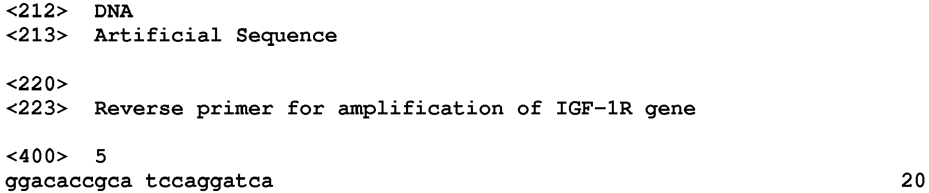
Claims (17)
- A cell laminate comprising:
an epidermal layer in which specific gene-deficient epidermal keratinocytes are laminated. - The cell laminate according to claim 1, wherein the epidermal keratinocyte is a human epidermal keratinocyte.
- The cell laminate according to claim 1 or 2, further comprising:
a support layer. - The cell laminate according to claim 3, wherein the support layer is a layer including collagen and at least one of mesenchymal cells or mesenchymal stem cells.
- The cell laminate according to any one of claims 1 to 4, wherein the specific gene includes a cell membrane receptor gene.
- The cell laminate according to any one of claims 1 to 5, wherein the specific gene includes an insulin-like growth factor-1 receptor gene.
- A method for producing a cell laminate, comprising:
a step of culturing specific gene-deficient epidermal keratinocytes by air-liquid interface culture. - The method for producing a cell laminate according to claim 7, wherein the epidermal keratinocyte is a human epidermal keratinocyte.
- The method for producing a cell laminate according to claim 7 or 8, further comprising:
a step of deleting the specific gene of the epidermal keratinocyte using a site-specific nuclease. - The method for producing a cell laminate according to any one of claims 7 to 9, wherein the step of culturing specific gene-deficient epidermal keratinocytes by air-liquid interface culture includes culturing the specific gene-deficient epidermal keratinocytes on a support layer by air-liquid interface culture.
- The method for producing a cell laminate according to claim 10, wherein the support layer is a layer including collagen and at least one of mesenchymal cells or mesenchymal stem cells.
- The method for producing a cell laminate according to any one of claims 7 to 11, wherein the specific gene includes a cell membrane receptor gene.
- The method for producing a cell laminate according to any one of claims 7 to 12, wherein the specific gene includes an insulin-like growth factor-1 receptor gene.
- A method for evaluating epidermal proliferation or differentiation, comprising:
a step of culturing specific gene-deficient epidermal keratinocytes by air-liquid interface culture. - A method for evaluating a test substance, comprising:
a step of culturing specific gene-deficient epidermal keratinocytes in the presence of a test substance by air-liquid interface culture. - A method for evaluating a test substance, comprising:
a step of bringing a test substance into contact with the cell laminate according to any one of claims 1 to 6. - A biological implantation material comprising:
the cell laminate according to any one of claims 1 to 6.
Applications Claiming Priority (2)
| Application Number | Priority Date | Filing Date | Title |
|---|---|---|---|
| JP2016150296 | 2016-07-29 | ||
| PCT/JP2017/024647 WO2018020970A1 (en) | 2016-07-29 | 2017-07-05 | Cell laminate, method for producing cell laminate, method for evaluating epidermal proliferation or differentiation, method for evaluating test substance, and biological implantation material |
Publications (2)
| Publication Number | Publication Date |
|---|---|
| EP3492583A1 true EP3492583A1 (en) | 2019-06-05 |
| EP3492583A4 EP3492583A4 (en) | 2019-06-19 |
Family
ID=61016891
Family Applications (1)
| Application Number | Title | Priority Date | Filing Date |
|---|---|---|---|
| EP17833979.2A Withdrawn EP3492583A4 (en) | 2016-07-29 | 2017-07-05 | Cell laminate, method for producing cell laminate, method for evaluating epidermal proliferation or differentiation, method for evaluating test substance, and biological implantation material |
Country Status (4)
| Country | Link |
|---|---|
| US (1) | US20190231824A1 (en) |
| EP (1) | EP3492583A4 (en) |
| JP (1) | JPWO2018020970A1 (en) |
| WO (1) | WO2018020970A1 (en) |
Families Citing this family (4)
| Publication number | Priority date | Publication date | Assignee | Title |
|---|---|---|---|---|
| GB201816911D0 (en) * | 2018-10-17 | 2018-11-28 | Univ London Queen Mary | Apparatus and methods |
| JP7246066B2 (en) * | 2018-12-11 | 2023-03-27 | 日本メナード化粧品株式会社 | Three-dimensional cultured epidermis model and manufacturing method thereof |
| WO2021149761A1 (en) * | 2020-01-22 | 2021-07-29 | 国立研究開発法人産業技術総合研究所 | Three-dimensional culture of immortalized skin cells having superficial layer formed from stratum corneum, method for producing said three-dimensional culture, and method for evaluating test substance using said three-dimensional culture |
| CN113201481B (en) * | 2021-04-19 | 2023-10-13 | 清华大学深圳国际研究生院 | Skin microsphere and preparation method and application thereof |
Family Cites Families (1)
| Publication number | Priority date | Publication date | Assignee | Title |
|---|---|---|---|---|
| EP1375646A1 (en) * | 2002-06-21 | 2004-01-02 | Deutsches Krebsforschungszentrum Stiftung des öffentlichen Rechts | Genetically engineered keratinocytes expressing at least one growth factor and/or activated EGF receptor, skin equivalent comprising said keratinocytes and toxicity assay using said keratinocytes |
-
2017
- 2017-07-05 WO PCT/JP2017/024647 patent/WO2018020970A1/en not_active Ceased
- 2017-07-05 EP EP17833979.2A patent/EP3492583A4/en not_active Withdrawn
- 2017-07-05 JP JP2018529467A patent/JPWO2018020970A1/en not_active Abandoned
-
2018
- 2018-12-16 US US16/221,577 patent/US20190231824A1/en not_active Abandoned
Also Published As
| Publication number | Publication date |
|---|---|
| US20190231824A1 (en) | 2019-08-01 |
| EP3492583A4 (en) | 2019-06-19 |
| WO2018020970A1 (en) | 2018-02-01 |
| JPWO2018020970A1 (en) | 2019-01-24 |
Similar Documents
| Publication | Publication Date | Title |
|---|---|---|
| Larderet et al. | Human side population keratinocytes exhibit long-term proliferative potential and a specific gene expression profile and can form a pluristratified epidermis | |
| JP6775157B2 (en) | Three-dimensional structure, its manufacturing method, and a three-dimensional structure forming agent | |
| Webber et al. | CRISPR/Cas9-based genetic correction for recessive dystrophic epidermolysis bullosa | |
| Lizier et al. | Scaling-up of dental pulp stem cells isolated from multiple niches | |
| US20190231824A1 (en) | Cell laminate, method for producing cell laminate, method for evaluating epidermal proliferation or differentiation, method for evaluating test substance, and biological implantation material | |
| Henn et al. | Cas9-mediated knockout of Ndrg2 enhances the regenerative potential of dendritic cells for wound healing | |
| Kim et al. | Effective differentiation of induced pluripotent stem cells into dental cells | |
| EP3168297B1 (en) | Cardiac cell culture material | |
| Kogata et al. | Sox9 regulates cell state and activity of embryonic mouse mammary progenitor cells | |
| Kawakami et al. | Approach for the derivation of melanocytes from induced pluripotent stem cells | |
| Yu et al. | Message of nexin 1, a serine protease inhibitor, is accumulated in the follicular papilla during anagen of the hair cycle | |
| Choi et al. | Generation of trichogenic adipose-derived stem cells by expression of three factors | |
| Duss et al. | Mesenchymal precursor cells maintain the differentiation and proliferation potentials of breast epithelial cells | |
| Quadri et al. | Investigating cutaneous squamous cell carcinoma in vitro and in vivo: novel 3D tools and animal models | |
| Sahu et al. | Spatiotemporal modulation of growth factors directs the generation of multilineage mouse embryonic stem cell-derived mammary organoids | |
| EP3867354A1 (en) | Compositions for and methods of producing tumor organoids | |
| Heng et al. | Directing stem cells into the keratinocyte lineage in vitro | |
| Queiroz et al. | Periodontal ligament cells mobilized by transforming growth factor-beta 1 and migrated without stimuli showed enhanced osteogenic differentiation | |
| JP5097387B2 (en) | Artificial skin | |
| Vazana-Netzarim et al. | Distinct HAND2/HAND2-AS1 expression levels may fine-tune mesenchymal and epithelial cell plasticity of human mesenchymal stem cells | |
| Jensen et al. | Tissue culture of human epidermal keratinocytes: a differentiating model system for gene testing and somatic gene therapy | |
| Ite et al. | Stem cell challenges and opportunities | |
| Lin et al. | Induction of human stem cells into ameloblasts by reaggregation strategy | |
| KR101418876B1 (en) | A method for regulating differentiation of skin adult stem cell | |
| EP4464766A1 (en) | Container for producing tension-applied three-dimensional artificial skin and production method thereof |
Legal Events
| Date | Code | Title | Description |
|---|---|---|---|
| PUAI | Public reference made under article 153(3) epc to a published international application that has entered the european phase |
Free format text: ORIGINAL CODE: 0009012 |
|
| 17P | Request for examination filed |
Effective date: 20190123 |
|
| AK | Designated contracting states |
Kind code of ref document: A1 Designated state(s): AL AT BE BG CH CY CZ DE DK EE ES FI FR GB GR HR HU IE IS IT LI LT LU LV MC MK MT NL NO PL PT RO RS SE SI SK SM TR |
|
| AX | Request for extension of the european patent |
Extension state: BA ME |
|
| A4 | Supplementary search report drawn up and despatched |
Effective date: 20190521 |
|
| RIC1 | Information provided on ipc code assigned before grant |
Ipc: G01N 33/15 20060101ALI20190510BHEP Ipc: C12N 5/071 20100101AFI20190510BHEP Ipc: A61P 17/00 20060101ALI20190510BHEP Ipc: C12Q 1/02 20060101ALI20190510BHEP Ipc: A61K 35/36 20150101ALI20190510BHEP Ipc: G01N 33/50 20060101ALI20190510BHEP Ipc: A61L 27/60 20060101ALI20190510BHEP |
|
| DAV | Request for validation of the european patent (deleted) | ||
| DAX | Request for extension of the european patent (deleted) | ||
| STAA | Information on the status of an ep patent application or granted ep patent |
Free format text: STATUS: THE APPLICATION IS DEEMED TO BE WITHDRAWN |
|
| 18D | Application deemed to be withdrawn |
Effective date: 20191218 |