EP3488019B1 - Extracellular mrna markers of muscular dystrophies in human urine - Google Patents
Extracellular mrna markers of muscular dystrophies in human urine Download PDFInfo
- Publication number
- EP3488019B1 EP3488019B1 EP17831981.0A EP17831981A EP3488019B1 EP 3488019 B1 EP3488019 B1 EP 3488019B1 EP 17831981 A EP17831981 A EP 17831981A EP 3488019 B1 EP3488019 B1 EP 3488019B1
- Authority
- EP
- European Patent Office
- Prior art keywords
- splicing
- mrna
- spliced
- urine
- dmd
- Prior art date
- Legal status (The legal status is an assumption and is not a legal conclusion. Google has not performed a legal analysis and makes no representation as to the accuracy of the status listed.)
- Active
Links
Images
















Classifications
-
- C—CHEMISTRY; METALLURGY
- C12—BIOCHEMISTRY; BEER; SPIRITS; WINE; VINEGAR; MICROBIOLOGY; ENZYMOLOGY; MUTATION OR GENETIC ENGINEERING
- C12Q—MEASURING OR TESTING PROCESSES INVOLVING ENZYMES, NUCLEIC ACIDS OR MICROORGANISMS; COMPOSITIONS OR TEST PAPERS THEREFOR; PROCESSES OF PREPARING SUCH COMPOSITIONS; CONDITION-RESPONSIVE CONTROL IN MICROBIOLOGICAL OR ENZYMOLOGICAL PROCESSES
- C12Q1/00—Measuring or testing processes involving enzymes, nucleic acids or microorganisms; Compositions therefor; Processes of preparing such compositions
- C12Q1/68—Measuring or testing processes involving enzymes, nucleic acids or microorganisms; Compositions therefor; Processes of preparing such compositions involving nucleic acids
- C12Q1/6876—Nucleic acid products used in the analysis of nucleic acids, e.g. primers or probes
- C12Q1/6883—Nucleic acid products used in the analysis of nucleic acids, e.g. primers or probes for diseases caused by alterations of genetic material
-
- C—CHEMISTRY; METALLURGY
- C12—BIOCHEMISTRY; BEER; SPIRITS; WINE; VINEGAR; MICROBIOLOGY; ENZYMOLOGY; MUTATION OR GENETIC ENGINEERING
- C12Q—MEASURING OR TESTING PROCESSES INVOLVING ENZYMES, NUCLEIC ACIDS OR MICROORGANISMS; COMPOSITIONS OR TEST PAPERS THEREFOR; PROCESSES OF PREPARING SUCH COMPOSITIONS; CONDITION-RESPONSIVE CONTROL IN MICROBIOLOGICAL OR ENZYMOLOGICAL PROCESSES
- C12Q1/00—Measuring or testing processes involving enzymes, nucleic acids or microorganisms; Compositions therefor; Processes of preparing such compositions
- C12Q1/68—Measuring or testing processes involving enzymes, nucleic acids or microorganisms; Compositions therefor; Processes of preparing such compositions involving nucleic acids
- C12Q1/6844—Nucleic acid amplification reactions
- C12Q1/6865—Promoter-based amplification, e.g. nucleic acid sequence amplification [NASBA], self-sustained sequence replication [3SR] or transcription-based amplification system [TAS]
-
- G—PHYSICS
- G01—MEASURING; TESTING
- G01N—INVESTIGATING OR ANALYSING MATERIALS BY DETERMINING THEIR CHEMICAL OR PHYSICAL PROPERTIES
- G01N33/00—Investigating or analysing materials by specific methods not covered by groups G01N1/00 - G01N31/00
- G01N33/48—Biological material, e.g. blood, urine; Haemocytometers
- G01N33/50—Chemical analysis of biological material, e.g. blood, urine; Testing involving biospecific ligand binding methods; Immunological testing
-
- C—CHEMISTRY; METALLURGY
- C12—BIOCHEMISTRY; BEER; SPIRITS; WINE; VINEGAR; MICROBIOLOGY; ENZYMOLOGY; MUTATION OR GENETIC ENGINEERING
- C12Q—MEASURING OR TESTING PROCESSES INVOLVING ENZYMES, NUCLEIC ACIDS OR MICROORGANISMS; COMPOSITIONS OR TEST PAPERS THEREFOR; PROCESSES OF PREPARING SUCH COMPOSITIONS; CONDITION-RESPONSIVE CONTROL IN MICROBIOLOGICAL OR ENZYMOLOGICAL PROCESSES
- C12Q2600/00—Oligonucleotides characterized by their use
- C12Q2600/106—Pharmacogenomics, i.e. genetic variability in individual responses to drugs and drug metabolism
-
- C—CHEMISTRY; METALLURGY
- C12—BIOCHEMISTRY; BEER; SPIRITS; WINE; VINEGAR; MICROBIOLOGY; ENZYMOLOGY; MUTATION OR GENETIC ENGINEERING
- C12Q—MEASURING OR TESTING PROCESSES INVOLVING ENZYMES, NUCLEIC ACIDS OR MICROORGANISMS; COMPOSITIONS OR TEST PAPERS THEREFOR; PROCESSES OF PREPARING SUCH COMPOSITIONS; CONDITION-RESPONSIVE CONTROL IN MICROBIOLOGICAL OR ENZYMOLOGICAL PROCESSES
- C12Q2600/00—Oligonucleotides characterized by their use
- C12Q2600/156—Polymorphic or mutational markers
Definitions
- DM1 myotonic dystrophy type 1
- DMD Duchenne muscular dystrophy
- BMD Becker muscular dystrophy
- LGMD1B limb girdle muscular dystrophy type 1B
- LMNA-linked dilated cardiomyopathy DCM
- Hutchinson-Gilford progeria syndrome HGPS
- Familial partial lipodystrophy type 2 FPLD2
- SMA spinal muscular atrophy
- ALS amyotrophic lateral sclerosis
- Pre-mRNA splicing occurs when introns are removed to generate a protein-coding message, while alternative splicing involves inclusion or exclusion of certain exons to code for different protein isoforms from the same gene.
- These splice variants are a fundamental process of nature designed to increase biodiversity, mainly in eukaryotes. Mis-regulation of pre-mRNA alternative splicing is found in a number of neurologic and neuromuscular diseases 1 .
- DM1 myotonic dystrophy type 1
- MBNL muscleblind-like
- DE1020060072249 discloses methods for determining bladder carcinoma with differentiation grade 2 by determining the amount of mRNA in urine or urinary sediment.
- WO2015187825 discloses methods of altering RNA splicing in a subject.
- WO2015082372 discloses in vitro process for the diagnosis of prostate cancer and other tunor types in exosomes by identifying the AGR2 gene.
- Urine contains extracellular RNA (exRNA) markers of urogenital cancers.
- exRNA RNA
- ex-mRNA splicing outcomes is needed to monitor therapeutic antisense oligonucleotide (ASO) drug effects.
- ASO therapeutic antisense oligonucleotide
- the present inventors examined whether ex-mRNA splice variants in human urine could meet sensitivity and specificity as robust markers of muscular dystrophies and other conditions associated with aberrant splicing, e.g., conditions associated with muscle weakness or dystrophy.
- Urine also contains mutation-specific dystrophin deletion mRNAs amenable to therapeutic exon skipping ASO strategies in DMD patients, and a dystrophin cryptic splice site in a patient with Becker muscular dystrophy.
- DM1 myotonic dystrophy type 1
- DMD Duchenne muscular dystrophy
- BMD Becker muscular dystrophy
- LGMD1B limb girdle muscular dystrophy type 1B
- DCM Hutchinson-Gilford progeria syndrome
- FPLD2 Familial partial lipodystrophy type 2
- SMA spinal muscular atrophy
- ALS amyotrophic lateral sclerosis
- the methods include isolating extracellular mRNA in a sample comprising urine from a subject who has, or is suspected to have, a disease associated with aberrant mRNA splicing; determining one or more selected mRNA in the sample, wherein the one or more selected mRNA is aberrantly spliced in the subject, and is suspected to be present in a plurality of spliced isoforms in the sample, wherein the spliced isoforms comprise properly spliced isoforms and mis-spliced isoforms; quantitating levels of the properly spliced isoforms and mis-spliced isoforms of the one or more selected mRNAs in the sample; determining a ratio of the properly spliced isoforms to the mis-spliced isoforms of the one or more selected mRNAs in the sample; and comparing the ratio of properly spliced to mis-spliced in a subject to a reference
- DM1 myotonic dystrophy type 1
- DMD Duchenne muscular dystrophy
- BMD Becker muscular dystrophy
- LGMD 1B limb girdle muscular dystrophy type 1B
- DCM Hutchinson-Gilford progeria syndrome
- FPLD2 Familial partial lipodystrophy type 2
- SMA spinal muscular atrophy
- ALS amyotrophic lateral sclerosis
- the methods include determining a first ratio of properly spliced isoforms to mis-spliced isoforms in a sample from the subject using a method described herein; determining a subsequent ratio of properly spliced isoforms to mis-spliced isoforms in a sample from the subject using a method described herein after a treatment for the disease has been administered to the subject; and comparing the first and subsequent ratios, wherein a ratio in the second sample that is higher than the ratio in the subsequent sample indicates that the treatment is effective.
- the treatment that is intended to correct splicing; to inhibit or reduce levels of mis-spliced transcripts; or to alter splicing to produce a functional protein.
- the treatment is an antisense oligonucleotide.
- the disease is myotonic dystrophy type 1 (DM1), and wherein the one or more selected mRNAs is selected from the group consisting of the transcript for insulin receptor (INSR); muscleblind like splicing regulator 2 (MBNL2); SOS Ras/Rac guanine nucleotide exchange factor 1 (SOS1); cytoplasmic linker associated protein 1 (CLASP1); muscleblind like splicing regulator 1 (MBNL1); mitogen-activated protein kinase kinase kinase 4 (MAP3K4); nuclear factor I X (NFIX); nuclear receptor corepressor 2 (NCOR2); VPS39, HOPS complex subunit (VPS39); and microtubule associated protein tau (MAPT).
- ITR insulin receptor
- MBNL2 muscleblind like splicing regulator 2
- SOS1 SOS Ras/Rac guanine nucleotide exchange factor 1
- CLASP1 cytoplasmic linker associated protein 1
- the selected mRNAs comprise MBNL2, MBNL1, SOS1, CLASP1, MAP3K4, and optionally INSR.
- the disease is associated with aberrant splicing of dystrophin (DMD); lamin A/C (LMNA); survival of motor neuron 2, centromeric (SMN2); solute carrier family 1 member 2 (SLC1A2); TAR DNA-binding protein (TARDP); or FUS RNA binding protein (FUS).
- DMD dystrophin
- LMNA lamin A/C
- SLC1A2 solute carrier family 1 member 2
- TARDP TAR DNA-binding protein
- FUS FUS RNA binding protein
- one or more selected mRNAs is selected from the group consisting of the transcript for insulin receptor (INSR); muscleblind like splicing regulator 2 (MBNL2); SOS Ras/Rac guanine nucleotide exchange factor 1 (SOS1); cytoplasmic linker associated protein 1 (CLASP1); muscleblind like splicing regulator 1 (MBNL1); mitogen-activated protein kinase kinase kinase 4 (MAP3K4); nuclear factor I X (NFIX); nuclear receptor corepressor 2 (NCOR2); VPS39, HOPS complex subunit (VPS39); microtubule associated protein tau (MAPT); dystrophin (DMD); lamin A/C (LMNA); survival of motor neuron 2, centromeric (SMN2); solute carrier family 1 member 2 (SLC1A2); TAR DNA-binding protein (TARDP); and FUS RNA binding protein (FUS).
- INSR insulin receptor
- MBNL2 muscleblind like splicing
- DM1 pre-mRNA splicing outcomes in muscle biopsies are biomarkers of disease severity 4 , while in DM1 mice they serve as sensitive indicators of therapeutic drug effects 5, 6 .
- Less invasive biomarkers to assess disease state and response to therapy in DM are currently unavailable, and optimal outcome measures of therapeutic success remain undefined.
- ASO antisense oligonucleotide
- This experimental drug for DM1 is designed to induce knockdown through the RNase H pathway of mutant transcripts, thereby rescuing muscle cells from the pathogenic effects of splicing mis-regulation 6, 8 .
- Extracellular vesicles include exosomes, microvesicles, and other membrane-encased nanoparticles released and taken up by cells as a form of extracellular communication 9 .
- EVs in serum and urine contain mRNA and non-coding RNAs, including microRNA (miRNA), termed exRNAs, released from different tissues and can serve as genetic biomarkers of cancers and other disease states 10-12 . Mutations, deletions, translocations, and transcriptome variations also have been shown extensively in EVs, especially for cancers 13-15 . Differentiated skeletal muscle cells in culture release EVs 16, 17 and a handful of miRNA biomarkers and several protein signatures have been identified in serum of muscular dystrophy patients 18 .
- miRNA microRNA
- RNA splice products in human urine have sufficient sensitivity and specificity to be robust biomarkers of muscular dystrophies.
- mRNA splicing patterns in "liquid biopsies” present a rich source of personalized biomarkers with applications to a number of genetic diseases.
- DM1 we found 10 alternative splice variants in urine that serve as a robust composite biomarker of DM1 disease activity.
- Mis-regulated alternative splicing outcomes in muscle tissue were sensitive indicators of therapeutic response in DM1 mice 5, 6 and disease activity in DM1 patients 4 .
- splice products in muscle biopsies were used in a recent clinical trial as measures of ASO activity in DM1 patients 7 .
- the present methods using splicing outcomes provides powerful biomarkers of DM1, in part because the disease mechanism involves mis-regulated alternative splicing; in addition, the ratiometric measurements of exon inclusion/exclusion described herein are inherently more sensitive than the unidirectional changes that are typical of most biomarkers.
- DM1 is primarily a disease of skeletal muscle, heart, and the central nervous system (CNS), it is counter-intuitive that exRNA reflecting the characteristic mis-regulated splicing events appears in urine rather than in blood, as exRNA has not been shown to pass from the blood through the proximal tubules of the kidney 29 .
- Urine exRNA should enable comprehensive non-invasive investigation of splicing outcomes in children with DM for the first time, facilitate clinical trials to these patients earlier, and enable convenient titration of dose.
- the shared pathogenic mechanism of alternative splicing misregulation in DM1 and DM2 4 suggests urine exRNA also may be useful for monitoring disease activity in DM2 patients.
- the urine splice products are more than traditional biomarkers: they are personalized genetic markers that are designed specifically for each individual patient and enable the possibility to monitor splice-shifting ASO drug effects 27, 28 .
- Dystrophin protein measurement in biopsy tissue is presently used as a surrogate marker of drug effect that led to the accelerated approval of eteplirsen by the U.S. Food and Drug Administration 31 .
- monitoring the ratio of skipped/unskipped DMD splice products in urine during the course of treatment may be used to complement RT-PCR analysis of muscle biopsies and/or in place of dystrophin protein measurement as a surrogate marker of therapeutic effect as newer and better splice-shifting drugs are developed.
- DMD cryptic splice site responsible for Becker MD phenotype in an individual with dystrophinopathy further suggests the value of liquid biopsies as a means to identify novel splice variants that may help correlate genotype with phenotype for a number of diseases for which non-invasive biomarkers are unavailable.
- HGPS Hutchinson-Gilford progeria syndrome
- point mutations in the LMNA gene activate a weak splice site in exon 11 that shortens the transcript and produces a truncated progerin protein 32 .
- ASOs that reduce use of this weak splice site are being evaluated as strategy to treat HGPS 33 .
- LMNA exon 11 in urine shows that the present methods including use of exRNA can be used to monitor drug effects in these patients as well.
- the present findings also support development of exRNA from urine, serum, or CSF as a biomarker replacement for tissue biopsies in other diseases with altered mRNA splicing, including limb girdle muscular dystrophy type 1B, spinal muscular atrophy and amyotrophic lateral sclerosis 34-36 .
- Table A provides a list of conditions that can be diagnosed, treated, or monitored using the present methods, along with the mutated genes (though note that the mutation may or may not result in altered splicing of that specific gene, or not only that specific gene).
- DM1 Myotonic dystrophy type 1
- DM1 is caused by a heterozygous trinucleotide repeat expansion (CTG)n in the 3-prime untranslated region of the dystrophia myotonica protein kinase gene (DMPK); a repeat length exceeding 50 CTG repeats is pathogenic ( Musova et al., Am. J. Med. Genet. 149A: 1365-1374, 2009 ).
- CUG repeats form RNA hairpins that bind proteins including muscleblind-like 1 (MBNL1), a splicing regulatory factor; nuclear sequestration of MBNL1 prevents its activity and results in aberrant splicing of several genes.
- MBNL1 muscleblind-like 1
- nuclear sequestration of MBNL1 prevents its activity and results in aberrant splicing of several genes.
- NCOR2 Homo sapiens nuclear receptor corepressor 2 (NCOR2), transcript variant 1, mRNA NM_006312.5 VPS39 Homo sapiens VPS39, HOPS complex subunit (VPS39), transcript variant 2, mRNA NM_015289.3
- MAPT Homo sapiens microtubule associated protein tau (MAPT), transcript variant 1, mRNA NM_016835.4
- the methods can include determining ratios of properly spliced mRNA to aberrantly spliced mRNA for all or a subset, e.g., 1, 2, 3, 4, 5, 6, 7, 8, 9 or all 10 of the above.
- the subset includes 1, 2, 3, 4, or all of MBNL2, MBNL1, SOS1, CLASP 1, MAP3K4, and optionally also INSR.
- DM1 affects skeletal and smooth muscle as well as the eye, heart, endocrine system, and central nervous system. Symptoms can include muscle weakness, e.g., in the leg, hand, neck, and/or face; myotonia, e.g., grip myotonia or percussion myotonia; and posterior subcapsular cataracts (which are detectable as red and green iridescent opacities on slit lamp examination).
- the methods described herein can also include monitoring the efficacy of a treatment for DM1, e.g., a treatment that is expected or intended to affect splicing, e.g., to correct splicing or to inhibit or reduce levels of aberrantly spliced transcripts, e.g., as described herein.
- the present methods can be used to monitor efficacy, e.g., to determine whether the treatment affects splicing, by detecting a change in the ratio of properly spliced mRNA to aberrantly spliced mRNA. An increase in the properly spliced mRNA, and/or a decrease in aberrantly spliced mRNA, would result in an increase in the ratio and indicates that the treatment is effective.
- DMD Duchenne muscular dystrophy
- BMD Becker muscular dystrophy
- DMD and BMD are inherited progressive muscle disorders that are noninflammatory and not associated with a central or peripheral nerve abnormality.
- the disease affects the muscles with definite fiber degeneration but without evidence of morphologic aberrations, resulting in progressive muscle wasting, and are caused by defects in the dystrophin gene DMD. See, e.g., Aartsma-Rus et al., J Med Genet. 2016 Mar;53(3):145-51 ; Flanigan et al., Hum Mutat. 2011 Mar; 32(3): 299-308 .
- DMD or BMD are caused by mutations that affect splicing of the transcript, e.g., acceptor or donor splice site mutations.
- the present methods can be used to detect these alternative mRNA splice variants or mRNA of different lengths.
- the methods can include determining ratios of properly spliced DMD mRNA to aberrantly spliced DMD mRNA.
- the methods described herein can also include monitoring the efficacy of a treatment for DMD or BMD, e.g., a treatment that is expected or intended to affect splicing, e.g., to correct splicing of the dystrophin transcripts, reduce levels of aberrant transcripts, or to produce transcripts that encode functional dystrophin protein.
- ASOs for DMD are used to induce new splicing changes that serve to restore the open reading frame rather than correct aberrant splicing. It may be possible that some DMD mutations that lead to a Duchenne phenotype and that ASOs could be designed to treat this.
- Alternative splicing of DMD transcripts typically includes exon 71, 78, and perhaps exon 68. The remainder of the DMD exons are spliced constitutively.
- Urine RNA can also be used to identify novel aberrant splicing, as in our Becker patient with a cryptic splice site.
- the present methods can be used to monitor efficacy, e.g., to determine whether the treatment affects splicing, e.g., by detecting a change in the ratio of properly spliced mRNA (or mRNA of a desired size or sequence) to aberrantly spliced mRNA (or mRNA of a non-desired size or sequence).
- An increase in the properly spliced mRNA or mRNA of a desired size or sequence, and/or a decrease in aberrantly spliced mRNA or mRNA of a non-desired size or sequence would result in an increase in the ratio and indicates that the treatment is effective.
- HGPS Hutchinson-Gilford progeria syndrome
- DCM LMNA-linked dilated cardiomyopathy
- FPLD2 Familial partial lipodystrophy type 2
- LMNA lamin A
- Hutchinson-Gilford progeria syndrome is caused by mutations within the LMNA gene that lead to increased usage of an internal splice site, resulting in alternative lamin A transcript with internal deletions of 150 nucleotides (LMNA G608G (GGC>GGT) mutation); see Eriksson et al., Nature 423, 293-298 (2003 ); Rodriguez et al., Eur J Hum Genet. 2009 Jul; 17(7): 928-937 .
- LGMD Limb-girdle muscular dystrophies
- FPLD2 familial partial lipodystrophy type 2
- LMNA-linked dilated cardiomyopathy is associated with an alternative 3' splice site generated by an A>G mutation (c. 640-10A>G); see Scotti and Swanson, Nature Reviews Genetics 17:19-32 (2016 )).
- the methods described herein can also include monitoring the efficacy of a treatment for a conditions associated with mis-splicing of LMNA, e.g., a treatment that is expected or intended to affect splicing, e.g., to correct splicing of the dystrophin transcripts, reduce levels of aberrant transcripts, or to produce transcripts that encode functional dystrophin protein.
- the present methods can be used to monitor efficacy, e.g., to determine whether the treatment affects splicing, by detecting a change in the ratio of properly spliced mRNA (or mRNA of a desired size or sequence) to aberrantly spliced mRNA (or mRNA of a non-desired size or sequence).
- SMA spinal muscular atrophy
- SMN is part of a large multi-protein complex (with additional proteins, including Gemeins 2-7) that is necessary for biogenesis of small nuclear RNA ribonucleoproteins (snRNPs), which are major components of pre-mRNA splicing machinery. Genetic alterations in the SMN gene result in the reduced capacity for snRNP assembly, and defects in RNA splicing. See Dreyfuss, Eukaryon, 6:75-79, 2010 .
- the methods described herein can also include monitoring the efficacy of a treatment for SMA, e.g., a treatment that is expected or intended to affect splicing, e.g., to correct splicing of the dystrophin transcripts, reduce levels of aberrant transcripts, or to produce transcripts that encode functional dystrophin protein.
- Splicing of SMN2 normally involves skipping of exon 7 in the majority of SMN2 transcripts.
- ASO treatment of SMA involves increasing inclusion of SMN2 exon 7, which results in an increase of SMN protein levels, the same protein that is lost by mutations in SMN1.
- the present methods can be used to monitor efficacy, e.g., to determine whether the treatment affects splicing, by detecting a change in the ratio of properly spliced mRNA (or mRNA of a desired size or sequence) to aberrantly spliced mRNA (or mRNA of a non-desired size or sequence).
- An increase in the properly spliced mRNA or mRNA of a desired size or sequence, and/or a decrease in aberrantly spliced mRNA or mRNA of a non-desired size or sequence would result in an increase in the ratio and indicates that the treatment is effective.
- ALS Amyotrophic lateral sclerosis
- EAAT2 also known as SLC1A2
- Defective pre-mRNA splicing in the motor cortex and spinal cord is responsible for the loss of EAAT2 protein. This defective splicing is caused by a defect in a splicing regulatory factor, rather than a mutation in the EAAT2 gene that causes alternative aberrant splicing or a defect in a general splicing apparatus, such as the spliceosome.
- the defective pre-mRNA splicing process for EAAT2 can skip normal 5' and 3' splice sites (donor and acceptor splicing sites), or use inappropriate 5' and 3' splice sites (i.e., other than the normal GU or AU for the donor site, and AG or AC for the acceptor site), resulting in multiple abnormal RNAs in ALS patients.
- the aberrant splicing results in transcripts that partially retain introns or skip exons, as well as transcripts that have exonic sequences at random sites. Two aberrantly spliced EAAT2 mRNAs are found predominantly in sporadic ALS patients.
- RNA transcripts that partially retains intron 7 and an mRNA transcript that skips exon 9 of the gene.
- the intro 7-retaining RNA causes a dominant-negative effect on normal EAAT2 that has been shown to result in a loss of protein and activity. See, e.g., Honig et al., Neurology. 2000 Oct 24;55(8): 1082-8 ; Lin et al., Neuron. 1998 Mar;20(3):589-602 ; Meyer et al., J Neurol Sci. 1999 Nov 15;170(1):45-50 .
- Certain forms of ALS are associated with the presence of mutations in the TARDP (c. 991C>A), (c.1009A>G) and FUS (c.
- the methods described herein can also include monitoring the efficacy of a treatment for conditions associated with mis-splicing of SLC1A2, TARDP, or FUS, e.g., a treatment that is expected or intended to affect splicing, e.g., to correct splicing of the dystrophin transcripts, reduce levels of aberrant transcripts, or to produce transcripts that encode functional dystrophin protein.
- the present methods can be used to monitor efficacy, e.g., to determine whether the treatment affects splicing, by detecting a change in the ratio of properly spliced mRNA (or mRNA of a desired size or sequence) to aberrantly spliced mRNA (or mRNA of a non-desired size or sequence).
- An increase in the properly spliced mRNA or mRNA of a desired size or sequence, and/or a decrease in aberrantly spliced mRNA or mRNA of a non-desired size or sequence would result in an increase in the ratio and indicates that the treatment is effective.
- DM1 myotonic dystrophy type 1
- DMD Duchenne muscular dystrophy
- BMD Becker muscular dystrophy
- LGMD1B limb girdle muscular dystrophy type 1B
- DCM Hutchinson-Gilford progeria syndrome
- FPLD2 Familial partial lipodystrophy type 2
- SMA spinal muscular atrophy
- ALS amyotrophic lateral sclerosis
- properly spliced which can include transcripts that are spliced by an ASO
- mis-spliced isoforms of affected transcripts in urine samples e.g., mis-spliced isoforms of affected transcripts in urine samples.
- "properly spliced” means that the transcript has a desired splice pattern, e.g., has wild-type splicing, or is spliced in a way that is desired, e.g., to produce a functional protein.
- the DMD exons targeted by ASOs are constitutively spliced, meaning they are always included in DMD patients and UA individuals.
- Treatment with ASOs in DMD is designed to induce a new unique splice event absent in DMD patients or UA individuals, and the ratio of the inclusion of the unique splice site (properly spliced) to inclusion of the constitutive splice (mis-spliced) provides a convenient estimation of ASO drug effects in urine a new unique splice event absent in DMD patients or UA individuals; in this case, a transcript that was "properly spliced" in a subject with DMD being treated with these ASOs would include the unique splice event.
- the methods can include determining that the mRNA is of a desired size or sequence (i.e., "properly spliced"), or is of a non-desired size or sequence ("mis-spliced").
- the methods include using a urine sample that has been obtained from a subject, determining levels of properly spliced and aberrantly spliced extracellular transcripts, and determining a ratio of properly spliced to mis-spliced transcript in the sample.
- the methods can include comparing the ratio with one or more reference ratios, e.g., a control reference that represents a normal ratio of properly spliced:mis-spliced transcript, e.g., a level in an unaffected subject, and/or a disease reference that represents a ratio associated with the disease.
- a reference ratio of properly spliced:mis-spliced transcripts in an unaffected subject may approach 1:0, since there would not be expected to be a large number of mis-spliced transcripts in such individuals.
- RNA samples can include identification and/or isolation and/or purification of a transcript from a sample.
- An "isolated” or “purified” biological marker is substantially free of cellular material or other contaminants from the cell or tissue source from which the biological marker is derived i.e. partially or completely altered or removed from the natural state through human intervention.
- nucleic acids contained in the sample can be isolated according to standard methods, for example using filtration, centrifugation, or other methods of purification to obtain a sample that contains extracellular transcripts but does not contain cells or cellular transcripts.
- the methods can include using chemical solutions nucleic acid-binding resins following the manufacturer's instructions.
- the entire volume of urine is centrifuged, e.g., at 2,000-3,000 x g, e.g., at 2,450 x g for 5-15 minutes, e.g., 10 minutes at room temperature, and then the supernatant is passed through a filter, e.g., a 0.8 ⁇ m filter, before being ultracentrifuged, e.g., at 100,000 x g 2 hours at 4° C, to pellet the RNA. Then the supernatant is removed and, RNA is extracted from the translucent ribonucleoprotein pellet, e.g., using Trizol (Life Technologies) according to manufacturer instructions. To enhance RNA pellet visibility, linear acrylamide (Ambion) or other reagents can be added.
- the transcripts can be evaluated using methods known in the art, e.g., using polymerase chain reaction (PCR), reverse transcriptase polymerase chain reaction (RT-PCR), quantitative or semi-quantitative real-time RT-PCR, digital PCR i.e. BEAMing (( Beads, Emulsion, Amplification, Magnetics) Diehl (2006) Nat Methods 3:551-559 ) ; RNAse protection assay; Northern blot; various types of nucleic acid sequencing (Sanger, pyrosequencing, NextGeneration Sequencing); fluorescent in-situ hybridization (FISH); or gene array/chips) ( Lehninger Biochemistry (Worth Publishers, Inc.
- PCR polymerase chain reaction
- RT-PCR reverse transcriptase polymerase chain reaction
- FISH fluorescent in-situ hybridization
- gene array/chips Lehninger Biochemistry (Worth Publishers, Inc.
- Measurement of the level of different splice isoforms can be direct or indirect.
- the abundance levels of various differently spliced isoforms can be directly quantitated, e.g., based on size or the presence or absence of a selected sequence.
- a technique suitable for the detection of alterations in the structure or sequence of nucleic acids such as the presence of deletions, amplifications, or substitutions, can be used for the detection of different splice isoforms.
- Gene arrays are prepared by selecting probes which comprise a polynucleotide sequence, and then immobilizing such probes to a solid support or surface.
- the probes may comprise DNA sequences, RNA sequences, co-polymer sequences of DNA and RNA, DNA and/or RNA analogues, or combinations thereof, which detect various spliced isoforms.
- the probe sequences can be synthesized either enzymatically in vivo, enzymatically in vitro (e.g. by PCR), or non-enzymatically in vitro.
- the methods can be used to diagnose a condition described herein; for example, when the ratio of properly spliced:mis-spliced transcripts in a subject (e.g., a subject who has one or more symptoms associated with the disease) is comparable to a reference ratio in a representative subject with the disease, then the subject can be diagnosed with the disease.
- a treatment e.g., as known in the art or as described herein, can be selected for administration.
- Suitable reference values can be determined using methods known in the art, e.g., using standard clinical trial methodology and statistical analysis.
- the reference values can have any relevant form.
- the reference comprises a predetermined value for a meaningful ratio, e.g., a control reference level that represents a normal level ratio, e.g., a level in an unaffected subject, and/or a disease reference that represents a ratio associated with the disease, e.g., a level in a subject having a disease as described herein, namely DMD, DM1, BMD, LGMD, HGPS, DCM, HGPS, FPLD2, SMA, or ALS.
- a meaningful ratio e.g., a control reference level that represents a normal level ratio, e.g., a level in an unaffected subject
- a disease reference that represents a ratio associated with the disease, e.g., a level in a subject having a disease as described herein, namely DMD, DM1, BMD
- the predetermined ratio can be a single cut-off (threshold) value, such as a median or mean, or a ratio that defines the boundaries of an upper or lower quartile, tertile, or other segment of a clinical trial population that is determined to be statistically different from the other segments. It can be a range of cut-off (or threshold) ratios, such as a confidence interval. It can be established based upon comparative groups, such as where association with presence of disease in one defined group is a fold higher, or lower, (e.g., approximately 2-fold, 4-fold, 8-fold, 16-fold or more) than the presence of disease in another defined group.
- groups such as a low-risk group, a medium-risk group and a high-risk group, or into quartiles, the lowest quartile being subjects with the lowest risk and the highest quartile being subjects with the highest risk, or into n-quantiles (i.e., n regularly spaced intervals) the lowest of the n-quantiles being subjects with the lowest risk and the highest of the n-quantiles being subjects
- the predetermined ratio is a ratio in the same subject, e.g., at a different time point, e.g., an earlier time point.
- a control reference subject does not have a disorder described herein (DMD, DM1, BMD, LGMD, HGPS, DCM, HGPS, FPLD2, SMA, and ALS). In some cases it may be desirable that the control subject is a first or second degree relative of the subject to be tested.
- a disease reference subject is one who has (has been diagnosed with) a disease as described herein, namely DMD, DM1, BMD, LGMD, HGPS, DCM, HGPS, FPLD2, SMA, or ALS.
- the ratio of properly spliced:mis-spliced in a subject being less than a reference ratio is indicative of a clinical status (e.g., indicative of presence of a disorder as described herein, namely DMD, DM1, BMD, LGMD, HGPS, DCM, HGPS, FPLD2, SMA, or ALS), or indicative of an ineffective therapy.
- the ratio in a subject being greater than or equal to the reference ratio is indicative of the absence of disease, or an effective therapy.
- the amount by which the ratio in the subject is the less than the reference ratio is sufficient to distinguish a subject from a control subject, and optionally is a statistically significantly less than the ratio in a control subject. In cases where the ratio in a subject being equal to the reference ratio, the "being equal" refers to being approximately equal (e.g., not statistically different).
- the predetermined ratio can depend upon the particular population of subjects (e.g., human subjects) selected. For example, an apparently healthy population may have a different 'normal' range of ratios than will a population of subjects which have, are likely to have, or are at greater risk to have, a disorder described herein. Accordingly, the predetermined values selected may take into account the category (e.g., sex, age, health, risk, presence of other diseases) in which a subject (e.g., human subject) falls. Appropriate ranges and categories can be selected with no more than routine experimentation by those of ordinary skill in the art.
- category e.g., sex, age, health, risk, presence of other diseases
- the methods described herein can include monitoring the efficacy of a treatment of disorders associated with aberrant splicing.
- the disorder is DMD, DM1, BMD, LGMD, HGPS, DCM, HGPS, FPLD2, SMA, or ALS.
- the methods include determining a subsequent ratio after a treatment has been administered to a subject identified using a method described herein.
- to "treat” means to ameliorate at least one symptom of the disorder associated with aberrant splicing.
- a treatment can result in a reduction in muscle weakness or a reduction in rate of muscle loss or weakening.
- Exon-skipping antisense oligonucleotides that correct missplicing can be used, e.g., as described in Siva et al., Nucleic Acid Ther. 2014 Feb 1; 24(1): 69-86 ; Scotti and Swanson, Nature Reviews Genetics 17:19-32 (2016 ).
- LNAs bicyclic-locked nucleic acids
- ENAs ethylene-bridged nucleic acids
- 20ME-PSs 2'-O-methyl phosphorothioate AO (20ME-PSs)
- PNAs peptide nucleic acids
- PMOs phosphorodiamidate morpholino oligomers
- the ASOs can be delivered, e.g., parenterally in liposomal complexes, e.g., cationic lipoplexes, or using a viral vector, e.g., a lentivirus, adenovirus, or adeno-associated virus.
- a viral vector e.g., a lentivirus, adenovirus, or adeno-associated virus.
- Exon skipping uses antisense oligonucleotides (ASOs) to alter transcript splicing; the present methods can be used to detect these transcripts with desired splicing.
- ASOs antisense oligonucleotides
- These treatments can include antisense oligonucleotide-targeted exon skipping to induce near normal, e.g., for dystrophin, e.g., as described in Aartsma-Rus, Methods Mol Biol. 2012;867:97-116 .
- Clinical trials of ASOs in DMD have been conducted, see, e.g., Koo and Wood, Hum Gene Ther. 2013 May;24(5):479-88 ; Voit et al., Lancet Neurol. 2014;13(10):987-996 .
- exon 11 antisense oligonucleotide that increased lamin C production has been shown to shift the output of LMNA more toward lamin C and reduce levels of the mutant protein in fibroblasts derived from patients with HGPS ( Lee et al., J Clin Invest. 2016 Apr 1;126(4):1592-602 ).
- Exon skipping ASOs directed against an intron splice silencer in SMN2 increase the amount of full-length SMN transcript in the CNS, restoring SMN to treat subjects with spinal muscular atrophy (SMA) (see Burghes and McGovern, Genes Dev. 2010 Aug 1; 24(15): 1574-1579 ).
- SMA spinal muscular atrophy
- ASO drug nusinersen enhanced exon 7 inclusion in a clinical trial, see Chiriboga et al., Neurology. 2016 Mar 8; 86(10): 890-897 .
- ASOs for use in exon 51 skipping therapy include PRO051 (2OME-PS, Netherlands) and AVI-4658 (PMO, UK).
- a plurality of ASOs can also be used, e.g., to induce exon skipping in multiple exons; see, e.g., Wood et al., Brain. 2010 Apr;133(Pt 4):957-72 See also Fletcher et al., Mol Ther Nucleic Acids. 2012 Oct; 1(10): e48 ; McClorey et al., Curr Opin Pharmacol. 2005 Oct;5(5):529-34 .
- an ASO inhibiting mutant DMPK transcripts can be used, e.g., a 149-bp antisense RNA complementary to the (CUG)13 repeats and to the 110-bp region following the repeats sequence has been described, see Furling et al., Gene Ther. 2003 May;10(9):795-802 . See also Maga ⁇ a and Cisneros, J Neurosci Res. 2011 Mar;89(3):275-85 ; Thornton et al., Curr Opin Genet Dev. 2017 Jun;44:135-140 ; Gao and Cooper, Hum Gene Ther. 2013 May; 24(5): 499-507 .
- Small molecule therapeutics can also be used, e.g., PTC124, a 284.24-Da, achiral, 1,2,4-oxadiazole linked to fluorobenzene and benzoic acid rings, which selectively induces ribosomal read-through of premature but not normal termination codons, see Welch et al., Nature 447: 87-91, 2007 , and has been used in clinical trials for DMD.
- Inclusion criteria for DM1 subjects were age 13 years or older, a diagnosis of DM1 based on genetic testing that identified a DMPK- CTG repeat expansion of ⁇ 50, or clinical diagnosis of DM1 and a 1 st degree relative with DM1 due to a DMPK- CTG repeat expansion of ⁇ 50, and ability to provide informed consent or assent for participation.
- Inclusion criteria for MDC subjects included known diagnosis of DMD, BMD, or FSHD, ages 13 years or older, and ability to provide informed consent.
- Inclusion criteria for unaffected individuals were age 18 years or older, no known history of any muscular dystrophy, and ability to provide informed consent.
- the training cohort consisted of a combined 34 DM1 or UA participants chosen randomly, and the remaining 11 combined DM1 and UA subjects comprised the validation cohort.
- informed consent was obtained for blood and/or urine collection from all subjects; due to severe autism, informed consent for the individual with Becker muscular dystrophy was obtained from his mother/legal guardian, according to IRB protocol. Subject information is shown in Tables 1A-1B.
- Tables 1A-1B Clinical data from DM1 subjects.
- Table 1A Subjects # Male # Female Mean age Age range DM1 19 8 41 17 - 63 UA 7 19 n/a n/a MDC 9 1 24 14 - 49
- Table 1B ID Leukocytes Nitrite Urobilinogen Protein pH Blood Specific gravity Ketone Bilirubin Glucose DM1 - - 3.5 +/- 5 - 1.025 - - - DM1 - - 3.5 +/- 6 - 1.020 - - - DM1 - - 3.5 +/- 5 - 1.020 - - - DM1 - - 3.5 +/- 5 - 1.010 - - - DM1 - - 3.5 +/- 5 - 1.020 - - DM1 - - 3.5 +/- 5 - 1.030 - - - DM1 - - 3.5 +/- 5 - 1.020 - - DM1 - - 3.5 +/
- mice The MGH IACUC approved all experiments involving mice.
- HSA LR transgenic and Mbnl1 knockout (Mbnl1 ⁇ E3/ ⁇ E3 ) models of DM1 (both FVB background) have been described 2, 26 .
- FVB wild-type mice served as controls.
- HSA LR mice that were treated with antisense oligonucleotide (ASO) 445236 received subcutaneous injections of 25 mg/kg twice weekly for 4 weeks, as previously described 6 .
- ASO 445236 was a gift of Dr. Frank Bennett at Ionis Pharmaceuticals (Carlsbad, CA).
- Nanoparticle tracking To determine nanoparticle size and concentration, we used the Nanosight LM10 system and Nanoparticle Tracking Analysis 2.0 analytical software according to manufacturer instructions (Malvern). The system uses a laser beam, light microscope, and CCD camera to visualize and video record particles in liquid suspension moving under Brownian motion. For accurate measurements, we diluted serum samples 1:1000 and urine either 1:10 or 1:20 in saline to stay in the target concentration range of 1.0 x 10 8 and 2.5 x 10 9 particles/milliliter. We recorded 60-second videos and analyzed data in auto mode.
- RNA integrity number RIN
- Quantitative real-time RT-PCR Quantitative real-time RT-PCR (qPCR).
- qPCR Quantitative real-time RT-PCR
- RNA from normal human skeletal muscle and kidney served as tissue controls.
- mice we chose sample sizes for splicing analysis in muscle based on previously reported differences in muscle tissue of these models 2, 3, 5, 6 . Mice ranged from 2 to 4 months of age and were chosen randomly by genotype, stratified for sex to allow an approximately equal number of females and males, and examined without blinding.
- Predictive model We used principal component regression to develop a predictive model of DM1 using the splicing quantification of the 10 genes, shown in Fig. 2 .
- the pls package in R which uses singular value decomposition algorithm for the fitting, was used to implement the model 41 .
- A_23_P343808 4.77 CLASP1* NM_015282 93.
- A_23_P311232 4.57 MBNL1* NM_021038 94.
- A_23_P357811 6.26 MAP3K4* NM_005922 95.
- A_23_P42096 5.07 NFIX* NM_002501 96.
- A_23_P165295 4.85 NCOR2* NM_006312 97.
- A_23_P203891 5.84 VPS39* NM_015289 98.
- A_24_P167825 5.08 MAPT* NM 016835 99.
- A_23_P207699 5.19 KIF13A NM_022113 100.
- A_23_P44083 6.03 IMPDH2 NM_000884 109.
- A_24_P166042 5.00 ALPK3 NM_020778 110.
- A_23_P348728 5.95 ANK2 NM_001148 111.
- A_23_P133068 5.50 BIN1 NM_139346 112.
- A_23_P165333 5.63 NRAP NM_198060 113.
- A_23_P402765 4.58 OPA1 NM _ 130837 114.
- A_23_P211797 6.54 PHKA1 NM_002637 115.
- A_32_P186121 5.26 UBE2D3 NM_181886 116.
- A_24_P363005 6.05 PDLIM3 NM _ 014476 117.
- A_23_P110403 4.51 LDB3 NM_007078 118.
- A_32_P98227 5.11 TTN NM_133378 119.
- A_23_P85269 4.66 FHOD1 NM_013241 120.
- A_23_P37778 5.46 TBC1D15 NM_022771 121.
- A_23_P139558 5.10 RYR1 NM_000540 122.
- A_23_P78867 4.76 DTNA NM_001392 123.
- A_23P208158 4.82 TNNT2 NM_000364 124.
- A_23_P34700 5.28 FXR1 NM_001013 439 125.
- A_23_P132784 5.47 CKM NM_001824 126.
- A_23_P50250 4.75 ACTA1 NM_001100 127.
- A_23_P1102 4.98 MYH3 NM_002470 128.
- A_23_P26865 4.86
- RNA samples from healthy control subjects were filtered through an 0.8 ⁇ m filter to remove cells, ultracentrifuged at 100,000 x g for 90 minutes to collect extracellular RNA in EVs and particles, and the EV/ribonucleoprotein (RNP) pellet was lysed.
- RNA was extracted using the Qiagen miRNeasy kit and examined by mRNA microarray analysis. The data are represented as quartile normalized with background subtraction and values indicate expression levels of each gene ( Noerholm et al., BMC Cancer 12, 22 (2012 )).
- a previous study collected 3300 ml of urine from a human male, centrifuged the entire volume at low speed to pellet cells, passed the supernatant through a 0.8 ⁇ m filter to remove remaining debris, ultracentrifuged the filtered supernatant, isolated RNA from the exosome/microvesicle pellet using a commercially available kit (Qiagen), and identified genes present using massively parallel RNA sequencing ( Miranda et al., PLoS One 9, e96094 (2014 )). Shown are raw and normalized counts of genes from that study that also were reported as biomarkers of DM1 in muscle biopsies 5, as well as other genes associated with muscular dystrophies. (*: transcripts we report here that show differential urine exRNA splicing in DM1 vs MDC and UA controls).
- RNAs in urine have been used as biomarkers of prostate cancer, bladder cancer, and kidney transplant rejection, suggesting that cells lining the urinary tract are the primary contributors to the urine ex-RNA pool 12, 24, 25 .
- Our finding of slightly different exon inclusion/exclusion percentages of some transcripts in urine as compared to muscle tissue suggests the exRNA found in urine may represent a pool from multiple different cell types along this urinary route.
- splice isoforms in human and mouse kidney and mouse bladder Figures 10 and 12 . Splicing of all transcripts examined showed different exon inclusion/exclusion percentages in normal human kidney compared to skeletal muscle ( Figures 10 and 12A ).
- ASOs also are being evaluated therapeutically for another form of muscle disease, Duchenne muscular dystrophy (DMD), to modify dystrophin pre-mRNA splicing directly by inducing skipping of a target exon to restore the open reading frame and produce a truncated, partially functional protein 27, 28 .
- DMD Duchenne muscular dystrophy
- Detection of therapeutic drug effects in DMD patients involves multiple muscle biopsies to examine splicing outcomes and dystrophin protein production.
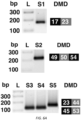
- biofluid exRNA contains DMD deletion transcripts, we examined urine from several subjects with DMD and found patient-specific DMD deletion transcripts ( Figures 6A and B ), suggesting this biofluid exRNA is a viable approach to monitor therapeutic exon-skipping ASO drug effects in DMD patients as personalized genetic markers 27, 28 .
Landscapes
- Chemical & Material Sciences (AREA)
- Life Sciences & Earth Sciences (AREA)
- Health & Medical Sciences (AREA)
- Engineering & Computer Science (AREA)
- Organic Chemistry (AREA)
- Proteomics, Peptides & Aminoacids (AREA)
- Wood Science & Technology (AREA)
- Analytical Chemistry (AREA)
- Zoology (AREA)
- Immunology (AREA)
- Molecular Biology (AREA)
- Genetics & Genomics (AREA)
- General Health & Medical Sciences (AREA)
- Biotechnology (AREA)
- Microbiology (AREA)
- Physics & Mathematics (AREA)
- Biochemistry (AREA)
- Bioinformatics & Cheminformatics (AREA)
- General Engineering & Computer Science (AREA)
- Biophysics (AREA)
- Pathology (AREA)
- Biomedical Technology (AREA)
- Hematology (AREA)
- Urology & Nephrology (AREA)
- Chemical Kinetics & Catalysis (AREA)
- Cell Biology (AREA)
- Food Science & Technology (AREA)
- Medicinal Chemistry (AREA)
- General Physics & Mathematics (AREA)
- Measuring Or Testing Involving Enzymes Or Micro-Organisms (AREA)
Description
- This invention was made with Government support under Grant No. CA069246 awarded by the National Institutes of Health. The Government has certain rights in the invention.
- Described herein are methods for diagnosing and monitoring subjects with diseases associated with aberrant mRNA splicing, wherein the disease is myotonic dystrophy type 1 (DM1); Duchenne muscular dystrophy (DMD); Becker muscular dystrophy (BMD); limb girdle muscular dystrophy type 1B (LGMD1B); LMNA-linked dilated cardiomyopathy (DCM); Hutchinson-Gilford progeria syndrome (HGPS); Familial partial lipodystrophy type 2 (FPLD2); spinal muscular atrophy (SMA); or amyotrophic lateral sclerosis (ALS), based upon detecting properly spliced isoforms and mis-spliced isoforms in a urine sample from the subject.
- Pre-mRNA splicing occurs when introns are removed to generate a protein-coding message, while alternative splicing involves inclusion or exclusion of certain exons to code for different protein isoforms from the same gene. These splice variants are a fundamental process of nature designed to increase biodiversity, mainly in eukaryotes. Mis-regulation of pre-mRNA alternative splicing is found in a number of neurologic and neuromuscular diseases 1. For example, in myotonic dystrophy type 1 (DM1) an expanded trinucleotide repeat in the 3' UTR of the DMPK transcript disrupts splicing regulator proteins in the muscleblind-like (MBNL) family, causing abnormal splicing of a number of pre-mRNAs 2, 3.
-
DE1020060072249 differentiation grade 2 by determining the amount of mRNA in urine or urinary sediment. -
WO2015187825 discloses methods of altering RNA splicing in a subject. -
WO2015082372 discloses in vitro process for the diagnosis of prostate cancer and other tunor types in exosomes by identifying the AGR2 gene. - The invention is set out in the appended set of claims.
- Urine contains extracellular RNA (exRNA) markers of urogenital cancers. However, the capacity of genetic material in urine to identify systemic diseases outside the urinary tract is unknown. In clinical trials for myotonic dystrophy type 1 (DM1) and Duchenne muscular dystrophy (DMD), non-invasive detection of mRNA splicing outcomes is needed to monitor therapeutic antisense oligonucleotide (ASO) drug effects. The present inventors examined whether ex-mRNA splice variants in human urine could meet sensitivity and specificity as robust markers of muscular dystrophies and other conditions associated with aberrant splicing, e.g., conditions associated with muscle weakness or dystrophy.
- Ten transcripts were identified that are spliced differently in urine from DM1 patients as compared to unaffected individuals and disease controls. The predictive model was 100% accurate in our independent validation set. Urine also contains mutation-specific dystrophin deletion mRNAs amenable to therapeutic exon skipping ASO strategies in DMD patients, and a dystrophin cryptic splice site in a patient with Becker muscular dystrophy.
- These results show that urine provides a renewable source of ex-mRNA splice variants that can serve as a powerful composite biomarker of DM1 or personalized genetic markers of DMD, suggesting its potential to monitor therapeutic response.
- Also provided are methods for diagnosing a disease associated with aberrant mRNA splicing, wherein the disease is myotonic dystrophy type 1 (DM1); Duchenne muscular dystrophy (DMD); Becker muscular dystrophy (BMD); limb girdle muscular dystrophy type 1B (LGMD1B); LMNA-linked dilated cardiomyopathy (DCM); Hutchinson-Gilford progeria syndrome (HGPS); Familial partial lipodystrophy type 2 (FPLD2); spinal muscular atrophy (SMA); or amyotrophic lateral sclerosis (ALS). The methods include isolating extracellular mRNA in a sample comprising urine from a subject who has, or is suspected to have, a disease associated with aberrant mRNA splicing; determining one or more selected mRNA in the sample, wherein the one or more selected mRNA is aberrantly spliced in the subject, and is suspected to be present in a plurality of spliced isoforms in the sample, wherein the spliced isoforms comprise properly spliced isoforms and mis-spliced isoforms; quantitating levels of the properly spliced isoforms and mis-spliced isoforms of the one or more selected mRNAs in the sample; determining a ratio of the properly spliced isoforms to the mis-spliced isoforms of the one or more selected mRNAs in the sample; and comparing the ratio of properly spliced to mis-spliced in a subject to a reference ration, wherein a ratio in the subject that is less than the reference ratio indicates the presence of a disease associated with aberrant mRNA splicing.
- In addition, provided herein are methods for monitoring the efficacy of a treatment for a disease associated with aberrant mRNA splicing in a subject, wherein the disease is myotonic dystrophy type 1 (DM1); Duchenne muscular dystrophy (DMD); Becker muscular dystrophy (BMD); limb girdle muscular dystrophy type 1B (LGMD 1B); LMNA-linked dilated cardiomyopathy (DCM); Hutchinson-Gilford progeria syndrome (HGPS); Familial partial lipodystrophy type 2 (FPLD2); spinal muscular atrophy (SMA); or amyotrophic lateral sclerosis (ALS). The methods include determining a first ratio of properly spliced isoforms to mis-spliced isoforms in a sample from the subject using a method described herein; determining a subsequent ratio of properly spliced isoforms to mis-spliced isoforms in a sample from the subject using a method described herein after a treatment for the disease has been administered to the subject; and comparing the first and subsequent ratios, wherein a ratio in the second sample that is higher than the ratio in the subsequent sample indicates that the treatment is effective.
- In some embodiments, the treatment that is intended to correct splicing; to inhibit or reduce levels of mis-spliced transcripts; or to alter splicing to produce a functional protein. In some embodiments, the treatment is an antisense oligonucleotide.
- In some embodiments, the disease is myotonic dystrophy type 1 (DM1), and wherein the one or more selected mRNAs is selected from the group consisting of the transcript for insulin receptor (INSR); muscleblind like splicing regulator 2 (MBNL2); SOS Ras/Rac guanine nucleotide exchange factor 1 (SOS1); cytoplasmic linker associated protein 1 (CLASP1); muscleblind like splicing regulator 1 (MBNL1); mitogen-activated protein kinase kinase kinase 4 (MAP3K4); nuclear factor I X (NFIX); nuclear receptor corepressor 2 (NCOR2); VPS39, HOPS complex subunit (VPS39); and microtubule associated protein tau (MAPT).
- In some embodiments, the selected mRNAs comprise MBNL2, MBNL1, SOS1, CLASP1, MAP3K4, and optionally INSR.
- In some embodiments, the disease is associated with aberrant splicing of dystrophin (DMD); lamin A/C (LMNA); survival of
motor neuron 2, centromeric (SMN2);solute carrier family 1 member 2 (SLC1A2); TAR DNA-binding protein (TARDP); or FUS RNA binding protein (FUS). - In some embodiments, one or more selected mRNAs is selected from the group consisting of the transcript for insulin receptor (INSR); muscleblind like splicing regulator 2 (MBNL2); SOS Ras/Rac guanine nucleotide exchange factor 1 (SOS1); cytoplasmic linker associated protein 1 (CLASP1); muscleblind like splicing regulator 1 (MBNL1); mitogen-activated protein kinase kinase kinase 4 (MAP3K4); nuclear factor I X (NFIX); nuclear receptor corepressor 2 (NCOR2); VPS39, HOPS complex subunit (VPS39); microtubule associated protein tau (MAPT); dystrophin (DMD); lamin A/C (LMNA); survival of
motor neuron 2, centromeric (SMN2);solute carrier family 1 member 2 (SLC1A2); TAR DNA-binding protein (TARDP); and FUS RNA binding protein (FUS). - Unless otherwise defined, all technical and scientific terms used herein have the same meaning as commonly understood by one of ordinary skill in the art to which this invention belongs. Methods and materials are described herein for use in the present invention; other, suitable methods and materials known in the art can also be used. The materials, methods, and examples are illustrative only and not intended to be limiting. In case of conflict, the present specification, including definitions, will control.
- Other features and advantages of the invention will be apparent from the following detailed description and figures, and from the claims.
-
-
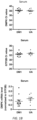
Figures 1A-B. Extracellular mRNA in human urine and serum. We isolated and examined extracellular RNA (exRNA) from urine (N = 15 DM1 patients and 13 UA control subjects without muscular dystrophy) and serum (N = 12 DM1 and 8 UA controls). (A) Expression of DMPK mRNA (upper) and GTF2B mRNA (middle) by qPCR as measured by cycle threshold (Ct) values, and DMPK expression normalized to GTF2B (lower) in human urine, and (B) in human serum. Individual data points represent the mean of duplicate assays for each sample. Error bars = mean ± s.e.m. **** P < 0.0001; *** P = 0.0005 (t-test). -
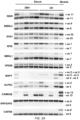
Figures 2A-C. mRNA alternative splicing in exRNA from human urine. We isolated urine exRNA from 27 DM1, 14 DMD/BMD controls (MDC), and 26 unaffected (UA) subjects, and examined alternative splicing by RT-PCR and gel electrophoresis.4 (A) Representative gel images showing alterative splicing of human insulin receptor (INSR)exon 11, MBNL2 exon 6, SOS1 exon 25, MBNL1 exon 7, CLASP1 exon 20,MAP3K4 exon 17, NFIX exon 7, NCOR2exon 45a,VPS39 exon 3, andMAPT exons -
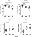
Figures 3A-C. Principle component analysis and predictive modeling of urine splicing outcomes. Using principal component regression, a linear combination of 10 urine transcripts that show differential splicing in DM1 subjects (INSR, MBNL2, SOS1, MBNL1, CLASP1, MAP3K4, NFIX, NCOR2, VPS39, and MAPT) was used to develop a predictive model of DM1. (A) Principle component (PC) score for each subject (N = 23 DM1, 8 MDC, 22 UA). (B) DM1 (N = 23) and UA (N = 22) subjects were combined (N = 45 total), then 34 randomly assigned, irrespective of genotype, to a training set that was used to generate a predictive model of urine splicing outcomes. Using a singular value decomposition algorithm for the fitting and a threshold of 0.5 (see Methods), the model produced zero false positives and false negatives in a 5-fold cross-validation test. The Receiver Operating Characteristic (ROC) curve is shown. (C) The remaining 11 subjects, plus an additional 8 subjects examined while blinded to genotype (N = 19 total), formed an independent validation set that produced zero false positives and false negatives. -
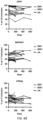
Figures 4A-C. Reliability of exRNA alternative splicing outcomes in human urine. We collected separate urine specimens from 12 DM1, 4 MDC, and 10 UA subjects several months apart and examined exRNA splicing outcomes by RT-PCR. Note that due to low collection volume in one of the UA specimens, 8 of the 10 transcripts have replicates from only 9 UA specimens. -
Figures 5A-B. Alternative splicing of exRNA in human serum. (A) We used RT-PCR to screen splicing in DM1 (N = 5) and UA control (N = 4) serum exRNA samples of several transcripts mis-spliced in DM1 muscle biopsies.28 Splicing in normal human muscle tissue served as a control. Transcript name is shown on the left and target exon/intron on the right. PCR cycle number was 36 (VPS39, CAPZB) or 40 (MBNL1, MAPT, CAMK2B, ARFGAP2, ALPK3). Examination of VPS39 in an additional 9 DM1 and 7 UA samples and ALPK3 in an additional 4 DM1 and 3 UA samples also showed no difference in splicing patterns by genotype. Alternative splicing patterns in serum appeared similar in DM1 and UA for all transcripts tested. (B) Alternative splicing by RT-PCR and gel electrophoresis of INSR exon 11 (N = 14 DM1, 10 UA), MBNL2 exon 6 (N = 9 DM1, 9 UA), SOS1 exon 25 (N = 14 DM1, 12 UA), and NFIX exon 7 (N = 14 DM1, 12 UA) in human serum exRNA. PCR cycle number was 36 (MBNL2, SOS1, NFIX), and 40 (INSR). Control muscle cDNA was diluted 1:100 and amplified in the same PCR reaction as serum samples. Individual data points represent quantitation of splicing of all serum samples examined. Error bars = mean ± s.e.m. -
Figures 6A-E. DMD genetic markers in urine exRNA. We used RT-PCR and DNA sequencing to examine urine exRNA from five DMD subjects with frame-shifting deletions of exons 18 - 22 (Subject 1; S1), exons 51 - 53 (S2), exons 45 - 52 (S3, S4), and exons 24 - 43 (S5). Deletions in S2, S3, and S4 are candidates for exon skipping antisense oligonucleotide drugs to restore the DMD reading frame. (A) Using RT-PCR, we detected DMD deletion mRNAs corresponding to the genetic mutation for all five subjects (S1 - S5). The boxes show the exons amplified. "bp" = base pairs; "L" = DNA ladder. (B) DNA sequencing of extracted bands in a) identified the deletions for S1 and S2. (C) RT-PCR analysis of urine and serum exRNA from an unaffected subject appears similar to muscle tissue, while those from a subject with Becker muscular dystrophy (BMD) due to a t-to-g substitution inintron 67 identifies a second larger band. (D) DNA sequencing of the lower BMD bands showed normal splicing ofexon 67/68, while the upper BMD band is a heteroduplex containing both the normally spliced DMD transcript and a 2nd transcript that includes the 1st five nucleotides ofintron 67. (E) Diagram of the cryptic splice site inintron 67 that shifts the reading frame and produces a poorly functional dystrophin protein. -
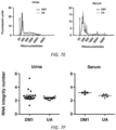
Figures 7A-H. Characterization of exRNA in human urine and serum from DM1 and unaffected (UA) control subjects. (A) Nanoparticle analysis in human urine and serum samples. We determined nanoparticle size and concentration in urine and serum samples from DM1 (N = 12 urine, 10 serum) and UA control (N = 9 urine, 8 serum) subjects. For accurate measurements, we diluted serum samples 1: 1000 and urine either 1:10 or 1:20 in saline to stay in the target concentration range of 1.0 x 10⁸ and 2.5 x 10⁹ particles/milliliter (see Methods). Representative traces of nanoparticle size from DM1 and UA subjects in urine (left) and serum (right) are shown. (B) Mean nanoparticle size from each individual sample in urine (left) and serum (right) ± s.e.m. Note that nanoparticle concentration in urine was lower than serum, while mean particle size was higher. (C) Using ultracentrifugation and Trizol (see Methods), we isolated exRNA from urine (N = 14 DM1 and 12 UA) and serum (N = 5 DM1 and 7 UA) samples and examined optical density using a microvolume spectrophotometer (Nanodrop). Representative optical density spectra of exRNA urine (left) and serum (right) are shown. Vehicle (water) served as reference. The peak of 268 nm reflects residual phenol that was used to purify the RNA (Krebs et al., Anal Biochem 387, 136-138 (2009)), which seemed to have no effect on electropherogram analysis (E) or cDNA synthesis (Figures 2A-C ,9A-B ,10 ,11A-B ). (D) We used capillary gel electrophoresis (Agilent Bioanalyzer) to analyze the concentration, size distribution, and quality of exRNA in urine (N = 22 DM1; N = 21 UA controls) and serum (N = 5 DM1 and 7 UA controls). Using the RNA concentration, we determined the quantity of RNA recovered per volume of urine (left) or serum (right) starting material (range 38 ml - 120 ml urine; 5.5 - 8.5 ml serum). Individual data points from each sample are shown. Error bars represent the mean ± s.e.m. (E) Representative electropherogram traces of exRNA size in nucleotides from urine (left) and serum (right). Most of the species are < 200 nt. (F) RNA integrity number (RIN) in urine (left) and serum (right), as calculated from electropherogram traces using a software algorithm (Agilent). RIN results for three DM1 and two UA serum samples were read as undetermined by the software algorithm. Error bars represent mean ± s.e.m. (G) qPCR analysis of GAPDH and DMPK gene expression in human urine samples (N = 14 DM1 and 13 UA controls). Individual data points indicate the mean of duplicate assays from each individual sample examined. Error bars represent mean ± S.E.M. ** P = 0.003, *** P = 0.0002; t-test with Welch's correction. (H) Serum mRNA expression of reference gene GAPDH (left) and DMPK relative to GAPDH (right) by qPCR. Error bars represent mean ± s.e.m. -
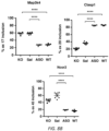
Figures 8A-B. Regulation of Map3k4, Clasp1, and Ncor2 splicing by MBNL1 protein and response to ASO treatment. (A) We used RT-PCR to analyze alternative splicing of Map3k4, Clasp1, and Ncor2 in gastrocnemius muscles from 2 mouse models of DM1, the Mbnl1 knockout (Kanadia et al., Science 302, 1978-1980 (2003)) (Mbnl1ΔE3/ΔE3; N = 4) and HSALR transgenic (Mankodi et al., Science 289, 1769-1773 (2000)) (N = 3), and FVB wild-type (N = 3). The graph shows quantitation ofalternative exon 17 splicing in each individual replicate. **** P < 0.0001 (1-way ANOVA). (B) We treated HSALR with either saline or ASO 445236 (Wheeler et al., Nature 488, 111-115 (2012)) (N = 4 each) using a dose of 25 mg/kg twice weekly for 4 weeks, and analyzed alternative splicing in quadriceps muscles from these mice by RT-PCR. Gastrocnemius muscles from untreated FVB wild type (N = 2) served as controls. The graph shows quantitation ofalternative exon 17 splicing in each individual replicate. **** P < 0.0001 (1-way ANOVA). These data demonstrate that alternative splicing of Map3k4 is regulated by Mbnl1 protein and that mis-splicing of Map3k4 in the HSALR mouse model of DM1 is rescued by ASO treatment, similar to ASO rescue of other alternatively spliced exons that are regulated by MBNL1 protein. -
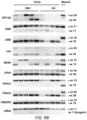
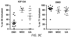
Figures 9A-C. Alternative splicing in urine exRNA isolated from DM1 and unaffected subjects. (A,B) We used RT-PCR to screen 23 candidate DM mis-regulated splice events (Nakamori et al., Ann Neurol 74, 862-872 (2013)) in urine exRNA from DM1 (N = 5) and UA control (N = 4) subjects. Splicing in normal muscle tissue served as a control. Transcript name is shown on the left and target exon/intron on the right. PCR cycle number was 34 (ARFGAP2), 36 (CACNA1S, ALPK3, COPZ2, ANK2, CAPZB, GFPT1, IMPDH2, MAPT, BIN1, FN1, NRAP, OPA1, PHKA1, UBE2D3), and 40 (CAMK2B, ATP2A1, CLCN1, KIF13A, DMD, LMNA). ALPK3 band intensity was variable, requiring 40 cycles to identify bands in an additional 7 DM1 and 4 UA samples (not shown). Splicing of CAMK2B, ARFGAP2, and CAPZB in an additional 5 DM1 and 3 UA control samples was identical to samples shown above. We detected theATP2A1 exon 22 exclusion band in 6/8 DM1 samples and 2/7 UA controls. (C) Quantitation of alternative splicing by RT-PCR of KIF13A and DMD in DM1 (N = 14), MDC (N = 6 or 5), and UA (N = 14 or 12) subjects. Note that KIF13A bands were absent in 1 DM1 and 4 UA subjects. Error bars = s.e.m. -
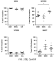
Figures 10A-B. Alternative splicing in urine cell pellet RNA isolated from DM1 and unaffected subjects. We isolated total RNA from urine cell pellets obtained from DM1 (N = 9), MDC (N = 4), and UA (N = 9) subjects and analyzed alternative splicing by RT-PCR and gel electrophoresis. (A) Representative gel images showing alterative splicing of human insulin receptor (INSR)exon 11,MBNL2 exon 6,SOS1 exon 25,MBNL1 exon 7,CLASP1 exon 20,MAP3K4 exon 17,NFIX exon 7,NCOR2 exon 45a,VPS39 exon 3, andMAPT exons -
Figures 11A-B. RT-PCR analysis of alternative splicing in human and mouse kidney and skeletal muscle. (A) Urine EVs are derived from the kidney and urinary tract (Erdbrugger and Le, J )), and splicing of transcripts derived from these tissues may explain the different splicing pattern of several transcripts in urine exRNA than in skeletal muscle (Figs 2A-C ,9A-B ). To determine if the urinary tract may be the predominant source of urine exRNA, we screened alternative splicing of 12 splice events in commercially available mRNA from normal human bladder (B), urothelial (Ut; transitional epithelial) cells, kidney (K) and muscle (M) tissue by RT-PCR using random primers and 26 cycles for each transcript. (B) Percent exon inclusion of each transcript shown in (A) and mean percent exon inclusion ± standard error of the mean (SEM) of urine ex-RNA and urine cell pellets (Figs 2A-C ,9A-B ,10A-B ). "n/d" = not done. -
Figures 12A-D. We examined splicing of 6 alternatively spliced transcripts by RT-PCR in bladder (N = 3), kidney (N = 2), and muscle tissue (N = 2) from FVB wild-type mice and muscle tissue from the HSALR transgenic mouse model of DM1 (N = 2) using oligo dT and 26 cycles for each transcript. Transcript name is shown on the left and target exon on the right. The percent exon inclusion is indicated below each lane. (A) Urine EVs are derived from the kidney and urinary tract (6), and splicing of transcripts derived from these tissues may explain the different splicing pattern of several transcripts in urine exRNA than in skeletal muscle (Figs 2 , S7). To determine if the kidney may be the predominant source of urine exRNA, we screened alternative splicing of 10 transcripts in commercially available mRNA from normal human kidney (k) and muscle (m) tissue (Ambion) by RT-PCR using random primers and 26 cycles for each transcript. (B) We examined splicing of 6 alternatively spliced transcripts by RT-PCR in bladder (N = 3), kidney (N = 2), and muscle tissue (N = 2) from FVB wild-type mice and muscle tissue from the HSALR transgenic mouse model of DM1 (N = 2) using oligo dT and 26 cycles for each transcript. Transcript name is shown on the left and target exon on the right. The percent exon inclusion is indicated below each lane. (C) We used qPCR to analyze expression of genes DMPK, GTF2B, GAPDH, and CKM in mRNA derived from human kidney and muscle tissue. (Upper) the mean qPCR cycle threshold (Ct) of duplicate assays for each gene is shown. (Lower) DMPK expression mRNA expression level normalized to reference genes GTF2B (left) and GAPDH (right). Expression of DMPK in kidney tissue indicates kidney is a potential source of these transcripts in urine exRNA. (D) DMD transcripts expressed in human kidney (k) and muscle (m) tissue using RT-PCR. "E" = empty lane; "Lad" = DNA ladder; "NT" = nucleotides. - In DM1 patients, pre-mRNA splicing outcomes in muscle biopsies are biomarkers of disease severity 4, while in DM1 mice they serve as sensitive indicators of therapeutic drug effects 5, 6. Less invasive biomarkers to assess disease state and response to therapy in DM are currently unavailable, and optimal outcome measures of therapeutic success remain undefined. As a result, a recent clinical trial of an antisense oligonucleotide (ASO) drug for DM1 required participants' consent to multiple muscle biopsies to monitor splicing outcomes in response to therapy and was restricted to adult patients 7. This experimental drug for DM1 is designed to induce knockdown through the RNase H pathway of mutant transcripts, thereby rescuing muscle cells from the pathogenic effects of splicing mis-regulation 6, 8.
- Extracellular vesicles (EVs) include exosomes, microvesicles, and other membrane-encased nanoparticles released and taken up by cells as a form of extracellular communication9. EVs in serum and urine contain mRNA and non-coding RNAs, including microRNA (miRNA), termed exRNAs, released from different tissues and can serve as genetic biomarkers of cancers and other disease states 10-12. Mutations, deletions, translocations, and transcriptome variations also have been shown extensively in EVs, especially for cancers 13-15. Differentiated skeletal muscle cells in culture release EVs 16, 17 and a handful of miRNA biomarkers and several protein signatures have been identified in serum of muscular dystrophy patients 18. However, the capacity of muscle-derived exRNA in urine to serve as biomarkers for muscular dystrophies seems unlikely given that they would be released into the blood circulation and would be unable to pass through the glomerular filtration of serum in the kidney 19. The present results demonstrate that RNA splice products in human urine have sufficient sensitivity and specificity to be robust biomarkers of muscular dystrophies.
- As shown herein, mRNA splicing patterns in "liquid biopsies" present a rich source of personalized biomarkers with applications to a number of genetic diseases. For DM1, we found 10 alternative splice variants in urine that serve as a robust composite biomarker of DM1 disease activity. Mis-regulated alternative splicing outcomes in muscle tissue were sensitive indicators of therapeutic response in DM1 mice 5, 6 and disease activity in DM1 patients 4. Indeed, splice products in muscle biopsies were used in a recent clinical trial as measures of ASO activity in DM1 patients 7. The present methods using splicing outcomes provides powerful biomarkers of DM1, in part because the disease mechanism involves mis-regulated alternative splicing; in addition, the ratiometric measurements of exon inclusion/exclusion described herein are inherently more sensitive than the unidirectional changes that are typical of most biomarkers.
- The detection of differential splicing in urine and not in serum was surprising, and suggests that the source of exRNA in these biofluids may be different and that the primary source in serum is unlikely to be muscle tissue. Because DM1 is primarily a disease of skeletal muscle, heart, and the central nervous system (CNS), it is counter-intuitive that exRNA reflecting the characteristic mis-regulated splicing events appears in urine rather than in blood, as exRNA has not been shown to pass from the blood through the proximal tubules of the kidney 29. In earlier pre-clinical studies, therapeutic ASOs induced target knockdown and exon skipping in kidney tissue of mice and non-human primates 8, 30, suggesting ASOs could have similar effects in human kidney and other tissues lining the urinary tract that release exRNA into the urine. The potential to evaluate exRNA splicing outcomes as pharmacodynamic biomarkers in urine has the advantage of being non-invasive and can be repeated routinely over the course of treatment to evaluate efficacy. For example, due to the need for general anesthesia and the absence of a therapeutic benefit, muscle biopsies generally are avoided in children with DM1. Consequently, detailed study of splicing outcomes in children with DM1 remains an unmet medical need. Urine exRNA should enable comprehensive non-invasive investigation of splicing outcomes in children with DM for the first time, facilitate clinical trials to these patients earlier, and enable convenient titration of dose. The shared pathogenic mechanism of alternative splicing misregulation in DM1 and DM2 4 suggests urine exRNA also may be useful for monitoring disease activity in DM2 patients.
- For DMD, the urine splice products are more than traditional biomarkers: they are personalized genetic markers that are designed specifically for each individual patient and enable the possibility to monitor splice-shifting ASO drug effects 27, 28. Dystrophin protein measurement in biopsy tissue is presently used as a surrogate marker of drug effect that led to the accelerated approval of eteplirsen by the U.S. Food and Drug Administration 31. However, monitoring the ratio of skipped/unskipped DMD splice products in urine during the course of treatment may be used to complement RT-PCR analysis of muscle biopsies and/or in place of dystrophin protein measurement as a surrogate marker of therapeutic effect as newer and better splice-shifting drugs are developed.
- The finding of a DMD cryptic splice site responsible for Becker MD phenotype in an individual with dystrophinopathy further suggests the value of liquid biopsies as a means to identify novel splice variants that may help correlate genotype with phenotype for a number of diseases for which non-invasive biomarkers are unavailable. For example, in patients with Hutchinson-Gilford progeria syndrome (HGPS), point mutations in the LMNA gene activate a weak splice site in
exon 11 that shortens the transcript and produces a truncated progerin protein 32. ASOs that reduce use of this weak splice site are being evaluated as strategy to treat HGPS 33. The presence ofLMNA exon 11 in urine (Figure 9B ) shows that the present methods including use of exRNA can be used to monitor drug effects in these patients as well. The present findings also support development of exRNA from urine, serum, or CSF as a biomarker replacement for tissue biopsies in other diseases with altered mRNA splicing, including limb girdle muscular dystrophy type 1B, spinal muscular atrophy and amyotrophic lateral sclerosis 34-36. - Table A provides a list of conditions that can be diagnosed, treated, or monitored using the present methods, along with the mutated genes (though note that the mutation may or may not result in altered splicing of that specific gene, or not only that specific gene).
Table A - Diseases associated with aberrant mRNA splicing Disease Mutated Gene NCBI RefSeq ID Myotonic dystrophy type 1 (DM1) dystrophia myotonica protein kinase gene (DMPK) NM_004409.4 Duchenne muscular dystrophy (DMD) dystrophin (DMD), transcript variant Dp427m NM_004006.2 Becker muscular dystrophy (BMD) dystrophin (DMD), transcript variant Dp427m NM_004006.2 Limb girdle muscular dystrophy type 1B (LGMD1B) lamin A/C (LMNA), isoform A NM_170707.3 LMNA-linked dilated cardiomyopathy (DCM) Hutchinson-Gilford progeria syndrome (HGPS) Familial partial lipodystrophy type 2 (FPLD2) Spinal muscular atrophy (SMA) survival of motor neuron 2, centromeric (SMN2)NG_008728.1 Amyotrophic lateral sclerosis (ALS) solute carrier family 1 member 2 (SLC1A2 aka EAAT2)NM_004171.3 TAR DNA-binding protein (TARDP) NM_007375.3 FUS RNA binding protein (FUS) NM_004960.3 - DM1 is caused by a heterozygous trinucleotide repeat expansion (CTG)n in the 3-prime untranslated region of the dystrophia myotonica protein kinase gene (DMPK); a repeat length exceeding 50 CTG repeats is pathogenic (Musova et al., Am. J. Med. Genet. 149A: 1365-1374, 2009). The CUG repeats form RNA hairpins that bind proteins including muscleblind-like 1 (MBNL1), a splicing regulatory factor; nuclear sequestration of MBNL1 prevents its activity and results in aberrant splicing of several genes. As shown herein, a number of pre-mRNAs are aberrantly spliced in urine (see Tables 5 and 6); of those, the following showed differential urine exRNA splicing in DM1 vs MDC and UA controls:
Gene Gene Name NCBI Ref SEQ INSR Homo sapiens insulin receptor (INSR), transcript variant 1, mRNANM_000208.3 MBNL2 Homo sapiens muscleblind like splicing regulator 2 (MBNL2), transcript variant 1, mRNANM_144778.3 SOS1 Homo sapiens SOS Ras/Rac guanine nucleotide exchange factor 1 (SOS1), mRNA NM_005633.3 CLASP1 Homo sapiens cytoplasmic linker associated protein 1 (CLASP1), transcript variant 1, mRNANM_015282.2 MBNL1 Homo sapiens muscleblind like splicing regulator 1 (MBNL1), transcript variant 1, mRNANM_021038.4 MAP3K4 Homo sapiens mitogen-activated protein kinase kinase kinase 4 (MAP3K4), transcript variant 1, mRNANM_005922.3 NFIX Homo sapiens nuclear factor I X (NFIX), transcript variant 2. mRNANM_002501.3 NCOR2 Homo sapiens nuclear receptor corepressor 2 (NCOR2), transcript variant 1, mRNANM_006312.5 VPS39 Homo sapiens VPS39, HOPS complex subunit (VPS39), transcript variant 2, mRNANM_015289.3 MAPT Homo sapiens microtubule associated protein tau (MAPT), transcript variant 1, mRNANM_016835.4 - The methods can include determining ratios of properly spliced mRNA to aberrantly spliced mRNA for all or a subset, e.g., 1, 2, 3, 4, 5, 6, 7, 8, 9 or all 10 of the above. In some embodiments, the subset includes 1, 2, 3, 4, or all of MBNL2, MBNL1, SOS1,
CLASP 1, MAP3K4, and optionally also INSR. - DM1 affects skeletal and smooth muscle as well as the eye, heart, endocrine system, and central nervous system. Symptoms can include muscle weakness, e.g., in the leg, hand, neck, and/or face; myotonia, e.g., grip myotonia or percussion myotonia; and posterior subcapsular cataracts (which are detectable as red and green iridescent opacities on slit lamp examination).
- The methods described herein can also include monitoring the efficacy of a treatment for DM1, e.g., a treatment that is expected or intended to affect splicing, e.g., to correct splicing or to inhibit or reduce levels of aberrantly spliced transcripts, e.g., as described herein. The present methods can be used to monitor efficacy, e.g., to determine whether the treatment affects splicing, by detecting a change in the ratio of properly spliced mRNA to aberrantly spliced mRNA. An increase in the properly spliced mRNA, and/or a decrease in aberrantly spliced mRNA, would result in an increase in the ratio and indicates that the treatment is effective.
- DMD and BMD are inherited progressive muscle disorders that are noninflammatory and not associated with a central or peripheral nerve abnormality. The disease affects the muscles with definite fiber degeneration but without evidence of morphologic aberrations, resulting in progressive muscle wasting, and are caused by defects in the dystrophin gene DMD. See, e.g., Aartsma-Rus et al., J Med Genet. 2016 Mar;53(3):145-51; Flanigan et al., Hum Mutat. 2011 Mar; 32(3): 299-308. In some cases, DMD or BMD are caused by mutations that affect splicing of the transcript, e.g., acceptor or donor splice site mutations. The present methods can be used to detect these alternative mRNA splice variants or mRNA of different lengths. The methods can include determining ratios of properly spliced DMD mRNA to aberrantly spliced DMD mRNA.
- The methods described herein can also include monitoring the efficacy of a treatment for DMD or BMD, e.g., a treatment that is expected or intended to affect splicing, e.g., to correct splicing of the dystrophin transcripts, reduce levels of aberrant transcripts, or to produce transcripts that encode functional dystrophin protein. In general, ASOs for DMD are used to induce new splicing changes that serve to restore the open reading frame rather than correct aberrant splicing. It may be possible that some DMD mutations that lead to a Duchenne phenotype and that ASOs could be designed to treat this. Alternative splicing of DMD transcripts typically includes
exon exon 68. The remainder of the DMD exons are spliced constitutively. Urine RNA can also be used to identify novel aberrant splicing, as in our Becker patient with a cryptic splice site. - The present methods can be used to monitor efficacy, e.g., to determine whether the treatment affects splicing, e.g., by detecting a change in the ratio of properly spliced mRNA (or mRNA of a desired size or sequence) to aberrantly spliced mRNA (or mRNA of a non-desired size or sequence). An increase in the properly spliced mRNA or mRNA of a desired size or sequence, and/or a decrease in aberrantly spliced mRNA or mRNA of a non-desired size or sequence, would result in an increase in the ratio and indicates that the treatment is effective.
- Mutations in the lamin A (LMNA) gene that result in aberrant splicing are associated with a number of hereditary disorders. See Scotti and Swanson, Nature Reviews Genetics 17:19-32 (2016).
- Hutchinson-Gilford progeria syndrome (HGPS) is caused by mutations within the LMNA gene that lead to increased usage of an internal splice site, resulting in alternative lamin A transcript with internal deletions of 150 nucleotides (LMNA G608G (GGC>GGT) mutation); see Eriksson et al., Nature 423, 293-298 (2003); Rodriguez et al., Eur J Hum Genet. 2009 Jul; 17(7): 928-937.
- Limb-girdle muscular dystrophies (LGMD) are a heterogeneous group of muscle disorders; symptoms begin in the voluntary muscles of the hips and shoulders. See Nigro and Saverese, Acta Myol. 2014 May;33(1):1-12.
- In familial partial lipodystrophy type 2 (FPLD2), a G>C mutation leads to
aberrant intron 8 retention, nonsense-mediated decay and may lead to translation of a truncated lamin A/C. see Scotti and Swanson, Nature Reviews Genetics 17:19-32 (2016)). - LMNA-linked dilated cardiomyopathy (DCM) is associated with an alternative 3' splice site generated by an A>G mutation (c. 640-10A>G); see Scotti and Swanson, Nature Reviews Genetics 17:19-32 (2016)).
- The methods described herein can also include monitoring the efficacy of a treatment for a conditions associated with mis-splicing of LMNA, e.g., a treatment that is expected or intended to affect splicing, e.g., to correct splicing of the dystrophin transcripts, reduce levels of aberrant transcripts, or to produce transcripts that encode functional dystrophin protein. The present methods can be used to monitor efficacy, e.g., to determine whether the treatment affects splicing, by detecting a change in the ratio of properly spliced mRNA (or mRNA of a desired size or sequence) to aberrantly spliced mRNA (or mRNA of a non-desired size or sequence). An increase in the properly spliced mRNA or mRNA of a desired size or sequence, and/or a decrease in aberrantly spliced mRNA or mRNA of a non-desired size or sequence, would result in an increase in the ratio and indicates that the treatment is effective.
- SMA is associated with mutations in the SMN1 gene (including c.922 + 6 T/G deletion) and loss of SMN full-length protein; see Lorson et al., Proc. Natl Acad. Sci. USA 96, 6307-6311 (1999); Lefebvre et al., ); Scotti and Swanson, Nature Reviews Genetics 17:19-32 (2016)). Approximately 98% of spinal muscular atrophy (SMA) patients have a survival of motor neurons (SMN) gene that has been deleted or mutated. SMN is part of a large multi-protein complex (with additional proteins, including Gemeins 2-7) that is necessary for biogenesis of small nuclear RNA ribonucleoproteins (snRNPs), which are major components of pre-mRNA splicing machinery. Genetic alterations in the SMN gene result in the reduced capacity for snRNP assembly, and defects in RNA splicing. See Dreyfuss, Eukaryon, 6:75-79, 2010.
- The methods described herein can also include monitoring the efficacy of a treatment for SMA, e.g., a treatment that is expected or intended to affect splicing, e.g., to correct splicing of the dystrophin transcripts, reduce levels of aberrant transcripts, or to produce transcripts that encode functional dystrophin protein. Splicing of SMN2 normally involves skipping of
exon 7 in the majority of SMN2 transcripts. ASO treatment of SMA involves increasing inclusion ofSMN2 exon 7, which results in an increase of SMN protein levels, the same protein that is lost by mutations in SMN1. The present methods can be used to monitor efficacy, e.g., to determine whether the treatment affects splicing, by detecting a change in the ratio of properly spliced mRNA (or mRNA of a desired size or sequence) to aberrantly spliced mRNA (or mRNA of a non-desired size or sequence). An increase in the properly spliced mRNA or mRNA of a desired size or sequence, and/or a decrease in aberrantly spliced mRNA or mRNA of a non-desired size or sequence, would result in an increase in the ratio and indicates that the treatment is effective. - Approximately 60%-70% of patients with sporadic Amyotrophic lateral sclerosis (ALS) display a loss of the astrocytic glutamate transporter protein EAAT2 (also known as SLC1A2) in motor cortex and spinal cord. See Rothstein et al., Ann. Neurol. 38:73-84, 1995. Defective pre-mRNA splicing in the motor cortex and spinal cord is responsible for the loss of EAAT2 protein. This defective splicing is caused by a defect in a splicing regulatory factor, rather than a mutation in the EAAT2 gene that causes alternative aberrant splicing or a defect in a general splicing apparatus, such as the spliceosome. The defective pre-mRNA splicing process for EAAT2 can skip normal 5' and 3' splice sites (donor and acceptor splicing sites), or use inappropriate 5' and 3' splice sites (i.e., other than the normal GU or AU for the donor site, and AG or AC for the acceptor site), resulting in multiple abnormal RNAs in ALS patients. The aberrant splicing results in transcripts that partially retain introns or skip exons, as well as transcripts that have exonic sequences at random sites. Two aberrantly spliced EAAT2 mRNAs are found predominantly in sporadic ALS patients. These include an mRNA transcript that partially retains
intron 7 and an mRNA transcript that skipsexon 9 of the gene. The intro 7-retaining RNA causes a dominant-negative effect on normal EAAT2 that has been shown to result in a loss of protein and activity. See, e.g., Honig et al., Neurology. 2000 ; Lin et al., Neuron. 1998 Mar;20(3):589-602; Meyer et al., J Neurol Sci. 1999 . Certain forms of ALS are associated with the presence of mutations in the TARDP (c. 991C>A), (c.1009A>G) and FUS (c. 1566C>T), (c. 1561T>G) genes; see Scotti and Swanson, Nature Reviews Genetics 17:19-32 (2016)); Bai and Lipton, 20(3):363-366 (1998); Zhou et al., PLoS Genet. 2013 Oct;9(10):e1003895; Orozco and Edbauer, J Mol Med (Berl). 2013 Dec;91(12):1343-54; Belzil et al., J Mol Med (Berl). 2013 Dec;91(12):1343-54. - The methods described herein can also include monitoring the efficacy of a treatment for conditions associated with mis-splicing of SLC1A2, TARDP, or FUS, e.g., a treatment that is expected or intended to affect splicing, e.g., to correct splicing of the dystrophin transcripts, reduce levels of aberrant transcripts, or to produce transcripts that encode functional dystrophin protein. The present methods can be used to monitor efficacy, e.g., to determine whether the treatment affects splicing, by detecting a change in the ratio of properly spliced mRNA (or mRNA of a desired size or sequence) to aberrantly spliced mRNA (or mRNA of a non-desired size or sequence). An increase in the properly spliced mRNA or mRNA of a desired size or sequence, and/or a decrease in aberrantly spliced mRNA or mRNA of a non-desired size or sequence, would result in an increase in the ratio and indicates that the treatment is effective.
- Included herein are methods for diagnosing and monitoring subjects with a disease associated with a genetic mutation that results in aberrant splicing, wherein the disease is myotonic dystrophy type 1 (DM1), Duchenne muscular dystrophy (DMD), Becker muscular dystrophy (BMD), limb girdle muscular dystrophy type 1B (LGMD1B), LMNA-linked dilated cardiomyopathy (DCM); Hutchinson-Gilford progeria syndrome (HGPS); Familial partial lipodystrophy type 2 (FPLD2), spinal muscular atrophy (SMA) or amyotrophic lateral sclerosis (ALS).
- The methods rely on detection of ratios of properly spliced (which can include transcripts that are spliced by an ASO) to mis-spliced isoforms of affected transcripts in urine samples. As used herein, "properly spliced" means that the transcript has a desired splice pattern, e.g., has wild-type splicing, or is spliced in a way that is desired, e.g., to produce a functional protein. For example, in some embodiments, the DMD exons targeted by ASOs are constitutively spliced, meaning they are always included in DMD patients and UA individuals. Treatment with ASOs in DMD is designed to induce a new unique splice event absent in DMD patients or UA individuals, and the ratio of the inclusion of the unique splice site (properly spliced) to inclusion of the constitutive splice (mis-spliced) provides a convenient estimation of ASO drug effects in urine a new unique splice event absent in DMD patients or UA individuals; in this case, a transcript that was "properly spliced" in a subject with DMD being treated with these ASOs would include the unique splice event. The methods can include determining that the mRNA is of a desired size or sequence (i.e., "properly spliced"), or is of a non-desired size or sequence ("mis-spliced").
- The methods include using a urine sample that has been obtained from a subject, determining levels of properly spliced and aberrantly spliced extracellular transcripts, and determining a ratio of properly spliced to mis-spliced transcript in the sample. The methods can include comparing the ratio with one or more reference ratios, e.g., a control reference that represents a normal ratio of properly spliced:mis-spliced transcript, e.g., a level in an unaffected subject, and/or a disease reference that represents a ratio associated with the disease. For example, in some embodiments a reference ratio of properly spliced:mis-spliced transcripts in an unaffected subject may approach 1:0, since there would not be expected to be a large number of mis-spliced transcripts in such individuals.
- Various methods are well known within the art for determining levels of properly spliced and aberrantly spliced extracellular transcripts. These methods can include identification and/or isolation and/or purification of a transcript from a sample. An "isolated" or "purified" biological marker is substantially free of cellular material or other contaminants from the cell or tissue source from which the biological marker is derived i.e. partially or completely altered or removed from the natural state through human intervention. For example, nucleic acids contained in the sample can be isolated according to standard methods, for example using filtration, centrifugation, or other methods of purification to obtain a sample that contains extracellular transcripts but does not contain cells or cellular transcripts. The methods can include using chemical solutions nucleic acid-binding resins following the manufacturer's instructions. In one example, the entire volume of urine is centrifuged, e.g., at 2,000-3,000 x g, e.g., at 2,450 x g for 5-15 minutes, e.g., 10 minutes at room temperature, and then the supernatant is passed through a filter, e.g., a 0.8 µm filter, before being ultracentrifuged, e.g., at 100,000
x g 2 hours at 4° C, to pellet the RNA. Then the supernatant is removed and, RNA is extracted from the translucent ribonucleoprotein pellet, e.g., using Trizol (Life Technologies) according to manufacturer instructions. To enhance RNA pellet visibility, linear acrylamide (Ambion) or other reagents can be added. - The transcripts can be evaluated using methods known in the art, e.g., using polymerase chain reaction (PCR), reverse transcriptase polymerase chain reaction (RT-PCR), quantitative or semi-quantitative real-time RT-PCR, digital PCR i.e. BEAMing ((Beads, Emulsion, Amplification, Magnetics) Diehl (2006) Nat Methods 3:551-559) ; RNAse protection assay; Northern blot; various types of nucleic acid sequencing (Sanger, pyrosequencing, NextGeneration Sequencing); fluorescent in-situ hybridization (FISH); or gene array/chips) (Lehninger Biochemistry (Worth Publishers, Inc., current addition; Sambrook, et al, Molecular Cloning: A Laboratory Manual (3. Sup.rd Edition, 2001); Bernard (2002) Clin Chem 48(8): 1178-1185; Miranda (2010) Kidney International 78:191-199; Bianchi (2011) EMBO Mol Med 3:495-503; Taylor (2013) Front. Genet. 4:142; Yang (2014) PLOS One 9(11):e110641); Nordstrom (2000) Biotechnol. Appl. Biochem. 31(2):107-112; Ahmadian (2000) Anal Biochem 280:103-110. In some embodiments, high throughput methods, e.g., protein or gene chips as are known in the art (see, e.g., Ch. 12, Genomics, in Griffiths et al., Eds. Modern genetic Analysis, 1999,W. H. Freeman and Company; Ekins and Chu, Trends in Biotechnology, 1999, 17:217-218; MacBeath and Schreiber, ; Simpson, Proteins and Proteomics: A Laboratory Manual, Cold Spring Harbor Laboratory Press; 2002; Hardiman, Microarrays Methods and Applications: Nuts & Bolts, DNA Press, 2003), can be used to detect the presence and/or level of different splice isoforms. multiple-exon-skipping detection assay (MESDA) can also be used (see Singh et al., 2012, PLoS One. 2012;7(11):e49595). Measurement of the level of different splice isoforms can be direct or indirect. For example, the abundance levels of various differently spliced isoforms can be directly quantitated, e.g., based on size or the presence or absence of a selected sequence. In some embodiments a technique suitable for the detection of alterations in the structure or sequence of nucleic acids, such as the presence of deletions, amplifications, or substitutions, can be used for the detection of different splice isoforms.
- Gene arrays are prepared by selecting probes which comprise a polynucleotide sequence, and then immobilizing such probes to a solid support or surface. For example, the probes may comprise DNA sequences, RNA sequences, co-polymer sequences of DNA and RNA, DNA and/or RNA analogues, or combinations thereof, which detect various spliced isoforms. The probe sequences can be synthesized either enzymatically in vivo, enzymatically in vitro (e.g. by PCR), or non-enzymatically in vitro.
- In some embodiments, the methods can be used to diagnose a condition described herein; for example, when the ratio of properly spliced:mis-spliced transcripts in a subject (e.g., a subject who has one or more symptoms associated with the disease) is comparable to a reference ratio in a representative subject with the disease, then the subject can be diagnosed with the disease. In some embodiments, once it has been determined that a person has a disease described herein, namely DM1, DMD, BMD, LGMD1B, SMA, or ALS, then a treatment, e.g., as known in the art or as described herein, can be selected for administration.
- Suitable reference values can be determined using methods known in the art, e.g., using standard clinical trial methodology and statistical analysis. The reference values can have any relevant form. In some cases, the reference comprises a predetermined value for a meaningful ratio, e.g., a control reference level that represents a normal level ratio, e.g., a level in an unaffected subject, and/or a disease reference that represents a ratio associated with the disease, e.g., a level in a subject having a disease as described herein, namely DMD, DM1, BMD, LGMD, HGPS, DCM, HGPS, FPLD2, SMA, or ALS.
- The predetermined ratio can be a single cut-off (threshold) value, such as a median or mean, or a ratio that defines the boundaries of an upper or lower quartile, tertile, or other segment of a clinical trial population that is determined to be statistically different from the other segments. It can be a range of cut-off (or threshold) ratios, such as a confidence interval. It can be established based upon comparative groups, such as where association with presence of disease in one defined group is a fold higher, or lower, (e.g., approximately 2-fold, 4-fold, 8-fold, 16-fold or more) than the presence of disease in another defined group. It can be a range, for example, where a population of subjects (e.g., control subjects) is divided equally (or unequally) into groups, such as a low-risk group, a medium-risk group and a high-risk group, or into quartiles, the lowest quartile being subjects with the lowest risk and the highest quartile being subjects with the highest risk, or into n-quantiles (i.e., n regularly spaced intervals) the lowest of the n-quantiles being subjects with the lowest risk and the highest of the n-quantiles being subjects with the highest risk.
- In some embodiments, the predetermined ratio is a ratio in the same subject, e.g., at a different time point, e.g., an earlier time point.
- Subjects associated with predetermined values are typically referred to as reference subjects. For example, in some embodiments, a control reference subject does not have a disorder described herein (DMD, DM1, BMD, LGMD, HGPS, DCM, HGPS, FPLD2, SMA, and ALS). In some cases it may be desirable that the control subject is a first or second degree relative of the subject to be tested.
- A disease reference subject is one who has (has been diagnosed with) a disease as described herein, namely DMD, DM1, BMD, LGMD, HGPS, DCM, HGPS, FPLD2, SMA, or ALS.
- Thus, in some cases the ratio of properly spliced:mis-spliced in a subject being less than a reference ratio is indicative of a clinical status (e.g., indicative of presence of a disorder as described herein, namely DMD, DM1, BMD, LGMD, HGPS, DCM, HGPS, FPLD2, SMA, or ALS), or indicative of an ineffective therapy. In other cases the ratio in a subject being greater than or equal to the reference ratio is indicative of the absence of disease, or an effective therapy. In some embodiments, the amount by which the ratio in the subject is the less than the reference ratio is sufficient to distinguish a subject from a control subject, and optionally is a statistically significantly less than the ratio in a control subject. In cases where the ratio in a subject being equal to the reference ratio, the "being equal" refers to being approximately equal (e.g., not statistically different).
- The predetermined ratio can depend upon the particular population of subjects (e.g., human subjects) selected. For example, an apparently healthy population may have a different 'normal' range of ratios than will a population of subjects which have, are likely to have, or are at greater risk to have, a disorder described herein. Accordingly, the predetermined values selected may take into account the category (e.g., sex, age, health, risk, presence of other diseases) in which a subject (e.g., human subject) falls. Appropriate ranges and categories can be selected with no more than routine experimentation by those of ordinary skill in the art.
- In characterizing likelihood, or risk, numerous predetermined values can be established.
- The methods described herein can include monitoring the efficacy of a treatment of disorders associated with aberrant splicing. The disorder is DMD, DM1, BMD, LGMD, HGPS, DCM, HGPS, FPLD2, SMA, or ALS. Generally, the methods include determining a subsequent ratio after a treatment has been administered to a subject identified using a method described herein.
- As used in this context, to "treat" means to ameliorate at least one symptom of the disorder associated with aberrant splicing. For example, where the disease is a muscular dystrophy, a treatment can result in a reduction in muscle weakness or a reduction in rate of muscle loss or weakening.
- Exon-skipping antisense oligonucleotides (ASOs) that correct missplicing can be used, e.g., as described in Siva et al., Nucleic Acid Ther. 2014 ; Scotti and Swanson, Nature Reviews Genetics 17:19-32 (2016). For example, bicyclic-locked nucleic acids (LNAs), ethylene-bridged nucleic acids (ENAs), 2'-O-methyl phosphorothioate AO (20ME-PSs), peptide nucleic acids (PNAs), or phosphorodiamidate morpholino oligomers (PMOs) have been described that correct missplicing in clinical trials and animal models; see, e.g., Brolin and Shiraishi, Artif DNA PNA XNA. 2011 Jan-Mar; 2(1): 6-15; Scotti and Swanson, Nature Reviews Genetics 17:19-32 (2016); Touznik et al., Expert Opin Biol Ther. 2014 Jun;14(6):809-19. The ASOs can be delivered, e.g., parenterally in liposomal complexes, e.g., cationic lipoplexes, or using a viral vector, e.g., a lentivirus, adenovirus, or adeno-associated virus. See e.g., Jarver et al., Nucleic Acid Ther. 2014;24(1):37-47; Aartsma-Rus et al., Hum Gene Ther. 2014;25(10):885-892, McNally and Wyatt, J Clin Invest. 2016 ; Imbert et al., Genes 2017, 8(2), 51; doi:10.3390/genes8020051.
- Exon skipping uses antisense oligonucleotides (ASOs) to alter transcript splicing; the present methods can be used to detect these transcripts with desired splicing. These treatments can include antisense oligonucleotide-targeted exon skipping to induce near normal, e.g., for dystrophin, e.g., as described in Aartsma-Rus, Methods Mol Biol. 2012;867:97-116. Clinical trials of ASOs in DMD have been conducted, see, e.g., Koo and Wood, Hum Gene Ther. 2013 May;24(5):479-88; Voit et al., Lancet Neurol. 2014;13(10):987-996.
- An
exon 11 antisense oligonucleotide (ASO) that increased lamin C production has been shown to shift the output of LMNA more toward lamin C and reduce levels of the mutant protein in fibroblasts derived from patients with HGPS (Lee et al., J Clin Invest. 2016 ). - Exon skipping ASOs directed against an intron splice silencer in SMN2 increase the amount of full-length SMN transcript in the CNS, restoring SMN to treat subjects with spinal muscular atrophy (SMA) (see Burghes and McGovern, Genes Dev. 2010 ). ASO drug nusinersen enhanced
exon 7 inclusion in a clinical trial, see Chiriboga et al., Neurology. 2016 . - Specific ASOs for use in
exon 51 skipping therapy, e.g., in DMD, include PRO051 (2OME-PS, Netherlands) and AVI-4658 (PMO, UK). A plurality of ASOs can also be used, e.g., to induce exon skipping in multiple exons; see, e.g., Wood et al., Brain. 2010 Apr;133(Pt 4):957-72 See also Fletcher et al., Mol Ther Nucleic Acids. 2012 Oct; 1(10): e48; McClorey et al., Curr Opin Pharmacol. 2005 Oct;5(5):529-34. - Similar methods can be used in DM1, as described in Chamberlain and Chamberlain, Nature Medicine 16:170-171 (2010). For example, an ASO inhibiting mutant DMPK transcripts can be used, e.g., a 149-bp antisense RNA complementary to the (CUG)13 repeats and to the 110-bp region following the repeats sequence has been described, see Furling et al., Gene Ther. 2003 May;10(9):795-802. See also Magaña and Cisneros, J Neurosci Res. 2011 Mar;89(3):275-85; Thornton et al., Curr Opin Genet Dev. 2017 Jun;44:135-140; Gao and Cooper, Hum Gene Ther. 2013 May; 24(5): 499-507.
- See also Gao et al., Hum Gene Ther. 2013 May; 24(5): 499-507; Wheeler et al., Science 2009, 325, 336-339; Wheeler et al., Nature 2012, 488, 111-115; Wojtkowiak-Szlachcic et al., Nucleic Acids Res. 2015, 43, 3318-3331; Mulders et al., Proc. Natl. ; Frangois et al., Nat. Struct. Mol. Biol. 2011, 18, 85-87; Cavazzana-Calvo et al., ; Cornetta et al., Mol. Ther. J. Am. Soc. Gene Ther. 2011, 19, 557-566.
- Small molecule therapeutics can also be used, e.g., PTC124, a 284.24-Da, achiral, 1,2,4-oxadiazole linked to fluorobenzene and benzoic acid rings, which selectively induces ribosomal read-through of premature but not normal termination codons, see Welch et al., Nature 447: 87-91, 2007, and has been used in clinical trials for DMD.
- The invention is further described in the following examples, which do not limit the scope of the invention described in the claims.
- The following materials and methods were used in the Examples set forth below.
- Human subjects. The Partners Health Service/MGH IRB approved all studies involving human subjects described here. We recruited study participants from the MGH Neuromuscular Diagnostic Center. Three groups were studied: (1) individuals with DM1 (N = 23), (2) individuals with a muscular dystrophy besides DM (N = 8 total; 6 DMD, 1 BMD, 1 FSHD), and (3) individuals with no known muscular dystrophy (unaffected; N = 22) that were either a parent, spouse, or cousin of a study participant with muscular dystrophy. Inclusion criteria for DM1 subjects were
age 13 years or older, a diagnosis of DM1 based on genetic testing that identified a DMPK-CTG repeat expansion of ≥ 50, or clinical diagnosis of DM1 and a 1st degree relative with DM1 due to a DMPK-CTG repeat expansion of ≥ 50, and ability to provide informed consent or assent for participation. Inclusion criteria for MDC subjects included known diagnosis of DMD, BMD, or FSHD,ages 13 years or older, and ability to provide informed consent. Inclusion criteria for unaffected individuals wereage 18 years or older, no known history of any muscular dystrophy, and ability to provide informed consent. The training cohort consisted of a combined 34 DM1 or UA participants chosen randomly, and the remaining 11 combined DM1 and UA subjects comprised the validation cohort. Prior to participation in the study, informed consent was obtained for blood and/or urine collection from all subjects; due to severe autism, informed consent for the individual with Becker muscular dystrophy was obtained from his mother/legal guardian, according to IRB protocol. Subject information is shown in Tables 1A-1B. -
Table 1A Subjects # Male # Female Mean age Age range DM1 19 8 41 17 - 63 UA 7 19 n/a n/a MDC 9 1 24 14 - 49 Table 1B ID Leukocytes Nitrite Urobilinogen Protein pH Blood Specific gravity Ketone Bilirubin Glucose DM1 - - 3.5 +/- 5 - 1.025 - - - DM1 - - 3.5 +/- 6 - 1.020 - - - DM1 - - 3.5 +/- 5 - 1.020 - - - DM1 - - 3.5 +/- 5 - 1.010 - - - DM1 - - 3.5 +/- 5 - 1.020 - - - DM1 - - 3.5 +/- 5 - 1.030 - - - DM1 - - 3.5 +/- 5 - 1.020 - - - DM1 - - 3.5 +/- 6 - 1.015 - - - DM1 - - 3.5 +/- 5 - 1.015 - - - DM1 - - 3.5 +/- 5 - 1.015 - - - DM1 - - 1(17) +/- 6 - 1.010 - - - DM1 - - 3.5 +/- 5 - 1.030 - - - DM1 - - 3.5 - 6 - 1.005 - - - DM1 - - 3.5 +/- 5 - 1.030 - - - DM1 - - 3.5 +/- 6 - 1.015 - - - DM1 +/- - 3.5 - 5 - 1.015 - - - DM1 + - 3.5 +/- 5 - 1.030 - - - UA + - 3.5 - 5 - 1.025 - - - UA - - 3.5 +/- 5 - 1.010 - - - UA - - 3.5 +/- 6.5 - 1.010 - - - UA - - 3.5 +/- 5 - 1.015 - 1(1-7)+ - UA - - 3.5 - 6.5 - 1.010 - - - UA - - 3.5 +/- 5.5 - 1.010 - - - UA - - 3.5 +/- 5 - 1.010 - - - UA - - 3.5 +/- 6.5 - 1.010 - - - UA - - 3.5 - 5 - 1.015 - - - UA - - 3.5 +/- 7.5 - 1.010 - - - UA - - 3.5 - 5 - 1.010 - - - UA +/- - 3.5 - 5 - 1.015 - - - UA - - 3.5 +/- 5 +/- 1.025 - - - UA +/- - 3.5 - 6.5 +/- 1.010 - - - UA - - 3.5 +/- 5 - 1.025 - - - UA - - 3.5 +/- 7 - 1.010 - - - - (1A) Gender, age, and age range of participating DM1, MDC, and UA subjects.
- (1B) We examined urine from a subset of 18 DM1 and 16 UA subjects using urinalysis reagents strips (ACON Laboratories) as a general screening tool for urinary tract disorders, endocrine disorders, or metabolic or systemic diseases that affect kidney function. Specific gravity values (detection range 1.000 - 1.030) tended to be higher in DM1 than in UA subjects (P = 0.0568; Mann-Whitney test). Specific gravity is measure of the kidney's ability to concentrate the urine. Urobilinogen detection range is 0.2 - 1.0 mg/dL (3.5 - 200 µmol/L).
- Collection and processing of human urine. Subjects donated urine (range from 20 - 120 milliliters) in a standard specimen container. To remove cells, we centrifuged the entire volume at 2,450 x g for 10 minutes at room temperature, passed the supernatant through a 0.8 µm filter into sterile 50 ml tubes, and placed on wet ice within 2 hours of collection. We proceeded with exRNA isolation from specimens either immediately or after storage at 4° C overnight. To analyze total RNA in urine cell pellets, we used Trizol (Life Technologies) according to manufacturer recommendations.
- Collection and processing of human serum. Blood was collected in two standard red top serum separator tubes (Becton Dickinson), incubated at room temperature for 30 - 45 minutes, and centrifuged at 2,450 x g for 10 minutes at room temperature. To remove any remaining cells, we passed the serum through a 0.8 µm filter into a sterile 15 ml tube, placed on wet ice within 2 hours of collection, and stored at -80° C. The volume of serum recovered ranged from 5.5 - 8.5 ml. The blood sample from one individual with DM1 was unusable due to hemolysis and total volume of less than 3 ml.
- Experimental mice. The MGH IACUC approved all experiments involving mice. HSA LR transgenic and Mbnl1 knockout (Mbnl1ΔE3/ΔE3) models of DM1 (both FVB background) have been described 2, 26. FVB wild-type mice served as controls. HSALR mice that were treated with antisense oligonucleotide (ASO) 445236 received subcutaneous injections of 25 mg/kg twice weekly for 4 weeks, as previously described 6. ASO 445236 was a gift of Dr. Frank Bennett at Ionis Pharmaceuticals (Carlsbad, CA).
- Nanoparticle tracking. To determine nanoparticle size and concentration, we used the Nanosight LM10 system and Nanoparticle Tracking Analysis 2.0 analytical software according to manufacturer instructions (Malvern). The system uses a laser beam, light microscope, and CCD camera to visualize and video record particles in liquid suspension moving under Brownian motion. For accurate measurements, we diluted serum samples 1:1000 and urine either 1:10 or 1:20 in saline to stay in the target concentration range of 1.0 x 10⁸ and 2.5 x 10⁹ particles/milliliter. We recorded 60-second videos and analyzed data in auto mode.
- Isolation of exRNA from biofluids. We ultracentrifuged urine and serum samples at 100,000
x g 2 hours at 4° C, removed the supernatant, extracted RNA from the translucent ribonucleoprotein pellet using 700 µl Trizol (Life Technologies) according to manufacturer instructions. To enhance RNA pellet visibility, we added 1.4 µl linear acrylamide (Ambion) to each sample and mixed well prior to isopropanol precipitation. Pellets were re-suspended in molecular grade water. - exRNA analysis. We measured optical density spectra using a microvolume spectrophotometer (Nanodrop). To measure exRNA size, quality, and total mass of recovered, we used chip-based capillary gel electrophoresis according to manufacturer instructions (2100 Bioanalyzer, Agilent Technologies). Using electropherogram traces, a software algorithm (Agilent) automatically determined the RNA integrity number (RIN) based on using a numbering system of 1 (most degraded) to 10 (fully intact) 37.
- Quantitative real-time RT-PCR (qPCR). To quantitate DMPK gene expression, we used Taqman qPCR (Applied Biosciences 7500) and standard assays for GAPDH, and GTF2B (Applied Biosciences, FAM-MGB; assay IDs Hs00976255_m1 and Hs02758991_g1) as normalization controls. The primer probe set for DMPK was published previously 6. To determine expression levels, we used the mean of duplicate assays from individual samples.
- RT-PCR analysis of splicing outcomes. We generated cDNA using Superscript III reverse transcriptase (Life Technologies) and random primers, and performed PCR using Amplitaq Gold DNA polymerase (Life Technologies) and gene specific primers (Tables 2 and 3). We used previously published primers for INSR and APT2A1 3, 38 and designed all other primers using Primer3 software 39, 40. Due to the small size of the exRNA species, we targeted the product size for exon exclusion isoforms to be ~ 100 - 200 nucleotides whenever possible. Total RNA from normal human skeletal muscle and kidney (Ambion AM7982 and AM 7976) served as tissue controls. We separated PCR products using agarose gels, stained with SYBR I green nucleic acid gel stain, and quantitated band intensities using a transilluminator, CCD camera, XcitaBlue™ conversion screen, and Image Lab image acquisition and analysis software (Bio-Rad).
Table 2. PCR primers for human transcripts. Gene Left primer (5' - 3') # Right primer (5' - 3') # Target exon(s) + ex size (nt) - ex size (nt) ALPK3 GAGCTACCTGCTCAGCGTG 1. CTGTGACGATGCAGGTGAAC 2. 2 194 155 ANK2 CCGATAACCAGCCTGAGACC 3. ACGGTGTGTCCATGCTCATC 4. 21 223 130 ARFGAP2 GTGCCGTTCCTAATCACTCC 5. GTGAGGTGCCAAGCAGGTC 6. 6 186 104 BIN1 AACCTCAATGATGTGCTGGTC 7. CTCTGGCTCGTGGTTGACTC 8. 11 213 168 CACNA1S TGATTGTCATTGGCAGCATC 9. AGGGTTCGCACTCCTTCTG 10. 29 205 148 CAMK2B AGCCATCCTCACCACCATG 11. AGGAGGAAGCGTCCCTTTG 12. 13 217 142 CAPZB AATCAGAAGTACGCTGAACGAG 13. CCTCCACCAGGTCATTCTTC 14. 8 or 9 238 113 CLASP1 TATTGATGTGAACGCAGCAG 15. CCGGTTATCAGGTGTAGAGG 16. 20 202 178 CLCN1 CCTGAAGGAATACCTCACAATG 17. TGAGGACAGCAGCACAGATG 18. 7a 200 134 COPZ2 CGGCTTGACTGAACAGAGTG 19. CTGGCTGGAGACCTTAGGAG 20. 9b ~320 213 DMD TACGGTGACCACAAGGGAAC 21. TTTCACAGTGGTGCTGAGATAG 22. 18 - 22 deletion n/a 174 DMD CCAGCTGGTTGAGCATTGTC 23. GTTCAGCTTCTGTTAGCCACTG 24. 24 - 43 deletion n/a 144 DMD AATTGGGAAGCCTGAATCTG 25. CTCCGGTTCTGAAGGTGTTC 26. 46 - 52 deletion n/a 119 DMD AGCCACTCAGCCAGTGAAG 27. GCAGAATAATCCCGGAGAAG 28. 51 - 53 deletion n/a 214 DMD ACTGGCATCATTTCCCTGTG 29. GGGTTCCAGTCTCATCCAGTC 30. 67 267 n/a DMD GCTACCTGCCAGTGCAGAC 31. TGCGTGAATGAGTATCATCG 32. 71 132 87 DMD GAGCAACT CAACAACT CCT T CC 33. TAAGGACTCCATCGCTCTGC 34. 78 128 96 FN1 CAAGGATGACAAGGAAAGTGTC 35. TGGACCAATGTTGGTGAATC 36. 25 364 91 GFPT1 CCTCTGTTGATTGGTGTACGG 37. GTGCTGTCCACACGAGAGAG 38. - 9 170 116 IMPDH2 CCAGGCTGGTGTGGATGTAG 39. TTGTACACTGCTGTTGCTTGG 40. 8 or 9 255 159 KIF13A GGACACTGCCACTTATGGTTG 41. TGAGTGCATCTGACCACCTC 42. 25 183 144 LMNA AGATGACCTGCTCCATCACC 43. TACATGATGCTGCAGTTCTGG 44. 11 321 171* MAP3K4 GGTACCTCGATGCCATAGTGAC 45. CAGCTATGGAAGCCAATCG 46. 17 260 110 MAPT CGAAGTGATGGAAGATCACG 47. GTGTCTCCAATGCCTGCTTC 48. 2 + 3 309; 135 222 MAPT AACGAAGATCGCCACACC 49. CCACTGCCACCTTCTTGG 50. 10 296 242 MBNL1 CTGCCCAATACCAGGTCAAC 51. GGCTAGAGCCTGTTGGTATTG 52. 7 230 176 MBNL2 CAGCACCAAGCCAACCAAG 53. GAGCCTGCTGGTAGTGCAAG 54. 6 226 172 NCOR2 GCTGGAGGCCATAATTAGAAAG 55. GAGTGCACTGAGGAGACAGAG 56. 45a 282 144 NFIX AGCCCTGTTGATGACGTGTT 57. AGTGCAGGGCTGATGCTGT 58. 7 250 127 NRAP AGGCGCTGCACATTATCAC 59. ATAGCGGCCTCTCATGTGG 60. 12 235 130 OPA1 GGATTGTGCCTGACATTGTG 61. CCACGATCTGTTGCTCTAAACG 62. 4b 250 196 PHKA1 CTGGACCTGAGGGTAAGCTG 63. GGGAAGCCTGAAATAACTTCG 64. 19 318 141 PHKA1 TATCCACGAGATTGGTGCTG 65. GTTGCCATTGACCTTGACG 66. 28 212 173 SOS1 TGGTGCTTCCAGTACCACAG 67. GGCAGATTCTGGTCGTCTTC 68. 25 208 163 UBE2D3 GTCGCCTGCTTTAACAATTTC 69. CTTGGGCAACTGTTCTCTTG 70. 10 456 406 VPS39 TAGGATTCGGAAGGACGTTG 71. CTCACCGGTCTCTGTGTGC 72. 3 274 241 #: SEQ ID NO:
*, progerinTable 3. PCR primers for mouse transcripts. Gene Left primer (5' - 3') # Right primer (5' - 3') # Target exon(s) + ex size (nt) - ex size (nt) mCamk2b GCCACACGGAATTTCTCAG 73. CAGGAGGGAGAGATCCTTTG 74. 13 196 121 mCapzb ATCCGAAGCACGCTGAATG 75. GCCTCCACCAGGTCGTTC 76. 9 238 125 mClasp1 GTCGACGACAGGATCTCTCC 77. GAGCTCTGCCGTCTCGTG 78. 20 198 174 mMap3k4 CAACAGAATCAGCGATGCCATC 79. TGGTCTGGCTGATGAGTGTTCG 80. 17 355 199 mMbnl1 ACCTGCAAGCCAAGATCAAG 81. TGTTGGCTAGAGCCTGTTGG 82. 7 255 201 mMbnl2 TCACCCTCCTGCACACTTG 83. TCTTTGGTAAGGGATGAAGAGC 84. 6 194 140 mNcor2 CAGGCGGTGCAAGAACAC 85. TTCGGCTGCTAGGTCTGC 86. 45a 253 112 mNfix TCGACGACAGTGAGATGGAG 87. CTGGATGATGGACGTGGAAG 88. 7 238 115
Sample size. Splicing patterns in human urine and serum, or even whether alternative splice isoforms are present or detectable in these biofluids, were unknown. Therefore, we were unable to choose a sample size ahead of time to ensure adequate power to detect disease-specific differences. Instead, we chose a sample size based on splicing outcomes in muscle biopsies 4 and a goal of enrolling a similar number of DM1 and UA controls. In mice, we chose sample sizes for splicing analysis in muscle based on previously reported differences in muscle tissue of these models 2, 3, 5, 6. Mice ranged from 2 to 4 months of age and were chosen randomly by genotype, stratified for sex to allow an approximately equal number of females and males, and examined without blinding. - Statistics. Group data are presented as mean ± s.e.m. We compared groups using an unpaired two-tailed t-test or analysis of variance (ANOVA) as indicated. We used the F test to compare variances between DM1 and UA control samples analyzed by qPCR and RT-PCR (Table 4). In groups with statistically significant difference in variance, we used t-test with Welch's correction to determine differences between groups. A P value < 0.05 was considered significant.
- Principle component analysis was performed using R statistical software. The principle component score for each subject was calculated using a linear combination of the 10 splicing outcomes shown in
Fig. 2 (INSR, MBNL2, SOS1, MBNL1, CLASP1, MAP3K4, NFIX, NCOR2, VPS39, and MAPT).Table 4. Variance between groups. Transcript P value Are variances significantly different? Variance greater in DM1 or UA? Urine qPCR Ct values DMPK 0.7302 no n/a GTF2B 0.9178 no n/a GAPDH 0.3577 no n/a Urine qPCR normalized values DMPK-GTF2B 0.0171 yes UA DMPK-GAPDH 0.0226 yes UA Serum qPCR Ct values DMPK 0.3377 no n/a GTF2B 0.8546 no n/a GAPDH 0.4702 no n/a Serum qPCR normalized values DMPK-GTF2B 0.4137 no n/a DMPK-GAPDH 0.6131 no n/a Serum splicing INSR ex 11 0.6479 no n/a MBNL2 ex 60.0787 no n/a SOS1 ex 250.9233 no n/a NFIX ex 70.1731 no n/a VPS39 ex 30.4998 no n/a - We used the F test to compare variances between DM1 and UA control groups for gene expression in urine and serum samples analyzed by qPCR and splicing in serum samples by RT-PCR. In groups with statistically significant difference in variance, we used t-test with Welch's correction to determine differences between groups. The difference in variances of some groups may represent true differences in the two populations, and may be as important as the finding of different means (
Figure 1 ,Figures 7G and7 H,Figures 11A and11B ). - Predictive model. We used principal component regression to develop a predictive model of DM1 using the splicing quantification of the 10 genes, shown in
Fig. 2 . The pls package in R, which uses singular value decomposition algorithm for the fitting, was used to implement the model 41. For the model, DM1 (N = 23) and UA (N = 22) subjects were randomly assigned to a training cohort that consisted of 17 subjects with DM1 and 17 unaffected controls. For subjects that provided 2nd samples, the mean value of the splicing quantitation measurements was used. Only the first principal component was used for prediction. - To examine the possibility of detecting biomarkers of muscular dystrophies (MDs) in human biofluids, we analyzed exRNA microarray and raw sequencing data from two previous studies and found that more than 30 transcripts previously reported as "splicing biomarkers" in DM1 muscle biopsy tissue could be detected in control human serum and urine (Tables 5 and 6) 20, 21. To determine whether splice variants of these transcripts are also detectable in human biofluids, we collected blood and/or urine from 23 subjects with DM1, 22 unaffected (UA) individuals, and 8 MD controls (MDC) (Table 1). First we screened biofluids for the presence of exRNA and found that nanoparticle content was greater in serum than urine, and showed no difference in number or size between DM1 and controls (
Figure 7A ). Conversely, nanoparticle size spanned a larger range in urine than serum in both DM1 and UA controls (Figure 7B ). Optical density curves at 260 nm appeared similar in DM1 and UA controls in both urine and serum (Figure 7C ). Using capillary gel electrophoresis, we calculated RNA mass recovered per milliliter of biofluid, exRNA size distribution, and RNA quality (Figure 7D to F ). Although the concentration of exRNA was 2.5 - 3 fold greater in serum than urine, the total recovery of exRNA was greater in urine due to larger specimen volume (20 - 120 ml urine vs 5.5 - 8.5 ml serum).Table 5. Transcripts mis-spliced in DM1 muscle tissue and other muscle transcripts that are expressed in human serum extracellular RNA (exRNA). Gene Name Systematic name Sequence # Probe Name Avg DMPK NM_004409 
89. A_23_P50535 5.66 INSR* NM_000208 
90. A_23_P4764 4.87 MBNL2* NM_144778 
91. A_24_P56317 4.60 SOS1* NM_005633 
92. A_23_P343808 4.77 CLASP1* NM_015282 
93. A_23_P311232 4.57 MBNL1* NM_021038 
94. A_23_P357811 6.26 MAP3K4* NM_005922 
95. A_23_P42096 5.07 NFIX* NM_002501 
96. A_23_P165295 4.85 NCOR2* NM_006312 
97. A_23_P203891 5.84 VPS39* NM_015289 
98. A_24_P167825 5.08 MAPT* NM 016835 
99. A_23_P207699 5.19 KIF13A NM_022113 
100. A_23_P214111 4.69 DMD NM_004019 
101. A_23_P321860 4.44 CLCN1 NM_000083 
102. A_23_P59772 6.90 ATP2A1 NM_173201 
103. A_23_P72462 5.17 CAPZB NM_004930 
104. A_23_P126752 5.22 CACNA1S NM_000069 
105. A_23_P85765 5.80 CAMK2B NM_172082 
106. A_23_P42882 4.79 COPZ2 NM_016429 
107. A_23_P101093 4.76 
GFPT1 NM_002056 
108. A_23_P44083 6.03 IMPDH2 NM_000884 
109. A_24_P166042 5.00 ALPK3 NM_020778 
110. A_23_P348728 5.95 ANK2 NM_001148 
111. A_23_P133068 5.50 BIN1 NM_139346 
112. A_23_P165333 5.63 NRAP NM_198060 
113. A_23_P402765 4.58 OPA1 NM_130837 
114. A_23_P211797 6.54 PHKA1 NM_002637 
115. A_32_P186121 5.26 UBE2D3 NM_181886 
116. A_24_P363005 6.05 PDLIM3 NM_014476 
117. A_23_P110403 4.51 LDB3 NM_007078 
118. A_32_P98227 5.11 TTN NM_133378 
119. A_23_P85269 4.66 FHOD1 NM_013241 
120. A_23_P37778 5.46 TBC1D15 NM_022771 
121. A_23_P139558 5.10 RYR1 NM_000540 
122. A_23_P78867 4.76 DTNA NM_001392 
123. A_23P208158 4.82 TNNT2 NM_000364 
124. A_23_P34700 5.28 FXR1 NM_001013 439 
125. A_23_P132784 5.47 CKM NM_001824 
126. A_23_P50250 4.75 ACTA1 NM_001100 
127. A_23_P1102 4.98 MYH3 NM_002470 
128. A_23_P26865 4.86 - Serum samples from healthy control subjects (Ctrl; N=7) were filtered through an 0.8 µm filter to remove cells, ultracentrifuged at 100,000 x g for 90 minutes to collect extracellular RNA in EVs and particles, and the EV/ribonucleoprotein (RNP) pellet was lysed. RNA was extracted using the Qiagen miRNeasy kit and examined by mRNA microarray analysis. The data are represented as quartile normalized with background subtraction and values indicate expression levels of each gene (Noerholm et al., )). More than two-dozen transcripts of the mis-spliced mRNAs found in tibialis anterior (TA) muscle tissue of
DM 1 patients and reported as biomarkers of muscle weakness (Nakamori et al., Ann Neurol 74, 862-872 (2013)) are detected in the serum EV/RNP mRNA fraction and are candidate serum biomarkers. Note the similar levels of expression of muscle transcripts between healthy controls. The presence of muscle transcripts in EVs in this study is consistent with previous reports of EVs released by muscle cells (Romancino et al., FEBS Lett 587, 1379-1384 (2013)) and measurement of muscle-enriched miRs in human serum (Aoi et al., Front Physiol 4, 80 (2013)). (*: transcripts we report here that show differential urine exRNA splicing in DM1 vs MDC and UA controls; #, SEQ ID NO:).Table 6. Screening of human urine exosomes/microvesicles for presence of transcripts that are mis-spliced in DM1 muscle tissue. Gene Normalized Raw DMPK 2.348005036 380 INSR* 5.410552617 2204 SOS1* 11.78539296 4408 MBNL2* 28.38510805 6004 CLASP1* 10.58959612 3876 MBNL1* 16.93241215 4180 MAP3K4* 22.19040448 5472 NFIX* 80.93189513 20292 NCOR2* 17.48260768 6976 VPS39* 16.7391543 3637 MAPT* 3.968124064 1216 KIF13A 9.413147298 2736 DMD 0.430326742 304 CLCN1 0 0 ATP2A1 0 0 CAPZB 184.636637 14197 CACNA1S 0 0 CAMK2B 0.736559261 152 COPZ2 10.73770858 436 GFPT1 29.71567389 11546 IMPDH2 72.96428986 5548 ALPK3 1.393280124 684 ANK2 3.375451832 2204 ARFGAP2 30.19114602 3641 BIN1 13.30854948 1596 NRAP 0 0 OPA1 15.35810568 4484 PHKA1 6.584110353 1824 PHKA2 3.110806445 698 UBE2D3 53.16414472 6947 PDLIM3 0 0 LDB3 0.25908561 76 TTN 0.166126138 836 FHOD1 1.754220849 304 TBC1D15 7.176337802 1900 RYR1 0 0 DTNA 0.315128162 152 TNNT2 1.443318118 76 FXR1 13.46978301 5396 LMNA 55.02644764 8284 MLF1 1.303656519 152 ABLIM2 0.699642342 152 CKM 0 0 ACTA1 0 0 MYH3 0.561759871 152 - A previous study collected 3300 ml of urine from a human male, centrifuged the entire volume at low speed to pellet cells, passed the supernatant through a 0.8 µm filter to remove remaining debris, ultracentrifuged the filtered supernatant, isolated RNA from the exosome/microvesicle pellet using a commercially available kit (Qiagen), and identified genes present using massively parallel RNA sequencing (Miranda et al., PLoS One 9, e96094 (2014)). Shown are raw and normalized counts of genes from that study that also were reported as biomarkers of DM1 in
muscle biopsies 5, as well as other genes associated with muscular dystrophies. (*: transcripts we report here that show differential urine exRNA splicing in DM1 vs MDC and UA controls). - Based on qPCR cycle threshold (Ct) values of reference genes GTF2B and GAPDH, urine ex-mRNA content tended to be higher in DM1 as compared to UA subjects (
Figure 1A andFigure 7G ). The lower expression of DMPK, the gene causing DM1, in urine from DM1 vs. UA subjects may be due to retention of mutant transcripts in the nucleus preventing their release into the cytoplasm and incorporation into EVs 22. In serum, these transcripts were expressed at similar levels in both DM1 and UA subjects, and were present at lower levels than in urine (Figure 1B andFigure 7H ). - Next we examined splice products in exRNA, focusing on transcripts previously reported as biomarkers of DM1 disease severity in muscle biopsies 4. In urine exRNA, we identified 10 candidate DM1-specific splice products of 32 examined (
Figure 2 ), 9 of which are MBNL1 protein-dependent (Figures 8A-B ) 4, 23. Principle component analysis of these 10 splicing outcomes confirmed separation of DM1 from MDC and UA individuals (Figure 3A ). We randomly assigned 76% of urine specimens from DM1 and UA subjects regardless of genotype to a training cohort and generated a predictive model that was 100% accurate in identifying the outcome of the remaining 24% of individuals in the validation cohort (Figure 3B and C ). Splice products appeared similar in consecutive samples collected several months apart from the same individual (Figure 4 ), suggesting reliability of the assay. Interestingly, splicing of several other candidate DM mis-regulated splice events 4 was similar in urine from DM1 and UA individuals (Figures 9A-9C ). Surprisingly, examination of these splice products in serum exRNA failed to show a significant difference between DM1 and UA controls for any of the transcripts examined (Figure 5A andB ). - In previous studies, RNAs in urine have been used as biomarkers of prostate cancer, bladder cancer, and kidney transplant rejection, suggesting that cells lining the urinary tract are the primary contributors to the urine ex-RNA pool 12, 24, 25. Our finding of slightly different exon inclusion/exclusion percentages of some transcripts in urine as compared to muscle tissue suggests the exRNA found in urine may represent a pool from multiple different cell types along this urinary route. To determine whether the urinary tract is the primary contributor of the ex-mRNA alternative splice variants in urine, we examined splice isoforms in human and mouse kidney and mouse bladder (
Figures 10 and12 ). Splicing of all transcripts examined showed different exon inclusion/exclusion percentages in normal human kidney compared to skeletal muscle (Figures 10 and12A ). In wild-type mouse bladder and kidney, splicing showed no consistent pattern relative to muscle tissue from wild-type or the HSALR mouse model of DM1 (Figure 12B )26. qPCR analysis also revealed that kidney expresses DMPK, although at a lower level than muscle (Figure 12C ). - ASOs also are being evaluated therapeutically for another form of muscle disease, Duchenne muscular dystrophy (DMD), to modify dystrophin pre-mRNA splicing directly by inducing skipping of a target exon to restore the open reading frame and produce a truncated, partially functional protein 27, 28. Detection of therapeutic drug effects in DMD patients involves multiple muscle biopsies to examine splicing outcomes and dystrophin protein production. To test whether biofluid exRNA contains DMD deletion transcripts, we examined urine from several subjects with DMD and found patient-specific DMD deletion transcripts (
Figures 6A andB ), suggesting this biofluid exRNA is a viable approach to monitor therapeutic exon-skipping ASO drug effects in DMD patients as personalized genetic markers 27, 28. - We also examined exRNA from a BMD patient with a normal DMD coding sequence, but a point mutation in intron 67 (c9807+ 6 T>G substitution). The normal coding sequence presumably produces a full-length dystrophin protein, suggesting the mutation in this patient causes dystrophinopathy by an overall reduction of dystrophin protein expression. RT-PCR analysis identified a splice product corresponding to the normal DMD exon 67 - 68 sequence in urine and serum from this patient and a UA subject, identical to muscle tissue (
Figure 6C ). In addition, a second larger product unique to the BMD samples was evident. DNA sequencing confirmed the larger band was a heteroduplex containing the normal product identical to that in the lower band, as well as one with inclusion of the 1st five nucleotides ofintron 67, indicating a cryptic splice site (Figure 6D ) created by the mutation. The result is a frame shift and premature termination codon inexon 68, reducing functional dystrophin protein expression (Figure 6E ). Thus, urine exRNA also can be used to identify this molecular disease mechanism. The expression in the kidney of DMD transcripts spanning the deletions and point mutation (Figure 12D ) is consistent with the urinary tract as the primary source of exRNA in urine. -
- 1. Scotti, M.M. & Swanson, M.S. RNA mis-splicing in disease. ).
- 2. Kanadia, R.N. et al. A muscleblind knockout model for myotonic dystrophy. Science 302, 1978-1980 (2003).
- 3. Lin, X. et al. Failure of MBNL1-dependent post-natal splicing transitions in myotonic dystrophy. ).
- 4. Nakamori, M. et al. Splicing biomarkers of disease severity in myotonic dystrophy. Ann Neurol 74, 862-872 (2013).
- 5. Wheeler, T.M. et al. Reversal of RNA dominance by displacement of protein sequestered on triplet repeat RNA. Science 325, 336-339 (2009).
- 6. Wheeler, T.M. et al. Targeting nuclear RNA for in vivo correction of myotonic dystrophy. Nature 488, 111-115 (2012).
- 7. Clinicaltrials.gov https://clinicaltrials.gov/ct2/show/NCT02312011. (2016).
- 8. Pandey, S.K. et al. Identification and characterization of modified antisense oligonucleotides targeting DMPK in mice and nonhuman primates for the treatment of ).
- 9. Tkach, M. & Thery, C. Communication by Extracellular Vesicles: Where We Are and Where We Need to Go. Cell 164, 1226-1232 (2016).
- 10. Skog, J. et al. Glioblastoma microvesicles transport RNA and proteins that promote tumour growth and provide diagnostic biomarkers. ).
- 11. Chen, W.W. et al. BEAMing and Droplet Digital PCR Analysis of Mutant IDH1 mRNA in Glioma Patient Serum and Cerebrospinal Fluid Extracellular Vesicles. Mol ).
- 12. Nilsson, J. et al. Prostate cancer-derived urine exosomes: a novel approach to biomarkers for prostate cancer. ).
- 13. San Lucas, F.A. et al. Minimally invasive genomic and transcriptomic profiling of visceral cancers by next-generation sequencing of circulating exosomes. ).
- 14. Khan, S. et al. Early diagnostic value of survivin and its alternative splice variants in breast cancer. BMC Cancer 14, 176 (2014).
- 15. Neeb, A. et al. Splice variant transcripts of the ).
- 16. Romancino, D.P. et al. Identification and characterization of the nano-sized vesicles released by muscle cells. FEBS Lett 587, 1379-1384 (2013).
- 17. Forterre, A. et al. Myotube-derived exosomal miRNAs downregulate Sirtuin1 in myoblasts during muscle cell differentiation. ).
- 18. Hathout, Y. et al. Clinical utility of serum biomarkers in Duchenne muscular dystrophy. ).
- 19. Moeller, M.J. & Tenten, V. Renal albumin filtration: alternative models to the standard physical barriers. ).
- 20. Noerholm, M. et al. RNA expression patterns in serum microvesicles from patients with glioblastoma multiforme and controls. ).
- 21. Miranda, K.C. et al. Massively parallel sequencing of human urinary exosome/microvesicle RNA reveals a predominance of non-coding RNA. PLoS One 9, e96094 (2014).
- 22. Davis, B.M., McCurrach, M.E., Taneja, K.L., Singer, R.H. & Housman, D.E. Expansion of a CUG trinucleotide repeat in the 3' untranslated region of myotonic dystrophy protein kinase transcripts results in nuclear retention of transcripts. Proc Natl Acad Sci U S A 94, 7388-7393 (1997).
- 23. Wagner, S.D. et al. Dose-Dependent Regulation of Alternative Splicing by MBNL Proteins Reveals Biomarkers for Myotonic Dystrophy. ).
- 24. Motamedinia, P. et al. Urine Exosomes for Non-Invasive Assessment of Gene Expression and Mutations of Prostate Cancer. ).
- 25. Urquidi, V. et al. Urinary mRNA biomarker panel for the detection of urothelial carcinoma. ).
- 26. Mankodi, A. et al. Myotonic dystrophy in transgenic mice expressing an expanded CUG repeat. Science 289, 1769-1773 (2000).
- 27. Mendell, J.R. et al. Eteplirsen for the treatment of Duchenne muscular dystrophy. Ann Neurol 74, 637-647 (2013).
- 28. Kinali, M. et al. Local restoration of dystrophin expression with the morpholino oligomer AVI-4658 in Duchenne muscular dystrophy: a single-blind, placebo-controlled, dose-escalation, proof-of-concept study. ).
- 29. Erdbrugger, U. & Le, T.H. Extracellular Vesicles in Renal Diseases: More than Novel Biomarkers? J ).
- 30. Morcos, P.A., Li, Y. & Jiang, S. Vivo-Morpholinos: a non-peptide transporter delivers Morpholinos into a wide array of mouse tissues. ).
- 31. Aartsma-Rus, A. & Krieg, A.M. FDA Approves Eteplirsen for Duchenne Muscular Dystrophy: The Next Chapter in the Eteplirsen Saga. ).
- 32. Eriksson, M. et al. Recurrent de novo point mutations in lamin A cause Hutchinson-Gilford progeria syndrome. Nature 423, 293-298 (2003).
- 33. Lee, J.M. et al. Modulation of LMNA splicing as a strategy to treat prelamin A diseases. J Clin Invest 126, 1592-1602 (2016).
- 34. Hua, Y. et al. Peripheral SMN restoration is essential for long-term rescue of a severe spinal muscular atrophy mouse model. Nature 478, 123-126 (2011).
- 35. Honda, D. et al. The ALS/FTLD-related RNA-binding proteins TDP-43 and FUS have common downstream RNA targets in cortical neurons. FEBS Open Bio 4, 1-10 (2013).
- 36. Genschel, J. & Schmidt, H.H. Mutations in the LMNA gene encoding lamin A/).
- 37. Imbeaud, S. et al. Towards standardization of RNA quality assessment using user-independent classifiers of microcapillary electrophoresis traces. Nucleic Acids Res 33, e56 (2005).
- 38. Savkur, R.S., Philips, A.V. & Cooper, T.A. Aberrant regulation of insulin receptor alternative splicing is associated with insulin resistance in myotonic dystrophy. ).
- 39. Koressaar, T. & Remm, M. Enhancements and modifications of primer design program Primer3. ).
- 40. Untergasser, A. et al. Primer3--new capabilities and interfaces. ).
- 41. Mevik, B.-H. & Wehrens, R. The pls Package: Principal Component and Partial Least Squares Regression in R. ).
Claims (8)
- A method of diagnosing a disease associated with aberrant mRNA splicing,wherein the disease is myotonic dystrophy type 1 (DM1); Duchenne muscular dystrophy (DMD); Becker muscular dystrophy (BMD); limb girdle muscular dystrophy type 1B (LGMD1B); LMNA-linked dilated cardiomyopathy (DCM); Hutchinson-Gilford progeria syndrome (HGPS); Familial partial lipodystrophy type 2 (FPLD2); spinal muscular atrophy (SMA); or amyotrophic lateral sclerosis (ALS), the method comprising;isolating extracellular mRNA in a sample comprising urine obtained from a subject who has, or is suspected to have, the disease associated with aberrant mRNA splicing;determining one or more selected mRNA in the sample, wherein the one or more selected mRNA is aberrantly spliced in the subject, and is suspected to be present in a plurality of spliced isoforms in the sample, wherein the spliced isoforms comprise properly spliced isoforms and mis-spliced isoforms;quantitating levels of the properly spliced isoforms and mis-spliced isoforms of the one or more selected mRNAs in the sample;determining a ratio of the properly spliced isoforms to the mis-spliced isoforms of the one or more selected mRNAs in the sample; andcomparing the ratio of properly spliced to mis-spliced in a subject to a reference ratio, wherein a ratio in the subject that is less than the reference ratio indicates the presence of a disease associated with aberrant mRNA splicing.
- A method of monitoring the efficacy of a treatment for a disease associated with aberrant mRNA splicing in a subject, wherein the disease is myotonic dystrophy type 1 (DM1); Duchenne muscular dystrophy (DMD); Becker muscular dystrophy (BMD); limb girdle muscular dystrophy type 1B (LGMD1B); LMNA-linked dilated cardiomyopathy (DCM); Hutchinson-Gilford progeria syndrome (HGPS); Familial partial lipodystrophy type 2 (FPLD2); spinal muscular atrophy (SMA); or amyotrophic lateral sclerosis (ALS), the method comprising:determining a first ratio of properly spliced isoforms to mis-spliced isoforms in a sample from the subject using the method of claim 1; determining a subsequent ratio of properly spliced isoforms to mis-spliced isoforms in a sample from the subject using the method of claim 1 after a treatment for the disease has been administered to the subject; andcomparing the first and subsequent ratios, wherein a ratio in the second sample that is higher than the ratio in the subsequent sample indicates that the treatment is effective.
- The method of claim 2, wherein the treatment that is intended to correct splicing; to inhibit or reduce levels of mis-spliced transcripts; or to alter splicing to produce a functional protein.
- The method of claim 3, wherein the treatment is an antisense oligonucleotide.
- The method of claims 1-4, wherein the disease is myotonic dystrophy type 1 (DM1), and wherein the one or more selected mRNAs is selected from the group consisting of the transcript for insulin receptor (INSR); muscleblind like splicing regulator 2 (MBNL2); SOS Ras/Rac guanine nucleotide exchange factor 1 (SOS1); cytoplasmic linker associated protein 1 (CLASP1); muscleblind like splicing regulator 1 (MBNL1); mitogen-activated protein kinase kinase kinase 4 (MAP3K4); nuclear factor I X (NFIX); nuclear receptor corepressor 2 (NCOR2); VPS39, HOPS complex subunit (VPS39); and microtubule associated protein tau (MAPT).
- The method of claim 5, wherein the selected mRNAs comprise MBNL2, MBNL1, SOS1, CLASP1, MAP3K4, and optionally INSR.
- The method of claims 1-4, wherein the disease is associated with aberrant splicing of dystrophin (DMD); lamin A/C (LMNA); survival of motor neuron 2, centromeric (SMN2); solute carrier family 1 member 2 (SLC1A2); TAR DNA-binding protein (TARDP); or FUS RNA binding protein (FUS).
- The method of claim 1, wherein the one or more selected mRNAs is selected from the group consisting of the transcript for insulin receptor (INSR); muscleblind like splicing regulator 2 (MBNL2); SOS Ras/Rac guanine nucleotide exchange factor 1 (SOS1); cytoplasmic linker associated protein 1 (CLASP1); muscleblind like splicing regulator 1 (MBNL1); mitogen-activated protein kinase kinase kinase 4 (MAP3K4); nuclear factor I X (NFIX); nuclear receptor corepressor 2 (NCOR2); VPS39, HOPS complex subunit (VPS39); microtubule associated protein tau (MAPT); dystrophin (DMD); lamin A/C (LMNA); survival of motor neuron 2, centromeric (SMN2); solute carrier family 1 member 2 (SLC1A2); TAR DNA-binding protein (TARDP); and FUS RNA binding protein (FUS).
Applications Claiming Priority (2)
| Application Number | Priority Date | Filing Date | Title |
|---|---|---|---|
| US201662365139P | 2016-07-21 | 2016-07-21 | |
| PCT/US2017/043348 WO2018017991A1 (en) | 2016-07-21 | 2017-07-21 | Extracellular mrna markers of muscular dystrophies in human urine |
Publications (3)
| Publication Number | Publication Date |
|---|---|
| EP3488019A1 EP3488019A1 (en) | 2019-05-29 |
| EP3488019A4 EP3488019A4 (en) | 2020-01-29 |
| EP3488019B1 true EP3488019B1 (en) | 2025-02-12 |
Family
ID=60992733
Family Applications (1)
| Application Number | Title | Priority Date | Filing Date |
|---|---|---|---|
| EP17831981.0A Active EP3488019B1 (en) | 2016-07-21 | 2017-07-21 | Extracellular mrna markers of muscular dystrophies in human urine |
Country Status (4)
| Country | Link |
|---|---|
| US (2) | US11866783B2 (en) |
| EP (1) | EP3488019B1 (en) |
| CA (1) | CA3034385A1 (en) |
| WO (1) | WO2018017991A1 (en) |
Families Citing this family (6)
| Publication number | Priority date | Publication date | Assignee | Title |
|---|---|---|---|---|
| US11866783B2 (en) * | 2016-07-21 | 2024-01-09 | The General Hospital Corporation | Extracellular mRNA markers of muscular dystrophies in human urine |
| WO2020135677A1 (en) * | 2018-12-29 | 2020-07-02 | 中美瑞康核酸技术(南通)研究院有限公司 | Oligomeric nucleic acid molecule and application thereof |
| CN111849968A (en) | 2019-04-30 | 2020-10-30 | 中美瑞康核酸技术(南通)研究院有限公司 | Oligonucleotide molecule and application thereof in acute intermittent porphyria treatment |
| CN110218248B (en) * | 2019-06-10 | 2020-12-18 | 北京市农林科学院 | Na+ /H+ Antiporter in Plasma Membrane and Its Encoding Gene and Application |
| WO2021231887A1 (en) * | 2020-05-15 | 2021-11-18 | University Of Florida Research Foundation, Incorporated | Compositions and methods of detection of pre-symptomatic als |
| WO2025076486A1 (en) * | 2023-10-06 | 2025-04-10 | Dyne Therapeutics, Inc. | Biomarkers for repeat expansion diseases |
Family Cites Families (10)
| Publication number | Priority date | Publication date | Assignee | Title |
|---|---|---|---|---|
| US20020061571A1 (en) | 2000-03-20 | 2002-05-23 | Mahadevan Mani S. | Novel isoform of myotonic dystrophy associated protein kinase and uses thereof |
| US20150232933A1 (en) | 2001-07-25 | 2015-08-20 | Michael Kopreski | Methods for evaluating pathologic conditions using extracellular rna |
| US20060057624A1 (en) * | 2004-08-30 | 2006-03-16 | Potashkin Judith A | Alternatively spliced pre-mRNA transcripts in neurodegenerative disease |
| EP1896495A2 (en) * | 2005-03-30 | 2008-03-12 | Novartis Vaccines and Diagnostics, Inc. | Dkkl-1 splice product modulators for cancer diagnosis and therapy |
| DE102006007249B4 (en) * | 2006-02-15 | 2008-07-31 | Universitätsklinikum Schleswig-Holstein | Method for the determination of bladder carcinomas of differentiation grade G2 |
| US10260098B2 (en) * | 2008-03-06 | 2019-04-16 | The Regents Of The University Of Michigan | FIG4 gene mutations in neurodegeneration |
| EP3077528B1 (en) * | 2013-12-02 | 2018-08-22 | Albert-Ludwigs-Universität Freiburg | Process for the diagnosis of cancer by using exosomes |
| WO2015187825A2 (en) * | 2014-06-03 | 2015-12-10 | University Of Massachusetts | Compositions and methods for modulating dysferlin expression |
| US10724092B2 (en) | 2014-06-10 | 2020-07-28 | Erasmus University Medical Center Rotterdam | Methods for characterizing alternatively or aberrantly spliced mRNA isoforms |
| US11866783B2 (en) * | 2016-07-21 | 2024-01-09 | The General Hospital Corporation | Extracellular mRNA markers of muscular dystrophies in human urine |
-
2017
- 2017-07-21 US US16/318,658 patent/US11866783B2/en active Active
- 2017-07-21 EP EP17831981.0A patent/EP3488019B1/en active Active
- 2017-07-21 CA CA3034385A patent/CA3034385A1/en active Pending
- 2017-07-21 WO PCT/US2017/043348 patent/WO2018017991A1/en not_active Ceased
-
2023
- 2023-11-22 US US18/517,748 patent/US20240084388A1/en not_active Abandoned
Also Published As
| Publication number | Publication date |
|---|---|
| CA3034385A1 (en) | 2018-01-25 |
| US20240084388A1 (en) | 2024-03-14 |
| WO2018017991A1 (en) | 2018-01-25 |
| US11866783B2 (en) | 2024-01-09 |
| US20190284628A1 (en) | 2019-09-19 |
| EP3488019A4 (en) | 2020-01-29 |
| EP3488019A1 (en) | 2019-05-29 |
Similar Documents
| Publication | Publication Date | Title |
|---|---|---|
| US20240084388A1 (en) | Extracellular mRNA Markers of Muscular Dystrophies in Human Urine | |
| Goffena et al. | Elongator and codon bias regulate protein levels in mammalian peripheral neurons | |
| CN105861712B (en) | Methods for early detection and monitoring of Mild Cognitive Impairment (MCI) and Alzheimer's Disease (AD) using miRNA from bodily fluids | |
| Roberts et al. | Extracellular microRNAs are dynamic non-vesicular biomarkers of muscle turnover | |
| Chen et al. | Correlation between DNA methylation and gene expression in the brains of patients with bipolar disorder and schizophrenia | |
| Bilsen et al. | Identification and allele-specific silencing of the mutant huntingtin allele in Huntington's disease patient-derived fibroblasts | |
| US9914977B2 (en) | Individualized cancer therapy | |
| CN113286895A (en) | Long non-coding RNA (lncRNA) for the diagnosis and treatment of brain disorders, in particular cognitive disorders | |
| Doxakis | Insights into the multifaceted role of circular RNAs: implications for Parkinson’s disease pathogenesis and diagnosis | |
| EP3679157B1 (en) | Non-coding rnas (ncrna) for the diagnosis of cognitive disorders | |
| EP3202914B2 (en) | Method for treating a neurodegenerative disease | |
| US9528991B2 (en) | Individualized cancer therapy | |
| EP3891295A1 (en) | Methods of diagnosing a disease state | |
| Vihola et al. | Altered expression and splicing of Ca2+ metabolism genes in myotonic dystrophies DM1 and DM2 | |
| CN112980941B (en) | A kit for diagnosing whether a subject suffers from Alzheimer's disease and its use | |
| CA2978368C (en) | Signature of health | |
| US20200255900A1 (en) | MicroRNA-455-3p as a Peripheral Biomarker for Alzheimer's Disease | |
| EP2924124A1 (en) | Method for the diagnosis and/or prognosis of neurodegenerative diseases | |
| US20200255899A1 (en) | MicroRNA-455-3p as a Peripheral Biomarker for Alzheimer's Disease | |
| US20230383366A1 (en) | Dynamic Changes In Circulating Free RNA Of Neural Tumors | |
| Scatolini et al. | RNase H1 counteracts DNA damage and ameliorates SMN-dependent phenotypes in a Drosophila model of Spinal Muscular Atrophy | |
| JP2020524509A (en) | Circulating serum microRNA biomarkers and methods for diagnosis of Alzheimer's disease | |
| Fitz et al. | Small nucleolar RNAs in plasma and their discriminatory power as diagnostic biomarkers of Alzheimer’s disease | |
| CN111378743B (en) | Application of gene in Alzheimer's diagnosis | |
| EP4705520A1 (en) | Use of micrornas as tools for the diagnosis and differentiation of tauopathies |
Legal Events
| Date | Code | Title | Description |
|---|---|---|---|
| STAA | Information on the status of an ep patent application or granted ep patent |
Free format text: STATUS: THE INTERNATIONAL PUBLICATION HAS BEEN MADE |
|
| PUAI | Public reference made under article 153(3) epc to a published international application that has entered the european phase |
Free format text: ORIGINAL CODE: 0009012 |
|
| STAA | Information on the status of an ep patent application or granted ep patent |
Free format text: STATUS: REQUEST FOR EXAMINATION WAS MADE |
|
| 17P | Request for examination filed |
Effective date: 20190219 |
|
| AK | Designated contracting states |
Kind code of ref document: A1 Designated state(s): AL AT BE BG CH CY CZ DE DK EE ES FI FR GB GR HR HU IE IS IT LI LT LU LV MC MK MT NL NO PL PT RO RS SE SI SK SM TR |
|
| AX | Request for extension of the european patent |
Extension state: BA ME |
|
| RIN1 | Information on inventor provided before grant (corrected) |
Inventor name: WHEELER, THURMAN Inventor name: BREAKEFIELD, XANDRA O. Inventor name: BALAJ, LEONORA |
|
| DAV | Request for validation of the european patent (deleted) | ||
| DAX | Request for extension of the european patent (deleted) | ||
| A4 | Supplementary search report drawn up and despatched |
Effective date: 20200108 |
|
| RIC1 | Information provided on ipc code assigned before grant |
Ipc: C12Q 1/6883 20180101AFI20191220BHEP |
|
| STAA | Information on the status of an ep patent application or granted ep patent |
Free format text: STATUS: EXAMINATION IS IN PROGRESS |
|
| 17Q | First examination report despatched |
Effective date: 20220701 |
|
| GRAP | Despatch of communication of intention to grant a patent |
Free format text: ORIGINAL CODE: EPIDOSNIGR1 |
|
| STAA | Information on the status of an ep patent application or granted ep patent |
Free format text: STATUS: GRANT OF PATENT IS INTENDED |
|
| INTG | Intention to grant announced |
Effective date: 20240322 |
|
| GRAJ | Information related to disapproval of communication of intention to grant by the applicant or resumption of examination proceedings by the epo deleted |
Free format text: ORIGINAL CODE: EPIDOSDIGR1 |
|
| STAA | Information on the status of an ep patent application or granted ep patent |
Free format text: STATUS: EXAMINATION IS IN PROGRESS |
|
| INTC | Intention to grant announced (deleted) | ||
| GRAP | Despatch of communication of intention to grant a patent |
Free format text: ORIGINAL CODE: EPIDOSNIGR1 |
|
| STAA | Information on the status of an ep patent application or granted ep patent |
Free format text: STATUS: GRANT OF PATENT IS INTENDED |
|
| P01 | Opt-out of the competence of the unified patent court (upc) registered |
Free format text: CASE NUMBER: APP_45794/2024 Effective date: 20240807 |
|
| INTG | Intention to grant announced |
Effective date: 20240910 |
|
| GRAS | Grant fee paid |
Free format text: ORIGINAL CODE: EPIDOSNIGR3 |
|
| GRAA | (expected) grant |
Free format text: ORIGINAL CODE: 0009210 |
|
| STAA | Information on the status of an ep patent application or granted ep patent |
Free format text: STATUS: THE PATENT HAS BEEN GRANTED |
|
| AK | Designated contracting states |
Kind code of ref document: B1 Designated state(s): AL AT BE BG CH CY CZ DE DK EE ES FI FR GB GR HR HU IE IS IT LI LT LU LV MC MK MT NL NO PL PT RO RS SE SI SK SM TR |
|
| REG | Reference to a national code |
Ref country code: GB Ref legal event code: FG4D |
|
| REG | Reference to a national code |
Ref country code: CH Ref legal event code: EP |
|
| REG | Reference to a national code |
Ref country code: DE Ref legal event code: R096 Ref document number: 602017087739 Country of ref document: DE |
|
| REG | Reference to a national code |
Ref country code: IE Ref legal event code: FG4D |
|
| REG | Reference to a national code |
Ref country code: NL Ref legal event code: MP Effective date: 20250212 |
|
| PG25 | Lapsed in a contracting state [announced via postgrant information from national office to epo] |
Ref country code: RS Free format text: LAPSE BECAUSE OF FAILURE TO SUBMIT A TRANSLATION OF THE DESCRIPTION OR TO PAY THE FEE WITHIN THE PRESCRIBED TIME-LIMIT Effective date: 20250512 |
|
| PG25 | Lapsed in a contracting state [announced via postgrant information from national office to epo] |
Ref country code: FI Free format text: LAPSE BECAUSE OF FAILURE TO SUBMIT A TRANSLATION OF THE DESCRIPTION OR TO PAY THE FEE WITHIN THE PRESCRIBED TIME-LIMIT Effective date: 20250212 |
|
| PG25 | Lapsed in a contracting state [announced via postgrant information from national office to epo] |
Ref country code: PL Free format text: LAPSE BECAUSE OF FAILURE TO SUBMIT A TRANSLATION OF THE DESCRIPTION OR TO PAY THE FEE WITHIN THE PRESCRIBED TIME-LIMIT Effective date: 20250212 |
|
| PG25 | Lapsed in a contracting state [announced via postgrant information from national office to epo] |
Ref country code: ES Free format text: LAPSE BECAUSE OF FAILURE TO SUBMIT A TRANSLATION OF THE DESCRIPTION OR TO PAY THE FEE WITHIN THE PRESCRIBED TIME-LIMIT Effective date: 20250212 |
|
| REG | Reference to a national code |
Ref country code: LT Ref legal event code: MG9D |
|
| PG25 | Lapsed in a contracting state [announced via postgrant information from national office to epo] |
Ref country code: IS Free format text: LAPSE BECAUSE OF FAILURE TO SUBMIT A TRANSLATION OF THE DESCRIPTION OR TO PAY THE FEE WITHIN THE PRESCRIBED TIME-LIMIT Effective date: 20250612 Ref country code: NO Free format text: LAPSE BECAUSE OF FAILURE TO SUBMIT A TRANSLATION OF THE DESCRIPTION OR TO PAY THE FEE WITHIN THE PRESCRIBED TIME-LIMIT Effective date: 20250512 |
|
| PG25 | Lapsed in a contracting state [announced via postgrant information from national office to epo] |
Ref country code: NL Free format text: LAPSE BECAUSE OF FAILURE TO SUBMIT A TRANSLATION OF THE DESCRIPTION OR TO PAY THE FEE WITHIN THE PRESCRIBED TIME-LIMIT Effective date: 20250212 |
|
| PG25 | Lapsed in a contracting state [announced via postgrant information from national office to epo] |
Ref country code: HR Free format text: LAPSE BECAUSE OF FAILURE TO SUBMIT A TRANSLATION OF THE DESCRIPTION OR TO PAY THE FEE WITHIN THE PRESCRIBED TIME-LIMIT Effective date: 20250212 |
|
| PG25 | Lapsed in a contracting state [announced via postgrant information from national office to epo] |
Ref country code: PT Free format text: LAPSE BECAUSE OF FAILURE TO SUBMIT A TRANSLATION OF THE DESCRIPTION OR TO PAY THE FEE WITHIN THE PRESCRIBED TIME-LIMIT Effective date: 20250612 Ref country code: LV Free format text: LAPSE BECAUSE OF FAILURE TO SUBMIT A TRANSLATION OF THE DESCRIPTION OR TO PAY THE FEE WITHIN THE PRESCRIBED TIME-LIMIT Effective date: 20250212 |
|
| PG25 | Lapsed in a contracting state [announced via postgrant information from national office to epo] |
Ref country code: GR Free format text: LAPSE BECAUSE OF FAILURE TO SUBMIT A TRANSLATION OF THE DESCRIPTION OR TO PAY THE FEE WITHIN THE PRESCRIBED TIME-LIMIT Effective date: 20250513 Ref country code: BG Free format text: LAPSE BECAUSE OF FAILURE TO SUBMIT A TRANSLATION OF THE DESCRIPTION OR TO PAY THE FEE WITHIN THE PRESCRIBED TIME-LIMIT Effective date: 20250212 |
|
| REG | Reference to a national code |
Ref country code: AT Ref legal event code: MK05 Ref document number: 1766104 Country of ref document: AT Kind code of ref document: T Effective date: 20250212 |
|
| PG25 | Lapsed in a contracting state [announced via postgrant information from national office to epo] |
Ref country code: SE Free format text: LAPSE BECAUSE OF FAILURE TO SUBMIT A TRANSLATION OF THE DESCRIPTION OR TO PAY THE FEE WITHIN THE PRESCRIBED TIME-LIMIT Effective date: 20250212 |
|
| PG25 | Lapsed in a contracting state [announced via postgrant information from national office to epo] |
Ref country code: SM Free format text: LAPSE BECAUSE OF FAILURE TO SUBMIT A TRANSLATION OF THE DESCRIPTION OR TO PAY THE FEE WITHIN THE PRESCRIBED TIME-LIMIT Effective date: 20250212 |
|
| PG25 | Lapsed in a contracting state [announced via postgrant information from national office to epo] |
Ref country code: DK Free format text: LAPSE BECAUSE OF FAILURE TO SUBMIT A TRANSLATION OF THE DESCRIPTION OR TO PAY THE FEE WITHIN THE PRESCRIBED TIME-LIMIT Effective date: 20250212 |
|
| PGFP | Annual fee paid to national office [announced via postgrant information from national office to epo] |
Ref country code: DE Payment date: 20250729 Year of fee payment: 9 |
|
| PGFP | Annual fee paid to national office [announced via postgrant information from national office to epo] |
Ref country code: IT Payment date: 20250721 Year of fee payment: 9 |
|
| PGFP | Annual fee paid to national office [announced via postgrant information from national office to epo] |
Ref country code: GB Payment date: 20250728 Year of fee payment: 9 |
|
| PG25 | Lapsed in a contracting state [announced via postgrant information from national office to epo] |
Ref country code: AT Free format text: LAPSE BECAUSE OF FAILURE TO SUBMIT A TRANSLATION OF THE DESCRIPTION OR TO PAY THE FEE WITHIN THE PRESCRIBED TIME-LIMIT Effective date: 20250212 |
|
| PGFP | Annual fee paid to national office [announced via postgrant information from national office to epo] |
Ref country code: FR Payment date: 20250725 Year of fee payment: 9 |
|
| PG25 | Lapsed in a contracting state [announced via postgrant information from national office to epo] |
Ref country code: EE Free format text: LAPSE BECAUSE OF FAILURE TO SUBMIT A TRANSLATION OF THE DESCRIPTION OR TO PAY THE FEE WITHIN THE PRESCRIBED TIME-LIMIT Effective date: 20250212 Ref country code: CZ Free format text: LAPSE BECAUSE OF FAILURE TO SUBMIT A TRANSLATION OF THE DESCRIPTION OR TO PAY THE FEE WITHIN THE PRESCRIBED TIME-LIMIT Effective date: 20250212 |
|
| PG25 | Lapsed in a contracting state [announced via postgrant information from national office to epo] |
Ref country code: RO Free format text: LAPSE BECAUSE OF FAILURE TO SUBMIT A TRANSLATION OF THE DESCRIPTION OR TO PAY THE FEE WITHIN THE PRESCRIBED TIME-LIMIT Effective date: 20250212 |
|
| PG25 | Lapsed in a contracting state [announced via postgrant information from national office to epo] |
Ref country code: SK Free format text: LAPSE BECAUSE OF FAILURE TO SUBMIT A TRANSLATION OF THE DESCRIPTION OR TO PAY THE FEE WITHIN THE PRESCRIBED TIME-LIMIT Effective date: 20250212 |
|
| REG | Reference to a national code |
Ref country code: DE Ref legal event code: R097 Ref document number: 602017087739 Country of ref document: DE |
|
| PLBE | No opposition filed within time limit |
Free format text: ORIGINAL CODE: 0009261 |
|
| STAA | Information on the status of an ep patent application or granted ep patent |
Free format text: STATUS: NO OPPOSITION FILED WITHIN TIME LIMIT |
|
| REG | Reference to a national code |
Ref country code: CH Ref legal event code: L10 Free format text: ST27 STATUS EVENT CODE: U-0-0-L10-L00 (AS PROVIDED BY THE NATIONAL OFFICE) Effective date: 20251224 |
|
| 26N | No opposition filed |
Effective date: 20251113 |
|
| REG | Reference to a national code |
Ref country code: CH Ref legal event code: H13 Free format text: ST27 STATUS EVENT CODE: U-0-0-H10-H13 (AS PROVIDED BY THE NATIONAL OFFICE) Effective date: 20260224 |
|
| PG25 | Lapsed in a contracting state [announced via postgrant information from national office to epo] |
Ref country code: LU Free format text: LAPSE BECAUSE OF NON-PAYMENT OF DUE FEES Effective date: 20250721 |
|
| REG | Reference to a national code |
Ref country code: BE Ref legal event code: MM Effective date: 20250731 |
|
| PG25 | Lapsed in a contracting state [announced via postgrant information from national office to epo] |
Ref country code: BE Free format text: LAPSE BECAUSE OF NON-PAYMENT OF DUE FEES Effective date: 20250731 |
|
| PG25 | Lapsed in a contracting state [announced via postgrant information from national office to epo] |
Ref country code: CH Free format text: LAPSE BECAUSE OF NON-PAYMENT OF DUE FEES Effective date: 20250731 |