EP3473163A1 - Model for evaluation of dermal fillers - Google Patents
Model for evaluation of dermal fillers Download PDFInfo
- Publication number
- EP3473163A1 EP3473163A1 EP17197195.5A EP17197195A EP3473163A1 EP 3473163 A1 EP3473163 A1 EP 3473163A1 EP 17197195 A EP17197195 A EP 17197195A EP 3473163 A1 EP3473163 A1 EP 3473163A1
- Authority
- EP
- European Patent Office
- Prior art keywords
- skin
- dermal filler
- injection
- model
- dermal
- Prior art date
- Legal status (The legal status is an assumption and is not a legal conclusion. Google has not performed a legal analysis and makes no representation as to the accuracy of the status listed.)
- Withdrawn
Links
- 239000000945 filler Substances 0.000 title claims abstract description 138
- 230000002500 effect on skin Effects 0.000 title claims abstract description 130
- 238000011156 evaluation Methods 0.000 title description 7
- 238000002347 injection Methods 0.000 claims abstract description 84
- 239000007924 injection Substances 0.000 claims abstract description 84
- 238000000034 method Methods 0.000 claims abstract description 24
- 230000000694 effects Effects 0.000 claims abstract description 7
- 210000003491 skin Anatomy 0.000 claims description 156
- 210000004207 dermis Anatomy 0.000 claims description 16
- 238000009792 diffusion process Methods 0.000 claims description 8
- 102000008186 Collagen Human genes 0.000 claims description 4
- 108010035532 Collagen Proteins 0.000 claims description 4
- 229920001436 collagen Polymers 0.000 claims description 4
- 230000015572 biosynthetic process Effects 0.000 claims description 3
- 230000028709 inflammatory response Effects 0.000 claims description 3
- 238000003786 synthesis reaction Methods 0.000 claims description 3
- 238000012800 visualization Methods 0.000 description 20
- 239000000523 sample Substances 0.000 description 18
- 238000005259 measurement Methods 0.000 description 17
- 238000002604 ultrasonography Methods 0.000 description 15
- 238000012360 testing method Methods 0.000 description 11
- 244000309715 mini pig Species 0.000 description 9
- KIUKXJAPPMFGSW-DNGZLQJQSA-N (2S,3S,4S,5R,6R)-6-[(2S,3R,4R,5S,6R)-3-Acetamido-2-[(2S,3S,4R,5R,6R)-6-[(2R,3R,4R,5S,6R)-3-acetamido-2,5-dihydroxy-6-(hydroxymethyl)oxan-4-yl]oxy-2-carboxy-4,5-dihydroxyoxan-3-yl]oxy-5-hydroxy-6-(hydroxymethyl)oxan-4-yl]oxy-3,4,5-trihydroxyoxane-2-carboxylic acid Chemical compound CC(=O)N[C@H]1[C@H](O)O[C@H](CO)[C@@H](O)[C@@H]1O[C@H]1[C@H](O)[C@@H](O)[C@H](O[C@H]2[C@@H]([C@@H](O[C@H]3[C@@H]([C@@H](O)[C@H](O)[C@H](O3)C(O)=O)O)[C@H](O)[C@@H](CO)O2)NC(C)=O)[C@@H](C(O)=O)O1 KIUKXJAPPMFGSW-DNGZLQJQSA-N 0.000 description 8
- 229920002674 hyaluronan Polymers 0.000 description 8
- 229960003160 hyaluronic acid Drugs 0.000 description 8
- 241001465754 Metazoa Species 0.000 description 6
- 239000002537 cosmetic Substances 0.000 description 6
- 238000010191 image analysis Methods 0.000 description 3
- 238000001727 in vivo Methods 0.000 description 3
- 238000007373 indentation Methods 0.000 description 3
- 239000004615 ingredient Substances 0.000 description 3
- 239000003086 colorant Substances 0.000 description 2
- 210000002615 epidermis Anatomy 0.000 description 2
- 230000001815 facial effect Effects 0.000 description 2
- 230000008821 health effect Effects 0.000 description 2
- 238000000338 in vitro Methods 0.000 description 2
- 239000000463 material Substances 0.000 description 2
- 238000001000 micrograph Methods 0.000 description 2
- 238000012014 optical coherence tomography Methods 0.000 description 2
- 208000030507 AIDS Diseases 0.000 description 1
- 206010010356 Congenital anomaly Diseases 0.000 description 1
- 102000016942 Elastin Human genes 0.000 description 1
- 108010014258 Elastin Proteins 0.000 description 1
- 206010049287 Lipodystrophy acquired Diseases 0.000 description 1
- 241000283973 Oryctolagus cuniculus Species 0.000 description 1
- 206010074268 Reproductive toxicity Diseases 0.000 description 1
- 230000003187 abdominal effect Effects 0.000 description 1
- 229920000615 alginic acid Polymers 0.000 description 1
- 235000010443 alginic acid Nutrition 0.000 description 1
- 238000004458 analytical method Methods 0.000 description 1
- 238000010171 animal model Methods 0.000 description 1
- 230000015556 catabolic process Effects 0.000 description 1
- 238000004113 cell culture Methods 0.000 description 1
- 150000001875 compounds Chemical class 0.000 description 1
- 239000013068 control sample Substances 0.000 description 1
- 238000012937 correction Methods 0.000 description 1
- 239000008406 cosmetic ingredient Substances 0.000 description 1
- 238000002316 cosmetic surgery Methods 0.000 description 1
- 230000007547 defect Effects 0.000 description 1
- 238000006731 degradation reaction Methods 0.000 description 1
- 238000002224 dissection Methods 0.000 description 1
- 229920002549 elastin Polymers 0.000 description 1
- 239000001963 growth medium Substances 0.000 description 1
- 210000003780 hair follicle Anatomy 0.000 description 1
- 238000003384 imaging method Methods 0.000 description 1
- 238000002513 implantation Methods 0.000 description 1
- 238000011065 in-situ storage Methods 0.000 description 1
- 238000011835 investigation Methods 0.000 description 1
- 208000006132 lipodystrophy Diseases 0.000 description 1
- 239000011159 matrix material Substances 0.000 description 1
- 239000000203 mixture Substances 0.000 description 1
- 229920000642 polymer Polymers 0.000 description 1
- 230000001105 regulatory effect Effects 0.000 description 1
- 231100000191 repeated dose toxicity Toxicity 0.000 description 1
- 230000007696 reproductive toxicity Effects 0.000 description 1
- 231100000372 reproductive toxicity Toxicity 0.000 description 1
- 238000011160 research Methods 0.000 description 1
- 150000003839 salts Chemical class 0.000 description 1
- 230000037394 skin elasticity Effects 0.000 description 1
- 239000000243 solution Substances 0.000 description 1
- 239000000126 substance Substances 0.000 description 1
- 229920001059 synthetic polymer Polymers 0.000 description 1
- 231100000607 toxicokinetics Toxicity 0.000 description 1
- 230000000472 traumatic effect Effects 0.000 description 1
- 238000010200 validation analysis Methods 0.000 description 1
- 230000037303 wrinkles Effects 0.000 description 1
Images





Classifications
-
- G—PHYSICS
- G01—MEASURING; TESTING
- G01N—INVESTIGATING OR ANALYSING MATERIALS BY DETERMINING THEIR CHEMICAL OR PHYSICAL PROPERTIES
- G01N33/00—Investigating or analysing materials by specific methods not covered by groups G01N1/00 - G01N31/00
- G01N33/48—Biological material, e.g. blood, urine; Haemocytometers
- G01N33/50—Chemical analysis of biological material, e.g. blood, urine; Testing involving biospecific ligand binding methods; Immunological testing
- G01N33/5005—Chemical analysis of biological material, e.g. blood, urine; Testing involving biospecific ligand binding methods; Immunological testing involving human or animal cells
- G01N33/5008—Chemical analysis of biological material, e.g. blood, urine; Testing involving biospecific ligand binding methods; Immunological testing involving human or animal cells for testing or evaluating the effect of chemical or biological compounds, e.g. drugs, cosmetics
- G01N33/5082—Supracellular entities, e.g. tissue, organisms
- G01N33/5088—Supracellular entities, e.g. tissue, organisms of vertebrates
-
- A—HUMAN NECESSITIES
- A61—MEDICAL OR VETERINARY SCIENCE; HYGIENE
- A61B—DIAGNOSIS; SURGERY; IDENTIFICATION
- A61B5/00—Measuring for diagnostic purposes; Identification of persons
- A61B5/44—Detecting, measuring or recording for evaluating the integumentary system, e.g. skin, hair or nails
- A61B5/441—Skin evaluation, e.g. for skin disorder diagnosis
- A61B5/442—Evaluating skin mechanical properties, e.g. elasticity, hardness, texture, wrinkle assessment
-
- G—PHYSICS
- G01—MEASURING; TESTING
- G01N—INVESTIGATING OR ANALYSING MATERIALS BY DETERMINING THEIR CHEMICAL OR PHYSICAL PROPERTIES
- G01N33/00—Investigating or analysing materials by specific methods not covered by groups G01N1/00 - G01N31/00
- G01N33/48—Biological material, e.g. blood, urine; Haemocytometers
- G01N33/50—Chemical analysis of biological material, e.g. blood, urine; Testing involving biospecific ligand binding methods; Immunological testing
- G01N33/5005—Chemical analysis of biological material, e.g. blood, urine; Testing involving biospecific ligand binding methods; Immunological testing involving human or animal cells
- G01N33/5008—Chemical analysis of biological material, e.g. blood, urine; Testing involving biospecific ligand binding methods; Immunological testing involving human or animal cells for testing or evaluating the effect of chemical or biological compounds, e.g. drugs, cosmetics
- G01N33/502—Chemical analysis of biological material, e.g. blood, urine; Testing involving biospecific ligand binding methods; Immunological testing involving human or animal cells for testing or evaluating the effect of chemical or biological compounds, e.g. drugs, cosmetics for testing non-proliferative effects
-
- G—PHYSICS
- G01—MEASURING; TESTING
- G01N—INVESTIGATING OR ANALYSING MATERIALS BY DETERMINING THEIR CHEMICAL OR PHYSICAL PROPERTIES
- G01N2800/00—Detection or diagnosis of diseases
- G01N2800/20—Dermatological disorders
Definitions
- the invention concerns a new model using ex vivo human skin samples to evaluate dermal fillers. More specifically the invention concerns a model of human skin coupled with injection of dermal filler. This new model is a cost-effective, rapid and simple tool suitable to monitor skin parameter changes induced by dermal filler injection as well as the visualization of dermal filler within skin samples.
- injectable dermal fillers are becoming very useful for the correction of congenital or traumatic facial defects and in patients suffering from lipodystrophy following AIDS. Moreover, these substances are becoming very popular for the treatment of facial wrinkle(s).
- fillers can be chosen from collagen and derivates, hyaluronic acid, its salts and derivates, in a free form or crosslinked, alginates, synthetic polymers, elastin, biologic polymer and their mixtures.
- Hyaluronic acid (HA) based fillers have varied life spans ranging from weeks to months depending upon their degree of reticulation. Highly reticulated HA fillers are more resistant to in situ degradation and show clinical efficacy for up to 1 year.
- the marketing ban will apply step by step as soon as alternative methods are validated and adopted in EU legislation with due regard to the OECD validation process, but with a maximum cut-off date of 10 years after entry into force of the Directive, i.e ., 11 March 2013, irrespective of the availability of alternative non-animal tests.
- animal skin are used to test different compounds in pharmaceutical research, it has been found to be very different compared to human skin with regard to:
- a first object of the solution proposed by the invention to remedy this problem is to provide a model of skin sample wherein the model is ex vivo human skin and coupled with injection of dermal filler in the skin.
- a second object is to use the model of skin of the invention to visualize effect of the dermal filler in the skin.
- a third object is to use the model of skin of the invention to measure skin parameters and skin parameters changes before and after dermal filler injection.
- Other objects of the invention are methods of use of the model of skin of the invention respectively to visualize effect of the dermal filler in the skin; to measure skin parameters and skin parameters changes before and after dermal filler injection; to compare the diffusion and distribution of various dermal fillers in the skin; to measure the early inflammatory response due to dermal filler injection; and to measure the collagen synthesis due to dermal filler injection.
- the model claimed uses ex vivo human skin samples.
- Human skin samples are obtained from plastic surgery, from male or female, from various part of the human body.
- human skin samples are obtained from abdominal, mammary or face skin.
- Skin can be fresh, freezed or used freshly thawed.
- Preferably skin is fresh or freshly thawed.
- Skin samples can be used with or without hypodermis.
- subcutaneaous fat can be removed carefully by dissection before using, to leave only dermis.
- skin samples are maintained in organoculture conditions up to three (3) weeks, preferably up to one (1) week, more preferably up to 120 hours.
- the model of human skin is to be used to characterize injectable dermal filler.
- Injection of dermal filler in ex vivo human skin may be performed intradermally or intrahypodermally or both.
- intradermally means within the dermis
- intrahypodermally means within the subcatenous fat, when not removed. Both site of injection can be used.
- Injection is performed using injection needle classically used in dermal filler injection.
- injection is performed using a 30G1/2 needle.
- injection of dermal filler in the ex vivo human skin sample may be a localized one or a linear retro-tracing one.
- Injected volumes of dermal filler are comprised between 10 and 500 ⁇ L, preferably between 50 and 200 ⁇ L, more preferably equal to 50, 100 or 200 ⁇ L.
- Intradermal injection of dermal filler in the skin sample according to the invention is done with dermal side down or dermal side up.
- the ex vivo human skin sample necessary for injection of dermal filler represents a surface between 3 and 10 cm 2 , preferably at minimum equal to 4 cm 2 (2 x 2 cm).
- the human skin according to the invention is used to characterize various parameters related to fillers and/or skin.
- this measurement can be done with high frequency ultrasound or dermo-echography.
- a 20 MHz probe is used.
- Dermo-echography coupled to the human skin sample according to the invention lead to a non-invasive method that allows indirect visualization of the dermal filler in the skin and allows measurement of the skin parameters before and after dermal filler injection.
- human skin parameters which can be measured are skin thickness, skin area and/or volume.
- the human skin sample according to the invention can be coupled with histopathology.
- histopathology is performed with image analysis, allowing direct visualization of dermal filler in skin sections, measurement of skin thickness changes, and visualization of the distribution of dermal filler within the dermal matrix.
- OCT Optical Coherence Tomography
- 3D digital photography is a non-invasive method allowing measurement of skin volume change due to dermal filler.
- Resonnance Magnetic Imaging is a non-invasive method allowing direct visualization of dermal filler in the skin.
- AixPlorer platform is a non-invasive method allowing indirect visualization of dermal filler in the dermis and also in the hypodermis and measurement of skin thickness changes due to dermal filler as well as measurement of skin viscoelasticity change due to dermal filler.
- Ballistometer is a non-invasive method allowing measurement of skin firming and elasticity change due to dermal filler.
- the ex vivo human skin sample is used to measure the kinetics of diffusion of dermal filler in the skin.
- This evaluation of dermal filler in ex vivo human skin may be performed just after filler injection (T0) or several hours after filler injection up to three (3) weeks, preferably up to one (1) week, preferably up to 120 hours.
- skin samples should be maintained in skin culture medium in cell culture incubator set at 37°C, 5% CO 2 and humidified atmosphere.
- Example 1 Intradermal injection of dermal filler in ex vivo human skin
- Figures 1A-1E show different kind and volume of intradermal injection of dermal filler in ex vivo human skin.
- Figures 1A-1B the position of the needle during intradermal injection of a dermal filler in an ex vivo human skin sample is illustrated by Figures 1A-1B .
- Figures 1C and 1D show more particularly the different aspects between respectively a localized injection and a linear retro-tracing injection, and Figure 1E of 50 ⁇ L and 100 ⁇ L of the dermal filler in an ex vivo human skin sample.
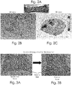
- Figure 2A shows an ex vivo human skin sample after intradermal localized injection of dermal filler and Figures 2B-2C show resulting 3D digital photography views.
- intradermal localized injection of dermal filler in ex vivo human skin samples induces an increase in skin volume and forms a hard ball into the skin as observed in 3D view.
- Example 3 Intradermal injection of dermal filler in ex vivo minipig skin
- Figures 3A-3B show slides of minipig skin respectively epidermal face and dermal face, after intradermal injection of dermal filler in ex vivo minipig skin. The following observations can be made:
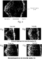
- Figure 4 shows image by high frequency ultrasound of ex vivo human skin after injection of dermal filler
- the dermis After injection of dermal filler, the dermis is largely thickened and appears less echogenic.
- Example 5 Ultrasound images of ex vivo human skin sample after injection of dermal filler. Comparison between fresh and frozen skin samples
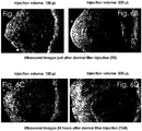
- Example 6 Ultrasound images of ex vivo human skin sample after injection of dermal filler. Comparison of the volume of injection between 100 ⁇ L and 200 ⁇ L just after dermal filler injection (T0) or 24 hours after (T24)
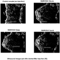
- Example 7 Ultrasound images of ex vivo human skin sample after injection of different dermal fillers just after dermal filler injection (T0)
- Example 8 Visualization of dermal filler by Histopathology after skin section and special stain
- Figures 8A-8E show microphotographs of ex vivo human skin after injection of dermal filler by histopathology after skin section and special stain.
- Figures 8A-8B show comparison results of injection volumes. As illustrated, dermal filler is clearly visible as it is stained [in blue - not applicable here due to black and white colors which have to be used] due to hyaluronic acid content.
- Measurement of the area (mm 2 ) occupied by the dermal filler is performed by image analysis after color deconvolution and use of a binary mask in MATLAB®. A statistically significant discrimination is possible between 100 and 200 ⁇ L injection volumes (p ⁇ 0.001).
- Figures 8C-8E show comparison results of different dermal fillers. As illustrated, respective dermal fillers are stained [in blue - not applicable here due to black and white colors which have to be used] due to hyaluronic acid content.
- Example 9 Visualization of dermal filler by RMI in ex vivo human skin
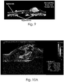
- Figure 9 shows visualization of dermal filler by RMI in ex vivo human skin.
- Figures 10A-10B show visualization of dermal filler by Explorer platform in ex vivo human skin.
- Figure 10A shows visualization of dermal filler in ex vivo human skin with simultaneous measurement of viscoelasticity induced by the filler after intradermal injection.
- Figure 10B shows visualization of dermal filler in ex vivo human skin with simultaneous measurement of viscoelasticity induced by the filler after intrahypodermal injection.
- Example 11 Evaluation of skin visco-elastic properties and general skin firming after injection of dermal filler using the torsionnal ballistometer
- Figure 11 shows an evaluation of skin visco-elastic properties and general skin firming after injection of dermal filler using the torsionnal ballistometer
- Indentation is a measure of skin firming. A decrease of indentation value indicates an increase in skin firming.
- Example 12 Evaluation of skin thickness increase after injection of dermal filler in ex vivo human skin as measured by ultrasound
- Example 13 Comparison of skin thickness induced by different dermal filler
Landscapes
- Health & Medical Sciences (AREA)
- Life Sciences & Earth Sciences (AREA)
- Engineering & Computer Science (AREA)
- Biomedical Technology (AREA)
- Immunology (AREA)
- Molecular Biology (AREA)
- Urology & Nephrology (AREA)
- Hematology (AREA)
- Chemical & Material Sciences (AREA)
- Cell Biology (AREA)
- Pathology (AREA)
- General Health & Medical Sciences (AREA)
- Physics & Mathematics (AREA)
- Medicinal Chemistry (AREA)
- Bioinformatics & Cheminformatics (AREA)
- Food Science & Technology (AREA)
- Biotechnology (AREA)
- Tropical Medicine & Parasitology (AREA)
- Analytical Chemistry (AREA)
- Biochemistry (AREA)
- Toxicology (AREA)
- General Physics & Mathematics (AREA)
- Microbiology (AREA)
- Dermatology (AREA)
- Biophysics (AREA)
- Heart & Thoracic Surgery (AREA)
- Medical Informatics (AREA)
- Surgery (AREA)
- Animal Behavior & Ethology (AREA)
- Public Health (AREA)
- Veterinary Medicine (AREA)
- Investigating Or Analysing Biological Materials (AREA)
- Measuring And Recording Apparatus For Diagnosis (AREA)
Abstract
The invention relates to a model of skin sample. The model is ex vivo human skin and coupled with injection of dermal filler in the skin.
The invention also relates to use the model of skin of the invention to visualize effect of the dermal filler in the skin and to measure skin parameters and skin parameters changes before and after dermal filler injection and methods of use of the model of the invention.
Description
- The invention concerns a new model using ex vivo human skin samples to evaluate dermal fillers. More specifically the invention concerns a model of human skin coupled with injection of dermal filler. This new model is a cost-effective, rapid and simple tool suitable to monitor skin parameter changes induced by dermal filler injection as well as the visualization of dermal filler within skin samples.
- In recent decades, injectable dermal fillers are becoming very useful for the correction of congenital or traumatic facial defects and in patients suffering from lipodystrophy following AIDS. Moreover, these substances are becoming very popular for the treatment of facial wrinkle(s).
- There are numerous dermal fillers available based on different materials, and their properties differ both between and among classes. The model of ex vivo skin sample according to the invention can be used to visualize all types of existing filler. For examples, fillers can be chosen from collagen and derivates, hyaluronic acid, its salts and derivates, in a free form or crosslinked, alginates, synthetic polymers, elastin, biologic polymer and their mixtures.
- Hyaluronic acid (HA) based fillers have varied life spans ranging from weeks to months depending upon their degree of reticulation. Highly reticulated HA fillers are more resistant to in situ degradation and show clinical efficacy for up to 1 year.
- Current non-clinical assessment of dermal filler performance mainly involves in vitro analyses, which are poorly indicative, or in vivo implantation studies in animal models which are time consuming and are less and less used due to the regulatory context. In fact, the Cosmetics Directive establishes a prohibition to test finished cosmetic products and cosmetic ingredients on animals (testing ban), and a prohibition to market in the European Community finished cosmetic products and ingredients included in cosmetic products which were tested on animals (marketing ban). The testing ban on finished cosmetic products applies since 11 September 2004; the testing ban on ingredients or combination of ingredients applies since 11 March 2009. The marketing ban applies since 11 March 2009 for all human health effects with the exception of repeated-dose toxicity, reproductive toxicity and toxicokinetics. For these specific health effects the marketing ban will apply step by step as soon as alternative methods are validated and adopted in EU legislation with due regard to the OECD validation process, but with a maximum cut-off date of 10 years after entry into force of the Directive, i.e., 11 March 2013, irrespective of the availability of alternative non-animal tests.
- In addition to prohibition, differences exist between animal skin and human skin which also motivate the man skilled in the art to find alternative way of testing. Although animal skin are used to test different compounds in pharmaceutical research, it has been found to be very different compared to human skin with regard to:
- Skin touch: animal skin is hairy, thus not suitable for cosmetic testing;
- Skin elasticity: minipig skin is less elastic compared to human skin, thus less suitable for dermal filler testing in vitro;
- Skin thickness: epidermis in mouse, rat and rabbit is too thin and leads to difficulties to perform injection. In minipig skin epidermis is thicker however, the very low elasticity does not allow easy injection.
- For all the reasons mentioned above and for ethical considerations, there is a need to find alternative methods to test dermal filler in a method that mimics the real condition of use of the filler to do the best to characterize their behaviour and properties in the human skin in vivo.
- A new model for investigation of dermal filler is described in the present invention.
- Consequently, a first object of the solution proposed by the invention to remedy this problem is to provide a model of skin sample wherein the model is ex vivo human skin and coupled with injection of dermal filler in the skin.
- Thus, a second object is to use the model of skin of the invention to visualize effect of the dermal filler in the skin.
- A third object is to use the model of skin of the invention to measure skin parameters and skin parameters changes before and after dermal filler injection.
- Other objects of the invention are methods of use of the model of skin of the invention respectively to visualize effect of the dermal filler in the skin; to measure skin parameters and skin parameters changes before and after dermal filler injection; to compare the diffusion and distribution of various dermal fillers in the skin; to measure the early inflammatory response due to dermal filler injection; and to measure the collagen synthesis due to dermal filler injection.
- Finally, other objects of the invention are methods of using a model consisting of ex vivo skin sample coupled with dermal filler injection for respectively visualizing effect of the dermal filler in the skin, and measuring skin parameters changes after injection of a dermal filler in the skin.
- The invention shall be better understood when reading the following non-restricted description, with reference to the accompanying drawings, wherein:
-
Figures 1A-1B show how to inject dermal filler in the dermis of ex vivo human skin;Figures 1C-1E show different kind and volume of intradermal injection of dermal filler in ex vivo human skin; -
Figure 2A shows an ex vivo human skin sample after intradermal localized injection of dermal filler andFigures 2B-2C show resulting 3D digital photography views; -
Figures 3A-3B show slides of minipig skin respectively epidermal face and dermal face, after intradermal injection of dermal filler in ex vivo minipig skin; -
Figure 4 shows images by high frequency ultrasound of ex vivo human skin after injection of dermal filler; -
Figures 5A-5D show ultrasound images of ex vivo human skin samples after injection of dermal filler by comparing fresh and frozen skin samples; -
Figures 6A-6D show ultrasound images of ex vivo human skin samples after injection of dermal filler by comparing the volume of injection; -
Figures 7A-7D show ultrasound images of ex vivo human skin samples after injection of different dermal fillers; -
Figures 8A-8E show microphotographs of ex vivo human skin after injection of dermal filler by histopathology after skin section and special stain:Figures 8A-8B show comparison results of injection volumes andFigures 8C-8E show comparison results of different dermal fillers; -
Figure 9 shows visualization of dermal filler by RMI in ex vivo human skin; -
Figures 10A-10B show visualization of dermal filler by Explorer platform in ex vivo human skin:Figure 10A shows visualization with simultaneous measurement of viscoelasticity induced by the filler after intradermal injection andFigure 10B shows visualization with simultaneous measurement of viscoelasticity induced by the filler after intrahypodermal injection; -
Figure 11 shows an evaluation of skin visco-elastic properties and general skin firming after injection of dermal filler using the torsionnal ballistometer; -
Figure 12 shows an evaluation of skin thickness increase after injection of dermal filler in ex vivo human skin as measured by ultrasound; and -
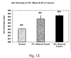
Figure 13 shows comparison results of skin thickness induced by different dermal fillers. - According to the invention, the model claimed uses ex vivo human skin samples. Human skin samples are obtained from plastic surgery, from male or female, from various part of the human body. Preferably, human skin samples are obtained from abdominal, mammary or face skin.
- Skin can be fresh, freezed or used freshly thawed. Preferably skin is fresh or freshly thawed.
- Skin samples can be used with or without hypodermis. Depending of the parameters to be analysed after injection of the dermal filler in the skin, subcutaneaous fat can be removed carefully by dissection before using, to leave only dermis.
- When prepared, skin samples are maintained in organoculture conditions up to three (3) weeks, preferably up to one (1) week, more preferably up to 120 hours.
- According to the invention, the model of human skin is to be used to characterize injectable dermal filler. Injection of dermal filler in ex vivo human skin may be performed intradermally or intrahypodermally or both.
- According to the invention, intradermally means within the dermis, intrahypodermally means within the subcatenous fat, when not removed. Both site of injection can be used.
- Injection is performed using injection needle classically used in dermal filler injection. Preferably, injection is performed using a 30G1/2 needle.
- As performed in vivo on human, injection of dermal filler in the ex vivo human skin sample may be a localized one or a linear retro-tracing one.
- Injected volumes of dermal filler are comprised between 10 and 500 µL, preferably between 50 and 200 µL, more preferably equal to 50, 100 or 200 µL.
- Intradermal injection of dermal filler in the skin sample according to the invention is done with dermal side down or dermal side up.
- In the present invention, the ex vivo human skin sample necessary for injection of dermal filler represents a surface between 3 and 10 cm2, preferably at minimum equal to 4 cm2 (2 x 2 cm).
- When injected with the dermal filler, the human skin according to the invention is used to characterize various parameters related to fillers and/or skin.
- Visualization of dermal filler diffusion within the skin (in the dermis and in the hypodermis);
- Measurement of skin change (thickness increase, area, volume, firming, elasticity) due to dermal filler;
- Measurement of dermal filler diffusion within the skin;
- Comparison of diffusion and distribution pattern of different dermal filler;
- Measurement of early inflammatory response due to dermal filler;
- Measurement of collagen synthesis change due to dermal filler.
- Various techniques can be used in combination with the model according to the invention to visualize and/or measure these parameters before and after dermal filler injection.
- The techniques usable to measure and visualize these parameters are listed below.
- In one preferred embodiment of the invention, this measurement can be done with high frequency ultrasound or dermo-echography. Preferably, a 20 MHz probe is used. Dermo-echography coupled to the human skin sample according to the invention lead to a non-invasive method that allows indirect visualization of the dermal filler in the skin and allows measurement of the skin parameters before and after dermal filler injection. More preferably, human skin parameters which can be measured are skin thickness, skin area and/or volume.
- In another embodiment, the human skin sample according to the invention can be coupled with histopathology. Preferably, histopathology is performed with image analysis, allowing direct visualization of dermal filler in skin sections, measurement of skin thickness changes, and visualization of the distribution of dermal filler within the dermal matrix.
- Various other techniques can be used in combination with the model according to the invention to visualize volume changes, skin thickness changes, diffusion of the filler with time.
- Optical Coherence Tomography (OCT) is a non-invasive method allowing visualization of dermal filler in the skin and measurement of skin thickness changes due to dermal filler.
- 3D digital photography is a non-invasive method allowing measurement of skin volume change due to dermal filler.
- Resonnance Magnetic Imaging is a non-invasive method allowing direct visualization of dermal filler in the skin.
- AixPlorer platform is a non-invasive method allowing indirect visualization of dermal filler in the dermis and also in the hypodermis and measurement of skin thickness changes due to dermal filler as well as measurement of skin viscoelasticity change due to dermal filler.
- Ballistometer is a non-invasive method allowing measurement of skin firming and elasticity change due to dermal filler.
- In one embodiment of the invention, the ex vivo human skin sample is used to measure the kinetics of diffusion of dermal filler in the skin. This evaluation of dermal filler in ex vivo human skin may be performed just after filler injection (T0) or several hours after filler injection up to three (3) weeks, preferably up to one (1) week, preferably up to 120 hours. In those cases, skin samples should be maintained in skin culture medium in cell culture incubator set at 37°C, 5% CO2 and humidified atmosphere.
- The following examples give some non-exhaustive examples of use of ex vivo human skin according to the invention.
-
Figures 1A-1E show different kind and volume of intradermal injection of dermal filler in ex vivo human skin. - For example, the position of the needle during intradermal injection of a dermal filler in an ex vivo human skin sample is illustrated by
Figures 1A-1B . -
Figures 1C and 1D show more particularly the different aspects between respectively a localized injection and a linear retro-tracing injection, andFigure 1E of 50 µL and 100 µL of the dermal filler in an ex vivo human skin sample. -
Figure 2A shows an ex vivo human skin sample after intradermal localized injection of dermal filler andFigures 2B-2C show resulting 3D digital photography views. - As illustrated, intradermal localized injection of dermal filler in ex vivo human skin samples induces an increase in skin volume and forms a hard ball into the skin as observed in 3D view.
-
Figures 3A-3B show slides of minipig skin respectively epidermal face and dermal face, after intradermal injection of dermal filler in ex vivo minipig skin. The following observations can be made: - Minipig skin is very less elastic than human skin.
- When injected intradermally, leakage of dermal filler is observed on the dermal face, through hair follicle base.
- Ex vivo minipig skin is not a suitable model to evaluate dermal filler diffusion.
-
Figure 4 shows image by high frequency ultrasound of ex vivo human skin after injection of dermal filler; - Ultrasound image of the ex vivo human skin sample after injection of dermal filler shows three layer structures:
- Uppermost layer: echogenic
- Intermediate layer: poorly echogenic
- Dermis-hypodermis interface: echogenic
- After injection of dermal filler, the dermis is largely thickened and appears less echogenic.
- As shown by
Figures 5A-5D , no difference is observed between fresh and freshly thawed skin just after dermal filler injection (T0) or 24 hours after (T24). - Ultrasound images after dermal filler injection of
Figures 6A-6D show that: - 1. The dermis is largely thickened.
- 2. The dermis appears less echogenic.
- 3. In some cases, totally non-echogenic zones are observed within the dermis probably corresponding to hyaluronic acid that is non-echogenic.
- Ultrasound images after injection of EMERVEL® dermal fillers of
Figures 7B-7D (by comparison toFigure 7A (control sample) show that: - 1. The dermis is largely thickened.
- 2. The dermis appears less echogenic.
- 3. With the three dermal fillers of EMERVEL® range, totally non-echogenic zones are observed within the dermis probably corresponding to the dermis area occupied by hyaluronic acid (non-echogenic material).
-
Figures 8A-8E show microphotographs of ex vivo human skin after injection of dermal filler by histopathology after skin section and special stain. -
Figures 8A-8B show comparison results of injection volumes. As illustrated, dermal filler is clearly visible as it is stained [in blue - not applicable here due to black and white colors which have to be used] due to hyaluronic acid content. - At microscopic examination, the site of injection is well visible at low magnification in the dermis.
- Measurement of the area (mm2) occupied by the dermal filler is performed by image analysis after color deconvolution and use of a binary mask in MATLAB®. A statistically significant discrimination is possible between 100 and 200 µL injection volumes (p < 0.001).
-
Figures 8C-8E show comparison results of different dermal fillers. As illustrated, respective dermal fillers are stained [in blue - not applicable here due to black and white colors which have to be used] due to hyaluronic acid content. -
Figure 9 shows visualization of dermal filler by RMI in ex vivo human skin. -
Figures 10A-10B show visualization of dermal filler by Explorer platform in ex vivo human skin. - 10a)
Figure 10A shows visualization of dermal filler in ex vivo human skin with simultaneous measurement of viscoelasticity induced by the filler after intradermal injection. - 10b)
Figure 10B shows visualization of dermal filler in ex vivo human skin with simultaneous measurement of viscoelasticity induced by the filler after intrahypodermal injection. -
Figure 11 shows an evaluation of skin visco-elastic properties and general skin firming after injection of dermal filler using the torsionnal ballistometer; - Indentation is a measure of skin firming. A decrease of indentation value indicates an increase in skin firming.
- As illustrated by
Figure 11 , injection of dermal filler induces a statistically significant decrease in indentation, indicating a significant increase in skin firming as measured by ballistometer. - As illustrated by
Figure 12 : - Dermal filler injection in ex vivo human skin induces a marked skin thickness increase that can be estimated by image analysis (Dermascan Software).
- Relative to the concurrent control (0 µL), skin thicknesses increase by a 2.2-fold factor after 100 µL injection and by a 2.6-fold factor after 200 µL injection at T0 (n = 3 donors).
- There is a low inter-individual variability in skin thickness, with CV ranging between 7% and 27%.
- As illustrated by
Figure 13 , a significative difference between EMERVEL® Touch and EMERVEL® Classic (Student Test, p<0.05) is observed: a lower increase of skin thickness is observed with EMERVEL® Touch (the less reticulated filler) compared to EMERVEL® Classic.
Claims (14)
- A model of skin sample wherein the model is ex vivo human skin and coupled with injection of dermal filler in the skin.
- The model according to claim 1, wherein the human skin is fresh, freezed, or freshly thawed.
- The model according to claim 1 or 2, wherein the human skin comprises the dermis.
- The model according to any of claims 1 to 3, wherein the human skin comprises the dermis and the hypodermis.
- Use of the model of any of claims 1 to 4 to visualize effect of the dermal filler in the skin.
- Use of the model of any of claims 1 to 4 to measure skin parameters and skin parameters changes before and after dermal filler injection.
- The use according to claim 6, wherein the skin parameters are thickness, area, volume, firming, elasticity.
- A method of use of the model according to any of claims 1 to 4 to visualize effect of the dermal filler in the skin.
- A method of use of the model according to any of claims 1 to 4 to measure skin parameters and skin parameters changes before and after dermal filler injection.
- A method of use of the model according to any of claims 1 to 4 to compare the diffusion and distribution of various dermal fillers in the skin.
- A method of use of the model according to any of claims 1 to 4 to measure the early inflammatory response due to dermal filler injection.
- A method of use of the model according to any of claims 1 to 4 to measure the collagen synthesis due to dermal filler injection.
- A method of visualizing effect of the dermal filler in the skin, said method using a model consisting of ex vivo skin sample coupled with dermal filler injection according to any of claims 1 to 4.
- A method of measuring skin parameters changes after injection of a dermal filler in the skin, said method using a model consisting of ex vivo skin sample coupled with dermal filler injection according to any of claims 1 to 4.
Priority Applications (2)
| Application Number | Priority Date | Filing Date | Title |
|---|---|---|---|
| EP17197195.5A EP3473163A1 (en) | 2017-10-19 | 2017-10-19 | Model for evaluation of dermal fillers |
| PCT/EP2018/071221 WO2019076507A1 (en) | 2017-10-19 | 2018-08-06 | Model for evaluation of dermal fillers |
Applications Claiming Priority (1)
| Application Number | Priority Date | Filing Date | Title |
|---|---|---|---|
| EP17197195.5A EP3473163A1 (en) | 2017-10-19 | 2017-10-19 | Model for evaluation of dermal fillers |
Publications (1)
| Publication Number | Publication Date |
|---|---|
| EP3473163A1 true EP3473163A1 (en) | 2019-04-24 |
Family
ID=60262696
Family Applications (1)
| Application Number | Title | Priority Date | Filing Date |
|---|---|---|---|
| EP17197195.5A Withdrawn EP3473163A1 (en) | 2017-10-19 | 2017-10-19 | Model for evaluation of dermal fillers |
Country Status (2)
| Country | Link |
|---|---|
| EP (1) | EP3473163A1 (en) |
| WO (1) | WO2019076507A1 (en) |
-
2017
- 2017-10-19 EP EP17197195.5A patent/EP3473163A1/en not_active Withdrawn
-
2018
- 2018-08-06 WO PCT/EP2018/071221 patent/WO2019076507A1/en not_active Ceased
Non-Patent Citations (4)
| Title |
|---|
| CHRISTIAN TRAN ET AL: "In vivo Bio-Integration of Three Hyaluronic Acid Fillers in Human Skin: A Histological Study", DERMATOLOGY, vol. 228, no. 1, 6 February 2014 (2014-02-06), CH, pages 47 - 54, XP055459189, ISSN: 1018-8665, DOI: 10.1159/000354384 * |
| LA GATTA ANNALISA ET AL: "Biophysical and biological characterization of a new line of hyaluronan-based dermal fillers: A scientific rationale to specific clinical indications", MATERIALS SCIENCE AND ENGINEERING C, ELSEVIER SCIENCE S.A, CH, vol. 68, 7 June 2016 (2016-06-07), pages 565 - 572, XP029684664, ISSN: 0928-4931, DOI: 10.1016/J.MSEC.2016.06.008 * |
| MICHEELS P ET AL: "Visual, Ultrasonographic, and Microscopic Study on Hyaluronic Acid-Based Gel", JOURNAL OF DRUGS IN DERMATOLOGY, STRATEGIC COMMUNICATION IN DERMATOLOGY, NEW YORK, NY, US, vol. 15, no. 9, 1 September 2016 (2016-09-01), pages 1092 - 1098, XP009504124, ISSN: 1545-9616 * |
| TIMOTHY CORCORAN FLYNN ET AL: "Comparative Histology of Intradermal Implantation of Mono and Biphasic Hyaluronic Acid Fillers", DERMATOLOGIC SURGERY, vol. 37, no. 5, 27 January 2011 (2011-01-27), pages 637 - 643, XP055042976, ISSN: 1076-0512, DOI: 10.1111/j.1524-4725.2010.01852.x * |
Also Published As
| Publication number | Publication date |
|---|---|
| WO2019076507A1 (en) | 2019-04-25 |
Similar Documents
| Publication | Publication Date | Title |
|---|---|---|
| Meador et al. | The regional-dependent biaxial behavior of young and aged mouse skin: A detailed histomechanical characterization, residual strain analysis, and constitutive model | |
| Hillmann et al. | Diagnosis of hair disorders | |
| Zelickson et al. | Epidermal dendritic cells: A quantitative study | |
| de Vries et al. | Dermal organization in scleroderma: the fast Fourier transform and the laser scatter method objectify fibrosis in nonlesional as well as lesional skin | |
| JP7639201B2 (en) | Method for predicting hair and facial characteristics using scalp stratum corneum cell characteristics | |
| Besler et al. | Quantitative measures of bone shape, cartilage morphometry and joint alignment are associated with disease in an ACLT and MMx rat model of osteoarthritis | |
| Thali et al. | Is ‘virtual histology’the next step after the ‘virtual autopsy’? Magnetic resonance microscopy in forensic medicine | |
| JP6995054B2 (en) | Sample preparation method for radiography | |
| Rubin et al. | Imaging Tools for Noninvasive Hair Assessment. | |
| Chuah et al. | Diagnosis of melasma in brown skin: Wood’s lamp, dermoscopy, and confocal microscopy | |
| Bonetti et al. | Production and characterization of bovine hoof membranes as standardized in vitro model for nail studies | |
| WO2019076507A1 (en) | Model for evaluation of dermal fillers | |
| Baldeweck et al. | In vivo multiphoton microscopy associated to 3D image processing for human skin characterization | |
| Sans et al. | High-resolution magnetic resonance imaging in study of the skin: normal patterns | |
| WO2015173413A1 (en) | Skin topography as a marker for skin maturation | |
| Güngör et al. | Aneurysmatic dermatofibroma with dermoscopic and reflectance confocal microscopic features. | |
| Oliveira et al. | Balloon cell naevus: new perspectives using high-definition optical coherence tomography with dermoscopic and reflectance confocal microscopic correlation. | |
| Cernat et al. | Tissue characterization of puborectalis muscle from 3-D ultrasound | |
| John et al. | Label-Free Imaging of Cancer Cells-Laden 3D Hydrogel Scaffold Using Spectral Domain Optical Coherence Tomography | |
| Mehta et al. | A global perspective on safety and efficacy testing of hair care products | |
| JP2010148548A (en) | Method of predicting wrinkle | |
| Scope et al. | Dermoscopic and clinical predictors of reflectance confocal microscopy patterns of typical nevi on the back and legs: a cross-sectional study | |
| Ardigò et al. | Normal skin: terminology | |
| Shurche et al. | Assessing Dihydrotestosterone-Induced Skin Alterations in C57BL/6 Mice: Implications for Androgenetic Alopecia through High-Resolution Ultrasound Imaging. | |
| Shurche et al. | Assessing Dihydrotestosterone-Induced Skin Alterations in C57BL/6 Mice: Implications for Androgenetic Alopecia through High-Resolution Ultrasound Imaging |
Legal Events
| Date | Code | Title | Description |
|---|---|---|---|
| PUAI | Public reference made under article 153(3) epc to a published international application that has entered the european phase |
Free format text: ORIGINAL CODE: 0009012 |
|
| STAA | Information on the status of an ep patent application or granted ep patent |
Free format text: STATUS: REQUEST FOR EXAMINATION WAS MADE |
|
| 17P | Request for examination filed |
Effective date: 20180803 |
|
| AK | Designated contracting states |
Kind code of ref document: A1 Designated state(s): AL AT BE BG CH CY CZ DE DK EE ES FI FR GB GR HR HU IE IS IT LI LT LU LV MC MK MT NL NO PL PT RO RS SE SI SK SM TR |
|
| AX | Request for extension of the european patent |
Extension state: BA ME |
|
| STAA | Information on the status of an ep patent application or granted ep patent |
Free format text: STATUS: THE APPLICATION IS DEEMED TO BE WITHDRAWN |
|
| 18D | Application deemed to be withdrawn |
Effective date: 20191025 |