EP3466967A1 - Compositions and methods for tcr reprogramming using fusion proteins - Google Patents
Compositions and methods for tcr reprogramming using fusion proteins Download PDFInfo
- Publication number
- EP3466967A1 EP3466967A1 EP18207665.3A EP18207665A EP3466967A1 EP 3466967 A1 EP3466967 A1 EP 3466967A1 EP 18207665 A EP18207665 A EP 18207665A EP 3466967 A1 EP3466967 A1 EP 3466967A1
- Authority
- EP
- European Patent Office
- Prior art keywords
- cell
- tfp
- domain
- tcr
- cells
- Prior art date
- Legal status (The legal status is an assumption and is not a legal conclusion. Google has not performed a legal analysis and makes no representation as to the accuracy of the status listed.)
- Withdrawn
Links
- 238000000034 method Methods 0.000 title claims abstract description 110
- 108020001507 fusion proteins Proteins 0.000 title claims abstract description 34
- 102000037865 fusion proteins Human genes 0.000 title claims abstract description 34
- 239000000203 mixture Substances 0.000 title claims description 40
- 230000008672 reprogramming Effects 0.000 title 1
- 108091008874 T cell receptors Proteins 0.000 claims abstract description 363
- 102000016266 T-Cell Antigen Receptors Human genes 0.000 claims abstract description 363
- 210000001744 T-lymphocyte Anatomy 0.000 claims abstract description 259
- 206010028980 Neoplasm Diseases 0.000 claims abstract description 122
- 208000037265 diseases, disorders, signs and symptoms Diseases 0.000 claims abstract description 79
- 201000010099 disease Diseases 0.000 claims abstract description 64
- 201000011510 cancer Diseases 0.000 claims abstract description 60
- 210000004027 cell Anatomy 0.000 claims description 358
- 230000027455 binding Effects 0.000 claims description 174
- 150000007523 nucleic acids Chemical class 0.000 claims description 137
- 241000282414 Homo sapiens Species 0.000 claims description 136
- 102000039446 nucleic acids Human genes 0.000 claims description 120
- 108020004707 nucleic acids Proteins 0.000 claims description 120
- 108090000765 processed proteins & peptides Proteins 0.000 claims description 118
- 102000004196 processed proteins & peptides Human genes 0.000 claims description 105
- 229920001184 polypeptide Polymers 0.000 claims description 101
- 102100024222 B-lymphocyte antigen CD19 Human genes 0.000 claims description 98
- 101000980825 Homo sapiens B-lymphocyte antigen CD19 Proteins 0.000 claims description 98
- 102000006942 B-Cell Maturation Antigen Human genes 0.000 claims description 95
- 108010008014 B-Cell Maturation Antigen Proteins 0.000 claims description 95
- 108090000623 proteins and genes Proteins 0.000 claims description 94
- 230000014509 gene expression Effects 0.000 claims description 83
- 230000004068 intracellular signaling Effects 0.000 claims description 68
- 102000004169 proteins and genes Human genes 0.000 claims description 60
- 230000003834 intracellular effect Effects 0.000 claims description 47
- 230000002401 inhibitory effect Effects 0.000 claims description 40
- 230000011664 signaling Effects 0.000 claims description 39
- 230000000139 costimulatory effect Effects 0.000 claims description 34
- 108010019670 Chimeric Antigen Receptors Proteins 0.000 claims description 31
- 230000004936 stimulating effect Effects 0.000 claims description 30
- 101000914514 Homo sapiens T-cell-specific surface glycoprotein CD28 Proteins 0.000 claims description 28
- 102100036011 T-cell surface glycoprotein CD4 Human genes 0.000 claims description 28
- 102100027213 T-cell-specific surface glycoprotein CD28 Human genes 0.000 claims description 28
- 102100034922 T-cell surface glycoprotein CD8 alpha chain Human genes 0.000 claims description 26
- 102100035360 Cerebellar degeneration-related antigen 1 Human genes 0.000 claims description 25
- 241000124008 Mammalia Species 0.000 claims description 25
- 239000008194 pharmaceutical composition Substances 0.000 claims description 25
- -1 ICOS Proteins 0.000 claims description 21
- DHMQDGOQFOQNFH-UHFFFAOYSA-N Glycine Chemical compound NCC(O)=O DHMQDGOQFOQNFH-UHFFFAOYSA-N 0.000 claims description 15
- 231100000135 cytotoxicity Toxicity 0.000 claims description 15
- 230000003013 cytotoxicity Effects 0.000 claims description 15
- 108010002350 Interleukin-2 Proteins 0.000 claims description 13
- 102100027207 CD27 antigen Human genes 0.000 claims description 12
- 101000914511 Homo sapiens CD27 antigen Proteins 0.000 claims description 12
- 102100022153 Tumor necrosis factor receptor superfamily member 4 Human genes 0.000 claims description 12
- 238000004519 manufacturing process Methods 0.000 claims description 10
- 108010064548 Lymphocyte Function-Associated Antigen-1 Proteins 0.000 claims description 9
- 230000001965 increasing effect Effects 0.000 claims description 9
- 102100029822 B- and T-lymphocyte attenuator Human genes 0.000 claims description 7
- 239000004471 Glycine Substances 0.000 claims description 7
- 101000864344 Homo sapiens B- and T-lymphocyte attenuator Proteins 0.000 claims description 7
- 102100025237 T-cell surface antigen CD2 Human genes 0.000 claims description 7
- 101710165473 Tumor necrosis factor receptor superfamily member 4 Proteins 0.000 claims description 7
- 230000000735 allogeneic effect Effects 0.000 claims description 7
- 102100024263 CD160 antigen Human genes 0.000 claims description 6
- 101000761938 Homo sapiens CD160 antigen Proteins 0.000 claims description 6
- 101000934346 Homo sapiens T-cell surface antigen CD2 Proteins 0.000 claims description 5
- MTCFGRXMJLQNBG-UHFFFAOYSA-N Serine Natural products OCC(N)C(O)=O MTCFGRXMJLQNBG-UHFFFAOYSA-N 0.000 claims description 5
- 102100038078 CD276 antigen Human genes 0.000 claims description 3
- 101710185679 CD276 antigen Proteins 0.000 claims description 3
- 101150013553 CD40 gene Proteins 0.000 claims description 3
- 101001109503 Homo sapiens NKG2-C type II integral membrane protein Proteins 0.000 claims description 3
- 101000801234 Homo sapiens Tumor necrosis factor receptor superfamily member 18 Proteins 0.000 claims description 3
- 101000851376 Homo sapiens Tumor necrosis factor receptor superfamily member 8 Proteins 0.000 claims description 3
- 102100022683 NKG2-C type II integral membrane protein Human genes 0.000 claims description 3
- 102100040678 Programmed cell death protein 1 Human genes 0.000 claims description 3
- 102100027208 T-cell antigen CD7 Human genes 0.000 claims description 3
- 102100033728 Tumor necrosis factor receptor superfamily member 18 Human genes 0.000 claims description 3
- 102100040245 Tumor necrosis factor receptor superfamily member 5 Human genes 0.000 claims description 3
- 102100036857 Tumor necrosis factor receptor superfamily member 8 Human genes 0.000 claims description 3
- 101000971538 Homo sapiens Killer cell lectin-like receptor subfamily F member 1 Proteins 0.000 claims description 2
- 101000633784 Homo sapiens SLAM family member 7 Proteins 0.000 claims description 2
- 101000795169 Homo sapiens Tumor necrosis factor receptor superfamily member 13C Proteins 0.000 claims description 2
- 102100021458 Killer cell lectin-like receptor subfamily F member 1 Human genes 0.000 claims description 2
- 108010061593 Member 14 Tumor Necrosis Factor Receptors Proteins 0.000 claims description 2
- 102100029198 SLAM family member 7 Human genes 0.000 claims description 2
- 102100029690 Tumor necrosis factor receptor superfamily member 13C Human genes 0.000 claims description 2
- 102100028785 Tumor necrosis factor receptor superfamily member 14 Human genes 0.000 claims description 2
- 230000001472 cytotoxic effect Effects 0.000 claims description 2
- 210000002966 serum Anatomy 0.000 claims description 2
- BGFTWECWAICPDG-UHFFFAOYSA-N 2-[bis(4-chlorophenyl)methyl]-4-n-[3-[bis(4-chlorophenyl)methyl]-4-(dimethylamino)phenyl]-1-n,1-n-dimethylbenzene-1,4-diamine Chemical compound C1=C(C(C=2C=CC(Cl)=CC=2)C=2C=CC(Cl)=CC=2)C(N(C)C)=CC=C1NC(C=1)=CC=C(N(C)C)C=1C(C=1C=CC(Cl)=CC=1)C1=CC=C(Cl)C=C1 BGFTWECWAICPDG-UHFFFAOYSA-N 0.000 claims 4
- 101100112922 Candida albicans CDR3 gene Proteins 0.000 claims 4
- 102100035361 Cerebellar degeneration-related protein 2 Human genes 0.000 claims 4
- 101000737793 Homo sapiens Cerebellar degeneration-related antigen 1 Proteins 0.000 claims 4
- 101000737796 Homo sapiens Cerebellar degeneration-related protein 2 Proteins 0.000 claims 4
- 210000002203 alpha-beta t lymphocyte Anatomy 0.000 claims 2
- 210000004475 gamma-delta t lymphocyte Anatomy 0.000 claims 2
- 210000001266 CD8-positive T-lymphocyte Anatomy 0.000 claims 1
- 101000914496 Homo sapiens T-cell antigen CD7 Proteins 0.000 claims 1
- 102100022339 Integrin alpha-L Human genes 0.000 claims 1
- 239000003937 drug carrier Substances 0.000 claims 1
- 210000000581 natural killer T-cell Anatomy 0.000 claims 1
- 238000011282 treatment Methods 0.000 abstract description 49
- 102000017420 CD3 protein, epsilon/gamma/delta subunit Human genes 0.000 description 128
- 108050005493 CD3 protein, epsilon/gamma/delta subunit Proteins 0.000 description 128
- 239000000427 antigen Substances 0.000 description 116
- 108091007433 antigens Proteins 0.000 description 116
- 102000036639 antigens Human genes 0.000 description 116
- 125000003275 alpha amino acid group Chemical group 0.000 description 107
- 239000013598 vector Substances 0.000 description 87
- 230000004048 modification Effects 0.000 description 63
- 238000012986 modification Methods 0.000 description 63
- 102000008394 Immunoglobulin Fragments Human genes 0.000 description 59
- 235000018102 proteins Nutrition 0.000 description 59
- 108010021625 Immunoglobulin Fragments Proteins 0.000 description 58
- 108020004414 DNA Proteins 0.000 description 50
- 102000053602 DNA Human genes 0.000 description 50
- 229920002477 rna polymer Polymers 0.000 description 43
- 239000003795 chemical substances by application Substances 0.000 description 38
- 108020004999 messenger RNA Proteins 0.000 description 38
- 235000001014 amino acid Nutrition 0.000 description 37
- 125000003729 nucleotide group Chemical group 0.000 description 35
- 239000012634 fragment Substances 0.000 description 34
- 238000000338 in vitro Methods 0.000 description 34
- 229940024606 amino acid Drugs 0.000 description 33
- 150000001413 amino acids Chemical class 0.000 description 33
- 201000003793 Myelodysplastic syndrome Diseases 0.000 description 32
- 239000002773 nucleotide Substances 0.000 description 32
- 108091028043 Nucleic acid sequence Proteins 0.000 description 30
- 208000024893 Acute lymphoblastic leukemia Diseases 0.000 description 26
- 238000011534 incubation Methods 0.000 description 26
- 210000003162 effector t lymphocyte Anatomy 0.000 description 25
- 230000000694 effects Effects 0.000 description 25
- 150000002632 lipids Chemical class 0.000 description 25
- 208000014697 Acute lymphocytic leukaemia Diseases 0.000 description 24
- 208000006664 Precursor Cell Lymphoblastic Leukemia-Lymphoma Diseases 0.000 description 24
- 238000013518 transcription Methods 0.000 description 24
- 230000035897 transcription Effects 0.000 description 24
- 101000716102 Homo sapiens T-cell surface glycoprotein CD4 Proteins 0.000 description 23
- 101000946843 Homo sapiens T-cell surface glycoprotein CD8 alpha chain Proteins 0.000 description 22
- 101100519207 Mus musculus Pdcd1 gene Proteins 0.000 description 22
- 230000004044 response Effects 0.000 description 22
- 238000003556 assay Methods 0.000 description 20
- 230000002489 hematologic effect Effects 0.000 description 20
- 238000003752 polymerase chain reaction Methods 0.000 description 20
- 230000004913 activation Effects 0.000 description 19
- 210000003719 b-lymphocyte Anatomy 0.000 description 18
- 230000000295 complement effect Effects 0.000 description 18
- 102000040430 polynucleotide Human genes 0.000 description 18
- 108091033319 polynucleotide Proteins 0.000 description 18
- 239000002157 polynucleotide Substances 0.000 description 18
- 108060003951 Immunoglobulin Proteins 0.000 description 17
- 125000000539 amino acid group Chemical group 0.000 description 17
- 102000018358 immunoglobulin Human genes 0.000 description 17
- 239000000047 product Substances 0.000 description 17
- 241000713666 Lentivirus Species 0.000 description 16
- 241000699670 Mus sp. Species 0.000 description 16
- 239000011324 bead Substances 0.000 description 16
- 230000006870 function Effects 0.000 description 16
- 230000036210 malignancy Effects 0.000 description 16
- 239000013612 plasmid Substances 0.000 description 16
- 238000006467 substitution reaction Methods 0.000 description 16
- 108020003589 5' Untranslated Regions Proteins 0.000 description 15
- 208000035475 disorder Diseases 0.000 description 15
- 239000002502 liposome Substances 0.000 description 15
- 108020005345 3' Untranslated Regions Proteins 0.000 description 14
- 210000004369 blood Anatomy 0.000 description 14
- 239000008280 blood Substances 0.000 description 14
- 230000002147 killing effect Effects 0.000 description 14
- 101000851370 Homo sapiens Tumor necrosis factor receptor superfamily member 9 Proteins 0.000 description 13
- 206010035226 Plasma cell myeloma Diseases 0.000 description 13
- 102100036856 Tumor necrosis factor receptor superfamily member 9 Human genes 0.000 description 13
- 201000003444 follicular lymphoma Diseases 0.000 description 13
- 208000032839 leukemia Diseases 0.000 description 13
- 239000003446 ligand Substances 0.000 description 13
- 238000013519 translation Methods 0.000 description 13
- 102000004127 Cytokines Human genes 0.000 description 12
- 108090000695 Cytokines Proteins 0.000 description 12
- 102000000588 Interleukin-2 Human genes 0.000 description 12
- 230000001086 cytosolic effect Effects 0.000 description 12
- 210000001151 cytotoxic T lymphocyte Anatomy 0.000 description 12
- 210000001519 tissue Anatomy 0.000 description 12
- 210000004881 tumor cell Anatomy 0.000 description 12
- 208000007541 Preleukemia Diseases 0.000 description 11
- 239000000306 component Substances 0.000 description 11
- 230000007423 decrease Effects 0.000 description 11
- 238000000684 flow cytometry Methods 0.000 description 11
- 230000003211 malignant effect Effects 0.000 description 11
- 241001465754 Metazoa Species 0.000 description 10
- 208000034578 Multiple myelomas Diseases 0.000 description 10
- 108091036407 Polyadenylation Proteins 0.000 description 10
- 239000012636 effector Substances 0.000 description 10
- 239000013604 expression vector Substances 0.000 description 10
- 230000028993 immune response Effects 0.000 description 10
- 230000001976 improved effect Effects 0.000 description 10
- 238000001727 in vivo Methods 0.000 description 10
- 230000002062 proliferating effect Effects 0.000 description 10
- 230000035755 proliferation Effects 0.000 description 10
- 239000000523 sample Substances 0.000 description 10
- 208000010839 B-cell chronic lymphocytic leukemia Diseases 0.000 description 9
- 102100038080 B-cell receptor CD22 Human genes 0.000 description 9
- 108010074708 B7-H1 Antigen Proteins 0.000 description 9
- IAZDPXIOMUYVGZ-UHFFFAOYSA-N Dimethylsulphoxide Chemical compound CS(C)=O IAZDPXIOMUYVGZ-UHFFFAOYSA-N 0.000 description 9
- 101000884305 Homo sapiens B-cell receptor CD22 Proteins 0.000 description 9
- 101100383038 Homo sapiens CD19 gene Proteins 0.000 description 9
- OUYCCCASQSFEME-QMMMGPOBSA-N L-tyrosine Chemical compound OC(=O)[C@@H](N)CC1=CC=C(O)C=C1 OUYCCCASQSFEME-QMMMGPOBSA-N 0.000 description 9
- 241001529936 Murinae Species 0.000 description 9
- 208000015914 Non-Hodgkin lymphomas Diseases 0.000 description 9
- 102100024216 Programmed cell death 1 ligand 1 Human genes 0.000 description 9
- 238000002617 apheresis Methods 0.000 description 9
- 238000002347 injection Methods 0.000 description 9
- 239000007924 injection Substances 0.000 description 9
- 239000002245 particle Substances 0.000 description 9
- 230000008569 process Effects 0.000 description 9
- 230000000638 stimulation Effects 0.000 description 9
- 230000001225 therapeutic effect Effects 0.000 description 9
- OUYCCCASQSFEME-UHFFFAOYSA-N tyrosine Natural products OC(=O)C(N)CC1=CC=C(O)C=C1 OUYCCCASQSFEME-UHFFFAOYSA-N 0.000 description 9
- 108091032973 (ribonucleotides)n+m Proteins 0.000 description 8
- 102000040650 (ribonucleotides)n+m Human genes 0.000 description 8
- 208000006994 Precancerous Conditions Diseases 0.000 description 8
- 108700008625 Reporter Genes Proteins 0.000 description 8
- 230000008901 benefit Effects 0.000 description 8
- 238000004422 calculation algorithm Methods 0.000 description 8
- 201000005787 hematologic cancer Diseases 0.000 description 8
- 239000000463 material Substances 0.000 description 8
- 238000009126 molecular therapy Methods 0.000 description 8
- 230000001105 regulatory effect Effects 0.000 description 8
- 239000000243 solution Substances 0.000 description 8
- 238000002560 therapeutic procedure Methods 0.000 description 8
- 238000012546 transfer Methods 0.000 description 8
- 206010000830 Acute leukaemia Diseases 0.000 description 7
- 208000032791 BCR-ABL1 positive chronic myelogenous leukemia Diseases 0.000 description 7
- 210000004366 CD4-positive T-lymphocyte Anatomy 0.000 description 7
- 101000917858 Homo sapiens Low affinity immunoglobulin gamma Fc region receptor III-A Proteins 0.000 description 7
- 101000917839 Homo sapiens Low affinity immunoglobulin gamma Fc region receptor III-B Proteins 0.000 description 7
- 102100037850 Interferon gamma Human genes 0.000 description 7
- 108010074328 Interferon-gamma Proteins 0.000 description 7
- 102100029185 Low affinity immunoglobulin gamma Fc region receptor III-B Human genes 0.000 description 7
- 206010025323 Lymphomas Diseases 0.000 description 7
- 230000015572 biosynthetic process Effects 0.000 description 7
- 210000000601 blood cell Anatomy 0.000 description 7
- 208000024207 chronic leukemia Diseases 0.000 description 7
- 150000001875 compounds Chemical class 0.000 description 7
- 238000004520 electroporation Methods 0.000 description 7
- 239000005090 green fluorescent protein Substances 0.000 description 7
- 230000001589 lymphoproliferative effect Effects 0.000 description 7
- 208000007525 plasmablastic lymphoma Diseases 0.000 description 7
- 102000005962 receptors Human genes 0.000 description 7
- 108020003175 receptors Proteins 0.000 description 7
- 230000004083 survival effect Effects 0.000 description 7
- 238000010361 transduction Methods 0.000 description 7
- 230000026683 transduction Effects 0.000 description 7
- 238000001890 transfection Methods 0.000 description 7
- 241001430294 unidentified retrovirus Species 0.000 description 7
- 208000036170 B-Cell Marginal Zone Lymphoma Diseases 0.000 description 6
- 208000032568 B-cell prolymphocytic leukaemia Diseases 0.000 description 6
- 208000011691 Burkitt lymphomas Diseases 0.000 description 6
- 208000016778 CD4+/CD56+ hematodermic neoplasm Diseases 0.000 description 6
- 102100037904 CD9 antigen Human genes 0.000 description 6
- HEDRZPFGACZZDS-UHFFFAOYSA-N Chloroform Chemical compound ClC(Cl)Cl HEDRZPFGACZZDS-UHFFFAOYSA-N 0.000 description 6
- 102100039498 Cytotoxic T-lymphocyte protein 4 Human genes 0.000 description 6
- 108090000626 DNA-directed RNA polymerases Proteins 0.000 description 6
- 102000004163 DNA-directed RNA polymerases Human genes 0.000 description 6
- 206010058314 Dysplasia Diseases 0.000 description 6
- 238000002965 ELISA Methods 0.000 description 6
- 206010061850 Extranodal marginal zone B-cell lymphoma (MALT type) Diseases 0.000 description 6
- 102100026122 High affinity immunoglobulin gamma Fc receptor I Human genes 0.000 description 6
- 101000913074 Homo sapiens High affinity immunoglobulin gamma Fc receptor I Proteins 0.000 description 6
- 101000934341 Homo sapiens T-cell surface glycoprotein CD5 Proteins 0.000 description 6
- 101000801255 Homo sapiens Tumor necrosis factor receptor superfamily member 17 Proteins 0.000 description 6
- 108010073807 IgG Receptors Proteins 0.000 description 6
- 102000009490 IgG Receptors Human genes 0.000 description 6
- 208000031671 Large B-Cell Diffuse Lymphoma Diseases 0.000 description 6
- 201000003791 MALT lymphoma Diseases 0.000 description 6
- 208000025205 Mantle-Cell Lymphoma Diseases 0.000 description 6
- OKKJLVBELUTLKV-UHFFFAOYSA-N Methanol Chemical compound OC OKKJLVBELUTLKV-UHFFFAOYSA-N 0.000 description 6
- 208000035416 Prolymphocytic B-Cell Leukemia Diseases 0.000 description 6
- 241000283984 Rodentia Species 0.000 description 6
- 102100025244 T-cell surface glycoprotein CD5 Human genes 0.000 description 6
- 102100036922 Tumor necrosis factor ligand superfamily member 13B Human genes 0.000 description 6
- 241000700605 Viruses Species 0.000 description 6
- 208000016025 Waldenstroem macroglobulinemia Diseases 0.000 description 6
- 208000033559 Waldenström macroglobulinemia Diseases 0.000 description 6
- 230000009286 beneficial effect Effects 0.000 description 6
- 208000035269 cancer or benign tumor Diseases 0.000 description 6
- 208000032852 chronic lymphocytic leukemia Diseases 0.000 description 6
- 239000002299 complementary DNA Substances 0.000 description 6
- 206010012818 diffuse large B-cell lymphoma Diseases 0.000 description 6
- 238000005516 engineering process Methods 0.000 description 6
- 201000009277 hairy cell leukemia Diseases 0.000 description 6
- 102000046935 human TNFRSF17 Human genes 0.000 description 6
- 230000000977 initiatory effect Effects 0.000 description 6
- 201000007924 marginal zone B-cell lymphoma Diseases 0.000 description 6
- 208000021937 marginal zone lymphoma Diseases 0.000 description 6
- 230000001404 mediated effect Effects 0.000 description 6
- 210000005259 peripheral blood Anatomy 0.000 description 6
- 239000011886 peripheral blood Substances 0.000 description 6
- 210000005134 plasmacytoid dendritic cell Anatomy 0.000 description 6
- 230000019491 signal transduction Effects 0.000 description 6
- 241000894007 species Species 0.000 description 6
- 208000024891 symptom Diseases 0.000 description 6
- 239000013603 viral vector Substances 0.000 description 6
- 230000003612 virological effect Effects 0.000 description 6
- 102100034458 Hepatitis A virus cellular receptor 2 Human genes 0.000 description 5
- 101000738354 Homo sapiens CD9 antigen Proteins 0.000 description 5
- 101000889276 Homo sapiens Cytotoxic T-lymphocyte protein 4 Proteins 0.000 description 5
- 101001068133 Homo sapiens Hepatitis A virus cellular receptor 2 Proteins 0.000 description 5
- 101000777628 Homo sapiens Leukocyte antigen CD37 Proteins 0.000 description 5
- 101001137987 Homo sapiens Lymphocyte activation gene 3 protein Proteins 0.000 description 5
- 101000934338 Homo sapiens Myeloid cell surface antigen CD33 Proteins 0.000 description 5
- 101000738771 Homo sapiens Receptor-type tyrosine-protein phosphatase C Proteins 0.000 description 5
- 101000914484 Homo sapiens T-lymphocyte activation antigen CD80 Proteins 0.000 description 5
- 108020004684 Internal Ribosome Entry Sites Proteins 0.000 description 5
- 102000017578 LAG3 Human genes 0.000 description 5
- 102100031586 Leukocyte antigen CD37 Human genes 0.000 description 5
- 102100025243 Myeloid cell surface antigen CD33 Human genes 0.000 description 5
- 108700026244 Open Reading Frames Proteins 0.000 description 5
- 108700030875 Programmed Cell Death 1 Ligand 2 Proteins 0.000 description 5
- 102100024213 Programmed cell death 1 ligand 2 Human genes 0.000 description 5
- 102100037422 Receptor-type tyrosine-protein phosphatase C Human genes 0.000 description 5
- 108020004511 Recombinant DNA Proteins 0.000 description 5
- 102100027222 T-lymphocyte activation antigen CD80 Human genes 0.000 description 5
- 238000004458 analytical method Methods 0.000 description 5
- 230000000259 anti-tumor effect Effects 0.000 description 5
- 238000002659 cell therapy Methods 0.000 description 5
- 238000012217 deletion Methods 0.000 description 5
- 230000037430 deletion Effects 0.000 description 5
- LOKCTEFSRHRXRJ-UHFFFAOYSA-I dipotassium trisodium dihydrogen phosphate hydrogen phosphate dichloride Chemical compound P(=O)(O)(O)[O-].[K+].P(=O)(O)([O-])[O-].[Na+].[Na+].[Cl-].[K+].[Cl-].[Na+] LOKCTEFSRHRXRJ-UHFFFAOYSA-I 0.000 description 5
- 239000003814 drug Substances 0.000 description 5
- 238000001476 gene delivery Methods 0.000 description 5
- 239000012642 immune effector Substances 0.000 description 5
- 210000000987 immune system Anatomy 0.000 description 5
- 230000003053 immunization Effects 0.000 description 5
- 238000002649 immunization Methods 0.000 description 5
- 229940121354 immunomodulator Drugs 0.000 description 5
- 239000003112 inhibitor Substances 0.000 description 5
- 230000003993 interaction Effects 0.000 description 5
- 239000002953 phosphate buffered saline Substances 0.000 description 5
- 230000008488 polyadenylation Effects 0.000 description 5
- 230000001177 retroviral effect Effects 0.000 description 5
- 238000003786 synthesis reaction Methods 0.000 description 5
- 238000012360 testing method Methods 0.000 description 5
- 241000701161 unidentified adenovirus Species 0.000 description 5
- 238000011144 upstream manufacturing Methods 0.000 description 5
- 238000005406 washing Methods 0.000 description 5
- IJGRMHOSHXDMSA-UHFFFAOYSA-N Atomic nitrogen Chemical compound N#N IJGRMHOSHXDMSA-UHFFFAOYSA-N 0.000 description 4
- 208000023275 Autoimmune disease Diseases 0.000 description 4
- 102100025466 Carcinoembryonic antigen-related cell adhesion molecule 3 Human genes 0.000 description 4
- 108091026890 Coding region Proteins 0.000 description 4
- 108091033380 Coding strand Proteins 0.000 description 4
- PMATZTZNYRCHOR-CGLBZJNRSA-N Cyclosporin A Chemical compound CC[C@@H]1NC(=O)[C@H]([C@H](O)[C@H](C)C\C=C\C)N(C)C(=O)[C@H](C(C)C)N(C)C(=O)[C@H](CC(C)C)N(C)C(=O)[C@H](CC(C)C)N(C)C(=O)[C@@H](C)NC(=O)[C@H](C)NC(=O)[C@H](CC(C)C)N(C)C(=O)[C@H](C(C)C)NC(=O)[C@H](CC(C)C)N(C)C(=O)CN(C)C1=O PMATZTZNYRCHOR-CGLBZJNRSA-N 0.000 description 4
- 229930105110 Cyclosporin A Natural products 0.000 description 4
- 108010036949 Cyclosporine Proteins 0.000 description 4
- 241000702421 Dependoparvovirus Species 0.000 description 4
- WQZGKKKJIJFFOK-GASJEMHNSA-N Glucose Natural products OC[C@H]1OC(O)[C@H](O)[C@@H](O)[C@@H]1O WQZGKKKJIJFFOK-GASJEMHNSA-N 0.000 description 4
- 208000032672 Histiocytosis haematophagic Diseases 0.000 description 4
- 241000282412 Homo Species 0.000 description 4
- 101000914337 Homo sapiens Carcinoembryonic antigen-related cell adhesion molecule 3 Proteins 0.000 description 4
- 101000935040 Homo sapiens Integrin beta-2 Proteins 0.000 description 4
- 101001138062 Homo sapiens Leukocyte-associated immunoglobulin-like receptor 1 Proteins 0.000 description 4
- 101000831007 Homo sapiens T-cell immunoreceptor with Ig and ITIM domains Proteins 0.000 description 4
- 241000725303 Human immunodeficiency virus Species 0.000 description 4
- 102100025390 Integrin beta-2 Human genes 0.000 description 4
- 108010064593 Intercellular Adhesion Molecule-1 Proteins 0.000 description 4
- 102100037877 Intercellular adhesion molecule 1 Human genes 0.000 description 4
- HNDVDQJCIGZPNO-YFKPBYRVSA-N L-histidine Chemical compound OC(=O)[C@@H](N)CC1=CN=CN1 HNDVDQJCIGZPNO-YFKPBYRVSA-N 0.000 description 4
- AGPKZVBTJJNPAG-WHFBIAKZSA-N L-isoleucine Chemical compound CC[C@H](C)[C@H](N)C(O)=O AGPKZVBTJJNPAG-WHFBIAKZSA-N 0.000 description 4
- COLNVLDHVKWLRT-QMMMGPOBSA-N L-phenylalanine Chemical compound OC(=O)[C@@H](N)CC1=CC=CC=C1 COLNVLDHVKWLRT-QMMMGPOBSA-N 0.000 description 4
- AYFVYJQAPQTCCC-GBXIJSLDSA-N L-threonine Chemical compound C[C@@H](O)[C@H](N)C(O)=O AYFVYJQAPQTCCC-GBXIJSLDSA-N 0.000 description 4
- QIVBCDIJIAJPQS-VIFPVBQESA-N L-tryptophane Chemical compound C1=CC=C2C(C[C@H](N)C(O)=O)=CNC2=C1 QIVBCDIJIAJPQS-VIFPVBQESA-N 0.000 description 4
- KZSNJWFQEVHDMF-BYPYZUCNSA-N L-valine Chemical compound CC(C)[C@H](N)C(O)=O KZSNJWFQEVHDMF-BYPYZUCNSA-N 0.000 description 4
- 102100020943 Leukocyte-associated immunoglobulin-like receptor 1 Human genes 0.000 description 4
- 208000031422 Lymphocytic Chronic B-Cell Leukemia Diseases 0.000 description 4
- 241000699666 Mus <mouse, genus> Species 0.000 description 4
- 108010038807 Oligopeptides Proteins 0.000 description 4
- 102000015636 Oligopeptides Human genes 0.000 description 4
- 108091034057 RNA (poly(A)) Proteins 0.000 description 4
- 102100024834 T-cell immunoreceptor with Ig and ITIM domains Human genes 0.000 description 4
- QJJXYPPXXYFBGM-LFZNUXCKSA-N Tacrolimus Chemical compound C1C[C@@H](O)[C@H](OC)C[C@@H]1\C=C(/C)[C@@H]1[C@H](C)[C@@H](O)CC(=O)[C@H](CC=C)/C=C(C)/C[C@H](C)C[C@H](OC)[C@H]([C@H](C[C@H]2C)OC)O[C@@]2(O)C(=O)C(=O)N2CCCC[C@H]2C(=O)O1 QJJXYPPXXYFBGM-LFZNUXCKSA-N 0.000 description 4
- AYFVYJQAPQTCCC-UHFFFAOYSA-N Threonine Natural products CC(O)C(N)C(O)=O AYFVYJQAPQTCCC-UHFFFAOYSA-N 0.000 description 4
- 239000004473 Threonine Substances 0.000 description 4
- 108700019146 Transgenes Proteins 0.000 description 4
- QIVBCDIJIAJPQS-UHFFFAOYSA-N Tryptophan Natural products C1=CC=C2C(CC(N)C(O)=O)=CNC2=C1 QIVBCDIJIAJPQS-UHFFFAOYSA-N 0.000 description 4
- KZSNJWFQEVHDMF-UHFFFAOYSA-N Valine Natural products CC(C)C(N)C(O)=O KZSNJWFQEVHDMF-UHFFFAOYSA-N 0.000 description 4
- 238000007792 addition Methods 0.000 description 4
- 238000010171 animal model Methods 0.000 description 4
- 238000013459 approach Methods 0.000 description 4
- WQZGKKKJIJFFOK-VFUOTHLCSA-N beta-D-glucose Chemical compound OC[C@H]1O[C@@H](O)[C@H](O)[C@@H](O)[C@@H]1O WQZGKKKJIJFFOK-VFUOTHLCSA-N 0.000 description 4
- 210000001185 bone marrow Anatomy 0.000 description 4
- 239000000872 buffer Substances 0.000 description 4
- 229960001265 ciclosporin Drugs 0.000 description 4
- 238000010367 cloning Methods 0.000 description 4
- 229930182912 cyclosporin Natural products 0.000 description 4
- 238000007710 freezing Methods 0.000 description 4
- 230000008014 freezing Effects 0.000 description 4
- 230000004927 fusion Effects 0.000 description 4
- HNDVDQJCIGZPNO-UHFFFAOYSA-N histidine Natural products OC(=O)C(N)CC1=CN=CN1 HNDVDQJCIGZPNO-UHFFFAOYSA-N 0.000 description 4
- 230000003463 hyperproliferative effect Effects 0.000 description 4
- 230000001939 inductive effect Effects 0.000 description 4
- 238000001802 infusion Methods 0.000 description 4
- 230000005764 inhibitory process Effects 0.000 description 4
- AGPKZVBTJJNPAG-UHFFFAOYSA-N isoleucine Natural products CCC(C)C(N)C(O)=O AGPKZVBTJJNPAG-UHFFFAOYSA-N 0.000 description 4
- 229960000310 isoleucine Drugs 0.000 description 4
- 230000007774 longterm Effects 0.000 description 4
- 229920002521 macromolecule Polymers 0.000 description 4
- 239000000693 micelle Substances 0.000 description 4
- HPNSFSBZBAHARI-UHFFFAOYSA-N micophenolic acid Natural products OC1=C(CC=C(C)CCC(O)=O)C(OC)=C(C)C2=C1C(=O)OC2 HPNSFSBZBAHARI-UHFFFAOYSA-N 0.000 description 4
- 230000035772 mutation Effects 0.000 description 4
- HPNSFSBZBAHARI-RUDMXATFSA-N mycophenolic acid Chemical compound OC1=C(C\C=C(/C)CCC(O)=O)C(OC)=C(C)C2=C1C(=O)OC2 HPNSFSBZBAHARI-RUDMXATFSA-N 0.000 description 4
- 230000002688 persistence Effects 0.000 description 4
- COLNVLDHVKWLRT-UHFFFAOYSA-N phenylalanine Natural products OC(=O)C(N)CC1=CC=CC=C1 COLNVLDHVKWLRT-UHFFFAOYSA-N 0.000 description 4
- 229920000642 polymer Polymers 0.000 description 4
- 230000002265 prevention Effects 0.000 description 4
- 239000000126 substance Substances 0.000 description 4
- QJJXYPPXXYFBGM-SHYZHZOCSA-N tacrolimus Natural products CO[C@H]1C[C@H](CC[C@@H]1O)C=C(C)[C@H]2OC(=O)[C@H]3CCCCN3C(=O)C(=O)[C@@]4(O)O[C@@H]([C@H](C[C@H]4C)OC)[C@@H](C[C@H](C)CC(=C[C@@H](CC=C)C(=O)C[C@H](O)[C@H]2C)C)OC QJJXYPPXXYFBGM-SHYZHZOCSA-N 0.000 description 4
- 239000004474 valine Substances 0.000 description 4
- MTCFGRXMJLQNBG-REOHCLBHSA-N (2S)-2-Amino-3-hydroxypropansäure Chemical compound OC[C@H](N)C(O)=O MTCFGRXMJLQNBG-REOHCLBHSA-N 0.000 description 3
- 208000031261 Acute myeloid leukaemia Diseases 0.000 description 3
- 102100034540 Adenomatous polyposis coli protein Human genes 0.000 description 3
- 102000006306 Antigen Receptors Human genes 0.000 description 3
- 108010083359 Antigen Receptors Proteins 0.000 description 3
- 108010028006 B-Cell Activating Factor Proteins 0.000 description 3
- 108020004705 Codon Proteins 0.000 description 3
- 241000701022 Cytomegalovirus Species 0.000 description 3
- 108090000331 Firefly luciferases Proteins 0.000 description 3
- BCCRXDTUTZHDEU-VKHMYHEASA-N Gly-Ser Chemical compound NCC(=O)N[C@@H](CO)C(O)=O BCCRXDTUTZHDEU-VKHMYHEASA-N 0.000 description 3
- 101000924577 Homo sapiens Adenomatous polyposis coli protein Proteins 0.000 description 3
- 101000809875 Homo sapiens TYRO protein tyrosine kinase-binding protein Proteins 0.000 description 3
- 101000666896 Homo sapiens V-type immunoglobulin domain-containing suppressor of T-cell activation Proteins 0.000 description 3
- 108091006905 Human Serum Albumin Proteins 0.000 description 3
- 102000008100 Human Serum Albumin Human genes 0.000 description 3
- 206010020751 Hypersensitivity Diseases 0.000 description 3
- 108091092195 Intron Proteins 0.000 description 3
- QNAYBMKLOCPYGJ-REOHCLBHSA-N L-alanine Chemical compound C[C@H](N)C(O)=O QNAYBMKLOCPYGJ-REOHCLBHSA-N 0.000 description 3
- 239000000232 Lipid Bilayer Substances 0.000 description 3
- 102000007474 Multiprotein Complexes Human genes 0.000 description 3
- 108010085220 Multiprotein Complexes Proteins 0.000 description 3
- 208000033776 Myeloid Acute Leukemia Diseases 0.000 description 3
- 108010003723 Single-Domain Antibodies Proteins 0.000 description 3
- FAPWRFPIFSIZLT-UHFFFAOYSA-M Sodium chloride Chemical compound [Na+].[Cl-] FAPWRFPIFSIZLT-UHFFFAOYSA-M 0.000 description 3
- 102100038717 TYRO protein tyrosine kinase-binding protein Human genes 0.000 description 3
- 108060008682 Tumor Necrosis Factor Proteins 0.000 description 3
- 108060008683 Tumor Necrosis Factor Receptor Proteins 0.000 description 3
- 102000000852 Tumor Necrosis Factor-alpha Human genes 0.000 description 3
- 101710187885 Tumor necrosis factor receptor superfamily member 17 Proteins 0.000 description 3
- 102100033726 Tumor necrosis factor receptor superfamily member 17 Human genes 0.000 description 3
- 108091023045 Untranslated Region Proteins 0.000 description 3
- 102100038282 V-type immunoglobulin domain-containing suppressor of T-cell activation Human genes 0.000 description 3
- 239000000654 additive Substances 0.000 description 3
- 230000000996 additive effect Effects 0.000 description 3
- OIRDTQYFTABQOQ-KQYNXXCUSA-N adenosine Chemical compound C1=NC=2C(N)=NC=NC=2N1[C@@H]1O[C@H](CO)[C@@H](O)[C@H]1O OIRDTQYFTABQOQ-KQYNXXCUSA-N 0.000 description 3
- 235000004279 alanine Nutrition 0.000 description 3
- 230000007815 allergy Effects 0.000 description 3
- 230000036783 anaphylactic response Effects 0.000 description 3
- 230000005809 anti-tumor immunity Effects 0.000 description 3
- 230000000890 antigenic effect Effects 0.000 description 3
- 208000006673 asthma Diseases 0.000 description 3
- 230000001580 bacterial effect Effects 0.000 description 3
- 230000004071 biological effect Effects 0.000 description 3
- 229940112129 campath Drugs 0.000 description 3
- 238000002619 cancer immunotherapy Methods 0.000 description 3
- 230000015556 catabolic process Effects 0.000 description 3
- 230000010261 cell growth Effects 0.000 description 3
- 238000002512 chemotherapy Methods 0.000 description 3
- 238000003776 cleavage reaction Methods 0.000 description 3
- 206010052015 cytokine release syndrome Diseases 0.000 description 3
- 230000009089 cytolysis Effects 0.000 description 3
- 230000001461 cytolytic effect Effects 0.000 description 3
- 238000002784 cytotoxicity assay Methods 0.000 description 3
- 231100000263 cytotoxicity test Toxicity 0.000 description 3
- 230000003247 decreasing effect Effects 0.000 description 3
- 238000006731 degradation reaction Methods 0.000 description 3
- 210000004443 dendritic cell Anatomy 0.000 description 3
- 239000008121 dextrose Substances 0.000 description 3
- 230000004069 differentiation Effects 0.000 description 3
- 230000002255 enzymatic effect Effects 0.000 description 3
- IJJVMEJXYNJXOJ-UHFFFAOYSA-N fluquinconazole Chemical compound C=1C=C(Cl)C=C(Cl)C=1N1C(=O)C2=CC(F)=CC=C2N=C1N1C=NC=N1 IJJVMEJXYNJXOJ-UHFFFAOYSA-N 0.000 description 3
- 238000001415 gene therapy Methods 0.000 description 3
- 210000004602 germ cell Anatomy 0.000 description 3
- 230000012010 growth Effects 0.000 description 3
- 239000001963 growth medium Substances 0.000 description 3
- 229940072221 immunoglobulins Drugs 0.000 description 3
- 230000008676 import Effects 0.000 description 3
- 208000015181 infectious disease Diseases 0.000 description 3
- 208000027866 inflammatory disease Diseases 0.000 description 3
- 238000001990 intravenous administration Methods 0.000 description 3
- 229960005386 ipilimumab Drugs 0.000 description 3
- 229960004942 lenalidomide Drugs 0.000 description 3
- GOTYRUGSSMKFNF-UHFFFAOYSA-N lenalidomide Chemical compound C1C=2C(N)=CC=CC=2C(=O)N1C1CCC(=O)NC1=O GOTYRUGSSMKFNF-UHFFFAOYSA-N 0.000 description 3
- 210000000265 leukocyte Anatomy 0.000 description 3
- 206010025135 lupus erythematosus Diseases 0.000 description 3
- FVVLHONNBARESJ-NTOWJWGLSA-H magnesium;potassium;trisodium;(2r,3s,4r,5r)-2,3,4,5,6-pentahydroxyhexanoate;acetate;tetrachloride;nonahydrate Chemical compound O.O.O.O.O.O.O.O.O.[Na+].[Na+].[Na+].[Mg+2].[Cl-].[Cl-].[Cl-].[Cl-].[K+].CC([O-])=O.OC[C@@H](O)[C@@H](O)[C@H](O)[C@@H](O)C([O-])=O FVVLHONNBARESJ-NTOWJWGLSA-H 0.000 description 3
- 239000003550 marker Substances 0.000 description 3
- 239000012528 membrane Substances 0.000 description 3
- 238000010369 molecular cloning Methods 0.000 description 3
- 210000001616 monocyte Anatomy 0.000 description 3
- 201000000050 myeloid neoplasm Diseases 0.000 description 3
- 210000003819 peripheral blood mononuclear cell Anatomy 0.000 description 3
- 230000010076 replication Effects 0.000 description 3
- 230000002441 reversible effect Effects 0.000 description 3
- 230000007017 scission Effects 0.000 description 3
- 230000028327 secretion Effects 0.000 description 3
- 230000009870 specific binding Effects 0.000 description 3
- 150000003431 steroids Chemical class 0.000 description 3
- 230000001052 transient effect Effects 0.000 description 3
- 102000035160 transmembrane proteins Human genes 0.000 description 3
- 108091005703 transmembrane proteins Proteins 0.000 description 3
- 238000002054 transplantation Methods 0.000 description 3
- 102000003298 tumor necrosis factor receptor Human genes 0.000 description 3
- 239000003981 vehicle Substances 0.000 description 3
- 238000001262 western blot Methods 0.000 description 3
- 108010065816 zeta chain antigen T cell receptor Proteins 0.000 description 3
- GFFGJBXGBJISGV-UHFFFAOYSA-N Adenine Chemical compound NC1=NC=NC2=C1N=CN2 GFFGJBXGBJISGV-UHFFFAOYSA-N 0.000 description 2
- 206010002198 Anaphylactic reaction Diseases 0.000 description 2
- 239000004475 Arginine Substances 0.000 description 2
- DCXYFEDJOCDNAF-UHFFFAOYSA-N Asparagine Natural products OC(=O)C(N)CC(N)=O DCXYFEDJOCDNAF-UHFFFAOYSA-N 0.000 description 2
- 241000894006 Bacteria Species 0.000 description 2
- 206010006187 Breast cancer Diseases 0.000 description 2
- 208000026310 Breast neoplasm Diseases 0.000 description 2
- 102100035793 CD83 antigen Human genes 0.000 description 2
- OYPRJOBELJOOCE-UHFFFAOYSA-N Calcium Chemical compound [Ca] OYPRJOBELJOOCE-UHFFFAOYSA-N 0.000 description 2
- 102000000844 Cell Surface Receptors Human genes 0.000 description 2
- 108010001857 Cell Surface Receptors Proteins 0.000 description 2
- 241000282693 Cercopithecidae Species 0.000 description 2
- 206010008342 Cervix carcinoma Diseases 0.000 description 2
- 241000588724 Escherichia coli Species 0.000 description 2
- 108060002716 Exonuclease Proteins 0.000 description 2
- WHUUTDBJXJRKMK-UHFFFAOYSA-N Glutamic acid Natural products OC(=O)C(N)CCC(O)=O WHUUTDBJXJRKMK-UHFFFAOYSA-N 0.000 description 2
- 108010017213 Granulocyte-Macrophage Colony-Stimulating Factor Proteins 0.000 description 2
- 102100039620 Granulocyte-macrophage colony-stimulating factor Human genes 0.000 description 2
- NYHBQMYGNKIUIF-UUOKFMHZSA-N Guanosine Chemical compound C1=NC=2C(=O)NC(N)=NC=2N1[C@@H]1O[C@H](CO)[C@@H](O)[C@H]1O NYHBQMYGNKIUIF-UUOKFMHZSA-N 0.000 description 2
- 208000036066 Hemophagocytic Lymphohistiocytosis Diseases 0.000 description 2
- 101000946856 Homo sapiens CD83 antigen Proteins 0.000 description 2
- 101000917826 Homo sapiens Low affinity immunoglobulin gamma Fc region receptor II-a Proteins 0.000 description 2
- 101000917824 Homo sapiens Low affinity immunoglobulin gamma Fc region receptor II-b Proteins 0.000 description 2
- 108010073816 IgE Receptors Proteins 0.000 description 2
- 102000009438 IgE Receptors Human genes 0.000 description 2
- 206010061598 Immunodeficiency Diseases 0.000 description 2
- 108010002386 Interleukin-3 Proteins 0.000 description 2
- 102000004889 Interleukin-6 Human genes 0.000 description 2
- 108090001005 Interleukin-6 Proteins 0.000 description 2
- 208000008839 Kidney Neoplasms Diseases 0.000 description 2
- XUJNEKJLAYXESH-REOHCLBHSA-N L-Cysteine Chemical compound SC[C@H](N)C(O)=O XUJNEKJLAYXESH-REOHCLBHSA-N 0.000 description 2
- ONIBWKKTOPOVIA-BYPYZUCNSA-N L-Proline Chemical compound OC(=O)[C@@H]1CCCN1 ONIBWKKTOPOVIA-BYPYZUCNSA-N 0.000 description 2
- ODKSFYDXXFIFQN-BYPYZUCNSA-P L-argininium(2+) Chemical compound NC(=[NH2+])NCCC[C@H]([NH3+])C(O)=O ODKSFYDXXFIFQN-BYPYZUCNSA-P 0.000 description 2
- DCXYFEDJOCDNAF-REOHCLBHSA-N L-asparagine Chemical compound OC(=O)[C@@H](N)CC(N)=O DCXYFEDJOCDNAF-REOHCLBHSA-N 0.000 description 2
- CKLJMWTZIZZHCS-REOHCLBHSA-N L-aspartic acid Chemical compound OC(=O)[C@@H](N)CC(O)=O CKLJMWTZIZZHCS-REOHCLBHSA-N 0.000 description 2
- WHUUTDBJXJRKMK-VKHMYHEASA-N L-glutamic acid Chemical compound OC(=O)[C@@H](N)CCC(O)=O WHUUTDBJXJRKMK-VKHMYHEASA-N 0.000 description 2
- ZDXPYRJPNDTMRX-VKHMYHEASA-N L-glutamine Chemical compound OC(=O)[C@@H](N)CCC(N)=O ZDXPYRJPNDTMRX-VKHMYHEASA-N 0.000 description 2
- ROHFNLRQFUQHCH-YFKPBYRVSA-N L-leucine Chemical compound CC(C)C[C@H](N)C(O)=O ROHFNLRQFUQHCH-YFKPBYRVSA-N 0.000 description 2
- KDXKERNSBIXSRK-YFKPBYRVSA-N L-lysine Chemical compound NCCCC[C@H](N)C(O)=O KDXKERNSBIXSRK-YFKPBYRVSA-N 0.000 description 2
- FFEARJCKVFRZRR-BYPYZUCNSA-N L-methionine Chemical compound CSCC[C@H](N)C(O)=O FFEARJCKVFRZRR-BYPYZUCNSA-N 0.000 description 2
- FBOZXECLQNJBKD-ZDUSSCGKSA-N L-methotrexate Chemical compound C=1N=C2N=C(N)N=C(N)C2=NC=1CN(C)C1=CC=C(C(=O)N[C@@H](CCC(O)=O)C(O)=O)C=C1 FBOZXECLQNJBKD-ZDUSSCGKSA-N 0.000 description 2
- ROHFNLRQFUQHCH-UHFFFAOYSA-N Leucine Natural products CC(C)CC(N)C(O)=O ROHFNLRQFUQHCH-UHFFFAOYSA-N 0.000 description 2
- 102100029204 Low affinity immunoglobulin gamma Fc region receptor II-a Human genes 0.000 description 2
- 206010058467 Lung neoplasm malignant Diseases 0.000 description 2
- KDXKERNSBIXSRK-UHFFFAOYSA-N Lysine Natural products NCCCCC(N)C(O)=O KDXKERNSBIXSRK-UHFFFAOYSA-N 0.000 description 2
- 239000004472 Lysine Substances 0.000 description 2
- 241000829100 Macaca mulatta polyomavirus 1 Species 0.000 description 2
- 208000004987 Macrophage activation syndrome Diseases 0.000 description 2
- 206010027476 Metastases Diseases 0.000 description 2
- 206010033128 Ovarian cancer Diseases 0.000 description 2
- 206010061535 Ovarian neoplasm Diseases 0.000 description 2
- 206010061902 Pancreatic neoplasm Diseases 0.000 description 2
- 108010077524 Peptide Elongation Factor 1 Proteins 0.000 description 2
- 108091093037 Peptide nucleic acid Proteins 0.000 description 2
- 101710124239 Poly(A) polymerase Proteins 0.000 description 2
- RJKFOVLPORLFTN-LEKSSAKUSA-N Progesterone Chemical compound C1CC2=CC(=O)CC[C@]2(C)[C@@H]2[C@@H]1[C@@H]1CC[C@H](C(=O)C)[C@@]1(C)CC2 RJKFOVLPORLFTN-LEKSSAKUSA-N 0.000 description 2
- 101710089372 Programmed cell death protein 1 Proteins 0.000 description 2
- ONIBWKKTOPOVIA-UHFFFAOYSA-N Proline Natural products OC(=O)C1CCCN1 ONIBWKKTOPOVIA-UHFFFAOYSA-N 0.000 description 2
- 206010060862 Prostate cancer Diseases 0.000 description 2
- 208000000236 Prostatic Neoplasms Diseases 0.000 description 2
- 241000700159 Rattus Species 0.000 description 2
- 206010038389 Renal cancer Diseases 0.000 description 2
- 239000006146 Roswell Park Memorial Institute medium Substances 0.000 description 2
- 240000004808 Saccharomyces cerevisiae Species 0.000 description 2
- 206010039491 Sarcoma Diseases 0.000 description 2
- 108091027967 Small hairpin RNA Proteins 0.000 description 2
- 230000006044 T cell activation Effects 0.000 description 2
- 230000005867 T cell response Effects 0.000 description 2
- IQFYYKKMVGJFEH-XLPZGREQSA-N Thymidine Chemical compound O=C1NC(=O)C(C)=CN1[C@@H]1O[C@H](CO)[C@@H](O)C1 IQFYYKKMVGJFEH-XLPZGREQSA-N 0.000 description 2
- 108020004566 Transfer RNA Proteins 0.000 description 2
- 101710181056 Tumor necrosis factor ligand superfamily member 13B Proteins 0.000 description 2
- DRTQHJPVMGBUCF-XVFCMESISA-N Uridine Chemical compound O[C@@H]1[C@H](O)[C@@H](CO)O[C@H]1N1C(=O)NC(=O)C=C1 DRTQHJPVMGBUCF-XVFCMESISA-N 0.000 description 2
- 208000006105 Uterine Cervical Neoplasms Diseases 0.000 description 2
- 230000002378 acidificating effect Effects 0.000 description 2
- 230000009471 action Effects 0.000 description 2
- 150000003838 adenosines Chemical class 0.000 description 2
- 208000026935 allergic disease Diseases 0.000 description 2
- 208000003455 anaphylaxis Diseases 0.000 description 2
- 238000009175 antibody therapy Methods 0.000 description 2
- 239000003963 antioxidant agent Substances 0.000 description 2
- 239000012736 aqueous medium Substances 0.000 description 2
- ODKSFYDXXFIFQN-UHFFFAOYSA-N arginine Natural products OC(=O)C(N)CCCNC(N)=N ODKSFYDXXFIFQN-UHFFFAOYSA-N 0.000 description 2
- 125000003118 aryl group Chemical group 0.000 description 2
- 235000009582 asparagine Nutrition 0.000 description 2
- 229960001230 asparagine Drugs 0.000 description 2
- 235000003704 aspartic acid Nutrition 0.000 description 2
- 229960002170 azathioprine Drugs 0.000 description 2
- LMEKQMALGUDUQG-UHFFFAOYSA-N azathioprine Chemical compound CN1C=NC([N+]([O-])=O)=C1SC1=NC=NC2=C1NC=N2 LMEKQMALGUDUQG-UHFFFAOYSA-N 0.000 description 2
- OQFSQFPPLPISGP-UHFFFAOYSA-N beta-carboxyaspartic acid Natural products OC(=O)C(N)C(C(O)=O)C(O)=O OQFSQFPPLPISGP-UHFFFAOYSA-N 0.000 description 2
- 239000012472 biological sample Substances 0.000 description 2
- 210000001772 blood platelet Anatomy 0.000 description 2
- 230000037396 body weight Effects 0.000 description 2
- 239000011575 calcium Substances 0.000 description 2
- 229910052791 calcium Inorganic materials 0.000 description 2
- 239000001506 calcium phosphate Substances 0.000 description 2
- 229910000389 calcium phosphate Inorganic materials 0.000 description 2
- 235000011010 calcium phosphates Nutrition 0.000 description 2
- 210000000170 cell membrane Anatomy 0.000 description 2
- 230000004663 cell proliferation Effects 0.000 description 2
- 201000010881 cervical cancer Diseases 0.000 description 2
- HVYWMOMLDIMFJA-DPAQBDIFSA-N cholesterol Chemical compound C1C=C2C[C@@H](O)CC[C@]2(C)[C@@H]2[C@@H]1[C@@H]1CC[C@H]([C@H](C)CCCC(C)C)[C@@]1(C)CC2 HVYWMOMLDIMFJA-DPAQBDIFSA-N 0.000 description 2
- 235000018417 cysteine Nutrition 0.000 description 2
- XUJNEKJLAYXESH-UHFFFAOYSA-N cysteine Natural products SCC(N)C(O)=O XUJNEKJLAYXESH-UHFFFAOYSA-N 0.000 description 2
- 210000000805 cytoplasm Anatomy 0.000 description 2
- OPTASPLRGRRNAP-UHFFFAOYSA-N cytosine Chemical compound NC=1C=CNC(=O)N=1 OPTASPLRGRRNAP-UHFFFAOYSA-N 0.000 description 2
- 230000006378 damage Effects 0.000 description 2
- 230000001419 dependent effect Effects 0.000 description 2
- 238000011161 development Methods 0.000 description 2
- 230000018109 developmental process Effects 0.000 description 2
- 229940119744 dextran 40 Drugs 0.000 description 2
- RNPXCFINMKSQPQ-UHFFFAOYSA-N dicetyl hydrogen phosphate Chemical compound CCCCCCCCCCCCCCCCOP(O)(=O)OCCCCCCCCCCCCCCCC RNPXCFINMKSQPQ-UHFFFAOYSA-N 0.000 description 2
- 239000003085 diluting agent Substances 0.000 description 2
- 230000006806 disease prevention Effects 0.000 description 2
- 210000003743 erythrocyte Anatomy 0.000 description 2
- 102000013165 exonuclease Human genes 0.000 description 2
- 239000013613 expression plasmid Substances 0.000 description 2
- 229960000390 fludarabine Drugs 0.000 description 2
- GIUYCYHIANZCFB-FJFJXFQQSA-N fludarabine phosphate Chemical compound C1=NC=2C(N)=NC(F)=NC=2N1[C@@H]1O[C@H](COP(O)(O)=O)[C@@H](O)[C@@H]1O GIUYCYHIANZCFB-FJFJXFQQSA-N 0.000 description 2
- 238000009472 formulation Methods 0.000 description 2
- 235000013922 glutamic acid Nutrition 0.000 description 2
- 239000004220 glutamic acid Substances 0.000 description 2
- ZDXPYRJPNDTMRX-UHFFFAOYSA-N glutamine Natural products OC(=O)C(N)CCC(N)=O ZDXPYRJPNDTMRX-UHFFFAOYSA-N 0.000 description 2
- 210000003714 granulocyte Anatomy 0.000 description 2
- 210000002443 helper t lymphocyte Anatomy 0.000 description 2
- 210000003958 hematopoietic stem cell Anatomy 0.000 description 2
- 208000014752 hemophagocytic syndrome Diseases 0.000 description 2
- 230000002163 immunogen Effects 0.000 description 2
- 239000003018 immunosuppressive agent Substances 0.000 description 2
- 229940125721 immunosuppressive agent Drugs 0.000 description 2
- 230000010354 integration Effects 0.000 description 2
- FZWBNHMXJMCXLU-BLAUPYHCSA-N isomaltotriose Chemical compound O[C@@H]1[C@@H](O)[C@H](O)[C@@H](CO)O[C@@H]1OC[C@@H]1[C@@H](O)[C@H](O)[C@@H](O)[C@@H](OC[C@@H](O)[C@@H](O)[C@H](O)[C@@H](O)C=O)O1 FZWBNHMXJMCXLU-BLAUPYHCSA-N 0.000 description 2
- 201000010982 kidney cancer Diseases 0.000 description 2
- 238000001638 lipofection Methods 0.000 description 2
- 239000007788 liquid Substances 0.000 description 2
- 201000007270 liver cancer Diseases 0.000 description 2
- 208000014018 liver neoplasm Diseases 0.000 description 2
- 238000001325 log-rank test Methods 0.000 description 2
- 201000005202 lung cancer Diseases 0.000 description 2
- 208000020816 lung neoplasm Diseases 0.000 description 2
- 210000001165 lymph node Anatomy 0.000 description 2
- 210000004324 lymphatic system Anatomy 0.000 description 2
- 210000004698 lymphocyte Anatomy 0.000 description 2
- 208000015486 malignant pancreatic neoplasm Diseases 0.000 description 2
- 230000035800 maturation Effects 0.000 description 2
- 238000005259 measurement Methods 0.000 description 2
- 229930182817 methionine Natural products 0.000 description 2
- 229960000485 methotrexate Drugs 0.000 description 2
- 238000002156 mixing Methods 0.000 description 2
- 238000002703 mutagenesis Methods 0.000 description 2
- 231100000350 mutagenesis Toxicity 0.000 description 2
- 229940014456 mycophenolate Drugs 0.000 description 2
- 229960000951 mycophenolic acid Drugs 0.000 description 2
- 210000004296 naive t lymphocyte Anatomy 0.000 description 2
- 229910052757 nitrogen Inorganic materials 0.000 description 2
- 210000004940 nucleus Anatomy 0.000 description 2
- 201000002528 pancreatic cancer Diseases 0.000 description 2
- 208000008443 pancreatic carcinoma Diseases 0.000 description 2
- 230000036961 partial effect Effects 0.000 description 2
- 150000003904 phospholipids Chemical class 0.000 description 2
- 230000000770 proinflammatory effect Effects 0.000 description 2
- 230000005855 radiation Effects 0.000 description 2
- ZAHRKKWIAAJSAO-UHFFFAOYSA-N rapamycin Natural products COCC(O)C(=C/C(C)C(=O)CC(OC(=O)C1CCCCN1C(=O)C(=O)C2(O)OC(CC(OC)C(=CC=CC=CC(C)CC(C)C(=O)C)C)CCC2C)C(C)CC3CCC(O)C(C3)OC)C ZAHRKKWIAAJSAO-UHFFFAOYSA-N 0.000 description 2
- 230000009467 reduction Effects 0.000 description 2
- 210000003289 regulatory T cell Anatomy 0.000 description 2
- 229960003452 romidepsin Drugs 0.000 description 2
- OHRURASPPZQGQM-GCCNXGTGSA-N romidepsin Chemical compound O1C(=O)[C@H](C(C)C)NC(=O)C(=C/C)/NC(=O)[C@H]2CSSCC\C=C\[C@@H]1CC(=O)N[C@H](C(C)C)C(=O)N2 OHRURASPPZQGQM-GCCNXGTGSA-N 0.000 description 2
- 238000012216 screening Methods 0.000 description 2
- 125000003607 serino group Chemical group [H]N([H])[C@]([H])(C(=O)[*])C(O[H])([H])[H] 0.000 description 2
- 229960002930 sirolimus Drugs 0.000 description 2
- QFJCIRLUMZQUOT-HPLJOQBZSA-N sirolimus Chemical compound C1C[C@@H](O)[C@H](OC)C[C@@H]1C[C@@H](C)[C@H]1OC(=O)[C@@H]2CCCCN2C(=O)C(=O)[C@](O)(O2)[C@H](C)CC[C@H]2C[C@H](OC)/C(C)=C/C=C/C=C/[C@@H](C)C[C@@H](C)C(=O)[C@H](OC)[C@H](O)/C(C)=C/[C@@H](C)C(=O)C1 QFJCIRLUMZQUOT-HPLJOQBZSA-N 0.000 description 2
- 239000004055 small Interfering RNA Substances 0.000 description 2
- 238000002415 sodium dodecyl sulfate polyacrylamide gel electrophoresis Methods 0.000 description 2
- 210000000952 spleen Anatomy 0.000 description 2
- 238000010561 standard procedure Methods 0.000 description 2
- 210000000130 stem cell Anatomy 0.000 description 2
- 238000007920 subcutaneous administration Methods 0.000 description 2
- 239000006228 supernatant Substances 0.000 description 2
- 238000001356 surgical procedure Methods 0.000 description 2
- 230000002459 sustained effect Effects 0.000 description 2
- 229940124597 therapeutic agent Drugs 0.000 description 2
- 230000002103 transcriptional effect Effects 0.000 description 2
- 230000002463 transducing effect Effects 0.000 description 2
- 230000010474 transient expression Effects 0.000 description 2
- QORWJWZARLRLPR-UHFFFAOYSA-H tricalcium bis(phosphate) Chemical compound [Ca+2].[Ca+2].[Ca+2].[O-]P([O-])([O-])=O.[O-]P([O-])([O-])=O QORWJWZARLRLPR-UHFFFAOYSA-H 0.000 description 2
- 210000003171 tumor-infiltrating lymphocyte Anatomy 0.000 description 2
- 238000011179 visual inspection Methods 0.000 description 2
- XLYOFNOQVPJJNP-UHFFFAOYSA-N water Substances O XLYOFNOQVPJJNP-UHFFFAOYSA-N 0.000 description 2
- MZOFCQQQCNRIBI-VMXHOPILSA-N (3s)-4-[[(2s)-1-[[(2s)-1-[[(1s)-1-carboxy-2-hydroxyethyl]amino]-4-methyl-1-oxopentan-2-yl]amino]-5-(diaminomethylideneamino)-1-oxopentan-2-yl]amino]-3-[[2-[[(2s)-2,6-diaminohexanoyl]amino]acetyl]amino]-4-oxobutanoic acid Chemical compound OC[C@@H](C(O)=O)NC(=O)[C@H](CC(C)C)NC(=O)[C@H](CCCN=C(N)N)NC(=O)[C@H](CC(O)=O)NC(=O)CNC(=O)[C@@H](N)CCCCN MZOFCQQQCNRIBI-VMXHOPILSA-N 0.000 description 1
- VGONTNSXDCQUGY-RRKCRQDMSA-N 2'-deoxyinosine Chemical group C1[C@H](O)[C@@H](CO)O[C@H]1N1C(N=CNC2=O)=C2N=C1 VGONTNSXDCQUGY-RRKCRQDMSA-N 0.000 description 1
- UEJJHQNACJXSKW-UHFFFAOYSA-N 2-(2,6-dioxopiperidin-3-yl)-1H-isoindole-1,3(2H)-dione Chemical compound O=C1C2=CC=CC=C2C(=O)N1C1CCC(=O)NC1=O UEJJHQNACJXSKW-UHFFFAOYSA-N 0.000 description 1
- 102000002627 4-1BB Ligand Human genes 0.000 description 1
- 108010082808 4-1BB Ligand Proteins 0.000 description 1
- 229960000549 4-dimethylaminophenol Drugs 0.000 description 1
- VHYFNPMBLIVWCW-UHFFFAOYSA-N 4-dimethylaminopyridine Substances CN(C)C1=CC=NC=C1 VHYFNPMBLIVWCW-UHFFFAOYSA-N 0.000 description 1
- 108020005029 5' Flanking Region Proteins 0.000 description 1
- OGHAROSJZRTIOK-KQYNXXCUSA-O 7-methylguanosine Chemical compound C1=2N=C(N)NC(=O)C=2[N+](C)=CN1[C@@H]1O[C@H](CO)[C@@H](O)[C@H]1O OGHAROSJZRTIOK-KQYNXXCUSA-O 0.000 description 1
- 108020005176 AU Rich Elements Proteins 0.000 description 1
- 102000007469 Actins Human genes 0.000 description 1
- 108010085238 Actins Proteins 0.000 description 1
- 229930024421 Adenine Natural products 0.000 description 1
- 241000588813 Alcaligenes faecalis Species 0.000 description 1
- 102000002260 Alkaline Phosphatase Human genes 0.000 description 1
- 108020004774 Alkaline Phosphatase Proteins 0.000 description 1
- 108700028369 Alleles Proteins 0.000 description 1
- 208000033399 Anaphylactic responses Diseases 0.000 description 1
- 108091023037 Aptamer Proteins 0.000 description 1
- 206010003445 Ascites Diseases 0.000 description 1
- 241000271566 Aves Species 0.000 description 1
- 241000714230 Avian leukemia virus Species 0.000 description 1
- 208000004736 B-Cell Leukemia Diseases 0.000 description 1
- 102100022005 B-lymphocyte antigen CD20 Human genes 0.000 description 1
- 208000035143 Bacterial infection Diseases 0.000 description 1
- DWRXFEITVBNRMK-UHFFFAOYSA-N Beta-D-1-Arabinofuranosylthymine Natural products O=C1NC(=O)C(C)=CN1C1C(O)C(O)C(CO)O1 DWRXFEITVBNRMK-UHFFFAOYSA-N 0.000 description 1
- 102100026189 Beta-galactosidase Human genes 0.000 description 1
- 206010005003 Bladder cancer Diseases 0.000 description 1
- 108091003079 Bovine Serum Albumin Proteins 0.000 description 1
- 208000003174 Brain Neoplasms Diseases 0.000 description 1
- 239000002126 C01EB10 - Adenosine Substances 0.000 description 1
- 108091033409 CRISPR Proteins 0.000 description 1
- 108010021064 CTLA-4 Antigen Proteins 0.000 description 1
- 229940045513 CTLA4 antagonist Drugs 0.000 description 1
- 102100025570 Cancer/testis antigen 1 Human genes 0.000 description 1
- 241000222122 Candida albicans Species 0.000 description 1
- 241000282472 Canis lupus familiaris Species 0.000 description 1
- 201000009030 Carcinoma Diseases 0.000 description 1
- 108010035563 Chloramphenicol O-acetyltransferase Proteins 0.000 description 1
- 108020004638 Circular DNA Proteins 0.000 description 1
- 108091062157 Cis-regulatory element Proteins 0.000 description 1
- 206010009944 Colon cancer Diseases 0.000 description 1
- 208000001333 Colorectal Neoplasms Diseases 0.000 description 1
- 108091035707 Consensus sequence Proteins 0.000 description 1
- 102000004420 Creatine Kinase Human genes 0.000 description 1
- 108010042126 Creatine kinase Proteins 0.000 description 1
- MIKUYHXYGGJMLM-GIMIYPNGSA-N Crotonoside Natural products C1=NC2=C(N)NC(=O)N=C2N1[C@H]1O[C@@H](CO)[C@H](O)[C@@H]1O MIKUYHXYGGJMLM-GIMIYPNGSA-N 0.000 description 1
- CMSMOCZEIVJLDB-UHFFFAOYSA-N Cyclophosphamide Chemical compound ClCCN(CCCl)P1(=O)NCCCO1 CMSMOCZEIVJLDB-UHFFFAOYSA-N 0.000 description 1
- FBPFZTCFMRRESA-KVTDHHQDSA-N D-Mannitol Chemical compound OC[C@@H](O)[C@@H](O)[C@H](O)[C@H](O)CO FBPFZTCFMRRESA-KVTDHHQDSA-N 0.000 description 1
- NYHBQMYGNKIUIF-UHFFFAOYSA-N D-guanosine Natural products C1=2NC(N)=NC(=O)C=2N=CN1C1OC(CO)C(O)C1O NYHBQMYGNKIUIF-UHFFFAOYSA-N 0.000 description 1
- WQZGKKKJIJFFOK-QTVWNMPRSA-N D-mannopyranose Chemical compound OC[C@H]1OC(O)[C@@H](O)[C@@H](O)[C@@H]1O WQZGKKKJIJFFOK-QTVWNMPRSA-N 0.000 description 1
- 102000016928 DNA-directed DNA polymerase Human genes 0.000 description 1
- 108010014303 DNA-directed DNA polymerase Proteins 0.000 description 1
- 229920002307 Dextran Polymers 0.000 description 1
- GZDFHIJNHHMENY-UHFFFAOYSA-N Dimethyl dicarbonate Chemical compound COC(=O)OC(=O)OC GZDFHIJNHHMENY-UHFFFAOYSA-N 0.000 description 1
- KCXVZYZYPLLWCC-UHFFFAOYSA-N EDTA Chemical compound OC(=O)CN(CC(O)=O)CCN(CC(O)=O)CC(O)=O KCXVZYZYPLLWCC-UHFFFAOYSA-N 0.000 description 1
- 102100031780 Endonuclease Human genes 0.000 description 1
- 108010042407 Endonucleases Proteins 0.000 description 1
- 241000701867 Enterobacteria phage T7 Species 0.000 description 1
- 102000004190 Enzymes Human genes 0.000 description 1
- 108090000790 Enzymes Proteins 0.000 description 1
- 239000005977 Ethylene Substances 0.000 description 1
- 241000206602 Eukaryota Species 0.000 description 1
- 108700024394 Exon Proteins 0.000 description 1
- 241000282326 Felis catus Species 0.000 description 1
- 102000016359 Fibronectins Human genes 0.000 description 1
- 108010067306 Fibronectins Proteins 0.000 description 1
- 241000233866 Fungi Species 0.000 description 1
- 108700028146 Genetic Enhancer Elements Proteins 0.000 description 1
- 108700039691 Genetic Promoter Regions Proteins 0.000 description 1
- 108060005986 Granzyme Proteins 0.000 description 1
- 102000001398 Granzyme Human genes 0.000 description 1
- 108010043121 Green Fluorescent Proteins Proteins 0.000 description 1
- 102000025850 HLA-A2 Antigen Human genes 0.000 description 1
- 108010074032 HLA-A2 Antigen Proteins 0.000 description 1
- 102000006354 HLA-DR Antigens Human genes 0.000 description 1
- 108010058597 HLA-DR Antigens Proteins 0.000 description 1
- 241000606768 Haemophilus influenzae Species 0.000 description 1
- 208000002250 Hematologic Neoplasms Diseases 0.000 description 1
- 102100029360 Hematopoietic cell signal transducer Human genes 0.000 description 1
- 102100031573 Hematopoietic progenitor cell antigen CD34 Human genes 0.000 description 1
- 102000001554 Hemoglobins Human genes 0.000 description 1
- 108010054147 Hemoglobins Proteins 0.000 description 1
- 241000238631 Hexapoda Species 0.000 description 1
- 101000897405 Homo sapiens B-lymphocyte antigen CD20 Proteins 0.000 description 1
- 101000856237 Homo sapiens Cancer/testis antigen 1 Proteins 0.000 description 1
- 101000990188 Homo sapiens Hematopoietic cell signal transducer Proteins 0.000 description 1
- 101000777663 Homo sapiens Hematopoietic progenitor cell antigen CD34 Proteins 0.000 description 1
- 101000878602 Homo sapiens Immunoglobulin alpha Fc receptor Proteins 0.000 description 1
- 101001046686 Homo sapiens Integrin alpha-M Proteins 0.000 description 1
- 101001057504 Homo sapiens Interferon-stimulated gene 20 kDa protein Proteins 0.000 description 1
- 101001055144 Homo sapiens Interleukin-2 receptor subunit alpha Proteins 0.000 description 1
- 101000878605 Homo sapiens Low affinity immunoglobulin epsilon Fc receptor Proteins 0.000 description 1
- 101001023379 Homo sapiens Lysosome-associated membrane glycoprotein 1 Proteins 0.000 description 1
- 101000950695 Homo sapiens Mitogen-activated protein kinase 8 Proteins 0.000 description 1
- 101000946889 Homo sapiens Monocyte differentiation antigen CD14 Proteins 0.000 description 1
- 101000946860 Homo sapiens T-cell surface glycoprotein CD3 epsilon chain Proteins 0.000 description 1
- 101000851434 Homo sapiens Tumor necrosis factor ligand superfamily member 13B Proteins 0.000 description 1
- 102000003839 Human Proteins Human genes 0.000 description 1
- 108090000144 Human Proteins Proteins 0.000 description 1
- 241000701044 Human gammaherpesvirus 4 Species 0.000 description 1
- 229920001612 Hydroxyethyl starch Polymers 0.000 description 1
- 208000001953 Hypotension Diseases 0.000 description 1
- 206010021143 Hypoxia Diseases 0.000 description 1
- 101150102264 IE gene Proteins 0.000 description 1
- 229940124790 IL-6 inhibitor Drugs 0.000 description 1
- 102000009786 Immunoglobulin Constant Regions Human genes 0.000 description 1
- 108010009817 Immunoglobulin Constant Regions Proteins 0.000 description 1
- 108010054477 Immunoglobulin Fab Fragments Proteins 0.000 description 1
- 102000001706 Immunoglobulin Fab Fragments Human genes 0.000 description 1
- 102000012745 Immunoglobulin Subunits Human genes 0.000 description 1
- 108010079585 Immunoglobulin Subunits Proteins 0.000 description 1
- 108010067060 Immunoglobulin Variable Region Proteins 0.000 description 1
- 102000017727 Immunoglobulin Variable Region Human genes 0.000 description 1
- 102100038005 Immunoglobulin alpha Fc receptor Human genes 0.000 description 1
- 102100022338 Integrin alpha-M Human genes 0.000 description 1
- 102100027268 Interferon-stimulated gene 20 kDa protein Human genes 0.000 description 1
- 108010002352 Interleukin-1 Proteins 0.000 description 1
- 102000003814 Interleukin-10 Human genes 0.000 description 1
- 108090000174 Interleukin-10 Proteins 0.000 description 1
- 102000003816 Interleukin-13 Human genes 0.000 description 1
- 108090000176 Interleukin-13 Proteins 0.000 description 1
- 108050003558 Interleukin-17 Proteins 0.000 description 1
- 102000013691 Interleukin-17 Human genes 0.000 description 1
- 102000000646 Interleukin-3 Human genes 0.000 description 1
- 108090000978 Interleukin-4 Proteins 0.000 description 1
- 102000004388 Interleukin-4 Human genes 0.000 description 1
- 108010055717 JNK Mitogen-Activated Protein Kinases Proteins 0.000 description 1
- 102000019145 JUN kinase activity proteins Human genes 0.000 description 1
- 102100020880 Kit ligand Human genes 0.000 description 1
- 108091026898 Leader sequence (mRNA) Proteins 0.000 description 1
- 102100038007 Low affinity immunoglobulin epsilon Fc receptor Human genes 0.000 description 1
- 102100029193 Low affinity immunoglobulin gamma Fc region receptor III-A Human genes 0.000 description 1
- 108060001084 Luciferase Proteins 0.000 description 1
- 239000005089 Luciferase Substances 0.000 description 1
- 102100035133 Lysosome-associated membrane glycoprotein 1 Human genes 0.000 description 1
- FYYHWMGAXLPEAU-UHFFFAOYSA-N Magnesium Chemical compound [Mg] FYYHWMGAXLPEAU-UHFFFAOYSA-N 0.000 description 1
- 229930195725 Mannitol Natural products 0.000 description 1
- 108010052285 Membrane Proteins Proteins 0.000 description 1
- 201000009906 Meningitis Diseases 0.000 description 1
- 206010027480 Metastatic malignant melanoma Diseases 0.000 description 1
- 102100037808 Mitogen-activated protein kinase 8 Human genes 0.000 description 1
- 102100035877 Monocyte differentiation antigen CD14 Human genes 0.000 description 1
- 241000713333 Mouse mammary tumor virus Species 0.000 description 1
- 241000714177 Murine leukemia virus Species 0.000 description 1
- 241000204031 Mycoplasma Species 0.000 description 1
- 102000003505 Myosin Human genes 0.000 description 1
- 108060008487 Myosin Proteins 0.000 description 1
- 108010057466 NF-kappa B Proteins 0.000 description 1
- 102000003945 NF-kappa B Human genes 0.000 description 1
- 108091008877 NK cell receptors Proteins 0.000 description 1
- 102000010648 Natural Killer Cell Receptors Human genes 0.000 description 1
- 206010028813 Nausea Diseases 0.000 description 1
- 241000588653 Neisseria Species 0.000 description 1
- 238000000636 Northern blotting Methods 0.000 description 1
- 108091005461 Nucleic proteins Proteins 0.000 description 1
- 108091034117 Oligonucleotide Proteins 0.000 description 1
- 241000283973 Oryctolagus cuniculus Species 0.000 description 1
- 240000007019 Oxalis corniculata Species 0.000 description 1
- 208000030852 Parasitic disease Diseases 0.000 description 1
- 102000010292 Peptide Elongation Factor 1 Human genes 0.000 description 1
- 102000004503 Perforin Human genes 0.000 description 1
- 108010056995 Perforin Proteins 0.000 description 1
- KHGNFPUMBJSZSM-UHFFFAOYSA-N Perforine Natural products COC1=C2CCC(O)C(CCC(C)(C)O)(OC)C2=NC2=C1C=CO2 KHGNFPUMBJSZSM-UHFFFAOYSA-N 0.000 description 1
- 208000002151 Pleural effusion Diseases 0.000 description 1
- 108010039918 Polylysine Proteins 0.000 description 1
- 102000015623 Polynucleotide Adenylyltransferase Human genes 0.000 description 1
- 108010024055 Polynucleotide adenylyltransferase Proteins 0.000 description 1
- 102100037935 Polyubiquitin-C Human genes 0.000 description 1
- 208000008691 Precursor B-Cell Lymphoblastic Leukemia-Lymphoma Diseases 0.000 description 1
- 102000003923 Protein Kinase C Human genes 0.000 description 1
- 108090000315 Protein Kinase C Proteins 0.000 description 1
- 241000589517 Pseudomonas aeruginosa Species 0.000 description 1
- 108020005161 RNA Caps Proteins 0.000 description 1
- 108010065868 RNA polymerase SP6 Proteins 0.000 description 1
- 241000712907 Retroviridae Species 0.000 description 1
- 241000714474 Rous sarcoma virus Species 0.000 description 1
- 108010062314 Signaling Lymphocytic Activation Molecule Family Proteins 0.000 description 1
- 102000010841 Signaling Lymphocytic Activation Molecule Family Human genes 0.000 description 1
- 108010074687 Signaling Lymphocytic Activation Molecule Family Member 1 Proteins 0.000 description 1
- 102000008115 Signaling Lymphocytic Activation Molecule Family Member 1 Human genes 0.000 description 1
- 241000700584 Simplexvirus Species 0.000 description 1
- 208000000453 Skin Neoplasms Diseases 0.000 description 1
- 108020004459 Small interfering RNA Proteins 0.000 description 1
- 238000002105 Southern blotting Methods 0.000 description 1
- 241000191967 Staphylococcus aureus Species 0.000 description 1
- 108010039445 Stem Cell Factor Proteins 0.000 description 1
- 201000005010 Streptococcus pneumonia Diseases 0.000 description 1
- 241000193998 Streptococcus pneumoniae Species 0.000 description 1
- 241000193996 Streptococcus pyogenes Species 0.000 description 1
- 229930006000 Sucrose Natural products 0.000 description 1
- CZMRCDWAGMRECN-UGDNZRGBSA-N Sucrose Chemical compound O[C@H]1[C@H](O)[C@@H](CO)O[C@@]1(CO)O[C@@H]1[C@H](O)[C@@H](O)[C@H](O)[C@@H](CO)O1 CZMRCDWAGMRECN-UGDNZRGBSA-N 0.000 description 1
- 230000006052 T cell proliferation Effects 0.000 description 1
- 102100035794 T-cell surface glycoprotein CD3 epsilon chain Human genes 0.000 description 1
- 210000000662 T-lymphocyte subset Anatomy 0.000 description 1
- 101710137500 T7 RNA polymerase Proteins 0.000 description 1
- 101150090104 TNFRSF17 gene Proteins 0.000 description 1
- 239000004098 Tetracycline Substances 0.000 description 1
- 108091036066 Three prime untranslated region Proteins 0.000 description 1
- 102000006601 Thymidine Kinase Human genes 0.000 description 1
- 108020004440 Thymidine kinase Proteins 0.000 description 1
- 102100037116 Transcription elongation factor 1 homolog Human genes 0.000 description 1
- 108010056354 Ubiquitin C Proteins 0.000 description 1
- 208000007097 Urinary Bladder Neoplasms Diseases 0.000 description 1
- 208000002495 Uterine Neoplasms Diseases 0.000 description 1
- 208000036142 Viral infection Diseases 0.000 description 1
- 108010017070 Zinc Finger Nucleases Proteins 0.000 description 1
- ATBOMIWRCZXYSZ-XZBBILGWSA-N [1-[2,3-dihydroxypropoxy(hydroxy)phosphoryl]oxy-3-hexadecanoyloxypropan-2-yl] (9e,12e)-octadeca-9,12-dienoate Chemical compound CCCCCCCCCCCCCCCC(=O)OCC(COP(O)(=O)OCC(O)CO)OC(=O)CCCCCCC\C=C\C\C=C\CCCCC ATBOMIWRCZXYSZ-XZBBILGWSA-N 0.000 description 1
- 230000001594 aberrant effect Effects 0.000 description 1
- 230000035508 accumulation Effects 0.000 description 1
- 238000009825 accumulation Methods 0.000 description 1
- 239000002253 acid Substances 0.000 description 1
- 150000007513 acids Chemical class 0.000 description 1
- 230000003213 activating effect Effects 0.000 description 1
- 239000012190 activator Substances 0.000 description 1
- 238000011374 additional therapy Methods 0.000 description 1
- 229960000643 adenine Drugs 0.000 description 1
- 208000009956 adenocarcinoma Diseases 0.000 description 1
- 229960005305 adenosine Drugs 0.000 description 1
- 239000002671 adjuvant Substances 0.000 description 1
- 230000002411 adverse Effects 0.000 description 1
- 239000000443 aerosol Substances 0.000 description 1
- 230000002776 aggregation Effects 0.000 description 1
- 238000004220 aggregation Methods 0.000 description 1
- 238000013019 agitation Methods 0.000 description 1
- 229940005347 alcaligenes faecalis Drugs 0.000 description 1
- 150000001298 alcohols Chemical class 0.000 description 1
- 150000001299 aldehydes Chemical class 0.000 description 1
- 150000001338 aliphatic hydrocarbons Chemical class 0.000 description 1
- 125000000266 alpha-aminoacyl group Chemical group 0.000 description 1
- AWUCVROLDVIAJX-UHFFFAOYSA-N alpha-glycerophosphate Natural products OCC(O)COP(O)(O)=O AWUCVROLDVIAJX-UHFFFAOYSA-N 0.000 description 1
- 230000004075 alteration Effects 0.000 description 1
- WNROFYMDJYEPJX-UHFFFAOYSA-K aluminium hydroxide Chemical compound [OH-].[OH-].[OH-].[Al+3] WNROFYMDJYEPJX-UHFFFAOYSA-K 0.000 description 1
- 150000001412 amines Chemical class 0.000 description 1
- 150000001414 amino alcohols Chemical class 0.000 description 1
- 230000033115 angiogenesis Effects 0.000 description 1
- 238000000137 annealing Methods 0.000 description 1
- 230000001093 anti-cancer Effects 0.000 description 1
- 230000005875 antibody response Effects 0.000 description 1
- 210000000628 antibody-producing cell Anatomy 0.000 description 1
- 210000000612 antigen-presenting cell Anatomy 0.000 description 1
- 239000003443 antiviral agent Substances 0.000 description 1
- 239000007864 aqueous solution Substances 0.000 description 1
- 239000000823 artificial membrane Substances 0.000 description 1
- 230000006472 autoimmune response Effects 0.000 description 1
- 208000022362 bacterial infectious disease Diseases 0.000 description 1
- 108010028263 bacteriophage T3 RNA polymerase Proteins 0.000 description 1
- 102000012740 beta Adrenergic Receptors Human genes 0.000 description 1
- 108010079452 beta Adrenergic Receptors Proteins 0.000 description 1
- 108010005774 beta-Galactosidase Proteins 0.000 description 1
- IQFYYKKMVGJFEH-UHFFFAOYSA-N beta-L-thymidine Natural products O=C1NC(=O)C(C)=CN1C1OC(CO)C(O)C1 IQFYYKKMVGJFEH-UHFFFAOYSA-N 0.000 description 1
- DRTQHJPVMGBUCF-PSQAKQOGSA-N beta-L-uridine Natural products O[C@H]1[C@@H](O)[C@H](CO)O[C@@H]1N1C(=O)NC(=O)C=C1 DRTQHJPVMGBUCF-PSQAKQOGSA-N 0.000 description 1
- 239000003012 bilayer membrane Substances 0.000 description 1
- 238000005842 biochemical reaction Methods 0.000 description 1
- 238000010170 biological method Methods 0.000 description 1
- 230000031018 biological processes and functions Effects 0.000 description 1
- 230000029918 bioluminescence Effects 0.000 description 1
- 238000005415 bioluminescence Methods 0.000 description 1
- 210000003969 blast cell Anatomy 0.000 description 1
- 239000012888 bovine serum Substances 0.000 description 1
- 229940098773 bovine serum albumin Drugs 0.000 description 1
- 229960005520 bryostatin Drugs 0.000 description 1
- MJQUEDHRCUIRLF-TVIXENOKSA-N bryostatin 1 Chemical compound C([C@@H]1CC(/[C@@H]([C@@](C(C)(C)/C=C/2)(O)O1)OC(=O)/C=C/C=C/CCC)=C\C(=O)OC)[C@H]([C@@H](C)O)OC(=O)C[C@H](O)C[C@@H](O1)C[C@H](OC(C)=O)C(C)(C)[C@]1(O)C[C@@H]1C\C(=C\C(=O)OC)C[C@H]\2O1 MJQUEDHRCUIRLF-TVIXENOKSA-N 0.000 description 1
- MUIWQCKLQMOUAT-AKUNNTHJSA-N bryostatin 20 Natural products COC(=O)C=C1C[C@@]2(C)C[C@]3(O)O[C@](C)(C[C@@H](O)CC(=O)O[C@](C)(C[C@@]4(C)O[C@](O)(CC5=CC(=O)O[C@]45C)C(C)(C)C=C[C@@](C)(C1)O2)[C@@H](C)O)C[C@H](OC(=O)C(C)(C)C)C3(C)C MUIWQCKLQMOUAT-AKUNNTHJSA-N 0.000 description 1
- 239000007975 buffered saline Substances 0.000 description 1
- 230000000981 bystander Effects 0.000 description 1
- 210000004899 c-terminal region Anatomy 0.000 description 1
- 239000003710 calcium ionophore Substances 0.000 description 1
- 238000004364 calculation method Methods 0.000 description 1
- 229940095731 candida albicans Drugs 0.000 description 1
- 150000001720 carbohydrates Chemical class 0.000 description 1
- 235000014633 carbohydrates Nutrition 0.000 description 1
- 239000000969 carrier Substances 0.000 description 1
- 125000002091 cationic group Chemical group 0.000 description 1
- 150000001768 cations Chemical class 0.000 description 1
- 230000020411 cell activation Effects 0.000 description 1
- 239000002458 cell surface marker Substances 0.000 description 1
- 229940030156 cell vaccine Drugs 0.000 description 1
- 230000001413 cellular effect Effects 0.000 description 1
- 230000005754 cellular signaling Effects 0.000 description 1
- 238000005119 centrifugation Methods 0.000 description 1
- 239000002738 chelating agent Substances 0.000 description 1
- 125000003636 chemical group Chemical group 0.000 description 1
- 238000006243 chemical reaction Methods 0.000 description 1
- 239000003153 chemical reaction reagent Substances 0.000 description 1
- 208000018805 childhood acute lymphoblastic leukemia Diseases 0.000 description 1
- 201000011633 childhood acute lymphocytic leukemia Diseases 0.000 description 1
- 235000012000 cholesterol Nutrition 0.000 description 1
- 230000002759 chromosomal effect Effects 0.000 description 1
- 210000000349 chromosome Anatomy 0.000 description 1
- 239000013599 cloning vector Substances 0.000 description 1
- 238000012761 co-transfection Methods 0.000 description 1
- 206010009887 colitis Diseases 0.000 description 1
- 238000001246 colloidal dispersion Methods 0.000 description 1
- 239000000084 colloidal system Substances 0.000 description 1
- 238000002648 combination therapy Methods 0.000 description 1
- 238000013329 compounding Methods 0.000 description 1
- 238000004590 computer program Methods 0.000 description 1
- 230000003750 conditioning effect Effects 0.000 description 1
- 238000010276 construction Methods 0.000 description 1
- 239000000356 contaminant Substances 0.000 description 1
- 238000007796 conventional method Methods 0.000 description 1
- 230000004940 costimulation Effects 0.000 description 1
- 239000013632 covalent dimer Substances 0.000 description 1
- 238000005138 cryopreservation Methods 0.000 description 1
- 238000012258 culturing Methods 0.000 description 1
- 238000011461 current therapy Methods 0.000 description 1
- 230000016396 cytokine production Effects 0.000 description 1
- 102000003675 cytokine receptors Human genes 0.000 description 1
- 108010057085 cytokine receptors Proteins 0.000 description 1
- 229940104302 cytosine Drugs 0.000 description 1
- 230000003436 cytoskeletal effect Effects 0.000 description 1
- 231100000433 cytotoxic Toxicity 0.000 description 1
- 230000007547 defect Effects 0.000 description 1
- 238000004925 denaturation Methods 0.000 description 1
- 230000036425 denaturation Effects 0.000 description 1
- 230000000779 depleting effect Effects 0.000 description 1
- 239000003599 detergent Substances 0.000 description 1
- 230000001627 detrimental effect Effects 0.000 description 1
- 238000003745 diagnosis Methods 0.000 description 1
- 229940093541 dicetylphosphate Drugs 0.000 description 1
- 235000014113 dietary fatty acids Nutrition 0.000 description 1
- 238000007865 diluting Methods 0.000 description 1
- 239000000539 dimer Substances 0.000 description 1
- BPHQZTVXXXJVHI-UHFFFAOYSA-N dimyristoyl phosphatidylglycerol Chemical compound CCCCCCCCCCCCCC(=O)OCC(COP(O)(=O)OCC(O)CO)OC(=O)CCCCCCCCCCCCC BPHQZTVXXXJVHI-UHFFFAOYSA-N 0.000 description 1
- 239000002552 dosage form Substances 0.000 description 1
- 231100000371 dose-limiting toxicity Toxicity 0.000 description 1
- 231100000673 dose–response relationship Toxicity 0.000 description 1
- 229940079593 drug Drugs 0.000 description 1
- 241001493065 dsRNA viruses Species 0.000 description 1
- 229960000284 efalizumab Drugs 0.000 description 1
- 239000000839 emulsion Substances 0.000 description 1
- 238000005538 encapsulation Methods 0.000 description 1
- 239000002158 endotoxin Substances 0.000 description 1
- 239000003623 enhancer Substances 0.000 description 1
- 229940088598 enzyme Drugs 0.000 description 1
- 230000008029 eradication Effects 0.000 description 1
- 229940023064 escherichia coli Drugs 0.000 description 1
- CJAONIOAQZUHPN-KKLWWLSJSA-N ethyl 12-[[2-[(2r,3r)-3-[2-[(12-ethoxy-12-oxododecyl)-methylamino]-2-oxoethoxy]butan-2-yl]oxyacetyl]-methylamino]dodecanoate Chemical compound CCOC(=O)CCCCCCCCCCCN(C)C(=O)CO[C@H](C)[C@@H](C)OCC(=O)N(C)CCCCCCCCCCCC(=O)OCC CJAONIOAQZUHPN-KKLWWLSJSA-N 0.000 description 1
- 210000003527 eukaryotic cell Anatomy 0.000 description 1
- 238000011156 evaluation Methods 0.000 description 1
- 238000011124 ex vivo culture Methods 0.000 description 1
- 229930195729 fatty acid Natural products 0.000 description 1
- 239000000194 fatty acid Substances 0.000 description 1
- 150000004665 fatty acids Chemical class 0.000 description 1
- 230000002349 favourable effect Effects 0.000 description 1
- 210000004700 fetal blood Anatomy 0.000 description 1
- 239000012530 fluid Substances 0.000 description 1
- 230000037406 food intake Effects 0.000 description 1
- 239000012595 freezing medium Substances 0.000 description 1
- 238000002825 functional assay Methods 0.000 description 1
- 238000012215 gene cloning Methods 0.000 description 1
- 238000012239 gene modification Methods 0.000 description 1
- 230000002068 genetic effect Effects 0.000 description 1
- 230000005017 genetic modification Effects 0.000 description 1
- 235000013617 genetically modified food Nutrition 0.000 description 1
- 239000003862 glucocorticoid Substances 0.000 description 1
- 239000008103 glucose Substances 0.000 description 1
- RWSXRVCMGQZWBV-WDSKDSINSA-N glutathione Chemical compound OC(=O)[C@@H](N)CCC(=O)N[C@@H](CS)C(=O)NCC(O)=O RWSXRVCMGQZWBV-WDSKDSINSA-N 0.000 description 1
- 125000003630 glycyl group Chemical group [H]N([H])C([H])([H])C(*)=O 0.000 description 1
- 108010033706 glycylserine Proteins 0.000 description 1
- 239000003102 growth factor Substances 0.000 description 1
- UYTPUPDQBNUYGX-UHFFFAOYSA-N guanine Chemical class O=C1NC(N)=NC2=C1N=CN2 UYTPUPDQBNUYGX-UHFFFAOYSA-N 0.000 description 1
- 229940029575 guanosine Drugs 0.000 description 1
- 230000003394 haemopoietic effect Effects 0.000 description 1
- 238000003306 harvesting Methods 0.000 description 1
- 210000003494 hepatocyte Anatomy 0.000 description 1
- 229940064366 hespan Drugs 0.000 description 1
- 208000021760 high fever Diseases 0.000 description 1
- 210000005260 human cell Anatomy 0.000 description 1
- 230000036543 hypotension Effects 0.000 description 1
- 230000007954 hypoxia Effects 0.000 description 1
- 238000003384 imaging method Methods 0.000 description 1
- 230000002519 immonomodulatory effect Effects 0.000 description 1
- 230000008004 immune attack Effects 0.000 description 1
- 210000002865 immune cell Anatomy 0.000 description 1
- 230000001900 immune effect Effects 0.000 description 1
- 230000008629 immune suppression Effects 0.000 description 1
- 230000005847 immunogenicity Effects 0.000 description 1
- 230000016784 immunoglobulin production Effects 0.000 description 1
- 238000009169 immunotherapy Methods 0.000 description 1
- 238000002513 implantation Methods 0.000 description 1
- 238000010874 in vitro model Methods 0.000 description 1
- 238000010348 incorporation Methods 0.000 description 1
- 239000000411 inducer Substances 0.000 description 1
- 238000003780 insertion Methods 0.000 description 1
- 230000037431 insertion Effects 0.000 description 1
- 238000007689 inspection Methods 0.000 description 1
- 102000006495 integrins Human genes 0.000 description 1
- 108010044426 integrins Proteins 0.000 description 1
- 230000004073 interleukin-2 production Effects 0.000 description 1
- 238000007918 intramuscular administration Methods 0.000 description 1
- 230000002601 intratumoral effect Effects 0.000 description 1
- 238000010253 intravenous injection Methods 0.000 description 1
- 238000002955 isolation Methods 0.000 description 1
- 239000010410 layer Substances 0.000 description 1
- 230000004807 localization Effects 0.000 description 1
- 230000002934 lysing effect Effects 0.000 description 1
- 239000011777 magnesium Substances 0.000 description 1
- 229910052749 magnesium Inorganic materials 0.000 description 1
- 230000014759 maintenance of location Effects 0.000 description 1
- 210000004962 mammalian cell Anatomy 0.000 description 1
- 239000000594 mannitol Substances 0.000 description 1
- 235000010355 mannitol Nutrition 0.000 description 1
- 210000003519 mature b lymphocyte Anatomy 0.000 description 1
- 230000007246 mechanism Effects 0.000 description 1
- 230000009401 metastasis Effects 0.000 description 1
- 208000021039 metastatic melanoma Diseases 0.000 description 1
- MYWUZJCMWCOHBA-VIFPVBQESA-N methamphetamine Chemical compound CN[C@@H](C)CC1=CC=CC=C1 MYWUZJCMWCOHBA-VIFPVBQESA-N 0.000 description 1
- YACKEPLHDIMKIO-UHFFFAOYSA-N methylphosphonic acid Chemical class CP(O)(O)=O YACKEPLHDIMKIO-UHFFFAOYSA-N 0.000 description 1
- 238000000520 microinjection Methods 0.000 description 1
- 239000004005 microsphere Substances 0.000 description 1
- 239000000178 monomer Substances 0.000 description 1
- 125000004573 morpholin-4-yl group Chemical group N1(CCOCC1)* 0.000 description 1
- 210000000066 myeloid cell Anatomy 0.000 description 1
- 208000025113 myeloid leukemia Diseases 0.000 description 1
- 239000002088 nanocapsule Substances 0.000 description 1
- 239000002105 nanoparticle Substances 0.000 description 1
- 229960005027 natalizumab Drugs 0.000 description 1
- 230000008693 nausea Effects 0.000 description 1
- 239000013642 negative control Substances 0.000 description 1
- 108010069768 negative elongation factor Proteins 0.000 description 1
- 230000007935 neutral effect Effects 0.000 description 1
- 108091027963 non-coding RNA Proteins 0.000 description 1
- 102000042567 non-coding RNA Human genes 0.000 description 1
- 229920002113 octoxynol Polymers 0.000 description 1
- 238000004806 packaging method and process Methods 0.000 description 1
- 229940023041 peptide vaccine Drugs 0.000 description 1
- 229930192851 perforin Natural products 0.000 description 1
- 210000005105 peripheral blood lymphocyte Anatomy 0.000 description 1
- 230000035699 permeability Effects 0.000 description 1
- 239000000546 pharmaceutical excipient Substances 0.000 description 1
- 239000012071 phase Substances 0.000 description 1
- WTJKGGKOPKCXLL-RRHRGVEJSA-N phosphatidylcholine Chemical compound CCCCCCCCCCCCCCCC(=O)OC[C@H](COP([O-])(=O)OCC[N+](C)(C)C)OC(=O)CCCCCCCC=CCCCCCCCC WTJKGGKOPKCXLL-RRHRGVEJSA-N 0.000 description 1
- 238000000053 physical method Methods 0.000 description 1
- 230000004962 physiological condition Effects 0.000 description 1
- 229940081858 plasmalyte a Drugs 0.000 description 1
- 210000004180 plasmocyte Anatomy 0.000 description 1
- 229920000656 polylysine Polymers 0.000 description 1
- 239000013641 positive control Substances 0.000 description 1
- 230000003389 potentiating effect Effects 0.000 description 1
- 238000001556 precipitation Methods 0.000 description 1
- 239000002243 precursor Substances 0.000 description 1
- 239000003755 preservative agent Substances 0.000 description 1
- 210000004986 primary T-cell Anatomy 0.000 description 1
- 238000012545 processing Methods 0.000 description 1
- 239000000186 progesterone Substances 0.000 description 1
- 229960003387 progesterone Drugs 0.000 description 1
- 238000011321 prophylaxis Methods 0.000 description 1
- 230000001681 protective effect Effects 0.000 description 1
- 238000010188 recombinant method Methods 0.000 description 1
- 230000006798 recombination Effects 0.000 description 1
- 238000005215 recombination Methods 0.000 description 1
- 238000011084 recovery Methods 0.000 description 1
- 230000008929 regeneration Effects 0.000 description 1
- 238000011069 regeneration method Methods 0.000 description 1
- 230000022532 regulation of transcription, DNA-dependent Effects 0.000 description 1
- 230000008521 reorganization Effects 0.000 description 1
- 230000003362 replicative effect Effects 0.000 description 1
- 238000011160 research Methods 0.000 description 1
- 108091008146 restriction endonucleases Proteins 0.000 description 1
- 238000003757 reverse transcription PCR Methods 0.000 description 1
- 238000012552 review Methods 0.000 description 1
- 206010039073 rheumatoid arthritis Diseases 0.000 description 1
- 210000003705 ribosome Anatomy 0.000 description 1
- 238000010187 selection method Methods 0.000 description 1
- 238000000926 separation method Methods 0.000 description 1
- 238000012163 sequencing technique Methods 0.000 description 1
- 238000004904 shortening Methods 0.000 description 1
- 230000008054 signal transmission Effects 0.000 description 1
- 238000004088 simulation Methods 0.000 description 1
- 238000002741 site-directed mutagenesis Methods 0.000 description 1
- 201000000849 skin cancer Diseases 0.000 description 1
- 239000011780 sodium chloride Substances 0.000 description 1
- 239000002904 solvent Substances 0.000 description 1
- 230000002269 spontaneous effect Effects 0.000 description 1
- 238000011301 standard therapy Methods 0.000 description 1
- 238000011272 standard treatment Methods 0.000 description 1
- 238000011476 stem cell transplantation Methods 0.000 description 1
- 239000011550 stock solution Substances 0.000 description 1
- 238000003860 storage Methods 0.000 description 1
- 238000010254 subcutaneous injection Methods 0.000 description 1
- 239000007929 subcutaneous injection Substances 0.000 description 1
- 239000005720 sucrose Substances 0.000 description 1
- 235000000346 sugar Nutrition 0.000 description 1
- 150000008163 sugars Chemical class 0.000 description 1
- 230000001629 suppression Effects 0.000 description 1
- 230000020382 suppression by virus of host antigen processing and presentation of peptide antigen via MHC class I Effects 0.000 description 1
- 239000004094 surface-active agent Substances 0.000 description 1
- 239000000725 suspension Substances 0.000 description 1
- 206010042863 synovial sarcoma Diseases 0.000 description 1
- 238000010189 synthetic method Methods 0.000 description 1
- 229960002180 tetracycline Drugs 0.000 description 1
- 229930101283 tetracycline Natural products 0.000 description 1
- 235000019364 tetracycline Nutrition 0.000 description 1
- 150000003522 tetracyclines Chemical class 0.000 description 1
- 229960003433 thalidomide Drugs 0.000 description 1
- 229940104230 thymidine Drugs 0.000 description 1
- 208000008732 thymoma Diseases 0.000 description 1
- 210000001541 thymus gland Anatomy 0.000 description 1
- 108010078373 tisagenlecleucel Proteins 0.000 description 1
- 229960003989 tocilizumab Drugs 0.000 description 1
- 230000005030 transcription termination Effects 0.000 description 1
- 230000009466 transformation Effects 0.000 description 1
- 230000009261 transgenic effect Effects 0.000 description 1
- 238000003146 transient transfection Methods 0.000 description 1
- 230000032258 transport Effects 0.000 description 1
- 238000011277 treatment modality Methods 0.000 description 1
- 238000011269 treatment regimen Methods 0.000 description 1
- 229950007217 tremelimumab Drugs 0.000 description 1
- 230000005740 tumor formation Effects 0.000 description 1
- 229940046728 tumor necrosis factor alpha inhibitor Drugs 0.000 description 1
- 239000002452 tumor necrosis factor alpha inhibitor Substances 0.000 description 1
- 230000004222 uncontrolled growth Effects 0.000 description 1
- 241001529453 unidentified herpesvirus Species 0.000 description 1
- 241001515965 unidentified phage Species 0.000 description 1
- DRTQHJPVMGBUCF-UHFFFAOYSA-N uracil arabinoside Natural products OC1C(O)C(CO)OC1N1C(=O)NC(=O)C=C1 DRTQHJPVMGBUCF-UHFFFAOYSA-N 0.000 description 1
- 229940045145 uridine Drugs 0.000 description 1
- 201000005112 urinary bladder cancer Diseases 0.000 description 1
- 206010046766 uterine cancer Diseases 0.000 description 1
- 229960005486 vaccine Drugs 0.000 description 1
- 239000012808 vapor phase Substances 0.000 description 1
- 230000035899 viability Effects 0.000 description 1
- 108700026220 vif Genes Proteins 0.000 description 1
- 230000003442 weekly effect Effects 0.000 description 1
- 229940055760 yervoy Drugs 0.000 description 1
Images
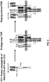
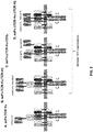


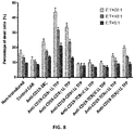

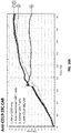


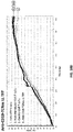

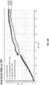


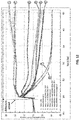
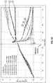
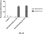
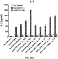
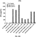
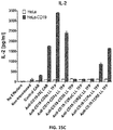
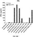
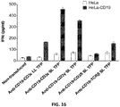
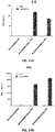

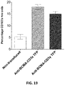
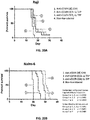
Classifications
-
- C—CHEMISTRY; METALLURGY
- C07—ORGANIC CHEMISTRY
- C07K—PEPTIDES
- C07K14/00—Peptides having more than 20 amino acids; Gastrins; Somatostatins; Melanotropins; Derivatives thereof
- C07K14/435—Peptides having more than 20 amino acids; Gastrins; Somatostatins; Melanotropins; Derivatives thereof from animals; from humans
- C07K14/705—Receptors; Cell surface antigens; Cell surface determinants
- C07K14/70503—Immunoglobulin superfamily
- C07K14/7051—T-cell receptor (TcR)-CD3 complex
-
- A—HUMAN NECESSITIES
- A61—MEDICAL OR VETERINARY SCIENCE; HYGIENE
- A61P—SPECIFIC THERAPEUTIC ACTIVITY OF CHEMICAL COMPOUNDS OR MEDICINAL PREPARATIONS
- A61P35/00—Antineoplastic agents
-
- A—HUMAN NECESSITIES
- A61—MEDICAL OR VETERINARY SCIENCE; HYGIENE
- A61K—PREPARATIONS FOR MEDICAL, DENTAL OR TOILETRY PURPOSES
- A61K39/00—Medicinal preparations containing antigens or antibodies
- A61K39/0005—Vertebrate antigens
- A61K39/0011—Cancer antigens
-
- A—HUMAN NECESSITIES
- A61—MEDICAL OR VETERINARY SCIENCE; HYGIENE
- A61K—PREPARATIONS FOR MEDICAL, DENTAL OR TOILETRY PURPOSES
- A61K39/00—Medicinal preparations containing antigens or antibodies
- A61K39/46—Cellular immunotherapy
- A61K39/461—Cellular immunotherapy characterised by the cell type used
- A61K39/4611—T-cells, e.g. tumor infiltrating lymphocytes [TIL], lymphokine-activated killer cells [LAK] or regulatory T cells [Treg]
-
- A—HUMAN NECESSITIES
- A61—MEDICAL OR VETERINARY SCIENCE; HYGIENE
- A61K—PREPARATIONS FOR MEDICAL, DENTAL OR TOILETRY PURPOSES
- A61K39/00—Medicinal preparations containing antigens or antibodies
- A61K39/46—Cellular immunotherapy
- A61K39/463—Cellular immunotherapy characterised by recombinant expression
- A61K39/4631—Chimeric Antigen Receptors [CAR]
-
- A—HUMAN NECESSITIES
- A61—MEDICAL OR VETERINARY SCIENCE; HYGIENE
- A61K—PREPARATIONS FOR MEDICAL, DENTAL OR TOILETRY PURPOSES
- A61K39/00—Medicinal preparations containing antigens or antibodies
- A61K39/46—Cellular immunotherapy
- A61K39/463—Cellular immunotherapy characterised by recombinant expression
- A61K39/4632—T-cell receptors [TCR]; antibody T-cell receptor constructs
-
- A—HUMAN NECESSITIES
- A61—MEDICAL OR VETERINARY SCIENCE; HYGIENE
- A61K—PREPARATIONS FOR MEDICAL, DENTAL OR TOILETRY PURPOSES
- A61K39/00—Medicinal preparations containing antigens or antibodies
- A61K39/46—Cellular immunotherapy
- A61K39/464—Cellular immunotherapy characterised by the antigen targeted or presented
- A61K39/4643—Vertebrate antigens
- A61K39/4644—Cancer antigens
- A61K39/464402—Receptors, cell surface antigens or cell surface determinants
- A61K39/464411—Immunoglobulin superfamily
- A61K39/464412—CD19 or B4
-
- A—HUMAN NECESSITIES
- A61—MEDICAL OR VETERINARY SCIENCE; HYGIENE
- A61K—PREPARATIONS FOR MEDICAL, DENTAL OR TOILETRY PURPOSES
- A61K39/00—Medicinal preparations containing antigens or antibodies
- A61K39/46—Cellular immunotherapy
- A61K39/464—Cellular immunotherapy characterised by the antigen targeted or presented
- A61K39/4643—Vertebrate antigens
- A61K39/4644—Cancer antigens
- A61K39/464402—Receptors, cell surface antigens or cell surface determinants
- A61K39/464416—Receptors for cytokines
- A61K39/464417—Receptors for tumor necrosis factors [TNF], e.g. lymphotoxin receptor [LTR], CD30
-
- A—HUMAN NECESSITIES
- A61—MEDICAL OR VETERINARY SCIENCE; HYGIENE
- A61P—SPECIFIC THERAPEUTIC ACTIVITY OF CHEMICAL COMPOUNDS OR MEDICINAL PREPARATIONS
- A61P35/00—Antineoplastic agents
- A61P35/02—Antineoplastic agents specific for leukemia
-
- A—HUMAN NECESSITIES
- A61—MEDICAL OR VETERINARY SCIENCE; HYGIENE
- A61P—SPECIFIC THERAPEUTIC ACTIVITY OF CHEMICAL COMPOUNDS OR MEDICINAL PREPARATIONS
- A61P43/00—Drugs for specific purposes, not provided for in groups A61P1/00-A61P41/00
-
- C—CHEMISTRY; METALLURGY
- C07—ORGANIC CHEMISTRY
- C07K—PEPTIDES
- C07K14/00—Peptides having more than 20 amino acids; Gastrins; Somatostatins; Melanotropins; Derivatives thereof
- C07K14/435—Peptides having more than 20 amino acids; Gastrins; Somatostatins; Melanotropins; Derivatives thereof from animals; from humans
- C07K14/705—Receptors; Cell surface antigens; Cell surface determinants
- C07K14/70578—NGF-receptor/TNF-receptor superfamily, e.g. CD27, CD30, CD40, CD95
-
- C—CHEMISTRY; METALLURGY
- C07—ORGANIC CHEMISTRY
- C07K—PEPTIDES
- C07K16/00—Immunoglobulins [IGs], e.g. monoclonal or polyclonal antibodies
- C07K16/18—Immunoglobulins [IGs], e.g. monoclonal or polyclonal antibodies against material from animals or humans
- C07K16/28—Immunoglobulins [IGs], e.g. monoclonal or polyclonal antibodies against material from animals or humans against receptors, cell surface antigens or cell surface determinants
- C07K16/2803—Immunoglobulins [IGs], e.g. monoclonal or polyclonal antibodies against material from animals or humans against receptors, cell surface antigens or cell surface determinants against the immunoglobulin superfamily
-
- C—CHEMISTRY; METALLURGY
- C07—ORGANIC CHEMISTRY
- C07K—PEPTIDES
- C07K16/00—Immunoglobulins [IGs], e.g. monoclonal or polyclonal antibodies
- C07K16/18—Immunoglobulins [IGs], e.g. monoclonal or polyclonal antibodies against material from animals or humans
- C07K16/28—Immunoglobulins [IGs], e.g. monoclonal or polyclonal antibodies against material from animals or humans against receptors, cell surface antigens or cell surface determinants
- C07K16/2878—Immunoglobulins [IGs], e.g. monoclonal or polyclonal antibodies against material from animals or humans against receptors, cell surface antigens or cell surface determinants against the NGF-receptor/TNF-receptor superfamily, e.g. CD27, CD30, CD40, CD95
-
- C—CHEMISTRY; METALLURGY
- C07—ORGANIC CHEMISTRY
- C07K—PEPTIDES
- C07K16/00—Immunoglobulins [IGs], e.g. monoclonal or polyclonal antibodies
- C07K16/18—Immunoglobulins [IGs], e.g. monoclonal or polyclonal antibodies against material from animals or humans
- C07K16/28—Immunoglobulins [IGs], e.g. monoclonal or polyclonal antibodies against material from animals or humans against receptors, cell surface antigens or cell surface determinants
- C07K16/30—Immunoglobulins [IGs], e.g. monoclonal or polyclonal antibodies against material from animals or humans against receptors, cell surface antigens or cell surface determinants from tumour cells
-
- C—CHEMISTRY; METALLURGY
- C07—ORGANIC CHEMISTRY
- C07K—PEPTIDES
- C07K16/00—Immunoglobulins [IGs], e.g. monoclonal or polyclonal antibodies
- C07K16/40—Immunoglobulins [IGs], e.g. monoclonal or polyclonal antibodies against enzymes
-
- C—CHEMISTRY; METALLURGY
- C07—ORGANIC CHEMISTRY
- C07K—PEPTIDES
- C07K19/00—Hybrid peptides, i.e. peptides covalently bound to nucleic acids, or non-covalently bound protein-protein complexes
-
- C—CHEMISTRY; METALLURGY
- C12—BIOCHEMISTRY; BEER; SPIRITS; WINE; VINEGAR; MICROBIOLOGY; ENZYMOLOGY; MUTATION OR GENETIC ENGINEERING
- C12N—MICROORGANISMS OR ENZYMES; COMPOSITIONS THEREOF; PROPAGATING, PRESERVING, OR MAINTAINING MICROORGANISMS; MUTATION OR GENETIC ENGINEERING; CULTURE MEDIA
- C12N5/00—Undifferentiated human, animal or plant cells, e.g. cell lines; Tissues; Cultivation or maintenance thereof; Culture media therefor
- C12N5/06—Animal cells or tissues; Human cells or tissues
- C12N5/0602—Vertebrate cells
- C12N5/0634—Cells from the blood or the immune system
- C12N5/0636—T lymphocytes
-
- A—HUMAN NECESSITIES
- A61—MEDICAL OR VETERINARY SCIENCE; HYGIENE
- A61K—PREPARATIONS FOR MEDICAL, DENTAL OR TOILETRY PURPOSES
- A61K39/00—Medicinal preparations containing antigens or antibodies
- A61K2039/505—Medicinal preparations containing antigens or antibodies comprising antibodies
-
- A—HUMAN NECESSITIES
- A61—MEDICAL OR VETERINARY SCIENCE; HYGIENE
- A61K—PREPARATIONS FOR MEDICAL, DENTAL OR TOILETRY PURPOSES
- A61K39/00—Medicinal preparations containing antigens or antibodies
- A61K2039/51—Medicinal preparations containing antigens or antibodies comprising whole cells, viruses or DNA/RNA
- A61K2039/515—Animal cells
- A61K2039/5158—Antigen-pulsed cells, e.g. T-cells
-
- A—HUMAN NECESSITIES
- A61—MEDICAL OR VETERINARY SCIENCE; HYGIENE
- A61K—PREPARATIONS FOR MEDICAL, DENTAL OR TOILETRY PURPOSES
- A61K2239/00—Indexing codes associated with cellular immunotherapy of group A61K39/46
- A61K2239/31—Indexing codes associated with cellular immunotherapy of group A61K39/46 characterized by the route of administration
-
- A—HUMAN NECESSITIES
- A61—MEDICAL OR VETERINARY SCIENCE; HYGIENE
- A61K—PREPARATIONS FOR MEDICAL, DENTAL OR TOILETRY PURPOSES
- A61K2239/00—Indexing codes associated with cellular immunotherapy of group A61K39/46
- A61K2239/38—Indexing codes associated with cellular immunotherapy of group A61K39/46 characterised by the dose, timing or administration schedule
-
- A—HUMAN NECESSITIES
- A61—MEDICAL OR VETERINARY SCIENCE; HYGIENE
- A61K—PREPARATIONS FOR MEDICAL, DENTAL OR TOILETRY PURPOSES
- A61K2239/00—Indexing codes associated with cellular immunotherapy of group A61K39/46
- A61K2239/46—Indexing codes associated with cellular immunotherapy of group A61K39/46 characterised by the cancer treated
- A61K2239/48—Blood cells, e.g. leukemia or lymphoma
-
- C—CHEMISTRY; METALLURGY
- C07—ORGANIC CHEMISTRY
- C07K—PEPTIDES
- C07K2317/00—Immunoglobulins specific features
- C07K2317/20—Immunoglobulins specific features characterized by taxonomic origin
- C07K2317/24—Immunoglobulins specific features characterized by taxonomic origin containing regions, domains or residues from different species, e.g. chimeric, humanized or veneered
-
- C—CHEMISTRY; METALLURGY
- C07—ORGANIC CHEMISTRY
- C07K—PEPTIDES
- C07K2317/00—Immunoglobulins specific features
- C07K2317/50—Immunoglobulins specific features characterized by immunoglobulin fragments
- C07K2317/56—Immunoglobulins specific features characterized by immunoglobulin fragments variable (Fv) region, i.e. VH and/or VL
-
- C—CHEMISTRY; METALLURGY
- C07—ORGANIC CHEMISTRY
- C07K—PEPTIDES
- C07K2317/00—Immunoglobulins specific features
- C07K2317/60—Immunoglobulins specific features characterized by non-natural combinations of immunoglobulin fragments
- C07K2317/62—Immunoglobulins specific features characterized by non-natural combinations of immunoglobulin fragments comprising only variable region components
- C07K2317/622—Single chain antibody (scFv)
-
- C—CHEMISTRY; METALLURGY
- C07—ORGANIC CHEMISTRY
- C07K—PEPTIDES
- C07K2317/00—Immunoglobulins specific features
- C07K2317/70—Immunoglobulins specific features characterized by effect upon binding to a cell or to an antigen
- C07K2317/73—Inducing cell death, e.g. apoptosis, necrosis or inhibition of cell proliferation
-
- C—CHEMISTRY; METALLURGY
- C07—ORGANIC CHEMISTRY
- C07K—PEPTIDES
- C07K2319/00—Fusion polypeptide
-
- C—CHEMISTRY; METALLURGY
- C07—ORGANIC CHEMISTRY
- C07K—PEPTIDES
- C07K2319/00—Fusion polypeptide
- C07K2319/01—Fusion polypeptide containing a localisation/targetting motif
- C07K2319/02—Fusion polypeptide containing a localisation/targetting motif containing a signal sequence
-
- C—CHEMISTRY; METALLURGY
- C07—ORGANIC CHEMISTRY
- C07K—PEPTIDES
- C07K2319/00—Fusion polypeptide
- C07K2319/01—Fusion polypeptide containing a localisation/targetting motif
- C07K2319/03—Fusion polypeptide containing a localisation/targetting motif containing a transmembrane segment
-
- C—CHEMISTRY; METALLURGY
- C07—ORGANIC CHEMISTRY
- C07K—PEPTIDES
- C07K2319/00—Fusion polypeptide
- C07K2319/01—Fusion polypeptide containing a localisation/targetting motif
- C07K2319/10—Fusion polypeptide containing a localisation/targetting motif containing a tag for extracellular membrane crossing, e.g. TAT or VP22
-
- C—CHEMISTRY; METALLURGY
- C07—ORGANIC CHEMISTRY
- C07K—PEPTIDES
- C07K2319/00—Fusion polypeptide
- C07K2319/33—Fusion polypeptide fusions for targeting to specific cell types, e.g. tissue specific targeting, targeting of a bacterial subspecies
-
- C—CHEMISTRY; METALLURGY
- C12—BIOCHEMISTRY; BEER; SPIRITS; WINE; VINEGAR; MICROBIOLOGY; ENZYMOLOGY; MUTATION OR GENETIC ENGINEERING
- C12N—MICROORGANISMS OR ENZYMES; COMPOSITIONS THEREOF; PROPAGATING, PRESERVING, OR MAINTAINING MICROORGANISMS; MUTATION OR GENETIC ENGINEERING; CULTURE MEDIA
- C12N2510/00—Genetically modified cells
Definitions
- cancer immunotherapy Most patients with hematological malignancies or with late-stage solid tumors are incurable with standard therapy. In addition, traditional treatment options often have serious side effects. Numerous attempts have been made to engage a patient's immune system for rejecting cancerous cells, an approach collectively referred to as cancer immunotherapy. However, several obstacles make it rather difficult to achieve clinical effectiveness. Although hundreds of so-called tumor antigens have been identified, these are often derived from self and thus can direct the cancer immunotherapy against healthy tissue, or are poorly immunogenic. Furthermore, cancer cells use multiple mechanisms to render themselves invisible or hostile to the initiation and propagation of an immune attack by cancer immunotherapies.
- CAR chimeric antigen receptor
- CTL019 The clinical results with CD19-specific CAR T-cells (called CTL019) have shown complete remissions in patients suffering from chronic lymphocytic leukemia (CLL) as well as in childhood acute lymphoblastic leukemia (ALL) (see, e.g., Kalos et al., Sci Transl Med 3:95ra73 (2011 ), Porter et al., NEJM 365:725-733 (2011 ), Grupp et al., NEJM 368:1509-1518 (2013 )).
- An alternative approach is the use of T-cell receptor (TCR) alpha and beta chains selected for a tumor-associated peptide antigen for genetically engineering autologous T-cells.
- TCR T-cell receptor
- TCR chains will form complete TCR complexes and provide the T-cells with a TCR for a second defined specificity. Encouraging results were obtained with engineered autologous T-cells expressing NY-ESO-1-specific TCR alpha and beta chains in patients with synovial carcinoma.
- Described herein are novel fusion proteins of TCR subunits, including CD3 epsilon, CD3gamma and CD3 delta, and of TCR alpha and TCR beta chains with binding domains specific for cell surface antigens that have the potential to overcome limitations of existing approaches.
- novel fusion proteins that more efficiently kill target cells than CARs, but release comparable or lower levels of pro-inflammatory cytokines. These fusion proteins and methods of their use represent an advantage for TFPs relative to CARs because elevated levels of these cytokines have been associated with dose-limiting toxicities for adoptive CAR-T therapies.
- TCR T-cell receptor
- TFPs T-cell receptor fusion proteins
- an isolated recombinant nucleic acid molecule encoding a T-cell receptor (TCR) fusion protein (TFP) comprising a TCR subunit comprising at least a portion of a TCR extracellular domain, and a TCR intracellular domain comprising a stimulatory domain from an intracellular signaling domain of CD3 epsilon; and a human or humanized antibody domain comprising an antigen binding domain wherein the TCR subunit and the antibody domain are operatively linked, and wherein the TFP incorporates into a TCR when expressed in a T-cell.
- TCR T-cell receptor
- TFP T-cell receptor fusion protein
- an isolated recombinant nucleic acid molecule encoding a T-cell receptor (TCR) fusion protein (TFP) comprising a TCR subunit comprising at least a portion of a TCR extracellular domain, and a TCR intracellular domain comprising a stimulatory domain from an intracellular signaling domain of CD3 gamma; and a human or humanized antibody domain comprising an antigen binding domain wherein the TCR subunit and the antibody domain are operatively linked, and wherein the TFP incorporates into a TCR when expressed in a T-cell.
- TCR T-cell receptor
- TFP T-cell receptor fusion protein
- an isolated recombinant nucleic acid molecule encoding a T-cell receptor (TCR) fusion protein (TFP) comprising a TCR subunit comprising at least a portion of a TCR extracellular domain, and a TCR intracellular domain comprising a stimulatory domain from an intracellular signaling domain of CD3 delta; and a human or humanized antibody domain comprising an antigen binding domainwherein the TCR subunit and the antibody domain are operatively linked, and wherein the TFP incorporates into a TCR when expressed in a T-cell.
- TCR T-cell receptor
- TFP T-cell receptor fusion protein
- an isolated recombinant nucleic acid molecule encoding a T-cell receptor (TCR) fusion protein (TFP) comprising a TCR subunit comprising at least a portion of a TCR extracellular domain, and a TCR intracellular domain comprising a stimulatory domain from an intracellular signaling domain of TCR alpha; and a human or humanized antibody domain comprising an antigen binding domain wherein the TCR subunit and the antibody domain are operatively linked, and wherein the TFP incorporates into a TCR when expressed in a T-cell.
- TCR T-cell receptor
- TFP T-cell receptor fusion protein
- an isolated recombinant nucleic acid molecule encoding a T-cell receptor (TCR) fusion protein (TFP) comprising a TCR subunit comprising at least a portion of a TCR extracellular domain, and a TCR intracellular domain comprising a stimulatory domain from an intracellular signaling domain of TCR beta; and a human or humanized antibody domain comprising an antigen binding domain wherein the TCR subunit and the antibody domain are operatively linked, and wherein the TFP incorporates into a TCR when expressed in a T-cell.
- TCR T-cell receptor
- TFP T-cell receptor fusion protein
- TCR T-cell receptor
- TFP T-cell receptor fusion protein
- TCR T-cell receptor
- TFP T-cell receptor fusion protein
- BCMA anti-B-cell maturation antigen
- the TCR subunit and the antibody domain are operatively linked.
- the TFP incorporates into a TCR when expressed in a T-cell.
- the encoded antigen binding domain is connected to the TCR extracellular domain by a linker sequence.
- the TCR subunit comprises a TCR extracellular domain.
- the TCR subunit comprises a TCR transmembrane domain.
- the TCR subunit comprises a TCR intracellular domain.
- the TCR subunit comprises (i) a TCR extracellular domain, (ii) a TCR transmembrane domain, and (iii) a TCR intracellular domain, wherein at least two of (i), (ii), and (iii) are from the same TCR subunit.
- the TCR subunit comprises a TCR intracellular domain comprising a stimulatory domain selected from an intracellular signaling domain of CD3 epsilon, CD3 gamma or CD3 delta, or an amino acid sequence having at least one, two or three modifications thereto.
- the TCR subunit comprises an intracellular domain comprising a stimulatory domain selected from a functional signaling domain of 4-1BB and/or a functional signaling domain of CD3 zeta, or an amino acid sequence having at least one modification thereto.
- the human or humanized antibody domain comprises an antibody fragment.
- the human or humanized antibody domain comprises a scFv or a V H domain.
- the isolated nucleic acid molecule encodes (i) a light chain (LC) CDR1, LC CDR2 and LC CDR3 of an anti-CD19 light chain binding domain amino acid sequence with 70-100% sequence identity to SEQ ID NO: 25, SEQ ID NO: 27 and SEQ ID NO: 29, repectively, and/or (ii) a heavy chain (HC) CDR1, HC CDR2 and HC CDR3 of an anti-CD 19 heavy chain binding domain amino acid sequence with 70-100% sequence identity to SEQ ID NO: 31, SEQ ID NO: 33 and SEQ ID NO: 35, respectively.
- LC light chain
- the isolated nucleic acid molecule encodes a light chain variable region, wherein the light chain variable region comprises an amino acid sequence having at least one but not more than 30 modifications of a light chain variable region amino acid sequence of SEQ ID NO: 49, or a sequence with 95-99% identity to a light chain variable region amino acid sequence of SEQ ID NO: 49.
- the isolated nucleic acid molecule encodes a heavy chain variable region, wherein the heavy chain variable region comprises an amino acid sequence having at least one but not more than 30 modifications of a heavy chain variable region amino acid sequence of SEQ ID NO: 51, or a sequence with 95-99% identity to a heavy chain variable region amino acid sequence of SEQ ID NO: 51.
- the isolated nucleic acid molecule encodes (i) a light chain (LC) CDR1, LC CDR2 and LC CDR3 of an anti-BCMA light chain binding domain amino acid sequence with 70-100% sequence identity to SEQ ID NO: 37, SEQ ID NO: 39and SEQ ID NO: 41, repectively, and/or (ii) a heavy chain (HC) CDR1, HC CDR2 and HC CDR3 of an anti-BCMA heavy chain binding domain amino acid sequence with 70-100% sequence identity to SEQ ID NO: 43, SEQ ID NO: 45 and SEQ ID NO: 47, respectively.
- LC light chain
- the isolated nucleic acid molecule encodes a light chain variable region, wherein the light chain variable region comprises an amino acid sequence having at least one but not more than 30 modifications of a light chain variable region amino acid sequence of SEQ ID NO: 53, or a sequence with 95-99% identity to a light chain variable region amino acid sequence of SEQ ID NO: 53.
- the isolated nucleic acid molecule encodes a heavy chain variable region, wherein the heavy chain variable region comprises an amino acid sequence having at least one but not more than 30 modifications of a heavy chain variable region amino acid sequence of SEQ ID NO: 55, or a sequence with 95-99% identity to a heavy chain variable region amino acid sequence of SEQ ID NO: 55.
- the TFP includes an extracellular domain of a TCR subunit that comprises an extracellular domain or portion thereof of a protein selected from the group consisting of a TCR alpha chain, a TCR beta chain, a CD3 epsilon TCR subunit, a CD3 gamma TCR subunit, a CD3 delta TCR subunit, functional fragments thereof, and amino acid sequences thereof having at least one but not more than 20 modifications.
- the encoded TFP includes a transmembrane domain that comprises a transmembrane domain of a protein selected from the group consisting of a TCR alpha chain, a TCR beta chain, a CD3 epsilon TCR subunit, a CD3 gamma TCR subunit, a CD3 delta TCR subunit, functional fragments thereof, and amino acid sequences thereof having at least one but not more than 20 modifications.
- the encoded TFP includes a transmembrane domain that comprises a transmembrane domain of a protein selected from the group consisting of a TCR alpha chain, a TCR beta chain, a TCR zeta chain, a CD3 epsilon TCR subunit, a CD3 gamma TCR subunit, a CD3 delta TCR subunit, CD45, CD4, CD5, CD8, CD9, CD16, CD22, CD33, CD28, CD37, CD64, CD80, CD86, CD134, CD137, CD154, functional fragments thereof, and amino acid sequences thereof having at least one but not more than 20 modifications.
- a transmembrane domain that comprises a transmembrane domain of a protein selected from the group consisting of a TCR alpha chain, a TCR beta chain, a TCR zeta chain, a CD3 epsilon TCR subunit, a CD3 gamma TCR subunit, a CD3 delta
- the isolated nucleic acid molecule further comprises a sequence encoding a costimulatory domain.
- the costimulatory domain is a functional signaling domain obtained from a protein selected from the group consisting of OX40, CD2, CD27, CD28, CDS, ICAM-1, LFA-1 (CD11a/CD18), ICOS (CD278), and 4-1BB (CD137), and amino acid sequences thereof having at least one but not more than 20 modifications thereto.
- the isolated nucleic acid molecule further comprises a leader sequence.
- the isolated nucleic acid molecule is mRNA.
- the TFP includes an immunoreceptor tyrosine-based activation motif (ITAM) of a TCR subunit that comprises an ITAM or portion thereof of a protein selected from the group consisting of CD3 zeta TCR subunit, CD3 epsilon TCR subunit, CD3 gamma TCR subunit, CD3 delta TCR subunit, TCR zeta chain, Fc epsilon receptor 1 chain, Fc epsilon receptor 2 chain, Fc gamma receptor 1 chain, Fc gamma receptor 2a chain, Fc gamma receptor 2b1 chain, Fc gamma receptor 2b2 chain, Fc gamma receptor 3a chain, Fc gamma receptor 3b chain, Fc beta receptor 1 chain, TYROBP (DAP12), CD5, CD16a, CD16b, CD22, CD23, CD32, CD64, CD79a, CD79b, CD89, CD278, CD66d, functional fragments thereof
- the ITAM replaces an ITAM of CD3 gamma, CD3 delta, or CD3 epsilon.
- the ITAM is selected from the group consisting of CD3 zeta TCR subunit, CD3 epsilon TCR subunit, CD3 gamma TCR subunit, and CD3 delta TCR subunit and replaces a differenct ITAM selected from the group consisting of CD3 zeta TCR subunit, CD3 epsilon TCR subunit, CD3 gamma TCR subunit, and CD3 delta TCR subunit.
- the nucleic acid comprises a nucleotide analog.
- the nucleotide analog is selected from the group consisting of 2'-O-methyl, 2'-O-methoxyethyl (2'-O-MOE), 2'-O-aminopropyl, 2'-deoxy, T-deoxy-2'-fluoro, 2'-O-aminopropyl (2'-O-AP), 2'-O-dimethylaminoethyl (2'-O-DMAOE), 2'-O-dimethylaminopropyl (2'-O-DMAP), T-O-dimethylaminoethyloxyethyl (2'-O-DMAEOE), 2'-O-N-methylacetamido (2'-O-NMA) modified, a locked nucleic acid (LNA), an ethylene nucleic acid (ENA), a peptide nucleic acid (PNA), a 1',5'- anhydro
- LNA locked
- provided herein is an isolated polypeptide molecule encoded by a nucleic acid molecule provided herein.
- an isolated TFP molecule comprising a human or humanized anti-CD 19 binding domain, a TCR extracellular domain, a transmembrane domain, and an intracellular domain.
- an isolated TFP molecule comprising a human or humanized anti-CD 19 binding domain, a TCR extracellular domain, a transmembrane domain, and an intracellular signaling domain, wherein the TFP molecule is capable of functionally interacting with an endogenous TCR complex and/or at least one endogenous TCR polypeptide.
- an isolated TFP molecule comprising a human or humanized anti-CD 19 binding domain, a TCR extracellular domain, a transmembrane domain, and an intracellular signaling domain, wherein the TFP molecule is capable of functionally integrating into an endogenous TCR complex
- the isolated TFP molecule comprises an antibody or antibody fragment comprising a human or humanized anti-CD 19 binding domain, a TCR extracellular domain, a transmembrane domain, and an intracellular domain.
- the anti-CD19 binding domain is a scFv or a V H domain.
- the anti-CD 19 binding domain comprises a heavy chain with 95-100% identity to an amino acid sequence of SEQ ID NO: 51, a functional fragment thereof, or an amino acid sequence thereof having at least one but not more than 30 modifications.
- the anti-CD19 binding domain comprises a light chain with 95-100% identity to an amino acid sequence of SEQ ID NO: 49, a functional fragment thereof, or an amino acid sequence thereof having at least one but not more than 30 modifications.
- the isolated TFP molecule comprises a TCR extracellular domain that comprises an extracellular domain or portion thereof of a protein selected from the group consisting of a TCR alpha chain, a TCR beta chain, a CD3 epsilon TCR subunit, a CD3 gamma TCR subunit, a CD3 delta TCR subunit, functional fragments thereof, and amino acid sequences thereof having at least one but not more than 20 modifications.
- the anti-CD 19 binding domain is connected to the TCR extracellular domain by a linker sequence.
- an isolated TFP molecule comprising a human or humanized anti-BCMA binding domain, a TCR extracellular domain, a transmembrane domain, and an intracellular domain.
- an isolated TFP molecule comprising a human or humanized anti-BCMA binding domain, a TCR extracellular domain, a transmembrane domain, and an intracellular signaling domain, wherein the TFP molecule is capable of functionally interacting with an endogenous TCR complex and/or at least one endogenous TCR polypeptide.
- an isolated TFP molecule comprising a human or humanized anti-BCMA binding domain, a TCR extracellular domain, a transmembrane domain, and an intracellular signaling domain, wherein the TFP molecule is capable of functionally integrating into an endogenous TCR complex.
- the isolated TFP molecule comprises an antibody or antibody fragment comprising a human or humanized anti- BCMA binding domain, a TCR extracellular domain, a transmembrane domain, and an intracellular domain.
- the anti-BCMA binding domain is a scFv or a V H domain.
- the anti-BCMA binding domain comprises a heavy chain with 95-100% identity to an amino acid sequence of SEQ ID NO: 55, a functional fragment thereof, or an amino acid sequence thereof having at least one but not more than 30 modifications.
- the anti-BCMA binding domain comprises a light chain with 95-100% identity to an amino acid sequence of SEQ ID NO: 53, a functional fragment thereof, or an amino acid sequence thereof having at least one but not more than 30 modifications.
- the isolated TFP molecule comprises a TCR extracellular domain that comprises an extracellular domain or portion thereof of a protein selected from the group consisting of a TCR alpha chain, a TCR beta chain, a CD3 epsilon TCR subunit, a CD3 gamma TCR subunit, a CD3 delta TCR subunit, functional fragments thereof, and amino acid sequences thereof having at least one but not more than 20 modifications.
- the anti-BCMA binding domain is connected to the TCR extracellular domain by a linker sequence.
- the isolated TFP molecule further comprises a sequence encoding a costimulatory domain.
- the isolated TFP molecule further comprises a sequence encoding an intracellular signaling domain.
- the isolated TFP molecule further comprises a leader sequence.
- a vector comprising a nucleic acid molecule encoding a TFP provided herein.
- the vector is selected from the group consisting of a DNA, a RNA, a plasmid, a lentivirus vector, adenoviral vector, a Rous sarcoma viral (RSV) vector, or a retrovirus vector.
- the vector further comprises a promoter.
- the vector is an in vitro transcribed vector.
- a nucleic acid sequence in the vector further comprises a poly(A) tail.
- a nucleic acid sequence in the vector further comprises a 3'UTR.
- a cell comprising a vector provided herein.
- the cell is a human T-cell.
- the T-cell is a CD8+ or CD4+ T-cell.
- the cell further comprises a nucleic acid encoding an inhibitory molecule that comprises a first polypeptide that comprises at least a portion of an inhibitory molecule, associated with a second polypeptide that comprises a positive signal from an intracellular signaling domain.
- the inhibitory molecule comprise first polypeptide that comprises at least a portion of PD1 and a second polypeptide comprising a costimulatory domain and primary signaling domain.
- a human CD8+ or CD4+ T-cell comprising at least two TFP molecules, the TFP molecules comprising a human or humanized anti-CD 19 binding domain, a TCR extracellular domain, a transmembrane domain, and an intracellular domain, wherein the TFP molecule is capable of functionally interacting with an endogenous TCR complex and/or at least one endogenous TCR polypeptide in, at and/or on the surface of the human CD8+ or CD4+ T-cell.
- a protein complex comprising: a TFP molecule comprising a human or humanized anti-CD 19 binding domain, a TCR extracellular domain, a transmembrane domain, and an intracellular domain; and at least one endogenous TCR complex.
- the TCR comprises an extracellular domain or portion thereof of a protein selected from the group consisting of TCR alpha chain, a TCR beta chain, a CD3 epsilon TCR subunit, a CD3 gamma TCR subunit, and a CD3 delta TCR subunit.
- the anti-CD 19 binding domain is connected to the TCR extracellular domain by a linker sequence.
- a protein complex comprising: a TFP molecule comprising a human or humanized anti-BCMA binding domain, a TCR extracellular domain, a transmembrane domain, and an intracellular domain; and at least one endogenous TCR complex.
- the TCR comprises an extracellular domain or portion thereof of a protein selected from the group consisting of TCR alpha chain, a TCR beta chain, a CD3 epsilon TCR subunit, a CD3 gamma TCR subunit, and a CD3 delta TCR subunit.
- the anti-BCMA binding domain is connected to the TCR extracellular domain by a linker sequence.
- a human CD8+ or CD4+ T-cell comprising at least two different TFP proteins per a protein complex provided herein.
- provided herein is a method of making a cell comprising transducing a T-cell with a vector provided herein.
- RNA-engineered cells comprising introducing an in vitro transcribed RNA or synthetic RNA into a cell, where the RNA comprises a nucleic acid encoding a TFP molecule provided herein.
- provided herein is a method of providing an anti-tumor immunity in a mammal comprising administering to the mammal an effective amount of a cell expressing a TFP molecule provided herein, or expressing a polypeptide molecule provided herein.
- the cell is an autologous T-cell. In some instances, the cell is an allogeneic T-cell. In some instances, the mammal is a human.
- provided herein is a method of treating a mammal having a disease associated with expression of CD19 or BCMA comprising administering to the mammal an effective amount of a TFP molecule provided herein, a cell provided herein, or a polypeptide molecule provided herein.
- the disease associated with CD 19 or BCMA expression is selected from the group consisting of a proliferative disease, a cancer, a malignancy, myelodysplasia, a myelodysplastic syndrome, a preleukemia, a non-cancer related indication associated with expression of CD19.
- the disease is a hematologic cancer selected from the group consisting of B-cell acute lymphoid leukemia (B-ALL), T-cell acute lymphoid leukemia (T-ALL), acute lymphoblastic leukemia (ALL); chronic myelogenous leukemia (CML), chronic lymphocytic leukemia (CLL), B cell prolymphocytic leukemia, blastic plasmacytoid dendritic cell neoplasm, Burkitt's lymphoma, diffuse large B cell lymphoma, follicular lymphoma, hairy cell leukemia, small cell-follicular lymphoma, large cell-follicular lymphoma, malignant lymphoproliferative conditions, MALT lymphoma, mantle cell lymphoma, Marginal zone lymphoma, multiple myeloma, myelodysplasia, myelodysplastic syndrome, non-Hodgkin's lymphoma, plasmablastic lymphoma
- the cells expressing a TFP molecule are administered in combination with an agent that increases the efficacy of a cell expressing a TFP molecule. In some instances, less cytokines are released in the mammal compared a mammal administered an effective amount of a T-cell expressing an anti-CD 19 chimeric antigen receptor (CAR) or an anti-BCMA CAR. In some instances, the cells expressing a TFP molecule are administered in combination with an agent that ameliorates one or more side effects associated with administration of a cell expressing a TFP molecule. In some instances, the cells expressing a TFP molecule are administered in combination with an agent that treats the disease associated with CD19 or BCMA.
- an isolated nucleic acid molecule provided herein, an isolated polypeptide molecule provided herein, an isolated TFP provided herein, a complex provided herein, a vector provided herein, or a cell provided herein is for use as a medicament.
- provided herein is a method of treating a mammal having a disease associated with expression of CD19 or BCMA comprising administering to the mammal an effective amount of a TFP molecule provided herein, a cell provided herein, or a polypeptide molecule provided herein, wherein less cytokines are released in the mammal compared a mammal administered an effective amount of a T-cell expressing an anti-CD19 chimeric antigen receptor (CAR) or an anti-BCMA CAR.
- CAR anti-CD19 chimeric antigen receptor
- TCR subunit comprises a TCR extracellular domain.
- TCR subunit comprises a TCR transmembrane domain.
- TCR subunit comprises a TCR intracellular domain.
- the TCR subunit comprises (i) a TCR extracellular domain, (ii) a TCR transmembrane domain, and (iii) a TCR intracellular domain, wherein at least two of (i), (ii), and (iii) are from the same TCR subunit.
- the TCR subunit comprises a TCR intracellular domain comprising a stimulatory domain selected from an intracellular signaling domain of CD3 epsilon, CD3 gamma or CD3 delta, or an amino acid sequence having at least one, two or three modifications thereto.
- the TCR subunit comprises an intracellular domain comprising a stimulatory domain selected from a functional signaling domain of 4-1BB and/or a functional signaling domain of CD3 zeta, or an amino acid sequence having at least one, two or three modifications thereto.
- the human or humanized antibody domain comprises an antibody fragment. In some embodiments, the human or humanized antibody domain comprises a scFv or a V H domain.
- the isolated nucleic acid molecules comprise (i) a light chain (LC) CDR1, LC CDR2 and LC CDR3 of any anti-CD 19 light chain binding domain amino acid sequence provided herein, and/or (ii) a heavy chain (HC) CDR1, HC CDR2 and HC CDR3 of any anti-CD 19 heavy chain binding domain amino acid sequence provided herein.
- LC light chain
- HC heavy chain
- the light chain variable region comprises an amino acid sequence having at least one, two or three modifications but not more than 30, 20 or 10 modifications of an amino acid sequence of a light chain variable region provided herein, or a sequence with 95-99% identity to an amino acid sequence provided herein.
- the heavy chain variable region comprises an amino acid sequence having at least one, two or three modifications but not more than 30, 20 or 10 modifications of an amino acid sequence of a heavy chain variable region provided herein, or a sequence with 95-99% identity to an amino acid sequence provided herein.
- the TFP includes an extracellular domain of a TCR subunit that comprises an extracellular domain or portion thereof of a protein selected from the group consisting of the alpha or beta chain of the T-cell receptor, CD3 delta, CD3 epsilon, or CD3 gamma, or a functional fragment thereof, or an amino acid sequence having at least one, two or three modifications but not more than 20, 10 or 5 modifications thereto.
- the encoded TFP includes a transmembrane domain that comprises a transmembrane domain of a protein selected from the group consisting of the alpha, beta chain of the TCR or TCR subunits CD3 epsilon, CD3 gamma and CD3 delta, or a functional fragment thereof, or an amino acid sequence having at least one, two or three modifications but not more than 20, 10 or 5 modifications thereto.
- the encoded TFP includes a transmembrane domain that comprises a transmembrane domain of a protein selected from the group consisting of the alpha, beta or zeta chain of the TCR or CD3 epsilon, CD3 gamma and CD3 delta CD45, CD4, CD5, CD8, CD9, CD16, CD22, CD33, CD28, CD37, CD64, CD80, CD86, CD134, CD137 and CD154, or a functional fragment thereof, or an amino acid sequence having at least one, two or three modifications but not more than 20, 10 or 5 modifications thereto.
- a protein selected from the group consisting of the alpha, beta or zeta chain of the TCR or CD3 epsilon, CD3 gamma and CD3 delta CD45, CD4, CD5, CD8, CD9, CD16, CD22, CD33, CD28, CD37, CD64, CD80, CD86, CD134, CD137 and CD154, or a functional fragment thereof, or an amino acid sequence having at
- the encoded anti-CD 19 binding domain is connected to the TCR extracellular domain by a linker sequence.
- the encoded linker sequence comprises a long linker (LL) sequence.
- the encoded linker sequence comprises a short linker (SL) sequence.
- the isolated nucleic acid molecules further comprise a sequence encoding a costimulatory domain.
- the costimulatory domain is a functional signaling domain obtained from a protein selected from the group consisting of OX40, CD2, CD27, CD28, CDS, ICAM-1, LFA-1 (CD11a/CD18), ICOS (CD278), and 4-1BB (CD137), or an amino acid sequence having at least one, two or three modifications but not more than 20, 10 or 5 modifications thereto.
- the isolated nucleic acid molecules further comprise a leader sequence.
- isolated polypeptide molecules encoded by any of the previously described nucleic acid molecules.
- isolated T-cell receptor fusion protein (TFP) molecules that comprise a human or humanized anti-CD 19 binding domain, a TCR extracellular domain, a transmembrane domain, and an intracellular domain.
- the isolated TFP molecules comprises an antibody or antibody fragment comprising a human or humanized anti-CD 19 binding domain, a TCR extracellular domain, a transmembrane domain, and an intracellular domain.
- the anti-CD 19 binding domain is a scFv or a V H domain.
- the anti-CD 19 binding domain comprises a light chain and a heavy chain of an amino acid sequence provided herein, or a functional fragment thereof, or an amino acid sequence having at least one, two or three modifications but not more than 30, 20 or 10 modifications of an amino acid sequence of a light chain variable region provided herein, or a sequence with 95-99% identity with an amino acid sequence provided herein.
- the isolated TFP molecules comprise a TCR extracellular domain that comprises an extracellular domain or portion thereof of a protein selected from the group consisting of the alpha or beta chain of the T-cell receptor, CD3 delta, CD3 epsilon, or CD3 gamma, or an amino acid sequence having at least one, two or three modifications but not more than 20, 10 or 5 modifications thereto.
- the anti-CD 19 binding domain is connected to the TCR extracellular domain by a linker sequence.
- the linker sequence comprises a long linker (LL) sequence.
- the linker sequence comprises a short linker (SL) sequence.
- the isolated TFP molecules further comprise a sequence encoding a costimulatory domain. In other embodiments, the isolated TFP molecules further comprise a sequence encoding an intracellular signaling domain. In yet other embodiments, the isolated TFP molecules further comprise a leader sequence.
- vectors that comprise a nucleic acid molecule encoding any of the previously described TFP molecules.
- the vector is selected from the group consisting of a DNA, a RNA, a plasmid, a lentivirus vector, adenoviral vector, or a retrovirus vector.
- the vector further comprises a promoter.
- the vector is an in vitro transcribed vector.
- a nucleic acid sequence in the vector further comprises a poly(A) tail.
- a nucleic acid sequence in the vector further comprises a 3'UTR.
- the cell is a human T-cell. In some embodiments, the cell is a CD8+ or CD4+ T-cell. In other embodiments, the cells further comprise a nucleic acid encoding an inhibitory molecule that comprises a first polypeptide that comprises at least a portion of an inhibitory molecule, associated with a second polypeptide that comprises a positive signal from an intracellular signaling domain. In some instances, the inhibitory molecule comprise first polypeptide that comprises at least a portion of PD1 and a second polypeptide comprising a costimulatory domain and primary signaling domain.
- TFP molecules that comprise a human or humanized anti-CD 19 binding domain, a TCR extracellular domain, a transmembrane domain, and an intracellular signaling domain, wherein the TFP molecule is capable of functionally interacting with an endogenous TCR complex and/or at least one endogenous TCR polypeptide.
- TFP molecules that comprise a human or humanized anti-CD 19 binding domain, a TCR extracellular domain, a transmembrane domain, and an intracellular signaling domain, wherein the TFP molecule is capable of functionally integrating into an endogenous TCR complex.
- human CD8+ or CD4+ T-cells that comprise at least two TFP molecules, the TFP molecules comprising a human or humanized anti-CD 19 binding domain, a TCR extracellular domain, a transmembrane domain, and an intracellular domain, wherein the TFP molecule is capable of functionally interacting with an endogenous TCR complex and/or at least one endogenous TCR polypeptide in, at and/or on the surface of the human CD8+ or CD4+ T-cell.
- protein complexes that comprise i) a TFP molecule comprising a human or humanized anti-CD 19 binding domain, a TCR extracellular domain, a transmembrane domain, and an intracellular domain; and ii) at least one endogenous TCR complex.
- the TCR comprises an extracellular domain or portion thereof of a protein selected from the group consisting of the alpha or beta chain of the T-cell receptor, CD3 delta, CD3 epsilon, or CD3 gamma.
- the anti-CD 19 binding domain is connected to the TCR extracellular domain by a linker sequence.
- the linker sequence comprises a long linker (LL) sequence.
- the linker sequence comprises a short linker (SL) sequence.
- human CD8+ or CD4+ T-cells that comprise at least two different TFP proteins per any of the described protein complexes.
- a population of human CD8+ or CD4+ T-cells wherein the T-cells of the population individually or collectively comprise at least two TFP molecules, the TFP molecules comprising a human or humanized anti-CD 19 or anti-BCMA binding domain, a TCR extracellular domain, a transmembrane domain, and an intracellular domain, wherein the TFP molecule is capable of functionally interacting with an endogenous TCR complex and/or at least one endogenous TCR polypeptide in, at and/or on the surface of the human CD8+ or CD4+ T-cell.
- a population of human CD8+ or CD4+ T-cells wherein the T-cells of the population individually or collectively comprise at least two TFP molecules encoded by an isolated nucleic acid molecule provided herein.
- provided herein are methods of making a cell that comprise transducing a T-cell with any of the described vectors.
- RNA-engineered cells comprising introducing an in vitro transcribed RNA or synthetic RNA into a cell, where the RNA comprises a nucleic acid encoding any of the described TFP molecules.
- provided herein are methods of providing an anti-tumor immunity in a mammal that comprise administering to the mammal an effective amount of a cell expressing any of the described TFP molecules.
- the cell is an autologous T-cell.
- the cell is an allogeneic T-cell.
- the mammal is a human.
- the disease associated with CD19 expression is selected from a proliferative disease such as a cancer or malignancy or a precancerous condition such as a myelodysplasia, a myelodysplastic syndrome or a preleukemia, or is a non-cancer related indication associated with expression of CD19.
- the disease is a hematologic cancer selected from the group consisting of one or more acute leukemias including but not limited to B-cell acute lymphoid leukemia ("B-ALL”), T-cell acute lymphoid leukemia ("T-ALL”), acute lymphoblastic leukemia (ALL); one or more chronic leukemias including but not limited to chronic myelogenous leukemia (CML), chronic lymphocytic leukemia (CLL); additional hematologic cancers or hematologic conditions including, but not limited to B cell prolymphocytic leukemia, blastic plasmacytoid dendritic cell neoplasm, Burkitt's lymphoma, diffuse large B cell lymphoma, follicular lymphoma, hairy cell leukemia, small cell- or a large cell-follicular lymphoma, malignant lymphoproliferative conditions, MALT lymphoma, mantle cell lymphoma, Marginal zone lymphoma, multiple myelogenous
- the cells expressing any of the described TFP molecules are administered in combination with an agent that ameliorates one or more side effects associated with administration of a cell expressing a TFP molecule. In some embodiments, the cells expressing any of the described TFP molecules are administered in combination with an agent that treats the disease associated with CD19.
- an element means one element or more than one element.
- subject or “subjects” or “individuals” may include, but are not limited to, mammals such as humans or non-human mammals, e.g ., domesticated, agricultural or wild, animals, as well as birds, and aquatic animals.
- persons are subjects suffering from or at risk of developing a disease, disorder or condition or otherwise in need of the compositions and methods provided herein.
- treating refers to any indicia of success in the treatment or amelioration of the disease or condition. Treating can include, for example, reducing, delaying or alleviating the severity of one or more symptoms of the disease or condition, or it can include reducing the frequency with which symptoms of a disease, defect, disorder, or adverse condition, and the like, are experienced by a patient.
- treat or prevent is sometimes used herein to refer to a method that results in some level of treatment or amelioration of the disease or condition, and contemplates a range of results directed to that end, including but not restricted to prevention of the condition entirely.
- preventing refers to the prevention of the disease or condition, e.g ., tumor formation, in the patient. For example, if an individual at risk of developing a tumor or other form of cancer is treated with the methods of the present invention and does not later develop the tumor or other form of cancer, then the disease has been prevented, at least over a period of time, in that individual.
- the disease or condition e.g ., tumor formation
- a “therapeutically effective amount” is the amount of a composition or an active component thereof sufficient to provide a beneficial effect or to otherwise reduce a detrimental non-beneficial event to the individual to whom the composition is administered.
- therapeutically effective dose herein is meant a dose that produces one or more desired or desirable (e.g ., beneficial) effects for which it is administered, such administration occurring one or more times over a given period of time. The exact dose will depend on the purpose of the treatment, and will be ascertainable by one skilled in the art using known techniques (see, e.g. Lieberman, Pharmaceutical Dosage Forms (vols. 1-3, 1992 ); Lloyd, The Art, Science and Technology of Pharmaceutical Compounding (1999 ); and Pickar, Dosage Calculations (1999 ))
- TCR T-cell receptor
- TCP T-cell receptor
- CD19 refers to the Cluster of Differentiation 19 protein, which is an antigenic determinant detectable on B cell leukemia precursor cells, other malignant B cells and most cells of the normal B cell lineage.
- the human and murine amino acid and nucleic acid sequences can be found in a public database, such as GenBank, UniProt and Swiss-Prot.
- the amino acid sequence of human CD19 can be found as UniProt/Swiss-Prot Accession No. P15391.
- the human CD19 polypeptide canonical sequence is UniProt Accession No. P15391 (or P15391-1):
- CD19 is expressed on most B lineage cancers, including, e.g ., ALL, CLL and non-Hodgkin's lymphoma (NHL). Other cells that express CD19 are provided below in the definition of "disease associated with expression of CD19.” It is also an early marker of normal B cell progenitors. See, e.g ., Nicholson et al. Mol. Immun. 34 (16-17): 1157-1165 (1997 ). In one example, the antigen-binding portion of TFPs recognizes and binds an epitope within the extracellular domain of the CD19 protein as expressed on a malignant and normal B cell.
- BCMA refers to the B-cell maturation antigen also known as tumor necrosis factor receptor superfamily member 17 (TNFRSF17) and Cluster of Differentiation 269 protein (CD269) is a protein that in humans is encoded by the TNFRSF17 gene.
- TNFRSF17 is a cell surface receptor of the TNF receptor superfamily which recognizes B-cell activating factor (BAFF) (see, e.g., Laabi et al., EMBO 11 (11): 3897-904 (1992 ). This receptor is expressed in mature B lymphocytes, and may be important for B-cell development and autoimmune response.
- BAFF B-cell activating factor
- the human and murine amino acid and nucleic acid sequences can be found in a public database, such as GenBank, UniProt and Swiss-Prot.
- amino acid sequence of human BCMA can be found as UniProt/Swiss-Prot Accession No. Q02223.
- the human BCMA polypeptide canonical sequence is UniProt Accession No. Q02223 (or Q02223-1):
- BCMA The nucleotide sequence encoding of the human BCMAcan be found at Accession No. NM001192.
- BCMA is expressed on most B-lineage cancers, including, e.g ., leukemia, lymphomas, and multiple myeloma. Other cells that express BCMA are provided below in the definition of "disease associated with expression of BCMA.”
- This receptor has been shown to specifically bind to the tumor necrosis factor (ligand) superfamily, member 13b (TNFSF13B/TALL-1/BAFF), and to lead to NF-kappaB and MAPK8/JNK activation.
- This receptor also binds to various TRAF family members, and thus may transduce signals for cell survival and proliferation (see, e.g., Laabi et al., Nucleic Acids Research 22 (7): 1147-54 (1994 ).
- the antigen-binding portion of TFPs recognizes and binds an epitope within the extracellular domain of the BCMA protein as expressed on a malignant and normal B cell.
- antibody refers to a protein, or polypeptide sequences derived from an immunoglobulin molecule, which specifically binds to an antigen.
- Antibodies can be intact immunoglobulins of polyclonal or monoclonal origin, or fragments thereof and can be derived from natural or from recombinant sources.
- antibody fragment refers to at least one portion of an antibody, or recombinant variants thereof, that contains the antigen binding domain, i.e., an antigenic determining variable region of an intact antibody, that is sufficient to confer recognition and specific binding of the antibody fragment to a target, such as an antigen and its defined epitope.
- antibody fragments include, but are not limited to, Fab, Fab', F(ab') 2 , and Fv fragments, single-chain (sc)Fv (“scFv”) antibody fragments, linear antibodies, single domain antibodies such as sdAb (either V L or V H ), camelid V HH domains, and multi-specific antibodies formed from antibody fragments.
- scFv refers to a fusion protein comprising at least one antibody fragment comprising a variable region of a light chain and at least one antibody fragment comprising a variable region of a heavy chain, wherein the light and heavy chain variable regions are contiguously linked via a short flexible polypeptide linker, and capable of being expressed as a single polypeptide chain, and wherein the scFv retains the specificity of the intact antibody from which it is derived.
- Heavy chain variable region or “V H” with regard to an antibody refers to the fragment of the heavy chain that contains three CDRs interposed between flanking stretches known as framework regions, these framework regions are generally more highly conserved than the CDRs and form a scaffold to support the CDRs.
- a scFv may have the V L and V H variable regions in either order, e.g ., with respect to the N-terminal and C-terminal ends of the polypeptide, the scFv may comprise V L -linker-V H or may comprise V H -linker-V L .
- the portion of the TFP composition of the invention comprising an antibody or antibody fragment thereof may exist in a variety of forms where the antigen binding domain is expressed as part of a contiguous polypeptide chain including, for example, a single domain antibody fragment (sdAb), a single chain antibody (scFv) derived from a murine, humanized or human antibody ( Harlow et al., 1999, In: Using Antibodies: A Laboratory Manual, Cold Spring Harbor Laboratory Press, N.Y .; Harlow et al., 1989, In: Antibodies: A Laboratory Manual, Cold Spring Harbor, N.Y .; Houston et al., 1988, Proc. Natl. Acad. Sci.
- sdAb single domain antibody fragment
- scFv single chain antibody
- the antigen binding domain of a TFP composition of the invention comprises an antibody fragment.
- the TFP comprises an antibody fragment that comprises a scFv or a sdAb.
- antibody heavy chain refers to the larger of the two types of polypeptide chains present in antibody molecules in their naturally occurring conformations, and which normally determines the class to which the antibody belongs.
- antibody light chain refers to the smaller of the two types of polypeptide chains present in antibody molecules in their naturally occurring conformations. Kappa (“ ⁇ ”) and lambda (“ ⁇ ”) light chains refer to the two major antibody light chain isotypes.
- recombinant antibody refers to an antibody that is generated using recombinant DNA technology, such as, for example, an antibody expressed by a bacteriophage or yeast expression system.
- the term should also be construed to mean an antibody which has been generated by the synthesis of a DNA molecule encoding the antibody and which DNA molecule expresses an antibody protein, or an amino acid sequence specifying the antibody, wherein the DNA or amino acid sequence has been obtained using recombinant DNA or amino acid sequence technology which is available and well known in the art.
- antigen or "Ag” refers to a molecule that is capable of being bound specifically by an antibody, or otherwise provokes an immune response. This immune response may involve either antibody production, or the activation of specific immunologically-competent cells, or both.
- antigens can be derived from recombinant or genomic DNA.
- any DNA which comprises a nucleotide sequences or a partial nucleotide sequence encoding a protein that elicits an immune response therefore encodes an "antigen" as that term is used herein.
- an antigen need not be encoded solely by a full length nucleotide sequence of a gene.
- the present invention includes, but is not limited to, the use of partial nucleotide sequences of more than one gene and that these nucleotide sequences are arranged in various combinations to encode polypeptides that elicit the desired immune response.
- an antigen need not be encoded by a "gene” at all. It is readily apparent that an antigen can be generated synthesized or can be derived from a biological sample, or might be macromolecule besides a polypeptide.
- a biological sample can include, but is not limited to a tissue sample, a tumor sample, a cell or a fluid with other biological components.
- anti-tumor effect refers to a biological effect which can be manifested by various means, including but not limited to, e.g., a decrease in tumor volume, a decrease in the number of tumor cells, a decrease in the number of metastases, an increase in life expectancy, decrease in tumor cell proliferation, decrease in tumor cell survival, or amelioration of various physiological symptoms associated with the cancerous condition.
- An "anti-tumor effect” can also be manifested by the ability of the peptides, polynucleotides, cells and antibodies of the invention in prevention of the occurrence of tumor in the first place.
- autologous refers to any material derived from the same individual to whom it is later to be re-introduced into the individual.
- allogeneic refers to any material derived from a different animal of the same species or different patient as the individual to whom the material is introduced. Two or more individuals are said to be allogeneic to one another when the genes at one or more loci are not identical. In some aspects, allogeneic material from individuals of the same species may be sufficiently unlike genetically to interact antigenically.
- xenogeneic refers to a graft derived from an animal of a different species.
- cancer refers to a disease characterized by the rapid and uncontrolled growth of aberrant cells. Cancer cells can spread locally or through the bloodstream and lymphatic system to other parts of the body. Examples of various cancers are described herein and include but are not limited to, breast cancer, prostate cancer, ovarian cancer, cervical cancer, skin cancer, pancreatic cancer, colorectal cancer, renal cancer, liver cancer, brain cancer, lymphoma, leukemia, lung cancer and the like.
- disease associated with expression of CD19 and “disease associated with expression of BCMA” includes, but is not limited to, a disease associated with expression of CD19 or BCMA or condition associated with cells which express CD19 or BCMA including, e.g ., proliferative diseases such as a cancer or malignancy or a precancerous condition such as a myelodysplasia, a myelodysplastic syndrome or a preleukemia; or a noncancer related indication associated with cells which express CD19 or BCMA.
- a cancer associated with expression of CD19 or BCMA is a hematological cancer.
- the hematological cancer is a leukemia or a lymphoma.
- a cancer associated with expression of CD19 or BCMA includes cancers and malignancies including, but not limited to, e.g., one or more acute leukemias including but not limited to, e.g., B cell ALL, T-cell acute lymphoid leukemia (TALL), one or more chronic leukemias including but not limited to, e.g., CLL or chronic myelogenous leukemia (CML).
- one or more acute leukemias including but not limited to, e.g., B cell ALL, T-cell acute lymphoid leukemia (TALL), one or more chronic leukemias including but not limited to, e.g., CLL or chronic myelogenous leukemia (CML).
- TALL T-cell acute lymphoid leukemia
- CML chronic myelogenous leukemia
- Additional cancers or hematologic conditions associated with expression of CD19 comprise, but are not limited to, e.g., B cell prolymphocytic leukemia, blastic plasmacytoid dendritic cell neoplasm, Burkitt's lymphoma, diffuse large B cell lymphoma, Follicular lymphoma, Hairy cell leukemia, small cell- or a large cell-follicular lymphoma, malignant lymphoproliferative conditions, MALT lymphoma, mantle cell lymphoma, Marginal zone lymphoma, multiple myeloma, myelodysplasia and myelodysplastic syndrome, non-Hodgkin's lymphoma, plasmablastic lymphoma, plasmacytoid dendritic cell neoplasm, Waldenstrom macroglobulinemia, and "preleukemia” which are a diverse collection of hematological conditions united by ineffective production (or dysplasia) of myeloid blood cells, and the like
- CD19 or BCMA expression include, but are not limited to, e.g., atypical and/or non-classical cancers, malignancies, precancerous conditions or proliferative diseases associated with expression of CD19 or BCMA.
- Non-cancer related indications associated with expression of CD19 or BCMA include, but are not limited to, e.g., autoimmune disease, ( e.g., lupus, rheumatoid arthritis, colitis), inflammatory disorders (allergy and asthma), and transplantation.
- conservative sequence modifications refers to amino acid modifications that do not significantly affect or alter the binding characteristics of the antibody or antibody fragment containing the amino acid sequence. Such conservative modifications include amino acid substitutions, additions and deletions. Modifications can be introduced into an antibody or antibody fragment of the invention by standard techniques known in the art, such as site-directed mutagenesis and PCR-mediated mutagenesis. Conservative amino acid substitutions are ones in which the amino acid residue is replaced with an amino acid residue having a similar side chain. Families of amino acid residues having similar side chains have been defined in the art.
- amino acids with basic side chains e.g ., lysine, arginine, histidine
- acidic side chains e.g., aspartic acid, glutamic acid
- uncharged polar side chains e.g., glycine, asparagine, glutamine, serine, threonine, tyrosine, cysteine, tryptophan
- nonpolar side chains e.g ., alanine, valine, leucine, isoleucine, proline, phenylalanine, methionine
- beta-branched side chains e.g ., threonine, valine, isoleucine
- aromatic side chains e.g ., tyrosine, phenylalanine, tryptophan, histidine.
- one or more amino acid residues within a TFP of the invention can be replaced with other amino acid residues from the same side chain family and the altered TFP can be tested using the functional as
- stimulation refers to a primary response induced by binding of a stimulatory domain or stimulatory molecule (e.g., a TCR/CD3 complex) with its cognate ligand thereby mediating a signal transduction event, such as, but not limited to, signal transduction via the TCR/CD3 complex.
- a stimulatory domain or stimulatory molecule e.g., a TCR/CD3 complex
- signal transduction event such as, but not limited to, signal transduction via the TCR/CD3 complex.
- Stimulation can mediate altered expression of certain molecules, and/or reorganization of cytoskeletal structures, and the like.
- stimulation molecule or “stimulatory domain” refers to a molecule or portion thereof expressed by a T-cell that provides the primary cytoplasmic signaling sequence(s) that regulate primary activation of the TCR complex in a stimulatory way for at least some aspect of the T-cell signaling pathway.
- the primary signal is initiated by, for instance, binding of a TCR/CD3 complex with an MHC molecule loaded with peptide, and which leads to mediation of a T-cell response, including, but not limited to, proliferation, activation, differentiation, and the like.
- a primary cytoplasmic signaling sequence (also referred to as a "primary signaling domain") that acts in a stimulatory manner may contain a signaling motif which is known as immunoreceptor tyrosine-based activation motif or "ITAM".
- ITAM immunoreceptor tyrosine-based activation motif
- Examples of an ITAM containing primary cytoplasmic signaling sequence that is of particular use in the invention includes, but is not limited to, those derived from TCR zeta, FcR gamma, FcR beta, CD3 gamma, CD3 delta, CD3 epsilon, CD5, CD22, CD79a, CD79b, CD278 (also known as "ICOS”) and CD66d.
- an immune system cell such as an accessory cell (e.g., a B-cell, a dendritic cell, and the like) that displays a foreign antigen complexed with major histocompatibility complexes (MHC's) on its surface.
- MHC's major histocompatibility complexes
- T-cells may recognize these complexes using their T-cell receptors (TCRs).
- APCs process antigens and present them to T-cells.
- intracellular signaling domain refers to an intracellular portion of a molecule.
- the intracellular signaling domain generates a signal that promotes an immune effector function of the TFP containing cell, e.g., a TFP-expressing T-cell.
- immune effector function e.g ., in a TFP-expressing T-cell
- the intracellular signaling domain can comprise a primary intracellular signaling domain.
- Exemplary primary intracellular signaling domains include those derived from the molecules responsible for primary stimulation, or antigen dependent simulation.
- the intracellular signaling domain can comprise a costimulatory intracellular domain.
- Exemplary costimulatory intracellular signaling domains include those derived from molecules responsible for costimulatory signals, or antigen independent stimulation.
- a primary intracellular signaling domain can comprise an ITAM ("immunoreceptor tyrosine-based activation motif').
- ITAM containing primary cytoplasmic signaling sequences include, but are not limited to, those derived from CD3 zeta, FcR gamma, FcR beta, CD3 gamma, CD3 delta, CD3 epsilon, CD5, CD22, CD79a, CD79b, and CD66d DAP10 and DAP12.
- costimulatory molecule refers to the cognate binding partner on a T-cell that specifically binds with a costimulatory ligand, thereby mediating a costimulatory response by the T-cell, such as, but not limited to, proliferation.
- Costimulatory molecules are cell surface molecules other than antigen receptors or their ligands that are required for an efficient immune response.
- Costimulatory molecules include, but are not limited to an MHC class 1 molecule, BTLA and a Toll ligand receptor, as well as OX40, CD2, CD27, CD28, CDS, ICAM-1, LFA-1 (CD11a/CD18) and 4-1BB (CD137).
- a costimulatory intracellular signaling domain can be the intracellular portion of a costimulatory molecule.
- a costimulatory molecule can be represented in the following protein families: TNF receptor proteins, Immunoglobulin-like proteins, cytokine receptors, integrins, signaling lymphocytic activation molecules (SLAM proteins), and activating NK cell receptors.
- Examples of such molecules include CD27, CD28, 4-1BB (CD137), OX40, GITR, CD30, CD40, ICOS, BAFFR, HVEM, lymphocyte function-associated antigen-1 (LFA-1), CD2, CD7, LIGHT, NKG2C, SLAMF7, NKp80, CD160, B7-H3, and a ligand that specifically binds with CD83, and the like.
- the intracellular signaling domain can comprise the entire intracellular portion, or the entire native intracellular signaling domain, of the molecule from which it is derived, or a functional fragment thereof.
- 4-1BB refers to a member of the TNFR superfamily with an amino acid sequence provided as GenBank Acc. No.
- AAA62478.2 or the equivalent residues from a non-human species, e.g ., mouse, rodent, monkey, ape and the like; and a "4-1BB costimulatory domain" is defined as amino acid residues 214-255 of GenBank Acc. No. AAA62478.2, or the equivalent residues from a non-human species, e.g ., mouse, rodent, monkey, ape and the like.
- encoding refers to the inherent property of specific sequences of nucleotides in a polynucleotide, such as a gene, a cDNA, or an mRNA, to serve as templates for synthesis of other polymers and macromolecules in biological processes having either a defined sequence of nucleotides (e.g ., rRNA, tRNA and mRNA) or a defined sequence of amino acids and the biological properties resulting therefrom.
- a gene, cDNA, or RNA encodes a protein if transcription and translation of mRNA corresponding to that gene produces the protein in a cell or other biological system.
- Both the coding strand the nucleotide sequence of which is identical to the mRNA sequence and is usually provided in sequence listings, and the non-coding strand, used as the template for transcription of a gene or cDNA, can be referred to as encoding the protein or other product of that gene or cDNA.
- nucleotide sequence encoding an amino acid sequence includes all nucleotide sequences that are degenerate versions of each other and that encode the same amino acid sequence.
- the phrase nucleotide sequence that encodes a protein or an RNA may also include introns to the extent that the nucleotide sequence encoding the protein may in some version contain one or more introns.
- an effective amount or “therapeutically effective amount” are used interchangeably herein, and refer to an amount of a compound, formulation, material, or composition, as described herein effective to achieve a particular biological or therapeutic result.
- endogenous refers to any material from or produced inside an organism, cell, tissue or system.
- exogenous refers to any material introduced from or produced outside an organism, cell, tissue or system.
- expression refers to the transcription and/or translation of a particular nucleotide sequence driven by a promoter.
- transfer vector refers to a composition of matter which comprises an isolated nucleic acid and which can be used to deliver the isolated nucleic acid to the interior of a cell.
- Numerous vectors are known in the art including, but not limited to, linear polynucleotides, polynucleotides associated with ionic or amphiphilic compounds, plasmids, and viruses.
- the term "transfer vector” includes an autonomously replicating plasmid or a virus.
- the term should also be construed to further include non-plasmid and non-viral compounds which facilitate transfer of nucleic acid into cells, such as, for example, a polylysine compound, liposome, and the like.
- Examples of viral transfer vectors include, but are not limited to, adenoviral vectors, adeno-associated virus vectors, retroviral vectors, lentiviral vectors, and the like.
- expression vector refers to a vector comprising a recombinant polynucleotide comprising expression control sequences operatively linked to a nucleotide sequence to be expressed.
- An expression vector comprises sufficient cis-acting elements for expression; other elements for expression can be supplied by the host cell or in an in vitro expression system.
- Expression vectors include all those known in the art, including cosmids, plasmids ( e.g ., naked or contained in liposomes) and viruses (e.g ., lentiviruses, retroviruses, adenoviruses, and adeno-associated viruses) that incorporate the recombinant polynucleotide.
- lentivirus refers to a genus of the Retroviridae family. Lentiviruses are unique among the retroviruses in being able to infect non-dividing cells; they can deliver a significant amount of genetic information into the DNA of the host cell, so they are one of the most efficient methods of a gene delivery vector. HIV, SIV, and FIV are all examples of lentiviruses.
- lentiviral vector refers to a vector derived from at least a portion of a lentivirus genome, including especially a self-inactivating lentiviral vector as provided in Milone et al., Mol. Ther. 17(8): 1453-1464 (2009 ).
- Other examples of lentivirus vectors that may be used in the clinic include but are not limited to, e.g., the LENTIVECTORTM gene delivery technology from Oxford BioMedica, the LENTIMAXTM vector system from Lentigen, and the like. Nonclinical types of lentiviral vectors are also available and would be known to one skilled in the art.
- homologous refers to the subunit sequence identity between two polymeric molecules, e.g ., between two nucleic acid molecules, such as, two DNA molecules or two RNA molecules, or between two polypeptide molecules.
- two nucleic acid molecules such as, two DNA molecules or two RNA molecules
- polypeptide molecules between two polypeptide molecules.
- a subunit position in both of the two molecules is occupied by the same monomeric subunit; e.g ., if a position in each of two DNA molecules is occupied by adenine, then they are homologous or identical at that position.
- the homology between two sequences is a direct function of the number of matching or homologous positions; e.g., if half (e.g., five positions in a polymer ten subunits in length) of the positions in two sequences are homologous, the two sequences are 50% homologous; if 90% of the positions ( e.g ., 9 of 10), are matched or homologous, the two sequences are 90% homologous.
- Humanized forms of non-human (e.g ., murine) antibodies are chimeric immunoglobulins, immunoglobulin chains or fragments thereof (such as Fv, Fab, Fab', F(ab') 2 or other antigen-binding subsequences of antibodies) which contain minimal sequence derived from non-human immunoglobulin.
- humanized antibodies and antibody fragments thereof are human immunoglobulins (recipient antibody or antibody fragment) in which residues from a complementary-determining region (CDR) of the recipient are replaced by residues from a CDR of a non-human species (donor antibody) such as mouse, rat or rabbit having the desired specificity, affinity, and capacity.
- CDR complementary-determining region
- Fv framework region (FR) residues of the human immunoglobulin are replaced by corresponding non-human residues.
- a humanized antibody/antibody fragment can comprise residues which are found neither in the recipient antibody nor in the imported CDR or framework sequences. These modifications can further refine and optimize antibody or antibody fragment performance.
- the humanized antibody or antibody fragment thereof will comprise substantially all of at least one, and typically two, variable domains, in which all or substantially all of the CDR regions correspond to those of a non-human immunoglobulin and all or a significant portion of the FR regions are those of a human immunoglobulin sequence.
- the humanized antibody or antibody fragment can also comprise at least a portion of an immunoglobulin constant region (Fc), typically that of a human immunoglobulin.
- Human or “fully human” refers to an immunoglobulin, such as an antibody or antibody fragment, where the whole molecule is of human origin or consists of an amino acid sequence identical to a human form of the antibody or immunoglobulin.
- isolated means altered or removed from the natural state.
- a nucleic acid or a peptide naturally present in a living animal is not “isolated,” but the same nucleic acid or peptide partially or completely separated from the coexisting materials of its natural state is “isolated.”
- An isolated nucleic acid or protein can exist in substantially purified form, or can exist in a non-native environment such as, for example, a host cell.
- A refers to adenosine
- C refers to cytosine
- G refers to guanosine
- T refers to thymidine
- U refers to uridine.
- operably linked refers to functional linkage between a regulatory sequence and a heterologous nucleic acid sequence resulting in expression of the latter.
- a first nucleic acid sequence is operably linked with a second nucleic acid sequence when the first nucleic acid sequence is placed in a functional relationship with the second nucleic acid sequence.
- a promoter is operably linked to a coding sequence if the promoter affects the transcription or expression of the coding sequence.
- Operably linked DNA sequences can be contiguous with each other and, e.g ., where necessary to join two protein coding regions, are in the same reading frame.
- parenteral administration of an immunogenic composition includes, e.g ., subcutaneous (s.c.), intravenous (i.v.), intramuscular (i.m.), or intrasternal injection, intratumoral, or infusion techniques.
- nucleic acid refers to deoxyribonucleic acids (DNA) or ribonucleic acids (RNA) and polymers thereof in either single- or double-stranded form. Unless specifically limited, the term encompasses nucleic acids containing known analogues of natural nucleotides that have similar binding properties as the reference nucleic acid and are metabolized in a manner similar to naturally occurring nucleotides. Unless otherwise indicated, a particular nucleic acid sequence also implicitly encompasses conservatively modified variants thereof (e.g ., degenerate codon substitutions), alleles, orthologs, SNPs, and complementary sequences as well as the sequence explicitly indicated.
- DNA deoxyribonucleic acids
- RNA ribonucleic acids
- degenerate codon substitutions may be achieved by generating sequences in which the third position of one or more selected (or all) codons is substituted with mixed-base and/or deoxyinosine residues ( Batzer et al., Nucleic Acid Res. 19:5081 (1991 ); Ohtsuka et al., J. Biol. Chem. 260:2605-2608 (1985 ); and Rossolini et al., Mol. Cell. Probes 8:91-98 (1994 )).
- peptide refers to a compound comprised of amino acid residues covalently linked by peptide bonds.
- a protein or peptide must contain at least two amino acids, and no limitation is placed on the maximum number of amino acids that can comprise a protein's or peptide's sequence.
- Polypeptides include any peptide or protein comprising two or more amino acids joined to each other by peptide bonds.
- the term refers to both short chains, which also commonly are referred to in the art as peptides, oligopeptides and oligomers, for example, and to longer chains, which generally are referred to in the art as proteins, of which there are many types.
- Polypeptides include, for example, biologically active fragments, substantially homologous polypeptides, oligopeptides, homodimers, heterodimers, variants of polypeptides, modified polypeptides, derivatives, analogs, fusion proteins, among others.
- a polypeptide includes a natural peptide, a recombinant peptide, or a combination thereof.
- promoter refers to a DNA sequence recognized by the transcription machinery of the cell, or introduced synthetic machinery, required to initiate the specific transcription of a polynucleotide sequence.
- promoter/regulatory sequence refers to a nucleic acid sequence which is required for expression of a gene product operably linked to the promoter/regulatory sequence. In some instances, this sequence may be the core promoter sequence and in other instances, this sequence may also include an enhancer sequence and other regulatory elements which are required for expression of the gene product.
- the promoter/regulatory sequence may, for example, be one which expresses the gene product in a tissue specific manner.
- constitutive promoter refers to a nucleotide sequence which, when operably linked with a polynucleotide which encodes or specifies a gene product, causes the gene product to be produced in a cell under most or all physiological conditions of the cell.
- inducible promoter refers to a nucleotide sequence which, when operably linked with a polynucleotide which encodes or specifies a gene product, causes the gene product to be produced in a cell substantially only when an inducer which corresponds to the promoter is present in the cell.
- tissue-specific promoter refers to a nucleotide sequence which, when operably linked with a polynucleotide encodes or specified by a gene, causes the gene product to be produced in a cell substantially only if the cell is a cell of the tissue type corresponding to the promoter.
- linker and "flexible polypeptide linker” as used in the context of a scFv refers to a peptide linker that consists of amino acids such as glycine and/or serine residues used alone or in combination, to link variable heavy and variable light chain regions together.
- the flexible polypeptide linkers include, but are not limited to, (Gly 4 Ser) 4 or (Gly 4 Ser) 3 .
- the linkers include multiple repeats of (Gly 2 Ser), (GlySer) or (Gly 3 Ser). Also included within the scope of the invention are linkers described in WO2012/138475 (incorporated herein by reference).
- the linker sequence comprises a long linker (LL) sequence.
- the linker sequence comprises a short linker (SL) sequence.
- a 5' cap (also termed an RNA cap, an RNA 7-methylguanosine cap or an RNA m7G cap) is a modified guanine nucleotide that has been added to the "front" or 5' end of a eukaryotic messenger RNA shortly after the start of transcription.
- the 5' cap consists of a terminal group which is linked to the first transcribed nucleotide. Its presence is critical for recognition by the ribosome and protection from RNases. Cap addition is coupled to transcription, and occurs co-transcriptionally, such that each influences the other.
- RNA polymerase Shortly after the start of transcription, the 5' end of the mRNA being synthesized is bound by a cap-synthesizing complex associated with RNA polymerase. This enzymatic complex catalyzes the chemical reactions that are required for mRNA capping. Synthesis proceeds as a multi-step biochemical reaction.
- the capping moiety can be modified to modulate functionality of mRNA such as its stability or efficiency of translation.
- in vitro transcribed RNA refers to RNA, preferably mRNA, which has been synthesized in vitro.
- the in vitro transcribed RNA is generated from an in vitro transcription vector.
- the in vitro transcription vector comprises a template that is used to generate the in vitro transcribed RNA.
- a "poly(A)” is a series of adenosines attached by polyadenylation to the mRNA.
- the polyA is between 50 and 5000, preferably greater than 64, more preferably greater than 100, most preferably greater than 300 or 400.
- Poly(A) sequences can be modified chemically or enzymatically to modulate mRNA functionality such as localization, stability or efficiency of translation.
- polyadenylation refers to the covalent linkage of a polyadenylyl moiety, or its modified variant, to a messenger RNA molecule.
- mRNA messenger RNA
- the 3' poly(A) tail is a long sequence of adenine nucleotides (often several hundred) added to the pre-mRNA through the action of an enzyme, polyadenylate polymerase.
- poly(A) tail is added onto transcripts that contain a specific sequence, the polyadenylation signal.
- Polyadenylation is also important for transcription termination, export of the mRNA from the nucleus, and translation. Polyadenylation occurs in the nucleus immediately after transcription of DNA into RNA, but additionally can also occur later in the cytoplasm.
- the mRNA chain is cleaved through the action of an endonuclease complex associated with RNA polymerase.
- the cleavage site is usually characterized by the presence of the base sequence AAUAAA near the cleavage site.
- adenosine residues are added to the free 3' end at the cleavage site.
- transient refers to expression of a non-integrated transgene for a period of hours, days or weeks, wherein the period of time of expression is less than the period of time for expression of the gene if integrated into the genome or contained within a stable plasmid replicon in the host cell.
- signal transduction pathway refers to the biochemical relationship between a variety of signal transduction molecules that play a role in the transmission of a signal from one portion of a cell to another portion of a cell.
- cell surface receptor includes molecules and complexes of molecules capable of receiving a signal and transmitting signal across the membrane of a cell.
- subject is intended to include living organisms in which an immune response can be elicited (e.g ., mammals, human).
- substantially purified cell refers to a cell that is essentially free of other cell types.
- a substantially purified cell also refers to a cell which has been separated from other cell types with which it is normally associated in its naturally occurring state.
- a population of substantially purified cells refers to a homogenous population of cells. In other instances, this term refers simply to cell that have been separated from the cells with which they are naturally associated in their natural state.
- the cells are cultured in vitro. In other aspects, the cells are not cultured in vitro.
- terapéutica as used herein means a treatment.
- a therapeutic effect is obtained by reduction, suppression, remission, or eradication of a disease state.
- prophylaxis means the prevention of or protective treatment for a disease or disease state.
- tumor antigen or “hyperproliferative disorder antigen” or “antigen associated with a hyperproliferative disorder” refers to antigens that are common to specific hyperproliferative disorders.
- the hyperproliferative disorder antigens of the present invention are derived from, cancers including but not limited to primary or metastatic melanoma, thymoma, lymphoma, sarcoma, lung cancer, liver cancer, NHL, leukemias, uterine cancer, cervical cancer, bladder cancer, kidney cancer and adenocarcinomas such as breast cancer, prostate cancer, ovarian cancer, pancreatic cancer, and the like.
- transfected or “transformed” or “transduced” refers to a process by which exogenous nucleic acid is transferred or introduced into the host cell.
- a “transfected” or “transformed” or “transduced” cell is one which has been transfected, transformed or transduced with exogenous nucleic acid.
- the cell includes the primary subject cell and its progeny.
- the term “specifically binds,” refers to an antibody, an antibody fragment or a specific ligand, which recognizes and binds a cognate binding partner (e.g., CD19) present in a sample, but which does not necessarily and substantially recognize or bind other molecules in the sample.
- a cognate binding partner e.g., CD19
- ranges throughout this disclosure, various aspects of the invention can be presented in a range format. It should be understood that the description in range format is merely for convenience and brevity and should not be construed as an inflexible limitation on the scope of the invention. Accordingly, the description of a range should be considered to have specifically disclosed all the possible subranges as well as individual numerical values within that range. For example, description of a range such as from 1 to 6 should be considered to have specifically disclosed subranges such as from 1 to 3, from 1 to 4, from 1 to 5, from 2 to 4, from 2 to 6, from 3 to 6 etc., as well as individual numbers within that range, for example, 1, 2, 2.7, 3, 4, 5, 5.3, and 6.
- a range such as 95-99% identity includes something with 95%, 96%, 97%, 98% or 99% identity, and includes subranges such as 96-99%, 96-98%, 96-97%, 97-99%, 97-98% and 98-99% identity. This applies regardless of the breadth of the range.
- compositions of matter and methods of use for the treatment of a disease such as cancer, using T-cell receptor (TCR) fusion proteins include a "T-cell receptor (TCR) fusion protein" or “TFP” includes a recombinant polypeptide derived from the various polypeptides comprising the TCR that is generally capable of i) binding to a surface antigen on target cells and ii) interacting with other polypeptide components of the intact TCR complex, typically when co-located in or on the surface of a T-cell.
- TFPs provide substantial benefits as compared to Chimeric Antigen Receptors.
- CAR Chimeric Antigen Receptor
- a CAR refers to a recombinant polypeptide comprising an extracellular antigen binding domain in the form of a scFv, a transmembrane domain, and cytoplasmic signaling domains (also referred to herein as "an intracellular signaling domains") comprising a functional signaling domain derived from a stimulatory molecule as defined below.
- the central intracellular signaling domain of a CAR is derived from the CD3 zeta chain that is normally found associated with the TCR complex.
- the CD3 zeta signaling domain can be fused with one or more functional signaling domains derived from at least one costimulatory molecule such as 4-1BB (i.e., CD137), CD27 and/or CD28.
- TCR T-cell receptor
- TFP T-cell receptor fusion proteins
- the present invention encompasses recombinant DNA constructs encoding TFPs, wherein the TFP comprises an antibody fragment that binds specifically to CD19, e.g., human CD19, wherein the sequence of the antibody fragment is contiguous with and in the same reading frame as a nucleic acid sequence encoding a TCR subunit or portion thereof.
- the present invention encompasses recombinant DNA constructs encoding TFPs, wherein the TFP comprises an antibody fragment that binds specifically to BCMA, e.g ., human BCMA, wherein the sequence of the antibody fragment is contiguous with and in the same reading frame as a nucleic acid sequence encoding a TCR subunit or portion thereof.
- the TFPs provided herein are able to associate with one or more endogenous (or alternatively, one or more exogenous, or a combination of endogenous and exogenous) TCR subunits in order to form a functional TCR complex.
- the TFP of the invention comprises a target-specific binding element otherwise referred to as an antigen binding domain.
- the choice of moiety depends upon the type and number of target antigen that define the surface of a target cell.
- the antigen binding domain may be chosen to recognize a target antigen that acts as a cell surface marker on target cells associated with a particular disease state.
- cell surface markers that may act as target antigens for the antigen binding domain in a TFP of the invention include those associated with viral, bacterial and parasitic infections; autoimmune diseases; and cancerous diseases ( e.g ., malignant diseases).
- the TFP-mediated T-cell response can be directed to an antigen of interest by way of engineering an antigen-binding domain into the TFP that specifically binds a desired antigen.
- the portion of the TFP comprising the antigen binding domain comprises an antigen binding domain that targets CD19. In one aspect, the antigen binding domain targets human CD19. In one aspect, the portion of the TFP comprising the antigen binding domain comprises an antigen binding domain that targetsBCMA. In one aspect, the antigen binding domain targets human BCMA.
- the antigen binding domain can be any domain that binds to the antigen including but not limited to a monoclonal antibody, a polyclonal antibody, a recombinant antibody, a human antibody, a humanized antibody, and a functional fragment thereof, including but not limited to a single-domain antibody such as a heavy chain variable domain (V H ), a light chain variable domain (V L ) and a variable domain (V HH ) of a camelid derived nanobody, and to an alternative scaffold known in the art to function as antigen binding domain, such as a recombinant fibronectin domain, anticalin, DARPIN and the like.
- V H heavy chain variable domain
- V L light chain variable domain
- V HH variable domain of a camelid derived nanobody
- antigen binding domain for the TFP can be used as antigen binding domain for the TFP.
- the antigen binding domain of the TFP may be beneficial for the antigen binding domain of the TFP to comprise human or humanized residues for the antigen binding domain of an antibody or antibody fragment.
- the antigen-binding domain comprises a humanized or human antibody or an antibody fragment, or a murine antibody or antibody fragment.
- the humanized or human anti-CD19 or anti-BCMA binding domain comprises one or more ( e.g., all three) light chain complementary determining region 1 (LC CDR1), light chain complementary determining region 2 (LC CDR2), and light chain complementary determining region 3 (LC CDR3) of a humanized or human anti-CD 19 or anti-BCMA binding domain described herein, and/or one or more ( e.g., all three) heavy chain complementary determining region 1 (HC CDR1), heavy chain complementary determining region 2 (HC CDR2), and heavy chain complementary determining region 3 (HC CDR3) of a humanized or human anti-CD 19 binding domain described herein, e.g., a humanized or human anti-CD 19 or anti-BCMA binding domain comprising one or more, e.g., all three, LC CDRs and one or more, e.g.
- the humanized or human anti-CD 19 binding domain comprises one or more (e.g., all three) heavy chain complementary determining region 1 (HC CDR1), heavy chain complementary determining region 2 (HC CDR2), and heavy chain complementary determining region 3 (HC CDR3) of a humanized or human anti-CD 19 or anti-BCMA binding domain described herein, e.g., the humanized or human anti-CD 19 or anti-BCMA binding domain has two variable heavy chain regions, each comprising a HC CDR1, a HC CDR2 and a HC CDR3 described herein.
- HC CDR1 heavy chain complementary determining region 1
- HC CDR2 heavy chain complementary determining region 2
- HC CDR3 heavy chain complementary determining region 3
- the humanized or human anti-CD 19 or anti-BCMA binding domain comprises a humanized or human light chain variable region described herein and/or a humanized or human heavy chain variable region described herein.
- the humanized or human anti-CD19 or anti-BCMA binding domain comprises a humanized heavy chain variable region described herein, e.g., at least two humanized or human heavy chain variable regions described herein.
- the anti-CD19 or anti-BCMA binding domain is a scFv comprising a light chain and a heavy chain of an amino acid sequence provided herein.
- the anti-CD19 or anti-BCMA binding domain comprises: a light chain variable region comprising an amino acid sequence having at least one, two or three modifications ( e.g ., substitutions) but not more than 30, 20 or 10 modifications ( e.g ., substitutions) of an amino acid sequence of a light chain variable region provided herein, or a sequence with 95-99% identity with an amino acid sequence provided herein; and/or a heavy chain variable region comprising an amino acid sequence having at least one, two or three modifications ( e.g ., substitutions) but not more than 30, 20 or 10 modifications ( e.g ., substitutions) of an amino acid sequence of a heavy chain variable region provided herein, or a sequence with 95-99% identity to an amino acid sequence provided herein.
- a light chain variable region comprising an amino acid sequence having at least one, two or three modifications ( e.g ., substitutions) but not more than 30, 20 or 10 modifications ( e.g ., substitutions) of an amino acid sequence of a heavy chain variable
- the humanized or human anti-CD 19 or anti-BCMA binding domain is a scFv, and a light chain variable region comprising an amino acid sequence described herein, is attached to a heavy chain variable region comprising an amino acid sequence described herein, via a linker, e.g., a linker described herein.
- the humanized anti-CD19 or anti-BCMA binding domain includes a (Gly 4 -Ser) n linker, wherein n is 1, 2, 3, 4, 5, or 6, preferably 3 or 4.
- the light chain variable region and heavy chain variable region of a scFv can be, e.g., in any of the following orientations: light chain variable region-linker-heavy chain variable region or heavy chain variable region-linker-light chain variable region.
- the linker sequence comprises a long linker (LL) sequence.
- the linker sequence comprises a short linker (SL) sequence.
- a non-human antibody is humanized, where specific sequences or regions of the antibody are modified to increase similarity to an antibody naturally produced in a human or fragment thereof.
- the antigen binding domain is humanized.
- a humanized antibody can be produced using a variety of techniques known in the art, including but not limited to, CDR-grafting (see, e.g ., European Patent No. EP 239,400 ; International Publication No. WO 91/09967 ; and U.S. Pat. Nos. 5,225,539 , 5,530,101 , and 5,585,089 , each of which is incorporated herein in its entirety by reference), veneering or resurfacing (see, e.g ., European Patent Nos.
- framework residues in the framework regions will be substituted with the corresponding residue from the CDR donor antibody to alter, for example improve, antigen binding.
- These framework substitutions are identified by methods well-known in the art, e.g., by modeling of the interactions of the CDR and framework residues to identify framework residues important for antigen binding and sequence comparison to identify unusual framework residues at particular positions (see, e.g., Queen et al., U.S. Pat. No. 5,585,089 ; and Riechmann et al., 1988, Nature, 332:323 , which are incorporated herein by reference in their entireties.)
- a humanized antibody or antibody fragment has one or more amino acid residues remaining in it from a source which is nonhuman. These nonhuman amino acid residues are often referred to as "import” residues, which are typically taken from an “import” variable domain.
- humanized antibodies or antibody fragments comprise one or more CDRs from nonhuman immunoglobulin molecules and framework regions wherein the amino acid residues comprising the framework are derived completely or mostly from human germline.
- variable domains both light and heavy
- the choice of human variable domains, both light and heavy, to be used in making the humanized antibodies is to reduce antigenicity.
- sequence of the variable domain of a rodent antibody is screened against the entire library of known human variable-domain sequences.
- the human sequence which is closest to that of the rodent is then accepted as the human framework (FR) for the humanized antibody ( Sims et al., J. Immunol., 151:2296 (1993 ); Chothia et al., J. Mol. Biol., 196:901 (1987 ), the contents of which are incorporated herein by reference herein in their entirety).
- Another method uses a particular framework derived from the consensus sequence of all human antibodies of a particular subgroup of light or heavy chains.
- the same framework may be used for several different humanized antibodies (see, e.g ., Nicholson et al. Mol. Immun. 34 (16-17): 1157-1165 (1997 ); Carter et al., Proc. Natl. Acad. Sci. USA, 89:4285 (1992 ); Presta et al., J. Immunol., 151:2623 (1993 ), the contents of which are incorporated herein by reference herein in their entirety).
- the framework region e.g., all four framework regions, of the heavy chain variable region are derived from a V H 4-4-59 germline sequence.
- the framework region can comprise, one, two, three, four or five modifications, e.g ., substitutions, e.g., from the amino acid at the corresponding murine sequence.
- the framework region e.g., all four framework regions of the light chain variable region are derived from a VK3-1.25 germline sequence.
- the framework region can comprise, one, two, three, four or five modifications, e.g., substitutions, e.g., from the amino acid at the corresponding murine sequence.
- the portion of a TFP composition of the invention that comprises an antibody fragment is humanized with retention of high affinity for the target antigen and other favorable biological properties.
- humanized antibodies and antibody fragments are prepared by a process of analysis of the parental sequences and various conceptual humanized products using three-dimensional models of the parental and humanized sequences. Three-dimensional immunoglobulin models are commonly available and are familiar to those skilled in the art. Computer programs are available which illustrate and display probable three-dimensional conformational structures of selected candidate immunoglobulin sequences. Inspection of these displays permits analysis of the likely role of the residues in the functioning of the candidate immunoglobulin sequence, e.g ., the analysis of residues that influence the ability of the candidate immunoglobulin to bind the target antigen.
- FR residues can be selected and combined from the recipient and import sequences so that the desired antibody or antibody fragment characteristic, such as increased affinity for the target antigen, is achieved.
- the CDR residues are directly and most substantially involved in influencing antigen binding.
- a humanized antibody or antibody fragment may retain a similar antigenic specificity as the original antibody, e.g ., in the present invention, the ability to bind human CD19.
- a humanized antibody or antibody fragment may have improved affinity and/or specificity of binding to human CD19 or human BCMA.
- the anti-CD19 or anti-BCMA binding domain is characterized by particular functional features or properties of an antibody or antibody fragment.
- the portion of a TFP composition of the invention that comprises an antigen binding domain specifically binds human CD19 pr human BCMA.
- the antigen binding domain has the same or a similar binding specificity to human CD19 as the FMC63 scFv described in Nicholson et al. Mol. Immun. 34 (16-17): 1157-1165 (1997 ).
- the invention relates to an antigen binding domain comprising an antibody or antibody fragment, wherein the antibody binding domain specifically binds to a CD19 or BCMA protein or fragment thereof, wherein the antibody or antibody fragment comprises a variable light chain and/or a variable heavy chain that includes an amino acid sequence provided herein.
- the scFv is contiguous with and in the same reading frame as a leader sequence.
- the anti-CD19 or anti-BCMA binding domain is a fragment, e.g., a single chain variable fragment (scFv).
- the anti-CD19 binding domain is a Fv, a Fab, a (Fab') 2 , or a bi-functional (e.g. bi-specific) hybrid antibody (e.g., Lanzavecchia et al., Eur. J. Immunol. 17, 105 (1987 )).
- the antibodies and fragments thereof of the invention binds a CD19 protein with wild-type or enhanced affinity.
- a target antigen e.g., CD19, BCMA or any target antigen described elsewhere herein for targets of fusion moiety binding domains
- V H domains and scFvs can be prepared according to method known in the art (see, for example, Bird et al., (1988) Science 242:423-426 and Huston et al., (1988) Proc. Natl. Acad. Sci. USA 85:5879-5883 ).
- scFv molecules can be produced by linking V H and V L regions together using flexible polypeptide linkers.
- the scFv molecules comprise a linker (e.g., a Ser-Gly linker) with an optimized length and/or amino acid composition. The linker length can greatly affect how the variable regions of a scFv fold and interact.
- the linker sequence comprises a long linker (LL) sequence.
- the linker sequence comprises a short linker (SL) sequence.
- a scFv can comprise a linker of about 10, 11, 12, 13, 14, 15 or greater than 15 residues between its V L and V H regions.
- the linker sequence may comprise any naturally occurring amino acid.
- the linker sequence comprises amino acids glycine and serine.
- the linker sequence comprises sets of glycine and serine repeats such as (Gly 4 Ser) n , where n is a positive integer equal to or greater than 1.
- the linker can be (Gly 4 Ser) 4 or (Gly 4 Ser) 3 . Variation in the linker length may retain or enhance activity, giving rise to superior efficacy in activity studies.
- the linker sequence comprises a long linker (LL) sequence.
- the linker sequence comprises a short linker (SL) sequence.
- an anti-CD19 or anti-BCMA binding domain e.g., scFv molecules (e.g., soluble scFv) can be evaluated in reference to the biophysical properties (e.g ., thermal stability) of a conventional control scFv molecule or a full length antibody.
- the humanized or human scFv has a thermal stability that is greater than about 0.1, about 0.25, about 0.5, about 0.75, about 1, about 1.25, about 1.5, about 1.75, about 2, about 2.5, about 3, about 3.5, about 4, about 4.5, about 5, about 5.5, about 6, about 6.5, about 7, about 7.5, about 8, about 8.5, about 9, about 9.5, about 10 degrees, about 11 degrees, about 12 degrees, about 13 degrees, about 14 degrees, or about 15 degrees Celsius than a parent scFv in the described assays.
- the improved thermal stability of the anti-CD19 or anti-BCMA binding domain is subsequently conferred to the entire CD19-TFP construct, leading to improved therapeutic properties of the anti-CD19 or anti-BCMA TFP construct.
- the thermal stability of the anti-CD19 or anti-BCMA binding domain, e.g., scFv can be improved by at least about 2 °C or 3 °C as compared to a conventional antibody.
- the anti-CD19 or anti-BCMA binding domain, e.g ., scFv has a 1 °C improved thermal stability as compared to a conventional antibody.
- the anti-CD19 binding domain e.g ., scFv has a 2 °C improved thermal stability as compared to a conventional antibody.
- the scFv has a 4 °C, 5 °C, 6 °C, 7 °C, 8 °C, 9 °C, 10 °C, 11 °C, 12 °C, 13 °C, 14 °C, or 15 °C improved thermal stability as compared to a conventional antibody. Comparisons can be made, for example, between the scFv molecules disclosed herein and scFv molecules or Fab fragments of an antibody from which the scFv V H and V L were derived.
- Thermal stability can be measured using methods known in the art. For example, in one embodiment, T M can be measured. Methods for measuring T M and other methods of determining protein stability are described in more detail below.
- the anti-CD19 or anti-BCMA binding domain e.g ., a scFv, comprises at least one mutation arising from the humanization process such that the mutated scFv confers improved stability to the Anti-CD19 TFP construct.
- the anti-CD19 binding domain e.g ., scFv comprises at least 1, 2, 3, 4, 5, 6, 7, 8, 9, 10 mutations arising from the humanization process such that the mutated scFv confers improved stability to the CD19-TFP or BCMA-TFP construct.
- the antigen binding domain of the TFP comprises an amino acid sequence that is homologous to an antigen binding domain amino acid sequence described herein, and the antigen binding domain retains the desired functional properties of the anti-CD19 or anti-BCMA antibody fragments described herein.
- the TFP composition of the invention comprises an antibody fragment. In a further aspect, that antibody fragment comprises a scFv.
- the antigen binding domain of the TFP is engineered by modifying one or more amino acids within one or both variable regions (e.g ., V H and/or V L ), for example within one or more CDR regions and/or within one or more framework regions.
- the TFP composition of the invention comprises an antibody fragment.
- that antibody fragment comprises a scFv.
- the antibody or antibody fragment of the invention may further be modified such that they vary in amino acid sequence (e.g., from wild-type), but not in desired activity.
- additional nucleotide substitutions leading to amino acid substitutions at "non-essential" amino acid residues may be made to the protein.
- a nonessential amino acid residue in a molecule may be replaced with another amino acid residue from the same side chain family.
- a string of amino acids can be replaced with a structurally similar string that differs in order and/or composition of side chain family members, e.g ., a conservative substitution, in which an amino acid residue is replaced with an amino acid residue having a similar side chain, may be made.
- Families of amino acid residues having similar side chains have been defined in the art, including basic side chains (e.g., lysine, arginine, histidine), acidic side chains (e.g., aspartic acid, glutamic acid), uncharged polar side chains (e.g ., glycine, asparagine, glutamine, serine, threonine, tyrosine, cysteine), nonpolar side chains ( e.g ., alanine, valine, leucine, isoleucine, proline, phenylalanine, methionine, tryptophan), beta-branched side chains ( e.g ., threonine, valine, isoleucine) and aromatic side chains ( e.g ., tyrosine, phenylalanine, tryptophan, histidine).
- basic side chains e.g., lysine, arginine, histidine
- acidic side chains e.
- Percent identity in the context of two or more nucleic acids or polypeptide sequences refers to two or more sequences that are the same. Two sequences are "substantially identical" if two sequences have a specified percentage of amino acid residues or nucleotides that are the same (e.g., 60% identity, optionally 70%, 71% , 72%, 73%, 74%, 75%, 76%, 77%, 78%, 79%, 80%, 81%, 82%, 83%, 84%, 85%, 86%, 87%, 88%, 89%, 90%, 91%, 92%, 93%, 94%, 95%, 96%, 97%, 98%, or 99% identity over a specified region, or, when not specified, over the entire sequence), when compared and aligned for maximum correspondence over a comparison window, or designated region as measured using one of the following sequence comparison algorithms or by manual alignment and visual inspection.
- the identity exists over a region that is at least about 50 nucleotides (or 10 amino acids) in length, or more preferably over a region that is 100 to 500 or 1000 or more nucleotides (or 20, 50, 200 or more amino acids) in length.
- sequence comparison typically one sequence acts as a reference sequence, to which test sequences are compared.
- test and reference sequences are entered into a computer, subsequence coordinates are designated, if necessary, and sequence algorithm program parameters are designated. Default program parameters can be used, or alternative parameters can be designated.
- sequence comparison algorithm then calculates the percent sequence identities for the test sequences relative to the reference sequence, based on the program parameters.
- Methods of alignment of sequences for comparison are well known in the art. Optimal alignment of sequences for comparison can be conducted, e.g., by the local homology algorithm of Smith and Waterman, (1970) Adv. Appl. Math. 2:482c , by the homology alignment algorithm of Needleman and Wunsch, (1970) J.
- the present invention contemplates modifications of the starting antibody or fragment (e.g ., scFv) amino acid sequence that generate functionally equivalent molecules.
- the V H or V L of an anti-CD 19 or anti-BCMA binding domain, e.g., scFv, comprised in the TFP can be modified to retain at least about 70%, 71%. 72%.
- the present invention contemplates modifications of the entire TFP construct, e.g ., modifications in one or more amino acid sequences of the various domains of the TFP construct in order to generate functionally equivalent molecules.
- the TFP construct can be modified to retain at least about 70%, 71%. 72%.
- the extracellular domain may be derived either from a natural or from a recombinant source. Where the source is natural, the domain may be derived from any protein, but in particular a membrane-bound or transmembrane protein. In one aspect the extracellular domain is capable of associating with the transmembrane domain.
- An extracellular domain of particular use in this invention may include at least the extracellular region(s) of e.g., the alpha, beta or zeta chain of the T-cell receptor, or CD3 epsilon, CD3 gamma, or CD3 delta, or in alternative embodiments, CD28, CD45, CD4, CD5, CD8, CD9, CD16, CD22, CD33, CD37, CD64, CD80, CD86, CD134, CD137, CD154.
- a TFP sequence contains an extracellular domain and a transmembrane domain encoded by a single genomic sequence.
- a TFP can be designed to comprise a transmembrane domain that is heterologous to the extracellular domain of the TFP.
- a transmembrane domain can include one or more additional amino acids adjacent to the transmembrane region, e.g., one or more amino acid associated with the extracellular region of the protein from which the transmembrane was derived ( e.g., 1, 2, 3, 4, 5, 6, 7, 8, 9, 10 or up to 15 amino acids of the extracellular region) and/or one or more additional amino acids associated with the intracellular region of the protein from which the transmembrane protein is derived (e.g., 1, 2, 3, 4, 5, 6, 7, 8, 9, 10 or up to 15 amino acids of the intracellular region).
- the transmembrane domain is one that is associated with one of the other domains of the TFP is used.
- the transmembrane domain can be selected or modified by amino acid substitution to avoid binding of such domains to the transmembrane domains of the same or different surface membrane proteins, e.g ., to minimize interactions with other members of the receptor complex.
- the transmembrane domain is capable of homodimerization with another TFP on the TFP-T-cell surface.
- the amino acid sequence of the transmembrane domain may be modified or substituted so as to minimize interactions with the binding domains of the native binding partner present in the same TFP.
- the transmembrane domain may be derived either from a natural or from a recombinant source. Where the source is natural, the domain may be derived from any membrane-bound or transmembrane protein. In one aspect the transmembrane domain is capable of signaling to the intracellular domain(s) whenever the TFP has bound to a target.
- a transmembrane domain of particular use in this invention may include at least the transmembrane region(s) of e.g., the alpha, beta or zeta chain of the T-cell receptor, CD28, CD3 epsilon, CD45, CD4, CD5, CD8, CD9, CD16, CD22, CD33, CD37, CD64, CD80, CD86, CD134, CD137, CD154.
- the transmembrane domain can be attached to the extracellular region of the TFP, e.g., the antigen binding domain of the TFP, via a hinge, e.g., a hinge from a human protein.
- a hinge e.g., a hinge from a human protein.
- the hinge can be a human immunoglobulin (Ig) hinge, e.g., an IgG4 hinge, or a CD8a hinge.
- a short oligo- or polypeptide linker may form the linkage between the transmembrane domain and the cytoplasmic region of the TFP.
- a glycine-serine doublet provides a particularly suitable linker.
- the linker comprises the amino acid sequence of GGGGSGGGGS (SEQ ID NO: 3).
- the linker is encoded by a nucleotide sequence of GGTGGCGGAGGTTCTGGAGGTGGAGGTTCC (SEQ ID NO: 4).
- the cytoplasmic domain of the TFP can include an intracellular signaling domain, if the TFP contains CD3 gamma, delta or epsilon polypeptides; TCR alpha and TCR beta subunits are generally lacking in a signaling domain.
- An intracellular signaling domain is generally responsible for activation of at least one of the normal effector functions of the immune cell in which the TFP has been introduced.
- effector function refers to a specialized function of a cell. Effector function of a T-cell, for example, may be cytolytic activity or helper activity including the secretion of cytokines.
- intracellular signaling domain refers to the portion of a protein which transduces the effector function signal and directs the cell to perform a specialized function. While usually the entire intracellular signaling domain can be employed, in many cases it is not necessary to use the entire chain. To the extent that a truncated portion of the intracellular signaling domain is used, such truncated portion may be used in place of the intact chain as long as it transduces the effector function signal.
- intracellular signaling domain is thus meant to include any truncated portion of the intracellular signaling domain sufficient to transduce the effector function signal.
- intracellular signaling domains for use in the TFP of the invention include the cytoplasmic sequences of the T-cell receptor (TCR) and co-receptors that act in concert to initiate signal transduction following antigen receptor engagement, as well as any derivative or variant of these sequences and any recombinant sequence that has the same functional capability.
- TCR T-cell receptor
- co-receptors that act in concert to initiate signal transduction following antigen receptor engagement
- naive T-cell activation can be said to be mediated by two distinct classes of cytoplasmic signaling sequences: those that initiate antigen-dependent primary activation through the TCR (primary intracellular signaling domains) and those that act in an antigen-independent manner to provide a secondary or costimulatory signal (secondary cytoplasmic domain, e.g ., a costimulatory domain).
- a primary signaling domain regulates primary activation of the TCR complex either in a stimulatory way, or in an inhibitory way.
- Primary intracellular signaling domains that act in a stimulatory manner may contain signaling motifs which are known as immunoreceptor tyrosine-based activation motifs (ITAMs).
- ITAMs immunoreceptor tyrosine-based activation motifs
- ITAMs containing primary intracellular signaling domains examples include those of CD3 zeta, FcR gamma, FcR beta, CD3 gamma, CD3 delta, CD3 epsilon, CD5, CD22, CD79a, CD79b, and CD66d.
- a TFP of the invention comprises an intracellular signaling domain, e.g ., a primary signaling domain of CD3-epsilon.
- a primary signaling domain comprises a modified ITAM domain, e.g., a mutated ITAM domain which has altered ( e.g., increased or decreased) activity as compared to the native ITAM domain.
- a primary signaling domain comprises a modified ITAM-containing primary intracellular signaling domain, e.g ., an optimized and/or truncated ITAM-containing primary intracellular signaling domain.
- a primary signaling domain comprises one, two, three, four or more ITAM motifs.
- the intracellular signaling domain of the TFP can comprise the CD3 zeta signaling domain by itself or it can be combined with any other desired intracellular signaling domain(s) useful in the context of a TFP of the invention.
- the intracellular signaling domain of the TFP can comprise a CD3 epsilon chain portion and a costimulatory signaling domain.
- the costimulatory signaling domain refers to a portion of the TFP comprising the intracellular domain of a costimulatory molecule.
- a costimulatory molecule is a cell surface molecule other than an antigen receptor or its ligands that is required for an efficient response of lymphocytes to an antigen.
- CD27 costimulation has been demonstrated to enhance expansion, effector function, and survival of human TFP-T-cells in vitro and augments human T-cell persistence and antitumor activity in vivo ( Song et al. Blood. 2012; 119(3):696-706 ).
- the intracellular signaling sequences within the cytoplasmic portion of the TFP of the invention may be linked to each other in a random or specified order.
- a short oligo- or polypeptide linker for example, between 2 and 10 amino acids (e.g., 2, 3, 4, 5, 6, 7, 8, 9, or 10 amino acids) in length may form the linkage between intracellular signaling sequences.
- a glycine-serine doublet can be used as a suitable linker.
- a single amino acid e.g., an alanine, a glycine, can be used as a suitable linker.
- the TFP-expressing cell described herein can further comprise a second TFP, e.g., a second TFP that includes a different antigen binding domain, e.g., to the same target (CD19 or BCMA) or a different target ( e.g., CD123).
- a second TFP e.g., a second TFP that includes a different antigen binding domain, e.g., to the same target (CD19 or BCMA) or a different target (e.g., CD123).
- the antigen binding domains of the different TFPs can be such that the antigen binding domains do not interact with one another.
- a cell expressing a first and second TFP can have an antigen binding domain of the first TFP, e.g., as a fragment, e.g., a scFv, that does not form an association with the antigen binding domain of the second TFP, e.g., the antigen binding domain of the second TFP is a V HH .
- the TFP-expressing cell described herein can further express another agent, e.g., an agent which enhances the activity of a TFP-expressing cell.
- the agent can be an agent which inhibits an inhibitory molecule.
- Inhibitory molecules e.g ., PD1
- PD1 can, in some embodiments, decrease the ability of a TFP-expressing cell to mount an immune effector response.
- inhibitory molecules include PD1, PD-L1, CTLA4, TIM3, LAG3, VISTA, BTLA, TIGIT, LAIR1, CD160, 2B4 and TGFR beta.
- the agent which inhibits an inhibitory molecule comprises a first polypeptide, e.g ., an inhibitory molecule, associated with a second polypeptide that provides a positive signal to the cell, e.g., an intracellular signaling domain described herein.
- the agent comprises a first polypeptide, e.g., of an inhibitory molecule such as PD1, LAG3, CTLA4, CD160, BTLA, LAIR1, TIM3, 2B4 and TIGIT, or a fragment of any of these (e.g., at least a portion of an extracellular domain of any of these), and a second polypeptide which is an intracellular signaling domain described herein (e.g., comprising a costimulatory domain (e.g., 4-1BB, CD27 or CD28, e.g ., as described herein) and/or a primary signaling domain (e.g., a CD3 zeta signaling domain described herein).
- an inhibitory molecule such as PD1, LAG3, CTLA4, CD160, BTLA, LAIR1, TIM3, 2B4 and TIGIT
- a fragment of any of these e.g., at least a portion of an extracellular domain of any of these
- a second polypeptide which is an intracellular
- the agent comprises a first polypeptide of PD1 or a fragment thereof (e.g., at least a portion of an extracellular domain of PD1), and a second polypeptide of an intracellular signaling domain described herein (e.g., a CD28 signaling domain described herein and/or a CD3 zeta signaling domain described herein).
- PD1 is an inhibitory member of the CD28 family of receptors that also includes CD28, CTLA-4, ICOS, and BTLA.
- PD-1 is expressed on activated B cells, T-cells and myeloid cells ( Agata et al. 1996 Int. Immunol 8:765-75 ).
- PD-L1 Two ligands for PD1, PD-L1 and PD-L2 have been shown to downregulate T-cell activation upon binding to PD1 ( Freeman et al. 2000 J Exp Med 192:1027-34 ; Latchman et al. 2001 Nat Immunol 2:261-8 ; Carter et al. 2002 Eur J Immunol 32:634-43 ).
- PD-L1 is abundant in human cancers ( Dong et al. 2003 J Mol Med 81:281-7 ; Blank et al. 2005 Cancer Immunol. Immunother 54:307-314 ; Konishi et al. 2004 Clin Cancer Res 10:5094 ). Immune suppression can be reversed by inhibiting the local interaction of PD1 with PD-L1.
- the agent comprises the extracellular domain (ECD) of an inhibitory molecule, e.g ., Programmed Death 1 (PD1) can be fused to a transmembrane domain and optionally an intracellular signaling domain such as 41BB and CD3 zeta (also referred to herein as a PD1 TFP).
- the PD1 TFP when used in combinations with an anti-CD19 TFP described herein, improves the persistence of the T-cell.
- the TFP is a PD1 TFP comprising the extracellular domain of PD 1.
- TFPs containing an antibody or antibody fragment such as a scFv that specifically binds to the Programmed Death-Ligand 1 (PD-L1) or Programmed Death-Ligand 2 (PD-L2).
- the present invention provides a population of TFP-expressing T-cells, e.g ., TFP-T-cells.
- the population of TFP-expressing T-cells comprises a mixture of cells expressing different TFPs.
- the population of TFP-T-cells can include a first cell expressing a TFP having an anti-CD19 or anti-BCMA binding domain described herein, and a second cell expressing a TFP having a different anti-CD19 or anti-BCMA binding domain, e.g ., an anti-CD 19 or anti-BCMA binding domain described herein that differs from the anti-CD 19 binding domain in the TFP expressed by the first cell.
- the population of TFP-expressing cells can include a first cell expressing a TFP that includes an anti-CD19 or anti-BCMA binding domain, e.g ., as described herein, and a second cell expressing a TFP that includes an antigen binding domain to a target other than CD19 or BCMA (e.g ., another tumor-associated antigen).
- the present invention provides a population of cells wherein at least one cell in the population expresses a TFP having an anti-CD19 or anti-BCMA domain described herein, and a second cell expressing another agent, e.g., an agent which enhances the activity of a TFP-expressing cell.
- the agent can be an agent which inhibits an inhibitory molecule.
- Inhibitory molecules e.g ., can, in some embodiments, decrease the ability of a TFP-expressing cell to mount an immune effector response. Examples of inhibitory molecules include PD1, PD-L1, PD-L2, CTLA4, TIM3, LAG3, VISTA, BTLA, TIGIT, LAIR1, CD160, 2B4 and TGFR beta.
- the agent that inhibits an inhibitory molecule comprises a first polypeptide, e.g ., an inhibitory molecule, associated with a second polypeptide that provides a positive signal to the cell, e.g., an intracellular signaling domain described herein.
- the present invention also includes a TFP encoding RNA construct that can be directly transfected into a cell.
- a method for generating mRNA for use in transfection can involve in vitro transcription (IVT) of a template with specially designed primers, followed by polyA addition, to produce a construct containing 3' and 5' untranslated sequence ("UTR"), a 5' cap and/or Internal Ribosome Entry Site (IRES), the nucleic acid to be expressed, and a polyA tail, typically 50-2000 bases in length.
- RNA so produced can efficiently transfect different kinds of cells.
- the template includes sequences for the TFP.
- the anti-CD 19 or anti-BCMA TFP is encoded by a messenger RNA (mRNA).
- mRNA messenger RNA
- the mRNA encoding the anti-CD19 or anti-BCMA TFP is introduced into a T-cell for production of a TFP-T-cell.
- the in vitro transcribed RNA TFP can be introduced to a cell as a form of transient transfection.
- the RNA is produced by in vitro transcription using a polymerase chain reaction (PCR)-generated template. DNA of interest from any source can be directly converted by PCR into a template for in vitro mRNA synthesis using appropriate primers and RNA polymerase.
- PCR polymerase chain reaction
- the source of the DNA can be, for example, genomic DNA, plasmid DNA, phage DNA, cDNA, synthetic DNA sequence or any other appropriate source of DNA.
- the desired template for in vitro transcription is a TFP of the present invention.
- the DNA to be used for PCR contains an open reading frame.
- the DNA can be from a naturally occurring DNA sequence from the genome of an organism.
- the nucleic acid can include some or all of the 5' and/or 3' untranslated regions (UTRs).
- the nucleic acid can include exons and introns.
- the DNA to be used for PCR is a human nucleic acid sequence.
- the DNA to be used for PCR is a human nucleic acid sequence including the 5' and 3' UTRs.
- the DNA can alternatively be an artificial DNA sequence that is not normally expressed in a naturally occurring organism.
- An exemplary artificial DNA sequence is one that contains portions of genes that are ligated together to form an open reading frame that encodes a fusion protein.
- the portions of DNA that are ligated together can be from a single organism or from more than one organism.
- PCR is used to generate a template for in vitro transcription of mRNA which is used for transfection.
- Methods for performing PCR are well known in the art.
- Primers for use in PCR are designed to have regions that are substantially complementary to regions of the DNA to be used as a template for the PCR.
- “Substantially complementary,” as used herein, refers to sequences of nucleotides where a majority or all of the bases in the primer sequence are complementary, or one or more bases are non-complementary, or mismatched. Substantially complementary sequences are able to anneal or hybridize with the intended DNA target under annealing conditions used for PCR.
- the primers can be designed to be substantially complementary to any portion of the DNA template.
- the primers can be designed to amplify the portion of a nucleic acid that is normally transcribed in cells (the open reading frame), including 5' and 3' UTRs.
- the primers can also be designed to amplify a portion of a nucleic acid that encodes a particular domain of interest.
- the primers are designed to amplify the coding region of a human cDNA, including all or portions of the 5' and 3' UTRs.
- Primers useful for PCR can be generated by synthetic methods that are well known in the art.
- "Forward primers" are primers that contain a region of nucleotides that are substantially complementary to nucleotides on the DNA template that are upstream of the DNA sequence that is to be amplified.
- Upstream is used herein to refer to a location 5, to the DNA sequence to be amplified relative to the coding strand.
- reverse primers are primers that contain a region of nucleotides that are substantially complementary to a double-stranded DNA template that are downstream of the DNA sequence that is to be amplified.
- Downstream is used herein to refer to a location 3' to the DNA sequence to be amplified relative to the coding strand.
- DNA polymerase useful for PCR can be used in the methods disclosed herein.
- the reagents and polymerase are commercially available from a number of sources.
- the RNA preferably has 5' and 3' UTRs.
- the 5' UTR is between one and 3000 nucleotides in length.
- the length of 5' and 3' UTR sequences to be added to the coding region can be altered by different methods, including, but not limited to, designing primers for PCR that anneal to different regions of the UTRs. Using this approach, one of ordinary skill in the art can modify the 5' and 3' UTR lengths required to achieve optimal translation efficiency following transfection of the transcribed RNA.
- the 5' and 3' UTRs can be the naturally occurring, endogenous 5' and 3' UTRs for the nucleic acid of interest.
- UTR sequences that are not endogenous to the nucleic acid of interest can be added by incorporating the UTR sequences into the forward and reverse primers or by any other modifications of the template.
- the use of UTR sequences that are not endogenous to the nucleic acid of interest can be useful for modifying the stability and/or translation efficiency of the RNA. For example, it is known that AU-rich elements in 3'UTR sequences can decrease the stability of mRNA. Therefore, 3' UTRs can be selected or designed to increase the stability of the transcribed RNA based on properties of UTRs that are well known in the art.
- the 5' UTR can contain the Kozak sequence of the endogenous nucleic acid.
- a consensus Kozak sequence can be redesigned by adding the 5' UTR sequence.
- Kozak sequences can increase the efficiency of translation of some RNA transcripts, but does not appear to be required for all RNAs to enable efficient translation. The requirement for Kozak sequences for many mRNAs is known in the art.
- the 5' UTR can be 5'UTR of an RNA virus whose RNA genome is stable in cells.
- various nucleotide analogues can be used in the 3' or 5' UTR to impede exonuclease degradation of the mRNA.
- a promoter of transcription should be attached to the DNA template upstream of the sequence to be transcribed.
- the RNA polymerase promoter becomes incorporated into the PCR product upstream of the open reading frame that is to be transcribed.
- the promoter is a T7 polymerase promoter, as described elsewhere herein.
- Other useful promoters include, but are not limited to, T3 and SP6 RNA polymerase promoters. Consensus nucleotide sequences for T7, T3 and SP6 promoters are known in the art.
- the mRNA has both a cap on the 5' end and a 3' poly(A) tail which determine ribosome binding, initiation of translation and stability mRNA in the cell.
- RNA polymerase produces a long concatameric product which is not suitable for expression in eukaryotic cells.
- the transcription of plasmid DNA linearized at the end of the 3' UTR results in normal sized mRNA which is not effective in eukaryotic transfection even if it is polyadenylated after transcription.
- phage T7 RNA polymerase can extend the 3' end of the transcript beyond the last base of the template ( Schenborn and Mierendorf, Nuc Acids Res., 13:6223-36 (1985 ); Nacheva and Berzal-Herranz, Eur. J. Biochem., 270:1485-65 (2003 ).
- the polyA/T segment of the transcriptional DNA template can be produced during PCR by using a reverse primer containing a polyT tail, such as 100 T tail (size can be 50-5000 T), or after PCR by any other method, including, but not limited to, DNA ligation or in vitro recombination.
- Poly(A) tails also provide stability to RNAs and reduce their degradation. Generally, the length of a poly(A) tail positively correlates with the stability of the transcribed RNA. In one embodiment, the poly(A) tail is between 100 and 5000 adenosines.
- Poly(A) tails of RNAs can be further extended following in vitro transcription with the use of a poly(A) polymerase, such as E. coli polyA polymerase (E-PAP).
- E-PAP E. coli polyA polymerase
- increasing the length of a poly(A) tail from 100 nucleotides to between 300 and 400 nucleotides results in about a two-fold increase in the translation efficiency of the RNA.
- the attachment of different chemical groups to the 3' end can increase mRNA stability. Such attachment can contain modified/artificial nucleotides, aptamers and other compounds.
- ATP analogs can be incorporated into the poly(A) tail using poly(A) polymerase. ATP analogs can further increase the stability of the RNA.
- RNAs produced by the methods disclosed herein include a 5' cap.
- the 5' cap is provided using techniques known in the art and described herein ( Cougot, et al., Trends in Biochem. Sci., 29:436-444 (2001 ); Stepinski, et al., RNA, 7:1468-95 (2001 ); Elango, et al., Biochim. Biophys. Res. Commun., 330:958-966 (2005 )).
- RNAs produced by the methods disclosed herein can also contain an internal ribosome entry site (IRES) sequence.
- IRES sequence may be any viral, chromosomal or artificially designed sequence which initiates cap-independent ribosome binding to mRNA and facilitates the initiation of translation. Any solutes suitable for cell electroporation, which can contain factors facilitating cellular permeability and viability such as sugars, peptides, lipids, proteins, antioxidants, and surfactants can be included.
- RNA can be introduced into target cells using any of a number of different methods, for instance, commercially available methods which include, but are not limited to, electroporation (Amaxa Nucleofector-II (Amaxa Biosystems, Cologne, Germany)), (ECM 830 (BTX) (Harvard Instruments, Boston, Mass.) or the Gene Pulser II (BioRad, Denver, Colo.), Multiporator (Eppendort, Hamburg Germany), cationic liposome mediated transfection using lipofection, polymer encapsulation, peptide mediated transfection, or biolistic particle delivery systems such as "gene guns” (see, for example, Nishikawa, et al. Hum Gene Ther., 12(8):861-70 (2001 ).
- the present invention also provides nucleic acid molecules encoding one or more TFP constructs described herein.
- the nucleic acid molecule is provided as a messenger RNA transcript.
- the nucleic acid molecule is provided as a DNA construct.
- nucleic acid sequences coding for the desired molecules can be obtained using recombinant methods known in the art, such as, for example by screening libraries from cells expressing the gene, by deriving the gene from a vector known to include the same, or by isolating directly from cells and tissues containing the same, using standard techniques.
- the gene of interest can be produced synthetically, rather than cloned.
- the present invention also provides vectors in which a DNA of the present invention is inserted.
- Vectors derived from retroviruses such as the lentivirus are suitable tools to achieve long-term gene transfer since they allow long-term, stable integration of a transgene and its propagation in daughter cells.
- Lentiviral vectors have the added advantage over vectors derived from onco-retroviruses such as murine leukemia viruses in that they can transduce non-proliferating cells, such as hepatocytes. They also have the added advantage of low immunogenicity.
- the vector comprising the nucleic acid encoding the desired TFP of the invention is an adenoviral vector (A5/35).
- the expression of nucleic acids encoding TFPs can be accomplished using of transposons such as sleeping beauty, crisper, CAS9, and zinc finger nucleases. See below June et al. 2009 Nature Reviews Immunology 9.10: 704-716 , is incorporated herein by reference.
- the expression constructs of the present invention may also be used for nucleic acid immunization and gene therapy, using standard gene delivery protocols. Methods for gene delivery are known in the art (see, e.g., U.S. Pat. Nos. 5,399,346 , 5,580,859 , 5,589,466 , incorporated by reference herein in their entireties).
- the invention provides a gene therapy vector.
- the nucleic acid can be cloned into a number of types of vectors.
- the nucleic acid can be cloned into a vector including, but not limited to a plasmid, a phagemid, a phage derivative, an animal virus, and a cosmid.
- Vectors of particular interest include expression vectors, replication vectors, probe generation vectors, and sequencing vectors.
- the expression vector may be provided to a cell in the form of a viral vector.
- Viral vector technology is well known in the art and is described, for example, in Sambrook et al., 2012, Molecular Cloning: A Laboratory Manual, volumes 1-4, Cold Spring Harbor Press, NY ), and in other virology and molecular biology manuals.
- Viruses, which are useful as vectors include, but are not limited to, retroviruses, adenoviruses, adeno-associated viruses, herpes viruses, and lentiviruses.
- a suitable vector contains an origin of replication functional in at least one organism, a promoter sequence, convenient restriction endonuclease sites, and one or more selectable markers, (e.g., WO 01/96584 ; WO 01/29058 ; and U.S. Pat. No. 6,326,193 ).
- retroviruses provide a convenient platform for gene delivery systems.
- a selected gene can be inserted into a vector and packaged in retroviral particles using techniques known in the art.
- the recombinant virus can then be isolated and delivered to cells of the subject either in vivo or ex vivo.
- retroviral systems are known in the art.
- adenovirus vectors are used.
- a number of adenovirus vectors are known in the art.
- lentivirus vectors are used.
- promoter elements e.g., enhancers
- promoters regulate the frequency of transcriptional initiation.
- these are located in the region 30-110 bp upstream of the start site, although a number of promoters have been shown to contain functional elements downstream of the start site as well.
- the spacing between promoter elements frequently is flexible, so that promoter function is preserved when elements are inverted or moved relative to one another.
- tk thymidine kinase
- the spacing between promoter elements can be increased to 50 bp apart before activity begins to decline.
- individual elements can function either cooperatively or independently to activate transcription.
- a promoter that is capable of expressing a TFP transgene in a mammalian T-cell is the EFla promoter.
- the native EFla promoter drives expression of the alpha subunit of the elongation factor-1 complex, which is responsible for the enzymatic delivery of aminoacyl tRNAs to the ribosome.
- the EFla promoter has been extensively used in mammalian expression plasmids and has been shown to be effective in driving TFP expression from transgenes cloned into a lentiviral vector (see, e.g., Milone et al., Mol. Ther. 17(8): 1453-1464 (2009 )).
- CMV immediate early cytomegalovirus
- This promoter sequence is a strong constitutive promoter sequence capable of driving high levels of expression of any polynucleotide sequence operatively linked thereto.
- other constitutive promoter sequences may also be used, including, but not limited to the simian virus 40 (SV40) early promoter, mouse mammary tumor virus (MMTV), human immunodeficiency virus (HIV) long terminal repeat (LTR) promoter, MoMuLV promoter, an avian leukemia virus promoter, an Epstein-Barr virus immediate early promoter, a Rous sarcoma virus promoter, as well as human gene promoters such as, but not limited to, the actin promoter, the myosin promoter, the elongation factor-la promoter, the hemoglobin promoter, and the creatine kinase promoter.
- SV40 simian virus 40
- MMTV mouse mammary tumor virus
- HSV human immunodeficiency virus
- inducible promoters are also contemplated as part of the invention.
- the use of an inducible promoter provides a molecular switch capable of turning on expression of the polynucleotide sequence which it is operatively linked when such expression is desired, or turning off the expression when expression is not desired.
- inducible promoters include, but are not limited to a metallothionine promoter, a glucocorticoid promoter, a progesterone promoter, and a tetracycline-regulated promoter.
- the expression vector to be introduced into a cell can also contain either a selectable marker gene or a reporter gene or both to facilitate identification and selection of expressing cells from the population of cells sought to be transfected or infected through viral vectors.
- the selectable marker may be carried on a separate piece of DNA and used in a co-transfection procedure. Both selectable markers and reporter genes may be flanked with appropriate regulatory sequences to enable expression in the host cells.
- Useful selectable markers include, for example, antibiotic-resistance genes, such as neo and the like.
- Reporter genes are used for identifying potentially transfected cells and for evaluating the functionality of regulatory sequences.
- a reporter gene is a gene that is not present in or expressed by the recipient organism or tissue and that encodes a polypeptide whose expression is manifested by some easily detectable property, e.g., enzymatic activity. Expression of the reporter gene is assayed at a suitable time after the DNA has been introduced into the recipient cells.
- Suitable reporter genes may include genes encoding luciferase, beta-galactosidase, chloramphenicol acetyl transferase, secreted alkaline phosphatase, or the green fluorescent protein gene ( e.g., Ui-Tei et al., 2000 FEBS Letters 479: 79-82 ).
- Suitable expression systems are well known and may be prepared using known techniques or obtained commercially.
- the construct with the minimal 5' flanking region showing the highest level of expression of reporter gene is identified as the promoter.
- Such promoter regions may be linked to a reporter gene and used to evaluate agents for the ability to modulate promoter-driven transcription.
- the vector can be readily introduced into a host cell, e.g., mammalian, bacterial, yeast, or insect cell by any method in the art.
- the expression vector can be transferred into a host cell by physical, chemical, or biological means.
- Physical methods for introducing a polynucleotide into a host cell include calcium phosphate precipitation, lipofection, particle bombardment, microinjection, electroporation, and the like. Methods for producing cells comprising vectors and/or exogenous nucleic acids are well-known in the art. See, for example, Sambrook et al., 2012, Molecular Cloning: A Laboratory Manual, volumes 1-4, Cold Spring Harbor Press, NY ). A preferred method for the introduction of a polynucleotide into a host cell is calcium phosphate transfection
- Biological methods for introducing a polynucleotide of interest into a host cell include the use of DNA and RNA vectors.
- Viral vectors, and especially retroviral vectors have become the most widely used method for inserting genes into mammalian, e.g., human cells.
- Other viral vectors can be derived from lentivirus, poxviruses, herpes simplex virus I, adenoviruses and adeno-associated viruses, and the like (see, e.g., U.S. Pat. Nos. 5,350,674 and 5,585,362 .
- Chemical means for introducing a polynucleotide into a host cell include colloidal dispersion systems, such as macromolecule complexes, nanocapsules, microspheres, beads, and lipid-based systems including oil-in-water emulsions, micelles, mixed micelles, and liposomes.
- An exemplary colloidal system for use as a delivery vehicle in vitro and in vivo is a liposome (e.g., an artificial membrane vesicle).
- Other methods of state-of-the-art targeted delivery of nucleic acids are available, such as delivery of polynucleotides with targeted nanoparticles or other suitable sub-micron sized delivery system.
- an exemplary delivery vehicle is a liposome.
- lipid formulations is contemplated for the introduction of the nucleic acids into a host cell ( in vitro, ex vivo or in vivo ).
- the nucleic acid may be associated with a lipid.
- the nucleic acid associated with a lipid may be encapsulated in the aqueous interior of a liposome, interspersed within the lipid bilayer of a liposome, attached to a liposome via a linking molecule that is associated with both the liposome and the oligonucleotide, entrapped in a liposome, complexed with a liposome, dispersed in a solution containing a lipid, mixed with a lipid, combined with a lipid, contained as a suspension in a lipid, contained or complexed with a micelle, or otherwise associated with a lipid.
- Lipid, lipid/DNA or lipid/expression vector associated compositions are not limited to any particular structure in solution.
- Lipids are fatty substances which may be naturally occurring or synthetic lipids.
- lipids include the fatty droplets that naturally occur in the cytoplasm as well as the class of compounds which contain long-chain aliphatic hydrocarbons and their derivatives, such as fatty acids, alcohols, amines, amino alcohols, and aldehydes.
- Lipids suitable for use can be obtained from commercial sources.
- DMPC dimyristyl phosphatidylcholine
- DCP dicetyl phosphate
- Choi cholesterol
- DMPG dimyristyl phosphatidylglycerol
- Stock solutions of lipids in chloroform or chloroform/methanol can be stored at about -20 °C.
- Liposome is a generic term encompassing a variety of single and multilamellar lipid vehicles formed by the generation of enclosed lipid bilayers or aggregates. Liposomes can be characterized as having vesicular structures with a phospholipid bilayer membrane and an inner aqueous medium. Multilamellar liposomes have multiple lipid layers separated by aqueous medium. They form spontaneously when phospholipids are suspended in an excess of aqueous solution.
- compositions that have different structures in solution than the normal vesicular structure are also encompassed.
- the lipids may assume a micellar structure or merely exist as nonuniform aggregates of lipid molecules.
- lipofectamine-nucleic acid complexes are also contemplated.
- assays include, for example, "molecular biological” assays well known to those of skill in the art, such as Southern and Northern blotting, RT-PCR and PCR; "biochemical” assays, such as detecting the presence or absence of a particular peptide, e.g., by immunological means (ELISAs and Western blots) or by assays described herein to identify agents falling within the scope of the invention.
- molecular biological assays well known to those of skill in the art, such as Southern and Northern blotting, RT-PCR and PCR
- biochemical assays, such as detecting the presence or absence of a particular peptide, e.g., by immunological means (ELISAs and Western blots) or by assays described herein to identify agents falling within the scope of the invention.
- the present invention further provides a vector comprising a TFP encoding nucleic acid molecule.
- a TFP vector can be directly transduced into a cell, e.g., a T-cell.
- the vector is a cloning or expression vector, e.g., a vector including, but not limited to, one or more plasmids (e.g. , expression plasmids, cloning vectors, minicircles, minivectors, double minute chromosomes), retroviral and lentiviral vector constructs.
- the vector is capable of expressing the TFP construct in mammalian T-cells.
- the mammalian T-cell is a human T-cell.
- T-cells Prior to expansion and genetic modification, a source of T-cells is obtained from a subject.
- subject is intended to include living organisms in which an immune response can be elicited (e.g. , mammals). Examples of subjects include humans, dogs, cats, mice, rats, and transgenic species thereof.
- T-cells can be obtained from a number of sources, including peripheral blood mononuclear cells, bone marrow, lymph node tissue, cord blood, thymus tissue, tissue from a site of infection, ascites, pleural effusion, spleen tissue, and tumors. In certain aspects of the present invention, any number of T-cell lines available in the art, may be used.
- T-cells can be obtained from a unit of blood collected from a subject using any number of techniques known to the skilled artisan, such as FicollTM separation.
- cells from the circulating blood of an individual are obtained by apheresis.
- the apheresis product typically contains lymphocytes, including T-cells, monocytes, granulocytes, B cells, other nucleated white blood cells, red blood cells, and platelets.
- the cells collected by apheresis may be washed to remove the plasma fraction and to place the cells in an appropriate buffer or media for subsequent processing steps.
- the cells are washed with phosphate buffered saline (PBS).
- PBS phosphate buffered saline
- the wash solution lacks calcium and may lack magnesium or may lack many if not all divalent cations. Initial activation steps in the absence of calcium can lead to magnified activation.
- a washing step may be accomplished by methods known to those in the art, such as by using a semi-automated "flow-through" centrifuge (for example, the Cobe 2991 cell processor, the Baxter CytoMate, or the Haemonetics Cell Saver 5) according to the manufacturer's instructions.
- the cells may be resuspended in a variety of biocompatible buffers, such as, for example, Ca-free, Mg-free PBS, PlasmaLyte A, or other saline solution with or without buffer.
- the undesirable components of the apheresis sample may be removed and the cells directly resuspended in culture media.
- T-cells are isolated from peripheral blood lymphocytes by lysing the red blood cells and depleting the monocytes, for example, by centrifugation through a PERCOLLTM gradient or by counterflow centrifugal elutriation.
- a specific subpopulation of T-cells such as CD3+, CD28+, CD4+, CD8+, CD45RA+, and CD45RO+ T-cells, can be further isolated by positive or negative selection techniques.
- T-cells are isolated by incubation with anti-CD3/anti-CD28 (e.g.
- the time period is about 30 minutes. In a further aspect, the time period ranges from 30 minutes to 36 hours or longer and all integer values there between. In a further aspect, the time period is at least 1, 2, 3, 4, 5, or 6 hours. In yet another preferred aspect, the time period is 10 to 24 hours. In one aspect, the incubation time period is 24 hours.
- T-cells Longer incubation times may be used to isolate T-cells in any situation where there are few T-cells as compared to other cell types, such in isolating tumor infiltrating lymphocytes (TIL) from tumor tissue or from immunocompromised individuals. Further, use of longer incubation times can increase the efficiency of capture of CD8+ T-cells. Thus, by simply shortening or lengthening the time T-cells are allowed to bind to the CD3/CD28 beads and/or by increasing or decreasing the ratio of beads to T-cells (as described further herein), subpopulations of T-cells can be preferentially selected for or against at culture initiation or at other time points during the process.
- TIL tumor infiltrating lymphocytes
- subpopulations of T-cells can be preferentially selected for or against at culture initiation or at other desired time points.
- multiple rounds of selection can also be used in the context of this invention. In certain aspects, it may be desirable to perform the selection procedure and use the "unselected" cells in the activation and expansion process. "Unselected" cells can also be subjected to further rounds of selection.
- Enrichment of a T-cell population by negative selection can be accomplished with a combination of antibodies directed to surface markers unique to the negatively selected cells.
- One method is cell sorting and/or selection via negative magnetic immunoadherence or flow cytometry that uses a cocktail of monoclonal antibodies directed to cell surface markers present on the cells negatively selected.
- a monoclonal antibody cocktail typically includes antibodies to CD14, CD20, CD11b, CD16, HLA-DR, and CD8.
- T regulatory cells are depleted by anti-C25 conjugated beads or other similar method of selection.
- a T-cell population can be selected that expresses one or more of IFN- ⁇ , TNF-alpha, IL-17A, IL-2, IL-3, IL-4, GM-CSF, IL-10, IL-13, granzyme B, and perforin, or other appropriate molecules, e.g., other cytokines.
- Methods for screening for cell expression can be determined, e.g., by the methods described in PCT Publication No.: WO 2013/126712 .
- the concentration of cells and surface e.g., particles such as beads
- a concentration of 2 billion cells/mL is used.
- a concentration of 1 billion cells/mL is used.
- greater than 100 million cells/mL is used.
- a concentration of cells of 10, 15, 20, 25, 30, 35, 40, 45, or 50 million cells/mL is used.
- a concentration of cells from 75, 80, 85, 90, 95, or 100 million cells/mL is used.
- concentrations of 125 or 150 million cells/mL can be used.
- Using high concentrations can result in increased cell yield, cell activation, and cell expansion.
- use of high cell concentrations allows more efficient capture of cells that may weakly express target antigens of interest, such as CD28-negative T-cells, or from samples where there are many tumor cells present (e.g., leukemic blood, tumor tissue, etc.). Such populations of cells may have therapeutic value and would be desirable to obtain.
- using high concentration of cells allows more efficient selection of CD8+ T-cells that normally have weaker CD28 expression.
- the concentration of cells used is 5x10 6 /mL. In other aspects, the concentration used can be from about 1x10 5 /mL to 1x10 6 /mL, and any integer value in between. In other aspects, the cells may be incubated on a rotator for varying lengths of time at varying speeds at either 2-10 °C or at room temperature.
- T-cells for stimulation can also be frozen after a washing step.
- the freeze and subsequent thaw step provides a more uniform product by removing granulocytes and to some extent monocytes in the cell population.
- the cells may be suspended in a freezing solution.
- one method involves using PBS containing 20% DMSO and 8% human serum albumin, or culture media containing 10% Dextran 40 and 5% Dextrose, 20% Human Serum Albumin and 7.5% DMSO, or 31.25% Plasmalyte-A, 31.25% Dextrose 5%, 0.45% NaCl, 10% Dextran 40 and 5% Dextrose, 20% Human Serum Albumin, and 7.5% DMSO or other suitable cell freezing media containing for example, Hespan and PlasmaLyte A, the cells then are frozen to -80 °C at a rate of 1 per minute and stored in the vapor phase of a liquid nitrogen storage tank.
- cryopreserved cells are thawed and washed as described herein and allowed to rest for one hour at room temperature prior to activation using the methods of the present invention.
- a blood sample or an apheresis product is taken from a generally healthy subject.
- a blood sample or an apheresis is taken from a generally healthy subject who is at risk of developing a disease, but who has not yet developed a disease, and the cells of interest are isolated and frozen for later use.
- the T-cells may be expanded, frozen, and used at a later time.
- samples are collected from a patient shortly after diagnosis of a particular disease as described herein but prior to any treatments.
- the cells are isolated from a blood sample or an apheresis from a subject prior to any number of relevant treatment modalities, including but not limited to treatment with agents such as natalizumab, efalizumab, antiviral agents, chemotherapy, radiation, immunosuppressive agents, such as cyclosporin, azathioprine, methotrexate, mycophenolate, and FK506, antibodies, or other immunoablative agents such as CAMPATH, anti-CD3 antibodies, cytoxan, fludarabine, cyclosporin, FK506, rapamycin, mycophenolic acid, steroids, FR901228 , and irradiation.
- agents such as natalizumab, efalizumab, antiviral agents, chemotherapy, radiation, immunosuppressive agents, such as cyclosporin, azathioprine, methotrexate, mycophenolate, and FK506, antibodies, or other immunoablative agents such as CAMPATH, anti-CD
- T-cells are obtained from a patient directly following treatment that leaves the subject with functional T-cells.
- the quality of T-cells obtained may be optimal or improved for their ability to expand ex vivo.
- these cells may be in a preferred state for enhanced engraftment and in vivo expansion.
- mobilization for example, mobilization with GM-CSF
- conditioning regimens can be used to create a condition in a subject wherein repopulation, recirculation, regeneration, and/or expansion of particular cell types is favored, especially during a defined window of time following therapy.
- Illustrative cell types include T-cells, B cells, dendritic cells, and other cells of the immune system.
- T-cells may be activated and expanded generally using methods as described, for example, in U.S. Pat. Nos. 6,352,694 ; 6,534,055 ; 6,905,680 ; 6,692,964 ; 5,858,358 ; 6,887,466 ; 6,905,681 ; 7,144,575 ; 7,067,318 ; 7,172,869 ; 7,232,566 ; 7,175,843 ; 5,883,223 ; 6,905,874 ; 6,797,514 ; 6,867,041 ; and U.S. Patent Application Publication No. 20060121005 .
- the T-cells of the invention may be expanded by contact with a surface having attached thereto an agent that stimulates a CD3/TCR complex associated signal and a ligand that stimulates a costimulatory molecule on the surface of the T-cells.
- T-cell populations may be stimulated as described herein, such as by contact with an anti-CD3 antibody, or antigen-binding fragment thereof, or an anti-CD2 antibody immobilized on a surface, or by contact with a protein kinase C activator (e.g., bryostatin) in conjunction with a calcium ionophore.
- a protein kinase C activator e.g., bryostatin
- a ligand that binds the accessory molecule is used for co-stimulation of an accessory molecule on the surface of the T-cells.
- a population of T-cells can be contacted with an anti-CD3 antibody and an anti-CD28 antibody, under conditions appropriate for stimulating proliferation of the T-cells.
- an anti-CD3 antibody and an anti-CD28 antibody are examples of an anti-CD28 antibody.
- an anti-CD28 antibody include 9.3, B-T3, XR-CD28 (Diaclone, Besancon, France) can be used as can other methods commonly known in the art ( Berg et al., Transplant Proc.
- T-cells that have been exposed to varied stimulation times may exhibit different characteristics.
- typical blood or apheresed peripheral blood mononuclear cell products have a helper T-cell population (TH, CD4+) that is greater than the cytotoxic or suppressor T-cell population (TC, CD8+).
- TH, CD4+ helper T-cell population
- TC cytotoxic or suppressor T-cell population
- TH, CD4+ helper T-cell population
- TC cytotoxic or suppressor T-cell population
- TC cytotoxic or suppressor T-cell population
- Ex vivo expansion of T-cells by stimulating CD3 and CD28 receptors produces a population of T-cells that prior to about days 8-9 consists predominately of TH cells, while after about days 8-9, the population of T-cells comprises an increasingly greater population of TC cells. Accordingly, depending on the purpose of treatment, infusing a subject with a T-cell population comprising predominately of TH cells may be advantageous.
- an anti-CD19 or anti-BCMA TFP is constructed, various assays can be used to evaluate the activity of the molecule, such as but not limited to, the ability to expand T-cells following antigen stimulation, sustain T-cell expansion in the absence of re-stimulation, and anti-cancer activities in appropriate in vitro and animal models. Assays to evaluate the effects of an anti-CD19 or anti-BCMA TFP are described in further detail below
- T-cells (1:1 mixture of CD4 + and CD8 + T-cells) expressing the TFPs are expanded in vitro for more than 10 days followed by lysis and SDS-PAGE under reducing conditions. TFPs are detected by Western blotting using an antibody to a TCR chain. The same T-cell subsets are used for SDS-PAGE analysis under non-reducing conditions to permit evaluation of covalent dimer formation.
- TFP + T-cells following antigen stimulation can be measured by flow cytometry.
- a mixture of CD4 + and CD8 + T-cells are stimulated with alphaCD3/alphaCD28 and APCs followed by transduction with lentiviral vectors expressing GFP under the control of the promoters to be analyzed.
- exemplary promoters include the CMV IE gene, EF-1 alpha, ubiquitin C, or phosphoglycerokinase (PGK) promoters.
- GFP fluorescence is evaluated on day 6 of culture in the CD4+ and/or CD8+ T-cell subsets by flow cytometry (see, e.g., Milone et al., Molecular Therapy 17(8): 1453-1464 (2009 )).
- a mixture of CD4+ and CD8+ T-cells are stimulated with alphaCD3/alphaCD28 coated magnetic beads on day 0, and transduced with TFP on day 1 using a bicistronic lentiviral vector expressing TFP along with eGFP using a 2A ribosomal skipping sequence.
- Cultures are re-stimulated with either CD19+ K562 cells (K562-CD19), wild-type K562 cells (K562 wild type) or K562 cells expressing hCD32 and 4-1BBL in the presence of antiCD3 and anti-CD28 antibody (K562-BBL-3/28) following washing.
- Exogenous IL-2 is added to the cultures every other day at 100 IU/mL.
- GFP+ T-cells are enumerated by flow cytometry using bead-based counting (see, e.g., Milone et al., Molecular Therapy 17(8): 1453-1464 (2009 )).
- Sustained TFP+ T-cell expansion in the absence of re-stimulation can also be measured (see, e.g., Milone et al., Molecular Therapy 17(8): 1453-1464 (2009 )). Briefly, mean T-cell volume (fl) is measured on day 8 of culture using a Coulter Multisizer III particle counter following stimulation with alphaCD3/alphaCD28 coated magnetic beads on day 0, and transduction with the indicated TFP on day 1.
- mice can also be used to measure a TFP-T activity.
- xenograft model using human CD 19-specific TFP+ T-cells to treat a primary human pre-B ALL in immunodeficient mice can be used (see, e.g., Milone et al., Molecular Therapy 17(8): 1453-1464 (2009 )).
- mice are randomized as to treatment groups. Different numbers of engineered T-cells are coinjected at a 1:1 ratio into NOD/SCID/ ⁇ -/- mice bearing B-ALL. The number of copies of each vector in spleen DNA from mice is evaluated at various times following T-cell injection. Animals are assessed for leukemia at weekly intervals.
- Peripheral blood CD19+ B-ALL blast cell counts are measured in mice that are injected with alphaCD19-zeta TFP+ T-cells or mock-transduced T-cells. Survival curves for the groups are compared using the log-rank test.
- absolute peripheral blood CD4+ and CD8+ T-cell counts 4 weeks following T-cell injection in NOD/SCID/ ⁇ -/- mice can also be analyzed. Mice are injected with leukemic cells and 3 weeks later are injected with T-cells engineered to express TFP by a bicistronic lentiviral vector that encodes the TFP linked to eGFP.
- T-cells are normalized to 45-50% input GFP+ T-cells by mixing with mock-transduced cells prior to injection, and confirmed by flow cytometry. Animals are assessed for leukemia at 1-week intervals. Survival curves for the TFP+ T-cell groups are compared using the log-rank test.
- Dose dependent TFP treatment response can be evaluated (see, e.g., Milone et al., Molecular Therapy 17(8): 1453-1464 (2009 )).
- peripheral blood is obtained 35-70 days after establishing leukemia in mice injected on day 21 with TFP T-cells, an equivalent number of mock-transduced T-cells, or no T-cells.
- Mice from each group are randomly bled for determination of peripheral blood CD19+ ALL blast counts and then killed on days 35 and 49. The remaining animals are evaluated on days 57 and 70.
- TFP-mediated proliferation is performed in microtiter plates by mixing washed T-cells with K562 cells expressing CD19 (K19) or CD32 and CD137 (KT32-BBL) for a final T-cell:K562 ratio of 2:1. K562 cells are irradiated with gamma-radiation prior to use.
- Anti-CD3 (clone OKT3) and anti-CD28 (clone 9.3) monoclonal antibodies are added to cultures with KT32-BBL cells to serve as a positive control for stimulating T-cell proliferation since these signals support long-term CD8+ T-cell expansion ex vivo.
- T-cells are enumerated in cultures using CountBrightTM fluorescent beads (Invitrogen) and flow cytometry as described by the manufacturer.
- TFP+ T-cells are identified by GFP expression using T-cells that are engineered with eGFP-2A linked TFP-expressing lentiviral vectors.
- TFP+ T-cells not expressing GFP the TFP+ T-cells are detected with biotinylated recombinant CD19 protein and a secondary avidin-PE conjugate. CD4+ and CD8+ expression on T-cells are also simultaneously detected with specific monoclonal antibodies (BD Biosciences). Cytokine measurements are performed on supernatants collected 24 hours following re-stimulation using the human TH1/TH2 cytokine cytometric bead array kit (BD Biosciences) according the manufacturer's instructions. Fluorescence is assessed using a FACScalibur flow cytometer, and data is analyzed according to the manufacturer's instructions.
- Cytotoxicity can be assessed by a standard 51 Cr-release assay (see, e.g., Milone et al., Molecular Therapy 17(8): 1453-1464 (2009 )). Briefly, target cells (K562 lines and primary pro-B-ALL cells) are loaded with 51 Cr (as NaCrO 4 , New England Nuclear) at 37 °C for 2 hours with frequent agitation, washed twice in complete RPMI and plated into microtiter plates. Effector T-cells are mixed with target cells in the wells in complete RPMI at varying ratios of effector cell:target cell (E:T).
- 51 Cr as NaCrO 4 , New England Nuclear
- Imaging technologies can be used to evaluate specific trafficking and proliferation of TFPs in tumor-bearing animal models. Such assays have been described, e.g., in Barrett et al., Human Gene Therapy 22:1575-1586 (2011 ). Briefly, NOD/SCID/ ⁇ c-/- (NSG) mice are injected IV with Nalm-6 cells followed 7 days later with T-cells 4 hour after electroporation with the TFP constructs. The T-cells are stably transfected with a lentiviral construct to express firefly luciferase, and mice are imaged for bioluminescence.
- therapeutic efficacy and specificity of a single injection of TFP+ T-cells in Nalm-6 xenograft model can be measured as the following: NSG mice are injected with Nalm-6 transduced to stably express firefly luciferase, followed by a single tail-vein injection of T-cells electroporated with CD19 TFP 7 days later. Animals are imaged at various time points post injection. For example, photon-density heat maps of firefly luciferase positive leukemia in representative mice at day 5 (2 days before treatment) and day 8 (24 hours post TFP+ PBLs) can be generated.
- the invention provides methods for treating a disease associated with CD19 or BCMA expression. In one aspect, the invention provides methods for treating a disease wherein part of the tumor is negative for CD19 or BCMA and part of the tumor is positive for CD19 or BCMA.
- the TFP of the invention is useful for treating subjects that have undergone treatment for a disease associated with elevated expression of CD19 or BCMA, wherein the subject that has undergone treatment for elevated levels of CD19 or BCMA exhibits a disease associated with elevated levels of CD19 or BCMA.
- the invention pertains to a vector comprising anti-CD19 or BCMA TFP operably linked to promoter for expression in mammalian T-cells.
- the invention provides a recombinant T-cell expressing the CD19 or BCMA TFP for use in treating CD19- or BCMA- expressing tumors, wherein the recombinant T-cell expressing the CD19 or BCMA TFP is termed a CD19 or BCMA TFP-T.
- the CD19 or BCMA TFP-T of the invention is capable of contacting a tumor cell with at least one CD19 or BCMA TFP of the invention expressed on its surface such that the TFP-T targets the tumor cell and growth of the tumor is inhibited.
- the invention pertains to a method of inhibiting growth of a CD19- or BCMA- expressing tumor cell, comprising contacting the tumor cell with a CD19 or BCMA TFP T-cell of the present invention such that the TFP-T is activated in response to the antigen and targets the cancer cell, wherein the growth of the tumor is inhibited.
- the invention pertains to a method of treating cancer in a subject.
- the method comprises administering to the subject a CD19 or BCMA TFP T-cell of the present invention such that the cancer is treated in the subject.
- An example of a cancer that is treatable by the CD19 or BCMA TFP T-cell of the invention is a cancer associated with expression of CD19 or BCMA.
- the cancer associated with expression of CD19 or BCMA is a hematological cancer.
- the hematological cancer is leukemia or lymphoma.
- a cancer associated with expression of CD19 includes cancers and malignancies including, but not limited to, e.g., one or more acute leukemias including but not limited to, e.g., B-cell acute Lymphoid Leukemia ("BALL”), T-cell acute Lymphoid Leukemia (“TALL”), acute lymphoid leukemia (ALL); one or more chronic leukemias including but not limited to, e.g., chronic myelogenous leukemia (CML), Chronic Lymphoid Leukemia (CLL).
- BALL B-cell acute Lymphoid Leukemia
- TALL T-cell acute Lymphoid Leukemia
- ALL acute lymphoid leukemia
- chronic leukemias including but not limited to, e.g., chronic myelogenous leukemia (CML), Chronic Lymphoid Leukemia (CLL).
- CML chronic myelogenous leukemia
- CLL Chronic Lymphoid Leukemia
- Additional cancers or hematologic conditions associated with expression of CD19 or BCMA include, but are not limited to, e.g., B cell prolymphocytic leukemia, blastic plasmacytoid dendritic cell neoplasm, Burkitt's lymphoma, diffuse large B cell lymphoma, Follicular lymphoma, Hairy cell leukemia, small cell- or a large cell-follicular lymphoma, malignant lymphoproliferative conditions, MALT lymphoma, mantle cell lymphoma, Marginal zone lymphoma, multiple myeloma, myelodysplasia and myelodysplastic syndrome, non-Hodgkin's lymphoma, plasmablastic lymphoma, plasmacytoid dendritic cell neoplasm, Waldenstrom macroglobulinemia, and "preleukemia” which are a diverse collection of hematological conditions united by ineffective production (or dysplasia) of myeloid blood cells,
- a cancer that can be treated with a CD19 or BCMA TFP is multiple myeloma.
- Multiple myeloma is a cancer of the blood, characterized by accumulation of a plasma cell clone in the bone marrow.
- Current therapies for multiple myeloma include, but are not limited to, treatment with lenalidomide, which is an analog of thalidomide.
- Lenalidomide has activities which include anti-tumor activity, angiogenesis inhibition, and immunomodulation.
- myeloma cells are thought to be negative for CD19 or BCMA expression by flow cytometry.
- the present invention encompasses the recognition that a small percent of myeloma tumor cells express CD19 or BCMA.
- a C19 or BCMA TFP e.g., as described herein, may be used to target myeloma cells.
- CD19 or BCMA TFP therapy can be used in combination with one or more additional therapies, e.g., lenalidomide treatment.
- the invention includes a type of cellular therapy where T-cells are genetically modified to express a TFP and the TFP-expressing T-cell is infused to a recipient in need thereof.
- the infused cell is able to kill tumor cells in the recipient.
- TFP-expressing T-cells are able to replicate in vivo resulting in long-term persistence that can lead to sustained tumor control.
- the T-cells administered to the patient, or their progeny persist in the patient for at least four months, five months, six months, seven months, eight months, nine months, ten months, eleven months, twelve months, thirteen months, fourteen month, fifteen months, sixteen months, seventeen months, eighteen months, nineteen months, twenty months, twenty-one months, twenty-two months, twenty-three months, two years, three years, four years, or five years after administration of the T-cell to the patient.
- the invention also includes a type of cellular therapy where T-cells are modified, e.g., by in vitro transcribed RNA, to transiently express a TFP and the TFP-expressing T-cell is infused to a recipient in need thereof.
- the infused cell is able to kill tumor cells in the recipient.
- the T-cells administered to the patient is present for less than one month, e.g., three weeks, two weeks, or one week, after administration of the T-cell to the patient.
- the anti-tumor immunity response elicited by the TFP-expressing T-cells may be an active or a passive immune response, or alternatively may be due to a direct vs indirect immune response.
- the TFP transduced T-cells exhibit specific proinflammatory cytokine secretion and potent cytolytic activity in response to human cancer cells expressing the CD19 or BCMA antigen, resist soluble CD19 or BCMA inhibition, mediate bystander killing and mediate regression of an established human tumor.
- antigen-less tumor cells within a heterogeneous field of CD19-expressing or BCMA-expressing tumor may be susceptible to indirect destruction by CD19-redirected or BCMA-redirectedT-cells that has previously reacted against adjacent antigen-positive cancer cells.
- the human TFP-modified T-cells of the invention may be a type of vaccine for ex vivo immunization and/or in vivo therapy in a mammal.
- the mammal is a human.
- cells are isolated from a mammal (e.g ., a human) and genetically modified (i.e., transduced or transfected in vitro ) with a vector expressing a TFP disclosed herein.
- the TFP-modified cell can be administered to a mammalian recipient to provide a therapeutic benefit.
- the mammalian recipient may be a human and the TFP-modified cell can be autologous with respect to the recipient.
- the cells can be allogeneic, syngeneic or xenogeneic with respect to the recipient.
- ex vivo culture and expansion of T-cells comprises: (1) collecting CD34+ hematopoietic stem and progenitor cells from a mammal from peripheral blood harvest or bone marrow explants; and (2) expanding such cells ex vivo.
- other factors such as flt3-L, IL-1, IL-3 and c-kit ligand, can be used for culturing and expansion of the cells.
- the present invention also provides compositions and methods for in vivo immunization to elicit an immune response directed against an antigen in a patient.
- the cells activated and expanded as described herein may be utilized in the treatment and prevention of diseases that arise in individuals who are immunocompromised.
- the TFP-modified T-cells of the invention are used in the treatment of diseases, disorders and conditions associated with expression of CD19 or BCMA.
- the cells of the invention are used in the treatment of patients at risk for developing diseases, disorders and conditions associated with expression of CD19 or BCMA.
- the present invention provides methods for the treatment or prevention of diseases, disorders and conditions associated with expression of CD19 or BCMA comprising administering to a subject in need thereof, a therapeutically effective amount of the TFP-modified T-cells of the invention.
- the TFP-T-cells of the inventions may be used to treat a proliferative disease such as a cancer or malignancy or is a precancerous condition such as a myelodysplasia, a myelodysplastic syndrome or a preleukemia.
- a proliferative disease such as a cancer or malignancy
- a precancerous condition such as a myelodysplasia, a myelodysplastic syndrome or a preleukemia.
- the cancer is a hematological cancer.
- the hematological cancer is leukemia or lymphoma.
- the TFP-T-cells of the invention may be used to treat cancers and malignancies such as, but not limited to, e.g., acute leukemias including but not limited to, e.g., B-cell acute lymphoid leukemia ("BALL"), T-cell acute lymphoid leukemia ("TALL"), acute lymphoid leukemia (ALL); one or more chronic leukemias including but not limited to, e.g., chronic myelogenous leukemia (CML), chronic lymphocytic leukemia (CLL); additional hematologic cancers or hematologic conditions including, but not limited to, e.g., B cell prolymphocytic leukemia, blastic plasmacytoid dendritic cell neoplasm, Burkitt's lymphoma, diffuse large B cell lymphoma, Follicular lymphoma, Hairy cell leukemia, small cell- or a large cell-follicular lymphoma, malignant lymph
- a disease associated with CD19 or BCMA expression include, but not limited to, e.g., atypical and/or non-classical cancers, malignancies, precancerous conditions or proliferative diseases expressing CD19 or BCMA.
- Non-cancer related indications associated with expression of CD19 or BCMA include, but are not limited to, e.g., autoimmune disease, ( e.g., lupus), inflammatory disorders (allergy and asthma) and transplantation.
- TFP-modified T-cells of the present invention may be administered either alone, or as a pharmaceutical composition in combination with diluents and/or with other components such as IL-2 or other cytokines or cell populations.
- Hematological cancer conditions are the types of cancer such as leukemia and malignant lymphoproliferative conditions that affect blood, bone marrow and the lymphatic system.
- Leukemia can be classified as acute leukemia and chronic leukemia.
- Acute leukemia can be further classified as acute myelogenous leukemia (AML) and acute lymphoid leukemia (ALL).
- Chronic leukemia includes chronic myelogenous leukemia (CML) and chronic lymphoid leukemia (CLL).
- CML chronic myelogenous leukemia
- CLL chronic lymphoid leukemia
- Other related conditions include myelodysplastic syndromes (MDS, formerly known as "preleukemia") which are a diverse collection of hematological conditions united by ineffective production (or dysplasia) of myeloid blood cells and risk of transformation to AML.
- MDS myelodysplastic syndromes
- the cancer is a hematologic cancer including but is not limited to hematological cancer is a leukemia or a lymphoma.
- the TFP-T-cells of the invention may be used to treat cancers and malignancies such as, but not limited to, e.g., acute leukemias including but not limited to, e.g., B-cell acute lymphoid leukemia ("BALL"), T-cell acute lymphoid leukemia ("TALL”), acute lymphoid leukemia (ALL); one or more chronic leukemias including but not limited to, e.g., chronic myelogenous leukemia (CML), chronic lymphocytic leukemia (CLL); additional hematologic cancers or hematologic conditions including, but not limited to, e.g., B cell prolymphocytic leukemia, blastic plasmacytoid dendritic cell neoplasm, Burkit
- the present invention also provides methods for inhibiting the proliferation or reducing a CD19- or BCMA-expressing cell population, the methods comprising contacting a population of cells comprising a CD19- or BCMA-expressing cell with an anti-CD19 or anti-BCMA TFP-T-cell of the invention that binds to the CD19- or BCMA-expressing cell.
- the present invention provides methods for inhibiting the proliferation or reducing the population of cancer cells expressing CD19 or BCMA, the methods comprising contacting the CD19- or BCMA-expressing cancer cell population with an anti-CD 19 or anti-BCMA TFP-T-cell of the invention that binds to the CD19- or BCMA-expressing cell.
- the present invention provides methods for inhibiting the proliferation or reducing the population of cancer cells expressing CD19 or BCMA, the methods comprising contacting the CD19- or BCMA-expressing cancer cell population with an anti-CD 19 or anti-BCMA TFP-T-cell of the invention that binds to the CD 19- or BCMA-expressing cell.
- the anti-CD 19 or anti-BCMA TFP-T-cell of the invention reduces the quantity, number, amount or percentage of cells and/or cancer cells by at least 25%, at least 30%, at least 40%, at least 50%, at least 65%, at least 75%, at least 85%, at least 95%, or at least 99% in a subject with or animal model for myeloid leukemia or another cancer associated with CD19- or BCMA-expressing cells relative to a negative control.
- the subject is a human.
- the present invention also provides methods for preventing, treating and/or managing a disease associated with CD19- or BCMA-expressing cells (e.g., a hematologic cancer or atypical cancer expressing CD19 or BCMA), the methods comprising administering to a subject in need an anti-CD 19 or anti-BCMA TFP-T-cell of the invention that binds to the CD 19- or BCMA-expressing cell.
- the subject is a human.
- disorders associated with CD19- or BCMA-expressing cells include autoimmune disorders (such as lupus), inflammatory disorders (such as allergies and asthma) and cancers (such as hematological cancers or atypical cancers expressing CD19 or BCMA).
- the present invention also provides methods for preventing, treating and/or managing a disease associated with CD19- or BCMA-expressing cells, the methods comprising administering to a subject in need an anti-CD19 or anti-BCMA TFP-T-cell of the invention that binds to the CD19- or BCMA-expressing cell.
- the subject is a human.
- the present invention provides methods for preventing relapse of cancer associated with CD19- or BCMA-expressing cells, the methods comprising administering to a subject in need thereof an anti-CD 19 or anti-BCMA TFP-T-cell of the invention that binds to the CD 19- or BCMA-expressing cell.
- the methods comprise administering to the subject in need thereof an effective amount of an anti-CD 19 or anti-BCMA TFP-T-cell described herein that binds to the CD19- or BCMA-expressing cell in combination with an effective amount of another therapy.
- a TFP-expressing cell described herein may be used in combination with other known agents and therapies.
- Administered "in combination”, as used herein, means that two (or more) different treatments are delivered to the subject during the course of the subject's affliction with the disorder, e.g., the two or more treatments are delivered after the subject has been diagnosed with the disorder and before the disorder has been cured or eliminated or treatment has ceased for other reasons.
- the delivery of one treatment is still occurring when the delivery of the second begins, so that there is overlap in terms of administration. This is sometimes referred to herein as “simultaneous" or “concurrent delivery".
- the delivery of one treatment ends before the delivery of the other treatment begins.
- the treatment is more effective because of combined administration.
- the second treatment is more effective, e.g., an equivalent effect is seen with less of the second treatment, or the second treatment reduces symptoms to a greater extent, than would be seen if the second treatment were administered in the absence of the first treatment or the analogous situation is seen with the first treatment.
- delivery is such that the reduction in a symptom, or other parameter related to the disorder is greater than what would be observed with one treatment delivered in the absence of the other.
- the effect of the two treatments can be partially additive, wholly additive, or greater than additive.
- the delivery can be such that an effect of the first treatment delivered is still detectable when the second is delivered.
- the "at least one additional therapeutic agent” includes a TFP-expressing cell.
- T-cells that express multiple TFPs, which bind to the same or different target antigens, or same or different epitopes on the same target antigen.
- populations of T-cells in which a first subset of T-cells express a first TFP and a second subset of T-cells express a second TFP.
- a TFP-expressing cell described herein and the at least one additional therapeutic agent can be administered simultaneously, in the same or in separate compositions, or sequentially.
- the TFP-expressing cell described herein can be administered first, and the additional agent can be administered second, or the order of administration can be reversed.
- a TFP-expressing cell described herein may be used in a treatment regimen in combination with surgery, chemotherapy, radiation, immunosuppressive agents, such as cyclosporin, azathioprine, methotrexate, mycophenolate, and FK506, antibodies, or other immunoablative agents such as CAMPATH, anti-CD3 antibodies or other antibody therapies, cytoxin, fludarabine, cyclosporin, FK506, rapamycin, mycophenolic acid, steroids, FR901228 , cytokines, and irradiation.
- peptide vaccine such as that described in Izumoto et al. 2008 J Neurosurg 108:963-971 .
- the subject can be administered an agent which reduces or ameliorates a side effect associated with the administration of a TFP-expressing cell.
- Side effects associated with the administration of a TFP-expressing cell include, but are not limited to cytokine release syndrome (CRS), and hemophagocytic lymphohistiocytosis (HLH), also termed Macrophage Activation Syndrome (MAS).
- CRS cytokine release syndrome
- HHL hemophagocytic lymphohistiocytosis
- MAS Macrophage Activation Syndrome
- Symptoms of CRS include high fevers, nausea, transient hypotension, hypoxia, and the like.
- the methods described herein can comprise administering a TFP-expressing cell described herein to a subject and further administering an agent to manage elevated levels of a soluble factor resulting from treatment with a TFP-expressing cell.
- the soluble factor elevated in the subject is one or more of IFN- ⁇ , TNF ⁇ , IL-2 and IL-6. Therefore, an agent administered to treat this side effect can be an agent that neutralizes one or more of these soluble factors.
- agents include, but are not limited to a steroid, an inhibitor of TNF ⁇ , and an inhibitor of IL-6.
- An example of a TNF ⁇ inhibitor is entanercept.
- An example of an IL-6 inhibitor is tocilizumab (toc).
- the subject can be administered an agent which enhances the activity of a TFP-expressing cell.
- the agent can be an agent which inhibits an inhibitory molecule.
- Inhibitory molecules e.g., Programmed Death 1 (PD1)
- PD1 can, in some embodiments, decrease the ability of a TFP-expressing cell to mount an immune effector response.
- Examples of inhibitory molecules include PD1, PD-L1, CTLA4, TIM3, LAG3, VISTA, BTLA, TIGIT, LAIR1, CD160, 2B4 and TGFR beta.
- Inhibition of an inhibitory molecule e.g., by inhibition at the DNA, RNA or protein level, can optimize a TFP-expressing cell performance.
- an inhibitory nucleic acid e.g., an inhibitory nucleic acid, e.g., a dsRNA, e.g., an siRNA or shRNA
- an inhibitory nucleic acid e.g., an inhibitory nucleic acid, e.g., a dsRNA, e.g., an siRNA or shRNA
- the inhibitor is a shRNA.
- the inhibitory molecule is inhibited within a TFP-expressing cell.
- a dsRNA molecule that inhibits expression of the inhibitory molecule is linked to the nucleic acid that encodes a component, e.g., all of the components, of the TFP.
- the inhibitor of an inhibitory signal can be, e.g., an antibody or antibody fragment that binds to an inhibitory molecule.
- the agent can be an antibody or antibody fragment that binds to PD1, PD-L1, PD-L2 or CTLA4 (e.g. , ipilimumab (also referred to as MDX-010 and MDX-101, and marketed as YervoyTM; Bristol-Myers Squibb; Tremelimumab (IgG2 monoclonal antibody available from Pfizer, formerly known as ticilimumab, CP-675,206)).
- the agent is an antibody or antibody fragment that binds to TIM3.
- the agent is an antibody or antibody fragment that binds to LAG3.
- the agent which enhances the activity of a TFP-expressing cell can be, e.g., a fusion protein comprising a first domain and a second domain, wherein the first domain is an inhibitory molecule, or fragment thereof, and the second domain is a polypeptide that is associated with a positive signal, e.g., a polypeptide comprising an intracellular signaling domain as described herein.
- the polypeptide that is associated with a positive signal can include a costimulatory domain of CD28, CD27, ICOS, e.g., an intracellular signaling domain of CD28, CD27 and/or ICOS, and/or a primary signaling domain, e.g., of CD3 zeta, e.g., described herein.
- the fusion protein is expressed by the same cell that expressed the TFP. In another embodiment, the fusion protein is expressed by a cell, e.g., a T-cell that does not express an anti-CD 19 TFP.
- compositions of the present invention may comprise a TFP-expressing cell, e.g., a plurality of TFP-expressing cells, as described herein, in combination with one or more pharmaceutically or physiologically acceptable carriers, diluents or excipients.
- Such compositions may comprise buffers such as neutral buffered saline, phosphate buffered saline and the like; carbohydrates such as glucose, mannose, sucrose or dextrans, mannitol; proteins; polypeptides or amino acids such as glycine; antioxidants; chelating agents such as EDTA or glutathione; adjuvants ( e.g., aluminum hydroxide); and preservatives.
- Compositions of the present invention are in one aspect formulated for intravenous administration.
- compositions of the present invention may be administered in a manner appropriate to the disease to be treated (or prevented).
- the quantity and frequency of administration will be determined by such factors as the condition of the patient, and the type and severity of the patient's disease, although appropriate dosages may be determined by clinical trials.
- the pharmaceutical composition is substantially free of, e.g., there are no detectable levels of a contaminant, e.g., selected from the group consisting of endotoxin, mycoplasma, replication competent lentivirus (RCL), p24, VSV-G nucleic acid, HIV gag, residual anti-CD3/anti-CD28 coated beads, mouse antibodies, pooled human serum, bovine serum albumin, bovine serum, culture media components, vector packaging cell or plasmid components, a bacterium and a fungus.
- a contaminant e.g., selected from the group consisting of endotoxin, mycoplasma, replication competent lentivirus (RCL), p24, VSV-G nucleic acid, HIV gag, residual anti-CD3/anti-CD28 coated beads, mouse antibodies, pooled human serum, bovine serum albumin, bovine serum, culture media components, vector packaging cell or plasmid components, a bacterium and a fungus.
- the bacterium is at least one selected from the group consisting of Alcaligenes faecalis, Candida albicans, Escherichia coli, Haemophilus influenza, Neisseria meningitides, Pseudomonas aeruginosa, Staphylococcus aureus, Streptococcus pneumonia, and Streptococcus pyogenes group A.
- an immunologically effective amount When “an immunologically effective amount,” “an anti-tumor effective amount,” “a tumor-inhibiting effective amount,” or “therapeutic amount” is indicated, the precise amount of the compositions of the present invention to be administered can be determined by a physician with consideration of individual differences in age, weight, tumor size, extent of infection or metastasis, and condition of the patient (subject). It can generally be stated that a pharmaceutical composition comprising the T-cells described herein may be administered at a dosage of 10 4 to 10 9 cells/kg body weight, in some instances 10 5 to 10 6 cells/kg body weight, including all integer values within those ranges. T-cell compositions may also be administered multiple times at these dosages. The cells can be administered by using infusion techniques that are commonly known in immunotherapy (see, e.g., Rosenberg et al., New Eng. J. of Med. 319:1676, 1988 ).
- T-cells can be activated from blood draws of from 10 cc to 400 cc. In certain aspects, T-cells are activated from blood draws of 20 cc, 30 cc, 40 cc, 50 cc, 60 cc, 70 cc, 80 cc, 90 cc, or 100 cc.
- compositions described herein may be administered to a patient trans arterially, subcutaneously, intradermally, intratumorally, intranodally, intramedullary, intramuscularly, by intravenous (i.v.) injection, or intraperitoneally.
- the T-cell compositions of the present invention are administered to a patient by intradermal or subcutaneous injection.
- the T-cell compositions of the present invention are administered by i.v. injection.
- the compositions of T-cells may be injected directly into a tumor, lymph node, or site of infection.
- subjects may undergo leukapheresis, wherein leukocytes are collected, enriched, or depleted ex vivo to select and/or isolate the cells of interest, e.g., T-cells.
- T-cell isolates may be expanded by methods known in the art and treated such that one or more TFP constructs of the invention may be introduced, thereby creating a TFP-expressing T-cell of the invention.
- Subjects in need thereof may subsequently undergo standard treatment with high dose chemotherapy followed by peripheral blood stem cell transplantation.
- subjects receive an infusion of the expanded TFP T-cells of the present invention.
- expanded cells are administered before or following surgery.
- the dosage of the above treatments to be administered to a patient will vary with the precise nature of the condition being treated and the recipient of the treatment.
- the scaling of dosages for human administration can be performed according to art-accepted practices.
- the dose for CAMPATH for example, will generally be in the range 1 to about 100 mg for an adult patient, usually administered daily for a period between 1 and 30 days.
- the preferred daily dose is 1 to 10 mg per day although in some instances larger doses of up to 40 mg per day may be used (described in U.S. Pat. No. 6,120,766 ).
- the TFP is introduced into T-cells, e.g., using in vitro transcription, and the subject (e.g. , human) receives an initial administration of TFP T-cells of the invention, and one or more subsequent administrations of the TFP T-cells of the invention, wherein the one or more subsequent administrations are administered less than 15 days, e.g., 14, 13, 12, 11, 10, 9, 8, 7, 6, 5, 4, 3, or 2 days after the previous administration.
- more than one administration of the TFP T-cells of the invention are administered to the subject ( e.g. , human) per week, e.g., 2, 3, or 4 administrations of the TFP T-cells of the invention are administered per week.
- the subject receives more than one administration of the TFP T-cells per week (e.g., 2, 3 or 4 administrations per week) (also referred to herein as a cycle), followed by a week of no TFP T-cells administrations, and then one or more additional administration of the TFP T-cells (e.g., more than one administration of the TFP T-cells per week) is administered to the subject.
- the subject e.g ., human subject
- the TFP T-cells are administered every other day for 3 administrations per week.
- the TFP T-cells of the invention are administered for at least two, three, four, five, six, seven, eight or more weeks.
- CD19 TFP T-cells are generated using lentiviral viral vectors, such as lentivirus. TFP-T-cells generated that way will have stable TFP expression.
- TFP T-cells transiently express TFP vectors for 4, 5, 6, 7, 8, 9, 10, 11, 12, 13, 14, 15 days after transduction.
- Transient expression of TFPs can be effected by RNA TFP vector delivery.
- the TFP RNA is transduced into the T-cell by electroporation.
- TFP T-cells A potential issue that can arise in patients being treated using transiently expressing TFP T-cells (particularly with murine scFv bearing TFP T-cells) is anaphylaxis after multiple treatments.
- anaphylactic response might be caused by a patient developing humoral anti-TFP response, i.e., anti-TFP antibodies having an anti-IgE isotype. It is thought that a patient's antibody producing cells undergo a class switch from IgG isotype (that does not cause anaphylaxis) to IgE isotype when there is a ten to fourteen day break in exposure to antigen.
- TFP T-cell infusion breaks should not last more than ten to fourteen days.
- Anti-CD 19 TFP constructs were engineered by cloning an anti-CD 19 scFv DNA fragment linked to a CD3 or TCR DNA fragment by either a DNA sequence encoding a short linker (SL): AAAGGGGSGGGGSGGGGSLE (SEQ ID NO: 5) or a long linker (LL): AAAIEVMYPPPYLGGGGSGGGGSGGGGSLE (SEQ ID NO: 6) into p510 vector ((System Biosciences (SBI)) at XbaI and EcoR1 sites.
- SL short linker
- LL long linker
- the anti-CD19 TRuC constructs generated were p510_antiCD19_LLTCR ⁇ (anti-CD19 scFv - long linker- human full length T cell receptor ⁇ chain), p510_antiCD19_LL_TCR ⁇ C (anti-CD 19 scFv - long linker- human T cell receptor ⁇ constant domain chain), p510_antiCD19_LL_TCRP ⁇ (anti-CD 19 scFv - long linker- human full length T cell receptor ⁇ chain), p510_antiCD19_LL_TCR ⁇ C (anti-CD 19 scFv - long linker- human T cell receptor ⁇ constant domain chain), p510_antiCD19_LL_CD3 ⁇ (anti-CD 19 scFv - long linker- human CD3 ⁇ chain), p510_antiCD19_LL_CD3 ⁇ (anti-CD19 scFv - long linker- human CD3 ⁇ chain), p510_antiCD19_LL_CD3 ⁇ (
- the anti-CD 19 CAR construct, p510_antiCD19_28 ⁇ was generated by cloning synthesized DNA encoding anti-CD 19, partial CD28 extracellular domain, CD28 transmembrane domain, CD28 intracellular domain and CD3 zeta into p510 vector at XbaI and EcoR1 sites.
- Anti-BCMA TFP constructs were engineered by cloning an anti-BCMA scFv DNA fragment linked to a CD3 DNA fragment by a DNA sequence encoding the linker: GGGGSGGGGSGGGGSLE (SEQ ID NO: 7) into p510 vector (SBI) at XbaI and EcoR1 sites.
- the anti-BCMA TFP constructs generated were p510_antiBCMA_CD3 ⁇ (anti-BCMA scFv - linker- human CD3 ⁇ chain) and p510_antiBCMA_CD3 ⁇ (anti-BCMA scFv - linker- human CD3 ⁇ chain).
- Anti-Fibroblast activation protein (FAP) and anti-Carboanhydrase-9 (CAIX) TFP constructs are engineered by cloning an anti-FAP or anti-CAIX scFv DNA fragment linked to a CD3 DNA fragment by a DNA sequence encoding the linker: GGGGSGGGGSGGGGSLE (SEQ ID NO: 7) into p510 vector (SBI) at XbaI and EcoR1 sites.
- the anti-FAP or anti-CAIX TFP constructs that can be generated include p510_antiFAP_CD3 ⁇ (anti-FAP scFv - linker-human CD3 ⁇ chain) and p510_antiFAP_CD3 ⁇ (anti-FAP scFv - linker- human CD3 ⁇ chain) and p510_antiCAIX_CD3 ⁇ (anti-CAIX scFv - linker-human CD3 ⁇ chain) and p510_antiCAIX CD3 ⁇ (anti-CAIX scFv - linker- human CD3 ⁇ chain).
- Full length FAP and CAIX can be synthesized and cloned into p514 (SBI) at BamHI and NheI sites to generate the constructs p514_FAP and p514_CAIX, that can be used to generate stable target cell lines.
- the human CD19 polypeptide canonical sequence is UniProt Accession No. P15391 (or P15391-1).
- the human BCMA polypeptide canonical sequence is UniProt Accession No. Q02223 (or Q02223-1).
- Anti-CD 19, anti-FAP, anti-CAIX and anti-BCMA antibodies can be generated using diverse technologies (see, e.g., (Nicholson et al, 1997).
- murine anti-CD19, anti-FAP, anti-CAIX or anti-BCMA antibodies are used as a starting material
- humanization of murine anti-CD19, anti-FAP, anti-CAIX or anti-BCMA antibodies is desired for the clinical setting, where the mouse-specific residues may induce a human-anti-mouse antigen (HAMA) response in subjects who receive T-cell receptor (TCR) fusion protein (TFP) treatment, i.e., treatment with T-cells transduced with the TFP.CD19, TFP.FAP, TFP.CAIX, or TFP.BCMA construct.
- TCR T-cell receptor
- TFP TFP fusion protein
- Humanization is accomplished by grafting CDR regions from murine anti-CD19, anti-FAP, anti-CAIX or anti-BCMA antibody onto appropriate human germline acceptor frameworks, optionally including other modifications to CDR and/or framework regions.
- antibody and antibody fragment residue numbering follows Kabat (Kabat E. A. et al, 1991; Chothia et al, 1987).
- Human or humanized anti-CD 19, anti-FAP, anti-CAIX or anti-BMCA IgGs are used to generate scFv sequences for TFP constructs.
- DNA sequences coding for human or humanized V L and V H domains are obtained, and the codons for the constructs are, optionally, optimized for expression in cells from Homo sapiens.
- the order in which the V L and V H domains appear in the scFv is varied (i.e., V L -V H , or V H -V L orientation), and three copies of the "G4S" or "G 4 S" subunit (G 4 S) 3 connect the variable domains to create the scFv domain.
- Anti-CD19, anti-FAP, anti-CAIX and anti-BCMA scFv plasmid constructs can have optional Flag, His or other affinity tags, and are electroporated into HEK293 or other suitable human or mammalian cell lines and purified.
- Validation assays include binding analysis by FACS, kinetic analysis using Proteon, and staining of CD19-expressing cells.
- anti-CD 19 or anti-BMCA CDRs of V L and V H domains and the nucleotide sequences encoding them, respectively, are shown below:
- Subunits of the human T Cell Receptor (TCR) complex all contain an extracellular domain, a transmembrane domain, and an intracellular domain.
- a human TCR complex contains the CD3-epsilon polypeptide, the CD3-gamma polypeptide, the CD3-delta polypeptide, the CD3-zeta polypeptide, the TCR alpha chain polypeptide and the TCR beta chain polypeptide.
- the human CD3-epsilon polypeptide canonical sequence is Uniprot Accession No. P07766.
- the human CD3-gamma polypeptide canonical sequence is Uniprot Accession No. P09693.
- the human CD3-delta polypeptide canonical sequence is Uniprot Accession No. P043234.
- the human CD3-zeta polypeptide canonical sequence is Uniprot Accession No. P20963.
- the human TCR alpha chain canonical sequence is Uniprot Accession No. Q6ISU1.
- the human TCR beta chain C region canonical sequence is Uniprot Accession No. P01850, a human TCR beta chain V region sequence is P04435.
- An exemplary anti-BCMA heavy chain sequence is:
- the CD19 or BCMA scFvs are recombinantly linked to CD3-epsilon or other TCR subunits (see 1C) using a linker sequence, such as G 4 S, (G 4 S) 2 (G 4 S) 3 or (G 4 S) 4 .
- a linker sequence such as G 4 S, (G 4 S) 2 (G 4 S) 3 or (G 4 S) 4 .
- Various linkers and scFv configurations are utilized.
- TCR alpha and TCR beta chains were used for generation of TFPs either as full length polypeptides or only their constant domains. Any variable sequence of TCR alpha and TCR beta chains is allowed for making TFPs.
- Expression vectors include: a promoter (Cytomegalovirus (CMV) enhancer-promoter), a signal sequence to enable secretion, a polyadenylation signal and transcription terminator (Bovine Growth Hormone (BGH) gene), an element allowing episomal replication and replication in prokaryotes (e.g., SV40 origin and ColE1 or others known in the art) and elements to allow selection (ampicillin resistance gene and zeocin marker).
- CMV Cytomegalovirus
- BGH Bovine Growth Hormone
- the TFP-encoding nucleic acid construct is cloned into a lentiviral expression vector and expression validated based on the quantity and quality of the effector T-cell response of TFP.CD19-transduced T-cells ("CD19.TFP” or “CD19.TFP T-cells” or “TFP.CD19” or “TFP.CD19 T-cells”) in response to CD19+ target cells, TFP.FAP-transduced T-cells ( “FAP.TFP” or “FAP.TFP T-cells” or “TFP.FAP” or “TFP.FAP T-cells") in response to FAP+ target cells, TFP.CAIX-transduced T-cells ("CAIX.TFP” or “CAIX.TFP T-cells” or “TFP.CAIX” or “TFP.CAIX T-cells”) in response to CAIX+ target cells, or TFP.BCMA-transduced T-cells ("
- TFP.CD19, TFP.FAP, TFP.CAIX or TFP.BCMA lentiviral transfer vectors are used to produce the genomic material packaged into the VSVg pseudotyped lentiviral particles.
- Lentiviral transfer vector DNA is mixed with the three packaging components of VSVg, gag/pol and rev in combination with Lipofectamine reagent to transfect them together into 293 cells. After 24 and 48 hours, the media is collected, filtered and concentrated by ultracentrifugation. The resulting viral preparation is stored at -80C. The number of transducing units is determined by titration on SupT1 cells.
- Redirected TFP.CD19, TFP.FAP, TFP.CAIX or TFP.BCMA T-cells are produced by activating fresh naive T-cells with anti-CD3x anti-CD28 beads for 24 hrs and then adding the appropriate number of transducing units to obtain the desired percentage of transduced T-cells. These modified T-cells are allowed to expand until they become rested and come down in size at which point they are cryopreserved for later analysis. The cell numbers and sizes are measured using a coulter multisizer III.
- percentage of cells transduced (expressing the TFP.CD19, TFP.FAP, TFP.CAIX or TFP.BCMA on the cell surface) and their relative fluorescence intensity of that expression are determined by flow cytometric analysis. From the histogram plots, the relative expression levels of the TFPs are examined by comparing percentage transduced with their relative fluorescent intensity.
- multiple TFPs are introduced by T-cell transduction with multiple viral vectors.
- TFP.CD19, TFP.FAP, TFP.CAIX or TFP.BCMA T-cells are determined using assays known in the art.
- Human PBMCs e.g., blood from a normal apheresed donor whose naive T-cells are obtained by negative selection for T-cells, CD4+ and CD8+ lymphocytes
- IL-2 human interleukin-2
- Flow cytometry assays are utilized to confirm cell surface presence of a TFP, such as by an anti-FLAG antibody or an anti-murine variable domain antibody.
- Cytokine (e.g ., IFN- ⁇ ) production is measured using ELISA or other assays.
- ALL-bearing mice can be used to test the efficacy of human TFP.CD19, TFP.FAP, TFP.CAIX or TFP.BCMA T-cells, for instance, in the model HALLX5447.
- the readout in this model is the survival of mice after intravenous (i.v.) infusion of ALL cells in the absence and presence of i.v. administered human TFP.CD19, TFP.FAP, TFP.CAIX or TFP.BCMA T-cells.
- Example 4 Human TFP T-cell Treatment in an In Vivo Solid Tumor Xenograft Mouse Model
- TFP.CD19 or TFP.BCMA T-cells can also be tested in immune compromised mouse models bearing subcutaneous solid tumors derived from human CD19- or BCMA-expressing ALL, CLL or NHL human cell lines. Tumor shrinkage in response to human TFP.CD19, TFP.FAP, TFP.CAIX or TFP.BCMA T-cell treatment can be either assessed by caliper measurement of tumor size, or by following the intensity of a GFP fluorescence signal emitted by GFP-expressing tumor cells.
- Exemplary solid cancer cells include solid tumor cell lines, such as provided in The Cancer Genome Atlas (TCGA) and/or the Broad Cancer Cell Line Encyclopedia (CCLE, see Barretina et al., Nature 483:603 (2012 )).
- Exemplary solid cancer cells include primary tumor cells isolated from renal cell carcinoma, breast cancer, lung cancer, ovarian cancer, prostate cancer, colon cancer, cervical cancer, brain cancer, liver cancer, pancreatic cancer, kidney or stomach cancer.
- mice can be used to test the efficacy of TFP.CD19, TFP.FAP, TFP.CAIX or TFP.BCMA T-cells in the human tumor xenograft models (see, e.g., Morton et al., Nat. Procol. 2:247 (2007 )).
- TFP.CD19, TFP.FAP, TFP.CAIX or TFP.BCMA T-cells in the human tumor xenograft models (see, e.g., Morton et al., Nat. Procol. 2:247 (2007 )).
- tumors are allowed to grow to 200-500 mm 3 prior to initiation of treatment.
- the TFP polypeptides provided herein are capable of functionally associating with endogenous TCR subunit polypeptides to form functional TCR complexes.
- multiple TFPs in lentiviral vectors are used to transduce T-cells in order to create a functional, multiplexed recombinant TCR complex.
- T-cell containing i) a first TFP having an extracellular domain, a transmembrane domain, and an intracellular domain from the CD3-dselta polypeptide and an CD19-, FAP-, CAIX-, or BCMA-specific scFv antibody fragment, and ii) a second TFP having an extracellular domain, a transmembrane domain, and an intracellular domain from the CD3-gamma polypeptide and a CD19-, FAP-, CAIX-, or BCMA-specific antibody fragment.
- the first TFP and second TFP are capable of interacting with each other and with endogenous TCR subunit polypeptides, thereby forming a functional TCR complex.
- TFP.CD19, TFP.FAP, TFP.CAIX or TFP.BCMA T-cells is demonstrated in liquid and solid tumors as provided in Examples 2 and 3 above.
- Lentivirus encoding the appropriate constructs were prepared as follows. 5x10 6 HEK293FT-cells were seeded into a 100 mm dish and allowed to reach 70-90% confluency overnight. 2.5 ⁇ g of the indicated DNA plasmids and 20 ⁇ L Lentivirus Packaging Mix (ALSTEM, cat# VP100; see Appendix B3) were diluted in 0.5 mL DMEM or Opti-MEM I Medium without serum and mixed gently. In a separate tube, 30 ⁇ L of NanoFect transfection reagent (ALSTEM, cat.no. NF100) was diluted in 0.5 mL DMEM or Opti-MEM I Medium without serum and mixed gently.
- ALSTEM NanoFect transfection reagent
- NanoFect/DMEM and DNA/DMEM solutions were then mixed together and Circuitxed for 10-15 seconds prior to incubation of the DMEM-plasmid-NanoFect mixture at room temperature for 15 minutes.
- the complete transfection complex from the previous step was added dropwise to the plate of cells and rocked to disperse the transfection complex evenly in the plate.
- the plate was then incubated overnight at 37 °C in a humidified 5% CO 2 incubator. The following day, the supernatant was replaced with 10 mL fresh media and supplemented with 20 ⁇ L of ViralBoost (500x, ALSTEM, cat.no. VB100). The plates were then incubated at 37 °C for an additional 24 hours.
- ViralBoost 500x, ALSTEM, cat.no. VB100
- the lentivirus containing supernatant was then collected into a 50 mL sterile, capped conical centrifuge tube and put on ice. After centrifugation at 3000 rpm for 15 minutes at 4 °C, the cleared supernatant was filtered with a low-protein binding 0.45 ⁇ m sterile filter and virus was subsequently isolated by ultracentrifugation at 25,000 rpm (Beckmann, L8-70M) for 1.5 hours, at 4 °C. The pellet was removed and re-suspended in DMEM media and Lentivirus concentrations/titers were established by quantitative RT-PCR, using the Lenti-X qRT-PCR Titration kit (Clontech; catalog number 631235). Any residual plasmid DNA was removed by treatment with DNaseI. The virus stock preparation was either used for infection immediately or aliquoted and stored at -80 °C for future use.
- PBMCs Peripheral Blood Mononuclear Cells
- PBMCs Peripheral Blood Mononuclear Cells
- Whole blood was collected in 10 mL Heparin vacutainers and either processed immediately or stored overnight at 4 °C.
- Approximately 10 mL of whole anti-coagulated blood was mixed with sterile phosphate buffered saline (PBS) buffer for a total volume of 20 mL in a 50 mL conical centrifuge tube (PBS, pH 7.4, without Ca 2+ /Mg 2+ ).
- PBS sterile phosphate buffered saline
- Buffy coat was purchased from Research Blood Components (Boston, MA). Leucosep tubes (Greiner bio-one) were prepared by adding 15 mL Ficoll-Paque (GE Health Care) and centrifuged at 1000g for 1 minute. Buffy coat was diluted 1:3 in PBS (pH 7.4, without Ca 2+ or Mg 2+ ). The diluted buffy coat was transferred to Leucosep tube and centrifuged at 1000g for 15 minutes with no brake application. The layer of cells containing peripheral blood mononuclear cells (PBMC), seen at the diluted plasma/Ficoll interface, was removed carefully to minimize contamination by Ficoll.
- PBMC peripheral blood mononuclear cells
- Residual Ficoll, platelets, and plasma proteins were then removed by washing the PBMCs three times with 40 mL of PBS by centrifugation at 200g for 10 minutes at room temperature. The cells were then counted with a hemocytometer.
- the washed PBMC were washed once with CAR-T media (AIM V-AlbuMAX (BSA) (Life Technologies), with 5% AB serum and 1.25 ⁇ g/mL amphotericin B (Gemini Bioproducts, Woodland, CA), 100 U/mL penicillin, and 100 ⁇ g/mL streptomycin).
- CAR-T media AIM V-AlbuMAX (BSA) (Life Technologies) (Life Technologies), with 5% AB serum and 1.25 ⁇ g/mL amphotericin B (Gemini Bioproducts, Woodland, CA), 100 U/mL penicillin, and 100 ⁇ g/mL streptomycin).
- the washed PBMC's were transferred to insulated vials
- PBMCs Peripheral Blood Mononuclear Cells prepared from either whole blood or buffy coat were stimulated with anti-human CD28 and CD3 antibody-conjugated magnetic beads for 24 hours prior to viral transduction.
- Freshly isolated PBMC were washed once in CAR-T media (AIM V-AlbuMAX(BSA)(Life Technologies), with 5% AB serum and 1.25 ⁇ g/mL amphotericin B (Gemini Bioproducts), 100 U/mL penicillin, and 100 ⁇ g/mL streptomycin) without huIL-2, before being re-suspended at a final concentration of 1x10 6 cells/mL in CAR-T medium with 300 IU/mL human IL-2 (from a 1000x stock; Invitrogen).
- PBMCs had previously been frozen they were thawed and re-suspended at 1x10 7 cells/mL in 9 mL of pre-warmed (37 °C) cDMEM media (Life Technologies), in the presence of 10% FBS, 100 U/mL penicillin, and 100 ⁇ g/mL streptomycin, at a concentration of 1x10 6 cells/mL prior to washing once in CART medium, re-suspension at 1x10 6 cells/mL in CAR-T medium, and addition of IL-2 as described above.
- anti-human CD28 and CD3 antibody-conjugated magnetic beads Prior to activation, anti-human CD28 and CD3 antibody-conjugated magnetic beads (Invitrogen) were washed three times with 1 mL of sterile 1x PBS (pH7.4), using a magnetic rack to isolate beads from the solution, before re-suspension in CAR-T medium, with 300 IU/mL human IL-2, to a final concentration of 4x10 7 beads/mL.
- PBMC and beads were then mixed at a 1:1 bead-to-cell ratio, by transferring 25 ⁇ L (1x10 6 beads) of beads to 1 mL of PBMC. The desired number of aliquots were then dispensed to single wells of a 12-well low-attachment, or non-treated cell culture plate, and incubated at 37 °C, with 5% CO 2 , for 24 hours before viral transduction.
- PBMC cells Following activation of PBMC cells were incubated for 24 hours at 37 °C, 5% CO 2 .
- Lentivirus was thawed on ice and 5x10 6 lentivirus, along with 2 ⁇ L of Transplus (Alstem) per mL of media (a final dilution of 1:500) was added to each well of 1x10 6 cells. Cells were incubated for an additional 24 hours before repeating addition of virus.
- lentivirus was thawed on ice and the respective virus was added at 5 or 50 MOI in presence of 5 ⁇ g/mL Polybrene (Sigma). Cells were spinoculated at 100g for 100 minutes at room temperature.
- Cells were then grown in the continued presence of 300 IU/mL of human IL-2 for a period of 6-14 days (total incubation time is dependent on the final number of CAR-T-cells required). Cell concentrations were analyzed every 2-3 days, with media being added at that time to maintain the cell suspension at 1x10 6 cells/mL.
- activated PBMCs were electroporated with in vitro transcribed (IVT) mRNA ( FIG. 14 ).
- Human PBMCs were stimulated with Dyna beads (ThermoFisher) at 1-to-1 ratio for 3 days in the presence of 300 IU/ml recombinant human IL-2 (R&D System). The beads were removed before electroporation. The cells were washed and re-suspended in OPTI-MEM medium (ThermoFisher) at the concentration of 2.5x10 7 cells/ mL.
- OPTI-MEM medium ThermoFisher
- T-cells were washed three times in 3 mL staining buffer (PBS, 4% BSA) and re-suspended in PBS at 1x10 6 cells per well. For dead cell exclusion, cells were incubated with Live dead aqua (Invitrogen) for 30 minutes on ice.
- Both antibodies were added at 10 ⁇ g/mL in a reaction volume of 100 ⁇ L.
- Cells were then incubated at 4 °C for 45 minutes, washed once, re-suspended in FACS buffer, and blocked with normal mouse IgG (Invitrogen) by adding 100 ⁇ L 1:1000 diluted normal mouse IgG to each tube. The cells were then incubated on ice for 10 minutes, washed with stain buffer and re-suspended in 100 ⁇ L stain buffer.
- the cells were then stained by the addition of 1.0 ⁇ L phycoerythrin (PE)-labeled streptavidin (BD Biosciences) and APC anti-human CD3 antibody (Clone-UCHTl, BD Biosciences), PerCP/Cy5.5 anti-human CD8 antibody (Clone-SKI, BD Biosciences) and Pacific Blue anti-human CD4 antibody (Clone-RPA-T4, BD Biosciences) were added to each tube.
- Flow cytometry was performed using LSRFortessaTM X20 (BD Biosciences) and data was acquired using FACS diva software and was analyzed with FlowJo (Treestar, Inc. Ashland, OR).
- Target cells that were either positive or negative for the respective CD19, FAP, CAIX or BCMA targets, were labelled with the fluorescent dye, carboxyfluorescein diacetate succinimidyl ester (CFSE). These target cells were mixed with effector T-cells that were either un-transduced, transduced with control CAR-T constructs, or transduced with TFPs. After the indicated incubation period, the percentage of dead to live CFSE-labeled target cells and negative control target cells was determined for each effector/target cell culture by flow cytometry. The percent survival of target cells in each T-cell + target cell culture was calculated relative to wells containing target cells alone.
- CFSE carboxyfluorescein diacetate succinimidyl ester
- the cytotoxic activity of effector T-cells was measured by comparing the number of surviving target cells in target cells without or with effector T-cells, following co-incubation of effector and target cells, using flow cytometry.
- the target cells were CD19-positive Raji Burkitt lymphoma cells (ATCC, CCL-86), while cells used as a negative control were CD19-negative K562 cells (ATCC, CCL-243).
- the target cells were BCMA-positive RPMI-8226 plasmacytoma/myeloma cells (ATCC, CCL-155), while cells used as a negative control were BCMA-negative Raji Burkitt's lymphoma cells (ATCC, CCL-86).
- Target cells were washed once, and re-suspended in PBS at 1 ⁇ 10 6 cells/mL.
- the fluorescent dye carboxyfluorescein diacetate succinimidyl ester (CFSE) (ThermoFisher) was added to the cell suspension at a concentration of 0.03 ⁇ M and the cells were incubated for 20 minutes at room temperature.
- the labeling reaction was stopped, by adding to the cell suspension with complete cell culture medium (RPMI-1640 + 10% HI-FBS) at the volume 5 times of the reaction volume, and the cells were incubated for an additional 2 minutes at room temperature.
- the cells were pelleted by centrifugation and re-suspended in cytotoxicity medium (Phenol red-free RPMI1640 (Invitrogen) plus 5% AB serum (Gemini Bioproducts) at 2x10 5 cells/mL. Fifty microliters of CFSE labelled-target cell suspension (equivalent to 10,000 cells) were added to each well of the 96-well U-bottom plate (Corning).
- cytotoxicity medium Phenol red-free RPMI1640 (Invitrogen) plus 5% AB serum (Gemini Bioproducts) at 2x10 5 cells/mL.
- Fifty microliters of CFSE labelled-target cell suspension were added to each well of the 96-well U-bottom plate (Corning).
- 7AAD (7-aminoactinomycin D) (BioLegend) was added to the cultured cells as recommended by the manufacturer, and flow cytometry was performed with a BD Fortessa X-20 (BD Biosciences). Analysis of flow cytometric data was performed using FlowJo software (TreeStar, Inc.).
- the percentage of survival for RPMI-8226 target cells was calculated by dividing the number of alive RPMI-8226 target cells (CFSE+7-AAD-) in sample with effector T-cells and target cells, by the number of alive RPMI-8226 (CFSE+7-AAD-) cells in the sample with target cells alone.
- T-cells transduced with an anti-CD19 28 ⁇ CAR construct demonstrated cytotoxicity against CD19-expressing Raji B cells, when compared to T-cells that were either non-transduced or were transduced with a non-CD 19-specific CAR control ( FIG. 8 ).
- T-cells transduced with anti-CD 19-CD3 ⁇ induced more efficient cytotoxicity against the Raji targets than the anti-CD19 CAR control at all effector:target ratios tested.
- Anti-CD19-CD3y TFPs also mediated robust cytotoxicity that was greater than that observed with anti-CD19-CAR at effector:target ratios between 5 and 10:1 ( FIG. 8 ).
- T-cells electroporated with mRNA encoding TFPs specific for CD-19 also demonstrated robust cytotoxicity against CD19-expressing Raji cells While no significant killing of the CD19-negative K562 cells was seen with either control or anti-CD19 TRuC constructs, CD19-specific killing of Raji was observed by T cells transduced with either anti-CD 19-CD3 ⁇ SL, or anti-CD19-CD3 ⁇ SL TRuCs ( FIG. 14 ).
- T-cells transduced with TFPs specific for B-cell maturation antigen also demonstrated robust cytotoxicity against BCMA-expressing RPMI8226 cells.
- T-cells transduced with anti-BCMA-CD3 ⁇ or anti-BCMA-CD3y TFPs efficiently mediated cytotoxicity against the BCMA-expressing RPMI8226 target cells. At 10:1 ratio of effectors to target cells, almost 100% of the target cells were killed ( FIG. 9 ).
- Anti-CD19 and anti-BCMA TFPs also demonstrated superior cytotoxicity to anti-CD19 CARs in the real-time cytotoxicity assay (RTCA) format.
- the RTCA assay measures the electrical impedance of an adherent target cell monolayer, in each well of a specialized 96-well plate, in real time and presents the final readout as a value called the cell index. Changes in cell index indicate disruption of the target cell monolayer as a result of killing of target cells by co-incubated T-cell effectors.
- the cytotoxicity of the effector T-cells can be evaluated as the change in cell index of wells with both target cells and effector T-cells compared to that of wells with target cells alone.
- Target cells for RTCA were HeLa cells expressing either CD19 (CD19-HeLa) or BCMA (BCMA-HeLa) with parental, non-transduced, HeLa cells as negative controls.
- the DNA encoding full-length human CD19 or BCMA was synthesized by GeneArt (ThermoFisher) and inserted into the multiple cloning site of dual-promoter lentiviral vector pCDH514B (System Bioscience) carrying neomycin as selection marker, under the control of EF1a promoter. Lentivirus carrying either the CD19 or BCMA encoding vector was then packaged.
- HeLa cells were transduced with either CD19- or BCMA-lentivirus for 24 hours and then selected with G418 (1 mg/mL).
- CD19 or BCMA The expression of CD19 or BCMA by the transduced CD19-Hela or BCMA-HeLa was confirmed by FACS analysis with anti-human CD19 or BCMA antibodies (BioLegend, clone# 19A2; Miltenyi, clone# REA315).
- Adherent target cells were cultured in DMEM, 10% FBS, 1% Antibiotic-Antimycotic (Life Technologies).
- DMEM fetal bovine serum
- FBS fetal bovine serum
- Antibiotic-Antimycotic Life Technologies
- 50 ⁇ L of RPMI medium was added into the appropriate wells of an E-plate (ACEA Biosciences, Inc, Catalog#: JL-10-156010-1A).
- the plate was then placed into a RTCA MP instrument (ACEA Biosciences, Inc.) and the appropriate plate layout and assay schedule entered into the RTCA 2.0 software as described in the manufacturers manual. Baseline measurement was performed every 15 minutes for 100 measurements. 1x10 4 target cells in a 100 ⁇ L volume were then added to each assay well and the cells were allowed to settle for 15 minutes. The plate was returned to the reader and readings were resumed.
- effector T-cells were washed and re-suspended in cytotoxicity media (Phenol red-free RPMI1640 (Invitrogen) plus 5% AB serum (Gemini Bioproducts; 100-318)).
- the plate was then removed from the instrument and the effector T-cells, suspended in cytotoxicity medium (Phenol red-free RPMI1640 + 5% AB serum), were added to each well at 100,000 cells or 50,000 cells to reach the effector-to-target ratio of 10-to-1 or 5-to-1, respectively.
- the plate was then placed back to the instrument. The measurement was carried out for every 2 minutes for 100 measurements, and then every 15 minutes for 1000 measurements.
- T-cells transduced with a number of TFP constructs comprising other CD3 and TCR constructs Similar results were obtained with anti-CD 19 TFPs constructed with an alternative hinge region. Cytotoxicity against CD19-transduced HeLa target cells was again greater with anti-CD 19-CD3 ⁇ or anti-CD 19-CD3 ⁇ TFP-transduced T-cells than with anti-CD 19-CAR-transduced T-cells.
- T-cells transduced with anti-BCMA TFPs also demonstrated robust cytotoxicity against BCMA-expressing RPMI8226 cells.
- T-cells transduced with anti-BCMA-CD3 ⁇ or anti-BCMA-CD3y TFPs efficiently mediated cytotoxicity against the BCMA-expressing RPMI8226 target cells.
- the cytotoxic activity of TFP-transduced T-cells was dose-dependent with respect to the amount of virus (MOI) used for transduction. Increased killing of CD19-HeLa was observed with increasing MOI of anti-CD 19-CD3 ⁇ TFP lentivirus, further reinforcing the relationship between TFP transduction and cytotoxic activity ( FIG. 13 ).
- effector T-cell activation and proliferation associated with the recognition of cells bearing cognate antigen is the production of effector cytokines such as interleukin-2 (IL-2) and interferon-gamma (IFN-y).
- IL-2 interleukin-2
- IFN-y interferon-gamma
- ELISA assays for Human IL-2 (catalog #EH2IL2, Thermo Scientific) and IFN- ⁇ catalog #KHC4012, Invitrogen) were performed as described in the product inserts. Briefly, 50 ⁇ L of reconstituted standards or samples in duplicate were added to each well of a 96 well plate followed by 50 ⁇ L of Biotinylated Antibody Reagent. Samples were mixed by gently tapping the plate several times. 50 ⁇ L of Standard Diluent was then added to all wells that did not contain standards or samples and the plate was carefully sealed with an adhesive plate cover prior to incubation for 3 hours at room temperature (20-25 °C). The plate cover was then removed, plate contents were emptied, and each well was filled with Wash Buffer.
- 2-Plex assays were performed using the Human Cytokine Magnetic Buffer Reagent Kit (Invitrogen, LHB0001M) with the Human IL-2 Magnetic Bead Kit (Invitrogen, LHC0021M) and the Human IFN- ⁇ Magnetic Bead Kit (Invitrogen, LHC4031M). Briefly, 25 ⁇ L of Human IL-2 and IFN- ⁇ antibody beads were added to each well of a 96 well plate and washed using the following guidelines: two washes of 200 ⁇ L 1x wash solution, placing the plate in contact with a Magnetic 96-well plate Separator (Invitrogen, A14179), letting the beads settle for 1 minute and decanting the liquid.
- a Magnetic 96-well plate Separator Invitrogen, A14179
- the plate was washed following the same washing guidelines and 100 ⁇ L of Streptavidin-R-Phycoerythrin was added to each well. Samples were mixed in the dark at 600 rpm with an orbital shaker with a 3 mm orbital radius for 30 minutes at room temperature. The plate was washed 3 times using the same washing guidelines and after decanting the liquid the samples were re-suspended in 150 ⁇ L of 1x wash solution. The samples were mixed at 600 rpm with an orbital shaker with a 3 mm orbital radius for 3 minutes and stored over night at 4 °C. Afterwards, the plate was washed following the same washing guidelines and the samples were re-suspended in 150 ⁇ L of 1x wash solution.
- the plate was read using the MAGPIX System (Luminex) and xPONENT software. Analysis of the data was performed using MILLIPLEX Analyst software, which provides the standard curve and cytokine concentrations.
- FIG. 15 shows that, relative to non-transduced or control CAR-transduced T-cells, T-cells transduced with anti-CD 19 TFPs produce higher levels of both IL-2 and IFN- ⁇ when co-cultured with either Raji cells that endogenously express CD19 or CD19-transduced HeLa cells. In contrast, co-culture with CD19 negative K562 cells or non-transduced HeLa cells, results in little or no cytokine release from TFP-transduced T-cells. Consistent with the previous cytotoxicity data, anti-CD19 TFPs constructed with an alternative hinge region generated similar results upon co-culture with CD19-bearing target cells ( FIG. 16 ).
- anti-CD 19-CD3 ⁇ and anti-CD 19-CD3 ⁇ produced the highest IL-2 and IFN- ⁇ levels of the TFP constructs ( FIGs. 15 and 16 ).
- cytokine production by T-cells transduced with anti-CD 19-CD3 ⁇ and anti-CD 19-CD3 ⁇ TFPs was comparable to that of T-cells expressing anti-CD19-28 ⁇ CAR, despite the TFPs demonstrating much higher levels of target cell killing ( FIGs. 8 and 11 ).
- TFPs may more efficiently kill target cells than CARs, but release comparable or lower levels of pro-inflammatory cytokines, represents a potential advantage for TFPs relative to CARs since elevated levels of these cytokines have been associated with dose-limiting toxicities for adoptive CAR-T therapies.
- T-cells transduced with anti-BCMA-CD3 ⁇ or anti-BCMA-CD3 ⁇ TFPs also produced IL-2 and IFN- ⁇ upon co-culture with BCMA-HeLa but not control HeLa cells that did not express BCMA ( FIG. 17 ).
- An additional assay for T-cell activation is surface expression of CD107a, a lysosomal associated membrane protein (LAMP-1) that is located in the membrane of cytoplasmic cytolytic granules in resting cells. Degranulation of effector T-cells, a prerequisite for cytolytic activity, results in mobilization of CD107a to the cell surface following activation-induced granule exocytosis.
- CD107a exposure provides an additional measure of T-cell activation, in addition to cytokine production, that correlates closely with cytotoxicity.
- Target and effector cells were separately washed and re-suspended in cytotoxicity medium (RPMI+5% human AB serum + 1% antibiotic antimycotic).
- the assay was performed by combining 2x10 5 effectors cells with 2x10 5 target cells in a 100 ⁇ L final volume in U-bottom 96-well plates (Corning), in the presence of 0.5 ⁇ L/well of PE/Cy7-labelled anti-human CD107a (LAMP-1) antibody (Clone-H4A3, BD Biosciences). The cultures were then incubated for an hour at 37 °C, 5% CO 2 .
- Exposure of CD107a on the surface of T-cells was detected by flow cytometry.
- Flow cytometry was performed with a LSRFortessaTM X20 (BD Biosciences) and analysis of flow cytometric data was performed using FlowJo software (Treestar, Inc. Ashland, OR). The percentage of CD8+ effector cells, within the CD3 gate, that were CD107 +ve was determined for each effector/target cell culture.
- CD19-expressing target cells such as Raji or Nalm-6 cells
- effector T-cells transduced with anti-CD19-28 ⁇ CAR induced a 3 to 5-fold increase in surface CD107a expression relative to effectors incubated with CD19 -ve target cells ( FIG. 18 ).
- anti-CD 19-CD3 ⁇ LL or anti-CD 19-CD3 ⁇ LL TFP-expressing effectors exhibited a 5 to 7-fold induction of CD107a expression.
- Anti-CD19 TFPs constructed with an alternative hinge region generated similar results upon co-culture with CD19-bearing target cells.
- effector T-cells transduced with anti-CD19 TFPs were adoptively transferred into NOD/SCID/IL-2R ⁇ -/- (NSG-JAX) mice that had previously been inoculated with CD19+ Raji or Nalm6 human leukemic cell lines.
- mice Female NOD/SCID/IL-2R ⁇ -/- (NSG-JAX) mice, at least 6 weeks of age prior to the start of the study, were obtained from The Jackson Laboratory (stock number 005557) and acclimated for 3 days before experimental use. Raji and Nalm-6 human leukemic cell lines for inoculation were maintained in log-phase culture prior to harvesting and counting with trypan blue to determine a viable cell count. On the day of tumor challenge, the cells were centrifuged at 300g for 5 minutes and re-suspended in pre-warmed sterile PBS at either 1x10 6 cells/100 ⁇ L (Nalm-6) or 5x10 5 cells/100 ⁇ L (Raji).
- T-cells for adoptive transfer either non-transduced or transduced with anti-CD 19-28 ⁇ CAR, anti-CD 19-CD3 ⁇ LL TFP or anti-CD3 ⁇ LL TFP constructs were prepared.
- 10 animals per experimental group were challenged intravenously with either 5x10 5 Raji or 1x10 6 Nalm-6 cells. 3 days later, 5x10 6 of the indicated effector T-cell populations were intravenously transferred to each animal in 100 ⁇ L of sterile PBS.
- Detailed clinical observations on the animals were recorded daily until euthanasia. Body weight measurements were made on all animals weekly until death or euthanasia. All animals were euthanized 35 days after adoptive transfer of test and control articles. Any animals appearing moribund during the study were euthanized at the discretion of the study director in consultation with a veterinarian.
- TFPs represent an alternative platform for engineering chimeric receptors that demonstrate superior antigen-specific killing to first generation CARs both in vitro and in vivo.
Abstract
Description
- This application claims the benefit of
U.S. Provisional Application No. 62/163,342, filed May 18, 2015 - Most patients with hematological malignancies or with late-stage solid tumors are incurable with standard therapy. In addition, traditional treatment options often have serious side effects. Numerous attempts have been made to engage a patient's immune system for rejecting cancerous cells, an approach collectively referred to as cancer immunotherapy. However, several obstacles make it rather difficult to achieve clinical effectiveness. Although hundreds of so-called tumor antigens have been identified, these are often derived from self and thus can direct the cancer immunotherapy against healthy tissue, or are poorly immunogenic. Furthermore, cancer cells use multiple mechanisms to render themselves invisible or hostile to the initiation and propagation of an immune attack by cancer immunotherapies.
- Recent developments using chimeric antigen receptor (CAR) modified autologous T-cell therapy, which relies on redirecting genetically engineered T-cells to a suitable cell-surface molecule on cancer cells, show promising results in harnessing the power of the immune system to treat B cell malignancies (see, e.g., Sadelain et al., Cancer Discovery 3:388-398 (2013)). The clinical results with CD19-specific CAR T-cells (called CTL019) have shown complete remissions in patients suffering from chronic lymphocytic leukemia (CLL) as well as in childhood acute lymphoblastic leukemia (ALL) (see, e.g., Kalos et al., Sci Transl Med 3:95ra73 (2011), Porter et al., NEJM 365:725-733 (2011), Grupp et al., NEJM 368:1509-1518 (2013)). An alternative approach is the use of T-cell receptor (TCR) alpha and beta chains selected for a tumor-associated peptide antigen for genetically engineering autologous T-cells. These TCR chains will form complete TCR complexes and provide the T-cells with a TCR for a second defined specificity. Encouraging results were obtained with engineered autologous T-cells expressing NY-ESO-1-specific TCR alpha and beta chains in patients with synovial carcinoma.
- Besides the ability for genetically modified T-cells expressing a CAR or a second TCR to recognize and destroy respective target cells in vitro/ex vivo, successful patient therapy with engineered T-cells requires the T-cells to be capable of strong activation, expansion, persistence over time, and, in case of relapsing disease, to enable a 'memory' response. High and manageable clinical efficacy of CAR T-cells is currently limited to CD19-positive B cell malignancies and to NY-ESO-1-peptide expressing synovial sarcoma patients expressing HLA-A2. There is a clear need to improve genetically engineered T-cells to more broadly act against various human malignancies. Described herein are novel fusion proteins of TCR subunits, including CD3 epsilon, CD3gamma and CD3 delta, and of TCR alpha and TCR beta chains with binding domains specific for cell surface antigens that have the potential to overcome limitations of existing approaches. Described herein are novel fusion proteins that more efficiently kill target cells than CARs, but release comparable or lower levels of pro-inflammatory cytokines. These fusion proteins and methods of their use represent an advantage for TFPs relative to CARs because elevated levels of these cytokines have been associated with dose-limiting toxicities for adoptive CAR-T therapies.
- Provided herein are T-cell receptor (TCR) fusion proteins (TFPs), T-cells engineered to express one or more TFPs, and methods of use thereof for the treatment of diseases.
- In one aspect, provided herein is an isolated recombinant nucleic acid molecule encoding a T-cell receptor (TCR) fusion protein (TFP) comprising a TCR subunit comprising at least a portion of a TCR extracellular domain, and a TCR intracellular domain comprising a stimulatory domain from an intracellular signaling domain of CD3 epsilon; and a human or humanized antibody domain comprising an antigen binding domain wherein the TCR subunit and the antibody domain are operatively linked, and wherein the TFP incorporates into a TCR when expressed in a T-cell.
- In one aspect, provided herein is an isolated recombinant nucleic acid molecule encoding a T-cell receptor (TCR) fusion protein (TFP) comprising a TCR subunit comprising at least a portion of a TCR extracellular domain, and a TCR intracellular domain comprising a stimulatory domain from an intracellular signaling domain of CD3 gamma; and a human or humanized antibody domain comprising an antigen binding domain wherein the TCR subunit and the antibody domain are operatively linked, and wherein the TFP incorporates into a TCR when expressed in a T-cell.
- In one aspect, provided herein is an isolated recombinant nucleic acid molecule encoding a T-cell receptor (TCR) fusion protein (TFP) comprising a TCR subunit comprising at least a portion of a TCR extracellular domain, and a TCR intracellular domain comprising a stimulatory domain from an intracellular signaling domain of CD3 delta; and a human or humanized antibody domain comprising an antigen binding domainwherein the TCR subunit and the antibody domain are operatively linked, and wherein the TFP incorporates into a TCR when expressed in a T-cell.
- In one aspect, provided herein is an isolated recombinant nucleic acid molecule encoding a T-cell receptor (TCR) fusion protein (TFP) comprising a TCR subunit comprising at least a portion of a TCR extracellular domain, and a TCR intracellular domain comprising a stimulatory domain from an intracellular signaling domain of TCR alpha; and a human or humanized antibody domain comprising an antigen binding domain wherein the TCR subunit and the antibody domain are operatively linked, and wherein the TFP incorporates into a TCR when expressed in a T-cell.
- In one aspect, provided herein is an isolated recombinant nucleic acid molecule encoding a T-cell receptor (TCR) fusion protein (TFP) comprising a TCR subunit comprising at least a portion of a TCR extracellular domain, and a TCR intracellular domain comprising a stimulatory domain from an intracellular signaling domain of TCR beta; and a human or humanized antibody domain comprising an antigen binding domain wherein the TCR subunit and the antibody domain are operatively linked, and wherein the TFP incorporates into a TCR when expressed in a T-cell.
- In one aspect, provided herein is an isolated recombinant nucleic acid molecule encoding a T-cell receptor (TCR) fusion protein (TFP) comprising a TCR subunit and a human or humanized antibody domain comprising an antigen binding domain that is an anti-CD 19 binding domain.
- In one aspect, provided herein is an isolated recombinant nucleic acid molecule encoding a T-cell receptor (TCR) fusion protein (TFP) comprising a TCR subunit and a human or humanized antibody domain comprising an antigen binding domain that is an anti-B-cell maturation antigen (BCMA) binding domain.
- In some instances, the TCR subunit and the antibody domain are operatively linked. In some instances, the TFP incorporates into a TCR when expressed in a T-cell. In some instances, the encoded antigen binding domain is connected to the TCR extracellular domain by a linker sequence. In some instances, the encoded linker sequence comprises (G4S)n, wherein n=1 to 4. In some instances, the TCR subunit comprises a TCR extracellular domain. In some instances, the TCR subunit comprises a TCR transmembrane domain. In some instances, the TCR subunit comprises a TCR intracellular domain. In some instances, the TCR subunit comprises (i) a TCR extracellular domain, (ii) a TCR transmembrane domain, and (iii) a TCR intracellular domain, wherein at least two of (i), (ii), and (iii) are from the same TCR subunit. In some instances, the TCR subunit comprises a TCR intracellular domain comprising a stimulatory domain selected from an intracellular signaling domain of CD3 epsilon, CD3 gamma or CD3 delta, or an amino acid sequence having at least one, two or three modifications thereto. In some instances, the TCR subunit comprises an intracellular domain comprising a stimulatory domain selected from a functional signaling domain of 4-1BB and/or a functional signaling domain of CD3 zeta, or an amino acid sequence having at least one modification thereto. In some instances, the human or humanized antibody domain comprises an antibody fragment. In some instances, the human or humanized antibody domain comprises a scFv or a VH domain. In some instances, the isolated nucleic acid molecule encodes (i) a light chain (LC) CDR1, LC CDR2 and LC CDR3 of an anti-CD19 light chain binding domain amino acid sequence with 70-100% sequence identity to SEQ ID NO: 25, SEQ ID NO: 27 and SEQ ID NO: 29, repectively, and/or (ii) a heavy chain (HC) CDR1, HC CDR2 and HC CDR3 of an anti-CD 19 heavy chain binding domain amino acid sequence with 70-100% sequence identity to SEQ ID NO: 31, SEQ ID NO: 33 and SEQ ID NO: 35, respectively. In some instances, the isolated nucleic acid molecule encodes a light chain variable region, wherein the light chain variable region comprises an amino acid sequence having at least one but not more than 30 modifications of a light chain variable region amino acid sequence of SEQ ID NO: 49, or a sequence with 95-99% identity to a light chain variable region amino acid sequence of SEQ ID NO: 49. In some instances, the isolated nucleic acid molecule encodes a heavy chain variable region, wherein the heavy chain variable region comprises an amino acid sequence having at least one but not more than 30 modifications of a heavy chain variable region amino acid sequence of SEQ ID NO: 51, or a sequence with 95-99% identity to a heavy chain variable region amino acid sequence of SEQ ID NO: 51. In some instances, the isolated nucleic acid molecule encodes (i) a light chain (LC) CDR1, LC CDR2 and LC CDR3 of an anti-BCMA light chain binding domain amino acid sequence with 70-100% sequence identity to SEQ ID NO: 37, SEQ ID NO: 39and SEQ ID NO: 41, repectively, and/or (ii) a heavy chain (HC) CDR1, HC CDR2 and HC CDR3 of an anti-BCMA heavy chain binding domain amino acid sequence with 70-100% sequence identity to SEQ ID NO: 43, SEQ ID NO: 45 and SEQ ID NO: 47, respectively. In some instances, the isolated nucleic acid molecule encodes a light chain variable region, wherein the light chain variable region comprises an amino acid sequence having at least one but not more than 30 modifications of a light chain variable region amino acid sequence of SEQ ID NO: 53, or a sequence with 95-99% identity to a light chain variable region amino acid sequence of SEQ ID NO: 53. In some instances, the isolated nucleic acid molecule encodes a heavy chain variable region, wherein the heavy chain variable region comprises an amino acid sequence having at least one but not more than 30 modifications of a heavy chain variable region amino acid sequence of SEQ ID NO: 55, or a sequence with 95-99% identity to a heavy chain variable region amino acid sequence of SEQ ID NO: 55. In some instances, the TFP includes an extracellular domain of a TCR subunit that comprises an extracellular domain or portion thereof of a protein selected from the group consisting of a TCR alpha chain, a TCR beta chain, a CD3 epsilon TCR subunit, a CD3 gamma TCR subunit, a CD3 delta TCR subunit, functional fragments thereof, and amino acid sequences thereof having at least one but not more than 20 modifications. In some instances, the encoded TFP includes a transmembrane domain that comprises a transmembrane domain of a protein selected from the group consisting of a TCR alpha chain, a TCR beta chain, a CD3 epsilon TCR subunit, a CD3 gamma TCR subunit, a CD3 delta TCR subunit, functional fragments thereof, and amino acid sequences thereof having at least one but not more than 20 modifications. In some instances, the encoded TFP includes a transmembrane domain that comprises a transmembrane domain of a protein selected from the group consisting of a TCR alpha chain, a TCR beta chain, a TCR zeta chain, a CD3 epsilon TCR subunit, a CD3 gamma TCR subunit, a CD3 delta TCR subunit, CD45, CD4, CD5, CD8, CD9, CD16, CD22, CD33, CD28, CD37, CD64, CD80, CD86, CD134, CD137, CD154, functional fragments thereof, and amino acid sequences thereof having at least one but not more than 20 modifications. In some instances, the isolated nucleic acid molecule further comprises a sequence encoding a costimulatory domain. In some instances, the costimulatory domain is a functional signaling domain obtained from a protein selected from the group consisting of OX40, CD2, CD27, CD28, CDS, ICAM-1, LFA-1 (CD11a/CD18), ICOS (CD278), and 4-1BB (CD137), and amino acid sequences thereof having at least one but not more than 20 modifications thereto. In some instances, the isolated nucleic acid molecule further comprises a leader sequence. In some instances, the isolated nucleic acid molecule is mRNA.
- In some instances, the TFP includes an immunoreceptor tyrosine-based activation motif (ITAM) of a TCR subunit that comprises an ITAM or portion thereof of a protein selected from the group consisting of CD3 zeta TCR subunit, CD3 epsilon TCR subunit, CD3 gamma TCR subunit, CD3 delta TCR subunit, TCR zeta chain,
Fc epsilon receptor 1 chain,Fc epsilon receptor 2 chain,Fc gamma receptor 1 chain, Fc gamma receptor 2a chain, Fc gamma receptor 2b1 chain, Fc gamma receptor 2b2 chain, Fc gamma receptor 3a chain, Fc gamma receptor 3b chain,Fc beta receptor 1 chain, TYROBP (DAP12), CD5, CD16a, CD16b, CD22, CD23, CD32, CD64, CD79a, CD79b, CD89, CD278, CD66d, functional fragments thereof, and amino acid sequences thereof having at least one but not more than 20 modifications thereto. In some instances, the ITAM replaces an ITAM of CD3 gamma, CD3 delta, or CD3 epsilon. In some instances, the ITAM is selected from the group consisting of CD3 zeta TCR subunit, CD3 epsilon TCR subunit, CD3 gamma TCR subunit, and CD3 delta TCR subunit and replaces a differenct ITAM selected from the group consisting of CD3 zeta TCR subunit, CD3 epsilon TCR subunit, CD3 gamma TCR subunit, and CD3 delta TCR subunit. - In some instances, the nucleic acid comprises a nucleotide analog. In some instances, the nucleotide analog is selected from the group consisting of 2'-O-methyl, 2'-O-methoxyethyl (2'-O-MOE), 2'-O-aminopropyl, 2'-deoxy, T-deoxy-2'-fluoro, 2'-O-aminopropyl (2'-O-AP), 2'-O-dimethylaminoethyl (2'-O-DMAOE), 2'-O-dimethylaminopropyl (2'-O-DMAP), T-O-dimethylaminoethyloxyethyl (2'-O-DMAEOE), 2'-O-N-methylacetamido (2'-O-NMA) modified, a locked nucleic acid (LNA), an ethylene nucleic acid (ENA), a peptide nucleic acid (PNA), a 1',5'- anhydrohexitol nucleic acid (HNA), a morpholino, a methylphosphonate nucleotide, a thiolphosphonate nucleotide, and a 2'-fluoro N3-P5'-phosphoramidite
- In one aspect, provided herein is an isolated polypeptide molecule encoded by a nucleic acid molecule provided herein.
- In one aspect, provided herein is an isolated TFP molecule comprising a human or humanized anti-CD 19 binding domain, a TCR extracellular domain, a transmembrane domain, and an intracellular domain.
- In one aspect, provided herein is an isolated TFP molecule comprising a human or humanized anti-CD 19 binding domain, a TCR extracellular domain, a transmembrane domain, and an intracellular signaling domain, wherein the TFP molecule is capable of functionally interacting with an endogenous TCR complex and/or at least one endogenous TCR polypeptide.
- In one aspect, provided herein is an isolated TFP molecule comprising a human or humanized anti-CD 19 binding domain, a TCR extracellular domain, a transmembrane domain, and an intracellular signaling domain, wherein the TFP molecule is capable of functionally integrating into an endogenous TCR complex
- In some instances, the isolated TFP molecule comprises an antibody or antibody fragment comprising a human or humanized anti-CD 19 binding domain, a TCR extracellular domain, a transmembrane domain, and an intracellular domain. In some instances, the anti-CD19 binding domain is a scFv or a VH domain. In some instances, the anti-CD 19 binding domain comprises a heavy chain with 95-100% identity to an amino acid sequence of SEQ ID NO: 51, a functional fragment thereof, or an amino acid sequence thereof having at least one but not more than 30 modifications. In some instances, the anti-CD19 binding domain comprises a light chain with 95-100% identity to an amino acid sequence of SEQ ID NO: 49, a functional fragment thereof, or an amino acid sequence thereof having at least one but not more than 30 modifications.In some instances, the isolated TFP molecule comprises a TCR extracellular domain that comprises an extracellular domain or portion thereof of a protein selected from the group consisting of a TCR alpha chain, a TCR beta chain, a CD3 epsilon TCR subunit, a CD3 gamma TCR subunit, a CD3 delta TCR subunit, functional fragments thereof, and amino acid sequences thereof having at least one but not more than 20 modifications. In some instances, the anti-CD 19 binding domain is connected to the TCR extracellular domain by a linker sequence. In some instances, the linker region comprises (G4S)n, wherein n=1 to 4.
- In one aspect, provided herein is an isolated TFP molecule comprising a human or humanized anti-BCMA binding domain, a TCR extracellular domain, a transmembrane domain, and an intracellular domain.
- In one aspect, provided herein is an isolated TFP molecule comprising a human or humanized anti-BCMA binding domain, a TCR extracellular domain, a transmembrane domain, and an intracellular signaling domain, wherein the TFP molecule is capable of functionally interacting with an endogenous TCR complex and/or at least one endogenous TCR polypeptide.
- In one aspect, provided herein is an isolated TFP molecule comprising a human or humanized anti-BCMA binding domain, a TCR extracellular domain, a transmembrane domain, and an intracellular signaling domain, wherein the TFP molecule is capable of functionally integrating into an endogenous TCR complex.
- In some instances, the isolated TFP molecule comprises an antibody or antibody fragment comprising a human or humanized anti- BCMA binding domain, a TCR extracellular domain, a transmembrane domain, and an intracellular domain. In some instances, the anti-BCMA binding domain is a scFv or a VH domain. In some instances, the anti-BCMA binding domain comprises a heavy chain with 95-100% identity to an amino acid sequence of SEQ ID NO: 55, a functional fragment thereof, or an amino acid sequence thereof having at least one but not more than 30 modifications. In some instances, the anti-BCMA binding domain comprises a light chain with 95-100% identity to an amino acid sequence of SEQ ID NO: 53, a functional fragment thereof, or an amino acid sequence thereof having at least one but not more than 30 modifications. In some instances, the isolated TFP molecule comprises a TCR extracellular domain that comprises an extracellular domain or portion thereof of a protein selected from the group consisting of a TCR alpha chain, a TCR beta chain, a CD3 epsilon TCR subunit, a CD3 gamma TCR subunit, a CD3 delta TCR subunit, functional fragments thereof, and amino acid sequences thereof having at least one but not more than 20 modifications. In some instances, the anti-BCMA binding domain is connected to the TCR extracellular domain by a linker sequence. In some instances, the linker region comprises (G4S)n, wherein n=1 to 4. In some instances, the isolated TFP molecule further comprises a sequence encoding a costimulatory domain. In some instances, the isolated TFP molecule further comprises a sequence encoding an intracellular signaling domain. In some instances, the isolated TFP molecule further comprises a leader sequence.
- In one aspect, provided herein is a vector comprising a nucleic acid molecule encoding a TFP provided herein. In some instances, the vector is selected from the group consisting of a DNA, a RNA, a plasmid, a lentivirus vector, adenoviral vector, a Rous sarcoma viral (RSV) vector, or a retrovirus vector. In some instances, the vector further comprises a promoter. In some instances, the vector is an in vitro transcribed vector. In some instances, a nucleic acid sequence in the vector further comprises a poly(A) tail. In some instances, a nucleic acid sequence in the vector further comprises a 3'UTR.
- In one aspect, provided herein is a cell comprising a vector provided herein. In some instances, the cell is a human T-cell. In some instances, the T-cell is a CD8+ or CD4+ T-cell. In some instances, the cell further comprises a nucleic acid encoding an inhibitory molecule that comprises a first polypeptide that comprises at least a portion of an inhibitory molecule, associated with a second polypeptide that comprises a positive signal from an intracellular signaling domain. In some instances, the inhibitory molecule comprise first polypeptide that comprises at least a portion of PD1 and a second polypeptide comprising a costimulatory domain and primary signaling domain.
- In one aspect, provided herein is a human CD8+ or CD4+ T-cell comprising at least two TFP molecules, the TFP molecules comprising a human or humanized anti-CD 19 binding domain, a TCR extracellular domain, a transmembrane domain, and an intracellular domain, wherein the TFP molecule is capable of functionally interacting with an endogenous TCR complex and/or at least one endogenous TCR polypeptide in, at and/or on the surface of the human CD8+ or CD4+ T-cell.
- In one aspect, provided herein is a protein complex comprising: a TFP molecule comprising a human or humanized anti-CD 19 binding domain, a TCR extracellular domain, a transmembrane domain, and an intracellular domain; and at least one endogenous TCR complex.
- In some instances, the TCR comprises an extracellular domain or portion thereof of a protein selected from the group consisting of TCR alpha chain, a TCR beta chain, a CD3 epsilon TCR subunit, a CD3 gamma TCR subunit, and a CD3 delta TCR subunit. In some instances, the anti-CD 19 binding domain is connected to the TCR extracellular domain by a linker sequence. In some instances, the linker region comprises (G4S)n, wherein n=1 to 4.
- In one aspect, provided herein is a protein complex comprising: a TFP molecule comprising a human or humanized anti-BCMA binding domain, a TCR extracellular domain, a transmembrane domain, and an intracellular domain; and at least one endogenous TCR complex.
- In some instances, the TCR comprises an extracellular domain or portion thereof of a protein selected from the group consisting of TCR alpha chain, a TCR beta chain, a CD3 epsilon TCR subunit, a CD3 gamma TCR subunit, and a CD3 delta TCR subunit. In some instances, the anti-BCMA binding domain is connected to the TCR extracellular domain by a linker sequence. In some instances, the linker region comprises (G4S)n, wherein n=1 to 4.
- In one aspect, provided herein is a human CD8+ or CD4+ T-cell comprising at least two different TFP proteins per a protein complex provided herein.
- In one aspect, provided herein is a method of making a cell comprising transducing a T-cell with a vector provided herein.
- In one aspect, provided herein is a method of generating a population of RNA-engineered cells comprising introducing an in vitro transcribed RNA or synthetic RNA into a cell, where the RNA comprises a nucleic acid encoding a TFP molecule provided herein.
- In one aspect, provided herein is a method of providing an anti-tumor immunity in a mammal comprising administering to the mammal an effective amount of a cell expressing a TFP molecule provided herein, or expressing a polypeptide molecule provided herein.
- In some instances, the cell is an autologous T-cell. In some instances, the cell is an allogeneic T-cell. In some instances, the mammal is a human.
- In one aspect, provided herein is a method of treating a mammal having a disease associated with expression of CD19 or BCMA comprising administering to the mammal an effective amount of a TFP molecule provided herein, a cell provided herein, or a polypeptide molecule provided herein.
- In some instances, the disease associated with CD 19 or BCMA expression is selected from the group consisting of a proliferative disease, a cancer, a malignancy, myelodysplasia, a myelodysplastic syndrome, a preleukemia, a non-cancer related indication associated with expression of CD19. In some instances, the disease is a hematologic cancer selected from the group consisting of B-cell acute lymphoid leukemia (B-ALL), T-cell acute lymphoid leukemia (T-ALL), acute lymphoblastic leukemia (ALL); chronic myelogenous leukemia (CML), chronic lymphocytic leukemia (CLL), B cell prolymphocytic leukemia, blastic plasmacytoid dendritic cell neoplasm, Burkitt's lymphoma, diffuse large B cell lymphoma, follicular lymphoma, hairy cell leukemia, small cell-follicular lymphoma, large cell-follicular lymphoma, malignant lymphoproliferative conditions, MALT lymphoma, mantle cell lymphoma, Marginal zone lymphoma, multiple myeloma, myelodysplasia, myelodysplastic syndrome, non-Hodgkin's lymphoma, plasmablastic lymphoma, plasmacytoid dendritic cell neoplasm, Waldenstrom macroglobulinemia, preleukemia, a disease associated with CD19 or BCMA expression, and combinations thereof. In some instances, the cells expressing a TFP molecule are administered in combination with an agent that increases the efficacy of a cell expressing a TFP molecule. In some instances, less cytokines are released in the mammal compared a mammal administered an effective amount of a T-cell expressing an anti-CD 19 chimeric antigen receptor (CAR) or an anti-BCMA CAR. In some instances, the cells expressing a TFP molecule are administered in combination with an agent that ameliorates one or more side effects associated with administration of a cell expressing a TFP molecule. In some instances, the cells expressing a TFP molecule are administered in combination with an agent that treats the disease associated with CD19 or BCMA.
- In one aspect, an isolated nucleic acid molecule provided herein, an isolated polypeptide molecule provided herein, an isolated TFP provided herein, a complex provided herein, a vector provided herein, or a cell provided herein, is for use as a medicament.
- In one aspect, provided herein is a method of treating a mammal having a disease associated with expression of CD19 or BCMA comprising administering to the mammal an effective amount of a TFP molecule provided herein, a cell provided herein, or a polypeptide molecule provided herein, wherein less cytokines are released in the mammal compared a mammal administered an effective amount of a T-cell expressing an anti-CD19 chimeric antigen receptor (CAR) or an anti-BCMA CAR.
- All publications, patents, and patent applications mentioned in this specification are herein incorporated by reference to the same extent as if each individual publication, patent, or patent application was specifically and individually indicated to be incorporated by reference.
- The novel features of the invention are set forth with particularity in the appended claims. A better understanding of the features and advantages of the present invention will be obtained by reference to the following detailed description that sets forth illustrative embodiments, in which the principles of the invention are utilized, and the accompanying drawings of which:
-
FIG. 1 is a schematic illustration demonstrating the use of T-cell receptor fusion polypeptides (TFPs) of the invention. An exemplary TFP contains an anti-CD 19 scFv and a full-length CD3 epsilon polypeptide fused via a (G4S)3 linker sequence. When produced by or introduced into a T-cell, the TFP associates with other polypeptides of the endogenous T-cell receptor (TCR) (shown to include two CD3 epsilon polypeptides, one CD3 gamma polypeptide, one CD3 delta polypeptide, two CD3 zeta polypeptides, one TCR alpha subunit and one TCR beta subunit, where the horizontal grey segment represents the plasma membrane) to form a reprogrammed TCR in which one or both of the endogenous CD3 epsilon polypeptides are substituted by the TFP. -
FIG. 2A represents schematic illustrations demonstrating exemplary variations of reprogrammed T-cell receptor fusion polypeptides (TFPs) of the invention. -
Figure 2A illustrates an exemplary reprogrammed TCR containing a TFP that contains an anti-CD 19 scFv and a full-length TCR Vα polypeptide fused via a (G4S)3 linker sequence. -
FIG. 2B illustrates a series of exemplary reprogrammed TCRs that contain multiple TFPs including i) an anti-CD19 scFv and a full-length TCR Vα polypeptide fused via a (G4S)3 linker sequence and ii) an anti-CD19 scFv and a full-length TCR Vβ polypeptide fused via a (G4S)3 linker sequence. -
FIG. 2C illustrates an exemplary reprogrammed TCR that contains multiple TFPs including i) an anti-CD19 scFv and a truncated (Δ) TCR polypeptide fused via a (G4S)3 linker sequence and ii) an anti-CD19 scFv and a full-length CD3 epsilon polypeptide fused via a (G4S)3 linker sequence. The truncated (Δ) TCR polypeptide is truncated by the deletion of the Vα. -
FIG. 2D illustrates an exemplary reprogrammed TCR that contains multiple TFPs including i) an anti-CD19 scFv and a truncated (Δ) TCR Vα polypeptide fused via a (G4S)3 linker sequence and ii) an anti-CD19 scFv and a truncated (Δ) TCR Vβ polypeptide fused via a (G4S)3 linker sequence. The truncated (Δ) TCR polypeptide is truncated by the deletion of the Vβ. -
FIG. 3 is a schematic illustration demonstrating the use of T-cell receptor fusion polypeptides (TFPs) of the invention. An exemplary TFP contains an anti-CD 19 VH domain and a full-length CD3 epsilon polypeptide fused via a (G4S)3 linker sequence. When produced by a T-cell or introduced into a T-cell, the TFP associates with other polypeptides of the endogenous T-cell receptor (TCR) (shown to include two CD3 epsilon polypeptides, one CD3 gamma polypeptide, one CD3 delta polypeptide, two CD3 zeta polypeptides, one TCR alpha subunit and one TCR beta subunit, where the horizontal grey segment represents the plasma membrane) to form a reprogrammed TCR in which one or both of the endogenous CD3 epsilon polypeptides are substituted by the TFP. -
FIG. 4 is a series of schematic illustrations demonstrating DNA constructs encoding various TFPs. -
FIG. 5 is an exemplary bar graph depicting surface expression of anti-CD 19 LL (long linker) TFPs on T-cells after lentiviral transduction. Effector T-cells were either un-transduced or transduced with either anti-CD19-28ζ CAR or the indicated anti-CD19 LL TFP constructs. After being expanded for 10 days in IL-2, their surface expression of the appropriate CAR or TFP construct was determined by flow cytometry. -
FIG. 6 is an exemplary bar graph depicting surface expression of anti-CD 19 SL (short linker) TFPs on T-cells after lentiviral transduction. Effector T-cells were either un-transduced or transduced with either anti-CD19-28 ζ CAR or the indicated anti-CD19 SL TFP constructs. After being expanded for 7 days in IL-2, their surface expression of the appropriate CAR or TFP construct was determined by flow cytometry. -
FIG. 7 is an exemplary bar graph depicting surface expression of anti-BCMA TFPs on T-cells after lentiviral transduction. Effector T-cells were either un-transduced or transduced with either anti-BCMA-CD3ε or anti-BCMA-CD3γ TFP constructs. After being expanded for 10 days in IL-2, their surface TFP expression was determined by flow cytometry. -
FIG. 8 is an exemplary bar graph depicting killing of CD19-expressing Raji target cells by anti-CD 19 LL TFPs. Transduced effector T-cells were expanded for 14 days prior to incubation for 18 hours with 1x10⁴ Raji target cells at E:T ratios of 20:1, 10:1, or 5:1. The percentage cytotoxicity was determined in a flow-cytometric cytotoxicity assay. -
FIG. 9 is an exemplary bar graph depicting killing of BCMA-expressing RPMI8226 target cells by anti-BCMA TFPs. Transduced effector T-cells were expanded for 12 days prior to incubation for 4 hours with 1x10⁴ RPMI8226 target cells at E:T ratios of 10:1, or 5:1. The percentage cytotoxicity was determined in a flow-cytometric cytotoxicity assay. -
FIG. 10A is an exemplary graph depicting killing of CD19-transduced HeLa target cells by an anti-CD 19-28ζ CAR construct over time. Transduced effector T-cells were expanded for 14 days prior to incubation with 1x10⁴ CD19-transduced HeLa target cells. The cell index, indicative of cytotoxicity, was determined in a RTCA assay. -
FIG. 10B is an exemplary graph depicting killing of CD19-transduced HeLa target cells by an anti-CD19-CD3ε LL TFP construct over time. Transduced effector T-cells were expanded for 14 days prior to incubation with 1x10⁴ CD19-transduced HeLa target cells. The cell index, indicative of cytotoxicity, was determined in a RTCA assay. -
FIG. 10C is an exemplary graph depicting killing of CD19-transduced HeLa target cells by an anti-CD 19-CD3γ LL TFP construct over time. Transduced effector T-cells were expanded for 14 days prior to incubation with 1x10⁴ CD19-transduced HeLa target cells. The cell index, indicative of cytotoxicity, was determined in a RTCA assay. -
FIG. 10D is an exemplary graph depicting killing of CD19-transduced HeLa target cells by anti-CD19-TCRαc LL TFP construct over time. Transduced effector T-cells were expanded for 14 days prior to incubation with 1x10⁴ CD19-transduced HeLa target cells. The cell index, indicative of cytotoxicity, was determined in a RTCA assay. -
FIG. 10E is an exemplary graph depicting killing of CD19-transduced HeLa target cells by anti-CD19-TCRβc LL TFP construct over time. Transduced effector T-cells were expanded for 14 days prior to incubation with 1x10⁴ CD19-transduced HeLa target cells. The cell index, indicative of cytotoxicity, was determined in a RTCA assay. -
FIG. 10F is an exemplary graph depicting killing of CD19-transduced HeLa target cells by anti-CD19-TCRα LL TFP construct over time. Transduced effector T-cells were expanded for 14 days prior to incubation with 1x10⁴ CD19-transduced HeLa target cells. The cell index, indicative of cytotoxicity, was determined in a RTCA assay. -
FIG. 10G is an exemplary graph depicting killing of CD19-transduced HeLa target cells by anti-CD19-TCRβ LL TFP construct over time. Transduced effector T-cells were expanded for 14 days prior to incubation with 1x10⁴ CD19-transduced HeLa target cells. The cell index, indicative of cytotoxicity, was determined in a RTCA assay. -
FIG. 11 is an exemplary graph depicting killing of CD19-transduced HeLa target cells by anti-CD19 TFPs. Transduced effector T-cells were expanded for 7 days prior to incubation with 1x10⁴ CD19-transduced HeLa target cells. The cell index, indicative of cytotoxicity, was determined in a RTCA assay. -
FIG. 12 is an exemplary graph depicting killing of BCMA-transduced HeLa target cells by anti-BCMA TFPs over time. Effector T-cells that were either non-transduced or transduced with either anti-BCMA-CD3ε or anti-BCMA-CD3γ TFPs were expanded for 7 days prior to incubation with either 1x10⁴ HeLa or HeLa-BCMA target cells. The cell index, indicative of cytotoxicity, was determined in a RTCA assay. -
FIG. 13 is an exemplary graph depicting killing activity of T-cells transduced with various amounts of lentivirus encoding anti-CD19-CD3ε LL TFP over time. T-cells transduced with the indicated MOI of lentivirus encoding anti-CD19-CD3ε LL TFP were expanded for 14 days prior to incubation with 1x10⁴ CD19-transduced HeLa target cells. The cell index, indicative of cytotoxicity, was determined. -
FIG. 14 is an exemplary graph depicting killing activity of T-cells transfected by electroporation with in vitro transcribed (IVT) mRNA encoding anti-CD19-CD3ε SL or anti-CD19-CD3y SL TRuCs. Effector T cells were transfected by electroporation of activated PBMCs with in vitro transcribed (IVT) mRNAs encoding either GFP control, anti-CD19-CD3ε SL, or anti-CD19-CD3γ SL TRuCs. After expansion for 3 days the effectors were incubated for 4 hours with 1x10⁴ Raji cells or K562 cells at E:T ratios of 10:1. The percentage cytotoxicity was determined in a flow-cytometric cytotoxicity assay. -
FIG. 15A is an exemplary graph depicting IL-2 release by T-cells transduced with anti-CD19 LL TFPs in response to CD19-bearing target cells. Effector T-cells that were either non-transduced, transduced with a control CAR, an anti-CD 19-28ζ CAT or the indicated anti-CD 19 LL TFP, were expanded for 14 days prior to incubation with either 1x10⁴ Raji or K562 target cells. IL-2 levels were determined by ELISA. -
FIG. 15B is an exemplary graph depicting IFN-y release by T-cells transduced with anti-CD19 LL TFPs in response to CD19-bearing target cells. Effector T-cells that were either non-transduced, transduced with a control CAR, an anti-CD 19-28ζ CAT or the indicated anti-CD 19 LL TFP, were expanded for 14 days prior to incubation with either 1x10⁴ Raji or K562 target cells. IFN-γ levels were determined by ELISA. -
FIG. 15C is an exemplary graph depicting IL-2 release by T-cells transduced with anti-CD19 LL TFPs in response to CD19-bearing target cells. Effector T-cells that were either non-transduced, transduced with a control CAR, an anti-CD 19-28ζ CAT or the indicated anti-CD 19 LL TFP, were expanded for 14 days prior to incubation with either 1x10⁴ HeLa or CD19-HeLa target cells. IL-2 levels were determined by ELISA. -
FIG. 15D is an exemplary graph depicting IFN-y release by T-cells transduced with anti-CD19 LL TFPs in response to CD19-bearing target cells. Effector T-cells that were either non-transduced, transduced with a control CAR, an anti-CD 19-28ζ CAT or the indicated anti-CD 19 LL TFP, were expanded for 14 days prior to incubation with either 1x10⁴ HeLa or CD19-HeLa target cells. IFN-γ levels were determined by ELISA. -
FIG. 16 is an exemplary graph depicting IFN-γ release by T-cells transduced with anti-CD19 TFPs in response to CD19-bearing target cells. Effector T-cells that were either non-transduced or transduced with the indicated anti-CD 19 TFP were expanded for 7 days prior to incubation with either 1x10⁴ HeLa or CD19-HeLa target cells. IFN-γ levels were determined by ELISA. -
FIG. 17A is an exemplary graph depicting IL-2 release by T-cells transduced with anti-BCMA TFPs in response to BCMA-bearing target cells. Effector T-cells that were either non-transduced or transduced with either anti-BCMA-CD3ε or anti-BCMA-CD3γ TFPs were expanded for 7 days prior to incubation with either 1x10⁴ HeLa or HeLa-BCMA target cells. IL-2 production was determined by 2-plex Luminex. -
FIG. 17B is an exemplary graph depicting IFN-γ release by T-cells transduced with anti-BCMA TFPs in response to BCMA-bearing target cells. Effector T-cells that were either non-transduced or transduced with either anti-BCMA-CD3ε or anti-BCMA-CD3γ TFPs were expanded for 7 days prior to incubation with either 1x10⁴ HeLa or HeLa-BCMA target cells. IFN-y production was determined by 2-plex Luminex. -
FIG. 18 is an exemplary graph depicting degranulation of T-cells transduced with anti-CD19 TFPs in response to CD19-bearing target cells. Effector T-cells that were either non-transduced or transduced with either anti-CD 19-28ζ CAR, anti-BCMA-CD3ε LL TFP or anti-BCMA-CD3γ LL TFP were expanded for 14 days prior to incubation with 1x10⁴ of the indicated CD19 +ve or CD19 -ve target cells. The percentage of CD107+ cells in the CD3+CD8+ gate was determined. Target and effector cells were co-cultured in the presence of a fluorescently-labelled anti-CD107a antibody. The percentage of T-cells within CD3 and CD4/CD8 gates that stained positively for cell surface CD107a was then determined by flow cytometry. -
FIG. 19 is an exemplary graph depicting degranulation of T-cells transduced with anti-BCMA TFPs in response to BCMA-bearing target cells. Effector T-cells that were either non-transduced or transduced with 50 MOI of either anti-BCMA-CD3ε or anti-BCMA-CD3γ TFPs were expanded for 13 days prior to incubation with 1x10⁴ of the indicated BCMA +ve RPMI8226 target cells. The percentage of CD107+ cells in the CD3+CD8+ gate was determined. -
FIG. 20A depicts exemplary graphs of the in-vivo efficacy of T-cells transduced with anti-CD19 LL TFPs in disseminated human leukemic xenograft models. NSG mice were challenged intravenously with either 5x10⁵ Raji cells three days prior to adoptive transfer of 5x10⁶ T-cells that were either non-transduced or transduced with either anti-CD 19-28ζ CAR, anti-CD19-CD3ε LL TFP or anti-CD 19-CD3γ LL TFP. -
FIG. 20B depicts exemplary graphs of the in-vivo efficacy of T-cells transduced with anti-CD19 LL TFPs in disseminated human leukemic xenograft models. NSG mice were challenged intravenously with either 1x10⁶ Nalm-6 cells (right) three days prior to adoptive transfer of 5x10⁶ T-cells that were either non-transduced or transduced with either anti-CD 19-28ζ CAR, anti-CD 19-CD3ε LL TFP or anti-CD 19-CD3γ LL TFP. Comparison of survival curves by the log-rank (Mantel-Cox) test showed a p = 0.0001 (Group 4vs Group 1vs 2, 3), and p = 0.0004 (Group 2 vs 3). Comparison of survival curves by the Gehan-Breslow-Wilcoxon testshowed a p = 0.0001 (Group 4vs Group 1vs 2, 3), and p = 0.0005 (Group 2 vs 3). - In one aspect, described herein are isolated nucleic acid molecules encoding a T-cell Receptor (TCR) fusion protein (TFP) that comprise a TCR subunit and a human or humanized antibody domain comprising an anti-CD 19 binding domain. In some embodiments, the TCR subunit comprises a TCR extracellular domain. In other embodiments, the TCR subunit comprises a TCR transmembrane domain. In yet other embodiments, the TCR subunit comprises a TCR intracellular domain. In further embodiments, the TCR subunit comprises (i) a TCR extracellular domain, (ii) a TCR transmembrane domain, and (iii) a TCR intracellular domain, wherein at least two of (i), (ii), and (iii) are from the same TCR subunit. In yet further embodiments, the TCR subunit comprises a TCR intracellular domain comprising a stimulatory domain selected from an intracellular signaling domain of CD3 epsilon, CD3 gamma or CD3 delta, or an amino acid sequence having at least one, two or three modifications thereto. In yet further embodiments, the TCR subunit comprises an intracellular domain comprising a stimulatory domain selected from a functional signaling domain of 4-1BB and/or a functional signaling domain of CD3 zeta, or an amino acid sequence having at least one, two or three modifications thereto.
- In some embodiments, the human or humanized antibody domain comprises an antibody fragment. In some embodiments, the human or humanized antibody domain comprises a scFv or a VH domain.
- In some embodiments, the isolated nucleic acid molecules comprise (i) a light chain (LC) CDR1, LC CDR2 and LC CDR3 of any anti-CD 19 light chain binding domain amino acid sequence provided herein, and/or (ii) a heavy chain (HC) CDR1, HC CDR2 and HC CDR3 of any anti-CD 19 heavy chain binding domain amino acid sequence provided herein.
- In some embodiments, the light chain variable region comprises an amino acid sequence having at least one, two or three modifications but not more than 30, 20 or 10 modifications of an amino acid sequence of a light chain variable region provided herein, or a sequence with 95-99% identity to an amino acid sequence provided herein. In other embodiments, the heavy chain variable region comprises an amino acid sequence having at least one, two or three modifications but not more than 30, 20 or 10 modifications of an amino acid sequence of a heavy chain variable region provided herein, or a sequence with 95-99% identity to an amino acid sequence provided herein.
- In some embodiments, the TFP includes an extracellular domain of a TCR subunit that comprises an extracellular domain or portion thereof of a protein selected from the group consisting of the alpha or beta chain of the T-cell receptor, CD3 delta, CD3 epsilon, or CD3 gamma, or a functional fragment thereof, or an amino acid sequence having at least one, two or three modifications but not more than 20, 10 or 5 modifications thereto. In other embodiments, the encoded TFP includes a transmembrane domain that comprises a transmembrane domain of a protein selected from the group consisting of the alpha, beta chain of the TCR or TCR subunits CD3 epsilon, CD3 gamma and CD3 delta, or a functional fragment thereof, or an amino acid sequence having at least one, two or three modifications but not more than 20, 10 or 5 modifications thereto.
- In some embodiments, the encoded TFP includes a transmembrane domain that comprises a transmembrane domain of a protein selected from the group consisting of the alpha, beta or zeta chain of the TCR or CD3 epsilon, CD3 gamma and CD3 delta CD45, CD4, CD5, CD8, CD9, CD16, CD22, CD33, CD28, CD37, CD64, CD80, CD86, CD134, CD137 and CD154, or a functional fragment thereof, or an amino acid sequence having at least one, two or three modifications but not more than 20, 10 or 5 modifications thereto.
- In some embodiments, the encoded anti-CD 19 binding domain is connected to the TCR extracellular domain by a linker sequence. In some instances, the encoded linker sequence comprises (G4S)n, wherein n=1 to 4. In some instances, the encoded linker sequence comprises a long linker (LL) sequence. In some instances, the encoded long linker sequence comprises (G4S)n, wherein n=2 to 4. In some instances, the encoded linker sequence comprises a short linker (SL) sequence. In some instances, the encoded short linker sequence comprises (G4S)n, wherein n=1 to 3.
- In some embodiments, the isolated nucleic acid molecules further comprise a sequence encoding a costimulatory domain. In some instances, the costimulatory domain is a functional signaling domain obtained from a protein selected from the group consisting of OX40, CD2, CD27, CD28, CDS, ICAM-1, LFA-1 (CD11a/CD18), ICOS (CD278), and 4-1BB (CD137), or an amino acid sequence having at least one, two or three modifications but not more than 20, 10 or 5 modifications thereto.
- In some embodiments, the isolated nucleic acid molecules further comprise a leader sequence.
- Also provided herein are isolated polypeptide molecules encoded by any of the previously described nucleic acid molecules.
- Also provided herein in another aspect, are isolated T-cell receptor fusion protein (TFP) molecules that comprise a human or humanized anti-CD 19 binding domain, a TCR extracellular domain, a transmembrane domain, and an intracellular domain. In some embodiments, the isolated TFP molecules comprises an antibody or antibody fragment comprising a human or humanized anti-CD 19 binding domain, a TCR extracellular domain, a transmembrane domain, and an intracellular domain.
- In some embodiments, the anti-CD 19 binding domain is a scFv or a VH domain. In other embodiments, the anti-CD 19 binding domain comprises a light chain and a heavy chain of an amino acid sequence provided herein, or a functional fragment thereof, or an amino acid sequence having at least one, two or three modifications but not more than 30, 20 or 10 modifications of an amino acid sequence of a light chain variable region provided herein, or a sequence with 95-99% identity with an amino acid sequence provided herein.
- In some embodiments, the isolated TFP molecules comprise a TCR extracellular domain that comprises an extracellular domain or portion thereof of a protein selected from the group consisting of the alpha or beta chain of the T-cell receptor, CD3 delta, CD3 epsilon, or CD3 gamma, or an amino acid sequence having at least one, two or three modifications but not more than 20, 10 or 5 modifications thereto.
- In some embodiments, the anti-CD 19 binding domain is connected to the TCR extracellular domain by a linker sequence. In some instances, the linker region comprises (G4S)n, wherein n=1 to 4. In some instances, the linker sequence comprises a long linker (LL) sequence. In some instances, the long linker sequence comprises (G4S)n, wherein n=2 to 4. In some instances, the linker sequence comprises a short linker (SL) sequence. In some instances, the short linker sequence comprises (G4S)n, wherein n=1 to 3.
- In some embodiments, the isolated TFP molecules further comprise a sequence encoding a costimulatory domain. In other embodiments, the isolated TFP molecules further comprise a sequence encoding an intracellular signaling domain. In yet other embodiments, the isolated TFP molecules further comprise a leader sequence.
- Also provided herein are vectors that comprise a nucleic acid molecule encoding any of the previously described TFP molecules. In some embodiments, the vector is selected from the group consisting of a DNA, a RNA, a plasmid, a lentivirus vector, adenoviral vector, or a retrovirus vector. In some embodiments, the vector further comprises a promoter. In some embodiments, the vector is an in vitro transcribed vector. In some embodiments, a nucleic acid sequence in the vector further comprises a poly(A) tail. In some embodiments, a nucleic acid sequence in the vector further comprises a 3'UTR.
- Also provided herein are cells that comprise any of the described vectors. In some embodiments, the cell is a human T-cell. In some embodiments, the cell is a CD8+ or CD4+ T-cell. In other embodiments, the cells further comprise a nucleic acid encoding an inhibitory molecule that comprises a first polypeptide that comprises at least a portion of an inhibitory molecule, associated with a second polypeptide that comprises a positive signal from an intracellular signaling domain. In some instances, the inhibitory molecule comprise first polypeptide that comprises at least a portion of PD1 and a second polypeptide comprising a costimulatory domain and primary signaling domain.
- In another aspect, provided herein are isolated TFP molecules that comprise a human or humanized anti-CD 19 binding domain, a TCR extracellular domain, a transmembrane domain, and an intracellular signaling domain, wherein the TFP molecule is capable of functionally interacting with an endogenous TCR complex and/or at least one endogenous TCR polypeptide.
- In another aspect, provided herein are isolated TFP molecules that comprise a human or humanized anti-CD 19 binding domain, a TCR extracellular domain, a transmembrane domain, and an intracellular signaling domain, wherein the TFP molecule is capable of functionally integrating into an endogenous TCR complex.
- In another aspect, provided herein are human CD8+ or CD4+ T-cells that comprise at least two TFP molecules, the TFP molecules comprising a human or humanized anti-CD 19 binding domain, a TCR extracellular domain, a transmembrane domain, and an intracellular domain, wherein the TFP molecule is capable of functionally interacting with an endogenous TCR complex and/or at least one endogenous TCR polypeptide in, at and/or on the surface of the human CD8+ or CD4+ T-cell.
- In another aspect, provided herein are protein complexes that comprise i) a TFP molecule comprising a human or humanized anti-CD 19 binding domain, a TCR extracellular domain, a transmembrane domain, and an intracellular domain; and ii) at least one endogenous TCR complex.
- In some embodiments, the TCR comprises an extracellular domain or portion thereof of a protein selected from the group consisting of the alpha or beta chain of the T-cell receptor, CD3 delta, CD3 epsilon, or CD3 gamma. In some embodiments, the anti-CD 19 binding domain is connected to the TCR extracellular domain by a linker sequence. In some instances, the linker region comprises (G4S)n, wherein n=1 to 4. In some instances, the linker sequence comprises a long linker (LL) sequence. In some instances, the long linker sequence comprises (G4S)n, wherein n=2 to 4. In some instances, the linker sequence comprises a short linker (SL) sequence. In some instances, the short linker sequence comprises (G4S)n, wherein n=1 to 3.
- Also provided herein are human CD8+ or CD4+ T-cells that comprise at least two different TFP proteins per any of the described protein complexes.
- In another aspect, provided herein is a population of human CD8+ or CD4+ T-cells, wherein the T-cells of the population individually or collectively comprise at least two TFP molecules, the TFP molecules comprising a human or humanized anti-CD 19 or anti-BCMA binding domain, a TCR extracellular domain, a transmembrane domain, and an intracellular domain, wherein the TFP molecule is capable of functionally interacting with an endogenous TCR complex and/or at least one endogenous TCR polypeptide in, at and/or on the surface of the human CD8+ or CD4+ T-cell.
- In another aspect, provided herein is a population of human CD8+ or CD4+ T-cells, wherein the T-cells of the population individually or collectively comprise at least two TFP molecules encoded by an isolated nucleic acid molecule provided herein.
- In another aspect, provided herein are methods of making a cell that comprise transducing a T-cell with any of the described vectors.
- In another aspect, provided herein are methods of generating a population of RNA-engineered cells that comprise introducing an in vitro transcribed RNA or synthetic RNA into a cell, where the RNA comprises a nucleic acid encoding any of the described TFP molecules.
- In another aspect, provided herein are methods of providing an anti-tumor immunity in a mammal that comprise administering to the mammal an effective amount of a cell expressing any of the described TFP molecules. In some embodiments, the cell is an autologous T-cell. In some embodiments, the cell is an allogeneic T-cell. In some embodiments, the mammal is a human.
- In another aspect, provided herein are methods of treating a mammal having a disease associated with expression of CD19 that comprise administering to the mammal an effective amount of the cell of comprising any of the described TFP molecules. In some embodiments, the disease associated with CD19 expression is selected from a proliferative disease such as a cancer or malignancy or a precancerous condition such as a myelodysplasia, a myelodysplastic syndrome or a preleukemia, or is a non-cancer related indication associated with expression of CD19. In some embodiments, the disease is a hematologic cancer selected from the group consisting of one or more acute leukemias including but not limited to B-cell acute lymphoid leukemia ("B-ALL"), T-cell acute lymphoid leukemia ("T-ALL"), acute lymphoblastic leukemia (ALL); one or more chronic leukemias including but not limited to chronic myelogenous leukemia (CML), chronic lymphocytic leukemia (CLL); additional hematologic cancers or hematologic conditions including, but not limited to B cell prolymphocytic leukemia, blastic plasmacytoid dendritic cell neoplasm, Burkitt's lymphoma, diffuse large B cell lymphoma, follicular lymphoma, hairy cell leukemia, small cell- or a large cell-follicular lymphoma, malignant lymphoproliferative conditions, MALT lymphoma, mantle cell lymphoma, Marginal zone lymphoma, multiple myeloma, myelodysplasia and myelodysplastic syndrome, non-Hodgkin's lymphoma, plasmablastic lymphoma, plasmacytoid dendritic cell neoplasm, Waldenstrom macroglobulinemia, and "preleukemia" which are a diverse collection of hematological conditions united by ineffective production (or dysplasia) of myeloid blood cells, and to disease associated with CD19 expression include, but not limited to atypical and/or non-classical cancers, malignancies, precancerous conditions or proliferative diseases expressing CD19; and combinations thereof.
- In some embodiments, the cells expressing any of the described TFP molecules are administered in combination with an agent that ameliorates one or more side effects associated with administration of a cell expressing a TFP molecule. In some embodiments, the cells expressing any of the described TFP molecules are administered in combination with an agent that treats the disease associated with CD19.
- Also provided herein are any of the described isolated nucleic acid molecules, any of the described isolated polypeptide molecules, any of the described isolated TFPs, any of the described protein complexes, any of the described vectors or any of the described cells for use as a medicament
- Unless defined otherwise, all technical and scientific terms used herein have the same meaning as commonly understood by one of ordinary skill in the art to which the invention pertains.
- The term "a" and "an" refers to one or to more than one (i.e., to at least one) of the grammatical object of the article. By way of example, "an element" means one element or more than one element.
- As used herein, "about" can mean plus or minus less than 1 or 1, 2, 3, 4, 5, 6, 7, 8, 9, 10, 11, 12, 13, 14, 15, 16, 17, 18, 19, 20, 25, 30, or greater than 30 percent, depending upon the situation and known or knowable by one skilled in the art.
- As used herein the specification, "subject" or "subjects" or "individuals" may include, but are not limited to, mammals such as humans or non-human mammals, e.g., domesticated, agricultural or wild, animals, as well as birds, and aquatic animals. "Patients" are subjects suffering from or at risk of developing a disease, disorder or condition or otherwise in need of the compositions and methods provided herein.
- As used herein, "treating" or "treatment" refers to any indicia of success in the treatment or amelioration of the disease or condition. Treating can include, for example, reducing, delaying or alleviating the severity of one or more symptoms of the disease or condition, or it can include reducing the frequency with which symptoms of a disease, defect, disorder, or adverse condition, and the like, are experienced by a patient. As used herein, "treat or prevent" is sometimes used herein to refer to a method that results in some level of treatment or amelioration of the disease or condition, and contemplates a range of results directed to that end, including but not restricted to prevention of the condition entirely.
- As used herein, "preventing" refers to the prevention of the disease or condition, e.g., tumor formation, in the patient. For example, if an individual at risk of developing a tumor or other form of cancer is treated with the methods of the present invention and does not later develop the tumor or other form of cancer, then the disease has been prevented, at least over a period of time, in that individual.
- As used herein, a "therapeutically effective amount" is the amount of a composition or an active component thereof sufficient to provide a beneficial effect or to otherwise reduce a detrimental non-beneficial event to the individual to whom the composition is administered. By "therapeutically effective dose" herein is meant a dose that produces one or more desired or desirable (e.g., beneficial) effects for which it is administered, such administration occurring one or more times over a given period of time. The exact dose will depend on the purpose of the treatment, and will be ascertainable by one skilled in the art using known techniques (see, e.g. Lieberman, Pharmaceutical Dosage Forms (vols. 1-3, 1992); Lloyd, The Art, Science and Technology of Pharmaceutical Compounding (1999); and Pickar, Dosage Calculations (1999))
- As used herein, a "T-cell receptor (TCR) fusion protein" or "TFP" includes a recombinant polypeptide derived from the various polypeptides comprising the TCR that is generally capable of i) binding to a surface antigen on target cells and ii) interacting with other polypeptide components of the intact TCR complex, typically when co-located in or on the surface of a T-cell.
- As used herein, the term "CD19" refers to the Cluster of Differentiation 19 protein, which is an antigenic determinant detectable on B cell leukemia precursor cells, other malignant B cells and most cells of the normal B cell lineage. The human and murine amino acid and nucleic acid sequences can be found in a public database, such as GenBank, UniProt and Swiss-Prot. For example, the amino acid sequence of human CD19 can be found as UniProt/Swiss-Prot Accession No. P15391. The human CD19 polypeptide canonical sequence is UniProt Accession No. P15391 (or P15391-1):


- The nucleotide sequence encoding of the human CD19 can be found at Accession No. NM001178098. CD19 is expressed on most B lineage cancers, including, e.g., ALL, CLL and non-Hodgkin's lymphoma (NHL). Other cells that express CD19 are provided below in the definition of "disease associated with expression of CD19." It is also an early marker of normal B cell progenitors. See, e.g., Nicholson et al. Mol. Immun. 34 (16-17): 1157-1165 (1997). In one example, the antigen-binding portion of TFPs recognizes and binds an epitope within the extracellular domain of the CD19 protein as expressed on a malignant and normal B cell.
- As used herein, the term "BCMA" refers to the B-cell maturation antigen also known as tumor necrosis factor receptor superfamily member 17 (TNFRSF17) and Cluster of Differentiation 269 protein (CD269) is a protein that in humans is encoded by the TNFRSF17 gene. TNFRSF17 is a cell surface receptor of the TNF receptor superfamily which recognizes B-cell activating factor (BAFF) (see, e.g., Laabi et al., EMBO 11 (11): 3897-904 (1992). This receptor is expressed in mature B lymphocytes, and may be important for B-cell development and autoimmune response. The human and murine amino acid and nucleic acid sequences can be found in a public database, such as GenBank, UniProt and Swiss-Prot. For example, the amino acid sequence of human BCMA can be found as UniProt/Swiss-Prot Accession No. Q02223. The human BCMA polypeptide canonical sequence is UniProt Accession No. Q02223 (or Q02223-1):

- The nucleotide sequence encoding of the human BCMAcan be found at Accession No. NM001192. BCMA is expressed on most B-lineage cancers, including, e.g., leukemia, lymphomas, and multiple myeloma. Other cells that express BCMA are provided below in the definition of "disease associated with expression of BCMA." This receptor has been shown to specifically bind to the tumor necrosis factor (ligand) superfamily, member 13b (TNFSF13B/TALL-1/BAFF), and to lead to NF-kappaB and MAPK8/JNK activation. This receptor also binds to various TRAF family members, and thus may transduce signals for cell survival and proliferation (see, e.g., Laabi et al., Nucleic Acids Research 22 (7): 1147-54 (1994). In one example, the antigen-binding portion of TFPs recognizes and binds an epitope within the extracellular domain of the BCMA protein as expressed on a malignant and normal B cell.
- The term "antibody," as used herein, refers to a protein, or polypeptide sequences derived from an immunoglobulin molecule, which specifically binds to an antigen. Antibodies can be intact immunoglobulins of polyclonal or monoclonal origin, or fragments thereof and can be derived from natural or from recombinant sources.
- The terms "antibody fragment" or "antibody binding domain" refer to at least one portion of an antibody, or recombinant variants thereof, that contains the antigen binding domain, i.e., an antigenic determining variable region of an intact antibody, that is sufficient to confer recognition and specific binding of the antibody fragment to a target, such as an antigen and its defined epitope. Examples of antibody fragments include, but are not limited to, Fab, Fab', F(ab')2, and Fv fragments, single-chain (sc)Fv ("scFv") antibody fragments, linear antibodies, single domain antibodies such as sdAb (either VL or VH), camelid VHH domains, and multi-specific antibodies formed from antibody fragments.
- The term "scFv" refers to a fusion protein comprising at least one antibody fragment comprising a variable region of a light chain and at least one antibody fragment comprising a variable region of a heavy chain, wherein the light and heavy chain variable regions are contiguously linked via a short flexible polypeptide linker, and capable of being expressed as a single polypeptide chain, and wherein the scFv retains the specificity of the intact antibody from which it is derived.
- "Heavy chain variable region" or "VH" with regard to an antibody refers to the fragment of the heavy chain that contains three CDRs interposed between flanking stretches known as framework regions, these framework regions are generally more highly conserved than the CDRs and form a scaffold to support the CDRs.
- Unless specified, as used herein a scFv may have the VL and VH variable regions in either order, e.g., with respect to the N-terminal and C-terminal ends of the polypeptide, the scFv may comprise VL-linker-VH or may comprise VH-linker-VL.
- The portion of the TFP composition of the invention comprising an antibody or antibody fragment thereof may exist in a variety of forms where the antigen binding domain is expressed as part of a contiguous polypeptide chain including, for example, a single domain antibody fragment (sdAb), a single chain antibody (scFv) derived from a murine, humanized or human antibody (Harlow et al., 1999, In: Using Antibodies: A Laboratory Manual, Cold Spring Harbor Laboratory Press, N.Y.; Harlow et al., 1989, In: Antibodies: A Laboratory Manual, Cold Spring Harbor, N.Y.; Houston et al., 1988, Proc. Natl. Acad. Sci. USA 85:5879-5883; Bird et al., 1988, Science 242:423-426). In one aspect, the antigen binding domain of a TFP composition of the invention comprises an antibody fragment. In a further aspect, the TFP comprises an antibody fragment that comprises a scFv or a sdAb.
- The term "antibody heavy chain," refers to the larger of the two types of polypeptide chains present in antibody molecules in their naturally occurring conformations, and which normally determines the class to which the antibody belongs.
- The term "antibody light chain," refers to the smaller of the two types of polypeptide chains present in antibody molecules in their naturally occurring conformations. Kappa ("κ") and lambda ("λ") light chains refer to the two major antibody light chain isotypes.
- The term "recombinant antibody" refers to an antibody that is generated using recombinant DNA technology, such as, for example, an antibody expressed by a bacteriophage or yeast expression system. The term should also be construed to mean an antibody which has been generated by the synthesis of a DNA molecule encoding the antibody and which DNA molecule expresses an antibody protein, or an amino acid sequence specifying the antibody, wherein the DNA or amino acid sequence has been obtained using recombinant DNA or amino acid sequence technology which is available and well known in the art.
- The term "antigen" or "Ag" refers to a molecule that is capable of being bound specifically by an antibody, or otherwise provokes an immune response. This immune response may involve either antibody production, or the activation of specific immunologically-competent cells, or both.
- The skilled artisan will understand that any macromolecule, including virtually all proteins or peptides, can serve as an antigen. Furthermore, antigens can be derived from recombinant or genomic DNA. A skilled artisan will understand that any DNA, which comprises a nucleotide sequences or a partial nucleotide sequence encoding a protein that elicits an immune response therefore encodes an "antigen" as that term is used herein. Furthermore, one skilled in the art will understand that an antigen need not be encoded solely by a full length nucleotide sequence of a gene. It is readily apparent that the present invention includes, but is not limited to, the use of partial nucleotide sequences of more than one gene and that these nucleotide sequences are arranged in various combinations to encode polypeptides that elicit the desired immune response. Moreover, a skilled artisan will understand that an antigen need not be encoded by a "gene" at all. It is readily apparent that an antigen can be generated synthesized or can be derived from a biological sample, or might be macromolecule besides a polypeptide. Such a biological sample can include, but is not limited to a tissue sample, a tumor sample, a cell or a fluid with other biological components.
- The term "anti-tumor effect" refers to a biological effect which can be manifested by various means, including but not limited to, e.g., a decrease in tumor volume, a decrease in the number of tumor cells, a decrease in the number of metastases, an increase in life expectancy, decrease in tumor cell proliferation, decrease in tumor cell survival, or amelioration of various physiological symptoms associated with the cancerous condition. An "anti-tumor effect" can also be manifested by the ability of the peptides, polynucleotides, cells and antibodies of the invention in prevention of the occurrence of tumor in the first place.
- The term "autologous" refers to any material derived from the same individual to whom it is later to be re-introduced into the individual.
- The term "allogeneic" refers to any material derived from a different animal of the same species or different patient as the individual to whom the material is introduced. Two or more individuals are said to be allogeneic to one another when the genes at one or more loci are not identical. In some aspects, allogeneic material from individuals of the same species may be sufficiently unlike genetically to interact antigenically.
- The term "xenogeneic" refers to a graft derived from an animal of a different species.
- The term "cancer" refers to a disease characterized by the rapid and uncontrolled growth of aberrant cells. Cancer cells can spread locally or through the bloodstream and lymphatic system to other parts of the body. Examples of various cancers are described herein and include but are not limited to, breast cancer, prostate cancer, ovarian cancer, cervical cancer, skin cancer, pancreatic cancer, colorectal cancer, renal cancer, liver cancer, brain cancer, lymphoma, leukemia, lung cancer and the like.
- The phrase "disease associated with expression of CD19" and "disease associated with expression of BCMA" includes, but is not limited to, a disease associated with expression of CD19 or BCMA or condition associated with cells which express CD19 or BCMA including, e.g., proliferative diseases such as a cancer or malignancy or a precancerous condition such as a myelodysplasia, a myelodysplastic syndrome or a preleukemia; or a noncancer related indication associated with cells which express CD19 or BCMA. In one aspect, a cancer associated with expression of CD19 or BCMA is a hematological cancer. In one aspect, the hematological cancer is a leukemia or a lymphoma. In one aspect, a cancer associated with expression of CD19 or BCMA includes cancers and malignancies including, but not limited to, e.g., one or more acute leukemias including but not limited to, e.g., B cell ALL, T-cell acute lymphoid leukemia (TALL), one or more chronic leukemias including but not limited to, e.g., CLL or chronic myelogenous leukemia (CML). Additional cancers or hematologic conditions associated with expression of CD19 comprise, but are not limited to, e.g., B cell prolymphocytic leukemia, blastic plasmacytoid dendritic cell neoplasm, Burkitt's lymphoma, diffuse large B cell lymphoma, Follicular lymphoma, Hairy cell leukemia, small cell- or a large cell-follicular lymphoma, malignant lymphoproliferative conditions, MALT lymphoma, mantle cell lymphoma, Marginal zone lymphoma, multiple myeloma, myelodysplasia and myelodysplastic syndrome, non-Hodgkin's lymphoma, plasmablastic lymphoma, plasmacytoid dendritic cell neoplasm, Waldenstrom macroglobulinemia, and "preleukemia" which are a diverse collection of hematological conditions united by ineffective production (or dysplasia) of myeloid blood cells, and the like. Further diseases associated with expression of CD19 or BCMA expression include, but are not limited to, e.g., atypical and/or non-classical cancers, malignancies, precancerous conditions or proliferative diseases associated with expression of CD19 or BCMA. Non-cancer related indications associated with expression of CD19 or BCMA include, but are not limited to, e.g., autoimmune disease, (e.g., lupus, rheumatoid arthritis, colitis), inflammatory disorders (allergy and asthma), and transplantation.
- The term "conservative sequence modifications" refers to amino acid modifications that do not significantly affect or alter the binding characteristics of the antibody or antibody fragment containing the amino acid sequence. Such conservative modifications include amino acid substitutions, additions and deletions. Modifications can be introduced into an antibody or antibody fragment of the invention by standard techniques known in the art, such as site-directed mutagenesis and PCR-mediated mutagenesis. Conservative amino acid substitutions are ones in which the amino acid residue is replaced with an amino acid residue having a similar side chain. Families of amino acid residues having similar side chains have been defined in the art. These families include amino acids with basic side chains (e.g., lysine, arginine, histidine), acidic side chains (e.g., aspartic acid, glutamic acid), uncharged polar side chains (e.g., glycine, asparagine, glutamine, serine, threonine, tyrosine, cysteine, tryptophan), nonpolar side chains (e.g., alanine, valine, leucine, isoleucine, proline, phenylalanine, methionine), beta-branched side chains (e.g., threonine, valine, isoleucine) and aromatic side chains (e.g., tyrosine, phenylalanine, tryptophan, histidine). Thus, one or more amino acid residues within a TFP of the invention can be replaced with other amino acid residues from the same side chain family and the altered TFP can be tested using the functional assays described herein.
- The term "stimulation" refers to a primary response induced by binding of a stimulatory domain or stimulatory molecule (e.g., a TCR/CD3 complex) with its cognate ligand thereby mediating a signal transduction event, such as, but not limited to, signal transduction via the TCR/CD3 complex. Stimulation can mediate altered expression of certain molecules, and/or reorganization of cytoskeletal structures, and the like.
- The term "stimulatory molecule" or "stimulatory domain" refers to a molecule or portion thereof expressed by a T-cell that provides the primary cytoplasmic signaling sequence(s) that regulate primary activation of the TCR complex in a stimulatory way for at least some aspect of the T-cell signaling pathway. In one aspect, the primary signal is initiated by, for instance, binding of a TCR/CD3 complex with an MHC molecule loaded with peptide, and which leads to mediation of a T-cell response, including, but not limited to, proliferation, activation, differentiation, and the like. A primary cytoplasmic signaling sequence (also referred to as a "primary signaling domain") that acts in a stimulatory manner may contain a signaling motif which is known as immunoreceptor tyrosine-based activation motif or "ITAM". Examples of an ITAM containing primary cytoplasmic signaling sequence that is of particular use in the invention includes, but is not limited to, those derived from TCR zeta, FcR gamma, FcR beta, CD3 gamma, CD3 delta, CD3 epsilon, CD5, CD22, CD79a, CD79b, CD278 (also known as "ICOS") and CD66d.
- The term "antigen presenting cell" or "APC" refers to an immune system cell such as an accessory cell (e.g., a B-cell, a dendritic cell, and the like) that displays a foreign antigen complexed with major histocompatibility complexes (MHC's) on its surface. T-cells may recognize these complexes using their T-cell receptors (TCRs). APCs process antigens and present them to T-cells.
- An "intracellular signaling domain," as the term is used herein, refers to an intracellular portion of a molecule. The intracellular signaling domain generates a signal that promotes an immune effector function of the TFP containing cell, e.g., a TFP-expressing T-cell. Examples of immune effector function, e.g., in a TFP-expressing T-cell, include cytolytic activity and T helper cell activity, including the secretion of cytokines. In an embodiment, the intracellular signaling domain can comprise a primary intracellular signaling domain. Exemplary primary intracellular signaling domains include those derived from the molecules responsible for primary stimulation, or antigen dependent simulation. In an embodiment, the intracellular signaling domain can comprise a costimulatory intracellular domain. Exemplary costimulatory intracellular signaling domains include those derived from molecules responsible for costimulatory signals, or antigen independent stimulation.
- A primary intracellular signaling domain can comprise an ITAM ("immunoreceptor tyrosine-based activation motif'). Examples of ITAM containing primary cytoplasmic signaling sequences include, but are not limited to, those derived from CD3 zeta, FcR gamma, FcR beta, CD3 gamma, CD3 delta, CD3 epsilon, CD5, CD22, CD79a, CD79b, and CD66d DAP10 and DAP12.
- The term "costimulatory molecule" refers to the cognate binding partner on a T-cell that specifically binds with a costimulatory ligand, thereby mediating a costimulatory response by the T-cell, such as, but not limited to, proliferation. Costimulatory molecules are cell surface molecules other than antigen receptors or their ligands that are required for an efficient immune response. Costimulatory molecules include, but are not limited to an
MHC class 1 molecule, BTLA and a Toll ligand receptor, as well as OX40, CD2, CD27, CD28, CDS, ICAM-1, LFA-1 (CD11a/CD18) and 4-1BB (CD137). A costimulatory intracellular signaling domain can be the intracellular portion of a costimulatory molecule. A costimulatory molecule can be represented in the following protein families: TNF receptor proteins, Immunoglobulin-like proteins, cytokine receptors, integrins, signaling lymphocytic activation molecules (SLAM proteins), and activating NK cell receptors. Examples of such molecules include CD27, CD28, 4-1BB (CD137), OX40, GITR, CD30, CD40, ICOS, BAFFR, HVEM, lymphocyte function-associated antigen-1 (LFA-1), CD2, CD7, LIGHT, NKG2C, SLAMF7, NKp80, CD160, B7-H3, and a ligand that specifically binds with CD83, and the like. The intracellular signaling domain can comprise the entire intracellular portion, or the entire native intracellular signaling domain, of the molecule from which it is derived, or a functional fragment thereof. The term "4-1BB" refers to a member of the TNFR superfamily with an amino acid sequence provided as GenBank Acc. No. AAA62478.2, or the equivalent residues from a non-human species, e.g., mouse, rodent, monkey, ape and the like; and a "4-1BB costimulatory domain" is defined as amino acid residues 214-255 of GenBank Acc. No. AAA62478.2, or the equivalent residues from a non-human species, e.g., mouse, rodent, monkey, ape and the like. - The term "encoding" refers to the inherent property of specific sequences of nucleotides in a polynucleotide, such as a gene, a cDNA, or an mRNA, to serve as templates for synthesis of other polymers and macromolecules in biological processes having either a defined sequence of nucleotides (e.g., rRNA, tRNA and mRNA) or a defined sequence of amino acids and the biological properties resulting therefrom. Thus, a gene, cDNA, or RNA, encodes a protein if transcription and translation of mRNA corresponding to that gene produces the protein in a cell or other biological system. Both the coding strand, the nucleotide sequence of which is identical to the mRNA sequence and is usually provided in sequence listings, and the non-coding strand, used as the template for transcription of a gene or cDNA, can be referred to as encoding the protein or other product of that gene or cDNA.
- Unless otherwise specified, a "nucleotide sequence encoding an amino acid sequence" includes all nucleotide sequences that are degenerate versions of each other and that encode the same amino acid sequence. The phrase nucleotide sequence that encodes a protein or an RNA may also include introns to the extent that the nucleotide sequence encoding the protein may in some version contain one or more introns.
- The term "effective amount" or "therapeutically effective amount" are used interchangeably herein, and refer to an amount of a compound, formulation, material, or composition, as described herein effective to achieve a particular biological or therapeutic result.
- The term "endogenous" refers to any material from or produced inside an organism, cell, tissue or system.
- The term "exogenous" refers to any material introduced from or produced outside an organism, cell, tissue or system.
- The term "expression" refers to the transcription and/or translation of a particular nucleotide sequence driven by a promoter.
- The term "transfer vector" refers to a composition of matter which comprises an isolated nucleic acid and which can be used to deliver the isolated nucleic acid to the interior of a cell. Numerous vectors are known in the art including, but not limited to, linear polynucleotides, polynucleotides associated with ionic or amphiphilic compounds, plasmids, and viruses. Thus, the term "transfer vector" includes an autonomously replicating plasmid or a virus. The term should also be construed to further include non-plasmid and non-viral compounds which facilitate transfer of nucleic acid into cells, such as, for example, a polylysine compound, liposome, and the like. Examples of viral transfer vectors include, but are not limited to, adenoviral vectors, adeno-associated virus vectors, retroviral vectors, lentiviral vectors, and the like.
- The term "expression vector" refers to a vector comprising a recombinant polynucleotide comprising expression control sequences operatively linked to a nucleotide sequence to be expressed. An expression vector comprises sufficient cis-acting elements for expression; other elements for expression can be supplied by the host cell or in an in vitro expression system. Expression vectors include all those known in the art, including cosmids, plasmids (e.g., naked or contained in liposomes) and viruses (e.g., lentiviruses, retroviruses, adenoviruses, and adeno-associated viruses) that incorporate the recombinant polynucleotide.
- The term "lentivirus" refers to a genus of the Retroviridae family. Lentiviruses are unique among the retroviruses in being able to infect non-dividing cells; they can deliver a significant amount of genetic information into the DNA of the host cell, so they are one of the most efficient methods of a gene delivery vector. HIV, SIV, and FIV are all examples of lentiviruses.
- The term "lentiviral vector" refers to a vector derived from at least a portion of a lentivirus genome, including especially a self-inactivating lentiviral vector as provided in Milone et al., Mol. Ther. 17(8): 1453-1464 (2009). Other examples of lentivirus vectors that may be used in the clinic, include but are not limited to, e.g., the LENTIVECTOR™ gene delivery technology from Oxford BioMedica, the LENTIMAX™ vector system from Lentigen, and the like. Nonclinical types of lentiviral vectors are also available and would be known to one skilled in the art.
- The term "homologous" or "identity" refers to the subunit sequence identity between two polymeric molecules, e.g., between two nucleic acid molecules, such as, two DNA molecules or two RNA molecules, or between two polypeptide molecules. When a subunit position in both of the two molecules is occupied by the same monomeric subunit; e.g., if a position in each of two DNA molecules is occupied by adenine, then they are homologous or identical at that position. The homology between two sequences is a direct function of the number of matching or homologous positions; e.g., if half (e.g., five positions in a polymer ten subunits in length) of the positions in two sequences are homologous, the two sequences are 50% homologous; if 90% of the positions (e.g., 9 of 10), are matched or homologous, the two sequences are 90% homologous.
- "Humanized" forms of non-human (e.g., murine) antibodies are chimeric immunoglobulins, immunoglobulin chains or fragments thereof (such as Fv, Fab, Fab', F(ab')2 or other antigen-binding subsequences of antibodies) which contain minimal sequence derived from non-human immunoglobulin. For the most part, humanized antibodies and antibody fragments thereof are human immunoglobulins (recipient antibody or antibody fragment) in which residues from a complementary-determining region (CDR) of the recipient are replaced by residues from a CDR of a non-human species (donor antibody) such as mouse, rat or rabbit having the desired specificity, affinity, and capacity. In some instances, Fv framework region (FR) residues of the human immunoglobulin are replaced by corresponding non-human residues. Furthermore, a humanized antibody/antibody fragment can comprise residues which are found neither in the recipient antibody nor in the imported CDR or framework sequences. These modifications can further refine and optimize antibody or antibody fragment performance. In general, the humanized antibody or antibody fragment thereof will comprise substantially all of at least one, and typically two, variable domains, in which all or substantially all of the CDR regions correspond to those of a non-human immunoglobulin and all or a significant portion of the FR regions are those of a human immunoglobulin sequence. The humanized antibody or antibody fragment can also comprise at least a portion of an immunoglobulin constant region (Fc), typically that of a human immunoglobulin. For further details, see Jones et al., Nature, 321: 522-525, 1986; Reichmann et al., Nature, 332: 323-329, 1988; Presta, Curr. Op. Struct. Biol., 2: 593-596, 1992.
- "Human" or "fully human" refers to an immunoglobulin, such as an antibody or antibody fragment, where the whole molecule is of human origin or consists of an amino acid sequence identical to a human form of the antibody or immunoglobulin.
- The term "isolated" means altered or removed from the natural state. For example, a nucleic acid or a peptide naturally present in a living animal is not "isolated," but the same nucleic acid or peptide partially or completely separated from the coexisting materials of its natural state is "isolated." An isolated nucleic acid or protein can exist in substantially purified form, or can exist in a non-native environment such as, for example, a host cell.
- In the context of the present invention, the following abbreviations for the commonly occurring nucleic acid bases are used. "A" refers to adenosine, "C" refers to cytosine, "G" refers to guanosine, "T" refers to thymidine, and "U" refers to uridine.
- The term "operably linked" or "transcriptional control" refers to functional linkage between a regulatory sequence and a heterologous nucleic acid sequence resulting in expression of the latter. For example, a first nucleic acid sequence is operably linked with a second nucleic acid sequence when the first nucleic acid sequence is placed in a functional relationship with the second nucleic acid sequence. For instance, a promoter is operably linked to a coding sequence if the promoter affects the transcription or expression of the coding sequence. Operably linked DNA sequences can be contiguous with each other and, e.g., where necessary to join two protein coding regions, are in the same reading frame.
- The term "parenteral" administration of an immunogenic composition includes, e.g., subcutaneous (s.c.), intravenous (i.v.), intramuscular (i.m.), or intrasternal injection, intratumoral, or infusion techniques.
- The term "nucleic acid" or "polynucleotide" refers to deoxyribonucleic acids (DNA) or ribonucleic acids (RNA) and polymers thereof in either single- or double-stranded form. Unless specifically limited, the term encompasses nucleic acids containing known analogues of natural nucleotides that have similar binding properties as the reference nucleic acid and are metabolized in a manner similar to naturally occurring nucleotides. Unless otherwise indicated, a particular nucleic acid sequence also implicitly encompasses conservatively modified variants thereof (e.g., degenerate codon substitutions), alleles, orthologs, SNPs, and complementary sequences as well as the sequence explicitly indicated. Specifically, degenerate codon substitutions may be achieved by generating sequences in which the third position of one or more selected (or all) codons is substituted with mixed-base and/or deoxyinosine residues (Batzer et al., Nucleic Acid Res. 19:5081 (1991); Ohtsuka et al., J. Biol. Chem. 260:2605-2608 (1985); and Rossolini et al., Mol. Cell. Probes 8:91-98 (1994)).
- The terms "peptide," "polypeptide," and "protein" are used interchangeably, and refer to a compound comprised of amino acid residues covalently linked by peptide bonds. A protein or peptide must contain at least two amino acids, and no limitation is placed on the maximum number of amino acids that can comprise a protein's or peptide's sequence. Polypeptides include any peptide or protein comprising two or more amino acids joined to each other by peptide bonds. As used herein, the term refers to both short chains, which also commonly are referred to in the art as peptides, oligopeptides and oligomers, for example, and to longer chains, which generally are referred to in the art as proteins, of which there are many types. "Polypeptides" include, for example, biologically active fragments, substantially homologous polypeptides, oligopeptides, homodimers, heterodimers, variants of polypeptides, modified polypeptides, derivatives, analogs, fusion proteins, among others. A polypeptide includes a natural peptide, a recombinant peptide, or a combination thereof.
- The term "promoter" refers to a DNA sequence recognized by the transcription machinery of the cell, or introduced synthetic machinery, required to initiate the specific transcription of a polynucleotide sequence.
- The term "promoter/regulatory sequence" refers to a nucleic acid sequence which is required for expression of a gene product operably linked to the promoter/regulatory sequence. In some instances, this sequence may be the core promoter sequence and in other instances, this sequence may also include an enhancer sequence and other regulatory elements which are required for expression of the gene product. The promoter/regulatory sequence may, for example, be one which expresses the gene product in a tissue specific manner.
- The term "constitutive" promoter refers to a nucleotide sequence which, when operably linked with a polynucleotide which encodes or specifies a gene product, causes the gene product to be produced in a cell under most or all physiological conditions of the cell.
- The term "inducible" promoter refers to a nucleotide sequence which, when operably linked with a polynucleotide which encodes or specifies a gene product, causes the gene product to be produced in a cell substantially only when an inducer which corresponds to the promoter is present in the cell.
- The term "tissue-specific" promoter refers to a nucleotide sequence which, when operably linked with a polynucleotide encodes or specified by a gene, causes the gene product to be produced in a cell substantially only if the cell is a cell of the tissue type corresponding to the promoter.
- The terms "linker" and "flexible polypeptide linker" as used in the context of a scFv refers to a peptide linker that consists of amino acids such as glycine and/or serine residues used alone or in combination, to link variable heavy and variable light chain regions together. In one embodiment, the flexible polypeptide linker is a Gly/Ser linker and comprises the amino acid sequence (Gly-Gly-Gly-Ser)n, where n is a positive integer equal to or greater than 1. For example, n=1, n=2, n=3, n=4, n=5, n=6, n=7, n=8, n=9 and n=10. In one embodiment, the flexible polypeptide linkers include, but are not limited to, (Gly4Ser)4 or (Gly4Ser)3. In another embodiment, the linkers include multiple repeats of (Gly2Ser), (GlySer) or (Gly3Ser). Also included within the scope of the invention are linkers described in
WO2012/138475 (incorporated herein by reference). In some instances, the linker sequence comprises a long linker (LL) sequence. In some instances, the long linker sequence comprises (G4S)n, wherein n=2 to 4. In some instances, the linker sequence comprises a short linker (SL) sequence. In some instances, the short linker sequence comprises (G4S)n, wherein n=1 to 3. - As used herein, a 5' cap (also termed an RNA cap, an RNA 7-methylguanosine cap or an RNA m7G cap) is a modified guanine nucleotide that has been added to the "front" or 5' end of a eukaryotic messenger RNA shortly after the start of transcription. The 5' cap consists of a terminal group which is linked to the first transcribed nucleotide. Its presence is critical for recognition by the ribosome and protection from RNases. Cap addition is coupled to transcription, and occurs co-transcriptionally, such that each influences the other. Shortly after the start of transcription, the 5' end of the mRNA being synthesized is bound by a cap-synthesizing complex associated with RNA polymerase. This enzymatic complex catalyzes the chemical reactions that are required for mRNA capping. Synthesis proceeds as a multi-step biochemical reaction. The capping moiety can be modified to modulate functionality of mRNA such as its stability or efficiency of translation.
- As used herein, "in vitro transcribed RNA" refers to RNA, preferably mRNA, which has been synthesized in vitro. Generally, the in vitro transcribed RNA is generated from an in vitro transcription vector. The in vitro transcription vector comprises a template that is used to generate the in vitro transcribed RNA.
- As used herein, a "poly(A)" is a series of adenosines attached by polyadenylation to the mRNA. In the preferred embodiment of a construct for transient expression, the polyA is between 50 and 5000, preferably greater than 64, more preferably greater than 100, most preferably greater than 300 or 400. Poly(A) sequences can be modified chemically or enzymatically to modulate mRNA functionality such as localization, stability or efficiency of translation.
- As used herein, "polyadenylation" refers to the covalent linkage of a polyadenylyl moiety, or its modified variant, to a messenger RNA molecule. In eukaryotic organisms, most messenger RNA (mRNA) molecules are polyadenylated at the 3' end. The 3' poly(A) tail is a long sequence of adenine nucleotides (often several hundred) added to the pre-mRNA through the action of an enzyme, polyadenylate polymerase. In higher eukaryotes, the poly(A) tail is added onto transcripts that contain a specific sequence, the polyadenylation signal. The poly(A) tail and the protein bound to it aid in protecting mRNA from degradation by exonucleases. Polyadenylation is also important for transcription termination, export of the mRNA from the nucleus, and translation. Polyadenylation occurs in the nucleus immediately after transcription of DNA into RNA, but additionally can also occur later in the cytoplasm. After transcription has been terminated, the mRNA chain is cleaved through the action of an endonuclease complex associated with RNA polymerase. The cleavage site is usually characterized by the presence of the base sequence AAUAAA near the cleavage site. After the mRNA has been cleaved, adenosine residues are added to the free 3' end at the cleavage site.
- As used herein, "transient" refers to expression of a non-integrated transgene for a period of hours, days or weeks, wherein the period of time of expression is less than the period of time for expression of the gene if integrated into the genome or contained within a stable plasmid replicon in the host cell.
- The term "signal transduction pathway" refers to the biochemical relationship between a variety of signal transduction molecules that play a role in the transmission of a signal from one portion of a cell to another portion of a cell. The phrase "cell surface receptor" includes molecules and complexes of molecules capable of receiving a signal and transmitting signal across the membrane of a cell.
- The term "subject" is intended to include living organisms in which an immune response can be elicited (e.g., mammals, human).
- The term, a "substantially purified" cell refers to a cell that is essentially free of other cell types. A substantially purified cell also refers to a cell which has been separated from other cell types with which it is normally associated in its naturally occurring state. In some instances, a population of substantially purified cells refers to a homogenous population of cells. In other instances, this term refers simply to cell that have been separated from the cells with which they are naturally associated in their natural state. In some aspects, the cells are cultured in vitro. In other aspects, the cells are not cultured in vitro.
- The term "therapeutic" as used herein means a treatment. A therapeutic effect is obtained by reduction, suppression, remission, or eradication of a disease state.
- The term "prophylaxis" as used herein means the prevention of or protective treatment for a disease or disease state.
- In the context of the present invention, "tumor antigen" or "hyperproliferative disorder antigen" or "antigen associated with a hyperproliferative disorder" refers to antigens that are common to specific hyperproliferative disorders. In certain aspects, the hyperproliferative disorder antigens of the present invention are derived from, cancers including but not limited to primary or metastatic melanoma, thymoma, lymphoma, sarcoma, lung cancer, liver cancer, NHL, leukemias, uterine cancer, cervical cancer, bladder cancer, kidney cancer and adenocarcinomas such as breast cancer, prostate cancer, ovarian cancer, pancreatic cancer, and the like.
- The term "transfected" or "transformed" or "transduced" refers to a process by which exogenous nucleic acid is transferred or introduced into the host cell. A "transfected" or "transformed" or "transduced" cell is one which has been transfected, transformed or transduced with exogenous nucleic acid. The cell includes the primary subject cell and its progeny.
- The term "specifically binds," refers to an antibody, an antibody fragment or a specific ligand, which recognizes and binds a cognate binding partner (e.g., CD19) present in a sample, but which does not necessarily and substantially recognize or bind other molecules in the sample.
- Ranges: throughout this disclosure, various aspects of the invention can be presented in a range format. It should be understood that the description in range format is merely for convenience and brevity and should not be construed as an inflexible limitation on the scope of the invention. Accordingly, the description of a range should be considered to have specifically disclosed all the possible subranges as well as individual numerical values within that range. For example, description of a range such as from 1 to 6 should be considered to have specifically disclosed subranges such as from 1 to 3, from 1 to 4, from 1 to 5, from 2 to 4, from 2 to 6, from 3 to 6 etc., as well as individual numbers within that range, for example, 1, 2, 2.7, 3, 4, 5, 5.3, and 6. As another example, a range such as 95-99% identity, includes something with 95%, 96%, 97%, 98% or 99% identity, and includes subranges such as 96-99%, 96-98%, 96-97%, 97-99%, 97-98% and 98-99% identity. This applies regardless of the breadth of the range.
- Provided herein are compositions of matter and methods of use for the treatment of a disease such as cancer, using T-cell receptor (TCR) fusion proteins. As used herein, a "T-cell receptor (TCR) fusion protein" or "TFP" includes a recombinant polypeptide derived from the various polypeptides comprising the TCR that is generally capable of i) binding to a surface antigen on target cells and ii) interacting with other polypeptide components of the intact TCR complex, typically when co-located in or on the surface of a T-cell. As provided herein, TFPs provide substantial benefits as compared to Chimeric Antigen Receptors. The term "Chimeric Antigen Receptor" or alternatively a "CAR" refers to a recombinant polypeptide comprising an extracellular antigen binding domain in the form of a scFv, a transmembrane domain, and cytoplasmic signaling domains (also referred to herein as "an intracellular signaling domains") comprising a functional signaling domain derived from a stimulatory molecule as defined below. Generally, the central intracellular signaling domain of a CAR is derived from the CD3 zeta chain that is normally found associated with the TCR complex. The CD3 zeta signaling domain can be fused with one or more functional signaling domains derived from at least one costimulatory molecule such as 4-1BB (i.e., CD137), CD27 and/or CD28.
- The present invention encompasses recombinant DNA constructs encoding TFPs, wherein the TFP comprises an antibody fragment that binds specifically to CD19, e.g., human CD19, wherein the sequence of the antibody fragment is contiguous with and in the same reading frame as a nucleic acid sequence encoding a TCR subunit or portion thereof. The present invention encompasses recombinant DNA constructs encoding TFPs, wherein the TFP comprises an antibody fragment that binds specifically to BCMA, e.g., human BCMA, wherein the sequence of the antibody fragment is contiguous with and in the same reading frame as a nucleic acid sequence encoding a TCR subunit or portion thereof. The TFPs provided herein are able to associate with one or more endogenous (or alternatively, one or more exogenous, or a combination of endogenous and exogenous) TCR subunits in order to form a functional TCR complex.
- In one aspect, the TFP of the invention comprises a target-specific binding element otherwise referred to as an antigen binding domain. The choice of moiety depends upon the type and number of target antigen that define the surface of a target cell. For example, the antigen binding domain may be chosen to recognize a target antigen that acts as a cell surface marker on target cells associated with a particular disease state. Thus examples of cell surface markers that may act as target antigens for the antigen binding domain in a TFP of the invention include those associated with viral, bacterial and parasitic infections; autoimmune diseases; and cancerous diseases (e.g., malignant diseases).
- In one aspect, the TFP-mediated T-cell response can be directed to an antigen of interest by way of engineering an antigen-binding domain into the TFP that specifically binds a desired antigen.
- In one aspect, the portion of the TFP comprising the antigen binding domain comprises an antigen binding domain that targets CD19. In one aspect, the antigen binding domain targets human CD19. In one aspect, the portion of the TFP comprising the antigen binding domain comprises an antigen binding domain that targetsBCMA. In one aspect, the antigen binding domain targets human BCMA.
- The antigen binding domain can be any domain that binds to the antigen including but not limited to a monoclonal antibody, a polyclonal antibody, a recombinant antibody, a human antibody, a humanized antibody, and a functional fragment thereof, including but not limited to a single-domain antibody such as a heavy chain variable domain (VH), a light chain variable domain (VL) and a variable domain (VHH) of a camelid derived nanobody, and to an alternative scaffold known in the art to function as antigen binding domain, such as a recombinant fibronectin domain, anticalin, DARPIN and the like. Likewise a natural or synthetic ligand specifically recognizing and binding the target antigen can be used as antigen binding domain for the TFP. In some instances, it is beneficial for the antigen binding domain to be derived from the same species in which the TFP will ultimately be used in. For example, for use in humans, it may be beneficial for the antigen binding domain of the TFP to comprise human or humanized residues for the antigen binding domain of an antibody or antibody fragment.
- Thus, in one aspect, the antigen-binding domain comprises a humanized or human antibody or an antibody fragment, or a murine antibody or antibody fragment. In one embodiment, the humanized or human anti-CD19 or anti-BCMA binding domain comprises one or more (e.g., all three) light chain complementary determining region 1 (LC CDR1), light chain complementary determining region 2 (LC CDR2), and light chain complementary determining region 3 (LC CDR3) of a humanized or human anti-CD 19 or anti-BCMA binding domain described herein, and/or one or more (e.g., all three) heavy chain complementary determining region 1 (HC CDR1), heavy chain complementary determining region 2 (HC CDR2), and heavy chain complementary determining region 3 (HC CDR3) of a humanized or human anti-CD 19 binding domain described herein, e.g., a humanized or human anti-CD 19 or anti-BCMA binding domain comprising one or more, e.g., all three, LC CDRs and one or more, e.g., all three, HC CDRs. In one embodiment, the humanized or human anti-CD 19 binding domain comprises one or more (e.g., all three) heavy chain complementary determining region 1 (HC CDR1), heavy chain complementary determining region 2 (HC CDR2), and heavy chain complementary determining region 3 (HC CDR3) of a humanized or human anti-CD 19 or anti-BCMA binding domain described herein, e.g., the humanized or human anti-CD 19 or anti-BCMA binding domain has two variable heavy chain regions, each comprising a HC CDR1, a HC CDR2 and a HC CDR3 described herein. In one embodiment, the humanized or human anti-CD 19 or anti-BCMA binding domain comprises a humanized or human light chain variable region described herein and/or a humanized or human heavy chain variable region described herein. In one embodiment, the humanized or human anti-CD19 or anti-BCMA binding domain comprises a humanized heavy chain variable region described herein, e.g., at least two humanized or human heavy chain variable regions described herein. In one embodiment, the anti-CD19 or anti-BCMA binding domain is a scFv comprising a light chain and a heavy chain of an amino acid sequence provided herein. In an embodiment, the anti-CD19 or anti-BCMA binding domain (e.g., a scFv) comprises: a light chain variable region comprising an amino acid sequence having at least one, two or three modifications (e.g., substitutions) but not more than 30, 20 or 10 modifications (e.g., substitutions) of an amino acid sequence of a light chain variable region provided herein, or a sequence with 95-99% identity with an amino acid sequence provided herein; and/or a heavy chain variable region comprising an amino acid sequence having at least one, two or three modifications (e.g., substitutions) but not more than 30, 20 or 10 modifications (e.g., substitutions) of an amino acid sequence of a heavy chain variable region provided herein, or a sequence with 95-99% identity to an amino acid sequence provided herein. In one embodiment, the humanized or human anti-CD 19 or anti-BCMA binding domain is a scFv, and a light chain variable region comprising an amino acid sequence described herein, is attached to a heavy chain variable region comprising an amino acid sequence described herein, via a linker, e.g., a linker described herein. In one embodiment, the humanized anti-CD19 or anti-BCMA binding domain includes a (Gly4-Ser)n linker, wherein n is 1, 2, 3, 4, 5, or 6, preferably 3 or 4. The light chain variable region and heavy chain variable region of a scFv can be, e.g., in any of the following orientations: light chain variable region-linker-heavy chain variable region or heavy chain variable region-linker-light chain variable region. In some instances, the linker sequence comprises a long linker (LL) sequence. In some instances, the long linker sequence comprises (G4S)n, wherein n=2 to 4. In some instances, the linker sequence comprises a short linker (SL) sequence. In some instances, the short linker sequence comprises (G4S)n, wherein n=1 to 3.
- In some aspects, a non-human antibody is humanized, where specific sequences or regions of the antibody are modified to increase similarity to an antibody naturally produced in a human or fragment thereof. In one aspect, the antigen binding domain is humanized.
- A humanized antibody can be produced using a variety of techniques known in the art, including but not limited to, CDR-grafting (see, e.g., European Patent No.
EP 239,400 WO 91/09967 U.S. Pat. Nos. 5,225,539 ,5,530,101 , and5,585,089 , each of which is incorporated herein in its entirety by reference), veneering or resurfacing (see, e.g., European Patent Nos.EP 592,106 EP 519,596 U.S. Pat. No. 5,565,332 , which is incorporated herein in its entirety by reference), and techniques disclosed in, e.g., U.S. Patent Application Publication No.US2005/0042664 , U.S. Patent Application Publication No.US2005/0048617 ,U.S. Pat. No. 6,407,213 ,U.S. Pat. No. 5,766,886 , International Publication No.WO 9317105 Queen et al., U.S. Pat. No. 5,585,089 ; and Riechmann et al., 1988, Nature, 332:323, which are incorporated herein by reference in their entireties.) - A humanized antibody or antibody fragment has one or more amino acid residues remaining in it from a source which is nonhuman. These nonhuman amino acid residues are often referred to as "import" residues, which are typically taken from an "import" variable domain. As provided herein, humanized antibodies or antibody fragments comprise one or more CDRs from nonhuman immunoglobulin molecules and framework regions wherein the amino acid residues comprising the framework are derived completely or mostly from human germline. Multiple techniques for humanization of antibodies or antibody fragments are well-known in the art and can essentially be performed following the method of Winter and co-workers (Jones et al., Nature, 321:522-525 (1986); Riechmann et al., Nature, 332:323-327 (1988); Verhoeyen et al., Science, 239:1534-1536 (1988)), by substituting rodent CDRs or CDR sequences for the corresponding sequences of a human antibody, i.e., CDR-grafting (
EP 239,400 PCT Publication No. WO 91/09967 U.S. Pat. Nos. 4,816,567 ;6,331,415 ;5,225,539 ;5,530,101 ;5,585,089 ;6,548,640 , the contents of which are incorporated herein by reference in their entirety). In such humanized antibodies and antibody fragments, substantially less than an intact human variable domain has been substituted by the corresponding sequence from a nonhuman species. Humanized antibodies are often human antibodies in which some CDR residues and possibly some framework (FR) residues are substituted by residues from analogous sites in rodent antibodies. Humanization of antibodies and antibody fragments can also be achieved by veneering or resurfacing (EP 592,106 EP 519,596 U.S. Pat. No. 5,565,332 ), the contents of which are incorporated herein by reference in their entirety. - The choice of human variable domains, both light and heavy, to be used in making the humanized antibodies is to reduce antigenicity. According to the so-called "best-fit" method, the sequence of the variable domain of a rodent antibody is screened against the entire library of known human variable-domain sequences. The human sequence which is closest to that of the rodent is then accepted as the human framework (FR) for the humanized antibody (Sims et al., J. Immunol., 151:2296 (1993); Chothia et al., J. Mol. Biol., 196:901 (1987), the contents of which are incorporated herein by reference herein in their entirety). Another method uses a particular framework derived from the consensus sequence of all human antibodies of a particular subgroup of light or heavy chains. The same framework may be used for several different humanized antibodies (see, e.g., Nicholson et al. Mol. Immun. 34 (16-17): 1157-1165 (1997); Carter et al., Proc. Natl. Acad. Sci. USA, 89:4285 (1992); Presta et al., J. Immunol., 151:2623 (1993), the contents of which are incorporated herein by reference herein in their entirety). In some embodiments, the framework region, e.g., all four framework regions, of the heavy chain variable region are derived from a VH4-4-59 germline sequence. In one embodiment, the framework region can comprise, one, two, three, four or five modifications, e.g., substitutions, e.g., from the amino acid at the corresponding murine sequence. In one embodiment, the framework region, e.g., all four framework regions of the light chain variable region are derived from a VK3-1.25 germline sequence. In one embodiment, the framework region can comprise, one, two, three, four or five modifications, e.g., substitutions, e.g., from the amino acid at the corresponding murine sequence.
- In some aspects, the portion of a TFP composition of the invention that comprises an antibody fragment is humanized with retention of high affinity for the target antigen and other favorable biological properties. According to one aspect of the invention, humanized antibodies and antibody fragments are prepared by a process of analysis of the parental sequences and various conceptual humanized products using three-dimensional models of the parental and humanized sequences. Three-dimensional immunoglobulin models are commonly available and are familiar to those skilled in the art. Computer programs are available which illustrate and display probable three-dimensional conformational structures of selected candidate immunoglobulin sequences. Inspection of these displays permits analysis of the likely role of the residues in the functioning of the candidate immunoglobulin sequence, e.g., the analysis of residues that influence the ability of the candidate immunoglobulin to bind the target antigen. In this way, FR residues can be selected and combined from the recipient and import sequences so that the desired antibody or antibody fragment characteristic, such as increased affinity for the target antigen, is achieved. In general, the CDR residues are directly and most substantially involved in influencing antigen binding.
- A humanized antibody or antibody fragment may retain a similar antigenic specificity as the original antibody, e.g., in the present invention, the ability to bind human CD19. In some embodiments, a humanized antibody or antibody fragment may have improved affinity and/or specificity of binding to human CD19 or human BCMA.
- In one aspect, the anti-CD19 or anti-BCMA binding domain is characterized by particular functional features or properties of an antibody or antibody fragment. For example, in one aspect, the portion of a TFP composition of the invention that comprises an antigen binding domain specifically binds human CD19 pr human BCMA. In one aspect, the antigen binding domain has the same or a similar binding specificity to human CD19 as the FMC63 scFv described in Nicholson et al. Mol. Immun. 34 (16-17): 1157-1165 (1997). In one aspect, the invention relates to an antigen binding domain comprising an antibody or antibody fragment, wherein the antibody binding domain specifically binds to a CD19 or BCMA protein or fragment thereof, wherein the antibody or antibody fragment comprises a variable light chain and/or a variable heavy chain that includes an amino acid sequence provided herein. In certain aspects, the scFv is contiguous with and in the same reading frame as a leader sequence.
- In one aspect, the anti-CD19 or anti-BCMA binding domain is a fragment, e.g., a single chain variable fragment (scFv). In one aspect, the anti-CD19 binding domain is a Fv, a Fab, a (Fab')2, or a bi-functional (e.g. bi-specific) hybrid antibody (e.g., Lanzavecchia et al., Eur. J. Immunol. 17, 105 (1987)). In one aspect, the antibodies and fragments thereof of the invention binds a CD19 protein with wild-type or enhanced affinity.
- Also provided herein are methods for obtaining an antibody antigen binding domain specific for a target antigen (e.g., CD19, BCMA or any target antigen described elsewhere herein for targets of fusion moiety binding domains), the method comprising providing by way of addition, deletion, substitution or insertion of one or more amino acids in the amino acid sequence of a VH domain set out herein a VH domain which is an amino acid sequence variant of the VH domain, optionally combining the VH domain thus provided with one or more VL domains, and testing the VH domain or VH/NL combination or combinations to identify a specific binding member or an antibody antigen binding domain specific for a target antigen of interest (e.g., CD19 or BCMA) and optionally with one or more desired properties.
- In some instances, VH domains and scFvs can be prepared according to method known in the art (see, for example, Bird et al., (1988) Science 242:423-426 and Huston et al., (1988) Proc. Natl. Acad. Sci. USA 85:5879-5883). scFv molecules can be produced by linking VH and VL regions together using flexible polypeptide linkers. The scFv molecules comprise a linker (e.g., a Ser-Gly linker) with an optimized length and/or amino acid composition. The linker length can greatly affect how the variable regions of a scFv fold and interact. In fact, if a short polypeptide linker is employed (e.g., between 5-10 amino acids) intra-chain folding is prevented. Inter-chain folding is also required to bring the two variable regions together to form a functional epitope binding site. In some instances, the linker sequence comprises a long linker (LL) sequence. In some instances, the long linker sequence comprises (G4S)n, wherein n=2 to 4. In some instances, the linker sequence comprises a short linker (SL) sequence. In some instances, the short linker sequence comprises (G4S)n, wherein n=1 to 3. For examples of linker orientation and size see, e.g., Hollinger et al. 1993 Proc Natl Acad. Sci. U.S.A. 90:6444-6448,
U.S. Patent Application Publication Nos. 2005/0100543 ,2005/0175606 ,2007/0014794 , andPCT publication Nos. WO2006/020258 andWO2007/024715 - A scFv can comprise a linker of about 10, 11, 12, 13, 14, 15 or greater than 15 residues between its VL and VH regions. The linker sequence may comprise any naturally occurring amino acid. In some embodiments, the linker sequence comprises amino acids glycine and serine. In another embodiment, the linker sequence comprises sets of glycine and serine repeats such as (Gly4Ser)n, where n is a positive integer equal to or greater than 1. In one embodiment, the linker can be (Gly4Ser)4 or (Gly4Ser)3. Variation in the linker length may retain or enhance activity, giving rise to superior efficacy in activity studies. In some instances, the linker sequence comprises a long linker (LL) sequence. In some instances, the long linker sequence comprises (G4S)n, wherein n=2 to 4. In some instances, the linker sequence comprises a short linker (SL) sequence. In some instances, the short linker sequence comprises (G4S)n, wherein n=1 to 3.
- The stability of an anti-CD19 or anti-BCMA binding domain, e.g., scFv molecules (e.g., soluble scFv) can be evaluated in reference to the biophysical properties (e.g., thermal stability) of a conventional control scFv molecule or a full length antibody. In one embodiment, the humanized or human scFv has a thermal stability that is greater than about 0.1, about 0.25, about 0.5, about 0.75, about 1, about 1.25, about 1.5, about 1.75, about 2, about 2.5, about 3, about 3.5, about 4, about 4.5, about 5, about 5.5, about 6, about 6.5, about 7, about 7.5, about 8, about 8.5, about 9, about 9.5, about 10 degrees, about 11 degrees, about 12 degrees, about 13 degrees, about 14 degrees, or about 15 degrees Celsius than a parent scFv in the described assays.
- The improved thermal stability of the anti-CD19 or anti-BCMA binding domain, e.g., scFv is subsequently conferred to the entire CD19-TFP construct, leading to improved therapeutic properties of the anti-CD19 or anti-BCMA TFP construct. The thermal stability of the anti-CD19 or anti-BCMA binding domain, e.g., scFv can be improved by at least about 2 °C or 3 °C as compared to a conventional antibody. In one embodiment, the anti-CD19 or anti-BCMA binding domain, e.g., scFv has a 1 °C improved thermal stability as compared to a conventional antibody. In another embodiment, the anti-CD19 binding domain, e.g., scFv has a 2 °C improved thermal stability as compared to a conventional antibody. In another embodiment, the scFv has a 4 °C, 5 °C, 6 °C, 7 °C, 8 °C, 9 °C, 10 °C, 11 °C, 12 °C, 13 °C, 14 °C, or 15 °C improved thermal stability as compared to a conventional antibody. Comparisons can be made, for example, between the scFv molecules disclosed herein and scFv molecules or Fab fragments of an antibody from which the scFv VH and VL were derived. Thermal stability can be measured using methods known in the art. For example, in one embodiment, TM can be measured. Methods for measuring TM and other methods of determining protein stability are described in more detail below.
- Mutations in scFv (arising through humanization or direct mutagenesis of the soluble scFv) alter the stability of the scFv and improve the overall stability of the scFv and the anti-CD19 or anti-BCMA TFP construct. Stability of the humanized scFv is compared against the murine scFv using measurements such as TM, temperature denaturation and temperature aggregation. In one embodiment, the anti-CD19 or anti-BCMA binding domain, e.g., a scFv, comprises at least one mutation arising from the humanization process such that the mutated scFv confers improved stability to the Anti-CD19 TFP construct. In another embodiment, the anti-CD19 binding domain, e.g., scFv comprises at least 1, 2, 3, 4, 5, 6, 7, 8, 9, 10 mutations arising from the humanization process such that the mutated scFv confers improved stability to the CD19-TFP or BCMA-TFP construct.
- In one aspect, the antigen binding domain of the TFP comprises an amino acid sequence that is homologous to an antigen binding domain amino acid sequence described herein, and the antigen binding domain retains the desired functional properties of the anti-CD19 or anti-BCMA antibody fragments described herein. In one specific aspect, the TFP composition of the invention comprises an antibody fragment. In a further aspect, that antibody fragment comprises a scFv.
- In various aspects, the antigen binding domain of the TFP is engineered by modifying one or more amino acids within one or both variable regions (e.g., VH and/or VL), for example within one or more CDR regions and/or within one or more framework regions. In one specific aspect, the TFP composition of the invention comprises an antibody fragment. In a further aspect, that antibody fragment comprises a scFv.
- It will be understood by one of ordinary skill in the art that the antibody or antibody fragment of the invention may further be modified such that they vary in amino acid sequence (e.g., from wild-type), but not in desired activity. For example, additional nucleotide substitutions leading to amino acid substitutions at "non-essential" amino acid residues may be made to the protein. For example, a nonessential amino acid residue in a molecule may be replaced with another amino acid residue from the same side chain family. In another embodiment, a string of amino acids can be replaced with a structurally similar string that differs in order and/or composition of side chain family members, e.g., a conservative substitution, in which an amino acid residue is replaced with an amino acid residue having a similar side chain, may be made.
- Families of amino acid residues having similar side chains have been defined in the art, including basic side chains (e.g., lysine, arginine, histidine), acidic side chains (e.g., aspartic acid, glutamic acid), uncharged polar side chains (e.g., glycine, asparagine, glutamine, serine, threonine, tyrosine, cysteine), nonpolar side chains (e.g., alanine, valine, leucine, isoleucine, proline, phenylalanine, methionine, tryptophan), beta-branched side chains (e.g., threonine, valine, isoleucine) and aromatic side chains (e.g., tyrosine, phenylalanine, tryptophan, histidine).
- Percent identity in the context of two or more nucleic acids or polypeptide sequences refers to two or more sequences that are the same. Two sequences are "substantially identical" if two sequences have a specified percentage of amino acid residues or nucleotides that are the same (e.g., 60% identity, optionally 70%, 71% , 72%, 73%, 74%, 75%, 76%, 77%, 78%, 79%, 80%, 81%, 82%, 83%, 84%, 85%, 86%, 87%, 88%, 89%, 90%, 91%, 92%, 93%, 94%, 95%, 96%, 97%, 98%, or 99% identity over a specified region, or, when not specified, over the entire sequence), when compared and aligned for maximum correspondence over a comparison window, or designated region as measured using one of the following sequence comparison algorithms or by manual alignment and visual inspection. Optionally, the identity exists over a region that is at least about 50 nucleotides (or 10 amino acids) in length, or more preferably over a region that is 100 to 500 or 1000 or more nucleotides (or 20, 50, 200 or more amino acids) in length.
- For sequence comparison, typically one sequence acts as a reference sequence, to which test sequences are compared. When using a sequence comparison algorithm, test and reference sequences are entered into a computer, subsequence coordinates are designated, if necessary, and sequence algorithm program parameters are designated. Default program parameters can be used, or alternative parameters can be designated. The sequence comparison algorithm then calculates the percent sequence identities for the test sequences relative to the reference sequence, based on the program parameters. Methods of alignment of sequences for comparison are well known in the art. Optimal alignment of sequences for comparison can be conducted, e.g., by the local homology algorithm of Smith and Waterman, (1970) Adv. Appl. Math. 2:482c, by the homology alignment algorithm of Needleman and Wunsch, (1970) J. Mol. Biol. 48:443, by the search for similarity method of Pearson and Lipman, (1988) Proc. Nat'l. Acad. Sci. USA 85:2444, by computerized implementations of these algorithms (GAP, BESTFIT, FASTA, and TFASTA in the Wisconsin Genetics Software Package, Genetics Computer Group, 575 Science Dr., Madison, Wis.), or by manual alignment and visual inspection (see, e.g., Brent et al., (2003) Current Protocols in Molecular Biology). Two examples of algorithms that are suitable for determining percent sequence identity and sequence similarity are the BLAST and BLAST 2.0 algorithms, which are described in Altschul et al., (1977) Nuc. Acids Res. 25:3389-3402; and Altschul et al., (1990) J. Mol. Biol. 215:403-410, respectively. Software for performing BLAST analyses is publicly available through the National Center for Biotechnology Information.
- In one aspect, the present invention contemplates modifications of the starting antibody or fragment (e.g., scFv) amino acid sequence that generate functionally equivalent molecules. For example, the VH or VL of an anti-CD 19 or anti-BCMA binding domain, e.g., scFv, comprised in the TFP can be modified to retain at least about 70%, 71%. 72%. 73%, 74%, 75%, 76%, 77%, 78%, 79%, 80%, 81%, 82%, 83%, 84%, 85%, 86%, 87%, 88%, 89%, 90%, 91%, 92%, 93%, 94%, 95%, 96%, 97%, 98%, 99% identity of the starting VH or VL framework region of the anti-CD19 binding domain, e.g., scFv. The present invention contemplates modifications of the entire TFP construct, e.g., modifications in one or more amino acid sequences of the various domains of the TFP construct in order to generate functionally equivalent molecules. The TFP construct can be modified to retain at least about 70%, 71%. 72%. 73%, 74%, 75%, 76%, 77%, 78%, 79%, 80%, 81%, 82%, 83%, 84%, 85%, 86%, 87%, 88%, 89%, 90%, 91%, 92%, 93%, 94%, 95%, 96%, 97%, 98%, 99% identity of the starting TFP construct.
- The extracellular domain may be derived either from a natural or from a recombinant source. Where the source is natural, the domain may be derived from any protein, but in particular a membrane-bound or transmembrane protein. In one aspect the extracellular domain is capable of associating with the transmembrane domain. An extracellular domain of particular use in this invention may include at least the extracellular region(s) of e.g., the alpha, beta or zeta chain of the T-cell receptor, or CD3 epsilon, CD3 gamma, or CD3 delta, or in alternative embodiments, CD28, CD45, CD4, CD5, CD8, CD9, CD16, CD22, CD33, CD37, CD64, CD80, CD86, CD134, CD137, CD154.
- In general, a TFP sequence contains an extracellular domain and a transmembrane domain encoded by a single genomic sequence. In alternative embodiments, a TFP can be designed to comprise a transmembrane domain that is heterologous to the extracellular domain of the TFP. A transmembrane domain can include one or more additional amino acids adjacent to the transmembrane region, e.g., one or more amino acid associated with the extracellular region of the protein from which the transmembrane was derived (e.g., 1, 2, 3, 4, 5, 6, 7, 8, 9, 10 or up to 15 amino acids of the extracellular region) and/or one or more additional amino acids associated with the intracellular region of the protein from which the transmembrane protein is derived (e.g., 1, 2, 3, 4, 5, 6, 7, 8, 9, 10 or up to 15 amino acids of the intracellular region). In one aspect, the transmembrane domain is one that is associated with one of the other domains of the TFP is used. In some instances, the transmembrane domain can be selected or modified by amino acid substitution to avoid binding of such domains to the transmembrane domains of the same or different surface membrane proteins, e.g., to minimize interactions with other members of the receptor complex. In one aspect, the transmembrane domain is capable of homodimerization with another TFP on the TFP-T-cell surface. In a different aspect the amino acid sequence of the transmembrane domain may be modified or substituted so as to minimize interactions with the binding domains of the native binding partner present in the same TFP.
- The transmembrane domain may be derived either from a natural or from a recombinant source. Where the source is natural, the domain may be derived from any membrane-bound or transmembrane protein. In one aspect the transmembrane domain is capable of signaling to the intracellular domain(s) whenever the TFP has bound to a target. A transmembrane domain of particular use in this invention may include at least the transmembrane region(s) of e.g., the alpha, beta or zeta chain of the T-cell receptor, CD28, CD3 epsilon, CD45, CD4, CD5, CD8, CD9, CD16, CD22, CD33, CD37, CD64, CD80, CD86, CD134, CD137, CD154.
- In some instances, the transmembrane domain can be attached to the extracellular region of the TFP, e.g., the antigen binding domain of the TFP, via a hinge, e.g., a hinge from a human protein. For example, in one embodiment, the hinge can be a human immunoglobulin (Ig) hinge, e.g., an IgG4 hinge, or a CD8a hinge.
- Optionally, a short oligo- or polypeptide linker, between 2 and 10 amino acids in length may form the linkage between the transmembrane domain and the cytoplasmic region of the TFP. A glycine-serine doublet provides a particularly suitable linker. For example, in one aspect, the linker comprises the amino acid sequence of GGGGSGGGGS (SEQ ID NO: 3). In some embodiments, the linker is encoded by a nucleotide sequence of GGTGGCGGAGGTTCTGGAGGTGGAGGTTCC (SEQ ID NO: 4).
- The cytoplasmic domain of the TFP can include an intracellular signaling domain, if the TFP contains CD3 gamma, delta or epsilon polypeptides; TCR alpha and TCR beta subunits are generally lacking in a signaling domain. An intracellular signaling domain is generally responsible for activation of at least one of the normal effector functions of the immune cell in which the TFP has been introduced. The term "effector function" refers to a specialized function of a cell. Effector function of a T-cell, for example, may be cytolytic activity or helper activity including the secretion of cytokines. Thus the term "intracellular signaling domain" refers to the portion of a protein which transduces the effector function signal and directs the cell to perform a specialized function. While usually the entire intracellular signaling domain can be employed, in many cases it is not necessary to use the entire chain. To the extent that a truncated portion of the intracellular signaling domain is used, such truncated portion may be used in place of the intact chain as long as it transduces the effector function signal. The term intracellular signaling domain is thus meant to include any truncated portion of the intracellular signaling domain sufficient to transduce the effector function signal.
- Examples of intracellular signaling domains for use in the TFP of the invention include the cytoplasmic sequences of the T-cell receptor (TCR) and co-receptors that act in concert to initiate signal transduction following antigen receptor engagement, as well as any derivative or variant of these sequences and any recombinant sequence that has the same functional capability.
- It is known that signals generated through the TCR alone are insufficient for full activation of naive T-cells and that a secondary and/or costimulatory signal is required. Thus, naive T-cell activation can be said to be mediated by two distinct classes of cytoplasmic signaling sequences: those that initiate antigen-dependent primary activation through the TCR (primary intracellular signaling domains) and those that act in an antigen-independent manner to provide a secondary or costimulatory signal (secondary cytoplasmic domain, e.g., a costimulatory domain).
- A primary signaling domain regulates primary activation of the TCR complex either in a stimulatory way, or in an inhibitory way. Primary intracellular signaling domains that act in a stimulatory manner may contain signaling motifs which are known as immunoreceptor tyrosine-based activation motifs (ITAMs).
- Examples of ITAMs containing primary intracellular signaling domains that are of particular use in the invention include those of CD3 zeta, FcR gamma, FcR beta, CD3 gamma, CD3 delta, CD3 epsilon, CD5, CD22, CD79a, CD79b, and CD66d. In one embodiment, a TFP of the invention comprises an intracellular signaling domain, e.g., a primary signaling domain of CD3-epsilon. In one embodiment, a primary signaling domain comprises a modified ITAM domain, e.g., a mutated ITAM domain which has altered (e.g., increased or decreased) activity as compared to the native ITAM domain. In one embodiment, a primary signaling domain comprises a modified ITAM-containing primary intracellular signaling domain, e.g., an optimized and/or truncated ITAM-containing primary intracellular signaling domain. In an embodiment, a primary signaling domain comprises one, two, three, four or more ITAM motifs.
- The intracellular signaling domain of the TFP can comprise the CD3 zeta signaling domain by itself or it can be combined with any other desired intracellular signaling domain(s) useful in the context of a TFP of the invention. For example, the intracellular signaling domain of the TFP can comprise a CD3 epsilon chain portion and a costimulatory signaling domain. The costimulatory signaling domain refers to a portion of the TFP comprising the intracellular domain of a costimulatory molecule. A costimulatory molecule is a cell surface molecule other than an antigen receptor or its ligands that is required for an efficient response of lymphocytes to an antigen. Examples of such molecules include CD27, CD28, 4-1BB (CD137), OX40, CD30, CD40, PD1, ICOS, lymphocyte function-associated antigen-1 (LFA-1), CD2, CD7, LIGHT, NKG2C, B7-H3, and a ligand that specifically binds with CD83, and the like. For example, CD27 costimulation has been demonstrated to enhance expansion, effector function, and survival of human TFP-T-cells in vitro and augments human T-cell persistence and antitumor activity in vivo (Song et al. Blood. 2012; 119(3):696-706).
- The intracellular signaling sequences within the cytoplasmic portion of the TFP of the invention may be linked to each other in a random or specified order. Optionally, a short oligo- or polypeptide linker, for example, between 2 and 10 amino acids (e.g., 2, 3, 4, 5, 6, 7, 8, 9, or 10 amino acids) in length may form the linkage between intracellular signaling sequences.
- In one embodiment, a glycine-serine doublet can be used as a suitable linker. In one embodiment, a single amino acid, e.g., an alanine, a glycine, can be used as a suitable linker.
- In one aspect, the TFP-expressing cell described herein can further comprise a second TFP, e.g., a second TFP that includes a different antigen binding domain, e.g., to the same target (CD19 or BCMA) or a different target (e.g., CD123). In one embodiment, when the TFP-expressing cell comprises two or more different TFPs, the antigen binding domains of the different TFPs can be such that the antigen binding domains do not interact with one another. For example, a cell expressing a first and second TFP can have an antigen binding domain of the first TFP, e.g., as a fragment, e.g., a scFv, that does not form an association with the antigen binding domain of the second TFP, e.g., the antigen binding domain of the second TFP is a VHH.
- In another aspect, the TFP-expressing cell described herein can further express another agent, e.g., an agent which enhances the activity of a TFP-expressing cell. For example, in one embodiment, the agent can be an agent which inhibits an inhibitory molecule. Inhibitory molecules, e.g., PD1, can, in some embodiments, decrease the ability of a TFP-expressing cell to mount an immune effector response. Examples of inhibitory molecules include PD1, PD-L1, CTLA4, TIM3, LAG3, VISTA, BTLA, TIGIT, LAIR1, CD160, 2B4 and TGFR beta. In one embodiment, the agent which inhibits an inhibitory molecule comprises a first polypeptide, e.g., an inhibitory molecule, associated with a second polypeptide that provides a positive signal to the cell, e.g., an intracellular signaling domain described herein. In one embodiment, the agent comprises a first polypeptide, e.g., of an inhibitory molecule such as PD1, LAG3, CTLA4, CD160, BTLA, LAIR1, TIM3, 2B4 and TIGIT, or a fragment of any of these (e.g., at least a portion of an extracellular domain of any of these), and a second polypeptide which is an intracellular signaling domain described herein (e.g., comprising a costimulatory domain (e.g., 4-1BB, CD27 or CD28, e.g., as described herein) and/or a primary signaling domain (e.g., a CD3 zeta signaling domain described herein). In one embodiment, the agent comprises a first polypeptide of PD1 or a fragment thereof (e.g., at least a portion of an extracellular domain of PD1), and a second polypeptide of an intracellular signaling domain described herein (e.g., a CD28 signaling domain described herein and/or a CD3 zeta signaling domain described herein). PD1 is an inhibitory member of the CD28 family of receptors that also includes CD28, CTLA-4, ICOS, and BTLA. PD-1 is expressed on activated B cells, T-cells and myeloid cells (Agata et al. 1996 Int. Immunol 8:765-75). Two ligands for PD1, PD-L1 and PD-L2 have been shown to downregulate T-cell activation upon binding to PD1 (Freeman et al. 2000 J Exp Med 192:1027-34; Latchman et al. 2001 Nat Immunol 2:261-8; Carter et al. 2002 Eur J Immunol 32:634-43). PD-L1 is abundant in human cancers (Dong et al. 2003 J Mol Med 81:281-7; Blank et al. 2005 Cancer Immunol. Immunother 54:307-314; Konishi et al. 2004 Clin Cancer Res 10:5094). Immune suppression can be reversed by inhibiting the local interaction of PD1 with PD-L1.
- In one embodiment, the agent comprises the extracellular domain (ECD) of an inhibitory molecule, e.g., Programmed Death 1 (PD1) can be fused to a transmembrane domain and optionally an intracellular signaling domain such as 41BB and CD3 zeta (also referred to herein as a PD1 TFP). In one embodiment, the PD1 TFP, when used in combinations with an anti-CD19 TFP described herein, improves the persistence of the T-cell. In one embodiment, the TFP is a PD1 TFP comprising the extracellular domain of
PD 1. Alternatively, provided are TFPs containing an antibody or antibody fragment such as a scFv that specifically binds to the Programmed Death-Ligand 1 (PD-L1) or Programmed Death-Ligand 2 (PD-L2). - In another aspect, the present invention provides a population of TFP-expressing T-cells, e.g., TFP-T-cells. In some embodiments, the population of TFP-expressing T-cells comprises a mixture of cells expressing different TFPs. For example, in one embodiment, the population of TFP-T-cells can include a first cell expressing a TFP having an anti-CD19 or anti-BCMA binding domain described herein, and a second cell expressing a TFP having a different anti-CD19 or anti-BCMA binding domain, e.g., an anti-CD 19 or anti-BCMA binding domain described herein that differs from the anti-CD 19 binding domain in the TFP expressed by the first cell. As another example, the population of TFP-expressing cells can include a first cell expressing a TFP that includes an anti-CD19 or anti-BCMA binding domain, e.g., as described herein, and a second cell expressing a TFP that includes an antigen binding domain to a target other than CD19 or BCMA (e.g., another tumor-associated antigen).
- In another aspect, the present invention provides a population of cells wherein at least one cell in the population expresses a TFP having an anti-CD19 or anti-BCMA domain described herein, and a second cell expressing another agent, e.g., an agent which enhances the activity of a TFP-expressing cell. For example, in one embodiment, the agent can be an agent which inhibits an inhibitory molecule. Inhibitory molecules, e.g., can, in some embodiments, decrease the ability of a TFP-expressing cell to mount an immune effector response. Examples of inhibitory molecules include PD1, PD-L1, PD-L2, CTLA4, TIM3, LAG3, VISTA, BTLA, TIGIT, LAIR1, CD160, 2B4 and TGFR beta. In one embodiment, the agent that inhibits an inhibitory molecule comprises a first polypeptide, e.g., an inhibitory molecule, associated with a second polypeptide that provides a positive signal to the cell, e.g., an intracellular signaling domain described herein.
- Disclosed herein are methods for producing in vitro transcribed RNA encoding TFPs. The present invention also includes a TFP encoding RNA construct that can be directly transfected into a cell. A method for generating mRNA for use in transfection can involve in vitro transcription (IVT) of a template with specially designed primers, followed by polyA addition, to produce a construct containing 3' and 5' untranslated sequence ("UTR"), a 5' cap and/or Internal Ribosome Entry Site (IRES), the nucleic acid to be expressed, and a polyA tail, typically 50-2000 bases in length. RNA so produced can efficiently transfect different kinds of cells. In one aspect, the template includes sequences for the TFP.
- In one aspect the anti-CD 19 or anti-BCMA TFP is encoded by a messenger RNA (mRNA). In one aspect the mRNA encoding the anti-CD19 or anti-BCMA TFP is introduced into a T-cell for production of a TFP-T-cell. In one embodiment, the in vitro transcribed RNA TFP can be introduced to a cell as a form of transient transfection. The RNA is produced by in vitro transcription using a polymerase chain reaction (PCR)-generated template. DNA of interest from any source can be directly converted by PCR into a template for in vitro mRNA synthesis using appropriate primers and RNA polymerase. The source of the DNA can be, for example, genomic DNA, plasmid DNA, phage DNA, cDNA, synthetic DNA sequence or any other appropriate source of DNA. The desired template for in vitro transcription is a TFP of the present invention. In one embodiment, the DNA to be used for PCR contains an open reading frame. The DNA can be from a naturally occurring DNA sequence from the genome of an organism. In one embodiment, the nucleic acid can include some or all of the 5' and/or 3' untranslated regions (UTRs). The nucleic acid can include exons and introns. In one embodiment, the DNA to be used for PCR is a human nucleic acid sequence. In another embodiment, the DNA to be used for PCR is a human nucleic acid sequence including the 5' and 3' UTRs. The DNA can alternatively be an artificial DNA sequence that is not normally expressed in a naturally occurring organism. An exemplary artificial DNA sequence is one that contains portions of genes that are ligated together to form an open reading frame that encodes a fusion protein. The portions of DNA that are ligated together can be from a single organism or from more than one organism.
- PCR is used to generate a template for in vitro transcription of mRNA which is used for transfection. Methods for performing PCR are well known in the art. Primers for use in PCR are designed to have regions that are substantially complementary to regions of the DNA to be used as a template for the PCR. "Substantially complementary," as used herein, refers to sequences of nucleotides where a majority or all of the bases in the primer sequence are complementary, or one or more bases are non-complementary, or mismatched. Substantially complementary sequences are able to anneal or hybridize with the intended DNA target under annealing conditions used for PCR. The primers can be designed to be substantially complementary to any portion of the DNA template. For example, the primers can be designed to amplify the portion of a nucleic acid that is normally transcribed in cells (the open reading frame), including 5' and 3' UTRs. The primers can also be designed to amplify a portion of a nucleic acid that encodes a particular domain of interest. In one embodiment, the primers are designed to amplify the coding region of a human cDNA, including all or portions of the 5' and 3' UTRs. Primers useful for PCR can be generated by synthetic methods that are well known in the art. "Forward primers" are primers that contain a region of nucleotides that are substantially complementary to nucleotides on the DNA template that are upstream of the DNA sequence that is to be amplified. "Upstream" is used herein to refer to a
location 5, to the DNA sequence to be amplified relative to the coding strand. "Reverse primers" are primers that contain a region of nucleotides that are substantially complementary to a double-stranded DNA template that are downstream of the DNA sequence that is to be amplified. "Downstream" is used herein to refer to a location 3' to the DNA sequence to be amplified relative to the coding strand. - Any DNA polymerase useful for PCR can be used in the methods disclosed herein. The reagents and polymerase are commercially available from a number of sources.
- Chemical structures with the ability to promote stability and/or translation efficiency may also be used. The RNA preferably has 5' and 3' UTRs. In one embodiment, the 5' UTR is between one and 3000 nucleotides in length. The length of 5' and 3' UTR sequences to be added to the coding region can be altered by different methods, including, but not limited to, designing primers for PCR that anneal to different regions of the UTRs. Using this approach, one of ordinary skill in the art can modify the 5' and 3' UTR lengths required to achieve optimal translation efficiency following transfection of the transcribed RNA.
- The 5' and 3' UTRs can be the naturally occurring, endogenous 5' and 3' UTRs for the nucleic acid of interest. Alternatively, UTR sequences that are not endogenous to the nucleic acid of interest can be added by incorporating the UTR sequences into the forward and reverse primers or by any other modifications of the template. The use of UTR sequences that are not endogenous to the nucleic acid of interest can be useful for modifying the stability and/or translation efficiency of the RNA. For example, it is known that AU-rich elements in 3'UTR sequences can decrease the stability of mRNA. Therefore, 3' UTRs can be selected or designed to increase the stability of the transcribed RNA based on properties of UTRs that are well known in the art.
- In one embodiment, the 5' UTR can contain the Kozak sequence of the endogenous nucleic acid. Alternatively, when a 5' UTR that is not endogenous to the nucleic acid of interest is being added by PCR as described above, a consensus Kozak sequence can be redesigned by adding the 5' UTR sequence. Kozak sequences can increase the efficiency of translation of some RNA transcripts, but does not appear to be required for all RNAs to enable efficient translation. The requirement for Kozak sequences for many mRNAs is known in the art. In other embodiments the 5' UTR can be 5'UTR of an RNA virus whose RNA genome is stable in cells. In other embodiments various nucleotide analogues can be used in the 3' or 5' UTR to impede exonuclease degradation of the mRNA.
- To enable synthesis of RNA from a DNA template without the need for gene cloning, a promoter of transcription should be attached to the DNA template upstream of the sequence to be transcribed. When a sequence that functions as a promoter for an RNA polymerase is added to the 5' end of the forward primer, the RNA polymerase promoter becomes incorporated into the PCR product upstream of the open reading frame that is to be transcribed. In one preferred embodiment, the promoter is a T7 polymerase promoter, as described elsewhere herein. Other useful promoters include, but are not limited to, T3 and SP6 RNA polymerase promoters. Consensus nucleotide sequences for T7, T3 and SP6 promoters are known in the art.
- In a preferred embodiment, the mRNA has both a cap on the 5' end and a 3' poly(A) tail which determine ribosome binding, initiation of translation and stability mRNA in the cell. On a circular DNA template, for instance, plasmid DNA, RNA polymerase produces a long concatameric product which is not suitable for expression in eukaryotic cells. The transcription of plasmid DNA linearized at the end of the 3' UTR results in normal sized mRNA which is not effective in eukaryotic transfection even if it is polyadenylated after transcription.
- On a linear DNA template, phage T7 RNA polymerase can extend the 3' end of the transcript beyond the last base of the template (Schenborn and Mierendorf, Nuc Acids Res., 13:6223-36 (1985); Nacheva and Berzal-Herranz, Eur. J. Biochem., 270:1485-65 (2003).
- The conventional method of integration of polyA/T stretches into a DNA template is molecular cloning. However polyA/T sequence integrated into plasmid DNA can cause plasmid instability, which is why plasmid DNA templates obtained from bacterial cells are often highly contaminated with deletions and other aberrations. This makes cloning procedures not only laborious and time consuming but often not reliable. That is why a method which allows construction of DNA templates with polyA/T 3' stretch without cloning highly desirable.
- The polyA/T segment of the transcriptional DNA template can be produced during PCR by using a reverse primer containing a polyT tail, such as 100 T tail (size can be 50-5000 T), or after PCR by any other method, including, but not limited to, DNA ligation or in vitro recombination. Poly(A) tails also provide stability to RNAs and reduce their degradation. Generally, the length of a poly(A) tail positively correlates with the stability of the transcribed RNA. In one embodiment, the poly(A) tail is between 100 and 5000 adenosines.
- Poly(A) tails of RNAs can be further extended following in vitro transcription with the use of a poly(A) polymerase, such as E. coli polyA polymerase (E-PAP). In one embodiment, increasing the length of a poly(A) tail from 100 nucleotides to between 300 and 400 nucleotides results in about a two-fold increase in the translation efficiency of the RNA. Additionally, the attachment of different chemical groups to the 3' end can increase mRNA stability. Such attachment can contain modified/artificial nucleotides, aptamers and other compounds. For example, ATP analogs can be incorporated into the poly(A) tail using poly(A) polymerase. ATP analogs can further increase the stability of the RNA.
- 5' caps on also provide stability to RNA molecules. In a preferred embodiment, RNAs produced by the methods disclosed herein include a 5' cap. The 5' cap is provided using techniques known in the art and described herein (Cougot, et al., Trends in Biochem. Sci., 29:436-444 (2001); Stepinski, et al., RNA, 7:1468-95 (2001); Elango, et al., Biochim. Biophys. Res. Commun., 330:958-966 (2005)).
- The RNAs produced by the methods disclosed herein can also contain an internal ribosome entry site (IRES) sequence. The IRES sequence may be any viral, chromosomal or artificially designed sequence which initiates cap-independent ribosome binding to mRNA and facilitates the initiation of translation. Any solutes suitable for cell electroporation, which can contain factors facilitating cellular permeability and viability such as sugars, peptides, lipids, proteins, antioxidants, and surfactants can be included.
- RNA can be introduced into target cells using any of a number of different methods, for instance, commercially available methods which include, but are not limited to, electroporation (Amaxa Nucleofector-II (Amaxa Biosystems, Cologne, Germany)), (ECM 830 (BTX) (Harvard Instruments, Boston, Mass.) or the Gene Pulser II (BioRad, Denver, Colo.), Multiporator (Eppendort, Hamburg Germany), cationic liposome mediated transfection using lipofection, polymer encapsulation, peptide mediated transfection, or biolistic particle delivery systems such as "gene guns" (see, for example, Nishikawa, et al. Hum Gene Ther., 12(8):861-70 (2001).
- The present invention also provides nucleic acid molecules encoding one or more TFP constructs described herein. In one aspect, the nucleic acid molecule is provided as a messenger RNA transcript. In one aspect, the nucleic acid molecule is provided as a DNA construct.
- The nucleic acid sequences coding for the desired molecules can be obtained using recombinant methods known in the art, such as, for example by screening libraries from cells expressing the gene, by deriving the gene from a vector known to include the same, or by isolating directly from cells and tissues containing the same, using standard techniques. Alternatively, the gene of interest can be produced synthetically, rather than cloned.
- The present invention also provides vectors in which a DNA of the present invention is inserted. Vectors derived from retroviruses such as the lentivirus are suitable tools to achieve long-term gene transfer since they allow long-term, stable integration of a transgene and its propagation in daughter cells. Lentiviral vectors have the added advantage over vectors derived from onco-retroviruses such as murine leukemia viruses in that they can transduce non-proliferating cells, such as hepatocytes. They also have the added advantage of low immunogenicity.
- In another embodiment, the vector comprising the nucleic acid encoding the desired TFP of the invention is an adenoviral vector (A5/35). In another embodiment, the expression of nucleic acids encoding TFPs can be accomplished using of transposons such as sleeping beauty, crisper, CAS9, and zinc finger nucleases. See below June et al. 2009 Nature Reviews Immunology 9.10: 704-716, is incorporated herein by reference.
- The expression constructs of the present invention may also be used for nucleic acid immunization and gene therapy, using standard gene delivery protocols. Methods for gene delivery are known in the art (see, e.g.,
U.S. Pat. Nos. 5,399,346 ,5,580,859 ,5,589,466 , incorporated by reference herein in their entireties). In another embodiment, the invention provides a gene therapy vector. - The nucleic acid can be cloned into a number of types of vectors. For example, the nucleic acid can be cloned into a vector including, but not limited to a plasmid, a phagemid, a phage derivative, an animal virus, and a cosmid. Vectors of particular interest include expression vectors, replication vectors, probe generation vectors, and sequencing vectors.
- Further, the expression vector may be provided to a cell in the form of a viral vector. Viral vector technology is well known in the art and is described, for example, in Sambrook et al., 2012, Molecular Cloning: A Laboratory Manual, volumes 1-4, Cold Spring Harbor Press, NY), and in other virology and molecular biology manuals. Viruses, which are useful as vectors include, but are not limited to, retroviruses, adenoviruses, adeno-associated viruses, herpes viruses, and lentiviruses. In general, a suitable vector contains an origin of replication functional in at least one organism, a promoter sequence, convenient restriction endonuclease sites, and one or more selectable markers, (e.g.,
WO 01/96584 WO 01/29058 U.S. Pat. No. 6,326,193 ). - A number of virally based systems have been developed for gene transfer into mammalian cells. For example, retroviruses provide a convenient platform for gene delivery systems. A selected gene can be inserted into a vector and packaged in retroviral particles using techniques known in the art. The recombinant virus can then be isolated and delivered to cells of the subject either in vivo or ex vivo. A number of retroviral systems are known in the art. In some embodiments, adenovirus vectors are used. A number of adenovirus vectors are known in the art. In one embodiment, lentivirus vectors are used.
- Additional promoter elements, e.g., enhancers, regulate the frequency of transcriptional initiation. Typically, these are located in the region 30-110 bp upstream of the start site, although a number of promoters have been shown to contain functional elements downstream of the start site as well. The spacing between promoter elements frequently is flexible, so that promoter function is preserved when elements are inverted or moved relative to one another. In the thymidine kinase (tk) promoter, the spacing between promoter elements can be increased to 50 bp apart before activity begins to decline. Depending on the promoter, it appears that individual elements can function either cooperatively or independently to activate transcription.
- An example of a promoter that is capable of expressing a TFP transgene in a mammalian T-cell is the EFla promoter. The native EFla promoter drives expression of the alpha subunit of the elongation factor-1 complex, which is responsible for the enzymatic delivery of aminoacyl tRNAs to the ribosome. The EFla promoter has been extensively used in mammalian expression plasmids and has been shown to be effective in driving TFP expression from transgenes cloned into a lentiviral vector (see, e.g., Milone et al., Mol. Ther. 17(8): 1453-1464 (2009)). Another example of a promoter is the immediate early cytomegalovirus (CMV) promoter sequence. This promoter sequence is a strong constitutive promoter sequence capable of driving high levels of expression of any polynucleotide sequence operatively linked thereto. However, other constitutive promoter sequences may also be used, including, but not limited to the simian virus 40 (SV40) early promoter, mouse mammary tumor virus (MMTV), human immunodeficiency virus (HIV) long terminal repeat (LTR) promoter, MoMuLV promoter, an avian leukemia virus promoter, an Epstein-Barr virus immediate early promoter, a Rous sarcoma virus promoter, as well as human gene promoters such as, but not limited to, the actin promoter, the myosin promoter, the elongation factor-la promoter, the hemoglobin promoter, and the creatine kinase promoter. Further, the invention should not be limited to the use of constitutive promoters. Inducible promoters are also contemplated as part of the invention. The use of an inducible promoter provides a molecular switch capable of turning on expression of the polynucleotide sequence which it is operatively linked when such expression is desired, or turning off the expression when expression is not desired. Examples of inducible promoters include, but are not limited to a metallothionine promoter, a glucocorticoid promoter, a progesterone promoter, and a tetracycline-regulated promoter.
- In order to assess the expression of a TFP polypeptide or portions thereof, the expression vector to be introduced into a cell can also contain either a selectable marker gene or a reporter gene or both to facilitate identification and selection of expressing cells from the population of cells sought to be transfected or infected through viral vectors. In other aspects, the selectable marker may be carried on a separate piece of DNA and used in a co-transfection procedure. Both selectable markers and reporter genes may be flanked with appropriate regulatory sequences to enable expression in the host cells. Useful selectable markers include, for example, antibiotic-resistance genes, such as neo and the like.
- Reporter genes are used for identifying potentially transfected cells and for evaluating the functionality of regulatory sequences. In general, a reporter gene is a gene that is not present in or expressed by the recipient organism or tissue and that encodes a polypeptide whose expression is manifested by some easily detectable property, e.g., enzymatic activity. Expression of the reporter gene is assayed at a suitable time after the DNA has been introduced into the recipient cells. Suitable reporter genes may include genes encoding luciferase, beta-galactosidase, chloramphenicol acetyl transferase, secreted alkaline phosphatase, or the green fluorescent protein gene (e.g., Ui-Tei et al., 2000 FEBS Letters 479: 79-82). Suitable expression systems are well known and may be prepared using known techniques or obtained commercially. In general, the construct with the minimal 5' flanking region showing the highest level of expression of reporter gene is identified as the promoter. Such promoter regions may be linked to a reporter gene and used to evaluate agents for the ability to modulate promoter-driven transcription.
- Methods of introducing and expressing genes into a cell are known in the art. In the context of an expression vector, the vector can be readily introduced into a host cell, e.g., mammalian, bacterial, yeast, or insect cell by any method in the art. For example, the expression vector can be transferred into a host cell by physical, chemical, or biological means.
- Physical methods for introducing a polynucleotide into a host cell include calcium phosphate precipitation, lipofection, particle bombardment, microinjection, electroporation, and the like. Methods for producing cells comprising vectors and/or exogenous nucleic acids are well-known in the art. See, for example, Sambrook et al., 2012, Molecular Cloning: A Laboratory Manual, volumes 1-4, Cold Spring Harbor Press, NY). A preferred method for the introduction of a polynucleotide into a host cell is calcium phosphate transfection
- Biological methods for introducing a polynucleotide of interest into a host cell include the use of DNA and RNA vectors. Viral vectors, and especially retroviral vectors, have become the most widely used method for inserting genes into mammalian, e.g., human cells. Other viral vectors can be derived from lentivirus, poxviruses, herpes simplex virus I, adenoviruses and adeno-associated viruses, and the like (see, e.g.,
U.S. Pat. Nos. 5,350,674 and5,585,362 . - Chemical means for introducing a polynucleotide into a host cell include colloidal dispersion systems, such as macromolecule complexes, nanocapsules, microspheres, beads, and lipid-based systems including oil-in-water emulsions, micelles, mixed micelles, and liposomes. An exemplary colloidal system for use as a delivery vehicle in vitro and in vivo is a liposome (e.g., an artificial membrane vesicle). Other methods of state-of-the-art targeted delivery of nucleic acids are available, such as delivery of polynucleotides with targeted nanoparticles or other suitable sub-micron sized delivery system.
- In the case where a non-viral delivery system is utilized, an exemplary delivery vehicle is a liposome. The use of lipid formulations is contemplated for the introduction of the nucleic acids into a host cell (in vitro, ex vivo or in vivo). In another aspect, the nucleic acid may be associated with a lipid. The nucleic acid associated with a lipid may be encapsulated in the aqueous interior of a liposome, interspersed within the lipid bilayer of a liposome, attached to a liposome via a linking molecule that is associated with both the liposome and the oligonucleotide, entrapped in a liposome, complexed with a liposome, dispersed in a solution containing a lipid, mixed with a lipid, combined with a lipid, contained as a suspension in a lipid, contained or complexed with a micelle, or otherwise associated with a lipid. Lipid, lipid/DNA or lipid/expression vector associated compositions are not limited to any particular structure in solution. For example, they may be present in a bilayer structure, as micelles, or with a "collapsed" structure. They may also simply be interspersed in a solution, possibly forming aggregates that are not uniform in size or shape. Lipids are fatty substances which may be naturally occurring or synthetic lipids. For example, lipids include the fatty droplets that naturally occur in the cytoplasm as well as the class of compounds which contain long-chain aliphatic hydrocarbons and their derivatives, such as fatty acids, alcohols, amines, amino alcohols, and aldehydes.
- Lipids suitable for use can be obtained from commercial sources. For example, dimyristyl phosphatidylcholine ("DMPC") can be obtained from Sigma, St. Louis, Mo.; dicetyl phosphate ("DCP") can be obtained from K & K Laboratories (Plainview, N.Y.); cholesterol ("Choi") can be obtained from Calbiochem-Behring; dimyristyl phosphatidylglycerol ("DMPG") and other lipids may be obtained from Avanti Polar Lipids, Inc. (Birmingham, Ala.). Stock solutions of lipids in chloroform or chloroform/methanol can be stored at about -20 °C. Chloroform is used as the only solvent since it is more readily evaporated than methanol. "Liposome" is a generic term encompassing a variety of single and multilamellar lipid vehicles formed by the generation of enclosed lipid bilayers or aggregates. Liposomes can be characterized as having vesicular structures with a phospholipid bilayer membrane and an inner aqueous medium. Multilamellar liposomes have multiple lipid layers separated by aqueous medium. They form spontaneously when phospholipids are suspended in an excess of aqueous solution. The lipid components undergo self-rearrangement before the formation of closed structures and entrap water and dissolved solutes between the lipid bilayers (Ghosh et al., 1991 Glycobiology 5: 505-10). However, compositions that have different structures in solution than the normal vesicular structure are also encompassed. For example, the lipids may assume a micellar structure or merely exist as nonuniform aggregates of lipid molecules. Also contemplated are lipofectamine-nucleic acid complexes.
- Regardless of the method used to introduce exogenous nucleic acids into a host cell or otherwise expose a cell to the inhibitor of the present invention, in order to confirm the presence of the recombinant DNA sequence in the host cell, a variety of assays may be performed. Such assays include, for example, "molecular biological" assays well known to those of skill in the art, such as Southern and Northern blotting, RT-PCR and PCR; "biochemical" assays, such as detecting the presence or absence of a particular peptide, e.g., by immunological means (ELISAs and Western blots) or by assays described herein to identify agents falling within the scope of the invention.
- The present invention further provides a vector comprising a TFP encoding nucleic acid molecule. In one aspect, a TFP vector can be directly transduced into a cell, e.g., a T-cell. In one aspect, the vector is a cloning or expression vector, e.g., a vector including, but not limited to, one or more plasmids (e.g., expression plasmids, cloning vectors, minicircles, minivectors, double minute chromosomes), retroviral and lentiviral vector constructs. In one aspect, the vector is capable of expressing the TFP construct in mammalian T-cells. In one aspect, the mammalian T-cell is a human T-cell.
- Prior to expansion and genetic modification, a source of T-cells is obtained from a subject. The term "subject" is intended to include living organisms in which an immune response can be elicited (e.g., mammals). Examples of subjects include humans, dogs, cats, mice, rats, and transgenic species thereof. T-cells can be obtained from a number of sources, including peripheral blood mononuclear cells, bone marrow, lymph node tissue, cord blood, thymus tissue, tissue from a site of infection, ascites, pleural effusion, spleen tissue, and tumors. In certain aspects of the present invention, any number of T-cell lines available in the art, may be used. In certain aspects of the present invention, T-cells can be obtained from a unit of blood collected from a subject using any number of techniques known to the skilled artisan, such as Ficoll™ separation. In one preferred aspect, cells from the circulating blood of an individual are obtained by apheresis. The apheresis product typically contains lymphocytes, including T-cells, monocytes, granulocytes, B cells, other nucleated white blood cells, red blood cells, and platelets. In one aspect, the cells collected by apheresis may be washed to remove the plasma fraction and to place the cells in an appropriate buffer or media for subsequent processing steps. In one aspect of the invention, the cells are washed with phosphate buffered saline (PBS). In an alternative aspect, the wash solution lacks calcium and may lack magnesium or may lack many if not all divalent cations. Initial activation steps in the absence of calcium can lead to magnified activation. As those of ordinary skill in the art would readily appreciate a washing step may be accomplished by methods known to those in the art, such as by using a semi-automated "flow-through" centrifuge (for example, the Cobe 2991 cell processor, the Baxter CytoMate, or the Haemonetics Cell Saver 5) according to the manufacturer's instructions. After washing, the cells may be resuspended in a variety of biocompatible buffers, such as, for example, Ca-free, Mg-free PBS, PlasmaLyte A, or other saline solution with or without buffer. Alternatively, the undesirable components of the apheresis sample may be removed and the cells directly resuspended in culture media.
- In one aspect, T-cells are isolated from peripheral blood lymphocytes by lysing the red blood cells and depleting the monocytes, for example, by centrifugation through a PERCOLLTM gradient or by counterflow centrifugal elutriation. A specific subpopulation of T-cells, such as CD3+, CD28+, CD4+, CD8+, CD45RA+, and CD45RO+ T-cells, can be further isolated by positive or negative selection techniques. For example, in one aspect, T-cells are isolated by incubation with anti-CD3/anti-CD28 (e.g., 3x28)-conjugated beads, such as DYNABEADS™ M-450 CD3/CD28 T, for a time period sufficient for positive selection of the desired T-cells. In one aspect, the time period is about 30 minutes. In a further aspect, the time period ranges from 30 minutes to 36 hours or longer and all integer values there between. In a further aspect, the time period is at least 1, 2, 3, 4, 5, or 6 hours. In yet another preferred aspect, the time period is 10 to 24 hours. In one aspect, the incubation time period is 24 hours. Longer incubation times may be used to isolate T-cells in any situation where there are few T-cells as compared to other cell types, such in isolating tumor infiltrating lymphocytes (TIL) from tumor tissue or from immunocompromised individuals. Further, use of longer incubation times can increase the efficiency of capture of CD8+ T-cells. Thus, by simply shortening or lengthening the time T-cells are allowed to bind to the CD3/CD28 beads and/or by increasing or decreasing the ratio of beads to T-cells (as described further herein), subpopulations of T-cells can be preferentially selected for or against at culture initiation or at other time points during the process. Additionally, by increasing or decreasing the ratio of anti-CD3 and/or anti-CD28 antibodies on the beads or other surface, subpopulations of T-cells can be preferentially selected for or against at culture initiation or at other desired time points. The skilled artisan would recognize that multiple rounds of selection can also be used in the context of this invention. In certain aspects, it may be desirable to perform the selection procedure and use the "unselected" cells in the activation and expansion process. "Unselected" cells can also be subjected to further rounds of selection.
- Enrichment of a T-cell population by negative selection can be accomplished with a combination of antibodies directed to surface markers unique to the negatively selected cells. One method is cell sorting and/or selection via negative magnetic immunoadherence or flow cytometry that uses a cocktail of monoclonal antibodies directed to cell surface markers present on the cells negatively selected. For example, to enrich for CD4+ cells by negative selection, a monoclonal antibody cocktail typically includes antibodies to CD14, CD20, CD11b, CD16, HLA-DR, and CD8. In certain aspects, it may be desirable to enrich for or positively select for regulatory T-cells which typically express CD4+, CD25+, CD62Lhi, GITR+, and FoxP3+. Alternatively, in certain aspects, T regulatory cells are depleted by anti-C25 conjugated beads or other similar method of selection.
- In one embodiment, a T-cell population can be selected that expresses one or more of IFN-γ, TNF-alpha, IL-17A, IL-2, IL-3, IL-4, GM-CSF, IL-10, IL-13, granzyme B, and perforin, or other appropriate molecules, e.g., other cytokines. Methods for screening for cell expression can be determined, e.g., by the methods described in
PCT Publication No.: WO 2013/126712 . - For isolation of a desired population of cells by positive or negative selection, the concentration of cells and surface (e.g., particles such as beads) can be varied. In certain aspects, it may be desirable to significantly decrease the volume in which beads and cells are mixed together (e.g., increase the concentration of cells), to ensure maximum contact of cells and beads. For example, in one aspect, a concentration of 2 billion cells/mL is used. In one aspect, a concentration of 1 billion cells/mL is used. In a further aspect, greater than 100 million cells/mL is used. In a further aspect, a concentration of cells of 10, 15, 20, 25, 30, 35, 40, 45, or 50 million cells/mL is used. In yet one aspect, a concentration of cells from 75, 80, 85, 90, 95, or 100 million cells/mL is used. In further aspects, concentrations of 125 or 150 million cells/mL can be used. Using high concentrations can result in increased cell yield, cell activation, and cell expansion. Further, use of high cell concentrations allows more efficient capture of cells that may weakly express target antigens of interest, such as CD28-negative T-cells, or from samples where there are many tumor cells present (e.g., leukemic blood, tumor tissue, etc.). Such populations of cells may have therapeutic value and would be desirable to obtain. For example, using high concentration of cells allows more efficient selection of CD8+ T-cells that normally have weaker CD28 expression.
- In a related aspect, it may be desirable to use lower concentrations of cells. By significantly diluting the mixture of T-cells and surface (e.g., particles such as beads), interactions between the particles and cells is minimized. This selects for cells that express high amounts of desired antigens to be bound to the particles. For example, CD4+ T-cells express higher levels of CD28 and are more efficiently captured than CD8+ T-cells in dilute concentrations. In one aspect, the concentration of cells used is 5x10⁶/mL. In other aspects, the concentration used can be from about 1x10⁵/mL to 1x10⁶/mL, and any integer value in between. In other aspects, the cells may be incubated on a rotator for varying lengths of time at varying speeds at either 2-10 °C or at room temperature.
- T-cells for stimulation can also be frozen after a washing step. Wishing not to be bound by theory, the freeze and subsequent thaw step provides a more uniform product by removing granulocytes and to some extent monocytes in the cell population. After the washing step that removes plasma and platelets, the cells may be suspended in a freezing solution. While many freezing solutions and parameters are known in the art and will be useful in this context, one method involves using PBS containing 20% DMSO and 8% human serum albumin, or culture media containing 10
% Dextran % Dextran - Also contemplated in the context of the invention is the collection of blood samples or apheresis product from a subject at a time period prior to when the expanded cells as described herein might be needed. As such, the source of the cells to be expanded can be collected at any time point necessary, and desired cells, such as T-cells, isolated and frozen for later use in T-cell therapy for any number of diseases or conditions that would benefit from T-cell therapy, such as those described herein. In one aspect a blood sample or an apheresis is taken from a generally healthy subject. In certain aspects, a blood sample or an apheresis is taken from a generally healthy subject who is at risk of developing a disease, but who has not yet developed a disease, and the cells of interest are isolated and frozen for later use. In certain aspects, the T-cells may be expanded, frozen, and used at a later time. In certain aspects, samples are collected from a patient shortly after diagnosis of a particular disease as described herein but prior to any treatments. In a further aspect, the cells are isolated from a blood sample or an apheresis from a subject prior to any number of relevant treatment modalities, including but not limited to treatment with agents such as natalizumab, efalizumab, antiviral agents, chemotherapy, radiation, immunosuppressive agents, such as cyclosporin, azathioprine, methotrexate, mycophenolate, and FK506, antibodies, or other immunoablative agents such as CAMPATH, anti-CD3 antibodies, cytoxan, fludarabine, cyclosporin, FK506, rapamycin, mycophenolic acid, steroids,
FR901228 - In a further aspect of the present invention, T-cells are obtained from a patient directly following treatment that leaves the subject with functional T-cells. In this regard, it has been observed that following certain cancer treatments, in particular treatments with drugs that damage the immune system, shortly after treatment during the period when patients would normally be recovering from the treatment, the quality of T-cells obtained may be optimal or improved for their ability to expand ex vivo. Likewise, following ex vivo manipulation using the methods described herein, these cells may be in a preferred state for enhanced engraftment and in vivo expansion. Thus, it is contemplated within the context of the present invention to collect blood cells, including T-cells, dendritic cells, or other cells of the hematopoietic lineage, during this recovery phase. Further, in certain aspects, mobilization (for example, mobilization with GM-CSF) and conditioning regimens can be used to create a condition in a subject wherein repopulation, recirculation, regeneration, and/or expansion of particular cell types is favored, especially during a defined window of time following therapy. Illustrative cell types include T-cells, B cells, dendritic cells, and other cells of the immune system.
- T-cells may be activated and expanded generally using methods as described, for example, in
U.S. Pat. Nos. 6,352,694 ;6,534,055 ;6,905,680 ;6,692,964 ;5,858,358 ;6,887,466 ;6,905,681 ;7,144,575 ;7,067,318 ;7,172,869 ;7,232,566 ;7,175,843 ;5,883,223 ;6,905,874 ;6,797,514 ;6,867,041 ; andU.S. Patent Application Publication No. 20060121005 . - Generally, the T-cells of the invention may be expanded by contact with a surface having attached thereto an agent that stimulates a CD3/TCR complex associated signal and a ligand that stimulates a costimulatory molecule on the surface of the T-cells. In particular, T-cell populations may be stimulated as described herein, such as by contact with an anti-CD3 antibody, or antigen-binding fragment thereof, or an anti-CD2 antibody immobilized on a surface, or by contact with a protein kinase C activator (e.g., bryostatin) in conjunction with a calcium ionophore. For co-stimulation of an accessory molecule on the surface of the T-cells, a ligand that binds the accessory molecule is used. For example, a population of T-cells can be contacted with an anti-CD3 antibody and an anti-CD28 antibody, under conditions appropriate for stimulating proliferation of the T-cells. To stimulate proliferation of either CD4+ T-cells or CD8+ T-cells, an anti-CD3 antibody and an anti-CD28 antibody. Examples of an anti-CD28 antibody include 9.3, B-T3, XR-CD28 (Diaclone, Besancon, France) can be used as can other methods commonly known in the art (Berg et al., Transplant Proc. 30(8):3975-3977, 1998; Haanen et al., J. Exp. Med. 190(9):13191328, 1999; Garland et al., J. Immunol. Meth. 227(1-2):53-63, 1999).
- T-cells that have been exposed to varied stimulation times may exhibit different characteristics. For example, typical blood or apheresed peripheral blood mononuclear cell products have a helper T-cell population (TH, CD4+) that is greater than the cytotoxic or suppressor T-cell population (TC, CD8+). Ex vivo expansion of T-cells by stimulating CD3 and CD28 receptors produces a population of T-cells that prior to about days 8-9 consists predominately of TH cells, while after about days 8-9, the population of T-cells comprises an increasingly greater population of TC cells. Accordingly, depending on the purpose of treatment, infusing a subject with a T-cell population comprising predominately of TH cells may be advantageous. Similarly, if an antigen-specific subset of TC cells has been isolated it may be beneficial to expand this subset to a greater degree.
- Further, in addition to CD4 and CD8 markers, other phenotypic markers vary significantly, but in large part, reproducibly during the course of the cell expansion process. Thus, such reproducibility enables the ability to tailor an activated T-cell product for specific purposes.
- Once an anti-CD19 or anti-BCMA TFP is constructed, various assays can be used to evaluate the activity of the molecule, such as but not limited to, the ability to expand T-cells following antigen stimulation, sustain T-cell expansion in the absence of re-stimulation, and anti-cancer activities in appropriate in vitro and animal models. Assays to evaluate the effects of an anti-CD19 or anti-BCMA TFP are described in further detail below
- Western blot analysis of TFP expression in primary T-cells can be used to detect the presence of monomers and dimers (see, e.g., Milone et al., Molecular Therapy 17(8): 1453-1464 (2009)). Very briefly, T-cells (1:1 mixture of CD4+ and CD8+ T-cells) expressing the TFPs are expanded in vitro for more than 10 days followed by lysis and SDS-PAGE under reducing conditions. TFPs are detected by Western blotting using an antibody to a TCR chain. The same T-cell subsets are used for SDS-PAGE analysis under non-reducing conditions to permit evaluation of covalent dimer formation.
- In vitro expansion of TFP+ T-cells following antigen stimulation can be measured by flow cytometry. For example, a mixture of CD4+ and CD8+ T-cells are stimulated with alphaCD3/alphaCD28 and APCs followed by transduction with lentiviral vectors expressing GFP under the control of the promoters to be analyzed. Exemplary promoters include the CMV IE gene, EF-1 alpha, ubiquitin C, or phosphoglycerokinase (PGK) promoters. GFP fluorescence is evaluated on
day 6 of culture in the CD4+ and/or CD8+ T-cell subsets by flow cytometry (see, e.g., Milone et al., Molecular Therapy 17(8): 1453-1464 (2009)). Alternatively, a mixture of CD4+ and CD8+ T-cells are stimulated with alphaCD3/alphaCD28 coated magnetic beads onday 0, and transduced with TFP onday 1 using a bicistronic lentiviral vector expressing TFP along with eGFP using a 2A ribosomal skipping sequence. Cultures are re-stimulated with either CD19+ K562 cells (K562-CD19), wild-type K562 cells (K562 wild type) or K562 cells expressing hCD32 and 4-1BBL in the presence of antiCD3 and anti-CD28 antibody (K562-BBL-3/28) following washing. Exogenous IL-2 is added to the cultures every other day at 100 IU/mL. GFP+ T-cells are enumerated by flow cytometry using bead-based counting (see, e.g., Milone et al., Molecular Therapy 17(8): 1453-1464 (2009)). - Sustained TFP+ T-cell expansion in the absence of re-stimulation can also be measured (see, e.g., Milone et al., Molecular Therapy 17(8): 1453-1464 (2009)). Briefly, mean T-cell volume (fl) is measured on
day 8 of culture using a Coulter Multisizer III particle counter following stimulation with alphaCD3/alphaCD28 coated magnetic beads onday 0, and transduction with the indicated TFP onday 1. - Animal models can also be used to measure a TFP-T activity. For example, xenograft model using human CD 19-specific TFP+ T-cells to treat a primary human pre-B ALL in immunodeficient mice can be used (see, e.g., Milone et al., Molecular Therapy 17(8): 1453-1464 (2009)). Very briefly, after establishment of ALL, mice are randomized as to treatment groups. Different numbers of engineered T-cells are coinjected at a 1:1 ratio into NOD/SCID/γ-/- mice bearing B-ALL. The number of copies of each vector in spleen DNA from mice is evaluated at various times following T-cell injection. Animals are assessed for leukemia at weekly intervals. Peripheral blood CD19+ B-ALL blast cell counts are measured in mice that are injected with alphaCD19-zeta TFP+ T-cells or mock-transduced T-cells. Survival curves for the groups are compared using the log-rank test. In addition, absolute peripheral blood CD4+ and CD8+ T-cell counts 4 weeks following T-cell injection in NOD/SCID/γ-/- mice can also be analyzed. Mice are injected with leukemic cells and 3 weeks later are injected with T-cells engineered to express TFP by a bicistronic lentiviral vector that encodes the TFP linked to eGFP. T-cells are normalized to 45-50% input GFP+ T-cells by mixing with mock-transduced cells prior to injection, and confirmed by flow cytometry. Animals are assessed for leukemia at 1-week intervals. Survival curves for the TFP+ T-cell groups are compared using the log-rank test.
- Dose dependent TFP treatment response can be evaluated (see, e.g., Milone et al., Molecular Therapy 17(8): 1453-1464 (2009)). For example, peripheral blood is obtained 35-70 days after establishing leukemia in mice injected on day 21 with TFP T-cells, an equivalent number of mock-transduced T-cells, or no T-cells. Mice from each group are randomly bled for determination of peripheral blood CD19+ ALL blast counts and then killed on days 35 and 49. The remaining animals are evaluated on days 57 and 70.
- Assessment of cell proliferation and cytokine production has been previously described, e.g., at Milone et al., Molecular Therapy 17(8): 1453-1464 (2009). Briefly, assessment of TFP-mediated proliferation is performed in microtiter plates by mixing washed T-cells with K562 cells expressing CD19 (K19) or CD32 and CD137 (KT32-BBL) for a final T-cell:K562 ratio of 2:1. K562 cells are irradiated with gamma-radiation prior to use. Anti-CD3 (clone OKT3) and anti-CD28 (clone 9.3) monoclonal antibodies are added to cultures with KT32-BBL cells to serve as a positive control for stimulating T-cell proliferation since these signals support long-term CD8+ T-cell expansion ex vivo. T-cells are enumerated in cultures using CountBright™ fluorescent beads (Invitrogen) and flow cytometry as described by the manufacturer. TFP+ T-cells are identified by GFP expression using T-cells that are engineered with eGFP-2A linked TFP-expressing lentiviral vectors. For TFP+ T-cells not expressing GFP, the TFP+ T-cells are detected with biotinylated recombinant CD19 protein and a secondary avidin-PE conjugate. CD4+ and CD8+ expression on T-cells are also simultaneously detected with specific monoclonal antibodies (BD Biosciences). Cytokine measurements are performed on supernatants collected 24 hours following re-stimulation using the human TH1/TH2 cytokine cytometric bead array kit (BD Biosciences) according the manufacturer's instructions. Fluorescence is assessed using a FACScalibur flow cytometer, and data is analyzed according to the manufacturer's instructions.
- Cytotoxicity can be assessed by a standard 51Cr-release assay (see, e.g., Milone et al., Molecular Therapy 17(8): 1453-1464 (2009)). Briefly, target cells (K562 lines and primary pro-B-ALL cells) are loaded with 51Cr (as NaCrO4, New England Nuclear) at 37 °C for 2 hours with frequent agitation, washed twice in complete RPMI and plated into microtiter plates. Effector T-cells are mixed with target cells in the wells in complete RPMI at varying ratios of effector cell:target cell (E:T). Additional wells containing media only (spontaneous release, SR) or a 1% solution of triton-
X 100 detergent (total release, TR) are also prepared. After 4 hours of incubation at 37 °C, supernatant from each well is harvested. Released 51Cr is then measured using a gamma particle counter (Packard Instrument Co., Waltham, Mass.). Each condition is performed in at least triplicate, and the percentage of lysis is calculated using the formula: % Lysis=(ER-SR)/(TR-SR), where ER represents the average 51Cr released for each experimental condition. - Imaging technologies can be used to evaluate specific trafficking and proliferation of TFPs in tumor-bearing animal models. Such assays have been described, e.g., in Barrett et al., Human Gene Therapy 22:1575-1586 (2011). Briefly, NOD/SCID/γc-/- (NSG) mice are injected IV with Nalm-6 cells followed 7 days later with T-
cells 4 hour after electroporation with the TFP constructs. The T-cells are stably transfected with a lentiviral construct to express firefly luciferase, and mice are imaged for bioluminescence. Alternatively, therapeutic efficacy and specificity of a single injection of TFP+ T-cells in Nalm-6 xenograft model can be measured as the following: NSG mice are injected with Nalm-6 transduced to stably express firefly luciferase, followed by a single tail-vein injection of T-cells electroporated withCD19 TFP 7 days later. Animals are imaged at various time points post injection. For example, photon-density heat maps of firefly luciferase positive leukemia in representative mice at day 5 (2 days before treatment) and day 8 (24 hours post TFP+ PBLs) can be generated. - Other assays, including those described in the Example section herein as well as those that are known in the art can also be used to evaluate the anti-CD19 or anti-BCMA TFP constructs of the invention.
- In one aspect, the invention provides methods for treating a disease associated with CD19 or BCMA expression. In one aspect, the invention provides methods for treating a disease wherein part of the tumor is negative for CD19 or BCMA and part of the tumor is positive for CD19 or BCMA. For example, the TFP of the invention is useful for treating subjects that have undergone treatment for a disease associated with elevated expression of CD19 or BCMA, wherein the subject that has undergone treatment for elevated levels of CD19 or BCMA exhibits a disease associated with elevated levels of CD19 or BCMA.
- In one aspect, the invention pertains to a vector comprising anti-CD19 or BCMA TFP operably linked to promoter for expression in mammalian T-cells. In one aspect, the invention provides a recombinant T-cell expressing the CD19 or BCMA TFP for use in treating CD19- or BCMA- expressing tumors, wherein the recombinant T-cell expressing the CD19 or BCMA TFP is termed a CD19 or BCMA TFP-T. In one aspect, the CD19 or BCMA TFP-T of the invention is capable of contacting a tumor cell with at least one CD19 or BCMA TFP of the invention expressed on its surface such that the TFP-T targets the tumor cell and growth of the tumor is inhibited.
- In one aspect, the invention pertains to a method of inhibiting growth of a CD19- or BCMA- expressing tumor cell, comprising contacting the tumor cell with a CD19 or BCMA TFP T-cell of the present invention such that the TFP-T is activated in response to the antigen and targets the cancer cell, wherein the growth of the tumor is inhibited.
- In one aspect, the invention pertains to a method of treating cancer in a subject. The method comprises administering to the subject a CD19 or BCMA TFP T-cell of the present invention such that the cancer is treated in the subject. An example of a cancer that is treatable by the CD19 or BCMA TFP T-cell of the invention is a cancer associated with expression of CD19 or BCMA. In one aspect, the cancer associated with expression of CD19 or BCMA is a hematological cancer. In one aspect, the hematological cancer is leukemia or lymphoma. In one aspect, a cancer associated with expression of CD19 includes cancers and malignancies including, but not limited to, e.g., one or more acute leukemias including but not limited to, e.g., B-cell acute Lymphoid Leukemia ("BALL"), T-cell acute Lymphoid Leukemia ("TALL"), acute lymphoid leukemia (ALL); one or more chronic leukemias including but not limited to, e.g., chronic myelogenous leukemia (CML), Chronic Lymphoid Leukemia (CLL). Additional cancers or hematologic conditions associated with expression of CD19 or BCMA include, but are not limited to, e.g., B cell prolymphocytic leukemia, blastic plasmacytoid dendritic cell neoplasm, Burkitt's lymphoma, diffuse large B cell lymphoma, Follicular lymphoma, Hairy cell leukemia, small cell- or a large cell-follicular lymphoma, malignant lymphoproliferative conditions, MALT lymphoma, mantle cell lymphoma, Marginal zone lymphoma, multiple myeloma, myelodysplasia and myelodysplastic syndrome, non-Hodgkin's lymphoma, plasmablastic lymphoma, plasmacytoid dendritic cell neoplasm, Waldenstrom macroglobulinemia, and "preleukemia" which are a diverse collection of hematological conditions united by ineffective production (or dysplasia) of myeloid blood cells, and the like. Further a disease associated with CD19 or BCMA expression include, but not limited to, e.g., atypical and/or non-classical cancers, malignancies, precancerous conditions or proliferative diseases associated with expression of CD19 or BCMA.
- In some embodiments, a cancer that can be treated with a CD19 or BCMA TFP, e.g., described herein, is multiple myeloma. Multiple myeloma is a cancer of the blood, characterized by accumulation of a plasma cell clone in the bone marrow. Current therapies for multiple myeloma include, but are not limited to, treatment with lenalidomide, which is an analog of thalidomide. Lenalidomide has activities which include anti-tumor activity, angiogenesis inhibition, and immunomodulation. Generally, myeloma cells are thought to be negative for CD19 or BCMA expression by flow cytometry. The present invention encompasses the recognition that a small percent of myeloma tumor cells express CD19 or BCMA. Thus, in some embodiments, a C19 or BCMA TFP, e.g., as described herein, may be used to target myeloma cells. In some embodiments, CD19 or BCMA TFP therapy can be used in combination with one or more additional therapies, e.g., lenalidomide treatment.
- The invention includes a type of cellular therapy where T-cells are genetically modified to express a TFP and the TFP-expressing T-cell is infused to a recipient in need thereof. The infused cell is able to kill tumor cells in the recipient. Unlike antibody therapies, TFP-expressing T-cells are able to replicate in vivo resulting in long-term persistence that can lead to sustained tumor control. In various aspects, the T-cells administered to the patient, or their progeny, persist in the patient for at least four months, five months, six months, seven months, eight months, nine months, ten months, eleven months, twelve months, thirteen months, fourteen month, fifteen months, sixteen months, seventeen months, eighteen months, nineteen months, twenty months, twenty-one months, twenty-two months, twenty-three months, two years, three years, four years, or five years after administration of the T-cell to the patient.
- The invention also includes a type of cellular therapy where T-cells are modified, e.g., by in vitro transcribed RNA, to transiently express a TFP and the TFP-expressing T-cell is infused to a recipient in need thereof. The infused cell is able to kill tumor cells in the recipient. Thus, in various aspects, the T-cells administered to the patient, is present for less than one month, e.g., three weeks, two weeks, or one week, after administration of the T-cell to the patient.
- Without wishing to be bound by any particular theory, the anti-tumor immunity response elicited by the TFP-expressing T-cells may be an active or a passive immune response, or alternatively may be due to a direct vs indirect immune response. In one aspect, the TFP transduced T-cells exhibit specific proinflammatory cytokine secretion and potent cytolytic activity in response to human cancer cells expressing the CD19 or BCMA antigen, resist soluble CD19 or BCMA inhibition, mediate bystander killing and mediate regression of an established human tumor. For example, antigen-less tumor cells within a heterogeneous field of CD19-expressing or BCMA-expressing tumor may be susceptible to indirect destruction by CD19-redirected or BCMA-redirectedT-cells that has previously reacted against adjacent antigen-positive cancer cells.
- In one aspect, the human TFP-modified T-cells of the invention may be a type of vaccine for ex vivo immunization and/or in vivo therapy in a mammal. In one aspect, the mammal is a human.
- With respect to ex vivo immunization, at least one of the following occurs in vitro prior to administering the cell into a mammal: i) expansion of the cells, ii) introducing a nucleic acid encoding a TFP to the cells or iii) cryopreservation of the cells.
- Ex vivo procedures are well known in the art and are discussed more fully below. Briefly, cells are isolated from a mammal (e.g., a human) and genetically modified (i.e., transduced or transfected in vitro) with a vector expressing a TFP disclosed herein. The TFP-modified cell can be administered to a mammalian recipient to provide a therapeutic benefit. The mammalian recipient may be a human and the TFP-modified cell can be autologous with respect to the recipient. Alternatively, the cells can be allogeneic, syngeneic or xenogeneic with respect to the recipient.
- The procedure for ex vivo expansion of hematopoietic stem and progenitor cells is described in
U.S. Pat. No. 5,199,942 , incorporated herein by reference, can be applied to the cells of the present invention. Other suitable methods are known in the art, therefore the present invention is not limited to any particular method of ex vivo expansion of the cells. Briefly, ex vivo culture and expansion of T-cells comprises: (1) collecting CD34+ hematopoietic stem and progenitor cells from a mammal from peripheral blood harvest or bone marrow explants; and (2) expanding such cells ex vivo. In addition to the cellular growth factors described inU.S. Pat. No. 5,199,942 , other factors such as flt3-L, IL-1, IL-3 and c-kit ligand, can be used for culturing and expansion of the cells. - In addition to using a cell-based vaccine in terms of ex vivo immunization, the present invention also provides compositions and methods for in vivo immunization to elicit an immune response directed against an antigen in a patient.
- Generally, the cells activated and expanded as described herein may be utilized in the treatment and prevention of diseases that arise in individuals who are immunocompromised. In particular, the TFP-modified T-cells of the invention are used in the treatment of diseases, disorders and conditions associated with expression of CD19 or BCMA. In certain aspects, the cells of the invention are used in the treatment of patients at risk for developing diseases, disorders and conditions associated with expression of CD19 or BCMA. Thus, the present invention provides methods for the treatment or prevention of diseases, disorders and conditions associated with expression of CD19 or BCMA comprising administering to a subject in need thereof, a therapeutically effective amount of the TFP-modified T-cells of the invention.
- In one aspect the TFP-T-cells of the inventions may be used to treat a proliferative disease such as a cancer or malignancy or is a precancerous condition such as a myelodysplasia, a myelodysplastic syndrome or a preleukemia. In one aspect, the cancer is a hematological cancer. In one aspect, the hematological cancer is leukemia or lymphoma. In one aspect, the TFP-T-cells of the invention may be used to treat cancers and malignancies such as, but not limited to, e.g., acute leukemias including but not limited to, e.g., B-cell acute lymphoid leukemia ("BALL"), T-cell acute lymphoid leukemia ("TALL"), acute lymphoid leukemia (ALL); one or more chronic leukemias including but not limited to, e.g., chronic myelogenous leukemia (CML), chronic lymphocytic leukemia (CLL); additional hematologic cancers or hematologic conditions including, but not limited to, e.g., B cell prolymphocytic leukemia, blastic plasmacytoid dendritic cell neoplasm, Burkitt's lymphoma, diffuse large B cell lymphoma, Follicular lymphoma, Hairy cell leukemia, small cell- or a large cell-follicular lymphoma, malignant lymphoproliferative conditions, MALT lymphoma, mantle cell lymphoma, Marginal zone lymphoma, multiple myeloma, myelodysplasia and myelodysplastic syndrome, non-Hodgkin's lymphoma, plasmablastic lymphoma, plasmacytoid dendritic cell neoplasm, Waldenstrom macroglobulinemia, and "preleukemia" which are a diverse collection of hematological conditions united by ineffective production (or dysplasia) of myeloid blood cells, and the like. Further a disease associated with CD19 or BCMA expression include, but not limited to, e.g., atypical and/or non-classical cancers, malignancies, precancerous conditions or proliferative diseases expressing CD19 or BCMA. Non-cancer related indications associated with expression of CD19 or BCMA include, but are not limited to, e.g., autoimmune disease, (e.g., lupus), inflammatory disorders (allergy and asthma) and transplantation.
- The TFP-modified T-cells of the present invention may be administered either alone, or as a pharmaceutical composition in combination with diluents and/or with other components such as IL-2 or other cytokines or cell populations.
- Hematological cancer conditions are the types of cancer such as leukemia and malignant lymphoproliferative conditions that affect blood, bone marrow and the lymphatic system.
- Leukemia can be classified as acute leukemia and chronic leukemia. Acute leukemia can be further classified as acute myelogenous leukemia (AML) and acute lymphoid leukemia (ALL). Chronic leukemia includes chronic myelogenous leukemia (CML) and chronic lymphoid leukemia (CLL). Other related conditions include myelodysplastic syndromes (MDS, formerly known as "preleukemia") which are a diverse collection of hematological conditions united by ineffective production (or dysplasia) of myeloid blood cells and risk of transformation to AML.
- The present invention provides for compositions and methods for treating cancer. In one aspect, the cancer is a hematologic cancer including but is not limited to hematological cancer is a leukemia or a lymphoma. In one aspect, the TFP-T-cells of the invention may be used to treat cancers and malignancies such as, but not limited to, e.g., acute leukemias including but not limited to, e.g., B-cell acute lymphoid leukemia ("BALL"), T-cell acute lymphoid leukemia ("TALL"), acute lymphoid leukemia (ALL); one or more chronic leukemias including but not limited to, e.g., chronic myelogenous leukemia (CML), chronic lymphocytic leukemia (CLL); additional hematologic cancers or hematologic conditions including, but not limited to, e.g., B cell prolymphocytic leukemia, blastic plasmacytoid dendritic cell neoplasm, Burkitt's lymphoma, diffuse large B cell lymphoma, Follicular lymphoma, Hairy cell leukemia, small cell- or a large cell-follicular lymphoma, malignant lymphoproliferative conditions, MALT lymphoma, mantle cell lymphoma, Marginal zone lymphoma, multiple myeloma, myelodysplasia and myelodysplastic syndrome, non-Hodgkin's lymphoma, plasmablastic lymphoma, plasmacytoid dendritic cell neoplasm, Waldenstrom macroglobulinemia, and "preleukemia" which are a diverse collection of hematological conditions united by ineffective production (or dysplasia) of myeloid blood cells, and the like. Further a disease associated with CD19 or BCMA expression includes, but not limited to, e.g., atypical and/or non-classical cancers, malignancies, precancerous conditions or proliferative diseases expressing CD19 or BCMA.
- The present invention also provides methods for inhibiting the proliferation or reducing a CD19- or BCMA-expressing cell population, the methods comprising contacting a population of cells comprising a CD19- or BCMA-expressing cell with an anti-CD19 or anti-BCMA TFP-T-cell of the invention that binds to the CD19- or BCMA-expressing cell. In a specific aspect, the present invention provides methods for inhibiting the proliferation or reducing the population of cancer cells expressing CD19 or BCMA, the methods comprising contacting the CD19- or BCMA-expressing cancer cell population with an anti-CD 19 or anti-BCMA TFP-T-cell of the invention that binds to the CD19- or BCMA-expressing cell. In one aspect, the present invention provides methods for inhibiting the proliferation or reducing the population of cancer cells expressing CD19 or BCMA, the methods comprising contacting the CD19- or BCMA-expressing cancer cell population with an anti-CD 19 or anti-BCMA TFP-T-cell of the invention that binds to the CD 19- or BCMA-expressing cell. In certain aspects, the anti-CD 19 or anti-BCMA TFP-T-cell of the invention reduces the quantity, number, amount or percentage of cells and/or cancer cells by at least 25%, at least 30%, at least 40%, at least 50%, at least 65%, at least 75%, at least 85%, at least 95%, or at least 99% in a subject with or animal model for myeloid leukemia or another cancer associated with CD19- or BCMA-expressing cells relative to a negative control. In one aspect, the subject is a human.
- The present invention also provides methods for preventing, treating and/or managing a disease associated with CD19- or BCMA-expressing cells (e.g., a hematologic cancer or atypical cancer expressing CD19 or BCMA), the methods comprising administering to a subject in need an anti-CD 19 or anti-BCMA TFP-T-cell of the invention that binds to the CD 19- or BCMA-expressing cell. In one aspect, the subject is a human. Non-limiting examples of disorders associated with CD19- or BCMA-expressing cells include autoimmune disorders (such as lupus), inflammatory disorders (such as allergies and asthma) and cancers (such as hematological cancers or atypical cancers expressing CD19 or BCMA).
- The present invention also provides methods for preventing, treating and/or managing a disease associated with CD19- or BCMA-expressing cells, the methods comprising administering to a subject in need an anti-CD19 or anti-BCMA TFP-T-cell of the invention that binds to the CD19- or BCMA-expressing cell. In one aspect, the subject is a human.
- The present invention provides methods for preventing relapse of cancer associated with CD19- or BCMA-expressing cells, the methods comprising administering to a subject in need thereof an anti-CD 19 or anti-BCMA TFP-T-cell of the invention that binds to the CD 19- or BCMA-expressing cell. In one aspect, the methods comprise administering to the subject in need thereof an effective amount of an anti-CD 19 or anti-BCMA TFP-T-cell described herein that binds to the CD19- or BCMA-expressing cell in combination with an effective amount of another therapy.
- A TFP-expressing cell described herein may be used in combination with other known agents and therapies. Administered "in combination", as used herein, means that two (or more) different treatments are delivered to the subject during the course of the subject's affliction with the disorder, e.g., the two or more treatments are delivered after the subject has been diagnosed with the disorder and before the disorder has been cured or eliminated or treatment has ceased for other reasons. In some embodiments, the delivery of one treatment is still occurring when the delivery of the second begins, so that there is overlap in terms of administration. This is sometimes referred to herein as "simultaneous" or "concurrent delivery". In other embodiments, the delivery of one treatment ends before the delivery of the other treatment begins. In some embodiments of either case, the treatment is more effective because of combined administration. For example, the second treatment is more effective, e.g., an equivalent effect is seen with less of the second treatment, or the second treatment reduces symptoms to a greater extent, than would be seen if the second treatment were administered in the absence of the first treatment or the analogous situation is seen with the first treatment. In some embodiments, delivery is such that the reduction in a symptom, or other parameter related to the disorder is greater than what would be observed with one treatment delivered in the absence of the other. The effect of the two treatments can be partially additive, wholly additive, or greater than additive. The delivery can be such that an effect of the first treatment delivered is still detectable when the second is delivered.
- In some embodiments, the "at least one additional therapeutic agent" includes a TFP-expressing cell. Also provided are T-cells that express multiple TFPs, which bind to the same or different target antigens, or same or different epitopes on the same target antigen. Also provided are populations of T-cells in which a first subset of T-cells express a first TFP and a second subset of T-cells express a second TFP.
- A TFP-expressing cell described herein and the at least one additional therapeutic agent can be administered simultaneously, in the same or in separate compositions, or sequentially. For sequential administration, the TFP-expressing cell described herein can be administered first, and the additional agent can be administered second, or the order of administration can be reversed.
- In further aspects, a TFP-expressing cell described herein may be used in a treatment regimen in combination with surgery, chemotherapy, radiation, immunosuppressive agents, such as cyclosporin, azathioprine, methotrexate, mycophenolate, and FK506, antibodies, or other immunoablative agents such as CAMPATH, anti-CD3 antibodies or other antibody therapies, cytoxin, fludarabine, cyclosporin, FK506, rapamycin, mycophenolic acid, steroids,
FR901228 - In one embodiment, the subject can be administered an agent which reduces or ameliorates a side effect associated with the administration of a TFP-expressing cell. Side effects associated with the administration of a TFP-expressing cell include, but are not limited to cytokine release syndrome (CRS), and hemophagocytic lymphohistiocytosis (HLH), also termed Macrophage Activation Syndrome (MAS). Symptoms of CRS include high fevers, nausea, transient hypotension, hypoxia, and the like. Accordingly, the methods described herein can comprise administering a TFP-expressing cell described herein to a subject and further administering an agent to manage elevated levels of a soluble factor resulting from treatment with a TFP-expressing cell. In one embodiment, the soluble factor elevated in the subject is one or more of IFN-γ, TNFα, IL-2 and IL-6. Therefore, an agent administered to treat this side effect can be an agent that neutralizes one or more of these soluble factors. Such agents include, but are not limited to a steroid, an inhibitor of TNFα, and an inhibitor of IL-6. An example of a TNFα inhibitor is entanercept. An example of an IL-6 inhibitor is tocilizumab (toc).
- In one embodiment, the subject can be administered an agent which enhances the activity of a TFP-expressing cell. For example, in one embodiment, the agent can be an agent which inhibits an inhibitory molecule. Inhibitory molecules, e.g., Programmed Death 1 (PD1), can, in some embodiments, decrease the ability of a TFP-expressing cell to mount an immune effector response. Examples of inhibitory molecules include PD1, PD-L1, CTLA4, TIM3, LAG3, VISTA, BTLA, TIGIT, LAIR1, CD160, 2B4 and TGFR beta. Inhibition of an inhibitory molecule, e.g., by inhibition at the DNA, RNA or protein level, can optimize a TFP-expressing cell performance. In embodiments, an inhibitory nucleic acid, e.g., an inhibitory nucleic acid, e.g., a dsRNA, e.g., an siRNA or shRNA, can be used to inhibit expression of an inhibitory molecule in the TFP-expressing cell. In an embodiment the inhibitor is a shRNA. In an embodiment, the inhibitory molecule is inhibited within a TFP-expressing cell. In these embodiments, a dsRNA molecule that inhibits expression of the inhibitory molecule is linked to the nucleic acid that encodes a component, e.g., all of the components, of the TFP. In one embodiment, the inhibitor of an inhibitory signal can be, e.g., an antibody or antibody fragment that binds to an inhibitory molecule. For example, the agent can be an antibody or antibody fragment that binds to PD1, PD-L1, PD-L2 or CTLA4 (e.g., ipilimumab (also referred to as MDX-010 and MDX-101, and marketed as Yervoy™; Bristol-Myers Squibb; Tremelimumab (IgG2 monoclonal antibody available from Pfizer, formerly known as ticilimumab, CP-675,206)). In an embodiment, the agent is an antibody or antibody fragment that binds to TIM3. In an embodiment, the agent is an antibody or antibody fragment that binds to LAG3.
- In some embodiments, the agent which enhances the activity of a TFP-expressing cell can be, e.g., a fusion protein comprising a first domain and a second domain, wherein the first domain is an inhibitory molecule, or fragment thereof, and the second domain is a polypeptide that is associated with a positive signal, e.g., a polypeptide comprising an intracellular signaling domain as described herein. In some embodiments, the polypeptide that is associated with a positive signal can include a costimulatory domain of CD28, CD27, ICOS, e.g., an intracellular signaling domain of CD28, CD27 and/or ICOS, and/or a primary signaling domain, e.g., of CD3 zeta, e.g., described herein. In one embodiment, the fusion protein is expressed by the same cell that expressed the TFP. In another embodiment, the fusion protein is expressed by a cell, e.g., a T-cell that does not express an anti-CD 19 TFP.
- Pharmaceutical compositions of the present invention may comprise a TFP-expressing cell, e.g., a plurality of TFP-expressing cells, as described herein, in combination with one or more pharmaceutically or physiologically acceptable carriers, diluents or excipients. Such compositions may comprise buffers such as neutral buffered saline, phosphate buffered saline and the like; carbohydrates such as glucose, mannose, sucrose or dextrans, mannitol; proteins; polypeptides or amino acids such as glycine; antioxidants; chelating agents such as EDTA or glutathione; adjuvants (e.g., aluminum hydroxide); and preservatives. Compositions of the present invention are in one aspect formulated for intravenous administration.
- Pharmaceutical compositions of the present invention may be administered in a manner appropriate to the disease to be treated (or prevented). The quantity and frequency of administration will be determined by such factors as the condition of the patient, and the type and severity of the patient's disease, although appropriate dosages may be determined by clinical trials.
- In one embodiment, the pharmaceutical composition is substantially free of, e.g., there are no detectable levels of a contaminant, e.g., selected from the group consisting of endotoxin, mycoplasma, replication competent lentivirus (RCL), p24, VSV-G nucleic acid, HIV gag, residual anti-CD3/anti-CD28 coated beads, mouse antibodies, pooled human serum, bovine serum albumin, bovine serum, culture media components, vector packaging cell or plasmid components, a bacterium and a fungus. In one embodiment, the bacterium is at least one selected from the group consisting of Alcaligenes faecalis, Candida albicans, Escherichia coli, Haemophilus influenza, Neisseria meningitides, Pseudomonas aeruginosa, Staphylococcus aureus, Streptococcus pneumonia, and Streptococcus pyogenes group A.
- When "an immunologically effective amount," "an anti-tumor effective amount," "a tumor-inhibiting effective amount," or "therapeutic amount" is indicated, the precise amount of the compositions of the present invention to be administered can be determined by a physician with consideration of individual differences in age, weight, tumor size, extent of infection or metastasis, and condition of the patient (subject). It can generally be stated that a pharmaceutical composition comprising the T-cells described herein may be administered at a dosage of 104 to 109 cells/kg body weight, in some
instances 105 to 106 cells/kg body weight, including all integer values within those ranges. T-cell compositions may also be administered multiple times at these dosages. The cells can be administered by using infusion techniques that are commonly known in immunotherapy (see, e.g., Rosenberg et al., New Eng. J. of Med. 319:1676, 1988). - In certain aspects, it may be desired to administer activated T-cells to a subject and then subsequently redraw blood (or have an apheresis performed), activate T-cells therefrom according to the present invention, and reinfuse the patient with these activated and expanded T-cells. This process can be carried out multiple times every few weeks. In certain aspects, T-cells can be activated from blood draws of from 10 cc to 400 cc. In certain aspects, T-cells are activated from blood draws of 20 cc, 30 cc, 40 cc, 50 cc, 60 cc, 70 cc, 80 cc, 90 cc, or 100 cc.
- The administration of the subject compositions may be carried out in any convenient manner, including by aerosol inhalation, injection, ingestion, transfusion, implantation or transplantation. The compositions described herein may be administered to a patient trans arterially, subcutaneously, intradermally, intratumorally, intranodally, intramedullary, intramuscularly, by intravenous (i.v.) injection, or intraperitoneally. In one aspect, the T-cell compositions of the present invention are administered to a patient by intradermal or subcutaneous injection. In one aspect, the T-cell compositions of the present invention are administered by i.v. injection. The compositions of T-cells may be injected directly into a tumor, lymph node, or site of infection.
- In a particular exemplary aspect, subjects may undergo leukapheresis, wherein leukocytes are collected, enriched, or depleted ex vivo to select and/or isolate the cells of interest, e.g., T-cells. These T-cell isolates may be expanded by methods known in the art and treated such that one or more TFP constructs of the invention may be introduced, thereby creating a TFP-expressing T-cell of the invention. Subjects in need thereof may subsequently undergo standard treatment with high dose chemotherapy followed by peripheral blood stem cell transplantation. In certain aspects, following or concurrent with the transplant, subjects receive an infusion of the expanded TFP T-cells of the present invention. In an additional aspect, expanded cells are administered before or following surgery.
- The dosage of the above treatments to be administered to a patient will vary with the precise nature of the condition being treated and the recipient of the treatment. The scaling of dosages for human administration can be performed according to art-accepted practices. The dose for CAMPATH, for example, will generally be in the
range 1 to about 100 mg for an adult patient, usually administered daily for a period between 1 and 30 days. The preferred daily dose is 1 to 10 mg per day although in some instances larger doses of up to 40 mg per day may be used (described inU.S. Pat. No. 6,120,766 ). - In one embodiment, the TFP is introduced into T-cells, e.g., using in vitro transcription, and the subject (e.g., human) receives an initial administration of TFP T-cells of the invention, and one or more subsequent administrations of the TFP T-cells of the invention, wherein the one or more subsequent administrations are administered less than 15 days, e.g., 14, 13, 12, 11, 10, 9, 8, 7, 6, 5, 4, 3, or 2 days after the previous administration. In one embodiment, more than one administration of the TFP T-cells of the invention are administered to the subject (e.g., human) per week, e.g., 2, 3, or 4 administrations of the TFP T-cells of the invention are administered per week. In one embodiment, the subject (e.g., human subject) receives more than one administration of the TFP T-cells per week (e.g., 2, 3 or 4 administrations per week) (also referred to herein as a cycle), followed by a week of no TFP T-cells administrations, and then one or more additional administration of the TFP T-cells (e.g., more than one administration of the TFP T-cells per week) is administered to the subject. In another embodiment, the subject (e.g., human subject) receives more than one cycle of TFP T-cells, and the time between each cycle is less than 10, 9, 8, 7, 6, 5, 4, or 3 days. In one embodiment, the TFP T-cells are administered every other day for 3 administrations per week. In one embodiment, the TFP T-cells of the invention are administered for at least two, three, four, five, six, seven, eight or more weeks.
- In one aspect, CD19 TFP T-cells are generated using lentiviral viral vectors, such as lentivirus. TFP-T-cells generated that way will have stable TFP expression.
- In one aspect, TFP T-cells transiently express TFP vectors for 4, 5, 6, 7, 8, 9, 10, 11, 12, 13, 14, 15 days after transduction. Transient expression of TFPs can be effected by RNA TFP vector delivery. In one aspect, the TFP RNA is transduced into the T-cell by electroporation.
- A potential issue that can arise in patients being treated using transiently expressing TFP T-cells (particularly with murine scFv bearing TFP T-cells) is anaphylaxis after multiple treatments.
- Without being bound by this theory, it is believed that such an anaphylactic response might be caused by a patient developing humoral anti-TFP response, i.e., anti-TFP antibodies having an anti-IgE isotype. It is thought that a patient's antibody producing cells undergo a class switch from IgG isotype (that does not cause anaphylaxis) to IgE isotype when there is a ten to fourteen day break in exposure to antigen.
- If a patient is at high risk of generating an anti-TFP antibody response during the course of transient TFP therapy (such as those generated by RNA transductions), TFP T-cell infusion breaks should not last more than ten to fourteen days.
- The present invention further relates to the following numbered embodiments:
- 1. An isolated recombinant nucleic acid molecule encoding a T-cell receptor (TCR) fusion protein (TFP) comprising
- (a) a TCR subunit comprising
- (i) at least a portion of a TCR extracellular domain, and
- (ii) a TCR intracellular domain comprising a stimulatory domain from an intracellular signaling domain of CD3 epsilon; and
- (b) a human or humanized antibody domain comprising an antigen binding domain; wherein the TCR subunit and the antibody domain are operatively linked, and
- (a) a TCR subunit comprising
- 2. An isolated recombinant nucleic acid molecule encoding a T-cell receptor (TCR) fusion protein (TFP) comprising
- (a) a TCR subunit comprising
- (i) at least a portion of a TCR extracellular domain, and
- (ii) a TCR intracellular domain comprising a stimulatory domain from an intracellular signaling domain of CD3 gamma; and
- (b) a human or humanized antibody domain comprising an antigen binding domain;
- (a) a TCR subunit comprising
- 3. An isolated recombinant nucleic acid molecule encoding a T-cell receptor (TCR) fusion protein (TFP) comprising
- (a) a TCR subunit comprising
- (i) at least a portion of a TCR extracellular domain, and
- (ii) a TCR intracellular domain comprising a stimulatory domain from an intracellular signaling domain of CD3 delta; and
- (b) a human or humanized antibody domain comprising an antigen binding domain; wherein the TCR subunit and the antibody domain are operatively linked, and
- (a) a TCR subunit comprising
- 4. An isolated recombinant nucleic acid molecule encoding a T-cell receptor (TCR) fusion protein (TFP) comprising
- (a) a TCR subunit comprising
- (i) at least a portion of a TCR extracellular domain, and
- (ii) a TCR intracellular domain comprising a stimulatory domain from an intracellular signaling domain of TCR alpha; and
- (b) a human or humanized antibody domain comprising an antigen binding domain; wherein the TCR subunit and the antibody domain are operatively linked, and
- (a) a TCR subunit comprising
- 5. An isolated recombinant nucleic acid molecule encoding a T-cell receptor (TCR) fusion protein (TFP) comprising
- (a) a TCR subunit comprising
- (i) at least a portion of a TCR extracellular domain, and
- (ii) a TCR intracellular domain comprising a stimulatory domain from an intracellular signaling domain of TCR beta; and
- (b) a human or humanized antibody domain comprising an antigen binding domain; wherein the TCR subunit and the antibody domain are operatively linked, and
- (a) a TCR subunit comprising
- 6. An isolated recombinant nucleic acid molecule encoding a T-cell receptor (TCR) fusion protein (TFP) comprising a TCR subunit and a human or humanized antibody domain comprising an antigen binding domain that is an anti-CD 19 binding domain.
- 7. An isolated recombinant nucleic acid molecule encoding a T-cell receptor (TCR) fusion protein (TFP) comprising a TCR subunit and a human or humanized antibody domain comprising an antigen binding domain that is an anti-B-cell maturation antigen (BCMA) binding domain.
- 8. The isolated nucleic acid molecule of
embodiment - 9. The isolated nucleic acid molecule of any one of embodiments 6-8, wherein the TFP incorporates into a TCR when expressed in a T-cell.
- 10. The isolated nucleic acid molecule of any one of embodiments 1-9, wherein the encoded antigen binding domain is connected to the TCR extracellular domain by a linker sequence.
- 11. The isolated nucleic acid molecule of
embodiment 10, wherein the encoded linker sequence comprises (G4S)n, wherein n=1 to 4. - 12. The isolated nucleic acid molecule of any one of embodiments 1-11, wherein the TCR subunit comprises a TCR extracellular domain.
- 13. The isolated nucleic acid molecule of any one of embodiments 1-12, wherein the TCR subunit comprises a TCR transmembrane domain.
- 14. The isolated nucleic acid molecule of any one of embodiments 1-13, wherein the TCR subunit comprises a TCR intracellular domain.
- 15. The isolated nucleic acid molecule of any one of embodiments 1-14, wherein the TCR subunit comprises (i) a TCR extracellular domain, (ii) a TCR transmembrane domain, and (iii) a TCR intracellular domain, wherein at least two of (i), (ii), and (iii) are from the same TCR subunit.
- 16. The isolated nucleic acid molecule of any one of embodiments 1-15, wherein the TCR subunit comprises a TCR intracellular domain comprising a stimulatory domain selected from an intracellular signaling domain of CD3 epsilon, CD3 gamma or CD3 delta, or an amino acid sequence having at least one modification thereto.
- 17. The isolated nucleic acid molecule of any one of embodiments 1-16, wherein the TCR subunit comprises an intracellular domain comprising a stimulatory domain selected from a functional signaling domain of 4-1BB and/or a functional signaling domain of CD3 zeta, or an amino acid sequence having at least one modification thereto.
- 18. The isolated nucleic acid molecule of any one of embodiments 1-17, wherein the human or humanized antibody domain comprises an antibody fragment.
- 19. The isolated nucleic acid molecule of any one of embodiments 1-18, wherein the human or humanized antibody domain comprises a scFv or a VH domain.
- 20. The isolated nucleic acid molecule of any one of embodiments 1-19, encoding (i) a light chain (LC) CDR1, LC CDR2 and LC CDR3 of an anti-CD 19 light chain binding domain amino acid sequence with 70-100% sequence identity to SEQ ID NO: 25, SEQ ID NO: 27 and SEQ ID NO: 29, repectively, and/or (ii) a heavy chain (HC) CDR1, HC CDR2 and HC CDR3 of an anti-CD 19 heavy chain binding domain amino acid sequence with 70-100% sequence identity to SEQ ID NO: 31, SEQ ID NO: 33 and SEQ ID NO: 35, respectively.
- 21. The isolated nucleic acid molecule of any one of embodiments 1-20, encoding a light chain variable region, wherein the light chain variable region comprises an amino acid sequence having at least one but not more than 30 modifications of a light chain variable region amino acid sequence of SEQ ID NO: 49, or a sequence with 95-99% identity to a light chain variable region amino acid sequence of SEQ ID NO: 49.
- 22. The isolated nucleic acid molecule of any one of embodiments 1-21, encoding a heavy chain variable region, wherein the heavy chain variable region comprises an amino acid sequence having at least one but not more than 30 modifications of a heavy chain variable region amino acid sequence of SEQ ID NO: 51, or a sequence with 95-99% identity to a heavy chain variable region amino acid sequence of SEQ ID NO: 51.
- 23. The isolated nucleic acid molecule of any one of embodiments 1-19, encoding (i) a light chain (LC) CDR1, LC CDR2 and LC CDR3 of an anti-BCMA light chain binding domain amino acid sequence with 70-100% sequence identity to SEQ ID NO: 37, SEQ ID NO: 39and SEQ ID NO: 41, repectively, and/or (ii) a heavy chain (HC) CDR1, HC CDR2 and HC CDR3 of an anti-BCMA heavy chain binding domain amino acid sequence with 70-100% sequence identity to SEQ ID NO: 43, SEQ ID NO: 45 and SEQ ID NO: 47, respectively.
- 24. The isolated nucleic acid molecule of any one of embodiments 1-19 and 23, encoding a light chain variable region, wherein the light chain variable region comprises an amino acid sequence having at least one but not more than 30 modifications of a light chain variable region amino acid sequence of SEQ ID NO: 53, or a sequence with 95-99% identity to a light chain variable region amino acid sequence of SEQ ID NO: 53.
- 25. The isolated nucleic acid molecule of any one of embodiments 1-19, 23 and 24, encoding a heavy chain variable region, wherein the heavy chain variable region comprises an amino acid sequence having at least one but not more than 30 modifications of a heavy chain variable region amino acid sequence of SEQ ID NO: 55, or a sequence with 95-99% identity to a heavy chain variable region amino acid sequence of SEQ ID NO: 55.
- 26. The isolated nucleic acid molecule of any one of embodiments 1-25, wherein the TFP includes an extracellular domain of a TCR subunit that comprises an extracellular domain or portion thereof of a protein selected from the group consisting of a TCR alpha chain, a TCR beta chain, a CD3 epsilon TCR subunit, a CD3 gamma TCR subunit, a CD3 delta TCR subunit, functional fragments thereof, and amino acid sequences thereof having at least one but not more than 20 modifications.
- 27. The isolated nucleic acid molecule of any one of embodiments 1-26, wherein the encoded TFP includes a transmembrane domain that comprises a transmembrane domain of a protein selected from the group consisting of a TCR alpha chain, a TCR beta chain, a CD3 epsilon TCR subunit, a CD3 gamma TCR subunit, a CD3 delta TCR subunit, functional fragments thereof, and amino acid sequences thereof having at least one but not more than 20 modifications.
- 28. The isolated nucleic acid molecule of any one of embodiments 1-27, wherein the encoded TFP includes a transmembrane domain that comprises a transmembrane domain of a protein selected from the group consisting of a TCR alpha chain, a TCR beta chain, a TCR zeta chain, a CD3 epsilon TCR subunit, a CD3 gamma TCR subunit, a CD3 delta TCR subunit, CD45, CD4, CD5, CD8, CD9, CD16, CD22, CD33, CD28, CD37, CD64, CD80, CD86, CD134, CD137, CD154, functional fragments thereof, and amino acid sequences thereof having at least one but not more than 20 modifications.
- 29. The isolated nucleic acid molecule of any one of embodiments 1-28, further comprising a sequence encoding a costimulatory domain.
- 30. The isolated nucleic acid molecule of embodiment 29, wherein the costimulatory domain is a functional signaling domain obtained from a protein selected from the group consisting of OX40, CD2, CD27, CD28, CDS, ICAM-1, LFA-1 (CD11a/CD18), ICOS (CD278), and 4-1BB (CD137), and amino acid sequences thereof having at least one but not more than 20 modifications thereto.
- 31. The isolated nucleic acid molecule of any one of embodiments 1-30, wherein the at least one but not more than 20 modifications thereto comprise a modification of an amino acid that mediates cell signaling or a modification of an amino acid that is phosphorylated in response to a ligand binding to the TFP.
- 32. The isolated nucleic acid molecule of any one of embodiments 1-31, wherein the isolated nucleic acid molecule is mRNA.
- 33. The isolated nucleic acid molecule of any one of embodiments 1-32, wherein the TFP includes an immunoreceptor tyrosine-based activation motif (ITAM) of a TCR subunit that comprises an ITAM or portion thereof of a protein selected from the group consisting of CD3 zeta TCR subunit, CD3 epsilon TCR subunit, CD3 gamma TCR subunit, CD3 delta TCR subunit, TCR zeta chain,
Fc epsilon receptor 1 chain,Fc epsilon receptor 2 chain,Fc gamma receptor 1 chain, Fc gamma receptor 2a chain, Fc gamma receptor 2b1 chain, Fc gamma receptor 2b2 chain, Fc gamma receptor 3a chain, Fc gamma receptor 3b chain,Fc beta receptor 1 chain, TYROBP (DAP12), CD5, CD16a, CD16b, CD22, CD23, CD32, CD64, CD79a, CD79b, CD89, CD278, CD66d, functional fragments thereof, and amino acid sequences thereof having at least one but not more than 20 modifications thereto. - 34. The isolated nucleic acid molecule of embodiment 33, wherein the ITAM replaces an ITAM of CD3 gamma, CD3 delta, or CD3 epsilon.
- 35. The isolated nucleic acid molecule of embodiment 33, wherein the ITAM is selected from the group consisting of CD3 zeta TCR subunit, CD3 epsilon TCR subunit, CD3 gamma TCR subunit, and CD3 delta TCR subunit and replaces a differenct ITAM selected from the group consisting of CD3 zeta TCR subunit, CD3 epsilon TCR subunit, CD3 gamma TCR subunit, and CD3 delta TCR subunit.
- 36. The isolated nucleic acid molecule of any one of embodiments 1-35, wherein the nucleic acid comprises a nucleotide analog.
- 37. The isolated nucleic acid molecule of embodiment 36, wherein the nucleotide analog is selected from the group consisting of 2'-O-methyl, 2'-O-methoxyethyl (2'-O-MOE), 2'-O-aminopropyl, 2'-deoxy, T-deoxy-2'-fluoro, 2'-O-aminopropyl (2'-O-AP), '-O-dimethylaminoethyl (2'-O-DMAOE), 2'-O-dimethylaminopropyl (2'-O-DMAP), T-O-dimethylaminoethyloxyethyl (2'-O-DMAEOE), 2'-O-N-methylacetamido (2'-O-NMA) modified, a locked nucleic acid (LNA), an ethylene nucleic acid (ENA), a peptide nucleic acid (PNA), a 1',5'- anhydrohexitol nucleic acid (HNA), a morpholino, a methylphosphonate nucleotide, a thiolphosphonate nucleotide, and a 2'-fluoro N3-P5'-phosphoramidite.
- 38. The isolated nucleic acid molecule of any one of embodiments 1-37, further comprising a leader sequence.
- 39. An isolated polypeptide molecule encoded by the nucleic acid molecule of any one of embodiments 1-38.
- 40. An isolated recombinant TFP molecule comprising a human or humanized anti-CD19 binding domain, a TCR extracellular domain, a transmembrane domain, and an intracellular domain.
- 41. An isolated recombinant TFP molecule comprising a human or humanized anti-CD 19 binding domain, a TCR extracellular domain, a transmembrane domain, and an intracellular signaling domain, wherein the TFP molecule is capable of functionally interacting with an endogenous TCR complex and/or at least one endogenous TCR polypeptide.
- 42. An isolated recombinant TFP molecule comprising a human or humanized anti-CD 19 binding domain, a TCR extracellular domain, a transmembrane domain, and an intracellular signaling domain, wherein the TFP molecule is capable of functionally integrating into an endogenous TCR complex.
- 43. The isolated TFP molecule of
embodiment 40, comprising an antibody or antibody fragment comprising a human or humanized anti-CD 19 binding domain, a TCR extracellular domain, a transmembrane domain, and an intracellular domain. - 44. The isolated TFP molecule of any one of embodiments 40-43, wherein the anti-CD19 binding domain is a scFv or a VH domain.
- 45. The isolated TFP molecule of any one of embodiments 40-44, wherein the anti-CD19 binding domain comprises a heavy chain with 95-100% identity to an amino acid sequence of SEQ ID NO: 51, a functional fragment thereof, or an amino acid sequence thereof having at least one but not more than 30 modifications.
- 46. The isolated TFP molecule of any one of embodiments 40-45, wherein the anti-CD19 binding domain comprises a light chain with 95-100% identity to an amino acid sequence of SEQ ID NO: 49, a functional fragment thereof, or an amino acid sequence thereof having at least one but not more than 30 modifications.
- 47. The isolated TFP molecule of any one of embodiments 40-46, comprising a TCR extracellular domain that comprises an extracellular domain or portion thereof of a protein selected from the group consisting of a TCR alpha chain, a TCR beta chain, a CD3 epsilon TCR subunit, a CD3 gamma TCR subunit, a CD3 delta TCR subunit, functional fragments thereof, and amino acid sequences thereof having at least one but not more than 20 modifications.
- 48. The isolated TFP molecule of any one of embodiments 40-47, wherein
- 49. The isolated TFP molecule of any one of embodiments 40-48, wherein the anti-CD19 binding domain is connected to the TCR extracellular domain by a linker sequence.
- 50. The isolated TFP molecule of embodiment 49, wherein the linker region comprises (G4S)n, wherein n=1 to 4.
- 51. An isolated recombinant TFP molecule comprising a human or humanized anti-BCMA binding domain, a TCR extracellular domain, a transmembrane domain, and an intracellular domain.
- 52. An isolated recombinant TFP molecule comprising a human or humanized anti-BCMA binding domain, a TCR extracellular domain, a transmembrane domain, and an intracellular signaling domain, wherein the TFP molecule is capable of functionally interacting with an endogenous TCR complex and/or at least one endogenous TCR polypeptide.
- 53. An isolated recombinant TFP molecule comprising a human or humanized anti-BCMA binding domain, a TCR extracellular domain, a transmembrane domain, and an intracellular signaling domain, wherein the TFP molecule is capable of functionally integrating into an endogenous TCR complex.
- 54. The isolated TFP molecule of
embodiment 53, comprising an antibody or antibody fragment comprising a human or humanized anti- BCMA binding domain, a TCR extracellular domain, a transmembrane domain, and an intracellular domain. - 55. The isolated TFP molecule of any one of embodiments 51-54, wherein the anti- BCMA binding domain is a scFv or a VH domain.
- 56. The isolated TFP molecule of any one of embodiments 51-55, wherein the anti-BCMA binding domain comprises a heavy chain with 95-100% identity to an amino acid sequence of SEQ ID NO: 55, a functional fragment thereof, or an amino acid sequence thereof having at least one but not more than 30 modifications.
- 57. The isolated TFP molecule of any one of embodiments 51-56, wherein the anti-BCMA binding domain comprises a light chain with 95-100% identity to an amino acid sequence of SEQ ID NO: 53, a functional fragment thereof, or an amino acid sequence thereof having at least one but not more than 30 modifications.
- 58. The isolated TFP molecule of any one of embodiments 51-57, comprising a TCR extracellular domain that comprises an extracellular domain or portion thereof of a protein selected from the group consisting of a TCR alpha chain, a TCR beta chain, a CD3 epsilon TCR subunit, a CD3 gamma TCR subunit, a CD3 delta TCR subunit, functional fragments thereof, and amino acid sequences thereof having at least one but not more than 20 modifications.
- 59. The isolated TFP molecule of any one of embodiments 51-58, wherein the anti-BCMA binding domain is connected to the TCR extracellular domain by a linker sequence.
- 60. The isolated TFP molecule of embodiment 59, wherein the linker region comprises (G4S)n, wherein n=1 to 4.
- 61. The isolated TFP molecule of any one of embodiments 40-60, further comprising a sequence encoding a costimulatory domain.
- 62. The isolated TFP molecule of any one of embodiments 40-61, further comprising a sequence encoding an intracellular signaling domain.
- 63. The isolated TFP molecule of any one of embodiments 40-62, further comprising a leader sequence.
- 64. A nucleic acid comprising a sequence encoding a TFP of any one of embodiments 40-63.
- 65. The nucleic acid of embodiment 64, wherein the nucleic acid is selected from the group consisting of a DNA and a RNA.
- 66. The nucleic acid of embodiment 64 or 65, wherein the nucleic acid is a mRNA.
- 67. The nucleic acid of any one of embodiments 64-66, wherein the nucleic acid comprises a nucleotide analog.
- 68. The nucleic acid of embodiment 67, wherein the nucleotide analog is selected from the group consisting of 2'-O-methyl, 2'-O-methoxyethyl (2'-O-MOE), 2'-O-aminopropyl, 2'-deoxy, T-deoxy-2'-fluoro, 2'-O-aminopropyl (2'-O-AP), 2'-O-dimethylaminoethyl (2'-O-DMAOE), 2'-O-dimethylaminopropyl (2'-O-DMAP), T-O-dimethylaminoethyloxyethyl (2'-O-DMAEOE), 2'-O-N-methylacetamido (2'-O-NMA) modified, a locked nucleic acid (LNA), an ethylene nucleic acid (ENA), a peptide nucleic acid (PNA), a 1',5'-anhydrohexitol nucleic acid (HNA), a morpholino, a methylphosphonate nucleotide, a thiolphosphonate nucleotide, and a 2'-fluoro N3-P5'-phosphoramidite.
- 69. The nucleic acid of any one of embodiments 64-68, further comprising a promoter.
- 70. The nucleic acid of any one of embodiments 64-69, wherein the nucleic acid is an in vitro transcribed nucleic acid.
- 71. The nucleic acid of any one of embodiments 64-70, wherein the nucleic acid further comprises a sequence encoding a poly(A) tail.
- 72. The nucleic acid of any one of embodiments 64-71, wherein the nucleic acid further comprises a 3'UTR sequence.
- 73. A vector comprising a nucleic acid molecule encoding a TFP of any one of embodiments 40-63.
- 74. The vector of embodiment 73, wherein the vector is selected from the group consisting of a DNA, a RNA, a plasmid, a lentivirus vector, adenoviral vector, a Rous sarcoma viral (RSV) vector, or a retrovirus vector.
- 75. The vector of embodiment 73 or 74, further comprising a promoter.
- 76. The vector of any one of embodiments 73-75, wherein the vector is an in vitro transcribed vector.
- 77. The vector of any one of embodiments 73-76, wherein a nucleic acid sequence in the vector further comprises a poly(A) tail.
- 78. The vector of any one of embodiments 73-77, wherein a nucleic acid sequence in the vector further comprises a 3'UTR.
- 79. A cell comprising the isolated nucleic acid molecule of any one of embodiments 1-38, the polypeptide molecule of embodiment 39, the TFP molecule of any one of embodiments 40-63, the nucleic acid of any one of embodiments 64-72, the vector of any one of embodiments 73-78.
- 80. The cell of embodiment 79, wherein the cell is a human T-cell.
- 81. The cell of embodiment 80, wherein the T-cell is a CD8+ or CD4+ T-cell.
- 82. The cell of any one of embodiments 79-81, further comprising a nucleic acid encoding an inhibitory molecule that comprises a first polypeptide that comprises at least a portion of an inhibitory molecule, associated with a second polypeptide that comprises a positive signal from an intracellular signaling domain.
- 83. The cell of embodiment 82, wherein the inhibitory molecule comprise first polypeptide that comprises at least a portion of PD1 and a second polypeptide comprising a costimulatory domain and primary signaling domain.
- 84. A human CD8+ or CD4+ T-cell comprising at least two TFP molecules, the TFP molecules comprising a human or humanized anti-CD 19 or anti-BCMA binding domain, a TCR extracellular domain, a transmembrane domain, and an intracellular domain, wherein the TFP molecule is capable of functionally interacting with an endogenous TCR complex and/or at least one endogenous TCR polypeptide in, at and/or on the surface of the human CD8+ or CD4+ T-cell.
- 85. A protein complex comprising:
- i) a TFP molecule comprising a human or humanized anti-CD 19 binding domain, a TCR extracellular domain, a transmembrane domain, and an intracellular domain; and
- ii) at least one endogenous TCR subunit or endogenous TCR complex.
- 86. The protein complex of embodiment 85, wherein the TCR comprises an extracellular domain or portion thereof of a protein selected from the group consisting of TCR alpha chain, a TCR beta chain, a CD3 epsilon TCR subunit, a CD3 gamma TCR subunit, and a CD3 delta TCR subunit.
- 87. The protein complex of embodiment 85 or 86, wherein the anti-CD 19 binding domain is connected to the TCR extracellular domain by a linker sequence.
- 88. The protein complex of embodiment 87, wherein the linker region comprises (G4S)n, wherein n=1 to 4.
- 89. A protein complex comprising
- (a) a TFP encoded by the isolated nucleic acid molecule of any one of embodiments 1-38, and
- (b) at least one endogenous TCR subunit or endogenous TCR complex.
- 90. A protein complex comprising:
- i) a TFP molecule comprising a human or humanized anti-BCMA binding domain, a TCR extracellular domain, a transmembrane domain, and an intracellular domain; and
- ii) at least one endogenous TCR subunit or endogenous TCR complex.
- 91. The protein complex of embodiment 90, wherein the TCR comprises an extracellular domain or portion thereof of a protein selected from the group consisting of TCR alpha chain, a TCR beta chain, a CD3 epsilon TCR subunit, a CD3 gamma TCR subunit, and a CD3 delta TCR subunit.
- 92. The protein complex of embodiment 90 or 91, wherein the anti-BCMA binding domain is connected to the TCR extracellular domain by a linker sequence.
- 93. The protein complex of embodiment 92, wherein the linker region comprises (G4S)n, wherein n=1 to 4.
- 94. A human CD8+ or CD4+ T-cell comprising at least two different TFP proteins per the protein complex of any one of embodiments 85-93.
- 95. A human CD8+ or CD4+ T-cell comprising at least two different TFP molecules encoded by the isolated nucleic acid molecule of any one of embodiments 1-38.
- 96. A population of human CD8+ or CD4+ T-cells, wherein the T-cells of the population individually or collectively comprise at least two TFP molecules, the TFP molecules comprising a human or humanized anti-CD 19 or anti-BCMA binding domain, a TCR extracellular domain, a transmembrane domain, and an intracellular domain, wherein the TFP molecule is capable of functionally interacting with an endogenous TCR complex and/or at least one endogenous TCR polypeptide in, at and/or on the surface of the human CD8+ or CD4+ T-cell.
- 97. A population of human CD8+ or CD4+ T-cells, wherein the T-cells of the population individually or collectively comprise at least two TFP molecules encoded by the isolated nucleic acid molecule of any one of embodiments 1-38.
- 98. A method of making a cell comprising transducing a T-cell with the isolated nucleic acid molecule of any one of embodiments 1-38, the nucleic acid of any one of embodiments 64-72, or the vector of any one of embodiments 73-78.
- 99. A method of generating a population of RNA-engineered cells comprising introducing an in vitro transcribed RNA or synthetic RNA into a cell, where the RNA comprises a nucleic acid encoding the TFP molecule of any one of embodiments 40-63.
- 100. A method of providing an anti-tumor immunity in a mammal comprising administering to the mammal an effective amount of the isolated nucleic acid molecule of any one of embodiments 1-38, the polypeptide molecule of embodiment 39, a cell expressing the polypeptide molecule of embodiment 39, the TFP molecule of any one of embodiments 40-63, the nucleic acid of any one of embodiments 64-72, the vector of any one of embodiments 73-78, or the cell of any one of embodiments 79-84 and 94-98.
- 101. The method of
embodiment 100, wherein the cell is an autologous T-cell. - 102. The method of
embodiment 100, wherein the cell is an allogeneic T-cell. - 103. The method of any one of embodiments 100-102, wherein the mammal is a human.
- 104. A method of treating a mammal having a disease associated with expression of CD19 or BCMA comprising administering to the mammal an effective amount of the isolated nucleic acid molecule of any one of embodiments 1-38, the polypeptide molecule of embodiment 39, a cell expressing the polypeptide molecule of embodiment 39, the TFP molecule of any one of embodiments 40-63, the nucleic acid of any one of embodiments 64-72, the vector of any one of embodiments 73-78, or the cell of any one of embodiments 79-84 and 94-98.
- 105. The method of embodiment 104, wherein the disease associated with CD19 or BCMA expression is selected from the group consisting of a proliferative disease, a cancer, a malignancy, myelodysplasia, a myelodysplastic syndrome, a preleukemia, a non-cancer related indication associated with expression of CD19.
- 106. The method of embodiment 104, wherein the disease is a hematologic cancer selected from the group consisting of B-cell acute lymphoid leukemia (B-ALL), T-cell acute lymphoid leukemia (T-ALL), acute lymphoblastic leukemia (ALL); chronic myelogenous leukemia (CML), chronic lymphocytic leukemia (CLL), B cell prolymphocytic leukemia, blastic plasmacytoid dendritic cell neoplasm, Burkitt's lymphoma, diffuse large B cell lymphoma, follicular lymphoma, hairy cell leukemia, small cell-follicular lymphoma, large cell-follicular lymphoma, malignant lymphoproliferative conditions, MALT lymphoma, mantle cell lymphoma, Marginal zone lymphoma, multiple myeloma, myelodysplasia, myelodysplastic syndrome, non-Hodgkin's lymphoma, plasmablastic lymphoma, plasmacytoid dendritic cell neoplasm, Waldenstrom macroglobulinemia, preleukemia, a disease associated with CD19 or BCMA expression, and combinations thereof.
- 107. The method of embodiment 104, wherein the cells expressing a TFP molecule are administered in combination with an agent that increases the efficacy of a cell expressing a TFP molecule.
- 108. The method of any one of embodiments 104-107, wherein less cytokines are released in the mammal compared a mammal administered an effective amount of a T-cell expressing an anti-CD 19 chimeric antigen receptor (CAR) or an anti-BCMA CAR.
- 109. The method of any one of embodiments 104-108, wherein the cells expressing a TFP molecule are administered in combination with an agent that ameliorates one or more side effects associated with administration of a cell expressing a TFP molecule.
- 110. The method of any one of embodiments 104-109, wherein the cells expressing a TFP molecule are administered in combination with an agent that treats the disease associated with CD19 or BCMA.
- 111. The isolated nucleic acid molecule of any one of embodiments 1-38, the isolated polypeptide molecule of embodiment 39, a cell expressing the polypeptide molecule of embodiment 39, the isolated TFP of any one of embodiments 40-63, the nucleic acid of any one of embodiments 64-72, the vector of any one embodiments 73-78, the complex of any one of embodiments 85-93, or the cell of any one of embodiments 79-84 and 94-98, for use as a medicament.
- 112. A method of treating a mammal having a disease associated with expression of CD19 or BCMA comprising administering to the mammal an effective amount of the isolated nucleic acid molecule of any one of embodiments 1-38, the polypeptide molecule of embodiment 39, a cell expressing the polypeptide molecule of embodiment 39, the TFP molecule of any one of embodiments 40-63, the nucleic acid of any one of embodiments 64-72, the vector of any one of embodiments 73-78, or the cell of any one of embodiments 79-84 and 94-98, wherein less cytokines are released in the mammal compared a mammal administered an effective amount of a T-cell expressing an anti-CD 19 chimeric antigen receptor (CAR) or an anti-BCMA CAR.
- The invention is further described in detail by reference to the following experimental examples. These examples are provided for purposes of illustration only, and are not intended to be limiting unless otherwise specified. Thus, the invention should in no way be construed as being limited to the following examples, but rather, should be construed to encompass any and all variations which become evident as a result of the teaching provided herein. Without further description, it is believed that one of ordinary skill in the art can, using the preceding description and the following illustrative examples, make and utilize the compounds of the present invention and practice the claimed methods. The following working examples specifically point out various aspects of the present invention, and are not to be construed as limiting in any way the remainder of the disclosure.
- Anti-CD 19 TFP constructs were engineered by cloning an anti-CD 19 scFv DNA fragment linked to a CD3 or TCR DNA fragment by either a DNA sequence encoding a short linker (SL): AAAGGGGSGGGGSGGGGSLE (SEQ ID NO: 5) or a long linker (LL): AAAIEVMYPPPYLGGGGSGGGGSGGGGSLE (SEQ ID NO: 6) into p510 vector ((System Biosciences (SBI)) at XbaI and EcoR1 sites.
- The anti-CD19 TRuC constructs generated were p510_antiCD19_LLTCRα (anti-CD19 scFv - long linker- human full length T cell receptor α chain), p510_antiCD19_LL_TCR αC (anti-CD 19 scFv - long linker- human T cell receptor α constant domain chain),
p510_antiCD19_LL_TCRPβ (anti-CD 19 scFv - long linker- human full length T cell receptor β chain), p510_antiCD19_LL_TCRβC (anti-CD 19 scFv - long linker- human T cell receptor β constant domain chain), p510_antiCD19_LL_CD3γ (anti-CD 19 scFv - long linker- human CD3γ chain), p510_antiCD19_LL_CD3δ (anti-CD19 scFv - long linker- human CD3δ chain), p510_antiCD19_LLCD3ε (anti-CD19 scFv - long linker- human CD3ε chain),
p510_antiCD19_SL_TCRβ (anti-CD19 scFv - short linker- human full length T cell receptor β chain), p510_antiCD19_SL_CD3γ (anti-CD 19 scFv - short linker- human CD3γ chain), p510_antiCD19_SL_CD3δ (anti-CD19 scFv - short linker- human CD3δ chain),
p510_antiCD19_SL_CD3ε (anti-CD19 scFv - short linker- human CD3ε chain). - The anti-CD 19 CAR construct, p510_antiCD19_28ζ was generated by cloning synthesized DNA encoding anti-CD 19, partial CD28 extracellular domain, CD28 transmembrane domain, CD28 intracellular domain and CD3 zeta into p510 vector at XbaI and EcoR1 sites.
- Anti-BCMA TFP constructs were engineered by cloning an anti-BCMA scFv DNA fragment linked to a CD3 DNA fragment by a DNA sequence encoding the linker: GGGGSGGGGSGGGGSLE (SEQ ID NO: 7) into p510 vector (SBI) at XbaI and EcoR1 sites. The anti-BCMA TFP constructs generated were p510_antiBCMA_CD3γ (anti-BCMA scFv - linker- human CD3γ chain) and p510_antiBCMA_CD3ε (anti-BCMA scFv - linker- human CD3ε chain).
- Full length BCMA was synthesized and cloned into p514 (SBI) at BamHI and NheI sites to generate the construct p514_BCMA, used to generate stable target cell lines.
- Anti-Fibroblast activation protein (FAP) and anti-Carboanhydrase-9 (CAIX) TFP constructs are engineered by cloning an anti-FAP or anti-CAIX scFv DNA fragment linked to a CD3 DNA fragment by a DNA sequence encoding the linker: GGGGSGGGGSGGGGSLE (SEQ ID NO: 7) into p510 vector (SBI) at XbaI and EcoR1 sites. The anti-FAP or anti-CAIX TFP constructs that can be generated include p510_antiFAP_CD3γ (anti-FAP scFv - linker-human CD3γ chain) and p510_antiFAP_CD3ε (anti-FAP scFv - linker- human CD3ε chain) and p510_antiCAIX_CD3γ (anti-CAIX scFv - linker-human CD3γ chain) and p510_antiCAIX CD3ε (anti-CAIX scFv - linker- human CD3ε chain).
- Full length FAP and CAIX can be synthesized and cloned into p514 (SBI) at BamHI and NheI sites to generate the constructs p514_FAP and p514_CAIX, that can be used to generate stable target cell lines.
- Exemplary construct sequences are shown below:
-
- P514_BCMA (SEQ ID NO: 8)


-
- p510_antiCD19_28z (SEQ ID NO: 9)


- p526A_19BBZ (SEQ ID NO: 10)


-
- p510_antiCD19_LL_TCRalpha (SEQ ID NO: 11)


- p510_antiCD19_LL_TCRalphaC (SEQ ID NO: 12)


- p510_antiCD19_LL_TCRbeta (SEQ ID NO: 13)


- p510_antiCD19_LL_TCRbetaC (SEQ ID NO: 14)


- p510_antiCD19_LL_CD3gamma (SEQ ID NO: 15)


- p510_antiCD19_LL_CD3delta (SEQ ID NO: 16)


- p510_antiCD19_LL_CD3epsilon (SEQ ID NO: 17)


- p510_antiCD19_SL_CD3epsilon (SEQ ID NO: 18)


- p510_antiCD19_SL_CD3gamma (SEQ ID NO: 19)


- p510_antiCD19_SL_CD3delta (SEQ ID NO: 20)


- p510_antiCD19_SL_TCRbeta (SEQ ID NO: 21)


- p510_antiBCMA_CD3epsilon (SEQ ID NO: 22)


- p510_antiBCMA_CD3gamma (SEQ ID NO: 23)


- The human CD19 polypeptide canonical sequence is UniProt Accession No. P15391 (or P15391-1). The human BCMA polypeptide canonical sequence is UniProt Accession No. Q02223 (or Q02223-1). Provided are antibody polypeptides that are capable of specifically binding to the human CD19 polypeptide or human BCMA polypeptide or human FAP polypeptide or human BCMA polypeptide, and fragments or domains thereof. Anti-CD 19, anti-FAP, anti-CAIX and anti-BCMA antibodies can be generated using diverse technologies (see, e.g., (Nicholson et al, 1997). Where murine anti-CD19, anti-FAP, anti-CAIX or anti-BCMA antibodies are used as a starting material, humanization of murine anti-CD19, anti-FAP, anti-CAIX or anti-BCMA antibodies is desired for the clinical setting, where the mouse-specific residues may induce a human-anti-mouse antigen (HAMA) response in subjects who receive T-cell receptor (TCR) fusion protein (TFP) treatment, i.e., treatment with T-cells transduced with the TFP.CD19, TFP.FAP, TFP.CAIX, or TFP.BCMA construct. Humanization is accomplished by grafting CDR regions from murine anti-CD19, anti-FAP, anti-CAIX or anti-BCMA antibody onto appropriate human germline acceptor frameworks, optionally including other modifications to CDR and/or framework regions. As provided herein, antibody and antibody fragment residue numbering follows Kabat (Kabat E. A. et al, 1991; Chothia et al, 1987).
- Human or humanized anti-CD 19, anti-FAP, anti-CAIX or anti-BMCA IgGs are used to generate scFv sequences for TFP constructs. DNA sequences coding for human or humanized VL and VH domains are obtained, and the codons for the constructs are, optionally, optimized for expression in cells from Homo sapiens. The order in which the VL and VH domains appear in the scFv is varied (i.e., VL-VH, or VH-VL orientation), and three copies of the "G4S" or "G4S" subunit (G4S)3 connect the variable domains to create the scFv domain. Anti-CD19, anti-FAP, anti-CAIX and anti-BCMA scFv plasmid constructs can have optional Flag, His or other affinity tags, and are electroporated into HEK293 or other suitable human or mammalian cell lines and purified. Validation assays include binding analysis by FACS, kinetic analysis using Proteon, and staining of CD19-expressing cells.
- Exemplary anti-CD 19 or anti-BMCA CDRs of VL and VH domains and the nucleotide sequences encoding them, respectively, are shown below:
- Coding Sequence: AGGGCAAGTCAGGACATTAGTAAA (SEQ ID NO: 24).
Amino acid sequence: RASQDISK (SEQ ID NO: 25). - Coding Sequence: ATCTACCATACATCAAGATTA (SEQ ID NO: 26).
Amino acid sequence: IYHTSRL (SEQ ID NO: 27). - Coding Sequence: CAACAGGGTAATACGCTTCCGTACACG (SEQ ID NO: 28).
Amino acid sequence: QQGNTLPYT (SEQ ID NO: 29). - Coding Sequence: GGGGTCTCATTACCCGACTATGGTGTAAGC (SEQ ID NO: 30).
Amino acid sequence: GVSLPDYGVS (SEQ ID NO: 31). - Coding Sequence: GTAATATGGGGTAGTGAAACCACATACTATAATTCAGCTCTC (SEQ ID NO: 32).
Amino acid sequence: VIWGSETTYYNSAL (SEQ ID NO: 33). - Coding Sequence: CATTATTACTACGGTGGTAGCTATGCTATGGACTAC (SEQ ID NO: 34).
Amino acid sequence: HYYYGGSYAMDY (SEQ ID NO: 35). - Coding Sequence: AAAAGCAGCCAGAGCCTGGTGCATAGCAACGGCAACACCTATCTGCAT (SEQ ID NO: 36).
Amino acid sequence: KSSQSLVHSNGNTYLH (SEQ ID NO: 37). - Coding Sequence: AAAGTGAGCAACCGCTTTAGC (SEQ ID NO: 38).
Amino acid sequence: KVSNRFS (SEQ ID NO: 39). - Coding Sequence: GCGGAAACCAGCCATGTGCCGTGGACC (SEQ ID NO: 40)
Amino acid sequence: AETSHVPWT (SEQ ID NO: 41). - Coding Sequence: AAAGCGAGCGGCTATAGCTTTCCGGATTATTATATTAAC (SEQ ID NO: 42).
Amino acid sequence: KASGYSFPDYYIN (SEQ ID NO: 43). - Coding Sequence: TGGATTTATTTTGCGAGCGGCAACAGCGAATATAACCAGAAATTTACCGGC (SEQ ID NO: 44).
Amino acid sequence: WIYFASGNSEYNQKFTG (SEQ ID NO: 45). - Coding Sequence: CTGTATGATTATGATTGGTATTTTGATGTG (SEQ ID NO: 46).
Amino acid sequence: LYDYDWYFDV (SEQ ID NO: 47). -
-
Coding Sequence:

-
Amino acid sequence:

-
-
Coding Sequence:

-
Amino acid sequence:

-
-
Coding Sequence:

-
Amino acid sequence:

-
-
Coding Sequence:


-
Amino acid sequence:

- Subunits of the human T Cell Receptor (TCR) complex all contain an extracellular domain, a transmembrane domain, and an intracellular domain. A human TCR complex contains the CD3-epsilon polypeptide, the CD3-gamma polypeptide, the CD3-delta polypeptide, the CD3-zeta polypeptide, the TCR alpha chain polypeptide and the TCR beta chain polypeptide. The human CD3-epsilon polypeptide canonical sequence is Uniprot Accession No. P07766. The human CD3-gamma polypeptide canonical sequence is Uniprot Accession No. P09693. The human CD3-delta polypeptide canonical sequence is Uniprot Accession No. P043234. The human CD3-zeta polypeptide canonical sequence is Uniprot Accession No. P20963. The human TCR alpha chain canonical sequence is Uniprot Accession No. Q6ISU1. The human TCR beta chain C region canonical sequence is Uniprot Accession No. P01850, a human TCR beta chain V region sequence is P04435.
- The human CD3-epsilon polypeptide canonical sequence is:

- The human CD3-gamma polypeptide canonical sequence is:

- The human CD3-delta polypeptide canonical sequence is:

- The human CD3-zeta polypeptide canonical sequence is:

- The human TCR alpha chain canonical sequence is:

- The human TCR alpha chain C region canonical sequence is:

- The human TCR alpha chain V region CTL-L17 canonical sequence is:

- The human TCR beta chain C region canonical sequence is:

- The human TCR beta chain V region CTL-L17 canonical sequence is:

- The human TCR beta chain V region YT35 canonical sequence is:

- An exemplary anti-BCMA heavy chain sequence is:
- The CD19 or BCMA scFvs are recombinantly linked to CD3-epsilon or other TCR subunits (see 1C) using a linker sequence, such as G4S, (G4S)2 (G4S)3 or (G4S)4. Various linkers and scFv configurations are utilized. TCR alpha and TCR beta chains were used for generation of TFPs either as full length polypeptides or only their constant domains. Any variable sequence of TCR alpha and TCR beta chains is allowed for making TFPs.
- Expression vectors are provided that include: a promoter (Cytomegalovirus (CMV) enhancer-promoter), a signal sequence to enable secretion, a polyadenylation signal and transcription terminator (Bovine Growth Hormone (BGH) gene), an element allowing episomal replication and replication in prokaryotes (e.g., SV40 origin and ColE1 or others known in the art) and elements to allow selection (ampicillin resistance gene and zeocin marker).
- Preferably, the TFP-encoding nucleic acid construct is cloned into a lentiviral expression vector and expression validated based on the quantity and quality of the effector T-cell response of TFP.CD19-transduced T-cells ("CD19.TFP" or "CD19.TFP T-cells" or "TFP.CD19" or "TFP.CD19 T-cells") in response to CD19+ target cells, TFP.FAP-transduced T-cells ( "FAP.TFP" or "FAP.TFP T-cells" or "TFP.FAP" or "TFP.FAP T-cells") in response to FAP+ target cells, TFP.CAIX-transduced T-cells ("CAIX.TFP" or "CAIX.TFP T-cells" or "TFP.CAIX" or "TFP.CAIX T-cells") in response to CAIX+ target cells, or TFP.BCMA-transduced T-cells ("BCMA.TFP" or "BCMA.TFP T-cells" or "TFP.BCMA" or "TFP.BCMA T-cells") in response to BCMA + target cells. Effector T-cell responses include, but are not limited to, cellular expansion, proliferation, doubling, cytokine production and target cell lysis or cytolytic activity (i.e., degranulation).
- The TFP.CD19, TFP.FAP, TFP.CAIX or TFP.BCMA lentiviral transfer vectors are used to produce the genomic material packaged into the VSVg pseudotyped lentiviral particles. Lentiviral transfer vector DNA is mixed with the three packaging components of VSVg, gag/pol and rev in combination with Lipofectamine reagent to transfect them together into 293 cells. After 24 and 48 hours, the media is collected, filtered and concentrated by ultracentrifugation. The resulting viral preparation is stored at -80C. The number of transducing units is determined by titration on SupT1 cells. Redirected TFP.CD19, TFP.FAP, TFP.CAIX or TFP.BCMA T-cells are produced by activating fresh naive T-cells with anti-CD3x anti-CD28 beads for 24 hrs and then adding the appropriate number of transducing units to obtain the desired percentage of transduced T-cells. These modified T-cells are allowed to expand until they become rested and come down in size at which point they are cryopreserved for later analysis. The cell numbers and sizes are measured using a coulter multisizer III. Before cryopreserving, percentage of cells transduced (expressing the TFP.CD19, TFP.FAP, TFP.CAIX or TFP.BCMA on the cell surface) and their relative fluorescence intensity of that expression are determined by flow cytometric analysis. From the histogram plots, the relative expression levels of the TFPs are examined by comparing percentage transduced with their relative fluorescent intensity.
- In some embodiments multiple TFPs are introduced by T-cell transduction with multiple viral vectors.
- The functional abilities of TFP.CD19, TFP.FAP, TFP.CAIX or TFP.BCMA T-cells to produce cell-surface expressed TFPs, and to kill target tumor cells, proliferate and secrete cytokines are determined using assays known in the art.
- Human PBMCs (e.g., blood from a normal apheresed donor whose naive T-cells are obtained by negative selection for T-cells, CD4+ and CD8+ lymphocytes) are treated with human interleukin-2 (IL-2) then activated with anti-CD3x anti-CD28 beads, e.g., in 10% RPMI at 37 °C, 5% CO2 prior to transduction with the TFP-encoding lentiviral vectors. Flow cytometry assays are utilized to confirm cell surface presence of a TFP, such as by an anti-FLAG antibody or an anti-murine variable domain antibody. Cytokine (e.g., IFN-γ) production is measured using ELISA or other assays.
- Primary human ALL cells can be grown in immune compromised mice (e.g., NSG or NOD) without having to culture them in vitro. Likewise, cultured human ALL cell lines can induce leukemia in such mice. ALL-bearing mice can be used to test the efficacy of human TFP.CD19, TFP.FAP, TFP.CAIX or TFP.BCMA T-cells, for instance, in the model HALLX5447. The readout in this model is the survival of mice after intravenous (i.v.) infusion of ALL cells in the absence and presence of i.v. administered human TFP.CD19, TFP.FAP, TFP.CAIX or TFP.BCMA T-cells.
- The efficacy of human TFP.CD19 or TFP.BCMA T-cells can also be tested in immune compromised mouse models bearing subcutaneous solid tumors derived from human CD19- or BCMA-expressing ALL, CLL or NHL human cell lines. Tumor shrinkage in response to human TFP.CD19, TFP.FAP, TFP.CAIX or TFP.BCMA T-cell treatment can be either assessed by caliper measurement of tumor size, or by following the intensity of a GFP fluorescence signal emitted by GFP-expressing tumor cells.
- Primary human solid tumor cells can be grown in immune compromised mice without having to culture them in vitro. Exemplary solid cancer cells include solid tumor cell lines, such as provided in The Cancer Genome Atlas (TCGA) and/or the Broad Cancer Cell Line Encyclopedia (CCLE, see Barretina et al., Nature 483:603 (2012)). Exemplary solid cancer cells include primary tumor cells isolated from renal cell carcinoma, breast cancer, lung cancer, ovarian cancer, prostate cancer, colon cancer, cervical cancer, brain cancer, liver cancer, pancreatic cancer, kidney or stomach cancer. These mice can be used to test the efficacy of TFP.CD19, TFP.FAP, TFP.CAIX or TFP.BCMA T-cells in the human tumor xenograft models (see, e.g., Morton et al., Nat. Procol. 2:247 (2007)). Following an implant or injection of 1x10⁶-1x10⁷ primary cells (collagenase-treated bulk tumor suspensions in EC matrix material) or tumor fragments (primary tumor fragments in EC matrix material) subcutaneously, tumors are allowed to grow to 200-500 mm3 prior to initiation of treatment.
- The TFP polypeptides provided herein are capable of functionally associating with endogenous TCR subunit polypeptides to form functional TCR complexes. Here, multiple TFPs in lentiviral vectors are used to transduce T-cells in order to create a functional, multiplexed recombinant TCR complex. For example, provided is T-cell containing i) a first TFP having an extracellular domain, a transmembrane domain, and an intracellular domain from the CD3-dselta polypeptide and an CD19-, FAP-, CAIX-, or BCMA-specific scFv antibody fragment, and ii) a second TFP having an extracellular domain, a transmembrane domain, and an intracellular domain from the CD3-gamma polypeptide and a CD19-, FAP-, CAIX-, or BCMA-specific antibody fragment. The first TFP and second TFP are capable of interacting with each other and with endogenous TCR subunit polypeptides, thereby forming a functional TCR complex.
- The use of these multiplexed humanized TFP.CD19, TFP.FAP, TFP.CAIX or TFP.BCMA T-cells is demonstrated in liquid and solid tumors as provided in Examples 2 and 3 above.
- Lentivirus encoding the appropriate constructs were prepared as follows. 5x10⁶ HEK293FT-cells were seeded into a 100 mm dish and allowed to reach 70-90% confluency overnight. 2.5 µg of the indicated DNA plasmids and 20 µL Lentivirus Packaging Mix (ALSTEM, cat# VP100; see Appendix B3) were diluted in 0.5 mL DMEM or Opti-MEM I Medium without serum and mixed gently. In a separate tube, 30 µL of NanoFect transfection reagent (ALSTEM, cat.no. NF100) was diluted in 0.5 mL DMEM or Opti-MEM I Medium without serum and mixed gently. The NanoFect/DMEM and DNA/DMEM solutions were then mixed together and votrexed for 10-15 seconds prior to incubation of the DMEM-plasmid-NanoFect mixture at room temperature for 15 minutes. The complete transfection complex from the previous step was added dropwise to the plate of cells and rocked to disperse the transfection complex evenly in the plate. The plate was then incubated overnight at 37 °C in a humidified 5% CO2 incubator. The following day, the supernatant was replaced with 10 mL fresh media and supplemented with 20 µL of ViralBoost (500x, ALSTEM, cat.no. VB100). The plates were then incubated at 37 °C for an additional 24 hours. The lentivirus containing supernatant was then collected into a 50 mL sterile, capped conical centrifuge tube and put on ice. After centrifugation at 3000 rpm for 15 minutes at 4 °C, the cleared supernatant was filtered with a low-protein binding 0.45 µm sterile filter and virus was subsequently isolated by ultracentrifugation at 25,000 rpm (Beckmann, L8-70M) for 1.5 hours, at 4 °C. The pellet was removed and re-suspended in DMEM media and Lentivirus concentrations/titers were established by quantitative RT-PCR, using the Lenti-X qRT-PCR Titration kit (Clontech; catalog number 631235). Any residual plasmid DNA was removed by treatment with DNaseI. The virus stock preparation was either used for infection immediately or aliquoted and stored at -80 °C for future use.
- Similar experiments can be carried out with FAP.TFP and CAIX.TFP constructs.
- Peripheral Blood Mononuclear Cells (PBMCs) were prepared from either whole blood or buffy coat. Whole blood was collected in 10 mL Heparin vacutainers and either processed immediately or stored overnight at 4 °C. Approximately 10 mL of whole anti-coagulated blood was mixed with sterile phosphate buffered saline (PBS) buffer for a total volume of 20 mL in a 50 mL conical centrifuge tube (PBS, pH 7.4, without Ca2+/Mg2+). 20 mL of this blood/PBS mixture was then gently overlayed onto the surface of 15 mL of Ficoll-Paque PLUS (GE Healthcare, 17-1440-03) prior to centrifugation at 400g for 30-40 min at room temperature with no brake application.
- Buffy coat was purchased from Research Blood Components (Boston, MA). Leucosep tubes (Greiner bio-one) were prepared by adding 15 mL Ficoll-Paque (GE Health Care) and centrifuged at 1000g for 1 minute. Buffy coat was diluted 1:3 in PBS (pH 7.4, without Ca2+ or Mg2+). The diluted buffy coat was transferred to Leucosep tube and centrifuged at 1000g for 15 minutes with no brake application. The layer of cells containing peripheral blood mononuclear cells (PBMC), seen at the diluted plasma/Ficoll interface, was removed carefully to minimize contamination by Ficoll. Residual Ficoll, platelets, and plasma proteins were then removed by washing the PBMCs three times with 40 mL of PBS by centrifugation at 200g for 10 minutes at room temperature. The cells were then counted with a hemocytometer. The washed PBMC were washed once with CAR-T media (AIM V-AlbuMAX (BSA) (Life Technologies), with 5% AB serum and 1.25 µg/mL amphotericin B (Gemini Bioproducts, Woodland, CA), 100 U/mL penicillin, and 100 µg/mL streptomycin). Alternatively, the washed PBMC's were transferred to insulated vials and frozen at -80 °C for 24 hours before storing in liquid nitrogen for later use.
- Similar experiments can be carried out with FAP.TFP and CAIX.TFP constructs.
- Peripheral Blood Mononuclear Cells (PBMCs) prepared from either whole blood or buffy coat were stimulated with anti-human CD28 and CD3 antibody-conjugated magnetic beads for 24 hours prior to viral transduction. Freshly isolated PBMC were washed once in CAR-T media (AIM V-AlbuMAX(BSA)(Life Technologies), with 5% AB serum and 1.25 µg/mL amphotericin B (Gemini Bioproducts), 100 U/mL penicillin, and 100 µg/mL streptomycin) without huIL-2, before being re-suspended at a final concentration of 1x10⁶ cells/mL in CAR-T medium with 300 IU/mL human IL-2 (from a 1000x stock; Invitrogen). If the PBMCs had previously been frozen they were thawed and re-suspended at 1x10⁷ cells/mL in 9 mL of pre-warmed (37 °C) cDMEM media (Life Technologies), in the presence of 10% FBS, 100 U/mL penicillin, and 100 µg/mL streptomycin, at a concentration of 1x10⁶ cells/mL prior to washing once in CART medium, re-suspension at 1x10⁶ cells/mL in CAR-T medium, and addition of IL-2 as described above.
- Prior to activation, anti-human CD28 and CD3 antibody-conjugated magnetic beads (Invitrogen) were washed three times with 1 mL of sterile 1x PBS (pH7.4), using a magnetic rack to isolate beads from the solution, before re-suspension in CAR-T medium, with 300 IU/mL human IL-2, to a final concentration of 4x10⁷ beads/mL. PBMC and beads were then mixed at a 1:1 bead-to-cell ratio, by transferring 25 µL (1x10⁶ beads) of beads to 1 mL of PBMC. The desired number of aliquots were then dispensed to single wells of a 12-well low-attachment, or non-treated cell culture plate, and incubated at 37 °C, with 5% CO2, for 24 hours before viral transduction.
- Similar experiments can be carried out with FAP.TFP and CAIX.TFP constructs.
- Following activation of PBMC cells were incubated for 24 hours at 37 °C, 5% CO2. Lentivirus was thawed on ice and 5x10⁶ lentivirus, along with 2 µL of Transplus (Alstem) per mL of media (a final dilution of 1:500) was added to each well of 1x10⁶ cells. Cells were incubated for an additional 24 hours before repeating addition of virus. Alternatively, lentivirus was thawed on ice and the respective virus was added at 5 or 50 MOI in presence of 5 µg/mL Polybrene (Sigma). Cells were spinoculated at 100g for 100 minutes at room temperature. Cells were then grown in the continued presence of 300 IU/mL of human IL-2 for a period of 6-14 days (total incubation time is dependent on the final number of CAR-T-cells required). Cell concentrations were analyzed every 2-3 days, with media being added at that time to maintain the cell suspension at 1x10⁶ cells/mL.
- In some instances, activated PBMCs were electroporated with in vitro transcribed (IVT) mRNA (
FIG. 14 ). Human PBMCs were stimulated with Dyna beads (ThermoFisher) at 1-to-1 ratio for 3 days in the presence of 300 IU/ml recombinant human IL-2 (R&D System). The beads were removed before electroporation. The cells were washed and re-suspended in OPTI-MEM medium (ThermoFisher) at the concentration of 2.5x10⁷ cells/ mL. 200 µL of the cell suspension (5x10⁶ cells) were transferred to the 2 mm gap Electroporation Cuvettes Plus™ (Harvard Apparatus BTX) and prechilled on ice. 10 µg of IVT TFP mRNA was added to the cell suspension. The mRNA/cell mixture was then electroporated at 200 V for 20 milliseconds using ECM830 Electro Square Wave Porator (Harvard Apparatus BTX). Immediately after the electroporation, the cells were transferred to fresh cell culture medium (AIM V AlbuMAX (BSA) serum free medium + 5% human AB serum + 300 IU/ml IL-2) and incubated at 37 °C. - Similar experiments can be carried out with FAP.TFP and CAIX.TFP constructs.
- Following lentiviral transduction or mRNA electroporation, expression of anti-CD 19, anti-FAP, anti-CAIX and anti-BCMA CARs and TFPs was confirmed by flow cytometry, using an anti-mouse Fab antibody to detect the murine anti-CD19, anti-FAP, anti-CAIX or anti-BCMA scFv. T-cells were washed three times in 3 mL staining buffer (PBS, 4% BSA) and re-suspended in PBS at 1x10⁶ cells per well. For dead cell exclusion, cells were incubated with Live dead aqua (Invitrogen) for 30 minutes on ice. Cells were washed twice with PBS and re-suspended in 50 µL staining buffer. To block Fc receptors, 1 µL of 1:100 diluted normal goat IgG (LifeTechnologies) was added to each tube and incubated in ice for 10 minutes. 1.0 mL FACS buffer was added to each tube, mixed well, and cells were pelleted by centrifugation at 300g for 5 min. Surface expression of scFv TFPs was detected by biotin-labeled polyclonal goat anti-mouse-F(ab)2 antibodies (Life Technologies) with biotin-labeled normal polyclonal goat IgG antibodies (Life Technologies) serving as an isotype control. Both antibodies were added at 10 µg/mL in a reaction volume of 100 µL. Cells were then incubated at 4 °C for 45 minutes, washed once, re-suspended in FACS buffer, and blocked with normal mouse IgG (Invitrogen) by adding 100 µL 1:1000 diluted normal mouse IgG to each tube. The cells were then incubated on ice for 10 minutes, washed with stain buffer and re-suspended in 100 µL stain buffer. The cells were then stained by the addition of 1.0 µL phycoerythrin (PE)-labeled streptavidin (BD Biosciences) and APC anti-human CD3 antibody (Clone-UCHTl, BD Biosciences), PerCP/Cy5.5 anti-human CD8 antibody (Clone-SKI, BD Biosciences) and Pacific Blue anti-human CD4 antibody (Clone-RPA-T4, BD Biosciences) were added to each tube. Flow cytometry was performed using LSRFortessa™ X20 (BD Biosciences) and data was acquired using FACS diva software and was analyzed with FlowJo (Treestar, Inc. Ashland, OR). Between 20% and 40% of the transduced T-cells expressed anti-CD 19 CAR, anti-CD 19 LL TFP, anti-CD19 SL TFP or anti-BCMA TFP, indicating comparable levels of transduction and surface expression of CAR and TFP constructs (
FIGs. 5-7 ). - Similar experiments can be carried out with FAP.TFP and CAIX.TFP constructs.
- Target cells that were either positive or negative for the respective CD19, FAP, CAIX or BCMA targets, were labelled with the fluorescent dye, carboxyfluorescein diacetate succinimidyl ester (CFSE). These target cells were mixed with effector T-cells that were either un-transduced, transduced with control CAR-T constructs, or transduced with TFPs. After the indicated incubation period, the percentage of dead to live CFSE-labeled target cells and negative control target cells was determined for each effector/target cell culture by flow cytometry. The percent survival of target cells in each T-cell + target cell culture was calculated relative to wells containing target cells alone.
- The cytotoxic activity of effector T-cells was measured by comparing the number of surviving target cells in target cells without or with effector T-cells, following co-incubation of effector and target cells, using flow cytometry. In experiments with CD19 TFPs or CAR-T-cells, the target cells were CD19-positive Raji Burkitt lymphoma cells (ATCC, CCL-86), while cells used as a negative control were CD19-negative K562 cells (ATCC, CCL-243). In experiments with BCMA TFP T-cells, the target cells were BCMA-positive RPMI-8226 plasmacytoma/myeloma cells (ATCC, CCL-155), while cells used as a negative control were BCMA-negative Raji Burkitt's lymphoma cells (ATCC, CCL-86).
- Target cells were washed once, and re-suspended in PBS at 1×10⁶ cells/mL. The fluorescent dye carboxyfluorescein diacetate succinimidyl ester (CFSE) (ThermoFisher) was added to the cell suspension at a concentration of 0.03 µM and the cells were incubated for 20 minutes at room temperature. The labeling reaction was stopped, by adding to the cell suspension with complete cell culture medium (RPMI-1640 + 10% HI-FBS) at the
volume 5 times of the reaction volume, and the cells were incubated for an additional 2 minutes at room temperature. The cells were pelleted by centrifugation and re-suspended in cytotoxicity medium (Phenol red-free RPMI1640 (Invitrogen) plus 5% AB serum (Gemini Bioproducts) at 2x10⁵ cells/mL. Fifty microliters of CFSE labelled-target cell suspension (equivalent to 10,000 cells) were added to each well of the 96-well U-bottom plate (Corning). - Effector T-cells transduced with BCMA TFP constructs, together with non-transduced T-cells as negative controls, were washed and suspended at 2x10⁶ cells/mL, or 1x10⁶ cells/mL in cytotoxicity medium. 50 µL of effector T-cell suspensions (equivalent to 100,000 or 50,000 cells) were added to the plated target cells to reach the effector-to-target ratio of 10-to-1 or 5-to-1, respectively, in a total volume of 100 µL. The cultures were then mixed, spin down, and incubated for 4 hours at 37 °C, 5% CO2. Immediately following this incubation, 7AAD (7-aminoactinomycin D) (BioLegend) was added to the cultured cells as recommended by the manufacturer, and flow cytometry was performed with a BD Fortessa X-20 (BD Biosciences). Analysis of flow cytometric data was performed using FlowJo software (TreeStar, Inc.).
- The percentage of survival for RPMI-8226 target cells was calculated by dividing the number of alive RPMI-8226 target cells (CFSE+7-AAD-) in sample with effector T-cells and target cells, by the number of alive RPMI-8226 (CFSE+7-AAD-) cells in the sample with target cells alone. The Cytotoxicity for effector cells was calculated as the percentage of killing for RPMI-8226 = 100% - percentage of survival for RPMI-8226 cells.
- As previously described, T-cells transduced with an anti-CD19 28ζ CAR construct demonstrated cytotoxicity against CD19-expressing Raji B cells, when compared to T-cells that were either non-transduced or were transduced with a non-CD 19-specific CAR control (
FIG. 8 ). However, T-cells transduced with anti-CD 19-CD3ε induced more efficient cytotoxicity against the Raji targets than the anti-CD19 CAR control at all effector:target ratios tested. Anti-CD19-CD3y TFPs also mediated robust cytotoxicity that was greater than that observed with anti-CD19-CAR at effector:target ratios between 5 and 10:1 (FIG. 8 ). Some cytotoxicity was observed with anti-CD 19-TCRα and anti-CD 19-TCRβ TFPs. Similar results were obtained with anti-CD 19 TFPs constructed with an alternative hinge region. Once again, cytotoxicity against CD19-expressing Raji target cells was greater with anti-CD 19-CD3ε or anti-CD 19-CD3γ TFP-transduced T-cells than with anti-CD 19-CAR-transduced T-cells. - T-cells electroporated with mRNA encoding TFPs specific for CD-19 also demonstrated robust cytotoxicity against CD19-expressing Raji cells While no significant killing of the CD19-negative K562 cells was seen with either control or anti-CD19 TRuC constructs, CD19-specific killing of Raji was observed by T cells transduced with either anti-CD 19-CD3ε SL, or anti-CD19-CD3γ SL TRuCs (
FIG. 14 ). - T-cells transduced with TFPs specific for B-cell maturation antigen (BCMA) also demonstrated robust cytotoxicity against BCMA-expressing RPMI8226 cells. T-cells transduced with anti-BCMA-CD3ε or anti-BCMA-CD3y TFPs efficiently mediated cytotoxicity against the BCMA-expressing RPMI8226 target cells. At 10:1 ratio of effectors to target cells, almost 100% of the target cells were killed (
FIG. 9 ). - Similar experiments can be carried out with FAP.TFP and CAIX.TFP constructs.
- Anti-CD19 and anti-BCMA TFPs also demonstrated superior cytotoxicity to anti-CD19 CARs in the real-time cytotoxicity assay (RTCA) format. The RTCA assay measures the electrical impedance of an adherent target cell monolayer, in each well of a specialized 96-well plate, in real time and presents the final readout as a value called the cell index. Changes in cell index indicate disruption of the target cell monolayer as a result of killing of target cells by co-incubated T-cell effectors. Thus the cytotoxicity of the effector T-cells can be evaluated as the change in cell index of wells with both target cells and effector T-cells compared to that of wells with target cells alone.
- Target cells for RTCA were HeLa cells expressing either CD19 (CD19-HeLa) or BCMA (BCMA-HeLa) with parental, non-transduced, HeLa cells as negative controls. The DNA encoding full-length human CD19 or BCMA was synthesized by GeneArt (ThermoFisher) and inserted into the multiple cloning site of dual-promoter lentiviral vector pCDH514B (System Bioscience) carrying neomycin as selection marker, under the control of EF1a promoter. Lentivirus carrying either the CD19 or BCMA encoding vector was then packaged. HeLa cells were transduced with either CD19- or BCMA-lentivirus for 24 hours and then selected with G418 (1 mg/mL). The expression of CD19 or BCMA by the transduced CD19-Hela or BCMA-HeLa was confirmed by FACS analysis with anti-human CD19 or BCMA antibodies (BioLegend, clone# 19A2; Miltenyi, clone# REA315).
- Adherent target cells were cultured in DMEM, 10% FBS, 1% Antibiotic-Antimycotic (Life Technologies). To prepare the RTCA, 50 µL of RPMI medium was added into the appropriate wells of an E-plate (ACEA Biosciences, Inc, Catalog#: JL-10-156010-1A). The plate was then placed into a RTCA MP instrument (ACEA Biosciences, Inc.) and the appropriate plate layout and assay schedule entered into the RTCA 2.0 software as described in the manufacturers manual. Baseline measurement was performed every 15 minutes for 100 measurements. 1x10⁴ target cells in a 100 µL volume were then added to each assay well and the cells were allowed to settle for 15 minutes. The plate was returned to the reader and readings were resumed.
- The next day, effector T-cells were washed and re-suspended in cytotoxicity media (Phenol red-free RPMI1640 (Invitrogen) plus 5% AB serum (Gemini Bioproducts; 100-318)). The plate was then removed from the instrument and the effector T-cells, suspended in cytotoxicity medium (Phenol red-free RPMI1640 + 5% AB serum), were added to each well at 100,000 cells or 50,000 cells to reach the effector-to-target ratio of 10-to-1 or 5-to-1, respectively. The plate was then placed back to the instrument. The measurement was carried out for every 2 minutes for 100 measurements, and then every 15 minutes for 1000 measurements.
- In the RTCA assay, killing of CD19-transduced HeLa was observed by T-cells transduced with anti-CD 19-28ζ CAR-transduced T-cells, as demonstrated by a time-dependent decrease in the cell index following addition of the effector cells relative to HeLa alone or HeLa co-incubated with T-cells transduced with a control CAR construct (
FIG. 11 ). However, target cell killing by anti-CD 19-CD3ε or anti-BCMA-CD3y TFP-expressing T-cells was deeper and more rapid than that observed with the anti-CD19 CAR. For example, within 4 hours of addition of T-cells transduced with anti-CD 19-CD3ε TFP, killing of the CD19-expressing target cells was essentially complete. Little or no killing was observed with T-cells transduced with a number of TFP constructs comprising other CD3 and TCR constructs. Similar results were obtained with anti-CD 19 TFPs constructed with an alternative hinge region. Cytotoxicity against CD19-transduced HeLa target cells was again greater with anti-CD 19-CD3ε or anti-CD 19-CD3γ TFP-transduced T-cells than with anti-CD 19-CAR-transduced T-cells. - T-cells transduced with anti-BCMA TFPs also demonstrated robust cytotoxicity against BCMA-expressing RPMI8226 cells. As shown in
FIG. 9 , T-cells transduced with anti-BCMA-CD3ε or anti-BCMA-CD3y TFPs efficiently mediated cytotoxicity against the BCMA-expressing RPMI8226 target cells. At an effector to target ratio of 10:1, almost 100% of the target cells were killed (FIG. 12 ). - The cytotoxic activity of TFP-transduced T-cells was dose-dependent with respect to the amount of virus (MOI) used for transduction. Increased killing of CD19-HeLa was observed with increasing MOI of anti-CD 19-CD3ε TFP lentivirus, further reinforcing the relationship between TFP transduction and cytotoxic activity (
FIG. 13 ). - Similar experiments can be carried out with FAP.TFP and CAIX.TFP constructs.
- Another measure of effector T-cell activation and proliferation associated with the recognition of cells bearing cognate antigen is the production of effector cytokines such as interleukin-2 (IL-2) and interferon-gamma (IFN-y).
- ELISA assays for Human IL-2 (catalog #EH2IL2, Thermo Scientific) and IFN-γ catalog #KHC4012, Invitrogen) were performed as described in the product inserts. Briefly, 50 µL of reconstituted standards or samples in duplicate were added to each well of a 96 well plate followed by 50 µL of Biotinylated Antibody Reagent. Samples were mixed by gently tapping the plate several times. 50 µL of Standard Diluent was then added to all wells that did not contain standards or samples and the plate was carefully sealed with an adhesive plate cover prior to incubation for 3 hours at room temperature (20-25 °C). The plate cover was then removed, plate contents were emptied, and each well was filled with Wash Buffer. This wash procedure was repeated a total of 3 times and the plate was blotted onto paper towels or other absorbent material. 100 µL of prepared Streptavidin-HRP Solution was added to each well and a new plate cover was attached prior to incubation for 30 minutes at room temperature. The plate cover was again removed, the plate contents were discarded, and 100 µL of TMB Substrate Solution was added into each well. The reaction was allowed to develop at room temperature in the dark for 30 minutes, after which 100 µL of Stop Solution was added to each well. Evaluate the plate. Absorbance was measured on an ELISA plate reader set at 450 nm and 550 nm within 30 minutes of stopping the reaction. 550 nm values were subtracted from 450 nm values and IL-2 amounts in unknown samples were calculated relative to values obtained from an IL-2 standard curve.
- Alternatively, 2-Plex assays were performed using the Human Cytokine Magnetic Buffer Reagent Kit (Invitrogen, LHB0001M) with the Human IL-2 Magnetic Bead Kit (Invitrogen, LHC0021M) and the Human IFN-γ Magnetic Bead Kit (Invitrogen, LHC4031M). Briefly, 25 µL of Human IL-2 and IFN-γ antibody beads were added to each well of a 96 well plate and washed using the following guidelines: two washes of 200 µL 1x wash solution, placing the plate in contact with a Magnetic 96-well plate Separator (Invitrogen, A14179), letting the beads settle for 1 minute and decanting the liquid. Then, 50 µL of Incubation Buffer was added to each well of the plate with 100 µL of reconstituted standards in duplicates or 50 µL of samples (supernatants from cytotoxicity assays) and 50 µL of Assay Diluent, in triplicate, for a total volume of 150 µL. Samples were mixed in the dark at 600 rpm with an orbital shaker with a 3 mm orbital radius for 2 hours at room temperature. The plate was washed following the same washing guidelines and 100 µL of human IL-2 and IFN-γ biotinylated detector antibody was added to each well. Samples were mixed in the dark at 600 rpm with an orbital shaker with a 3 mm orbital radius for 1 hour at room temperature. The plate was washed following the same washing guidelines and 100 µL of Streptavidin-R-Phycoerythrin was added to each well. Samples were mixed in the dark at 600 rpm with an orbital shaker with a 3 mm orbital radius for 30 minutes at room temperature. The plate was washed 3 times using the same washing guidelines and after decanting the liquid the samples were re-suspended in 150 µL of 1x wash solution. The samples were mixed at 600 rpm with an orbital shaker with a 3 mm orbital radius for 3 minutes and stored over night at 4 °C. Afterwards, the plate was washed following the same washing guidelines and the samples were re-suspended in 150 µL of 1x wash solution.
- The plate was read using the MAGPIX System (Luminex) and xPONENT software. Analysis of the data was performed using MILLIPLEX Analyst software, which provides the standard curve and cytokine concentrations.
-
FIG. 15 shows that, relative to non-transduced or control CAR-transduced T-cells, T-cells transduced with anti-CD 19 TFPs produce higher levels of both IL-2 and IFN-γ when co-cultured with either Raji cells that endogenously express CD19 or CD19-transduced HeLa cells. In contrast, co-culture with CD19 negative K562 cells or non-transduced HeLa cells, results in little or no cytokine release from TFP-transduced T-cells. Consistent with the previous cytotoxicity data, anti-CD19 TFPs constructed with an alternative hinge region generated similar results upon co-culture with CD19-bearing target cells (FIG. 16 ). - In agreement with the previous cytotoxicity data, anti-CD 19-CD3ε and anti-CD 19-CD3γ produced the highest IL-2 and IFN-γ levels of the TFP constructs (
FIGs. 15 and16 ). However, cytokine production by T-cells transduced with anti-CD 19-CD3ε and anti-CD 19-CD3γ TFPs was comparable to that of T-cells expressing anti-CD19-28ζ CAR, despite the TFPs demonstrating much higher levels of target cell killing (FIGs. 8 and11 ). The possibility that TFPs may more efficiently kill target cells than CARs, but release comparable or lower levels of pro-inflammatory cytokines, represents a potential advantage for TFPs relative to CARs since elevated levels of these cytokines have been associated with dose-limiting toxicities for adoptive CAR-T therapies. - T-cells transduced with anti-BCMA-CD3ε or anti-BCMA-CD3γ TFPs also produced IL-2 and IFN-γ upon co-culture with BCMA-HeLa but not control HeLa cells that did not express BCMA (
FIG. 17 ). - Similar experiments can be carried out with FAP.TFP and CAIX.TFP constructs.
- An additional assay for T-cell activation is surface expression of CD107a, a lysosomal associated membrane protein (LAMP-1) that is located in the membrane of cytoplasmic cytolytic granules in resting cells. Degranulation of effector T-cells, a prerequisite for cytolytic activity, results in mobilization of CD107a to the cell surface following activation-induced granule exocytosis. Thus, CD107a exposure provides an additional measure of T-cell activation, in addition to cytokine production, that correlates closely with cytotoxicity.
- Target and effector cells were separately washed and re-suspended in cytotoxicity medium (RPMI+5% human AB serum + 1% antibiotic antimycotic). The assay was performed by combining 2x10⁵ effectors cells with 2x10⁵ target cells in a 100 µL final volume in U-bottom 96-well plates (Corning), in the presence of 0.5 µL/well of PE/Cy7-labelled anti-human CD107a (LAMP-1) antibody (Clone-H4A3, BD Biosciences). The cultures were then incubated for an hour at 37 °C, 5% CO2. Immediately following this incubation, 10 µL of a 1:10 dilution of the secretion inhibitor monensin (1000x solution, BD GolgiStop™) was carefully added to each well without disturbing the cells. The plates were then incubated for a further 2.5 hours at 37 °C, 5% CO2. Following this incubation, the cells were stained with APC anti-human CD3 antibody (Clone-UCHTl, BD Biosciences), PerCP/Cy5.5 anti-human CD8 antibody (Clone-SKI, BD Biosciences) and Pacific Blue anti-human CD4 antibody (Clone-RPA-T4, BD Biosciences) and then incubated for 30 minutes at 37 °C, 5% CO2. The cells were then washed 2x with FACS buffer (and resuspended in 100 µL FACS buffer and 100ul IC fix buffer prior to analysis.
- Exposure of CD107a on the surface of T-cells was detected by flow cytometry. Flow cytometry was performed with a LSRFortessa™ X20 (BD Biosciences) and analysis of flow cytometric data was performed using FlowJo software (Treestar, Inc. Ashland, OR). The percentage of CD8+ effector cells, within the CD3 gate, that were CD107 +ve was determined for each effector/target cell culture.
- Consistent with the previous cytotoxicity and cytokine data, co-culture of CD19-expressing target cells, such as Raji or Nalm-6 cells, with effector T-cells transduced with anti-CD19-28ζ CAR induced a 3 to 5-fold increase in surface CD107a expression relative to effectors incubated with CD19 -ve target cells (
FIG. 18 ). In comparison, under the same conditions, anti-CD 19-CD3ε LL or anti-CD 19-CD3γ LL TFP-expressing effectors exhibited a 5 to 7-fold induction of CD107a expression. Anti-CD19 TFPs constructed with an alternative hinge region generated similar results upon co-culture with CD19-bearing target cells. - Relative to non-transduced T-cells, cells transduced with anti-BCMA-CD3ε or anti-BCMA-CD3y TFPs also exhibited an increase in surface expression of CD107a upon co-culture with BCMA +ve RPMI8226 cells (
FIG. 19 ). These results indicate that TFP-transduced effector T-cells become activated and degranulate upon exposure to target cells expressing their cognate antigen. - Similar experiments can be carried out with FAP.TFP and CAIX.TFP constructs.
- To assess the ability of effector T-cells transduced with anti-CD19 TFPs to achieve antitumor responses in vivo, effector T-cells transduced with either anti-CD 19-28ξ CAR, anti-CD 19-CD3ε LL TFP or anti-CD 19-CD3γ LL TFP were adoptively transferred into NOD/SCID/IL-2Rγ-/- (NSG-JAX) mice that had previously been inoculated with CD19+ Raji or Nalm6 human leukemic cell lines.
- Female NOD/SCID/IL-2Rγ-/- (NSG-JAX) mice, at least 6 weeks of age prior to the start of the study, were obtained from The Jackson Laboratory (stock number 005557) and acclimated for 3 days before experimental use. Raji and Nalm-6 human leukemic cell lines for inoculation were maintained in log-phase culture prior to harvesting and counting with trypan blue to determine a viable cell count. On the day of tumor challenge, the cells were centrifuged at 300g for 5 minutes and re-suspended in pre-warmed sterile PBS at either 1x10⁶ cells/100 µL (Nalm-6) or 5x10⁵ cells/100 µL (Raji). T-cells for adoptive transfer, either non-transduced or transduced with anti-CD 19-28ξ CAR, anti-CD 19-CD3ε LL TFP or anti-CD3γ LL TFP constructs were prepared. On
day 0 of the study, 10 animals per experimental group were challenged intravenously with either 5x10⁵ Raji or 1x10⁶ Nalm-6 cells. 3 days later, 5x10⁶ of the indicated effector T-cell populations were intravenously transferred to each animal in 100 µL of sterile PBS. Detailed clinical observations on the animals were recorded daily until euthanasia. Body weight measurements were made on all animals weekly until death or euthanasia. All animals were euthanized 35 days after adoptive transfer of test and control articles. Any animals appearing moribund during the study were euthanized at the discretion of the study director in consultation with a veterinarian. - Relative to non-transduced T-cells, adoptive transfer of T-cell transduced with either anti-CD 19-28ξ CAR, anti-CD 19-CD3ε LL TFP or anti-CD 19-CD3γ LL TFP prolonged survival of both Raji (
FIG. 20A ) and Nalm6 (FIG. 20B ) tumor-bearing mice, indicating that both anti-CD19 CAR and TFP-transduced T-cells were capable of mediating target cell killing with corresponding increased survival in these mouse models. Collectively, these data indicate that TFPs represent an alternative platform for engineering chimeric receptors that demonstrate superior antigen-specific killing to first generation CARs both in vitro and in vivo. - Similar experiments can be carried out with FAP.TFP and CAIX.TFP constructs.
- While preferred embodiments of the present invention have been shown and described herein, it will be obvious to those skilled in the art that such embodiments are provided by way of example only. Numerous variations, changes, and substitutions will now occur to those skilled in the art without departing from the invention. It should be understood that various alternatives to the embodiments of the invention described herein may be employed in practicing the invention. It is intended that the following claims define the scope of the invention and that methods and structures within the scope of these claims and their equivalents be covered thereby.


Claims (20)
- A pharmaceutical composition comprising(I) a T cell from a human subject, wherein the T cell comprises a recombinant nucleic acid molecule encoding a T cell receptor (TCR) fusion protein (TFP) comprising(a) a TCR subunit comprising(i) at least a portion of a TCR extracellular domain,(ii) a transmembrane domain, and(iii) a TCR intracellular domain comprising a stimulatory domain from an intracellular signaling domain; and(b) a human or humanized sdAb or scFv comprising an anti-CD19 or anti-BCMA binding domain; and(II) a pharmaceutically acceptable carrier;
wherein the TCR subunit and the anti-CD 19 or anti-BCMA binding domain are operatively linked;
wherein the extracellular, transmembrane, and intracellular signaling domains of the TCR subunit are derived only from a TCR subunit other than a TCR alpha chain or a TCR beta chain;
wherein the TFP functionally interacts with a TCR when expressed in the T cell. - The pharmaceutical composition of claim 1, wherein the T cell comprising the TFP exhibits increased cytotoxicity to a cell expressing CD19 or BCMA compared to a T cell not containing the TFP.
- The pharmaceutical composition of claim 1, wherein the sequence encoding the anti-CD19 or anti-BCMA binding domain is connected to the sequence encoding the TCR extracellular domain by a sequence encoding a linker.
- The pharmaceutical composition of claim 3, wherein the sequence encoding a linker encodes a linker comprising (G4S)n, wherein G is glycine, S is serine, and n is an integer from 1 to 4.
- The pharmaceutical composition of claim 1, wherein the encoded anti-CD 19 binding domain comprises a light chain variable sequence with 70-100% sequence identity to SEQ ID NO:48 and a heavy chain variable sequence with 70-100% sequence identity to SEQ ID NO:50.
- The pharmaceutical composition of claim 1, wherein the encoded anti-CD19 binding domain comprises: (i) a light chain (LC) CDR1 sequence of SEQ ID NO:24, a LC CDR2 sequence of SEQ ID NO:26 and a LC CDR3 sequence of SEQ ID NO:28; and (ii) a heavy chain (HC) CDR1 sequence of SEQ ID NO:30, a HC CDR2 sequence of SEQ ID NO:32 and a HC CDR3 sequence of SEQ ID NO:34.
- The pharmaceutical composition of claim 1, wherein the encoded anti-BCMA binding domain comprises a light chain variable sequence with 70-100% sequence identity to SEQ ID NO:52 and a heavy chain sequence with 70-100% sequence identity to SEQ ID NO:54.
- The pharmaceutical composition of claim 1, wherein the encoded anti-BCMA binding domain comprises: (i) a light chain (LC) CDR1 sequence of SEQ ID NO:36, a LC CDR2 sequence of SEQ ID NO:38 and a LC CDR3 sequence of SEQ ID NO:40; and (ii) a heavy chain (HC) CDR1 sequence of SEQ ID NO:42, a HC CDR2 sequence of SEQ ID NO:44 and a HC CDR3 sequence of SEQ ID NO:46.
- The pharmaceutical composition of claim 1, wherein the pharmaceutical composition is substantially free of serum.
- The pharmaceutical composition of claim 1, wherein the T cell has greater than or more efficient cytotoxic activity than a CD8+ or CD4+ T cell comprising a nucleic acid encoding a chimeric antigen receptor (CAR) comprising (a) the anti-CD19 or the anti-BCMA binding domain, operatively linked to (b) at least a portion of a CD28 extracellular domain (c) a CD28 transmembrane domain (d) at least a portion of a CD28 intracellular domain and (e) a CD3 zeta intracellular domain.
- The pharmaceutical composition of claim 1, wherein the encoded TFP molecule functionally interacts with an endogenous TCR complex, at least one endogenous TCR polypeptide, or a combination thereof when expressed in the T cell.
- The pharmaceutical composition of claim 1, wherein the T cell is a CD8+ T cell, a CD4+ T cell, a CD4+ CD8+ T cell, a gamma-delta T cell, an alpha-beta T cell or a NKT cell.
- The pharmaceutical composition of claim 1, wherein the T cell further comprises a recombinant nucleic acid encoding a first polypeptide comprising at least a portion of an inhibitory molecule selected from the group consisting of PD-1 and BTLA, wherein the at least a portion of an inhibitory molecule is associated with a second polypeptide comprising a positive signal from an intracellular signaling domain.
- The pharmaceutical composition of claim 13, wherein the second polypeptide comprises a costimulatory domain and a primary signaling domain from a protein selected from the group consisting of CD28, CD27, ICOS, CD3ζ, 41-BB, OX40, GITR, CD30, CD40, ICOS, BAFFR, HVEM, LFA-1, CD2, CD7, LIGHT, NKG2C, SLAMF7, NKp80, CD160, and B7-H3.
- The pharmaceutical composition of claim 1, wherein production of IL-2 or IFNγ by the T cell comprising the TFP is increased in the presence of a cell expressing CD19 or BCMA compared to a T cell not containing the TFP.
- The pharmaceutical composition of claim 1, wherein the T cell is a population of human CD8+ T cells, CD4+ T cells, or CD8+ CD4+ T cells, wherein an individual T cell of the population comprises at least two TFP molecules, or at least two T cells of the population collectively comprise at least two TFP molecules; wherein the at least two TFP molecules comprise a human or humanized anti-CD 19 or anti-BCMA binding domain, a TCR extracellular domain, a transmembrane domain, and an intracellular domain; and wherein at least one of the at least two TFP molecules functionally interacts with an endogenous TCR complex, at least one endogenous TCR polypeptide, or a combination thereof.
- The pharmaceutical composition of claim 1, wherein the extracellular, transmembrane, and intracellular signaling domains of the TCR subunit: are derived only from CD3 epsilon; or are derived only from CD3 gamma; or are derived only from CD3 delta.
- The pharmaceutical composition of claim 1, wherein the T cell is: an autologous T cell, an allogeneic T cell, an alpha-beta T cell, or a gamma-delta T cell.
- A composition for use in a method for treating a mammal having a disease associated with expression of CD19 or BCMA, wherein the composition comprises a recombinant nucleic acid molecule encoding a T cell receptor (TCR) fusion protein (TFP) comprising(a) a TCR subunit comprising(i) at least a portion of a TCR extracellular domain,(ii) a transmembrane domain, and(iii) a TCR intracellular domain comprising a stimulatory domain from an intracellular signaling domain; and(b) a human or humanized sdAb or scFv comprising an anti-CD19 or anti-BCMA binding domain;
wherein the TCR subunit and the anti-CD 19 or anti-BCMA binding domain are operatively linked;
wherein the extracellular, transmembrane, and intracellular signaling domains of the TCR subunit are derived only from a TCR subunit other than a TCR alpha chain or a TCR beta chain;
wherein the TFP functionally interacts with a TCR when expressed in the T cell. - The composition for the use of claim 19, wherein the disease associated with expression of CD19 or BCMA is a cancer.
Applications Claiming Priority (4)
| Application Number | Priority Date | Filing Date | Title |
|---|---|---|---|
| US201562163342P | 2015-05-18 | 2015-05-18 | |
| US201561163342P | 2015-05-18 | 2015-05-18 | |
| PCT/US2016/033146 WO2016187349A1 (en) | 2015-05-18 | 2016-05-18 | Compositions and methods for tcr reprogramming using fusion proteins |
| EP16797244.7A EP3298033B2 (en) | 2015-05-18 | 2016-05-18 | Compositions and medical uses for tcr reprogramming using fusion proteins |
Related Parent Applications (2)
| Application Number | Title | Priority Date | Filing Date |
|---|---|---|---|
| EP16797244.7A Division-Into EP3298033B2 (en) | 2015-05-18 | 2016-05-18 | Compositions and medical uses for tcr reprogramming using fusion proteins |
| EP16797244.7A Division EP3298033B2 (en) | 2015-05-18 | 2016-05-18 | Compositions and medical uses for tcr reprogramming using fusion proteins |
Publications (1)
| Publication Number | Publication Date |
|---|---|
| EP3466967A1 true EP3466967A1 (en) | 2019-04-10 |
Family
ID=57320750
Family Applications (3)
| Application Number | Title | Priority Date | Filing Date |
|---|---|---|---|
| EP20180627.0A Pending EP3770168A1 (en) | 2015-05-18 | 2016-05-18 | Compositions and methods for tcr reprogramming using fusion proteins |
| EP16797244.7A Active EP3298033B2 (en) | 2015-05-18 | 2016-05-18 | Compositions and medical uses for tcr reprogramming using fusion proteins |
| EP18207665.3A Withdrawn EP3466967A1 (en) | 2015-05-18 | 2016-05-18 | Compositions and methods for tcr reprogramming using fusion proteins |
Family Applications Before (2)
| Application Number | Title | Priority Date | Filing Date |
|---|---|---|---|
| EP20180627.0A Pending EP3770168A1 (en) | 2015-05-18 | 2016-05-18 | Compositions and methods for tcr reprogramming using fusion proteins |
| EP16797244.7A Active EP3298033B2 (en) | 2015-05-18 | 2016-05-18 | Compositions and medical uses for tcr reprogramming using fusion proteins |
Country Status (20)
| Country | Link |
|---|---|
| US (6) | US10442849B2 (en) |
| EP (3) | EP3770168A1 (en) |
| JP (4) | JP2018515123A (en) |
| KR (1) | KR20180029201A (en) |
| CN (2) | CN114634943A (en) |
| AU (2) | AU2016264323B2 (en) |
| BR (1) | BR112017024757A2 (en) |
| CA (1) | CA2986254A1 (en) |
| DK (1) | DK3298033T4 (en) |
| EA (2) | EA202090931A3 (en) |
| ES (1) | ES2819976T5 (en) |
| FI (1) | FI3298033T4 (en) |
| HK (1) | HK1252643A1 (en) |
| HU (1) | HUE051661T2 (en) |
| IL (2) | IL255697B2 (en) |
| MX (1) | MX2017014822A (en) |
| PL (1) | PL3298033T5 (en) |
| PT (1) | PT3298033T (en) |
| SG (1) | SG10201914069SA (en) |
| WO (1) | WO2016187349A1 (en) |
Families Citing this family (77)
| Publication number | Priority date | Publication date | Assignee | Title |
|---|---|---|---|---|
| US9273283B2 (en) * | 2009-10-29 | 2016-03-01 | The Trustees Of Dartmouth College | Method of producing T cell receptor-deficient T cells expressing a chimeric receptor |
| AU2016264323B2 (en) | 2015-05-18 | 2021-02-18 | TCR2 Therapeutics Inc. | Compositions and methods for TCR reprogramming using fusion proteins |
| WO2017004252A1 (en) * | 2015-06-30 | 2017-01-05 | The Arizona Board Of Regents On Behalf Of The University Of Arizona | Redirected cells with mhc chimeric receptors and methods of use in immunotherapy |
| US11667691B2 (en) | 2015-08-07 | 2023-06-06 | Novartis Ag | Treatment of cancer using chimeric CD3 receptor proteins |
| US11458167B2 (en) | 2015-08-07 | 2022-10-04 | Seattle Children's Hospital | Bispecific CAR T-cells for solid tumor targeting |
| WO2017059796A1 (en) * | 2015-10-08 | 2017-04-13 | Shanghai Sidansai Biotechnology Co., Ltd | Activation and expansion of t cells |
| SG11201802895QA (en) | 2015-10-23 | 2018-05-30 | Eureka Therapeutics Inc | Antibody/t-cell receptor chimeric constructs and uses thereof |
| ES2962588T3 (en) * | 2016-03-23 | 2024-03-20 | Helmholtz Zentrum Muenchen Deutsches Forschungszentrum Gesundheit & Umwelt Gmbh | PD-1 and 4-1BB fusion proteins |
| WO2017192536A1 (en) | 2016-05-02 | 2017-11-09 | University Of Kansas | Eliminating mhc restriction from the t cell receptor as a strategy for immunotherapy |
| US10086055B2 (en) * | 2016-06-01 | 2018-10-02 | Promab Biotechnologies, Inc. | Adherent cancer cell line expressing a hematological tumor antigen |
| AU2017306432A1 (en) | 2016-08-02 | 2019-03-21 | TCR2 Therapeutics Inc. | Compositions and methods for TCR reprogramming using fusion proteins |
| CA3039406A1 (en) | 2016-09-19 | 2018-03-22 | University Of Southern California | Non-radioactive cytotoxicity assays |
| RU2019112860A (en) | 2016-09-27 | 2020-10-30 | Серо Терапьютикс, Инк. | CHIMERIC INTERNALIZING RECEPTOR MOLECULES |
| ES2875959T3 (en) * | 2016-10-07 | 2021-11-11 | Tcr2 Therapeutics Inc | Compositions and methods for T-cell receptor reprogramming using fusion proteins |
| AU2017363311A1 (en) | 2016-11-22 | 2019-06-13 | TCR2 Therapeutics Inc. | Compositions and methods for TCR reprogramming using fusion proteins |
| EP3558348A1 (en) * | 2016-12-21 | 2019-10-30 | TCR2 Therapeutics Inc. | Engineered t cells for the treatment of cancer |
| CN110248677B (en) * | 2017-01-05 | 2023-05-16 | 上海煦顼技术有限公司 | Humanized anti-CD 19 antibodies and their use with chimeric antigen receptors |
| CN110582509A (en) * | 2017-01-31 | 2019-12-17 | 诺华股份有限公司 | Treatment of cancer using chimeric T cell receptor proteins with multispecific properties |
| WO2018166589A1 (en) * | 2017-03-15 | 2018-09-20 | Biontech Cell & Gene Therapies Gmbh | Antigen receptors and uses thereof |
| AU2018258049A1 (en) | 2017-04-26 | 2019-12-12 | Eureka Therapeutics, Inc. | Constructs specifically recognizing glypican 3 and uses thereof |
| SG10201913656TA (en) | 2017-04-26 | 2020-03-30 | Eureka Therapeutics Inc | Cells expressing chimeric activating receptors and chimeric stimulating receptors and uses thereof |
| IL310031A (en) * | 2017-05-01 | 2024-03-01 | Juno Therapeutics Inc | Combination of a cell therapy and an immunomodulatory compound |
| KR20200005596A (en) | 2017-05-12 | 2020-01-15 | 크리스퍼 테라퓨틱스 아게 | Materials and methods for engineering cells, and their use in immuno-oncology |
| US11166985B2 (en) | 2017-05-12 | 2021-11-09 | Crispr Therapeutics Ag | Materials and methods for engineering cells and uses thereof in immuno-oncology |
| WO2018218038A1 (en) | 2017-05-24 | 2018-11-29 | Effector Therapeutics, Inc. | Methods and compositions for cellular immunotherapy |
| SI3428194T1 (en) | 2017-07-14 | 2021-12-31 | Immatics Biotechnologies Gmbh | Improved dual specificity polypeptide molecule |
| MX2020007266A (en) | 2017-09-26 | 2020-09-07 | Cero Therapeutics Inc | Chimeric engulfment receptor molecules and methods of use. |
| US11851679B2 (en) | 2017-11-01 | 2023-12-26 | Juno Therapeutics, Inc. | Method of assessing activity of recombinant antigen receptors |
| CN116082518A (en) * | 2018-02-01 | 2023-05-09 | 南京驯鹿生物技术股份有限公司 | Chimeric Antigen Receptor (CAR) combined with BCMA and application thereof |
| US11266690B2 (en) | 2018-02-01 | 2022-03-08 | Nanjing Iaso Biotherapeutics Co., Ltd. | Chimeric antigen receptor (CAR) binding to BCMA, and uses thereof |
| EP3749349A4 (en) * | 2018-02-10 | 2021-11-24 | Berkeley Lights, Inc. | Mutant idh1 specific t cell receptor |
| JP2021513347A (en) | 2018-02-11 | 2021-05-27 | メモリアル スローン ケタリング キャンサー センター | Non-HLA restricted T cell receptor and its use |
| JP2021513849A (en) | 2018-02-17 | 2021-06-03 | フラッグシップ パイオニアリング イノベーションズ ブイ, インコーポレイテッド | Compositions and Methods for Membrane Protein Delivery |
| US20210361704A1 (en) * | 2018-03-09 | 2021-11-25 | TCR2 Therapeutics Inc. | Compositions and methods for tcr reprogramming using fusion proteins |
| KR20210024441A (en) | 2018-03-28 | 2021-03-05 | 세로 테라퓨틱스, 인코포레이티드 | Expression vectors for chimeric phagocytic receptors, genetically modified host cells, and uses thereof |
| US20210023135A1 (en) | 2018-03-28 | 2021-01-28 | Cero Therapeutics, Inc. | Cellular immunotherapy compositions and uses thereof |
| EP3774906A1 (en) | 2018-03-28 | 2021-02-17 | Cero Therapeutics, Inc. | Chimeric tim4 receptors and uses thereof |
| PE20210666A1 (en) | 2018-05-11 | 2021-03-31 | Crispr Therapeutics Ag | METHODS AND COMPOSITIONS TO TREAT CANCER |
| AU2019302603A1 (en) * | 2018-07-13 | 2021-01-14 | Nanjing Legend Biotech Co., Ltd. | Co-receptor systems for treating infectious diseases |
| EP3827013A4 (en) * | 2018-07-26 | 2022-05-25 | TCR2 Therapeutics Inc. | Compositions and methods for tcr reprogramming using target specific fusion proteins |
| CN109735558B (en) * | 2018-12-12 | 2022-04-15 | 中南大学 | Recombinant CAR19-IL24 gene, lentiviral vector, CAR19-IL24-T cell and application |
| US20230192805A1 (en) * | 2019-01-14 | 2023-06-22 | Nanjing Legend Biotech Co., Ltd. | Chimeric receptor polypeptides and uses thereof |
| US20220144916A1 (en) * | 2019-02-12 | 2022-05-12 | Board Of Regents, The University Of Texas System | High affinity engineered t-cell receptors targeting cmv infected cells |
| EP3715368A1 (en) | 2019-03-28 | 2020-09-30 | Albert-Ludwigs-Universität Freiburg | Chimeric antigen receptors, vectors coding for such receptors and their use in the modification of t cells |
| WO2020201527A1 (en) | 2019-04-04 | 2020-10-08 | Umc Utrecht Holding B.V. | Modified immune receptor constructs |
| CN114258430A (en) * | 2019-04-22 | 2022-03-29 | T细胞受体治疗公司 | Compositions and methods for TCR reprogramming using fusion proteins |
| SG11202112032WA (en) | 2019-04-30 | 2021-11-29 | Crispr Therapeutics Ag | Allogeneic cell therapy of b cell malignancies using genetically engineered t cells targeting cd19 |
| US20220252594A1 (en) * | 2019-05-28 | 2022-08-11 | The University Of Vermont And State Agricultural College | Gamma-delta t cell ligands for cancer immunotherapy |
| CA3138188A1 (en) | 2019-07-02 | 2021-01-07 | Fred Hutchinson Cancer Research Center | Recombinant ad35 vectors and related gene therapy improvements |
| DE102019121007A1 (en) | 2019-08-02 | 2021-02-04 | Immatics Biotechnologies Gmbh | Antigen binding proteins that specifically bind to MAGE-A |
| WO2021035170A1 (en) | 2019-08-21 | 2021-02-25 | Precision Biosciences, Inc. | Compositions and methods for tcr reprogramming using fusion proteins |
| EP4038097A1 (en) | 2019-10-03 | 2022-08-10 | Cero Therapeutics, Inc. | Chimeric tim4 receptors and uses thereof |
| CN110964122B (en) * | 2019-12-24 | 2022-04-15 | 南京北恒生物科技有限公司 | T cell receptor fusion proteins and uses thereof |
| EP4126962A1 (en) * | 2020-04-01 | 2023-02-08 | Medigene Immunotherapies GmbH | Cd3-fusion protein and uses thereof |
| WO2022020720A1 (en) * | 2020-07-24 | 2022-01-27 | TCR2 Therapeutics Inc. | Compositions and methods for treating cancer |
| WO2022036285A1 (en) | 2020-08-14 | 2022-02-17 | Cero Therapeutics, Inc. | Compositions and methods for treating cancer with chimeric tim receptors in combination with inhibitors of poly (adp-ribose) polymerase |
| WO2022036265A1 (en) | 2020-08-14 | 2022-02-17 | Cero Therapeutics, Inc. | Chimeric tim receptors and uses thereof |
| WO2022036287A1 (en) | 2020-08-14 | 2022-02-17 | Cero Therapeutics, Inc. | Anti-cd72 chimeric receptors and uses thereof |
| US20240018210A1 (en) | 2020-11-13 | 2024-01-18 | The United States Of America, As Represented By The Secretary, Department Of Health And Human Servic | Enhanced antigen reactivity of immune cells expressing a mutant non-signaling cd3 zeta chain |
| CN112521513B (en) * | 2020-12-15 | 2021-08-24 | 青岛西凯生物技术有限公司 | Chimeric Antigen Receptor (CAR) targeting CD19 and application thereof |
| EP4263600A1 (en) | 2020-12-18 | 2023-10-25 | Century Therapeutics, Inc. | Chimeric antigen receptor systems with adaptable receptor specificity |
| TW202241479A (en) | 2020-12-30 | 2022-11-01 | 美商安迅生物製藥公司 | Combination therapy of an oncolytic virus delivering a foreign antigen and an engineered immune cell expressing a chimeric receptor targeting the foreign antigen |
| WO2022174172A1 (en) * | 2021-02-15 | 2022-08-18 | H. Lee Moffitt Cancer Center And Research Institute Inc. | Customized chimeric antigen receptor polypeptides |
| WO2022177966A1 (en) * | 2021-02-16 | 2022-08-25 | TCR2 Therapeutics Inc. | Compositions and methods for the treatment of hiv |
| JP2024512324A (en) | 2021-03-05 | 2024-03-19 | ジーオー セラピューティクス,インコーポレイテッド | Anti-glycoCD44 antibodies and their uses |
| WO2022192286A1 (en) * | 2021-03-09 | 2022-09-15 | TCR2 Therapeutics Inc. | Compositions and methods for tcr reprogramming using fusion proteins and rna interference |
| IL308257A (en) | 2021-05-05 | 2024-01-01 | Immatics Biotechnologies Gmbh | Antigen binding proteins specifically binding prame |
| WO2023010097A1 (en) | 2021-07-28 | 2023-02-02 | Cero Therapeutics, Inc. | Chimeric tim4 receptors and uses thereof |
| WO2023010436A1 (en) * | 2021-08-05 | 2023-02-09 | 卡瑞济(北京)生命科技有限公司 | Tcr expression construct, and preparation method therefor and use thereof |
| AU2022324456A1 (en) | 2021-08-05 | 2024-02-15 | Go Therapeutics, Inc. | Anti-glyco-muc4 antibodies and their uses |
| WO2023034571A1 (en) | 2021-09-03 | 2023-03-09 | Go Therapeutics, Inc. | Anti-glyco-lamp1 antibodies and their uses |
| CA3230934A1 (en) | 2021-09-03 | 2023-03-09 | Go Therapeutics, Inc. | Anti-glyco-cmet antibodies and their uses |
| WO2023081767A1 (en) | 2021-11-05 | 2023-05-11 | Precision Biosciences, Inc. | Methods for immunotherapy |
| WO2023091910A1 (en) | 2021-11-16 | 2023-05-25 | Precision Biosciences, Inc. | Methods for cancer immunotherapy |
| WO2023108150A1 (en) | 2021-12-10 | 2023-06-15 | Precision Biosciences, Inc. | Methods for cancer immunotherapy |
| WO2023215725A1 (en) | 2022-05-02 | 2023-11-09 | Fred Hutchinson Cancer Center | Compositions and methods for cellular immunotherapy |
| WO2024062138A1 (en) | 2022-09-23 | 2024-03-28 | Mnemo Therapeutics | Immune cells comprising a modified suv39h1 gene |
Citations (44)
| Publication number | Priority date | Publication date | Assignee | Title |
|---|---|---|---|---|
| EP0239400A2 (en) | 1986-03-27 | 1987-09-30 | Medical Research Council | Recombinant antibodies and methods for their production |
| WO1991009967A1 (en) | 1989-12-21 | 1991-07-11 | Celltech Limited | Humanised antibodies |
| EP0519596A1 (en) | 1991-05-17 | 1992-12-23 | Merck & Co. Inc. | A method for reducing the immunogenicity of antibody variable domains |
| US5199942A (en) | 1991-06-07 | 1993-04-06 | Immunex Corporation | Method for improving autologous transplantation |
| US5225539A (en) | 1986-03-27 | 1993-07-06 | Medical Research Council | Recombinant altered antibodies and methods of making altered antibodies |
| WO1993017105A1 (en) | 1992-02-19 | 1993-09-02 | Scotgen Limited | Altered antibodies, products and processes relating thereto |
| EP0592106A1 (en) | 1992-09-09 | 1994-04-13 | Immunogen Inc | Resurfacing of rodent antibodies |
| US5350674A (en) | 1992-09-04 | 1994-09-27 | Becton, Dickinson And Company | Intrinsic factor - horse peroxidase conjugates and a method for increasing the stability thereof |
| EP0638119A1 (en) * | 1992-03-18 | 1995-02-15 | Yeda Research And Development Co. Ltd. | Chimeric receptor genes and cells transformed therewith |
| US5399346A (en) | 1989-06-14 | 1995-03-21 | The United States Of America As Represented By The Department Of Health And Human Services | Gene therapy |
| US5530101A (en) | 1988-12-28 | 1996-06-25 | Protein Design Labs, Inc. | Humanized immunoglobulins |
| US5565332A (en) | 1991-09-23 | 1996-10-15 | Medical Research Council | Production of chimeric antibodies - a combinatorial approach |
| US5580859A (en) | 1989-03-21 | 1996-12-03 | Vical Incorporated | Delivery of exogenous DNA sequences in a mammal |
| US5585362A (en) | 1989-08-22 | 1996-12-17 | The Regents Of The University Of Michigan | Adenovirus vectors for gene therapy |
| US5766886A (en) | 1991-12-13 | 1998-06-16 | Xoma Corporation | Modified antibody variable domains |
| US5858358A (en) | 1992-04-07 | 1999-01-12 | The United States Of America As Represented By The Secretary Of The Navy | Methods for selectively stimulating proliferation of T cells |
| US6120766A (en) | 1991-12-04 | 2000-09-19 | Hale; Geoffrey | CDW52-specific antibody for treatment of multiple sclerosis |
| WO2001029058A1 (en) | 1999-10-15 | 2001-04-26 | University Of Massachusetts | Rna interference pathway genes as tools for targeted genetic interference |
| US6326193B1 (en) | 1999-11-05 | 2001-12-04 | Cambria Biosciences, Llc | Insect control agent |
| US6331415B1 (en) | 1983-04-08 | 2001-12-18 | Genentech, Inc. | Methods of producing immunoglobulins, vectors and transformed host cells for use therein |
| WO2001096584A2 (en) | 2000-06-12 | 2001-12-20 | Akkadix Corporation | Materials and methods for the control of nematodes |
| US6352694B1 (en) | 1994-06-03 | 2002-03-05 | Genetics Institute, Inc. | Methods for inducing a population of T cells to proliferate using agents which recognize TCR/CD3 and ligands which stimulate an accessory molecule on the surface of the T cells |
| US6407213B1 (en) | 1991-06-14 | 2002-06-18 | Genentech, Inc. | Method for making humanized antibodies |
| US6534055B1 (en) | 1988-11-23 | 2003-03-18 | Genetics Institute, Inc. | Methods for selectively stimulating proliferation of T cells |
| US6548640B1 (en) | 1986-03-27 | 2003-04-15 | Btg International Limited | Altered antibodies |
| US6692964B1 (en) | 1995-05-04 | 2004-02-17 | The United States Of America As Represented By The Secretary Of The Navy | Methods for transfecting T cells |
| US6797514B2 (en) | 2000-02-24 | 2004-09-28 | Xcyte Therapies, Inc. | Simultaneous stimulation and concentration of cells |
| US20050042664A1 (en) | 2003-08-22 | 2005-02-24 | Medimmune, Inc. | Humanization of antibodies |
| US20050048617A1 (en) | 2003-08-18 | 2005-03-03 | Medimmune, Inc. | Humanization of antibodies |
| US6867041B2 (en) | 2000-02-24 | 2005-03-15 | Xcyte Therapies, Inc. | Simultaneous stimulation and concentration of cells |
| US20050100543A1 (en) | 2003-07-01 | 2005-05-12 | Immunomedics, Inc. | Multivalent carriers of bi-specific antibodies |
| US6905874B2 (en) | 2000-02-24 | 2005-06-14 | Xcyte Therapies, Inc. | Simultaneous stimulation and concentration of cells |
| US6905680B2 (en) | 1988-11-23 | 2005-06-14 | Genetics Institute, Inc. | Methods of treating HIV infected subjects |
| US20050175606A1 (en) | 2001-04-11 | 2005-08-11 | Hua-Liang Huang | Cyclic single-chain trispecific antibody |
| WO2006020258A2 (en) | 2004-07-17 | 2006-02-23 | Imclone Systems Incorporated | Novel tetravalent bispecific antibody |
| US20060121005A1 (en) | 2000-02-24 | 2006-06-08 | Xcyte Therapies, Inc. | Activation and expansion of cells |
| US7067318B2 (en) | 1995-06-07 | 2006-06-27 | The Regents Of The University Of Michigan | Methods for transfecting T cells |
| US20070014794A1 (en) | 1995-03-01 | 2007-01-18 | Genentech, Inc. | Method for making heteromultimeric polypeptides |
| US7175843B2 (en) | 1994-06-03 | 2007-02-13 | Genetics Institute, Llc | Methods for selectively stimulating proliferation of T cells |
| WO2007024715A2 (en) | 2005-08-19 | 2007-03-01 | Abbott Laboratories | Dual variable domain immunoglobin and uses thereof |
| WO2010052014A1 (en) * | 2008-11-07 | 2010-05-14 | Micromet Ag | Treatment of acute lymphoblastic leukemia |
| EP2258720A1 (en) * | 2009-05-19 | 2010-12-08 | Max Delbrück Centrum für Molekulare Medizin (MDC) Berlin-Buch; | Multiple target T cell receptor |
| WO2013126712A1 (en) | 2012-02-22 | 2013-08-29 | The Trustees Of The University Of Pennsylvania | Compositions and methods for generating a persisting population of t cells useful for the treatment of cancer |
| WO2013176916A1 (en) * | 2012-05-25 | 2013-11-28 | Roman Galetto | Use of pre t alpha or functional variant thereof for expanding tcr alpha deficient t cells |
Family Cites Families (185)
| Publication number | Priority date | Publication date | Assignee | Title |
|---|---|---|---|---|
| US1009390A (en) | 1910-04-18 | 1911-11-21 | Miehle Printing Press & Mfg | Manufacture of printing-plates. |
| US1035847A (en) | 1910-05-23 | 1912-08-20 | William P Bettendorf | Means for cushioning the bodies of railway-cars. |
| US1020828A (en) | 1911-12-11 | 1912-03-19 | William A Rees | Electric washing-machine. |
| US1044284A (en) | 1912-04-29 | 1912-11-12 | Gen Electric | Heating device. |
| FR901228A (en) | 1943-01-16 | 1945-07-20 | Deutsche Edelstahlwerke Ag | Ring gap magnet system |
| US5906936A (en) * | 1988-05-04 | 1999-05-25 | Yeda Research And Development Co. Ltd. | Endowing lymphocytes with antibody specificity |
| WO1993019163A1 (en) | 1992-03-18 | 1993-09-30 | Yeda Research And Development Co, Ltd. | Chimeric receptor genes and cells transformed therewith |
| GB9809658D0 (en) | 1998-05-06 | 1998-07-01 | Celltech Therapeutics Ltd | Biological products |
| DE60039510D1 (en) | 1999-05-27 | 2008-08-28 | Us Gov Health & Human Serv | IMMUNOKON JUGATES WITH HIGH BINDING SAFFINITY |
| US6822082B2 (en) | 2000-06-30 | 2004-11-23 | Zymogenetics, Inc. | Polynucleotides for mammalian secreted protein, Z1055G2P |
| WO2003068822A2 (en) | 2002-02-13 | 2003-08-21 | Micromet Ag | De-immunized (poly)peptide constructs |
| DE10244457A1 (en) | 2002-09-24 | 2004-04-01 | Johannes-Gutenberg-Universität Mainz | Process for the rational mutagenesis of alpha / beta T-cell receptors and corresponding mutated MDM2 protein-specific alpha / beta T-cell receptors |
| NZ563471A (en) | 2002-11-08 | 2009-04-30 | Ablynx Nv | Camelidae antibodies against imminoglobulin E and use thereof for the treatment of allergic disorders |
| US20050129671A1 (en) | 2003-03-11 | 2005-06-16 | City Of Hope | Mammalian antigen-presenting T cells and bi-specific T cells |
| US7251470B2 (en) | 2003-06-25 | 2007-07-31 | Nokia Corporation | Emergency response system with personal emergency device |
| US7902338B2 (en) | 2003-07-31 | 2011-03-08 | Immunomedics, Inc. | Anti-CD19 antibodies |
| RU2401843C2 (en) | 2003-10-16 | 2010-10-20 | Микромет Аг | Multispecific deimmunising cd3-binders |
| US20130266551A1 (en) | 2003-11-05 | 2013-10-10 | St. Jude Children's Research Hospital, Inc. | Chimeric receptors with 4-1bb stimulatory signaling domain |
| WO2006018833A2 (en) | 2004-08-16 | 2006-02-23 | Tadiran Spectralink Ltd. | A wearable device, system and method for measuring a pulse while a user is in motion |
| US9707302B2 (en) | 2013-07-23 | 2017-07-18 | Immunomedics, Inc. | Combining anti-HLA-DR or anti-Trop-2 antibodies with microtubule inhibitors, PARP inhibitors, bruton kinase inhibitors or phosphoinositide 3-kinase inhibitors significantly improves therapeutic outcome in cancer |
| WO2006099141A2 (en) | 2005-03-10 | 2006-09-21 | Morphotek, Inc. | Anti-mesothelin antibodies |
| CA2607305A1 (en) | 2005-05-12 | 2006-11-23 | The Government Of The United States Of America As Represented By The Sec Retary Of The Department Of Health And Human Services | Anti-mesothelin antibodies useful for immunological assays |
| AT503861B1 (en) | 2006-07-05 | 2008-06-15 | F Star Biotech Forsch & Entw | METHOD FOR MANIPULATING T-CELL RECEPTORS |
| WO2008121420A1 (en) | 2007-03-30 | 2008-10-09 | Memorial Sloan-Kettering Cancer Center | Constitutive expression of costimulatory ligands on adoptively transferred t lymphocytes |
| EP2634196A1 (en) | 2007-11-26 | 2013-09-04 | Bayer Intellectual Property GmbH | Anti-mesothelin antibodies and uses therefor |
| EP2225275A4 (en) | 2007-11-28 | 2013-04-03 | Medimmune Llc | Protein formulation |
| KR20090092900A (en) | 2008-02-28 | 2009-09-02 | 신준호 | Safety and Care System for Child and Feeble Person |
| US20090322513A1 (en) | 2008-06-27 | 2009-12-31 | Franklin Dun-Jen Hwang | Medical emergency alert system and method |
| HUE058891T2 (en) | 2008-08-26 | 2022-09-28 | Hope City | Method and compositions for enhanced anti-tumor effector functioning of t cells |
| BRPI0919840B1 (en) | 2008-10-01 | 2023-02-28 | Amgen Research (Munich) Gmbh | BISPECIFIC SINGLE CHAIN ANTIBODY MOLECULE, ITS USE, AND PHARMACEUTICAL COMPOSITION COMPRISING IT |
| JP2012518404A (en) | 2009-02-23 | 2012-08-16 | グレンマーク・ファーマシューティカルズ・エスエー | Humanized antibodies that bind to CD19 and uses thereof |
| US9034324B2 (en) * | 2009-03-10 | 2015-05-19 | Biogen Idec Ma Inc. | Anti-BCMA antibodies |
| CA2759733C (en) | 2009-04-23 | 2019-09-03 | The United States Of America, As Represented By The Secretary, Department Of Health And Human Services | Anti-human ror1 antibodies |
| ES2649890T3 (en) | 2009-10-23 | 2018-01-16 | Nexisvision, Inc. | Corneal enervation for the treatment of eye pain |
| US9181527B2 (en) | 2009-10-29 | 2015-11-10 | The Trustees Of Dartmouth College | T cell receptor-deficient T cell compositions |
| PL2361936T3 (en) | 2010-02-25 | 2016-10-31 | Antigen-binding molecule and uses thereof | |
| JP5837567B2 (en) | 2010-03-26 | 2015-12-24 | メモリアル スローン−ケタリング キャンサー センター | Antibodies against MUC16 and methods of use thereof |
| WO2012008860A2 (en) | 2010-07-16 | 2012-01-19 | Auckland Uniservices Limited | Bacterial nitroreductase enzymes and methods relating thereto |
| US9493740B2 (en) | 2010-09-08 | 2016-11-15 | Baylor College Of Medicine | Immunotherapy of cancer using genetically engineered GD2-specific T cells |
| MA34619B1 (en) | 2010-10-27 | 2013-10-02 | Amgen Res Munich Gmbh | MEANS AND METHODS OF TREATMENT OF LARGE-CELL DIFFUSED LYMPHOMA B |
| NZ713462A (en) | 2010-10-27 | 2017-01-27 | Baylor College Medicine | Chimeric cd27 receptors for redirecting t cells to cd70-positive malignancies |
| US9758586B2 (en) | 2010-12-01 | 2017-09-12 | The United States Of America, As Represented By The Secretary, Department Of Health And Human Services | Chimeric rabbit/human ROR1 antibodies |
| AP2013006918A0 (en) | 2010-12-09 | 2013-06-30 | Univ Pennsylvania | Use of chimeric antigen receptormodified T-cells to treat cancer |
| GB201020995D0 (en) | 2010-12-10 | 2011-01-26 | Bioinvent Int Ab | Biological materials and uses thereof |
| NZ610976A (en) | 2010-12-20 | 2015-07-31 | Genentech Inc | Anti-mesothelin antibodies and immunoconjugates |
| PT2663579T (en) | 2011-01-14 | 2017-07-28 | Univ California | Therapeutic antibodies against ror-1 protein and methods for use of same |
| US9402865B2 (en) | 2011-01-18 | 2016-08-02 | The Trustees Of The University Of Pennsylvania | Compositions and methods for treating cancer |
| WO2012123590A1 (en) | 2011-03-17 | 2012-09-20 | Miltenyi Biotec Gmbh | Cell preparations depleted of tcr alpha/beta |
| EP3459560B1 (en) | 2011-04-08 | 2021-02-24 | The United States of America, as represented by The Secretary, Department of Health and Human Services | Anti-epidermal growth factor receptor variant iii chimeric antigen receptors and use of same for the treatment of cancer |
| CA2848410A1 (en) | 2011-09-16 | 2013-03-21 | The Trustees Of The University Of Pennsylvania | Rna engineered t cells for the treatment of cancer |
| WO2013063419A2 (en) | 2011-10-28 | 2013-05-02 | The Trustees Of The University Of Pennsylvania | A fully human, anti-mesothelin specific chimeric immune receptor for redirected mesothelin-expressing cell targeting |
| ES2861435T3 (en) | 2011-11-03 | 2021-10-06 | Univ Pennsylvania | Specific compositions of isolated B7-H4 and methods of using them |
| US20140322216A1 (en) | 2011-11-08 | 2014-10-30 | The Trustees Of The University Of Pennsylvania | Glypican-3-specific antibody and uses thereof |
| TWI679212B (en) | 2011-11-15 | 2019-12-11 | 美商安進股份有限公司 | Binding molecules for e3 of bcma and cd3 |
| ES2774160T3 (en) | 2012-02-13 | 2020-07-17 | Seattle Childrens Hospital D/B/A Seattle Childrens Res Institute | Bispecific chimeric antigen receptors and therapeutic uses thereof |
| EP4275699A3 (en) * | 2012-02-22 | 2024-01-03 | The Trustees of the University of Pennsylvania | Use of the cd2 signaling domain in second-generation chimeric antigen receptors |
| CA3209571A1 (en) | 2012-03-23 | 2013-09-26 | The United States Of America, As Represented By The Secretary, Department Of Health And Human Services | Anti-mesothelin chimeric antigen receptors |
| US9765342B2 (en) | 2012-04-11 | 2017-09-19 | The United States Of America, As Represented By The Secretary, Department Of Health And Human Services | Chimeric antigen receptors targeting B-cell maturation antigen |
| US20130280220A1 (en) | 2012-04-20 | 2013-10-24 | Nabil Ahmed | Chimeric antigen receptor for bispecific activation and targeting of t lymphocytes |
| BR112015000638A2 (en) | 2012-07-13 | 2017-08-08 | Univ Pennsylvania | isolated nucleic acid sequence, cell, methods for stimulating a cell-mediated immune response to a target cell or tissue population in a mammal, for providing antitumor immunity in a mammal, for treating a mammal having a disease, disorder or condition associated with high expression of a tumor antigen |
| IN2014DN11156A (en) | 2012-07-13 | 2015-10-02 | Univ Pennsylvania | |
| WO2014011993A2 (en) | 2012-07-13 | 2014-01-16 | The Trustees Of The University Of Pennsylvania | Epitope spreading associated with car t-cells |
| EP3824905A1 (en) | 2012-08-20 | 2021-05-26 | Fred Hutchinson Cancer Research Center | Method and compositions for cellular immunotherapy |
| EP2900695B1 (en) | 2012-09-27 | 2018-01-17 | The United States Of America, As Represented By The Secretary, Department Of Health And Human Services | Mesothelin antibodies and methods for eliciting potent antitumor activity |
| WO2014055442A2 (en) | 2012-10-01 | 2014-04-10 | The Trustees Of The University Of Pennsylvania | Compositions and methods for targeting stromal cells for the treatment of cancer |
| CN112430580A (en) | 2012-10-02 | 2021-03-02 | 纪念斯隆-凯特琳癌症中心 | Compositions and methods for immunotherapy |
| US9562110B2 (en) | 2012-11-21 | 2017-02-07 | Wuhan Yzy Biopharma Co., Ltd. | Bispecific antibody |
| US8697359B1 (en) | 2012-12-12 | 2014-04-15 | The Broad Institute, Inc. | CRISPR-Cas systems and methods for altering expression of gene products |
| WO2014099671A1 (en) | 2012-12-20 | 2014-06-26 | Bluebird Bio, Inc. | Chimeric antigen receptors and immune cells targeting b cell malignancies |
| JP6636803B2 (en) | 2013-02-05 | 2020-01-29 | エンクマフ エスアーエールエル | Method for selection of antibodies to BCMA |
| ES2748398T3 (en) | 2013-02-06 | 2020-03-16 | Celgene Corp | T lymphocytes modified with improved specificity |
| PT2956175T (en) | 2013-02-15 | 2017-12-26 | Univ California | Chimeric antigen receptor and methods of use thereof |
| UY35340A (en) * | 2013-02-20 | 2014-09-30 | Novartis Ag | EFFECTIVE MARKING OF HUMAN LEUKEMIA USING CELLS DESIGNED WITH AN ANTIGEN CHEMERIC RECEIVER ANTI-CD123 |
| EP2961773B1 (en) | 2013-02-26 | 2019-03-20 | Roche Glycart AG | Bispecific t cell activating antigen binding molecules |
| KR102593475B1 (en) | 2013-02-26 | 2023-10-26 | 메모리얼 슬로안 케터링 캔서 센터 | Compositions and methods for immunotherapy |
| ES2750550T3 (en) | 2013-03-01 | 2020-03-26 | Univ Minnesota | TALEN-based gene correction |
| ES2681948T3 (en) | 2013-03-05 | 2018-09-17 | Baylor College Of Medicine | Coupling cells for immunotherapy |
| KR20150128997A (en) | 2013-03-15 | 2015-11-18 | 메모리얼 슬로안-케터링 캔서 센터 | Compositions and methods for immunotherapy |
| AR095374A1 (en) | 2013-03-15 | 2015-10-14 | Amgen Res (Munich) Gmbh | UNION MOLECULES FOR BCMA AND CD3 |
| US10150800B2 (en) | 2013-03-15 | 2018-12-11 | Zyngenia, Inc. | EGFR-binding modular recognition domains |
| EP2970372B1 (en) | 2013-03-15 | 2020-09-30 | Celgene Corporation | Modified t lymphocytes |
| US9446105B2 (en) | 2013-03-15 | 2016-09-20 | The Trustees Of The University Of Pennsylvania | Chimeric antigen receptor specific for folate receptor β |
| TWI654206B (en) | 2013-03-16 | 2019-03-21 | 諾華公司 | Treatment of cancer with a humanized anti-CD19 chimeric antigen receptor |
| DK2981607T3 (en) | 2013-04-03 | 2020-11-16 | Memorial Sloan Kettering Cancer Center | EFFICIENT GENERATION OF TUMOR-TARGETED T-CELLS DERIVED FROM PLURIPOTENT STEM CELLS |
| ES2930431T3 (en) | 2013-05-13 | 2022-12-12 | Cellectis | Chimeric antigen-specific receptor for CD19 and its uses |
| US11311575B2 (en) | 2013-05-13 | 2022-04-26 | Cellectis | Methods for engineering highly active T cell for immunotherapy |
| US9701758B2 (en) | 2013-05-24 | 2017-07-11 | Board Of Regents, The University Of Texas System | Anti-CD19 scFv (FMC63) polypeptide |
| ES2883131T3 (en) | 2013-05-29 | 2021-12-07 | Cellectis | Methods for modifying T cells for immunotherapy using the RNA-guided CAS nuclease system |
| WO2014191128A1 (en) | 2013-05-29 | 2014-12-04 | Cellectis | Methods for engineering t cells for immunotherapy by using rna-guided cas nuclease system |
| CA2917886A1 (en) | 2013-07-12 | 2015-01-15 | Zymeworks Inc. | Bispecific cd3 and cd19 antigen binding constructs |
| US9951118B2 (en) | 2013-08-02 | 2018-04-24 | The Regents Of The University Of California | Engineering antiviral T cell immunity through stem cells and chimeric antigen receptors |
| RU2020106752A (en) | 2013-09-25 | 2020-03-11 | Сайтомкс Терапьютикс, Инк. | SUBSTRATES OF MATRIX METALLOPROTEINASES AND OTHER DISSOLIDABLE FRAGMENTS AND METHODS FOR USING THEM |
| GB201317929D0 (en) | 2013-10-10 | 2013-11-27 | Ucl Business Plc | Chimeric antigen receptor |
| EP3057994B1 (en) | 2013-10-15 | 2020-09-23 | The Scripps Research Institute | Peptidic chimeric antigen receptor t cell switches and uses thereof |
| AU2014337385B2 (en) | 2013-10-15 | 2020-04-30 | The Scripps Research Institute | Chimeric antigen receptor T cell switches and uses thereof |
| CA2933707A1 (en) | 2013-12-20 | 2015-06-25 | Fred Hutchinson Cancer Research Center | Tagged chimeric effector molecules and receptors thereof |
| EP3087101A4 (en) | 2013-12-20 | 2017-12-06 | Novartis AG | Regulatable chimeric antigen receptor |
| CA2934436A1 (en) | 2013-12-20 | 2015-06-25 | Cellectis | Method of engineering multi-input signal sensitive t cell for immunotherapy |
| CA2931641C (en) | 2013-12-30 | 2022-05-10 | Epimab Biotherapeutics Inc. | Fabs-in-tandem immunoglobulin and uses thereof |
| AU2015206040B2 (en) | 2014-01-14 | 2018-11-01 | Cellectis | Chimeric antigen receptor using antigen recognition domains derived from cartilaginous fish |
| US11028143B2 (en) * | 2014-01-21 | 2021-06-08 | Novartis Ag | Enhanced antigen presenting ability of RNA CAR T cells by co-introduction of costimulatory molecules |
| TWI681969B (en) | 2014-01-23 | 2020-01-11 | 美商再生元醫藥公司 | Human antibodies to pd-1 |
| US20160333422A1 (en) | 2014-01-24 | 2016-11-17 | The United States Of America, As Represented By The Secretary, Department Of Health And Human Serv | Anti-ny-br-1 polypeptides, proteins, and chimeric antigen receptors |
| WO2015120096A2 (en) | 2014-02-04 | 2015-08-13 | Marc Better | Methods for producing autologous t cells useful to treat b cell malignancies and other cancers and compositions thereof |
| RU2753965C2 (en) | 2014-02-14 | 2021-08-24 | Борд Оф Риджентс, Дзе Юниверсити Оф Техас Систем | Chimeric antigen receptors and their production methods |
| EP3505623A1 (en) | 2014-02-14 | 2019-07-03 | Cellectis | Cells for immunotherapy engineered for targeting antigen present both on immune cells and pathological cells |
| WO2015124715A1 (en) | 2014-02-21 | 2015-08-27 | Cellectis | Method for in situ inhibition of regulatory t cells |
| CA2935748A1 (en) | 2014-02-25 | 2015-09-03 | Immunomedics, Inc. | Humanized rfb4 anti-cd22 antibody |
| KR101605421B1 (en) | 2014-03-05 | 2016-03-23 | 국립암센터 | A monoclonal antibody which specifically recognizes B cell lymphoma and use thereof |
| WO2015142661A1 (en) | 2014-03-15 | 2015-09-24 | Novartis Ag | Regulatable chimeric antigen receptor |
| WO2015142675A2 (en) | 2014-03-15 | 2015-09-24 | Novartis Ag | Treatment of cancer using chimeric antigen receptor |
| CA2945620C (en) | 2014-04-14 | 2022-12-06 | Cellectis | Bcma (cd269) specific chimeric antigen receptors for cancer immunotherapy |
| RU2708311C2 (en) | 2014-04-25 | 2019-12-05 | Блубёрд Био, Инк. | Chimeric antigenic receptors with mnd-promoter |
| LT3134095T (en) | 2014-04-25 | 2020-06-10 | Bluebird Bio, Inc. | Improved methods for manufacturing adoptive cell therapies |
| EP3166968B1 (en) | 2014-05-02 | 2021-09-22 | The Trustees of the University of Pennsylvania | Compositions and methods of chimeric autoantibody receptor t cells |
| US11041021B2 (en) | 2014-05-23 | 2021-06-22 | University Of Florida Research Foundation, Incorporated | Car based immunotherapy |
| US10287360B2 (en) | 2014-05-23 | 2019-05-14 | INSERM (Institut National de la Santé et de la Recherche Médicale | Anti-EGFR conformational single domain antibodies and uses thereof |
| EP3685842B1 (en) | 2014-06-06 | 2022-08-31 | Memorial Sloan-Kettering Cancer Center | Mesothelin-targeted chimeric antigen receptors and uses thereof |
| MX2017000646A (en) | 2014-07-15 | 2017-04-27 | Juno Therapeutics Inc | Engineered cells for adoptive cell therapy. |
| EP3172237A2 (en) * | 2014-07-21 | 2017-05-31 | Novartis AG | Treatment of cancer using humanized anti-bcma chimeric antigen receptor |
| MX2017001079A (en) | 2014-07-24 | 2017-09-12 | Bluebird Bio Inc | Bcma chimeric antigen receptors. |
| JP6721568B2 (en) | 2014-07-29 | 2020-07-15 | セレクティスCellectis | ROR1 (NTRKR1)-specific chimeric antigen receptor for cancer immunotherapy |
| EP2982692A1 (en) | 2014-08-04 | 2016-02-10 | EngMab AG | Bispecific antibodies against CD3epsilon and BCMA |
| EP3177711A4 (en) | 2014-08-07 | 2018-01-03 | Northwestern University | Use of ligands for the programmed cell death receptor conjugated to solid supports for the expansion of human regulatory t cells |
| WO2016019969A1 (en) | 2014-08-08 | 2016-02-11 | Ludwig-Maximilians-Universität München | Subcutaneously administered bispecific antibodies for use in the treatment of cancer |
| PL3177640T3 (en) | 2014-08-08 | 2020-11-02 | The Board Of Trustees Of The Leland Stanford Junior University | High affinity pd-1 agents and methods of use |
| SG11201701111SA (en) | 2014-08-12 | 2017-03-30 | Anthrogenesis Corp | Car-t lymphocytes engineered to home to lymph node b cell zone, skin, or gastrointestinal tract |
| AU2015308818B2 (en) | 2014-08-28 | 2021-02-25 | Bioatla Llc | Conditionally active chimeric antigen receptors for modified T-cells |
| GB201415347D0 (en) | 2014-08-29 | 2014-10-15 | Ucl Business Plc | Signalling system |
| WO2016036678A1 (en) | 2014-09-02 | 2016-03-10 | Medimmune, Llc | Formulations of bispecific antibodies |
| SG10201902168PA (en) | 2014-09-09 | 2019-04-29 | Unum Therapeutics | Chimeric receptors and uses thereof in immune therapy |
| AU2015317351B2 (en) | 2014-09-19 | 2020-07-02 | City Of Hope | Costimulatory chimeric antigen receptor T cells targeting IL3Ra2 |
| WO2016054520A2 (en) * | 2014-10-03 | 2016-04-07 | The California Institute For Biomedical Research | Engineered cell surface proteins and uses thereof |
| AU2015330017A1 (en) | 2014-10-07 | 2017-04-27 | Cellectis | Method for modulating car-induced immune cells activity |
| SG11201703203RA (en) | 2014-10-20 | 2017-05-30 | Juno Therapeutics Inc | Methods and compositions for dosing in adoptive cell therapy |
| CN107106670A (en) † | 2014-10-31 | 2017-08-29 | 宾夕法尼亚大学董事会 | Method and composition for the T cell of modification |
| US10688166B2 (en) | 2014-11-03 | 2020-06-23 | Cerus Corporation | Compositions and methods for improved car-T cell therapies |
| LT3215601T (en) | 2014-11-05 | 2020-08-25 | Juno Therapeutics, Inc. | Methods for transduction and cell processing |
| CA2968162A1 (en) | 2014-11-20 | 2016-05-26 | F. Hoffmann-La Roche Ag | Common light chains and methods of use |
| EP3023437A1 (en) | 2014-11-20 | 2016-05-25 | EngMab AG | Bispecific antibodies against CD3epsilon and BCMA |
| US10201606B2 (en) | 2014-11-26 | 2019-02-12 | Miltenyi Biotec Gmbh | Combination immunotherapy of antigen-recognizing receptors and hematopoietic cells for the treatment of diseases |
| EP3029067A1 (en) | 2014-12-01 | 2016-06-08 | Deutsches Krebsforschungszentrum | Use of blocking-reagents for reducing unspecific T cell-activation |
| EP3029068A1 (en) | 2014-12-03 | 2016-06-08 | EngMab AG | Bispecific antibodies against CD3epsilon and BCMA for use in the treatment of diseases |
| US20180334490A1 (en) | 2014-12-03 | 2018-11-22 | Qilong H. Wu | Methods for b cell preconditioning in car therapy |
| JP7068820B2 (en) | 2014-12-03 | 2022-05-17 | ジュノー セラピューティクス インコーポレイテッド | Methods and Compositions for Adoptive Cell Therapy |
| IL252617B (en) | 2014-12-05 | 2022-09-01 | Memorial Sloan Kettering Cancer Center | Chimeric antigen receptors targeting b-cell maturation antigen and uses thereof |
| CA2969864A1 (en) | 2014-12-05 | 2016-06-09 | Memorial Sloan-Kettering Cancer Center | Chimeric antigen receptors targeting g-protein coupled receptor and uses thereof |
| PL3226897T3 (en) | 2014-12-05 | 2021-08-30 | Memorial Sloan Kettering Cancer Center | Antibodies targeting b-cell maturation antigen and methods of use |
| AU2015367317A1 (en) | 2014-12-17 | 2017-06-01 | Cellectis | Inhibitory chimeric antigen receptor (iCAR or N-CAR) expressing non-T cell transduction domain |
| EP3037170A1 (en) | 2014-12-27 | 2016-06-29 | Miltenyi Biotec GmbH | Multisort cell separation method |
| AU2015374296B2 (en) | 2014-12-29 | 2021-09-02 | Novartis Ag | Methods of making chimeric antigen receptor-expressing cells |
| BR112017015136A2 (en) | 2015-01-14 | 2018-01-30 | Compass Therapeutics Llc | multispecific immunomodulator antigen binding construct polypeptide, multispecific immunomodulator antigen binding construct, conjugate, pharmaceutical composition, method for treating an individual with cancer, method for inhibiting or reducing cancer growth, composition, cell, method of making a polypeptide of multispecific immunomodulatory antigen binding construct, vector or vector set and kit |
| WO2016115482A1 (en) | 2015-01-16 | 2016-07-21 | Novartis Pharma Ag | Phosphoglycerate kinase 1 (pgk) promoters and methods of use for expressing chimeric antigen receptor |
| CA2973964A1 (en) | 2015-01-16 | 2016-07-21 | Juno Therapeutics, Inc. | Antibodies and chimeric antigen receptors specific for ror1 |
| GB201501175D0 (en) | 2015-01-23 | 2015-03-11 | Univ Oslo Hf | A universal T-cell for personalised medicine |
| JP6912386B2 (en) | 2015-01-26 | 2021-08-04 | ザ ユニバーシティー オブ シカゴ | CAR T cells that recognize cancer-specific IL13Rα2 |
| US11161907B2 (en) | 2015-02-02 | 2021-11-02 | Novartis Ag | Car-expressing cells against multiple tumor antigens and uses thereof |
| EP3253408A4 (en) | 2015-02-05 | 2018-09-26 | Stc.Unm | Anti-pre-bcr antagonists and methods |
| EP3253414A4 (en) | 2015-02-05 | 2018-07-11 | The University Of Queensland | Targeting constructs for delivery of payloads |
| US20160228547A1 (en) | 2015-02-06 | 2016-08-11 | Batu Biologics, Inc. | Chimeric antigen receptor targeting of tumor endothelium |
| JP6846352B2 (en) | 2015-02-12 | 2021-03-24 | ユニバーシティー ヘルス ネットワーク | Chimeric antigen receptor |
| US20160237407A1 (en) | 2015-02-17 | 2016-08-18 | Batu Biologics, Inc. | Universal donor chimeric antigen receptor cells |
| IL284985B2 (en) | 2015-02-18 | 2023-03-01 | Enlivex Therapeutics R& D Ltd | Combination immune therapy and cytokine control therapy for cancer treatment |
| CN108064283B (en) | 2015-02-24 | 2024-01-09 | 加利福尼亚大学董事会 | Binding triggered transcription switches and methods of use thereof |
| JP6336684B2 (en) | 2015-03-02 | 2018-06-06 | イノベイティブ セルラー セラピューティクス シーオー.,エルティディ.Innovative Cellular Therapeutics Co.,Ltd. | Reduction of immune tolerance induced by PD-L1 |
| GB201504840D0 (en) | 2015-03-23 | 2015-05-06 | Ucl Business Plc | Chimeric antigen receptor |
| KR20170134525A (en) | 2015-04-02 | 2017-12-06 | 메모리얼 슬로안-케터링 캔서 센터 | TNFRSF14 / HVEM proteins and methods for their use |
| AU2016264323B2 (en) | 2015-05-18 | 2021-02-18 | TCR2 Therapeutics Inc. | Compositions and methods for TCR reprogramming using fusion proteins |
| CN115927474A (en) | 2015-06-19 | 2023-04-07 | 塞巴斯蒂安·科博尔德 | PD-1-CD28 fusion protein and application thereof in medicine |
| CN106519037B (en) | 2015-09-11 | 2019-07-23 | 科济生物医药(上海)有限公司 | Activable Chimerical receptor |
| EP3756682B8 (en) | 2015-10-05 | 2022-04-27 | Precision Biosciences, Inc. | Genetically-modified cells comprising a modified human t cell receptor alpha constant region gene |
| JP7082055B2 (en) | 2015-12-22 | 2022-06-07 | ノバルティス アーゲー | Antibodies to Mesothelin Chimeric Antigen Receptor (CAR) and PD-L1 Inhibitors for Combined Use in Anticancer Treatment |
| BR112018016281A2 (en) | 2016-03-22 | 2019-01-02 | Hoffmann La Roche | protease activatable bispecific t-cell activating molecule, idiotype-specific polypeptide, pharmaceutical composition, uses of the bispecific molecule and method of treating a disease in an individual |
| AR108427A1 (en) | 2016-04-01 | 2018-08-22 | Kite Pharma Inc | CHEMERIC ANTIGEN AND T-CELL RECEPTORS AND METHODS OF USE |
| EP3468997B1 (en) | 2016-06-08 | 2023-09-13 | Xencor, Inc. | Treatment of igg4-related diseases with anti-cd19 antibodies crossbinding to cd32b |
| AU2017306432A1 (en) | 2016-08-02 | 2019-03-21 | TCR2 Therapeutics Inc. | Compositions and methods for TCR reprogramming using fusion proteins |
| ES2875959T3 (en) | 2016-10-07 | 2021-11-11 | Tcr2 Therapeutics Inc | Compositions and methods for T-cell receptor reprogramming using fusion proteins |
| AU2017363311A1 (en) | 2016-11-22 | 2019-06-13 | TCR2 Therapeutics Inc. | Compositions and methods for TCR reprogramming using fusion proteins |
| EP3558348A1 (en) | 2016-12-21 | 2019-10-30 | TCR2 Therapeutics Inc. | Engineered t cells for the treatment of cancer |
| SG10201913656TA (en) | 2017-04-26 | 2020-03-30 | Eureka Therapeutics Inc | Cells expressing chimeric activating receptors and chimeric stimulating receptors and uses thereof |
| EP3638295A1 (en) | 2017-06-13 | 2020-04-22 | TCR2 Therapeutics Inc. | Compositions and methods for tcr reprogramming using fusion proteins |
| US20210361704A1 (en) | 2018-03-09 | 2021-11-25 | TCR2 Therapeutics Inc. | Compositions and methods for tcr reprogramming using fusion proteins |
| WO2019222275A2 (en) | 2018-05-14 | 2019-11-21 | TCR2 Therapeutics Inc. | Compositions and methods for tcr reprogramming using inducible fusion proteins |
| EP3827013A4 (en) | 2018-07-26 | 2022-05-25 | TCR2 Therapeutics Inc. | Compositions and methods for tcr reprogramming using target specific fusion proteins |
| US20210253666A1 (en) | 2018-08-30 | 2021-08-19 | TCR2 Therapeutics Inc. | Compositions and methods for tcr reprogramming using fusion proteins |
-
2016
- 2016-05-18 AU AU2016264323A patent/AU2016264323B2/en active Active
- 2016-05-18 IL IL255697A patent/IL255697B2/en unknown
- 2016-05-18 PL PL16797244.7T patent/PL3298033T5/en unknown
- 2016-05-18 HU HUE16797244A patent/HUE051661T2/en unknown
- 2016-05-18 BR BR112017024757-7A patent/BR112017024757A2/en active Search and Examination
- 2016-05-18 CN CN202210028922.8A patent/CN114634943A/en active Pending
- 2016-05-18 ES ES16797244T patent/ES2819976T5/en active Active
- 2016-05-18 EA EA202090931A patent/EA202090931A3/en unknown
- 2016-05-18 DK DK16797244.7T patent/DK3298033T4/en active
- 2016-05-18 CA CA2986254A patent/CA2986254A1/en active Pending
- 2016-05-18 KR KR1020177036173A patent/KR20180029201A/en not_active Application Discontinuation
- 2016-05-18 EP EP20180627.0A patent/EP3770168A1/en active Pending
- 2016-05-18 JP JP2017560616A patent/JP2018515123A/en not_active Withdrawn
- 2016-05-18 MX MX2017014822A patent/MX2017014822A/en unknown
- 2016-05-18 EP EP16797244.7A patent/EP3298033B2/en active Active
- 2016-05-18 SG SG10201914069SA patent/SG10201914069SA/en unknown
- 2016-05-18 PT PT167972447T patent/PT3298033T/en unknown
- 2016-05-18 EP EP18207665.3A patent/EP3466967A1/en not_active Withdrawn
- 2016-05-18 WO PCT/US2016/033146 patent/WO2016187349A1/en active Application Filing
- 2016-05-18 EA EA201792420A patent/EA035618B1/en unknown
- 2016-05-18 IL IL303905A patent/IL303905A/en unknown
- 2016-05-18 FI FIEP16797244.7T patent/FI3298033T4/en active
- 2016-05-18 CN CN201680032042.0A patent/CN107995913B/en active Active
-
2017
- 2017-01-30 US US15/419,398 patent/US10442849B2/en active Active
-
2018
- 2018-04-27 US US15/965,739 patent/US10358474B2/en active Active
- 2018-04-27 US US15/965,738 patent/US10358473B2/en active Active
- 2018-09-17 HK HK18111950.5A patent/HK1252643A1/en active IP Right Maintenance
-
2019
- 2019-06-10 US US16/436,110 patent/US20210206827A9/en not_active Abandoned
-
2020
- 2020-08-10 US US16/989,602 patent/US11028142B2/en active Active
-
2021
- 2021-05-13 AU AU2021203052A patent/AU2021203052A1/en active Pending
- 2021-07-29 JP JP2021124590A patent/JP7047172B2/en active Active
- 2021-08-26 JP JP2021138030A patent/JP7262535B2/en active Active
-
2022
- 2022-12-21 US US18/069,749 patent/US20230235010A1/en active Pending
-
2023
- 2023-04-07 JP JP2023062797A patent/JP2023085509A/en active Pending
Patent Citations (52)
| Publication number | Priority date | Publication date | Assignee | Title |
|---|---|---|---|---|
| US6331415B1 (en) | 1983-04-08 | 2001-12-18 | Genentech, Inc. | Methods of producing immunoglobulins, vectors and transformed host cells for use therein |
| EP0239400A2 (en) | 1986-03-27 | 1987-09-30 | Medical Research Council | Recombinant antibodies and methods for their production |
| US5225539A (en) | 1986-03-27 | 1993-07-06 | Medical Research Council | Recombinant altered antibodies and methods of making altered antibodies |
| US6548640B1 (en) | 1986-03-27 | 2003-04-15 | Btg International Limited | Altered antibodies |
| US7232566B2 (en) | 1988-11-23 | 2007-06-19 | The United States As Represented By The Secretary Of The Navy | Methods for treating HIV infected subjects |
| US7144575B2 (en) | 1988-11-23 | 2006-12-05 | The Regents Of The University Of Michigan | Methods for selectively stimulating proliferation of T cells |
| US6905680B2 (en) | 1988-11-23 | 2005-06-14 | Genetics Institute, Inc. | Methods of treating HIV infected subjects |
| US6887466B2 (en) | 1988-11-23 | 2005-05-03 | Genetics Institute, Inc. | Methods for selectively stimulating proliferation of T cells |
| US6534055B1 (en) | 1988-11-23 | 2003-03-18 | Genetics Institute, Inc. | Methods for selectively stimulating proliferation of T cells |
| US5883223A (en) | 1988-11-23 | 1999-03-16 | Gray; Gary S. | CD9 antigen peptides and antibodies thereto |
| US5530101A (en) | 1988-12-28 | 1996-06-25 | Protein Design Labs, Inc. | Humanized immunoglobulins |
| US5585089A (en) | 1988-12-28 | 1996-12-17 | Protein Design Labs, Inc. | Humanized immunoglobulins |
| US5580859A (en) | 1989-03-21 | 1996-12-03 | Vical Incorporated | Delivery of exogenous DNA sequences in a mammal |
| US5589466A (en) | 1989-03-21 | 1996-12-31 | Vical Incorporated | Induction of a protective immune response in a mammal by injecting a DNA sequence |
| US5399346A (en) | 1989-06-14 | 1995-03-21 | The United States Of America As Represented By The Department Of Health And Human Services | Gene therapy |
| US5585362A (en) | 1989-08-22 | 1996-12-17 | The Regents Of The University Of Michigan | Adenovirus vectors for gene therapy |
| WO1991009967A1 (en) | 1989-12-21 | 1991-07-11 | Celltech Limited | Humanised antibodies |
| EP0519596A1 (en) | 1991-05-17 | 1992-12-23 | Merck & Co. Inc. | A method for reducing the immunogenicity of antibody variable domains |
| US5199942A (en) | 1991-06-07 | 1993-04-06 | Immunex Corporation | Method for improving autologous transplantation |
| US6407213B1 (en) | 1991-06-14 | 2002-06-18 | Genentech, Inc. | Method for making humanized antibodies |
| US5565332A (en) | 1991-09-23 | 1996-10-15 | Medical Research Council | Production of chimeric antibodies - a combinatorial approach |
| US6120766A (en) | 1991-12-04 | 2000-09-19 | Hale; Geoffrey | CDW52-specific antibody for treatment of multiple sclerosis |
| US5766886A (en) | 1991-12-13 | 1998-06-16 | Xoma Corporation | Modified antibody variable domains |
| WO1993017105A1 (en) | 1992-02-19 | 1993-09-02 | Scotgen Limited | Altered antibodies, products and processes relating thereto |
| EP0638119A1 (en) * | 1992-03-18 | 1995-02-15 | Yeda Research And Development Co. Ltd. | Chimeric receptor genes and cells transformed therewith |
| US5858358A (en) | 1992-04-07 | 1999-01-12 | The United States Of America As Represented By The Secretary Of The Navy | Methods for selectively stimulating proliferation of T cells |
| US5350674A (en) | 1992-09-04 | 1994-09-27 | Becton, Dickinson And Company | Intrinsic factor - horse peroxidase conjugates and a method for increasing the stability thereof |
| EP0592106A1 (en) | 1992-09-09 | 1994-04-13 | Immunogen Inc | Resurfacing of rodent antibodies |
| US6352694B1 (en) | 1994-06-03 | 2002-03-05 | Genetics Institute, Inc. | Methods for inducing a population of T cells to proliferate using agents which recognize TCR/CD3 and ligands which stimulate an accessory molecule on the surface of the T cells |
| US6905681B1 (en) | 1994-06-03 | 2005-06-14 | Genetics Institute, Inc. | Methods for selectively stimulating proliferation of T cells |
| US7175843B2 (en) | 1994-06-03 | 2007-02-13 | Genetics Institute, Llc | Methods for selectively stimulating proliferation of T cells |
| US20070014794A1 (en) | 1995-03-01 | 2007-01-18 | Genentech, Inc. | Method for making heteromultimeric polypeptides |
| US6692964B1 (en) | 1995-05-04 | 2004-02-17 | The United States Of America As Represented By The Secretary Of The Navy | Methods for transfecting T cells |
| US7172869B2 (en) | 1995-05-04 | 2007-02-06 | The United States Of America As Represented By The Secretary Of The Navy | Methods for transfecting T cells |
| US7067318B2 (en) | 1995-06-07 | 2006-06-27 | The Regents Of The University Of Michigan | Methods for transfecting T cells |
| WO2001029058A1 (en) | 1999-10-15 | 2001-04-26 | University Of Massachusetts | Rna interference pathway genes as tools for targeted genetic interference |
| US6326193B1 (en) | 1999-11-05 | 2001-12-04 | Cambria Biosciences, Llc | Insect control agent |
| US20060121005A1 (en) | 2000-02-24 | 2006-06-08 | Xcyte Therapies, Inc. | Activation and expansion of cells |
| US6797514B2 (en) | 2000-02-24 | 2004-09-28 | Xcyte Therapies, Inc. | Simultaneous stimulation and concentration of cells |
| US6905874B2 (en) | 2000-02-24 | 2005-06-14 | Xcyte Therapies, Inc. | Simultaneous stimulation and concentration of cells |
| US6867041B2 (en) | 2000-02-24 | 2005-03-15 | Xcyte Therapies, Inc. | Simultaneous stimulation and concentration of cells |
| WO2001096584A2 (en) | 2000-06-12 | 2001-12-20 | Akkadix Corporation | Materials and methods for the control of nematodes |
| US20050175606A1 (en) | 2001-04-11 | 2005-08-11 | Hua-Liang Huang | Cyclic single-chain trispecific antibody |
| US20050100543A1 (en) | 2003-07-01 | 2005-05-12 | Immunomedics, Inc. | Multivalent carriers of bi-specific antibodies |
| US20050048617A1 (en) | 2003-08-18 | 2005-03-03 | Medimmune, Inc. | Humanization of antibodies |
| US20050042664A1 (en) | 2003-08-22 | 2005-02-24 | Medimmune, Inc. | Humanization of antibodies |
| WO2006020258A2 (en) | 2004-07-17 | 2006-02-23 | Imclone Systems Incorporated | Novel tetravalent bispecific antibody |
| WO2007024715A2 (en) | 2005-08-19 | 2007-03-01 | Abbott Laboratories | Dual variable domain immunoglobin and uses thereof |
| WO2010052014A1 (en) * | 2008-11-07 | 2010-05-14 | Micromet Ag | Treatment of acute lymphoblastic leukemia |
| EP2258720A1 (en) * | 2009-05-19 | 2010-12-08 | Max Delbrück Centrum für Molekulare Medizin (MDC) Berlin-Buch; | Multiple target T cell receptor |
| WO2013126712A1 (en) | 2012-02-22 | 2013-08-29 | The Trustees Of The University Of Pennsylvania | Compositions and methods for generating a persisting population of t cells useful for the treatment of cancer |
| WO2013176916A1 (en) * | 2012-05-25 | 2013-11-28 | Roman Galetto | Use of pre t alpha or functional variant thereof for expanding tcr alpha deficient t cells |
Non-Patent Citations (84)
| Title |
|---|
| AGATA ET AL., INT. IMMUNOL, vol. 8, 1996, pages 765 - 775 |
| ALTSCHUL ET AL., J. MOL. BIOL., vol. 215, 1990, pages 403 - 410 |
| ALTSCHUL ET AL., NUC. ACIDS RES., vol. 25, 1977, pages 3389 - 3402 |
| BACA ET AL., J. BIOL. CHEM., vol. 272, no. 16, 1997, pages 10678 - 10684 |
| BARRETINA ET AL., NATURE, vol. 483, 2012, pages 603 |
| BARRETT ET AL., HUMAN GENE THERAPY, vol. 22, 2011, pages 1575 - 1586 |
| BATZER ET AL., NUCLEIC ACID RES., vol. 19, 1991, pages 5081 |
| BERG ET AL., TRANSPLANT PROC., vol. 30, no. 8, 1998, pages 3975 - 3977 |
| BIRD ET AL., SCIENCE, vol. 242, 1988, pages 423 - 426 |
| BLANK ET AL., CANCER IMMUNOL. IMMUNOTHER, vol. 54, 2005, pages 307 - 314 |
| BRENT ET AL., CURRENT PROTOCOLS IN MOLECULAR BIOLOGY, 2003 |
| CALDAS ET AL., PROTEIN ENG., vol. 13, no. 5, 2000, pages 353 - 360 |
| CARTER ET AL., EUR J IMMUNOL, vol. 32, 2002, pages 634 - 643 |
| CARTER ET AL., PROC. NATL. ACAD. SCI. USA, vol. 89, 1992, pages 4285 |
| CHOTHIA ET AL., J. MOL. BIOL., vol. 196, 1987, pages 901 |
| COUGOT ET AL., TRENDS IN BIOCHEM. SCI., vol. 29, 2001, pages 436 - 444 |
| COUTO ET AL., CANCER RES., vol. 55, no. 23, 1995, pages 5973s - 5977s |
| COUTO ET AL., CANCER RES., vol. 55, no. 8, 1995, pages 1717 - 1722 |
| DATABASE Protein [O] retrieved from ncbi Database accession no. AAA62478.2 |
| DATABASE UniProt [O] Database accession no. P15391 |
| DATABASE UniProt [O] Database accession no. Q02223 |
| DATABASE UniProt/Swiss-Prot [O] Database accession no. P15391 |
| DATABASE UniProt/Swiss-Prot [O] Database accession no. Q02223 |
| DONG ET AL., J MOL MED, vol. 81, 2003, pages 281 - 287 |
| ELANGO ET AL., BIOCHIM. BIOPHYS. RES. COMMUN., vol. 330, 2005, pages 958 - 966 |
| FREEMAN ET AL., J EXP MED, vol. 192, 2000, pages 1027 - 1034 |
| GARLAND ET AL., J. IMMUNOL. METH., vol. 227, no. 1-2, 1999, pages 53 - 63 |
| GHOSH ET AL., GLYCOBIOLOGY, vol. 5, 1991, pages 505 - 510 |
| GRUPP ET AL., NEJM, vol. 368, 2013, pages 1509 - 1518 |
| HAANEN ET AL., J. EXP. MED., vol. 190, no. 9, 1999, pages 1319 - 1328 |
| HARLOW ET AL.: "Antibodies: A Laboratory Manual", 1989, COLD SPRING HARBOR |
| HARLOW ET AL.: "Using Antibodies: A Laboratory Manual", 1999, COLD SPRING HARBOR LABORATORY PRESS |
| HOLLINGER ET AL., PROC NATL ACAD. SCI. U.S.A., vol. 90, 1993, pages 6444 - 6448 |
| HOUSTON ET AL., PROC. NATL. ACAD. SCI. USA, vol. 85, 1988, pages 5879 - 5883 |
| HUSTON ET AL., PROC. NATL. ACAD. SCI. USA, vol. 85, 1988, pages 5879 - 5883 |
| IZUMOTO ET AL., J NEUROSURG, vol. 108, 2008, pages 963 - 971 |
| JONES ET AL., NATURE, vol. 321, 1986, pages 522 - 525 |
| JUNE ET AL., NATURE REVIEWS IMMUNOLOGY, vol. 9.10, 2009, pages 704 - 716 |
| KALOS ET AL., SCI TRANSL MED, vol. 3, 2011, pages 95ra73 |
| KONISHI ET AL., CLIN CANCER RES, vol. 10, 2004, pages 5094 |
| LAABI ET AL., EMBO, vol. 11, no. 11, 1992, pages 3897 - 904 |
| LAABI ET AL., NUCLEIC ACIDS RESEARCH, vol. 22, no. 7, 1994, pages 1147 - 1154 |
| LANZAVECCHIA ET AL., EUR. J. IMMUNOL., vol. 17, 1987, pages 105 |
| LATCHMAN ET AL., NAT IMMUNOL, vol. 2, 2001, pages 261 - 268 |
| LIEBERMAN, PHARMACEUTICAL DOSAGE FORMS, vol. 1-3, 1992 |
| LLOYD, THE ART, SCIENCE AND TECHNOLOGY OF PHARMACEUTICAL COMPOUNDING, 1999 |
| MILONE ET AL., MOL. THER., vol. 17, no. 8, 2009, pages 1453 - 1464 |
| MILONE ET AL., MOLECULAR THERAPY, vol. 17, no. 8, 2009, pages 1453 - 1464 |
| MOREA ET AL., METHODS, vol. 20, no. 3, 2000, pages 267 - 279 |
| MORTON ET AL., NAT. PROCOL., vol. 2, 2007, pages 247 |
| MOSQUERA LUIS A ET AL: "In vitro and in vivo characterization of a novel antibody-like single-chain TCR human IgG1 fusion protein", THE JOURNAL OF IMMUNOLOGY, THE AMERICAN ASSOCIATION OF IMMUNOLOGISTS, US, vol. 174, no. 7, 1 April 2005 (2005-04-01), pages 4381 - 4388, XP002504087, ISSN: 0022-1767 * |
| NACHEVA; BERZAL-HERRANZ, EUR. J. BIOCHEM., vol. 270, 2003, pages 1485 - 1465 |
| NEEDLEMAN; WUNSCH, J. MOL. BIOL., vol. 48, 1970, pages 443 |
| NICHOLSON ET AL., MOL. IMMUN, vol. 34, no. 16-17, 1997, pages 1157 - 1165 |
| NICHOLSON ET AL., MOL. IMMUN., vol. 34, no. 16-17, 1997, pages 1157 - 1165 |
| NISHIKAWA ET AL., HUM GENE THER., vol. 12, no. 8, 2001, pages 861 - 870 |
| OHTSUKA ET AL., J. BIOL. CHEM., vol. 260, 1985, pages 2605 - 2608 |
| PADLAN, MOLECULAR IMMUNOLOGY, vol. 28, no. 4/5, 1991, pages 489 - 498 |
| PEARSON; LIPMAN, PROC. NAT'L. ACAD. SCI. USA, vol. 85, 1988, pages 2444 |
| PEDERSEN ET AL., J. MOL. BIOL., vol. 235, no. 3, 1994, pages 959 - 973 |
| PICKAR, DOSAGE CALCULATIONS, 1999 |
| PORTER ET AL., NEJM, vol. 365, 2011, pages 725 - 733 |
| PRESTA ET AL., J. IMMUNOL., vol. 151, 1993, pages 2623 |
| PRESTA, CURR. OP. STRUCT. BIOL., vol. 2, 1992, pages 593 - 596 |
| REICHMANN ET AL., NATURE, vol. 332, 1988, pages 323 - 329 |
| RIECHMANN ET AL., NATURE, vol. 332, 1988, pages 323 |
| RIECHMANN ET AL., NATURE, vol. 332, 1988, pages 323 - 327 |
| ROGUSKA ET AL., PNAS, vol. 91, 1994, pages 969 - 973 |
| ROGUSKA ET AL., PROTEIN ENG., vol. 9, no. 10, 1996, pages 895 - 904 |
| ROSENBERG ET AL., NEW ENG. J. OF MED., vol. 319, 1988, pages 1676 |
| ROSSOLINI ET AL., MOL. CELL. PROBES, vol. 8, 1994, pages 91 - 98 |
| SADELAIN ET AL., CANCER DISCOVERY, vol. 3, 2013, pages 388 - 398 |
| SAMBROOK ET AL.: "Molecular Cloning: A Laboratory Manual", vol. 1-4, 2012, COLD SPRING HARBOR PRESS |
| SANDHU J S, GENE, vol. 150, no. 2, 1994, pages 409 - 410 |
| SCHENBORN; MIERENDORF, NUC ACIDS RES., vol. 13, 1985, pages 6223 - 6236 |
| SIMS ET AL., J. IMMUNOL., vol. 151, 1993, pages 2296 |
| SMITH; WATERMAN, ADV. APPL. MATH., vol. 2, 1970, pages 482c |
| SOMMERMEYER DANIEL ET AL: "Designer T cells by T cell receptor replacement", EUROPEAN JOURNAL OF IMMUNOLOGY NOV 2006,, vol. 36, no. 11, 1 November 2006 (2006-11-01), pages 3052 - 3059, XP002550367, DOI: 10.1002/EJI.200636539 * |
| SONG ET AL., BLOOD, vol. 119, no. 3, 2012, pages 696 - 706 |
| STEPINSKI ET AL., RNA, vol. 7, 2001, pages 1468 - 1495 |
| STUDNICKA ET AL., PROTEIN ENGINEERING, vol. 7, no. 6, 1994, pages 805 - 814 |
| TAN ET AL., J. IMMUNOL., vol. 169, 2002, pages 1119 - 1125 |
| UI-TEI ET AL., FEBS LETTERS, vol. 479, 2000, pages 79 - 82 |
| VERHOEYEN ET AL., SCIENCE, vol. 239, 1988, pages 1534 - 1536 |
Also Published As
Similar Documents
| Publication | Publication Date | Title |
|---|---|---|
| JP7262535B2 (en) | Compositions and methods for TCR reprogramming using fusion proteins | |
| US11085021B2 (en) | Compositions and methods for TCR reprogramming using fusion proteins | |
| US11242376B2 (en) | Compositions and methods for TCR reprogramming using fusion proteins | |
| US20210079057A1 (en) | Compositions and methods for tcr reprogramming using fusion proteins | |
| CA3044593A1 (en) | Compositions and methods for tcr reprogramming using fusion proteins | |
| US20210187022A1 (en) | Engineered t cells for the treatment of cancer | |
| EA043737B1 (en) | COMPOSITIONS AND METHODS FOR REPROGRAMMING T-CELL RECEPTORS USING HYBRID PROTEINS |
Legal Events
| Date | Code | Title | Description |
|---|---|---|---|
| STAA | Information on the status of an ep patent application or granted ep patent |
Free format text: STATUS: EXAMINATION IS IN PROGRESS |
|
| PUAI | Public reference made under article 153(3) epc to a published international application that has entered the european phase |
Free format text: ORIGINAL CODE: 0009012 |
|
| 17P | Request for examination filed |
Effective date: 20181121 |
|
| AC | Divisional application: reference to earlier application |
Ref document number: 3298033 Country of ref document: EP Kind code of ref document: P |
|
| AK | Designated contracting states |
Kind code of ref document: A1 Designated state(s): AL AT BE BG CH CY CZ DE DK EE ES FI FR GB GR HR HU IE IS IT LI LT LU LV MC MK MT NL NO PL PT RO RS SE SI SK SM TR |
|
| RAP1 | Party data changed (applicant data changed or rights of an application transferred) |
Owner name: TCR2 THERAPEUTICS INC. |
|
| RIN1 | Information on inventor provided before grant (corrected) |
Inventor name: SIECZKIEWICZ, GREGORY Inventor name: BAEUERLE, PATRICK Inventor name: HOFMEISTER, ROBERT |
|
| REG | Reference to a national code |
Ref country code: HK Ref legal event code: DE Ref document number: 40008051 Country of ref document: HK |
|
| STAA | Information on the status of an ep patent application or granted ep patent |
Free format text: STATUS: THE APPLICATION IS DEEMED TO BE WITHDRAWN |
|
| 18D | Application deemed to be withdrawn |
Effective date: 20200603 |