EP3458613B1 - Method for detection of a pcr product - Google Patents
Method for detection of a pcr product Download PDFInfo
- Publication number
- EP3458613B1 EP3458613B1 EP17800142.6A EP17800142A EP3458613B1 EP 3458613 B1 EP3458613 B1 EP 3458613B1 EP 17800142 A EP17800142 A EP 17800142A EP 3458613 B1 EP3458613 B1 EP 3458613B1
- Authority
- EP
- European Patent Office
- Prior art keywords
- strand
- cluster
- catalyst
- tail
- electrode
- Prior art date
- Legal status (The legal status is an assumption and is not a legal conclusion. Google has not performed a legal analysis and makes no representation as to the accuracy of the status listed.)
- Active
Links
Images





Classifications
-
- C—CHEMISTRY; METALLURGY
- C12—BIOCHEMISTRY; BEER; SPIRITS; WINE; VINEGAR; MICROBIOLOGY; ENZYMOLOGY; MUTATION OR GENETIC ENGINEERING
- C12Q—MEASURING OR TESTING PROCESSES INVOLVING ENZYMES, NUCLEIC ACIDS OR MICROORGANISMS; COMPOSITIONS OR TEST PAPERS THEREFOR; PROCESSES OF PREPARING SUCH COMPOSITIONS; CONDITION-RESPONSIVE CONTROL IN MICROBIOLOGICAL OR ENZYMOLOGICAL PROCESSES
- C12Q1/00—Measuring or testing processes involving enzymes, nucleic acids or microorganisms; Compositions therefor; Processes of preparing such compositions
- C12Q1/68—Measuring or testing processes involving enzymes, nucleic acids or microorganisms; Compositions therefor; Processes of preparing such compositions involving nucleic acids
- C12Q1/6813—Hybridisation assays
- C12Q1/6816—Hybridisation assays characterised by the detection means
- C12Q1/6825—Nucleic acid detection involving sensors
Definitions
- the subject matter disclosed herein relates to a polymerase chain reaction (PCR) process and, more particularly, to a method for nested detection of a PCR product.
- PCR polymerase chain reaction
- PCR is a technique that allows for replicating and amplifying trace amounts of DNA fragments into quantities that are sufficient for analysis.
- PCR can be used in a variety of applications, such as DNA sequencing and detecting DNA fragment in samples, such as for detection of pathogens in samples.
- PCR involves the use of a series of repeated temperature changes or cycles that cause the DNA to melt or denature, yielding two single-stranded DNA molecules that then act as templates.
- Primers short DNA fragments, containing sequences complementary to a target region of DNA along with a DNA polymerase, are used to selectively repeat amplification for a particular DNA region or sequence.
- two primers are included in a reaction mixture.
- the primers are single- stranded sequences, but are shorter than the length of the target region of DNA.
- the primers bind to a complementary part of the DNA strand and the DNA polymerase binds to the primer-DNA hybrid and begins DNA formation of a new DNA strand complementary to the DNA template strand. The process is repeated until multiple copies of the DNA strands have been created.
- the primers can be subject to hetero-dimerization, in which sequences of the primer bind to each other, rather than to the DNA, resulting in short chains of dimers or artifact amplification products, known as primer dimers.
- primer dimers artifact amplification products
- These artifact products can form in the early stages of PCR and subsequently be amplified.
- a Method of suppressing artifact amplification products in the PCR phase by the use of PNA clamping probe is taught in US 2015/0176065 .
- a method which forgoes PCR altogether is taught in WO 2007/084077 .
- the original target DNA molecules are tethered to a substrate in a probe, making amplification unnecessary.
- An electronic sensor for detection of specific target nucleic acid molecules can include capture probes immobilized on a sensor surface between a set of paired electrodes.
- An example of a system and method for detecting target nucleic acid molecules is described in U.S. Patent No. 7,645,574 .
- amplified products or amplicons derived from targeted pathogen sequences are captured by the probes via a 5' single-stranded tail, which was incorporated in the molecules during the amplification process by the use of primers made with an internal replication block.
- Nano-gold clusters functionalized with a second capture oligonucleotide having a complementary sequence to a universal 5'-tail tagged onto the other end of the amplified product, are used for localized hybridization to only sensor sites having captured amplification products. Subsequently, using a short treatment with a gold developer reagent, the nano-gold clusters serve as catalytic nucleation sites for metallization, which cascades into the development of a fully conductive film. The presence of the gold film shorts the gap between the electrodes and is measured by a drop in resistance, allowing the presence of the captured amplification products can then be measured.
- primer-only artifact products or possible amplicons derived from spurious nucleic acid molecules with both primers having the requisite 5' tails, can react with such sensors in the same way a DNA target would and can result in false positive results.
- a method for detecting a nucleic acid molecule in a biological sample includes amplifying a nucleic acid molecule to generate an amplicon having a single 5'-tail and hybridizing the 5'-tail to one of a plurality of capture probes on a surface of a sensor.
- the amplicon is converted to a single strand molecule and a target-specific catalyst cluster is bound to the single strand molecule.
- the catalyst cluster is subjected to metallization in order to detect a target nucleic acid.
- a method for detecting a target nucleic acid molecule in a sample with a sensor includes a first electrode and a second electrode coupled to a sensor surface in a spaced apart arrangement and a plurality of capture probes coupled to the sensor surface between the first electrode and the second electrode.
- the method includes performing nucleic acid molecule amplification via polymerase chain reaction (PCR) using a first primer having a 5'-tail and a second primer having no 5'-tail to form a plurality of double-stranded amplicons having a first strand with a 5'-tail and a second strand with no tail and hybridizing the plurality of amplicons to the plurality of capture probes.
- PCR polymerase chain reaction
- the method further includes converting the plurality of amplicons to a plurality of single strand molecules and binding a catalyst cluster to an interior section of each of the plurality of single strand molecules.
- the method includes contacting the plurality of single strand molecules having a catalyst cluster bound thereon with a metal or metal alloy to deposit the metal or metal alloy on the catalyst cluster and determining if an electrical current can be carried between the electrodes. The electrical current between the electrodes indicates the presence of the target nucleic acid molecule in the sample.
- a method for preparing a nucleic acid molecule detector includes a first electrode and a second electrode coupled to a sensor surface in a spaced apart arrangement and a plurality of capture probes coupled to the sensor surface between the first electrode and the second electrode.
- the method includes receiving a biological sample, amplifying a nucleic acid molecule within the biological sample to generate an amplicon having a single 5'-tail, and binding the 5'-tail of the amplicon to one of the plurality of capture probes.
- the method additionally includes employing an exonuclease to digest one strand of the amplicon to convert the amplicon to a single strand molecule and synthesizing a target-specific catalyst cluster.
- the method further includes contacting the catalyst cluster with the single strand molecule of the amplicon to bind the target-specific catalyst cluster to an interior region of the single strand molecule.
- An advantage that may be realized in the practice of some disclosed embodiments is reduction or elimination of false positives due to the formation of primer-dimer artifacts or other unintended amplification products.
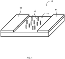
- FIG. 1 illustrates an embodiment of a detector sensor microchip 10.
- the microchip 10 includes a first electrode 12 and a second electrode 14 positioned so that the first 12 and second 14 electrode do not contact each other, with a plurality of capture probes 16 in the form of a functionalized oxide surface allowing attachment and immobilization of capture probe molecules 16 on the sensor surface 18 between the first electrode 12 and the second electrode 14.
- the capture probes 16 are designed to capture PCR amplified products via interaction with 5' tails incorporated during the amplification process.
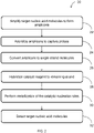
- FIG. 2 illustrates an embodiment of a method 20 for detection of target nucleic acid molecules.
- target nucleic acid molecules collected from a biological sample are amplified via PCR.
- the biological sample could be any suitable type of material, such as blood, mucous, and skin, among others. It is to be understood that any suitable type of PCR methodology can be employed.
- two primers are used during the PCR process. The first primer is synthesized as a 5'-tailed oligonucleotide with an internal replication block and the second primer is a non-tailed oligonucleotide. In an example, the second primer has a 5' phosphate group.
- the amplicons 30 are hybridized to the capture probes 16 on the surface of the detector microchip 10, illustrated in FIG. 1 .
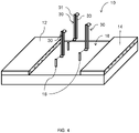
- the 5' tails of the amplicons 30 bind to the capture probes 16, as illustrated in FIG. 4 .
- the hybridized amplicons 30 are converted to single strand molecules 36, as illustrated in FIG. 5 .
- a 5'-to-3' directional helicase is used to convert the amplicons 30 to single strand molecules 36.
- an exonuclease is employed to convert the amplicons 30 to single strand molecules 36.
- the exonuclease has 5' to 3' directionality and preferentially digests the strand 33 extended from the non-tail primer.
- the exonuclease cannot access the 5' end of the strand 31, and therefore cannot digest the strand 31, preventing degradation of the tailed strand 31.
- the strand 33 may experience incomplete digestion, resulting in a remaining portion 38 of the digested strand 33.
- the strand 33 extended from the non-tail primer is synthesized with a 5' phosphate and the exonuclease is a lambda exonuclease.
- a catalyst reagent such as a gold catalyst reagent
- the catalyst reagent is in the form of catalyst clusters.
- a single catalyst cluster 60 binds to the tailed strand 31.
- a plurality of catalyst clusters 60 bind to each tailed strand 31.
- the catalyst clusters are target-specific, i.e., the catalyst clusters bind to specific target sequences in the strand 31. Because the primer-dimer artifacts do not include these target sequences, the catalyst clusters 60 do not bind to primer-dimer artifacts, thus avoiding potential false positive measurements.
- a thiol-modified oligonucleotide 40 can be reacted with a catalytic cluster 42 to form a catalyst cluster 44 functionalized with at least 20 oligonucleotides.
- FIG. 7A a thiol-modified oligonucleotide 40 can be reacted with a catalytic cluster 42 to form a catalyst cluster 44 functionalized with at least 20 oligonucleotides.
- a universal oligonucleotide 46 that possesses a universal cluster binding sequence 45 and amplicon-specific bind sequence 47 can be hybridized with a base generic cluster 48 to form a catalyst cluster 50.
- each new oligonucleotide is concatenated at the 3'-end with an adaptor specific sequence complementary to the cluster generic oligonucleotide. While the resulting catalyst cluster 50 in FIG.
- adaptor oligonucleotides 52 are depicted as having four hybridized adaptor oligonucleotides 52, it is to be understood that the actual number of adaptors will depend on cluster size, which limits the number of capture oligonucleotides on the base cluster, and the stoichiometry of adaptor oligonucleotides added to the cluster.
- the base generic clusters 48 can be prepared with mixtures of probe oligonucleotides A, B, C, D to form a catalyst cluster 54 with multiplexing capabilities.
- cluster size which determines the surface area available for functionalization, it is possible to load multiple different probes onto a single cluster. In order to support efficient metallization reactions, the ratio for the number of each probe type per cluster may require optimization.
- tailored mixtures of multiplexed clusters 54 can be created. The mixtures of multiplexed clusters 54 can both multiplex for a large number of targeted amplicons and populate each targeted amplicon with multiple clusters, as illustrated in FIG. 8C .
- metallization of the catalyst clusters 60 can be performed to form a conductive film and resistance between the electrodes 12, 14 ( FIG. 1 ) can be measured to detect target nucleic acid molecules at block 32.
- the catalyst clusters 60 are gold clusters and a gold developer reagent is applied to the catalyst clusters 60 to cascade into the development of the conductive film, which in this example is a gold film.
- the presence of the gold film electrically shorts the gap between the electrodes 12, 14 and is measured by a drop in resistance.
- a negative sensor has a resistance of more than one million ohms and a positive sensor has a resistance of about one thousand ohms.
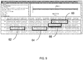
- FIG. 9 is a screenshot taken from the INVITROGEN TM program showing a portion of the 18S ribosomal gene to which primers (shown in bold lettering) are designed.
- the two primers, forward primer 60 and reverse primer 62, used in PCR for this method each have a sensor and catalyst binding 5'-tail.
- a new reverse primer 64 located downstream of the original reverse primer 62 was identified. Synthesis of the new reverse primer 64 omitted the 5' tail for catalyst binding while including a 5'-phosphorl group to facilitate exonuclease degradation.
- modification of the catalyst cluster for use with the method 20 was accomplished by pre-hybridization of the original reverse primer oligonucleotide onto a universal cluster to form target-specific catalyst gold clusters.
- Preparation of these target-specific catalyst gold clusters included a heated incubation with 1000 fold molar excess of the primer 62, cooling to room temperature, and removal of unbound excess oligonucleotide by washing, repeated twice, via high-speed centrifugation and resuspension with the final reagent buffer.
- a second catalyst reagent 66 with specificity towards a different sequence element located upstream of the first cluster binding sequence was prepared.
- this second cluster served as only a preliminary attempt to assess the processivity of the exonuclease in the digestion of sensor-bound amplicons.
- the nuclease must have digested to within at least 95 nucleotides of completely degrading the extraneous DNA strand, leaving a single-stranded tract of about eighty nucleotides available for cluster binding.
- FIG. 10 illustrates the results of time coarse experiments of Lambda exonuclease treatments on derived amplicons hybridized to microchips. All microchips in Panel A and Panel B were hybridized for five minutes at 45 degrees Celsius with 100 ng of the amplicon, washed twice by dipping into a 10 mL volume of hybridization buffer, and dried under a nitrogen gas stream. In a humidified petri dish held in a 37 degree Celsius incubator, the microchips were spotted with 25 ⁇ L Lambda reaction solution containing one unit of the exonuclease.
- exonuclease is defined as the amount of enzyme required to produce 10 nmol of acid-soluble deoxyribonucleotide from a double-stranded substrate in a total reaction volume of 50 ⁇ L in 30 minutes at 37 degrees Celsius in IX lambda Exonuclease Reaction Buffer with 1 ⁇ g sonicated duplex [ 3 H]-DNA. After incubation for a designated time, indicated above each microchip, the reactions were quenched by transferring the microchips into a petri dish with 20 mL of hybridization buffer.
- FIG. 11 illustrates the effect of titration of the Lambda exonuclease on the gold development of target amplicons that were hybridized on the surface of the microchips.
- the microchips were hybridized as described above with regard to FIG. 10 . However, the units of Lambda exonuclease used on each microchip illustrated in FIG. 11 was varied as indicated above each microchip.
- the ability of the adaptor modified clusters to hybridize to a more internal site within the 5'-tailed strand were investigated by preparation of the second cluster reagent, described above. This second cluster was formed to bind about thirty nucleotides proximal of the hybridization site of the first cluster.
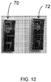
- FIG. 12 which compares development of a first microchip 70 processed with the first cluster reagent and a second microchip 72 processed with the second cluster reagent. Comparison of the first microchip 70 and second microchip 72 shows that both clusters performed equally well yielding comparable gold spots on the respective treated microchips.
- Negative and positive samples consisted respectively of either 10 ⁇ L of water or a 1 ⁇ L blood culture of P.f. (10 5 ) cells diluted in a buffer to 10 ⁇ L.
- a fully automated assay including sample preparation, PCR amplification, and microchip hybridization, nuclease digestion, and metallization reactions, was carried out.
- Post assay electrical measurements were performed by removal of each microchip board from the cartridge after each test run and visually inspecting each microchip before placing the microchip on a probe station for collection of electrical results. Table 1 presents the raw data from the fifty assays.
- RT-PCR multiplexed reverse transcription PCR
- Nucleic acid material was isolated from the homogenates using magnetic particle purification, desalted by gel-filtration, and used in the RT-PCR amplification with the appropriate multiplexed primer sets, either pan-flavivirus plus bunyavirus primers or alpha primers, to generate single-stranded 5'-tailed amplicons.
- the findings indicate that the primer sets for the pan-alpha and pan-flavivirus tests inhibit one another during PCR amplification.
- the test cartridge provides two separate PCR chambers allowing for interfering primer sets to be run separately and mixed prior to hybridization on the sensor chip. FIG.
- FIG. 13A illustrates gel analysis of gene replicates of RT-PCR for dengue viral RNA (lanes 1-4), purified from spike mosquitoes, in multiplexed reaction using pan-flavivirus primers mixed with the Cal/Bun primers. Lane 5 is a negative control with no RNA input.
- FIG. 13B illustrates gel analysis of RT-PCR for LaCrosse RNA (lanes 1-5), purified from spiked mosquitoes, in multiplexed reaction using pan-flavivirus primers mixed with the Cal/Bun primers. Lane 6 is a negative control, no RNA input. The findings indicate that combinations of pan-bunyavirus and pan-flavivirus primers are compatible in PCR reactions.
- Possible advantages of the above described method include reduction or elimination of false positive measurements due to primer-dimer artifact formation.
Landscapes
- Chemical & Material Sciences (AREA)
- Life Sciences & Earth Sciences (AREA)
- Organic Chemistry (AREA)
- Analytical Chemistry (AREA)
- Zoology (AREA)
- Wood Science & Technology (AREA)
- Proteomics, Peptides & Aminoacids (AREA)
- Health & Medical Sciences (AREA)
- Engineering & Computer Science (AREA)
- Microbiology (AREA)
- Bioinformatics & Cheminformatics (AREA)
- Molecular Biology (AREA)
- Biotechnology (AREA)
- Biophysics (AREA)
- Physics & Mathematics (AREA)
- Biochemistry (AREA)
- Immunology (AREA)
- General Engineering & Computer Science (AREA)
- General Health & Medical Sciences (AREA)
- Genetics & Genomics (AREA)
- Measuring Or Testing Involving Enzymes Or Micro-Organisms (AREA)
- Apparatus Associated With Microorganisms And Enzymes (AREA)
- Analysing Materials By The Use Of Radiation (AREA)
Description
- The subject matter disclosed herein relates to a polymerase chain reaction (PCR) process and, more particularly, to a method for nested detection of a PCR product.
- PCR is a technique that allows for replicating and amplifying trace amounts of DNA fragments into quantities that are sufficient for analysis. As such, PCR can be used in a variety of applications, such as DNA sequencing and detecting DNA fragment in samples, such as for detection of pathogens in samples.
- In operation, PCR involves the use of a series of repeated temperature changes or cycles that cause the DNA to melt or denature, yielding two single-stranded DNA molecules that then act as templates. Primers, short DNA fragments, containing sequences complementary to a target region of DNA along with a DNA polymerase, are used to selectively repeat amplification for a particular DNA region or sequence. Typically, two primers are included in a reaction mixture. The primers are single- stranded sequences, but are shorter than the length of the target region of DNA. The primers bind to a complementary part of the DNA strand and the DNA polymerase binds to the primer-DNA hybrid and begins DNA formation of a new DNA strand complementary to the DNA template strand. The process is repeated until multiple copies of the DNA strands have been created.
- However, in some instances, the primers can be subject to hetero-dimerization, in which sequences of the primer bind to each other, rather than to the DNA, resulting in short chains of dimers or artifact amplification products, known as primer dimers. These artifact products can form in the early stages of PCR and subsequently be amplified.
A Method of suppressing artifact amplification products in the PCR phase by the use of PNA clamping probe is taught inUS 2015/0176065 .
A method which forgoes PCR altogether is taught inWO 2007/084077 . Here the original target DNA molecules are tethered to a substrate in a probe, making amplification unnecessary.
Different modified methods of PCR are disclosed inUS 2005/0196760 A1 andUS 2008/0108055 Al .
The fundamentals of DNA-based electrochemical biosensors are discussed in Rias G A et al: "Electrochemical Biosensors for sequence-specific DNA detection". Analytical Letters, Taylor & Francis INC, US, vol. 38, no. 15, 1 January 2005 (2005-01-01). - An electronic sensor for detection of specific target nucleic acid molecules can include capture probes immobilized on a sensor surface between a set of paired electrodes. An example of a system and method for detecting target nucleic acid molecules is described in
U.S. Patent No. 7,645,574 . Following PCR, amplified products or amplicons derived from targeted pathogen sequences are captured by the probes via a 5' single-stranded tail, which was incorporated in the molecules during the amplification process by the use of primers made with an internal replication block. Nano-gold clusters, functionalized with a second capture oligonucleotide having a complementary sequence to a universal 5'-tail tagged onto the other end of the amplified product, are used for localized hybridization to only sensor sites having captured amplification products. Subsequently, using a short treatment with a gold developer reagent, the nano-gold clusters serve as catalytic nucleation sites for metallization, which cascades into the development of a fully conductive film. The presence of the gold film shorts the gap between the electrodes and is measured by a drop in resistance, allowing the presence of the captured amplification products can then be measured. However, primer-only artifact products or possible amplicons derived from spurious nucleic acid molecules, with both primers having the requisite 5' tails, can react with such sensors in the same way a DNA target would and can result in false positive results. - A method for detecting a nucleic acid molecule in a biological sample includes amplifying a nucleic acid molecule to generate an amplicon having a single 5'-tail and hybridizing the 5'-tail to one of a plurality of capture probes on a surface of a sensor. The amplicon is converted to a single strand molecule and a target-specific catalyst cluster is bound to the single strand molecule. The catalyst cluster is subjected to metallization in order to detect a target nucleic acid.
- In an embodiment, a method for detecting a target nucleic acid molecule in a sample with a sensor is disclosed. The sensor includes a first electrode and a second electrode coupled to a sensor surface in a spaced apart arrangement and a plurality of capture probes coupled to the sensor surface between the first electrode and the second electrode. The method includes performing nucleic acid molecule amplification via polymerase chain reaction (PCR) using a first primer having a 5'-tail and a second primer having no 5'-tail to form a plurality of double-stranded amplicons having a first strand with a 5'-tail and a second strand with no tail and hybridizing the plurality of amplicons to the plurality of capture probes. The method further includes converting the plurality of amplicons to a plurality of single strand molecules and binding a catalyst cluster to an interior section of each of the plurality of single strand molecules. In addition, the method includes contacting the plurality of single strand molecules having a catalyst cluster bound thereon with a metal or metal alloy to deposit the metal or metal alloy on the catalyst cluster and determining if an electrical current can be carried between the electrodes. The electrical current between the electrodes indicates the presence of the target nucleic acid molecule in the sample.
- In another embodiment, a method for preparing a nucleic acid molecule detector is disclosed. The nucleic acid molecule detector includes a first electrode and a second electrode coupled to a sensor surface in a spaced apart arrangement and a plurality of capture probes coupled to the sensor surface between the first electrode and the second electrode. The method includes receiving a biological sample, amplifying a nucleic acid molecule within the biological sample to generate an amplicon having a single 5'-tail, and binding the 5'-tail of the amplicon to one of the plurality of capture probes. The method additionally includes employing an exonuclease to digest one strand of the amplicon to convert the amplicon to a single strand molecule and synthesizing a target-specific catalyst cluster. The method further includes contacting the catalyst cluster with the single strand molecule of the amplicon to bind the target-specific catalyst cluster to an interior region of the single strand molecule.
- An advantage that may be realized in the practice of some disclosed embodiments is reduction or elimination of false positives due to the formation of primer-dimer artifacts or other unintended amplification products.
- The above embodiments are exemplary only. Other embodiments are within the scope of the disclosed subject matter.
- So that the manner in which the features of the invention as defined by the appended claims can be understood, a detailed description of the invention may be had by reference to certain embodiment, some of which are illustrated in the accompanying drawings. It is to be noted, however, that the drawings illustrate only certain embodiments of this invention and are therefore not to be considered limiting of its scope, for the scope of the disclosed subject matter encompasses other embodiments as well. The drawings are not necessarily to scale, emphasis generally being placed upon illustrating the features of certain embodiments of the invention. In the drawings, like numerals are used to indicate like parts throughout the various views.
-
FIG. 1 is an illustration of an embodiment of a nucleic acid molecule sensor surface; -
FIG. 2 is a flowchart illustrating a method of detecting nucleic acid molecules; -
FIG. 3 is an illustration of an embodiment of an amplicon having a single 5' tail; -
FIG. 4 is an illustration of the sensor surface ofFIG. 1 having the amplicon ofFIG. 3 coupled thereto; -
FIG. 5 is an illustration of the sensor surface ofFIG. 4 with the amplicon converted to a single strand molecule; -
FIG. 6 is an illustration of the sensor surface ofFIG. 5 with a catalyst cluster coupled to the single strand molecule; -
FIG. 7A is an illustration of an embodiment of a method of forming a catalyst cluster; -
FIG. 7B is an illustration of an embodiment of another method of forming a catalyst cluster; -
FIG. 8A is an illustration of an embodiment of a method of forming a multiplexed catalyst cluster; -
FIG. 8B is an illustration of an embodiment of a method of mixing multiplexed catalyst clusters; -
FIG. 8C is an illustration of an embodiment of a method of multiple site nesting of multiplexed catalyst clusters on single strand amplicon molecules; -
FIG. 9 is an illustration of a portion of a ribosomal gene used for testing; -
FIG. 10 is a photograph of microchip surfaces resulting from testing the effects of time on exonuclease digestion of amplicon strands; -
FIG. 11 is a photograph of microchip surfaces resulting from testing the effects of varying exonuclease concentration; -
FIG. 12 is a photograph of microchip surfaces comparing the results of two different catalyst cluster reagents; -
FIG. 13A is a photograph of a gel analysis of replicates of RT-PCR for dengue viral RNA in multiplexed reaction using pan-flavivirus primers mixed with Cal/Bun primers; and -
FIG. 13B is a photograph of a gel analysis of replicates of RT-PCR for LaCrosse RNA in multiplexed reaction using pan-flavivirus primers mixed with Cal/Bun virus primers. -
FIG. 1 illustrates an embodiment of adetector sensor microchip 10. In this embodiment, themicrochip 10 includes afirst electrode 12 and asecond electrode 14 positioned so that the first 12 and second 14 electrode do not contact each other, with a plurality of capture probes 16 in the form of a functionalized oxide surface allowing attachment and immobilization ofcapture probe molecules 16 on thesensor surface 18 between thefirst electrode 12 and thesecond electrode 14. The capture probes 16 are designed to capture PCR amplified products via interaction with 5' tails incorporated during the amplification process. -
FIG. 2 illustrates an embodiment of amethod 20 for detection of target nucleic acid molecules. Atblock 22, target nucleic acid molecules collected from a biological sample are amplified via PCR. The biological sample could be any suitable type of material, such as blood, mucous, and skin, among others. It is to be understood that any suitable type of PCR methodology can be employed. In this embodiment, two primers are used during the PCR process. The first primer is synthesized as a 5'-tailed oligonucleotide with an internal replication block and the second primer is a non-tailed oligonucleotide. In an example, the second primer has a 5' phosphate group. Multiple cycles are performed until a plurality of double-stranded amplicons 30 (replications of the target nucleic acid molecules) are formed having afirst strand 31 with a 5'tail 32 and asecond strand 33 extended from the non-tailed primer, as illustrated inFIG. 3 . In an example, illustrated inFIG. 3 , thesecond strand 33 has a 5'-phosphate group 34. In another example, not illustrated, thesecond strand 33 does not have a 5'-phosphate group. Returning toFIG. 2 , atblock 24, theamplicons 30 are hybridized to the capture probes 16 on the surface of thedetector microchip 10, illustrated inFIG. 1 . In particular, the 5' tails of theamplicons 30 bind to the capture probes 16, as illustrated inFIG. 4 . - At
block 26, the hybridizedamplicons 30 are converted tosingle strand molecules 36, as illustrated inFIG. 5 . In an example, rather than employing heat, which would denature theamplicons 30 off of the capture probes 16, a 5'-to-3' directional helicase is used to convert theamplicons 30 tosingle strand molecules 36. In another example, an exonuclease is employed to convert theamplicons 30 tosingle strand molecules 36. In this example, the exonuclease has 5' to 3' directionality and preferentially digests thestrand 33 extended from the non-tail primer. Because thetailed strand 31 is bound to thecapture probe 16, the exonuclease cannot access the 5' end of thestrand 31, and therefore cannot digest thestrand 31, preventing degradation of thetailed strand 31. In an example, depending on nuclease processivity or steric hindrance at a particular distance from thesensor surface 18, thestrand 33 may experience incomplete digestion, resulting in a remainingportion 38 of the digestedstrand 33. In an example, thestrand 33 extended from the non-tail primer is synthesized with a 5' phosphate and the exonuclease is a lambda exonuclease. - Digestion of the
strand 33 exposes the internal sequence region of thetailed strand 31. Atblock 28 of the method 20 (FIG. 2 ), a catalyst reagent, such as a gold catalyst reagent, is directly hybridized to thetailed strand 31, as illustrated inFIG. 6 . In an embodiment, the catalyst reagent is in the form of catalyst clusters. In an embodiment, asingle catalyst cluster 60 binds to thetailed strand 31. In another embodiment, and depending on the length of thetailed strand 31, a plurality ofcatalyst clusters 60 bind to eachtailed strand 31. - Since a generic oligonucleotide is not suitable for binding to internal sequences within the amplicons, the catalyst clusters are target-specific, i.e., the catalyst clusters bind to specific target sequences in the
strand 31. Because the primer-dimer artifacts do not include these target sequences, thecatalyst clusters 60 do not bind to primer-dimer artifacts, thus avoiding potential false positive measurements. As illustrated inFIG. 7A , in one example, a thiol-modifiedoligonucleotide 40 can be reacted with acatalytic cluster 42 to form acatalyst cluster 44 functionalized with at least 20 oligonucleotides. In another example, illustrated inFIG. 7B , auniversal oligonucleotide 46 that possesses a universalcluster binding sequence 45 and amplicon-specific bind sequence 47 can be hybridized with a basegeneric cluster 48 to form acatalyst cluster 50. As a result, each new oligonucleotide is concatenated at the 3'-end with an adaptor specific sequence complementary to the cluster generic oligonucleotide. While the resultingcatalyst cluster 50 inFIG. 7B is depicted as having four hybridizedadaptor oligonucleotides 52, it is to be understood that the actual number of adaptors will depend on cluster size, which limits the number of capture oligonucleotides on the base cluster, and the stoichiometry of adaptor oligonucleotides added to the cluster. - As illustrated in
FIG. 8A , the basegeneric clusters 48 can be prepared with mixtures of probe oligonucleotides A, B, C, D to form acatalyst cluster 54 with multiplexing capabilities. Depending on cluster size, which determines the surface area available for functionalization, it is possible to load multiple different probes onto a single cluster. In order to support efficient metallization reactions, the ratio for the number of each probe type per cluster may require optimization. As illustrated inFIG. 8B , tailored mixtures of multiplexedclusters 54 can be created. The mixtures of multiplexedclusters 54 can both multiplex for a large number of targeted amplicons and populate each targeted amplicon with multiple clusters, as illustrated inFIG. 8C . - Returning to
FIG. 2 , atblock 30, metallization of thecatalyst clusters 60, which serve as catalytic nucleation sites, can be performed to form a conductive film and resistance between theelectrodes 12, 14 (FIG. 1 ) can be measured to detect target nucleic acid molecules atblock 32. In an example, thecatalyst clusters 60 are gold clusters and a gold developer reagent is applied to thecatalyst clusters 60 to cascade into the development of the conductive film, which in this example is a gold film. In this example, the presence of the gold film electrically shorts the gap between theelectrodes - An existing test developed for plasmodium faliciprium was used to test the
method 20 described above.FIG. 9 is a screenshot taken from the INVITROGEN™ program showing a portion of the 18S ribosomal gene to which primers (shown in bold lettering) are designed. Typically, the two primers,forward primer 60 andreverse primer 62, used in PCR for this method each have a sensor and catalyst binding 5'-tail. However, using themethod 20 described above, anew reverse primer 64 located downstream of theoriginal reverse primer 62 was identified. Synthesis of thenew reverse primer 64 omitted the 5' tail for catalyst binding while including a 5'-phosphorl group to facilitate exonuclease degradation. - Because the 5'-tail on the
original reverse primer 62 hybridizes to a universal catalyst reagent, modification of the catalyst cluster for use with themethod 20 was accomplished by pre-hybridization of the original reverse primer oligonucleotide onto a universal cluster to form target-specific catalyst gold clusters. Preparation of these target-specific catalyst gold clusters included a heated incubation with 1000 fold molar excess of theprimer 62, cooling to room temperature, and removal of unbound excess oligonucleotide by washing, repeated twice, via high-speed centrifugation and resuspension with the final reagent buffer. Similarly, asecond catalyst reagent 66 with specificity towards a different sequence element located upstream of the first cluster binding sequence was prepared. The ability of this second cluster to effect metallization and detection served as only a preliminary attempt to assess the processivity of the exonuclease in the digestion of sensor-bound amplicons. With this particular derived amplicon, the nuclease must have digested to within at least 95 nucleotides of completely degrading the extraneous DNA strand, leaving a single-stranded tract of about eighty nucleotides available for cluster binding. - Select 5' to 3' exonucleases with suitable properties to perform a digestion as per the
method 20 were assessed. Two suitable commercially available exonucleases, Lambda and T7, were identified. Lambda was used in this example.FIGs. 10 and11 illustrate the results of this experiment. -
FIG. 10 illustrates the results of time coarse experiments of Lambda exonuclease treatments on derived amplicons hybridized to microchips. All microchips in Panel A and Panel B were hybridized for five minutes at 45 degrees Celsius with 100 ng of the amplicon, washed twice by dipping into a 10 mL volume of hybridization buffer, and dried under a nitrogen gas stream. In a humidified petri dish held in a 37 degree Celsius incubator, the microchips were spotted with 25 µL Lambda reaction solution containing one unit of the exonuclease. One unit of exonuclease is defined as the amount of enzyme required to produce 10 nmol of acid-soluble deoxyribonucleotide from a double-stranded substrate in a total reaction volume of 50 µL in 30 minutes at 37 degrees Celsius in IX lambda Exonuclease Reaction Buffer with 1 µg sonicated duplex [3H]-DNA. After incubation for a designated time, indicated above each microchip, the reactions were quenched by transferring the microchips into a petri dish with 20 mL of hybridization buffer. Subsequently, for metallization, catalyst hybridization with clusters modified with the primer oligonucleotide was performed for five minutes at 45 degrees Celsius, two washes were performed by swirling the chips in petri dishes containing hybridization buffer, and then gold development was performed for four minutes at 55 degrees Celsius with 25 µL of developer reagent placed on the chip surface.FIG. 11 illustrates the effect of titration of the Lambda exonuclease on the gold development of target amplicons that were hybridized on the surface of the microchips. The microchips were hybridized as described above with regard toFIG. 10 . However, the units of Lambda exonuclease used on each microchip illustrated inFIG. 11 was varied as indicated above each microchip. - One finding of these experiments was that the Lambda exonuclease could not digest the 5'-tail primer extended strand, which would have caused a loss in the capacity to capture the catalyst reagent. This finding is supported by the longer time coarse digestion experiment illustrated in
FIG. 10 , which shows there is no loss of chip metallization, even with the most extensive Lambda incubation time of 300 minutes. - The ability of the adaptor modified clusters to hybridize to a more internal site within the 5'-tailed strand were investigated by preparation of the second cluster reagent, described above. This second cluster was formed to bind about thirty nucleotides proximal of the hybridization site of the first cluster. The results of this investigation of illustrated in
FIG. 12 , which compares development of afirst microchip 70 processed with the first cluster reagent and asecond microchip 72 processed with the second cluster reagent. Comparison of thefirst microchip 70 andsecond microchip 72 shows that both clusters performed equally well yielding comparable gold spots on the respective treated microchips. In addition, the results established that, on a majority of the hybridized amplicons, the Lambda exonuclease digested a minimum of ninety bases and was not inhibited while approaching the microchip surface. In addition, the longer stretch of single-stranded DNA with which the second cluster interacted did not appear to affect bind of the second cluster relative to binding of the first cluster. - Using the method described above for Plasmodium falciparum (P.f.), fifty test cartridge runs were performed. Negative and positive samples consisted respectively of either 10 µL of water or a 1 µL blood culture of P.f. (105) cells diluted in a buffer to 10 µL. After pipetting and sealing a sample into the test cartridge, a fully automated assay, including sample preparation, PCR amplification, and microchip hybridization, nuclease digestion, and metallization reactions, was carried out. Post assay electrical measurements were performed by removal of each microchip board from the cartridge after each test run and visually inspecting each microchip before placing the microchip on a probe station for collection of electrical results. Table 1 presents the raw data from the fifty assays. As indicated by "NO TEST" in the "results" column of Table 1, certain assays were excluded due to machine failure or operator mistakes, as further indicated in the "Notes" column in Table 1. Table 2 indicates the percentages of correct true positives and negatives obtained. As illustrated in Table 2, using a modified, single-tailed amplicons resulted in 100% correct negative measurements and 88% correct positive measurements.
TABLE 1: E-test # of shorted sensors Date TEST TARGET CTL NCOMP RESULT Notes 3/14/16 POS 10 0 0 CORRECT 3/14/16 POS 10 0 0 CORRECT 3/14/16 NEG 0 0 0 CORRECT 3/14/16 NEG 0 0 0 CORRECT 3/14/16 NEG 0 0 0 CORRECT 3/15/16 POS 0 0 0 INCORRECT No sign of target development - Benchtop development indicates SP- PCR ok 3/15/16 POS 0 0 0 NO TEST Cartridge leaked. Benchtop development of hyb buffer indicates SP-PCR ok 3/15/16 NEG NO TEST PCR Temp Timeout 3/15/16 NEG 0 0 0 CORRECT 3/15/16 NEG 0 0 0 CORRECT 3/15/16 NEG 0 0 0 CORRECT 3/16/16 POS 10 0 0 CORRECT 3/16/16 POS 10 0 0 CORRECT 3/16/16 NEG 0 0 0 CORRECT 3/17/16 POS 10 0 0 CORRECT 3/17/16 POS 10 0 0 CORRECT 3/17/16 NEG 0 0 0 CORRECT 3/17/16 NEG 0 0 0 CORRECT 3/18/16 POS 10 0 0 CORRECT 3/18/16 POS 10 0 0 CORRECT 3/18/16 NEG 0 0 0 CORRECT 3/18/16 NEG 0 0 0 CORRECT 3/18/16 NEG 0 0 0 CORRECT 3/21/16 POS 10 0 0 CORRECT 3/21/16 POS 10 0 0 CORRECT 3/21/16 NEG 0 0 0 NO TEST Developer misloaded - turned out ok when completed on benchtop 3/21/16 NEG 0 0 0 CORRECT 3/21/16 NEG 0 0 0 CORRECT 3/22/16 POS 10 0 0 CORRECT 3/22/16 POS 10 0 0 CORRECT 3/22/16 NEG 0 0 0 CORRECT 3/22/16 NEG 0 0 0 CORRECT 3/22/16 NEG 0 0 0 CORRECT 3/23/16 POS 9 0 0 CORRECT 3/23/16 POS 0 0 0 NO TEST Weak development - machine ran unusually slow (SD card issues) 3/23/16 POS 10 0 0 CORRECT 3/23/16 NEG 0 0 0 CORRECT 3/23/16 NEG 0 0 0 CORRECT 3/23/16 POS 10 0 0 CORRECT 3/23/16 POS 10 0 0 CORRECT 3/23/16 POS 10 0 0 CORRECT 3/28/16 POS 0 0 0 INCORRECT No development 3/28/16 POS 0 0 0 INCORRECT No development 3/28/16 POS NO TEST Motor COM error, Motortimeout 3/28/16 POS 10 0 0 CORRECT 3/28/16 NEG NO TEST Syringe timeout error 3/31/16 POS 10 0 0 CORRECT 3/31/16 POS 10 0 0 CORRECT 3/31/16 POS 6 0 0 CORRECT Light development 3/31/16 POS 10 0 0 CORRECT 
- Various combinations of multiplexed reverse transcription PCR (RT-PCR) were assessed using primer sets designed for development of a pan-flavivirus/pan-alphavirus/pan-bunyavirus test. A total of nine primers were used in this assay, with all assayed materials derived from the homogenization, using ultrasonically driven bead-beating, of pooled six to eight mosquitoes that were spiked or non spiked with a virus, VEE-TC83, Dengue, or LaCrosse Virus. Nucleic acid material was isolated from the homogenates using magnetic particle purification, desalted by gel-filtration, and used in the RT-PCR amplification with the appropriate multiplexed primer sets, either pan-flavivirus plus bunyavirus primers or alpha primers, to generate single-stranded 5'-tailed amplicons. The findings indicate that the primer sets for the pan-alpha and pan-flavivirus tests inhibit one another during PCR amplification. To address this problem, the test cartridge provides two separate PCR chambers allowing for interfering primer sets to be run separately and mixed prior to hybridization on the sensor chip.
FIG. 13A illustrates gel analysis of gene replicates of RT-PCR for dengue viral RNA (lanes 1-4), purified from spike mosquitoes, in multiplexed reaction using pan-flavivirus primers mixed with the Cal/Bun primers.Lane 5 is a negative control with no RNA input.FIG. 13B illustrates gel analysis of RT-PCR for LaCrosse RNA (lanes 1-5), purified from spiked mosquitoes, in multiplexed reaction using pan-flavivirus primers mixed with the Cal/Bun primers.Lane 6 is a negative control, no RNA input. The findings indicate that combinations of pan-bunyavirus and pan-flavivirus primers are compatible in PCR reactions. - Possible advantages of the above described method include reduction or elimination of false positive measurements due to primer-dimer artifact formation.
- To the extent that the claims recite the phrase "at least one of' in reference to a plurality of elements, this recitation is intended to mean at least one or more of the listed elements, and is not limited to at least one of each element. For example, "at least one of an element A, element B, and element C," is intended to indicate element A alone, or element B alone, or element C alone, or any combination thereof. "At least one of element A, element B, and element C" is not intended to be limited to at least one of an element A, at least one of an element B, and at least one of an element C.
Claims (7)
- A method for detecting a target nucleic acid molecule in a sample with a sensor comprising a first electrode and a second electrode coupled to a sensor surface in a spaced apart arrangement and a plurality of capture probes coupled to the sensor surface between the first electrode and the second electrode, the method comprising:performing nucleic acid molecule amplification via polymerase chain reaction (PCR) using a first primer having a 5'-tail and comprising an internal replication block configured to stop a polymerase action short of the 5'-tail, and a second primer being complimentary to a region of the target nucleic acid molecule and having no 5'-tail, wherein the PCR forms a plurality of double-stranded amplicons with a single-strand 5'-tail, wherein each double stranded amplicon comprisesa first strand including an internal target sequence, wherein the first strand further comprises the 5'-tail, anda second strand with no tail;applying the plurality of double-stranded amplicons formed by the PCR to the sensor surface;hybridizing the plurality of double-stranded amplicons to the plurality of capture probe sequences, wherein the single-strand 5'-tails of the plurality of double-stranded amplicons bind to the plurality of capture probe sequences;converting the plurality of double-stranded amplicons to a plurality of single strand molecules using a 5'-3' exonuclease with specificity for double-stranded DNA to expose the target sequence of the first strand, wherein the 5'-tails of the plurality of single-strand molecules remain bound to the plurality of capture probe sequences and an opposing 3' end is unbound;hybridizing a catalyst cluster to the internal target sequence of each of the plurality of single strand molecules;contacting the plurality of single strand molecules having a catalyst cluster bound thereon with a metal or metal alloy, wherein the metal or metal alloy binds to the catalyst cluster to form a conductive metal film between the first electrode and the second electrode; anddetermining if an electrical current can be carried between the electrodes, the electrical current between the electrodes indicating presence of the target nucleic acid molecule in the sample.
- The method of claim 1, wherein converting the plurality of amplicons to single strand molecules comprises employing an exonuclease to digest the second strand with no tail.
- The method of claim 1, wherein the catalyst cluster comprises a catalyst gold cluster and wherein the metal comprises gold.
- The method of claim 1, further comprising forming a target specific catalyst cluster configured to bind to an interior region of the first strand of the amplicon.
- The method of claim 1, wherein the catalyst cluster is a generic cluster having an adaptor oligonucleotide coupled thereto.
- The method of claim 5, wherein the generic cluster has a plurality of oligonucleotides coupled thereto, each oligonucleotide configured to target a different amplicon.
- The method of claim 1, wherein binding the catalyst cluster comprises binding a plurality of catalyst clusters to interior sections of each of the single strand molecules.
Applications Claiming Priority (2)
| Application Number | Priority Date | Filing Date | Title |
|---|---|---|---|
| US201662337917P | 2016-05-18 | 2016-05-18 | |
| PCT/US2017/033255 WO2017201239A1 (en) | 2016-05-18 | 2017-05-18 | Method for detection of a pcr product |
Publications (3)
| Publication Number | Publication Date |
|---|---|
| EP3458613A1 EP3458613A1 (en) | 2019-03-27 |
| EP3458613A4 EP3458613A4 (en) | 2019-03-27 |
| EP3458613B1 true EP3458613B1 (en) | 2022-09-14 |
Family
ID=60325605
Family Applications (1)
| Application Number | Title | Priority Date | Filing Date |
|---|---|---|---|
| EP17800142.6A Active EP3458613B1 (en) | 2016-05-18 | 2017-05-18 | Method for detection of a pcr product |
Country Status (7)
| Country | Link |
|---|---|
| US (1) | US20190309349A1 (en) |
| EP (1) | EP3458613B1 (en) |
| JP (1) | JP6962596B2 (en) |
| AU (1) | AU2017268369B2 (en) |
| CA (1) | CA3023256A1 (en) |
| ES (1) | ES2928217T3 (en) |
| WO (1) | WO2017201239A1 (en) |
Citations (3)
| Publication number | Priority date | Publication date | Assignee | Title |
|---|---|---|---|---|
| US20040185470A1 (en) * | 2000-03-08 | 2004-09-23 | Getts Robert C. | Nucleic acid detection |
| US20050196760A1 (en) * | 2004-03-05 | 2005-09-08 | Alexander Pemov | Dual phase multiplex polymerase chain reaction |
| US20080108055A1 (en) * | 2006-09-11 | 2008-05-08 | Gen-Probe Incorporated | Method for detecting west nile virus nucleic acids in the 3' non-coding region |
Family Cites Families (27)
| Publication number | Priority date | Publication date | Assignee | Title |
|---|---|---|---|---|
| IL124322A (en) * | 1998-05-04 | 2002-05-23 | Technion Res & Dev Foundation | Detection of an entity in a sample |
| CA2367405A1 (en) * | 1999-04-07 | 2000-10-12 | Dennis Michael Connolly | High resolution dna detection methods and devices |
| US6339147B1 (en) * | 1999-07-29 | 2002-01-15 | Epoch Biosciences, Inc. | Attachment of oligonucleotides to solid supports through Schiff base type linkages for capture and detection of nucleic acids |
| US6358679B1 (en) * | 2000-08-24 | 2002-03-19 | Pe Corporation (Ny) | Methods for external controls for nucleic acid amplification |
| DK1370690T3 (en) * | 2001-03-16 | 2012-07-09 | Kalim Mir | Arrays and methods for using them |
| US7153656B2 (en) * | 2001-09-11 | 2006-12-26 | Los Alamos National Security, Llc | Nucleic acid sequence detection using multiplexed oligonucleotide PCR |
| AU2002363062B2 (en) * | 2001-10-09 | 2007-03-22 | Nanosphere, Inc. | Nanoparticles having oligonucleotides attached thereto and uses therefor |
| DE60324253D1 (en) * | 2002-02-08 | 2008-12-04 | Olympus Corp | SPECIFIC MULTIPLEX ANALYSIS OF NUCLEIC ACIDS |
| US20030203384A1 (en) * | 2002-03-08 | 2003-10-30 | Chafin David R. | Multiplex detection of biological materials in a sample |
| EP1511862A4 (en) * | 2002-05-14 | 2006-01-18 | Nanosphere Inc | Electrical detection of dna hybridization and specific binding events |
| WO2004065570A2 (en) * | 2003-01-23 | 2004-08-05 | Integrated Nano-Technologies, Llc | Methods of metallizing nucleic acid molecules and methods of attaching nucleic acid molecules to conductive surfaces |
| KR20070090158A (en) * | 2004-10-18 | 2007-09-05 | 브랜데이스 유니버시티 | Reagents and Methods to Improve Reproducibility and Reduce Mispriming in PCR Amplification |
| JP2008535518A (en) * | 2005-04-14 | 2008-09-04 | アプレラ コーポレイション | 3 'modified oligonucleotides containing pseudoisocytosine nucleobase derivatives and their application as primers or probes |
| AU2006335690A1 (en) | 2006-01-20 | 2007-07-26 | Agency For Science, Technology And Research | Biosensor cell and biosensor array |
| US8465926B2 (en) * | 2006-08-11 | 2013-06-18 | The Hong Kong University Of Science And Technology | Method and system for real time quantification and monitoring of nucleic acid amplification using electroconductive or electrochemically active labels |
| US8058640B2 (en) * | 2006-09-11 | 2011-11-15 | President And Fellows Of Harvard College | Branched nanoscale wires |
| WO2008127314A1 (en) * | 2006-11-22 | 2008-10-23 | President And Fellows Of Harvard College | High-sensitivity nanoscale wire sensors |
| WO2011046972A2 (en) * | 2009-10-12 | 2011-04-21 | Life Technologies Corporation | Compositions and methods for suppressing primer interactions |
| EP2516678A4 (en) * | 2009-12-24 | 2013-06-19 | Seegene Inc | Real-time multiplexing detection of target nucleic acid sequences with elimination of false signals |
| US8703653B2 (en) * | 2011-02-18 | 2014-04-22 | NVS Technologies, Inc. | Quantitative, highly multiplexed detection of nucleic acids |
| CA2828303C (en) * | 2011-02-28 | 2020-04-07 | Seattle Children's Research Institute | Coupling endonucleases with end-processing enzymes drive high efficiency gene disruption |
| WO2012149154A1 (en) * | 2011-04-26 | 2012-11-01 | Swift Biosciences, Inc. | Polynucleotide primers and probes |
| JP6181751B2 (en) * | 2012-06-18 | 2017-08-16 | ニューゲン テクノロジーズ, インコーポレイテッド | Compositions and methods for negative selection of unwanted nucleic acid sequences |
| US9909159B2 (en) * | 2013-02-07 | 2018-03-06 | Rutgers, The State University Of New Jersey | Highly selective nucleic acid amplification primers |
| US20140274811A1 (en) * | 2013-03-14 | 2014-09-18 | Lyle J. Arnold | Methods for Amplifying a Complete Genome or Transcriptome |
| US9745633B2 (en) * | 2013-06-28 | 2017-08-29 | Diacarta Ltd | Detection of PNA clamping |
| US10072285B2 (en) * | 2014-08-19 | 2018-09-11 | Bio-Rad Laboratories, Inc. | RNA amplification methods |
-
2017
- 2017-05-18 CA CA3023256A patent/CA3023256A1/en active Pending
- 2017-05-18 WO PCT/US2017/033255 patent/WO2017201239A1/en not_active Ceased
- 2017-05-18 JP JP2019512950A patent/JP6962596B2/en not_active Expired - Fee Related
- 2017-05-18 AU AU2017268369A patent/AU2017268369B2/en not_active Ceased
- 2017-05-18 ES ES17800142T patent/ES2928217T3/en active Active
- 2017-05-18 EP EP17800142.6A patent/EP3458613B1/en active Active
- 2017-05-18 US US16/302,346 patent/US20190309349A1/en not_active Abandoned
Patent Citations (3)
| Publication number | Priority date | Publication date | Assignee | Title |
|---|---|---|---|---|
| US20040185470A1 (en) * | 2000-03-08 | 2004-09-23 | Getts Robert C. | Nucleic acid detection |
| US20050196760A1 (en) * | 2004-03-05 | 2005-09-08 | Alexander Pemov | Dual phase multiplex polymerase chain reaction |
| US20080108055A1 (en) * | 2006-09-11 | 2008-05-08 | Gen-Probe Incorporated | Method for detecting west nile virus nucleic acids in the 3' non-coding region |
Non-Patent Citations (1)
| Title |
|---|
| RIVAS G A ET AL: "ELECTROCHEMICAL BIOSENSORS FOR SEQUENCE-SPECIFIC DNA DETECTION", ANALYTICAL LETTERS, TAYLOR & FRANCIS INC, US, vol. 38, no. 15, 1 January 2005 (2005-01-01), pages 2653 - 2703, XP009075475, ISSN: 0003-2719, DOI: 10.1080/00032710500371121 * |
Also Published As
| Publication number | Publication date |
|---|---|
| CA3023256A1 (en) | 2017-11-23 |
| AU2017268369A1 (en) | 2018-11-22 |
| AU2017268369B2 (en) | 2022-09-01 |
| EP3458613A1 (en) | 2019-03-27 |
| JP6962596B2 (en) | 2021-11-05 |
| EP3458613A4 (en) | 2019-03-27 |
| JP2019516408A (en) | 2019-06-20 |
| WO2017201239A1 (en) | 2017-11-23 |
| ES2928217T3 (en) | 2022-11-16 |
| US20190309349A1 (en) | 2019-10-10 |
Similar Documents
| Publication | Publication Date | Title |
|---|---|---|
| CN102459630B (en) | Sample analysis method and detection kit for the method | |
| EP1975248B1 (en) | Nucleotide primer set and nucleotide probe for detecting genotype of methylene tetrahydrofolate reductase (MTHFR) | |
| US7252946B2 (en) | Nucleic acid detection | |
| US7919252B2 (en) | Method for detecting a target nucleic acid sequence | |
| JP2017532980A (en) | Sequence-based detection method | |
| WO2007115491A1 (en) | Rapid genotyping analysis and the device thereof | |
| WO2008022538A1 (en) | Method and system for real time quantification and monitoring of nucleic acid amplification using electroconductive or electrochemically active labels | |
| WO2014114189A1 (en) | Methods and compositions for detecting target snp | |
| CN109415721B (en) | Primer group, kit and method for detecting more than 2 target nucleic acids | |
| EP4215619A1 (en) | Methods for sensitive and accurate parallel quantification of nucleic acids | |
| Oberc et al. | Nucleic acid amplification test (NAAT) conducted in a microfluidic chip to differentiate between various ginseng species | |
| AU2017268369B2 (en) | Method for detection of a PCR product | |
| KR20040037015A (en) | Nucleic Acid Probe-Immobilized Substrate and Method of Detecting the Presence of Target Nucleic Acid by Using the Same | |
| CN101280338B (en) | Nucleic acid amplification method for detecting polymorphism of nucleic acid | |
| EP2789688A1 (en) | Method for detecting nucleotide mutation, and detection kit | |
| JP2024035110A (en) | Sensitive method for accurate parallel quantification of mutant nucleic acids | |
| CN119955908B (en) | Combined liquid-phase and solid-phase DNA amplification | |
| US20080199857A1 (en) | Method of increasing specificity of nucleic acid hybridization using zwitterionic compound | |
| CN112176089B (en) | System and method for identifying single nucleotide polymorphism of panax plants | |
| JPWO2007055255A1 (en) | Method for amplifying a plurality of nucleic acid sequences for identification | |
| JP2011004733A (en) | Microarray for detecting target nucleic acid in a plurality of specimens | |
| KR20250065218A (en) | Highly sensitive methods for accurate parallel quantification of nucleic acids | |
| JP4528889B1 (en) | Sample analysis method and assay kit used therein | |
| CN114214414A (en) | SNaPshot multiplex analysis kit for detecting SNP (Single nucleotide polymorphism) of ALL (ALL related gene) and application of snaPshot multiplex analysis kit | |
| JP2005160430A (en) | Method for inspection of gene sequence |
Legal Events
| Date | Code | Title | Description |
|---|---|---|---|
| STAA | Information on the status of an ep patent application or granted ep patent |
Free format text: STATUS: THE INTERNATIONAL PUBLICATION HAS BEEN MADE |
|
| PUAI | Public reference made under article 153(3) epc to a published international application that has entered the european phase |
Free format text: ORIGINAL CODE: 0009012 |
|
| STAA | Information on the status of an ep patent application or granted ep patent |
Free format text: STATUS: REQUEST FOR EXAMINATION WAS MADE |
|
| 17P | Request for examination filed |
Effective date: 20181122 |
|
| A4 | Supplementary search report drawn up and despatched |
Effective date: 20190212 |
|
| AK | Designated contracting states |
Kind code of ref document: A1 Designated state(s): AL AT BE BG CH CY CZ DE DK EE ES FI FR GB GR HR HU IE IS IT LI LT LU LV MC MK MT NL NO PL PT RO RS SE SI SK SM TR |
|
| AX | Request for extension of the european patent |
Extension state: BA ME |
|
| DAV | Request for validation of the european patent (deleted) | ||
| DAX | Request for extension of the european patent (deleted) | ||
| STAA | Information on the status of an ep patent application or granted ep patent |
Free format text: STATUS: EXAMINATION IS IN PROGRESS |
|
| 17Q | First examination report despatched |
Effective date: 20200526 |
|
| GRAP | Despatch of communication of intention to grant a patent |
Free format text: ORIGINAL CODE: EPIDOSNIGR1 |
|
| STAA | Information on the status of an ep patent application or granted ep patent |
Free format text: STATUS: GRANT OF PATENT IS INTENDED |
|
| INTG | Intention to grant announced |
Effective date: 20220421 |
|
| GRAS | Grant fee paid |
Free format text: ORIGINAL CODE: EPIDOSNIGR3 |
|
| GRAA | (expected) grant |
Free format text: ORIGINAL CODE: 0009210 |
|
| STAA | Information on the status of an ep patent application or granted ep patent |
Free format text: STATUS: THE PATENT HAS BEEN GRANTED |
|
| AK | Designated contracting states |
Kind code of ref document: B1 Designated state(s): AL AT BE BG CH CY CZ DE DK EE ES FI FR GB GR HR HU IE IS IT LI LT LU LV MC MK MT NL NO PL PT RO RS SE SI SK SM TR |
|
| REG | Reference to a national code |
Ref country code: GB Ref legal event code: FG4D |
|
| REG | Reference to a national code |
Ref country code: CH Ref legal event code: EP |
|
| REG | Reference to a national code |
Ref country code: DE Ref legal event code: R096 Ref document number: 602017061828 Country of ref document: DE |
|
| REG | Reference to a national code |
Ref country code: IE Ref legal event code: FG4D |
|
| REG | Reference to a national code |
Ref country code: AT Ref legal event code: REF Ref document number: 1518721 Country of ref document: AT Kind code of ref document: T Effective date: 20221015 |
|
| REG | Reference to a national code |
Ref country code: ES Ref legal event code: FG2A Ref document number: 2928217 Country of ref document: ES Kind code of ref document: T3 Effective date: 20221116 |
|
| REG | Reference to a national code |
Ref country code: LT Ref legal event code: MG9D |
|
| REG | Reference to a national code |
Ref country code: NL Ref legal event code: MP Effective date: 20220914 |
|
| PG25 | Lapsed in a contracting state [announced via postgrant information from national office to epo] |
Ref country code: SE Free format text: LAPSE BECAUSE OF FAILURE TO SUBMIT A TRANSLATION OF THE DESCRIPTION OR TO PAY THE FEE WITHIN THE PRESCRIBED TIME-LIMIT Effective date: 20220914 Ref country code: RS Free format text: LAPSE BECAUSE OF FAILURE TO SUBMIT A TRANSLATION OF THE DESCRIPTION OR TO PAY THE FEE WITHIN THE PRESCRIBED TIME-LIMIT Effective date: 20220914 Ref country code: NO Free format text: LAPSE BECAUSE OF FAILURE TO SUBMIT A TRANSLATION OF THE DESCRIPTION OR TO PAY THE FEE WITHIN THE PRESCRIBED TIME-LIMIT Effective date: 20221214 Ref country code: LV Free format text: LAPSE BECAUSE OF FAILURE TO SUBMIT A TRANSLATION OF THE DESCRIPTION OR TO PAY THE FEE WITHIN THE PRESCRIBED TIME-LIMIT Effective date: 20220914 Ref country code: LT Free format text: LAPSE BECAUSE OF FAILURE TO SUBMIT A TRANSLATION OF THE DESCRIPTION OR TO PAY THE FEE WITHIN THE PRESCRIBED TIME-LIMIT Effective date: 20220914 Ref country code: FI Free format text: LAPSE BECAUSE OF FAILURE TO SUBMIT A TRANSLATION OF THE DESCRIPTION OR TO PAY THE FEE WITHIN THE PRESCRIBED TIME-LIMIT Effective date: 20220914 |
|
| REG | Reference to a national code |
Ref country code: AT Ref legal event code: MK05 Ref document number: 1518721 Country of ref document: AT Kind code of ref document: T Effective date: 20220914 |
|
| PG25 | Lapsed in a contracting state [announced via postgrant information from national office to epo] |
Ref country code: HR Free format text: LAPSE BECAUSE OF FAILURE TO SUBMIT A TRANSLATION OF THE DESCRIPTION OR TO PAY THE FEE WITHIN THE PRESCRIBED TIME-LIMIT Effective date: 20220914 Ref country code: GR Free format text: LAPSE BECAUSE OF FAILURE TO SUBMIT A TRANSLATION OF THE DESCRIPTION OR TO PAY THE FEE WITHIN THE PRESCRIBED TIME-LIMIT Effective date: 20221215 |
|
| PG25 | Lapsed in a contracting state [announced via postgrant information from national office to epo] |
Ref country code: SM Free format text: LAPSE BECAUSE OF FAILURE TO SUBMIT A TRANSLATION OF THE DESCRIPTION OR TO PAY THE FEE WITHIN THE PRESCRIBED TIME-LIMIT Effective date: 20220914 Ref country code: RO Free format text: LAPSE BECAUSE OF FAILURE TO SUBMIT A TRANSLATION OF THE DESCRIPTION OR TO PAY THE FEE WITHIN THE PRESCRIBED TIME-LIMIT Effective date: 20220914 Ref country code: PT Free format text: LAPSE BECAUSE OF FAILURE TO SUBMIT A TRANSLATION OF THE DESCRIPTION OR TO PAY THE FEE WITHIN THE PRESCRIBED TIME-LIMIT Effective date: 20230116 Ref country code: CZ Free format text: LAPSE BECAUSE OF FAILURE TO SUBMIT A TRANSLATION OF THE DESCRIPTION OR TO PAY THE FEE WITHIN THE PRESCRIBED TIME-LIMIT Effective date: 20220914 Ref country code: AT Free format text: LAPSE BECAUSE OF FAILURE TO SUBMIT A TRANSLATION OF THE DESCRIPTION OR TO PAY THE FEE WITHIN THE PRESCRIBED TIME-LIMIT Effective date: 20220914 |
|
| PG25 | Lapsed in a contracting state [announced via postgrant information from national office to epo] |
Ref country code: SK Free format text: LAPSE BECAUSE OF FAILURE TO SUBMIT A TRANSLATION OF THE DESCRIPTION OR TO PAY THE FEE WITHIN THE PRESCRIBED TIME-LIMIT Effective date: 20220914 Ref country code: PL Free format text: LAPSE BECAUSE OF FAILURE TO SUBMIT A TRANSLATION OF THE DESCRIPTION OR TO PAY THE FEE WITHIN THE PRESCRIBED TIME-LIMIT Effective date: 20220914 Ref country code: IS Free format text: LAPSE BECAUSE OF FAILURE TO SUBMIT A TRANSLATION OF THE DESCRIPTION OR TO PAY THE FEE WITHIN THE PRESCRIBED TIME-LIMIT Effective date: 20230114 Ref country code: EE Free format text: LAPSE BECAUSE OF FAILURE TO SUBMIT A TRANSLATION OF THE DESCRIPTION OR TO PAY THE FEE WITHIN THE PRESCRIBED TIME-LIMIT Effective date: 20220914 |
|
| REG | Reference to a national code |
Ref country code: DE Ref legal event code: R097 Ref document number: 602017061828 Country of ref document: DE |
|
| PG25 | Lapsed in a contracting state [announced via postgrant information from national office to epo] |
Ref country code: NL Free format text: LAPSE BECAUSE OF FAILURE TO SUBMIT A TRANSLATION OF THE DESCRIPTION OR TO PAY THE FEE WITHIN THE PRESCRIBED TIME-LIMIT Effective date: 20220914 Ref country code: AL Free format text: LAPSE BECAUSE OF FAILURE TO SUBMIT A TRANSLATION OF THE DESCRIPTION OR TO PAY THE FEE WITHIN THE PRESCRIBED TIME-LIMIT Effective date: 20220914 |
|
| P01 | Opt-out of the competence of the unified patent court (upc) registered |
Effective date: 20230601 |
|
| PLBE | No opposition filed within time limit |
Free format text: ORIGINAL CODE: 0009261 |
|
| STAA | Information on the status of an ep patent application or granted ep patent |
Free format text: STATUS: NO OPPOSITION FILED WITHIN TIME LIMIT |
|
| PG25 | Lapsed in a contracting state [announced via postgrant information from national office to epo] |
Ref country code: DK Free format text: LAPSE BECAUSE OF FAILURE TO SUBMIT A TRANSLATION OF THE DESCRIPTION OR TO PAY THE FEE WITHIN THE PRESCRIBED TIME-LIMIT Effective date: 20220914 |
|
| PGFP | Annual fee paid to national office [announced via postgrant information from national office to epo] |
Ref country code: FR Payment date: 20230525 Year of fee payment: 7 Ref country code: ES Payment date: 20230601 Year of fee payment: 7 Ref country code: DE Payment date: 20230530 Year of fee payment: 7 |
|
| 26N | No opposition filed |
Effective date: 20230615 |
|
| PG25 | Lapsed in a contracting state [announced via postgrant information from national office to epo] |
Ref country code: SI Free format text: LAPSE BECAUSE OF FAILURE TO SUBMIT A TRANSLATION OF THE DESCRIPTION OR TO PAY THE FEE WITHIN THE PRESCRIBED TIME-LIMIT Effective date: 20220914 |
|
| PGFP | Annual fee paid to national office [announced via postgrant information from national office to epo] |
Ref country code: GB Payment date: 20230529 Year of fee payment: 7 |
|
| REG | Reference to a national code |
Ref country code: CH Ref legal event code: PL |
|
| PG25 | Lapsed in a contracting state [announced via postgrant information from national office to epo] |
Ref country code: MC Free format text: LAPSE BECAUSE OF FAILURE TO SUBMIT A TRANSLATION OF THE DESCRIPTION OR TO PAY THE FEE WITHIN THE PRESCRIBED TIME-LIMIT Effective date: 20220914 |
|
| REG | Reference to a national code |
Ref country code: BE Ref legal event code: MM Effective date: 20230531 |
|
| PG25 | Lapsed in a contracting state [announced via postgrant information from national office to epo] |
Ref country code: MC Free format text: LAPSE BECAUSE OF FAILURE TO SUBMIT A TRANSLATION OF THE DESCRIPTION OR TO PAY THE FEE WITHIN THE PRESCRIBED TIME-LIMIT Effective date: 20220914 Ref country code: LU Free format text: LAPSE BECAUSE OF NON-PAYMENT OF DUE FEES Effective date: 20230518 Ref country code: LI Free format text: LAPSE BECAUSE OF NON-PAYMENT OF DUE FEES Effective date: 20230531 Ref country code: CH Free format text: LAPSE BECAUSE OF NON-PAYMENT OF DUE FEES Effective date: 20230531 |
|
| REG | Reference to a national code |
Ref country code: IE Ref legal event code: MM4A |
|
| PG25 | Lapsed in a contracting state [announced via postgrant information from national office to epo] |
Ref country code: IE Free format text: LAPSE BECAUSE OF NON-PAYMENT OF DUE FEES Effective date: 20230518 |
|
| PG25 | Lapsed in a contracting state [announced via postgrant information from national office to epo] |
Ref country code: IE Free format text: LAPSE BECAUSE OF NON-PAYMENT OF DUE FEES Effective date: 20230518 |
|
| PG25 | Lapsed in a contracting state [announced via postgrant information from national office to epo] |
Ref country code: IT Free format text: LAPSE BECAUSE OF FAILURE TO SUBMIT A TRANSLATION OF THE DESCRIPTION OR TO PAY THE FEE WITHIN THE PRESCRIBED TIME-LIMIT Effective date: 20220914 Ref country code: BE Free format text: LAPSE BECAUSE OF NON-PAYMENT OF DUE FEES Effective date: 20230531 |
|
| PG25 | Lapsed in a contracting state [announced via postgrant information from national office to epo] |
Ref country code: BG Free format text: LAPSE BECAUSE OF FAILURE TO SUBMIT A TRANSLATION OF THE DESCRIPTION OR TO PAY THE FEE WITHIN THE PRESCRIBED TIME-LIMIT Effective date: 20220914 |
|
| PG25 | Lapsed in a contracting state [announced via postgrant information from national office to epo] |
Ref country code: BG Free format text: LAPSE BECAUSE OF FAILURE TO SUBMIT A TRANSLATION OF THE DESCRIPTION OR TO PAY THE FEE WITHIN THE PRESCRIBED TIME-LIMIT Effective date: 20220914 |
|
| REG | Reference to a national code |
Ref country code: DE Ref legal event code: R119 Ref document number: 602017061828 Country of ref document: DE |
|
| GBPC | Gb: european patent ceased through non-payment of renewal fee |
Effective date: 20240518 |
|
| PG25 | Lapsed in a contracting state [announced via postgrant information from national office to epo] |
Ref country code: DE Free format text: LAPSE BECAUSE OF NON-PAYMENT OF DUE FEES Effective date: 20241203 |
|
| PG25 | Lapsed in a contracting state [announced via postgrant information from national office to epo] |
Ref country code: FR Free format text: LAPSE BECAUSE OF NON-PAYMENT OF DUE FEES Effective date: 20240531 |
|
| PG25 | Lapsed in a contracting state [announced via postgrant information from national office to epo] |
Ref country code: GB Free format text: LAPSE BECAUSE OF NON-PAYMENT OF DUE FEES Effective date: 20240518 |
|
| REG | Reference to a national code |
Ref country code: ES Ref legal event code: FD2A Effective date: 20250701 |
|
| PG25 | Lapsed in a contracting state [announced via postgrant information from national office to epo] |
Ref country code: ES Free format text: LAPSE BECAUSE OF NON-PAYMENT OF DUE FEES Effective date: 20240519 |
|
| PG25 | Lapsed in a contracting state [announced via postgrant information from national office to epo] |
Ref country code: CY Free format text: LAPSE BECAUSE OF FAILURE TO SUBMIT A TRANSLATION OF THE DESCRIPTION OR TO PAY THE FEE WITHIN THE PRESCRIBED TIME-LIMIT; INVALID AB INITIO Effective date: 20170518 |
|
| PG25 | Lapsed in a contracting state [announced via postgrant information from national office to epo] |
Ref country code: HU Free format text: LAPSE BECAUSE OF FAILURE TO SUBMIT A TRANSLATION OF THE DESCRIPTION OR TO PAY THE FEE WITHIN THE PRESCRIBED TIME-LIMIT; INVALID AB INITIO Effective date: 20170518 |
|
| PG25 | Lapsed in a contracting state [announced via postgrant information from national office to epo] |
Ref country code: TR Free format text: LAPSE BECAUSE OF FAILURE TO SUBMIT A TRANSLATION OF THE DESCRIPTION OR TO PAY THE FEE WITHIN THE PRESCRIBED TIME-LIMIT Effective date: 20220914 |