EP3451210A1 - Method for comparing reference values in medical imaging processes, system comprising a local medical imaging device, computer program product and computer-readable program - Google Patents
Method for comparing reference values in medical imaging processes, system comprising a local medical imaging device, computer program product and computer-readable program Download PDFInfo
- Publication number
- EP3451210A1 EP3451210A1 EP17188845.6A EP17188845A EP3451210A1 EP 3451210 A1 EP3451210 A1 EP 3451210A1 EP 17188845 A EP17188845 A EP 17188845A EP 3451210 A1 EP3451210 A1 EP 3451210A1
- Authority
- EP
- European Patent Office
- Prior art keywords
- medical imaging
- local
- imaging device
- identification
- mapping
- Prior art date
- Legal status (The legal status is an assumption and is not a legal conclusion. Google has not performed a legal analysis and makes no representation as to the accuracy of the status listed.)
- Granted
Links
Images
Classifications
-
- G—PHYSICS
- G16—INFORMATION AND COMMUNICATION TECHNOLOGY [ICT] SPECIALLY ADAPTED FOR SPECIFIC APPLICATION FIELDS
- G16H—HEALTHCARE INFORMATICS, i.e. INFORMATION AND COMMUNICATION TECHNOLOGY [ICT] SPECIALLY ADAPTED FOR THE HANDLING OR PROCESSING OF MEDICAL OR HEALTHCARE DATA
- G16H30/00—ICT specially adapted for the handling or processing of medical images
- G16H30/20—ICT specially adapted for the handling or processing of medical images for handling medical images, e.g. DICOM, HL7 or PACS
-
- G—PHYSICS
- G16—INFORMATION AND COMMUNICATION TECHNOLOGY [ICT] SPECIALLY ADAPTED FOR SPECIFIC APPLICATION FIELDS
- G16H—HEALTHCARE INFORMATICS, i.e. INFORMATION AND COMMUNICATION TECHNOLOGY [ICT] SPECIALLY ADAPTED FOR THE HANDLING OR PROCESSING OF MEDICAL OR HEALTHCARE DATA
- G16H50/00—ICT specially adapted for medical diagnosis, medical simulation or medical data mining; ICT specially adapted for detecting, monitoring or modelling epidemics or pandemics
- G16H50/70—ICT specially adapted for medical diagnosis, medical simulation or medical data mining; ICT specially adapted for detecting, monitoring or modelling epidemics or pandemics for mining of medical data, e.g. analysing previous cases of other patients
-
- G—PHYSICS
- G06—COMPUTING OR CALCULATING; COUNTING
- G06F—ELECTRIC DIGITAL DATA PROCESSING
- G06F16/00—Information retrieval; Database structures therefor; File system structures therefor
- G06F16/20—Information retrieval; Database structures therefor; File system structures therefor of structured data, e.g. relational data
- G06F16/28—Databases characterised by their database models, e.g. relational or object models
- G06F16/284—Relational databases
- G06F16/285—Clustering or classification
-
- G—PHYSICS
- G16—INFORMATION AND COMMUNICATION TECHNOLOGY [ICT] SPECIALLY ADAPTED FOR SPECIFIC APPLICATION FIELDS
- G16H—HEALTHCARE INFORMATICS, i.e. INFORMATION AND COMMUNICATION TECHNOLOGY [ICT] SPECIALLY ADAPTED FOR THE HANDLING OR PROCESSING OF MEDICAL OR HEALTHCARE DATA
- G16H10/00—ICT specially adapted for the handling or processing of patient-related medical or healthcare data
- G16H10/60—ICT specially adapted for the handling or processing of patient-related medical or healthcare data for patient-specific data, e.g. for electronic patient records
-
- G—PHYSICS
- G16—INFORMATION AND COMMUNICATION TECHNOLOGY [ICT] SPECIALLY ADAPTED FOR SPECIFIC APPLICATION FIELDS
- G16H—HEALTHCARE INFORMATICS, i.e. INFORMATION AND COMMUNICATION TECHNOLOGY [ICT] SPECIALLY ADAPTED FOR THE HANDLING OR PROCESSING OF MEDICAL OR HEALTHCARE DATA
- G16H15/00—ICT specially adapted for medical reports, e.g. generation or transmission thereof
-
- G—PHYSICS
- G16—INFORMATION AND COMMUNICATION TECHNOLOGY [ICT] SPECIALLY ADAPTED FOR SPECIFIC APPLICATION FIELDS
- G16H—HEALTHCARE INFORMATICS, i.e. INFORMATION AND COMMUNICATION TECHNOLOGY [ICT] SPECIALLY ADAPTED FOR THE HANDLING OR PROCESSING OF MEDICAL OR HEALTHCARE DATA
- G16H40/00—ICT specially adapted for the management or administration of healthcare resources or facilities; ICT specially adapted for the management or operation of medical equipment or devices
- G16H40/60—ICT specially adapted for the management or administration of healthcare resources or facilities; ICT specially adapted for the management or operation of medical equipment or devices for the operation of medical equipment or devices
- G16H40/67—ICT specially adapted for the management or administration of healthcare resources or facilities; ICT specially adapted for the management or operation of medical equipment or devices for the operation of medical equipment or devices for remote operation
-
- A—HUMAN NECESSITIES
- A61—MEDICAL OR VETERINARY SCIENCE; HYGIENE
- A61B—DIAGNOSIS; SURGERY; IDENTIFICATION
- A61B6/00—Apparatus or devices for radiation diagnosis; Apparatus or devices for radiation diagnosis combined with radiation therapy equipment
- A61B6/02—Arrangements for diagnosis sequentially in different planes; Stereoscopic radiation diagnosis
- A61B6/03—Computed tomography [CT]
- A61B6/032—Transmission computed tomography [CT]
-
- G—PHYSICS
- G16—INFORMATION AND COMMUNICATION TECHNOLOGY [ICT] SPECIALLY ADAPTED FOR SPECIFIC APPLICATION FIELDS
- G16H—HEALTHCARE INFORMATICS, i.e. INFORMATION AND COMMUNICATION TECHNOLOGY [ICT] SPECIALLY ADAPTED FOR THE HANDLING OR PROCESSING OF MEDICAL OR HEALTHCARE DATA
- G16H40/00—ICT specially adapted for the management or administration of healthcare resources or facilities; ICT specially adapted for the management or operation of medical equipment or devices
- G16H40/20—ICT specially adapted for the management or administration of healthcare resources or facilities; ICT specially adapted for the management or operation of medical equipment or devices for the management or administration of healthcare resources or facilities, e.g. managing hospital staff or surgery rooms
-
- G—PHYSICS
- G16—INFORMATION AND COMMUNICATION TECHNOLOGY [ICT] SPECIALLY ADAPTED FOR SPECIFIC APPLICATION FIELDS
- G16H—HEALTHCARE INFORMATICS, i.e. INFORMATION AND COMMUNICATION TECHNOLOGY [ICT] SPECIALLY ADAPTED FOR THE HANDLING OR PROCESSING OF MEDICAL OR HEALTHCARE DATA
- G16H40/00—ICT specially adapted for the management or administration of healthcare resources or facilities; ICT specially adapted for the management or operation of medical equipment or devices
- G16H40/60—ICT specially adapted for the management or administration of healthcare resources or facilities; ICT specially adapted for the management or operation of medical equipment or devices for the operation of medical equipment or devices
-
- G—PHYSICS
- G16—INFORMATION AND COMMUNICATION TECHNOLOGY [ICT] SPECIALLY ADAPTED FOR SPECIFIC APPLICATION FIELDS
- G16H—HEALTHCARE INFORMATICS, i.e. INFORMATION AND COMMUNICATION TECHNOLOGY [ICT] SPECIALLY ADAPTED FOR THE HANDLING OR PROCESSING OF MEDICAL OR HEALTHCARE DATA
- G16H50/00—ICT specially adapted for medical diagnosis, medical simulation or medical data mining; ICT specially adapted for detecting, monitoring or modelling epidemics or pandemics
- G16H50/50—ICT specially adapted for medical diagnosis, medical simulation or medical data mining; ICT specially adapted for detecting, monitoring or modelling epidemics or pandemics for simulation or modelling of medical disorders
Definitions
- the present invention describes a method for comparing reference values in medical imaging processes, a system comprising a local medical imaging device, a computer program product and a computer-readable program.
- Medical imaging devices such as computer tomography (CT)-scanner, nuclear medicine (NM)-scanner, positron emission tomography (PET)-scanner, X-ray-scanner (XA), fluoroscopy (RF)-scanner, mammography (MG)-scanner, typically use radiation, in particular X-ray radiation, for recording a medical image.
- CT computer tomography
- NM nuclear medicine
- PET positron emission tomography
- XA X-ray-scanner
- RF fluoroscopy
- MG mammography
- the reference value being used at different local medical imaging devices might distinguish from each other, because the medical imaging devices are from different vendors and/or the reference value being used depends on the modality of the radiation process. Due to the different individual local identifications for labelling of the medical images at each local medical imaging device it cannot guaranteed that the used reference values of different local medical instruments are comparable with each other.
- a method for comparing reference values in medical imaging processes of different medical imaging devices comprising the steps:
- mapping correlates the individual local identification and the global defined identification, in particular at the individual local medical imaging device.
- the mapping correlates the individual local identification and the global defined identification, in particular at the individual local medical imaging device.
- the global identification allows establishing a benchmarking of reference values by referring specific reference values to the global identification.
- the label is created by an operator and or a workstation, such as a computer, being configured for replacing the local identification by the global identification.
- the data base is included into a server, for example in a network or a cloud, and forms a register for reference values.
- the term "local medical imaging device” describes a specific medical imaging device, such as a nuclear medicine (NM)-scanner, a positron emission tomography (PET)-scanner, X-ray-scanner (XA), fluoroscopy (RF)-scanner, mammography (MG)-scanner, located at a defined spot.
- NM nuclear medicine
- PET positron emission tomography
- XA X-ray-scanner
- RF fluoroscopy
- MG mammography
- the term “local workstation” can also refer to a (personal) computer, a virtual machine running on host hardware, a microcontroller, or an integrated circuit.
- the local workstation can be a real or a virtual group of computers (the technical term for a real group of computers is "cluster", the technical term for a virtual group of computers is "cloud").
- the local workstation comprises a calculation unit and a memory unit.
- a calculation unit can comprise hardware elements and software elements, for example a microprocessor or a field programmable gate array.
- a memory unit can be embodied as non-permanent main memory (e.g. random access memory) or as permanent mass storage (e.g. hard disk, USB stick, SD card, solid state disk).
- the local workstation can be part of the local medical imaging device.
- the local medical imaging device represents a specific medical imaging device located at a specific hospital or institution. That means the "local medical imaging device” and the "local workstation” represent an individual medical imaging device and/or an individual local workstation. Due to a setting incorporated into the medical imaging device by the producer of the medical imaging device and/or due to guidelines established in the hospital and/or country each of the local medical imaging devices can use or create a different local identification for labelling the same medical imaging process by their own. The result is a not-unified labelling by using the respective local identification, since each hospital, workstation and/or medical imaging device uses a different local identification for labelling the same or comparable medical imaging process. In other words: the labelling of the medical imaging process is managed locally and therefore the local identifications differ from each other from medical imaging device to medical imaging device and/or form hospital to hospital.
- the term "medical imaging process” represents a collective term for recording a medical imaging data set by using the specific medical imaging device.
- the medical imaging process is specified for example by the type of the medical imaging device, a body region being examined, a time of recording the medical imaging device and/or parameters and/or configurations of the medical imaging device.
- the medical imaging process is performed based on a medical imaging protocol comprising the parameters for configuring the medical imaging device and the local identification represents the labelling for the specific medical imaging protocol at the specific medical imaging device and/or hospital.
- the term "reference value" preferably comprises a magnitude of the radiation and/or a duration of the radiation.
- the reference value comprises a scan-/exam-radiation -dose value, a scan-/exam-duration value and/or an image quality optimized-scan-/exam-radiation-dose value.
- reference value comprises the acquisition duration used by a magnetic resonance scanner or an ultrasound scanner.
- the duration such as the acquisition duration, is a benchmark category, when the benchmark category is not the magnitude of the radiation.
- the data base is configured such that for each possible modality and body region a reference value is provided.
- the term "mapping" means that a link, in particular an individual link, is established between the local identification and the global identification.
- the set of global identifications represents a set of standard-modalities, in particular all available or realizable modalities, specified to a body part, in particular an inner body part.
- the term modality is a generic term for the specific medical imaging device used for exposing the patient to radiation.
- the global identification includes information about the medical device, the modality and/or the body part being exposed to the radiation.
- the global identification is represented by a Radlex-standard.
- each label name such as XA abdominal aorta
- RPID such as RPID 6195 for instance.
- the modality such as using a X-ray (XA)
- XA X-ray
- the body region or the body part such as abdominal aorta
- the method is not restricted to value management relevant modalities.
- the medical imaging device is a computer tomography (CT)-scanner, a nuclear medicine (NM)-scanner, positron emission tomography (PET)-scanner, a X-ray-scanner, a fluoroscopy (RF)-scanner, a mammography (MG)-scanner, a x-ray-system, a angiography (XA) scanner, magnetic resonance tomography (MRT) scanner or an ultrasound(US) scanner.
- CT computer tomography
- NM nuclear medicine
- PET positron emission tomography
- X-ray-scanner a fluoroscopy-scanner
- RF fluoroscopy
- MG mammography
- XA angiography
- MRT magnetic resonance tomography
- US ultrasound
- a key performance indicator is provided, wherein the KPI depends on the modality, and the reference values are assigned to these KPI.
- CT values for SSDE (size-specific dose estimates)-Max, DLP (dose length product)-Max, CTDIVOL (computed tomography dose index volume)-Max (per scan and/or maximal values in the exam) represent KPIs, as well as a DLP-Acc and an EFF (effective)-Dose (on exam level).
- a phantom size represents a CT relevant KPI (all reference values should be defaulted to a Body32 phantom and it should be paid attention for events with mixed phantom types).
- KPI for MG are the values for RP (Reference Point) -Max, AGD (Average Glandular Dose)-Max (per scan and/or maximal values in the exam), AGD-Acc and Eff-Dose (on exam level).
- RP Reference Point
- AGD Average Glandular Dose
- XR XR
- RP RP-Max
- DAP dose-area product
- Eff-Dose on exam level
- MBq MegaBecquerel
- Eff-Dose on exam level
- a benchmarking data set in particular a benchmarking data set of the reference values, is created for a subset of local medical imaging devices, in particular by a benchmarking module, comprising
- the benchmarking data set is created on demand or after a defined period of time, such as every month or every quarter.
- the benchmarking data set is created interactively by using an application.
- the operator can choose a preferred classification value for comparing the radiation values of different hospitals having the same classification value.
- a pre-calculated benchmarking data set is provided by a background process, which runs once for every defined period of time to collect data from the data base, in particular from all available servers.
- the medical imaging device or an interface is configured for choosing between the "Start-/End-Date selection"-mode and "period selection”-mode.
- the benchmarking data set it is advantageously possible that the operator, for example the hospital, relies on benchmarking data being already present at the data base. Consequently, time is saved and the pre-calculated benchmarking data set can be extended, i. e. updated, every month/quarter to add a new chunk of data being made available to the data base, i. e. to the server.
- the benchmarking data set is automatically created after a defined period of time, such as every month or every quarter, by the server or the system of server.
- the mapping is provided by an auto-mapping mechanism or a manual mapping mechanism, in particular wherein the mapping is provided by the manual mapping mechanism, when the auto-mapping mechanism fails.
- the auto-mapping mechanism it is advantageously possible to establish a link between the local identification and the global identification.
- the auto-mapping mechanism preferably maps local identifications which are different across scanner models, vendors and/or institutions to the global identification. It is also thinkable that the mapping is performed semi-automatically, in particular in such cases in that a specific correlation between a local and a global identification fails in the auto-mapping mechanism.
- the auto-mapping mechanism is performed by an artificial network, for example by a deep-learning mechanism.
- the artificial network for example analyses previous local identification and settings of the medical imaging device for assigning the local identification to the global identification. Furthermore, it is provided that a duality matrix is realized for all relevant modalities for value relevant tags. In particular, the duality matrix supports the operator, when the global identification is established by the auto-mapping mechanism.
- an identifier such as a hospital ID
- assigning the identifier to the local medical imaging device is included in registering the hospital for accessing the data base.
- one or more classification values are assigned to the hospital during registration in the preparation step.
- mapping that assigns the local identification to the defined global identification is visualized to an operator, in particular the global identification and the local identification are visualized simultaneously, and/or wherein the benchmarking data set is visualized to the operator, in particular in form of a boxplot, i. e. box and whisker plots are known from the stock market to display the variance of big data chunks in one picture.
- the operator is informed about the global identification and can correlate the global identification to the local identification being used by the operator in the past.
- a "standard-view-first"-mode is provided, wherein according to the "standard-view-first" - mode displaying the global identification is preferred to the local identification, in particular the local identification is replaced by the global identification automatically.
- a "duality of view"-mode is provided, wherein according to the "duality of view”-mode the global identification and the local identification are displayed together.
- the benchmarking data set for different classification values are presented next to each other for comparing the benchmarking data sets.
- a global benchmarking data set i. e. a teamplay boxplot
- the benchmarking data set is provided together with the global identification, such as the RPID.
- the global identification is included as information into a DICOM object.
- the auto-mapping mechanism for example to the Radlex-standard, does not fail.
- the medical imaging device is configured such that the DICOM object is labelled with the global identification automatically by the medical imaging device.
- the RPID is included to the DICOM-header.
- the global identification is incorporated into the medical imaging device, in particular such that the medical imaging device can use the incorporated global identification for labelling the medical imaging process.
- the medical imaging device can use the incorporated global identification for labelling the medical imaging process.
- the data base of reference values is stored on a server.
- the server is part of a network or a cloud being available to several local medical imaging devices, in particular at different hospitals.
- the data base can be used by several local medical imaging devices together.
- the reference values or reference durations are stored in a reference matrix being specified to the modality.
- the reference matrix is the final definition of a value register, which enables global reference value management, such for example time and/or dose or Exam-/scan-time management and benchmarking of reference values.
- the reference matrix includes the global identifications, in particular specifying the modality, and the reference values.
- an alarm is generated by an alert module, when the configuration value being used exceed the reference value.
- the local medical imaging device can be informed about exceeding the reference value, for example by an alert notification, such as an email, to the tenant or operator.
- the alert notification includes the event and reference values used by the institution and/or in a network of several medical imaging device.
- the alert notification includes the national reference value.
- the alert module is included into the local medical imaging devices.
- references values are provided on basis of one reference values by a configuration module.
- several reference values and preferably categories and value/time units per modality can be extracted from one general reference values.
- the general reference values is given for each modality and by using a k-value specified for each body region the specific reference values can be deduced from the general reference values.
- a correlation between reference values between different phantom types, such as a 16cm or 32cm phantom, and/or a correlation between the reference values of an adult and a pediatric are used for extracting the reference values from a general reference value.
- Another aspect of the present invention is a system comprising a local medical imaging device, wherein the system is configured for
- a further aspect of the present invention is a computer program product comprising program elements for carrying out the steps of the method according to the present invention, when the program elements are loaded into a memory of a programmable device.
- a further aspect of the present invention is a computer-readable medium on which is stored a program elements that can be read and executed by a computer unit in order to perform steps of the method according to the present invention when the program elements are executed.
- the computer-readable medium may be a data carrier such as the cloud, a hard disc, CD-ROM, SD-card or other digital medium.
- the computer unit and the programmable device may be any form of digital calculating device, e.g. a PC, smart phone, tablet computer, cloud, workstation or handheld device.
- the computer unit and/or the programmable device are incorporated into the system described above, in particular into the control unit.
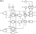
- FIG 1 a flow diagram illustrating a method for comparing reference values in medical imaging processes of different medical imaging devices 1 at a local medical imaging device, in particular at a local medical imaging device located at a hospital is presented.
- the method establishes a fundament for comparing reference values 16 of radiation used for medical imaging although the medical imaging devices 1 might differ in respect to their location and their vendor and/or in the modality of using the reference values.
- the medical imaging device 1 is a computer tomography (CT)-scanner, a nuclear medicine (NM)-scanner, positron emission tomography (PET)-scanner, a X-ray-scanner, a fluoroscopy (RF)-scanner, a mammography (MG)-scanner, a x-ray-system, a angiography (XA) scanner.
- CT computer tomography
- NM nuclear medicine
- PET positron emission tomography
- X-ray-scanner a fluoroscopy-scanner
- RF fluoroscopy
- MG mammography
- XA angiography
- the reference value depends on a magnitude and/or a duration of radiation a patient is exposed to.
- a data base 10 is provided, wherein the data base 10 is configured such that a reference value 16 is assigned to a defined global identification 15 specifying the medical imaging process.
- the data base 10 is incorporated into a server such as a cloud or a network and for example a reference value is provided for each global identification.
- a configuration module 11 is provided for configuring the data base 10 or for setting the reference values 16.
- the several references values 16 are provided on basis of one reference value 16.
- an algorithm for evaluating the several reference values 16 is provided.
- the several reference values 16 can be extracted by using the algorithm.
- the several reference values 16 form a sub-ensemble corresponding to the one reference value 16.
- the reference values are respectively correlated to a patient related information.
- the several reference values 16 correspond to the same modality but to an adult on one hand and to a pediatric on the other hand, for instance.
- the reference values 16 correspond to the same modality but to a 16 cm phantom ("head”) on the one hand and to a 32 cm phantom ("body”) on the other hand.
- REF 16 phantom
- REF("32 phantom) corresponds to the reference value of the 32 cm phantom
- REF (“Adult") corresponds to the reference value 16 of the adult
- REF (“pediatric”) corresponds to the reference value 16 of the pediatric.
- the defined global identification 15 includes
- a mapping 21 between the local identification, i. e. the local label names, and the global identification 15 is provided, in particular by a mapping module 20.
- the global identifications 16 is provided to the medical imaging device 1, i. e. transferring the global identification 16 to the mapping module 20.
- an analysis 5 of the local identification is performed, for example by analysing the DICOM data.
- relevant DICOM information 6 are extracted from the DICOM data and are transferred to the mapping module 20 for using the extracted DICOM information 6 as well as the results of analysing 5 the DICOM data for mapping 21.
- the mapping 21 is performed automatically. It is also thinkable that an artificial network is used for mapping 21 the local identification to the global identification 15.
- the mapping 21 is realized manually, i.e. the operator correlates the local identification to a global identification 15.
- the manual mapping is performed for those global identifications 15 that could not correlated to a local identification automatically.
- the mapping 21 is visualized to the operator, for example on a screen. For example, an array or matrix including both the local identification and the correlated global identification 15 is presented.
- using the proper global identification is supported by the visualisation and the operator can select or choose the proper global identification 15.
- the operator is informed about the change from using the local identification to the global identification 15 for the further proceeding.
- the global identification 15 is incorporated into the medical imaging device 1, in particular the medical imaging device 1 is configured such that the DICOM objects generated by the specific medical imaging devices includes the global identification 15, such as the RADLEX-RPID.
- the mapping 21 using the DICOM data is simplified and the automatically mapping does not fail.
- the use of a complex auto-mapping can be avoided.
- the local medical imaging device After transferring the local identification to the global identification 15 at the local medical imaging device 1, the local medical imaging device 1, in particular a reference module 22, receives the reference data 16 based on the global identification 15.
- the reference data 16 includes national reference values, institutional reference values and/or network specified reference values.
- an alert notification 25 is generated by an alert module 24.
- the results of analysing 5 the local identification and the results of the alert module 24 are transferred to a data collector module 30. These information are saved in the data collector module 30 together with the corresponding global identification 15. As a consequence, information saved in the data collector module 30 can be analysed by a analyse module 31, for example for further mapping processes or comparing different local medical imaging devices 1. Additionally the data collector module 30 can be used for benchmarking using a benchmark module 32.
- the benchmarking module 32 is configured for presenting benchmarking data set 41 to the local medical imaging device 1 or hospital 50 for comparing reference values of local medical imaging devices 1 of different hospitals 50 of the same type, location and/or area.
- the benchmarking module 32 is able to compare medical imaging devices 1 being comparable based on one or several classification values, such as a type of the hospital, a region and/or area the hospital is located in.
- classification values such as a type of the hospital, a region and/or area the hospital is located in.
- the local medical imaging device 1 or hospital 50 is registered by assigning an identifier to the hospital 50.
- the classification values of the hospital 50 are saved together with the identifier to a memory during the registration 60, for example at the server.
- the server or the cloud can rely on the classification values for calculating a benchmarking data set 41.
- the benchmarking data set 41 comprises a min-value, a max-value, a 25%-value, a 75% value, a mean value and/or a median value of reference values for different classification values, preferably defined by the registering 60 the hospital 50.
- the benchmarking data set 41 is generated monthly by a server 40 for calculating benchmarking data sets be relying on the data base. For example,
- incorporating 8 the global identification into the medical imaging device is provided, in particular such that the medical imaging device 1 can use the incorporated global identification 15 for labelling the medical imaging process, for example by incorporating the global identification into DICOM object, in particular for each modality.
- the medical imaging device 1 can use the incorporated global identification 15 for labelling the medical imaging process, for example by incorporating the global identification into DICOM object, in particular for each modality.
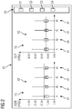
- FIG 2 a presentation of a benchmarking data set 41 according to the preferred embodiment of the present invention is shown.
- a benchmarking data set 41 is provided to an operator on a screen such as a display of a workstation, of a tablet or a smartphone, preferably in form of a boxplot.
- the benchmarking data set 41 is presented in form of a boxplot and benchmarking data sets 41 for different classification values are displayed next to each other.
- such an illustration of the results is available as well to show the evaluation with respect to the data available at the server, in particular at the cloud, without applying the restrictive classification values.
- a control area 55 is displayed on the screen 61, wherein the user can chose a specific modality in a list of modalities 51. Furthermore the user can select one or more RADLEX body regions from a list of RADLEX body regions 52, one or more RADLEX protocols from a list of RADLEX protocols 53 and/or the patient age 54 (adult or pediatric).
- an input unit is provided, wherein the input unit represents a human machine interface.
- the benchmarking data being presented on the screen depends on the KPI defined for the selected modality. In the displayed embodiment it is provided that the benchmarking data for a first KPI 42, such a SSDE-value, and a second KPI 43, such as a DLP-value, are presented next to each other.
Landscapes
- Engineering & Computer Science (AREA)
- Health & Medical Sciences (AREA)
- Medical Informatics (AREA)
- Public Health (AREA)
- General Health & Medical Sciences (AREA)
- Primary Health Care (AREA)
- Epidemiology (AREA)
- Databases & Information Systems (AREA)
- Biomedical Technology (AREA)
- Data Mining & Analysis (AREA)
- Business, Economics & Management (AREA)
- General Business, Economics & Management (AREA)
- Theoretical Computer Science (AREA)
- Nuclear Medicine, Radiotherapy & Molecular Imaging (AREA)
- Radiology & Medical Imaging (AREA)
- Pathology (AREA)
- Physics & Mathematics (AREA)
- General Engineering & Computer Science (AREA)
- General Physics & Mathematics (AREA)
- Measuring And Recording Apparatus For Diagnosis (AREA)
- Apparatus For Radiation Diagnosis (AREA)
Abstract
- providing a data base (10) of reference values (16), wherein each reference value (16) of the data base (10) is assigned to a defined global identification (15) specifying the medical imaging process;
- creating a label for the medical imaging process at a local medical imaging device (1) by using the defined global identification (15) for specifying the medical imaging process; and
- providing a mapping (21) that assigns a local identification to the defined global identification (16) for supporting the creation of the label for the medical imaging process.
Description
- The present invention describes a method for comparing reference values in medical imaging processes, a system comprising a local medical imaging device, a computer program product and a computer-readable program.
- Medical imaging devices, such as computer tomography (CT)-scanner, nuclear medicine (NM)-scanner, positron emission tomography (PET)-scanner, X-ray-scanner (XA), fluoroscopy (RF)-scanner, mammography (MG)-scanner, typically use radiation, in particular X-ray radiation, for recording a medical image. For example the radiation dose used for recording the medical image is a crucial technical parameter, since a radiation dose being too large can cause damages to a patient. Therefore, reference values exist that represents critical thresholds.
- However, the reference value being used at different local medical imaging devices might distinguish from each other, because the medical imaging devices are from different vendors and/or the reference value being used depends on the modality of the radiation process. Due to the different individual local identifications for labelling of the medical images at each local medical imaging device it cannot guaranteed that the used reference values of different local medical instruments are comparable with each other.
- It is therefore an object of the present invention to provide a method for comparing reference values in medical imaging processes of different local medical imaging devices, although the medical imaging devices use their own local identification.
- This object is achieved by the method according to
claim 1 for comparing reference values in medical imaging processes, by the system according to claim 12, the computer program product according to claim 13 and by the computer readable computer medium according to claim 14. - According to a first aspect of the present invention a method for comparing reference values in medical imaging processes of different medical imaging devices, is provided, comprising the steps:
- providing a data base of reference values, wherein each reference value of the data base is assigned to a defined global identification specifying the medical imaging process, in particular specifying a medical imaging protocol for the medical imaging process, for example for each medical imaging device, i. e. modality;
- creating a label, such as specific name or number for example, for the medical imaging process at a local medical imaging device and/or a local workstation, in particular a workstation related to the local medical imaging device, by using the defined global identification for specifying the medical imaging process; and
- providing a mapping that assigns a local identification to the defined global identification for supporting the creation of the label for the medical imaging process, wherein the data base is available to several spatial separated local medical imaging devices, in particular to local medical imaging devices at different hospitals.
- Contrary to the state of the art, a mapping is provided, wherein the mapping correlates the individual local identification and the global defined identification, in particular at the individual local medical imaging device. Thus, it is advantageously possible to use a common set of global identifications for specifying each medical imaging process or type of medical imaging process. As a consequence, a plurality of medical imaging devices being located at different hospitals can specify medical images by referring to a common reference value that is assigned to the respective global identification. In particular, it is possible to compare the medical imaging processes although the different local medical imaging devices use different local identifications. Thereby, the mapping supports the creation of the label in a proper form for comparing, i. e. in a form using the global identification. Furthermore, using the global identification allows establishing a benchmarking of reference values by referring specific reference values to the global identification. Moreover, it is thinkable that the label is created by an operator and or a workstation, such as a computer, being configured for replacing the local identification by the global identification. Preferably, the data base is included into a server, for example in a network or a cloud, and forms a register for reference values. Thus, several different hospitals can access to one common server, one common server system having several server and/or a common cloud.
- Preferably, the term "local medical imaging device" describes a specific medical imaging device, such as a nuclear medicine (NM)-scanner, a positron emission tomography (PET)-scanner, X-ray-scanner (XA), fluoroscopy (RF)-scanner, mammography (MG)-scanner, located at a defined spot. Preferably, the term "local workstation" can also refer to a (personal) computer, a virtual machine running on host hardware, a microcontroller, or an integrated circuit. As an alternative, the local workstation can be a real or a virtual group of computers (the technical term for a real group of computers is "cluster", the technical term for a virtual group of computers is "cloud"). Preferably, the local workstation comprises a calculation unit and a memory unit. A calculation unit can comprise hardware elements and software elements, for example a microprocessor or a field programmable gate array. A memory unit can be embodied as non-permanent main memory (e.g. random access memory) or as permanent mass storage (e.g. hard disk, USB stick, SD card, solid state disk). The local workstation can be part of the local medical imaging device.
- In particular, the local medical imaging device represents a specific medical imaging device located at a specific hospital or institution. That means the "local medical imaging device" and the "local workstation" represent an individual medical imaging device and/or an individual local workstation. Due to a setting incorporated into the medical imaging device by the producer of the medical imaging device and/or due to guidelines established in the hospital and/or country each of the local medical imaging devices can use or create a different local identification for labelling the same medical imaging process by their own. The result is a not-unified labelling by using the respective local identification, since each hospital, workstation and/or medical imaging device uses a different local identification for labelling the same or comparable medical imaging process. In other words: the labelling of the medical imaging process is managed locally and therefore the local identifications differ from each other from medical imaging device to medical imaging device and/or form hospital to hospital.
- In general, the term "medical imaging process" represents a collective term for recording a medical imaging data set by using the specific medical imaging device. The medical imaging process is specified for example by the type of the medical imaging device, a body region being examined, a time of recording the medical imaging device and/or parameters and/or configurations of the medical imaging device. Preferably, the medical imaging process is performed based on a medical imaging protocol comprising the parameters for configuring the medical imaging device and the local identification represents the labelling for the specific medical imaging protocol at the specific medical imaging device and/or hospital.
- In particular, the term "reference value" preferably comprises a magnitude of the radiation and/or a duration of the radiation. Preferably, the reference value comprises a scan-/exam-radiation -dose value, a scan-/exam-duration value and/or an image quality optimized-scan-/exam-radiation-dose value. It is also thinkable that reference value comprises the acquisition duration used by a magnetic resonance scanner or an ultrasound scanner. In particular, it is provided that the duration, such as the acquisition duration, is a benchmark category, when the benchmark category is not the magnitude of the radiation. Preferably, the data base is configured such that for each possible modality and body region a reference value is provided. Preferably, the term "mapping" means that a link, in particular an individual link, is established between the local identification and the global identification. Preferably, the set of global identifications represents a set of standard-modalities, in particular all available or realizable modalities, specified to a body part, in particular an inner body part. Thus, it is possible to establish reference values for each modalities and each body region. As a result, comparing is simplified, since the compared reference values are correlated to comparable modalities and it is possible to establish reference values for each modality. Preferably, the term modality is a generic term for the specific medical imaging device used for exposing the patient to radiation. In particular, the global identification includes information about the medical device, the modality and/or the body part being exposed to the radiation. Preferably, the global identification is represented by a Radlex-standard. According to the Radlex-standard each label name, such as XA abdominal aorta, is identified by a RPID, such as RPID 6195 for instance. Thus, the modality, such as using a X-ray (XA), and the body region or the body part, such as abdominal aorta, can be extracted directly from the label name or indirectly by the RPID. Preferably, only the RPID, the label name, the modality and the body region are used. As a consequence, the method is not restricted to value management relevant modalities. For example, the medical imaging device is a computer tomography (CT)-scanner, a nuclear medicine (NM)-scanner, positron emission tomography (PET)-scanner, a X-ray-scanner, a fluoroscopy (RF)-scanner, a mammography (MG)-scanner, a x-ray-system, a angiography (XA) scanner, magnetic resonance tomography (MRT) scanner or an ultrasound(US) scanner.
- In particular, a key performance indicator (KPI) is provided, wherein the KPI depends on the modality, and the reference values are assigned to these KPI. For CT values for SSDE (size-specific dose estimates)-Max, DLP (dose length product)-Max, CTDIVOL (computed tomography dose index volume)-Max (per scan and/or maximal values in the exam) represent KPIs, as well as a DLP-Acc and an EFF (effective)-Dose (on exam level). Furthermore, a phantom size represents a CT relevant KPI (all reference values should be defaulted to a Body32 phantom and it should be paid attention for events with mixed phantom types). KPI for MG are the values for RP (Reference Point) -Max, AGD (Average Glandular Dose)-Max (per scan and/or maximal values in the exam), AGD-Acc and Eff-Dose (on exam level). For XA, RF, XR and RP the values for RP -Max, DAP (dose-area product)-Max (per scan and/or maximal values in the exam), DAP-Acc and Eff-Dose (on exam level) are the corresponding KPIs. Finally, MBq (MegaBecquerel) and Eff-Dose (on exam level) represent the KPIs for NM and PET.
- Particularly advantageous embodiments and features of the invention are given by the dependent claims as revealed in the following description. Features of different claim categories may be combined as appropriate to give further embodiments not described herein.
- Preferably, it is provided that a benchmarking data set, in particular a benchmarking data set of the reference values, is created for a subset of local medical imaging devices, in particular by a benchmarking module, comprising
- assigning a classification value to the subset of medical imaging device, in particular assigning a classification value to a subset of hospitals assigned the medical imaging device; and
- creating a benchmarking data set based on the classification value. Preferably, the created benchmarking data set is assigned to the global identification. An example for a classification value might be a type of hospital, such as a paediatric clinic, a multi-specialized hospital, an imaging center, a community hospital or an university hospital. Another classification value might be the area of the hospital. For example, the hospital is located in a rural area or metropolitan area. A further classification value might be a region, in which the hospital is located, such as for example a country, for instance Texas, and/or an orientation, for instance north, west, south, middle, east or the like (these classification values might relevant within the country the hospital is located). Thus, it is advantageously possible to create benchmark reference value sets of hospitals being comparable to each other and to compare hospitals of the same kind. Preferably, an identifier for the hospital and the classification values are saved in a hospital data set, in particular at the data base. Preferably, the benchmarking data set comprises a min-value, a max-value, a 25%-value, a 75%-value, a mean value and/or a median value of the reference value, for example the radiation value and/or the radiation duration, for different classification values, preferably defined by the registration of the hospital.
- Preferably, the benchmarking data set is created on demand or after a defined period of time, such as every month or every quarter. For example, the benchmarking data set is created interactively by using an application. Thus, the operator can choose a preferred classification value for comparing the radiation values of different hospitals having the same classification value. It is also thinkable that in a "period selection"-mode a pre-calculated benchmarking data set is provided by a background process, which runs once for every defined period of time to collect data from the data base, in particular from all available servers. Alternatively or additionally in a "Start-/End-Date selection"-mode it is possible to access online calculated benchmarking data sets, in particular calculated by the server or the cloud, provided on request from the data base to which the hospital and/or the medical imaging device is connected to. Preferably the medical imaging device or an interface is configured for choosing between the "Start-/End-Date selection"-mode and "period selection"-mode. By creating the benchmarking data set it is advantageously possible that the operator, for example the hospital, relies on benchmarking data being already present at the data base. Consequently, time is saved and the pre-calculated benchmarking data set can be extended, i. e. updated, every month/quarter to add a new chunk of data being made available to the data base, i. e. to the server. In a preferred embodiment it is provided that the benchmarking data set is automatically created after a defined period of time, such as every month or every quarter, by the server or the system of server.
- According to a preferred embodiment, it is provided that the mapping is provided by an auto-mapping mechanism or a manual mapping mechanism, in particular wherein the mapping is provided by the manual mapping mechanism, when the auto-mapping mechanism fails. By using the auto-mapping mechanism it is advantageously possible to establish a link between the local identification and the global identification. Thereby the auto-mapping mechanism preferably maps local identifications which are different across scanner models, vendors and/or institutions to the global identification. It is also thinkable that the mapping is performed semi-automatically, in particular in such cases in that a specific correlation between a local and a global identification fails in the auto-mapping mechanism. Preferably, it is provided that the auto-mapping mechanism is performed by an artificial network, for example by a deep-learning mechanism. Thereby the artificial network for example analyses previous local identification and settings of the medical imaging device for assigning the local identification to the global identification. Furthermore, it is provided that a duality matrix is realized for all relevant modalities for value relevant tags. In particular, the duality matrix supports the operator, when the global identification is established by the auto-mapping mechanism.
- In particular, it is provided that in a preparation step an identifier, such as a hospital ID, is assigned to the local medical imaging device, in particular to the hospital of the local medical imaging device. For example, assigning the identifier to the local medical imaging device is included in registering the hospital for accessing the data base. Furthermore one or more classification values are assigned to the hospital during registration in the preparation step.
- In another embodiment it is provided that the mapping that assigns the local identification to the defined global identification is visualized to an operator, in particular the global identification and the local identification are visualized simultaneously, and/or wherein the benchmarking data set is visualized to the operator, in particular in form of a boxplot, i. e. box and whisker plots are known from the stock market to display the variance of big data chunks in one picture. Thus, the operator is informed about the global identification and can correlate the global identification to the local identification being used by the operator in the past. In particular, a "standard-view-first"-mode is provided, wherein according to the "standard-view-first" - mode displaying the global identification is preferred to the local identification, in particular the local identification is replaced by the global identification automatically. Alternatively or additionally, a "duality of view"-mode is provided, wherein according to the "duality of view"-mode the global identification and the local identification are displayed together. Thus, confusions by the operator can be avoided. In particular, the benchmarking data set for different classification values are presented next to each other for comparing the benchmarking data sets. Furthermore, it is thinkable that a global benchmarking data set, i. e. a teamplay boxplot, is displayed to show the evaluation in respect to the data being available at a server, in particular the cloud, without applying the classification value. Furthermore, it is possible to compare the benchmarking data to previous benchmarking data of the same modality. In particular, the benchmarking data set is provided together with the global identification, such as the RPID.
- According to a preferred embodiment, it is provided that the global identification is included as information into a DICOM object. As a result, the auto-mapping mechanism, for example to the Radlex-standard, does not fail. In other word: the medical imaging device is configured such that the DICOM object is labelled with the global identification automatically by the medical imaging device. For example, the RPID is included to the DICOM-header. As a consequence, no manual mapping is needed, for any modality not just reference value relevant modalities.
- Furthermore, it is provided that the global identification is incorporated into the medical imaging device, in particular such that the medical imaging device can use the incorporated global identification for labelling the medical imaging process. As consequence, it is advantageously possible to perform the labelling without mapping, in particular the auto-mapping mechanism.
- Preferably, it is provided that the data base of reference values is stored on a server. Preferably, the server is part of a network or a cloud being available to several local medical imaging devices, in particular at different hospitals. Thus, the data base can be used by several local medical imaging devices together. Preferably, the reference values or reference durations are stored in a reference matrix being specified to the modality. The reference matrix is the final definition of a value register, which enables global reference value management, such for example time and/or dose or Exam-/scan-time management and benchmarking of reference values. The reference matrix includes the global identifications, in particular specifying the modality, and the reference values.
- According to another embodiment, it is provided that an alarm is generated by an alert module, when the configuration value being used exceed the reference value. Thus the local medical imaging device can be informed about exceeding the reference value, for example by an alert notification, such as an email, to the tenant or operator. In particular, the alert notification includes the event and reference values used by the institution and/or in a network of several medical imaging device. Furthermore, the alert notification includes the national reference value. Preferably, the alert module is included into the local medical imaging devices.
- In a preferred embodiment, it is provided that several references values are provided on basis of one reference values by a configuration module. Thus, several reference values and preferably categories and value/time units per modality can be extracted from one general reference values. For example, the general reference values is given for each modality and by using a k-value specified for each body region the specific reference values can be deduced from the general reference values. It is also thinkable that a correlation between reference values between different phantom types, such as a 16cm or 32cm phantom, and/or a correlation between the reference values of an adult and a pediatric are used for extracting the reference values from a general reference value.
- Another aspect of the present invention is a system comprising a local medical imaging device, wherein the system is configured for
- providing a data base of reference values, wherein each reference value of the data base is assigned to a defined global identification specifying the medical imaging process;
- creating a label for the medical imaging process at a local medical imaging device and/or a local workstation, in particular a workstation related to the local medical imaging device, by using the defined global identification for specifying the medical imaging process; and
- providing a mapping that assigns a local identification to the defined global identification for supporting the creation of the label for the medical imaging process. Preferably, the system comprises at least a control unit having a processor that is configured for executing one of the steps of the method according to the invention.
- A further aspect of the present invention is a computer program product comprising program elements for carrying out the steps of the method according to the present invention, when the program elements are loaded into a memory of a programmable device.
- A further aspect of the present invention is a computer-readable medium on which is stored a program elements that can be read and executed by a computer unit in order to perform steps of the method according to the present invention when the program elements are executed. The computer-readable medium may be a data carrier such as the cloud, a hard disc, CD-ROM, SD-card or other digital medium. The computer unit and the programmable device may be any form of digital calculating device, e.g. a PC, smart phone, tablet computer, cloud, workstation or handheld device.Preferably, the computer unit and/or the programmable device are incorporated into the system described above, in particular into the control unit.
- In the drawing:
- Fig. 1
- shows a flow diagram illustrating a method for comparing reference values in medical imaging processes of different medical imaging devices at a local medical imaging device, in particular at a local medical imaging device located at a hospital and
- FIG. 2
- shows a presentation of a benchmarking data set according to the preferred embodiment of the present invention.
- In
figure 1 a flow diagram illustrating a method for comparing reference values in medical imaging processes of differentmedical imaging devices 1 at a local medical imaging device, in particular at a local medical imaging device located at a hospital is presented. In particular, it is provided that the method establishes a fundament for comparingreference values 16 of radiation used for medical imaging although themedical imaging devices 1 might differ in respect to their location and their vendor and/or in the modality of using the reference values. For example, themedical imaging device 1 is a computer tomography (CT)-scanner, a nuclear medicine (NM)-scanner, positron emission tomography (PET)-scanner, a X-ray-scanner, a fluoroscopy (RF)-scanner, a mammography (MG)-scanner, a x-ray-system, a angiography (XA) scanner. Preferably, the reference value depends on a magnitude and/or a duration of radiation a patient is exposed to. - For realizing a comparison, a
data base 10 is provided, wherein thedata base 10 is configured such that areference value 16 is assigned to a definedglobal identification 15 specifying the medical imaging process. Preferably, thedata base 10 is incorporated into a server such as a cloud or a network and for example a reference value is provided for each global identification. In particular, a configuration module 11 is provided for configuring thedata base 10 or for setting the reference values 16. It is also thinkable that thedata base 10 includes a k-factor for each global identification, wherein the k-factor transforms the reference value to a reference value for a specific organ, in particular based on the formula:
- Furthermore, it is provided that the several references values 16 are provided on basis of one
reference value 16. In particular, an algorithm for evaluating theseveral reference values 16 is provided. In other words: theseveral reference values 16 can be extracted by using the algorithm. For example, theseveral reference values 16 form a sub-ensemble corresponding to the onereference value 16. For instance, the reference values are respectively correlated to a patient related information. Theseveral reference values 16 correspond to the same modality but to an adult on one hand and to a pediatric on the other hand, for instance. Alternatively or additionally, the reference values 16 correspond to the same modality but to a 16 cm phantom ("head") on the one hand and to a 32 cm phantom ("body") on the other hand. Preferably, the algorithm is based on the formula

reference value 16 of the 16 cm phantom, REF("32 phantom") corresponds to the reference value of the 32 cm phantom, REF ("Adult") corresponds to thereference value 16 of the adult and REF ("pediatric") corresponds to thereference value 16 of the pediatric. As a consequence, it is possible to provide all fourreference values 16 based on the reference values 16 of the 16 cm phantom of an adult, for instance by using the configuration module 11. - In particular, the defined
global identification 15 includes - a label of modality for using the reference values 16, such as specifying the
medical imaging device 1 as CT-scanner, and - a label of body region, such as aorta, chest, arteries for example. In particular, the
global identification 15 includes a label. Thus, anindividual reference value 16 can be assigned to each pair comprising modality and body region. Preferably, a Radlex standard description is used asglobal identification 15. Such a Radlex standard unambiguously correlates a label name, such as RPID6195, to a pair comprising a specific modality, such as angiography, and a body-region, such as an abdominal aorta. As a consequence, it is possible to directly extract from theglobal identification 15, i. e. the label name in the Radlex standard, the information about the modality and the body region of the medical imaging process. For comparing the reference values 16 of the different medical imaging devices it is intended that an operator at a localmedical imaging device 1 uses theglobal identification 15 for labelling each medical imaging process. However, each operator and each medical uses an individual labelling, i. e. local identification. - For using the
global identification 15 at the local medical imaging device 1 amapping 21 between the local identification, i. e. the local label names, and theglobal identification 15 is provided, in particular by amapping module 20. Thereby theglobal identifications 16 is provided to themedical imaging device 1, i. e. transferring theglobal identification 16 to themapping module 20. Furthermore, it is provided that an analysis 5 of the local identification is performed, for example by analysing the DICOM data. As a resultrelevant DICOM information 6 are extracted from the DICOM data and are transferred to themapping module 20 for using the extractedDICOM information 6 as well as the results of analysing 5 the DICOM data formapping 21. Preferably, themapping 21 is performed automatically. It is also thinkable that an artificial network is used for mapping 21 the local identification to theglobal identification 15. Alternatively or additionally, it is provided that themapping 21 is realized manually, i.e. the operator correlates the local identification to aglobal identification 15. For example, the manual mapping is performed for thoseglobal identifications 15 that could not correlated to a local identification automatically. In order to support the operator labelling the medical imaging process it is preferably provided that themapping 21 is visualized to the operator, for example on a screen. For example, an array or matrix including both the local identification and the correlatedglobal identification 15 is presented. Thus, using the proper global identification is supported by the visualisation and the operator can select or choose the properglobal identification 15. Furthermore, it is conceivable that the operator is informed about the change from using the local identification to theglobal identification 15 for the further proceeding. - It is also thinkable that the
global identification 15 is incorporated into themedical imaging device 1, in particular themedical imaging device 1 is configured such that the DICOM objects generated by the specific medical imaging devices includes theglobal identification 15, such as the RADLEX-RPID. As a consequence, themapping 21 using the DICOM data is simplified and the automatically mapping does not fail. In particular, it is possible to extract the RADLEX RPID directly from the DICOM data. Thus, the use of a complex auto-mapping can be avoided. - After transferring the local identification to the
global identification 15 at the localmedical imaging device 1, the localmedical imaging device 1, in particular areference module 22, receives thereference data 16 based on theglobal identification 15. In particular, thereference data 16 includes national reference values, institutional reference values and/or network specified reference values. Provided that a corresponding configuration value being used at the local medical imaging device exceeds thereference value 16 analert notification 25 is generated by analert module 24. By transferring 26 the alert notification to the operator of the localmedical imaging device 1 the operator is informed about the mismatch between the used reference value and the reference value. - Furthermore it is provided that the results of analysing 5 the local identification and the results of the
alert module 24 are transferred to adata collector module 30. These information are saved in thedata collector module 30 together with the correspondingglobal identification 15. As a consequence, information saved in thedata collector module 30 can be analysed by a analysemodule 31, for example for further mapping processes or comparing different localmedical imaging devices 1. Additionally thedata collector module 30 can be used for benchmarking using abenchmark module 32. - In particular, it is provided that the
benchmarking module 32 is configured for presenting benchmarking data set 41 to the localmedical imaging device 1 orhospital 50 for comparing reference values of localmedical imaging devices 1 ofdifferent hospitals 50 of the same type, location and/or area. In other words: the benchmarkingmodule 32 is able to comparemedical imaging devices 1 being comparable based on one or several classification values, such as a type of the hospital, a region and/or area the hospital is located in. Preferably, it is possible to select or to choose the classification values for calculating different benchmarking data sets at the local medical imaging device. - Preferably, it is provided that in a preparation step the local
medical imaging device 1 orhospital 50 is registered by assigning an identifier to thehospital 50. For example, the classification values of thehospital 50 are saved together with the identifier to a memory during theregistration 60, for example at the server. As a result, the server or the cloud can rely on the classification values for calculating abenchmarking data set 41. Preferably, thebenchmarking data set 41 comprises a min-value, a max-value, a 25%-value, a 75% value, a mean value and/or a median value of reference values for different classification values, preferably defined by the registering 60 thehospital 50. Further, it is provided that thebenchmarking data set 41 is generated monthly by aserver 40 for calculating benchmarking data sets be relying on the data base. For example, - a global benchmarking data set 41 or
- a
benchmarking data set 41 based on one classification value or based on a set of classification values is calculated. This calculated benchmarking data set 41 can be provided to the localmedical imaging device 1 orhospital 50. Thus, each registeredhospital 50 has immediately access to the preferredbenchmarking data set 41. - Furthermore, incorporating 8 the global identification into the medical imaging device is provided, in particular such that the
medical imaging device 1 can use the incorporatedglobal identification 15 for labelling the medical imaging process, for example by incorporating the global identification into DICOM object, in particular for each modality. As consequence, it is advantageously possible to perform the labelling withoutmapping 21, in particular the auto-mapping mechanism. - In
figure 2 a presentation of abenchmarking data set 41 according to the preferred embodiment of the present invention is shown. For example, such abenchmarking data set 41 is provided to an operator on a screen such as a display of a workstation, of a tablet or a smartphone, preferably in form of a boxplot. In particular, thebenchmarking data set 41 is presented in form of a boxplot and benchmarking data sets 41 for different classification values are displayed next to each other. Moreover, such an illustration of the results is available as well to show the evaluation with respect to the data available at the server, in particular at the cloud, without applying the restrictive classification values. Finally, there is an evaluation showing the results of the hospital itself with its own values. - Furthermore, a
control area 55 is displayed on thescreen 61, wherein the user can chose a specific modality in a list ofmodalities 51. Furthermore the user can select one or more RADLEX body regions from a list ofRADLEX body regions 52, one or more RADLEX protocols from a list ofRADLEX protocols 53 and/or the patient age 54 (adult or pediatric). For selecting the modality an input unit is provided, wherein the input unit represents a human machine interface. Preferably, it is provided that the benchmarking data being presented on the screen depends on the KPI defined for the selected modality. In the displayed embodiment it is provided that the benchmarking data for afirst KPI 42, such a SSDE-value, and asecond KPI 43, such as a DLP-value, are presented next to each other.
Claims (14)
- Method for comparing reference values in medical imaging processes of different medical imaging devices (1), comprising the steps:- providing a data base (10) of reference values (16), wherein each reference value (16) of the data base (10) is assigned to a defined global identification (15) specifying the medical imaging process;- creating a label for the medical imaging process at a local medical imaging device (1) and/or a local workstation, in particular a workstation related to the local medical imaging device (1), by using the defined global identification (15) for specifying the medical imaging process; and- providing a mapping (21) that assigns a local identification to the defined global identification (16) for supporting the creation of the label for the medical imaging process, wherein the data base (10) is available to several spatial separated local medical imaging devices (1), in particular to local medical imaging devices (1) at different hospitals (50).
- Method according to claim 1, wherein a benchmarking data set (41) is created for a subset of local medical imaging devices (1), in particular of different hospitals (50), comprising- assigning a classification value to the medical imaging device (1), in particular to the hospital (50) of the medical imaging device; and- creating a benchmarking data set (41) based on the classification value.
- Method according to claim 2, wherein the benchmarking data set (41) is created on demand or after a defined period of time.
- Method according to one of the preceding claims, wherein the mapping (21) is provided by an auto-mapping mechanism or a manual mapping mechanism, in particular wherein the mapping is provided by the manual mapping mechanism, when the auto-mapping mechanism fails.
- Method according to claim 4, wherein the auto-mapping mechanism is performed by an artificial network.
- Method according to one of the preceding claims, wherein in a preparation step an identifier is assigned to the local medical imaging device (1), in particular to the hospital (50) of the local medical imaging device (1).
- Method according to one of the preceding claims, wherein the mapping (21) that assigns the local identification to the defined global identification (15) is visualized to an operator, in particular the global identification (15) and the local identification are visualized simultaneously and/or wherein the benchmarking data set (41) is visualized to the operator.
- Method according to one of the preceding claims, wherein the global identification (15) is included as information into a DICOM-object.
- Method according to one of the preceding claims, wherein the data base (10) of reference values (16) is stored on a server.
- Method according to one of the preceding claims, wherein an alarm is generated by an alert module (24), when a configuration values being used exceed the reference value (16).
- Method according to one of the preceding claims, wherein several references values are provided on basis of one reference values (16) by a configuration module (11).
- System comprising a local medical imaging device (1) and/or a local workstation, wherein the system is configured for- providing a data base (10) of reference values (16), wherein each reference value (16) of the data base (10) is assigned to a defined global identification (15) specifying the medical imaging process;- Creating a label for the medical imaging process at the local medical imaging device (1) and/or the local workstation, in particular a workstation related to the local medical imaging device (1), by using the defined global identification (15) for specifying the medical imaging process; and- providing a mapping (21) that assigns a local identification to the defined global identification (15) for supporting the creation the label for the medical imaging process, wherein the data base (10) is available to several spatial separated local medical imaging devices (1).
- Computer program product comprising program elements which induce a programmable device to carry out the steps of the method according one of the preceding claims, when the program elements are loaded into a memory of a programmable device.
- Computer-readable medium on which is stored program elements than can be read and executed by a computer unit in order to perform steps of the method according one of the claims 1 to 9, when the program elements are executed by the computer unit.
Priority Applications (3)
| Application Number | Priority Date | Filing Date | Title |
|---|---|---|---|
| EP17188845.6A EP3451210B1 (en) | 2017-08-31 | 2017-08-31 | Method for comparing reference values in medical imaging processes, system comprising a local medical imaging device, computer program product and computer-readable program |
| US16/114,617 US11636953B2 (en) | 2017-08-31 | 2018-08-28 | Method for comparing reference values in medical imaging processes, system comprising a local medical imaging device, computer program product and computer-readable program |
| CN201811016565.3A CN109427416B (en) | 2017-08-31 | 2018-08-31 | Method, system for comparing reference values in a medical imaging procedure |
Applications Claiming Priority (1)
| Application Number | Priority Date | Filing Date | Title |
|---|---|---|---|
| EP17188845.6A EP3451210B1 (en) | 2017-08-31 | 2017-08-31 | Method for comparing reference values in medical imaging processes, system comprising a local medical imaging device, computer program product and computer-readable program |
Publications (2)
| Publication Number | Publication Date |
|---|---|
| EP3451210A1 true EP3451210A1 (en) | 2019-03-06 |
| EP3451210B1 EP3451210B1 (en) | 2021-03-03 |
Family
ID=59772436
Family Applications (1)
| Application Number | Title | Priority Date | Filing Date |
|---|---|---|---|
| EP17188845.6A Active EP3451210B1 (en) | 2017-08-31 | 2017-08-31 | Method for comparing reference values in medical imaging processes, system comprising a local medical imaging device, computer program product and computer-readable program |
Country Status (3)
| Country | Link |
|---|---|
| US (1) | US11636953B2 (en) |
| EP (1) | EP3451210B1 (en) |
| CN (1) | CN109427416B (en) |
Cited By (2)
| Publication number | Priority date | Publication date | Assignee | Title |
|---|---|---|---|---|
| DE102020213497A1 (en) | 2020-10-27 | 2022-04-28 | Siemens Healthcare Gmbh | Improved dose data management system and procedures |
| EP4435795A1 (en) * | 2023-03-23 | 2024-09-25 | GE Precision Healthcare LLC | Systems and methods for automatic benchmarking for radiology |
Citations (1)
| Publication number | Priority date | Publication date | Assignee | Title |
|---|---|---|---|---|
| WO2014116926A1 (en) * | 2013-01-24 | 2014-07-31 | The Research Foundation For The State University Of New York | Real-time national radiation dose database and monitoring radiation dosages using this database |
Family Cites Families (13)
| Publication number | Priority date | Publication date | Assignee | Title |
|---|---|---|---|---|
| US6272469B1 (en) * | 1998-11-25 | 2001-08-07 | Ge Medical Systems Global Technology Company, Llc | Imaging system protocol handling method and apparatus |
| DE10213284B4 (en) | 2002-03-25 | 2007-11-08 | Siemens Ag | Method for imaging in medical imaging |
| US20070081700A1 (en) | 2005-09-29 | 2007-04-12 | General Electric Company | Systems, methods and apparatus for creation of a database of images from categorical indices |
| US8489431B2 (en) * | 2009-03-20 | 2013-07-16 | General Electric Company | System and method of remote reporting of radiation dose usage in image acquisition |
| CN102576387B (en) * | 2009-10-22 | 2016-08-03 | 皇家飞利浦电子股份有限公司 | Sweep parameter strategy |
| WO2011068475A1 (en) | 2009-11-26 | 2011-06-09 | Agency For Science, Technology And Research | A method for construction and use of a probabilistic atlas for diagnosis and prediction of a medical outcome |
| US9129044B2 (en) * | 2010-04-30 | 2015-09-08 | Cornell University | System and method for radiation dose reporting |
| JP5632203B2 (en) | 2010-06-08 | 2014-11-26 | 株式会社東芝 | Ultrasonic diagnostic apparatus, ultrasonic image processing apparatus, and ultrasonic image processing program |
| US8503613B2 (en) * | 2010-11-24 | 2013-08-06 | General Electric Company | Dose level indication |
| DE102012211892B4 (en) | 2012-07-09 | 2015-03-19 | Siemens Aktiengesellschaft | Method for extracting a data record from a medical image data record and medical image recording device and computer program |
| CN106530236B (en) | 2015-09-11 | 2020-06-02 | 上海联影医疗科技有限公司 | Medical image processing method and system |
| CN107025369B (en) | 2016-08-03 | 2020-03-10 | 北京推想科技有限公司 | Method and device for performing conversion learning on medical images |
| CN106682435B (en) | 2016-12-31 | 2021-01-29 | 西安百利信息科技有限公司 | System and method for automatically detecting lesion in medical image through multi-model fusion |
-
2017
- 2017-08-31 EP EP17188845.6A patent/EP3451210B1/en active Active
-
2018
- 2018-08-28 US US16/114,617 patent/US11636953B2/en active Active
- 2018-08-31 CN CN201811016565.3A patent/CN109427416B/en active Active
Patent Citations (1)
| Publication number | Priority date | Publication date | Assignee | Title |
|---|---|---|---|---|
| WO2014116926A1 (en) * | 2013-01-24 | 2014-07-31 | The Research Foundation For The State University Of New York | Real-time national radiation dose database and monitoring radiation dosages using this database |
Non-Patent Citations (4)
| Title |
|---|
| COOK T S ET AL: "Automated Extraction of Radiation Dose Information for CT Examinations", JOURNAL OF THE AMERICAN COLLEGE OF RADIOLOGY, ELSEVIER, AMSTERDAM, NL, vol. 7, no. 11, 1 November 2010 (2010-11-01), pages 871 - 877, XP027453142, ISSN: 1546-1440, [retrieved on 20101030], DOI: 10.1016/J.JACR.2010.06.026 * |
| RICHARD L MORIN ET AL: "ACR Dose Index Registry", JOURNAL OF THE AMERICAN COLLEGE OF RADIOLOGY, vol. 8, no. 4, 1 March 2011 (2011-03-01), pages 288 - 291, XP028159641, ISSN: 1546-1440, [retrieved on 20110103], DOI: 10.1016/J.JACR.2010.12.022 * |
| SILVA LUÍS A ET AL: "Normalizing Heterogeneous Medical Imaging Data to Measure the Impact of Radiation Dose", JOURNAL OF DIGITAL IMAGING, SPRINGER-VERLAG, CHAM, vol. 28, no. 6, 27 May 2015 (2015-05-27), pages 671 - 683, XP035895352, ISSN: 0897-1889, [retrieved on 20150527], DOI: 10.1007/S10278-015-9805-5 * |
| SINGH SARABJEET ET AL: "Standardized CT protocols and nomenclature: better, but not yet there", PEDIATRIC RADIOLOGY, SPRINGER VERLAG, DE, vol. 44, no. 3, 11 October 2014 (2014-10-11), pages 440 - 443, XP035406755, ISSN: 0301-0449, [retrieved on 20141011], DOI: 10.1007/S00247-014-3096-8 * |
Cited By (2)
| Publication number | Priority date | Publication date | Assignee | Title |
|---|---|---|---|---|
| DE102020213497A1 (en) | 2020-10-27 | 2022-04-28 | Siemens Healthcare Gmbh | Improved dose data management system and procedures |
| EP4435795A1 (en) * | 2023-03-23 | 2024-09-25 | GE Precision Healthcare LLC | Systems and methods for automatic benchmarking for radiology |
Also Published As
| Publication number | Publication date |
|---|---|
| US11636953B2 (en) | 2023-04-25 |
| CN109427416A (en) | 2019-03-05 |
| US20190066848A1 (en) | 2019-02-28 |
| EP3451210B1 (en) | 2021-03-03 |
| CN109427416B (en) | 2022-02-08 |
Similar Documents
| Publication | Publication Date | Title |
|---|---|---|
| JP6796060B2 (en) | Image report annotation identification | |
| JP5744877B2 (en) | System and method for supporting clinical judgment | |
| EP2169577A1 (en) | Method and system for medical imaging reporting | |
| JP7403434B2 (en) | Method and system for automatic scanning recommendation protocol | |
| EP4321100A1 (en) | Medical image device, medical image method, and medical image program | |
| US20010032101A1 (en) | Management system and method for the management of medical data | |
| US11468659B2 (en) | Learning support device, learning support method, learning support program, region-of-interest discrimination device, region-of-interest discrimination method, region-of-interest discrimination program, and learned model | |
| US11669960B2 (en) | Learning system, method, and program | |
| US8892577B2 (en) | Apparatus and method for storing medical information | |
| BRPI0917748A2 (en) | image processing system, medical imaging workstation, method of generating a medical image visualization and computer program product | |
| US20250046458A1 (en) | Systems and methods for generating correct radiological recommendations | |
| CN103258111A (en) | Structured, image-assisted finding generation | |
| JP2019061579A (en) | Learning support device, operation method for learning support device, learning support program, learning support system, and terminal device | |
| CN103608843A (en) | Image display apparatus | |
| US20070078674A1 (en) | Display method for image-based questionnaires | |
| US20090016579A1 (en) | Method and system for performing quality control of medical images in a clinical trial | |
| US8923582B2 (en) | Systems and methods for computer aided detection using pixel intensity values | |
| US7961921B2 (en) | System and method for medical diagnosis and tracking using three-dimensional subtraction in a picture archiving communication system | |
| EP3451210B1 (en) | Method for comparing reference values in medical imaging processes, system comprising a local medical imaging device, computer program product and computer-readable program | |
| WO2021177358A1 (en) | Information processing device, information processing method, and information processing program | |
| JP2025178348A (en) | Medical image display device, method, and program | |
| WO2022209501A1 (en) | Information processing device, method for operating information processing device, and program for operating information processing device | |
| US20080120140A1 (en) | Managing medical imaging data | |
| Seibert | Modalities and data acquisition | |
| Hara et al. | ACRIN CT colonography trial: does reader’s preference for primary two-dimensional versus primary three-dimensional interpretation affect performance? |
Legal Events
| Date | Code | Title | Description |
|---|---|---|---|
| PUAI | Public reference made under article 153(3) epc to a published international application that has entered the european phase |
Free format text: ORIGINAL CODE: 0009012 |
|
| STAA | Information on the status of an ep patent application or granted ep patent |
Free format text: STATUS: REQUEST FOR EXAMINATION WAS MADE |
|
| 17P | Request for examination filed |
Effective date: 20180518 |
|
| AK | Designated contracting states |
Kind code of ref document: A1 Designated state(s): AL AT BE BG CH CY CZ DE DK EE ES FI FR GB GR HR HU IE IS IT LI LT LU LV MC MK MT NL NO PL PT RO RS SE SI SK SM TR |
|
| AX | Request for extension of the european patent |
Extension state: BA ME |
|
| STAA | Information on the status of an ep patent application or granted ep patent |
Free format text: STATUS: EXAMINATION IS IN PROGRESS |
|
| 17Q | First examination report despatched |
Effective date: 20200127 |
|
| REG | Reference to a national code |
Ref country code: DE Ref legal event code: R079 Ref document number: 602017033685 Country of ref document: DE Free format text: PREVIOUS MAIN CLASS: G06F0019000000 Ipc: G16H0040200000 |
|
| GRAP | Despatch of communication of intention to grant a patent |
Free format text: ORIGINAL CODE: EPIDOSNIGR1 |
|
| STAA | Information on the status of an ep patent application or granted ep patent |
Free format text: STATUS: GRANT OF PATENT IS INTENDED |
|
| RIC1 | Information provided on ipc code assigned before grant |
Ipc: G16H 40/20 20180101AFI20200824BHEP |
|
| GRAJ | Information related to disapproval of communication of intention to grant by the applicant or resumption of examination proceedings by the epo deleted |
Free format text: ORIGINAL CODE: EPIDOSDIGR1 |
|
| INTG | Intention to grant announced |
Effective date: 20200915 |
|
| STAA | Information on the status of an ep patent application or granted ep patent |
Free format text: STATUS: EXAMINATION IS IN PROGRESS |
|
| GRAP | Despatch of communication of intention to grant a patent |
Free format text: ORIGINAL CODE: EPIDOSNIGR1 |
|
| STAA | Information on the status of an ep patent application or granted ep patent |
Free format text: STATUS: GRANT OF PATENT IS INTENDED |
|
| INTC | Intention to grant announced (deleted) | ||
| INTG | Intention to grant announced |
Effective date: 20201030 |
|
| GRAS | Grant fee paid |
Free format text: ORIGINAL CODE: EPIDOSNIGR3 |
|
| GRAA | (expected) grant |
Free format text: ORIGINAL CODE: 0009210 |
|
| STAA | Information on the status of an ep patent application or granted ep patent |
Free format text: STATUS: THE PATENT HAS BEEN GRANTED |
|
| AK | Designated contracting states |
Kind code of ref document: B1 Designated state(s): AL AT BE BG CH CY CZ DE DK EE ES FI FR GB GR HR HU IE IS IT LI LT LU LV MC MK MT NL NO PL PT RO RS SE SI SK SM TR |
|
| REG | Reference to a national code |
Ref country code: GB Ref legal event code: FG4D |
|
| REG | Reference to a national code |
Ref country code: AT Ref legal event code: REF Ref document number: 1368087 Country of ref document: AT Kind code of ref document: T Effective date: 20210315 Ref country code: CH Ref legal event code: EP |
|
| REG | Reference to a national code |
Ref country code: DE Ref legal event code: R096 Ref document number: 602017033685 Country of ref document: DE |
|
| REG | Reference to a national code |
Ref country code: IE Ref legal event code: FG4D |
|
| REG | Reference to a national code |
Ref country code: LT Ref legal event code: MG9D |
|
| PG25 | Lapsed in a contracting state [announced via postgrant information from national office to epo] |
Ref country code: NO Free format text: LAPSE BECAUSE OF FAILURE TO SUBMIT A TRANSLATION OF THE DESCRIPTION OR TO PAY THE FEE WITHIN THE PRESCRIBED TIME-LIMIT Effective date: 20210603 Ref country code: HR Free format text: LAPSE BECAUSE OF FAILURE TO SUBMIT A TRANSLATION OF THE DESCRIPTION OR TO PAY THE FEE WITHIN THE PRESCRIBED TIME-LIMIT Effective date: 20210303 Ref country code: GR Free format text: LAPSE BECAUSE OF FAILURE TO SUBMIT A TRANSLATION OF THE DESCRIPTION OR TO PAY THE FEE WITHIN THE PRESCRIBED TIME-LIMIT Effective date: 20210604 Ref country code: FI Free format text: LAPSE BECAUSE OF FAILURE TO SUBMIT A TRANSLATION OF THE DESCRIPTION OR TO PAY THE FEE WITHIN THE PRESCRIBED TIME-LIMIT Effective date: 20210303 Ref country code: BG Free format text: LAPSE BECAUSE OF FAILURE TO SUBMIT A TRANSLATION OF THE DESCRIPTION OR TO PAY THE FEE WITHIN THE PRESCRIBED TIME-LIMIT Effective date: 20210603 Ref country code: LT Free format text: LAPSE BECAUSE OF FAILURE TO SUBMIT A TRANSLATION OF THE DESCRIPTION OR TO PAY THE FEE WITHIN THE PRESCRIBED TIME-LIMIT Effective date: 20210303 |
|
| REG | Reference to a national code |
Ref country code: NL Ref legal event code: MP Effective date: 20210303 |
|
| REG | Reference to a national code |
Ref country code: AT Ref legal event code: MK05 Ref document number: 1368087 Country of ref document: AT Kind code of ref document: T Effective date: 20210303 |
|
| PG25 | Lapsed in a contracting state [announced via postgrant information from national office to epo] |
Ref country code: SE Free format text: LAPSE BECAUSE OF FAILURE TO SUBMIT A TRANSLATION OF THE DESCRIPTION OR TO PAY THE FEE WITHIN THE PRESCRIBED TIME-LIMIT Effective date: 20210303 Ref country code: RS Free format text: LAPSE BECAUSE OF FAILURE TO SUBMIT A TRANSLATION OF THE DESCRIPTION OR TO PAY THE FEE WITHIN THE PRESCRIBED TIME-LIMIT Effective date: 20210303 Ref country code: LV Free format text: LAPSE BECAUSE OF FAILURE TO SUBMIT A TRANSLATION OF THE DESCRIPTION OR TO PAY THE FEE WITHIN THE PRESCRIBED TIME-LIMIT Effective date: 20210303 Ref country code: PL Free format text: LAPSE BECAUSE OF FAILURE TO SUBMIT A TRANSLATION OF THE DESCRIPTION OR TO PAY THE FEE WITHIN THE PRESCRIBED TIME-LIMIT Effective date: 20210303 |
|
| PG25 | Lapsed in a contracting state [announced via postgrant information from national office to epo] |
Ref country code: NL Free format text: LAPSE BECAUSE OF FAILURE TO SUBMIT A TRANSLATION OF THE DESCRIPTION OR TO PAY THE FEE WITHIN THE PRESCRIBED TIME-LIMIT Effective date: 20210303 |
|
| PG25 | Lapsed in a contracting state [announced via postgrant information from national office to epo] |
Ref country code: CZ Free format text: LAPSE BECAUSE OF FAILURE TO SUBMIT A TRANSLATION OF THE DESCRIPTION OR TO PAY THE FEE WITHIN THE PRESCRIBED TIME-LIMIT Effective date: 20210303 Ref country code: EE Free format text: LAPSE BECAUSE OF FAILURE TO SUBMIT A TRANSLATION OF THE DESCRIPTION OR TO PAY THE FEE WITHIN THE PRESCRIBED TIME-LIMIT Effective date: 20210303 Ref country code: AT Free format text: LAPSE BECAUSE OF FAILURE TO SUBMIT A TRANSLATION OF THE DESCRIPTION OR TO PAY THE FEE WITHIN THE PRESCRIBED TIME-LIMIT Effective date: 20210303 Ref country code: SM Free format text: LAPSE BECAUSE OF FAILURE TO SUBMIT A TRANSLATION OF THE DESCRIPTION OR TO PAY THE FEE WITHIN THE PRESCRIBED TIME-LIMIT Effective date: 20210303 |
|
| PG25 | Lapsed in a contracting state [announced via postgrant information from national office to epo] |
Ref country code: IS Free format text: LAPSE BECAUSE OF FAILURE TO SUBMIT A TRANSLATION OF THE DESCRIPTION OR TO PAY THE FEE WITHIN THE PRESCRIBED TIME-LIMIT Effective date: 20210703 Ref country code: PT Free format text: LAPSE BECAUSE OF FAILURE TO SUBMIT A TRANSLATION OF THE DESCRIPTION OR TO PAY THE FEE WITHIN THE PRESCRIBED TIME-LIMIT Effective date: 20210705 Ref country code: RO Free format text: LAPSE BECAUSE OF FAILURE TO SUBMIT A TRANSLATION OF THE DESCRIPTION OR TO PAY THE FEE WITHIN THE PRESCRIBED TIME-LIMIT Effective date: 20210303 Ref country code: SK Free format text: LAPSE BECAUSE OF FAILURE TO SUBMIT A TRANSLATION OF THE DESCRIPTION OR TO PAY THE FEE WITHIN THE PRESCRIBED TIME-LIMIT Effective date: 20210303 |
|
| REG | Reference to a national code |
Ref country code: DE Ref legal event code: R097 Ref document number: 602017033685 Country of ref document: DE |
|
| PLBE | No opposition filed within time limit |
Free format text: ORIGINAL CODE: 0009261 |
|
| STAA | Information on the status of an ep patent application or granted ep patent |
Free format text: STATUS: NO OPPOSITION FILED WITHIN TIME LIMIT |
|
| PG25 | Lapsed in a contracting state [announced via postgrant information from national office to epo] |
Ref country code: AL Free format text: LAPSE BECAUSE OF FAILURE TO SUBMIT A TRANSLATION OF THE DESCRIPTION OR TO PAY THE FEE WITHIN THE PRESCRIBED TIME-LIMIT Effective date: 20210303 Ref country code: DK Free format text: LAPSE BECAUSE OF FAILURE TO SUBMIT A TRANSLATION OF THE DESCRIPTION OR TO PAY THE FEE WITHIN THE PRESCRIBED TIME-LIMIT Effective date: 20210303 Ref country code: ES Free format text: LAPSE BECAUSE OF FAILURE TO SUBMIT A TRANSLATION OF THE DESCRIPTION OR TO PAY THE FEE WITHIN THE PRESCRIBED TIME-LIMIT Effective date: 20210303 |
|
| 26N | No opposition filed |
Effective date: 20211206 |
|
| PG25 | Lapsed in a contracting state [announced via postgrant information from national office to epo] |
Ref country code: SI Free format text: LAPSE BECAUSE OF FAILURE TO SUBMIT A TRANSLATION OF THE DESCRIPTION OR TO PAY THE FEE WITHIN THE PRESCRIBED TIME-LIMIT Effective date: 20210303 |
|
| REG | Reference to a national code |
Ref country code: CH Ref legal event code: PL |
|
| PG25 | Lapsed in a contracting state [announced via postgrant information from national office to epo] |
Ref country code: MC Free format text: LAPSE BECAUSE OF FAILURE TO SUBMIT A TRANSLATION OF THE DESCRIPTION OR TO PAY THE FEE WITHIN THE PRESCRIBED TIME-LIMIT Effective date: 20210303 |
|
| REG | Reference to a national code |
Ref country code: BE Ref legal event code: MM Effective date: 20210831 |
|
| PG25 | Lapsed in a contracting state [announced via postgrant information from national office to epo] |
Ref country code: LI Free format text: LAPSE BECAUSE OF NON-PAYMENT OF DUE FEES Effective date: 20210831 Ref country code: IT Free format text: LAPSE BECAUSE OF FAILURE TO SUBMIT A TRANSLATION OF THE DESCRIPTION OR TO PAY THE FEE WITHIN THE PRESCRIBED TIME-LIMIT Effective date: 20210303 Ref country code: CH Free format text: LAPSE BECAUSE OF NON-PAYMENT OF DUE FEES Effective date: 20210831 |
|
| PG25 | Lapsed in a contracting state [announced via postgrant information from national office to epo] |
Ref country code: IS Free format text: LAPSE BECAUSE OF FAILURE TO SUBMIT A TRANSLATION OF THE DESCRIPTION OR TO PAY THE FEE WITHIN THE PRESCRIBED TIME-LIMIT Effective date: 20210703 Ref country code: LU Free format text: LAPSE BECAUSE OF NON-PAYMENT OF DUE FEES Effective date: 20210831 |
|
| PG25 | Lapsed in a contracting state [announced via postgrant information from national office to epo] |
Ref country code: IE Free format text: LAPSE BECAUSE OF NON-PAYMENT OF DUE FEES Effective date: 20210831 Ref country code: BE Free format text: LAPSE BECAUSE OF NON-PAYMENT OF DUE FEES Effective date: 20210831 |
|
| PG25 | Lapsed in a contracting state [announced via postgrant information from national office to epo] |
Ref country code: CY Free format text: LAPSE BECAUSE OF FAILURE TO SUBMIT A TRANSLATION OF THE DESCRIPTION OR TO PAY THE FEE WITHIN THE PRESCRIBED TIME-LIMIT Effective date: 20210303 |
|
| PG25 | Lapsed in a contracting state [announced via postgrant information from national office to epo] |
Ref country code: HU Free format text: LAPSE BECAUSE OF FAILURE TO SUBMIT A TRANSLATION OF THE DESCRIPTION OR TO PAY THE FEE WITHIN THE PRESCRIBED TIME-LIMIT; INVALID AB INITIO Effective date: 20170831 |
|
| REG | Reference to a national code |
Ref country code: DE Ref legal event code: R081 Ref document number: 602017033685 Country of ref document: DE Owner name: SIEMENS HEALTHINEERS AG, DE Free format text: FORMER OWNER: SIEMENS HEALTHCARE GMBH, MUENCHEN, DE |
|
| PG25 | Lapsed in a contracting state [announced via postgrant information from national office to epo] |
Ref country code: MK Free format text: LAPSE BECAUSE OF FAILURE TO SUBMIT A TRANSLATION OF THE DESCRIPTION OR TO PAY THE FEE WITHIN THE PRESCRIBED TIME-LIMIT Effective date: 20210303 |
|
| PG25 | Lapsed in a contracting state [announced via postgrant information from national office to epo] |
Ref country code: TR Free format text: LAPSE BECAUSE OF FAILURE TO SUBMIT A TRANSLATION OF THE DESCRIPTION OR TO PAY THE FEE WITHIN THE PRESCRIBED TIME-LIMIT Effective date: 20210303 |
|
| PG25 | Lapsed in a contracting state [announced via postgrant information from national office to epo] |
Ref country code: MT Free format text: LAPSE BECAUSE OF FAILURE TO SUBMIT A TRANSLATION OF THE DESCRIPTION OR TO PAY THE FEE WITHIN THE PRESCRIBED TIME-LIMIT Effective date: 20210303 |
|
| PGFP | Annual fee paid to national office [announced via postgrant information from national office to epo] |
Ref country code: GB Payment date: 20250902 Year of fee payment: 9 |
|
| PGFP | Annual fee paid to national office [announced via postgrant information from national office to epo] |
Ref country code: FR Payment date: 20250827 Year of fee payment: 9 |
|
| PGFP | Annual fee paid to national office [announced via postgrant information from national office to epo] |
Ref country code: DE Payment date: 20251020 Year of fee payment: 9 |