EP3445412B1 - Tissue structure scaffolds - Google Patents
Tissue structure scaffolds Download PDFInfo
- Publication number
- EP3445412B1 EP3445412B1 EP17727114.5A EP17727114A EP3445412B1 EP 3445412 B1 EP3445412 B1 EP 3445412B1 EP 17727114 A EP17727114 A EP 17727114A EP 3445412 B1 EP3445412 B1 EP 3445412B1
- Authority
- EP
- European Patent Office
- Prior art keywords
- collagen
- cross
- dimensional tissue
- scaffold
- scaffold structure
- Prior art date
- Legal status (The legal status is an assumption and is not a legal conclusion. Google has not performed a legal analysis and makes no representation as to the accuracy of the status listed.)
- Active
Links
Images





Classifications
-
- A—HUMAN NECESSITIES
- A61—MEDICAL OR VETERINARY SCIENCE; HYGIENE
- A61L—METHODS OR APPARATUS FOR STERILISING MATERIALS OR OBJECTS IN GENERAL; DISINFECTION, STERILISATION OR DEODORISATION OF AIR; CHEMICAL ASPECTS OF BANDAGES, DRESSINGS, ABSORBENT PADS OR SURGICAL ARTICLES; MATERIALS FOR BANDAGES, DRESSINGS, ABSORBENT PADS OR SURGICAL ARTICLES
- A61L27/00—Materials for grafts or prostheses or for coating grafts or prostheses
- A61L27/14—Macromolecular materials
- A61L27/26—Mixtures of macromolecular compounds
-
- A—HUMAN NECESSITIES
- A61—MEDICAL OR VETERINARY SCIENCE; HYGIENE
- A61L—METHODS OR APPARATUS FOR STERILISING MATERIALS OR OBJECTS IN GENERAL; DISINFECTION, STERILISATION OR DEODORISATION OF AIR; CHEMICAL ASPECTS OF BANDAGES, DRESSINGS, ABSORBENT PADS OR SURGICAL ARTICLES; MATERIALS FOR BANDAGES, DRESSINGS, ABSORBENT PADS OR SURGICAL ARTICLES
- A61L27/00—Materials for grafts or prostheses or for coating grafts or prostheses
- A61L27/14—Macromolecular materials
- A61L27/22—Polypeptides or derivatives thereof, e.g. degradation products
- A61L27/24—Collagen
-
- A—HUMAN NECESSITIES
- A61—MEDICAL OR VETERINARY SCIENCE; HYGIENE
- A61L—METHODS OR APPARATUS FOR STERILISING MATERIALS OR OBJECTS IN GENERAL; DISINFECTION, STERILISATION OR DEODORISATION OF AIR; CHEMICAL ASPECTS OF BANDAGES, DRESSINGS, ABSORBENT PADS OR SURGICAL ARTICLES; MATERIALS FOR BANDAGES, DRESSINGS, ABSORBENT PADS OR SURGICAL ARTICLES
- A61L27/00—Materials for grafts or prostheses or for coating grafts or prostheses
- A61L27/50—Materials characterised by their function or physical properties, e.g. injectable or lubricating compositions, shape-memory materials, surface modified materials
- A61L27/56—Porous materials, e.g. foams or sponges
-
- C—CHEMISTRY; METALLURGY
- C08—ORGANIC MACROMOLECULAR COMPOUNDS; THEIR PREPARATION OR CHEMICAL WORKING-UP; COMPOSITIONS BASED THEREON
- C08J—WORKING-UP; GENERAL PROCESSES OF COMPOUNDING; AFTER-TREATMENT NOT COVERED BY SUBCLASSES C08B, C08C, C08F, C08G or C08H
- C08J9/00—Working-up of macromolecular substances to porous or cellular articles or materials; After-treatment thereof
- C08J9/28—Working-up of macromolecular substances to porous or cellular articles or materials; After-treatment thereof by elimination of a liquid phase from a macromolecular composition or article, e.g. drying of coagulum
- C08J9/283—Working-up of macromolecular substances to porous or cellular articles or materials; After-treatment thereof by elimination of a liquid phase from a macromolecular composition or article, e.g. drying of coagulum a discontinuous liquid phase emulsified in a continuous macromolecular phase
-
- A—HUMAN NECESSITIES
- A61—MEDICAL OR VETERINARY SCIENCE; HYGIENE
- A61L—METHODS OR APPARATUS FOR STERILISING MATERIALS OR OBJECTS IN GENERAL; DISINFECTION, STERILISATION OR DEODORISATION OF AIR; CHEMICAL ASPECTS OF BANDAGES, DRESSINGS, ABSORBENT PADS OR SURGICAL ARTICLES; MATERIALS FOR BANDAGES, DRESSINGS, ABSORBENT PADS OR SURGICAL ARTICLES
- A61L2430/00—Materials or treatment for tissue regeneration
Definitions
- This invention relates to a method of manufacture of three dimensional (3D) porous cross-linked collagen or collagen-chitosan scaffolds which may be used for tissue engineering applications.
- the invention also relates to tissue scaffolds made by the method and to devices incorporating the scaffolds.
- Tissue scaffolds find application in tissue engineering for improvement of tissue and organ repair and replacement of biological functions.
- Key components of tissue engineering are biomaterials that provide an interface similar to a naturally occurring extracellular matrix (ECM). This allows cells to grow on the material without being recognized as foreign to the body.
- ECM extracellular matrix
- Biomaterials have been based on biopolymers, especially based on biopolymers derived from the ECM.
- Collagen which is a natural biopolymer, is used in the biomedical industry for applications such as encapsulation, microspheres and matrices for implants and tissue engineering. Chitosan is also useful for biomedical and clinical applications because it has high biocompatibility, biodegradability and nonantigenicity. Chitosan-collagen hybrid polymer network scaffolds have been prepared by freeze-drying and have been evaluated using buffalo stem cells and with respect to growth of hepatic tissue.
- Nanofibrous collagen (NF-collagen) mats have been prepared by using an electrospinning technique.
- the electrospinning technique typically forms two-dimensional collagen sheets and is incapable of fabricating 3D NF-collagen scaffolds with well-defined pore size.
- 3D porous structures is critical to the function of biological tissue, transport of oxygen and nutrients.
- a method of forming a scaffold comprises the steps of: preparing a high internal phase emulsion (HIPE) comprising an aqueous solution of:
- the collagen structure of the present invention may have fundamentally the same chemical composition as natural collagen and may be used for guiding tissue regeneration.
- Particularly advantageous embodiments have the advantage that they exhibit the nanofibrous architecture which is beneficial to regulation of cell adhesion and function.
- Collagen gels form through a single-strand-to- triple helix transition.
- chemical cross-linking is a much more consistent method to ensure structural integrity and to provide a gel which exhibits an optimized, reproducible performance in vivo or under physiological conditions.
- the method may be used to induce tissue formation from liver cells, skin fibroblast cells, bone cells, cartilage or skin cells.
- a three-dimensional tissue scaffold structure comprises: a protein selected from the group consisting of: collagen and mixtures of collagen and chitosan, cross-linked with a cross-linking agent, wherein the scaffold structure comprises voids interconnected by pores and the scaffold includes laminar regions forming webs between adjacent pores.
- scaffold used in the present invention refers to a three-dimensional structure with a porous microstructure suitable to support cells during tissue growth.
- Preferred scaffolds in accordance with this invention have beneficial configuration, morphology and properties suitable for tissue engineering including: good biocompatibility, mechanical performance, a degradation rate which can match a rate of tissue regeneration and a highly porous interconnecting structure. Use of cell lines which are difficult to grow by other methods is facilitated.
- the tissue scaffold of the present invention comprises voids interconnected by pores.
- the scaffold may include struts extending between adjacent pores or voids, the struts each having a length and thickness.
- Embodiments of the invention allow manufacture of a scaffold with a configuration and properties which closely resembles decellularised natural tissue, for example rat liver tissue ( Uygun et al; Nature Medicine 2010, 16, 814-821 . See Figure 2g on page 816) .
- Previously disclosed poly-HIPE structures have a high proportion of spherical or near-spherical voids with circular or near-circular pores.
- a low interconnect pore sized distribution was described in Carnachan, R.J.; Bokhari, M.; Przyborski, S.A. and Cameron, NR.; Soft Matter 2006, 2, 608-616 . See Table 1 and Figure 4 .
- the voids are generally spherical.
- Scaffolds of the present invention may have a substantial absence of spherical voids.
- the struts between adjacent pores may have a wide range of thicknesses and lengths in comparison to poly-HIPE structures, as disclosed in Carnachan et al. Less than 10% of the voids may be spheroidal or approximately spherical. Preferably there are no spheroidal or approximately spherical voids.
- Embodiments of the invention allow manufacture of scaffolds with configurations and properties which closely resemble decellularised natural tissue, for example rat liver tissue.
- Previously disclosed poly-HIPE structures have a high proportion of spherical or near-spherical voids with circular or near-circular pores. A low interconnect pore size distribution was observed, as illustrated in Carnachan, et al.
- the voids are generally spherical, typically having a mean void size of 94 ⁇ m and a range of pore sizes from 10 to 30 ⁇ m and a mean pore size of 19 ⁇ m. This is a narrow pore size distribution.
- Scaffolds of the present invention may have a substantial absence of spherical or spheroidal voids.
- the struts between adjacent pores may have a wide range of thicknesses and lengths in comparison to poly-HIPE structures.
- Embodiments of the invention may provide a high variety of configurations or shapes of the voids and pores.
- the voids may be dimensioned so that 90% of the voids have a mean size in the range 3.5 ⁇ m to 6.5 ⁇ m.
- the voids may typically have a mean size of 4.5 ⁇ m and a standard deviation of ⁇ 1.8 ⁇ m.
- the corresponding structure of rat liver tissue may have a void size of 4.2 ⁇ m ⁇ 2.2 ⁇ m.
- the pore size may be in the range 2.0 ⁇ m to 8.7 ⁇ m, comparable to the pore size of rat liver which may be 1.0 ⁇ m to 12 ⁇ m.
- the uniformity of the dimensions and configurations of the voids and pores are low.
- the boundaries between adjacent voids or pores may be irregular with a substantial absence of spherical voids and circular shaped pores.
- the aspect ratio of the pores may be defined by the ratio of the minimum dimension (dmin) to the maximum dimension (dmax). These may be measured on perpendicular axes or otherwise angled axes. With non-circular pores the minimum and maximum dimensions are not perpendicular. In embodiments the aspect ratio, dmin/dmax, 50% of the pores may be less than about 0.7, alternatively 50% less than about 0.5, alternatively 50% less than about 0.4.
- a prior art poly-HIPE structure may have 50% of voids with an aspect ratio greater than 0.7, commonly greater than 0.8, typically greater than 0.9.
- Scaffolds of the invention may include laminar regions in the form of webs between adjacent pores. This configuration provides a relatively high surface area of the scaffold defining the voids. A high surface area may be beneficial to cell growth, particularly in combination with a high degree of interconnection by pores between the voids.
- the softness or conversely stiffness of the scaffolds may be determined as the gradient of stress-strain behaviour for the scaffold.
- the stiffness of the scaffold is similar to soft tissues such as liver, for example within 30%, alternatively within 20%, further alternatively within 10%, of the value for liver tissue.
- the stiffness may be determined by manual inspection or may be measured instrumentally.
- the scaffold may have a softness selected to be similar to that of natural collagen to facilitate cellular expression, for example of liver cells.
- the stiffness or softness may be controlled to have a value close to that of an alternative tissue type, as required.
- the scaffold of the present invention may exhibit one or more of the following properties.
- the porosity may be greater than 70%, alternatively greater than 80%, alternatively greater than 90%, for example 91% as measured by mercury porosimetry.
- the scaffold of the present invention may find application in making repair tissue which resembles natural tissues, but without having a biological history, for example dependent on age, disease, gender or environmental factors. Consistency of tissue characteristics can be controlled.
- the structure may have struts with a length in the range 0.5 ⁇ m to 6 ⁇ m, preferably 0.6 ⁇ m to 5.5 ⁇ m.
- the struts may have an average thickness of 1 to 3 ⁇ m, preferably 1 to 2 ⁇ m.
- a microfibrous structure is particularly effective in regulating cell adhesion to the scaffold.
- the cross-linking step may take place before or after complete or partial removal of the solvent.
- Cross-linking may be taken to completion before removal of the solvent. Alternatively, cross-linking may be carried out on the solidified solvent-free structure. Alternatively, an initial partial cross-linking step may be followed by solvent removal and a further cross-linking step.
- a preferred cross-linking agent is glutaraldehyde (GA).
- cross-linking agents which may be employed are genipin, carbodiimide, transglutaminase, chondroitin sulphate, dextran dialdehyde, bis(vinylsulfonyl)methane and hexamethylene di-isocyanate.
- the concentration of the cross-linking agent, preferably glutaraldehyde, in the emulsion may in the range of 1wt% to 5wt%, preferably 2wt% to 3wt%, more preferably about 2.5wt%.
- the concentration of the cross-linking agent may be selected to provide a desired degree of stiffness or softness dependent on the required properties of the tissue scaffold.
- glutaraldehyde as a cross-linker in accordance with this invention does not require use of vinyl derivatives of collagen and allows for quicker production of collagen based porous materials removing the need for purification of any vinylised derivatives.
- HIPE high internal phase emulsion
- PolyHIPE polyethylene glycol
- an internal pore structure which can be controlled by varying one or more manufacturing conditions such as emulsion stability, stirring rate and rate of droplet phase addition.
- HIPEs have been used as templates prior to polymerisation.
- a water soluble polymer such as collagen
- an inverse oil-in-water HIPE may be formed.
- the porous structure is produced either on cooling below the gelation temperature or by the addition of a cross-linking agent.
- the concentration of collagen in the emulsion may in the range of about 5wt% to about 30wt%, preferably about 7.5wt% to about 15wt%. Relatively high concentrations of collagen have been found to reduce swelling of the scaffold on contact with water, providing a more stable scaffold structure.
- the aqueous solution may further comprise a surfactant, for example in an amount of 5 wt% to 10 wt%, typically about 7 wt%.
- a surfactant for example in an amount of 5 wt% to 10 wt%, typically about 7 wt%.
- An amphoteric surfactant is preferred, for example disodium cocoampho dipropionate, for example manufactured by Solvay under the trade mark MIRANOL C2M-SF Conc.
- An alternative non-ionic surfactant is octylphenol ethoxylate, for example manufactured by the Dow Chemical Company under the trade mark TRITON X-405.
- the HIPE may be solidified by freezing, for example by cryogenic freezing. Use of a controlled freezing rate is not necessary in carrying out the method of the present invention. Freeze drying may be employed to remove the water and organic solvent. For example, if the solvent is toluene (melting point -90°C) the emulsion may be frozen to a temperature of about - 80°C so that the solvent can be removed by evaporation.
- Collagen, glutaraldehyde (GA) (25 % w/v in water), dexamethasone, ⁇ glycerphosphate disodium salt hydrate and 2-phospho-L-ascorbic acid trisodium salt were obtained from Sigma-Aldrich.
- Dulbecccos Modified Eagles medium (DMEM), phosphate buffered saline (PBS), penicillin/ streptomycin and trypsin/EDTA were obtained from Life Technologies
- the apparatus for the HIPE preparation consisted of a three-necked round bottom flask filled with a D-shaped paddle driven by an overhead stirrer. Collagen was dissolved in deionised water containing 7 % Miranol C2M-CONC as surfactant. The flask, containing the collagen solution, was only partially submerged in the water bath maintained at 50 °C. This is the temperature at which concentrated collagen solutions are suitable for processing into a HIPE. Toluene was then added dropwise to achieve a 3:1 ratio (15:5 ml or 45:15 ml). By the end point a white HIPE had formed. The HIPE was stirred for a further 5 minutes and then transferred to a 50 ml Polyethylene bottle or centrifuge tube.
- Glutaraldehyde (GA) (2.5 % w/v in water) was then added and mixed into the HIPE by gentle shaking. The HIPE was then allowed to gel overnight prior to washing in ethanol and deionised water to remove the toluene and surfactant respectively. The washed materials were then frozen in liquid nitrogen and freeze dried for 24-48 hours.
- Collagen PolyHIPEs (GA cross-linked) were then placed in 12-well culture plates and washed in 0.1 M glycine overnight to inactivate vacant aldehyde groups. The materials were then sterilised with 70 % ethanol for 30 minutes, then washed with phosphate buffered saline three times. The materials were finally washed with Soas-2 culture medium (2-3 ml) and maintained at 37 °C and 5 % CO 2 until cell inoculation.
- samples were placed in 12 well plates and post cross-linked in 2 ml of 2.5 % w/v GA overnight.
- the cross-linked samples are then washed in PBS (x 5) to remove the residual GA and soaked in 2 ml of 0.1M glycine solution for 4 hours to deactivate vacant aldehyde groups.
- the samples are then sterilised in 70 % ethanol and washed with PBS (x3) and then finally washed in 2-3 ml of Saos-2 culture medium prior to cell inoculation.
- collagen- chitosan composite materials collagen (1.5 g, 10 % w/w) and chitosan (0.3 g, 2 % w/w) were dissolved in 2 % acetic acid containing Miranol C2M-CONC. The solution was maintained at 50 ° C and toluene was added dropwise to the solution until a HIPE had formed. These materials were then cross-linked and processed as described above for the collagen materials.
- Saos-2 cells are maintained in DMEM (Gibco, Life Technologies) containing 10 % FBS (Gibco, Life Technologies) and 100 U/ml Penicillin/Streptomycin (Gibco, Life Technologies).
- a freeze-thaw technique was used for cell lysis. Cultures were washed with biological grade water and placed in 1.5 ml falcon tubes containing 1ml of biological grade water. Cells cultured in 2D were removed using a cell scraper and re-suspended in 1 ml of biological grade water. Cell lysates were stored at -20 ° C.
- DNA content was analysed using the Quant IT PicoGreen assay (Life Technologies). Cell lysates were thawed at 37 °C and homogenised using a 21G needle (10-15 times), samples were then diluted 1 in 10 with TE buffer (1 x) by placing 10 ⁇ l of sample and 90 ⁇ l of TE buffer in a black bottom 96-well plate. 100 ⁇ l of Pico Green reagent was then added and mixed at room temperature for 5 minutes whilst wrapped in tin foil. The fluorescence was then measured at excitation and emission wavelengths of 480 and 520 nm respectively.
- a freeze-thaw technique was used for cell lysis. Cultures were washed with biological grade water and placed in 1.5 ml falcon tubes containing 1ml of biological grade water. Cells cultured in 2D were removed using a cell scraper and re-suspended in 0.5 ml of biological grade water. Cell lysates were stored at -20 ° C.
- Alkaline phosphatase (ALP) levels are associated with various diseases in the liver and bone.
- the AbCam alkaline phosphatase kit uses p-nitrophenyl phosphate (p-NP) as a phosphate substrate which turns yellow (405 nm) when dephosphorylated by ALP.
- p-NP p-nitrophenyl phosphate
- ALP activity was then normalised to DNA content of the samples to give activity in Units per ng DNA.
- the median pore size of the material was determined using Mercury Intrusion Porosimetry which was performed using a Micromeritics AutoPore IV. Intrusion and extrusion mercury contact angles of 130 degrees were used. The penetrometers had a stem volume of 1.836 ml and bulb volume of 5ml. Void diameter ranges were investigated using Scanning Electron Microscopy (SEM, Philips XL30 SEM) operating at 15 kV. Prior to visualisation, fractured samples were coated with gold and mounted onto aluminium stubs using carbon pads. Void diameter sizes were calculated as described previously.
- Samples were washed 3 times in PBS and then fixed in 4 % PFA overnight. Samples were then dehydrated through a series of graded ethanol (40, 60, 80, 90 and 95 % ethanol) for 1 hour and then in 100 % ethanol for 1 hour, histoclear (15 minutes), 50/50 histoclear/wax (30 minutes) and 100 % wax (60 minutes). The samples were then transversely embedded into moulding cassettes and left to cool overnight. Sections of 10 ⁇ m were then dewaxed and rehydrated through a series of graded ethanol. The sections were then incubated with permeabilisation solution and blocking buffer (15 minutes each solution). For antibody staining, sections were incubated with the primary osteocalcin antibody overnight in a humidified chamber.
- graded ethanol 40, 60, 80, 90 and 95 % ethanol
- Sections were then washed and incubated with the appropriate secondary antibody for 60 minutes in the dark.
- the sections were then mounted in coverslips using DAPI /Vectashield.
- Haematoxylin and Eosin staining the sections are left in Haematoxlyin and Eosin respectively to visualise cell nuclei and staining of the collagen material structure. Sections were visualised using Leica DM500 and DMI 300 B microscopes.
- Wt represented the weight of the wet scaffolds and W 0 represented the initial weight of the dry scaffolds.
- Collagen HIPEs were prepared initially using two different surfactants. However those prepared using Triton X-405 were not as stable as those prepared using Miranol C2M-CONC, especially at the 60 ml emulsion scale. Only those made using Miranol CM-SF CONC are referred to below. After the addition of the cross-linker glutaraldehyde the gelation occurs rapidly. The gelated
- HIPEs were orange in colour with the resulting freeze dried monolith being a slightly paler orange colour.
- the ratios of cross-linker used for gelation at a 20 ml scale for 10 % w/w collagen concentration are shown in Table 1. For varying concentrations of collagen and for 60 ml production, the amounts were adjusted accordingly. Table 1.
- Cross-linking ratio % w/w) 0.5 0.25 0.125 0.5 0.5 0.25 0.5 1 0.5 0.5 2 1
- the internal pore structure of the collagen materials was analysed using Scanning Electron Microscopy.
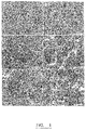
- All of the collagen air dried PolyHIPE materials exhibited the standard PolyHIPE emulsion templated structure. All of the materials exhibited an open interconnecting structure with void size decreasing with increasing collagen concentration ( Fig. 1 ).
- the increase in HIPE viscosity with increasing collagen concentration correlates with increasing emulsion stability.
- the void size of the template material is controlled by the emulsion droplet size, and this can be regulated by affecting the emulsion stability by varying temperature and time of the droplet phase.
- the interconnecting windows/pores are also controlled by the emulsion stability and are formed during polymerisation of the continuous phase film surrounding the emulsion droplets. At the higher concentration of collagen it becomes more difficult to observe a well-defined void structure ( Fig. 1E-F ).
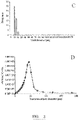
- Freeze-dried collagen PolyHIPEs (GA cross-linked) again exhibited a highly interconnected porous structure ( Fig 3 .). No defined spherical void structure could be observed from SEM images and mercury intrusion porosimetry calculated a median interconnect diameter of 39 ⁇ m ( Fig 1D .), indicating an extremely open interconnecting structure.
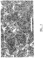
- the GA cross-linked materials exhibited more of a nanofibrous structure compared to the non cross-linked materials. Two different methods were used to crosslink the materials; the GA was added either added to the HIPE directly after HIPE formation, or to the dried materials which were post cross- linked in GA solution overnight. The air dried GA cross-linked materials exhibited a more uniform interconnecting structure compared to the freeze dried materials ( Fig 4 .) The strut dimensions of the structure are marked as white bars on the photomicrograph shown in Figure 8 .
- ALP alkaline phosphatase activity
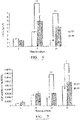
- DNA analysis was used to study cell growth/number on both of 2D and 3D culture of Saos-2 cells. Increased levels of DNA were observed in the 3D collagen scaffolds compared to 2D after the first 24 hours of culture ( Fig 6 .). The levels were highest after 8 days on both the 3D and 2D cultures. After 14 days, cell growth levels decrease in both the 2D and 3D cultures possibly due to cell differentiation. At 21 and 35 days increased levels were observed in the 3D collagen cultures compared to standard 2D cultures (data not shown).
- Alkaline phosphatase catalyses the hydrolysis of phosphate esters in alkaline buffer and produces an organic radical and inorganic phosphate. Changes in alkaline phosphatase level and activity are associated with various diseases state in liver and bone. Alkaline phosphatase is involved in the maturation step of osteoblasts during differentiation. This enzyme belongs to a family of proteins that are anchored to the plasma membrane via a glycosyl-phosphatidinositol linkage. ALP is one of the earliest phenotypic markers for a committed osteoblast. In general, committed osteoblasts exhibit higher basal levels of ALP than do cells which do not mineralise their matrix, such as fibroblasts.
- ALP alkaline phosphatase may be involved in preparing the extracellular matrix for the deposition of mineral and for the co-expression of other genes.
- High levels of ALP are seen in both pre-osteoblasts and osteoblasts in vivo and in differentiating osteoblasts in vitro.
- the biological functions of ALP is still unknown except for its role in bone mineralization.
- Connective tissue cells expressing high levels of ALP can be induced to deposit mineral in vitro.
- ALP may be involved in the degradation of inorganic pyrophosphate, thereby providing a sufficient local concentration of phosphate or inorganic pyrophosphate for mineralization to proceed.
- the ALP activity after normalisation to DNA content showed increased ALP levels per cell after 14 and 21 days on the 3D collagen materials compared to standard 2D culture.
- the 3D collagen porous materials provide a suitable biomimetic environment for enhanced differentiation of bone cells. (Data for 35 days not shown.)
- Figure 9a is a SEM micrograph of decellularised liver tissue ( Uygun et al; Nature Medicine 2010, 16, 814-821 ). Size of interconnecting pores were assessed using the added measurement bars, some of which are indicated by the fine lines in the micrograph. Scale bar represents 20 micron.
- Figure 9b shows SEM micrograph of natural collagen scaffold (N1, an embodiment of the present invention). Size of interconnecting pores were assessed using the added measurement bars, some of which are indicated by the fine lines in the micrograph. Scale bar represents 20 micron.
- a SEM micrograph of a poly-HIPE structure is disclosed in Carnahan et al, Soft Matter 2006, 2, 608-616 .
- the structure has generally spherical voids and circular pores.
- the voids and pores have a much smaller range of sizes and configurations than the liver tissue shown in Figure 9a .
- Figure 10 is a bar chart showing interconnecting pore size distribution of decellularised liver tissue and natural collagen scaffold of the present invention. This Figure shows that the profile of the pore size distribution of the scaffold of the present closely reproduces that of liver tissue.
- Table 1 Gene expression changes in primary human hepatocytes cultured on 3D scaffolds.
- Scaffold type Gene of interest Albumin HNF4a CYP3A4 NTCP N1 17.82091 0.0273 0.078 0.013 S1 38.34588 0.0086 0.008 0.000 S1 38.34588 0.0086 0.008 0.000 N4 9.059928 0.0113 0.107 0.017 S4 260.2942 0.0095 0.046 0.002 Thawed PHH 274.6593 0.1763 1.417 0.504
Landscapes
- Health & Medical Sciences (AREA)
- Chemical & Material Sciences (AREA)
- Life Sciences & Earth Sciences (AREA)
- Medicinal Chemistry (AREA)
- Dermatology (AREA)
- General Health & Medical Sciences (AREA)
- Veterinary Medicine (AREA)
- Epidemiology (AREA)
- Oral & Maxillofacial Surgery (AREA)
- Animal Behavior & Ethology (AREA)
- Transplantation (AREA)
- Public Health (AREA)
- Dispersion Chemistry (AREA)
- Biophysics (AREA)
- Chemical Kinetics & Catalysis (AREA)
- Polymers & Plastics (AREA)
- Organic Chemistry (AREA)
- Engineering & Computer Science (AREA)
- Materials Engineering (AREA)
- Materials For Medical Uses (AREA)
Description
- This invention relates to a method of manufacture of three dimensional (3D) porous cross-linked collagen or collagen-chitosan scaffolds which may be used for tissue engineering applications. The invention also relates to tissue scaffolds made by the method and to devices incorporating the scaffolds.
- Tissue scaffolds find application in tissue engineering for improvement of tissue and organ repair and replacement of biological functions. Key components of tissue engineering are biomaterials that provide an interface similar to a naturally occurring extracellular matrix (ECM). This allows cells to grow on the material without being recognized as foreign to the body. Biomaterials have been based on biopolymers, especially based on biopolymers derived from the ECM.
- Collagen, which is a natural biopolymer, is used in the biomedical industry for applications such as encapsulation, microspheres and matrices for implants and tissue engineering. Chitosan is also useful for biomedical and clinical applications because it has high biocompatibility, biodegradability and nonantigenicity. Chitosan-collagen hybrid polymer network scaffolds have been prepared by freeze-drying and have been evaluated using buffalo stem cells and with respect to growth of hepatic tissue.
- Nanofibrous collagen (NF-collagen) mats have been prepared by using an electrospinning technique. (Ko C-L, Tien Y-C, Wang J-C, Chen W-C. Characterization of controlled highly porous hyaluronan/collagen cross-linking sponges for tissue engineering. J Mech Behav Biomed Mater 2012;14:227-38). The electrospinning technique typically forms two-dimensional collagen sheets and is incapable of fabricating 3D NF-collagen scaffolds with well-defined pore size. In order to facilitate effective tissue engineering, we recognized that the development of 3D porous structures is critical to the function of biological tissue, transport of oxygen and nutrients.
- L. Ma et al., Biomaterials 24, 4833-4841 (2003) discloses collagen/ chitosan porous scaffolds crosslinked with glutaraldehyde. The scaffolds are prepared by lyophilization of a collagen/chitosan blend obtained by combining a solution of chitosan and a suspension of collagen and have an interconnected 3D porous structure.
- According to a first aspect of the present invention, there is provided a method of forming a scaffold comprises the steps of:
preparing a high internal phase emulsion (HIPE) comprising an aqueous solution of: - proteins selected from the group consisting of collagen and mixtures of collagen and chitosan; and
- a cross-linking agent;
- by addition of an immiscible solvent to the solution to form the HIPE;
- reducing the temperature of the HIPE to solidify the HIPE;
- removing water and solvent by vaporisation at a temperature above the melting point of the solvent to form a porous structure; and
- before or after removal of solvent, causing cross-linking of the protein with the cross-linking agent to form a cross-linked scaffold structure.
- The collagen structure of the present invention may have fundamentally the same chemical composition as natural collagen and may be used for guiding tissue regeneration. Particularly advantageous embodiments have the advantage that they exhibit the nanofibrous architecture which is beneficial to regulation of cell adhesion and function.
- Collagen gels form through a single-strand-to- triple helix transition. However, chemical cross-linking is a much more consistent method to ensure structural integrity and to provide a gel which exhibits an optimized, reproducible performance in vivo or under physiological conditions.
- The method may be used to induce tissue formation from liver cells, skin fibroblast cells, bone cells, cartilage or skin cells.
- According to a second aspect of the present invention, there is provided a three-dimensional tissue scaffold structure comprises: a protein selected from the group consisting of: collagen and mixtures of collagen and chitosan, cross-linked with a cross-linking agent, wherein the scaffold structure comprises voids interconnected by pores and the scaffold includes laminar regions forming webs between adjacent pores.
- The term "scaffold" used in the present invention refers to a three-dimensional structure with a porous microstructure suitable to support cells during tissue growth.
- Preferred scaffolds in accordance with this invention have beneficial configuration, morphology and properties suitable for tissue engineering including: good biocompatibility, mechanical performance, a degradation rate which can match a rate of tissue regeneration and a highly porous interconnecting structure. Use of cell lines which are difficult to grow by other methods is facilitated.
- The scaffold may have the structure of a collagen foam with a porous structure suitable for use as a tissue scaffold.
- Embodiments of the invention comprising a scaffold formed from crosslinked collagen and mixtures of collagen and chitosan may have particular suitability for use in comparison to previously disclosed porous polymers, for example polystyrene based poly-HIPE structures.
- The tissue scaffold of the present invention comprises voids interconnected by pores. The scaffold may include struts extending between adjacent pores or voids, the struts each having a length and thickness.
- The structure of a scaffold of this invention may be characterised by one or more of the following properties:
- Embodiments of the invention allow manufacture of a scaffold with a configuration and properties which closely resembles decellularised natural tissue, for example rat liver tissue (Uygun et al; Nature Medicine 2010, 16, 814-821 . See Figure 2g on page 816).
- Previously disclosed poly-HIPE structures have a high proportion of spherical or near-spherical voids with circular or near-circular pores. A low interconnect pore sized distribution was described in Carnachan, R.J.; Bokhari, M.; Przyborski, S.A. and Cameron, NR.; Soft Matter 2006, 2, 608-616 . See Table 1 and
Figure 4 . In such prior art arrangements the voids are generally spherical. - Scaffolds of the present invention may have a substantial absence of spherical voids. The struts between adjacent pores may have a wide range of thicknesses and lengths in comparison to poly-HIPE structures, as disclosed in Carnachan et al. Less than 10% of the voids may be spheroidal or approximately spherical. Preferably there are no spheroidal or approximately spherical voids.
- Embodiments of the invention allow manufacture of scaffolds with configurations and properties which closely resemble decellularised natural tissue, for example rat liver tissue.
- Previously disclosed poly-HIPE structures have a high proportion of spherical or near-spherical voids with circular or near-circular pores. A low interconnect pore size distribution was observed, as illustrated in Carnachan, et al.
- In such prior art arrangements the voids are generally spherical, typically having a mean void size of 94 µm and a range of pore sizes from 10 to 30 µm and a mean pore size of 19 µm. This is a narrow pore size distribution.
- Scaffolds of the present invention may have a substantial absence of spherical or spheroidal voids. The struts between adjacent pores may have a wide range of thicknesses and lengths in comparison to poly-HIPE structures.
- Embodiments of the invention may provide a high variety of configurations or shapes of the voids and pores. The voids may be dimensioned so that 90% of the voids have a mean size in the range 3.5 µm to 6.5 µm. The voids may typically have a mean size of 4.5 µm and a standard deviation of ±1.8 µm. The corresponding structure of rat liver tissue may have a void size of 4.2 µm ±2.2 µm. The pore size may be in the range 2.0 µm to 8.7 µm, comparable to the pore size of rat liver which may be 1.0 µm to 12 µm.
- In embodiments the uniformity of the dimensions and configurations of the voids and pores are low. The boundaries between adjacent voids or pores may be irregular with a substantial absence of spherical voids and circular shaped pores.
- The aspect ratio of the pores may be defined by the ratio of the minimum dimension (dmin) to the maximum dimension (dmax). These may be measured on perpendicular axes or otherwise angled axes. With non-circular pores the minimum and maximum dimensions are not perpendicular. In embodiments the aspect ratio, dmin/dmax, 50% of the pores may be less than about 0.7, alternatively 50% less than about 0.5, alternatively 50% less than about 0.4.
- In contrast, a prior art poly-HIPE structure may have 50% of voids with an aspect ratio greater than 0.7, commonly greater than 0.8, typically greater than 0.9.
- Scaffolds of the invention may have laminar regions in the form of webs between the pores. This configuration provides a relatively high surface area of the voids of the scaffold. A high surface area may be beneficial to cell growth, particularly in combination with a high degree of interconnection by pores between the voids.
- Scaffolds of the invention may include laminar regions in the form of webs between adjacent pores. This configuration provides a relatively high surface area of the scaffold defining the voids. A high surface area may be beneficial to cell growth, particularly in combination with a high degree of interconnection by pores between the voids.
- The softness or conversely stiffness of the scaffolds may be determined as the gradient of stress-strain behaviour for the scaffold.
- In embodiments the stiffness of the scaffold is similar to soft tissues such as liver, for example within 30%, alternatively within 20%, further alternatively within 10%, of the value for liver tissue. The stiffness may be determined by manual inspection or may be measured instrumentally.
- An important property is that the scaffold may have a softness selected to be similar to that of natural collagen to facilitate cellular expression, for example of liver cells. The stiffness or softness may be controlled to have a value close to that of an alternative tissue type, as required.
- In preferred embodiments, the scaffold of the present invention may exhibit one or more of the following properties. The porosity may be greater than 70%, alternatively greater than 80%, alternatively greater than 90%, for example 91% as measured by mercury porosimetry.
- The scaffold of the present invention may find application in making repair tissue which resembles natural tissues, but without having a biological history, for example dependent on age, disease, gender or environmental factors. Consistency of tissue characteristics can be controlled.
- Advantageous embodiments of this invention have a microfibrous structure, particularly with a thin interconnecting strut dimension. For example the structure may have struts with a length in the range 0.5 µm to 6 µm, preferably 0.6 µm to 5.5 µm. The struts may have an average thickness of 1 to 3 µm, preferably 1 to 2 µm. A microfibrous structure is particularly effective in regulating cell adhesion to the scaffold.
- The cross-linking step may take place before or after complete or partial removal of the solvent.
- Cross-linking may be taken to completion before removal of the solvent. Alternatively, cross-linking may be carried out on the solidified solvent-free structure. Alternatively, an initial partial cross-linking step may be followed by solvent removal and a further cross-linking step. A preferred cross-linking agent is glutaraldehyde (GA).
- Of the cross-linking agents which may be employed are genipin, carbodiimide, transglutaminase, chondroitin sulphate, dextran dialdehyde, bis(vinylsulfonyl)methane and hexamethylene di-isocyanate.
- The concentration of the cross-linking agent, preferably glutaraldehyde, in the emulsion may in the range of 1wt% to 5wt%, preferably 2wt% to 3wt%, more preferably about 2.5wt%. The concentration of the cross-linking agent may be selected to provide a desired degree of stiffness or softness dependent on the required properties of the tissue scaffold.
- Using glutaraldehyde as a cross-linker in accordance with this invention does not require use of vinyl derivatives of collagen and allows for quicker production of collagen based porous materials removing the need for purification of any vinylised derivatives.
- Polymerisation of a high internal phase emulsion (HIPE) template produces a material (referred to as a PolyHIPE) with an internal pore structure which can be controlled by varying one or more manufacturing conditions such as emulsion stability, stirring rate and rate of droplet phase addition. For polymeric foams such as those based on styrene/DVB water-in-oil, HIPEs have been used as templates prior to polymerisation. When using a water soluble polymer such as collagen, an inverse oil-in-water HIPE may be formed. The porous structure is produced either on cooling below the gelation temperature or by the addition of a cross-linking agent.
- The concentration of collagen in the emulsion may in the range of about 5wt% to about 30wt%, preferably about 7.5wt% to about 15wt%. Relatively high concentrations of collagen have been found to reduce swelling of the scaffold on contact with water, providing a more stable scaffold structure.
- The aqueous solution may further comprise a surfactant, for example in an amount of 5 wt% to 10 wt%, typically about 7 wt%. An amphoteric surfactant is preferred, for example disodium cocoampho dipropionate, for example manufactured by Solvay under the trade mark MIRANOL C2M-SF Conc. An alternative non-ionic surfactant is octylphenol ethoxylate, for example manufactured by the Dow Chemical Company under the trade mark TRITON X-405.
- The HIPE may be solidified by freezing, for example by cryogenic freezing. Use of a controlled freezing rate is not necessary in carrying out the method of the present invention. Freeze drying may be employed to remove the water and organic solvent. For example, if the solvent is toluene (melting point -90°C) the emulsion may be frozen to a temperature of about - 80°C so that the solvent can be removed by evaporation.
- The organic solvent may be added over a period of 2 minutes at a rate dependent on the size of the sample to be prepared.
- The invention is further described by means of example but not in any limitative sense with reference to the drawings, of which:
-
Figure 1 shows the internal structure of non cross-linked collagen polyHIPES; -
Figure 2 shows the void sized distribution of air-dried polyHIPE materials; -
Figure 3 shows the internal structure of freeze dried glutaraldehyde cross-linked collagen polyHIPES; -
Figure 4 shows the internal structure of air-dried collagen polyHIPES (10%ww) crosslinked with glutaraldehyde; -
Figure 5 shows scanning electron microscope images of the internal structure of collagen-chitosan polyHIPE materials; -
Figure 6 shows the DNA content of Soas-2 cultures in collagen based polyHIPES; -
Figure 7 shows swelling ratio of non cross-linked collagen materials and collagen materials cross-linked with glutaraldehyde; -
Figure 8 shows a scanning electron microscope (SEM) image showing the internal structure of air dried collagen polyHIPE (10 wt%) cross-linked with glutaraldehyde with strut dimensions labelled; -
Figure 9a is a SEM micrograph of decellularised liver. -
Figure 9b shows SEM micrograph of natural collagen scaffold in accordance with the invention. and -
Figure 10 is a bar chart showing interconnecting pore size distributions. - The following materials and methods were used
- Collagen, glutaraldehyde (GA) (25 % w/v in water), dexamethasone, β glycerphosphate disodium salt hydrate and 2-phospho-L-ascorbic acid trisodium salt were obtained from Sigma-Aldrich. Dulbecccos Modified Eagles medium (DMEM), phosphate buffered saline (PBS), penicillin/ streptomycin and trypsin/EDTA were obtained from Life Technologies
- The apparatus for the HIPE preparation consisted of a three-necked round bottom flask filled with a D-shaped paddle driven by an overhead stirrer. Collagen was dissolved in deionised water containing 7 % Miranol C2M-CONC as surfactant. The flask, containing the collagen solution, was only partially submerged in the water bath maintained at 50 °C. This is the temperature at which concentrated collagen solutions are suitable for processing into a HIPE. Toluene was then added dropwise to achieve a 3:1 ratio (15:5 ml or 45:15 ml). By the end point a white HIPE had formed. The HIPE was stirred for a further 5 minutes and then transferred to a 50 ml Polyethylene bottle or centrifuge tube. Glutaraldehyde (GA) (2.5 % w/v in water) was then added and mixed into the HIPE by gentle shaking. The HIPE was then allowed to gel overnight prior to washing in ethanol and deionised water to remove the toluene and surfactant respectively. The washed materials were then frozen in liquid nitrogen and freeze dried for 24-48 hours.
- Collagen PolyHIPEs (GA cross-linked) were then placed in 12-well culture plates and washed in 0.1 M glycine overnight to inactivate vacant aldehyde groups. The materials were then sterilised with 70 % ethanol for 30 minutes, then washed with phosphate buffered saline three times. The materials were finally washed with Soas-2 culture medium (2-3 ml) and maintained at 37 °C and 5 % CO2 until cell inoculation.
- For non-cross-linked collagen materials, samples were placed in 12 well plates and post cross-linked in 2 ml of 2.5 % w/v GA overnight. The cross-linked samples are then washed in PBS (x 5) to remove the residual GA and soaked in 2 ml of 0.1M glycine solution for 4 hours to deactivate vacant aldehyde groups. The samples are then sterilised in 70 % ethanol and washed with PBS (x3) and then finally washed in 2-3 ml of Saos-2 culture medium prior to cell inoculation.
- The preparation followed was essentially that given in section 2.2. For collagen- chitosan composite materials, collagen (1.5 g, 10 % w/w) and chitosan (0.3 g, 2 % w/w) were dissolved in 2 % acetic acid containing Miranol C2M-CONC. The solution was maintained at 50 °C and toluene was added dropwise to the solution until a HIPE had formed. These materials were then cross-linked and processed as described above for the collagen materials.
- For standard cell culture, Saos-2 cells are maintained in DMEM (Gibco, Life Technologies) containing 10 % FBS (Gibco, Life Technologies) and 100 U/ml Penicillin/Streptomycin (Gibco, Life Technologies).
-
- i) Saos-2 cells were seeded at 250,000 cells per scaffold in 12 well plates. Saos-2 cells were maintained in osteogenic differentiation media, DMEM supplemented with 10 % FBS, 10 nM dexamethasone, 5 mmol/l β glycerophosphate and 100 µg/ml ascorbic acid. Cells were allowed to settle for 30 minutes after seeding prior to flooding the well with 3 ml of differentiation media.
- ii) L929 cells were seeded at 100,000 cells/50 µl per well (2 × 10⁶ cells/ml) and allowed to settle on the scaffolds for 30 minutes in the cell incubator. The wells were then topped up with 3 ml of culture media and placed by in the incubator and maintained at 37 °C and 5 % CO2. L929 cells were culture on the natural collagen scaffolds for 10 days.
- iii) Three primary human cell types (i.e. primary human hepatocytes (PHH), human liver cancer cell line HepG2 and primary human hepatic stellate cells) were cultured on the natural collagen scaffolds made as described in the present invention (N type). Also they were compared with relatively soft styrene-EHA (2-ethylhexyl acrylate) 3D polyHIPE synthetic scaffolds (S type) and 2D hard plastic standard tissue culture plates. All cells cultured successfully. The natural collagen scaffolds (N type) promoted improved phenotypes compared to those on synthetic scaffolds (S type). In particular, primary human hepatic stellate cells cultured on the collagen scaffolds (N type) had reduced activation ("more in vivo like") compared to those cultured on traditional 2D hard plastic.
- A freeze-thaw technique was used for cell lysis. Cultures were washed with biological grade water and placed in 1.5 ml falcon tubes containing 1ml of biological grade water. Cells cultured in 2D were removed using a cell scraper and re-suspended in 1 ml of biological grade water. Cell lysates were stored at -20 °C.
- DNA content was analysed using the Quant IT PicoGreen assay (Life Technologies). Cell lysates were thawed at 37 °C and homogenised using a 21G needle (10-15 times), samples were then diluted 1 in 10 with TE buffer (1 x) by placing 10 µl of sample and 90 µl of TE buffer in a black bottom 96-well plate. 100 µl of Pico Green reagent was then added and mixed at room temperature for 5 minutes whilst wrapped in tin foil. The fluorescence was then measured at excitation and emission wavelengths of 480 and 520 nm respectively.
- A freeze-thaw technique was used for cell lysis. Cultures were washed with biological grade water and placed in 1.5 ml falcon tubes containing 1ml of biological grade water. Cells cultured in 2D were removed using a cell scraper and re-suspended in 0.5 ml of biological grade water. Cell lysates were stored at -20 °C.
- Alkaline phosphatase (ALP) levels are associated with various diseases in the liver and bone. The AbCam alkaline phosphatase kit uses p-nitrophenyl phosphate (p-NP) as a phosphate substrate which turns yellow (405 nm) when dephosphorylated by ALP.
- Cell lysates were thawed at 37 °C. Then 5 µl of cell lysate was added to each well of a 96-well plate. The volume was then made up to 80 µl using the assay buffer. 50 µl of p-NP (5 mM) was then added to each sample well. The plate was then wrapped in tin foil and incubated at room temperature for 60 minutes. The absorbance was then read at 405 nm using a Synergy H4 plate reader. The sample readings were then applied to the p-NP standard curve and ALP activity of the test samples calculated as follows:

- A is amount of p-NP generated by samples (µmol).
- V is the volume of sample added in the assay well (in ml) T is the reaction (in minutes).
- ALP activity was then normalised to DNA content of the samples to give activity in Units per ng DNA.
- The median pore size of the material was determined using Mercury Intrusion Porosimetry which was performed using a Micromeritics AutoPore IV. Intrusion and extrusion mercury contact angles of 130 degrees were used. The penetrometers had a stem volume of 1.836 ml and bulb volume of 5ml. Void diameter ranges were investigated using Scanning Electron Microscopy (SEM, Philips XL30 SEM) operating at 15 kV. Prior to visualisation, fractured samples were coated with gold and mounted onto aluminium stubs using carbon pads. Void diameter sizes were calculated as described previously.
- Samples were washed 3 times in PBS and then fixed in 4 % PFA overnight. Samples were then dehydrated through a series of graded ethanol (40, 60, 80, 90 and 95 % ethanol) for 1 hour and then in 100 % ethanol for 1 hour, histoclear (15 minutes), 50/50 histoclear/wax (30 minutes) and 100 % wax (60 minutes). The samples were then transversely embedded into moulding cassettes and left to cool overnight. Sections of 10 µm were then dewaxed and rehydrated through a series of graded ethanol. The sections were then incubated with permeabilisation solution and blocking buffer (15 minutes each solution). For antibody staining, sections were incubated with the primary osteocalcin antibody overnight in a humidified chamber. After primary incubation the sections were then washed and incubated with the appropriate secondary antibody for 60 minutes in the dark. The sections were then mounted in coverslips using DAPI /Vectashield. For standard Haematoxylin and Eosin staining the sections are left in Haematoxlyin and Eosin respectively to visualise cell nuclei and staining of the collagen material structure. Sections were visualised using Leica DM500 and DMI 300 B microscopes.
- Dry samples were weighed and placed in centrifuge tubes containing 30 ml of water. The samples were then removed after 30 minutes. The excess water was removed using a dry paper towel and the swollen samples were then reweighed and placed back into 30 ml of water. This process was repeated every 30 minutes for up to 120 minutes. The swelling ratios (WSR) of the materials were calculated using
equation 3.
- Wt represented the weight of the wet scaffolds and W0 represented the initial weight of the dry scaffolds.
- Collagen HIPEs were prepared initially using two different surfactants. However those prepared using Triton X-405 were not as stable as those prepared using Miranol C2M-CONC, especially at the 60 ml emulsion scale. Only those made using Miranol CM-SF CONC are referred to below. After the addition of the cross-linker glutaraldehyde the gelation occurs rapidly. The gelated
- HIPEs were orange in colour with the resulting freeze dried monolith being a slightly paler orange colour. The ratios of cross-linker used for gelation at a 20 ml scale for 10 % w/w collagen concentration are shown in Table 1. For varying concentrations of collagen and for 60 ml production, the amounts were adjusted accordingly.
Table 1. Cross-linking ratios for materials containing 10 % w/w collagen Collagen (g) Glutaraldehyde (2.5 % v/v) (mL) Cross-linking ratio (% w/w) 0.5 0.25 0.125 0.5 0.5 0.25 0.5 1 0.5 0.5 2 1 - The internal pore structure of the collagen materials was analysed using Scanning Electron Microscopy.
- All of the collagen air dried PolyHIPE materials exhibited the standard PolyHIPE emulsion templated structure. All of the materials exhibited an open interconnecting structure with void size decreasing with increasing collagen concentration (
Fig. 1 ). The increase in HIPE viscosity with increasing collagen concentration correlates with increasing emulsion stability. The void size of the template material is controlled by the emulsion droplet size, and this can be regulated by affecting the emulsion stability by varying temperature and time of the droplet phase. The interconnecting windows/pores are also controlled by the emulsion stability and are formed during polymerisation of the continuous phase film surrounding the emulsion droplets. At the higher concentration of collagen it becomes more difficult to observe a well-defined void structure (Fig. 1E-F ). - From image analysis of the collagen PHP materials, increasing the collagen concentration leads to a material with a narrower range of void sizes and a smaller overall average void size of the material (
Fig 1A-C .). The void size reduces from 70µm to 10 µm as one progresses from 10 % and 30 % collagen. This is due to the increase in emulsion viscosity with increasing collagen concentration suggesting an increase in HIPE stability as the concentration of collagen increases. Increased HIPE stability leads to a porous material with smaller average void size as discussed in the literature. - Increasing the concentration of collagen to 20 % w/w produced a PHP material with lower porosity with thicker strut size compared to nanofibrous structure observed with the 10 % w/w collagen material.
- Freeze-dried collagen PolyHIPEs (GA cross-linked) again exhibited a highly interconnected porous structure (
Fig 3 .). No defined spherical void structure could be observed from SEM images and mercury intrusion porosimetry calculated a median interconnect diameter of 39 µm (Fig 1D .), indicating an extremely open interconnecting structure. The GA cross-linked materials exhibited more of a nanofibrous structure compared to the non cross-linked materials. Two different methods were used to crosslink the materials; the GA was added either added to the HIPE directly after HIPE formation, or to the dried materials which were post cross- linked in GA solution overnight. The air dried GA cross-linked materials exhibited a more uniform interconnecting structure compared to the freeze dried materials (Fig 4 .) The strut dimensions of the structure are marked as white bars on the photomicrograph shown inFigure 8 . - The internal structure of air-dried collagen-chitosan p olyHIPE cross-linked with glutaraldehyde was shown to indicate successful cell culture.
- DNA analysis was used to study cell growth/number on both of 2D and 3D culture of Saos-2 cells. Increased levels of DNA were observed in the 3D collagen scaffolds compared to 2D after the first 24 hours of culture (
Fig 6 .). The levels were highest after 8 days on both the 3D and 2D cultures. After 14 days, cell growth levels decrease in both the 2D and 3D cultures possibly due to cell differentiation. At 21 and 35 days increased levels were observed in the 3D collagen cultures compared to standard 2D cultures (data not shown). - Alkaline phosphatase (ALP) catalyses the hydrolysis of phosphate esters in alkaline buffer and produces an organic radical and inorganic phosphate. Changes in alkaline phosphatase level and activity are associated with various diseases state in liver and bone. Alkaline phosphatase is involved in the maturation step of osteoblasts during differentiation. This enzyme belongs to a family of proteins that are anchored to the plasma membrane via a glycosyl-phosphatidinositol linkage. ALP is one of the earliest phenotypic markers for a committed osteoblast. In general, committed osteoblasts exhibit higher basal levels of ALP than do cells which do not mineralise their matrix, such as fibroblasts. The expression of ALP mRNA and enzyme activity before the initiation of osteoblast mineralization suggests that alkaline phosphatase may be involved in preparing the extracellular matrix for the deposition of mineral and for the co-expression of other genes. High levels of ALP are seen in both pre-osteoblasts and osteoblasts in vivo and in differentiating osteoblasts in vitro. The biological functions of ALP is still unknown except for its role in bone mineralization. Connective tissue cells expressing high levels of ALP can be induced to deposit mineral in vitro. ALP may be involved in the degradation of inorganic pyrophosphate, thereby providing a sufficient local concentration of phosphate or inorganic pyrophosphate for mineralization to proceed. Studies have been carried out to show how vital ALP is for bone formation. In a human model of ALP deficiency, hypophosphatasia, the bone became soft in the absence of ALP activity, suggesting that ALP physiological function is to maintain bone mineralization after birth. More recently two mouse ALP knockout models showed that, when the gene for ALP is specifically deleted, the mice develop defects in bone mineralization shortly after birth but not necessarily during embryonic development. Unknown redundant or backup mechanisms may provide for mineralization during embryonic life in ALP-deficient knockouts, so that ALP might still play an important role in the initial mineralization processes in bone. These results suggest that ALP may play an important role in the maintenance of bone mineralization.
- The ALP activity after normalisation to DNA content, showed increased ALP levels per cell after 14 and 21 days on the 3D collagen materials compared to standard 2D culture. The 3D collagen porous materials provide a suitable biomimetic environment for enhanced differentiation of bone cells. (Data for 35 days not shown.)
- Haematoxylin staining of cell nuclei showed the presence of Soas-2 cells (24 hours growing within the top third of collagen matrix. However the collagen scaffolds also showed a strong pink/ red colour when stained with Eosin, which made deciphering between cells and scaffold difficult when used in combination.
- Further DAPI staining of cell nuclei indicated Saos-2 cells penetrating into the collagen scaffold structure showing good 3D cell growth after a further 7 days in culture. Osteocalcin is a late marker for bone differentiation. Collagen- based PolyHIPE materials reported in the literature have been manufactured from vinyl derivatives of collagen, chondroitin sulfate and hyaluronic acid; to allow for free radical polymerisation of the collagen.
-
Figure 9a is a SEM micrograph of decellularised liver tissue (Uygun et al; Nature Medicine 2010, 16, 814-821 ). Size of interconnecting pores were assessed using the added measurement bars, some of which are indicated by the fine lines in the micrograph. Scale bar represents 20 micron. -
Figure 9b shows SEM micrograph of natural collagen scaffold (N1, an embodiment of the present invention). Size of interconnecting pores were assessed using the added measurement bars, some of which are indicated by the fine lines in the micrograph. Scale bar represents 20 micron. - A SEM micrograph of a poly-HIPE structure is disclosed in Carnahan et al, . The structure has generally spherical voids and circular pores. The voids and pores have a much smaller range of sizes and configurations than the liver tissue shown in
Figure 9a . -
Figure 10 is a bar chart showing interconnecting pore size distribution of decellularised liver tissue and natural collagen scaffold of the present invention. This Figure shows that the profile of the pore size distribution of the scaffold of the present closely reproduces that of liver tissue. - L929 cells cultured on the natural collagen scaffolds (N type) show good cell viability/growth over a culture period of 10 days. MTT absorbance values increase from 0.05 to 0.3 over the 10 day culture period. MTT viability assay was conducted. A blank scaffold (no cells) was run in parallel with each time point.
- Cultures were made of three primary human cell types (i.e. primary human hepatocytes (PHH), human liver cancer cell line HepG2 and primary human hepatic stellate cells) on the scaffolds of this invention. All cells cultured successfully. The natural collagen scaffolds of this invention promoted improved phenotypes compared to synthetic scaffolds (S type). In particular, stellate cells cultured on the collagen scaffolds (N type) had reduced activation ("more in vivo like") compared to those cultured on traditional 2D hard plastic. The results clearly demonstrate that the natural collagen scaffolds (N type) which were manufactured by the method disclosed herein, were decidedly superior in maintaining desired gene expressions. The results are shown in Table 1.
Table 1 - Gene expression changes in primary human hepatocytes cultured on 3D scaffolds. Scaffold type Gene of interest Albumin HNF4a CYP3A4 NTCP N1 17.82091 0.0273 0.078 0.013 S1 38.34588 0.0086 0.008 0.000 S1 38.34588 0.0086 0.008 0.000 N4 9.059928 0.0113 0.107 0.017 S4 260.2942 0.0095 0.046 0.002 Thawed PHH 274.6593 0.1763 1.417 0.504 - Primary human hepatocytes were cultured for 7 days on 3D scaffolds (S1, S4, N1 and N4 [S = synthetic, N = natural]). Total RNA was extracted from all cells and analysed by QPCR for the expression of genes associated with hepatocyte function. Data shown are relative gene expression compared to GAPDH housekeeping gene and expressed as 2^-deltaCT. Maintaining expression of these genes (in comparison to thawed cells) is desired for optimal in vitro culture. Culture of cells on natural (N) scaffolds appeared to be preferential for maintaining hepatic gene expression, however expression was lower than in freshly thawed cells.
Table 2 - Albumin gene expression in primary human hepatocytes and HepG2 cells cultured on natural 3D scaffolds. Scaffold type and cell type Gene of interest Albumin PHH - N1 17.82091 PHH - N4 9.059928 Thawed PHH 274.6593 HepG2 - N1 29.64054647 HepG2 - N4 20.09131754 - Primary human hepatocytes and HepG2 were cultured for 7 days on scaffolds of this invention (N1 and N4, N = natural). Total RNA was extracted from all cells and analysed by QPCR for the expression of albumin. Data shown are relative gene expression compared to GAPDH housekeeping gene and expressed as 2^-deltaCT. Maintaining expression of albumin expression (in comparison to thawed cells) is desired for optimal in vitro culture. Both cells types had comparable expression of albumin when cultured on the natural scaffolds, however this was still lower than expression in freshly thawed cells. The results are shown in Table 2.
Table 3 - Gene expression changes in primary human hepatic stellate cells cultured on 3D scaffolds. Scaffold type Gene of interest ACTA2 COL1A1 TGFβ N1 0.165 0.165 0.092 N4 0.450 0.566 0.117 S1 2.054 40.45 0.837 S4 1.894 41.01 0.815 Pellet 0.638 1.796 0.040 2D plastic 0.417 3.637 0.061 - Primary human hepatic stellate cells were cultured for 7 days either on standard plastic multi-well plates or on synthetic scaffolds of this invention (S1, S4, N1 and N4 [S = synthetic, N = natural]). Total RNA was extracted from all cells and analysed by Taqman QPCR for the expression of genes associated with stellate cell activation. Data shown are relative gene expression compared to GAPDH housekeeping gene and expressed as 2^-deltaCT. Expression of these genes is maintained or promoted by culture on "hard" 2D plastic plates, but was reduced in samples cultured on natural (N) scaffolds. Cells cultured on synthetic (S) scaffolds had increased gene expression. Reduced expression of these genes is highly advantageous as it better represents in vivo conditions. The results are shown in Table 3.
Claims (14)
- A three-dimensional tissue scaffold structure comprising: a protein selected from the group consisting of collagen and mixtures of collagen and chitosan, cross-linked with a cross-linking agent;
wherein the scaffold structure comprises voids interconnected by pores and the scaffold includes laminar regions forming webs between adjacent pores. - A three-dimensional tissue scaffold structure as claimed in claim 1, wherein the structure has a substantial absence of spherical or spheroidal voids.
- A three-dimensional tissue scaffold structure as claimed in claim 1 or 2, wherein 90% of the voids have a mean size in the range 3.5 µm to 6.5 µm.
- A three-dimensional tissue scaffold structure as claimed in any of claims 1 to 3, wherein the pore size is in the range 1.0 to 15 µm, preferably in the range 2.0 µm to 8.7 µm.
- A three-dimensional tissue scaffold structure as claimed in any of claims 1 to 4, wherein the boundaries between adjacent voids and/or the adjacent pores are irregular.
- A three-dimensional tissue scaffold structure as claimed in any of claims 1 to 5, wherein the aspect ratio of 50% of the pores is less than 0.7, preferably 0.5, more preferably 0.4.
- A three-dimensional tissue scaffold structure as claimed in any of claims 1 to 6, wherein the scaffold has a stiffness within 30%, preferably within 20%, more preferably within 10%, of the stiffness of natural collagen 1.
- A three-dimensional tissue scaffold structure as claimed in any of claims 1 to 7, wherein the porosity of the scaffold is 70 to 95%, preferably 80 to 95% as measured by mercury porosimetry.
- A three-dimensional tissue scaffold structure as claimed in any of claims 1 to 2. 8 for manufacture of a prosthesis.
- A method of forming a scaffold comprising the steps of:
preparing a high internal phase emulsion (HIPE) comprising an aqueous solution of:proteins selected from the group consisting of collagen and mixtures of collagen and chitosan; anda cross-linking agent;by addition of an immiscible solvent to the solution to form the HIPE;reducing the temperature to solidify the HIPE;removing water and solvent by vaporisation at a temperature above the melting point of the solvent to form a porous structure; andbefore or after removal of solvent, causing cross-linking of the protein with the cross-linking agent to form a crosslinked scaffold structure. - A method as claimed in claim 10, wherein the concentration of collagen in the aqueous solution is from about 2.5wt% to about 20wt%, preferably about 5wt% to about 10wt%.
- A method as claimed in claim 10 or 11, wherein the cross-linking reagent is selected from a group consisting of glutaraldehyde, genipin, carbodiimide, transglutaminase, chondroitin sulfate, dextran dialdehyde, bis(vinylsulfonyl)methane and hexamethylene di-isocyanate.
- A method as claimed in any of claims 10 to 12, wherein the immiscible solvent is selected from a group consisting of toluene and methylated tetrahydrofuran.
- A three-dimensional tissue scaffold structure as claimed in any of claims 1 to 9, wherein the scaffold was made by the method claimed in any one of claims 10 to 13.
Applications Claiming Priority (2)
| Application Number | Priority Date | Filing Date | Title |
|---|---|---|---|
| GB1606976.7A GB2549522A (en) | 2016-04-21 | 2016-04-21 | Tissue structure scaffolds |
| PCT/EP2017/061542 WO2017182676A1 (en) | 2016-04-21 | 2017-05-12 | Tissue structure scaffolds |
Publications (2)
| Publication Number | Publication Date |
|---|---|
| EP3445412A1 EP3445412A1 (en) | 2019-02-27 |
| EP3445412B1 true EP3445412B1 (en) | 2022-12-21 |
Family
ID=58992799
Family Applications (1)
| Application Number | Title | Priority Date | Filing Date |
|---|---|---|---|
| EP17727114.5A Active EP3445412B1 (en) | 2016-04-21 | 2017-05-12 | Tissue structure scaffolds |
Country Status (5)
| Country | Link |
|---|---|
| US (1) | US10940237B2 (en) |
| EP (1) | EP3445412B1 (en) |
| CN (1) | CN109310804A (en) |
| GB (1) | GB2549522A (en) |
| WO (1) | WO2017182676A1 (en) |
Families Citing this family (4)
| Publication number | Priority date | Publication date | Assignee | Title |
|---|---|---|---|---|
| GB201804366D0 (en) * | 2018-03-19 | 2018-05-02 | Univ Oxford Innovation Ltd | Scaffold |
| US11998654B2 (en) | 2018-07-12 | 2024-06-04 | Bard Shannon Limited | Securing implants and medical devices |
| GB202017052D0 (en) * | 2020-10-27 | 2020-12-09 | Jd Biomedical Solutions Ltd | Tissue Scaffolds |
| CN115011542A (en) * | 2021-12-07 | 2022-09-06 | 中科睿极(深圳)医学科技有限公司 | Porous microcarrier and preparation method thereof |
Family Cites Families (5)
| Publication number | Priority date | Publication date | Assignee | Title |
|---|---|---|---|---|
| US5387207A (en) * | 1991-08-12 | 1995-02-07 | The Procter & Gamble Company | Thin-unit-wet absorbent foam materials for aqueous body fluids and process for making same |
| GB9826701D0 (en) * | 1998-12-05 | 1999-01-27 | Univ Newcastle | Microcellular polymers as cell growth media and novel polymers |
| US7931918B2 (en) * | 2004-09-14 | 2011-04-26 | The University Of Hong Kong | Collagen-based microspheres and methods of preparation and uses thereof |
| CN101766837B (en) * | 2009-12-28 | 2013-03-27 | 中国人民解放军第二炮兵总医院 | Biomimetic porous microsphere tissue engineering scaffold and manufacturing method thereof |
| CN103834057B (en) * | 2014-03-05 | 2016-02-03 | 四川大学 | A kind of take High Internal Phase Emulsion as gelatin porous material and the preparation method of Template preparation |
-
2016
- 2016-04-21 GB GB1606976.7A patent/GB2549522A/en not_active Withdrawn
-
2017
- 2017-05-12 EP EP17727114.5A patent/EP3445412B1/en active Active
- 2017-05-12 US US16/095,420 patent/US10940237B2/en active Active
- 2017-05-12 CN CN201780038934.6A patent/CN109310804A/en active Pending
- 2017-05-12 WO PCT/EP2017/061542 patent/WO2017182676A1/en not_active Ceased
Also Published As
| Publication number | Publication date |
|---|---|
| US10940237B2 (en) | 2021-03-09 |
| WO2017182676A1 (en) | 2017-10-26 |
| US20190134264A1 (en) | 2019-05-09 |
| GB2549522A (en) | 2017-10-25 |
| EP3445412A1 (en) | 2019-02-27 |
| CN109310804A (en) | 2019-02-05 |
Similar Documents
| Publication | Publication Date | Title |
|---|---|---|
| Khan et al. | Preparation and structural characterization of surface modified microporous bacterial cellulose scaffolds: A potential material for skin regeneration applications in vitro and in vivo | |
| KR100664772B1 (en) | Biodegradable Polymer Scaffolds | |
| Reverchon et al. | Supercritical fluids in 3-D tissue engineering | |
| Wang et al. | Development of hyaluronic acid-based scaffolds for brain tissue engineering | |
| Coogan et al. | Fabrication of micro-porous hyaluronic acid hydrogels through salt leaching | |
| Mahoney et al. | Three-dimensional growth and function of neural tissue in degradable polyethylene glycol hydrogels | |
| US6875442B2 (en) | Process for growing tissue in a biocompatible macroporous polymer scaffold and products therefrom | |
| US8691973B2 (en) | Method of producing chitosan scaffold having high tensile strength and chitosan scaffold produced using the method | |
| US10655120B2 (en) | Transport of cells in hydrogels | |
| Pisanti et al. | Tubular perfusion system culture of human mesenchymal stem cells on poly‐L‐lactic acid scaffolds produced using a supercritical carbon dioxide‐assisted process | |
| US10940237B2 (en) | Tissue structure scaffolds | |
| Guo et al. | Modulation of keratocyte phenotype by collagen fibril nanoarchitecture in membranes for corneal repair | |
| Panseri et al. | Bone-like ceramic scaffolds designed with bioinspired porosity induce a different stem cell response | |
| JP2000512666A (en) | Polysaccharide sponges for cell culture and transplantation | |
| CN106581762B (en) | 3D printing biological ink, preparation method and 3D printing forming method | |
| Wu et al. | Developing highly porous collagen scaffolds by using alginate microsphere porogens for stem cell cultures | |
| WO2021074439A1 (en) | Eluting moulds for hydrogel crosslinking | |
| Norouzi et al. | Alendronate releasing silk fibroin 3D bioprinted scaffolds for application in bone tissue engineering: Effects of alginate concentration on printability, mechanical properties and stability | |
| Margulis et al. | In vitro fabrication of engineered human skin | |
| Qi et al. | Construction and characterization of human oral mucosa equivalent using hyper-dry amniotic membrane as a matrix | |
| Chun et al. | Fabrication of porous chitosan scaffold in order to improve biocompatibility | |
| Takeda et al. | Fabrication of 2D and 3D constructs from reconstituted decellularized tissue extracellular matrices | |
| Paterson et al. | Highly porous polycaprolactone microspheres for skeletal repair promote a mature bone cell phenotype in vitro | |
| JP2013226205A (en) | Biological material for wound healing and preparation the same | |
| KR20110008013A (en) | Reconstructed cornea and mucosa |
Legal Events
| Date | Code | Title | Description |
|---|---|---|---|
| STAA | Information on the status of an ep patent application or granted ep patent |
Free format text: STATUS: UNKNOWN |
|
| STAA | Information on the status of an ep patent application or granted ep patent |
Free format text: STATUS: THE INTERNATIONAL PUBLICATION HAS BEEN MADE |
|
| PUAI | Public reference made under article 153(3) epc to a published international application that has entered the european phase |
Free format text: ORIGINAL CODE: 0009012 |
|
| STAA | Information on the status of an ep patent application or granted ep patent |
Free format text: STATUS: REQUEST FOR EXAMINATION WAS MADE |
|
| 17P | Request for examination filed |
Effective date: 20181017 |
|
| AK | Designated contracting states |
Kind code of ref document: A1 Designated state(s): AL AT BE BG CH CY CZ DE DK EE ES FI FR GB GR HR HU IE IS IT LI LT LU LV MC MK MT NL NO PL PT RO RS SE SI SK SM TR |
|
| AX | Request for extension of the european patent |
Extension state: BA ME |
|
| RIN1 | Information on inventor provided before grant (corrected) |
Inventor name: PRZYBORSKI, STEFAN Inventor name: WU, JUNJIE Inventor name: CARNACHAN, ROSS |
|
| DAV | Request for validation of the european patent (deleted) | ||
| DAX | Request for extension of the european patent (deleted) | ||
| STAA | Information on the status of an ep patent application or granted ep patent |
Free format text: STATUS: EXAMINATION IS IN PROGRESS |
|
| 17Q | First examination report despatched |
Effective date: 20190916 |
|
| GRAP | Despatch of communication of intention to grant a patent |
Free format text: ORIGINAL CODE: EPIDOSNIGR1 |
|
| STAA | Information on the status of an ep patent application or granted ep patent |
Free format text: STATUS: GRANT OF PATENT IS INTENDED |
|
| INTG | Intention to grant announced |
Effective date: 20220719 |
|
| RIN1 | Information on inventor provided before grant (corrected) |
Inventor name: PRZYBORSKI, STEFAN Inventor name: CARNACHAN, ROSS Inventor name: WU, JUNJE |
|
| GRAS | Grant fee paid |
Free format text: ORIGINAL CODE: EPIDOSNIGR3 |
|
| GRAA | (expected) grant |
Free format text: ORIGINAL CODE: 0009210 |
|
| STAA | Information on the status of an ep patent application or granted ep patent |
Free format text: STATUS: THE PATENT HAS BEEN GRANTED |
|
| RIN1 | Information on inventor provided before grant (corrected) |
Inventor name: PRZYBORSKI, STEFAN Inventor name: CARNACHAN, ROSS Inventor name: WU, JUNJIE |
|
| AK | Designated contracting states |
Kind code of ref document: B1 Designated state(s): AL AT BE BG CH CY CZ DE DK EE ES FI FR GB GR HR HU IE IS IT LI LT LU LV MC MK MT NL NO PL PT RO RS SE SI SK SM TR |
|
| REG | Reference to a national code |
Ref country code: GB Ref legal event code: FG4D |
|
| REG | Reference to a national code |
Ref country code: DE Ref legal event code: R096 Ref document number: 602017064774 Country of ref document: DE |
|
| REG | Reference to a national code |
Ref country code: CH Ref legal event code: EP |
|
| REG | Reference to a national code |
Ref country code: AT Ref legal event code: REF Ref document number: 1538663 Country of ref document: AT Kind code of ref document: T Effective date: 20230115 |
|
| REG | Reference to a national code |
Ref country code: IE Ref legal event code: FG4D |
|
| REG | Reference to a national code |
Ref country code: LT Ref legal event code: MG9D |
|
| REG | Reference to a national code |
Ref country code: NL Ref legal event code: MP Effective date: 20221221 |
|
| PG25 | Lapsed in a contracting state [announced via postgrant information from national office to epo] |
Ref country code: SE Free format text: LAPSE BECAUSE OF FAILURE TO SUBMIT A TRANSLATION OF THE DESCRIPTION OR TO PAY THE FEE WITHIN THE PRESCRIBED TIME-LIMIT Effective date: 20221221 Ref country code: NO Free format text: LAPSE BECAUSE OF FAILURE TO SUBMIT A TRANSLATION OF THE DESCRIPTION OR TO PAY THE FEE WITHIN THE PRESCRIBED TIME-LIMIT Effective date: 20230321 Ref country code: LT Free format text: LAPSE BECAUSE OF FAILURE TO SUBMIT A TRANSLATION OF THE DESCRIPTION OR TO PAY THE FEE WITHIN THE PRESCRIBED TIME-LIMIT Effective date: 20221221 Ref country code: FI Free format text: LAPSE BECAUSE OF FAILURE TO SUBMIT A TRANSLATION OF THE DESCRIPTION OR TO PAY THE FEE WITHIN THE PRESCRIBED TIME-LIMIT Effective date: 20221221 |
|
| REG | Reference to a national code |
Ref country code: AT Ref legal event code: MK05 Ref document number: 1538663 Country of ref document: AT Kind code of ref document: T Effective date: 20221221 |
|
| PG25 | Lapsed in a contracting state [announced via postgrant information from national office to epo] |
Ref country code: RS Free format text: LAPSE BECAUSE OF FAILURE TO SUBMIT A TRANSLATION OF THE DESCRIPTION OR TO PAY THE FEE WITHIN THE PRESCRIBED TIME-LIMIT Effective date: 20221221 Ref country code: LV Free format text: LAPSE BECAUSE OF FAILURE TO SUBMIT A TRANSLATION OF THE DESCRIPTION OR TO PAY THE FEE WITHIN THE PRESCRIBED TIME-LIMIT Effective date: 20221221 Ref country code: HR Free format text: LAPSE BECAUSE OF FAILURE TO SUBMIT A TRANSLATION OF THE DESCRIPTION OR TO PAY THE FEE WITHIN THE PRESCRIBED TIME-LIMIT Effective date: 20221221 Ref country code: GR Free format text: LAPSE BECAUSE OF FAILURE TO SUBMIT A TRANSLATION OF THE DESCRIPTION OR TO PAY THE FEE WITHIN THE PRESCRIBED TIME-LIMIT Effective date: 20230322 |
|
| P01 | Opt-out of the competence of the unified patent court (upc) registered |
Effective date: 20230412 |
|
| PG25 | Lapsed in a contracting state [announced via postgrant information from national office to epo] |
Ref country code: NL Free format text: LAPSE BECAUSE OF FAILURE TO SUBMIT A TRANSLATION OF THE DESCRIPTION OR TO PAY THE FEE WITHIN THE PRESCRIBED TIME-LIMIT Effective date: 20221221 |
|
| PG25 | Lapsed in a contracting state [announced via postgrant information from national office to epo] |
Ref country code: SM Free format text: LAPSE BECAUSE OF FAILURE TO SUBMIT A TRANSLATION OF THE DESCRIPTION OR TO PAY THE FEE WITHIN THE PRESCRIBED TIME-LIMIT Effective date: 20221221 Ref country code: RO Free format text: LAPSE BECAUSE OF FAILURE TO SUBMIT A TRANSLATION OF THE DESCRIPTION OR TO PAY THE FEE WITHIN THE PRESCRIBED TIME-LIMIT Effective date: 20221221 Ref country code: PT Free format text: LAPSE BECAUSE OF FAILURE TO SUBMIT A TRANSLATION OF THE DESCRIPTION OR TO PAY THE FEE WITHIN THE PRESCRIBED TIME-LIMIT Effective date: 20230421 Ref country code: ES Free format text: LAPSE BECAUSE OF FAILURE TO SUBMIT A TRANSLATION OF THE DESCRIPTION OR TO PAY THE FEE WITHIN THE PRESCRIBED TIME-LIMIT Effective date: 20221221 Ref country code: EE Free format text: LAPSE BECAUSE OF FAILURE TO SUBMIT A TRANSLATION OF THE DESCRIPTION OR TO PAY THE FEE WITHIN THE PRESCRIBED TIME-LIMIT Effective date: 20221221 Ref country code: CZ Free format text: LAPSE BECAUSE OF FAILURE TO SUBMIT A TRANSLATION OF THE DESCRIPTION OR TO PAY THE FEE WITHIN THE PRESCRIBED TIME-LIMIT Effective date: 20221221 Ref country code: AT Free format text: LAPSE BECAUSE OF FAILURE TO SUBMIT A TRANSLATION OF THE DESCRIPTION OR TO PAY THE FEE WITHIN THE PRESCRIBED TIME-LIMIT Effective date: 20221221 |
|
| PG25 | Lapsed in a contracting state [announced via postgrant information from national office to epo] |
Ref country code: SK Free format text: LAPSE BECAUSE OF FAILURE TO SUBMIT A TRANSLATION OF THE DESCRIPTION OR TO PAY THE FEE WITHIN THE PRESCRIBED TIME-LIMIT Effective date: 20221221 Ref country code: PL Free format text: LAPSE BECAUSE OF FAILURE TO SUBMIT A TRANSLATION OF THE DESCRIPTION OR TO PAY THE FEE WITHIN THE PRESCRIBED TIME-LIMIT Effective date: 20221221 Ref country code: IS Free format text: LAPSE BECAUSE OF FAILURE TO SUBMIT A TRANSLATION OF THE DESCRIPTION OR TO PAY THE FEE WITHIN THE PRESCRIBED TIME-LIMIT Effective date: 20230421 Ref country code: AL Free format text: LAPSE BECAUSE OF FAILURE TO SUBMIT A TRANSLATION OF THE DESCRIPTION OR TO PAY THE FEE WITHIN THE PRESCRIBED TIME-LIMIT Effective date: 20221221 |
|
| REG | Reference to a national code |
Ref country code: DE Ref legal event code: R097 Ref document number: 602017064774 Country of ref document: DE |
|
| PLBE | No opposition filed within time limit |
Free format text: ORIGINAL CODE: 0009261 |
|
| STAA | Information on the status of an ep patent application or granted ep patent |
Free format text: STATUS: NO OPPOSITION FILED WITHIN TIME LIMIT |
|
| PG25 | Lapsed in a contracting state [announced via postgrant information from national office to epo] |
Ref country code: DK Free format text: LAPSE BECAUSE OF FAILURE TO SUBMIT A TRANSLATION OF THE DESCRIPTION OR TO PAY THE FEE WITHIN THE PRESCRIBED TIME-LIMIT Effective date: 20221221 |
|
| 26N | No opposition filed |
Effective date: 20230922 |
|
| REG | Reference to a national code |
Ref country code: CH Ref legal event code: PL |
|
| PG25 | Lapsed in a contracting state [announced via postgrant information from national office to epo] |
Ref country code: MC Free format text: LAPSE BECAUSE OF FAILURE TO SUBMIT A TRANSLATION OF THE DESCRIPTION OR TO PAY THE FEE WITHIN THE PRESCRIBED TIME-LIMIT Effective date: 20221221 |
|
| REG | Reference to a national code |
Ref country code: BE Ref legal event code: MM Effective date: 20230531 |
|
| PG25 | Lapsed in a contracting state [announced via postgrant information from national office to epo] |
Ref country code: SI Free format text: LAPSE BECAUSE OF FAILURE TO SUBMIT A TRANSLATION OF THE DESCRIPTION OR TO PAY THE FEE WITHIN THE PRESCRIBED TIME-LIMIT Effective date: 20221221 Ref country code: MC Free format text: LAPSE BECAUSE OF FAILURE TO SUBMIT A TRANSLATION OF THE DESCRIPTION OR TO PAY THE FEE WITHIN THE PRESCRIBED TIME-LIMIT Effective date: 20221221 Ref country code: LU Free format text: LAPSE BECAUSE OF NON-PAYMENT OF DUE FEES Effective date: 20230512 Ref country code: LI Free format text: LAPSE BECAUSE OF NON-PAYMENT OF DUE FEES Effective date: 20230531 Ref country code: CH Free format text: LAPSE BECAUSE OF NON-PAYMENT OF DUE FEES Effective date: 20230531 |
|
| REG | Reference to a national code |
Ref country code: IE Ref legal event code: MM4A |
|
| PG25 | Lapsed in a contracting state [announced via postgrant information from national office to epo] |
Ref country code: IE Free format text: LAPSE BECAUSE OF NON-PAYMENT OF DUE FEES Effective date: 20230512 |
|
| PG25 | Lapsed in a contracting state [announced via postgrant information from national office to epo] |
Ref country code: IE Free format text: LAPSE BECAUSE OF NON-PAYMENT OF DUE FEES Effective date: 20230512 |
|
| PG25 | Lapsed in a contracting state [announced via postgrant information from national office to epo] |
Ref country code: IT Free format text: LAPSE BECAUSE OF FAILURE TO SUBMIT A TRANSLATION OF THE DESCRIPTION OR TO PAY THE FEE WITHIN THE PRESCRIBED TIME-LIMIT Effective date: 20221221 Ref country code: BE Free format text: LAPSE BECAUSE OF NON-PAYMENT OF DUE FEES Effective date: 20230531 |
|
| PG25 | Lapsed in a contracting state [announced via postgrant information from national office to epo] |
Ref country code: BG Free format text: LAPSE BECAUSE OF FAILURE TO SUBMIT A TRANSLATION OF THE DESCRIPTION OR TO PAY THE FEE WITHIN THE PRESCRIBED TIME-LIMIT Effective date: 20221221 |
|
| PG25 | Lapsed in a contracting state [announced via postgrant information from national office to epo] |
Ref country code: BG Free format text: LAPSE BECAUSE OF FAILURE TO SUBMIT A TRANSLATION OF THE DESCRIPTION OR TO PAY THE FEE WITHIN THE PRESCRIBED TIME-LIMIT Effective date: 20221221 |
|
| PGFP | Annual fee paid to national office [announced via postgrant information from national office to epo] |
Ref country code: GB Payment date: 20250327 Year of fee payment: 9 |
|
| PGFP | Annual fee paid to national office [announced via postgrant information from national office to epo] |
Ref country code: DE Payment date: 20250528 Year of fee payment: 9 |
|
| PGFP | Annual fee paid to national office [announced via postgrant information from national office to epo] |
Ref country code: FR Payment date: 20250430 Year of fee payment: 9 |
|
| PG25 | Lapsed in a contracting state [announced via postgrant information from national office to epo] |
Ref country code: CY Free format text: LAPSE BECAUSE OF FAILURE TO SUBMIT A TRANSLATION OF THE DESCRIPTION OR TO PAY THE FEE WITHIN THE PRESCRIBED TIME-LIMIT; INVALID AB INITIO Effective date: 20170512 |
|
| PG25 | Lapsed in a contracting state [announced via postgrant information from national office to epo] |
Ref country code: HU Free format text: LAPSE BECAUSE OF FAILURE TO SUBMIT A TRANSLATION OF THE DESCRIPTION OR TO PAY THE FEE WITHIN THE PRESCRIBED TIME-LIMIT; INVALID AB INITIO Effective date: 20170512 |
|
| PG25 | Lapsed in a contracting state [announced via postgrant information from national office to epo] |
Ref country code: TR Free format text: LAPSE BECAUSE OF FAILURE TO SUBMIT A TRANSLATION OF THE DESCRIPTION OR TO PAY THE FEE WITHIN THE PRESCRIBED TIME-LIMIT Effective date: 20221221 |