EP3443356B1 - Host biomarkers for immunodiagnosis and monitoring of tuberculosis disease - Google Patents
Host biomarkers for immunodiagnosis and monitoring of tuberculosis disease Download PDFInfo
- Publication number
- EP3443356B1 EP3443356B1 EP17782034.7A EP17782034A EP3443356B1 EP 3443356 B1 EP3443356 B1 EP 3443356B1 EP 17782034 A EP17782034 A EP 17782034A EP 3443356 B1 EP3443356 B1 EP 3443356B1
- Authority
- EP
- European Patent Office
- Prior art keywords
- biomarkers
- sample
- ncam
- ferritin
- mip
- Prior art date
- Legal status (The legal status is an assumption and is not a legal conclusion. Google has not performed a legal analysis and makes no representation as to the accuracy of the status listed.)
- Active
Links
Images




Classifications
-
- G—PHYSICS
- G01—MEASURING; TESTING
- G01N—INVESTIGATING OR ANALYSING MATERIALS BY DETERMINING THEIR CHEMICAL OR PHYSICAL PROPERTIES
- G01N33/00—Investigating or analysing materials by specific methods not covered by groups G01N1/00 - G01N31/00
- G01N33/48—Biological material, e.g. blood, urine; Haemocytometers
- G01N33/50—Chemical analysis of biological material, e.g. blood, urine; Testing involving biospecific ligand binding methods; Immunological testing
- G01N33/68—Chemical analysis of biological material, e.g. blood, urine; Testing involving biospecific ligand binding methods; Immunological testing involving proteins, peptides or amino acids
- G01N33/6893—Chemical analysis of biological material, e.g. blood, urine; Testing involving biospecific ligand binding methods; Immunological testing involving proteins, peptides or amino acids related to diseases not provided for elsewhere
-
- G—PHYSICS
- G01—MEASURING; TESTING
- G01N—INVESTIGATING OR ANALYSING MATERIALS BY DETERMINING THEIR CHEMICAL OR PHYSICAL PROPERTIES
- G01N2333/00—Assays involving biological materials from specific organisms or of a specific nature
- G01N2333/435—Assays involving biological materials from specific organisms or of a specific nature from animals; from humans
- G01N2333/705—Assays involving receptors, cell surface antigens or cell surface determinants
- G01N2333/70596—Molecules with a "CD"-designation not provided for elsewhere in G01N2333/705
-
- G—PHYSICS
- G01—MEASURING; TESTING
- G01N—INVESTIGATING OR ANALYSING MATERIALS BY DETERMINING THEIR CHEMICAL OR PHYSICAL PROPERTIES
- G01N2800/00—Detection or diagnosis of diseases
- G01N2800/12—Pulmonary diseases
Definitions
- the invention relates to host biomarkers which can be used for diagnosing tuberculosis (TB) disease or monitoring the response to TB treatment.
- Tuberculosis (TB) disease although curable, still accounted for the deaths of 1 .5 million people in 2014 [1]. Rapid and accurate tools are urgently needed for early diagnosis of the disease and monitoring of the response to treatment.
- XpertMTB/RI F test (Cepheid Inc., Sunnyvale, USA) was a significant advance in the TB diagnostic field, as the test yields results within 2 hours, coupled with the detection of resistance to rifampicin, as a proxy for multi-drug resistant TB [4].
- the Xpert test is also not useful in monitoring of TB treatment response, as the test cannot distinguish between DNA from dead and live bacteria [5].
- Immunodiagnostic techniques may be useful in both the diagnosis of TB disease and monitoring of the response to treatment, especially as they may be easily adaptable into rapid, point-of-care tests, which would be suitable in resource-constrained settings.
- such tests will also be beneficial in cases where a conventional sputum-based diagnosis (smear microscopy, culture or Xpert) is difficult, e.g. in paediatric TB and in individuals with extrapulmonary TB.
- IFN-gamma (IFN-y) release assays remain the most widely used commercial immunodiagnostic tests for TB. These assays have been shown to be useful in the diagnosis of infection with Mycobacterium tuberculosis (MTB) but as they cannot discriminate between active TB disease and latent MTB infection, they are of limited value in high TB-endemic areas.
- MTB Mycobacterium tuberculosis
- IGRAs as tools for monitoring of the response to TB treatment has so far yielded conflicting results [6][7].
- An important limitation of overnight culture-based assays such as IGRAs is the fact that they are not suitable as point-of-care tests. These tests are therefore not ideal for resource-constrained settings.
- WO2015/128830 A1 discloses methods of diagnosing TB using various panels of biomarkers in unstimulated blood samples from subjects.
- TB tuberculosis disease
- the sample may be tested for the presence of NCAM and at least two other biomarkers, at least three other biomarkers, at least four other biomarkers, at least five other biomarkers, or at least six other biomarkers, the other biomarkers being selected from ferritin, SAP, A2M, IL-22, CFH, ECM-1 , IL-1 ⁇ , sCD40L,IL-13, Apo-A-1 , myoglobin, IL-12(p40) and MIP-4/CCL18.
- the sample may be tested for the presence of NCAM and:
- MIP-4 may be CCL18.
- At least one of the other biomarkers may be ferritin or SAP.
- the sample may be tested for the following biomarkers:
- the method of diagnosing tuberculosis disease (TB) in a subject may further comprise the steps of:
- the patient may be diagnosed as having TB if at least two, at least three, at least four, at least five or at least six of the biomarkers are detected in the sample.
- the sample may be a whole blood, plasma or serum sample, and is preferably an unstimulated sample.
- the TB may be pulmonary TB.
- the method may distinguish TB from latent Mycobacterium tuberculosis infection.
- a capture agent specific for each of the biomarkers may be used to bind each of the respective biomarkers.
- One or more indicators may be provided to indicate when binding of each of the capture agents and biomarkers occurs.
- a device for diagnosing tuberculosis disease (TB) according to the methods described above comprising:
- the device may include measuring means for measuring the levels of the detected biomarkers.
- the device may further include amplifying means for increasing the sensitivity of the detection of the biomarkers.
- kits for diagnosing tuberculosis disease (TB) in a blood sample from a subject according to the methods described above comprising:
- the kit may also include:
- kit may also include the use of a device for performing the method and/or instructions for performing the method of diagnosis.
- Neural cell adhesion molecule CD56 is described herein as a biomarker for diagnosing tuberculosis disease (TB) in a subject.
- the method of diagnosis detects NCAM in a single biomarker test or as one biomarker in a panel of 2, 3, 4, 5, 6 or even more biomarkers.
- the method of diagnosis can be used to distinguish active TB disease from latent Mycobacterium tuberculosis infection in the subject.
- the other biomarkers can be selected from: CRP, SAP, PCT, ferritin, TPA, SAA, ADAMTS-13, p-selectin, GDF-15, I-309, IFN-Y, IP-10, TNF-a, CFH, MIG, ITAC, HCC-1 , MIP-4/CCL18, antithrombin III, Apo A-1 , transthyretin, BDNF, sFas, lipocalin-2, VEGF, PEDF, CC4, IL-33, A2M, IL-22, ECM-1 , IL-1 ⁇ , SCD40L, IL-13, myoglobin, IL-12(p40), TNF- ⁇ , MIP-1 ⁇ , VEGF and SDF-1 a.
- the other biomarkers are selected from CRP, SAP, PCT, ferritin, TPA, SAA, I-309, MIG, MIP-4/CCL18, Apo A-1 , transthyretin, CFH, TNF-a, IP-10, antithrombin III, GDF-15, HCC1 , ECM-1 , II_-1 ⁇ , sCD40L and IL-13.
- the other biomarkers are selected from CRP, SAP, PCT, ferritin, TPA, SAA, I-309, MIG, MIP-4/CCL18, Apo A-1 , transthyretin, CFH, TNF-a, IP-10, antithrombin III, GDF-15 and HCC1.
- the other biomarkers are selected from ferritin, SAP, A2M, IL-22, CFH, ECM-1, IL-1 ⁇ , sCD40L, IL-13, Apo-A-1 , myoglobin, IL-12(p40), MIP-4/CCL18, antithrombin III, GDF-15 and HCC1.
- one of the other biomarkers is ferritin.
- one of the other biomarkers is SAP, one of the other biomarkers is A2M, and/or one of the other biomarkers is IL-22.
- the subject may be diagnosed as having TB if a reduced level of NCAM is detected in the sample relative to a level of NCAM which is associated with subjects with upper and lower respiratory tract infections, chronic obstructive pulmonary disease and asthma, and if at least one of the additional biomarkers is detected in the sample, or if at least two, at least three, at least four, at least five or at least six of the additional biomarkers are detected in the sample.
- the sample is typically a blood sample containing immune cells, and can be a whole blood sample or a plasma or serum sample.
- the sample does not need to be stimulated with any antigens before it is tested.
- the TB can be pulmonary TB or extra-pulmonary TB.
- Capture agents specific for each of the biomarkers can be used to bind each of the respective biomarkers.
- One or more indicators can be provided to indicate when binding of each of the capture agents and biomarkers occurs.
- a capture agent and indicator can be used to bind to each of the biomarkers, and an indicator indicates when binding of the capture agents and each of the biomarkers occurs.
- biomarkers can be detected using commercially available techniques, such as ELISA techniques or multiplex bead array technology, although it is intended that a specific point-of-care diagnostic device will be developed for performing the method, particularly for use in resource poor settings.
- Cut-off or threshold values can be determined based on levels of biomarkers which are typically found in patients without TB, and the levels of the biomarkers detected in the sample can be compared to the cut-off levels when making the determination of whether or not the subject has TB. In other words, the method will detect whether the biomarkers in the panels are under- or over-expressed relative to a subject who does not have TB.
- Antibodies, affybodies, ankyrin repeat proteins, armadillo repeat proteins, nucleic acid aptamers, modified nucleic acid aptamers, peptides, modified peptides, carbohydrate ligands, synthetic ligands or synthetic polymers can be used as the capture agents, and the indicator can be a calorimetric, electrochemical, chromogenic, optical, fluorescent or a radio-labeled indicator.
- the method can be used as an initial diagnostic tool whereby a positive diagnosis from this method can, if necessary, be subsequently confirmed by means of a second diagnostic method. In the interim, while waiting for the results of the second test, the subject can be started on treatment. Conversely, the method of the invention can also be used to rule out TB, thus preventing overtreatment of non-TB subjects.
- the method of the invention can be performed using a diagnostic device which detects and indicates the presence of the biomarkers in the sample.
- the device has a means for receiving the sample from the subject, such as a loading or receiving area onto or into which the sample is placed.
- the capture agents and indicators are present in the device, and once the sample has been loaded onto or received into the device, the sample is brought into contact with the capture agents, which are allowed to bind to the biomarkers if present.
- the indicator will signify that binding has occurred.
- the device is typically a point-of-care device, such as a lateral flow device.
- the device may be a dip-stick which can be dipped into the plasma or serum sample or onto which the sample can be placed, similar to many home pregnancy test kits.
- the capture agents are included on the dipstick, and generate a signal when they bind to the biomarkers in the panel, together with a control signal.
- a suitable point-of-care screening test is based on user-friendly lateral flow technology as recently demonstrated in a multi-centered African study [44]. While the sample can be plasma or serum, a whole blood sample (such as from a finger-prick) will be preferable.
- a nurse or front-line/first contact community healthcare worker can collect finger-prick blood from a patient suspected of having TB disease based on the symptoms, dilute the sample in a buffer which is provided in a kit, and add this to a membrane/test strip containing multiple biomarkers (or antibodies thereto) that have been coupled to up-converting phosphor (UCP) reporter particles.
- UCP up-converting phosphor
- the blood can be left to flow on the strip for a few minutes and after it is dry, can be scanned using a portable hand-held device, typically one which is battery-operated (such as the UCP-Quant strip scanner reader, adapted from the ESE Quant LFR strip reader, Qiagen Lake Constance GmbH).
- a portable hand-held device typically one which is battery-operated (such as the UCP-Quant strip scanner reader, adapted from the ESE Quant LFR strip reader, Qiagen Lake Constance GmbH).
- UCP reporter technology utilizes nano-sized or sub-micron particles that are composed of rare earth lanthanides in a crystal.
- the particles display a distinct anti-stokes behaviour upon excitation with infrared (IR) light: low-energy IR light is upconverted to higher energy visible light, with the emission spectrum dependent on the embedded rare earth components.
- IR infrared
- the particles have an unmatched contrast as any possible autofluorescence is absent.
- the assay format can apply sandwich-based immunochromatography with a complementary pair of chemokine-specific antibodies.
- Utilisation of dry UCP reporter material and lyophilized assay buffer allows convenient storage and transport of assay materials at ambient temperature.
- kits for diagnosing tuberculosis can be provided to enable health workers to easily perform the diagnosis in a field setting.
- the kit can include a means for obtaining the blood sample from the patient (such as a needle, syringe or finger-prick device), optionally a blood sample collection device (such as a tube or capillary tube), a buffer for mixing with the blood sample, and a membrane/test strip with antibodies for the biomarker(s).
- Participants enrolled into the present study were individuals who presented with signs and symptoms requiring investigation for TB disease at the Fisantekraal Community Clinic in the outskirts of Cape Town, South Africa.
- the study was a sub-study of a larger diagnostic biomarker project (the African European Tuberculosis Consortium), that was ongoing at the study site and at field sites situated in six other African countries (www.ae-tbc.eu). All study participants presented with persistent cough lasting ⁇ 2 weeks and at least one of either fever, malaise, recent weight loss, night sweats, knowledge of close contact with a TB patient, haemoptysis, chest pain or loss of appetite. Participants were eligible for the study if they were 18 years or older and willing to give written informed consent for participation in the study, including consent for HIV testing.
- BD Biosciences heparinized BD vacutainer tubes
- tubes were centrifuged at 2000 rpm for 10 minutes after which plasma was harvested, aliquoted and stored at -80 °C until analysed.
- Sputum samples were collected from all study participants and cultured using the MGIT method (BD Biosciences). Positive MGIT cultures were examined for acid fast bacilli using the Ziehl-Neelsen technique (to check for contamination), followed by Capilia TB testing (TAUNS, Numazu, Japan), to confirm the isolation of organisms of the M.tb complex, before being designated as positive cultures.
- the concentrations of 74 host markers including alpha-2-macroglobulin (A2M) , haptoglobin, C-reactive protein (CRP), serum amyloid P (SAP), procalcitonin (PCT), ferritin, tissue plasminogen activator (TPA), fibrinogen, serum amyloid A (SAA) (kits purchased from Bio-Rad Laboratories, Hercules, CA, USA), vitronectin, extracellular matrix protein 1 (ECM1 ), antithrombin III, vitamin D binding protein (VDBP), sFas, granzyme A, sFasL, sCD137, granzyme B, perforin, myoglobin, ADAMTS13, P-selectin, lipocalin-2, growth differentiation factor (GDF) -15 , thrombopoietin (TPO), stem cell factor (SCF), B-cell attracting chemokine (BCA)-1 , epithelial neutrophil activating
- GDA General Discriminant Analysis
- the most optimal diagnostic biosignature (irrespective of HIV status) was a combination between six markers (NCAM, SAP, IL-1 ⁇ , sCD40L, IL-13 and Apo A-1 ), which diagnosed TB disease with a sensitivity 100% (95% Cl, 86.3-100%) and specificity of 89.3% (95%CI, 67.6-97.3%) after leave-one-out cross validation.
- the positive and negative predictive values of the six- marker biosignature were 87.5% (95% Cl, 66.5-96.7%) and 100% (95%CI, 83.4-100%) respectively ( Figure 3 ).
- NCAM NCAM
- A2M A2M
- IL-22 ferritin
- NCAM 5 A2M, IL-22, ferritin, TNF- ⁇ and MIP-4
- AUC 1 .0, 95% Cl, 1 .0-1 .0
- NCAM was the most frequent analyte in biosignatures, appearing in all the top 13 biosignatures for diagnosing TB disease regardless of HIV infection status, and in 68%(23 of the 34) biosignatures that were generated for the diagnosis of TB disease after excluding HIV infected individuals.
- Other markers that occurred 10 most frequently in diagnostic biosignatures for TB disease included GDF-15, SAP, CFH, A2M, TNF- ⁇ , ferritin, SDF-1 a amongst others ( Figure 3 ).
- the host markers were evaluated in plasma samples that were collected from TB patients at the end of standard TB treatment (month 6). However, of the 22 TB patients that were investigated in this study, only 15 (68%) returned to the clinic and provided samples at the end of treatment. Compared to baseline levels, the concentrations of 1 1 host markers changed significantly during the course of treatment. There was a significant 20 decrease in the levels of CRP, SAP, ferritin, IFN- ⁇ , VEGF, IP-10, CC3, CFH and a-1 - antitrypsin from baseline to month 6, whereas a significant increase in the levels of transthyretin and MMP-2 was observed.
- IL-I ⁇ , SAA, sFas and MIG showed trends towards decreasing levels from baseline to month 6, whereas Apo-CIII, Apo A-1 and GCP-2 showed trends towards increasing levels at the end of treatment ( Figures 4 to 12 ).
- the diagnostic potentials of 74 host markers in plasma samples that were obtained from confirmed active TB cases and individuals with ORD were investigated as candidates for the diagnosis of TB disease.
- 18 of the 74 host markers (including new biomarkers in the TB field antithrombin III, GDF-15, NCAM, HCC1 and recently identified markers MIP-4, I-309, MIG, Apo A-1, transthyretin and CFH) showed potential in the diagnosis of TB disease, regardless of HIV infection status, as determined by area under the ROC curve (AUC).
- the most optimal diagnostic biosignature (irrespective of HIV infection status) was a six-marker model of NCAM, SAP, IL-1 ⁇ , sCD40L, IL-13 and Apo A-1 , which diagnosed TB disease with a sensitivity of 100% and specificity of 89.3%, with promising positive and negative predictive values.
- six-marker biosignatures diagnosed TB disease with 100% accuracy.
- Acute phase proteins are primarily produced by the liver, and act as opsonins at inflammatory sites [16].
- Serum CRP has been extensively studied and shown to be promising for both the diagnosis of TB disease and monitoring of TB treatment response[17][18].
- SAP a homologue of CRP, has also been shown to have a protective role against bacterial infections [19].
- Ferritin is a well-recognised protein in iron storage processes. It has long been established that iron acquisition is an essential virulence mechanism for pathogenic bacteria [20][21]. The concentrations of all the acute phase proteins that showed potential in the current study were elevated in the TB patients, and this is in agreement with previous findings [10][21 ][22].
- the chemokines are found in abundance in activated bronchial epithelium[23].
- MIP-4(CCL18) is a chemokine that is produced mainly by antigen presenting cells [24]. These chemokines play vital roles in the recruitment of activated T-cells to the site of infection [23][25].
- the present study found significantly higher levels of these chemokines in the plasma of patients with active TB.
- the relatively new host markers investigated in this study (GDF-15, antithrombin III, HCC1 and NCAM) showed individual potential in the diagnosis of TB disease.
- GDF-15 is a member of the transforming growth factor beta superfamily (TGF- ⁇ ) and its expression is associated with tissue damage, but has also been reported to exhibit tissue protective functions [28][29]. It has been identified as a prognostic marker for prostate cancer, with high serum levels observed in patients with liver cirrhosis and hepatocellular carcinoma [30][31].
- Antithrombin complexes are important mediators of the coagulation system, with antithrombin III being one of the most important inhibitors of this system [32].
- HCC1 has been identified as a monocyte chemoattractant, with high concentrations observed in patients with chronic renal failure [34].
- P-selectin is part of the selectin family of cell adhesion molecules, that promotes inflammatory reactions [35].
- ADAMTS-13 is a metalloprotease with thrombospondin repeats, and has shown low activity in patients with recurrent thrombaotic thrombocytopenic purpura [36][37].
- GDF-15 levels were not significantly different between TB patients, latently infected individuals and healthy controls [31]. However, GDF-15 levels showed potential in the current study.
- NCAM (CD56) is important in cell-cell or cell-matrix interactions, and is involved in neuronal differentiation, branching and survival [39]. It has been shown to play a role in lung tumor progression [40]. In the present study, the potential of NCAM as a biomarker for TB disease was demonstrated for the first time. NCAM was the most frequently occurring marker in biosignatures for the diagnosis of TB disease, and was included in all top 13 marker combinations for the diagnosis of TB disease, regardless of HIV infection status, and in 68% of the models that were generated when HIV infected individuals were excluded.
- NCAM NCAM with five other markers (SAP, IL-1 ⁇ , sCD40L, IL-13 and Apo A-1) diagnosed TB disease with high accuracy, regardless of HIV infection status, and all the TB patients and individuals with ORD were accurately classified (100% sensitivity and specificity) when NCAM was used in combination with either A2M+IL-22+ferritin+myoglobin+IL-12(p40), or A2M+IL-22+ferritin+TNF ⁇ +MIP-4, in the absence of HIV infection.
- optimal diagnosis of TB disease was achieved using a biosignature of CRP, IFN- ⁇ , IP-10, Apo A-1 , SAA and CFH on a serum sample.
- 1 1 of the markers changed with treatment, thereby indicating that they may be potential candidates for monitoring the response to TB treatment.
- MMP-2 increasing levels from baseline to month 6
- Ugarte-Gil et al decreasing levels from baseline to month 6 in sputum culture positive individuals after 2 weeks of treatment
- the observations for CRP, SAP, ferritin, IP-10, and a-1 -antitrypsin are in agreement with findings from previous studies[42][43].
Landscapes
- Life Sciences & Earth Sciences (AREA)
- Health & Medical Sciences (AREA)
- Engineering & Computer Science (AREA)
- Molecular Biology (AREA)
- Chemical & Material Sciences (AREA)
- Biomedical Technology (AREA)
- Urology & Nephrology (AREA)
- Hematology (AREA)
- Immunology (AREA)
- Biotechnology (AREA)
- Microbiology (AREA)
- Cell Biology (AREA)
- Proteomics, Peptides & Aminoacids (AREA)
- Food Science & Technology (AREA)
- Medicinal Chemistry (AREA)
- Physics & Mathematics (AREA)
- Analytical Chemistry (AREA)
- Biochemistry (AREA)
- General Health & Medical Sciences (AREA)
- General Physics & Mathematics (AREA)
- Pathology (AREA)
- Investigating Or Analysing Biological Materials (AREA)
Description
- The invention relates to host biomarkers which can be used for diagnosing tuberculosis (TB) disease or monitoring the response to TB treatment.
- Tuberculosis (TB) disease, although curable, still accounted for the deaths of 1 .5 million people in 2014 [1]. Rapid and accurate tools are urgently needed for early diagnosis of the disease and monitoring of the response to treatment.
- The gold standard test for TB (culture) is not widely available, especially in resource-poor settings. The Ziehl Nielsen sputum smear test is often the only available diagnostic tool in these settings, even though its limitations are well publicised [2].
Month 2 culture conversion is the most investigated biomarker for TB treatment response, but the wide unavailability of culture and its long turn-around time are serious limitations. Smear microscopy is not very useful for monitoring anti-TB treatment response as it is unable to distinguish live from dead bacilli [3]. The development of the XpertMTB/RI F test (Cepheid Inc., Sunnyvale, USA) was a significant advance in the TB diagnostic field, as the test yields results within 2 hours, coupled with the detection of resistance to rifampicin, as a proxy for multi-drug resistant TB [4]. However, the high operating costs and need for infrastructure are major obstacles for its implementation in resource-poor settings. The Xpert test is also not useful in monitoring of TB treatment response, as the test cannot distinguish between DNA from dead and live bacteria [5]. Immunodiagnostic techniques may be useful in both the diagnosis of TB disease and monitoring of the response to treatment, especially as they may be easily adaptable into rapid, point-of-care tests, which would be suitable in resource-constrained settings. Furthermore, such tests will also be beneficial in cases where a conventional sputum-based diagnosis (smear microscopy, culture or Xpert) is difficult, e.g. in paediatric TB and in individuals with extrapulmonary TB. - IFN-gamma (IFN-y) release assays (IGRA) remain the most widely used commercial immunodiagnostic tests for TB. These assays have been shown to be useful in the diagnosis of infection with Mycobacterium tuberculosis (MTB) but as they cannot discriminate between active TB disease and latent MTB infection, they are of limited value in high TB-endemic areas. The use of IGRAs as tools for monitoring of the response to TB treatment has so far yielded conflicting results [6][7]. An important limitation of overnight culture-based assays such as IGRAs is the fact that they are not suitable as point-of-care tests. These tests are therefore not ideal for resource-constrained settings. The potential value of diagnostic approaches that are based on the detection of host biomarkers ex vivo, in easily obtainable samples such as saliva, serum or plasma has been demonstrated in previous studies [8][9][10]. Such host biomarker-based tests in addition to being used as diagnostic tests for TB, may also be useful in monitoring of the response to TB treatment [8][11][12]. Despite the promise shown so far in these previous investigations, no validated diagnostic tests based on the detection of host biomarkers in unstimulated samples currently exist.
WO2015/128830 A1 discloses methods of diagnosing TB using various panels of biomarkers in unstimulated blood samples from subjects. - There is therefore still a need to identify new candidate host markers that might be useful in the diagnosis of TB disease.
- According to a first embodiment of the invention, there is provided a method of diagnosing tuberculosis disease (TB) in a subject, the method comprising the steps of:
- a) testing a blood sample from a subject suspected of having TB for the presence of neural cell adhesion molecule (NCAM/CD56); and
- b) diagnosing the patient as having TB if a reduced level of NCAM is detected in the sample relative to a level of NCAM which is associated with subjects with upper and lower respiratory tract infections, chronic obstructive pulmonary disease and asthma.
- The sample may be tested for the presence of NCAM and at least two other biomarkers, at least three other biomarkers, at least four other biomarkers, at least five other biomarkers, or at least six other biomarkers, the other biomarkers being selected from ferritin, SAP, A2M, IL-22, CFH, ECM-1 , IL-1 β, sCD40L,IL-13, Apo-A-1 , myoglobin, IL-12(p40) and MIP-4/CCL18.
- For example, the sample may be tested for the presence of NCAM and:
- at least two other biomarkers selected from ferritin, SAP, A2M, IL-22, CFH, ECM-1, IL-1β, sCD40L, IL-13, Apo-A-1, myoglobin, IL-12(p40), MIP-1β, CRP, PCT, TPA, SAA, ADAMTS-13, p-selectin, GDF-15, I-309, IFN-γ, IP-10, TNF-α, MIG, ITAC, HCC-1, antithrombin III, transthyretin, BDNF, sFas, lipocalin-2, VEGF, PEDF, CC4, IL-33, TNF-β and SDF-1 α;
- at least three other biomarkers selected from ferritin, SAP, A2M, IL-22, CFH, ECM-1, IL-1 β, sCD40L, IL-13, Apo-A-1, myoglobin, IL-12(p40), MIP-4/CCL18, CRP, PCT, TPA, SAA, ADAMTS-13, p-selectin, GDF-15, I-309, IFN-γ, IP-10, TNF-α, MIG, ITAC, HCC-1, antithrombin III, transthyretin, BDNF, sFas, lipocalin-2, VEGF, PEDF, CC4, IL-33, TNF-β, MIP-1β and SDF-1 α;
- at least four other biomarkers selected from ferritin, SAP, A2M, IL-22, CFH, ECM-1, IL-1 β, sCD40L, IL-13, Apo-A-1, myoglobin, IL-12(p40), MIP-4/CCL18, CRP, PCT, TPA, SAA, ADAMTS-13, p-selectin, GDF-15, I-309, IFN-γ, IP-10, TNF-α, MIG, ITAC, HCC-1, antithrombin III, transthyretin, BDNF, sFas, lipocalin-2, VEGF, PEDF, CC4, IL-33, TNF-β, MIP-1β and SDF-1 α; or
- at least five other biomarkers selected from ferritin, SAP, A2M, IL-22, CFH, ECM-1, IL-1 β, sCD40L, IL-13, Apo-A-1, myoglobin, IL-12(p40), MIP-4/CCL18, CRP, PCT, TPA, SAA, ADAMTS-13, p-selectin, GDF-15, I-309, IFN-γ, IP-10, TNF-α, MIG, ITAC, HCC-1, antithrombin III, transthyretin, BDNF, sFas, lipocalin-2, VEGF, PEDF, CC4, IL-33, TNF-β, MIP-1β and SDF-1 α.
- It is stated herein that another term for MIP-4 may be CCL18.
- At least one of the other biomarkers may be ferritin or SAP.
- For example, the sample may be tested for the following biomarkers:
- NCAM, SAP, ferritin, CFH and ECM-1;
- NCAM, SAP, IL-1 β, sCD40L, IL-13 and Apo A-1;
- NCAM, A2M, IL-22, ferritin, myoglobin and IL-12(p40); or
- NCAM, A2M, IL-22, ferritin, TNF-β and MIP-4/CCL18.
- The method of diagnosing tuberculosis disease (TB) in a subject may further comprise the steps of:
- a) in addition to testing said blood sample from a subject suspected of having TB for the presence of NCAM, testing a blood sample for at least one other biomarker selected from ferritin, serum amyloid P (SAP), alpha-2-macroglobulin (A2M), interleukin-22 (IL-22), complement factor H (CFH), extracellular matrix protein 1 (ECM-1), interleukin-1 β (IL-1 β), soluble CD40 ligand (sCD40L), interleukin-13 (IL-13), apolipoprotein A-1 (Apo-A-1), myoglobin, interleukin-12 (IL-12(p40)), macrophage inflammatory protein-4 (MIP-4/CCL18), antithrombin III, growth differentiation factor (GDF-15), hemofiltrate CC chemokine-1 (HCC1), C-reactive protein (CRP), procalcitonin (PCT), tissue plasminogen activator (TPA), serum amyloid A (SAA), a disintegrin and metalloproteinase with a
thrombospondin type 1 motif, member 13 (ADAMTS-13 / von Willebrand factor-cleaving protease (VWFCP)), p-selectin, Chemokine (C-C motif) ligand 1 (CCL1 / 1-309), interferon γ (IFN-γ), interferon gamma inducible protein 10 (IP-10), tumor necrosis factor α (TNF-α), monokine induced by gamma interferon (MIG), interferon inducible T-cell alpha chemoattractant (ITAC), transthyretin, brain-derived neurotrophic factor (BDNF), soluble Fas (sFas), lipocalin-2, vascular endothelial growth factor (VEGF), pigment epithelium derived factor (PEDF), complement component 4 (CC4), interleukin-33 (IL-33), tumor necrosis factor β (TNF-β), macrophage inflammatory protein 1β (MIP-1β) and stromal cell derived factor-1 alpha (SDF-1 α); and - b) diagnosing the patient as having TB if NCAM at a said reduced level and at least one of the biomarkers is detected in said sample.
- The patient may be diagnosed as having TB if at least two, at least three, at least four, at least five or at least six of the biomarkers are detected in the sample.
- The sample may be a whole blood, plasma or serum sample, and is preferably an unstimulated sample.
- The TB may be pulmonary TB.
- The method may distinguish TB from latent Mycobacterium tuberculosis infection.
- A capture agent specific for each of the biomarkers may be used to bind each of the respective biomarkers. One or more indicators may be provided to indicate when binding of each of the capture agents and biomarkers occurs.
- According to a reference example which is not included in the scope of the present invention, there is provided a device for diagnosing tuberculosis disease (TB) according to the methods described above, the device comprising:
- a) a means for receiving a blood sample from a subject;
- b) capture agent(s) for binding at least NCAM and optionally other biomarkers selected from ferritin, SAP, A2M, IL-22, CFH, ECM-1 , IL-1 β, sCD40L,IL-13, Apo-A-1 , myoglobin, IL-12(p40) and MIP-4/CCL18; and
- c) at least one indicator which indicates when the capture agents bind to the biomarkers.
- The device may include measuring means for measuring the levels of the detected biomarkers.
- The device may further include amplifying means for increasing the sensitivity of the detection of the biomarkers.
- According to a further embodiment of the invention, there is provided a use of a kit for diagnosing tuberculosis disease (TB) in a blood sample from a subject according to the methods described above, the kit comprising:
- a) a means of receiving a sample from a patient;
- b) a capture agent for binding NCAM and optionally also capture agents for binding other biomarkers selected from ferritin, SAP, A2M, IL-22, CFH, ECM-1 , IL-1 β, sCD40L,IL-13, Apo-A-1 , myoglobin, IL-12(p40) and MIP-4/CCL18; and
- c) at least one indicator which indicates when the capture agents bind to the biomarkers.
- The kit may also include:
- a) means for obtaining the blood sample from the patient,
- b) a blood sample collection device,
- c) a buffer for mixing with the blood sample, and
- d) a membrane/test strip with antibodies for the biomarker(s).
- Use of the kit may also include the use of a device for performing the method and/or instructions for performing the method of diagnosis.
-
-
Figure 1 : shows concentrations of host markers detected in plasma samples from TB patients (n=22) and individuals with other respiratory diseases (n=33) and receiver operator characteristics curves showing the accuracies of these markers in the diagnosis of TB disease. Representative plots are shown for CRP, SAP and ferritin. Error bars in the scatter dot plots represent the median with interquartile range. -
Figure 2 : shows concentrations of host markers detected in plasma samples from TB patients (n=22) and individuals with other respiratory diseases (n=33) and receiver operator characteristics curves showing the accuracies of these markers in the diagnosis of TB disease. Representative plots are shown for IP-10, NCAM and MIG. Error bars in the scatter dot plots represent the median with interquartile range. -
Figure 3 : shows the accuracy of multi-marker models in the diagnosis of TB disease. A receiver operator characteristics (ROC) curve shows the accuracy of the most accurate six-marker biosignature (NCAM, SAP, IL-1 β, sCD40L, IL-13 and Apo A-1 ) in the diagnosis of TB disease in all study participants, regardless of HIV infection status (A), frequency of analytes in the top 13 general discriminant analysis (GDA) models that most accurately classified study participants as TB disease or ORD irrespective of HIV status (B), ROC curve showing the accuracy of the most accurate six-marker biosignature (NCAM+A2M+IL-22+ferritin+ myoglobin+IL-12(p40) or NCAM+A2M+IL-22+ferritin+TNF- +MIP-4/CCL18) in the diagnosis of TB disease in HIV negative study participants (C), and frequency of analytes in the top 34 GDA models that most accurately classified study participants as TB disease or ORD in the absence of HIV infection (D). The bar graphs (B and D) indicate the frequency of analytes in the most accurate GDA models. -
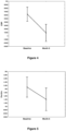
Figure 4 : shows before (baseline) and after treatment (month 6) concentrations of CRP in plasma samples from TB patients. Plasma was collected from patients at recruitment, prior to the initiation of anti-TB therapy and then at the end of standard TB treatment (month 6). Error bars indicate the Least Squared means with 95% Confidence Intervals. -
Figure 5 : shows before (baseline) and after treatment (month 6) concentrations of ferritin in plasma samples from TB patients. Plasma was collected from patients at recruitment, prior to the initiation of anti-TB therapy and then at the end of standard TB treatment (month 6). Error bars indicate the Least Squared means with 95% Confidence Intervals. -
Figure 6 : shows before (baseline) and after treatment (month 6) concentrations of I-309 in plasma samples from TB patients. Plasma was collected from patients at recruitment, prior to the initiation of anti-TB therapy and then at the end of standard TB treatment (month 6). Error bars indicate the Least Squared means with 95% Confidence Intervals. -
Figure 7 : shows before (baseline) and after treatment (month 6) concentrations of IP-10 in plasma samples from TB patients. Plasma was collected from patients at recruitment, prior to the initiation of anti-TB therapy and then at the end of standard TB treatment (month 6). Error bars indicate the Least Squared means with 95% Confidence Intervals. -
Figure 8 : shows before (baseline) and after treatment (month 6) concentrations of transthyretin in plasma samples from TB patients. Plasma was collected from patients at recruitment, prior to the initiation of anti-TB therapy and then at the end of standard TB treatment (month 6). Error bars indicate the Least Squared means with 95% Confidence Intervals. -
Figure 9 : shows before (baseline) and after treatment (month 6) concentrations of Comp_C3 in plasma samples from TB patients. Plasma was collected from patients at recruitment, prior to the initiation of anti-TB therapy and then at the end of standard TB treatment (month 6). Error bars indicate the Least Squared means with 95% Confidence Intervals. -
Figure 10 : shows before (baseline) and after treatment (month 6) concentrations of Comp-FH in plasma samples from TB patients. Plasma was collected from patients at recruitment, prior to the initiation of anti-TB therapy and then at the end of standard TB treatment (month 6). Error bars indicate the Least Squared means with 95% Confidence Intervals. -
Figure 11 : shows before (baseline) and after treatment (month 6) concentrations of MMP-2 in plasma samples from TB patients. Plasma was collected from patients at recruitment, prior to the initiation of anti-TB therapy and then at the end of standard TB treatment (month 6). Error bars indicate the Least Squared means with 95% Confidence Intervals. -
Figure 12 : shows before (baseline) and after treatment (month 6) concentrations of SAP in plasma samples from TB patients. Plasma was collected from patients at recruitment, prior to the initiation of anti-TB therapy and then at the end of standard TB treatment (month 6). Error bars indicate the Least Squared means with 95% Confidence Intervals. - Neural cell adhesion molecule CD56 (NCAM) is described herein as a biomarker for diagnosing tuberculosis disease (TB) in a subject. The method of diagnosis detects NCAM in a single biomarker test or as one biomarker in a panel of 2, 3, 4, 5, 6 or even more biomarkers. The method of diagnosis can be used to distinguish active TB disease from latent Mycobacterium tuberculosis infection in the subject.
- Throughout the specification and claims unless the contents requires otherwise the word 'comprise' or variations such as 'comprises' or 'comprising' will be understood to imply the inclusion of a stated integer or group of integers but not the exclusion of any other integer or group of integers.
- When the diagnostic test detects NCAM as one of a panel of biomarkers, the other biomarkers can be selected from: CRP, SAP, PCT, ferritin, TPA, SAA, ADAMTS-13, p-selectin, GDF-15, I-309, IFN-Y, IP-10, TNF-a, CFH, MIG, ITAC, HCC-1 , MIP-4/CCL18, antithrombin III, Apo A-1 , transthyretin, BDNF, sFas, lipocalin-2, VEGF, PEDF, CC4, IL-33, A2M, IL-22, ECM-1 , IL-1 β, SCD40L, IL-13, myoglobin, IL-12(p40), TNF-β, MIP-1 β, VEGF and SDF-1 a.
- In one embodiment, the other biomarkers are selected from CRP, SAP, PCT, ferritin, TPA, SAA, I-309, MIG, MIP-4/CCL18, Apo A-1 , transthyretin, CFH, TNF-a, IP-10, antithrombin III, GDF-15, HCC1 , ECM-1 , II_-1 β, sCD40L and IL-13.
- In one embodiment, the other biomarkers are selected from CRP, SAP, PCT, ferritin, TPA, SAA, I-309, MIG, MIP-4/CCL18, Apo A-1 , transthyretin, CFH, TNF-a, IP-10, antithrombin III, GDF-15 and HCC1.
- In one embodiment, the other biomarkers are selected from ferritin, SAP, A2M, IL-22, CFH, ECM-1, IL-1 β, sCD40L, IL-13, Apo-A-1 , myoglobin, IL-12(p40), MIP-4/CCL18, antithrombin III, GDF-15 and HCC1.
- In one embodiment, one of the other biomarkers is ferritin. Alternatively, or in addition, one of the other biomarkers is SAP, one of the other biomarkers is A2M, and/or one of the other biomarkers is IL-22.
- Examples of suitable panels of biomarkers which can be used to diagnose TB in the method of the present invention include:
- NCAM, SAP, ferritin, CFH and ECM-1;
- NCAM, SAP, IL-1 β, sCD40L, IL-13 and Apo A-1;
- NCAM, A2M, IL-22, ferritin, myoglobin and IL-12(p40);
- NCAM, A2M, IL-22, ferritin, TNF-β and MIP-4/CCL18;
- NCAM, SAP, ferritin, PCT, MIG, TPA, GDF-15, HCC1, MIP-4/CCL18, antithrombin III;
- NCAM, CRP, IFN-γ, IP-10, Apo A-1, SAA and CFH;
- NCAM, CRP, IFN-Y, IP-10, Apo A-1 and CFH; and
- NCAM, CRP, IFN-Y, IP-10, Apo A-1, CFH and ferritin.
- The subject may be diagnosed as having TB if a reduced level of NCAM is detected in the sample relative to a level of NCAM which is associated with subjects with upper and lower respiratory tract infections, chronic obstructive pulmonary disease and asthma, and if at least one of the additional biomarkers is detected in the sample, or if at least two, at least three, at least four, at least five or at least six of the additional biomarkers are detected in the sample.
- The sample is typically a blood sample containing immune cells, and can be a whole blood sample or a plasma or serum sample. The sample does not need to be stimulated with any antigens before it is tested.
- The TB can be pulmonary TB or extra-pulmonary TB.
- Capture agents specific for each of the biomarkers can be used to bind each of the respective biomarkers. One or more indicators can be provided to indicate when binding of each of the capture agents and biomarkers occurs.
- Whole blood, serum and plasma are usually readily available from subjects and therefore the method of the present invention is suited to a point-of-care test based on the detection of biomarkers in these samples.
- A capture agent and indicator can be used to bind to each of the biomarkers, and an indicator indicates when binding of the capture agents and each of the biomarkers occurs.
- The biomarkers can be detected using commercially available techniques, such as ELISA techniques or multiplex bead array technology, although it is intended that a specific point-of-care diagnostic device will be developed for performing the method, particularly for use in resource poor settings.
- Cut-off or threshold values can be determined based on levels of biomarkers which are typically found in patients without TB, and the levels of the biomarkers detected in the sample can be compared to the cut-off levels when making the determination of whether or not the subject has TB. In other words, the method will detect whether the biomarkers in the panels are under- or over-expressed relative to a subject who does not have TB.
- Antibodies, affybodies, ankyrin repeat proteins, armadillo repeat proteins, nucleic acid aptamers, modified nucleic acid aptamers, peptides, modified peptides, carbohydrate ligands, synthetic ligands or synthetic polymers can be used as the capture agents, and the indicator can be a calorimetric, electrochemical, chromogenic, optical, fluorescent or a radio-labeled indicator.
- The method can be used as an initial diagnostic tool whereby a positive diagnosis from this method can, if necessary, be subsequently confirmed by means of a second diagnostic method. In the interim, while waiting for the results of the second test, the subject can be started on treatment. Conversely, the method of the invention can also be used to rule out TB, thus preventing overtreatment of non-TB subjects.
- The method of the invention can be performed using a diagnostic device which detects and indicates the presence of the biomarkers in the sample. The device has a means for receiving the sample from the subject, such as a loading or receiving area onto or into which the sample is placed. The capture agents and indicators are present in the device, and once the sample has been loaded onto or received into the device, the sample is brought into contact with the capture agents, which are allowed to bind to the biomarkers if present. The indicator will signify that binding has occurred. The device is typically a point-of-care device, such as a lateral flow device.
- The device may be a dip-stick which can be dipped into the plasma or serum sample or onto which the sample can be placed, similar to many home pregnancy test kits. The capture agents are included on the dipstick, and generate a signal when they bind to the biomarkers in the panel, together with a control signal.
- One example of a suitable point-of-care screening test is based on user-friendly lateral flow technology as recently demonstrated in a multi-centered African study [44]. While the sample can be plasma or serum, a whole blood sample (such as from a finger-prick) will be preferable. A nurse or front-line/first contact community healthcare worker can collect finger-prick blood from a patient suspected of having TB disease based on the symptoms, dilute the sample in a buffer which is provided in a kit, and add this to a membrane/test strip containing multiple biomarkers (or antibodies thereto) that have been coupled to up-converting phosphor (UCP) reporter particles. The blood can be left to flow on the strip for a few minutes and after it is dry, can be scanned using a portable hand-held device, typically one which is battery-operated (such as the UCP-Quant strip scanner reader, adapted from the ESE Quant LFR strip reader, Qiagen Lake Constance GmbH).
- UCP reporter technology utilizes nano-sized or sub-micron particles that are composed of rare earth lanthanides in a crystal. The particles display a distinct anti-stokes behaviour upon excitation with infrared (IR) light: low-energy IR light is upconverted to higher energy visible light, with the emission spectrum dependent on the embedded rare earth components. The particles have an unmatched contrast as any possible autofluorescence is absent.
- The assay format can apply sandwich-based immunochromatography with a complementary pair of chemokine-specific antibodies. Utilisation of dry UCP reporter material and lyophilized assay buffer allows convenient storage and transport of assay materials at ambient temperature.
- Use of a kit for diagnosing tuberculosis can be provided to enable health workers to easily perform the diagnosis in a field setting. The kit can include a means for obtaining the blood sample from the patient (such as a needle, syringe or finger-prick device), optionally a blood sample collection device (such as a tube or capillary tube), a buffer for mixing with the blood sample, and a membrane/test strip with antibodies for the biomarker(s).
- The invention will now be described in more detail by way of the following non-limiting examples.
- Participants enrolled into the present study were individuals who presented with signs and symptoms requiring investigation for TB disease at the Fisantekraal Community Clinic in the outskirts of Cape Town, South Africa. The study was a sub-study of a larger diagnostic biomarker project (the African European Tuberculosis Consortium), that was ongoing at the study site and at field sites situated in six other African countries (www.ae-tbc.eu). All study participants presented with persistent cough lasting≥2 weeks and at least one of either fever, malaise, recent weight loss, night sweats, knowledge of close contact with a TB patient, haemoptysis, chest pain or loss of appetite. Participants were eligible for the study if they were 18 years or older and willing to give written informed consent for participation in the study, including consent for HIV testing. Patients were excluded if they were pregnant, had not been residing in the study community for more than 3 months, were severely anaemic (haemoglobin <10 g/I), were on anti-TB treatment, had received anti-TB treatment in the previous 90 days or if they were on quinolone or aminoglycoside antibiotics during the past 60 days. The study was approved by the Health Research Ethics Committee of the Faculty of Medicine and Health Sciences of the University of Stellenbosch.
- At enrolment, 6ml of blood was collected into heparinized BD vacutainer tubes (BD Biosciences) and transported to the laboratory at 4-8°C for further processing. Upon receipt in the laboratory, tubes were centrifuged at 2000 rpm for 10 minutes after which plasma was harvested, aliquoted and stored at -80 °C until analysed. Sputum samples were collected from all study participants and cultured using the MGIT method (BD Biosciences). Positive MGIT cultures were examined for acid fast bacilli using the Ziehl-Neelsen technique (to check for contamination), followed by Capilia TB testing (TAUNS, Numazu, Japan), to confirm the isolation of organisms of the M.tb complex, before being designated as positive cultures.
- Participants were classified as definite TB cases, probable TB cases, participants with other respiratory diseases (ORD) or questionable disease status using a combination of clinical, radiological, and laboratory findings [8]. Only definite TB cases (culture positive individuals) and those with ORD were included in the study. Individuals with ORD had a range of other diagnoses, including upper and lower respiratory tract infections (viral and bacterial infections, although attempts to identify organisms by bacterial or viral cultures were not made), and acute exacerbations of chronic obstructive pulmonary disease or asthma. Because of the disproportionately high number of individuals with ORD, all 22 culture positive TB cases that were available at the study site were included in the study, as were 33 randomly selected individuals with ORD from the study biobank. In total, 55 study participants (22 of whom were culture positive TB cases) were investigated. The mean age of all study participants was 35.8 ± 10.2 years and 14 (25%) were HIV infected. The clinical and demographic characteristics of study participants are shown in Table 1.
Table 1: Clinical and demographic characteristics of study participants Number of participants All (n=55) TB (n=22) ORD (n=33) Males, n (%) 22 (40) 7 (32) 15 (45) Mean age, (Years)±SD 35.8 ± 10.2 38.8 ±10.1 33.9 ± 9.9 HIV Infected, n (%) 14(25) 4(18) 10(30) Quantiferon results Positive, n (%) 34 (64) 15(75) 19 (58) Negative, n (%) 18 (34) 4 (20) 14 (42) Indeterminate, n (%) 1 (2) 1 (5) 0 (0) Abbreviations: TB= pulmonary tuberculosis, SD=standard deviation - The concentrations of 74 host markers including alpha-2-macroglobulin (A2M) , haptoglobin, C-reactive protein (CRP), serum amyloid P (SAP), procalcitonin (PCT), ferritin, tissue plasminogen activator (TPA), fibrinogen, serum amyloid A (SAA) (kits purchased from Bio-Rad Laboratories, Hercules, CA, USA), vitronectin, extracellular matrix protein 1 (ECM1 ), antithrombin III, vitamin D binding protein (VDBP), sFas, granzyme A, sFasL, sCD137, granzyme B, perforin, myoglobin, ADAMTS13, P-selectin, lipocalin-2, growth differentiation factor (GDF) -15 , thrombopoietin (TPO), stem cell factor (SCF), B-cell attracting chemokine (BCA)-1 , epithelial neutrophil activating protein (ENA-78), thymic stromal lymphopoietin (TSLP), I-309(CCL-1 ), stromal cell derived factor- 1 alpha (SDF-1a), interferon (IFN)-y, IFN-a2, interferon gamma inducible protein (IP)-10 (CXCL10), macrophage inflammatory protein (MIP)-1 β, tumor necrosis factor (TNF)-a, TNF-β, vascular endothelial growth factor (VEGF), soluble CD40 ligand (sCD40L), apolipoprotein (Apo) A-1 , Apo CIII, complement component 3 (CC3), transthyretin, complement factor H (CFH), total plasminogen activator inhibitor-1 (PAI-1 ), neural cell adhesion molecule (NCAM), brain-derived neurotrophic factor (BDNF), cathepsin D, myeloperoxidase (MPO), matrix metalloproteinase (MMP)-2, MMP-9, monokine induced by gamma interferon (MIG/CXCL9), granulocyte chemotactic protein-2 (GCP2), interferon inducible T-cell alpha chemoattractant (I-TAC/CXCL1 1 ), hemofiltrate CC chemokine-1 (HCC1 ), a1 -antitrypsin, pigment epithelium derived factor (PEDF), macrophage inflammatory protein-4 (MIP-4/CCL18), complement C4, interleukin (IL)-17F, IL-17A, IL-22, IL-33, IL-21 , IL-23, IL-25, IL-31 , IL-28A, IL-16, IL-I β, IL-12(p40), IL-13, IL-1 1 and IL-29 (Merck Millipore, Billerica, MA, USA), were investigated in plasma samples from all the study participants. The experiments were performed blindly, according to the instructions of the kit manufacturers, on the Bio-Plex platform (Bio-Rad). The Bio-Plex manager Software version 6.1 was used for bead acquisition and analysis of median fluorescence intensities.
- Differences in the concentrations of host markers between TB patients and individuals with ORD were analysed using the Mann-Whitney U test. The diagnostic abilities of individual host markers were assessed by receiver operator characteristics (ROC) curve analysis. The predictive abilities of combinations of host markers were investigated by general discriminant analysis (GDA), with leave-one-out cross validation [13]. Differences in the expression profiles of host markers during the course of TB treatment were analysed using mixed model repeated measures analysis of variance (ANOVA), with Fisher's Least Significant Difference (LSD) post hoc testing. P-values ≤0.05 were considered significant. The data were analysed using Statistica (Statsoft, Ohio, USA) and Graphpad Prism version 5 (Graphpad Software Inc., CA, USA).
- When the baseline concentrations of host markers in TB patients (n=22) were compared to the levels detected in patients with ORD (n=33), by the Mann Whitney U test, the concentrations of 23 out of the 74 analytes were significantly different between the two groups. The median levels of CRP, SAP, PCT, ferritin, TPA, SAA, ADAMTS-13, p-selectin, GDF-15, I-309, IFN-Y, IP-10, TNF-a, CFH, MIG, ITAC, HCC-1 and MIP-4 were significantly higher in TB cases, whereas the levels of antithrombin III, Apo A-1 , transthyretin, NCAM and BDNF were significantly higher in the ORD group. Trends (0.05<p≤ 0.01) towards higher levels of sFas, lipocalin-2, VEGF, PEDF, CC4 and IL-33 were observed in TB cases (Table 2). When the diagnostic accuracies of individual host markers were investigated by ROC curve analysis, the area under the ROC curve (AUC) was≥0.70 for 18 markers (Table 2). The most accurate single host markers included CRP, SAP, NCAM, TPA, I-309, and MIG, which all performed with AUC≥ 0.80 (Table 2,
Figures 1 and2 ). When data was stratified according to HIV infection status, concentrations of three additional markers (A2M, MIP-1 β and VEGF) became significant in the two groups, with AUC's of 0.70, 0.69 and 0.69 respectively. - When the data obtained from all the TB patients and those with ORD were fitted into General Discriminant Analysis (GDA) models regardless of HIV status, combinations of up to six different host markers showed potential in the diagnosis of TB disease. A five-marker biosignature of NCAM, SAP, ferritin, CFH and ECM-1 diagnosed TB disease with a sensitivity of 95.2% (95% Cl, 81 .0-99.9%) and specificity of 92.9% (95% Cl, 70.8-98.9%) in the resubstitution classification matrix and sensitivity of 95.2% (95% Cl, 81 .0-99.9%) and specificity of 89.3% (95% Cl, 66.4-97.2%) after leave-one-out cross validation. However, the most optimal diagnostic biosignature (irrespective of HIV status) was a combination between six markers (NCAM, SAP, IL-1 β, sCD40L, IL-13 and Apo A-1 ), which diagnosed TB disease with a
sensitivity 100% (95% Cl, 86.3-100%) and specificity of 89.3% (95%CI, 67.6-97.3%) after leave-one-out cross validation. The positive and negative predictive values of the six- marker biosignature were 87.5% (95% Cl, 66.5-96.7%) and 100% (95%CI, 83.4-100%) respectively (Figure 3 ).

- When the GDA procedure was repeated after excluding the HIV infected individuals, two six-marker biosignatures:- NCAM, A2M, IL-22, ferritin, myoglobin and IL-12(p40), and NCAM, 5 A2M, IL-22, ferritin, TNF-β and MIP-4, diagnosed TB disease with both sensitivity and specificity of 100% (AUC = 1 .0, 95% Cl, 1 .0-1 .0). NCAM was the most frequent analyte in biosignatures, appearing in all the top 13 biosignatures for diagnosing TB disease regardless of HIV infection status, and in 68%(23 of the 34) biosignatures that were generated for the diagnosis of TB disease after excluding HIV infected individuals. Other markers that occurred 10 most frequently in diagnostic biosignatures for TB disease included GDF-15, SAP, CFH, A2M, TNF-β, ferritin, SDF-1 a amongst others (
Figure 3 ). - To investigate whether any of the 74 markers could potentially be used to monitor the 15 response to TB treatment, the host markers were evaluated in plasma samples that were collected from TB patients at the end of standard TB treatment (month 6). However, of the 22 TB patients that were investigated in this study, only 15 (68%) returned to the clinic and provided samples at the end of treatment. Compared to baseline levels, the concentrations of 1 1 host markers changed significantly during the course of treatment. There was a significant 20 decrease in the levels of CRP, SAP, ferritin, IFN-γ, VEGF, IP-10, CC3, CFH and a-1 - antitrypsin from baseline to
month 6, whereas a significant increase in the levels of transthyretin and MMP-2 was observed. The levels of IL-I β, SAA, sFas and MIG showed trends towards decreasing levels from baseline tomonth 6, whereas Apo-CIII, Apo A-1 and GCP-2 showed trends towards increasing levels at the end of treatment (Figures 4 to 12 ). - The diagnostic potentials of 74 host markers in plasma samples that were obtained from confirmed active TB cases and individuals with ORD were investigated as candidates for the diagnosis of TB disease. 18 of the 74 host markers (including new biomarkers in the TB field antithrombin III, GDF-15, NCAM, HCC1 and recently identified markers MIP-4, I-309, MIG, Apo A-1, transthyretin and CFH) showed potential in the diagnosis of TB disease, regardless of HIV infection status, as determined by area under the ROC curve (AUC). The most optimal diagnostic biosignature (irrespective of HIV infection status) was a six-marker model of NCAM, SAP, IL-1 β, sCD40L, IL-13 and Apo A-1 , which diagnosed TB disease with a sensitivity of 100% and specificity of 89.3%, with promising positive and negative predictive values. In the absence of HIV infection, six-marker biosignatures diagnosed TB disease with 100% accuracy.
- Amongst the 18 host markers that showed the most potential in the diagnosis of TB disease individually as determined by AUC, six (CRP, SAP, PCT, ferritin, TPA and SAA) were acute phase proteins, six (I-309, MIG, MIP-4, Apo A-1 , transthyretin and CFH) were markers that play various roles in the body and have recently been identified as promising TB diagnostic candidates [14][10], two (TNF-a and IP-10) are widely investigated TB biomarkers [15], and four (antithrombin III, GDF-15, NCAM, HCC1 ) are markers which have not previously been investigated in the TB field. Other host markers (including ECM-1, IL-1 β, sCD40L and IL-13), although not very promising individually in the current study, were included into diagnostic biosignatures for TB disease.
- Acute phase proteins are primarily produced by the liver, and act as opsonins at inflammatory sites [16]. Serum CRP has been extensively studied and shown to be promising for both the diagnosis of TB disease and monitoring of TB treatment response[17][18]. SAP, a homologue of CRP, has also been shown to have a protective role against bacterial infections [19]. Ferritin is a well-recognised protein in iron storage processes. It has long been established that iron acquisition is an essential virulence mechanism for pathogenic bacteria [20][21]. The concentrations of all the acute phase proteins that showed potential in the current study were elevated in the TB patients, and this is in agreement with previous findings [10][21 ][22].
- The chemokines; MIG (CXCL9), IP-10(CXCL10), ITAC(CXCL11) and I-309(CCL-1) are found in abundance in activated bronchial epithelium[23]. MIP-4(CCL18) is a chemokine that is produced mainly by antigen presenting cells [24]. These chemokines play vital roles in the recruitment of activated T-cells to the site of infection [23][25]. The present study found significantly higher levels of these chemokines in the plasma of patients with active TB. The relatively new host markers investigated in this study (GDF-15, antithrombin III, HCC1 and NCAM) showed individual potential in the diagnosis of TB disease. Other markers (including p-selectin, ADAMTS-13 and BDNF), although not amongst the most promising single markers as demonstrated by ROC curve analysis, were significantly different between the TB patients and individuals with ORD, with the Mann-Whitney U test. GDF-15 is a member of the transforming growth factor beta superfamily (TGF-β) and its expression is associated with tissue damage, but has also been reported to exhibit tissue protective functions [28][29]. It has been identified as a prognostic marker for prostate cancer, with high serum levels observed in patients with liver cirrhosis and hepatocellular carcinoma [30][31]. Antithrombin complexes are important mediators of the coagulation system, with antithrombin III being one of the most important inhibitors of this system [32]. Markedly lowered antithrombin III plasma levels have been observed in sepsis [33]. HCC1 has been identified as a monocyte chemoattractant, with high concentrations observed in patients with chronic renal failure [34]. P-selectin is part of the selectin family of cell adhesion molecules, that promotes inflammatory reactions [35]. ADAMTS-13 is a metalloprotease with thrombospondin repeats, and has shown low activity in patients with recurrent thrombaotic thrombocytopenic purpura [36][37]. In a previous study by Liu et al, GDF-15 levels were not significantly different between TB patients, latently infected individuals and healthy controls [31]. However, GDF-15 levels showed potential in the current study. The current study observed significantly higher levels of p-selectin in TB patients in comparison to individuals with ORD. For the other newly identified TB diagnostic candidates, higher levels of HCC1 and ADAMTS-13 were observed in TB patients, but the levels of antithrombin III and NCAM were higher in individuals with ORD.
- NCAM (CD56) is important in cell-cell or cell-matrix interactions, and is involved in neuronal differentiation, branching and survival [39]. It has been shown to play a role in lung tumor progression [40]. In the present study, the potential of NCAM as a biomarker for TB disease was demonstrated for the first time. NCAM was the most frequently occurring marker in biosignatures for the diagnosis of TB disease, and was included in all top 13 marker combinations for the diagnosis of TB disease, regardless of HIV infection status, and in 68% of the models that were generated when HIV infected individuals were excluded. The combination of NCAM with five other markers (SAP, IL-1 β, sCD40L, IL-13 and Apo A-1) diagnosed TB disease with high accuracy, regardless of HIV infection status, and all the TB patients and individuals with ORD were accurately classified (100% sensitivity and specificity) when NCAM was used in combination with either A2M+IL-22+ferritin+myoglobin+IL-12(p40), or A2M+IL-22+ferritin+TNF^+MIP-4, in the absence of HIV infection. In a previous serum-based study by the applicant [10], optimal diagnosis of TB disease was achieved using a
biosignature of CRP, IFN-γ, IP-10, Apo A-1 , SAA and CFH on a serum sample. There was excellent agreement between the findings of this and the serum study, as the diagnostic potential observed for individual host markers including CRP, SAA, SAP, CFH, Apo A-1, and ferritin amongst others was replicated in the current study. It is therefore envisaged that the new host markers identified in this study could be used in conjunction with, or as alternatives to, the markers that were included in the previous biosignature [10] if a point-of-care screening test based on these biosignatures were to be developed. - In addition to potentially being useful as diagnostic candidates for TB disease, 1 1 of the markers (including CRP, SAP, ferritin, IP-10, a-1 -antitrypsin) changed with treatment, thereby indicating that they may be potential candidates for monitoring the response to TB treatment. Although observations for MMP-2 (increasing levels from baseline to month 6) were contrary to the observations of Ugarte-Gil et al (decreasing levels from baseline to
month 6 in sputum culture positive individuals after 2 weeks of treatment)[41], the observations for CRP, SAP, ferritin, IP-10, and a-1 -antitrypsin are in agreement with findings from previous studies[42][43]. -
- 1. WHO.Global Tuberculosis Report, 2015.
- 2. Desikan P: Sputum smear microscopy in tuberculosis: Is it still relevant? Indian J Med Res 2013, 137:442-444.
- 3. Home DJ, Royce SE, Gooze L, Narita M, Hopewell PC, Nahid P, Steingart KR: Sputum monitoring during tuberculosis treatment for predicting outcome: systematic review and metaanalysis. Lancet Infect D/'s 2010, 10:387-394.
- 4. Zeka AN, Tasbakan S, Cavusoglu C: Evaluation of the GeneXpert MTB/RIF Assay for Rapid Diagnosis of Tuberculosis and Detection of Rifampin Resistance in Pulmonary and Extrapulmonary Specimens. J Clin Microbiol 201 1, 49:4138-4141 .
- 5. Miotto P, Bigoni S, Migliori GB, Matteelli A, Cirillo DM: Early tuberculosis treatment monitoring by Xpert(R) MTB/RIF. Eur Respir J 2012, 39:1269-1271.
- 6. Denkinger CM, Pai M, Patel M, Menzies D: Gamma Interferon Release Assay for Monitoring of Treatment Response for Active Tuberculosis: an Explosion in the Spaghetti Factory. J Clin Microbiol 2013, 51:607-61 0.
- 7. Higuchi K, Harada N, Mori T: Interferon-y responses after isoniazid chemotherapy for latent tuberculosis. Respirology 2008, 13:468-472.
- 8. Jacobs R, Tshehla E, Malherbe S, Kriel M, Loxton AG, Stanley K, van der Spuy G, Walzl G, Chegou NN: Host biomarkers detected in saliva show promise as markers for the diagnosis of pulmonary tuberculosis disease and monitoring of the response to tuberculosis treatment. Cytokine 201 6, 81:50-56.
- 9. Phalane KG, Kriel M, Loxton AG, Menezes A, Stanley K, van der Spuy GD, Walzl G, Chegou NN: Differential Expression of Host Biomarkers in Saliva and Serum Samples from Individuals with Suspected Pulmonary Tuberculosis. Mediators Inflamm 2013, 2013:1 -1 0.
- 10.
Chegou, N, Walzl. G, Mihret.A: Method for diagnosing Tuberculosis. International Patent Application no: PCT/IB2015/051435 WO2015128830. [Accessed 1 April 2016 ] - 11. Walzl G, Ronacher K, Hanekom W, Scriba TJ, Zumla A: Immunological biomarkers of tuberculosis. Nat Rev Immunol 201 1, 11 :343-354.
- 12. De Groote MA, Nahid P, Jarlsberg L, Johnson JL, Weiner M, Muzanyi G, Janjic N, Sterling DG, Ochsner UA: Elucidating Novel Serum Biomarkers Associated with Pulmonary Tuberculosis Treatment. PLoS ONE 2013, 8:e61 002.
- 13. General Discriminant Analysis - Statistics Textbook [http://documents.software.dell.com/Statistics/Textbook/General-Discriminant-AnalysisllAccessed 1 April 2016]
- 14. Chen T, Li H, Lin J, Guo H, Wang W, Chen L, Zhang X, Wang Y, Chen Y, Liao Q, others: Profiling the Human Immune Response to Mycobacterium Tuberculosis by Humane Cytokine Array. Tuberculosis 2016.
- 15. Chegou NN, Heyckendorf J, Walzl G, Lange C, Ruhwald M: Beyond the IFN-γ horizon: Biomarkers for immunodiagnosis of infection with M. tuberculosis. Eur Respir J 2013.
- 16. Gruys E, Toussaint MJM, Niewold TA, Koopmans SJ: Acute phase reaction and acute phase proteins. J Zhejiang Univ Sci 2005, 6B:1045-1056.
- 17. Shaikh MK, Samo JA, Devrajani BR, Shah SZA, Shaikh S, Shaikh I : C-reactive protein in patients with pulmonary tuberculosis. World Appl Sci J ' 2012, 17:140-144.
- 18. Rao S, Bernhardt V: Serum C-reactive protein in pulmonary tuberculosis: correlation with bacteriological load and extent of disease. Infect Dis Clin Pract 2009, 17:314-316.
- 19. Noursadeghi M, Bickerstaff MC, Gallimore JR, Herbert J, Cohen J, Pepys MB: Role of serum amyloid P component in bacterial infection: protection of the host or protection of the pathogen. Proc .
- 20. Kotru M, Rusia U, Sikka M, Chaturvedi S, Jain AK: Evaluation of serum ferritin in screening for iron deficiency in tuberculosis. Ann Hematol 2004, 83:95-100.
- 21. Opolot JO, Theron AJ, Anderson R, Feldman C: Acute phase proteins and stress hormone responses in patients with newly diagnosed active pulmonary tuberculosis. Lung 2015, 193:13-18.
- 22. Radovic M, Pejcic T, Stankovic I, Ristic L, Rancic M, Ciric Z: Acute-Phase Inflammatory Response in Patients with Pulmonary Tuberculosis. Acta Fac Medicae Naissensis 2014, 31.
- 23. Sauty A, Dziejman M, Taha RA, larossi AS, Neote K, Garcia-Zepeda EA, Hamid Q, Luster AD: The T cell-specific CXC chemokines IP-10, Mig, and I-TAC are expressed by activated human bronchial epithelial cells. J Immunol 1999, 162:3549-3558.
- 24. Ferrara G, Bleck B, Richeldi L, Reibman J, Fabbri LM, Rom WN, Condos R: Mycobacterium tuberculosis Induces CCL18 Expression in Human Macrophages: CCL18 in Tuberculosis Infection. Scand J Immunol 2008, 68:668-674.
- 25. Haque NS, Fallon JT, Taubman MB, Harpel PC: The chemokine receptor CCR8 mediates human endothelial cell chemotaxis induced by I-309 and Kaposi sarcoma herpesvirus-encoded vMIP-I and by lipoprotein (a)-stimulated endothelial cell conditioned medium. Blood 2001 , 97:39-45.
- 26. Hong JY, Jung GS, Kim H, Kim YM, Lee HJ, Cho S-N, Kim SK, Chang J, Kang YA: Efficacy of .
- 27. Sauty A, Colvin RA, Wagner L, Rochat S, Spertini F, Luster AD: CXCR3 Internalization Following T Cell-Endothelial Cell Contact: Preferential Role of IFN-Inducible T Cell Chemoattractant (CXCL11). J Immunol 2001 , 167:7084-7093.
- 28. Hsiao EC, Koniaris LG, Zimmers-Koniaris T, Sebald SM, Huynh TV, Lee S-J: Characterization of growth- .
- 29. Vanhara P, Hampl A, Kozubik A, Soucek K: Growth/differentiation factor-15: prostate cancer suppressor or promoter [quest]. Prostate Cancer Prostatic Dis 2012, 15:320-328.
- 30. Martin Boegemann FJW: GDF15 and Hepcidin as Prognostic Factors in Patients with Prostate Cancer. J Mol Biomark Diagn 2014, 05.
- 31. Liu X, Chi X, Gong Q, Gao L, Niu Y, Chi X, Cheng M, Si Y, Wang M, Zhong J, Niu J, Yang W: Association of Serum Level of .
- 32. Levi M, Keller TT, van Gorp E, ten Cate H: Infection and inflammation and the coagulation system. Cardiovasc Res 2003, 60:26-39.
- 33. Mesters RM, Mannucci PM, Coppola R, Keller T, Ostermann H, Kienast J: Factor Vila and antithrombin III activity during severe sepsis and septic shock in neutropenic patients. Blood 1996, 88:881 -886.
- 34. Schulz-Knappe P, Magert HJ, Dewaid B, Meyer M, Cetin Y, Kubbies M, Tomeczkowski J, Kirchhoff K, Raida M, Adermann K, others: HCC-1 , a novel chemokine from human plasma. J Exp Med 1996, 183:295-299.
- 35. Wynn TA, Hesse M, Sandler NG, Kaviratne M, Hoffmann KF, Chiaramonte MG, Reiman R, Cheever AW, Sypek JP, Mentink-Kane MM: P-selectin suppresses hepatic inflammation and fibrosis in mice by regulating interferon ? and the IL-13 decoy receptor. Hepatology 2004, 39:676-687.
- 36. Soejima K, Mimura N, Hiroshima M, Maeda H, Hamamoto T, Nakagaki T, Nozaki C: A Novel Human Metalloprotease Synthesized in the Liver and Secreted into the Blood: Possibly, the von Willebrand Factor- Cleaving Protease? J Biochem (Tokyo) 2001 , 130:475-480.
- 37. Peyvandi F, Lavoretano S, Palla R, Feys HB, Vanhoorelbeke K, Battaglioli T, Valsecchi C, Canciani MT, Fabris F, Zver S, Reti M, Mikovic D, Karimi M, Giuffrida G, Laurenti L, Mannucci PM:ADAMTS13 and anti-ADAMTS13 antibodies as markers for recurrence of acquired thrombotic thrombocytopenic purpura during remission. Haematologica 2008, 93:232-239.
- 38. Mukae H, ASHITANI J, Tokojima M, Ihi T, Kohno S, Matsukura S: Elevated levels of circulating adhesion molecules in patients with active pulmonary tuberculosis. Respirology 2003, 8:326-331.
- 39. Kiryushko D, Korshunova I, Berezin V, Bock E: Neural cell adhesion molecule induces intracellular signaling via multiple mechanisms of Ca2+ homeostasis. Mol Biol Cell 2006, 17:2278-2286.
- 40. Campodonico PB, de Kier Joffe EDB, Urtreger AJ, Lauria LS, Lastiri JM, Puricelli LI, Todaro LB: The neural cell adhesion molecule is involved in the metastatic capacity in a murine model of lung cancer. Mol Carcinog 2010:n/a-n/a.
- 41. Ugarte-Gil CA, Elkington P, Gilman RH, Coronel J, Tezera LB, Bernabe-Ortiz A, Gotuzzo E, Friedland JS, Moore DAJ: Induced Sputum MMP-1 , -3 & -8 Concentrations during Treatment of Tuberculosis. PLoS ONE 2013, 8:e61333.
- 42. Martins C, Gama AC de C, Valcarenghi D, Batschauer AP de B: Markers of acute-phase response in the treatment of pulmonary tuberculosis. J Bras Patol E Med Lab 2014, 50.
- 43. Hong JY, Lee HJ, Kim SY, Chung KS, Kim EY, Jung JY, Park MS, Kim YS, Kim SK, Chang J, Cho S-N, Kang YA: Efficacy of IP-10 as a biomarker for monitoring tuberculosis treatment. J Infect 2014, 68:252-258.
- 44. Corstjens PLAM, Tjon Kon Fat EM, de Dood CJ, van der Ploeg-van Schip JJ, Franken KLMC, Chegou NN, Sutherland JS, Howe R, Mihret A, Kassa D, van der Vyver M, Sheehama J, Simukonda F, Mayanja-Kizza H, Ottenhoff THM, Walzl G, Geluk A: Multi-center evaluation of a user-friendly lateral flow assay to determine IP-10 and CCL4 levels in blood of TB and non-TB cases in Africa. Clin Biochem 2016, 49:22-31
Claims (12)
- A method of diagnosing tuberculosis disease (TB) in a subject, the method comprising the steps of:a) testing a blood sample from a subject suspected of having TB for the presence of neural cell adhesion molecule (NCAM/ CD56); andb) diagnosing the patient as having TB if a reduced level of NCAM is detected in the sample relative to a level of NCAM which is associated with subjects with upper and lower respiratory tract infections, chronic obstructive pulmonary disease and asthma.
- A method according to claim 1, which comprises the steps of:a) in addition to testing said blood sample from a subject suspected of having TB for the presence of NCAM, testing a blood sample for at least one other biomarker selected from ferritin, serum amyloid P (SAP), alpha-2-macroglobulin (A2M), interleukin-22 (IL-22), complement factor H (CFH), extracellular matrix protein 1 (ECM-1), interleukin-1 β (IL-1 β), soluble CD40 ligand (sCD40L), interleukin-13 (IL-13), apolipoprotein A-1 (Apo-A-1), myoglobin, interleukin-12 (IL-12(p40)), macrophage inflammatory protein-4 (MIP-4/CCL18), antithrombin III, growth differentiation factor (GDF-15), hemofiltrate CC chemokine-1 (HCC1), C-reactive protein (CRP), procalcitonin (PCT), tissue plasminogen activator (TPA), serum amyloid A (SAA), a disintegrin and metalloproteinase with a thrombospondin type 1 motif, member 13 (ADAMTS-13 / von Willebrand factor-cleaving protease (VWFCP)), p-selectin, Chemokine (C-C motif) ligand 1 (CCL1 / I-309), interferon γ (IFN-γ), interferon gamma inducible protein 10 (IP-10), tumor necrosis factor α (TNF-α), monokine induced by gamma interferon (MIG), interferon inducible T-cell alpha chemoattractant (ITAC), transthyretin, brain-derived neurotrophic factor (BDNF), soluble Fas (sFas), lipocalin-2, vascular endothelial growth factor (VEGF), pigment epithelium derived factor (PEDF), complement component 4 (CC4), interleukin-33 (IL-33), tumor necrosis factor β (TNF-β), macrophage inflammatory protein 1β (MIP-1β) and stromal cell derived factor-1 alpha (SDF-1 α); andb) diagnosing the patient as having TB if NCAM at a said reduced level and at least one of the biomarkers is detected in said sample.
- A method according to claim 2, wherein the sample is tested for the presence of NCAM and:at least two other biomarkers selected from ferritin, SAP, A2M, IL-22, CFH, ECM-1, IL-1β, sCD40L, IL-13, Apo-A-1, myoglobin, IL-12(p40), MIP-1β, CRP, PCT, TPA, SAA, ADAMTS-13, p-selectin, GDF-15, I-309, IFN-γ, IP-10, TNF-α, MIG, ITAC, HCC-1, antithrombin III, transthyretin, BDNF, sFas, lipocalin-2, VEGF, PEDF, CC4, IL-33, TNF-β and SDF-1 α;at least three other biomarkers selected from ferritin, SAP, A2M, IL-22, CFH, ECM-1, IL-1 β, sCD40L, IL-13, Apo-A-1, myoglobin, IL-12(p40), MIP-4/CCL18, CRP, PCT, TPA, SAA, ADAMTS-13, p-selectin, GDF-15, I-309, IFN-γ, IP-10, TNF-α, MIG, ITAC, HCC-1, antithrombin III, transthyretin, BDNF, sFas, lipocalin-2, VEGF, PEDF, CC4, IL-33, TNF-β, MIP-1β and SDF-1 α;at least four other biomarkers selected from ferritin, SAP, A2M, IL-22, CFH, ECM-1, IL-1 β, sCD40L, IL-13, Apo-A-1, myoglobin, IL-12(p40), MIP-4/CCL18, CRP, PCT, TPA, SAA, ADAMTS-13, p-selectin, GDF-15, I-309, IFN-γ, IP-10, TNF-α, MIG, ITAC, HCC-1, antithrombin III, transthyretin, BDNF, sFas, lipocalin-2, VEGF, PEDF, CC4, IL-33, TNF-β, MIP-1β and SDF-1 α; orat least five other biomarkers selected from ferritin, SAP, A2M, IL-22, CFH, ECM-1, IL-1 β, sCD40L, IL-13, Apo-A-1, myoglobin, IL-12(p40), MIP-4/CCL18, CRP, PCT, TPA, SAA, ADAMTS-13, p-selectin, GDF-15, I-309, IFN-γ, IP-10, TNF-α, MIG, ITAC, HCC-1, antithrombin III, transthyretin, BDNF, sFas, lipocalin-2, VEGF, PEDF, CC4, IL-33, TNF-β, MIP-1β and SDF-1 α.
- A method according to either of claims 2 or 3, wherein at least one of the other biomarkers is ferritin or SAP.
- A method according to either of claims 3 or 4, wherein the sample is tested for the following biomarkers:NCAM, SAP, ferritin, CFH and ECM-1;NCAM, SAP, IL-1β, sCD40L, IL-13 and Apo A-1;NCAM, A2M, IL-22, ferritin, myoglobin and IL-12(p40); orNCAM, A2M, IL-22, ferritin, TNF-β and MIP-4/CCL18.
- A method according to any one of claims 2 to 5, wherein the patient is diagnosed as having TB if at least two of the biomarkers are detected in the sample, if at least three of the biomarkers are detected in the sample, if at least four of the biomarkers are detected in the sample, if at least five of the biomarkers are detected in the sample, or if at least six of the biomarkers are detected in the sample.
- A method according to any one of claims 1 to 6, wherein the sample is a plasma or serum sample.
- A method according to any one of claims 1 to 7, wherein the sample is an unstimulated sample.
- A method according to any one of claims 1 to 8, wherein the tuberculosis is pulmonary tuberculosis.
- A method according to any one of claims 1 to 9, wherein capture agents specific for each of the biomarkers are used to bind each of the respective biomarkers and one or more indicators are provided to indicate when binding of each of the capture agents and biomarkers occurs.
- Use of a kit for diagnosing tuberculosis in a blood sample from a subject according to the method of any one of claims 1 to 10, the kit comprising:a) a means of receiving a sample from a patient;b) a capture agent for binding NCAM and optionally also capture agents for binding other biomarkers selected from ferritin, SAP, A2M, IL-22, CFH, ECM-1, IL-1β, sCD40L, IL-13, Apo-A-1, myoglobin, IL-12(p40) and MIP-4/CCL18; andc) at least one indicator which indicates when the capture agents bind to the biomarkers.
- Use of a kit according to claim 11, wherein the kit includes:a) means for obtaining the blood sample from the patient,b) a blood sample collection device,c) a buffer for mixing with the blood sample, andd) a membrane/test strip with antibodies for the biomarker(s).
Applications Claiming Priority (2)
| Application Number | Priority Date | Filing Date | Title |
|---|---|---|---|
| ZA201602557 | 2016-04-15 | ||
| PCT/IB2017/052142 WO2017179008A1 (en) | 2016-04-15 | 2017-04-13 | Host biomarkers for immunodiagnosis and monitoring of tuberculosis disease |
Publications (3)
| Publication Number | Publication Date |
|---|---|
| EP3443356A1 EP3443356A1 (en) | 2019-02-20 |
| EP3443356A4 EP3443356A4 (en) | 2019-12-11 |
| EP3443356B1 true EP3443356B1 (en) | 2024-06-05 |
Family
ID=60041447
Family Applications (1)
| Application Number | Title | Priority Date | Filing Date |
|---|---|---|---|
| EP17782034.7A Active EP3443356B1 (en) | 2016-04-15 | 2017-04-13 | Host biomarkers for immunodiagnosis and monitoring of tuberculosis disease |
Country Status (3)
| Country | Link |
|---|---|
| EP (1) | EP3443356B1 (en) |
| WO (1) | WO2017179008A1 (en) |
| ZA (1) | ZA201807635B (en) |
Families Citing this family (8)
| Publication number | Priority date | Publication date | Assignee | Title |
|---|---|---|---|---|
| EP3770602A1 (en) * | 2019-07-23 | 2021-01-27 | Fundació Assistencial de Mútua de Terrassa, FPC | In vitro method for discriminating latent from active tuberculosis |
| BR112023006262A2 (en) * | 2020-10-06 | 2023-05-09 | Cepheid | METHODS FOR TUBERCULOSIS DIAGNOSIS AND DIFFERENTIATION BETWEEN ACTIVE AND LATENT TUBERCULOSIS |
| WO2023057922A1 (en) * | 2021-10-06 | 2023-04-13 | Stellenbosch University | Biomarkers for diagnosing tuberculosis |
| CN115786491A (en) * | 2022-08-22 | 2023-03-14 | 深圳市第三人民医院 | Application of LCN2 and FCN1 as markers in diagnosis, prognosis and curative effect evaluation of diabetes combined tuberculosis |
| WO2024264081A1 (en) * | 2023-06-22 | 2024-12-26 | Stellenbosch University | Method for diagnosing tuberculosis disease |
| CN116990527A (en) * | 2023-08-03 | 2023-11-03 | 首都医科大学附属北京胸科医院 | Application of a biomarker in the differential diagnosis of tuberculous pleurisy |
| CN117129678A (en) * | 2023-08-18 | 2023-11-28 | 深圳大学 | Application of biomarkers relevant to the assessment of tuberculous pleural effusion |
| CN120610003B (en) * | 2025-06-11 | 2026-04-14 | 武汉大学人民医院(湖北省人民医院) | Application of growth differentiation factor 15 in preparation of products for diagnosis, prognosis and treatment of retinal artery occlusion disease |
Family Cites Families (7)
| Publication number | Priority date | Publication date | Assignee | Title |
|---|---|---|---|---|
| US20140155411A1 (en) * | 2012-04-13 | 2014-06-05 | Somalogic, Inc. | Tuberculosis Biomarkers and Uses Thereof |
| US10001481B2 (en) * | 2012-05-25 | 2018-06-19 | Stellenbosch University | Method for diagnosing tuberculosis disease by detecting induced markers after stimulation of T-cells with antigens |
| GB201213567D0 (en) * | 2012-07-31 | 2012-09-12 | Proteinlogic Ltd | Biomarkers |
| JP6416793B2 (en) * | 2013-02-28 | 2018-10-31 | カプリオン プロテオミクス インコーポレーテッド | Tuberculosis biomarkers and uses thereof |
| CA2887722C (en) | 2013-10-10 | 2017-01-24 | Kevin Moran | A method for producing a zirconium concentrated product from froth treatment tailings |
| AP2016009427A0 (en) * | 2014-02-26 | 2016-08-31 | Univ Stellenbosch | Method for diagnosing tuberculosis |
| WO2016162827A1 (en) * | 2015-04-09 | 2016-10-13 | Universita' Degli Studi Di Genova | Population of human lymphoid precursors, method for their identification and uses thereof |
-
2017
- 2017-04-13 EP EP17782034.7A patent/EP3443356B1/en active Active
- 2017-04-13 WO PCT/IB2017/052142 patent/WO2017179008A1/en not_active Ceased
-
2018
- 2018-11-13 ZA ZA2018/07635A patent/ZA201807635B/en unknown
Non-Patent Citations (2)
| Title |
|---|
| GERHARD WALZL ET AL: "Immunological biomarkers of tuberculosis", NATURE REVIEWS IMMUNOLOGY, vol. 11, no. 5, 8 April 2011 (2011-04-08), pages 343 - 354, XP055109445, ISSN: 1474-1733, DOI: 10.1038/nri2960 * |
| JAYNE S. SUTHERLAND ET AL: "Highly Accurate Diagnosis of Pleural Tuberculosis by Immunological Analysis of the Pleural Effusion", PLOS ONE, vol. 7, no. 1, 25 January 2012 (2012-01-25), pages e30324, XP055154918, DOI: 10.1371/journal.pone.0030324 * |
Also Published As
| Publication number | Publication date |
|---|---|
| EP3443356A4 (en) | 2019-12-11 |
| ZA201807635B (en) | 2019-07-31 |
| WO2017179008A1 (en) | 2017-10-19 |
| EP3443356A1 (en) | 2019-02-20 |
Similar Documents
| Publication | Publication Date | Title |
|---|---|---|
| Jacobs et al. | Identification of novel host biomarkers in plasma as candidates for the immunodiagnosis of tuberculosis disease and monitoring of tuberculosis treatment response | |
| WO2017179008A1 (en) | Host biomarkers for immunodiagnosis and monitoring of tuberculosis disease | |
| Oeschger et al. | Point of care technologies for sepsis diagnosis and treatment | |
| Weng et al. | The role of infection in acute exacerbation of idiopathic pulmonary fibrosis | |
| Bridel et al. | Cerebrospinal fluid angiotensin-converting enzyme for diagnosis of neurosarcoidosis | |
| Phalane et al. | Differential expression of host biomarkers in saliva and serum samples from individuals with suspected pulmonary tuberculosis | |
| Fan et al. | Diagnosing sepsis–The role of laboratory medicine | |
| Jacobs et al. | Host biomarkers detected in saliva show promise as markers for the diagnosis of pulmonary tuberculosis disease and monitoring of the response to tuberculosis treatment | |
| CN106461675B (en) | For diagnosing phthisical method | |
| US10001481B2 (en) | Method for diagnosing tuberculosis disease by detecting induced markers after stimulation of T-cells with antigens | |
| US20140342936A1 (en) | Methods and kits for diagnosing latent tuberculosis infection | |
| Staples et al. | Optimising the quantification of cytokines present at low concentrations in small human mucosal tissue samples using Luminex assays | |
| Siawaya et al. | Differential cytokine/chemokines and KL-6 profiles in patients with different forms of tuberculosis | |
| Lee et al. | CXCR3 ligands as clinical markers for pulmonary tuberculosis | |
| US11204352B2 (en) | Methods and kits for determining tuberculosis infection status | |
| Sato et al. | Serum level of soluble triggering receptor expressed on myeloid cells-1 as a biomarker of disease activity in relapsing polychondritis | |
| CN106537146A (en) | Biomarkers | |
| Chen et al. | Synovial fluid interleukin-16, interleukin-18, and CRELD2 as novel biomarkers of prosthetic joint infections | |
| Delhaes et al. | Serum markers for allergic bronchopulmonary aspergillosis in cystic fibrosis: State of the art and further challenges | |
| Antas et al. | Interleukin-6 and-27 as potential novel biomarkers for human pleural tuberculosis regardless of the immunological status | |
| Zhang et al. | Clinical value of alkaline phosphatase on the surface membrane of neutrophils for prediction of bacteremia in patients with systemic inflammatory response syndrome | |
| KR20170115039A (en) | Method and kit for detecting kawasaki disease | |
| KR102775478B1 (en) | Method for tuberculosis diagnosis based IFN-γ and TNF-α release assay | |
| Mamishi et al. | Diagnostic accuracy of monocyte chemotactic protein (MCP)-2 as biomarker in response to PE35/PPE68 proteins: a promising diagnostic method for the discrimination of active and latent tuberculosis | |
| Koochak et al. | Diagnostic value of interferon-gamma assay in tuberculosis pericardial effusions: study on a cohort of Iranian patients |
Legal Events
| Date | Code | Title | Description |
|---|---|---|---|
| STAA | Information on the status of an ep patent application or granted ep patent |
Free format text: STATUS: THE INTERNATIONAL PUBLICATION HAS BEEN MADE |
|
| PUAI | Public reference made under article 153(3) epc to a published international application that has entered the european phase |
Free format text: ORIGINAL CODE: 0009012 |
|
| STAA | Information on the status of an ep patent application or granted ep patent |
Free format text: STATUS: REQUEST FOR EXAMINATION WAS MADE |
|
| 17P | Request for examination filed |
Effective date: 20181109 |
|
| AK | Designated contracting states |
Kind code of ref document: A1 Designated state(s): AL AT BE BG CH CY CZ DE DK EE ES FI FR GB GR HR HU IE IS IT LI LT LU LV MC MK MT NL NO PL PT RO RS SE SI SK SM TR |
|
| AX | Request for extension of the european patent |
Extension state: BA ME |
|
| DAV | Request for validation of the european patent (deleted) | ||
| DAX | Request for extension of the european patent (deleted) | ||
| A4 | Supplementary search report drawn up and despatched |
Effective date: 20191113 |
|
| RIC1 | Information provided on ipc code assigned before grant |
Ipc: G01N 33/68 20060101AFI20191107BHEP |
|
| STAA | Information on the status of an ep patent application or granted ep patent |
Free format text: STATUS: EXAMINATION IS IN PROGRESS |
|
| 17Q | First examination report despatched |
Effective date: 20200904 |
|
| GRAP | Despatch of communication of intention to grant a patent |
Free format text: ORIGINAL CODE: EPIDOSNIGR1 |
|
| STAA | Information on the status of an ep patent application or granted ep patent |
Free format text: STATUS: GRANT OF PATENT IS INTENDED |
|
| INTG | Intention to grant announced |
Effective date: 20230627 |
|
| GRAJ | Information related to disapproval of communication of intention to grant by the applicant or resumption of examination proceedings by the epo deleted |
Free format text: ORIGINAL CODE: EPIDOSDIGR1 |
|
| STAA | Information on the status of an ep patent application or granted ep patent |
Free format text: STATUS: EXAMINATION IS IN PROGRESS |
|
| GRAP | Despatch of communication of intention to grant a patent |
Free format text: ORIGINAL CODE: EPIDOSNIGR1 |
|
| STAA | Information on the status of an ep patent application or granted ep patent |
Free format text: STATUS: GRANT OF PATENT IS INTENDED |
|
| INTC | Intention to grant announced (deleted) | ||
| INTG | Intention to grant announced |
Effective date: 20231121 |
|
| GRAS | Grant fee paid |
Free format text: ORIGINAL CODE: EPIDOSNIGR3 |
|
| GRAA | (expected) grant |
Free format text: ORIGINAL CODE: 0009210 |
|
| STAA | Information on the status of an ep patent application or granted ep patent |
Free format text: STATUS: THE PATENT HAS BEEN GRANTED |
|
| AK | Designated contracting states |
Kind code of ref document: B1 Designated state(s): AL AT BE BG CH CY CZ DE DK EE ES FI FR GB GR HR HU IE IS IT LI LT LU LV MC MK MT NL NO PL PT RO RS SE SI SK SM TR |
|
| REG | Reference to a national code |
Ref country code: GB Ref legal event code: FG4D |
|
| REG | Reference to a national code |
Ref country code: CH Ref legal event code: EP |
|
| REG | Reference to a national code |
Ref country code: DE Ref legal event code: R096 Ref document number: 602017082418 Country of ref document: DE |
|
| REG | Reference to a national code |
Ref country code: IE Ref legal event code: FG4D |
|
| REG | Reference to a national code |
Ref country code: NL Ref legal event code: FP |
|
| REG | Reference to a national code |
Ref country code: LT Ref legal event code: MG9D |
|
| PG25 | Lapsed in a contracting state [announced via postgrant information from national office to epo] |
Ref country code: BG Free format text: LAPSE BECAUSE OF FAILURE TO SUBMIT A TRANSLATION OF THE DESCRIPTION OR TO PAY THE FEE WITHIN THE PRESCRIBED TIME-LIMIT Effective date: 20240605 |
|
| PG25 | Lapsed in a contracting state [announced via postgrant information from national office to epo] |
Ref country code: FI Free format text: LAPSE BECAUSE OF FAILURE TO SUBMIT A TRANSLATION OF THE DESCRIPTION OR TO PAY THE FEE WITHIN THE PRESCRIBED TIME-LIMIT Effective date: 20240605 Ref country code: HR Free format text: LAPSE BECAUSE OF FAILURE TO SUBMIT A TRANSLATION OF THE DESCRIPTION OR TO PAY THE FEE WITHIN THE PRESCRIBED TIME-LIMIT Effective date: 20240605 |
|
| PG25 | Lapsed in a contracting state [announced via postgrant information from national office to epo] |
Ref country code: GR Free format text: LAPSE BECAUSE OF FAILURE TO SUBMIT A TRANSLATION OF THE DESCRIPTION OR TO PAY THE FEE WITHIN THE PRESCRIBED TIME-LIMIT Effective date: 20240906 |
|
| PG25 | Lapsed in a contracting state [announced via postgrant information from national office to epo] |
Ref country code: ES Free format text: LAPSE BECAUSE OF FAILURE TO SUBMIT A TRANSLATION OF THE DESCRIPTION OR TO PAY THE FEE WITHIN THE PRESCRIBED TIME-LIMIT Effective date: 20240605 |
|
| PG25 | Lapsed in a contracting state [announced via postgrant information from national office to epo] |
Ref country code: LV Free format text: LAPSE BECAUSE OF FAILURE TO SUBMIT A TRANSLATION OF THE DESCRIPTION OR TO PAY THE FEE WITHIN THE PRESCRIBED TIME-LIMIT Effective date: 20240605 |
|
| PG25 | Lapsed in a contracting state [announced via postgrant information from national office to epo] |
Ref country code: NO Free format text: LAPSE BECAUSE OF FAILURE TO SUBMIT A TRANSLATION OF THE DESCRIPTION OR TO PAY THE FEE WITHIN THE PRESCRIBED TIME-LIMIT Effective date: 20240905 Ref country code: LV Free format text: LAPSE BECAUSE OF FAILURE TO SUBMIT A TRANSLATION OF THE DESCRIPTION OR TO PAY THE FEE WITHIN THE PRESCRIBED TIME-LIMIT Effective date: 20240605 Ref country code: HR Free format text: LAPSE BECAUSE OF FAILURE TO SUBMIT A TRANSLATION OF THE DESCRIPTION OR TO PAY THE FEE WITHIN THE PRESCRIBED TIME-LIMIT Effective date: 20240605 Ref country code: GR Free format text: LAPSE BECAUSE OF FAILURE TO SUBMIT A TRANSLATION OF THE DESCRIPTION OR TO PAY THE FEE WITHIN THE PRESCRIBED TIME-LIMIT Effective date: 20240906 Ref country code: FI Free format text: LAPSE BECAUSE OF FAILURE TO SUBMIT A TRANSLATION OF THE DESCRIPTION OR TO PAY THE FEE WITHIN THE PRESCRIBED TIME-LIMIT Effective date: 20240605 Ref country code: ES Free format text: LAPSE BECAUSE OF FAILURE TO SUBMIT A TRANSLATION OF THE DESCRIPTION OR TO PAY THE FEE WITHIN THE PRESCRIBED TIME-LIMIT Effective date: 20240605 Ref country code: BG Free format text: LAPSE BECAUSE OF FAILURE TO SUBMIT A TRANSLATION OF THE DESCRIPTION OR TO PAY THE FEE WITHIN THE PRESCRIBED TIME-LIMIT Effective date: 20240605 Ref country code: RS Free format text: LAPSE BECAUSE OF FAILURE TO SUBMIT A TRANSLATION OF THE DESCRIPTION OR TO PAY THE FEE WITHIN THE PRESCRIBED TIME-LIMIT Effective date: 20240905 |
|
| REG | Reference to a national code |
Ref country code: AT Ref legal event code: MK05 Ref document number: 1692818 Country of ref document: AT Kind code of ref document: T Effective date: 20240605 |
|
| PG25 | Lapsed in a contracting state [announced via postgrant information from national office to epo] |
Ref country code: PT Free format text: LAPSE BECAUSE OF FAILURE TO SUBMIT A TRANSLATION OF THE DESCRIPTION OR TO PAY THE FEE WITHIN THE PRESCRIBED TIME-LIMIT Effective date: 20241007 |
|
| PG25 | Lapsed in a contracting state [announced via postgrant information from national office to epo] |
Ref country code: PT Free format text: LAPSE BECAUSE OF FAILURE TO SUBMIT A TRANSLATION OF THE DESCRIPTION OR TO PAY THE FEE WITHIN THE PRESCRIBED TIME-LIMIT Effective date: 20241007 |
|
| PG25 | Lapsed in a contracting state [announced via postgrant information from national office to epo] |
Ref country code: PL Free format text: LAPSE BECAUSE OF FAILURE TO SUBMIT A TRANSLATION OF THE DESCRIPTION OR TO PAY THE FEE WITHIN THE PRESCRIBED TIME-LIMIT Effective date: 20240605 |
|
| PG25 | Lapsed in a contracting state [announced via postgrant information from national office to epo] |
Ref country code: EE Free format text: LAPSE BECAUSE OF FAILURE TO SUBMIT A TRANSLATION OF THE DESCRIPTION OR TO PAY THE FEE WITHIN THE PRESCRIBED TIME-LIMIT Effective date: 20240605 |
|
| PG25 | Lapsed in a contracting state [announced via postgrant information from national office to epo] |
Ref country code: AT Free format text: LAPSE BECAUSE OF FAILURE TO SUBMIT A TRANSLATION OF THE DESCRIPTION OR TO PAY THE FEE WITHIN THE PRESCRIBED TIME-LIMIT Effective date: 20240605 Ref country code: IS Free format text: LAPSE BECAUSE OF FAILURE TO SUBMIT A TRANSLATION OF THE DESCRIPTION OR TO PAY THE FEE WITHIN THE PRESCRIBED TIME-LIMIT Effective date: 20241005 |
|
| PG25 | Lapsed in a contracting state [announced via postgrant information from national office to epo] |
Ref country code: CZ Free format text: LAPSE BECAUSE OF FAILURE TO SUBMIT A TRANSLATION OF THE DESCRIPTION OR TO PAY THE FEE WITHIN THE PRESCRIBED TIME-LIMIT Effective date: 20240605 |
|
| PG25 | Lapsed in a contracting state [announced via postgrant information from national office to epo] |
Ref country code: SK Free format text: LAPSE BECAUSE OF FAILURE TO SUBMIT A TRANSLATION OF THE DESCRIPTION OR TO PAY THE FEE WITHIN THE PRESCRIBED TIME-LIMIT Effective date: 20240605 Ref country code: RO Free format text: LAPSE BECAUSE OF FAILURE TO SUBMIT A TRANSLATION OF THE DESCRIPTION OR TO PAY THE FEE WITHIN THE PRESCRIBED TIME-LIMIT Effective date: 20240605 |
|
| PG25 | Lapsed in a contracting state [announced via postgrant information from national office to epo] |
Ref country code: SM Free format text: LAPSE BECAUSE OF FAILURE TO SUBMIT A TRANSLATION OF THE DESCRIPTION OR TO PAY THE FEE WITHIN THE PRESCRIBED TIME-LIMIT Effective date: 20240605 |
|
| PG25 | Lapsed in a contracting state [announced via postgrant information from national office to epo] |
Ref country code: SM Free format text: LAPSE BECAUSE OF FAILURE TO SUBMIT A TRANSLATION OF THE DESCRIPTION OR TO PAY THE FEE WITHIN THE PRESCRIBED TIME-LIMIT Effective date: 20240605 Ref country code: SK Free format text: LAPSE BECAUSE OF FAILURE TO SUBMIT A TRANSLATION OF THE DESCRIPTION OR TO PAY THE FEE WITHIN THE PRESCRIBED TIME-LIMIT Effective date: 20240605 Ref country code: RO Free format text: LAPSE BECAUSE OF FAILURE TO SUBMIT A TRANSLATION OF THE DESCRIPTION OR TO PAY THE FEE WITHIN THE PRESCRIBED TIME-LIMIT Effective date: 20240605 Ref country code: PL Free format text: LAPSE BECAUSE OF FAILURE TO SUBMIT A TRANSLATION OF THE DESCRIPTION OR TO PAY THE FEE WITHIN THE PRESCRIBED TIME-LIMIT Effective date: 20240605 Ref country code: IS Free format text: LAPSE BECAUSE OF FAILURE TO SUBMIT A TRANSLATION OF THE DESCRIPTION OR TO PAY THE FEE WITHIN THE PRESCRIBED TIME-LIMIT Effective date: 20241005 Ref country code: EE Free format text: LAPSE BECAUSE OF FAILURE TO SUBMIT A TRANSLATION OF THE DESCRIPTION OR TO PAY THE FEE WITHIN THE PRESCRIBED TIME-LIMIT Effective date: 20240605 Ref country code: CZ Free format text: LAPSE BECAUSE OF FAILURE TO SUBMIT A TRANSLATION OF THE DESCRIPTION OR TO PAY THE FEE WITHIN THE PRESCRIBED TIME-LIMIT Effective date: 20240605 Ref country code: AT Free format text: LAPSE BECAUSE OF FAILURE TO SUBMIT A TRANSLATION OF THE DESCRIPTION OR TO PAY THE FEE WITHIN THE PRESCRIBED TIME-LIMIT Effective date: 20240605 |
|
| PG25 | Lapsed in a contracting state [announced via postgrant information from national office to epo] |
Ref country code: IT Free format text: LAPSE BECAUSE OF FAILURE TO SUBMIT A TRANSLATION OF THE DESCRIPTION OR TO PAY THE FEE WITHIN THE PRESCRIBED TIME-LIMIT Effective date: 20240605 |
|
| REG | Reference to a national code |
Ref country code: DE Ref legal event code: R097 Ref document number: 602017082418 Country of ref document: DE |
|
| PLBE | No opposition filed within time limit |
Free format text: ORIGINAL CODE: 0009261 |
|
| STAA | Information on the status of an ep patent application or granted ep patent |
Free format text: STATUS: NO OPPOSITION FILED WITHIN TIME LIMIT |
|
| PG25 | Lapsed in a contracting state [announced via postgrant information from national office to epo] |
Ref country code: DK Free format text: LAPSE BECAUSE OF FAILURE TO SUBMIT A TRANSLATION OF THE DESCRIPTION OR TO PAY THE FEE WITHIN THE PRESCRIBED TIME-LIMIT Effective date: 20240605 |
|
| 26N | No opposition filed |
Effective date: 20250306 |
|
| PG25 | Lapsed in a contracting state [announced via postgrant information from national office to epo] |
Ref country code: SE Free format text: LAPSE BECAUSE OF FAILURE TO SUBMIT A TRANSLATION OF THE DESCRIPTION OR TO PAY THE FEE WITHIN THE PRESCRIBED TIME-LIMIT Effective date: 20240605 |
|
| REG | Reference to a national code |
Ref country code: CH Ref legal event code: H13 Free format text: ST27 STATUS EVENT CODE: U-0-0-H10-H13 (AS PROVIDED BY THE NATIONAL OFFICE) Effective date: 20251125 |
|
| PG25 | Lapsed in a contracting state [announced via postgrant information from national office to epo] |
Ref country code: LU Free format text: LAPSE BECAUSE OF NON-PAYMENT OF DUE FEES Effective date: 20250413 |
|
| PG25 | Lapsed in a contracting state [announced via postgrant information from national office to epo] |
Ref country code: MC Free format text: LAPSE BECAUSE OF FAILURE TO SUBMIT A TRANSLATION OF THE DESCRIPTION OR TO PAY THE FEE WITHIN THE PRESCRIBED TIME-LIMIT Effective date: 20240605 |
|
| REG | Reference to a national code |
Ref country code: BE Ref legal event code: MM Effective date: 20250430 |
|
| PG25 | Lapsed in a contracting state [announced via postgrant information from national office to epo] |
Ref country code: FR Free format text: LAPSE BECAUSE OF NON-PAYMENT OF DUE FEES Effective date: 20250430 |
|
| PG25 | Lapsed in a contracting state [announced via postgrant information from national office to epo] |
Ref country code: BE Free format text: LAPSE BECAUSE OF NON-PAYMENT OF DUE FEES Effective date: 20250430 |
|
| PG25 | Lapsed in a contracting state [announced via postgrant information from national office to epo] |
Ref country code: CH Free format text: LAPSE BECAUSE OF NON-PAYMENT OF DUE FEES Effective date: 20250430 |
|
| PG25 | Lapsed in a contracting state [announced via postgrant information from national office to epo] |
Ref country code: IE Free format text: LAPSE BECAUSE OF NON-PAYMENT OF DUE FEES Effective date: 20250413 |
|
| PGFP | Annual fee paid to national office [announced via postgrant information from national office to epo] |
Ref country code: NL Payment date: 20260419 Year of fee payment: 10 |
|
| PG25 | Lapsed in a contracting state [announced via postgrant information from national office to epo] |
Ref country code: CY Free format text: LAPSE BECAUSE OF FAILURE TO SUBMIT A TRANSLATION OF THE DESCRIPTION OR TO PAY THE FEE WITHIN THE PRESCRIBED TIME-LIMIT Effective date: 20240605 |
|
| PG25 | Lapsed in a contracting state [announced via postgrant information from national office to epo] |
Ref country code: LT Free format text: LAPSE BECAUSE OF FAILURE TO SUBMIT A TRANSLATION OF THE DESCRIPTION OR TO PAY THE FEE WITHIN THE PRESCRIBED TIME-LIMIT Effective date: 20240605 |
|
| PGFP | Annual fee paid to national office [announced via postgrant information from national office to epo] |
Ref country code: GB Payment date: 20260409 Year of fee payment: 10 |
|
| PGFP | Annual fee paid to national office [announced via postgrant information from national office to epo] |
Ref country code: DE Payment date: 20260419 Year of fee payment: 10 |