EP3443112B1 - Isolation of extracellular vesicles (evs) from biological fluid samples - Google Patents
Isolation of extracellular vesicles (evs) from biological fluid samples Download PDFInfo
- Publication number
- EP3443112B1 EP3443112B1 EP17715760.9A EP17715760A EP3443112B1 EP 3443112 B1 EP3443112 B1 EP 3443112B1 EP 17715760 A EP17715760 A EP 17715760A EP 3443112 B1 EP3443112 B1 EP 3443112B1
- Authority
- EP
- European Patent Office
- Prior art keywords
- evs
- forming polymer
- extracellular matrix
- matrix forming
- biological fluid
- Prior art date
- Legal status (The legal status is an assumption and is not a legal conclusion. Google has not performed a legal analysis and makes no representation as to the accuracy of the status listed.)
- Active
Links
Images



Classifications
-
- B—PERFORMING OPERATIONS; TRANSPORTING
- B01—PHYSICAL OR CHEMICAL PROCESSES OR APPARATUS IN GENERAL
- B01D—SEPARATION
- B01D21/00—Separation of suspended solid particles from liquids by sedimentation
- B01D21/01—Separation of suspended solid particles from liquids by sedimentation using flocculating agents
-
- C—CHEMISTRY; METALLURGY
- C12—BIOCHEMISTRY; BEER; SPIRITS; WINE; VINEGAR; MICROBIOLOGY; ENZYMOLOGY; MUTATION OR GENETIC ENGINEERING
- C12Q—MEASURING OR TESTING PROCESSES INVOLVING ENZYMES, NUCLEIC ACIDS OR MICROORGANISMS; COMPOSITIONS OR TEST PAPERS THEREFOR; PROCESSES OF PREPARING SUCH COMPOSITIONS; CONDITION-RESPONSIVE CONTROL IN MICROBIOLOGICAL OR ENZYMOLOGICAL PROCESSES
- C12Q1/00—Measuring or testing processes involving enzymes, nucleic acids or microorganisms; Compositions therefor; Processes of preparing such compositions
- C12Q1/68—Measuring or testing processes involving enzymes, nucleic acids or microorganisms; Compositions therefor; Processes of preparing such compositions involving nucleic acids
- C12Q1/6806—Preparing nucleic acids for analysis, e.g. for polymerase chain reaction [PCR] assay
-
- B—PERFORMING OPERATIONS; TRANSPORTING
- B01—PHYSICAL OR CHEMICAL PROCESSES OR APPARATUS IN GENERAL
- B01D—SEPARATION
- B01D21/00—Separation of suspended solid particles from liquids by sedimentation
- B01D21/26—Separation of sediment aided by centrifugal force or centripetal force
- B01D21/262—Separation of sediment aided by centrifugal force or centripetal force by using a centrifuge
Definitions
- the present invention relates to a method of isolating extracellular vesicles (EVs) from a biological fluid sample and to a composition for precipitation of extracellular vesicles (EVs) from a biological fluid sample.
- EVs extracellular vesicles
- EVs Extracellular vesicles
- the EV content of proteins, lipids and nucleic acids varies with the cell of origin and, after incorporation into recipient cells, they may transfer information which may change the phenotype and function of recipient cells.
- polymeric precipitation of EVs has been suggested as an alternative method mainly focused on the evaluation of RNA and protein content.
- the polymeric precipitation methods are based on the formation of a mesh-like net, which embeds EVs with a size ranging from 60 to 180 nm. Such methods may be applied either to culture media or to body fluids.
- polymeric precipitation methods may have the advantage for detection of biomarkers in vesicles derived from small biological samples.
- the "gold standard” methods of EV purification are the differential ultracentrifugation or the density gradient ultracentrifugation. These methods, however, are influenced by several parameters difficult to standardize such as viscosity of solutions, rotor type, centrifugal radius and g force. In addition, the integrity of EVs after prolonged high speed ultracentrifugation may be damaged. In fact, membrane debris were observed by electron microscopy and difficulty in recovering RNA and exosomal proteins has been reported.
- the size exclusion chromatography may have an advantage on ultracentrifugation in maintaining EVs integrity, since with this method EVs are not subjected to shear stress. Filtration with membranes with appropriate pores is also an alternative, but it does not guarantee removal of several small contaminants and does not avoid loss of EVs by binding to membranes.
- Immunoaffinity purification may isolate specific exosome subtypes maintaining integrity of their cargo.
- the polymeric precipitation technique based on the ability of PEG to entrap EVs, has been shown to be a fast approach to EV isolation from biological samples. This technique has been developed on the observation that PEG allows virus precipitation and several products based on the use of PEG with 8000 Da molecular weight are commercially available.
- US 9,005,888 discloses a method of isolating cell secreted microvesicles from a liquid sample by the use of a precipitation solution comprising at least one species of polyethylene glycol (PEG) having a molecular weight of between 400 and 8,000 Daltons.
- PEG polyethylene glycol
- WO 2013/188832 discloses a method of isolating cell secreted microvesicles from a liquid sample by the use of a precipitation solution comprising at least one species of polyethylene glycol (PEG) having a molecular weight of about 8,000 Daltons or about 10,000 Daltons.
- PEG polyethylene glycol
- WO 2015/131153 discloses a method of purification of extracellular vesicles by precipitation using an acetate buffer.
- US 8,901,284 discloses a method for the isolation of exosomes from a biological fluid sample by the use of a volume-excluding polymer such as polyethylene glycol, dextran, dextran sulfate, dextran acetate, polyvinyl alcohol, polyvinyl acetate, or polyvinyl sulfate.
- a volume-excluding polymer such as polyethylene glycol, dextran, dextran sulfate, dextran acetate, polyvinyl alcohol, polyvinyl acetate, or polyvinyl sulfate.
- the present invention provides a method of isolating EVs from biological fluid samples as defined in appended claim 1.
- the method of isolating extracellular vesicles (EVs) according to the present invention results from the finding that EVs display a negative charge, which allows them to interact with positive-charged molecules.
- the inventors precipitated EVs from biological fluids and cell culture conditioned media in the presence of positively-charged protamine in a polymeric matrix, and compared the efficiency with that of ultracentrifugation, in terms of yield of number of recovered vesicles, efficiency of RNA extraction, exosomal protein expression and biological activity.
- Protamine was surprisingly found to induce EV precipitation without the need of ultracentrifugation. Moreover, EV resuspension was facilitated when protamine (P) precipitation was performed in the presence of an extracellular matrix forming polymer.
- the recovery of precipitated EVs evaluated by NanoSight analysis was more efficient than that obtained by ultracentrifugation.
- the size of EVs was similar with the two methods, and the expression of CD63, CD9 and CD 81 exosomal markers in the EVs isolated according to the method of the present invention indicated an enrichment in exosomes.
- the RNA recovery of the EVs isolated according to the method of the present invention was similar to that of EV isolated by ultracentrifugation.
- the EVs isolated according to the method of the present invention retained the biological activity in vitro as seen by the induction of wound closure by keratinocytes and of proliferation of tubular epithelial cells.
- the isolation of EVs according to the method of the present invention has the merit of simplicity and avoids requirement of expensive equipment and may be used for an efficient isolation of EVs from small biological samples.
- the method of isolating extracellular vesicles (EVs) from a biological fluid sample comprises the steps of mixing the biological fluid sample with a polycationic substance and an extracellular matrix forming polymer, incubating the resulting mixture, thereby achieving precipitation of EVs, and separating the precipitated EVs from the mixture.
- a polycationic substance is a polymeric molecule having a positive charges in multiple places. Any polycationic substance may be used in the method of the invention, on account of the fact that EVs are negatively charged and polycations are able to aggregate them. Preferred polycationic substances have a molecular weight of 0.5-50kDa and/or a positive zeta potential of 2-20 mV.
- Protamine preferably in the form of a salt, is more preferred because it is suitable for clinical applications.
- the most preferred form of protamine is protamine chloride or hydrochloride.
- Suitable alternatives to protamine are for example polylysine or cationic dextrans such as DEAE dextran, preferably in the form of a salt such as hydrochloride.
- the cationic substance polylysine together with polyethylene glycol is disclosed in CN 103100093 .
- the polycationic substance preferably the salt of protamine, is preferably mixed with the extracellular matrix forming polymer and the biological fluid sample to result in a concentration of 0.02 - 2 mg/ml in the mixture.
- the extracellular matrix forming polymer suitable for use in the method of the invention is any polymeric substance which is capable of forming a mesh-like net which embed the EVs contained in the biological fluid sample.
- the extracellular matrix forming polymer is a hydrogel.
- Hydrogel-forming natural polymers include proteins such as collagen and gelatine and polysaccharides such as starch, alginate, and agarose. Synthetic polymers that form hydrogels are traditionally prepared using chemical polymerization methods.
- the extracellular matrix forming polymer is selected from the group consisting of polyethylene glycol, dextran, dextran sulfate, dextran acetate, polyvinyl alcohol, polyvinyl acetate, or polyvinyl sulfate, polyvinylpyrrolidon, hyaluronic acid, hydroxy ethyl starch.
- Polyethylene glycol (PEG) having an average molecular weight comprised within the range of from 4 to 100 kDa is the preferred extracellular matrix forming polymer. More preferably, PEG having an average molecular weight comprised within the range of from 10 to 50 kDa, even more preferably of about 35 kDa, is employed in the method of the invention.
- Examples of preferred polycationic substance/ extracellular matrix forming polymer pairs for use in the method of the present invention are:
- the extracellular matrix forming polymer is mixed with the polycationic substance and with the biological fluid sample to result in a concentration of 0.01 - 0.2 g/ml in the mixture.
- any separation technique which may prevent the risk of disrupting the integrity of the EV membranes may be used.
- a preferred method is centrifugation, at a speed preferably comprised within the range of from 1000 to 50000 g, more preferably of from 1000 to 10000 g, even more preferably of from 1000 to 5000 g.
- the inventors employed the method of the invention to isolate EVs from both biological fluids (serum and saliva) and from cell culture conditioned media (HLSCs).
- the method of the invention can be used for isolating EVs from any biological fluid or cell culture conditioned medium, such as for example from blood, serum, plasma, saliva, urine, cerebrospinal fluid, or from a cell culture conditioned medium, preferably an adult stem cell culture conditioned medium, more preferably a mesenchymal stem cell culture conditioned medium or a liver pluripotent progenitor cell culture conditioned medium.
- the scope of the invention also comprises a composition for precipitation of extracellular vesicles (EVs) from a biological fluid sample, which is suitable for use in the method according to the invention.
- a composition comprises a vehicle (preferably water, e.g. distilled water), as well as the polycationic substance and the extracellular matrix forming polymer as previously defined in relation to the method. Since the inventors found that the preferred volume ratio of the precipitation solution to the biological fluid is of about 4, of the precipitation composition of the present invention includes the extracellular matrix forming polymer at a concentration comprised within the range of from 0.05 to 1 g/ml and the polycationic substance at a concentration comprised within the range of from 0.1 to 10 mg/ml.
- the size of EVs was similar but the membrane debris present in the ultra-centrifuged EVs were absent in P/PEG EV preparations. In particular, EVs precipitated from saliva were very homogeneous in size and shape.
- exosomal markers in EVs obtained by P/PEG precipitation suggests an enrichment in exosomes in these precipitates.
- Sephadex G-25 spin columns has been suggested to remove PEG 8000 containing lipoproteins from precipitated EVs (1). Since the inventors precipitated EVs with protamine in association with PEG 35000 Da, Sephadex G 100 spin columns were used showing the effective removal of apo-lipoprotein contaminants. After absorption, the total RNA was reduced but still suitable for detection of miRNA and mRNA content of EVs.
- EVs obtained by P/PEG precipitation retained the biological activity in vitro, as shown by the induction of wound closure by keratinocytes stimulated with EVs from saliva and by induction of proliferation of tubular epithelial cells challenged with EVs released by HLSC.
- the inventors showed that the precipitation method of the present invention provides for efficient isolation of EVs from biological samples and may be exploited for search of new biomarkers.
- HLSCs Adult human liver stem cells
- HLSCs were isolated from human cryopreserved normal adult hepatocytes (Lonza, Basel, Switzerland) cultured and characterized as previously described (2). Concisely, hepatocytes first cultivated for 2 weeks in Hepatozyme-SFM medium then in ⁇ -MEM/EBM-1 (3:1) (Invitrogen, Carlsbad, CA) media added with Hepes (12 mM, pH 7.4), 1-glutamine (5 mM) penicillin (50 IU/ml), streptomycin (50 ⁇ g/ml) (all from Sigma, St. Louis, MO, USA), and fetal calf serum (FCS) (10%) (Invitrogen), Cells were expanded and characterized.
- ⁇ -MEM/EBM-1 3:1 (Invitrogen, Carlsbad, CA) media added with Hepes (12 mM, pH 7.4), 1-glutamine (5 mM) penicillin (50 IU/ml), streptomycin (50 ⁇ g/ml
- HLSCs The characterization of HLSCs by cytofluorimetric analysis demonstrated the expression of the mesenchymal stem cell markers but not the endothelial and hematopoietic markers as described (3). HLSCs also expressed alpha-fetoprotein and human albumin and the vimentin and nestin resident stem cell markers, but not CD34, CD117 and cytocheratin 19 oval cell markers (2). In addition, HLSCs were positive for the nanog, Sox2, Oct4, and SSEA4 embryonic stem cell markers (4). HLSCs under proper culture conditions underwent endothelial, osteogenic and hepatic differentiation (2).
- Keratinocytes were purchased from Lonza (Lonza, Basel, Switzerland) and cultured with KBM-gold basal medium (Lonza, Basel, Switzerland) at 37°C with 5% CO 2 . Cells were seeded at density 3500 cell/cm 2 , using 1 ml of medium per cm 2 and subcultured when cell confluence was 70-80%. Briefly, flasks were washed with HEPES buffer saline solution, incubated with trypsin solution for 6 minutes and then trypsin was neutralized with medium containing 10% FCS. If cells were not completely detached within 7 minutes, incubation with trypsin was repeated.
- TEC Renal tubular epithelial cells
- TEC line immortalized by infection with a hybrid Adeno5/SV40 virus was previously developed by Cantaluppi et al. (5).
- Cells were grown with DMEM (Lonza, Basel, Switzerland) containing 10% FCS (GIBCO) and 2 mM glutamine (Life Technologies).
- TEC showed negative staining for von Willebrand factor, minimal staining for desmin and vimentin, and marked staining with antibodies directed to cytokeratins and actin.
- TEC were also positive for markers of fully differentiated proximal TEC such as alkaline phosphatase, aminopeptidase A, and megalin.
- EVs were purified from HLSC culture media, human serum and saliva. EVs isolated from supernatants of HLSCs (2 ⁇ 10 6 cells/T75) were obtained after 24 hour culture in RPMI deprived of FCS. At the time of EV isolation, the 97-99% of cells was viable by trypan blue exclusion assay and TUNEL assay did not detect apoptotic cells.
- Saliva samples (5 ml) were collected in sterile tubes and kept in ice during harvest. One hour before donation, healthy donors followed a protocol of fasting without drinking and eating.
- Serum samples were collected from healthy donors using serum separating tubes (BD) centrifuged 1500 g for 15 minutes.
- BD serum separating tubes
- HLSC supernatant, saliva and serum samples were submitted to two centrifugations at 3000g for 20 minutes in order to remove cell debris and other contaminants.
- the saliva samples were diluted 1:1 with PBS and filtered with 0.22 ⁇ m filters.
- EVs were purified as described by Thery et al (10) by a first ultracentrifugation at 10,000 g followed by ultracentrifugation at 100,000 g for 1 hour at 4° C (Beckman Coulter Optima L-90K, Fullerton, CA, USA).
- the biological samples ready for precipitation procedure were transferred in sterile vials and added with the protamine (P) (Sigma, St. Louis, MO, USA) /Polyethylene glycol (PEG 35000 Merck KGaA, Darmstadt, Germany) precipitation solution (P/PEG) (1 volume precipitation solution :4 volume sample).
- P protamine
- P/PEG Polyethylene glycol
- P/PEG polyethylene glycol
- the composition of precipitation solution was 0.25g Polyethylene glycol (PEG 35000, Merck) and 1mg protamine hydrochloride/ml (Sigma) of distilled water.
- Zeta-sizer nano instrument Mervern Instruments SA, Voussieux, France; Size range: 0.3 nm-10 ⁇ m.
- Zeta-potential (slipping plane) is generated at x distance from the particle indicating the degree of electrostatic repulsion between adjacent, similarly charged particles in a dispersion.
- Negative Zeta-potential indicates a high grade of dispersion across the particles.
- NTA Nanoparticle tracking analysis
- NanoSight LM10 (Malvern Instruments SA) was used to analyze concentration and size distribution of EVs by means of the NTAsoftware.
- Protein content of the EV preparations was quantified by Bradford method (Bio-Rad, Hercules, CA, USA). Protein samples were separated by 4% to 15% gradient sodium dodecyl sulfate-polyacrylamide gel electrophoresis and subjected to immunoblotting with rabbit polyclonal antibodies anti-CD9, CD63, CD81, anti-apoliporotein B100 and goat polyclonal antibody anti-apolipoprotein A1 (Abcam, Cambridge UK). The protein bands were visualized with an enhanced chemiluminescence (ECL) detection kit and ChemiDocTM XRS+System (BioRad). Cell and EV lysates were loaded at concentration of 30 ⁇ g/well.
- ECL enhanced chemiluminescence
- the mirVana RNA isolation kit (Thermo Fisher Scientific, Waltham, MA, USA) has been used to extract total RNA from EVs following the manifacturer's procedure and the RNA has been spectrophotometrically quantified (Nanodrop ND-1000, Wilmington, DE, USA).
- Quantitative real-time PCR was carried out as previously described (6) using a 48- well StepOneTM Real Time System (Applied Biosystems, Waltham, MA, USA). Concisely, 0.2 ⁇ g RNA were first reverse transcribed using miScript Reverse Transcription Kit, then 3 ng of cDNA in triplicate were employed to identify and measure significant miRNAs performing qRT-PCR with miScript SYBR Green PCR kit (Qiagen, Valencia, CA, USA). miRNA specific primers to hsa-miR-16, 29a, 99b, 191, 223 were used in separate reactions. The RNU44 and RNU48 snoRNAs served as positive controls and 10 ⁇ l of water were used as negative controls in place of the RNA.
- qRT-PCR analysis was also performed on Saliva EVs for the presence of IL8 and Meosin, mRNA and on HLSC EVs for the presence of Ago2 and GAPDH mRNA.
- TEC Immortalized tubular epithelial cells
- Results are expressed as mean ⁇ SD. Statistical analysis was performed by using ANOVA with Dunnet's multicomparison tests when appropriate. P ⁇ 0.05 was considered significant.
- Figure 1 shows the comparison by NTA of EV recovery from serum (B), saliva (C) and cell free supernatant of HLSC (D) after ultracentrifugation (UC) or precipitation with P/PEG, PEG alone and protamine alone.
- the results indicate that P/PEG precipitation was more efficient than other conditions in terms of number of EVs detected by NTA.
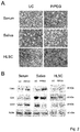
- the size of EVs isolated in the different conditions was similar as seen by transmission electron microscopy. Serum derived EVs ranged from 35 to 95 nm, whereas those derived from saliva were a more homogeneous population with a size ranging from 45 to 65 nm. EVs derived from HLSC ranged from 45 to 75 nm ( Figure 2A ).
- Apo A1 was detectable in EVs obtained from saliva after ultracentrifugation whereas was barely detectable in P/PEG precipitation samples.
- Apo B100 was absent in EVs purified from HLSC culture media both by ultracentrifugation and precipitation, whereas Apo A1 was detectable only in EVs purified by ultracentrifugation.
- Sephadex G-100 spin columns were used and the EVs were recovered in the void volumes whereas apo-lipoproteins were retained.
- gel-filtration with Sephadex G-100 removed Apo B100 and Apo A1 from EV preparations.
- RNA extracted after Sephadex G-100 pre-absorption was reduced both in serum and in saliva but the difference was not statistically significant.
- PCR analysis showed also a reduction of about 2 cycles of a representative mRNA present in serum EVs (ID1 mRNA; Figure 3D ).
- Figure 4 shows a comparison between RNA extracted from EVs prepared by P/PEG precipitation and by ultracentrifugation. No significant difference of RNA content was observed between EVs isolated by the two methods.
- RT-PCR analysis was performed.
- RT-PCR analysis showed the presence of comparable amount of miR16, 29a, 99b, 191 e 223, in EVs isolated from normal subjects using both techniques. In contrast, miR500, 142-3p, 127-3p, and 155 were either undetectable or detectable in very low level (not shown).
- Figure 4B shows a representative amplification plot for miR191 in EVs obtained by ultracentrifugation and precipitation.
- qRT-PCR analysis for detection of mRNA.
- comparable amounts of selected mRNA were detected in EVs derived from serum, saliva and HLSC either purified by ultracentrifugation or P/PEG precipitation.
- the biological activity of EVs obtained by ultracentrifugation and by P/PEG precipitation was evaluated for saliva and HLSC EVs.
- Saliva EVs obtained by P/PEG induced a significant wound closure comparable to that of EGF ( Figure 5A-E ).
- precipitated EVs were more effective than EVs obtained by ultracentrifugation ( Figure 5E ).
- Tested substances Mean intensity of PKH26-labeled serum EVs Protamine chloride + PEG 79 ⁇ 11 Poly-L-lysine 57 ⁇ 18 Poly-L-lysine + Dextran sulfate 74 ⁇ 42 PEG 50 ⁇ 12 Poly-L-lysine + PEG 73 ⁇ 39 DEAE Dextran 49 ⁇ 14 PEG + DEAE Dextran 79 ⁇ 11 Poly-L-lysine hydrochoride 30mg/ml DEAE-Dextran 120mg/ml Dextran sulfate 120mg/ml PEG35k 750mg/ml
- the table shows the fluorimetric determination of serum extracellular vesicles (EVs) labeled with the fluorescent red dye PKH26, precipitated with the indicated substances and resuspended in phosphate buffered saline (PBS). Quantitation is expressed as the mean intensity of fluorescence. The effective labeling was verified by confocal microscopy.
- results obtained show that: a) that all of the tested polycationic substances are effective in favoring precipitation of EVs from serum samples, and b) a precipitation solution comprising a polymeric substance/extracellular matrix forming polymer pair is more effective than a precipitation solution comprising either the polymeric substance alone or the extracellular matrix forming polymer alone.
Landscapes
- Chemical & Material Sciences (AREA)
- Chemical Kinetics & Catalysis (AREA)
- Life Sciences & Earth Sciences (AREA)
- Organic Chemistry (AREA)
- Analytical Chemistry (AREA)
- Zoology (AREA)
- Wood Science & Technology (AREA)
- Proteomics, Peptides & Aminoacids (AREA)
- Health & Medical Sciences (AREA)
- Engineering & Computer Science (AREA)
- Molecular Biology (AREA)
- Bioinformatics & Cheminformatics (AREA)
- Immunology (AREA)
- Biotechnology (AREA)
- Biophysics (AREA)
- Physics & Mathematics (AREA)
- Biochemistry (AREA)
- Microbiology (AREA)
- General Engineering & Computer Science (AREA)
- General Health & Medical Sciences (AREA)
- Genetics & Genomics (AREA)
- Measuring Or Testing Involving Enzymes Or Micro-Organisms (AREA)
- Investigating Or Analysing Biological Materials (AREA)
- Sampling And Sample Adjustment (AREA)
Description
- The present invention relates to a method of isolating extracellular vesicles (EVs) from a biological fluid sample and to a composition for precipitation of extracellular vesicles (EVs) from a biological fluid sample.
- Vesicular-mediated communication between cells appears critical in many biological processes. Small vesicles released from cells have recently emerged as important mediators of inter-cellular communication. These vesicles, that have been termed extracellular vesicles (EVs), are inclusive of exosomes released from the endosomal cell-membrane compartment and of microvesicles released from the cell surface by plasma membrane budding. The EV content of proteins, lipids and nucleic acids varies with the cell of origin and, after incorporation into recipient cells, they may transfer information which may change the phenotype and function of recipient cells.
- Several studies have addressed the role of EVs in physiological and pathological conditions based on their biological activity and molecular constituents. Moreover, since EVs retain the signature of the cell of origin and are present in all body fluids, their potential use as diagnostics in different pathological conditions has been suggested.
- A fundamental issue remains how to isolate EVs from cultured cells to study their biological functions or from biological fluids for diagnostic purposes. Since foetal bovine serum frequently used for cell culture is enriched in EVs, the in vitro experiments require the use of EVs-depleted serum. The isolation of EVs from body fluids, on the other hand, has to face the complexity due to the concomitant presence of EVs of different cellular origin. Therefore, in order to identify a potential biomarker, it is critical to discriminate cellular origin on the basis of EV molecular expression or content, by proteomic or genomic analysis. After removal of cell debris by centrifugation, three main methods are conventionally used for isolation of EVs, namely differential ultracentrifugation in the absence or presence of sucrose gradient, size exclusion chromatography and immune affinity. All these methods have some advantages, which are mainly related to the possibility to discriminate between different EV populations, and concerns, which are related to the risk of damaging vesicles during purification with loss of biological activity, to the need of a sufficiently large sample and to the efficiency of isolation.
- Moreover, polymeric precipitation of EVs has been suggested as an alternative method mainly focused on the evaluation of RNA and protein content. The polymeric precipitation methods are based on the formation of a mesh-like net, which embeds EVs with a size ranging from 60 to 180 nm. Such methods may be applied either to culture media or to body fluids. In particular, polymeric precipitation methods may have the advantage for detection of biomarkers in vesicles derived from small biological samples.
- Currently, the "gold standard" methods of EV purification are the differential ultracentrifugation or the density gradient ultracentrifugation. These methods, however, are influenced by several parameters difficult to standardize such as viscosity of solutions, rotor type, centrifugal radius and g force. In addition, the integrity of EVs after prolonged high speed ultracentrifugation may be damaged. In fact, membrane debris were observed by electron microscopy and difficulty in recovering RNA and exosomal proteins has been reported.
- Several other approaches to EV purification have been investigated. The size exclusion chromatography may have an advantage on ultracentrifugation in maintaining EVs integrity, since with this method EVs are not subjected to shear stress. Filtration with membranes with appropriate pores is also an alternative, but it does not guarantee removal of several small contaminants and does not avoid loss of EVs by binding to membranes. Immunoaffinity purification may isolate specific exosome subtypes maintaining integrity of their cargo.
- A limitation of most of these techniques is the efficiency in the recovery of sufficient amounts of EVs starting from small biological samples.
- The polymeric precipitation technique, based on the ability of PEG to entrap EVs, has been shown to be a fast approach to EV isolation from biological samples. This technique has been developed on the observation that PEG allows virus precipitation and several products based on the use of PEG with 8000 Da molecular weight are commercially available.
-
US 9,005,888 -
WO 2013/188832 discloses a method of isolating cell secreted microvesicles from a liquid sample by the use of a precipitation solution comprising at least one species of polyethylene glycol (PEG) having a molecular weight of about 8,000 Daltons or about 10,000 Daltons. -
WO 2015/131153 discloses a method of purification of extracellular vesicles by precipitation using an acetate buffer. -
US 8,901,284 discloses a method for the isolation of exosomes from a biological fluid sample by the use of a volume-excluding polymer such as polyethylene glycol, dextran, dextran sulfate, dextran acetate, polyvinyl alcohol, polyvinyl acetate, or polyvinyl sulfate. - In order to overcome the drawbacks of the prior art, in particular the complexity and expensiveness of the above-mentioned prior art methods, their low efficiency in EV recovery, the possible presence of contaminants in the isolated EVs and the risk of damaging EV membranes, the present invention provides a method of isolating EVs from biological fluid samples as defined in appended
claim 1. - Further features and advantages of the method of the invention are defined in the dependent claims.
- Also within the scope of the invention is a precipitation composition as defined in appended
claim 13, which is suitable for use in the isolation method of the invention. - The method of isolating extracellular vesicles (EVs) according to the present invention results from the finding that EVs display a negative charge, which allows them to interact with positive-charged molecules.
- Based on this finding, the inventors precipitated EVs from biological fluids and cell culture conditioned media in the presence of positively-charged protamine in a polymeric matrix, and compared the efficiency with that of ultracentrifugation, in terms of yield of number of recovered vesicles, efficiency of RNA extraction, exosomal protein expression and biological activity.
- Protamine was surprisingly found to induce EV precipitation without the need of ultracentrifugation. Moreover, EV resuspension was facilitated when protamine (P) precipitation was performed in the presence of an extracellular matrix forming polymer. The recovery of precipitated EVs evaluated by NanoSight analysis was more efficient than that obtained by ultracentrifugation. By electron microscopy, the size of EVs was similar with the two methods, and the expression of CD63, CD9 and CD 81 exosomal markers in the EVs isolated according to the method of the present invention indicated an enrichment in exosomes. The RNA recovery of the EVs isolated according to the method of the present invention was similar to that of EV isolated by ultracentrifugation. Moreover, the EVs isolated according to the method of the present invention retained the biological activity in vitro as seen by the induction of wound closure by keratinocytes and of proliferation of tubular epithelial cells. In summary, the isolation of EVs according to the method of the present invention has the merit of simplicity and avoids requirement of expensive equipment and may be used for an efficient isolation of EVs from small biological samples.
- The method of isolating extracellular vesicles (EVs) from a biological fluid sample according to the present invention comprises the steps of mixing the biological fluid sample with a polycationic substance and an extracellular matrix forming polymer, incubating the resulting mixture, thereby achieving precipitation of EVs, and separating the precipitated EVs from the mixture.
- A polycationic substance is a polymeric molecule having a positive charges in multiple places. Any polycationic substance may be used in the method of the invention, on account of the fact that EVs are negatively charged and polycations are able to aggregate them. Preferred polycationic substances have a molecular weight of 0.5-50kDa and/or a positive zeta potential of 2-20 mV.
- Protamine, preferably in the form of a salt, is more preferred because it is suitable for clinical applications. The most preferred form of protamine is protamine chloride or hydrochloride. Suitable alternatives to protamine are for example polylysine or cationic dextrans such as DEAE dextran, preferably in the form of a salt such as hydrochloride. The cationic substance polylysine together with polyethylene glycol is disclosed in
CN 103100093 . - The polycationic substance, preferably the salt of protamine, is preferably mixed with the extracellular matrix forming polymer and the biological fluid sample to result in a concentration of 0.02 - 2 mg/ml in the mixture.
- The extracellular matrix forming polymer suitable for use in the method of the invention is any polymeric substance which is capable of forming a mesh-like net which embed the EVs contained in the biological fluid sample. Preferably, the extracellular matrix forming polymer is a hydrogel. Hydrogel-forming natural polymers include proteins such as collagen and gelatine and polysaccharides such as starch, alginate, and agarose. Synthetic polymers that form hydrogels are traditionally prepared using chemical polymerization methods. More preferably the extracellular matrix forming polymer is selected from the group consisting of polyethylene glycol, dextran, dextran sulfate, dextran acetate, polyvinyl alcohol, polyvinyl acetate, or polyvinyl sulfate, polyvinylpyrrolidon, hyaluronic acid, hydroxy ethyl starch. Polyethylene glycol (PEG) having an average molecular weight comprised within the range of from 4 to 100 kDa is the preferred extracellular matrix forming polymer. More preferably, PEG having an average molecular weight comprised within the range of from 10 to 50 kDa, even more preferably of about 35 kDa, is employed in the method of the invention.
- Examples of preferred polycationic substance/ extracellular matrix forming polymer pairs for use in the method of the present invention are:
- (i) Protamine (P)+PEG,
- (ii) poly-L-lysine + PEG,
- (iii) poly-L-lysine + dextran sulfate,
- (iv) DEAE dextran + PEG.
- According to a preferred embodiment of the method of the invention, the extracellular matrix forming polymer is mixed with the polycationic substance and with the biological fluid sample to result in a concentration of 0.01 - 0.2 g/ml in the mixture.
- In order to separate the precipitated EVs from the mixture, any separation technique which may prevent the risk of disrupting the integrity of the EV membranes may be used. A preferred method is centrifugation, at a speed preferably comprised within the range of from 1000 to 50000 g, more preferably of from 1000 to 10000 g, even more preferably of from 1000 to 5000 g.
- As it will be illustrated in more detail in the following experimental examples, the inventors employed the method of the invention to isolate EVs from both biological fluids (serum and saliva) and from cell culture conditioned media (HLSCs). Accordingly, the method of the invention can be used for isolating EVs from any biological fluid or cell culture conditioned medium, such as for example from blood, serum, plasma, saliva, urine, cerebrospinal fluid, or from a cell culture conditioned medium, preferably an adult stem cell culture conditioned medium, more preferably a mesenchymal stem cell culture conditioned medium or a liver pluripotent progenitor cell culture conditioned medium.
- The scope of the invention also comprises a composition for precipitation of extracellular vesicles (EVs) from a biological fluid sample, which is suitable for use in the method according to the invention. Such a composition comprises a vehicle (preferably water, e.g. distilled water), as well as the polycationic substance and the extracellular matrix forming polymer as previously defined in relation to the method. Since the inventors found that the preferred volume ratio of the precipitation solution to the biological fluid is of about 4, of the precipitation composition of the present invention includes the extracellular matrix forming polymer at a concentration comprised within the range of from 0.05 to 1 g/ml and the polycationic substance at a concentration comprised within the range of from 0.1 to 10 mg/ml.
- The following examples are provided by way of illustration only and are not intended to limit the scope of the invention as determined by the appended claims.
- The examples were carried out by using protamine as the polycationic substance and PEG 35000 Da as the extracellular matrix forming polymer. The inventors compared this technique, which in the following shall be designated as "P/PEG precipitation" or more briefly as "P/PEG", with the differential ultracentrifugation which is the gold standard for EV isolation.
- The following examples show that P/PEG was more efficient for the recovery of EVs from small volumes of serum and saliva as well as from the conditioned medium of cultured cells than ultracentrifugation, as judged by NTA.
- The size of EVs, as observed by electron microscopy, was similar but the membrane debris present in the ultra-centrifuged EVs were absent in P/PEG EV preparations. In particular, EVs precipitated from saliva were very homogeneous in size and shape.
- The expression of exosomal markers in EVs obtained by P/PEG precipitation suggests an enrichment in exosomes in these precipitates.
- Since one of the main concerns for EVs obtained by precipitation methods is the presence of contaminants of non-vesicular origin, such as lipoproteins, the inventors also evaluated the presence of Apo B100 and Apo A in the different preparations obtained. The results indicate that in serum EVs, Apo B100 and Apo A1 were present not only in EV precipitates but also in EVs purified by differential ultracentrifugation. This may be a limitation for the use of serum EVs for diagnostic proposes if the intent is to discriminate exRNA associated with vesicles from those associated with lipoproteins. However, detection of exRNA in the biological sample may be exploited for liquid biopsy independently from their vehicle and precipitation techniques may be suitable for this purpose. Apo B100 and Apo A1 were absent or barely detectable in saliva EVs and in EVs purified from culture media by P/PEG preparations, suggesting that lipoprotein contamination is less relevant for these biological samples.
- The use of Sephadex G-25 spin columns has been suggested to remove PEG 8000 containing lipoproteins from precipitated EVs (1). Since the inventors precipitated EVs with protamine in association with PEG 35000 Da,
Sephadex G 100 spin columns were used showing the effective removal of apo-lipoprotein contaminants. After absorption, the total RNA was reduced but still suitable for detection of miRNA and mRNA content of EVs. - EVs obtained by P/PEG precipitation retained the biological activity in vitro, as shown by the induction of wound closure by keratinocytes stimulated with EVs from saliva and by induction of proliferation of tubular epithelial cells challenged with EVs released by HLSC.
- Further experiments were carried out with different polycationic substance/extracellular matrix forming polymer pairs, which proved to be similarly effective in favouring EVs precipitation.
- All prior art methods available for EV purification have some advantages and disadvantages and possibly no one is ideal for every application. The herein described method has the merit of simplicity and avoids requirement of expensive equipment. In addition, the isolated EVs retained the biological activities.
- In conclusion, the inventors showed that the precipitation method of the present invention provides for efficient isolation of EVs from biological samples and may be exploited for search of new biomarkers.
- Saliva was obtained from adult normal volunteers (n=5). Human serum from healthy donors (n=5) was provided by the Blood Bank of Città della Salute e della Scienza di Torino, after informed consent and approval by the internal Review Board of Blood Bank.
- HLSCs were isolated from human cryopreserved normal adult hepatocytes (Lonza, Basel, Switzerland) cultured and characterized as previously described (2). Concisely, hepatocytes first cultivated for 2 weeks in Hepatozyme-SFM medium then in α-MEM/EBM-1 (3:1) (Invitrogen, Carlsbad, CA) media added with Hepes (12 mM, pH 7.4), 1-glutamine (5 mM) penicillin (50 IU/ml), streptomycin (50 µg/ml) (all from Sigma, St. Louis, MO, USA), and fetal calf serum (FCS) (10%) (Invitrogen), Cells were expanded and characterized. The characterization of HLSCs by cytofluorimetric analysis demonstrated the expression of the mesenchymal stem cell markers but not the endothelial and hematopoietic markers as described (3). HLSCs also expressed alpha-fetoprotein and human albumin and the vimentin and nestin resident stem cell markers, but not CD34, CD117 and cytocheratin 19 oval cell markers (2). In addition, HLSCs were positive for the nanog, Sox2, Oct4, and SSEA4 embryonic stem cell markers (4). HLSCs under proper culture conditions underwent endothelial, osteogenic and hepatic differentiation (2).
- Keratinocytes (HaCaT) were purchased from Lonza (Lonza, Basel, Switzerland) and cultured with KBM-gold basal medium (Lonza, Basel, Switzerland) at 37°C with 5% CO2. Cells were seeded at density 3500 cell/cm2, using 1 ml of medium per cm2 and subcultured when cell confluence was 70-80%. Briefly, flasks were washed with HEPES buffer saline solution, incubated with trypsin solution for 6 minutes and then trypsin was neutralized with medium containing 10% FCS. If cells were not completely detached within 7 minutes, incubation with trypsin was repeated.
- TEC line immortalized by infection with a hybrid Adeno5/SV40 virus was previously developed by Cantaluppi et al. (5). Cells were grown with DMEM (Lonza, Basel, Switzerland) containing 10% FCS (GIBCO) and 2 mM glutamine (Life Technologies). TEC showed negative staining for von Willebrand factor, minimal staining for desmin and vimentin, and marked staining with antibodies directed to cytokeratins and actin. TEC were also positive for markers of fully differentiated proximal TEC such as alkaline phosphatase, aminopeptidase A, and megalin.
- EVs were purified from HLSC culture media, human serum and saliva. EVs isolated from supernatants of HLSCs (2·106 cells/T75) were obtained after 24 hour culture in RPMI deprived of FCS. At the time of EV isolation, the 97-99% of cells was viable by trypan blue exclusion assay and TUNEL assay did not detect apoptotic cells.
- Saliva samples (5 ml) were collected in sterile tubes and kept in ice during harvest. One hour before donation, healthy donors followed a protocol of fasting without drinking and eating.
- Serum samples were collected from healthy donors using serum separating tubes (BD) centrifuged 1500 g for 15 minutes.
- Before isolation procedures, HLSC supernatant, saliva and serum samples were submitted to two centrifugations at 3000g for 20 minutes in order to remove cell debris and other contaminants. The saliva samples were diluted 1:1 with PBS and filtered with 0.22µm filters.
- After removal of cell debris and apoptotic bodies by two centrifugations at 3,000 g for 20 minutes, EVs were purified as described by Thery et al (10) by a first ultracentrifugation at 10,000 g followed by ultracentrifugation at 100,000 g for 1 hour at 4° C (Beckman Coulter Optima L-90K, Fullerton, CA, USA).
- The biological samples ready for precipitation procedure were transferred in sterile vials and added with the protamine (P) (Sigma, St. Louis, MO, USA) /Polyethylene glycol (PEG 35000 Merck KGaA, Darmstadt, Germany) precipitation solution (P/PEG) (1 volume precipitation solution :4 volume sample). As a control, P or PEG 35000 alone (PEG) were used. The composition of precipitation solution was 0.25g Polyethylene glycol (PEG 35000, Merck) and 1mg protamine hydrochloride/ml (Sigma) of distilled water.
- After overnight incubation at 4°C, the mixture was centrifuged at 1500 g for 30 minutes at room temperature and the supernatant discarded. The pellet was re-suspended in the appropriate buffer to study biological activities or in lysis buffer for RNA extraction and Western Blot analysis.
- To remove lipoproteins Sephadex G-100 (GE Healthcare Bio-Sciences AB, Uppsala, Sweden) spin columns were used. EVs were recovered in the void volumes.
- The analysis was performed by Zeta-sizer nanoinstrument (Malvern Instruments SA, Vénissieux, France; Size range: 0.3 nm-10µm). Zeta-potential (slipping plane) is generated at x distance from the particle indicating the degree of electrostatic repulsion between adjacent, similarly charged particles in a dispersion. Negative Zeta-potential indicates a high grade of dispersion across the particles.
- NanoSight LM10 (Malvern Instruments SA) was used to analyze concentration and size distribution of EVs by means of the NTAsoftware. The Brownian movements of EVs present in the sample subjected to a laser light source were recorded by a camera and converted into size and concentration parameters by NTA through the Stokes-Einstein equation.
- Transmission electron microscopy was performed on EVs isolated by ultracentrifugation or charge-based precipitation resuspended in PBS, placed on 200 mesh nickel formvar carbon coated grids (Electron Microscopy Science, Hatfield, PA) and left to adhere for 20 min. Grids were then incubated with 2.5 % glutaraldehyde containing 2% sucrose and after washings in distilled water the EVs were negatively stained with NanoVan (Nanoprobes, Yaphank, NK, USA) and observed by
Jeol JEM 1010 electron microscope (Jeol, Tokyo, Japan). - Protein content of the EV preparations was quantified by Bradford method (Bio-Rad, Hercules, CA, USA). Protein samples were separated by 4% to 15% gradient sodium dodecyl sulfate-polyacrylamide gel electrophoresis and subjected to immunoblotting with rabbit polyclonal antibodies anti-CD9, CD63, CD81, anti-apoliporotein B100 and goat polyclonal antibody anti-apolipoprotein A1 (Abcam, Cambridge UK). The protein bands were visualized with an enhanced chemiluminescence (ECL) detection kit and ChemiDoc™ XRS+System (BioRad). Cell and EV lysates were loaded at concentration of 30 µg/well.
- The mirVana RNA isolation kit (Thermo Fisher Scientific, Waltham, MA, USA) has been used to extract total RNA from EVs following the manifacturer's procedure and the RNA has been spectrophotometrically quantified (Nanodrop ND-1000, Wilmington, DE, USA).
- Quantitative real-time PCR was carried out as previously described (6) using a 48- well StepOne™ Real Time System (Applied Biosystems, Waltham, MA, USA). Concisely, 0.2 µg RNA were first reverse transcribed using miScript Reverse Transcription Kit, then 3 ng of cDNA in triplicate were employed to identify and measure significant miRNAs performing qRT-PCR with miScript SYBR Green PCR kit (Qiagen, Valencia, CA, USA). miRNA specific primers to hsa-miR-16, 29a, 99b, 191, 223 were used in separate reactions. The RNU44 and RNU48 snoRNAs served as positive controls and 10 µl of water were used as negative controls in place of the RNA.
- qRT-PCR analysis was also performed on Saliva EVs for the presence of IL8 and Meosin, mRNA and on HLSC EVs for the presence of Ago2 and GAPDH mRNA.
- Immortalized tubular epithelial cells (TEC) were seeded at a density of 3x10³ cells/well in 96 well plates in DMEM supplemented with 10% FCS. 12 hours later, TEC were starved with medium w/o FCS for 2 hours, stimulated with HLSC EVs and then 10µM BrdU was added overnight. The plates were analyzed by BrdU kit (BrdU; Roche Diagnostics) and the absorption values were determined at 405 nm wavelength.
- HACAT cells were seeded at a density of about 50 × 103 cells/well in a 24 well plates in DMEM supplemented with 10% FCS. When cells reached the complete confluence, they were starved with medium w/o FCS overnight. The day after, scratch wounds were created with a sterile tip. Before stimulation (t=0), micrographs of the well were acquired with LEICA microscope. Cells were then stimulated with EVs (50,000 EVs per target cells) isolated from saliva of three different donors. The 'wound closure' phenomenon was monitored for 36 h using the LEICA microscope and images were analyzed by image J software observing the decrease of wound area in cells stimulated with saliva EVs in comparison to cells not stimulated with EVs.
- Results are expressed as mean ±SD. Statistical analysis was performed by using ANOVA with Dunnet's multicomparison tests when appropriate. P<0.05 was considered significant.
- The analysis of Zeta potential was performed on different biological samples showing that EVs display a negative charge (Table 1).
Table 1 Zeta potential mV HLSC EVs -13,800mV Serum EVs -7,825mV Saliva EVs -8,54mV - In preliminary experiments serum was incubated with different doses of protamine (1, 0.5, 0.25, 0.1 mg/ml) overnight at 4°C and precipitated EVs were recovered by centrifugation at 3000 g for 30 minutes (
Figure 1A ). However, the EV pellet was easily re-suspended with the dose of 0.25 mg protamine/ml serum whereas highest dose generated pellets more difficult to re-suspend. We observed that addition of PEG 35,000 to protamine favored resuspension. On this basis, a precipitation strategy was set to favor precipitation of negative charged EVs into a polymeric matrix that would allow the recovery of EVs after centrifugation without the need of an ultracentrifugation step. -
Figure 1 shows the comparison by NTA of EV recovery from serum (B), saliva (C) and cell free supernatant of HLSC (D) after ultracentrifugation (UC) or precipitation with P/PEG, PEG alone and protamine alone. The results indicate that P/PEG precipitation was more efficient than other conditions in terms of number of EVs detected by NTA. The size of EVs isolated in the different conditions was similar as seen by transmission electron microscopy. Serum derived EVs ranged from 35 to 95 nm, whereas those derived from saliva were a more homogeneous population with a size ranging from 45 to 65 nm. EVs derived from HLSC ranged from 45 to 75 nm (Figure 2A ). - As for EVs obtained by ultracentrifugation, the Western blot analysis of EVs precipitated from serum, saliva and HLSC by P/PEG showed the expression of CD63, CD9 and CD 81 exosomal markers (
Figure 2B ). Since it has been suggested that precipitation techniques co-isolate contaminant lipoproteins (7, 8), we evaluated by Western blot the presence of Apo B100 and Apo A1 in EVs obtained by ultracentrifugation and P/PEG precipitation. As shown inFigure 3A , Apo B100 and Apo A1 were detected in serum EVs obtained both by ultracentrifugation and precipitation. In saliva EVs Apo B100 was absent. Apo A1 was detectable in EVs obtained from saliva after ultracentrifugation whereas was barely detectable in P/PEG precipitation samples. Apo B100 was absent in EVs purified from HLSC culture media both by ultracentrifugation and precipitation, whereas Apo A1 was detectable only in EVs purified by ultracentrifugation. To remove lipoproteins, Sephadex G-100 spin columns were used and the EVs were recovered in the void volumes whereas apo-lipoproteins were retained. As seen inFigure 3B , gel-filtration with Sephadex G-100 removed Apo B100 and Apo A1 from EV preparations. - As shown in
Figure 3C , the amount of RNA extracted after Sephadex G-100 pre-absorption was reduced both in serum and in saliva but the difference was not statistically significant. PCR analysis showed also a reduction of about 2 cycles of a representative mRNA present in serum EVs (ID1 mRNA;Figure 3D ).Figure 4 shows a comparison between RNA extracted from EVs prepared by P/PEG precipitation and by ultracentrifugation. No significant difference of RNA content was observed between EVs isolated by the two methods. To evaluate whether RNA extracted from EVs prepared by P/PEG precipitation was suitable for detection of miRNAs or mRNA, RT-PCR analysis was performed. RT-PCR analysis showed the presence of comparable amount of miR16, 29a, 99b, 191 e 223, in EVs isolated from normal subjects using both techniques. In contrast, miR500, 142-3p, 127-3p, and 155 were either undetectable or detectable in very low level (not shown).Figure 4B shows a representative amplification plot for miR191 in EVs obtained by ultracentrifugation and precipitation. We also performed qRT-PCR analysis for detection of mRNA. As shown inFigure 4C , comparable amounts of selected mRNA were detected in EVs derived from serum, saliva and HLSC either purified by ultracentrifugation or P/PEG precipitation. - The biological activity of EVs obtained by ultracentrifugation and by P/PEG precipitation was evaluated for saliva and HLSC EVs.
- In order to test the biological activity of saliva EVs we performed in vitro wound-closure assay using human HaCaT keratinocytes. Saliva EVs obtained by P/PEG induced a significant wound closure comparable to that of EGF (
Figure 5A-E ). In particular, precipitated EVs were more effective than EVs obtained by ultracentrifugation (Figure 5E ). - In order to test the biological activity of HLSC EVs we performed in vitro proliferation of tubular epithelial cells. Both precipitated and ultra-centrifuged EVs were able to significantly increase cell proliferation (
Figure 5F ). -
Tested substances Mean intensity of PKH26-labeled serum EVs Protamine chloride + PEG 79±11 Poly-L-lysine 57 ± 18 Poly-L-lysine + Dextran sulfate 74 ± 42 PEG 50 ± 12 Poly-L-lysine + PEG 73 ± 39 DEAE Dextran 49 ± 14 PEG + DEAE Dextran 79 ± 11 Poly-L-lysine hydrochoride 30mg/ml DEAE-Dextran 120mg/ml Dextran sulfate 120mg/ml PEG35k 750mg/ml - The table shows the fluorimetric determination of serum extracellular vesicles (EVs) labeled with the fluorescent red dye PKH26, precipitated with the indicated substances and resuspended in phosphate buffered saline (PBS). Quantitation is expressed as the mean intensity of fluorescence. The effective labeling was verified by confocal microscopy.
- The results obtained show that: a) that all of the tested polycationic substances are effective in favoring precipitation of EVs from serum samples, and b) a precipitation solution comprising a polymeric substance/extracellular matrix forming polymer pair is more effective than a precipitation solution comprising either the polymeric substance alone or the extracellular matrix forming polymer alone.
-
- 1. Szatanek R, Baran J, Siedlar M and Baj-Krzyworzeka M: Isolation of extracellular vesicles: Determining the correct approach (Review). Int J Mol Med 36: 11-17, 2015.
- 2. Herrera MB, Bruno S, Buttiglieri S, Tetta C, Gatti S, Deregibus MC, Bussolati B and Camussi G. Isolation and characterization of a stem cell population from adult human liver. Stem Cells 24: 2840-2850, 2006.
- 3. Ratajczak J, Wysoczynski M, Hayek F, Janowska-Wieczorek A and Ratajczak MZ Membrane-derived microvesicles: important and underappreciated mediators of cell-to-cell communication. Leukemia 20: 1487-1495, 2006.
- 4. Herrera MB, Fonsato V, Gatti S, Deregibus MC, Sordi A, Cantarella D, Calogero R, Bussolati B, Tetta C and Camussi G: Human liver stem cell-derived microvesicles accelerate hepatic regeneration in hepatectomized rats. J Cell Mol Med 14: 1605-1618, 2010.
- 5. Cantaluppi V, Biancone L, Romanazzi GM, Figliolini F, Beltramo S, Galimi F, Camboni MG, Deriu E, Conaldi P, Bottelli A, Orlandi V, Herrera MB, Pacitti A,Segoloni GP and Camussi G: Macrophage stimulating protein may promote tubular regeneration after acute injury. J Am Soc Nephrol 19: 1904-1918, 2008.
- 6. Iavello A, Frech VS, Gai C, Deregibus MC, Quesenberry PJ and Camussi G: Role of Alix in miRNA packaging during extracellular vesicle biogenesis. Int J Mol Med 37: 958-966, 2016
- 7. Witwer KW, Buzás EI, Bemis LT, Bora A, Lässer C, Lotvall J, Nolte-'t Hoen EN, Piper MG, Sivaraman S, Skog J, Théry C, Wauben MH and Hochberg F : Standardization of sample collection, isolation and analysis methods in extracellular vesicle research. J Extracell Vesicles 2: 20360, 2013.
- 8. Momen-Heravi F, Balaj L, Alian S, Trachtenberg AJ, Hochberg FH, Skog J and Kuo WP: Impact of biofluid viscosity on size and sedimentation efficiency of the isolated microvesicles. Front Physiol 3: 162, 2012.
Claims (17)
- A method of isolating extracellular vesicles (EVs) from a biological fluid sample, the method comprising the steps of:(i) mixing the biological fluid sample with a polycationic substance and an extracellular matrix forming polymer;(ii) incubating the mixture, thereby achieving precipitation of the EVs; and(ii) separating the precipitated EVs from the mixture.
- The method according to claim 1, wherein the step of separating the precipitated EVs from the mixture is effected by centrifugation.
- The method according to claim 1 or 2, wherein the polycationic substance is selected from a protamine salt, a polylysine salt or a cationic dextran salt.
- The method according to claim 3, wherein the salt is a hydrochloride.
- The method according to any one of claims 1 to 4, wherein the extracellular matrix forming polymer is a hydrogel.
- The method according to any one of claims 1 to 5, wherein the extracellular matrix forming polymer is selected from the group consisting of collagen, gelatine, starch, alginate, agarose, polyethylene glycol, dextran, dextran sulfate, dextran acetate, polyvinyl alcohol, polyvinyl acetate, or polyvinyl sulfate, polyvinylpyrrolidon, hyalyronic acid, hydroxy ethyl starch.
- The method according to any one of claims 1 to 6, wherein biological fluid is selected from the group consisting of blood, serum, plasma, saliva, urine, cerebrospinal fluid and the conditioned medium of a cell culture.
- The method according to claim 7, wherein the cell culture is an adult stem cell culture, preferably a mesenchymal stem cell culture or a liver pluripotent progenitor cell culture.
- The method according to any one of claims 1 to 8, wherein the polycationic substance is mixed with the extracellular matrix forming polymer and with the biological fluid sample to result in a concentration of 0.02 - 2 mg/ml in the mixture.
- The method according to any one of claims 1 to 9, wherein the extracellular matrix forming polymer is mixed with the polycationic substance and with the biological fluid sample to result in a concentration of 0.01 - 0.2 g/ml in the mixture.
- The method according to any one of claims 1 to 10, wherein the extracellular matrix forming polymer has an average molecular weight comprised within the range of from 4 to 100 kDa.
- The method according to any one of claims 2 to 11, wherein centrifugation is carried out at 1000 - 50000 g, preferably at 1000 - 10000 g, more preferably at 1000 - 5000 g.
- A composition for precipitation of extracellular vesicles (EVs) from a biological fluid sample, the composition comprising a vehicle, a polycationic substance and an extracellular matrix forming polymer, wherein the extracellular matrix forming polymer has a concentration comprised within the range of from 0.05 to 1 g/ml.
- The composition according to claim 13, wherein the extracellular matrix forming polymer is as defined in claim 5 or 6.
- The composition according to claim 13 or 14, wherein the polycationic substance is as defined in claim 3 or 4.
- The composition according to any one of claims 13 to 15, wherein the polycationic substance has a concentration comprised within the range of from 0.1 to 10 mg/ml.
- The composition according to any one of claims 13 to 16, wherein the vehicle is water, preferably distilled water.
Applications Claiming Priority (2)
| Application Number | Priority Date | Filing Date | Title |
|---|---|---|---|
| EP16164889 | 2016-04-12 | ||
| PCT/EP2017/058649 WO2017178472A1 (en) | 2016-04-12 | 2017-04-11 | Isolation of extracellular vesicles (evs) from biological fluid samples |
Publications (2)
| Publication Number | Publication Date |
|---|---|
| EP3443112A1 EP3443112A1 (en) | 2019-02-20 |
| EP3443112B1 true EP3443112B1 (en) | 2020-08-05 |
Family
ID=56008452
Family Applications (1)
| Application Number | Title | Priority Date | Filing Date |
|---|---|---|---|
| EP17715760.9A Active EP3443112B1 (en) | 2016-04-12 | 2017-04-11 | Isolation of extracellular vesicles (evs) from biological fluid samples |
Country Status (5)
| Country | Link |
|---|---|
| US (1) | US11484816B2 (en) |
| EP (1) | EP3443112B1 (en) |
| JP (1) | JP2019518428A (en) |
| CN (1) | CN109153968A (en) |
| WO (1) | WO2017178472A1 (en) |
Families Citing this family (29)
| Publication number | Priority date | Publication date | Assignee | Title |
|---|---|---|---|---|
| TWI690320B (en) | 2010-05-12 | 2020-04-11 | 開曼群島商英瑞金公司 | Bioactive kidney cells |
| EP3727630B1 (en) * | 2017-12-19 | 2024-04-10 | Advanced Extracellular Vesicle Applications S.R.L. | Method and stationary phase for isolating extracellular vesicles from biological material |
| CA3089825C (en) | 2018-01-29 | 2022-08-30 | Forever Labs, Inc. | System and method for isolating extracellular vesicles |
| US12270741B2 (en) * | 2018-04-03 | 2025-04-08 | Tymora Analytical Operations, Llc | Methods for affinity-based non-antibody capture and purification of extracellular vesicles |
| JP7710849B2 (en) * | 2018-10-17 | 2025-07-22 | 合同会社H.U.グループ中央研究所 | Method for recovering extracellular vesicles |
| CN109355258B (en) * | 2018-11-02 | 2020-04-21 | 北京恩泽康泰生物科技有限公司 | Extracting agent for extracting extracellular vesicles from body fluid and extraction method thereof |
| EP3925703B1 (en) * | 2019-02-14 | 2025-04-16 | Korea University Research and Business Foundation | Method for extracting microvesicles from biological samples |
| CN109929802A (en) * | 2019-04-02 | 2019-06-25 | 武汉理工大学 | The methods and applications of room adsorbing separation excretion body under a kind of orifice plate upper chamber culture cell based on Transwell |
| KR102220777B1 (en) * | 2019-08-20 | 2021-02-26 | 연세대학교 산학협력단 | Amino acid-based block copolymer, compositions or kits for isolating a vesicle comprising the same, and methods for isolating the vesicle using the same |
| JP7357346B2 (en) * | 2019-09-10 | 2023-10-06 | 国立大学法人広島大学 | Exosome isolation method, exosome isolation kit, exosome removal method, separation solution acquisition method, and exosome isolation column |
| US12377121B2 (en) | 2020-04-10 | 2025-08-05 | Zeo Scientifix, Inc. | Compositions comprising nanoparticles, method of making and uses thereof |
| US20230227916A1 (en) * | 2020-04-15 | 2023-07-20 | H.U. Group Research Institute G.K. | Method for recovering extracellular vesicle |
| EP4141107B1 (en) * | 2020-04-24 | 2026-03-11 | Korea University Research and Business Foundation | Microvesicle isolation method and microvesicle isolation device |
| EP4166944A4 (en) * | 2020-06-12 | 2024-02-07 | Tokushima University | SUPERABSORBENT POLYMER FOR PARTICLE CLASSIFICATION AND CLASSIFICATION METHOD USING SAME |
| EP3936864A1 (en) * | 2020-07-06 | 2022-01-12 | Consejo Superior de Investigaciones Científicas (CSIC) | Method for the detection and/or quantification of extracellular vesicles in fluid biological samples |
| CN113930342B (en) * | 2020-07-13 | 2024-06-21 | 香港科技大学 | Cultivation system and method for promoting the growth of photosynthetic organisms |
| JPWO2022080461A1 (en) * | 2020-10-16 | 2022-04-21 | ||
| JP7625408B2 (en) * | 2020-11-30 | 2025-02-03 | 社会医療法人大雄会 | Methods for Enriching Extracellular Vesicles and/or Cell-Free DNA |
| IT202100000569A1 (en) * | 2021-01-14 | 2022-07-14 | Evobiotech S R L | COMPOSITION INCLUDING ENGINEERED EXTRACELLULAR VEGETABLES OF PLANT AND ITS USE AS A VACCINE |
| CN113092642B (en) * | 2021-03-30 | 2022-11-01 | 苏州爱宝德生物科技有限公司 | A quick extraction element for extracellular vesicles |
| US20240201055A1 (en) * | 2021-03-31 | 2024-06-20 | Sanyo Chemical Industries, Ltd. | Cross-linked polymer, method for separating substance, kit for separating substance, kit for testing for disease, and device for separating substance |
| EP4353815A4 (en) | 2021-06-04 | 2025-05-07 | Institute for Basic Science | METHOD FOR ISOLATING EXTRACELLULAR VESICLE USING FRACTIONAL SALT PRECIPITATION |
| CN113583954B (en) * | 2021-07-16 | 2023-11-28 | 珈泌生物科技(武汉)有限责任公司 | In-situ labeling and rapid separation method for circulating extracellular vesicles |
| AU2022335348A1 (en) | 2021-08-24 | 2024-02-22 | Forever Labs, Inc. | Device and method for isolating extracellular vesicles |
| US12213994B2 (en) | 2021-12-08 | 2025-02-04 | Zeo Scientifix, Inc. | Compositions and methods for treating pain with extracellular vesicles |
| CN114574437B (en) * | 2022-01-19 | 2024-10-25 | 大连博格林生物科技有限公司 | Plasma exosome extraction reagent, enrichment method, extraction kit and application thereof |
| KR102959311B1 (en) | 2023-03-29 | 2026-04-30 | 고려대학교 산학협력단 | Method and apparatus for bulk extraction and purification of extracellular vesicles from biological samples |
| JP7638481B2 (en) * | 2023-07-21 | 2025-03-04 | ヤマト科学株式会社 | Method for concentrating biological substances |
| CN120536342A (en) * | 2025-05-23 | 2025-08-26 | 苏州乐胜医药科技有限公司 | Method for isolating and purifying exosomes from cell culture fluid |
Family Cites Families (13)
| Publication number | Priority date | Publication date | Assignee | Title |
|---|---|---|---|---|
| FR2784580B1 (en) * | 1998-10-16 | 2004-06-25 | Biosepra Inc | POLYVINYL-ALCOHOL MICROSPHERES AND METHODS OF MAKING THE SAME |
| WO2003025167A2 (en) * | 2001-09-20 | 2003-03-27 | Centre For Translational Research In Cancer | Cultured stromal cells and uses thereof |
| DE10151296A1 (en) * | 2001-10-17 | 2003-04-30 | Boehringer Ingelheim Pharma | Keratinocytes useful as a biologically active substance in the treatment of wounds |
| JP2007271388A (en) * | 2006-03-30 | 2007-10-18 | Hidetoshi Tsuchida | Separation method of serum or plasma, and blood separation tube |
| EP2247715A1 (en) * | 2008-02-01 | 2010-11-10 | The Regents of the University of California | Microfluidic imaging cytometry |
| CN102068691A (en) * | 2010-12-24 | 2011-05-25 | 福尔生物制药股份有限公司 | Method for preparing high purity hand-foot-and-mouth disease vaccine with sucrose zonal ultracentrifugation |
| JP6450673B2 (en) * | 2012-04-03 | 2019-01-09 | リニューロン・リミテッドReNeuron Limited | Stem cell microparticles |
| US20130273544A1 (en) | 2012-04-17 | 2013-10-17 | Life Technologies Corporation | Methods and compositions for exosome isolation |
| WO2013188832A1 (en) | 2012-06-14 | 2013-12-19 | System Biosciences, Llc | Methods for microvesicle isolation and selective removal |
| US9005888B2 (en) | 2012-06-14 | 2015-04-14 | System Biosciences, Llc | Methods for microvesicle isolation and selective removal |
| CN103100093B (en) * | 2013-01-23 | 2015-05-06 | 中山大学附属第三医院 | Load small interfering RNA nanoscale lipid microbubble ultrasonic contrast agent and preparation method |
| WO2015131153A1 (en) | 2014-02-27 | 2015-09-03 | Board Of Regents, The University Of Texas System | Methods and compositions for isolating exosomes |
| CN103954770B (en) * | 2014-04-25 | 2015-09-30 | 江南大学 | EVs-TRPC5 is detecting the application in breast carcinoma resistance degree |
-
2017
- 2017-04-11 WO PCT/EP2017/058649 patent/WO2017178472A1/en not_active Ceased
- 2017-04-11 JP JP2018554488A patent/JP2019518428A/en active Pending
- 2017-04-11 EP EP17715760.9A patent/EP3443112B1/en active Active
- 2017-04-11 US US16/093,471 patent/US11484816B2/en active Active
- 2017-04-11 CN CN201780022886.1A patent/CN109153968A/en active Pending
Non-Patent Citations (1)
| Title |
|---|
| None * |
Also Published As
| Publication number | Publication date |
|---|---|
| JP2019518428A (en) | 2019-07-04 |
| CN109153968A (en) | 2019-01-04 |
| US11484816B2 (en) | 2022-11-01 |
| WO2017178472A1 (en) | 2017-10-19 |
| US20200179827A1 (en) | 2020-06-11 |
| EP3443112A1 (en) | 2019-02-20 |
Similar Documents
| Publication | Publication Date | Title |
|---|---|---|
| US11484816B2 (en) | Isolation of extracellular vesicles (EVs) from biological fluid samples | |
| Deregibus et al. | Charge-based precipitation of extracellular vesicles | |
| Fernandes et al. | Secretome from SH-SY5Y APPSwe cells trigger time-dependent CHME3 microglia activation phenotypes, ultimately leading to miR-21 exosome shuttling | |
| US9829483B2 (en) | Methods of isolating extracellular vesicles | |
| McBride et al. | Extracellular vesicles as biomarkers and therapeutics in dermatology: a focus on exosomes | |
| EP2713163B1 (en) | Methods for isolating vesicles | |
| US12551828B2 (en) | Method for extracting microvesicles from biological sample | |
| Ma et al. | Isolation, identification, and challenges of extracellular vesicles: emerging players in clinical applications | |
| Stremersch et al. | Nucleic acid loading and fluorescent labeling of isolated extracellular vesicles requires adequate purification | |
| US20200080997A1 (en) | Lipid-based probes for extracellular isolation | |
| EP3727630B1 (en) | Method and stationary phase for isolating extracellular vesicles from biological material | |
| US12441993B2 (en) | Isolation of extracellular vesicles by precipitation or immobilization using polyethylenimine and polyethylenimine-coated solid supports | |
| RU2678988C1 (en) | Method for isolating extracellular vesicles from biological fluids | |
| EP3964566A1 (en) | Method for selecting stem cells having ability to produce extracellular vesicles with high efficiency by using activity of protease-activated receptor-mediated signaling pathways | |
| US12123024B1 (en) | Absolute precipitation of exosomes (APEX) isolation | |
| US20250361481A1 (en) | Method of separating and concentrating large number of tissue-derived vesicles | |
| de Boer | Development of a multi-stage purification process for serum-derived exosomes and evaluation of their regenerative capacity | |
| Tavormina | Identification and Molecular Analysis of Dna In Exosomes | |
| Kügler | Role of extracellular vesicles mRNA in neuroinflammation in a mouse model of stroke | |
| Mozes | Toward the Isolation of Exosomes by Flow Cytometry | |
| Stremersch et al. | Inadequate purification of extracellular vesicles can lead to misinterpretation of downstream experimental data | |
| Palanisamy et al. | Nanostructural and Transcriptomic Analyses of Human Saliva Derived |
Legal Events
| Date | Code | Title | Description |
|---|---|---|---|
| STAA | Information on the status of an ep patent application or granted ep patent |
Free format text: STATUS: UNKNOWN |
|
| STAA | Information on the status of an ep patent application or granted ep patent |
Free format text: STATUS: THE INTERNATIONAL PUBLICATION HAS BEEN MADE |
|
| PUAI | Public reference made under article 153(3) epc to a published international application that has entered the european phase |
Free format text: ORIGINAL CODE: 0009012 |
|
| STAA | Information on the status of an ep patent application or granted ep patent |
Free format text: STATUS: REQUEST FOR EXAMINATION WAS MADE |
|
| 17P | Request for examination filed |
Effective date: 20181108 |
|
| AK | Designated contracting states |
Kind code of ref document: A1 Designated state(s): AL AT BE BG CH CY CZ DE DK EE ES FI FR GB GR HR HU IE IS IT LI LT LU LV MC MK MT NL NO PL PT RO RS SE SI SK SM TR |
|
| AX | Request for extension of the european patent |
Extension state: BA ME |
|
| DAV | Request for validation of the european patent (deleted) | ||
| DAX | Request for extension of the european patent (deleted) | ||
| STAA | Information on the status of an ep patent application or granted ep patent |
Free format text: STATUS: EXAMINATION IS IN PROGRESS |
|
| 17Q | First examination report despatched |
Effective date: 20190816 |
|
| GRAP | Despatch of communication of intention to grant a patent |
Free format text: ORIGINAL CODE: EPIDOSNIGR1 |
|
| STAA | Information on the status of an ep patent application or granted ep patent |
Free format text: STATUS: GRANT OF PATENT IS INTENDED |
|
| INTG | Intention to grant announced |
Effective date: 20200219 |
|
| GRAS | Grant fee paid |
Free format text: ORIGINAL CODE: EPIDOSNIGR3 |
|
| GRAA | (expected) grant |
Free format text: ORIGINAL CODE: 0009210 |
|
| STAA | Information on the status of an ep patent application or granted ep patent |
Free format text: STATUS: THE PATENT HAS BEEN GRANTED |
|
| AK | Designated contracting states |
Kind code of ref document: B1 Designated state(s): AL AT BE BG CH CY CZ DE DK EE ES FI FR GB GR HR HU IE IS IT LI LT LU LV MC MK MT NL NO PL PT RO RS SE SI SK SM TR |
|
| REG | Reference to a national code |
Ref country code: GB Ref legal event code: FG4D |
|
| REG | Reference to a national code |
Ref country code: CH Ref legal event code: EP |
|
| REG | Reference to a national code |
Ref country code: AT Ref legal event code: REF Ref document number: 1298773 Country of ref document: AT Kind code of ref document: T Effective date: 20200815 |
|
| REG | Reference to a national code |
Ref country code: DE Ref legal event code: R096 Ref document number: 602017021027 Country of ref document: DE |
|
| REG | Reference to a national code |
Ref country code: IE Ref legal event code: FG4D |
|
| REG | Reference to a national code |
Ref country code: LT Ref legal event code: MG4D |
|
| REG | Reference to a national code |
Ref country code: NL Ref legal event code: MP Effective date: 20200805 |
|
| REG | Reference to a national code |
Ref country code: AT Ref legal event code: MK05 Ref document number: 1298773 Country of ref document: AT Kind code of ref document: T Effective date: 20200805 |
|
| PG25 | Lapsed in a contracting state [announced via postgrant information from national office to epo] |
Ref country code: PT Free format text: LAPSE BECAUSE OF FAILURE TO SUBMIT A TRANSLATION OF THE DESCRIPTION OR TO PAY THE FEE WITHIN THE PRESCRIBED TIME-LIMIT Effective date: 20201207 Ref country code: ES Free format text: LAPSE BECAUSE OF FAILURE TO SUBMIT A TRANSLATION OF THE DESCRIPTION OR TO PAY THE FEE WITHIN THE PRESCRIBED TIME-LIMIT Effective date: 20200805 Ref country code: SE Free format text: LAPSE BECAUSE OF FAILURE TO SUBMIT A TRANSLATION OF THE DESCRIPTION OR TO PAY THE FEE WITHIN THE PRESCRIBED TIME-LIMIT Effective date: 20200805 Ref country code: BG Free format text: LAPSE BECAUSE OF FAILURE TO SUBMIT A TRANSLATION OF THE DESCRIPTION OR TO PAY THE FEE WITHIN THE PRESCRIBED TIME-LIMIT Effective date: 20201105 Ref country code: AT Free format text: LAPSE BECAUSE OF FAILURE TO SUBMIT A TRANSLATION OF THE DESCRIPTION OR TO PAY THE FEE WITHIN THE PRESCRIBED TIME-LIMIT Effective date: 20200805 Ref country code: GR Free format text: LAPSE BECAUSE OF FAILURE TO SUBMIT A TRANSLATION OF THE DESCRIPTION OR TO PAY THE FEE WITHIN THE PRESCRIBED TIME-LIMIT Effective date: 20201106 Ref country code: NO Free format text: LAPSE BECAUSE OF FAILURE TO SUBMIT A TRANSLATION OF THE DESCRIPTION OR TO PAY THE FEE WITHIN THE PRESCRIBED TIME-LIMIT Effective date: 20201105 Ref country code: FI Free format text: LAPSE BECAUSE OF FAILURE TO SUBMIT A TRANSLATION OF THE DESCRIPTION OR TO PAY THE FEE WITHIN THE PRESCRIBED TIME-LIMIT Effective date: 20200805 Ref country code: HR Free format text: LAPSE BECAUSE OF FAILURE TO SUBMIT A TRANSLATION OF THE DESCRIPTION OR TO PAY THE FEE WITHIN THE PRESCRIBED TIME-LIMIT Effective date: 20200805 Ref country code: LT Free format text: LAPSE BECAUSE OF FAILURE TO SUBMIT A TRANSLATION OF THE DESCRIPTION OR TO PAY THE FEE WITHIN THE PRESCRIBED TIME-LIMIT Effective date: 20200805 |
|
| PG25 | Lapsed in a contracting state [announced via postgrant information from national office to epo] |
Ref country code: LV Free format text: LAPSE BECAUSE OF FAILURE TO SUBMIT A TRANSLATION OF THE DESCRIPTION OR TO PAY THE FEE WITHIN THE PRESCRIBED TIME-LIMIT Effective date: 20200805 Ref country code: PL Free format text: LAPSE BECAUSE OF FAILURE TO SUBMIT A TRANSLATION OF THE DESCRIPTION OR TO PAY THE FEE WITHIN THE PRESCRIBED TIME-LIMIT Effective date: 20200805 Ref country code: RS Free format text: LAPSE BECAUSE OF FAILURE TO SUBMIT A TRANSLATION OF THE DESCRIPTION OR TO PAY THE FEE WITHIN THE PRESCRIBED TIME-LIMIT Effective date: 20200805 Ref country code: NL Free format text: LAPSE BECAUSE OF FAILURE TO SUBMIT A TRANSLATION OF THE DESCRIPTION OR TO PAY THE FEE WITHIN THE PRESCRIBED TIME-LIMIT Effective date: 20200805 Ref country code: IS Free format text: LAPSE BECAUSE OF FAILURE TO SUBMIT A TRANSLATION OF THE DESCRIPTION OR TO PAY THE FEE WITHIN THE PRESCRIBED TIME-LIMIT Effective date: 20201205 |
|
| PG25 | Lapsed in a contracting state [announced via postgrant information from national office to epo] |
Ref country code: SM Free format text: LAPSE BECAUSE OF FAILURE TO SUBMIT A TRANSLATION OF THE DESCRIPTION OR TO PAY THE FEE WITHIN THE PRESCRIBED TIME-LIMIT Effective date: 20200805 Ref country code: EE Free format text: LAPSE BECAUSE OF FAILURE TO SUBMIT A TRANSLATION OF THE DESCRIPTION OR TO PAY THE FEE WITHIN THE PRESCRIBED TIME-LIMIT Effective date: 20200805 Ref country code: CZ Free format text: LAPSE BECAUSE OF FAILURE TO SUBMIT A TRANSLATION OF THE DESCRIPTION OR TO PAY THE FEE WITHIN THE PRESCRIBED TIME-LIMIT Effective date: 20200805 Ref country code: DK Free format text: LAPSE BECAUSE OF FAILURE TO SUBMIT A TRANSLATION OF THE DESCRIPTION OR TO PAY THE FEE WITHIN THE PRESCRIBED TIME-LIMIT Effective date: 20200805 Ref country code: RO Free format text: LAPSE BECAUSE OF FAILURE TO SUBMIT A TRANSLATION OF THE DESCRIPTION OR TO PAY THE FEE WITHIN THE PRESCRIBED TIME-LIMIT Effective date: 20200805 |
|
| REG | Reference to a national code |
Ref country code: DE Ref legal event code: R097 Ref document number: 602017021027 Country of ref document: DE |
|
| PG25 | Lapsed in a contracting state [announced via postgrant information from national office to epo] |
Ref country code: AL Free format text: LAPSE BECAUSE OF FAILURE TO SUBMIT A TRANSLATION OF THE DESCRIPTION OR TO PAY THE FEE WITHIN THE PRESCRIBED TIME-LIMIT Effective date: 20200805 |
|
| PLBE | No opposition filed within time limit |
Free format text: ORIGINAL CODE: 0009261 |
|
| STAA | Information on the status of an ep patent application or granted ep patent |
Free format text: STATUS: NO OPPOSITION FILED WITHIN TIME LIMIT |
|
| PG25 | Lapsed in a contracting state [announced via postgrant information from national office to epo] |
Ref country code: SK Free format text: LAPSE BECAUSE OF FAILURE TO SUBMIT A TRANSLATION OF THE DESCRIPTION OR TO PAY THE FEE WITHIN THE PRESCRIBED TIME-LIMIT Effective date: 20200805 |
|
| 26N | No opposition filed |
Effective date: 20210507 |
|
| PG25 | Lapsed in a contracting state [announced via postgrant information from national office to epo] |
Ref country code: SI Free format text: LAPSE BECAUSE OF FAILURE TO SUBMIT A TRANSLATION OF THE DESCRIPTION OR TO PAY THE FEE WITHIN THE PRESCRIBED TIME-LIMIT Effective date: 20200805 |
|
| PG25 | Lapsed in a contracting state [announced via postgrant information from national office to epo] |
Ref country code: MC Free format text: LAPSE BECAUSE OF FAILURE TO SUBMIT A TRANSLATION OF THE DESCRIPTION OR TO PAY THE FEE WITHIN THE PRESCRIBED TIME-LIMIT Effective date: 20200805 |
|
| PG25 | Lapsed in a contracting state [announced via postgrant information from national office to epo] |
Ref country code: LU Free format text: LAPSE BECAUSE OF NON-PAYMENT OF DUE FEES Effective date: 20210411 |
|
| REG | Reference to a national code |
Ref country code: BE Ref legal event code: MM Effective date: 20210430 |
|
| PG25 | Lapsed in a contracting state [announced via postgrant information from national office to epo] |
Ref country code: CH Free format text: LAPSE BECAUSE OF NON-PAYMENT OF DUE FEES Effective date: 20210430 Ref country code: LI Free format text: LAPSE BECAUSE OF NON-PAYMENT OF DUE FEES Effective date: 20210430 |
|
| PG25 | Lapsed in a contracting state [announced via postgrant information from national office to epo] |
Ref country code: IE Free format text: LAPSE BECAUSE OF NON-PAYMENT OF DUE FEES Effective date: 20210411 |
|
| PG25 | Lapsed in a contracting state [announced via postgrant information from national office to epo] |
Ref country code: IS Free format text: LAPSE BECAUSE OF FAILURE TO SUBMIT A TRANSLATION OF THE DESCRIPTION OR TO PAY THE FEE WITHIN THE PRESCRIBED TIME-LIMIT Effective date: 20201205 |
|
| PG25 | Lapsed in a contracting state [announced via postgrant information from national office to epo] |
Ref country code: BE Free format text: LAPSE BECAUSE OF NON-PAYMENT OF DUE FEES Effective date: 20210430 |
|
| PG25 | Lapsed in a contracting state [announced via postgrant information from national office to epo] |
Ref country code: CY Free format text: LAPSE BECAUSE OF FAILURE TO SUBMIT A TRANSLATION OF THE DESCRIPTION OR TO PAY THE FEE WITHIN THE PRESCRIBED TIME-LIMIT Effective date: 20200805 |
|
| P01 | Opt-out of the competence of the unified patent court (upc) registered |
Effective date: 20230525 |
|
| PG25 | Lapsed in a contracting state [announced via postgrant information from national office to epo] |
Ref country code: HU Free format text: LAPSE BECAUSE OF FAILURE TO SUBMIT A TRANSLATION OF THE DESCRIPTION OR TO PAY THE FEE WITHIN THE PRESCRIBED TIME-LIMIT; INVALID AB INITIO Effective date: 20170411 |
|
| PG25 | Lapsed in a contracting state [announced via postgrant information from national office to epo] |
Ref country code: MK Free format text: LAPSE BECAUSE OF FAILURE TO SUBMIT A TRANSLATION OF THE DESCRIPTION OR TO PAY THE FEE WITHIN THE PRESCRIBED TIME-LIMIT Effective date: 20200805 |
|
| PG25 | Lapsed in a contracting state [announced via postgrant information from national office to epo] |
Ref country code: MT Free format text: LAPSE BECAUSE OF FAILURE TO SUBMIT A TRANSLATION OF THE DESCRIPTION OR TO PAY THE FEE WITHIN THE PRESCRIBED TIME-LIMIT Effective date: 20200805 |
|
| PGFP | Annual fee paid to national office [announced via postgrant information from national office to epo] |
Ref country code: FR Payment date: 20250319 Year of fee payment: 9 |
|
| PGFP | Annual fee paid to national office [announced via postgrant information from national office to epo] |
Ref country code: IT Payment date: 20250319 Year of fee payment: 9 Ref country code: GB Payment date: 20250319 Year of fee payment: 9 |
|
| PGFP | Annual fee paid to national office [announced via postgrant information from national office to epo] |
Ref country code: DE Payment date: 20250319 Year of fee payment: 9 |
|
| PG25 | Lapsed in a contracting state [announced via postgrant information from national office to epo] |
Ref country code: TR Free format text: LAPSE BECAUSE OF FAILURE TO SUBMIT A TRANSLATION OF THE DESCRIPTION OR TO PAY THE FEE WITHIN THE PRESCRIBED TIME-LIMIT Effective date: 20200805 |