EP3433269B1 - Fusion proteins of pd-1 and 4-1bb - Google Patents
Fusion proteins of pd-1 and 4-1bb Download PDFInfo
- Publication number
- EP3433269B1 EP3433269B1 EP17713250.3A EP17713250A EP3433269B1 EP 3433269 B1 EP3433269 B1 EP 3433269B1 EP 17713250 A EP17713250 A EP 17713250A EP 3433269 B1 EP3433269 B1 EP 3433269B1
- Authority
- EP
- European Patent Office
- Prior art keywords
- cell
- fusion protein
- cells
- nucleic acid
- host cell
- Prior art date
- Legal status (The legal status is an assumption and is not a legal conclusion. Google has not performed a legal analysis and makes no representation as to the accuracy of the status listed.)
- Active
Links
Images





Classifications
-
- C—CHEMISTRY; METALLURGY
- C07—ORGANIC CHEMISTRY
- C07K—PEPTIDES
- C07K14/00—Peptides having more than 20 amino acids; Gastrins; Somatostatins; Melanotropins; Derivatives thereof
- C07K14/435—Peptides having more than 20 amino acids; Gastrins; Somatostatins; Melanotropins; Derivatives thereof from animals; from humans
- C07K14/705—Receptors; Cell surface antigens; Cell surface determinants
- C07K14/70578—NGF-receptor/TNF-receptor superfamily, e.g. CD27, CD30, CD40, CD95
-
- A—HUMAN NECESSITIES
- A61—MEDICAL OR VETERINARY SCIENCE; HYGIENE
- A61P—SPECIFIC THERAPEUTIC ACTIVITY OF CHEMICAL COMPOUNDS OR MEDICINAL PREPARATIONS
- A61P31/00—Antiinfectives, i.e. antibiotics, antiseptics, chemotherapeutics
- A61P31/12—Antivirals
-
- A—HUMAN NECESSITIES
- A61—MEDICAL OR VETERINARY SCIENCE; HYGIENE
- A61P—SPECIFIC THERAPEUTIC ACTIVITY OF CHEMICAL COMPOUNDS OR MEDICINAL PREPARATIONS
- A61P35/00—Antineoplastic agents
-
- C—CHEMISTRY; METALLURGY
- C07—ORGANIC CHEMISTRY
- C07K—PEPTIDES
- C07K14/00—Peptides having more than 20 amino acids; Gastrins; Somatostatins; Melanotropins; Derivatives thereof
- C07K14/435—Peptides having more than 20 amino acids; Gastrins; Somatostatins; Melanotropins; Derivatives thereof from animals; from humans
- C07K14/705—Receptors; Cell surface antigens; Cell surface determinants
- C07K14/70503—Immunoglobulin superfamily
-
- C—CHEMISTRY; METALLURGY
- C07—ORGANIC CHEMISTRY
- C07K—PEPTIDES
- C07K14/00—Peptides having more than 20 amino acids; Gastrins; Somatostatins; Melanotropins; Derivatives thereof
- C07K14/435—Peptides having more than 20 amino acids; Gastrins; Somatostatins; Melanotropins; Derivatives thereof from animals; from humans
- C07K14/705—Receptors; Cell surface antigens; Cell surface determinants
- C07K14/70503—Immunoglobulin superfamily
- C07K14/70521—CD28, CD152
-
- C—CHEMISTRY; METALLURGY
- C07—ORGANIC CHEMISTRY
- C07K—PEPTIDES
- C07K14/00—Peptides having more than 20 amino acids; Gastrins; Somatostatins; Melanotropins; Derivatives thereof
- C07K14/435—Peptides having more than 20 amino acids; Gastrins; Somatostatins; Melanotropins; Derivatives thereof from animals; from humans
- C07K14/705—Receptors; Cell surface antigens; Cell surface determinants
- C07K14/70503—Immunoglobulin superfamily
- C07K14/70532—B7 molecules, e.g. CD80, CD86
-
- C—CHEMISTRY; METALLURGY
- C07—ORGANIC CHEMISTRY
- C07K—PEPTIDES
- C07K14/00—Peptides having more than 20 amino acids; Gastrins; Somatostatins; Melanotropins; Derivatives thereof
- C07K14/435—Peptides having more than 20 amino acids; Gastrins; Somatostatins; Melanotropins; Derivatives thereof from animals; from humans
- C07K14/705—Receptors; Cell surface antigens; Cell surface determinants
- C07K14/70575—NGF/TNF-superfamily, e.g. CD70, CD95L, CD153, CD154
-
- C—CHEMISTRY; METALLURGY
- C12—BIOCHEMISTRY; BEER; SPIRITS; WINE; VINEGAR; MICROBIOLOGY; ENZYMOLOGY; MUTATION OR GENETIC ENGINEERING
- C12N—MICROORGANISMS OR ENZYMES; COMPOSITIONS THEREOF; PROPAGATING, PRESERVING, OR MAINTAINING MICROORGANISMS; MUTATION OR GENETIC ENGINEERING; CULTURE MEDIA
- C12N15/00—Mutation or genetic engineering; DNA or RNA concerning genetic engineering, vectors, e.g. plasmids, or their isolation, preparation or purification; Use of hosts therefor
- C12N15/09—Recombinant DNA-technology
- C12N15/11—DNA or RNA fragments; Modified forms thereof; Non-coding nucleic acids having a biological activity
- C12N15/62—DNA sequences coding for fusion proteins
-
- C—CHEMISTRY; METALLURGY
- C07—ORGANIC CHEMISTRY
- C07K—PEPTIDES
- C07K2319/00—Fusion polypeptide
- C07K2319/01—Fusion polypeptide containing a localisation/targetting motif
- C07K2319/03—Fusion polypeptide containing a localisation/targetting motif containing a transmembrane segment
-
- C—CHEMISTRY; METALLURGY
- C07—ORGANIC CHEMISTRY
- C07K—PEPTIDES
- C07K2319/00—Fusion polypeptide
- C07K2319/70—Fusion polypeptide containing domain for protein-protein interaction
- C07K2319/74—Fusion polypeptide containing domain for protein-protein interaction containing a fusion for binding to a cell surface receptor
Definitions
- the present invention relates to fusion proteins comprising (a) an extracellular domain containing a polypeptide derived from PD-1 at its N-terminus; (b) a transmembrane domain derived from PD-1, wherein said transmembrane domain comprises the amino acid sequence of SEQ ID NO: 8; and (c) an intracellular domain containing a polypeptide derived from 4-1 BB at its C-terminus, wherein said intracellular domain comprises an amino acid sequence with 0, 1, 2, 3, 4, 5, 6, 7, 8, 9, or 10 amino acid substitutions, deletions, and/or insertions compared to the amino acid sequence of SEQ ID NO: 4, wherein said fusion protein is capable of increasing the proliferation rate of a CD8 + T cell when transduced, transformed or otherwise introduced into a said CD8 + T cell upon stimulation of said CD8 + T cell with a PD-L1/L2 + target cell.
- the present invention also relates to nucleic acid molecules encoding such fusion proteins, vectors containing such nucleic acid molecules, and host cells containing such vectors.
- the present invention further relates to methods for producing such host cells.
- the present invention relates to the fusion proteins, nucleic acid molecules, vectors, and/or host cells for use in a method of treating cancer and chronic viral infection.
- TILs tumor infiltrating Lymphocytes
- T cells show short survival, and lose their function in patients ( Janicki et al., Cancer Res (2008), 68: 2993-3000 ; Bai et al., PNAS USA (2008), 105: 13003-13008 ; Bendle et al., Cancer Res (2004), 64: 8052-8056 ; Anderson et al., J Immunol (2007), 178: 1268-1276 ).
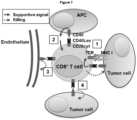
- Co-stimulation of the TCR signalling cascade with co-stimulatory receptors such as CD28 can enhance proliferation, survival and cytotoxicity of the T cells ( Chen et al., Nat Rev Immunol (2013), 13: 227-242 ).
- chimeric co-stimulatory receptors were created. They consist of the extracellular domain of the co-inhibitory receptor PD-1 (also known as CD279) and the signalling domain of CD28 ( Ankri et al., J Immunol (2013), 4121-4129 ; Prosser et al., Mol Immunol (2012), 263-272 ; WO 2013/019615 ).
- Tang et al. 2015 also specifies a fusion protein comprising a PD-1 extracellular domain and further comprising a 4-1BB intracellular domain and a human CD28 transmembrane domain.
- tumor specific T cell express PD-1 on the surface which then binds to its ligand PD-L1 expressed on tumor cells. This binding results in blocking of TCR-signalling and T cell activation, thus leading to inhibition of the tumor specific T cells.
- stimulation of CD28 would be necessary; see above.
- Chimeric co-stimulatory receptors comprising PD-1 and CD28 domains shall exhibit the relevant functions of both, the extracellular receptor function of PD-1 on the one hand, and the intracellular signalling function of CD28 on the other hand. In such chimeric constructs presented so far (Ankri, loc cit; Prosser, loc cit; WO 2013/019615 ), the transmembrane domains were taken from the respective signalling molecule.
- the present invention relates in a first aspect to a fusion protein comprising
- the present invention also relates to the fusion protein as defined above, further comprising a CD3 ⁇ domain.
- the present invention also relates to the fusion protein as defined above, wherein said extracellular domain does not comprise a linker or a hinge domain.
- the present invention also relates to the fusion protein as defined above, wherein
- the present invention also relates to the fusion protein as defined above, wherein said extracellular domain containing a polypeptide derived from PD-1 comprises an amino acid sequence with 0, 1, 2, 3, 4, 5, 6, 7, 8, 9, or 10 amino acid substitutions, deletions, and/or insertions compared to the amino acid sequence of SEQ ID NO: 2, wherein said fusion protein exhibits PD-L1/2 binding affinity, preferably wherein said extracellular domain comprises the amino acid sequence of SEQ ID NO: 2.
- the present invention additionally relates to a nucleic acid molecule encoding the fusion protein as defined above. Further, the invention relates to a vector comprising said nucleic acid molecule, and to a host cell comprising said nucleic acid molecule or said vector as defined above. In addition, the present invention relates to the host cell as defined above, wherein the host cell is transduced with said nucleic acid molecule or said vector. Further, the present invention relates to said host cell as defined above, wherein said nucleic acid molecule or said vector is stably integrated into the genome of the host cell. In a preferred embodiment, the present invention relates to said host cell as defined above which is transduced via retroviral transduction. The present invention also relates to said host cell as defined above stably expressing the fusion protein encoded by the nucleic acid molecule as defined above. The present invention also relates to said host cell as defined above which is a CD8 + T-cell.
- the present invention relates to a method of preparing the host cell as defined above comprising
- the present invention relates to the fusion protein, the nucleic acid molecule, the vector, the host cell as defined above for use in a method of treating cancer and chronic viral infection.
- the extracellular domain contains a polypeptide derived from PD-1 at its N-terminus
- the intracellular domain contains a polypeptide derived from 4-1BB at its C-terminus as defined above and vice versa.
- 4-1BB (CD137) is a co-stimulatory receptor present on a subset of T cells capable of augmenting TCR signaling.
- 4-1BB is a member of the tumor necrosis factor receptor (TNFR) superfamily and is absent in naive T cells but induced following T cell stimulation and differentiation into effector cells ( Cheuk at al., Cancer Gene Ther (2004), 11: 215-226 ).
- TNFR tumor necrosis factor receptor
- the intracellular domain of 4-1BB contains the QEE motif, which, upon ligation with 4-1BBL, expressed on APCs, recruits TNFR associated factor 2 (TRAF2) ( Arch et al., Mol Cell Biol (1998), 18: 558-565 ; Nam et al., J Immunol (2005), 174: 1898-1905 ).
- TRAF2 activates MAPK pathways including ERK and activates nuclear translocation of NF ⁇ B ( Watts, Annu Rev Immunol (2005), 23: 23-68 ). It thereby enhances cytokine production and T cell survival.
- the present invention relates to fusion (also termed herein "chimeric") proteins comprising or consisting of the extracellular domain (ECD) of PD-1 and the intracellular signaling domain (ICD) of 4-1BB being defined as comprising an amino acid sequence with 0, 1, 2, 3, 4, 5, 6, 7, 8, 9, or 10 amino acid substitutions, deletions, and/or insertions compared to the amino acid sequence of SEQ ID NO: 4.
- ECD extracellular domain
- ICD intracellular signaling domain
- fusion proteins comprising or consisting of the extracellular domain (ECD) derived from PD-1, a transmembrane domain (TMD), and the intracellular domain (ICD) derived from 4-1BB are also referred to herein as "PD-1:4-1BB” or "PD-1:BB”.
- fusion proteins comprising or consisting of the extracellular domain (ECD) derived from PD-1, a transmembrane domain (TMD), and the intracellular domain (ICD) derived from CD28 are also referred to herein as "PD-1:CD28”.
- the index " TM " or " tm " in context with fusion proteins indicate which transmembrane domain is used for the respective construct.
- PD-1 TM :BB or “PD-1 tm BB” means that the fusion protein comprises the transmembrane domain of PD-1
- PD-1:BB TM or “PD-1:BB tm” means the fusion protein comprises the transmembrane domain of 4-1BB.
- a fusion protein which comprises the extracellular domain (ECD) of PD-1 (e.g., PD-1), a transmembrane domain (TMD) derived from PD-1, and the intracellular domain (ICD) of 4-1BB as defined above.
- ECD extracellular domain
- TMD transmembrane domain
- ICD intracellular domain
- a fusion product comprising the ICD of 4-1BB exhibits superior effects in form of increased proliferation rates compared to constructs comprising the ICD of CD28 when expressed in T cells, e.g., in human melanoma xenograft.
- the ECD of the fusion protein described and provided in context with the present invention having an ECD derived from PD-1 preferably has the function to bind to PD-L1/2 on the surface of tumour cells which express PD-L1 as part of an escape mechanism as known in the art.
- the ICD of the fusion protein - comprising a polypeptide derived from 4-1BB - preferably acts as activating signalling molecule, thus increasing proliferation of the host cell (e.g., T cell such as CD8 + T cell) and/or cytokine secretion.
- the fusion protein provided in accordance with the present invention may further comprise a CD3 ⁇ domain. This may particularly be applicable for cases where the fusion protein is not expressed in a T cell or generally in a TCR negative cell or in cases where TCR and/or CAR are not co-transduced (or generally co-expressed) in the cell expressing the fusion construct of the present invention.
- the ICD amino acid sequence of CD3 ⁇ can be taken from data base known in the art (NP_932170). Generally, CD3 ⁇ may preferably be introduced after the ICD of the 4-1BB or CD28 protein.
- the fusion proteins provided herein may particularly comprise at the N-terminus an ECD containing a polypeptide derived from PD-1, preferably the ECD of PD-1 (e.g., human or murine, preferably human PD-1).
- the term "derived from” particularly means that the polypeptide contained in the ECD comprises at least a part of PD-1 (e.g., human or murine, preferably human PD-1), preferably the ECD of PD-1, respectively.
- the term "derived from” PD-1 also allows that up to 0, 1, 2, 3, 4, 5, 6, 7, 8, 9, 10 or more amino acid are substituted, deleted, and/or inserted compared to a native sequence of PD-1 (human or murine, preferably human PD-1), or part thereof (e.g., ECD).
- ECD the native sequence of PD-1
- the signal peptide sequence (as recognizable for those of skill in the art and as also depicted in specific SEQ ID NOs. referred to herein) is usually cut off in the mature protein before, during or after integration of the fusion protein in the membrane. That is, when referring to fusion proteins or host cells expressing the fusion proteins as described and provided herein, it is always also encompassed in accordance with the present invention that the ECD of the fusion protein may lack the respective signal peptide.
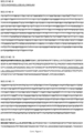
- the fusion protein comprises an ECD containing a polypeptide derived from PD-1 comprises an amino acid sequence with up to 0, 1, 2, 3, 4, 5, 6, 7, 8, 9, or 10 amino acid substitutions (preferably conservative or highly conservative substitutions), deletions and/or insertions compared to the amino acid sequence of the ECD of human or murine PD-1, e.g., of human PD-1 as depicted in SEQ ID NO: 2 (with or without the signal peptide as depicted in Figure 1 for SEQ ID NO: 2), wherein said fusion protein exhibits PD-L1/2 binding affinity, respectively.
- binding affinity may be compared to the binding affinity of native PD-1 (e.g., human PD-1) to its respective ligand PD-L1/2.
- native PD-1 e.g., human PD-1
- a polypeptide having a binding affinity to native human PD-L1/2 which is at least 0.8-fold, preferably at least 0.9-fold, or more preferably at least 1.0-fold as high compared to binding affinity of native human PD-1 to PD-L1/2, is considered to exhibit binding affinity to PD-L1/2, respectively.
- the binding affinity of a given polypeptide to PD-L1/2 can be measured by methods known in the art and usually and preferably comprises the measurement of the K D value (dissociation constant) which is expressed as a molar concentration.
- K D value dissociation constant
- Such methods for measuring protein interactions in terms of K D are well known in the art and comprise, e.g., ELISA, flow cytometry, surface plasmon resonance, biacore measurement, and the like.
- the fusion protein comprises an ECD comprising or consisting of the amino acid sequence of the ECD of PD-1 according to SEQ ID NO: 2 (with or without the signal peptide as depicted in Figure 1 for SEQ ID NO: 2).
- the ECD of the fusion protein described and provided herein may further comprise a hinge- and/or a linker region at the C-termimus of the ECD (e.g., between the ECD and TMD of the fusion protein, or between the ECD and the ICD of the fusion protein where the TMD is located C-terminally of the ICD) in order to allow more flexibility to the ECD.
- a hinge- and/or a linker region at the C-termimus of the ECD e.g., between the ECD and TMD of the fusion protein, or between the ECD and the ICD of the fusion protein where the TMD is located C-terminally of the ICD
- Typical hinge- or linker regions are known in the art and comprise those derived from the constant region (Fc) of antibodies (e.g., IgG1, CD8alpha) (see, e.g., Shirasu et al., Anticancer Res (2012), 32: 2377-2383 and Cartellieri et al., J Biomed Biotechnol (2010), 956304 ) (e.g., IgGFc spacers), Gly/Ser linkers, or filamin (e.g., Fil3 spacers).
- Fc constant region
- linker- or hinge regions may also cause side effects due to activation of NK cells which secrete high amounts of inflammatory cytokines
- the fusion proteins described and provided herein do not comprise linker- or hinge regions. That is, in one embodiment of the present invention, the ECD of the fusion protein does not comprise a linker- or hinge region.
- the fusion proteins provided herein further comprise a TMD operably linked between the ECD and the ICD or linked C-terminally of the ICD.
- the TMD is not limited to a specific TMD.
- the TMD allows stable anchorage of the fusion protein in the membrane of a cell expressing the fusion protein (e.g., a T cell) and further allows binding of the ECD to PD-L1/2, respectively, and, upon binding to PD-L1/2, allows signaling induction of the ICD containing a polypeptide derived from 4-1BB as described and exemplified herein.
- the TMD is derived from PD-1, wherein said transmembrane domain comprises the amino acid sequence of SEQ ID NO: 8 .
- the TMD of the fusion protein is not derived from CD8(alpha), and/or ICOS. If the ECD is derived from PD-1 and the ICD is derived from 4-1BB as defined above, the TMD is also not derived from CD28, but is derived from PD-1, wherein said transmembrane domain comprises the amino acid sequence of SEQ ID NO: 8.
- the TMD of the fusion protein comprises a polypeptide derived from PD-1 comprising the amino acid sequence of SEQ ID NO: 8, particularly where the ECD is from PD-1 and the ICD is from 4-1BB as defined above.
- the term "derived from” particularly means that the polypeptide contained in the TMD comprises at least a part of human PD-1, particularly where the ECD is from PD-1 and the ICD is from 4-1BB as it has been defined above.
- derived from PD-1 also allows that up to 0, 1, 2, 3, 4, 5, 6, 7, 8, 9, 10 or more amino acid are substituted, deleted, and/or inserted compared to a native sequence of human PD-1 or part thereof (e.g., TMD).
- fusion proteins having an ICD derived from 4-1BB generally exhibit superior proliferation rates of the host cells (e.g., T cells such as CD8 + T cells) compared to similar constructs having an ICD derived from CD28 and an ECD from PD-1. Furthermore, fusion proteins comprising a TMD derived from PD-1 lead to an even higher secretion rate of cytokines (e.g., IFN ⁇ or IL-2) in transduced T cells compared to fusion proteins comprising a TMD derived from 4-1BB. Accordingly, in a specific embodiment of the present invention, the TMD of the fusion protein comprises or consists of a polypeptide derived from PD-1 comprising the amino acid sequence of SEQ ID NO: 8.
- a fusion protein comprising a TMD containing a polypeptide derived from 4-1BB as used in the present invention as comparison to the fusion proteins of the present invention comprising a TMD derived from PD-1 as defined above, and further comprising an amino acid sequence with up to 0, 1, 2, 3, 4, 5, 6, 7, 8, 9, or 10 amino acid substitutions (preferably conservative or highly conservative substitutions), deletions and/or insertions compared to the amino acid sequence of the TMD of human or murine 4-1BB, e.g., of human 4-1BB as depicted in SEQ ID NO: 6 is capable of increasing the proliferation rate of a CD8 + T cell when retrovirally transduced into a said CD8 + T cell upon stimulation of said CD8 + T cell with a PD-L1/2 + target cell as described and exemplified herein.
- Proliferation can be quantified by CFSE dye dilution, i.e. reduction of CFSE fluorescence intensity measured by flow cytometry (as exemplified in Figure 5 and Method description "Proliferation of TCR-D115 T cells", or any other suitable method known in the art to determine proliferation, e.g., H 3 thymidin incorporation, BrdU incorporation, etc. If the proliferation of the CD8 + T cell retrovirally transduced with the given fusion protein is greater than 1.0, such as at least 1.2-fold, preferably at least 1.3-fold, more preferably at least 1.5-fold higher compared to the CD8 + T cell which was not transduced with the fusion protein, the fusion protein is considered capable of increasing proliferation.
- the proliferation difference can be calculated as the ratio of mean fluorescence activity (MFI) between T cells without chimeric receptor (mock) and T cells expressing a PD-1:BB or PD-1:CD28 variant.
- MFI mean fluorescence activity
- Such fusion protein used as comparison comprises a TMD comprising or consisting of the amino acid sequence of the TMD of 4-1BB according to SEQ ID NO: 6, particularly where the ECD is derived from PD-1 and the ICD is derived from 4-1BB.
- the fusion protein comprises a TMD containing a polypeptide derived from PD-1 comprising the amino acid sequence as depicted in SEQ ID NO: 8, wherein said fusion protein is capable of increasing secretion of IFN ⁇ and/or IL-2 when retrovirally transduced into a CD8 + T cell upon stimulation of said CD8 + T cell with a PD-L1/2 + target cell.
- Assessment whether a given fusion protein is capable of increasing secretion of IFN ⁇ and/or IL-2 when retrovirally transduced into a CD8 + T cell upon stimulation of said CD8 + T cell with a PD-L1/2 + target cell can be performed by methods known in the art and as also described and exemplified herein (cf.
- both CD8 + T cells are stimulated with a PD-L1/2 + target cell as described and exemplified herein followed by measuring the secretion level of IFN ⁇ and/or IL-2.
- the CD8 + T cell transduced with the fusion protein and the non-transduced control CD8 + T cell are usually derived from the same donor.
- transgenic human T cells may be retrovirally transduced to express the fusion protein containing said polypeptide derived from 4-1BB, then cultured with HEK/Tyr or HEK/Tyr/PD-L1 cells at a 1:2 ratio.
- Co-culture supernatants may then be harvested after 16 h and analyzed by sandwich ELISA (BD) or Bio-Plex (Bio-Rad) according to the manufacturer's protocol.
- Methods for measuring the secretion level of IFN ⁇ and IL-2 are well known in the art and also exemplified herein and comprise, inter alia, ELISA, Bio-Plex, intracellular flow cytometry (ICS), or the like.
- the fusion protein is considered capable of increasing secretion of IFN ⁇ and/or IL-2.
- the fusion protein comprises a TMD comprising or consisting of the amino acid sequence of the TMD of PD-1 according to SEQ ID NO: 8.
- the fusion proteins provided herein further comprise an ICD operably linked to the C-terminus of the TMD (particularly for fusion proteins where the ECD is from PD-1 and the ICD is from 4-1BB), or operably linked to the C- or N-terminus of the TMD.
- the ICD of the inventive fusion proteins contains a polypeptide which is derived from 4-1BB (CD137).
- the term "derived from” particularly means that the polypeptide contained in the ICD comprises at least a part of human 4-1BB.
- the term "derived from” 4-1BB also allows that up to 0, 1, 2, 3, 4, 5, 6, 7, 8, 9, 10 or more amino acid are substituted, deleted, and/or inserted compared to a native sequence of human 4-1BB or part thereof (e.g., ICD).
- the ICD of the fusion protein of the present invention comprises a polypeptide derived from the ICD of 4-1BB, wherein said ICD comprises an amino acid sequence with 0, 1, 2, 3, 4, 5, 6, 7, 8, 9, or 10 amino acid substitutions, deletions, and/or insertions compared to the amino acid sequence of SEQ ID NO: 4.
- the fusion protein of the present invention comprises an ICD containing a polypeptide derived from 4-1BB comprising an amino acid sequence with up to 0, 1, 2, 3, 4, 5, 6, 7, 8, 9, or 10 amino acid substitutions (preferably conservative or highly conservative substitutions), deletions and/or insertions compared to the amino acid sequence of the ICD of human 4-1BB as depicted in SEQ ID NO: 4, wherein said fusion protein is capable of increasing the proliferation rate of a CD8+ T cell when retrovirally transduced into said CD8 + T cell upon stimulation of said CD8 + T cell with a PD-L1/2 + target cell as described and exemplified herein.
- the fusion protein comprises an ICD comprising or consisting of the amino acid sequence of the ICD of 4-1BB according to SEQ ID NO: 4.
- all domains are derived from corresponding human domains.
- the fusion protein used as comparison in the present invention comprises or consists of an amino acid sequence with up to 0, 1, 2, 3, 4, 5, 6, 7, 8, 9, or 10 amino acid substitutions (preferably conservative or highly conservative substitutions), deletions and/or insertions compared to the amino acid sequence of SEQ ID NO: 22 (with or without the signal peptide as depicted in Figure 1 for SEQ ID NO: 22), wherein said fusion protein exhibits PD-L1/2 binding affinity as described herein, and wherein said fusion protein is capable of increasing the proliferation rate of a CD8+ T cell when retrovirally transduced into said CD8 + T cell upon stimulation of said CD8 + T cell with a PD-L1/2 + target cell as described and exemplified herein.
- the fusion protein provided and described herein comprises or consists of an ECD derived from PD-1, a TMD derived from PD-1, and an ICD derived from 4-1BB as defined herein. In one embodiment, all domains are derived from corresponding human domains.
- the fusion protein of the present invention comprises or consists of an amino acid sequence with up to 0, 1, 2, 3, 4, 5, 6, 7, 8, 9, or 10 amino acid substitutions (preferably conservative or highly conservative substitutions), deletions and/or insertions compared to the amino acid sequence of SEQ ID NO: 24 (with or without the signal peptide as depicted in Figure 1 for SEQ ID NO: 24), wherein said fusion protein exhibits PD-L1/2 binding affinity as described herein, and whereinsaid fusion protein is capable of increasing secretion of IFN ⁇ and/or IL-2 when retrovirally transduced into a CD8 + T cell upon stimulation of said CD8 + T cell with a PD-L1/2 + target cell as described and exemplified herein.
- a given fusion protein in order to assess whether a given fusion protein is capable of increasing the proliferation rate of a CD8+ T cell as well as increasing the IFN ⁇ and/or IL-2 secretion level of said CD8 + T cell retrovirally transduced with said fusion protein is compared to the proliferation rate/the IFN ⁇ and/or IL-2 secretion level of a comparable CD8 + T cell not transduced with said fusion protein.
- Methods for assessing proliferation and IFN ⁇ /IL-2 secretion rates are known in the art and described and exemplified herein above and below.
- the fusion protein comprises or consists of an amino acid according to SEQ ID NO: 24 (with or without the signal peptide as depicted in Figure 1 for SEQ ID NO: 24).
- polypeptide is equally used herein with the term “protein”. Proteins (including fragments thereof, preferably biologically active fragments, and peptides, usually having less than 30 amino acids) comprise one or more amino acids coupled to each other via a covalent peptide bond (resulting in a chain of amino acids).
- polypeptide as used herein describes a group of molecules which typically comprise more than 10 amino acids. Polypeptides may further form multimers such as dimers, trimers and higher oligomers, i.e. consisting of more than one polypeptide molecule. Polypeptide molecules forming such dimers, trimers etc. may be identical or non-identical.
- heteromultimer is an antibody molecule, which, in its naturally occurring form, consists of two identical light polypeptide chains and two identical heavy polypeptide chains.
- polypeptide and protein also refer to naturally modified polypeptides/proteins wherein the modification is effected e.g. by post-translational modifications like glycosylation, acetylation, phosphorylation and the like. Such modifications are well known in the art.
- amino acid or “amino acid residue” as used herein typically refers to an amino acid having its art recognized definition such as proteinogenic amino acid selected from the group consisting of: alanine (Ala or A); arginine (Arg or R); asparagine (Asn or N); aspartic acid (Asp or D); cysteine (Cys or C); glutamine (Gln or Q); glutamic acid (Glu or E); glycine (Gly or G); histidine (His or H); isoleucine (He or I): leucine (Leu or L); lysine (Lys or K); methionine (Met or M); phenylalanine (Phe or F); pro line (Pro or P); serine (Ser or S); threonine (Thr or T); tryptophan (Trp or W); tyrosine (Tyr or Y); and valine (Val or V), although modified, synthetic, or rare amino acids may be used
- amino acids can be grouped as having a nonpolar side chain (e.g., Ala, Cys, He, Leu, Met, Phe, Pro, Val); a negatively charged side chain (e.g., Asp, Glu); a positively charged sidechain (e.g., Arg, His, Lys); or an uncharged polar side chain (e.g., Asn, Cys, Gln, Gly, His, Met, Phe, Ser, Thr, Trp, and Tyr).
- a nonpolar side chain e.g., Ala, Cys, He, Leu, Met, Phe, Pro, Val
- a negatively charged side chain e.g., Asp, Glu
- a positively charged sidechain e.g., Arg, His, Lys
- an uncharged polar side chain e.g., Asn, Cys, Gln, Gly, His, Met, Phe, Ser, Thr, Trp, and Tyr.
- fusion protein relates to a protein which is made of polypeptide parts from different sources. Accordingly, it may be also understood as a “chimeric protein", a “chimeric construct”, “fusion construct”, or the like.
- fusion proteins are proteins created through the joining of two or more genes (or preferably cDNAs) that originally coded for separate proteins. Translation of this fusion gene (or fusion cDNA) results in a single polypeptide, preferably with functional properties derived from each of the original proteins.
- Recombinant fusion proteins are created artificially by recombinant DNA technology for use in biological research or therapeutics. Further details to the production of the fusion protein of the present invention are known in the art and described and exemplified herein.
- the present invention further relates to a nucleic acid molecule encoding the fusion protein provided and described herein.
- the present invention also relates to nucleic acid molecules which only encode parts of the fusion protein described and provided herein, e.g., which encode only the ECD, the TMD, and/or the ICD of the fusion protein.
- the present invention specifically relates to nucleic acid molecules according to SEQ ID NOs. 1, 3, 7, or 23, or combinations thereof linked via nucleic acid bonds, provided a fusion protein according to the present invention or a part thereof (e.g., the ECD, TMD and/or ICD) is encoded.
- the present invention also relates to nucleic acid molecules where 30, 27, 24, 21, 18, 15, 12, 9, 6, 3, or 0 nucleotides have been substituted (preferably silent mutations which do not result in a change of translated amino acid), deleted or inserted compared to nucleic acid molecules according to SEQ ID NOs. 1, 3, 7, or 23, or combinations thereof linked via nucleic acid bonds, provided a fusion protein according to the present invention or a part thereof (e.g., the ECD, TMD and/or ICD) is encoded.
- the present invention preferably relates to nucleic acids encoding fusion proteins specifically described and embodied herein. For example, SEQ ID NOs.
- FIG. 13 and 14 which are not part of the fusion protein of the present invention show the human nucleic acid and amino acid sequence of CD28, respectively, where the predominant transcript of CD28 is depicted. Yet, given that CD28 also exists in further splice variants (e.g., in-frame donor splice variants or variants lacking an in-frame coding exon), such variants or parts thereof (particularly TMD thereof) are also encompassed to be part of the fusion protein described and provided herein. Generally, SEQ ID NOs. 13 and 14 show the human nucleic acid and amino acid sequence of CD28, respectively, where the predominant transcript of CD28 is depicted.
- splice variants e.g., in-frame donor splice variants or variants lacking an in-frame coding exon
- SEQ ID NOs. 13 and 14 show the human nucleic acid and amino acid sequence of CD28, respectively, where the predominant transcript of CD28 is depicted.
- nucleic acid or “nucleic acid molecule” is used synonymously with “oligonucleotide”, “nucleic acid strand”, “polynucleotide”, or the like, and means a polymer comprising one, two, or more nucleotides.
- nucleic acid molecule relates to the sequence of bases comprising purine- and pyrimidine bases which are comprised by polynucleotides, whereby said bases represent the primary structure of a nucleic acid molecule.
- nucleic acid molecule includes all kinds of nucleic acid, including DNA, cDNA, genomic DNA, RNA, synthetic forms of DNA and mixed polymers comprising two or more of these molecules, and preferably relates to DNA and cDNA.
- nucleic acid sequences provided herein represent sequences of DNA and also comprise corresponding RNA sequences where T is replaced by U.
- nucleic acid molecule generally comprises sense and antisense strands.
- Nucleic acid molecule may further comprise non-natural or derivatized nucleotide bases as well as natural or artificial nucleotide analogues, e.g., in order to protect the nucleic acid molecule against endo- and/or exonucleases as will be readily appreciated by those skilled in the art.
- the present invention further relates to a vector comprising the nucleic acid molecule described and provided herein.
- vector generally comprises all kinds of linear or circular nucleic acid molecules which can replicate autonomously is a suitable host cell.
- Such vectors comprise, but are not limited to, plasmids, cosmids, phages, virus (e.g., adeno-, adeno-associated-, lenti-, or preferably retroviral vectors), and other vectors or shuttles known in the art which are suitable to carry and transfer genes into host cells in order to allow stable or transient translation and constitutive or conditional expression of the inventive fusion protein in the host cell.
- the vector is usually not integrated into the cell genome, but may also be integrated.
- Vectors according to the present invention which comprise nucleic acid molecules as described and provided herein preferably allow stable expression of the fusion protein of the present invention in the host cell (expression vector).
- Vectors of the present invention may further comprise marker genes, promoter and/or enhancer sequences (operably linked to the nucleic acid molecule of the present invention), replication origin suitable for the respective host cell, restriction sited, multiple cloning sites, labels and further functional units as known in the art.
- the vectors may inter alia be transferred into host cells via a shuttle such as a virus (which may itself be considered a vector), or be nakedly transformed or transduced into host cells.
- the vector is preferably adapted to suit to the respective host cell where it is to be transformed or transduced into.
- the vector (plasmid) pGEM is a suitable vector for transformation into bacterial cells
- the retroviral vector pMP71 is suitable for transduction into eukaryotic cells (e.g., T cells).
- the vector of the present invention is a viral vector, e.g., a retroviral or lentiviral vector, e.g., a retroviral vector.
- retroviral vectors include, e.g., pMP71-PRE ( Leisegang, K Mol Med (2008), 86(5): 573-583 ), SAMEN CMV/SRa, LZRS-id3-IHRES ( Heemskerk et al., J. Exp. Med. 186 (1997), 1597-1602 ), FeLV ( Neil et al., Nature 308 (1984), 814-820 ), SAX ( Kantoff et al., Proc. Natl.
- the vector is pMP71-PRE or pMP71.
- the present invention further relates to a host cell comprising the nucleic acid molecule or the vector as described and provided herein.
- the host cell of the present invention is transduced or transformed with the nucleic acid molecule or the vector as described and provided herein.
- the terms “transduced” or “transformed” (as well as “transduction” or “transformation”) or the like may be used interchangeably and generally mean any kind of transfer of a nucleic acid molecule and/or vector into a host cell, regardless of the kind of host cell and regardless of the way of transfer (e.g., (chemical) transformation, (viral) transduction, electroporation, transfection, etc.).
- the nucleic acid molecule and/or the vector may be stably integrated into the genome of the host cell, or be extrachromosomal ( i.e. transient expression). Examples for suitable methods for achieving transient expression in a host cell are known in the art and comprise mRNA transfection.
- the nucleic acid molecule and/or the vector is stably integrated into the genome.
- the host cell described and provided in context with the present invention comprising the nucleic acid molecule or the vector as described and provided herein is preferably able to stably or transiently (e.g., stably) express (either constitutively or conditionally) the fusion protein of the present invention.
- the host cell may generally be transduced or transformed by any method with any suitable nucleic acid molecule or vector.
- the host cell is transduced with a retroviral or lentiviral (e.g., retroviral) vector comprising a nucleic acid molecule encoding the fusion protein of the present invention or parts thereof (e.g., ECD, TMD, and/or ICD) as described above.
- the host cell of the present invention is transduced with a retroviral vector comprising a nucleic acid molecule encoding the fusion protein of the present invention or parts thereof (e.g., ECD, TMD, and/or ICD) as described above and stably expresses (either constitutively or conditionally) the fusion protein or part thereof.
- the host cell then stably expresses the fusion protein in its membrane, with the ECD of the fusion protein of the present invention directed to the surface, the TMD being (largely) embedded in the membrane, and the ICD directed to the cytoplasm.
- the host cell comprising the nucleic acid molecule or the vector as described and provided herein relates to a genetically modified cell wherein said nucleic acid molecule or said vector was transduced, transformed or otherwise introduced into the host cell.
- the host cell of the present invention may be a cell which transiently or stably expresses the fusion protein of the present invention.
- the nucleic acid molecule encoding the fusion protein of the present invention can be stably integrated into the genome of the cell by retroviral or lentiviral (e.g., retroviral) transduction.
- the PD-1-BB fusion protein is expressed in the membrane of the herein provided transduced cell.
- the detection of the ECD of the PD-1 polypeptide can be carried out by using an antibody or other binding molecule specifically binding the ECD of PD-1 as described herein, e.g., by ELISA or by flow cytometry, or microscopy.
- the transduced cell of the present invention may be, e.g., CD8 + T cells, CD4 + T cells, double-negative ⁇ / ⁇ T cells, NK (natural killer) cells, ⁇ T cells, macrophages, dendritic cells, as well as cells suitable store and/or reproduce the nucleic acid molecule or vector of the present invention, including bacterial cells (e.g., E. coli ) and further eukaryotes.
- the host cell of the present invention is a T cell, e.g., a CD8 + T cell.
- the host cell of the present invention may be transduced with a nucleic acid molecule or a vector encoding the fusion protein as described and provided herein.
- the host cell provided and described herein may be co-transduced with further nucleic acid molecules, e.g. with a nucleic acid molecule encoding a T cell receptor (TCR) or a chimeric antigen receptor (CAR).
- TCR T cell receptor
- CAR chimeric antigen receptor
- suitable host cells include, but are not limited to, T cells, e.g. CD8 + T cells, CD4 + T cells, TCR such as (but not limited to) TCR-T58 or TCR-D115 T cells, double-negative ⁇ / ⁇ T cells, NK (natural killer) cells, ⁇ T cells, macrophages, dendritic cells, as well as cells suitable store and/or reproduce the nucleic acid molecule or vector of the present invention, including bacterial cells (e.g., E. coli ) and further eukaryotes.
- the cells may be autologous or non-autologous, but are preferably autologous. Also, the cells may be allogeneic or non-allogeneic as readily clear for the skilled person.
- the host cell of the present invention is a CD8 + T cell.
- the present invention also relates to a method of preparing a host cell of the present invention as described and provided herein, said method comprising
- the host cell is transduced or transformed outside the human body.
- Methods for obtaining, isolating and culturing cells e.g., T cells such as CD8 + T cells, CD4 + T cells, TCR such as (but not limited to) TCR-T58 or TCR-D115 T cells
- donors e.g., human donors
- T cells such as CD8 + T cells, CD4 + T cells, TCR such as (but not limited to) TCR-T58 or TCR-D115 T cells
- the host cell may be transduced or transformed or otherwise be provided with a nucleic acid molecule or a vector as described and provided herein by any method known in the art. Such methods comprise, inter alia, (chemical) transformation, (viral) transduction, electroporation, transfection, and the like. In one embodiment, the host cell is transduced with a retroviral vector.
- the host cell to be prepared in accordance with the present invention may be any host cell as described herein.
- the host cell is a T cell, for example a CD8 + T cell, CD4 + T cell, TCR such as (but not limited to) TCR-T58 or TCR-D115 T cell.
- the present disclosure which is not part of the invention and only present as illustration purposes comprises a pharmaceutical composition comprising a fusion protein, a nucleic acid molecule, a vector, and/or a host cell as described and provided by the present invention.
- Such pharmaceutical composition is suitable to be administered to patient (preferably, human patient), particularly to the donor of the host cells as described above.
- the present disclosure also comprises methods for treating a disease or disorder by administering comprising a pharmaceutical composition comprising a fusion protein, a nucleic acid molecule, a vector, and/or a host cell as described and provided by the present invention.
- the pharmaceutical composition of the present disclosure may further comprise a pharmaceutically acceptable carrier and further components, e.g., for galenic.
- the pharmaceutical composition is particularly useful for treating diseases or disorders associated with the expression of PD-1 ligands (e.g., PD-L1 or PD-L2) and/or CD40.
- Such diseases and disorders are known to the skilled person and comprise particularly (but not limited to) different types of cancer such as lung cancer, gastric cancer, renal cell cancer, colon cancer, breast cancer, ovarian cancer, urothelial cancer, melanoma, pancreatic cancer, myeloma, Hodkin's lymphoma, retinoblastoma, leukemia, cervical cancer, esophageal cancer, glioma, non-Hodkin's lymphoma, hepatocellular cancer, oral cancer, and others.
- Further diseases and disorders which may be treated by the pharmaceutical compositions provided herein comprise (chronic) viral infections and (chronic) inflammations, particularly for cases where PD-L1/L2 and/or CD40 is expressed.
- kit or kit-in-parts comprising a fusion protein, a nucleic acid molecule, a vector, and/or a host cell as described and provided in context with the present invention.
- Chimeric receptor sequences were ordered at Geneart, Life Technologies. They were delivered as a lyophilized powder and dissolved in nuclease-free water at a concentration of 0.5 ⁇ g/ ⁇ l.
- TOP10 or MACH1 E.coli were chemically transformed with the Geneart constructs, following standard plasmid preparation methods.
- Chimeric receptor sequences were cloned into pGEM for ivtRNA preparation. Cloning into the pGEM vector (provided by S. Milosevic, Medigene GmbH, Martinsried, Germany) was achieved using Hindlll or Hindll and EcoRI (New England Biolabs). IvtRNA was generated from pGEM plasmids using the mMESSAGEmMACHINE Kit (Ambion) according to the manufacturer's protocol. Human primary T cells were electroporated with 20 ⁇ g ivtRNA at 900 V for 2.3 ms using Gene Pulser Xcell (Bio-Rad).
- Chimeric receptor sequences were cloned into pMP71-PRE vector (Leisegang 2008, loc. Cit, for retroviral transduction). Retroviral transduction of T cells was achieved as described (Leisegang 2008, loc. cit.).
- Human PBMCs from healthy donors were plated into 24-well plates at a cell density of 1x10⁶/ml per well in RPMI1640 supplemented with 10% human serum, 1% L-glutamine, 1% non-essential amino acids, 1% sodium pyruvate and 1% penicillin/streptomycin (all Invitrogen) plus 100 U/ml IL-2 (Cancernova) and activated with 5 ⁇ g/ml OKT3 (provided by E. Kremmer, Helmholtz Center Munich, Germany) and 1 ⁇ g/ml anti-CD28 (BD Pharmingen) for 2 days.
- OKT3 provided by E. Kremmer, Helmholtz Center Munich, Germany
- BD Pharmingen 1 ⁇ g/
- Amphotrophic chimeric receptor-encoding retroviruses were generated as described ( Leisegang at al., Clin Cancer Res (2010), 16(8): 2333-2343 ) using TranslT ® -LT1 Reagent (Mirus) according to the manufacturer's protocol. Virus supernatant was harvested after 48 h and bound to RetroNectin ® (10 ⁇ g/ml, Takara) coated plates by centrifugation.
- PBMCs which were activated for 2 days, were added to virus-coated plates for 24 h, then split to freshly virus-coated plates and cultivated for another 3 days. Transduced PBMCs were transferred to uncoated plates and cultivated for at least 12 additional days reducing the amount of IL-2 to 50 U/ml. Receptor expression was determined at day 12 after transduction using anti-PD-1 antibody (BioLegend).
- HEK/Tyr and HEK/Tyr/PD-L1 were generated by transducing HLA-A2+ HEK293 cells to express tyrosinase (HEK/Tyr) or tyrosinase and PD-L1 (HEK/Tyr/PD-L1). After transduction, HEK293 cells were single-cell cloned and clones selected for comparable HLA-A2 and tyrosinase expression.
- SK-Mel23 gift from M.C.
- HEK/Tyr and HEK/Tyr/PD-L1 were grown in RPMI-1640 supplemented with 1% L-glutamine, 1% non-essential amino acids, 1% sodium pyruvate, 1% penicillin/streptomycin (RMPI basic) plus 12% FCS.
- LSRII LSRII
- PBS Invitrogen
- human serum 0.1% sodium azide
- 2 mM EDTA both Sigma-Aldrich
- Expression of human chimeric receptors and transgenic TCRs was analyzed using anti-CD3-PE-Cy7, anti-mouse TCR ⁇ -constant region-PB (both BioLegend), anti-CD4-APC-A780, anti-PD-1-APC (both eBioscience), anti-CD8-V500 (BD) and 7-AAD (Sigma-Aldrich).
- HEK293 cells were analyzed using anti-HLA-A2 (ATCC HB54) plus anti-mouse IgG1-A488 (Invitrogen), anti-PD-L1-FITC (BD), anti-tyrosinase (Upstate) plus anti-mouse IgG2a-A647 (Invitrogen) and 7-AAD.
- T cells after injection into xenograft tumors were analyzed using CFSE, anti-CD45-PE-Cy7, anti-CD8-PB (BD), CD4-APC-A780, anti-PD-1-APC and 7-AAD.
- CD45+ leukocytes were selected and CFSE intensity was analyzed in CD8 + , CD4 + and dbl- cells after gating on viable and single cells. Data were analyzed using FlowJo 8.8.7 software.
- TCR-T58 or TCR-D115 transgenic human T cells were electroporated with ivtRNA or transduced with retroviral vectors to express chimeric receptors, then cultured with HEK/Tyr or HEK/Tyr/PD-L1 cells at a 1:2 ratio. Co-culture supernatants were harvested after 16 h and analyzed by sandwich ELISA (BD) or Bio-Plex (Bio-Rad) according to the manufacturer's protocol.
- the amount of measured cytokine was normalized to the percentage of TCR + CD8 + T cells within the cell suspension (determined by flow cytometry). The following formula was applied:
- NSG mice were obtained from Charles River. NOD/scid IL2Rgnull (NSG) mice were bred on the genetic background of non-obese diabetic (NOD) mice characterized by reduced innate immunity. NSG mice carry the prkdcscid mutation, a loss-of-function mutation in the PRKDC gene, leading to defective repair of DNA strand breaks during V(D)J recombination in the development of B and T cells. This severe combined immunodeficiency (scid) is characterized by a major reduction of T and B cells. Additionally, NSG mice carry a null mutation in the IL-2 receptor gamma chain (IL2Rgnull) blocking NK cell differentiation. The impairment of innate immunity and absence of adaptive immunity render NSG mice a good model system for adoptive T cell therapy of human tumor xenografts.
- NSG mice NOD/scid IL2Rgnull
- mice Animal experiments were approved by the local authorities and performed according to the legal regulations. 7-11 weeks old, male mice were injected s.c. with 5 x 10⁶ HLA-A2+ tyrosinase + human melanoma cells SK-Mel23 (gift of Monica C. Panelli, NIH, Bethesda, USA). This melanoma line was selected because it expresses PD-L1/L2 as well as HLA-A2 and tyrosinase, which are required to form the ligand for TCR-D115 and TCR-T58 ( Wilde et al., Blood (2009), 114: 2131-2139 ). T cells expressing TCR-D115 were selected for the mouse experiment as they can recognize SK-Mel23 with low avidity which is not sufficient to eradicate established tumors.
- Cell tracer dyes i.e CFDA-SE
- CFDA-SE Cell tracer dyes
- CFSE fluorescent carboxyfluorescein succinimidyl esters
- TCR-D115 T cells without or with chimeric receptor expression were labeled with 0.15 ⁇ M CFDA-SE for 8 minutes at 37°C.
- the reaction was stopped with FCS, T cells were washed twice with PBS and re-suspended in PBS at a concentration of 10 x 107 cells per ml.
- Tumors were harvested 1, 2, 4, 6 and 11 days after i.t. injection.
- Single cell suspensions were prepared by mechanical and enzymatic digestion ( Prinz et al., J Immunol (2012), 188: 5990-6000 ) and used for flow cytometry analysis.
Landscapes
- Health & Medical Sciences (AREA)
- Life Sciences & Earth Sciences (AREA)
- Chemical & Material Sciences (AREA)
- Organic Chemistry (AREA)
- Genetics & Genomics (AREA)
- Immunology (AREA)
- Molecular Biology (AREA)
- Zoology (AREA)
- General Health & Medical Sciences (AREA)
- Biophysics (AREA)
- Biochemistry (AREA)
- Medicinal Chemistry (AREA)
- Engineering & Computer Science (AREA)
- Toxicology (AREA)
- Cell Biology (AREA)
- Gastroenterology & Hepatology (AREA)
- Proteomics, Peptides & Aminoacids (AREA)
- Biomedical Technology (AREA)
- Wood Science & Technology (AREA)
- Bioinformatics & Cheminformatics (AREA)
- Biotechnology (AREA)
- General Engineering & Computer Science (AREA)
- Microbiology (AREA)
- Physics & Mathematics (AREA)
- Plant Pathology (AREA)
- Public Health (AREA)
- Nuclear Medicine, Radiotherapy & Molecular Imaging (AREA)
- Veterinary Medicine (AREA)
- Chemical Kinetics & Catalysis (AREA)
- Animal Behavior & Ethology (AREA)
- General Chemical & Material Sciences (AREA)
- Pharmacology & Pharmacy (AREA)
- Oncology (AREA)
- Communicable Diseases (AREA)
- Virology (AREA)
- Peptides Or Proteins (AREA)
- Micro-Organisms Or Cultivation Processes Thereof (AREA)
- Medicines That Contain Protein Lipid Enzymes And Other Medicines (AREA)
- Medicinal Preparation (AREA)
- Medicines Containing Material From Animals Or Micro-Organisms (AREA)
Description
- The present invention relates to fusion proteins comprising (a) an extracellular domain containing a polypeptide derived from PD-1 at its N-terminus; (b) a transmembrane domain derived from PD-1, wherein said transmembrane domain comprises the amino acid sequence of SEQ ID NO: 8; and (c) an intracellular domain containing a polypeptide derived from 4-1 BB at its C-terminus, wherein said intracellular domain comprises an amino acid sequence with 0, 1, 2, 3, 4, 5, 6, 7, 8, 9, or 10 amino acid substitutions, deletions, and/or insertions compared to the amino acid sequence of SEQ ID NO: 4, wherein said fusion protein is capable of increasing the proliferation rate of a CD8+ T cell when transduced, transformed or otherwise introduced into a said CD8+ T cell upon stimulation of said CD8+ T cell with a PD-L1/L2+ target cell. The present invention also relates to nucleic acid molecules encoding such fusion proteins, vectors containing such nucleic acid molecules, and host cells containing such vectors. The present invention further relates to methods for producing such host cells. Finally, the present invention relates to the fusion proteins, nucleic acid molecules, vectors, and/or host cells for use in a method of treating cancer and chronic viral infection.
- Adoptive transfer of tumor infiltrating Lymphocytes (TILs) is a promising therapy option against tumors (Rosenberg et al., Science (2015), 348: 62-68). Unfortunately, the isolation of TILs and their expansion is not possible with all tumor entities. Therefore, the concept of genetic engineering of T cells with a transgene T cell receptor (TCR) was developed (Hurwitz et al., Cancer Microenviron (2014), 7: 1-9). Yet, to this date, such T cells show short survival, and lose their function in patients (Janicki et al., Cancer Res (2008), 68: 2993-3000; Bai et al., PNAS USA (2008), 105: 13003-13008; Bendle et al., Cancer Res (2004), 64: 8052-8056; Anderson et al., J Immunol (2007), 178: 1268-1276). Co-stimulation of the TCR signalling cascade with co-stimulatory receptors such as CD28 can enhance proliferation, survival and cytotoxicity of the T cells (Chen et al., Nat Rev Immunol (2013), 13: 227-242). However, as epithelial tumors do not express ligands (CD80/86) of the co-stimulatory receptor CD28 and human effector T cells are themselves negative for such receptors, co-stimulation cannot take place in the conservative way. To provide co-stimulation to the T cells that is independent from this classical way, chimeric co-stimulatory receptors were created. They consist of the extracellular domain of the co-inhibitory receptor PD-1 (also known as CD279) and the signalling domain of CD28 (Ankri et al., J Immunol (2013), 4121-4129; Prosser et al., Mol Immunol (2012), 263-272;
WO 2013/019615 ). In this regard, Tang et al. 2015 also specifies a fusion protein comprising a PD-1 extracellular domain and further comprising a 4-1BB intracellular domain and a human CD28 transmembrane domain. - Usually, tumor specific T cell express PD-1 on the surface which then binds to its ligand PD-L1 expressed on tumor cells. This binding results in blocking of TCR-signalling and T cell activation, thus leading to inhibition of the tumor specific T cells. In order to co-stimulate TCR signalling (and, thus, to enhance T cell activation), stimulation of CD28 would be necessary; see above.
Chimeric co-stimulatory receptors comprising PD-1 and CD28 domains shall exhibit the relevant functions of both, the extracellular receptor function of PD-1 on the one hand, and the intracellular signalling function of CD28 on the other hand. In such chimeric constructs presented so far (Ankri, loc cit; Prosser, loc cit;WO 2013/019615 ), the transmembrane domains were taken from the respective signalling molecule. - However, although such constructs have been described to reduce PD-L1 mediated T cell inhibition and to enhance T cell activation, such constructs still leave room for further improvement.
- This problem has been addressed by the present invention as described herein and as defined in the claims.
- The present invention relates in a first aspect to a fusion protein comprising
- (a) an extracellular domain (ECD) containing a polypeptide derived from PD-1 at its N-terminus;
- (b) a transmembrane domain (TMD) derived from PD-1, wherein said transmembrane domain comprises the amino acid sequence of SEQ ID NO: 8; and
- (c) an intracellular domain (ICD) containing a polypeptide derived from 4-1 BB at its C-terminus, wherein said intracellular domain comprises an amino acid sequence with 0, 1, 2, 3, 4, 5, 6, 7, 8, 9, or 10 amino acid substitutions, deletions, and/or insertions compared to the amino acid sequence of SEQ ID NO: 4; wherein said fusion protein is capable of increasing the proliferation rate of a CD8+ T cell when transduced, transformed or otherwise introduced into a said CD8+ T cell upon stimulation of said CD8+ T cell with a PD-L1/L2+ target cell.
- The present invention also relates to the fusion protein as defined above, further comprising a CD3ζ domain.
- The present invention also relates to the fusion protein as defined above, wherein said extracellular domain does not comprise a linker or a hinge domain.
- The present invention also relates to the fusion protein as defined above, wherein
- (i) said polypeptide derived from PD-1 comprised by said extracellular and/or by said transmembrane domain is a polypeptide derived from human PD-1; and/or
- (ii) said polypeptide derived from 4-1BB comprised by said intracellular domain is a polypeptide derived from human 4-1BB.
- The present invention also relates to the fusion protein as defined above, wherein said extracellular domain containing a polypeptide derived from PD-1 comprises an amino acid sequence with 0, 1, 2, 3, 4, 5, 6, 7, 8, 9, or 10 amino acid substitutions, deletions, and/or insertions compared to the amino acid sequence of SEQ ID NO: 2, wherein said fusion protein exhibits PD-L1/2 binding affinity, preferably wherein said extracellular domain comprises the amino acid sequence of SEQ ID NO: 2.
- The present invention additionally relates to a nucleic acid molecule encoding the fusion protein as defined above. Further, the invention relates to a vector comprising said nucleic acid molecule, and to a host cell comprising said nucleic acid molecule or said vector as defined above. In addition, the present invention relates to the host cell as defined above, wherein the host cell is transduced with said nucleic acid molecule or said vector. Further, the present invention relates to said host cell as defined above, wherein said nucleic acid molecule or said vector is stably integrated into the genome of the host cell. In a preferred embodiment, the present invention relates to said host cell as defined above which is transduced via retroviral transduction. The present invention also relates to said host cell as defined above stably expressing the fusion protein encoded by the nucleic acid molecule as defined above. The present invention also relates to said host cell as defined above which is a CD8+ T-cell.
- In a second aspect, the present invention relates to a method of preparing the host cell as defined above comprising
- (1) transducing a host cell with said nucleic acid molecule as defined above or said vector as defined above;
- (2) cultivating the transduced host cell of step (1) in a suitable medium allowing growth of the cell and expression of the fusion protein encoded by said nucleic acid molecule or said vector; and
- (3) collecting the host cell from the medium.
- In a third aspect, the present invention relates to the fusion protein, the nucleic acid molecule, the vector, the host cell as defined above for use in a method of treating cancer and chronic viral infection.
- Preferably, in accordance with the present invention, if the extracellular domain (ECD) contains a polypeptide derived from PD-1 at its N-terminus, the intracellular domain (ICD) contains a polypeptide derived from 4-1BB at its C-terminus as defined above and vice versa.
- Compared to CD28, 4-1BB (CD137) is a co-stimulatory receptor present on a subset of T cells capable of augmenting TCR signaling. 4-1BB is a member of the tumor necrosis factor receptor (TNFR) superfamily and is absent in naive T cells but induced following T cell stimulation and differentiation into effector cells (Cheuk at al., Cancer Gene Ther (2004), 11: 215-226). The intracellular domain of 4-1BB contains the QEE motif, which, upon ligation with 4-1BBL, expressed on APCs, recruits TNFR associated factor 2 (TRAF2) (Arch et al., Mol Cell Biol (1998), 18: 558-565; Nam et al., J Immunol (2005), 174: 1898-1905). TRAF2 activates MAPK pathways including ERK and activates nuclear translocation of NFκB (Watts, Annu Rev Immunol (2005), 23: 23-68). It thereby enhances cytokine production and T cell survival. Accordingly, the present invention relates to fusion (also termed herein "chimeric") proteins comprising or consisting of the extracellular domain (ECD) of PD-1 and the intracellular signaling domain (ICD) of 4-1BB being defined as comprising an amino acid sequence with 0, 1, 2, 3, 4, 5, 6, 7, 8, 9, or 10 amino acid substitutions, deletions, and/or insertions compared to the amino acid sequence of SEQ ID NO: 4.
- Generally, in context with the present invention, unless otherwise specified herein, fusion proteins comprising or consisting of the extracellular domain (ECD) derived from PD-1, a transmembrane domain (TMD), and the intracellular domain (ICD) derived from 4-1BB are also referred to herein as "PD-1:4-1BB" or "PD-1:BB". Likewise, fusion proteins comprising or consisting of the extracellular domain (ECD) derived from PD-1, a transmembrane domain (TMD), and the intracellular domain (ICD) derived from CD28 are also referred to herein as "PD-1:CD28". The index "™" or "tm" in context with fusion proteins indicate which transmembrane domain is used for the respective construct. For example, "PD-1™:BB" or "PD-1tmBB" means that the fusion protein comprises the transmembrane domain of PD-1, while "PD-1:BB™" or "PD-1:BBtm" means the fusion protein comprises the transmembrane domain of 4-1BB.
- In accordance with the present invention, a fusion protein is provided which comprises the extracellular domain (ECD) of PD-1 (e.g., PD-1), a transmembrane domain (TMD) derived from PD-1, and the intracellular domain (ICD) of 4-1BB as defined above. As has been surprisingly found in context with the present invention, T cells expressing such a PD-1:BB fusion protein exhibit increased and earlier proliferation rates compared to T cells expressing PD-1:CD28 fusion proteins as exemplarily shown in human melanoma xenografts.
- That is, as has been found in context with the present invention, a fusion product comprising the ICD of 4-1BB (CD137) exhibits superior effects in form of increased proliferation rates compared to constructs comprising the ICD of CD28 when expressed in T cells, e.g., in human melanoma xenograft.
- Accordingly, the ECD of the fusion protein described and provided in context with the present invention having an ECD derived from PD-1 preferably has the function to bind to PD-L1/2 on the surface of tumour cells which express PD-L1 as part of an escape mechanism as known in the art. Upon binding of the ECD of the inventive fusion protein, the ICD of the fusion protein - comprising a polypeptide derived from 4-1BB - preferably acts as activating signalling molecule, thus increasing proliferation of the host cell (e.g., T cell such as CD8+ T cell) and/or cytokine secretion.
- The fusion protein provided in accordance with the present invention may further comprise a CD3ζ domain. This may particularly be applicable for cases where the fusion protein is not expressed in a T cell or generally in a TCR negative cell or in cases where TCR and/or CAR are not co-transduced (or generally co-expressed) in the cell expressing the fusion construct of the present invention. The ICD amino acid sequence of CD3ζ can be taken from data base known in the art (NP_932170). Generally, CD3ζ may preferably be introduced after the ICD of the 4-1BB or CD28 protein.
- The fusion proteins provided herein may particularly comprise at the N-terminus an ECD containing a polypeptide derived from PD-1, preferably the ECD of PD-1 (e.g., human or murine, preferably human PD-1). In this context, the term "derived from" particularly means that the polypeptide contained in the ECD comprises at least a part of PD-1 (e.g., human or murine, preferably human PD-1), preferably the ECD of PD-1, respectively. As used herein, the term "derived from" PD-1 also allows that up to 0, 1, 2, 3, 4, 5, 6, 7, 8, 9, 10 or more amino acid are substituted, deleted, and/or inserted compared to a native sequence of PD-1 (human or murine, preferably human PD-1), or part thereof (e.g., ECD). As will be readily recognized by the skilled person, for the ECD of the fusion protein the signal peptide sequence (as recognizable for those of skill in the art and as also depicted in specific SEQ ID NOs. referred to herein) is usually cut off in the mature protein before, during or after integration of the fusion protein in the membrane. That is, when referring to fusion proteins or host cells expressing the fusion proteins as described and provided herein, it is always also encompassed in accordance with the present invention that the ECD of the fusion protein may lack the respective signal peptide.
- In one embodiment of the present invention, the fusion protein comprises an ECD containing a polypeptide derived from PD-1 comprises an amino acid sequence with up to 0, 1, 2, 3, 4, 5, 6, 7, 8, 9, or 10 amino acid substitutions (preferably conservative or highly conservative substitutions), deletions and/or insertions compared to the amino acid sequence of the ECD of human or murine PD-1, e.g., of human PD-1 as depicted in SEQ ID NO: 2 (with or without the signal peptide as depicted in
Figure 1 for SEQ ID NO: 2), wherein said fusion protein exhibits PD-L1/2 binding affinity, respectively. Such binding affinity may be compared to the binding affinity of native PD-1 (e.g., human PD-1) to its respective ligand PD-L1/2. In context with the present invention, in order to determine whether a given polypeptide exhibits PD-L1/2 binding affinity or not, a polypeptide having a binding affinity to native human PD-L1/2 which is at least 0.8-fold, preferably at least 0.9-fold, or more preferably at least 1.0-fold as high compared to binding affinity of native human PD-1 to PD-L1/2, is considered to exhibit binding affinity to PD-L1/2, respectively. In this context, the binding affinity of a given polypeptide to PD-L1/2 can be measured by methods known in the art and usually and preferably comprises the measurement of the KD value (dissociation constant) which is expressed as a molar concentration. Such methods for measuring protein interactions in terms of KD are well known in the art and comprise, e.g., ELISA, flow cytometry, surface plasmon resonance, biacore measurement, and the like. - In one embodiment of the present invention, the fusion protein comprises an ECD comprising or consisting of the amino acid sequence of the ECD of PD-1 according to SEQ ID NO: 2 (with or without the signal peptide as depicted in
Figure 1 for SEQ ID NO: 2). - The ECD of the fusion protein described and provided herein may further comprise a hinge- and/or a linker region at the C-termimus of the ECD (e.g., between the ECD and TMD of the fusion protein, or between the ECD and the ICD of the fusion protein where the TMD is located C-terminally of the ICD) in order to allow more flexibility to the ECD. Typical hinge- or linker regions are known in the art and comprise those derived from the constant region (Fc) of antibodies (e.g., IgG1, CD8alpha) (see, e.g., Shirasu et al., Anticancer Res (2012), 32: 2377-2383 and Cartellieri et al., J Biomed Biotechnol (2010), 956304) (e.g., IgGFc spacers), Gly/Ser linkers, or filamin (e.g., Fil3 spacers). Yet, as such linker- or hinge regions may also cause side effects due to activation of NK cells which secrete high amounts of inflammatory cytokines, it may be desirable in context with the present invention that the fusion proteins described and provided herein do not comprise linker- or hinge regions. That is, in one embodiment of the present invention, the ECD of the fusion protein does not comprise a linker- or hinge region.
- The fusion proteins provided herein further comprise a TMD operably linked between the ECD and the ICD or linked C-terminally of the ICD. Generally, the TMD is not limited to a specific TMD. Preferably, the TMD allows stable anchorage of the fusion protein in the membrane of a cell expressing the fusion protein (e.g., a T cell) and further allows binding of the ECD to PD-L1/2, respectively, and, upon binding to PD-L1/2, allows signaling induction of the ICD containing a polypeptide derived from 4-1BB as described and exemplified herein. In context with the present invention, the TMD is derived from PD-1, wherein said transmembrane domain comprises the amino acid sequence of SEQ ID NO: 8 .
- In one embodiment of the present invention, the TMD of the fusion protein is not derived from CD8(alpha), and/or ICOS. If the ECD is derived from PD-1 and the ICD is derived from 4-1BB as defined above, the TMD is also not derived from CD28, but is derived from PD-1, wherein said transmembrane domain comprises the amino acid sequence of SEQ ID NO: 8.
- In one embodiment of the present invention, the TMD of the fusion protein comprises a polypeptide derived from PD-1 comprising the amino acid sequence of SEQ ID NO: 8, particularly where the ECD is from PD-1 and the ICD is from 4-1BB as defined above. In this context, the term "derived from" particularly means that the polypeptide contained in the TMD comprises at least a part of human PD-1, particularly where the ECD is from PD-1 and the ICD is from 4-1BB as it has been defined above. As used herein, the term "derived from" PD-1 also allows that up to 0, 1, 2, 3, 4, 5, 6, 7, 8, 9, 10 or more amino acid are substituted, deleted, and/or inserted compared to a native sequence of human PD-1 or part thereof (e.g., TMD).
- As has been shown in context with the present invention, fusion proteins having an ICD derived from 4-1BB generally exhibit superior proliferation rates of the host cells (e.g., T cells such as CD8+ T cells) compared to similar constructs having an ICD derived from CD28 and an ECD from PD-1. Furthermore, fusion proteins comprising a TMD derived from PD-1 lead to an even higher secretion rate of cytokines (e.g., IFNγ or IL-2) in transduced T cells compared to fusion proteins comprising a TMD derived from 4-1BB. Accordingly, in a specific embodiment of the present invention, the TMD of the fusion protein comprises or consists of a polypeptide derived from PD-1 comprising the amino acid sequence of SEQ ID NO: 8.
- A fusion protein comprising a TMD containing a polypeptide derived from 4-1BB as used in the present invention as comparison to the fusion proteins of the present invention comprising a TMD derived from PD-1 as defined above, and further comprising an amino acid sequence with up to 0, 1, 2, 3, 4, 5, 6, 7, 8, 9, or 10 amino acid substitutions (preferably conservative or highly conservative substitutions), deletions and/or insertions compared to the amino acid sequence of the TMD of human or murine 4-1BB, e.g., of human 4-1BB as depicted in SEQ ID NO: 6 is capable of increasing the proliferation rate of a CD8+ T cell when retrovirally transduced into a said CD8+ T cell upon stimulation of said CD8+ T cell with a PD-L1/2+ target cell as described and exemplified herein. Proliferation can be quantified by CFSE dye dilution, i.e. reduction of CFSE fluorescence intensity measured by flow cytometry (as exemplified in
Figure 5 and Method description "Proliferation of TCR-D115 T cells...", or any other suitable method known in the art to determine proliferation, e.g., H3 thymidin incorporation, BrdU incorporation, etc. If the proliferation of the CD8+ T cell retrovirally transduced with the given fusion protein is greater than 1.0, such as at least 1.2-fold, preferably at least 1.3-fold, more preferably at least 1.5-fold higher compared to the CD8+ T cell which was not transduced with the fusion protein, the fusion protein is considered capable of increasing proliferation. Using the CFSE dilution method, the proliferation difference can be calculated as the ratio of mean fluorescence activity (MFI) between T cells without chimeric receptor (mock) and T cells expressing a PD-1:BB or PD-1:CD28 variant. A MFI ratio of 1.0 indicates no difference in proliferation, whereas a MFI ratio of greater than 1.0 indicates proliferation. - Such fusion protein used as comparison comprises a TMD comprising or consisting of the amino acid sequence of the TMD of 4-1BB according to SEQ ID NO: 6, particularly where the ECD is derived from PD-1 and the ICD is derived from 4-1BB.
- In one embodiment of the present invention, the fusion protein comprises a TMD containing a polypeptide derived from PD-1 comprising the amino acid sequence as depicted in SEQ ID NO: 8, wherein said fusion protein is capable of increasing secretion of IFNγ and/or IL-2 when retrovirally transduced into a CD8+ T cell upon stimulation of said CD8+ T cell with a PD-L1/2+ target cell. Assessment whether a given fusion protein is capable of increasing secretion of IFNγ and/or IL-2 when retrovirally transduced into a CD8+ T cell upon stimulation of said CD8+ T cell with a PD-L1/2+ target cell can be performed by methods known in the art and as also described and exemplified herein (cf. also "Coculture and cytokine assay" as exemplified herein below). In order to assess whether a given fusion protein is capable of increasing secretion of IFNγ and/or IL-2, the IFNγ and/or IL-2 secretion level of a CD8+ T cell retrovirally transduced with said fusion protein is compared to the IFNγ and/or IL-2 secretion level of a comparable CD8+ T cell not transduced with said fusion protein. For this purpose, both CD8+ T cells (one transduced with the fusion protein, the other one not transduced) are stimulated with a PD-L1/2+ target cell as described and exemplified herein followed by measuring the secretion level of IFNγ and/or IL-2. The CD8+ T cell transduced with the fusion protein and the non-transduced control CD8+ T cell are usually derived from the same donor. For example, transgenic human T cells may be retrovirally transduced to express the fusion protein containing said polypeptide derived from 4-1BB, then cultured with HEK/Tyr or HEK/Tyr/PD-L1 cells at a 1:2 ratio. Co-culture supernatants may then be harvested after 16 h and analyzed by sandwich ELISA (BD) or Bio-Plex (Bio-Rad) according to the manufacturer's protocol. In case the transduced TCRs are only functional in CD8+ T cells and the CD8+/CD4+ T cell ratio varies, the amount of measured cytokine can be normalized to the percentage of TCR+CD8+ T cells within the cell suspension (determined by flow cytometry), applying the following formula:

- Specifically as mentioned above, in one embodiment of the present invention, the fusion protein comprises a TMD comprising or consisting of the amino acid sequence of the TMD of PD-1 according to SEQ ID NO: 8.
- The fusion proteins provided herein further comprise an ICD operably linked to the C-terminus of the TMD (particularly for fusion proteins where the ECD is from PD-1 and the ICD is from 4-1BB), or operably linked to the C- or N-terminus of the TMD. The ICD of the inventive fusion proteins contains a polypeptide which is derived from 4-1BB (CD137). In this context, the term "derived from" particularly means that the polypeptide contained in the ICD comprises at least a part of human 4-1BB. As used herein, the term "derived from" 4-1BB also allows that up to 0, 1, 2, 3, 4, 5, 6, 7, 8, 9, 10 or more amino acid are substituted, deleted, and/or inserted compared to a native sequence of human 4-1BB or part thereof (e.g., ICD). Specifically, the ICD of the fusion protein of the present invention comprises a polypeptide derived from the ICD of 4-1BB, wherein said ICD comprises an amino acid sequence with 0, 1, 2, 3, 4, 5, 6, 7, 8, 9, or 10 amino acid substitutions, deletions, and/or insertions compared to the amino acid sequence of SEQ ID NO: 4.
- Specifically, the fusion protein of the present invention comprises an ICD containing a polypeptide derived from 4-1BB comprising an amino acid sequence with up to 0, 1, 2, 3, 4, 5, 6, 7, 8, 9, or 10 amino acid substitutions (preferably conservative or highly conservative substitutions), deletions and/or insertions compared to the amino acid sequence of the ICD of human 4-1BB as depicted in SEQ ID NO: 4, wherein said fusion protein is capable of increasing the proliferation rate of a CD8+ T cell when retrovirally transduced into said CD8+ T cell upon stimulation of said CD8+ T cell with a PD-L1/2+ target cell as described and exemplified herein.
- In one embodiment of the present invention, the fusion protein comprises an ICD comprising or consisting of the amino acid sequence of the ICD of 4-1BB according to SEQ ID NO: 4.
- In one embodiment, all domains are derived from corresponding human domains. The fusion protein used as comparison in the present invention comprises or consists of an amino acid sequence with up to 0, 1, 2, 3, 4, 5, 6, 7, 8, 9, or 10 amino acid substitutions (preferably conservative or highly conservative substitutions), deletions and/or insertions compared to the amino acid sequence of SEQ ID NO: 22 (with or without the signal peptide as depicted in
Figure 1 for SEQ ID NO: 22), wherein said fusion protein exhibits PD-L1/2 binding affinity as described herein, and wherein said fusion protein is capable of increasing the proliferation rate of a CD8+ T cell when retrovirally transduced into said CD8+ T cell upon stimulation of said CD8+ T cell with a PD-L1/2+ target cell as described and exemplified herein. - In one embodiment of the present invention, the fusion protein provided and described herein comprises or consists of an ECD derived from PD-1, a TMD derived from PD-1, and an ICD derived from 4-1BB as defined herein. In one embodiment, all domains are derived from corresponding human domains. In a specific embodiment, the fusion protein of the present invention comprises or consists of an amino acid sequence with up to 0, 1, 2, 3, 4, 5, 6, 7, 8, 9, or 10 amino acid substitutions (preferably conservative or highly conservative substitutions), deletions and/or insertions compared to the amino acid sequence of SEQ ID NO: 24 (with or without the signal peptide as depicted in
Figure 1 for SEQ ID NO: 24), wherein said fusion protein exhibits PD-L1/2 binding affinity as described herein, and whereinsaid fusion protein is capable of increasing secretion of IFNγ and/or IL-2 when retrovirally transduced into a CD8+ T cell upon stimulation of said CD8+ T cell with a PD-L1/2+ target cell as described and exemplified herein. In this context, in order to assess whether a given fusion protein is capable of increasing the proliferation rate of a CD8+ T cell as well as increasing the IFNγ and/or IL-2 secretion level of said CD8+ T cell retrovirally transduced with said fusion protein is compared to the proliferation rate/the IFNγ and/or IL-2 secretion level of a comparable CD8+ T cell not transduced with said fusion protein. Methods for assessing proliferation and IFNγ /IL-2 secretion rates are known in the art and described and exemplified herein above and below. - In a further specific embodiment of the present invention, the fusion protein comprises or consists of an amino acid according to SEQ ID NO: 24 (with or without the signal peptide as depicted in
Figure 1 for SEQ ID NO: 24). - It must be noted that as used herein, the singular forms "a", "an", and "the", include plural references unless the context clearly indicates otherwise. Thus, for example, reference to "a reagent" includes one or more of such different reagents.
- Unless otherwise indicated, the term "at least" preceding a series of elements is to be understood to refer to every element in the series. Those skilled in the art will recognize, or be able to ascertain using no more than routine experimentation.
- The term "and/or" wherever used herein includes the meaning of "and", "or" and "all or any other combination of the elements connected by said term".
- The term "about" or "approximately" as used herein means within 20%, preferably within 10%, and more preferably within 5% of a given value or range.
- Throughout this specification and the claims which follow, unless the context requires otherwise, the word "comprise", and variations such as "comprises" and "comprising", will be understood to imply the inclusion of a stated integer or step or group of integers or steps but not the exclusion of any other integer or step or group of integer or step. When used herein the term "comprising" can be substituted with the term "containing" or "including" or sometimes when used herein with the term "having".
- When used herein "consisting of" excludes any element, step, or ingredient not specified in the claim element. When used herein, "consisting essentially of" does not exclude materials or steps that do not materially affect the basic and novel characteristics of the claim.
- As used herein, "conservative" substitutions mean substitutions as listed as "Exemplary Substitutions" in Table 1 below. "Highly conservative" substitutions as used herein mean substitutions as shown under the heading "Preferred Substitutions" in Table I below.
TABLE I Amino Acid Substitutions Original Exemplary Substitutions Preferred Substitutions Ala (A) val; leu; ile Val Arg (R) lys; gin; asn lys Asn (N) gln; his; asp, lys; arg gln Asp (D) glu; asn glu Cys (C) ser; ala ser Gln (Q) asn; glu asn Glu (E) asp; gin asp Gly (G) ala ala His (H) asn; gin; lys; arg arg Ile (I) leu; val; met; ala; phe; leu Leu (L) norleucine; ile; val; met; ala; ile Lys (K) arg; gin; asn arg Met (M) leu; phe; ile leu Phe (F) leu; val; ile; ala; tyr tyr Pro (P) ala ala Ser (S) thr thr Thr (T) ser ser Trp (W) tyr; phe tyr Tyr (Y) trp; phe; thr; ser Phe Val (V) ile; leu; met; phe; ala; leu - The term "polypeptide" is equally used herein with the term "protein". Proteins (including fragments thereof, preferably biologically active fragments, and peptides, usually having less than 30 amino acids) comprise one or more amino acids coupled to each other via a covalent peptide bond (resulting in a chain of amino acids). The term "polypeptide" as used herein describes a group of molecules which typically comprise more than 10 amino acids. Polypeptides may further form multimers such as dimers, trimers and higher oligomers, i.e. consisting of more than one polypeptide molecule. Polypeptide molecules forming such dimers, trimers etc. may be identical or non-identical. The corresponding higher order structures of such multimers are, consequently, termed homo- or heterodimers, homo- or heterotrimers etc. An example for a heteromultimer is an antibody molecule, which, in its naturally occurring form, consists of two identical light polypeptide chains and two identical heavy polypeptide chains. The terms "polypeptide" and "protein" also refer to naturally modified polypeptides/proteins wherein the modification is effected e.g. by post-translational modifications like glycosylation, acetylation, phosphorylation and the like. Such modifications are well known in the art.
- The term "amino acid" or "amino acid residue" as used herein typically refers to an amino acid having its art recognized definition such as proteinogenic amino acid selected from the group consisting of: alanine (Ala or A); arginine (Arg or R); asparagine (Asn or N); aspartic acid (Asp or D); cysteine (Cys or C); glutamine (Gln or Q); glutamic acid (Glu or E); glycine (Gly or G); histidine (His or H); isoleucine (He or I): leucine (Leu or L); lysine (Lys or K); methionine (Met or M); phenylalanine (Phe or F); pro line (Pro or P); serine (Ser or S); threonine (Thr or T); tryptophan (Trp or W); tyrosine (Tyr or Y); and valine (Val or V), although modified, synthetic, or rare amino acids may be used as desired. Generally, amino acids can be grouped as having a nonpolar side chain (e.g., Ala, Cys, He, Leu, Met, Phe, Pro, Val); a negatively charged side chain (e.g., Asp, Glu); a positively charged sidechain (e.g., Arg, His, Lys); or an uncharged polar side chain (e.g., Asn, Cys, Gln, Gly, His, Met, Phe, Ser, Thr, Trp, and Tyr).
- Generally, as used herein, the term "fusion protein" relates to a protein which is made of polypeptide parts from different sources. Accordingly, it may be also understood as a "chimeric protein", a "chimeric construct", "fusion construct", or the like. Usually, fusion proteins are proteins created through the joining of two or more genes (or preferably cDNAs) that originally coded for separate proteins. Translation of this fusion gene (or fusion cDNA) results in a single polypeptide, preferably with functional properties derived from each of the original proteins. Recombinant fusion proteins are created artificially by recombinant DNA technology for use in biological research or therapeutics. Further details to the production of the fusion protein of the present invention are known in the art and described and exemplified herein.
- The present invention further relates to a nucleic acid molecule encoding the fusion protein provided and described herein. The present invention also relates to nucleic acid molecules which only encode parts of the fusion protein described and provided herein, e.g., which encode only the ECD, the TMD, and/or the ICD of the fusion protein. The present invention specifically relates to nucleic acid molecules according to SEQ ID NOs. 1, 3, 7, or 23, or combinations thereof linked via nucleic acid bonds, provided a fusion protein according to the present invention or a part thereof (e.g., the ECD, TMD and/or ICD) is encoded. The present invention also relates to nucleic acid molecules where 30, 27, 24, 21, 18, 15, 12, 9, 6, 3, or 0 nucleotides have been substituted (preferably silent mutations which do not result in a change of translated amino acid), deleted or inserted compared to nucleic acid molecules according to SEQ ID NOs. 1, 3, 7, or 23, or combinations thereof linked via nucleic acid bonds, provided a fusion protein according to the present invention or a part thereof (e.g., the ECD, TMD and/or ICD) is encoded. In principle, the present invention preferably relates to nucleic acids encoding fusion proteins specifically described and embodied herein. For example, SEQ ID NOs. 13 and 14 which are not part of the fusion protein of the present invention show the human nucleic acid and amino acid sequence of CD28, respectively, where the predominant transcript of CD28 is depicted. Yet, given that CD28 also exists in further splice variants (e.g., in-frame donor splice variants or variants lacking an in-frame coding exon), such variants or parts thereof (particularly TMD thereof) are also encompassed to be part of the fusion protein described and provided herein. Generally, SEQ ID NOs. 13 and 14 show the human nucleic acid and amino acid sequence of CD28, respectively, where the predominant transcript of CD28 is depicted.
- As used herein, unless specifically defined otherwise, the term "nucleic acid" or "nucleic acid molecule" is used synonymously with "oligonucleotide", "nucleic acid strand", "polynucleotide", or the like, and means a polymer comprising one, two, or more nucleotides. The term "nucleic acid molecule" relates to the sequence of bases comprising purine- and pyrimidine bases which are comprised by polynucleotides, whereby said bases represent the primary structure of a nucleic acid molecule. Herein, the term "nucleic acid molecule" includes all kinds of nucleic acid, including DNA, cDNA, genomic DNA, RNA, synthetic forms of DNA and mixed polymers comprising two or more of these molecules, and preferably relates to DNA and cDNA. As readily understood by those of skill in the art, the nucleic acid sequences provided herein represent sequences of DNA and also comprise corresponding RNA sequences where T is replaced by U. The term "nucleic acid molecule" generally comprises sense and antisense strands. "Nucleic acid molecule" may further comprise non-natural or derivatized nucleotide bases as well as natural or artificial nucleotide analogues, e.g., in order to protect the nucleic acid molecule against endo- and/or exonucleases as will be readily appreciated by those skilled in the art.
- The present invention further relates to a vector comprising the nucleic acid molecule described and provided herein.
- The term "vector" as used herein generally comprises all kinds of linear or circular nucleic acid molecules which can replicate autonomously is a suitable host cell. Such vectors comprise, but are not limited to, plasmids, cosmids, phages, virus (e.g., adeno-, adeno-associated-, lenti-, or preferably retroviral vectors), and other vectors or shuttles known in the art which are suitable to carry and transfer genes into host cells in order to allow stable or transient translation and constitutive or conditional expression of the inventive fusion protein in the host cell. The vector is usually not integrated into the cell genome, but may also be integrated. Vectors according to the present invention which comprise nucleic acid molecules as described and provided herein preferably allow stable expression of the fusion protein of the present invention in the host cell (expression vector). Vectors of the present invention may further comprise marker genes, promoter and/or enhancer sequences (operably linked to the nucleic acid molecule of the present invention), replication origin suitable for the respective host cell, restriction sited, multiple cloning sites, labels and further functional units as known in the art. The vectors may inter alia be transferred into host cells via a shuttle such as a virus (which may itself be considered a vector), or be nakedly transformed or transduced into host cells. The vector is preferably adapted to suit to the respective host cell where it is to be transformed or transduced into. The skilled person will readily understand that different host cells will require different kinds of vectors. For example, as shown herein the vector (plasmid) pGEM is a suitable vector for transformation into bacterial cells, while the retroviral vector pMP71 is suitable for transduction into eukaryotic cells (e.g., T cells).
- In one embodiment of the present invention, the vector of the present invention is a viral vector, e.g., a retroviral or lentiviral vector, e.g., a retroviral vector. Examples for suitable retroviral vectors are known in the art and include, e.g., pMP71-PRE (Leisegang, K Mol Med (2008), 86(5): 573-583), SAMEN CMV/SRa, LZRS-id3-IHRES (Heemskerk et al., J. Exp. Med. 186 (1997), 1597-1602), FeLV (Neil et al., Nature 308 (1984), 814-820), SAX (Kantoff et al., Proc. Natl. Acad. Sei. USA 83 (1986), 6563-6567), pDOL (Desiderio, J. Exp. Med. 167 (1988), 372-388), N2 (Kasid et al., Proc. Natl. Acad. Sei. USA 87 (1990), 473- 477), LNL6 (Tiberghien et al., Blood 84 (1994), 1333-1341), pZipNEO (Chen et al., J. Immunol. 153 (1994), 3630-3638), LASN (Mullen et al., Hum. Gene Ther. 7 (1996), 1123- 1129), pGIXsNa (Taylor et al., J. Exp. Med. 184 (1996), 2031-2036), LCNX (Sun et al., Hum. Gene Ther. 8 (1997), 1041-1048), SFG (Gallardo et al., Blood 90 (1997), LXSN (Sun et al., Hum. Gene Ther. 8 (1997), 1041-1048), SFG (Gallardo et al., Blood 90 (1997), 952- 957), HMB-Hb-Hu (Vieillard et al., Proc. Natl. Acad. Sei. USA 94 (1997), 11595-11600), pMV7 (Cochlovius et al., Cancer Immunol. Immunother. 46 (1998), 61-66), pSTITCH (Weitjens et al., Gene Ther 5 (1998), 1195-1203), pLZR (Yang et al., Hum. Gene Ther. 10 (1999), 123-132), pBAG (Wu et al., Hum. Gene Ther. 10 (1999), 977-982), rKat.43.267bn (Gilham et al., J. Immunother. 25 (2002), 139-151), pLGSN (Engels et al., Hum. Gene Ther. 14 (2003), 1155-1168), pMP71 (Engels et al., Hum. Gene Ther. 14 (2003), 1155-1168), pGCSAM (Morgan et al., J. Immunol. 171 (2003), 3287-3295), pMSGV (Zhao et al., J. Immunol. 174 (2005), 4415-4423), or pMX (de Witte et al., J. Immunol. 181 (2008), 5128- 5136). In a specific embodiment of the present invention, the vector is pMP71-PRE or pMP71.
- The present invention further relates to a host cell comprising the nucleic acid molecule or the vector as described and provided herein. In one embodiment, the host cell of the present invention is transduced or transformed with the nucleic acid molecule or the vector as described and provided herein.
- Generally, as used herein unless specifically defined otherwise, the terms "transduced" or "transformed" (as well as "transduction" or "transformation") or the like may be used interchangeably and generally mean any kind of transfer of a nucleic acid molecule and/or vector into a host cell, regardless of the kind of host cell and regardless of the way of transfer (e.g., (chemical) transformation, (viral) transduction, electroporation, transfection, etc.).
The nucleic acid molecule and/or the vector may be stably integrated into the genome of the host cell, or be extrachromosomal (i.e. transient expression). Examples for suitable methods for achieving transient expression in a host cell are known in the art and comprise mRNA transfection. In one embodiment, the nucleic acid molecule and/or the vector is stably integrated into the genome. - The host cell described and provided in context with the present invention comprising the nucleic acid molecule or the vector as described and provided herein is preferably able to stably or transiently (e.g., stably) express (either constitutively or conditionally) the fusion protein of the present invention. The host cell may generally be transduced or transformed by any method with any suitable nucleic acid molecule or vector. In one embodiment, the host cell is transduced with a retroviral or lentiviral (e.g., retroviral) vector comprising a nucleic acid molecule encoding the fusion protein of the present invention or parts thereof (e.g., ECD, TMD, and/or ICD) as described above.
- In one embodiment, the host cell of the present invention is transduced with a retroviral vector comprising a nucleic acid molecule encoding the fusion protein of the present invention or parts thereof (e.g., ECD, TMD, and/or ICD) as described above and stably expresses (either constitutively or conditionally) the fusion protein or part thereof. Preferably, the host cell then stably expresses the fusion protein in its membrane, with the ECD of the fusion protein of the present invention directed to the surface, the TMD being (largely) embedded in the membrane, and the ICD directed to the cytoplasm.
- In context with the present, the host cell comprising the nucleic acid molecule or the vector as described and provided herein relates to a genetically modified cell wherein said nucleic acid molecule or said vector was transduced, transformed or otherwise introduced into the host cell. As already mentioned, the host cell of the present invention may be a cell which transiently or stably expresses the fusion protein of the present invention. For example, the nucleic acid molecule encoding the fusion protein of the present invention can be stably integrated into the genome of the cell by retroviral or lentiviral (e.g., retroviral) transduction. The PD-1-BB fusion protein is expressed in the membrane of the herein provided transduced cell. The ECD of the PD-1 part of the fusion protein located on the cell surface, while the TMD and ICD intracellular are bound to the membrane but are not detectable on the cell surface. The detection of the ECD of the PD-1 polypeptide can be carried out by using an antibody or other binding molecule specifically binding the ECD of PD-1 as described herein, e.g., by ELISA or by flow cytometry, or microscopy. The transduced cell of the present invention may be, e.g., CD8+ T cells, CD4+ T cells, double-negative α/β T cells, NK (natural killer) cells, γδ T cells, macrophages, dendritic cells, as well as cells suitable store and/or reproduce the nucleic acid molecule or vector of the present invention, including bacterial cells (e.g., E. coli) and further eukaryotes. In one embodiment, the host cell of the present invention is a T cell, e.g., a CD8+ T cell.
- The host cell of the present invention may be transduced with a nucleic acid molecule or a vector encoding the fusion protein as described and provided herein. Preferably, the host cell provided and described herein may be co-transduced with further nucleic acid molecules, e.g. with a nucleic acid molecule encoding a T cell receptor (TCR) or a chimeric antigen receptor (CAR). Such co-transduction (or other method for introducing nucleic acid molecules into cells as described and exemplified herein) is known in the art and also described and exemplified herein.
- Examples of suitable host cells according to the present invention include, but are not limited to, T cells, e.g. CD8+ T cells, CD4+ T cells, TCR such as (but not limited to) TCR-T58 or TCR-D115 T cells, double-negative α/β T cells, NK (natural killer) cells, γδ T cells, macrophages, dendritic cells, as well as cells suitable store and/or reproduce the nucleic acid molecule or vector of the present invention, including bacterial cells (e.g., E. coli) and further eukaryotes. The cells may be autologous or non-autologous, but are preferably autologous. Also, the cells may be allogeneic or non-allogeneic as readily clear for the skilled person. In one embodiment, the host cell of the present invention is a CD8+ T cell.
- The present invention also relates to a method of preparing a host cell of the present invention as described and provided herein, said method comprising
- (1) transducing or transforming a host cell as described above with a nucleic acid molecule or a vector as described and provided herein;
- (2) cultivating the transduced host cell of step (1) in a suitable medium allowing growth of the cell and expression of the fusion protein encoded by said nucleic acid molecule or said vector; and
- (3) collecting the host cells from the medium.
- In the present invention, the host cell is transduced or transformed outside the human body. Methods for obtaining, isolating and culturing cells (e.g., T cells such as CD8+ T cells, CD4+ T cells, TCR such as (but not limited to) TCR-T58 or TCR-D115 T cells) from donors (e.g., human donors) are known in the art and comprise inter alia blood draw or bone marrow removal.
- In accordance with the method of the present invention, the host cell may be transduced or transformed or otherwise be provided with a nucleic acid molecule or a vector as described and provided herein by any method known in the art. Such methods comprise, inter alia, (chemical) transformation, (viral) transduction, electroporation, transfection, and the like. In one embodiment, the host cell is transduced with a retroviral vector.
- The host cell to be prepared in accordance with the present invention may be any host cell as described herein. In one embodiment, the host cell is a T cell, for example a CD8+ T cell, CD4+ T cell, TCR such as (but not limited to) TCR-T58 or TCR-D115 T cell.
- Not part of the invention and only present as illustration purposes is also a host cell obtainable by the preparation method provided herein.
- The present disclosure which is not part of the invention and only present as illustration purposes comprises a pharmaceutical composition comprising a fusion protein, a nucleic acid molecule, a vector, and/or a host cell as described and provided by the present invention. Such pharmaceutical composition is suitable to be administered to patient (preferably, human patient), particularly to the donor of the host cells as described above. Accordingly, the present disclosure also comprises methods for treating a disease or disorder by administering comprising a pharmaceutical composition comprising a fusion protein, a nucleic acid molecule, a vector, and/or a host cell as described and provided by the present invention.
- The pharmaceutical composition of the present disclosure not being part of the invention may further comprise a pharmaceutically acceptable carrier and further components, e.g., for galenic. The pharmaceutical composition is particularly useful for treating diseases or disorders associated with the expression of PD-1 ligands (e.g., PD-L1 or PD-L2) and/or CD40. Such diseases and disorders are known to the skilled person and comprise particularly (but not limited to) different types of cancer such as lung cancer, gastric cancer, renal cell cancer, colon cancer, breast cancer, ovarian cancer, urothelial cancer, melanoma, pancreatic cancer, myeloma, Hodkin's lymphoma, retinoblastoma, leukemia, cervical cancer, esophageal cancer, glioma, non-Hodkin's lymphoma, hepatocellular cancer, oral cancer, and others. Further diseases and disorders which may be treated by the pharmaceutical compositions provided herein comprise (chronic) viral infections and (chronic) inflammations, particularly for cases where PD-L1/L2 and/or CD40 is expressed.
- The present disclosure which is not part of the invention and just present as illustration purposes only further comprises a kit or kit-in-parts comprising a fusion protein, a nucleic acid molecule, a vector, and/or a host cell as described and provided in context with the present invention.
- General legend for sequences unless specified otherwise:
Bold: signal peptide (usually cut off in mature protein); Underlined: TMD; Italics and Bold: ICD; CAPITAL LETTERS for nucleotide sequences: codon-optimized sequences; SEQ ID NOs: 27-30 are not part of the present invention and present as illustration purposes only. - Figure 1:
-
- SEQ ID NO 1: nucleic acid sequence ECD human PD-1
- SEQ ID NO: 2: amino acid sequence ECD human PD-1
- SEQ ID NO: 3: nucleic acid sequence ICD human 4-1BB
- SEQ ID NO: 4: amino acid sequence ICD human 4-1BB
- SEQ ID NO: 5: nucleic acid sequence TMD human 4-1BB
- SEQ ID NO: 6: amino acid sequence TMD human 4-1BB
- SEQ ID NO: 7: nucleic acid sequence TMD human PD-1
- SEQ ID NO: 8: amino acid sequence TMD human PD-1
- SEQ ID NO: 9: nucleic acid sequence human PD-1
- SEQ ID NO: 10: amino acid sequence human PD-1
- SEQ ID NO: 11: nucleic acid sequence human 4-1BB
- SEQ ID NO: 12: amino acid sequence human 4-1BB
- SEQ ID NO: 13: nucleic acid sequence human CD28
- SEQ ID NO: 14: amino acid sequence human CD28
- SEQ ID NO: 15: nucleic acid sequence murine PD-1
- SEQ ID NO: 16: amino acid sequence murine PD-1
- SEQ ID NO: 17: nucleic acid sequence murine 4-1BB
- SEQ ID NO: 18: amino acid sequence murine 4-1BB
- SEQ ID NO: 19: nucleic acid sequence murine CD28
- SEQ ID NO: 20: amino acid sequence murine CD28
- SEQ ID NO: 21: nucleic acid sequence human PD-1:BB™
- SEQ ID NO: 22: amino acid sequence human PD-1:BB™
- SEQ ID NO: 23: nucleic acid sequence human PD-1™:BB
- SEQ ID NO: 24: amino acid sequence human PD-1™:BB
- SEQ ID NO: 25: nucleic acid sequence human PD-1:CD28™
- SEQ ID NO: 26: amino acid sequence human PD-1:CD28™
- SEQ and ID NO: 27: amino acid sequence CD40L:CD28tm with Gly/Ser (G/S) linker IgGFc spacer:
- PD1SP (signal peptide from PD-1, first underlined sequence part)
- ECD from CD40L (aa 113-261 from SEQ ID NO: 30)
- GS-linker (second underlined sequence part)
- IgGFc (spacer in bold)
- CD28TM (third underlined sequence part)
- ICD from CD28 (aa 148-220, bold italics)
- SEQ and ID NO: 28: amino acid sequence CD40L:CD28tm with Gly/Ser (G/S) linker Fil3 spacer:
- PD1SP (signal peptide from PD-1, first underlined sequence part)
- ECD from CD40L (aa 113-261 261 from SEQ ID NO: 30)
- GS-linker (second underlined sequence part)
- Fil3 (spacer in bold)
- CD28TM (third underlined sequence part)
- ICD from CD28 (aa 148-220, bold italics)
- SEQ CD40 ID NO: 29: CD28:CD40Ltm with inverted CD28 ICD at N-terminus, short ICD-fragment, TMD from CD40L and ECD from CD40L at C-terminus
- CD40L (aa 14-261 of SEQ ID NO: 30)
- CD28ICD inverted (bold italics; aa 180-220)
- SEQ ID NO: 30: native CD40L sequence
- ICD (bold italics)
- TM (underlined)
- ECD
- Figure 2:
- Design of chimeric PD-1 co-stimulatory receptors
amino acids (aa) correspond to the respective human parental proteins. Note that for the PD-1 ECD, the signal peptide sequence is removed and thus the mature protein is depicted - Figure 3:
- Chimeric receptor expression on TCR-T58+ CD8+ T cells after retroviral transduction
Human T cells, which stably express the HLA-A2 restricted tyrosinase-specific T cell receptor TCR-T58, were retrovirally transduced with vectors encoding indicated chimeric receptors. T cells were frozen 15 days after transduction for later use. Receptor expression on the T cell surface was evaluated by flow cytometry after defrosting T cells and before T cells were used in co-culture experiments (3 days culture in medium containing 50 U/ml IL-2). Shown are representative FACS histograms demonstrating surface expression of PD-1:28tm, PD-1:BBtm and PD-1tm:BB as determined by anti-PD-1 staining. Numbers are the % of receptor-positive cells and the corresponding MFI. Black line histograms and black numbers correspond to isotype staining, red line histograms and red numbers correspond to the PD-1 staining. - Figure 4:
- Effect of chimeric receptors on TCR-induced cytokine secretion
TCR-T58+ T cells which expressed indicated chimeric receptors (seeFigure 3 ) were co-cultured with (HEK/tyr) or HEK/tyr/PD-L1 for 16 hours. Supernatants were removed and the content of IFNγ and IL-2 was determined by ELISA. Graphics represent the effect of chimeric receptor expression on IFNγ (left) and IL-2 (right) secretion. For each experiment, the x-fold change in cytokine between co-cultures with HEK/tyr and HEK/tyr/PD-L1 was calculated. From all performed experiments (n=2-3), the mean of the x-fold change was determined and presented as bar diagram. Error bars are the SEM. Statistical analysis employed the two-factorial analysis of variance (acc to Sidak). * p < 0.02 *** p < 0.0002; **** p < 0.0001; ns= not significant - Figure 5:
- Effect of chimeric receptors on T cell proliferation in the milieu of human melanoma xenografts in NSG mice
SK-Mel23 human melanoma cells expressing the peptide-MHC complex (pMHC) for TCR-D115 T cells (HLA-A2/tyrosinase) were injected s.c. into the flank of immuno-deficient NSG mice. CFSE-labelled TCR-T58+ T cells without co-receptor expression (Mock transduced) and those expressing the chimeric receptor PD-1tm:BB or PD-1:CD28tm, respectively, were injected intratumorally into established xenografts (802 mm3, SEM ± 83, ca. 17 days). 1 day, 2 days, 4 days, 6 days and 11 days after T cell injection tumors were harvested, dissociated into single cell suspension and used for flow cytometry. CFSE staining intensity was assessed on T cells (CD8+, CD4+ and CD4-CD8- double-negative (dbl-) cells) as a means to determine their proliferative history in the tumor milieu. TCR-D115+ CD8+ T cells can be activated by the tumor cells and acquire a dbl- phenotype due to activation. CD8+ and dbl- T cells demonstrated comparable CFSE dilution and are represented as one population. CD4+ T cells cannot recognize the melanoma cells and, therefore, cannot undergo pMHC-specific proliferation. CFSE dilution in CD4+ T cells was not seen until 6 days after T cell injection and most likely occurred due to cytokines produced by the activated CD8+ T cells (not shown). Shown are representative histograms of CFSE stainings of CD8+/dbl- T cells after 1 day and 2 days of i.t. injection. Each histogram corresponds to one tumor of one mouse. Numbers indicate mean fluorescence intensity of CFSE. Vertical line indicates the original CSFE intensity indicative for cells that had not yet started to proliferate. It was observed that onday 1 andday 2, CD8+/dbl- T cells that coexpressed the chimeric receptor PD-1tm:BB had lower CFSE intensity compared to CD8+/dbl- T cells without chimeric receptor or expressing the PD-1:CD28tm receptor, respectively. Onday 4, CD8+dbl- T cells displayed comparable CFSE dilution independent of chimeric receptor expression (not shown). The early dilution of CFSE by PD-1tm:BB expressing T cells indicates that these T cells reacted with the tumor cells more strongly and initiated proliferation earlier than T cells without chimeric receptor or T cells expressing the chimeric receptor PD-1:CD28tm.- 2 upper lines (green) for TCR-D115/PD-1tm:BB
- 2 center lines (red) for TCR-D115/PD-1:CD28tm
- 2 lower lines (blue) for TCR-D115/mock (control)
- Figure 6:
- SK-Mel23 human melanoma cells expressing the peptide-MHC complex (pMHC) for TCR-D115 and TCR-T58 T cells (HLA-A2/tyrosinase) were injected s.c. into the flank of immuno-deficient NSG mice. CFSE-labelled TCR-D115 T cells without co-receptor expression (Mock) and those expressing the chimeric receptor PD-1tm:BB were injected intratumorally into established xenografts (802 mm3, SEM ± 83, ca. 17 days). Tumor volume was measured before T cell injection and on
day day 2 than T cells without chimeric receptor, and numbers were still elevated at day7. Concomitantly with higher cell numbers, TCR-D115/PD-1tm:BB T cells achieved good tumor control with reduction in tumor volume compared to starting volume atday 2, and still better tumor control at day 7 (mean fold-change in tumor volume of 1.7 compared to 2 at day 7) (B). - The following
Figures 7 to 11 are not part of the invention and only present as illustration purposes. - Figure 7:
- Projected effects of CD40L:28 chimeric proteins expressed in human T cells.
1) cis-activity- supportive effect on transgene-expressing T cells: Ligation of the fusion protein on T cells induces CD28 signaling and thus supports TCR signals, CTL effector function, and survival, leading to better tumor killing. 2) trans-effect on antigen presenting cells (APC): CD40 activation on APCs will stimulate APC to secrete cytokines (IL-12) and chemokines that will enhance CTL effector function. 3) trans-effect on endothelium: ligation of CD40 expressed on tumor endothelium will cause endothelial apoptosis, thus destroying the tumor's vascular support. 4) trans-effect on tumor cells: ligation of CD40 expressed on tumor cells will cause tumor cell apoptosis. - Figure 8:
- 3 different constructs are created with different linker and orientation of the domains.
1) CD40LFc:28; 2) CD40LFil:28; 3) CD40L:28i - Figure 9:
- Expression characteristics of CD40L:28 constructs on human T cells after stable retroviral transduction. Human T cells were transduced retrovirally to express the chimeric proteins, and surface protein expression was analyzed on
day 7 andday 17. While surface expression was detected onday 7, expression vanished untilday 17 concomitant with T cells reaching a resting state (Figure 9A ). Surface expression was again achieved after TCR-specific activation (Figure 9B ). Thus, these chimeric proteins exhibit inducible surface presence in a manner that fits with when and where the T cell support is required. - Figure 10:
- Chimeric CD40L:28 proteins expressed on T cells can activate B cells (trans-effect).
T cells were co-cultured with primary B cells at 1:1 ratio for 24 h. Thereafter, cells were harvested and analyzed for CD86 and Fas by flow cytometry. Bars depict the mean fluorescence intensity of analyzed marker on B cells. The CD40L-ECD of the chimeric proteins was functionally active as demonstrated by observed higher CD86 and Fas expression on B cells. Similar effect was seen for CD83, another marker of B cell activation (not shown). The 3 different constructs showed graded activity, similar or higher than the native CD40L protein. - Figure 11:
- Chimeric CD40L:28 proteins expressed on TCR-T58 T cells support T cell function (cis-effect).
TCR-T58 transgenic T cells without chimeric protein (crtl) or TCR-T58 transgenic T cells expressing native CD40L or chimeric proteins after retroviral transduction were co-cultured with melanoma cell lines (SK-Mel23, FM86, positive for the TCR ligand and CD40). A) IFN-γ was measured in 48 h coculture supernatants. B) Cytotoxicity against melanoma tumor cells was measured after 4 h of co-culture (chromium release assay). Depicted results are from 5:1 effector:target ratio. As can be seen in both assays, T cells expressing the chimeric proteins secreted more IFN-γ and exhibited higher cytotoxicity against the tumor cells compared to ctrl. Thus, the CD28-ICD of the chimeric proteins is functionally active, i.e. enhancing effector activity of chimeric protein expressing T cells. - The present invention is further illustrated by the following examples. Yet, the examples and specific embodiments described therein must not be construed as limiting the invention to such specific embodiments
- Chimeric receptor sequences were ordered at Geneart, Life Technologies. They were delivered as a lyophilized powder and dissolved in nuclease-free water at a concentration of 0.5 µg/µl. For amplification TOP10 or MACH1 E.coli were chemically transformed with the Geneart constructs, following standard plasmid preparation methods.
- Chimeric receptor sequences were cloned into pGEM for ivtRNA preparation. Cloning into the pGEM vector (provided by S. Milosevic, Medigene GmbH, Martinsried, Germany) was achieved using Hindlll or Hindll and EcoRI (New England Biolabs). IvtRNA was generated from pGEM plasmids using the mMESSAGEmMACHINE Kit (Ambion) according to the manufacturer's protocol. Human primary T cells were electroporated with 20 µg ivtRNA at 900 V for 2.3 ms using Gene Pulser Xcell (Bio-Rad).
- Chimeric receptor sequences were cloned into pMP71-PRE vector (Leisegang 2008, loc. Cit, for retroviral transduction). Retroviral transduction of T cells was achieved as described (Leisegang 2008, loc. cit.). Human PBMCs from healthy donors were plated into 24-well plates at a cell density of 1x10⁶/ml per well in RPMI1640 supplemented with 10% human serum, 1% L-glutamine, 1% non-essential amino acids, 1% sodium pyruvate and 1% penicillin/streptomycin (all Invitrogen) plus 100 U/ml IL-2 (Cancernova) and activated with 5 µg/ml OKT3 (provided by E. Kremmer, Helmholtz Center Munich, Germany) and 1 µg/ml anti-CD28 (BD Pharmingen) for 2 days.
- Amphotrophic chimeric receptor-encoding retroviruses were generated as described (Leisegang at al., Clin Cancer Res (2010), 16(8): 2333-2343) using TranslT®-LT1 Reagent (Mirus) according to the manufacturer's protocol. Virus supernatant was harvested after 48 h and bound to RetroNectin® (10 µg/ml, Takara) coated plates by centrifugation.
- PBMCs, which were activated for 2 days, were added to virus-coated plates for 24 h, then split to freshly virus-coated plates and cultivated for another 3 days. Transduced PBMCs were transferred to uncoated plates and cultivated for at least 12 additional days reducing the amount of IL-2 to 50 U/ml. Receptor expression was determined at
day 12 after transduction using anti-PD-1 antibody (BioLegend). - HEK/Tyr and HEK/Tyr/PD-L1 were generated by transducing HLA-A2+ HEK293 cells to express tyrosinase (HEK/Tyr) or tyrosinase and PD-L1 (HEK/Tyr/PD-L1). After transduction, HEK293 cells were single-cell cloned and clones selected for comparable HLA-A2 and tyrosinase expression. SK-Mel23 (gift from M.C. Panelli, NIH, Bethesda, USA), HEK/Tyr and HEK/Tyr/PD-L1 were grown in RPMI-1640 supplemented with 1% L-glutamine, 1% non-essential amino acids, 1% sodium pyruvate, 1% penicillin/streptomycin (RMPI basic) plus 12% FCS.
- Flow cytometry analysis was performed on a LSRII (BD). Cells were stained in PBS (Invitrogen) supplemented with 2% human serum, 0.1% sodium azide and 2 mM EDTA (both Sigma-Aldrich). Expression of human chimeric receptors and transgenic TCRs was analyzed using anti-CD3-PE-Cy7, anti-mouse TCRβ-constant region-PB (both BioLegend), anti-CD4-APC-A780, anti-PD-1-APC (both eBioscience), anti-CD8-V500 (BD) and 7-AAD (Sigma-Aldrich).
- HEK293 cells were analyzed using anti-HLA-A2 (ATCC HB54) plus anti-mouse IgG1-A488 (Invitrogen), anti-PD-L1-FITC (BD), anti-tyrosinase (Upstate) plus anti-mouse IgG2a-A647 (Invitrogen) and 7-AAD.
- T cells after injection into xenograft tumors were analyzed using CFSE, anti-CD45-PE-Cy7, anti-CD8-PB (BD), CD4-APC-A780, anti-PD-1-APC and 7-AAD. CD45+ leukocytes were selected and CFSE intensity was analyzed in CD8+, CD4+ and dbl- cells after gating on viable and single cells. Data were analyzed using FlowJo 8.8.7 software.
- TCR-T58 or TCR-D115 transgenic human T cells were electroporated with ivtRNA or transduced with retroviral vectors to express chimeric receptors, then cultured with HEK/Tyr or HEK/Tyr/PD-L1 cells at a 1:2 ratio. Co-culture supernatants were harvested after 16 h and analyzed by sandwich ELISA (BD) or Bio-Plex (Bio-Rad) according to the manufacturer's protocol.
- Since the transduced TCRs T58 and D115 are only functional in CD8+ T cells and the CD8+/CD4+ T cell ratio varied between experiments, the amount of measured cytokine was normalized to the percentage of TCR+CD8+ T cells within the cell suspension (determined by flow cytometry). The following formula was applied:
- NSG mice were obtained from Charles River. NOD/scid IL2Rgnull (NSG) mice were bred on the genetic background of non-obese diabetic (NOD) mice characterized by reduced innate immunity. NSG mice carry the prkdcscid mutation, a loss-of-function mutation in the PRKDC gene, leading to defective repair of DNA strand breaks during V(D)J recombination in the development of B and T cells. This severe combined immunodeficiency (scid) is characterized by a major reduction of T and B cells. Additionally, NSG mice carry a null mutation in the IL-2 receptor gamma chain (IL2Rgnull) blocking NK cell differentiation. The impairment of innate immunity and absence of adaptive immunity render NSG mice a good model system for adoptive T cell therapy of human tumor xenografts.
- Animal experiments were approved by the local authorities and performed according to the legal regulations. 7-11 weeks old, male mice were injected s.c. with 5 x 10⁶ HLA-A2+ tyrosinase+ human melanoma cells SK-Mel23 (gift of Monica C. Panelli, NIH, Bethesda, USA). This melanoma line was selected because it expresses PD-L1/L2 as well as HLA-A2 and tyrosinase, which are required to form the ligand for TCR-D115 and TCR-T58 (Wilde et al., Blood (2009), 114: 2131-2139). T cells expressing TCR-D115 were selected for the mouse experiment as they can recognize SK-Mel23 with low avidity which is not sufficient to eradicate established tumors.
- Tumors were grown for about 16 days until they reached a size of 802 mm3 (SEM = ±83). Tumor size (mm3) was calculated using the formula for determining ellipsoid volumes: π/6 x length x width x height.
- Cell tracer dyes, i.e CFDA-SE, are used to assess cell proliferation. They permeate cell membranes and are converted to fluorescent carboxyfluorescein succinimidyl esters (i.e. CFSE). With each cell division the fluorescence intensity of CFSE is halved allowing monitoring of T cell proliferation.
- Here, TCR-D115 T cells without or with chimeric receptor expression were labeled with 0.15 µM CFDA-SE for 8 minutes at 37°C. The reaction was stopped with FCS, T cells were washed twice with PBS and re-suspended in PBS at a concentration of 10 x 107 cells per ml. 50 µl of T cell suspension were injected i.t. into established s.c. SK-Mel23 xenografts (802 mm3, SEM = ±83).
- Tumors were harvested 1, 2, 4, 6 and 11 days after i.t. injection. Single cell suspensions were prepared by mechanical and enzymatic digestion (Prinz et al., J Immunol (2012), 188: 5990-6000) and used for flow cytometry analysis.
- 51Cr-labeled melanoma cells were used as targets at a constant cell number of 2000 cells per well in 96-well V-bottom plates. Experiments were performed with duplicate measurements of four-step titrations of effector cells. In parallel wells, target cells were incubated without T cells to determine the spontaneous release of [51Cr]. Supernatants were harvested after 4 h and transferred to counting plates (PerkinElmer) for cpm measurements. The maximal cpm was determined by directly transferring labeled target cells to the counting for cpm measurements. The percent of specific lysis was calculated as follows: % specific lysis = (experimental cpm - spontaneous cpm) / (maximal cpm - spontaneous cpm) x 100.
- Statistical tests, as indicated in the figure legends, were performed using
GraphPad Prism 6 software. - Results are shown in the Figures.
Claims (15)
- A fusion protein comprising(a) an extracellular domain containing a polypeptide derived from PD-1 at its N-terminus;(b) a transmembrane domain derived from PD-1, wherein said transmembrane domain comprises the amino acid sequence of SEQ ID NO: 8; and(c) an intracellular domain containing a polypeptide derived from 4-1BB at its C-terminus, wherein said intracellular domain comprises an amino acid sequence with 0, 1, 2, 3, 4, 5, 6, 7, 8, 9, or 10 amino acid substitutions, deletions, and/or insertions compared to the amino acid sequence of SEQ ID NO: 4,wherein said fusion protein is capable of increasing the proliferation rate of a CD8+ T cell when transduced, transformed or otherwise introduced into said CD8+ T cell upon stimulation of said CD8+ T cell with a PD-L1/L2+ target cell.
- The fusion protein of claim 1, further comprising a CD3ζ domain.
- The fusion protein of claim 1 or 2, wherein said extracellular domain does not comprise a linker or a hinge domain.
- The fusion protein of any one of the preceding claims, wherein(i) said polypeptide derived from PD-1 comprised by said extracellular and/or by said transmembrane domain is a polypeptide derived from human PD-1; and/or(ii) said polypeptide derived from 4-1BB comprised by said intracellular domain is a polypeptide derived from human 4-1BB.
- The fusion protein of any one of the preceding claims, wherein said extracellular domain containing a polypeptide derived from PD-1 comprises an amino acid sequence with 0, 1, 2, 3, 4, 5, 6, 7, 8, 9, or 10 amino acid substitutions, deletions, and/or insertions compared to the amino acid sequence of SEQ ID NO: 2,
wherein said fusion protein exhibits PD-L1/2 binding affinity, preferably wherein said extracellular domain comprises the amino acid sequence of SEQ ID NO: 2. - A nucleic acid molecule encoding the fusion protein of any one of the preceding claims.
- A vector comprising the nucleic acid molecule of claim 6.
- An isolated host cell comprising the nucleic acid molecule of claim 6 or the vector of claim 7.
- The host cell of claim 8, wherein the host cell is transduced with the nucleic acid molecule of claim 6 or the vector of claim 7.
- The host cell of claim 8 or 9, wherein said nucleic acid molecule or said vector is stably integrated into the genome of the host cell.
- The host cell of claim 9 which is transduced via retroviral transduction.
- The host cell of any one of claims 8 to 11 stably expressing a fusion protein encoded by the nucleic acid molecule of claim 6.
- The host cell of any one of claims 8 to 11 which is a CD8+ T-cell.
- A method of preparing a host cell of any one of claims 8 to 11 comprising(1) transducing a host cell with a nucleic acid molecule of claim 6 or a vector of claim 7;(2) cultivating the transduced host cell of step (1) in a suitable medium allowing growth of the cell and expression of the fusion protein encoded by said nucleic acid molecule or said vector; and(3) collecting the host cell from the medium.
- The fusion protein of any one of claims 1 to 5, the nucleic acid molecule of claim 6, the vector of claim 7, the host cell of any one of claims 8 to 13 for use in a method of treating cancer and chronic viral infection.
Priority Applications (1)
| Application Number | Priority Date | Filing Date | Title |
|---|---|---|---|
| EP23193951.3A EP4286522A3 (en) | 2016-03-23 | 2017-03-23 | Fusion proteins of pd-1 and 4-1bb |
Applications Claiming Priority (2)
| Application Number | Priority Date | Filing Date | Title |
|---|---|---|---|
| LU93006 | 2016-03-23 | ||
| PCT/EP2017/056931 WO2017162797A1 (en) | 2016-03-23 | 2017-03-23 | Fusion proteins of pd-1 and 4-1bb |
Related Child Applications (1)
| Application Number | Title | Priority Date | Filing Date |
|---|---|---|---|
| EP23193951.3A Division EP4286522A3 (en) | 2016-03-23 | 2017-03-23 | Fusion proteins of pd-1 and 4-1bb |
Publications (3)
| Publication Number | Publication Date |
|---|---|
| EP3433269A1 EP3433269A1 (en) | 2019-01-30 |
| EP3433269B1 true EP3433269B1 (en) | 2023-09-27 |
| EP3433269C0 EP3433269C0 (en) | 2023-09-27 |
Family
ID=55702054
Family Applications (2)
| Application Number | Title | Priority Date | Filing Date |
|---|---|---|---|
| EP17713250.3A Active EP3433269B1 (en) | 2016-03-23 | 2017-03-23 | Fusion proteins of pd-1 and 4-1bb |
| EP23193951.3A Pending EP4286522A3 (en) | 2016-03-23 | 2017-03-23 | Fusion proteins of pd-1 and 4-1bb |
Family Applications After (1)
| Application Number | Title | Priority Date | Filing Date |
|---|---|---|---|
| EP23193951.3A Pending EP4286522A3 (en) | 2016-03-23 | 2017-03-23 | Fusion proteins of pd-1 and 4-1bb |
Country Status (8)
| Country | Link |
|---|---|
| US (3) | US11365237B2 (en) |
| EP (2) | EP3433269B1 (en) |
| JP (2) | JP7250519B2 (en) |
| CN (1) | CN109195985B (en) |
| AU (1) | AU2017236069B2 (en) |
| CA (1) | CA3017036A1 (en) |
| ES (1) | ES2962588T3 (en) |
| WO (1) | WO2017162797A1 (en) |
Families Citing this family (16)
| Publication number | Priority date | Publication date | Assignee | Title |
|---|---|---|---|---|
| BR112017024757A2 (en) | 2015-05-18 | 2018-11-13 | TCR2 Therapeutics Inc. | compositions and methods for reprogramming tcr using fusion proteins |
| CN115927474A (en) | 2015-06-19 | 2023-04-07 | 塞巴斯蒂安·科博尔德 | PD-1-CD28 fusion protein and its use in medicine |
| WO2017162797A1 (en) * | 2016-03-23 | 2017-09-28 | Helmholtz Zentrum München - Deutsches Forschungszentrum für Gesundheit und Umwelt (GmbH) | Fusion proteins of pd-1 and 4-1bb |
| AU2017363311A1 (en) | 2016-11-22 | 2019-06-13 | TCR2 Therapeutics Inc. | Compositions and methods for TCR reprogramming using fusion proteins |
| JP2020512284A (en) * | 2016-12-22 | 2020-04-23 | ウィンドミル セラピューティクス インコーポレイテッド | Immune system modulating compositions and methods |
| WO2018181390A1 (en) * | 2017-03-31 | 2018-10-04 | 東亞合成株式会社 | Antitumor peptide having pd-1 signal sequence and utilization thereof |
| CA3104778A1 (en) * | 2018-07-11 | 2020-01-16 | Kahr Medical Ltd. | Pd1-4-1bbl variant fusion protein and methods of use thereof |
| KR102351041B1 (en) * | 2018-12-19 | 2022-01-13 | 한국화학연구원 | Cell penetrating Domain derived from human LRRC24 protein |
| US12497439B2 (en) * | 2019-01-07 | 2025-12-16 | Thomas Jefferson University | Multi-functional fusion proteins and uses thereof |
| EP3999079A4 (en) * | 2019-07-19 | 2024-04-03 | Memorial Sloan Kettering Cancer Center | FUSION POLYPEPTIDE FOR IMMUNOTHERAPY |
| CN115956086A (en) * | 2020-05-06 | 2023-04-11 | 萨摩亚商柏沛生医股份有限公司 | Fusion proteins for immunotherapy against cancer and infectious diseases |
| WO2022036495A1 (en) | 2020-08-17 | 2022-02-24 | Utc Therapeutics Inc. | Lymphocytes-antigen presenting cells co-stimulators and uses thereof |
| EP4229079A4 (en) * | 2020-10-09 | 2024-12-11 | Seattle Children's Hospital (DBA Seattle Children's Research Institute) | METHODS AND COMPOSITIONS CONTAINING PD1-CHIMERIC POLYPEPTIDES |
| US12331116B2 (en) * | 2021-01-11 | 2025-06-17 | Eutilex Co., Ltd. | Bispecific epitope binding protein comprising anti-4-1BB antibody and a PD-1 protein or fragments thereof and use thereof |
| WO2023025779A1 (en) * | 2021-08-25 | 2023-03-02 | Medigene Immunotherapies Gmbh | Combination of antigen specific t cell receptors and chimeric co-stimulatory receptors |
| CN119768178A (en) * | 2022-08-23 | 2025-04-04 | 基因医疗免疫疗法有限责任公司 | Combination of NY-ESO-1-specific T cell receptor and chimeric co-stimulatory receptor |
Family Cites Families (22)
| Publication number | Priority date | Publication date | Assignee | Title |
|---|---|---|---|---|
| PH12014500247A1 (en) * | 2011-07-29 | 2014-03-24 | Univ Pennsylvania | Switch costimulatory receptors |
| US9688740B2 (en) * | 2011-10-26 | 2017-06-27 | National Cancer Center | Mutant CTLA4 gene transfected T cell and composition including same for anticancer immunotherapy |
| AU2013204922B2 (en) * | 2012-12-20 | 2015-05-14 | Celgene Corporation | Chimeric antigen receptors |
| CA2907397C (en) * | 2013-03-15 | 2022-11-22 | Anthrogenesis Corporation | Modified t lymphocytes |
| TWI654206B (en) | 2013-03-16 | 2019-03-21 | 諾華公司 | Treatment of cancer with a humanized anti-CD19 chimeric antigen receptor |
| EP2992020B1 (en) | 2013-05-03 | 2020-01-15 | Ohio State Innovation Foundation | Cs1-specific chimeric antigen receptor engineered immune effector cells |
| EP3593812A3 (en) * | 2014-03-15 | 2020-05-27 | Novartis AG | Treatment of cancer using chimeric antigen receptor |
| MX2017001079A (en) * | 2014-07-24 | 2017-09-12 | Bluebird Bio Inc | Bcma chimeric antigen receptors. |
| US20170209492A1 (en) * | 2014-07-31 | 2017-07-27 | Novartis Ag | Subset-optimized chimeric antigen receptor-containing t-cells |
| WO2016120218A1 (en) * | 2015-01-26 | 2016-08-04 | Cellectis | ANTI-CLL1 SPECIFIC SINGLE-CHAIN CHIMERIC ANTIGEN RECEPTORS (scCARS) FOR CANCER IMMUNOTHERAPY |
| EP4223873A3 (en) * | 2015-01-31 | 2023-09-06 | The Trustees of the University of Pennsylvania | Compositions and methods for t cell delivery of therapeutic molecules |
| KR102133857B1 (en) * | 2015-03-02 | 2020-07-20 | 이노베이티브 셀룰러 테라퓨틱스 코퍼레이션 리미티드 | Reduction of immune tolerance induced by PDD-L1 |
| CN107531805A (en) | 2015-03-05 | 2018-01-02 | 弗雷德哈钦森癌症研究中心 | Immunological regulation fusion protein and application thereof |
| BR112017024757A2 (en) * | 2015-05-18 | 2018-11-13 | TCR2 Therapeutics Inc. | compositions and methods for reprogramming tcr using fusion proteins |
| CN104829733B (en) * | 2015-05-25 | 2018-06-05 | 广州百暨基因科技有限公司 | The Chimeric antigen receptor and preparation method and application that antigen-binding unit is stablized |
| CN115927474A (en) * | 2015-06-19 | 2023-04-07 | 塞巴斯蒂安·科博尔德 | PD-1-CD28 fusion protein and its use in medicine |
| US20180185434A1 (en) * | 2015-06-29 | 2018-07-05 | The Johns Hopkins University | Immune checkpoint chimeric antigen receptors therapy |
| US11242375B2 (en) * | 2015-09-04 | 2022-02-08 | Memorial Sloan Kettering Cancer Center | Immune cell compositions and methods of use |
| AU2017230011A1 (en) * | 2016-03-11 | 2018-09-27 | 2Seventy Bio, Inc. | Genome edited immune effector cells |
| WO2017162797A1 (en) * | 2016-03-23 | 2017-09-28 | Helmholtz Zentrum München - Deutsches Forschungszentrum für Gesundheit und Umwelt (GmbH) | Fusion proteins of pd-1 and 4-1bb |
| EP3759132A1 (en) * | 2018-03-02 | 2021-01-06 | Allogene Therapeutics, Inc. | Inducible chimeric cytokine receptors |
| WO2019191334A1 (en) * | 2018-03-28 | 2019-10-03 | Cero Therapeutics, Inc. | Chimeric tim4 receptors and uses thereof |
-
2017
- 2017-03-23 WO PCT/EP2017/056931 patent/WO2017162797A1/en not_active Ceased
- 2017-03-23 EP EP17713250.3A patent/EP3433269B1/en active Active
- 2017-03-23 CN CN201780031958.9A patent/CN109195985B/en active Active
- 2017-03-23 EP EP23193951.3A patent/EP4286522A3/en active Pending
- 2017-03-23 ES ES17713250T patent/ES2962588T3/en active Active
- 2017-03-23 CA CA3017036A patent/CA3017036A1/en active Pending
- 2017-03-23 US US16/087,262 patent/US11365237B2/en active Active
- 2017-03-23 JP JP2018549452A patent/JP7250519B2/en active Active
- 2017-03-23 AU AU2017236069A patent/AU2017236069B2/en active Active
-
2022
- 2022-06-08 US US17/835,936 patent/US12516101B2/en active Active
-
2023
- 2023-03-22 JP JP2023045262A patent/JP7746323B2/en active Active
- 2023-11-16 US US18/510,940 patent/US20240247046A1/en active Pending
Also Published As
| Publication number | Publication date |
|---|---|
| US11365237B2 (en) | 2022-06-21 |
| CA3017036A1 (en) | 2017-09-28 |
| JP7250519B2 (en) | 2023-04-03 |
| JP2023078362A (en) | 2023-06-06 |
| AU2017236069A1 (en) | 2018-09-20 |
| EP3433269C0 (en) | 2023-09-27 |
| CN109195985B (en) | 2022-07-26 |
| EP4286522A2 (en) | 2023-12-06 |
| US20240247046A1 (en) | 2024-07-25 |
| NZ745944A (en) | 2025-05-30 |
| CN109195985A (en) | 2019-01-11 |
| JP2019512251A (en) | 2019-05-16 |
| JP7746323B2 (en) | 2025-09-30 |
| EP3433269A1 (en) | 2019-01-30 |
| US20220380435A1 (en) | 2022-12-01 |
| AU2017236069B2 (en) | 2021-04-08 |
| ES2962588T3 (en) | 2024-03-20 |
| WO2017162797A1 (en) | 2017-09-28 |
| US20190106478A1 (en) | 2019-04-11 |
| EP4286522A3 (en) | 2024-02-28 |
| US12516101B2 (en) | 2026-01-06 |
Similar Documents
| Publication | Publication Date | Title |
|---|---|---|
| US12516101B2 (en) | Fusion proteins of PD-1 and 4-1BB | |
| JP7460675B2 (en) | PD-1-CD28 fusion protein and its use in medicine | |
| KR20170120701A (en) | Immunomodulatory fusion proteins and uses thereof | |
| CN108137670A (en) | NY-ESO-1 specific TCRs and methods of use thereof | |
| WO2015112626A1 (en) | Enhanced antigen presenting ability of car t cells by co-introduction of costimulatory molecules | |
| KR20210022690A (en) | Chimeric growth factor receptor | |
| Olguín-Contreras et al. | Double strike approach for tumor attack: engineering T cells using a CD40L: CD28 chimeric co-stimulatory switch protein for enhanced tumor targeting in adoptive cell therapy | |
| JP2024541976A (en) | Chimeric adaptor polypeptides | |
| HK40109405A (en) | Pd1-cd28 fusion proteins and their use in medicine | |
| HK40064520A (en) | Pd1-cd28 fusion proteins and their use in medicine | |
| HK40064520B (en) | Pd1-cd28 fusion proteins and their use in medicine | |
| WO2023237541A1 (en) | Delta t-cell or gamma t-cell receptor chains or parts thereof that mediate an anti-tumour or anti-infective response | |
| HK40066141B (en) | Genetically engineered cells and uses thereof | |
| HK40066141A (en) | Genetically engineered cells and uses thereof |
Legal Events
| Date | Code | Title | Description |
|---|---|---|---|
| STAA | Information on the status of an ep patent application or granted ep patent |
Free format text: STATUS: UNKNOWN |
|
| STAA | Information on the status of an ep patent application or granted ep patent |
Free format text: STATUS: THE INTERNATIONAL PUBLICATION HAS BEEN MADE |
|
| PUAI | Public reference made under article 153(3) epc to a published international application that has entered the european phase |
Free format text: ORIGINAL CODE: 0009012 |
|
| STAA | Information on the status of an ep patent application or granted ep patent |
Free format text: STATUS: REQUEST FOR EXAMINATION WAS MADE |
|
| 17P | Request for examination filed |
Effective date: 20181022 |
|
| AK | Designated contracting states |
Kind code of ref document: A1 Designated state(s): AL AT BE BG CH CY CZ DE DK EE ES FI FR GB GR HR HU IE IS IT LI LT LU LV MC MK MT NL NO PL PT RO RS SE SI SK SM TR |
|
| AX | Request for extension of the european patent |
Extension state: BA ME |
|
| DAV | Request for validation of the european patent (deleted) | ||
| DAX | Request for extension of the european patent (deleted) | ||
| STAA | Information on the status of an ep patent application or granted ep patent |
Free format text: STATUS: EXAMINATION IS IN PROGRESS |
|
| 17Q | First examination report despatched |
Effective date: 20200123 |
|
| GRAP | Despatch of communication of intention to grant a patent |
Free format text: ORIGINAL CODE: EPIDOSNIGR1 |
|
| STAA | Information on the status of an ep patent application or granted ep patent |
Free format text: STATUS: GRANT OF PATENT IS INTENDED |
|
| INTG | Intention to grant announced |
Effective date: 20230413 |
|
| P01 | Opt-out of the competence of the unified patent court (upc) registered |
Effective date: 20230508 |
|
| GRAS | Grant fee paid |
Free format text: ORIGINAL CODE: EPIDOSNIGR3 |
|
| GRAA | (expected) grant |
Free format text: ORIGINAL CODE: 0009210 |
|
| STAA | Information on the status of an ep patent application or granted ep patent |
Free format text: STATUS: THE PATENT HAS BEEN GRANTED |
|
| P04 | Withdrawal of opt-out of the competence of the unified patent court (upc) registered |
Effective date: 20230816 |
|
| AK | Designated contracting states |
Kind code of ref document: B1 Designated state(s): AL AT BE BG CH CY CZ DE DK EE ES FI FR GB GR HR HU IE IS IT LI LT LU LV MC MK MT NL NO PL PT RO RS SE SI SK SM TR |
|
| REG | Reference to a national code |
Ref country code: GB Ref legal event code: FG4D |
|
| REG | Reference to a national code |
Ref country code: CH Ref legal event code: EP |
|
| REG | Reference to a national code |
Ref country code: DE Ref legal event code: R096 Ref document number: 602017074625 Country of ref document: DE |
|
| REG | Reference to a national code |
Ref country code: IE Ref legal event code: FG4D |
|
| U01 | Request for unitary effect filed |
Effective date: 20231027 |
|
| U07 | Unitary effect registered |
Designated state(s): AT BE BG DE DK EE FI FR IT LT LU LV MT NL PT SE SI Effective date: 20231102 |
|
| PG25 | Lapsed in a contracting state [announced via postgrant information from national office to epo] |
Ref country code: GR Free format text: LAPSE BECAUSE OF FAILURE TO SUBMIT A TRANSLATION OF THE DESCRIPTION OR TO PAY THE FEE WITHIN THE PRESCRIBED TIME-LIMIT Effective date: 20231228 |
|
| PG25 | Lapsed in a contracting state [announced via postgrant information from national office to epo] |
Ref country code: RS Free format text: LAPSE BECAUSE OF FAILURE TO SUBMIT A TRANSLATION OF THE DESCRIPTION OR TO PAY THE FEE WITHIN THE PRESCRIBED TIME-LIMIT Effective date: 20230927 Ref country code: NO Free format text: LAPSE BECAUSE OF FAILURE TO SUBMIT A TRANSLATION OF THE DESCRIPTION OR TO PAY THE FEE WITHIN THE PRESCRIBED TIME-LIMIT Effective date: 20231227 Ref country code: HR Free format text: LAPSE BECAUSE OF FAILURE TO SUBMIT A TRANSLATION OF THE DESCRIPTION OR TO PAY THE FEE WITHIN THE PRESCRIBED TIME-LIMIT Effective date: 20230927 Ref country code: GR Free format text: LAPSE BECAUSE OF FAILURE TO SUBMIT A TRANSLATION OF THE DESCRIPTION OR TO PAY THE FEE WITHIN THE PRESCRIBED TIME-LIMIT Effective date: 20231228 |
|
| REG | Reference to a national code |
Ref country code: ES Ref legal event code: FG2A Ref document number: 2962588 Country of ref document: ES Kind code of ref document: T3 Effective date: 20240320 |
|
| PG25 | Lapsed in a contracting state [announced via postgrant information from national office to epo] |
Ref country code: IS Free format text: LAPSE BECAUSE OF FAILURE TO SUBMIT A TRANSLATION OF THE DESCRIPTION OR TO PAY THE FEE WITHIN THE PRESCRIBED TIME-LIMIT Effective date: 20240127 |
|
| PG25 | Lapsed in a contracting state [announced via postgrant information from national office to epo] |
Ref country code: SM Free format text: LAPSE BECAUSE OF FAILURE TO SUBMIT A TRANSLATION OF THE DESCRIPTION OR TO PAY THE FEE WITHIN THE PRESCRIBED TIME-LIMIT Effective date: 20230927 Ref country code: RO Free format text: LAPSE BECAUSE OF FAILURE TO SUBMIT A TRANSLATION OF THE DESCRIPTION OR TO PAY THE FEE WITHIN THE PRESCRIBED TIME-LIMIT Effective date: 20230927 Ref country code: IS Free format text: LAPSE BECAUSE OF FAILURE TO SUBMIT A TRANSLATION OF THE DESCRIPTION OR TO PAY THE FEE WITHIN THE PRESCRIBED TIME-LIMIT Effective date: 20240127 Ref country code: CZ Free format text: LAPSE BECAUSE OF FAILURE TO SUBMIT A TRANSLATION OF THE DESCRIPTION OR TO PAY THE FEE WITHIN THE PRESCRIBED TIME-LIMIT Effective date: 20230927 Ref country code: SK Free format text: LAPSE BECAUSE OF FAILURE TO SUBMIT A TRANSLATION OF THE DESCRIPTION OR TO PAY THE FEE WITHIN THE PRESCRIBED TIME-LIMIT Effective date: 20230927 |
|
| U20 | Renewal fee for the european patent with unitary effect paid |
Year of fee payment: 8 Effective date: 20240325 |
|
| PG25 | Lapsed in a contracting state [announced via postgrant information from national office to epo] |
Ref country code: PL Free format text: LAPSE BECAUSE OF FAILURE TO SUBMIT A TRANSLATION OF THE DESCRIPTION OR TO PAY THE FEE WITHIN THE PRESCRIBED TIME-LIMIT Effective date: 20230927 |
|
| REG | Reference to a national code |
Ref country code: DE Ref legal event code: R097 Ref document number: 602017074625 Country of ref document: DE |
|
| PLBE | No opposition filed within time limit |
Free format text: ORIGINAL CODE: 0009261 |
|
| STAA | Information on the status of an ep patent application or granted ep patent |
Free format text: STATUS: NO OPPOSITION FILED WITHIN TIME LIMIT |
|
| 26N | No opposition filed |
Effective date: 20240628 |
|
| PG25 | Lapsed in a contracting state [announced via postgrant information from national office to epo] |
Ref country code: MC Free format text: LAPSE BECAUSE OF FAILURE TO SUBMIT A TRANSLATION OF THE DESCRIPTION OR TO PAY THE FEE WITHIN THE PRESCRIBED TIME-LIMIT Effective date: 20230927 |
|
| PG25 | Lapsed in a contracting state [announced via postgrant information from national office to epo] |
Ref country code: MC Free format text: LAPSE BECAUSE OF FAILURE TO SUBMIT A TRANSLATION OF THE DESCRIPTION OR TO PAY THE FEE WITHIN THE PRESCRIBED TIME-LIMIT Effective date: 20230927 |
|
| U20 | Renewal fee for the european patent with unitary effect paid |
Year of fee payment: 9 Effective date: 20250325 |
|
| PGFP | Annual fee paid to national office [announced via postgrant information from national office to epo] |
Ref country code: ES Payment date: 20250429 Year of fee payment: 9 |
|
| PGFP | Annual fee paid to national office [announced via postgrant information from national office to epo] |
Ref country code: CH Payment date: 20250401 Year of fee payment: 9 |
|
| PG25 | Lapsed in a contracting state [announced via postgrant information from national office to epo] |
Ref country code: CY Free format text: LAPSE BECAUSE OF FAILURE TO SUBMIT A TRANSLATION OF THE DESCRIPTION OR TO PAY THE FEE WITHIN THE PRESCRIBED TIME-LIMIT; INVALID AB INITIO Effective date: 20170323 |
|
| PG25 | Lapsed in a contracting state [announced via postgrant information from national office to epo] |
Ref country code: HU Free format text: LAPSE BECAUSE OF FAILURE TO SUBMIT A TRANSLATION OF THE DESCRIPTION OR TO PAY THE FEE WITHIN THE PRESCRIBED TIME-LIMIT; INVALID AB INITIO Effective date: 20170323 |
|
| PG25 | Lapsed in a contracting state [announced via postgrant information from national office to epo] |
Ref country code: TR Free format text: LAPSE BECAUSE OF FAILURE TO SUBMIT A TRANSLATION OF THE DESCRIPTION OR TO PAY THE FEE WITHIN THE PRESCRIBED TIME-LIMIT Effective date: 20230927 |
|
| REG | Reference to a national code |
Ref country code: CH Ref legal event code: U11 Free format text: ST27 STATUS EVENT CODE: U-0-0-U10-U11 (AS PROVIDED BY THE NATIONAL OFFICE) Effective date: 20260401 |
|
| PGFP | Annual fee paid to national office [announced via postgrant information from national office to epo] |
Ref country code: GB Payment date: 20260324 Year of fee payment: 10 |
|
| PGFP | Annual fee paid to national office [announced via postgrant information from national office to epo] |
Ref country code: IE Payment date: 20260319 Year of fee payment: 10 |
|
| U20 | Renewal fee for the european patent with unitary effect paid |
Year of fee payment: 10 Effective date: 20260326 |