EP3432263A1 - Semantic segmentation for cancer detection in digital breast tomosynthesis - Google Patents
Semantic segmentation for cancer detection in digital breast tomosynthesis Download PDFInfo
- Publication number
- EP3432263A1 EP3432263A1 EP17464009.4A EP17464009A EP3432263A1 EP 3432263 A1 EP3432263 A1 EP 3432263A1 EP 17464009 A EP17464009 A EP 17464009A EP 3432263 A1 EP3432263 A1 EP 3432263A1
- Authority
- EP
- European Patent Office
- Prior art keywords
- structures
- specific
- image
- dbt
- dbt volume
- Prior art date
- Legal status (The legal status is an assumption and is not a legal conclusion. Google has not performed a legal analysis and makes no representation as to the accuracy of the status listed.)
- Granted
Links
Images
Classifications
-
- G—PHYSICS
- G06—COMPUTING OR CALCULATING; COUNTING
- G06T—IMAGE DATA PROCESSING OR GENERATION, IN GENERAL
- G06T7/00—Image analysis
- G06T7/0002—Inspection of images, e.g. flaw detection
- G06T7/0012—Biomedical image inspection
-
- A—HUMAN NECESSITIES
- A61—MEDICAL OR VETERINARY SCIENCE; HYGIENE
- A61B—DIAGNOSIS; SURGERY; IDENTIFICATION
- A61B6/00—Apparatus or devices for radiation diagnosis; Apparatus or devices for radiation diagnosis combined with radiation therapy equipment
- A61B6/02—Arrangements for diagnosis sequentially in different planes; Stereoscopic radiation diagnosis
- A61B6/025—Tomosynthesis
-
- A—HUMAN NECESSITIES
- A61—MEDICAL OR VETERINARY SCIENCE; HYGIENE
- A61B—DIAGNOSIS; SURGERY; IDENTIFICATION
- A61B6/00—Apparatus or devices for radiation diagnosis; Apparatus or devices for radiation diagnosis combined with radiation therapy equipment
- A61B6/02—Arrangements for diagnosis sequentially in different planes; Stereoscopic radiation diagnosis
- A61B6/03—Computed tomography [CT]
- A61B6/032—Transmission computed tomography [CT]
-
- A—HUMAN NECESSITIES
- A61—MEDICAL OR VETERINARY SCIENCE; HYGIENE
- A61B—DIAGNOSIS; SURGERY; IDENTIFICATION
- A61B6/00—Apparatus or devices for radiation diagnosis; Apparatus or devices for radiation diagnosis combined with radiation therapy equipment
- A61B6/50—Apparatus or devices for radiation diagnosis; Apparatus or devices for radiation diagnosis combined with radiation therapy equipment specially adapted for specific body parts; specially adapted for specific clinical applications
- A61B6/502—Apparatus or devices for radiation diagnosis; Apparatus or devices for radiation diagnosis combined with radiation therapy equipment specially adapted for specific body parts; specially adapted for specific clinical applications for diagnosis of breast, i.e. mammography
-
- A—HUMAN NECESSITIES
- A61—MEDICAL OR VETERINARY SCIENCE; HYGIENE
- A61B—DIAGNOSIS; SURGERY; IDENTIFICATION
- A61B6/00—Apparatus or devices for radiation diagnosis; Apparatus or devices for radiation diagnosis combined with radiation therapy equipment
- A61B6/52—Devices using data or image processing specially adapted for radiation diagnosis
- A61B6/5205—Devices using data or image processing specially adapted for radiation diagnosis involving processing of raw data to produce diagnostic data
-
- G—PHYSICS
- G06—COMPUTING OR CALCULATING; COUNTING
- G06F—ELECTRIC DIGITAL DATA PROCESSING
- G06F16/00—Information retrieval; Database structures therefor; File system structures therefor
- G06F16/20—Information retrieval; Database structures therefor; File system structures therefor of structured data, e.g. relational data
- G06F16/28—Databases characterised by their database models, e.g. relational or object models
- G06F16/284—Relational databases
- G06F16/285—Clustering or classification
-
- G—PHYSICS
- G06—COMPUTING OR CALCULATING; COUNTING
- G06F—ELECTRIC DIGITAL DATA PROCESSING
- G06F17/00—Digital computing or data processing equipment or methods, specially adapted for specific functions
- G06F17/10—Complex mathematical operations
- G06F17/17—Function evaluation by approximation methods, e.g. inter- or extrapolation, smoothing, least mean square method
- G06F17/175—Function evaluation by approximation methods, e.g. inter- or extrapolation, smoothing, least mean square method of multidimensional data
-
- G—PHYSICS
- G06—COMPUTING OR CALCULATING; COUNTING
- G06T—IMAGE DATA PROCESSING OR GENERATION, IN GENERAL
- G06T12/00—Tomographic reconstruction from projections
- G06T12/20—Inverse problem, i.e. transformations from projection space into object space
-
- G—PHYSICS
- G06—COMPUTING OR CALCULATING; COUNTING
- G06T—IMAGE DATA PROCESSING OR GENERATION, IN GENERAL
- G06T7/00—Image analysis
- G06T7/10—Segmentation; Edge detection
- G06T7/11—Region-based segmentation
-
- G—PHYSICS
- G06—COMPUTING OR CALCULATING; COUNTING
- G06T—IMAGE DATA PROCESSING OR GENERATION, IN GENERAL
- G06T7/00—Image analysis
- G06T7/10—Segmentation; Edge detection
- G06T7/143—Segmentation; Edge detection involving probabilistic approaches, e.g. Markov random field [MRF] modelling
-
- G—PHYSICS
- G06—COMPUTING OR CALCULATING; COUNTING
- G06T—IMAGE DATA PROCESSING OR GENERATION, IN GENERAL
- G06T2200/00—Indexing scheme for image data processing or generation, in general
- G06T2200/04—Indexing scheme for image data processing or generation, in general involving 3D image data
-
- G—PHYSICS
- G06—COMPUTING OR CALCULATING; COUNTING
- G06T—IMAGE DATA PROCESSING OR GENERATION, IN GENERAL
- G06T2207/00—Indexing scheme for image analysis or image enhancement
- G06T2207/10—Image acquisition modality
- G06T2207/10072—Tomographic images
- G06T2207/10112—Digital tomosynthesis [DTS]
-
- G—PHYSICS
- G06—COMPUTING OR CALCULATING; COUNTING
- G06T—IMAGE DATA PROCESSING OR GENERATION, IN GENERAL
- G06T2207/00—Indexing scheme for image analysis or image enhancement
- G06T2207/20—Special algorithmic details
- G06T2207/20004—Adaptive image processing
- G06T2207/20012—Locally adaptive
-
- G—PHYSICS
- G06—COMPUTING OR CALCULATING; COUNTING
- G06T—IMAGE DATA PROCESSING OR GENERATION, IN GENERAL
- G06T2207/00—Indexing scheme for image analysis or image enhancement
- G06T2207/20—Special algorithmic details
- G06T2207/20016—Hierarchical, coarse-to-fine, multiscale or multiresolution image processing; Pyramid transform
-
- G—PHYSICS
- G06—COMPUTING OR CALCULATING; COUNTING
- G06T—IMAGE DATA PROCESSING OR GENERATION, IN GENERAL
- G06T2207/00—Indexing scheme for image analysis or image enhancement
- G06T2207/20—Special algorithmic details
- G06T2207/20076—Probabilistic image processing
-
- G—PHYSICS
- G06—COMPUTING OR CALCULATING; COUNTING
- G06T—IMAGE DATA PROCESSING OR GENERATION, IN GENERAL
- G06T2207/00—Indexing scheme for image analysis or image enhancement
- G06T2207/20—Special algorithmic details
- G06T2207/20081—Training; Learning
-
- G—PHYSICS
- G06—COMPUTING OR CALCULATING; COUNTING
- G06T—IMAGE DATA PROCESSING OR GENERATION, IN GENERAL
- G06T2207/00—Indexing scheme for image analysis or image enhancement
- G06T2207/20—Special algorithmic details
- G06T2207/20084—Artificial neural networks [ANN]
-
- G—PHYSICS
- G06—COMPUTING OR CALCULATING; COUNTING
- G06T—IMAGE DATA PROCESSING OR GENERATION, IN GENERAL
- G06T2207/00—Indexing scheme for image analysis or image enhancement
- G06T2207/30—Subject of image; Context of image processing
- G06T2207/30004—Biomedical image processing
- G06T2207/30068—Mammography; Breast
Definitions
- the present invention relates to a method, apparatus and product for processing three-dimensional digital breast tomosynthesis images for cancer diagnosis.
- DBT digital breast tomosynthesis
- a DBT image reduces dramatically the amount of false positive findings arising from the superposition of normal fibroglandular tissues in conventional 2D mammography.
- DBT increases the workload of radiologist due to the larger amount of slices to be inspected.
- ANN artificial neural networks
- This paper proposes a semi-automatic 3 stages approach: 1) detection stage, 2) user feedback stage and 3) segmentation stage.
- detection stage authors combine deep belief network and Gaussian mixture model as candidate generator followed by a false positive reduction approach based on convolutional neural network and Random forest. Subsequently, refinement is performed using Bayesian optimization.
- lesion candidates are shown to the user who has to provide feedback by further rejecting false positives.
- segmentation is performed by using deep structured learning model followed by a level-set refinement approach. This approach does not refer to 3D DBT data but only to mammograms and necessitates user feedback of the training data set, which may demand high resources.
- a solution is to provide an automated image analysis approach that would permit the radiologists to navigate efficiently through the data towards the most relevant locations.
- this object is solved by a method for segmenting different types of breast structures, including pathologic structures, like cancerous lesions and calcifications and regular structures, like vessels, glandular structures, the nipple, the pectoralis muscle and skin, that are relevant for cancer diagnosis in a DBT volume, comprising the steps of:
- the segmentation does not only refer to cancerous structures, like lesions. It is a major advantage of the present invention that not only potential pathologic structures like lesions and calcifications are detected, but also regular (healthy) structures like vessels, skin, muscle and nipple. This has the advantage that navigation and orientation and image processing may be improved.
- the method has as input a three-dimensional digital breast tomosynthesis (DBT) volume.
- the output is an augmented image including a three-dimensional probabilistic map, encoding for each voxel the probability of belonging to anatomical structures that are relevant for cancer diagnosis (as mentioned above healthy and pathological structures may be both relevant).
- the output may be integrated into the original DBT volume or may serve to create a new two- or three-dimension view of the original DBT volume, summarizing the most relevant structures (for example, by enhancing the contours or by highlighting the content etc.).
- Localizing and segmenting both refer to a multi-step pipeline.
- the pipeline may be implemented in software on a computer by using at least one multi-stream deep image-to-image network.
- Localizing and segmenting may be implemented in hardware structures, in a localizer and in a segmentation unit.
- the network is a deep learning (also known as deep structured learning or hierarchical learning) artificial neural network (ANN). It may contain more than one hidden layer.
- the network uses machine learning methods based on learning data representations, as opposed to task specific algorithms.
- the learning is supervised or partially supervised.
- the network may also be composed of one or more convolutional layers. In particular, it may consist of a fully convolutional network that is trained in an end to end fashion with optionally skip connections. It uses pooling and deconvolutional layers.
- the network takes as input an image/DBT volume and provides as output one or multiple images/volumes.
- the network is trained in an end-to-end fashion, e.g. by comparing directly the whole produced output with the whole ground truth, e.g. the whole segmentation mask (instead of considering patches and labels). It might be trained using deep supervision where the loss function consists of a combination of costs computed at each level of resolution.
- the probabilistic map may include datasets referring to different types of structures (lesion, calcification, anatomical structures etc.).
- the map includes for each voxel a probability measure for belonging to one of these different structures. For example, for a voxel the following output may be provided and displayed in coded form (highlighted differently in the respective image): 80% tumor tissue, 15% dense tissue and 5% fatty tissue.
- the step of localizing is based on a five-step procedure, comprising:
- the step of localizing additionally includes a pre-segmentation as a first step (which is similar to the segmentation, which will be explained below).
- the step of localizing is executed by using an image-to-image multi-scale multi-streams deep learning network, taking as input at least one image channel to generate inverse distance maps to a center of each structure of interest, such as a lesion.
- the inverse distance maps may be Euclidean and/or geodesic and/or may be computed according to a center of each structure of interest, such as a lesion.
- the inverse distance maps may also be signed distance maps according to a boundary of the structure of interest.
- Tversky loss function (which is a more generic form of the DICE loss function) may be used, that permits to weight the compromise between false positives and false negatives.
- the DBT volume is forwarded as input to multiple - and at least three different - resolution-specific streams and as an output of each of the resolution-specific streams a set of deconvolution layers is followed by activation layers and wherein different outputs of the resolution-specific streams are concatenated using a concatenation layer.
- the step of localizing is based on generating a bounding box or an ellipsoid for the set of 3D structures (for example ROIs, referring to a possible structure of interest or lesion).
- the step of segmenting is based on procedure, comprising:
- the step of segmenting further comprises:
- the method further comprises the step of:
- the present invention refers to a computer program loadable into a memory of a digital computer comprising software code portions for executing the method according to one of the method claims above, when the computer program is run on the computer.
- the computer program may also be executed on a plurality of network nodes as a distributed system.
- the computer program may be stored on a computer readable medium and/or may be downloaded form a server.
- an apparatus for segmenting different types of structures including cancerous lesions and regular structures like vessels and skin, in a DBT volume, comprising:
- the present invention refers to a novel processing pipeline based on deep learning for the generation of 3D maps that encodes for each voxel the probability of belonging to a relevant anatomical structure for breast cancer diagnosis given a DBT volume.
- structure may in one embodiment refer to lesions or potential lesions, like tumors, calcifications and others.
- a structure may refer to regular tissue in the breast, like vessels, skin, milk ducts and the nipple.
- tissue structures for radiologists it is very helpful to provide such tissue structures in the image, which may be used as a kind of orientation or navigation in the image data.
- the nipple serves as an important landmark or marker in the image for measuring distances.
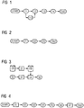
- Figure 1 shows a novel processing pipeline, which consists of three parts (which may be represented as three method steps):
- a first step 11 the DBT volume is detected or read in and in the last step 14 the result data with a probability map is provided and may be displayed on a screen.
- the incoming DBT volume is pre-segmented and pre-classified into fatty or dense breast to enable further class-specific processing/modeling.
- lesion candidates are localized, i.e. the parameters of their corresponding bounding boxes are inferred.
- probabilities for belonging to relevant anatomical structures for diagnosis e.g. lesions, ducts, vessels are inferred for each voxel within the volume.
- the result is a set of 3D probabilistic maps that can be overlaid onto the original DBT volume, used to provide regions of interests or additional information to the radiologist, and integrated as semantic information for further processing such as improved visualization, automatic reporting or decision support.
- the DBT volume is detected or read in.
- the read-in data is pre-segmented and in step 23 the image data (voxel wise) are classified in dense and fatty tissue.
- the goal is to generate from the given DBT volume tissue channels that encode the probability of belonging to following structures:
- This pre-segmentation 22 is generated using following processing pipeline.
- Extraction of breast mask this is performed using thresholding to separate tissues from air and keeping the largest connected component.
- Extraction of skin line this is done by computing the Euclidean distance to the mask boundary, a first skin region can be extracted using a pre-defined threshold. This is then refined by keeping only high curvatures regions within this region, curvature being defined as negatives value within Laplacian of Gaussian responses.
- Extraction of high density and salient tissues this is done by using thresholding on Laplacian of Gaussian response or any other type of blob filters. All voxels that belong to high density tissues and that have a negative response on the Laplacian of Gaussian output are labeled as high density and salient.
- the next step in the pre-classification pipeline refers to the step of classification 23.
- the goal of this step is to classify the current case as fatty or dense breast, using the original or raw DBT volume as well as the 5 channels, generated at the previous step.
- a fully convolutional deep network (FCN) is trained taking either as input 5 channels, i.e. raw intensities and the 5 tissue channels, or 5 channels, i.e. the element-wise product of the raw intensity with the tissue channels.
- the FCN consists of several convolutional blocks of 4 layers (convolutional + leaky rectifiers + batchnorm + spatial dropout) followed by pooling layers.
- the last layer can be a sigmoid activation layer with one single neuron where values towards 0 would encode fatty breast and values towards 1 dense breast, or a softmax layer with two neurons corresponding to each class.
- the current DBT volume will be subsequently processed by models trained specifically for fatty or dense breast. This has the technical effect that the localization procedure 12 and segmentation procedure 13 may be improved significantly due to dedicated specification and determined procedure.
- the training procedure for this network is shown on Figure 3 .
- the database DB serves to store the DBT volume and the corresponding labels (fatty - dense), as processed in the previous step.
- Data augmentation 31 can be performed to increase the amount of training samples by using simple transformations such as rotation, flipping or intensity changes. This permits to prevent from overfitting in addition to regularization and dropout layers.
- the training and cross validation set may, then, be stored in the database DB, too.
- Architecture parameters 32 are fed into the FCN for network initialization 33.
- Supervised learning is depicted in Figure 3 with reference numeral 34.
- a trained classifier 35 may be provided.
- the cost function used can be in this case binary cross entropy and the optimizer for instance an Adam optimizer.
- the next step in the processing pipeline is the step of localizing 12.
- the goal of this localization stage is to detect a set of lesion candidates, i.e. to infer the 6 parameters of their bounding boxes (position x,y,z and scale sx,sy,sz). As shown Figure 4 and Figure 6 , two possible embodiments are proposed.
- the first embodiment of localizing 12 is presented in Figure 4 and consists of 5 core steps:
- the DBT volume is detected or read in and in the last step 47 the result, including a probability map is visualized on a display unit on demand for each voxel.
- Regions of interest extraction 42 are regions of interest extraction 42:
- this first step aims at generating a probability map that encodes the probability of belonging to a region of interest (ROI) represented either as a bounding box or as an ellipsoid.
- ROI region of interest
- a multi-stream deep convolutional network such as the one detailed in Figure 5 is trained end to end relating directly the raw image to a mask encoding voxel-wise the lesion bounding boxes or ellipsoid regions.
- the input is forwarded to three different streams:
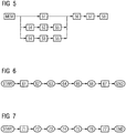
- Figure 5 shows a multi-streams multi-scale ROI extraction system with a storage unit MEM for providing the raw DBT volume.
- three different streams are used: full resolution subnet 51, an intermediate resolution subnet 52 and a coarse resolution subnet 53. It has to be noted that more than three streams could be used if needed.
- a pooling layer 54 For each resolution specific stream, a pooling layer 54 is used for generating lower resolution version of the input using either max or average pooling operations. Then at the output of each stream, a deconvolution layer followed by an activation layer such as rectified linear unit (ReLU) 55 is used for upsampling the prediction to the original resolution.
- the reference numeral 55 refers to both units (deconvolution and ReLU).
- the different outputs are then concatenated using a concatenation layer 56 and forwarded to a stack of 1x1x1 convolution layers 57 as equivalent for fully connected layers.
- the output layer 58 modeling the classification task can be a sigmoid or a softmax layer. A subnet example is shown in Figure 9 .
- Thresholding and connected components analysis applied to the map resulting from previous step to extract bounding box candidates.
- Non-local maxima suppression can be further performed especially in the case where candidates appearing at several scales are present.
- a deep convolutional neural network adjust the position of the bounding boxes obtained at the previous step in order to maximize the overlap ratio with the target bounding boxes. This can be done using a similar network as mentioned before with respect to the pre-classification step, but trained in a regression fashion, i.e. trained to predict the offset between the candidate bounding box and the target box. During training, sum of square differences or jaccard loss can be used. Moreover, we propose to regress a quality measure such as the intersection over union to infer the confidence of the network about its prediction. This measure is then used to rank the candidates to keep only the best subset, and as additional input for the next step dealing with false positive detection.
- this step is to further reduce the number of candidates to remove possible false positive.
- the goal of this step is to provide for each remaining bounding box candidate the probability to belong to a round mass, spiculated or benign lesion. This is also done by using a deep convolutional network.
- the second embodiment is presented in Figure 6 showing a multi-streams multi-scale ROI extraction and consists of 5 core steps:
- the DBT volume is detected or read in and in the last step 67 the result, including a probability map is visualized on a display unit on demand for each voxel.
- end to end deep learning to generate inverse distance map to lesion centers.
- a multi-scale i.e. multi-streams network.
- the inverse distance map can be computed using the Euclidean distance but alternatively it can integrate intensity information by using a Geodesic distance.
- a deep convolutional neural network is used to predict the offset from center location to the corners of the bounding box, deriving thereby its parameters.
- a deep convolutional network is used to classify bounding box content as either false positive or not.
- This step is similar as in the previous embodiment.
- a deep convolutional network is used to provide for each bounding box its probability to belong to a round mass, speculated or benign lesion and provide the same as intermediate result.
- this segmentation stage (step of segmenting 13) is to generate the different probabilistic channels encoding the probability of belonging to a relevant structure for cancer diagnosis.
- regular structures such as skin, vessels and ducts need to be segmented.
- a suspicious structure e.g. cancerous lesion needs to be inferred.
- segmenting 13 is shown in Figure 7 and consists of 5 core steps:
- the DBT volume is detected or read in and in the last step 77 the result, including a probability map is visualized on a display unit on demand for each voxel.
- the goal is to generate tissue probability maps that encode the probability of belonging to anatomical structures that are relevant for cancer diagnosis, e.g. skin, vessels, and ducts.
- tissue probability maps that encode the probability of belonging to anatomical structures that are relevant for cancer diagnosis, e.g. skin, vessels, and ducts.
- the rough segmentation is performed similarly as in the previous stage, 5 different types of structures channels are generated: 1) skin, 2) low density tissues, 3) high density tissues, 4) high density and salient tissues and 5) vessel-like structures.
- these 5 channels are forwarded to the next step which is tissue segmentation.
- the goal of the lesion-specific segmentation is to generate for each lesion bounding box candidate so-called lesion-specific channels.
- Each output channel encodes the probability for each voxel to belong to a specific type of lesion, namely round masses, spiculated lesions or benign lesions. Therefore, a dedicated deep convolutional network is trained for each type of lesions.
- a dedicated deep convolutional network is trained for each type of lesions.
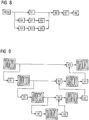
- Figure 8 shows a lesion-specific multi-streams end-to-end fully convolutional network.
- the memory MEM is adapted to provide or store the raw DBT volume data and the tissue channels.
- a pooling layer 84 is used for generating lower resolution version of the input using either max or average pooling operation.
- a deconvolution layer followed by an activation layer such as rectified linear unit (ReLU) 85 is used for'upsampling the prediction to the original resolution.
- the different outputs are then concatenated using a concatenation layer 86 and forwarded to a stack of 1x1x1 convolution layers 87 as equivalent for fully connected layers.
- the output layer 88 modeling the classification task can be a sigmoid or a softmax layer.
- a subnet example is shown on Figure 9 .
- Figure 9 shows a scale-specific subnet consisting of convolutional blocks, pooling, skip connections, deconvolution and concatenation layers.
- a convolutional layer is represented in Fig. 9 with hashed lines from left downwards to right upwards.
- Leaky Rectifiers are represented in hashed lines from left upwards to right downwards.
- a Batchnorm is represented in horizontally hashed lines and Spatial Dropout is represented in dotted manner.
- Pooling is represented in Fig. 9 via reference numeral 91, a deconvolution + rectified linear unit (ReLU) together have reference numeral 92 and the concatenation layer is depicted in Fig. 9 with numeral 93.
- ReLU rectified linear unit
- a U-net type of architecture is used where skip connections are used to feed earlier output at higher resolutions at later stage. This has been shown to permit to increase the quality of predictions in terms of resolution, which is crucial for segmentation. Further skip connections can be also used to learn residuals when more convolutional blocks are used.
- DBT volumes the tissue channels and their associated ground truth segmentation masks
- the whole network is trained in an end to end fashion, i.e. for a given tuple ([DBT,TissueChannels],Ground Truth), the output of the network Pred is compared directly to the whole mask GT (GT stands for Ground Truth). While this permits to take into account the context of the whole volume, it requires a careful choice for the cost function to optimize.
- Tversky GT Pred 1 ⁇ Pred ⁇ GT Pred ⁇ GT + ⁇ . Pred ⁇ GT + 1 ⁇ ⁇ . GT ⁇ Pred
- Tversky global ⁇ c ⁇ classes ⁇ c .
- refinement of the lesion-specific prediction channels is performed by using approaches that models neighborhood dependencies such as Markov random fields (MRF) or Conditional random fields (CRF).
- MRF Markov random fields
- CRF Conditional random fields
- CRF is used as their pairwise terms conditioned on the original volume permits usually to achieve better segmentation performance.
- the goal of this optional step is to classify the current candidate into one of the different lesion class. This is done by using a fully convolutional network similar to the one described above with respect to the step of pre-classification.
- the output is a probability distribution over the different lesion classes in the form of a class histogram.
- class probabilities are available from both localization and segmentation step, these can be averaged to provide the factors P(c). While model averaging permits to achieve better results, using class probabilities to weight their contributions permits to give more weight to the channel coming from the most probable lesion-specific model.
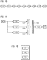
- FIG. 10 Another preferred embodiment for the segmentation step 13 is shown in Figure 10 and consists of 5 core steps:
- the DBT volume is detected or read in and in the last step 107 the result, including a probability map is visualized on a display unit on demand for each voxel.
- This step is similar to the one described in the previous section.
- the goal is to generate a compact representation for the considered region of interest.
- a global signature is generated for the considered candidate region of interest by concatenating the lesion-specific signatures into one large vector as shown in Figure 11 .
- Figure 11 depicts a system for extraction of compact signatures from candidate region of interest (ROI) and retrieval of most similar regions from a database of ROIs with corresponding segmentation masks.
- Memory MEM stores the raw DBT volume and four (4) tissue channels.
- a first lesion-specific encoder 1101, a second lesion-specific encoder 1102 and a third lesion-specific encoder 1103 are used, which serve to provide lesion specific-signatures 1104 and a global signature 1105.
- 1106 most similar regions are retrieved.
- the result may be stored in the database DB by encoding ROIs and corresponding segmentation masks.
- a set of N similar regions of interest and their corresponding ground truth segmentation masks are retrieved from a large database.
- their global signatures can be compared by using L2, L1 or L0 norm, i.e. Euclidean, Manhattan or Hamming distance.
- M being the ground truth mask of a given retrieved example.
- Figure 12 shows a system for segmentation of anatomical structures according to a preferred embodiment, comprising at least a localizer 122 and a segmentation unit 123.
- the localizer 122 is adapted to execute the localizing pipeline 12 and the segmentation unit is adapted to execute the segmentation pipeline 13, mentioned above with respect to Fig. 1 .
- the system additionally comprises a pre-classifier 121, which is adapted to execute the pre-classification 112, mentioned above.
- the present invention refers to a fully automatic pipeline based on deep learning that permits - given a DBT volume - to generate 3D maps encoding for each voxel the probability of belonging to anatomical structures that are relevant for breast cancer diagnosis.
- the proposed approach embodies a crucial part for supporting radiologists in their increasing workload when working with DBT images. Indeed, as described in the previous section, the resulting 3D probabilistic maps can be overlaid onto the original DBT volume, used to provide regions of interests or additional information to the radiologist, and integrated as semantic information for further processing such as improved visualization, automatic reporting or decision support.
- All method steps can be implemented by means that are adapted for performing the corresponding method step. All functions that are performed by particular means or apparatus modules can be implemented as a method step of a corresponding method, respectively.
Landscapes
- Engineering & Computer Science (AREA)
- Physics & Mathematics (AREA)
- Health & Medical Sciences (AREA)
- Life Sciences & Earth Sciences (AREA)
- Theoretical Computer Science (AREA)
- Medical Informatics (AREA)
- General Physics & Mathematics (AREA)
- Computer Vision & Pattern Recognition (AREA)
- General Health & Medical Sciences (AREA)
- Nuclear Medicine, Radiotherapy & Molecular Imaging (AREA)
- Radiology & Medical Imaging (AREA)
- Biomedical Technology (AREA)
- Surgery (AREA)
- Veterinary Medicine (AREA)
- Biophysics (AREA)
- High Energy & Nuclear Physics (AREA)
- Optics & Photonics (AREA)
- Pathology (AREA)
- Public Health (AREA)
- Heart & Thoracic Surgery (AREA)
- Molecular Biology (AREA)
- Animal Behavior & Ethology (AREA)
- Databases & Information Systems (AREA)
- Data Mining & Analysis (AREA)
- Software Systems (AREA)
- Mathematical Optimization (AREA)
- Pure & Applied Mathematics (AREA)
- Mathematical Analysis (AREA)
- Mathematical Physics (AREA)
- Computational Mathematics (AREA)
- General Engineering & Computer Science (AREA)
- Probability & Statistics with Applications (AREA)
- Quality & Reliability (AREA)
- Algebra (AREA)
- Oral & Maxillofacial Surgery (AREA)
- Dentistry (AREA)
- Pulmonology (AREA)
- Apparatus For Radiation Diagnosis (AREA)
- Image Analysis (AREA)
Abstract
Description
- The present invention relates to a method, apparatus and product for processing three-dimensional digital breast tomosynthesis images for cancer diagnosis.
- Conventional mammography techniques rely on a pair of two-dimensional X-ray images of the female breast, taken from two different directions with the breast being compressed in different ways. The breast tissue to be examined is compressed and held between two compression plates to ensure that the entire breast tissue is viewed. Nowadays, full-field digital mammography systems (FFDM) are commonly used. However, digital mammography is plagued by low sensitivity and high false-positive rates caused by low-contrast characteristics and image noise, while still having the problem of overlapping structures common to traditional projection radiographs. Reported false-negative rates on mammographic screening range from 8% to 66% and depend on a multitude of factors including, but not limited to, age, breast density, tumor sub-type, and misinterpretation of subtle malignant mammographic findings.
- Due to these disadvantages of two-dimensional mammographic imaging, recently, digital breast tomosynthesis (DBT) is increasingly replacing common 2D mammography for differential diagnoses and is in discussion for screening. DBT images provide more information than regular FFDM images for early detection of anomalies and cancer. DBT provides 3D image volumes of the compressed breast that are reconstructed from multiple 2D projections acquired at varying angles. Being a 3D imaging modality DBT naturally allows superior spatial localization of suspicious lesions. A mediolateral-oblique (MLO) and, typically, a second cranio-caudal (CC) scan is acquired during an examination. The breast is compressed differently for MLO and CC scans. For reporting and surgical planning it is common clinical practice to mark the lesions in the scans and to communicate the rough localization of suspicious findings in the uncompressed breast via schematic 2D drawings. The latter naturally suffers from inaccuracies and can often only be dissolved by additional, potentially ionizing and costly, imaging. Providing more accurate lesion localization in the un-compressed breast, e.g., in terms of a 3D rendering view, without additional imaging has the potential not only to facilitate surgical planning and related procedures, e.g., placing pre-operative markers but also to resolve the problem of ambiguous mapping of multiple similarly looking lesions between CC, MLO DBT and FFDM scans including previously acquired images.
- Recent advances in machine vision based on deep learning, e.g. deep neural networks or deep convolutional neural networks, and massive training sets such as ImageNet gathered a lot of attention and attracted a lot of investment. Indeed, they are reaching now so-called superhuman performances for tasks such as natural image classification. Such technology could also be applied to the medical image analysis field, for example for the detection and segmentation of organs and anatomical structures. While great improvements can be expected, the detection of pathologies such as lesions or tumors still remains challenging due to their heterogeneous appearance and shape, and the need of massive databases annotated by expert radiologists. The benefits of applying such systems in the context of screening could be however tremendous, as it would support radiologists in their increasing workload.
- By providing 3D information on the breast anatomy, a DBT image reduces dramatically the amount of false positive findings arising from the superposition of normal fibroglandular tissues in conventional 2D mammography. On the other hand, DBT increases the workload of radiologist due to the larger amount of slices to be inspected.
- In state of the art it is known to apply artificial neural networks (ANN) in the context of breast lesion detection and segmentation. In the respect it is referred to the paper "Dhungel, N., Carneiro, G., Bradley, A.P, A deep learning approach for the analysis of masses in mammograms with minimal user intervention. Medical Image Analysis (2017)". This paper proposes a semi-automatic 3 stages approach: 1) detection stage, 2) user feedback stage and 3) segmentation stage. In the detection stage, authors combine deep belief network and Gaussian mixture model as candidate generator followed by a false positive reduction approach based on convolutional neural network and Random forest. Subsequently, refinement is performed using Bayesian optimization. In the second stage, lesion candidates are shown to the user who has to provide feedback by further rejecting false positives. In the third stage, segmentation is performed by using deep structured learning model followed by a level-set refinement approach. This approach does not refer to 3D DBT data but only to mammograms and necessitates user feedback of the training data set, which may demand high resources.
- Thus, it is an object of the present invention to improve and automate the processing of DBT images for cancer diagnosis and to augment and extend the DBT image with processed semantic data for improved visualization.
- To make diagnosis and lesion search more efficient, a solution is to provide an automated image analysis approach that would permit the radiologists to navigate efficiently through the data towards the most relevant locations.
- This object is solved by subject-matter according to the independent claims. Beneficial embodiments are subject to the dependent claims the description and the figures.
- According to a first aspect this object is solved by a method for segmenting different types of breast structures, including pathologic structures, like cancerous lesions and calcifications and regular structures, like vessels, glandular structures, the nipple, the pectoralis muscle and skin, that are relevant for cancer diagnosis in a DBT volume, comprising the steps of:
- Localizing a set of structures in the DBT volume by using at least one multi-stream deep image-to-image network, taking as input at least one image channel;
- Segmenting the localized structures by calculating a probability for belonging to a specific type of structure for each voxel in the DBT volume by using at least one multi-stream deep image-to-image network, taking as input at least one image channel for providing a three-dimensional probabilistic map.
- In the following the terms used within this application are defined in more detail.
- The segmentation does not only refer to cancerous structures, like lesions. It is a major advantage of the present invention that not only potential pathologic structures like lesions and calcifications are detected, but also regular (healthy) structures like vessels, skin, muscle and nipple. This has the advantage that navigation and orientation and image processing may be improved.
- The method has as input a three-dimensional digital breast tomosynthesis (DBT) volume. The output is an augmented image including a three-dimensional probabilistic map, encoding for each voxel the probability of belonging to anatomical structures that are relevant for cancer diagnosis (as mentioned above healthy and pathological structures may be both relevant). The output may be integrated into the original DBT volume or may serve to create a new two- or three-dimension view of the original DBT volume, summarizing the most relevant structures (for example, by enhancing the contours or by highlighting the content etc.).
- Localizing and segmenting both refer to a multi-step pipeline. The pipeline may be implemented in software on a computer by using at least one multi-stream deep image-to-image network. Localizing and segmenting may be implemented in hardware structures, in a localizer and in a segmentation unit.
- The network is a deep learning (also known as deep structured learning or hierarchical learning) artificial neural network (ANN). It may contain more than one hidden layer. The network uses machine learning methods based on learning data representations, as opposed to task specific algorithms. Preferably, the learning is supervised or partially supervised. The network may also be composed of one or more convolutional layers. In particular, it may consist of a fully convolutional network that is trained in an end to end fashion with optionally skip connections. It uses pooling and deconvolutional layers. The network takes as input an image/DBT volume and provides as output one or multiple images/volumes. The network is trained in an end-to-end fashion, e.g. by comparing directly the whole produced output with the whole ground truth, e.g. the whole segmentation mask (instead of considering patches and labels). It might be trained using deep supervision where the loss function consists of a combination of costs computed at each level of resolution.
- The probabilistic map may include datasets referring to different types of structures (lesion, calcification, anatomical structures etc.). The map includes for each voxel a probability measure for belonging to one of these different structures. For example, for a voxel the following output may be provided and displayed in coded form (highlighted differently in the respective image): 80% tumor tissue, 15% dense tissue and 5% fatty tissue.
- In a preferred embodiment the method further comprises the following step prior to the step of localizing:
- Pre-Classifying the DBT volume in order to provide a pre-classification result, which distinguishes between dense and fatty breast.
- In a preferred embodiment of the present invention, the step of localizing is based on a five-step procedure, comprising:
- Extracting regions of interest;
- Generating bounding boxes;
- Adjusting the generated bounding box;
- Rejecting false positive structures;
- Pre-classifying different regions of interest in the DBT volume by providing a class probability to belong to a specific type of structure, including, round mass, spiculated or benign lesions by using a deep convolutional neural network. In this step another kind of network (not a multi-stream deep image-to-image network) is used, because the output is a label (not an image, volume or channel).
- In a preferred embodiment, the step of localizing additionally includes a pre-segmentation as a first step (which is similar to the segmentation, which will be explained below).
- In another preferred embodiment of the present invention, the step of localizing is executed by using an image-to-image multi-scale multi-streams deep learning network, taking as input at least one image channel to generate inverse distance maps to a center of each structure of interest, such as a lesion. The inverse distance maps may be Euclidean and/or geodesic and/or may be computed according to a center of each structure of interest, such as a lesion. The inverse distance maps may also be signed distance maps according to a boundary of the structure of interest.
- In another preferred embodiment of the present invention, for training the network a Tversky loss function (which is a more generic form of the DICE loss function) may be used, that permits to weight the compromise between false positives and false negatives.
- In another preferred embodiment of the present invention, the DBT volume is forwarded as input to multiple - and at least three different - resolution-specific streams and as an output of each of the resolution-specific streams a set of deconvolution layers is followed by activation layers and wherein different outputs of the resolution-specific streams are concatenated using a concatenation layer. By processing the DBT volume at different resolution, this permits to detect and segment structures at different scales. This is very important as lesions for instance show high variation in size.
- In another preferred embodiment of the present invention, the step of localizing is based on generating a bounding box or an ellipsoid for the set of 3D structures (for example ROIs, referring to a possible structure of interest or lesion).
- In another preferred embodiment of the present invention, the step of segmenting is based on procedure, comprising:
- Pre-segmenting different types of structures, represented in the DBT volume, including regular structures like skin, vessels, ducts and suspicious structures by means of advanced image processing approaches, including filters, like for example Laplacian or Gaussian or Hessian-based filers, and multi-stream deep image-to-image network;
- Structure-specific segmentation by generating for each lesion bounding box candidate a structure-specific prediction channel, encoding the probability for each voxel to belong to a specific type of structure, including a regular or a suspicious structure;
- Structure-specific refinement of the structure-specific prediction channels by modeling neighborhood dependencies.
Preferably, the step of refining the calculated probabilities is executed by using Markov random fields or conditional random fields. This can be also done by retrieving similar region/structure of interests plus corresponding masks from a database and aggregating their masks. - Aggregating the calculated different structure-specific prediction channels by fusing the different channels and probabilities and class probabilities by applying:

- In another preferred embodiment of the present invention, the step of segmenting further comprises:
- Calculating a class probability by classifying the candidate structure into a specific structure class by using a multi-stream deep image-to-image network.
- In another preferred embodiment of the present invention, the method further comprises the step of:
- providing result data for each voxel of the DBT volume, encoding the probability of belonging to a specific type of structure for breast cancer diagnosis. In another embodiment, the result data may further comprise other parameters, like a volume of the structure or lesion, the shape of the structure and/or its distance to nipple and pectoralis. The result data may be overlaid onto the original DBT volume. The result may also be used for creating a new 2D or 3D view of the original DBT volume summarizing and/or enhancing the most relevant information.
- In another aspect, the present invention refers to a computer program loadable into a memory of a digital computer comprising software code portions for executing the method according to one of the method claims above, when the computer program is run on the computer. The computer program may also be executed on a plurality of network nodes as a distributed system. The computer program may be stored on a computer readable medium and/or may be downloaded form a server.
- According to another aspect the object is solved by an apparatus for segmenting different types of structures, including cancerous lesions and regular structures like vessels and skin, in a DBT volume, comprising:
- A localizer for localizing a set of structures in the DBT volume by using a multi-stream deep convolutional neural network;
- A segmentation unit for segmenting the localized structures by calculating a probability for belonging to a specific type of structure for each voxel in the DBT volume by using a deep convolutional neural network for providing a three-dimensional probabilistic map.
- According to a preferred embodiment the apparatus further comprises:
- A pre-classification unit which is adapted for providing a pre-classification result by differentiating in the DBT volume between dense and fatty breast,
- Embodiments of the invention are shown in the figures and explained in the following detailed description.
- Fig. 1
- shows a schematic view of a method for DBT image segmentation based on a preferred embodiment of the present invention;
- Fig. 2
- shows a schematic view of a method for pre-classification of a DBT image in dense versus fatty tissue based on a preferred embodiment of the present invention;
- Fig. 3
- refers to a schematic representation of a system for training a fully convolutional network using data augmentation;
- Fig. 4
- shows a preferred embodiment of a localizing pipeline with 5 steps;
- Fig. 5
- shows a multi-streams multi-scale region of interest extraction according to a preferred embodiment of the present invention;
- Fig. 6
- shows another embodiment of a localizing pipeline with 5 core steps;
- Fig. 7
- depicts a segmentation phase with 5 core steps according to a preferred embodiment of the present invention;
- Fig. 8
- shows a multi-streams lesion or structure specific end to end fully convolutional network according to a preferred embodiment of the present invention;
- Fig. 9
- refers to a schematic representation of a scale specific subnet consisting of convolutional blocks, pooling, skip connections, deconvolutions and concatenation layers;
- Fig. 10
- depicts a segmentation phase with 5 core steps according to another preferred embodiment of the present invention;
- Fig. 11
- shows in a schematic manner a system for extraction of compact signatures form candidate regions of interest of most similar regions from a database of ROIs with corresponding segmentations masks according a preferred embodiment of the present invention and
- Fig. 12
- is a schematic overview figure of a localization and segmentation system according to a preferred embodiment of the present invention.
- The present invention refers to a novel processing pipeline based on deep learning for the generation of 3D maps that encodes for each voxel the probability of belonging to a relevant anatomical structure for breast cancer diagnosis given a DBT volume.
- The term "structure" may in one embodiment refer to lesions or potential lesions, like tumors, calcifications and others. According to another embodiment, a structure may refer to regular tissue in the breast, like vessels, skin, milk ducts and the nipple. For radiologists it is very helpful to provide such tissue structures in the image, which may be used as a kind of orientation or navigation in the image data. For example, the nipple serves as an important landmark or marker in the image for measuring distances.
-
Figure 1 shows a novel processing pipeline, which consists of three parts (which may be represented as three method steps): - 1) an (optional)
pre-classification stage 112, - 2) a
localization stage 12 for executing the step of localizing and - 3) a segmentation stage 13 for executing the step of segmenting.
- In a
first step 11 the DBT volume is detected or read in and in the last step 14 the result data with a probability map is provided and may be displayed on a screen. - In the first stage, the incoming DBT volume is pre-segmented and pre-classified into fatty or dense breast to enable further class-specific processing/modeling. In the second stage, lesion candidates are localized, i.e. the parameters of their corresponding bounding boxes are inferred. Finally, and in the third stage, probabilities for belonging to relevant anatomical structures for diagnosis, e.g. lesions, ducts, vessels are inferred for each voxel within the volume. The result is a set of 3D probabilistic maps that can be overlaid onto the original DBT volume, used to provide regions of interests or additional information to the radiologist, and integrated as semantic information for further processing such as improved visualization, automatic reporting or decision support.
- As depicted in
Figure 1 and explained above, it is preferred to include a pre-classification procedure. This has the technical effect of distinguishing between dense vs fatty breast. However, this procedure is optional. In this stage, the goal is to determine whether the current case shows characteristics of a fatty breast or consists mostly of dense tissues. Within fatty breasts, there are almost no dense structures, so that if lesions are present, they are clearly visible within the image, as they will appear as high density structures. In the case of dense breast, as the major part of the tissues show high density, finding lesions becomes very challenging, even for experienced observers. For this reason, we propose as a very first step to distinguish between dense and fatty cases, so that models (for localization and for segmentation) specific to dense or fatty cases can be applied for subsequent analysis. For deciding whether a case belongs to the fatty or dense breast class, we propose an approach described inFigure 2 . - As shown in
Figure 2 , in thefirst step 21 the DBT volume is detected or read in. Instep 22 the read-in data is pre-segmented and instep 23 the image data (voxel wise) are classified in dense and fatty tissue. The goal is to generate from the given DBT volume tissue channels that encode the probability of belonging to following structures: - 1) skin,
- 2) low density tissues,
- 3) high density tissues,
- 4) high density and salient tissues and
- 5) vessel-like tissues.
- This
pre-segmentation 22 is generated using following processing pipeline. - Extraction of breast mask: this is performed using thresholding to separate tissues from air and keeping the largest connected component.
- Extraction of skin line: this is done by computing the Euclidean distance to the mask boundary, a first skin region can be extracted using a pre-defined threshold. This is then refined by keeping only high curvatures regions within this region, curvature being defined as negatives value within Laplacian of Gaussian responses.
- Extraction of low density tissues: this is done by using thresholding (that can be learned from data) within breast mask. Voxels below this threshold are labeled as low density.
- Extraction of high density tissues: this is done by using the same threshold than in the previous step. Voxels above this threshold are labeled as high density.
- Extraction of high density and salient tissues: this is done by using thresholding on Laplacian of Gaussian response or any other type of blob filters. All voxels that belong to high density tissues and that have a negative response on the Laplacian of Gaussian output are labeled as high density and salient.
- Extraction of vessel-like tissues: this is done by using filtering approaches that enhances linear structures based on the Hessian matrix and its eigenvectors, such as determinant of hessians.
- As depicted in
Figure 2 , the next step in the pre-classification pipeline refers to the step ofclassification 23. The goal of this step is to classify the current case as fatty or dense breast, using the original or raw DBT volume as well as the 5 channels, generated at the previous step. To this end, a fully convolutional deep network (FCN) is trained taking either as input 5 channels, i.e. raw intensities and the 5 tissue channels, or 5 channels, i.e. the element-wise product of the raw intensity with the tissue channels. The FCN consists of several convolutional blocks of 4 layers (convolutional + leaky rectifiers + batchnorm + spatial dropout) followed by pooling layers. At the very end, a stack of convolutional layers with 1x1x1 kernels are used as equivalent for fully connected layers. To model the classification task, the last layer can be a sigmoid activation layer with one single neuron where values towards 0 would encode fatty breast and values towards 1 dense breast, or a softmax layer with two neurons corresponding to each class. - Depending on the (binary: fatty versus dense) output of this step, the current DBT volume will be subsequently processed by models trained specifically for fatty or dense breast. This has the technical effect that the
localization procedure 12 and segmentation procedure 13 may be improved significantly due to dedicated specification and determined procedure. - The training procedure for this network is shown on
Figure 3 . The database DB serves to store the DBT volume and the corresponding labels (fatty - dense), as processed in the previous step. Data augmentation 31 can be performed to increase the amount of training samples by using simple transformations such as rotation, flipping or intensity changes. This permits to prevent from overfitting in addition to regularization and dropout layers. The training and cross validation set may, then, be stored in the database DB, too.Architecture parameters 32 are fed into the FCN fornetwork initialization 33. Supervised learning is depicted inFigure 3 withreference numeral 34. A trainedclassifier 35 may be provided. The cost function used can be in this case binary cross entropy and the optimizer for instance an Adam optimizer. - After having pre-classified the DBT volume, the next step in the processing pipeline, shown in
Fig. 1 , is the step of localizing 12. - The goal of this localization stage (step of localizing 12) is to detect a set of lesion candidates, i.e. to infer the 6 parameters of their bounding boxes (position x,y,z and scale sx,sy,sz). As shown
Figure 4 andFigure 6 , two possible embodiments are proposed. - The first embodiment of localizing 12 is presented in
Figure 4 and consists of 5 core steps: - 1) regions of interest extraction, 42
- 2) bounding box generation, 43
- 3) bounding box adjustments, 44
- 4) false
positive rejection 45 and - 5)
pre-classification 46. - In the
first step 41 the DBT volume is detected or read in and in thelast step 47 the result, including a probability map is visualized on a display unit on demand for each voxel. - The first embodiment of a localization processing pipeline is explained in more detail below, with respect to
Figure 4 . - Given the raw DBT volume, this first step aims at generating a probability map that encodes the probability of belonging to a region of interest (ROI) represented either as a bounding box or as an ellipsoid. To this end, a multi-stream deep convolutional network such as the one detailed in
Figure 5 is trained end to end relating directly the raw image to a mask encoding voxel-wise the lesion bounding boxes or ellipsoid regions. In this example, the input is forwarded to three different streams: - i) full resolution subnet,
- ii) intermediate resolution subnet and
- iii)the coarse resolution subnet.
-
Figure 5 shows a multi-streams multi-scale ROI extraction system with a storage unit MEM for providing the raw DBT volume. In this example three different streams are used:full resolution subnet 51, anintermediate resolution subnet 52 and acoarse resolution subnet 53. It has to be noted that more than three streams could be used if needed. - For each resolution specific stream, a
pooling layer 54 is used for generating lower resolution version of the input using either max or average pooling operations. Then at the output of each stream, a deconvolution layer followed by an activation layer such as rectified linear unit (ReLU) 55 is used for upsampling the prediction to the original resolution. Thereference numeral 55 refers to both units (deconvolution and ReLU). The different outputs are then concatenated using aconcatenation layer 56 and forwarded to a stack of 1x1x1 convolution layers 57 as equivalent for fully connected layers. Theoutput layer 58 modeling the classification task can be a sigmoid or a softmax layer. A subnet example is shown inFigure 9 . - Thresholding and connected components analysis applied to the map resulting from previous step to extract bounding box candidates. Non-local maxima suppression can be further performed especially in the case where candidates appearing at several scales are present.
- Using a deep convolutional neural network, adjust the position of the bounding boxes obtained at the previous step in order to maximize the overlap ratio with the target bounding boxes. This can be done using a similar network as mentioned before with respect to the pre-classification step, but trained in a regression fashion, i.e. trained to predict the offset between the candidate bounding box and the target box. During training, sum of square differences or jaccard loss can be used. Moreover, we propose to regress a quality measure such as the intersection over union to infer the confidence of the network about its prediction. This measure is then used to rank the candidates to keep only the best subset, and as additional input for the next step dealing with false positive detection.
- The goal of this step is to further reduce the number of candidates to remove possible false positive. We propose to use here a well a deep convolutional network as described by
Figure 3 . Note that to train this network, one needs to mine so called hard negatives, which are difficult to distinguish from lesion ROIs. In addition to the raw DBT volume, this network takes the predicted intersection over union from the previous step. This estimate can be concatenated at a deeper level within the network. - Given the raw DBT volume, the goal of this step is to provide for each remaining bounding box candidate the probability to belong to a round mass, spiculated or benign lesion. This is also done by using a deep convolutional network.
- The second embodiment is presented in
Figure 6 showing a multi-streams multi-scale ROI extraction and consists of 5 core steps: - 1) pre-localization of
lesion candidates 62, - 2) candidates extraction 63,
- 3) localization 64,
- 4) false positive rejection 65 and
- 5) pre-classification 66.
- In the
first step 61 the DBT volume is detected or read in and in thelast step 67 the result, including a probability map is visualized on a display unit on demand for each voxel. - In this embodiment, it is proposed to use end to end deep learning to generate inverse distance map to lesion centers. As in the previous embodiment, it is proposed to use a multi-scale, i.e. multi-streams network. Note that the inverse distance map can be computed using the Euclidean distance but alternatively it can integrate intensity information by using a Geodesic distance.
- Mean shift clustering or thresholding and connected components analysis applied to the map resulting from previous step as preferred embodiment. Non-local maxima suppression could be added if several scales are used.
- Similarly, as in the previous embodiment, a deep convolutional neural network is used to predict the offset from center location to the corners of the bounding box, deriving thereby its parameters. As in the previous embodiment, we propose to regress the intersection over union value as quality measure.
- This step is similar as in the previous embodiment. A deep convolutional network is used to classify bounding box content as either false positive or not.
- This step is similar as in the previous embodiment. A deep convolutional network is used to provide for each bounding box its probability to belong to a round mass, speculated or benign lesion and provide the same as intermediate result.
- After having finished the localization procedure 12 (shown in
Figure 1 ), the intermediate result of this procedure is forwarded and used in the next procedure, the segmentation procedure 13, which will be describe below with reference toFigure 7 . - The goal of this segmentation stage (step of segmenting 13) is to generate the different probabilistic channels encoding the probability of belonging to a relevant structure for cancer diagnosis. First, regular structures such as skin, vessels and ducts need to be segmented. Second, using the detected bounding box from the previous stage, the probability of the voxels to belong to a suspicious structure, e.g. cancerous lesion needs to be inferred. As shown on
Figure 7 andFigure 10 , we propose two possible embodiments. - The preferred embodiment of segmenting 13 is shown in
Figure 7 and consists of 5 core steps: - 1)
pre-segmentation 72, - 2) lesion-
specific segmentation 73, - 3) lesion-
specific refinement 74, - 4) classification 75 as an optional step and
- 5) aggregation 76.
- In the
first step 71 the DBT volume is detected or read in and in thelast step 77 the result, including a probability map is visualized on a display unit on demand for each voxel. - In this stage, the goal is to generate tissue probability maps that encode the probability of belonging to anatomical structures that are relevant for cancer diagnosis, e.g. skin, vessels, and ducts. To this end, we propose a two-steps approach, consisting of a rough pre-segmentation and tissue segmentation. The rough segmentation is performed similarly as in the previous stage, 5 different types of structures channels are generated: 1) skin, 2) low density tissues, 3) high density tissues, 4) high density and salient tissues and 5) vessel-like structures. Providing a first rough voxel-wise encoding, these 5 channels are forwarded to the next step which is tissue segmentation.
- In this second step, we propose to use a deep convolutional network trained in an end to end fashion such as the one presented in
Figure 9 . It provides multi-class output by using a softmax layer as very last layer. It can be trained by using multi-class cross entropy or some more advanced segmentation losses as well will describe in the next section. - Given the raw DBT volume and the tissue channels computed in the previous step, the goal of the lesion-specific segmentation is to generate for each lesion bounding box candidate so-called lesion-specific channels. Each output channel encodes the probability for each voxel to belong to a specific type of lesion, namely round masses, spiculated lesions or benign lesions. Therefore, a dedicated deep convolutional network is trained for each type of lesions. To provide voxel-wise predictions at full resolution, we propose to use a multi-streams end-to-end fully convolutional network as shown on
Figure 8 . -
Figure 8 shows a lesion-specific multi-streams end-to-end fully convolutional network. The memory MEM is adapted to provide or store the raw DBT volume data and the tissue channels. - As already described in the localization stage, all channels are forwarded to three
different streams - i)
full resolution subnet 81, - ii)
intermediate resolution subnet 82 and - iii)the
coarse resolution subnet 83. - Note that more than three streams could be used if needed. For each resolution specific stream, a
pooling layer 84 is used for generating lower resolution version of the input using either max or average pooling operation. Then at the output of each stream, a deconvolution layer followed by an activation layer such as rectified linear unit (ReLU) 85 is used for'upsampling the prediction to the original resolution. The different outputs are then concatenated using a concatenation layer 86 and forwarded to a stack of 1x1x1 convolution layers 87 as equivalent for fully connected layers. Theoutput layer 88 modeling the classification task can be a sigmoid or a softmax layer. A subnet example is shown onFigure 9 . -
Figure 9 shows a scale-specific subnet consisting of convolutional blocks, pooling, skip connections, deconvolution and concatenation layers. A convolutional layer is represented inFig. 9 with hashed lines from left downwards to right upwards. Leaky Rectifiers are represented in hashed lines from left upwards to right downwards. InFig. 9 a Batchnorm is represented in horizontally hashed lines and Spatial Dropout is represented in dotted manner. Pooling is represented inFig. 9 viareference numeral 91, a deconvolution + rectified linear unit (ReLU) together havereference numeral 92 and the concatenation layer is depicted inFig. 9 withnumeral 93. - In this example, a U-net type of architecture is used where skip connections are used to feed earlier output at higher resolutions at later stage. This has been shown to permit to increase the quality of predictions in terms of resolution, which is crucial for segmentation. Further skip connections can be also used to learn residuals when more convolutional blocks are used. Using DBT volumes, the tissue channels and their associated ground truth segmentation masks, the whole network is trained in an end to end fashion, i.e. for a given tuple ([DBT,TissueChannels],Ground Truth), the output of the network Pred is compared directly to the whole mask GT (GT stands for Ground Truth). While this permits to take into account the context of the whole volume, it requires a careful choice for the cost function to optimize. Indeed, if there is high imbalance between background and class of interest, binary cross entropy might be badly impacted towards the background class. To avoid this issue, we introduce a new loss function based on the Tversky index, which is a more generic version of the DICE coefficient.

- With an extra parameter α one can control the impact of false positives. If α=0.5, the Tversky index becomes the DICE coefficient. Note that this loss is differentiable, and the intersection and relative complements are computed using element-wise product operations. While this loss is perfect in the case where a lesion, e.g. foreground is present, it does not provide any valuable information for a negative case (as the intersection becomes 0). To be able to learn also from negative cases, we propose to use a weighted multi-class version of this loss:

- In this step, refinement of the lesion-specific prediction channels is performed by using approaches that models neighborhood dependencies such as Markov random fields (MRF) or Conditional random fields (CRF). In a preferred embodiment, CRF is used as their pairwise terms conditioned on the original volume permits usually to achieve better segmentation performance.
- Given the original image, e.g. a region of interest defined by a candidate bounding box, and all lesion-specific prediction channels, the goal of this optional step is to classify the current candidate into one of the different lesion class. This is done by using a fully convolutional network similar to the one described above with respect to the step of pre-classification. The output is a probability distribution over the different lesion classes in the form of a class histogram.
- Given the different lesion-specific prediction channels and the probability distribution over the different lesion classes provided by the previous step or by the classification step in the localization stage, or both, the final segmentation output is generated by fusing the different channels based on the class probabilities:

- If class probabilities are available from both localization and segmentation step, these can be averaged to provide the factors P(c). While model averaging permits to achieve better results, using class probabilities to weight their contributions permits to give more weight to the channel coming from the most probable lesion-specific model.
- Another preferred embodiment for the segmentation step 13 is shown in
Figure 10 and consists of 5 core steps: - 1) pre-segmentation 102,
- 2) lesion-
specific encoding 103, - 3)
code aggregation 104, - 4) retrieval 105 and
- 5) aggregation 106.
- In the
first step 101 the DBT volume is detected or read in and in thelast step 107 the result, including a probability map is visualized on a display unit on demand for each voxel. - This step is similar to the one described in the previous section.
- Using raw volumes and the tissue channels provided by the pre-segmentation step, the goal is to generate a compact representation for the considered region of interest. To this end, we propose to use deep convolutional auto-encoder with several convolutional blocks with 4 (four) layers and followed by pooling layers, dropout, sigmoid and a flattening layer. Looking quite similar to a fully convolutional network for classification, it ends with a flattening layer to reshape the output to a vector. This vector is a compact latent representation of the visual context contained within the region of interest. To train such a deep convolutional auto-encoder, one needs to train simultaneously a decoder network which is very similar to the encoder network with the only difference is that its role is to reconstruct the input volume + channels. One can use shared weights between encoding and decoding networks or different weights. Usually, noise is added to the input volume and channels to improve the training of the whole encoding and decoding pipeline. The loss function is based on the reconstruction error computed by comparing the original volume and its reconstruction. We propose to train such a deep convolutional auto-encoder for each type of lesions. As a result, a set of lesion-specific signatures are generated by this step.
- Given the lesion-specific signatures from the previous step, a global signature is generated for the considered candidate region of interest by concatenating the lesion-specific signatures into one large vector as shown in
Figure 11 . -
Figure 11 depicts a system for extraction of compact signatures from candidate region of interest (ROI) and retrieval of most similar regions from a database of ROIs with corresponding segmentation masks. Memory MEM stores the raw DBT volume and four (4) tissue channels. A first lesion-specific encoder 1101, a second lesion-specific encoder 1102 and a third lesion-specific encoder 1103 are used, which serve to provide lesion specific-signatures 1104 and aglobal signature 1105. In 1106 most similar regions are retrieved. The result may be stored in the database DB by encoding ROIs and corresponding segmentation masks. - Using the compact representation provided by the previous step, a set of N similar regions of interest and their corresponding ground truth segmentation masks are retrieved from a large database. To assess similarity between the candidate region and regions within the database, their global signatures can be compared by using L2, L1 or L0 norm, i.e. Euclidean, Manhattan or Hamming distance.
- Finally, a final prediction channel is created by aggregating the segmentation masks retrieved from the database. This can be simply averaged or linearly combined depending on their distance to the query candidate.

- M being the ground truth mask of a given retrieved example.
-
Figure 12 shows a system for segmentation of anatomical structures according to a preferred embodiment, comprising at least alocalizer 122 and asegmentation unit 123. Thelocalizer 122 is adapted to execute the localizingpipeline 12 and the segmentation unit is adapted to execute the segmentation pipeline 13, mentioned above with respect toFig. 1 . In the preferred embodiment, shown inFig. 12 , the system additionally comprises a pre-classifier 121, which is adapted to execute thepre-classification 112, mentioned above. - In sum, the present invention, refers to a fully automatic pipeline based on deep learning that permits - given a DBT volume - to generate 3D maps encoding for each voxel the probability of belonging to anatomical structures that are relevant for breast cancer diagnosis. The proposed approach embodies a crucial part for supporting radiologists in their increasing workload when working with DBT images. Indeed, as described in the previous section, the resulting 3D probabilistic maps can be overlaid onto the original DBT volume, used to provide regions of interests or additional information to the radiologist, and integrated as semantic information for further processing such as improved visualization, automatic reporting or decision support.
- All features discussed or shown with respect to particular embodiments of the invention can be provided in various combinations in order to simultaneously realize their positive technical effects.
- All method steps can be implemented by means that are adapted for performing the corresponding method step. All functions that are performed by particular means or apparatus modules can be implemented as a method step of a corresponding method, respectively.
- The scope of the present invention is given by the claims and is not restricted by features discussed in the description or shown in the figures.
Claims (14)
- Method for segmenting different types of breast structures, including pathologic structures and regular structures in a DBT volume, comprising the steps of:- Localizing (12) a set of structures in the DBT volume by using at least one multi-stream deep image-to-image network, taking as input at least one image channel;- Segmenting (13) the localized structures by calculating a probability for belonging to a specific type of structure for each voxel in the DBT volume by using at least one multi-stream deep image-to-image network, taking as input at least one image channel for providing a three-dimensional probabilistic map.
- Method according to claim 1, wherein the method further comprises the following step prior to the step of localizing (12):- Pre-Classifying (112) the DBT volume in order to provide a pre-classification result, which distinguishes between dense and fatty tissue and wherein the step of localizing (12) is based on the pre-classification result and is executed specifically with a specific model for dense and with a specific model for fatty tissue and/or wherein the step of segmenting (13) is based on the pre-classification result and is executed specifically with a specific model for dense and with a specific model for fatty tissue.
- Method according to claim 1 or 2, wherein the step of localizing (12) is based on a five-step procedure, comprising:- Extracting (42) regions of interest;- Generating (43) bounding boxes;- Adjusting (44) the generated bounding box;- Rejecting (45) false positive structures;- Pre-classifying (46) different regions of interest in the DBT volume by providing a class probability to belong to a specific type of structure, including, round mass, spiculated or benign lesions by using a deep convolutional neural network.
- Method according to any of the preceding claims, wherein the step of localizing (12) is executed by using an image-to-image multi-scale multi-streams deep learning network, taking as input at least one image channel to generate inverse distance maps to a center of each structure of interest, such as a lesion.
- Method according to the preceding claim 4, wherein the inverse distance maps are Euclidean and/or geodesic and/or are computed according to the center of each structure of interest, such as a lesion, and/or be signed distance maps according to a boundary of the structure of interest.
- Method according to any of the preceding claims, wherein for training the network a Tversky loss function and, in particular, a DICE loss function is used.
- Method according to any of the preceding claims, wherein the DBT volume is forwarded as input to multiple resolution-specific streams and as an output of each of the resolution-specific streams a set of deconvolution layers is followed by activation layers and wherein different outputs of the resolution-specific streams are concatenated using a concatenation layer.
- Method according to any of the preceding claims, wherein the step of localizing (12) is based on generating (43) a bounding box or an ellipsoid for the set of 3D structures.
- Method according to any of the preceding claims, wherein the step of segmenting (13) is based on procedure, comprising:- Pre-segmenting (72) different types of structures, represented in the DBT volume, including regular structures like skin, vessels, ducts and suspicious structures by means of filters and multi-stream deep image-to-image network;- Structure-specific segmentation (73) by generating for each lesion bounding box candidate a structure-specific prediction channel, encoding the probability for each voxel to belong to a specific type of structure, including a regular or a suspicious structure;- Structure-specific refinement (74) of the structure-specific prediction channels by modeling neighborhood dependencies;- Aggregating (76) the calculated different structure-specific prediction channels by fusing the different channels and probabilities and class probabilities by applying:

- Method according to any of the preceding claims, wherein the step of segmenting (13) further comprises:- Calculating (75) a class probability by classifying the candidate structure into a specific structure class by using a multi-stream deep image-to-image network.
- Method according to any of the preceding claims, wherein the method further comprises the step of:- providing (14) result data for each voxel of the DBT volume, encoding the probability of belonging to a specific type of structure for breast cancer diagnosis.
- Computer program loadable into a memory of a digital computer comprising software code portions for executing the method according to one of the method claims above, when the computer program is run on the computer.
- Apparatus for segmenting different types of structures, including cancerous lesions and regular structures like vessels and skin, in a DBT volume, comprising:- A localizer (122) for localizing a set of structures in the DBT volume by using a multi-stream deep convolutional neural network;- A segmentation unit (123) for segmenting the localized structures by calculating a probability for belonging to a specific type of structure for each voxel in the DBT volume by using a deep convolutional neural network for providing a three-dimensional probabilistic map.
- Apparatus according to the apparatus claim, wherein the apparatus further comprises:- A pre-classification unit (121) which is adapted for providing a pre-classification result by differentiating in the DBT volume between dense and fatty tissue wherein the pre-classification result is forwarded to the localizer (122) and to the segmentation unit (123) for specific processing of dense and fatty tissue.
Priority Applications (2)
| Application Number | Priority Date | Filing Date | Title |
|---|---|---|---|
| EP17464009.4A EP3432263B1 (en) | 2017-07-17 | 2017-07-17 | Semantic segmentation for cancer detection in digital breast tomosynthesis |
| US16/033,332 US10779785B2 (en) | 2017-07-17 | 2018-07-12 | Semantic segmentation for cancer detection in digital breast tomosynthesis |
Applications Claiming Priority (1)
| Application Number | Priority Date | Filing Date | Title |
|---|---|---|---|
| EP17464009.4A EP3432263B1 (en) | 2017-07-17 | 2017-07-17 | Semantic segmentation for cancer detection in digital breast tomosynthesis |
Publications (2)
| Publication Number | Publication Date |
|---|---|
| EP3432263A1 true EP3432263A1 (en) | 2019-01-23 |
| EP3432263B1 EP3432263B1 (en) | 2020-09-16 |
Family
ID=59631710
Family Applications (1)
| Application Number | Title | Priority Date | Filing Date |
|---|---|---|---|
| EP17464009.4A Active EP3432263B1 (en) | 2017-07-17 | 2017-07-17 | Semantic segmentation for cancer detection in digital breast tomosynthesis |
Country Status (2)
| Country | Link |
|---|---|
| US (1) | US10779785B2 (en) |
| EP (1) | EP3432263B1 (en) |
Cited By (11)
| Publication number | Priority date | Publication date | Assignee | Title |
|---|---|---|---|---|
| CN109902682A (en) * | 2019-03-06 | 2019-06-18 | 太原理工大学 | A Breast X-ray Image Detection Method Based on Residual Convolutional Neural Network |
| CN110084136A (en) * | 2019-04-04 | 2019-08-02 | 北京工业大学 | Context based on super-pixel CRF model optimizes indoor scene semanteme marking method |
| CN110490850A (en) * | 2019-02-14 | 2019-11-22 | 腾讯科技(深圳)有限公司 | Method, device and medical image processing equipment for tumor region detection |
| CN111968135A (en) * | 2020-08-15 | 2020-11-20 | 中南大学 | Three-dimensional abdomen CT image multi-organ registration method based on full convolution network |
| WO2020244108A1 (en) * | 2019-06-05 | 2020-12-10 | Boe Technology Group Co., Ltd. | Methods and apparatuses for semantically segmenting input image, and computer-program product |
| EP3751516A1 (en) * | 2019-06-11 | 2020-12-16 | Holo Surgical Inc. | Autonomous multidimensional segmentation of anatomical structures on three-dimensional medical imaging |
| CN113240014A (en) * | 2021-05-18 | 2021-08-10 | 长春理工大学 | Class II segmentation loss function and construction method and application thereof |
| US11090019B2 (en) | 2017-10-10 | 2021-08-17 | Holo Surgical Inc. | Automated segmentation of three dimensional bony structure images |
| US11257252B2 (en) | 2019-05-22 | 2022-02-22 | Fujitsu Limited | Image coding apparatus, probability model generating apparatus and image compression system |
| US11263772B2 (en) | 2018-08-10 | 2022-03-01 | Holo Surgical Inc. | Computer assisted identification of appropriate anatomical structure for medical device placement during a surgical procedure |
| US11278359B2 (en) | 2017-08-15 | 2022-03-22 | Holo Surgical, Inc. | Graphical user interface for use in a surgical navigation system with a robot arm |
Families Citing this family (47)
| Publication number | Priority date | Publication date | Assignee | Title |
|---|---|---|---|---|
| WO2016139647A1 (en) * | 2015-03-05 | 2016-09-09 | Crystalview Medical Imaging Limited | Clutter suppression in ultrasonic imaging systems |
| WO2019108888A1 (en) * | 2017-11-30 | 2019-06-06 | The Research Foundation For The State University Of New York | SYSTEM AND METHOD TO QUANTIFY TUMOR-INFILTRATING LYMPHOCYTES (TILs) FOR CLINICAL PATHOLOGY ANALYSIS |
| US10748281B2 (en) * | 2018-07-21 | 2020-08-18 | International Business Machines Corporation | Negative sample enhanced object detection machine |
| US20200065706A1 (en) * | 2018-08-24 | 2020-02-27 | Htc Corporation | Method for verifying training data, training system, and computer program product |
| CN109685762A (en) * | 2018-11-09 | 2019-04-26 | 五邑大学 | A kind of Downtilt measurement method based on multiple dimensioned deep semantic segmentation network |
| CN109544534B (en) | 2018-11-26 | 2020-10-16 | 上海联影智能医疗科技有限公司 | A lesion image detection device, method and computer readable storage medium |
| US10373317B1 (en) * | 2019-01-22 | 2019-08-06 | StradVision, Inc. | Learning method and learning device for attention-driven image segmentation by using at least one adaptive loss weight map to be used for updating HD maps required to satisfy level 4 of autonomous vehicles and testing method and testing device using the same |
| US10402695B1 (en) * | 2019-01-23 | 2019-09-03 | StradVision, Inc. | Learning method and learning device for convolutional neural network using 1×H convolution for image recognition to be used for hardware optimization, and testing method and testing device using the same |
| US10387754B1 (en) * | 2019-01-23 | 2019-08-20 | StradVision, Inc. | Learning method and learning device for object detector based on CNN using 1×H convolution to be used for hardware optimization, and testing method and testing device using the same |
| CN109872332B (en) * | 2019-01-31 | 2022-11-11 | 广州瑞多思医疗科技有限公司 | Three-dimensional medical image registration method based on U-NET neural network |
| CN109784424B (en) | 2019-03-26 | 2021-02-09 | 腾讯科技(深圳)有限公司 | Image classification model training method, image processing method and device |
| CN110120047B (en) * | 2019-04-04 | 2023-08-08 | 平安科技(深圳)有限公司 | Image segmentation model training method, image segmentation method, device, equipment and medium |
| CN110473192B (en) * | 2019-04-10 | 2021-05-14 | 腾讯医疗健康(深圳)有限公司 | Digestive tract endoscope image recognition model training and recognition method, device and system |
| KR102245219B1 (en) * | 2019-05-24 | 2021-04-27 | 주식회사 루닛 | Method for discriminating suspicious lesion in medical image, method for interpreting medical image, and computing device implementing the methods |
| US11334994B2 (en) * | 2019-05-24 | 2022-05-17 | Lunit Inc. | Method for discriminating suspicious lesion in medical image, method for interpreting medical image, and computing device implementing the methods |
| CN110349170B (en) * | 2019-07-13 | 2022-07-08 | 长春工业大学 | Full-connection CRF cascade FCN and K mean brain tumor segmentation algorithm |
| US11749406B2 (en) * | 2019-08-23 | 2023-09-05 | Siemens Healthcare Gmbh | Spine segmentation system |
| ES2813777B2 (en) * | 2019-09-23 | 2023-10-27 | Quibim S L | METHOD AND SYSTEM FOR THE AUTOMATIC SEGMENTATION OF WHITE MATTER HYPERINTENSITIES IN BRAIN MAGNETIC RESONANCE IMAGES |
| CN110853045B (en) * | 2019-09-24 | 2022-02-11 | 西安交通大学 | Blood vessel wall segmentation method, equipment and storage medium based on nuclear magnetic resonance images |
| CN114846512A (en) | 2019-10-25 | 2022-08-02 | 永康智能股份有限公司 | System and method for analyzing three-dimensional image data |
| US12367574B2 (en) | 2019-12-23 | 2025-07-22 | DeepHealth, Inc. | Systems and methods for analyzing two-dimensional and three-dimensional image data |
| CN111325714B (en) * | 2020-01-21 | 2024-03-26 | 上海联影智能医疗科技有限公司 | Method for processing region of interest, computer device and readable storage medium |
| CN111325743A (en) * | 2020-03-05 | 2020-06-23 | 北京深睿博联科技有限责任公司 | Mammary gland X-ray image analysis method and device based on combined signs |
| US11610316B2 (en) * | 2020-03-06 | 2023-03-21 | Siemens Healthcare Gmbh | Method of computing a boundary |
| KR102534762B1 (en) * | 2020-03-30 | 2023-05-19 | 주식회사 뷰웍스 | Method and appararus for generating synthetic 2d image |
| US12272034B2 (en) * | 2020-04-13 | 2025-04-08 | GE Precision Healthcare LLC | Systems and methods for background aware reconstruction using deep learning |
| JP2021170284A (en) * | 2020-04-17 | 2021-10-28 | 富士フイルムビジネスイノベーション株式会社 | Information processing device and program |
| TWI779284B (en) * | 2020-05-06 | 2022-10-01 | 商之器科技股份有限公司 | Device for marking image data |
| US11301999B2 (en) * | 2020-05-07 | 2022-04-12 | Tencent America LLC | Shape-aware organ segmentation by predicting signed distance maps |
| KR102928507B1 (en) * | 2020-09-04 | 2026-02-20 | 재단법인대구경북과학기술원 | Ultrasound image system and method for operating of ultrasound image system for analyzing body composition |
| CN112258457B (en) * | 2020-09-28 | 2023-09-05 | 汕头大学 | A multidimensional feature extraction method for full-volume 3D ultrasound images |
| CN112183635A (en) * | 2020-09-29 | 2021-01-05 | 南京农业大学 | Method for realizing segmentation and identification of plant leaf lesions by multi-scale deconvolution network |
| WO2022086910A1 (en) * | 2020-10-20 | 2022-04-28 | The Johns Hopkins University | Anatomically-informed deep learning on contrast-enhanced cardiac mri |
| CN112308865A (en) * | 2020-10-29 | 2021-02-02 | 中国科学院计算技术研究所厦门数据智能研究院 | Mammary molybdenum target image segmentation method fusing UNet model and anatomical features |
| CN112508850B (en) * | 2020-11-10 | 2021-07-20 | 广州柏视医疗科技有限公司 | Detection method of malignant region of thyroid cell pathological slice based on deep learning |
| AU2021403021A1 (en) * | 2020-12-15 | 2023-07-06 | Mars, Incorporated | Systems and methods for identifying cancer in pets |
| CN112766178B (en) * | 2021-01-22 | 2023-06-23 | 广州大学 | Disease and insect locating method, device, equipment and medium based on intelligent deworming system |
| CN112967300B (en) * | 2021-02-23 | 2024-10-18 | 艾瑞迈迪医疗科技(北京)有限公司 | Three-dimensional ultrasonic thyroid segmentation method and device based on multi-scale fusion network |
| KR20220127559A (en) | 2021-03-11 | 2022-09-20 | 삼성전자주식회사 | Operation method of storage device configured to support multi-streams |
| CN113159147B (en) * | 2021-04-08 | 2023-09-26 | 平安科技(深圳)有限公司 | Image recognition method and device based on neural network and electronic equipment |
| CN113240698B (en) * | 2021-05-18 | 2022-07-05 | 长春理工大学 | An application method of a multi-class segmentation loss function in realizing multi-class segmentation of spinal tissue images |
| CN113256657B (en) * | 2021-06-03 | 2022-11-04 | 上海交通大学烟台信息技术研究院 | Efficient medical image segmentation method and system, terminal and medium |
| CN113378696B (en) * | 2021-06-08 | 2024-11-05 | 北京百度网讯科技有限公司 | Image processing method, device, equipment and storage medium |
| TWI790689B (en) * | 2021-07-21 | 2023-01-21 | 財團法人資訊工業策進會 | Method and electric device for processing breast tomosynthesis images |
| CN113592843B (en) * | 2021-08-09 | 2023-06-23 | 北京联合大学 | Method and device for segmenting fundus retinal blood vessel images based on improved U-Net |
| CN116363155B (en) * | 2023-05-25 | 2023-08-15 | 南方医科大学南方医院 | Intelligent pectoral large muscle region segmentation method, device and storage medium |
| CN120931656B (en) * | 2025-10-14 | 2026-02-03 | 宁波市第二医院 | Segmentation guide type image classification method and system based on hierarchical attention mechanism |
Citations (1)
| Publication number | Priority date | Publication date | Assignee | Title |
|---|---|---|---|---|
| US20170200067A1 (en) * | 2016-01-08 | 2017-07-13 | Siemens Healthcare Gmbh | Deep Image-to-Image Network Learning for Medical Image Analysis |
Family Cites Families (3)
| Publication number | Priority date | Publication date | Assignee | Title |
|---|---|---|---|---|
| CN110688891B (en) * | 2015-08-15 | 2024-05-31 | 硕动力公司 | Three-dimensional (3D) convolution with 3D batch normalization |
| US10482633B2 (en) * | 2016-09-12 | 2019-11-19 | Zebra Medical Vision Ltd. | Systems and methods for automated detection of an indication of malignancy in a mammographic image |
| US10417788B2 (en) * | 2016-09-21 | 2019-09-17 | Realize, Inc. | Anomaly detection in volumetric medical images using sequential convolutional and recurrent neural networks |
-
2017
- 2017-07-17 EP EP17464009.4A patent/EP3432263B1/en active Active
-
2018
- 2018-07-12 US US16/033,332 patent/US10779785B2/en active Active
Patent Citations (1)
| Publication number | Priority date | Publication date | Assignee | Title |
|---|---|---|---|---|
| US20170200067A1 (en) * | 2016-01-08 | 2017-07-13 | Siemens Healthcare Gmbh | Deep Image-to-Image Network Learning for Medical Image Analysis |
Non-Patent Citations (5)
| Title |
|---|
| FOTIN SERGEI V ET AL: "Detection of soft tissue densities from digital breast tomosynthesis: comparison of conventional and deep learning approaches", PROGRESS IN BIOMEDICAL OPTICS AND IMAGING, SPIE - INTERNATIONAL SOCIETY FOR OPTICAL ENGINEERING, BELLINGHAM, WA, US, vol. 9785, 24 March 2016 (2016-03-24), pages 97850X - 97850X, XP060070470, ISSN: 1605-7422, ISBN: 978-1-5106-0027-0, DOI: 10.1117/12.2217045 * |
| FRANCESCO CIOMPI ET AL: "Towards automatic pulmonary nodule management in lung cancer screening with deep learning", OBESITY, FITNESS & WELLNESS WEEK, 6 May 2017 (2017-05-06), pages 429, XP055431761, Retrieved from the Internet <URL:https://arxiv.org/pdf/1610.09157.pdf> [retrieved on 20171204] * |
| GEERT LITJENS ET AL: "A Survey on Deep Learning in Medical Image Analysis", ARXIV.ORG, CORNELL UNIVERSITY LIBRARY, 201 OLIN LIBRARY CORNELL UNIVERSITY ITHACA, NY 14853, 19 February 2017 (2017-02-19), XP080747655 * |
| SAMALA RAVI K ET AL: "Deep-learning convolution neural network for computer-aided detection of microcalcifications in digital breast tomosynthesis", PROGRESS IN BIOMEDICAL OPTICS AND IMAGING, SPIE - INTERNATIONAL SOCIETY FOR OPTICAL ENGINEERING, BELLINGHAM, WA, US, vol. 9785, 24 March 2016 (2016-03-24), pages 97850Y - 97850Y, XP060070471, ISSN: 1605-7422, ISBN: 978-1-5106-0027-0, DOI: 10.1117/12.2217092 * |
| SAMALA RAVI K ET AL: "Mass detection in digital breast tomosynthesis: Deep convolutional neural network with transfer learning from mammography", MEDICAL PHYSICS, AIP, MELVILLE, NY, US, vol. 43, no. 12, 29 November 2016 (2016-11-29), pages 6654 - 6666, XP012214029, ISSN: 0094-2405, [retrieved on 20161129], DOI: 10.1118/1.4967345 * |
Cited By (16)
| Publication number | Priority date | Publication date | Assignee | Title |
|---|---|---|---|---|
| US11278359B2 (en) | 2017-08-15 | 2022-03-22 | Holo Surgical, Inc. | Graphical user interface for use in a surgical navigation system with a robot arm |
| US11622818B2 (en) | 2017-08-15 | 2023-04-11 | Holo Surgical Inc. | Graphical user interface for displaying automatically segmented individual parts of anatomy in a surgical navigation system |
| US11090019B2 (en) | 2017-10-10 | 2021-08-17 | Holo Surgical Inc. | Automated segmentation of three dimensional bony structure images |
| US11263772B2 (en) | 2018-08-10 | 2022-03-01 | Holo Surgical Inc. | Computer assisted identification of appropriate anatomical structure for medical device placement during a surgical procedure |
| CN110490850A (en) * | 2019-02-14 | 2019-11-22 | 腾讯科技(深圳)有限公司 | Method, device and medical image processing equipment for tumor region detection |
| CN110490850B (en) * | 2019-02-14 | 2021-01-08 | 腾讯科技(深圳)有限公司 | Mass region detection method, device and medical image processing equipment |
| EP3926581A4 (en) * | 2019-02-14 | 2022-04-27 | Tencent Technology (Shenzhen) Company Limited | MEDICAL IMAGE AREA FILTERING METHOD AND APPARATUS, AND STORAGE MEDIA |
| US11995821B2 (en) | 2019-02-14 | 2024-05-28 | Tencent Technology (Shenzhen) Company Limited | Medical image region screening method and apparatus and storage medium |
| CN109902682A (en) * | 2019-03-06 | 2019-06-18 | 太原理工大学 | A Breast X-ray Image Detection Method Based on Residual Convolutional Neural Network |
| CN110084136A (en) * | 2019-04-04 | 2019-08-02 | 北京工业大学 | Context based on super-pixel CRF model optimizes indoor scene semanteme marking method |
| US11257252B2 (en) | 2019-05-22 | 2022-02-22 | Fujitsu Limited | Image coding apparatus, probability model generating apparatus and image compression system |
| US11244196B2 (en) | 2019-06-05 | 2022-02-08 | Boe Technology Group Co., Ltd. | Method of semantically segmenting input image, apparatus for semantically segmenting input image, method of pre-training apparatus for semantically segmenting input image, training apparatus for pre-training apparatus for semantically segmenting input image, and computer-program product |
| WO2020244108A1 (en) * | 2019-06-05 | 2020-12-10 | Boe Technology Group Co., Ltd. | Methods and apparatuses for semantically segmenting input image, and computer-program product |
| EP3751516A1 (en) * | 2019-06-11 | 2020-12-16 | Holo Surgical Inc. | Autonomous multidimensional segmentation of anatomical structures on three-dimensional medical imaging |
| CN111968135A (en) * | 2020-08-15 | 2020-11-20 | 中南大学 | Three-dimensional abdomen CT image multi-organ registration method based on full convolution network |
| CN113240014A (en) * | 2021-05-18 | 2021-08-10 | 长春理工大学 | Class II segmentation loss function and construction method and application thereof |
Also Published As
| Publication number | Publication date |
|---|---|
| US20190015059A1 (en) | 2019-01-17 |
| US10779785B2 (en) | 2020-09-22 |
| EP3432263B1 (en) | 2020-09-16 |
Similar Documents
| Publication | Publication Date | Title |
|---|---|---|
| EP3432263B1 (en) | Semantic segmentation for cancer detection in digital breast tomosynthesis | |
| US12361543B2 (en) | Automated detection of tumors based on image processing | |
| Halder et al. | Lung nodule detection from feature engineering to deep learning in thoracic CT images: a comprehensive review | |
| Lessmann et al. | Iterative fully convolutional neural networks for automatic vertebra segmentation and identification | |
| Al-Antari et al. | A fully integrated computer-aided diagnosis system for digital X-ray mammograms via deep learning detection, segmentation, and classification | |
| CN109447065B (en) | A method and device for breast image recognition | |
| Bi et al. | Automatic detection and classification of regions of FDG uptake in whole-body PET-CT lymphoma studies | |
| Shaukat et al. | Computer-aided detection of lung nodules: a review | |
| Alilou et al. | A comprehensive framework for automatic detection of pulmonary nodules in lung CT images | |
| Fan et al. | Lung nodule detection based on 3D convolutional neural networks | |
| Zhao et al. | BASCNet: Bilateral adaptive spatial and channel attention network for breast density classification in the mammogram | |
| Sarath et al. | A two-stage multiple instance learning framework for the detection of breast cancer in mammograms | |
| Alam et al. | A novel automated system to detect breast cancer from ultrasound images using deep fused features with super resolution | |
| Deepa et al. | A fuzzy shape representation of a segmented vessel tree and kernel-induced random forest classifier for the efficient prediction of lung cancer: P. Deepa, M. Suganthi | |
| Thamilarasi et al. | Survey on Lung Segmentation in Chest X-Ray Images | |
| Pezeshki et al. | Mass classification of mammograms using fractal dimensions and statistical features | |
| Shin et al. | Three aspects on using convolutional neural networks for computer-aided detection in medical imaging | |
| CN101366059A (en) | Cad detection system for multiple organ systems | |
| Kulkarni et al. | SqueezeU-Net-based detection and diagnosis of microcalcification in mammograms | |
| CN112862786B (en) | CTA image data processing method, device and storage medium | |
| Yedurkar et al. | A hybrid breast mass detection in mammograms using geometric deformable contour model and convolutional neural network | |
| CN114913185A (en) | Texture segmentation method and system for lung CT image | |
| Yurchenko et al. | Detection and classification of objects in three-dimensional images using deep learning methods | |
| Jones | Developing novel computer aided diagnosis schemes for improved classification of mammography detected masses | |
| Velarde et al. | Independent evaluation of state-of-the-art deep networks for mammography |
Legal Events
| Date | Code | Title | Description |
|---|---|---|---|
| STAA | Information on the status of an ep patent application or granted ep patent |
Free format text: STATUS: UNKNOWN |
|
| PUAI | Public reference made under article 153(3) epc to a published international application that has entered the european phase |
Free format text: ORIGINAL CODE: 0009012 |
|
| STAA | Information on the status of an ep patent application or granted ep patent |
Free format text: STATUS: THE APPLICATION HAS BEEN PUBLISHED |
|
| AK | Designated contracting states |
Kind code of ref document: A1 Designated state(s): AL AT BE BG CH CY CZ DE DK EE ES FI FR GB GR HR HU IE IS IT LI LT LU LV MC MK MT NL NO PL PT RO RS SE SI SK SM TR |
|
| AX | Request for extension of the european patent |
Extension state: BA ME |
|
| STAA | Information on the status of an ep patent application or granted ep patent |
Free format text: STATUS: REQUEST FOR EXAMINATION WAS MADE |
|
| 17P | Request for examination filed |
Effective date: 20191001 |
|
| RBV | Designated contracting states (corrected) |
Designated state(s): AL AT BE BG CH CY CZ DE DK EE ES FI FR GB GR HR HU IE IS IT LI LT LU LV MC MK MT NL NO PL PT RO RS SE SI SK SM TR |
|
| RIC1 | Information provided on ipc code assigned before grant |
Ipc: G06T 7/143 20170101ALI20200122BHEP Ipc: G06T 7/11 20170101ALI20200122BHEP Ipc: G06T 7/00 20170101AFI20200122BHEP |
|
| GRAP | Despatch of communication of intention to grant a patent |
Free format text: ORIGINAL CODE: EPIDOSNIGR1 |
|
| STAA | Information on the status of an ep patent application or granted ep patent |
Free format text: STATUS: GRANT OF PATENT IS INTENDED |
|
| INTG | Intention to grant announced |
Effective date: 20200311 |
|
| RIN1 | Information on inventor provided before grant (corrected) |
Inventor name: PAULY, OLIVIER, DR. Inventor name: ITU, LUCIAN MIHAI DR. Inventor name: LIU, SIQI Inventor name: TURCEA, ALEXANDRU Inventor name: VIZITIU, ANAMARIA Inventor name: XU, DAGUANG Inventor name: SEEGERER, PHILIPP Inventor name: ZHOU, SHAOHUA KEVIN DR. Inventor name: STROIA, LULIAN IONUT Inventor name: LAZAR, LASZLO |
|
| GRAS | Grant fee paid |
Free format text: ORIGINAL CODE: EPIDOSNIGR3 |
|
| GRAJ | Information related to disapproval of communication of intention to grant by the applicant or resumption of examination proceedings by the epo deleted |
Free format text: ORIGINAL CODE: EPIDOSDIGR1 |
|
| GRAL | Information related to payment of fee for publishing/printing deleted |
Free format text: ORIGINAL CODE: EPIDOSDIGR3 |
|
| STAA | Information on the status of an ep patent application or granted ep patent |
Free format text: STATUS: REQUEST FOR EXAMINATION WAS MADE |
|
| GRAR | Information related to intention to grant a patent recorded |
Free format text: ORIGINAL CODE: EPIDOSNIGR71 |
|
| STAA | Information on the status of an ep patent application or granted ep patent |
Free format text: STATUS: GRANT OF PATENT IS INTENDED |
|
| INTC | Intention to grant announced (deleted) | ||
| GRAA | (expected) grant |
Free format text: ORIGINAL CODE: 0009210 |
|
| STAA | Information on the status of an ep patent application or granted ep patent |
Free format text: STATUS: THE PATENT HAS BEEN GRANTED |
|
| INTG | Intention to grant announced |
Effective date: 20200806 |
|
| AK | Designated contracting states |
Kind code of ref document: B1 Designated state(s): AL AT BE BG CH CY CZ DE DK EE ES FI FR GB GR HR HU IE IS IT LI LT LU LV MC MK MT NL NO PL PT RO RS SE SI SK SM TR |
|
| REG | Reference to a national code |
Ref country code: GB Ref legal event code: FG4D |
|
| REG | Reference to a national code |
Ref country code: CH Ref legal event code: EP |
|
| REG | Reference to a national code |
Ref country code: DE Ref legal event code: R096 Ref document number: 602017023692 Country of ref document: DE |
|
| REG | Reference to a national code |
Ref country code: IE Ref legal event code: FG4D |
|
| REG | Reference to a national code |
Ref country code: AT Ref legal event code: REF Ref document number: 1314822 Country of ref document: AT Kind code of ref document: T Effective date: 20201015 |
|
| PG25 | Lapsed in a contracting state [announced via postgrant information from national office to epo] |
Ref country code: NO Free format text: LAPSE BECAUSE OF FAILURE TO SUBMIT A TRANSLATION OF THE DESCRIPTION OR TO PAY THE FEE WITHIN THE PRESCRIBED TIME-LIMIT Effective date: 20201216 Ref country code: GR Free format text: LAPSE BECAUSE OF FAILURE TO SUBMIT A TRANSLATION OF THE DESCRIPTION OR TO PAY THE FEE WITHIN THE PRESCRIBED TIME-LIMIT Effective date: 20201217 Ref country code: SE Free format text: LAPSE BECAUSE OF FAILURE TO SUBMIT A TRANSLATION OF THE DESCRIPTION OR TO PAY THE FEE WITHIN THE PRESCRIBED TIME-LIMIT Effective date: 20200916 Ref country code: BG Free format text: LAPSE BECAUSE OF FAILURE TO SUBMIT A TRANSLATION OF THE DESCRIPTION OR TO PAY THE FEE WITHIN THE PRESCRIBED TIME-LIMIT Effective date: 20201216 Ref country code: FI Free format text: LAPSE BECAUSE OF FAILURE TO SUBMIT A TRANSLATION OF THE DESCRIPTION OR TO PAY THE FEE WITHIN THE PRESCRIBED TIME-LIMIT Effective date: 20200916 Ref country code: HR Free format text: LAPSE BECAUSE OF FAILURE TO SUBMIT A TRANSLATION OF THE DESCRIPTION OR TO PAY THE FEE WITHIN THE PRESCRIBED TIME-LIMIT Effective date: 20200916 |
|
| REG | Reference to a national code |
Ref country code: AT Ref legal event code: MK05 Ref document number: 1314822 Country of ref document: AT Kind code of ref document: T Effective date: 20200916 |
|
| REG | Reference to a national code |
Ref country code: NL Ref legal event code: MP Effective date: 20200916 |
|
| PG25 | Lapsed in a contracting state [announced via postgrant information from national office to epo] |
Ref country code: LV Free format text: LAPSE BECAUSE OF FAILURE TO SUBMIT A TRANSLATION OF THE DESCRIPTION OR TO PAY THE FEE WITHIN THE PRESCRIBED TIME-LIMIT Effective date: 20200916 Ref country code: RS Free format text: LAPSE BECAUSE OF FAILURE TO SUBMIT A TRANSLATION OF THE DESCRIPTION OR TO PAY THE FEE WITHIN THE PRESCRIBED TIME-LIMIT Effective date: 20200916 |
|
| REG | Reference to a national code |
Ref country code: LT Ref legal event code: MG4D |
|
| PG25 | Lapsed in a contracting state [announced via postgrant information from national office to epo] |
Ref country code: CZ Free format text: LAPSE BECAUSE OF FAILURE TO SUBMIT A TRANSLATION OF THE DESCRIPTION OR TO PAY THE FEE WITHIN THE PRESCRIBED TIME-LIMIT Effective date: 20200916 Ref country code: PT Free format text: LAPSE BECAUSE OF FAILURE TO SUBMIT A TRANSLATION OF THE DESCRIPTION OR TO PAY THE FEE WITHIN THE PRESCRIBED TIME-LIMIT Effective date: 20210118 Ref country code: RO Free format text: LAPSE BECAUSE OF FAILURE TO SUBMIT A TRANSLATION OF THE DESCRIPTION OR TO PAY THE FEE WITHIN THE PRESCRIBED TIME-LIMIT Effective date: 20200916 Ref country code: LT Free format text: LAPSE BECAUSE OF FAILURE TO SUBMIT A TRANSLATION OF THE DESCRIPTION OR TO PAY THE FEE WITHIN THE PRESCRIBED TIME-LIMIT Effective date: 20200916 Ref country code: EE Free format text: LAPSE BECAUSE OF FAILURE TO SUBMIT A TRANSLATION OF THE DESCRIPTION OR TO PAY THE FEE WITHIN THE PRESCRIBED TIME-LIMIT Effective date: 20200916 Ref country code: SM Free format text: LAPSE BECAUSE OF FAILURE TO SUBMIT A TRANSLATION OF THE DESCRIPTION OR TO PAY THE FEE WITHIN THE PRESCRIBED TIME-LIMIT Effective date: 20200916 |
|
| PG25 | Lapsed in a contracting state [announced via postgrant information from national office to epo] |
Ref country code: AL Free format text: LAPSE BECAUSE OF FAILURE TO SUBMIT A TRANSLATION OF THE DESCRIPTION OR TO PAY THE FEE WITHIN THE PRESCRIBED TIME-LIMIT Effective date: 20200916 Ref country code: AT Free format text: LAPSE BECAUSE OF FAILURE TO SUBMIT A TRANSLATION OF THE DESCRIPTION OR TO PAY THE FEE WITHIN THE PRESCRIBED TIME-LIMIT Effective date: 20200916 Ref country code: ES Free format text: LAPSE BECAUSE OF FAILURE TO SUBMIT A TRANSLATION OF THE DESCRIPTION OR TO PAY THE FEE WITHIN THE PRESCRIBED TIME-LIMIT Effective date: 20200916 Ref country code: PL Free format text: LAPSE BECAUSE OF FAILURE TO SUBMIT A TRANSLATION OF THE DESCRIPTION OR TO PAY THE FEE WITHIN THE PRESCRIBED TIME-LIMIT Effective date: 20200916 Ref country code: IS Free format text: LAPSE BECAUSE OF FAILURE TO SUBMIT A TRANSLATION OF THE DESCRIPTION OR TO PAY THE FEE WITHIN THE PRESCRIBED TIME-LIMIT Effective date: 20210116 |
|
| REG | Reference to a national code |
Ref country code: DE Ref legal event code: R097 Ref document number: 602017023692 Country of ref document: DE |
|
| PG25 | Lapsed in a contracting state [announced via postgrant information from national office to epo] |
Ref country code: SK Free format text: LAPSE BECAUSE OF FAILURE TO SUBMIT A TRANSLATION OF THE DESCRIPTION OR TO PAY THE FEE WITHIN THE PRESCRIBED TIME-LIMIT Effective date: 20200916 |
|
| PLBE | No opposition filed within time limit |
Free format text: ORIGINAL CODE: 0009261 |
|
| STAA | Information on the status of an ep patent application or granted ep patent |
Free format text: STATUS: NO OPPOSITION FILED WITHIN TIME LIMIT |
|
| 26N | No opposition filed |
Effective date: 20210617 |
|
| PG25 | Lapsed in a contracting state [announced via postgrant information from national office to epo] |
Ref country code: SI Free format text: LAPSE BECAUSE OF FAILURE TO SUBMIT A TRANSLATION OF THE DESCRIPTION OR TO PAY THE FEE WITHIN THE PRESCRIBED TIME-LIMIT Effective date: 20200916 Ref country code: DK Free format text: LAPSE BECAUSE OF FAILURE TO SUBMIT A TRANSLATION OF THE DESCRIPTION OR TO PAY THE FEE WITHIN THE PRESCRIBED TIME-LIMIT Effective date: 20200916 |
|
| PG25 | Lapsed in a contracting state [announced via postgrant information from national office to epo] |
Ref country code: IT Free format text: LAPSE BECAUSE OF FAILURE TO SUBMIT A TRANSLATION OF THE DESCRIPTION OR TO PAY THE FEE WITHIN THE PRESCRIBED TIME-LIMIT Effective date: 20200916 |
|
| REG | Reference to a national code |
Ref country code: CH Ref legal event code: PL |
|
| PG25 | Lapsed in a contracting state [announced via postgrant information from national office to epo] |
Ref country code: MC Free format text: LAPSE BECAUSE OF FAILURE TO SUBMIT A TRANSLATION OF THE DESCRIPTION OR TO PAY THE FEE WITHIN THE PRESCRIBED TIME-LIMIT Effective date: 20200916 |
|
| REG | Reference to a national code |
Ref country code: BE Ref legal event code: MM Effective date: 20210731 |
|
| PG25 | Lapsed in a contracting state [announced via postgrant information from national office to epo] |
Ref country code: LI Free format text: LAPSE BECAUSE OF NON-PAYMENT OF DUE FEES Effective date: 20210731 Ref country code: CH Free format text: LAPSE BECAUSE OF NON-PAYMENT OF DUE FEES Effective date: 20210731 |
|
| PG25 | Lapsed in a contracting state [announced via postgrant information from national office to epo] |
Ref country code: LU Free format text: LAPSE BECAUSE OF NON-PAYMENT OF DUE FEES Effective date: 20210717 |
|
| PG25 | Lapsed in a contracting state [announced via postgrant information from national office to epo] |
Ref country code: IE Free format text: LAPSE BECAUSE OF NON-PAYMENT OF DUE FEES Effective date: 20210717 Ref country code: BE Free format text: LAPSE BECAUSE OF NON-PAYMENT OF DUE FEES Effective date: 20210731 |
|
| PG25 | Lapsed in a contracting state [announced via postgrant information from national office to epo] |
Ref country code: NL Free format text: LAPSE BECAUSE OF NON-PAYMENT OF DUE FEES Effective date: 20200923 Ref country code: CY Free format text: LAPSE BECAUSE OF FAILURE TO SUBMIT A TRANSLATION OF THE DESCRIPTION OR TO PAY THE FEE WITHIN THE PRESCRIBED TIME-LIMIT Effective date: 20200916 |
|
| PG25 | Lapsed in a contracting state [announced via postgrant information from national office to epo] |
Ref country code: HU Free format text: LAPSE BECAUSE OF FAILURE TO SUBMIT A TRANSLATION OF THE DESCRIPTION OR TO PAY THE FEE WITHIN THE PRESCRIBED TIME-LIMIT; INVALID AB INITIO Effective date: 20170717 |
|
| REG | Reference to a national code |
Ref country code: DE Ref legal event code: R081 Ref document number: 602017023692 Country of ref document: DE Owner name: SIEMENS HEALTHINEERS AG, DE Free format text: FORMER OWNER: SIEMENS HEALTHCARE GMBH, 91052 ERLANGEN, DE |
|
| PG25 | Lapsed in a contracting state [announced via postgrant information from national office to epo] |
Ref country code: MK Free format text: LAPSE BECAUSE OF FAILURE TO SUBMIT A TRANSLATION OF THE DESCRIPTION OR TO PAY THE FEE WITHIN THE PRESCRIBED TIME-LIMIT Effective date: 20200916 |
|
| PG25 | Lapsed in a contracting state [announced via postgrant information from national office to epo] |
Ref country code: MT Free format text: LAPSE BECAUSE OF FAILURE TO SUBMIT A TRANSLATION OF THE DESCRIPTION OR TO PAY THE FEE WITHIN THE PRESCRIBED TIME-LIMIT Effective date: 20200916 |
|
| PGFP | Annual fee paid to national office [announced via postgrant information from national office to epo] |
Ref country code: DE Payment date: 20250919 Year of fee payment: 9 |
|
| PGFP | Annual fee paid to national office [announced via postgrant information from national office to epo] |
Ref country code: GB Payment date: 20250811 Year of fee payment: 9 |
|
| PGFP | Annual fee paid to national office [announced via postgrant information from national office to epo] |
Ref country code: FR Payment date: 20250716 Year of fee payment: 9 |
|
| PG25 | Lapsed in a contracting state [announced via postgrant information from national office to epo] |
Ref country code: TR Free format text: LAPSE BECAUSE OF FAILURE TO SUBMIT A TRANSLATION OF THE DESCRIPTION OR TO PAY THE FEE WITHIN THE PRESCRIBED TIME-LIMIT Effective date: 20200916 |