EP3431496A1 - Anti- isoasp7 amyloid beta antibodies and uses thereof - Google Patents
Anti- isoasp7 amyloid beta antibodies and uses thereof Download PDFInfo
- Publication number
- EP3431496A1 EP3431496A1 EP17182167.1A EP17182167A EP3431496A1 EP 3431496 A1 EP3431496 A1 EP 3431496A1 EP 17182167 A EP17182167 A EP 17182167A EP 3431496 A1 EP3431496 A1 EP 3431496A1
- Authority
- EP
- European Patent Office
- Prior art keywords
- seq
- antibody
- antigen
- binding fragment
- isoasp7
- Prior art date
- Legal status (The legal status is an assumption and is not a legal conclusion. Google has not performed a legal analysis and makes no representation as to the accuracy of the status listed.)
- Ceased
Links
Images


Classifications
-
- G—PHYSICS
- G01—MEASURING; TESTING
- G01N—INVESTIGATING OR ANALYSING MATERIALS BY DETERMINING THEIR CHEMICAL OR PHYSICAL PROPERTIES
- G01N33/00—Investigating or analysing materials by specific methods not covered by groups G01N1/00 - G01N31/00
- G01N33/48—Biological material, e.g. blood, urine; Haemocytometers
- G01N33/50—Chemical analysis of biological material, e.g. blood, urine; Testing involving biospecific ligand binding methods; Immunological testing
- G01N33/68—Chemical analysis of biological material, e.g. blood, urine; Testing involving biospecific ligand binding methods; Immunological testing involving proteins, peptides or amino acids
- G01N33/6893—Chemical analysis of biological material, e.g. blood, urine; Testing involving biospecific ligand binding methods; Immunological testing involving proteins, peptides or amino acids related to diseases not provided for elsewhere
- G01N33/6896—Neurological disorders, e.g. Alzheimer's disease
-
- C—CHEMISTRY; METALLURGY
- C07—ORGANIC CHEMISTRY
- C07K—PEPTIDES
- C07K16/00—Immunoglobulins [IG], e.g. monoclonal or polyclonal antibodies
- C07K16/18—Immunoglobulins [IG], e.g. monoclonal or polyclonal antibodies against material from animals or humans
-
- A—HUMAN NECESSITIES
- A61—MEDICAL OR VETERINARY SCIENCE; HYGIENE
- A61K—PREPARATIONS FOR MEDICAL, DENTAL OR TOILETRY PURPOSES
- A61K39/00—Medicinal preparations containing antigens or antibodies
- A61K39/395—Antibodies; Immunoglobulins; Immune serum, e.g. antilymphocytic serum
- A61K39/39583—Antibodies; Immunoglobulins; Immune serum, e.g. antilymphocytic serum against materials not provided for elsewhere, e.g. haptens, coenzymes
-
- C—CHEMISTRY; METALLURGY
- C07—ORGANIC CHEMISTRY
- C07K—PEPTIDES
- C07K16/00—Immunoglobulins [IG], e.g. monoclonal or polyclonal antibodies
- C07K16/44—Immunoglobulins [IG], e.g. monoclonal or polyclonal antibodies against material not provided for elsewhere, e.g. haptens, metals, DNA, RNA, amino acids
-
- G—PHYSICS
- G01—MEASURING; TESTING
- G01N—INVESTIGATING OR ANALYSING MATERIALS BY DETERMINING THEIR CHEMICAL OR PHYSICAL PROPERTIES
- G01N33/00—Investigating or analysing materials by specific methods not covered by groups G01N1/00 - G01N31/00
- G01N33/48—Biological material, e.g. blood, urine; Haemocytometers
- G01N33/50—Chemical analysis of biological material, e.g. blood, urine; Testing involving biospecific ligand binding methods; Immunological testing
- G01N33/53—Immunoassay; Biospecific binding assay; Materials therefor
- G01N33/543—Immunoassay; Biospecific binding assay; Materials therefor with an insoluble carrier for immobilising immunochemicals
-
- A—HUMAN NECESSITIES
- A61—MEDICAL OR VETERINARY SCIENCE; HYGIENE
- A61K—PREPARATIONS FOR MEDICAL, DENTAL OR TOILETRY PURPOSES
- A61K39/00—Medicinal preparations containing antigens or antibodies
- A61K2039/505—Medicinal preparations containing antigens or antibodies comprising antibodies
-
- C—CHEMISTRY; METALLURGY
- C07—ORGANIC CHEMISTRY
- C07K—PEPTIDES
- C07K2317/00—Immunoglobulins specific features
- C07K2317/20—Immunoglobulins specific features characterized by taxonomic origin
- C07K2317/24—Immunoglobulins specific features characterized by taxonomic origin containing regions, domains or residues from different species, e.g. chimeric, humanized or veneered
-
- C—CHEMISTRY; METALLURGY
- C07—ORGANIC CHEMISTRY
- C07K—PEPTIDES
- C07K2317/00—Immunoglobulins specific features
- C07K2317/50—Immunoglobulins specific features characterized by immunoglobulin fragments
- C07K2317/56—Immunoglobulins specific features characterized by immunoglobulin fragments variable (Fv) region, i.e. VH and/or VL
- C07K2317/565—Complementarity determining region [CDR]
-
- C—CHEMISTRY; METALLURGY
- C07—ORGANIC CHEMISTRY
- C07K—PEPTIDES
- C07K2317/00—Immunoglobulins specific features
- C07K2317/90—Immunoglobulins specific features characterized by (pharmaco)kinetic aspects or by stability of the immunoglobulin
- C07K2317/92—Affinity (KD), association rate (Ka), dissociation rate (Kd) or EC50 value
Definitions
- the present invention can be included in the field of medicine. Specifically, the present invention provides antibodies and antigen-binding fragments thereof which can bind isoAsp7 amyloid ⁇ (A ⁇ ) and a pharmaceutical composition comprising the antibodies or antigen-binding fragments thereof. The antibodies, antigen-binding fragments thereof and the pharmaceutical composition can be used to treat and/or prevent neurodegenerative diseases. Further, the present invention provides hybridoma cell lines, the use of the antibodies or antigen-binding fragments thereof for the diagnosis and/or prognosis of a neurodegenerative disease and a method for detecting isoAsp7 A ⁇ in an isolated sample.
- AD Alzheimer's disease
- AD patients Two histological alterations can be seen post mortem in AD patients: senile plaques and neurofibrillary tangles, consisting of hyperphosphorylated Tau protein.
- the former are extracellular deposits, basically composed of fibrillary amyloid beta (A ⁇ ).
- a ⁇ peptides arise through endoproteolytic cleavage of APP. Because of their presence within the transmembrane region of APP, A ⁇ peptides are high in hydrophobic side chains, resulting in poor solubility. After release of A ⁇ peptides, they have a strong tendency to aggregate. In AD patients, there is an imbalance between generation and degradation of A ⁇ peptides, they aggregate and deposit, leading to neuronal cell death.
- the main A ⁇ variants observed in human brain are A ⁇ 40 and A ⁇ 42, but also N-terminal truncated variants and other posttranslational modified forms are observed in plaques.
- AD very rare hereditary form
- sporadic form of which about 95 percent of patients are affected.
- FAD is marked by AD symptoms that appear at an unusually early age.
- Beside presenilin 1 (PSI) and presenilin 2 (PS2), APP belongs to the three known genes that can cause FAD. Mutations in these genes lead to increased A ⁇ production and virtually guarantee the development of AD. However, it remains unresolved how A ⁇ exerts its toxic effects.
- PSI Beside presenilin 1
- PS2 presenilin 2
- APP belongs to the three known genes that can cause FAD. Mutations in these genes lead to increased A ⁇ production and virtually guarantee the development of AD. However, it remains unresolved how A ⁇ exerts its toxic effects.
- FAD is clearly the consequence of the malfunctioning of the mutated genes, whereas the cause of late-onset spontaneous AD is still not completely understood.
- AD can be divided into a pre-symptomatic phase in which subjects are cognitively normal, a prodromal phase known as mild cognitive impairment (MCI) and a third phase when patients show dementia with impairments or even loss of function in daily activities ( Petersen (2004) J Intern Med 256:183-94 ; Sawa et al. (2009) N Engl J Med 360:2302-2309 ).
- MCI mild cognitive impairment
- the only highly predictive biomarkers for AD are the genetic mutations that are pathogenic for FAD. They can be detected years before disease onset and identify those individuals who will go on to develop AD later in life.
- ADNI Alzheimer's disease Neuroimaging Initiative
- isoAsp isoaspartate
- the generation of isoAsp from aspartyl residues is a spontaneous posttranslational modification of peptides and proteins. This reaction is considered to determine the half-life of proteins ( Robinson and Rudd (1974) Curr Top Cell Regul.8(0):247-95 ; Robinson and Robinson (2001) PNAS 98(3):944-949 ).
- isoAsp-formation introduces an additional methylene group into the backbone of the protein or peptide ( Aswad et al. (2000) J Pharm Biomed Anal.
- IsoAsp forms most easily at sequences in which the side chain of the C-flanking amino acid is relatively small and hydrophilic, and is less likely to form when bulky or hydrophobic residues are in this position.
- the most favorable C-flanking amino acids are glycine, serine, and histidine ( Shimizu et al. (2005) Biol Pharm Bull. 28(9):1590-6 ).
- isoAsp7 A ⁇ is present in extracellular deposits in AD brain as well as amyloid-bearing vessels and serves as an indicator of plaque age ( Fonseca et al. (1999) Exp Neurol.
- a ⁇ is able to activate the classical complement pathway (CCP) by direct binding of Clq, resulting in the recruitment of reactive glial cells to the site of fibrillary A ⁇ protein plaque.
- CCP complement pathway
- AD has been known for over 100 years, there are still only symptomatic treatments available on the market. Active immunization approaches with A ⁇ and fragments thereof as well as passive immunization with anti-A ⁇ antibodies was effective in different animal models. Vaccination of humans with A ⁇ inhibited the development of A ⁇ plaques and reduced the A ⁇ burden in AD patients. However, the clinical studies needed to be stopped due to some patients developing severe meningoencephalitis ( Orgogozo et al. (2003) Neurology 61:46 - 54 ) or a humoral and cellular response against A ⁇ resulting in a strong immune response against the endogenous A ⁇ peptide ( Holmes et al. (2008) Lancet 372:216 - 223 ).
- a ⁇ plaque-associated diseases such as Alzheimer's disease.
- the present application provides antibodies that were found to be more effective in a relevant animal model and are thus expected to be more effective at treating and/or preventing A ⁇ plaque-associated diseases such as Alzheimer's disease.
- Targeting of isoAsp7 A ⁇ in AD patients is a new promising approach, because antibodies will solely bind modified and aged A ⁇ peptides. Consequently, freshly synthesized circulating A ⁇ in the periphery will be largely unaffected, thereby preventing loss of active antibodies via A ⁇ binding in e.g. blood or CSF. This possibly allows a reduction of antibody dosage. Furthermore, the epitope density of isoAsp7-modified species in A ⁇ deposits is low in comparison to native A ⁇ variants. This leads to a better antibody distribution within the brain tissue and a lower reactivity with amyloid deposits in the walls of blood vessels in the central nervous system (cerebral amyloid angiopathy, CAA), thereby preventing ARIA.
- the present invention thus provides an antibody or antigen-binding fragment thereof which specifically binds to isoAsp7 amyloid ⁇ (A ⁇ ), wherein the K D of the interaction between the antibody and SEQ ID NO: 44 is at least 10 times less than the K D of the interaction between the antibody and SEQ ID NO: 8.
- the present invention also provides a pharmaceutical composition comprising the antibody or antigen-binding fragment thereof of the present invention and a pharmaceutically acceptable carrier or diluent.
- the present invention encompasses the use of the antibody or antigen-binding fragment thereof of the present invention or the pharmaceutical composition of the present invention as a medicament, specifically for the treatment and/or prevention of a neurodegenerative disease.
- the present invention provides the use of the antibody or antigen-binding fragment thereof of the present invention for the diagnosis and/or prognosis of a neurodegenerative disease.
- the present invention also provides hybridoma cell-lines and a method for detecting isoAsp7 A ⁇ comprising a step wherein an isolated sample is put into contact with the antibody or antigen-binding fragment thereof of the present invention.
- the present invention provides an antibody or antigen-binding fragment thereof which specifically binds to isoAsp7 amyloid ⁇ (A ⁇ ), wherein the K D of the interaction between the antibody and SEQ ID NO: 44 is at least 10 times less than the K D of the interaction between the antibody and SEQ ID NO: 8.
- the K D is determined by surface plasmon resonance or isothermal titration calorimetry. More preferably, the K D is determined by surface plasmon resonance at 25 °C.
- antibody refers to a protein comprising at least one immunoglobulin variable domain sequence.
- the term antibody includes, for example, full-length and mature antibodies.
- an antibody can include a heavy (H) chain variable domain sequence (abbreviated herein as VH), and a light (L) chain variable domain sequence (abbreviated herein as VL).
- an antibody molecule in another example, includes two heavy (H) chain variable domain sequences and two light (L) chain variable domain sequence, thereby forming two antigen binding sites, such as Fab, Fab', F(ab')2, Fc, Fd, Fd', Fv, single chain antibodies (scFv for example), single variable domain antibodies, diabodies (Dab) (bivalent and bispecific), and chimeric (e.g., humanized) antibodies, which may be produced by the modification of whole antibodies or those synthesized de novo using recombinant DNA technologies. These functional antibody fragments retain the ability to selectively bind with their respective antigen or receptor.
- Antibodies and antibody fragments can be from any class of antibodies including, but not limited to, IgG, IgA, IgM, IgD, and IgE, and from any subclass (e.g., IgG1, IgG2, IgG3, and IgG4) of antibodies.
- the antibodies of the present invention can be monoclonal or polyclonal.
- the antibody can also be a human, humanized, CDR-grafted, or in vitro generated antibody.
- the antibody can have a heavy chain constant region chosen from, e.g., IgG1, IgG2, IgG3, or IgG4.
- the antibody can also have a light chain chosen from, e.g., kappa or lambda.
- antigen-binding fragments include: (i) a Fab fragment, a monovalent fragment consisting of the VL, VH, CL and CH1 domains; (ii) a F(ab')2 fragment, a bivalent fragment comprising two Fab fragments linked by a disulfide bridge at the hinge region; (iii) a Fd fragment consisting of the VH and CH1 domains; (iv) a Fv fragment consisting of the VL and VH domains of a single arm of an antibody, (v) a diabody (dAb) fragment, which consists of a VH domain; (vi) a camelid or camelized variable domain; (vii) a single chain Fv (scFv), see e.g., Bird et al.
- a Fab fragment a monovalent fragment consisting of the VL, VH, CL and CH1 domains
- a F(ab')2 fragment a bivalent fragment comprising two Fab fragments linked by a
- antibody includes intact molecules. Constant regions of the antibodies can be altered, e.g., mutated, to modify the properties of the antibody (e.g., to increase or decrease one or more of: Fc receptor binding, antibody glycosylation, the number of cysteine residues, effector cell function or complement function).
- Antibody molecules can also be single domain antibodies.
- Single domain antibodies can include antibodies whose complementary determining regions are part of a single domain polypeptide. Examples include, but are not limited to, heavy chain antibodies, antibodies naturally devoid of light chains, single domain antibodies derived from conventional 4-chain antibodies, engineered antibodies and single domain scaffolds other than those derived from antibodies.
- Single domain antibodies may be any of the art, or any future single domain antibodies.
- Single domain antibodies may be derived from any species including, but not limited to mouse, human, camel, llama, fish, shark, goat, rabbit, and bovine.
- a single domain antibody is a naturally occurring single domain antibody known as heavy chain antibody devoid of light chains. Such single domain antibodies are disclosed in WO 9404678 , for example.
- variable domain derived from a heavy chain antibody naturally devoid of light chain is known herein as a VHH or nanobody to distinguish it from the conventional VH of four chain immunoglobulins.
- VHH molecule can be derived from antibodies raised in Camelidae species, for example in camel, llama, dromedary, alpaca and guanaco. Other species besides Camelidae may produce heavy chain antibodies naturally devoid of light chain; such VHHs are within the scope of the invention.
- VH and VL regions can be subdivided into regions of hypervariability, termed " complementarity determining regions” (CDR), interspersed with regions that are more conserved, termed framework regions " (FR or FW).
- CDR complementarity determining regions
- FR framework regions
- complementarity determining region refers to the sequences of amino acids within antibody variable regions which confer antigen specificity and binding affinity. In general, there are three CDRs in each heavy chain variable region (HCDR1, HCDR2, HCDR3) and three CDRs in each light chain variable region (LCDR1, LCDR2, LCDR3).
- monoclonal antibody refers to a preparation of antibody molecules of single molecular composition.
- a monoclonal antibody composition displays a single binding specificity and affinity for a particular epitope.
- a monoclonal antibody can be made by hybridoma technology or by methods that do not use hybridoma technology (e.g., recombinant methods).
- the antibody or antigen-binding fragment thereof can be a polyclonal or a monoclonal antibody.
- the antibody can be recombinantly produced, e.g., produced by phage display or by combinatorial methods.
- the antibody or antigen-binding fragment thereof is a monoclonal antibody or antigen-binding fragment thereof.
- Phage display and combinatorial methods for generating antibodies are known in the art (as described in, e.g., Ladner et al. U.S. Patent No.5,223,409 ; Kang et al. International Publication No. WO 92/18619 ; Dower et al. International Publication No. WO 91/17271 ; Winter et al. International Publication WO 92/20791 ; Markland et al. International Publication No. WO 92/15679 ; Breitling et al. International Publication WO 93/01288 ; McCafferty et al. International Publication No. WO 92/01047 ; Garrard et al. International Publication No.
- WO 92/09690 Ladner et al. International Publication No. WO 90/02809 ; Fuchs et al. (1991) Bio/Technology 9:1370-1372 ; Hay et al. (1992) Hum Antibod Hybridomas 3:81-85 ; Huse et al. (1989) Science 246:1275-1281 ; Griffths et al. (1993) EMBO J 12:725-734 ; Hawkins et al. (1992) J Mol Biol 226:889-896 ; Clackson et al. (1991) Nature 352:624-628 ; Gram et al.
- the antibody is a fully human antibody (e.g., an antibody made in a mouse which has been genetically engineered to produce an antibody from a human immunoglobulin sequence), or a non-human antibody, e.g., a rodent (mouse or rat), goat, primate (e.g., monkey), camel antibody.
- a rodent mouse or rat
- the non-human antibody is a rodent (mouse or rat antibody).
- Methods of producing rodent antibodies are known in the art.
- Human monoclonal antibodies can be generated using transgenic mice carrying the human immunoglobulin genes rather than the mouse system. Splenocytes from these transgenic mice immunized with the antigen of interest are used to produce hybridomas that secrete human mAbs with specific affinities for epitopes from a human protein (see, e.g., Wood et al. International Application WO 91/00906 , Kucherlapati et al. PCT publication WO 91/10741 ; Lonberg et al. International Application WO 92/03918 ; Kay et al. International Application 92/03917 ; Lonberg, N. et al.1994 Nature 368:856-859 ; Green, L.L.
- An antibody can be one in which the variable region, or a portion thereof, e.g., the CDRs, are generated in a non-human organism, e.g., a rat or mouse. Chimeric, CDR-grafted, and humanized antibodies are within the invention. Antibodies generated in a non-human organism, e.g., a rat or mouse, and then modified, e.g., in the variable framework or constant region, to decrease antigenicity in a human are within the invention.
- Chimeric antibodies can be produced by recombinant DNA techniques known in the art (see Robinson et al., International Patent Publication PCT/US86/02269 ; Akira, et al., European Patent Application 184,187 ; Taniguchi, M., European Patent Application 171,496 ; Morrison et al., European Patent Application 173,494 ; Neuberger et al., International Application WO 86/01533 ; Cabilly et al. U.S. Patent No.4,816,567 ; Cabilly et al., European Patent Application 125,023 ; Better et al. (1988 Science 240:1041-1043 ); Liu et al.
- a humanized or CDR-grafted antibody will have at least one or two but generally all three recipient CDRs (of heavy and or light immuoglobulin chains) replaced with a donor CDR.
- the antibody may be replaced with at least a portion of a non-human CDR or only some of the CDRs may be replaced with non-human CDRs. It is only necessary to replace the number of CDRs required for binding of the humanized antibody to isoAsp7 A ⁇ .
- the donor will be a rodent antibody, e.g., a rat or mouse antibody
- the recipient will be a human framework or a human consensus framework.
- the immunoglobulin providing the CDRs is called the " donor” and the immunoglobulin providing the framework is called the " acceptor ".
- the donor immunoglobulin is a non-human (e.g., rodent) immunoglobulin.
- the acceptor framework is a naturally-occurring (e.g., a human) framework or a consensus framework, or a sequence about 85% or higher, preferably 90%, 95%, 99% or higher identical thereto.
- consensus sequence refers to the sequence formed from the most frequently occurring amino acids (or nucleotides) in a family of related sequences (see e.g., Winnaker, From Genes to Clones (Verlagsgesellschaft, Weinheim, Germany 1987)). In a family of proteins, each position in the consensus sequence is occupied by the amino acid occurring most frequently at that position in the family. If two amino acids occur equally frequently, either can be included in the consensus sequence.
- a “ consensus framework” refers to the framework region in the consensus immunoglobulin sequence.
- An antibody can be humanized by methods known in the art (see e.g., Morrison, S. L., 1985, Science 229:1202-1207 , by Oi et al., 1986, BioTechniques 4:214 , and by Queen et al. US 5,585,089 , US 5,693,761 and US 5,693,762 , the contents of all of which are hereby incorporated by reference).
- Humanized or CDR-grafted antibodies can be produced by CDR-grafting or CDR substitution, wherein one, two, or all CDRs of an immunoglobulin chain can be replaced. See e.g., U.S. Patent 5,225,539 ; Jones et al.1986 Nature 321:552-525 ; Verhoeyan et al.1988 Science 239:1534 ; Beidler et al.1988 J. Immunol.141:4053-4060 ; Winter US 5,225,539 , the contents of all of which are hereby expressly incorporated by reference.
- humanized antibodies in which specific amino acids have been substituted, deleted or added. Criteria for selecting amino acids from the donor are described in US 5,585,089 , e.g., columns 12-16 of US 5,585,089 , e.g., columns 12-16 of US 5,585,089 , the contents of which are hereby incorporated by reference. Other techniques for humanizing antibodies are described in Padlan et al. EP 519596 A1 published on December 23, 1992.
- the antibody can be a single chain antibody.
- a single-chain antibody (scFV) may be engineered (see, for example, Colcher, D. et al. (1999) Ann N Y Acad Sci 880:263-80 ; and Reiter, Y. (1996) Clin Cancer Res 2:245-52 ).
- the single chain antibody can be dimerized or multimerized to generate multivalent antibodies having specificities for different epitopes of the same target protein.
- the antibody has a heavy chain constant region chosen from, e.g., the heavy chain constant regions of IgG1, IgG2, IgG3, IgG4, IgM, IgA1 IgA2, IgD, and IgE; particularly, chosen from, e.g., the (e.g., human) heavy chain constant regions of IgG1, IgG2, IgG3, and IgG4.
- the antibody has a light chain constant region chosen from, e.g., the (e.g., human) light chain constant regions of kappa or lambda.
- the constant region can be altered, e.g., mutated, to modify the properties of the antibody (e.g., to increase or decrease one or more of: Fc receptor binding, antibody glycosylation, the number of cysteine residues, effector cell function, and/or complement function).
- the antibody has effector function and can fix complement. In other embodiments the antibody does not recruit effector cells or fix complement.
- the antibody has reduced or no ability to bind an Fc receptor. For example, it is a isotype or subtype, fragment or other mutant, which does not support binding to an Fc receptor, e.g., it has a mutagenized or deleted Fc receptor binding region.
- Antibodies with altered function e.g. altered affinity for an effector ligand, such as FcR on a cell, or the C1 component of complement can be produced by replacing at least one amino acid residue in the constant portion of the antibody with a different residue (see e.g., EP 388,151 A1 , U.S. Pat. No. 5,624,821 and U.S. Pat. No.5,648,260 , the contents of all of which are hereby incorporated by reference). Similar type of alterations could be described which if applied to the murine, or other species immunoglobulin would reduce or eliminate these functions.
- an antibody can be derivatized or linked to another functional molecule (e.g., another peptide or protein).
- a " derivatized " antibody molecule is one that has been modified. Methods of derivatization include but are not limited to the addition of a fluorescent moiety, a radionucleotide, a toxin, an enzyme or an affinity ligand such as biotin. Accordingly, the antibody molecules of the invention are intended to include derivatized and otherwise modified forms of the antibodies described herein, including immunoadhesion molecules.
- an antibody molecule can be functionally linked (by chemical coupling, genetic fusion, noncovalent association or otherwise) to one or more other molecular entities, such as another antibody (e.g., a bispecific antibody or a diabody), a detectable agent, a cytotoxic agent, a pharmaceutical agent, and/or a protein or peptide that can mediate association of the antibody or antibody portion with another molecule (such as a streptavidin core region or a polyhistidine tag).
- another antibody e.g., a bispecific antibody or a diabody
- detectable agent e.g., a detectable agent, a cytotoxic agent, a pharmaceutical agent, and/or a protein or peptide that can mediate association of the antibody or antibody portion with another molecule (such as a streptavidin core region or a polyhistidine tag).
- One type of derivatized antibody molecule is produced by crosslinking two or more antibodies (of the same type or of different types, e.g., to create bispecific antibodies).
- Suitable crosslinkers include those that are heterobifunctional, having two distinctly reactive groups separated by an appropriate spacer (e.g., m-maleimidobenzoyl-N-hydroxysuccinimide ester) or homobifunctional (e.g., disuccinimidyl suberate).
- Such linkers are available from Pierce Chemical Company, Rockford, Ill.
- Radioactive isotopes can be used in diagnostic or therapeutic applications.
- radioactive isotopes include, but are not limited to iodine (131I or 125I), yttrium (90Y), lutetium (177Lu), actinium (225Ac), praseodymium, astatine (211At), rhenium (186Re), bismuth (212Bi or 213Bi), indium (111In), technetium (99 mTc), phosphorus (32P), rhodium (188Rh), sulfur (35S), carbon (14C), tritium (3H), chromium (51Cr), chlorine (36Cl), cobalt (57Co or 58Co), iron (59Fe), selenium (75Se), or gallium (67Ga).
- Radioisotopes useful as labels include iodine (131I or 125I), indium (111In), technetium (99mTc), phosphorus (32P), carbon (14C), and tritium (3 H), or one or more of the therapeutic isotopes listed above.
- the invention provides radiolabeled antibody molecules and methods of labeling the same.
- a method of labeling an antibody molecule is disclosed. The method includes contacting an antibody molecule, with a chelating agent, to thereby produce a conjugated antibody.
- the conjugated antibody is radiolabeled with a radioisotope, e.g., 111Indium, 90Yttrium and 177Lutetium, to thereby produce a labeled antibody molecule.
- the antibody molecule can be conjugated to a therapeutic agent.
- isoAsp7 amyloid ⁇ refers to an amyloid ⁇ (A ⁇ ) polypeptide wherein the Asp at position 7 has isomerized.
- isoAsp7 A ⁇ refers to an A ⁇ polypeptide which comprises SEQ ID NO: 44 and an antibody which is specific for isoAsp7 A ⁇ will preferentially bind an epitope comprising the isoAsp present in SEQ ID NO: 44.
- the K D of the interaction between the antibody and SEQ ID NO: 44 is at least 100 times less than the K D of the interaction between the antibody and SEQ ID NO: 8. More preferably, the K D of the interaction between the antibody and SEQ ID NO: 44 is at least 150, 200, 250, 300, 350 or 400 times less than the K D of the interaction between the antibody and SEQ ID NO: 8.
- the K D is determined by surface plasmon resonance or isothermal titration calorimetry. More preferably, the K D is determined by surface plasmon resonance at 25 °C.
- the K D of the interaction of the antibody or antigen-binding fragment and SEQ ID NO: 44 at 25 °C is less than 100 nM.
- the K D is less than 50, 40, 30, 20 or 10 nM. More preferably, the K D is less than 50 nM. Most preferably, the K D is less than 10 nM.
- the K D is determined by surface plasmon resonance or isothermal titration calorimetry. More preferably, the K D is determined by surface plasmon resonance at 25 °C.
- the antibody or antigen-binding fragment thereof comprises a light chain variable region (VL) and a heavy chain variable region (VH), wherein said VL comprises LCDR1, LCDR2 and LCDR3 polypeptides and VH comprises HCDR1, HCDR2 and HCDR3 polypeptides which are selected from the group consisting of:
- the antibody or antigen-binding fragment thereof comprises a VL and a VH, wherein the VL and VH are polypeptides selected from the group consisting of: (a) VL of SEQ ID NO: 19 and VH of SEQ ID NO: 20; (b) VL of SEQ ID NO: 36 and VH of SEQ ID NO: 37; (c) VL of SEQ ID NO: 19 and VH of SEQ ID NO: 37; and (d) VL of SEQ ID NO: 36 and VH of SEQ ID NO: 20.
- VL and VH are polypeptides selected from the group consisting of: (a) VL of SEQ ID NO: 19 and VH of SEQ ID NO: 20; (b) VL of SEQ ID NO: 36 and VH of SEQ ID NO: 37; (c) VL of SEQ ID NO: 19 and VH of SEQ ID NO: 37; and (d) VL of SEQ ID NO: 36 and VH of SEQ ID NO: 20.
- the antibody or antigen-binding fragment thereof comprises a light chain (LC) and a heavy chain (HC), wherein said LC and HC are polypeptides selected from the group consisting of: (a) LC of SEQ ID NO: 21 and HC of SEQ ID NO: 22; (b) LC of SEQ ID NO: 38 and HC of SEQ ID NO: 39;(c) LC of SEQ ID NO: 21 and HC of SEQ ID NO: 39; and (d) LC of SEQ ID NO: 38 and HC of SEQ ID NO: 22.
- LC and HC are polypeptides selected from the group consisting of: (a) LC of SEQ ID NO: 21 and HC of SEQ ID NO: 22; (b) LC of SEQ ID NO: 38 and HC of SEQ ID NO: 39;(c) LC of SEQ ID NO: 21 and HC of SEQ ID NO: 39; and (d) LC of SEQ ID NO: 38 and HC of SEQ ID NO: 22.
- the antibody or antigen-binding fragment thereof comprises two LCs and two HCs, wherein each LC and each HC are polypeptides selected from the group consisting of: (a) LC of SEQ ID NO: 21 and HC of SEQ ID NO: 22; (b) LC of SEQ ID NO: 38 and HC of SEQ ID NO: 39; (c) LC of SEQ ID NO: 21 and HC of SEQ ID NO: 39; and (d) LC of SEQ ID NO: 38 and HC of SEQ ID NO: 22.
- the antibody or antigen-binding fragment thereof is obtained or is obtainable from the hybridoma cell line
- hybridoma cell lines were deposited in accordance with the Budapest Treaty and are available at the Deutsche Sammlung für Mikroorganismen und Zellkulturen (DSMZ), Inhoffenstr. 7b, 38124 Braunschweig, DE.
- DSMZ Deutsche Sammlung für Mikroorganismen und Zellkulturen
- the antibody or antigen-binding fragment thereof is a monoclonal antibody or antigen-binding fragment thereof.
- the invention provides antibodies and functional fragments thereof that bind to isoAsp7 A ⁇ peptides in the circulation and tissue, in particular in the brain.
- the antibodies of the invention are capable of binding isoAsp7 A ⁇ peptide molecules in a monomeric, dimeric, trimeric, etc, or a polymeric form, in form of an aggregate, oligomer, fibers, filaments or in the condensed form of a plaque.
- the invention provides antibodies and antigen binding fragments thereof, wherein the antibodies specifically bind to the isoaspartate modification of isoAsp7 A ⁇ .
- the present invention provides a pharmaceutical composition
- a pharmaceutical composition comprising the antibody or antigen-binding fragment thereof of the present invention and a pharmaceutically acceptable carrier or diluent.
- pharmaceutically acceptable carrier or " pharmaceutically acceptable diluent” means any and all solvents, dispersion media, coatings, antibacterial and antifungal agents, isotonic and absorption delaying agents, compatible with pharmaceutical administration.
- solvents dispersion media, coatings, antibacterial and antifungal agents, isotonic and absorption delaying agents, compatible with pharmaceutical administration.
- the use of such media and agents for pharmaceutically active substances is well known in the art.
- Acceptable carriers, excipients, or stabilizers are nontoxic to recipients at the dosages and concentrations employed and, without limiting the scope of the present invention, include: additional buffering agents; preservatives; co-solvents; antioxidants, including ascorbic acid and methionine; chelating agents such as EDTA; metal complexes (e.g., Zn-protein complexes); biodegradable polymers, such as polyesters; salt-forming counterions, such as sodium, polyhydric sugar alcohols; amino acids, such as alanine, glycine, glutamine, asparagine, histidine, arginine, lysine, ornithine, leucine, 2-phenylalanine, glutamic acid, and threonine; organic sugars or sugar alcohols, such as lactitol, stachyose, mannose, sorbose, xylose, ribose, ribitol, myoinisitose, myoinis
- a pharmaceutical composition as described herein may also contain other substances. These substances include, but are not limited to, cryoprotectants, lyoprotectants, surfactants, bulking agents, anti-oxidants, and stabilizing agents. In some embodiments, the pharmaceutical composition may be lyophilized.
- cryoprotectant includes agents which provide stability to the antibody against freezing-induced stresses, by being preferentially excluded from the antibody's surface. Cryoprotectants may also offer protection during primary and secondary drying and long-term product storage.
- cryoprotectants include sugars, such as sucrose, glucose, trehalose, mannitol, mannose, and lactose; polymers, such as dextran, hydroxyethyl starch and polyethylene glycol; surfactants, such as polysorbates (e.g., PS-20 or PS-80); and amino acids, such as glycine, arginine, leucine, and serine.
- a cryoprotectant exhibiting low toxicity in biological systems is generally used.
- a lyoprotectant is added to a pharmaceutical composition described herein.
- lyoprotectant includes agents that provide stability to the antibody during the freeze-drying or dehydration process (primary and secondary freeze- drying cycles), by providing an amorphous glassy matrix and by binding with the antibody's surface through hydrogen bonding, replacing the water molecules that are removed during the drying process. This helps to minimize product degradation during the lyophilization cycle, and improve the long-term product stability.
- Non-limiting examples of lyoprotectants include sugars, such as sucrose or trehalose; an amino acid, such as monosodium glutamate, non-crystalline glycine or histidine; a methylamine, such as betaine; a lyotropic salt, such as magnesium sulfate; a polyol, such as trihydric or higher sugar alcohols, e.g., glycerin, erythritol, glycerol, arabitol, xylitol, sorbitol, and mannitol; propylene glycol; polyethylene glycol; pluronics; and combinations thereof.
- the amount of lyoprotectant added to a pharmaceutical composition is generally an amount that does not lead to an unacceptable amount of degradation of the strain when the pharmaceutical composition is lyophilized.
- a bulking agent is included in the pharmaceutical composition.
- bulking agents may also impart useful qualities in regard to modifying the collapse temperature, providing freeze-thaw protection, and enhancing the strain stability over long-term storage.
- Non-limiting examples of bulking agents include mannitol, glycine, lactose, and sucrose.
- Bulking agents may be crystalline (such as glycine, mannitol, or sodium chloride) or amorphous (such as dextran, hydroxyethyl starch) and are generally used in formulations in an amount from 0.5% to 10%.
- pharmaceutically acceptable carriers such as those described in Remington's Pharmaceutical Sciences 16th edition, Osol, A. Ed. (1980) may also be included in a pharmaceutical composition described herein, provided that they do not adversely affect the desired characteristics of the pharmaceutical composition.
- pharmaceutically acceptable carrier means any and all solvents, dispersion media, coatings, antibacterial and antifungal agents, isotonic and absorption delaying agents, compatible with pharmaceutical administration. The use of such media and agents for pharmaceutically active substances is well known in the art.
- Acceptable carriers, excipients, or stabilizers are nontoxic to recipients at the dosages and concentrations employed and include: additional buffering agents; preservatives; co-solvents; antioxidants, including ascorbic acid and methionine; chelating agents such as EDTA; metal complexes (e.g., Zn-protein complexes); biodegradable polymers, such as polyesters; salt-forming counterions, such as sodium, polyhydric sugar alcohols; amino acids, such as alanine, glycine, glutamine, asparagine, histidine, arginine, lysine, ornithine, leucine, 2-phenylalanine, glutamic acid, and threonine; organic sugars or sugar alcohols, such as lactitol, stachyose, mannose, sorbose, xylose, ribose, ribitol, myoinisitose, myoinisitol, galactose, gal
- the pharmaceutical composition may be prepared for oral, sublingual, buccal, intravenous, intramuscular, subcutaneous, intraperitoneal, conjunctival, rectal, transdermal, intrathecal, topical and/or inhalation-mediated administration.
- the pharmaceutical composition may be a solution which is suitable for intravenous, intramuscular, conjunctival, transdermal, intraperitoneal and/or subcutaneous administration.
- the pharmaceutical composition may be a gel or solution which is suitable for intrathecal administration.
- the pharmaceutical composition may further comprise common excipients and carriers which are known in the state of the art.
- conventional nontoxic solid carriers may be used which include, for example, pharmaceutical grades of mannitol, lactose, starch, magnesium stearate, sodium saccharin, talcum, cellulose, glucose, sucrose, magnesium carbonate, and the like.
- the pharmaceutical composition may further comprise cryoprotectants, lyoprotectants, surfactants, bulking agents, anti-oxidants, stabilizing agents and pharmaceutically acceptable carriers.
- the pharmaceutical compositions are generally supplied in finely divided form along with a surfactant and propellant. The surfactant must, of course, be nontoxic, and is generally soluble in the propellant.
- esters or partial esters of fatty acids containing from 6 to 22 carbon atoms such as caproic, octanoic, lauric, palmitic, stearic, linoleic, linolenic, olesteric and oleic acids with an aliphatic polyhydric alcohol or its cyclic anhydride.
- Mixed esters, such as mixed or natural glycerides may be employed.
- a carrier can also be included, as desired, as with, e.g., lecithin for intranasal delivery.
- traditional binders and carriers may include, for example, polyalkalene glycols or triglycerides.
- the pharmaceutical composition further comprises donepezil, gelantamine, memantine, rivastigmine, a beta sectretase inhibitor, a gamma secretase modulator, an additional antibody selected from the group of pan-Aß specific antibodies like aducanumab, bapineuzumab, crenezumab, ganteneumab, solanezumab and/or an antibody with specificity to posttranslational phosphorylated or nitrated Aß peptides.
- pan-Aß specific antibodies like aducanumab, bapineuzumab, crenezumab, ganteneumab, solanezumab and/or an antibody with specificity to posttranslational phosphorylated or nitrated Aß peptides.
- the present invention provides the antibody or antigen-binding fragment thereof of the present invention or the pharmaceutical composition of the present invention for use as a medicament.
- the present invention provides the antibody or antigen-binding fragment thereof of the present invention or the pharmaceutical composition of the present invention for use in the treatment and/or prevention of a neurodegenerative disease.
- treatment and therapy refer to a set of hygienic, pharmacological, surgical and/or physical means used with the intent to cure and/or alleviate a disease and/or symptoms with the goal of remediating the health problem.
- treatment and therapy include preventive and curative methods, since both are directed to the maintenance and/or reestablishment of the health of an individual or animal. Regardless of the origin of the symptoms, disease and disability, the administration of a suitable medicament to alleviate and/or cure a health problem should be interpreted as a form of treatment or therapy within the context of this application.
- prevention refers to a set of hygienic, pharmacological, surgical and/or physical means used to prevent the onset and/or development of a disease and/or symptoms.
- prevention encompasses prophylactic methods, since these are used to maintain the health of an animal or individual.
- therapeutically effective amount refers to an amount of antibody or fragment thereof which has a therapeutic effect and which is able to alleviate and/or cure a neurological disease.
- neurodegenerative disorder or " neurodegenerative disease” is understood as any hereditary and/or sporadic condition which is characterized by a progressive nervous system dysfunction. These disorders are often associated with the atrophy of the affected central or peripheral structures of the nervous system.
- the neurodegenerative disease is A ⁇ -related.
- the neurodegenerative disease is associated with the formation of isoAsp7 A ⁇ -containing plaques.
- the antibodies of the invention which are capable of binding to and clearing or removing isoAsp7 A ⁇ peptides in biological fluids and tissues, are useful for the prevention and/or treatment of conditions associated with the formation of isoAsp7 A ⁇ - containing plaques, such as diffuse, neuritic, and cerebrovascular plaques in the brain.
- the administration of the antibodies of the invention may lead to the clearance or removal of isoAsp7 A ⁇ from the aforementioned plaques or other biological complexes.
- the humanized antibody of the invention will be readily transported in the circulation, other body fluids and to sites where the aforementioned plaques and/or other biological complexes are formed or elsewhere where isoAsp7 A ⁇ exhibits damaging effects.
- isoAsp7 A ⁇ may lead to the solubilization of insoluble forms of plaques and thus lead to the removal of complete plaques from the affected tissue, such as brain tissue.
- MCI mild cognitive impairment
- AD clinical or pre-clinical Alzheimer's disease
- SAD sporadic Alzheimer's disease
- FAD Familial Alzheimer's dementias
- BFD Familial British Dementia
- FDD Familial Danish Dementia
- the present invention provides a method of treating Alzheimer's disease.
- IsoAsp7 A ⁇ has a high affinity to other, modified or unmodified A ⁇ peptides, which results in the formation of oligomeric and supermolecular structures, such as amyloid plaques. It has been shown that in particular these oligomeric structures are highly neurotoxic. The formation of oligomeric structures leads to cell damage and death of neuronal cells.
- the invention also provides methods of preventing neurodegenerative diseases, such as mild cognitive impairment (MCI), clinical or pre-clinical Alzheimer's disease (AD), like for instance sporadic Alzheimer's disease (SAD) or Familial Alzheimer's dementias (FAD) like Familial British Dementia (FBD) and Familial Danish Dementia (FDD) or others, neurodegeneration in Down Syndrome, and clinical or pre- clinical CAA.
- MCI mild cognitive impairment
- AD clinical or pre-clinical Alzheimer's disease
- AD sporadic Alzheimer's disease
- FAD Familial Alzheimer's dementias
- BFD Familial British Dementia
- FDD Familial Danish Dementia
- the invention further provides methods of preventing and /or treating other diseases which are based on or associated with amyloid-like proteins, in particular A ⁇ isoAsp7, such as progressive supranuclear palsy, multiple sclerosis, Creutzfeld Jacob disease, Parkinson's disease, HIV-related dementia, ALS (amyotropic lateral sclerosis), dementia related to Adult Onset Diabetes; senile cardiac amyloidosis, and others, including macular degeneration.
- diseases which are based on or associated with amyloid-like proteins, in particular A ⁇ isoAsp7, such as progressive supranuclear palsy, multiple sclerosis, Creutzfeld Jacob disease, Parkinson's disease, HIV-related dementia, ALS (amyotropic lateral sclerosis), dementia related to Adult Onset Diabetes; senile cardiac amyloidosis, and others, including macular degeneration.
- the neurodegenerative disease is selected from the list consisting of mild cognitive impairment, clinical or preclinical Alzheimer's disease, neurodegeneration in Down Syndrome, clinical and preclinical amyloid angiopathy, progressive supranuclear palsy, multiple sclerosis, Creutzfeld Jacob disease, Parkinson's disease, HIV-related dementia, ALS (amyotropic lateral sclerosis), dementia related to Adult Onset Diabetes; senile cardiac amyloidosis and muscular degeneration.
- the disease is clinical or preclinical Alzheimer's disease.
- the present invention provides a hybridoma cell line deposited as follows:
- the present invention provides the use of the antibody or antigen-binding fragment thereof of the present invention for the diagnosis and/or prognosis of a neurodegenerative disease.
- the antibody or antigen-binding fragment is derivatized in any manner which has been previously discussed.
- the antibody may be fused to hydrogen peroxidase and used in an ELISA to diagnose and/or prognose a neurodegenerative disease.
- the invention further envisions the use of the antibody or antigen-binding fragment thereof of the present invention in a highly sensitive and concomitantly robust detection technique that allows quantitative determination of A ⁇ variants, in particular isoAsp7 A ⁇ , in biological samples, e.g. liquor (cerebrospinal fluid) or serum samples, preferably serum samples, or tissue samples.
- a ⁇ variants in particular isoAsp7 A ⁇
- biological samples e.g. liquor (cerebrospinal fluid) or serum samples, preferably serum samples, or tissue samples.
- the present invention provides a method for detecting isoAsp7 A ⁇ comprising a step wherein an isolated sample is put into contact with the antibody or antigen-binding fragment thereof of the present invention.
- the method may involve the method steps of a direct or indirect ELISA, an agglutination assay, an immunochromatography assay, a radioimmunology assay, a pull-down assay, an immunofluorescence assay and an immunostaining assay.
- the isolated sample is an isolated serum, liquor/cerebrospinal or another body fluid or tissue sample.
- a serum sample Preferably a serum sample.
- the aim was the generation of monoclonal antibodies, which react with isoAsp7-A ⁇ and shorter peptides thereof containing isoaspartate at position 7, but not with the same molecules possessing an aspartate at position 7.
- a mixture of the peptides isoD7-A ⁇ (1-12)Cys (SEQ ID NO: 3) and isoD7-A ⁇ (5-9)repCys (SEQ ID NO: 4) was used.
- the sulfhydryl groups of terminal cysteine residues were used to conjugate the peptides to Bacterial Transglutaminase (BTG) as carrier.
- BTG Bacterial Transglutaminase
- BTG Bacterial Transglutaminase
- mice For generation of monoclonal antibodies, 8-week-old female BALB/c mice were immunized with the peptide-BTG-conjugates. Mice were immunized intraperitoneally with a water-in-oil emulsion that was prepared by emulsifying both antigens in equal volumes of Freund's complete adjuvant (priming) or incomplete adjuvant (boosting).
- mice After mice showed sufficient antibody titer in serum, they were sacrificed by cervical dislocation. Spleens were aseptically removed, pooled, homogenized and immortalized by cell fusion using myeloma cell line SP2/0-Agl4 purchased from the German Collection of Microorganisms and Cell Culture (DSMZ GmbH, Braunschweig).
- the resulting hybridoma clones were screened according their ability to bind SEQ ID NO: 3-BSA-conjugate, SEQ ID NO: 4-BSA- conjugate, isoD7-A ⁇ (1-18)-PEG-Biotin (SEQ ID NO: 5) but not the wildtype peptide A ⁇ (1-18)-PEG-Biotin (SEQ ID NO: 6). Screening of BSA conjugated antigens occurred via direct enzyme-linked immunosorbent assay (ELISA). Binding to biotinylated antigens was analysed by applying the peptides to streptavidin coated plates, followed by direct ELISA.
- ELISA enzyme-linked immunosorbent assay
- Stable antibody-producing hybridomas have been selected and subsequently cloned for a second time by limited dilution in order to ensure the monoclonality of the hybridomas.
- Hybridoma subclones were screened again by ELISA and the best clones chosen for cryopreservation, isotyping and mycoplasma testing.
- a simple Dot Blot protocol was accomplished to obtain a general idea about the specificity and cross reactivity of antibodies.
- 2 ⁇ l of the following peptides based on human A ⁇ were spotted in descending concentrations (200 - 0.02 ⁇ M) on a nitrocellulose membrane: 1 - isoD7-A ⁇ (1-17) (SEQ ID NO:7); 2 - isoD7-A ⁇ (5-9)repCys (SEQ ID NO: 4); 3 - A ⁇ (1-18) (SEQ ID NO: 8); 4 - isoD7-A ⁇ (1-18) (SEQ ID NO: 44).
- TBST-M Tris buffered saline + 0.05 % (v/v) Tween-20) + 5 % (w/v) skimmed milk
- TBST Tris buffered saline + 0.05 % (v/v) Tween-20
- skimmed milk 5 % (w/v) skimmed milk
- membranes were incubated for 3 hours up to overnight at 4 °C with antibodies K11, K119 and 6E10 diluted to 1 ⁇ g/ml in equal volumes of TBST-M.
- Secondary anti-mouse antibody conjugated with alkaline phosphatase was used for signal detection, following standard procedures.
- K11 and K119 are specific for L-isoaspartate at position 7 in the A ⁇ peptide (Lane 4).
- 6E10 is specific for A ⁇ (1-18) and does not recognize isoAsp7-modified A ⁇ peptides. Only a weak reactivity was observed for K11 with the highest concentration of the peptide possessing the corresponding D-isoaspartate (Lane 1). The wildtype peptide containing aspartate at position 7 (Lane 3) was not recognized. IsoD7 A ⁇ (1-18) peptide concentrations down to 0.5 ⁇ M were clearly detected by K11, but not K119.
- Binding affinities of K11 1 and K119 to different A ⁇ species was determined by using Biacore 3000 at a temperature of 25°C.
- Biacore 3000 In order to bind analyzing antibodies K11 and K119 to a CM5 sensor Chip (GE Healthcare, Product code BR100012), approximately 15,000-20,000 RU of goat anti mouse IgG (Thermo Fisher Scientific, PA1 28555) were immobilized first.
- the carboxymethylated dextran surface of the sensor chip was activated by mixing 0.1 M N-hydroxysuccinimide (NHS) with 0.4 M N-ethyl-N'-(dimethylaminopropyl)carbodiimide hydrochloride (EDC) 1:1.
- NHS N-hydroxysuccinimide
- EDC N-ethyl-N'-(dimethylaminopropyl)carbodiimide hydrochloride
- EDC/NHS was applied to the sensor chip for 10 minutes with a flow rate of 10 ⁇ l/min.
- Goat anti mouse IgG was diluted to 50 ⁇ g/ml in 10 mM Sodium acetate, pH 5.5 and injected for 2 x 3 minutes with a flow rate of 10 ⁇ l/min.
- After deactivation with 1 M ethanolamine, pH 8.5 for 2 x 7 minutes with a flow rate of 10 ⁇ l/min, 0.1 M glycine, pH 1.7 was applied to the sensor chip with a flow rate of 30 ⁇ l/min for 3 minutes, followed by a washing step with HBS-EP buffer (GE Healthcare, Product code BR100188).
- Binding of about 2,000 RU anti isoAsp7-A ⁇ antibodies occurred with a flow rate of 10 ⁇ l/min. To achieve this, antibodies were diluted to 25 ⁇ g/ml in HBS-EP buffer and applied to the sensor chip, followed by washing with HBS-EP until the RU signal remains constant.
- Kinetic constants were determined by applying A ⁇ peptides at different concentrations and calculated from the combined set of data by using BIAevaluation software (Biacore AB) ( Figure 2A and Figure 3 ).
- Table 1 shows the kinetic constants obtained for binding of isoD7-A ⁇ (1-18) by K11 and K119.
- K11 binds the L-isoAsp containing antigen isoD7-A ⁇ (1-18) (SEQ ID NO: 44) with a K D value of 6.3 nM.
- K119 has a lower affinity to the antigen (K D value of 68.5 nM).
- Table 1 Kinetic constants of isoD7-A ⁇ (1-18) binding by K11 and K119 Clone k on in s -1 M -1 k off in s -1 K D (k off /k on ) K11 4.07 x 10 4 2.57 x 10 -4 6.31 nM K119 102 x 10 4 0.07 68.5 nM
- Table 2 Kinetic constants for binding of different A ⁇ peptides by K11 and K119 Peptide K D (nM) K11 K119 isoD7-A ⁇ (1-18) 6.3 68.5 isod7-A ⁇ (1-17) 2690 n.e.* A ⁇ (1-18) 2700 931,000 Not evaluable because of too fast dissociation
- Antibodies were dialyzed against ITC buffer (25 mM KH 2 PO 4 ; 25 mM Na 2 HPO 4 ; 150 mM NaCl; 1 mM EDTA, pH 7.4) overnight at 4 °C. Lyophilised peptides were dissolved in ITC buffer to concentrations between 50 - 240 ⁇ M. Afterwards the exact concentration of antibody and peptides was calculated from absorbance at 280 nm and the respective extinction coefficient. The binding heat was recorded at 25°C by titration of 1 x 2 ⁇ l and 21 x 14 ⁇ l of antigens into the antibody solution every 5 minutes.
- the top graphs in Figure 4 and 5 show the titration curves resulting from titration of antigens in the calorimetric cell with antibody (see Figure 4 for K11, Figure 5 for K119).
- the bottom graphs show the integrated heat pulses, normalized per mol of injectant as a function of molar ratio. In contrast to SPR analysis, no binding of K11 to A ⁇ (1-18) (SEQ ID NO: 8), and isod7-A ⁇ (1-17) (SEQ ID NO: 7) was detected.
- Table 3 gives an overview about the values calculated for stoichiometry, association constant, reaction enthalpy and reaction entropy for K11 and K119 binding to isoD7-A ⁇ (1-18) (SEQ ID NO: 44).
- K D values obtained by SPR analysis and ITC are shown.
- mice In order to verify the usage of our antibodies for the identification of isoAsp7-A ⁇ modification in amyloid plaques, sections of brain tissue from animals of a 5xFAD mouse model in different ages have been prepared. 5xFAD transgenic mice overexpress mutant human APP(695) with the Swedish (K670N, M671L), Florida (I716V), and London (V717I) FAD mutations along with human PS1 harboring two FAD mutations, M146L and L286V. In consequence of these mutations, they start to develop an Alzheimer's disease phenotype. The mice have been first described by Oakley et al. ( Oakley et al., (2006) J. Neurosci.
- mice Male and female 5xFAD mice have been narcotized with sodium pentobarbital, followed by brain withdrawal.
- Brains were fixed with paraformaldehyde, incubated for 3 d in 30 % (w/v) sucrose in 0.1 M phosphate buffer, shock frozen in methylbutane and stored at -20 °C until further use. Frozen brains were sliced in 30 ⁇ m thick sections by using Cryostar NX70. Immunostaining occurred by the avidin biotin peroxidase complex (ABC) method described by HSU et al ( HSU et al (1981) J Histochem Cytochem. 29(4):577-80 ).
- ABSC avidin biotin peroxidase complex
- brain slices were incubated for 30 min in 1 % (v/v) H 2 O 2 ; 60 % (v/v) methanol, followed by another wash step and incubation for 30 min in blocking solution (5 % (v/v) goat serum; 2.3 % (v/v) M.O.M.TM Blocking Reagent in TBS with 0.3 % (v/v) Triton-X 100).
- biotinylated goat anti mouse IgG (Thermo Fisher Scientific) was diluted 1:1000 in TBS with 2 % BSA and incubated for 60 min with the samples.
- ExtrAvidin-Peroxidase (Sigma-Aldrich) was added 1:1000 in TBS with 2 % (v/v) BSA after 3x5 min washing steps and incubated for 60 min, followed by 3 x 5 min washing with TBS and another 5 min washing step with 0.05 M Tris-HCl, pH 7.6.
- Chromogenic substrate 3,3 '-Diaminobenzidin (DAB) (0.05 % (w/v) DAB; 0.015 % (v/v) H 2 O 2 in 0.05 M Tris-HCl, pH 7.6) was added for 4-7 min.
- Amyloid plaque formation occurs in 5xFAD mice about six months after birth ( Figure 6C and H ).
- a ⁇ N-terminal specific antibody 6E10 reacts with cells in cortex ( Figure 6G , arrow 1), Stratum pyramidale ( Figure 6G , arrow 2) and basolateral amygdala ( Figure 6G , arrow 3). This is likely due to staining of freshly synthesized A ⁇ , e.g. membrane bound A ⁇ species in form of APP.
- isoAsp7-A ⁇ specific antibody K11 stains exclusively extracellular plaques, presumably consisting of deposited and aged A ⁇ species.
- Figure 7 shows a comparative staining of brain slices from 12 months old 5xFAD mice with K119 (A), K11 (B), 6E10 (C) and without secondary antibody (D). Furthermore, there is no isoAsp7-A ⁇ staining with antibody K119 in wildtype brain slices (E). K119 shows similar staining results like K11.
- K11 was diluted in PBS to 2 ⁇ g/ml and immobilized on polystyrene 96-well microtitre plates overnight at 4°C. Blocking occurred for 2 hours at 4°C with ELISA Blocker (Thermo Fisher Scientific).
- ELISA Blocker Thermo Fisher Scientific
- synthetic isoD7-A ⁇ (1-30) was serially diluted with ELISA Blocker + Tween (Thermo Fisher Scientific) from 150 pg/ml down to 1.6 pg/ml and added to the wells in duplicate.
- ELISA Blocker + Tween represent the standard curve value 0 pg/ml. After an incubation period of 2 hours at room temperature, plates were washed six times with TBS-T. For detection of bound isoAsp7-A ⁇ species, the HRP-conjugated anti A ⁇ antibody clone 4G8 (Biolegends) was diluted to a final concentration of 1 ⁇ g/ml in ELISA Blocker + Tween and incubated for 1 hour at 4°C with the samples.
- K11 was recombinantly expressed with an IgG2a subtype in Hek293 cells and purified by protein G affinity chromatography.
- mice Three months old 5xFAD mice were treated intraperitoneally once a week with 500 ⁇ g, 150 ⁇ g K11 or 500 ⁇ g isotype control in PBS. After treatment for 12 weeks, mice were sacrificed and one hemisphere was used for immunohistological staining (for method see Example 5), the other one for ELISA analysis, cerebellum and brainstem for IgG2a quantification (see Example 8). The last antibody application occurred 7 days before brain preparation.
- Regions of interest (ROI) in hippocampal brain slices were selected by staining with 2 ⁇ g/ml 6E10 (for general A ⁇ ) and 2 ⁇ g/ml isoAsp7-A ⁇ specific antibody K11 (for isoAsp7-A ⁇ ). All pictures were recorded by using the microscope Biorevo BZ-9000 (Keyence) with transmitted light modus and an exposure time of 1/200 s. Percental area of isoAsp7-Abeta (ROI isoD7 in %) was quantified based on total area of ROI by using the program BZ II Analyzer.
- the left hemisphere was homogenized in T-Per buffer (Thermo Fisher Scientific) by using a Precellys homogeniser (VWR), followed by sonification for 10 s.
- the homogenate was centrifuged for 1 hour at 100,000 x g.
- the resulting pellet was dissolved to 150 mg/ml in 5 M Guanidine hydrochloride, followed by an incubation step in an overhead shaker for 3 hours at room temperature. After a next centrifugation step (1 h at 100,000 x g), supernatant was collected and stored at -20°C until use.
- T-Per buffer Thermo Fisher Scientific
- VWR Precellys homogeniser
- cerebellum and brainstem have no or very low plaque load in 5xFAD mice, they have been used for quantification of IgG2a content. Therefore, cerebellum and brainstem were homogenized in ELISA Blocker + Tween (Thermo Fisher Scientific) by using a Precellys homogenisator (VWR), followed by 30 minutes centrifugation at 25,000 x g. Protein concentration in the resulting supernatants was determined by using BCA assay (Thermo Fisher Scientific). IgG2a concentration was quantificated by a mouse IgG2a specific ELISA.
- rat anti mouse IgG2a (BD Bioscience) was diluted in PBS to 1 ⁇ g/ml and immobilized on polystyrene 96 - well microtitre plates overnight at 4°C. Blocking occurred for 2 hours at room temperature with ELISA Blocker (Thermo Fisher Scientific).
- ELISA Blocker (Thermo Fisher Scientific)
- recombinant IgG2a subtype K11 was serially diluted with ELISA Blocker + Tween (Thermo Fisher Scientific) from 500 ng/ml down to 0.7 ng/ml and added to the wells in duplicate. Two wells filled with ELISA Blocker + Tween represent the standard curve value 0 pg/ml.
- Figure 13 shows a dose dependent increase of brain IgG2a after application of K11 to 5xFAD mice in comparison to an untreated control group.
Landscapes
- Health & Medical Sciences (AREA)
- Chemical & Material Sciences (AREA)
- Life Sciences & Earth Sciences (AREA)
- Immunology (AREA)
- Engineering & Computer Science (AREA)
- Molecular Biology (AREA)
- Medicinal Chemistry (AREA)
- General Health & Medical Sciences (AREA)
- Organic Chemistry (AREA)
- Biomedical Technology (AREA)
- Biochemistry (AREA)
- Hematology (AREA)
- Urology & Nephrology (AREA)
- Proteomics, Peptides & Aminoacids (AREA)
- Microbiology (AREA)
- Biophysics (AREA)
- Genetics & Genomics (AREA)
- General Physics & Mathematics (AREA)
- Analytical Chemistry (AREA)
- Pathology (AREA)
- Physics & Mathematics (AREA)
- Food Science & Technology (AREA)
- Cell Biology (AREA)
- Biotechnology (AREA)
- Neurosurgery (AREA)
- Neurology (AREA)
- Animal Behavior & Ethology (AREA)
- Epidemiology (AREA)
- Pharmacology & Pharmacy (AREA)
- Public Health (AREA)
- Veterinary Medicine (AREA)
- Mycology (AREA)
- Bioinformatics & Cheminformatics (AREA)
- Peptides Or Proteins (AREA)
- Medicines That Contain Protein Lipid Enzymes And Other Medicines (AREA)
- Medicines Containing Antibodies Or Antigens For Use As Internal Diagnostic Agents (AREA)
- Micro-Organisms Or Cultivation Processes Thereof (AREA)
- Apparatus Associated With Microorganisms And Enzymes (AREA)
Abstract
The present invention can be included in the field of medicine. Specifically, the present invention provides antibodies and antigen-binding fragments thereof which can bind isoAsp7 amyloid β (Aβ) and a pharmaceutical composition comprising the antibodies or antigen-binding fragments thereof. IsoAsp7 Aβ can be found in plaques of Alzheimer's patients and is thus a suitable target for the treatment and/or prevention of Aβ-related diseases such as Alzheimer's disease. Thus, the antibodies, antigen-binding fragments thereof and the pharmaceutical composition comprising either can be used to treat and/or prevent neurodegenerative diseases. Further, the present invention provides hybridoma cell lines, the use of the antibodies or antigen-binding fragments thereof for the diagnosis and/or prognosis of a neurodegenerative disease and a method for detecting isoAsp7 Aβ in an isolated sample.
Description
- The present invention can be included in the field of medicine. Specifically, the present invention provides antibodies and antigen-binding fragments thereof which can bind isoAsp7 amyloid β (Aβ) and a pharmaceutical composition comprising the antibodies or antigen-binding fragments thereof. The antibodies, antigen-binding fragments thereof and the pharmaceutical composition can be used to treat and/or prevent neurodegenerative diseases. Further, the present invention provides hybridoma cell lines, the use of the antibodies or antigen-binding fragments thereof for the diagnosis and/or prognosis of a neurodegenerative disease and a method for detecting isoAsp7 Aβ in an isolated sample.
- Alzheimer's disease (AD) is a progressive incurable neuronal damage of the brain, occurring in mid or late life. One of the first symptoms is short-term memory loss, followed by behavioral issues and disorientation up to loss of body functions. AD always leads to death; usually people die 7 to 10 years after diagnosis.
- Two histological alterations can be seen post mortem in AD patients: senile plaques and neurofibrillary tangles, consisting of hyperphosphorylated Tau protein. The former are extracellular deposits, basically composed of fibrillary amyloid beta (Aβ). Aβ peptides arise through endoproteolytic cleavage of APP. Because of their presence within the transmembrane region of APP, Aβ peptides are high in hydrophobic side chains, resulting in poor solubility. After release of Aβ peptides, they have a strong tendency to aggregate. In AD patients, there is an imbalance between generation and degradation of Aβ peptides, they aggregate and deposit, leading to neuronal cell death. The main Aβ variants observed in human brain are Aβ40 and Aβ42, but also N-terminal truncated variants and other posttranslational modified forms are observed in plaques.
- There are two different types of AD: the very rare hereditary form (familial AD - FAD) and the sporadic form, of which about 95 percent of patients are affected. FAD is marked by AD symptoms that appear at an unusually early age. Beside presenilin 1 (PSI) and presenilin 2 (PS2), APP belongs to the three known genes that can cause FAD. Mutations in these genes lead to increased Aβ production and virtually guarantee the development of AD. However, it remains unresolved how Aβ exerts its toxic effects. As a genetic disorder, FAD is clearly the consequence of the malfunctioning of the mutated genes, whereas the cause of late-onset spontaneous AD is still not completely understood.
- Not only are the reasons for the development of spontaneous AD unknown, scientists all over the world are trying to find AD biomarkers in order to identify AD patients in a possibly early stage. AD can be divided into a pre-symptomatic phase in which subjects are cognitively normal, a prodromal phase known as mild cognitive impairment (MCI) and a third phase when patients show dementia with impairments or even loss of function in daily activities (Petersen (2004) J Intern Med 256:183-94; Sawa et al. (2009) N Engl J Med 360:2302-2309). To date, the only highly predictive biomarkers for AD are the genetic mutations that are pathogenic for FAD. They can be detected years before disease onset and identify those individuals who will go on to develop AD later in life. The Alzheimer's disease Neuroimaging Initiative (ADNI) generated a model for the temporal ordering of AD biomarkers which suggests that Aβ amyloid biomarkers become abnormal first, followed by changes in neurodegenerative biomarkers (CSF tau, FDG-PET, MRI) and the onset of clinical symptoms. That means, Aβ peptide arises to pathological concentrations in brain even before patients show first neurological symptoms in the MCI phase. Furthermore, it could be shown that not only mutations, but also posttranslational modifications of the Aβ peptide can accelerate their aggregation behavior, possibly resulting in a severe course of disease.
- One posttranslational modification observed in the Aβ peptide is the formation of isoaspartate (isoAsp). The generation of isoAsp from aspartyl residues is a spontaneous posttranslational modification of peptides and proteins. This reaction is considered to determine the half-life of proteins (Robinson and Rudd (1974) Curr Top Cell Regul.8(0):247-95; Robinson and Robinson (2001) PNAS 98(3):944-949). Besides that, isoAsp-formation introduces an additional methylene group into the backbone of the protein or peptide (Aswad et al. (2000) J Pharm Biomed Anal. 21(6):1129-36; Geiger and Clarke (1987) J Biol Chem 262:785-794), consequently altering its structure. This posttranslational modification may also change the properties of proteins like solubility, conformation and function. IsoAsp forms most easily at sequences in which the side chain of the C-flanking amino acid is relatively small and hydrophilic, and is less likely to form when bulky or hydrophobic residues are in this position. The most favorable C-flanking amino acids are glycine, serine, and histidine (Shimizu et al. (2005) Biol Pharm Bull. 28(9):1590-6).
- After deposition of insoluble Aβ in senile plaques, the formation of isoAsp7 is likely to occur as time goes by, since Aβ has a serine residue in
position 8. The presence of isoAsp7 Aβ in brains of AD patients was first described in 1993 (Roher et al. (1993) Proc. Natl. Acad. Sci. USA 90:10836-10840). By using polyclonal anti-isoAsp7 Aβ antibodies, it could be shown that isoAsp7 Aβ is present in extracellular deposits in AD brain as well as amyloid-bearing vessels and serves as an indicator of plaque age (Fonseca et al. (1999) Exp Neurol. 157(2):277-88; Shimizu et al. (2000) Arch Biochem Biophys. 381(2):225-34). Aβ is able to activate the classical complement pathway (CCP) by direct binding of Clq, resulting in the recruitment of reactive glial cells to the site of fibrillary Aβ protein plaque. Velazquez and colleagues found that isomerization of Asp7 resulted in complete elimination of CCP activation. This could prevent plaque recognition by the complement system. - Since isoAsp7 modification does not influence aggregation of Aβ peptide (Fukuda et al. (1999) Bioorg Med Chem Lett. 9(7):953-6; Shimizu et al. (2002) J Neurosci Res. 70(3):451-61), it is not likely that this modification accelerates deposition and plaque formation. However, Wakutani et al. described in the year 2004 a new case of FAD, called Japanese-Tottori FAD. In some members of this family, a missense mutation within APP (D678N) replaces the aspartate 7 of Aβ with asparagine (Wakutani et al. (2004) J Neurol Neurosurg Psychiatry. 75(7):1039-42). Asparagine residues undergo isomerization about 10 times quicker than aspartate (Stephenson and Clarke (1989) J Biol Chem 264:6164-6170). Manifestation of AD symptoms in this pedigree may be not due to Asn7-Aβ, but the enhanced formation of isoAsp7 Aβ.
- Although AD has been known for over 100 years, there are still only symptomatic treatments available on the market. Active immunization approaches with Aβ and fragments thereof as well as passive immunization with anti-Aβ antibodies was effective in different animal models. Vaccination of humans with Aβ inhibited the development of Aβ plaques and reduced the Aβ burden in AD patients. However, the clinical studies needed to be stopped due to some patients developing severe meningoencephalitis (Orgogozo et al. (2003) Neurology 61:46 - 54) or a humoral and cellular response against Aβ resulting in a strong immune response against the endogenous Aβ peptide (Holmes et al. (2008) Lancet 372:216 - 223).
- Consequently, passive immunization was considered safer and more controllable than active immunization. Several antibodies targeting the Aβ peptide have been used in clinical trials of passive immunization therapy in AD patients. However, most antibodies are directed against linear epitopes in the native non modified peptide. Treatment studies showed a positive effect, but have side effects such as amyloid-related imaging abnormalities (ARIA), seizures and death (Moreth et al. (2013) Immun Ageing. 10(1):18).
- Thus, there is a need for antibodies for the effective treatment and/or prevention of Aβ plaque-associated diseases such as Alzheimer's disease. The present application provides antibodies that were found to be more effective in a relevant animal model and are thus expected to be more effective at treating and/or preventing Aβ plaque-associated diseases such as Alzheimer's disease.
-
-
Figure 1 : Investigation of antibody specificity by Dot Blot analysis. 2µl of Aβ peptides were spotted in descending concentrations on a nitrocellulose membrane and blocked for 1h in blocking solution (5 % (w/v) milk powder in TBS-T (TBS + 0,05 % Tween 20 (v/v)). Antibodies K11 (A), K119 (B) and 6E10 (C) were diluted to 1µg/ml in blocking solution and incubated with the membrane for 1h, followed by 3 x 5 minutes washing steps with TBS-T. Anti-mouse antibody conjugated to alkaline phosphatase (AP) was added and incubated for 1h, followed by 3 x 5 minutes washing steps and subsequent colorimetric detection of AP activity by addition of substrates BCIP (5-bromo-4-chloro-3-indolyl-phosphate) and NBT (nitro blue tetrazolium). 1 - isod7-Aβ(1-17); 2 - isoD7-Aβ(5-9)rep; 3 - Aβ(1-18); 4 - isoD7-Aβ(1-12). Of note, antibody 6E10 does not recognize isoAsp-modified Aβ. -
Figure 2 : Determination of binding constants of immobilized K11 with different Aβ-peptides by SPR analysis. 1,500 - 2,000 RU of antibody K11 were immobilized on a CM5 chip by binding to goat anti mouse IgG. Monomeric Aβ peptides were injected in varying concentrations. KD values were calculated by using single-cycle kinetic analysis of BIAevaluation software. A - isoD7-Aβ(1-18) was applied at 1 nM, 3 nM, 9 nM, 27 nM, 81 nM and 243 nM. B - isod7-Aβ(1-17) was applied at 1 nM, 3 nM, 9 nM, 27 nM, 81 nM and 243 nM. C - Aβ(1-18) was applied at 10 mM, 30 nM, 90 nM, 270 nM, 810 nM and 2430 nM. -
Figure 3 : KD value determination of immobilized K119 with isoD7-Aβ(1-18) by SPR analysis. About 2,000 RU of antibody K119 were immobilized on a CM5 chip by binding to goat anti mouse IgG. Monomeric isoD7-Aβ(1-18) peptides were injected in varying concentrations (2. 1 nM, 2 nM, 5 nM, 10 nM, 20 nM, 50 nM, 100 nM, 200 nM, 500 nM, 1 µM). KD value was calculated by using multi-cycle kinetic analysis of BIAevaluation software. -
Figure 4 : Thermodynamic characterization of K11 with different Aβ- peptides. Determination of the thermodynamic parameter of K11 with different antigens at 25 °C. K11 was dialyzed against an Aβ-peptides dissolved in ITC buffer (25 mM KH2PO4; 25 mM Na2HPO4; 150 mM NaCl; 1 mM EDTA, pH 7.4). Peptides were added every 5 minutes to antibody solution in the following concentrations:- A - 50.8 µM isoD7-Aβ(1-18) was titrated to 2.5 µM K11
- B - 56.7 µM Aβ(1-18) was titrated to 11.9 µM K11
- C - 238 µM isod7-Aβ (1-17) was titrated to 11.9 µM K11
-
Figure 5 : Thermodynamic characterization of K119 binding to isoD7-Aβ(1-18) by using isothermal titration calorimetry. K119 was dialyzed against and the peptide isoD7-Aβ(1-18) dissolved in ITC buffer (25 mM KH2PO4; 25 mM Na2HPO4; 150 mM NaCl; 1 mM EDTA, pH 7.4). 44.9 µM peptide solution was added every 5 minutes to 2.2 µM antibody solution.
The top graph shows raw data of heat pulses resulting from titration of antigen in the calorimetric cell with antibody K119. The bottom graph shows the integrated heat pulses, normalized per mol of injectant as a function of molar ratio. -
Figure 6 : Immunohistochemical analysis of Aβ aggregates in brain samples from 5xFAD and wildtype mice by using K11. Paraformaldehyde treated and cryoconserved brain slices from 5xFAD mice (B-E and G-J) with different ages and 12 month old wildtype mice (A and F) have been treated with anti isoAsp7-Aβ antibody K11 (A-E) and commercially available antibody 6E10, followed by application of biotinylated anti mouse IgG. Immunostaining was performed by addition of ExtrAvidin-Peroxidase (Sigma-Aldrich) and thechromogenic substrate 3,3'-Diaminobenzidin (DAB). A) Wildtype mouse, 12 months old, stained with K11. B) 5xFAD mouse, 3 months old, stained with K11. C) 5xFAD mouse, 6 months old, stained with K11. D) 5xFAD mouse, 9 months old, stained with K11. E) 5xFAD mouse, 12 months old, stained with K11. F) Wildtype mouse, 12 months old, stained with 6E10. G) 5xFAD mouse, 3 months old, stained with 6E10. H) 5xFAD mouse, 6 months old, stained with 6E10. I) 5xFAD mouse, 9 months old, stained with 6E10. J) 5xFAD mouse, 12 months old, stained with 6E10. -
Figure 7 : Comparative staining of Aβ aggregates in brain samples from 12 months old 5xFAD mice with different antibodies. Formaldehyde treated and cryoconserved brain slices from 12 months old 5xFAD mice have been treated with anti isoAsp7-Aβ antibody K119 (A), K11 (B), 6E10 (C) and without primary antibody (D), followed by application of biotinylated anti mouse IgG.Figure 7 (E) shows staining of a wildtype mouse brain slice with K119. Immunostaining was performed by addition of ExtrAvidin-Peroxidase (Sigma-Aldrich) and thechromogenic substrate 3,3'-Diaminobenzidin (DAB). -
Figure 8 : Sandwich ELISA for quantification of isoAsp7-Aβ and total Aβ concentrations in biological samples. K11 (A) or Aβ specific antibody 3D6 (B) (murine hybridoma cell line obtained from ATCC, RB96 3D6.32.2.4 (PTA-5130)) were diluted to 2 µg/ml and immobilized on microtitre plates overnight at 4°C. Blocking occurred for 2 hours at room temperature. isoD7-Aβ(1-30) (A) or Aβ(1-30) (B) were serially diluted from 150 pg/ml down to 1.6 pg/ml and added to the wells in duplicate. Brain samples were also added in a suitable dilution. After an incubation period of 2 hours at 4°C, plates were washed six times with TBS-T. HRP-conjugated anti Aβ antibody clone 4G8 was added in a final concentration of 1 µg/ml and incubated for 1 hour at 4°C. After washing with TBS-T, a color reaction with TMB was performed and stopped by the addition of 1,2 N H2SO4. The standard curve was calculated from measured absorption at 450/540 nm by a 4-Parameter-Logistic-Fit: y = (A2 + (A1-A2)/(1+(x/x0)^p). -
Figure 9 : Quantification of total Aβ (A) and isoAsp7-Aβ (B) peptides in 5 M GdmCl fractions from 5xFAD mice brain treated with K11 and isotype control. Three months old 5xFAD mice were treated intraperitoneally once a week with 500 µg, 150 µg K11 or 500 µg isotype control. After 12 week treatment, mice were sacrificed and the left hemisphere was homogenized in T-Per buffer, followed by centrifugation. The resulting pellet was resuspended in 5 M GdmCl (150 mg/ml), again centrifuged and the supernatant applied to a total Aβ (A) or isoAsp7-Aβ (B) specific ELISA. -
Figure 10 : Immunohistochemical analysis of isoAsp7-Aβ containing aggregates in hippocampal brain slices from 5xFAD mice treated with K11 and isotype control. Three months old 5xFAD mice were treated intraperitoneally once a week with 500 µg, 150 µg K11 or 500 µg isotype control. After 12 week treatment, mice were sacrificed and the right hemisphere was treated with formaldehyde, cryoconserved and used for immunohistological staining. 30 µm slices were made from hippocampal sections and incubated with 2 µg/ml K11, followed by application of biotinylated anti mouse IgG. Immunostaining was performed by addition of ExtrAvidin-Peroxidase (Sigma-Aldrich) and thechromogenic substrate 3,3'-Diaminobenzidin (DAB). Regions of interest (ROI) in hippocampal brain slices were selected by staining with 2 µg/ml 6E10 (for general Aβ) and 2 µg/ml isoAsp7-Aβ specific antibody K11 (for isoAsp7-Aβ). All pictures were recorded by using the microscope Biorevo BZ-9000 (Keyence) with transmitted light modus and an exposure time of 1/200 s. Percental area of isoAsp7-Aβ (ROI isoD7 in %) was quantified based on total area of ROI by using the program BZ II Analyzer. -
Figure 11 : Immunohistochemical staining of of isoAsp7-Aβ containing aggregates in brain samples from 5xFAD mice treated with K11 and isotype control. Three months old 5xFAD mice were treated intraperitoneal once a week with 500 µg, 150 µg K11 or 500 µg isotype control. After 12 weeks of treatment, mice were sacrificed. The right hemisphere was used to prepare paraformaldehyde treated and cryoconserved brain slices. Brain slices have been treated with K11, followed by application of biotinylated anti mouse IgG. Immunostaining was performed by addition of ExtrAvidin-Peroxidase (Sigma-Aldrich) and thechromogenic substrate 3,3'- Diaminobenzidin (DAB). A) 3 months old 5xFAD mouse (baseline control),B) 6 months old 5xFAD mouse, treated with 500 µg Isotype control antibody, C) 6 months old 5xFAD mouse, treated with 150 µg K11, D) 6 months old 5xFAD mouse, treated with 500 µg K11. -
Figure 12 : Immunohistochemical staining of Aβ aggregates (antibody 6E10) in brain samples from 5xFAD mice treated with K11 and isotype control. Three months old 5xFAD mice were treated intraperitoneal once a week with 500 µg, 150 µg K11 or 500 µg isotype control. After 12 weeks of treatment, mice were sacrificed. The right hemisphere was used to prepare paraformaldehyde treated and cryoconserved brain slices. Brain slices have been treated with commercially available antibody 6E10, followed by application of biotinylated anti mouse IgG. Immunostaining was performed by addition of ExtrAvidin-Peroxidase (Sigma-Aldrich) and thechromogenic substrate 3,3'-Diaminobenzidin (DAB). A) 3 months old 5xFAD mouse (baseline control),B) 6 months old 5xFAD mouse, treated with 500 µg Isotype control antibody, C) 6 months old 5xFAD mouse, treated with 150 µg K11, D) 6 months old 5xFAD mouse, treated with 500 µg K11. -
Figure 13 : Quantification of IgG2a in cerebellum and brainstem from 5xFAD mice treated with K11. Three months old 5xFAD mice were treated intraperitoneally once a week with 500 µg or 150 µg K11. After 12 week treatment, mice were sacrificed; cerebellum and brainstem were homogenized in ELISA Blocker + Tween, followed by centrifugation. The resulting supernatant was applied to a mouse IgG2a specific ELISA. Baseline represents 3 months old 5xFAD mice without treatment. - Targeting of isoAsp7 Aβ in AD patients is a new promising approach, because antibodies will solely bind modified and aged Aβ peptides. Consequently, freshly synthesized circulating Aβ in the periphery will be largely unaffected, thereby preventing loss of active antibodies via Aβ binding in e.g. blood or CSF. This possibly allows a reduction of antibody dosage. Furthermore, the epitope density of isoAsp7-modified species in Aβ deposits is low in comparison to native Aβ variants. This leads to a better antibody distribution within the brain tissue and a lower reactivity with amyloid deposits in the walls of blood vessels in the central nervous system (cerebral amyloid angiopathy, CAA), thereby preventing ARIA.
- Here, we show that treatment of transgenic mice with Alzheimer-pathology with an isoAsp7-Aβ specific antibody results in attenuation of disease pathology. Surprisingly, we observed that application of a highly isoAsp-specific antibody does not only reduce isoAsp7-Aβ in these mice but also shows an unexpected reduction of amyloid plaques and non-isoAsp7-modified Aβ (see
Figures 9 and12 below). This result was obtained by using an antibody derived from clone 6E10, which, as shown here for the first time, specifically detects unmodified (i.e. containing Asp instead of isoAsp in position 7) Aβ (Figure 1 ). Therefore, both antibodies were used to prove this differential activity of isoAsp-Aβ specific antibodies, enabling an application of isoAsp-Aβ for treatment of Aβ plaque-associated diseases such as Alzheimer's disease. - Moreover, we also show that the isoAsp7-modified Aβ is, compared to total Aβ, an underrepresented species, making up only 4% in mice (
Figure 9 ). Despite this low concentration, we observed an unexpected reduction of amyloid plaques after treatment (Figure 9 ,11 and12 ). This novel finding might help to circumvent previous limitations of Aβ immunotherapy. - The present invention thus provides an antibody or antigen-binding fragment thereof which specifically binds to isoAsp7 amyloid β(Aβ), wherein the KD of the interaction between the antibody and SEQ ID NO: 44 is at least 10 times less than the KD of the interaction between the antibody and SEQ ID NO: 8. The present invention also provides a pharmaceutical composition comprising the antibody or antigen-binding fragment thereof of the present invention and a pharmaceutically acceptable carrier or diluent. Also, the present invention encompasses the use of the antibody or antigen-binding fragment thereof of the present invention or the pharmaceutical composition of the present invention as a medicament, specifically for the treatment and/or prevention of a neurodegenerative disease. Further, the present invention provides the use of the antibody or antigen-binding fragment thereof of the present invention for the diagnosis and/or prognosis of a neurodegenerative disease. The present invention also provides hybridoma cell-lines and a method for detecting isoAsp7 Aβ comprising a step wherein an isolated sample is put into contact with the antibody or antigen-binding fragment thereof of the present invention.
- In a first aspect, the present invention provides an antibody or antigen-binding fragment thereof which specifically binds to isoAsp7 amyloid β (Aβ), wherein the KD of the interaction between the antibody and SEQ ID NO: 44 is at least 10 times less than the KD of the interaction between the antibody and SEQ ID NO: 8. Preferably, the KD is determined by surface plasmon resonance or isothermal titration calorimetry. More preferably, the KD is determined by surface plasmon resonance at 25 °C.
- SEQ ID NO: 44 is isoAsp7 Aβ (1-18) and has the following sequence:
- DAEFRHXSGYEVHHQKLV,
- SEQ ID NO: 8 is Aβ (1-18) and has the following sequence:
- DAEFRHDSGYEVHHQKLV
- As used herein, the term "antibody" refers to a protein comprising at least one immunoglobulin variable domain sequence. The term antibody includes, for example, full-length and mature antibodies. For example, an antibody can include a heavy (H) chain variable domain sequence (abbreviated herein as VH), and a light (L) chain variable domain sequence (abbreviated herein as VL). In another example, an antibody molecule includes two heavy (H) chain variable domain sequences and two light (L) chain variable domain sequence, thereby forming two antigen binding sites, such as Fab, Fab', F(ab')2, Fc, Fd, Fd', Fv, single chain antibodies (scFv for example), single variable domain antibodies, diabodies (Dab) (bivalent and bispecific), and chimeric (e.g., humanized) antibodies, which may be produced by the modification of whole antibodies or those synthesized de novo using recombinant DNA technologies. These functional antibody fragments retain the ability to selectively bind with their respective antigen or receptor. Antibodies and antibody fragments can be from any class of antibodies including, but not limited to, IgG, IgA, IgM, IgD, and IgE, and from any subclass (e.g., IgG1, IgG2, IgG3, and IgG4) of antibodies. The antibodies of the present invention can be monoclonal or polyclonal. The antibody can also be a human, humanized, CDR-grafted, or in vitro generated antibody. The antibody can have a heavy chain constant region chosen from, e.g., IgG1, IgG2, IgG3, or IgG4. The antibody can also have a light chain chosen from, e.g., kappa or lambda.
- Examples of antigen-binding fragments include: (i) a Fab fragment, a monovalent fragment consisting of the VL, VH, CL and CH1 domains; (ii) a F(ab')2 fragment, a bivalent fragment comprising two Fab fragments linked by a disulfide bridge at the hinge region; (iii) a Fd fragment consisting of the VH and CH1 domains; (iv) a Fv fragment consisting of the VL and VH domains of a single arm of an antibody, (v) a diabody (dAb) fragment, which consists of a VH domain; (vi) a camelid or camelized variable domain; (vii) a single chain Fv (scFv), see e.g., Bird et al. (1988) Science 242:423-426; and Huston et al. (1988) Proc. Natl. Acad. Sci. USA 85:5879-5883); (viii) a single domain antibody. These antibody fragments are obtained using conventional techniques known to those with skill in the art, and the fragments are screened for utility in the same manner as are intact antibodies.
- The term "antibody" includes intact molecules. Constant regions of the antibodies can be altered, e.g., mutated, to modify the properties of the antibody (e.g., to increase or decrease one or more of: Fc receptor binding, antibody glycosylation, the number of cysteine residues, effector cell function or complement function).
- Antibody molecules can also be single domain antibodies. Single domain antibodies can include antibodies whose complementary determining regions are part of a single domain polypeptide. Examples include, but are not limited to, heavy chain antibodies, antibodies naturally devoid of light chains, single domain antibodies derived from conventional 4-chain antibodies, engineered antibodies and single domain scaffolds other than those derived from antibodies. Single domain antibodies may be any of the art, or any future single domain antibodies. Single domain antibodies may be derived from any species including, but not limited to mouse, human, camel, llama, fish, shark, goat, rabbit, and bovine. According to another aspect of the invention, a single domain antibody is a naturally occurring single domain antibody known as heavy chain antibody devoid of light chains. Such single domain antibodies are disclosed in
WO 9404678 - The VH and VL regions can be subdivided into regions of hypervariability, termed "complementarity determining regions" (CDR), interspersed with regions that are more conserved, termed framework regions" (FR or FW).
- The extent of the framework region and CDRs has been precisely defined by a number of methods (see, Kabat, E. A., et al. (1991) Sequences of Proteins of Immunological Interest, Fifth Edition, U.S. Department of Health and Human Services, NIH Publication No.91-3242; Chothia, C. et al. (1987) J. Mol. Bio1.196:901-917; and the AbM definition used by Oxford Molecular's AbM antibody modeling software). See, generally, e.g., Protein Sequence and Structure Analysis of Antibody Variable Domains. In: Antibody Engineering Lab Manual (Ed.: Duebel, S. and Kontermann, R., Springer-Verlag, Heidelberg).
- The terms "complementarity determining region," and "CDR," as used herein refer to the sequences of amino acids within antibody variable regions which confer antigen specificity and binding affinity. In general, there are three CDRs in each heavy chain variable region (HCDR1, HCDR2, HCDR3) and three CDRs in each light chain variable region (LCDR1, LCDR2, LCDR3).
- The precise amino acid sequence boundaries of a given CDR can be determined using any of a number of well-known schemes, including those described by Kabat et al. (1991), "Sequences of Proteins of Immunological Interest," 5th Ed. Public Health Service, National Institutes of Health, Bethesda, MD ("Kabat" numbering scheme), Al-Lazikani et al., (1997) JMB 273,927-948 ("Chothia" numbering scheme). As used herein, the CDRs defined according the "Chothia" number scheme are also sometimes referred to as "hypervariable loops".
- The terms "monoclonal antibody" as used herein refers to a preparation of antibody molecules of single molecular composition. A monoclonal antibody composition displays a single binding specificity and affinity for a particular epitope. A monoclonal antibody can be made by hybridoma technology or by methods that do not use hybridoma technology (e.g., recombinant methods).
- The antibody or antigen-binding fragment thereof can be a polyclonal or a monoclonal antibody. In other embodiments, the antibody can be recombinantly produced, e.g., produced by phage display or by combinatorial methods. Preferably, the antibody or antigen-binding fragment thereof is a monoclonal antibody or antigen-binding fragment thereof.
- Phage display and combinatorial methods for generating antibodies are known in the art (as described in, e.g.,
Ladner et al. U.S. Patent No.5,223,409 ;Kang et al. International Publication No. WO 92/18619 Dower et al. International Publication No. WO 91/17271 Winter et al. International Publication WO 92/20791 Markland et al. International Publication No. WO 92/15679 Breitling et al. International Publication WO 93/01288 McCafferty et al. International Publication No. WO 92/01047 Garrard et al. International Publication No. WO 92/09690 Ladner et al. International Publication No. WO 90/02809 - In one embodiment, the antibody is a fully human antibody (e.g., an antibody made in a mouse which has been genetically engineered to produce an antibody from a human immunoglobulin sequence), or a non-human antibody, e.g., a rodent (mouse or rat), goat, primate (e.g., monkey), camel antibody. Preferably, the non-human antibody is a rodent (mouse or rat antibody). Methods of producing rodent antibodies are known in the art.
- Human monoclonal antibodies can be generated using transgenic mice carrying the human immunoglobulin genes rather than the mouse system. Splenocytes from these transgenic mice immunized with the antigen of interest are used to produce hybridomas that secrete human mAbs with specific affinities for epitopes from a human protein (see, e.g., Wood et al. International Application
WO 91/00906 Kucherlapati et al. PCT publication WO 91/10741 WO 92/03918 92/03917 - An antibody can be one in which the variable region, or a portion thereof, e.g., the CDRs, are generated in a non-human organism, e.g., a rat or mouse. Chimeric, CDR-grafted, and humanized antibodies are within the invention. Antibodies generated in a non-human organism, e.g., a rat or mouse, and then modified, e.g., in the variable framework or constant region, to decrease antigenicity in a human are within the invention.
- Chimeric antibodies can be produced by recombinant DNA techniques known in the art (see Robinson et al.,
International Patent Publication PCT/US86/02269 ; Akira, et al., European Patent Application184,187 171,496 173,494 WO 86/01533 U.S. Patent No.4,816,567 ; Cabilly et al., European Patent Application125,023 - A humanized or CDR-grafted antibody will have at least one or two but generally all three recipient CDRs (of heavy and or light immuoglobulin chains) replaced with a donor CDR. The antibody may be replaced with at least a portion of a non-human CDR or only some of the CDRs may be replaced with non-human CDRs. It is only necessary to replace the number of CDRs required for binding of the humanized antibody to isoAsp7 Aβ. Preferably, the donor will be a rodent antibody, e.g., a rat or mouse antibody, and the recipient will be a human framework or a human consensus framework. Typically, the immunoglobulin providing the CDRs is called the "donor" and the immunoglobulin providing the framework is called the "acceptor". In one embodiment, the donor immunoglobulin is a non-human (e.g., rodent) immunoglobulin. The acceptor framework is a naturally-occurring (e.g., a human) framework or a consensus framework, or a sequence about 85% or higher, preferably 90%, 95%, 99% or higher identical thereto.
- As used herein, the term "consensus sequence" refers to the sequence formed from the most frequently occurring amino acids (or nucleotides) in a family of related sequences (see e.g., Winnaker, From Genes to Clones (Verlagsgesellschaft, Weinheim, Germany 1987)). In a family of proteins, each position in the consensus sequence is occupied by the amino acid occurring most frequently at that position in the family. If two amino acids occur equally frequently, either can be included in the consensus sequence. A "consensus framework" refers to the framework region in the consensus immunoglobulin sequence.
- An antibody can be humanized by methods known in the art (see e.g., Morrison, S. L., 1985, Science 229:1202-1207, by Oi et al., 1986, BioTechniques 4:214, and by Queen et al.
US 5,585,089 ,US 5,693,761 andUS 5,693,762 , the contents of all of which are hereby incorporated by reference). - Humanized or CDR-grafted antibodies can be produced by CDR-grafting or CDR substitution, wherein one, two, or all CDRs of an immunoglobulin chain can be replaced. See e.g.,
U.S. Patent 5,225,539 ; Jones et al.1986 Nature 321:552-525; Verhoeyan et al.1988 Science 239:1534; Beidler et al.1988 J. Immunol.141:4053-4060; WinterUS 5,225,539 , the contents of all of which are hereby expressly incorporated by reference. Winter describes a CDR-grafting method which may be used to prepare the humanized antibodies of the present invention (UK Patent ApplicationGB 2188638A US 5,225,539 ), the contents of which is expressly incorporated by reference. - Also within the scope of the invention are humanized antibodies in which specific amino acids have been substituted, deleted or added. Criteria for selecting amino acids from the donor are described in
US 5,585,089 , e.g., columns 12-16 ofUS 5,585,089 , e.g., columns 12-16 ofUS 5,585,089 , the contents of which are hereby incorporated by reference. Other techniques for humanizing antibodies are described in Padlan et al.EP 519596 A1 - The antibody can be a single chain antibody. A single-chain antibody (scFV) may be engineered (see, for example, Colcher, D. et al. (1999) Ann N Y Acad Sci 880:263-80; and Reiter, Y. (1996) Clin Cancer Res 2:245-52). The single chain antibody can be dimerized or multimerized to generate multivalent antibodies having specificities for different epitopes of the same target protein.
- In yet other embodiments, the antibody has a heavy chain constant region chosen from, e.g., the heavy chain constant regions of IgG1, IgG2, IgG3, IgG4, IgM, IgA1 IgA2, IgD, and IgE; particularly, chosen from, e.g., the (e.g., human) heavy chain constant regions of IgG1, IgG2, IgG3, and IgG4. In another embodiment, the antibody has a light chain constant region chosen from, e.g., the (e.g., human) light chain constant regions of kappa or lambda. The constant region can be altered, e.g., mutated, to modify the properties of the antibody (e.g., to increase or decrease one or more of: Fc receptor binding, antibody glycosylation, the number of cysteine residues, effector cell function, and/or complement function). In one embodiment the antibody has effector function and can fix complement. In other embodiments the antibody does not recruit effector cells or fix complement. In another embodiment, the antibody has reduced or no ability to bind an Fc receptor. For example, it is a isotype or subtype, fragment or other mutant, which does not support binding to an Fc receptor, e.g., it has a mutagenized or deleted Fc receptor binding region.
- Methods for altering an antibody constant region are known in the art. Antibodies with altered function, e.g. altered affinity for an effector ligand, such as FcR on a cell, or the C1 component of complement can be produced by replacing at least one amino acid residue in the constant portion of the antibody with a different residue (see e.g.,
EP 388,151 A1 U.S. Pat. No. 5,624,821 andU.S. Pat. No.5,648,260 , the contents of all of which are hereby incorporated by reference). Similar type of alterations could be described which if applied to the murine, or other species immunoglobulin would reduce or eliminate these functions. - An antibody can be derivatized or linked to another functional molecule (e.g., another peptide or protein). As used herein, a "derivatized" antibody molecule is one that has been modified. Methods of derivatization include but are not limited to the addition of a fluorescent moiety, a radionucleotide, a toxin, an enzyme or an affinity ligand such as biotin. Accordingly, the antibody molecules of the invention are intended to include derivatized and otherwise modified forms of the antibodies described herein, including immunoadhesion molecules. For example, an antibody molecule can be functionally linked (by chemical coupling, genetic fusion, noncovalent association or otherwise) to one or more other molecular entities, such as another antibody (e.g., a bispecific antibody or a diabody), a detectable agent, a cytotoxic agent, a pharmaceutical agent, and/or a protein or peptide that can mediate association of the antibody or antibody portion with another molecule (such as a streptavidin core region or a polyhistidine tag).
- One type of derivatized antibody molecule is produced by crosslinking two or more antibodies (of the same type or of different types, e.g., to create bispecific antibodies). Suitable crosslinkers include those that are heterobifunctional, having two distinctly reactive groups separated by an appropriate spacer (e.g., m-maleimidobenzoyl-N-hydroxysuccinimide ester) or homobifunctional (e.g., disuccinimidyl suberate). Such linkers are available from Pierce Chemical Company, Rockford, Ill.
- An antibody molecules may be conjugated to another molecular entity, typically a label or a therapeutic (e.g., a cytotoxic or cytostatic) agent or moiety. Radioactive isotopes can be used in diagnostic or therapeutic applications. Such radioactive isotopes include, but are not limited to iodine (131I or 125I), yttrium (90Y), lutetium (177Lu), actinium (225Ac), praseodymium, astatine (211At), rhenium (186Re), bismuth (212Bi or 213Bi), indium (111In), technetium (99 mTc), phosphorus (32P), rhodium (188Rh), sulfur (35S), carbon (14C), tritium (3H), chromium (51Cr), chlorine (36Cl), cobalt (57Co or 58Co), iron (59Fe), selenium (75Se), or gallium (67Ga). Radioisotopes useful as labels, e.g., for use in diagnostics, include iodine (131I or 125I), indium (111In), technetium (99mTc), phosphorus (32P), carbon (14C), and tritium (3 H), or one or more of the therapeutic isotopes listed above.
- The invention provides radiolabeled antibody molecules and methods of labeling the same. In one embodiment, a method of labeling an antibody molecule is disclosed. The method includes contacting an antibody molecule, with a chelating agent, to thereby produce a conjugated antibody. The conjugated antibody is radiolabeled with a radioisotope, e.g., 111Indium, 90Yttrium and 177Lutetium, to thereby produce a labeled antibody molecule.
- The antibody molecule can be conjugated to a therapeutic agent.
- The terms "isoAsp7 amyloid β", "isoAsp7 Aβ", "isoD7 Aβ" and "isoD7 amyloid β" refer to an amyloid β (Aβ) polypeptide wherein the Asp at position 7 has isomerized. Thus, isoAsp7 Aβ refers to an Aβ polypeptide which comprises SEQ ID NO: 44 and an antibody which is specific for isoAsp7 Aβ will preferentially bind an epitope comprising the isoAsp present in SEQ ID NO: 44.
- In a preferred embodiment, the KD of the interaction between the antibody and SEQ ID NO: 44 is at least 100 times less than the KD of the interaction between the antibody and SEQ ID NO: 8. More preferably, the KD of the interaction between the antibody and SEQ ID NO: 44 is at least 150, 200, 250, 300, 350 or 400 times less than the KD of the interaction between the antibody and SEQ ID NO: 8. Preferably, the KD is determined by surface plasmon resonance or isothermal titration calorimetry. More preferably, the KD is determined by surface plasmon resonance at 25 °C.
- In a preferred embodiment, the KD of the interaction of the antibody or antigen-binding fragment and SEQ ID NO: 44 at 25 °C is less than 100 nM. Preferably, the KD is less than 50, 40, 30, 20 or 10 nM. More preferably, the KD is less than 50 nM. Most preferably, the KD is less than 10 nM. Preferably, the KD is determined by surface plasmon resonance or isothermal titration calorimetry. More preferably, the KD is determined by surface plasmon resonance at 25 °C.
- In a preferred embodiment, the antibody or antigen-binding fragment thereof comprises a light chain variable region (VL) and a heavy chain variable region (VH), wherein said VL comprises LCDR1, LCDR2 and LCDR3 polypeptides and VH comprises HCDR1, HCDR2 and HCDR3 polypeptides which are selected from the group consisting of:
- (a) LCDR1 is KSSQSLLNSRNRKNYLA (SEQ ID NO: 9), LCDR2 is WASTRDS (SEQ ID NO: 11), LCDR3 is KQSYNLRT (SEQ ID NO: 13), HCDR1 is GFSLTSYGVH (SEQ ID NO: 14), HCDR2 is ALWASGNTDYSSTLMS (SEQ ID NO: 15), and HCDR3 is DRGILTGGYFDV (SEQ ID NO: 17);
- (b) LCDR1 is KSSQSLFNSRTRKNYVA (SEQ ID NO: 27), LCDR2 is WASTRES (SEQ ID NO: 29), LCDR3 is KQSYNLRA (SEQ ID NO: 30), HCDR1 is GFTFTDYYMS (SEQ ID NO: 32), HCDR2 is FIRNKANGYTTEYSASVKG (SEQ ID NO: 34), and HCDR3 is DIPTIMDY (SEQ ID NO: 35);
- (c) LCDR1 is KSSQSLLNX1RX2RKNYLA (SEQ ID NO: 10), LCDR2 is WASTRX3S (SEQ ID NO: 12), LCDR3 is KQSYNLRT (SEQ ID NO: 13), HCDR1 is GFSLTSYGVH (SEQ ID NO: 14), HCDR2 is X4LWASGX5TDYX6SX7LMS (SEQ ID NO: 16), and HCDR3 is DRGIX8TGGYFDV (SEQ ID NO: 18), wherein X1 is S or R, X2 is N or T, X3 is D or E, X4 is A or V, X5 is N or R, X6 is S or N, X7 is T or A, and X8 is L, T or M; and
- (d) LCDR1 is KSSQX1LX2NSRTRKNYX3A (SEQ ID NO: 28), LCDR2 is WASTRES (SEQ ID NO: 29), LCDR3 is X4QSYNLRX5 (SEQ ID NO: 31), HCDR1 is GFTFX6DYYMX7 (SEQ ID NO: 33), HCDR2 is FIRNKANGYTTEYSASVKG (SEQ ID NO: 34), and HCDR3 is DIPTIMDY (SEQ ID NO: 35), wherein X1 is S or N, X2 is F or L, X3 is V or L, X4 is K or M, X5 is A or T, X6 is T or S, and X7 is S or N.
- In a preferred embodiment, the antibody or antigen-binding fragment thereof comprises a VL and a VH, wherein the VL and VH are polypeptides selected from the group consisting of: (a) VL of SEQ ID NO: 19 and VH of SEQ ID NO: 20; (b) VL of SEQ ID NO: 36 and VH of SEQ ID NO: 37; (c) VL of SEQ ID NO: 19 and VH of SEQ ID NO: 37; and (d) VL of SEQ ID NO: 36 and VH of SEQ ID NO: 20.
- SEQ ID NO: 19 is the VL fragment present in K11 and has the following sequence:

- SEQ ID NO: 20 is the VH fragment present in K11 and has the following sequence:

- SEQ ID NO: 36 is the VL fragment present in K119 and has the following sequence:

- SEQ ID NO: 37 is the VH fragment present in K119 and has the following sequence:

- In a preferred embodiment, the antibody or antigen-binding fragment thereof comprises a light chain (LC) and a heavy chain (HC), wherein said LC and HC are polypeptides selected from the group consisting of: (a) LC of SEQ ID NO: 21 and HC of SEQ ID NO: 22; (b) LC of SEQ ID NO: 38 and HC of SEQ ID NO: 39;(c) LC of SEQ ID NO: 21 and HC of SEQ ID NO: 39; and (d) LC of SEQ ID NO: 38 and HC of SEQ ID NO: 22.
- SEQ ID NO: 21 is the LC present in K11 and has the following sequence:

- SEQ ID NO: 22 is the HC present in K11 and has the following sequence:

- SEQ ID NO: 38 is the LC present in K119 and has the following sequence:

- SEQ ID NO: 39 is the HC present in K119 and has the following sequence:

- In a preferred embodiment, the antibody or antigen-binding fragment thereof comprises two LCs and two HCs, wherein each LC and each HC are polypeptides selected from the group consisting of: (a) LC of SEQ ID NO: 21 and HC of SEQ ID NO: 22; (b) LC of SEQ ID NO: 38 and HC of SEQ ID NO: 39; (c) LC of SEQ ID NO: 21 and HC of SEQ ID NO: 39; and (d) LC of SEQ ID NO: 38 and HC of SEQ ID NO: 22.
- In a preferred embodiment, the antibody or antigen-binding fragment thereof is obtained or is obtainable from the hybridoma cell line
- (a) MWT 11-1-3, Deposit No: DSM ACC3314, Deposit date: Dec. 01, 2016 or
- (b) MWT 119-8-6, Deposit No: DSM ACC3316, Deposit date: Dec. 01, 2016,
- The hybridoma cell lines were deposited in accordance with the Budapest Treaty and are available at the Deutsche Sammlung für Mikroorganismen und Zellkulturen (DSMZ), Inhoffenstr. 7b, 38124 Braunschweig, DE.
- In a preferred embodiment, the antibody or antigen-binding fragment thereof is a monoclonal antibody or antigen-binding fragment thereof.
- In another embodiment, the invention provides antibodies and functional fragments thereof that bind to isoAsp7 Aβ peptides in the circulation and tissue, in particular in the brain. The antibodies of the invention are capable of binding isoAsp7 Aβ peptide molecules in a monomeric, dimeric, trimeric, etc, or a polymeric form, in form of an aggregate, oligomer, fibers, filaments or in the condensed form of a plaque.
- In a further embodiment, the invention provides antibodies and antigen binding fragments thereof, wherein the antibodies specifically bind to the isoaspartate modification of isoAsp7 Aβ.
- In a second aspect, the present invention provides a pharmaceutical composition comprising the antibody or antigen-binding fragment thereof of the present invention and a pharmaceutically acceptable carrier or diluent.
- As used herein, "pharmaceutically acceptable carrier" or "pharmaceutically acceptable diluent" means any and all solvents, dispersion media, coatings, antibacterial and antifungal agents, isotonic and absorption delaying agents, compatible with pharmaceutical administration. The use of such media and agents for pharmaceutically active substances is well known in the art. Acceptable carriers, excipients, or stabilizers are nontoxic to recipients at the dosages and concentrations employed and, without limiting the scope of the present invention, include: additional buffering agents; preservatives; co-solvents; antioxidants, including ascorbic acid and methionine; chelating agents such as EDTA; metal complexes (e.g., Zn-protein complexes); biodegradable polymers, such as polyesters; salt-forming counterions, such as sodium, polyhydric sugar alcohols; amino acids, such as alanine, glycine, glutamine, asparagine, histidine, arginine, lysine, ornithine, leucine, 2-phenylalanine, glutamic acid, and threonine; organic sugars or sugar alcohols, such as lactitol, stachyose, mannose, sorbose, xylose, ribose, ribitol, myoinisitose, myoinisitol, galactose, galactitol, glycerol, cyclitols (e.g., inositol), polyethylene glycol; sulfur containing reducing agents, such as urea, glutathione, thioctic acid, sodium thioglycolate, thioglycerol, [alpha]-monothioglycerol, and sodium thio sulfate; low molecular weight proteins, such as human serum albumin, bovine serum albumin, gelatin, or other immunoglobulins; and hydrophilic polymers, such as polyvinylpyrrolidone.
- A pharmaceutical composition as described herein may also contain other substances. These substances include, but are not limited to, cryoprotectants, lyoprotectants, surfactants, bulking agents, anti-oxidants, and stabilizing agents. In some embodiments, the pharmaceutical composition may be lyophilized.
- The term "cryoprotectant" as used herein, includes agents which provide stability to the antibody against freezing-induced stresses, by being preferentially excluded from the antibody's surface. Cryoprotectants may also offer protection during primary and secondary drying and long-term product storage. Non-limiting examples of cryoprotectants include sugars, such as sucrose, glucose, trehalose, mannitol, mannose, and lactose; polymers, such as dextran, hydroxyethyl starch and polyethylene glycol; surfactants, such as polysorbates (e.g., PS-20 or PS-80); and amino acids, such as glycine, arginine, leucine, and serine. A cryoprotectant exhibiting low toxicity in biological systems is generally used.
- In one embodiment, a lyoprotectant is added to a pharmaceutical composition described herein. The term "lyoprotectant" as used herein, includes agents that provide stability to the antibody during the freeze-drying or dehydration process (primary and secondary freeze- drying cycles), by providing an amorphous glassy matrix and by binding with the antibody's surface through hydrogen bonding, replacing the water molecules that are removed during the drying process. This helps to minimize product degradation during the lyophilization cycle, and improve the long-term product stability. Non-limiting examples of lyoprotectants include sugars, such as sucrose or trehalose; an amino acid, such as monosodium glutamate, non-crystalline glycine or histidine; a methylamine, such as betaine; a lyotropic salt, such as magnesium sulfate; a polyol, such as trihydric or higher sugar alcohols, e.g., glycerin, erythritol, glycerol, arabitol, xylitol, sorbitol, and mannitol; propylene glycol; polyethylene glycol; pluronics; and combinations thereof. The amount of lyoprotectant added to a pharmaceutical composition is generally an amount that does not lead to an unacceptable amount of degradation of the strain when the pharmaceutical composition is lyophilized.
- In some embodiments, a bulking agent is included in the pharmaceutical composition. The term "bulking agent" as used herein, includes agents that provide the structure of the freeze- dried product without interacting directly with the pharmaceutical product. In addition to providing a pharmaceutically elegant cake, bulking agents may also impart useful qualities in regard to modifying the collapse temperature, providing freeze-thaw protection, and enhancing the strain stability over long-term storage. Non-limiting examples of bulking agents include mannitol, glycine, lactose, and sucrose. Bulking agents may be crystalline (such as glycine, mannitol, or sodium chloride) or amorphous (such as dextran, hydroxyethyl starch) and are generally used in formulations in an amount from 0.5% to 10%.
- Other pharmaceutically acceptable carriers, excipients, or stabilizers, such as those described in Remington's Pharmaceutical Sciences 16th edition, Osol, A. Ed. (1980) may also be included in a pharmaceutical composition described herein, provided that they do not adversely affect the desired characteristics of the pharmaceutical composition. As used herein, "pharmaceutically acceptable carrier" means any and all solvents, dispersion media, coatings, antibacterial and antifungal agents, isotonic and absorption delaying agents, compatible with pharmaceutical administration. The use of such media and agents for pharmaceutically active substances is well known in the art. Acceptable carriers, excipients, or stabilizers are nontoxic to recipients at the dosages and concentrations employed and include: additional buffering agents; preservatives; co-solvents; antioxidants, including ascorbic acid and methionine; chelating agents such as EDTA; metal complexes (e.g., Zn-protein complexes); biodegradable polymers, such as polyesters; salt-forming counterions, such as sodium, polyhydric sugar alcohols; amino acids, such as alanine, glycine, glutamine, asparagine, histidine, arginine, lysine, ornithine, leucine, 2-phenylalanine, glutamic acid, and threonine; organic sugars or sugar alcohols, such as lactitol, stachyose, mannose, sorbose, xylose, ribose, ribitol, myoinisitose, myoinisitol, galactose, galactitol, glycerol, cyclitols (e.g., inositol), polyethylene glycol; sulfur containing reducing agents, such as urea, glutathione, thioctic acid, sodium thioglycolate, thioglycerol, [alpha]-monothioglycerol, and sodium thio sulfate; low molecular weight proteins, such as human serum albumin, bovine serum albumin, gelatin, or other immunoglobulins; and hydrophilic polymers, such as polyvinylpyrrolidone.
- The pharmaceutical composition may be prepared for oral, sublingual, buccal, intravenous, intramuscular, subcutaneous, intraperitoneal, conjunctival, rectal, transdermal, intrathecal, topical and/or inhalation-mediated administration. In a preferred embodiment, the pharmaceutical composition may be a solution which is suitable for intravenous, intramuscular, conjunctival, transdermal, intraperitoneal and/or subcutaneous administration. In an alternative embodiment, the pharmaceutical composition may be a gel or solution which is suitable for intrathecal administration.
- The pharmaceutical composition may further comprise common excipients and carriers which are known in the state of the art. For solid pharmaceutical compositions, conventional nontoxic solid carriers may be used which include, for example, pharmaceutical grades of mannitol, lactose, starch, magnesium stearate, sodium saccharin, talcum, cellulose, glucose, sucrose, magnesium carbonate, and the like. For solution for injection, the pharmaceutical composition may further comprise cryoprotectants, lyoprotectants, surfactants, bulking agents, anti-oxidants, stabilizing agents and pharmaceutically acceptable carriers. For aerosol administration, the pharmaceutical compositions are generally supplied in finely divided form along with a surfactant and propellant. The surfactant must, of course, be nontoxic, and is generally soluble in the propellant. Representative of such agents are the esters or partial esters of fatty acids containing from 6 to 22 carbon atoms, such as caproic, octanoic, lauric, palmitic, stearic, linoleic, linolenic, olesteric and oleic acids with an aliphatic polyhydric alcohol or its cyclic anhydride. Mixed esters, such as mixed or natural glycerides may be employed. A carrier can also be included, as desired, as with, e.g., lecithin for intranasal delivery. For suppositories, traditional binders and carriers may include, for example, polyalkalene glycols or triglycerides.
- In a preferred embodiment, the pharmaceutical composition further comprises donepezil, gelantamine, memantine, rivastigmine, a beta sectretase inhibitor, a gamma secretase modulator, an additional antibody selected from the group of pan-Aß specific antibodies like aducanumab, bapineuzumab, crenezumab, ganteneumab, solanezumab and/or an antibody with specificity to posttranslational phosphorylated or nitrated Aß peptides.
- In a third aspect, the present invention provides the antibody or antigen-binding fragment thereof of the present invention or the pharmaceutical composition of the present invention for use as a medicament. In a fourth aspect, the present invention provides the antibody or antigen-binding fragment thereof of the present invention or the pharmaceutical composition of the present invention for use in the treatment and/or prevention of a neurodegenerative disease.
- The terms "treatment" and "therapy", as used in the present application, refer to a set of hygienic, pharmacological, surgical and/or physical means used with the intent to cure and/or alleviate a disease and/or symptoms with the goal of remediating the health problem. The terms "treatment" and "therapy" include preventive and curative methods, since both are directed to the maintenance and/or reestablishment of the health of an individual or animal. Regardless of the origin of the symptoms, disease and disability, the administration of a suitable medicament to alleviate and/or cure a health problem should be interpreted as a form of treatment or therapy within the context of this application.
- The term "prevention", as used in the present application, refers to a set of hygienic, pharmacological, surgical and/or physical means used to prevent the onset and/or development of a disease and/or symptoms. The term "prevention" encompasses prophylactic methods, since these are used to maintain the health of an animal or individual.
- The term "therapeutically effective amount" refers to an amount of antibody or fragment thereof which has a therapeutic effect and which is able to alleviate and/or cure a neurological disease.
- The terms "individual", "patient" or "subject" are used interchangeably in the present application and are not meant to be limiting in any way. The "individual", "patient" or "subject" can be of any age, sex and physical condition.
- In the context of the present invention, the term "neurodegenerative disorder" or "neurodegenerative disease" is understood as any hereditary and/or sporadic condition which is characterized by a progressive nervous system dysfunction. These disorders are often associated with the atrophy of the affected central or peripheral structures of the nervous system. In a preferred embodiment, the neurodegenerative disease is Aβ-related. Preferably, the neurodegenerative disease is associated with the formation of isoAsp7 Aβ-containing plaques.
- In a particular embodiment, the antibodies of the invention, which are capable of binding to and clearing or removing isoAsp7 Aβ peptides in biological fluids and tissues, are useful for the prevention and/or treatment of conditions associated with the formation of isoAsp7 Aβ- containing plaques, such as diffuse, neuritic, and cerebrovascular plaques in the brain.
- The administration of the antibodies of the invention, including immunologically reactive fragments thereof, may lead to the clearance or removal of isoAsp7 Aβ from the aforementioned plaques or other biological complexes. Thus, the humanized antibody of the invention will be readily transported in the circulation, other body fluids and to sites where the aforementioned plaques and/or other biological complexes are formed or elsewhere where isoAsp7 Aβ exhibits damaging effects.
- In addition, removal of isoAsp7 Aβ from plaques or other biological complexes by antibodies of the invention, including immunologically reactive fragments thereof, may lead to the solubilization of insoluble forms of plaques and thus lead to the removal of complete plaques from the affected tissue, such as brain tissue. This, in turn, may lead to improvement of cognition in patients diagnosed with a neurodegenerative disease, such as mild cognitive impairment (MCI), clinical or pre-clinical Alzheimer's disease (AD), like for instance sporadic Alzheimer's disease (SAD) or Familial Alzheimer's dementias (FAD) like Familial British Dementia (FBD) and Familial Danish Dementia (FDD) or others, neurodegeneration in Down Syndrome, and clinical or pre-clinical CAA.
- In a preferred embodiment, the present invention provides a method of treating Alzheimer's disease.
- The binding of antibodies or antigen binding fragments of the invention to isoAsp7 Aβ in the circulation or other body fluids may further result in the removal of the circulating or soluble forms of isoAsp7 Aβ. IsoAsp7 Aβ has a high affinity to other, modified or unmodified Aβ peptides, which results in the formation of oligomeric and supermolecular structures, such as amyloid plaques. It has been shown that in particular these oligomeric structures are highly neurotoxic. The formation of oligomeric structures leads to cell damage and death of neuronal cells. Thus, the removal of circulating or soluble forms of Aβ isoAsp7 or even of oligomers comprising isoAsp7 Aβ leads to the prevention of cell damage and/or neurotoxicity. Thus, the invention also provides methods of preventing neurodegenerative diseases, such as mild cognitive impairment (MCI), clinical or pre-clinical Alzheimer's disease (AD), like for instance sporadic Alzheimer's disease (SAD) or Familial Alzheimer's dementias (FAD) like Familial British Dementia (FBD) and Familial Danish Dementia (FDD) or others, neurodegeneration in Down Syndrome, and clinical or pre- clinical CAA.
- The invention further provides methods of preventing and /or treating other diseases which are based on or associated with amyloid-like proteins, in particular Aβ isoAsp7, such as progressive supranuclear palsy, multiple sclerosis, Creutzfeld Jacob disease, Parkinson's disease, HIV-related dementia, ALS (amyotropic lateral sclerosis), dementia related to Adult Onset Diabetes; senile cardiac amyloidosis, and others, including macular degeneration.
- In a preferred embodiment, the neurodegenerative disease is selected from the list consisting of mild cognitive impairment, clinical or preclinical Alzheimer's disease, neurodegeneration in Down Syndrome, clinical and preclinical amyloid angiopathy, progressive supranuclear palsy, multiple sclerosis, Creutzfeld Jacob disease, Parkinson's disease, HIV-related dementia, ALS (amyotropic lateral sclerosis), dementia related to Adult Onset Diabetes; senile cardiac amyloidosis and muscular degeneration. Preferably, the disease is clinical or preclinical Alzheimer's disease.
- In a fifth aspect, the present invention provides a hybridoma cell line deposited as follows:
- (a) MWT 11-1-3, Deposit No: DSM ACC3314, Deposit date: Dec. 01, 2016; and/or
- (b) MWT 119-8-6, Deposit No: DSM ACC3316, Deposit date: Dec. 01, 2016.
- In a sixth aspect, the present invention provides the use of the antibody or antigen-binding fragment thereof of the present invention for the diagnosis and/or prognosis of a neurodegenerative disease.
- In a preferred embodiment, the antibody or antigen-binding fragment is derivatized in any manner which has been previously discussed. For example, the antibody may be fused to hydrogen peroxidase and used in an ELISA to diagnose and/or prognose a neurodegenerative disease.
- The invention further envisions the use of the antibody or antigen-binding fragment thereof of the present invention in a highly sensitive and concomitantly robust detection technique that allows quantitative determination of Aβ variants, in particular isoAsp7 Aβ, in biological samples, e.g. liquor (cerebrospinal fluid) or serum samples, preferably serum samples, or tissue samples. This is a tremendous challenge, taking the low abundance of these isoAsp7 Aβ peptides in blood into account. Having such a detection technique available is, however, a prerequisite for studying efficacy of small molecule inhibitors in drug screening and drug development programs.
- In a seventh aspect, the present invention provides a method for detecting isoAsp7 Aβ comprising a step wherein an isolated sample is put into contact with the antibody or antigen-binding fragment thereof of the present invention.
- The method may involve the method steps of a direct or indirect ELISA, an agglutination assay, an immunochromatography assay, a radioimmunology assay, a pull-down assay, an immunofluorescence assay and an immunostaining assay.
- In a preferred embodiment, the isolated sample is an isolated serum, liquor/cerebrospinal or another body fluid or tissue sample. Preferably a serum sample.
- The aim was the generation of monoclonal antibodies, which react with isoAsp7-Aβ and shorter peptides thereof containing isoaspartate at position 7, but not with the same molecules possessing an aspartate at position 7.
- For immunization, a mixture of the peptides isoD7-Aβ(1-12)Cys (SEQ ID NO: 3) and isoD7-Aβ (5-9)repCys (SEQ ID NO: 4) was used. The sulfhydryl groups of terminal cysteine residues were used to conjugate the peptides to Bacterial Transglutaminase (BTG) as carrier. BTG was activated by using the crosslinker SMPH (Succinimidyl-6-[(β-maleimidopropionamido)hexanoate]).
- For generation of monoclonal antibodies, 8-week-old female BALB/c mice were immunized with the peptide-BTG-conjugates. Mice were immunized intraperitoneally with a water-in-oil emulsion that was prepared by emulsifying both antigens in equal volumes of Freund's complete adjuvant (priming) or incomplete adjuvant (boosting).
- After mice showed sufficient antibody titer in serum, they were sacrificed by cervical dislocation. Spleens were aseptically removed, pooled, homogenized and immortalized by cell fusion using myeloma cell line SP2/0-Agl4 purchased from the German Collection of Microorganisms and Cell Culture (DSMZ GmbH, Braunschweig).
- The resulting hybridoma clones were screened according their ability to bind SEQ ID NO: 3-BSA-conjugate, SEQ ID NO: 4-BSA- conjugate, isoD7-Aβ(1-18)-PEG-Biotin (SEQ ID NO: 5) but not the wildtype peptide Aβ(1-18)-PEG-Biotin (SEQ ID NO: 6). Screening of BSA conjugated antigens occurred via direct enzyme-linked immunosorbent assay (ELISA). Binding to biotinylated antigens was analysed by applying the peptides to streptavidin coated plates, followed by direct ELISA.
- Stable antibody-producing hybridomas have been selected and subsequently cloned for a second time by limited dilution in order to ensure the monoclonality of the hybridomas. Hybridoma subclones were screened again by ELISA and the best clones chosen for cryopreservation, isotyping and mycoplasma testing.
- A simple Dot Blot protocol was accomplished to obtain a general idea about the specificity and cross reactivity of antibodies. 2 µl of the following peptides based on human Aβ were spotted in descending concentrations (200 - 0.02 µM) on a nitrocellulose membrane: 1 - isoD7-Aβ(1-17) (SEQ ID NO:7); 2 - isoD7-Aβ (5-9)repCys (SEQ ID NO: 4); 3 - Aβ(1-18) (SEQ ID NO: 8); 4 - isoD7-Aβ(1-18) (SEQ ID NO: 44). For analysis, membranes were blocked for one hour with TBST-M (TBST (Tris buffered saline + 0.05 % (v/v) Tween-20) + 5 % (w/v) skimmed milk) at room temperature with gentle shaking. Afterwards, membranes were incubated for 3 hours up to overnight at 4 °C with antibodies K11, K119 and 6E10 diluted to 1 µg/ml in equal volumes of TBST-M. Secondary anti-mouse antibody conjugated with alkaline phosphatase was used for signal detection, following standard procedures.
- As shown in
Figure 1A and B , K11 and K119 are specific for L-isoaspartate at position 7 in the Aβ peptide (Lane 4). In contrast to our isoAsp7-Aβ antibodies, 6E10 is specific for Aβ(1-18) and does not recognize isoAsp7-modified Aβ peptides. Only a weak reactivity was observed for K11 with the highest concentration of the peptide possessing the corresponding D-isoaspartate (Lane 1). The wildtype peptide containing aspartate at position 7 (Lane 3) was not recognized. IsoD7 Aβ(1-18) peptide concentrations down to 0.5 µM were clearly detected by K11, but not K119. - Binding affinities of
K11 1 and K119 to different Aβ species was determined by using Biacore 3000 at a temperature of 25°C. In order to bind analyzing antibodies K11 and K119 to a CM5 sensor Chip (GE Healthcare, Product code BR100012), approximately 15,000-20,000 RU of goat anti mouse IgG (Thermo Fisher Scientific, PA1 28555) were immobilized first. To immobilize the anti mouse IgG, the carboxymethylated dextran surface of the sensor chip was activated by mixing 0.1 M N-hydroxysuccinimide (NHS) with 0.4 M N-ethyl-N'-(dimethylaminopropyl)carbodiimide hydrochloride (EDC) 1:1. EDC/NHS was applied to the sensor chip for 10 minutes with a flow rate of 10 µl/min. Goat anti mouse IgG was diluted to 50 µg/ml in 10 mM Sodium acetate, pH 5.5 and injected for 2 x 3 minutes with a flow rate of 10 µl/min. After deactivation with 1 M ethanolamine, pH 8.5 for 2 x 7 minutes with a flow rate of 10 µl/min, 0.1 M glycine, pH 1.7 was applied to the sensor chip with a flow rate of 30 µl/min for 3 minutes, followed by a washing step with HBS-EP buffer (GE Healthcare, Product code BR100188). - Binding of about 2,000 RU anti isoAsp7-Aβ antibodies occurred with a flow rate of 10 µl/min. To achieve this, antibodies were diluted to 25 µg/ml in HBS-EP buffer and applied to the sensor chip, followed by washing with HBS-EP until the RU signal remains constant.
- Kinetic constants were determined by applying Aβ peptides at different concentrations and calculated from the combined set of data by using BIAevaluation software (Biacore AB) (
Figure 2A andFigure 3 ). - Table 1 shows the kinetic constants obtained for binding of isoD7-Aβ(1-18) by K11 and K119. K11 binds the L-isoAsp containing antigen isoD7-Aβ (1-18) (SEQ ID NO: 44) with a KD value of 6.3 nM. K119 has a lower affinity to the antigen (KD value of 68.5 nM).
- Much higher KD values were obtained for binding the D-isoAsp isomer containing peptide derivative isod7-Aβ(1-17) (SEQ ID NO:7) and the appropriate wildtype sequence Aβ(1-18) (SEQ ID NO: 8) (Table 2). Both antibodies show a strong antigen specificity to L-isoaspartate 7 modified Aβ peptides.
Table 1: Kinetic constants of isoD7-Aβ(1-18) binding by K11 and K119 Clone kon in s-1 M-1 koff in s-1 KD (koff/kon) K11 4.07 x 10⁴ 2.57 x 10⁻⁴ 6.31 nM K119 102 x 104 0.07 68.5 nM Table 2: Kinetic constants for binding of different Aβ peptides by K11 and K119 Peptide KD (nM) K11 K119 isoD7-Aβ(1-18) 6.3 68.5 isod7-Aβ(1-17) 2690 n.e.* Aβ(1-18) 2700 931,000 Not evaluable because of too fast dissociation - In order to verify KD values obtained by SPR analysis and to further analyze binding kinetics of K11 to isoD7-Aβ(1-18) (SEQ ID NO: 44), Aβ(1-18) (SEQ ID NO: 8), and isod7-Aβ(1-17) (SEQ ID NO: 7) in more detail, the association constant KA, reaction enthalpy ΔH as well as reaction entropy ΔS have been determined by using a VP-ITC microcalorimeter (MicroCal).
- Antibodies were dialyzed against ITC buffer (25 mM KH2PO4; 25 mM Na2HPO4; 150 mM NaCl; 1 mM EDTA, pH 7.4) overnight at 4 °C. Lyophilised peptides were dissolved in ITC buffer to concentrations between 50 - 240 µM. Afterwards the exact concentration of antibody and peptides was calculated from absorbance at 280 nm and the respective extinction coefficient. The binding heat was recorded at 25°C by titration of 1 x 2 µl and 21 x 14 µl of antigens into the antibody solution every 5 minutes. In order to evaluate the heat development originated by the dilution of Aβ peptides, these values were determined by titration of peptides into dialysis buffer using defined conditions and instrument setup. Plotting of data occurred by MicroCal ORIGIN software. The calculated binding heat was corrected by the heat originated by dilution of the antigen. The resulting curve was fitted by the "One Set of Sites" binding model. With this model, the stoichiometry, association constant, reaction enthalpy and reaction entropy can be calculated.
- The top graphs in
Figure 4 and5 show the titration curves resulting from titration of antigens in the calorimetric cell with antibody (seeFigure 4 for K11,Figure 5 for K119). The bottom graphs show the integrated heat pulses, normalized per mol of injectant as a function of molar ratio. In contrast to SPR analysis, no binding of K11 to Aβ(1-18) (SEQ ID NO: 8), and isod7-Aβ(1-17) (SEQ ID NO: 7) was detected. - Table 3 gives an overview about the values calculated for stoichiometry, association constant, reaction enthalpy and reaction entropy for K11 and K119 binding to isoD7-Aβ(1-18) (SEQ ID NO: 44). In order to compare kinetic parameter received from different methods, KD values obtained by SPR analysis and ITC are shown.
Table 3: Thermodynamic parameters of K11 and K119 binding to isoD7-Aβ(1-18) Thermodynamic parameter K11 K119 N 2.02 1.88 KA (mol-1) 1.15 x 10⁸ 0.205 x 10⁸ ΔH (kcal/mol) -26.12 -20.39 ΔS (cal/mol/K) -0.051 0.035 TΔS (kcal/mol) -10.99 -9.8 ΔG (kcal/mol) -15.13 -10.59 KD ITC (nM) 8.7 48.8 KD SPR (nM) 6.3 68.5 - In order to verify the usage of our antibodies for the identification of isoAsp7-Aβ modification in amyloid plaques, sections of brain tissue from animals of a 5xFAD mouse model in different ages have been prepared. 5xFAD transgenic mice overexpress mutant human APP(695) with the Swedish (K670N, M671L), Florida (I716V), and London (V717I) FAD mutations along with human PS1 harboring two FAD mutations, M146L and L286V. In consequence of these mutations, they start to develop an Alzheimer's disease phenotype. The mice have been first described by Oakley et al. (Oakley et al., (2006) J. Neurosci. 26(40):10129-40) and used in numerous pharmacological assessments since then (Ardestani et al., (2017) Neuropharmacology 116:371-386; Ano et al., (2017) J. Biol. Chem. 292(9):3720-3728; Cha et al., (2017) Stem Cells Transl. Med. 6(1):293-305; Torika et al., (2017) Brain Behav. Immun. 64:80-90; MacPherson et al., (2017) Neurobiol. Dis. 102:81-95). Male and female 5xFAD mice have been narcotized with sodium pentobarbital, followed by brain withdrawal. Brains were fixed with paraformaldehyde, incubated for 3 d in 30 % (w/v) sucrose in 0.1 M phosphate buffer, shock frozen in methylbutane and stored at -20 °C until further use. Frozen brains were sliced in 30 µm thick sections by using Cryostar NX70. Immunostaining occurred by the avidin biotin peroxidase complex (ABC) method described by HSU et al (HSU et al (1981) J Histochem Cytochem. 29(4):577-80). After washing 3x5 min with TBS, brain slices were incubated for 30 min in 1 % (v/v) H2O2; 60 % (v/v) methanol, followed by another wash step and incubation for 30 min in blocking solution (5 % (v/v) goat serum; 2.3 % (v/v) M.O.M.™ Blocking Reagent in TBS with 0.3 % (v/v) Triton-X 100). Incubation with 2 µg/ml primary antibodies K11, K119 or 6E10 (Hiss Diagnostics) in 5 % (v/v) goat serum; 0.1 % (v/v) Triton-
X 100 in TBS occurred overnight at 4 °C. After 3x5 min washing with TBS, biotinylated goat anti mouse IgG (Thermo Fisher Scientific) was diluted 1:1000 in TBS with 2 % BSA and incubated for 60 min with the samples. ExtrAvidin-Peroxidase (Sigma-Aldrich) was added 1:1000 in TBS with 2 % (v/v) BSA after 3x5 min washing steps and incubated for 60 min, followed by 3 x 5 min washing with TBS and another 5 min washing step with 0.05 M Tris-HCl, pH 7.6.Chromogenic substrate 3,3 '-Diaminobenzidin (DAB) (0.05 % (w/v) DAB; 0.015 % (v/v) H2O2 in 0.05 M Tris-HCl, pH 7.6) was added for 4-7 min. - Amyloid plaque formation occurs in 5xFAD mice about six months after birth (
Figure 6C and H ). In 3 months old animals, Aβ N-terminal specific antibody 6E10 reacts with cells in cortex (Figure 6G , arrow 1), Stratum pyramidale (Figure 6G , arrow 2) and basolateral amygdala (Figure 6G , arrow 3). This is likely due to staining of freshly synthesized Aβ, e.g. membrane bound Aβ species in form of APP. In contrast to 6E10, isoAsp7-Aβ specific antibody K11 stains exclusively extracellular plaques, presumably consisting of deposited and aged Aβ species. Quantity and proliferation of amyloid plaques further proceed during aging of 5xFAD mice, shown by 6E10 and K11 positive staining (Figure 6C-E, H-J ). While there is no isoAsp7-Aβ staining in wildtype mouse brain (Figure 6 A) , antibody 6E10 reacts with single Aβ aggregates, distributed in different brain regions (Figure 6 F) . -
Figure 7 shows a comparative staining of brain slices from 12 months old 5xFAD mice with K119 (A), K11 (B), 6E10 (C) and without secondary antibody (D). Furthermore, there is no isoAsp7-Aβ staining with antibody K119 in wildtype brain slices (E). K119 shows similar staining results like K11. - In order to determine the amounts of isoAsp7-Aβ in biological samples, we established an indirect Sandwich ELISA. Therefore, K11 was diluted in PBS to 2 µg/ml and immobilized on polystyrene 96-well microtitre plates overnight at 4°C. Blocking occurred for 2 hours at 4°C with ELISA Blocker (Thermo Fisher Scientific). For preparation of the standard curve, synthetic isoD7-Aβ(1-30) was serially diluted with ELISA Blocker + Tween (Thermo Fisher Scientific) from 150 pg/ml down to 1.6 pg/ml and added to the wells in duplicate. Two wells filled with ELISA Blocker + Tween represent the
standard curve value 0 pg/ml. After an incubation period of 2 hours at room temperature, plates were washed six times with TBS-T. For detection of bound isoAsp7-Aβ species, the HRP-conjugated anti Aβ antibody clone 4G8 (Biolegends) was diluted to a final concentration of 1 µg/ml in ELISA Blocker + Tween and incubated for 1 hour at 4°C with the samples. After three washing steps with TBS-T, a color reaction with commercially available HRP substrate TMB (SureBlue Reserve TMB Microwell Peroxidase Substrate (1-component), KPL) was performed (30 minutes incubation at room temperature in the dark) and subsequently stopped by the addition of 1.2 N H2SO4. Absorption at 450/540 nm was determined by a Tecan Sunrise plate reader. The standard curve was calculated from measured absorption by a 4-Parameter-Logistic-Fit: y = (A2 + (A1-A2)/(1+(x/x0)^p). - By using the anti isoAsp7-Aβ specific antibody K11, we developed an indirect Sandwich ELISA for the quantitative detection of isoAsp7-Aβ down to 1.6 pg/ml.
Figure 8 (A) shows a characteristic standard curve. - We evaluated the capability of K11 in the reduction of Aβ plaque load in a 5xFAD mouse model. Therefore, K11 was recombinantly expressed with an IgG2a subtype in Hek293 cells and purified by protein G affinity chromatography.
- Three months old 5xFAD mice were treated intraperitoneally once a week with 500 µg, 150 µg K11 or 500 µg isotype control in PBS. After treatment for 12 weeks, mice were sacrificed and one hemisphere was used for immunohistological staining (for method see Example 5), the other one for ELISA analysis, cerebellum and brainstem for IgG2a quantification (see Example 8). The last antibody application occurred 7 days before brain preparation.
- Regions of interest (ROI) in hippocampal brain slices were selected by staining with 2 µg/ml 6E10 (for general Aβ) and 2 µg/ml isoAsp7-Aβ specific antibody K11 (for isoAsp7-Aβ). All pictures were recorded by using the microscope Biorevo BZ-9000 (Keyence) with transmitted light modus and an exposure time of 1/200 s. Percental area of isoAsp7-Abeta (ROI isoD7 in %) was quantified based on total area of ROI by using the program BZ II Analyzer.
- In order to prepare mouse brain for ELISA analysis, the left hemisphere was homogenized in T-Per buffer (Thermo Fisher Scientific) by using a Precellys homogeniser (VWR), followed by sonification for 10 s. The homogenate was centrifuged for 1 hour at 100,000 x g. The resulting pellet was dissolved to 150 mg/ml in 5 M Guanidine hydrochloride, followed by an incubation step in an overhead shaker for 3 hours at room temperature. After a next centrifugation step (1 h at 100,000 x g), supernatant was collected and stored at -20°C until use. For details of ELISA measurement see Example 6.
- By using our isoAsp7- and total Aβ specific ELISAs, we saw a dose dependent reduction of isoAsp7- and total Aβ plaque load in comparison to the isotype control group (see
Figure 9 ).
Immunohistological evaluation after plaque staining with K11 also shows a clear reduction of Aβ plaque load in comparison to the isotype control group (seeFigure 10 and11 ).
As shown by Dot Blot analysis inFigure 1 , commercially available antibody 6E10 is specific for unmodified Aβ peptides and does not react with isoAsp7-Aβ.Figure 12 shows immunohistological staining of brain slices from K11-treated 5xFAD mice by using 6E10. Despite 6E10 not recognizing isoAsp7-Aβ, we saw a clear reduction in Aβ plaque load after K11 treatment. This clearly demonstrates that targeting isoAsp7-Aβ results in a reduction of non-modified total Aβ plaque load (Figure 12 ). - Since cerebellum and brainstem have no or very low plaque load in 5xFAD mice, they have been used for quantification of IgG2a content. Therefore, cerebellum and brainstem were homogenized in ELISA Blocker + Tween (Thermo Fisher Scientific) by using a Precellys homogenisator (VWR), followed by 30 minutes centrifugation at 25,000 x g. Protein concentration in the resulting supernatants was determined by using BCA assay (Thermo Fisher Scientific). IgG2a concentration was quantificated by a mouse IgG2a specific ELISA. Therefore, rat anti mouse IgG2a (BD Bioscience) was diluted in PBS to 1 µg/ml and immobilized on polystyrene 96 - well microtitre plates overnight at 4°C. Blocking occurred for 2 hours at room temperature with ELISA Blocker (Thermo Fisher Scientific). For preparation of the standard curve, recombinant IgG2a subtype K11 was serially diluted with ELISA Blocker + Tween (Thermo Fisher Scientific) from 500 ng/ml down to 0.7 ng/ml and added to the wells in duplicate. Two wells filled with ELISA Blocker + Tween represent the
standard curve value 0 pg/ml. After an incubation period of 2 hours at room temperature, plates were washed three times with TBS-T. For detection of bound IgG2a molecules, goat anti mouse HRP (KPL) was diluted 1:5000 in ELISA Blocker + Tween and incubated for 1 hour at 4°C with the samples. For colour reaction and calculation of standard curve see isoAsp7-Aβ specific ELISA (Example 6). -
Figure 13 shows a dose dependent increase of brain IgG2a after application of K11 to 5xFAD mice in comparison to an untreated control group.
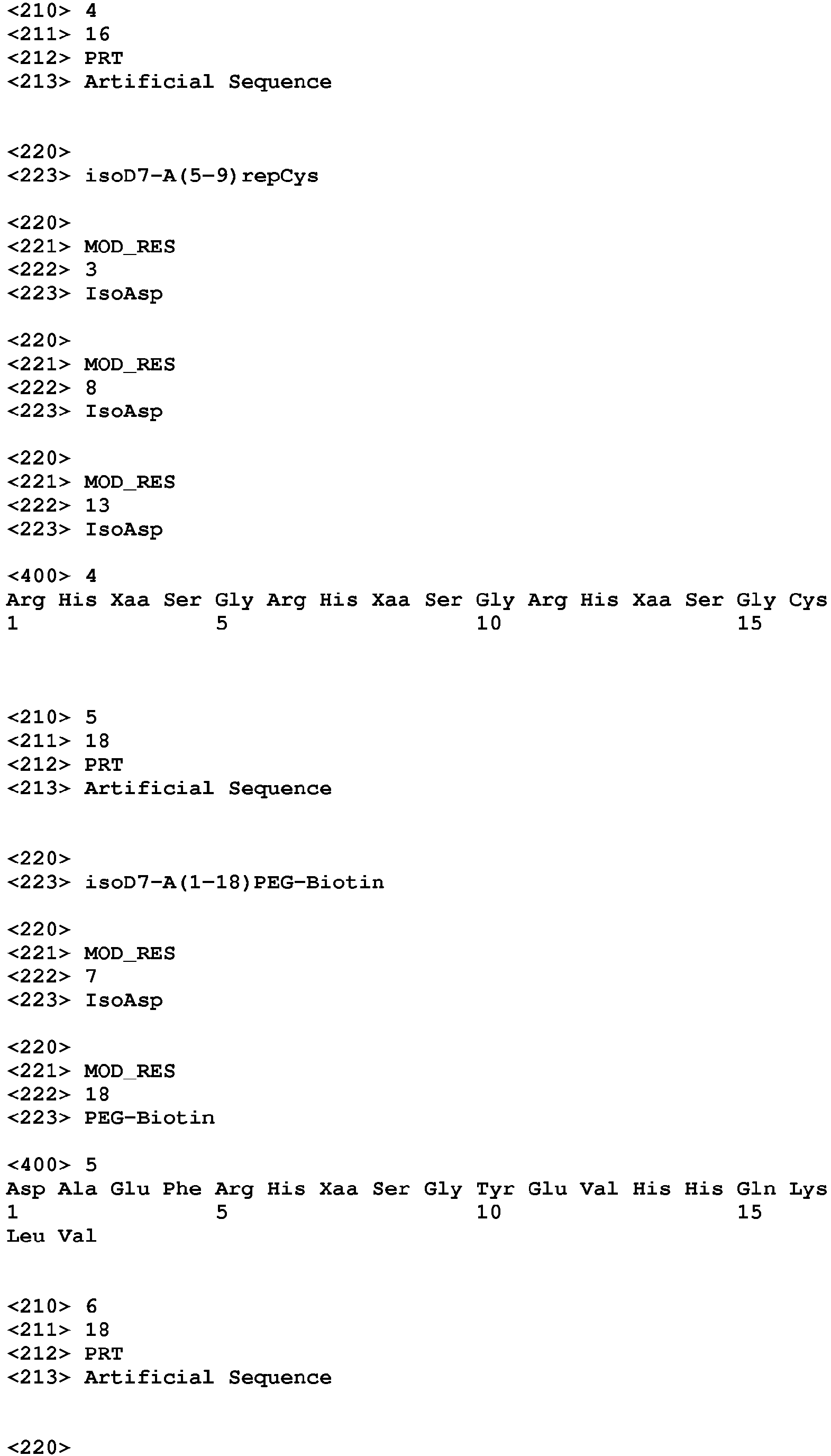


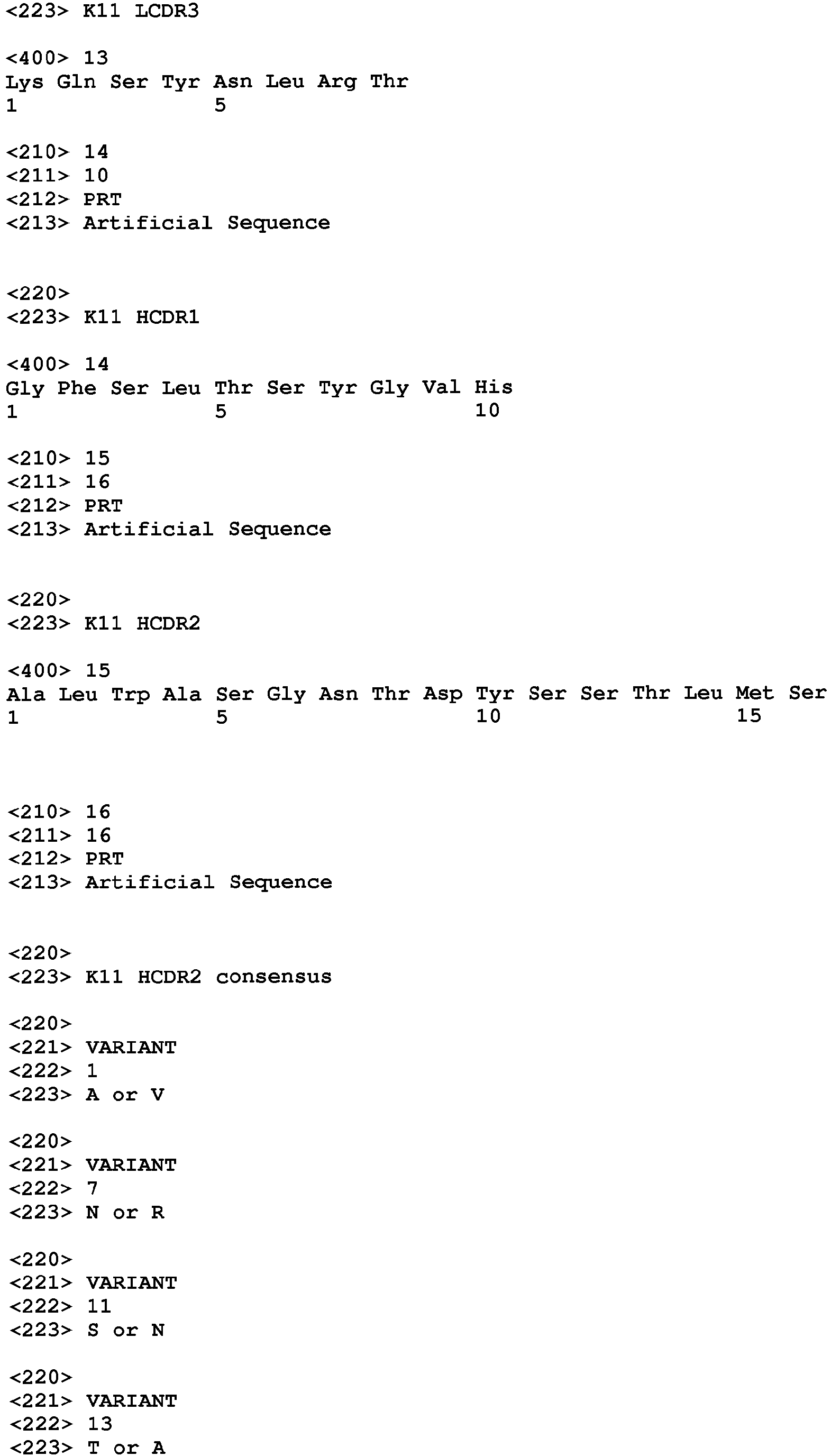


Claims (17)
- An antibody or antigen-binding fragment thereof which specifically binds to isoAsp7 amyloid β (Aβ), wherein the KD of the interaction between the antibody and SEQ ID NO: 44 is at least 10 times less than the KD of the interaction between the antibody and SEQ ID NO: 8.
- The antibody or antigen-binding fragment thereof according to claim 2, wherein the KD of the interaction between the antibody and SEQ ID NO: 44 is at least 100 times less than the KD of the interaction between the antibody and SEQ ID NO: 8.
- The antibody or antigen-binding fragment thereof according to any one of claims 1-2, wherein the antibody or antigen-binding fragment thereof comprises a light chain variable region (VL) and a heavy chain variable region (VH), wherein said VL comprises LCDR1, LCDR2 and LCDR3 polypeptides and VH comprises HCDR1, HCDR2 and HCDR3 polypeptides which are selected from the group consisting of:(a) LCDR1 is KSSQSLLNSRNRKNYLA (SEQ ID NO: 9), LCDR2 is WASTRDS (SEQ ID NO: 11), LCDR3 is KQSYNLRT (SEQ ID NO: 13), HCDR1 is GFSLTSYGVH (SEQ ID NO: 14), HCDR2 is ALWASGNTDYSSTLMS (SEQ ID NO: 15), and HCDR3 is DRGILTGGYFDV (SEQ ID NO: 17);(b) LCDR1 is KSSQSLFNSRTRKNYVA (SEQ ID NO: 27), LCDR2 is WASTRES (SEQ ID NO: 29), LCDR3 is KQSYNLRA (SEQ ID NO: 30), HCDR1 is GFTFTDYYMS (SEQ ID NO: 32), HCDR2 is FIRNKANGYTTEYSASVKG (SEQ ID NO: 34), and HCDR3 is DIPTIMDY (SEQ ID NO: 35);(c) LCDR1 is KSSQSLLNX1RX2RKNYLA (SEQ ID NO: 10), LCDR2 is WASTRX3S (SEQ ID NO: 12), LCDR3 is KQSYNLRT (SEQ ID NO: 13), HCDR1 is GFSLTSYGVH (SEQ ID NO: 14), HCDR2 is X4LWASGX5TDYX6SX7LMS (SEQ ID NO: 16), and HCDR3 is DRGIX8TGGYFDV (SEQ ID NO: 18), wherein X1 is S or R, X2 is N or T, X3 is D or E, X4 is A or V, X5 is N or R, X6 is S or N, X7 is T or A, and X8 is L, T or M; and(d) LCDR1 is KSSQX1LX2NSRTRKNYX3A (SEQ ID NO: 28), LCDR2 is WASTRES (SEQ ID NO: 29), LCDR3 is X4QSYNLRX5 (SEQ ID NO: 31), HCDR1 is GFTFX6DYYMX7 (SEQ ID NO: 33), HCDR2 is FIRNKANGYTTEYSASVKG (SEQ ID NO: 34), and HCDR3 is DIPTIMDY (SEQ ID NO: 35), wherein X1 is S or N, X2 is F or L, X3 is V or L, X4 is K or M, X5 is A or T, X6 is T or S, and X7 is S or N.
- The antibody or antigen-binding fragment thereof according to any one of claims 1-3, wherein the antibody or antigen-binding fragment thereof comprises a VL and a VH, wherein the VL and VH are polypeptides selected from the group consisting of:(a) VL of SEQ ID NO: 19 and VH of SEQ ID NO: 20;(b) VL of SEQ ID NO: 36 and VH of SEQ ID NO: 37;(c) VL of SEQ ID NO: 19 and VH of SEQ ID NO: 37; and(d) VL of SEQ ID NO: 36 and VH of SEQ ID NO: 20.
- The antibody or antigen-binding fragment thereof according to any one of claims 1-4, wherein the antibody or antigen-binding fragment thereof comprises a light chain (LC) and a heavy chain (HC), wherein said LC and HC are polypeptides selected from the group consisting of:(a) LC of SEQ ID NO: 21 and HC of SEQ ID NO: 22;(b) LC of SEQ ID NO: 38 and HC of SEQ ID NO: 39;(c) LC of SEQ ID NO: 21 and HC of SEQ ID NO: 39; and(d) LC of SEQ ID NO: 38 and HC of SEQ ID NO: 22.
- The antibody or antigen-binding fragment thereof according to claim 5, wherein the antibody or antigen-binding fragment thereof comprises two LCs and two HCs, wherein each LC and each HC are polypeptides selected from the group consisting of:(a) LC of SEQ ID NO: 21 and HC of SEQ ID NO: 22;(b) LC of SEQ ID NO: 38 and HC of SEQ ID NO: 39;(c) LC of SEQ ID NO: 21 and HC of SEQ ID NO: 39; and(d) LC of SEQ ID NO: 38 and HC of SEQ ID NO: 22.
- The antibody or antigen-binding fragment thereof according to any one of claims 1-6, wherein the antibody or antigen-binding fragment thereof is obtained or is obtainable from the hybridoma cell line(a) MWT 11-1-3, Deposit No: DSM ACC3314, Deposit date: Dec. 01, 2016 or(b) MWT 119-8-6, Deposit No: DSM ACC3316, Deposit date: Dec. 01, 2016.
- The antibody or antigen-binding fragment thereof according to any one of claims 1-7, wherein the antibody or antigen-binding fragment thereof is a monoclonal antibody or antigen-binding fragment thereof.
- A pharmaceutical composition comprising the antibody or antigen-binding fragment thereof according to any one of claims 1-8 and a pharmaceutically acceptable carrier or diluent.
- The pharmaceutical composition according to claim 9, wherein the composition further comprises donepezil, gelantamine, memantine, rivastigmine, a beta sectretase inhibitor, a gamma secretase modulator, an additional antibody selected from the group of pan-Aß specific antibodies like aducanumab, bapineuzumab, crenezumab, ganteneumab, solanezumab and/or an antibody with specificity to posttranslational phosphorylated or nitrated Aß peptides.
- The antibody or antigen-binding fragment thereof according to any one of claims 1-8 or the pharmaceutical composition according to any one of claims 9-10 for use as a medicament.
- The antibody or antigen-binding fragment thereof according to any one of claims 1-8 or the pharmaceutical composition according to any one of claims 9-10 for use in the treatment and/or prevention of a neurodegenerative disease.
- The antibody or antigen-binding fragment thereof or the pharmaceutical composition for use according to claim 12, wherein the neurodegenerative disease is selected from the list consisting of mild cognitive impairment, clinical or preclinical Alzheimer's disease, neurodegeneration in Down Syndrome, clinical and preclinical amyloid angiopathy, progressive supranuclear palsy, multiple sclerosis, Creutzfeld Jacob disease, Parkinson's disease, HIV-related dementia, ALS (amyotropic lateral sclerosis), dementia related to Adult Onset Diabetes, senile cardiac amyloidosis and muscular degeneration.
- The antibody or antigen-binding fragment thereof or the pharmaceutical composition for use according to any one of claims 12-13, wherein the neurodegenerative disease is clinical or preclinical Alzheimer's disease.
- A hybridoma cell line deposited as follows:(a) MWT 11-1-3, Deposit No: DSM ACC3314, Deposit date: Dec. 01, 2016; and/or(b) MWT 119-8-6, Deposit No: DSM ACC3316, Deposit date: Dec. 01, 2016.
- Use of the antibody or antigen-binding fragment thereof according to any one of claims 1-8 for the diagnosis and/or prognosis of a neurodegenerative disease.
- A method for detecting isoAsp7 Aβ comprising a step wherein an isolated sample is put into contact with the antibody or antigen-binding fragment thereof according to any one of claims 1-8.
Priority Applications (7)
| Application Number | Priority Date | Filing Date | Title |
|---|---|---|---|
| EP17182167.1A EP3431496A1 (en) | 2017-07-19 | 2017-07-19 | Anti- isoasp7 amyloid beta antibodies and uses thereof |
| ES18738361T ES3033808T3 (en) | 2017-07-19 | 2018-07-17 | Anti- isoasp7 amyloid beta (abeta) antibodies and uses thereof |
| US16/632,289 US11703511B2 (en) | 2017-07-19 | 2018-07-17 | Anti-isoAsp7 amyloid β (Aβ) antibodies and uses thereof |
| JP2020501790A JP2020527561A (en) | 2017-07-19 | 2018-07-17 | Anti-isoAsp7 amyloid β (Aβ) antibody and its use |
| PCT/EP2018/069404 WO2019016213A1 (en) | 2017-07-19 | 2018-07-17 | ANTI- ISOASP7 AMYLOID β (Aβ) ANTIBODIES AND USES THEREOF |
| EP18738361.7A EP3655431B1 (en) | 2017-07-19 | 2018-07-17 | Anti- isoasp7 amyloid beta (abeta) antibodies and uses thereof |
| JP2023030237A JP2023075191A (en) | 2017-07-19 | 2023-02-28 | Anti-isoAsp7 amyloid β (Aβ) antibody and use thereof |
Applications Claiming Priority (1)
| Application Number | Priority Date | Filing Date | Title |
|---|---|---|---|
| EP17182167.1A EP3431496A1 (en) | 2017-07-19 | 2017-07-19 | Anti- isoasp7 amyloid beta antibodies and uses thereof |
Publications (1)
| Publication Number | Publication Date |
|---|---|
| EP3431496A1 true EP3431496A1 (en) | 2019-01-23 |
Family
ID=59381199
Family Applications (2)
| Application Number | Title | Priority Date | Filing Date |
|---|---|---|---|
| EP17182167.1A Ceased EP3431496A1 (en) | 2017-07-19 | 2017-07-19 | Anti- isoasp7 amyloid beta antibodies and uses thereof |
| EP18738361.7A Active EP3655431B1 (en) | 2017-07-19 | 2018-07-17 | Anti- isoasp7 amyloid beta (abeta) antibodies and uses thereof |
Family Applications After (1)
| Application Number | Title | Priority Date | Filing Date |
|---|---|---|---|
| EP18738361.7A Active EP3655431B1 (en) | 2017-07-19 | 2018-07-17 | Anti- isoasp7 amyloid beta (abeta) antibodies and uses thereof |
Country Status (5)
| Country | Link |
|---|---|
| US (1) | US11703511B2 (en) |
| EP (2) | EP3431496A1 (en) |
| JP (2) | JP2020527561A (en) |
| ES (1) | ES3033808T3 (en) |
| WO (1) | WO2019016213A1 (en) |
Cited By (1)
| Publication number | Priority date | Publication date | Assignee | Title |
|---|---|---|---|---|
| WO2023038560A1 (en) * | 2021-10-29 | 2023-03-16 | Roman Zubarev | Method for determining risk factors for neurodegenerative diseases in blood |
Families Citing this family (1)
| Publication number | Priority date | Publication date | Assignee | Title |
|---|---|---|---|---|
| WO2022020680A1 (en) | 2020-07-23 | 2022-01-27 | Othair Prothena Limited | Anti-abeta antibodies |
Citations (27)
| Publication number | Priority date | Publication date | Assignee | Title |
|---|---|---|---|---|
| EP0125023A1 (en) | 1983-04-08 | 1984-11-14 | Genentech, Inc. | Recombinant immunoglobulin preparations, methods for their preparation, DNA sequences, expression vectors and recombinant host cells therefor |
| EP0171496A2 (en) | 1984-08-15 | 1986-02-19 | Research Development Corporation of Japan | Process for the production of a chimera monoclonal antibody |
| EP0173494A2 (en) | 1984-08-27 | 1986-03-05 | The Board Of Trustees Of The Leland Stanford Junior University | Chimeric receptors by DNA splicing and expression |
| WO1986001533A1 (en) | 1984-09-03 | 1986-03-13 | Celltech Limited | Production of chimeric antibodies |
| EP0184187A2 (en) | 1984-12-04 | 1986-06-11 | Teijin Limited | Mouse-human chimaeric immunoglobulin heavy chain, and chimaeric DNA encoding it |
| GB2188638A (en) | 1986-03-27 | 1987-10-07 | Gregory Paul Winter | Chimeric antibodies |
| WO1990002809A1 (en) | 1988-09-02 | 1990-03-22 | Protein Engineering Corporation | Generation and selection of recombinant varied binding proteins |
| EP0388151A1 (en) | 1989-03-13 | 1990-09-19 | Celltech Limited | Modified antibodies |
| WO1991000906A1 (en) | 1989-07-12 | 1991-01-24 | Genetics Institute, Inc. | Chimeric and transgenic animals capable of producing human antibodies |
| WO1991010741A1 (en) | 1990-01-12 | 1991-07-25 | Cell Genesys, Inc. | Generation of xenogeneic antibodies |
| WO1991017271A1 (en) | 1990-05-01 | 1991-11-14 | Affymax Technologies N.V. | Recombinant library screening methods |
| WO1992001047A1 (en) | 1990-07-10 | 1992-01-23 | Cambridge Antibody Technology Limited | Methods for producing members of specific binding pairs |
| WO1992003917A1 (en) | 1990-08-29 | 1992-03-19 | Genpharm International | Homologous recombination in mammalian cells |
| WO1992003918A1 (en) | 1990-08-29 | 1992-03-19 | Genpharm International, Inc. | Transgenic non-human animals capable of producing heterologous antibodies |
| WO1992009690A2 (en) | 1990-12-03 | 1992-06-11 | Genentech, Inc. | Enrichment method for variant proteins with altered binding properties |
| WO1992015679A1 (en) | 1991-03-01 | 1992-09-17 | Protein Engineering Corporation | Improved epitode displaying phage |
| WO1992018619A1 (en) | 1991-04-10 | 1992-10-29 | The Scripps Research Institute | Heterodimeric receptor libraries using phagemids |
| WO1992020791A1 (en) | 1990-07-10 | 1992-11-26 | Cambridge Antibody Technology Limited | Methods for producing members of specific binding pairs |
| EP0519596A1 (en) | 1991-05-17 | 1992-12-23 | Merck & Co. Inc. | A method for reducing the immunogenicity of antibody variable domains |
| WO1993001288A1 (en) | 1991-07-08 | 1993-01-21 | Deutsches Krebsforschungszentrum Stiftung des öffentlichen Rechts | Phagemide for screening antibodies |
| US5223409A (en) | 1988-09-02 | 1993-06-29 | Protein Engineering Corp. | Directed evolution of novel binding proteins |
| US5225539A (en) | 1986-03-27 | 1993-07-06 | Medical Research Council | Recombinant altered antibodies and methods of making altered antibodies |
| WO1994004678A1 (en) | 1992-08-21 | 1994-03-03 | Casterman Cecile | Immunoglobulins devoid of light chains |
| US5585089A (en) | 1988-12-28 | 1996-12-17 | Protein Design Labs, Inc. | Humanized immunoglobulins |
| US5624821A (en) | 1987-03-18 | 1997-04-29 | Scotgen Biopharmaceuticals Incorporated | Antibodies with altered effector functions |
| WO2006036291A2 (en) * | 2004-07-30 | 2006-04-06 | Rinat Neuroscience Corp. | Antibodies directed against amyloid-beta peptide and methods using same |
| US8602269B2 (en) | 2009-09-14 | 2013-12-10 | Guala Dispensing S.P.A. | Trigger sprayer |
Family Cites Families (4)
| Publication number | Priority date | Publication date | Assignee | Title |
|---|---|---|---|---|
| EP0247091B1 (en) | 1985-11-01 | 1993-09-29 | Xoma Corporation | Modular assembly of antibody genes, antibodies prepared thereby and use |
| PE20061323A1 (en) * | 2005-04-29 | 2007-02-09 | Rinat Neuroscience Corp | ANTIBODIES TARGETED AGAINST AMYLOID BETA PEPTIDE AND METHODS USING THEM |
| GB0624500D0 (en) * | 2006-12-07 | 2007-01-17 | Istituto Superiore Di Sanito | A novel passive vaccine for candida infections |
| CA2874083C (en) * | 2014-12-05 | 2024-01-02 | Universite Laval | Tdp-43-binding polypeptides useful for the treatment of neurodegenerative diseases |
-
2017
- 2017-07-19 EP EP17182167.1A patent/EP3431496A1/en not_active Ceased
-
2018
- 2018-07-17 WO PCT/EP2018/069404 patent/WO2019016213A1/en not_active Ceased
- 2018-07-17 EP EP18738361.7A patent/EP3655431B1/en active Active
- 2018-07-17 ES ES18738361T patent/ES3033808T3/en active Active
- 2018-07-17 JP JP2020501790A patent/JP2020527561A/en active Pending
- 2018-07-17 US US16/632,289 patent/US11703511B2/en active Active
-
2023
- 2023-02-28 JP JP2023030237A patent/JP2023075191A/en active Pending
Patent Citations (31)
| Publication number | Priority date | Publication date | Assignee | Title |
|---|---|---|---|---|
| EP0125023A1 (en) | 1983-04-08 | 1984-11-14 | Genentech, Inc. | Recombinant immunoglobulin preparations, methods for their preparation, DNA sequences, expression vectors and recombinant host cells therefor |
| US4816567A (en) | 1983-04-08 | 1989-03-28 | Genentech, Inc. | Recombinant immunoglobin preparations |
| EP0171496A2 (en) | 1984-08-15 | 1986-02-19 | Research Development Corporation of Japan | Process for the production of a chimera monoclonal antibody |
| EP0173494A2 (en) | 1984-08-27 | 1986-03-05 | The Board Of Trustees Of The Leland Stanford Junior University | Chimeric receptors by DNA splicing and expression |
| WO1986001533A1 (en) | 1984-09-03 | 1986-03-13 | Celltech Limited | Production of chimeric antibodies |
| EP0184187A2 (en) | 1984-12-04 | 1986-06-11 | Teijin Limited | Mouse-human chimaeric immunoglobulin heavy chain, and chimaeric DNA encoding it |
| GB2188638A (en) | 1986-03-27 | 1987-10-07 | Gregory Paul Winter | Chimeric antibodies |
| US5225539A (en) | 1986-03-27 | 1993-07-06 | Medical Research Council | Recombinant altered antibodies and methods of making altered antibodies |
| US5648260A (en) | 1987-03-18 | 1997-07-15 | Scotgen Biopharmaceuticals Incorporated | DNA encoding antibodies with altered effector functions |
| US5624821A (en) | 1987-03-18 | 1997-04-29 | Scotgen Biopharmaceuticals Incorporated | Antibodies with altered effector functions |
| US5223409A (en) | 1988-09-02 | 1993-06-29 | Protein Engineering Corp. | Directed evolution of novel binding proteins |
| WO1990002809A1 (en) | 1988-09-02 | 1990-03-22 | Protein Engineering Corporation | Generation and selection of recombinant varied binding proteins |
| US5693761A (en) | 1988-12-28 | 1997-12-02 | Protein Design Labs, Inc. | Polynucleotides encoding improved humanized immunoglobulins |
| US5693762A (en) | 1988-12-28 | 1997-12-02 | Protein Design Labs, Inc. | Humanized immunoglobulins |
| US5585089A (en) | 1988-12-28 | 1996-12-17 | Protein Design Labs, Inc. | Humanized immunoglobulins |
| EP0388151A1 (en) | 1989-03-13 | 1990-09-19 | Celltech Limited | Modified antibodies |
| WO1991000906A1 (en) | 1989-07-12 | 1991-01-24 | Genetics Institute, Inc. | Chimeric and transgenic animals capable of producing human antibodies |
| WO1991010741A1 (en) | 1990-01-12 | 1991-07-25 | Cell Genesys, Inc. | Generation of xenogeneic antibodies |
| WO1991017271A1 (en) | 1990-05-01 | 1991-11-14 | Affymax Technologies N.V. | Recombinant library screening methods |
| WO1992001047A1 (en) | 1990-07-10 | 1992-01-23 | Cambridge Antibody Technology Limited | Methods for producing members of specific binding pairs |
| WO1992020791A1 (en) | 1990-07-10 | 1992-11-26 | Cambridge Antibody Technology Limited | Methods for producing members of specific binding pairs |
| WO1992003917A1 (en) | 1990-08-29 | 1992-03-19 | Genpharm International | Homologous recombination in mammalian cells |
| WO1992003918A1 (en) | 1990-08-29 | 1992-03-19 | Genpharm International, Inc. | Transgenic non-human animals capable of producing heterologous antibodies |
| WO1992009690A2 (en) | 1990-12-03 | 1992-06-11 | Genentech, Inc. | Enrichment method for variant proteins with altered binding properties |
| WO1992015679A1 (en) | 1991-03-01 | 1992-09-17 | Protein Engineering Corporation | Improved epitode displaying phage |
| WO1992018619A1 (en) | 1991-04-10 | 1992-10-29 | The Scripps Research Institute | Heterodimeric receptor libraries using phagemids |
| EP0519596A1 (en) | 1991-05-17 | 1992-12-23 | Merck & Co. Inc. | A method for reducing the immunogenicity of antibody variable domains |
| WO1993001288A1 (en) | 1991-07-08 | 1993-01-21 | Deutsches Krebsforschungszentrum Stiftung des öffentlichen Rechts | Phagemide for screening antibodies |
| WO1994004678A1 (en) | 1992-08-21 | 1994-03-03 | Casterman Cecile | Immunoglobulins devoid of light chains |
| WO2006036291A2 (en) * | 2004-07-30 | 2006-04-06 | Rinat Neuroscience Corp. | Antibodies directed against amyloid-beta peptide and methods using same |
| US8602269B2 (en) | 2009-09-14 | 2013-12-10 | Guala Dispensing S.P.A. | Trigger sprayer |
Non-Patent Citations (65)
| Title |
|---|
| AL-LAZIKANI ET AL., JMB, vol. 273, 1997, pages 927 - 948 |
| ANO ET AL., J. BIOL. CHEM., vol. 292, no. 9, 2017, pages 3720 - 3728 |
| ARDESTANI ET AL., NEUROPHARMACOLOGY, vol. 116, 2017, pages 371 - 386 |
| ASWAD ET AL., J PHARM BIOMED ANAL., vol. 21, no. 6, 2000, pages 1129 - 36 |
| BARBAS ET AL., PNAS, vol. 88, 1991, pages 7978 - 7982 |
| BEIDLER ET AL., J. IMMUNOL., vol. 141, 1988, pages 4053 - 4060 |
| BETTER ET AL., SCIENCE, vol. 240, 1988, pages 1041 - 1043 |
| BIRD ET AL., SCIENCE, vol. 242, 1988, pages 423 - 426 |
| BRUGGEMAN ET AL., EUR J IMMUNOL, vol. 21, 1991, pages 1323 - 1326 |
| BRUGGEMAN ET AL., YEAR IMMUNOL, vol. 7, 1993, pages 33 - 40 |
| CHA ET AL., STEM CELLS TRANSL. MED., vol. 6, no. l, 2017, pages 293 - 305 |
| CHOTHIA, C. ET AL., J. MOL. BIOL., vol. l96, 1987, pages 901 - 917 |
| CLACKSON ET AL., NATURE, vol. 352, 1991, pages 624 - 628 |
| COLCHER, D. ET AL., ANN N Y ACAD SCI, vol. 880, 1999, pages 263 - 80 |
| DUEBEL, S. AND KONTERMANN, R.,: "Antibody Engineering Lab Manual", SPRINGER-VERLAG, article "Protein Sequence and Structure Analysis of Antibody Variable Domains" |
| FONSECA ET AL., EXP NEUROL., vol. 157, no. 2, 1999, pages 277 - 88 |
| FUCHS ET AL., BIO/TECHNOLOGY, vol. 9, 1991, pages 1370 - 1372 |
| FUKUDA ET AL., BIOORG MED CHEM LETT., vol. 9, no. 7, 1999, pages 953 - 6 |
| GARRAD ET AL., BIO/TECHNOLOGY, vol. 9, 1991, pages 1373 - 1377 |
| GEIGER; CLARKE, J BIOL CHEM, vol. 262, 1987, pages 785 - 794 |
| GRAM ET AL., PNAS, vol. 89, 1992, pages 3576 - 3580 |
| GREEN, L.L. ET AL., NATURE GENET., vol. 7, 1994, pages 13 - 21 |
| GRIFFTHS ET AL., EMBO J, vol. 12, 1993, pages 725 - 734 |
| HAWKINS ET AL., J MOL BIOL, vol. 226, 1992, pages 889 - 896 |
| HAY ET AL., HUM ANTIBOD HYBRIDOMAS, vol. 3, 1992, pages 81 - 85 |
| HOLMES ET AL., LANCET, vol. 372, 2008, pages 216 - 223 |
| HOOGENBOOM ET AL., NUC ACID RES, vol. 19, 1991, pages 4133 - 4137 |
| HSU ET AL., HISTOCHEM CYTOCHEM., vol. 29, no. 4, 1981, pages 577 - 80 |
| HUSE ET AL., SCIENCE, vol. 246, 1989, pages 1275 - 1281 |
| HUSTON ET AL., PROC. NATL. ACAD. SCI. USA, vol. 85, 1988, pages 5879 - 5883 |
| JONES ET AL., NATURE, vol. 321, 1986, pages 552 - 525 |
| KABAT ET AL.: "Sequences of Proteins of Immunological Interest" 5th Ed.", 1991, PUBLIC HEALTH SERVICE, NATIONAL INSTITUTES OF HEALTH |
| KABAT, E. A. ET AL.: "Sequences of Proteins of Immunological Interest, Fifth Edition,", 1991, NIH |
| LIU ET AL., J. IMMUNOL., vol. 139, 1987, pages 3521 - 3526 |
| LIU ET AL., PNAS, vol. 84, 1987, pages 3439 - 3443 |
| LONBERG, N. ET AL., NATURE, vol. 368, 1994, pages 856 - 859 |
| MACPHERSON ET AL., NEUROBIOL. DIS., vol. 102, 2017, pages 81 - 95 |
| MORETH ET AL., IMMUN AGEING., vol. 10, no. 1, 2013, pages 18 |
| MORRISON, S. L., SCIENCE, vol. 229, 1985, pages 1202 - 1207 |
| MORRISON, S.L. ET AL., PROC. NATL. ACAD. SCI. USA, vol. 81, 1994, pages 6851 - 6855 |
| NISHIMURA ET AL., CANE. RES., vol. 47, 1987, pages 999 - 1005 |
| OAKLEY ET AL., J. NEUROSCI., vol. 26, no. 40, 2006, pages 10129 - 40 |
| OI ET AL., BIOTECHNIQUES, vol. 4, 1986, pages 214 |
| ORGOGOZO ET AL., NEUROLOGY, vol. 61, 2003, pages 46 - 54 |
| OSOL, A.: "Remington's Pharmaceutical Sciences 16th edition,", 1980 |
| PETERSEN, J INTERN MED, vol. 256, 2004, pages 183 - 94 |
| REITER, Y., CLIN CANCER RES, vol. 2, 1996, pages 245 - 52 |
| ROBINSON; ROBINSON, PNAS, vol. 98, no. 3, 2001, pages 944 - 949 |
| ROBINSON; RUDD, CURR TOP CELL REGUL., vol. 8, no. 0, 1974, pages 247 - 95 |
| ROHER ET AL., PROC. NATL. ACAD. SCI. USA, vol. 90, 1993, pages 10836 - 10840 |
| SAWA ET AL., N ENGL J MED, vol. 360, 2009, pages 2302 - 2309 |
| SHAW ET AL., J. NATL CANCER INST., vol. 80, 1988, pages 1553 - 1559 |
| SHIMIZU ET AL., ARCH BIOCHEM BIOPHYS., vol. 381, no. 2, 2000, pages 225 - 34 |
| SHIMIZU ET AL., BIOL PHARM BULL., vol. 28, no. 9, 2005, pages 1590 - 6 |
| SHIMIZU ET AL., J NEUROSCI RES., vol. 70, no. 3, 2002, pages 451 - 61 |
| STEPHENSON; CLARKE, J BIOL CHEM, vol. 264, 1989, pages 6164 - 6170 |
| SUN ET AL., PNAS, vol. 84, 1987, pages 214 - 218 |
| TAKAHIKO SHIMIZU ET AL: "Isoaspartate Formation and Neurodegeneration in Alzheimer's Disease", ARCHIVES OF BIOCHEMISTRY AND BIOPHYSICS, vol. 381, no. 2, 1 September 2000 (2000-09-01), US, pages 225 - 234, XP055422974, ISSN: 0003-9861, DOI: 10.1006/abbi.2000.1955 * |
| TONG ZHANG ET AL: "Atomic and Dynamic Insights into the Beneficial Effect of the 1,4- Naphthoquinon-2-yl-L-tryptophan-tryptophan Inhibitor on Alzheimer's A[beta]1-42 Dimer in Terms of Aggregation and Toxicity", ACS CHEMICAL NEUROSCIENCE, vol. 5, no. 2, 19 February 2014 (2014-02-19), US, pages 148 - 159, XP055422956, ISSN: 1948-7193, DOI: 10.1021/cn400197x * |
| TORIKA ET AL., BRAIN BEHAV. IMMUN., vol. 64, 2017, pages 80 - 90 |
| TUAILLON ET AL., PNAS, vol. 90, 1993, pages 3720 - 3724 |
| VERHOEYAN ET AL., SCIENCE, vol. 239, 1988, pages 1534 |
| WAKUTANI ET AL., J NEUROL NEUROSURG PSYCHIATRY., vol. 75, no. 7, 2004, pages 1039 - 42 |
| WINNAKER: "From Genes to Clones", 1987, VERLAGSGESELLSCHAFT |
| WOOD ET AL., NATURE, vol. 314, 1985, pages 446 - 449 |
Cited By (1)
| Publication number | Priority date | Publication date | Assignee | Title |
|---|---|---|---|---|
| WO2023038560A1 (en) * | 2021-10-29 | 2023-03-16 | Roman Zubarev | Method for determining risk factors for neurodegenerative diseases in blood |
Also Published As
| Publication number | Publication date |
|---|---|
| WO2019016213A1 (en) | 2019-01-24 |
| US20200264197A1 (en) | 2020-08-20 |
| ES3033808T3 (en) | 2025-08-08 |
| EP3655431B1 (en) | 2025-05-21 |
| JP2020527561A (en) | 2020-09-10 |
| EP3655431A1 (en) | 2020-05-27 |
| US11703511B2 (en) | 2023-07-18 |
| JP2023075191A (en) | 2023-05-30 |
Similar Documents
| Publication | Publication Date | Title |
|---|---|---|
| JP5641737B2 (en) | Humanized antibody against amyloid β | |
| CA2690435C (en) | Humanized antibodies to amyloid beta | |
| JP2018095647A (en) | HUMANIZED ANTIBODIES AGAINST AMYLOID β | |
| KR20160017126A (en) | Monoclonal anti beta amyloid antibody | |
| JP5051806B2 (en) | Anti-amyloid β oligomer humanized antibody | |
| JP2023075191A (en) | Anti-isoAsp7 amyloid β (Aβ) antibody and use thereof | |
| CN106831983B (en) | Humanized antibodies against amyloid beta | |
| KR102655944B1 (en) | Anti-amyloid beta antibodies and methods of using the same | |
| US20250163136A1 (en) | Anti-transthyretin (ttr) binding proteins and uses thereof | |
| EP3988566A1 (en) | Humanized antibody against amyloid beta | |
| JP6076274B2 (en) | Humanized antibody against amyloid β | |
| HK1239710A1 (en) | Humanized antibody against amyloid beta | |
| HK1194083A (en) | Humanized antibody against amyloid beta | |
| HK1194083B (en) | Humanized antibody against amyloid beta | |
| HK1139154B (en) | Humani zed antibody against amyloid beta | |
| HK1172352B (en) | Humanized antibody against amyloid beta | |
| HK1172352A (en) | Humanized antibody against amyloid beta |
Legal Events
| Date | Code | Title | Description |
|---|---|---|---|
| PUAI | Public reference made under article 153(3) epc to a published international application that has entered the european phase |
Free format text: ORIGINAL CODE: 0009012 |
|
| AK | Designated contracting states |
Kind code of ref document: A1 Designated state(s): AL AT BE BG CH CY CZ DE DK EE ES FI FR GB GR HR HU IE IS IT LI LT LU LV MC MK MT NL NO PL PT RO RS SE SI SK SM TR |
|
| AX | Request for extension of the european patent |
Extension state: BA ME |
|
| STAA | Information on the status of an ep patent application or granted ep patent |
Free format text: STATUS: THE APPLICATION HAS BEEN REFUSED |
|
| 18R | Application refused |
Effective date: 20190210 |