EP3430129B1 - Colony forming medium and use thereof - Google Patents
Colony forming medium and use thereof Download PDFInfo
- Publication number
- EP3430129B1 EP3430129B1 EP17765572.7A EP17765572A EP3430129B1 EP 3430129 B1 EP3430129 B1 EP 3430129B1 EP 17765572 A EP17765572 A EP 17765572A EP 3430129 B1 EP3430129 B1 EP 3430129B1
- Authority
- EP
- European Patent Office
- Prior art keywords
- msc
- fgf2
- cell
- licl
- days
- Prior art date
- Legal status (The legal status is an assumption and is not a legal conclusion. Google has not performed a legal analysis and makes no representation as to the accuracy of the status listed.)
- Active
Links
Images



Classifications
-
- A—HUMAN NECESSITIES
- A61—MEDICAL OR VETERINARY SCIENCE; HYGIENE
- A61K—PREPARATIONS FOR MEDICAL, DENTAL OR TOILETRY PURPOSES
- A61K35/00—Medicinal preparations containing materials or reaction products thereof with undetermined constitution
- A61K35/12—Materials from mammals; Compositions comprising non-specified tissues or cells; Compositions comprising non-embryonic stem cells; Genetically modified cells
- A61K35/48—Reproductive organs
- A61K35/54—Ovaries; Ova; Ovules; Embryos; Foetal cells; Germ cells
- A61K35/545—Embryonic stem cells; Pluripotent stem cells; Induced pluripotent stem cells; Uncharacterised stem cells
-
- C—CHEMISTRY; METALLURGY
- C12—BIOCHEMISTRY; BEER; SPIRITS; WINE; VINEGAR; MICROBIOLOGY; ENZYMOLOGY; MUTATION OR GENETIC ENGINEERING
- C12N—MICROORGANISMS OR ENZYMES; COMPOSITIONS THEREOF; PROPAGATING, PRESERVING, OR MAINTAINING MICROORGANISMS; MUTATION OR GENETIC ENGINEERING; CULTURE MEDIA
- C12N5/00—Undifferentiated human, animal or plant cells, e.g. cell lines; Tissues; Cultivation or maintenance thereof; Culture media therefor
- C12N5/06—Animal cells or tissues; Human cells or tissues
- C12N5/0602—Vertebrate cells
- C12N5/0603—Embryonic cells ; Embryoid bodies
- C12N5/0606—Pluripotent embryonic cells, e.g. embryonic stem cells [ES]
-
- C—CHEMISTRY; METALLURGY
- C12—BIOCHEMISTRY; BEER; SPIRITS; WINE; VINEGAR; MICROBIOLOGY; ENZYMOLOGY; MUTATION OR GENETIC ENGINEERING
- C12N—MICROORGANISMS OR ENZYMES; COMPOSITIONS THEREOF; PROPAGATING, PRESERVING, OR MAINTAINING MICROORGANISMS; MUTATION OR GENETIC ENGINEERING; CULTURE MEDIA
- C12N5/00—Undifferentiated human, animal or plant cells, e.g. cell lines; Tissues; Cultivation or maintenance thereof; Culture media therefor
- C12N5/06—Animal cells or tissues; Human cells or tissues
- C12N5/0602—Vertebrate cells
- C12N5/0652—Cells of skeletal and connective tissues; Mesenchyme
- C12N5/0662—Stem cells
-
- A—HUMAN NECESSITIES
- A61—MEDICAL OR VETERINARY SCIENCE; HYGIENE
- A61P—SPECIFIC THERAPEUTIC ACTIVITY OF CHEMICAL COMPOUNDS OR MEDICINAL PREPARATIONS
- A61P9/00—Drugs for disorders of the cardiovascular system
-
- A—HUMAN NECESSITIES
- A61—MEDICAL OR VETERINARY SCIENCE; HYGIENE
- A61K—PREPARATIONS FOR MEDICAL, DENTAL OR TOILETRY PURPOSES
- A61K35/00—Medicinal preparations containing materials or reaction products thereof with undetermined constitution
- A61K35/12—Materials from mammals; Compositions comprising non-specified tissues or cells; Compositions comprising non-embryonic stem cells; Genetically modified cells
- A61K35/28—Bone marrow; Haematopoietic stem cells; Mesenchymal stem cells of any origin, e.g. adipose-derived stem cells
-
- A—HUMAN NECESSITIES
- A61—MEDICAL OR VETERINARY SCIENCE; HYGIENE
- A61P—SPECIFIC THERAPEUTIC ACTIVITY OF CHEMICAL COMPOUNDS OR MEDICINAL PREPARATIONS
- A61P19/00—Drugs for skeletal disorders
- A61P19/02—Drugs for skeletal disorders for joint disorders, e.g. arthritis, arthrosis
-
- A—HUMAN NECESSITIES
- A61—MEDICAL OR VETERINARY SCIENCE; HYGIENE
- A61P—SPECIFIC THERAPEUTIC ACTIVITY OF CHEMICAL COMPOUNDS OR MEDICINAL PREPARATIONS
- A61P37/00—Drugs for immunological or allergic disorders
-
- A—HUMAN NECESSITIES
- A61—MEDICAL OR VETERINARY SCIENCE; HYGIENE
- A61P—SPECIFIC THERAPEUTIC ACTIVITY OF CHEMICAL COMPOUNDS OR MEDICINAL PREPARATIONS
- A61P37/00—Drugs for immunological or allergic disorders
- A61P37/02—Immunomodulators
- A61P37/06—Immunosuppressants, e.g. drugs for graft rejection
-
- C—CHEMISTRY; METALLURGY
- C12—BIOCHEMISTRY; BEER; SPIRITS; WINE; VINEGAR; MICROBIOLOGY; ENZYMOLOGY; MUTATION OR GENETIC ENGINEERING
- C12N—MICROORGANISMS OR ENZYMES; COMPOSITIONS THEREOF; PROPAGATING, PRESERVING, OR MAINTAINING MICROORGANISMS; MUTATION OR GENETIC ENGINEERING; CULTURE MEDIA
- C12N5/00—Undifferentiated human, animal or plant cells, e.g. cell lines; Tissues; Cultivation or maintenance thereof; Culture media therefor
- C12N5/06—Animal cells or tissues; Human cells or tissues
- C12N5/0602—Vertebrate cells
- C12N5/0652—Cells of skeletal and connective tissues; Mesenchyme
- C12N5/0662—Stem cells
- C12N5/0663—Bone marrow mesenchymal stem cells (BM-MSC)
-
- C—CHEMISTRY; METALLURGY
- C12—BIOCHEMISTRY; BEER; SPIRITS; WINE; VINEGAR; MICROBIOLOGY; ENZYMOLOGY; MUTATION OR GENETIC ENGINEERING
- C12N—MICROORGANISMS OR ENZYMES; COMPOSITIONS THEREOF; PROPAGATING, PRESERVING, OR MAINTAINING MICROORGANISMS; MUTATION OR GENETIC ENGINEERING; CULTURE MEDIA
- C12N5/00—Undifferentiated human, animal or plant cells, e.g. cell lines; Tissues; Cultivation or maintenance thereof; Culture media therefor
- C12N5/06—Animal cells or tissues; Human cells or tissues
- C12N5/0602—Vertebrate cells
- C12N5/0652—Cells of skeletal and connective tissues; Mesenchyme
- C12N5/0662—Stem cells
- C12N5/0665—Blood-borne mesenchymal stem cells, e.g. from umbilical cord blood
-
- C—CHEMISTRY; METALLURGY
- C12—BIOCHEMISTRY; BEER; SPIRITS; WINE; VINEGAR; MICROBIOLOGY; ENZYMOLOGY; MUTATION OR GENETIC ENGINEERING
- C12N—MICROORGANISMS OR ENZYMES; COMPOSITIONS THEREOF; PROPAGATING, PRESERVING, OR MAINTAINING MICROORGANISMS; MUTATION OR GENETIC ENGINEERING; CULTURE MEDIA
- C12N5/00—Undifferentiated human, animal or plant cells, e.g. cell lines; Tissues; Cultivation or maintenance thereof; Culture media therefor
- C12N5/06—Animal cells or tissues; Human cells or tissues
- C12N5/0602—Vertebrate cells
- C12N5/0652—Cells of skeletal and connective tissues; Mesenchyme
- C12N5/0662—Stem cells
- C12N5/0668—Mesenchymal stem cells from other natural sources
-
- A—HUMAN NECESSITIES
- A61—MEDICAL OR VETERINARY SCIENCE; HYGIENE
- A61K—PREPARATIONS FOR MEDICAL, DENTAL OR TOILETRY PURPOSES
- A61K35/00—Medicinal preparations containing materials or reaction products thereof with undetermined constitution
-
- C—CHEMISTRY; METALLURGY
- C12—BIOCHEMISTRY; BEER; SPIRITS; WINE; VINEGAR; MICROBIOLOGY; ENZYMOLOGY; MUTATION OR GENETIC ENGINEERING
- C12N—MICROORGANISMS OR ENZYMES; COMPOSITIONS THEREOF; PROPAGATING, PRESERVING, OR MAINTAINING MICROORGANISMS; MUTATION OR GENETIC ENGINEERING; CULTURE MEDIA
- C12N2500/00—Specific components of cell culture medium
- C12N2500/02—Atmosphere, e.g. low oxygen conditions
-
- C—CHEMISTRY; METALLURGY
- C12—BIOCHEMISTRY; BEER; SPIRITS; WINE; VINEGAR; MICROBIOLOGY; ENZYMOLOGY; MUTATION OR GENETIC ENGINEERING
- C12N—MICROORGANISMS OR ENZYMES; COMPOSITIONS THEREOF; PROPAGATING, PRESERVING, OR MAINTAINING MICROORGANISMS; MUTATION OR GENETIC ENGINEERING; CULTURE MEDIA
- C12N2500/00—Specific components of cell culture medium
- C12N2500/05—Inorganic components
-
- C—CHEMISTRY; METALLURGY
- C12—BIOCHEMISTRY; BEER; SPIRITS; WINE; VINEGAR; MICROBIOLOGY; ENZYMOLOGY; MUTATION OR GENETIC ENGINEERING
- C12N—MICROORGANISMS OR ENZYMES; COMPOSITIONS THEREOF; PROPAGATING, PRESERVING, OR MAINTAINING MICROORGANISMS; MUTATION OR GENETIC ENGINEERING; CULTURE MEDIA
- C12N2501/00—Active agents used in cell culture processes, e.g. differentation
- C12N2501/10—Growth factors
- C12N2501/115—Basic fibroblast growth factor (bFGF, FGF-2)
-
- C—CHEMISTRY; METALLURGY
- C12—BIOCHEMISTRY; BEER; SPIRITS; WINE; VINEGAR; MICROBIOLOGY; ENZYMOLOGY; MUTATION OR GENETIC ENGINEERING
- C12N—MICROORGANISMS OR ENZYMES; COMPOSITIONS THEREOF; PROPAGATING, PRESERVING, OR MAINTAINING MICROORGANISMS; MUTATION OR GENETIC ENGINEERING; CULTURE MEDIA
- C12N2501/00—Active agents used in cell culture processes, e.g. differentation
- C12N2501/10—Growth factors
- C12N2501/135—Platelet-derived growth factor [PDGF]
-
- C—CHEMISTRY; METALLURGY
- C12—BIOCHEMISTRY; BEER; SPIRITS; WINE; VINEGAR; MICROBIOLOGY; ENZYMOLOGY; MUTATION OR GENETIC ENGINEERING
- C12N—MICROORGANISMS OR ENZYMES; COMPOSITIONS THEREOF; PROPAGATING, PRESERVING, OR MAINTAINING MICROORGANISMS; MUTATION OR GENETIC ENGINEERING; CULTURE MEDIA
- C12N2501/00—Active agents used in cell culture processes, e.g. differentation
- C12N2501/10—Growth factors
- C12N2501/155—Bone morphogenic proteins [BMP]; Osteogenins; Osteogenic factor; Bone inducing factor
-
- C—CHEMISTRY; METALLURGY
- C12—BIOCHEMISTRY; BEER; SPIRITS; WINE; VINEGAR; MICROBIOLOGY; ENZYMOLOGY; MUTATION OR GENETIC ENGINEERING
- C12N—MICROORGANISMS OR ENZYMES; COMPOSITIONS THEREOF; PROPAGATING, PRESERVING, OR MAINTAINING MICROORGANISMS; MUTATION OR GENETIC ENGINEERING; CULTURE MEDIA
- C12N2501/00—Active agents used in cell culture processes, e.g. differentation
- C12N2501/10—Growth factors
- C12N2501/16—Activin; Inhibin; Mullerian inhibiting substance
-
- C—CHEMISTRY; METALLURGY
- C12—BIOCHEMISTRY; BEER; SPIRITS; WINE; VINEGAR; MICROBIOLOGY; ENZYMOLOGY; MUTATION OR GENETIC ENGINEERING
- C12N—MICROORGANISMS OR ENZYMES; COMPOSITIONS THEREOF; PROPAGATING, PRESERVING, OR MAINTAINING MICROORGANISMS; MUTATION OR GENETIC ENGINEERING; CULTURE MEDIA
- C12N2501/00—Active agents used in cell culture processes, e.g. differentation
- C12N2501/20—Cytokines; Chemokines
- C12N2501/22—Colony stimulating factors (G-CSF, GM-CSF)
-
- C—CHEMISTRY; METALLURGY
- C12—BIOCHEMISTRY; BEER; SPIRITS; WINE; VINEGAR; MICROBIOLOGY; ENZYMOLOGY; MUTATION OR GENETIC ENGINEERING
- C12N—MICROORGANISMS OR ENZYMES; COMPOSITIONS THEREOF; PROPAGATING, PRESERVING, OR MAINTAINING MICROORGANISMS; MUTATION OR GENETIC ENGINEERING; CULTURE MEDIA
- C12N2501/00—Active agents used in cell culture processes, e.g. differentation
- C12N2501/70—Enzymes
- C12N2501/72—Transferases [EC 2.]
- C12N2501/727—Kinases (EC 2.7.)
-
- C—CHEMISTRY; METALLURGY
- C12—BIOCHEMISTRY; BEER; SPIRITS; WINE; VINEGAR; MICROBIOLOGY; ENZYMOLOGY; MUTATION OR GENETIC ENGINEERING
- C12N—MICROORGANISMS OR ENZYMES; COMPOSITIONS THEREOF; PROPAGATING, PRESERVING, OR MAINTAINING MICROORGANISMS; MUTATION OR GENETIC ENGINEERING; CULTURE MEDIA
- C12N2501/00—Active agents used in cell culture processes, e.g. differentation
- C12N2501/999—Small molecules not provided for elsewhere
-
- C—CHEMISTRY; METALLURGY
- C12—BIOCHEMISTRY; BEER; SPIRITS; WINE; VINEGAR; MICROBIOLOGY; ENZYMOLOGY; MUTATION OR GENETIC ENGINEERING
- C12N—MICROORGANISMS OR ENZYMES; COMPOSITIONS THEREOF; PROPAGATING, PRESERVING, OR MAINTAINING MICROORGANISMS; MUTATION OR GENETIC ENGINEERING; CULTURE MEDIA
- C12N2506/00—Differentiation of animal cells from one lineage to another; Differentiation of pluripotent cells
- C12N2506/45—Differentiation of animal cells from one lineage to another; Differentiation of pluripotent cells from artificially induced pluripotent stem cells
-
- C—CHEMISTRY; METALLURGY
- C12—BIOCHEMISTRY; BEER; SPIRITS; WINE; VINEGAR; MICROBIOLOGY; ENZYMOLOGY; MUTATION OR GENETIC ENGINEERING
- C12N—MICROORGANISMS OR ENZYMES; COMPOSITIONS THEREOF; PROPAGATING, PRESERVING, OR MAINTAINING MICROORGANISMS; MUTATION OR GENETIC ENGINEERING; CULTURE MEDIA
- C12N2533/00—Supports or coatings for cell culture, characterised by material
- C12N2533/50—Proteins
- C12N2533/52—Fibronectin; Laminin
-
- C—CHEMISTRY; METALLURGY
- C12—BIOCHEMISTRY; BEER; SPIRITS; WINE; VINEGAR; MICROBIOLOGY; ENZYMOLOGY; MUTATION OR GENETIC ENGINEERING
- C12N—MICROORGANISMS OR ENZYMES; COMPOSITIONS THEREOF; PROPAGATING, PRESERVING, OR MAINTAINING MICROORGANISMS; MUTATION OR GENETIC ENGINEERING; CULTURE MEDIA
- C12N2533/00—Supports or coatings for cell culture, characterised by material
- C12N2533/50—Proteins
- C12N2533/54—Collagen; Gelatin
Definitions
- the invention relates to a method of differentiating a human pluripotent stem cell (PSC) into a mesenchymal stem cell (MSC), and to compositions useful for such differentiation.
- PSC pluripotent stem cell
- MSC mesenchymal stem cell
- Pluripotent stem cells in particular human PSCs, which include Embryonic Stem Cells (ESCs) and induced Pluripotent Stem Cells (iPSCs), provide the opportunity to study human development in vitro and develop novel cell-based therapeutic products.
- ESCs Embryonic Stem Cells
- iPSCs induced Pluripotent Stem Cells
- the use of PSCs as a starting material for the manufacture of therapeutic products has several advantages, including the potential to manufacture a virtually limitless number of cells from a single cell bank, as a result of the capacity of PSCs to reproduce indefinitely in vitro, and to differentiate into any cell type.
- US 2014/0273211 A1 describes a method for differentiating human pluripotent stem cells comprising: (a) providing human pluripotent stem cells; and (b) culturing the human pluripotent stem cells under hypoxic conditions in a cell culture medium comprising FGF2, BMP4, Activin A, and LiCl for a period of about two days to form a cell population of EMH lin-KDR+APLNR+PDGFRalpha+ primitive mesoderm cells with mesenchymoangioblast potential.
- US 2014/0273211 A1 also describes a xenogen-free culture medium for differentiating human pluripotent stem cells, comprising IF9S medium supplemented with: about 50 to about 250 ng/mL BMP4; about 10 to about 15 ng/mL Activin A; about 10 to about 50 ng/mL FGF2; and about 1 mM to about 2 mM LiCl.
- M-CFM mesenchymal-colony forming medium
- MSCs Mesenchymal stem cells
- iPSC Induced pluripotent stem cell
- the inventors have identified a specific composition for a differentiation medium that differentiates PSCs to MSCs, wherein such MSCs possess superior immunosuppressive properties relative to MSCs differentiated in the absence of the differentiation medium disclosed herein. Furthermore, the method is applicable to GMP-scale, i.e. a scale sufficient to supply MSCs for therapeutic use.
- the invention provides a method for producing an MSC, the method comprising:
- the primitive mesodermal cell is a primitive mesodermal cell with mesenchymoangioblast (having a EMH lin - KDR + APLNR + PDGFRalpha + phenotype.
- sufficient time for the mesenchymal colony to be produced is about 8 days to about 14 days. In one embodiment, sufficient time for the mesenchymal colony to be produced is about 12 days.
- T-cell immunosuppressive properties of the MSC are determined relative to an MSC produced by a method comprising:
- T-cell immunosuppressive properties of the MSC are determined relative to an MSC produced from a primitive mesodermal cell differentiated from a PSC in a differentiation medium comprising FGF2, BMP4, Activin A, and LiCl under hypoxic conditions for about two days.
- This provides an MSC produced by the method of the first aspect and a population of MSCs, comprising the MSC of the first aspect and a therapeutic composition comprising the MSC, or the population of MSCs as well as a kit comprising a container containing the MSC or the population of MSCs, or the therapeutic composition.
- M-CFM mesenchymal colony forming medium
- M-CFM mesenchymal colony forming medium
- a mesenchymal colony forming medium comprising about 1 mM LiCl, and about 5 ng/mL to about 100 ng/mL FGF2, or about 10 ng/mL to about 50 ng/mL FGF2, about 10 ng/mL FGF2, or about 20 ng/mL FGF2, but excluding PDGF or a composition comprising LiCl and FGF2, but excluding PDGF, that, when admixed with a liquid comprising water, produces a M-CFM comprising about 1 mM LiCl, and about 5 ng/mL to about 100 ng/mL FGF2, or about 10 ng/mL to about 50 ng/mL FGF2, about 10 ng/mL FGF2, or about 20 ng/mL FGF2, but excluding PDGF.
- the method above further comprises: (e) expanding the MSC of (d).
- the primitive mesodermal cell is a primitive mesodermal cell with mesenchymoangioblast (MCA) potential.
- MCA mesenchymoangioblast
- the primitive mesodermal cell with MCA potential is a EMH lin - KDR + APLNR + PDGFRalpha + primitive mesoderm cell with MCA potential.
- sufficient time for the mesenchymal colony to be form is about 8 days to about 14 days. In one embodiment, sufficient time for the mesenchymal colony to form is about 12 days.
- T-cell immunosuppressive properties of the MSC are determined relative to an MSC produced from a primitive mesodermal cell cultured in a medium comprising FGF2 and optionally PDGF, but excluding LiCl.
- T-cell immunosuppressive properties of the MSC are determined relative to an MSC produced from a primitive mesodermal cell differentiated from a PSC in a differentiation medium comprising FGF2, BMP4, Activin A, and LiCl under hypoxic conditions for about two days.
- the inventors have defined an improved chemically-defined, xenogen-free differentiation protocol, including in particular, an improved chemically-defined, xenogen-free mesenchymal colony forming protocol.
- the improved protocols provide MSCs with superior immunosuppressive properties when compared to existing differentiation and mesenchymal colony forming protocols.
- the inventors investigated several variables, including: presence/ absence of PDGF; presence/ absence of LiCl; low/ high activin A concentration; and low/ high seed density.
- PDGF was tested because it was asserted in US 7,615,374 that "PDGF-BB improved growth of mesenchymal cells, but was not essential for colony formation". Surprisingly, however, the inventors describe herein that contrary to this prior teaching, absence of PDGF generated MSCs with superior immunosuppressive properties relative to MSCs generated by differentiation in medium comprising PDGF.
- the inventors surprisingly found that immunosuppression was improved in MSCs differentiated in medium comprising the lower concentration of activin A relative to the higher concentration of activin A.
- LiCl activates Wnt signaling and is included in differentiation medium to improve mesoderm induction during the first 48 hours of differentiation, which is understood in the art.
- PSCs e.g. human ESCs or human iPSCs
- Such differentiation provides an MSC or MSC populations that may be a source for functional studies of these lineages as well as a source for clinical therapies.
- MSCs ability to exert immunomodulatory/ immunosuppressive effects, in particular by suppressing T cells, is believed to be central to the therapeutic effects of MSCs in a wide range of conditions, including GvHD, immune disorders including autoimmune disorders, cardiovascular disorders, orthopaedic disorders and rejection of transplanted solid organs.
- Some specific examples include bone cysts, bone neoplasms, fractures, cartilage defects, osteoarthritis, ligament injury, osteogenesis imperfecta, osteonecrosis, osteoporosis, aplastic anaemia, myelodysplastic syndrome; Type 1 diabetes, Type 2 diabetes, autoimmune hepatitis, liver cirrhosis, liver failure, dilated cardiomyopathy, heart failure, myocardial infarction, myocardial ischemia, Crohn's disease, ulcerative colitis, burns, epidermolysis bullosa, lupus erythematosus, rheumatoid arthritis, Sjogren's disease, systemic sclerosis, bronchopulmonary dysplasia, chronic obstructive airways disease, emphysema, pulmonary fibrosis, ALS, Alzheimer's disease, brain injury, ataxia, degenerative disc disease, multiple system atrophy, multiple sclerosis, Parkinson's disease, retinitis pigmentos
- MSCs are thought to perform a critical role in injury healing, and have been shown to be effective in treating tissue injury and degenerative diseases, including in the digestive system, for example in liver cirrhosis and liver failure, in the musculoskeletal system, in periodontal tissue, in diabetic critical limb ischemia, in osteonecrosis, in burn-related disorders, in myocardial infarction, in cornea damage, in the brain, in the spinal cord, in the lungs, and in treating radiation exposure.
- tissue injury and degenerative diseases including in the digestive system, for example in liver cirrhosis and liver failure, in the musculoskeletal system, in periodontal tissue, in diabetic critical limb ischemia, in osteonecrosis, in burn-related disorders, in myocardial infarction, in cornea damage, in the brain, in the spinal cord, in the lungs, and in treating radiation exposure.
- MSCs have shown therapeutic outcomes in immune disorders, including graft-versus-host disease, systemic lupus erythematosus (SLE), Crohn's disease, multiple system atrophy, multiple sclerosis, amyotrophic lateral sclerosis, and stroke.
- SLE systemic lupus erythematosus
- Crohn's disease multiple system atrophy
- multiple sclerosis multiple sclerosis
- amyotrophic lateral sclerosis and stroke.
- MSCs have been shown to exert immunosuppressive activities against T cells, B cells, dendritic cells, macrophages, and natural killer cells.
- the underlying mechanisms may comprise immunosuppressive mediators, for example nitric oxide, indoleamine 2,3, dioxygenase, prostaglandin E2, tumour necrosis factor-inducible gene 6 protein, CCL-2, and programmed death ligand 1.
- immunosuppressive mediators for example nitric oxide, indoleamine 2,3, dioxygenase, prostaglandin E2, tumour necrosis factor-inducible gene 6 protein, CCL-2, and programmed death ligand 1.
- mediators are expressed at a low level until stimulated, for example by an inflammatory cytokines, such as IFN ⁇ , TNF ⁇ , and IL-17.
- MSCs may be engrafted into damaged tissues following administration and migration.
- MSCs may be administered systemically or peripherally, for example by routes including intravenous (IV), intra-arterial, intramuscular, intraperitoneal, intracerobrospinal, subcutaneous (SC), intra-articular, intrasynovial, intrathecal, intracoronary, transendocardial, surgical implantation, topical and inhalation (e.g. intrapulmonary).
- IV intravenous
- SC subcutaneous
- intra-articular intrasynovial
- intrathecal intracoronary
- transendocardial surgical implantation
- topical and inhalation e.g. intrapulmonary
- MSCs may be administered before, during or after injury, disorder or disease progression. In one embodiment, MSCs are administered during inflammation. MSCs may be administered (a) as a preventative measure, (b) as soon as the relevant condition has been diagnosed, (c) when other treatments fail, and/or (d) when a condition advances to a pre-defined degree of severity.
- the obtained MSCs are pre-treated prior to administration.
- Pre-treatment may be with a growth factor or by gene editing, for example, where a growth factor may prime the MSC and gene editing may confer a new therapeutic property on the MSC.
- a or “an” refers to one or more, for example, “a molecule,” is understood to represent one or more molecules.
- the terms “a” or “an”, “one or more,” and “at least one” may be used interchangeably herein.
- the timing of the events may be varied by at least 25%.
- a particular step may be disclosed in one embodiment as lasting one day, the event may last for more or less than one day.
- "one day” may include a period of about 18 to about 30 hours.
- periods of time may vary by ⁇ 20%, ⁇ 15%, ⁇ 10%, or ⁇ 5% of that period of time.
- Periods of time indicated that are multiple day periods may be multiples of "one day,” such as, for example, two days may span a period of about 36 to about 60 hours, and the like.
- time variation may be lessened, for example, where day 2 is 48 ⁇ 3 hours from day 0; day 4 is 96 ⁇ 3 hours from day 0, and day 5 is 120 hours ⁇ 3 hours from day 0.
- pluripotent stem cell refers to a cell that has the ability to reproduce itself indefinitely, and to differentiate into any other cell type.
- pluripotent stem cell There are two main types of pluripotent stem cell: embryonic stem cells (ESCs) and induced pluripotent stem cells (iPSCs).
- ESCs embryonic stem cells
- iPSCs induced pluripotent stem cells
- iPSC induced pluripotent stem cell
- iPSCs have very similar characteristics to ESCs, but avoid the ethical concerns associated with ESCs, since iPSCs are not derived from embryos. Instead, iPSCs are typically derived from fully differentiated adult cells that have been "reprogrammed” back into a pluripotent state.
- Suitable human iPSCs include, but are not limited to, iPSC 19-9-7T, MIRJT6i-mND1-4 and MIRJT7i-mND2-0 derived from fibroblasts and iPSC BM119-9 derived from bone marrow mononuclear cells.
- Other suitable iPSCs may be obtained from Cellular Dynamics International (CDI; Nasdaq: ICEL) of Madison, WI, USA.
- the PSCs are plated at an initial density of about 5000 cells/cm 2 to about 15,000 cells/cm 2 , e.g., 6000 cells/cm 2 , 7000 cells/cm 2 , 8000 cells/cm 2 , 9000 cells/cm 2 , 10,000 cells/cm 2 , 11,000 cells/cm 2 , 12,000 cells/cm 2 , 13,000 cells/cm 2 , or 14,000 cells/cm 2 .
- sufficient time for the mesenchymal colony to be produced is about 8 days to about 14 days. In one embodiment, sufficient time for the mesenchymal colony to be produced is about 10 days to about 14 days. In one embodiment, sufficient time for the mesenchymal colony to be produced is about 11 days to about 13 days. In one embodiment, sufficient time for the mesenchymal colony to be produced is about 8 days, about 9 days, about 10 days, about 11 days, about 13 days, or about 14 days. In one embodiment, sufficient time for the mesenchymal colony to be produced is about 12 days.
- EMH lin-KDR + APLNR + PDGFRalpha + primitive mesoderm cell with mesenchymoangioblast (MCA) potential refers to a cell expressing typical primitive streak and lateral plate/extraembryonic mesoderm genes. These cells have potential to form mesenchymoangioblast (MCA) and hemangioblast colonies in serum-free medium in response to FGF2. According to the method of present invention, these cells become mesenchymal stem cells (MSCs).
- MCA mesenchymal stem cells
- EMH lin denotes lack of expression of CD31, VE-cadherin endothelial markers, CD73 and Cd105 mesenchymal/endothelial markers, and CD43 and CD45 hematopoietic markers.
- meenchyme or “mesenchymal” refers to to embryonic connective tissue that is derived from the mesoderm and that differentiates into hematopoietic tissue (lymphatic and circulatory systems) and connective tissue, such as bone and cartilage.
- MSCs do not differentiate into hematopoietic cells.
- mesenchymal colony refers to CD73-mesenchymal progenitors that precede true MSCs. Additional adherent culture of mesenchymal colonies, e.g. on fibronectin/collagen-coated plates, is required to produce CD73+ MSCs.
- MSC mesenchymal stem cell
- MSCs refers to a particular type of stem cell that may be isolated from a wide range of tissues, including bone marrow, adipose tissue (fat), placenta and umbilical cord blood. MSCs are also known as “mesenchymal stromal cells”. According to the method of present invention, MSCs are formed from EMH lin - KDR + APLNR + PDGFRalpha + primitive mesoderm cells with mesenchymoangioblast (MCA) potential.
- MCA mesenchymoangioblast
- MSCs secrete bioactive molecules such as cytokines, chemokines and growth factors and have the ability to modulate the immune system.
- MSCs have been shown to facilitate regeneration and effects on the immune system without relying upon engraftment. In other words, the MSCs themselves do not necessarily become incorporated into the host - rather, they exert their effects and are then eliminated within a short period of time. However, MSCs may be engrafted.
- Therapeutic MSCs can be either “autologous” or "allogeneic".
- autologous means a patient is treated with their own cells isolated from bone marrow or adipose tissue, for example, whereas “allogeneic” means that cells from a donor are used to treat other people.
- allogeneic MSCs are derived from a donor via an iPSC.
- Allogeneic MSCs have not been shown to cause immune reactions in other people, so they do not require immune-matching the donor to the recipient. This has important commercial advantages.
- MCA meenchymoangioblast
- differentiate refers to a process of a cell changing from one cell type to another, in particular a less specialized type of cell becoming a more specialized type of cell.
- the cell culture medium comprises an IF9S medium.
- the medium employs a 9S concentrated medium supplement, wherein dilution of the 9S concentrated medium supplement in an IMDM/F12 base medium yields an IF9S cell culture medium.
- the concentrated 9S medium supplement comprises 9 supplements as follows: L-ascorbic acid 2-phosphate Mg 2+ salt, 1-thioglycerol (monothioglycerol), additional sodium selenite, polyvinyl alcohol, GLUTAMAX TM (or glutamine), non-essential amino acids, chemically defined lipid concentrate, holo-transferrin, and insulin.
- the concentrated 9S medium supplement comprises each component at a concentration 10 ⁇ to 1000 ⁇ of the final working concentration once diluted in a base medium.
- an IF9S medium comprises IMDM, F12 and 9S as follows: IMDM 0.5x, F12 0.5x, sodium bicarbonate 2.1 mg/mL, L-ascorbic acid 2-phosphate Mg 2+ salt (64 ⁇ g/mL), 1-thioglycerol(50 ⁇ g/mL (460 ⁇ M, 40 ⁇ L/L)), sodium selenite (in addition to any present in the base medium; 8.4 ng/mL), polyvinyl alcohol (10 mg/mL), GLUTAMAX TM (1 ⁇ ), non-essential amino acids (1 ⁇ ), chemically defined lipid concentrate (1 ⁇ ), Holo-Transferrin (10.6 ⁇ g/mL), and insulin (20 ⁇ g/mL).
- the presently disclosed media may include specific components (e.g. morphogens, small molecules, and hematopoietic cytokines), it is contemplated that other components with the same, equivalent, or similar properties may be used in addition to or in place of those disclosed, as are known in the art.
- specific components e.g. morphogens, small molecules, and hematopoietic cytokines
- components of an IF9S medium may be substituted.
- ascorbic acid and 1-thioglycerol can be replaced with an optional supplement of a compound and/or a thiol-containing compound with antioxidant properties.
- GLUTAMAX TM can be replaced with an optional supplement of L-glutamine.
- Non-essential amino acids which is a general term for amino acids that the human body can produce from other amino acids, can be replaced with an optional supplement of amino acids.
- “Chemically defined lipid concentrate” which is a solution specifically distributed by Life Technologies, can be replaced with an optional supplement of lipids. Additional selenite, insulin, and holo-transferrin can be replaced with any insulin-transferrin-selenite supplement.
- Polyvinyl alcohol can be replaced with an optional supplement of a biologically inactive media thickening compound.
- differentiation medium refers to a medium designed to support the differentiation of cells, that is, supporting the process of a cell changing from one cell type to another. According to the method of the present invention, a differentiation medium is used to support the process of changing a human PSC into a EMH lin - KDR + APLNR + PDGFRalpha + primitive mesoderm cell with mesenchymoangioblast (MCA) potential.
- MCA mesenchymoangioblast
- the concentration, in the differentiation medium, of: BMP4 is about 10 ng/mL to about 250 mg/mL; activin A is about 1 ng/mL to about 15 ng/mL; FGF2 is about 5 ng/mL to about 50 ng/mL; and LiCl is about 1 mM to about 2 mM.
- the concentration of BMP4 is in the differentiation medium is about 10 ng/mL, about 15 ng/mL, about 20 ng/mL, about 25 ng/mL, about 30 ng/mL, about 35 ng/mL, about 40 ng/mL, about 45 ng/mL, about 50 ng/mL, about 60 ng/mL, about 70 ng/mL, about 80 ng/mL, about 90 ng/mL, about 100 ng/mL, about 110 ng/mL, about 120 ng/mL, about 130 ng/mL, about 140 ng/mL, about 150 ng/mL, about 160 ng/mL, about 170 ng/mL, about 180 ng/mL, about 190 ng/mL, about 200 ng/mL, about 210 ng/mL, about 220 ng/mL, about 230 ng/mL, about 240 ng/mL, or about 250 ng/mL
- the concentration of activin A in the differentiation medium may be about 1 ng/mL, about 2 ng/mL, about 3 ng/mL, about 4 ng/mL, about 5 ng/mL, about 6 ng/mL, about 7 ng/mL, about 8 ng/mL, about 9 ng/mL, about 10 ng/mL, about 11 ng/mL, about 12 ng/mL, about 13 ng/mL, about 14 ng/mL, or about 15 ng/mL.
- the concentration of FGF2 in the differentiation medium may be about 1 ng/mL, about 5 ng/mL, about 10 ng/mL, about 15 ng/mL, about 20 ng/mL, about 25 ng/mL, about 30 ng/mL, about 35 ng/mL, about 40 ng/mL, about 45 ng/mL, or about 50 ng/mL.
- the concentration of LiCl in the differentiation medium may be about 0.5 mM, about 0.6 mM, about 0.7 mM, about 0.8 mM, about 0.9 mM, about 1 mM, about 1.1mM, about 1.2 mM, about 1.3 mM, about 1.4 mM, about 1.5 mM, about 1.6 mM, about 1.7 mM, about 1.8 mM, about 1.9 mM, about 2 mM, about 2.1 mM, about 2.2 mM, about 2.3 mM, about 2.4 mM, or about 2.5 mM.
- concentration of LiCl in the differentiation medium may be about 2 mM.
- the differentiation medium comprises a base medium, L-ascorbic acid 2-phosphate Mg 2+ salt, 1-thioglycerol, sodium selenite (in addition to any present in the base medium), polyvinyl alcohol, GLUTAMAX TM , non-essential amino acids, chemically defined lipid concentrate, holo-transferrin, and insulin.
- Suitable base media for the differentiation media described herein include, but are not limited to, Iscove's Modified Dulbecco's Medium/F12 (IMDM/F12), TeSR1 base medium (mTeSR1 TM base medium, Stem Cell Technologies) without FGF2 and TGF-beta; DF4S base medium, which is Essential 8 TM medium (Life Technologies; also known as "E8" medium) without FGF2 and TGF-beta, I4S base medium, which is DF4S base with Iscove's modified Dulbecco's medium (IMDM) instead of DMEM/F12, and IF4S base is DF4S base with IMDM/F12 instead of DMEM/F12.
- IMDM Iscove's Modified Dulbecco's Medium/F12
- TeSR1 base medium mTeSR1 TM base medium, Stem Cell Technologies
- DF4S base medium which is Essential 8 TM medium (Life Technologies; also known as "E8
- the base medium to be used is albumin-free.
- IMDM/F12 is a highly enriched synthetic medium suited for rapidly proliferating, high-density cell cultures with an added nutrient mixture. These media are known to the person skilled in the art.
- the medium referred to herein as "IF9S” comprises IMDM/F12, L-ascorbic acid 2-phosphate Mg 2+ salt, 1-thioglycerol, sodium selenite (in addition to any present in the base medium), polyvinyl alcohol, GLUTAMAX TM , non-essential amino acids, chemically defined lipid concentrate, Holo-Transferrin, and insulin.
- M-CFM mesenchymal colony forming medium
- the concentration, in the the M-CFM, of: LiCl is about 1 mM and FGF2 is about 10 ng/mL; or LiCl is about 1 mM and FGF2 is about 20 ng/mL.
- the concentration of FGF2 in the M-CFM may be about 1 ng/mL, about 5 ng/mL, about 10 ng/mL, about 15 ng/mL, about 20 ng/mL, about 25 ng/mL, about 30 ng/mL, about 35 ng/mL, about 40 ng/mL, about 45 ng/mL, or about 50 ng/mL.
- the concentration of LiCl in the M-CFM may be about 0.5 mM, about 0.6 mM, about 0.7 mM, about 0.8 mM, about 0.9 mM, about 1 mM, about 1.1mM, about 1.2 mM, about 1.3 mM, about 1.4 mM, about 1.5 mM, about 1.6 mM, about 1.7 mM, about 1.8 mM, about 1.9 mM, about 2 mM, about 2.1 mM, about 2.2 mM, about 2.3 mM, about 2.4 mM, or about 2.5 mM.
- the concentration of LiCl in the M-CFM is 1 mM.
- inclusion of LiCl in a M-CFM to improve colony formation in clonogenic cultures was not known prior to the present invention and provides clear advantages for the method disclosed herein over methods known in the art.
- defined medium means that the identity and quantity of each component of a medium is known.
- media disclosed herein comprise xenogenic materials.
- xenogen or “xenogenic” refers to non-human, biologically derived materials. Nonetheless, xenogenic materials may be defined.
- a xenogenic material may be a recombinant protein of xenogenic origin.
- the differentiation medium and/or the M-CFM comprises one or more xenogenic (e.g., recombinant non-human proteins).
- the medium is or the media are xenogen-free.
- xenogen-free differentiated cells are obtained using tenascin C or collagen IV, which essentially replaces contact with OP9 cells used in earlier differentiation systems.
- defined media may be xenogen-free, and incorporate human proteins isolated from natural sources, such as from placenta or other human tissues, or that can be produced using recombinant technology.
- all proteins described herein are human.
- all of the proteins used in the differentiation medium are human proteins.
- all of the proteins used in the M-CFM medium are human proteins.
- all proteins described herein are human recombinant proteins.
- all of the proteins used in the differentiation medium are recombinant human proteins.
- all of the proteins used in the M-CFM medium are recombinant human proteins.
- Media disclosed herein may be also made in concentrated, including dried, forms that are diluted prior to use, such as 2 ⁇ , 10 ⁇ , 100 ⁇ , or 1000 ⁇ concentrations.
- culturing refers to the process by which cells are grown under controlled conditions.
- Passaging refers to the process of producing a new culture with refreshed concentrations of nutrients, no toxic metabolites, and optionally a lower density of cells than the originating culture.
- passaging comprises: culturing the mesenchymal colony for about 3 days; culturing the mesenchymal colony on fibronectin and/or collagen I; and/or 1, 2, 3, 4, 5, or 6 passages.
- the number of passages may be 7, 8, 9, 10, 11, 12, 13, 14, 15, 16, 17, 18, 19, 20, 21, 22, 23, 24, or 25.
- the number of passages is 10 or fewer. More preferably, the number of passages is 5 or 6.
- hypoxic refers to conditions in which the oxygen concentration of the gas mixture is about 3% O 2 to about 10% O 2 .
- hypoxic conditions are about 5% O 2 , but may be about 4% O 2 , about 6% O 2 , about 7% O 2 , about 8% O 2 , or about 9% O 2 .
- the cell culture medium becomes hypoxic owing to the lower concentration of oxygen dissolved in the medium compared to a cell culture medium equilibrated under normoxic conditions.
- normoxic refers to conditions in which the oxygen concentration of the gas mixture is about 20% O 2 , but may be about 18% O 2 , about 19% O 2 , about 21% O 2 , or about 22% O 2 .
- activin A refers to activin A.
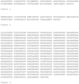
- activin A exists as a homodimer of the polypeptide represented by the amino acid sequence provided in Figure 1 as SEQ ID NO: 1.
- BMP4 refers to bone morphogenic protein 4.
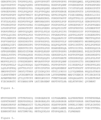
- BMP4 exists as a homodimer of the polypeptide represented by the amino acid sequence provided in Figure 2 as SEQ ID NO: 2.
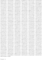
- type I collagen or "collagen I” exists as a triple helix trimer comprising two of the polypeptide represented by the amino acid sequence provided in Figure 3 as SEQ ID NO: 3, and a third collagen I chain.
- type IV collagen or "collagen IV” exists as a triple helix heterotrimer comprising one of the polypeptide represented as the amino acid sequence provided in Figure 4 as SEQ ID NO: 4, and two additional collagen IV chains.
- FGF2 refers to fibroblast growth factor 2, also known as basic fibroblast growth factor.
- FGF2 exists as a polypeptide represented as the amino acid sequence provided in Figure 5 as SEQ ID NO: 5.
- fibronectin exists as a dimer of the polypeptide represented by the amino acid sequence provided in Figure 6 as SEQ ID NO: 6.
- PDGF refers to platelet derived growth factor.
- PDGF exists as a homodimer of the B subunit polypeptide represented by the amino acid sequence provided in Figure 7 as SEQ ID NO: 7 (PDGF-BB). It is disclosed herein that supplementing M-CFM with PDGF (10 ng/mL) during culture had a significant negative impact on immune-suppression of iPSC-derived MSCs (4-fold reduction).
- tenascin C exists as a hexamer of the polypeptide represented by the amino acid sequence provided in Figure 8 as SEQ ID NO: 8.
- adherent human PSCs are cultured on tenascin C or provided on a substrate treated with tenascin C.
- any of the above-referenced cells e.g., human PSCs, human iPSCs
- any of the described cells are seeded on a substrate treated with an amount of tenascin C sufficient to adhere 10,000 cells/cm 2 to the substrate.
- the tenascin C is human tenascin C (e.g. comprising an amino acid sequence represented by SEQ ID NO: 8. or provided by GenBank Accession No.
- the substrate is treated with tenascin C at a concentration of at least about 0.25 ⁇ g/cm 2 to about 1 ⁇ g/cm 2 , e.g., 0.3 ⁇ g/cm 2 , 0.4 ⁇ g/cm 2 , 0.5 ⁇ g/cm 2 , 0.6 ⁇ g/cm 2 , 0.7 ⁇ g/cm 2 , 0.8 ⁇ g/cm 2 , or 0.9 ⁇ g/cm 2 .
- Cells can be grown on, e.g., tenascin C-coated cell culture dishes, multi-well cell culture plates, or microcarrier beads.
- tenascin C and hypoxic conditions may enable generation of EMH lin - KDR + APLNR + PDGFRalpha + primitive mesoderm cells with mesenchymoangioblast (MCA) potential and mesenchymal stem cells at higher percentages than compared to cells cultured on collagen IV or OP9 cells, such as greater than 10%, or greater than about 20%, or greater than about 30%, or greater than about 35%, or greater than about 40%, or greater than about 50%, or greater than about 60%, or greater than about 70%, or greater than about 80%, when compared per stage per platform.
- MCA mesenchymoangioblast
- “superior T-cell immunosuppressive properties” refers to the capacity of a MSC to produce a greater magnitude of suppression of proliferation of T helper (CD4 + ) lymphocytes, relative to a reference, for example as determined using an ImmunoPotency Assay.
- Superior T-cell immunosuppressive properties may be about a 1% increase, about a 2% increase, about a 3% increase, about a 4% increase, about a 5% increase, about a 6% increase, about a 7% increase, about a 8% increase, about a 9% increase, about a 10% increase, about a 20% increase, about a 30% increase, about a 40% increase, about a 50% increase, about a 60% increase, about a 70% increase, about a 80% increase, about a 90% increase, about a 100%, or greater increase in T-cell immunosuppressive properties in an MSC or a population of MSCs produced according to the method disclosed herein.
- superior T-cell immunosuppressive properties may be about a 2-fold, about a 3-fold, about a 4-fold, about a 5-fold, about a 6-fold, about a 7-fold, about a 8-fold, about a 9-fold, about a 10-fold, or more increase in T-cell immunosuppressive properties in an MSC or a population of MSCs produced according to the method disclosed herein.
- a suitable ImmunoPotency Assay uses an irradiated test MSC (e.g. iPSC-MSC produced according to the method disclosed herein) and an irradiated reference sample MSC, which are plated separately at various concentrations with carboxyfluorescein succinimidyl ester-labelled leukocytes purified from healthy donor peripheral blood.
- T helper (CD4+) lymphocytes that represent a subset of the reference sample are stimulated by adding CD3 and CD28 antibodies.
- CD4 labelled T cells are enumerated using flow cytometry to assess T cell proliferation. IC50 values are reported as a function of the reference sample.
- a higher IC50 value indicates a greater magnitude of suppression of proliferation of T helper (CD4 + ) lymphocytes and thus is indicative of superior T-cell immunosuppressive properties.
- MSC samples are irradiated prior to use in this assay to eliminate the confounding factor of their proliferative potential.
- the method and its resulting MSC or population of MSCs disclosed herein are suitable for medical treatment of humans, they are also applicable to veterinary treatment, including treatment of domestic animals such as horses, cattle and sheep, companion animals such as dogs and cats, or zoo animals such as felids, canids, bovids and ungulates.
- GLUTAMAX comprises L-alanyl-L-glutamine dipeptide, usually supplied at 200 mM in 0.85% NaCl. GLUTAMAX releases L-glutamine upon cleavage of the dipeptide bond by the cells being cultured.
- Chemically defined lipid concentrate comprises arachidonic acid 2 mg/L, cholesterol 220 mg/L, DL-alpha-tocopherol acetate 70 mg/L, linoleic acid 10 mg/L, linolenic acid 10 mg/L, myristic acid 10 mg/L, oleic acid 10 mg/L, palmitic acid 10 mg/L, palmitoleic acid 10 mg/L, pluronic F-68 90 g/L, stearic acid 10 mg/L, TWEEN 80 ® 2.2 g/L, and ethyl alcohol.
- H-1152 and Y27632 are highly potent, cell-permeable, selective ROCK (Rho-associated coiled coil forming protein serine/threonine kinase) inhibitors.
- Table 2. IF6S medium (10X concentration) 10X IF6S Quantity Final Concentration IMDM 1 package, powder for 1 L 5X Ham's F12 nutrient mix 1 package, powder for 1 L 5X sodium bicarbonate 4.2 g 21 mg/mL L-ascorbic acid 2-phosphate Mg 2+ 128 mg 640 ug/mL 1-thioglycerol 80 ⁇ L 4.6 mM sodium selenite (0.7 mg/mL solution) 24 ⁇ L 84 ng/mL GLUTAMAX 20 mL 10X non essential amino acids 20 mL 10X chemically defined lipid concentrate 4 mL 10X embryo transfer grade water To 200 mL NA Table 3.
- IF9S medium (1X concentration; based on IF6S)
- IF9S Quantity Final Concentration IF6S 5 mL 1X polyvinyl alcohol (PVA; 20 mg/mL solution) 25 mL 10 mg/mL holo-transferrin (10.6 mg/mL solution) 50 ⁇ L 10.6 ⁇ g/mL insulin 100 ⁇ L 20 ug/mL embryo transfer grade water To 50 mL NA Table 4.
- Differentiation medium (1X concentration; based on IF9S) Differentiation Medium Quantity Final Concentration IF9S 36 mL 1X FGF2 1.8 ⁇ g 50 ng/mL LiCl (2M solution) 36 ⁇ L 2mM BMP4 (100 ⁇ g/mL solution) 18 ⁇ L 50 ng/mL Activin A (10 mg/mL solution) 5.4 ⁇ L 1.5 ng/mL Table 5.
- Example 2 Protocol for differentiating a human PSC into a MSC
- T cell suppression was evaluated generated using Waisman Biomanufacturing's ImmunoPotency Assay (IPA).
- This assay is designed to assess the degree to which each MSC line can suppress the proliferation of T helper (CD4+) lymphocytes.
- Cryopreserved MSCs are tested using cryopreserved leukocytes purified from the peripheral blood of healthy individuals (peripheral blood mononucleocyte cells (PBMC) derived from Leucopaks (LPK)). As such, LPK cell population variation is expected from donor to donor.
- PBMC peripheral blood mononucleocyte cells
- LPK Leucopaks
- Each MSC test sample is tested against the PMBC from two different individuals for clinical grade material with the option to limit testing to a single PMBC sample for research grade material.
- the assay for each MSC test sample is run in conjunction with a reference standard MSC line to ensure assay integrity/ reproducibility and to normalize test samples. The assay is described in Bloom et al. Cytotherapy, 2015, 17(2):140-51 .
- test MSCs are exposed to 21 Gy of gamma irradiation.
- tissue culture plate 4 ⁇ 10e 5 , 2 ⁇ 10e 5 , 4 ⁇ 10e 4 , and 2 ⁇ 10e 4 irradiated MSCs are plated into individual wells.
- PMBC are separately labelled with carboxyfluorescein succinimidyl ester.
- Labelled PMBC cells are plated at 4 ⁇ 10 5 cells per well containing the MSCs above. This results in titrated PBMC:MSC ratios of 1:1, 1:0.5, 1:0.1, and 1:0.05.
- An additional well is plated with stimulated PBMCs alone, another with MSCs alone, and another 1:0.05 ratio without stimulation, all which serve as controls.
- T cell-stimulatory monoclonal antibodies, anti-human CD3-epilson and anti-human CD28 R&D Systems, Inc., Minneapolis, MN
- MSC alone control serves to gate out MSCs from co-culture wells.
- PBMC alone control serves as the positive control for maximum T cell proliferation against which the degree of MSC mediated suppression is measured.
- the non-stimulated 1:0.05 ratio well is used to generate a negative control gate against which proliferation is measured.
- IC50 values are normalized to the reference standard (IC50 Ref Std/IC50 Test Sample). This normalized IC50 yields larger values for more potent (more suppressive) samples and smaller values for less potent samples.
- IC50 data presented in Table 7 show that M-CFM supplemented with LiCl, but excluding PDGF (i.e. PDGF-/LiCl+) is optimal for differentiating of iPSCs to produce iPSC-MSCs that are immunosuppressive. Furthermore, a lower concentration of activin A also improved the immunosuppression of iPSC-MSCs. Table 7.
- the MSC produced according to Example 2 underwent analysis against a microRNA (miRNA) microarray comprising 1194 miRNAs and a proprietary miRNA panel consisting of miR-127-3p, miR-145-5p, miR-181b-5p, miR-214-3p, miR-299-5p. Each of the panel of 5 miRNAs was expressed in all of 71 MSC samples, but not 94 non-MSC samples, thereby enabling classification of cells as MSC or non-MSC.
- miRNA microRNA
- the MSC produced according to Example 2 expressed each of miR-145-5p, miR-181b-5p, and miR-214-3p, but not miR-127-3p and miR-299-5p.
- Immunopotency of MSCs is evaluated as follows: human PBMCs from various donors are pooled (to minimise inter-individual variability in immune response) in phosphate-buffered saline and stained with carboxyfluorescein succinimidyl ester (CFSE, 2 ⁇ M) for 15 minutes at 37 °C in the dark, at a cell density of 2 ⁇ 10 7 PBMCs/mL. The reaction is stopped by adding an equal amount of RPMI-1640 medium supplemented with 10 % human blood group AB serum.
- CFSE carboxyfluorescein succinimidyl ester
- T-cell proliferation is determined using a Gallios 10-color flow cytometer and the Kaluza G1.0 software (both Coulter).
- Viable 7-aminoactinomycin-D-excluding (7-AAD-; BD Pharmingen) CD3-APC+ (eBioscience) T cells are analysed after 4 to 7 days. Proliferation kinetics and population distribution are analysed using Modfit 4.1 software (Verity).
- T helper (CD4 + ) lymphocytes are stained with CellTrace violet (CTV; Invitrogen) according to the manufacturer's instructions and then stimulated with anti-CD3/CD28-coated beads (Dynabeads, Invitrogen) at a T-cell/bead ratio of 5:1 in 96-well U-bottomed plates.
- Responder CD4 T cells are then incubated with irradiated (at 100 Gy) Karpas 299 cells (K299 cells; Sigma) as a reference standard, or MSCs.
- the co-cultured cells are incubated at 37°C in 5% CO 2 in RPMI-1640 medium for 72 h.
- MSCs Mesenchymal stem cells of the present disclosure were used in an experimental rat model of myocardial infarction (heart attack) to repair the rat heart after a heart attack.
- Cardiac function and scar size were assessed over a 28 day period after a heart attack was induced in a total of 11 rats.
- Four animals were treated with the MSCs of the disclosure, three animals were treated with bone marrow-derived MSCs, and a further four animals received a placebo/ vehicle control ( figure 10 ).
- arrhythmia will be assessed using programmed ventricular stimulation (EPS).
- EPS programmed ventricular stimulation
- mice were randomly assigned to treatment groups, and in all cases, the relevant treatment was injected via the tail vein.
- NSG NOD.Cg- Prkdcscid Il2rgtm1Wjl /SzJ mice
- mice were lightly irradiated with 2.5Gy, then rested for 4 hours.
- GvHD was induced by intravenous (tail vein) transfer of 10 million human PBMCs.
- mice were housed under pathogen-free conditions in micro-isolator cages and received acidified, antibiotic-supplemented water throughout the duration of the experimental procedures.
- GvHD signs/ symptoms i.e. weight loss, posture, activity, fur texture and skin integrity - as described in Table 9.
- An overall GvHD score was calculated by assigning 1 point for each Grade 1 sign/ symptom and 2 points for each Grade 2 sign/ symptom. Once a GvHD score of 8 or higher was reached in any animal, it was euthanised. Table 9.
- the primary endpoint was duration of survival.
- iPSC-MSCs were thawed, washed, and resuspended in sterile PBS. Two million iPSC-MSCs were administered to relevant animals through the tail vein on day +14 (d14, single dose regimen) or on days +14 and +18 (d14, d18, dual-dose regimen).
- GvHD The severity of GvHD was assessed using a standardised scoring system, which included five different criteria: weight loss, posture, activity, fur texture, and skin integrity. Mice were evaluated daily and scored from 0 (the least severe) to 2 (the most severe) for each criterion. Clinical scores were generated by adding scores for the five criteria. When a clinical score of "8" was reached, mice were removed from the study and humanely euthanised. The day of removal from the study was recorded as the day of lethal GvHD induction. Survival benefit was determined using Kaplan-Meier analysis with an applied log-rank test. p values of ⁇ 0.05 were considered significantly different.
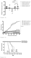
- mice treated with single- or dual-dose regimens showed significant attenuation of disease symptoms, with further significant differences in symptoms noted between single- and dual-dose treatments on days +24 and +25 post-GvHD-induction ( Figure 19 ).
- CYMERUS TM iPSC-MSCs protect mice from weight loss, significantly attenuate disease severity, and provide a robust survival benefit when administered either in single- or dual-dose treatments, in a pre-clinical model of GvHD.
Landscapes
- Health & Medical Sciences (AREA)
- Life Sciences & Earth Sciences (AREA)
- Engineering & Computer Science (AREA)
- Biomedical Technology (AREA)
- Chemical & Material Sciences (AREA)
- Bioinformatics & Cheminformatics (AREA)
- Organic Chemistry (AREA)
- Developmental Biology & Embryology (AREA)
- Zoology (AREA)
- Biotechnology (AREA)
- General Health & Medical Sciences (AREA)
- Wood Science & Technology (AREA)
- Genetics & Genomics (AREA)
- Immunology (AREA)
- Cell Biology (AREA)
- Rheumatology (AREA)
- Medicinal Chemistry (AREA)
- Pharmacology & Pharmacy (AREA)
- Animal Behavior & Ethology (AREA)
- Public Health (AREA)
- Veterinary Medicine (AREA)
- Microbiology (AREA)
- General Engineering & Computer Science (AREA)
- Biochemistry (AREA)
- General Chemical & Material Sciences (AREA)
- Nuclear Medicine, Radiotherapy & Molecular Imaging (AREA)
- Chemical Kinetics & Catalysis (AREA)
- Hematology (AREA)
- Reproductive Health (AREA)
- Gynecology & Obstetrics (AREA)
- Virology (AREA)
- Epidemiology (AREA)
- Transplantation (AREA)
- Heart & Thoracic Surgery (AREA)
- Cardiology (AREA)
- Orthopedic Medicine & Surgery (AREA)
- Physical Education & Sports Medicine (AREA)
- Micro-Organisms Or Cultivation Processes Thereof (AREA)
- Medicines Containing Material From Animals Or Micro-Organisms (AREA)
- Peptides Or Proteins (AREA)
Description
- The invention relates to a method of differentiating a human pluripotent stem cell (PSC) into a mesenchymal stem cell (MSC), and to compositions useful for such differentiation.
- Pluripotent stem cells (PSCs), in particular human PSCs, which include Embryonic Stem Cells (ESCs) and induced Pluripotent Stem Cells (iPSCs), provide the opportunity to study human development in vitro and develop novel cell-based therapeutic products. The use of PSCs as a starting material for the manufacture of therapeutic products has several advantages, including the potential to manufacture a virtually limitless number of cells from a single cell bank, as a result of the capacity of PSCs to reproduce indefinitely in vitro, and to differentiate into any cell type.
- Most protocols differentiate human PSCs grown on mouse embryonic fibroblasts (MEFs) or MATRIGEL®, a solubilised basement membrane preparation extracted from the Engelbreth-Holm-Swarm (EHS) mouse sarcoma. A chemically-defined, xenogen-free medium and matrix for human PSC-derivation and maintenance has been described. A chemically-defined, xenogen-free directed differentiation protocol for deriving hematopoietic progenitors from human PSCs has also been described in
US 2014/0273211 A1 . - In particular,
US 2014/0273211 A1 describes a method for differentiating human pluripotent stem cells comprising:
(a) providing human pluripotent stem cells; and (b) culturing the human pluripotent stem cells under hypoxic conditions in a cell culture medium comprising FGF2, BMP4, Activin A, and LiCl for a period of about two days to form a cell population of EMH lin-KDR+APLNR+PDGFRalpha+ primitive mesoderm cells with mesenchymoangioblast potential.US 2014/0273211 A1 also describes a xenogen-free culture medium for differentiating human pluripotent stem cells, comprising IF9S medium supplemented with: about 50 to about 250 ng/mL BMP4; about 10 to about 15 ng/mL Activin A; about 10 to about 50 ng/mL FGF2; and about 1 mM to about 2 mM LiCl. - However,
US 2014/0273211 A1 is silent in respect of any improved mesenchymal-colony forming medium (M-CFM). - Mesenchymal stem cells (MSCs) hold significant promise as a cell therapy and are currently in clinical trials for treating numerous diseases, including graft-versus host disease. Induced pluripotent stem cell (iPSC)-derived MSCs have a unique advantage over directly sourced MSCs, i.e. derived from tissues such as bone marrow, umbilical cord blood, adipose tissue, because in vitro expansion of iPSCs can provide a virtually unlimited supply of MSCs.
- Nevertheless, despite their promise, there remain challenges in producing MSCs in vitro, for example scaling up production from laboratory scale to good manufacturing practice (GMP) scale, which for any biological product is neither facile nor necessarily predictable, as will be appreciated in the art.
- It follows that there is a need for improved and scalable methods for differentiating PSCs, including iPSCs, into MSCs.
- The inventors have identified a specific composition for a differentiation medium that differentiates PSCs to MSCs, wherein such MSCs possess superior immunosuppressive properties relative to MSCs differentiated in the absence of the differentiation medium disclosed herein. Furthermore, the method is applicable to GMP-scale, i.e. a scale sufficient to supply MSCs for therapeutic use.
- In a first aspect, the invention provides a method for producing an MSC, the method comprising:
- (a) culturing a primitive mesoderm cell in a M-CFM comprising LiCl and FGF2, but excluding PDGF, under normoxic conditions for sufficient time for a mesenchymal colony to form; and
- (b) culturing the mesenchymal colony of (a) adherently to produce the MSC,
- The primitive mesodermal cell is a primitive mesodermal cell with mesenchymoangioblast (having a EMH lin-KDR+APLNR+PDGFRalpha+ phenotype.
- In one embodiment, sufficient time for the mesenchymal colony to be produced is about 8 days to about 14 days. In one embodiment, sufficient time for the mesenchymal colony to be produced is about 12 days.
- In one embodiment, T-cell immunosuppressive properties of the MSC are determined relative to an MSC produced by a method comprising:
- (a') culturing a primitive mesodermal cell in a medium comprising FGF2 and optionally PDGF, but excluding LiCl, under normoxic conditions for sufficient time for a mesenchymal colony to form; and
- (b') culturing the mesenchymal colony of (a') adherently to produce the MSC.
- In one embodiment, T-cell immunosuppressive properties of the MSC are determined relative to an MSC produced from a primitive mesodermal cell differentiated from a PSC in a differentiation medium comprising FGF2, BMP4, Activin A, and LiCl under hypoxic conditions for about two days.
- This provides an MSC produced by the method of the first aspect and a population of MSCs, comprising the MSC of the first aspect and a therapeutic composition comprising the MSC, or the population of MSCs as well as a kit comprising a container containing the MSC or the population of MSCs, or the therapeutic composition.
- Further disclosed is a mesenchymal colony forming medium (M-CFM), comprising about 1 mM LiCl, and about 5 ng/mL to about 100 ng/mL FGF2, or about 10 ng/mL to about 50 ng/mL FGF2, about 10 ng/mL FGF2, or about 20 ng/mL FGF2, but excluding PDGF or a composition comprising LiCl and FGF2, but excluding PDGF, that, when admixed with a liquid comprising water, produces a M-CFM comprising about 1 mM LiCl, and about 5 ng/mL to about 100 ng/mL FGF2, or about 10 ng/mL to about 50 ng/mL FGF2, about 10 ng/mL FGF2, or about 20 ng/mL FGF2, but excluding PDGF.
- Also disclosed is a method of differentiating a pluripotent stem cell (PSC) into a mesenchymal stem cell (MSC), the method comprising:
- (a) culturing the PSC in a differentiation medium comprising FGF2, BMP4, Activin A, and LiCl under hypoxic conditions for about two days to form a primitive mesoderm cell;
- (b) replacing the differentiation medium of (a) with a mesenchymal colony forming medium (M-CFM) comprising LiCl and FGF2, but excluding PDGF;
- (c) culturing the primitive mesoderm cell of (b) in the M-CFM of (b) under normoxic conditions for sufficient time for a mesenchymal colony to form; and
- (d) culturing the mesenchymal colony of (c) adherently to produce the MSC,
- In one embodiment, the method above further comprises:
(e) expanding the MSC of (d). - The primitive mesodermal cell is a primitive mesodermal cell with mesenchymoangioblast (MCA) potential. In one embodiment, the primitive mesodermal cell with MCA potential is a EMH lin-KDR+APLNR+PDGFRalpha+ primitive mesoderm cell with MCA potential.
- In one embodiment, sufficient time for the mesenchymal colony to be form is about 8 days to about 14 days. In one embodiment, sufficient time for the mesenchymal colony to form is about 12 days.
- In one embodiment, T-cell immunosuppressive properties of the MSC are determined relative to an MSC produced from a primitive mesodermal cell cultured in a medium comprising FGF2 and optionally PDGF, but excluding LiCl.
- In one embodiment, T-cell immunosuppressive properties of the MSC are determined relative to an MSC produced from a primitive mesodermal cell differentiated from a PSC in a differentiation medium comprising FGF2, BMP4, Activin A, and LiCl under hypoxic conditions for about two days.
- Embodiments of the invention are described below by way of example only with reference to the accompanying figures in which
-
Figure 1 is an amino acid sequence (SEQ ID NO: 1) representing a polypeptide that as a homodimer is one example of human activin A. -
Figure 2 is an amino acid sequence (SEQ ID NO: 2) representing a polypeptide that as a homodimer is one example of human BMP4. SEQ ID NO: 2 corresponds to amino acids 303 to 408 of an amino acid sequence of a full length BMP4 precursor. -
Figure 3 is an amino acid sequence (SEQ ID NO: 3) representing a polypeptide that is one example of a human type I collagen alpha1 chain. Human type I collagen (collagen I) is a triple helix trimer that in one example has two alphal chains represented by SEQ ID NO: 3 and one alpha2 chain. SEQ ID NO: 3 corresponds to amino acids 162 to 1218 of a full length type I collagen alpha1 chain precursor. -
Figure 4 is an amino acid sequence (SEQ ID NO: 4) representing a polypeptide that is one example of a human type IV collagen alpha1 chain. Human type IV collagen (collagen IV) is a triple helix heterotrimer that in one example comprises one alphal chain represented by SEQ ID NO: 4. SEQ ID NO: 4 corresponds to amino acids 173 to 1669 of a full length type IV collagen alphal chain precursor. -
Figure 5 is an amino acid sequence (SEQ ID NO: 5) representing a polypeptide that is one example of human FGF2. -
Figure 6 is an amino acid sequence (SEQ ID NO: 6) representing a polypeptide that as a dimer is one example of human fibronectin. SEQ ID NO: 6 corresponds to amino acids 32 to 2446 of a full length fibronectin precursor. -
Figure 7 is an amino acid sequence (SEQ ID NO: 7) representing a polypeptide that as a homodimer is one example of human PDGF (PDGF-BB). SEQ ID NO: 7 corresponds to amino acids 82 to 190 of a full length PDGF subunit B precursor. -
Figure 8 is an amino acid sequence (SEQ ID NO: 8) representing a polypeptide that as a hexamer is one example of human tenascin C. SEQ ID NO: 8 corresponds to amino acids 23 to 2201 of a full length tenascin C precursor. -
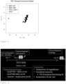
Figure 9 is a plot representing a principal component analysis of miRNA expression of an MSC of the disclosure and 71 other MSC samples obtained by means other than the present method. AD, adipose (■); BM, bone marrow (◆); iPSC_MSC, MSC of the disclosure (▲); iPSC, iPSC before differentiation to an MSC of the disclosure (+). -
Figure 10 is a schematic representation of the study of example 6. -
Figure 11 is a schematic representation of the histology of the study of example 6. -
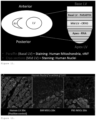
Figure 12 comprises fluorescent photomicrographs of engraftment (human nuclei staining) of MSCs in the hearts of example 6. Human nuclei staining was performed at all levels in mid LV (cryo sections). There was no evidence of cell engraftment atday 28. Human mitochondria staining was performed at basal LV (paraffin sections). There was no evidence of cell engraftment atday 28. -
Figure 13 shows functional assessment (TTE) and comprises short axis of M-mode images of hearts of vehicle, BM MSC, and PSC-MSC recipients of example 6 at baseline and 1 month time points. The images show reduced ventricular dilatation and increased ventricular contractility in the PSC-MSC recipients compared to the vehicle and BM MSC recipients. -
Figure 14 shows functional assessment (TTE) and comprises line graphs showing individual assessments and column graphs showing group assessment of fractional shortening (FS) of the infarcted hearts of example 6. In the vehicle group, baseline FS was higher in 1 rat injected with vehicle, but overall, no or minimal change in FS onday 28 compared today 0 except for 1 rat (mean increase 1.39%). In the BM MSC group, 3/3 rats had worsening FS onday 28 compared to day 0 (mean decrease 2.3%). In the PSC-MSC group, 4/4 rats had increase in FS onday 28 compared to day 0 (mean increase 5.48%). -
Figure 15 comprises photomicrographs showing scar size using picosirius red staining in infarcted hearts of example 6. The images show an obvious reduction in scar size using Picosirius Red Staining in the PSC-MSC treated groups compared to the vehicle and BM-MSC treated groups. -
Figure 16 comprises fluorescent photomicrographs assessing angiogenesis (vWF staining) in the hearts of example 6. The images show no difference in size and number of vessels between groups. -
Figure 17 is a schematic representation of a study similar to example 6. -
Figure 18 is a scatter plot of percent weight change at day +19 of mice of example 7 relating to GvHD. -
Figure 19 is a line graph showing GvHD disease severity in mice of example 7. -
Figure 20 is a Kaplan-Meier plot showing GvHD survival of mice of example 7. - The inventors have defined an improved chemically-defined, xenogen-free differentiation protocol, including in particular, an improved chemically-defined, xenogen-free mesenchymal colony forming protocol. Surprisingly, the improved protocols provide MSCs with superior immunosuppressive properties when compared to existing differentiation and mesenchymal colony forming protocols.
- In developing the improved chemically-defined, xenogen-free differentiation protocol, including the improved mesenchymal colony forming protocol, the inventors investigated several variables, including: presence/ absence of PDGF; presence/ absence of LiCl; low/ high activin A concentration; and low/ high seed density.
- PDGF was tested because it was asserted in
US 7,615,374 that "PDGF-BB improved growth of mesenchymal cells, but was not essential for colony formation". Surprisingly, however, the inventors describe herein that contrary to this prior teaching, absence of PDGF generated MSCs with superior immunosuppressive properties relative to MSCs generated by differentiation in medium comprising PDGF. - With respect to activin A, the inventors surprisingly found that immunosuppression was improved in MSCs differentiated in medium comprising the lower concentration of activin A relative to the higher concentration of activin A.
- LiCl activates Wnt signaling and is included in differentiation medium to improve mesoderm induction during the first 48 hours of differentiation, which is understood in the art.
- However, before the present invention, LiCl had not been used in a M-CFM to improve colony formation in clonogenic cultures. Again, this was a surprising advantage of the method disclosed herein.
- Accordingly, disclosed herein are methods for differentiating PSCs (e.g. human ESCs or human iPSCs) under defined conditions. Such differentiation provides an MSC or MSC populations that may be a source for functional studies of these lineages as well as a source for clinical therapies.
- The ability of MSCs to exert immunomodulatory/ immunosuppressive effects, in particular by suppressing T cells, is believed to be central to the therapeutic effects of MSCs in a wide range of conditions, including GvHD, immune disorders including autoimmune disorders, cardiovascular disorders, orthopaedic disorders and rejection of transplanted solid organs. Some specific examples include bone cysts, bone neoplasms, fractures, cartilage defects, osteoarthritis, ligament injury, osteogenesis imperfecta, osteonecrosis, osteoporosis, aplastic anaemia, myelodysplastic syndrome; Type 1 diabetes,
Type 2 diabetes, autoimmune hepatitis, liver cirrhosis, liver failure, dilated cardiomyopathy, heart failure, myocardial infarction, myocardial ischemia, Crohn's disease, ulcerative colitis, burns, epidermolysis bullosa, lupus erythematosus, rheumatoid arthritis, Sjogren's disease, systemic sclerosis, bronchopulmonary dysplasia, chronic obstructive airways disease, emphysema, pulmonary fibrosis, ALS, Alzheimer's disease, brain injury, ataxia, degenerative disc disease, multiple system atrophy, multiple sclerosis, Parkinson's disease, retinitis pigmentosa, Romberg's disease, spinal cord injury, stroke, muscular dystrophy, limb ischaemia, kidney injury, lupus nephritis, endometriosis and complications of bone marrow or solid organ transplantation, - MSCs are thought to perform a critical role in injury healing, and have been shown to be effective in treating tissue injury and degenerative diseases, including in the digestive system, for example in liver cirrhosis and liver failure, in the musculoskeletal system, in periodontal tissue, in diabetic critical limb ischemia, in osteonecrosis, in burn-related disorders, in myocardial infarction, in cornea damage, in the brain, in the spinal cord, in the lungs, and in treating radiation exposure.
- MSCs have shown therapeutic outcomes in immune disorders, including graft-versus-host disease, systemic lupus erythematosus (SLE), Crohn's disease, multiple system atrophy, multiple sclerosis, amyotrophic lateral sclerosis, and stroke.
- MSCs have been shown to exert immunosuppressive activities against T cells, B cells, dendritic cells, macrophages, and natural killer cells. While not wishing to be bound by theory, the underlying mechanisms may comprise immunosuppressive mediators, for example nitric oxide,
indoleamine - MSCs may be engrafted into damaged tissues following administration and migration.
- MSCs may be administered systemically or peripherally, for example by routes including intravenous (IV), intra-arterial, intramuscular, intraperitoneal, intracerobrospinal, subcutaneous (SC), intra-articular, intrasynovial, intrathecal, intracoronary, transendocardial, surgical implantation, topical and inhalation (e.g. intrapulmonary). MSCs may be administered in combination with a scaffold of biocompatible material.
- MSCs may be administered before, during or after injury, disorder or disease progression. In one embodiment, MSCs are administered during inflammation. MSCs may be administered (a) as a preventative measure, (b) as soon as the relevant condition has been diagnosed, (c) when other treatments fail, and/or (d) when a condition advances to a pre-defined degree of severity.
- In one embodiment, the obtained MSCs are pre-treated prior to administration. Pre-treatment may be with a growth factor or by gene editing, for example, where a growth factor may prime the MSC and gene editing may confer a new therapeutic property on the MSC.
- Unless defined otherwise in this specification, technical and scientific terms used herein have the same meaning as commonly understood by the person skilled in the art to which this invention belongs and by reference to published texts.
- It is to be noted that the term "a" or "an" refers to one or more, for example, "a molecule," is understood to represent one or more molecules. As such, the terms "a" or "an", "one or more," and "at least one" may be used interchangeably herein.
- In the claims which follow and in the description of the invention, except where the context requires otherwise due to express language or necessary implication, the word "comprise" or variations such as "comprises" or "comprising" is used in an inclusive sense, i.e. to specify the presence of the stated features but not to preclude the presence or addition of further features in various embodiments of the invention.
- The term "about" as used herein contemplates a range of values for a given number of ±25% the magnitude of that number. In other embodiments, the term "about" contemplates a range of values for a given number of ±20%, ±15%, ±10%, or ±5% the magnitude of that number. For example, in one embodiment, "about 3 grams" indicates a value of 2.7 to 3.3 grams (i.e. 3 grams ±10%), and the like.
- Similarly, while differentiation processes include ordered, sequential events, the timing of the events may be varied by at least 25%. For example, while a particular step may be disclosed in one embodiment as lasting one day, the event may last for more or less than one day. For example, "one day" may include a period of about 18 to about 30 hours. In other embodiments, periods of time may vary by ±20%, ±15%, ±10%, or ±5% of that period of time. Periods of time indicated that are multiple day periods may be multiples of "one day," such as, for example, two days may span a period of about 36 to about 60 hours, and the like. In another embodiment, time variation may be lessened, for example, where
day 2 is 48±3 hours fromday 0;day 4 is 96±3 hours fromday 0, and day 5 is 120 hours±3 hours fromday 0. - As used herein, "pluripotent stem cell" or "PSC" refers to a cell that has the ability to reproduce itself indefinitely, and to differentiate into any other cell type. There are two main types of pluripotent stem cell: embryonic stem cells (ESCs) and induced pluripotent stem cells (iPSCs).
- As used herein, "induced pluripotent stem cell" or "iPSC" refers to an ESC-like cell derived from adult cells. iPSCs have very similar characteristics to ESCs, but avoid the ethical concerns associated with ESCs, since iPSCs are not derived from embryos. Instead, iPSCs are typically derived from fully differentiated adult cells that have been "reprogrammed" back into a pluripotent state.
- Suitable human iPSCs include, but are not limited to, iPSC 19-9-7T, MIRJT6i-mND1-4 and MIRJT7i-mND2-0 derived from fibroblasts and iPSC BM119-9 derived from bone marrow mononuclear cells. Other suitable iPSCs may be obtained from Cellular Dynamics International (CDI; Nasdaq: ICEL) of Madison, WI, USA.
- In some embodiments, the PSCs are plated at an initial density of about 5000 cells/cm2 to about 15,000 cells/cm2, e.g., 6000 cells/cm2, 7000 cells/cm2, 8000 cells/cm2, 9000 cells/cm2, 10,000 cells/cm2, 11,000 cells/cm2, 12,000 cells/cm2, 13,000 cells/cm2, or 14,000 cells/cm2.
- In one embodiment, sufficient time for the mesenchymal colony to be produced is about 8 days to about 14 days. In one embodiment, sufficient time for the mesenchymal colony to be produced is about 10 days to about 14 days. In one embodiment, sufficient time for the mesenchymal colony to be produced is about 11 days to about 13 days. In one embodiment, sufficient time for the mesenchymal colony to be produced is about 8 days, about 9 days, about 10 days, about 11 days, about 13 days, or about 14 days. In one embodiment, sufficient time for the mesenchymal colony to be produced is about 12 days.
- As used herein, " EMH lin-KDR+APLNR+PDGFRalpha+ primitive mesoderm cell with mesenchymoangioblast (MCA) potential" refers to a cell expressing typical primitive streak and lateral plate/extraembryonic mesoderm genes. These cells have potential to form mesenchymoangioblast (MCA) and hemangioblast colonies in serum-free medium in response to FGF2. According to the method of present invention, these cells become mesenchymal stem cells (MSCs).
- The term EMH lin- denotes lack of expression of CD31, VE-cadherin endothelial markers, CD73 and Cd105 mesenchymal/endothelial markers, and CD43 and CD45 hematopoietic markers.
- As used herein, "mesenchyme" or "mesenchymal" refers to to embryonic connective tissue that is derived from the mesoderm and that differentiates into hematopoietic tissue (lymphatic and circulatory systems) and connective tissue, such as bone and cartilage. However, MSCs do not differentiate into hematopoietic cells.
- As used herein, "mesenchymal colony" refers to CD73-mesenchymal progenitors that precede true MSCs. Additional adherent culture of mesenchymal colonies, e.g. on fibronectin/collagen-coated plates, is required to produce CD73+ MSCs.
- As used herein, "mesenchymal stem cell" or "MSC" refers to a particular type of stem cell that may be isolated from a wide range of tissues, including bone marrow, adipose tissue (fat), placenta and umbilical cord blood. MSCs are also known as "mesenchymal stromal cells". According to the method of present invention, MSCs are formed from EMH lin-KDR+APLNR+PDGFRalpha+ primitive mesoderm cells with mesenchymoangioblast (MCA) potential.
- MSCs secrete bioactive molecules such as cytokines, chemokines and growth factors and have the ability to modulate the immune system. MSCs have been shown to facilitate regeneration and effects on the immune system without relying upon engraftment. In other words, the MSCs themselves do not necessarily become incorporated into the host - rather, they exert their effects and are then eliminated within a short period of time. However, MSCs may be engrafted.
- Therapeutic MSCs can be either "autologous" or "allogeneic". As used herein, "autologous" means a patient is treated with their own cells isolated from bone marrow or adipose tissue, for example, whereas "allogeneic" means that cells from a donor are used to treat other people. According to the present disclosure, allogeneic MSCs are derived from a donor via an iPSC.
- Allogeneic MSCs have not been shown to cause immune reactions in other people, so they do not require immune-matching the donor to the recipient. This has important commercial advantages.
- As used herein, "mesenchymoangioblast" or "MCA" refers to a cell that is a precursor to a MSC. A MSC may be produced by the method of the invention.
- As used herein, "differentiating" refers to a process of a cell changing from one cell type to another, in particular a less specialized type of cell becoming a more specialized type of cell.
- As used herein, "medium" or its plural "media" refers to a liquid or gel designed to support the growth of cells. In some embodiments, the cell culture medium comprises an IF9S medium. In some embodiments, the medium employs a 9S concentrated medium supplement, wherein dilution of the 9S concentrated medium supplement in an IMDM/F12 base medium yields an IF9S cell culture medium. The concentrated 9S medium supplement comprises 9 supplements as follows: L-ascorbic acid 2-phosphate Mg2+ salt, 1-thioglycerol (monothioglycerol), additional sodium selenite, polyvinyl alcohol, GLUTAMAX™ (or glutamine), non-essential amino acids, chemically defined lipid concentrate, holo-transferrin, and insulin. In some embodiments, the concentrated 9S medium supplement comprises each component at a
concentration 10× to 1000× of the final working concentration once diluted in a base medium. In one embodiment, an IF9S medium comprises IMDM, F12 and 9S as follows: IMDM 0.5x, F12 0.5x, sodium bicarbonate 2.1 mg/mL, L-ascorbic acid 2-phosphate Mg2+ salt (64 µg/mL), 1-thioglycerol(50 µg/mL (460 µM, 40 µL/L)), sodium selenite (in addition to any present in the base medium; 8.4 ng/mL), polyvinyl alcohol (10 mg/mL), GLUTAMAX™ (1×), non-essential amino acids (1×), chemically defined lipid concentrate (1×), Holo-Transferrin (10.6 µg/mL), and insulin (20µg/mL). - Although the presently disclosed media may include specific components (e.g. morphogens, small molecules, and hematopoietic cytokines), it is contemplated that other components with the same, equivalent, or similar properties may be used in addition to or in place of those disclosed, as are known in the art.
- In some embodiments, components of an IF9S medium may be substituted. For example, ascorbic acid and 1-thioglycerol can be replaced with an optional supplement of a compound and/or a thiol-containing compound with antioxidant properties. GLUTAMAX™ can be replaced with an optional supplement of L-glutamine. "Non-essential amino acids", which is a general term for amino acids that the human body can produce from other amino acids, can be replaced with an optional supplement of amino acids. "Chemically defined lipid concentrate," which is a solution specifically distributed by Life Technologies, can be replaced with an optional supplement of lipids. Additional selenite, insulin, and holo-transferrin can be replaced with any insulin-transferrin-selenite supplement. Polyvinyl alcohol can be replaced with an optional supplement of a biologically inactive media thickening compound.
- As used herein, "differentiation medium" refers to a medium designed to support the differentiation of cells, that is, supporting the process of a cell changing from one cell type to another. According to the method of the present invention, a differentiation medium is used to support the process of changing a human PSC into a EMH lin-KDR+APLNR+PDGFRalpha+ primitive mesoderm cell with mesenchymoangioblast (MCA) potential.
- In some embodiments, the concentration, in the differentiation medium, of: BMP4 is about 10 ng/mL to about 250 mg/mL; activin A is about 1 ng/mL to about 15 ng/mL; FGF2 is about 5 ng/mL to about 50 ng/mL; and LiCl is about 1 mM to about 2 mM.
- In some embodiments, the concentration of BMP4 is in the differentiation medium is about 10 ng/mL, about 15 ng/mL, about 20 ng/mL, about 25 ng/mL, about 30 ng/mL, about 35 ng/mL, about 40 ng/mL, about 45 ng/mL, about 50 ng/mL, about 60 ng/mL, about 70 ng/mL, about 80 ng/mL, about 90 ng/mL, about 100 ng/mL, about 110 ng/mL, about 120 ng/mL, about 130 ng/mL, about 140 ng/mL, about 150 ng/mL, about 160 ng/mL, about 170 ng/mL, about 180 ng/mL, about 190 ng/mL, about 200 ng/mL, about 210 ng/mL, about 220 ng/mL, about 230 ng/mL, about 240 ng/mL, or about 250 ng/mL.
- The concentration of activin A in the differentiation medium may be about 1 ng/mL, about 2 ng/mL, about 3 ng/mL, about 4 ng/mL, about 5 ng/mL, about 6 ng/mL, about 7 ng/mL, about 8 ng/mL, about 9 ng/mL, about 10 ng/mL, about 11 ng/mL, about 12 ng/mL, about 13 ng/mL, about 14 ng/mL, or about 15 ng/mL.
- The concentration of FGF2 in the differentiation medium may be about 1 ng/mL, about 5 ng/mL, about 10 ng/mL, about 15 ng/mL, about 20 ng/mL, about 25 ng/mL, about 30 ng/mL, about 35 ng/mL, about 40 ng/mL, about 45 ng/mL, or about 50 ng/mL.
- The concentration of LiCl in the differentiation medium may be about 0.5 mM, about 0.6 mM, about 0.7 mM, about 0.8 mM, about 0.9 mM, about 1 mM, about 1.1mM, about 1.2 mM, about 1.3 mM, about 1.4 mM, about 1.5 mM, about 1.6 mM, about 1.7 mM, about 1.8 mM, about 1.9 mM, about 2 mM, about 2.1 mM, about 2.2 mM, about 2.3 mM, about 2.4 mM, or about 2.5 mM. Preferably, as understood in the art, concentration of LiCl in the differentiation medium may be about 2 mM.
- In some embodiments, the differentiation medium comprises a base medium, L-ascorbic acid 2-phosphate Mg2+ salt, 1-thioglycerol, sodium selenite (in addition to any present in the base medium), polyvinyl alcohol, GLUTAMAX™, non-essential amino acids, chemically defined lipid concentrate, holo-transferrin, and insulin. Suitable base media for the differentiation media described herein include, but are not limited to, Iscove's Modified Dulbecco's Medium/F12 (IMDM/F12), TeSR1 base medium (mTeSR1™ base medium, Stem Cell Technologies) without FGF2 and TGF-beta; DF4S base medium, which is Essential 8™ medium (Life Technologies; also known as "E8" medium) without FGF2 and TGF-beta, I4S base medium, which is DF4S base with Iscove's modified Dulbecco's medium (IMDM) instead of DMEM/F12, and IF4S base is DF4S base with IMDM/F12 instead of DMEM/F12. Preferably, the base medium to be used is albumin-free. IMDM/F12 is a highly enriched synthetic medium suited for rapidly proliferating, high-density cell cultures with an added nutrient mixture. These media are known to the person skilled in the art.
- In some embodiments, the medium referred to herein as "IF9S", comprises IMDM/F12, L-ascorbic acid 2-phosphate Mg2+ salt, 1-thioglycerol, sodium selenite (in addition to any present in the base medium), polyvinyl alcohol, GLUTAMAX™, non-essential amino acids, chemically defined lipid concentrate, Holo-Transferrin, and insulin.
- As used herein, "mesenchymal colony forming medium (M-CFM)" refers to a medium designed to support the formation of mesenchymal colonies from EMH lin-KDR+APLNR+PDGFRalpha+ primitive mesoderm cells with mesenchymoangioblast (MCA) potential.
- In some embodiments, the concentration, in the the M-CFM, of: LiCl is about 1 mM and FGF2 is about 10 ng/mL; or LiCl is about 1 mM and FGF2 is about 20 ng/mL.
- The concentration of FGF2 in the M-CFM may be about 1 ng/mL, about 5 ng/mL, about 10 ng/mL, about 15 ng/mL, about 20 ng/mL, about 25 ng/mL, about 30 ng/mL, about 35 ng/mL, about 40 ng/mL, about 45 ng/mL, or about 50 ng/mL.
- The concentration of LiCl in the M-CFM may be about 0.5 mM, about 0.6 mM, about 0.7 mM, about 0.8 mM, about 0.9 mM, about 1 mM, about 1.1mM, about 1.2 mM, about 1.3 mM, about 1.4 mM, about 1.5 mM, about 1.6 mM, about 1.7 mM, about 1.8 mM, about 1.9 mM, about 2 mM, about 2.1 mM, about 2.2 mM, about 2.3 mM, about 2.4 mM, or about 2.5 mM. Preferably, the concentration of LiCl in the M-CFM is 1 mM. As noted above, inclusion of LiCl in a M-CFM to improve colony formation in clonogenic cultures was not known prior to the present invention and provides clear advantages for the method disclosed herein over methods known in the art.
- As referred to herein, the term "defined medium" means that the identity and quantity of each component of a medium is known.
- In some embodiments, media disclosed herein comprise xenogenic materials. As used herein, "xenogen" or "xenogenic" refers to non-human, biologically derived materials. Nonetheless, xenogenic materials may be defined. For example, a xenogenic material may be a recombinant protein of xenogenic origin. In one embodiment, the differentiation medium and/or the M-CFM comprises one or more xenogenic (e.g., recombinant non-human proteins).
- In some embodiments, the medium is or the media are xenogen-free. Of central importance for clinical therapies is the absence of xenogenic materials in the derived cell populations, i.e., no non-human cells, cell fragments, sera, proteins, and the like. In one embodiment, xenogen-free differentiated cells are obtained using tenascin C or collagen IV, which essentially replaces contact with OP9 cells used in earlier differentiation systems.
- Advantageously, defined media may be xenogen-free, and incorporate human proteins isolated from natural sources, such as from placenta or other human tissues, or that can be produced using recombinant technology. In some embodiments, all proteins described herein are human. In some embodiments, all of the proteins used in the differentiation medium are human proteins. In some embodiments all of the proteins used in the M-CFM medium are human proteins. In some embodiments, all proteins described herein are human recombinant proteins. In some embodiments, all of the proteins used in the differentiation medium are recombinant human proteins. In some embodiments all of the proteins used in the M-CFM medium are recombinant human proteins.
- All proteins described herein are known to the person skilled in the art, and most if not all proteins described herein are available commercially.
- Media disclosed herein may be also made in concentrated, including dried, forms that are diluted prior to use, such as 2×, 10×, 100×, or 1000× concentrations.
- As used herein, "culturing" refers to the process by which cells are grown under controlled conditions.
- Cells cannot be held in culture indefinitely owing to the increasing concentration of toxic metabolites, decreasing concentration of nutrients, and, for dividing cells, an increasing number of cells. As used herein, "passaging" refers to the process of producing a new culture with refreshed concentrations of nutrients, no toxic metabolites, and optionally a lower density of cells than the originating culture.
- In one embodiment, passaging comprises: culturing the mesenchymal colony for about 3 days; culturing the mesenchymal colony on fibronectin and/or collagen I; and/or 1, 2, 3, 4, 5, or 6 passages. The number of passages may be 7, 8, 9, 10, 11, 12, 13, 14, 15, 16, 17, 18, 19, 20, 21, 22, 23, 24, or 25. Preferably the number of passages is 10 or fewer. More preferably, the number of passages is 5 or 6.
- As used herein, "hypoxic" refers to conditions in which the oxygen concentration of the gas mixture is about 3% O2 to about 10% O2. In some embodiments, hypoxic conditions are about 5% O2, but may be about 4% O2, about 6% O2, about 7% O2, about 8% O2, or about 9% O2. In embodiments where a cell culture medium is allowed to equilibrate under hypoxic conditions, the cell culture medium becomes hypoxic owing to the lower concentration of oxygen dissolved in the medium compared to a cell culture medium equilibrated under normoxic conditions.
- As used herein, "normoxic" refers to conditions in which the oxygen concentration of the gas mixture is about 20% O2, but may be about 18% O2, about 19% O2, about 21% O2, or about 22% O2.
- As used herein "AA" refers to activin A. In one embodiment, activin A exists as a homodimer of the polypeptide represented by the amino acid sequence provided in
Figure 1 as SEQ ID NO: 1. - As used herein, "BMP4" refers to bone
morphogenic protein 4. In one embodiment, BMP4 exists as a homodimer of the polypeptide represented by the amino acid sequence provided inFigure 2 as SEQ ID NO: 2. - In one embodiment, "type I collagen" or "collagen I" exists as a triple helix trimer comprising two of the polypeptide represented by the amino acid sequence provided in
Figure 3 as SEQ ID NO: 3, and a third collagen I chain. - In one embodiment, "type IV collagen" or "collagen IV" exists as a triple helix heterotrimer comprising one of the polypeptide represented as the amino acid sequence provided in
Figure 4 as SEQ ID NO: 4, and two additional collagen IV chains. - As used herein, "FGF2" refers to
fibroblast growth factor 2, also known as basic fibroblast growth factor. In one embodiment, FGF2 exists as a polypeptide represented as the amino acid sequence provided inFigure 5 as SEQ ID NO: 5. - In one embodiment, fibronectin exists as a dimer of the polypeptide represented by the amino acid sequence provided in
Figure 6 as SEQ ID NO: 6. - As used herein, "PDGF" refers to platelet derived growth factor. In one embodiment, PDGF exists as a homodimer of the B subunit polypeptide represented by the amino acid sequence provided in
Figure 7 as SEQ ID NO: 7 (PDGF-BB). It is disclosed herein that supplementing M-CFM with PDGF (10 ng/mL) during culture had a significant negative impact on immune-suppression of iPSC-derived MSCs (4-fold reduction). - In one embodiment, "tenascin C" exists as a hexamer of the polypeptide represented by the amino acid sequence provided in
Figure 8 as SEQ ID NO: 8. - In some embodiments, adherent human PSCs are cultured on tenascin C or provided on a substrate treated with tenascin C. In some embodiments, any of the above-referenced cells (e.g., human PSCs, human iPSCs) are cultured on tenascin C. In some embodiments, any of the described cells are seeded on a substrate treated with an amount of tenascin C sufficient to adhere 10,000 cells/cm2 to the substrate. In some embodiments, the tenascin C is human tenascin C (e.g. comprising an amino acid sequence represented by SEQ ID NO: 8. or provided by GenBank Accession No. CAA55309.1, or available commercially, e.g., Millipore Cat. No. CC065). In some embodiments, the substrate is treated with tenascin C at a concentration of at least about 0.25 µg/cm2 to about 1 µg/cm2, e.g., 0.3 µg/cm2, 0.4 µg/cm2, 0.5 µg/cm2, 0.6 µg/cm2, 0.7 µg/cm2, 0.8 µg/cm2, or 0.9 µg/cm2. Cells can be grown on, e.g., tenascin C-coated cell culture dishes, multi-well cell culture plates, or microcarrier beads.
- The use of tenascin C and hypoxic conditions may enable generation of EMH lin-KDR+APLNR+PDGFRalpha+ primitive mesoderm cells with mesenchymoangioblast (MCA) potential and mesenchymal stem cells at higher percentages than compared to cells cultured on collagen IV or OP9 cells, such as greater than 10%, or greater than about 20%, or greater than about 30%, or greater than about 35%, or greater than about 40%, or greater than about 50%, or greater than about 60%, or greater than about 70%, or greater than about 80%, when compared per stage per platform.
- As used herein, "superior T-cell immunosuppressive properties" refers to the capacity of a MSC to produce a greater magnitude of suppression of proliferation of T helper (CD4+) lymphocytes, relative to a reference, for example as determined using an ImmunoPotency Assay.
- Superior T-cell immunosuppressive properties may be about a 1% increase, about a 2% increase, about a 3% increase, about a 4% increase, about a 5% increase, about a 6% increase, about a 7% increase, about a 8% increase, about a 9% increase, about a 10% increase, about a 20% increase, about a 30% increase, about a 40% increase, about a 50% increase, about a 60% increase, about a 70% increase, about a 80% increase, about a 90% increase, about a 100%, or greater increase in T-cell immunosuppressive properties in an MSC or a population of MSCs produced according to the method disclosed herein. Alternatively, superior T-cell immunosuppressive properties may be about a 2-fold, about a 3-fold, about a 4-fold, about a 5-fold, about a 6-fold, about a 7-fold, about a 8-fold, about a 9-fold, about a 10-fold, or more increase in T-cell immunosuppressive properties in an MSC or a population of MSCs produced according to the method disclosed herein.
- A suitable ImmunoPotency Assay uses an irradiated test MSC (e.g. iPSC-MSC produced according to the method disclosed herein) and an irradiated reference sample MSC, which are plated separately at various concentrations with carboxyfluorescein succinimidyl ester-labelled leukocytes purified from healthy donor peripheral blood. T helper (CD4+) lymphocytes that represent a subset of the reference sample are stimulated by adding CD3 and CD28 antibodies. CD4 labelled T cells are enumerated using flow cytometry to assess T cell proliferation. IC50 values are reported as a function of the reference sample. A higher IC50 value indicates a greater magnitude of suppression of proliferation of T helper (CD4+) lymphocytes and thus is indicative of superior T-cell immunosuppressive properties. MSC samples are irradiated prior to use in this assay to eliminate the confounding factor of their proliferative potential.
- It will be appreciated by the person skilled in the art that the exact manner of administering to a subject a therapeutically effective amount of an MSC or a population of MSCs differentiated according to the present method for treating a condition, disease or disorder will be at the discretion of the medical practitioner. The mode of administration, including dosage, combination with other agents, timing and frequency of administration, and the like, may be affected by the diagnosis of a subject's likely responsiveness to treatment with the MSC or population of MSCs, as well as the subject's condition and history.
- Although it is particularly contemplated that the method and its resulting MSC or population of MSCs disclosed herein are suitable for medical treatment of humans, they are also applicable to veterinary treatment, including treatment of domestic animals such as horses, cattle and sheep, companion animals such as dogs and cats, or zoo animals such as felids, canids, bovids and ungulates.
-
Table 1. Reagents Description Vendor / Cat # or Ref # DMEM/F12 Base Medium Invitrogen / A1516901 E8 supplement Invitrogen / A1517101 vitronectin Life Technologies / A14700 collagen IV Sigma / C5533 H-1152 ROCK Inhibitor EMD Millipore / 555550 Y27632 dihydrochloride ROCK Inhibitor Tocris / 1254 FGF2 Waisman Biomanufacturing / WC-FGF2-FP human endothelial-SFM Life Technologies / 11111-044 stemline II hematopoietic stem cell expansion medium Sigma / S0192 GLUTAMAX Invitrogen / 35050-061 insulin Sigma / I9278 lithium chloride (LiCl) Sigma / L4408 collagen I solution Sigma / C2249 fibronectin Life Technologies / 33016-015 DMEM/F12 Invitrogen / 11330032 recombinant human BMP4 Peprotech / 120-05ET activin A Peprotech / 120-14E Iscove's modified Dulbecco's medium (IMDM) Invitrogen / 12200036 Ham's F12 nutrient mix Invitrogen / 21700075 sodium bicarbonate Sigma / S5761 L-ascorbic acid 2-phosphate Mg2+ Sigma / A8960 1-thioglycerol Sigma / M6145 sodium selenite Sigma / S5261 non essential amino acids HyClone / SH30853.01 chemically defined lipid concentrate Invitrogen / 11905031 embryo transfer grade water Sigma / W1503 polyvinyl alcohol (PVA) MP Bio / 151-941-83 holo-transferrin Sigma / T0665 ES-CULT M3120 Stem Cell Technologies / 03120 STEMSPAN serum-free expansion medium (SFEM) Stem Cell Technologies / 09650 L-ascorbic acid Sigma / A4544 PDGF-BB Peprotech / 110-14B - The reagents listed in Table 1 are known to the person skilled in the art and have accepted compositions, for example IMDM and Ham's F12. GLUTAMAX comprises L-alanyl-L-glutamine dipeptide, usually supplied at 200 mM in 0.85% NaCl. GLUTAMAX releases L-glutamine upon cleavage of the dipeptide bond by the cells being cultured. Chemically defined lipid concentrate comprises
arachidonic acid 2 mg/L, cholesterol 220 mg/L, DL-alpha-tocopherol acetate 70 mg/L,linoleic acid 10 mg/L,linolenic acid 10 mg/L,myristic acid 10 mg/L,oleic acid 10 mg/L,palmitic acid 10 mg/L,palmitoleic acid 10 mg/L, pluronic F-68 90 g/L,stearic acid 10 mg/L,TWEEN 80® 2.2 g/L, and ethyl alcohol. H-1152 and Y27632 are highly potent, cell-permeable, selective ROCK (Rho-associated coiled coil forming protein serine/threonine kinase) inhibitors.Table 2. IF6S medium (10X concentration) 10X IF6S Quantity Final Concentration IMDM 1 package, powder for 1 L 5X Ham's F12 nutrient mix 1 package, powder for 1 L 5X sodium bicarbonate 4.2 g 21 mg/mL L-ascorbic acid 2-phosphate Mg2+ 128 mg 640 ug/mL 1- thioglycerol 80 µL 4.6 mM sodium selenite (0.7 mg/mL solution) 24 µL 84 ng/ mL GLUTAMAX 20 mL 10X non essential amino acids 20 mL 10X chemically defined lipid concentrate 4 mL 10X embryo transfer grade water To 200 mL NA Table 3. IF9S medium (1X concentration; based on IF6S) IF9S Quantity Final Concentration IF6S 5 mL 1X polyvinyl alcohol (PVA; 20 mg/mL solution) 25 mL 10 mg/mL holo-transferrin (10.6 mg/mL solution) 50 µL 10.6 µg/ mL insulin 100 µL 20 ug/mL embryo transfer grade water To 50 mL NA Table 4. Differentiation medium (1X concentration; based on IF9S) Differentiation Medium Quantity Final Concentration IF9S 36 mL 1X FGF2 1.8 µg 50 ng/mL LiCl (2M solution) 36 µL 2mM BMP4 (100 µg/mL solution) 18 µL 50 ng/mL Activin A (10 mg/mL solution) 5.4 µL 1.5 ng/mL Table 5. Mesenchymal colony forming medium (1X concentration) M-CFM Quantity Final Concentration ES- CULT M3120 40 mL 40% STEMSPAN SFEM 30 mL 30% human endothelial-SFM 30 mL 30% GLUTAMAX 1 mL 1X L-ascorbic acid (250 mM solution) 100 µL 250 µM LiCl (2M solution) 50 µL 1 mM 1-thioglycerol (100 mM solution) 100 µL 100 µM FGF2 600 ng 20 ng/mL Table 6. Mesenchymal serum-free expansion medium (1X concentration) M-SFEM Quantity Final Concentration human endothelial-SFM 5 L 50% STEMLINE II HSFM 5 L 50 % GLUTAMAX 100 mL 1X 1-thioglycerol 87 µL 100 µM FGF2 100 µg 10 ng/mL -
- 1. Thawed iPSCs in E8 Complete Medium (DMEM/F12 Base Medium + E8 Supplement) + 1 µM H1152 on Vitronectin coated (0.5 µg/cm2) plastic ware. Incubated plated iPSCs at 37°C, 5% CO2, 20% O2 (normoxic).
- 2. Expanded iPSCs three passages in E8 Complete Medium (without ROCK inhibitor) on Vitronectin coated (0.5 µg/cm2) plastic ware and incubated at 37°C, 5% CO2, 20% O2 (normoxic) prior to initiating differentiation process.
- 3. Harvested and seeded iPSCs as single cells/small colonies at 5×10³ cells/cm2 on Collagen IV coated (0.5 µg/cm2) plastic ware in E8 Complete Medium + 10 µM Y27632 and incubated at 37°C, 5% CO2, 20% O2 (normoxic) for 24 h.
- 4. Replaced E8 Complete Medium + 10 µM Y27632 with Differentiation Medium and incubated at 37°C, 5% CO2, 5% O2 (hypoxic) for 48 h.
- 5. Harvested colony forming cells from Differentiation Medium adherent culture as a single cell suspension, transferred to M-CFM suspension culture and incubated at 37°C, 5% CO2, 20% O2 (normoxic) for 12 days.
- 6. Harvested and seeded colonies (Passage 0) on Fibronectin/Collagen I coated (0.67 µg/cm2 Fibronectin, 1.2 µg /cm2 Collagen I) plastic ware in M-SFEM and incubated at 37°C, 5% CO2, 20% O2 (normoxic) for 3 days.
- 7. Harvested colonies and seeded as single cells (Passage 1) at 1.3×10⁴ cells/cm2 on Fibronectin/Collagen 1 coated plastic ware in M-SFEM and incubated at 37°C, 5% CO2, 20% O2 (normoxic) for 3 days.
- 8. Harvested and seeded as single cells (Passage 2) at 1.3×10⁴ cells/cm2 on Fibronectin/Collagen 1 coated plastic ware in M-SFEM and incubated at 37°C, 5% CO2, 20% O2 (normoxic) for 3 days.
- 9. Harvested and seeded as single cells (Passage 3) at 1.3×10⁴ cells/cm2 on Fibronectin/Collagen 1 coated plastic ware in M-SFEM and incubated at 37°C, 5% CO2, 20% O2 (normoxic) for 3 days.
- 10. Harvested and seeded as single cells (Passage 4) at 1.3×10⁴ cells/cm2 on Fibronectin/Collagen 1 coated plastic ware in M-SFEM and incubated at 37°C, 5% CO2, 20% O2 (normoxic) for 3 days.
- 11. Harvested and seeded as single cells (Passage 5) at 1.3×10⁴ cells/cm2 on Fibronectin/Collagen 1 coated plastic ware in M-SFEM and incubated at 37°C, 5% CO2, 20% O2 (normoxic) for 3 days.
- 12. Harvested as single cells and froze final product.
- Two experiments (TC-A-96 and DAD-V-90) were executed to investigate the impact of supplementing M-CFM with PDGF-BB (10 ng/mL) and/or LiCl (1 mM) on T cell suppression of iPSC-MSCs. T cell suppression was evaluated generated using Waisman Biomanufacturing's ImmunoPotency Assay (IPA).
- As outlined in Table 7, the following combinations of PDGF and LiCl were evaluated: PDGF+/LiCl+, PDGF-/LiCl-, PDGF+/LiCl- and PDGF-/LiCl+. Note that two different Dneg1 seed densities (5×10³ cells/cm2 and 1×10⁴ cells/cm2) and two different concentrations of activin A (AA) in the Differentiation Medium (1X AA = 15ng/mL and 0.1X AA = 1.5ng/mL) were compared in the TC-A-96 experiment. A single Dneg1 seed density (5×10e3 cells/cm2) and activin A concentration (1.5 ng/mL) were used in the DAD-V-90 experiment. Also note that a single leukopak (LPK7) was used in the first IPA (IPA 1) and two leukopaks (LPK7 and LPK8) were used in the second IPA (IPA 2).
- This assay is designed to assess the degree to which each MSC line can suppress the proliferation of T helper (CD4+) lymphocytes. Cryopreserved MSCs are tested using cryopreserved leukocytes purified from the peripheral blood of healthy individuals (peripheral blood mononucleocyte cells (PBMC) derived from Leucopaks (LPK)). As such, LPK cell population variation is expected from donor to donor. Each MSC test sample is tested against the PMBC from two different individuals for clinical grade material with the option to limit testing to a single PMBC sample for research grade material. The assay for each MSC test sample is run in conjunction with a reference standard MSC line to ensure assay integrity/ reproducibility and to normalize test samples. The assay is described in Bloom et al. Cytotherapy, 2015, 17(2):140-51.
- In brief, test MSCs are exposed to 21 Gy of gamma irradiation. In a 48-well
tissue culture plate 4×10e5, 2×10e5, 4×10e4, and 2×10e4 irradiated MSCs are plated into individual wells. PMBC are separately labelled with carboxyfluorescein succinimidyl ester. Labelled PMBC cells are plated at 4×10⁵ cells per well containing the MSCs above. This results in titrated PBMC:MSC ratios of 1:1, 1:0.5, 1:0.1, and 1:0.05. An additional well is plated with stimulated PBMCs alone, another with MSCs alone, and another 1:0.05 ratio without stimulation, all which serve as controls. Subsequently, T cell-stimulatory monoclonal antibodies, anti-human CD3-epilson and anti-human CD28 (R&D Systems, Inc., Minneapolis, MN), are added to each well. - On day four of culture, cells are harvested from individual wells. Cells from each well are incubated with allophycocyanin-labelled anti-human CD4. CD4+ cells are then analysed for proliferation via carboxyfluorescein intensity using a flow cytometer. The MSC alone control serves to gate out MSCs from co-culture wells. The PBMC alone control serves as the positive control for maximum T cell proliferation against which the degree of MSC mediated suppression is measured. The non-stimulated 1:0.05 ratio well is used to generate a negative control gate against which proliferation is measured.
- From test sample ratios a best fit curve is used to generate IC50 values. The IC50 values are normalized to the reference standard (IC50 Ref Std/IC50 Test Sample). This normalized IC50 yields larger values for more potent (more suppressive) samples and smaller values for less potent samples.
- IC50 data presented in Table 7 show that M-CFM supplemented with LiCl, but excluding PDGF (i.e. PDGF-/LiCl+) is optimal for differentiating of iPSCs to produce iPSC-MSCs that are immunosuppressive. Furthermore, a lower concentration of activin A also improved the immunosuppression of iPSC-MSCs.
Table 7. ImmunoPotency Assay IC50 (LPK7) IC50 (LPK8) Sample PDGF LiCl Activin A Seed Density (D2) NA not suppressive TC-A-96-B3 + + 0.1X (1.5ng/mL) 5x10³ cells/cm2 NA 0.17 TC-A-96-B1 + + 1X (15ng/mL) 5x10³ cells/cm2 NA 0.17 DAD-V-90-4 + + 0.1X (1.5ng/mL) 5x10³ cells/cm2 NA 0.19 TC-A-96-D3 + + 0.1X (1.5ng/mL) 1x10⁴ cells/cm2 NA 0.36 DAD-V-90-2 + - 0.1X (1.5ng/mL) 5x10³ cells/cm2 NA 0.57 DAD-V-90-1 - - 0.1X (1.5ng/mL) 5x10³ cells/cm2 0.39 0.54 TC-A-96-B2 - + 1X (15ng/mL) 5x10³ cells/cm2 0.37 0.58 TC-A-96-D2 - + 1X (15ng/mL) 1x10⁴ cells/cm2 0.69 0.93 DAD-V-90-3 - + 0.1X (1.5ng/mL) 5x10³ cells/cm2 NA - not applicable - The MSC produced according to Example 2 underwent analysis against a microRNA (miRNA) microarray comprising 1194 miRNAs and a proprietary miRNA panel consisting of miR-127-3p, miR-145-5p, miR-181b-5p, miR-214-3p, miR-299-5p. Each of the panel of 5 miRNAs was expressed in all of 71 MSC samples, but not 94 non-MSC samples, thereby enabling classification of cells as MSC or non-MSC.
- The MSC produced according to Example 2 expressed each of miR-145-5p, miR-181b-5p, and miR-214-3p, but not miR-127-3p and miR-299-5p.
- A principal component analysis of the 233 miRNAs of the microarray reliably detected in the normalised data (present in at least one sample tested) generated for all the samples tested demonstrated that the MSC produced according to Example 2 was distinct from each of the other 71 MSC samples (
Figure 9 ). - Immunopotency of MSCs is evaluated as follows: human PBMCs from various donors are pooled (to minimise inter-individual variability in immune response) in phosphate-buffered saline and stained with carboxyfluorescein succinimidyl ester (CFSE, 2 µM) for 15 minutes at 37 °C in the dark, at a cell density of 2 × 10⁷ PBMCs/mL. The reaction is stopped by adding an equal amount of RPMI-1640 medium supplemented with 10 % human blood group AB serum. 3 × 10⁵ CFSE labelled PBMCs resuspended in RPMI-1640 medium supplemented with 10 % pooled human platelet lysate, 2 IU/mL preservative-free heparin (Biochrom), 2 mM L-glutamine, 10 mM (4-(2-hydroxyethyl)-1-piperazineethanesulfonic acid ) (HEPES; Gibco), 100 IU/mL penicillin (Sigma) and 100 µg/mL streptomycin (Sigma) are then plated per well in triplicate in 96-well flat-bottomed plates (Corning). T-cell proliferation is determined using a Gallios 10-color flow cytometer and the Kaluza G1.0 software (both Coulter). Viable 7-aminoactinomycin-D-excluding (7-AAD-; BD Pharmingen) CD3-APC+ (eBioscience) T cells are analysed after 4 to 7 days. Proliferation kinetics and population distribution are analysed using Modfit 4.1 software (Verity).
- Immunopotency of MSCs is evaluated as follows: T helper (CD4+) lymphocytes are stained with CellTrace violet (CTV; Invitrogen) according to the manufacturer's instructions and then stimulated with anti-CD3/CD28-coated beads (Dynabeads, Invitrogen) at a T-cell/bead ratio of 5:1 in 96-well U-bottomed plates. Responder CD4 T cells are then incubated with irradiated (at 100 Gy) Karpas 299 cells (K299 cells; Sigma) as a reference standard, or MSCs. The co-cultured cells are incubated at 37°C in 5% CO2 in RPMI-1640 medium for 72 h. The cells are then washed with AnnexinV binding buffer (BD Biosciences) and stained with Annexin Vefluorescein isothiocyanate or APC (BD Biosciences) for 15 min in the dark at room temperature. After this incubation, the cells are stained with propidium iodide (PI) (Molecular Probes) and then immediately acquired on a LSRII Fortessa (BD Biosciences). Collected data are analysed with the use of FlowJo software (version 8.8.6; Tree Star). The viability is measured by the population of Annexin Venegative and PI-negative T cells. This proportion of viable cells is analysed for CTV dim (% proliferation). Suppression of T-cell proliferation is calculated by means of the equation: % Suppression = 100 - (a/b * 100), where a is the percentage proliferation in the presence of suppressor cells and b is the percentage proliferation in the absence of suppressor cells.
- Mesenchymal stem cells (MSCs) of the present disclosure were used in an experimental rat model of myocardial infarction (heart attack) to repair the rat heart after a heart attack.
- Cardiac function and scar size were assessed over a 28 day period after a heart attack was induced in a total of 11 rats. Four animals were treated with the MSCs of the disclosure, three animals were treated with bone marrow-derived MSCs, and a further four animals received a placebo/ vehicle control (
figure 10 ). - Assessment of cell engraftment (
figures 11 and 12 ) showed: - No evidence of cell engraftment using human nuclei and human mitochondria staining at
day 28 in the BM-MSC and PSC-MSC groups - Functional assessment (
figures 13 and14 ) using fractional shortening (FS %) atday 28 compared today 0 showed: - minimal/ no change in LV contractility in vehicle group
- LV contractility in BM MSC group
- LV contractility in PSC-MSC group
- Assessment of scar size (
figure 15 ) showed: - Apparent reduction in scar size on
day 28 in PSC-MSC group (picosirius red) compared to vehicle and BM-MSCs group. - Assessment of angiogenesis (
figure 16 ) showed: - No obvious difference in size and number of vessels between groups (vWF staining).
- The results showed that cardiac function was improved and scar size was reduced in the MSC recipients at
day 28 compared to animals in both of the other groups. The results showed that the MSCs of the disclosure caused a substantial functional and structural improvement after a heart attack. - In a modification of the study of example 6 as shown in
figure 17 , arrhythmia will be assessed using programmed ventricular stimulation (EPS). - Animals were randomly assigned to treatment groups, and in all cases, the relevant treatment was injected via the tail vein. On
day 0, six-week old female NOD.Cg-Prkdcscid Il2rgtm1Wjl/SzJ (NSG) mice were lightly irradiated with 2.5Gy, then rested for 4 hours. GvHD was induced by intravenous (tail vein) transfer of 10 million human PBMCs. Following disease induction, mice were housed under pathogen-free conditions in micro-isolator cages and received acidified, antibiotic-supplemented water throughout the duration of the experimental procedures. The study design is summarised in Table 8.Table 8. Summary of GvHD Study Design Group Group description PBMC Dose1 MSC Dose n Parts 1 and 2 - Survival studies 1A Irradiation controls (no GvHD) 0 0 8 1B GvHD controls2 10x10 60 8 1C Single dose controls (no GvHD) 0 2x10⁶ on d14 8 1D MSC treated - single dose 10x 106 2x10⁶ on d14 12 2A Dual dose controls (no GvHD) 0 2x10⁶ on d14 & d18 8 2B MSC treated - dual dose 10x106 2x10⁶ on d14 & d18 12 - 1. All animals were lightly irradiated (2.5Gy) on
Day 0. PBMCs were administered 4 hours after irradiation to induce GvHD (if applicable). - 2. GvHD controls received phosphate buffered saline (PBS) instead of MSC treatment.
- Animals were assessed on a daily basis for GvHD signs/ symptoms - i.e. weight loss, posture, activity, fur texture and skin integrity - as described in Table 9. An overall GvHD score was calculated by assigning 1 point for each Grade 1 sign/ symptom and 2 points for each
Grade 2 sign/ symptom. Once a GvHD score of 8 or higher was reached in any animal, it was euthanised.Table 9. GvHD grading Symptoms Grade 0 Grade 1 Grade 2Weight loss <10% >10%; <20% >20% Posture Normal Hunching noted only at rest Severe hunching and/or impairment of movement Activity Normal Mild-moderate decrease Stationary unless stimulated Fur texture Normal Mild to moderate ruffling Severe ruffling and poor grooming Skin integrity Normal Scaling of paws/tail Obvious areas of denuded skin - The primary endpoint was duration of survival.
- iPSC-MSCs were thawed, washed, and resuspended in sterile PBS. Two million iPSC-MSCs were administered to relevant animals through the tail vein on day +14 (d14, single dose regimen) or on days +14 and +18 (d14, d18, dual-dose regimen).
- The severity of GvHD was assessed using a standardised scoring system, which included five different criteria: weight loss, posture, activity, fur texture, and skin integrity. Mice were evaluated daily and scored from 0 (the least severe) to 2 (the most severe) for each criterion. Clinical scores were generated by adding scores for the five criteria. When a clinical score of "8" was reached, mice were removed from the study and humanely euthanised. The day of removal from the study was recorded as the day of lethal GvHD induction. Survival benefit was determined using Kaplan-Meier analysis with an applied log-rank test. p values of ≤0.05 were considered significantly different.
- Animals receiving single- or dual-dose regimens of iPSC-MSCs showed significant relief from weight loss typically associated with this pre-clinical model (
Figure 18 ). - Mice treated with single- or dual-dose regimens showed significant attenuation of disease symptoms, with further significant differences in symptoms noted between single- and dual-dose treatments on days +24 and +25 post-GvHD-induction (
Figure 19 ). - In survival studies, single- and dual-dose treatments conferred significant survival benefits over GVHD controls (p<0.0001). Survival of animals receiving dual-dose regimens was slightly improved over single-dose-treated animals, although this increase did not reach statistical significance (p=0.0715). (
Figure 20 ) - From these survival studies, we conclude that CYMERUS™ iPSC-MSCs protect mice from weight loss, significantly attenuate disease severity, and provide a robust survival benefit when administered either in single- or dual-dose treatments, in a pre-clinical model of GvHD.
Claims (11)
- A method for producing a mesenchymal stem cell (MSC), the method comprising:(a) culturing a primitive mesoderm cell with mesenchymoangioblast (MCA) potential that has a EMH lin-KDR+APLNR+PDGFRalpha+ phenotype in a mesenchymal-colony forming medium (M-CFM) comprising LiCl and FGF2, but excluding PDGF, under normoxic conditions for sufficient time for a mesenchymal colony to form; and(b) culturing the mesenchymal colony of (a) adherently to produce the MSC,wherein the MSC of (b) has superior T-cell immunosuppressive properties relative to an MSC not produced in said M-CFM.
- The method of claim 1, wherein the superior T-cell immunosuppressive properties comprise suppression of proliferation of T helper (CD4+) lymphocytes.
- The method of claim 1, wherein sufficient time for the mesenchymal colony to form is about 8 days to about 14 days, about 10 days to about 14 days, about 11 to about 13 days, or about 12 days.
- The method of claim 1, wherein the T-cell immunosuppressive properties of the MSC are determined relative to an MSC produced by a method comprising:(a') culturing a primitive mesodermal cell in a medium comprising FGF2 and optionally PDGF, but excluding LiCl, under normoxic conditions for sufficient time for a mesenchymal colony to form; and(b') culturing the mesenchymal colony of (a') adherently to produce the MSC,optionally wherein the T-cell immunosuppressive properties of the MSC are determined relative to an MSC produced from a primitive mesodermal cell differentiated from a PSC in a differentiation medium comprising FGF2, BMP4, Activin A, and LiCl under hypoxic conditions for about two days.
- The method of claim 1, wherein the M-CFM comprises:about 1 mM LiCl; orabout 5 ng/mL to about 100 ng/mL FGF2, or about 10 ng/mL to about 50 ng/mL FGF2, about 10 ng/mL FGF2, or about 20 ng/mL FGF2; orabout 1 mM LiCl and about 10 ng/mL FGF2, or about 1 mM LiCl and about 20 ng/mL FGF2.
- The method of claim 1, further comprising, prior to culturing the primitive mesoderm cell, differentiating a pluripotent stem cell (PSC), optionally an induced PSC (iPSC), into a primitive mesoderm cell comprising:culturing the PSC in a differentiation medium comprising FGF2, BMP4, Activin A, and LiCl under hypoxic conditions for about two days to form the primitive mesoderm; andreplacing the differentiation medium with the M-CFM comprising LiCl and FGF2, but excluding PDGF.
- The method of claim 1, wherein the cells are human.
- The method of claim 6, wherein the differentiation medium comprises:about 10 ng/mL to about 50 ng/mL FGF2, or about 50 ng/mL FGF2;about 50 ng/mL to about 250 ng/mL BMP4, or about 50 ng/mL BMP4;about 1 ng/mL to about 15 ng/mL Activin A, about 10 ng/mL to about 15 ng/mL Activin A, about 12.5 ng/mL Activin A, or about 1.5 ng/mL Activin A; and/orabout 1 mM to about 2 mM LiCl.
- The method of claim 6, wherein the differentiation medium comprises:about 50 ng/mL FGF2;about 50 ng/mL BMP4;about 1.5 ng/mL Activin A; andabout 2 mM LiCl.
- The method of claim 1, comprising culturing on collagen IV and/or tenascin C.
- The method of claim 1, further comprising passaging the mesenchymal colony and/or MSC, optionally wherein passaging comprises:culturing the mesenchymal colony and/or MSC for about 3 days;culturing the mesenchymal colony and/or MSC on fibronectin and/or collagen I; and/or1, 2, 3, 4, 5, or 6 passages.
Applications Claiming Priority (4)
| Application Number | Priority Date | Filing Date | Title |
|---|---|---|---|
| AU2016900983A AU2016900983A0 (en) | 2016-03-16 | Colony forming medium and use thereof | |
| AU2016904039A AU2016904039A0 (en) | 2016-10-05 | Colony forming medium and use thereof | |
| AU2017900318A AU2017900318A0 (en) | 2017-02-02 | Colony forming medium and use thereof | |
| PCT/AU2017/050228 WO2017156580A1 (en) | 2016-03-16 | 2017-03-14 | Colony forming medium and use thereof |
Publications (3)
| Publication Number | Publication Date |
|---|---|
| EP3430129A1 EP3430129A1 (en) | 2019-01-23 |
| EP3430129A4 EP3430129A4 (en) | 2019-10-23 |
| EP3430129B1 true EP3430129B1 (en) | 2023-10-25 |
Family
ID=59850029
Family Applications (1)
| Application Number | Title | Priority Date | Filing Date |
|---|---|---|---|
| EP17765572.7A Active EP3430129B1 (en) | 2016-03-16 | 2017-03-14 | Colony forming medium and use thereof |
Country Status (13)
| Country | Link |
|---|---|
| US (2) | US11174461B2 (en) |
| EP (1) | EP3430129B1 (en) |
| JP (2) | JP7048977B2 (en) |
| KR (1) | KR102388828B1 (en) |
| CN (1) | CN108884441B (en) |
| AU (1) | AU2017234378B2 (en) |
| CA (1) | CA3017772C (en) |
| DK (1) | DK3430129T5 (en) |
| ES (1) | ES2969240T3 (en) |
| MX (1) | MX2018010828A (en) |
| RU (1) | RU2756561C2 (en) |
| SG (1) | SG11201807114TA (en) |
| WO (1) | WO2017156580A1 (en) |
Families Citing this family (17)
| Publication number | Priority date | Publication date | Assignee | Title |
|---|---|---|---|---|
| AU2007238660C1 (en) | 2006-04-14 | 2013-11-07 | Astellas Institute For Regenerative Medicine | Hemangio-colony forming cells |
| US8961956B2 (en) | 2011-11-30 | 2015-02-24 | Ocata Therapeutics, Inc. | Mesenchymal stromal cells and uses related thereto |
| PL2785359T3 (en) | 2011-11-30 | 2018-12-31 | Astellas Institute For Regenerative Medicine | MEZENCHYMAL RIGID CELLS AND RELATED APPLICATIONS |
| CN104471059B (en) | 2012-07-12 | 2018-04-17 | 珠海横琴爱姆斯坦生物科技有限公司 | Mesenchymal-like stem cells derived from human embryonic stem cells, methods and applications thereof |
| KR102388828B1 (en) | 2016-03-16 | 2022-04-21 | 시나타 세라퓨틱스 엘티디 | Colony forming medium and its use |
| KR20190133254A (en) * | 2017-04-07 | 2019-12-02 | 시나타 세라퓨틱스 엘티디 | How to treat the side effects of chimeric antigen receptor (CAR) T cell therapy |
| CA3066799A1 (en) * | 2017-06-16 | 2018-12-20 | Cynata Therapeutics Limited | Method for treating a side effect of immunotherapy |
| WO2019051536A1 (en) * | 2017-09-15 | 2019-03-21 | Cynata Therapeutics Limited | Method for treating allergic airways disease (aad)/ asthma |
| TW202045716A (en) | 2019-02-28 | 2020-12-16 | 澳大利亞商辛那塔治療有限公司 | Method for improving angiogenic potential of a mesenchymal stem cell |
| JP7391181B2 (en) | 2019-07-26 | 2023-12-04 | プレクソジェン インコーポレイテッド | Progenitor cells of mesenchymal stem cells derived from induced pluripotent stem cells (iPSCs) and methods for producing the same |
| CN110628712B (en) * | 2019-09-27 | 2023-11-24 | 南京市妇幼保健院 | Preparation methods and applications of therapeutic grade mesenchymal stem cells based on induced pluripotent stem cells |
| CN115087730A (en) * | 2019-11-28 | 2022-09-20 | 香港大学 | Mesenchymal stromal cells as IPSC-induced reprogramming source |
| CN114940712B (en) * | 2022-06-01 | 2023-12-26 | 山西锦波生物医药股份有限公司 | A kind of preparation method of biosynthetic human body structural material |
| JP2025525599A (en) * | 2022-07-20 | 2025-08-05 | シンバイオセルテック・エルエルシー | Species-specific tissue culture media for the growth of cells |
| CN116333094B (en) * | 2023-02-04 | 2024-03-29 | 山东多美康生物医药有限公司 | A recombinant humanized type I collagen α1 and its expression vector and application |
| CN116218864B (en) | 2023-04-10 | 2024-01-09 | 山东多美康生物医药有限公司 | Recombinant III type humanized collagen alpha 1 and expression vector and application thereof |
| CN117903287B (en) * | 2024-01-18 | 2025-01-28 | 西安巨子生物基因技术股份有限公司 | Recombinant type IV collagen with antioxidant activity and application thereof |
Family Cites Families (12)
| Publication number | Priority date | Publication date | Assignee | Title |
|---|---|---|---|---|
| CA2564679C (en) * | 2004-03-22 | 2015-06-23 | Osiris Therapeutics, Inc. | Mesenchymal stem cells and uses therefor |
| WO2008053909A1 (en) | 2006-10-31 | 2008-05-08 | Kyowa Hakko Kirin Co., Ltd. | Agent for controlling growth and/or differentiation of mesenchymal stem cell |
| US7615374B2 (en) | 2007-09-25 | 2009-11-10 | Wisconsin Alumni Research Foundation | Generation of clonal mesenchymal progenitors and mesenchymal stem cell lines under serum-free conditions |
| US8685728B2 (en) * | 2008-01-31 | 2014-04-01 | Rutgers The State University Of New Jersey | Kit containing stem cells and cytokines for use in attenuating immune responses |
| KR20160065213A (en) | 2008-05-07 | 2016-06-08 | 본 테라퓨틱스 소시에테아노님 | Mesenchymal stem cells and bone-forming cells |
| WO2013124816A2 (en) | 2012-02-22 | 2013-08-29 | Brainstem Biotec Ltd. | Generation of neural stem cells and motor neurons |
| MX354775B (en) * | 2012-03-07 | 2018-03-20 | Janssen Biotech Inc | Defined media for expansion and maintenance of pluripotent stem cells. |
| WO2013151725A1 (en) * | 2012-04-05 | 2013-10-10 | The Regents Of The University Of California | Regenerative sera cells and mesenchymal stem cells |
| EP2872619B1 (en) | 2012-07-11 | 2018-02-14 | Imstem Biotechnology Inc. | Mesenchymal-like stem cells derived from human embryonic stem cells, methods and uses thereof |
| EP2970912B1 (en) * | 2013-03-13 | 2019-03-06 | Wisconsin Alumni Research Foundation | Methods and materials for hematoendothelial differentiation of human pluripotent stem cells under defined conditions |
| ES2769778T3 (en) | 2013-12-13 | 2020-06-29 | Swedish Stromabio Ab | Immunomodulatory compositions |
| KR102388828B1 (en) | 2016-03-16 | 2022-04-21 | 시나타 세라퓨틱스 엘티디 | Colony forming medium and its use |
-
2017
- 2017-03-14 KR KR1020187027264A patent/KR102388828B1/en active Active
- 2017-03-14 CN CN201780017613.8A patent/CN108884441B/en active Active
- 2017-03-14 EP EP17765572.7A patent/EP3430129B1/en active Active
- 2017-03-14 RU RU2018133339A patent/RU2756561C2/en active
- 2017-03-14 SG SG11201807114TA patent/SG11201807114TA/en unknown
- 2017-03-14 JP JP2018568463A patent/JP7048977B2/en active Active
- 2017-03-14 AU AU2017234378A patent/AU2017234378B2/en active Active
- 2017-03-14 DK DK17765572.7T patent/DK3430129T5/en active
- 2017-03-14 MX MX2018010828A patent/MX2018010828A/en unknown
- 2017-03-14 US US16/084,464 patent/US11174461B2/en active Active
- 2017-03-14 ES ES17765572T patent/ES2969240T3/en active Active
- 2017-03-14 WO PCT/AU2017/050228 patent/WO2017156580A1/en not_active Ceased
- 2017-03-14 CA CA3017772A patent/CA3017772C/en active Active
-
2021
- 2021-10-12 US US17/499,569 patent/US12545889B2/en active Active
-
2022
- 2022-02-04 JP JP2022016667A patent/JP2022078044A/en active Pending
Also Published As
| Publication number | Publication date |
|---|---|
| CN108884441B (en) | 2022-05-06 |
| WO2017156580A1 (en) | 2017-09-21 |
| BR112018068473A2 (en) | 2019-01-22 |
| CA3017772A1 (en) | 2017-09-21 |
| AU2017234378A1 (en) | 2018-09-27 |
| US11174461B2 (en) | 2021-11-16 |
| JP2022078044A (en) | 2022-05-24 |
| DK3430129T3 (en) | 2024-01-15 |
| SG11201807114TA (en) | 2018-09-27 |
| US20190071637A1 (en) | 2019-03-07 |
| JP7048977B2 (en) | 2022-04-06 |
| US20220090008A1 (en) | 2022-03-24 |
| US12545889B2 (en) | 2026-02-10 |
| MX2018010828A (en) | 2019-02-14 |
| AU2017234378B2 (en) | 2022-10-13 |
| CA3017772C (en) | 2023-02-21 |
| RU2756561C2 (en) | 2021-10-01 |
| EP3430129A4 (en) | 2019-10-23 |
| KR102388828B1 (en) | 2022-04-21 |
| ES2969240T3 (en) | 2024-05-17 |
| CN108884441A (en) | 2018-11-23 |
| DK3430129T5 (en) | 2024-09-16 |
| RU2018133339A3 (en) | 2020-05-19 |
| RU2018133339A (en) | 2020-04-16 |
| JP2019509065A (en) | 2019-04-04 |
| EP3430129A1 (en) | 2019-01-23 |
| KR20180127355A (en) | 2018-11-28 |
Similar Documents
| Publication | Publication Date | Title |
|---|---|---|
| US12545889B2 (en) | Colony forming medium and use thereof | |
| JP6983195B2 (en) | Mesenchymal stromal cells and related uses | |
| US11578310B2 (en) | Method for producing CD4/CD8 double-positive T cells | |
| Golpanian et al. | Rebuilding the damaged heart: mesenchymal stem cells, cell-based therapy, and engineered heart tissue | |
| JP6348848B2 (en) | Proliferation of mesenchymal stem cells | |
| JP2024161089A (en) | Methods for expanding mesenchymal stromal cells | |
| RU2535966C2 (en) | Cell-based therapy of ischemic tissue | |
| WO2015004609A2 (en) | Adherent cells from placenta and use thereof in treatment of injured tendons | |
| AU2018283041B2 (en) | Compositions and methods for inducing myeloid suppressive cells and use thereof | |
| JP7369347B2 (en) | Method for producing insulin-producing cells | |
| HK40000643A (en) | Colony forming medium and use thereof | |
| HK40000643B (en) | Colony forming medium and use thereof | |
| BR112018068473B1 (en) | METHOD FOR PRODUCING A MESENCHYMAL STEM CELL | |
| CN113039269A (en) | Method for producing insulin-producing cells and composition |
Legal Events
| Date | Code | Title | Description |
|---|---|---|---|
| STAA | Information on the status of an ep patent application or granted ep patent |
Free format text: STATUS: THE INTERNATIONAL PUBLICATION HAS BEEN MADE |
|
| PUAI | Public reference made under article 153(3) epc to a published international application that has entered the european phase |
Free format text: ORIGINAL CODE: 0009012 |
|
| STAA | Information on the status of an ep patent application or granted ep patent |
Free format text: STATUS: REQUEST FOR EXAMINATION WAS MADE |
|
| 17P | Request for examination filed |
Effective date: 20180921 |
|
| AK | Designated contracting states |
Kind code of ref document: A1 Designated state(s): AL AT BE BG CH CY CZ DE DK EE ES FI FR GB GR HR HU IE IS IT LI LT LU LV MC MK MT NL NO PL PT RO RS SE SI SK SM TR |
|
| AX | Request for extension of the european patent |
Extension state: BA ME |
|
| DAV | Request for validation of the european patent (deleted) | ||
| DAX | Request for extension of the european patent (deleted) | ||
| A4 | Supplementary search report drawn up and despatched |
Effective date: 20190919 |
|
| RIC1 | Information provided on ipc code assigned before grant |
Ipc: C12N 5/0775 20100101AFI20190914BHEP Ipc: A61K 35/28 20150101ALI20190914BHEP |
|
| REG | Reference to a national code |
Ref country code: HK Ref legal event code: DE Ref document number: 40000643 Country of ref document: HK |
|
| STAA | Information on the status of an ep patent application or granted ep patent |
Free format text: STATUS: EXAMINATION IS IN PROGRESS |
|
| 17Q | First examination report despatched |
Effective date: 20200625 |
|
| GRAP | Despatch of communication of intention to grant a patent |
Free format text: ORIGINAL CODE: EPIDOSNIGR1 |
|
| STAA | Information on the status of an ep patent application or granted ep patent |
Free format text: STATUS: GRANT OF PATENT IS INTENDED |
|
| P01 | Opt-out of the competence of the unified patent court (upc) registered |
Effective date: 20230523 |
|
| INTG | Intention to grant announced |
Effective date: 20230614 |
|
| GRAS | Grant fee paid |
Free format text: ORIGINAL CODE: EPIDOSNIGR3 |
|
| GRAA | (expected) grant |
Free format text: ORIGINAL CODE: 0009210 |
|
| STAA | Information on the status of an ep patent application or granted ep patent |
Free format text: STATUS: THE PATENT HAS BEEN GRANTED |
|
| AK | Designated contracting states |
Kind code of ref document: B1 Designated state(s): AL AT BE BG CH CY CZ DE DK EE ES FI FR GB GR HR HU IE IS IT LI LT LU LV MC MK MT NL NO PL PT RO RS SE SI SK SM TR |
|
| REG | Reference to a national code |
Ref country code: GB Ref legal event code: FG4D |
|
| REG | Reference to a national code |
Ref country code: CH Ref legal event code: EP |
|
| REG | Reference to a national code |
Ref country code: DE Ref legal event code: R096 Ref document number: 602017075701 Country of ref document: DE |
|
| REG | Reference to a national code |
Ref country code: IE Ref legal event code: FG4D |
|
| REG | Reference to a national code |
Ref country code: DK Ref legal event code: T3 Effective date: 20240111 |
|
| REG | Reference to a national code |
Ref country code: NL Ref legal event code: FP |
|
| REG | Reference to a national code |
Ref country code: SE Ref legal event code: TRGR |
|
| REG | Reference to a national code |
Ref country code: LT Ref legal event code: MG9D |
|
| REG | Reference to a national code |
Ref country code: NO Ref legal event code: T2 Effective date: 20231025 |
|
| REG | Reference to a national code |
Ref country code: AT Ref legal event code: MK05 Ref document number: 1624687 Country of ref document: AT Kind code of ref document: T Effective date: 20231025 |
|
| PG25 | Lapsed in a contracting state [announced via postgrant information from national office to epo] |
Ref country code: GR Free format text: LAPSE BECAUSE OF FAILURE TO SUBMIT A TRANSLATION OF THE DESCRIPTION OR TO PAY THE FEE WITHIN THE PRESCRIBED TIME-LIMIT Effective date: 20240126 |
|
| PG25 | Lapsed in a contracting state [announced via postgrant information from national office to epo] |
Ref country code: IS Free format text: LAPSE BECAUSE OF FAILURE TO SUBMIT A TRANSLATION OF THE DESCRIPTION OR TO PAY THE FEE WITHIN THE PRESCRIBED TIME-LIMIT Effective date: 20240225 |
|
| PG25 | Lapsed in a contracting state [announced via postgrant information from national office to epo] |
Ref country code: LT Free format text: LAPSE BECAUSE OF FAILURE TO SUBMIT A TRANSLATION OF THE DESCRIPTION OR TO PAY THE FEE WITHIN THE PRESCRIBED TIME-LIMIT Effective date: 20231025 |
|
| PG25 | Lapsed in a contracting state [announced via postgrant information from national office to epo] |
Ref country code: AT Free format text: LAPSE BECAUSE OF FAILURE TO SUBMIT A TRANSLATION OF THE DESCRIPTION OR TO PAY THE FEE WITHIN THE PRESCRIBED TIME-LIMIT Effective date: 20231025 |
|
| PG25 | Lapsed in a contracting state [announced via postgrant information from national office to epo] |
Ref country code: LT Free format text: LAPSE BECAUSE OF FAILURE TO SUBMIT A TRANSLATION OF THE DESCRIPTION OR TO PAY THE FEE WITHIN THE PRESCRIBED TIME-LIMIT Effective date: 20231025 Ref country code: IS Free format text: LAPSE BECAUSE OF FAILURE TO SUBMIT A TRANSLATION OF THE DESCRIPTION OR TO PAY THE FEE WITHIN THE PRESCRIBED TIME-LIMIT Effective date: 20240225 Ref country code: GR Free format text: LAPSE BECAUSE OF FAILURE TO SUBMIT A TRANSLATION OF THE DESCRIPTION OR TO PAY THE FEE WITHIN THE PRESCRIBED TIME-LIMIT Effective date: 20240126 Ref country code: BG Free format text: LAPSE BECAUSE OF FAILURE TO SUBMIT A TRANSLATION OF THE DESCRIPTION OR TO PAY THE FEE WITHIN THE PRESCRIBED TIME-LIMIT Effective date: 20240125 Ref country code: AT Free format text: LAPSE BECAUSE OF FAILURE TO SUBMIT A TRANSLATION OF THE DESCRIPTION OR TO PAY THE FEE WITHIN THE PRESCRIBED TIME-LIMIT Effective date: 20231025 Ref country code: PT Free format text: LAPSE BECAUSE OF FAILURE TO SUBMIT A TRANSLATION OF THE DESCRIPTION OR TO PAY THE FEE WITHIN THE PRESCRIBED TIME-LIMIT Effective date: 20240226 |
|
| REG | Reference to a national code |
Ref country code: ES Ref legal event code: FG2A Ref document number: 2969240 Country of ref document: ES Kind code of ref document: T3 Effective date: 20240517 |
|
| PG25 | Lapsed in a contracting state [announced via postgrant information from national office to epo] |
Ref country code: RS Free format text: LAPSE BECAUSE OF FAILURE TO SUBMIT A TRANSLATION OF THE DESCRIPTION OR TO PAY THE FEE WITHIN THE PRESCRIBED TIME-LIMIT Effective date: 20231025 Ref country code: PL Free format text: LAPSE BECAUSE OF FAILURE TO SUBMIT A TRANSLATION OF THE DESCRIPTION OR TO PAY THE FEE WITHIN THE PRESCRIBED TIME-LIMIT Effective date: 20231025 Ref country code: LV Free format text: LAPSE BECAUSE OF FAILURE TO SUBMIT A TRANSLATION OF THE DESCRIPTION OR TO PAY THE FEE WITHIN THE PRESCRIBED TIME-LIMIT Effective date: 20231025 Ref country code: HR Free format text: LAPSE BECAUSE OF FAILURE TO SUBMIT A TRANSLATION OF THE DESCRIPTION OR TO PAY THE FEE WITHIN THE PRESCRIBED TIME-LIMIT Effective date: 20231025 |
|
| PG25 | Lapsed in a contracting state [announced via postgrant information from national office to epo] |
Ref country code: CZ Free format text: LAPSE BECAUSE OF FAILURE TO SUBMIT A TRANSLATION OF THE DESCRIPTION OR TO PAY THE FEE WITHIN THE PRESCRIBED TIME-LIMIT Effective date: 20231025 |
|
| REG | Reference to a national code |
Ref country code: DE Ref legal event code: R097 Ref document number: 602017075701 Country of ref document: DE |
|
| PG25 | Lapsed in a contracting state [announced via postgrant information from national office to epo] |
Ref country code: SK Free format text: LAPSE BECAUSE OF FAILURE TO SUBMIT A TRANSLATION OF THE DESCRIPTION OR TO PAY THE FEE WITHIN THE PRESCRIBED TIME-LIMIT Effective date: 20231025 |
|
| PG25 | Lapsed in a contracting state [announced via postgrant information from national office to epo] |
Ref country code: SM Free format text: LAPSE BECAUSE OF FAILURE TO SUBMIT A TRANSLATION OF THE DESCRIPTION OR TO PAY THE FEE WITHIN THE PRESCRIBED TIME-LIMIT Effective date: 20231025 Ref country code: SK Free format text: LAPSE BECAUSE OF FAILURE TO SUBMIT A TRANSLATION OF THE DESCRIPTION OR TO PAY THE FEE WITHIN THE PRESCRIBED TIME-LIMIT Effective date: 20231025 Ref country code: RO Free format text: LAPSE BECAUSE OF FAILURE TO SUBMIT A TRANSLATION OF THE DESCRIPTION OR TO PAY THE FEE WITHIN THE PRESCRIBED TIME-LIMIT Effective date: 20231025 Ref country code: EE Free format text: LAPSE BECAUSE OF FAILURE TO SUBMIT A TRANSLATION OF THE DESCRIPTION OR TO PAY THE FEE WITHIN THE PRESCRIBED TIME-LIMIT Effective date: 20231025 Ref country code: CZ Free format text: LAPSE BECAUSE OF FAILURE TO SUBMIT A TRANSLATION OF THE DESCRIPTION OR TO PAY THE FEE WITHIN THE PRESCRIBED TIME-LIMIT Effective date: 20231025 |
|
| PLBE | No opposition filed within time limit |
Free format text: ORIGINAL CODE: 0009261 |
|
| STAA | Information on the status of an ep patent application or granted ep patent |
Free format text: STATUS: NO OPPOSITION FILED WITHIN TIME LIMIT |
|
| REG | Reference to a national code |
Ref country code: DK Ref legal event code: T5 Effective date: 20240912 |
|
| 26N | No opposition filed |
Effective date: 20240726 |
|
| PG25 | Lapsed in a contracting state [announced via postgrant information from national office to epo] |
Ref country code: SI Free format text: LAPSE BECAUSE OF FAILURE TO SUBMIT A TRANSLATION OF THE DESCRIPTION OR TO PAY THE FEE WITHIN THE PRESCRIBED TIME-LIMIT Effective date: 20231025 |
|
| PG25 | Lapsed in a contracting state [announced via postgrant information from national office to epo] |
Ref country code: SI Free format text: LAPSE BECAUSE OF FAILURE TO SUBMIT A TRANSLATION OF THE DESCRIPTION OR TO PAY THE FEE WITHIN THE PRESCRIBED TIME-LIMIT Effective date: 20231025 |
|
| PG25 | Lapsed in a contracting state [announced via postgrant information from national office to epo] |
Ref country code: LU Free format text: LAPSE BECAUSE OF NON-PAYMENT OF DUE FEES Effective date: 20240314 |
|
| PG25 | Lapsed in a contracting state [announced via postgrant information from national office to epo] |
Ref country code: MC Free format text: LAPSE BECAUSE OF FAILURE TO SUBMIT A TRANSLATION OF THE DESCRIPTION OR TO PAY THE FEE WITHIN THE PRESCRIBED TIME-LIMIT Effective date: 20231025 |
|
| PG25 | Lapsed in a contracting state [announced via postgrant information from national office to epo] |
Ref country code: MC Free format text: LAPSE BECAUSE OF FAILURE TO SUBMIT A TRANSLATION OF THE DESCRIPTION OR TO PAY THE FEE WITHIN THE PRESCRIBED TIME-LIMIT Effective date: 20231025 Ref country code: LU Free format text: LAPSE BECAUSE OF NON-PAYMENT OF DUE FEES Effective date: 20240314 |
|
| REG | Reference to a national code |
Ref country code: BE Ref legal event code: MM Effective date: 20240331 |
|
| PG25 | Lapsed in a contracting state [announced via postgrant information from national office to epo] |
Ref country code: BE Free format text: LAPSE BECAUSE OF NON-PAYMENT OF DUE FEES Effective date: 20240331 |
|
| PG25 | Lapsed in a contracting state [announced via postgrant information from national office to epo] |
Ref country code: BE Free format text: LAPSE BECAUSE OF NON-PAYMENT OF DUE FEES Effective date: 20240331 |
|
| PG25 | Lapsed in a contracting state [announced via postgrant information from national office to epo] |
Ref country code: CY Free format text: LAPSE BECAUSE OF FAILURE TO SUBMIT A TRANSLATION OF THE DESCRIPTION OR TO PAY THE FEE WITHIN THE PRESCRIBED TIME-LIMIT; INVALID AB INITIO Effective date: 20170314 |
|
| PG25 | Lapsed in a contracting state [announced via postgrant information from national office to epo] |
Ref country code: HU Free format text: LAPSE BECAUSE OF FAILURE TO SUBMIT A TRANSLATION OF THE DESCRIPTION OR TO PAY THE FEE WITHIN THE PRESCRIBED TIME-LIMIT; INVALID AB INITIO Effective date: 20170314 |
|
| PG25 | Lapsed in a contracting state [announced via postgrant information from national office to epo] |
Ref country code: FI Free format text: LAPSE BECAUSE OF FAILURE TO SUBMIT A TRANSLATION OF THE DESCRIPTION OR TO PAY THE FEE WITHIN THE PRESCRIBED TIME-LIMIT Effective date: 20231025 |
|
| REG | Reference to a national code |
Ref country code: CH Ref legal event code: U11 Free format text: ST27 STATUS EVENT CODE: U-0-0-U10-U11 (AS PROVIDED BY THE NATIONAL OFFICE) Effective date: 20260401 |
|
| PGFP | Annual fee paid to national office [announced via postgrant information from national office to epo] |
Ref country code: SE Payment date: 20260319 Year of fee payment: 10 |
|
| PGFP | Annual fee paid to national office [announced via postgrant information from national office to epo] |
Ref country code: GB Payment date: 20260324 Year of fee payment: 10 |
|
| PGFP | Annual fee paid to national office [announced via postgrant information from national office to epo] |
Ref country code: DE Payment date: 20260319 Year of fee payment: 10 Ref country code: NO Payment date: 20260323 Year of fee payment: 10 Ref country code: DK Payment date: 20260324 Year of fee payment: 10 Ref country code: IE Payment date: 20260319 Year of fee payment: 10 |
|
| PGFP | Annual fee paid to national office [announced via postgrant information from national office to epo] |
Ref country code: IT Payment date: 20260324 Year of fee payment: 10 |
|
| PGFP | Annual fee paid to national office [announced via postgrant information from national office to epo] |
Ref country code: NL Payment date: 20260319 Year of fee payment: 10 |
|
| PGFP | Annual fee paid to national office [announced via postgrant information from national office to epo] |
Ref country code: FR Payment date: 20260320 Year of fee payment: 10 |
|
| PGFP | Annual fee paid to national office [announced via postgrant information from national office to epo] |
Ref country code: TR Payment date: 20260310 Year of fee payment: 10 |
|
| PGFP | Annual fee paid to national office [announced via postgrant information from national office to epo] |
Ref country code: ES Payment date: 20260427 Year of fee payment: 10 |
|
| PGFP | Annual fee paid to national office [announced via postgrant information from national office to epo] |
Ref country code: CH Payment date: 20260401 Year of fee payment: 10 |