EP3427755A1 - Hpv particles and uses thereof - Google Patents
Hpv particles and uses thereof Download PDFInfo
- Publication number
- EP3427755A1 EP3427755A1 EP18171291.0A EP18171291A EP3427755A1 EP 3427755 A1 EP3427755 A1 EP 3427755A1 EP 18171291 A EP18171291 A EP 18171291A EP 3427755 A1 EP3427755 A1 EP 3427755A1
- Authority
- EP
- European Patent Office
- Prior art keywords
- hpv
- protein
- cells
- vlps
- hpv31
- Prior art date
- Legal status (The legal status is an assumption and is not a legal conclusion. Google has not performed a legal analysis and makes no representation as to the accuracy of the status listed.)
- Granted
Links
- 239000002245 particle Substances 0.000 title claims abstract description 110
- 241000701806 Human papillomavirus Species 0.000 claims abstract description 343
- 239000003814 drug Substances 0.000 claims abstract description 79
- 229940124597 therapeutic agent Drugs 0.000 claims abstract description 78
- 108090000623 proteins and genes Proteins 0.000 claims description 179
- 102000004169 proteins and genes Human genes 0.000 claims description 155
- 238000000034 method Methods 0.000 claims description 132
- 239000002105 nanoparticle Substances 0.000 claims description 97
- 150000001875 compounds Chemical class 0.000 claims description 40
- 239000003795 chemical substances by application Substances 0.000 claims description 37
- 125000003275 alpha amino acid group Chemical group 0.000 claims description 28
- 230000005847 immunogenicity Effects 0.000 claims description 23
- 108090000565 Capsid Proteins Proteins 0.000 claims description 22
- 102100023321 Ceruloplasmin Human genes 0.000 claims description 22
- 239000002246 antineoplastic agent Substances 0.000 claims description 20
- 239000003443 antiviral agent Substances 0.000 claims description 18
- 241001631646 Papillomaviridae Species 0.000 claims description 12
- 230000002829 reductive effect Effects 0.000 claims description 12
- 230000001093 anti-cancer Effects 0.000 claims description 7
- 239000012216 imaging agent Substances 0.000 claims description 7
- 238000006467 substitution reaction Methods 0.000 claims description 6
- 238000002560 therapeutic procedure Methods 0.000 claims description 2
- 238000003745 diagnosis Methods 0.000 claims 1
- 210000004962 mammalian cell Anatomy 0.000 claims 1
- 206010028980 Neoplasm Diseases 0.000 abstract description 79
- 108020004459 Small interfering RNA Proteins 0.000 abstract description 70
- 238000011282 treatment Methods 0.000 abstract description 58
- 208000015181 infectious disease Diseases 0.000 abstract description 45
- 208000037265 diseases, disorders, signs and symptoms Diseases 0.000 abstract description 22
- 201000010099 disease Diseases 0.000 abstract description 21
- 210000004400 mucous membrane Anatomy 0.000 abstract description 17
- 210000004027 cell Anatomy 0.000 description 203
- 235000018102 proteins Nutrition 0.000 description 146
- 208000022361 Human papillomavirus infectious disease Diseases 0.000 description 122
- 230000003472 neutralizing effect Effects 0.000 description 104
- 108091027967 Small hairpin RNA Proteins 0.000 description 101
- 241000341655 Human papillomavirus type 16 Species 0.000 description 94
- 239000004055 small Interfering RNA Substances 0.000 description 87
- 239000000203 mixture Substances 0.000 description 80
- 235000001014 amino acid Nutrition 0.000 description 79
- 229940024606 amino acid Drugs 0.000 description 75
- 150000001413 amino acids Chemical class 0.000 description 72
- 241000699670 Mus sp. Species 0.000 description 60
- 230000027455 binding Effects 0.000 description 53
- 201000011510 cancer Diseases 0.000 description 52
- 230000003612 virological effect Effects 0.000 description 50
- 230000028993 immune response Effects 0.000 description 37
- 150000007523 nucleic acids Chemical class 0.000 description 37
- LOKCTEFSRHRXRJ-UHFFFAOYSA-I dipotassium trisodium dihydrogen phosphate hydrogen phosphate dichloride Chemical compound P(=O)(O)(O)[O-].[K+].P(=O)(O)([O-])[O-].[Na+].[Na+].[Cl-].[K+].[Cl-].[Na+] LOKCTEFSRHRXRJ-UHFFFAOYSA-I 0.000 description 36
- 230000014509 gene expression Effects 0.000 description 36
- 239000002953 phosphate buffered saline Substances 0.000 description 36
- 239000013612 plasmid Substances 0.000 description 36
- 102000039446 nucleic acids Human genes 0.000 description 35
- 108020004707 nucleic acids Proteins 0.000 description 35
- 108090000765 processed proteins & peptides Proteins 0.000 description 32
- 108020004414 DNA Proteins 0.000 description 29
- 102000053602 DNA Human genes 0.000 description 29
- 208000006105 Uterine Cervical Neoplasms Diseases 0.000 description 28
- 201000010881 cervical cancer Diseases 0.000 description 28
- 206010008342 Cervix carcinoma Diseases 0.000 description 27
- 210000001151 cytotoxic T lymphocyte Anatomy 0.000 description 27
- 230000005764 inhibitory process Effects 0.000 description 26
- 208000009608 Papillomavirus Infections Diseases 0.000 description 25
- 229960005486 vaccine Drugs 0.000 description 25
- 239000013598 vector Substances 0.000 description 25
- FAPWRFPIFSIZLT-UHFFFAOYSA-M Sodium chloride Chemical compound [Na+].[Cl-] FAPWRFPIFSIZLT-UHFFFAOYSA-M 0.000 description 23
- 230000000694 effects Effects 0.000 description 23
- 108060001084 Luciferase Proteins 0.000 description 22
- 239000005089 Luciferase Substances 0.000 description 22
- 238000006386 neutralization reaction Methods 0.000 description 22
- 210000001519 tissue Anatomy 0.000 description 22
- 241000700605 Viruses Species 0.000 description 21
- 238000004519 manufacturing process Methods 0.000 description 21
- 238000003780 insertion Methods 0.000 description 20
- 230000037431 insertion Effects 0.000 description 20
- 230000035772 mutation Effects 0.000 description 20
- 238000011534 incubation Methods 0.000 description 19
- 230000002441 reversible effect Effects 0.000 description 19
- HTTJABKRGRZYRN-UHFFFAOYSA-N Heparin Chemical compound OC1C(NC(=O)C)C(O)OC(COS(O)(=O)=O)C1OC1C(OS(O)(=O)=O)C(O)C(OC2C(C(OS(O)(=O)=O)C(OC3C(C(O)C(O)C(O3)C(O)=O)OS(O)(=O)=O)C(CO)O2)NS(O)(=O)=O)C(C(O)=O)O1 HTTJABKRGRZYRN-UHFFFAOYSA-N 0.000 description 18
- 229920000669 heparin Polymers 0.000 description 18
- 229960002897 heparin Drugs 0.000 description 18
- 102000004196 processed proteins & peptides Human genes 0.000 description 18
- 108010052285 Membrane Proteins Proteins 0.000 description 17
- 239000000499 gel Substances 0.000 description 17
- 230000001225 therapeutic effect Effects 0.000 description 17
- 239000011592 zinc chloride Substances 0.000 description 17
- JIAARYAFYJHUJI-UHFFFAOYSA-L zinc dichloride Chemical compound [Cl-].[Cl-].[Zn+2] JIAARYAFYJHUJI-UHFFFAOYSA-L 0.000 description 17
- 108091003079 Bovine Serum Albumin Proteins 0.000 description 16
- 206010008263 Cervical dysplasia Diseases 0.000 description 16
- 238000002965 ELISA Methods 0.000 description 16
- 102000018697 Membrane Proteins Human genes 0.000 description 16
- 238000012228 RNA interference-mediated gene silencing Methods 0.000 description 16
- 238000012217 deletion Methods 0.000 description 16
- 230000037430 deletion Effects 0.000 description 16
- 239000012894 fetal calf serum Substances 0.000 description 16
- 230000009368 gene silencing by RNA Effects 0.000 description 16
- 230000003902 lesion Effects 0.000 description 16
- -1 pyranose sugars Chemical class 0.000 description 16
- 230000009467 reduction Effects 0.000 description 16
- 108020004999 messenger RNA Proteins 0.000 description 15
- 125000003729 nucleotide group Chemical group 0.000 description 15
- 241000282414 Homo sapiens Species 0.000 description 14
- 238000001514 detection method Methods 0.000 description 14
- 238000012360 testing method Methods 0.000 description 14
- 108091032973 (ribonucleotides)n+m Proteins 0.000 description 13
- 102000040650 (ribonucleotides)n+m Human genes 0.000 description 13
- 101000867232 Escherichia coli Heat-stable enterotoxin II Proteins 0.000 description 13
- 230000001580 bacterial effect Effects 0.000 description 13
- 208000007951 cervical intraepithelial neoplasia Diseases 0.000 description 13
- 150000003384 small molecules Chemical class 0.000 description 13
- 206010058314 Dysplasia Diseases 0.000 description 12
- 101000721661 Homo sapiens Cellular tumor antigen p53 Proteins 0.000 description 12
- 230000006907 apoptotic process Effects 0.000 description 12
- 238000003556 assay Methods 0.000 description 12
- 210000000981 epithelium Anatomy 0.000 description 12
- 210000000987 immune system Anatomy 0.000 description 12
- 230000003993 interaction Effects 0.000 description 12
- 229920002477 rna polymer Polymers 0.000 description 12
- 239000011780 sodium chloride Substances 0.000 description 12
- 102100025064 Cellular tumor antigen p53 Human genes 0.000 description 11
- 230000001965 increasing effect Effects 0.000 description 11
- 238000002347 injection Methods 0.000 description 11
- 239000007924 injection Substances 0.000 description 11
- 230000004048 modification Effects 0.000 description 11
- 238000012986 modification Methods 0.000 description 11
- 238000002360 preparation method Methods 0.000 description 11
- 235000000346 sugar Nutrition 0.000 description 11
- 241000701447 unidentified baculovirus Species 0.000 description 11
- 238000001262 western blot Methods 0.000 description 11
- AOJJSUZBOXZQNB-TZSSRYMLSA-N Doxorubicin Chemical compound O([C@H]1C[C@@](O)(CC=2C(O)=C3C(=O)C=4C=CC=C(C=4C(=O)C3=C(O)C=21)OC)C(=O)CO)[C@H]1C[C@H](N)[C@H](O)[C@H](C)O1 AOJJSUZBOXZQNB-TZSSRYMLSA-N 0.000 description 10
- 241000238631 Hexapoda Species 0.000 description 10
- 241000713666 Lentivirus Species 0.000 description 10
- 239000000872 buffer Substances 0.000 description 10
- 210000000234 capsid Anatomy 0.000 description 10
- 239000005090 green fluorescent protein Substances 0.000 description 10
- 239000002773 nucleotide Substances 0.000 description 10
- 241000894006 Bacteria Species 0.000 description 9
- 239000006144 Dulbecco’s modified Eagle's medium Substances 0.000 description 9
- 241001465754 Metazoa Species 0.000 description 9
- 238000004458 analytical method Methods 0.000 description 9
- 210000004899 c-terminal region Anatomy 0.000 description 9
- AIYUHDOJVYHVIT-UHFFFAOYSA-M caesium chloride Chemical compound [Cl-].[Cs+] AIYUHDOJVYHVIT-UHFFFAOYSA-M 0.000 description 9
- 230000001413 cellular effect Effects 0.000 description 9
- 210000003679 cervix uteri Anatomy 0.000 description 9
- 230000007423 decrease Effects 0.000 description 9
- 238000011161 development Methods 0.000 description 9
- 230000018109 developmental process Effects 0.000 description 9
- 239000012634 fragment Substances 0.000 description 9
- 230000030279 gene silencing Effects 0.000 description 9
- 239000003112 inhibitor Substances 0.000 description 9
- 239000012528 membrane Substances 0.000 description 9
- 101150027802 L2 gene Proteins 0.000 description 8
- TWRXJAOTZQYOKJ-UHFFFAOYSA-L Magnesium chloride Chemical compound [Mg+2].[Cl-].[Cl-] TWRXJAOTZQYOKJ-UHFFFAOYSA-L 0.000 description 8
- 230000002411 adverse Effects 0.000 description 8
- 230000009260 cross reactivity Effects 0.000 description 8
- SDUQYLNIPVEERB-QPPQHZFASA-N gemcitabine Chemical compound O=C1N=C(N)C=CN1[C@H]1C(F)(F)[C@H](O)[C@@H](CO)O1 SDUQYLNIPVEERB-QPPQHZFASA-N 0.000 description 8
- 238000002649 immunization Methods 0.000 description 8
- 230000003053 immunization Effects 0.000 description 8
- UCSJYZPVAKXKNQ-HZYVHMACSA-N streptomycin Chemical compound CN[C@H]1[C@H](O)[C@@H](O)[C@H](CO)O[C@H]1O[C@@H]1[C@](C=O)(O)[C@H](C)O[C@H]1O[C@@H]1[C@@H](NC(N)=N)[C@H](O)[C@@H](NC(N)=N)[C@H](O)[C@H]1O UCSJYZPVAKXKNQ-HZYVHMACSA-N 0.000 description 8
- 230000008685 targeting Effects 0.000 description 8
- RCINICONZNJXQF-MZXODVADSA-N taxol Chemical compound O([C@@H]1[C@@]2(C[C@@H](C(C)=C(C2(C)C)[C@H](C([C@]2(C)[C@@H](O)C[C@H]3OC[C@]3([C@H]21)OC(C)=O)=O)OC(=O)C)OC(=O)[C@H](O)[C@@H](NC(=O)C=1C=CC=CC=1)C=1C=CC=CC=1)O)C(=O)C1=CC=CC=C1 RCINICONZNJXQF-MZXODVADSA-N 0.000 description 8
- 108700020796 Oncogene Proteins 0.000 description 7
- 238000002835 absorbance Methods 0.000 description 7
- 238000007792 addition Methods 0.000 description 7
- 229960000723 ampicillin Drugs 0.000 description 7
- AVKUERGKIZMTKX-NJBDSQKTSA-N ampicillin Chemical compound C1([C@@H](N)C(=O)N[C@H]2[C@H]3SC([C@@H](N3C2=O)C(O)=O)(C)C)=CC=CC=C1 AVKUERGKIZMTKX-NJBDSQKTSA-N 0.000 description 7
- 230000000692 anti-sense effect Effects 0.000 description 7
- 238000002819 bacterial display Methods 0.000 description 7
- 239000006071 cream Substances 0.000 description 7
- 229960005277 gemcitabine Drugs 0.000 description 7
- 102000006602 glyceraldehyde-3-phosphate dehydrogenase Human genes 0.000 description 7
- 108020004445 glyceraldehyde-3-phosphate dehydrogenase Proteins 0.000 description 7
- 238000013507 mapping Methods 0.000 description 7
- 210000004379 membrane Anatomy 0.000 description 7
- 229960001592 paclitaxel Drugs 0.000 description 7
- 239000008194 pharmaceutical composition Substances 0.000 description 7
- 230000004044 response Effects 0.000 description 7
- 239000000126 substance Substances 0.000 description 7
- 239000006228 supernatant Substances 0.000 description 7
- 230000000699 topical effect Effects 0.000 description 7
- 230000004614 tumor growth Effects 0.000 description 7
- YBJHBAHKTGYVGT-ZKWXMUAHSA-N (+)-Biotin Chemical compound N1C(=O)N[C@@H]2[C@H](CCCCC(=O)O)SC[C@@H]21 YBJHBAHKTGYVGT-ZKWXMUAHSA-N 0.000 description 6
- HEDRZPFGACZZDS-UHFFFAOYSA-N Chloroform Chemical compound ClC(Cl)Cl HEDRZPFGACZZDS-UHFFFAOYSA-N 0.000 description 6
- VWFCHDSQECPREK-LURJTMIESA-N Cidofovir Chemical compound NC=1C=CN(C[C@@H](CO)OCP(O)(O)=O)C(=O)N=1 VWFCHDSQECPREK-LURJTMIESA-N 0.000 description 6
- 101100209954 Human papillomavirus type 16 L1 gene Proteins 0.000 description 6
- 108091028043 Nucleic acid sequence Proteins 0.000 description 6
- 229930012538 Paclitaxel Natural products 0.000 description 6
- ISWSIDIOOBJBQZ-UHFFFAOYSA-N Phenol Chemical compound OC1=CC=CC=C1 ISWSIDIOOBJBQZ-UHFFFAOYSA-N 0.000 description 6
- 229920001213 Polysorbate 20 Polymers 0.000 description 6
- 108010067390 Viral Proteins Proteins 0.000 description 6
- 208000036142 Viral infection Diseases 0.000 description 6
- 230000000840 anti-viral effect Effects 0.000 description 6
- 230000000903 blocking effect Effects 0.000 description 6
- 230000010261 cell growth Effects 0.000 description 6
- 230000001684 chronic effect Effects 0.000 description 6
- 229960000724 cidofovir Drugs 0.000 description 6
- 230000007812 deficiency Effects 0.000 description 6
- 238000013461 design Methods 0.000 description 6
- 238000000338 in vitro Methods 0.000 description 6
- 230000006698 induction Effects 0.000 description 6
- 239000000463 material Substances 0.000 description 6
- 230000007246 mechanism Effects 0.000 description 6
- 239000002609 medium Substances 0.000 description 6
- 235000013336 milk Nutrition 0.000 description 6
- 239000008267 milk Substances 0.000 description 6
- 210000004080 milk Anatomy 0.000 description 6
- 239000008188 pellet Substances 0.000 description 6
- 230000002085 persistent effect Effects 0.000 description 6
- 239000000256 polyoxyethylene sorbitan monolaurate Substances 0.000 description 6
- 108020003175 receptors Proteins 0.000 description 6
- 102000005962 receptors Human genes 0.000 description 6
- 238000012216 screening Methods 0.000 description 6
- 201000008261 skin carcinoma Diseases 0.000 description 6
- DAEPDZWVDSPTHF-UHFFFAOYSA-M sodium pyruvate Chemical compound [Na+].CC(=O)C([O-])=O DAEPDZWVDSPTHF-UHFFFAOYSA-M 0.000 description 6
- 239000000243 solution Substances 0.000 description 6
- 230000009385 viral infection Effects 0.000 description 6
- 238000005406 washing Methods 0.000 description 6
- 102100026189 Beta-galactosidase Human genes 0.000 description 5
- CURLTUGMZLYLDI-UHFFFAOYSA-N Carbon dioxide Chemical compound O=C=O CURLTUGMZLYLDI-UHFFFAOYSA-N 0.000 description 5
- GHASVSINZRGABV-UHFFFAOYSA-N Fluorouracil Chemical compound FC1=CNC(=O)NC1=O GHASVSINZRGABV-UHFFFAOYSA-N 0.000 description 5
- 101100049401 Human papillomavirus type 16 L2 gene Proteins 0.000 description 5
- 101000954493 Human papillomavirus type 16 Protein E6 Proteins 0.000 description 5
- MHAJPDPJQMAIIY-UHFFFAOYSA-N Hydrogen peroxide Chemical compound OO MHAJPDPJQMAIIY-UHFFFAOYSA-N 0.000 description 5
- 101150075239 L1 gene Proteins 0.000 description 5
- 241000699666 Mus <mouse, genus> Species 0.000 description 5
- 108091034117 Oligonucleotide Proteins 0.000 description 5
- 102000011362 Thioredoxin domains Human genes 0.000 description 5
- 108050001669 Thioredoxin domains Proteins 0.000 description 5
- 239000002253 acid Substances 0.000 description 5
- 108010005774 beta-Galactosidase Proteins 0.000 description 5
- 238000010790 dilution Methods 0.000 description 5
- 239000012895 dilution Substances 0.000 description 5
- BNIILDVGGAEEIG-UHFFFAOYSA-L disodium hydrogen phosphate Chemical compound [Na+].[Na+].OP([O-])([O-])=O BNIILDVGGAEEIG-UHFFFAOYSA-L 0.000 description 5
- 229910000397 disodium phosphate Inorganic materials 0.000 description 5
- 229960004679 doxorubicin Drugs 0.000 description 5
- 108020001507 fusion proteins Proteins 0.000 description 5
- 102000037865 fusion proteins Human genes 0.000 description 5
- 208000014829 head and neck neoplasm Diseases 0.000 description 5
- 239000000411 inducer Substances 0.000 description 5
- 230000001404 mediated effect Effects 0.000 description 5
- 235000010486 polyoxyethylene sorbitan monolaurate Nutrition 0.000 description 5
- 229920000136 polysorbate Polymers 0.000 description 5
- 239000000047 product Substances 0.000 description 5
- 230000009257 reactivity Effects 0.000 description 5
- 210000003491 skin Anatomy 0.000 description 5
- 150000008163 sugars Chemical class 0.000 description 5
- 238000002198 surface plasmon resonance spectroscopy Methods 0.000 description 5
- 238000010361 transduction Methods 0.000 description 5
- 230000026683 transduction Effects 0.000 description 5
- 238000001890 transfection Methods 0.000 description 5
- 238000012546 transfer Methods 0.000 description 5
- RFLVMTUMFYRZCB-UHFFFAOYSA-N 1-methylguanine Chemical compound O=C1N(C)C(N)=NC2=C1N=CN2 RFLVMTUMFYRZCB-UHFFFAOYSA-N 0.000 description 4
- 108010011170 Ala-Trp-Arg-His-Pro-Gln-Phe-Gly-Gly Proteins 0.000 description 4
- 206010003445 Ascites Diseases 0.000 description 4
- 102000011727 Caspases Human genes 0.000 description 4
- 108010076667 Caspases Proteins 0.000 description 4
- 101150071673 E6 gene Proteins 0.000 description 4
- 101150013359 E7 gene Proteins 0.000 description 4
- ZHNUHDYFZUAESO-UHFFFAOYSA-N Formamide Chemical compound NC=O ZHNUHDYFZUAESO-UHFFFAOYSA-N 0.000 description 4
- 208000009889 Herpes Simplex Diseases 0.000 description 4
- 229930182555 Penicillin Natural products 0.000 description 4
- JGSARLDLIJGVTE-MBNYWOFBSA-N Penicillin G Chemical compound N([C@H]1[C@H]2SC([C@@H](N2C1=O)C(O)=O)(C)C)C(=O)CC1=CC=CC=C1 JGSARLDLIJGVTE-MBNYWOFBSA-N 0.000 description 4
- 108010067902 Peptide Library Proteins 0.000 description 4
- 238000010171 animal model Methods 0.000 description 4
- 239000000427 antigen Substances 0.000 description 4
- 230000000890 antigenic effect Effects 0.000 description 4
- 108091007433 antigens Proteins 0.000 description 4
- 102000036639 antigens Human genes 0.000 description 4
- 229910002092 carbon dioxide Inorganic materials 0.000 description 4
- 238000004113 cell culture Methods 0.000 description 4
- 230000030833 cell death Effects 0.000 description 4
- 230000015861 cell surface binding Effects 0.000 description 4
- 238000005119 centrifugation Methods 0.000 description 4
- 208000019065 cervical carcinoma Diseases 0.000 description 4
- 230000008859 change Effects 0.000 description 4
- 238000006243 chemical reaction Methods 0.000 description 4
- 239000002872 contrast media Substances 0.000 description 4
- 230000006378 damage Effects 0.000 description 4
- 230000001419 dependent effect Effects 0.000 description 4
- 230000002500 effect on skin Effects 0.000 description 4
- 210000002919 epithelial cell Anatomy 0.000 description 4
- 239000013613 expression plasmid Substances 0.000 description 4
- 239000013604 expression vector Substances 0.000 description 4
- 239000012530 fluid Substances 0.000 description 4
- 229960002949 fluorouracil Drugs 0.000 description 4
- 238000009472 formulation Methods 0.000 description 4
- 238000012226 gene silencing method Methods 0.000 description 4
- 230000012010 growth Effects 0.000 description 4
- 201000010536 head and neck cancer Diseases 0.000 description 4
- 210000004408 hybridoma Anatomy 0.000 description 4
- 238000003384 imaging method Methods 0.000 description 4
- 230000001939 inductive effect Effects 0.000 description 4
- 238000011835 investigation Methods 0.000 description 4
- 230000002147 killing effect Effects 0.000 description 4
- 229910001629 magnesium chloride Inorganic materials 0.000 description 4
- MXWHMTNPTTVWDM-NXOFHUPFSA-N mitoguazone Chemical compound NC(N)=N\N=C(/C)\C=N\N=C(N)N MXWHMTNPTTVWDM-NXOFHUPFSA-N 0.000 description 4
- 230000003232 mucoadhesive effect Effects 0.000 description 4
- 229960002566 papillomavirus vaccine Drugs 0.000 description 4
- 229940049954 penicillin Drugs 0.000 description 4
- 229920001223 polyethylene glycol Polymers 0.000 description 4
- 229920000642 polymer Polymers 0.000 description 4
- 235000008476 powdered milk Nutrition 0.000 description 4
- 230000008569 process Effects 0.000 description 4
- 229960005322 streptomycin Drugs 0.000 description 4
- 239000000758 substrate Substances 0.000 description 4
- 229910052717 sulfur Inorganic materials 0.000 description 4
- NRUKOCRGYNPUPR-QBPJDGROSA-N teniposide Chemical compound COC1=C(O)C(OC)=CC([C@@H]2C3=CC=4OCOC=4C=C3[C@@H](O[C@H]3[C@@H]([C@@H](O)[C@@H]4O[C@@H](OC[C@H]4O3)C=3SC=CC=3)O)[C@@H]3[C@@H]2C(OC3)=O)=C1 NRUKOCRGYNPUPR-QBPJDGROSA-N 0.000 description 4
- 210000004881 tumor cell Anatomy 0.000 description 4
- 238000005199 ultracentrifugation Methods 0.000 description 4
- XLYOFNOQVPJJNP-UHFFFAOYSA-N water Chemical compound O XLYOFNOQVPJJNP-UHFFFAOYSA-N 0.000 description 4
- GEYOCULIXLDCMW-UHFFFAOYSA-N 1,2-phenylenediamine Chemical compound NC1=CC=CC=C1N GEYOCULIXLDCMW-UHFFFAOYSA-N 0.000 description 3
- QKNYBSVHEMOAJP-UHFFFAOYSA-N 2-amino-2-(hydroxymethyl)propane-1,3-diol;hydron;chloride Chemical compound Cl.OCC(N)(CO)CO QKNYBSVHEMOAJP-UHFFFAOYSA-N 0.000 description 3
- XMIIGOLPHOKFCH-UHFFFAOYSA-N 3-phenylpropionic acid Chemical compound OC(=O)CCC1=CC=CC=C1 XMIIGOLPHOKFCH-UHFFFAOYSA-N 0.000 description 3
- NLXLAEXVIDQMFP-UHFFFAOYSA-N Ammonia chloride Chemical compound [NH4+].[Cl-] NLXLAEXVIDQMFP-UHFFFAOYSA-N 0.000 description 3
- 206010059313 Anogenital warts Diseases 0.000 description 3
- 238000011740 C57BL/6 mouse Methods 0.000 description 3
- UXVMQQNJUSDDNG-UHFFFAOYSA-L Calcium chloride Chemical compound [Cl-].[Cl-].[Ca+2] UXVMQQNJUSDDNG-UHFFFAOYSA-L 0.000 description 3
- 241000283707 Capra Species 0.000 description 3
- 201000009030 Carcinoma Diseases 0.000 description 3
- 108020004635 Complementary DNA Proteins 0.000 description 3
- 208000000907 Condylomata Acuminata Diseases 0.000 description 3
- IAZDPXIOMUYVGZ-UHFFFAOYSA-N Dimethylsulphoxide Chemical compound CS(C)=O IAZDPXIOMUYVGZ-UHFFFAOYSA-N 0.000 description 3
- LFQSCWFLJHTTHZ-UHFFFAOYSA-N Ethanol Chemical compound CCO LFQSCWFLJHTTHZ-UHFFFAOYSA-N 0.000 description 3
- 108090000331 Firefly luciferases Proteins 0.000 description 3
- WSFSSNUMVMOOMR-UHFFFAOYSA-N Formaldehyde Chemical compound O=C WSFSSNUMVMOOMR-UHFFFAOYSA-N 0.000 description 3
- PEDCQBHIVMGVHV-UHFFFAOYSA-N Glycerine Chemical compound OCC(O)CO PEDCQBHIVMGVHV-UHFFFAOYSA-N 0.000 description 3
- 108010043121 Green Fluorescent Proteins Proteins 0.000 description 3
- 102000004144 Green Fluorescent Proteins Human genes 0.000 description 3
- 208000031886 HIV Infections Diseases 0.000 description 3
- 208000037357 HIV infectious disease Diseases 0.000 description 3
- 208000032843 Hemorrhage Diseases 0.000 description 3
- 101100405322 Homo sapiens NSL1 gene Proteins 0.000 description 3
- 108010001336 Horseradish Peroxidase Proteins 0.000 description 3
- 101000767631 Human papillomavirus type 16 Protein E7 Proteins 0.000 description 3
- 101000954519 Human papillomavirus type 18 Protein E6 Proteins 0.000 description 3
- 239000007836 KH2PO4 Substances 0.000 description 3
- 102100021532 Kinetochore-associated protein NSL1 homolog Human genes 0.000 description 3
- HLFSDGLLUJUHTE-SNVBAGLBSA-N Levamisole Chemical compound C1([C@H]2CN3CCSC3=N2)=CC=CC=C1 HLFSDGLLUJUHTE-SNVBAGLBSA-N 0.000 description 3
- 239000004472 Lysine Substances 0.000 description 3
- KDXKERNSBIXSRK-UHFFFAOYSA-N Lysine Natural products NCCCCC(N)C(O)=O KDXKERNSBIXSRK-UHFFFAOYSA-N 0.000 description 3
- 102000043276 Oncogene Human genes 0.000 description 3
- 208000030852 Parasitic disease Diseases 0.000 description 3
- DNIAPMSPPWPWGF-UHFFFAOYSA-N Propylene glycol Chemical compound CC(O)CO DNIAPMSPPWPWGF-UHFFFAOYSA-N 0.000 description 3
- 241001112090 Pseudovirus Species 0.000 description 3
- 102000007056 Recombinant Fusion Proteins Human genes 0.000 description 3
- 108010008281 Recombinant Fusion Proteins Proteins 0.000 description 3
- HEMHJVSKTPXQMS-UHFFFAOYSA-M Sodium hydroxide Chemical compound [OH-].[Na+] HEMHJVSKTPXQMS-UHFFFAOYSA-M 0.000 description 3
- QAOWNCQODCNURD-UHFFFAOYSA-N Sulfuric acid Chemical compound OS(O)(=O)=O QAOWNCQODCNURD-UHFFFAOYSA-N 0.000 description 3
- 108700019146 Transgenes Proteins 0.000 description 3
- 108010028230 Trp-Ser- His-Pro-Gln-Phe-Glu-Lys Proteins 0.000 description 3
- COQLPRJCUIATTQ-UHFFFAOYSA-N Uranyl acetate Chemical compound O.O.O=[U]=O.CC(O)=O.CC(O)=O COQLPRJCUIATTQ-UHFFFAOYSA-N 0.000 description 3
- 230000002378 acidificating effect Effects 0.000 description 3
- 239000000443 aerosol Substances 0.000 description 3
- 238000013019 agitation Methods 0.000 description 3
- WNROFYMDJYEPJX-UHFFFAOYSA-K aluminium hydroxide Chemical compound [OH-].[OH-].[OH-].[Al+3] WNROFYMDJYEPJX-UHFFFAOYSA-K 0.000 description 3
- 229910021502 aluminium hydroxide Inorganic materials 0.000 description 3
- 125000000539 amino acid group Chemical group 0.000 description 3
- 208000025009 anogenital human papillomavirus infection Diseases 0.000 description 3
- 201000004201 anogenital venereal wart Diseases 0.000 description 3
- VSRXQHXAPYXROS-UHFFFAOYSA-N azanide;cyclobutane-1,1-dicarboxylic acid;platinum(2+) Chemical compound [NH2-].[NH2-].[Pt+2].OC(=O)C1(C(O)=O)CCC1 VSRXQHXAPYXROS-UHFFFAOYSA-N 0.000 description 3
- 229960002685 biotin Drugs 0.000 description 3
- 235000020958 biotin Nutrition 0.000 description 3
- 239000011616 biotin Substances 0.000 description 3
- 239000001110 calcium chloride Substances 0.000 description 3
- 229910001628 calcium chloride Inorganic materials 0.000 description 3
- 230000003833 cell viability Effects 0.000 description 3
- 230000004700 cellular uptake Effects 0.000 description 3
- DQLATGHUWYMOKM-UHFFFAOYSA-L cisplatin Chemical compound N[Pt](N)(Cl)Cl DQLATGHUWYMOKM-UHFFFAOYSA-L 0.000 description 3
- 229960004316 cisplatin Drugs 0.000 description 3
- 238000010367 cloning Methods 0.000 description 3
- 239000002299 complementary DNA Substances 0.000 description 3
- 102000052116 epidermal growth factor receptor activity proteins Human genes 0.000 description 3
- 108700015053 epidermal growth factor receptor activity proteins Proteins 0.000 description 3
- 210000002615 epidermis Anatomy 0.000 description 3
- 208000037828 epithelial carcinoma Diseases 0.000 description 3
- 230000009841 epithelial lesion Effects 0.000 description 3
- DEFVIWRASFVYLL-UHFFFAOYSA-N ethylene glycol bis(2-aminoethyl)tetraacetic acid Chemical compound OC(=O)CN(CC(O)=O)CCOCCOCCN(CC(O)=O)CC(O)=O DEFVIWRASFVYLL-UHFFFAOYSA-N 0.000 description 3
- 239000000284 extract Substances 0.000 description 3
- 235000013861 fat-free Nutrition 0.000 description 3
- BRZYSWJRSDMWLG-CAXSIQPQSA-N geneticin Chemical compound O1C[C@@](O)(C)[C@H](NC)[C@@H](O)[C@H]1O[C@@H]1[C@@H](O)[C@H](O[C@@H]2[C@@H]([C@@H](O)[C@H](O)[C@@H](C(C)O)O2)N)[C@@H](N)C[C@H]1N BRZYSWJRSDMWLG-CAXSIQPQSA-N 0.000 description 3
- 230000002962 histologic effect Effects 0.000 description 3
- 208000033519 human immunodeficiency virus infectious disease Diseases 0.000 description 3
- 230000008696 hypoxemic pulmonary vasoconstriction Effects 0.000 description 3
- 230000037451 immune surveillance Effects 0.000 description 3
- 238000001727 in vivo Methods 0.000 description 3
- 230000015788 innate immune response Effects 0.000 description 3
- UWKQSNNFCGGAFS-XIFFEERXSA-N irinotecan Chemical compound C1=C2C(CC)=C3CN(C(C4=C([C@@](C(=O)OC4)(O)CC)C=4)=O)C=4C3=NC2=CC=C1OC(=O)N(CC1)CCC1N1CCCCC1 UWKQSNNFCGGAFS-XIFFEERXSA-N 0.000 description 3
- 229910000402 monopotassium phosphate Inorganic materials 0.000 description 3
- YOHYSYJDKVYCJI-UHFFFAOYSA-N n-[3-[[6-[3-(trifluoromethyl)anilino]pyrimidin-4-yl]amino]phenyl]cyclopropanecarboxamide Chemical compound FC(F)(F)C1=CC=CC(NC=2N=CN=C(NC=3C=C(NC(=O)C4CC4)C=CC=3)C=2)=C1 YOHYSYJDKVYCJI-UHFFFAOYSA-N 0.000 description 3
- 230000036961 partial effect Effects 0.000 description 3
- GNSKLFRGEWLPPA-UHFFFAOYSA-M potassium dihydrogen phosphate Chemical compound [K+].OP(O)([O-])=O GNSKLFRGEWLPPA-UHFFFAOYSA-M 0.000 description 3
- 238000000746 purification Methods 0.000 description 3
- 150000003212 purines Chemical class 0.000 description 3
- 150000003230 pyrimidines Chemical class 0.000 description 3
- 238000003757 reverse transcription PCR Methods 0.000 description 3
- 150000003839 salts Chemical class 0.000 description 3
- 239000000523 sample Substances 0.000 description 3
- 229920006395 saturated elastomer Polymers 0.000 description 3
- 238000012163 sequencing technique Methods 0.000 description 3
- 229940054269 sodium pyruvate Drugs 0.000 description 3
- 239000007921 spray Substances 0.000 description 3
- 238000007619 statistical method Methods 0.000 description 3
- 238000011200 topical administration Methods 0.000 description 3
- 241001529453 unidentified herpesvirus Species 0.000 description 3
- ZOCKGBMQLCSHFP-KQRAQHLDSA-N valrubicin Chemical compound O([C@H]1C[C@](CC2=C(O)C=3C(=O)C4=CC=CC(OC)=C4C(=O)C=3C(O)=C21)(O)C(=O)COC(=O)CCCC)[C@H]1C[C@H](NC(=O)C(F)(F)F)[C@H](O)[C@H](C)O1 ZOCKGBMQLCSHFP-KQRAQHLDSA-N 0.000 description 3
- 230000007501 viral attachment Effects 0.000 description 3
- FPVKHBSQESCIEP-UHFFFAOYSA-N (8S)-3-(2-deoxy-beta-D-erythro-pentofuranosyl)-3,6,7,8-tetrahydroimidazo[4,5-d][1,3]diazepin-8-ol Natural products C1C(O)C(CO)OC1N1C(NC=NCC2O)=C2N=C1 FPVKHBSQESCIEP-UHFFFAOYSA-N 0.000 description 2
- LMDZBCPBFSXMTL-UHFFFAOYSA-N 1-Ethyl-3-(3-dimethylaminopropyl)carbodiimide Substances CCN=C=NCCCN(C)C LMDZBCPBFSXMTL-UHFFFAOYSA-N 0.000 description 2
- SWQQELWGJDXCFT-PNHWDRBUSA-N 1-[(2r,3r,4s,5r)-3,4-dihydroxy-5-(hydroxymethyl)oxolan-2-yl]-5-ethynylimidazole-4-carboxamide Chemical compound C#CC1=C(C(=O)N)N=CN1[C@H]1[C@H](O)[C@H](O)[C@@H](CO)O1 SWQQELWGJDXCFT-PNHWDRBUSA-N 0.000 description 2
- HPZMWTNATZPBIH-UHFFFAOYSA-N 1-methyladenine Chemical compound CN1C=NC2=NC=NC2=C1N HPZMWTNATZPBIH-UHFFFAOYSA-N 0.000 description 2
- BHNQPLPANNDEGL-UHFFFAOYSA-N 2-(4-octylphenoxy)ethanol Chemical compound CCCCCCCCC1=CC=C(OCCO)C=C1 BHNQPLPANNDEGL-UHFFFAOYSA-N 0.000 description 2
- FZWGECJQACGGTI-UHFFFAOYSA-N 2-amino-7-methyl-1,7-dihydro-6H-purin-6-one Chemical compound NC1=NC(O)=C2N(C)C=NC2=N1 FZWGECJQACGGTI-UHFFFAOYSA-N 0.000 description 2
- YZHIXLCGPOTQNB-UHFFFAOYSA-N 2-methyl-furan-3-carbothioic acid [4-chloro-3-(3-methyl-but-2-enyloxy)-phenyl]-amide Chemical compound C1=C(Cl)C(OCC=C(C)C)=CC(NC(=S)C2=C(OC=C2)C)=C1 YZHIXLCGPOTQNB-UHFFFAOYSA-N 0.000 description 2
- AZKSAVLVSZKNRD-UHFFFAOYSA-M 3-(4,5-dimethylthiazol-2-yl)-2,5-diphenyltetrazolium bromide Chemical compound [Br-].S1C(C)=C(C)N=C1[N+]1=NC(C=2C=CC=CC=2)=NN1C1=CC=CC=C1 AZKSAVLVSZKNRD-UHFFFAOYSA-M 0.000 description 2
- AOJJSUZBOXZQNB-VTZDEGQISA-N 4'-epidoxorubicin Chemical compound O([C@H]1C[C@@](O)(CC=2C(O)=C3C(=O)C=4C=CC=C(C=4C(=O)C3=C(O)C=21)OC)C(=O)CO)[C@H]1C[C@H](N)[C@@H](O)[C@H](C)O1 AOJJSUZBOXZQNB-VTZDEGQISA-N 0.000 description 2
- OVONXEQGWXGFJD-UHFFFAOYSA-N 4-sulfanylidene-1h-pyrimidin-2-one Chemical compound SC=1C=CNC(=O)N=1 OVONXEQGWXGFJD-UHFFFAOYSA-N 0.000 description 2
- OIVLITBTBDPEFK-UHFFFAOYSA-N 5,6-dihydrouracil Chemical compound O=C1CCNC(=O)N1 OIVLITBTBDPEFK-UHFFFAOYSA-N 0.000 description 2
- DCPSTSVLRXOYGS-UHFFFAOYSA-N 6-amino-1h-pyrimidine-2-thione Chemical compound NC1=CC=NC(S)=N1 DCPSTSVLRXOYGS-UHFFFAOYSA-N 0.000 description 2
- 102000007469 Actins Human genes 0.000 description 2
- 108010085238 Actins Proteins 0.000 description 2
- 229920001817 Agar Polymers 0.000 description 2
- 102000002260 Alkaline Phosphatase Human genes 0.000 description 2
- 108020004774 Alkaline Phosphatase Proteins 0.000 description 2
- 108020005544 Antisense RNA Proteins 0.000 description 2
- 208000007860 Anus Neoplasms Diseases 0.000 description 2
- 208000035143 Bacterial infection Diseases 0.000 description 2
- 241000701822 Bovine papillomavirus Species 0.000 description 2
- 101100297347 Caenorhabditis elegans pgl-3 gene Proteins 0.000 description 2
- OKTJSMMVPCPJKN-UHFFFAOYSA-N Carbon Chemical compound [C] OKTJSMMVPCPJKN-UHFFFAOYSA-N 0.000 description 2
- 208000009458 Carcinoma in Situ Diseases 0.000 description 2
- 229940124957 Cervarix Drugs 0.000 description 2
- 206010008267 Cervical incompetence Diseases 0.000 description 2
- 241000282552 Chlorocebus aethiops Species 0.000 description 2
- PTOAARAWEBMLNO-KVQBGUIXSA-N Cladribine Chemical compound C1=NC=2C(N)=NC(Cl)=NC=2N1[C@H]1C[C@H](O)[C@@H](CO)O1 PTOAARAWEBMLNO-KVQBGUIXSA-N 0.000 description 2
- 108020004705 Codon Proteins 0.000 description 2
- 108700010070 Codon Usage Proteins 0.000 description 2
- UHDGCWIWMRVCDJ-CCXZUQQUSA-N Cytarabine Chemical compound O=C1N=C(N)C=CN1[C@H]1[C@@H](O)[C@H](O)[C@@H](CO)O1 UHDGCWIWMRVCDJ-CCXZUQQUSA-N 0.000 description 2
- 241000450599 DNA viruses Species 0.000 description 2
- 241000588724 Escherichia coli Species 0.000 description 2
- 208000000461 Esophageal Neoplasms Diseases 0.000 description 2
- 229940124897 Gardasil Drugs 0.000 description 2
- WQZGKKKJIJFFOK-GASJEMHNSA-N Glucose Natural products OC[C@H]1OC(O)[C@H](O)[C@@H](O)[C@@H]1O WQZGKKKJIJFFOK-GASJEMHNSA-N 0.000 description 2
- SXRSQZLOMIGNAQ-UHFFFAOYSA-N Glutaraldehyde Chemical compound O=CCCCC=O SXRSQZLOMIGNAQ-UHFFFAOYSA-N 0.000 description 2
- 229920002971 Heparan sulfate Polymers 0.000 description 2
- 101000742340 Human papillomavirus type 16 Minor capsid protein L2 Proteins 0.000 description 2
- VSNHCAURESNICA-UHFFFAOYSA-N Hydroxyurea Chemical compound NC(=O)NO VSNHCAURESNICA-UHFFFAOYSA-N 0.000 description 2
- KFZMGEQAYNKOFK-UHFFFAOYSA-N Isopropanol Chemical compound CC(C)O KFZMGEQAYNKOFK-UHFFFAOYSA-N 0.000 description 2
- GDBQQVLCIARPGH-UHFFFAOYSA-N Leupeptin Natural products CC(C)CC(NC(C)=O)C(=O)NC(CC(C)C)C(=O)NC(C=O)CCCN=C(N)N GDBQQVLCIARPGH-UHFFFAOYSA-N 0.000 description 2
- 239000012097 Lipofectamine 2000 Substances 0.000 description 2
- GQYIWUVLTXOXAJ-UHFFFAOYSA-N Lomustine Chemical compound ClCCN(N=O)C(=O)NC1CCCCC1 GQYIWUVLTXOXAJ-UHFFFAOYSA-N 0.000 description 2
- 208000032271 Malignant tumor of penis Diseases 0.000 description 2
- 241000124008 Mammalia Species 0.000 description 2
- XOGTZOOQQBDUSI-UHFFFAOYSA-M Mesna Chemical compound [Na+].[O-]S(=O)(=O)CCS XOGTZOOQQBDUSI-UHFFFAOYSA-M 0.000 description 2
- 101710157639 Minor capsid protein Proteins 0.000 description 2
- 208000003445 Mouth Neoplasms Diseases 0.000 description 2
- 241001529936 Murinae Species 0.000 description 2
- NWIBSHFKIJFRCO-WUDYKRTCSA-N Mytomycin Chemical compound C1N2C(C(C(C)=C(N)C3=O)=O)=C3[C@@H](COC(N)=O)[C@@]2(OC)[C@@H]2[C@H]1N2 NWIBSHFKIJFRCO-WUDYKRTCSA-N 0.000 description 2
- ZDZOTLJHXYCWBA-VCVYQWHSSA-N N-debenzoyl-N-(tert-butoxycarbonyl)-10-deacetyltaxol Chemical compound O([C@H]1[C@H]2[C@@](C([C@H](O)C3=C(C)[C@@H](OC(=O)[C@H](O)[C@@H](NC(=O)OC(C)(C)C)C=4C=CC=CC=4)C[C@]1(O)C3(C)C)=O)(C)[C@@H](O)C[C@H]1OC[C@]12OC(=O)C)C(=O)C1=CC=CC=C1 ZDZOTLJHXYCWBA-VCVYQWHSSA-N 0.000 description 2
- 108700019961 Neoplasm Genes Proteins 0.000 description 2
- 102000048850 Neoplasm Genes Human genes 0.000 description 2
- 239000000020 Nitrocellulose Substances 0.000 description 2
- GQPLMRYTRLFLPF-UHFFFAOYSA-N Nitrous Oxide Chemical compound [O-][N+]#N GQPLMRYTRLFLPF-UHFFFAOYSA-N 0.000 description 2
- 241000283973 Oryctolagus cuniculus Species 0.000 description 2
- 208000031481 Pathologic Constriction Diseases 0.000 description 2
- 208000002471 Penile Neoplasms Diseases 0.000 description 2
- 206010034299 Penile cancer Diseases 0.000 description 2
- 108091093037 Peptide nucleic acid Proteins 0.000 description 2
- 101710136297 Protein VP2 Proteins 0.000 description 2
- 102000000574 RNA-Induced Silencing Complex Human genes 0.000 description 2
- 108010016790 RNA-Induced Silencing Complex Proteins 0.000 description 2
- YJDYDFNKCBANTM-QCWCSKBGSA-N SDZ PSC 833 Chemical compound C\C=C\C[C@@H](C)C(=O)[C@@H]1N(C)C(=O)[C@H](C(C)C)N(C)C(=O)[C@H](CC(C)C)N(C)C(=O)[C@H](CC(C)C)N(C)C(=O)[C@@H](C)NC(=O)[C@H](C)NC(=O)[C@H](CC(C)C)N(C)C(=O)[C@H](C(C)C)NC(=O)[C@H](CC(C)C)N(C)C(=O)CN(C)C(=O)[C@H](C(C)C)NC1=O YJDYDFNKCBANTM-QCWCSKBGSA-N 0.000 description 2
- 108091081021 Sense strand Proteins 0.000 description 2
- VYPSYNLAJGMNEJ-UHFFFAOYSA-N Silicium dioxide Chemical compound O=[Si]=O VYPSYNLAJGMNEJ-UHFFFAOYSA-N 0.000 description 2
- 208000000102 Squamous Cell Carcinoma of Head and Neck Diseases 0.000 description 2
- 208000032124 Squamous Intraepithelial Lesions Diseases 0.000 description 2
- 108091081024 Start codon Proteins 0.000 description 2
- 238000000692 Student's t-test Methods 0.000 description 2
- BPEGJWRSRHCHSN-UHFFFAOYSA-N Temozolomide Chemical compound O=C1N(C)N=NC2=C(C(N)=O)N=CN21 BPEGJWRSRHCHSN-UHFFFAOYSA-N 0.000 description 2
- IQFYYKKMVGJFEH-XLPZGREQSA-N Thymidine Chemical compound O=C1NC(=O)C(C)=CN1[C@@H]1O[C@H](CO)[C@@H](O)C1 IQFYYKKMVGJFEH-XLPZGREQSA-N 0.000 description 2
- GWEVSGVZZGPLCZ-UHFFFAOYSA-N Titan oxide Chemical compound O=[Ti]=O GWEVSGVZZGPLCZ-UHFFFAOYSA-N 0.000 description 2
- 229940123468 Transferase inhibitor Drugs 0.000 description 2
- ISAKRJDGNUQOIC-UHFFFAOYSA-N Uracil Chemical compound O=C1C=CNC(=O)N1 ISAKRJDGNUQOIC-UHFFFAOYSA-N 0.000 description 2
- HDOVUKNUBWVHOX-QMMMGPOBSA-N Valacyclovir Chemical compound N1C(N)=NC(=O)C2=C1N(COCCOC(=O)[C@@H](N)C(C)C)C=N2 HDOVUKNUBWVHOX-QMMMGPOBSA-N 0.000 description 2
- 108020000999 Viral RNA Proteins 0.000 description 2
- JLCPHMBAVCMARE-UHFFFAOYSA-N [3-[[3-[[3-[[3-[[3-[[3-[[3-[[3-[[3-[[3-[[3-[[5-(2-amino-6-oxo-1H-purin-9-yl)-3-[[3-[[3-[[3-[[3-[[3-[[5-(2-amino-6-oxo-1H-purin-9-yl)-3-[[5-(2-amino-6-oxo-1H-purin-9-yl)-3-hydroxyoxolan-2-yl]methoxy-hydroxyphosphoryl]oxyoxolan-2-yl]methoxy-hydroxyphosphoryl]oxy-5-(5-methyl-2,4-dioxopyrimidin-1-yl)oxolan-2-yl]methoxy-hydroxyphosphoryl]oxy-5-(6-aminopurin-9-yl)oxolan-2-yl]methoxy-hydroxyphosphoryl]oxy-5-(6-aminopurin-9-yl)oxolan-2-yl]methoxy-hydroxyphosphoryl]oxy-5-(6-aminopurin-9-yl)oxolan-2-yl]methoxy-hydroxyphosphoryl]oxy-5-(6-aminopurin-9-yl)oxolan-2-yl]methoxy-hydroxyphosphoryl]oxyoxolan-2-yl]methoxy-hydroxyphosphoryl]oxy-5-(5-methyl-2,4-dioxopyrimidin-1-yl)oxolan-2-yl]methoxy-hydroxyphosphoryl]oxy-5-(4-amino-2-oxopyrimidin-1-yl)oxolan-2-yl]methoxy-hydroxyphosphoryl]oxy-5-(5-methyl-2,4-dioxopyrimidin-1-yl)oxolan-2-yl]methoxy-hydroxyphosphoryl]oxy-5-(5-methyl-2,4-dioxopyrimidin-1-yl)oxolan-2-yl]methoxy-hydroxyphosphoryl]oxy-5-(6-aminopurin-9-yl)oxolan-2-yl]methoxy-hydroxyphosphoryl]oxy-5-(6-aminopurin-9-yl)oxolan-2-yl]methoxy-hydroxyphosphoryl]oxy-5-(4-amino-2-oxopyrimidin-1-yl)oxolan-2-yl]methoxy-hydroxyphosphoryl]oxy-5-(4-amino-2-oxopyrimidin-1-yl)oxolan-2-yl]methoxy-hydroxyphosphoryl]oxy-5-(4-amino-2-oxopyrimidin-1-yl)oxolan-2-yl]methoxy-hydroxyphosphoryl]oxy-5-(6-aminopurin-9-yl)oxolan-2-yl]methoxy-hydroxyphosphoryl]oxy-5-(4-amino-2-oxopyrimidin-1-yl)oxolan-2-yl]methyl [5-(6-aminopurin-9-yl)-2-(hydroxymethyl)oxolan-3-yl] hydrogen phosphate Polymers Cc1cn(C2CC(OP(O)(=O)OCC3OC(CC3OP(O)(=O)OCC3OC(CC3O)n3cnc4c3nc(N)[nH]c4=O)n3cnc4c3nc(N)[nH]c4=O)C(COP(O)(=O)OC3CC(OC3COP(O)(=O)OC3CC(OC3COP(O)(=O)OC3CC(OC3COP(O)(=O)OC3CC(OC3COP(O)(=O)OC3CC(OC3COP(O)(=O)OC3CC(OC3COP(O)(=O)OC3CC(OC3COP(O)(=O)OC3CC(OC3COP(O)(=O)OC3CC(OC3COP(O)(=O)OC3CC(OC3COP(O)(=O)OC3CC(OC3COP(O)(=O)OC3CC(OC3COP(O)(=O)OC3CC(OC3COP(O)(=O)OC3CC(OC3COP(O)(=O)OC3CC(OC3COP(O)(=O)OC3CC(OC3COP(O)(=O)OC3CC(OC3CO)n3cnc4c(N)ncnc34)n3ccc(N)nc3=O)n3cnc4c(N)ncnc34)n3ccc(N)nc3=O)n3ccc(N)nc3=O)n3ccc(N)nc3=O)n3cnc4c(N)ncnc34)n3cnc4c(N)ncnc34)n3cc(C)c(=O)[nH]c3=O)n3cc(C)c(=O)[nH]c3=O)n3ccc(N)nc3=O)n3cc(C)c(=O)[nH]c3=O)n3cnc4c3nc(N)[nH]c4=O)n3cnc4c(N)ncnc34)n3cnc4c(N)ncnc34)n3cnc4c(N)ncnc34)n3cnc4c(N)ncnc34)O2)c(=O)[nH]c1=O JLCPHMBAVCMARE-UHFFFAOYSA-N 0.000 description 2
- 230000005856 abnormality Effects 0.000 description 2
- RJURFGZVJUQBHK-UHFFFAOYSA-N actinomycin D Natural products CC1OC(=O)C(C(C)C)N(C)C(=O)CN(C)C(=O)C2CCCN2C(=O)C(C(C)C)NC(=O)C1NC(=O)C1=C(N)C(=O)C(C)=C2OC(C(C)=CC=C3C(=O)NC4C(=O)NC(C(N5CCCC5C(=O)N(C)CC(=O)N(C)C(C(C)C)C(=O)OC4C)=O)C(C)C)=C3N=C21 RJURFGZVJUQBHK-UHFFFAOYSA-N 0.000 description 2
- 230000004913 activation Effects 0.000 description 2
- 239000000853 adhesive Substances 0.000 description 2
- 230000001070 adhesive effect Effects 0.000 description 2
- 239000008272 agar Substances 0.000 description 2
- 125000000217 alkyl group Chemical group 0.000 description 2
- 229960000473 altretamine Drugs 0.000 description 2
- 229950010817 alvocidib Drugs 0.000 description 2
- BIIVYFLTOXDAOV-YVEFUNNKSA-N alvocidib Chemical compound O[C@@H]1CN(C)CC[C@@H]1C1=C(O)C=C(O)C2=C1OC(C=1C(=CC=CC=1)Cl)=CC2=O BIIVYFLTOXDAOV-YVEFUNNKSA-N 0.000 description 2
- XCPGHVQEEXUHNC-UHFFFAOYSA-N amsacrine Chemical compound COC1=CC(NS(C)(=O)=O)=CC=C1NC1=C(C=CC=C2)C2=NC2=CC=CC=C12 XCPGHVQEEXUHNC-UHFFFAOYSA-N 0.000 description 2
- 230000000259 anti-tumor effect Effects 0.000 description 2
- 230000005875 antibody response Effects 0.000 description 2
- 230000001640 apoptogenic effect Effects 0.000 description 2
- 238000003782 apoptosis assay Methods 0.000 description 2
- 238000013459 approach Methods 0.000 description 2
- 239000012298 atmosphere Substances 0.000 description 2
- 208000022362 bacterial infectious disease Diseases 0.000 description 2
- CGVWPQOFHSAKRR-NDEPHWFRSA-N biricodar Chemical compound COC1=C(OC)C(OC)=CC(C(=O)C(=O)N2[C@@H](CCCC2)C(=O)OC(CCCC=2C=NC=CC=2)CCCC=2C=NC=CC=2)=C1 CGVWPQOFHSAKRR-NDEPHWFRSA-N 0.000 description 2
- 208000034158 bleeding Diseases 0.000 description 2
- 230000000740 bleeding effect Effects 0.000 description 2
- 229960001169 brivudine Drugs 0.000 description 2
- 238000004364 calculation method Methods 0.000 description 2
- 229910052799 carbon Inorganic materials 0.000 description 2
- 229960004562 carboplatin Drugs 0.000 description 2
- 230000015556 catabolic process Effects 0.000 description 2
- 239000013592 cell lysate Substances 0.000 description 2
- 208000016420 cervical intraepithelial neoplasia grade 2/3 Diseases 0.000 description 2
- 210000003756 cervix mucus Anatomy 0.000 description 2
- 229960002436 cladribine Drugs 0.000 description 2
- 239000003184 complementary RNA Substances 0.000 description 2
- 238000000315 cryotherapy Methods 0.000 description 2
- 230000009089 cytolysis Effects 0.000 description 2
- 238000007405 data analysis Methods 0.000 description 2
- 230000003247 decreasing effect Effects 0.000 description 2
- 238000006731 degradation reaction Methods 0.000 description 2
- WHBIGIKBNXZKFE-UHFFFAOYSA-N delavirdine Chemical compound CC(C)NC1=CC=CN=C1N1CCN(C(=O)C=2NC3=CC=C(NS(C)(=O)=O)C=C3C=2)CC1 WHBIGIKBNXZKFE-UHFFFAOYSA-N 0.000 description 2
- 229950005454 doxifluridine Drugs 0.000 description 2
- ZWAOHEXOSAUJHY-ZIYNGMLESA-N doxifluridine Chemical compound O[C@@H]1[C@H](O)[C@@H](C)O[C@H]1N1C(=O)NC(=O)C(F)=C1 ZWAOHEXOSAUJHY-ZIYNGMLESA-N 0.000 description 2
- 238000012377 drug delivery Methods 0.000 description 2
- 238000000635 electron micrograph Methods 0.000 description 2
- 238000001493 electron microscopy Methods 0.000 description 2
- MLILORUFDVLTSP-UHFFFAOYSA-N emivirine Chemical compound O=C1NC(=O)N(COCC)C(CC=2C=CC=CC=2)=C1C(C)C MLILORUFDVLTSP-UHFFFAOYSA-N 0.000 description 2
- JOZGNYDSEBIJDH-UHFFFAOYSA-N eniluracil Chemical compound O=C1NC=C(C#C)C(=O)N1 JOZGNYDSEBIJDH-UHFFFAOYSA-N 0.000 description 2
- 229960001904 epirubicin Drugs 0.000 description 2
- 229960005420 etoposide Drugs 0.000 description 2
- VJJPUSNTGOMMGY-MRVIYFEKSA-N etoposide Chemical compound COC1=C(O)C(OC)=CC([C@@H]2C3=CC=4OCOC=4C=C3[C@@H](O[C@H]3[C@@H]([C@@H](O)[C@@H]4O[C@H](C)OC[C@H]4O3)O)[C@@H]3[C@@H]2C(OC3)=O)=C1 VJJPUSNTGOMMGY-MRVIYFEKSA-N 0.000 description 2
- 238000011156 evaluation Methods 0.000 description 2
- 238000000684 flow cytometry Methods 0.000 description 2
- GIUYCYHIANZCFB-FJFJXFQQSA-N fludarabine phosphate Chemical compound C1=NC=2C(N)=NC(F)=NC=2N1[C@@H]1O[C@H](COP(O)(O)=O)[C@@H](O)[C@@H]1O GIUYCYHIANZCFB-FJFJXFQQSA-N 0.000 description 2
- 239000006260 foam Substances 0.000 description 2
- 230000002068 genetic effect Effects 0.000 description 2
- 239000008103 glucose Substances 0.000 description 2
- ODZBBRURCPAEIQ-PIXDULNESA-N helpin Chemical compound C1[C@H](O)[C@@H](CO)O[C@H]1N1C(=O)NC(=O)C(\C=C\Br)=C1 ODZBBRURCPAEIQ-PIXDULNESA-N 0.000 description 2
- UUVWYPNAQBNQJQ-UHFFFAOYSA-N hexamethylmelamine Chemical compound CN(C)C1=NC(N(C)C)=NC(N(C)C)=N1 UUVWYPNAQBNQJQ-UHFFFAOYSA-N 0.000 description 2
- 125000002887 hydroxy group Chemical group [H]O* 0.000 description 2
- 229960001330 hydroxycarbamide Drugs 0.000 description 2
- 230000002163 immunogen Effects 0.000 description 2
- 201000004933 in situ carcinoma Diseases 0.000 description 2
- 238000011221 initial treatment Methods 0.000 description 2
- 238000011081 inoculation Methods 0.000 description 2
- NOESYZHRGYRDHS-UHFFFAOYSA-N insulin Chemical compound N1C(=O)C(NC(=O)C(CCC(N)=O)NC(=O)C(CCC(O)=O)NC(=O)C(C(C)C)NC(=O)C(NC(=O)CN)C(C)CC)CSSCC(C(NC(CO)C(=O)NC(CC(C)C)C(=O)NC(CC=2C=CC(O)=CC=2)C(=O)NC(CCC(N)=O)C(=O)NC(CC(C)C)C(=O)NC(CCC(O)=O)C(=O)NC(CC(N)=O)C(=O)NC(CC=2C=CC(O)=CC=2)C(=O)NC(CSSCC(NC(=O)C(C(C)C)NC(=O)C(CC(C)C)NC(=O)C(CC=2C=CC(O)=CC=2)NC(=O)C(CC(C)C)NC(=O)C(C)NC(=O)C(CCC(O)=O)NC(=O)C(C(C)C)NC(=O)C(CC(C)C)NC(=O)C(CC=2NC=NC=2)NC(=O)C(CO)NC(=O)CNC2=O)C(=O)NCC(=O)NC(CCC(O)=O)C(=O)NC(CCCNC(N)=N)C(=O)NCC(=O)NC(CC=3C=CC=CC=3)C(=O)NC(CC=3C=CC=CC=3)C(=O)NC(CC=3C=CC(O)=CC=3)C(=O)NC(C(C)O)C(=O)N3C(CCC3)C(=O)NC(CCCCN)C(=O)NC(C)C(O)=O)C(=O)NC(CC(N)=O)C(O)=O)=O)NC(=O)C(C(C)CC)NC(=O)C(CO)NC(=O)C(C(C)O)NC(=O)C1CSSCC2NC(=O)C(CC(C)C)NC(=O)C(NC(=O)C(CCC(N)=O)NC(=O)C(CC(N)=O)NC(=O)C(NC(=O)C(N)CC=1C=CC=CC=1)C(C)C)CC1=CN=CN1 NOESYZHRGYRDHS-UHFFFAOYSA-N 0.000 description 2
- 230000002452 interceptive effect Effects 0.000 description 2
- 230000002601 intratumoral effect Effects 0.000 description 2
- 210000003292 kidney cell Anatomy 0.000 description 2
- 238000000608 laser ablation Methods 0.000 description 2
- 230000002045 lasting effect Effects 0.000 description 2
- GDBQQVLCIARPGH-ULQDDVLXSA-N leupeptin Chemical compound CC(C)C[C@H](NC(C)=O)C(=O)N[C@@H](CC(C)C)C(=O)N[C@H](C=O)CCCN=C(N)N GDBQQVLCIARPGH-ULQDDVLXSA-N 0.000 description 2
- 108010052968 leupeptin Proteins 0.000 description 2
- 229960001614 levamisole Drugs 0.000 description 2
- 230000000670 limiting effect Effects 0.000 description 2
- 125000005647 linker group Chemical group 0.000 description 2
- 230000004807 localization Effects 0.000 description 2
- 235000004213 low-fat Nutrition 0.000 description 2
- 238000003670 luciferase enzyme activity assay Methods 0.000 description 2
- 238000004020 luminiscence type Methods 0.000 description 2
- 239000006166 lysate Substances 0.000 description 2
- 230000003211 malignant effect Effects 0.000 description 2
- OCSMOTCMPXTDND-OUAUKWLOSA-N marimastat Chemical compound CNC(=O)[C@H](C(C)(C)C)NC(=O)[C@H](CC(C)C)[C@H](O)C(=O)NO OCSMOTCMPXTDND-OUAUKWLOSA-N 0.000 description 2
- 230000003278 mimic effect Effects 0.000 description 2
- KKZJGLLVHKMTCM-UHFFFAOYSA-N mitoxantrone Chemical compound O=C1C2=C(O)C=CC(O)=C2C(=O)C2=C1C(NCCNCCO)=CC=C2NCCNCCO KKZJGLLVHKMTCM-UHFFFAOYSA-N 0.000 description 2
- 238000002156 mixing Methods 0.000 description 2
- 238000002703 mutagenesis Methods 0.000 description 2
- 231100000350 mutagenesis Toxicity 0.000 description 2
- 230000017095 negative regulation of cell growth Effects 0.000 description 2
- NQDJXKOVJZTUJA-UHFFFAOYSA-N nevirapine Chemical compound C12=NC=CC=C2C(=O)NC=2C(C)=CC=NC=2N1C1CC1 NQDJXKOVJZTUJA-UHFFFAOYSA-N 0.000 description 2
- 229920001220 nitrocellulos Polymers 0.000 description 2
- 108091008819 oncoproteins Proteins 0.000 description 2
- 230000003287 optical effect Effects 0.000 description 2
- 201000002740 oral squamous cell carcinoma Diseases 0.000 description 2
- 238000004806 packaging method and process Methods 0.000 description 2
- 238000004091 panning Methods 0.000 description 2
- 238000009595 pap smear Methods 0.000 description 2
- 244000045947 parasite Species 0.000 description 2
- 230000008506 pathogenesis Effects 0.000 description 2
- 230000037361 pathway Effects 0.000 description 2
- FPVKHBSQESCIEP-JQCXWYLXSA-N pentostatin Chemical compound C1[C@H](O)[C@@H](CO)O[C@H]1N1C(N=CNC[C@H]2O)=C2N=C1 FPVKHBSQESCIEP-JQCXWYLXSA-N 0.000 description 2
- 108010091212 pepstatin Proteins 0.000 description 2
- FAXGPCHRFPCXOO-LXTPJMTPSA-N pepstatin A Chemical compound OC(=O)C[C@H](O)[C@H](CC(C)C)NC(=O)[C@H](C)NC(=O)C[C@H](O)[C@H](CC(C)C)NC(=O)[C@H](C(C)C)NC(=O)[C@H](C(C)C)NC(=O)CC(C)C FAXGPCHRFPCXOO-LXTPJMTPSA-N 0.000 description 2
- 102000013415 peroxidase activity proteins Human genes 0.000 description 2
- 108040007629 peroxidase activity proteins Proteins 0.000 description 2
- ZJAOAACCNHFJAH-UHFFFAOYSA-N phosphonoformic acid Chemical compound OC(=O)P(O)(O)=O ZJAOAACCNHFJAH-UHFFFAOYSA-N 0.000 description 2
- BASFCYQUMIYNBI-UHFFFAOYSA-N platinum Chemical compound [Pt] BASFCYQUMIYNBI-UHFFFAOYSA-N 0.000 description 2
- 230000003389 potentiating effect Effects 0.000 description 2
- 238000011533 pre-incubation Methods 0.000 description 2
- 230000002265 prevention Effects 0.000 description 2
- 238000001959 radiotherapy Methods 0.000 description 2
- 230000008929 regeneration Effects 0.000 description 2
- 238000011069 regeneration method Methods 0.000 description 2
- BOLDJAUMGUJJKM-LSDHHAIUSA-N renifolin D Natural products CC(=C)[C@@H]1Cc2c(O)c(O)ccc2[C@H]1CC(=O)c3ccc(O)cc3O BOLDJAUMGUJJKM-LSDHHAIUSA-N 0.000 description 2
- 230000000717 retained effect Effects 0.000 description 2
- 238000009738 saturating Methods 0.000 description 2
- 238000001338 self-assembly Methods 0.000 description 2
- 210000002966 serum Anatomy 0.000 description 2
- 201000010153 skin papilloma Diseases 0.000 description 2
- 239000001509 sodium citrate Substances 0.000 description 2
- NLJMYIDDQXHKNR-UHFFFAOYSA-K sodium citrate Chemical compound O.O.[Na+].[Na+].[Na+].[O-]C(=O)CC(O)(CC([O-])=O)C([O-])=O NLJMYIDDQXHKNR-UHFFFAOYSA-K 0.000 description 2
- 239000007787 solid Substances 0.000 description 2
- 208000020077 squamous cell intraepithelial neoplasia Diseases 0.000 description 2
- 230000036262 stenosis Effects 0.000 description 2
- 208000037804 stenosis Diseases 0.000 description 2
- 239000008223 sterile water Substances 0.000 description 2
- 238000012916 structural analysis Methods 0.000 description 2
- 235000011149 sulphuric acid Nutrition 0.000 description 2
- 230000001629 suppression Effects 0.000 description 2
- FIAFUQMPZJWCLV-UHFFFAOYSA-N suramin Chemical compound OS(=O)(=O)C1=CC(S(O)(=O)=O)=C2C(NC(=O)C3=CC=C(C(=C3)NC(=O)C=3C=C(NC(=O)NC=4C=C(C=CC=4)C(=O)NC=4C(=CC=C(C=4)C(=O)NC=4C5=C(C=C(C=C5C(=CC=4)S(O)(=O)=O)S(O)(=O)=O)S(O)(=O)=O)C)C=CC=3)C)=CC=C(S(O)(=O)=O)C2=C1 FIAFUQMPZJWCLV-UHFFFAOYSA-N 0.000 description 2
- 229960005314 suramin Drugs 0.000 description 2
- 229960004964 temozolomide Drugs 0.000 description 2
- 229960001278 teniposide Drugs 0.000 description 2
- RWQNBRDOKXIBIV-UHFFFAOYSA-N thymine Chemical compound CC1=CNC(=O)NC1=O RWQNBRDOKXIBIV-UHFFFAOYSA-N 0.000 description 2
- UCFGDBYHRUNTLO-QHCPKHFHSA-N topotecan Chemical compound C1=C(O)C(CN(C)C)=C2C=C(CN3C4=CC5=C(C3=O)COC(=O)[C@]5(O)CC)C4=NC2=C1 UCFGDBYHRUNTLO-QHCPKHFHSA-N 0.000 description 2
- 230000031998 transcytosis Effects 0.000 description 2
- 239000003558 transferase inhibitor Substances 0.000 description 2
- 238000011269 treatment regimen Methods 0.000 description 2
- 239000001226 triphosphate Substances 0.000 description 2
- 235000011178 triphosphate Nutrition 0.000 description 2
- UNXRWKVEANCORM-UHFFFAOYSA-N triphosphoric acid Chemical compound OP(O)(=O)OP(O)(=O)OP(O)(O)=O UNXRWKVEANCORM-UHFFFAOYSA-N 0.000 description 2
- 238000011144 upstream manufacturing Methods 0.000 description 2
- 206010046901 vaginal discharge Diseases 0.000 description 2
- 208000013139 vaginal neoplasm Diseases 0.000 description 2
- 229940093257 valacyclovir Drugs 0.000 description 2
- 229960000653 valrubicin Drugs 0.000 description 2
- 229950010938 valspodar Drugs 0.000 description 2
- 108010082372 valspodar Proteins 0.000 description 2
- 230000007482 viral spreading Effects 0.000 description 2
- 238000012800 visualization Methods 0.000 description 2
- 229960002555 zidovudine Drugs 0.000 description 2
- HBOMLICNUCNMMY-XLPZGREQSA-N zidovudine Chemical compound O=C1NC(=O)C(C)=CN1[C@@H]1O[C@H](CO)[C@@H](N=[N+]=[N-])C1 HBOMLICNUCNMMY-XLPZGREQSA-N 0.000 description 2
- UJJIREDPTVPNLW-MRTMQBJTSA-N (1s,2r,5r)-5-(4-aminoimidazo[4,5-c]pyridin-1-yl)cyclopent-3-ene-1,2-diol Chemical compound C1=NC=2C(N)=NC=CC=2N1[C@@H]1C=C[C@@H](O)[C@H]1O UJJIREDPTVPNLW-MRTMQBJTSA-N 0.000 description 1
- XUGWUUDOWNZAGW-VDAHYXPESA-N (1s,2r,5r)-5-(6-aminopurin-9-yl)-3-(hydroxymethyl)cyclopent-3-ene-1,2-diol Chemical compound C1=NC=2C(N)=NC=NC=2N1[C@@H]1C=C(CO)[C@@H](O)[C@H]1O XUGWUUDOWNZAGW-VDAHYXPESA-N 0.000 description 1
- RQPALADHFYHEHK-JKMUOGBPSA-N (1s,2r,5r)-5-(6-aminopurin-9-yl)cyclopent-3-ene-1,2-diol Chemical compound C1=NC=2C(N)=NC=NC=2N1[C@@H]1C=C[C@@H](O)[C@H]1O RQPALADHFYHEHK-JKMUOGBPSA-N 0.000 description 1
- DIGQNXIGRZPYDK-WKSCXVIASA-N (2R)-6-amino-2-[[2-[[(2S)-2-[[2-[[(2R)-2-[[(2S)-2-[[(2R,3S)-2-[[2-[[(2S)-2-[[2-[[(2S)-2-[[(2S)-2-[[(2R)-2-[[(2S,3S)-2-[[(2R)-2-[[(2S)-2-[[(2S)-2-[[(2S)-2-[[2-[[(2S)-2-[[(2R)-2-[[2-[[2-[[2-[(2-amino-1-hydroxyethylidene)amino]-3-carboxy-1-hydroxypropylidene]amino]-1-hydroxy-3-sulfanylpropylidene]amino]-1-hydroxyethylidene]amino]-1-hydroxy-3-sulfanylpropylidene]amino]-1,3-dihydroxypropylidene]amino]-1-hydroxyethylidene]amino]-1-hydroxypropylidene]amino]-1,3-dihydroxypropylidene]amino]-1,3-dihydroxypropylidene]amino]-1-hydroxy-3-sulfanylpropylidene]amino]-1,3-dihydroxybutylidene]amino]-1-hydroxy-3-sulfanylpropylidene]amino]-1-hydroxypropylidene]amino]-1,3-dihydroxypropylidene]amino]-1-hydroxyethylidene]amino]-1,5-dihydroxy-5-iminopentylidene]amino]-1-hydroxy-3-sulfanylpropylidene]amino]-1,3-dihydroxybutylidene]amino]-1-hydroxy-3-sulfanylpropylidene]amino]-1,3-dihydroxypropylidene]amino]-1-hydroxyethylidene]amino]-1-hydroxy-3-sulfanylpropylidene]amino]-1-hydroxyethylidene]amino]hexanoic acid Chemical compound C[C@@H]([C@@H](C(=N[C@@H](CS)C(=N[C@@H](C)C(=N[C@@H](CO)C(=NCC(=N[C@@H](CCC(=N)O)C(=NC(CS)C(=N[C@H]([C@H](C)O)C(=N[C@H](CS)C(=N[C@H](CO)C(=NCC(=N[C@H](CS)C(=NCC(=N[C@H](CCCCN)C(=O)O)O)O)O)O)O)O)O)O)O)O)O)O)O)N=C([C@H](CS)N=C([C@H](CO)N=C([C@H](CO)N=C([C@H](C)N=C(CN=C([C@H](CO)N=C([C@H](CS)N=C(CN=C(C(CS)N=C(C(CC(=O)O)N=C(CN)O)O)O)O)O)O)O)O)O)O)O)O DIGQNXIGRZPYDK-WKSCXVIASA-N 0.000 description 1
- LGLVVVCSQBZONM-HCCLCSBVSA-N (2r)-2-[[(2r)-2-[[(2r)-2-[[(2r)-2-[[(2r)-2-[[(2r)-2-[[(2r)-2-[[(2r)-2-[[(2r)-2-acetamido-5-(diaminomethylideneamino)pentanoyl]amino]-5-(diaminomethylideneamino)pentanoyl]amino]-5-(diaminomethylideneamino)pentanoyl]amino]-5-(diaminomethylideneamino)pentano Chemical compound NC(N)=NCCC[C@@H](NC(=O)C)C(=O)N[C@H](CCCN=C(N)N)C(=O)N[C@H](CCCN=C(N)N)C(=O)N[C@H](CCCN=C(N)N)C(=O)N[C@H](CCCN=C(N)N)C(=O)N[C@H](CCCN=C(N)N)C(=O)N[C@H](CCCN=C(N)N)C(=O)N[C@H](CCCN=C(N)N)C(=O)N[C@H](CCCN=C(N)N)C(N)=O LGLVVVCSQBZONM-HCCLCSBVSA-N 0.000 description 1
- KXJQNQWYMAXZEV-BXXZVTAOSA-N (2r,3r,4r)-2,3,4,5-tetrahydroxypentanoyl azide Chemical compound OC[C@@H](O)[C@@H](O)[C@@H](O)C(=O)N=[N+]=[N-] KXJQNQWYMAXZEV-BXXZVTAOSA-N 0.000 description 1
- CDKUWLLYHVOVCD-IECXKTIHSA-N (2r,5r)-5-(6-aminopurin-9-yl)-3-[(1r)-1-hydroxyethyl]cyclopent-3-ene-1,2-diol Chemical compound OC1[C@H](O)C([C@H](O)C)=C[C@H]1N1C2=NC=NC(N)=C2N=C1 CDKUWLLYHVOVCD-IECXKTIHSA-N 0.000 description 1
- PHIRWRJXMDCHJF-GBYUNDSOSA-N (2s)-1-[(2r)-2-[[(2r)-6-amino-2-[[(2r)-6-amino-2-[[2-[[2-[[2-[[2-[[2-(6-aminohexylamino)acetyl]-[3-(diaminomethylideneamino)propyl]amino]acetyl]-[3-(diaminomethylideneamino)propyl]amino]acetyl]-benzylamino]acetyl]-[3-(diaminomethylideneamino)propyl]amino] Chemical compound O=C([C@@H](CCCN=C(N)N)NC(=O)[C@@H](CCCCN)NC(=O)[C@@H](CCCCN)NC(=O)CN(CCCN=C(N)N)C(=O)CN(C(=O)CN(CCCN=C(N)N)C(=O)CN(CCCN=C(N)N)C(=O)CNCCCCCCN)CC=1C=CC=CC=1)N1CCC[C@H]1C(N)=O PHIRWRJXMDCHJF-GBYUNDSOSA-N 0.000 description 1
- IEJSCSAMMLUINT-NRFANRHFSA-N (2s)-2-[[4-[(2,7-dimethyl-4-oxo-1h-quinazolin-6-yl)methyl-prop-2-ynylamino]-2-fluorobenzoyl]amino]-4-(2h-tetrazol-5-yl)butanoic acid Chemical compound C([C@H](NC(=O)C1=CC=C(C=C1F)N(CC#C)CC=1C=C2C(=O)N=C(NC2=CC=1C)C)C(O)=O)CC=1N=NNN=1 IEJSCSAMMLUINT-NRFANRHFSA-N 0.000 description 1
- DEQANNDTNATYII-OULOTJBUSA-N (4r,7s,10s,13r,16s,19r)-10-(4-aminobutyl)-19-[[(2r)-2-amino-3-phenylpropanoyl]amino]-16-benzyl-n-[(2r,3r)-1,3-dihydroxybutan-2-yl]-7-[(1r)-1-hydroxyethyl]-13-(1h-indol-3-ylmethyl)-6,9,12,15,18-pentaoxo-1,2-dithia-5,8,11,14,17-pentazacycloicosane-4-carboxa Chemical compound C([C@@H](N)C(=O)N[C@H]1CSSC[C@H](NC(=O)[C@H]([C@@H](C)O)NC(=O)[C@H](CCCCN)NC(=O)[C@@H](CC=2C3=CC=CC=C3NC=2)NC(=O)[C@H](CC=2C=CC=CC=2)NC1=O)C(=O)N[C@H](CO)[C@H](O)C)C1=CC=CC=C1 DEQANNDTNATYII-OULOTJBUSA-N 0.000 description 1
- FDKXTQMXEQVLRF-ZHACJKMWSA-N (E)-dacarbazine Chemical compound CN(C)\N=N\c1[nH]cnc1C(N)=O FDKXTQMXEQVLRF-ZHACJKMWSA-N 0.000 description 1
- VOTJUWBJENROFB-UHFFFAOYSA-N 1-[3-[[3-(2,5-dioxo-3-sulfopyrrolidin-1-yl)oxy-3-oxopropyl]disulfanyl]propanoyloxy]-2,5-dioxopyrrolidine-3-sulfonic acid Chemical compound O=C1C(S(=O)(=O)O)CC(=O)N1OC(=O)CCSSCCC(=O)ON1C(=O)C(S(O)(=O)=O)CC1=O VOTJUWBJENROFB-UHFFFAOYSA-N 0.000 description 1
- OLWQKSDKYHVLAF-UHFFFAOYSA-N 1-[4-(3,4,5-trimethoxybenzoyl)piperazin-1-yl]propan-2-yl 3,4,5-trimethoxybenzoate;hydrochloride Chemical compound [Cl-].COC1=C(OC)C(OC)=CC(C(=O)OC(C)C[NH+]2CCN(CC2)C(=O)C=2C=C(OC)C(OC)=C(OC)C=2)=C1 OLWQKSDKYHVLAF-UHFFFAOYSA-N 0.000 description 1
- UDJZTGMLYITLIQ-UHFFFAOYSA-N 1-ethenylpyrrolidine Chemical compound C=CN1CCCC1 UDJZTGMLYITLIQ-UHFFFAOYSA-N 0.000 description 1
- SATCOUWSAZBIJO-UHFFFAOYSA-N 1-methyladenine Natural products N=C1N(C)C=NC2=C1NC=N2 SATCOUWSAZBIJO-UHFFFAOYSA-N 0.000 description 1
- HWPZZUQOWRWFDB-UHFFFAOYSA-N 1-methylcytosine Chemical compound CN1C=CC(N)=NC1=O HWPZZUQOWRWFDB-UHFFFAOYSA-N 0.000 description 1
- HLYBTPMYFWWNJN-UHFFFAOYSA-N 2-(2,4-dioxo-1h-pyrimidin-5-yl)-2-hydroxyacetic acid Chemical compound OC(=O)C(O)C1=CNC(=O)NC1=O HLYBTPMYFWWNJN-UHFFFAOYSA-N 0.000 description 1
- SGAKLDIYNFXTCK-UHFFFAOYSA-N 2-[(2,4-dioxo-1h-pyrimidin-5-yl)methylamino]acetic acid Chemical compound OC(=O)CNCC1=CNC(=O)NC1=O SGAKLDIYNFXTCK-UHFFFAOYSA-N 0.000 description 1
- SVBOROZXXYRWJL-UHFFFAOYSA-N 2-[(4-oxo-2-sulfanylidene-1h-pyrimidin-5-yl)methylamino]acetic acid Chemical compound OC(=O)CNCC1=CNC(=S)NC1=O SVBOROZXXYRWJL-UHFFFAOYSA-N 0.000 description 1
- PDHWTDJKKJYOGD-UHFFFAOYSA-N 2-[2-amino-6-(cyclopropylamino)purin-9-yl]ethoxymethylphosphonic acid Chemical compound C=12N=CN(CCOCP(O)(O)=O)C2=NC(N)=NC=1NC1CC1 PDHWTDJKKJYOGD-UHFFFAOYSA-N 0.000 description 1
- JKMHFZQWWAIEOD-UHFFFAOYSA-N 2-[4-(2-hydroxyethyl)piperazin-1-yl]ethanesulfonic acid Chemical compound OCC[NH+]1CCN(CCS([O-])(=O)=O)CC1 JKMHFZQWWAIEOD-UHFFFAOYSA-N 0.000 description 1
- XMSMHKMPBNTBOD-UHFFFAOYSA-N 2-dimethylamino-6-hydroxypurine Chemical compound N1C(N(C)C)=NC(=O)C2=C1N=CN2 XMSMHKMPBNTBOD-UHFFFAOYSA-N 0.000 description 1
- PMUNIMVZCACZBB-UHFFFAOYSA-N 2-hydroxyethylazanium;chloride Chemical compound Cl.NCCO PMUNIMVZCACZBB-UHFFFAOYSA-N 0.000 description 1
- SMADWRYCYBUIKH-UHFFFAOYSA-N 2-methyl-7h-purin-6-amine Chemical compound CC1=NC(N)=C2NC=NC2=N1 SMADWRYCYBUIKH-UHFFFAOYSA-N 0.000 description 1
- FPQQSJJWHUJYPU-UHFFFAOYSA-N 3-(dimethylamino)propyliminomethylidene-ethylazanium;chloride Chemical compound Cl.CCN=C=NCCCN(C)C FPQQSJJWHUJYPU-UHFFFAOYSA-N 0.000 description 1
- OMKHWTRUYNAGFG-IEBDPFPHSA-N 3-deazaneplanocin a Chemical compound C1=NC=2C(N)=NC=CC=2N1[C@@H]1C=C(CO)[C@@H](O)[C@H]1O OMKHWTRUYNAGFG-IEBDPFPHSA-N 0.000 description 1
- KOLPWZCZXAMXKS-UHFFFAOYSA-N 3-methylcytosine Chemical compound CN1C(N)=CC=NC1=O KOLPWZCZXAMXKS-UHFFFAOYSA-N 0.000 description 1
- GJAKJCICANKRFD-UHFFFAOYSA-N 4-acetyl-4-amino-1,3-dihydropyrimidin-2-one Chemical compound CC(=O)C1(N)NC(=O)NC=C1 GJAKJCICANKRFD-UHFFFAOYSA-N 0.000 description 1
- FWMNVWWHGCHHJJ-SKKKGAJSSA-N 4-amino-1-[(2r)-6-amino-2-[[(2r)-2-[[(2r)-2-[[(2r)-2-amino-3-phenylpropanoyl]amino]-3-phenylpropanoyl]amino]-4-methylpentanoyl]amino]hexanoyl]piperidine-4-carboxylic acid Chemical compound C([C@H](C(=O)N[C@H](CC(C)C)C(=O)N[C@H](CCCCN)C(=O)N1CCC(N)(CC1)C(O)=O)NC(=O)[C@H](N)CC=1C=CC=CC=1)C1=CC=CC=C1 FWMNVWWHGCHHJJ-SKKKGAJSSA-N 0.000 description 1
- JTEGQNOMFQHVDC-RQJHMYQMSA-N 4-amino-1-[(2s,5r)-2-(hydroxymethyl)-1,3-oxathiolan-5-yl]pyrimidin-2-one Chemical compound O=C1N=C(N)C=CN1[C@@H]1O[C@H](CO)SC1 JTEGQNOMFQHVDC-RQJHMYQMSA-N 0.000 description 1
- MQJSSLBGAQJNER-UHFFFAOYSA-N 5-(methylaminomethyl)-1h-pyrimidine-2,4-dione Chemical compound CNCC1=CNC(=O)NC1=O MQJSSLBGAQJNER-UHFFFAOYSA-N 0.000 description 1
- DEQPBRIACBATHE-FXQIFTODSA-N 5-[(3as,4s,6ar)-2-oxo-1,3,3a,4,6,6a-hexahydrothieno[3,4-d]imidazol-4-yl]-2-iminopentanoic acid Chemical compound N1C(=O)N[C@@H]2[C@H](CCCC(=N)C(=O)O)SC[C@@H]21 DEQPBRIACBATHE-FXQIFTODSA-N 0.000 description 1
- VOJOOGPALCATLT-ZFQBPCNVSA-N 5-[1-(3-carboxy-5-chloro-4-hydroxyphenyl)-4-[(3s,5s,8r,9s,10s,13r,14s,17r)-10,13-dimethyl-17-[(2r)-6-methylheptan-2-yl]-2,3,4,5,6,7,8,9,11,12,14,15,16,17-tetradecahydro-1h-cyclopenta[a]phenanthren-3-yl]but-1-enyl]-3-chloro-2-hydroxybenzoic acid Chemical class C([C@@H]1C[C@@H]2CC[C@H]3[C@@H]4CC[C@@H]([C@]4(CC[C@@H]3[C@@]2(C)CC1)C)[C@H](C)CCCC(C)C)CC=C(C=1C=C(C(O)=C(Cl)C=1)C(O)=O)C1=CC(Cl)=C(O)C(C(O)=O)=C1 VOJOOGPALCATLT-ZFQBPCNVSA-N 0.000 description 1
- NMUSYJAQQFHJEW-KVTDHHQDSA-N 5-azacytidine Chemical compound O=C1N=C(N)N=CN1[C@H]1[C@H](O)[C@H](O)[C@@H](CO)O1 NMUSYJAQQFHJEW-KVTDHHQDSA-N 0.000 description 1
- OPIFSICVWOWJMJ-AEOCFKNESA-N 5-bromo-4-chloro-3-indolyl beta-D-galactoside Chemical compound O[C@@H]1[C@@H](O)[C@@H](O)[C@@H](CO)O[C@H]1OC1=CNC2=CC=C(Br)C(Cl)=C12 OPIFSICVWOWJMJ-AEOCFKNESA-N 0.000 description 1
- QRXMUCSWCMTJGU-UHFFFAOYSA-N 5-bromo-4-chloro-3-indolyl phosphate Chemical compound C1=C(Br)C(Cl)=C2C(OP(O)(=O)O)=CNC2=C1 QRXMUCSWCMTJGU-UHFFFAOYSA-N 0.000 description 1
- LQLQRFGHAALLLE-UHFFFAOYSA-N 5-bromouracil Chemical compound BrC1=CNC(=O)NC1=O LQLQRFGHAALLLE-UHFFFAOYSA-N 0.000 description 1
- CFGDUDUEDQSSKF-UHFFFAOYSA-N 5-butyl-1h-pyrimidine-2,4-dione Chemical compound CCCCC1=CNC(=O)NC1=O CFGDUDUEDQSSKF-UHFFFAOYSA-N 0.000 description 1
- RHIULBJJKFDJPR-UHFFFAOYSA-N 5-ethyl-1h-pyrimidine-2,4-dione Chemical compound CCC1=CNC(=O)NC1=O RHIULBJJKFDJPR-UHFFFAOYSA-N 0.000 description 1
- DHMYGZIEILLVNR-UHFFFAOYSA-N 5-fluoro-1-(oxolan-2-yl)pyrimidine-2,4-dione;1h-pyrimidine-2,4-dione Chemical compound O=C1C=CNC(=O)N1.O=C1NC(=O)C(F)=CN1C1OCCC1 DHMYGZIEILLVNR-UHFFFAOYSA-N 0.000 description 1
- KELXHQACBIUYSE-UHFFFAOYSA-N 5-methoxy-1h-pyrimidine-2,4-dione Chemical compound COC1=CNC(=O)NC1=O KELXHQACBIUYSE-UHFFFAOYSA-N 0.000 description 1
- ZLAQATDNGLKIEV-UHFFFAOYSA-N 5-methyl-2-sulfanylidene-1h-pyrimidin-4-one Chemical compound CC1=CNC(=S)NC1=O ZLAQATDNGLKIEV-UHFFFAOYSA-N 0.000 description 1
- LRSASMSXMSNRBT-UHFFFAOYSA-N 5-methylcytosine Chemical compound CC1=CNC(=O)N=C1N LRSASMSXMSNRBT-UHFFFAOYSA-N 0.000 description 1
- QCRCBPQJIOLDSS-UHFFFAOYSA-N 5-pentyl-1h-pyrimidine-2,4-dione Chemical compound CCCCCC1=CNC(=O)NC1=O QCRCBPQJIOLDSS-UHFFFAOYSA-N 0.000 description 1
- JHEKLAXXCHLMNM-UHFFFAOYSA-N 5-propyl-1h-pyrimidine-2,4-dione Chemical compound CCCC1=CNC(=O)NC1=O JHEKLAXXCHLMNM-UHFFFAOYSA-N 0.000 description 1
- UDZRZGNQQSUDNP-UHFFFAOYSA-N 6-(aminomethyl)-5-methoxy-2-sulfanylidene-1H-pyrimidin-4-one Chemical compound COC=1C(NC(NC=1CN)=S)=O UDZRZGNQQSUDNP-UHFFFAOYSA-N 0.000 description 1
- PLUDYDNNASPOEE-UHFFFAOYSA-N 6-(aziridin-1-yl)-1h-pyrimidin-2-one Chemical compound C1=CNC(=O)N=C1N1CC1 PLUDYDNNASPOEE-UHFFFAOYSA-N 0.000 description 1
- WRDFPHCRHWMZJL-UHFFFAOYSA-N 6-(methylamino)-7,9-dihydropurin-8-one Chemical compound CNC1=NC=NC2=C1NC(O)=N2 WRDFPHCRHWMZJL-UHFFFAOYSA-N 0.000 description 1
- WYWHKKSPHMUBEB-UHFFFAOYSA-N 6-Mercaptoguanine Natural products N1C(N)=NC(=S)C2=C1N=CN2 WYWHKKSPHMUBEB-UHFFFAOYSA-N 0.000 description 1
- CZJGCEGNCSGRBI-UHFFFAOYSA-N 6-amino-5-ethyl-1h-pyrimidin-2-one Chemical compound CCC1=CNC(=O)N=C1N CZJGCEGNCSGRBI-UHFFFAOYSA-N 0.000 description 1
- QHAZIWURUZYEQM-UHFFFAOYSA-N 6-amino-5-pentyl-1h-pyrimidin-2-one Chemical compound CCCCCC1=CNC(=O)N=C1N QHAZIWURUZYEQM-UHFFFAOYSA-N 0.000 description 1
- OJPWPQVMVIQVRH-UHFFFAOYSA-N 6-amino-5-propyl-1h-pyrimidin-2-one Chemical compound CCCC1=CNC(=O)N=C1N OJPWPQVMVIQVRH-UHFFFAOYSA-N 0.000 description 1
- CKOMXBHMKXXTNW-UHFFFAOYSA-N 6-methyladenine Chemical compound CNC1=NC=NC2=C1N=CN2 CKOMXBHMKXXTNW-UHFFFAOYSA-N 0.000 description 1
- KABRXLINDSPGDF-UHFFFAOYSA-N 7-bromoisoquinoline Chemical compound C1=CN=CC2=CC(Br)=CC=C21 KABRXLINDSPGDF-UHFFFAOYSA-N 0.000 description 1
- ZCYVEMRRCGMTRW-UHFFFAOYSA-N 7553-56-2 Chemical compound [I] ZCYVEMRRCGMTRW-UHFFFAOYSA-N 0.000 description 1
- VKKXEIQIGGPMHT-UHFFFAOYSA-N 7h-purine-2,8-diamine Chemical compound NC1=NC=C2NC(N)=NC2=N1 VKKXEIQIGGPMHT-UHFFFAOYSA-N 0.000 description 1
- XGWFJBFNAQHLEF-UHFFFAOYSA-N 9-anthroic acid Chemical compound C1=CC=C2C(C(=O)O)=C(C=CC=C3)C3=CC2=C1 XGWFJBFNAQHLEF-UHFFFAOYSA-N 0.000 description 1
- NYHBQMYGNKIUIF-FJFJXFQQSA-N 9-beta-D-arabinofuranosylguanine Chemical compound C12=NC(N)=NC(O)=C2N=CN1[C@@H]1O[C@H](CO)[C@@H](O)[C@@H]1O NYHBQMYGNKIUIF-FJFJXFQQSA-N 0.000 description 1
- 208000030507 AIDS Diseases 0.000 description 1
- 206010000234 Abortion spontaneous Diseases 0.000 description 1
- 229930024421 Adenine Natural products 0.000 description 1
- GFFGJBXGBJISGV-UHFFFAOYSA-N Adenine Chemical compound NC1=NC=NC2=C1N=CN2 GFFGJBXGBJISGV-UHFFFAOYSA-N 0.000 description 1
- 201000007490 Adenocarcinoma in Situ Diseases 0.000 description 1
- 102100027211 Albumin Human genes 0.000 description 1
- 108010088751 Albumins Proteins 0.000 description 1
- APKFDSVGJQXUKY-KKGHZKTASA-N Amphotericin-B Natural products O[C@H]1[C@@H](N)[C@H](O)[C@@H](C)O[C@H]1O[C@H]1C=CC=CC=CC=CC=CC=CC=C[C@H](C)[C@@H](O)[C@@H](C)[C@H](C)OC(=O)C[C@H](O)C[C@H](O)CC[C@@H](O)[C@H](O)C[C@H](O)C[C@](O)(C[C@H](O)[C@H]2C(O)=O)O[C@H]2C1 APKFDSVGJQXUKY-KKGHZKTASA-N 0.000 description 1
- 206010061424 Anal cancer Diseases 0.000 description 1
- 206010061629 Anal neoplasm Diseases 0.000 description 1
- 108091023037 Aptamer Proteins 0.000 description 1
- 101100462123 Arabidopsis thaliana OHP1 gene Proteins 0.000 description 1
- 102000015790 Asparaginase Human genes 0.000 description 1
- 108010024976 Asparaginase Proteins 0.000 description 1
- DCXYFEDJOCDNAF-UHFFFAOYSA-N Asparagine Natural products OC(=O)C(N)CC(N)=O DCXYFEDJOCDNAF-UHFFFAOYSA-N 0.000 description 1
- 241000713838 Avian myeloblastosis virus Species 0.000 description 1
- 238000011725 BALB/c mouse Methods 0.000 description 1
- 206010004146 Basal cell carcinoma Diseases 0.000 description 1
- 102100036597 Basement membrane-specific heparan sulfate proteoglycan core protein Human genes 0.000 description 1
- 241000341809 Betapapillomavirus Species 0.000 description 1
- 206010005003 Bladder cancer Diseases 0.000 description 1
- 108010006654 Bleomycin Proteins 0.000 description 1
- 206010006187 Breast cancer Diseases 0.000 description 1
- 208000026310 Breast neoplasm Diseases 0.000 description 1
- ODZBBRURCPAEIQ-DJLDLDEBSA-N Brivudine Chemical compound C1[C@H](O)[C@@H](CO)O[C@H]1N1C(=O)NC(=O)C(C=CBr)=C1 ODZBBRURCPAEIQ-DJLDLDEBSA-N 0.000 description 1
- COVZYZSDYWQREU-UHFFFAOYSA-N Busulfan Chemical compound CS(=O)(=O)OCCCCOS(C)(=O)=O COVZYZSDYWQREU-UHFFFAOYSA-N 0.000 description 1
- 108010038168 CGP 64222 Proteins 0.000 description 1
- FVLVBPDQNARYJU-XAHDHGMMSA-N C[C@H]1CCC(CC1)NC(=O)N(CCCl)N=O Chemical compound C[C@H]1CCC(CC1)NC(=O)N(CCCl)N=O FVLVBPDQNARYJU-XAHDHGMMSA-N 0.000 description 1
- 206010007134 Candida infections Diseases 0.000 description 1
- GAGWJHPBXLXJQN-UORFTKCHSA-N Capecitabine Chemical compound C1=C(F)C(NC(=O)OCCCCC)=NC(=O)N1[C@H]1[C@H](O)[C@H](O)[C@@H](C)O1 GAGWJHPBXLXJQN-UORFTKCHSA-N 0.000 description 1
- GAGWJHPBXLXJQN-UHFFFAOYSA-N Capecitabine Natural products C1=C(F)C(NC(=O)OCCCCC)=NC(=O)N1C1C(O)C(O)C(C)O1 GAGWJHPBXLXJQN-UHFFFAOYSA-N 0.000 description 1
- 229920002134 Carboxymethyl cellulose Polymers 0.000 description 1
- 238000003731 Caspase Glo 3/7 Assay Methods 0.000 description 1
- 108090000994 Catalytic RNA Proteins 0.000 description 1
- 102000053642 Catalytic RNA Human genes 0.000 description 1
- YDDGKXBLOXEEMN-IABMMNSOSA-L Chicoric acid Natural products C1=C(O)C(O)=CC=C1\C=C\C(=O)O[C@@H](C([O-])=O)[C@H](C([O-])=O)OC(=O)\C=C\C1=CC=C(O)C(O)=C1 YDDGKXBLOXEEMN-IABMMNSOSA-L 0.000 description 1
- 229920001661 Chitosan Polymers 0.000 description 1
- JWBOIMRXGHLCPP-UHFFFAOYSA-N Chloditan Chemical compound C=1C=CC=C(Cl)C=1C(C(Cl)Cl)C1=CC=C(Cl)C=C1 JWBOIMRXGHLCPP-UHFFFAOYSA-N 0.000 description 1
- WTEVQBCEXWBHNA-UHFFFAOYSA-N Citral Natural products CC(C)=CCCC(C)=CC=O WTEVQBCEXWBHNA-UHFFFAOYSA-N 0.000 description 1
- 208000001333 Colorectal Neoplasms Diseases 0.000 description 1
- 108091035707 Consensus sequence Proteins 0.000 description 1
- 238000011537 Coomassie blue staining Methods 0.000 description 1
- 241000186249 Corynebacterium sp. Species 0.000 description 1
- 108010024986 Cyclin-Dependent Kinase 2 Proteins 0.000 description 1
- 108010025464 Cyclin-Dependent Kinase 4 Proteins 0.000 description 1
- 102100021906 Cyclin-O Human genes 0.000 description 1
- 102100036239 Cyclin-dependent kinase 2 Human genes 0.000 description 1
- 102100036252 Cyclin-dependent kinase 4 Human genes 0.000 description 1
- CMSMOCZEIVJLDB-UHFFFAOYSA-N Cyclophosphamide Chemical compound ClCCN(CCCl)P1(=O)NCCCO1 CMSMOCZEIVJLDB-UHFFFAOYSA-N 0.000 description 1
- 101710195240 Cysteine-rich venom protein Proteins 0.000 description 1
- 101710167080 Cysteine-rich venom protein 2 Proteins 0.000 description 1
- 101710190922 Cysteine-rich venom protein pseudechetoxin-like Proteins 0.000 description 1
- HAIWUXASLYEWLM-UHFFFAOYSA-N D-manno-Heptulose Natural products OCC1OC(O)(CO)C(O)C(O)C1O HAIWUXASLYEWLM-UHFFFAOYSA-N 0.000 description 1
- 230000007067 DNA methylation Effects 0.000 description 1
- 230000033616 DNA repair Effects 0.000 description 1
- 108010092160 Dactinomycin Proteins 0.000 description 1
- GUGHGUXZJWAIAS-QQYBVWGSSA-N Daunorubicin hydrochloride Chemical compound Cl.O([C@H]1C[C@@](O)(CC=2C(O)=C3C(=O)C=4C=CC=C(C=4C(=O)C3=C(O)C=21)OC)C(C)=O)[C@H]1C[C@H](N)[C@H](O)[C@H](C)O1 GUGHGUXZJWAIAS-QQYBVWGSSA-N 0.000 description 1
- YDDGKXBLOXEEMN-UHFFFAOYSA-N Di-E-caffeoyl-meso-tartaric acid Natural products C=1C=C(O)C(O)=CC=1C=CC(=O)OC(C(O)=O)C(C(=O)O)OC(=O)C=CC1=CC=C(O)C(O)=C1 YDDGKXBLOXEEMN-UHFFFAOYSA-N 0.000 description 1
- SNRUBQQJIBEYMU-UHFFFAOYSA-N Dodecane Natural products CCCCCCCCCCCC SNRUBQQJIBEYMU-UHFFFAOYSA-N 0.000 description 1
- 238000012286 ELISA Assay Methods 0.000 description 1
- XPOQHMRABVBWPR-UHFFFAOYSA-N Efavirenz Natural products O1C(=O)NC2=CC=C(Cl)C=C2C1(C(F)(F)F)C#CC1CC1 XPOQHMRABVBWPR-UHFFFAOYSA-N 0.000 description 1
- HTIJFSOGRVMCQR-UHFFFAOYSA-N Epirubicin Natural products COc1cccc2C(=O)c3c(O)c4CC(O)(CC(OC5CC(N)C(=O)C(C)O5)c4c(O)c3C(=O)c12)C(=O)CO HTIJFSOGRVMCQR-UHFFFAOYSA-N 0.000 description 1
- 229920000896 Ethulose Polymers 0.000 description 1
- 239000001859 Ethyl hydroxyethyl cellulose Substances 0.000 description 1
- 241000192125 Firmicutes Species 0.000 description 1
- 101150066002 GFP gene Proteins 0.000 description 1
- 241000207201 Gardnerella vaginalis Species 0.000 description 1
- 206010061183 Genitourinary tract neoplasm Diseases 0.000 description 1
- 239000004471 Glycine Substances 0.000 description 1
- 244000060234 Gmelina philippensis Species 0.000 description 1
- 108010009202 Growth Factor Receptors Proteins 0.000 description 1
- 102000009465 Growth Factor Receptors Human genes 0.000 description 1
- 239000007995 HEPES buffer Substances 0.000 description 1
- 101710121996 Hexon protein p72 Proteins 0.000 description 1
- 101000945318 Homo sapiens Calponin-1 Proteins 0.000 description 1
- 101000897441 Homo sapiens Cyclin-O Proteins 0.000 description 1
- 101000692455 Homo sapiens Platelet-derived growth factor receptor beta Proteins 0.000 description 1
- 101000904173 Homo sapiens Progonadoliberin-1 Proteins 0.000 description 1
- 101000652736 Homo sapiens Transgelin Proteins 0.000 description 1
- 101000781698 Human papillomavirus 35 Major capsid protein L1 Proteins 0.000 description 1
- 241000701828 Human papillomavirus type 11 Species 0.000 description 1
- 101100540311 Human papillomavirus type 16 E6 gene Proteins 0.000 description 1
- 101000641175 Human papillomavirus type 18 Major capsid protein L1 Proteins 0.000 description 1
- 241000883306 Huso huso Species 0.000 description 1
- 229920002153 Hydroxypropyl cellulose Polymers 0.000 description 1
- 206010021531 Impetigo Diseases 0.000 description 1
- UGQMRVRMYYASKQ-KQYNXXCUSA-N Inosine Chemical compound O[C@@H]1[C@H](O)[C@@H](CO)O[C@H]1N1C2=NC=NC(O)=C2N=C1 UGQMRVRMYYASKQ-KQYNXXCUSA-N 0.000 description 1
- 229930010555 Inosine Natural products 0.000 description 1
- 102100023915 Insulin Human genes 0.000 description 1
- 108090001061 Insulin Proteins 0.000 description 1
- 102100034343 Integrase Human genes 0.000 description 1
- 108010078049 Interferon alpha-2 Proteins 0.000 description 1
- 102000003996 Interferon-beta Human genes 0.000 description 1
- 108090000467 Interferon-beta Proteins 0.000 description 1
- 102000000588 Interleukin-2 Human genes 0.000 description 1
- 108010002350 Interleukin-2 Proteins 0.000 description 1
- 241000701646 Kappapapillomavirus 2 Species 0.000 description 1
- DCXYFEDJOCDNAF-REOHCLBHSA-N L-asparagine Chemical compound OC(=O)[C@@H](N)CC(N)=O DCXYFEDJOCDNAF-REOHCLBHSA-N 0.000 description 1
- HSNZZMHEPUFJNZ-UHFFFAOYSA-N L-galacto-2-Heptulose Natural products OCC(O)C(O)C(O)C(O)C(=O)CO HSNZZMHEPUFJNZ-UHFFFAOYSA-N 0.000 description 1
- FBOZXECLQNJBKD-ZDUSSCGKSA-N L-methotrexate Chemical compound C=1N=C2N=C(N)N=C(N)C2=NC=1CN(C)C1=CC=C(C(=O)N[C@@H](CCC(O)=O)C(O)=O)C=C1 FBOZXECLQNJBKD-ZDUSSCGKSA-N 0.000 description 1
- QIVBCDIJIAJPQS-VIFPVBQESA-N L-tryptophane Chemical compound C1=CC=C2C(C[C@H](N)C(O)=O)=CNC2=C1 QIVBCDIJIAJPQS-VIFPVBQESA-N 0.000 description 1
- 101710128836 Large T antigen Proteins 0.000 description 1
- 206010023849 Laryngeal papilloma Diseases 0.000 description 1
- 108010000817 Leuprolide Proteins 0.000 description 1
- 102000043136 MAP kinase family Human genes 0.000 description 1
- 108091054455 MAP kinase family Proteins 0.000 description 1
- CPLXHLVBOLITMK-UHFFFAOYSA-N Magnesium oxide Chemical compound [Mg]=O CPLXHLVBOLITMK-UHFFFAOYSA-N 0.000 description 1
- 101710125418 Major capsid protein Proteins 0.000 description 1
- 206010064912 Malignant transformation Diseases 0.000 description 1
- NPPQSCRMBWNHMW-UHFFFAOYSA-N Meprobamate Chemical compound NC(=O)OCC(C)(CCC)COC(N)=O NPPQSCRMBWNHMW-UHFFFAOYSA-N 0.000 description 1
- 102000003792 Metallothionein Human genes 0.000 description 1
- 108090000157 Metallothionein Proteins 0.000 description 1
- 206010027476 Metastases Diseases 0.000 description 1
- 241000192041 Micrococcus Species 0.000 description 1
- 208000007101 Muscle Cramp Diseases 0.000 description 1
- 208000031888 Mycoses Diseases 0.000 description 1
- SGSSKEDGVONRGC-UHFFFAOYSA-N N(2)-methylguanine Chemical compound O=C1NC(NC)=NC2=C1N=CN2 SGSSKEDGVONRGC-UHFFFAOYSA-N 0.000 description 1
- NQTADLQHYWFPDB-UHFFFAOYSA-N N-Hydroxysuccinimide Chemical compound ON1C(=O)CCC1=O NQTADLQHYWFPDB-UHFFFAOYSA-N 0.000 description 1
- 241000588652 Neisseria gonorrhoeae Species 0.000 description 1
- 229930193140 Neomycin Natural products 0.000 description 1
- 101710163270 Nuclease Proteins 0.000 description 1
- MSHZHSPISPJWHW-UHFFFAOYSA-N O-(chloroacetylcarbamoyl)fumagillol Chemical compound O1C(CC=C(C)C)C1(C)C1C(OC)C(OC(=O)NC(=O)CCl)CCC21CO2 MSHZHSPISPJWHW-UHFFFAOYSA-N 0.000 description 1
- 108010016076 Octreotide Proteins 0.000 description 1
- 229940123282 Oncogene inhibitor Drugs 0.000 description 1
- 206010031096 Oropharyngeal cancer Diseases 0.000 description 1
- 206010057444 Oropharyngeal neoplasm Diseases 0.000 description 1
- 206010033128 Ovarian cancer Diseases 0.000 description 1
- 206010061535 Ovarian neoplasm Diseases 0.000 description 1
- 239000012661 PARP inhibitor Substances 0.000 description 1
- 238000012408 PCR amplification Methods 0.000 description 1
- JNTOCHDNEULJHD-UHFFFAOYSA-N Penciclovir Chemical compound N1C(N)=NC(=O)C2=C1N(CCC(CO)CO)C=N2 JNTOCHDNEULJHD-UHFFFAOYSA-N 0.000 description 1
- 102100026547 Platelet-derived growth factor receptor beta Human genes 0.000 description 1
- 229920003171 Poly (ethylene oxide) Polymers 0.000 description 1
- 229940121906 Poly ADP ribose polymerase inhibitor Drugs 0.000 description 1
- 101710159752 Poly(3-hydroxyalkanoate) polymerase subunit PhaE Proteins 0.000 description 1
- 229920000776 Poly(Adenosine diphosphate-ribose) polymerase Polymers 0.000 description 1
- 102100025067 Potassium voltage-gated channel subfamily H member 4 Human genes 0.000 description 1
- 101710163352 Potassium voltage-gated channel subfamily H member 4 Proteins 0.000 description 1
- 101710130262 Probable Vpr-like protein Proteins 0.000 description 1
- 102100024028 Progonadoliberin-1 Human genes 0.000 description 1
- 206010060862 Prostate cancer Diseases 0.000 description 1
- 208000000236 Prostatic Neoplasms Diseases 0.000 description 1
- 102000001253 Protein Kinase Human genes 0.000 description 1
- 102000014450 RNA Polymerase III Human genes 0.000 description 1
- 108010078067 RNA Polymerase III Proteins 0.000 description 1
- 108010092799 RNA-directed DNA polymerase Proteins 0.000 description 1
- 239000012980 RPMI-1640 medium Substances 0.000 description 1
- 208000035415 Reinfection Diseases 0.000 description 1
- IWUCXVSUMQZMFG-AFCXAGJDSA-N Ribavirin Chemical compound N1=C(C(=O)N)N=CN1[C@H]1[C@H](O)[C@H](O)[C@@H](CO)O1 IWUCXVSUMQZMFG-AFCXAGJDSA-N 0.000 description 1
- 240000004808 Saccharomyces cerevisiae Species 0.000 description 1
- HAIWUXASLYEWLM-AZEWMMITSA-N Sedoheptulose Natural products OC[C@H]1[C@H](O)[C@H](O)[C@H](O)[C@@](O)(CO)O1 HAIWUXASLYEWLM-AZEWMMITSA-N 0.000 description 1
- 229920002684 Sepharose Polymers 0.000 description 1
- 238000012300 Sequence Analysis Methods 0.000 description 1
- 208000000453 Skin Neoplasms Diseases 0.000 description 1
- 206010041067 Small cell lung cancer Diseases 0.000 description 1
- 229920002125 Sokalan® Polymers 0.000 description 1
- 241000256248 Spodoptera Species 0.000 description 1
- 241000256251 Spodoptera frugiperda Species 0.000 description 1
- 241000191940 Staphylococcus Species 0.000 description 1
- 241000191967 Staphylococcus aureus Species 0.000 description 1
- 108010090804 Streptavidin Proteins 0.000 description 1
- 241000193996 Streptococcus pyogenes Species 0.000 description 1
- NINIDFKCEFEMDL-UHFFFAOYSA-N Sulfur Chemical compound [S] NINIDFKCEFEMDL-UHFFFAOYSA-N 0.000 description 1
- 101000996723 Sus scrofa Gonadotropin-releasing hormone receptor Proteins 0.000 description 1
- 108090000054 Syndecan-2 Proteins 0.000 description 1
- JXAGDPXECXQWBC-LJQANCHMSA-N Tanomastat Chemical compound C([C@H](C(=O)O)CC(=O)C=1C=CC(=CC=1)C=1C=CC(Cl)=CC=1)SC1=CC=CC=C1 JXAGDPXECXQWBC-LJQANCHMSA-N 0.000 description 1
- 108010006785 Taq Polymerase Proteins 0.000 description 1
- 229920002807 Thiomer Polymers 0.000 description 1
- FOCVUCIESVLUNU-UHFFFAOYSA-N Thiotepa Chemical compound C1CN1P(N1CC1)(=S)N1CC1 FOCVUCIESVLUNU-UHFFFAOYSA-N 0.000 description 1
- 102100031013 Transgelin Human genes 0.000 description 1
- 241000224527 Trichomonas vaginalis Species 0.000 description 1
- 239000007983 Tris buffer Substances 0.000 description 1
- 229920004890 Triton X-100 Polymers 0.000 description 1
- QIVBCDIJIAJPQS-UHFFFAOYSA-N Tryptophan Natural products C1=CC=C2C(CC(N)C(O)=O)=CNC2=C1 QIVBCDIJIAJPQS-UHFFFAOYSA-N 0.000 description 1
- 108010078814 Tumor Suppressor Protein p53 Proteins 0.000 description 1
- 102000015098 Tumor Suppressor Protein p53 Human genes 0.000 description 1
- 108010040002 Tumor Suppressor Proteins Proteins 0.000 description 1
- 102000001742 Tumor Suppressor Proteins Human genes 0.000 description 1
- 208000004062 Tumor Virus Infections Diseases 0.000 description 1
- 108090000848 Ubiquitin Proteins 0.000 description 1
- 102000044159 Ubiquitin Human genes 0.000 description 1
- 208000007097 Urinary Bladder Neoplasms Diseases 0.000 description 1
- 208000008385 Urogenital Neoplasms Diseases 0.000 description 1
- 108091008605 VEGF receptors Proteins 0.000 description 1
- 102000005789 Vascular Endothelial Growth Factors Human genes 0.000 description 1
- 108010019530 Vascular Endothelial Growth Factors Proteins 0.000 description 1
- 102100033177 Vascular endothelial growth factor receptor 2 Human genes 0.000 description 1
- 101710123658 Venom allergen 3 Proteins 0.000 description 1
- 101710123661 Venom allergen 5 Proteins 0.000 description 1
- 101710178543 Venom allergen 5.01 Proteins 0.000 description 1
- 101710178552 Venom allergen 5.02 Proteins 0.000 description 1
- 241000251539 Vertebrata <Metazoa> Species 0.000 description 1
- JXLYSJRDGCGARV-WWYNWVTFSA-N Vinblastine Natural products O=C(O[C@H]1[C@](O)(C(=O)OC)[C@@H]2N(C)c3c(cc(c(OC)c3)[C@]3(C(=O)OC)c4[nH]c5c(c4CCN4C[C@](O)(CC)C[C@H](C3)C4)cccc5)[C@@]32[C@H]2[C@@]1(CC)C=CCN2CC3)C JXLYSJRDGCGARV-WWYNWVTFSA-N 0.000 description 1
- 206010047741 Vulval cancer Diseases 0.000 description 1
- 208000000260 Warts Diseases 0.000 description 1
- 230000002159 abnormal effect Effects 0.000 description 1
- 229960004150 aciclovir Drugs 0.000 description 1
- MKUXAQIIEYXACX-UHFFFAOYSA-N aciclovir Chemical compound N1C(N)=NC(=O)C2=C1N(COCCO)C=N2 MKUXAQIIEYXACX-UHFFFAOYSA-N 0.000 description 1
- RJURFGZVJUQBHK-IIXSONLDSA-N actinomycin D Chemical compound C[C@H]1OC(=O)[C@H](C(C)C)N(C)C(=O)CN(C)C(=O)[C@@H]2CCCN2C(=O)[C@@H](C(C)C)NC(=O)[C@H]1NC(=O)C1=C(N)C(=O)C(C)=C2OC(C(C)=CC=C3C(=O)N[C@@H]4C(=O)N[C@@H](C(N5CCC[C@H]5C(=O)N(C)CC(=O)N(C)[C@@H](C(C)C)C(=O)O[C@@H]4C)=O)C(C)C)=C3N=C21 RJURFGZVJUQBHK-IIXSONLDSA-N 0.000 description 1
- 239000004480 active ingredient Substances 0.000 description 1
- WOZSCQDILHKSGG-UHFFFAOYSA-N adefovir depivoxil Chemical compound N1=CN=C2N(CCOCP(=O)(OCOC(=O)C(C)(C)C)OCOC(=O)C(C)(C)C)C=NC2=C1N WOZSCQDILHKSGG-UHFFFAOYSA-N 0.000 description 1
- 229960003205 adefovir dipivoxil Drugs 0.000 description 1
- 229960000643 adenine Drugs 0.000 description 1
- 239000002671 adjuvant Substances 0.000 description 1
- 229940009456 adriamycin Drugs 0.000 description 1
- 238000001042 affinity chromatography Methods 0.000 description 1
- 239000011543 agarose gel Substances 0.000 description 1
- 125000001931 aliphatic group Chemical group 0.000 description 1
- 229940100198 alkylating agent Drugs 0.000 description 1
- 239000002168 alkylating agent Substances 0.000 description 1
- SHGAZHPCJJPHSC-YCNIQYBTSA-N all-trans-retinoic acid Chemical compound OC(=O)\C=C(/C)\C=C\C=C(/C)\C=C\C1=C(C)CCCC1(C)C SHGAZHPCJJPHSC-YCNIQYBTSA-N 0.000 description 1
- 230000004075 alteration Effects 0.000 description 1
- 150000001408 amides Chemical class 0.000 description 1
- ROBVIMPUHSLWNV-UHFFFAOYSA-N aminoglutethimide Chemical compound C=1C=C(N)C=CC=1C1(CC)CCC(=O)NC1=O ROBVIMPUHSLWNV-UHFFFAOYSA-N 0.000 description 1
- 229960003437 aminoglutethimide Drugs 0.000 description 1
- BFNBIHQBYMNNAN-UHFFFAOYSA-N ammonium sulfate Chemical compound N.N.OS(O)(=O)=O BFNBIHQBYMNNAN-UHFFFAOYSA-N 0.000 description 1
- 229910052921 ammonium sulfate Inorganic materials 0.000 description 1
- 235000011130 ammonium sulphate Nutrition 0.000 description 1
- APKFDSVGJQXUKY-INPOYWNPSA-N amphotericin B Chemical compound O[C@H]1[C@@H](N)[C@H](O)[C@@H](C)O[C@H]1O[C@H]1/C=C/C=C/C=C/C=C/C=C/C=C/C=C/[C@H](C)[C@@H](O)[C@@H](C)[C@H](C)OC(=O)C[C@H](O)C[C@H](O)CC[C@@H](O)[C@H](O)C[C@H](O)C[C@](O)(C[C@H](O)[C@H]2C(O)=O)O[C@H]2C1 APKFDSVGJQXUKY-INPOYWNPSA-N 0.000 description 1
- 229960003942 amphotericin b Drugs 0.000 description 1
- 229960001220 amsacrine Drugs 0.000 description 1
- 125000000129 anionic group Chemical group 0.000 description 1
- 238000000137 annealing Methods 0.000 description 1
- 230000002424 anti-apoptotic effect Effects 0.000 description 1
- 238000011394 anticancer treatment Methods 0.000 description 1
- 229940045719 antineoplastic alkylating agent nitrosoureas Drugs 0.000 description 1
- 201000009550 anus benign neoplasm Diseases 0.000 description 1
- 201000011165 anus cancer Diseases 0.000 description 1
- PYMYPHUHKUWMLA-WDCZJNDASA-N arabinose Chemical compound OC[C@@H](O)[C@@H](O)[C@H](O)C=O PYMYPHUHKUWMLA-WDCZJNDASA-N 0.000 description 1
- PYMYPHUHKUWMLA-UHFFFAOYSA-N arabinose Natural products OCC(O)C(O)C(O)C=O PYMYPHUHKUWMLA-UHFFFAOYSA-N 0.000 description 1
- 125000003118 aryl group Chemical group 0.000 description 1
- 229960003272 asparaginase Drugs 0.000 description 1
- DCXYFEDJOCDNAF-UHFFFAOYSA-M asparaginate Chemical compound [O-]C(=O)C(N)CC(N)=O DCXYFEDJOCDNAF-UHFFFAOYSA-M 0.000 description 1
- 229960001230 asparagine Drugs 0.000 description 1
- 235000009582 asparagine Nutrition 0.000 description 1
- 238000003149 assay kit Methods 0.000 description 1
- 229960002756 azacitidine Drugs 0.000 description 1
- XFILPEOLDIKJHX-QYZOEREBSA-N batimastat Chemical compound C([C@@H](C(=O)NC)NC(=O)[C@H](CC(C)C)[C@H](CSC=1SC=CC=1)C(=O)NO)C1=CC=CC=C1 XFILPEOLDIKJHX-QYZOEREBSA-N 0.000 description 1
- 229950001858 batimastat Drugs 0.000 description 1
- 230000009286 beneficial effect Effects 0.000 description 1
- 230000008901 benefit Effects 0.000 description 1
- SRBFZHDQGSBBOR-UHFFFAOYSA-N beta-D-Pyranose-Lyxose Natural products OC1COC(O)C(O)C1O SRBFZHDQGSBBOR-UHFFFAOYSA-N 0.000 description 1
- 239000000227 bioadhesive Substances 0.000 description 1
- 230000004071 biological effect Effects 0.000 description 1
- 238000001574 biopsy Methods 0.000 description 1
- 230000015572 biosynthetic process Effects 0.000 description 1
- 229930189065 blasticidin Natural products 0.000 description 1
- 229960001561 bleomycin Drugs 0.000 description 1
- OYVAGSVQBOHSSS-UAPAGMARSA-O bleomycin A2 Chemical compound N([C@H](C(=O)N[C@H](C)[C@@H](O)[C@H](C)C(=O)N[C@@H]([C@H](O)C)C(=O)NCCC=1SC=C(N=1)C=1SC=C(N=1)C(=O)NCCC[S+](C)C)[C@@H](O[C@H]1[C@H]([C@@H](O)[C@H](O)[C@H](CO)O1)O[C@@H]1[C@H]([C@@H](OC(N)=O)[C@H](O)[C@@H](CO)O1)O)C=1N=CNC=1)C(=O)C1=NC([C@H](CC(N)=O)NC[C@H](N)C(N)=O)=NC(N)=C1C OYVAGSVQBOHSSS-UAPAGMARSA-O 0.000 description 1
- 210000004204 blood vessel Anatomy 0.000 description 1
- 230000037396 body weight Effects 0.000 description 1
- XJMXIWNOKIEIMX-UHFFFAOYSA-N bromo chloro 1h-indol-2-yl phosphate Chemical compound C1=CC=C2NC(OP(=O)(OBr)OCl)=CC2=C1 XJMXIWNOKIEIMX-UHFFFAOYSA-N 0.000 description 1
- 229960002092 busulfan Drugs 0.000 description 1
- 238000010804 cDNA synthesis Methods 0.000 description 1
- 229940088954 camptosar Drugs 0.000 description 1
- 208000035269 cancer or benign tumor Diseases 0.000 description 1
- 229950002826 canertinib Drugs 0.000 description 1
- 229960004117 capecitabine Drugs 0.000 description 1
- YQXCVAGCMNFUMQ-UHFFFAOYSA-N capravirine Chemical compound C=1C(Cl)=CC(Cl)=CC=1SC1=C(C(C)C)N=C(COC(N)=O)N1CC1=CC=NC=C1 YQXCVAGCMNFUMQ-UHFFFAOYSA-N 0.000 description 1
- 229950008230 capravirine Drugs 0.000 description 1
- 239000002775 capsule Substances 0.000 description 1
- 125000002837 carbocyclic group Chemical group 0.000 description 1
- 239000001569 carbon dioxide Substances 0.000 description 1
- 125000003178 carboxy group Chemical group [H]OC(*)=O 0.000 description 1
- 239000001768 carboxy methyl cellulose Substances 0.000 description 1
- 235000010948 carboxy methyl cellulose Nutrition 0.000 description 1
- 239000008112 carboxymethyl-cellulose Substances 0.000 description 1
- 230000025084 cell cycle arrest Effects 0.000 description 1
- 230000032823 cell division Effects 0.000 description 1
- 210000000170 cell membrane Anatomy 0.000 description 1
- 230000005859 cell recognition Effects 0.000 description 1
- 238000003570 cell viability assay Methods 0.000 description 1
- 230000036755 cellular response Effects 0.000 description 1
- 108010046713 cemadotin Proteins 0.000 description 1
- 201000003565 cervix uteri carcinoma in situ Diseases 0.000 description 1
- 238000012412 chemical coupling Methods 0.000 description 1
- 238000010382 chemical cross-linking Methods 0.000 description 1
- 229940044683 chemotherapy drug Drugs 0.000 description 1
- YDDGKXBLOXEEMN-IABMMNSOSA-N chicoric acid Chemical compound O([C@@H](C(=O)O)[C@@H](OC(=O)\C=C\C=1C=C(O)C(O)=CC=1)C(O)=O)C(=O)\C=C\C1=CC=C(O)C(O)=C1 YDDGKXBLOXEEMN-IABMMNSOSA-N 0.000 description 1
- 229930016920 cichoric acid Natural products 0.000 description 1
- 201000004196 common wart Diseases 0.000 description 1
- 230000000295 complement effect Effects 0.000 description 1
- 238000010276 construction Methods 0.000 description 1
- 238000001816 cooling Methods 0.000 description 1
- 229910052802 copper Inorganic materials 0.000 description 1
- 230000008878 coupling Effects 0.000 description 1
- 238000010168 coupling process Methods 0.000 description 1
- 238000005859 coupling reaction Methods 0.000 description 1
- 239000013078 crystal Substances 0.000 description 1
- 239000012228 culture supernatant Substances 0.000 description 1
- 210000004748 cultured cell Anatomy 0.000 description 1
- 229960004397 cyclophosphamide Drugs 0.000 description 1
- 229960000684 cytarabine Drugs 0.000 description 1
- 230000001086 cytosolic effect Effects 0.000 description 1
- 229940127089 cytotoxic agent Drugs 0.000 description 1
- 229960003901 dacarbazine Drugs 0.000 description 1
- 229960000640 dactinomycin Drugs 0.000 description 1
- 229960000975 daunorubicin Drugs 0.000 description 1
- 230000034994 death Effects 0.000 description 1
- 231100000517 death Toxicity 0.000 description 1
- 229960005319 delavirdine Drugs 0.000 description 1
- 238000004925 denaturation Methods 0.000 description 1
- 230000036425 denaturation Effects 0.000 description 1
- 229940070968 depocyt Drugs 0.000 description 1
- YDDGKXBLOXEEMN-PMACEKPBSA-N dicaffeoyl-D-tartaric acid Natural products O([C@H](C(=O)O)[C@H](OC(=O)C=CC=1C=C(O)C(O)=CC=1)C(O)=O)C(=O)C=CC1=CC=C(O)C(O)=C1 YDDGKXBLOXEEMN-PMACEKPBSA-N 0.000 description 1
- YDDGKXBLOXEEMN-WOJBJXKFSA-N dicaffeoyl-L-tartaric acid Natural products O([C@@H](C(=O)O)[C@@H](OC(=O)C=CC=1C=C(O)C(O)=CC=1)C(O)=O)C(=O)C=CC1=CC=C(O)C(O)=C1 YDDGKXBLOXEEMN-WOJBJXKFSA-N 0.000 description 1
- 230000004069 differentiation Effects 0.000 description 1
- 238000009792 diffusion process Methods 0.000 description 1
- 239000013024 dilution buffer Substances 0.000 description 1
- AYXBAIULRDEVAS-UHFFFAOYSA-N dimethyl-[[4-[[3-(4-methylphenyl)-8,9-dihydro-7h-benzo[7]annulene-6-carbonyl]amino]phenyl]methyl]-(oxan-4-yl)azanium;iodide Chemical compound [I-].C1=CC(C)=CC=C1C1=CC=C(CCCC(=C2)C(=O)NC=3C=CC(C[N+](C)(C)C4CCOCC4)=CC=3)C2=C1 AYXBAIULRDEVAS-UHFFFAOYSA-N 0.000 description 1
- 208000028659 discharge Diseases 0.000 description 1
- 208000035475 disorder Diseases 0.000 description 1
- 238000006073 displacement reaction Methods 0.000 description 1
- 238000010494 dissociation reaction Methods 0.000 description 1
- 230000005593 dissociations Effects 0.000 description 1
- 229960003668 docetaxel Drugs 0.000 description 1
- 229940115080 doxil Drugs 0.000 description 1
- 201000010582 ecthyma Diseases 0.000 description 1
- 229960003804 efavirenz Drugs 0.000 description 1
- XPOQHMRABVBWPR-ZDUSSCGKSA-N efavirenz Chemical compound C([C@]1(C2=CC(Cl)=CC=C2NC(=O)O1)C(F)(F)F)#CC1CC1 XPOQHMRABVBWPR-ZDUSSCGKSA-N 0.000 description 1
- 238000001962 electrophoresis Methods 0.000 description 1
- 230000009881 electrostatic interaction Effects 0.000 description 1
- 230000008030 elimination Effects 0.000 description 1
- 238000003379 elimination reaction Methods 0.000 description 1
- 229950002002 emivirine Drugs 0.000 description 1
- 210000001163 endosome Anatomy 0.000 description 1
- 230000022770 endosome transport Effects 0.000 description 1
- 230000003511 endothelial effect Effects 0.000 description 1
- 239000003623 enhancer Substances 0.000 description 1
- 229950010213 eniluracil Drugs 0.000 description 1
- 230000007613 environmental effect Effects 0.000 description 1
- 230000002255 enzymatic effect Effects 0.000 description 1
- 239000003797 essential amino acid Substances 0.000 description 1
- 235000020776 essential amino acid Nutrition 0.000 description 1
- 229960001766 estramustine phosphate sodium Drugs 0.000 description 1
- IIUMCNJTGSMNRO-VVSKJQCTSA-L estramustine sodium phosphate Chemical compound [Na+].[Na+].ClCCN(CCCl)C(=O)OC1=CC=C2[C@H]3CC[C@](C)([C@H](CC4)OP([O-])([O-])=O)[C@@H]4[C@@H]3CCC2=C1 IIUMCNJTGSMNRO-VVSKJQCTSA-L 0.000 description 1
- 235000019326 ethyl hydroxyethyl cellulose Nutrition 0.000 description 1
- LIQODXNTTZAGID-OCBXBXKTSA-N etoposide phosphate Chemical compound COC1=C(OP(O)(O)=O)C(OC)=CC([C@@H]2C3=CC=4OCOC=4C=C3[C@@H](O[C@H]3[C@@H]([C@@H](O)[C@@H]4O[C@H](C)OC[C@H]4O3)O)[C@@H]3[C@@H]2C(OC3)=O)=C1 LIQODXNTTZAGID-OCBXBXKTSA-N 0.000 description 1
- 238000000605 extraction Methods 0.000 description 1
- 229960004396 famciclovir Drugs 0.000 description 1
- GGXKWVWZWMLJEH-UHFFFAOYSA-N famcyclovir Chemical compound N1=C(N)N=C2N(CCC(COC(=O)C)COC(C)=O)C=NC2=C1 GGXKWVWZWMLJEH-UHFFFAOYSA-N 0.000 description 1
- 229960000961 floxuridine Drugs 0.000 description 1
- ODKNJVUHOIMIIZ-RRKCRQDMSA-N floxuridine Chemical compound C1[C@H](O)[C@@H](CO)O[C@H]1N1C(=O)NC(=O)C(F)=C1 ODKNJVUHOIMIIZ-RRKCRQDMSA-N 0.000 description 1
- 229960000390 fludarabine Drugs 0.000 description 1
- 229960005304 fludarabine phosphate Drugs 0.000 description 1
- MKXKFYHWDHIYRV-UHFFFAOYSA-N flutamide Chemical compound CC(C)C(=O)NC1=CC=C([N+]([O-])=O)C(C(F)(F)F)=C1 MKXKFYHWDHIYRV-UHFFFAOYSA-N 0.000 description 1
- 229960002074 flutamide Drugs 0.000 description 1
- 229960005102 foscarnet Drugs 0.000 description 1
- 229960002963 ganciclovir Drugs 0.000 description 1
- IRSCQMHQWWYFCW-UHFFFAOYSA-N ganciclovir Chemical compound O=C1NC(N)=NC2=C1N=CN2COC(CO)CO IRSCQMHQWWYFCW-UHFFFAOYSA-N 0.000 description 1
- 230000002496 gastric effect Effects 0.000 description 1
- XGALLCVXEZPNRQ-UHFFFAOYSA-N gefitinib Chemical compound C=12C=C(OCCCN3CCOCC3)C(OC)=CC2=NC=NC=1NC1=CC=C(F)C(Cl)=C1 XGALLCVXEZPNRQ-UHFFFAOYSA-N 0.000 description 1
- 229960002584 gefitinib Drugs 0.000 description 1
- 229940020967 gemzar Drugs 0.000 description 1
- 238000001415 gene therapy Methods 0.000 description 1
- 238000002695 general anesthesia Methods 0.000 description 1
- 210000004392 genitalia Anatomy 0.000 description 1
- 231100000024 genotoxic Toxicity 0.000 description 1
- 230000001738 genotoxic effect Effects 0.000 description 1
- 210000004907 gland Anatomy 0.000 description 1
- 150000004676 glycans Chemical class 0.000 description 1
- XLXSAKCOAKORKW-UHFFFAOYSA-N gonadorelin Chemical compound C1CCC(C(=O)NCC(N)=O)N1C(=O)C(CCCN=C(N)N)NC(=O)C(CC(C)C)NC(=O)CNC(=O)C(NC(=O)C(CO)NC(=O)C(CC=1C2=CC=CC=C2NC=1)NC(=O)C(CC=1NC=NC=1)NC(=O)C1NC(=O)CC1)CC1=CC=C(O)C=C1 XLXSAKCOAKORKW-UHFFFAOYSA-N 0.000 description 1
- 239000006451 grace's insect medium Substances 0.000 description 1
- 239000001963 growth medium Substances 0.000 description 1
- 125000001475 halogen functional group Chemical group 0.000 description 1
- 238000010438 heat treatment Methods 0.000 description 1
- 208000002672 hepatitis B Diseases 0.000 description 1
- 229940022353 herceptin Drugs 0.000 description 1
- 125000000623 heterocyclic group Chemical group 0.000 description 1
- 208000021145 human papilloma virus infection Diseases 0.000 description 1
- 229940088013 hycamtin Drugs 0.000 description 1
- 239000000017 hydrogel Substances 0.000 description 1
- 229910052739 hydrogen Inorganic materials 0.000 description 1
- 230000002209 hydrophobic effect Effects 0.000 description 1
- 229940071826 hydroxyethyl cellulose Drugs 0.000 description 1
- 229920013819 hydroxyethyl ethylcellulose Polymers 0.000 description 1
- 239000001863 hydroxypropyl cellulose Substances 0.000 description 1
- 235000010977 hydroxypropyl cellulose Nutrition 0.000 description 1
- 229960001101 ifosfamide Drugs 0.000 description 1
- HOMGKSMUEGBAAB-UHFFFAOYSA-N ifosfamide Chemical compound ClCCNP1(=O)OCCCN1CCCl HOMGKSMUEGBAAB-UHFFFAOYSA-N 0.000 description 1
- 150000002463 imidates Chemical class 0.000 description 1
- 125000001841 imino group Chemical group [H]N=* 0.000 description 1
- 210000004201 immune sera Anatomy 0.000 description 1
- 229940042743 immune sera Drugs 0.000 description 1
- 238000003119 immunoblot Methods 0.000 description 1
- 230000016784 immunoglobulin production Effects 0.000 description 1
- 239000002955 immunomodulating agent Substances 0.000 description 1
- 238000009169 immunotherapy Methods 0.000 description 1
- 238000010348 incorporation Methods 0.000 description 1
- SETFNECMODOHTO-UHFFFAOYSA-N indisulam Chemical compound C1=CC(S(=O)(=O)N)=CC=C1S(=O)(=O)NC1=CC=CC2=C1NC=C2Cl SETFNECMODOHTO-UHFFFAOYSA-N 0.000 description 1
- 230000001524 infective effect Effects 0.000 description 1
- 230000002401 inhibitory effect Effects 0.000 description 1
- 229960003786 inosine Drugs 0.000 description 1
- 229940125396 insulin Drugs 0.000 description 1
- 230000010354 integration Effects 0.000 description 1
- 102000006495 integrins Human genes 0.000 description 1
- 108010044426 integrins Proteins 0.000 description 1
- 229960003521 interferon alfa-2a Drugs 0.000 description 1
- 230000009545 invasion Effects 0.000 description 1
- 238000012977 invasive surgical procedure Methods 0.000 description 1
- 229910052740 iodine Inorganic materials 0.000 description 1
- 239000011630 iodine Substances 0.000 description 1
- 230000005865 ionizing radiation Effects 0.000 description 1
- 229960004768 irinotecan Drugs 0.000 description 1
- 229910052742 iron Inorganic materials 0.000 description 1
- 238000009533 lab test Methods 0.000 description 1
- 229950000518 labetuzumab Drugs 0.000 description 1
- 208000009000 laryngeal papillomatosis Diseases 0.000 description 1
- GFIJNRVAKGFPGQ-LIJARHBVSA-N leuprolide Chemical compound CCNC(=O)[C@@H]1CCCN1C(=O)[C@H](CCCNC(N)=N)NC(=O)[C@H](CC(C)C)NC(=O)[C@@H](CC(C)C)NC(=O)[C@@H](NC(=O)[C@H](CO)NC(=O)[C@H](CC=1C2=CC=CC=C2NC=1)NC(=O)[C@H](CC=1N=CNC=1)NC(=O)[C@H]1NC(=O)CC1)CC1=CC=C(O)C=C1 GFIJNRVAKGFPGQ-LIJARHBVSA-N 0.000 description 1
- 229960004338 leuprorelin Drugs 0.000 description 1
- 239000007788 liquid Substances 0.000 description 1
- 239000012160 loading buffer Substances 0.000 description 1
- 238000011068 loading method Methods 0.000 description 1
- 238000002690 local anesthesia Methods 0.000 description 1
- 230000007108 local immune response Effects 0.000 description 1
- 230000033001 locomotion Effects 0.000 description 1
- 229960002247 lomustine Drugs 0.000 description 1
- 230000005923 long-lasting effect Effects 0.000 description 1
- 238000007422 luminescence assay Methods 0.000 description 1
- 239000012139 lysis buffer Substances 0.000 description 1
- 150000002671 lyxoses Chemical class 0.000 description 1
- 230000036212 malign transformation Effects 0.000 description 1
- 230000036210 malignancy Effects 0.000 description 1
- 150000008146 mannosides Chemical class 0.000 description 1
- 108010082117 matrigel Proteins 0.000 description 1
- 229950008001 matuzumab Drugs 0.000 description 1
- 229960004961 mechlorethamine Drugs 0.000 description 1
- HAWPXGHAZFHHAD-UHFFFAOYSA-N mechlorethamine Chemical class ClCCN(C)CCCl HAWPXGHAZFHHAD-UHFFFAOYSA-N 0.000 description 1
- QZIQJVCYUQZDIR-UHFFFAOYSA-N mechlorethamine hydrochloride Chemical compound Cl.ClCCN(C)CCCl QZIQJVCYUQZDIR-UHFFFAOYSA-N 0.000 description 1
- 230000008384 membrane barrier Effects 0.000 description 1
- GLVAUDGFNGKCSF-UHFFFAOYSA-N mercaptopurine Chemical compound S=C1NC=NC2=C1NC=N2 GLVAUDGFNGKCSF-UHFFFAOYSA-N 0.000 description 1
- 229960001428 mercaptopurine Drugs 0.000 description 1
- 229960004635 mesna Drugs 0.000 description 1
- 229940101533 mesnex Drugs 0.000 description 1
- 230000037353 metabolic pathway Effects 0.000 description 1
- 229910052751 metal Inorganic materials 0.000 description 1
- 239000002184 metal Substances 0.000 description 1
- 150000002739 metals Chemical class 0.000 description 1
- 230000009401 metastasis Effects 0.000 description 1
- 229960000485 methotrexate Drugs 0.000 description 1
- DJLUSNAYRNFVSM-UHFFFAOYSA-N methyl 2-(2,4-dioxo-1h-pyrimidin-5-yl)acetate Chemical compound COC(=O)CC1=CNC(=O)NC1=O DJLUSNAYRNFVSM-UHFFFAOYSA-N 0.000 description 1
- IZAGSTRIDUNNOY-UHFFFAOYSA-N methyl 2-[(2,4-dioxo-1h-pyrimidin-5-yl)oxy]acetate Chemical compound COC(=O)COC1=CNC(=O)NC1=O IZAGSTRIDUNNOY-UHFFFAOYSA-N 0.000 description 1
- 229920000609 methyl cellulose Polymers 0.000 description 1
- 150000004702 methyl esters Chemical class 0.000 description 1
- 239000001923 methylcellulose Substances 0.000 description 1
- 235000010981 methylcellulose Nutrition 0.000 description 1
- 108091070501 miRNA Proteins 0.000 description 1
- HPNSFSBZBAHARI-UHFFFAOYSA-N micophenolic acid Natural products OC1=C(CC=C(C)CCC(O)=O)C(OC)=C(C)C2=C1C(=O)OC2 HPNSFSBZBAHARI-UHFFFAOYSA-N 0.000 description 1
- 239000002679 microRNA Substances 0.000 description 1
- 230000000813 microbial effect Effects 0.000 description 1
- 229950010895 midostaurin Drugs 0.000 description 1
- BMGQWWVMWDBQGC-IIFHNQTCSA-N midostaurin Chemical compound CN([C@H]1[C@H]([C@]2(C)O[C@@H](N3C4=CC=CC=C4C4=C5C(=O)NCC5=C5C6=CC=CC=C6N2C5=C43)C1)OC)C(=O)C1=CC=CC=C1 BMGQWWVMWDBQGC-IIFHNQTCSA-N 0.000 description 1
- 208000015994 miscarriage Diseases 0.000 description 1
- CFCUWKMKBJTWLW-BKHRDMLASA-N mithramycin Chemical compound O([C@@H]1C[C@@H](O[C@H](C)[C@H]1O)OC=1C=C2C=C3C[C@H]([C@@H](C(=O)C3=C(O)C2=C(O)C=1C)O[C@@H]1O[C@H](C)[C@@H](O)[C@H](O[C@@H]2O[C@H](C)[C@H](O)[C@H](O[C@@H]3O[C@H](C)[C@@H](O)[C@@](C)(O)C3)C2)C1)[C@H](OC)C(=O)[C@@H](O)[C@@H](C)O)[C@H]1C[C@@H](O)[C@H](O)[C@@H](C)O1 CFCUWKMKBJTWLW-BKHRDMLASA-N 0.000 description 1
- 229960003539 mitoguazone Drugs 0.000 description 1
- 229960004857 mitomycin Drugs 0.000 description 1
- 229960000350 mitotane Drugs 0.000 description 1
- 229960001156 mitoxantrone Drugs 0.000 description 1
- 229950003063 mitumomab Drugs 0.000 description 1
- 238000010172 mouse model Methods 0.000 description 1
- 210000003097 mucus Anatomy 0.000 description 1
- 229960000951 mycophenolic acid Drugs 0.000 description 1
- HPNSFSBZBAHARI-RUDMXATFSA-N mycophenolic acid Chemical compound OC1=C(C\C=C(/C)CCC(O)=O)C(OC)=C(C)C2=C1C(=O)OC2 HPNSFSBZBAHARI-RUDMXATFSA-N 0.000 description 1
- XJVXMWNLQRTRGH-UHFFFAOYSA-N n-(3-methylbut-3-enyl)-2-methylsulfanyl-7h-purin-6-amine Chemical compound CSC1=NC(NCCC(C)=C)=C2NC=NC2=N1 XJVXMWNLQRTRGH-UHFFFAOYSA-N 0.000 description 1
- FZQMZXGTZAPBAK-UHFFFAOYSA-N n-(3-methylbutyl)-7h-purin-6-amine Chemical compound CC(C)CCNC1=NC=NC2=C1NC=N2 FZQMZXGTZAPBAK-UHFFFAOYSA-N 0.000 description 1
- BSIZUMJRKYHEBR-QGZVFWFLSA-N n-hydroxy-2(r)-[[(4-methoxyphenyl)sulfonyl](3-picolyl)amino]-3-methylbutanamide hydrochloride Chemical compound C1=CC(OC)=CC=C1S(=O)(=O)N([C@H](C(C)C)C(=O)NO)CC1=CC=CN=C1 BSIZUMJRKYHEBR-QGZVFWFLSA-N 0.000 description 1
- 239000013642 negative control Substances 0.000 description 1
- 229960004927 neomycin Drugs 0.000 description 1
- 230000009826 neoplastic cell growth Effects 0.000 description 1
- XUGWUUDOWNZAGW-UHFFFAOYSA-N neplanocin A Natural products C1=NC=2C(N)=NC=NC=2N1C1C=C(CO)C(O)C1O XUGWUUDOWNZAGW-UHFFFAOYSA-N 0.000 description 1
- WTEVQBCEXWBHNA-YFHOEESVSA-N neral Chemical compound CC(C)=CCC\C(C)=C/C=O WTEVQBCEXWBHNA-YFHOEESVSA-N 0.000 description 1
- 230000007935 neutral effect Effects 0.000 description 1
- 229960000689 nevirapine Drugs 0.000 description 1
- 229910052759 nickel Inorganic materials 0.000 description 1
- JPXMTWWFLBLUCD-UHFFFAOYSA-N nitro blue tetrazolium(2+) Chemical compound COC1=CC(C=2C=C(OC)C(=CC=2)[N+]=2N(N=C(N=2)C=2C=CC=CC=2)C=2C=CC(=CC=2)[N+]([O-])=O)=CC=C1[N+]1=NC(C=2C=CC=CC=2)=NN1C1=CC=C([N+]([O-])=O)C=C1 JPXMTWWFLBLUCD-UHFFFAOYSA-N 0.000 description 1
- 239000001272 nitrous oxide Substances 0.000 description 1
- 208000002154 non-small cell lung carcinoma Diseases 0.000 description 1
- 230000009871 nonspecific binding Effects 0.000 description 1
- 230000025308 nuclear transport Effects 0.000 description 1
- 229960002700 octreotide Drugs 0.000 description 1
- 125000000962 organic group Chemical group 0.000 description 1
- 201000006958 oropharynx cancer Diseases 0.000 description 1
- 230000036281 parasite infection Effects 0.000 description 1
- 230000001575 pathological effect Effects 0.000 description 1
- 229960001179 penciclovir Drugs 0.000 description 1
- SZZACTGRBZTAKY-NKNBZPHVSA-F pentasodium;samarium-153(3+);n,n,n',n'-tetrakis(phosphonatomethyl)ethane-1,2-diamine Chemical compound [Na+].[Na+].[Na+].[Na+].[Na+].[153Sm+3].[O-]P([O-])(=O)CN(CP([O-])([O-])=O)CCN(CP([O-])([O-])=O)CP([O-])([O-])=O SZZACTGRBZTAKY-NKNBZPHVSA-F 0.000 description 1
- 229960002340 pentostatin Drugs 0.000 description 1
- 239000000137 peptide hydrolase inhibitor Substances 0.000 description 1
- 230000000144 pharmacologic effect Effects 0.000 description 1
- 125000002467 phosphate group Chemical group [H]OP(=O)(O[H])O[*] 0.000 description 1
- 230000000704 physical effect Effects 0.000 description 1
- 229910052697 platinum Inorganic materials 0.000 description 1
- YIQPUIGJQJDJOS-UHFFFAOYSA-N plerixafor Chemical compound C=1C=C(CN2CCNCCCNCCNCCC2)C=CC=1CN1CCCNCCNCCCNCC1 YIQPUIGJQJDJOS-UHFFFAOYSA-N 0.000 description 1
- 229960002169 plerixafor Drugs 0.000 description 1
- 229960003171 plicamycin Drugs 0.000 description 1
- 229920002401 polyacrylamide Polymers 0.000 description 1
- 229920005646 polycarboxylate Polymers 0.000 description 1
- 229920002338 polyhydroxyethylmethacrylate Polymers 0.000 description 1
- 238000003752 polymerase chain reaction Methods 0.000 description 1
- 229920001282 polysaccharide Polymers 0.000 description 1
- 239000005017 polysaccharide Substances 0.000 description 1
- 229920002451 polyvinyl alcohol Polymers 0.000 description 1
- 239000004800 polyvinyl chloride Substances 0.000 description 1
- 229920000915 polyvinyl chloride Polymers 0.000 description 1
- 239000000276 potassium ferrocyanide Substances 0.000 description 1
- 125000002924 primary amino group Chemical group [H]N([H])* 0.000 description 1
- YKPYIPVDTNNYCN-INIZCTEOSA-N prinomastat Chemical compound ONC(=O)[C@H]1C(C)(C)SCCN1S(=O)(=O)C(C=C1)=CC=C1OC1=CC=NC=C1 YKPYIPVDTNNYCN-INIZCTEOSA-N 0.000 description 1
- 229950003608 prinomastat Drugs 0.000 description 1
- 230000000861 pro-apoptotic effect Effects 0.000 description 1
- CPTBDICYNRMXFX-UHFFFAOYSA-N procarbazine Chemical compound CNNCC1=CC=C(C(=O)NC(C)C)C=C1 CPTBDICYNRMXFX-UHFFFAOYSA-N 0.000 description 1
- 229960000624 procarbazine Drugs 0.000 description 1
- 229940002612 prodrug Drugs 0.000 description 1
- 239000000651 prodrug Substances 0.000 description 1
- 230000001737 promoting effect Effects 0.000 description 1
- 230000000644 propagated effect Effects 0.000 description 1
- 230000000069 prophylactic effect Effects 0.000 description 1
- 229940021993 prophylactic vaccine Drugs 0.000 description 1
- 238000011321 prophylaxis Methods 0.000 description 1
- XJMOSONTPMZWPB-UHFFFAOYSA-M propidium iodide Chemical compound [I-].[I-].C12=CC(N)=CC=C2C2=CC=C(N)C=C2[N+](CCC[N+](C)(CC)CC)=C1C1=CC=CC=C1 XJMOSONTPMZWPB-UHFFFAOYSA-M 0.000 description 1
- 238000000159 protein binding assay Methods 0.000 description 1
- 108060006633 protein kinase Proteins 0.000 description 1
- 230000005180 public health Effects 0.000 description 1
- 229940087876 quadramet Drugs 0.000 description 1
- 238000011002 quantification Methods 0.000 description 1
- 239000002096 quantum dot Substances 0.000 description 1
- 108700042226 ras Genes Proteins 0.000 description 1
- 230000006798 recombination Effects 0.000 description 1
- 238000005215 recombination Methods 0.000 description 1
- 239000003488 releasing hormone Substances 0.000 description 1
- 230000010076 replication Effects 0.000 description 1
- 210000005000 reproductive tract Anatomy 0.000 description 1
- 238000011160 research Methods 0.000 description 1
- 238000010839 reverse transcription Methods 0.000 description 1
- 229960000329 ribavirin Drugs 0.000 description 1
- HZCAHMRRMINHDJ-DBRKOABJSA-N ribavirin Natural products O[C@@H]1[C@H](O)[C@@H](CO)O[C@H]1N1N=CN=C1 HZCAHMRRMINHDJ-DBRKOABJSA-N 0.000 description 1
- 239000003161 ribonuclease inhibitor Substances 0.000 description 1
- 108091092562 ribozyme Proteins 0.000 description 1
- 229910052707 ruthenium Inorganic materials 0.000 description 1
- 238000005185 salting out Methods 0.000 description 1
- LBGFKUUHOPIEMA-PEARBKPGSA-N sapacitabine Chemical compound O=C1N=C(NC(=O)CCCCCCCCCCCCCCC)C=CN1[C@H]1[C@@H](C#N)[C@H](O)[C@@H](CO)O1 LBGFKUUHOPIEMA-PEARBKPGSA-N 0.000 description 1
- 230000028327 secretion Effects 0.000 description 1
- HSNZZMHEPUFJNZ-SHUUEZRQSA-N sedoheptulose Chemical compound OC[C@@H](O)[C@@H](O)[C@@H](O)[C@H](O)C(=O)CO HSNZZMHEPUFJNZ-SHUUEZRQSA-N 0.000 description 1
- 229960003440 semustine Drugs 0.000 description 1
- 230000035945 sensitivity Effects 0.000 description 1
- 238000002864 sequence alignment Methods 0.000 description 1
- 238000013207 serial dilution Methods 0.000 description 1
- 230000001568 sexual effect Effects 0.000 description 1
- 230000019491 signal transduction Effects 0.000 description 1
- 239000000377 silicon dioxide Substances 0.000 description 1
- 235000012239 silicon dioxide Nutrition 0.000 description 1
- 201000000849 skin cancer Diseases 0.000 description 1
- 208000017520 skin disease Diseases 0.000 description 1
- 206010040872 skin infection Diseases 0.000 description 1
- 201000010106 skin squamous cell carcinoma Diseases 0.000 description 1
- 239000011734 sodium Substances 0.000 description 1
- 229910052708 sodium Inorganic materials 0.000 description 1
- 238000002415 sodium dodecyl sulfate polyacrylamide gel electrophoresis Methods 0.000 description 1
- 239000002904 solvent Substances 0.000 description 1
- 241000894007 species Species 0.000 description 1
- 230000009870 specific binding Effects 0.000 description 1
- 238000001228 spectrum Methods 0.000 description 1
- 208000000995 spontaneous abortion Diseases 0.000 description 1
- 238000003892 spreading Methods 0.000 description 1
- 208000022159 squamous carcinoma in situ Diseases 0.000 description 1
- 206010041823 squamous cell carcinoma Diseases 0.000 description 1
- 239000011550 stock solution Substances 0.000 description 1
- 229940076156 streptococcus pyogenes Drugs 0.000 description 1
- 229960001052 streptozocin Drugs 0.000 description 1
- ZSJLQEPLLKMAKR-GKHCUFPYSA-N streptozocin Chemical compound O=NN(C)C(=O)N[C@H]1[C@@H](O)O[C@H](CO)[C@@H](O)[C@@H]1O ZSJLQEPLLKMAKR-GKHCUFPYSA-N 0.000 description 1
- AHBGXTDRMVNFER-FCHARDOESA-L strontium-89(2+);dichloride Chemical class [Cl-].[Cl-].[89Sr+2] AHBGXTDRMVNFER-FCHARDOESA-L 0.000 description 1
- JJAHTWIKCUJRDK-UHFFFAOYSA-N succinimidyl 4-(N-maleimidomethyl)cyclohexane-1-carboxylate Chemical compound C1CC(CN2C(C=CC2=O)=O)CCC1C(=O)ON1C(=O)CCC1=O JJAHTWIKCUJRDK-UHFFFAOYSA-N 0.000 description 1
- QAOWNCQODCNURD-UHFFFAOYSA-L sulfate group Chemical group S(=O)(=O)([O-])[O-] QAOWNCQODCNURD-UHFFFAOYSA-L 0.000 description 1
- 239000011593 sulfur Substances 0.000 description 1
- 239000000829 suppository Substances 0.000 description 1
- 238000011477 surgical intervention Methods 0.000 description 1
- 238000003786 synthesis reaction Methods 0.000 description 1
- VAZAPHZUAVEOMC-UHFFFAOYSA-N tacedinaline Chemical compound C1=CC(NC(=O)C)=CC=C1C(=O)NC1=CC=CC=C1N VAZAPHZUAVEOMC-UHFFFAOYSA-N 0.000 description 1
- FQZYTYWMLGAPFJ-OQKDUQJOSA-N tamoxifen citrate Chemical compound [H+].[H+].[H+].[O-]C(=O)CC(O)(CC([O-])=O)C([O-])=O.C=1C=CC=CC=1C(/CC)=C(C=1C=CC(OCCN(C)C)=CC=1)/C1=CC=CC=C1 FQZYTYWMLGAPFJ-OQKDUQJOSA-N 0.000 description 1
- 229960003454 tamoxifen citrate Drugs 0.000 description 1
- DKPFODGZWDEEBT-QFIAKTPHSA-N taxane Chemical class C([C@]1(C)CCC[C@@H](C)[C@H]1C1)C[C@H]2[C@H](C)CC[C@@H]1C2(C)C DKPFODGZWDEEBT-QFIAKTPHSA-N 0.000 description 1
- 229940063683 taxotere Drugs 0.000 description 1
- 229940061532 tegafur / uracil Drugs 0.000 description 1
- JFVZFKDSXNQEJW-CQSZACIVSA-N tenofovir disoproxil Chemical compound N1=CN=C2N(C[C@@H](C)OCP(=O)(OCOC(=O)OC(C)C)OCOC(=O)OC(C)C)C=NC2=C1N JFVZFKDSXNQEJW-CQSZACIVSA-N 0.000 description 1
- 229960001355 tenofovir disoproxil Drugs 0.000 description 1
- XOGGUFAVLNCTRS-UHFFFAOYSA-N tetrapotassium;iron(2+);hexacyanide Chemical compound [K+].[K+].[K+].[K+].[Fe+2].N#[C-].N#[C-].N#[C-].N#[C-].N#[C-].N#[C-] XOGGUFAVLNCTRS-UHFFFAOYSA-N 0.000 description 1
- 238000010257 thawing Methods 0.000 description 1
- 229920001169 thermoplastic Polymers 0.000 description 1
- 239000004416 thermosoftening plastic Substances 0.000 description 1
- AYEKOFBPNLCAJY-UHFFFAOYSA-O thiamine pyrophosphate Chemical compound CC1=C(CCOP(O)(=O)OP(O)(O)=O)SC=[N+]1CC1=CN=C(C)N=C1N AYEKOFBPNLCAJY-UHFFFAOYSA-O 0.000 description 1
- 229960001196 thiotepa Drugs 0.000 description 1
- 229960002898 threonine Drugs 0.000 description 1
- 229960003087 tioguanine Drugs 0.000 description 1
- MNRILEROXIRVNJ-UHFFFAOYSA-N tioguanine Chemical compound N1C(N)=NC(=S)C2=NC=N[C]21 MNRILEROXIRVNJ-UHFFFAOYSA-N 0.000 description 1
- 239000004408 titanium dioxide Substances 0.000 description 1
- 229960000303 topotecan Drugs 0.000 description 1
- 229960005267 tositumomab Drugs 0.000 description 1
- 231100000419 toxicity Toxicity 0.000 description 1
- 230000001988 toxicity Effects 0.000 description 1
- 238000012549 training Methods 0.000 description 1
- 238000013518 transcription Methods 0.000 description 1
- 230000035897 transcription Effects 0.000 description 1
- 230000002103 transcriptional effect Effects 0.000 description 1
- 230000009466 transformation Effects 0.000 description 1
- 230000001052 transient effect Effects 0.000 description 1
- 230000014616 translation Effects 0.000 description 1
- 238000004627 transmission electron microscopy Methods 0.000 description 1
- 230000032258 transport Effects 0.000 description 1
- LENZDBCJOHFCAS-UHFFFAOYSA-N tris Chemical compound OCC(N)(CO)CO LENZDBCJOHFCAS-UHFFFAOYSA-N 0.000 description 1
- RXRGZNYSEHTMHC-BQBZGAKWSA-N troxacitabine Chemical compound O=C1N=C(N)C=CN1[C@H]1O[C@@H](CO)OC1 RXRGZNYSEHTMHC-BQBZGAKWSA-N 0.000 description 1
- 238000009281 ultraviolet germicidal irradiation Methods 0.000 description 1
- 241000701161 unidentified adenovirus Species 0.000 description 1
- 241001430294 unidentified retrovirus Species 0.000 description 1
- 229940035893 uracil Drugs 0.000 description 1
- 201000005112 urinary bladder cancer Diseases 0.000 description 1
- 208000022625 uterine cervix carcinoma in situ Diseases 0.000 description 1
- 208000024719 uterine cervix neoplasm Diseases 0.000 description 1
- 238000002255 vaccination Methods 0.000 description 1
- 206010046885 vaginal cancer Diseases 0.000 description 1
- KDQAABAKXDWYSZ-PNYVAJAMSA-N vinblastine sulfate Chemical compound OS(O)(=O)=O.C([C@H](C[C@]1(C(=O)OC)C=2C(=CC3=C([C@]45[C@H]([C@@]([C@H](OC(C)=O)[C@]6(CC)C=CCN([C@H]56)CC4)(O)C(=O)OC)N3C)C=2)OC)C[C@@](C2)(O)CC)N2CCC2=C1NC1=CC=CC=C21 KDQAABAKXDWYSZ-PNYVAJAMSA-N 0.000 description 1
- 229960004982 vinblastine sulfate Drugs 0.000 description 1
- OGWKCGZFUXNPDA-UHFFFAOYSA-N vincristine Natural products C1C(CC)(O)CC(CC2(C(=O)OC)C=3C(=CC4=C(C56C(C(C(OC(C)=O)C7(CC)C=CCN(C67)CC5)(O)C(=O)OC)N4C=O)C=3)OC)CN1CCC1=C2NC2=CC=CC=C12 OGWKCGZFUXNPDA-UHFFFAOYSA-N 0.000 description 1
- OGWKCGZFUXNPDA-XQKSVPLYSA-N vincristine Chemical compound C([N@]1C[C@@H](C[C@]2(C(=O)OC)C=3C(=CC4=C([C@]56[C@H]([C@@]([C@H](OC(C)=O)[C@]7(CC)C=CCN([C@H]67)CC5)(O)C(=O)OC)N4C=O)C=3)OC)C[C@@](C1)(O)CC)CC1=C2NC2=CC=CC=C12 OGWKCGZFUXNPDA-XQKSVPLYSA-N 0.000 description 1
- 229960004528 vincristine Drugs 0.000 description 1
- 229960005212 vindesine sulfate Drugs 0.000 description 1
- 238000003260 vortexing Methods 0.000 description 1
- 201000005102 vulva cancer Diseases 0.000 description 1
- 230000003442 weekly effect Effects 0.000 description 1
- 238000005303 weighing Methods 0.000 description 1
- 150000003742 xyloses Chemical class 0.000 description 1
- 229910052727 yttrium Inorganic materials 0.000 description 1
- 229910052725 zinc Inorganic materials 0.000 description 1
Images


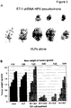


Classifications
-
- A—HUMAN NECESSITIES
- A61—MEDICAL OR VETERINARY SCIENCE; HYGIENE
- A61K—PREPARATIONS FOR MEDICAL, DENTAL OR TOILETRY PURPOSES
- A61K45/00—Medicinal preparations containing active ingredients not provided for in groups A61K31/00 - A61K41/00
- A61K45/06—Mixtures of active ingredients without chemical characterisation, e.g. antiphlogistics and cardiaca
-
- A—HUMAN NECESSITIES
- A61—MEDICAL OR VETERINARY SCIENCE; HYGIENE
- A61K—PREPARATIONS FOR MEDICAL, DENTAL OR TOILETRY PURPOSES
- A61K39/00—Medicinal preparations containing antigens or antibodies
- A61K39/12—Viral antigens
-
- A—HUMAN NECESSITIES
- A61—MEDICAL OR VETERINARY SCIENCE; HYGIENE
- A61K—PREPARATIONS FOR MEDICAL, DENTAL OR TOILETRY PURPOSES
- A61K39/00—Medicinal preparations containing antigens or antibodies
- A61K39/0005—Vertebrate antigens
- A61K39/0011—Cancer antigens
-
- A—HUMAN NECESSITIES
- A61—MEDICAL OR VETERINARY SCIENCE; HYGIENE
- A61K—PREPARATIONS FOR MEDICAL, DENTAL OR TOILETRY PURPOSES
- A61K45/00—Medicinal preparations containing active ingredients not provided for in groups A61K31/00 - A61K41/00
-
- A—HUMAN NECESSITIES
- A61—MEDICAL OR VETERINARY SCIENCE; HYGIENE
- A61K—PREPARATIONS FOR MEDICAL, DENTAL OR TOILETRY PURPOSES
- A61K47/00—Medicinal preparations characterised by the non-active ingredients used, e.g. carriers or inert additives; Targeting or modifying agents chemically bound to the active ingredient
- A61K47/30—Macromolecular organic or inorganic compounds, e.g. inorganic polyphosphates
- A61K47/42—Proteins; Polypeptides; Degradation products thereof; Derivatives thereof, e.g. albumin, gelatin or zein
-
- A—HUMAN NECESSITIES
- A61—MEDICAL OR VETERINARY SCIENCE; HYGIENE
- A61K—PREPARATIONS FOR MEDICAL, DENTAL OR TOILETRY PURPOSES
- A61K47/00—Medicinal preparations characterised by the non-active ingredients used, e.g. carriers or inert additives; Targeting or modifying agents chemically bound to the active ingredient
- A61K47/50—Medicinal preparations characterised by the non-active ingredients used, e.g. carriers or inert additives; Targeting or modifying agents chemically bound to the active ingredient the non-active ingredient being chemically bound to the active ingredient, e.g. polymer-drug conjugates
- A61K47/69—Medicinal preparations characterised by the non-active ingredients used, e.g. carriers or inert additives; Targeting or modifying agents chemically bound to the active ingredient the non-active ingredient being chemically bound to the active ingredient, e.g. polymer-drug conjugates the conjugate being characterised by physical or galenical forms, e.g. emulsion, particle, inclusion complex, stent or kit
- A61K47/6901—Conjugates being cells, cell fragments, viruses, ghosts, red blood cells or viral vectors
-
- A—HUMAN NECESSITIES
- A61—MEDICAL OR VETERINARY SCIENCE; HYGIENE
- A61P—SPECIFIC THERAPEUTIC ACTIVITY OF CHEMICAL COMPOUNDS OR MEDICINAL PREPARATIONS
- A61P15/00—Drugs for genital or sexual disorders; Contraceptives
-
- A—HUMAN NECESSITIES
- A61—MEDICAL OR VETERINARY SCIENCE; HYGIENE
- A61P—SPECIFIC THERAPEUTIC ACTIVITY OF CHEMICAL COMPOUNDS OR MEDICINAL PREPARATIONS
- A61P17/00—Drugs for dermatological disorders
-
- A—HUMAN NECESSITIES
- A61—MEDICAL OR VETERINARY SCIENCE; HYGIENE
- A61P—SPECIFIC THERAPEUTIC ACTIVITY OF CHEMICAL COMPOUNDS OR MEDICINAL PREPARATIONS
- A61P31/00—Antiinfectives, i.e. antibiotics, antiseptics, chemotherapeutics
- A61P31/12—Antivirals
-
- A—HUMAN NECESSITIES
- A61—MEDICAL OR VETERINARY SCIENCE; HYGIENE
- A61P—SPECIFIC THERAPEUTIC ACTIVITY OF CHEMICAL COMPOUNDS OR MEDICINAL PREPARATIONS
- A61P31/00—Antiinfectives, i.e. antibiotics, antiseptics, chemotherapeutics
- A61P31/12—Antivirals
- A61P31/14—Antivirals for RNA viruses
- A61P31/18—Antivirals for RNA viruses for HIV
-
- A—HUMAN NECESSITIES
- A61—MEDICAL OR VETERINARY SCIENCE; HYGIENE
- A61P—SPECIFIC THERAPEUTIC ACTIVITY OF CHEMICAL COMPOUNDS OR MEDICINAL PREPARATIONS
- A61P31/00—Antiinfectives, i.e. antibiotics, antiseptics, chemotherapeutics
- A61P31/12—Antivirals
- A61P31/20—Antivirals for DNA viruses
-
- A—HUMAN NECESSITIES
- A61—MEDICAL OR VETERINARY SCIENCE; HYGIENE
- A61P—SPECIFIC THERAPEUTIC ACTIVITY OF CHEMICAL COMPOUNDS OR MEDICINAL PREPARATIONS
- A61P31/00—Antiinfectives, i.e. antibiotics, antiseptics, chemotherapeutics
- A61P31/12—Antivirals
- A61P31/20—Antivirals for DNA viruses
- A61P31/22—Antivirals for DNA viruses for herpes viruses
-
- A—HUMAN NECESSITIES
- A61—MEDICAL OR VETERINARY SCIENCE; HYGIENE
- A61P—SPECIFIC THERAPEUTIC ACTIVITY OF CHEMICAL COMPOUNDS OR MEDICINAL PREPARATIONS
- A61P33/00—Antiparasitic agents
-
- A—HUMAN NECESSITIES
- A61—MEDICAL OR VETERINARY SCIENCE; HYGIENE
- A61P—SPECIFIC THERAPEUTIC ACTIVITY OF CHEMICAL COMPOUNDS OR MEDICINAL PREPARATIONS
- A61P35/00—Antineoplastic agents
-
- A—HUMAN NECESSITIES
- A61—MEDICAL OR VETERINARY SCIENCE; HYGIENE
- A61P—SPECIFIC THERAPEUTIC ACTIVITY OF CHEMICAL COMPOUNDS OR MEDICINAL PREPARATIONS
- A61P37/00—Drugs for immunological or allergic disorders
- A61P37/02—Immunomodulators
-
- C—CHEMISTRY; METALLURGY
- C07—ORGANIC CHEMISTRY
- C07K—PEPTIDES
- C07K14/00—Peptides having more than 20 amino acids; Gastrins; Somatostatins; Melanotropins; Derivatives thereof
- C07K14/005—Peptides having more than 20 amino acids; Gastrins; Somatostatins; Melanotropins; Derivatives thereof from viruses
-
- C—CHEMISTRY; METALLURGY
- C07—ORGANIC CHEMISTRY
- C07K—PEPTIDES
- C07K16/00—Immunoglobulins [IGs], e.g. monoclonal or polyclonal antibodies
- C07K16/08—Immunoglobulins [IGs], e.g. monoclonal or polyclonal antibodies against material from viruses
- C07K16/081—Immunoglobulins [IGs], e.g. monoclonal or polyclonal antibodies against material from viruses from DNA viruses
- C07K16/084—Papovaviridae, e.g. papillomavirus, polyomavirus, SV40, BK virus, JC virus
-
- C—CHEMISTRY; METALLURGY
- C12—BIOCHEMISTRY; BEER; SPIRITS; WINE; VINEGAR; MICROBIOLOGY; ENZYMOLOGY; MUTATION OR GENETIC ENGINEERING
- C12N—MICROORGANISMS OR ENZYMES; COMPOSITIONS THEREOF; PROPAGATING, PRESERVING, OR MAINTAINING MICROORGANISMS; MUTATION OR GENETIC ENGINEERING; CULTURE MEDIA
- C12N15/00—Mutation or genetic engineering; DNA or RNA concerning genetic engineering, vectors, e.g. plasmids, or their isolation, preparation or purification; Use of hosts therefor
- C12N15/09—Recombinant DNA-technology
- C12N15/11—DNA or RNA fragments; Modified forms thereof; Non-coding nucleic acids having a biological activity
- C12N15/111—General methods applicable to biologically active non-coding nucleic acids
-
- C—CHEMISTRY; METALLURGY
- C12—BIOCHEMISTRY; BEER; SPIRITS; WINE; VINEGAR; MICROBIOLOGY; ENZYMOLOGY; MUTATION OR GENETIC ENGINEERING
- C12N—MICROORGANISMS OR ENZYMES; COMPOSITIONS THEREOF; PROPAGATING, PRESERVING, OR MAINTAINING MICROORGANISMS; MUTATION OR GENETIC ENGINEERING; CULTURE MEDIA
- C12N15/00—Mutation or genetic engineering; DNA or RNA concerning genetic engineering, vectors, e.g. plasmids, or their isolation, preparation or purification; Use of hosts therefor
- C12N15/09—Recombinant DNA-technology
- C12N15/87—Introduction of foreign genetic material using processes not otherwise provided for, e.g. co-transformation
- C12N15/88—Introduction of foreign genetic material using processes not otherwise provided for, e.g. co-transformation using microencapsulation, e.g. using amphiphile liposome vesicle
-
- C—CHEMISTRY; METALLURGY
- C12—BIOCHEMISTRY; BEER; SPIRITS; WINE; VINEGAR; MICROBIOLOGY; ENZYMOLOGY; MUTATION OR GENETIC ENGINEERING
- C12N—MICROORGANISMS OR ENZYMES; COMPOSITIONS THEREOF; PROPAGATING, PRESERVING, OR MAINTAINING MICROORGANISMS; MUTATION OR GENETIC ENGINEERING; CULTURE MEDIA
- C12N7/00—Viruses; Bacteriophages; Compositions thereof; Preparation or purification thereof
-
- A—HUMAN NECESSITIES
- A61—MEDICAL OR VETERINARY SCIENCE; HYGIENE
- A61K—PREPARATIONS FOR MEDICAL, DENTAL OR TOILETRY PURPOSES
- A61K39/00—Medicinal preparations containing antigens or antibodies
- A61K2039/51—Medicinal preparations containing antigens or antibodies comprising whole cells, viruses or DNA/RNA
- A61K2039/525—Virus
- A61K2039/5258—Virus-like particles
-
- A—HUMAN NECESSITIES
- A61—MEDICAL OR VETERINARY SCIENCE; HYGIENE
- A61K—PREPARATIONS FOR MEDICAL, DENTAL OR TOILETRY PURPOSES
- A61K39/00—Medicinal preparations containing antigens or antibodies
- A61K2039/80—Vaccine for a specifically defined cancer
- A61K2039/892—Reproductive system [uterus, ovaries, cervix, testes]
-
- C—CHEMISTRY; METALLURGY
- C07—ORGANIC CHEMISTRY
- C07K—PEPTIDES
- C07K2317/00—Immunoglobulins specific features
- C07K2317/30—Immunoglobulins specific features characterized by aspects of specificity or valency
- C07K2317/34—Identification of a linear epitope shorter than 20 amino acid residues or of a conformational epitope defined by amino acid residues
-
- C—CHEMISTRY; METALLURGY
- C12—BIOCHEMISTRY; BEER; SPIRITS; WINE; VINEGAR; MICROBIOLOGY; ENZYMOLOGY; MUTATION OR GENETIC ENGINEERING
- C12N—MICROORGANISMS OR ENZYMES; COMPOSITIONS THEREOF; PROPAGATING, PRESERVING, OR MAINTAINING MICROORGANISMS; MUTATION OR GENETIC ENGINEERING; CULTURE MEDIA
- C12N2310/00—Structure or type of the nucleic acid
- C12N2310/10—Type of nucleic acid
- C12N2310/11—Antisense
-
- C—CHEMISTRY; METALLURGY
- C12—BIOCHEMISTRY; BEER; SPIRITS; WINE; VINEGAR; MICROBIOLOGY; ENZYMOLOGY; MUTATION OR GENETIC ENGINEERING
- C12N—MICROORGANISMS OR ENZYMES; COMPOSITIONS THEREOF; PROPAGATING, PRESERVING, OR MAINTAINING MICROORGANISMS; MUTATION OR GENETIC ENGINEERING; CULTURE MEDIA
- C12N2310/00—Structure or type of the nucleic acid
- C12N2310/10—Type of nucleic acid
- C12N2310/14—Type of nucleic acid interfering N.A.
-
- C—CHEMISTRY; METALLURGY
- C12—BIOCHEMISTRY; BEER; SPIRITS; WINE; VINEGAR; MICROBIOLOGY; ENZYMOLOGY; MUTATION OR GENETIC ENGINEERING
- C12N—MICROORGANISMS OR ENZYMES; COMPOSITIONS THEREOF; PROPAGATING, PRESERVING, OR MAINTAINING MICROORGANISMS; MUTATION OR GENETIC ENGINEERING; CULTURE MEDIA
- C12N2320/00—Applications; Uses
- C12N2320/30—Special therapeutic applications
- C12N2320/32—Special delivery means, e.g. tissue-specific
-
- C—CHEMISTRY; METALLURGY
- C12—BIOCHEMISTRY; BEER; SPIRITS; WINE; VINEGAR; MICROBIOLOGY; ENZYMOLOGY; MUTATION OR GENETIC ENGINEERING
- C12N—MICROORGANISMS OR ENZYMES; COMPOSITIONS THEREOF; PROPAGATING, PRESERVING, OR MAINTAINING MICROORGANISMS; MUTATION OR GENETIC ENGINEERING; CULTURE MEDIA
- C12N2710/00—MICROORGANISMS OR ENZYMES; COMPOSITIONS THEREOF; PROPAGATING, PRESERVING, OR MAINTAINING MICROORGANISMS; MUTATION OR GENETIC ENGINEERING; CULTURE MEDIA dsDNA viruses
- C12N2710/00011—Details
- C12N2710/14011—Baculoviridae
- C12N2710/14111—Nucleopolyhedrovirus, e.g. autographa californica nucleopolyhedrovirus
- C12N2710/14141—Use of virus, viral particle or viral elements as a vector
- C12N2710/14143—Use of virus, viral particle or viral elements as a vector viral genome or elements thereof as genetic vector
-
- C—CHEMISTRY; METALLURGY
- C12—BIOCHEMISTRY; BEER; SPIRITS; WINE; VINEGAR; MICROBIOLOGY; ENZYMOLOGY; MUTATION OR GENETIC ENGINEERING
- C12N—MICROORGANISMS OR ENZYMES; COMPOSITIONS THEREOF; PROPAGATING, PRESERVING, OR MAINTAINING MICROORGANISMS; MUTATION OR GENETIC ENGINEERING; CULTURE MEDIA
- C12N2710/00—MICROORGANISMS OR ENZYMES; COMPOSITIONS THEREOF; PROPAGATING, PRESERVING, OR MAINTAINING MICROORGANISMS; MUTATION OR GENETIC ENGINEERING; CULTURE MEDIA dsDNA viruses
- C12N2710/00011—Details
- C12N2710/20011—Papillomaviridae
- C12N2710/20022—New viral proteins or individual genes, new structural or functional aspects of known viral proteins or genes
-
- C—CHEMISTRY; METALLURGY
- C12—BIOCHEMISTRY; BEER; SPIRITS; WINE; VINEGAR; MICROBIOLOGY; ENZYMOLOGY; MUTATION OR GENETIC ENGINEERING
- C12N—MICROORGANISMS OR ENZYMES; COMPOSITIONS THEREOF; PROPAGATING, PRESERVING, OR MAINTAINING MICROORGANISMS; MUTATION OR GENETIC ENGINEERING; CULTURE MEDIA
- C12N2710/00—MICROORGANISMS OR ENZYMES; COMPOSITIONS THEREOF; PROPAGATING, PRESERVING, OR MAINTAINING MICROORGANISMS; MUTATION OR GENETIC ENGINEERING; CULTURE MEDIA dsDNA viruses
- C12N2710/00011—Details
- C12N2710/20011—Papillomaviridae
- C12N2710/20023—Virus like particles [VLP]
-
- C—CHEMISTRY; METALLURGY
- C12—BIOCHEMISTRY; BEER; SPIRITS; WINE; VINEGAR; MICROBIOLOGY; ENZYMOLOGY; MUTATION OR GENETIC ENGINEERING
- C12N—MICROORGANISMS OR ENZYMES; COMPOSITIONS THEREOF; PROPAGATING, PRESERVING, OR MAINTAINING MICROORGANISMS; MUTATION OR GENETIC ENGINEERING; CULTURE MEDIA
- C12N2710/00—MICROORGANISMS OR ENZYMES; COMPOSITIONS THEREOF; PROPAGATING, PRESERVING, OR MAINTAINING MICROORGANISMS; MUTATION OR GENETIC ENGINEERING; CULTURE MEDIA dsDNA viruses
- C12N2710/00011—Details
- C12N2710/20011—Papillomaviridae
- C12N2710/20034—Use of virus or viral component as vaccine, e.g. live-attenuated or inactivated virus, VLP, viral protein
-
- C—CHEMISTRY; METALLURGY
- C12—BIOCHEMISTRY; BEER; SPIRITS; WINE; VINEGAR; MICROBIOLOGY; ENZYMOLOGY; MUTATION OR GENETIC ENGINEERING
- C12N—MICROORGANISMS OR ENZYMES; COMPOSITIONS THEREOF; PROPAGATING, PRESERVING, OR MAINTAINING MICROORGANISMS; MUTATION OR GENETIC ENGINEERING; CULTURE MEDIA
- C12N2800/00—Nucleic acids vectors
- C12N2800/22—Vectors comprising a coding region that has been codon optimised for expression in a respective host
Definitions
- the invention relates to human papillomavirus like particles (VLPs) and their use as therapeutic agents.
- VLPs human papillomavirus like particles
- Cervical cancer is one of the leading causes of cancer deaths in women world-wide, killing more than 233,000 women each year. Cervical cancer was the most common malignancy in both incidence and mortality among women prior to the 20th century. The reduction in the incidence of cervical cancer is one of the major public health achievements in developed countries, largely due to the implementation of population-based screening, detection and treatment programs for pre-invasive disease. However, while the incidence of cervical cancer in developed nations has fallen, the disease continues to be the second most common cancer in women worldwide.
- Papanicolaou (Pap) smears can detect cervical cancer or pre-cancerous changes in the cervix, many of which are related to HPV. These Pap smears have greatly reduced the incidence and mortality of cervical cancer in developed countries where widespread screening procedures occur. In developing countries where screening procedures are still limited, cervical cancer is the most frequently reported cancer in women, and the incidence continues to rise.
- Cervical intraepithelial neoplasia refers to a pre-invasive pathological intermediate to cervical cancer.
- the abnormalities observed on a cytologic smear or tissue biopsy of the cervix represent alterations in the degree of differentiation of cervical epithelial cells.
- This cellular dysplasia is categorized into three different groups of severity: CIN I refers to mild dysplasia confined to the basal third of the epithelium; CIN II refers to lesions confined to the basal two-thirds of the epithelium; and CIN III refers to cellular dysplasia encompassing greater than two-thirds of the epithelial thickness.
- HPV infection is endemic among sexually active individuals. Women who have multiple sexual partners have a higher chance of acquiring HPV and consequently, an HPV-related cervical infection. Infection with high-risk HPV types increases the odds that a woman will develop cervical cancer. Screening for and subsequently treating pre-cancerous cervical conditions is highly effective in the prevention of cervical cancer in HPV-infected women. However, current treatments often require surgical intervention and alternative therapeutic options are needed.
- compositions and methods of the invention are useful for treating mucosal conditions (e.g., diseases and/or infections of mucosal tissue, for example of mucosal epithelial cells).
- mucosal conditions e.g., diseases and/or infections of mucosal tissue, for example of mucosal epithelial cells.
- aspects of the invention relate to modified HPV-based particles that can deliver a therapeutic agent to mucosal tissue (e.g., topically).
- the therapeutic agent can be an antiviral agent.
- the antiviral agent may be used to treat a viral infection by, for example, a human papilloma virus, a Herpes virus, or other virus that targets mucosal tissue.
- the therapeutic agent can be an anticancer agent.
- the anticancer agent may be used to treat, for example, cervical cancer or any other cancer of a mucosal tissue.
- the invention provides methods for treating human papilloma virus (HPV) infection and methods for treating HPV-associated diseases including, but not limited to, cervical cancer in a subject.
- HPV human papilloma virus
- modified viral particles of the invention may be used to deliver other types of therapeutic and/or medical agents (e.g., imaging or contrast agents).
- particles of the invention may be delivered topically (e.g., in the form of a cream, foam, spray, aerosol, or other formulation suitable for topical delivery) to any mucosal tissue (e.g., to cervical, nasal, oral, or other mucosal tissue).
- any mucosal tissue e.g., to cervical, nasal, oral, or other mucosal tissue.
- aspects of the invention are not limited to topical delivery and modified particles may be delivered subcutaneously, intravenously, parenterally, and/or via any other suitable delivery route.
- Methods provided herein comprise administering to the subject one or more therapeutic agents delivered by a virus-like particle (VLP)-based delivery system.
- the VLP-based delivery system comprises human papilloma virus (HPV)-like nanoparticles.
- HPV nanoparticles comprise viral L1 protein.
- HPV nanoparticles comprise viral L1 protein and viral L2 protein.
- the L1 and/or L2 proteins may, in some embodiments be wild-type viral proteins. In some embodiments, L1 and/or L2 proteins may be altered by mutation and/or insertion/deletion.
- amino acids in surface-exposed loops of the HPV nanoparticle comprising L1 and/or L2 are mutated, inserted and/or deleted.
- mutation, deletion and/or insertion of amino acids in surface-exposed loops leads to changes in immunogenicity of the HPV nanoparticle.
- immunogenicity can be altered in such way that HPV nanoparticles of a certain serotype are no longer recognized by antibodies raised against this serotype.
- the altered HPV nanoparticle is immuno-silent in the host harboring the serotype specific antibodies.
- HPV-based particles of the invention may be modified to have reduced or altered immunogenicity. Such particles may be selected for delivery to patients that have a neutralizing anti-HPV response.
- a series of HPV-based particles that have different serotypes are administered to a subject for therapeutic purposes to reduce the effect of a neutralizing immune response against any one of the serotypes.
- a first serotype may be used for a first set (e.g., 1, 2, 3, 4, 5, 5-10, or more) of administrations to a subject.
- a second serotype may be used for a second set (e.g., 1, 2, 3, 4, 5, 5-10, or more) of administrations to reduce the impact of a neutralizing immune response that the subject develops against the first serotype.
- a second set e.g., 1, 2, 3, 4, 5, 5-10, or more
- further sets of administration may involve a third, fourth, fifth, etc. serotype.
- the different serotypes may be naturally occurring serotypes, chimeric serotypes, other mutant serotypes, or any combination thereof. It should be appreciated that such serial or sequential applications may be used for chronic administration (e.g., treatment) of a particular compound (e.g., the same one in each of the different serotypes), or a series of different compounds, over a time period of months and/or years.
- mutation, deletion and/or insertion of amino acids are introduced in the viral capsid proteins L1 and/or L2 in such a way that the resulting HPV nanoparticle does not lose the ability to deliver therapeutic agents to the target cell.
- the HPV nanoparticle does not lose the ability to transfer nucleic acids (e.g., siRNA or shRNA) into target cells.
- immunogenicity can be altered in such way that HPV nanoparticles of a certain serotype induce an immune response that produces cross-specific neutralizing antibodies.
- the HPV nanoparticle is altered in such way that it exhibits multiple sero-specific epitopes on its surface (for example by insertion of epitopes in surface-exposed loops) or that it exhibits epitopes that are more conserved between serotypes (for example by insertion of portions of the L2 protein in surface-exposed loops, or by linking conserved epitopes to the surface of the HPV nanoparticle).
- production of cross-specific neutralizing antibodies are induced in an HPV-infected individual that have undergone treatment to eliminate or reduce the number of HPV-infected cells.
- the HPV-infected individual has undergone treatment to eliminate or reduce the size of a HPV-associated tumor.
- production of cross-specific neutralizing antibodies may be induced to generate an immune response that is sufficient for immune-surveillance of the HPV-infected cells that may remain after treatment.
- the resulting immune-surveillance in some embodiments, is sufficient to prevent new infection of HPV or repopulation of HPV-infected cells or recurrence of HPV-infected tumors.
- cross-specific neutralizing antibodies are effective against one or more HPV serotypes.
- the therapeutic agents delivered by the VLP-based delivery system are nucleic acids.
- Therapeutic agents that are nucleic acids can be, for example, siRNA or shRNA molecules or plasmids encoding them.
- Other therapeutic agents can be, for example, small molecules, such as small molecules with anti-viral or anti-cancer activity.
- administering to a subject having a HPV infection or a HPV-associated cancer, such as a cervical cancer, or lesion one or more therapeutic agents delivered by a VLP-based delivery system leads to killing and/or clearance of HPV-infected cells.
- HPV-infected transformed cells are thought to be causative for HPV-associated cancers or lesions.
- killing and/or clearance of the HPV-infected cells leads to partial or complete remission of the HPV-associated cancer.
- the treatment methods described herein can be combined with the administration of other therapeutic agents (e.g., anti-cancer agents and/or antiviral agents) and/or immune-modulators and/or radiotherapy or immunotherapy either before, concurrently or after treatment with HPV-nanoparticles comprising therapeutic agents.
- the anti-cancer and/or anti-viral agents can be delivered by the HPV nanoparticles.
- the anti-cancer and/or anti-viral agents can be administered together with the HPV nanoparticles or can be administered separately, e.g., at a different time or a different site of administration or via a different route of administration.
- HPV-infected cells in a subject treated according to the methods described herein are killed and cleared and HPV is no longer detectable in the HPV-infected subject. In other embodiments, some HPV-infected cells in a subject are killed or cleared and HPV is still detectable in the HPV-infected subject.
- a subject having a HPV-associated lesion or cancer may experience partial or complete remission of the cancer or lesion. In some embodiments, subjects do not experience a recurrence of the lesion or cancer or viral infection. In other embodiments, subjects experience a recurrence of the lesion or cancer or viral infection.
- the methods of treatment described herein are further combined with anti-viral treatment during and/or after treatment with HPV nanoparticles comprising therapeutic agents.
- Anti-viral treatment may be given to prevent, for example, HPV replication, viral spreading and/or repopulation of cells with HPV that has survived the initial treatment or that have entered the body of the subject as a new infection.
- altered HPV nanoparticles are administered at the end of the initial treatment regimen.
- the HPV nanoparticles are altered in such way that they exhibit multiple sero-specific epitopes on its surface or that they exhibit epitopes that are more conserved between serotypes.
- administration of such altered HPV nanoparticles may induce a local immune response directed against the administered epitopes, for example inducing an increase in cross-specific neutralizing antibodies, that is sufficient for immune-surveillance capable of detecting and killing newly HPV-infected cells.
- prevention of new viral infection and/or spreading and repopulation is sufficient inhibits recurrence of the HPV-associated cancer or lesion.
- the methods described herein comprise administration of HPV nanoparticles comprising one or more therapeutic agents via different routes.
- HPV nanoparticles are administered via topical application.
- topical administration is targeted to mucosal membranes.
- the HPV nanoparticle comprising one or more therapeutic agents can be applied topically to or adjacent to an epithelium such as the cervical epithelial or topically to or adjacent to an epithelial lesion such as cervical or anal epithelial carcinoma.
- HPV nanoparticles comprising altered viral capsid proteins are provided, wherein the viral capsid proteins are altered by mutations of wild-type amino acids, insertions of additional amino acids, an/or deletion of wilt-type amino acids, as described herein.
- the altered HPV nanoparticles are more or less immunogenic in a subject or have an altered immunogenicity.
- the altered HPV nanoparticles maintain the ability to deliver or transfer therapeutic agents to a target cell.
- aspects of the invention relate to methods and compositions for delivering one or more compounds to a subject.
- a modified human papilloma virus (HPV)-like particle is used, wherein the particle comprises one or more heterologous compounds packaged in an HPV-like particle comprising a surface protein having altered immunogenicity.
- the heterologous compounds are non-HPV molecules (e.g., a compound that is not an HPV genome, not an HPV nucleic acid, and/or not an other HPV molecule).
- Such compounds may be therapeutic or other medical compounds.
- the compound may be one or more of: a therapeutic or medical agent, a nucleic acid or a small molecule, or an an imaging or contrast agent.
- the therapeutic agent is an siRNA, an shRNA, an antisense nucleic acid, or a nucleic acid encoding an siRNA, an shRNA, or an antisense nucleic acid.
- the siRNA, shRNA, or antisense nucleic acid targets HPV-E6 and/or HPV-E7.
- the small molecule is an anti-viral agent.
- the small molecule is an anti-cancer agent (e.g., Gemcitabine).
- compositions of the invention may be provided along with one or more other agents (e.g., an anti-viral agent, an anti-cancer agent, for example Gemcitaine, or any combination thereof).
- agents e.g., an anti-viral agent, an anti-cancer agent, for example Gemcitaine, or any combination thereof.
- the HPV-like particle comprises an L1 protein or an L1 protein and an L2 protein.
- the surface protein is a modified L1 protein with a modified FG loop sequence.
- the L1 protein has a sequence of an HPV16, HPV31, HPV33, HPV34, HPV35, HPV52, HPV58, HPV73, or HPV91 serotype, and wherein the FG loop of the L1 protein has one or more amino acid changes that alter the immunogenicity of the protein in a human subject.
- the one or more amino acid changes are at one or more of positions X 1 - X 17 of SEQ ID NO: 11.
- the amino acid at postion X 16 is not altered.
- one or more of positions X 1 , X 2 , X 3 , X 5 , X 6 , X 11 , and X 14 of SEQ ID NO: 11 are modified.
- 2-3 of positions X 1 , X 2 , X 3 , X 5 , X 6 , X 11 , and X 14 of SEQ ID NO: 11 are modified.
- 4-7 of positions X 1 , X 2 , X 3 , X 5 , X 6 , X 11 , and X 14 of SEQ ID NO: 11 are modified.
- positions X 1 , X 2 , X 3 , X 5 , X 6 , X 11 , and X 14 of SEQ ID NO: 11 are modified.
- positions X 6 , X 11 , and X 14 of SEQ ID NO: 11 are modified.
- all of positions X 1 , X 2 , X 3 , X 5 , X 6 , X 11 , and X 14 of SEQ ID NO: 11 are modified.
- the L1 protein has a sequence of an HPV16 serotype with positions X 6 , X 11 , and X 14 of SEQ ID NO: 11 changed to T, T, and N, respectively, and the remainder of the positions of SEQ ID NO: 11 having an amino acid characteristic of an HPV16 serotype.
- the L1 protein has a sequence of an HPV16 serotype with positions X 1 , X 2 , X 3 , X 5 , X 6 , X 11 , and X 14 changed to F, S, T, S, T, T, and N, respectively, and the remainder of the positions of SEQ ID NO: 11 having an amino acid characteristic of an HPV16 serotype.
- an HPV-like particle is administered to a subject that does not have a neutralizing immune response against the serotype of the surface protein, for example when the subject has not been immunized against the serotype of the surface protein, or when the subject has not been infected with an HPV having the same serotype as the serotype of the surface protein in the HPV-like particle.
- the immune response of the subject is evaluated prior to selecting an HPV-like particle for administration.
- an HPV-like particle is administered to a mucosal tissue.
- the mucosal tissue may be at any locus as described herein (e.g., cervical, urogenital, oral, etc.).
- an HPV-like particle is administered to an epidermal locus (e.g., to treat an epidermal or skin cancer or infection).
- compositions of the invention may be administered topically and/or via any other suitable route (e.g., via injection, aerosol, spray, or other form of administration).
- compositions of the invention may be administered to a tissue infected with a virus, bacteria, and/or other microbes or parasites.
- a site of infection may be infected with several different organisms (e.g., multiple viruses and/or other microbes, for example HPV and a herpes virus, for example HSV).
- a composition of the invention may include a compound that broadly targets several different organisms, or several different compounds that are each targeted to a different organism, or a combination thereof.
- the same HPV-like particle may contain different therapeutic compounds.
- a composition or treatment regimen may include two or more different HPV-like particles each containing a compound that is targeted to a particular infection.
- compositions of the invention may be administered to a subject to treat a cancer (e.g., at or near the site of administration).
- a cancer e.g., at or near the site of administration.
- the cancer may be associated with an infection (e.g., an HPV or HSV infection).
- the cancer may not be associated with an infection. Accordingly, aspects of the invention may be used to treat skin cacner (e.g, non-melanoma skin cancer).
- compositions of the invention may be administered to a subject having an immune system deficiency to treat a condition (e.g., infection, cancer, or other condition) associated with the immune system deficiency.
- a condition e.g., infection, cancer, or other condition
- the immune system deficiency may be caused by an infection (e.g., an HIV infection, AIDS) or other condition (e.g., cancer).
- an HPV-like particle of the invention may be administered along with a composition that promotes a non-specific immune response (e.g., at or near the site of administration).
- the non-specific immune response may be helpful to treat the infection and/or a cancer or other condition.
- an increased immune response in response to the administration of several HPV-like particles with different serotypes may be beneficial.
- some compositions of the invention comprise several different serotypes.
- a chronic administration of a composition may be accomplished by administering a first HPV-like particle comprising a compound to a subject for a first period of time, and administering a second HPV-like particle comprising the compound to the subject for a second period of time, wherein the first and second HPV-like particles have different serotypes. Further administrations may be made using further serotypes.
- the serotypes of the first and second HPV-like particles are independently naturally occurring or altered serotypes.
- the serotype of the first and/or second HPV-like particle is a chimeric serotype.
- a composition or method of the invention may involve administering an HPV-like particle that has a chimeric serotype by including an L2 sequence fused in an L1 loop.
- the L2 sequence is attached to the surface of an L1 particle.
- the L2 sequence consists of residues 13 to 88 of the HPV31 L2 protein.
- HPV-based particles are used to deliver one or more agents (e.g., therapeutic agents, imaging agents, and/or other medical agents) to a target cell or tissue (e.g., a mucosal tissue, for example a mucosal tissue surface).
- agents e.g., therapeutic agents, imaging agents, and/or other medical agents
- a target cell or tissue e.g., a mucosal tissue, for example a mucosal tissue surface.
- aspects of the invention relate to an HPV particle that contains one or more naturally occurring HPV surface proteins (e.g., L1 and/or L2 proteins) and that is loaded with one or more medical and/or therapeutic agents, or a combination of two or more thereof.
- aspects of the invention relate to modified HPV-based particles that contain one or more variant surface proteins (e.g., variant L1 and/or L2 proteins) that have reduced or modified immunogenicity in a subject.
- the modification may be an amino acid sequence change that reduces or avoids neutralization by the immune system of the subject.
- a modified HPV-based particle is a particle that contains a recombinant HPV protein (e.g., a recombinant L1 and/or L2 protein) that includes one or more amino acid changes that alter the immunogenicity of the protein in a subject (e.g., in a human subject).
- a modified HPV-based particle has an altered immunogenicity but retains the ability to package and deliver molecules to a subject.
- modified HPV particles of the invention may be loaded with one or more agents (e.g., instead of an HPV nucleic acid). Such particles may be delivered to a subject without inducing an immune response that would be induced by a naturally-occurring HPV particle.
- a diseased tissue e.g., mucosal tissue, epithelial tissue, or endothelial tissue
- a diseased tissue may be an infected tissue (e.g., infected with a virus such as HPV or HSV).
- the mucosal tissue is cervical tissue and the disease is dysplasia or cancer (e.g., cervical dysplasia, cervical cancer, for example associated with persistent HPV infection).
- HPV-based particles may be used to deliver compositions to other tissues (e.g., epidermis).
- HPV-based particles may be used to treat HPV.
- HPV-based particles may be used to deliver therapeutic agents to treat other diseases or conditions.
- Some embodiments of the invention are useful for delivering one or more imaging or contrast agents to a subject.
- quantum dots, metals, and/or other imaging agents may be delivered.
- agents may be used to track early stage diseases (e.g., early stage metastasis).
- radiosensitive agents may be used to enhance the effects of radiotherapy.
- agents may be delivered to induce tumor cells to express different receptors or sugars on their membrane.
- aspects of the invention may be used to deliver an agent that promotes the expression in a tumor cell of a signal that would enhance the immune system recognition of the tumor (e.g., an agent that would make the tumor cell look like a bacteria, or a virus).
- an agent may be delivered to block the uptake of a sugar by a tumor cell.
- any suitable therapeutic and/or other medical agent may be delivered according to aspects of the invention.
- aspects of the invention relate to HPV-based particles that are modified to display epitopes from two or more different naturally-occurring HPV variants.
- modified particles may be used to provide immunization against infection by any of two or more naturally occurring HPV variants.
- aspects of the invention relate to protocols for administering one or more different HPV particles for therapeutic applications.
- the different HPV particles may be any combination of different HPV variants, HPV-based particles containing different agents, and HPV particles that have modified immunogenicity.
- HPV nanoparticles may be used to deliver one or more therapeutic agents to mucosal cells (e.g., HPV-infected cells).
- altered HPV nanoparticles may be used to deliver the one or more therapeutic agents to target cells.
- the altered HPV nanoparticle is immune-silent in the host harboring serotype specific antibodies.
- a subject infected with a first HPV serotype e.g., HPV-16
- develops antibodies against that serotype develops antibodies against that serotype.
- viral particles having the first serotype e.g., wild-type HPV 16-based VLPs
- a subject may be immunized against HPV (e.g., with GARDASIL and CERVARIX) and has developed neutralizing antibodies against a first and/or second serotype (e.g., HPV-16 and HPV18).
- a first and/or second serotype e.g., HPV-16 and HPV18
- viral particles having the first and/or serotype e.g., wild-type HPV16- or HPV18-based VLPs
- HPV nanoparticles can be altered by methods described herein so that the altered HPV nanoparticle is not recognized by the first serotype-specific antibodies in the host (e.g., the HPV-16 serotype-specific antibodies in the host) and/or the second serotype-specific antibodies in the host (e.g., the HPV-18 serotype-specific antibodies in the host).
- Such altered HPV or HPV nanoparticles comprising L1 and/or L2 proteins from a different serotype may be used therapeutically.
- such altered HPV nanoparticles or VLP based on a different serotype may be used to deliver one or more therapeutic agents to an HPV-immunized or HPV-infected host without inducing a serotype-specific immune response and/or without being neutralized by a host response and without losing efficacy.
- Such altered HPV nanoparticles or VLP based on a different serotype may be used repeatedly (e.g., for 1, 2, 3, 4, 5, 6, 7, 8, 9, 10 or more administrations) to deliver therapeutic agents.
- the subject will develop new antibodies that will recognize the altered HPV nanoparticle or VLP based on a different serotype.
- a subject that is not infected with HPV may develop an immune response against the serotype of an HPV-based particle that is administered to the subject for therapeutic purposes.
- a second differently altered HPV nanoparticle or a second HPV nanoparticle comprising L1 and/or L2 proteins from a further different serotype may be used for continued delivery of therapeutic agents to a subject (e.g., to HPV-infected cells or cells within a subject that has been immunized against HPV or treated with HPV-based compositions of the invention) without inducing an immune response (e.g., a neutralizing immune response) specific to the original HPV serotype and the second altered HPV nanoparticle.
- a third altered HPV nanoparticle or a third VLP containing third L1 and/or L2 proteins may be used. This process may be repeated several times with a series of different altered HPV nanoparticles and/or different VLPs containing a series of different L1 and/or L2 proteins may be used.
- the switch from one set of particles to a different one may be made when the first set looses efficacy in a subject, when an immune response to the first set is detected in the subject, or at a predetermined time (e.g., after a predetermined number of administrations or a predetermined time period of administration of the first set) after treatment is initiated.
- the VLP based on a different serotype is chosen on the basis of genotype and/or serotype similarity of the HPV. In some embodiments, the VLP based on a different serotype is chosen on the basis of neutralizing cross-reactivity of antibodies. In some embodiments, a VLP based on a different serotype is chosen that is most distantly related to the first and/or second serotype against which the subject has developed neutralizing antibodies. For example, HPV 18 and HPV 45 are closely related and show a high degree of cross-protection of neutralizing antibodies. HPV 16 and HPV 31 are closely related and show a high degree of cross-protection of neutralizing antibodies. HPV 58 is more distantly related to HPV 16 and HPV18 and little or no cross-protection by neutralizing antibodies is observed.
- a treatment series may involve administering a series of VLP compositions of the invention (e.g., containing one or more therapeutic agents), wherein each successive VLP composition is based on a VLP from a different HPV serotype, for example, from remotely related serotypes.
- a suitable VLP based on a different serotype can be a particle comprising L1 and/or L2 proteins from wild-type HPV 58.
- the VLP distantly related to the first and/or second serotype against which the subject has developed neutralizing antibodies may be selected from papilloma viruses that are not HPV.
- VLP may comprise capsid proteins from a papilloma virus that infects a mammal that is not human, e.g., bovine papilloma virus (BVP) or cottontail rabbit papilloma virus (CRVP) or Shope papilloma virus.
- BVP bovine papilloma virus
- CRVP cottontail rabbit papilloma virus
- Shope papilloma virus e.g., Shope papilloma virus.
- a treatment series may involve a series of VLPs based on different HPV serotypes and/or different papilloma viruses that infect other non-human hosts.
- the differently altered HPV nanoparticle or the HPV nanoparticle of a different serotype may be used for delivery of therapeutic agents until the subject develops antibodies to the differently altered HPV nanoparticle or the HPV nanoparticle of a different serotype.
- altering VLPs based on serotype-differences and/or immune response altering mutations subjects may be treated for multiple rounds with therapeutic agents without the loss of efficacy of the delivery system due to immune responses of the subject.
- such regimens allow repeated or continued treatment of the HPV infection and/or HPV-associated cancer until essentially all HPV-infected cells are eliminated and/or remission (partial or complete) of the cancer or lesion has occurred.
- such regimens may be used for repeated or continued treatment of other conditions according to aspects of the invention.
- a subject is vaccinated using a HPV vaccine (such a those commercially available, e.g., GARDASIL and CERVARIX).
- a HPV vaccine such as those commercially available, e.g., GARDASIL and CERVARIX.
- the immunization protects the subject from becoming infected with the viruses that are targeted by the vaccine (for example, the viruses for which the subject has raised an immune response upon vaccination) and from developing HPV-associated diseases caused by these vaccine-specific viruses.
- the currently available vaccines will not protect against infection of all HPV genotypes and/or serotypes.
- HPV-vaccinated subjects will become infected with an HPV (e.g., a high-risk HPV) that is not targeted by the vaccine, for example, for which the vaccinated subject has not developed an immune response.
- the vaccinated subject encounters multiple incidences of infection with different HPV types.
- subjects may become infected with one, two, three, four, five, six, seven, eight, nine, ten, or more different HPV types that infect -and are harbored in- different cells in the subject.
- a vaccinated subject becoming infected with a high-risk HPV for which the vaccinated subject has not developed an immune response may develop a HPV-associated disease, e.g., a HPV-associated dysplasia or cancer, caused by the high-risk HPV for which the vaccinated subject has not developed an immune response.
- the subject may be treated with the VLPs described herein using the methods described herein.
- a subject immunized with one of the commercially available vaccines may develop neutralizing antibodies against HPV16 and 18 and may also develop neutralizing antibodies against HPV6 and 11.
- the immunized subject is protected against the development of cervical pre-cancers and/or genital warts caused by these HPV-types (HPV 16, 18 (cancer), and HPV 6, 11 (warts)).
- a subject having developed neutralizing antibodies against HPV 16 and 18 upon immunization may develop some neutralizing antibodies that display cross-reactivity with other HPV types (such as, e.g., HPV 31, for neutralizing antibodies that are raised against HPV 16, and HPV 45 for neutralizing antibodies that are raised against HPV 18).
- HPV-33 HPV-35, HPV-39, HPV-51, HPV-52, HPV-56, HPV-59, HPV-68, and HPV-69.
- the immunized subject has not developed cross-neutralizing antibodies, or has not developed sufficiently specific cross-neutralizing antibodies, or has not developed sufficient titers of cross-neutralizing antibodies, the immunized subject is still at risk of developing a HPV-associated disease, such as dysplasia or cancer, when infected with one or more of the high-risk HPV types, for which no sufficient protection was developed by the subject.
- a HPV-associated disease such as dysplasia or cancer
- modified VLPs described herein or viral particles of a different (more distantly related) serotype comprising on or more therapeutic agents (e.g., E7 siRNA) may be administered to the subject to treat early stage disease developed by the persistent high-risk HPV infection of that subject caused by a high-risk HPV for which no sufficient protection was developed by the subject.
- the treatment enables the elimination of the early dysplasia and additionally may provide broader cross-protection to the subject against further infection with other additional HPV types.
- HPV nanoparticles comprise viral L1 protein. In some embodiments, HPV nanoparticles comprise viral L1 protein and viral L2 protein.
- the L1 and/or L2 proteins may, in some embodiments, be wild-type viral proteins. In some embodiments, L 1 and/or L2 proteins may be altered by mutation and/or deletion so that the resulting L1 and/or L2 proteins comprise only 'minimal' domains essential for assembly of the nanoparticle. In some embodiments, L1 and/or L2 proteins may also be fused to other proteins and/or peptides that provide additional functionality. These other proteins may be viral or non-viral and could, in some embodiments, be for example host-specific or cell type specific.
- VLPs may be based on particles containing one or more recombinant proteins or fragments thereof (e.g., one or more HPV membrane and/or surface proteins or fragments thereof). In some embodiments, VLPs may be based on naturally-occurring particles that are processed to incorporate one or more agents as described herein, as aspects of the invention are not limited in this respect. In certain embodiments, particles comprising one or more targeting peptides may be used. Other combinations of HPV proteins or peptides may be used as aspects of the invention are not limited in this respect.
- viral wild-type capsid proteins are altered by mutations, insertions and deletions. All conformation-dependent type-specific epitopes identified to date are found on the HPV-VLP surface within hypervariable loops where the amino acid sequence is highly divergent between HPV types, which are designated BC, DE, EF, FG and HI loops. Most neutralizing antibodies are generated against epitopes in these variable lops and are type-specific, with limited cross-reactivity, cross-neutralization and cross-protection. Different HPV serotypes induce antibodies directed to different type-specific epitopes and/or to different loops.
- viral capsid proteins HPV L1 and/or L2 are mutated at one or more amino acid positions located in one or more hypervariable and/or surface-exposed loops.
- the mutations are made at amino acid positions within the loops that are not conserved between HPV serotypes. These positions can be completely non-conserved, that is that any amino acid can be at this position, or the position can be conserved in that only conservative amino acid changes can be made.
- Conservative amino acid changes may be made according to functional, chemical or structural considerations.
- conservative amino acid changes may be made according to chemical similarity: acidic (D/E), aliphatic (A/G/I/L/V), amide (N/Q), aromatic (F/W/Y), basic (R/H/K), hydroxyl (S/T), imino (P), sulfur (C/M); or functional similarity: acidic (D/E), basic (R/H/K), hydrophobic (A/I/L/M/F/P/W/V), polar (N/C/Q/G/S/T/Y); or similarity in charge: acidic, basic, neutral; or structural similarity: ambivalent (A/C/G/P/S/T/W/Y), external (R/N/D/Q/E/H/K), internal (I/L/M/F/V), wherein any amino acid of a group of amino acids in parentheses can be changed into another in that group and such change would be considered a conservative change
- amino acid changes are introduced in one or more loops at one or more positions that alter the wild-type amino acid sequence of one serotype in the one or more amino acid positions and in the one or more loops to an amino acid sequence that is found in another HPV serotype.
- the amino acid sequence for serotype X is ABCDEFG and in the same loop on a different serotype Y the amino acid sequence is ABHIJG (where ABCDEFG and ABHIJFG are different amino acid sequences) then AB and FG are conserved and CDE may be mutated. Mutations may be introduced in serotype Y in C or D or E, or may be introduced in CD or DE or CE, or may be introduced in CDE.
- C can be mutated to H
- D can be mutated to I
- E can be mutated to J.
- the one or more loops may have the amino acid sequence of one serotype (e.g.,Y) whereas the remainder of the protein and the remainder of the (unaltered loops) are of a different serotype (e.g.,X). In these embodiments, only a small portion of the viral capsid protein is mutated.
- Table 2 shows examples of an alignment of FG loops of different HPV types: 256-FVRHLFNRAGAVGENVPDDLYIKGS--GSTANLASSNYFPT-294 HPV16 (SEQ ID NO: 1) 257-FVRHFFNRSGTVGESVPTDLYIKGS--GSTATLANSTYFPT-295 HPV31 (SEQ ID NO: 2) 256-FVRHFFNRAGKLGEAVPDDLYIKGS--GTTASIQSSAFFPT-294 HPV33 (SEQ ID NO: 3) 279-FVRHLFNRAGTVGDAIPDDLMIKGT--GNTASPSSCVFYPT-317 HPV34 (SEQ ID NO: 4) 259-FVRHLFNRAGTVGETVPADLYIK----GTTGTLPSTSYFPT-295 HPV35 (SEQ ID NO: 5) 285-FVRHFFNRAGTLGDPVPGDLYIKGSNSGNTATVQSSAFFPT-325 HPV52 (SEQ ID NO: 6) 282-
- Table 2 shows that mutations may be introduced in any position 'X' and conservative mutations may be introduced at any positions marked in parenthesis, while keeping the conserved amino acids (bold) the same.
- a person of ordinary skill based on the example in Table 2, can align HPV sequences of any number of HPV viruses for any of the surface exposed hypervariable loops and derive the conserved amino acids and those that are not conserved without undue experimentation using well known alignment programs.
- one or more amino acid changes may be made in one or more loops, changing the non-conserved wild-type amino acids of one serotype for the equivalent wild-type amino acids of another serotype.
- the wild-type amino acid of position 260 (L) of the FG loop of HPV 16 may be altered to (F), which is the equivalent wild-type amino acid at position 261 of HPV31.
- the wild-type amino acid of position 264 (A) of the FG loop of HPV16 may be altered to (S), which is the equivalent wild-type amino acid at position 265 of HPV31, and so forth.
- one or more loops of the viral capsid protein of one serotype may be altered to more or less closely mimic the amino acid sequence of the loop of the same viral capsid protein of another serotype (e.g., FG loop of L1 of HPV31) keeping all other amino acids of the capsid protein wild-type (e.g., L1 of HPV16).
- altering the amino acids of one or more loops that harbor the major epitopes of a specific serotype to amino acids located in the equivalent positions of the same loop in a different serotype reduces recognition of the viral particle by HPV-specific antibodies of the immune system of an HPV-infected individual.
- the altered HPV nanoparticle is immuno-silent and is not recognized by the HPV-specific antibodies developed by the HPV-infected subject against HPV. For example, a subject immunized or infected with HPV16 develops HPV16-specific antibodies. If the immune system encounters VLPs comprising wild-type L1 protein derived from HPV16 an immune response will occur.
- VLPs comprising L1 protein derived from HPV16 that is altered in a way described herein an HPV16-specific immune response will not (initially) occur. After repeated challenge with the altered VLP the subject receiving the altered VLP will develop a new immune response directed against the particle. In this case a differently altered VLP and/or a VLP from another serotype can be used for the methods of treatment described herein.
- the VLP comprising the altered capsid protein is not recognized by neutralizing antibodies directed against either serotype.
- the serotype is determined by that epitope in the context of the remainder of the viral capsid protein.
- one or more positions can be changed to generate a new serotype while retaining the ability to package and deliver an agent (e.g., a nucleic acid, for example an RNA or DNA, for example a recombinant nucleic acid, for example a therapeutic nucleic acid as described herein).
- an agent e.g., a nucleic acid, for example an RNA or DNA, for example a recombinant nucleic acid, for example a therapeutic nucleic acid as described herein).
- one or more of positions X 1 , X 2 , X 3 , X 5 , X 6 , X 11 , and X 14 of the FG loop may be altered to generate a new serotype that is still capable of packaging and delivering an agent (e.g., a heterologous nucleic acid that is different from the HPV nucleic acid, (e.g., a nucleic acid, for example an RNA or DNA, for example a recombinant nucleic acid, for example a therapeutic nucleic acid as described herein).
- a heterologous nucleic acid that is different from the HPV nucleic acid e.g., a nucleic acid, for example an RNA or DNA, for example a recombinant nucleic acid, for example a therapeutic nucleic acid as described herein.
- one or more of these positions in a first L1 protein are changed from the amino acid of a first serotype to the amino acid of a second serotype.
- positions X 1 , X 2 , X 3 , X 5 , X 6 , X 11 , and X 14 may be changed from a first HPV serotype sequence (e.g., an HPV16 serotype sequence) to a second HPV serotype sequence (e.g., an HPV31 serotype sequence) in the context of the first (e.g., the HPV16) L1 sequence.
- a first HPV serotype sequence e.g., an HPV16 serotype sequence
- a second HPV serotype sequence e.g., an HPV31 serotype sequence
- X 6 , X 11 , and X 14 are changed from an amino acid of a first first HPV serotype sequence (e.g., an HPV16 serotype sequence) to a second HPV serotype sequence (e.g., an HPV31 serotype sequence) in the context of the first (e.g., the HPV16) L1 sequence.
- a first first HPV serotype sequence e.g., an HPV16 serotype sequence
- second HPV serotype sequence e.g., an HPV31 serotype sequence
- any combination of X 1 , X 2 , X 3 , X 5 , X 6 , X 11 , and X 14 may be altered from an amino acid of a first serotype to the amino acid of a second serotype without changing the remainder of the L1 sequence.
- the first and second serotypes may be any suitable serotypes (e.g., HPV16, HPV31, HPV33, HPV 34, HPV35, HPV52, HPV58, HPV73, HPV91, or any other serotype with specific FG loop sequences).
- any one or more of these positions may be changed to any conservative or non-conservative amino acid (regardless of whether the change corresponds to an amino acid from another naturally-occurring serotype) in the context of an otherwise unchanged L1 sequence or portion thereof that retains the ability to package and deliver an agent (e.g., a nucleic acid that is different from the natural HPV nucleic acid, or any other agent as described herein).
- an agent e.g., a nucleic acid that is different from the natural HPV nucleic acid, or any other agent as described herein.
- a modified HPV particle that can still package and deliver an agent does not have a modification at position X 16 of the L1 protein.
- a modified HPV16 may have one or more changes at other positions but retains an asparagine (N) at position X 16 .
- major epitopes for neutralizing MAbs have been identified on one or more loops that differ between HPV serotypes.
- HPV11 in the DE loop for HPV6 in the BC and EF loops and for HPV33 in the BC, DE, and FG loops
- HPV16 in the FG loop and for HPV31 in the EF loop as described for example in Fleury et al. Prot. Sci. 2009 .
- one of ordinary skill may align sequences of other loops that have been shown to comprise major epitopes to generate additional modified VLPs.
- any number of amino acid changes can be made at any amino acid position within the viral capsid protein (in one or more of the surface loops, at sites comprising amino acid with internally facing groups, or at any other position in the capsid protein) to modify or alter immunogenicity or for any other reason (e.g., to induce or prevent conformational changes, to increase or decrease charged amino acid groups, to alter targeting, to increase bioavailability, to induce specific modifications to increase uptake via a specific route of administration), maintaining the ability of the altered capsid protein to form VLPs and maintaining the ability of the resulting VLPs to transfer therapeutic agent(s) to the target cell.
- amino acid modifications can be introduced guided by what is taught herein and by what is known in the art about linear and conformational epitopes positioned within the loops.
- Conformational epitopes that may, for example, be sites of recognition for neutralizing antibodies, or may be sites important for cellular targeting or may aid cell entry by the VLP have been identified (as described for example in Fleury et al. Prot. Sci. 2009 , and Sadeyen et al. Virology, 2002, 309:32-40 , the contents of which are incorporated herein by reference in their entirety).
- Modification of one or more amino acids within the loops may be designed according to conformational epitopes and may comprise modification of one or more amino acids that are conserved in addition to those that are non-conserved.
- amino acid sequences may be inserted into the loops.
- short amino acid sequences may be inserted into one or more surface-exposed loops.
- the short amino acid sequence inserts can be 4, 5, 6, 7, 8, 9, 10, 11, 12, 13, 14, 15, 16, 17, 18, 19, 20, 21, 22, 23, 24, 25, 30, 35, 40, 45, 50, 55, 60, 70, 80, 90, 100, 150, 200 or 250 amino acids long, or any length in between 4 amino acids and 250 amino acids.
- the inserts are between 4 and 50, 5 and 25, or 5 and 15 amino acids long.
- the inserts can be inserted anywhere in the loops. In some embodiments, the inserts are inserted approximately in the middle of a loop.
- the motif can be linear or structural, that is it may be based on the primary amino acid sequence or its secondary (or tertiary) structure.
- the motif can be a cellular recognition motif, that may facilitate VLP uptake and/or target cell recognition, or it may be an epitope that is recognized by certain antibodies or that is known to be antigenic.
- the inserts may comprise, for example, viral targeting domains. It will be appreciated that these domains are not limited to HPV. Viral targeting domains may be derived from any virus to target any cell that is desired.
- the inserts may also comprise, for example, host-specific cellular recognition motifs, such as receptor recognition motifs.
- insertions may comprise amino acid sequences that comprise epitopes for affinity tags, such as e.g., Strep TagTM (STII, WSHPQFEK, SEQ ID NO: 12).
- the inserts are used to stimulate an immune response.
- the inserts may comprise one or more epitopes (e.g., a polytope) of viral origin (e.g., from various HPV serotypes).
- a polytope may be constructed comprising antigenic regions (epitopes) of L1 protein of various HPV serotypes, e.g., HPV16, 18, 31, 33, and 45.
- regions of the L2 protein may be inserted.
- insertions of one or more epitopes will generate an immune response in a subject to the one or more epitopes.
- the immune response confers protection against re-infection with virus (e.g., HPV) and/or repopulation of virus (e.g., originated from the remaining virus after anti-viral treatment) and/or recurrence of a virus-associated cancer (e.g., originated from the remaining virally transformed cancer cells after anti-cancer treatment).
- virus e.g., HPV
- repopulation of virus e.g., originated from the remaining virus after anti-viral treatment
- a virus-associated cancer e.g., originated from the remaining virally transformed cancer cells after anti-cancer treatment
- peptides may be attached to the VLP surface to induce an altered or amplified immune response (e.g., if the peptide comprises one or more epitopes) or to alter or amplify targeting and/or cellular uptake of the VLP (e.g.,if the peptide comprises one or more cellular receptors).
- the VLP can have an albumin binding domain to enhance their transport through the blood vessels or a peptide to enhance transcytosis (e.g., an integrin-binding (RGD) motif that enhances basal-to-apical transcytosis; heparan sulfate moieties or other moieties) and/or a receptor-specific binding domain to enhance their uptake by the targeted cells (e.g., EGFR binding peptide), and/or peptides that enhance endosome and/or nuclear transport.
- a peptide to enhance transcytosis e.g., an integrin-binding (RGD) motif that enhances basal-to-apical transcytosis; heparan sulfate moieties or other moieties
- a receptor-specific binding domain to enhance their uptake by the targeted cells
- EGFR binding peptide e.g., EGFR binding peptide
- the peptides can be attached through chemical cross-linking using a suitable linker, e.g., glutaraldehyde, imidoester, and BS(PEG)9, BS(PEG)5, DTSSP, EDC, SM(PEG)2, SMCC and sulfoSMCC (Thermo scientific).
- a suitable linker e.g., glutaraldehyde, imidoester, and BS(PEG)9, BS(PEG)5, DTSSP, EDC, SM(PEG)2, SMCC and sulfoSMCC (Thermo scientific).
- the peptides can be attached through affinity tags, such as StrepTagTM.
- the peptides are 10, 20, 30, 40, 50, 60, 70, 80, 90, 100, 150, 200, 250, 300, 350, 400, 450, or 500 amino acids long.
- L1 protein and L1+L2 protein may be produced recombinantly.
- recombinantly produced L1 protein and L1+L2 protein may self-assemble to form virus-like particles (VLPs).
- VLPs virus-like particles
- Recombinant production may occur in a bacterial, insect, yeast or mammalian host system.
- L1 protein may be expressed or L1+L2 protein may be co-expressed in the host system.
- the HPV nanoparticles or VLPs are loaded with the one or more therapeutic agents.
- HPV nanoparticles may be loaded by dissembling and reassembling L1 or L1 and L2 viral particles, as described herein. Salts that are useful in aiding dissembly/reassembly of viral capsid proteins into VLPs, include Zn, Cu and Ni, Ru and Fe salts. Other methods of loading may be used as the invention is not limited in this respect.
- HPV nanoparticles may be loaded with one or more therapeutic agents.
- HPV nanoparticles comprising L1 protein, or L1 and L2 protein, further comprise one or more therapeutic agents.
- the therapeutic agent comprises one or more siRNA molecules or one or more nucleic acids (e.g., plasmid or other vector) that each is capable of expressing one or more siRNA molecules.
- the therapeutic agent comprises one or more antisense nucleic acids (e.g., anti-E6 and/or anti-E7) one or more nucleic acids (e.g., plasmid or other vector) that each are capable of expressing one or more antisense nucleic acids.
- the HPV nanoparticles comprise combinations of two or more therapeutic agents.
- the therapeutic agent is an inducer of RNA interference or other inducer of gene silencing.
- An inducer of RNA interference may be a siRNA, a shRNA, a hybrid nucleic acid molecule comprising a first part that comprises a duplex ribonucleic acid (RNA) molecule and a second part that comprises a single stranded deoxyribonucleic acid (DNA) molecule, a longer double-stranded RNA or a DNA construct for expression of siRNA or longer RNA sequences.
- RNA duplex ribonucleic acid
- DNA deoxyribonucleic acid
- Other inducers of gene silencing include inducers of DNA methylation, or ribozymes, or aptamers.
- the therapeutic agent can be a modulator of gene expression such as a PNA (Peptide Nucleic Acid).
- RNA interference RNA interference is a process whereby the introduction of double stranded RNA (dsRNA) into a cell inhibits gene expression post-translationally, in a sequence dependent fashion.
- RNAi can be mediated by short (for example 19-25 nucleotides) dsRNAs or small interfering RNAs' (siRNA).
- dsRNA is cleaved in the cell to create siRNAs that are incorporated into an RNA-induced silencing complex (RISC), guiding the complex to a homologous endogenous mRNA, cleaving the mRNA transcript, and resulting in the destruction of the mRNA.
- RISC RNA-induced silencing complex
- dsRNA may be introduced into the cell as an isolated nucleic acid fragment or via a transgene, plasmid, or virus.
- VLPs are used to deliver dsRNA to the target cells.
- a short hairpin RNA molecule is expressed in the cell.
- a shRNA comprises short inverted repeats separated by a small loop sequence. One inverted repeat is complimentary to the gene target.
- the shRNA is then processed into an siRNA which degrades the target gene mRNA.
- shRNAs can produced within a cell with a DNA construct encoding the shRNA sequence under control of a RNA polymerase III promoter, such as the human HI or 7SK promoter.
- the shRNA may be synthesized exogenously and introduced directly into the cell, for example through VLP delivery.
- the shRNA sequence is between 40 and 100 bases in length or between 40 and 70 bases in length.
- the stem of the hairpin are, for example, between 19 and 30 base pairs in length. The stem may contain G-U pairings to stabilize the hairpin structure.
- siRNA sequences are selected on the basis of their homology to the target gene. Homology between two nucleotide sequences may be determined using a variety of programs including the BLAST program ( Altschul et al. (1990) J. Mol. Biol. 215: 403-10 ), or BestFit (Genetics Computer Group, 575 Science Drive, Madison, Wisconsin, USA, Wisconsin 53711). Sequence comparisons may be made using FASTA and FASTP (see Pearson & Lipman, 1988. Methods in Enzymology 183: 63-98 ). Tools for design and quality of siRNAs, shRNAs and/or miRNAs are known in the art.
- siRNA sequences and scrambled siRNA sequences are for example siDirect, siSearch, SEQ2SVM, Deqor, siRNA Wizard (InvivoGen).
- the specificity can be predicted using for example SpecificityServer, miRacle.
- Target sequences can be researched for example at HuSiDa (Human siRNA Database), and siRNAdb (a database of siRNA sequences). Sequence comparison may be made over the full length of the relevant sequence, or may more preferably be over a contiguous sequence of about or 10,15, 20,25 or 30 bases.
- the degree of homology between the siRNA and the target gene is at least 75%, at least 80%, at least 85%, at least 90%, at least 95%, at least 97%, or at least 99%, or 100%.
- the siRNA may be between 10bp and 30bp in length, or between 20bp and 25bp, or the siRNA is 20,21 or 22bp in length.
- RNAi can be detected by transfecting cultured cells with the siRNA, followed by RT-PCR of the mRNA of interest. Where RNAi is induced by the siRNA, levels of the mRNA of interest will be reduced in transfected cells as compared to control cells. A reduction in protein production can be confirmed by Western blotting of cell lysates followed by probing with an antibody reactive to the protein of interest.
- the gene to be silenced is an HPV E6 or E7 gene.
- the siRNA sequence may be any contiguous sequence of 10-30bp from any one of the E6 or E7 gene sequences, e.g.,those of HPV16 or HPV18, that induces RNAi.
- longer dsRNA fragments comprising contiguous sequences from these sequences may be used, as they will be cleaved to form siRNAs within the cell.
- siRNA molecules may be synthesized using standard solid or solution phase synthesis techniques which are known in the art. Synthetic siRNAs against mRNAs encoding HPV-16 E6 and HPV-18 E6, respectively, can be obtained commercially (for example, from Dharmacon Research, Lafayette, USA).
- the siRNA has an overhang at one or both ends of one or more deoxythymidine bases to increase the stability of the siRNA within cells by reducing its susceptibility to degradation by nucleases.
- the siRNA is a hybrid nucleic acid molecule comprising a first part that comprises a duplex ribonucleic acid (RNA) molecule and a second part that comprises a single stranded deoxyribonucleic acid (DNA) molecule.
- RNA duplex ribonucleic acid
- DNA deoxyribonucleic acid
- Linkages between nucleotides may be phosphodiester bonds or alternatives, for example, linking groups of the formula P (O) S, (thioate) ; P (S) S, (dithioate); P (O) NR'2 ; P (O) R'; P (O) OR6; CO ; or CONR'2 wherein R is H (or a salt) or alkyl (1-12C) and R6 is alkyl (1-9C) is joined to adjacent nucleotides through-O-or-S-.
- Modified nucleotide bases can be used in addition to the naturally occurring bases.
- modified bases may increase the stability of the siRNA molecule, thereby reducing the amount required for silencing.
- the term modified nucleotide base encompasses nucleotides with a covalently modified base and/or sugar.
- modified nucleotides include nucleotides having sugars which are covalently attached to low molecular weight organic groups other than a hydroxyl group at the 3'position and other than a phosphate group at the 5'position.
- modified nucleotides may also include 2'substituted sugars such as 2'-0-methyl- ; 2-0- alkyl; 2-0-allyl; 2'-S-alkyl; 2'-S-allyl ; 2'-fluoro- ; 2'-halo or 2; azido-ribose, carbocyclic sugar analogues a-anomeric sugars; epimeric sugars such as arabinose, xyloses or lyxoses, pyranose sugars, furanose sugars, and sedoheptulose.
- 2'substituted sugars such as 2'-0-methyl- ; 2-0- alkyl; 2-0-allyl; 2'-S-alkyl; 2'-S-allyl ; 2'-fluoro- ; 2'-halo or 2; azido-ribose, carbocyclic sugar analogues a-anomeric sugars; epimeric sugars such as arabinose,
- Modified nucleotides include alkylated purines and pyrimidines, acylated purines and pyrimidines, and other heterocycles. These classes of pyrimidines and purines are known in the art and include pseudoisocytosine, N4, N4-ethanocytosine, 8-hydroxy-N6-methyladenine, 4-acetylcytosine, 5- (carboxyhydroxylmethyl) uracil, 5 fluorouracil, 5-bromouracil, 5-carboxymethylaminomethyl-2-thiouracil, 5-carboxymethylaminomethyl uracil, dihydrouracil, inosine, N6- isopentyl-adenine, 1-methyladenine, 1-methylpseudouracil, 1- methylguanine, 2,2-dimethylguanine, 2methyladenine, 2- methylguanine, 3-methylcytosine, 5-methylcytosine, N6- methyladenine, 7-
- siRNA molecules or longer dsRNA molecules may be made recombinantly by transcription of a nucleic acid sequence, for example contained within a vector as described herein.
- the vector may be any RNA or DNA vector.
- the vector can be an expression vector, wherein the nucleotide sequence is operably linked to a promoter compatible with the cell.
- Promoters suitable for use in various vertebrate systems are well known in the art.
- suitable promoters include viral promoters such as mammalian retrovirus or DNA virus promoters, e. g. MLV, CMV, RSV, SV40 IEP (immediate early promoter) and adenovirus promoters and metallothionein promoter. Strong mammalian promoters may also be used. It will be appreciated that variants of such promoters retaining substantially similar transcriptional activities may also be used.
- the vector may have at least two promoters, one to direct expression of the sense strand and one to direct expression of the antisense strand of the dsRNA. In other embodiments, two vectors may be used, one for the sense strand and one for the antisense strand. Alternatively the vector may encode RNAs which form stem-loop structures which are subsequently cleaved by the cell to produce dsRNA.
- the nucleic acid construct may contain a specific cellular, viral or other promoter or repressor of gene expression.
- the promoter or repressor may be designed to reflect the context of the cell into which the construct is introduced.
- the construct may contain a viral promoter so expression from the construct is dependent upon the presence of a viral protein, so that the construct is expressed only in viral-infected cells.
- the construct may have a promoter or repressor specific to certain cell types or to certain developmental stages.
- a viral promoter which matches the disease-causing virus should be used, e. g. a HPV promoter (such as the promoter causing expression of HPV16 E6/E7) for HPV- infected cells.
- the vector will only be expressed in the virally-infected cells.
- the siRNA therapeutic agent is targeted against agents that promote or mediate the cell death or the apoptosis of the target cell.
- the siRNA therapeutic agent is targeted against HPV viral proteins.
- the viral proteins that are targeted by siRNA molecules are viral E6 and/or E7.
- E6 siRNA has been found to be potent in the suppression of viral oncogene expression, and E6 siRNA exhibits a potent growth inhibitory activity (Yoshinouchi et al, 2003). E7 silencing produced by siRNA induces apoptotitc cell death.
- siRNAs specifically directed against the antiapoptotic HPV E7 oncogene, restore dormant tumor suppressor pathways in HPV-positive cancer cells that are otherwise inactive in the presence of E7 leading to apoptosis and cell death.
- silencing of E7 by siRNA may be sufficient to lead to apoptosis of infected host cell, without the need to inhibit E6.
- siRNAs for viral E6 and E7 are described, for example in Butz et al. (Oncogene (2003) 22, 5938-5945 ) and Milner et al. (Patent application 0117359.0 and 0216929.0 and WO 2005/051431 ), incorporated by reference herein.
- HPV-16 E6 and HPV-18 E6 can be specifically targeted by siRNA.
- the respective target sequence for HPV-16 E6 is 5' -UACAACAAACCGUUGUGUG (SEQ ID NO: 13, nucleotides 377-395).
- the respective target sequence for HPV-18 E6 is 5' -CUAACUAACACUGGGUUAU (SEQ ID NO: 14, nucleotides 381-399) (as described in Butz et al.).
- E6 and E7 siRNA constructs are:
- E6 and E7 shRNA constructs are: TABLE 3 shRNA Oligonucleotides sequences (5'-3') Control Forward CACCAGAGTTCAAAAGCCCTTCATCGAAATGAAGGGCTTTTGAACTC (SEQ ID NO: 19) Reverse AAAAAGAGTTCAAAAGCCCTTCATTTCGATGAAGGGCTTTTGAACTC (SEQ ID NO: 20) LacZ Forward CACCGCTACACAAATCAGCGATTTCGAAAAATCGCTGATTTGTGTAG (SEQ ID NO: 21) Reverse AAAACTACACAAATCAGCGATTTTTCGAAATCGCTGATTTGTGTAGC (SEQ ID NO: 22) shE7-1 Forward CACCAGGAGGATGAAATAGATGGTTCGAAAACCATCTATTTCATCCTCC (SEQ ID NO: 23) Reverse AAAAGGAGGATGAAATAGATGGTTTTCGAACCATCTATTTCATCCTCCT (SEQ ID NO: 24) shE7-2 Forward CACCGCCCATTACAATATTGTAACCCGAA
- amino acids of the viral wild-type proteins such as L1 and/or L1+L2 assembling into the HPV nanoparticles are mutated and/or substituted and/or deleted. In certain embodiments, these amino acid are modified to enhance the positive charge of the VLP interior. In certain embodiments, modifications are introduced to allow a stronger electrostatic interaction of the siRNA molecule with one or more of the amino acids facing the interior of the VLP and/or to avoid leakage of the siRNA out of the HPV nanoparticle.
- nucleic acids are highly charged and do not cross cell membranes by free diffusion. Additionally, the hydrophilic character and anionic backbone of siRNAs reduces their uptake by the cells.
- therapeutic anti-viral siRNA may be delivered to a subject administering HPV nanoparticles to increase cellular uptake (traverse biological membrane barriers in vivo) and/or bioavailability of the siRNA.
- VLP compositions comprising agents that promote apoptosis in HPV-infected cells only are particularly useful in some embodiments, because they target cells that are infected by HPV (e.g., the VLP surface components target the same cell type that is targeted by naturally infective HPV) and deliver agents that kill the HPV-infected cells.
- These compositions can be used to cure HPV infections in some embodiments.
- chronic treatment is not required provided that suitable dosages (e.g., single dosages or a course of treatment over a predetermined treatment period, but not chronic) are used to kill the HPV-infected cells.
- compositions of the invention may not be sufficient to kill all HPV-infected cells and more that one course of treatment and/or chronic treatment is required.
- VLP compositions comprising anti-viral agents as opposed to pro-apoptotic agents may be used for chronic treatments.
- the therapeutic agent is an anticancer agent.
- the anticancer agent is Gemcitabine.
- the therapeutic agent is a chemotherapeutic agent, for instance, methotrexate, vincristine, adriamycin, cisplatin, non-sugar containing chloroethylnitrosoureas, 5-fluorouracil, mitomycin C, bleomycin, doxorubicin, dacarbazine, taxol, fragyline, Meglamine GLA, valrubicin, carmustaine and poliferposan, MMI270, BAY 12-9566, RAS famesyl transferase inhibitor, famesyl transferase inhibitor, MMP, MTA/LY231514, LY264618/Lometexol, Glamolec, CI-994, TNP-470, Hycamtin/Topotecan, PKC412, Valspodar/PSC833, Novantrone/Mitroxantrone, Metaret/Suramin, Batimastat, E7070, BCH-4556,
- the therapeutic agent is an immunotherapeutic agent, for instance, Ributaxin, Herceptin, Quadramet, Panorex, IDEC-Y2B8, BEC2, C225, Oncolym, SMART M195, ATRAGEN, Ovarex, Bexxar, LDP-03, ior t6, MDX-210, MDX-11, MDX-22, OV103, 3622W94, anti-VEGF, Zenapax, MDX-220, MDX-447, MELIMMUNE-2, MELIMMUNE-1, CEACIDE, Pretarget, NovoMAb-G2, TNT, Gliomab-H, GNI-250, EMD-72000, LymphoCide, CMA 676, Monopharm-C, 4B5, ior egf.r3, ior c5, BABS, anti-FLK-2, MDX-260, ANA Ab, SMART 1D10 Ab, SMART ABL 364 Ab or ImmuRAIT-CEA, but it
- the therapeutic agent is an antiviral agent.
- anti-viral agents are: Polysulfates (PVAS), Polysulfonates (PVS), Polycarboxylates, Polyoxometalates, Chicoric acid, zintevir, cosalane derivatives, Bicyclams (i.e., AMD3100), T-22, T-134, ALX-40-4C, CGP-64222, TAK-779, AZT (azidothymidine), ddI, ddC, d4T (didehydrodideoxythymidine), 3TC (3'-thiadideoxycytidine), ABC, and other ddN (2',3'-dideoxynucleoside) analogs, Nevirapine, delavirdine, efavirenz, emivirine (MKC-442), capravirine, thiocarboxanilide UC-781, acyclovir, valaciclovir, penci
- RNAi treatment as described herein further comprises treatment with Cidofovir.
- Cidofovir is an antiviral drug used to treat, for example, HPV-induced laryngeal papillomatosis. Cidofovir also has activity in cervical carcinoma cells. Cidofovir may be administered prior, concurrently or after VLP-based RNAi treatment (e.g.,E6 or E7-specific siRNA or shRNA). In some embodiments, Cidofovir is administered concurrently with the RNAi molecule and is delivered by the VLPs described herein.
- VLP-based RNAi treatment e.g.,E6 or E7-specific siRNA or shRNA
- Additional therapeutic agents are, for example, signal transduction inhibitors for example inhibitors of Serine-Threonin kinases, inhibitors of Ras/MAPK, inhibitors of the Insulin Growth Factor Receptor, inhibitors of EGFR and/or PDGFR, antiangiongenic agents like inhibitors of VEGF and/or VEGFR, PARP modulators and inhibitors, inhibitors of the Hedgehog pathway, agents related with the inhibition of the metabolic pathways, beta-interferon, TDF, or cPrPMEDAP.
- signal transduction inhibitors for example inhibitors of Serine-Threonin kinases, inhibitors of Ras/MAPK, inhibitors of the Insulin Growth Factor Receptor, inhibitors of EGFR and/or PDGFR, antiangiongenic agents like inhibitors of VEGF and/or VEGFR, PARP modulators and inhibitors, inhibitors of the Hedgehog pathway, agents related with the inhibition of the metabolic pathways, beta-interferon, TDF, or cPrPMEDAP.
- HPV nanoparticles comprising one or more therapeutic agents are administered to a subject infected with HPV in an amount sufficient to treat the subject.
- treatment of a subject having HPV infection with HPV nanoparticles comprising one or more therapeutic agents further comprises administration of an anticancer and/or antiviral agent. These agents may be co-administered at the same time or at a different time, for example before or after administration of HPV nanoparticles comprising one or more therapeutic agents.
- HPV nanoparticles comprising one or more therapeutic agents are administered to a subject infected with HPV topically.
- Topical administration may include administering the HPV nanoparticles comprising one or more therapeutic agents in form of a suitable pharmaceutical composition or formulation.
- the pharmaceutical composition or formulation suitable for topical administration may be a gel or cream. The gel or cream may be applied, in certain embodiments, to mucosal membranes.
- HPV nanoparticles comprising one or more therapeutic agents are administered to epithelial tissues with mucosal surfaces, for example for the treatment cervical or colorectal carcinomas.
- the pharmaceutical composition comprising HPV nanoparticles further comprises a mucoadhesive substance, such as a polymer.
- a mucoadhesive substance is any formulation that adheres to a mucosal surface lining a body cavity or surface including the lumenal surface of the gastro-intestinal epithelium, of the colorectal epithelium and of the cervix. Mucosal epithelial cell layers are rich in a viscous secretion (mucus). Mucoadhesive polymers have the ability to adhere to humid or to wet mucosal tissue surfaces such as those of the colorectal epithelium or of the cervical epithelium.
- gels, creams or other adhesive substances for example, a gel, such as a hydrogel, organogel or thermoreversible gel.
- Other useful polymer types include, but are not limited to, thermoplastics and films.
- the polymer may be, for example, poly (ethylene oxide), poly (ethylene glycol), poly (vinyl alcohol), poly (vinyl pyrrolidine), poly (acrylic acid), poly (hydroxy ethyl methacrylate), hydroxyethyl ethyl cellulose, hydroxy ethyl cellulose and chitosan and mixtures thereof, polysaccharides (agar) or carboxymethylcellulose.
- the gels, creams or other adhesive substances may contain other materials which provide a mucoadhesive effect. Such materials include titanium dioxide, silicon dioxide, and clays.
- the pharmaceutical composition may be applied directly, for example as a gel, or it may be comprised within a pre-assembled patch or other device which enables apposition of the gel with the tissues or cells to be targeted (as described, for example, in Milner et al. WO 2005/051431 ).
- a patch comprising a bioadhesive layer or mucoadhesive layer attached to a backing layer (for example comprising poly (vinyl chloride) or hydroxypropylcellulose) of suitable pliability to conform with the tissue architecture of the surface of the cervix could be employed for therapeutic delivery of the HPV nanoparticles comprising the therapeutic agents and optionally additional therapeutic agents (such as anti-cancer and/or anti-viral agents) to cervical tissues.
- the physical properties of the patch should ideally be retained over several hours to one or more days without discomfort to the patient and without displacement of the patch during normal body movements.
- the HPV nanoparticle comprising one or more therapeutic agents can be provided in the form of a gel such as preparation of propylene glycol methyl cellulose for application to a target area in which it is desired to focus.
- the gels maybe provided in a tube, such as 50mg of the active ingredient suspended in the gel. These gels are particularly convenient for application to the skin or other epithelial surface (for example the cervical or anogenital area).
- the gel is dispensed from an elongated applicator tube (such as a rectal or vaginal applicator), for example to apply it rectally or intravaginally.
- an elongated applicator tube such as a rectal or vaginal applicator
- a composition of the invention may be administered in any suitable form to reach HPV infected cells or other target cells, as the invention is not limited in this respect.
- compositions of the invention may be provided in the form of suppositories, capsules, creams, gels, foams, sprays, aerosols, and/or on the surface of or impregnated within material that can be contacted to a target region (e.g., oral, throat, cervical, vaginal, anal, etc.) or any combination of two or more thereof.
- a target region e.g., oral, throat, cervical, vaginal, anal, etc.
- the HPV nanoparticle comprising one or more therapeutic agents can also be applied as a cream or simple crystals or a pellet that can be applied on to or inserted into the epithelial surface or the skin with a 16-gauge trochar needle for short-term treatments.
- compositions of the invention may be administered to a subject infected with HPV (e.g., a subject diagnosed or known to have a persistent high-risk HPV infection) in order to treat a dysplasia or a cancer and/or prevent the development of a dysplasia or a cancer or other condition associated with HPV infection.
- compositions of the invention may be administered (e.g., prophylactically) to a subject with a persistent high-risk HPV infection who is suspected of being at risk of being infected with another serotype of high-risk HPV infection.
- compositions as described herein are to be administered to an individual, administration is in a "prophylactically effective amount" or a “therapeutically effective amount.”
- the final composition is administered as needed, which will depend on the disease to be treated and the size of the affected area. Administration may be, for example, daily, weekly or monthly.
- an effective amount of a composition refers to the amount necessary or sufficient for a composition alone, or together with further doses, to realize a desired biologic effect.
- the desired response will depend on the particular condition being treated. Combined with the teachings provided herein, by choosing among the various active compounds and weighing factors such as potency, relative bioavailability, patient body weight, severity of adverse side-effects and preferred mode of administration, an effective prophylactic or therapeutic treatment regimen can be planned which does not cause substantial toxicity and yet is entirely effective to treat the particular subject.
- the effective amount for any particular application can vary depending on such factors as the disease or adverse condition being treated, the size of the subject, or the severity of the disease or adverse condition.
- a maximum dose of the individual components or combinations thereof be used, that is, the highest safe dose according to sound medical judgment. It will be understood by those of ordinary skill in the art, however, that a patient may insist upon a lower dose or tolerable dose for medical reasons, psychological reasons or for virtually any other reasons. One of ordinary skill in the art can empirically determine the effective amount without necessitating undue experimentation.
- the therapeutically effective amount can be initially determined from animal models.
- a therapeutically effective dose can also be determined from data for compounds which are known to exhibit similar pharmacological activities, such as other nanoparticles.
- the applied dose can be adjusted based on the relative bioavailability and potency of the administered compound. Adjusting the dose to achieve maximal efficacy based on the methods described above and other methods as are well-known in the art is well within the capabilities of the ordinarily skilled artisan.
- methods for treating a subject are provided.
- the subject may be any mammal.
- an adverse condition such as a disorder or disease, for example, a cancer, dysplasia or neoplasm refers to a prophylactic treatment which increases the resistance of a subject to development of the adverse condition, or, in other words, decreases the likelihood that the subject will develop the adverse condition, as well as a treatment after the subject has developed the adverse condition in order to fight the disease, or prevent the adverse condition from becoming worse.
- the methods for treatment described herein are suitable for any HPV-infected subjects.
- methods are provided for the treatment of genital warts and/or verruca vulgaris.
- methods are provided for the treatment of early stage dysplasia, CIN (II, III) and/of ⁇ r carcinoma in situ.
- methods are provided for the treatment of cervical cancer and all other HPV related neoplasias, such as, for example, labial cancer, penile cancer, oral squamous cell carcinoma, head and neck cancer, or non-melanoma skin cancer.
- methods are provided for the treatment of infections concomitant to HPV infection, for example Herpes Simplex Virus infection.
- the subject is a human.
- the subject is female.
- aspects of the invention relate to treating infections of one or more of these tissues (e.g., labial, penile, oral, vaginal, cervical, skin or other tissue infections).
- these tissues e.g., labial, penile, oral, vaginal, cervical, skin or other tissue infections.
- HPV nanoparticles comprising L1 or L1 and L2 and further comprising one or more therapeutic agents may target HPV-infected cells in an HPV-infected subject that have the potential to become cancer cells.
- HPV nanoparticles comprising L1 or L1 and L2 and further comprising one or more therapeutic agents may target HPV-infected cells in an HPV-infected subject that are cancer cells.
- administering HPV nanoparticles comprising one or more therapeutic agents to a subject infected with HPV kills HPV-infected cancer cells in the subject. In certain embodiments, administering HPV nanoparticles comprising one or more therapeutic agents to a subject infected with HPV causes apoptosis in the HPV-infected cancer cells in the subject.
- Cervical cancer usually develops slowly over time. Before cancer appears in the cervix, the cells of the cervix go through changes known as dysplasia, in which cells that are not normal begin to appear in the cervical tissue. Later, cancer cells start to grow and spread more deeply into the cervix and to surrounding areas.
- Subjects in need of treatment are preferably subjects that have HPV infection.
- the subjects are in an early stage of infection.
- the subjects present early stage cervical dysplasia.
- the subjects present CIN.
- the subjects present cervical carcinoma.
- the methods comprise administering an HPV nanoparticle comprising L1 or L1 and L2 and further comprising one or more therapeutic agents to a HPV-infected subject in an amount sufficient to treat the HPV infection.
- the HPV nanoparticle is administered topically to a mucosal membrane.
- the methods further comprise administering anti-cancer and/or antiviral agents.
- the one or more therapeutic agents are siRNA molecules or siRNA encoding molecules.
- the siRNA is directed against viral E6 and/or E7.
- the anti-cancer agent is Gemcitabine.
- the VLPs may be used to deliver anti-cancer agents to a subject having cancer.
- the HPV nanoparticle comprising one or more therapeutic agents can be applied topically to or adjacent to an epithelium such as the cervical epithelial or topically to or adjacent to an epithelial lesion such as cervical or anal epithelial carcinoma.
- the one or more therapeutic agents are siRNA molecules or siRNA encoding molecules.
- the HPV nanoparticle comprising one or more therapeutic agents is placed in a topical preparation for application to an epithelial surface, for example by application to malignant epithelium, such as a urogenital neoplasm, such as anal, vaginal or cervical neoplasm, such as cervical CIN.
- malignant epithelium such as a urogenital neoplasm, such as anal, vaginal or cervical neoplasm, such as cervical CIN.
- the HPV nanoparticle comprising one or more therapeutic agents is prepared in a topical preparation for application to the epidermis, for example for treatment of non-melanoma skin cancer.
- the one or more therapeutic agents are siRNA molecules or siRNA encoding molecules.
- one or more HPV compositions or methods of the invention may be used in conjunction with a current treatment for HPV infection and/or cervical cancer.
- treatment methods and/or compositions of the invention may be used before and/or after one of the treatment techniques outlined in Table 4.
- Table 4 outlines common techniques currently used for treatment of CIN.
- Technique Description Advantage Disadvantage Cryotherapy Application of a supercooled probe (nitrous oxide or carbon dioxide) directly to the cervical lesion using one or more cooling and thawing cycles. Ease of use, low cost, and a low complication rate. Invasive.
- Excisional (cold knife) conization Excision of a cone shaped portion of the cervix using a scalpel Results in a specimen devoid of thermal marginal artifacts that might complicate the histologic assessment of specimens derived by laser or electrosurgical excision techniques.
- Loop electrosurgical excision procedure Uses a wire loop through which an electrical current is passed at variable power settings. The geometrical shape of the excised specimen is determined by the shape of the loop, which can be tailored to fit the lesion.
- Papillomaviruses are small non-enveloped DNA viruses and their icosahedral capsid is constituted of L1 and L2 proteins, which encapsidate a closed circular, double-stranded DNA of about 8 kbp.
- the viral capsid of 50-60 nm in diameter contains 72 pentamers of L1 major protein and 12 to 72 copies of L2 minor capsid protein.
- HPVs A subgroup of 15 HPVs including types 16, 18, 31, 33, 35, 39, 45, 51, 52, 56, 58, 59, 68, and HPV-69 has been designated high risk.
- High-risk HPV genes are found in almost 100 % of cervical cancer tissue samples (Walboomers et al., 1999).
- E6 binds to p53 tumor suppressor protein and targets it for ubiquitin-mediated degradation (Münger and Howley, 2002).
- p53 orchestrates the cellular response to various stress stimuli, for example genotoxic damage by ionizing radiation or chemotherapy drugs.
- p53 may result in cell cycle arrest, activation of the DNA repair machinery or apoptosis (Vousden and Lu, 2002).
- Nonfunctional or absent p53 allows accelerated cell division rate and promotes genetic instability, facilitating malignant transformation (Attardi, 2005).
- Constant expression of E6 and E7 mRNA by high-risk human papillomaviruses (HPV) abrogates p53 and pRb function, respectively, and is essential for the development of cervical cancer.
- HPV 16 is associated with a small subset of head and neck tumors, mainly of the tissue of the Waldeyer ring. It has been demonstrated that a spectrum of HPV types, including low-risk HPV types are present in malignant lesions of other sites of the head and neck, including oral cancers, as well as esophageal cancers (de Villers et al).
- Non-melanoma skin cancer (basal and squamous cell carcinomas) is by far the most frequent cancer among the Caucasian population worldwide. These cancers occur mainly on sun-exposed sites pointing to UV irradiation as a major environmental factor in the pathogenesis of this disease.
- Several genetic changes have in addition been associated, including mutations in the cellular gene p53.
- Several HPV types in the genus Beta-papillomavirus seem to be associated with squamous cell carcinoma of the skin. This include cutaneous HPV types (HPV 20, HPV 38 and HPV 27) which are either activated or suppressed by UV.
- aspects of the invention may be used to treat conditions other than cervical cancer associated with HPV infection.
- aspects of the invention may be used to treat head and neck cancers (e.g., oral and/or esophageal cancers) associated with HPV infection.
- HPV-based particles of the invention may be used as general delivery vehicles to treat mucosal diseases or conditions regardless of whether they are caused by HPV infection.
- methods and compositions of the invention may be used to treat other infections such as HSV or HIV infection, and/or for example, bacterial infections such as Gardnerella vaginalis, Neisseria gonorrhoeae; fungus infection such as Candida Albicans infections, or parasite infections such as the parasite Trichomonas vaginalis, or other infections which are common infections in the genital tract.
- bacterial infections such as Gardnerella vaginalis, Neisseria gonorrhoeae
- fungus infection such as Candida Albicans infections
- parasite infections such as the parasite Trichomonas vaginalis, or other infections which are common infections in the genital tract.
- an HPV nanoparticle comprising one or more therapeutic agents can be used to treat persistent high-risk HPV infection, cervical dysplasia, precancerous lesions CIN I, II, III (CIN2/3, VIN2/3) and/or carcinoma in situ.
- an HPV nanoparticle comprising one or more therapeutic agents can be used to treat genital warts, non melanoma skin cancer, head and neck cancer, oropharyngeal cancer, vulval cancer, vaginal cancer, penile cancer, and/or anal cancer.
- compositions of the invention may be administered to female or male subjects depending on the site of treatment.
- compositions comprising HPV-based VLP may be used for delivery of therapeutic agents to epithelial cells, for example to the skin.
- the compositions may be administered to treat infections of the epidermis caused by gram- positive bacteria which include, for example, Staphylococcus, Micrococcus, and Corynebacterium sp., Staphylococcus aureus, and Strepto coccus pyogenes, causing, for example, skin diseases, such as impetigo and ecthyma.
- compositions comprising HPV-based VLP may be used to treat non-melanoma skin cancer.
- modified HPV particles, as described herein, comprising one or more therapeutic agents may be administered systemically, (e.g., i.v.) for the treatment of tumors, e.g., ovarian cancer, breast cancer, oral squamous cell carcinoma, head and neck cancer, NSCLC, SCLC, bladder cancer, or prostate cancer.
- tumors e.g., ovarian cancer, breast cancer, oral squamous cell carcinoma, head and neck cancer, NSCLC, SCLC, bladder cancer, or prostate cancer.
- cancer cells disply receptors for HPV VLPs and modified HPV particles, as described herein can be used to target these cancer cells.
- modified HPV particles may additionally be altered to display targeting agents that are specific for a cancer cell.
- Example 1 Inhibition of cervical cancer cell growth by HPV-VLPs packaged with HPV oncoprotein shRNAs
- HPV mediated RNA interference for effective E6 and E7 gene silencing.
- HPV pseudovirions expressing E6 and E7 shRNA in cervical cancer cells resulted in the depletion of E6 and E7 expression and suppression of cancer cell growth, suggesting that HPV VLP are valuable to deliver plasmids encoding shRNA for the treatment of cervical cancer.
- the human cervical carcinoma cell lines CaSki (ATCC CRL-150) and C33-A (ATCC HTB-31; American Type Culture Collection) were grown at 37°C in a humidified atmosphere with 5% CO 2 in DMEM supplemented with 10% FCS (Invitrogen), 1% penicillin/streptomycin, and 1% sodium pyruvate.
- CaSki cells were infected by HPV-16 and express E6 and E7, whereas C33-A cells were negative for HPV.
- Murine cell line TCl (ATCC CRL-2785) was cotransformed by HPV-16 E6/E7 oncoproteins and c-Has-Ras.
- Murine 293FT cells were derived from the 293T cell line (ATCC CRL 11268). They express the SV40 large T antigen and were cultured in the presence of 500 ⁇ g/mL of geneticin (Invitrogen).
- the BLOCK-iT U6 entry vector was used.
- Complementary sequences of shRNA corresponding to E6 and E7 siRNA are described in Fig. IB.
- E6-2 and E7-2 sequences were selected using the "shRNA designer" software from Invitrogen.
- E6-1 and E7-1 sequences were those described by Jiang and Milner (Oncogene 2002; 21:6041-8 ).
- Equal amounts of the forward and reverse strand oligos were annealed to generate the double-stranded oligos by incubation in annealing buffer at 95°C for 4 min. After generating double-stranded oligos, they were ligated into the pENTER/U6 vector (Invitrogen). The plasmids were sequenced, propagated, and purified using the Qiagen plasmid midi kit (Qiagen, France). Entry clones were used for transient RNA interference analysis.
- VLPs were expressed in Sf21 cells infected with a recombinant baculovirus encoding codon-optimized HPV-31 L1 and L2 genes ( Fleury et al., Clin Vaccine Immunol 2008; 15:172-5 ). Cells were incubated at 27°C for 72 h ( Touze et al., Nucleic Acids Res 1998; 26:1317-23 ; Bousarghin et al., J Clin Microbiol 2004; 40:926-32 ). Cells were harvested by centrifugation, resuspended in PBS containing 0.5% NP40, and allowed to stand at room temperature for 30 min.
- the preparation was then diluted with increasing concentrations of CaCl 2 up to a final concentration of 5 mmol/L, with or without ZnCl 2 (10 nmol/L).
- ZnCl 2 was used because it has been reported that ZnCl 2 enhances the assembly of HPV capsomers into VLPs ( Hanslip et al., Biotechnol Prog 2006; 22:554-60 ).
- Pseudovirions were then dialyzed against 1 x PBS overnight and stored at 4°C before use.
- capsomers were analyzed by electron microscopy.
- samples were applied to carbon-coated grids, negatively stained with 1.5% uranyl acetate and observed at x 50,000 nominal magnification using a JEOL 1010 electron microscope.
- Lentivirus production was done using the BLOCK-iT lentiviral RNA interference expression system (Invitrogen). Briefly, an LR recombination reaction between the pENTR/ U6 plasmid encoding E7 shRNA and plenti6/BLOCK-iTDEST was done to generate the plenti6/BLOCK-iT-DEST expression construct. Lentivirus was produced by transfecting 293FT cells with 9 ⁇ g of the ViraPower Packaging Mix and 3 ⁇ g of plenti6/BLOCK-iT-DEST expression plasmid DNA using LipofectAMINE 2000 (Invitrogen). Cell culture supernatants were collected at 48 h posttransfection.
- Titers of E7 shRNA expressing lentivirus were determined by infecting TC1 cells with serial dilutions of lentivirus.
- the lentiviral stock titer was 1 x 10 6 TU/mL.
- Stably transduced TC1 cells were selected by placing cells under blasticidin selection (10 ⁇ g/mL).
- CaSki cells (10 6 ) transfected with shRNA pseudovirions were washed with 1 x PBS and then mRNA was isolated using the Dynabeads mRNA direct kit (Dynal France SA according to the instructions of the manufacturer.
- Single-stranded cDNAs were synthesized from mRNAs by reverse transcription for 1 h at 42°C in 1x incubation buffer containing 250 ⁇ mol/L of each deoxynucleotide triphosphate, 5 ⁇ mol/L oligo(dT) 20, 25 units of RNase inhibitor, and 20 units of avian myeloblastosis virus reverse transcriptase (Roche Diagnostics).
- cDNA samples were subjected to PCR amplification with forward and reverse primers specific to HPV-16 E6, E7, or glyceraldehyde-3-phosphate dehydrogenase (GAPDH).
- the primers used were E6 forward, 5' CACCAAAAGAGAACTGCAATGT 3' (SEQ ID NO: 31); and reverse, TTGCTGTTCTAATGTT GTTCCA (SEQ ID NO: 32); E7 forward, 5' GGAGATACACCTACATTGCATGA 3' (SEQ ID NO: 33); and reverse, GGGGCACACAATTCCTAGTG (SEQ ID NO: 34); glyceraldehyde-3-phosphate dehydrogenase forward, 5' ACAGTCCATGCCATCACTG CC 3' (SEQ ID NO: 35); and reverse, 5' GCCTGCTTCACCACCTTCTTG 3' (SEQ ID NO: 36).
- PCR was set up with 200 ⁇ mol/L of deoxynucleotide triphosphate, 2 ⁇ mol/L of each specific primer, and 1 unit of Taq polymerase (Invitrogen) in a GeneAmp 9700 thermocycler (Pekin Elmer Applied Biosystems, France) programmed for 25 cycles at 50°C, 55°C, or 62°C for E7, E6, and GAPDH, respectively. PCR products were visualized on 2% agarose gels and analyzed with GelDoc system (Bio-Rad).
- Bound antibodies were visualized using an alkaline phosphatase-conjugated anti-mouse IgG antibody (Sigma) with nitroblue tetrazolium and bromochloroindolylphosphate (Sigma) as substrates.
- ⁇ -galactosidase in CaSki,C33-A, and TC1 cells transfected with ⁇ -galactosidase plasmid and LacZ shRNA was undertaken in cells washed with PBS 1x and fixed with PBS containing 2% formaldehyde and 0.2% glutaraldehyde. After 10 min at room temperature, cells were washed and incubated with ⁇ -galactosidase revelation solution (2 mmol/L MgCl 2 , 4 mmol/L potassium ferrocyanide, 4 mmol/L potassium ferricyanide, and 1 mg/mL X-gal). Blue cells showing ⁇ -galactosidase activity were counted.
- luciferase gene expression was measured by luminescence assay (Firefly luciferase assay kit; Interchim). The luminescence was integrated over 10 s (Victor2, Wallac; Perkin-Elmer) and the results were expressed as counts per second (cps) per well.
- Apoptosis was detected using the anti-ssDNA/APOSTAIN (Abcys). Briefly, TC1 cells (2 x 10 5 ) grown in six-well plates were transduced with 10 ⁇ g of pseudovirions containing 1 ⁇ g of E6-1, E6-2, E7-1, E7-2 shRNAs, and control shRNA. After 2 days of infection, supernatants were harvested and the cells were fixed with ice-cold ethanol. After centrifugation, 5 _x 10 5 fixed cells were resuspended in 250 ⁇ L of formamide for 5 min at room temperature, then incubated at 75°C for 10 min. After this step, 2 mL of PBS containing 1% of nonfat dried milk were added.
- Apoptosis was also investigated using the Caspase-Glo 3/7 assay kit (Promega). Cells were transduced as previously with different shRNA. Two days after transduction, 2 x 10 5 cells were transferred to each well of a white-walled 96-well plate (Perkin-Elmer) and 100 ⁇ L of a caspase luminogenic substrate was added. After 1 h incubation at room temperature, the plates were read on a Luminoscan Ascent luminometer (Thermo Electron) for luminescence.
- mice were subcutaneously inoculated with TC1 cells (2 x 10 5 ).
- TC1 cells were transfected with pseudovirions (10 ⁇ g VLP/1 ⁇ g E6-1 or E7-1 shRNA), with a lentivirus encoding E7-1 shRNA (multiplicity of infection, 50), or with 1 ⁇ g of E7-1 shRNA with Lipofectamine.
- pseudovirions (10 ⁇ g VLP/1 ⁇ g E6-1 or E7-1 shRNA
- E7-1 shRNA multiplicity of infection, 50
- Lipofectamine A control group received TC1 cells without treatment (mock)
- one control group received TC1 cells treated with HPV VLPs.
- mice were sacrificed and tumors were excised and weighed.
- mice with TC1 cell tumors Two groups (10 mice/group) of 6-week-old female C57BL6 mice were s.c. inoculated with TC1 cells (2 x 10 5 ). Seven days after inoculation, palpable tumors had formed in all mice, and pseudovirions containing shRNA were directly injected into each tumor at a dose of 20 ⁇ g VLP/ 2 ⁇ g E7-1 or 20 ⁇ g VLP/2 ⁇ g control shRNA every 2 days for 2 weeks. After 3 weeks, mice were sacrificed and tumors were extracted and weighed. All animal studies were approved by the regional animal ethics committee (CREEA).
- CEEA regional animal ethics committee
- siE6-1 (138-159) and siE7-1 (101-119)
- Jiang and Milner Oncogene 2002; 21:6041-8
- siE6-2 two other sequences, siE6-2 (421-441) and siE7-2 (148-167), corresponding to E6 and E7, respectively
- siE7-2 two other sequences, siE6-2 (421-441) and siE7-2 (148-167), corresponding to E6 and E7, respectively, were selected using the Invitrogen shRNA designer software, and two sequences (LacZ shRNA and Jiang's control shRNA) were used as controls ( Fig. 1 and Table 3). These sequences were annealed and inserted into pENTR/U6.
- TC1, CaSki, and C33-A cells were transfected with a plasmid coding for hgalactosidase, with or without the pENTR/U6 LacZ plasmid coding for LacZ shRNA.
- LacZ shRNA a decrease in h-galactosidase expression was observed in all cell lines investigated, with 80%, 83%, and 81% inhibition in TC1, CaSki, and C33-A cells, respectively.
- HPV VLPs were used to encapsidate plasmids encoding for E6 and E7 shRNAs.
- E7-1 shRNA plasmid was then added and the preparation was diluted in a buffer containing 1% DMSO and 5 mmol/L CaCl 2 with or without ZnCl 2 (10 nmol/L) in order to refold the VLPs.
- the presence of ZnCl 2 increased the reassembly of capsomers into structures resembling pseudovirions.
- the capsomers In the presence of ZnCl 2 , the capsomers also assembled into tubular structures of 24 nm in diameter with lengths varying from 120 to 280 nm.
- ZnCl 2 The role of ZnCl 2 in the production of pseudovirions containing a plasmid coding for luciferase was evaluated by comparing the ability of the pseudovirion preparations obtained with or without ZnCl 2 to transduce 293FT cells. Luciferase activity of 9,981 cps was obtained with pseudovirions generated in the presence of ZnCl 2 , whereas it was only 845 cps with pseudovirions obtained without ZnCl 2 . Thus, a 12-fold increase in luciferase activity was observed when the L1 capsomers were reassembled into VLPs in the presence of ZnCl 2 ( Fig. 2 ).
- E6 and E7 shRNAs were evaluated by assaying their ability to interfere with protein expression. For this purpose, we first obtained the full-length cDNAs for E6 and E7 (and GAPDH as control) by reverse transcription PCR. The results showed that E7 mRNA decreased in the presence of shRNA directed against E7, a high level of interference being obtained with the Jiang sequence, whereas a lower decrease was observed with the E7-2 sequence and no decrease with control shRNA ( Fig. 3A ). Similar results were obtained for the detection of E6 mRNA in cells treated with E6 shRNA. A slight decrease in E6 mRNA was observed when cells were treated with E7 shRNA.
- E6 shRNA activity was screened on the basis of its ability to restore p53 expression.
- Our results showed that the p53 level detected by Western blotting increased after expression of E6-1 shRNA over 24 hours ( Fig. 3B ), and decreased with time.
- E7-1, E7-2, and E6-2 shRNA the increase in p53 expression was lower than that observed with E6-1 shRNA.
- p53 expression was not detected when cells were treated with LacZ shRNA.
- apoptosis assay based on the increased sensitivity of DNA in apoptotic cells to thermal denaturation was carried out to investigate whether cell death was due to apoptosis.
- DNA is denatured by heating in the presence of formamide and stained with monoclonal antibody F7-26 specific to ssDNA.
- Flow cytometry did not reveal a significantly increased number of apoptotic cells with E6-1, E6-2, E7-1, and E7-2 shRNA-transfected TC1 cells compared with control shRNA-transfected cells.
- E6 and E7 shRNA pseudovirions were also investigated in vivo using the TC1Imice model.
- HPV pseudovirions coding for E6-1 and E7-1 shRNA, HPV VLP alone and a lentivirus encoding for E7-1 shRNA were used to transduce TC1 cells in vitro. After 24 hours, nontransfected and transfected TC1 cells were s.c. injected into C57BL6 mice.
- mice were sacrificed 21 days later and tumors were excised and weighed. In control mice (mock and VLP alone), tumor weight ranged from 1.09 to 2.12 g (mean, 1.64 g). No reduction in tumor weight was observed with TC1 cells transfected with E7-1 shRNA and Lipofectamine (mean, 1.61 g). A 42% reduction in mean weight of tumors was observed with E6-1 HPV pseudovirions (P ⁇ 0.10) and a 70% to 87% reduction in mean size with the E7-1 lentivirus and E7-1 HPV pseudovirions, respectively (P ⁇ 0.05 and P ⁇ 0.001; Fig. 5 ).
- E7-1 shRNA The in vivo efficacy of the E7 shRNA-encoding pseudovirions was also investigated for their capacity to reduce tumor growth in mice with TC1 cell tumors.
- E7-1 shRNA was selected for these studies based on its in vitro efficacy.
- the main aim of this study was to characterize the antigenic structure and the mechanisms of neutralization of HPV31 pseudovirions.
- One of the questions is the exact location of the epitopes that induce HPV neutralizing antibodies and contribute to protection against infection due to interaction with the viral capsid.
- HPV-derived gene therapy vectors with reduced immunogenicity.
- One prerequisite for generating such vectors is greater understanding of viral determinants provoking neutralizing immune responses, to design pseudovirion vectors with deletion or mutation within the conformational epitopes responsible for the production of neutralizing antibodies.
- pseudovirion vectors with reduced immunogenicity will allow re-administration of these vectors without a dramatic loss of transgene efficacy due to induction of neutralizing antibodies against the vector.
- HPV31 MAbs used in this study were as previously produced and characterized ( Fleury et al., Arch Virol 2006; 151:1511-23 ; Fleury et al., Clin Vaccine Immunol 2008; 15:172-75 ).
- H31.D24 MAb recognize a common linear epitope that has been identified within the FG loop (amino acids 271-279) and
- H31.F7 MAb recognize a conformational crossneutralizing epitope that has been identified within the N-terminal part of the FG loop ( Fleury et al., Arch Virol 2006; 151:1511-23 ). Thirteen other MAbs recognize specific conformational neutralizing epitopes.
- CamVir-1 monoclonal antibody which recognizes a common linear epitope on the DE loop ( Carpentier et al., J Med Virol 2005; 77:558-65 ), was used as control.
- MAbs investigated using the bacterial cell surface display method were purified from crude ascites fluid by salting out with ammonium sulfate (33% final), then dialyzed against phosphate-buffered saline (PBS) followed by affinity chromatography on Protein AG/Sepharose (Pierce; Immunopure IgG purification kit).
- Assays measuring neutralization before pseudovirion cell surface binding were performed by adding the HPV31 pseudovirions previously preincubated with MAbs to the cells (100 ⁇ L) for 1 h at 37°C.
- Assays measuring neutralization after pseudovirion cell surface binding were performed by adding HPV31 pseudovirions to Cos-7 cells for 1 h at 37°C. After three washings to remove unbound pseudovirions, MAbs were added in 100 ⁇ L DMEM. For each assay, the supernatant was removed after 3 h at 37°C, and 200 ⁇ L complete DMEM was added.
- infectivity was scored by measuring the luciferase expressed by transfected cells using the Firefly Luciferase assay kit (Interchim, Montlucon, France) and luciferase expression was quantified using a Multiskan microplate luminometer (Thermo-Fisher Scientific, Courtaboeuf, France).
- MAbs were considered to be neutralizing if luciferase activity was reduced by >80%.
- Inhibition of pseudovirus binding to the cell was scored if MAbs neutralized the pseudoinfection only when added before pseudovirion binding to the cells. If antibodies neutralized pseudovirions before and after their addition to Cos-7 cells, MAbs were considered to neutralize pseudovirions via a postcell attachment mechanism.
- the Bac-to-Bac system (Invitrogen, Fisher-Scientific, Illkirch, France) was used for expression of the HPV L1 proteins in Spodoptera frugiperla (Sf21) cells.
- Baculoviruses encoding the L1 gene of HPV16, HPV31, HPV16 DC9, and HPV16 DC31 were generated previously ( Touze et al., FEMS Microbiol Lett 2000; 189:121-7 ; Touze et al., J Clin Microbiol 1998; 36:2046-51 ).
- HPV31 DC9 and HPV 31 DC31 truncated genes were amplified by PCR from a full-length HPV31 L1 codon-optimized gene 65 using forward (GGATCCCACCATGAGCCTGTGGAGACCCAGC, SEQ ID NO: 37) and reverse primers for DC9 (GGAAGCTTATGTGGTGG TGCTGGCGCTGGGGGC, SEQ ID NO: 38) and for DC31 (GCAAGCTT AGGCCTGCAGCAGGAACTTTCTGCCC, SEQ ID NO: 39), respectively.
- PCR products were cloned into pCR TOPO 2.1 by TA cloning. Positive clones were sequenced to verify the absence of unwanted mutations.
- HPV L1 genes were then cloned into pFastBac1 plasmid previously digested by BamHI and HindIII.
- Recombinant baculoviruses encoding the different L1 deleted genes were generated using the Bac-to-Bac system (Invitrogen) according to the manufacturer's recommendations.
- Sf21 cells maintained in Grace's insect medium (Invitrogen, Cergy Pontoise, France) supplemented with 10% fetal calf serum (FCS, Invitrogen) were infected with the respective recombinant baculoviruses and incubated at 27°C.
- VLPs Three days post infection, cells were harvested and VLPs were purified as previously described.29,40 Briefly, cells were resuspended in PBS containing Nonidet P40 (0.5%), pepstatin A, and leupeptin (1 ⁇ g/mL each, Sigma Aldrich, Saint Quentin Fallavier, France), and allowed to stand for 30 min at 4°C. Nuclear lysates were then centrifuged and pellets were resuspended in ice cold PBS containing pepstatin A and leupeptin and then sonicated. Samples were then loaded on a CsCl gradient and centrifuged to equilibrium (22 h, 27,000 rpm in a SW28 rotor, 4°C).
- CsCl gradient fractions were investigated for density by refractometry and for the presence of L1 protein by electrophoresis in 10% sodium dodecyl sulfate-polyacrylamide gel (SDS-PAGE) and Coomassie blue staining. Positive fractions were pooled, diluted in PBS and pelleted in a Beckman SW 28 rotor (3 h, 28,000 rpm, 4°C). After centrifugation, VLPs were resuspended in 0.15 mol/L NaCl and sonicated by one 5 s burst at 60% maximum power. Total protein content was determined using the MicroBCA kit (Pierce, Ozyme, France).
- HPV31 L1 HBc 263/264 mutant VLPs were produced in 293 FT cells.
- DNA-encoding chimeric L1 protein was obtained by mutagenesis of a codon-optimized HPV31 L1 gene using a two-step PCR protocol. Overlapping PCRs were performed to obtain the DPASRE sequence of the HBc protein at position 263/ 264 of HPV31 L1 protein.
- one fragment was generated using the optimized HPV31 L1 DNA sequence as template and 5' L1-NheI (CC GCTAGCCACCATGAGCCTGTGGAGACCC, SEQ ID NO: 40) and 3' LI-DPASRE (CTCTCTGCTGGCGGGGTCGTTGAAGAAGT GCCGCACGAA, SEQ ID NO: 41) as primers.
- Another fragment was amplified using the optimized HPV31 L1 DNA sequence as template and 3'L1-EcoRI (CGGAATTCT ATCACTTCTTGGTTTTCTTCC, SEQ ID NO: 42) and 5'L1-DPASRE (GACCCCGCCAGCAGAGAGAGAAGCGGCACCGTGGGC GAG, SEQ ID NO: 43) as primers.
- This DNA plasmid (HPV31 L1 HBc 263/264-pIRES) was prepared by classical alkaline lysis and phenol/chloroform extraction and used to transfect 293 FT cells with Fugene6 (Roche Diagnostic, Meylan, France) according to the manufacturer's instructions. The cells were transfected with a total of 0.5 ⁇ g of DNA and 1 ⁇ L of Fugene6 per cm 2 of culture area. 293FT cells were harvested 44 h after transfection and VLPs were purified as mentioned earlier.
- VLP preparations were applied to carbon-coated grids, negatively stained with 1.5% uranyl acetate and observed at 50,000 x nominal magnification with a JEOL 1010 electron microscope. All the electron micrographs were taken at 30,000 x or 50,000 x.
- CM3 sensorchips were treated with 35 ⁇ L of 0.05 mol/L 1-ethyl-3-(3-dimethylaminopropyl)-carbodiimide hydrochloride and 0.2 mol/L N-hydroxysuccinimide at a flow rate of 5 ⁇ L/min and then the HPV31 L1 VLPs (32 ⁇ g/mL in 35 ⁇ L of PBS buffer) were covalently coupled at a flow rate of 5 ⁇ L/min.
- the residual carboxyl groups were subsequently blocked by 50 ⁇ L of 1 mol/L ethanolamine-HCl (pH 8.5) at a flow rate of 10 ⁇ L/min. Unbound VLPs were eliminated by injection of 5 ⁇ L of HCl-glycine (10 mmol/L, pH 2.2) at a flow rate of 10 ⁇ L/min. A flow rate of 20 ⁇ L/min was used for all interaction analyses conducted at room temperature. Antibody saturation of the bound HPV31 L1 VLPs was obtained for each MAb by three injections of 30 ⁇ L of ascites fluid diluted 1:10 in PBS. The MAb saturation was verified by injection of 30 ⁇ L of hybridoma supernatant of the same MAb diluted 1:4 in PBS.
- the selected buffer (30 mmol/L HCl) was shown to remove 100% of the bound antibodies, not to remove VLPs from the sensorchip, and not to affect VLPs conformation, because antibodies directed at the conformational epitopes still bound to VLPs as effectively as before the regeneration treatment.
- the bacterial cell surface display method using the pFliTrx vector uses a 12-mer peptide library inserted in a thioredoxin domain (TrxA) to constrain the peptides.
- This thioredoxin domain is itself inserted into the major bacterial flagellar protein (FliC) to be displayed on the surface of E. coli ( Lu et al., Biotechnology (NY) 1995; 13:366-72 ; James et al., Science 2003; 299:1362-1367 ).
- the pFliTrx Random Peptide Display Library (Invitrogen, Cergy Pontoise, France) was obtained by inoculation of 1 mL of the peptide library stock solution into 50 mL of IMC Medium (6 g/L Na 2 HPO 4 , 3 g/L KH 2 PO 4 , 0.5 g/L NaCl, 1 g/L NH 4 Cl, 0.2% casamino acid, 0.5% glucose, 1 mmol/L MgCl 2 ) containing 100 ⁇ g/mL ampicillin and then shaken (225-250 rpm) overnight at 25°C.
- IMC Medium 6 g/L Na 2 HPO 4 , 3 g/L KH 2 PO 4 , 0.5 g/L NaCl, 1 g/L NH 4 Cl, 0.2% casamino acid, 0.5% glucose, 1 mmol/L MgCl 2 ) containing 100 ⁇ g/mL ampicillin and then shaken (225-250 rpm) overnight
- Peptide expression was induced by adding 100 ⁇ g/mL of tryptophan to 1010 bacteria from the overnight culture in 50 mL of IMC Medium containing 100 ⁇ g/mL of ampicillin, and then shaken at 25°C for 6 h.
- the bacteria cell culture After decanting the blocking solution, 10 mL of the bacteria cell culture were added with 0.1 g dried milk, 300 ⁇ L of 5 mol/L NaCl, 500 ⁇ L of 20% a-methyl mannoside (final concentration: 1% nonfat, dried milk, 150 mmol/L NaCl, 1% a-methyl mannoside). After gentle agitation at 50 rpm for 1 min and incubation at room temperature for 1 h, the plates were washed five times (50 rpm for 5 min) with 10 mL of the IMC medium containing 100 ⁇ g/mL ampicillin and 1% amethyl mannoside. The bound bacterial cells were then eluted into the residual volume of washing solution by vortexing the plates for 30 s.
- Microplate wells (Maxisorp, Nunc) were coated with VLPs. After incubation at 4°C overnight and two washes with PBS-Tween 20 (0.1%), wells were saturated with PBS supplemented with 1% FCS for 1 h at 37°C. Duplicate wells (two tests and one control) were incubated with MAbs diluted in PBS 5X-Tween (1%)-FCS (10%) for 1 h at 37°C. After four washes, peroxidase-conjugated goat anti-mouse Ig Fc (Sigma Aldrich) diluted 1:1,000 in PBS-Tween (1%)-FCS (10%) was added to the wells and incubated for 1 h at 37°C.
- VLPs The interaction between VLPs and heparin was tested using an assay derived from the heparin-binding assays described by Giroglou et al. (J Virol 2001; 75:1565-70 ) for HPV33 VLPs.
- Microtiter plates (Maxisorp, Nunc) were coated overnight at 4°C with 200 ng per well of heparin-BSA (Sigma) or MatrigelVR (BD Biosciences, Le pont de Claix, France) After four washes with PBS containing 0.1% Tween 20, nonspecific binding sites were blocked by incubation for 1 h at 37°C with PBS plus 1% FCS. After washing, 200 ng/well of VLPs diluted in PBS were added.
- anti-HPV31 VLP MAbs diluted 1:1000 in PBS, 0.1% Tween 20, and 10% FCS were added and incubated at 37°C for 60 min. After 1 h incubation at 37°C and four washes, bound antibodies were detected using mouse anti-IgG antibodies covalently linked to horseradish peroxidase. After 1 h incubation at 37°C and four washes, 100 ⁇ L of substrate solution containing O-phenylene diamine and H 2 O 2 were added. The reaction was stopped after 30 min by addition of 100 ⁇ L 2 mol/L H2SO4 and absorbance was read at 492 nm with an automated plate reader.
- Microtiter plates (Maxisorp, Nunc) were coated with heparin-BSA as described earlier. After a blocking step, VLPs diluted in PBS were added. Following incubation at 37°C for 60 min and washing, CanVir1 MAbs diluted 1:5000 in PBS, 0.1% Tween 20 and 10% FCS were added and incubated at 37°C for 60 min. Bound antibodies were detected after four washes using mouse anti-IgG antibodies covalently linked to horseradish peroxidase and then the tests were performed as mentioned earlier in the test for the interaction between heparin and MAbs. The results are expressed as OD value.
- HPV31 L1 protein The sequence for HPV31 L1 protein (Genbank ID, P17388) was submitted as input to the Swiss-Model modeling tool (swissmodel.expasy.org).
- a template search based on sequence similarity identified HPV16 L1 (PDB code 1DZL and 2R5H), HPV-35 L1 (2R5J), HPV-11 L1 (2R5K), and HPV-18 L1 (2R5I) as possible 3D templates. Because HPV31 L1 is more similar to HPV16 and HPV35 L1 sequences, we selected the corresponding 3D templates (1DZL, 2R5H and 2R5J) to model the L1 structure of HPV31.
- the model of HPV31 L1 was evaluated using ANAOLEA (swissmodel.expasy.org/anolea/) and found to be correct for further structural analysis of epitope locations.
- the L1 pentamer of HPV31 was reconstructed with SwissPDBViewer using the HPV31 L1 model and the information for non-crystallographic symmetries (transformation matrices) of the HPV16 VLP (1DZL) ( Chen et al., Mol Cell 2000; 5:557-67 ).
- Atomic coordinates of the pentamer for the HPV31 L1 VLP were saved in PDB format and displayed using the PYMOL program, a molecular graphic visualization tool for macromolecular structures (pymol.org).
- Epitope mapping was performed using 15 MAbs raised against HPV31 L1 protein. All competitions between these MAbs were tested. For example [ Fig. 7(a) ], epitope competition was established by saturating coupled HPV31 L1 VLPs with H31.F16 MAb (crude ascites fluid), and the MAb saturation was verified by injection of the same MAbs (hybridoma supernatant). The low-resonance unit (RU) that occurred after addition of H31.F16 MAb (-52 RU) proved that saturation was achieved. After saturation there was a decrease in RU, due to the dissociation of saturating MAb and this was the reason for the negative RU.
- RU low-resonance unit
- H31.B18, H31.D7, H31.E16, H31.E17, and H31.F7 MAbs honeydoma supernatants.
- H31.E16 and H31.F7 MAbs did not bind to HPV31 L1 VLPs (-1 and -13 RU, respectively), suggesting that the epitopes recognized by these two MAbs were similar, or very close, to the epitope recognized by the H31.F16 MAb.
- H31.B18, H31.D7, and H31.E17 MAbs (171, 523, and 69 RU, respectively), suggesting that these antibodies recognized epitopes which were different from that recognized by the H31.F16 MAb.
- the biosensor was then regenerated by three injections of 30 mmol/L HCl. This method was used for all the other competition assays. Each of the 15 MAbs investigated competed with at least three others, but none of the MAbs competed with all the other MAbs. An epitope map was established using these results [ Fig. 7(b) ] , and epitopes recognized by H31.F7 MAb had a central position in this map.
- the reactivity of MAbs was analyzed using the HPV31 L1/HBc 263/264 mutant and HPV31 L1 wt VLPs to identify whether some of the neutralizing epitopes were located on the FG loop.
- HPV31 L1 VLPs produced previously, we constructed a HPV31 L1 mutant by insertion of the hepatitis B core (HBc) motif DPASRE at position 263-264. VLP binding of all the type-specific MAbs was affected by the insertion of the HBc motif at position 263/264.
- Epitope mapping using bacteria for display of peptide libraries provides a new approach for epitope mapping of both monoclonal and polyclonal antibodies ( Rockberg et al., Nat Methods 2008; 5:1039-45 ).
- One such system the pFliTrx Bacterial Display system, was used to identify L1 epitopes.
- the bacterial cell surface display using the pFliTrx vector uses a 12-mer peptide library inserted in a thioredoxin domain to constrain the peptides, allowing the display of conformational epitopes. This thioredoxin domain is itself inserted in the major bacterial flagellar protein of E. coli to be displayed on the surface of bacteria.
- High-titer MAbs purified from ascites fluid were coated on Nuclon D plates for library screening against anti-HPV31 antibodies and in vitro selection rounds were performed on MAbs bound to the plates for the selection of bacteria displaying peptides interacting with the MAbs.
- the bacterial library was added to a cell culture dish for positive selection. The unbound bacteria were washed off, and bound bacteria were recovered. After five rounds, single clones were selected and DNA was isolated for sequencing. Sequences were first analyzed using the Dialign2 program. High-scoring matching peptides were selected and then aligned using Dialign2 with the full length HPV31 L1 protein sequence. The HPV31 L1 sequences matching all the selected peptides were retained.
- H31.D24 MAb which recognized a previously identified linear epitope at position 271-279 (SVPTDLYIK, SEQ ID NO: 44) within the FG loop of HPV31 L1.
- 24 positive clones were sequenced and analyzed.
- Six of the 24 peptides selected with H31.D24 MAb matched each F2 other and matched the HPV31 L1 protein (see Fig. 8 ), the consensus sequence identified being at position 275-279 (DLIYK, SEQ ID NO: 46).
- HPV16 and HPV33-specific antibodies have been shown to neutralize before or after attachment to target cells ( Selinka et al., J Virol 2003; 77:12961-67 ; Day et al., J Virol 2007; 81:8784-92 ).
- the mechanism of viral neutralization by anti-HPV31 MAbs was determined by neutralization assays, the MAbs being added either before addition of F3 the pseudovirions to COS-7 cells [ Fig. 9(a) ] or 1 h postcell attachment [ Fig. 9(b) ].
- the six MAbs [H31.B1, H31.C24, H31.G5, H31.E16, H31.F7, and H31.D7, Fig.9(b) ] neutralized HPV31 pseudovirions by inhibition of cell attachment because they neutralize before virus attachment but not after pseudovirus binding to the cell [ Fig. 9(b) ].
- the eight other MAbs neutralized HPV31 pseudovirions via a postcell attachment mechanism because they neutralized the pseudovirions before and after cell attachment.
- the epitope map [ Fig. 7(c) ] obtained by surface plasmon resonance analysis suggested a cluster of five of these six MAbs that neutralized HPV31 pseudovirions by inhibition of cell attachment.
- the epitope recognized by H31.D24 is a linear epitope and epitopes recognized by the three neutralizing MAbs (H31.B1, H31.F16, and H31.H12) are conformational, while H31.B1 MAb is partially cross-neutralizing and cross-reacting.
- H31.F7 MAb a cross-reacting and partially cross-neutralizing MAb
- RHFFNRSGTV SEQ ID NO: 45
- Fig. 12 the 3D capsomer structure was reconstructed with SwissPDBViewer using the HPV31 L1 model and the information for AQ6 non-crystallographic symmetries of the HPV16 VLP [PDB code: 1DZL, ( Chen et al., Mol Cell 2000; 5:557-67 )]], and is mostly inaccessible F6 from the surface of the capsomer according to the HPV31 model.
- the conformational, neutralizing epitopes identified were all located on the left side of the FG loop groove (see Fig. 12 ). The fact that antibodies directed against these epitopes did not strongly compete with HS may be explained by differences in the MAb binding modes, as previously described for MAbs directed against BPV L1 protein.67 However, some other HPV31 neutralizing MAbs did compete with HS binding, suggesting that either these MAbs have a different tilt when they bind to their respective epitopes, thus interfering with HS binding to a greater or lesser extent, or that they are directed against amino acid residues from both sides of the groove.
- H31.D24 MAb bound to the tips of VLPs and this supports the hypothesis that non-neutralizing epitopes are also present on the surface of HPV particles.
- the results obtained indicated that the FG hypervariable loop of HPV31 is dense in neutralizing epitopes and suggested that HPV31 MAbs bind to overlapping but distinct epitopes on the central part of the FG loop, in agreement with the exposure of the FG loop on the surface of HPV VLPs, and thus confirming that neutralizing antibodies are mainly located on the tip and crown of the capsomere.
- the results also support and confirm that the blocking of virus attachment to the ECM is an important neutralization mechanism but not the blocking of virus attachment to HS.
- Papillomaviruses are small non-enveloped DNA viruses and their icosahedral capsid is constituted of L1 and L2 proteins, which encapsidate a closed circular, double-stranded DNA of about 8 kbp.
- the viral capsid of 50-60 nm in diameter contains 72 pentamers of L1 major protein and 12 to 72 copies of L2 minor capsid protein.
- VLPs virus-like particles
- the antibody responses are typically generated against epitopes found on the external loops of the viral capsid L1 protein present on the outer VLP surface ( Christensen et al., Virology 2001; 291:324-34 ; Sadeyen et al., Virology 2003; 309:32-40 ; Orozco et al., J Virol 2005; 79:9503-14 ; Yaegashi et al., J Virol 1991; 65:1578-83 ).
- Neutralizing capsid epitopes are important determinants for antibody-mediated immune protection against HPVs, and both linear and conformational epitopes have been identified on the surface of HPV L1 VLPs and pseudovirions ( Yaegashi et al., J Virol 1991; 65:1578-83 ; Volpers et al., J Gen Virol 1995; 76:2661-67 ; Heino et al., J Gen Virol 1995; 76:1141-53 ; Christensen et al., Virology 1996; 224:477-86 ).
- the chimeric L1 proteins were produced and purified from Sf 21 insect cells infected with recombinant baculoviruses. Sf 21 insect cells produced a good yield of the chimeric L1 proteins (2.5-3mg/liter). L1 HPV X and L1 HPV Y chimeric proteins assembled similarly to HPV 16 wild-type L1 into VLP. Chimeric constructs harboring mutations in addition to the 7 exchanges made in HPV-VLP Y did not assemble into VLPs.
- HPV-VLP X and HPV-VLP Y and as a control HPV16 wild-type VLP were dissociated and a luciferase reporter plasmid was added and packaged in the VLPs upon re-association.
- the COS-7 cells African green monkey kidney cells, ATCC CRL-1651
- the chimeric HPV-VLPs X and Y retain the infections capabilities of wild-type HPV-16 VLP.
- HPV-VLP X and Y infected mice essentially no HPV16-specific antibodies are found, despite the fact that L1 HPV-VLP X and Y maintain almost entirely the HPV16 wild-type amino acid sequence. Thus, these chimeras do not raise HPV 16-specific antibodies. As expected, in HPV31 infected mice essentially no HPV16-specific antibodies can be found.
- HPV-VLP X and Y infected mice essentially no HPV31-specific antibodies are found, despite the fact that the FG loop of the chimera resembles that of HPV31, particularly for the FG loop of L1 HPV Y, which differs from the FG loop of wild-type HPV31 by just one amino acid (HPV 31 291T-N290 HPV16).
- HPV16 infected mice essentially no HPV31-specific antibodies can be found, while a solid response is seen for HPV31 (around 1900 GMT).
- X and Y -specific antibodies are also not detectable in HPV 16 or HPV31-immunized mice, suggesting that there is essentially no cross-reactivity. Moderate cross-reactivity is detectable for X and Y in X or Y immunized mice (538 vs 857 GMT for X and Y, respectively).
- Example 4 Induction of neutralizing antibodies against heterologous HPV by immunization with HPV pseudovirions packaged with the L2 gene
- VLPs virus-like particles
- L2 protein has emerged as a candidate prophylactic vaccine, since immunization with L2 in animal models of papillomavirus infection induces cross-neutralizing antibodiesthat are able to mediate broader protection than L1 VLPs [Roden et al., J Virol 1994; 68(11):7570-4 ; Christensen et al., Virology 1991; 181(2):572-9 ; Yin et al., Virology 1992; 187(2):612-9 ; Chandrachud et al., Virology 1995; 211(1):204-8 ; Gaukroger et al., J Gen Virol 1996; 77(7):1577-83 ; Campo et al., Virology 1997; 234(2):261-6 ; Roden et al., Virology 2000; 270(2):254-7 ; Embers et al., J Virol 2002; 76(19):9798-805 ], and preclinical and clinical
- L2 protein and L2 peptides are less immunogenic than L1 VLPs, and the incorporation of the L2 protein into L1 VLPs does not increase the anti-L2 response due to the immunodominance of L1 [ Roden et al., Virology 2000; 270(2):254-7 ]. This suggests that new vaccine strategies have to be investigated if such an L2 based vaccine is to be effective.
- the aims of this study were to investigate the possibility of increasing exposure of L2 protein to the immune system to generate a broad-spectrum HPV vaccine.
- CamVir-1 monoclonal antibody (BD Biosciences, Le Pont de Claix, France) binds to a linear epitope which has been mapped between amino acids 203 to 209 of the HPV-16 L1 protein [ Fleury et al., Arch Virol 2006; 151(8):1511-23 ].
- H16.V5 MAb is directed against a conformational neutralization epitope of the HPV16 L1 protein [ Christensen et al., Virology 1996; 223(1):174-84 ].
- Rabbit anti-HPV16 L2 immune sera was kindly provided by Richard Roden. StrepTag II Antibody (HRP conjugated) was used to detect the presence of StepTagII peptide on HPV VLPs (Novagen, VWR, Fontenay sous Bois, France).
- COS-7 cells African green monkey kidney cells, ATCC CRL-1651 were grown in Dulbecco's modified Eagle's Medium (Invitrogen, Illkirch, France) supplemented with 10% heat-inactivated fetal calf serum (FCS), 100 IU/ml penicillin, and 100 ⁇ g/ml streptomycin and 1 mmol/L sodium pyruvate.
- FCS heat-inactivated fetal calf serum
- the 293FT cell line (Invitrogen) is a fast growing variant of the 293 cell line that stably expresses SV40 TAg and the neomycin resistance gene from pCMVPORT6AT.neo plasmid.
- 293FT cells were grown in Dulbecco's modified Eagle'sMedium supplemented as above plus 1% non-essential amino acids and 500 ⁇ g/ml G418. Cell lines were grown at 37°C in a humidified atmosphere with 5% CO 2 .
- HPV58 L1/L2 VLPs, HPV31 L1/L2 VLPs and HPV18 L1/L2 VLPs were produced and purified from Sf 21 insect cells infected with recombinant baculoviruses encoding both L1 and L2 gene as previously described Touze et al., J Clin Microbiol. 1998; 36(7):2046-51 ; Combita et al., FEMS Microbiol Lett 2001; 204(1):183-8 ].
- L2 Streptactin fusion protein (L2SA)
- the SA sequence was cloned between SalI and HindIII sites of the pFastBacDual expression vector (Invitrogen) in order to obtain the pFastBacDual SA plasmid.
- the HPV16 L2 ORF was then fused at the 5' end of the SA ORF.
- the HPV16 L2 ⁇ NLS ORF (amino acids 12 to 442) was amplified by PCR from a plasmid containing a Homo sapiens codon adapted version of the wild type L2 gene (FN297862) using HPV16 L2 F and HPV16 L2 ⁇ R.
- a forward primer was designed to introduce a BamHI site, and a Kozak sequence upstream from the start codon and the reverse primer contained a SalI restriction site.
- the PCR product was then cloned by TA cloning into the pCR2.1 vector (Invitrogen). The absence of unwanted PCR-induced mutagenesis was then verified by sequencing.
- a recombinant baculovirus encoding L2SA was generated using the Bac-to-Bac system (Invitrogen) according to the manufacturer's recommendations.
- Sf21 insect cells were grown at 27°C in SF900II medium supplemented with penicillin, streptomycin and amphotericin B (Invitrogen). Cells were infected at a m.o.i. of ten and grown for four days. Cells were scraped off, centrifuged at 300xg and then resuspended in PBS IX containing 0.5% Nonidet P40 and an anti-protease cocktail (Roche, Meylan, France) and incubated on ice for 30 min.
- the lysate was centrifuged at 4°C for 10 min at 12,000xg, and the supernatant represented the cytoplasmic fraction.
- the pellet representing the nuclear fraction, was subjected to sonification (3 bursts, 15 s, Vibracell, Fischer Scientific, France).
- Expression of L2SA protein was analyzed by Western-blotting. For this purpose, proteins were separated by 12.5 % SDS PAGE and transferred to nitrocellulose membrane (Protran BA83, Schleicher and Schuell, Mantes la Ville, France). The membrane was saturated overnight at 4°C in TNT (15 mmol/L Tris, 140 mmol/L NaCl, 0.05% Tween 20) containing 5% low fat dried milk and then washed three times with TNT-5% milk.
- TNT 15 mmol/L Tris, 140 mmol/L NaCl, 0.05% Tween 20
- Membranes were incubated for 1h at room temperature with rabbit polyclonal anti-L2, diluted 1/1000 in TNT-5% milk, then washed three times and alkaline phosphatase-conjugated goat anti-mouse IgG Fc (Sigma Aldrich) (1/2,500 in TNT-5% milk) was added. After incubation for 1h at room temperature and three washes in TNT-5% milk plus two washes in TNT, the immunoblots were developed using the BCIP/NBT liquid substrate system (Sigma-Aldrich, Saint Quentin Fallavier, France). L2SA protein was purified by affinity on immobilized iminobiotin according to the manufacturer's instructions (Pierce, Ozyme, Montigny le Bretonneux, France).
- the L1 16-StepTagII gene was constructed after two PCR steps using primers permitting the insertion of StepTagII peptide (STII, WSHPQFEK, SEQ ID NO: 12) [ Schmidt et al., Nat Protoc 2007; 2(6):1528-35 ] at position 140-141 of the wild type HPV 16 L1 capsid gene as previously described [ Sadeyen et al., Virology 2003; 309(1):32-40 ].
- STII StepTagII peptide
- WSHPQFEK SEQ ID NO: 12
- the two overlapping fragments thus obtained were fused in a second PCR step using For L116 STII / rev L116 STII primers in order to generate the L1 16 StrepTagII sequence (L1STII).
- the final PCR product was cloned and sequenced as above and finally cloned between BamHI and HindIII of the pFastBacDual plasmid to generate a recombinant baculovirus.
- VLPs were purified from infected insect cells by ultracentrifugation on CsCl gradient.
- L2SA protein binding to L1STII VLPs was obtained by mixing L1STII VLPs with L2SA fusion proteins in the interaction buffer (100 mmol/L Tris HCl pH 8, 1M Nacl, 0.25% Triton X100).
- the interaction buffer 100 mmol/L Tris HCl pH 8, 1M Nacl, 0.25% Triton X100.
- VLPs were pelleted by ultracentrifugation (60,000 rpm, 1h) in a SW-60 rotor (Beckman) and then analyzed according to the presence of L2 and Strep-Tag by Western blotting as above.
- the cross-neutralizing epitope (amino acids 13-88) of the HPV31 L2 protein was selected for insertion within the L1 protein [ Pastrana et al., Virology 2005; 337(2):365-72 ; Gambhira et al., J Virol 2007; 81(21):11585-92 ; Richards et al., PNAS USA 2006; 103(5):1522-7 ].
- HPV 31 L1 gene containing restriction site XhoI and SmaI at position 140-141 was constructed by two PCR steps [ Fleury et Clin Vaccine Immunol 2008; 15(1):172-5 ] in order to insert the HPV 31 L2 peptide sequence 13-88 (L213-88) using pIRES31L1L2h.
- the first PCR step the 5' and 3' parts of HPV L1 31 were amplified using forward-L1h31/reverse-L1 and reverse-L1h31/forward-L1 as primers. These two fragments were used as template in the second PCR step using forward-L1h31/reverse L1h31 as primer.
- HPV31 and 58 pseudovirions were obtained using a cellular system with codon-modified HPV capsid genes [ Bucki et al., Methods Mol Med 2005; 119:445-62 ]. Briefly, HPV 58 L1 and L2 genes were designed to contain the most frequently used codons found in highly expressed genes in Homo sapiens (FN178626 and FN178627, respectively). L1 and L2 genes were cloned into the mammalian bicistronic expression vector pIRES (BDBiosciences, Clontech). The L1 gene was cloned between the NheI and EcoRI restriction sites of MCS A downstream from the CMV IE promoter.
- the L2 gene was subsequently cloned between the XbaI and NotI restriction sites of MCS B of pIRES-L1.
- DNA plasmid encoding luciferase (pGL3 luc, Promega, Charbonippos-les-Bains, France) or pIRES L2 ⁇ NLS used for the production of pseudovirions was prepared by classical phenol/chloroform DNA preparation.
- the latter plasmid contains the DNA sequence encoding amino acids 12 to 442 of the HPV31 L2 between the XbaI and NotI restriction sites.
- HPV16 and 18 pseudovirions were produced by the previously published disassembly-reassembly method [ Touze et al., Nucleic Acids Res 1998; 26(5):1317-23 ] with some modifications [ Bousarghin et al., Mol Cancer Ther 2009; 8(2):357-65 ].
- L1/L2 VLPs 100 ⁇ g were incubated in 50 mmol/L Tris-HCl buffer (pH 7.5) containing 20 mmol/L DTT and 1 mmol/L EGTA for 30 min at room temperature. At this stage, pGL3 luc (10 ⁇ g) was added to the disrupted VLPs.
- the preparation was then diluted with increasing concentrations of CaCl 2 (up to a final concentration of 5 mmol/L) and in the presence of 10 nM ZnCl 2 . Pseudovirions were then dialyzed against PBS IX overnight and stored at 4°C before use.
- mice Six-week-old female BALB/c mice (CERJ Janvier, Le Genest St Isle, France) were intramuscularly immunized with the different vaccine preparations.
- Mice from group 1 received saline
- mice from groups 2 to 5 received 10 ⁇ g of HPV 16 L2-SA protein (L2SA), L1STII-L2SA (L1L2SA VLPs), HPV16 L1L2 (L1L2 VLPs) with or without aluminium hydroxide, respectively.
- Mice from groups 6 and 7 received 1 or 10 ⁇ g of pIRES-HPV31 L2 ⁇ NLS plasmid (DNA L2), respectively.
- mice from groups 8 to 10 received HPV31 L1, HPV31 L1-31 L213-88 VLPs (L1/L2(13-88)VLPs), HPV31 L1L2 VLPs (31 L1L2 VLPs), respectively.
- Mice from group 11 received 10 ⁇ g of HPV31 pseudovirions containing HEV ORF2108-660 expression plasmid (HEV PsV).
- Mice from groups 12 and 13 received HPV58 pseudovirions containing GFP expression plasmid (GFP PsV), and HPV58 pseudovirions packaged with HPV31L2 ⁇ NLS plasmid (L2 PsV), respectively.
- the quantity of L1 protein was adjusted between the different L1L2 VLPs and pseudovirions by Western Blot analysis.
- mice were immunized at days 0, 7 and 21. Two weeks after the last injection, serum samples were collected and stored at -20°C. All animal procedures were performed according to approved protocols and in accordance with the recommendations for the proper use and care of laboratory animals, and experiments were approved by the regional animal ethics commmittee (CREEA Centre Limousin).
- VLPs Two hundred nanograms of VLPs were distributed in half of the wells of a 96-well plate (Maxisorp, Nunc, ATGC, Marne-la-Vhui, France) and incubated at 4°C overnight. After two washes with PBS-Tween (0.1%), the wells were saturated with PBS supplemented with 1% FCS for 1 h at 37°C. Duplicate wells (one test and one control) were incubated with two-fold dilutions (starting at 1:25) of mice sera in dilution buffer (PBS 5X, 1% Tween, 10% FCS) for 1 h at 45°C.
- PBS 5X dilution buffer
- L1 antigenicity of chimeric VLPs and the presence of L2 or Strep-Tag motifs on the surface of the VLPs were analyzed by ELISA. Microtiter plate wells were coated at 4°C overnight with 200 ng of VLPs. ELISAs were performed as above using an anti-StrepTagII monoclonal antibody or a polyclonal anti-L2 antibody for investigation of the presence of the StrepTagII motif and the L2 protein on the surface of the VLPs, respectively, or H16.V5 and H31.F16 monoclonal antibodies to investigate the presence of the conformational epitopes of HPV 16 and HPV 31 L1 proteins, respectively.
- Neutralization assays were performed by inhibition of pseudoinfection of COS-7 cells by pseudovirions containing pGL3-luc plasmid.
- COS-7 cells (104/well) were seeded in 96-well plates (TPP, ATGC). After 24h incubation at 37°C, cells were washed twice before addition of pseudovirion/diluted sera mixture.
- the amount of pseudovirions was adjusted to obtain a relative luciferase activity of 0.2 RLU (Luminoskan Ascent, Thermo scientific, Courtaboeuf, France) (1:500 for HPV16, 1:50 for HPV 18, 1:800 for HPV31, and 1:10,000 for HPV 58) and 50 ⁇ l of diluted pseudovirions were mixed with 50 ⁇ l of mice sera diluted by two fold dilution in incomplete DMEM from 1:25 to 1:51,200. After 1h incubation at 37°C, the mixture was added to the wells. After 3 h at 37°C, 100 ⁇ l of complete DMEM were added.
- RLU Luminoskan Ascent, Thermo scientific, Courtaboeuf, France
- the second chimeric L1/L2 protein contained the HPV31 L2 peptide (aa 13-88) inserted within the DE loop of the HPV31 L1 protein.
- the L1L2(13-88) recombinant protein did not allow the production of well-shaped VLPs, but resulted mainly in aggregates of proteins and/or capsomeres ( Fig. 16 ).
- the L1 antigenicity of these two chimeric VLPs was investigated by ELISA using anti-L1 MAbs directed against conformational and linear epitopes ( Fig. 17 ).
- L1STII VLPs ability of L1STII VLPs to bind to L2SA protein was determined by mixing 5 ⁇ g of the VLPs with 10 ⁇ g of L2SA fusion protein corresponding to a mass ratio of 1/1 (one L2SA protein/one L1 protein).
- the binding of L2SA to L1STII VLPs was analyzed by ultracentrifugation of the complexed VLPs followed by the detection of L2SA protein by western blotting.
- the detection of L2SA and L1STII VLPs in the ultracentrifugation pellet indicated that L2SA protein was effectively bound to the VLPs. Binding was not observed when L2SA was mixed with wild-type HPV16 VLPs.
- L2 protein in the L1L2SA VLPs was also evidenced by ELISA ( Fig. 17 ).
- HPV 58 pseudovirions packaged with a plasmid encoding the HPV-31 L2 ⁇ NLS gene were produced in 293 FT cells. Their ability to transduce the L2 gene was investigated by infection of COS-7 cells. Western Blot analysis of L2 protein expression indicated that L2 was detected two days after transduction ( Fig. 18 ). In order to rule out the possibility that L2 detected in COS-7 cells was due to the presence of the input pseudovirions, COS-7 cells were transduced with similar pseudovirions packaged with the GFP gene. The presence of L2 was not evidenced in these conditions ( Fig. 18 ).
- Table 6 Vaccines Anti-L2 Neutralizing Titers Lot No. Name L1 L2 HPV18 HPV31 HPV58 1 Saline - - - - - 2 L2SA - 16 348 - - 3 L1L2SA VLPs 16 16 1,600 - - - 4 L1L2 VLPs 16 16 1,055 - - - 5 L1L2 VLPs +Adj.
- mice immunized with HPV16 L2SA protein or HPV16L1L2 VLPs (groups 3 to 5) developed neutralizing antibodies against heterologous HPV types (HPV18, HPV31 and HPV58) (Table 6).
- Low levels of neutralizing antibodies against HPV31 (GMT 85) were only detected when aluminium hydroxide was added to the HPV16 VLPs obtained by self-assembly of L1 and L2 proteins (group 5).
- HPV16 neutralizing antibodies were not detected in mice immunized with HPV58 PsV coding for GFP (group 12) but were detected in mice immunized with HPV58 PsV coding for L2 (group 13) with a GMT of 60.
- Embodiments of the invention may include the features of the following enumerated paragraphs ("paras").
Abstract
Description
- The present application claims priority under 35 U.S.C. § 119(e) to U.S. provisional patent application USSN
61/168,914, filed April 13, 2009 - The invention relates to human papillomavirus like particles (VLPs) and their use as therapeutic agents.
- Cervical cancer is one of the leading causes of cancer deaths in women world-wide, killing more than 233,000 women each year. Cervical cancer was the most common malignancy in both incidence and mortality among women prior to the 20th century. The reduction in the incidence of cervical cancer is one of the major public health achievements in developed nations, largely due to the implementation of population-based screening, detection and treatment programs for pre-invasive disease. However, while the incidence of cervical cancer in developed nations has fallen, the disease continues to be the second most common cancer in women worldwide.
- Papanicolaou (Pap) smears can detect cervical cancer or pre-cancerous changes in the cervix, many of which are related to HPV. These Pap smears have greatly reduced the incidence and mortality of cervical cancer in developed countries where widespread screening procedures occur. In developing countries where screening procedures are still limited, cervical cancer is the most frequently reported cancer in women, and the incidence continues to rise.
- All current treatments for cervical intraepithelial abnormalities, including cryotherapy, laser ablation, excisional conization and loop electrosurgical excision procedure (LEEP), are invasive surgical procedures that often lead to significant side effects including excessive discharge, infection, bleeding, cramping, and cervical incompetence, which may lead to miscarriage, loss of cervical integrity and inability to become pregnant. In addition, these procedures must be performed in an outpatient facility, increasing the cost of treatment.
- Cervical intraepithelial neoplasia (CIN) refers to a pre-invasive pathological intermediate to cervical cancer. The abnormalities observed on a cytologic smear or tissue biopsy of the cervix represent alterations in the degree of differentiation of cervical epithelial cells. This cellular dysplasia is categorized into three different groups of severity: CIN I refers to mild dysplasia confined to the basal third of the epithelium; CIN II refers to lesions confined to the basal two-thirds of the epithelium; and CIN III refers to cellular dysplasia encompassing greater than two-thirds of the epithelial thickness.
- Approximately 3.5 million women in the United States will have abnormal Pap smear tests each year. Approximately 1.2 million of these women have a squamous intraepithelial lesion (SIL) of which 200,000 to 300,000 are classified as high-grade. The incidence of high-grade CIN in Latin America is more than 3 times that seen in the US. Table 1 provides a summary of the prevalence of HPV infections and CIN worldwide.
Table 1: Worldwide Prevalence of HPV and CIN Incidence High-Risk HPV Incidence CIN 2/3 US 1,750,000 250,000 Europe 1,839,200 275,880 Latin America 5,884,110 882,616 Japan 1,173,480 176,022 - HPV infection is endemic among sexually active individuals. Women who have multiple sexual partners have a higher chance of acquiring HPV and consequently, an HPV-related cervical infection. Infection with high-risk HPV types increases the odds that a woman will develop cervical cancer. Screening for and subsequently treating pre-cancerous cervical conditions is highly effective in the prevention of cervical cancer in HPV-infected women. However, current treatments often require surgical intervention and alternative therapeutic options are needed.
- Aspects of the invention relate to HPV-based particles and uses thereof for treating diseases and/or delivering therapeutic agents. In some embodiments, compositions and methods of the invention are useful for treating mucosal conditions (e.g., diseases and/or infections of mucosal tissue, for example of mucosal epithelial cells).
- In some embodiments, aspects of the invention relate to modified HPV-based particles that can deliver a therapeutic agent to mucosal tissue (e.g., topically). In some embodiments, the therapeutic agent can be an antiviral agent. The antiviral agent may be used to treat a viral infection by, for example, a human papilloma virus, a Herpes virus, or other virus that targets mucosal tissue. In some embodiments, the therapeutic agent can be an anticancer agent. The anticancer agent may be used to treat, for example, cervical cancer or any other cancer of a mucosal tissue. In some embodiments, the invention provides methods for treating human papilloma virus (HPV) infection and methods for treating HPV-associated diseases including, but not limited to, cervical cancer in a subject. However, it should be appreciated that modified viral particles of the invention may be used to deliver other types of therapeutic and/or medical agents (e.g., imaging or contrast agents).
- In some embodiments, particles of the invention may be delivered topically (e.g., in the form of a cream, foam, spray, aerosol, or other formulation suitable for topical delivery) to any mucosal tissue (e.g., to cervical, nasal, oral, or other mucosal tissue). However, aspects of the invention are not limited to topical delivery and modified particles may be delivered subcutaneously, intravenously, parenterally, and/or via any other suitable delivery route.
- Methods provided herein comprise administering to the subject one or more therapeutic agents delivered by a virus-like particle (VLP)-based delivery system. In certain embodiments, the VLP-based delivery system comprises human papilloma virus (HPV)-like nanoparticles. In some embodiments, HPV nanoparticles comprise viral L1 protein. In some embodiments, HPV nanoparticles comprise viral L1 protein and viral L2 protein. The L1 and/or L2 proteins may, in some embodiments be wild-type viral proteins. In some embodiments, L1 and/or L2 proteins may be altered by mutation and/or insertion/deletion. In certain embodiments, amino acids in surface-exposed loops of the HPV nanoparticle comprising L1 and/or L2 are mutated, inserted and/or deleted. In certain embodiments, mutation, deletion and/or insertion of amino acids in surface-exposed loops leads to changes in immunogenicity of the HPV nanoparticle. In certain embodiments, immunogenicity can be altered in such way that HPV nanoparticles of a certain serotype are no longer recognized by antibodies raised against this serotype. In these embodiments, the altered HPV nanoparticle is immuno-silent in the host harboring the serotype specific antibodies.
- Accordingly, in some embodiments, HPV-based particles of the invention may be modified to have reduced or altered immunogenicity. Such particles may be selected for delivery to patients that have a neutralizing anti-HPV response. In some embodiments, a series of HPV-based particles that have different serotypes are administered to a subject for therapeutic purposes to reduce the effect of a neutralizing immune response against any one of the serotypes. For example, a first serotype may be used for a first set (e.g., 1, 2, 3, 4, 5, 5-10, or more) of administrations to a subject. Subsequently, a second serotype may be used for a second set (e.g., 1, 2, 3, 4, 5, 5-10, or more) of administrations to reduce the impact of a neutralizing immune response that the subject develops against the first serotype. It should be appreciated that further sets of administration may involve a third, fourth, fifth, etc. serotype. The different serotypes may be naturally occurring serotypes, chimeric serotypes, other mutant serotypes, or any combination thereof. It should be appreciated that such serial or sequential applications may be used for chronic administration (e.g., treatment) of a particular compound (e.g., the same one in each of the different serotypes), or a series of different compounds, over a time period of months and/or years.
- In some embodiments, mutation, deletion and/or insertion of amino acids are introduced in the viral capsid proteins L1 and/or L2 in such a way that the resulting HPV nanoparticle does not lose the ability to deliver therapeutic agents to the target cell. In some embodiments, the HPV nanoparticle does not lose the ability to transfer nucleic acids (e.g., siRNA or shRNA) into target cells.
- In some embodiments, immunogenicity can be altered in such way that HPV nanoparticles of a certain serotype induce an immune response that produces cross-specific neutralizing antibodies. In these embodiments, the HPV nanoparticle is altered in such way that it exhibits multiple sero-specific epitopes on its surface (for example by insertion of epitopes in surface-exposed loops) or that it exhibits epitopes that are more conserved between serotypes (for example by insertion of portions of the L2 protein in surface-exposed loops, or by linking conserved epitopes to the surface of the HPV nanoparticle). In some embodiments, production of cross-specific neutralizing antibodies are induced in an HPV-infected individual that have undergone treatment to eliminate or reduce the number of HPV-infected cells. In some embodiments, the HPV-infected individual has undergone treatment to eliminate or reduce the size of a HPV-associated tumor. In these embodiments, production of cross-specific neutralizing antibodies may be induced to generate an immune response that is sufficient for immune-surveillance of the HPV-infected cells that may remain after treatment. The resulting immune-surveillance, in some embodiments, is sufficient to prevent new infection of HPV or repopulation of HPV-infected cells or recurrence of HPV-infected tumors. In some embodiments, cross-specific neutralizing antibodies are effective against one or more HPV serotypes.
- In certain embodiments, the therapeutic agents delivered by the VLP-based delivery system are nucleic acids. Therapeutic agents that are nucleic acids can be, for example, siRNA or shRNA molecules or plasmids encoding them. Other therapeutic agents can be, for example, small molecules, such as small molecules with anti-viral or anti-cancer activity.
- In certain embodiments, administering to a subject having a HPV infection or a HPV-associated cancer, such as a cervical cancer, or lesion one or more therapeutic agents delivered by a VLP-based delivery system leads to killing and/or clearance of HPV-infected cells. HPV-infected transformed cells are thought to be causative for HPV-associated cancers or lesions. In some embodiments, killing and/or clearance of the HPV-infected cells leads to partial or complete remission of the HPV-associated cancer.
- In some embodiments, the treatment methods described herein can be combined with the administration of other therapeutic agents (e.g., anti-cancer agents and/or antiviral agents) and/or immune-modulators and/or radiotherapy or immunotherapy either before, concurrently or after treatment with HPV-nanoparticles comprising therapeutic agents. In some embodiments, the anti-cancer and/or anti-viral agents can be delivered by the HPV nanoparticles. In some embodiments, the anti-cancer and/or anti-viral agents can be administered together with the HPV nanoparticles or can be administered separately, e.g., at a different time or a different site of administration or via a different route of administration.
- In some embodiments, most or all HPV-infected cells in a subject treated according to the methods described herein are killed and cleared and HPV is no longer detectable in the HPV-infected subject. In other embodiments, some HPV-infected cells in a subject are killed or cleared and HPV is still detectable in the HPV-infected subject. In some embodiments, a subject having a HPV-associated lesion or cancer may experience partial or complete remission of the cancer or lesion. In some embodiments, subjects do not experience a recurrence of the lesion or cancer or viral infection. In other embodiments, subjects experience a recurrence of the lesion or cancer or viral infection. In some embodiments, the methods of treatment described herein are further combined with anti-viral treatment during and/or after treatment with HPV nanoparticles comprising therapeutic agents. Anti-viral treatment may be given to prevent, for example, HPV replication, viral spreading and/or repopulation of cells with HPV that has survived the initial treatment or that have entered the body of the subject as a new infection.
- In other embodiments, altered HPV nanoparticles are administered at the end of the initial treatment regimen. The HPV nanoparticles are altered in such way that they exhibit multiple sero-specific epitopes on its surface or that they exhibit epitopes that are more conserved between serotypes. In some embodiments, administration of such altered HPV nanoparticles may induce a local immune response directed against the administered epitopes, for example inducing an increase in cross-specific neutralizing antibodies, that is sufficient for immune-surveillance capable of detecting and killing newly HPV-infected cells. In some embodiments, prevention of new viral infection and/or spreading and repopulation is sufficient inhibits recurrence of the HPV-associated cancer or lesion.
- In certain embodiments, the methods described herein comprise administration of HPV nanoparticles comprising one or more therapeutic agents via different routes. In some embodiments, HPV nanoparticles are administered via topical application. In some embodiments, topical administration is targeted to mucosal membranes. For example, the HPV nanoparticle comprising one or more therapeutic agents can be applied topically to or adjacent to an epithelium such as the cervical epithelial or topically to or adjacent to an epithelial lesion such as cervical or anal epithelial carcinoma.
- In certain embodiments, HPV nanoparticles comprising altered viral capsid proteins are provided, wherein the viral capsid proteins are altered by mutations of wild-type amino acids, insertions of additional amino acids, an/or deletion of wilt-type amino acids, as described herein. In certain embodiments, the altered HPV nanoparticles are more or less immunogenic in a subject or have an altered immunogenicity. In some embodiments, the altered HPV nanoparticles maintain the ability to deliver or transfer therapeutic agents to a target cell.
- Accordingly, aspects of the invention relate to methods and compositions for delivering one or more compounds to a subject. In some embodiments, a modified human papilloma virus (HPV)-like particle is used, wherein the particle comprises one or more heterologous compounds packaged in an HPV-like particle comprising a surface protein having altered immunogenicity. The heterologous compounds are non-HPV molecules (e.g., a compound that is not an HPV genome, not an HPV nucleic acid, and/or not an other HPV molecule). Such compounds may be therapeutic or other medical compounds. In some embodiments, the compound may be one or more of: a therapeutic or medical agent, a nucleic acid or a small molecule, or an an imaging or contrast agent. In some embodiments, the therapeutic agent is an siRNA, an shRNA, an antisense nucleic acid, or a nucleic acid encoding an siRNA, an shRNA, or an antisense nucleic acid. In some embodiments, the siRNA, shRNA, or antisense nucleic acid targets HPV-E6 and/or HPV-E7. In some embodiments, the small molecule is an anti-viral agent. In some embodiments, the small molecule is an anti-cancer agent (e.g., Gemcitabine).
- It should be appreciated that methods and compositions of the invention may be provided along with one or more other agents (e.g., an anti-viral agent, an anti-cancer agent, for example Gemcitaine, or any combination thereof).
- In some embodiments of methods and compositions of the invention, the HPV-like particle comprises an L1 protein or an L1 protein and an L2 protein. In some embodiments of methods and compositions of the invention, the surface protein is a modified L1 protein with a modified FG loop sequence. In some embodiments, the L1 protein has a sequence of an HPV16, HPV31, HPV33, HPV34, HPV35, HPV52, HPV58, HPV73, or HPV91 serotype, and wherein the FG loop of the L1 protein has one or more amino acid changes that alter the immunogenicity of the protein in a human subject. In some embodiments, the one or more amino acid changes are at one or more of positions X1 - X17 of SEQ ID NO: 11. In some embodiments, the amino acid at postion X16 is not altered. In some embodiments, one or more of positions X1, X2, X3, X5, X6, X11, and X14 of SEQ ID NO: 11 are modified. In some embodiments, 2-3 of positions X1, X2, X3, X5, X6, X11, and X14 of SEQ ID NO: 11 are modified. In some embodiments, 4-7 of positions X1, X2, X3, X5, X6, X11, and X14 of SEQ ID NO: 11 are modified. In some embodiments, 3 of positions X1, X2, X3, X5, X6, X11, and X14 of SEQ ID NO: 11 are modified. In some embodiments, positions X6, X11, and X14 of SEQ ID NO: 11 are modified. In some embodiments, all of positions X1, X2, X3, X5, X6, X11, and X14
of SEQ ID NO: 11 are modified. In some embodiments, the L1 protein has a sequence of an HPV16 serotype with positions X6, X11, and X14 of SEQ ID NO: 11 changed to T, T, and N, respectively, and the remainder of the positions of SEQ ID NO: 11 having an amino acid characteristic of an HPV16 serotype. In some embodiments, the L1 protein has a sequence of an HPV16 serotype with positions X1, X2, X3, X5, X6, X11, and X14 changed to F, S, T, S, T, T, and N, respectively, and the remainder of the positions of SEQ ID NO: 11 having an amino acid characteristic of an HPV16 serotype. - It should be appreciated that in some embodiments, an HPV-like particle is administered to a subject that does not have a neutralizing immune response against the serotype of the surface protein, for example when the subject has not been immunized against the serotype of the surface protein, or when the subject has not been infected with an HPV having the same serotype as the serotype of the surface protein in the HPV-like particle. In some embodiments, the immune response of the subject is evaluated prior to selecting an HPV-like particle for administration.
- In some embodiments, an HPV-like particle is administered to a mucosal tissue. The mucosal tissue may be at any locus as described herein (e.g., cervical, urogenital, oral, etc.). Furthermore, in some embodiments, an HPV-like particle is administered to an epidermal locus (e.g., to treat an epidermal or skin cancer or infection).
- It should be appreciated that compositions of the invention may be administered topically and/or via any other suitable route (e.g., via injection, aerosol, spray, or other form of administration).
- Compositions of the invention may be administered to a tissue infected with a virus, bacteria, and/or other microbes or parasites. In some embodiments, a site of infection may be infected with several different organisms (e.g., multiple viruses and/or other microbes, for example HPV and a herpes virus, for example HSV). Accordingly, a composition of the invention may include a compound that broadly targets several different organisms, or several different compounds that are each targeted to a different organism, or a combination thereof. In some embodiments, the same HPV-like particle may contain different therapeutic compounds. In some embodiments, a composition or treatment regimen may include two or more different HPV-like particles each containing a compound that is targeted to a particular infection.
- Compositions of the invention may be administered to a subject to treat a cancer (e.g., at or near the site of administration). In some embodiments, the cancer may be associated with an infection (e.g., an HPV or HSV infection). In some embodiments, the cancer may not be associated with an infection. Accordingly, aspects of the invention may be used to treat skin cacner (e.g, non-melanoma skin cancer).
- In some embodiments, compositions of the invention may be administered to a subject having an immune system deficiency to treat a condition (e.g., infection, cancer, or other condition) associated with the immune system deficiency. The immune system deficiency may be caused by an infection (e.g., an HIV infection, AIDS) or other condition (e.g., cancer).
- In some embodiments, an HPV-like particle of the invention may be administered along with a composition that promotes a non-specific immune response (e.g., at or near the site of administration). The non-specific immune response may be helpful to treat the infection and/or a cancer or other condition. In some embodiments, an increased immune response in response to the administration of several HPV-like particles with different serotypes may be beneficial. Accordingly, some compositions of the invention comprise several different serotypes.
- In some embodiments, a chronic administration of a composition may be accomplished by administering a first HPV-like particle comprising a compound to a subject for a first period of time, and administering a second HPV-like particle comprising the compound to the subject for a second period of time, wherein the first and second HPV-like particles have different serotypes. Further administrations may be made using further serotypes. In some embodiments, the serotypes of the first and second HPV-like particles are independently naturally occurring or altered serotypes. In some embodiments, the serotype of the first and/or second HPV-like particle is a chimeric serotype.
- In some embodiments, a composition or method of the invention may involve administering an HPV-like particle that has a chimeric serotype by including an L2 sequence fused in an L1 loop. In some embodiments, the L2 sequence is attached to the surface of an L1 particle. In some embodiments, the L2 sequence consists of
residues 13 to 88 of the HPV31 L2 protein. - These and other aspects of the invention are described in more detail in the following non-limiting examples and the description.
-
-
Figure 1 depicts the location of siRNAs on E6 and E7 genes of HPV-16. -
Figure 2 depicts a bar graph showing the role of ZnCl2 in the production of pseudovirions coding for luciferase. The luciferase gene expression was evaluated in 293FT cells transfected with pseudovirions reassembled without ZnCl2 and in 293FT, TCl, C33, and CaSki cells transfected by pseudovirions generated in the presence of ZnCl2. -
Figure 3 depicts A) a photograph of reverse transcribed mRNA extracted from CaSki cells that were transfected with LacZ, E6-1, E6-2, E7-1, and E7-2 shRNA. cDNA was PCR amplified with E6 and E7 gene-specific primers and glyceraldehyde-3-phosphate dehydrogenase (GAPDH) primers as control. B) a photograph of a western blot of cells extracts from CaSki cells (Mock) that were transfected with LacZ, E6-2, E6-1, E7-2, and E7-1 shRNAs. Extracts were analyzed usingp53 and β-actin antibodies. -
Figure 4 shows a bar graph depicting effects of E6 and E7 shRNAs on C33 (gray columns), CaSki (black columns) and TC1 (white columns) cells growth. Cells (C) were transfected with LacZ, E6-1, E6-2, E7-1, and E7-2 shRNAs. -
Figure 5 depicts a photograph showing inhibition of TC1 tumor growth in C57 BL6 mice. TC1 cells were transfected in vitro. A) comparison of tumors obtained after injection of TC1 cells treated with VLPs alone (bottom) and E7-1 pseudovirions (top). B) bar graph quantification of the comparison of the weight of tumors after different treatments with shRNA pseudovirions and shRNA lentivirus. -
Figure 6 depicts graphs showing inhibition of TC1 tumor growth in C57 BL6 mice after intratumoral injection of control shRNA and E7-1 shRNA pseudovirions (Pv). A) evolution of tumor size (mean diameter) after the first injection of control shRNA or E7-1 shRNA pseudovirions. B) weight of tumors at 3 weeks in mice treated with control shRNA and E7-1 shRNA pseudovirions. -
Figure 7 (a) depicts graphs of representative surface plasmon resonance data. (b) depicts an epitope map constructed from the interaction studies between the 15 HPV31 MAbs that recognized conformation epitopes by SPR. Antibodies that neutralized internalization are in black and those that neutralized cell attachment are underlined. The H31.D24 antibody was non-neutralizing (boxed). (c) The AQ5 antibodies that inhibited VLP binding to heparin are underlined. -
Figure 8 depicts the mapping the HPV31 L1 protein epitopes recognized by a non-neutralizing MAb (H31.D24) and four neutralizing MAbs (H31.B1, H31.F7, H31.F16, and H31.H12) using the bacterial display method. -
Figure 9 depicts MAb neutralization of pseudovirions pre- and postattachment. (a) Inhibition of HPV31 pseudovirion entry by HPV31 MAbs. HPV31 pseudovirions were preincubated with HPV31 MAbs and then added to cells. (b) Inhibition of HPV31 pseudovirion internalization by HPV31 MAbs. HPV31 pseudovirions were preattached to cells and then HPV31 MAbs were added. The five MAbs investigated using the bacterial display method are grouped at the right of the figure. Gray columns, neutralizing type-specific MAbs; black column, cross neutralizing F7 MAb. -
Figure 10 depicts VLP binding to HS and ECM: effects of HPV31 MAbs and L1 C-terminal deletion. (a) Inhibition of heparin binding after preincubation of VLPs with MAbs. (b) Inhibition of ECM binding after preincubation of VLPs with MAbs. The five MAbs investigated using the bacterial display method are grouped at the right of the figure. Gray columns: neutralizing type-specific MAbs; black column: F7 cross neutralizing MAb; open column: D24 non-neutralizing MAb. The values are the percentage of inhibition with SD. Inhibition greater than 50% was considered positive. -
Figure 11 depicts heparin binding of native VLPs (black columns) and denatured VLPs (grey columns) fortype 16 andtype 31 with or without C-terminal deletions (D9 and D31). -
Figure 12 depicts the localization of the epitopes recognized by five monoclonal antibodies on the FG loop of HPV31. Position of the epitopes recognized on the FG loop of the HPV31 L1 protein by four MAbs. -
Figure 13 depicts a sequence alignment of the FG loops of HPV16 and HPV31, respectively, and mutations inserted in the HPV16 FG loop wild-type sequence to generate chimeras (X and Y). -
Figure 14 depicts a graph showing luciferase activity (counts per minute, CPM) extracts from COS-7 cells transfected with wt HPV16, and chimeras HPV X and HPV Y. -
Figure 15 depicts a table showing titers (geometric mean titers, GMT) of HPV16, HPV31, HPV X, and HPV Y-specific antibodies measured in an ELISA assay from mice immunized with HPV16, HPV31, HPV X or HPV Y. -
Figure 16 depicts electron micrographs of chimeric VLPs L1STII (A) and 31L1-16L2 (13-88) (B) (bar=200 nm). -
Figure 17 depicts bar graphs showing analysis of the antigenicity of chimeric HPV16 L1-STII, HPV16 L1STIIL2SA and HPV31 L1-16 L213-88 VLP particles by ELISA. Results were adjusted to the L1 reactivity using CamVir-1 Mab. Results are presented as relative reactivity, i.e. the ratio between the OD obtained with the antigen considered and the OD obtained with the reference antigen (*). Particles were analyzed in native conditions using monoclonal antibodies directed against conformational (C) epitopes, and StrepTagII or L2 exposure. HPV16 L1 STII and HPV16 L1STII L2SA were analyzed with H16.V5 (C), a monoclonal antibody directed against the StrepTagII sequence and apolyclonal HPV 16 L2 antiserum. HPV16 L1 VLPs andL1L2 31 VLPs were used as controls. HPV31 L1-16 L213-88VLPs were analyzed using H31.F16 (C), and a polyclonal HPV16 L2 antiserum. Results are means of duplicates. -
Figure 18 depicts a photograph of a Western blot analysis of the expression of L2 protein. 1) purified L2SA fusion protein, 2) after interaction with LISTII VLPs and 3) with L1 VLPs; 4) in Cos-7 cells transduced with HPV58 pseudovirions encoding GFP and 5) with HPV58 pseudovirion encoding L2. -
Figure 19 depicts a graph showing detection of HPV31, HPV58 and HPV18 neutralizing antibodies. The individual mouse neutralizing titers are the means of the last reciprocal dilution providing more than 50% inhibition of luciferase expression. Geometric mean titers are indicated by bars. - Aspects of the invention relate to methods and compositions based on HPV particles and their use for medical and/or therapeutic applications. In some embodiments, HPV-based particles are used to deliver one or more agents (e.g., therapeutic agents, imaging agents, and/or other medical agents) to a target cell or tissue (e.g., a mucosal tissue, for example a mucosal tissue surface).
- In some embodiments, aspects of the invention relate to an HPV particle that contains one or more naturally occurring HPV surface proteins (e.g., L1 and/or L2 proteins) and that is loaded with one or more medical and/or therapeutic agents, or a combination of two or more thereof. In some embodiments, aspects of the invention relate to modified HPV-based particles that contain one or more variant surface proteins (e.g., variant L1 and/or L2 proteins) that have reduced or modified immunogenicity in a subject. The modification may be an amino acid sequence change that reduces or avoids neutralization by the immune system of the subject. In some embodiments a modified HPV-based particle is a particle that contains a recombinant HPV protein (e.g., a recombinant L1 and/or L2 protein) that includes one or more amino acid changes that alter the immunogenicity of the protein in a subject (e.g., in a human subject). In some embodiments, a modified HPV-based particle has an altered immunogenicity but retains the ability to package and deliver molecules to a subject. Accordingly, modified HPV particles of the invention may be loaded with one or more agents (e.g., instead of an HPV nucleic acid). Such particles may be delivered to a subject without inducing an immune response that would be induced by a naturally-occurring HPV particle.
- Certain embodiments of the invention are useful for delivering one or more therapeutic agents to diseased tissue (e.g., diseased mucosal tissue). In some embodiments, a diseased tissue (e.g., mucosal tissue, epithelial tissue, or endothelial tissue) may be an infected tissue (e.g., infected with a virus such as HPV or HSV). In some embodiments, the mucosal tissue is cervical tissue and the disease is dysplasia or cancer (e.g., cervical dysplasia, cervical cancer, for example associated with persistent HPV infection). However, in some embodiments, HPV-based particles may be used to deliver compositions to other tissues (e.g., epidermis). In some embodiments, HPV-based particles may be used to treat HPV. However, in some embodiments, HPV-based particles may be used to deliver therapeutic agents to treat other diseases or conditions.
- Some embodiments of the invention are useful for delivering one or more imaging or contrast agents to a subject. For example, quantum dots, metals, and/or other imaging agents may be delivered. In some embodiments, agents may be used to track early stage diseases (e.g., early stage metastasis). In some embodiments, radiosensitive agents may be used to enhance the effects of radiotherapy. In some embodiments, agents may be delivered to induce tumor cells to express different receptors or sugars on their membrane. For example, aspects of the invention may be used to deliver an agent that promotes the expression in a tumor cell of a signal that would enhance the immune system recognition of the tumor (e.g., an agent that would make the tumor cell look like a bacteria, or a virus). In some embodiments, an agent may be delivered to block the uptake of a sugar by a tumor cell. However, it should be appreciated that any suitable therapeutic and/or other medical agent may be delivered according to aspects of the invention.
- In some embodiments, aspects of the invention relate to HPV-based particles that are modified to display epitopes from two or more different naturally-occurring HPV variants. Such modified particles may be used to provide immunization against infection by any of two or more naturally occurring HPV variants.
- In some embodiments, aspects of the invention relate to protocols for administering one or more different HPV particles for therapeutic applications. The different HPV particles may be any combination of different HPV variants, HPV-based particles containing different agents, and HPV particles that have modified immunogenicity.
- In certain embodiments, HPV nanoparticles may be used to deliver one or more therapeutic agents to mucosal cells (e.g., HPV-infected cells). In other embodiments, altered HPV nanoparticles may be used to deliver the one or more therapeutic agents to target cells. In certain embodiments, the altered HPV nanoparticle is immune-silent in the host harboring serotype specific antibodies. For example, a subject infected with a first HPV serotype (e.g., HPV-16) develops antibodies against that serotype. In such subject, viral particles having the first serotype (e.g., wild-type HPV 16-based VLPs) may induce an immune-response that reduces the efficacy of the VLP-based drug delivery.
- In another example, a subject may be immunized against HPV (e.g., with GARDASIL and CERVARIX) and has developed neutralizing antibodies against a first and/or second serotype (e.g., HPV-16 and HPV18). In such a subject, viral particles having the first and/or serotype (e.g., wild-type HPV16- or HPV18-based VLPs) may induce an immune-response that drastically reduces the efficacy of the VLP-based drug delivery.
- However, HPV nanoparticles can be altered by methods described herein so that the altered HPV nanoparticle is not recognized by the first serotype-specific antibodies in the host (e.g., the HPV-16 serotype-specific antibodies in the host) and/or the second serotype-specific antibodies in the host (e.g., the HPV-18 serotype-specific antibodies in the host). Such altered HPV or HPV nanoparticles comprising L1 and/or L2 proteins from a different serotype may be used therapeutically. For example, such altered HPV nanoparticles or VLP based on a different serotype may be used to deliver one or more therapeutic agents to an HPV-immunized or HPV-infected host without inducing a serotype-specific immune response and/or without being neutralized by a host response and without losing efficacy. Such altered HPV nanoparticles or VLP based on a different serotype may be used repeatedly (e.g., for 1, 2, 3, 4, 5, 6, 7, 8, 9, 10 or more administrations) to deliver therapeutic agents. In certain embodiments, the subject will develop new antibodies that will recognize the altered HPV nanoparticle or VLP based on a different serotype. Also, a subject that is not infected with HPV may develop an immune response against the serotype of an HPV-based particle that is administered to the subject for therapeutic purposes. In these cases, a second differently altered HPV nanoparticle or a second HPV nanoparticle comprising L1 and/or L2 proteins from a further different serotype may be used for continued delivery of therapeutic agents to a subject (e.g., to HPV-infected cells or cells within a subject that has been immunized against HPV or treated with HPV-based compositions of the invention) without inducing an immune response (e.g., a neutralizing immune response) specific to the original HPV serotype and the second altered HPV nanoparticle. In the event that a subject develops an immune response (or in order to prevent the development of an immune response) against the second altered HPV nanoparticle or the second VLP containing second L1 and/or L2 proteins, a third altered HPV nanoparticle or a third VLP containing third L1 and/or L2 proteins may be used. This process may be repeated several times with a series of different altered HPV nanoparticles and/or different VLPs containing a series of different L1 and/or L2 proteins may be used. It should be appreciated that in some embodiments, the switch from one set of particles to a different one may be made when the first set looses efficacy in a subject, when an immune response to the first set is detected in the subject, or at a predetermined time (e.g., after a predetermined number of administrations or a predetermined time period of administration of the first set) after treatment is initiated.
- In certain embodiments, the VLP based on a different serotype is chosen on the basis of genotype and/or serotype similarity of the HPV. In some embodiments, the VLP based on a different serotype is chosen on the basis of neutralizing cross-reactivity of antibodies. In some embodiments, a VLP based on a different serotype is chosen that is most distantly related to the first and/or second serotype against which the subject has developed neutralizing antibodies. For example,
HPV 18 andHPV 45 are closely related and show a high degree of cross-protection of neutralizing antibodies.HPV 16 andHPV 31 are closely related and show a high degree of cross-protection of neutralizing antibodies. HPV 58 is more distantly related toHPV 16 and HPV18 and little or no cross-protection by neutralizing antibodies is observed. - Accordingly, a treatment series may involve administering a series of VLP compositions of the invention (e.g., containing one or more therapeutic agents), wherein each successive VLP composition is based on a VLP from a different HPV serotype, for example, from remotely related serotypes. For example, in a subject immunized against
HPV 16 andHPV 18, a suitable VLP based on a different serotype can be a particle comprising L1 and/or L2 proteins from wild-type HPV 58. - In another embodiment, the VLP distantly related to the first and/or second serotype against which the subject has developed neutralizing antibodies may be selected from papilloma viruses that are not HPV. In some embodiments, VLP may comprise capsid proteins from a papilloma virus that infects a mammal that is not human, e.g., bovine papilloma virus (BVP) or cottontail rabbit papilloma virus (CRVP) or Shope papilloma virus. It should be appreciated that a treatment series may involve a series of VLPs based on different HPV serotypes and/or different papilloma viruses that infect other non-human hosts.
- The differently altered HPV nanoparticle or the HPV nanoparticle of a different serotype may be used for delivery of therapeutic agents until the subject develops antibodies to the differently altered HPV nanoparticle or the HPV nanoparticle of a different serotype. In certain embodiments, using the method described above, altering VLPs based on serotype-differences and/or immune response altering mutations, subjects may be treated for multiple rounds with therapeutic agents without the loss of efficacy of the delivery system due to immune responses of the subject. In some embodiments, such regimens allow repeated or continued treatment of the HPV infection and/or HPV-associated cancer until essentially all HPV-infected cells are eliminated and/or remission (partial or complete) of the cancer or lesion has occurred. However, it should be appreciated that such regimens may be used for repeated or continued treatment of other conditions according to aspects of the invention.
- In some embodiments, a subject is vaccinated using a HPV vaccine (such a those commercially available, e.g., GARDASIL and CERVARIX). In these embodiments, the immunization protects the subject from becoming infected with the viruses that are targeted by the vaccine (for example, the viruses for which the subject has raised an immune response upon vaccination) and from developing HPV-associated diseases caused by these vaccine-specific viruses. However, it will be appreciated that the currently available vaccines will not protect against infection of all HPV genotypes and/or serotypes. In some embodiments, HPV-vaccinated subjects will become infected with an HPV (e.g., a high-risk HPV) that is not targeted by the vaccine, for example, for which the vaccinated subject has not developed an immune response. In some embodiments, the vaccinated subject encounters multiple incidences of infection with different HPV types. In some embodiments, subjects may become infected with one, two, three, four, five, six, seven, eight, nine, ten, or more different HPV types that infect -and are harbored in- different cells in the subject. In some embodiments, a vaccinated subject becoming infected with a high-risk HPV for which the vaccinated subject has not developed an immune response may develop a HPV-associated disease, e.g., a HPV-associated dysplasia or cancer, caused by the high-risk HPV for which the vaccinated subject has not developed an immune response. In these embodiments, the subject may be treated with the VLPs described herein using the methods described herein.
- For example, a subject immunized with one of the commercially available vaccines may develop neutralizing antibodies against HPV16 and 18 and may also develop neutralizing antibodies against HPV6 and 11. The immunized subject is protected against the development of cervical pre-cancers and/or genital warts caused by these HPV-types (
HPV 16, 18 (cancer), andHPV 6, 11 (warts)). A subject having developed neutralizing antibodies againstHPV HPV 31, for neutralizing antibodies that are raised againstHPV 16, andHPV 45 for neutralizing antibodies that are raised against HPV 18). However, immunized subjects will still be susceptible to infection with other high-risk HPV types for which cross-neutralizing antibodies are not developed by the subject (such as, e.g., HPV58, or others). Additional high-risk HPV types that may infect the immunized subject can be, for example, HPV-33, HPV-35, HPV-39, HPV-51, HPV-52, HPV-56, HPV-59, HPV-68, and HPV-69. If the immunized subject has not developed cross-neutralizing antibodies, or has not developed sufficiently specific cross-neutralizing antibodies, or has not developed sufficient titers of cross-neutralizing antibodies, the immunized subject is still at risk of developing a HPV-associated disease, such as dysplasia or cancer, when infected with one or more of the high-risk HPV types, for which no sufficient protection was developed by the subject. - In such a subject, modified VLPs described herein or viral particles of a different (more distantly related) serotype (e.g., wild-type HPV58-based VLPs) comprising on or more therapeutic agents (e.g., E7 siRNA) may be administered to the subject to treat early stage disease developed by the persistent high-risk HPV infection of that subject caused by a high-risk HPV for which no sufficient protection was developed by the subject. In this example, the treatment enables the elimination of the early dysplasia and additionally may provide broader cross-protection to the subject against further infection with other additional HPV types.
- In some embodiments, HPV nanoparticles comprise viral L1 protein. In some embodiments, HPV nanoparticles comprise viral L1 protein and viral L2 protein. The L1 and/or L2 proteins may, in some embodiments, be wild-type viral proteins. In some embodiments,
L 1 and/or L2 proteins may be altered by mutation and/or deletion so that the resulting L1 and/or L2 proteins comprise only 'minimal' domains essential for assembly of the nanoparticle. In some embodiments, L1 and/or L2 proteins may also be fused to other proteins and/or peptides that provide additional functionality. These other proteins may be viral or non-viral and could, in some embodiments, be for example host-specific or cell type specific. It should be appreciated that VLPs may be based on particles containing one or more recombinant proteins or fragments thereof (e.g., one or more HPV membrane and/or surface proteins or fragments thereof). In some embodiments, VLPs may be based on naturally-occurring particles that are processed to incorporate one or more agents as described herein, as aspects of the invention are not limited in this respect. In certain embodiments, particles comprising one or more targeting peptides may be used. Other combinations of HPV proteins or peptides may be used as aspects of the invention are not limited in this respect. - In some embodiments, viral wild-type capsid proteins are altered by mutations, insertions and deletions. All conformation-dependent type-specific epitopes identified to date are found on the HPV-VLP surface within hypervariable loops where the amino acid sequence is highly divergent between HPV types, which are designated BC, DE, EF, FG and HI loops. Most neutralizing antibodies are generated against epitopes in these variable lops and are type-specific, with limited cross-reactivity, cross-neutralization and cross-protection. Different HPV serotypes induce antibodies directed to different type-specific epitopes and/or to different loops.
- Provided herein are methods to exploit the limited cross-reactivity of antibodies generated against specific HPV serotypes for therapy. In certain embodiments, viral capsid proteins, HPV L1 and/or L2, are mutated at one or more amino acid positions located in one or more hypervariable and/or surface-exposed loops. The mutations are made at amino acid positions within the loops that are not conserved between HPV serotypes. These positions can be completely non-conserved, that is that any amino acid can be at this position, or the position can be conserved in that only conservative amino acid changes can be made.
- Conservative amino acid changes may be made according to functional, chemical or structural considerations. For example, conservative amino acid changes may be made according to chemical similarity: acidic (D/E), aliphatic (A/G/I/L/V), amide (N/Q), aromatic (F/W/Y), basic (R/H/K), hydroxyl (S/T), imino (P), sulfur (C/M); or functional similarity: acidic (D/E), basic (R/H/K), hydrophobic (A/I/L/M/F/P/W/V), polar (N/C/Q/G/S/T/Y); or similarity in charge: acidic, basic, neutral; or structural similarity: ambivalent (A/C/G/P/S/T/W/Y), external (R/N/D/Q/E/H/K), internal (I/L/M/F/V), wherein any amino acid of a group of amino acids in parentheses can be changed into another in that group and such change would be considered a conservative change according to the consideration applied, e.g.,structural, functional, or chemical. In some embodiments, one or more factors may be considered.
- In certain embodiments, amino acid changes are introduced in one or more loops at one or more positions that alter the wild-type amino acid sequence of one serotype in the one or more amino acid positions and in the one or more loops to an amino acid sequence that is found in another HPV serotype. For example, if in one loop the amino acid sequence for serotype X is ABCDEFG and in the same loop on a different serotype Y the amino acid sequence is ABHIJG (where ABCDEFG and ABHIJFG are different amino acid sequences) then AB and FG are conserved and CDE may be mutated. Mutations may be introduced in serotype Y in C or D or E, or may be introduced in CD or DE or CE, or may be introduced in CDE. In these embodiments, C can be mutated to H, D can be mutated to I, and E can be mutated to J. In these embodiments, the one or more loops may have the amino acid sequence of one serotype (e.g.,Y) whereas the remainder of the protein and the remainder of the (unaltered loops) are of a different serotype (e.g.,X). In these embodiments, only a small portion of the viral capsid protein is mutated.
Table 2 shows examples of an alignment of FG loops of different HPV types: 256-FVRHLFNRAGAVGENVPDDLYIKGS--GSTANLASSNYFPT-294 HPV16 (SEQ ID NO: 1) 257-FVRHFFNRSGTVGESVPTDLYIKGS--GSTATLANSTYFPT-295 HPV31 (SEQ ID NO: 2) 256-FVRHFFNRAGKLGEAVPDDLYIKGS--GTTASIQSSAFFPT-294 HPV33 (SEQ ID NO: 3) 279-FVRHLFNRAGTVGDAIPDDLMIKGT--GNTASPSSCVFYPT-317 HPV34 (SEQ ID NO: 4) 259-FVRHLFNRAGTVGETVPADLYIK----GTTGTLPSTSYFPT-295 HPV35 (SEQ ID NO: 5) 285-FVRHFFNRAGTLGDPVPGDLYIKGSNSGNTATVQSSAFFPT-325 HPV52 (SEQ ID NO: 6) 282-FVRHFFNRAGKLGEAVPDDLYIKGS--GNTAVIQSSAFFPT-320 HPV58 (SEQ ID NO: 7) 254-FVRHLFNRAGDTGDKIPMLMIKGT--GNTATPSSCVFYPT-292 HPV73 (SEQ ID NO: 8) 346-FVRHFFNRAGTTGDAVPKDLYIAGT--GNRANIAGSIYYST-384 HPV91 (SEQ ID NO: 9) FVRHFFNRAG-VGE-VP-DLYIKGS--GNTA---SS-FFPT Consensus (SEQ ID NO: 10) 
Consensus: FVRHX 1 FNRX2 GX3X4 G(E/D)X5 (V/I)PX6 DLX7 IX8 G(S/T)-GX9X10 (A/G) X11X12X13X14X15X16 (F/Y)(F/Y)X17 T (SEQ ID NO: 11) - The example in Table 2 shows that mutations may be introduced in any position 'X' and conservative mutations may be introduced at any positions marked in parenthesis, while keeping the conserved amino acids (bold) the same. A person of ordinary skill, based on the example in Table 2, can align HPV sequences of any number of HPV viruses for any of the surface exposed hypervariable loops and derive the conserved amino acids and those that are not conserved without undue experimentation using well known alignment programs.
- In certain embodiments, one or more amino acid changes may be made in one or more loops, changing the non-conserved wild-type amino acids of one serotype for the equivalent wild-type amino acids of another serotype. For example, according to the example in Table 2, the wild-type amino acid of position 260 (L) of the FG loop of
HPV 16 may be altered to (F), which is the equivalent wild-type amino acid at position 261 of HPV31. Additionally, the wild-type amino acid of position 264 (A) of the FG loop of HPV16 may be altered to (S), which is the equivalent wild-type amino acid atposition 265 of HPV31, and so forth. In this manner, one or more loops of the viral capsid protein of one serotype (e.g., FG loop of L1 of HPV16) may be altered to more or less closely mimic the amino acid sequence of the loop of the same viral capsid protein of another serotype (e.g., FG loop of L1 of HPV31) keeping all other amino acids of the capsid protein wild-type (e.g., L1 of HPV16). In some embodiments, altering the amino acids of one or more loops that harbor the major epitopes of a specific serotype to amino acids located in the equivalent positions of the same loop in a different serotype, in the way described here, reduces recognition of the viral particle by HPV-specific antibodies of the immune system of an HPV-infected individual. In some embodiments, the altered HPV nanoparticle is immuno-silent and is not recognized by the HPV-specific antibodies developed by the HPV-infected subject against HPV. For example, a subject immunized or infected with HPV16 develops HPV16-specific antibodies. If the immune system encounters VLPs comprising wild-type L1 protein derived from HPV16 an immune response will occur. If however the immune system encounters VLPs comprising L1 protein derived from HPV16 that is altered in a way described herein an HPV16-specific immune response will not (initially) occur. After repeated challenge with the altered VLP the subject receiving the altered VLP will develop a new immune response directed against the particle. In this case a differently altered VLP and/or a VLP from another serotype can be used for the methods of treatment described herein. - Surprisingly, in some embodiments, where one or more loops of the viral capsid protein of one serotype are altered to mimic the epitope structure of the loops of the viral capsid protein of another serotype, the VLP comprising the altered capsid protein is not recognized by neutralizing antibodies directed against either serotype. According to aspects of the invention, even though the loop (e.g., the FG loop) contains a major epitope, the serotype is determined by that epitope in the context of the remainder of the viral capsid protein. When only the loop is modified without changing the sequence of the remainder of the viral capsid protein, a novel serotype is obtained that surprisingly is not recogonized by antibodies against the original serotype (or serotypes when the loop sequence is changed from the sequence of a first serotype to the sequence of a second serotype). In some embodiments, one or more positions can be changed to generate a new serotype while retaining the ability to package and deliver an agent (e.g., a nucleic acid, for example an RNA or DNA, for example a recombinant nucleic acid, for example a therapeutic nucleic acid as described herein).
- In some embodiments, one or more of positions X1, X2, X3, X5, X6, X11, and X14 of the FG loop may be altered to generate a new serotype that is still capable of packaging and delivering an agent (e.g., a heterologous nucleic acid that is different from the HPV nucleic acid, (e.g., a nucleic acid, for example an RNA or DNA, for example a recombinant nucleic acid, for example a therapeutic nucleic acid as described herein). In some embodiments, one or more of these positions in a first L1 protein are changed from the amino acid of a first serotype to the amino acid of a second serotype. For example, in some embodiments all of positions X1, X2, X3, X5, X6, X11, and X14 may be changed from a first HPV serotype sequence (e.g., an HPV16 serotype sequence) to a second HPV serotype sequence (e.g., an HPV31 serotype sequence) in the context of the first (e.g., the HPV16) L1 sequence. In some embodiments only X6, X11, and X14 are changed from an amino acid of a first first HPV serotype sequence (e.g., an HPV16 serotype sequence) to a second HPV serotype sequence (e.g., an HPV31 serotype sequence) in the context of the first (e.g., the HPV16) L1 sequence. In some embodiments, any combination of X1, X2, X3, X5, X6, X11, and X14 (e.g., any 1, 2, 3, 4, 5, 6, or 7 of the positions) may be altered from an amino acid of a first serotype to the amino acid of a second serotype without changing the remainder of the L1 sequence. It should be appreciated that the first and second serotypes may be any suitable serotypes (e.g., HPV16, HPV31, HPV33, HPV 34, HPV35, HPV52, HPV58, HPV73, HPV91, or any other serotype with specific FG loop sequences). It also should be appreciated that in some embodiments any one or more of these positions may be changed to any conservative or non-conservative amino acid (regardless of whether the change corresponds to an amino acid from another naturally-occurring serotype) in the context of an otherwise unchanged L1 sequence or portion thereof that retains the ability to package and deliver an agent (e.g., a nucleic acid that is different from the natural HPV nucleic acid, or any other agent as described herein).
- In some embodiments, a modified HPV particle that can still package and deliver an agent does not have a modification at position X16 of the L1 protein. For example, a modified HPV16 may have one or more changes at other positions but retains an asparagine (N) at position X16.
- It should be appreciated that major epitopes for neutralizing MAbs have been identified on one or more loops that differ between HPV serotypes. For example, for HPV11 in the DE loop, for HPV6 in the BC and EF loops and for HPV33 in the BC, DE, and FG loops, for HPV16 in the FG loop and for HPV31 in the EF loop (as described for example in Fleury et al. Prot. Sci. 2009). Using the strategy described herein one of ordinary skill may align sequences of other loops that have been shown to comprise major epitopes to generate additional modified VLPs.
- It should also be appreciated, that any number of amino acid changes (mutations, deletions, additions) can be made at any amino acid position within the viral capsid protein (in one or more of the surface loops, at sites comprising amino acid with internally facing groups, or at any other position in the capsid protein) to modify or alter immunogenicity or for any other reason (e.g., to induce or prevent conformational changes, to increase or decrease charged amino acid groups, to alter targeting, to increase bioavailability, to induce specific modifications to increase uptake via a specific route of administration), maintaining the ability of the altered capsid protein to form VLPs and maintaining the ability of the resulting VLPs to transfer therapeutic agent(s) to the target cell.
- It should further be appreciated that amino acid modifications can be introduced guided by what is taught herein and by what is known in the art about linear and conformational epitopes positioned within the loops. Conformational epitopes that may, for example, be sites of recognition for neutralizing antibodies, or may be sites important for cellular targeting or may aid cell entry by the VLP have been identified (as described for example in Fleury et al. Prot. Sci. 2009, and Sadeyen et al. Virology, 2002, 309:32-40, the contents of which are incorporated herein by reference in their entirety). Modification of one or more amino acids within the loops may be designed according to conformational epitopes and may comprise modification of one or more amino acids that are conserved in addition to those that are non-conserved.
- In some embodiments, amino acid sequences may be inserted into the loops. For example, short amino acid sequences may be inserted into one or more surface-exposed loops. The short amino acid sequence inserts can be 4, 5, 6, 7, 8, 9, 10, 11, 12, 13, 14, 15, 16, 17, 18, 19, 20, 21, 22, 23, 24, 25, 30, 35, 40, 45, 50, 55, 60, 70, 80, 90, 100, 150, 200 or 250 amino acids long, or any length in between 4 amino acids and 250 amino acids. In certain embodiments, the inserts are between 4 and 50, 5 and 25, or 5 and 15 amino acids long. The inserts can be inserted anywhere in the loops. In some embodiments, the inserts are inserted approximately in the middle of a loop. It will be appreciated that if certain motifs are known to be presented by the a certain loop and it is desired to maintain the motif, the insertion will be made outside the motif, either N-terminal or C-terminal of the motif. On the other hand, if it is desired to disrupt a certain motif presented in a certain loop then the insertion will be made within the motif. The motif can be linear or structural, that is it may be based on the primary amino acid sequence or its secondary (or tertiary) structure. The motif can be a cellular recognition motif, that may facilitate VLP uptake and/or target cell recognition, or it may be an epitope that is recognized by certain antibodies or that is known to be antigenic. In some embodiments, where insertions are used to promote targeting or cellular uptake, the inserts may comprise, for example, viral targeting domains. It will be appreciated that these domains are not limited to HPV. Viral targeting domains may be derived from any virus to target any cell that is desired. The inserts may also comprise, for example, host-specific cellular recognition motifs, such as receptor recognition motifs. In some embodiments, insertions may comprise amino acid sequences that comprise epitopes for affinity tags, such as e.g., Strep Tag™ (STII, WSHPQFEK, SEQ ID NO: 12).
- In some embodiments, insertions are used to stimulate an immune response. In these embodiments, the inserts may comprise one or more epitopes (e.g., a polytope) of viral origin (e.g., from various HPV serotypes). For example, a polytope may be constructed comprising antigenic regions (epitopes) of L1 protein of various HPV serotypes, e.g., HPV16, 18, 31, 33, and 45. In some embodiments, regions of the L2 protein may be inserted.
- In some embodiments, insertions of one or more epitopes will generate an immune response in a subject to the one or more epitopes. In some embodiments, the immune response confers protection against re-infection with virus (e.g., HPV) and/or repopulation of virus (e.g., originated from the remaining virus after anti-viral treatment) and/or recurrence of a virus-associated cancer (e.g., originated from the remaining virally transformed cancer cells after anti-cancer treatment).
- In some embodiments, peptides may be attached to the VLP surface to induce an altered or amplified immune response (e.g., if the peptide comprises one or more epitopes) or to alter or amplify targeting and/or cellular uptake of the VLP (e.g.,if the peptide comprises one or more cellular receptors). For example, the VLP can have an albumin binding domain to enhance their transport through the blood vessels or a peptide to enhance transcytosis (e.g., an integrin-binding (RGD) motif that enhances basal-to-apical transcytosis; heparan sulfate moieties or other moieties) and/or a receptor-specific binding domain to enhance their uptake by the targeted cells (e.g., EGFR binding peptide), and/or peptides that enhance endosome and/or nuclear transport.
- In some embodiments, the peptides can be attached through chemical cross-linking using a suitable linker, e.g., glutaraldehyde, imidoester, and BS(PEG)9, BS(PEG)5, DTSSP, EDC, SM(PEG)2, SMCC and sulfoSMCC (Thermo scientific).
- In some embodiments, the peptides can be attached through affinity tags, such as StrepTag™.
- In some embodiments the peptides are 10, 20, 30, 40, 50, 60, 70, 80, 90, 100, 150, 200, 250, 300, 350, 400, 450, or 500 amino acids long.
- In certain embodiments, L1 protein and L1+L2 protein may be produced recombinantly. In certain embodiments, recombinantly produced L1 protein and L1+L2 protein may self-assemble to form virus-like particles (VLPs). Recombinant production may occur in a bacterial, insect, yeast or mammalian host system. L1 protein may be expressed or L1+L2 protein may be co-expressed in the host system.
- Methods for expressing and purifying L1 and L2 recombinant viral proteins in host systems, methods for disassembly and re-assembly of HPV nanoparticles or VLPs, and examples of modifications to the amino acid sequences of L1 and L2, administration of VLPs to subjects, and pharmaceutical compositions comprising VLPs are well known in the art and are taught herein. For example,
U.S. Patent Nos: 6,416,945 ;6,991,795 ; and7,205,126 , incorporated herein by reference. It should be appreciated however, that the methods and modes provided herein are not limited to those described in the aforementioned U.S. Patents. Other methods and modes known to those in the art may also be employed. - In certain embodiments, the HPV nanoparticles or VLPs are loaded with the one or more therapeutic agents. HPV nanoparticles may be loaded by dissembling and reassembling L1 or L1 and L2 viral particles, as described herein. Salts that are useful in aiding dissembly/reassembly of viral capsid proteins into VLPs, include Zn, Cu and Ni, Ru and Fe salts. Other methods of loading may be used as the invention is not limited in this respect. In some embodiments, HPV nanoparticles may be loaded with one or more therapeutic agents.
- In some embodiments, HPV nanoparticles comprising L1 protein, or L1 and L2 protein, further comprise one or more therapeutic agents. In certain embodiments the therapeutic agent comprises one or more siRNA molecules or one or more nucleic acids (e.g., plasmid or other vector) that each is capable of expressing one or more siRNA molecules. In some embodiments, the therapeutic agent comprises one or more antisense nucleic acids (e.g., anti-E6 and/or anti-E7) one or more nucleic acids (e.g., plasmid or other vector) that each are capable of expressing one or more antisense nucleic acids. In some embodiments, the HPV nanoparticles comprise combinations of two or more therapeutic agents.
- In some embodiments, the therapeutic agent is an inducer of RNA interference or other inducer of gene silencing. An inducer of RNA interference may be a siRNA, a shRNA, a hybrid nucleic acid molecule comprising a first part that comprises a duplex ribonucleic acid (RNA) molecule and a second part that comprises a single stranded deoxyribonucleic acid (DNA) molecule, a longer double-stranded RNA or a DNA construct for expression of siRNA or longer RNA sequences. Other inducers of gene silencing include inducers of DNA methylation, or ribozymes, or aptamers. In other embodiments, the therapeutic agent can be a modulator of gene expression such as a PNA (Peptide Nucleic Acid).
- RNA interference RNA interference (RNAi) is a process whereby the introduction of double stranded RNA (dsRNA) into a cell inhibits gene expression post-translationally, in a sequence dependent fashion. RNAi can be mediated by short (for example 19-25 nucleotides) dsRNAs or small interfering RNAs' (siRNA). dsRNA is cleaved in the cell to create siRNAs that are incorporated into an RNA-induced silencing complex (RISC), guiding the complex to a homologous endogenous mRNA, cleaving the mRNA transcript, and resulting in the destruction of the mRNA.
- To induce RNA interference in a cell, dsRNA may be introduced into the cell as an isolated nucleic acid fragment or via a transgene, plasmid, or virus. In certain embodiments, VLPs are used to deliver dsRNA to the target cells.
- In some embodiments, a short hairpin RNA molecule (shRNA) is expressed in the cell. A shRNA comprises short inverted repeats separated by a small loop sequence. One inverted repeat is complimentary to the gene target. The shRNA is then processed into an siRNA which degrades the target gene mRNA. shRNAs can produced within a cell with a DNA construct encoding the shRNA sequence under control of a RNA polymerase III promoter, such as the human HI or 7SK promoter. Alternatively, the shRNA may be synthesized exogenously and introduced directly into the cell, for example through VLP delivery. In certain embodiments, the shRNA sequence is between 40 and 100 bases in length or between 40 and 70 bases in length. The stem of the hairpin are, for example, between 19 and 30 base pairs in length. The stem may contain G-U pairings to stabilize the hairpin structure.
- siRNA sequences are selected on the basis of their homology to the target gene. Homology between two nucleotide sequences may be determined using a variety of programs including the BLAST program (Altschul et al. (1990) J. Mol. Biol. 215: 403-10), or BestFit (Genetics Computer Group, 575 Science Drive, Madison, Wisconsin, USA, Wisconsin 53711). Sequence comparisons may be made using FASTA and FASTP (see Pearson & Lipman, 1988. Methods in Enzymology 183: 63-98). Tools for design and quality of siRNAs, shRNAs and/or miRNAs are known in the art. Web-based online software system for designing siRNA sequences and scrambled siRNA sequences are for example siDirect, siSearch, SEQ2SVM, Deqor, siRNA Wizard (InvivoGen). The specificity can be predicted using for example SpecificityServer, miRacle. Target sequences can be researched for example at HuSiDa (Human siRNA Database), and siRNAdb (a database of siRNA sequences). Sequence comparison may be made over the full length of the relevant sequence, or may more preferably be over a contiguous sequence of about or 10,15, 20,25 or 30 bases. In certain embodiments, the degree of homology between the siRNA and the target gene is at least 75%, at least 80%, at least 85%, at least 90%, at least 95%, at least 97%, or at least 99%, or 100%. The siRNA may be between 10bp and 30bp in length, or between 20bp and 25bp, or the siRNA is 20,21 or 22bp in length.
- The occurrence of RNAi can be detected by transfecting cultured cells with the siRNA, followed by RT-PCR of the mRNA of interest. Where RNAi is induced by the siRNA, levels of the mRNA of interest will be reduced in transfected cells as compared to control cells. A reduction in protein production can be confirmed by Western blotting of cell lysates followed by probing with an antibody reactive to the protein of interest.
- In some embodiments, the gene to be silenced is an HPV E6 or E7 gene. The siRNA sequence may be any contiguous sequence of 10-30bp from any one of the E6 or E7 gene sequences, e.g.,those of HPV16 or HPV18, that induces RNAi. Alternatively, longer dsRNA fragments comprising contiguous sequences from these sequences may be used, as they will be cleaved to form siRNAs within the cell.
- siRNA molecules may be synthesized using standard solid or solution phase synthesis techniques which are known in the art. Synthetic siRNAs against mRNAs encoding HPV-16 E6 and HPV-18 E6, respectively, can be obtained commercially (for example, from Dharmacon Research, Lafayette, USA).
- In some embodiments, the siRNA has an overhang at one or both ends of one or more deoxythymidine bases to increase the stability of the siRNA within cells by reducing its susceptibility to degradation by nucleases.
- In some embodiments, the siRNA is a hybrid nucleic acid molecule comprising a first part that comprises a duplex ribonucleic acid (RNA) molecule and a second part that comprises a single stranded deoxyribonucleic acid (DNA) molecule.
- Linkages between nucleotides may be phosphodiester bonds or alternatives, for example, linking groups of the formula P (O) S, (thioate) ; P (S) S, (dithioate); P (O) NR'2 ; P (O) R'; P (O) OR6; CO ; or CONR'2 wherein R is H (or a salt) or alkyl (1-12C) and R6 is alkyl (1-9C) is joined to adjacent nucleotides through-O-or-S-.
- Modified nucleotide bases can be used in addition to the naturally occurring bases. For example, modified bases may increase the stability of the siRNA molecule, thereby reducing the amount required for silencing. The term modified nucleotide base encompasses nucleotides with a covalently modified base and/or sugar. For example, modified nucleotides include nucleotides having sugars which are covalently attached to low molecular weight organic groups other than a hydroxyl group at the 3'position and other than a phosphate group at the 5'position. Thus modified nucleotides may also include 2'substituted sugars such as 2'-0-methyl- ; 2-0- alkyl; 2-0-allyl; 2'-S-alkyl; 2'-S-allyl ; 2'-fluoro- ; 2'-halo or 2; azido-ribose, carbocyclic sugar analogues a-anomeric sugars; epimeric sugars such as arabinose, xyloses or lyxoses, pyranose sugars, furanose sugars, and sedoheptulose.
- Modified nucleotides are known in the art and include alkylated purines and pyrimidines, acylated purines and pyrimidines, and other heterocycles. These classes of pyrimidines and purines are known in the art and include pseudoisocytosine, N4, N4-ethanocytosine, 8-hydroxy-N6-methyladenine, 4-acetylcytosine, 5- (carboxyhydroxylmethyl) uracil, 5 fluorouracil, 5-bromouracil, 5-carboxymethylaminomethyl-2-thiouracil, 5-carboxymethylaminomethyl uracil, dihydrouracil, inosine, N6- isopentyl-adenine, 1-methyladenine, 1-methylpseudouracil, 1- methylguanine, 2,2-dimethylguanine, 2methyladenine, 2- methylguanine, 3-methylcytosine, 5-methylcytosine, N6- methyladenine, 7-methylguanine, 5-methylaminomethyl uracil, 5- methoxy amino methyl-2-thiouracil, -D-mannosylqueosine, 5- methoxycarbonylmethyluracil, 5methoxyuracil, 2 methylthio-N6-isopentenyladenine, uracil-5-oxyacetic acid methyl ester, psueouracil, 2-thiocytosine, 5-methyl-2 thiouracil, 2- thiouracil, 4-thiouracil, 5methyluracil, N-uracil-5-oxyacetic acid methylester, uracil 5-oxyacetic acid, queosine, 2- thiocytosine, 5-propyluracil, 5-propylcytosine, 5-ethyluracil, 5ethylcytosine, 5-butyluracil, 5-pentyluracil, 5-pentylcytosine, and 2,6, diaminopurine, methylpsuedouracil, 1-methylguanine, 1- methylcytosine.
- In some embodiments, siRNA molecules or longer dsRNA molecules may be made recombinantly by transcription of a nucleic acid sequence, for example contained within a vector as described herein. The vector may be any RNA or DNA vector.
- The vector can be an expression vector, wherein the nucleotide sequence is operably linked to a promoter compatible with the cell. Promoters suitable for use in various vertebrate systems are well known in the art. For example, suitable promoters include viral promoters such as mammalian retrovirus or DNA virus promoters, e. g. MLV, CMV, RSV, SV40 IEP (immediate early promoter) and adenovirus promoters and metallothionein promoter. Strong mammalian promoters may also be used. It will be appreciated that variants of such promoters retaining substantially similar transcriptional activities may also be used.
- In some embodiments, the vector may have at least two promoters, one to direct expression of the sense strand and one to direct expression of the antisense strand of the dsRNA. In other embodiments, two vectors may be used, one for the sense strand and one for the antisense strand. Alternatively the vector may encode RNAs which form stem-loop structures which are subsequently cleaved by the cell to produce dsRNA.
- The nucleic acid construct may contain a specific cellular, viral or other promoter or repressor of gene expression. The promoter or repressor may be designed to reflect the context of the cell into which the construct is introduced. For example, the construct may contain a viral promoter so expression from the construct is dependent upon the presence of a viral protein, so that the construct is expressed only in viral-infected cells. Similarly, the construct may have a promoter or repressor specific to certain cell types or to certain developmental stages. For example, where the vector is for use in virally infected cell such as cells infected with HPV, a viral promoter which matches the disease-causing virus should be used, e. g. a HPV promoter (such as the promoter causing expression of HPV16 E6/E7) for HPV- infected cells. In such embodiments, the vector will only be expressed in the virally-infected cells.
- In certain embodiments, the siRNA therapeutic agent is targeted against agents that promote or mediate the cell death or the apoptosis of the target cell. In certain embodiments, the siRNA therapeutic agent is targeted against HPV viral proteins. In some embodiments, the viral proteins that are targeted by siRNA molecules are viral E6 and/or E7. E6 siRNA has been found to be potent in the suppression of viral oncogene expression, and E6 siRNA exhibits a potent growth inhibitory activity (Yoshinouchi et al, 2003). E7 silencing produced by siRNA induces apoptotitc cell death. Without wanting to be bound by any particular theory, it has been described that synthetic small interfering (si)RNAs, specifically directed against the antiapoptotic HPV E7 oncogene, restore dormant tumor suppressor pathways in HPV-positive cancer cells that are otherwise inactive in the presence of E7 leading to apoptosis and cell death. In some embodiments, silencing of E7 by siRNA may be sufficient to lead to apoptosis of infected host cell, without the need to inhibit E6. (Milner et al.). siRNAs for viral E6 and E7 are described, for example in Butz et al. (Oncogene (2003) 22, 5938-5945) and Milner et al. (Patent application
0117359.0 0216929.0 WO 2005/051431 ), incorporated by reference herein. - In certain embodiments, HPV-16 E6 and HPV-18 E6 can be specifically targeted by siRNA. In certain embodiments, the respective target sequence for HPV-16 E6 is 5' -UACAACAAACCGUUGUGUG (SEQ ID NO: 13, nucleotides 377-395).
- In certain embodiments, the respective target sequence for HPV-18 E6 is 5' -CUAACUAACACUGGGUUAU (SEQ ID NO: 14, nucleotides 381-399) (as described in Butz et al.).
- In other embodiments, E6 and E7 siRNA constructs are:
- E6 (forward): 5' GAGGUAUAUGACUUUGCUUTT (SEQ ID NO: 15);
- E6 (reverse): TTCUCCAUAUACUGAAACGAA 5' (SEQ ID NO: 16) and
- E7 (forward): 5' AGGAGGAUGAAAUAGAUGGTT (SEQ ID NO: 17);
- E7 (reverse): TTUCCUCCUACUUUAUCUACC 5' (SEQ ID NO: 18) (as described in Milner,
WO 2005/051431 ). - In other embodiments, E6 and E7 shRNA constructs are:
TABLE 3 shRNA Oligonucleotides sequences (5'-3') Control Forward CACCAGAGTTCAAAAGCCCTTCATCGAAATGAAGGGCTTTTGAACTC (SEQ ID NO: 19) Reverse AAAAAGAGTTCAAAAGCCCTTCATTTCGATGAAGGGCTTTTGAACTC (SEQ ID NO: 20) LacZ Forward CACCGCTACACAAATCAGCGATTTCGAAAAATCGCTGATTTGTGTAG (SEQ ID NO: 21) Reverse AAAACTACACAAATCAGCGATTTTTCGAAATCGCTGATTTGTGTAGC (SEQ ID NO: 22) shE7-1 Forward CACCAGGAGGATGAAATAGATGGTTCGAAAACCATCTATTTCATCCTCC (SEQ ID NO: 23) Reverse AAAAGGAGGATGAAATAGATGGTTTTCGAACCATCTATTTCATCCTCCT (SEQ ID NO: 24) shE7-2 Forward CACCGCCCATTACAATATTGTAACCCGAAGGTTACAATATTGTAATGGGC (SEQ ID NO: 25) Reverse AAAAGCCCATTACAATATTGTAACCTTCGGGTTACAATATTGTAATGGGC (SEQ ID NO: 26) shE6-1 Forward CAACGAGGTATATGACTTTGCTTTTCGAAAAAAGCAAAGTCATATACCTC (SEQ ID NO: 27) Reverse AAAAGAGGTATATGACTTTGCTTTTTTCGAAAAGCAAAGTCATATACCTC (SEQ ID NO: 28) shE6-2 Forward CACCGGTCGATGTATGTCTTGTTGCCGAAGCAACAAGACATACATCGACC (SEQ ID NO: 29) Reverse AAAAGGTCGATGTATGTCTTCTTGCTTCGGCAACAAGACATACATCGACC (SEQ ID NO: 30) - In certain embodiments, amino acids of the viral wild-type proteins, such as L1 and/or L1+L2, assembling into the HPV nanoparticles are mutated and/or substituted and/or deleted. In certain embodiments, these amino acid are modified to enhance the positive charge of the VLP interior. In certain embodiments, modifications are introduced to allow a stronger electrostatic interaction of the siRNA molecule with one or more of the amino acids facing the interior of the VLP and/or to avoid leakage of the siRNA out of the HPV nanoparticle.
- Nucleic acids are highly charged and do not cross cell membranes by free diffusion. Additionally, the hydrophilic character and anionic backbone of siRNAs reduces their uptake by the cells. In certain embodiments, therapeutic anti-viral siRNA may be delivered to a subject administering HPV nanoparticles to increase cellular uptake (traverse biological membrane barriers in vivo) and/or bioavailability of the siRNA.
- It should be appreciated that VLP compositions comprising agents that promote apoptosis in HPV-infected cells only are particularly useful in some embodiments, because they target cells that are infected by HPV (e.g., the VLP surface components target the same cell type that is targeted by naturally infective HPV) and deliver agents that kill the HPV-infected cells. These compositions can be used to cure HPV infections in some embodiments. In some embodiments, chronic treatment is not required provided that suitable dosages (e.g., single dosages or a course of treatment over a predetermined treatment period, but not chronic) are used to kill the HPV-infected cells. However, in some embodiments, compositions of the invention may not be sufficient to kill all HPV-infected cells and more that one course of treatment and/or chronic treatment is required. In some embodiments, VLP compositions comprising anti-viral agents (as opposed to pro-apoptotic agents) may be used for chronic treatments.
- In certain embodiments, the therapeutic agent is an anticancer agent. In a preferred embodiment, the anticancer agent is Gemcitabine.
- In certain embodiments, the therapeutic agent is a chemotherapeutic agent, for instance, methotrexate, vincristine, adriamycin, cisplatin, non-sugar containing chloroethylnitrosoureas, 5-fluorouracil, mitomycin C, bleomycin, doxorubicin, dacarbazine, taxol, fragyline, Meglamine GLA, valrubicin, carmustaine and poliferposan, MMI270, BAY 12-9566, RAS famesyl transferase inhibitor, famesyl transferase inhibitor, MMP, MTA/LY231514, LY264618/Lometexol, Glamolec, CI-994, TNP-470, Hycamtin/Topotecan, PKC412, Valspodar/PSC833, Novantrone/Mitroxantrone, Metaret/Suramin, Batimastat, E7070, BCH-4556, CS-682, 9-AC, AG3340, AG3433, Incel/VX-710, VX-853, ZD0101, ISI641, ODN 698, TA 2516/Marmistat, BB2516/Marmistat, CDP 845, D2163, PD183805, DX8951f, Lemonal DP 2202, FK 317, Picibanil/OK-432, AD 32/Valrubicin, Metastron/strontium derivative, Temodal/Temozolomide, Evacet/liposomal doxorubicin, Yewtaxan/Paclitaxel, Taxol/Paclitaxel, Xeload/Capecitabine, Furtulon/Doxifluridine, Cyclopax/oral paclitaxel, Oral Taxoid, SPU-077/Cisplatin, HMR 1275/Flavopiridol, CP-358 (774)/EGFR, CP-609 (754)/RAS oncogene inhibitor, BMS-182751/oral platinum, UFT(Tegafur/Uracil), Ergamisol/Levamisole, Eniluracil/776C85/5FU enhancer, Campto/Levamisole, Camptosar/Irinotecan, Tumodex/Ralitrexed, Leustatin/Cladribine, Paxex/Paclitaxel, Doxil/liposomal doxorubicin, Caelyx/liposomal doxorubicin, Fludara/Fludarabine, Pharmarubicin/Epirubicin, DepoCyt, ZD1839, LU 79553/Bis-Naphtalimide, LU 103793/Dolastain, Caetyx/liposomal doxorubicin, Gemzar/Gemcitabine, ZD 0473/Anormed, YM 116, Iodine seeds, CDK4 and CDK2 inhibitors, PARP inhibitors, D4809/Dexifosamide, Ifes/Mesnex/Ifosamide, Vumon/Teniposide, Paraplatin/Carboplatin, Plantinol/cisplatin, Vepeside/Etoposide, ZD 9331, Taxotere/Docetaxel, prodrug of guanine arabinoside, Taxane Analog, nitrosoureas, alkylating agents such as melphelan and cyclophosphamide, Aminoglutethimide, Asparaginase, Busulfan, Carboplatin, Chlorombucil, Cytarabine HCI, Dactinomycin, Daunorubicin HCl, Estramustine phosphate sodium, Etoposide (VP16-213), Floxuridine, Fluorouracil (5-FU), Flutamide, Hydroxyurea (hydroxycarbamide), Ifosfamide, Interferon Alfa-2a, Alfa-2b, Leuprolide acetate (LHRH-releasing factor analogue), Lomustine (CCNU), Mechlorethamine HCl (nitrogen mustard), Mercaptopurine, Mesna, Mitotane (o.p'-DDD), Mitoxantrone HCI, Octreotide, Plicamycin, Procarbazine HCI, Streptozocin, Tamoxifen citrate, Thioguanine, Thiotepa, Vinblastine sulfate, Amsacrine (m-AMSA), Azacitidine, Erthropoietin, Hexamethylmelamine (HMM), Interleukin 2, Mitoguazone (methyl-GAG; methyl glyoxal bis-guanylhydrazone; MGBG), Pentostatin (2'deoxycoformycin), Semustine (methyl-CCNU), Teniposide (VM-26) or Vindesine sulfate, but it is not so limited.
- In certain embodiments, the therapeutic agent is an immunotherapeutic agent, for instance, Ributaxin, Herceptin, Quadramet, Panorex, IDEC-Y2B8, BEC2, C225, Oncolym, SMART M195, ATRAGEN, Ovarex, Bexxar, LDP-03, ior t6, MDX-210, MDX-11, MDX-22, OV103, 3622W94, anti-VEGF, Zenapax, MDX-220, MDX-447, MELIMMUNE-2, MELIMMUNE-1, CEACIDE, Pretarget, NovoMAb-G2, TNT, Gliomab-H, GNI-250, EMD-72000, LymphoCide, CMA 676, Monopharm-C, 4B5, ior egf.r3, ior c5, BABS, anti-FLK-2, MDX-260, ANA Ab, SMART 1D10 Ab, SMART ABL 364 Ab or ImmuRAIT-CEA, but it is not so limited.
- In certain embodiments, the therapeutic agent is an antiviral agent. Examples of anti-viral agents are: Polysulfates (PVAS), Polysulfonates (PVS), Polycarboxylates, Polyoxometalates, Chicoric acid, zintevir, cosalane derivatives, Bicyclams (i.e., AMD3100), T-22, T-134, ALX-40-4C, CGP-64222, TAK-779, AZT (azidothymidine), ddI, ddC, d4T (didehydrodideoxythymidine), 3TC (3'-thiadideoxycytidine), ABC, and other ddN (2',3'-dideoxynucleoside) analogs, Nevirapine, delavirdine, efavirenz, emivirine (MKC-442), capravirine, thiocarboxanilide UC-781, acyclovir, valaciclovir, penciclovir, famciclovir, bromovinyldeoxyuridine (BVDU, brivudin), Cidofovir, Adefovir dipivoxil, Tenofovir disoproxil, Ribavirin, valacyclovir, gancyclovir, formivirsen, foscarnet, EICAR (5-ethynyl-1-β-D-ribofuranosylimidazole-4-carboxamide), Mycophenolic acid, Neplanocin A, 3-deazaneplanocin A, 6'-C-methylneplanocin A, DHCeA (9-(trans-2',trans-3'-dihydroxycyclopent-4'-enyl)adenine), or c3DHCeA (9-(trans-2',trans-3'-dihydroxycyclopent-4'-enyl)-3-deazaadenine), as described, for example, in De Clercq J Pharmacol Exp Ther 2001, 297: 1-10, incorporated by reference herein, but it is not so limited.
- In some embodiments, RNAi treatment as described herein further comprises treatment with Cidofovir. Cidofovir is an antiviral drug used to treat, for example, HPV-induced laryngeal papillomatosis. Cidofovir also has activity in cervical carcinoma cells. Cidofovir may be administered prior, concurrently or after VLP-based RNAi treatment (e.g.,E6 or E7-specific siRNA or shRNA). In some embodiments, Cidofovir is administered concurrently with the RNAi molecule and is delivered by the VLPs described herein.
- Additional therapeutic agents are, for example, signal transduction inhibitors for example inhibitors of Serine-Threonin kinases, inhibitors of Ras/MAPK, inhibitors of the Insulin Growth Factor Receptor, inhibitors of EGFR and/or PDGFR, antiangiongenic agents like inhibitors of VEGF and/or VEGFR, PARP modulators and inhibitors, inhibitors of the Hedgehog pathway, agents related with the inhibition of the metabolic pathways, beta-interferon, TDF, or cPrPMEDAP.
- In certain embodiments, HPV nanoparticles comprising one or more therapeutic agents, are administered to a subject infected with HPV in an amount sufficient to treat the subject. In certain embodiments treatment of a subject having HPV infection with HPV nanoparticles comprising one or more therapeutic agents further comprises administration of an anticancer and/or antiviral agent. These agents may be co-administered at the same time or at a different time, for example before or after administration of HPV nanoparticles comprising one or more therapeutic agents.
- In certain embodiments, HPV nanoparticles comprising one or more therapeutic agents are administered to a subject infected with HPV topically. Topical administration may include administering the HPV nanoparticles comprising one or more therapeutic agents in form of a suitable pharmaceutical composition or formulation. In certain embodiments, the pharmaceutical composition or formulation suitable for topical administration may be a gel or cream. The gel or cream may be applied, in certain embodiments, to mucosal membranes.
- In certain embodiments, HPV nanoparticles comprising one or more therapeutic agents are administered to epithelial tissues with mucosal surfaces, for example for the treatment cervical or colorectal carcinomas. In these embodiments, the pharmaceutical composition comprising HPV nanoparticles further comprises a mucoadhesive substance, such as a polymer. A mucoadhesive substance is any formulation that adheres to a mucosal surface lining a body cavity or surface including the lumenal surface of the gastro-intestinal epithelium, of the colorectal epithelium and of the cervix. Mucosal epithelial cell layers are rich in a viscous secretion (mucus). Mucoadhesive polymers have the ability to adhere to humid or to wet mucosal tissue surfaces such as those of the colorectal epithelium or of the cervical epithelium.
- Many polymers can be utilized to form gels, creams or other adhesive substances, for example, a gel, such as a hydrogel, organogel or thermoreversible gel. Other useful polymer types include, but are not limited to, thermoplastics and films. The polymer may be, for example, poly (ethylene oxide), poly (ethylene glycol), poly (vinyl alcohol), poly (vinyl pyrrolidine), poly (acrylic acid), poly (hydroxy ethyl methacrylate), hydroxyethyl ethyl cellulose, hydroxy ethyl cellulose and chitosan and mixtures thereof, polysaccharides (agar) or carboxymethylcellulose. In some embodiments, the gels, creams or other adhesive substances may contain other materials which provide a mucoadhesive effect. Such materials include titanium dioxide, silicon dioxide, and clays.
- The pharmaceutical composition may be applied directly, for example as a gel, or it may be comprised within a pre-assembled patch or other device which enables apposition of the gel with the tissues or cells to be targeted (as described, for example, in
Milner et al. WO 2005/051431 ). For example, a patch comprising a bioadhesive layer or mucoadhesive layer attached to a backing layer (for example comprising poly (vinyl chloride) or hydroxypropylcellulose) of suitable pliability to conform with the tissue architecture of the surface of the cervix could be employed for therapeutic delivery of the HPV nanoparticles comprising the therapeutic agents and optionally additional therapeutic agents (such as anti-cancer and/or anti-viral agents) to cervical tissues. The physical properties of the patch should ideally be retained over several hours to one or more days without discomfort to the patient and without displacement of the patch during normal body movements. - In some embodiments, the HPV nanoparticle comprising one or more therapeutic agents can be provided in the form of a gel such as preparation of propylene glycol methyl cellulose for application to a target area in which it is desired to focus. The gels maybe provided in a tube, such as 50mg of the active ingredient suspended in the gel. These gels are particularly convenient for application to the skin or other epithelial surface (for example the cervical or anogenital area).
- In some embodiments the gel is dispensed from an elongated applicator tube (such as a rectal or vaginal applicator), for example to apply it rectally or intravaginally. However, it should be appreciated that a composition of the invention may be administered in any suitable form to reach HPV infected cells or other target cells, as the invention is not limited in this respect. For example, compositions of the invention may be provided in the form of suppositories, capsules, creams, gels, foams, sprays, aerosols, and/or on the surface of or impregnated within material that can be contacted to a target region (e.g., oral, throat, cervical, vaginal, anal, etc.) or any combination of two or more thereof.
- In some embodiments, the HPV nanoparticle comprising one or more therapeutic agents can also be applied as a cream or simple crystals or a pellet that can be applied on to or inserted into the epithelial surface or the skin with a 16-gauge trochar needle for short-term treatments.
- It should be appreciated that compositions of the invention may be administered to a subject infected with HPV (e.g., a subject diagnosed or known to have a persistent high-risk HPV infection) in order to treat a dysplasia or a cancer and/or prevent the development of a dysplasia or a cancer or other condition associated with HPV infection. However, in some embodiments, compositions of the invention may be administered (e.g., prophylactically) to a subject with a persistent high-risk HPV infection who is suspected of being at risk of being infected with another serotype of high-risk HPV infection.
- Where a composition as described herein is to be administered to an individual, administration is in a "prophylactically effective amount" or a "therapeutically effective amount." The final composition is administered as needed, which will depend on the disease to be treated and the size of the affected area. Administration may be, for example, daily, weekly or monthly.
- The term "effective amount" of a composition refers to the amount necessary or sufficient for a composition alone, or together with further doses, to realize a desired biologic effect. The desired response, of course, will depend on the particular condition being treated. Combined with the teachings provided herein, by choosing among the various active compounds and weighing factors such as potency, relative bioavailability, patient body weight, severity of adverse side-effects and preferred mode of administration, an effective prophylactic or therapeutic treatment regimen can be planned which does not cause substantial toxicity and yet is entirely effective to treat the particular subject. The effective amount for any particular application can vary depending on such factors as the disease or adverse condition being treated, the size of the subject, or the severity of the disease or adverse condition. It is generally preferred that a maximum dose of the individual components or combinations thereof be used, that is, the highest safe dose according to sound medical judgment. It will be understood by those of ordinary skill in the art, however, that a patient may insist upon a lower dose or tolerable dose for medical reasons, psychological reasons or for virtually any other reasons. One of ordinary skill in the art can empirically determine the effective amount without necessitating undue experimentation.
- For any compound described herein the therapeutically effective amount can be initially determined from animal models. A therapeutically effective dose can also be determined from data for compounds which are known to exhibit similar pharmacological activities, such as other nanoparticles. The applied dose can be adjusted based on the relative bioavailability and potency of the administered compound. Adjusting the dose to achieve maximal efficacy based on the methods described above and other methods as are well-known in the art is well within the capabilities of the ordinarily skilled artisan.
- In certain embodiments, methods for treating a subject are provided. The subject may be any mammal. As used herein, the terms "treat," "treated," or "treating" when used with respect to an adverse condition, such as a disorder or disease, for example, a cancer, dysplasia or neoplasm refers to a prophylactic treatment which increases the resistance of a subject to development of the adverse condition, or, in other words, decreases the likelihood that the subject will develop the adverse condition, as well as a treatment after the subject has developed the adverse condition in order to fight the disease, or prevent the adverse condition from becoming worse.
- In certain embodiments, the methods for treatment described herein are suitable for any HPV-infected subjects. In certain embodiments, methods are provided for the treatment of genital warts and/or verruca vulgaris. In certain embodiments, methods are provided for the treatment of early stage dysplasia, CIN (II, III) and/of\r carcinoma in situ. In certain embodiments, methods are provided for the treatment of cervical cancer and all other HPV related neoplasias, such as, for example, labial cancer, penile cancer, oral squamous cell carcinoma, head and neck cancer, or non-melanoma skin cancer. In some embodiments, methods are provided for the treatment of infections concomitant to HPV infection, for example Herpes Simplex Virus infection. In some embodiments, the subject is a human. In certain embodiments, the subject is female.
- Similarly, aspects of the invention relate to treating infections of one or more of these tissues (e.g., labial, penile, oral, vaginal, cervical, skin or other tissue infections).
- In certain embodiments, HPV nanoparticles comprising L1 or L1 and L2 and further comprising one or more therapeutic agents may target HPV-infected cells in an HPV-infected subject that have the potential to become cancer cells. In certain embodiments, HPV nanoparticles comprising L1 or L1 and L2 and further comprising one or more therapeutic agents may target HPV-infected cells in an HPV-infected subject that are cancer cells.
- In certain embodiments, administering HPV nanoparticles comprising one or more therapeutic agents to a subject infected with HPV kills HPV-infected cancer cells in the subject. In certain embodiments, administering HPV nanoparticles comprising one or more therapeutic agents to a subject infected with HPV causes apoptosis in the HPV-infected cancer cells in the subject.
- Cervical cancer usually develops slowly over time. Before cancer appears in the cervix, the cells of the cervix go through changes known as dysplasia, in which cells that are not normal begin to appear in the cervical tissue. Later, cancer cells start to grow and spread more deeply into the cervix and to surrounding areas.
- Methods are provided for treating a subject in need of treatment. Subjects in need of treatment are preferably subjects that have HPV infection. In some embodiments, the subjects are in an early stage of infection. In some embodiments, the subjects present early stage cervical dysplasia. In some embodiments, the subjects present CIN. In some embodiments, the subjects present cervical carcinoma.
- In some embodiments, the methods comprise administering an HPV nanoparticle comprising L1 or L1 and L2 and further comprising one or more therapeutic agents to a HPV-infected subject in an amount sufficient to treat the HPV infection. In certain embodiments, the HPV nanoparticle is administered topically to a mucosal membrane. In certain embodiments, the methods further comprise administering anti-cancer and/or antiviral agents. In some embodiments, the one or more therapeutic agents are siRNA molecules or siRNA encoding molecules. In certain embodiments the siRNA is directed against viral E6 and/or E7. In some embodiments, the anti-cancer agent is Gemcitabine.
- In some embodiments, the VLPs may be used to deliver anti-cancer agents to a subject having cancer.
- In certain embodiments, the HPV nanoparticle comprising one or more therapeutic agents can be applied topically to or adjacent to an epithelium such as the cervical epithelial or topically to or adjacent to an epithelial lesion such as cervical or anal epithelial carcinoma. In some embodiments, the one or more therapeutic agents are siRNA molecules or siRNA encoding molecules.
- In some embodiments, the HPV nanoparticle comprising one or more therapeutic agents is placed in a topical preparation for application to an epithelial surface, for example by application to malignant epithelium, such as a urogenital neoplasm, such as anal, vaginal or cervical neoplasm, such as cervical CIN.
- In some embodiments, the HPV nanoparticle comprising one or more therapeutic agents is prepared in a topical preparation for application to the epidermis, for example for treatment of non-melanoma skin cancer.
- In some embodiments, the one or more therapeutic agents are siRNA molecules or siRNA encoding molecules.
- In some embodiments, one or more HPV compositions or methods of the invention may be used in conjunction with a current treatment for HPV infection and/or cervical cancer. For example, treatment methods and/or compositions of the invention may be used before and/or after one of the treatment techniques outlined in Table 4.
Table 4 outlines common techniques currently used for treatment of CIN. Technique Description Advantage Disadvantage Cryotherapy Application of a supercooled probe (nitrous oxide or carbon dioxide) directly to the cervical lesion using one or more cooling and thawing cycles. Ease of use, low cost, and a low complication rate. Invasive. Evocation of a copious vaginal discharge lasting for weeks, a lack of tissue for histology, and the use of a probe which is not easily adjusted to the dimensions of the lesion and cervix. CO2 laser ablation Tissue is vaporized to a depth of at least 7 mm to assure that the bases of the deepest glands are destroyed. Precise and flexible. Invasive. Mild cramps and posttreatment vaginal discharge lasting one to two weeks. The technique is expensive, requires significant training and attention to safety issues, and precludes detection of occult invasion through histologic evaluation. Excisional (cold knife) conization Excision of a cone shaped portion of the cervix using a scalpel Results in a specimen devoid of thermal marginal artifacts that might complicate the histologic assessment of specimens derived by laser or electrosurgical excision techniques. Important for lesions extending into the endocervical canal and suspected adenocarcinoma in situ. Invasive. The need for general anesthesia and a higher rate of post surgical complications (e.g., bleeding, infection, cervical stenosis, cervical incompetence). Loop electrosurgical excision procedure (LEEP) Uses a wire loop through which an electrical current is passed at variable power settings. The geometrical shape of the excised specimen is determined by the shape of the loop, which can be tailored to fit the lesion. The approach of choice for treating CIN II and III because of its ease of use, low cost, and high rate of success. It can be performed readily in the office setting using local anesthesia. Invasive. May lead to infection and hemorrhage. Damage to the cervical stroma may lead to cervical stenosis or incompetence. The LEEP technique results in some thermal artifact in all specimens excised, although this generally does not preclude histologic evaluation. - These treatments have varying efficacy rates ranging from 60-90%. However, all of these treatments are invasive procedures that remove or destroy cervical tissue and are often associated with complications.
- The pathogenesis of cervical cancer is tightly linked to persistent Human Papilloma Virus infection (reviewed in zur Hausen et al 2002). Papillomaviruses are small non-enveloped DNA viruses and their icosahedral capsid is constituted of L1 and L2 proteins, which encapsidate a closed circular, double-stranded DNA of about 8 kbp. The viral capsid of 50-60 nm in diameter contains 72 pentamers of L1 major protein and 12 to 72 copies of L2 minor capsid protein.
- A subgroup of 15
HPVs including types - Additionally
HPV 16 is associated with a small subset of head and neck tumors, mainly of the tissue of the Waldeyer ring. It has been demonstrated that a spectrum of HPV types, including low-risk HPV types are present in malignant lesions of other sites of the head and neck, including oral cancers, as well as esophageal cancers (de Villers et al). - Non-melanoma skin cancer (basal and squamous cell carcinomas) is by far the most frequent cancer among the Caucasian population worldwide. These cancers occur mainly on sun-exposed sites pointing to UV irradiation as a major environmental factor in the pathogenesis of this disease. Several genetic changes have in addition been associated, including mutations in the cellular gene p53. Several HPV types in the genus Beta-papillomavirus seem to be associated with squamous cell carcinoma of the skin. This include cutaneous HPV types (
HPV 20, HPV 38 and HPV 27) which are either activated or suppressed by UV. - Accordingly, aspects of the invention may be used to treat conditions other than cervical cancer associated with HPV infection. For example, aspects of the invention may be used to treat head and neck cancers (e.g., oral and/or esophageal cancers) associated with HPV infection. However, it should be appreciated that HPV-based particles of the invention may be used as general delivery vehicles to treat mucosal diseases or conditions regardless of whether they are caused by HPV infection. Accordingly, methods and compositions of the invention may be used to treat other infections such as HSV or HIV infection, and/or for example, bacterial infections such as Gardnerella vaginalis, Neisseria gonorrhoeae; fungus infection such as Candida Albicans infections, or parasite infections such as the parasite Trichomonas vaginalis, or other infections which are common infections in the genital tract.
- In some embodiments, an HPV nanoparticle comprising one or more therapeutic agents can be used to treat persistent high-risk HPV infection, cervical dysplasia, precancerous lesions CIN I, II, III (CIN2/3, VIN2/3) and/or carcinoma in situ.
- In some embodiments, an HPV nanoparticle comprising one or more therapeutic agents can be used to treat genital warts, non melanoma skin cancer, head and neck cancer, oropharyngeal cancer, vulval cancer, vaginal cancer, penile cancer, and/or anal cancer.
- Compositions of the invention may be administered to female or male subjects depending on the site of treatment.
- In some embodiments, compositions comprising HPV-based VLP may be used for delivery of therapeutic agents to epithelial cells, for example to the skin. In some embodiments, the compositions may be administered to treat infections of the epidermis caused by gram- positive bacteria which include, for example, Staphylococcus, Micrococcus, and Corynebacterium sp., Staphylococcus aureus, and Strepto coccus pyogenes, causing, for example, skin diseases, such as impetigo and ecthyma. In some embodiments, compositions comprising HPV-based VLP may be used to treat non-melanoma skin cancer.
- In some embodiments, modified HPV particles, as described herein, comprising one or more therapeutic agents may be administered systemically, (e.g., i.v.) for the treatment of tumors, e.g., ovarian cancer, breast cancer, oral squamous cell carcinoma, head and neck cancer, NSCLC, SCLC, bladder cancer, or prostate cancer. In some embodiments, cancer cells disply receptors for HPV VLPs and modified HPV particles, as described herein, can be used to target these cancer cells. In some embodiments, modified HPV particles may additionally be altered to display targeting agents that are specific for a cancer cell.
- Aspects of the invention are not limited in its application to the details of construction and the arrangement of components set forth in the preceding description or illustrated in the examples or in the drawings. Aspects of the invention are capable of other embodiments and of being practiced or of being carried out in various ways. Also, the phraseology and terminology used herein is for the purpose of description and should not be regarded as limiting. The use of "including," "comprising," or "having," "containing," "involving," and variations thereof herein, is meant to encompass the items listed thereafter and equivalents thereof as well as additional items.
- The main aims of this study were to construct an HPV based shRNA expression system and to explore HPV mediated RNA interference for effective E6 and E7 gene silencing. We show here that HPV pseudovirions expressing E6 and E7 shRNA in cervical cancer cells resulted in the depletion of E6 and E7 expression and suppression of cancer cell growth, suggesting that HPV VLP are valuable to deliver plasmids encoding shRNA for the treatment of cervical cancer.
- The human cervical carcinoma cell lines CaSki (ATCC CRL-150) and C33-A (ATCC HTB-31; American Type Culture Collection) were grown at 37°C in a humidified atmosphere with 5% CO2 in DMEM supplemented with 10% FCS (Invitrogen), 1% penicillin/streptomycin, and 1% sodium pyruvate. CaSki cells were infected by HPV-16 and express E6 and E7, whereas C33-A cells were negative for HPV. Murine cell line TCl (ATCC CRL-2785) was cotransformed by HPV-16 E6/E7 oncoproteins and c-Has-Ras. These cells were grown in RPMI 1640 with 10 mmol/L of HEPES, 1 mmol/L of sodium pyruvate supplemented with 2 mmol/L of nonessential amino acids and 10% FCS. Murine 293FT cells (Invitrogen) were derived from the 293T cell line (ATCC CRL 11268). They express the SV40 large T antigen and were cultured in the presence of 500 µg/mL of geneticin (Invitrogen).
- One day before transfection, cells were trypsinized and seeded into six-well plates. Cells were then transfected with plasmids encoding shRNA using Lipofectamine 2000 (Invitrogen), HPV-31 pseudovirions, or lentivirus coding for shRNA. Cells were harvested for analysis at various times as indicated in the results.
- In order to produce the pENTR/U6 entry clone, the BLOCK-iT U6 entry vector was used. We first designed and synthesized complementary DNA oligos (Invitrogen), each containing four nucleotide overhangs necessary for directional cloning. Complementary sequences of shRNA corresponding to E6 and E7 siRNA are described in Fig. IB. E6-2 and E7-2 sequences were selected using the "shRNA designer" software from Invitrogen. E6-1 and E7-1 sequences were those described by Jiang and Milner (Oncogene 2002; 21:6041-8). We used LacZ and the control shRNA sequence described by Jiang and Milner (Oncogene 2002; 21:6041-8) as controls. All sequences were BLASTconfirmed for specificity.
- Equal amounts of the forward and reverse strand oligos were annealed to generate the double-stranded oligos by incubation in annealing buffer at 95°C for 4 min. After generating double-stranded oligos, they were ligated into the pENTER/U6 vector (Invitrogen). The plasmids were sequenced, propagated, and purified using the Qiagen plasmid midi kit (Qiagen, France). Entry clones were used for transient RNA interference analysis.
- VLPs were expressed in Sf21 cells infected with a recombinant baculovirus encoding codon-optimized HPV-31 L1 and L2 genes (Fleury et al., Clin Vaccine Immunol 2008; 15:172-5). Cells were incubated at 27°C for 72 h (Touze et al., Nucleic Acids Res 1998; 26:1317-23; Bousarghin et al., J Clin Microbiol 2004; 40:926-32). Cells were harvested by centrifugation, resuspended in PBS containing 0.5% NP40, and allowed to stand at room temperature for 30 min. Cell lysates were then centrifuged at 14,000 x g for 15 min at 4°C. The nuclear fraction was further resuspended in PBS and sonicated. The fraction was then loaded on top of a preformed cesium chloride gradient and centrifuged at equilibrium in a Beckman SW28 rotor (24 h, 27,000 rpm, 4°C). L1-positive fractions were pooled in PBS and centrifuged (SW28 rotor, 3 h, 28,000 rpm, 4°C). VLPs were resuspended in 0.15 mol/L of NaCl.
- Pseudovirions were generated as previously described with some modifications (Touze et al., Nucleic Acids Res 1998; 26:1317-23). Briefly, 1 µg of HPV-31 VLPs were incubated in 50 mmol/L of Tris-HCl buffer (pH 7.5) containing 20 mmol/L of DTT and 1 mmol/L of EGTA for 30 min at room temperature. At this stage, expression plasmids encoding shRNA, luciferase, or green fluorescent protein (100 ng) were added to the disrupted VLPs. The preparation was then diluted with increasing concentrations of CaCl2 up to a final concentration of 5 mmol/L, with or without ZnCl2 (10 nmol/L). ZnCl2 was used because it has been reported that ZnCl2 enhances the assembly of HPV capsomers into VLPs (Hanslip et al., Biotechnol Prog 2006; 22:554-60). Pseudovirions were then dialyzed against 1 x PBS overnight and stored at 4°C before use.
- The presence of capsomers, VLPs, and pseudovirions was analyzed by electron microscopy. For this purpose, samples were applied to carbon-coated grids, negatively stained with 1.5% uranyl acetate and observed at x 50,000 nominal magnification using a JEOL 1010 electron microscope.
- Lentivirus production was done using the BLOCK-iT lentiviral RNA interference expression system (Invitrogen). Briefly, an LR recombination reaction between the pENTR/ U6 plasmid encoding E7 shRNA and plenti6/BLOCK-iTDEST was done to generate the plenti6/BLOCK-iT-DEST expression construct. Lentivirus was produced by transfecting 293FT cells with 9 µg of the ViraPower Packaging Mix and 3 µg of plenti6/BLOCK-iT-DEST expression plasmid DNA using LipofectAMINE 2000 (Invitrogen). Cell culture supernatants were collected at 48 h posttransfection. Titers of E7 shRNA expressing lentivirus were determined by infecting TC1 cells with serial dilutions of lentivirus. The lentiviral stock titer was 1 x 10⁶ TU/mL. Stably transduced TC1 cells were selected by placing cells under blasticidin selection (10 µg/mL).
- CaSki cells (106) transfected with shRNA pseudovirions were washed with 1 x PBS and then mRNA was isolated using the Dynabeads mRNA direct kit (Dynal France SA according to the instructions of the manufacturer. Single-stranded cDNAs were synthesized from mRNAs by reverse transcription for 1 h at 42°C in 1x incubation buffer containing 250 µmol/L of each deoxynucleotide triphosphate, 5 µmol/L oligo(dT) 20, 25 units of RNase inhibitor, and 20 units of avian myeloblastosis virus reverse transcriptase (Roche Diagnostics). cDNA samples were subjected to PCR amplification with forward and reverse primers specific to HPV-16 E6, E7, or glyceraldehyde-3-phosphate dehydrogenase (GAPDH). The primers used were
E6 forward, 5' CACCAAAAGAGAACTGCAATGT 3' (SEQ ID NO: 31); and reverse, TTGCTGTTCTAATGTT GTTCCA (SEQ ID NO: 32); E7 forward, 5' GGAGATACACCTACATTGCATGA 3' (SEQ ID NO: 33); and reverse, GGGGCACACAATTCCTAGTG (SEQ ID NO: 34); glyceraldehyde-3-phosphate dehydrogenase forward, 5' ACAGTCCATGCCATCACTG CC 3' (SEQ ID NO: 35); and reverse, 5' GCCTGCTTCACCACCTTCTTG 3' (SEQ ID NO: 36). PCR was set up with 200 µmol/L of deoxynucleotide triphosphate, 2 µmol/L of each specific primer, and 1 unit of Taq polymerase (Invitrogen) in a GeneAmp 9700 thermocycler (Pekin Elmer Applied Biosystems, France) programmed for 25 cycles at 50°C, 55°C, or 62°C for E7, E6, and GAPDH, respectively. PCR products were visualized on 2% agarose gels and analyzed with GelDoc system (Bio-Rad). - Cells (2 x 10⁵) were washed with PBS 1 x, dissolved directly in 50 µL of SDS gel loading buffer, and incubated for 10 min at 95°C. Fifteen microliters of each sample were separated on 12% SDS-PAGE gels. Separated proteins were electroblotted onto nitrocellulose membrane for antibody detection. Human p53 protein was detected using monoclonal antibody DO-1 (Santa Cruz Biotechnologies) and endogenous β-actin was detected using a polyclonal antibody (Sigma). Bound antibodies were visualized using an alkaline phosphatase-conjugated anti-mouse IgG antibody (Sigma) with nitroblue tetrazolium and bromochloroindolylphosphate (Sigma) as substrates.
- Detection of β-galactosidase in CaSki,C33-A, and TC1 cells transfected with β-galactosidase plasmid and LacZ shRNA was undertaken in cells washed with PBS 1x and fixed with PBS containing 2% formaldehyde and 0.2% glutaraldehyde. After 10 min at room temperature, cells were washed and incubated with β-galactosidase revelation solution (2 mmol/L MgCl2, 4 mmol/L potassium ferrocyanide, 4 mmol/L potassium ferricyanide, and 1 mg/mL X-gal). Blue cells showing β-galactosidase activity were counted. The detection of luciferase gene expression was measured by luminescence assay (Firefly luciferase assay kit; Interchim). The luminescence was integrated over 10 s (Victor2, Wallac; Perkin-Elmer) and the results were expressed as counts per second (cps) per well.
- Transfected and untransfected cells were trypsinated and then seeded into 96-well plates. 3-(4,5-Dimethylthiazol-2-yl)-2,5-diphenyltetrazolium bromide (0.5 mg/mL) was added to cells seeded in 100 µL of DMEM without FCS and incubated at 37°C. After 2 h, 100 µL of isopropanol and 0.4 mmol/L of HCl were added and absorbance was measured at 540 nm. Cell viability was determined as the ratio between the absorbance obtained in test wells and the absorbance obtained in untreated cells.
- Apoptosis was detected using the anti-ssDNA/APOSTAIN (Abcys). Briefly, TC1 cells (2 x 10⁵) grown in six-well plates were transduced with 10 µg of pseudovirions containing 1 µg of E6-1, E6-2, E7-1, E7-2 shRNAs, and control shRNA. After 2 days of infection, supernatants were harvested and the cells were fixed with ice-cold ethanol. After centrifugation, 5
_x 105 fixed cells were resuspended in 250 µL of formamide for 5 min at room temperature, then incubated at 75°C for 10 min. After this step, 2 mL of PBS containing 1% of nonfat dried milk were added. After 15 min, cells were centrifuged and the pellet was resuspended in 100 µL of PBS containing anti-ssDNA monoclonal antibody F7-26. After 15 min of incubation and centrifugation, the cell pellet was resuspended in 100 µL of PBS containing fluorescence-conjugated anti-mouse IgM (20 µg/mL in PBS and 1% nonfat dry milk). After 15 min of incubation, cells were rinsed, centrifuged, and resuspended in 500 µL of PBS containing 1 µg/mL of propidium iodide. Negative controls were treated with mouse IgM instead of the specific primary antibody. Cells were then analyzed using a Coulter XL flow cytometer and with the Expo32 software (Beckman Coulter, France). - Apoptosis was also investigated using the Caspase-
Glo 3/7 assay kit (Promega). Cells were transduced as previously with different shRNA. Two days after transduction, 2 x 10⁵ cells were transferred to each well of a white-walled 96-well plate (Perkin-Elmer) and 100 µL of a caspase luminogenic substrate was added. After 1 h incubation at room temperature, the plates were read on a Luminoscan Ascent luminometer (Thermo Electron) for luminescence. - To investigate the effects of E6 and E7 shRNA pseudovirions on the tumorigenicity of TC1 cells, sixgroups (five mice/group) of 6-week-old female C57BL6 mice (CERJ, Le Genest St Isle, France) were subcutaneously inoculated with TC1 cells (2 x 10⁵). Before administration to mice, TC1 cells were transfected with pseudovirions (10 µg VLP/1 µg E6-1 or E7-1 shRNA), with a lentivirus encoding E7-1 shRNA (multiplicity of infection, 50), or with 1 µg of E7-1 shRNA with Lipofectamine. A control group received TC1 cells without treatment (mock), and one control group received TC1 cells treated with HPV VLPs. After 3 weeks, mice were sacrificed and tumors were excised and weighed.
- The antitumor efficacy of the E7 shRNA-encoding pseudovirions was also investigated in mice with TC1 cell tumors. Two groups (10 mice/group) of 6-week-old female C57BL6 mice were s.c. inoculated with TC1 cells (2 x 10⁵). Seven days after inoculation, palpable tumors had formed in all mice, and pseudovirions containing shRNA were directly injected into each tumor at a dose of 20 µg VLP/ 2 µg E7-1 or 20 µg VLP/2 µg control shRNA every 2 days for 2 weeks. After 3 weeks, mice were sacrificed and tumors were extracted and weighed. All animal studies were approved by the regional animal ethics committee (CREEA).
- Data were expressed as mean F SE. Statistical analysis was done using Student's t test, and P < 0.05 were considered significant.
- Six sequences were designed to promote specific silencing. Two of these sequences, i.e., siE6-1 (138-159) and siE7-1 (101-119), have already been described by Jiang and Milner (Oncogene 2002; 21:6041-8) as siRNA, two other sequences, siE6-2 (421-441) and siE7-2 (148-167), corresponding to E6 and E7, respectively, were selected using the Invitrogen shRNA designer software, and two sequences (LacZ shRNA and Jiang's control shRNA) were used as controls (
Fig. 1 and Table 3). These sequences were annealed and inserted into pENTR/U6. - To investigate the effectiveness of the pseudovirion system for delivery of shRNA, TC1, CaSki, and C33-A cells were transfected with a plasmid coding for hgalactosidase, with or without the pENTR/U6 LacZ plasmid coding for LacZ shRNA. In the presence of LacZ shRNA, a decrease in h-galactosidase expression was observed in all cell lines investigated, with 80%, 83%, and 81% inhibition in TC1, CaSki, and C33-A cells, respectively.
- HPV VLPs were used to encapsidate plasmids encoding for E6 and E7 shRNAs. To generate these pseudovirions, we used the disassembly-reassembly method as previously described, with the modification of adding ZnCl2 during the reassembly process. Briefly, purified VLPs were incubated in a buffer containing EGTA and DTT, and in these conditions, VLPs were completely disaggregated into structures resembling capsomers. E7-1 shRNA plasmid was then added and the preparation was diluted in a buffer containing 1% DMSO and 5 mmol/L CaCl2 with or without ZnCl2 (10 nmol/L) in order to refold the VLPs. The presence of ZnCl2 increased the reassembly of capsomers into structures resembling pseudovirions. In the presence of ZnCl2, the capsomers also assembled into tubular structures of 24 nm in diameter with lengths varying from 120 to 280 nm. The role of ZnCl2 in the production of pseudovirions containing a plasmid coding for luciferase was evaluated by comparing the ability of the pseudovirion preparations obtained with or without ZnCl2 to transduce 293FT cells. Luciferase activity of 9,981 cps was obtained with pseudovirions generated in the presence of ZnCl2, whereas it was only 845 cps with pseudovirions obtained without ZnCl2. Thus, a 12-fold increase in luciferase activity was observed when the L1 capsomers were reassembled into VLPs in the presence of ZnCl2 (
Fig. 2 ). In addition, the capacity of such HPV pseudovirions to transduce CaSki, C33-A, and TC1 cells was investigated. Such pseudovirions transduced all cell lines investigated (Fig. 2 ). However, a higher level of luciferase expression was observed in TC1 cells (7,990 cps, counts per second) than in CaSki cells (5,232 cps) or C33 cells (4,016 cps). In the absence of pseudovirions, luciferase activity of 57, 84, 51, and 41 cps was observed in 293FT, TC1, CaSki, and C33 cells, respectively. - The efficacy of gene transfer of pseudovirions in cells was investigated using pseudovirions containing a plasmid encoding green fluorescent protein. Eighty percent of TC1 cells expressed green fluorescent protein as detected by flow cytometry (data not shown). We evaluated the efficacy of E6 and E7 shRNAs by assaying their ability to interfere with protein expression. For this purpose, we first obtained the full-length cDNAs for E6 and E7 (and GAPDH as control) by reverse transcription PCR. The results showed that E7 mRNA decreased in the presence of shRNA directed against E7, a high level of interference being obtained with the Jiang sequence, whereas a lower decrease was observed with the E7-2 sequence and no decrease with control shRNA (
Fig. 3A ). Similar results were obtained for the detection of E6 mRNA in cells treated with E6 shRNA. A slight decrease in E6 mRNA was observed when cells were treated with E7 shRNA. - Because E6 is expressed in CaSki cells at levels too low to be detected by Western blot analysis using the antibodies available, as reported by Gu et al. (Cancer Gene Ther 2006; 13:1023-27) and Yamato et al. (Cancer Gene Ther 2008; 15:140-53), E6 shRNA activity was screened on the basis of its ability to restore p53 expression. Our results showed that the p53 level detected by Western blotting increased after expression of E6-1 shRNA over 24 hours (
Fig. 3B ), and decreased with time. In the presence of E7-1, E7-2, and E6-2 shRNA, the increase in p53 expression was lower than that observed with E6-1 shRNA. p53 expression was not detected when cells were treated with LacZ shRNA. These results suggest that the level of E6 protein was reduced by shRNA treatment, and that p53 not only accumulated but was also functionally active. - To verify whether inhibition of E6 and E7 expression could induce a reduction in cell viability, after 5 days of culture, a 3-(4,5-dimethylthiazol-2-yl)-2,5-diphenyltetrazolium bromide test was used, measuring absorbance at 540 nm, in shRNA transfected and nontransfected CaSki and TC1 cells. Our findings showed that E6 and E7 silencing induced a decrease in cell viability (
Fig. 4 ). This reduction was greater with E7 shRNA (70% cell growth inhibition). No inhibition of cell growth was observed when HPV-negative cervical cells such as C33-A were transfected with the same shRNA, indicating that cell growth inhibition by E6 and E7 shRNAs was specific. The reduction in cell growth was similar for E6-1 and E6-2 and for E7-1 and E7-2 shRNAs. - An apoptosis assay based on the increased sensitivity of DNA in apoptotic cells to thermal denaturation was carried out to investigate whether cell death was due to apoptosis. In this assay, DNA is denatured by heating in the presence of formamide and stained with monoclonal antibody F7-26 specific to ssDNA. Flow cytometry did not reveal a significantly increased number of apoptotic cells with E6-1, E6-2, E7-1, and E7-2 shRNA-transfected TC1 cells compared with control shRNA-transfected cells. To further assess the existence or absence of apoptosis, a caspase assay which measures the enzymatic activity of
caspases - The efficacy of the E6 and E7 shRNA pseudovirions was also investigated in vivo using the TC1Imice model. HPV pseudovirions coding for E6-1 and E7-1 shRNA, HPV VLP alone and a lentivirus encoding for E7-1 shRNA were used to transduce TC1 cells in vitro. After 24 hours, nontransfected and transfected TC1 cells were s.c. injected into C57BL6 mice.
- Mice were sacrificed 21 days later and tumors were excised and weighed. In control mice (mock and VLP alone), tumor weight ranged from 1.09 to 2.12 g (mean, 1.64 g). No reduction in tumor weight was observed with TC1 cells transfected with E7-1 shRNA and Lipofectamine (mean, 1.61 g). A 42% reduction in mean weight of tumors was observed with E6-1 HPV pseudovirions (P < 0.10) and a 70% to 87% reduction in mean size with the E7-1 lentivirus and E7-1 HPV pseudovirions, respectively (P < 0.05 and P < 0.001;
Fig. 5 ). - The in vivo efficacy of the E7 shRNA-encoding pseudovirions was also investigated for their capacity to reduce tumor growth in mice with TC1 cell tumors. E7-1 shRNA was selected for these studies based on its in vitro efficacy. E7-1 shRNA pseudovirions were directly injected in tumors every 2 days over 2 weeks. After 1 week, no tumor growth reduction was observed in E7-1 shRNA-treated mice compared with the control mice (
Fig. 6A ). A decrease in tumor growth was then clearly observed during the second week of treatment; with a mean tumor weight of 1.05 g in mice treated with control shRNA and 0.49 g in mice treated with the E7-1 shRNA pseudovirions at the end of the second week (P = 0.045). - The main aim of this study was to characterize the antigenic structure and the mechanisms of neutralization of HPV31 pseudovirions. One of the questions is the exact location of the epitopes that induce HPV neutralizing antibodies and contribute to protection against infection due to interaction with the viral capsid.
- It is now well established that conformational epitopes are responsible for neutralizing antibody production (Christensen et al., Virology 1994; 205:329-35; Rose et al., J Gen Virol 1994; 75:2075-79; White et al., J Virol 1998; 72:959-64; White et al. J Virol 1999; 73:4882-89; Giroglou et al. J Virol 2001; 75:1565-70). Because neutralizing epitopes of HPVs are conformation-dependent, their amino-acid composition and surface localization have not been fully characterized.
- These studies are useful in vaccine design and in the investigation of virus-cell interactions. In addition, an understanding of the antigen structure of HPV is crucial for designing HPV-derived gene therapy vectors with reduced immunogenicity. One prerequisite for generating such vectors is greater understanding of viral determinants provoking neutralizing immune responses, to design pseudovirion vectors with deletion or mutation within the conformational epitopes responsible for the production of neutralizing antibodies. These mutated vectors with reduced immunogenicity will allow re-administration of these vectors without a dramatic loss of transgene efficacy due to induction of neutralizing antibodies against the vector.
- The HPV31 MAbs used in this study were as previously produced and characterized (Fleury et al., Arch Virol 2006; 151:1511-23; Fleury et al., Clin Vaccine Immunol 2008; 15:172-75). H31.D24 MAb recognize a common linear epitope that has been identified within the FG loop (amino acids 271-279) and H31.F7 MAb recognize a conformational crossneutralizing epitope that has been identified within the N-terminal part of the FG loop (Fleury et al., Arch Virol 2006; 151:1511-23). Thirteen other MAbs recognize specific conformational neutralizing epitopes. In addition, CamVir-1 monoclonal antibody (CV), which recognizes a common linear epitope on the DE loop (Carpentier et al., J Med Virol 2005; 77:558-65), was used as control. MAbs investigated using the bacterial cell surface display method were purified from crude ascites fluid by salting out with ammonium sulfate (33% final), then dialyzed against phosphate-buffered saline (PBS) followed by affinity chromatography on Protein AG/Sepharose (Pierce; Immunopure IgG purification kit).
- The neutralizing ability of each monoclonal antibody was determined previously (Fleury et al., Arch Virol 2006; 151:1511-23; Fleury et al., Clin Vaccine Immunol 2008; 15:172-5). In addition, we investigated whether the neutralization took place before or after pseudovirion cell surface binding. Tests were performed with HPV31 pseudovirions produced in 293FT cells and neutralization assays were performed using Cos-7 cells cultured in complete Dulbecco's modified Eagle's medium (Invitrogen, DMEM supplemented with 10% FCS, 100 IU/mL penicillin and 100µg/mL streptomycin) seeded in 96-well plates and incubated for 24 h at 37°C. Assays measuring neutralization before pseudovirion cell surface binding were performed by adding the HPV31 pseudovirions previously preincubated with MAbs to the cells (100 µL) for 1 h at 37°C. Assays measuring neutralization after pseudovirion cell surface binding were performed by adding HPV31 pseudovirions to Cos-7 cells for 1 h at 37°C. After three washings to remove unbound pseudovirions, MAbs were added in 100 µL DMEM. For each assay, the supernatant was removed after 3 h at 37°C, and 200 µL complete DMEM was added. After a further 48 h incubation at 37°C, infectivity was scored by measuring the luciferase expressed by transfected cells using the Firefly Luciferase assay kit (Interchim, Montlucon, France) and luciferase expression was quantified using a Multiskan microplate luminometer (Thermo-Fisher Scientific, Courtaboeuf, France). MAbs were considered to be neutralizing if luciferase activity was reduced by >80%. Inhibition of pseudovirus binding to the cell was scored if MAbs neutralized the pseudoinfection only when added before pseudovirion binding to the cells. If antibodies neutralized pseudovirions before and after their addition to Cos-7 cells, MAbs were considered to neutralize pseudovirions via a postcell attachment mechanism.
- The Bac-to-Bac system (Invitrogen, Fisher-Scientific, Illkirch, France) was used for expression of the HPV L1 proteins in Spodoptera frugiperla (Sf21) cells. Baculoviruses encoding the L1 gene of HPV16, HPV31, HPV16 DC9, and HPV16 DC31 (with a 9 or 31 amino acid C-terminal deletion, respectively) were generated previously (Touze et al., ; Touze et al., J Clin Microbiol 1998; 36:2046-51). HPV31 DC9 and
HPV 31 DC31 truncated genes were amplified by PCR from a full-length HPV31 L1 codon-optimizedgene 65 using forward
(GGATCCCACCATGAGCCTGTGGAGACCCAGC, SEQ ID NO: 37) and reverse primers for DC9 (GGAAGCTTATGTGGTGG TGCTGGCGCTGGGGGC, SEQ ID NO: 38) and for DC31 (GCAAGCTT AGGCCTGCAGCAGGAACTTTCTGCCC, SEQ ID NO: 39), respectively. PCR products were cloned into pCR TOPO 2.1 by TA cloning. Positive clones were sequenced to verify the absence of unwanted mutations. HPV L1 genes were then cloned into pFastBac1 plasmid previously digested by BamHI and HindIII. Recombinant baculoviruses encoding the different L1 deleted genes were generated using the Bac-to-Bac system (Invitrogen) according to the manufacturer's recommendations. Sf21 cells maintained in Grace's insect medium (Invitrogen, Cergy Pontoise, France) supplemented with 10% fetal calf serum (FCS, Invitrogen) were infected with the respective recombinant baculoviruses and incubated at 27°C. Three days post infection, cells were harvested and VLPs were purified as previously described.29,40 Briefly, cells were resuspended in PBS containing Nonidet P40 (0.5%), pepstatin A, and leupeptin (1 µg/mL each, Sigma Aldrich, Saint Quentin Fallavier, France), and allowed to stand for 30 min at 4°C. Nuclear lysates were then centrifuged and pellets were resuspended in ice cold PBS containing pepstatin A and leupeptin and then sonicated. Samples were then loaded on a CsCl gradient and centrifuged to equilibrium (22 h, 27,000 rpm in a SW28 rotor, 4°C). CsCl gradient fractions were investigated for density by refractometry and for the presence of L1 protein by electrophoresis in 10% sodium dodecyl sulfate-polyacrylamide gel (SDS-PAGE) and Coomassie blue staining. Positive fractions were pooled, diluted in PBS and pelleted in aBeckman SW 28 rotor (3 h, 28,000 rpm, 4°C). After centrifugation, VLPs were resuspended in 0.15 mol/L NaCl and sonicated by one 5 s burst at 60% maximum power. Total protein content was determined using the MicroBCA kit (Pierce, Ozyme, France). -
HPV31 L1 HBc 263/264 mutant VLPs were produced in 293 FT cells. DNA-encoding chimeric L1 protein was obtained by mutagenesis of a codon-optimized HPV31 L1 gene using a two-step PCR protocol. Overlapping PCRs were performed to obtain the DPASRE sequence of the HBc protein atposition 263/ 264 of HPV31 L1 protein. In the first step, one fragment was generated using the optimized HPV31 L1 DNA sequence as template and 5' L1-NheI (CC GCTAGCCACCATGAGCCTGTGGAGACCC, SEQ ID NO: 40) and 3' LI-DPASRE (CTCTCTGCTGGCGGGGTCGTTGAAGAAGT GCCGCACGAA, SEQ ID NO: 41) as primers. Another fragment was amplified using the optimized HPV31 L1 DNA sequence as template and 3'L1-EcoRI (CGGAATTCT ATCACTTCTTGGTTTTCTTCC, SEQ ID NO: 42) and 5'L1-DPASRE
(GACCCCGCCAGCAGAGAGAGAAGCGGCACCGTGGGC GAG, SEQ ID NO: 43) as primers. These two overlapping fragments were used in the second PCR step as DNA templates using 5' L1-NheI and 3' L1-EcoRI primers. The resulting DNA sequence had an HBc78-83-encoding sequence betweenL1 bases 263/264 and an NheI restriction site in 5' and an EcoRI restriction site in 3'. For protein expression, theHPV31 L1 HBc 263/264 gene was cloned into the pIRES mammalian expression vector (BDbiosciences, Clontech). This DNA plasmid (HPV31 L1 HBc 263/264-pIRES) was prepared by classical alkaline lysis and phenol/chloroform extraction and used to transfect 293 FT cells with Fugene6 (Roche Diagnostic, Meylan, France) according to the manufacturer's instructions. The cells were transfected with a total of 0.5 µg of DNA and 1 µL of Fugene6 per cm2 of culture area. 293FT cells were harvested 44 h after transfection and VLPs were purified as mentioned earlier. - The self-assembly of the different HPV-L1 proteins expressed in VLPs was investigated by electron microscopy. For this purpose, VLP preparations were applied to carbon-coated grids, negatively stained with 1.5% uranyl acetate and observed at 50,000 x nominal magnification with a JEOL 1010 electron microscope. All the electron micrographs were taken at 30,000 x or 50,000 x.
- Analyses were performed with a Biacore 1000 (Biacore AB, Uppsala, Sweden) equipped with CM3 (carboxymethylated dextran) sensor chips. CM3 sensorchips were treated with 35 µL of 0.05 mol/L 1-ethyl-3-(3-dimethylaminopropyl)-carbodiimide hydrochloride and 0.2 mol/L N-hydroxysuccinimide at a flow rate of 5 µL/min and then the HPV31 L1 VLPs (32 µg/mL in 35 µL of PBS buffer) were covalently coupled at a flow rate of 5 µL/min. The residual carboxyl groups were subsequently blocked by 50 µL of 1 mol/L ethanolamine-HCl (pH 8.5) at a flow rate of 10 µL/min. Unbound VLPs were eliminated by injection of 5 µL of HCl-glycine (10 mmol/L, pH 2.2) at a flow rate of 10 µL/min. A flow rate of 20 µL/min was used for all interaction analyses conducted at room temperature. Antibody saturation of the bound HPV31 L1 VLPs was obtained for each MAb by three injections of 30 µL of ascites fluid diluted 1:10 in PBS. The MAb saturation was verified by injection of 30 µL of hybridoma supernatant of the same MAb diluted 1:4 in PBS. Competition was then established by successively injecting five different hybridoma supernatants diluted 1:4 in PBS (30 µL each). The biosensor was then regenerated by three injections of 10 µL of 30 mmol/L HCl, and another cycle of saturation-competition was performed on the same VLPs coupled flow-cell. Several other regeneration buffers (including 2 mol/L NaCl, 10 mmol/L NaOH, and 20, 25, 30, and 50 mmol/L HCl) were investigated. The selected buffer (30 mmol/L HCl) was shown to remove 100% of the bound antibodies, not to remove VLPs from the sensorchip, and not to affect VLPs conformation, because antibodies directed at the conformational epitopes still bound to VLPs as effectively as before the regeneration treatment.
- The bacterial cell surface display method using the pFliTrx vector uses a 12-mer peptide library inserted in a thioredoxin domain (TrxA) to constrain the peptides. This thioredoxin domain is itself inserted into the major bacterial flagellar protein (FliC) to be displayed on the surface of E. coli (Lu et al., Biotechnology (NY) 1995; 13:366-72; James et al., Science 2003; 299:1362-1367). The pFliTrx Random Peptide Display Library (Invitrogen, Cergy Pontoise, France) was obtained by inoculation of 1 mL of the peptide library stock solution into 50 mL of IMC Medium (6 g/L Na2HPO4, 3 g/L KH2PO4, 0.5 g/L NaCl, 1 g/L NH4Cl, 0.2% casamino acid, 0.5% glucose, 1 mmol/L MgCl2) containing 100 µg/mL ampicillin and then shaken (225-250 rpm) overnight at 25°C. Peptide expression was induced by adding 100 µg/mL of tryptophan to 1010 bacteria from the overnight culture in 50 mL of IMC Medium containing 100 µg/mL of ampicillin, and then shaken at 25°C for 6 h.
- For library screening against anti-HPV31 antibodies, 20 µg of MAb in 1 mL sterile water was coated on a 60-mm plate (Nunclon D, Nunc, ATGC, Marne-la-Valle'e, France) for 1 h with gentle agitation at 50 rpm on an orbital shaker. After washing with 10 mL of sterile water, 10 mL of Blocking Solution (IMC medium containing 100 µg/mL ampicillin, 1% low-fat dry milk, 150 mmol/L NaCl, 1% a-methyl mannoside) was added and incubated for 1 h under agitation (50 rpm). After decanting the blocking solution, 10 mL of the bacteria cell culture were added with 0.1 g dried milk, 300 µL of 5 mol/L NaCl, 500 µL of 20% a-methyl mannoside (final concentration: 1% nonfat, dried milk, 150 mmol/L NaCl, 1% a-methyl mannoside). After gentle agitation at 50 rpm for 1 min and incubation at room temperature for 1 h, the plates were washed five times (50 rpm for 5 min) with 10 mL of the IMC medium containing 100 µg/mL ampicillin and 1% amethyl mannoside. The bound bacterial cells were then eluted into the residual volume of washing solution by vortexing the plates for 30 s. They were transferred to a 50 mL culture flask and grown under shaking (225-250 rpm) at 25°C overnight. This panning cycle was repeated four more times and the overnight culture from the fifth panning was streaked onto RMG plates (6 g/L Na2HPO4, 3 g/L KH2PO4, 0.5 g/L NaCl, 1 g/L NH4Cl, 2% Casamino acid, 0.5% glucose, 1 mmol/L MgCl2, 1.5% agar) containing 100 µg/mL ampicillin and incubated overnight at 30°C. Twenty-four to 30 colonies from the RMG/ampicillin plate were each inoculated into 2 mL RM medium (6 g/L Na2HPO4, 3 g/L KH2PO4, 0,5 g/L NaCl, 1 g/L NH4Cl, 2% Casamino acid, 1% glycerol, 1 mmol/L MgCl2) containing 100 µg/mL ampicillin. After incubationat 30°C overnight with shaking, DNA was extracted by the classical alkaline lysis phenol/chloroform DNA minipreparation. Nucleotide sequence analysis was performed using the FliTrx forward sequencing primer. Sequences were run on an ABI PRISM 3100 Avant DNA sequencer (PerkinElmer, Courtaboeuf, France). Sequences were analyzed using Dialign2 (Morgenstern et al., Gioinformatics 1999; 15:211-18).
- Microplate wells (Maxisorp, Nunc) were coated with VLPs. After incubation at 4°C overnight and two washes with PBS-Tween 20 (0.1%), wells were saturated with PBS supplemented with 1% FCS for 1 h at 37°C. Duplicate wells (two tests and one control) were incubated with MAbs diluted in PBS 5X-Tween (1%)-FCS (10%) for 1 h at 37°C. After four washes, peroxidase-conjugated goat anti-mouse Ig Fc (Sigma Aldrich) diluted 1:1,000 in PBS-Tween (1%)-FCS (10%) was added to the wells and incubated for 1 h at 37°C. Then after four washes, 0.4 mg/mL O-phenylene-diamine and 0.03% hydrogen peroxide in 25 mmol/L sodium citrate and 50 mmol/L Na2HPO4 were added. After 30 min, the reaction was stopped with H2SO4 4N and absorbance was read at 490 nm. For data analysis, optical density (OD) values obtained in the absence of the first antibodies were subtracted from the OD values of test samples. The data presented are the means of three to four determinations.
- The interaction between VLPs and heparin was tested using an assay derived from the heparin-binding assays described by Giroglou et al. (J Virol 2001; 75:1565-70) for HPV33 VLPs. Microtiter plates (Maxisorp, Nunc) were coated overnight at 4°C with 200 ng per well of heparin-BSA (Sigma) or MatrigelVR (BD Biosciences, Le pont de Claix, France) After four washes with PBS containing 0.1
% Tween 20, nonspecific binding sites were blocked by incubation for 1 h at 37°C with PBS plus 1% FCS. After washing, 200 ng/well of VLPs diluted in PBS were added. Following incubation at 37°C for 60 min and four washes, anti-HPV31 VLP MAbs diluted 1:1000 in PBS, 0.1% Tween µL 2 mol/L H2SO4 and absorbance was read at 492 nm with an automated plate reader. The absorbance of control wells without VLPs was subtracted from values for test wells. Simultaneously, a second plate was coated with heparin-BSA or MatrigelVR . After incubation and washing, 200 ng/well of VLPs preincubated for 1 h at 37°C with MAbs diluted 1:1000 in PBS, 0.1% Tween - Microtiter plates (Maxisorp, Nunc) were coated with heparin-BSA as described earlier. After a blocking step, VLPs diluted in PBS were added. Following incubation at 37°C for 60 min and washing, CanVir1 MAbs diluted 1:5000 in PBS, 0.1
% Tween - The sequence for HPV31 L1 protein (Genbank ID, P17388) was submitted as input to the Swiss-Model modeling tool (swissmodel.expasy.org). A template search based on sequence similarity identified HPV16 L1 (PDB code 1DZL and 2R5H), HPV-35 L1 (2R5J), HPV-11 L1 (2R5K), and HPV-18 L1 (2R5I) as possible 3D templates. Because HPV31 L1 is more similar to HPV16 and HPV35 L1 sequences, we selected the corresponding 3D templates (1DZL, 2R5H and 2R5J) to model the L1 structure of HPV31. The model of HPV31 L1 was evaluated using ANAOLEA (swissmodel.expasy.org/anolea/) and found to be correct for further structural analysis of epitope locations. The L1 pentamer of HPV31 was reconstructed with SwissPDBViewer using the HPV31 L1 model and the information for non-crystallographic symmetries (transformation matrices) of the HPV16 VLP (1DZL) (Chen et al., ). Atomic coordinates of the pentamer for the HPV31 L1 VLP were saved in PDB format and displayed using the PYMOL program, a molecular graphic visualization tool for macromolecular structures (pymol.org).
- Epitope mapping was performed using 15 MAbs raised against HPV31 L1 protein. All competitions between these MAbs were tested. For example [
Fig. 7(a) ], epitope competition was established by saturating coupled HPV31 L1 VLPs with H31.F16 MAb (crude ascites fluid), and the MAb saturation was verified by injection of the same MAbs (hybridoma supernatant). The low-resonance unit (RU) that occurred after addition of H31.F16 MAb (-52 RU) proved that saturation was achieved. After saturation there was a decrease in RU, due to the dissociation of saturating MAb and this was the reason for the negative RU. MAb binding competition was then established by successively injecting H31.B18, H31.D7, H31.E16, H31.E17, and H31.F7 MAbs (hybridoma supernatants). H31.E16 and H31.F7 MAbs did not bind to HPV31 L1 VLPs (-1 and -13 RU, respectively), suggesting that the epitopes recognized by these two MAbs were similar, or very close, to the epitope recognized by the H31.F16 MAb. In contrast, significant binding to VLPs was observed using H31.B18, H31.D7, and H31.E17 MAbs (171, 523, and 69 RU, respectively), suggesting that these antibodies recognized epitopes which were different from that recognized by the H31.F16 MAb. The biosensor was then regenerated by three injections of 30 mmol/L HCl. This method was used for all the other competition assays.
Each of the 15 MAbs investigated competed with at least three others, but none of the MAbs competed with all the other MAbs. An epitope map was established using these results [Fig. 7(b) ] , and epitopes recognized by H31.F7 MAb had a central position in this map. The epitope recognized by this MAb had already been identified on the L1 FG loop (Carpentier et al., J Med Virol 2005; 77:558-65; Fleury et al., Arch Virol 2006; 1511-23). MAbs competing less with the other MAbs (H31.B18, H31.B1, and H31.H9) were located at the periphery of the epitope map. - The reactivity of MAbs was analyzed using the HPV31 L1/
HBc 263/264 mutant and HPV31 L1 wt VLPs to identify whether some of the neutralizing epitopes were located on the FG loop. In addition to HPV31 L1 VLPs produced previously, we constructed a HPV31 L1 mutant by insertion of the hepatitis B core (HBc) motif DPASRE at position 263-264. VLP binding of all the type-specific MAbs was affected by the insertion of the HBc motif atposition 263/264. It should be noted that the reactivity of the non-neutralizing H31.D24 MAb, which recognized a linear epitope located at position 271-279 (SVPTDLYIK, SEQ ID NO: 44) was not affected by the insertion. Binding of CamVir-1 MAb that recognized a linear epitope identified outside the FG loop was also not affected by the mutation introduced. The crossneutralizing MAb H31.F7 reacted similarly to both HPV16 and HPV31 wt VLPs, but was affected by insertion of the HBc motif atposition 263/ 264. - Epitope mapping using bacteria for display of peptide libraries provides a new approach for epitope mapping of both monoclonal and polyclonal antibodies (Rockberg et al., Nat Methods 2008; 5:1039-45). One such system, the pFliTrx Bacterial Display system, was used to identify L1 epitopes. The bacterial cell surface display using the pFliTrx vector uses a 12-mer peptide library inserted in a thioredoxin domain to constrain the peptides, allowing the display of conformational epitopes. This thioredoxin domain is itself inserted in the major bacterial flagellar protein of E. coli to be displayed on the surface of bacteria. High-titer MAbs purified from ascites fluid were coated on Nuclon D plates for library screening against anti-HPV31 antibodies and in vitro selection rounds were performed on MAbs bound to the plates for the selection of bacteria displaying peptides interacting with the MAbs. For each round, the bacterial library was added to a cell culture dish for positive selection. The unbound bacteria were washed off, and bound bacteria were recovered. After five rounds, single clones were selected and DNA was isolated for sequencing. Sequences were first analyzed using the Dialign2 program. High-scoring matching peptides were selected and then aligned using Dialign2 with the full length HPV31 L1 protein sequence. The HPV31 L1 sequences matching all the selected peptides were retained. We first used this system with the H31.D24 MAb, which recognized a previously identified linear epitope at position 271-279 (SVPTDLYIK, SEQ ID NO: 44) within the FG loop of HPV31 L1. After bacterial display selection, 24 positive clones were sequenced and analyzed. Six of the 24 peptides selected with H31.D24 MAb matched each F2 other and matched the HPV31 L1 protein (see
Fig. 8 ), the consensus sequence identified being at position 275-279 (DLIYK, SEQ ID NO: 46). - Bacterial display was therefore used to investigate the neutralizing conformational epitope recognized by MAb H31.F7, which is crossreactive with HPV16, 18 and 58, and weakly neutralizing for
HPV types - HPV16 and HPV33-specific antibodies have been shown to neutralize before or after attachment to target cells (Selinka et al., J Virol 2003; 77:12961-67; Day et al., J Virol 2007; 81:8784-92). The mechanism of viral neutralization by anti-HPV31 MAbs was determined by neutralization assays, the MAbs being added either before addition of F3 the pseudovirions to COS-7 cells [
Fig. 9(a) ] or 1 h postcell attachment [Fig. 9(b) ]. The six MAbs [H31.B1, H31.C24, H31.G5, H31.E16, H31.F7, and H31.D7,Fig.9(b) ] neutralized HPV31 pseudovirions by inhibition of cell attachment because they neutralize before virus attachment but not after pseudovirus binding to the cell [Fig. 9(b) ]. The eight other MAbs neutralized HPV31 pseudovirions via a postcell attachment mechanism because they neutralized the pseudovirions before and after cell attachment. It should be noted that the epitope map [Fig. 7(c) ] obtained by surface plasmon resonance analysis suggested a cluster of five of these six MAbs that neutralized HPV31 pseudovirions by inhibition of cell attachment. - The inhibition of VLP binding to heparin by HPV31 MAbs was investigated using an ELISA in which MAbs were added to VLPs before their binding to heparin coated on ELISA plates. The results indicated that the non-neutralizing H31.D24 MAb that recognized a linear epitope strongly inhibited the binding of VLPs to heparin, as only 6 of the 14 MAbs recognized confor-F4 mational neutralizing epitopes [
Fig. 10(a) ]. The epitope map obtained by surface plasmon resonance analysis indicated a cluster of some of the MAbs that inhibited VLP binding to heparin [Fig. 7(c) ]. There was no clear correlation between MAbs that neutralized the pseudovirions before attachment to the cells and those that inhibited binding to HS. However, three neutralizing antibodies (H31.G5, H31.E16, and H31.D7) demonstrated both abilities (seeFig. 10 ). Inhibition of VLP binding to ECM proteins was investigated by ELISA using MatrigelVR as a surrogate for ECM. The results [Fig. 10(b) ] indicated inhibition of VLP binding to ECM proteins (>50%) by all the neutralizing antibodies, with the exception of H31.C24 (37% inhibition). The non-neutralizing antibody H31.D24 did not inhibit binding of VLPs to ECM proteins. - To investigate further the interaction of VLPs with HPSGs, we used four C-terminal deletion mutants for the analysis of heparin binding to L1 proteins. HPV16 D9 and HPV16 D31 mutants with C-terminal deletions of 9 and 31 amino acids, respectively, had already been produced,44 and HPV31D9 and HPV31D31 L1 mutants were similarly produced for the purposes of this study. These HPV31 deletion mutants self-assembled into VLPs (see
Fig. 7 ) as previously observed for the corresponding HPV16 deletion mutants44 and these four C-terminal deleted VLPs bound to heparin in a similar manner to wt VLPs. However, binding to heparin is lost when these VLPs were denatured, confirming that heparin binds to a conformational motif on VLPs, and that this motif is not present in the C-terminal part of the L1 protein (seeFig. 11 ). As infection of target cells by HPV is a multistep process involving HSPG, ECM, and an unknown secondary receptor, we investigated the effects of HPV31 MAbs on neutralization of ECM HPV VLP binding. - The epitope recognized by H31.D24 is a linear epitope and epitopes recognized by the three neutralizing MAbs (H31.B1, H31.F16, and H31.H12) are conformational, while H31.B1 MAb is partially cross-neutralizing and cross-reacting.
- The epitope recognized by H31.F7 MAb, a cross-reacting and partially cross-neutralizing MAb, was identified on the N-terminal part of the FG loop of the HPV31 L1 protein at position 259-268 (RHFFNRSGTV, SEQ ID NO: 45) [
Fig. 12 , the 3D capsomer structure was reconstructed with SwissPDBViewer using the HPV31 L1 model and the information for AQ6 non-crystallographic symmetries of the HPV16 VLP [PDB code: 1DZL, (Chen et al., )]], and is mostly inaccessible F6 from the surface of the capsomer according to the HPV31 model. This is in agreement with the fact that the early region of the FG loop evidenced identical structures between HPV types and with the hypothesis of Bishop et al.64 on the structural analysis of L1 proteins suggesting that this region of the FG loop must be able to induce crossreactivity between types but be poorly antigenic and/or inaccessible. - For HPV16 and HPV33, it has been shown that various L1 protein surface exposed loops contribute to the induction of neutralizing antibodies to epitopes identified in only one loop and epitopes for which several loops contribute to the binding site, 15,46,47 and it has been suggested that noncontiguous regions of L1 could contribute to neutralizing epitopes recognized by MAbs. Our findings were in favor of the HPV31 neutralizing antibodies being mainly directed against the FG loop and the FG loop only contributing to the binding to neutralizing antibodies. The fact that L1 hypervariable loops are in close proximity to each other did not rule out the possibility that mutations or insertions in other loops affect the FG conformational epitopes as observed by Roth et al.47 We observed that the crossreacting H31.D24 MAb which recognized the DLYIK (SEQ ID NO: 44) sequence (275-279) dramatically reduced the ability of HPV31 to bind to heparin, thus confirming the role of Lysine 278 in HS binding. However, this antibody had no neutralizing effect on HPV31 pseudovirions,41,65 and three other neutralizing antibodies that recognized conformational epitopes known to bind to a sequence situated in the N-terminal part of the FG loop had no or limited effect on binding to heparin. H16.V5 and H16.E70 MAbs have been shown not to block interactions with the cell surface. However, these neutralizing antibodies inhibit virus binding to ECM produced from HaCaT cells, and neutralize pseudovirions via a postcell attachment mechanism.43 In agreement with this, we observed that the three type-specific neutralizing antibodies did not compete with HS for binding to VLPs and that only one of them (H31.B1) neutralized by inhibition of the binding of the VLPs to cells. However, all these antibodies compete for binding to the ECM, in contrast to the non-neutralizing H31.D24 MAb. These results suggested that these antibodies recognized an epitope in the vicinity of the conformational basic cluster of lysine involved in HPV16 VLP binding to HS35. It should be noted that the most important lysine of the cluster was present within the epitope recognized by the H31.D24 MAb. In addition, investigation of pre- and postattachment neutralization of HPV31 pseudovirions by MAbs revealed that six of the HPV31 neutralizing antibodies acted by preventing the cell surface binding of the viral particles, and the other eight neutralizing MAbs interfered with pseudovirions by preventing their internalization. Our findings indicate that, for the entire set of MAbs, there is no correlation between neutralization and the ability of MAbs to bind HS. Lopez et al.66 was recently reported that anti-HPV antibodies detected in natural infection inhibit HPV16 binding to HS, and they observed that those with the highest neutralizing titers were those that inhibited binding to HS. In conclusion, our findings showed that HPV31-neutralizing MAbs recognize conformational epitopes located on the FG loop of HPV31 L1 protein and that they act either by blocking attachment to target cells or by neutralization of post-cell attachment. The precise determination of three type-specific neutralizing epitopes by bacterial surface display confirmed their location on the central part of the FG loop and identified a cross-reacting and partially cross-neutralizing conformational epitope on the relatively conserved N-terminal part of the FG loop. The solvent-exposed amino acid residues of the FG loop were distributed on both sides of a groove formed along an axis defined by Pro 273 to Leu 287 [
Fig. 12 ]. Our findings showed that the right part of the groove contained a linear non-neutralizing epitope recognized by H31.D24 MAb. This is also a hot spot for heparan sulfate binding, thus explaining why H31.D24 and HS compete for binding to this region. The conformational, neutralizing epitopes identified were all located on the left side of the FG loop groove (seeFig. 12 ). The fact that antibodies directed against these epitopes did not strongly compete with HS may be explained by differences in the MAb binding modes, as previously described for MAbs directed against BPV L1 protein.67 However, some other HPV31 neutralizing MAbs did compete with HS binding, suggesting that either these MAbs have a different tilt when they bind to their respective epitopes, thus interfering with HS binding to a greater or lesser extent, or that they are directed against amino acid residues from both sides of the groove. H31.D24 MAb bound to the tips of VLPs and this supports the hypothesis that non-neutralizing epitopes are also present on the surface of HPV particles. The results obtained indicated that the FG hypervariable loop of HPV31 is dense in neutralizing epitopes and suggested that HPV31 MAbs bind to overlapping but distinct epitopes on the central part of the FG loop, in agreement with the exposure of the FG loop on the surface of HPV VLPs, and thus confirming that neutralizing antibodies are mainly located on the tip and crown of the capsomere. The results also support and confirm that the blocking of virus attachment to the ECM is an important neutralization mechanism but not the blocking of virus attachment to HS. - Papillomaviruses are small non-enveloped DNA viruses and their icosahedral capsid is constituted of L1 and L2 proteins, which encapsidate a closed circular, double-stranded DNA of about 8 kbp. The viral capsid of 50-60 nm in diameter contains 72 pentamers of L1 major protein and 12 to 72 copies of L2 minor capsid protein. Immunization with L1 protein self-assembled into virus-like particles (VLPs) induces the production of high levels of neutralizing antibodies and confers type-specific and long-lasting protection, as demonstrated in animal models (Breitburd et al., J Virol 1995; 69:3959-63; Suzich et al., PNAS 1995; 92:11553-57; Christensen et al., J Virol 1996; 70:960-65). The antibody responses are typically generated against epitopes found on the external loops of the viral capsid L1 protein present on the outer VLP surface (Christensen et al., Virology 2001; 291:324-34; Sadeyen et al., Virology 2003; 309:32-40; Orozco et al., J Virol 2005; 79:9503-14; Yaegashi et al., J Virol 1991; 65:1578-83). Neutralizing capsid epitopes are important determinants for antibody-mediated immune protection against HPVs, and both linear and conformational epitopes have been identified on the surface of HPV L1 VLPs and pseudovirions (Yaegashi et al., J Virol 1991; 65:1578-83; Volpers et al., J Gen Virol 1995; 76:2661-67; Heino et al., J Gen Virol 1995; 76:1141-53; Christensen et al., Virology 1996; 224:477-86).
- L1 residues within the FG loop of HPV16 L1 and the EF loop of HPV31 L1 have been reported to be involved in the binding of neutralizing antibodies (Sadeyen et al., Virology 2003; 309:32-40; White et al., J Virol 1999; 73:4882-89; Combita et al., J Virol 2002; 76:6480-86; Carter et al., J Virol 2003; 77:11625-32).
- The aim of this study was to investigate the immunogenicity of chimeric HPV particles. We substituted amino acid in the L1 capsid protein FG external loop harboring major epitopes of HPV16 (aa256-294) with corresponding amino acids of the L1 FG loop of HPV31 (aa257-295) generating chimeric L1 HPV nanoparticles that maintained the overall wild-type sequence of
HPV 16 L1 with only 3 HPV31-like substitutions (HPV-VLP X) or 7 HPV31-like substitutions (HPV-VLP Y) in the FG loop (Figure 13 ). - The chimeric L1 proteins were produced and purified from Sf21 insect cells infected with recombinant baculoviruses. Sf21 insect cells produced a good yield of the chimeric L1 proteins (2.5-3mg/liter). L1 HPV X and L1 HPV Y chimeric proteins assembled similarly to
HPV 16 wild-type L1 into VLP. Chimeric constructs harboring mutations in addition to the 7 exchanges made in HPV-VLP Y did not assemble into VLPs. - To test the infectiousness of the chimeric VLPs their ability to transfer genetic material into cells was assayed. HPV-VLP X and HPV-VLP Y and as a control HPV16 wild-type VLP were dissociated and a luciferase reporter plasmid was added and packaged in the VLPs upon re-association. The COS-7 cells (African green monkey kidney cells, ATCC CRL-1651) were contacted with VLPs in vitro and incubated. After incubation the culture media was removed, the cells were washed and lysed in luciferase lysis buffer. As shown
Figure 14 , the chimeric HPV-VLPs X and Y retain the infections capabilities of wild-type HPV-16 VLP. - To test the immunogenicity of the chimeric VLP mice (ten per group) were immunized with HPV16, HPV31 and the two chimeric HPV X and Y. As shown
Figure 15 , the immunoreactivity of the HPV-VLP chimera is of same order of magnitude as cross-reactivity betweenHPV 16 and HPV31, which is minimal (at least 2 logs less than an actual immune response). In mice immunized with HPV16, HPV 16-specific antibody titers, as measure by ELISA, are about 2000 GMT, which would be expected. Surprisingly, in HPV-VLP X and Y infected mice essentially no HPV16-specific antibodies are found, despite the fact that L1 HPV-VLP X and Y maintain almost entirely the HPV16 wild-type amino acid sequence. Thus, these chimeras do not raise HPV 16-specific antibodies. As expected, in HPV31 infected mice essentially no HPV16-specific antibodies can be found. An even more surprising finding was that in HPV-VLP X and Y infected mice essentially no HPV31-specific antibodies are found, despite the fact that the FG loop of the chimera resembles that of HPV31, particularly for the FG loop of L1 HPV Y, which differs from the FG loop of wild-type HPV31 by just one amino acid (HPV 31 291T-N290 HPV16). As expected, in HPV16 infected mice essentially no HPV31-specific antibodies can be found, while a solid response is seen for HPV31 (around 1900 GMT). Interestingly, X and Y -specific antibodies are also not detectable inHPV 16 or HPV31-immunized mice, suggesting that there is essentially no cross-reactivity. Moderate cross-reactivity is detectable for X and Y in X or Y immunized mice (538vs 857 GMT for X and Y, respectively). - The data show that it is possible to design pseudovirion vectors with mutations that are virtually non-crossreactive with the species that they are derived from. This much reduced immunogenicity will allow re-administration of these vectors without a dramatic loss of transgene efficacy due to induction of neutralizing antibodies against the vector in HPV-infected individuals.
- Immunization with L1 self-assembled into virus-like particles (VLPs) induces high titers of neutralizing antibodies and confers protection in animals against homologous experimental infection [Breitburd et al., J Virol 1995; 69(6):3959-63; Suzich et al., PNAS 1995; 92(25):11553-7]. It has been also shown that protection is mediated by neutralizing antibodies directed against conformational epitopes. These results have led to the industrial development of vaccines against genital HPV types. Pre-clinical studies have shown that the neutralizing antibodies induced by L1 VLPs are predominantly type-specific [Roden et al., J Virol 1994; 68(11):7570-4; Roden et al., J Virol 1996; 70(9):5875-83]. However, low levels of cross-neutralization have been reported between HPV6 and 11 and
HPV 16 and 31 [Christensen et al., Virology 1994; 205(1):329-35; White et al., J Virol 1998; 72(2):959-64; Giroglou et al., Vaccine 2001; 19(13-14):1783-93; Combita et al., J Virol 2002; 76(13):6480-6] and higher levels between HPV18 and 45 [McLaughlin-Drubin et al., Virology 2003; 312(1):1-7]. Clinical trials have shown that the immune response is associated with protection against HPV16 and HPV18 infections and associated lesions [Ault et al., Lancet 2007; 369(9876):1861-8; Paavonen et al., Lancet 2007; 369(9580):2161-70]. Current HPV vaccines containing L1 VLPs promote the generation of a strong, mainly type-specific neutralizing antibody response. Clinical trials with HPV16 and 18 vaccines have also revealed that cross-protection against HPV types is limited to closely related types. Protection against HPV31 was clearly established for both vaccines and protection against HPV45 infection for only one vaccine [Paavonen et al., Lancet 2007; 369(9580):2161-70; Brown et al., J Infect Dis 2009; 199(7):926-35]. As the licensed HPV vaccines target only two of the 15 high-risk HPV types, additional strategies to prevent infection of other high-risk HPV types are required. - The L2 protein has emerged as a candidate prophylactic vaccine, since immunization with L2 in animal models of papillomavirus infection induces cross-neutralizing antibodiesthat are able to mediate broader protection than L1 VLPs [Roden et al., J Virol 1994; 68(11):7570-4; Christensen et al., Virology 1991; 181(2):572-9; Yin et al., Virology 1992; 187(2):612-9; Chandrachud et al., Virology 1995; 211(1):204-8; Gaukroger et al., J Gen Virol 1996; 77(7):1577-83; Campo et al., Virology 1997; 234(2):261-6; Roden et al., ; Embers et al., J Virol 2002; 76(19):9798-805], and preclinical and clinical findings [Gambhira et al., Cancer Res 2006; 66(23):11120-4; Alphs et al., PNAS 2008; 105(15):5850-5; Karanam et al., Vaccine 2009; 27(7):1040-9] have confirmed that L2 vaccines induce broad-spectrum crossneutralizing antibodies. However, L2 protein and L2 peptides are less immunogenic than L1 VLPs, and the incorporation of the L2 protein into L1 VLPs does not increase the anti-L2 response due to the immunodominance of L1 [Roden et al., ]. This suggests that new vaccine strategies have to be investigated if such an L2 based vaccine is to be effective.
- The aims of this study were to investigate the possibility of increasing exposure of L2 protein to the immune system to generate a broad-spectrum HPV vaccine.
- CamVir-1 monoclonal antibody (BD Biosciences, Le Pont de Claix, France) binds to a linear epitope which has been mapped between amino acids 203 to 209 of the HPV-16 L1 protein [Fleury et al., Arch Virol 2006; 151(8):1511-23]. H16.V5 MAb is directed against a conformational neutralization epitope of the HPV16 L1 protein [Christensen et al., Virology 1996; 223(1):174-84]. Rabbit anti-HPV16 L2 immune sera was kindly provided by Richard Roden. StrepTag II Antibody (HRP conjugated) was used to detect the presence of StepTagII peptide on HPV VLPs (Novagen, VWR, Fontenay sous Bois, France).
- COS-7 cells (African green monkey kidney cells, ATCC CRL-1651) were grown in Dulbecco's modified Eagle's Medium (Invitrogen, Illkirch, France) supplemented with 10% heat-inactivated fetal calf serum (FCS), 100 IU/ml penicillin, and 100 µg/ml streptomycin and 1 mmol/L sodium pyruvate. The 293FT cell line (Invitrogen) is a fast growing variant of the 293 cell line that stably expresses SV40 TAg and the neomycin resistance gene from pCMVPORT6AT.neo plasmid. 293FT cells were grown in Dulbecco's modified Eagle'sMedium supplemented as above plus 1% non-essential amino acids and 500 µg/ml G418. Cell lines were grown at 37°C in a humidified atmosphere with 5% CO2.
- HPV58 L1/L2 VLPs, HPV31 L1/L2 VLPs and HPV18 L1/L2 VLPs were produced and purified from Sf21 insect cells infected with recombinant baculoviruses encoding both L1 and L2 gene as previously described Touze et al., J Clin Microbiol. 1998; 36(7):2046-51; Combita et al., FEMS Microbiol Lett 2001; 204(1):183-8].
- The Streptactin (SA) sequence without start codon [Voss et al., Protein Eng 1997; 10(8):975-82] including upstream (BamHI and SalI) and downstream (HindIII) restriction sites was synthesised by Geneart (Regensburg, Germany) using an adapted codon usage for expression in Spodoptera frugiperda. The SA sequence was cloned between SalI and HindIII sites of the pFastBacDual expression vector (Invitrogen) in order to obtain the pFastBacDual SA plasmid. The HPV16 L2 ORF was then fused at the 5' end of the SA ORF. For this purpose, the HPV16 L2ΔNLS ORF (
amino acids 12 to 442) was amplified by PCR from a plasmid containing a Homo sapiens codon adapted version of the wild type L2 gene (FN297862) using HPV16 L2 F and HPV16 L2Δ R. A forward primer was designed to introduce a BamHI site, and a Kozak sequence upstream from the start codon and the reverse primer contained a SalI restriction site. The PCR product was then cloned by TA cloning into the pCR2.1 vector (Invitrogen). The absence of unwanted PCR-induced mutagenesis was then verified by sequencing. Both pCR2.1-16 L2ΔNLS and pFastBacDual SA plasmids were submitted to restriction with BamHI and SalI and the L2 gene was fused to the Streptactin gene in order to generate the pFastBacDual-16 L2ΔNLS (L2SA). - A recombinant baculovirus encoding L2SA was generated using the Bac-to-Bac system
(Invitrogen) according to the manufacturer's recommendations. Sf21 insect cells were grown at 27°C in SF900II medium supplemented with penicillin, streptomycin and amphotericin B (Invitrogen). Cells were infected at a m.o.i. of ten and grown for four days. Cells were scraped
off, centrifuged at 300xg and then resuspended in PBS IX containing 0.5% Nonidet P40 and an anti-protease cocktail (Roche, Meylan, France) and incubated on ice for 30 min. The lysate
was centrifuged at 4°C for 10 min at 12,000xg, and the supernatant represented the cytoplasmic fraction. The pellet, representing the nuclear fraction, was subjected to sonification (3 bursts, 15 s, Vibracell, Fischer Scientific, France). Expression of L2SA protein
was analyzed by Western-blotting. For this purpose, proteins were separated by 12.5 % SDS PAGE and transferred to nitrocellulose membrane (Protran BA83, Schleicher and Schuell, Mantes la Ville, France). The membrane was saturated overnight at 4°C in TNT (15 mmol/L Tris, 140 mmol/L NaCl, 0.05% Tween 20) containing 5% low fat dried milk and then washed three times with TNT-5% milk. Membranes were incubated for 1h at room temperature with rabbit polyclonal anti-L2, diluted 1/1000 in TNT-5% milk, then washed three times and alkaline phosphatase-conjugated goat anti-mouse IgG Fc (Sigma Aldrich) (1/2,500 in TNT-5% milk) was added. After incubation for 1h at room temperature and three washes in TNT-5% milk plus two washes in TNT, the immunoblots were developed using the BCIP/NBT liquid substrate system (Sigma-Aldrich, Saint Quentin Fallavier, France). L2SA protein was purified by affinity on immobilized iminobiotin according to the manufacturer's instructions (Pierce, Ozyme, Montigny le Bretonneux, France). - The L1 16-StepTagII gene was constructed after two PCR steps using primers permitting the insertion of StepTagII peptide (STII, WSHPQFEK, SEQ ID NO: 12) [Schmidt et al., Nat Protoc 2007; 2(6):1528-35] at position 140-141 of the
wild type HPV 16 L1 capsid gene as previously described [Sadeyen et al., Virology 2003; 309(1):32-40]. In the first PCR step, For L116 STII
/reverse STII and forward STII/ rev L116 STII were used with a HPV16 L1 gene presenting a humanized codon usage as template. The two overlapping fragments thus obtained were fused in a second PCR step using For L116 STII / rev L116 STII primers in order to generate theL1 16 StrepTagII sequence (L1STII). The final PCR product was cloned and sequenced as above and finally cloned between BamHI and HindIII of the pFastBacDual plasmid to generate a recombinant baculovirus. - VLPs were purified from infected insect cells by ultracentrifugation on CsCl gradient. L2SA protein binding to L1STII VLPs was obtained by mixing L1STII VLPs with L2SA fusion proteins in the interaction buffer (100 mmol/L
Tris HCl pH 8, 1M Nacl, 0.25% Triton X100). To analyze the chimeric VLPs obtained (L1L2SA VLPs), VLPs were pelleted by ultracentrifugation (60,000 rpm, 1h) in a SW-60 rotor (Beckman) and then analyzed according to the presence of L2 and Strep-Tag by Western blotting as above. - The cross-neutralizing epitope (amino acids 13-88) of the HPV31 L2 protein was selected for insertion within the L1 protein [Pastrana et al., Virology 2005; 337(2):365-72; Gambhira et al., J Virol 2007; 81(21):11585-92; Richards et al., PNAS USA 2006; 103(5):1522-7]. The sequence of
HPV 31 L1 gene containing
restriction site XhoI and SmaI at position 140-141 was constructed by two PCR steps [Fleury et Clin Vaccine Immunol 2008; 15(1):172-5] in order to insert theHPV 31 L2 peptide sequence 13-88 (L213-88) using pIRES31L1L2h. In the first PCR step the 5' and 3' parts ofHPV L1 31 were amplified using forward-L1h31/reverse-L1 and reverse-L1h31/forward-L1 as primers. These two fragments were used as template in the second PCR step using forward-L1h31/reverse L1h31 as primer. This PCR product was then cloned into pCR TOPO 2.1, sequenced and subcloned into pFastBac1 plasmid previously digested by BamHI and HindIII. DNA encoding fragment 13-88 of theHPV 31 L2 gene was amplified from pIRES31L1L2h and L213-88 forward and reverse primers and cloned as above.Both pCR 2.1 TOPO/ L213-88 and pFastBac1/ L1 were digested by XhoI/SmaI in order to insert the sequence encoding the HPV L2 peptide. A recombinant baculovirus encoding the LI-L213-88 protein was generated and insect cells were infected as above.Table 5. Sequence of oligonucleotides used Name Primer (5' → 3') Restriction site for L116 STII GGATCCCACCATGAGCCTGTGGAGACCCAGC (SEQ ID NO: 51) BamHI rev L116 STII AAGCTTTCACTTCTTGGTTTTCTTCCGCTTG (SEQ ID NO: 52) HindIII Rev STII CTTCTCGAACTGGGGGTGGCTCCAGTTCTCGGTGTCGTCCAGCTTGTTC (SEQ ID NO: 53) For STII TGGAGCCACCCCCAGTTCGAGAAGGCCAGCGCCTACGCCGCCAACGCC (SEQ ID NO: 54) Rev-L1h31 AAGCTTTCACTTCTTGGTTTTCTTCCGCTTG (SEQ ID NO: 55) HindIII For-L1h31 GGATCCCACCATGAGCCTGTGGAGACCCAGC (SEQ ID NO: 56) BamHI rev-L1 CGGGTCTAGAGAATTCTCGAGAGGGCCTCCGGCGTATCTGTTGC SEQ ID NO: 57) SmaI for-L1 CTCGAGAATTCTCTAGACCCGGGCACCGATAACAGGGAGTGC (SEQ ID NO: 58) XhoI rev-L2 CCCGGGGGCCAGGGTGTCGGTGGCGGT (SEQ ID NO: 59) SmaI for-L2 CTCGAGGCCAGCGCCACCCAGCTGTACAAG (SEQ ID NO: 60) XhoI HPV16 L2Δ R GTCGACCATGTAGTAGCTGGGGTGCAGGATG (SEQ ID NO: 61) SalI HPV16 L2 F CCGGATCCGCCACCATGGCCAGCGCCACCCAGCTG (SEQ ID NO: 62) BamHI for L231ΔNLS CCCTCTAGAGCCACCATGGCCAGCGCCACCCAGCTGTAC (SEQ ID NO: 63) XbaI rev L231ΔNLS GCGGCCGCTATCACAGGATGTAGTAGCTGGGGTGCAG (SEQ ID NO: 64) NotI - HPV31 and 58 pseudovirions were obtained using a cellular system with codon-modified HPV capsid genes [Bucki et al., Methods Mol Med 2005; 119:445-62]. Briefly, HPV 58 L1 and L2 genes were designed to contain the most frequently used codons found in highly expressed genes in Homo sapiens (FN178626 and FN178627, respectively). L1 and L2 genes were cloned into the mammalian bicistronic expression vector pIRES (BDBiosciences, Clontech). The L1 gene was cloned between the NheI and EcoRI restriction sites of MCS A downstream from the CMV IE promoter. The L2 gene was subsequently cloned between the XbaI and NotI restriction sites of MCS B of pIRES-L1. DNA plasmid encoding luciferase (pGL3 luc, Promega, Charbonnières-les-Bains, France) or pIRES L2 ΔNLS used for the production of pseudovirions was prepared by classical phenol/chloroform DNA preparation. The latter plasmid contains the DNA sequence encoding
amino acids 12 to 442 of the HPV31 L2 between the XbaI and NotI restriction sites. This sequence was PCR-amplified from a plasmid containing a Homo sapiens codon-adapted HPV31 full length L2 gene [Fleury et al., Clin Vaccine Immunol 2008; 15(1):172-5]. For the generation of pseudovirions in 293FT cells, cells were transfected with 0.5 µg DNA (0.25 µg pGL3-Luc, or pCMV-GFP or pIRES-L2 plasmid, 0.25 µg L1L2 plasmid) and 1 µl Fugene6 (Roche) per cm2 of the culture area. Cells were harvested two days post transfection and pseudovirions were purified as previously described [Fleury et al., Clin Vaccine Immunol 2008; 15(1):172-5] and stored at -80°C until use. Amounts of pseudovirions were determined by western blotting using CamVir-1 antibody by comparison with known concentrations of VLPs of homologous types. - HPV16 and 18 pseudovirions were produced by the previously published disassembly-reassembly method [Touze et al., Nucleic Acids Res 1998; 26(5):1317-23] with some modifications [Bousarghin et al., Mol Cancer Ther 2009; 8(2):357-65]. L1/L2 VLPs (100 µg) were incubated in 50 mmol/L Tris-HCl buffer (pH 7.5) containing 20 mmol/L DTT and 1 mmol/L EGTA for 30 min at room temperature. At this stage, pGL3 luc (10 µg) was added to the disrupted VLPs. The preparation was then diluted with increasing concentrations of CaCl2 (up to a final concentration of 5 mmol/L) and in the presence of 10 nM ZnCl2. Pseudovirions were then dialyzed against PBS IX overnight and stored at 4°C before use.
- Six-week-old female BALB/c mice (CERJ Janvier, Le Genest St Isle, France) were intramuscularly immunized with the different vaccine preparations. Mice from
group 1 received saline, mice fromgroups 2 to 5 received 10 µg ofHPV 16 L2-SA protein (L2SA), L1STII-L2SA (L1L2SA VLPs), HPV16 L1L2 (L1L2 VLPs) with or without aluminium hydroxide, respectively. Mice fromgroups groups 8 to 10 received HPV31 L1, HPV31 L1-31 L213-88 VLPs (L1/L2(13-88)VLPs), HPV31 L1L2 VLPs (31 L1L2 VLPs), respectively. Mice fromgroup 11 received 10 µg of HPV31 pseudovirions containing HEV ORF2108-660 expression plasmid (HEV PsV). Mice fromgroups days - Two hundred nanograms of VLPs were distributed in half of the wells of a 96-well plate (Maxisorp, Nunc, ATGC, Marne-la-Vallée, France) and incubated at 4°C overnight. After two washes with PBS-Tween (0.1%), the wells were saturated with PBS supplemented with 1% FCS for 1 h at 37°C. Duplicate wells (one test and one control) were incubated with two-fold dilutions (starting at 1:25) of mice sera in dilution buffer (
PBS 5X, 1% Tween, 10% FCS) for 1 h at 45°C. After four washes, peroxidase-conjugated goat anti-mouse IgG (Fc specific) (Sigma Aldrich) diluted 1:1,000 in PBS - Tween (1%) - FCS (10%) was added to the wells and incubated for 1 h at 45°C. After four washes, 0.4 mg/ml o-phenylene-diamine and 0.03% hydrogen peroxide in 25 mmol/L sodium citrate and 50 mmol/L Na2HPO4 were added. After 30 min, the reaction was stopped with H2SO4 4N and optical density (OD) was read at 492 nm. For data analysis, OD values obtained in the absence of L2 were subtracted from OD values of test antigens. A result was considered positive when the difference in OD between test and control wells was greater than 0.2. Individual titers represented the reciprocal of the last dilution giving an OD difference greater than 0.2. Values for individual mice were the means of duplicates. Geometric mean titers (GMTs) were calculated for each group. Animals without detectable antibody titers (<25) were assigned a titer of 1 for calculation of GMTs. For the detection of anti-L2 antibodies the same ELISA as above was performed with the difference that purified L2SA protein was added to each well of the Nunc plates in place of the VLPs. - L1 antigenicity of chimeric VLPs and the presence of L2 or Strep-Tag motifs on the surface of the VLPs were analyzed by ELISA. Microtiter plate wells were coated at 4°C overnight with 200 ng of VLPs. ELISAs were performed as above using an anti-StrepTagII monoclonal antibody or a polyclonal anti-L2 antibody for investigation of the presence of the StrepTagII motif and the L2 protein on the surface of the VLPs, respectively, or H16.V5 and H31.F16 monoclonal antibodies to investigate the presence of the conformational epitopes of
HPV 16 andHPV 31 L1 proteins, respectively. - Neutralization assays were performed by inhibition of pseudoinfection of COS-7 cells by pseudovirions containing pGL3-luc plasmid. COS-7 cells (104/well) were seeded in 96-well plates (TPP, ATGC). After 24h incubation at 37°C, cells were washed twice before addition of pseudovirion/diluted sera mixture. The amount of pseudovirions was adjusted to obtain a relative luciferase activity of 0.2 RLU (Luminoskan Ascent, Thermo scientific, Courtaboeuf, France) (1:500 for HPV16, 1:50 for
HPV 18, 1:800 for HPV31, and 1:10,000 for HPV 58) and 50µl of diluted pseudovirions were mixed with 50 µl of mice sera diluted by two fold dilution in incomplete DMEM from 1:25 to 1:51,200. After 1h incubation at 37°C, the mixture was added to the wells. After 3 h at 37°C, 100 µl of complete DMEM were added. - After incubation at 37°C for 48 h, the luciferase gene expression was measured (Firefly
luciferase 1-step assay kit, Fluoprobes, Interchim, Montluçon, France). The results were expressed as the percentage of inhibition of luciferase activity [Richards et al., PNAS USA 2006; 103(5):1522-7]. The data presented are the means of 2 to 3 determinations performed in duplicate. Neutralization titers were defined as the reciprocal of the highest dilution of mice sera that induced at least 50% reduction in luciferase activity. Geometric mean titers were calculated for each group. Animals without detectable neutralizing antibodies were assigned a titer of 1 for the calculation of GMTs. HPV 16-neutralizing antibodies were only investigated ingroups - Individual antibody titers and geometric mean titers were compared to evaluate ELISA and neutralizing responses. Group results (10 animals per group) were compared by Student's t test using XLStat software (Addinsoft, Paris, France).
- We produced two chimeric L1-L2 particles to investigate the potential of L2 vaccines to
protect against a broad spectrum of HPV types. In order to decorate the outside of the capsid with L2, the first was based on the interaction between anHPV 16 L1 modified VLP protein and anHPV 16 L2 protein fused to streptactin, an engineered version of streptavidin. To achieve this, the StrepTagII peptide (WSHPQFEK, SEQ ID NO: 12), a sequence that mimics the biotin binding loop, was inserted into the DE loop between positions 140/141 of the L1 protein to avoid chemical coupling of biotin to L1 VLP with the risk of coupling biotin to L1 neutralizing epitopes. When expressed in Sf-21 insect cells using recombinant baculoviruses and purified on CsCl gradients, the L1STII recombinant protein self-assembled into virus-like particles with the same appearance and at a similar yield as wild-type L1 protein (Fig. 16 ). Slides were negatively stained with uranyl acetate and observed by transmission electron microscopy at 50 000× magnification (bar=200 nm). - The second chimeric L1/L2 protein contained the HPV31 L2 peptide (aa 13-88) inserted within the DE loop of the HPV31 L1 protein. When expressed in insect cells using recombinant baculoviruses, the L1L2(13-88) recombinant protein did not allow the production of well-shaped VLPs, but resulted mainly in aggregates of proteins and/or capsomeres (
Fig. 16 ).
The L1 antigenicity of these two chimeric VLPs was investigated by ELISA using anti-L1 MAbs directed against conformational and linear epitopes (Fig. 17 ). The results indicated that the conformational L1 epitope recognized by H16.V5 on chimeric HPV16 L1STII VLPs was not affected. However, a clear reduction in H31.F16 MAb binding to HPV31 L1-L213-88 VLPs was observed compared to that observed with HPV31 L1 VLPs. The presence of StrepTagII or L2 peptide (13-88) on the surface of the VLPs was also investigated by ELISA using a MAb directed against the StrepTagII sequence or a polyclonal anti-L2 antibody, respectively. The results obtained (Fig. 17 ) showed that the StrepTagII and L2 sequences were present on the surface of the chimeric VLPs. - In addition, the ability of L1STII VLPs to bind to L2SA protein was determined by mixing 5 µg of the VLPs with 10 µg of L2SA fusion protein corresponding to a mass ratio of 1/1 (one L2SA protein/one L1 protein). The binding of L2SA to L1STII VLPs was analyzed by
ultracentrifugation of the complexed VLPs followed by the detection of L2SA protein by western blotting. The detection of L2SA and L1STII VLPs in the ultracentrifugation pellet (Fig. 18 ) indicated that L2SA protein was effectively bound to the VLPs. Binding was not observed when L2SA was mixed with wild-type HPV16 VLPs. The presence of L2 protein in the L1L2SA VLPs was also evidenced by ELISA (Fig. 17 ). HPV 58 pseudovirions packaged with a plasmid encoding the HPV-31 L2ΔNLS gene were produced in 293 FT cells. Their ability to transduce the L2 gene was investigated by infection of COS-7 cells. Western Blot analysis of L2 protein expression indicated that L2 was detected two days after transduction (Fig. 18 ). In order to rule out the possibility that L2 detected in COS-7 cells was due to the presence of the input pseudovirions, COS-7 cells were transduced with similar pseudovirions packaged with the GFP gene. The presence of L2 was not evidenced in these conditions (Fig. 18 ). - Anti-HPV16 L2 antibodies were not detected in non-immunized mice (group 1). In mice
receiving the L2SA protein (group 2) an anti-L2 GMT of 348 was observed (Table 6). An increase in anti-L2 antibody levels was observed in mice immunized with the HPV16 L1L2SA VLPs and HPV16 L1L2 VLPs (groups 3 and 4), with GMTs of 1,160 and 1,055 (p = 0.035 and p=0.05), respectively. Addition of aluminium hydroxide as adjuvant to HPV16 L1L2 VLPs (group 5) further increased the level of homologous anti-L2 antibodies in a non-significant manner (p=0.247). - Anti-L2 were not detected in mice immunized with HPV31 L1 VLPs (group 8), but in all mice immunized the L1L213-88 VLPs (group 9) with a GMT of 730, and at a higher level in mice immunized with the LIL2 VLPs (group 10) with a GMT of 1,100 (p =0,189). Anti-L2 antibodies were detected at similar levels in mice immunized with control pseudovirions (
group 11 and 12) with GMT of 855 and 1,212 (p=0,459). By comparison with these control pseudovirions, the anti-L2 GMT (2,600) was higher in mice immunized with pseudovirions coding for L2 (p=0.001 and p=0.101, respectively).Table 6: Vaccines Anti-L2 Neutralizing Titers Lot No. Name L1 L2 HPV18 HPV31 HPV58 1 Saline - - - - - - 2 L2SA - 16 348 - - 3 L1L2SA VLPs 16 16 1,600 - - - 4 L1L2 VLPs 16 16 1,055 - - - 5 L1L2 VLPs +Adj. 16 16 2,064 - 85 - 6 DNA L2 (1µg) - 31 - - - - 7 DNA L2 (10µg) - 31 - - - - 8 L1 VLPs 31 - - - 2,800 - 9 L1L2(13-88) VLPs 31 31 730 - - - 10 L1L2 VLPs 31 31 1,100 - 3,400 65 11 HEV PsV 31 31 855 - 5,198 54 12 GFP PsV 58 58 1,212 - 50 4,650 13 L2 PsV 58 58/31 2,600 400 733 5,382 - None of the mice immunized with HPV16 L2SA protein or HPV16L1L2 VLPs (
groups 3 to 5) developed neutralizing antibodies against heterologous HPV types (HPV18, HPV31 and HPV58) (Table 6). Low levels of neutralizing antibodies against HPV31 (GMT 85) were only detected when aluminium hydroxide was added to the HPV16 VLPs obtained by self-assembly of L1 and L2 proteins (group 5). - In mice immunized with HPV31 L1 or HPV31 L1L2 VLPs and HPV31 HEV PsV (
groups Figure 19 ). Low titers of HPV58 neutralizing antibodies were only observed in mice receiving HPV31 L1L2 VLPs (group 10) and HPV31 pseudovirions containing the HEV ORF2 irrelevant gene (group 11). No neutralizing antibodies against HPV18 were detected in any of the mice fromgroups 8 to 11 receiving HPV31 vaccine preparations.
High levels of homologous neutralizing antibodies were detected in mice immunized with HPV58 pseudovirions (groups 12 and 13) with GMTs of 4,650 and 5,382, respectively. Low levels of neutralizing antibodies to HPV31 (GMT=50) were detected in mice immunized with pseudovirions coding for GFP, and a dramatic increase in anti-HPV31 neutralizing antibodies (with a GMT of 733) was observed in mice immunized with HPV58 pseudovirions coding for the HPV31 L2 protein. Neutralizing antibodies against HPV18 were only detected in mice immunized with the HPV58 L2 PsV, with a GMT of 400. Mice fromgroups - Patents, patent applications, and scientific publications and references are incorporated by reference herein in their entirety.
- Embodiments of the invention may include the features of the following enumerated paragraphs ("paras").
- 1. A method of delivering a compound to a subject, the method comprising administering to a subject a modified human papilloma virus (HPV)-like particle, wherein the particle comprises one or more heterologous compounds packaged in an HPV-like particle comprising a surface protein having altered immunogenicity.
- 2. The method of
para 1, wherein the surface protein is a modified L1 protein with a modified FG loop sequence. - 3. The method of
para 2, wherein the L1 protein has a sequence of an HPV16, HPV31, HPV33, HPV34, HPV35, HPV52, HPV58, HPV73, or HPV91 serotype, and wherein the FG loop of the L1 protein has one or more amino acid changes that alter the immunogenicity of the protein in a human subject. - 4. The method of
para 3, wherein the one or more amino acid changes are at one or more of positions X1 - X17 of SEQ ID NO: 11. - 5. The method of
para 4, wherein the amino acid at postion X16 is not altered. - 6. The method of
para 5, wherein one or more of positions X1, X2, X3, X5, X6, X11, and X14 of SEQ ID NO: 11 are modified. - 7. The method of
para 6, wherein 2-3 of positions X1, X2, X3, X5, X6, X11, and X14 of SEQ ID NO: 11 are modified. - 8. The method of
para 6, wherein 4-7 of positions X1, X2, X3, X5, X6, X11, and X14 of SEQ ID NO: 11 are modified. - 9. The method of
para 6, wherein 3 of positions X1, X2, X3, X5, X6, X11, and X14 of SEQ ID NO: 11 are modified. - 10. The method of
para 6, wherein positions X6, X11, and X14 of SEQ ID NO: 11 are modified. - 11. The method of
para 6, wherein all of positions X1, X2, X3, X5, X6, X11, and X14 of SEQ ID NO: 11 are modified. - 12. The method of
para 10, wherein the L1 protein has a sequence of an HPV16 serotype with positions X6, X11, and X14 of SEQ ID NO: 11 changed to T, T, and N, respectively, and the remainder of the positions of SEQ ID NO: 11 having an amino acid characteristic of an HPV16 serotype. - 13. The method of
para 10, wherein the L1 protein has a sequence of anHPV 16 serotype with positions X1, X2, X3, X5, X6, X11, and X14 changed to F, S, T, S, T, T, and N, respectively, and the remainder of the positions of SEQ ID NO: 11 having an amino acid characteristic of an HPV16 serotype. - 14. The method of any prior para, wherein the one or more compounds are therapeutic or medical agents.
- 15. The method of any prior para, wherein the therapeutic agent is a nucleic acid or a small molecule.
- 16. The method of para 15, wherein the therapeutic agent is an siRNA, an shRNA, or a nucleic acid encoding an siRNA or shRNA.
- 17. The method of
para 16, wherein the siRNA or shRNA targets HPV-E6 and/or HPV-E7. - 18. The method para 15, wherein the small molecule is an anti-viral agent.
- 19. The method of para 15, wherein the small molecule is an anti-cancer agent.
- 20. The method of para 15, wherein the small molecule is Gemcitabine.
- 21. The method of any prior para, wherein the HPV-like particle is administered along with an additional agent.
- 22. The method of para 21, wherein the additional agent is an anti-viral agent.
- 23. The method of para 21, wherein the additional agent in an anti-cancer agent.
- 24. The method of para 21, wherein the additional agent is Gemcitabine.
- 25. The method of any prior para, wherein the medical compound is an imaging or contrast agent.
- 26. The method of any prior para, wherein the HPV-like particle is administered to a subject that does not have a neutralizing immune response against the serotype of the surface protein.
- 27. The method of para 26, wherein the subject has not been immunized against the serotype of the surface protein.
- 28. The method of para 26, wherein the subject has not been infected with an HPV having the same serotype as the serotype of the surface protein in the HPV-like particle.
- 29. The method of para 26, wherein the immune response of the subject is evaluated prior to selecting an HPV-like particle for administration.
- 30. The method of any prior para, wherein the HPV-like particle is administered to a mucosal tissue.
- 31. The method of any prior para, wherein the HPV-like particle is administered to an epidermal locus.
- 32. The method of
para 30, wherein the mucosal tissue is a cervical tissue. - 33. The method of any prior para, wherein the HPV-like particle is administered topically.
- 34. The method of any prior para, wherein the HPV-like particle is administered intravenously.
- 35. The method of any prior para, wherein the HPV-like particle is administered to a tissue infected with a virus.
- 36. The method of para 35, wherein the virus is HPV.
- 37. The method of para 35, wherein the virus is a herpes virus.
- 38. The method of para 37, wherein the virus is HSV.
- 39. The method of any of paras 35-38 wherein the compound is an anti-viral compound.
- 40. The method of any of paras 35-38 wherein the compound is an siRNA, hnRNA, or antisense RNA that targets a viral RNA.
- 41. The method of any of paras 35-38 wherein the compound is a plasmid that expresses an siRNA, hnRNA, or antisense RNA that targets a viral RNA.
- 42. The method of any prior para, wherein the HPV-like particle is administered to a subject having a cancer associated with an HPV infection.
- 43. The method of any prior para, wherein the HPV-like particle is administered to a subject having a viral infection associated with an HPV infection.
- 44. The method of para 43, wherein the viral infection is an HSV infection.
- 45. The method of any prior para, wherein the HPV-like particle is administered to a subject having a bacterial or parasitic infection associated with an HPV infection.
- 46. A method of treating a dermal condition in a subject having an immune system deficiency,
the method comprising administering a composition of any prior para to a site of dermal infection in a subject having an immune system deficiency,
wherein the composition comprises an agent for treating the dermal infection. - 47. The method of
para 46, wherein the immune system deficiency is associated with an HIV infection. - 48. The method of
para 46, wherein the dermal infection is a viral, bacterial, microbial, or parasitic infection. - 49. A method comprising administering to a subject having a cancer a topical composition of any prior para, wherein the composition comprises a compound that is effective to reduce growth or development of the cancer, and wherein the composition is administered along with a composition for promoting a non-specific immune response at the site of administration.
- 50. A method comprising administering to a subject having a cancer a topical composition of any prior para, wherein the composition comprises a compound that is effective to reduce growth or development of the cancer, and wherein two or more different HPV surface proteins having different serotypes are administered in order to promote an immune response at the site of administration.
- 51. A method of administering a compound to a subject, the method comprising:
- administering a first HPV-like particle comprising a compound to a subject for a first period of time, and
- administering a second HPV-like particle comprising the compound to the subject for a second period of time, wherein the first and second HPV-like particles have different serotypes.
- 52. The method of para 51, wherein the serotypes of the first and second HPV-like particles are independently naturally occurring or altered serotypes.
- 53. The method of
para 52, wherein the serotype of the first and/or second HPV-like particle is a chimeric serotype. - 54. A method of treating HPV-associated cervical cancer in a subject, the method comprising:
administering to a subject having HPV-associated cervical cancer a HPV-like particle comprising one or more therapeutic agents in an amount sufficient to treat the HPV-associated cervical cancer. - 55. The method of para 54, wherein the HPV-like particle is administered topically.
- 56. The method of para 54, wherein the HPV-like particle is administered topically to or adjacent to an epithelium such as the cervical epithelial or topically to or adjacent to an epithelial lesion such as cervical or anal epithelial carcinoma.
- 57. A composition for delivering a compound to a subject, the composition comprising a modified human papilloma virus (HPV)-like particle, wherein the particle comprises one or more heterologous compounds packaged in an HPV-like particle comprising a surface protein having altered immunogenicity.
- 58. The composition of para 57, wherein the capsid protein is an L1 protein.
- 59. The composition of para 58, wherein the L1 protein has a modified FG loop sequence.
- 60. The composition of para 59, wherein the L1 protein has a sequence of an HPV16, HPV31, HPV33, HPV34, HPV35, HPV52, HPV58, HPV73, or HPV91 serotype, and wherein the FG loop of the L1 protein has one or more amino acid changes that alter the immunogenicity of the protein in a human subject.
- 61. The composition of
para 60, wherein the one or more amino acid changes are at one or more of positions X1 - X17 of SEQ ID NO: 11. - 62. The composition of any of paras 59-61, wherein the amino acid at postion X16 is not altered.
- 63. The composition of any of paras 59-61, wherein one or more of positions X1, X2, X3, X5, X6, X11, and X14 of SEQ ID NO: 11 are modified.
- 64. The composition of para 63, wherein 2-3 of positions X1, X2, X3, X5, X6, X11. and X14 of SEQ ID NO: 11 are modified.
- 65. The composition of para 63, wherein 4-7 of positions X1, X2, X3, X5, X6, X11, and X14 of SEQ ID NO: 11 are modified.
- 66. The composition of para 63, wherein 3 of positions X1, X2, X3, X5, X6, X11, and X14 of SEQ ID NO: 11 are modified.
- 67. The composition of para 63, wherein positions X6, X11, and X14 of SEQ ID NO: 11 are modified.
- 68. The composition of para 63, wherein all of positions X1, X2, X3, X5, X6, X11, and X14 of SEQ ID NO: 11 are modified.
- 69. The composition of para 67, wherein the L1 protein has a sequence of an HPV16 serotype with positions X6, X11, and X14 of SEQ ID NO: 11 changed to T, T, and N, respectively, and the remainder of the positions of SEQ ID NO: 11 having an amino acid characteristic of an HPV16 serotype.
- 70. The composition of para 68, wherein the L1 protein has a sequence of an HPV16 serotype with positions X1, X2, X3, X5, X6, X11, and X14 changed to F, S, T, S, T, T, and N, respectively, and the remainder of the positions of SEQ ID NO: 11 having an amino acid characteristic of an HPV16 serotype.
- 71. The composition of para 57, wherein the one or more compounds are therapeutic or medical agents.
- 72. The composition of para 71, wherein the therapeutic agent is a nucleic acid or a small molecule.
- 73. The composition of para 72, wherein the therapeutic agent is an siRNA, an shRNA, or a nucleic acid encoding an siRNA or shRNA.
- 74. The composition of para 73, wherein the siRNA or shRNA targets HPV-E6 and/or HPV-E7.
- 75. The composition of para 72, wherein the small molecule is an anti-viral agent.
- 76. The composition of para 75, wherein the small molecule is an anti-cancer agent.
- 77. The composition of para 75, wherein the small molecule is Gemcitabine.
- 78. The composition of para 57, wherein the HPV-like particle comprises an L1 protein or an L1 protein and an L2 protein.
- 79. The composition of para 57, wherein the surface protein is chimeric and comprises an L2 sequence fused into an L1 loop.
- 80. The composition of para 57, wherein an L2 sequence is attached to the surface of an L1 particle.
- 81. The composition of
para 79 or 80, wherein the L2 sequence isresidues 13 to 88 of the HPV31 L2 protein.


Claims (15)
- A nanoparticle comprising a papillomavirus L1 protein comprising the amino acid sequence of SEQ ID NO: 1 comprising amino acid substitutions D273T, N285T and S288N, wherein the papillomavirus L1 protein has reduced immunogenicity relative to wild-type human papillomavirus (HPV) serotype 16.
- The nanoparticle according to claim 1, wherein the nanoparticle comprises at least one heterologous compound.
- The nanoparticle according to claim 2, wherein the at least one heterologous compound comprises at least one therapeutic agent.
- The nanoparticle according to claim 2, wherein the at least one heterologous compound comprises at least one medical agent.
- The nanoparticle according to claim 2, wherein the at least one heterologous compound comprises at least one anti-cancer agent.
- The nanoparticle according to claim 2, wherein the at least one heterologous compound comprises at least one imaging agent.
- The nanoparticle according to any one of claims 1-6, wherein the nanoparticle is a self-assembled nanoparticle.
- The nanoparticle according to any one of claims 1-7, further comprising papillomavirus L2 capsid protein.
- The nanoparticle according to any one of claims 1-8, wherein the amino acid sequence of SEQ ID NO: 1 further comprises amino acid substitutions L260F, A264S, A266T and N270S.
- The nanoparticle according to any one of claims 1-9, for use in therapy or diagnosis.
- The nanoparticle according to any one of claims 1-10, for use in delivering anti-cancer and/or anti-viral agents.
- A virus-like particle (VLP) based delivery system comprising the nanoparticle according to any one of claims 1 to 11.
- A method of delivering a nanoparticle to a subject, the method comprising delivering to a subject the nanoparticle according to any one of claims 1-11.
- A papillomavirus L1 protein comprising the amino acid sequence of SEQ ID NO: 1 comprising amino acid substitutions D273T, N285T and S288N wherein the papillomavirus L1 protein has reduced immunogenicity relative to wild-type human papillomavirus (HPV) serotype 16, optionally wherein the amino acid sequence of SEQ ID NO: 1 further comprises amino acid substitutions L260F, A264S, A266T and N270S.
- A mammalian cell comprising the papillomavirus L1 protein of claim 14.
Applications Claiming Priority (3)
| Application Number | Priority Date | Filing Date | Title |
|---|---|---|---|
| US16891409P | 2009-04-13 | 2009-04-13 | |
| EP09788995.0A EP2419143B8 (en) | 2009-04-13 | 2009-07-24 | Hpv particles and uses thereof |
| PCT/US2009/004299 WO2010120266A1 (en) | 2009-04-13 | 2009-07-24 | Hpv particles and uses thereof |
Related Parent Applications (2)
| Application Number | Title | Priority Date | Filing Date |
|---|---|---|---|
| EP09788995.0A Division EP2419143B8 (en) | 2009-04-13 | 2009-07-24 | Hpv particles and uses thereof |
| EP09788995.0A Division-Into EP2419143B8 (en) | 2009-04-13 | 2009-07-24 | Hpv particles and uses thereof |
Publications (2)
| Publication Number | Publication Date |
|---|---|
| EP3427755A1 true EP3427755A1 (en) | 2019-01-16 |
| EP3427755B1 EP3427755B1 (en) | 2020-10-21 |
Family
ID=41328720
Family Applications (2)
| Application Number | Title | Priority Date | Filing Date |
|---|---|---|---|
| EP18171291.0A Active EP3427755B1 (en) | 2009-04-13 | 2009-07-24 | Hpv particles and uses thereof |
| EP09788995.0A Active EP2419143B8 (en) | 2009-04-13 | 2009-07-24 | Hpv particles and uses thereof |
Family Applications After (1)
| Application Number | Title | Priority Date | Filing Date |
|---|---|---|---|
| EP09788995.0A Active EP2419143B8 (en) | 2009-04-13 | 2009-07-24 | Hpv particles and uses thereof |
Country Status (8)
| Country | Link |
|---|---|
| US (3) | US9724404B2 (en) |
| EP (2) | EP3427755B1 (en) |
| JP (1) | JP5658230B2 (en) |
| CN (2) | CN107252489A (en) |
| CA (1) | CA2795906C (en) |
| ES (2) | ES2683352T3 (en) |
| HK (1) | HK1244695A1 (en) |
| WO (1) | WO2010120266A1 (en) |
Cited By (1)
| Publication number | Priority date | Publication date | Assignee | Title |
|---|---|---|---|---|
| CN114716562A (en) * | 2021-01-04 | 2022-07-08 | 中国医学科学院基础医学研究所 | Human papilloma virus 58 type chimeric protein and application thereof |
Families Citing this family (55)
| Publication number | Priority date | Publication date | Assignee | Title |
|---|---|---|---|---|
| ES2653251T3 (en) * | 2009-04-10 | 2018-02-06 | The Johns Hopkins University | Particles of the papillomavirus (VLP) type as broad-spectrum human papillomavirus (HPV) vaccines |
| JP5658230B2 (en) | 2009-04-13 | 2015-01-21 | インサーム(インスティテュート ナショナル デ ラ セント エ ドゥ ラ ルシェルシュ メディカル) | HPV particles and uses thereof |
| US8822663B2 (en) | 2010-08-06 | 2014-09-02 | Moderna Therapeutics, Inc. | Engineered nucleic acids and methods of use thereof |
| NZ608972A (en) | 2010-10-01 | 2015-09-25 | Moderna Therapeutics Inc | Engineered nucleic acids and methods of use thereof |
| EP2661508B1 (en) * | 2011-01-07 | 2018-09-12 | QIAGEN Gaithersburg, Inc. | Method for genotyping and quantifying a high-risk human papillomavirus |
| US20120207840A1 (en) * | 2011-02-10 | 2012-08-16 | Aura Biosciences, Inc. | Virion Derived Protein Nanoparticles For Delivering Diagnostic Or Therapeutic Agents For The Treatment Of Non-Melanoma Skin Cancer |
| AU2012236099A1 (en) | 2011-03-31 | 2013-10-03 | Moderna Therapeutics, Inc. | Delivery and formulation of engineered nucleic acids |
| WO2013009717A1 (en) * | 2011-07-10 | 2013-01-17 | Elisabet De Los Pinos | Virion derived protein nanoparticles for delivering diagnostic or therapeutic agents for the treatment of skin-related diseases |
| US9464124B2 (en) | 2011-09-12 | 2016-10-11 | Moderna Therapeutics, Inc. | Engineered nucleic acids and methods of use thereof |
| DE19216461T1 (en) | 2011-10-03 | 2021-10-07 | Modernatx, Inc. | MODIFIED NUCLEOSIDES, NUCLEOTIDES AND NUCLEIC ACIDS AND USES THEREOF |
| WO2013090648A1 (en) | 2011-12-16 | 2013-06-20 | modeRNA Therapeutics | Modified nucleoside, nucleotide, and nucleic acid compositions |
| US9700639B2 (en) | 2012-02-07 | 2017-07-11 | Aura Biosciences, Inc. | Virion-derived nanospheres for selective delivery of therapeutic and diagnostic agents to cancer cells |
| US9283287B2 (en) | 2012-04-02 | 2016-03-15 | Moderna Therapeutics, Inc. | Modified polynucleotides for the production of nuclear proteins |
| US9572897B2 (en) | 2012-04-02 | 2017-02-21 | Modernatx, Inc. | Modified polynucleotides for the production of cytoplasmic and cytoskeletal proteins |
| US9878056B2 (en) | 2012-04-02 | 2018-01-30 | Modernatx, Inc. | Modified polynucleotides for the production of cosmetic proteins and peptides |
| AU2013243948A1 (en) | 2012-04-02 | 2014-10-30 | Moderna Therapeutics, Inc. | Modified polynucleotides for the production of proteins associated with human disease |
| ES2921623T3 (en) | 2012-11-26 | 2022-08-30 | Modernatx Inc | terminally modified RNA |
| AU2014244618A1 (en) | 2013-03-13 | 2015-10-08 | University Of Miami | Method for isolation and purification of microvesicles from cell culture supernatants and biological fluids |
| EP2971010B1 (en) | 2013-03-14 | 2020-06-10 | ModernaTX, Inc. | Formulation and delivery of modified nucleoside, nucleotide, and nucleic acid compositions |
| US8980864B2 (en) | 2013-03-15 | 2015-03-17 | Moderna Therapeutics, Inc. | Compositions and methods of altering cholesterol levels |
| WO2015034925A1 (en) | 2013-09-03 | 2015-03-12 | Moderna Therapeutics, Inc. | Circular polynucleotides |
| AU2014315287A1 (en) | 2013-09-03 | 2015-03-12 | Moderna Therapeutics, Inc. | Chimeric polynucleotides |
| AU2014323424B2 (en) | 2013-09-18 | 2019-12-19 | Aura Biosciences, Inc. | Virus-like particle conjugates for diagnosis and treatment of tumors |
| WO2015048744A2 (en) | 2013-09-30 | 2015-04-02 | Moderna Therapeutics, Inc. | Polynucleotides encoding immune modulating polypeptides |
| EA201690675A1 (en) | 2013-10-03 | 2016-08-31 | Модерна Терапьютикс, Инк. | POLYNUCLEOTES ENCODING THE RECEPTOR OF LOW DENSITY LIPOPROTEINS |
| RU2017112022A (en) * | 2014-09-11 | 2018-10-11 | Ведантра Фармасьютикалз, Инк. | COMPOSITIONS OF MULTILAMELLAR LIPID VESICULES AND METHODS OF APPLICATION |
| BR112017008280A2 (en) * | 2014-10-24 | 2018-01-02 | Hpvvax, Llc. | method for treating a patient |
| CN105713087B (en) * | 2014-11-12 | 2020-05-08 | 北京康乐卫士生物技术股份有限公司 | Human papilloma virus 58 monoclonal antibody and application thereof |
| CN104673796A (en) * | 2015-02-09 | 2015-06-03 | 暨南大学 | Small-interference RNA of target HSV-1 virus UL18 gene and application of small-interference RNA |
| CA2994849A1 (en) | 2015-08-18 | 2017-02-23 | Aspyrian Therapeutics, Inc. | Compositions, combinations and related methods for photoimmunotherapy |
| WO2017031363A2 (en) | 2015-08-18 | 2017-02-23 | Aspyrian Therapeutics, Inc. | Methods for manufacturing phthalocyanine dye conjugates and stable conjugates |
| JP2018532801A (en) | 2015-10-30 | 2018-11-08 | ザ ユナイテッド ステイツ オブ アメリカ, アズ リプレゼンテッド バイ ザ セクレタリー, デパートメント オブ ヘルス アンド ヒューマン サービシーズ | Targeted cancer therapy |
| EP3368068B1 (en) | 2015-10-30 | 2020-11-18 | University of Copenhagen | Virus like particle with efficient epitope display |
| US10371610B2 (en) | 2016-02-23 | 2019-08-06 | Noul Co., Ltd. | Contact-type patch, staining method using the same, and manufacturing method thereof |
| KR20170099737A (en) | 2016-02-23 | 2017-09-01 | 노을 주식회사 | Contact-type staining patch and staining method using the same |
| CO2018009205A2 (en) * | 2016-02-27 | 2018-09-20 | Hpvvax Llc | Method and composition for treating cancer or a skin lesion using a vaccine |
| EP3583206A1 (en) | 2017-02-17 | 2019-12-25 | The U.S.A. As Represented By The Secretary, Department Of Health And Human Services | Efficient cell free production of papillomavirus gene transfer vectors |
| AU2018270111B2 (en) | 2017-05-18 | 2022-07-14 | Modernatx, Inc. | Polynucleotides encoding tethered interleukin-12 (IL12) polypeptides and uses thereof |
| KR20200027956A (en) | 2017-06-23 | 2020-03-13 | 패쏘백스 엘엘씨 | Chimeric virus-like particles and their use as antigen-specific redirectors of immune responses |
| US11357789B1 (en) | 2018-05-10 | 2022-06-14 | University Of South Florida | Strategy to increase anti-viral, anti-microbial, and anti-fungal defense |
| CN109234242B (en) * | 2018-09-10 | 2021-07-27 | 遵义医学院珠海校区 | Virus-like particle containing CASP8 apoptosis protein and preparation method and application thereof |
| AU2019414934A1 (en) | 2018-12-27 | 2021-06-24 | Verimmune Llc | Conjugated virus-like particles and uses thereof as anti-tumor immune redirectors |
| US20220194990A1 (en) * | 2019-01-31 | 2022-06-23 | Board Of Regents Of The University Of Nebraska | Virus-like particles and methods of use thereof |
| KR20210151848A (en) | 2019-03-26 | 2021-12-14 | 아우라 바이오사시언시스, 인크. | Human papillomavirus nanoparticle preparation |
| CN110646612A (en) * | 2019-09-30 | 2020-01-03 | 源道隆(苏州)医学科技有限公司 | Reagent and kit for detecting serum HPV antibody |
| CN110846318A (en) * | 2019-11-29 | 2020-02-28 | 华中科技大学同济医学院附属同济医院 | Novel targeting sequence in nanoparticle preparation for treating HPV infection and preparation method of novel PBAE |
| WO2022087013A1 (en) | 2020-10-19 | 2022-04-28 | Verimmune Inc. | Virus-inspired compositions and methods of redirecting preexisting immune responses using the same for treatment of cancer |
| WO2022150661A2 (en) * | 2021-01-08 | 2022-07-14 | The Regents Of The University Of California | Monovalent and multivalent vaccines for prevention and treatment of disease |
| CN115400222A (en) * | 2021-05-28 | 2022-11-29 | 圣诺制药公司 | Nucleic acid polypeptide nano-pharmaceutical composition for treating and preventing human papilloma virus infection |
| CN113621615B (en) * | 2021-07-30 | 2023-08-11 | 福建博奥医学检验所有限公司 | Aptamer HPV3101 of HPV31 virus particle and application thereof |
| CN113462695B (en) * | 2021-08-11 | 2023-08-01 | 吴冬 | Aptamer HPV3501 of HPV35 virus particle and application thereof |
| CN113584040B (en) * | 2021-08-11 | 2023-08-01 | 吴冬 | Aptamer HPV3301 of HPV33 virus particle and application thereof |
| CN113502290B (en) * | 2021-08-19 | 2023-08-11 | 福建博奥医学检验所有限公司 | Aptamer HPV3901 of HPV39 virus particle and application thereof |
| WO2023183458A1 (en) * | 2022-03-25 | 2023-09-28 | Merck Sharp & Dohme Llc | Controlled release vaccine formulations |
| CN116350803B (en) * | 2023-05-19 | 2023-11-24 | 健通(济南)生物科技有限公司 | mRNA drug delivery system taking papillomavirus-like particles as carrier and preparation method thereof |
Citations (8)
| Publication number | Priority date | Publication date | Assignee | Title |
|---|---|---|---|---|
| WO1999015630A1 (en) * | 1997-09-22 | 1999-04-01 | Inserm (Institut National De La Sante Et De | Artificial viruses derived from papillomavirus, and their uses, in particular gene therapy |
| US6416945B1 (en) | 1997-09-05 | 2002-07-09 | Medimmune, Inc. | Vitro method for disassembly/reassembly of papillomavirus virus-like particles (VLPS) |
| US6599739B1 (en) * | 1996-07-17 | 2003-07-29 | The United States Of America As Represented By The Department Of Health & Human Services | Infectious papillomavirus pseudoviral particles |
| US20040146531A1 (en) * | 1999-09-30 | 2004-07-29 | Active Biotech Ab | Virus-like particles devoid of HPV 16 type-specific antibody epitopes as carriers of peptides for introduction into cells |
| US20050118191A1 (en) * | 2002-02-14 | 2005-06-02 | Novavax, Inc. | Optimization of gene sequences of chimeric virus-like particles for expression in insect cells |
| WO2005051431A1 (en) | 2003-11-25 | 2005-06-09 | The University Of York | Colloidal delivery system for biological therapeutic agents |
| US6991795B1 (en) | 1998-08-14 | 2006-01-31 | Merck & Co., Inc. | Protein delivery system using human papillomavirus virus-like particles |
| US7205126B2 (en) | 2001-05-15 | 2007-04-17 | Loyola University Chicago | Papilloma pseudovirus and preparation |
Family Cites Families (71)
| Publication number | Priority date | Publication date | Assignee | Title |
|---|---|---|---|---|
| US4625014A (en) | 1984-07-10 | 1986-11-25 | Dana-Farber Cancer Institute, Inc. | Cell-delivery agent |
| US4659839A (en) | 1984-10-10 | 1987-04-21 | Mallinckrodt, Inc. | Coupling agents for radiolabeled antibody fragments |
| AU641361B2 (en) | 1988-05-02 | 1993-09-23 | Zynaxis Technologies, Incorporated | Compounds, compositions and method for binding bio-affecting substances to surface membranes of bio-particles |
| US5126129A (en) | 1988-05-23 | 1992-06-30 | The Government Of The United States Of America As Represented By The Secretary Of The Department Of Health & Human Services | Cancer therapy using interleukin-2 and flavone compounds |
| WO1991003162A1 (en) | 1989-08-31 | 1991-03-21 | City Of Hope | Chimeric dna-rna catalytic sequences |
| US5290551A (en) | 1990-05-08 | 1994-03-01 | Thomas Jefferson University | Treatment of melanoma with a vaccine comprising irradiated autologous melanoma tumor cells conjugated to a hapten |
| DE552178T1 (en) | 1990-10-12 | 1994-02-03 | Max Planck Gesellschaft | MODIFIED RIBOZYMS. |
| DE4216134A1 (en) | 1991-06-20 | 1992-12-24 | Europ Lab Molekularbiolog | SYNTHETIC CATALYTIC OLIGONUCLEOTIDE STRUCTURES |
| US5652094A (en) | 1992-01-31 | 1997-07-29 | University Of Montreal | Nucleozymes |
| US20030206887A1 (en) | 1992-05-14 | 2003-11-06 | David Morrissey | RNA interference mediated inhibition of hepatitis B virus (HBV) using short interfering nucleic acid (siNA) |
| US5437951A (en) | 1992-09-03 | 1995-08-01 | The United States Of America As Represented By The Department Of Health And Human Services | Self-assembling recombinant papillomavirus capsid proteins |
| US5478556A (en) | 1994-02-28 | 1995-12-26 | Elliott; Robert L. | Vaccination of cancer patients using tumor-associated antigens mixed with interleukin-2 and granulocyte-macrophage colony stimulating factor |
| US5716824A (en) | 1995-04-20 | 1998-02-10 | Ribozyme Pharmaceuticals, Inc. | 2'-O-alkylthioalkyl and 2-C-alkylthioalkyl-containing enzymatic nucleic acids (ribozymes) |
| US5783169A (en) | 1995-07-26 | 1998-07-21 | Brookhaven Science Associates Llc | Rigid bifunctional chelating agents |
| DE69637256T2 (en) | 1996-01-16 | 2008-06-19 | Sirna Therapeutics, Inc., Boulder | Synthesis of methoxynucleosides and enzymatic nucleic acid molecules |
| IT1283218B1 (en) | 1996-03-08 | 1998-04-16 | Bracco Spa | POLYKELANTS, THEIR COMPLEXES WITH METALLIC IONS, THEIR PREPARATION AND THEIR USES |
| US6180389B1 (en) | 1997-01-03 | 2001-01-30 | The Research And Development Institute, Inc. | Virion-constrained nanoparticles comprising a plant virion coat protein shell and encapsulated guest molecules |
| EP1012259B1 (en) | 1997-06-04 | 2009-09-30 | Oxford Biomedica (UK) Limited | Tumor targeted vector |
| US7351533B2 (en) | 1997-09-05 | 2008-04-01 | Medimmune, Inc. | In vitro method for disassmbly/reassembly of papillomavirus virus-like particles (VLPs). Homogeneous VLP and cavsomere compositions produced by said methods: use thereof as vehicle for improved purification, and delivery of active agents |
| WO2000009673A1 (en) | 1998-08-13 | 2000-02-24 | Johnson & Johnson Research Pty. Limited | Dnazymes and methods for treating hpv-related disorders |
| US20050112141A1 (en) | 2000-08-30 | 2005-05-26 | Terman David S. | Compositions and methods for treatment of neoplastic disease |
| WO2001055393A2 (en) | 2000-01-28 | 2001-08-02 | The Government Of The United States Of America, As Represented By The Secretary, Department Of Health And Human Services | Mhc class ii restricted t cell epitopes from the cancer antigen ny-eso-1 |
| EP1432799A2 (en) | 2001-07-17 | 2004-06-30 | Anne Josephine Milner | Silencing of gene expression by sirna |
| US20030129583A1 (en) | 2002-01-08 | 2003-07-10 | Martin William John | Therapy of stealth virus associated cancers and other conditions using light |
| WO2003061696A2 (en) | 2002-01-23 | 2003-07-31 | Light Sciences Corporation | Systems and methods for photodynamic therapy |
| US20040028694A1 (en) | 2002-02-01 | 2004-02-12 | Young Mark J. | Novel nanoparticles and use thereof |
| JP4212921B2 (en) | 2002-03-29 | 2009-01-21 | 独立行政法人科学技術振興機構 | Therapeutic agents using protein hollow nanoparticles presenting antibodies and protein hollow nanoparticles |
| JP2003286189A (en) | 2002-03-29 | 2003-10-07 | Japan Science & Technology Corp | Medicament obtained by together fusing hollow nano- particle forming protein with cell-transducing substance for treating disease |
| US20040115132A1 (en) | 2002-05-17 | 2004-06-17 | Young Mark J. | Protein cages for the delivery of medical imaging and therapeutic agents |
| WO2004000351A1 (en) * | 2002-06-20 | 2003-12-31 | Cytos Biotechnology Ag | Packaged virus-like particles for use as adjuvants: method of preparation and use |
| WO2004002459A1 (en) | 2002-06-28 | 2004-01-08 | Japan Science And Technology Agency | Hollow nanoparticle having modified cysteine residue and drug with the use thereof |
| JP2004261002A (en) | 2003-01-08 | 2004-09-24 | Tsutomu Suzuki | METHOD FOR PRODUCING siRNA |
| WO2005086667A2 (en) | 2004-02-27 | 2005-09-22 | The Dow Chemical Company | High efficiency peptide production in plant cells |
| US20060216238A1 (en) | 2005-02-28 | 2006-09-28 | Marianne Manchester | Compositions and methods for targeting or imaging a tissue in a vertebrate subject |
| ATE546526T1 (en) | 2005-05-25 | 2012-03-15 | Univ York | HYBRID INTERFERENCE RNA |
| GB2426687A (en) | 2005-05-26 | 2006-12-06 | Jason Kershaw | Inter-engaging fasteners |
| JP5077914B2 (en) | 2005-08-05 | 2012-11-21 | シンジーテック株式会社 | Polyurethane member for electrophotographic equipment |
| CA2558601A1 (en) | 2005-09-14 | 2007-03-14 | Japan Science And Technology Agency | Substance carrier using hollow nanoparticle of hepatitis b virus protein and liposome, and method of introducing substance into cell |
| US20070258889A1 (en) | 2005-11-09 | 2007-11-08 | Montana State University | Novel nanoparticles and use thereof |
| US20110104051A1 (en) | 2006-03-27 | 2011-05-05 | The Regents Of The University Of California | Chemically Modified Viral Capsids as Targeted Delivery Vectors for Diagnostic and Therapeutic Agents |
| US7700541B2 (en) | 2006-04-06 | 2010-04-20 | Nitto Denko Corporation | Biodegradable cationic polymers |
| CN100415859C (en) | 2006-07-28 | 2008-09-03 | 上海应用技术学院 | Preparation method of lavender nano-essential oil |
| KR100962301B1 (en) | 2006-11-03 | 2010-06-11 | 재단법인서울대학교산학협력재단 | Composition for treatment of cervix cancer |
| US20090035389A1 (en) | 2007-02-23 | 2009-02-05 | Specigen Inc. | Targeted protein cages |
| US20130136689A1 (en) | 2007-06-05 | 2013-05-30 | Christian Rohlff | Proteins |
| EP2145189B1 (en) | 2007-05-08 | 2016-04-06 | The United States Of America, As Represented By The Secretary, Department Of Health And Human Services | Papillomavirus pseudoviruses for detection and therapy of tumors |
| WO2009046738A1 (en) | 2007-10-09 | 2009-04-16 | Curevac Gmbh | Composition for treating lung cancer, particularly of non-small lung cancers (nsclc) |
| EP2262489A2 (en) | 2008-02-28 | 2010-12-22 | Deutsches Krebsforschungszentrum, Stiftung des öffentlichen Rechts | Hollow nanoparticles and uses thereof |
| US10179068B2 (en) | 2008-06-26 | 2019-01-15 | Surgical Design Corporation | Surgical hand piece with rotatable dual lumen work tip |
| CN104740610A (en) | 2008-08-25 | 2015-07-01 | 安普利穆尼股份有限公司 | PD-1 Antagonists and Methods for Treating Infectious Disease |
| US20120015899A1 (en) | 2008-10-25 | 2012-01-19 | Plant Bioscience, Limited | Modified plant virus particles and uses therefor |
| JP5658230B2 (en) | 2009-04-13 | 2015-01-21 | インサーム(インスティテュート ナショナル デ ラ セント エ ドゥ ラ ルシェルシュ メディカル) | HPV particles and uses thereof |
| JP5662431B2 (en) | 2009-06-12 | 2015-01-28 | エラスムス・ユニヴァーシティ・メディカル・センター・ロッテルダム | Targeted nanophotomedicine for photodynamic therapy of cancer |
| WO2011039646A2 (en) | 2009-09-30 | 2011-04-07 | Inserm (Institut National De La Sante Et De La Recherche Medicale) | Papilloma virus -like particles for targeted gene delivery |
| US20120207840A1 (en) | 2011-02-10 | 2012-08-16 | Aura Biosciences, Inc. | Virion Derived Protein Nanoparticles For Delivering Diagnostic Or Therapeutic Agents For The Treatment Of Non-Melanoma Skin Cancer |
| US20130071414A1 (en) | 2011-04-27 | 2013-03-21 | Gianpietro Dotti | Engineered cd19-specific t lymphocytes that coexpress il-15 and an inducible caspase-9 based suicide gene for the treatment of b-cell malignancies |
| WO2013009717A1 (en) | 2011-07-10 | 2013-01-17 | Elisabet De Los Pinos | Virion derived protein nanoparticles for delivering diagnostic or therapeutic agents for the treatment of skin-related diseases |
| US20130116408A1 (en) | 2011-11-05 | 2013-05-09 | Aura Biosciences, Inc. | Virion Derived Protein Nanoparticles For Delivering Radioisotopes For The Diagnosis And Treatment Of Malignant And Systemic Disease And The Monitoring Of Therapy |
| US20130115247A1 (en) | 2011-11-05 | 2013-05-09 | Aura Biosciences, Inc. | Virion Derived Protein Nanoparticles For Delivering Radioisotopes For The Diagnosis And Treatment Of Malignant And Systemic Disease And The Monitoring Of Therapy |
| US9771397B2 (en) | 2011-12-01 | 2017-09-26 | University Of Cape Town | HPV chimaeric particle |
| US9700639B2 (en) | 2012-02-07 | 2017-07-11 | Aura Biosciences, Inc. | Virion-derived nanospheres for selective delivery of therapeutic and diagnostic agents to cancer cells |
| MX367730B (en) | 2012-09-04 | 2019-09-04 | Cellectis | Multi-chain chimeric antigen receptor and uses thereof. |
| DK2986311T3 (en) | 2013-04-18 | 2019-02-25 | Tilt Biotherapeutics Oy | IMPROVED ADOPTIVE CELL THERAPY |
| US20160024469A1 (en) | 2013-07-29 | 2016-01-28 | Allan Yang Wu | Non-conventional Cellular Based Immunotherapy |
| AU2014323424B2 (en) | 2013-09-18 | 2019-12-19 | Aura Biosciences, Inc. | Virus-like particle conjugates for diagnosis and treatment of tumors |
| ES2861501T3 (en) | 2013-11-21 | 2021-10-06 | Autolus Ltd | Cell |
| ES2792849T3 (en) | 2014-02-10 | 2020-11-12 | Univ Emory | Expression of chemical polypeptide with variable lymphocyte receptors in immune cells and uses to treat cancer |
| US20170335281A1 (en) | 2014-03-15 | 2017-11-23 | Novartis Ag | Treatment of cancer using chimeric antigen receptor |
| GB201503776D0 (en) | 2015-03-05 | 2015-04-22 | Pci Biotech As | Compound and method |
| WO2017075399A1 (en) | 2015-10-30 | 2017-05-04 | The United States Of America, As Represented By The Secretary, Department Of Health And Human Services | Targeted cancer therapy |
| JP2018532801A (en) | 2015-10-30 | 2018-11-08 | ザ ユナイテッド ステイツ オブ アメリカ, アズ リプレゼンテッド バイ ザ セクレタリー, デパートメント オブ ヘルス アンド ヒューマン サービシーズ | Targeted cancer therapy |
-
2009
- 2009-07-24 JP JP2012505858A patent/JP5658230B2/en active Active
- 2009-07-24 EP EP18171291.0A patent/EP3427755B1/en active Active
- 2009-07-24 US US13/264,213 patent/US9724404B2/en active Active
- 2009-07-24 CN CN201710263195.2A patent/CN107252489A/en active Pending
- 2009-07-24 ES ES09788995.0T patent/ES2683352T3/en active Active
- 2009-07-24 CA CA2795906A patent/CA2795906C/en active Active
- 2009-07-24 WO PCT/US2009/004299 patent/WO2010120266A1/en active Application Filing
- 2009-07-24 ES ES18171291T patent/ES2845212T3/en active Active
- 2009-07-24 EP EP09788995.0A patent/EP2419143B8/en active Active
- 2009-07-24 CN CN2009801598560A patent/CN102481378A/en active Pending
-
2017
- 2017-06-28 US US15/636,112 patent/US10179168B2/en active Active
-
2018
- 2018-03-28 HK HK18104228.6A patent/HK1244695A1/en unknown
- 2018-11-29 US US16/204,019 patent/US10688172B2/en active Active
Patent Citations (8)
| Publication number | Priority date | Publication date | Assignee | Title |
|---|---|---|---|---|
| US6599739B1 (en) * | 1996-07-17 | 2003-07-29 | The United States Of America As Represented By The Department Of Health & Human Services | Infectious papillomavirus pseudoviral particles |
| US6416945B1 (en) | 1997-09-05 | 2002-07-09 | Medimmune, Inc. | Vitro method for disassembly/reassembly of papillomavirus virus-like particles (VLPS) |
| WO1999015630A1 (en) * | 1997-09-22 | 1999-04-01 | Inserm (Institut National De La Sante Et De | Artificial viruses derived from papillomavirus, and their uses, in particular gene therapy |
| US6991795B1 (en) | 1998-08-14 | 2006-01-31 | Merck & Co., Inc. | Protein delivery system using human papillomavirus virus-like particles |
| US20040146531A1 (en) * | 1999-09-30 | 2004-07-29 | Active Biotech Ab | Virus-like particles devoid of HPV 16 type-specific antibody epitopes as carriers of peptides for introduction into cells |
| US7205126B2 (en) | 2001-05-15 | 2007-04-17 | Loyola University Chicago | Papilloma pseudovirus and preparation |
| US20050118191A1 (en) * | 2002-02-14 | 2005-06-02 | Novavax, Inc. | Optimization of gene sequences of chimeric virus-like particles for expression in insect cells |
| WO2005051431A1 (en) | 2003-11-25 | 2005-06-09 | The University Of York | Colloidal delivery system for biological therapeutic agents |
Non-Patent Citations (86)
| Title |
|---|
| ALPHS ET AL., PNAS, vol. 105, no. 15, 2008, pages 5850 - 5855 |
| ALTSCHUL ET AL., J. MOL. BIOL., vol. 215, 1990, pages 403 - 10 |
| AULT ET AL., LANCET, vol. 369, no. 9876, 2007, pages 1861 - 1868 |
| BERGSDORF C ET AL: "Highly efficient transport of carboxyfluorescein diacetate succinimidyl ester into COS7 cells using human papillomavirus-like particles", FEBS LETTERS, ELSEVIER, AMSTERDAM, NL LNKD- DOI:10.1016/S0014-5793(03)00039-5, vol. 536, no. 1-3, 11 February 2003 (2003-02-11), pages 120 - 124, XP004630022, ISSN: 0014-5793 * |
| BOUSARGHIN ET AL., J CLIN MICROBIOL, vol. 40, 2004, pages 926 - 932 |
| BOUSARGHIN ET AL., MOL CANCER THER, vol. 8, 2009, pages 357 - 365 |
| BOUSARGHIN ET AL., MOL CANCER THER, vol. 8, no. 2, 2009, pages 357 - 365 |
| BOUSARGHIN L ET AL: "Inhibition of cervical cancer cell growth by human papillomavirus virus-like particles packaged with human papillomavirus oncoprotein short hairpin RNAs", MOLECULAR CANCER THERAPEUTICS, vol. 8, no. 2, 27 January 2009 (2009-01-27), pages 357 - 365, XP002599616, ISSN: 1535-7163 * |
| BREITBURD ET AL., J VIROL, vol. 69, 1995, pages 3959 - 3963 |
| BREITBURD ET AL., J VIROL, vol. 69, no. 6, 1995, pages 3959 - 3963 |
| BROWN ET AL., J INFECT DIS, vol. 199, no. 7, 2009, pages 926 - 935 |
| BUCKI ET AL., METHODS MOL MED, vol. 119, 2005, pages 445 - 462 |
| BUTZ ET AL., ONCOGENE, vol. 22, 2003, pages 5938 - 5945 |
| CAMPO ET AL., VIROLOGY, vol. 234, no. 2, 1997, pages 261 - 266 |
| CARPENTIER ET AL., J MED VIROL, vol. 77, 2005, pages 558 - 565 |
| CARTER ET AL., J VIROL, vol. 77, 2003, pages 11625 - 11632 |
| CHANDRACHUD ET AL., VIROLOGY, vol. 211, no. 1, 1995, pages 204 - 208 |
| CHEN ET AL., MOL CELL, vol. 5, 2000, pages 557 - 567 |
| CHRISTENSEN ET AL., J VIROL, vol. 70, 1996, pages 960 - 965 |
| CHRISTENSEN ET AL., VIROLOGY, vol. 181, no. 2, 1991, pages 572 - 579 |
| CHRISTENSEN ET AL., VIROLOGY, vol. 205, 1994, pages 329 - 335 |
| CHRISTENSEN ET AL., VIROLOGY, vol. 205, no. 1, 1994, pages 329 - 335 |
| CHRISTENSEN ET AL., VIROLOGY, vol. 223, no. 1, 1996, pages 174 - 184 |
| CHRISTENSEN ET AL., VIROLOGY, vol. 224, 1996, pages 477 - 486 |
| CHRISTENSEN ET AL., VIROLOGY, vol. 291, 2001, pages 324 - 334 |
| COMBITA ALBA LUCIA ET AL: "Gene transfer using human papillomavirus pseudovirions varies according to virus genotype and requires cell surface heparan sulfate", FEMS MICROBIOLOGY LETTERS, vol. 204, no. 1, 16 October 2001 (2001-10-16), pages 183 - 188, XP002599856, ISSN: 0378-1097 * |
| COMBITA ET AL., FEMS MICROBIOL LETT, vol. 204, no. 1, 2001, pages 183 - 188 |
| COMBITA ET AL., J VIROL, vol. 76, 2002, pages 6480 - 6486 |
| COMBITA ET AL., J VIROL, vol. 76, no. 13, 2002, pages 6480 - 6486 |
| DAY ET AL., J VIROL, vol. 81, 2007, pages 8784 - 8492 |
| DE CLERCQ, J PHARMACOL EXP THER, vol. 297, 2001, pages 1 - 10 |
| EMBERS ET AL., J VIROL, vol. 76, no. 19, 2002, pages 9798 - 9805 |
| FLEURY ET AL., ARCH VIROL, 2006, pages 1511 - 1523 |
| FLEURY ET AL., ARCH VIROL, vol. 151, 2006, pages 1511 - 1523 |
| FLEURY ET AL., ARCH VIROL, vol. 151, no. 8, 2006, pages 1511 - 1523 |
| FLEURY ET AL., CLIN VACCINE IMMUNOL, vol. 15, 2008, pages 172 - 175 |
| FLEURY ET AL., CLIN VACCINE IMMUNOL, vol. 15, no. 1, 2008, pages 172 - 175 |
| FLEURY ET AL., PROT. SCI., 2009 |
| FLEURY MAXIME J J ET AL: "Identification of neutralizing conformational epitopes on the human papillomavirus type 31 major capsid protein and functional implications.", PROTEIN SCIENCE, vol. 18, no. 7, 20 May 2009 (2009-05-20), pages 1425 - 1438, XP002599617, ISSN: 1469-896X * |
| FLEURY, CLIN VACCINE IMMUNOL, vol. 15, no. 1, 2008, pages 172 - 175 |
| GAMBHIRA ET AL., CANCER RES, vol. 66, no. 23, 2006, pages 11120 - 11124 |
| GAMBHIRA ET AL., J VIROL, vol. 81, no. 21, 2007, pages 11585 - 11592 |
| GAUKROGER ET AL., J GEN VIROL, vol. 77, no. 7, 1996, pages 1577 - 1583 |
| GIROGLOU ET AL., J VIROL, vol. 75, 2001, pages 1565 - 1570 |
| GIROGLOU ET AL., VACCINE, vol. 19, no. 13-14, 2001, pages 1783 - 1793 |
| GU ET AL., CANCER GENE THER, vol. 13, 2006, pages 1023 - 1027 |
| HANSLIP ET AL., BIOTECHNOL PROG, vol. 22, 2006, pages 554 - 560 |
| HEINO ET AL., J GEN VIROL, vol. 76, 1995, pages 1141 - 1153 |
| JAMES ET AL., SCIENCE, vol. 299, 2003, pages 1362 - 1367 |
| JIANG; MILNER, ONCOGENE, vol. 21, 2002, pages 6041 - 6048 |
| KARANAM ET AL., VACCINE, vol. 27, no. 7, 2009, pages 1040 - 1049 |
| LU ET AL., BIOTECHNOLOGY (NY, vol. 13, 1995, pages 366 - 372 |
| MCLAUGHLIN-DRUBIN ET AL., VIROLOGY, vol. 312, no. 1, 2003, pages 1 - 7 |
| MORGENSTERN ET AL., GIOINFORMATICS, vol. 15, 1999, pages 211 - 218 |
| OROZCO ET AL., J VIROL, vol. 79, 2005, pages 9503 - 9514 |
| PAAVONEN ET AL., LANCET, vol. 369, no. 9580, 2007, pages 2161 - 2170 |
| PASTRANA ET AL., VIROLOGY, vol. 337, no. 2, 2005, pages 365 - 372 |
| PEARSON; LIPMAN, METHODS IN ENZYMOLOGY, vol. 183, 1988, pages 63 - 98 |
| RICHARDS ET AL., PNAS USA, vol. 103, no. 5, 2006, pages 1522 - 1527 |
| ROCKBERG ET AL., NAT METHODS, vol. 5, 2008, pages 1039 - 1045 |
| RODEN ET AL., J VIROL, vol. 68, no. 11, 1994, pages 7570 - 7574 |
| RODEN ET AL., J VIROL, vol. 70, no. 9, 1996, pages 5875 - 5883 |
| RODEN ET AL., VIROLOGY, vol. 270, no. 2, 2000, pages 254 - 257 |
| ROSE ET AL., J GEN VIROL, vol. 75, 1994, pages 2075 - 2079 |
| RYDING J ET AL: "Deletion of a major neutralizing epitope of human papillomavirus type 16 virus-like particles", JOURNAL OF GENERAL VIROLOGY, vol. 88, no. Part 3, March 2007 (2007-03-01), pages 792 - 802, XP002599857, ISSN: 0022-1317, DOI: 10.1099/VIR.0.82449-0 * |
| SADEYEN ET AL., VIROLOGY, vol. 309, 2002, pages 32 - 40 |
| SADEYEN ET AL., VIROLOGY, vol. 309, 2003, pages 32 - 40 |
| SADEYEN ET AL., VIROLOGY, vol. 309, no. l, 2003, pages 32 - 40 |
| SADEYEN J-R ET AL: "Insertion of a foreign sequence on capsid surface loops of human papillomavirus type 16 virus-like particles reduces their capacity to induce neutralizing antibodies and delineates a conformational neutralizing epitope", VIROLOGY, ACADEMIC PRESS,ORLANDO, US LNKD- DOI:10.1016/S0042-6822(02)00134-4, vol. 309, no. 1, 1 January 2003 (2003-01-01), pages 32 - 40, XP002992695, ISSN: 0042-6822 * |
| SCHMIDT ET AL., NAT PROTOC, vol. 2, no. 6, 2007, pages 1528 - 1535 |
| SELINKA ET AL., J VIROL, vol. 77, 2003, pages 12961 - 12967 |
| SUZICH ET AL., PNAS, vol. 92, 1995, pages 11553 - 11557 |
| SUZICH ET AL., PNAS, vol. 92, no. 25, 1995, pages 11553 - 11557 |
| TOUZE ET AL., FEMS MICROBIOL LETT, vol. 189, 2000, pages 121 - 127 |
| TOUZE ET AL., J CLIN MICROBIOL, vol. 36, 1998, pages 2046 - 2051 |
| TOUZE ET AL., J CLIN MICROBIOL., vol. 36, no. 7, 1998, pages 2046 - 2051 |
| TOUZE ET AL., NUCLEIC ACIDS RES, vol. 26, 1998, pages 1317 - 1323 |
| TOUZE ET AL., NUCLEIC ACIDS RES, vol. 26, no. 5, 1998, pages 1317 - 1323 |
| VOLPERS ET AL., J GEN VIROL, vol. 76, 1995, pages 2661 - 2667 |
| VOSS ET AL., PROTEIN ENG, vol. 10, no. 8, 1997, pages 975 - 982 |
| WHITE ET AL., J VIROL, vol. 72, 1998, pages 959 - 964 |
| WHITE ET AL., J VIROL, vol. 72, no. 2, 1998, pages 959 - 964 |
| WHITE ET AL., J VIROL, vol. 73, 1999, pages 4882 - 4889 |
| YAEGASHI ET AL., J VIROL, vol. 65, 1991, pages 1578 - 1583 |
| YAMATO ET AL., CANCER GENE THER, vol. 15, 2008, pages 140 - 153 |
| YIN ET AL., VIROLOGY, vol. 187, no. 2, 1992, pages 612 - 619 |
Cited By (2)
| Publication number | Priority date | Publication date | Assignee | Title |
|---|---|---|---|---|
| CN114716562A (en) * | 2021-01-04 | 2022-07-08 | 中国医学科学院基础医学研究所 | Human papilloma virus 58 type chimeric protein and application thereof |
| CN114716562B (en) * | 2021-01-04 | 2023-11-10 | 中国医学科学院基础医学研究所 | Human papilloma virus 58 chimeric protein and application thereof |
Also Published As
| Publication number | Publication date |
|---|---|
| JP5658230B2 (en) | 2015-01-21 |
| EP2419143A1 (en) | 2012-02-22 |
| JP2012523455A (en) | 2012-10-04 |
| ES2845212T3 (en) | 2021-07-26 |
| US10688172B2 (en) | 2020-06-23 |
| US20170368162A1 (en) | 2017-12-28 |
| WO2010120266A1 (en) | 2010-10-21 |
| US20190142925A1 (en) | 2019-05-16 |
| EP2419143B8 (en) | 2018-06-27 |
| EP2419143B1 (en) | 2018-05-09 |
| US20120171290A1 (en) | 2012-07-05 |
| CA2795906C (en) | 2019-02-26 |
| US10179168B2 (en) | 2019-01-15 |
| CN107252489A (en) | 2017-10-17 |
| EP3427755B1 (en) | 2020-10-21 |
| HK1244695A1 (en) | 2018-08-17 |
| CN102481378A (en) | 2012-05-30 |
| US9724404B2 (en) | 2017-08-08 |
| ES2683352T3 (en) | 2018-09-26 |
| CA2795906A1 (en) | 2010-10-21 |
Similar Documents
| Publication | Publication Date | Title |
|---|---|---|
| US10688172B2 (en) | HPV particles and uses thereof | |
| US6365160B1 (en) | Papillomavirus polyprotein constructs | |
| Wang et al. | Immunologic control of Mus musculus papillomavirus type 1 | |
| US20130012566A1 (en) | Virion Derived Protein Nanoparticles For Delivering Diagnostic Or Therapeutic Agents For The Treatment of Alopecia | |
| JP6974358B2 (en) | Improvement of immunogenicity of HPV L2 peptide | |
| Mossadegh et al. | Codon optimization of the human papillomavirus 11 (HPV 11) L1 gene leads to increased gene expression and formation of virus-like particles in mammalian epithelial cells | |
| WO2013009717A1 (en) | Virion derived protein nanoparticles for delivering diagnostic or therapeutic agents for the treatment of skin-related diseases | |
| HU220747B1 (en) | Polynucleotide vaccine for papillomavirus | |
| WO2013119877A1 (en) | Virion-derived nanospheres for selective delivery of therapeutic and diagnostic agents to cancer cells | |
| ES2201129T3 (en) | PROCEDURE TO PRODUCE PURIFIED PROTEINS OF PAPILOMAVIRUS. | |
| WO2008145021A1 (en) | A truncated l1 protein of human papillomavirus 6 | |
| RU2494106C2 (en) | Genes coding major capsid protein l1 of human papilloma virus, and using them | |
| EP0782615A1 (en) | Dna encoding human papillomavirus type 6a | |
| Schellenbacher et al. | Establishment of an in vitro equine papillomavirus type 2 (EcPV2) neutralization assay and a VLP-based vaccine for protection of equids against EcPV2-associated genital tumors | |
| US8101342B2 (en) | DNA vaccine for treating or preventing cervical cancer comprising a gene encoding HPV protein | |
| Olczak et al. | Vaccination with human alphapapillomavirus-derived L2 multimer protects against human betapapillomavirus challenge, including in epidermodysplasia verruciformis model mice | |
| US20210228702A1 (en) | Combined prophylactic and therapeutic vaccines | |
| Kwak | Development of prophylactic human papillomavirus vaccines | |
| US20100317567A1 (en) | Diagnosing and protecting horses against papillomavirus | |
| Tachezy et al. | HPV TYPE DISTRIBUTION AND SEROPREVALENCE OF HPV-SPECIFIC ANTIBODIES IN VACCINATED WOMEN IN THE CZECH REPUBLIC. |
Legal Events
| Date | Code | Title | Description |
|---|---|---|---|
| PUAI | Public reference made under article 153(3) epc to a published international application that has entered the european phase |
Free format text: ORIGINAL CODE: 0009012 |
|
| STAA | Information on the status of an ep patent application or granted ep patent |
Free format text: STATUS: THE APPLICATION HAS BEEN PUBLISHED |
|
| AC | Divisional application: reference to earlier application |
Ref document number: 2419143 Country of ref document: EP Kind code of ref document: P |
|
| AK | Designated contracting states |
Kind code of ref document: A1 Designated state(s): AT BE BG CH CY CZ DE DK EE ES FI FR GB GR HR HU IE IS IT LI LT LU LV MC MK MT NL NO PL PT RO SE SI SK SM TR |
|
| RIN1 | Information on inventor provided before grant (corrected) |
Inventor name: COMBELAS, NICOLAS Inventor name: DE LOS PINOS, ELISABET Inventor name: COURSAGET, PIERRE L Inventor name: FLEURY, MAXIME J J Inventor name: TOUZE, ANTOINE A |
|
| STAA | Information on the status of an ep patent application or granted ep patent |
Free format text: STATUS: REQUEST FOR EXAMINATION WAS MADE |
|
| 17P | Request for examination filed |
Effective date: 20190712 |
|
| RBV | Designated contracting states (corrected) |
Designated state(s): AT BE BG CH CY CZ DE DK EE ES FI FR GB GR HR HU IE IS IT LI LT LU LV MC MK MT NL NO PL PT RO SE SI SK SM TR |
|
| REG | Reference to a national code |
Ref country code: HK Ref legal event code: DE Ref document number: 40003338 Country of ref document: HK |
|
| RIC1 | Information provided on ipc code assigned before grant |
Ipc: A61K 47/42 20170101ALI20200402BHEP Ipc: A61P 35/00 20060101ALI20200402BHEP Ipc: A61K 39/00 20060101ALI20200402BHEP Ipc: C07K 14/005 20060101ALI20200402BHEP Ipc: A61K 39/12 20060101ALI20200402BHEP Ipc: A61K 48/00 20060101AFI20200402BHEP Ipc: C12N 15/88 20060101ALI20200402BHEP Ipc: C12N 7/00 20060101ALI20200402BHEP Ipc: C12N 15/11 20060101ALI20200402BHEP Ipc: A61P 17/00 20060101ALI20200402BHEP Ipc: C12N 7/04 20060101ALI20200402BHEP Ipc: C07K 16/08 20060101ALI20200402BHEP |
|
| GRAP | Despatch of communication of intention to grant a patent |
Free format text: ORIGINAL CODE: EPIDOSNIGR1 |
|
| RIC1 | Information provided on ipc code assigned before grant |
Ipc: C12N 15/11 20060101ALI20200406BHEP Ipc: C12N 7/00 20060101ALI20200406BHEP Ipc: A61P 35/00 20060101ALI20200406BHEP Ipc: A61P 17/00 20060101ALI20200406BHEP Ipc: A61K 39/12 20060101ALI20200406BHEP Ipc: A61K 48/00 20060101AFI20200406BHEP Ipc: A61K 47/69 20170101ALI20200406BHEP Ipc: A61K 39/00 20060101ALI20200406BHEP Ipc: C07K 14/005 20060101ALI20200406BHEP Ipc: C12N 15/88 20060101ALI20200406BHEP Ipc: A61K 47/42 20170101ALI20200406BHEP Ipc: C12N 7/04 20060101ALI20200406BHEP Ipc: C07K 16/08 20060101ALI20200406BHEP |
|
| STAA | Information on the status of an ep patent application or granted ep patent |
Free format text: STATUS: GRANT OF PATENT IS INTENDED |
|
| INTG | Intention to grant announced |
Effective date: 20200514 |
|
| RIN1 | Information on inventor provided before grant (corrected) |
Inventor name: FLEURY, MAXIME J. J. Inventor name: TOUZE, ANTOINE A. Inventor name: DE LOS PINOS, ELISABET Inventor name: COURSAGET, PIERRE L. Inventor name: COMBELAS, NICOLAS |
|
| GRAS | Grant fee paid |
Free format text: ORIGINAL CODE: EPIDOSNIGR3 |
|
| GRAA | (expected) grant |
Free format text: ORIGINAL CODE: 0009210 |
|
| STAA | Information on the status of an ep patent application or granted ep patent |
Free format text: STATUS: THE PATENT HAS BEEN GRANTED |
|
| AC | Divisional application: reference to earlier application |
Ref document number: 2419143 Country of ref document: EP Kind code of ref document: P |
|
| AK | Designated contracting states |
Kind code of ref document: B1 Designated state(s): AT BE BG CH CY CZ DE DK EE ES FI FR GB GR HR HU IE IS IT LI LT LU LV MC MK MT NL NO PL PT RO SE SI SK SM TR |
|
| REG | Reference to a national code |
Ref country code: GB Ref legal event code: FG4D |
|
| REG | Reference to a national code |
Ref country code: CH Ref legal event code: EP |
|
| REG | Reference to a national code |
Ref country code: DE Ref legal event code: R096 Ref document number: 602009062969 Country of ref document: DE |
|
| REG | Reference to a national code |
Ref country code: IE Ref legal event code: FG4D |
|
| REG | Reference to a national code |
Ref country code: AT Ref legal event code: REF Ref document number: 1325177 Country of ref document: AT Kind code of ref document: T Effective date: 20201115 |
|
| REG | Reference to a national code |
Ref country code: AT Ref legal event code: MK05 Ref document number: 1325177 Country of ref document: AT Kind code of ref document: T Effective date: 20201021 |
|
| REG | Reference to a national code |
Ref country code: NL Ref legal event code: MP Effective date: 20201021 |
|
| PG25 | Lapsed in a contracting state [announced via postgrant information from national office to epo] |
Ref country code: PT Free format text: LAPSE BECAUSE OF FAILURE TO SUBMIT A TRANSLATION OF THE DESCRIPTION OR TO PAY THE FEE WITHIN THE PRESCRIBED TIME-LIMIT Effective date: 20210222 Ref country code: FI Free format text: LAPSE BECAUSE OF FAILURE TO SUBMIT A TRANSLATION OF THE DESCRIPTION OR TO PAY THE FEE WITHIN THE PRESCRIBED TIME-LIMIT Effective date: 20201021 Ref country code: NO Free format text: LAPSE BECAUSE OF FAILURE TO SUBMIT A TRANSLATION OF THE DESCRIPTION OR TO PAY THE FEE WITHIN THE PRESCRIBED TIME-LIMIT Effective date: 20210121 Ref country code: GR Free format text: LAPSE BECAUSE OF FAILURE TO SUBMIT A TRANSLATION OF THE DESCRIPTION OR TO PAY THE FEE WITHIN THE PRESCRIBED TIME-LIMIT Effective date: 20210122 |
|
| REG | Reference to a national code |
Ref country code: LT Ref legal event code: MG4D |
|
| PG25 | Lapsed in a contracting state [announced via postgrant information from national office to epo] |
Ref country code: BG Free format text: LAPSE BECAUSE OF FAILURE TO SUBMIT A TRANSLATION OF THE DESCRIPTION OR TO PAY THE FEE WITHIN THE PRESCRIBED TIME-LIMIT Effective date: 20210121 Ref country code: IS Free format text: LAPSE BECAUSE OF FAILURE TO SUBMIT A TRANSLATION OF THE DESCRIPTION OR TO PAY THE FEE WITHIN THE PRESCRIBED TIME-LIMIT Effective date: 20210221 Ref country code: LV Free format text: LAPSE BECAUSE OF FAILURE TO SUBMIT A TRANSLATION OF THE DESCRIPTION OR TO PAY THE FEE WITHIN THE PRESCRIBED TIME-LIMIT Effective date: 20201021 Ref country code: PL Free format text: LAPSE BECAUSE OF FAILURE TO SUBMIT A TRANSLATION OF THE DESCRIPTION OR TO PAY THE FEE WITHIN THE PRESCRIBED TIME-LIMIT Effective date: 20201021 Ref country code: SE Free format text: LAPSE BECAUSE OF FAILURE TO SUBMIT A TRANSLATION OF THE DESCRIPTION OR TO PAY THE FEE WITHIN THE PRESCRIBED TIME-LIMIT Effective date: 20201021 Ref country code: AT Free format text: LAPSE BECAUSE OF FAILURE TO SUBMIT A TRANSLATION OF THE DESCRIPTION OR TO PAY THE FEE WITHIN THE PRESCRIBED TIME-LIMIT Effective date: 20201021 |
|
| PG25 | Lapsed in a contracting state [announced via postgrant information from national office to epo] |
Ref country code: HR Free format text: LAPSE BECAUSE OF FAILURE TO SUBMIT A TRANSLATION OF THE DESCRIPTION OR TO PAY THE FEE WITHIN THE PRESCRIBED TIME-LIMIT Effective date: 20201021 Ref country code: NL Free format text: LAPSE BECAUSE OF FAILURE TO SUBMIT A TRANSLATION OF THE DESCRIPTION OR TO PAY THE FEE WITHIN THE PRESCRIBED TIME-LIMIT Effective date: 20201021 |
|
| REG | Reference to a national code |
Ref country code: DE Ref legal event code: R097 Ref document number: 602009062969 Country of ref document: DE |
|
| REG | Reference to a national code |
Ref country code: ES Ref legal event code: FG2A Ref document number: 2845212 Country of ref document: ES Kind code of ref document: T3 Effective date: 20210726 |
|
| PG25 | Lapsed in a contracting state [announced via postgrant information from national office to epo] |
Ref country code: SK Free format text: LAPSE BECAUSE OF FAILURE TO SUBMIT A TRANSLATION OF THE DESCRIPTION OR TO PAY THE FEE WITHIN THE PRESCRIBED TIME-LIMIT Effective date: 20201021 Ref country code: RO Free format text: LAPSE BECAUSE OF FAILURE TO SUBMIT A TRANSLATION OF THE DESCRIPTION OR TO PAY THE FEE WITHIN THE PRESCRIBED TIME-LIMIT Effective date: 20201021 Ref country code: LT Free format text: LAPSE BECAUSE OF FAILURE TO SUBMIT A TRANSLATION OF THE DESCRIPTION OR TO PAY THE FEE WITHIN THE PRESCRIBED TIME-LIMIT Effective date: 20201021 Ref country code: SM Free format text: LAPSE BECAUSE OF FAILURE TO SUBMIT A TRANSLATION OF THE DESCRIPTION OR TO PAY THE FEE WITHIN THE PRESCRIBED TIME-LIMIT Effective date: 20201021 Ref country code: CZ Free format text: LAPSE BECAUSE OF FAILURE TO SUBMIT A TRANSLATION OF THE DESCRIPTION OR TO PAY THE FEE WITHIN THE PRESCRIBED TIME-LIMIT Effective date: 20201021 Ref country code: EE Free format text: LAPSE BECAUSE OF FAILURE TO SUBMIT A TRANSLATION OF THE DESCRIPTION OR TO PAY THE FEE WITHIN THE PRESCRIBED TIME-LIMIT Effective date: 20201021 |
|
| PLBE | No opposition filed within time limit |
Free format text: ORIGINAL CODE: 0009261 |
|
| STAA | Information on the status of an ep patent application or granted ep patent |
Free format text: STATUS: NO OPPOSITION FILED WITHIN TIME LIMIT |
|
| PG25 | Lapsed in a contracting state [announced via postgrant information from national office to epo] |
Ref country code: DK Free format text: LAPSE BECAUSE OF FAILURE TO SUBMIT A TRANSLATION OF THE DESCRIPTION OR TO PAY THE FEE WITHIN THE PRESCRIBED TIME-LIMIT Effective date: 20201021 |
|
| 26N | No opposition filed |
Effective date: 20210722 |
|
| PG25 | Lapsed in a contracting state [announced via postgrant information from national office to epo] |
Ref country code: SI Free format text: LAPSE BECAUSE OF FAILURE TO SUBMIT A TRANSLATION OF THE DESCRIPTION OR TO PAY THE FEE WITHIN THE PRESCRIBED TIME-LIMIT Effective date: 20201021 |
|
| PG25 | Lapsed in a contracting state [announced via postgrant information from national office to epo] |
Ref country code: MC Free format text: LAPSE BECAUSE OF FAILURE TO SUBMIT A TRANSLATION OF THE DESCRIPTION OR TO PAY THE FEE WITHIN THE PRESCRIBED TIME-LIMIT Effective date: 20201021 |
|
| REG | Reference to a national code |
Ref country code: BE Ref legal event code: MM Effective date: 20210731 |
|
| PG25 | Lapsed in a contracting state [announced via postgrant information from national office to epo] |
Ref country code: IS Free format text: LAPSE BECAUSE OF FAILURE TO SUBMIT A TRANSLATION OF THE DESCRIPTION OR TO PAY THE FEE WITHIN THE PRESCRIBED TIME-LIMIT Effective date: 20210221 Ref country code: LU Free format text: LAPSE BECAUSE OF NON-PAYMENT OF DUE FEES Effective date: 20210724 |
|
| PG25 | Lapsed in a contracting state [announced via postgrant information from national office to epo] |
Ref country code: IE Free format text: LAPSE BECAUSE OF NON-PAYMENT OF DUE FEES Effective date: 20210724 Ref country code: BE Free format text: LAPSE BECAUSE OF NON-PAYMENT OF DUE FEES Effective date: 20210731 |
|
| PG25 | Lapsed in a contracting state [announced via postgrant information from national office to epo] |
Ref country code: CY Free format text: LAPSE BECAUSE OF FAILURE TO SUBMIT A TRANSLATION OF THE DESCRIPTION OR TO PAY THE FEE WITHIN THE PRESCRIBED TIME-LIMIT Effective date: 20201021 |
|
| P01 | Opt-out of the competence of the unified patent court (upc) registered |
Effective date: 20230526 |
|
| PG25 | Lapsed in a contracting state [announced via postgrant information from national office to epo] |
Ref country code: HU Free format text: LAPSE BECAUSE OF FAILURE TO SUBMIT A TRANSLATION OF THE DESCRIPTION OR TO PAY THE FEE WITHIN THE PRESCRIBED TIME-LIMIT; INVALID AB INITIO Effective date: 20090724 |
|
| PGFP | Annual fee paid to national office [announced via postgrant information from national office to epo] |
Ref country code: IT Payment date: 20230720 Year of fee payment: 15 Ref country code: GB Payment date: 20230727 Year of fee payment: 15 Ref country code: ES Payment date: 20230804 Year of fee payment: 15 Ref country code: CH Payment date: 20230802 Year of fee payment: 15 |
|
| PGFP | Annual fee paid to national office [announced via postgrant information from national office to epo] |
Ref country code: FR Payment date: 20230725 Year of fee payment: 15 Ref country code: DE Payment date: 20230727 Year of fee payment: 15 |