EP3423113B1 - Radiolabeled macrocyclic egfr inhibitor - Google Patents
Radiolabeled macrocyclic egfr inhibitor Download PDFInfo
- Publication number
- EP3423113B1 EP3423113B1 EP17707055.4A EP17707055A EP3423113B1 EP 3423113 B1 EP3423113 B1 EP 3423113B1 EP 17707055 A EP17707055 A EP 17707055A EP 3423113 B1 EP3423113 B1 EP 3423113B1
- Authority
- EP
- European Patent Office
- Prior art keywords
- compound
- formula
- radiolabeled
- egfr
- tumor
- Prior art date
- Legal status (The legal status is an assumption and is not a legal conclusion. Google has not performed a legal analysis and makes no representation as to the accuracy of the status listed.)
- Active
Links
Images


Classifications
-
- A—HUMAN NECESSITIES
- A61—MEDICAL OR VETERINARY SCIENCE; HYGIENE
- A61K—PREPARATIONS FOR MEDICAL, DENTAL OR TOILETRY PURPOSES
- A61K51/00—Preparations containing radioactive substances for use in therapy or testing in vivo
- A61K51/02—Preparations containing radioactive substances for use in therapy or testing in vivo characterised by the carrier, i.e. characterised by the agent or material covalently linked or complexing the radioactive nucleus
- A61K51/04—Organic compounds
- A61K51/041—Heterocyclic compounds
- A61K51/044—Heterocyclic compounds having nitrogen as a ring hetero atom, e.g. guanethidine, rifamycins
- A61K51/0459—Heterocyclic compounds having nitrogen as a ring hetero atom, e.g. guanethidine, rifamycins having six-membered rings with two nitrogen atoms as the only ring hetero atoms, e.g. piperazine
-
- A—HUMAN NECESSITIES
- A61—MEDICAL OR VETERINARY SCIENCE; HYGIENE
- A61B—DIAGNOSIS; SURGERY; IDENTIFICATION
- A61B6/00—Apparatus or devices for radiation diagnosis; Apparatus or devices for radiation diagnosis combined with radiation therapy equipment
- A61B6/02—Arrangements for diagnosis sequentially in different planes; Stereoscopic radiation diagnosis
- A61B6/03—Computed tomography [CT]
- A61B6/037—Emission tomography
-
- A—HUMAN NECESSITIES
- A61—MEDICAL OR VETERINARY SCIENCE; HYGIENE
- A61P—SPECIFIC THERAPEUTIC ACTIVITY OF CHEMICAL COMPOUNDS OR MEDICINAL PREPARATIONS
- A61P35/00—Antineoplastic agents
-
- C—CHEMISTRY; METALLURGY
- C07—ORGANIC CHEMISTRY
- C07B—GENERAL METHODS OF ORGANIC CHEMISTRY; APPARATUS THEREFOR
- C07B59/00—Introduction of isotopes of elements into organic compounds ; Labelled organic compounds per se
- C07B59/002—Heterocyclic compounds
-
- C—CHEMISTRY; METALLURGY
- C07—ORGANIC CHEMISTRY
- C07D—HETEROCYCLIC COMPOUNDS
- C07D498/00—Heterocyclic compounds containing in the condensed system at least one hetero ring having nitrogen and oxygen atoms as the only ring hetero atoms
- C07D498/02—Heterocyclic compounds containing in the condensed system at least one hetero ring having nitrogen and oxygen atoms as the only ring hetero atoms in which the condensed system contains two hetero rings
- C07D498/08—Bridged systems
Definitions
- the present invention relates to 18-Fluor radiolabeled macrocyclic quinazoline compounds, which are suitable as positron emission tomography (PET) tracers for imaging epidermal growth factor receptors (EGFR), and their use in in vivo diagnosis, preclinical and clinical tumour imaging, patient stratification on the basis of mutational status of EGFR, and assessing tumour response to therapeutic treatments.
- PET positron emission tomography
- the present invention also describes precursor compounds and methods of preparing the radiotracers.
- the invention is relevant to any cancer that is influenced or driven by deregulated EGFR, such as, but not limited to, non-small cell lung cancer (NSCLC), pancreatic, hepatocellular, oesophageal, gastric, colorectal, prostate, cervical, renal, ovarian, breast cancers, head and neck squamous cell carcinoma, and malignant glioma.
- NSCLC non-small cell lung cancer
- pancreatic hepatocellular
- oesophageal gastric
- colorectal gastric
- prostate cervical
- renal ovarian
- breast cancers head and neck squamous cell carcinoma
- malignant glioma malignant glioma
- PET Positron emission tomography
- kinases offer a lot of advantages and notably (i) they play a central role in cellular regulation, (ii) numerous kinase-specific small molecule libraries exist in biotech and pharma industry, (iii) several kinase-targeted therapies are used in clinic (imatinib, sorafenib, sunitinib%) with application across a variety of therapeutic indications.
- EGFR epidermal growth factor receptor
- NSCLC advanced non-small cell lung cancer
- TKIs EGFR tyrosine kinase inhibitors
- Iressa® erlotinib
- Tiotrif® afatinib
- Multiple randomized controlled trials have confirmed the association between the presence of activating EGFR mutations (exon 19 del.
- PET-imaging with radiolabeled TKIs can provide a tool to determine and predict responsiveness to EGFR TKIs in vivo.
- TKI-PET is a potential personalized medicine tool that will guide the physicians to adapt the treatment of their patients, choosing the best treatment or combination of treatments according the spatial and temporal evolution of tumor resistance and its molecular causality.
- the present invention provides a new radiolabeled (18-Fluor) compound targeting EGFR evaluated in vitro and in preclinical imaging study.
- This compound could be useful to predict the activity of EGFR, correlated with its mutational status, and follow-up of this activity in tumors treated by EGFR targeted therapies.
- Uptake of radiolabeled compound in the tumors can be determined with PET. Examples of this principle with 11 C-erlotinib or 18 F-afatinib are published respectively by Memon et al in British Journal of Cancer, 2011, 1850-1855 and by Slobbe et al in Nuclear Medicine and Biology 41 (2014) 749-757 .
- WO2017118197 discloses also 18 F-labelled quinazoline compounds for imaging EGFR tumours. Macrocyclic quinazoline derivatives have already been described to be suitable anti-proliferative agents ( WO2004105765 ), however, we have now surprisingly found that the particular compounds of the invention, are very suitable PET tracers.
- the present invention aims to provide radioligands selective for EGFR (erbB1) as PET tracer for in vivo diagnosis, preclinical and clinical tumour imaging, patient stratification on the basis of mutational status of EGFR, and tumour response to therapeutic treatments.
- the present invention provides a fluorine-18 labeled compound of formula (I), or any pharmaceutically acceptable salt, polymorph, solvate, hydrate, stereoisomer, radioisotope or tautomer thereof
- the present invention provides a fluorine-18 labeled compound according to this invention and having the S-stereoisochemistry as represented in formula (II), or the R-stereoisochemistry as represented in formula (III) or any pharmaceutically acceptable salt, polymorph, solvate, hydrate, stereoisomer, radioisotope or tautomer thereof; in particular the R-stereoisochemistry as represented in formula (III)
- the present invention provides a radiopharmaceutical composition comprising a radiolabeled compound according to anyone of formula (I), (II) or (III), optionally further comprising one or more inert carriers and/or diluent.
- the present invention provides a radiopharmaceutical composition comprising a radiolabeled compound according to formula (II), optionally further comprising one or more inert carriers and/or diluent.
- the prevent invention provides the radiolabeled compound according to anyone of formula (I), (II) or (III), or a radiopharmaceutical composition comprising a radiolabeled compound according to anyone of formula (I), (II) or (III) for use as diagnostic agent in human medicine.
- the present invention further provides the use of a radiolabeled compound according to anyone of formula (I), (II) or (III), or a radiopharmaceutical composition comprising a radiolabeled compound according to anyone of formula (I), (II) or (III); in tumor imaging.
- the present invention provides a radiolabeled compound according to anyone of formula (I), (II) or (III), or a radiopharmaceutical composition comprising a radiolabeled compound according to anyone of formula (I), (II) or (III) for use in in vivo diagnosis or tumor imaging.
- the present invention provides a method for diagnostic imaging of an EGFR-associated tumor in a human, which comprises administering to a human in need of such diagnostic imaging an effective amount of a compound according to anyone of formula (I), (II) or (III), or a radiopharmaceutical composition comprising a radiolabeled compound according to anyone of formula (I), (II) or (III); and obtaining an image useful for quantifying EGFR in the tumor of said human using positron emission tomography.
- the present invention provides a method for the quantification of EGFR in human tissue, which comprises contacting such human tissue in which quantification is desired with an effective amount of a compound according to anyone of formula (I), (II) or (III), or a radiopharmaceutical composition comprising a radiolabeled compound according to anyone of formula (I), (II) or (III); and detecting or quantifying EGFR using positron emission tomography.
- the present invention provides a method for preparing a radiolabeled compound according to formula (I); said method comprising the step of reacting radiolabeled with a compound of formula (Ib) and isolating the resulting compound of formula (I).
- the present invention provides a method for preparing a radiolabeled compound according to formula (III); said method comprising the step of reacting radiolabeled with a compound of formula (IIIb) and isolating the resulting compound of formula (III).
- the present invention provides a method for preparing a radiolabeled compound according to formula (II); said method comprising the step of reacting radiolabeled with a compound of formula (IIb)
- the present invention provides compounds of Formula (I), or any pharmaceutically acceptable salt, polymorph, solvate, hydrate, stereoisomer, radioisotope or tautomer thereof
- the compounds of the invention may contain one asymmetric carbon atom that serves as a chiral center, which may lead to different optical forms (e.g. enantiomers).
- the invention comprises all such optical forms in all possible configurations, as well as mixtures thereof.
- the compounds of the invention may exist in the form of different tautomers, geometrical isomers, conformational isomers, E/Z-isomers, stereochemical isomers (i.e. enantiomers and diastereoisomers). All these possible different tautomers, geometrical isomers, conformational isomers, E/Z-isomers, stereochemical isomers (i.e. enantiomers and diastereoisomers) and mixtures thereof are included within the scope of the invention.
- the term "compounds of the invention” or a similar term is meant to include the compounds of general Formula (I), (II) or (III) (i.e. the radio-labeled compounds) Any subgroup thereof of Formula (Ia), (IIa) or (IIIa) (i.e. the 'cold' compounds) are part of the disclosure only. This term also refers to any pharmaceutically acceptable salt, polymorph, solvate, hydrate, stereoisomer, radioisotope or tautomer thereof.
- Compounds (lb), (IIb) and (IIIb) are precursor compounds used in the preparation of compounds of the invention. An overview of the list of compounds of the invention, and precursor compounds can be found in table A.
- a compound means one compound or more than one compound.
- the present invention provides a compound of formula (III), i.e. being the R-enantiomer of the compound of formula (I)
- the present invention provides a compound of formula (II), i.e. being the S-enantiomer of the compound of formula (I)
- the present invention provides a radiopharmaceutical composition comprising a radiolabeled compound according to anyone of formula (I), (II) or (III), optionally further comprising one or more inert carriers and/or diluent.
- the present invention provides a radiopharmaceutical composition comprising a radiolabeled compound according to formula (II), optionally further comprising one or more inert carriers and/or diluent.
- the prevent invention provides the use of a radiolabeled compound according to anyone of formula (I), (II) or (III), or a radiopharmaceutical composition comprising a radiolabeled compound according to anyone of formula (I), (II) or (III); as a diagnostic agent in human medicine.
- the present invention further provides the use of a radiolabeled compound according to anyone of formula (I), (II) or (III), or a radiopharmaceutical composition comprising a radiolabeled compound according to anyone of formula (I), (II) or (III); in tumor imaging.
- the present invention provides a radiolabeled compound according to anyone of formula (I), (II) or (III), or a radiopharmaceutical composition comprising a radiolabeled compound according to anyone of formula (I), (II) or (III) for use in a method of in vivo diagnosis or tumor imaging.
- the present invention provides a method for diagnostic imaging of an EGFR-associated tumor in a human, which comprises administering to a human in need of such diagnostic imaging an effective amount of a compound according to anyone of formula (I), (II) or (III), or a radiopharmaceutical composition comprising a radiolabeled compound according to anyone of formula (I), (II) or (III); and obtaining an image useful for quantifying EGFR in the tumor of said human using positron emission tomography.
- the present invention provides a method for the quantification of EGFR in human tissue, which comprises contacting such human tissue in which quantification is desired with an effective amount of a compound according to anyone of formula (I), (II) or (III), or a radiopharmaceutical composition comprising a radiolabeled compound according to anyone of formula (I), (II) or (III); and detecting or quantifying EGFR using positron emission tomography.
- the compounds of the present invention can be prepared according to the reaction schemes provided in the examples hereinafter, but those skilled in the art will appreciate that these are only illustrative for the invention and that the compounds of this invention can be prepared by any of several standard synthetic processes commonly used by those skilled in the art of organic chemistry.
- the present invention provides a method for preparing a radiolabeled compound according to formula (I); said method comprising the step of reacting radiolabeled with a compound of formula (Ib) and isolating the resulting compound of formula (I).
- the present invention provides a method for preparing a radiolabeled compound according to formula (II); said method comprising the step of reacting radiolabeled with a compound of formula (IIIb) and isolating the resulting compound of formula (III)
- the present invention provides a method for preparing a radiolabeled compound according to formula (III); said method comprising the step of reacting radiolabeled with a compound of formula (IIIb) and isolating the resulting compound of formula (II)
- the present invention provides a compound or composition for use in a method for the diagnosis and treatment follow-up of cancer, more in particular at least one disease or disorder selected from the group comprising non-small cell lung cancer, pancreatic, hepatocellular, oesophageal, gastric, colorectal, prostate, cervical, renal, ovarian, breast cancers, head and neck squamous cell carcinoma, and malignant glioma.
- a disease or disorder selected from the group comprising non-small cell lung cancer, pancreatic, hepatocellular, oesophageal, gastric, colorectal, prostate, cervical, renal, ovarian, breast cancers, head and neck squamous cell carcinoma, and malignant glioma.
- the compounds of the invention may be used as a free acid or base, and/or in the form of a pharmaceutically acceptable acid-addition and/or base-addition salt (e.g. obtained with non-toxic organic or inorganic acid or base), in the form of a hydrate, solvate and/or complex.
- a pharmaceutically acceptable acid-addition and/or base-addition salt e.g. obtained with non-toxic organic or inorganic acid or base
- a hydrate, solvate and/or complex e.g. obtained with non-toxic organic or inorganic acid or base
- solvate includes any combination which may be formed by a compound of this invention with a suitable inorganic solvent (e.g. hydrates) or organic solvent, such as but not limited to alcohols, ketones, esters and the like.
- suitable inorganic solvent e.g. hydrates
- organic solvent such as but not limited to alcohols, ketones, esters and the like.
- salts, hydrates, solvates, etc. and the preparation thereof will be clear to the skilled person; reference is for instance made to the salts, hydrates, solvates, etc. described in US-A-6,372,778 , US-A-6,369,086 , US-A-6,369,087 and US-A-6,372,733 .
- the pharmaceutically acceptable salts of the compounds according to the invention include the conventional non-toxic salts or the quaternary ammonium salts which are formed, e.g., from inorganic or organic acids or bases.
- acid addition salts include acetate, adipate, alginate, aspartate, benzoate, benzenesulfonate, bisulfate, butyrate, citrate, camphorate, camphorsulfonate, cyclopentanepropionate, digluconate, dodecylsulfate, ethanesulfonate, fumarate, glucoheptanoate, glycerophosphate, hemisulfate, heptanoate, hexanoate, hydrochloride, hydrobromide, hydroiodide, 2-hydroxyethanesulfonate, lactate, maleate, methanesulfonate, 2-naphthalene-sulfonate, nicotinate, oxalate, palmoate, pectinate, persulfate, 3-phenylpropionate, picrate, pivalate, propionate, succinate, tartrate, thiocyanate, tos, to
- Base salts include ammonium salts, alkali metal salts such as sodium and potassium salts, alkaline earth metal salts such as calcium and magnesium salts, salts with organic bases such as dicyclohexylamine salts, N -methyl- D -glucamine, and salts with amino acids such as arginine, lysine, and so forth.
- the basic nitrogen-containing groups may be quaternized with such agents as lower alkyl halides, such as methyl, ethyl, propyl, and butyl chloride, bromides and iodides; dialkyl sulfates like dimethyl, diethyl, dibutyl; and diamyl sulfates, long chain halides such as decyl, lauryl, myristyl and stearyl chlorides, bromides and iodides, aralkyl halides like benzyl and phenethyl-bromides and others.
- Other pharmaceutically acceptable salts include the sulfate salt ethanolate and sulfate salts.
- the compounds of the inventions may be formulated as a diagnostic preparation or radiopharmaceutical composition
- a diagnostic preparation or radiopharmaceutical composition comprising at least one compound of the invention and at least one pharmaceutically acceptable carrier, diluent or excipient and/or adjuvant.
- such a formulation may be in a form suitable for parenteral administration (such as by intravenous infusion).
- parenteral administration such as by intravenous infusion.
- suitable administration forms as well as methods and carriers, diluents and excipients for use in the preparation thereof, will be clear to the skilled person; reference is again made to for instance US-A-6,372,778 , US-A-6,369,086 , US-A-6,369,087 and US-A-6,372,733 , as well as to the standard handbooks, such as the latest edition of Remington's Pharmaceutical Sciences.
- Such preparations include sterile injectable solutions for administration as a bolus and/or for continuous administration, which may be formulated with carriers, excipients, and diluents that are suitable per se for such formulations.
- cosolvents such as alcohols may improve the solubility and/or the stability of the compounds.
- addition of salts of the compounds of the invention can be more suitable due to their increased water solubility.
- the pharmaceutical preparations of the invention are preferably in a unit dosage form, and may be suitably packaged, for example in a box, blister, vial, bottle, sachet, ampoule or in any other suitable single-dose or multi-dose holder or container (which may be properly labeled); optionally with one or more leaflets containing product information and/or instructions for use.
- the compounds can be administered by intravenous route,
- the at least one compound of the invention will generally be administered in an "effective amount", by which is meant any amount of a compound of the Formula (I), (II) or (III) or any subgroup thereof that, upon suitable administration, is sufficient to allow diagnostic imaging in the individual to which it is administered.
- such an effective amount will usually be between 1-10 Mbq per kilogram body weight day of the patient per administration, more often between 3-5 Mbq/kg, per kilogram body weight day of the patient per day, which may be administered as a single daily dose,
- the amount(s) to be administered and the route of administration may be determined by the radiologist or nuclear physicist, depending on factors such as the age, gender and general condition of the patient.
- Chiral separation of the two enantiomers can be achieved either on the precursor compound (IIb) or on the cold analogue (IIa).
- the chiral separation of a 30/70 R/S mixture of a compound of formula (IIb) has been described.
- the pure enantiomers can be used to perform the alkylation step (cold or with 18 F).
- Chiral separation of the two enantiomers can be achieved either on the precursor compound (IIIb) or on the cold analogue (IIIa).
- the chiral separation of a 70/30 R/S mixture of a compounds of formula (IIIa) has been described.
- LCMS1 In addition to the general procedure LCMS1: Analyses were carried out on a Phenomenex Kinetex C18 column (50 mm long x 2.1 mm i.d.; 1.7 ⁇ m particles) at 60°C, with a flow rate of 1.5 mL/min. A gradient elution was performed from 90% (water + 0.1% formic acid) /10% Acetonitrile to 10% (water + 0.1% formic acid) / 90% acetonitrile in 1.50 minutes, then the final mobile phase composition was held for an additional 0.40 min. The standard injection volume was 2 ⁇ L. Acquisition ranges were set to 254 nm for the UV-PDA detector and 80-800 m/z for the MS detector.
- LCMS1 Analyses were carried out on a YMC pack ODS-AQ C18 column (50 mm long x 4.6 mm i.d.; 3 ⁇ m particles) at 35°C, with a flow rate of 2.6 mL/min. A gradient elution was performed from 95% (water + 0.1% formic acid) / 5% Acetonitrile to 5% (water + 0.1% formic acid) / 95% Acetonitrile in 4.80 minutes, then the final mobile phase composition was held for an additional 1.00 min. The standard injection volume was 2 ⁇ L. Acquisition ranges were set to 190-400nm for the UV-PDA detector and 100-1400 m/z for the MS detector.
- the synthesis of the radiolabeled compounds is performed on an automatic synthesizer (TracerLab FX-FN Pro, GE Healthcare).
- No-carrier-added aqueous 18F-fluoride ion was produced on a cyclotron (PET trace, GE Healthcare) by irradiation of enriched 18 O H 2 O with protons via the 18 O(p,n)18F nuclear reaction.
- 18F-Fluoride was transferred to a GE TRACERIab FX-FN synthesizer and passed through an anion-exchange resin (Waters Sep-Pak Accell Light QMA cartridge in the carbonate form).
- ethylene ditosylate (10 mg in 350 ⁇ L of ACN) was added to reactor 1 containing the fluoride. The reactor was heated at 90°C for 7 min and then cooled to 30°C.
- [ 18 F]Fluoroethyltosylate was purified on silica cartridge (Sep Pak Plus, WAT020520, Waters) conditioned with dichloromethane (DCM) and Cyclohexane (5 mL, 5/5:v/v). 2 mL of DCM/Cyclohexane (5/5 :v/:v) were added to the reactor. The mixture was passed through the cartridge and eluate sent to waste.
- the final radiolabeled compound can be obtained with a radiochemical purity greater than 98% in 100-110 min with a yield that can reach 25 % (decay corrected). It has been noticed that an uncompleted evaporation of the solvents used for the purification of [ 18 F]Fluoroethyltosylate hampers the second step and conducts to lower yields. Specific activity of productions was in the range of 70-150 GBq/ ⁇ mole.
- the radiolabeled compound is stable in the conditions of formulation as radiochemical purity still greater than 95% 20h after production with a storage at room temperature. Plasmatic stability was also checked up to 4h without significant degradation of the product.
- the (R)-enantiomer radiolabelled compound of formula (III) can be prepared according to the experimental procedures described above.
- a radiometric protein kinase assay ( 33 PanQinase® Activity Assay) was used for measuring the biochemical kinase activity of 3 protein kinases, EGFR wild-type and 2 EGFR mutants (L858R, L858R/T790M). The compounds were tested in 100 % DMSO, all recombinant protein kinases and substrates with respective concentration are described in table 1. Table 1: Recombinant protein kinases and substrates used for the biochemical kinase activity assays Kinase ATP Substrate Name Conc. nM Cone.
- Compound (IIa) and Gefitinib, used as reference compound targeting EGFR were profiled against the 3 forms of EGFR.
- Activity of Compound (IIa) against EGFR WT and EGFR L858R mutant was below 10 nM, in the same range than gefitinib (Table 2).
- the biochemical activity against EGFR L858R/T790M double mutant was improved compared to gefitinib.
- Table 2a Biochemical activities in nM of Compound Ia compared to gefitinib Biochemical IC 50 (nM) Compound (Ia) - Test 1 Compound (Ia) - Test 2 Compound (Ia) - Test 3 Gefitinib EGFR WT (activated) 3.2 5 5.7 1.35 EGFR L858R 3 8 4.1 1.8 EGFR L858R/T790M 372 990 246 1350
- Table 2b Biochemical activities in nM of Compound (IIa) and (IIIa) after chiral separation Biochemical IC 50 (nM) Compound (IIa) - Pure (S)-enantiomer - Test 1 Compound (IIIa) - Pure (R)-enantiomer - Test 1 EGFR WT (activated) 5.9 3.1 EGFR L858R 6.3 2.7 EGFR L858R/T790M 924 77.7 T790M 1011 54.7
- the residual activity of the compounds was determined at 100 nM and 1 ⁇ M following the same protocol as IC 50 determination.
- Corresponding kinases and substrates were always tested at the ATP Km concentration.
- the selectivity is represented on a dendrogram with dot size proportional to residual activity.
- the selectivity was very good since EGFR and RIPK2 were the only wild-type kinases inhibited for more than 50% by Compound (IIa) at the concentration of 100 nM (data not shown). This profile is comparable to the one for gefitinib. At 1 ⁇ M, which corresponds to a fold > 100 compared to primary activity against EGFR WT or EGFR_L858R mutant, we observed an inhibition of > 50% on 9 other kinases (2 other kinases for gefitinib).
- the selectivity scores S(50) is 2.2% and 11.9% respectively at 100 nM and 1 ⁇ M.
- the selectivity scores S(50) of compound (IIa) - Pure S - enantiomer is 2.5% and 13% respectively at 100 nM and 1 ⁇ M.
- the selectivity scores S(50) of compound (IIIa) - Pure R - enantiomer is 0.6% and 2.8% respectively at 100 Nm and 1 ⁇ M.
- Table 3 Cell lines description and respective EGFR mutation status Cell line Type EGFR mutation status Species NCI-H441 Lung papillary adenocarcinoma WT Human NCI-H3255 Lung adenocarcinoma Simple mutation, L858R NCI-H1975 Adenocarcinoma non-small cell lung cancer Double mutation, L858R/T790M MCF-7 Pleural effusion adenocarcinoma Very low to none expression, WT
- Tumor cells grew as monolayer at 37°C in a humidified atmosphere (5% CO2) in complete culture medium adapted to each cell line.
- the cellular activity of the compounds was assessed by measuring the viability of cancer cell in MTS assay using a tetrazolium compound (MTS, 3-(4,5-dimethylthiazol-2-yl)-5-(3-carboxymethoxy phenyl)-2-(4-sulfophenyl)-2H-tetrazolium) and an electron coupling reagent named PMS (phenazine methosulfate).
- MTS is bioreduced by cells into a formazan product that is directly soluble in culture medium without processing.
- the cells were seeded 24 hours before treatment at the appropriate density in their culture medium to be near 90% of confluence at the end of experiment in vehicle treated conditions. After addition of compounds, the cells were incubated for 72 hours.
- Microsomes final protein concentration 0.5 mg/mL
- Microsomal stability study showed, in rat, a half-life in the range of 40 minutes for both fluorinated compounds and a clearance superior to 30 ⁇ L/min/mg of proteins (Table 5).
- Compound (Ia) metabolic stability was better than in rat and favorable for clinical studies.
- the binding affinity of Compound (I) and the number of binding sites were first evaluated on NCI-H3255 and NCI-H441 tumor cell homogenates ( Figure 1 ).
- the binding affinity (Kd) of Compound (I) on NCI-H3255 tumor cell homogenates was 23.8 ⁇ 9.0 nM and the number of binding sites was 9.1 ⁇ 3.1 x10 9 sites/ ⁇ g protein.
- the affinity of Compound (I) on NCI-H441 tumor cell homogenates was 25.8 ⁇ 11.1 nM, being similar to that determined in NCI-H3255 tumor cell homogenates.
- the number of binding sites was of the same order of magnitude (6.1 ⁇ 0.5 x10 9 sites/ ⁇ g protein).
- binding affinity of the same order of magnitude and number of binding sites 8-fold lower in tumor homogenates than in tumor cell homogenates The lower number of binding sites reflects most likely the lower number of tumor cells in tumor homogenates due to the presence of stroma cells.
- the binding kinetic of Compound (I) was also assessed in NCI-H3255 and NCI-H1975 tumor homogenates ( Figure 3 ).
- the dissociation constant (K off ) of Compound (I) was slightly lower in NCI-H3255 homogenates than in NCI-H1975 tumor homogenates (0.114 ⁇ 0.001 vs 0.270 ⁇ 0.109 min - 1 , respectively) while the association constant (K on ) was slightly higher in NCI-H1975 tumor homogenates than in NCI-H3255 tumor homogenates (6.5 ⁇ 5.3 x10 7 L.mol -1 .min -1 vs 1.8 ⁇ 1.6 x10 7 L.mol -1 .min -1 , respectively). Binding kinetic data show that Compound (I) binds to its target and is released faster in NCl-H1975 than in NCH-H3255 tumor homogenates.
- NCI-H3255 cells were performed on D0, 24 to 72 hours after a whole-body irradiation with a ⁇ -source (5 Gy, 60 Co, BioMep SARL, Bretenines, France). Injection of either NCI-H441 or NCI-H1975 cells was performed 20 to 29 days post injection of NCI-H3255 cells to ensure a comparable volume for both tumors at the time of experiment. Tumors were grown until a target volume of 500-1500 mm 3 for both tumors was reached, whenever possible. All procedures using animals were approved by the Animal Care and Use Committee of Onco design (CNREEA agreement N° 91).
- the radiotracer solution was diluted in NaCl 0.9% to prepare a solution of activity 4-6 MBq (gamma-counting experiments) or 25-35 MBq (PET imaging experiments) in a volume of 300-400 or 600-800 ⁇ L, respectively.
- the radioactivity in the syringe was measured before and after tracer injection to determine the injected dose (ID) to animals.
- Rats were anesthetized with inhaled isoflurane (2% in air) for compound injection and throughout the course of image acquisition. After compound injection, rats were allowed to wake up and put back in their cage for an uptake period ranging from 20 to 120 minutes depending on the experiment.
- gamma counting For ex vivo gamma counting, after the determined uptake period or after imaging, rats were euthanized by decapitation, and the following organs were harvested and weighed: tumor, kidneys, blood, liver, heart, tail, thigh muscle, lungs, spleen and skin. Tracer distribution was assessed by determining 18 F content in each tissue with a gamma-well counter (PerkinElmer 2480 Wizard2 3). Counting data are reported as %ID per gram of tissue (%ID/g). Once radioactivity in organs was measured, some samples were prepared for immunohistochemistry.
- rat were positioned prone in a dedicated rat imaging chamber (Minerve, France). Their body temperature was maintained by a flow of warm air through the structure of the imaging bed.
- PET imaging was performed on a microPET eXplore Vista CT (General Electric Healthcare) system.
- the Vista system equipped with 36 dual-layer LYSO/GSO phoswich detector modules, has an effective axial/transaxial field of view of 4.8/6.7 cm.
- the spatial resolution is ⁇ 2 mm in all directions and in the whole field of view.
- the sensitivity is above 2.5% in the whole field of view.
- a CT scan was acquired (140 ⁇ A, 40 kV).
- Compound (I) was administered into a lateral tail vein via an i.v. line and flushed with a saline solution.
- Three-dimensional (3D) PET data were acquired in list mode for up to 120 minutes and reconstructed using OSEM-2D to create two data sets: one static image as the sum of data during the whole acquisition, and a set of dynamic images with time frames of 10 minutes. Corrections for attenuation, scatter, dead time, detector sensitivity, and randoms were applied to emission data. Both data sets had a spatial resolution of 0.4 ⁇ 0.4 ⁇ 0.8 mm and a size of 175 ⁇ 175 ⁇ 61 voxels. All rats were euthanized immediately after PET data acquisition by decapitation, and organs were harvested as described in the previous paragraph to obtain biodistribution data corresponding to PET image data.
- the reconstructed PET/CT images were viewed and analyzed in dedicated software (PMOD, version 3.4).
- Regions of interest (ROI) were manually drawn in CT images and, after manual registration of PET and CT images, decay-corrected activity data were obtained (in kBq/cm 3 , normalized by ROI volume).
- Compound (I) time-activity curves (TAC) were generated for each ROI by averaging over frames of 10 minutes. For the purpose of display, we also generated images from the static scan summed over the whole tumor volume (6 to 10 slices in the PET volumetric data set).
- Compound (I) was rapidly cleared from the blood ( ⁇ 0.05 %ID/g after 45 min, Figure 4 ).
- Compound (I) showed initial uptake in the kidneys and liver, but did not accumulate in these organs.
- Compound (I) also showed central accumulation and excretion through the gastrointestinal tract. Uptake of Compound (I) was higher in NCI-H3255 tumors than in NCI-H441 or NCI-H1975 tumors, both 90 minutes and 180 minutes post injection. Uptake decreased between 90 min and 180 min in all organs and tumors, except in NCI-H3255, where it stayed stable (around 0.2 %ID/g).
- uptake measures by PET imaging or by counting radioactivity in collected tumors were strongly correlated.
- Our experiments can then be described by studying relationships between tumor models (EGFR mutation status), radiotracer uptake measured on harvested tumors (in %ID/g), pEGFR staining measured by immunohistochemistry (IHC) on these tumors, and tumor volume.
- IHC immunohistochemistry
- xenografts were dissected and fixed in formalin-buffered saline for 24 hours followed by 70% ethanol before paraffin embedding and sectioning (thickness: 4 ⁇ m).
- EGFR phosphorylation was determined by immunostaining with an antibody against pEGFR (phospho Y1068, Abcam, ref ab32430) using an automated staining instrument (Ventana Discovery, Roche) and necrosis was evaluated on hematoxylin and eosin (H&E) stains. Negative controls were prepared omitting the primary antibody and showed no staining. Slides were digitized using a Nanozoomer slide scanner (Hamamatsu, ⁇ 20 magnification) for image analysis and quantification. Necrosis was evaluated by manual delineation of necrotic zones in tumors using NDPView (Hamamatsu, France) and reported as a percentage of necrotic surface area over the whole tumor tissue area.
- Staining of pEGFR was quantified using NDPView and Visilog (FEI Visualization Sciences Group, France) by thresholding and manual delineation of viable tumor tissue and tissue positive for pEGFR. Intensity of pEGFR is reported as the product of the number of pEGFR + pixels and their average staining intensity, over the area of viable tumor tissue in ⁇ m 2 .
- Figure 7 shows that double-mutation tumors were comparably bigger than the other two models, and that pEGFR intensity was lower in bigger tumors. We therefore divided the measurements into subsets based on tumor volumes (below and above 1000 mm 3 ). In terms of tumor biology, this threshold represents the volume at which necrosis starts to appear. Figure 7 also shows that this partition allows to identify a correlation between EGFR activity and measured radiotracer uptake in both single- and double-mutation models, in smaller tumors, while this correlation disappears in bigger tumors. For the WT tumor model, this correlation is not observed regardless of tumor volume.
- this PK profile is favorable for the use of Compound (I) as a radiotracer, provided a sufficiently long uptake period is observed before imaging.
- Compound (I) is a good PET radiotracer candidate to evaluate by PET imaging in human lung tumors the EGFR activity in correlation with its mutational status.
- the biodistribution of the R (III)- and S (II) - enantiomers was assessed by ⁇ -counting in the organs in which the accumulation of compound (I) was prominent (that is: blood, heart, kidneys, liver, stomach, small intestine and colon). Two timepoints were selected: 5 and 30 minutes post-injection. The choice of these two timepoints was driven by the fact that compound (I) did not accumulate in blood, liver and kidneys.
- R enantiomer (III) and S enantiomer (II) were rapidly cleared from the blood as observed for compound (I). These two tracers were also eliminated from the body both via the kidney and the liver and were not trapped in these organs. As for compound (I), an uptake was noticed in stomach, small intestine and colon for R enantiomer and S enantiomer.
Landscapes
- Chemical & Material Sciences (AREA)
- Organic Chemistry (AREA)
- Health & Medical Sciences (AREA)
- Life Sciences & Earth Sciences (AREA)
- Veterinary Medicine (AREA)
- Animal Behavior & Ethology (AREA)
- General Health & Medical Sciences (AREA)
- Public Health (AREA)
- Optics & Photonics (AREA)
- Physics & Mathematics (AREA)
- Medical Informatics (AREA)
- Engineering & Computer Science (AREA)
- Pharmacology & Pharmacy (AREA)
- Medicinal Chemistry (AREA)
- Nuclear Medicine, Radiotherapy & Molecular Imaging (AREA)
- Epidemiology (AREA)
- Proteomics, Peptides & Aminoacids (AREA)
- Biomedical Technology (AREA)
- Pathology (AREA)
- Radiology & Medical Imaging (AREA)
- Heart & Thoracic Surgery (AREA)
- Molecular Biology (AREA)
- Surgery (AREA)
- High Energy & Nuclear Physics (AREA)
- Biophysics (AREA)
- Chemical Kinetics & Catalysis (AREA)
- General Chemical & Material Sciences (AREA)
- Medicines Containing Antibodies Or Antigens For Use As Internal Diagnostic Agents (AREA)
- Pharmaceuticals Containing Other Organic And Inorganic Compounds (AREA)
- Nitrogen And Oxygen Or Sulfur-Condensed Heterocyclic Ring Systems (AREA)
Description
- The present invention relates to 18-Fluor radiolabeled macrocyclic quinazoline compounds, which are suitable as positron emission tomography (PET) tracers for imaging epidermal growth factor receptors (EGFR), and their use in in vivo diagnosis, preclinical and clinical tumour imaging, patient stratification on the basis of mutational status of EGFR, and assessing tumour response to therapeutic treatments. The present invention also describes precursor compounds and methods of preparing the radiotracers. The invention is relevant to any cancer that is influenced or driven by deregulated EGFR, such as, but not limited to, non-small cell lung cancer (NSCLC), pancreatic, hepatocellular, oesophageal, gastric, colorectal, prostate, cervical, renal, ovarian, breast cancers, head and neck squamous cell carcinoma, and malignant glioma.
- Positron emission tomography (PET) is a nuclear medicine imaging technique that produces images of functional processes of the body. Radiotracers are used in PET as diagnostic tools and to image tissue concentration of molecules of interest.
- The development of molecular imaging biomarkers is closely related to the development of therapeutic molecules. Among the potential targets, kinases offer a lot of advantages and notably (i) they play a central role in cellular regulation, (ii) numerous kinase-specific small molecule libraries exist in biotech and pharma industry, (iii) several kinase-targeted therapies are used in clinic (imatinib, sorafenib, sunitinib...) with application across a variety of therapeutic indications.
- Among the kinases, the epidermal growth factor receptor (EGFR) is an established target for the treatment of advanced non-small cell lung cancer (NSCLC). Three EGFR tyrosine kinase inhibitors (TKIs) gefitinib (Iressa®), erlotinib (Tarceva®) and afatinib (Giotrif®) have already been approved for treatment of NSCLC, and third generation of molecules is under clinical development. Multiple randomized controlled trials have confirmed the association between the presence of activating EGFR mutations (exon 19 del. or L858R point mutation) and objective response to gefitinib, erlotinib and afatinib, thus demonstrating their superiority over platinum-based chemotherapy as first-line treatment for NSCLC patients with EGFR-mutation-positive tumors (10 to 15%) (Sebastian et al., 2014, European Respiratory Review, 23 (131): 92-105). Unfortunately the majority of patients will develop a resistance to the TKI in the long term (6-12 months) despite initially good control. If the mechanism of resistance are not yet fully characterized, most patients (50%) will acquire an additional T790M mutation located in
exon 20 of EGFR (Pao et al., 2005, PLoS Medicine, 2(3): e73; Yun et al., 2008, PNAS 105 (6): 2070). Other subgroups of patients will show a resistance due to the amplification of MET protoongene which is responsible for up to 20% of relapsing patients or inactivation of the phosphatase and tensin homolog (PTEN) tumor suppressor gene, leading to the activation of phosphatidylinositol 3-Kinase (PI3K)/AKT pathway (Sequist et al., 2011, Science translational medicine, 3(75): 75ra26). The lack of an established therapeutic option for NSCLC patients who have progressive disease after EGFR-TKIs failure poses a great challenge to physicians in terms of how to best manage this growing group of patients. - PET-imaging with radiolabeled TKIs (TKI-PET) can provide a tool to determine and predict responsiveness to EGFR TKIs in vivo. There is a clinical need for non-invasive technology to early evaluate the treatment responsiveness and determine the spatial and temporal acquisition of molecular mutation leading to tumor resistance. TKI-PET is a potential personalized medicine tool that will guide the physicians to adapt the treatment of their patients, choosing the best treatment or combination of treatments according the spatial and temporal evolution of tumor resistance and its molecular causality.
- The present invention provides a new radiolabeled (18-Fluor) compound targeting EGFR evaluated in vitro and in preclinical imaging study. This compound could be useful to predict the activity of EGFR, correlated with its mutational status, and follow-up of this activity in tumors treated by EGFR targeted therapies. Uptake of radiolabeled compound in the tumors can be determined with PET. Examples of this principle with 11C-erlotinib or 18F-afatinib are published respectively by Memon et al in British Journal of Cancer, 2011, 1850-1855 and by Slobbe et al in Nuclear Medicine and Biology 41 (2014) 749-757.
WO2017118197 discloses also 18F-labelled quinazoline compounds for imaging EGFR tumours. Macrocyclic quinazoline derivatives have already been described to be suitable anti-proliferative agents (WO2004105765 ), however, we have now surprisingly found that the particular compounds of the invention, are very suitable PET tracers. - The present invention aims to provide radioligands selective for EGFR (erbB1) as PET tracer for in vivo diagnosis, preclinical and clinical tumour imaging, patient stratification on the basis of mutational status of EGFR, and tumour response to therapeutic treatments.
- In a first aspect, the present invention provides a fluorine-18 labeled compound of formula (I), or any pharmaceutically acceptable salt, polymorph, solvate, hydrate, stereoisomer, radioisotope or tautomer thereof

- In a particular embodiment, the present invention provides a fluorine-18 labeled compound according to this invention and having the S-stereoisochemistry as represented in formula (II), or the R-stereoisochemistry as represented in formula (III) or any pharmaceutically acceptable salt, polymorph, solvate, hydrate, stereoisomer, radioisotope or tautomer thereof; in particular the R-stereoisochemistry as represented in formula (III)

- In a further aspect, the present invention provides a radiopharmaceutical composition comprising a radiolabeled compound according to anyone of formula (I), (II) or (III), optionally further comprising one or more inert carriers and/or diluent. In preferred embodiment, the present invention provides a radiopharmaceutical composition comprising a radiolabeled compound according to formula (II), optionally further comprising one or more inert carriers and/or diluent.
- In yet a further aspect, the prevent invention provides the radiolabeled compound according to anyone of formula (I), (II) or (III), or a radiopharmaceutical composition comprising a radiolabeled compound according to anyone of formula (I), (II) or (III) for use as diagnostic agent in human medicine.
- The present invention further provides the use of a radiolabeled compound according to anyone of formula (I), (II) or (III), or a radiopharmaceutical composition comprising a radiolabeled compound according to anyone of formula (I), (II) or (III); in tumor imaging.
- In a further aspect, the present invention provides a radiolabeled compound according to anyone of formula (I), (II) or (III), or a radiopharmaceutical composition comprising a radiolabeled compound according to anyone of formula (I), (II) or (III) for use in in vivo diagnosis or tumor imaging.
- In yet a further aspect, the present invention provides a method for diagnostic imaging of an EGFR-associated tumor in a human, which comprises administering to a human in need of such diagnostic imaging an effective amount of a compound according to anyone of formula (I), (II) or (III), or a radiopharmaceutical composition comprising a radiolabeled compound according to anyone of formula (I), (II) or (III); and obtaining an image useful for quantifying EGFR in the tumor of said human using positron emission tomography.
- In a further aspect, the present invention provides a method for the quantification of EGFR in human tissue, which comprises contacting such human tissue in which quantification is desired with an effective amount of a compound according to anyone of formula (I), (II) or (III), or a radiopharmaceutical composition comprising a radiolabeled compound according to anyone of formula (I), (II) or (III); and detecting or quantifying EGFR using positron emission tomography.
- In another aspect, the present invention provides a method for preparing a radiolabeled compound according to formula (I); said method comprising the step of reacting radiolabeled


- In a preferred embodiment, the present invention provides a method for preparing a radiolabeled compound according to formula (III); said method comprising the step of reacting radiolabeled


- In an alternative embodiment, the present invention provides a method for preparing a radiolabeled compound according to formula (II); said method comprising the step of reacting radiolabeled


- With specific reference now to the figures, it is stressed that the particulars shown are by way of example and for purposes of illustrative discussion of the different embodiments of the present invention only. They are presented in the cause of providing what is believed to be the most useful and readily description of the principles and conceptual aspects of the invention. In this regard no attempt is made to show structural details of the invention in more detail than is necessary for a fundamental understanding of the invention. The description taken with the drawings making apparent to those skilled in the art how the several forms of the invention may be embodied in practice.
-
Fig. 1 : Binding affinity constant of Compound (II) determined in competition experiments on NCI-H3255 (A) and NCI-H441 (B) cell line extracts. NCI-H3255 and NCI-H441 cell line extracts were incubated at room temperature for 90 min with Compound (II) (0.5-0.7 nM) and with increasing concentrations of Compound (IIa) (0.05 nM to 10 µM). -
Fig. 2 : Binding affinity constant of Compound (II) determined in competition experiments on NCI-H3255 (A) and NCI-H1975 (B) tumor extracts. NCI-H3255 and NCI-H1975 tumor extracts were incubated at room temperature for 90 min with Compound (II) (0.1-0.3 nM) and with increasing concentrations of Compound (IIa) (0.25 nM to 1 µM). -
Fig. 3 : Binding kinetic of Compound (II) evaluated on NCI-H3255 (A) and NCI-H1975 (B) tumor extracts. Compound (II) (1.5-2.5 nM) was incubated with extracts for 2 to 90 minutes at room temperature. Non-specific binding was evaluated in the presence of a >100 fold excess Compound (IIa). -
Fig. 4 : Biodistribution of Compound (II) in several cell lines and the main organs (in %ID/g) 90 min, 6MBq (A); or 180 min, 30 MBq (B); and uptake in blood (C), muscle (D) and tumors (E). -
Fig. 5 : Normalized uptake of Compound (II) - ratio of tumor over muscle -
Fig. 6 : Uptake in tumors measured by harvesting tumors and counting radioactivity in relation with PET imaging -
Fig. 7 : Relationship between uptake measured from collected tumors and pEGFR intensity measured by immunohistochemistry (IHC), taking into account tumor volume as a confounding factor (A): pEGFR versus uptake, with tumor volume shown as the area of the point, for all three tumor models. B-D: for each model NCI-H441 (B), NCI-H3255 (C) and NCI-H1975 (D), correlation of uptake and pEGFR determined for tumor volumes below 1000 mm3 (black dots) or over 1000 mm3 (gray triangles). -
Fig. 8 : Normalized tumor uptake with or without competition with either gefitinib or Compound (IIa). (in %ID/g). (A) Tumor uptake -Biodis 90 min, 6MBq; (B) Tumor uptake -Biodis 180 min, 30 MBq; (C) T/M -Biodis 90 min, 6MBq; (D) T/M -Biodis 180 min, 30 MBq -
Fig. 9 : Time activity curves (% injected dose corrected for radioactive decay as a function of time) of compounds I, II and III in mains organs measured by dynamic TEP after a single injection in Sprague-Dawley rats. (A) Results for compounds I, II and III in kidneys and liver; (B) Results for compounds I, II and III in intestines, kidney, liver and stomach. - The present invention will now be further described. In the following passages, different aspects of the invention are defined in more detail. Each aspect so defined may be combined with any other aspect or aspects unless clearly indicated to the contrary. In particular, any feature indicated as being preferred or advantageous may be combined with any other feature or features indicated as being preferred or advantageous.
- Unless a context dictates otherwise, asterisks are used herein to indicate the point at which a mono- or bivalent radical depicted is connected to the structure to which it relates and of which the radical forms part.
- As already mentioned hereinbefore, in a first aspect the present invention provides compounds of Formula (I), or any pharmaceutically acceptable salt, polymorph, solvate, hydrate, stereoisomer, radioisotope or tautomer thereof

- The compounds of the invention may contain one asymmetric carbon atom that serves as a chiral center, which may lead to different optical forms (e.g. enantiomers). The invention comprises all such optical forms in all possible configurations, as well as mixtures thereof.
- More generally, from the above, it will be clear to the skilled person that the compounds of the invention may exist in the form of different tautomers, geometrical isomers, conformational isomers, E/Z-isomers, stereochemical isomers (i.e. enantiomers and diastereoisomers). All these possible different tautomers, geometrical isomers, conformational isomers, E/Z-isomers, stereochemical isomers (i.e. enantiomers and diastereoisomers) and mixtures thereof are included within the scope of the invention.
- Whenever used in the present invention the term "compounds of the invention" or a similar term is meant to include the compounds of general Formula (I), (II) or (III) (i.e. the radio-labeled compounds) Any subgroup thereof of Formula (Ia), (IIa) or (IIIa) (i.e. the 'cold' compounds) are part of the disclosure only. This term also refers to any pharmaceutically acceptable salt, polymorph, solvate, hydrate, stereoisomer, radioisotope or tautomer thereof. Compounds (lb), (IIb) and (IIIb) are precursor compounds used in the preparation of compounds of the invention. An overview of the list of compounds of the invention, and precursor compounds can be found in table A.

- As used in the specification and the appended claims, the singular forms "a", "an", and "the" include plural referents unless the context clearly dictates otherwise. By way of example, "a compound" means one compound or more than one compound.
- The terms described above and others used in the specification are well understood to those in the art.
- In a preferred embodiment, the present invention provides a compound of formula (III), i.e. being the R-enantiomer of the compound of formula (I)

- In an alternative embodiment, the present invention provides a compound of formula (II), i.e. being the S-enantiomer of the compound of formula (I)

- In a further aspect, the present invention provides a radiopharmaceutical composition comprising a radiolabeled compound according to anyone of formula (I), (II) or (III), optionally further comprising one or more inert carriers and/or diluent. In preferred embodiment, the present invention provides a radiopharmaceutical composition comprising a radiolabeled compound according to formula (II), optionally further comprising one or more inert carriers and/or diluent.
- In yet a further aspect, the prevent invention provides the use of a radiolabeled compound according to anyone of formula (I), (II) or (III), or a radiopharmaceutical composition comprising a radiolabeled compound according to anyone of formula (I), (II) or (III); as a diagnostic agent in human medicine.
- The present invention further provides the use of a radiolabeled compound according to anyone of formula (I), (II) or (III), or a radiopharmaceutical composition comprising a radiolabeled compound according to anyone of formula (I), (II) or (III); in tumor imaging.
- In a further aspect, the present invention provides a radiolabeled compound according to anyone of formula (I), (II) or (III), or a radiopharmaceutical composition comprising a radiolabeled compound according to anyone of formula (I), (II) or (III) for use in a method of in vivo diagnosis or tumor imaging.
- In yet a further aspect, the present invention provides a method for diagnostic imaging of an EGFR-associated tumor in a human, which comprises administering to a human in need of such diagnostic imaging an effective amount of a compound according to anyone of formula (I), (II) or (III), or a radiopharmaceutical composition comprising a radiolabeled compound according to anyone of formula (I), (II) or (III); and obtaining an image useful for quantifying EGFR in the tumor of said human using positron emission tomography.
- In a further aspect, the present invention provides a method for the quantification of EGFR in human tissue, which comprises contacting such human tissue in which quantification is desired with an effective amount of a compound according to anyone of formula (I), (II) or (III), or a radiopharmaceutical composition comprising a radiolabeled compound according to anyone of formula (I), (II) or (III); and detecting or quantifying EGFR using positron emission tomography.
- The compounds of the present invention can be prepared according to the reaction schemes provided in the examples hereinafter, but those skilled in the art will appreciate that these are only illustrative for the invention and that the compounds of this invention can be prepared by any of several standard synthetic processes commonly used by those skilled in the art of organic chemistry.
- Hence, in further aspect, the present invention provides a method for preparing a radiolabeled compound according to formula (I); said method comprising the step of reacting radiolabeled


- In a preferred embodiment, the present invention provides a method for preparing a radiolabeled compound according to formula (II); said method comprising the step of reacting radiolabeled
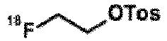

- In an alternative embodiment, the present invention provides a method for preparing a radiolabeled compound according to formula (III); said method comprising the step of reacting radiolabeled


- The present invention provides a compound or composition for use in a method for the diagnosis and treatment follow-up of cancer, more in particular at least one disease or disorder selected from the group comprising non-small cell lung cancer, pancreatic, hepatocellular, oesophageal, gastric, colorectal, prostate, cervical, renal, ovarian, breast cancers, head and neck squamous cell carcinoma, and malignant glioma.
- For diagnostic use, the compounds of the invention may be used as a free acid or base, and/or in the form of a pharmaceutically acceptable acid-addition and/or base-addition salt (e.g. obtained with non-toxic organic or inorganic acid or base), in the form of a hydrate, solvate and/or complex.
- As used herein and unless otherwise stated, the term "solvate" includes any combination which may be formed by a compound of this invention with a suitable inorganic solvent (e.g. hydrates) or organic solvent, such as but not limited to alcohols, ketones, esters and the like. Such salts, hydrates, solvates, etc. and the preparation thereof will be clear to the skilled person; reference is for instance made to the salts, hydrates, solvates, etc. described in
US-A-6,372,778 ,US-A-6,369,086 ,US-A-6,369,087 andUS-A-6,372,733 . - The pharmaceutically acceptable salts of the compounds according to the invention, i.e. in the form of water-, oil-soluble, or dispersible products, include the conventional non-toxic salts or the quaternary ammonium salts which are formed, e.g., from inorganic or organic acids or bases. Examples of such acid addition salts include acetate, adipate, alginate, aspartate, benzoate, benzenesulfonate, bisulfate, butyrate, citrate, camphorate, camphorsulfonate, cyclopentanepropionate, digluconate, dodecylsulfate, ethanesulfonate, fumarate, glucoheptanoate, glycerophosphate, hemisulfate, heptanoate, hexanoate, hydrochloride, hydrobromide, hydroiodide, 2-hydroxyethanesulfonate, lactate, maleate, methanesulfonate, 2-naphthalene-sulfonate, nicotinate, oxalate, palmoate, pectinate, persulfate, 3-phenylpropionate, picrate, pivalate, propionate, succinate, tartrate, thiocyanate, tosylate, and undecanoate. Base salts include ammonium salts, alkali metal salts such as sodium and potassium salts, alkaline earth metal salts such as calcium and magnesium salts, salts with organic bases such as dicyclohexylamine salts, N-methyl-D-glucamine, and salts with amino acids such as arginine, lysine, and so forth. In addition, the basic nitrogen-containing groups may be quaternized with such agents as lower alkyl halides, such as methyl, ethyl, propyl, and butyl chloride, bromides and iodides; dialkyl sulfates like dimethyl, diethyl, dibutyl; and diamyl sulfates, long chain halides such as decyl, lauryl, myristyl and stearyl chlorides, bromides and iodides, aralkyl halides like benzyl and phenethyl-bromides and others. Other pharmaceutically acceptable salts include the sulfate salt ethanolate and sulfate salts.
- Generally, for diagnostic use, the compounds of the inventions may be formulated as a diagnostic preparation or radiopharmaceutical composition comprising at least one compound of the invention and at least one pharmaceutically acceptable carrier, diluent or excipient and/or adjuvant.
- By means of non-limiting examples, such a formulation may be in a form suitable for parenteral administration (such as by intravenous infusion). Such suitable administration forms as well as methods and carriers, diluents and excipients for use in the preparation thereof, will be clear to the skilled person; reference is again made to for instance
US-A-6,372,778 ,US-A-6,369,086 ,US-A-6,369,087 andUS-A-6,372,733 , as well as to the standard handbooks, such as the latest edition of Remington's Pharmaceutical Sciences. - Some preferred, but non-limiting examples of such preparations include sterile injectable solutions for administration as a bolus and/or for continuous administration, which may be formulated with carriers, excipients, and diluents that are suitable per se for such formulations. In addition, cosolvents such as alcohols may improve the solubility and/or the stability of the compounds. In the preparation of aqueous compositions, addition of salts of the compounds of the invention can be more suitable due to their increased water solubility.
- The pharmaceutical preparations of the invention are preferably in a unit dosage form, and may be suitably packaged, for example in a box, blister, vial, bottle, sachet, ampoule or in any other suitable single-dose or multi-dose holder or container (which may be properly labeled); optionally with one or more leaflets containing product information and/or instructions for use. The compounds can be administered by intravenous route, The at least one compound of the invention will generally be administered in an "effective amount", by which is meant any amount of a compound of the Formula (I), (II) or (III) or any subgroup thereof that, upon suitable administration, is sufficient to allow diagnostic imaging in the individual to which it is administered. Usually, depending on the condition to be imaged, such an effective amount will usually be between 1-10 Mbq per kilogram body weight day of the patient per administration, more often between 3-5 Mbq/kg, per kilogram body weight day of the patient per day, which may be administered as a single daily dose, The amount(s) to be administered and the route of administration may be determined by the radiologist or nuclear physicist, depending on factors such as the age, gender and general condition of the patient.
- The invention will now be illustrated by means of the following synthetic and biological examples, which do not limit the scope of the invention in any way.
- The preparation of precursor compound (IIb) is described in Scheme 1.


- Following Scheme 1, starting from methyl (2S)-2-amino-3-methyl-butanoate, a mixture is obtained in which the S-enantiomer of formula (IIb) is the major occurring enantiomer. The S-enantiomer (IIb) can be separated from the R-enantiomer (IIIb) by chiral HPLC.
-

- To a stirred solution of 2-(4-chloro-2-nitro-phenyl)acetic acid(10.0 g, 46.38 mmol) and methyl (2S)-2-amino-3-methyl-butanoate;hydrochloride (7.78g, 46,38 mmol) in acetonitrile (139 ml) were added O-(benzotriazol-1-yl)-N,N,N',N'-tetramethyluronium hexafluorophosphate (21.11 g, 55.66 mmol) and N,N-diisoproylethylamine (23.42 ml, 139.14 mmol). The mixture was stirred at room temperature overnight. Ethyl acetate was added and the organic layer was washed with a saturated aqueous sodium bicarbonate solution. The organic layer was dried, filtered and the solvent was removed under reduced pressure. The product was used in the next step without further purification.
-

- A mixture of intermediate (3) (46.38 mmol), iron (12.95 g, 231.90 mmol) and ammonium chloride (24.81 g, 463.80 mmol) in toluene, tetrahydrofuran and water (450 ml) was stirred under reflux overnight. The reaction mixture was cooled, filtered and the residue was washed with a mixture of tetrahydrofuran and methanol (4:1). The solvent of the filtrate was removed under reduced pressure. Ethyl acetate was added and the organic layer was washed with a saturated aqueous sodium bicarbonate solution. The organic layer was dried, filtered and the solvent was removed under reduced pressure. The residue was purified by flash column chromatography over silica gel.
Yield: 12.0 g of intermediate (4) (87%)
LCMS method 1: MH+ = 299, RT = 0.763 min -

- A mixture of (4-chloro-7-methoxy-quinazolin-6-yl) acetate (10.02 g, 39.66 mmol) and intermediate (4) (11.85 g, 39.66 mmol) in isopropanol (119 ml) was stirred at 80°C for 5 hours. The mixture was cooled and the solvent was removed under reduced pressure. Ethyl acetate was added and the organic layer was washed with a saturated aqueous sodium bicarbonate solution. The organic layer was dried, filtered and the solvent was removed under reduced pressure. The product was used in the next step without further purification.
Yield: 9.64 g of intermediate (6) (47%)
LCMS method 1: MH+ = 492, RT = 0.430 min -

- Ammonia in methanol (7N) (38 ml) was added to a solution of intermediate (6) (13.87 g, 26.93 mmol) in methanol (110 ml). The reaction mixture was stirred at room temperature for 48 hours. Ethyl acetate was added and the organic layer was washed with brine. The organic layer was dried, filtered and the solvent was removed under reduced pressure. The product was used in the next step without further purification.
LCMS method 1: MH+ = 473, RT = 0.618 min -

- Cesium carbonate (9.36 g, 28.74 mmol) was added to a solution of intermediate (7) (9.06 g, 19.16 mmol) in dry N,N-dimethylformamide (57.5 ml) and the mixture was stirred at room temperature for 1 hour. Tert-butyl N-(3-bromopropyl)carbamate (5.02 g, 21.08 mmol) was added and the reaction mixture was stirred at room temperature overnight. Ethyl acetate was added and the organic layer was washed with a saturated aqueous sodium bicarbonate solution. The organic layer was dried, filtered and the solvent was removed under reduced pressure. The product was used in the next step without further purification.
Yield: 11.48 g of intermediate (9) (95%)
LCMS method 1: MH+ = 630, RT = 0.877 min -

- Intermediate (9) (11.48 g, 18.22 mmol) was dissolved in 1,4-dioxane (227 ml) and a 12N hydrochloric acid solution (45.5 ml, 546.60 mmol) was added. The reaction mixture was stirred at 60°C for 3 hours. The solvent was removed under reduced pressure. The residue was washed with diethyl ether and dried under reduced pressure. The product was used in the next step without further purification.
LCMS method 1: MH+ = 517, RT = 0.366 min -

- A solution of intermediate (10) (3.30 g, 5.97 mmol) in N,N-dimethylformamide (100 ml) was added over a period of 2 hours to a solution of O-(benzotriazol-1-yl)-N,N,N',N'-tetramethyluronium hexafluorophosphate (4.98 g, 13.13 mmol) and N,N-diisopropylethylamine (30.5 ml, 179.16 mmol) in N,N-dimethylformamide (200 ml). The reaction mixture was stirred at room temperature for 30 minutes. The solvent was removed under reduced pressure. Dichloromethane was added and the organic layerwas washed with a saturated aqueous sodium bicarbonate solution. The aqueous layer was washed with dichloromethane. The combined organic layers were dried, filtered and the solvent was removed under reduced pressure. The crude product was purified by crystallization from acetonitrile.
Yield: 1.55 g of intermediate (11) (52%)
LCMS method 1: MH+ = 498, RT = 0.581 min -

- The experiment was done in 6 batches on 0.3 g of intermediate (11) each.
- Water (2 drop/ml N,N-dimethylacetamide) was added drop wise to a mixture of intermediate (11) (0.3g, 0.60 mmol), lithium chloride (0.25 g, 5.90 mmol) and disodium sulphide (0.515 g, 6.60 mmol) in N,N-dimethylacetamide (8 ml). The reaction mixture was stirred at room temperature for 30 minutes and at 140°C for 4 hours. Ethyl acetate was added and the organic layer was washed with a saturated aqueous sodium bicarbonate solution. The aqueous layer was washed with ethyl acetate. The combined organic layers were dried, filtered and the solvent was removed under reduced pressure. The crude product was purified by crystallization from acetonitrile.
Yield: 2.22 g of intermediate (IIb) (85%)
LCMS method 2: MH+ = 484, RT = 2.000 min -

- To a solution of intermediate (IIb) (110 mg, 0.23 mmol) in dry N,N-dimethylformamide (4.0 ml) was added cesium carbonate (91 mg, 0.28 mmol). The mixture was stirred at room temperature for 1 hour. A solution of 1-fluoro-2-iodo-ethane (260 mg, 1.5 mmol) in dry N,N-dimethylformamide (1.0 ml) was added and the mixture reaction was stirred at room temperature overnight. Ethyl acetate was added and the organic layer was washed with a saturated aqueous sodium bicarbonate solution. The organic layer was dried, filtered and the solvent was removed under reduced pressure. The residue was purified by flash column chromatography over silica gel.
Yield: 54 mg of compound (IIa) (44%), purity = 99%
LCMS method 2: MH+ = 530, RT = 2.443 min - Chiral separation of the two enantiomers can be achieved either on the precursor compound (IIb) or on the cold analogue (IIa). As an example, but not limiting, the chiral separation of a 30/70 R/S mixture of a compound of formula (IIb) has been described.
- Column:
CHIRALPAK® IA 5 µm - 250 x 30 mm
Mobile phase: Carbon Dioxide/(Ethanol+1%Diethylamine) 60/40
Flow rate: 120ml/min
Detection: UV 230 nm
Outlet Pressure: 120 bar
Temperature: 25°C - Column: CHIRALPAK® AD-
H 5 µm - 250 x 4.6 mm
Mobile phase: n-Heptane/Ethanol/Ethylenediamine 80/20/0.1
Flow rate: 1 ml/min
Detection: DAD 300 nm
Temperature: 25°C - From 420 mg of crude material:
First eluting enantiomer Pure (R)-enantiomer (IIb) Second eluting enantiomer Pure (S)-enantiomer (IIIb) Retention time (min) = 4.5 Retention time (min) = 5.9 Quantity (mg) = 38 Quantity (mg) = 313 Chemical purity (area% at 314 nm) = 99.3 Chemical purity (area% at 314 nm) > 99.5 Enantiomeric excess (%) = 99.2 Enantiomeric excess (%) = 99.0 - The pure enantiomers can be used to perform the alkylation step (cold or with 18F).
- The (R)-enantiomer analogues of intermediate (IIIb) and compound (IIIa) can be prepared according to the experimental procedures described above.
- The preparation of precursor compound (IIIb) is described in
Scheme 2.

- Following
Scheme 2, starting from methyl (2R)-2-amino-3-methyl-butanoate, a mixture is obtained in which the R-enantiomer of formula (IIIb) is the major occurring enantiomer. The R-enantiomer (IIIb) can be separated from the S-enantiomer (IIb) by chiral HPLC. -

- To a stirred solution of 2-(4-chloro-2-nitro-phenyl)acetic acid (12.86 g, 59.65 mmol) and methyl (2R)-2-amino-3-methyl-butanoate;hydrochloride (10.0 g, 59.65 mmol) in acetonitrile (179 ml) were added O-(benzotriazol-1-yl)-N,N,N',N'-tetramethyluronium hexafluorophosphate (27.147 g, 71.58 mmol) and N,N-diisopropylethylamine (31.172 ml, 178.95 mmol). The mixture was stirred at room temperature overnight. Ethyl acetate was added and the organic layer was washed with a saturated aqueous sodium bicarbonate solution. The organic layer was dried, filtered and the solvent was removed under reduced pressure. The product was used in the next step without further purification.
-

- A mixture of intermediate (13) (59.65 mmol), iron (16.66 g, 298.95 mmol) and ammonium chloride (31.907 g, 596.5 mmol) in toluene, tetrahydrofuran and water (1:1:1, 450 ml) was stirred under reflux overnight. The reaction mixture was cooled, filtered and the residue was washed with a mixture of tetrahydrofuran and methanol (4:1). The solvent of the filtrate was removed under reduced pressure. Ethyl acetate was added and the organic layer was washed with a saturated aqueous sodium bicarbonate solution. The organic layer was dried, filtered and the solvent was removed under reduced pressure. The residue was purified by flash column chromatography over silica gel.
Yield: 16.5 g of intermediate (14) (93%) -

- A mixture of (4-chloro-7-methoxy-quinazolin-6-yl) acetate (9.553 g, 37.81 mmol) and intermediate (14) (11.30 g, 37.81 mmol) in isopropanol (113.4 ml) was stirred at 80°C for 5 hours. The mixture was cooled and the solvent was removed under reduced pressure. Ethyl acetate was added and the organic layer was washed with a saturated aqueous sodium bicarbonate solution. The organic layer was dried, filtered and the solvent was removed under reduced pressure. The product was used in the next step without further purification.
Yield: 19 g of intermediate (16) (98%)
LCMS method 1: MH+ = 515, RT = 1.209 min -

- Ammonia in methanol (7N) (60 ml) was added to a solution of intermediate (16) (19.00 g, 36.90 mmol) in methanol (110.7 ml). The reaction mixture was stirred at room temperature for 48 hours. Ethyl acetate was added and the organic layer was washed with brine. The organic layer was dried, filtered and the solvent was removed under reduced pressure. The product was used in the next step without further purification.
Yield: 13.6 g of intermediate (17) (78%) -

- Cesium carbonate (14.056 g, 43.14 mmol) was added to a solution of intermediate (17) (13.60 g, 28.76 mmol) in dry N,N-dimethylformamide (86.3ml) and the mixture was stirred at room temperature for 1 hour. Tert-butyl N-(3-bromopropyl)carbamate (6.85 g, 28.76 mmol) was added and the reaction mixture was stirred at room temperature overnight. Ethyl acetate was added and the organic layer was washed with a saturated aqueous sodium bicarbonate solution. The organic layer was dried, filtered and the solvent was removed under reduced pressure. The residue was purified by flash column chromatography over silica gel using heptane and ethyl acetate as eluents.
Yield: 3.05 g of intermediate (19) (17%)
LCMS method 1: MH+ = 630, RT = 1.474 min -

- Intermediate (19) (2.635 g, 4.18 mmol) was dissolved in 1,4-dioxane (55 ml) and a 12N hydrochloric acid solution (10.9 ml, 125.40 mmol) was added. The reaction mixture was stirred at 60°C for 3 hours. The solvent was removed under reduced pressure. The residue was washed with diethyl ether and dried under reduced pressure. The product was used in the next step without further purification.
-

- A solution of intermediate (20) (3.05 g, 5.52 mmol) in N,N-dimethylformamide (150 ml) was added over a period of 2 hours to a solution of O-(benzotriazol-1-yl)-N,N,N',N'-tetramethyluronium hexafluorophosphate (2.09 g, 5.52 mmol) and N,N-diisopropylethylamine (28.163 ml, 165.60 mmol) in N,N-dimethylformamide (250 ml). The reaction mixture was stirred at room temperature for 30 minutes. A solution of ammonia (7 N solution in methanol, 2 ml)) was added and the mixture was stirred at room temperature for 30 minutes.The solvent was removed under reduced pressure. Dichloromethane was added and the organic layer was washed with a saturated aqueous sodium bicarbonate solution. The aqueous layer was extracted with dichloromethane. The combined organic layers were dried, filtered and the solvent was removed under reduced pressure. The residue was purified by flash column chromatography over silica gel using dichloromethane and methanol (20%) as eluents. The product fractions were collected and the solvent was removed under reduced pressure. The product was triturated with methanol, filtered and the solid was dried under reduced pressure.
Yield: 750 mg of intermediate (21) (27%) -

- The experiment was done in 9 batches on 0.3 g of intermediate (21) each.
- Water (2 drop/ml N,N-dimethylacetamide) was added drop wise to a mixture of intermediate (21) (0.3 g, 0.60 mmol), lithium chloride (0.25 g, 6.00 mmol) and disodium sulphide (0.515 g, 6.60 mmol) in N,N-dimethylacetamide (8 ml). The reaction mixture was stirred at room temperature for 30 minutes and at 140°C for 4 hours. More disodium sulphide (0.4 eq.) was added and the reaction mixture was stirred at 140°C for 1 hour. Ethyl acetate was added and the organic layer was washed with a saturated aqueous sodium bicarbonate solution. The aqueous layer was extracted with ethyl acetate. The combined organic layers were dried, filtered and the solvent was removed under reduced pressure. The crude product was purified by crystallization from acetonitrile, the solid was filtered and washed with diethyl ether. The compound was dried under reduced pressure.
Yield: 164 mg of intermediate (IIIb) (56%) -

- To a solution of intermediate (IIIb) (156 mg, 0.32 mmol) in dry N,N-dimethylformamide (5.0 ml) was added cesium carbonate (124 mg, 0.38 mmol). The mixture was stirred at room temperature for 1 hour. A solution of 1-fluoro-2-iodo-ethane (110 mg, 0.64 mmol) in dry N,N-dimethylformamide (1.0 ml) was added and the mixture reaction was stirred at room temperature overnight. Ethyl acetate was added and the organic layer was washed with a saturated aqueous sodium bicarbonate solution. The organic layer was dried, filtered and the solvent was removed under reduced pressure. The residue was purified by flash column chromatography over silica gel. The product was triturated with diethyl ether, filtered and dried under reduced pressure.
Yield: 95 mg of compound (IIIa) (56%)
LCMS method 2: MH+ = 530, RT = 2.537 min - Chiral separation of the two enantiomers can be achieved either on the precursor compound (IIIb) or on the cold analogue (IIIa). As an example, but not limiting, the chiral separation of a 70/30 R/S mixture of a compounds of formula (IIIa) has been described.
- Chiral separation of compound with formula (IIIa):
- Column: CHIRALPAK®AD-
H 5 µm - 250 x 30 mm
Mobile phase: Ethanol/Methanol 50/50
Flow rate: 30 mL/min
Detection: UV 250 nm
Temperature: 25°C - Column:
CHIRALPAK® IA 5 µm - 250 x 4.6 mm
Mobile phase: Heptane/Isopropanol/Ethylendiamine 50/50/0.1
Flow rate: 1 mL/min
Detection: DAD 336 nm
Temperature: 35°C
Samples dissolved in 100% Ethanol - From 141 mg of crude material (IIIa):
First eluting enantiomer Pure (R)-enantiomer (IIIa) Second eluting enantiomer Pure (S)-enantiomer (IIa) Retention time (min) = 3.7 Retention time (min) = 5.6 Quantity (mg) = 90 Quantity (mg) = 42 Chemical purity (area% at 314 nm) > 99.5 Chemical purity (area% at 314 nm) > 99.5 Enantiomeric excess (%) > 99.5 Enantiomeric excess (%) = 98.8 - For LCMS-characterization of the compounds of the present invention, the following method was used.
- All analyses were performed using an Agilent 6110 series LC/MSD quadrupole coupled to an Agilent 1290 series liquid chromatography (LC) system consisting of a binary pump with degasser, auto sampler, thermostated column compartment and diode array detector. The mass spectrometer (MS) was operated with an atmospheric pressure electro-spray ionization (API-ES) source in positive ion mode. The capillary voltage was set to 3000 V, the fragmentor voltage to 70 V and the quadrupole temperature was maintained at 100°C. The drying gas flow and temperature values were 12.0 L/min and 350°C respectively. Nitrogen was used as the nebulizer gas, at a pressure of 35 psig. Data acquisition was performed with Agilent Chemstation software.
- In addition to the general procedure LCMS1: Analyses were carried out on a Phenomenex Kinetex C18 column (50 mm long x 2.1 mm i.d.; 1.7 µm particles) at 60°C, with a flow rate of 1.5 mL/min. A gradient elution was performed from 90% (water + 0.1% formic acid) /10% Acetonitrile to 10% (water + 0.1% formic acid) / 90% acetonitrile in 1.50 minutes, then the final mobile phase composition was held for an additional 0.40 min. The standard injection volume was 2 µL. Acquisition ranges were set to 254 nm for the UV-PDA detector and 80-800 m/z for the MS detector.
- In addition to the general procedure LCMS1: Analyses were carried out on a YMC pack ODS-AQ C18 column (50 mm long x 4.6 mm i.d.; 3 µm particles) at 35°C, with a flow rate of 2.6 mL/min. A gradient elution was performed from 95% (water + 0.1% formic acid) / 5% Acetonitrile to 5% (water + 0.1% formic acid) / 95% Acetonitrile in 4.80 minutes, then the final mobile phase composition was held for an additional 1.00 min. The standard injection volume was 2 µL. Acquisition ranges were set to 190-400nm for the UV-PDA detector and 100-1400 m/z for the MS detector.
- The preparation of the final radiolabeled compound (II) is described in Scheme 3.
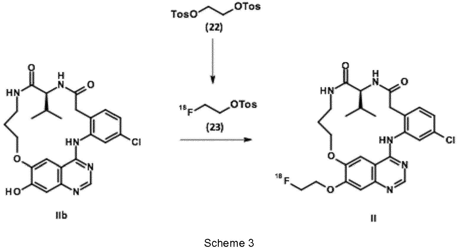
- The synthesis of the radiolabeled compounds is performed on an automatic synthesizer (TracerLab FX-FN Pro, GE Healthcare).
-

- No-carrier-added aqueous 18F-fluoride ion was produced on a cyclotron (PET trace, GE Healthcare) by irradiation of enriched 18O H2O with protons via the 18O(p,n)18F nuclear reaction. 18F-Fluoride was transferred to a GE TRACERIab FX-FN synthesizer and passed through an anion-exchange resin (Waters Sep-Pak Accell Light QMA cartridge in the carbonate form). Trapped 18F-fluoride was isolated by elution with a solution of aqueous eluent solution containing K2CO3 (7 mg in 300 µL of pure water), acetonitrile (300 µL), and 22 mg of Kryptofix-222 (4,7,13,16,21,24-hexaoxa-1,10-diazabicyclo [8.8.8] hexacosane). Azeotropic drying by addition of ACN (1 mL) was performed. The evaporation was performed at 90 °C under helium flow and vacuum, and the operation was repeated twice.
- The ethylene ditosylate (10 mg in 350 µL of ACN) was added to reactor 1 containing the fluoride. The reactor was heated at 90°C for 7 min and then cooled to 30°C. [18F]Fluoroethyltosylate was purified on silica cartridge (Sep Pak Plus, WAT020520, Waters) conditioned with dichloromethane (DCM) and Cyclohexane (5 mL, 5/5:v/v). 2 mL of DCM/Cyclohexane (5/5 :v/:v) were added to the reactor. The mixture was passed through the cartridge and eluate sent to waste. The [18F]Fluoroethyltosylate was eluated from the cartridge using 1.5 mL of DCM/Cyclohexane (7/3 :v/:v) to
reactor 2. Solvents were removed under atmospheric pressure by heating under a helium flow. The same operation was repeated once. -

- In
reactor 2 of the FX-FN Pro module containing the dried [18F]Fluoroethyltosylate was added the intermediate IIb (3 mg in 400 µL of DMSO) with 2.8 mg of cesium carbonate (2.8 mg in 20 µL of water). The reactor was put under pressure and heated at 100°C for 20 min. Then reactor was cooled to 30-35°C and 1.5 mL of ammonium acetate (0.1 M)/ACN (6/4:v/v) was added to dilute the crude solution. The solution was loaded onto the loop and purified by the HPLC integrated to the FX-FN Pro module. Purification occurred on an Agilent XDB C18 5µm 9.4 x250 mm column with ammonium acetate (0.1 M)/ACN (6/4: v/v) as mobile phase and a flow of 4 mL/min. In these conditions time retention is around 16 min. The collected fraction was diluted with 30 mL of water and passed through a tC18 light cartridge (Waters). The cartridge was rinsed with 5 mL of water and the final radiolabeled compound was eluted from the cartridge by using 500 µL of injectable ethanol. Formulation was completed by adding 3.5 mL of physiological serum. - By this method, the final radiolabeled compound can be obtained with a radiochemical purity greater than 98% in 100-110 min with a yield that can reach 25 % (decay corrected). It has been noticed that an uncompleted evaporation of the solvents used for the purification of [18F]Fluoroethyltosylate hampers the second step and conducts to lower yields. Specific activity of productions was in the range of 70-150 GBq/µmole. The radiolabeled compound is stable in the conditions of formulation as radiochemical purity still greater than 95% 20h after production with a storage at room temperature. Plasmatic stability was also checked up to 4h without significant degradation of the product.
- The (R)-enantiomer radiolabelled compound of formula (III), can be prepared according to the experimental procedures described above.

- Unless specifically stated, the in vitro and in vivo assays have been performed using a mixture of R (30%) and S (70%) compound.
- A radiometric protein kinase assay (33PanQinase® Activity Assay) was used for measuring the biochemical kinase activity of 3 protein kinases, EGFR wild-type and 2 EGFR mutants (L858R, L858R/T790M). The compounds were tested in 100 % DMSO, all recombinant protein kinases and substrates with respective concentration are described in table 1.
Table 1: Recombinant protein kinases and substrates used for the biochemical kinase activity assays Kinase ATP Substrate Name Conc. nM Cone. µM Name µg/50µl EGFR L858R (human, 672-1210) 4.5 1.0 poly(Glu, Tyr)4:1 0.25 EGFR T790M/L858R (human, 672-1210) 2.2 0.3 poly(Glu, Tyr)4:1 0.125 EGFR wt (human, 672-1210) 4.4 0.3 poly(Glu, Tyr)4:1 0.125 - Compound (IIa) and Gefitinib, used as reference compound targeting EGFR (wild-type and L858R mutant but not L858R/T790M) were profiled against the 3 forms of EGFR. Activity of Compound (IIa) against EGFR WT and EGFR L858R mutant was below 10 nM, in the same range than gefitinib (Table 2). On the other hand, the biochemical activity against EGFR L858R/T790M double mutant was improved compared to gefitinib.
Table 2a: Biochemical activities in nM of Compound Ia compared to gefitinib Biochemical IC50 (nM) Compound (Ia) - Test 1 Compound (Ia) - Test 2Compound (Ia) - Test 3 Gefitinib EGFR WT (activated) 3.2 5 5.7 1.35 EGFR L858R 3 8 4.1 1.8 EGFR L858R/T790M 372 990 246 1350 Table 2b: Biochemical activities in nM of Compound (IIa) and (IIIa) after chiral separation Biochemical IC50 (nM) Compound (IIa) - Pure (S)-enantiomer - Test 1 Compound (IIIa) - Pure (R)-enantiomer - Test 1 EGFR WT (activated) 5.9 3.1 EGFR L858R 6.3 2.7 EGFR L858R/T790M 924 77.7 T790M 1011 54.7 - The kinase selectivity against a panel of 92 wild-type kinases, chosen to represent the diversity of the kinome, was determined for Compound (IIa) and gefitinib. The residual activity of the compounds was determined at 100 nM and 1µM following the same protocol as IC50 determination. Corresponding kinases and substrates were always tested at the ATP Km concentration. The selectivity is represented on a dendrogram with dot size proportional to residual activity. The selectivity score S(50) is calculated as followed:

- The selectivity was very good since EGFR and RIPK2 were the only wild-type kinases inhibited for more than 50% by Compound (IIa) at the concentration of 100 nM (data not shown). This profile is comparable to the one for gefitinib. At 1µM, which corresponds to a fold > 100 compared to primary activity against EGFR WT or EGFR_L858R mutant, we observed an inhibition of > 50% on 9 other kinases (2 other kinases for gefitinib). The selectivity scores S(50) is 2.2% and 11.9% respectively at 100 nM and 1 µM.
- The selectivity scores S(50) of compound (IIa) - Pure S - enantiomer is 2.5% and 13% respectively at 100 nM and 1 µM.
- The selectivity scores S(50) of compound (IIIa) - Pure R - enantiomer is 0.6% and 2.8% respectively at 100 Nm and 1µM.
- The cell lines that were used to assay the cellular activity of the compounds are detailed in table 3 hereafter:
Table 3: Cell lines description and respective EGFR mutation status Cell line Type EGFR mutation status Species NCI-H441 Lung papillary adenocarcinoma WT Human NCI-H3255 Lung adenocarcinoma Simple mutation, L858R NCI-H1975 Adenocarcinoma non-small cell lung cancer Double mutation, L858R/T790M MCF-7 Pleural effusion adenocarcinoma Very low to none expression, WT - Tumor cells grew as monolayer at 37°C in a humidified atmosphere (5% CO2) in complete culture medium adapted to each cell line. The cellular activity of the compounds was assessed by measuring the viability of cancer cell in MTS assay using a tetrazolium compound (MTS, 3-(4,5-dimethylthiazol-2-yl)-5-(3-carboxymethoxy phenyl)-2-(4-sulfophenyl)-2H-tetrazolium) and an electron coupling reagent named PMS (phenazine methosulfate). MTS is bioreduced by cells into a formazan product that is directly soluble in culture medium without processing. The cells were seeded 24 hours before treatment at the appropriate density in their culture medium to be near 90% of confluence at the end of experiment in vehicle treated conditions. After addition of compounds, the cells were incubated for 72 hours.
- As shown in table 4, the cellular activities are in the same range for Compound (IIa) and gefitinib. Compound (IIIa) showed a very high activity on NCI-H3255 (mean IC50 of 6.1 nM) and a moderate activity on NCI-H1975 (mean IC50 of 5 µM), whereas it has a weak activity on NCI-H441 (mean IC50 of 34.7 µM). A set of experiments was also performed on MCF-7 which does not express EGFR (negative control). Compound (Ia) and the corresponding S (IIa) and R (IIIa) enantiomers have a very weak activity on MCF-7 (IC50 of 62.1 µM) suggesting a good cellular selectivity.
Table 4: IC50 of Compound (Ia), compound (IIa) (i.e. Pure S - enantiomer) and compound (IIIa) (i.e. Pure R-enantiomer) compared to gefitinib determined by MTS assay performed on NCI-H441, NCI-H3255, NCI-H1975 and MCF-7 Cellular IC50 (µM) Compound (Ia) Compound (IIa) - Pure (S)-enantiomer Compound (IIIa) - Pure (R)-enantiomer Gefitinib NCl-H441 (EGFR WT) 34.7 ± 24 > 25 > 25 14.9 ± 2.7 NCl-H3255 (EGFR_L858R) 0.006 ± 0.002 0.030/0.049 0.0008/0.056 0.012 ± 0.007 NCl-H1975 (EGFR L858R/T790M) 5.0 ± 1.4 10/25 20/25 17.7 ± 5.9 MCF-7 62.1 > 25 > 25 - To evaluate the inhibition of EGFR phosphorylation induced by Compound (Ia) compared to gefitinib, cells were treated with a dose range of each compound and induced with 10 ng/ml of EGF. Effect on Y1068 phosphorylation was observed by Western blot (data not shown). On NCI-H441, Compound (Ia) inhibited the phosphorylation induced by EGF, at a slightly better level than gefitinib, with almost full inhibition observed at 1 µM. On NCI-H3255, the full inhibition of constitutively active EGFR was observed. This kinase inhibition was observed approximately at 100 nM for Compound (Ia), at a level similar to gefitinib inhibition. On the constitutively active double mutant EGFR expressed by NCI-H1975, the gefitinib is not active at the top dose of 1 µM, as this cell line acquired resistance to this drug through T790M mutation. Interestingly, Compound (Ia) seemed to partially inhibit EGFR activity at 10 µM while gefitinib did not, showing again the slightly better inhibition of EGFR kinase activity of this new fluorinated compound compared to gefitinib.
- The kinetic solubility of the compounds was assessed in PBS at pH 7.4. Compound (Ia) showed a good solubility of 88 µM in PBS (Table 5).
Table 5: Solubility and stability of Compound (Ia) and Compound (I) Parameter Result Kinetic solubility at pH 7.4 (µM) (Compound la) 88 Radiochemical stability (h) (Compound I) > 20 Plasmatic stability (h) (Compound Ia and I) > 4 human microsomal stability (Compound Ia) Clint (µL/min/mg prot.) 7.76 ± 2.53 t1/2 (min) 179 rat microsomal stability(Compound Ia) Clint (µL/min/mg prot.) 37.9 ± 1.89 t1/2 (min) 36.5 - After radiosynthesis, the stability of the radiotracer was evaluated by HPLC both in its vehicle and in rat plasma. Compound (I) was formulated in physiologic serum/ethanol with the respective proportion 87.5/12.5 (v/v). A mixture of compounds (I) (i.e. radiolabelled) and (Ia) (i.e. cold analogue) was stable for up to 20 hours at room temperature in its vehicle, only one peak being detected at the expected retention time. In rat plasma two peaks were detected: one at 3.15 minutes and one at 8.02 minutes (corresponding to a mixture of compounds (I) and (Ia)) after an incubation at +37°C for 4 hours. The area of the peak corresponding to said mixture accounted for >95% of the total area of both peaks, indicating that said mixture was stable in human plasma for up to 4 hours (data not shown).
- Metabolic microsomal stability was performed on human and rat liver microsomes. Microsomes (final protein concentration 0.5 mg/mL), 0.1 M phosphate buffer pH 7.4 and test compound (final substrate concentration = 3 µM; final DMSO concentration = 0.25%) are pre-incubated at 37°C prior to the addition of NADPH (final concentration = 1 mM) to initiate the reaction. Analysis is done with generic LC-MS/MS conditions to determine half-life and intrinsic clearance using standard equations. Microsomal stability study showed, in rat, a half-life in the range of 40 minutes for both fluorinated compounds and a clearance superior to 30 µL/min/mg of proteins (Table 5). In human, Compound (Ia) metabolic stability was better than in rat and favorable for clinical studies.
- The binding affinity of Compound (I) and the number of binding sites were first evaluated on NCI-H3255 and NCI-H441 tumor cell homogenates (
Figure 1 ). The binding affinity (Kd) of Compound (I) on NCI-H3255 tumor cell homogenates was 23.8 ± 9.0 nM and the number of binding sites was 9.1 ± 3.1 x10⁹ sites/µg protein. The affinity of Compound (I) on NCI-H441 tumor cell homogenates was 25.8 ± 11.1 nM, being similar to that determined in NCI-H3255 tumor cell homogenates. The number of binding sites was of the same order of magnitude (6.1 ± 0.5 x10⁹ sites/µg protein). - Similar experiments were performed on NCI-H3255 and NCI-H1975 tumor homogenates (
Figure 2 ). Binding affinities were similar: 2.8 ± 2.4 nM and 4.7 ± 2.0 nM for NCI-H3255 and NCI-H1975 tumor homogenates, respectively. The number of binding sites was 1.1 ± 1.5 x10⁹ and 1.6 ± 1.8 x10⁹ sites/µg protein respectively in NCI-H3255 and NCI-H1975 tumor homogenates. - Considering the NCI-H3255 cell line extract and tumor homogenate, affinity and number of binding sites obtained for cell and tumor homogenates appeared consistent: binding affinity of the same order of magnitude and number of binding sites 8-fold lower in tumor homogenates than in tumor cell homogenates. The lower number of binding sites reflects most likely the lower number of tumor cells in tumor homogenates due to the presence of stroma cells.
- The binding kinetic of Compound (I) was also assessed in NCI-H3255 and NCI-H1975 tumor homogenates (
Figure 3 ). The dissociation constant (Koff) of Compound (I) was slightly lower in NCI-H3255 homogenates than in NCI-H1975 tumor homogenates (0.114 ± 0.001 vs 0.270 ± 0.109 min- 1, respectively) while the association constant (Kon) was slightly higher in NCI-H1975 tumor homogenates than in NCI-H3255 tumor homogenates (6.5 ± 5.3 x10⁷ L.mol-1.min-1 vs 1.8 ± 1.6 x10⁷ L.mol-1.min-1, respectively). Binding kinetic data show that Compound (I) binds to its target and is released faster in NCl-H1975 than in NCH-H3255 tumor homogenates. - As in vitro data showed that Compound (I) specifically bound to activated EGFR, autoradiography experiments on human frozen tumor sections were initiated (data not shown). Three types of human lung tumors were included: tumors harboring wild type EGFR and tumors carrying L858R mutated or exon 19 deleted EGFR genes. Binding competition with gefitinib and ATP was also evaluated on these frozen tumor sections. A specific binding of Compound (I) was observed on all tumor types. Gefitinib competed with the binding of Compound (I) but only partially while ATP (at 10 mM) totally abolished the binding of Compound (I) (data not shown). Data suggest that the displacement observed in the presence of Gefitinib may be higher on tumors harboring the L858R mutation or the deletion of exon 19 as compared to wild type tumors.
- In vitro experiments showed that a mixture of compounds (I) and (Ia) specifically bound to its target, that is presumably activated EGFR. The binding affinity of said mixture was in the nanomolar range which is suitable for in vivo studies.
- Dynamic PET scan imaging and ex vivo gamma counting were performed on three cancer xenografts (NCI-H441, NCI-H3255, NCI-H1975) in Nude rats. Xenografts were initiated in athymic, Hsd:RH-Foxn1rnu rats (Harlan, The Netherlands) by subcutaneous injection of 2×10⁷ cells in 200 µL RPMI 1640 in the left and right flanks. NCI-H3255 tumor cells were injected in the right flank and NCI-H441 or NCI-H1975 in the left flank. Each study consisted in two experiments on 18 or 19 rats, for a total of 113 rats. Injection of NCI-H3255 cells was performed on D0, 24 to 72 hours after a whole-body irradiation with a ©-source (5 Gy, 60Co, BioMep SARL, Bretenières, France). Injection of either NCI-H441 or NCI-H1975 cells was performed 20 to 29 days post injection of NCI-H3255 cells to ensure a comparable volume for both tumors at the time of experiment. Tumors were grown until a target volume of 500-1500 mm3 for both tumors was reached, whenever possible. All procedures using animals were approved by the Animal Care and Use Committee of Oncodesign (CNREEA agreement N° 91).
- The radiotracer solution was diluted in NaCl 0.9% to prepare a solution of activity 4-6 MBq (gamma-counting experiments) or 25-35 MBq (PET imaging experiments) in a volume of 300-400 or 600-800 µL, respectively. The radioactivity in the syringe was measured before and after tracer injection to determine the injected dose (ID) to animals. Rats were anesthetized with inhaled isoflurane (2% in air) for compound injection and throughout the course of image acquisition. After compound injection, rats were allowed to wake up and put back in their cage for an uptake period ranging from 20 to 120 minutes depending on the experiment.
- For ex vivo gamma counting, after the determined uptake period or after imaging, rats were euthanized by decapitation, and the following organs were harvested and weighed: tumor, kidneys, blood, liver, heart, tail, thigh muscle, lungs, spleen and skin. Tracer distribution was assessed by determining 18F content in each tissue with a gamma-well counter (PerkinElmer 2480 Wizard2 3). Counting data are reported as %ID per gram of tissue (%ID/g). Once radioactivity in organs was measured, some samples were prepared for immunohistochemistry.
- For imaging experiments, rats were positioned prone in a dedicated rat imaging chamber (Minerve, France). Their body temperature was maintained by a flow of warm air through the structure of the imaging bed. PET imaging was performed on a microPET eXplore Vista CT (General Electric Healthcare) system. The Vista system, equipped with 36 dual-layer LYSO/GSO phoswich detector modules, has an effective axial/transaxial field of view of 4.8/6.7 cm. The spatial resolution is < 2 mm in all directions and in the whole field of view. The sensitivity is above 2.5% in the whole field of view. After a scout image to adjust the position of the field of view, a CT scan was acquired (140 µA, 40 kV). One minute after initiation of PET image acquisition, Compound (I) was administered into a lateral tail vein via an i.v. line and flushed with a saline solution. Three-dimensional (3D) PET data were acquired in list mode for up to 120 minutes and reconstructed using OSEM-2D to create two data sets: one static image as the sum of data during the whole acquisition, and a set of dynamic images with time frames of 10 minutes. Corrections for attenuation, scatter, dead time, detector sensitivity, and randoms were applied to emission data. Both data sets had a spatial resolution of 0.4×0.4×0.8 mm and a size of 175×175×61 voxels. All rats were euthanized immediately after PET data acquisition by decapitation, and organs were harvested as described in the previous paragraph to obtain biodistribution data corresponding to PET image data.
- The reconstructed PET/CT images were viewed and analyzed in dedicated software (PMOD, version 3.4). Regions of interest (ROI) were manually drawn in CT images and, after manual registration of PET and CT images, decay-corrected activity data were obtained (in kBq/cm3, normalized by ROI volume). Compound (I) time-activity curves (TAC) were generated for each ROI by averaging over frames of 10 minutes. For the purpose of display, we also generated images from the static scan summed over the whole tumor volume (6 to 10 slices in the PET volumetric data set).
- Compound (I) was rapidly cleared from the blood (<0.05 %ID/g after 45 min,
Figure 4 ). Compound (I) showed initial uptake in the kidneys and liver, but did not accumulate in these organs. Compound (I) also showed central accumulation and excretion through the gastrointestinal tract. Uptake of Compound (I) was higher in NCI-H3255 tumors than in NCI-H441 or NCI-H1975 tumors, both 90 minutes and 180 minutes post injection. Uptake decreased between 90 min and 180 min in all organs and tumors, except in NCI-H3255, where it stayed stable (around 0.2 %ID/g). The absolute uptake in tumors was relatively low, (<0.5 %ID/g at 90 min, seefigure 4 ), but the normalized uptake (over muscle) increased between 90 min and 180 min in NCl-H3255 to reach a value of 12 at 180 min (NCI-H441: 2.6; NCI-H1975: 3.6, seeFigure 5 ). - For all three tumor models, representative PET images show the difference in uptake measured by in vivo imaging in tumors between the WT, the single-mutation and the double-mutation models (data not shown). Uptake is confirmed by the distribution of harvested activity strongly correlated with distribution observed in PET images (
Figure 6 ). - As shown in
Figure 6 , uptake measures by PET imaging or by counting radioactivity in collected tumors were strongly correlated. Our experiments can then be described by studying relationships between tumor models (EGFR mutation status), radiotracer uptake measured on harvested tumors (in %ID/g), pEGFR staining measured by immunohistochemistry (IHC) on these tumors, and tumor volume. For IHC, xenografts were dissected and fixed in formalin-buffered saline for 24 hours followed by 70% ethanol before paraffin embedding and sectioning (thickness: 4 µm). EGFR phosphorylation was determined by immunostaining with an antibody against pEGFR (phospho Y1068, Abcam, ref ab32430) using an automated staining instrument (Ventana Discovery, Roche) and necrosis was evaluated on hematoxylin and eosin (H&E) stains. Negative controls were prepared omitting the primary antibody and showed no staining. Slides were digitized using a Nanozoomer slide scanner (Hamamatsu, ×20 magnification) for image analysis and quantification. Necrosis was evaluated by manual delineation of necrotic zones in tumors using NDPView (Hamamatsu, France) and reported as a percentage of necrotic surface area over the whole tumor tissue area. Staining of pEGFR was quantified using NDPView and Visilog (FEI Visualization Sciences Group, France) by thresholding and manual delineation of viable tumor tissue and tissue positive for pEGFR. Intensity of pEGFR is reported as the product of the number of pEGFR+ pixels and their average staining intensity, over the area of viable tumor tissue in µm2. -
Figure 7 (top row) shows that double-mutation tumors were comparably bigger than the other two models, and that pEGFR intensity was lower in bigger tumors. We therefore divided the measurements into subsets based on tumor volumes (below and above 1000 mm3). In terms of tumor biology, this threshold represents the volume at which necrosis starts to appear.Figure 7 also shows that this partition allows to identify a correlation between EGFR activity and measured radiotracer uptake in both single- and double-mutation models, in smaller tumors, while this correlation disappears in bigger tumors. For the WT tumor model, this correlation is not observed regardless of tumor volume. - To evaluate the selectivity of Compound (I) for imaging activated EGFR in the three tumor models, rats were injected with either gefitinib (2 mg/rat, for an approximate dose of 10 mg/kg) or the nonradioactive (or cold) tracer Compound (Ia) (1 mg/rat, approximate dose: 5 mg/kg) in large excess to compete for its target. Normalized uptakes and ratios of tumor over muscle uptakes are presented in
Figure 8 . - Competition with neither gefitinib (+16%, p>0.95) nor Compound (Ia) (5%, p>0.99) modified the uptake of Compound (I) in WT NCI-H441 tumors. For NCI-H3255 tumors (simple mutation), T/M ratios are decreased by 29% by gefitinib competition (p<0.01) and by 52% (p<0.001) by Compound (Ia) competition. This shows the specificity of the radiotracer for EGFR as a common target in this tumor model. For NCI-H1975 tumors (double mutation), gefitinib does not have any influence on T/M ratios (+1%, p>0.99), but we have observed a marked but statistically non-significant decrease (-55%, p=0.2) after competition with Compound (Ia).
- Overall, this PK profile is favorable for the use of Compound (I) as a radiotracer, provided a sufficiently long uptake period is observed before imaging.
- Overall, Compound (I) is a good PET radiotracer candidate to evaluate by PET imaging in human lung tumors the EGFR activity in correlation with its mutational status.
- To evaluate the biodistribution of the pure S (II) and R (III) enantiomers compared to compound (I), dynamic PET scan imaging and ex vivo gamma counting were performed on Sprague Dawley rats. Animals were injected with the radiotracer solution diluted in NaCl 0.9% to prepare a solution of activity 4-6 MBq (gamma-counting experiments) or 11-13 MBq (PET imaging experiments) in a volume of 300-400 or 600-800 µL, respectively. Radioactivity content in all biological samples was determined with scintillation γ-counter (Cobra 4180, Perkin-Elmer Inc.).
- At the light of the biodistribution data of compound (I), the biodistribution of the R (III)- and S (II) - enantiomers was assessed by γ-counting in the organs in which the accumulation of compound (I) was prominent (that is: blood, heart, kidneys, liver, stomach, small intestine and colon). Two timepoints were selected: 5 and 30 minutes post-injection. The choice of these two timepoints was driven by the fact that compound (I) did not accumulate in blood, liver and kidneys. The higher uptakes of R enantiomer (III) and S enantiomer (II) in these organs were expected to be observed at early timepoints after injection, giving the highest signals in the γ-counter and therefore making the comparison of biodistribution data more relevant. Besides, as PET images were acquired with compound (I), PET imaging was included in the evaluation of the biodistribution of R enantiomer and S enantiomer. This gave the opportunity to had one additional timepoint at 4 hours post-injection for collection of major organs and determination of radioactivity content by ex vivo γ-counting.
- On the 5 minutes, post-injection timepoint, uptake in blood reached 0.20 ± 0.002%ID/g for R enantiomer (III) and 0.11 ± 0.01%ID/g for S enantiomer (II) (while the uptake was 0.13 ± 0.02%ID/g for compound (I). On the 0.5 hour timepoint, radioactivity uptake was similar for the three tracers (close to 0.03-0.04%ID/g). Similarly, uptake at 4 hours post-injection was in the same range of magnitude for the three tracers (0.02-0.04%ID/g). The behavior of R enantiomer (III) and S enantiomer (II) in blood was similar to that of compound (I).
- Five minutes after the injection, the uptake in heart of R enantiomer (III) and S enantiomer (II) reached 0.99 ± 0.02 and 1.35 ± 0.35%ID/g, respectively, these uptakes being comparable to that of compound (I) (0.91 ± 0.12%ID/g). At 30 minutes post-injection, uptakes were similar for R enantiomer and S enantiomer (0.10 ± 0.02 and 0.19 ± 0.08%ID/g) and were still comparable to that of compound (I) (0.23 ± 0.05%ID/g). On the 4 hours timepoint, uptake of the three tracers had decreased to 0.01%ID/g.
- Five minutes post-injection, the uptake of R (III) and S (II) enantiomer in kidneys were similar to that of compound (I): 4.15 ± 0.79 and 6.89 ± 0.52%ID/g vs 5.07 ± 0.30%ID/g, respectively. On the 30 minutes timepoint, uptake in kidneys was of the same order of magnitude for S enantiomer (II) and compound (I) (1.04 ± 0.17 and 1.47 ± 0.36%ID/g, respectively) but was lower for R enantiomer (III) (0.25 ± 0.08%ID/g). For this last tracer, individual uptakes recorded 30 minutes post-injection were lower than the lowest individual value recorded for compound (I) at the same timepoint. Kidneys uptake was in the 0.01-0.05%ID/g for the three
tracers 4 hours post-injection. - For R enantiomer (III), uptake in liver increased from 5 minutes to 30 minutes post injection: 0.02 ± 0.09 to 0.71 ± 0.07%ID/g, respectively. For S enantiomer (II), a decrease was noticed between 5 and 30 minutes post-injection: 0.97 ± 0.92 to 0.15 ± 0.03%ID/g as observed for compound (I) (1.66 ± 0.43 to 0.37 ± 0.07%ID/g). On the four hour timepoint, liver uptake did not exceed 0.02%ID/g for the three tracers.
- The uptake in stomach of R enantiomer (III) and S enantiomer (II) was 1.86 ± 0.73%ID/g and 0.72 ± 0.38%ID/g, respectively, at 5 minutes post-injection. At this timepoint the uptake of compound (I) was 1.27 ± 2.13%. The uptake of R enantiomer (III) and S enantiomer (II) declined to 0.26 ± 0.07 and 0.20 ± 0.05%ID/g on the 30 minutes timepoint, while a slight increase was noticed for compound (I) (2.72 ± 4.75%ID/g). Four hours post-injection, uptake in stomach increased for R enantiomer (III) and S enantiomer (II) (2.58 ± 2.52 and 2.39 ± 2.31 %ID/g) while it decreased for compound (I) (0.14 ± 0.23%ID/g).
- Uptake in small intestine reached 0.35 ± 0.19 and 0.08 ± 0.09%ID/g at 5 minutes post injection for R enantiomer (III) and S enantiomer (II), respectively, whereas the uptake was 0.28 ± 0.15%ID/g for compound (I). Thirty minutes post-injection, uptake of R enantiomer (III) and S enantiomer (II) was lower than that of compound (I) (0.02 ± 0.004 and 0.08 ± 0.07%ID/g vs 0.40 ± 0.40%ID/g, respectively). At 4 hours post-injection the uptake of R enantiomer (III) and S enantiomer (II) was similar to that of compound (I) (in the 0.6-0.8%ID/g range).
- Uptake in colon of R enantiomer (III), S enantiomer (II) and compound (I) was in the 0.2-0.4%ID/g for the three
tracers - In summary, R enantiomer (III) and S enantiomer (II) were rapidly cleared from the blood as observed for compound (I). These two tracers were also eliminated from the body both via the kidney and the liver and were not trapped in these organs. As for compound (I), an uptake was noticed in stomach, small intestine and colon for R enantiomer and S enantiomer.
- The overall behavior of R enantiomer (III) and S enantiomer (II) after an IV injection is comparable to that of compound (I). Pharmacokinetic profiles are similar for the three tracers.
- Uptake (%ID, corrected for radioactive decay) of compound (I), R enantiomer (III) and S enantiomer (II) in kidneys and liver determined by PET image analysis and ex vivo γ-counting are presented in
Figure 9(A) . 90 minutes time-activity curves (%ID corrected for radioactive decay as a function of time) of compound (I), R enantiomer (III) and S enantiomer (II) in kidneys, intestine, stomach, and liver after a single IV injection in Sprague-Dawley rats are presented inFigure 9(B) .
Claims (12)
- A fluorine-18 labeled compound of formula (I), or any pharmaceutically acceptable salt, polymorph, solvate, hydrate, stereoisomer, radioisotope or tautomer thereof
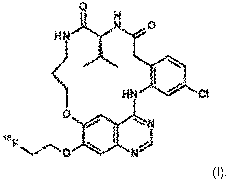
- A fluorine-18 labeled compound as defined in claim 1 and having the R-stereoisochemistry as represented in formula (III), or any pharmaceutically acceptable salt, polymorph, solvate, hydrate, stereoisomer, radioisotope or tautomer thereof

- A fluorine-18 labeled compound as defined in claim 1 and having the S-stereoisochemistry as represented in formula (II), or any pharmaceutically acceptable salt, polymorph, solvate, hydrate, stereoisomer, radioisotope or tautomer thereof

- A radiopharmaceutical composition comprising a radiolabeled compound according to anyone of claims 1, 2 or 3, optionally further comprising one or more inert carriers and/or diluents.
- A radiolabeled compound according to anyone of claims 1, 2 or 3 or a radiopharmaceutical composition according to claim 4 for use as a diagnostic agent in human medicine.
- Use of a radiolabeled compound according to anyone of claims 1, 2 or 3 or a radiopharmaceutical composition according to claim 4 in tumor imaging.
- A radiolabeled compound according to anyone of claims 1, 2 or 3 or a radiopharmaceutical composition according to claim 4, for use in in vivo diagnosis or tumor imaging.
- A method for diagnostic imaging of an EGFR-associated tumor in a human, which comprises administering to a human in need of such diagnostic imaging an effective amount of a compound according to anyone of claims 1, 2 or 3 or a radiopharmaceutical composition according to claim 4, and obtaining an image useful for quantifying EGFR in the tumor of said human using positron emission tomography.
- A method for the quantification of EGFR in human tissue, which comprises contacting such human tissue in which quantification is desired with an effective amount of a compound according to anyone of claims 1 or 2, or a radiopharmaceutical composition according to claim 4, and detecting or quantifying EGFR using positron emission tomography.
- A method for preparing a radiolabeled compound according to claim 1; said method comprising the step of
reacting radiolabeled

- A method for preparing a radiolabeled compound according to claim 2; said method comprising the step of
reacting radiolabeled

- A method for preparing a radiolabeled compound according to claim 3; said method comprising the step of
reacting radiolabeled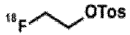

Applications Claiming Priority (2)
| Application Number | Priority Date | Filing Date | Title |
|---|---|---|---|
| EP16157885 | 2016-02-29 | ||
| PCT/EP2017/054611 WO2017148925A1 (en) | 2016-02-29 | 2017-02-28 | Radiolabeled macrocyclic egfr inhibitor |
Publications (2)
| Publication Number | Publication Date |
|---|---|
| EP3423113A1 EP3423113A1 (en) | 2019-01-09 |
| EP3423113B1 true EP3423113B1 (en) | 2020-09-02 |
Family
ID=55446700
Family Applications (1)
| Application Number | Title | Priority Date | Filing Date |
|---|---|---|---|
| EP17707055.4A Active EP3423113B1 (en) | 2016-02-29 | 2017-02-28 | Radiolabeled macrocyclic egfr inhibitor |
Country Status (10)
| Country | Link |
|---|---|
| US (1) | US10716869B2 (en) |
| EP (1) | EP3423113B1 (en) |
| JP (1) | JP6913101B2 (en) |
| AU (1) | AU2017226878B2 (en) |
| CA (1) | CA3015430A1 (en) |
| DK (1) | DK3423113T3 (en) |
| ES (1) | ES2833959T3 (en) |
| IL (1) | IL261274B (en) |
| PT (1) | PT3423113T (en) |
| WO (1) | WO2017148925A1 (en) |
Families Citing this family (4)
| Publication number | Priority date | Publication date | Assignee | Title |
|---|---|---|---|---|
| JP7262922B2 (en) * | 2017-12-06 | 2023-04-24 | キヤノンメディカルシステムズ株式会社 | Methods for Imaging Proteins |
| EP4031146A4 (en) * | 2019-09-18 | 2023-12-06 | The Regents Of The University Of California | Positron emission tomography guided delivery of mitochondral complex i inhibitors |
| GB201915206D0 (en) | 2019-10-21 | 2019-12-04 | Ge Healthcare Ltd | Use of cyclodextrins as a radiostabilizer |
| GB202108605D0 (en) * | 2021-06-16 | 2021-07-28 | Ge Healthcare Uk Ltd | Effect of water content |
Family Cites Families (6)
| Publication number | Priority date | Publication date | Assignee | Title |
|---|---|---|---|---|
| IS2334B (en) | 1992-09-08 | 2008-02-15 | Vertex Pharmaceuticals Inc., (A Massachusetts Corporation) | Aspartyl protease inhibitor of a new class of sulfonamides |
| US6043358A (en) | 1995-11-01 | 2000-03-28 | Merck & Co., Inc. | Hexahydro-5-imino-1,4-heteroazepine derivatives as inhibitors of nitric oxide synthases |
| GB9718913D0 (en) | 1997-09-05 | 1997-11-12 | Glaxo Group Ltd | Substituted oxindole derivatives |
| US6369087B1 (en) | 1999-08-26 | 2002-04-09 | Robert R. Whittle | Alkoxy substituted benzimidazole compounds, pharmaceutical preparations containing the same, and methods of using the same |
| PA8603801A1 (en) | 2003-05-27 | 2004-12-16 | Janssen Pharmaceutica Nv | DERIVATIVES OF QUINAZOLINE |
| EP2951171A1 (en) | 2013-02-01 | 2015-12-09 | Boehringer Ingelheim International GmbH | Radiolabeled quinazoline derivatives |
-
2017
- 2017-02-28 ES ES17707055T patent/ES2833959T3/en active Active
- 2017-02-28 JP JP2018545279A patent/JP6913101B2/en active Active
- 2017-02-28 AU AU2017226878A patent/AU2017226878B2/en active Active
- 2017-02-28 CA CA3015430A patent/CA3015430A1/en active Pending
- 2017-02-28 WO PCT/EP2017/054611 patent/WO2017148925A1/en not_active Ceased
- 2017-02-28 EP EP17707055.4A patent/EP3423113B1/en active Active
- 2017-02-28 DK DK17707055.4T patent/DK3423113T3/en active
- 2017-02-28 US US16/080,196 patent/US10716869B2/en active Active
- 2017-02-28 PT PT177070554T patent/PT3423113T/en unknown
-
2018
- 2018-08-21 IL IL261274A patent/IL261274B/en active IP Right Grant
Non-Patent Citations (1)
| Title |
|---|
| None * |
Also Published As
| Publication number | Publication date |
|---|---|
| ES2833959T3 (en) | 2021-06-16 |
| IL261274B (en) | 2020-05-31 |
| EP3423113A1 (en) | 2019-01-09 |
| JP2019507155A (en) | 2019-03-14 |
| JP6913101B2 (en) | 2021-08-04 |
| CA3015430A1 (en) | 2017-09-08 |
| DK3423113T3 (en) | 2020-11-30 |
| AU2017226878B2 (en) | 2020-07-23 |
| WO2017148925A1 (en) | 2017-09-08 |
| US20190054197A1 (en) | 2019-02-21 |
| AU2017226878A1 (en) | 2018-09-27 |
| PT3423113T (en) | 2020-12-10 |
| US10716869B2 (en) | 2020-07-21 |
| IL261274A (en) | 2018-10-31 |
Similar Documents
| Publication | Publication Date | Title |
|---|---|---|
| Slobbe et al. | Development of [18F] afatinib as new TKI-PET tracer for EGFR positive tumors | |
| US10716869B2 (en) | Radiolabeled macrocyclic EGFR inhibitor | |
| US20100226853A1 (en) | Fluorine-18 derivatives of dasatinib and uses thereof | |
| Hu et al. | Synthesis and biological evaluation of N-(2-[18F] Fluoropropionyl)-L-methionine for tumor imaging | |
| US20110097268A1 (en) | Radiohaloimatinibs and methods of their synthesis and use in pet imaging of cancers | |
| Valtorta et al. | [18F](2 S, 4 R)-4-Fluoroglutamine as a New Positron Emission Tomography Tracer in Myeloma | |
| EP2841402B1 (en) | Labelled quinoxaline derivatives as multimodal radiopharmaceuticals and their precursors | |
| Rizzo-Padoin et al. | [18F] MEL050 as a melanin-targeted PET tracer: fully automated radiosynthesis and comparison to 18F-FDG for the detection of pigmented melanoma in mice primary subcutaneous tumors and pulmonary metastases | |
| EP2795317B1 (en) | Composition for use in a method for cancer selection | |
| CA3036299C (en) | [18f]-labelled lactate derivative as pet radiotracer | |
| AU2024343052A1 (en) | Radiopharmaceutical | |
| KR20110098724A (en) | Radioisotope-labeled lysine and ornithine derivatives, uses thereof and methods for their preparation | |
| JP2019043882A (en) | Therapeutic effect predictors for alternative anticancer drugs | |
| EP3111960B1 (en) | Nuclear medicine diagnostic imaging agent | |
| JP6709552B2 (en) | Nuclear medicine diagnostic imaging agent | |
| Huang et al. | Investigation of brain tumors using 18F-fluorobutyl ethacrynic amide and its metabolite with positron emission tomography | |
| US12138320B2 (en) | Composition and methods for tumor imaging and treatment | |
| CN118994151B (en) | Imidazolopyridone compounds binding to sigma-2 receptors, preparation method and application thereof | |
| US20190023664A1 (en) | Radiolabeled erlotinib analogs and uses thereof | |
| Xie et al. | Synthesis, radiolabeling, and evaluation of a 68Ga-labeled tyrosine kinase inhibitor for detecting EGFRT790M mutations in vivo | |
| EP3112365B1 (en) | Nuclear medicine diagnostic imaging agent | |
| Fawwaz et al. | Imaging Non-Small Cell Lung Cancer EGFR Del19 and L858R with Radiolabeled Small Molecule | |
| JP6739987B2 (en) | Nuclear medicine diagnostic imaging agent | |
| JP2019064986A (en) | MOLECULE PROBE FOR Bcr-Abl PROTEIN IMAGING | |
| Hicks et al. | www. mdpi. com/journal/molecules |
Legal Events
| Date | Code | Title | Description |
|---|---|---|---|
| STAA | Information on the status of an ep patent application or granted ep patent |
Free format text: STATUS: UNKNOWN |
|
| STAA | Information on the status of an ep patent application or granted ep patent |
Free format text: STATUS: THE INTERNATIONAL PUBLICATION HAS BEEN MADE |
|
| PUAI | Public reference made under article 153(3) epc to a published international application that has entered the european phase |
Free format text: ORIGINAL CODE: 0009012 |
|
| STAA | Information on the status of an ep patent application or granted ep patent |
Free format text: STATUS: REQUEST FOR EXAMINATION WAS MADE |
|
| 17P | Request for examination filed |
Effective date: 20181001 |
|
| AK | Designated contracting states |
Kind code of ref document: A1 Designated state(s): AL AT BE BG CH CY CZ DE DK EE ES FI FR GB GR HR HU IE IS IT LI LT LU LV MC MK MT NL NO PL PT RO RS SE SI SK SM TR |
|
| AX | Request for extension of the european patent |
Extension state: BA ME |
|
| DAV | Request for validation of the european patent (deleted) | ||
| DAX | Request for extension of the european patent (deleted) | ||
| GRAP | Despatch of communication of intention to grant a patent |
Free format text: ORIGINAL CODE: EPIDOSNIGR1 |
|
| STAA | Information on the status of an ep patent application or granted ep patent |
Free format text: STATUS: GRANT OF PATENT IS INTENDED |
|
| RIC1 | Information provided on ipc code assigned before grant |
Ipc: C07D 498/08 20060101ALI20191009BHEP Ipc: A61K 101/02 20060101ALN20191009BHEP Ipc: C07B 59/00 20060101ALI20191009BHEP Ipc: A61K 51/04 20060101AFI20191009BHEP |
|
| RIC1 | Information provided on ipc code assigned before grant |
Ipc: A61K 101/02 20060101ALN20191017BHEP Ipc: C07B 59/00 20060101ALI20191017BHEP Ipc: A61K 51/04 20060101AFI20191017BHEP Ipc: C07D 498/08 20060101ALI20191017BHEP |
|
| RIC1 | Information provided on ipc code assigned before grant |
Ipc: C07B 59/00 20060101ALI20191018BHEP Ipc: C07D 498/08 20060101ALI20191018BHEP Ipc: A61K 51/04 20060101AFI20191018BHEP Ipc: A61K 101/02 20060101ALN20191018BHEP |
|
| INTG | Intention to grant announced |
Effective date: 20191105 |
|
| GRAJ | Information related to disapproval of communication of intention to grant by the applicant or resumption of examination proceedings by the epo deleted |
Free format text: ORIGINAL CODE: EPIDOSDIGR1 |
|
| STAA | Information on the status of an ep patent application or granted ep patent |
Free format text: STATUS: REQUEST FOR EXAMINATION WAS MADE |
|
| RIN1 | Information on inventor provided before grant (corrected) |
Inventor name: VIOT, GILLES Inventor name: HOFLACK, JAN MARIE CYRIEL JOZEF Inventor name: CATOEN, SARAH Inventor name: BERTHET, CYRIL Inventor name: BLOM, PETRA MARCELLA FRANCOISE Inventor name: DELOYE, JEAN-BERTRAND Inventor name: ROBIC, CAROLINE Inventor name: VERCOUILLIE, JOHNNY |
|
| GRAP | Despatch of communication of intention to grant a patent |
Free format text: ORIGINAL CODE: EPIDOSNIGR1 |
|
| STAA | Information on the status of an ep patent application or granted ep patent |
Free format text: STATUS: GRANT OF PATENT IS INTENDED |
|
| INTC | Intention to grant announced (deleted) | ||
| RIN1 | Information on inventor provided before grant (corrected) |
Inventor name: VIOT, GILLES Inventor name: BLOM, PETRA MARCELLA FRANCOISE Inventor name: CATOEN, SARAH Inventor name: BERTHET, CYRIL Inventor name: VERCOUILLIE, JOHNNY Inventor name: ROBIC, CAROLINE Inventor name: HOFLACK, JAN MARIE CYRIEL JOZEF Inventor name: DELOYE, JEAN-BERNARD |
|
| RIC1 | Information provided on ipc code assigned before grant |
Ipc: A61K 101/02 20060101ALN20200306BHEP Ipc: C07D 498/08 20060101ALI20200306BHEP Ipc: A61K 51/04 20060101AFI20200306BHEP Ipc: C07B 59/00 20060101ALI20200306BHEP |
|
| INTG | Intention to grant announced |
Effective date: 20200323 |
|
| GRAS | Grant fee paid |
Free format text: ORIGINAL CODE: EPIDOSNIGR3 |
|
| GRAA | (expected) grant |
Free format text: ORIGINAL CODE: 0009210 |
|
| STAA | Information on the status of an ep patent application or granted ep patent |
Free format text: STATUS: THE PATENT HAS BEEN GRANTED |
|
| AK | Designated contracting states |
Kind code of ref document: B1 Designated state(s): AL AT BE BG CH CY CZ DE DK EE ES FI FR GB GR HR HU IE IS IT LI LT LU LV MC MK MT NL NO PL PT RO RS SE SI SK SM TR |
|
| REG | Reference to a national code |
Ref country code: GB Ref legal event code: FG4D |
|
| REG | Reference to a national code |
Ref country code: AT Ref legal event code: REF Ref document number: 1308043 Country of ref document: AT Kind code of ref document: T Effective date: 20200915 Ref country code: CH Ref legal event code: EP |
|
| REG | Reference to a national code |
Ref country code: DE Ref legal event code: R096 Ref document number: 602017022745 Country of ref document: DE |
|
| REG | Reference to a national code |
Ref country code: IE Ref legal event code: FG4D |
|
| REG | Reference to a national code |
Ref country code: FI Ref legal event code: FGE |
|
| REG | Reference to a national code |
Ref country code: DK Ref legal event code: T3 Effective date: 20201123 |
|
| REG | Reference to a national code |
Ref country code: NL Ref legal event code: FP |
|
| REG | Reference to a national code |
Ref country code: PT Ref legal event code: SC4A Ref document number: 3423113 Country of ref document: PT Date of ref document: 20201210 Kind code of ref document: T Free format text: AVAILABILITY OF NATIONAL TRANSLATION Effective date: 20201111 |
|
| REG | Reference to a national code |
Ref country code: SE Ref legal event code: TRGR |
|
| REG | Reference to a national code |
Ref country code: LT Ref legal event code: MG4D |
|
| PG25 | Lapsed in a contracting state [announced via postgrant information from national office to epo] |
Ref country code: BG Free format text: LAPSE BECAUSE OF FAILURE TO SUBMIT A TRANSLATION OF THE DESCRIPTION OR TO PAY THE FEE WITHIN THE PRESCRIBED TIME-LIMIT Effective date: 20201202 Ref country code: LT Free format text: LAPSE BECAUSE OF FAILURE TO SUBMIT A TRANSLATION OF THE DESCRIPTION OR TO PAY THE FEE WITHIN THE PRESCRIBED TIME-LIMIT Effective date: 20200902 Ref country code: HR Free format text: LAPSE BECAUSE OF FAILURE TO SUBMIT A TRANSLATION OF THE DESCRIPTION OR TO PAY THE FEE WITHIN THE PRESCRIBED TIME-LIMIT Effective date: 20200902 |
|
| REG | Reference to a national code |
Ref country code: NO Ref legal event code: T2 Effective date: 20200902 |
|
| PG25 | Lapsed in a contracting state [announced via postgrant information from national office to epo] |
Ref country code: LV Free format text: LAPSE BECAUSE OF FAILURE TO SUBMIT A TRANSLATION OF THE DESCRIPTION OR TO PAY THE FEE WITHIN THE PRESCRIBED TIME-LIMIT Effective date: 20200902 Ref country code: PL Free format text: LAPSE BECAUSE OF FAILURE TO SUBMIT A TRANSLATION OF THE DESCRIPTION OR TO PAY THE FEE WITHIN THE PRESCRIBED TIME-LIMIT Effective date: 20200902 Ref country code: RS Free format text: LAPSE BECAUSE OF FAILURE TO SUBMIT A TRANSLATION OF THE DESCRIPTION OR TO PAY THE FEE WITHIN THE PRESCRIBED TIME-LIMIT Effective date: 20200902 |
|
| PG25 | Lapsed in a contracting state [announced via postgrant information from national office to epo] |
Ref country code: EE Free format text: LAPSE BECAUSE OF FAILURE TO SUBMIT A TRANSLATION OF THE DESCRIPTION OR TO PAY THE FEE WITHIN THE PRESCRIBED TIME-LIMIT Effective date: 20200902 Ref country code: SM Free format text: LAPSE BECAUSE OF FAILURE TO SUBMIT A TRANSLATION OF THE DESCRIPTION OR TO PAY THE FEE WITHIN THE PRESCRIBED TIME-LIMIT Effective date: 20200902 Ref country code: CZ Free format text: LAPSE BECAUSE OF FAILURE TO SUBMIT A TRANSLATION OF THE DESCRIPTION OR TO PAY THE FEE WITHIN THE PRESCRIBED TIME-LIMIT Effective date: 20200902 Ref country code: RO Free format text: LAPSE BECAUSE OF FAILURE TO SUBMIT A TRANSLATION OF THE DESCRIPTION OR TO PAY THE FEE WITHIN THE PRESCRIBED TIME-LIMIT Effective date: 20200902 |
|
| PG25 | Lapsed in a contracting state [announced via postgrant information from national office to epo] |
Ref country code: AL Free format text: LAPSE BECAUSE OF FAILURE TO SUBMIT A TRANSLATION OF THE DESCRIPTION OR TO PAY THE FEE WITHIN THE PRESCRIBED TIME-LIMIT Effective date: 20200902 Ref country code: IS Free format text: LAPSE BECAUSE OF FAILURE TO SUBMIT A TRANSLATION OF THE DESCRIPTION OR TO PAY THE FEE WITHIN THE PRESCRIBED TIME-LIMIT Effective date: 20210102 |
|
| REG | Reference to a national code |
Ref country code: DE Ref legal event code: R097 Ref document number: 602017022745 Country of ref document: DE |
|
| REG | Reference to a national code |
Ref country code: ES Ref legal event code: FG2A Ref document number: 2833959 Country of ref document: ES Kind code of ref document: T3 Effective date: 20210616 |
|
| PG25 | Lapsed in a contracting state [announced via postgrant information from national office to epo] |
Ref country code: SK Free format text: LAPSE BECAUSE OF FAILURE TO SUBMIT A TRANSLATION OF THE DESCRIPTION OR TO PAY THE FEE WITHIN THE PRESCRIBED TIME-LIMIT Effective date: 20200902 |
|
| PLBE | No opposition filed within time limit |
Free format text: ORIGINAL CODE: 0009261 |
|
| STAA | Information on the status of an ep patent application or granted ep patent |
Free format text: STATUS: NO OPPOSITION FILED WITHIN TIME LIMIT |
|
| 26N | No opposition filed |
Effective date: 20210603 |
|
| PG25 | Lapsed in a contracting state [announced via postgrant information from national office to epo] |
Ref country code: SI Free format text: LAPSE BECAUSE OF FAILURE TO SUBMIT A TRANSLATION OF THE DESCRIPTION OR TO PAY THE FEE WITHIN THE PRESCRIBED TIME-LIMIT Effective date: 20200902 |
|
| PG25 | Lapsed in a contracting state [announced via postgrant information from national office to epo] |
Ref country code: MC Free format text: LAPSE BECAUSE OF FAILURE TO SUBMIT A TRANSLATION OF THE DESCRIPTION OR TO PAY THE FEE WITHIN THE PRESCRIBED TIME-LIMIT Effective date: 20200902 |
|
| REG | Reference to a national code |
Ref country code: AT Ref legal event code: UEP Ref document number: 1308043 Country of ref document: AT Kind code of ref document: T Effective date: 20200902 |
|
| REG | Reference to a national code |
Ref country code: NO Ref legal event code: CREP Representative=s name: PLOUGMANN VINGTOFT, POSTBOKS 1003 SENTRUM, 0104 Ref country code: NO Ref legal event code: CHAD Owner name: ONCODESIGN PRECISION MEDICINE (OPM), FR |
|
| REG | Reference to a national code |
Ref country code: DE Ref legal event code: R081 Ref document number: 602017022745 Country of ref document: DE Owner name: ONCODESIGN PRECISION MEDICINE (OPM), FR Free format text: FORMER OWNER: ONCODESIGN S.A., DIJON, FR |
|
| REG | Reference to a national code |
Ref country code: LU Ref legal event code: PD Owner name: ONCODESIGN PRECISION MEDICINE (OPM); FR Free format text: FORMER OWNER: ONCODESIGN S.A. Effective date: 20230116 |
|
| REG | Reference to a national code |
Ref country code: NL Ref legal event code: PD Owner name: ONCODESIGN PRECISION MEDICINE (OPM); FR Free format text: DETAILS ASSIGNMENT: CHANGE OF OWNER(S), ASSIGNMENT; FORMER OWNER NAME: ONCODESIGN S.A. Effective date: 20230131 Ref country code: GB Ref legal event code: 732E Free format text: REGISTERED BETWEEN 20230119 AND 20230125 |
|
| REG | Reference to a national code |
Ref country code: BE Ref legal event code: PD Owner name: ONCODESIGN PRECISION MEDICINE (OPM); FR Free format text: DETAILS ASSIGNMENT: CHANGE OF OWNER(S), ASSIGNMENT; FORMER OWNER NAME: ONCODESIGN S.A. Effective date: 20230119 |
|
| REG | Reference to a national code |
Ref country code: ES Ref legal event code: PC2A Owner name: ONCODESIGN PRECISION MEDICINE (OPM) Effective date: 20230223 |
|
| REG | Reference to a national code |
Ref country code: AT Ref legal event code: PC Ref document number: 1308043 Country of ref document: AT Kind code of ref document: T Owner name: ONCODESIGN PRECISION MEDICINE (OPM), FR Effective date: 20230131 |
|
| PG25 | Lapsed in a contracting state [announced via postgrant information from national office to epo] |
Ref country code: CY Free format text: LAPSE BECAUSE OF FAILURE TO SUBMIT A TRANSLATION OF THE DESCRIPTION OR TO PAY THE FEE WITHIN THE PRESCRIBED TIME-LIMIT Effective date: 20200902 |
|
| P01 | Opt-out of the competence of the unified patent court (upc) registered |
Effective date: 20230526 |
|
| PG25 | Lapsed in a contracting state [announced via postgrant information from national office to epo] |
Ref country code: HU Free format text: LAPSE BECAUSE OF FAILURE TO SUBMIT A TRANSLATION OF THE DESCRIPTION OR TO PAY THE FEE WITHIN THE PRESCRIBED TIME-LIMIT; INVALID AB INITIO Effective date: 20170228 |
|
| PG25 | Lapsed in a contracting state [announced via postgrant information from national office to epo] |
Ref country code: MK Free format text: LAPSE BECAUSE OF FAILURE TO SUBMIT A TRANSLATION OF THE DESCRIPTION OR TO PAY THE FEE WITHIN THE PRESCRIBED TIME-LIMIT Effective date: 20200902 |
|
| PG25 | Lapsed in a contracting state [announced via postgrant information from national office to epo] |
Ref country code: MT Free format text: LAPSE BECAUSE OF FAILURE TO SUBMIT A TRANSLATION OF THE DESCRIPTION OR TO PAY THE FEE WITHIN THE PRESCRIBED TIME-LIMIT Effective date: 20200902 |
|
| PG25 | Lapsed in a contracting state [announced via postgrant information from national office to epo] |
Ref country code: TR Free format text: LAPSE BECAUSE OF FAILURE TO SUBMIT A TRANSLATION OF THE DESCRIPTION OR TO PAY THE FEE WITHIN THE PRESCRIBED TIME-LIMIT Effective date: 20200902 |
|
| REG | Reference to a national code |
Ref country code: CH Ref legal event code: U11 Free format text: ST27 STATUS EVENT CODE: U-0-0-U10-U11 (AS PROVIDED BY THE NATIONAL OFFICE) Effective date: 20260301 |
|
| PGFP | Annual fee paid to national office [announced via postgrant information from national office to epo] |
Ref country code: LU Payment date: 20260227 Year of fee payment: 10 Ref country code: NL Payment date: 20260226 Year of fee payment: 10 |
|
| PGFP | Annual fee paid to national office [announced via postgrant information from national office to epo] |
Ref country code: SE Payment date: 20260227 Year of fee payment: 10 |
|
| PGFP | Annual fee paid to national office [announced via postgrant information from national office to epo] |
Ref country code: GB Payment date: 20260227 Year of fee payment: 10 |
|
| PGFP | Annual fee paid to national office [announced via postgrant information from national office to epo] |
Ref country code: ES Payment date: 20260302 Year of fee payment: 10 |
|
| PGFP | Annual fee paid to national office [announced via postgrant information from national office to epo] |
Ref country code: DE Payment date: 20260227 Year of fee payment: 10 Ref country code: NO Payment date: 20260227 Year of fee payment: 10 Ref country code: DK Payment date: 20260225 Year of fee payment: 10 Ref country code: IE Payment date: 20260227 Year of fee payment: 10 |
|
| PGFP | Annual fee paid to national office [announced via postgrant information from national office to epo] |
Ref country code: AT Payment date: 20260227 Year of fee payment: 10 |
|
| PGFP | Annual fee paid to national office [announced via postgrant information from national office to epo] |
Ref country code: FI Payment date: 20260225 Year of fee payment: 10 Ref country code: IT Payment date: 20260219 Year of fee payment: 10 Ref country code: BE Payment date: 20260227 Year of fee payment: 10 |
|
| PGFP | Annual fee paid to national office [announced via postgrant information from national office to epo] |
Ref country code: FR Payment date: 20260225 Year of fee payment: 10 |
|
| PGFP | Annual fee paid to national office [announced via postgrant information from national office to epo] |
Ref country code: PT Payment date: 20260205 Year of fee payment: 10 Ref country code: CH Payment date: 20260301 Year of fee payment: 10 |
|
| PGFP | Annual fee paid to national office [announced via postgrant information from national office to epo] |
Ref country code: GR Payment date: 20260226 Year of fee payment: 10 |