EP3421611A1 - Leukemia diagnostic kit targeting prohibitin gene and diagnostic method using same - Google Patents
Leukemia diagnostic kit targeting prohibitin gene and diagnostic method using same Download PDFInfo
- Publication number
- EP3421611A1 EP3421611A1 EP17756770.8A EP17756770A EP3421611A1 EP 3421611 A1 EP3421611 A1 EP 3421611A1 EP 17756770 A EP17756770 A EP 17756770A EP 3421611 A1 EP3421611 A1 EP 3421611A1
- Authority
- EP
- European Patent Office
- Prior art keywords
- leukemia
- phb2
- determined
- value
- patient
- Prior art date
- Legal status (The legal status is an assumption and is not a legal conclusion. Google has not performed a legal analysis and makes no representation as to the accuracy of the status listed.)
- Granted
Links
Images


Classifications
-
- C—CHEMISTRY; METALLURGY
- C12—BIOCHEMISTRY; BEER; SPIRITS; WINE; VINEGAR; MICROBIOLOGY; ENZYMOLOGY; MUTATION OR GENETIC ENGINEERING
- C12Q—MEASURING OR TESTING PROCESSES INVOLVING ENZYMES, NUCLEIC ACIDS OR MICROORGANISMS; COMPOSITIONS OR TEST PAPERS THEREFOR; PROCESSES OF PREPARING SUCH COMPOSITIONS; CONDITION-RESPONSIVE CONTROL IN MICROBIOLOGICAL OR ENZYMOLOGICAL PROCESSES
- C12Q1/00—Measuring or testing processes involving enzymes, nucleic acids or microorganisms; Compositions therefor; Processes of preparing such compositions
- C12Q1/68—Measuring or testing processes involving enzymes, nucleic acids or microorganisms; Compositions therefor; Processes of preparing such compositions involving nucleic acids
- C12Q1/6876—Nucleic acid products used in the analysis of nucleic acids, e.g. primers or probes
- C12Q1/6883—Nucleic acid products used in the analysis of nucleic acids, e.g. primers or probes for diseases caused by alterations of genetic material
- C12Q1/6886—Nucleic acid products used in the analysis of nucleic acids, e.g. primers or probes for diseases caused by alterations of genetic material for cancer
-
- C—CHEMISTRY; METALLURGY
- C12—BIOCHEMISTRY; BEER; SPIRITS; WINE; VINEGAR; MICROBIOLOGY; ENZYMOLOGY; MUTATION OR GENETIC ENGINEERING
- C12Q—MEASURING OR TESTING PROCESSES INVOLVING ENZYMES, NUCLEIC ACIDS OR MICROORGANISMS; COMPOSITIONS OR TEST PAPERS THEREFOR; PROCESSES OF PREPARING SUCH COMPOSITIONS; CONDITION-RESPONSIVE CONTROL IN MICROBIOLOGICAL OR ENZYMOLOGICAL PROCESSES
- C12Q1/00—Measuring or testing processes involving enzymes, nucleic acids or microorganisms; Compositions therefor; Processes of preparing such compositions
- C12Q1/68—Measuring or testing processes involving enzymes, nucleic acids or microorganisms; Compositions therefor; Processes of preparing such compositions involving nucleic acids
-
- C—CHEMISTRY; METALLURGY
- C12—BIOCHEMISTRY; BEER; SPIRITS; WINE; VINEGAR; MICROBIOLOGY; ENZYMOLOGY; MUTATION OR GENETIC ENGINEERING
- C12Q—MEASURING OR TESTING PROCESSES INVOLVING ENZYMES, NUCLEIC ACIDS OR MICROORGANISMS; COMPOSITIONS OR TEST PAPERS THEREFOR; PROCESSES OF PREPARING SUCH COMPOSITIONS; CONDITION-RESPONSIVE CONTROL IN MICROBIOLOGICAL OR ENZYMOLOGICAL PROCESSES
- C12Q1/00—Measuring or testing processes involving enzymes, nucleic acids or microorganisms; Compositions therefor; Processes of preparing such compositions
- C12Q1/68—Measuring or testing processes involving enzymes, nucleic acids or microorganisms; Compositions therefor; Processes of preparing such compositions involving nucleic acids
- C12Q1/6844—Nucleic acid amplification reactions
- C12Q1/686—Polymerase chain reaction [PCR]
-
- C—CHEMISTRY; METALLURGY
- C12—BIOCHEMISTRY; BEER; SPIRITS; WINE; VINEGAR; MICROBIOLOGY; ENZYMOLOGY; MUTATION OR GENETIC ENGINEERING
- C12Q—MEASURING OR TESTING PROCESSES INVOLVING ENZYMES, NUCLEIC ACIDS OR MICROORGANISMS; COMPOSITIONS OR TEST PAPERS THEREFOR; PROCESSES OF PREPARING SUCH COMPOSITIONS; CONDITION-RESPONSIVE CONTROL IN MICROBIOLOGICAL OR ENZYMOLOGICAL PROCESSES
- C12Q1/00—Measuring or testing processes involving enzymes, nucleic acids or microorganisms; Compositions therefor; Processes of preparing such compositions
- C12Q1/68—Measuring or testing processes involving enzymes, nucleic acids or microorganisms; Compositions therefor; Processes of preparing such compositions involving nucleic acids
- C12Q1/6806—Preparing nucleic acids for analysis, e.g. for polymerase chain reaction [PCR] assay
-
- C—CHEMISTRY; METALLURGY
- C12—BIOCHEMISTRY; BEER; SPIRITS; WINE; VINEGAR; MICROBIOLOGY; ENZYMOLOGY; MUTATION OR GENETIC ENGINEERING
- C12Q—MEASURING OR TESTING PROCESSES INVOLVING ENZYMES, NUCLEIC ACIDS OR MICROORGANISMS; COMPOSITIONS OR TEST PAPERS THEREFOR; PROCESSES OF PREPARING SUCH COMPOSITIONS; CONDITION-RESPONSIVE CONTROL IN MICROBIOLOGICAL OR ENZYMOLOGICAL PROCESSES
- C12Q2600/00—Oligonucleotides characterized by their use
- C12Q2600/112—Disease subtyping, staging or classification
-
- C—CHEMISTRY; METALLURGY
- C12—BIOCHEMISTRY; BEER; SPIRITS; WINE; VINEGAR; MICROBIOLOGY; ENZYMOLOGY; MUTATION OR GENETIC ENGINEERING
- C12Q—MEASURING OR TESTING PROCESSES INVOLVING ENZYMES, NUCLEIC ACIDS OR MICROORGANISMS; COMPOSITIONS OR TEST PAPERS THEREFOR; PROCESSES OF PREPARING SUCH COMPOSITIONS; CONDITION-RESPONSIVE CONTROL IN MICROBIOLOGICAL OR ENZYMOLOGICAL PROCESSES
- C12Q2600/00—Oligonucleotides characterized by their use
- C12Q2600/118—Prognosis of disease development
-
- C—CHEMISTRY; METALLURGY
- C12—BIOCHEMISTRY; BEER; SPIRITS; WINE; VINEGAR; MICROBIOLOGY; ENZYMOLOGY; MUTATION OR GENETIC ENGINEERING
- C12Q—MEASURING OR TESTING PROCESSES INVOLVING ENZYMES, NUCLEIC ACIDS OR MICROORGANISMS; COMPOSITIONS OR TEST PAPERS THEREFOR; PROCESSES OF PREPARING SUCH COMPOSITIONS; CONDITION-RESPONSIVE CONTROL IN MICROBIOLOGICAL OR ENZYMOLOGICAL PROCESSES
- C12Q2600/00—Oligonucleotides characterized by their use
- C12Q2600/158—Expression markers
-
- C—CHEMISTRY; METALLURGY
- C12—BIOCHEMISTRY; BEER; SPIRITS; WINE; VINEGAR; MICROBIOLOGY; ENZYMOLOGY; MUTATION OR GENETIC ENGINEERING
- C12Q—MEASURING OR TESTING PROCESSES INVOLVING ENZYMES, NUCLEIC ACIDS OR MICROORGANISMS; COMPOSITIONS OR TEST PAPERS THEREFOR; PROCESSES OF PREPARING SUCH COMPOSITIONS; CONDITION-RESPONSIVE CONTROL IN MICROBIOLOGICAL OR ENZYMOLOGICAL PROCESSES
- C12Q2600/00—Oligonucleotides characterized by their use
- C12Q2600/16—Primer sets for multiplex assays
-
- C—CHEMISTRY; METALLURGY
- C12—BIOCHEMISTRY; BEER; SPIRITS; WINE; VINEGAR; MICROBIOLOGY; ENZYMOLOGY; MUTATION OR GENETIC ENGINEERING
- C12Q—MEASURING OR TESTING PROCESSES INVOLVING ENZYMES, NUCLEIC ACIDS OR MICROORGANISMS; COMPOSITIONS OR TEST PAPERS THEREFOR; PROCESSES OF PREPARING SUCH COMPOSITIONS; CONDITION-RESPONSIVE CONTROL IN MICROBIOLOGICAL OR ENZYMOLOGICAL PROCESSES
- C12Q2600/00—Oligonucleotides characterized by their use
- C12Q2600/166—Oligonucleotides used as internal standards, controls or normalisation probes
Definitions
- the present invention relates to a leukemia diagnostic kit and a diagnostic method using the same.
- leukemia results from gene mutations and chromosomal mutations including the same. Prognosis may vary depending on the type of chromosomal mutation, and a therapeutic regimen for a leukemia patient may be determined thereby.
- a diagnostic method combined with a method of identifying the arrangement of a fusion gene using long distance inverse polymerase chain reaction, and a method using PCR or reverse transcription (RT)-PCR performed on DNA of somatic cells have been developed.
- FISH which a method of directly detecting individual cells with a chromosomal translocation, cannot detect cells at the early stage of leukemia, and a method using PCR or RT-PCR has high sensitivity, but a false-positive reaction may occur due to the contamination of a specimen, and it is difficult to exactly determine the progression of the disease.
- leukemia diagnostic kits utilizing a real-time PCR method which can also quantify target RNA from a small amount of a specimen have been developed and purchased, but even for a specimen obtained from one patient, various genes involved in the development of leukemia have to be investigated to improve the accuracy of diagnosis, and therefore there is a disadvantage in that it is difficult to more rapidly diagnose leukemia due to considerable costs and time.
- PHB1 prohibitin-1
- PHB2 prohibitin-2
- a leukemia diagnostic kit is a kit for RT-PCR to confirm degrees of the expression of PHB1 and PHB2 in a specimen of a leukemia patient, and can be effectively used as a kit for diagnosing leukemia, measuring a residual lesion and determining a therapeutic effect due to higher accuracy and reproducibility of the diagnosis of leukemia than a conventional RT-PCR kit.
- the present invention provides a leukemia diagnostic kit, which includes a PHB1 gene including a forward primer of SEQ ID NO: 1 and a reverse primer of SEQ ID NO: 2; and a PHB2 gene including a forward primer of SEQ ID NO: 3 and a reverse primer of SEQ ID NO: 4.
- the leukemia diagnostic kit may be effectively used in measurement of a residual lesion and determination of a therapeutic effect as well as diagnosis, and the leukemia in the present invention includes acute myelogenous leukemia, acute lymphocytic leukemia, chronic myelogenous leukemia or chronic lymphocytic leukemia.
- the leukemia diagnostic kit of the present invention may be used to perform RT-PCR with respect to mRNA of a PHB1 gene and a PHB2 gene.
- white blood cells are obtained from specimens, preferably, peripheral blood and bone marrow specimens, and then total RNA is isolated therefrom.
- the separation of total RNA may be performed according to a conventional method known in the art. For example, total RNA in the cells may be easily isolated using TRIzol.
- cDNA is synthesized using the isolated mRNA as a template, and the cDNA is amplified. Since the total RNA of the present invention is isolated from a human sample, a poly-A tail is present at the end of mRNA, and cDNA may be easily synthesized using an oligo dT primer and a reverse transcriptase which utilize such a sequence characteristic. Subsequently, the synthesized cDNA is amplified using real-time PCR.
- the primer used in the present invention forms a double-stranded structure by hybridization or annealing at one region of the template.
- Suitable conditions for nucleic acid hybridization to form such a double-stranded structure are disclosed in Joseph Sambrook et al., Molecular Cloning, A Laboratory Manual, Cold Spring Harbor Laboratory Press, Cold Spring Harbor, N.Y. (2001 ) and Haymes, B. D. et al., Nucleic Acid Hybridization, A Practical Approach, IRL Press, Washington, D.C. (1985 ).
- annealing or hybridization is performed under strict conditions which allow specific binding between a target nucleotide sequence and a primer.
- the strict conditions for annealing are sequence-dependent, and vary according to environmental variables.
- DNA polymerases may be used in amplification of the present invention, and include the "Klenow" fragment of E. coli DNA polymerase I, a thermally stable DNA polymerase and bacteriophage T7 DNA polymerase.
- the polymerases are thermally stable DNA polymerases obtained from a variety of bacterial species, and include Thermus aquaticus (Taq), Thermus thermophilus (Tth), Thermus filiformis, Thermis flavus, Thermococcus literalis and Pyrococcus furiosus (Pfu).
- excessive components required for a reaction are preferably provided to a reaction container.
- the excessive amount of the components required for an amplification reaction refers to the amount of a component such that the amplification reaction is not substantially limited by a concentration of the component. It is required that a cofactor such as Mg 2+ , dATP, dCTP, dGTP and dTTP are provided to a reaction mixture to an extent that the desired degree of amplification can be achieved.
- All enzymes used in the amplification may be active under the same reaction conditions.
- a buffer solution allows all enzymes to approach the optimal reaction conditions. Therefore, the amplification of the present invention may be performed on a single reactant without a change in conditions such as the addition of a reactant.
- amplification refers to a reaction of amplifying a nucleic acid molecule.
- Various amplification reactions are reported in the art, and include, but are not limited to, PCR, RT-PCR, ligase chain reaction (LCR), Gap-LCR, repair chain reaction, transcription-mediated amplification (TMA), self-sustained sequence replication, selective amplification of target polynucleotide sequences, consensus sequence primed polymerase chain reaction (CP-PCR), arbitrarily primed polymerase chain reaction (AP-PCR), nucleic acid sequence based amplification (NASBA), strand displacement amplification and loop-mediated isothermal amplification (LAMP).
- Other amplification methods used herein are disclosed in U.S. Patent Nos. 5,242,794 , 5,494,810 and 4,988,617 , and U.S. Patent No. 09/854,317 .
- amplification is performed according to PCR disclosed in U.S. Patent Nos. 4,683,195 , 4,683,202 and 4,800,159 .
- PCR is the most well-known nucleic acid amplification method, and various modifications and applications are developed. For example, to improve the specificity or sensitivity of PCR, touchdown PCR, hot-start PCR, nested PCR and booster PCR were developed by modifying traditional PCR procedures. In addition, real-time PCR, differential display PCR (DD-PCR), rapid amplification of cDNA ends (RACE), multiplex PCR, inverse polymerase chain reaction (IPCR), vectorette PCR, thermal asymmetric interlaced PCR (TAIL-PCR) and multiplex PCR were developed for specific applications.
- DD-PCR differential display PCR
- RACE rapid amplification of cDNA ends
- IPCR inverse polymerase chain reaction
- TAIL-PCR thermal asymmetric interlaced PCR
- a primer used in the amplification reaction in the present invention is an oligonucleotide having a sequence complimentary to the cDNA sequence of PHB1 or PHB2.
- the term "primer” used herein refers to a single-stranded oligonucleotide which can act as the initiation point of template-directed DNA synthesis under suitable conditions (that is, four types of different nucleoside triphosphates and polymerases) in a suitable buffer solution at a suitable temperature. Suitable lengths of the primers may vary according to various factors, for example, a temperature and the use of a primer, and are typically 15-30 nucleotides. A short primer molecule generally requires a lower temperature to form a sufficiently stable hybrid complex with a template.
- the primer sequence does not need a sequence perfectly complementary to a part of the sequence of the template, and it is enough to have sufficient complementarity within a range at which the primer is hybridized with the template to exhibit the specific function of the primer.
- the primer used in the present invention has a sequence perfectly complementary to the cDNA sequence of PHB1 or PHB2.
- the primer of the present invention may be prepared using a sequence complementary to the mRNA (that is, cDNA) sequence of PHB1 disclosed in SEQ ID NO: 1, and may be prepared using a sequence complementary to the mRNA (that is, cDNA) sequence of PHB2 disclosed in SEQ ID NO: 3.
- Such a primer may be designed by those of ordinary skill in the art with reference to the cDNA sequence of PHB1 or PHB2 using, for example, a program for designing a primer (e. g., PRIMER 3 program).
- Degrees of the expression of PHB1 and PHB2 genes are investigated by analyzing cDNA of the amplified PHB1 or PHB2.
- the expression level of PHB1 or PHB2 is measured by applying a mixture prepared by mixing cDNA, the primers synthesized using sequences complementary to PHB1 and PHB2 genes with an SYBR Green I reagent to RT-PCR.
- the expression level of the ⁇ -actin gene widely known to be associated with a house-keeping gene may be simultaneously measured.
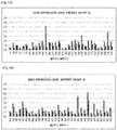
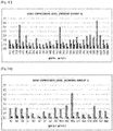
- measured values of the expression level of the PHB1 gene and the expression level of the PHB2 gene may be obtained with respect to a patient group and a normal group, as shown in FIGS. 1 and 2 , through such real-time quantitative PCR.
- the diagnosis of leukemia may be determined by any one equation selected from the group consisting of Equations 1 to 3 below, and the boundary values or boundary lines applied to Equations 1 to 3 in the present invention may be determined by mathematical, statistical, artificial intelligence and data-mining methods which are conventionally and widely applicable, and all of linear and non-linear types may be applied.
- Equations 1 to 3 represent linear or non-linear equations as an example of equations corresponding to the boundary line discriminating a "normal” and a "patient,” but may represent different equations according to a method of calculating the optimal boundary line.
- Y T ⁇ x 2 (In Equation 1,
- the present invention provides a method of diagnosing leukemia using the leukemia diagnostic kit, which includes
- the diagnosis of leukemia may be determined by any one equation selected from the group consisting of Equations 1 to 3 below, and the boundary values or boundary lines applied to Equations 1 to 3 in the present invention may be determined by mathematical, statistical, artificial intelligence and data-mining methods which are conventionally and widely applicable, and all of linear and non-linear types may be applied.
- Equations 1 to 3 represent linear or non-linear equations as an example of equations corresponding to the boundary line discriminating a "normal” and a "patient,” but may represent different equations according to a method of calculating the optimal boundary line.
- Y T ⁇ x 2 (In Equation 1,
- the leukemia in the present invention is acute myelogenous leukemia, acute lymphocytic leukemia, chronic myelogenous leukemia or chronic lymphocytic leukemia.
- Leukemia specimens used in the present invention were peripheral and bone marrow specimens obtained from 94 patients with acute myelogenous leukemia. For a normal group, peripheral blood was collected from 52 normal persons not having a specific disease through a separate basic test.
- Step 1 Collection of white blood cells from peripheral and bone marrow specimens
- Plasma was removed by centrifugation of whole blood at 3,500 rpm for 15 minutes, PBS (pH 7.4) was added to have a total volume of 5 mL, 3 ml of Lymphoprep was added thereto and then slowly mixed, and then the resulting mixture was centrifuged at 1,500 rpm for 30 min, thereby recovering a white blood cell (WBC) layer.
- PBS pH 7.4
- RNAqueous phenol-free total RNA isolation_AM1912 was used.
- 500 ⁇ l of a lysis/binding sol. was added to a specimen and subjected to vortex mixing, followed by the addition of 500 ⁇ l of 64% ethanol (the same amount of the lysis/binging sol.). Afterward, 700 ⁇ l of the sample was put into a column, and centrifuged at 14,000 rpm for 1 minute to remove a pellet.
- the extracted RNA was quantified using NanoDrop.
- cDNA was synthesized, and 500 ng of the total RNA quantified as described above was used in the cDNA synthesis.
- 20 ng (1/25) of the synthesized cDNA (500 ng) was diluted with ultrapure water, and mixed with SYBR PCR Master Mix and PHB1 and PHB2 primers to perform real-time PCR.
- PHB1, PHB2 and ⁇ -actin genes were subjected to a real-time quantitative PCR assay, and data was corrected by Equation 5 to overcome a difference in the degrees of gene expression between specimens (human).
- the PHB1 and PHB2 expression levels of 94 patients with leukemia are shown in Table 2 below and FIG. 1
- the PHB1 and PHB2 expression levels of 52 normal persons are shown in Table 3 below and FIG. 2 .
- the values measured by the real-time quantitative PCR amplification through the above-described process represent the minimum amplification cycles (Ct values) necessary for sensing the presence of a gene using a corresponding real-time quantitative PCR measurement device.
- the Ct values are cycles of the amplification of genes up to an amount capable of being measured using a real-time quantitative PCR device, and the higher the values, the smaller the amounts of genes originally present (expressed).
- a patient and a normal person can be distinguished only with the PHB2 values. More specifically, as confirmed in FIGS. 1 and 2 , it can be observed that "PHB1" does not show a significant difference between patients and normal persons, and "PHB2" is more highly expressed in the patient group than in the normal group.
- FIG. 3A shows only the PHB2 expression levels confirmed in Tables 2 and 3, and then, to eliminate difficulty in observation caused by overlapping generated when single values are present on one axis, for convenience, the patient group is represented on the line of 1.0 of the vertical axis, and the normal group is represented on the line of 0.5 of the vertical axis, and as a result of enlarging only the region in which PHB2 values of the normal group and the patient group overlap in FIG. 3B , it was confirmed that the region represented by the dotted line is the overlapping section (0.14 ⁇ 0.47).
- three vertical lines (0.2, 0.315, 0.435) in FIG. 3B represent the examples of boundary values, and when the middle vertical line (0.315) is selected as the boundary line between the normal and the patient, among the specimens in Tables 2 and 3, 8 patients are diagnosed as "normal,” and 8 patients are diagnosed as "patient.”
- Such a diagnostic method may be defined by Equation 1 below.
- Y T ⁇ x 2 (In Equation 1,
- diagnostic results for the specimens in Tables 2 and 3 may have the diagnosis accuracy as shown in Table 4 below.
- Table 4 Diagnostic group Patient Normal Total Patient 86 (91.5%) 8 (8.5%) 94 (100%) Normal 8 (15.4%) 44 (84.6%) 52 (100%)
- the horizontal axis presents PHB1
- the vertical axis presents PHB2
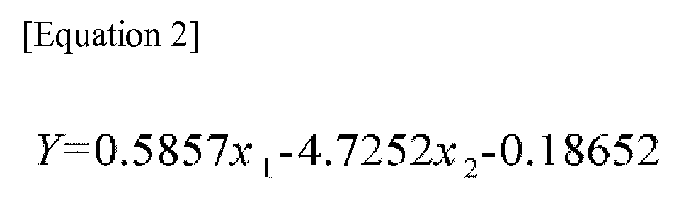
- corresponding values are represented as coordinates on the two-dimensional plane, according to the diagnostic method by combining the PHB1 and PHB2 expression levels in Tables 2 and 3, as shown in FIG. 4 , when the values are located above the boundary line that distinguishes the patient group and the normal group, they are diagnosed as "patient,” and when the values are located below the boundary line that distinguishes the patient group and the normal group, they are diagnosed as "normal,” which can be represented by Equation 2 below.
- Y 0.5857 x 1 ⁇ 4.7252 x 2 ⁇ 0.18652 (In Equation 2,
- the present invention was applied to the diagnostic method using a non-linear boundary line by combining the PHB1 and PHB2 expression levels in Tables 2 and 3.
Landscapes
- Chemical & Material Sciences (AREA)
- Life Sciences & Earth Sciences (AREA)
- Organic Chemistry (AREA)
- Health & Medical Sciences (AREA)
- Proteomics, Peptides & Aminoacids (AREA)
- Zoology (AREA)
- Wood Science & Technology (AREA)
- Engineering & Computer Science (AREA)
- Analytical Chemistry (AREA)
- Genetics & Genomics (AREA)
- Immunology (AREA)
- Molecular Biology (AREA)
- General Engineering & Computer Science (AREA)
- Biotechnology (AREA)
- Biophysics (AREA)
- Physics & Mathematics (AREA)
- Biochemistry (AREA)
- Bioinformatics & Cheminformatics (AREA)
- Microbiology (AREA)
- General Health & Medical Sciences (AREA)
- Chemical Kinetics & Catalysis (AREA)
- Pathology (AREA)
- Hospice & Palliative Care (AREA)
- Oncology (AREA)
- Measuring Or Testing Involving Enzymes Or Micro-Organisms (AREA)
Abstract
Description
- The present invention relates to a leukemia diagnostic kit and a diagnostic method using the same.
- While various causes of leukemia among blood cancers have been known, it has been most generally known that leukemia results from gene mutations and chromosomal mutations including the same. Prognosis may vary depending on the type of chromosomal mutation, and a therapeutic regimen for a leukemia patient may be determined thereby.
- Currently, leukemia is diagnosed using cytomorphological findings, immunophenotyping findings, and chromosomal mutations including gene mutations. To detect gene and chromosomal mutations, molecular genetic methods based on traditional chromosome testing and polymerase chain reaction (PCR) have been used, and various studies have been conducted to develop various molecular genetic testing methods which can be easily applied in actual investigation sites.
- For example, a diagnostic method combined with a method of identifying the arrangement of a fusion gene using long distance inverse polymerase chain reaction, and a method using PCR or reverse transcription (RT)-PCR performed on DNA of somatic cells have been developed.
- However, among the above-mentioned methods, FISH, which a method of directly detecting individual cells with a chromosomal translocation, cannot detect cells at the early stage of leukemia, and a method using PCR or RT-PCR has high sensitivity, but a false-positive reaction may occur due to the contamination of a specimen, and it is difficult to exactly determine the progression of the disease.
- To overcome the above-mentioned disadvantages, recently, leukemia diagnostic kits utilizing a real-time PCR method which can also quantify target RNA from a small amount of a specimen have been developed and purchased, but even for a specimen obtained from one patient, various genes involved in the development of leukemia have to be investigated to improve the accuracy of diagnosis, and therefore there is a disadvantage in that it is difficult to more rapidly diagnose leukemia due to considerable costs and time.
- It is an object of the present invention to provide a kit for diagnosing leukemia by combining an expression level of a prohibitin-1 (PHB1) gene and an expression level of a prohibitin-2 (PHB2) gene to solve a conventional problem of examining various genes involved in the development of leukemia to improve the accuracy of diagnosis, and perform rapid diagnosis of leukemia, and a method of diagnosing leukemia.
-
- 1. A leukemia diagnostic kit, which includes a PHB1 gene including a forward primer of SEQ ID NO: 1 and a reverse primer of SEQ ID NO: 2; and a PHB2 gene including a forward primer of SEQ ID NO: 3 and a reverse primer of SEQ ID NO: 4.
- 2. The leukemia diagnostic kit described above in 1, which is for measuring and comparing an expression level of the PHB1 gene and an expression level of the PHB2 gene.
- 3. The leukemia diagnostic kit described above in 1, wherein the diagnosis of leukemia is determined by any one equation selected from the group consisting of
Equations 1 to 3:
Equation 1,- T is a boundary value between expression values of PHB2 in a normal group and a patient group,
- x2 is an expression value of PHB2,
- when Y is a positive value, it is determined as "normal," and when Y is a negative value, it is determined as "patient.")

Equation 2,- x1 is an expression value of PHB1,
- x2 is an expression value of PHB2,
- when Y is a positive value, it is determined as "normal," and when Y is a negative value, it is determined as "patient.")

Equation 3,- g is a function defined by
Equation 4, - when Y is a positive value, it is determined as "normal," and when Y is a negative value, it is determined as "patient.")

- 4. The leukemia diagnostic kit described above in 1, which is used to perform RT-PCR with respect to mRNA of the PHB1 gene and the PHB2 gene.
- 5. The leukemia diagnostic kit described above in 1, further including a reagent for PCR.
- 6. The leukemia diagnostic kit described above in 1, further including a reagent for RNA extraction.
- 7. The leukemia diagnostic kit described above in 1, wherein the leukemia is acute myelogenous leukemia, acute lymphocytic leukemia, chronic myelogenous leukemia or chronic lymphocytic leukemia.
- 8. A method of diagnosing leukemia using the leukemia diagnostic kit described in 1, which includes diagnosing leukemia by measuring expression levels of a prohibitin-1 (PHB1) gene and a prohibitin-2 (PHB2) gene and comparing the gene expression levels.
- 9. The method according to 8, wherein the diagnosis of leukemia is determined by any one equation selected from the group consisting of
Equations 1 to 3:
Equation 1,- T is a boundary value between expression values of PHB2 in a normal group and a patient group,
- x2 is an expression value of PHB2,
- when Y is a positive value, it is determined as "normal," and when Y is a negative value, it is determined as "patient.")

Equation 2,- x1 is an expression value of PHB1,
- x2 is an expression value of PHB2,
- when Y is a positive value, it is determined as "normal," and when Y is a negative value, it is determined as "patient.")

Equation 3,- g is a function defined by
Equation 4, - when Y is a positive value, it is determined as "normal," and when Y is a negative value, it is determined as "patient.")

- A leukemia diagnostic kit according to the present invention is a kit for RT-PCR to confirm degrees of the expression of PHB1 and PHB2 in a specimen of a leukemia patient, and can be effectively used as a kit for diagnosing leukemia, measuring a residual lesion and determining a therapeutic effect due to higher accuracy and reproducibility of the diagnosis of leukemia than a conventional RT-PCR kit.
-
-
FIG. 1 is a set of graphs illustrating mRNA PHB1 and PHB2 expression levels in specimens obtained from leukemia patient groups according to an exemplary embodiment of the present invention. -
FIG. 2 is a set of graphs illustrating PHB1 and PHB2 expression levels in specimens obtained from normal groups according to an exemplary embodiment of the present invention. -
FIG. 3 is a set of graphs an expression level of PHB2 in specimens obtained from a leukemia patient group and a normal group according to an exemplary embodiment of the present invention. -
FIG. 4 is a graph illustrating that a linear boundary line for diagnosing leukemia is shown in coordinates on a two-dimensional plane with a horizontal axis indicating PHB1 and a vertical axis indicating PHB2 according to an exemplary embodiment of the present invention. -
FIG. 5 is a graph illustrating that a non-linear boundary line for diagnosing leukemia is shown in coordinates on a two-dimensional plane with a horizontal axis indicating PHB1 and a vertical axis indicating PHB2 according to an exemplary embodiment of the present invention. - Hereinafter, various aspects and a variety of exemplary embodiments of the present invention will be described in further detail.
- The present invention provides a leukemia diagnostic kit, which includes a PHB1 gene including a forward primer of SEQ ID NO: 1 and a reverse primer of SEQ ID NO: 2; and a PHB2 gene including a forward primer of SEQ ID NO: 3 and a reverse primer of SEQ ID NO: 4.
- According to the present invention, the leukemia diagnostic kit may be effectively used in measurement of a residual lesion and determination of a therapeutic effect as well as diagnosis, and the leukemia in the present invention includes acute myelogenous leukemia, acute lymphocytic leukemia, chronic myelogenous leukemia or chronic lymphocytic leukemia.
- The leukemia diagnostic kit of the present invention may be used to perform RT-PCR with respect to mRNA of a PHB1 gene and a PHB2 gene.
- According to the present invention, to obtain mRNA, white blood cells are obtained from specimens, preferably, peripheral blood and bone marrow specimens, and then total RNA is isolated therefrom. The separation of total RNA may be performed according to a conventional method known in the art. For example, total RNA in the cells may be easily isolated using TRIzol.
- Subsequently, cDNA is synthesized using the isolated mRNA as a template, and the cDNA is amplified. Since the total RNA of the present invention is isolated from a human sample, a poly-A tail is present at the end of mRNA, and cDNA may be easily synthesized using an oligo dT primer and a reverse transcriptase which utilize such a sequence characteristic. Subsequently, the synthesized cDNA is amplified using real-time PCR.
- The primer used in the present invention forms a double-stranded structure by hybridization or annealing at one region of the template. Suitable conditions for nucleic acid hybridization to form such a double-stranded structure are disclosed in Joseph Sambrook et al., Molecular Cloning, A Laboratory Manual, Cold Spring Harbor Laboratory Press, Cold Spring Harbor, N.Y. (2001) and Haymes, B. D. et al., Nucleic Acid Hybridization, A Practical Approach, IRL Press, Washington, D.C. (1985).
- In the present invention, annealing or hybridization is performed under strict conditions which allow specific binding between a target nucleotide sequence and a primer. The strict conditions for annealing are sequence-dependent, and vary according to environmental variables.
- Various DNA polymerases may be used in amplification of the present invention, and include the "Klenow" fragment of E. coli DNA polymerase I, a thermally stable DNA polymerase and bacteriophage T7 DNA polymerase. Preferably, the polymerases are thermally stable DNA polymerases obtained from a variety of bacterial species, and include Thermus aquaticus (Taq), Thermus thermophilus (Tth), Thermus filiformis, Thermis flavus, Thermococcus literalis and Pyrococcus furiosus (Pfu).
- When polymerization is performed, excessive components required for a reaction are preferably provided to a reaction container. The excessive amount of the components required for an amplification reaction refers to the amount of a component such that the amplification reaction is not substantially limited by a concentration of the component. It is required that a cofactor such as Mg2+, dATP, dCTP, dGTP and dTTP are provided to a reaction mixture to an extent that the desired degree of amplification can be achieved.
- All enzymes used in the amplification may be active under the same reaction conditions. In fact, a buffer solution allows all enzymes to approach the optimal reaction conditions. Therefore, the amplification of the present invention may be performed on a single reactant without a change in conditions such as the addition of a reactant.
- The term "amplification" used herein refers to a reaction of amplifying a nucleic acid molecule. Various amplification reactions are reported in the art, and include, but are not limited to, PCR, RT-PCR, ligase chain reaction (LCR), Gap-LCR, repair chain reaction, transcription-mediated amplification (TMA), self-sustained sequence replication, selective amplification of target polynucleotide sequences, consensus sequence primed polymerase chain reaction (CP-PCR), arbitrarily primed polymerase chain reaction (AP-PCR), nucleic acid sequence based amplification (NASBA), strand displacement amplification and loop-mediated isothermal amplification (LAMP). Other amplification methods used herein are disclosed in
U.S. Patent Nos. 5,242,794 ,5,494,810 and4,988,617 , andU.S. Patent No. 09/854,317 - In the most exemplary embodiment of the present invention, amplification is performed according to PCR disclosed in
U.S. Patent Nos. 4,683,195 ,4,683,202 and4,800,159 . - Meanwhile, PCR is the most well-known nucleic acid amplification method, and various modifications and applications are developed. For example, to improve the specificity or sensitivity of PCR, touchdown PCR, hot-start PCR, nested PCR and booster PCR were developed by modifying traditional PCR procedures. In addition, real-time PCR, differential display PCR (DD-PCR), rapid amplification of cDNA ends (RACE), multiplex PCR, inverse polymerase chain reaction (IPCR), vectorette PCR, thermal asymmetric interlaced PCR (TAIL-PCR) and multiplex PCR were developed for specific applications.
- A primer used in the amplification reaction in the present invention is an oligonucleotide having a sequence complimentary to the cDNA sequence of PHB1 or PHB2. The term "primer" used herein refers to a single-stranded oligonucleotide which can act as the initiation point of template-directed DNA synthesis under suitable conditions (that is, four types of different nucleoside triphosphates and polymerases) in a suitable buffer solution at a suitable temperature. Suitable lengths of the primers may vary according to various factors, for example, a temperature and the use of a primer, and are typically 15-30 nucleotides. A short primer molecule generally requires a lower temperature to form a sufficiently stable hybrid complex with a template.
- The primer sequence does not need a sequence perfectly complementary to a part of the sequence of the template, and it is enough to have sufficient complementarity within a range at which the primer is hybridized with the template to exhibit the specific function of the primer.
- As an example, the primer used in the present invention has a sequence perfectly complementary to the cDNA sequence of PHB1 or PHB2. The primer of the present invention may be prepared using a sequence complementary to the mRNA (that is, cDNA) sequence of PHB1 disclosed in SEQ ID NO: 1, and may be prepared using a sequence complementary to the mRNA (that is, cDNA) sequence of PHB2 disclosed in SEQ ID NO: 3.
- Such a primer may be designed by those of ordinary skill in the art with reference to the cDNA sequence of PHB1 or PHB2 using, for example, a program for designing a primer (e. g.,
PRIMER 3 program). - Degrees of the expression of PHB1 and PHB2 genes are investigated by analyzing cDNA of the amplified PHB1 or PHB2. For example, the expression level of PHB1 or PHB2 is measured by applying a mixture prepared by mixing cDNA, the primers synthesized using sequences complementary to PHB1 and PHB2 genes with an SYBR Green I reagent to RT-PCR. Here, to remove a difference in the expression of individual genes, the expression level of the β-actin gene widely known to be associated with a house-keeping gene may be simultaneously measured.
- Through such an amplification reaction, measured values of the expression level of the PHB1 gene and the expression level of the PHB2 gene may be obtained with respect to a patient group and a normal group, as shown in
FIGS. 1 and2 , through such real-time quantitative PCR. - The diagnosis of leukemia may be determined by any one equation selected from the group consisting of
Equations 1 to 3 below, and the boundary values or boundary lines applied toEquations 1 to 3 in the present invention may be determined by mathematical, statistical, artificial intelligence and data-mining methods which are conventionally and widely applicable, and all of linear and non-linear types may be applied. - In other words,
Equations 1 to 3 represent linear or non-linear equations as an example of equations corresponding to the boundary line discriminating a "normal" and a "patient," but may represent different equations according to a method of calculating the optimal boundary line.
Equation 1, - T is a boundary value between expression values of PHB2 in a normal group and a patient group,
- x2 is an expression value of PHB2,
- when Y is a positive value, it is determined as "normal," and when Y is a negative value, it is determined as "patient.")
- x1 is an expression value of PHB1,
- x2 is an expression value of PHB2,
- when Y is a positive value, it is determined as "normal," and when Y is a negative value, it is determined as "patient.")
- g is a function defined by
Equation 4, - when Y is a positive value, it is determined as "normal," and when Y is a negative value, it is determined as "patient.")
- In addition, the present invention provides a method of diagnosing leukemia using the leukemia diagnostic kit, which includes
- (1) a first step of measuring expression levels of PHB1 gene and PHB2 gene, which are obtained from leukemia specimens; and
- (2) a second step of diagnosing leukemia by combining the expression level of the PHB1 gene and the expression level of the PHB2 gene, which are measured in the first step.
- The diagnosis of leukemia may be determined by any one equation selected from the group consisting of
Equations 1 to 3 below, and
the boundary values or boundary lines applied toEquations 1 to 3 in the present invention may be determined by mathematical, statistical, artificial intelligence and data-mining methods which are conventionally and widely applicable, and all of linear and non-linear types may be applied. - In other words,
Equations 1 to 3 represent linear or non-linear equations as an example of equations corresponding to the boundary line discriminating a "normal" and a "patient," but may represent different equations according to a method of calculating the optimal boundary line.
Equation 1, - T is a boundary value between expression values of PHB2 in a normal group and a patient group,
- x2 is an expression value of PHB2,
- when Y is a positive value, it is determined as "normal," and when Y is a negative value, it is determined as "patient.")
- x1 is an expression value of PHB1,
- x2 is an expression value of PHB2,
- when Y is a positive value, it is determined as "normal," and when Y is a negative value, it is determined as "patient.")
- g is a function defined by
Equation 4, - when Y is a positive value, it is determined as "normal," and when Y is a negative value, it is determined as "patient.")
- The leukemia in the present invention is acute myelogenous leukemia, acute lymphocytic leukemia, chronic myelogenous leukemia or chronic lymphocytic leukemia.
- Hereinafter, the present invention will be described in further detail with reference to examples, but the scope and descriptions of the present invention may not be construed to be diminished or limited by the following examples. In addition, based on the disclosure of the present invention including the following examples, it is clear that the present invention in which specific experimental results are not presented can be easily carried out by those of ordinary skill in the art, and it is obvious that such alternations and modifications are also included in the accompanying claims.
- Leukemia specimens used in the present invention were peripheral and bone marrow specimens obtained from 94 patients with acute myelogenous leukemia. For a normal group, peripheral blood was collected from 52 normal persons not having a specific disease through a separate basic test.
- Plasma was removed by centrifugation of whole blood at 3,500 rpm for 15 minutes, PBS (pH 7.4) was added to have a total volume of 5 mL, 3 ml of Lymphoprep was added thereto and then slowly mixed, and then the resulting mixture was centrifuged at 1,500 rpm for 30 min, thereby recovering a white blood cell (WBC) layer.
- Sequentially, after adding PBS (pH 7.4) to have a total volume of 7 ml, the mixture was slowly mixed, and centrifuged at 1,500 rpm for 10 minutes to remove a supernatant, and then only a pellet (cells) was recovered.
- In the present invention, an Ambion kit (RNAqueous phenol-free total RNA isolation_AM1912) was used.
- 500 µl of a lysis/binding sol. was added to a specimen and subjected to vortex mixing, followed by the addition of 500 µl of 64% ethanol (the same amount of the lysis/binging sol.). Afterward, 700 µl of the sample was put into a column, and centrifuged at 14,000 rpm for 1 minute to remove a pellet.
- Subsequently, a process including adding 700 µl of Wash sol.1 to the resulting supernatant, removing a pellet by centrifugation at 14,000 rpm for 1 minute, adding 500 µl of each of Wash sol. 2 and 3 and removing a pellet by centrifugation at 14,000 rpm for 30 seconds was performed two times, and then the resulting supernatant was finally centrifuged at 14,000 rpm for 3 minutes, a column was transferred to a new tube, and then RNA was extracted by adding 40 µl of an elution buffer.
- The extracted RNA was quantified using NanoDrop.
- Using a commercially available Invitrogen kit (SuperScript III First-strand synthesis system for RT-PCR_18080-051), cDNA was synthesized, and 500 ng of the total RNA quantified as described above was used in the cDNA synthesis.
- 20 ng (1/25) of the synthesized cDNA (500 ng) was diluted with ultrapure water, and mixed with SYBR PCR Master Mix and PHB1 and PHB2 primers to perform real-time PCR.
- Primers used to detect PHB1 and PHB2 expression are shown in Table 1 below.
[Table 1] Type Sequence List SEQ ID NO: Primer Size NCBI Reference Sequence PHB-1 forward primer 
1 175 bp NM_ 002634 PHB-1 reverse primer 
2 PHB-2 forward primer 
3 115 bp NM_ 001144831 PHB-2 reverse primer 
4 - PHB1, PHB2 and β-actin genes were subjected to a real-time quantitative PCR assay, and data was corrected by Equation 5 to overcome a difference in the degrees of gene expression between specimens (human).
- As the number obtained by such a data conversion process is larger, it means that a gene expression level is relatively higher.
- The PHB1 and PHB2 expression levels of 94 patients with leukemia are shown in Table 2 below and
FIG. 1 , and the PHB1 and PHB2 expression levels of 52 normal persons are shown in Table 3 below andFIG. 2 . - At this time, the values measured by the real-time quantitative PCR amplification through the above-described process represent the minimum amplification cycles (Ct values) necessary for sensing the presence of a gene using a corresponding real-time quantitative PCR measurement device. In other words, the Ct values are cycles of the amplification of genes up to an amount capable of being measured using a real-time quantitative PCR device, and the higher the values, the smaller the amounts of genes originally present (expressed).

- x1 is an expression value of PHB1,
- x2 is an expression value of PHB2,
- P 1 is a PCR measured value of PHB1,
- P 2 is a PCR measured value of PHB2, and
- A is a PCR measured value of actin.
- As shown in Tables 2 and 3, according to the present invention, a patient and a normal person can be distinguished only with the PHB2 values. More specifically, as confirmed in
FIGS. 1 and2 , it can be observed that "PHB1" does not show a significant difference between patients and normal persons, and "PHB2" is more highly expressed in the patient group than in the normal group. - Therefore, based on the boundary value of PHB2, when a value is higher than the boundary value, it was diagnosed as "patient," and when a value is lower than the boundary value, it was diagnosed as "normal".
- To significantly distinguish "patient" and "normal" according to PHB1 and PHB2 expression levels shown in Tables 2 and 3, measured values were analyzed by
Equation 4 below. -
FIG. 3A shows only the PHB2 expression levels confirmed in Tables 2 and 3, and then, to eliminate difficulty in observation caused by overlapping generated when single values are present on one axis, for convenience, the patient group is represented on the line of 1.0 of the vertical axis, and the normal group is represented on the line of 0.5 of the vertical axis, and as a result of enlarging only the region in which PHB2 values of the normal group and the patient group overlap inFIG. 3B , it was confirmed that the region represented by the dotted line is the overlapping section (0.14 ∼ 0.47). - In the overlapping region, three vertical lines (0.2, 0.315, 0.435) in
FIG. 3B represent the examples of boundary values, and when the middle vertical line (0.315) is selected as the boundary line between the normal and the patient, among the specimens in Tables 2 and 3, 8 patients are diagnosed as "normal," and 8 patients are diagnosed as "patient." Such a diagnostic method may be defined byEquation 1 below.
Equation 1, - T is a boundary value between expression values of PHB2 in a normal group and a patient group,
- x2 is an expression value of PHB2,
- when Y is a positive value, it is determined as "normal," and when Y is a negative value, it is determined as "patient.")
- In other words, when T=0.315 is applied to the diagnostic method according to
Equation 1, diagnostic results for the specimens in Tables 2 and 3 may have the diagnosis accuracy as shown in Table 4 below.[Table 4] Diagnostic group Patient Normal Total Patient 86 (91.5%) 8 (8.5%) 94 (100%) Normal 8 (15.4%) 44 (84.6%) 52 (100%) - In addition, in the present invention, provided that the horizontal axis presents PHB1, the vertical axis presents PHB2, and corresponding values are represented as coordinates on the two-dimensional plane, according to the diagnostic method by combining the PHB1 and PHB2 expression levels in Tables 2 and 3, as shown in
FIG. 4 , when the values are located above the boundary line that distinguishes the patient group and the normal group, they are diagnosed as "patient," and when the values are located below the boundary line that distinguishes the patient group and the normal group, they are diagnosed as "normal," which can be represented byEquation 2 below.
Equation 2, - x1 is an expression value of PHB1,
- x2 is an expression value of PHB2,
- when Y is a positive value, it is determined as "normal," and when Y is a negative value, it is determined as "patient.").
- According to the diagnostic method by
Equation 2, as the result of diagnosing the specimens in Tables 2 and 3, diagnosis accuracy shown in Table 5 below may be obtained.[Table 5] Diagnostic group Patient Normal Total Patient 88 (93.6%) 6 (6.4%) 94 (100%) Normal 12 (23.1%) 40 (76.9%) 52 (100%) - In addition, the present invention was applied to the diagnostic method using a non-linear boundary line by combining the PHB1 and PHB2 expression levels in Tables 2 and 3.
- When the non-linear boundary line shown in
FIG. 5 is applied, diagnosis accuracy with respect to the specimens in Tables 2 and 3, shown in Table 6, may be obtained byEquations 
Equation 3, - g is a function defined by
Equation 4, - when Y is a positive value, it is determined as "normal," and when Y is a negative value, it is determined as "patient.")



| Expression levels of PHB1 and PHB2 in leukemia patients | ||||||||
| Index | PIIB 1 | PHB 2 | Index | PHB 1 | PHB 2 | Index | PIIB 1 | PHB 2 |
| P1 | 0.5154 | 0.3879 | P38 | 1.0453 | 0.8669 | P71 | 1.9777 | 1.1125 |
| P3 | 1.5303 | 0.4456 | P39 | 2.7970 | 2.0054 | P72 | 1.0525 | 0.5880 |
| P4 | 0.8790 | 1.0097 | P40 | 0.9618 | 0.5880 | P73 | 2.0761 | 1.1438 |
| P5 | 0.4245 | 0.2259 | P41 | 1.5093 | 1.6631 | P76 | 5.4788 | 0.6524 |
| P7 | 0.8201 | 0.7867 | P42 | 3.2577 | 1.8073 | P77 | 1.6176 | 0.5839 |
| P9 | 1.1760 | 0.8669 | P43 | 1.8453 | 1.2780 | P78 | 3.0607 | 0.7139 |
| P10 | 0.2167 | 0.1953 | P44 | 0.8032 | 0.4910 | P79 | 1.3985 | 0.7442 |
| P11 | 1.6176 | 0.6848 | P45 | 0.9685 | 0.4581 | P80 | 1.3048 | 0.3401 |
| P12 | 0.7867 | 0.5083 | P46 | 0.6661 | 0.4743 | P81 | 2.3848 | 0.7442 |
| P13 | 1.3985 | 0.8201 | P47 | 1.4378 | 0.5486 | P82 | 1.1049 | 0.2724 |
| P14 | 1.8453 | 0.9958 | P48 | 1.9641 | 1.2174 | P83 | 0.9753 | 0.3961 |
| P15 | 5.1832 | 0.5799 | P49 | 1.1598 | 0.4518 | P84 | 1.3415 | 0.7239 |
| P16 | 1.6176 | 1.5517 | P50 | 1.0525 | 0.6258 | P85 | 1.6863 | 0.7239 |
| P17 | 1.5734 | 0.8144 | P51 | 1.3230 | 1.0453 | P86 | 0.9099 | 0.4016 |
| P19 | 1.6064 | 0.7977 | P52 | 1.2780 | 0.5524 | P87 | 1.9104 | 1.1760 |
| P20 | 1.9641 | 1.5410 | P53 | 2.4518 | 1.3048 | P88 | 4.9721 | 1.1598 |
| P21 | 0.3721 | 0.1822 | P54 | 0.8974 | 0.5880 | P89 | 1.4279 | 0.8790 |
| P22 | 0.4395 | 0.3262 | P55 | 1.3230 | 0.7977 | P90 | 1.1679 | 0.4710 |
| P23 | 1.1598 | 0.9163 | P56 | 1.8581 | 1.1359 | P91 | 2.0333 | 1.1842 |
| P24 | 1.9237 | 0.3961 | P57 | 1.0598 | 0.5921 | P92 | 2.0475 | 0.9889 |
| P25 | 2.1493 | 1.3792 | P58 | 0.6570 | 0.3988 | P93 | 2.6645 | 1.2691 |
| P27 | 1.7098 | 1.1518 | P59 | 4.3889 | 0.6801 | P94 | 1.1438 | 0.5719 |
| P28 | 3.0820 | 1.2259 | P60 | 1.8581 | 0.7759 | P95 | 2.0054 | 0.9552 |
| P29 | 0.9163 | 0.7442 | P61 | 1.3792 | 0.5680 | P97 | 2.9564 | 0.9291 |
| P30 | 1.9370 | 0.4158 | P62 | 5.3290 | 3.4915 | P98 | 3.3032 | 0 5299 |
| P31 | 2.0617 | 1.1924 | P64 | 0.6801 | 0.4944 | P99 | 2.5559 | 0.7239 |
| P32 | 1.5198 | 1.1679 | P65 | 2.8360 | 0.2820 | P100 | 6.5154 | 0.8258 |
| P33 | 0.4678 | 0.2489 | P66 | 1.5410 | 0.7239 | P102 | 3.0607 | 0.1490 |
| P34 | 0.5759 | 0.3448 | P67 | 1.3048 | 0.4425 | P104 | 2.0054 | 0.8201 |
| P35 | 1.7098 | 0.7494 | P68 | 3.3960 | 0.9685 | P105 | 1.2780 | 0.7813 |
| P36 | 3.9010 | 0.8729 | P69 | 1.1518 | 0.7494 | |||
| P37 | 1.7948 | 0.8032 | P70 | 1.8453 | 0.2064 | |||
| Expression levels of PHB1 and PHB2 in normal persons | ||||||||
| Index | PHB-1 | PHB-2 | Index | PHB-1 | PHB-2 | Index | PHB-1 | PHB-2 |
| N1 | 2.0475 | 0.3696 | N19 | 1.3985 | 0.1861 | N37 | 6.7452 | 0.0301 |
| N2 | 2.1944 | 0.4518 | N20 | 0.9163 | 0.1202 | N38 | 3.4197 | 0.0475 |
| N3 | 1.9641 | 0.3852 | N21 | 2.2561 | 0.1654 | N39 | 1.9641 | 0.0334 |
| N4 | 2.0905 | 0.4613 | N22 | 2.6461 | 0.2405 | N40 | 0.5680 | 0.1532 |
| N5 | 1.0167 | 0.1400 | N23 | 2.3519 | 0.1631 | N41 | 0.6258 | 0.1011 |
| N6 | 0.4613 | 0.3044 | N24 | 1.5093 | 0.1861 | N42 | 5.9954 | 0.0044 |
| N7 | 1.3697 | 0.4275 | N25 | 4.3285 | 0.1362 | N43 | 2.5208 | 0.0188 |
| N8 | 0.9355 | 0.1609 | N26 | 5.1474 | 0.2152 | N44 | 5.7114 | 0.0185 |
| N9 | 2.1051 | 0.2455 | N27 | 1.9777 | 0.1848 | N45 | 6.1214 | 0.1501 |
| N10 | 1.9237 | 0.1430 | N28 | 2.0054 | 0.3521 | N46 | 4.7696 | 0.1099 |
| N11 | 2.6645 | 0.2323 | N29 | 1.9370 | 0.4245 | N47 | 4.7696 | 0.1069 |
| N12 | 5.2556 | 0.4100 | N30 | 0.3086 | 0.0970 | N48 | 4.3586 | 0.1069 |
| N13 | 1.2430 | 0.2650 | N31 | 0.3773 | 0.1228 | N49 | 5.7912 | 0.0265 |
| N14 | 0.7494 | 0.1797 | N32 | 0.3570 | 0.1253 | N50 | 2.1344 | 0.0041 |
| N15 | 0.7289 | 0.2801 | N33 | 0.7652 | 0.2650 | N51 | 4.2101 | 0.0331 |
| N16 | 2.4861 | 0.1712 | N34 | 0.6258 | 0.1522 | N52 | 2.4181 | 0.0397 |
| N17 | 1.5198 | 0.1106 | N35 | 0.7340 | 0.1953 | |||
| N18 | 1.4680 | 0.2388 | N36 | 0.7652 | 0.1620 | |||

| Diagnostic group | Patient | Normal | Total |
| Patient | 90 (95.7%) | 4 (4.3%) | 94 (100%) |
| Normal | 4 (7.7%) | 48 (92.3%) | 52 (100%) |


Claims (9)
- A leukemia diagnostic kit, comprising:a prohibitin-1 (PHB1) gene including a forward primer of SEQ ID NO: 1 and a reverse primer of SEQ ID NO: 2; anda prohibitin-2 (PHB2) gene including a forward primer of SEQ ID NO: 3 and a reverse primer of SEQ ID NO: 4.
- The leukemia diagnostic kit according to claim 1, which is for measuring and comparing an expression level of the PHB1 gene and an expression level of the PHB2 gene in a specimen.
- The leukemia diagnostic kit according to claim 1, wherein the diagnosis of leukemia is determined by any one equation selected from the group consisting of Equations 1 to 3:


- The leukemia diagnostic kit according to claim 1, which is used to perform RT-PCR with respect to mRNA of the PHB1 gene and the PHB2 gene.
- The leukemia diagnostic kit according to claim 1, further comprising a reagent for PCR.
- The leukemia diagnostic kit according to claim 1, further comprising a reagent for RNA extraction.
- The leukemia diagnostic kit according to claim 1, wherein the leukemia is acute myelogenous leukemia, acute lymphocytic leukemia, chronic myelogenous leukemia or chronic lymphocytic leukemia.
- A method of diagnosing leukemia using the leukemia diagnostic kit of claim 1, comprising:diagnosing leukemia by measuring expression levels of a prohibitin-1 (PHB1) gene and a prohibitin-2 (PHB2) gene, and comparing the gene expression levels.
- The method according to 8, wherein the diagnosis of leukemia is determined by any one equation selected from the group consisting of Equations 1 to 3:



Applications Claiming Priority (2)
| Application Number | Priority Date | Filing Date | Title |
|---|---|---|---|
| KR1020160021320A KR101845957B1 (en) | 2016-02-23 | 2016-02-23 | Kit for diagnosis of leukemia and diagnostic method targeting prohibitin gene |
| PCT/KR2017/001867 WO2017146432A1 (en) | 2016-02-23 | 2017-02-21 | Leukemia diagnostic kit targeting prohibitin gene and diagnostic method using same |
Publications (3)
| Publication Number | Publication Date |
|---|---|
| EP3421611A1 true EP3421611A1 (en) | 2019-01-02 |
| EP3421611A4 EP3421611A4 (en) | 2019-07-10 |
| EP3421611B1 EP3421611B1 (en) | 2023-11-29 |
Family
ID=59685335
Family Applications (1)
| Application Number | Title | Priority Date | Filing Date |
|---|---|---|---|
| EP17756770.8A Active EP3421611B1 (en) | 2016-02-23 | 2017-02-21 | Acute myelogenous leukemia diagnostic method targeting prohibitin gene |
Country Status (4)
| Country | Link |
|---|---|
| US (1) | US11339421B2 (en) |
| EP (1) | EP3421611B1 (en) |
| KR (1) | KR101845957B1 (en) |
| WO (1) | WO2017146432A1 (en) |
Families Citing this family (2)
| Publication number | Priority date | Publication date | Assignee | Title |
|---|---|---|---|---|
| KR102679988B1 (en) | 2021-02-22 | 2024-07-01 | 전남대학교산학협력단 | Composition for companion diagnosis with prohibitin for targeted treatment of leukemia |
| WO2022186673A1 (en) * | 2021-03-05 | 2022-09-09 | 전남대학교산학협력단 | Next-generation-sequencing-based rna sequencing panel for targeted genes, and analysis algorithm |
Family Cites Families (8)
| Publication number | Priority date | Publication date | Assignee | Title |
|---|---|---|---|---|
| IL86724A (en) | 1987-06-19 | 1995-01-24 | Siska Diagnostics Inc | Method and kits for the amplification and detection of nucleic acid sequences |
| JP2846018B2 (en) | 1988-01-21 | 1999-01-13 | ジェネンテク,インコーポレイテッド | Amplification and detection of nucleic acid sequences |
| CA1340807C (en) | 1988-02-24 | 1999-11-02 | Lawrence T. Malek | Nucleic acid amplification process |
| WO1990001069A1 (en) | 1988-07-20 | 1990-02-08 | Segev Diagnostics, Inc. | Process for amplifying and detecting nucleic acid sequences |
| WO1990006995A1 (en) | 1988-12-16 | 1990-06-28 | Siska Diagnostics, Inc. | Self-sustained sequence replication system |
| DE69118930T2 (en) | 1990-01-26 | 1997-01-09 | Abbott Lab | Improved method for the amplification of nucleic acid target sequence, usable for the polymerase and ligase chain reaction |
| CA2483284A1 (en) | 2004-11-08 | 2006-05-08 | Institut Pasteur | Method of diagnosis/prognosis of human chronic lymphocytic leukemia comprising the profiling of lpl/adam genes |
| KR20140056472A (en) * | 2012-10-26 | 2014-05-12 | 전남대학교산학협력단 | Biomarkers indicative of leukemia and diagnosis using the same |
-
2016
- 2016-02-23 KR KR1020160021320A patent/KR101845957B1/en active Active
-
2017
- 2017-02-21 EP EP17756770.8A patent/EP3421611B1/en active Active
- 2017-02-21 WO PCT/KR2017/001867 patent/WO2017146432A1/en not_active Ceased
- 2017-02-21 US US16/079,432 patent/US11339421B2/en active Active
Also Published As
| Publication number | Publication date |
|---|---|
| KR20170099218A (en) | 2017-08-31 |
| WO2017146432A1 (en) | 2017-08-31 |
| EP3421611B1 (en) | 2023-11-29 |
| KR101845957B1 (en) | 2018-04-05 |
| US20190048396A1 (en) | 2019-02-14 |
| US11339421B2 (en) | 2022-05-24 |
| EP3421611A4 (en) | 2019-07-10 |
Similar Documents
| Publication | Publication Date | Title |
|---|---|---|
| Manokhina et al. | Quantification of cell-free DNA in normal and complicated pregnancies: overcoming biological and technical issues | |
| JP2022530483A (en) | Detection Technique System and Use to Concentrate Low Abundance DNA Mutations Based on nuclease Cooperative PCR Principles | |
| CN105283557A (en) | Non-invasive early detection of solid organ transplant rejection by quantitative analysis of mixtures by deep sequencing of HLA gene amplicons using next generation systems | |
| CN112538528A (en) | Primer group and kit for detecting ALDH2 gene polymorphism | |
| Fan | Molecular counting: from noninvasive prenatal diagnostics to whole-genome haplotyping | |
| CN112280848A (en) | Relative quantitative detection method and kit for human motor neuron gene copy number | |
| CN115948532A (en) | SMA detection kit based on digital PCR technology | |
| CN107022619A (en) | KRAS gene mutation detection primer probe and its kit | |
| EP3421611B1 (en) | Acute myelogenous leukemia diagnostic method targeting prohibitin gene | |
| US10119166B2 (en) | Methods and kits for identifying and adjusting for bias in sequencing of polynucleotide samples | |
| US20210381030A1 (en) | Systems and methods of diagnosing and characterizing infections | |
| Souza et al. | Multiplex real-time PCR using SYBR Green: unspecific intercalating dye to detect antimicrobial resistance genes of Streptococcus pneumoniae in cerebrospinal fluid | |
| CN108026591B (en) | Diagnostic methods and compositions | |
| CN111334592A (en) | Nucleic acid composition for detecting helicobacter pylori drug-resistant gene and kit and application thereof | |
| CN113728114A (en) | Digital polymerase chain reaction method for detecting nucleic acid in sample | |
| CN105018490A (en) | Primer pairs, probes and kit for detecting polymorphism of human MTHFR gene | |
| TWI780315B (en) | Method of quantifying mutant allele burden of target gene | |
| KR101742016B1 (en) | Oligonucleotide for detecting Chlamydia trachomatis and/or Neisseria gonorrhoeae, and kit using the same | |
| US11359251B2 (en) | Methods for the detection of enterovirus D68 in complex samples | |
| US20260062749A1 (en) | A method for determining the tissue or cell of origin of dna | |
| TWI740427B (en) | Method for detecting non-tuberculous mycobacterium and kit thereof | |
| Koksal et al. | A tetra-primer polymerase chain reaction approach for the detection of JAK2 V617F mutation | |
| WO2006102569A2 (en) | Nucleic acid detection | |
| Obaro et al. | Comparison of bacterial culture with BioFire | |
| CN121518671A (en) | Nucleic acid compositions, kits, and their applications for the detection, typing, and/or drug resistance gene detection of Mycoplasma pneumoniae. |
Legal Events
| Date | Code | Title | Description |
|---|---|---|---|
| STAA | Information on the status of an ep patent application or granted ep patent |
Free format text: STATUS: THE INTERNATIONAL PUBLICATION HAS BEEN MADE |
|
| PUAI | Public reference made under article 153(3) epc to a published international application that has entered the european phase |
Free format text: ORIGINAL CODE: 0009012 |
|
| STAA | Information on the status of an ep patent application or granted ep patent |
Free format text: STATUS: REQUEST FOR EXAMINATION WAS MADE |
|
| 17P | Request for examination filed |
Effective date: 20180918 |
|
| AK | Designated contracting states |
Kind code of ref document: A1 Designated state(s): AL AT BE BG CH CY CZ DE DK EE ES FI FR GB GR HR HU IE IS IT LI LT LU LV MC MK MT NL NO PL PT RO RS SE SI SK SM TR |
|
| AX | Request for extension of the european patent |
Extension state: BA ME |
|
| DAV | Request for validation of the european patent (deleted) | ||
| DAX | Request for extension of the european patent (deleted) | ||
| RAP1 | Party data changed (applicant data changed or rights of an application transferred) |
Owner name: INDUSTRY FOUNDATION OF CHONNAM NATIONAL UNIVERSITY |
|
| A4 | Supplementary search report drawn up and despatched |
Effective date: 20190613 |
|
| RIC1 | Information provided on ipc code assigned before grant |
Ipc: C12Q 1/686 20180101ALI20190606BHEP Ipc: C12Q 1/6806 20180101ALI20190606BHEP Ipc: C12Q 1/68 20180101AFI20190606BHEP |
|
| STAA | Information on the status of an ep patent application or granted ep patent |
Free format text: STATUS: EXAMINATION IS IN PROGRESS |
|
| 17Q | First examination report despatched |
Effective date: 20201111 |
|
| REG | Reference to a national code |
Ref country code: DE Ref legal event code: R079 Free format text: PREVIOUS MAIN CLASS: C12Q0001680000 Ipc: C12Q0001688600 Ref country code: DE Ref legal event code: R079 Ref document number: 602017076982 Country of ref document: DE Free format text: PREVIOUS MAIN CLASS: C12Q0001680000 Ipc: C12Q0001688600 |
|
| GRAP | Despatch of communication of intention to grant a patent |
Free format text: ORIGINAL CODE: EPIDOSNIGR1 |
|
| STAA | Information on the status of an ep patent application or granted ep patent |
Free format text: STATUS: GRANT OF PATENT IS INTENDED |
|
| RIC1 | Information provided on ipc code assigned before grant |
Ipc: C12Q 1/6886 20180101AFI20230214BHEP |
|
| INTG | Intention to grant announced |
Effective date: 20230303 |
|
| GRAS | Grant fee paid |
Free format text: ORIGINAL CODE: EPIDOSNIGR3 |
|
| GRAJ | Information related to disapproval of communication of intention to grant by the applicant or resumption of examination proceedings by the epo deleted |
Free format text: ORIGINAL CODE: EPIDOSDIGR1 |
|
| GRAL | Information related to payment of fee for publishing/printing deleted |
Free format text: ORIGINAL CODE: EPIDOSDIGR3 |
|
| STAA | Information on the status of an ep patent application or granted ep patent |
Free format text: STATUS: EXAMINATION IS IN PROGRESS |
|
| INTC | Intention to grant announced (deleted) | ||
| GRAP | Despatch of communication of intention to grant a patent |
Free format text: ORIGINAL CODE: EPIDOSNIGR1 |
|
| STAA | Information on the status of an ep patent application or granted ep patent |
Free format text: STATUS: GRANT OF PATENT IS INTENDED |
|
| INTG | Intention to grant announced |
Effective date: 20230927 |
|
| GRAA | (expected) grant |
Free format text: ORIGINAL CODE: 0009210 |
|
| STAA | Information on the status of an ep patent application or granted ep patent |
Free format text: STATUS: THE PATENT HAS BEEN GRANTED |
|
| AK | Designated contracting states |
Kind code of ref document: B1 Designated state(s): AL AT BE BG CH CY CZ DE DK EE ES FI FR GB GR HR HU IE IS IT LI LT LU LV MC MK MT NL NO PL PT RO RS SE SI SK SM TR |
|
| REG | Reference to a national code |
Ref country code: GB Ref legal event code: FG4D |
|
| REG | Reference to a national code |
Ref country code: CH Ref legal event code: EP |
|
| REG | Reference to a national code |
Ref country code: DE Ref legal event code: R096 Ref document number: 602017076982 Country of ref document: DE |
|
| REG | Reference to a national code |
Ref country code: IE Ref legal event code: FG4D |
|
| REG | Reference to a national code |
Ref country code: LT Ref legal event code: MG9D |
|
| REG | Reference to a national code |
Ref country code: NL Ref legal event code: MP Effective date: 20231129 |
|
| PG25 | Lapsed in a contracting state [announced via postgrant information from national office to epo] |
Ref country code: GR Free format text: LAPSE BECAUSE OF FAILURE TO SUBMIT A TRANSLATION OF THE DESCRIPTION OR TO PAY THE FEE WITHIN THE PRESCRIBED TIME-LIMIT Effective date: 20240301 |
|
| PG25 | Lapsed in a contracting state [announced via postgrant information from national office to epo] |
Ref country code: IS Free format text: LAPSE BECAUSE OF FAILURE TO SUBMIT A TRANSLATION OF THE DESCRIPTION OR TO PAY THE FEE WITHIN THE PRESCRIBED TIME-LIMIT Effective date: 20240329 |
|
| PG25 | Lapsed in a contracting state [announced via postgrant information from national office to epo] |
Ref country code: LT Free format text: LAPSE BECAUSE OF FAILURE TO SUBMIT A TRANSLATION OF THE DESCRIPTION OR TO PAY THE FEE WITHIN THE PRESCRIBED TIME-LIMIT Effective date: 20231129 |
|
| PG25 | Lapsed in a contracting state [announced via postgrant information from national office to epo] |
Ref country code: ES Free format text: LAPSE BECAUSE OF FAILURE TO SUBMIT A TRANSLATION OF THE DESCRIPTION OR TO PAY THE FEE WITHIN THE PRESCRIBED TIME-LIMIT Effective date: 20231129 |
|
| PG25 | Lapsed in a contracting state [announced via postgrant information from national office to epo] |
Ref country code: LT Free format text: LAPSE BECAUSE OF FAILURE TO SUBMIT A TRANSLATION OF THE DESCRIPTION OR TO PAY THE FEE WITHIN THE PRESCRIBED TIME-LIMIT Effective date: 20231129 Ref country code: IS Free format text: LAPSE BECAUSE OF FAILURE TO SUBMIT A TRANSLATION OF THE DESCRIPTION OR TO PAY THE FEE WITHIN THE PRESCRIBED TIME-LIMIT Effective date: 20240329 Ref country code: GR Free format text: LAPSE BECAUSE OF FAILURE TO SUBMIT A TRANSLATION OF THE DESCRIPTION OR TO PAY THE FEE WITHIN THE PRESCRIBED TIME-LIMIT Effective date: 20240301 Ref country code: ES Free format text: LAPSE BECAUSE OF FAILURE TO SUBMIT A TRANSLATION OF THE DESCRIPTION OR TO PAY THE FEE WITHIN THE PRESCRIBED TIME-LIMIT Effective date: 20231129 Ref country code: BG Free format text: LAPSE BECAUSE OF FAILURE TO SUBMIT A TRANSLATION OF THE DESCRIPTION OR TO PAY THE FEE WITHIN THE PRESCRIBED TIME-LIMIT Effective date: 20240229 |
|
| REG | Reference to a national code |
Ref country code: AT Ref legal event code: MK05 Ref document number: 1636172 Country of ref document: AT Kind code of ref document: T Effective date: 20231129 |
|
| PG25 | Lapsed in a contracting state [announced via postgrant information from national office to epo] |
Ref country code: NL Free format text: LAPSE BECAUSE OF FAILURE TO SUBMIT A TRANSLATION OF THE DESCRIPTION OR TO PAY THE FEE WITHIN THE PRESCRIBED TIME-LIMIT Effective date: 20231129 |
|
| PG25 | Lapsed in a contracting state [announced via postgrant information from national office to epo] |
Ref country code: SE Free format text: LAPSE BECAUSE OF FAILURE TO SUBMIT A TRANSLATION OF THE DESCRIPTION OR TO PAY THE FEE WITHIN THE PRESCRIBED TIME-LIMIT Effective date: 20231129 Ref country code: RS Free format text: LAPSE BECAUSE OF FAILURE TO SUBMIT A TRANSLATION OF THE DESCRIPTION OR TO PAY THE FEE WITHIN THE PRESCRIBED TIME-LIMIT Effective date: 20231129 Ref country code: PL Free format text: LAPSE BECAUSE OF FAILURE TO SUBMIT A TRANSLATION OF THE DESCRIPTION OR TO PAY THE FEE WITHIN THE PRESCRIBED TIME-LIMIT Effective date: 20231129 Ref country code: NO Free format text: LAPSE BECAUSE OF FAILURE TO SUBMIT A TRANSLATION OF THE DESCRIPTION OR TO PAY THE FEE WITHIN THE PRESCRIBED TIME-LIMIT Effective date: 20240229 Ref country code: NL Free format text: LAPSE BECAUSE OF FAILURE TO SUBMIT A TRANSLATION OF THE DESCRIPTION OR TO PAY THE FEE WITHIN THE PRESCRIBED TIME-LIMIT Effective date: 20231129 Ref country code: LV Free format text: LAPSE BECAUSE OF FAILURE TO SUBMIT A TRANSLATION OF THE DESCRIPTION OR TO PAY THE FEE WITHIN THE PRESCRIBED TIME-LIMIT Effective date: 20231129 Ref country code: HR Free format text: LAPSE BECAUSE OF FAILURE TO SUBMIT A TRANSLATION OF THE DESCRIPTION OR TO PAY THE FEE WITHIN THE PRESCRIBED TIME-LIMIT Effective date: 20231129 |
|
| PG25 | Lapsed in a contracting state [announced via postgrant information from national office to epo] |
Ref country code: DK Free format text: LAPSE BECAUSE OF FAILURE TO SUBMIT A TRANSLATION OF THE DESCRIPTION OR TO PAY THE FEE WITHIN THE PRESCRIBED TIME-LIMIT Effective date: 20231129 |
|
| PG25 | Lapsed in a contracting state [announced via postgrant information from national office to epo] |
Ref country code: AT Free format text: LAPSE BECAUSE OF FAILURE TO SUBMIT A TRANSLATION OF THE DESCRIPTION OR TO PAY THE FEE WITHIN THE PRESCRIBED TIME-LIMIT Effective date: 20231129 Ref country code: CZ Free format text: LAPSE BECAUSE OF FAILURE TO SUBMIT A TRANSLATION OF THE DESCRIPTION OR TO PAY THE FEE WITHIN THE PRESCRIBED TIME-LIMIT Effective date: 20231129 |
|
| PG25 | Lapsed in a contracting state [announced via postgrant information from national office to epo] |
Ref country code: SK Free format text: LAPSE BECAUSE OF FAILURE TO SUBMIT A TRANSLATION OF THE DESCRIPTION OR TO PAY THE FEE WITHIN THE PRESCRIBED TIME-LIMIT Effective date: 20231129 |
|
| PG25 | Lapsed in a contracting state [announced via postgrant information from national office to epo] |
Ref country code: SM Free format text: LAPSE BECAUSE OF FAILURE TO SUBMIT A TRANSLATION OF THE DESCRIPTION OR TO PAY THE FEE WITHIN THE PRESCRIBED TIME-LIMIT Effective date: 20231129 Ref country code: SK Free format text: LAPSE BECAUSE OF FAILURE TO SUBMIT A TRANSLATION OF THE DESCRIPTION OR TO PAY THE FEE WITHIN THE PRESCRIBED TIME-LIMIT Effective date: 20231129 Ref country code: RO Free format text: LAPSE BECAUSE OF FAILURE TO SUBMIT A TRANSLATION OF THE DESCRIPTION OR TO PAY THE FEE WITHIN THE PRESCRIBED TIME-LIMIT Effective date: 20231129 Ref country code: IT Free format text: LAPSE BECAUSE OF FAILURE TO SUBMIT A TRANSLATION OF THE DESCRIPTION OR TO PAY THE FEE WITHIN THE PRESCRIBED TIME-LIMIT Effective date: 20231129 Ref country code: EE Free format text: LAPSE BECAUSE OF FAILURE TO SUBMIT A TRANSLATION OF THE DESCRIPTION OR TO PAY THE FEE WITHIN THE PRESCRIBED TIME-LIMIT Effective date: 20231129 Ref country code: DK Free format text: LAPSE BECAUSE OF FAILURE TO SUBMIT A TRANSLATION OF THE DESCRIPTION OR TO PAY THE FEE WITHIN THE PRESCRIBED TIME-LIMIT Effective date: 20231129 Ref country code: CZ Free format text: LAPSE BECAUSE OF FAILURE TO SUBMIT A TRANSLATION OF THE DESCRIPTION OR TO PAY THE FEE WITHIN THE PRESCRIBED TIME-LIMIT Effective date: 20231129 Ref country code: AT Free format text: LAPSE BECAUSE OF FAILURE TO SUBMIT A TRANSLATION OF THE DESCRIPTION OR TO PAY THE FEE WITHIN THE PRESCRIBED TIME-LIMIT Effective date: 20231129 |
|
| PG25 | Lapsed in a contracting state [announced via postgrant information from national office to epo] |
Ref country code: PT Free format text: LAPSE BECAUSE OF FAILURE TO SUBMIT A TRANSLATION OF THE DESCRIPTION OR TO PAY THE FEE WITHIN THE PRESCRIBED TIME-LIMIT Effective date: 20240401 |
|
| PG25 | Lapsed in a contracting state [announced via postgrant information from national office to epo] |
Ref country code: PT Free format text: LAPSE BECAUSE OF FAILURE TO SUBMIT A TRANSLATION OF THE DESCRIPTION OR TO PAY THE FEE WITHIN THE PRESCRIBED TIME-LIMIT Effective date: 20240401 |
|
| REG | Reference to a national code |
Ref country code: DE Ref legal event code: R097 Ref document number: 602017076982 Country of ref document: DE |
|
| PG25 | Lapsed in a contracting state [announced via postgrant information from national office to epo] |
Ref country code: MC Free format text: LAPSE BECAUSE OF FAILURE TO SUBMIT A TRANSLATION OF THE DESCRIPTION OR TO PAY THE FEE WITHIN THE PRESCRIBED TIME-LIMIT Effective date: 20231129 |
|
| REG | Reference to a national code |
Ref country code: CH Ref legal event code: PL |
|
| PLBE | No opposition filed within time limit |
Free format text: ORIGINAL CODE: 0009261 |
|
| STAA | Information on the status of an ep patent application or granted ep patent |
Free format text: STATUS: NO OPPOSITION FILED WITHIN TIME LIMIT |
|
| PG25 | Lapsed in a contracting state [announced via postgrant information from national office to epo] |
Ref country code: LU Free format text: LAPSE BECAUSE OF NON-PAYMENT OF DUE FEES Effective date: 20240221 |
|
| PG25 | Lapsed in a contracting state [announced via postgrant information from national office to epo] |
Ref country code: CH Free format text: LAPSE BECAUSE OF NON-PAYMENT OF DUE FEES Effective date: 20240229 |
|
| GBPC | Gb: european patent ceased through non-payment of renewal fee |
Effective date: 20240229 |
|
| PG25 | Lapsed in a contracting state [announced via postgrant information from national office to epo] |
Ref country code: SI Free format text: LAPSE BECAUSE OF FAILURE TO SUBMIT A TRANSLATION OF THE DESCRIPTION OR TO PAY THE FEE WITHIN THE PRESCRIBED TIME-LIMIT Effective date: 20231129 |
|
| PG25 | Lapsed in a contracting state [announced via postgrant information from national office to epo] |
Ref country code: SI Free format text: LAPSE BECAUSE OF FAILURE TO SUBMIT A TRANSLATION OF THE DESCRIPTION OR TO PAY THE FEE WITHIN THE PRESCRIBED TIME-LIMIT Effective date: 20231129 Ref country code: LU Free format text: LAPSE BECAUSE OF NON-PAYMENT OF DUE FEES Effective date: 20240221 Ref country code: CH Free format text: LAPSE BECAUSE OF NON-PAYMENT OF DUE FEES Effective date: 20240229 |
|
| 26N | No opposition filed |
Effective date: 20240830 |
|
| REG | Reference to a national code |
Ref country code: BE Ref legal event code: MM Effective date: 20240229 |
|
| PG25 | Lapsed in a contracting state [announced via postgrant information from national office to epo] |
Ref country code: BE Free format text: LAPSE BECAUSE OF NON-PAYMENT OF DUE FEES Effective date: 20240229 |
|
| PG25 | Lapsed in a contracting state [announced via postgrant information from national office to epo] |
Ref country code: GB Free format text: LAPSE BECAUSE OF NON-PAYMENT OF DUE FEES Effective date: 20240229 |
|
| PG25 | Lapsed in a contracting state [announced via postgrant information from national office to epo] |
Ref country code: FR Free format text: LAPSE BECAUSE OF NON-PAYMENT OF DUE FEES Effective date: 20240229 |
|
| PG25 | Lapsed in a contracting state [announced via postgrant information from national office to epo] |
Ref country code: IE Free format text: LAPSE BECAUSE OF NON-PAYMENT OF DUE FEES Effective date: 20240221 |
|
| PG25 | Lapsed in a contracting state [announced via postgrant information from national office to epo] |
Ref country code: IE Free format text: LAPSE BECAUSE OF NON-PAYMENT OF DUE FEES Effective date: 20240221 Ref country code: GB Free format text: LAPSE BECAUSE OF NON-PAYMENT OF DUE FEES Effective date: 20240229 Ref country code: FR Free format text: LAPSE BECAUSE OF NON-PAYMENT OF DUE FEES Effective date: 20240229 Ref country code: BE Free format text: LAPSE BECAUSE OF NON-PAYMENT OF DUE FEES Effective date: 20240229 |
|
| PG25 | Lapsed in a contracting state [announced via postgrant information from national office to epo] |
Ref country code: CY Free format text: LAPSE BECAUSE OF FAILURE TO SUBMIT A TRANSLATION OF THE DESCRIPTION OR TO PAY THE FEE WITHIN THE PRESCRIBED TIME-LIMIT; INVALID AB INITIO Effective date: 20170221 |
|
| PG25 | Lapsed in a contracting state [announced via postgrant information from national office to epo] |
Ref country code: HU Free format text: LAPSE BECAUSE OF FAILURE TO SUBMIT A TRANSLATION OF THE DESCRIPTION OR TO PAY THE FEE WITHIN THE PRESCRIBED TIME-LIMIT; INVALID AB INITIO Effective date: 20170221 |
|
| PG25 | Lapsed in a contracting state [announced via postgrant information from national office to epo] |
Ref country code: FI Free format text: LAPSE BECAUSE OF FAILURE TO SUBMIT A TRANSLATION OF THE DESCRIPTION OR TO PAY THE FEE WITHIN THE PRESCRIBED TIME-LIMIT Effective date: 20231129 |
|
| PG25 | Lapsed in a contracting state [announced via postgrant information from national office to epo] |
Ref country code: TR Free format text: LAPSE BECAUSE OF FAILURE TO SUBMIT A TRANSLATION OF THE DESCRIPTION OR TO PAY THE FEE WITHIN THE PRESCRIBED TIME-LIMIT Effective date: 20231129 |
|
| PGFP | Annual fee paid to national office [announced via postgrant information from national office to epo] |
Ref country code: DE Payment date: 20260115 Year of fee payment: 10 |