EP3418298A1 - Rage proteins for the treatment of fibrosis and dna damage mediated diseases - Google Patents
Rage proteins for the treatment of fibrosis and dna damage mediated diseases Download PDFInfo
- Publication number
- EP3418298A1 EP3418298A1 EP17177679.2A EP17177679A EP3418298A1 EP 3418298 A1 EP3418298 A1 EP 3418298A1 EP 17177679 A EP17177679 A EP 17177679A EP 3418298 A1 EP3418298 A1 EP 3418298A1
- Authority
- EP
- European Patent Office
- Prior art keywords
- rage
- phosphomimetic
- protein
- seq
- disease
- Prior art date
- Legal status (The legal status is an assumption and is not a legal conclusion. Google has not performed a legal analysis and makes no representation as to the accuracy of the status listed.)
- Withdrawn
Links
- 108090000623 proteins and genes Proteins 0.000 title claims abstract description 84
- 102000004169 proteins and genes Human genes 0.000 title claims abstract description 77
- 208000037265 diseases, disorders, signs and symptoms Diseases 0.000 title claims abstract description 45
- 201000010099 disease Diseases 0.000 title claims abstract description 44
- 238000011282 treatment Methods 0.000 title claims abstract description 18
- 230000006378 damage Effects 0.000 title claims description 4
- 206010016654 Fibrosis Diseases 0.000 title description 13
- 230000004761 fibrosis Effects 0.000 title description 11
- 230000001404 mediated effect Effects 0.000 title description 7
- 239000013598 vector Substances 0.000 claims abstract description 40
- 239000012634 fragment Substances 0.000 claims abstract description 38
- 108091033319 polynucleotide Proteins 0.000 claims abstract description 38
- 102000040430 polynucleotide Human genes 0.000 claims abstract description 38
- 239000002157 polynucleotide Substances 0.000 claims abstract description 38
- 239000000203 mixture Substances 0.000 claims abstract description 31
- 230000001965 increasing effect Effects 0.000 claims abstract description 22
- 108010029485 Protein Isoforms Proteins 0.000 claims abstract description 17
- 102000001708 Protein Isoforms Human genes 0.000 claims abstract description 17
- 230000033616 DNA repair Effects 0.000 claims abstract description 16
- 230000005778 DNA damage Effects 0.000 claims abstract description 14
- 231100000277 DNA damage Toxicity 0.000 claims abstract description 14
- 239000008186 active pharmaceutical agent Substances 0.000 claims abstract description 11
- 230000002265 prevention Effects 0.000 claims abstract description 9
- 230000001771 impaired effect Effects 0.000 claims abstract description 8
- MTCFGRXMJLQNBG-UHFFFAOYSA-N Serine Natural products OCC(N)C(O)=O MTCFGRXMJLQNBG-UHFFFAOYSA-N 0.000 claims abstract description 7
- 230000009758 senescence Effects 0.000 claims abstract description 5
- 235000018102 proteins Nutrition 0.000 claims description 66
- 206010012601 diabetes mellitus Diseases 0.000 claims description 48
- 150000001413 amino acids Chemical group 0.000 claims description 47
- 235000001014 amino acid Nutrition 0.000 claims description 40
- 238000000034 method Methods 0.000 claims description 39
- 108020001507 fusion proteins Proteins 0.000 claims description 30
- 102000037865 fusion proteins Human genes 0.000 claims description 30
- 210000001519 tissue Anatomy 0.000 claims description 25
- 241001529936 Murinae Species 0.000 claims description 24
- 238000000746 purification Methods 0.000 claims description 24
- 101001014223 Homo sapiens MAPK/MAK/MRK overlapping kinase Proteins 0.000 claims description 21
- 102000049409 human MOK Human genes 0.000 claims description 21
- HTTJABKRGRZYRN-UHFFFAOYSA-N Heparin Chemical compound OC1C(NC(=O)C)C(O)OC(COS(O)(=O)=O)C1OC1C(OS(O)(=O)=O)C(O)C(OC2C(C(OS(O)(=O)=O)C(OC3C(C(O)C(O)C(O3)C(O)=O)OS(O)(=O)=O)C(CO)O2)NS(O)(=O)=O)C(C(O)=O)O1 HTTJABKRGRZYRN-UHFFFAOYSA-N 0.000 claims description 18
- 229960002897 heparin Drugs 0.000 claims description 18
- 229920000669 heparin Polymers 0.000 claims description 18
- 210000004072 lung Anatomy 0.000 claims description 15
- 150000007523 nucleic acids Chemical group 0.000 claims description 15
- 210000000234 capsid Anatomy 0.000 claims description 12
- 238000004587 chromatography analysis Methods 0.000 claims description 11
- 230000008685 targeting Effects 0.000 claims description 11
- 241000702421 Dependoparvovirus Species 0.000 claims description 10
- 108091028043 Nucleic acid sequence Proteins 0.000 claims description 10
- 208000005069 pulmonary fibrosis Diseases 0.000 claims description 10
- 210000003734 kidney Anatomy 0.000 claims description 9
- 210000005036 nerve Anatomy 0.000 claims description 9
- 235000013922 glutamic acid Nutrition 0.000 claims description 8
- 239000004220 glutamic acid Substances 0.000 claims description 8
- 230000005855 radiation Effects 0.000 claims description 8
- 230000030648 nucleus localization Effects 0.000 claims description 7
- WHUUTDBJXJRKMK-UHFFFAOYSA-N Glutamic acid Natural products OC(=O)C(N)CCC(O)=O WHUUTDBJXJRKMK-UHFFFAOYSA-N 0.000 claims description 4
- 239000000356 contaminant Substances 0.000 claims description 4
- 230000004770 neurodegeneration Effects 0.000 claims description 4
- FWMNVWWHGCHHJJ-SKKKGAJSSA-N 4-amino-1-[(2r)-6-amino-2-[[(2r)-2-[[(2r)-2-[[(2r)-2-amino-3-phenylpropanoyl]amino]-3-phenylpropanoyl]amino]-4-methylpentanoyl]amino]hexanoyl]piperidine-4-carboxylic acid Chemical compound C([C@H](C(=O)N[C@H](CC(C)C)C(=O)N[C@H](CCCCN)C(=O)N1CCC(N)(CC1)C(O)=O)NC(=O)[C@H](N)CC=1C=CC=CC=1)C1=CC=CC=C1 FWMNVWWHGCHHJJ-SKKKGAJSSA-N 0.000 claims description 3
- CKLJMWTZIZZHCS-REOHCLBHSA-N L-aspartic acid Chemical compound OC(=O)[C@@H](N)CC(O)=O CKLJMWTZIZZHCS-REOHCLBHSA-N 0.000 claims description 3
- 235000003704 aspartic acid Nutrition 0.000 claims description 3
- OQFSQFPPLPISGP-UHFFFAOYSA-N beta-carboxyaspartic acid Natural products OC(=O)C(N)C(C(O)=O)C(O)=O OQFSQFPPLPISGP-UHFFFAOYSA-N 0.000 claims description 3
- 230000000451 tissue damage Effects 0.000 claims description 3
- 231100000827 tissue damage Toxicity 0.000 claims description 3
- 208000002249 Diabetes Complications Diseases 0.000 claims description 2
- 206010012655 Diabetic complications Diseases 0.000 claims description 2
- WHUUTDBJXJRKMK-VKHMYHEASA-N L-glutamic acid Chemical compound OC(=O)[C@@H](N)CCC(O)=O WHUUTDBJXJRKMK-VKHMYHEASA-N 0.000 claims description 2
- 201000004681 Psoriasis Diseases 0.000 claims description 2
- 208000001647 Renal Insufficiency Diseases 0.000 claims description 2
- 206010040047 Sepsis Diseases 0.000 claims description 2
- 210000004204 blood vessel Anatomy 0.000 claims description 2
- 238000002512 chemotherapy Methods 0.000 claims description 2
- 208000019425 cirrhosis of liver Diseases 0.000 claims description 2
- 210000002950 fibroblast Anatomy 0.000 claims description 2
- 208000028867 ischemia Diseases 0.000 claims description 2
- 210000002510 keratinocyte Anatomy 0.000 claims description 2
- 201000006370 kidney failure Diseases 0.000 claims description 2
- 210000004185 liver Anatomy 0.000 claims description 2
- 210000001365 lymphatic vessel Anatomy 0.000 claims description 2
- 210000002540 macrophage Anatomy 0.000 claims description 2
- 208000010125 myocardial infarction Diseases 0.000 claims description 2
- 208000015122 neurodegenerative disease Diseases 0.000 claims description 2
- 238000001959 radiotherapy Methods 0.000 claims description 2
- 230000010410 reperfusion Effects 0.000 claims description 2
- 238000009256 replacement therapy Methods 0.000 claims description 2
- 208000013223 septicemia Diseases 0.000 claims description 2
- 208000017520 skin disease Diseases 0.000 claims description 2
- 230000000391 smoking effect Effects 0.000 claims description 2
- 230000029663 wound healing Effects 0.000 claims description 2
- 150000002307 glutamic acids Chemical class 0.000 claims 1
- 125000003275 alpha amino acid group Chemical group 0.000 abstract description 19
- 102000005622 Receptor for Advanced Glycation End Products Human genes 0.000 description 213
- 108010045108 Receptor for Advanced Glycation End Products Proteins 0.000 description 213
- 210000004027 cell Anatomy 0.000 description 83
- 241000699670 Mus sp. Species 0.000 description 74
- FAPWRFPIFSIZLT-UHFFFAOYSA-M Sodium chloride Chemical compound [Na+].[Cl-] FAPWRFPIFSIZLT-UHFFFAOYSA-M 0.000 description 50
- 108090000765 processed proteins & peptides Proteins 0.000 description 30
- RAXXELZNTBOGNW-UHFFFAOYSA-N imidazole Natural products C1=CNC=N1 RAXXELZNTBOGNW-UHFFFAOYSA-N 0.000 description 28
- 239000011780 sodium chloride Substances 0.000 description 28
- 210000002845 virion Anatomy 0.000 description 28
- 239000000243 solution Substances 0.000 description 27
- 239000013604 expression vector Substances 0.000 description 25
- 108020004414 DNA Proteins 0.000 description 24
- PEDCQBHIVMGVHV-UHFFFAOYSA-N Glycerine Chemical compound OCC(O)CO PEDCQBHIVMGVHV-UHFFFAOYSA-N 0.000 description 24
- 230000026731 phosphorylation Effects 0.000 description 23
- 238000006366 phosphorylation reaction Methods 0.000 description 23
- 230000005782 double-strand break Effects 0.000 description 21
- 102000000872 ATM Human genes 0.000 description 19
- 241000702423 Adeno-associated virus - 2 Species 0.000 description 19
- 101000785063 Homo sapiens Serine-protein kinase ATM Proteins 0.000 description 19
- 239000012139 lysis buffer Substances 0.000 description 19
- 102000004196 processed proteins & peptides Human genes 0.000 description 19
- 230000006870 function Effects 0.000 description 18
- QKNYBSVHEMOAJP-UHFFFAOYSA-N 2-amino-2-(hydroxymethyl)propane-1,3-diol;hydron;chloride Chemical compound Cl.OCC(N)(CO)CO QKNYBSVHEMOAJP-UHFFFAOYSA-N 0.000 description 17
- 230000027455 binding Effects 0.000 description 15
- XLYOFNOQVPJJNP-UHFFFAOYSA-N water Substances O XLYOFNOQVPJJNP-UHFFFAOYSA-N 0.000 description 15
- 239000000872 buffer Substances 0.000 description 14
- 229920001184 polypeptide Polymers 0.000 description 14
- ZSJLQEPLLKMAKR-GKHCUFPYSA-N streptozocin Chemical compound O=NN(C)C(=O)N[C@H]1[C@@H](O)O[C@H](CO)[C@@H](O)[C@@H]1O ZSJLQEPLLKMAKR-GKHCUFPYSA-N 0.000 description 14
- 239000013612 plasmid Substances 0.000 description 13
- 235000004400 serine Nutrition 0.000 description 12
- LFQSCWFLJHTTHZ-UHFFFAOYSA-N Ethanol Chemical compound CCO LFQSCWFLJHTTHZ-UHFFFAOYSA-N 0.000 description 11
- 238000000338 in vitro Methods 0.000 description 11
- 210000004940 nucleus Anatomy 0.000 description 11
- DGVVWUTYPXICAM-UHFFFAOYSA-N β‐Mercaptoethanol Chemical compound OCCS DGVVWUTYPXICAM-UHFFFAOYSA-N 0.000 description 11
- 241000700605 Viruses Species 0.000 description 10
- 229940127093 camptothecin Drugs 0.000 description 10
- 230000001976 improved effect Effects 0.000 description 10
- 239000002609 medium Substances 0.000 description 10
- 125000003607 serino group Chemical class [H]N([H])[C@]([H])(C(=O)[*])C(O[H])([H])[H] 0.000 description 10
- 230000004199 lung function Effects 0.000 description 9
- 230000010076 replication Effects 0.000 description 9
- 230000004083 survival effect Effects 0.000 description 9
- WQZGKKKJIJFFOK-GASJEMHNSA-N Glucose Natural products OC[C@H]1OC(O)[C@H](O)[C@@H](O)[C@@H]1O WQZGKKKJIJFFOK-GASJEMHNSA-N 0.000 description 8
- 238000004458 analytical method Methods 0.000 description 8
- 230000000694 effects Effects 0.000 description 8
- 239000008103 glucose Substances 0.000 description 8
- 239000003446 ligand Substances 0.000 description 8
- 230000008439 repair process Effects 0.000 description 8
- 230000019491 signal transduction Effects 0.000 description 8
- 238000002415 sodium dodecyl sulfate polyacrylamide gel electrophoresis Methods 0.000 description 8
- 241000894006 Bacteria Species 0.000 description 7
- 108010006654 Bleomycin Proteins 0.000 description 7
- 206010063837 Reperfusion injury Diseases 0.000 description 7
- 229960001561 bleomycin Drugs 0.000 description 7
- OYVAGSVQBOHSSS-UAPAGMARSA-O bleomycin A2 Chemical compound N([C@H](C(=O)N[C@H](C)[C@@H](O)[C@H](C)C(=O)N[C@@H]([C@H](O)C)C(=O)NCCC=1SC=C(N=1)C=1SC=C(N=1)C(=O)NCCC[S+](C)C)[C@@H](O[C@H]1[C@H]([C@@H](O)[C@H](O)[C@H](CO)O1)O[C@@H]1[C@H]([C@@H](OC(N)=O)[C@H](O)[C@@H](CO)O1)O)C=1N=CNC=1)C(=O)C1=NC([C@H](CC(N)=O)NC[C@H](N)C(N)=O)=NC(N)=C1C OYVAGSVQBOHSSS-UAPAGMARSA-O 0.000 description 7
- 239000008280 blood Substances 0.000 description 7
- 210000004369 blood Anatomy 0.000 description 7
- 239000003795 chemical substances by application Substances 0.000 description 7
- 239000012149 elution buffer Substances 0.000 description 7
- 230000006698 induction Effects 0.000 description 7
- 102000039446 nucleic acids Human genes 0.000 description 7
- 108020004707 nucleic acids Proteins 0.000 description 7
- 239000011347 resin Substances 0.000 description 7
- 229920005989 resin Polymers 0.000 description 7
- 238000010361 transduction Methods 0.000 description 7
- 230000026683 transduction Effects 0.000 description 7
- 230000003612 virological effect Effects 0.000 description 7
- KLWPJMFMVPTNCC-UHFFFAOYSA-N Camptothecin Natural products CCC1(O)C(=O)OCC2=C1C=C3C4Nc5ccccc5C=C4CN3C2=O KLWPJMFMVPTNCC-UHFFFAOYSA-N 0.000 description 6
- 241001465754 Metazoa Species 0.000 description 6
- 230000037396 body weight Effects 0.000 description 6
- VSJKWCGYPAHWDS-FQEVSTJZSA-N camptothecin Chemical compound C1=CC=C2C=C(CN3C4=CC5=C(C3=O)COC(=O)[C@]5(O)CC)C4=NC2=C1 VSJKWCGYPAHWDS-FQEVSTJZSA-N 0.000 description 6
- 239000000969 carrier Substances 0.000 description 6
- VSJKWCGYPAHWDS-UHFFFAOYSA-N dl-camptothecin Natural products C1=CC=C2C=C(CN3C4=CC5=C(C3=O)COC(=O)C5(O)CC)C4=NC2=C1 VSJKWCGYPAHWDS-UHFFFAOYSA-N 0.000 description 6
- 238000001727 in vivo Methods 0.000 description 6
- 238000004519 manufacturing process Methods 0.000 description 6
- 238000005259 measurement Methods 0.000 description 6
- 238000010186 staining Methods 0.000 description 6
- 239000006228 supernatant Substances 0.000 description 6
- 229920000936 Agarose Polymers 0.000 description 5
- 241000588724 Escherichia coli Species 0.000 description 5
- 241000282412 Homo Species 0.000 description 5
- 241000124008 Mammalia Species 0.000 description 5
- 241000699666 Mus <mouse, genus> Species 0.000 description 5
- 108091005461 Nucleic proteins Proteins 0.000 description 5
- 108700019146 Transgenes Proteins 0.000 description 5
- 108091093126 WHP Posttrascriptional Response Element Proteins 0.000 description 5
- 210000000349 chromosome Anatomy 0.000 description 5
- NKLPQNGYXWVELD-UHFFFAOYSA-M coomassie brilliant blue Chemical compound [Na+].C1=CC(OCC)=CC=C1NC1=CC=C(C(=C2C=CC(C=C2)=[N+](CC)CC=2C=C(C=CC=2)S([O-])(=O)=O)C=2C=CC(=CC=2)N(CC)CC=2C=C(C=CC=2)S([O-])(=O)=O)C=C1 NKLPQNGYXWVELD-UHFFFAOYSA-M 0.000 description 5
- 238000001415 gene therapy Methods 0.000 description 5
- 208000015181 infectious disease Diseases 0.000 description 5
- 230000010354 integration Effects 0.000 description 5
- 239000006166 lysate Substances 0.000 description 5
- 102000002110 C2 domains Human genes 0.000 description 4
- 108050009459 C2 domains Proteins 0.000 description 4
- 108010010803 Gelatin Proteins 0.000 description 4
- TWRXJAOTZQYOKJ-UHFFFAOYSA-L Magnesium chloride Chemical compound [Mg+2].[Cl-].[Cl-] TWRXJAOTZQYOKJ-UHFFFAOYSA-L 0.000 description 4
- 241000701945 Parvoviridae Species 0.000 description 4
- 239000004697 Polyetherimide Substances 0.000 description 4
- 102000007056 Recombinant Fusion Proteins Human genes 0.000 description 4
- 108010008281 Recombinant Fusion Proteins Proteins 0.000 description 4
- 230000035508 accumulation Effects 0.000 description 4
- 238000009825 accumulation Methods 0.000 description 4
- 239000011324 bead Substances 0.000 description 4
- 238000004113 cell culture Methods 0.000 description 4
- 238000000502 dialysis Methods 0.000 description 4
- 235000019441 ethanol Nutrition 0.000 description 4
- 238000002474 experimental method Methods 0.000 description 4
- 239000000284 extract Substances 0.000 description 4
- 239000000499 gel Substances 0.000 description 4
- 239000008273 gelatin Substances 0.000 description 4
- 229920000159 gelatin Polymers 0.000 description 4
- 235000019322 gelatine Nutrition 0.000 description 4
- 235000011852 gelatine desserts Nutrition 0.000 description 4
- 125000000291 glutamic acid group Chemical group N[C@@H](CCC(O)=O)C(=O)* 0.000 description 4
- 238000011534 incubation Methods 0.000 description 4
- 239000003112 inhibitor Substances 0.000 description 4
- 230000003907 kidney function Effects 0.000 description 4
- 239000007788 liquid Substances 0.000 description 4
- 239000003550 marker Substances 0.000 description 4
- 239000002105 nanoparticle Substances 0.000 description 4
- 235000015097 nutrients Nutrition 0.000 description 4
- 239000008188 pellet Substances 0.000 description 4
- 229920001601 polyetherimide Polymers 0.000 description 4
- 229920002704 polyhistidine Polymers 0.000 description 4
- 230000008569 process Effects 0.000 description 4
- 238000001742 protein purification Methods 0.000 description 4
- 230000009325 pulmonary function Effects 0.000 description 4
- 230000003068 static effect Effects 0.000 description 4
- 238000006467 substitution reaction Methods 0.000 description 4
- 238000001890 transfection Methods 0.000 description 4
- 241000701161 unidentified adenovirus Species 0.000 description 4
- QTBSBXVTEAMEQO-UHFFFAOYSA-N Acetic acid Chemical compound CC(O)=O QTBSBXVTEAMEQO-UHFFFAOYSA-N 0.000 description 3
- 241000283690 Bos taurus Species 0.000 description 3
- IAZDPXIOMUYVGZ-UHFFFAOYSA-N Dimethylsulphoxide Chemical compound CS(C)=O IAZDPXIOMUYVGZ-UHFFFAOYSA-N 0.000 description 3
- 108060003951 Immunoglobulin Proteins 0.000 description 3
- 241000713666 Lentivirus Species 0.000 description 3
- 239000012506 Sephacryl® Substances 0.000 description 3
- ZSJLQEPLLKMAKR-UHFFFAOYSA-N Streptozotocin Natural products O=NN(C)C(=O)NC1C(O)OC(CO)C(O)C1O ZSJLQEPLLKMAKR-UHFFFAOYSA-N 0.000 description 3
- 238000003556 assay Methods 0.000 description 3
- 230000006037 cell lysis Effects 0.000 description 3
- 238000005119 centrifugation Methods 0.000 description 3
- 230000009089 cytolysis Effects 0.000 description 3
- 210000005220 cytoplasmic tail Anatomy 0.000 description 3
- 239000012153 distilled water Substances 0.000 description 3
- 230000034431 double-strand break repair via homologous recombination Effects 0.000 description 3
- 239000003814 drug Substances 0.000 description 3
- 238000009472 formulation Methods 0.000 description 3
- 102000018358 immunoglobulin Human genes 0.000 description 3
- 239000002502 liposome Substances 0.000 description 3
- 238000011068 loading method Methods 0.000 description 3
- 230000004942 nuclear accumulation Effects 0.000 description 3
- 210000000056 organ Anatomy 0.000 description 3
- BZQFBWGGLXLEPQ-REOHCLBHSA-N phosphoserine Chemical compound OC(=O)[C@@H](N)COP(O)(O)=O BZQFBWGGLXLEPQ-REOHCLBHSA-N 0.000 description 3
- 238000003752 polymerase chain reaction Methods 0.000 description 3
- 239000000047 product Substances 0.000 description 3
- 230000000770 proinflammatory effect Effects 0.000 description 3
- 239000013608 rAAV vector Substances 0.000 description 3
- 108020003175 receptors Proteins 0.000 description 3
- 102000005962 receptors Human genes 0.000 description 3
- 210000005084 renal tissue Anatomy 0.000 description 3
- 238000000527 sonication Methods 0.000 description 3
- 229960001052 streptozocin Drugs 0.000 description 3
- 238000012360 testing method Methods 0.000 description 3
- 235000008521 threonine Nutrition 0.000 description 3
- 235000002374 tyrosine Nutrition 0.000 description 3
- 108091032973 (ribonucleotides)n+m Proteins 0.000 description 2
- 208000010507 Adenocarcinoma of Lung Diseases 0.000 description 2
- 108010005094 Advanced Glycation End Products Proteins 0.000 description 2
- IJGRMHOSHXDMSA-UHFFFAOYSA-N Atomic nitrogen Chemical compound N#N IJGRMHOSHXDMSA-UHFFFAOYSA-N 0.000 description 2
- 241000282465 Canis Species 0.000 description 2
- 108091026890 Coding region Proteins 0.000 description 2
- 108020004705 Codon Proteins 0.000 description 2
- 108010047041 Complementarity Determining Regions Proteins 0.000 description 2
- 102000004127 Cytokines Human genes 0.000 description 2
- 108090000695 Cytokines Proteins 0.000 description 2
- 241000283073 Equus caballus Species 0.000 description 2
- 241001198387 Escherichia coli BL21(DE3) Species 0.000 description 2
- 241000206602 Eukaryota Species 0.000 description 2
- WZUVPPKBWHMQCE-UHFFFAOYSA-N Haematoxylin Chemical compound C12=CC(O)=C(O)C=C2CC2(O)C1C1=CC=C(O)C(O)=C1OC2 WZUVPPKBWHMQCE-UHFFFAOYSA-N 0.000 description 2
- 102000016844 Immunoglobulin-like domains Human genes 0.000 description 2
- 108050006430 Immunoglobulin-like domains Proteins 0.000 description 2
- XEEYBQQBJWHFJM-UHFFFAOYSA-N Iron Chemical compound [Fe] XEEYBQQBJWHFJM-UHFFFAOYSA-N 0.000 description 2
- AYFVYJQAPQTCCC-GBXIJSLDSA-N L-threonine Chemical compound C[C@@H](O)[C@H](N)C(O)=O AYFVYJQAPQTCCC-GBXIJSLDSA-N 0.000 description 2
- OUYCCCASQSFEME-QMMMGPOBSA-N L-tyrosine Chemical compound OC(=O)[C@@H](N)CC1=CC=C(O)C=C1 OUYCCCASQSFEME-QMMMGPOBSA-N 0.000 description 2
- 108700018351 Major Histocompatibility Complex Proteins 0.000 description 2
- PXHVJJICTQNCMI-UHFFFAOYSA-N Nickel Chemical compound [Ni] PXHVJJICTQNCMI-UHFFFAOYSA-N 0.000 description 2
- 102000002488 Nucleoplasmin Human genes 0.000 description 2
- 240000007594 Oryza sativa Species 0.000 description 2
- 235000007164 Oryza sativa Nutrition 0.000 description 2
- 241000121250 Parvovirinae Species 0.000 description 2
- 108091005804 Peptidases Proteins 0.000 description 2
- 102000004160 Phosphoric Monoester Hydrolases Human genes 0.000 description 2
- 108090000608 Phosphoric Monoester Hydrolases Proteins 0.000 description 2
- 239000004365 Protease Substances 0.000 description 2
- 102000001253 Protein Kinase Human genes 0.000 description 2
- 241000125945 Protoparvovirus Species 0.000 description 2
- 206010061481 Renal injury Diseases 0.000 description 2
- 102100037486 Reverse transcriptase/ribonuclease H Human genes 0.000 description 2
- 229920002684 Sepharose Polymers 0.000 description 2
- 108010034546 Serratia marcescens nuclease Proteins 0.000 description 2
- AYFVYJQAPQTCCC-UHFFFAOYSA-N Threonine Natural products CC(O)C(N)C(O)=O AYFVYJQAPQTCCC-UHFFFAOYSA-N 0.000 description 2
- 239000004473 Threonine Substances 0.000 description 2
- 230000004913 activation Effects 0.000 description 2
- 230000004075 alteration Effects 0.000 description 2
- 230000003321 amplification Effects 0.000 description 2
- 238000013459 approach Methods 0.000 description 2
- RMRJXGBAOAMLHD-IHFGGWKQSA-N buprenorphine Chemical compound C([C@]12[C@H]3OC=4C(O)=CC=C(C2=4)C[C@@H]2[C@]11CC[C@]3([C@H](C1)[C@](C)(O)C(C)(C)C)OC)CN2CC1CC1 RMRJXGBAOAMLHD-IHFGGWKQSA-N 0.000 description 2
- 150000001768 cations Chemical class 0.000 description 2
- 238000002659 cell therapy Methods 0.000 description 2
- 230000005754 cellular signaling Effects 0.000 description 2
- 239000002738 chelating agent Substances 0.000 description 2
- 238000006243 chemical reaction Methods 0.000 description 2
- 239000003153 chemical reaction reagent Substances 0.000 description 2
- 230000002759 chromosomal effect Effects 0.000 description 2
- 238000012258 culturing Methods 0.000 description 2
- 230000003247 decreasing effect Effects 0.000 description 2
- 230000001419 dependent effect Effects 0.000 description 2
- 230000002542 deteriorative effect Effects 0.000 description 2
- 238000010828 elution Methods 0.000 description 2
- 230000002255 enzymatic effect Effects 0.000 description 2
- 238000000605 extraction Methods 0.000 description 2
- 238000001914 filtration Methods 0.000 description 2
- 230000002068 genetic effect Effects 0.000 description 2
- 239000001963 growth medium Substances 0.000 description 2
- 230000006801 homologous recombination Effects 0.000 description 2
- 238000002744 homologous recombination Methods 0.000 description 2
- 229940072221 immunoglobulins Drugs 0.000 description 2
- 239000002054 inoculum Substances 0.000 description 2
- 238000003780 insertion Methods 0.000 description 2
- 230000037431 insertion Effects 0.000 description 2
- 230000003993 interaction Effects 0.000 description 2
- BPHPUYQFMNQIOC-NXRLNHOXSA-N isopropyl beta-D-thiogalactopyranoside Chemical compound CC(C)S[C@@H]1O[C@H](CO)[C@H](O)[C@H](O)[C@H]1O BPHPUYQFMNQIOC-NXRLNHOXSA-N 0.000 description 2
- 208000037806 kidney injury Diseases 0.000 description 2
- 230000003902 lesion Effects 0.000 description 2
- 231100000225 lethality Toxicity 0.000 description 2
- 238000007834 ligase chain reaction Methods 0.000 description 2
- 201000005249 lung adenocarcinoma Diseases 0.000 description 2
- 229910001629 magnesium chloride Inorganic materials 0.000 description 2
- 239000011159 matrix material Substances 0.000 description 2
- 239000012528 membrane Substances 0.000 description 2
- 238000002156 mixing Methods 0.000 description 2
- 239000013642 negative control Substances 0.000 description 2
- 230000012223 nuclear import Effects 0.000 description 2
- 238000003199 nucleic acid amplification method Methods 0.000 description 2
- 108060005597 nucleoplasmin Proteins 0.000 description 2
- 239000002773 nucleotide Substances 0.000 description 2
- 125000003729 nucleotide group Chemical group 0.000 description 2
- 238000007911 parenteral administration Methods 0.000 description 2
- 230000001575 pathological effect Effects 0.000 description 2
- 230000037361 pathway Effects 0.000 description 2
- 230000002085 persistent effect Effects 0.000 description 2
- 239000008194 pharmaceutical composition Substances 0.000 description 2
- 230000000144 pharmacologic effect Effects 0.000 description 2
- 229920000642 polymer Polymers 0.000 description 2
- 238000002203 pretreatment Methods 0.000 description 2
- 108020001580 protein domains Proteins 0.000 description 2
- 108060006633 protein kinase Proteins 0.000 description 2
- KIDHWZJUCRJVML-UHFFFAOYSA-N putrescine Chemical compound NCCCCN KIDHWZJUCRJVML-UHFFFAOYSA-N 0.000 description 2
- 238000003259 recombinant expression Methods 0.000 description 2
- 230000009467 reduction Effects 0.000 description 2
- 230000008929 regeneration Effects 0.000 description 2
- 238000011069 regeneration method Methods 0.000 description 2
- 210000002254 renal artery Anatomy 0.000 description 2
- 201000002793 renal fibrosis Diseases 0.000 description 2
- 230000008263 repair mechanism Effects 0.000 description 2
- 230000028617 response to DNA damage stimulus Effects 0.000 description 2
- 235000009566 rice Nutrition 0.000 description 2
- 150000003839 salts Chemical class 0.000 description 2
- 230000011664 signaling Effects 0.000 description 2
- 239000001509 sodium citrate Substances 0.000 description 2
- NLJMYIDDQXHKNR-UHFFFAOYSA-K sodium citrate Chemical compound O.O.[Na+].[Na+].[Na+].[O-]C(=O)CC(O)(CC([O-])=O)C([O-])=O NLJMYIDDQXHKNR-UHFFFAOYSA-K 0.000 description 2
- 241000894007 species Species 0.000 description 2
- ATHGHQPFGPMSJY-UHFFFAOYSA-N spermidine Chemical compound NCCCCNCCCN ATHGHQPFGPMSJY-UHFFFAOYSA-N 0.000 description 2
- PFNFFQXMRSDOHW-UHFFFAOYSA-N spermine Chemical compound NCCCNCCCCNCCCN PFNFFQXMRSDOHW-UHFFFAOYSA-N 0.000 description 2
- 102000005969 steroid hormone receptors Human genes 0.000 description 2
- 238000010254 subcutaneous injection Methods 0.000 description 2
- 239000007929 subcutaneous injection Substances 0.000 description 2
- 230000020382 suppression by virus of host antigen processing and presentation of peptide antigen via MHC class I Effects 0.000 description 2
- 239000000725 suspension Substances 0.000 description 2
- 210000003437 trachea Anatomy 0.000 description 2
- 230000002103 transcriptional effect Effects 0.000 description 2
- 238000012546 transfer Methods 0.000 description 2
- 238000003146 transient transfection Methods 0.000 description 2
- 230000014616 translation Effects 0.000 description 2
- OUYCCCASQSFEME-UHFFFAOYSA-N tyrosine Natural products OC(=O)C(N)CC1=CC=C(O)C=C1 OUYCCCASQSFEME-UHFFFAOYSA-N 0.000 description 2
- 230000003827 upregulation Effects 0.000 description 2
- 238000001262 western blot Methods 0.000 description 2
- BTLHODXEDLCLAD-VKHMYHEASA-N (2s)-2-(carboxymethylamino)butanedioic acid Chemical compound OC(=O)CN[C@H](C(O)=O)CC(O)=O BTLHODXEDLCLAD-VKHMYHEASA-N 0.000 description 1
- XRKYMMUGXMWDAO-UHFFFAOYSA-N 2-(4-morpholinyl)-6-(1-thianthrenyl)-4-pyranone Chemical compound O1C(C=2C=3SC4=CC=CC=C4SC=3C=CC=2)=CC(=O)C=C1N1CCOCC1 XRKYMMUGXMWDAO-UHFFFAOYSA-N 0.000 description 1
- JKMHFZQWWAIEOD-UHFFFAOYSA-N 2-[4-(2-hydroxyethyl)piperazin-1-yl]ethanesulfonic acid Chemical compound OCC[NH+]1CCN(CCS([O-])(=O)=O)CC1 JKMHFZQWWAIEOD-UHFFFAOYSA-N 0.000 description 1
- FHYNZKLNCPUNEU-UHFFFAOYSA-N 4-[(3,4-dihydroxyphenyl)methyl]-3-[(4-hydroxyphenyl)methyl]oxolan-2-one Chemical compound C1=CC(O)=CC=C1CC1C(=O)OCC1CC1=CC=C(O)C(O)=C1 FHYNZKLNCPUNEU-UHFFFAOYSA-N 0.000 description 1
- PCGISRHGYLRXSR-UHFFFAOYSA-N 4-hydroxy-7-[(5-hydroxy-7-sulfonaphthalen-2-yl)carbamoylamino]naphthalene-2-sulfonic acid Chemical compound OC1=CC(S(O)(=O)=O)=CC2=CC(NC(=O)NC=3C=C4C=C(C=C(C4=CC=3)O)S(O)(=O)=O)=CC=C21 PCGISRHGYLRXSR-UHFFFAOYSA-N 0.000 description 1
- WOVKYSAHUYNSMH-RRKCRQDMSA-N 5-bromodeoxyuridine Chemical compound C1[C@H](O)[C@@H](CO)O[C@H]1N1C(=O)NC(=O)C(Br)=C1 WOVKYSAHUYNSMH-RRKCRQDMSA-N 0.000 description 1
- 239000012103 Alexa Fluor 488 Substances 0.000 description 1
- IGAZHQIYONOHQN-UHFFFAOYSA-N Alexa Fluor 555 Substances C=12C=CC(=N)C(S(O)(=O)=O)=C2OC2=C(S(O)(=O)=O)C(N)=CC=C2C=1C1=CC=C(C(O)=O)C=C1C(O)=O IGAZHQIYONOHQN-UHFFFAOYSA-N 0.000 description 1
- 208000024827 Alzheimer disease Diseases 0.000 description 1
- 206010002091 Anaesthesia Diseases 0.000 description 1
- 201000001320 Atherosclerosis Diseases 0.000 description 1
- 241000193830 Bacillus <bacterium> Species 0.000 description 1
- 235000014469 Bacillus subtilis Nutrition 0.000 description 1
- 206010061692 Benign muscle neoplasm Diseases 0.000 description 1
- 102000015081 Blood Coagulation Factors Human genes 0.000 description 1
- 108010039209 Blood Coagulation Factors Proteins 0.000 description 1
- 102000004506 Blood Proteins Human genes 0.000 description 1
- 108010017384 Blood Proteins Proteins 0.000 description 1
- 108091003079 Bovine Serum Albumin Proteins 0.000 description 1
- 208000003174 Brain Neoplasms Diseases 0.000 description 1
- 238000011740 C57BL/6 mouse Methods 0.000 description 1
- UXVMQQNJUSDDNG-UHFFFAOYSA-L Calcium chloride Chemical compound [Cl-].[Cl-].[Ca+2] UXVMQQNJUSDDNG-UHFFFAOYSA-L 0.000 description 1
- 241000589876 Campylobacter Species 0.000 description 1
- 241000282472 Canis lupus familiaris Species 0.000 description 1
- OKTJSMMVPCPJKN-UHFFFAOYSA-N Carbon Chemical compound [C] OKTJSMMVPCPJKN-UHFFFAOYSA-N 0.000 description 1
- 208000005623 Carcinogenesis Diseases 0.000 description 1
- 241001466804 Carnivora Species 0.000 description 1
- 241000282693 Cercopithecidae Species 0.000 description 1
- 241000193403 Clostridium Species 0.000 description 1
- 208000003322 Coinfection Diseases 0.000 description 1
- 102000008186 Collagen Human genes 0.000 description 1
- 108010035532 Collagen Proteins 0.000 description 1
- 108020004635 Complementary DNA Proteins 0.000 description 1
- 241000699800 Cricetinae Species 0.000 description 1
- 102000053602 DNA Human genes 0.000 description 1
- 230000005971 DNA damage repair Effects 0.000 description 1
- 108700020911 DNA-Binding Proteins Proteins 0.000 description 1
- 102000052510 DNA-Binding Proteins Human genes 0.000 description 1
- 230000004568 DNA-binding Effects 0.000 description 1
- 239000006144 Dulbecco’s modified Eagle's medium Substances 0.000 description 1
- 241000196324 Embryophyta Species 0.000 description 1
- 241000194033 Enterococcus Species 0.000 description 1
- 102000004190 Enzymes Human genes 0.000 description 1
- 108090000790 Enzymes Proteins 0.000 description 1
- 241000283086 Equidae Species 0.000 description 1
- 108700024394 Exon Proteins 0.000 description 1
- 108010008177 Fd immunoglobulins Proteins 0.000 description 1
- 241000282324 Felis Species 0.000 description 1
- 241000282326 Felis catus Species 0.000 description 1
- 239000004606 Fillers/Extenders Substances 0.000 description 1
- 241000192125 Firmicutes Species 0.000 description 1
- 241000589565 Flavobacterium Species 0.000 description 1
- 241000605909 Fusobacterium Species 0.000 description 1
- 241000626621 Geobacillus Species 0.000 description 1
- 102000005720 Glutathione transferase Human genes 0.000 description 1
- 108010070675 Glutathione transferase Proteins 0.000 description 1
- 239000007995 HEPES buffer Substances 0.000 description 1
- 241000589989 Helicobacter Species 0.000 description 1
- 241000238631 Hexapoda Species 0.000 description 1
- 102100034533 Histone H2AX Human genes 0.000 description 1
- 108010033040 Histones Proteins 0.000 description 1
- 241001272567 Hominoidea Species 0.000 description 1
- 101001067891 Homo sapiens Histone H2AX Proteins 0.000 description 1
- 101000777277 Homo sapiens Serine/threonine-protein kinase Chk2 Proteins 0.000 description 1
- 108010001336 Horseradish Peroxidase Proteins 0.000 description 1
- 241000411968 Ilyobacter Species 0.000 description 1
- 206010061598 Immunodeficiency Diseases 0.000 description 1
- 208000029462 Immunodeficiency disease Diseases 0.000 description 1
- 108010054477 Immunoglobulin Fab Fragments Proteins 0.000 description 1
- 102000001706 Immunoglobulin Fab Fragments Human genes 0.000 description 1
- 102000006496 Immunoglobulin Heavy Chains Human genes 0.000 description 1
- 108010019476 Immunoglobulin Heavy Chains Proteins 0.000 description 1
- 108010067060 Immunoglobulin Variable Region Proteins 0.000 description 1
- 102000017727 Immunoglobulin Variable Region Human genes 0.000 description 1
- 206010061218 Inflammation Diseases 0.000 description 1
- 108010057186 Insulin Glargine Proteins 0.000 description 1
- COCFEDIXXNGUNL-RFKWWTKHSA-N Insulin glargine Chemical compound C([C@@H](C(=O)N[C@@H](CC(C)C)C(=O)N[C@H]1CSSC[C@H]2C(=O)N[C@H](C(=O)N[C@@H](CO)C(=O)N[C@H](C(=O)N[C@H](C(N[C@@H](CO)C(=O)N[C@@H](CC(C)C)C(=O)N[C@@H](CC=3C=CC(O)=CC=3)C(=O)N[C@@H](CCC(N)=O)C(=O)N[C@@H](CC(C)C)C(=O)N[C@@H](CCC(O)=O)C(=O)N[C@@H](CC(N)=O)C(=O)N[C@@H](CC=3C=CC(O)=CC=3)C(=O)N[C@@H](CSSC[C@H](NC(=O)[C@H](C(C)C)NC(=O)[C@H](CC(C)C)NC(=O)[C@H](CC=3C=CC(O)=CC=3)NC(=O)[C@H](CC(C)C)NC(=O)[C@H](C)NC(=O)[C@H](CCC(O)=O)NC(=O)[C@H](C(C)C)NC(=O)[C@H](CC(C)C)NC(=O)[C@H](CC=3NC=NC=3)NC(=O)[C@H](CO)NC(=O)CNC1=O)C(=O)NCC(=O)N[C@@H](CCC(O)=O)C(=O)N[C@@H](CCCNC(N)=N)C(=O)NCC(=O)N[C@@H](CC=1C=CC=CC=1)C(=O)N[C@@H](CC=1C=CC=CC=1)C(=O)N[C@@H](CC=1C=CC(O)=CC=1)C(=O)N[C@@H]([C@@H](C)O)C(=O)N1[C@@H](CCC1)C(=O)N[C@@H](CCCCN)C(=O)N[C@@H]([C@@H](C)O)C(=O)N[C@@H](CCCNC(N)=N)C(=O)N[C@@H](CCCNC(N)=N)C(O)=O)C(=O)NCC(O)=O)=O)CSSC[C@@H](C(N2)=O)NC(=O)[C@H](CCC(N)=O)NC(=O)[C@H](CCC(O)=O)NC(=O)[C@H](C(C)C)NC(=O)[C@@H](NC(=O)CN)[C@@H](C)CC)[C@@H](C)CC)[C@@H](C)O)NC(=O)[C@H](CCC(N)=O)NC(=O)[C@H](CC(N)=O)NC(=O)[C@@H](NC(=O)[C@@H](N)CC=1C=CC=CC=1)C(C)C)C1=CN=CN1 COCFEDIXXNGUNL-RFKWWTKHSA-N 0.000 description 1
- PIWKPBJCKXDKJR-UHFFFAOYSA-N Isoflurane Chemical compound FC(F)OC(Cl)C(F)(F)F PIWKPBJCKXDKJR-UHFFFAOYSA-N 0.000 description 1
- 241000186660 Lactobacillus Species 0.000 description 1
- 241000194036 Lactococcus Species 0.000 description 1
- 101710128836 Large T antigen Proteins 0.000 description 1
- 108090001030 Lipoproteins Proteins 0.000 description 1
- 102000004895 Lipoproteins Human genes 0.000 description 1
- 208000004852 Lung Injury Diseases 0.000 description 1
- NPPQSCRMBWNHMW-UHFFFAOYSA-N Meprobamate Chemical compound NC(=O)OCC(C)(CCC)COC(N)=O NPPQSCRMBWNHMW-UHFFFAOYSA-N 0.000 description 1
- 101100078999 Mus musculus Mx1 gene Proteins 0.000 description 1
- 108010021466 Mutant Proteins Proteins 0.000 description 1
- 102000008300 Mutant Proteins Human genes 0.000 description 1
- 101710135898 Myc proto-oncogene protein Proteins 0.000 description 1
- 102100038895 Myc proto-oncogene protein Human genes 0.000 description 1
- 201000004458 Myoma Diseases 0.000 description 1
- 241000588653 Neisseria Species 0.000 description 1
- 206010028980 Neoplasm Diseases 0.000 description 1
- 239000000020 Nitrocellulose Substances 0.000 description 1
- CTQNGGLPUBDAKN-UHFFFAOYSA-N O-Xylene Chemical compound CC1=CC=CC=C1C CTQNGGLPUBDAKN-UHFFFAOYSA-N 0.000 description 1
- 241001072230 Oceanobacillus Species 0.000 description 1
- 108010038807 Oligopeptides Proteins 0.000 description 1
- 102000015636 Oligopeptides Human genes 0.000 description 1
- 241000283973 Oryctolagus cuniculus Species 0.000 description 1
- 229930040373 Paraformaldehyde Natural products 0.000 description 1
- 206010033799 Paralysis Diseases 0.000 description 1
- 206010034133 Pathogen resistance Diseases 0.000 description 1
- BELBBZDIHDAJOR-UHFFFAOYSA-N Phenolsulfonephthalein Chemical compound C1=CC(O)=CC=C1C1(C=2C=CC(O)=CC=2)C2=CC=CC=C2S(=O)(=O)O1 BELBBZDIHDAJOR-UHFFFAOYSA-N 0.000 description 1
- 108091000080 Phosphotransferase Proteins 0.000 description 1
- 102000012338 Poly(ADP-ribose) Polymerases Human genes 0.000 description 1
- 108010061844 Poly(ADP-ribose) Polymerases Proteins 0.000 description 1
- 229920000776 Poly(Adenosine diphosphate-ribose) polymerase Polymers 0.000 description 1
- 241000288906 Primates Species 0.000 description 1
- 108010076504 Protein Sorting Signals Proteins 0.000 description 1
- 241000589516 Pseudomonas Species 0.000 description 1
- 239000005700 Putrescine Substances 0.000 description 1
- 108020004511 Recombinant DNA Proteins 0.000 description 1
- 241000283984 Rodentia Species 0.000 description 1
- 240000004808 Saccharomyces cerevisiae Species 0.000 description 1
- 241000607142 Salmonella Species 0.000 description 1
- 102100031075 Serine/threonine-protein kinase Chk2 Human genes 0.000 description 1
- 208000028990 Skin injury Diseases 0.000 description 1
- 241000191940 Staphylococcus Species 0.000 description 1
- 108010085012 Steroid Receptors Proteins 0.000 description 1
- 108010090804 Streptavidin Proteins 0.000 description 1
- 241000194017 Streptococcus Species 0.000 description 1
- 241000187747 Streptomyces Species 0.000 description 1
- 238000000692 Student's t-test Methods 0.000 description 1
- 241000282887 Suidae Species 0.000 description 1
- 241001493546 Suina Species 0.000 description 1
- 241000282898 Sus scrofa Species 0.000 description 1
- 101710150448 Transcriptional regulator Myc Proteins 0.000 description 1
- 206010069363 Traumatic lung injury Diseases 0.000 description 1
- 239000007983 Tris buffer Substances 0.000 description 1
- 239000013504 Triton X-100 Substances 0.000 description 1
- 229920004890 Triton X-100 Polymers 0.000 description 1
- 241000202898 Ureaplasma Species 0.000 description 1
- 208000027418 Wounds and injury Diseases 0.000 description 1
- 238000002679 ablation Methods 0.000 description 1
- 239000006096 absorbing agent Substances 0.000 description 1
- 239000003655 absorption accelerator Substances 0.000 description 1
- 230000003213 activating effect Effects 0.000 description 1
- 238000001042 affinity chromatography Methods 0.000 description 1
- 102000009899 alpha Karyopherins Human genes 0.000 description 1
- 108010077099 alpha Karyopherins Proteins 0.000 description 1
- 210000002383 alveolar type I cell Anatomy 0.000 description 1
- 238000012870 ammonium sulfate precipitation Methods 0.000 description 1
- 230000037005 anaesthesia Effects 0.000 description 1
- 238000004873 anchoring Methods 0.000 description 1
- 125000000129 anionic group Chemical group 0.000 description 1
- 239000000427 antigen Substances 0.000 description 1
- 108091007433 antigens Proteins 0.000 description 1
- 102000036639 antigens Human genes 0.000 description 1
- 235000009697 arginine Nutrition 0.000 description 1
- 150000001484 arginines Chemical class 0.000 description 1
- 210000004507 artificial chromosome Anatomy 0.000 description 1
- 230000008901 benefit Effects 0.000 description 1
- 239000011230 binding agent Substances 0.000 description 1
- 230000003115 biocidal effect Effects 0.000 description 1
- 239000003139 biocide Substances 0.000 description 1
- 239000003124 biologic agent Substances 0.000 description 1
- 230000000903 blocking effect Effects 0.000 description 1
- 239000003114 blood coagulation factor Substances 0.000 description 1
- 229940098773 bovine serum albumin Drugs 0.000 description 1
- UDSAIICHUKSCKT-UHFFFAOYSA-N bromophenol blue Chemical compound C1=C(Br)C(O)=C(Br)C=C1C1(C=2C=C(Br)C(O)=C(Br)C=2)C2=CC=CC=C2S(=O)(=O)O1 UDSAIICHUKSCKT-UHFFFAOYSA-N 0.000 description 1
- 229960001736 buprenorphine Drugs 0.000 description 1
- 210000004899 c-terminal region Anatomy 0.000 description 1
- 239000001110 calcium chloride Substances 0.000 description 1
- 229910001628 calcium chloride Inorganic materials 0.000 description 1
- 201000011510 cancer Diseases 0.000 description 1
- 230000036952 cancer formation Effects 0.000 description 1
- 229910052799 carbon Inorganic materials 0.000 description 1
- 125000003178 carboxy group Chemical group [H]OC(*)=O 0.000 description 1
- 231100000504 carcinogenesis Toxicity 0.000 description 1
- 125000002091 cationic group Chemical group 0.000 description 1
- 210000000170 cell membrane Anatomy 0.000 description 1
- 230000001413 cellular effect Effects 0.000 description 1
- 210000004978 chinese hamster ovary cell Anatomy 0.000 description 1
- 125000001309 chloro group Chemical group Cl* 0.000 description 1
- 238000011098 chromatofocusing Methods 0.000 description 1
- 238000010367 cloning Methods 0.000 description 1
- 230000008045 co-localization Effects 0.000 description 1
- 229910017052 cobalt Inorganic materials 0.000 description 1
- 239000010941 cobalt Substances 0.000 description 1
- GUTLYIVDDKVIGB-UHFFFAOYSA-N cobalt atom Chemical compound [Co] GUTLYIVDDKVIGB-UHFFFAOYSA-N 0.000 description 1
- 229920001436 collagen Polymers 0.000 description 1
- 230000000052 comparative effect Effects 0.000 description 1
- 239000002299 complementary DNA Substances 0.000 description 1
- 239000012141 concentrate Substances 0.000 description 1
- 238000011109 contamination Methods 0.000 description 1
- 231100000433 cytotoxic Toxicity 0.000 description 1
- 230000001472 cytotoxic effect Effects 0.000 description 1
- 230000002354 daily effect Effects 0.000 description 1
- 230000007423 decrease Effects 0.000 description 1
- 230000002950 deficient Effects 0.000 description 1
- 230000006735 deficit Effects 0.000 description 1
- 230000002939 deleterious effect Effects 0.000 description 1
- 238000001514 detection method Methods 0.000 description 1
- 230000001627 detrimental effect Effects 0.000 description 1
- 239000003085 diluting agent Substances 0.000 description 1
- 235000021186 dishes Nutrition 0.000 description 1
- 239000007884 disintegrant Substances 0.000 description 1
- BNIILDVGGAEEIG-UHFFFAOYSA-L disodium hydrogen phosphate Chemical compound [Na+].[Na+].OP([O-])([O-])=O BNIILDVGGAEEIG-UHFFFAOYSA-L 0.000 description 1
- 229910000397 disodium phosphate Inorganic materials 0.000 description 1
- 208000035475 disorder Diseases 0.000 description 1
- VHJLVAABSRFDPM-QWWZWVQMSA-N dithiothreitol Chemical compound SC[C@@H](O)[C@H](O)CS VHJLVAABSRFDPM-QWWZWVQMSA-N 0.000 description 1
- 239000002552 dosage form Substances 0.000 description 1
- 229940079593 drug Drugs 0.000 description 1
- 238000012377 drug delivery Methods 0.000 description 1
- 239000012636 effector Substances 0.000 description 1
- 239000003995 emulsifying agent Substances 0.000 description 1
- 239000000839 emulsion Substances 0.000 description 1
- 230000002708 enhancing effect Effects 0.000 description 1
- 210000003527 eukaryotic cell Anatomy 0.000 description 1
- 238000001704 evaporation Methods 0.000 description 1
- 230000008020 evaporation Effects 0.000 description 1
- 230000003203 everyday effect Effects 0.000 description 1
- 230000007717 exclusion Effects 0.000 description 1
- 238000000855 fermentation Methods 0.000 description 1
- 230000004151 fermentation Effects 0.000 description 1
- 230000003176 fibrotic effect Effects 0.000 description 1
- 239000000945 filler Substances 0.000 description 1
- 239000012530 fluid Substances 0.000 description 1
- 238000000799 fluorescence microscopy Methods 0.000 description 1
- 235000013305 food Nutrition 0.000 description 1
- 238000002825 functional assay Methods 0.000 description 1
- 230000002538 fungal effect Effects 0.000 description 1
- 238000001641 gel filtration chromatography Methods 0.000 description 1
- 238000002695 general anesthesia Methods 0.000 description 1
- 239000011521 glass Substances 0.000 description 1
- 239000003862 glucocorticoid Substances 0.000 description 1
- 230000013595 glycosylation Effects 0.000 description 1
- 238000006206 glycosylation reaction Methods 0.000 description 1
- 230000012010 growth Effects 0.000 description 1
- 229910001385 heavy metal Inorganic materials 0.000 description 1
- 208000006454 hepatitis Diseases 0.000 description 1
- 231100000283 hepatitis Toxicity 0.000 description 1
- HNDVDQJCIGZPNO-UHFFFAOYSA-N histidine Natural products OC(=O)C(N)CC1=CN=CN1 HNDVDQJCIGZPNO-UHFFFAOYSA-N 0.000 description 1
- 238000000265 homogenisation Methods 0.000 description 1
- 230000002209 hydrophobic effect Effects 0.000 description 1
- NBZBKCUXIYYUSX-UHFFFAOYSA-N iminodiacetic acid Chemical compound OC(=O)CNCC(O)=O NBZBKCUXIYYUSX-UHFFFAOYSA-N 0.000 description 1
- 230000007813 immunodeficiency Effects 0.000 description 1
- 238000010820 immunofluorescence microscopy Methods 0.000 description 1
- 230000006872 improvement Effects 0.000 description 1
- 239000012535 impurity Substances 0.000 description 1
- 210000004283 incisor Anatomy 0.000 description 1
- 230000002757 inflammatory effect Effects 0.000 description 1
- 230000004054 inflammatory process Effects 0.000 description 1
- 108700032552 influenza virus INS1 Proteins 0.000 description 1
- 239000004615 ingredient Substances 0.000 description 1
- 230000005764 inhibitory process Effects 0.000 description 1
- 230000000977 initiatory effect Effects 0.000 description 1
- 238000002347 injection Methods 0.000 description 1
- 239000007924 injection Substances 0.000 description 1
- 208000014674 injury Diseases 0.000 description 1
- 229960002869 insulin glargine Drugs 0.000 description 1
- 230000003834 intracellular effect Effects 0.000 description 1
- 230000031146 intracellular signal transduction Effects 0.000 description 1
- 238000010255 intramuscular injection Methods 0.000 description 1
- 239000007927 intramuscular injection Substances 0.000 description 1
- 238000007912 intraperitoneal administration Methods 0.000 description 1
- 239000007928 intraperitoneal injection Substances 0.000 description 1
- 238000010253 intravenous injection Methods 0.000 description 1
- 238000005342 ion exchange Methods 0.000 description 1
- 229910052742 iron Inorganic materials 0.000 description 1
- 238000001155 isoelectric focusing Methods 0.000 description 1
- 229960002725 isoflurane Drugs 0.000 description 1
- 238000002955 isolation Methods 0.000 description 1
- 238000005304 joining Methods 0.000 description 1
- 210000003292 kidney cell Anatomy 0.000 description 1
- 229940043355 kinase inhibitor Drugs 0.000 description 1
- 229940039696 lactobacillus Drugs 0.000 description 1
- 230000007774 longterm Effects 0.000 description 1
- 239000000314 lubricant Substances 0.000 description 1
- 231100000515 lung injury Toxicity 0.000 description 1
- 208000020816 lung neoplasm Diseases 0.000 description 1
- 235000018977 lysine Nutrition 0.000 description 1
- 150000002669 lysines Chemical class 0.000 description 1
- 230000014759 maintenance of location Effects 0.000 description 1
- 210000004962 mammalian cell Anatomy 0.000 description 1
- 239000000463 material Substances 0.000 description 1
- CWWARWOPSKGELM-SARDKLJWSA-N methyl (2s)-2-[[(2s)-2-[[2-[[(2s)-2-[[(2s)-2-[[(2s)-5-amino-2-[[(2s)-5-amino-2-[[(2s)-1-[(2s)-6-amino-2-[[(2s)-1-[(2s)-2-amino-5-(diaminomethylideneamino)pentanoyl]pyrrolidine-2-carbonyl]amino]hexanoyl]pyrrolidine-2-carbonyl]amino]-5-oxopentanoyl]amino]-5 Chemical compound C([C@@H](C(=O)NCC(=O)N[C@@H](CC(C)C)C(=O)N[C@@H](CCSC)C(=O)OC)NC(=O)[C@H](CC=1C=CC=CC=1)NC(=O)[C@H](CCC(N)=O)NC(=O)[C@H](CCC(N)=O)NC(=O)[C@H]1N(CCC1)C(=O)[C@H](CCCCN)NC(=O)[C@H]1N(CCC1)C(=O)[C@@H](N)CCCN=C(N)N)C1=CC=CC=C1 CWWARWOPSKGELM-SARDKLJWSA-N 0.000 description 1
- 238000000386 microscopy Methods 0.000 description 1
- 230000004048 modification Effects 0.000 description 1
- 238000012986 modification Methods 0.000 description 1
- 239000004570 mortar (masonry) Substances 0.000 description 1
- 230000035772 mutation Effects 0.000 description 1
- 210000002569 neuron Anatomy 0.000 description 1
- 201000001119 neuropathy Diseases 0.000 description 1
- 230000007823 neuropathy Effects 0.000 description 1
- 229910052759 nickel Inorganic materials 0.000 description 1
- MGFYIUFZLHCRTH-UHFFFAOYSA-N nitrilotriacetic acid Chemical compound OC(=O)CN(CC(O)=O)CC(O)=O MGFYIUFZLHCRTH-UHFFFAOYSA-N 0.000 description 1
- 229920001220 nitrocellulos Polymers 0.000 description 1
- 229910052757 nitrogen Inorganic materials 0.000 description 1
- 210000000929 nociceptor Anatomy 0.000 description 1
- 108091008700 nociceptors Proteins 0.000 description 1
- 230000006780 non-homologous end joining Effects 0.000 description 1
- 238000010606 normalization Methods 0.000 description 1
- 230000005937 nuclear translocation Effects 0.000 description 1
- 230000001293 nucleolytic effect Effects 0.000 description 1
- 230000003204 osmotic effect Effects 0.000 description 1
- 229960003379 pancuronium bromide Drugs 0.000 description 1
- NPIJXCQZLFKBMV-YTGGZNJNSA-L pancuronium bromide Chemical compound [Br-].[Br-].C[N+]1([C@@H]2[C@@H](OC(C)=O)C[C@@H]3CC[C@H]4[C@@H]5C[C@@H]([C@@H]([C@]5(CC[C@@H]4[C@@]3(C)C2)C)OC(=O)C)[N+]2(C)CCCCC2)CCCCC1 NPIJXCQZLFKBMV-YTGGZNJNSA-L 0.000 description 1
- 229920002866 paraformaldehyde Polymers 0.000 description 1
- 239000002245 particle Substances 0.000 description 1
- 229960001412 pentobarbital Drugs 0.000 description 1
- WEXRUCMBJFQVBZ-UHFFFAOYSA-N pentobarbital Chemical compound CCCC(C)C1(CC)C(=O)NC(=O)NC1=O WEXRUCMBJFQVBZ-UHFFFAOYSA-N 0.000 description 1
- 208000033808 peripheral neuropathy Diseases 0.000 description 1
- 239000000546 pharmaceutical excipient Substances 0.000 description 1
- 229960003531 phenolsulfonphthalein Drugs 0.000 description 1
- 125000002467 phosphate group Chemical group [H]OP(=O)(O[H])O[*] 0.000 description 1
- 150000003904 phospholipids Chemical class 0.000 description 1
- 102000020233 phosphotransferase Human genes 0.000 description 1
- 239000003757 phosphotransferase inhibitor Substances 0.000 description 1
- 230000035479 physiological effects, processes and functions Effects 0.000 description 1
- 230000035790 physiological processes and functions Effects 0.000 description 1
- 229920000729 poly(L-lysine) polymer Polymers 0.000 description 1
- 230000004481 post-translational protein modification Effects 0.000 description 1
- 239000000843 powder Substances 0.000 description 1
- 238000001556 precipitation Methods 0.000 description 1
- 238000002360 preparation method Methods 0.000 description 1
- 239000003755 preservative agent Substances 0.000 description 1
- 230000002206 pro-fibrotic effect Effects 0.000 description 1
- 238000012545 processing Methods 0.000 description 1
- 238000002331 protein detection Methods 0.000 description 1
- 238000001243 protein synthesis Methods 0.000 description 1
- 230000002685 pulmonary effect Effects 0.000 description 1
- 210000004879 pulmonary tissue Anatomy 0.000 description 1
- 239000011535 reaction buffer Substances 0.000 description 1
- 108700015048 receptor decoy activity proteins Proteins 0.000 description 1
- 238000011084 recovery Methods 0.000 description 1
- 230000003362 replicative effect Effects 0.000 description 1
- 230000029058 respiratory gaseous exchange Effects 0.000 description 1
- 108010035322 rhamnogalacturonan acetylesterase Proteins 0.000 description 1
- 210000003705 ribosome Anatomy 0.000 description 1
- 238000012216 screening Methods 0.000 description 1
- 230000035945 sensitivity Effects 0.000 description 1
- 108700014590 single-stranded DNA binding proteins Proteins 0.000 description 1
- 238000010563 solid-state fermentation Methods 0.000 description 1
- 229940063673 spermidine Drugs 0.000 description 1
- 229940063675 spermine Drugs 0.000 description 1
- 230000002269 spontaneous effect Effects 0.000 description 1
- 238000001694 spray drying Methods 0.000 description 1
- 239000003381 stabilizer Substances 0.000 description 1
- 239000008223 sterile water Substances 0.000 description 1
- 108020003113 steroid hormone receptors Proteins 0.000 description 1
- 239000000126 substance Substances 0.000 description 1
- 239000000758 substrate Substances 0.000 description 1
- QAOWNCQODCNURD-UHFFFAOYSA-L sulfate group Chemical group S(=O)(=O)([O-])[O-] QAOWNCQODCNURD-UHFFFAOYSA-L 0.000 description 1
- 230000009469 supplementation Effects 0.000 description 1
- 239000004094 surface-active agent Substances 0.000 description 1
- 230000002123 temporal effect Effects 0.000 description 1
- 230000001225 therapeutic effect Effects 0.000 description 1
- 150000003588 threonines Chemical class 0.000 description 1
- -1 thylenediamine Chemical class 0.000 description 1
- 238000013518 transcription Methods 0.000 description 1
- 230000035897 transcription Effects 0.000 description 1
- 238000013519 translation Methods 0.000 description 1
- 230000005945 translocation Effects 0.000 description 1
- 230000032258 transport Effects 0.000 description 1
- LENZDBCJOHFCAS-UHFFFAOYSA-N tris Chemical compound OCC(N)(CO)CO LENZDBCJOHFCAS-UHFFFAOYSA-N 0.000 description 1
- 150000003668 tyrosines Chemical class 0.000 description 1
- 241000701447 unidentified baculovirus Species 0.000 description 1
- 241001529453 unidentified herpesvirus Species 0.000 description 1
- 210000003462 vein Anatomy 0.000 description 1
- XOSXWYQMOYSSKB-LDKJGXKFSA-L water blue Chemical compound CC1=CC(/C(\C(C=C2)=CC=C2NC(C=C2)=CC=C2S([O-])(=O)=O)=C(\C=C2)/C=C/C\2=N\C(C=C2)=CC=C2S([O-])(=O)=O)=CC(S(O)(=O)=O)=C1N.[Na+].[Na+] XOSXWYQMOYSSKB-LDKJGXKFSA-L 0.000 description 1
- 239000008096 xylene Substances 0.000 description 1
Images




Classifications
-
- C—CHEMISTRY; METALLURGY
- C07—ORGANIC CHEMISTRY
- C07K—PEPTIDES
- C07K14/00—Peptides having more than 20 amino acids; Gastrins; Somatostatins; Melanotropins; Derivatives thereof
- C07K14/435—Peptides having more than 20 amino acids; Gastrins; Somatostatins; Melanotropins; Derivatives thereof from animals; from humans
- C07K14/705—Receptors; Cell surface antigens; Cell surface determinants
- C07K14/70503—Immunoglobulin superfamily
-
- C—CHEMISTRY; METALLURGY
- C07—ORGANIC CHEMISTRY
- C07K—PEPTIDES
- C07K2319/00—Fusion polypeptide
-
- C—CHEMISTRY; METALLURGY
- C07—ORGANIC CHEMISTRY
- C07K—PEPTIDES
- C07K2319/00—Fusion polypeptide
- C07K2319/01—Fusion polypeptide containing a localisation/targetting motif
- C07K2319/09—Fusion polypeptide containing a localisation/targetting motif containing a nuclear localisation signal
Definitions
- the present invention relates to RAGE proteins such as phosphomimetic RAGE proteins and their use in the treatment of DNA damage mediated diseases in particular fibrosis.
- CDDR DNA damage response
- HRR homologous recombination repair
- NHEJ error-prone non-homologous end joining repair pathway
- RAGE is known as the receptor for advanced glycation end products (AGEs) (Schmidt et al., 1992). AGEs are the product of non-enzymatic addition of sugar moieties to proteins. In healthy tissue, RAGE is expressed at a low basal level. However, the up-regulation of RAGE has been associated with a diverse range of pathological events from atherosclerosis to Alzheimer's disease (Bierhaus et al., 2005; Bierhaus and Nawroth, 2009), in which the detrimental effect of RAGE has been viewed as ligand-dependent.
- Pulmonary tissue expresses the highest basal levels of RAGE, specifically within type 1 alveolar epithelial cells, suggesting that RAGE may play a critical role in lung which is distinct from other tissues expressing RAGE (Demling et al., 2006; Fehrenbach et al., 1998). However, the exact physiological function of RAGE in the lung has yet to be fully understood.
- RAGE an isoform of RAGE, termed soluble RAGE is used. This isoform contains the only the extracellular region of RAGE.
- soluble RAGE is capable of binding RAGE ligands, prior to their interaction with full-length RAGE. Due to this capacity, soluble RAGE has been proposed and marketed to act as a decoy receptor which can prevent the deleterious effects which arise as a consequence of full-length RAGE activation (Bierhaus et al., 2005; Bierhaus and Nawroth, 2009).
- RAGE is either constitutively or inducibly localized to the nucleus.
- Nuclear-RAGE is recruited to the site of DNA damage via an ATM signaling cascade.
- RAGE interacts with MRN complex to participate in the nucleolytic processing of DNA ends and simultaneously it protects ssDNA by facilitating the loading of single stranded DNA binding proteins on it.
- Genetic ablation in vivo in mice or knockdown of RAGE in vitro leads to failure of homology directed repair, resulting in accumulation of DNA double stranded breaks (DSBs) and enhanced pro inflammatory cytokine milieu.
- Unrepaired DNA coupled with inflammation contributes to tissue fibrosis, oncogenesis and thereby perturbs organ physiology.
- RAGE can translocate to the nucleus and specifically within the lung, where it accumulates in areas of DSB, and becomes phosphorylated at the serines 376 and 389 by the DNA-DSB sensor kinase, 'ATM'.
- the present invention is, inter alia, based on the surprising finding that a replacement of at least one serine residue in the cytoplasmatic tail (CT) of murine RAGE and human RAGE with a phosphomimetic structure, in particular a phosphomimetic amino acid leads to a significant increase in efficiency of RAGE mediated DNA repair.
- CT cytoplasmatic tail
- phosphomimetic RAGE administered to diabetic mice with lung fibrosis does not only promote a stop of the process leading to fibrosis but also induces remission of fibrosis and almost normalization of the lung function. More importantly, this remission of fibrosis was also confirmed for other disease-affected tissues such as e.g. nerve and kidney cells.
- the invention provides a phosphomimetic RAGE protein comprising an amino acid sequence that is at least 90 % identical to a native mammalian RAGE isoform or fragment thereof wherein at least one serine present in the cytoplasmatic tail (CT) of the mammalian RAGE isoform is replaced by a phosphomimetic structure.
- CT cytoplasmatic tail
- the relevant serine residues replaced by phosphomimetic structures are in particular the targets of RAGE phosphorylation by the ATM signaling cascade.
- the improved effect in DNA repair of the phosphomimetic RAGE is at least partially caused by an increased nuclear import of the phosphomimetic RAGE relative to wild type RAGE. Therefore, according to a second aspect, the invention provides a fusion protein of a RAGE protein comprising an amino acid sequence that is at least 90 % identical to a native mammalian RAGE isoform or fragment thereof fused to at least one nuclear targeting structure.
- the invention provides an isolated polynucleotide with a nucleic acid sequence encoding the phosphomimetic RAGE according to the first aspect or the fusion protein according to the second aspect.
- This isolated polynucleotide is in particularly useful for in vitro expression of the RAGE protein according to the first aspect or the RAGE fusion protein according to the second aspect.
- the isolated polynucleotide may be used for in vivo expression of the RAGE protein, i.e. gene therapy.
- the isolated polynucleotide is in particular introduced into a vector, either for transient transfection and expression of the protein in the patient's cells or for stable integration into the genome of the patient's cells.
- a vector comprising a polynucleotide according to the third aspect and a promoter.
- either the phosphomimetic RAGE protein itself or a vector comprising a polynucleotide encoding the phosphomimetic RAGE protein maybe used as an active pharmaceutical ingredient (API).
- the API has to be transported into the cells to be treated.
- a carrier is required for transport.
- the invention provides a composition comprising carrier and an active pharmaceutical ingredient, selected from a phosphomimetic RAGE protein according to the first aspect, and a vector according to the fourth aspect, for use in the treatment or prevention of a disease in a patient.
- the disease is preferably selected from a disease initiated by impaired DNA repair, a disease initiated by increased DNA damage or a disease initiated by increased senescence.
- the invention provides a method of purification of a RAGE protein with a polyhistidine-tag, comprising the steps of:
- a “peptide” as used herein relates to a chain of amino acids connected by peptide bonds.
- a peptide may be composed of any number of amino acids of any type, preferably naturally occurring amino acids, which, preferably, are linked by peptide bonds.
- a peptide comprises at least 3 amino acids, preferably at least 5, at least 7, at least 9, at least 12, or at least 15 amino acids.
- a peptide according to the invention does not exceed a length of 500 amino acids, more preferably it does not exceed a length of 300 amino acids; even more preferably it is not longer than 250 amino acids.
- the term “peptide” includes "oligopeptides", which usually refer to peptides with a length of 2 to 20 amino acids and "polypeptides" with at least 60, at least 80, preferably at least 100 amino acids.
- polypeptide refers to a peptide.
- polypeptide and proteins are used interchangeably.
- the polypeptides and proteins as used herein include chemically synthesized proteins as well as naturally synthesized proteins which are encoded by genes.
- the polypeptides or proteins may be obtained from a natural source, such as human blood or produced in cell culture as recombinant proteins.
- protein as used herein relates to may contain one or more polypeptide chains. Proteins with one and one polypeptide chain are often expressed on one polypeptide chain from one gene and cleaved post translation only. Thus, the terms “protein” and “polypeptide” are often used interchangeably.
- protein domain refers to a region of a protein that can fold into a stable three-dimensional structure independently of the rest of the protein. This structure may maintain a specific function associated with the domain's function within the intact protein, including enzymatic activity, creation of a recognition motif for another molecule, or provision of the necessary structural components for a protein to exist in a particular environment. Protein domains are usually evolutionarily conserved regions of proteins, both within a protein family and within other protein superfamilies that require similar functions.
- fusion protein relates to proteins created through the joining of two or more genes, cDNAs or sequences that originally coded for separate proteins/peptides.
- the genes may be naturally occurring in the same organism or different organisms or may synthetic polynucleotides.
- patient denotes a mammal, in particular a human with brain cancer, who is to be treated.
- the term "mammal” refers to any mammal, including, but not limited to, mammals of the order Rodentia, such as mice and hamsters, and mammals of the order Logomorpha, such as rabbits, Carnivora, including Felines (cats) and Canines (dogs), Artiodactyla, including Bovines (cows) and Swines (pigs) or of the order Perssodactyla, including Equines (horses), Primates, Ceboids, or Simoids (monkeys) or of the order Anthropoids (humans and apes).
- the relatedness between two amino acid sequences or between two nucleotide sequences is described by the parameter "identity".
- the degree of sequence identity between two amino acid sequences is determined using the Needleman-Wunsch algorithm ( Needleman and Wunsch, 1970, J. Mol. Biol. 48: 443-453 ) as implemented in the Needle program of the EMBOSS package (EMBOSS: The European Molecular Biology Open Software Suite, Rice et a/., 2000, Trends Genet. 16: 276-277 ), preferably version 3.0.0 or later.
- the optional parameters used are gap open penalty of 10, gap extension penalty of 0.5, and the EBLOSUM62 (EMBOSS version of BLOSUM62) substitution matrix.
- the degree of sequence identity between two nucleotide sequences is determined using the Needleman-Wunsch algorithm (Needleman and Wunsch, 1970, supra) as implemented in the Needle program of the EMBOSS package (EMBOSS: The European Molecular Biology Open Software Suite, Rice et a/., 2000, supra), preferably version 3.0.0 or later.
- the optional parameters used are gap open penalty of 10, gap extension penalty of 0.5, and the EDNAFULL (EMBOSS version of NCBI NUC4.4) substitution matrix.
- an identity of at least 90 % includes 90 %, 91 %, 92 %, 93 %, 94 %, 95 %, 96 %, 97 %, 98 % or even 99% identity to the subject sequence.
- similar sequences will include the same residues in positions that are relevant for the function of the peptide or polynucleotide, such as active site residues or glycosylated amino acids, however though may include any number of conservative amino acid substitutions.
- carrier relates to molecules useful for delivery of a cargo into cells of a patient, in particular an active pharmaceutical ingredient, e.g. a polynucleotide or protein.
- Carriers may covalently or non-covalently bind to the cargo or may encapsulate the cargo.
- recombinant when used in reference to a cell, nucleic acid, protein or vector, indicates that the cell, nucleic acid, protein or vector has been modified by the introduction of a heterologous nucleic acid or protein or the alteration of a native nucleic acid or protein, or that the cell is derived from a cell so modified.
- recombinant cells express genes that are not found within the native (non-recombinant) form of the cell, or express native genes at different levels or under different conditions than found in nature.
- an "isolated" polynucleotide (e.g., an RNA, DNA or a mixed polymer) or peptide according to the invention is one which is substantially separated from other cellular components which naturally accompany a native human sequence or protein, e.g., ribosomes, polymerases, the chromosome, other RNA molecules and proteins.
- the term embraces a nucleic acid sequence or protein which has been removed from its naturally occurring environment, and includes recombinant or cloned DNA isolates and chemically synthesized analogs or analogs biologically synthesized by heterologous systems.
- transitional term "comprising”, which is synonymous with “including,” “containing,” or “characterized by,” is inclusive or open-ended and does not exclude additional, unrecited elements or method steps.
- the transitional phrase “consisting of” excludes any element, step, or ingredient not specified in the claim, except for impurities ordinarily associated therewith.
- the transitional phrase “consisting essentially of” limits the scope of a claim to the specified materials or steps "and those that do not materially affect the basic and novel characteristic(s)" of the claimed invention.
- a 'consisting essentially of claim occupies a middle ground between closed claims that are written in a 'consisting of' format and fully open claims that are drafted in a 'comprising' format.”
- the invention provides a phosphomimetic RAGE protein comprising an amino acid sequence that is at least 90 % identical to a native mammalian RAGE isoform or fragment thereof wherein at least one serine present in the CD of the mammalian RAGE isoform is replaced by a phosphomimetic structure.
- RAGE is a highly conserved protein among the higher multicellular organisms from mice to humans.
- the gene that encodes RAGE consists of eleven different exons and is located on chromosome 6 in humans and on chromosome 17 in mice, and is located close to the major histocompatibility complex (MHC) III in both species (Sugaya et al., 1994).
- MHC major histocompatibility complex
- RAGE belongs to the immunoglobulin superfamily, and the protein consists of an N-terminal signal peptide (amino acids 1-22 in human RAGE), a V-type immunoglobulin-like domain (amino acids 23-116 in human RAGE), two tandem C-type immunoglobulin-like domains (amino acids 124-221 and 227-317, respectively in human RAGE), a single transmembrane domain (amino acids 343-363 in human RAGE), and a short C-terminal intracellular cytoplasmic tail (amino acids 364-404 in human RAGE).
- variable domain The combined variable and C1 (VC1) domain is mainly involved in ligand binding while the C2 domain is mainly involved in fine tuning of ligand binding and specificity of ligand recognition.
- the variable domain recognizes ligands due to its inherent three-dimensional structures such as ⁇ -sheets and fibrils (Bierhaus, Humpert, Morcos, et al., 2005; Moser, Herold, & Schmidt, 2006; Schmidt, Yan, Yan, & Stern, 2000; S. F. Yan et al., 2007).
- the transmembrane domain allows anchoring and spanning of the receptor in the cell membrane.
- the small cytoplasmic tail is mainly involved in linking the receptor to intracellular signaling pathways (Kislinger et al., 1999; Neeper et al., 1992; Schmidt et al., 1992).
- RAGE undergoes various post-translational modifications such as glycosylation, ubiquititnation, and phosphorylation (Bannister & Kouzarides, 2011) (177,). Depending upon the degree of these modifications, its molecular weight can range from 48 to 64 KDa or greater.
- the phosphomimetic RAGE protein according to the invention preferably comprises at least the VC1 domain.
- the most highly conserved fragment of mammalian RAGE is formed by amino acids 40 to 241 of SEQ ID NO: 1, which includes a part of the V domain and the complete C1 domain.
- the native mammalian RAGE isoform or fragment thereof contains the amino acid sequence of amino acids 40 to 241 of SEQ ID NO: 1.
- the phosphomimetic RAGE protein comprises the VC1 and C2 domain. Moreover, the phosphomimetic replacement of at least one serine to a phosphomimetic structure occurs in the cytoplasmic tail (CT). Accordingly, the phosphomimetic RAGE protein comprises at least a part of the CT. According to a preferred embodiment, the phosphomimetic RAGE protein comprises the complete CT.
- the phosphomimetic RAGE contains the V domain, the C1 domain, the C2 domain, transmembrane domain and the CT.
- the phosphomimetic RAGE protein comprises an amino acid sequence that is at least 95 % identical to a native mammalian RAGE isoform or fragment thereof.
- the amino acid sequence is at least 98 % identical to a native mammalian RAGE isoform or fragment thereof.
- a phosphomimetic structure according to the invention relates to any moiety that appears chemically similar to a phosphorylated amino acid.
- phosphate groups are added to serines, tyrosines and threonines.
- a phosphomimetic structure due to similarities in structural and chemical properties can or may be recognized as a constantly phosphorylated serine, a tyrosine or a threonine and can or may have the same effect as a constantly phosphorylated serine, tyrosine or threonine.
- the phosphomimetic structures for replacing the one or more serines in the cytoplasmatic domain of RAGE may be the same or different moieties.
- the phosphomimetic structures may be natural or synthetic amino acids or any other molecule resembling a phosphorylated serine.
- the at least one serine is preferably replaced by a phosphomimetic amino acid.
- the phosphomimetic amino acid may be glutamic acid or aspartic acid. According to one embodiment, the phosphomimetic structure is glutamic acid.
- the phosphomimetic RAGE contains phosphomimetic structures, in particular in the positions of serines phosphorylated by the ATM signaling cascade.
- DSB DNA double strand breaks
- activating the ATM signaling cascade leads to a phosphorylation of murine RAGE at serines 376 and 389 (with SEQ ID NO: 1 as reference) and to a nuclear accumulation of murine RAGE in the nucleus.
- a phosphomimetic RAGE with a replacement of these serines by glutamic acids has an increased effect in the reduction of pulmonary fibrosis as shown in Example 1.
- the phosphomimetic RAGE may for example be derived from murine RAGE.
- the amino acid sequence is at least 95 % identical to SEQ ID NO: 1 or a fragment thereof.
- the phosphomimetic RAGE is at least 98 % identical, more preferably at least 99 % identical to murine RAGE identified by SEQ ID NO: 1 or a fragment thereof.
- the fragment of murine RAGE may for example contain amino acids 40 to 214 and 364 to 404 of SEQ ID NO:
- the fragment of murine RAGE contains amino acids 23 to 221 and 364 to 404 of SEQ ID NO: 1.
- the fragment of murine RAGE contains amino acids 23 to 317 and 364 to 404 of SEQ ID NO: 1.
- the fragment of murine RAGE contains amino acids 23 to 404 of SEQ ID NO: 1.
- the phosphomimetic RAGE protein derived from murine RAGE may contain a phosphomimentic structure that replaces Ser376 or Ser389. Without wanting to be bound to theory it is assumed that already a replacement of one of the two serines leads to an increase in activity and/or an increase in nuclear localization. However, as shown in the examples, preferably both Ser376 and Ser389 are replaced by phosphomimetic structures. Ser376 and Ser389 are preferably independently replaced by phosphomimetic amino acid(s), such as aspartic acid and glutamic acids. More preferably, Ser376 and Ser389 are both replaced by glutamic acids. Thus, according to one embodiment the phosphomimetic RAGE comprises an amino acid sequence that is at least 95 % identical, preferably at least 98 % identical, more preferably at least 99 % identical to SEQ ID NO: 2.
- the phosphomimetic RAGE may alternatively be derived from human RAGE.
- the amino acid sequence is at least 95 % identical to SEQ ID NO: 3 or a fragment thereof.
- the phosphomimetic RAGE is at least 98 % identical, more preferably at least 99 % identical to human RAGE identified by SEQ ID NO: 3 or a fragment thereof.
- the fragment of human RAGE may for example contain amino acids 40 to 214 and 364 to 404 of SEQ ID NO:
- the fragment of murine RAGE contains amino acids 23 to 221 and 364 to 404 of SEQ ID NO: 3.
- the fragment of human RAGE contains amino acids 23 to 317 and 364 to 404 of SEQ ID NO: 3.
- the fragment of human RAGE contains amino acids 23 to 404 of SEQ ID NO: 3.
- the phosphomimetic RAGE comprises an amino acid sequence that is at least 95 % identical, preferably at least 98 % identical, more preferably at least 99 % identical to SEQ ID NO: 4.
- the phosphomimetic RAGE protein may be also be fused to other peptides, such as affinity tags, half-life prolonging peptides or targeting peptides.
- the relevant serine residues replaced by phosphomimetic structures are in particular the targets of RAGE phosphorylation by the ATM signaling cascade.
- the improved effect in DNA repair of the phosphomimetic RAGE and, consequently, in the reduction of e.g. pulmonary fibrosis appears to be at least partially caused by an increased nuclear import of the phosphomimetic RAGE relative to wild-type RAGE.
- target the wild-type RAGE to the nucleus will cause an enhanced DNA repair and consequently improved remission of fibrosis.
- the invention provides a fusion protein of a RAGE protein comprising an amino acid sequence that is at least 90 % identical to a native mammalian RAGE isoform or fragment thereof, fused to at least one nuclear targeting structure.
- the addition of the nuclear targeting structure also prevents the increase in membrane bound RAGE and therefore avoids or at least reduces pathological events associated with the up-regulation of RAGE.
- the RAGE protein of the fusion protein may for example be derived from murine RAGE.
- the amino acid sequence is at least 95 % identical to SEQ ID NO: 1 or a fragment thereof.
- the RAGE is at least 98 % identical, more preferably at least 99 % identical to murine RAGE identified by SEQ ID NO: 1 or a fragment thereof.
- the fragment of murine RAGE may for example contain of amino acids 40 to 214 of SEQ ID NO: According to one embodiment the fragment of murine RAGE contains amino acids 23 to 221 of SEQ ID NO: 1. According to one embodiment the fragment of murine RAGE contains amino acids 23 to 317 of SEQ ID NO: 1. According to one embodiment the fragment of murine RAGE contains amino acids 23 to 404 of SEQ ID NO: 1.
- the RAGE protein in the fusion protein according to the second aspect may be a phosphomimetic RAGE protein according to the first aspect. Even though the phosphomimetic RAGE protein is shown to localize to the nucleus, an additional NLS may increase the efficiency of the nuclear localization of the phosphomimetic RAGE protein, thereby enhancing the DNA repair and reducing inflammatory side effects of the membrane bound RAGE protein.
- a nuclear targeting structure according to the invention may any moitey or sequence that with the potential of delivering the RAGE protein to the nucleus.
- the nuclear targeting structure is in particular a nuclear localization sequences (NLS).
- NLS nuclear localization sequences
- an NLS consists of one or more short sequences of positively charged lysines or arginines exposed on the protein surface, but other types of NLS are known.
- Non-limiting examples of NLSs include an NLS sequence derived from: the NLS of the SV40 virus large T-antigen, having the amino acid sequence PKKKRKV (SEQ ID NO: 7); the NLS from nucleoplasmin (e.g.
- the nucleoplasmin bipartite NLS with the sequence KRPAATKKAGQAKKKK (SEQ ID NO: 8); the c-myc NLS having the amino acid sequence PAAKRVKLD (SEQ ID NO: 9) or RQRRNELKRSP (SEQ ID NO: 10); the hRNPA1 M9 NLS having the sequence NQSSNFGPMKGGNFGGRSSGPYGGGGQYFAKPRNQGGY (SEQ ID NO: 11); the sequence RMRIZFKNKGKDTAELRRRRVEVSVELRKAKKDEQILKRRNV (SEQ ID NO: 12) of the IBB domain from importin-alpha; the sequences VSRKRPRP (SEQ ID NO: 13) and PPKKARED (SEQ ID NO: 14) of the myoma T protein; the sequence PQPKKKPL (SEQ ID NO: 15) of human p53; the sequence SALIKKKKKMAP (SEQ ID NO: 16) of mouse c-abl IV; the sequences D
- each may be selected independently of the others, such that a single NLS may be present in more than one copy and/or in combination with one or more other NLSs present in one or more copies.
- the nuclear targeting structure may be fused to the C-terminus or the N-terminus of the RAGE protein.
- the NLS is linked to the N-terminus.
- the fusion protein may be linked to one or more affinity tags.
- affinity tags are polyhistidine, protein A, glutathione S transferase, substance P, FLAG, streptavidin, and an immunoglobulin heavy chain constant region.
- the invention provides an isolated polynucleotide with a nucleic acid sequence encoding the phosphomimetic RAGE according to the first aspect or the fusion protein according to the second aspect.
- the techniques used to isolate or clone a polynucleotide encoding a peptide include isolation from genomic DNA, preparation from cDNA, or a combination thereof.
- the cloning of the polynucleotides from such genomic DNA can be effected, e.g., by using the well-known polymerase chain reaction (PCR) or antibody screening of expression libraries to detect cloned DNA fragments with shared structural features. See, e.g., Innis et al., 1990, PCR: A Guide to Methods and Application, Academic Press, New York .
- Other nucleic acid amplification procedures such as ligase chain reaction (LCR), ligation activated transcription (LAT) and polynucleotide-based amplification (NASBA) may be used.
- LCR ligase chain reaction
- LAT ligation activated transcription
- NASBA polynucleotide-based amplification
- isolated polynucleotide comprises a nucleic acid sequence that is at least 95 % identical, preferably at least 98 % identical, more preferably at least 99 % identical to the SEQ ID NO: 5, encoding murine phosphomimetic RAGE.
- isolated polynucleotide comprises a nucleic acid sequence that is at least 95 % identical, preferably at least 98 % identical, more preferably at least 99 %, most preferably 100 % identical identical to the SEQ ID NO: 6, encoding human phosphomimetic RAGE.
- This isolated polynucleotide is in particularly useful for insertion into an expression vector for in vitro expression of the RAGE protein according to the first aspect or the RAGE fusion protein according to the second aspect.
- the isolated polynucleotide may be used for in vivo expression of the RAGE protein, i.e. gene therapy.
- the invention also relates to an expression vector comprising a polynucleotide according to the third aspect.
- the isolated polynucleotide is in particular introduced into a vector, either for transient transfection and expression of the protein in the patient's cells or for stable integration into the genome of the patients cells introduced.
- the vector may be transgene expression vector to be used for gene therapy, in particular in vivo expression of the phosphomimetic RAGE protein according to the first or the fusion protein according to the second aspect.
- the transgene expression vector may be for example a virus derived vector such as a lentivirus vector, an adenovirus vector or an adeno-associated virus (AAV) vector.
- the transgene expression vector may a recombinant adeno-associated virus (rAAV) vector.
- the adeno-associated virus AAV belongs to the genus Dependo virus, which in turn belongs to the subfamily of the Parvovirinae, also referred to as parvoviruses, which are capable of infecting vertabrates.
- Parvovirinae belong to family of small DNA animal viruses, i.e. the Parvoviridae family.
- members of the Dependovirus are unique in that they usually require coinfection with a helper virus such as adenovirus or herpes virus for productive infection in cell culture.
- the genus Dependovirus includes AAV, which normally infects humans, and related viruses that infect other warm-blooded animals (e.g., bovine, canine, equine, and ovine adeno-associated viruses). Further information on parvoviruses and other members of the Parvoviridae is described in Kenneth I. Berns, "Parvoviridae: The Viruses and Their Replication," Chapter 69 in Fields Virology (3d Ed. 1996 )
- a rAAV is derived from the wild type adeno-associated virus (wtAAV) by using molecular methods.
- the rAAV vector is distinguished from a wtAAV vector, since all or a part of the viral genome has been replaced with a transgene, which is a non-native nucleic acid with respect to the AAV nucleic acid sequence as further defined herein.
- the transgene expression vector further preferably comprises control elements such as a promoter, and transcriptional and translational stop signals.
- control elements such as a promoter, and transcriptional and translational stop signals.
- the promoter may be a tissue specific promoter.
- the promoter is selected from the group consisting of a keratinocyte specific promoter, a liver specific promoter, a fibroblast specific promoter or a macrophage specific promoter.
- the rAAV virus carrying the gene encoding phosphomimetic RAGE or the fusion protein comprising RAGE may be produced as described in Example 1.
- the expression vector may be used for in vitro expression of the phosphomimetic RAGE protein according to the first or the fusion protein according to the second aspect.
- the expression vector further preferably comprises control elements such as a promoter, and transcriptional and translational stop signals.
- the polynucleotide of according to the third aspect and of the control elements may be joined together to produce a recombinant expression vector that may include one or more restriction sites to allow for insertion or substitution of the polynucleotide encoding the polypeptide at such sites.
- the polynucleotide may be inserted into an appropriate expression vector for expression.
- the coding sequence is located in the expression vector so that the coding sequence is operably linked with the appropriate control sequences for expression.
- the in vitro expression vector may be any vector (e.g., a plasmid or virus) that can be conveniently subjected to recombinant DNA procedures and can bring about expression of the polynucleotide of the fourth aspect of the invention.
- the choice of the in vitro expression vector will typically depend on the compatibility of the expression vector with the host cell into which the expression vector is to be introduced.
- the expression vectors may be a linear or closed circular plasmid.
- the in vitro expression vector may be adapted for cell-based or cell-free expression.
- the expression vector may be an autonomously replicating vector, i.e., a vector that exists as an extrachromosomal entity, the replication of which is independent of chromosomal replication, e.g., a plasmid, an extrachromosomal element, a minichromosome, or an artificial chromosome.
- the vector may further comprise an origin of replication enabling the vector to replicate autonomously in the host cell in question.
- the origin of replication may be any plasmid replicator mediating autonomous replication that functions in a cell.
- the term "origin of replication" or "plasmid replicator” means a polynucleotide that enables a plasmid or vector to replicate in vivo.
- the vector may be one that, when introduced into the host cell, is integrated into the genome and replicated together with the chromosome(s) into which it has been integrated.
- the expression vector may rely on any other element of the expression vector for integration into the genome by homologous or non-homologous recombination.
- the vector may contain additional polynucleotides for directing integration by homologous recombination into the genome of the host cell at a precise location in the chromosome.
- the in vitro expression vectors of the present invention preferably contain one or more (e.g., several) selectable markers that permit easy selection of transformed, transfected, transduced, or the like, cells.
- a selectable marker is a gene the product of which provides for biocide or viral resistance, resistance to heavy metals, prototrophy to auxotrophs, and the like.
- the invention provides a composition comprising carrier and an active pharmaceutical ingredient, selected from a phosphomimetic RAGE protein according to the first aspect, a fusion protein according to the second aspect and a vector according to the fourth aspect, for use in the treatment or prevention of a disease in a patient.
- the subject matter of the fifth aspect can also be defined as a method for the treatment or prevention of a disease in a patient, comprising administering to the patient a composition comprising carrier and an active pharmaceutical ingredient, selected from a phosphomimetic RAGE protein according to the first aspect, a fusion protein according to the second aspect and a vector according to the fourth aspect.
- a composition for use in the treatment or prevention of a disease in a patient wherein the composition comprises a carrier and a a vector according to the fourth aspect according to the first aspect.
- the composition comprises a carrier and the isolated polynucleotide according to the third aspect.
- compositions for use in the treatment or prevention of a disease in a patient wherein the composition comprises a carrier and a phosphomimetic RAGE protein according to the first aspect.
- composition for use in the treatment or prevention of a disease in a patient is provided wherein the composition comprises a carrier and a a fusion protein according to the second aspect.
- the phosphomimetic RAGE in form of a vector to diabetic mice with pulmonary fibrosis leads to a remission of the fibroses and a recovery of the pulmonary function to the level of a healthy individual. Accordingly the in vivo expressed phosphomimetic RAGE successfully enhances DNA repair such that pulmonary fibrosis is not only stopped but reduced. However, the effect of the phosphomimetic RAGE is not limited to lung tissue. The inventors were able to show that phosphomimetic RAGE reduces renal fibroses and DNA damage in mice with induced diabetes (see Example 7 and Fig. 6 ).
- the phosphomimetic RAGE has an effect on any disease that involves DNA damages such as diseases initiated by impaired DNA repair, diseases initiated by increased DNA damage or diseases initiated by increased senescence.
- the disease is a disease of the lung, nerve, kidney, blood vessels or lymphatic vessels.
- the disease is a disease of the lung, nerve or kidney.
- the disease is selected from age associated diseases, liver fibrosis, diabetic complications, neurodegenerative diseases, radiation damage, ischemia reperfusion, stroke, myocardial infarction, tissue damage associated with renal failure and replacement therapy, septicemia, skin disorders such as impaired wound healing and psoriasis, pulmonary fibrosis, wherein the pulmonary fibrosis is in particular caused by smoking, pollution, diabetes, radiation, or chemotherapy.
- Suitable carriers for introducing the vector or the phosphomimetic RAGE include viral carriers and non-viral carriers.
- viral carriers are adenovirus capsids, adeno associated virus capsids, lentivirus capsids and virus like particles (VLP) such as VLP from parvoviridae.
- VLP virus like particles
- Viral systems such adenovirus, AAV, or lentivirus may provide both the (expression) vector and the capsid as carrier.
- Non-viral carriers are for example PEI nanoparticles, liposome or gelatin nanoparticles, liposomes, immunoglobulins or fragments thereof.
- the PEI nanoparticles are formed by the polymer polyetherimide (PEI), have a diameter of 50-100 nm and are considered the long-term safe and biocompatibility.
- Gelatin is a protein derived from collagen.
- Cationic gelatin nanoparticles are 100-300 nm in diameter and are produced by chemically introducing cations such as thylenediamine, putrescine, spermidine, or spermine to the carboxyl group of gelatin.
- Liposomes are sphere-shaped vesicles consisting of one or more phospholipid bilayers.
- Immunoglobulins or fragments thereof include full antibodies, Fab fragments, F(ab') 2 fragments, Fd fragments, Fv fragments, dAb fragments, isolated complementarity determining region (CDR) and an single chain Fv (scFv).
- the API is a rAAV vector containing a polynucleotide encoding a phosphomimetic RAGE protein and the carrier is the an AAV capsid.
- the capsid is specific for the diseased tissue or cells.
- the AAV system (rAAV vector/capsid) is AAV2/8. Different AAV capsids are specific for different tissues. Thus, the AAV capsid should be selected based on the disease to be treated.
- the composition may further comprise a cell targeting moiety that specifically targets the diseased cells of the patient.
- the cell targeting moiety may be for example a peptide specifically bound by a tissue specific cell receptor.
- compositions of the embodiments can be prepared and administered to a subject by any methods well known in the art of pharmacy. See, e. g, Goodman & Gilman's The Pharmacological Basis of Therapeutics, Hardman et al., eds., McGraw-Hill Professional (10th ed., 2001 ); Remington: The Science and Practice of Pharmacy, Gennaro, ed., Lippincott Williams & Wilkins (20th ed., 2003 ); and Pharmaceutical Dosage Forms and Drug Delivery Systems, Ansel et al. (eds), Lippincott Williams & Wilkins (7th ed., 1999 ).
- the pharmaceutical compositions of the embodiments may also be formulated to include other medically useful drugs or biological agents.
- compositions can be administered to the patient by any customary administration route, e. g., orally, parenterally, or by topic application by inhalation.
- Parenteral administration includes intravenous injection, subcutaneous injection, intraperitoneal injection, intramuscular injection, liquid agents, suspensions, emulsions and dripping agents.
- the pharmaceutical composition should be an injectable agent such as a liquid agent or a suspension.
- the composition is preferably fourmulated in a carrier liquds such as sterile pyrogen-free water and sterile pyrogen-free saline solution.
- the use comprises an administering the composition to the patient intravenously (IV), intraperitoneally (IP), by inhalation or by topic application.
- IV intravenously
- IP intraperitoneally
- composition of a rAAV containing a polynucleotide according to the third aspect and an AAV capsid as carrier may be transduced to the patient in a dose in the range of 10 12 to 10 16 virions/kg of body weight in the primary infection.
- the dose in the primary rate is in the range of 10 13 to 10 15 virions/kg of body weight, more preferably 8 10 13 to 10 14 virions/kg of body weight
- the dosage in a booster infection may be in the range of 10 11 to 10 15 virions/kg of body weight in the primary infection.
- the dose in the primary rate is in the range of 10 12 to 10 14 virions/kg of body weight, more preferably 6 10 12 to 10 13 virions/kg of body weight
- composition may also be used in cell therapy.
- the active pharmaceutical ingredient is a vector according fourth aspect, and the treatment is a cellular therapy comprising the steps of:
- the invention provides a process of producing the phosphomimetic RAGE protein according to the first aspect or fusion protein according to the second aspect of the invention.
- the process at least comprises the steps of protein expression purification of the protein.
- fusion protein cell-based or cell-free ( in vitro ) expression systems may be used.
- Common cell based systems are bacteria, such as E.coli , B. subtilis, yeast, such as S. cerevisiae or eukaryotic cell lines, such as baculovirus infected Sf9 cells mammalian cells like CHO cells, HEK293 cells or HeLa cells.
- the process of producing the phosphomimetic RAGE or the fusion protein according to the first and second aspect of the invention comprises at least the steps of:
- the expression vector according to the fourth aspect is introduced into a host cell so that the expression vector is maintained as a chromosomal integrant or as a self-replicating extrachromosomal vector as described earlier.
- the term "host cell” encompasses any progeny of a parent cell that is not identical to the parent cell due to mutations that occur during replication. The choice of a host cell will, to a large extent, depend upon the gene encoding the polypeptide and its source.
- the host cell may be any cell useful in the recombinant production of a polypeptide of the present invention, e.g., a prokaryote or a eukaryote.
- the prokaryotic host cell may be any Gram positive bacterium or a Gram negative bacterium.
- Gram positive bacteria include, but not limited to, Bacillus, Streptococcus, Streptomyces, Staphylococcus, Enterococcus, Lactobacillus, Lactococcus, Clostridium, Geobacillus, and Oceanobacillus.
- Gram negative bacteria include, but not limited to, E.coli, Pseudomonas, Salmonella, Campylobacter, Helicobacter, Flavobacterium, Fusobacterium, Ilyobacter, Neisseria, and Ureaplasma.
- the host cell may also be a eukaryote, such as a mammalian, insect, plant, or fungal cell.
- the host cell is E.coli.
- the host cell may be cultivated by shake flask cultivation, small-scale or large-scale fermentation (including continuous, batch, fed-batch, or solid state fermentations) in laboratory or industrial fermentors performed in a suitable medium and under conditions allowing the fusion protein to be expressed.
- the cultivation takes place in a suitable nutrient medium comprising carbon and nitrogen sources and inorganic salts, using procedures known in the art.
- suitable media are available from commercial suppliers or may be prepared according to published compositions (e.g., in catalogues of the American Type Culture Collection).
- the fusion protein may or may not be secreted into the nutrient medium. In case it is secreted, the polypeptide can be recovered directly from the medium. Otherwise, the cells are separated from the culture medium and lysed.
- Methods of cell lysis are known in the art.
- Non-limiting examples of the methods of cell lysis are mechanical disruption, liquid homogenization, sonication, freeze-thaw procedure or mortar and pestle.
- the phosphomimetic RAGE protein or fusion protein may be recovered using methods known in the art.
- the phosphomimetic RAGE protein or fusion protein may be recovered from the nutrient medium by conventional procedures including, but not limited to, centrifugation, filtration, extraction, spray-drying, evaporation, or precipitation.
- the phosphomimetic RAGE protein or fusion protein may be purified by a variety of procedures known in the art including, but not limited to, chromatography (e.g., ion exchange, affinity, hydrophobic, chromatofocusing, and size exclusion), electrophoretic procedures (e.g., preparative isoelectric focusing), differential solubility (e.g., ammonium sulfate precipitation), SDS-PAGE, or extraction (see, e.g., Protein Purification, J.-C. Janson and Lars Ryden, editors, VCH Publishers, New York, 1989 ) to obtain substantially pure polypeptides.
- chromatography e.g., ion exchange, affinity, hydrophobic, chromatofocusing, and size exclusion
- electrophoretic procedures e.g., preparative isoelectric focusing
- differential solubility e.g., ammonium sulfate precipitation
- SDS-PAGE or extraction
- full length RAGE is generally purified using a polyhistidine tag affinity chromatography, in particular polyhistidine Ni-NTA purification system the Qiaexpressionist ® method commercialized by Qiagen.
- a polyhistidine-tag is an amino acid motif in proteins that consists of at least six histidine (His) residues, often at the N- or C-terminus of the protein. It is also known as hexa histidine-tag, 6xHis-tag, His6 tag and by the trademarked name His-tag (registered by EMD Biosciences).
- Polyhistidine-tags bind to chelators, such as iminodiacetic acid (Ni-IDA) and nitrilotriacetic acid (Ni-NTA) for nickel and carboxylmethylaspartate (Co-CMA) for cobalt, with micromolar affinity.
- chelators such as iminodiacetic acid (Ni-IDA) and nitrilotriacetic acid (Ni-NTA) for nickel and carboxylmethylaspartate (Co-CMA) for cobalt, with micromolar affinity.
- chelators such as iminodiacetic acid (Ni-IDA) and nitrilotriacetic acid (Ni-NTA) for nickel and carboxylmethylaspartate (Co-CMA) for cobalt, with micromolar affinity.
- Suitable resins for purification are sepharose/agarose functionalised with the chelator.
- Example 8 while this purification method achieves a certain degree of purity of the RAGE protein, the purity is not sufficient for a medical use of the full-length RAGE protein.
- the inventors have defined a novel purification method based on a first purification step, in particular poly-histidine Ni-NTA chromatography, in combination with heparin chromatography.
- This purification method provides highly purified RAGE proteins, both wild-type and mutant RAGE.
- Immobilized heparin has two main modes of interaction with proteins. It is known have an affinity for a wide range of biomolecules, including coagulation factors and other plasma proteins, lipoproteins, protein synthesis factors, enzymes that act on nucleic acids, and steroid receptors. Heparin also functions as a high capacity cation exchanger, due to its anionic sulfate groups. However, no binding off RAGE to heparin has been described so far.
- the invention provides a method of purification of a RAGE protein with a polyhistidine-tag, comprising the steps of:
- the method further comprises subjecting the third solution containing the RAGE protein to a gel filtration chromatography, in particular Sephacryl chromatography.
- the first, second and third solution may be subjected to filtration and/or buffer exchange.
- the first solution is preferably the supernatant obtained by lysis of the host cells in which the RAGE protein is produced and the subsequent centrifugation. Lysis of the cells can be performed with any of the methods listed above. Preferably lysis is caused by sonication.
- the lysis buffer preferably contains a Tris-HCl buffer, NaCl, glycerol, imidazole, and beta-mercaptoethanol.
- the concentration of Tris-HCl in the lysis buffer maybe in the range from 5 to 100 mM.
- the concentration of Tris-HCl is in the range from 10 to 50 mM. More preferably, the concentration of Tris-HCl is in the range from 15 to 35 mM. Most preferably, the concentration of Tris-HCl is in the range from 20 to 30 mM, in particular the concentration of Tris-HCl is about 25 mM.
- the concentration of NaCl in the lysis buffer maybe in the range from 50 to 2000 mM.
- the concentration of NaCl is in the range from 100 to 1000 mM. More preferably, the concentration of NaCl is in the range from 300 to 800 mM. Most preferably, the concentration of NaCl is in the range from 400 to 500 mM, in particular the concentration of NaCl is about 450 mM.
- the concentration of glycerol in the lysis buffer maybe in the range from 1 to 50 wt.-%.
- the concentration of glycerol is in the range from 2 to 40 wt.-%. More preferably, the concentration of glycerol is in the range from 5 to 20 wt.-%. Most preferably, the concentration of glycerol is in the range from 8 to 12 wt.-%, in particular the concentration of glycerol is about 10 wt.-%.
- the concentration of immidazole in the lysis buffer maybe in the range from 10 to 100 mM.
- the concentration of immidazole is in the range from 15 to 90 mM. More preferably, the concentration of immidazole is in the range from 20 to 80mM. Most preferably, the concentration of immidazole is in the range from 40 to 60 mM, in particular the concentration of immidazole is about 50 mM.
- the concentration of beta-mercaptoethanol in the lysis buffer maybe in the range from 0.1 to 10 mM.
- the concentration of beta-mercaptoethanol is in the range from 0.5 to 8 mM. More preferably, the concentration of beta-mercaptoethanol is in the range from 1 to 5 mM. Most preferably, the concentration of beta-mercaptoethanol is in the range from 1.5 to 2.5 mM, in particular the concentration of beta-mercaptoethanol is about 2 mM.
- the pH of the lysis buffer is in particular in the range from 7 to 8.
- the pH of the lysis buffer is in the range from 7.2 to 7.8. More preferably, the pH of the lysis buffer is in the range from 7.4 to 7.6, in particular, the pH is about 7.5.
- any Ni-NTA resin known in the art is applicable.
- the resin is a Ni-NTA Agarose from the Qiaexpressionist ® system from Qiagen.
- the binding is preferably carried out in batch mode.
- the binding time may be in the range from 20 min to 10 h.
- the binding time is in the range from 1 h to 5 h. More preferably, the binding time is in the range from 2.5 to 3.5 h.
- Ni-NTA elution buffer preferably contains the same components as the lysis buffer but an increased concentration of imidazole.
- the concentration of immidazole in the lysis buffer maybe in the range from 100 to 1000 mM.
- the concentration of immidazole is in the range from 200 to 800 mM. More preferably, the concentration of immidazole is in the range from 400 to 600 mM. Most preferably, the concentration of immidazole is in the range from 450 to 550 mM, in particular the concentration of immidazole is about 500 mM.
- the elution may be carried out using a gradient increasing the concentration of imidazole.
- the buffer may be exchanged, preferably by dialysis.
- the buffer exchanged second solution may in particular contain Tris-Cl, KCI and beta-mercaptoethanol.
- the concentration of Tris-HCl in the buffer exchanged second solution maybe in the range from 5 to 100 mM.
- the concentration of Tris-HCl is in the range from 10 to 50 mM. More preferably, the concentration of Tris-HCl is in the range from 15 to 35 mM. Most preferably, the concentration of Tris-HCl is in the range from 20 to 30 mM, in particular the concentration of Tris-HCl is about 25 mM.
- the concentration of KCI in the buffer exchanged second solution maybe in the range from 10 to 500 mM.
- the concentration of KCI is in the range from 20 to 200 mM. More preferably, the concentration of KCI is in the range from 50 to 150 mM. Most preferably, the concentration of KCI is in the range from 80 to 120 mM, in particular the concentration of KCI is about 100 mM.
- the pH of in the buffer exchanged second solution may be in the range from 7 to 8.
- the pH of the lysis buffer is in the range from 7.2 to 7.8. More preferably, the pH is in the range from 7.4 to 7.6, in particular, the pH is about 7.5.
- the resin may be any heparin resin known in the art.
- the heparin resin is preferably Heparin Sepharose 6 Fast Flow ® (GE Healthcare Life Sciences).
- the binding is preferably carried out in batch mode.
- the binding time may be in the range from 20 min to 100 min.
- Preferably the binding time is in the range from 40 min to 80 min. More preferably, the binding time is in the range from 50 to 70 min.
- the third solution eluted from the heparin resin using a heparin elution buffer.
- the heparin elution buffer may in particular contain Tris-Cl, NaCl and beta-mercaptoethanol.
- the concentration of Tris-HCl in the buffer exchanged second solution maybe in the range from 5 to 100 mM.
- the concentration of Tris-HCl is in the range from 10 to 50 mM. More preferably, the concentration of Tris-HCl is in the range from 15 to 35 mM. Most preferably, the concentration of Tris-HCl is in the range from 20 to 30 mM, in particular the concentration of Tris-HCl is about 25 mM.
- the concentration of NaCl in the heparin elution buffer maybe in the range from 50 to 2000 mM.
- the concentration of NaCl is in the range from 100 to 1000 mM. More preferably, the concentration of NaCl is in the range from 300 to 800 mM. Most preferably, the concentration of NaCl is in the range from 400 to 600 mM, in particular the concentration of NaCl is about 500 mM.
- the pH of in the heparin elution buffer may be for example be in the range from 7 to 8.
- the pH of the lysis buffer is in the range from 7.2 to 7.8. More preferably, the pH is in the range from 7.4 to 7.6, in particular, the pH is about 7.5.
- the triple-transfection of HEK 293T cells was set up as follow: for each confluent T150 flask, 12.5 ⁇ g of AAV backbone plasmid (cloned into pAM CBA WPRE bGH backbone plasmid by replacing GFP with the RAGE-RFP expression cassette; Table 1), 25 ⁇ g DP2rs helper plasmid and 12 ⁇ g pXr8 plasmid were added to 2.4 mL of sterile water in a 15 mL reaction tube, then 330 ⁇ L of 2.5 M CaCl 2 was added to the mixture.
- AAV backbone plasmid cloned into pAM CBA WPRE bGH backbone plasmid by replacing GFP with the RAGE-RFP expression cassette; Table 1
- 25 ⁇ g DP2rs helper plasmid and 12 ⁇ g pXr8 plasmid were added to 2.4 mL of sterile water in a 15 m
- the transfection mixture was filtered through a 0.2 ⁇ m syringe filter into another 15 mL reaction tube and whilst mixing the solution vigorously, 2.5 mL of 2x HeBs buffer (280 mM NaCl, 1.5 mM Na 2 HPO 4 , 50 mM HEPES, pH 7.5) was added. The mixture was incubated 5 min at room temperature and 5 mL was applied to the corresponding T150 flask.
- 2x HeBs buffer 280 mM NaCl, 1.5 mM Na 2 HPO 4 , 50 mM HEPES, pH 7.5
- Transfected cells were incubated at 37°C/5% CO2. 16 h post-transfection, media was removed and replaced with fresh complete DMDM. 96 h post-transfection, cells were lysed in lysis buffer (50 mM Tris, 150 mM NaCl at pH 8.4) by vigorous mixing to release the AAV virions. Lysate and culture medium samples were incubated at 37°C for 30 min in a water bath. cell debris was removed by centrifugation at 3000 g for 15 min at 4°C. Virions were purified and concentrated using an lodixanol gradient and a Vivaspin centrifugal concentrator (50 KDa cutoff). Table. 1: Details of plasmids used for AAV production.
- AAV construct Plasmid RAGE-RFP WT pAM CBA RAGERFP WPRE bGH & pDP2rs + Xr8.
- Example 2 Measuring the pulmonary lung function and static compliance of diabetics can be improved by AAV2/8-RAGE S376E-S389E virions.
- streptozotocin induced diabetic mice (six month of diabetes age) were transduced with AAV2/8-RAGE S376E-S389E virions.
- mice Two different mouse strains were used in this study.
- the wild type (WT) and RAGE knock out (RAGE -/- ) mice were in the same C57BL6 background.
- the RAGE -/- mouse was created as described in Constein, R., et al. (2001).
- WT mice were obtained from Charles River, Boston USA. The mice were housed in groups of four and a 12h/12h light/dark cycle with food and water ad libitum. Diabetes was induced by i. p. administration of STZ at (60mg/kg), freshly dissolved in sterile sodium citrate (0.05M, pH4.5) on 6 subsequent days. Mice serving as controls were administered sodium citrate only.
- Tight blood glucose control kept individual blood glucose levels stable throughout the experiments (healthy controls: 136.2 ⁇ 3.06 mg/dl diabetic mice: 408.3 ⁇ 15.3 mg/dl; p ⁇ 0.0001).
- functional assays were performed, before the animals were sacrificed with CO 2 and organs were processed according to the respective assay.
- Viral inoculums for transduction were prepared in saline. Isofluorene anaesthetized mice were then hanged on the angled wooden platform (hanging by its incisors on the surgical thread) and gently restrained it in place with a piece of ribbon and gently open its mouth to pull its tongue out with forceps and deliver the viral load directly into the trachea slowly along the side of the wall of trachea without blocking the access of air. This procedure was carried out gently so that no tissue damages occurred. The mouse was held upright for a few seconds to allow the inoculum to be inhaled into the lungs.
- mice were rested in normal growth conditions as described above for 4 more weeks and monitored regularly.
- mice were anesthetized with sodium pentobarbital (80 mg/kg), tracheostomized, and placed on a small animal ventilator (FlexiVent system, SCIREQ, Montreal, QC, Canada). To prevent spontaneous breathing, mice were then paralyzed with pancuronium bromide (0.5 mg/kg) and ventilated with a tidal volume of 10 mL/kg at a frequency of (150 breaths/min) and a positive end expiratory pressure of 3cm H 2 O to prevent alveolar collapse. Pressure-volume curves with stepwise increasing pressure (PVs-P) were consecutively measured. All perturbations were performed until three acceptable measurements were achieved.
- PVs-P stepwise increasing pressure
- diabetic mice transduced with the phosphomimetic RAGE mutant showed increased lung function and improved static compliance compared to the saline treated negative control.
- Diabetic mice treated with saline only exhibit a severe reduced pulmonary function compared to non-diabetic mice. This is in line with the fact that diabetes leads to emerging fibrosis within lung tissue. As a consequence, the lung loses is dynamics resulting in a decreased function.
- Mice transduced with AAV2/8-RAGE S376A-S389A virions exhibit a slightly improved lung function compared to the saline treated controls. Even better pulmonary function was observed in mice transduced with murine RAGE WT .
- significantly improved lung function and static compliance was achieved when mice were transduced with the phosphomimetic RAGE mutant (AAV2/8-RAGE S376E-S389E ) that performed in principle in same levels as non-diabetic mice.
- Example 3 Transduction of AAV2/8-RAGE S376E-S389E virions timely improves the efficiency of survival in reperfusion injury of diabetic mice kidney.
- mice suffering from a kidney injury WT mice were treated with Streptozotocin in order to induce diabetes (8-10 mice per group).
- mice diabetic for 6 months were pretreated with AAV2/8-RAGE S376E-S389E virions for four weeks.
- Reperfusion injury was induced as described in Dessing et al. (2012). Data represent group averages (8-10 mice per group).
- both renal arteries were clamped for 30 min under general anesthesia inhalation anesthesia with isoflurane (Baxter, Deerfield, USA) in WT non-diabetic mice (C57BL/6; 9-10 weeks) control or STZ treated mice,.
- Mice received a subcutaneous injection of 0.01mg/100g Buprenorphine (Temgesic, RB Pharmaceuticals, Berkshire, UK). Mice were monitored for survival for days.
- mice underwent similar pre-treatment without clamping of the renal arteries.
- mice pre-treatment with the phosphomimetic mutant of RAGE clearly improves the survival ratio in mice suffering from reperfusion injury of the kidney, whereas mice pretreated with saline or the empty virions exhibited a reduced survival (25% lethality) under the same conditions.
- Example 4 Activated ATM-mediated phosphorylation of RAGE is essential for nuclear translocation as well as for its DNA repair function.
- cell extracts were prepared in by resuspending the cells in 20mM Tris-Cl pH 7.5, 40mM NaCl, 2mM MgCl 2 , 0.5% NP40, 50U/ml benzonase, supplemented with protease and phosphatase inhibitors.After 15 min of incubation on ice, the NaCl concentration was adjusted to 150/450 mM and then it was further incubated for 15 more minutes. The lysate was then centrifuged at 14K for 15 min at 4°C.
- CPT camptothecin
- BL bleomycin
- hRAGE was then pulled down via the 3 FLAG-tag trapping beads (M2-agarose) and analyzed by western blotting for phosphorylation using the anti-pS/TQ antibody, which is specific for the conserved motif for DNA-PK or ATM substrates (#9607, Cell Signaling).
- A549 cells were grown on poly L-lysine coated coverslips (Thermo) or glass bottom dishes (Ibidi) were pre-sensitized with BrdU (10 ⁇ M for 24 hours in phenol red free DMEM medium) and then treated with 1 ⁇ M camptothecin for 60 minutes. After incubation, cells were fixed with 4% paraformaldehyde for 15mins at 4°C and permeabilized with 0.3% Triton X-100 in PBS for 5 mins at room temperature. Samples were then blocked in 5% bovine serum albumin and immune-stained using indicated primary antibodies and secondary antibodies. Species adsorbed AlexaFluor 488/555/647 secondary antibodies were purchased from Abcam.
- Fluorescent images of infected and control cells were captured with a CCD camera connected to an inverted fluorescence microscope (Cell Observer, Carl Zeiss, GmbH, Göttingen, Germany). Samples were scanned using an x63 oil objective. Images were further processed using ImageJ (Fiji) and Photoshop CS5 (Adobe).
- Example 5 Diabetes mediated reduced RAGE S376 and S389 phosphorylation affects the DNA repair function
- mice To test the phosphorylation status of RAGE in diabetic mice, western blot experiments on lung tissue extracts were performed. For this, lung tissue from diabetic mice was harvested and lysed. Mice exposed to radiation in order to generate DNA damages are compared to diabetic mice.
- Total body irradiation (TBI) of mice was performed with gamma irradiation using 137 Cs in a BioBeam 8000 (Gamma Service Medical GmbH, Germany) at a dose rate of 12Gy. Mice were anesthetized and held in ventilated radiation chamber for TBI. Post-radiation induced DNA damage mice were housed for another 4 hours and then sacrificed and organs were harvested. Control mice handled the same way, but were not exposed to radiation.
- Total cell extracts or tissue extracts were obtained by resuspending cells or tissue in 20mM Tris-Cl pH 7.5, 40mM NaCl, 2mM MgCl 2 , 0.5% NP40, 50U/ml benzonase, supplemented with protease and phosphatase inhibitors.After 15 min of incubation on ice, the NaCl concentration was adjusted to 150/450 mM and then it was further incubated for additional 15 min. The lysate was then centrifuged at 14K for 15 min at 4°C. The supernatant was collected in labeled tubes and the total protein was quantified using Bradford's reagent.
- Tissue samples from diabetic mice showed elevated levels of ⁇ H2AX indicating the onset of DNA DSB repair mechanisms comparable to mice exposed to radiation.
- the amount of phosphorylated RAGE is significantly reduced in diabetic samples. This might be due to a reduced ATM activity with advancing diabetes age, leading to an impaired DNA DSB repair.
- Example 6 Transduction of AAV2/8-RAGE S376E-S389E virions timely improves deteriorating nerve function.
- mice were transduced with AAV virions and after 8-10 weeks the nerve function was analyzed.
- WT control mice were compared to mice transduced with the respective virions.
- the foot withdrawal latency of the mice was measured using the Hargreaves apparatus ( Ugo Basile, Comerio, ltaly ) according to the general procedures of the Hargreaves assay.
- the cut-off time of the equipment was preset at 33 s with an intensity of 60. Briefly, each mouse was placed into the testing enclosure and allowed time (30 min) to acclimatize before measurements were taken.
- Example 7 Transduction of AAV2/8-RAGE S376E-S389E virions timely improves kidney function in diabetic mice
- Staining was performed on tissue sections after deparaffinizing them in xylol and rehydrating them through a series of 100% ethanol, 95% ethanol, 70% ethanol then washed extensively with water and then incubated with Weigert's iron hematoxylin solution for 10 minutes and then, washed extensively with distilled water and incubated with Biebrich scarlet-acid fuchsin solution for 10-15 minutes. After incubation the slides were washed extensively and differentiated in phosphomolybdic-phosphotungstic acid solution for 10-15 minutes and transfer them to aniline blue solution and stain for 5-10 minutes. Rinse briefly in distilled water and differentiate in 1% acetic acid solution for 2-5 minutes.
- the pET28a-RAGE construct was transformed into E. coli BL21 (DE3) Codon Plus (Novagen) competent cells.
- Protein expression was induced with IPTG (0.4 mM in Water).
- the culture was further incubated at constant conditions for 8 h.
- the cell culture was then centrifuged at 6000 rpm for 15 min at 4 °C. The supernatant was removed and the cell pellet was stored at -80°C.
- Celllysis was facilitated using sonication on ice bath with a pulse of 40W, while length of 2s persistent pulse was 5 m each.
- the recombinant protein was purified from the lysate using Ni NTA beads according to the QIAexpressionist method. The purified fractions were then analyzed onto SDS PAGE.
- recombinant RAGE 55 KDa
- CBB Coomassie Brilliant Blue
- Additional bands present in the CBB stained gel represent contaminants purified together with recombinant RAGE using the above mentioned purification method.
- the purest fractions were pooled and dialyzed in 1X PBS or indicated reaction buffer overnight at 4°C.
- Example 9 Improved method for expression and purification of ultrapure RAGE
- the pET28a-RAGE construct was transformed into E. coli BL21 (DE3) Codon Plus (Novagen) competent cells.
- Protein expression was induced with IPTG (0.3 mM in Water).
- the culture was further incubated at constant conditions for 12 h.
- the cell culture was centrifuged at 6000 rpm for 15 min at 4 °C. The supernatant was removed and the cell pellet was stored at -80°C.
- the lysate was then clarified by centrifuging it at 12K for 45min at 4°C. The supernatant was used for purifying the recombinant protein via Ni-NTA beads in batch mode for 3h at 4°C.
- the beads were extensively washed with lysis buffer and the protein was eluted with linear or step gradients (100mM to 500mM) of imidazole in lysis buffer.
- the dialyzed protein underwent further purification steps. The protein was first applied to a Heparin-agarose column for 1 h. Afterwards, the column is extensively washed with dialysis buffer.
- the ultrapure protein fractions were applied to a Sephacryl column. The column was equilibrated in dialysis buffer and then 2ml protein concentrate was applied.Peakfractions were collected and analyzed via SDS PAGE. The ultra pure fractions were then dialyzed in dialysis buffer and stored at 4 °C for further analysis.
Landscapes
- Health & Medical Sciences (AREA)
- Chemical & Material Sciences (AREA)
- Life Sciences & Earth Sciences (AREA)
- Immunology (AREA)
- Organic Chemistry (AREA)
- Biochemistry (AREA)
- Medicinal Chemistry (AREA)
- Zoology (AREA)
- Toxicology (AREA)
- Biophysics (AREA)
- General Health & Medical Sciences (AREA)
- Genetics & Genomics (AREA)
- Gastroenterology & Hepatology (AREA)
- Molecular Biology (AREA)
- Proteomics, Peptides & Aminoacids (AREA)
- Cell Biology (AREA)
- Peptides Or Proteins (AREA)
- Medicines That Contain Protein Lipid Enzymes And Other Medicines (AREA)
- Micro-Organisms Or Cultivation Processes Thereof (AREA)
Abstract
The present invention relates to a phosphomimetic RAGE protein comprising an amino acid sequence that is at least 90 % identical to a native mammalian RAGE isoform or fragment thereof wherein at least one serine present in the cytoplasmatic tail of the mammalian RAGE isoform is replaced by a phosphomimetic structure. The invention further relates to a polynucleotide encoding phosphomimetic RAGE protein and a vector comprising the polynucleotide. Finally, the invention relates to a composition comprising carrier and an active pharmaceutical ingredient, selected from the RAGE protein the vector for use in the treatment or prevention of a disease in a patient, wherein the disease is preferably selected from a disease initiated by impaired DNA repair, a disease initiated by increased DNA damage or a disease initiated by increased senescence.
Description
- The present invention relates to RAGE proteins such as phosphomimetic RAGE proteins and their use in the treatment of DNA damage mediated diseases in particular fibrosis.
- Any discussion of the background art throughout the specification should in no way be considered as an admission that such art is widely known or forms part of common general knowledge in the field.
- Every cell of the human body encounters tens to thousands of DNA damages every day (Jackson and Bartek, 2009; Ciccia and Elledge, 2010), of which DNA double strand breaks (DSBs) are the most cytotoxic form of DNA lesions. These lesions, if left unrepaired, lead to various disorders such as cancer, neurodegeneration, fibrosis and immunodeficiency (Jackson and Bartek, 2009; Aparicio et al., 2014; O'Driscoll, 2012).
- Normally, cells respond to these breaks by a complex series of events commonly known as the DNA damage response (CDDR) which detect, signal and repair DSB by either an error-free homologous recombination repair (HRR) pathway or error-prone non-homologous end joining repair pathway (NHEJ) (Branzei and Foiani, 2008) DSBs lead to the activation of the DSB-sensing ATM kinase, which then phosphorylates several downstream targets including the histone variant H2AX and the effector kinase CHK2 (Matsuoka et al., 2007).
- Thus, on-demand orchestration of temporal and spatial repair signaling plays an important role in preserving the genetic integrity. Impairment of DNA repair, leading to an accumulation of DNA DSBs can result in some of the most devastating diseases of the western society, including neurodegeneration, pulmonary fibrosis and cancer.
- RAGE is known as the receptor for advanced glycation end products (AGEs) (Schmidt et al., 1992). AGEs are the product of non-enzymatic addition of sugar moieties to proteins. In healthy tissue, RAGE is expressed at a low basal level. However, the up-regulation of RAGE has been associated with a diverse range of pathological events from atherosclerosis to Alzheimer's disease (Bierhaus et al., 2005; Bierhaus and Nawroth, 2009), in which the detrimental effect of RAGE has been viewed as ligand-dependent. Pulmonary tissue expresses the highest basal levels of RAGE, specifically within
type 1 alveolar epithelial cells, suggesting that RAGE may play a critical role in lung which is distinct from other tissues expressing RAGE (Demling et al., 2006; Fehrenbach et al., 1998). However, the exact physiological function of RAGE in the lung has yet to be fully understood. - Consistent with the traditionally held view of RAGE as a ligand-dependent, pro-inflammatory mediator of fibrosis, He et al. reported that homozygous RAGE deficient mice (RAGE-/-) were resistant to bleomycin induced lung injury, with enhanced survival and lower fibrotic scores. It was shown that the protein levels of the pro-fibrotic cytokines, TGFß1 and PDGF, in bronochoalveolar lavage fluid were not increased in RAGE-/- following treatment with bleomycin (He et al., 2007).
- Different therapeutic approaches addressing the pro-inflammatory function of RAGE have been proposed with RAGE. In these approaches, an isoform of RAGE, termed soluble RAGE is used. This isoform contains the only the extracellular region of RAGE. In this form, soluble RAGE is capable of binding RAGE ligands, prior to their interaction with full-length RAGE. Due to this capacity, soluble RAGE has been proposed and marketed to act as a decoy receptor which can prevent the deleterious effects which arise as a consequence of full-length RAGE activation (Bierhaus et al., 2005; Bierhaus and Nawroth, 2009).
- In a recently published document (Kumar et al, 2015) the inventors could show that RAGE is either constitutively or inducibly localized to the nucleus. Nuclear-RAGE is recruited to the site of DNA damage via an ATM signaling cascade. Furthermore, RAGE interacts with MRN complex to participate in the nucleolytic processing of DNA ends and simultaneously it protects ssDNA by facilitating the loading of single stranded DNA binding proteins on it. Genetic ablation in vivo in mice or knockdown of RAGE in vitro leads to failure of homology directed repair, resulting in accumulation of DNA double stranded breaks (DSBs) and enhanced pro inflammatory cytokine milieu. Unrepaired DNA coupled with inflammation contributes to tissue fibrosis, oncogenesis and thereby perturbs organ physiology.
- In the Kumar et al. 2015 it is shown that RAGE can translocate to the nucleus and specifically within the lung, where it accumulates in areas of DSB, and becomes phosphorylated at the serines 376 and 389 by the DNA-DSB sensor kinase, 'ATM'.
- The present invention is, inter alia, based on the surprising finding that a replacement of at least one serine residue in the cytoplasmatic tail (CT) of murine RAGE and human RAGE with a phosphomimetic structure, in particular a phosphomimetic amino acid leads to a significant increase in efficiency of RAGE mediated DNA repair. Moreover, such phosphomimetic RAGE administered to diabetic mice with lung fibrosis does not only promote a stop of the process leading to fibrosis but also induces remission of fibrosis and almost normalization of the lung function. More importantly, this remission of fibrosis was also confirmed for other disease-affected tissues such as e.g. nerve and kidney cells.
- Thus, according to the first aspect the invention provides a phosphomimetic RAGE protein comprising an amino acid sequence that is at least 90 % identical to a native mammalian RAGE isoform or fragment thereof wherein at least one serine present in the cytoplasmatic tail (CT) of the mammalian RAGE isoform is replaced by a phosphomimetic structure.
- The relevant serine residues replaced by phosphomimetic structures are in particular the targets of RAGE phosphorylation by the ATM signaling cascade. The improved effect in DNA repair of the phosphomimetic RAGE is at least partially caused by an increased nuclear import of the phosphomimetic RAGE relative to wild type RAGE. Therefore, according to a second aspect, the invention provides a fusion protein of a RAGE protein comprising an amino acid sequence that is at least 90 % identical to a native mammalian RAGE isoform or fragment thereof fused to at least one nuclear targeting structure.
- According to a third aspect, the invention provides an isolated polynucleotide with a nucleic acid sequence encoding the phosphomimetic RAGE according to the first aspect or the fusion protein according to the second aspect. This isolated polynucleotide is in particularly useful for in vitro expression of the RAGE protein according to the first aspect or the RAGE fusion protein according to the second aspect. Alternatively, the isolated polynucleotide may be used for in vivo expression of the RAGE protein, i.e. gene therapy.
- For gene therapy, the isolated polynucleotide is in particular introduced into a vector, either for transient transfection and expression of the protein in the patient's cells or for stable integration into the genome of the patient's cells. Thus, according to a fourth aspect, invention provides a vector comprising a polynucleotide according to the third aspect and a promoter.
- For treating patients with diseases such as diseases initiated by impaired DNA repair, a disease initiated by increased DNA damage or a disease initiated by increased senescence, either the phosphomimetic RAGE protein itself or a vector comprising a polynucleotide encoding the phosphomimetic RAGE protein maybe used as an active pharmaceutical ingredient (API). In both cases, the API has to be transported into the cells to be treated. For transport, generally, a carrier is required.
- Thus, according to a fifth aspect the invention provides a composition comprising carrier and an active pharmaceutical ingredient, selected from a phosphomimetic RAGE protein according to the first aspect, and a vector according to the fourth aspect, for use in the treatment or prevention of a disease in a patient. The disease is preferably selected from a disease initiated by impaired DNA repair, a disease initiated by increased DNA damage or a disease initiated by increased senescence.
- According to a sixth aspect the invention provides a method of purification of a RAGE protein with a polyhistidine-tag, comprising the steps of:
- providing a first solution comprising the RAGE protein with a polyhistidine-tag and contaminant proteins;
- subjecting the first solution to a Ni-NTA chromatography and eluting a second solution containing the RAGE protein; and
- subjecting the second solution to a heparin chromatography and eluting a third solution containing the RAGE protein.
- Embodiments of the invention will now be described, by way of example only, with reference to the accompanying drawings in which:
- Fig. 1
- shows that reconstitution of full-length mRAGE normalizes lung function in diabetic mice. WT non-diabetic and diabetic mice were transduced either with saline control, AAV2/8-RAGEWT, AAV2/8-RAGES376ES389E, or AAV2/8-RAGES376A-S389A virions. Four weeks after transduction, the pulmonary function of the mice was assessed by determination of the pressure-volume curves using the FlexiVent system. Data represent group averages (8-10 mice per group) of at least three independent measurements.
- Fig. 2
- shows that the phosphomimetic mutant of mRAGE improves the survival ratio in reperfusion injury of the kidney. Diabetic mice pretreated for four weeks with AAV2/8-RAGES376E-S389E virions showed an increased survival ratio compared to diabetic mice pretreated with saline or empty virion controls over 5-7 days after injury. Data represent relative lethality after reperfusion injury in control vs diabetic mice. STZ: Streptozotocin; N=8
- Fig. 3
- shows that ATM-mediated phosphorylation of RAGE plays an important role for nuclear accumulation of RAGE. (A) Human RAGE serves as downstream target of activated ATM signaling cascade after induction of DNA DBS by Bleomycin and CPT. The treatment with λ-phosphatase reverses the RAGE phosphorylation. Specific inhibition of ATM prevents phosphorylation of RAGE whereas the general kinase inhibitor Nu7026 could not inhibit RAGE phosphorylation. Moreover, the S391 of human RAGE serves as phosphoacceptor in DNB DSB repair signaling as indicated by the loss of phosphorylation in the hRAGES389A mutant. Phosphorylated hRAGE is detected via ATM-phosphospecific antibody α-S/TQ. α-FLAG serves as loading control for hRAGE-FLAG. CPT: Camptothecin, N=3.(B) The pre-sensitized lung adenocarcinoma cells were treated with DMSO alone or camptothecin (1 µM for 60 min) induced phosphorylation of hRAGE as well as its co-localization with DNA DSB repair foci marked by γH2AX. The nucleus was stained using DAPIAn antibodies directed to the respective targets as indicated in the figure.
- Fig. 4
- shows that ATM-mediated phosphorylation of RAGE is decreased in diabetes. RAGE serves as downstream target of the activated ATM signaling cascade. Diabetic mice exhibit a reduced RAGE phosphorylation compared to the irradiated mice. DNA DBS repair mechanisms are induced as indicated by the increased signal obtained in diabetic and irradiated samples for λH2AX. In contrast to the irradiated samples, RAGE is less phosphorylated in diabetic samples, although RAGE protein levels are equal.
- Fig. 5
- shows that the phosphomimetic mutant of RAGE improves the nerve regeneration. The neuropathy analysis of diabetic mice transduced with AAV2/8-RAGES376E-S389E virions dramatically improves nerve function. Error bars: ± SEM, N=6-8; p-value was calculcated in an unpaired, two-tailed Student's t-test.
- Fig. 6
- shows that the phosphomimetic mutant of RAGE reduces renal fibrosis and DNA damage on STZ induced diabetes. (A) Mice were transduced with AAV2/8-RAGES376E-S389E virions. Kidney tissue stained with Masson's trichrome staining revealed that treatment with the phosphomimitic RAGE resolves the accumulated ECM and hence improved the overall kidney function. (B) Immunofluorescence microscopy of STZ induced diabetic tissue showed a nuclear localization of the DNA DSB repair marker γH2AX. Nuclear localization of γH2AX was absent in tissue transduced with the phosphomimetic RAGE mutant. Scale bar, 50 µm (top panel) and 10 µm (bottom panel), Negative control: secondary antibody only.
- Fig. 7
- shows a comparative expression and purification of murine RAGE. Left: Top: Schematic overview of the expression and purification of the recombinant RAGE protein. The respective vector construct is transformed into E.coli cells. Transformed cells are cultured in TB. Protein purification is performed on Ni-NTA. The obtained purified fractions are dialysed and stored. Bottom Analysis of the purified protein fractions on an SDS-PAGE gel stained with CBB. The purity of WT RAGE (lane 1), RAGES376A (lane 2), RAGES389A (lane 3) and RAGES376A-S389A (lane 4) is shown. A prominent band at 55 kDa is visible in all tested fractions corresponding to recombinant RAGE. Additional bands with higher or lower molecular mass can be seen throughout all analyzed fractions representing contaminations purified together with the recombinant protein.
- Fig. 8
- shows the expression and purification of ultrapure RAGE. Left: Schematic overview of the expression and purification of the recombinant RAGE protein. The respective vector construct is transformed into E.coli cells. Transformed cells are cultured in TB. Protein purification is performed on Ni-NTA. The obtained purified fractions are applied on a Heparin Agarose column for further purification. The resulting ultrapure fractions are applied to a further purification step using a Sephacryl S200 column. The obtained ultrapure protein fractions are dialyzed and stored. Right: Analysis of the purified protein fractions on an SDS-PAGE gel stained with CBB. The purity of WT RAGE (lane 1), RAGES376A (lane 2), RAGES389A (lane 3) and RAGES376A-S389A (lane 4) and RAGES376E-S389E (lane 5) is shown. A prominent band at 55 kDa is visible in all tested fractions corresponding to recombinant RAGE. No additional bands with higher or lower molecular mass can be seen in all tested samples, suggesting a high purity of the protein fractions.
- In order to provide a clear and consistent understanding of the specification and claims, and of the scope to be given such terms, the following definitions are provided.
- A "peptide" as used herein relates to a chain of amino acids connected by peptide bonds. A peptide may be composed of any number of amino acids of any type, preferably naturally occurring amino acids, which, preferably, are linked by peptide bonds. In particular, a peptide comprises at least 3 amino acids, preferably at least 5, at least 7, at least 9, at least 12, or at least 15 amino acids. Furthermore, there is no upper limit for the length of a peptide. However, preferably, a peptide according to the invention does not exceed a length of 500 amino acids, more preferably it does not exceed a length of 300 amino acids; even more preferably it is not longer than 250 amino acids. Thus, the term "peptide" includes "oligopeptides", which usually refer to peptides with a length of 2 to 20 amino acids and "polypeptides" with at least 60, at least 80, preferably at least 100 amino acids.
- The term "polypeptide" as used herein refers to a peptide. The terms "polypeptide" and "protein" are used interchangeably. The polypeptides and proteins as used herein include chemically synthesized proteins as well as naturally synthesized proteins which are encoded by genes. The polypeptides or proteins may be obtained from a natural source, such as human blood or produced in cell culture as recombinant proteins.
- The term "protein" as used herein relates to may contain one or more polypeptide chains. Proteins with one and one polypeptide chain are often expressed on one polypeptide chain from one gene and cleaved post translation only. Thus, the terms "protein" and "polypeptide" are often used interchangeably.
- The term "protein domain" or "domain" as used herein refers to a region of a protein that can fold into a stable three-dimensional structure independently of the rest of the protein. This structure may maintain a specific function associated with the domain's function within the intact protein, including enzymatic activity, creation of a recognition motif for another molecule, or provision of the necessary structural components for a protein to exist in a particular environment. Protein domains are usually evolutionarily conserved regions of proteins, both within a protein family and within other protein superfamilies that require similar functions.
- The term "fusion protein" according to the invention relates to proteins created through the joining of two or more genes, cDNAs or sequences that originally coded for separate proteins/peptides. The genes may be naturally occurring in the same organism or different organisms or may synthetic polynucleotides.
- The term "patient" as used herein denotes a mammal, in particular a human with brain cancer, who is to be treated.
- As used herein, the term "mammal" refers to any mammal, including, but not limited to, mammals of the order Rodentia, such as mice and hamsters, and mammals of the order Logomorpha, such as rabbits, Carnivora, including Felines (cats) and Canines (dogs), Artiodactyla, including Bovines (cows) and Swines (pigs) or of the order Perssodactyla, including Equines (horses), Primates, Ceboids, or Simoids (monkeys) or of the order Anthropoids (humans and apes).
- The relatedness between two amino acid sequences or between two nucleotide sequences is described by the parameter "identity". For purposes of the present invention, the degree of sequence identity between two amino acid sequences is determined using the Needleman-Wunsch algorithm (Needleman and Wunsch, 1970, J. Mol. Biol. 48: 443-453) as implemented in the Needle program of the EMBOSS package (EMBOSS: The European Molecular Biology Open Software Suite, Rice et a/., 2000, Trends Genet. 16: 276-277), preferably version 3.0.0 or later. The optional parameters used are gap open penalty of 10, gap extension penalty of 0.5, and the EBLOSUM62 (EMBOSS version of BLOSUM62) substitution matrix. The output of Needle labeled "longest identity" (obtained using the no brief option) is used as the percent identity and is calculated as follows:

- For purposes of the present invention, the degree of sequence identity between two nucleotide sequences is determined using the Needleman-Wunsch algorithm (Needleman and Wunsch, 1970, supra) as implemented in the Needle program of the EMBOSS package (EMBOSS: The European Molecular Biology Open Software Suite, Rice et a/., 2000, supra), preferably version 3.0.0 or later. The optional parameters used are gap open penalty of 10, gap extension penalty of 0.5, and the EDNAFULL (EMBOSS version of NCBI NUC4.4) substitution matrix. The output of Needle labeled "longest identity" (obtained using the -nobrief option) is used as the percent identity and is calculated as follows:

- For example an identity of at least 90 % includes 90 %, 91 %, 92 %, 93 %, 94 %, 95 %, 96 %, 97 %, 98 % or even 99% identity to the subject sequence. Typically, similar sequences will include the same residues in positions that are relevant for the function of the peptide or polynucleotide, such as active site residues or glycosylated amino acids, however though may include any number of conservative amino acid substitutions.
- The term "carrier" according to the invention relates to molecules useful for delivery of a cargo into cells of a patient, in particular an active pharmaceutical ingredient, e.g. a polynucleotide or protein. Carriers may covalently or non-covalently bind to the cargo or may encapsulate the cargo.
- The term "recombinant" when used in reference to a cell, nucleic acid, protein or vector, indicates that the cell, nucleic acid, protein or vector has been modified by the introduction of a heterologous nucleic acid or protein or the alteration of a native nucleic acid or protein, or that the cell is derived from a cell so modified. Thus, for example, recombinant cells express genes that are not found within the native (non-recombinant) form of the cell, or express native genes at different levels or under different conditions than found in nature.
- An "isolated" polynucleotide (e.g., an RNA, DNA or a mixed polymer) or peptide according to the invention is one which is substantially separated from other cellular components which naturally accompany a native human sequence or protein, e.g., ribosomes, polymerases, the chromosome, other RNA molecules and proteins. The term embraces a nucleic acid sequence or protein which has been removed from its naturally occurring environment, and includes recombinant or cloned DNA isolates and chemically synthesized analogs or analogs biologically synthesized by heterologous systems.
- The transitional term "comprising", which is synonymous with "including," "containing," or "characterized by," is inclusive or open-ended and does not exclude additional, unrecited elements or method steps. The transitional phrase "consisting of" excludes any element, step, or ingredient not specified in the claim, except for impurities ordinarily associated therewith. When the phrase "consists of" appears in a clause of the body of a claim, rather than immediately following the preamble, it limits only the element set forth in that clause; other elements are not excluded from the claim as a whole. The transitional phrase "consisting essentially of" limits the scope of a claim to the specified materials or steps "and those that do not materially affect the basic and novel characteristic(s)" of the claimed invention. "A 'consisting essentially of claim occupies a middle ground between closed claims that are written in a 'consisting of' format and fully open claims that are drafted in a 'comprising' format."
- According to the first aspect the invention provides a phosphomimetic RAGE protein comprising an amino acid sequence that is at least 90 % identical to a native mammalian RAGE isoform or fragment thereof wherein at least one serine present in the CD of the mammalian RAGE isoform is replaced by a phosphomimetic structure.
- RAGE is a highly conserved protein among the higher multicellular organisms from mice to humans. The gene that encodes RAGE consists of eleven different exons and is located on
chromosome 6 in humans and on chromosome 17 in mice, and is located close to the major histocompatibility complex (MHC) III in both species (Sugaya et al., 1994). - Structurally, RAGE belongs to the immunoglobulin superfamily, and the protein consists of an N-terminal signal peptide (amino acids 1-22 in human RAGE), a V-type immunoglobulin-like domain (amino acids 23-116 in human RAGE), two tandem C-type immunoglobulin-like domains (amino acids 124-221 and 227-317, respectively in human RAGE), a single transmembrane domain (amino acids 343-363 in human RAGE), and a short C-terminal intracellular cytoplasmic tail (amino acids 364-404 in human RAGE).
- The combined variable and C1 (VC1) domain is mainly involved in ligand binding while the C2 domain is mainly involved in fine tuning of ligand binding and specificity of ligand recognition. The variable domain recognizes ligands due to its inherent three-dimensional structures such as β-sheets and fibrils (Bierhaus, Humpert, Morcos, et al., 2005; Moser, Herold, & Schmidt, 2006; Schmidt, Yan, Yan, & Stern, 2000; S. F. Yan et al., 2007).
- The transmembrane domain allows anchoring and spanning of the receptor in the cell membrane. The small cytoplasmic tail is mainly involved in linking the receptor to intracellular signaling pathways (Kislinger et al., 1999; Neeper et al., 1992; Schmidt et al., 1992). RAGE undergoes various post-translational modifications such as glycosylation, ubiquititnation, and phosphorylation (Bannister & Kouzarides, 2011) (177,). Depending upon the degree of these modifications, its molecular weight can range from 48 to 64 KDa or greater.
- The ligand binding V, C1 and C2 domains are also involved in DNA-binding. Therefore, without wanting to be bound to theory, it is suggested that these domains are essential for RAGE's function in DNA repair. Accordingly, the phosphomimetic RAGE protein according to the invention preferably comprises at least the VC1 domain. In this regard, it is noted that the most highly conserved fragment of mammalian RAGE is formed by amino acids 40 to 241 of SEQ ID NO: 1, which includes a part of the V domain and the complete C1 domain. According to one embodiment of the first aspect, the native mammalian RAGE isoform or fragment thereof contains the amino acid sequence of amino acids 40 to 241 of SEQ ID NO: 1.
- According to a further embodiment the phosphomimetic RAGE protein comprises the VC1 and C2 domain. Moreover, the phosphomimetic replacement of at least one serine to a phosphomimetic structure occurs in the cytoplasmic tail (CT). Accordingly, the phosphomimetic RAGE protein comprises at least a part of the CT. According to a preferred embodiment, the phosphomimetic RAGE protein comprises the complete CT.
- According to one embodiment, the phosphomimetic RAGE contains the V domain, the C1 domain, the C2 domain, transmembrane domain and the CT.
- According to one embodiment the phosphomimetic RAGE protein comprises an amino acid sequence that is at least 95 % identical to a native mammalian RAGE isoform or fragment thereof. Preferably, the amino acid sequence is at least 98 % identical to a native mammalian RAGE isoform or fragment thereof.
- A phosphomimetic structure according to the invention relates to any moiety that appears chemically similar to a phosphorylated amino acid. Commonly, phosphate groups are added to serines, tyrosines and threonines. Thus, a phosphomimetic structure due to similarities in structural and chemical properties can or may be recognized as a constantly phosphorylated serine, a tyrosine or a threonine and can or may have the same effect as a constantly phosphorylated serine, tyrosine or threonine.
- The phosphomimetic structures for replacing the one or more serines in the cytoplasmatic domain of RAGE may be the same or different moieties. The phosphomimetic structures may be natural or synthetic amino acids or any other molecule resembling a phosphorylated serine. In phosphomimetic RAGE the at least one serine is preferably replaced by a phosphomimetic amino acid. The phosphomimetic amino acid may be glutamic acid or aspartic acid. According to one embodiment, the phosphomimetic structure is glutamic acid.
- The phosphomimetic RAGE contains phosphomimetic structures, in particular in the positions of serines phosphorylated by the ATM signaling cascade. As shown in Kumar et al. 2015, induction of DNA double strand breaks (DSB) by bleomycin or camptothecin, activating the ATM signaling cascade leads to a phosphorylation of murine RAGE at serines 376 and 389 (with SEQ ID NO: 1 as reference) and to a nuclear accumulation of murine RAGE in the nucleus. Moreover, a phosphomimetic RAGE with a replacement of these serines by glutamic acids has an increased effect in the reduction of pulmonary fibrosis as shown in Example 1. In human RAGE an equivalent to murine Ser389 is found in position 391 (with SEQ ID NO: 3 as reference). Ser376 is not conserved in humans. The inventors could now show that induction of DNA DSBs by bleomycin or camptothecin leads to a phosphorylation of human RAGE at serine Ser391 and also to a nuclear accumulation of human RAGE in the nucleus (see Example 4 and in particular
Fig. 3 ). Accordingly, this provides evidence that the effect of nuclear localization and phosphorylation of Ser389 and optionally Ser376 by the ATM signaling cascade is conserved for all mammalian RAGE. Accordingly, the increased pharmacological effect on remission of fibrosis can be reasonably be generalized to phosphomimetic human RAGE and other mammalian RAGE proteins. - With reference to the examples, the phosphomimetic RAGE may for example be derived from murine RAGE. In this case the amino acid sequence is at least 95 % identical to SEQ ID NO: 1 or a fragment thereof. Preferably, the phosphomimetic RAGE is at least 98 % identical, more preferably at least 99 % identical to murine RAGE identified by SEQ ID NO: 1 or a fragment thereof. The fragment of murine RAGE may for example contain amino acids 40 to 214 and 364 to 404 of SEQ ID NO: According to one embodiment the fragment of murine RAGE contains amino acids 23 to 221 and 364 to 404 of SEQ ID NO: 1. According to one embodiment the fragment of murine RAGE contains amino acids 23 to 317 and 364 to 404 of SEQ ID NO: 1. According to one embodiment the fragment of murine RAGE contains amino acids 23 to 404 of SEQ ID NO: 1.
- The phosphomimetic RAGE protein derived from murine RAGE may contain a phosphomimentic structure that replaces Ser376 or Ser389. Without wanting to be bound to theory it is assumed that already a replacement of one of the two serines leads to an increase in activity and/or an increase in nuclear localization. However, as shown in the examples, preferably both Ser376 and Ser389 are replaced by phosphomimetic structures. Ser376 and Ser389 are preferably independently replaced by phosphomimetic amino acid(s), such as aspartic acid and glutamic acids. More preferably, Ser376 and Ser389 are both replaced by glutamic acids. Thus, according to one embodiment the phosphomimetic RAGE comprises an amino acid sequence that is at least 95 % identical, preferably at least 98 % identical, more preferably at least 99 % identical to SEQ ID NO: 2.
- The phosphomimetic RAGE may alternatively be derived from human RAGE. In this case, the amino acid sequence is at least 95 % identical to SEQ ID NO: 3 or a fragment thereof. Preferably, the phosphomimetic RAGE is at least 98 % identical, more preferably at least 99 % identical to human RAGE identified by SEQ ID NO: 3 or a fragment thereof. The fragment of human RAGE may for example contain amino acids 40 to 214 and 364 to 404 of SEQ ID NO: According to one embodiment the fragment of murine RAGE contains amino acids 23 to 221 and 364 to 404 of SEQ ID NO: 3. According to one embodiment the fragment of human RAGE contains amino acids 23 to 317 and 364 to 404 of SEQ ID NO: 3. According to one embodiment the fragment of human RAGE contains amino acids 23 to 404 of SEQ ID NO: 3. Thus, according to one embodiment the phosphomimetic RAGE comprises an amino acid sequence that is at least 95 % identical, preferably at least 98 % identical, more preferably at least 99 % identical to SEQ ID NO: 4.
- The phosphomimetic RAGE protein may be also be fused to other peptides, such as affinity tags, half-life prolonging peptides or targeting peptides.
- The relevant serine residues replaced by phosphomimetic structures are in particular the targets of RAGE phosphorylation by the ATM signaling cascade. The improved effect in DNA repair of the phosphomimetic RAGE and, consequently, in the reduction of e.g. pulmonary fibrosis appears to be at least partially caused by an increased nuclear import of the phosphomimetic RAGE relative to wild-type RAGE. Thus, without wanting to be bound to theory, also other means that target the wild-type RAGE to the nucleus will cause an enhanced DNA repair and consequently improved remission of fibrosis.
- Therefore, according to a second aspect, the invention provides a fusion protein of a RAGE protein comprising an amino acid sequence that is at least 90 % identical to a native mammalian RAGE isoform or fragment thereof, fused to at least one nuclear targeting structure.
- The addition of the nuclear targeting structure also prevents the increase in membrane bound RAGE and therefore avoids or at least reduces pathological events associated with the up-regulation of RAGE.
- The RAGE protein of the fusion protein may for example be derived from murine RAGE. In this case the amino acid sequence is at least 95 % identical to SEQ ID NO: 1 or a fragment thereof. Preferably, the RAGE is at least 98 % identical, more preferably at least 99 % identical to murine RAGE identified by SEQ ID NO: 1 or a fragment thereof. The fragment of murine RAGE may for example contain of amino acids 40 to 214 of SEQ ID NO: According to one embodiment the fragment of murine RAGE contains amino acids 23 to 221 of SEQ ID NO: 1. According to one embodiment the fragment of murine RAGE contains amino acids 23 to 317 of SEQ ID NO: 1. According to one embodiment the fragment of murine RAGE contains amino acids 23 to 404 of SEQ ID NO: 1.
- The RAGE protein in the fusion protein according to the second aspect may be a phosphomimetic RAGE protein according to the first aspect. Even though the phosphomimetic RAGE protein is shown to localize to the nucleus, an additional NLS may increase the efficiency of the nuclear localization of the phosphomimetic RAGE protein, thereby enhancing the DNA repair and reducing inflammatory side effects of the membrane bound RAGE protein.
- A nuclear targeting structure according to the invention may any moitey or sequence that with the potential of delivering the RAGE protein to the nucleus. The nuclear targeting structure is in particular a nuclear localization sequences (NLS). Typically, an NLS consists of one or more short sequences of positively charged lysines or arginines exposed on the protein surface, but other types of NLS are known. Non-limiting examples of NLSs include an NLS sequence derived from: the NLS of the SV40 virus large T-antigen, having the amino acid sequence PKKKRKV (SEQ ID NO: 7); the NLS from nucleoplasmin (e.g. the nucleoplasmin bipartite NLS with the sequence KRPAATKKAGQAKKKK (SEQ ID NO: 8); the c-myc NLS having the amino acid sequence PAAKRVKLD (SEQ ID NO: 9) or RQRRNELKRSP (SEQ ID NO: 10); the hRNPA1 M9 NLS having the sequence NQSSNFGPMKGGNFGGRSSGPYGGGGQYFAKPRNQGGY (SEQ ID NO: 11); the sequence RMRIZFKNKGKDTAELRRRRVEVSVELRKAKKDEQILKRRNV (SEQ ID NO: 12) of the IBB domain from importin-alpha; the sequences VSRKRPRP (SEQ ID NO: 13) and PPKKARED (SEQ ID NO: 14) of the myoma T protein; the sequence PQPKKKPL (SEQ ID NO: 15) of human p53; the sequence SALIKKKKKMAP (SEQ ID NO: 16) of mouse c-abl IV; the sequences DRLRR (SEQ ID NO: 17) and PKQKKRK (SEQ ID NO: 18) of the influenza virus NS1; the sequence RKLKKKIKKL (SEQ ID NO: 19) of the Hepatitis virus delta antigen; the sequence REKKKFLKRR (SEQ ID NO: 20) of the mouse Mx1 protein; the sequence KRKGDEVDGVDEVAKKKSKK (SEQ ID NO: 21) of the human poly(ADP-ribose) polymerase; and the sequence RKCLQAGMNLEARKTKK (SEQ ID NO: 22) of the steroid hormone receptors (human) glucocorticoid.
- When more than one NLS is present, each may be selected independently of the others, such that a single NLS may be present in more than one copy and/or in combination with one or more other NLSs present in one or more copies. The nuclear targeting structure may be fused to the C-terminus or the N-terminus of the RAGE protein. Preferably, the NLS is linked to the N-terminus.
- The fusion protein may be linked to one or more affinity tags. Examples of affinity tags are polyhistidine, protein A, glutathione S transferase, substance P, FLAG, streptavidin, and an immunoglobulin heavy chain constant region.
- According to a third aspect, the invention provides an isolated polynucleotide with a nucleic acid sequence encoding the phosphomimetic RAGE according to the first aspect or the fusion protein according to the second aspect.
- The techniques used to isolate or clone a polynucleotide encoding a peptide are known in the art and include isolation from genomic DNA, preparation from cDNA, or a combination thereof. The cloning of the polynucleotides from such genomic DNA can be effected, e.g., by using the well-known polymerase chain reaction (PCR) or antibody screening of expression libraries to detect cloned DNA fragments with shared structural features. See, e.g., Innis et al., 1990, PCR: A Guide to Methods and Application, Academic Press, New York. Other nucleic acid amplification procedures such as ligase chain reaction (LCR), ligation activated transcription (LAT) and polynucleotide-based amplification (NASBA) may be used.
- According to one embodiment, isolated polynucleotide according comprises a nucleic acid sequence that is at least 95 % identical, preferably at least 98 % identical, more preferably at least 99 % identical to the SEQ ID NO: 5, encoding murine phosphomimetic RAGE.
- According to an alternative embodiment, isolated polynucleotide according comprises a nucleic acid sequence that is at least 95 % identical, preferably at least 98 % identical, more preferably at least 99 %, most preferably 100 % identical identical to the SEQ ID NO: 6, encoding human phosphomimetic RAGE.
- This isolated polynucleotide is in particularly useful for insertion into an expression vector for in vitro expression of the RAGE protein according to the first aspect or the RAGE fusion protein according to the second aspect. Alternatively, the isolated polynucleotide may be used for in vivo expression of the RAGE protein, i.e. gene therapy.
- In a fourth aspect, the invention also relates to an expression vector comprising a polynucleotide according to the third aspect. For gene therapy, the isolated polynucleotide is in particular introduced into a vector, either for transient transfection and expression of the protein in the patient's cells or for stable integration into the genome of the patients cells introduced. The vector may be transgene expression vector to be used for gene therapy, in particular in vivo expression of the phosphomimetic RAGE protein according to the first or the fusion protein according to the second aspect.
- The transgene expression vector may be for example a virus derived vector such as a lentivirus vector, an adenovirus vector or an adeno-associated virus (AAV) vector. In particular the transgene expression vector may a recombinant adeno-associated virus (rAAV) vector.
- The adeno-associated virus AAV belongs to the genus Dependo virus, which in turn belongs to the subfamily of the Parvovirinae, also referred to as parvoviruses, which are capable of infecting vertabrates. Parvovirinae belong to family of small DNA animal viruses, i.e. the Parvoviridae family. As may be deduced from the name of their genus, members of the Dependovirus are unique in that they usually require coinfection with a helper virus such as adenovirus or herpes virus for productive infection in cell culture. The genus Dependovirus includes AAV, which normally infects humans, and related viruses that infect other warm-blooded animals (e.g., bovine, canine, equine, and ovine adeno-associated viruses). Further information on parvoviruses and other members of the Parvoviridae is described in Kenneth I. Berns, "Parvoviridae: The Viruses and Their Replication," Chapter 69 in Fields Virology (3d Ed. 1996)
- A rAAV is derived from the wild type adeno-associated virus (wtAAV) by using molecular methods. The rAAV vector is distinguished from a wtAAV vector, since all or a part of the viral genome has been replaced with a transgene, which is a non-native nucleic acid with respect to the AAV nucleic acid sequence as further defined herein.
- The transgene expression vector further preferably comprises control elements such as a promoter, and transcriptional and translational stop signals. In order to be able to provide expression of the phosphomimetic RAGE in the diseased cells or tissue, the promoter may be a tissue specific promoter. Preferably the promoter is selected from the group consisting of a keratinocyte specific promoter, a liver specific promoter, a fibroblast specific promoter or a macrophage specific promoter.
- The rAAV virus carrying the gene encoding phosphomimetic RAGE or the fusion protein comprising RAGE may be produced as described in Example 1.
- Alternatively, the expression vector may be used for in vitro expression of the phosphomimetic RAGE protein according to the first or the fusion protein according to the second aspect.
- The expression vector further preferably comprises control elements such as a promoter, and transcriptional and translational stop signals. The polynucleotide of according to the third aspect and of the control elements may be joined together to produce a recombinant expression vector that may include one or more restriction sites to allow for insertion or substitution of the polynucleotide encoding the polypeptide at such sites. The polynucleotide may be inserted into an appropriate expression vector for expression. In creating the expression vector, the coding sequence is located in the expression vector so that the coding sequence is operably linked with the appropriate control sequences for expression.
- The in vitro expression vector may be any vector (e.g., a plasmid or virus) that can be conveniently subjected to recombinant DNA procedures and can bring about expression of the polynucleotide of the fourth aspect of the invention. The choice of the in vitro expression vector will typically depend on the compatibility of the expression vector with the host cell into which the expression vector is to be introduced. The expression vectors may be a linear or closed circular plasmid.
- The in vitro expression vector may be adapted for cell-based or cell-free expression. The expression vector may be an autonomously replicating vector, i.e., a vector that exists as an extrachromosomal entity, the replication of which is independent of chromosomal replication, e.g., a plasmid, an extrachromosomal element, a minichromosome, or an artificial chromosome.
- For autonomous replication, the vector may further comprise an origin of replication enabling the vector to replicate autonomously in the host cell in question. The origin of replication may be any plasmid replicator mediating autonomous replication that functions in a cell. The term "origin of replication" or "plasmid replicator" means a polynucleotide that enables a plasmid or vector to replicate in vivo.
- Alternatively, the vector may be one that, when introduced into the host cell, is integrated into the genome and replicated together with the chromosome(s) into which it has been integrated. For integration into the host cell genome, the expression vector may rely on any other element of the expression vector for integration into the genome by homologous or non-homologous recombination. Alternatively, the vector may contain additional polynucleotides for directing integration by homologous recombination into the genome of the host cell at a precise location in the chromosome.
- The in vitro expression vectors of the present invention preferably contain one or more (e.g., several) selectable markers that permit easy selection of transformed, transfected, transduced, or the like, cells. A selectable marker is a gene the product of which provides for biocide or viral resistance, resistance to heavy metals, prototrophy to auxotrophs, and the like.
- The procedures used to ligate the elements described above to construct the recombinant expression vectors of the present invention are well known to one skilled in the art (see, e.g., Sambrook et al, 1989, supra).
- According to a fifth aspect the invention provides a composition comprising carrier and an active pharmaceutical ingredient, selected from a phosphomimetic RAGE protein according to the first aspect, a fusion protein according to the second aspect and a vector according to the fourth aspect, for use in the treatment or prevention of a disease in a patient. The subject matter of the fifth aspect can also be defined as a method for the treatment or prevention of a disease in a patient, comprising administering to the patient a composition comprising carrier and an active pharmaceutical ingredient, selected from a phosphomimetic RAGE protein according to the first aspect, a fusion protein according to the second aspect and a vector according to the fourth aspect.
- According to one embodiment of the fifth aspect a composition for use in the treatment or prevention of a disease in a patient, is provided wherein the composition comprises a carrier and a a vector according to the fourth aspect according to the first aspect. Instead of administering the gene in form of a vector it may also be possible to transfect or transduce the isolated polynucleotide. Thus, according to an alternative embodiment of the fifth aspect a composition for use in the treatment or prevention of a disease in a patient, is provided wherein the composition comprises a carrier and the isolated polynucleotide according to the third aspect. According to an alternative embodiment of the fifth aspect a composition for use in the treatment or prevention of a disease in a patient, is provided wherein the composition comprises a carrier and a phosphomimetic RAGE protein according to the first aspect. According to an alternative embodiment of the fifth aspect a composition for use in the treatment or prevention of a disease in a patient, is provided wherein the composition comprises a carrier and a a fusion protein according to the second aspect.
- As shown in the examples, administration of the phosphomimetic RAGE in form of a vector to diabetic mice with pulmonary fibrosis leads to a remission of the fibroses and a recovery of the pulmonary function to the level of a healthy individual. Accordingly the in vivo expressed phosphomimetic RAGE successfully enhances DNA repair such that pulmonary fibrosis is not only stopped but reduced. However, the effect of the phosphomimetic RAGE is not limited to lung tissue. The inventors were able to show that phosphomimetic RAGE reduces renal fibroses and DNA damage in mice with induced diabetes (see Example 7 and
Fig. 6 ). Moreover, the transduction of AAV2/8-RAGES376E-S389E virions improves survival rate in reperfusion injury of diabetic mice kidney. Finally, The phosphomimetic mutant of RAGE improves the nerve regeneration. (see Example 6 andFigure 5 ). - Based on this evidence, it can be reasonably expected that the phosphomimetic RAGE has an effect on any disease that involves DNA damages such as diseases initiated by impaired DNA repair, diseases initiated by increased DNA damage or diseases initiated by increased senescence.
- According to one embodiment, the disease is a disease of the lung, nerve, kidney, blood vessels or lymphatic vessels. Preferably, the disease is a disease of the lung, nerve or kidney.
- According to one embodiment, the disease is selected from age associated diseases, liver fibrosis, diabetic complications, neurodegenerative diseases, radiation damage, ischemia reperfusion, stroke, myocardial infarction, tissue damage associated with renal failure and replacement therapy, septicemia, skin disorders such as impaired wound healing and psoriasis, pulmonary fibrosis, wherein the pulmonary fibrosis is in particular caused by smoking, pollution, diabetes, radiation, or chemotherapy.
- Suitable carriers for introducing the vector or the phosphomimetic RAGE include viral carriers and non-viral carriers. Examples of viral carriers are adenovirus capsids, adeno associated virus capsids, lentivirus capsids and virus like particles (VLP) such as VLP from parvoviridae.
- Viral systems such adenovirus, AAV, or lentivirus may provide both the (expression) vector and the capsid as carrier.
- Non-viral carriers are for example PEI nanoparticles, liposome or gelatin nanoparticles, liposomes, immunoglobulins or fragments thereof. The PEI nanoparticles are formed by the polymer polyetherimide (PEI), have a diameter of 50-100 nm and are considered the long-term safe and biocompatibility. Gelatin is a protein derived from collagen. Cationic gelatin nanoparticles are 100-300 nm in diameter and are produced by chemically introducing cations such as thylenediamine, putrescine, spermidine, or spermine to the carboxyl group of gelatin. Liposomes are sphere-shaped vesicles consisting of one or more phospholipid bilayers. Immunoglobulins or fragments thereof include full antibodies, Fab fragments, F(ab')2 fragments, Fd fragments, Fv fragments, dAb fragments, isolated complementarity determining region (CDR) and an single chain Fv (scFv).
- According to preferred embodiment, the API is a rAAV vector containing a polynucleotide encoding a phosphomimetic RAGE protein and the carrier is the an AAV capsid. Preferably the capsid is specific for the diseased tissue or cells. According to one embodiment the AAV system (rAAV vector/capsid) is AAV2/8. Different AAV capsids are specific for different tissues. Thus, the AAV capsid should be selected based on the disease to be treated.
- In order to increase the specificity, the composition may further comprise a cell targeting moiety that specifically targets the diseased cells of the patient. The cell targeting moiety may be for example a peptide specifically bound by a tissue specific cell receptor.
- Compositions of the embodiments can be prepared and administered to a subject by any methods well known in the art of pharmacy. See, e. g, Goodman & Gilman's The Pharmacological Basis of Therapeutics, Hardman et al., eds., McGraw-Hill Professional (10th ed., 2001); Remington: The Science and Practice of Pharmacy, Gennaro, ed., Lippincott Williams & Wilkins (20th ed., 2003); and Pharmaceutical Dosage Forms and Drug Delivery Systems, Ansel et al. (eds), Lippincott Williams & Wilkins (7th ed., 1999). In addition, the pharmaceutical compositions of the embodiments may also be formulated to include other medically useful drugs or biological agents.
- The compositions can be administered to the patient by any customary administration route, e. g., orally, parenterally, or by topic application by inhalation. Parenteral administration includes intravenous injection, subcutaneous injection, intraperitoneal injection, intramuscular injection, liquid agents, suspensions, emulsions and dripping agents. For parenteral administration the pharmaceutical composition should be an injectable agent such as a liquid agent or a suspension. For this, the composition is preferably fourmulated in a carrier liquds such as sterile pyrogen-free water and sterile pyrogen-free saline solution. Other potential components of the formulation binders, disintegrants, surfactants, absorption accelerators, moisture retention agents, absorbers, lubricants, fillers, extenders, moisture imparting agents, preservatives, stabilizers, emulsifiers, solubilising agents, salts which control osmotic pressure, diluting agents such as buffers and excipients usually used depending on the use form of the formulation. These are optionally selected and used depending on the unit dosage of the resulting formulation. According to one embodiment, the use comprises an administering the composition to the patient intravenously (IV), intraperitoneally (IP), by inhalation or by topic application.
- The composition of a rAAV containing a polynucleotide according to the third aspect and an AAV capsid as carrier may be transduced to the patient in a dose in the range of 1012 to 1016 virions/kg of body weight in the primary infection. Preferably the dose in the primary rate is in the range of 1013 to 1015 virions/kg of body weight, more preferably 8 1013 to 1014 virions/kg of body weight
- After initial application further booster infections may be applied. The dosage in a booster infection may be in the range of 1011 to 1015 virions/kg of body weight in the primary infection. Preferably the dose in the primary rate is in the range of 1012 to 1014 virions/kg of body weight, more preferably 6 1012 to 1013 virions/kg of body weight
- The composition may also be used in cell therapy. Thus according to one embodiment of the fifth aspect, the active pharmaceutical ingredient is a vector according fourth aspect, and the treatment is a cellular therapy comprising the steps of:
- obtaining cells from a diseased tissue of a patient;
- culturing the cells;
- transfecting the cells of the patient with the composition, and
- aministering the transfected cells from the patient.
- According to a sixth aspect, the invention provides a process of producing the phosphomimetic RAGE protein according to the first aspect or fusion protein according to the second aspect of the invention. The process at least comprises the steps of protein expression purification of the protein.
- For expression of fusion protein cell-based or cell-free (in vitro) expression systems may be used. Common cell based systems are bacteria, such as E.coli, B. subtilis, yeast, such as S. cerevisiae or eukaryotic cell lines, such as baculovirus infected Sf9 cells mammalian cells like CHO cells, HEK293 cells or HeLa cells.
- Thus, according to one embodiment of the sixth aspect of the invention the process of producing the phosphomimetic RAGE or the fusion protein according to the first and second aspect of the invention comprises at least the steps of:
- a) introducing a polynucleotide according to third aspect into a host cell;
- b) culturing the transformed host cell in a medium under conditions leading to a protein expression with the polynucleotide as a template;
- c) isolating the expression product.
- The expression vector according to the fourth aspect is introduced into a host cell so that the expression vector is maintained as a chromosomal integrant or as a self-replicating extrachromosomal vector as described earlier.
- The term "host cell" encompasses any progeny of a parent cell that is not identical to the parent cell due to mutations that occur during replication. The choice of a host cell will, to a large extent, depend upon the gene encoding the polypeptide and its source. The host cell may be any cell useful in the recombinant production of a polypeptide of the present invention, e.g., a prokaryote or a eukaryote.
- The prokaryotic host cell may be any Gram positive bacterium or a Gram negative bacterium. Gram positive bacteria include, but not limited to, Bacillus, Streptococcus, Streptomyces, Staphylococcus, Enterococcus, Lactobacillus, Lactococcus, Clostridium, Geobacillus, and Oceanobacillus. Gram negative bacteria include, but not limited to, E.coli, Pseudomonas, Salmonella, Campylobacter, Helicobacter, Flavobacterium, Fusobacterium, Ilyobacter, Neisseria, and Ureaplasma. The host cell may also be a eukaryote, such as a mammalian, insect, plant, or fungal cell. Preferably, the host cell is E.coli.
- Methods of cultivation of host cells in a nutrient medium suitable for production of the fusion protein are well known in the art. For example, the host cell may be cultivated by shake flask cultivation, small-scale or large-scale fermentation (including continuous, batch, fed-batch, or solid state fermentations) in laboratory or industrial fermentors performed in a suitable medium and under conditions allowing the fusion protein to be expressed. The cultivation takes place in a suitable nutrient medium comprising carbon and nitrogen sources and inorganic salts, using procedures known in the art. Suitable media are available from commercial suppliers or may be prepared according to published compositions (e.g., in catalogues of the American Type Culture Collection).
- Depending on the host/vector system used, the fusion protein may or may not be secreted into the nutrient medium. In case it is secreted, the polypeptide can be recovered directly from the medium. Otherwise, the cells are separated from the culture medium and lysed.
- Methods of cell lysis are known in the art. Non-limiting examples of the methods of cell lysis are mechanical disruption, liquid homogenization, sonication, freeze-thaw procedure or mortar and pestle.
- The phosphomimetic RAGE protein or fusion protein may be recovered using methods known in the art. For example, the phosphomimetic RAGE protein or fusion protein may be recovered from the nutrient medium by conventional procedures including, but not limited to, centrifugation, filtration, extraction, spray-drying, evaporation, or precipitation.
- The phosphomimetic RAGE protein or fusion protein may be purified by a variety of procedures known in the art including, but not limited to, chromatography (e.g., ion exchange, affinity, hydrophobic, chromatofocusing, and size exclusion), electrophoretic procedures (e.g., preparative isoelectric focusing), differential solubility (e.g., ammonium sulfate precipitation), SDS-PAGE, or extraction (see, e.g., Protein Purification, J.-C. Janson and Lars Ryden, editors, VCH Publishers, New York, 1989) to obtain substantially pure polypeptides.
- According to the present state of the art, full length RAGE is generally purified using a polyhistidine tag affinity chromatography, in particular polyhistidine Ni-NTA purification system the Qiaexpressionist® method commercialized by Qiagen.
- A polyhistidine-tag is an amino acid motif in proteins that consists of at least six histidine (His) residues, often at the N- or C-terminus of the protein. It is also known as hexa histidine-tag, 6xHis-tag, His6 tag and by the trademarked name His-tag (registered by EMD Biosciences).
- Polyhistidine-tags bind to chelators, such as iminodiacetic acid (Ni-IDA) and nitrilotriacetic acid (Ni-NTA) for nickel and carboxylmethylaspartate (Co-CMA) for cobalt, with micromolar affinity. Suitable resins for purification are sepharose/agarose functionalised with the chelator.
- However, as shown in Example 8, while this purification method achieves a certain degree of purity of the RAGE protein, the purity is not sufficient for a medical use of the full-length RAGE protein. The final purification samples of all tested RAGE proteins, but in particular the RAGE mutant proteins contained several protein in addition to the purified RAGE. In this regard it is referred to
Fig .7 showing the results of an SDS-PAGE analysis. In the gel, in addition to the RAGE protein band at 55 kDA both higher molecular and lower molecular protein bands are invisible. - The inventors have defined a novel purification method based on a first purification step, in particular poly-histidine Ni-NTA chromatography, in combination with heparin chromatography. This purification method provides highly purified RAGE proteins, both wild-type and mutant RAGE.
- Immobilized heparin has two main modes of interaction with proteins. It is known have an affinity for a wide range of biomolecules, including coagulation factors and other plasma proteins, lipoproteins, protein synthesis factors, enzymes that act on nucleic acids, and steroid receptors. Heparin also functions as a high capacity cation exchanger, due to its anionic sulfate groups. However, no binding off RAGE to heparin has been described so far.
- Thus, according to a seventh aspect the invention provides a method of purification of a RAGE protein with a polyhistidine-tag, comprising the steps of:
- Providing a first solution comprising the RAGE protein with a polyhistidine-tag and contaminant proteins;
- Subjecting the first solution to a Ni-NTA chromatography and eluting a second solution containing the RAGE protein; and
- Subjecting the second solution to a heparin chromatography and eluting a third solution containing the RAGE protein;
- According to one embodiment of the seventh aspect the method further comprises subjecting the third solution containing the RAGE protein to a gel filtration chromatography, in particular Sephacryl chromatography.
- The first, second and third solution may be subjected to filtration and/or buffer exchange. The first solution is preferably the supernatant obtained by lysis of the host cells in which the RAGE protein is produced and the subsequent centrifugation. Lysis of the cells can be performed with any of the methods listed above. Preferably lysis is caused by sonication. The lysis buffer preferably contains a Tris-HCl buffer, NaCl, glycerol, imidazole, and beta-mercaptoethanol.
- The concentration of Tris-HCl in the lysis buffer maybe in the range from 5 to 100 mM. Preferably, the concentration of Tris-HCl is in the range from 10 to 50 mM. More preferably, the concentration of Tris-HCl is in the range from 15 to 35 mM. Most preferably, the concentration of Tris-HCl is in the range from 20 to 30 mM, in particular the concentration of Tris-HCl is about 25 mM.
- The concentration of NaCl in the lysis buffer maybe in the range from 50 to 2000 mM. Preferably, the concentration of NaCl is in the range from 100 to 1000 mM. More preferably, the concentration of NaCl is in the range from 300 to 800 mM. Most preferably, the concentration of NaCl is in the range from 400 to 500 mM, in particular the concentration of NaCl is about 450 mM.
- The concentration of glycerol in the lysis buffer maybe in the range from 1 to 50 wt.-%. Preferably, the concentration of glycerol is in the range from 2 to 40 wt.-%. More preferably, the concentration of glycerol is in the range from 5 to 20 wt.-%. Most preferably, the concentration of glycerol is in the range from 8 to 12 wt.-%, in particular the concentration of glycerol is about 10 wt.-%.
- The concentration of immidazole in the lysis buffer maybe in the range from 10 to 100 mM. Preferably, the concentration of immidazole is in the range from 15 to 90 mM. More preferably, the concentration of immidazole is in the range from 20 to 80mM. Most preferably, the concentration of immidazole is in the range from 40 to 60 mM, in particular the concentration of immidazole is about 50 mM.
- The concentration of beta-mercaptoethanol in the lysis buffer maybe in the range from 0.1 to 10 mM. Preferably, the concentration of beta-mercaptoethanol is in the range from 0.5 to 8 mM. More preferably, the concentration of beta-mercaptoethanol is in the range from 1 to 5 mM. Most preferably, the concentration of beta-mercaptoethanol is in the range from 1.5 to 2.5 mM, in particular the concentration of beta-mercaptoethanol is about 2 mM.
- The pH of the lysis buffer is in particular in the range from 7 to 8. Preferably, the pH of the lysis buffer is in the range from 7.2 to 7.8. More preferably, the pH of the lysis buffer is in the range from 7.4 to 7.6, in particular, the pH is about 7.5.
- For polyhistidine purification any Ni-NTA resin known in the art is applicable. Preferably, the resin is a Ni-NTA Agarose from the Qiaexpressionist® system from Qiagen. The binding is preferably carried out in batch mode. The binding time may be in the range from 20 min to 10 h. Preferably the binding time is in the range from 1 h to 5 h. More preferably, the binding time is in the range from 2.5 to 3.5 h.
- After binding of the first solution to the Ni-NTA column, the flow through is removed and the column is washed, preferably with the lysis buffer. The bound RAGE protein is then eluted with an Ni-NTA elution buffer. The Ni-NTA elution buffer preferably contains the same components as the lysis buffer but an increased concentration of imidazole.
- The concentration of immidazole in the lysis buffer maybe in the range from 100 to 1000 mM. Preferably, the concentration of immidazole is in the range from 200 to 800 mM. More preferably, the concentration of immidazole is in the range from 400 to 600 mM. Most preferably, the concentration of immidazole is in the range from 450 to 550 mM, in particular the concentration of immidazole is about 500 mM.
- The elution may be carried out using a gradient increasing the concentration of imidazole. After elution of the second solution from the Ni-NTA column the buffer may be exchanged, preferably by dialysis. The buffer exchanged second solution may in particular contain Tris-Cl, KCI and beta-mercaptoethanol.
- The concentration of Tris-HCl in the buffer exchanged second solution maybe in the range from 5 to 100 mM. Preferably, the concentration of Tris-HCl is in the range from 10 to 50 mM. More preferably, the concentration of Tris-HCl is in the range from 15 to 35 mM. Most preferably, the concentration of Tris-HCl is in the range from 20 to 30 mM, in particular the concentration of Tris-HCl is about 25 mM.
- The concentration of KCI in the buffer exchanged second solution maybe in the range from 10 to 500 mM. Preferably, the concentration of KCI is in the range from 20 to 200 mM. More preferably, the concentration of KCI is in the range from 50 to 150 mM. Most preferably, the concentration of KCI is in the range from 80 to 120 mM, in particular the concentration of KCI is about 100 mM.
- The pH of in the buffer exchanged second solution may be in the range from 7 to 8. Preferably, the pH of the lysis buffer is in the range from 7.2 to 7.8. More preferably, the pH is in the range from 7.4 to 7.6, in particular, the pH is about 7.5.
- For heparin chromatography, the resin may be any heparin resin known in the art. The heparin resin is preferably
Heparin Sepharose 6 Fast Flow® (GE Healthcare Life Sciences). The binding is preferably carried out in batch mode. The binding time may be in the range from 20 min to 100 min. Preferably the binding time is in the range from 40 min to 80 min. More preferably, the binding time is in the range from 50 to 70 min. - The third solution eluted from the heparin resin using a heparin elution buffer. The heparin elution buffer may in particular contain Tris-Cl, NaCl and beta-mercaptoethanol.
- The concentration of Tris-HCl in the buffer exchanged second solution maybe in the range from 5 to 100 mM. Preferably, the concentration of Tris-HCl is in the range from 10 to 50 mM. More preferably, the concentration of Tris-HCl is in the range from 15 to 35 mM. Most preferably, the concentration of Tris-HCl is in the range from 20 to 30 mM, in particular the concentration of Tris-HCl is about 25 mM.
- The concentration of NaCl in the heparin elution buffer maybe in the range from 50 to 2000 mM. Preferably, the concentration of NaCl is in the range from 100 to 1000 mM. More preferably, the concentration of NaCl is in the range from 300 to 800 mM. Most preferably, the concentration of NaCl is in the range from 400 to 600 mM, in particular the concentration of NaCl is about 500 mM.
- The pH of in the heparin elution buffer may be for example be in the range from 7 to 8. Preferably, the pH of the lysis buffer is in the range from 7.2 to 7.8. More preferably, the pH is in the range from 7.4 to 7.6, in particular, the pH is about 7.5.
- The invention is further explained by the following illustrative examples.
- The production of recombinant AAV virions in HEK293 cells was performed as described in O'Driscoll (2012). Cells were seeded at 5 x 10⁶ cells/flask for each vector type. Once the cells reached 80% confluency, theywere transfected with the respective plasmids presented in Table 1. The triple-transfection of HEK 293T cells was set up as follow: for each confluent T150 flask, 12.5 µg of AAV backbone plasmid (cloned into pAM CBA WPRE bGH backbone plasmid by replacing GFP with the RAGE-RFP expression cassette; Table 1), 25 µg DP2rs helper plasmid and 12 µg pXr8 plasmid were added to 2.4 mL of sterile water in a 15 mL reaction tube, then 330 µL of 2.5 M CaCl2 was added to the mixture. The transfection mixture was filtered through a 0.2 µm syringe filter into another 15 mL reaction tube and whilst mixing the solution vigorously, 2.5 mL of 2x HeBs buffer (280 mM NaCl, 1.5 mM Na2HPO4, 50 mM HEPES, pH 7.5) was added. The mixture was incubated 5 min at room temperature and 5 mL was applied to the corresponding T150 flask.
- Transfected cells were incubated at 37°C/5% CO2. 16 h post-transfection, media was removed and replaced with fresh complete DMDM. 96 h post-transfection, cells were lysed in lysis buffer (50 mM Tris, 150 mM NaCl at pH 8.4) by vigorous mixing to release the AAV virions. Lysate and culture medium samples were incubated at 37°C for 30 min in a water bath. cell debris was removed by centrifugation at 3000 g for 15 min at 4°C. Virions were purified and concentrated using an lodixanol gradient and a Vivaspin centrifugal concentrator (50 KDa cutoff).
Table. 1: Details of plasmids used for AAV production. AAV construct Plasmid RAGE-RFP WT pAM CBA RAGERFP WPRE bGH & pDP2rs + Xr8. RAGE-RFP S376A-S389A pAM CBA RAGE mut RFP WPRE bGH & pDP2rs + Xr8. RAGE-RFP S376E-S389E pAM CBA RAGE mut RFP WPRE bGH & pDP2rs + Xr8. RFP control pAM CBA RFP WPRE bGH & pDP2rs + Xr8. - In order to test the ability of phosphomimetic RAGE to restore lung functionality and static compliance of diabetics, streptozotocin induced diabetic mice (six month of diabetes age) were transduced with AAV2/8-RAGES376E-S389E virions.
- Two different mouse strains were used in this study. The wild type (WT) and RAGE knock out (RAGE-/-) mice were in the same C57BL6 background. The RAGE-/- mouse was created as described in Constein, R., et al. (2001). WT mice were obtained from Charles River, Boston USA. The mice were housed in groups of four and a 12h/12h light/dark cycle with food and water ad libitum. Diabetes was induced by i. p. administration of STZ at (60mg/kg), freshly dissolved in sterile sodium citrate (0.05M, pH4.5) on 6 subsequent days. Mice serving as controls were administered sodium citrate only. Diabetes was verified 16-25 days post-administration by measurement of blood glucose levels in samples from the tail vein. In the first two weeks following diabetes induction, blood glucose was measured daily. If glucose levels occasionally recovered, an additional STZ injection was given on day 25-27. More than 90% of mice became diabetic within the first four weeks. As soon as blood glucose increased above 300mg/dl, individual supplementation with 1-2U Insulin Glargine (40U/ml) was started. Once glucose levels had stabilized between 400 mg/dl and 450 mg/dl, blood glucose was determined at least once a week for the first two months, and thereafter every second week. Tight blood glucose control kept individual blood glucose levels stable throughout the experiments (healthy controls: 136.2 ± 3.06 mg/dl diabetic mice: 408.3 ± 15.3 mg/dl; p <0.0001). At the end of the experiments functional assays were performed, before the animals were sacrificed with CO2 and organs were processed according to the respective assay.
- Viral inoculums for transduction were prepared in saline. Isofluorene anaesthetized mice were then hanged on the angled wooden platform (hanging by its incisors on the surgical thread) and gently restrained it in place with a piece of ribbon and gently open its mouth to pull its tongue out with forceps and deliver the viral load directly into the trachea slowly along the side of the wall of trachea without blocking the access of air. This procedure was carried out gently so that no tissue damages occurred. The mouse was held upright for a few seconds to allow the inoculum to be inhaled into the lungs.
- Later mice were rested in normal growth conditions as described above for 4 more weeks and monitored regularly.
- In order to evaluate lung mechanics, invasive lung function analysis was performed as described in Wielputz et al. (2011). For this purpose, mice were anesthetized with sodium pentobarbital (80 mg/kg), tracheostomized, and placed on a small animal ventilator (FlexiVent system, SCIREQ, Montreal, QC, Canada). To prevent spontaneous breathing, mice were then paralyzed with pancuronium bromide (0.5 mg/kg) and ventilated with a tidal volume of 10 mL/kg at a frequency of (150 breaths/min) and a positive end expiratory pressure of 3cm H2O to prevent alveolar collapse. Pressure-volume curves with stepwise increasing pressure (PVs-P) were consecutively measured. All perturbations were performed until three acceptable measurements were achieved.
- As can be seen in
Figure 1 , diabetic mice transduced with the phosphomimetic RAGE mutant showed increased lung function and improved static compliance compared to the saline treated negative control. Diabetic mice treated with saline only exhibit a severe reduced pulmonary function compared to non-diabetic mice. This is in line with the fact that diabetes leads to emerging fibrosis within lung tissue. As a consequence, the lung loses is dynamics resulting in a decreased function. Mice transduced with AAV2/8-RAGES376A-S389A virions exhibit a slightly improved lung function compared to the saline treated controls. Even better pulmonary function was observed in mice transduced with murine RAGEWT. However, significantly improved lung function and static compliance was achieved when mice were transduced with the phosphomimetic RAGE mutant (AAV2/8-RAGES376E-S389E) that performed in principle in same levels as non-diabetic mice. - As phosphomimetic RAGE could improve the lung function of diabetic mice a possible function of the RAGE mutant was tested in mice suffering from a kidney injury. WT mice were treated with Streptozotocin in order to induce diabetes (8-10 mice per group). Next, mice diabetic for 6 months were pretreated with AAV2/8-RAGES376E-S389E virions for four weeks. Reperfusion injury was induced as described in Dessing et al. (2012). Data represent group averages (8-10 mice per group). To induce kidney injury, both renal arteries were clamped for 30 min under general anesthesia inhalation anesthesia with isoflurane (Baxter, Deerfield, USA) in WT non-diabetic mice (C57BL/6; 9-10 weeks) control or STZ treated mice,. Mice received a subcutaneous injection of 0.01mg/100g Buprenorphine (Temgesic, RB Pharmaceuticals, Berkshire, UK). Mice were monitored for survival for days. As a reference, mice underwent similar pre-treatment without clamping of the renal arteries.
- Pre-treatment with the phosphomimetic mutant of RAGE clearly improves the survival ratio in mice suffering from reperfusion injury of the kidney, whereas mice pretreated with saline or the empty virions exhibited a reduced survival (25% lethality) under the same conditions.
- Kumar (2016) has shown that murine RAGE is phosphorylated by ATM. To asses if human RAGE is also a target of ATM the status of RAGE phosphorylation after DNA DSB was analyzed in vitro by transfecting human lung adenocarcinoma cells (A549). For this purpose, following treatment with either camptothecin (CPT; 1µM) or bleomycin (BL; 30ug/ml) for 1 h, cell extracts were prepared in by resuspending the cells in 20mM Tris-Cl pH 7.5, 40mM NaCl, 2mM MgCl2, 0.5% NP40, 50U/ml benzonase, supplemented with protease and phosphatase inhibitors.After 15 min of incubation on ice, the NaCl concentration was adjusted to 150/450 mM and then it was further incubated for 15 more minutes. The lysate was then centrifuged at 14K for 15 min at 4°C. The supernatant was collected in labeled tubes. hRAGE was then pulled down via the 3 FLAG-tag trapping beads (M2-agarose) and analyzed by western blotting for phosphorylation using the anti-pS/TQ antibody, which is specific for the conserved motif for DNA-PK or ATM substrates (#9607, Cell Signaling).
- It was found that RAGE is phosphorylated following induction of DSBs by Bleomycin and CPT (see
Figure 3 A) . To identify the source of this phosphorylation, the experiment was repeated in presence of either Nu7026, a known inhibitor of DNA-PK, or Ku55933, a specific inhibitor of ATM. It was found that phosphorylation of RAGE, in response to DNA damage, is inhibited by Ku55933 but not Nu7026, suggesting that RAGE serves as a downstream target for ATM sensor kinase (seeFigure 3 A) . - A549 cells were grown on poly L-lysine coated coverslips (Thermo) or glass bottom dishes (Ibidi) were pre-sensitized with BrdU (10µM for 24 hours in phenol red free DMEM medium) and then treated with 1µM camptothecin for 60 minutes. After incubation, cells were fixed with 4% paraformaldehyde for 15mins at 4°C and permeabilized with 0.3% Triton X-100 in PBS for 5 mins at room temperature. Samples were then blocked in 5% bovine serum albumin and immune-stained using indicated primary antibodies and secondary antibodies. Species adsorbed AlexaFluor 488/555/647 secondary antibodies were purchased from Abcam. Fluorescent images of infected and control cells were captured with a CCD camera connected to an inverted fluorescence microscope (Cell Observer, Carl Zeiss, GmbH, Göttingen, Germany). Samples were scanned using an x63 oil objective. Images were further processed using ImageJ (Fiji) and Photoshop CS5 (Adobe).
- It was observed that upon induction of DNA DSB RAGE translocates to the nucleus as can be seen from
Figure 3B . Initiation of DNA repair is illustrated by the co-staining with the DNA DSB repair marker γH2AX. Given the fact, that upon induction of DNA DSB RGAE becomes phosphorylated it is assumed if RAGE phosphorylation precedes its translocation into the nucleus. - To test the phosphorylation status of RAGE in diabetic mice, western blot experiments on lung tissue extracts were performed. For this, lung tissue from diabetic mice was harvested and lysed. Mice exposed to radiation in order to generate DNA damages are compared to diabetic mice. Total body irradiation (TBI) of mice was performed with gamma irradiation using 137Cs in a BioBeam 8000 (Gamma Service Medical GmbH, Germany) at a dose rate of 12Gy. Mice were anesthetized and held in ventilated radiation chamber for TBI. Post-radiation induced DNA damage mice were housed for another 4 hours and then sacrificed and organs were harvested. Control mice handled the same way, but were not exposed to radiation.
- Total cell extracts or tissue extracts (liquid N2 grinded powder of respective tissue) were obtained by resuspending cells or tissue in 20mM Tris-Cl pH 7.5, 40mM NaCl, 2mM MgCl2, 0.5% NP40, 50U/ml benzonase, supplemented with protease and phosphatase inhibitors.After 15 min of incubation on ice, the NaCl concentration was adjusted to 150/450 mM and then it was further incubated for additional 15 min. The lysate was then centrifuged at 14K for 15 min at 4°C. The supernatant was collected in labeled tubes and the total protein was quantified using Bradford's reagent. About 10 to20µg of total protein was used for loading in Laemmli buffer (0.8% SDS, 4% glycerol, 100 mM dithiothreitol, 25 mM Tris-HCl pH 6.8, 0.005% bromophenol blue). Proteins were resolved by SDS-PAGE (transferred onto nitrocellulose (Protran) and probed using the appropriate primary and secondary antibodies coupled to horseradish peroxidase (Cell Signaling or Santa Cruz). Protein detection was done via ECL reagents (GE Healthcare).
- Tissue samples from diabetic mice showed elevated levels of γH2AX indicating the onset of DNA DSB repair mechanisms comparable to mice exposed to radiation. However, if compared to the irradiated tissue samples the amount of phosphorylated RAGE is significantly reduced in diabetic samples. This might be due to a reduced ATM activity with advancing diabetes age, leading to an impaired DNA DSB repair.
- The significant improvement in lungs and kidney function prompted to examine the benefits of RAGES376E-S389E mutant on deteriorating nerve function of diabetic mice. To do so, mice were transduced with AAV virions and after 8-10 weeks the nerve function was analyzed. For this, WT control mice were compared to mice transduced with the respective virions. In brief, the foot withdrawal latency of the mice was measured using the Hargreaves apparatus (Ugo Basile, Comerio, ltaly) according to the general procedures of the Hargreaves assay. The cut-off time of the equipment was preset at 33 s with an intensity of 60. Briefly, each mouse was placed into the testing enclosure and allowed time (30 min) to acclimatize before measurements were taken. Three measurements of foot withdrawal latency were made on each animal using alternating hind paws. Each trial was separated by an interval of 5min to decrease the possibly of skin injury and alteration of sensitivity of cutaneous nociceptors. As for the hot plate assay, the latency, until mice showed the first signs of discomfort, was recorded and the differences in latency times between control and treated animals determined.
- Since the phosphomimetic RAGE mutant improves the efficiency of survival in reperfusion injury of diabetic mice kidney an analysis of the function of the RAGE mutant in renal fibrosis was conducted. Therefore, Masson's trichrome staining was performed an STZ treated kidney tissue transduced with AAV2/8-RAGES376E-S389E virions. Staining was performed on tissue sections after deparaffinizing them in xylol and rehydrating them through a series of 100% ethanol, 95% ethanol, 70% ethanol then washed extensively with water and then incubated with Weigert's iron hematoxylin solution for 10 minutes and then, washed extensively with distilled water and incubated with Biebrich scarlet-acid fuchsin solution for 10-15 minutes. After incubation the slides were washed extensively and differentiated in phosphomolybdic-phosphotungstic acid solution for 10-15 minutes and transfer them to aniline blue solution and stain for 5-10 minutes. Rinse briefly in distilled water and differentiate in 1% acetic acid solution for 2-5 minutes. After this step slides were washed again with distilled water and dehydrated immediately with 95% ethyl alcohol, absolute ethyl alcohol and n-xylene. The slides were then mounted and microscopically analyzed. In
Figure 6A it can be seen, that in kidney tissue of mice transduced with AAV2/8-RAGES376E-S389E virions stained with Masson's trichrome staining the accumulated ECM, which forms when mice were treated with STZ, is resolved. The resolution of the ECM correlates with improved overall kidney function. - To further assess the function of the phosphomimetic RAGE in DNA damage repair on STZ induced diabetes, fluorescence microscopy on kidney sections was performed. As can be seen in
Figure 6B , STZ treatment leads to an accumulation of the DNA DSB repair marker λH2AX in the nuclei. In contrast, the accumulation of λH2AX is absent in STZ treated cells transduced with AAV2/8-RAGES376E-S389E virions comparable to cells of non-diabetic tissue, indicating a role of the RAGE mutant in reducing DNA damage. - For protein expression, the pET28a-RAGE construct was transformed into E. coli BL21 (DE3) Codon Plus (Novagen) competent cells. The bacteria were cultured in TB medium under vigorous shaking (200 rpm) at 37 °C to a density of OD600 = 0.8 and then transferred to 25°C. Protein expression was induced with IPTG (0.4 mM in Water). The culture was further incubated at constant conditions for 8 h. The cell culture was then centrifuged at 6000 rpm for 15 min at 4 °C. The supernatant was removed and the cell pellet was stored at -80°C.
- The frozen cell pellet was resuspended in lysis buffer (50mM NaHPO4 (pH=8.0), 300mM NaCl, 10mM Imidazole) for protein purification. Celllysis was facilitated using sonication on ice bath with a pulse of 40W, while length of 2s persistent pulse was 5 m each. The recombinant protein was purified from the lysate using Ni NTA beads according to the QIAexpressionist method. The purified fractions were then analyzed onto SDS PAGE.
- As can be seen in
Figure 7 , recombinant RAGE (55 KDa) is expressed and purified in amounts sufficient for detection using Coomassie Brilliant Blue (CBB) staining. Additional bands present in the CBB stained gel represent contaminants purified together with recombinant RAGE using the above mentioned purification method. For further analysis the purest fractions were pooled and dialyzed in 1X PBS or indicated reaction buffer overnight at 4°C. - For protein expression, the pET28a-RAGE construct was transformed into E. coli BL21 (DE3) Codon Plus (Novagen) competent cells. The bacteria were cultured in TB medium under vigorous shaking (200 rpm) at 37 °C to a density of OD600 = 0.6 and then transferred to 25°C. Protein expression was induced with IPTG (0.3 mM in Water). The culture was further incubated at constant conditions for 12 h. Next, the cell culture was centrifuged at 6000 rpm for 15 min at 4 °C. The supernatant was removed and the cell pellet was stored at -80°C.
- The pellet was then resuspended in lysis buffer (25mM Tris-Cl (pH=7.5), 450mM NaCl, 10% Glycerol, 50mM Imidazole, 2mM β-mercaptoethanol) and sonicated on ice bath with a pulse of 40W, while length of 2s persistent pulse was 5 min each. The lysate was then clarified by centrifuging it at 12K for 45min at 4°C. The supernatant was used for purifying the recombinant protein via Ni-NTA beads in batch mode for 3h at 4°C. After binding the beads were extensively washed with lysis buffer and the protein was eluted with linear or step gradients (100mM to 500mM) of imidazole in lysis buffer. The purified protein was analyzed on SDS PAGE and pooled fractions were then extensively dialyzed in 25mM Tris-Cl (pH=7.5), 100mM KCI, 1mM β-mercaptoethanol). The dialyzed protein underwent further purification steps. The protein was first applied to a Heparin-agarose column for 1 h. Afterwards, the column is extensively washed with dialysis buffer. The protein was then eluted from heparin column using an elution buffer (25mM Tris-Cl (pH=7.5), 500mM NaCl, 2mM β-mercaptoethanol). For further purification, the ultrapure protein fractions were applied to a Sephacryl column. The column was equilibrated in dialysis buffer and then 2ml protein concentrate was applied.Peakfractions were collected and analyzed via SDS PAGE. The ultra pure fractions were then dialyzed in dialysis buffer and stored at 4 °C for further analysis.
-
- Bierhaus, A. et al. (2005) Understanding RAGE, the receptor for advanced glycation endproducts. J. Mol. Med. 83, 876-886
- Bierhaus, A. & Nawroth, P.P. (2009) Multiple levels of regulation determine the role of the receptor for AGE (RAGE) as common soil in inflammation, immune responses and diabetes mellitus and its complications. Diabetologia. 52, 2251-2263
- Demling N, Ehrhardt C, Kasper M, Laue M, Knels L, Rieber EP. Promotion of cell adherence and spreading: a novel function of RAGE, the highly selective differentiation marker of human alveolar epithelial type I cells. Cell and Tissue Researh.2006;323(3):475-488.
- Fehrenbach H, Kasper M, Tschernig T, Shearman MS, Schuh D, Müller M. Receptor for advanced glycation endproducts (RAGE) exhibits highly differential cellular and subcellular localisation in rat and human lung. Cellular and Molecular Biology. 1998;44(7):1147-1157.
- He M, Kubo H, Ishizawa K, et al. The role of the receptor for advanced glycation end-products in lung fibrosis. American Journal of Physiology 2007;293(6): L1427-L1436
- O'Driscoll M., Diseases associated with defective responses to DNA damage. Cold Spring Harb Perspect Biol, 2012. 4(12). Wielputz, M.O., et al., In vivo monitoring of cystic fibrosis-like lung disease in mice by volumetric compted tomography. Eur Respir J, 2011.38(5): p. 1060-70.
- Dessing, M.C., et al., RAGE does not contribute to renal injury and damage upon ischemia/reperfusion-induced injury. J Innate Immun, 2012. 4(1):p. 80-5
- Kumar V., Homeostatic interaction between Receptor for advanced glycation end products (RAGE) and Ataxia telangiectasia mutated (ATM) is essential for DNA repair, PhD Thesis, Ruprechts-Karls-University Heidelberg, 2015.Constein, R., et al., Characterization of a novel EGFP reporter mouse to monitor Cre recombination as demonstrated by a Tie2 Cre mouse line. Genesis, 2001. 30(1):36-44.
- Jackson, S.P. ad J. Bartek, The DNA-damage response in human biology and disease. Nature, 2009.461 (7267):p. 1071-8- Ciccia, A. and S.J. Elledge, The DNA damage response: making it safe to play with knives. Mol Cell, 2010. 40(2): p. 179-204. Aparicio, T., R. Baer, and J. Gautier, DNA double-strand break repair pathway choice and cancer. DNA Repair (Amst), 2014. 19: 169-75.
- Branzei, D. and M. Foiani, Regulation of DNA repair throughout the cell cycle. Nat Rev Mol Cell Biol, 2008.)(4): p. 297-308. Matsuoka, S. et al., ATM and ATR substrate analysis reveals extensive protein networks responsive to DNA damage. Science, 2007. 316(5828):p 1160-6.
- Schmidt, A. M. et al. (1992). Isolation and characterization of two binding proteins for advanced glycosylation end products from bovine lung which are present on the endothelial cell surface. J Biol Chem, 267(21), 14987-14997
-
- SEQ ID NO: 1 Murine RAGE Q62151 of UniprotKB (Sequence version 2 (17 Feb 2016))
- SEQ ID NO: 2 Phosphmomimetic mRAGE

- SEQ ID NO: 3 human RAGE Q15109 of UniprotKB (Sequence version 1 (01 Nov 1997))
- SEQ ID NO: 4 Phosphmomimetic mRAGE (protein)

- SEQ ID NO: 5 Phosphmomimetic mRAGE (nucleotide)

- SEQ ID NO: 6 Phosphmomimetic hRAGE (nucleotide)


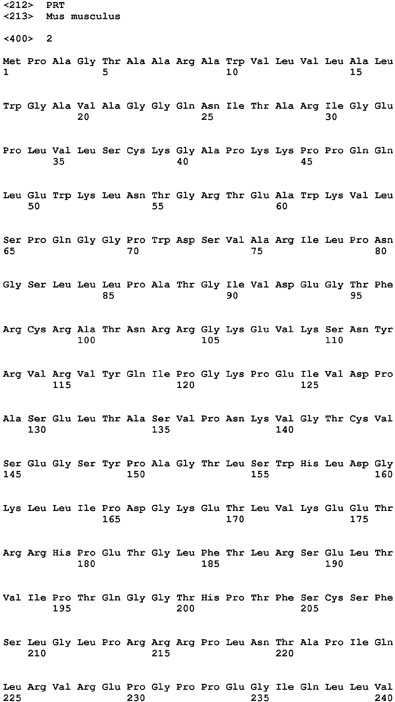


Claims (15)
- A phosphomimetic RAGE protein comprising an amino acid sequence that is at least 90 % identical to a native mammalian RAGE isoform or fragment thereof wherein at least one serine present in the cytoplasmatic tail (CT) of the mammalian RAGE isoform is replaced by a phosphomimetic structure.
- The phosphomimetic RAGE protein according to claim 1, wherein the native mammalian RAGE isoform or fragment thereof contains the amino acid sequence of amino acids 40 to 241 of SEQ ID NO: 1, and wherein the phosphomimetic structure is preferably selected from glutamic acid, aspartic acid.
- The phosphomimetic RAGE according to claim 1 or 2, wherein the amino acid sequence is at least 95 % identical, preferably at least 98 % identical, more preferably at least 99 % identical to murine RAGE identified by SEQ ID NO: 1 or a fragment thereof, wherein Ser376 and/or Ser389 as defined in SEQ ID NO: 1 are replaced by phosphomimetic amino acid(s), in particular glutamic acids.
- The phosphomimetic RAGE according to claim 3, comprising an amino acid sequence that is at least 95 % identical, preferably at least 98 % identical, more preferably at least 99 % identical to SEQ ID NO: 2.
- The phosphomimetic RAGE according to claim 1 or 2, wherein the amino acid sequence is at least 95 % identical, preferably at least 98 % identical, more preferably at least 99 % identical to human RAGE identified by SEQ ID NO: 3 or a fragment thereof, wherein Ser391 as defined in SEQ ID NO: 3 is replaced by a phosphomimetic amino acid, in particular glutamic acid.
- The phosphomimetic RAGE according to claim 5, comprising an amino acid sequence that is at least 95 % identical, preferably at least 98 % identical, more preferably at least 99 % identical to SEQ ID NO: 4.
- A fusion protein of a mammalian RAGE protein comprising an amino acid sequence that is at least 90 % identical to a native mammalian RAGE isoform or fragment thereof according to claim 1 or 2, fused to at least one nuclear targeting structure.
- The fusion protein according to claim 7, wherein the amino acid sequence of the mammalian RAGE is at least 95 % identical, preferably at least 98 % identical, more preferably at least 99 % identical to human RAGE identified by SEQ ID NO: 3 or murine RAGE identified by SEQ ID NO: 1 or a fragment thereof and the nuclear targeting structure is a nuclear localization sequence (NLS).
- An isolated polynucleotide with a nucleic acid sequence encoding the phosphomimetic RAGE according to any of claims 1 to 6 or the fusion protein according to claim 7 or 8.
- The isolated polynucleotide according to claim 9, with a nucleic acid sequence that is at least 95 % identical, preferably at least 98 % identical, more preferably at least 99 % identical to SEQ ID NO: 5 or 6.
- A vector comprising a polynucleotide according to claim 9 or 10 and a promoter, wherein the promoter is preferably a tissue specific promoter, more preferably selected from the group consisting of a keratinocyte specific promoter, a liver specific promoter, a fibroblast specific promoter, or a macrophage specific promoter,
- A composition comprising carrier and an active pharmaceutical ingredient, selected from a phosphomimetic RAGE protein according to any of claims 1 to 6, a fusion protein according to claim 7 or 8, and a vector according to claim 11, for use in the treatment or prevention of a disease in a patient, wherein the disease is preferably selected from a disease initiated by impaired DNA repair, a disease initiated by increased DNA damage or a disease initiated by increased senescence.
- The composition for use according to claim 12, wherein the disease is a disease of the lung, nerve, kidney, blood vessels or lymphatic vessels, preferably the disease is selected from age associated diseases, liver fibrosis, diabetic complications, neurodegenerative diseases, radiation damage, ischemia reperfusion, stroke, myocardial infarction, tissue damage associated with renal failure and replacement therapy, septicemia, skin disorders such as impaired wound healing and psoriasis, pulmonary fibrosis, wherein the pulmonary fibrosis is in particular caused by smoking, pollution, diabetes, radiation, or chemotherapy.
- The composition for use according to claim 12 or 13, wherein the carrier is an adeno associated virus (AAV) capsid, preferably a tissue specific AAV capsid.
- A method of purification of a RAGE protein with a polyhistidine-tag, comprising the steps of:- Providing a first solution comprising the RAGE protein with a polyhistidine-tag and contaminant proteins;- Subjecting the first solution to a Ni-NTA chromatography and eluting a second solution containing the RAGE protein; and- Subjecting the second solution to a heparin chromatography and eluting a third solution containing the RAGE protein.
Priority Applications (7)
| Application Number | Priority Date | Filing Date | Title |
|---|---|---|---|
| EP17177679.2A EP3418298A1 (en) | 2017-06-23 | 2017-06-23 | Rage proteins for the treatment of fibrosis and dna damage mediated diseases |
| MX2019014697A MX2019014697A (en) | 2017-06-23 | 2018-06-22 | Rage proteins for the treatment of fibrosis and dna damage mediated diseases. |
| PCT/EP2018/066794 WO2018234555A1 (en) | 2017-06-23 | 2018-06-22 | RABY PROTEINS FOR THE TREATMENT OF FIBROSIS AND DISEASES MEDIATED BY ALTERATION OF DNA |
| EP18736828.7A EP3642223A1 (en) | 2017-06-23 | 2018-06-22 | Rage proteins for the treatment of fibrosis and dna damage mediated diseases |
| CN201880042077.1A CN110785430A (en) | 2017-06-23 | 2018-06-22 | RAGE proteins for the treatment of fibrosis and DNA damage mediated diseases |
| US16/625,212 US20200140518A1 (en) | 2017-06-23 | 2018-06-22 | Rage proteins for the treatment of fibrosis and dna damage mediated diseases |
| US18/450,524 US20240025960A1 (en) | 2017-06-23 | 2023-08-16 | Rage proteins for the treatment of fibrosis and dna damage mediated diseases |
Applications Claiming Priority (1)
| Application Number | Priority Date | Filing Date | Title |
|---|---|---|---|
| EP17177679.2A EP3418298A1 (en) | 2017-06-23 | 2017-06-23 | Rage proteins for the treatment of fibrosis and dna damage mediated diseases |
Publications (1)
| Publication Number | Publication Date |
|---|---|
| EP3418298A1 true EP3418298A1 (en) | 2018-12-26 |
Family
ID=59284983
Family Applications (2)
| Application Number | Title | Priority Date | Filing Date |
|---|---|---|---|
| EP17177679.2A Withdrawn EP3418298A1 (en) | 2017-06-23 | 2017-06-23 | Rage proteins for the treatment of fibrosis and dna damage mediated diseases |
| EP18736828.7A Withdrawn EP3642223A1 (en) | 2017-06-23 | 2018-06-22 | Rage proteins for the treatment of fibrosis and dna damage mediated diseases |
Family Applications After (1)
| Application Number | Title | Priority Date | Filing Date |
|---|---|---|---|
| EP18736828.7A Withdrawn EP3642223A1 (en) | 2017-06-23 | 2018-06-22 | Rage proteins for the treatment of fibrosis and dna damage mediated diseases |
Country Status (5)
| Country | Link |
|---|---|
| US (2) | US20200140518A1 (en) |
| EP (2) | EP3418298A1 (en) |
| CN (1) | CN110785430A (en) |
| MX (1) | MX2019014697A (en) |
| WO (1) | WO2018234555A1 (en) |
Families Citing this family (2)
| Publication number | Priority date | Publication date | Assignee | Title |
|---|---|---|---|---|
| WO2022066899A1 (en) * | 2020-09-23 | 2022-03-31 | The Johns Hopkins University | Method to improve protein quality control |
| CN116251171A (en) * | 2021-12-10 | 2023-06-13 | 南京景瑞康分子医药科技有限公司 | Use of soluble advanced glycation end product receptor protein for preventing or treating pulmonary infection |
Citations (1)
| Publication number | Priority date | Publication date | Assignee | Title |
|---|---|---|---|---|
| WO2009108274A2 (en) * | 2008-02-26 | 2009-09-03 | The University Of North Carolina At Chapel Hill | Methods and compositions for adeno-associated virus (aav) with hi loop mutations |
Family Cites Families (1)
| Publication number | Priority date | Publication date | Assignee | Title |
|---|---|---|---|---|
| CN102803292A (en) * | 2009-04-20 | 2012-11-28 | 辉瑞公司 | Control of protein glycosylation and related compositions and methods |
-
2017
- 2017-06-23 EP EP17177679.2A patent/EP3418298A1/en not_active Withdrawn
-
2018
- 2018-06-22 CN CN201880042077.1A patent/CN110785430A/en active Pending
- 2018-06-22 WO PCT/EP2018/066794 patent/WO2018234555A1/en not_active Ceased
- 2018-06-22 US US16/625,212 patent/US20200140518A1/en not_active Abandoned
- 2018-06-22 EP EP18736828.7A patent/EP3642223A1/en not_active Withdrawn
- 2018-06-22 MX MX2019014697A patent/MX2019014697A/en unknown
-
2023
- 2023-08-16 US US18/450,524 patent/US20240025960A1/en not_active Abandoned
Patent Citations (1)
| Publication number | Priority date | Publication date | Assignee | Title |
|---|---|---|---|---|
| WO2009108274A2 (en) * | 2008-02-26 | 2009-09-03 | The University Of North Carolina At Chapel Hill | Methods and compositions for adeno-associated virus (aav) with hi loop mutations |
Non-Patent Citations (22)
| Title |
|---|
| "Goodman & Gilman's The Pharmacological Basis of Therapeutics", 2001, MCGRAW-HILL PROFESSIONAL |
| "Pharmaceutical Dosage Forms and Drug Delivery Systems", 1999, LIPPINCOTT WILLIAMS & WILKINS |
| "Remington: The Science and Practice of Pharmacy", 2003, LIPPINCOTT WILLIAMS & WILKINS |
| APARICIO, T.; R. BAER; J. GAUTIER: "DNA double-strand break repair pathway choice and cancer", DNA REPAIR (AMST, vol. 19, 2014, pages 169 - 75 |
| BIERHAUS, A. ET AL.: "Understanding RAGE, the receptor for advanced glycation endproducts", J. MOL. MED, vol. 83, 2005, pages 876 - 886, XP009058277, DOI: doi:10.1007/s00109-005-0688-7 |
| BIERHAUS, A.; NAWROTH, P.P.: "Multiple levels of regulation determine the role of the receptor for AGE (RAGE) as common soil in inflammation, immune responses and diabetes mellitus and its complications", DIABETOLOGIA, vol. 52, 2009, pages 2251 - 2263, XP019755186, DOI: doi:10.1007/s00125-009-1458-9 |
| BRANZEI, D.; M. FOIANI: "Regulation of DNA repair throughout the cell cycle", NAT REV MOL CELL BIOL, 2008, pages 297 - 308 |
| CICCIA, A.; S.J. ELLEDGE: "The DNA damage response: making it safe to play with knives", MOL CELL, vol. 40, no. 2, 2010, pages 179 - 204, XP055207185, DOI: doi:10.1016/j.molcel.2010.09.019 |
| CONSTEIN, R. ET AL.: "Characterization of a novel EGFP reporter mouse to monitor Cre recombination as demonstrated by a Tie2 Cre mouse line", GENESIS, vol. 30, no. 1, 2001, pages 36 - 44 |
| DEMLING N; EHRHARDT C; KASPER M; LAUE M; KNELS L; RIEBER EP: "Promotion of cell adherence and spreading: a novel function of RAGE, the highly selective differentiation marker of human alveolar epithelial type I cells", CELL AND TISSUE RESEARCH, vol. 323, no. 3, 2006, pages 475 - 488, XP019346670, DOI: doi:10.1007/s00441-005-0069-0 |
| DESSING, M.C. ET AL.: "RAGE does not contribute to renal injury and damage upon ischemia/reperfusion-induced injury", J INNATE IMMUN, vol. 4, no. 1, 2012, pages 80 - 5 |
| FEHRENBACH H; KASPER M; TSCHERNIG T; SHEARMAN MS; SCHUH D; MULLER M: "Receptor for advanced glycation endproducts (RAGE) exhibits highly differential cellular and subcellular localisation in rat and human lung", CELLULAR AND MOLECULAR BIOLOGY, vol. 44, no. 7, 1998, pages 1147 - 1157 |
| HE M; KUBO H; ISHIZAWA K ET AL.: "The role of the receptor for advanced glycation end-products in lung fibrosis", AMERICAN JOURNAL OF PHYSIOLOGY, vol. 293, no. 6, 2007, pages L 1427 - L1436 |
| JACKSON, S.P.; J. BARTEK: "The DNA-damage response in human biology and disease.", NATURE, vol. 461, no. 7267, 2009, pages 1071 - 8, XP055070409, DOI: doi:10.1038/nature08467 |
| KUMAR V.: "Homeostatic interaction between Receptor for advanced glycation end products (RAGE) and Ataxia telangiectasia mutated (ATM) is essential for DNA repair", PHD THESIS, 2015 |
| MASAKIYO SAKAGUCHI ET AL: "TIRAP, an Adaptor Protein for TLR2/4, Transduces a Signal from RAGE Phosphorylated upon Ligand Binding", PLOS ONE, vol. 6, no. 8, 1 August 2011 (2011-08-01), pages e23132, XP055423659, DOI: 10.1371/journal.pone.0023132 * |
| MATSUOKA, S. ET AL.: "ATM and ATR substrate analysis reveals extensive protein networks responsive to DNA damage", SCIENCE, vol. 316, no. 5828, 2007, pages 1160 - 6 |
| NEEDLEMAN; WUNSCH, J. MOL. BIOL., vol. 48, 1970, pages 443 - 453 |
| O'DRISCOLL M.: "Diseases associated with defective responses to DNA damage", COLD SPRING HARB PERSPECT BIOL, vol. 4, no. 12, 2012 |
| RICE: "EMBOSS: The European Molecular Biology Open Software Suite", TRENDS GENET., vol. 16, 2000, pages 276 - 277, XP004200114, DOI: doi:10.1016/S0168-9525(00)02024-2 |
| SCHMIDT, A. M ET AL.: "Isolation and characterization of two binding proteins for advanced glycosylation end products from bovine lung which are present on the endothelial cell surface", J BIOL CHEM, vol. 267, no. 21, 1992, pages 14987 - 14997 |
| WIELPUTZ, M.O. ET AL.: "In vivo monitoring of cystic fibrosis-like lung disease in mice by volumetric compted tomography", EUR RESPIR J, vol. 38, no. 5, 2011, pages 1060 - 70 |
Also Published As
| Publication number | Publication date |
|---|---|
| EP3642223A1 (en) | 2020-04-29 |
| US20240025960A1 (en) | 2024-01-25 |
| US20200140518A1 (en) | 2020-05-07 |
| MX2019014697A (en) | 2020-08-03 |
| CN110785430A (en) | 2020-02-11 |
| WO2018234555A1 (en) | 2018-12-27 |
Similar Documents
| Publication | Publication Date | Title |
|---|---|---|
| AU2018345919B2 (en) | Compositions and methods for editing RNA | |
| JP6166041B2 (en) | Methods and compositions for treating tubule myopathy using chimeric polypeptides comprising myotubularin 1 (MTM1) polypeptide | |
| US20240025960A1 (en) | Rage proteins for the treatment of fibrosis and dna damage mediated diseases | |
| AU2021282480A1 (en) | Methods and pharmaceutical composition for the treatment and the prevention of cardiomyopathy due to energy failure | |
| US20160256571A1 (en) | Invention | |
| JP2022554267A (en) | RECOMBINANT CDKL5 PROTEIN, GENE THERAPY AND PRODUCTION METHOD | |
| US20160137989A1 (en) | Method of suppressing gene transcription through histone lysine methylation | |
| CN112225793B (en) | Lysosome targeting peptide, fusion protein thereof, adeno-associated virus vector carrying fusion protein coding sequence and application thereof | |
| CN116685329B (en) | Nucleic acid constructs and their use in treating spinal muscular atrophy | |
| KR20210090157A (en) | cell penetrating peptide | |
| KR20210091117A (en) | cell penetrating peptide | |
| US20240209341A1 (en) | Compositions and methods for targeting inflammatory or activated cells and treating or ameliorating inflammatory conditions and pain | |
| WO2019108924A2 (en) | Cdkl5 expression variants and cdkl5 fusion proteins | |
| US20220356455A1 (en) | Solubilized apyrases, methods and use | |
| KR20210132109A (en) | DNA-binding domain transactivators and uses thereof | |
| KR20150003186A (en) | A novel drug delivery system based on jcv-vlp | |
| JP7357039B2 (en) | AAV/UPR-plus virus, UPR-plus fusion protein, gene therapy, and uses thereof in the treatment of neurodegenerative diseases, such as Parkinson's disease and Huntington's disease, among others | |
| CN107530407A (en) | Propionyl CoA carboxylase composition and application thereof | |
| CN117580941A (en) | Multiplex CRISPR/Cas9-mediated target gene activation system | |
| TW202221119A (en) | Dna-binding domain transactivators and uses thereof | |
| KR20240010489A (en) | Compositions and methods for improving vision function | |
| AU2024250682B2 (en) | Aav variants for treatment of complement disorders | |
| CN114616001A (en) | Effective doses of adenovirus-based biological delivery and expression systems for the treatment of osteoarthritis in humans and compositions comprising the same | |
| US20260069715A1 (en) | Compositions and methods for the treatment of huntingtons disease by editing the mutant huntingtin gene | |
| WO2025015328A2 (en) | Increasing nfe2l1 activity or expression as therapy for eye disorders |
Legal Events
| Date | Code | Title | Description |
|---|---|---|---|
| PUAI | Public reference made under article 153(3) epc to a published international application that has entered the european phase |
Free format text: ORIGINAL CODE: 0009012 |
|
| STAA | Information on the status of an ep patent application or granted ep patent |
Free format text: STATUS: THE APPLICATION HAS BEEN PUBLISHED |
|
| AK | Designated contracting states |
Kind code of ref document: A1 Designated state(s): AL AT BE BG CH CY CZ DE DK EE ES FI FR GB GR HR HU IE IS IT LI LT LU LV MC MK MT NL NO PL PT RO RS SE SI SK SM TR |
|
| AX | Request for extension of the european patent |
Extension state: BA ME |
|
| STAA | Information on the status of an ep patent application or granted ep patent |
Free format text: STATUS: THE APPLICATION IS DEEMED TO BE WITHDRAWN |
|
| 18D | Application deemed to be withdrawn |
Effective date: 20190627 |