EP3417074B1 - Analytical methods and arrays for use in the same - Google Patents
Analytical methods and arrays for use in the same Download PDFInfo
- Publication number
- EP3417074B1 EP3417074B1 EP17713238.8A EP17713238A EP3417074B1 EP 3417074 B1 EP3417074 B1 EP 3417074B1 EP 17713238 A EP17713238 A EP 17713238A EP 3417074 B1 EP3417074 B1 EP 3417074B1
- Authority
- EP
- European Patent Office
- Prior art keywords
- expression
- cells
- biomarkers
- potency
- skin
- Prior art date
- Legal status (The legal status is an assumption and is not a legal conclusion. Google has not performed a legal analysis and makes no representation as to the accuracy of the status listed.)
- Active
Links
Images







Classifications
-
- A—HUMAN NECESSITIES
- A61—MEDICAL OR VETERINARY SCIENCE; HYGIENE
- A61K—PREPARATIONS FOR MEDICAL, DENTAL OR TOILETRY PURPOSES
- A61K35/00—Medicinal preparations containing materials or reaction products thereof with undetermined constitution
- A61K35/66—Microorganisms or materials therefrom
- A61K35/76—Viruses; Subviral particles; Bacteriophages
- A61K35/763—Herpes virus
-
- A—HUMAN NECESSITIES
- A61—MEDICAL OR VETERINARY SCIENCE; HYGIENE
- A61K—PREPARATIONS FOR MEDICAL, DENTAL OR TOILETRY PURPOSES
- A61K31/00—Medicinal preparations containing organic active ingredients
- A61K31/33—Heterocyclic compounds
- A61K31/395—Heterocyclic compounds having nitrogen as a ring hetero atom, e.g. guanethidine or rifamycins
- A61K31/435—Heterocyclic compounds having nitrogen as a ring hetero atom, e.g. guanethidine or rifamycins having six-membered rings with one nitrogen as the only ring hetero atom
- A61K31/4353—Heterocyclic compounds having nitrogen as a ring hetero atom, e.g. guanethidine or rifamycins having six-membered rings with one nitrogen as the only ring hetero atom ortho- or peri-condensed with heterocyclic ring systems
- A61K31/437—Heterocyclic compounds having nitrogen as a ring hetero atom, e.g. guanethidine or rifamycins having six-membered rings with one nitrogen as the only ring hetero atom ortho- or peri-condensed with heterocyclic ring systems the heterocyclic ring system containing a five-membered ring having nitrogen as a ring hetero atom, e.g. indolizine, beta-carboline
-
- A—HUMAN NECESSITIES
- A61—MEDICAL OR VETERINARY SCIENCE; HYGIENE
- A61K—PREPARATIONS FOR MEDICAL, DENTAL OR TOILETRY PURPOSES
- A61K39/00—Medicinal preparations containing antigens or antibodies
- A61K39/395—Antibodies; Immunoglobulins; Immune serum, e.g. antilymphocytic serum
- A61K39/39533—Antibodies; Immunoglobulins; Immune serum, e.g. antilymphocytic serum against materials from animals
- A61K39/3955—Antibodies; Immunoglobulins; Immune serum, e.g. antilymphocytic serum against materials from animals against proteinaceous materials, e.g. enzymes, hormones, lymphokines
-
- A—HUMAN NECESSITIES
- A61—MEDICAL OR VETERINARY SCIENCE; HYGIENE
- A61P—SPECIFIC THERAPEUTIC ACTIVITY OF CHEMICAL COMPOUNDS OR MEDICINAL PREPARATIONS
- A61P35/00—Antineoplastic agents
-
- C—CHEMISTRY; METALLURGY
- C07—ORGANIC CHEMISTRY
- C07K—PEPTIDES
- C07K16/00—Immunoglobulins [IG], e.g. monoclonal or polyclonal antibodies
- C07K16/18—Immunoglobulins [IG], e.g. monoclonal or polyclonal antibodies against material from animals or humans
- C07K16/28—Immunoglobulins [IG], e.g. monoclonal or polyclonal antibodies against material from animals or humans against receptors, cell surface antigens or cell surface determinants
- C07K16/2803—Immunoglobulins [IG], e.g. monoclonal or polyclonal antibodies against material from animals or humans against receptors, cell surface antigens or cell surface determinants against the immunoglobulin superfamily
- C07K16/2818—Immunoglobulins [IG], e.g. monoclonal or polyclonal antibodies against material from animals or humans against receptors, cell surface antigens or cell surface determinants against the immunoglobulin superfamily against CD28 or CD152
-
- C—CHEMISTRY; METALLURGY
- C12—BIOCHEMISTRY; BEER; SPIRITS; WINE; VINEGAR; MICROBIOLOGY; ENZYMOLOGY; MUTATION OR GENETIC ENGINEERING
- C12Q—MEASURING OR TESTING PROCESSES INVOLVING ENZYMES, NUCLEIC ACIDS OR MICROORGANISMS; COMPOSITIONS OR TEST PAPERS THEREFOR; PROCESSES OF PREPARING SUCH COMPOSITIONS; CONDITION-RESPONSIVE CONTROL IN MICROBIOLOGICAL OR ENZYMOLOGICAL PROCESSES
- C12Q1/00—Measuring or testing processes involving enzymes, nucleic acids or microorganisms; Compositions therefor; Processes of preparing such compositions
- C12Q1/68—Measuring or testing processes involving enzymes, nucleic acids or microorganisms; Compositions therefor; Processes of preparing such compositions involving nucleic acids
- C12Q1/6876—Nucleic acid products used in the analysis of nucleic acids, e.g. primers or probes
-
- A—HUMAN NECESSITIES
- A61—MEDICAL OR VETERINARY SCIENCE; HYGIENE
- A61K—PREPARATIONS FOR MEDICAL, DENTAL OR TOILETRY PURPOSES
- A61K39/00—Medicinal preparations containing antigens or antibodies
- A61K2039/505—Medicinal preparations containing antigens or antibodies comprising antibodies
-
- C—CHEMISTRY; METALLURGY
- C12—BIOCHEMISTRY; BEER; SPIRITS; WINE; VINEGAR; MICROBIOLOGY; ENZYMOLOGY; MUTATION OR GENETIC ENGINEERING
- C12Q—MEASURING OR TESTING PROCESSES INVOLVING ENZYMES, NUCLEIC ACIDS OR MICROORGANISMS; COMPOSITIONS OR TEST PAPERS THEREFOR; PROCESSES OF PREPARING SUCH COMPOSITIONS; CONDITION-RESPONSIVE CONTROL IN MICROBIOLOGICAL OR ENZYMOLOGICAL PROCESSES
- C12Q2600/00—Oligonucleotides characterized by their use
- C12Q2600/106—Pharmacogenomics, i.e. genetic variability in individual responses to drugs and drug metabolism
-
- C—CHEMISTRY; METALLURGY
- C12—BIOCHEMISTRY; BEER; SPIRITS; WINE; VINEGAR; MICROBIOLOGY; ENZYMOLOGY; MUTATION OR GENETIC ENGINEERING
- C12Q—MEASURING OR TESTING PROCESSES INVOLVING ENZYMES, NUCLEIC ACIDS OR MICROORGANISMS; COMPOSITIONS OR TEST PAPERS THEREFOR; PROCESSES OF PREPARING SUCH COMPOSITIONS; CONDITION-RESPONSIVE CONTROL IN MICROBIOLOGICAL OR ENZYMOLOGICAL PROCESSES
- C12Q2600/00—Oligonucleotides characterized by their use
- C12Q2600/148—Screening for cosmetic compounds
Definitions
- the present invention relates to a method for identifying the skin sensitizer potency of a test agent, and arrays and analytical kits for use in such methods.
- Allergic contact dermatitis is an inflammatory skin disease that affects a significant proportion of the population. It is commonly caused by immunological responses towards chemical haptens leading to substantial economic burden for society.
- Current tests for sensitizing chemicals rely on animal experimentation. Legislation on the registration and use of chemicals within, e.g., the pharmaceutical and cosmetic industries, have stimulated significant research efforts to develop alternative human cell-based assays for the prediction of sensitization. The aim is to replace animal experiments with in vitro tests displaying a higher predictive power.
- ACD is characterized by eczema and recurrent episodes of itching [ Akhavan, A. and S.R. Cohen, The relationship between atopic dermatitis and contact dermatitis. Clin Dermatol, 2003. 21(2): p. 158-62 ].
- the disease affects a significant proportion of the population, with prevalence rates of 7.2% to 18.6% in Europe [ Mortz, C.G., et al., Prevalence of atopic dermatitis, asthma, allergic rhinitis, and hand and contact dermatitis in adolescents.
- ACD is a type IV delayed-type hypersensitivity response caused mainly by reactive T helper 1 (Th1) and interferon (IFN) ⁇ producing CD8 + T cells at site of contact with small chemical haptens in previously exposed, and immunologically sensitized, individuals Fonacier, L.S., S.C. Dreskin, and D.Y. Leung, Allergic skin diseases.
- Th1 reactive T helper 1
- IFN interferon
- DC Dendritic cells
- the REACH (Registration, Evaluation, and Authorisation of Chemicals) regulation requires that all new and existing chemicals within the European Union, involving approximately 30000 chemicals, should be tested for hazardous effects [ EC 1907 / 2006 , in Regulation (EC) No 1907 / 2006 ] .
- the REACH legislation will have a huge impact on the number of animals needed for testing.
- the 7th Amendment to the Cosmetics Directive (76/768/EEC) posed a ban on animal tests for the majority of cosmetic ingredients for human use in 2013.
- development of reliable in vitro alternatives to experimental animals for the assessment of sensitizing capacity of chemicals is urgent.
- Genomic Allergen Rapid Detection GARD ( Figure 8 ).
- GARD Genomic Allergen Rapid Detection
- the preferred assay is the mouse Local Lymph Node Assay (LLNA) [ Basketter, D.A., et al., Local lymph node assay - validation, conduct and use in practice. Food Chem Toxicol, 2002. 40(5): p. 593-8 ], followed by the Guinea pig maximization test (GPMT) [ Magnusson, B. and A.M. Kligman, The identification of contact allergens by animal assay. The guinea pig maximization test. J Invest Dermatol, 1969. 52(3): p. 268-76 ].
- LLNA mouse Local Lymph Node Assay
- GPMT Guinea pig maximization test
- WO 2012/068474 A2 describes a high-throughput assessment method for contact hypersensitivity; Albrekt et al. BMC Pharmacology & Toxicology 15(5) (2014): 1-12 describes skin sensitizers differentially regulating signalling pathways in MUTZ-3 cells in relation to their individual potency; Lambrechts et al. Toxicological Sciences 116(1) (2010): 122-129 describes assessment of chemical skin-sensitizing potency by an in vitro assay based on human dendritic cells; Schoeters et al. Molecular Immunology 44 (2007): 3222-3233 describes microarray analyses in dendritic cells revealing potential biomarkers for chemical-induced skin sensitization.
- a first aspect of the invention provides provides an in vitro method for determining the skin sensitizing potency of an agent comprising or consisting of the steps of:
- biomarkers measured in step (c) is selected from the group defined in Table A(i) and/or Table A(ii).
- step (c) comprises or consists of measuring the expression of one or more biomarker listed in Table A(i), for example, 2, 3, 4, 5, 6, 7, 8, 9, 10, 11, 12, 13, 14, 15, 16, 17, 18, 19, or 20 of the biomarkers listed in Table A(i).
- step (c) may comprise or consist of measuring the expression of all of the biomarkers listed in Table A(i).
- the method may include or exclude measuring the expression of HIST1H2BM.
- the method may include or exclude measuring the expression of HIST1H4B.
- the method may include or exclude measuring the expression of HIST1H1D.
- the method may include or exclude measuring the expression of PAICS.
- the method may include or exclude measuring the expression of HIST1H4D.
- the method may include or exclude measuring the expression of HIST1H2AL/// HIST1H2BN.
- the method may include or exclude measuring the expression of PLK1.
- the method may include or exclude measuring the expression of PHGDH.
- the method may include or exclude measuring the expression of MCM2.
- the method may include or exclude measuring the expression of MCM7.
- the method may include or exclude measuring the expression of CD53.
- the method may include or exclude measuring the expression of KIFC1.
- the method may include or exclude measuring the expression of WEE1.
- the method may include or exclude measuring the expression of TMPO.
- the method may include or exclude measuring the expression of MCM4.
- the method may include or exclude measuring the expression of PSRC1.
- the method may include or exclude measuring the expression of RNF149.
- the method may include or exclude measuring the expression of MCM6.
- the method may include or exclude measuring the expression of MCM5.
- the method may include or exclude measuring the expression of FANCA.
- step (c) comprises or consists of measuring the expression of one or more biomarkers listed in Table A(ii), for example, 2, 3, 4, 5, 6, 7, 8, 9, 10, 11, 12, 13, 14, 15, 16, 17, 18, 19, 20, 21, 22, 23, or 24 of the biomarkers listed in Table A(ii).
- step (c) may comprise or consist of measuring the expression of all of the biomarkers listed in Table A(ii).
- the method may include or exclude measuring the expression of CAD.
- the method may include or exclude measuring the expression of MRTO4.
- the method may include or exclude measuring the expression of TOE1.
- the method may include or exclude measuring the expression of CDC20.
- the method may include or exclude measuring the expression of PM20D2.
- the method may include or exclude measuring the expression of LOC344887.
- the method may include or exclude measuring the expression of UNG.
- the method may include or exclude measuring the expression of CDKN1A.
- the method may include or exclude measuring the expression of HIST1H2BF.
- the method may include or exclude measuring the expression of HIST1H1B.
- the method may include or exclude measuring the expression of Transcript ID 7896697.
- the method may include or exclude measuring the expression of JADE1.
- the method may include or exclude measuring the expression of KIAA0125.
- the method may include or exclude measuring the expression of CHRNA5.
- the method may include or exclude measuring the expression of CHAF1B /// MORC3.
- the method may include or exclude measuring the expression of SEL1L3.
- the method may include or exclude measuring the expression of SSRP1.
- the method may include or exclude measuring the expression of FOXM1.
- the method may include or exclude measuring the expression of LMNB1.
- the method may include or exclude measuring the expression of HIST1H2AK.
- the method may include or exclude measuring the expression of CENPA.
- the method may include or exclude measuring the expression of NCAPH.
- the method may include or exclude measuring the expression of HIST1H2BB.
- the method may include or exclude measuring the expression of ATP6V1B2.
- the method may include or exclude measuring the expression of TRAP1.
- step (c) may comprise or consist of measuring the expression of one or more biomarker listed in Table A(iii), for example, 1, 2, 3, 4, 5, 6, or 7 of the biomarkers listed in Table A(iii).
- step (c) comprises or consists of measuring the expression of all of the biomarkers listed in Table A(iii).
- the method may include or exclude measuring the expression of PFAS.
- the method may include or exclude measuring the expression of TMEM97.
- the method may include or exclude measuring the expression of DHCR24.
- the method may include or exclude measuring the expression of FTH1.
- the method may include or exclude measuring the expression of HIST1H2AE.
- the method may include or exclude measuring the expression of NQO1.
- the method may include or exclude measuring the expression of CD44.
- step (c) comprises or consists of measuring the expression of three or more of the biomarkers listed in Table A, for example, 4, 5, 6, 7, 8, 9, 10, 11, 12, 13, 14, 15, 16, 17, 18, 19, 20, 21, 22, 23, 24, 25, 26, 27, 28, 29, 30, 31, 32, 33, 34, 35, 36, 37, 38, 39, 40, 41, 42, 43, 44, 45, 46, 47, 48, 49, 50, or 51 of the biomarkers listed in Table A.
- step (c) may comprise or consist of measuring the expression of all of the biomarkers listed in Table A.
- the method may include or exclude measuring the expression of HIST1H2BM.
- the method may include or exclude measuring the expression of HIST1H4B.
- the method may include or exclude measuring the expression of HIST1H1D.
- the method may include or exclude measuring the expression of PAICS.
- the method may include or exclude measuring the expression of HIST1H4D.
- the method may include or exclude measuring the expression of HIST1H2AL/// HIST1H2BN.
- the method may include or exclude measuring the expression of PLK1.
- the method may include or exclude measuring the expression of PHGDH.
- the method may include or exclude measuring the expression of MCM2.
- the method may include or exclude measuring the expression of MCM7.
- the method may include or exclude measuring the expression of CD53.
- the method may include or exclude measuring the expression of KIFC1.
- the method may include or exclude measuring the expression of WEE1.
- the method may include or exclude measuring the expression of TMPO.
- the method may include or exclude measuring the expression of MCM4.
- the method may include or exclude measuring the expression of PSRC1.
- the method may include or exclude measuring the expression of RNF149.
- the method may include or exclude measuring the expression of MCM6.
- the method may include or exclude measuring the expression of MCM5.
- the method may include or exclude measuring the expression of FANCA.
- the method may include or exclude measuring the expression of CAD.
- the method may include or exclude measuring the expression of MRTO4.
- the method may include or exclude measuring the expression of TOE1.
- the method may include or exclude measuring the expression of CDC20.
- the method may include or exclude measuring the expression of PM20D2.
- the method may include or exclude measuring the expression of LOC344887.
- the method may include or exclude measuring the expression of UNG.
- the method may include or exclude measuring the expression of CDKN1A.
- the method may include or exclude measuring the expression of HIST1H2BF.
- the method may include or exclude measuring the expression of HIST1H1B.
- the method may include or exclude measuring the expression of Transcript ID 7896697.
- the method may include or exclude measuring the expression of JADE1.
- the method may include or exclude measuring the expression of KIAA0125.
- the method may include or exclude measuring the expression of CHRNA5.
- the method may include or exclude measuring the expression of CHAF1B /// MORC3.
- the method may include or exclude measuring the expression of SEL1L3.
- the method may include or exclude measuring the expression of SSRP1.
- the method may include or exclude measuring the expression of FOXM1.
- the method may include or exclude measuring the expression of LMNB1.
- the method may include or exclude measuring the expression of HIST1H2AK.
- the method may include or exclude measuring the expression of CENPA.
- the method may include or exclude measuring the expression of NCAPH.
- the method may include or exclude measuring the expression of HIST1H2BB.
- the method may include or exclude measuring the expression of ATP6V1B2.
- the method may include or exclude measuring the expression of TRAP1.
- the method may include or exclude measuring the expression of PFAS.
- the method may include or exclude measuring the expression of TMEM97.
- the method may include or exclude measuring the expression of DHCR24.
- the method may include or exclude measuring the expression of FTH1.
- the method may include or exclude measuring the expression of HIST1H2AE.
- the method may include or exclude measuring the expression of NQO1.
- the method may include or exclude measuring the expression of CD44.
- the method may include or exclude measuring the expression of one or more of MCM7, PFAS, HIST1H2AE.
- step (c) may comprise or consist of measuring the expression of one or more biomarker listed in Table B.
- step (c) may comprise or consist of measuring the expression of 2, 3, 4, 5, 6, 7, 8, 9, 10, 11, 12, 13, 14, 15, 16, 17, 18 of the biomarkers listed in Table B.
- step (c) may comprise or consist of measuring the expression of all of the biomarkers listed in Table B.
- step (c) comprises or consists of measuring the expression of at least 2 of the biomarkers listed in Table B, for example, ⁇ 3, ⁇ 4, ⁇ 5, ⁇ 6, ⁇ 7, ⁇ 8, ⁇ 9, ⁇ 10, ⁇ 11, ⁇ 12, ⁇ 13, ⁇ 14, ⁇ 15, ⁇ 16, ⁇ 17, or 18 of the biomarkers listed in Table B.
- step (c) comprises or consist of measuring the expression of one or more biomarker listed in Table B(i), for example, ⁇ 2, ⁇ 3, ⁇ 4, ⁇ 5, ⁇ 6, ⁇ 7, ⁇ 8, ⁇ 9, ⁇ 10, ⁇ 11, ⁇ 12, ⁇ 13, ⁇ 14, ⁇ 15, or 16 of the biomarkers listed in Table B(i).
- step (c) comprises or consist of measuring the expression of one or more biomarker listed in Table B(ii), for example, 2 of the biomarkers listed in Table B(ii).
- the method may include or exclude measuring the expression of CCNA2.
- the method may include or exclude measuring the expression of Transcript ID 8151252.
- the method may include or exclude measuring the expression of PIGW.
- the method may include or exclude measuring the expression of SNRPD1.
- the method may include or exclude measuring the expression of ARHGAP19 /// ARHGAP19-SLIT1.
- the method may include or exclude measuring the expression of HIST1H2AB.
- the method may include or exclude measuring the expression of LRCH2.
- the method may include or exclude measuring the expression of PKP4 /// PKP4.
- the method may include or exclude measuring the expression of RAVER2 /// RAVER2.
- the method may include or exclude measuring the expression of DUT /// DUT.
- the method may include or exclude measuring the expression of AURKA.
- the method may include or exclude measuring the expression of Transcript ID 8055309.
- the method may include or exclude measuring the expression of NDC1.
- the method may include or exclude measuring the expression of KIF22 /// KIF22.
- the method may include or exclude measuring the expression of OK/SW-CL.58.
- the method may include or exclude measuring the expression of Transcript ID 7994343.
- the method may include or exclude measuring the expression of HIST1H2AB /// HIST1 H2AE.
- the method may include or exclude measuring the expression of HMGB3.
- biomarker we include any biological molecule, or component or fragment thereof, the measurement of which can provide information useful in determining sensitizer potency.
- the biomarker may be a nucleic acid molecule, such as a mRNA or cDNA.
- the biomarker may be a protein encoded by the nucleic acid molecule or carbohydrate moiety, or an antigenic component or fragment thereof.
- test agent is already known to be, or suspected of being, capable of inducing sensitization of the skin.
- test agent may already be known to be capable of inducing sensitization of the skin by using a method already known in the art, for instance a method described WO 2012/056236 and/or Johansson H et al.
- the method is for identifying skin sensitizer potency and skin sensitizer/non-sensitizer status of the test agent (i.e., identifying whether the test agent is a sensitizer or not and identifying its potency as a skin sensitizer).
- the method comprises identifying whether the test agent is a sensitizer using the method described in WO 2012/056236 and/or Johansson H et al.
- skin sensitizing potency we include or mean the strength of the skin sensitizing ability of an agent.
- the relative potency or strength of sensitizing ability of an agent might lead to the ordering of a group of test agents from most potent to least potent or vice versa , and/or it might lead to their categorization according to one or more known regulation or system.
- the skin sensitization potency determined by the method is categorised according to the European Classification, Labelling and Packaging (CLP) Regulation (EC) 1272/2008 (http://echa.europa.eu/clp-2015).
- CLP European Classification, Labelling and Packaging
- EC 1272/2008 (http://echa.europa.eu/clp-2015).
- GLS Globally Harmonised System
- This system is based on the United Nations' Globally Harmonised System (GHS) and from June 2015, the only legislation to apply to the classification and labelling of both substances and mixtures. It requires companies to classify, label and package their products appropriately before placing them on the market. It provides the categories: 1A (strong), 1B (weak), or no cat (no sensitizer).
- the method may provide:
- the skin sensitization potency determined by the method is categorised according to the system described in Basketter et al., 2014, Categorization of chemicals according to their relative human skin sensitizing potency,' Dermatitis, 25(1):11-21 , i.e. categories 1 (strongest sensitizer), 2, 3, 4, 5, or 6 (true non-sensitizer) (e.g. Table 4, Figure 4 ).
- the method may provide:
- skin sensitization potency is categorised according to the local lymph node assay (LLNA) classification, Guinea pig maximisation test (GPMT) or no observed-effect level (NOEL).
- LLNA local lymph node assay
- GPMT Guinea pig maximisation test
- NOEL no observed-effect level
- NOEL no observed-effects level
- the expression of biomarkers measured in step (c) is measured in the cells provided in step (a) prior to and following exposure to the skin sensitizing agent of predetermined potency, and wherein the difference in expression between the biomarkers prior to and following exposure to the test agent is indicative of the potency of the skin sensitizing agent of step (b).
- the expression of the biomarkers measured in step (c) is measured in the cells provided in step (a) prior to and following exposure to the skin sensitizing agent of predetermined potency, and wherein the difference in expression between the biomarkers prior to and following step (c) is indicative of the potency of the skin sensitizing agent of step (b).
- the presence and or amount in a first sample differs from that of a second sample (e.g., a control agent sample).
- a second sample e.g., a control agent sample.
- the presence and/or amount is no more than 40% of that of the comparison sample, for example, no more than 39%, 38%, 37%, 36%, 35%, 34%, 33%, 32%, 31%, 30%, 29%, 28%, 27%, 26%, 25%, 24%, 23%, 22%, 21%, 20%, 19%, 18%, 17%, 16%, 15%, 14%, 13%, 12%, 11%, 10%, 9%, 8%, 7%, 6%, 5%, 4%, 3%, 2%, 1% or 0%.
- the biomarkers are measured in the cells provided in step (a) prior to and following exposure to the test agent, and wherein the difference in expression between the biomarkers prior to and following exposure to the test agent is indicative of the skin sensitizing potency of the test agent of step (b).
- the cells provided in step (a) may provide both the negative control and the test result.
- the biomarkers are measured in the cells provided in step (a) prior to and following exposure to the test agent, and wherein the difference in expression between the biomarkers prior to and following step (c) is indicative of the skin sensitizing potency of the test agent of step (b).
- the cells provided in step (a) may provide both the negative control and the test result.
- the vehicle control comprises DMSO.
- the presence and or amount in a first sample differs from that of a second sample (e.g., a control agent sample).
- a second sample e.g., a control agent sample.
- the presence and/or amount is no more than 40% of that of the comparison sample, for example, no more than 39%, 38%, 37%, 36%, 35%, 34%, 33%, 32%, 31%, 30%, 29%, 28%, 27%, 26%, 25%, 24%, 23%, 22%, 21%, 20%, 19%, 18%, 17%, 16%, 15%, 14%, 13%, 12%, 11%, 10%, 9%, 8%, 7%, 6%, 5%, 4%, 3%, 2%, 1% or 0%.
- the presence and or amount in a first sample is similar or identical to the presence and/or amount in a second sample (e.g., a control sample).
- a second sample e.g., a control sample.
- the presence and/or amount is at least 60% of that of the control sample, for example, at least 65%, 66%, 67%, 68%, 69%, 70%, 71%, 72%, 73%, 74%, 75%, 76%, 77%, 78%, 79%, 80%, 81%, 82%, 83%, 84%, 85%, 86%, 87%, 88%, 89%, 90%, 91%, 92%, 93%, 94%, 95%, 96%, 97%, 98%, 99% or 100%.
- the method comprises the further step of: (k) identifying the skin sensitizing potency of the test agent.
- the identification may be performed using any suitable statistical method or machine learning algorithm known in the art, such as Random Forest (RF), Support Vector Machine (SVM), Principal Component Analysis (PCA), ordinary least squares (OLS), partial least squares regression (PLS), orthogonal partial least squares regression (O-PLS) and other multivariate statistical analyses (e.g., backward stepwise logistic regression model).
- Random Forest RF
- Support Vector Machine SVM
- Principal Component Analysis PCA
- OLS ordinary least squares
- PLS partial least squares regression
- OF-PLS orthogonal partial least squares regression
- Statistical Science 2 (4): 396-413 Preferably a Random Forest model is used (such as is described in detail in Example A).
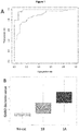
- sensitizing potency is determined with an ROC AUC of at least 0.45, for example with an ROC AUC of at least, 0.50, 0.55, 0.60, 0.65, 0.70, 0.75, 0.80, 0.85, 0.86, 0.87, 0.88, 0.89, 0.90, 0.91, 0.92, 0.93, 0.94, 0.95, 0.96, 0.97, 0.98, 0.99 or with an ROC AUC of 1.00.
- skin sensitizing potency is determined with an ROC AUC of at least 0.80, more preferably at least 0.85, still more preferably at least 0.88.
- the methods of the invention have an accuracy of at least 60%, for example, 61%, 62%, 63%, 64%, 65%, 66%, 67%, 68%, 69%, 70%, 71%, 72%, 73%, 74%, 75%, 76%, 77%, 78%, 79%, 80%, 81%, 82%, 83%, 84%, 85%, 86%, 87%, 88%, 89%, 90%, 91%, 92%, 93%, 94%, 95%, 96%, 97%, 98%, 99% or 100% accuracy.
- the methods of the invention have a sensitivity of at least 60%, for example, 61%, 62%, 63%, 64%, 65%, 66%, 67%, 68%, 69%, 70%, 71%, 72%, 73%, 74%, 75%, 76%, 77%, 78%, 79%, 80%, 81%, 82%, 83%, 84%, 85%, 86%, 87%, 88%, 89%, 90%, 91%, 92%, 93%, 94%, 95%, 96%, 97%, 98%, 99% or 100% sensitivity.
- the methods of the invention have a specificity of at least 60%, for example, 61%, 62%, 63%, 64%, 65%, 66%, 67%, 68%, 69%, 70%, 71%, 72%, 73%, 74%, 75%, 76%, 77%, 78%, 79%, 80%, 81%, 82%, 83%, 84%, 85%, 86%, 87%, 88%, 89%, 90%, 91%, 92%, 93%, 94%, 95%, 96%, 97%, 98%, 99% or 100% specificity.
- each repetition of steps (a) to (c) (and where present, steps (d)-(f/g)) is performed concurrently (i.e., with original steps (a) to (c) (and where present, steps (d)-(f/g))).
- each repetition of steps (a) to (c) (and where present, steps (d)-(f/g)) is performed consecutively.
- step (j) comprises or consists of one or more repetition wherein:
- the different agent of predetermined skin sensitization potency provided in step (i) is selected from the group consisting of:
- steps (a) to (c) are repeated at least once, for example, ⁇ 2, ⁇ 3, ⁇ 4, ⁇ 5, ⁇ 6, ⁇ 7, ⁇ 8, ⁇ 9, ⁇ 10, ⁇ 11, ⁇ 12, ⁇ 13, ⁇ 14, ⁇ 15, ⁇ 16, ⁇ 17, ⁇ 18, ⁇ 19, ⁇ 20, ⁇ 21, ⁇ 22, ⁇ 23, ⁇ 24, ⁇ 25, ⁇ 26, ⁇ 27, ⁇ 28, ⁇ 29, ⁇ 30, ⁇ 40, ⁇ 50, ⁇ 60, ⁇ 70, ⁇ 80, ⁇ 90, ⁇ 100, ⁇ 150, ⁇ 200, ⁇ 250, ⁇ 300, ⁇ 350, ⁇ 400, ⁇ 450, ⁇ 500, ⁇ 600, ⁇ 700, ⁇ 800, ⁇ 900 or ⁇ 1000 repetitions.
- each step or repetition of steps may be repeated identically to control for experimental error, for example, 2, 3, 4, 5, 6, 7, 8, 9 or 10 repeats.
- the expression of the biomarkers measured are measured for agents of predetermined skin sensitization potency over the full range of skin sensitization potencies.
- sensitizers of the highest and lowest degree of skin sensitizer category in the categorisation system being used.
- sensitizers are assessed at each sensitizer category in the categorisation system being used.
- the skin sensitizing agent of predetermined potency comprises or consist of one or more agents selected from the group consisting of the group defined in Table 2.
- the method may comprise measuring ⁇ 2, ⁇ 3, ⁇ 4, ⁇ 5, ⁇ 6, ⁇ 7, ⁇ 8, ⁇ 9, ⁇ 10, or 11, of the agents defined in Table 2.
- the skin sensitizing agent of predetermined potency comprises or consist of agents selected from the group consisting of the group defined in Table 8, Table 9 and/or Table 8, e.g., Table 8 only, Table 9 only, Table 10 only, Tables 8 and 9, Tables 8 and 10, Tables 9 and 10 or Tables 8, 9 and 10.
- the method may comprise measuring ⁇ 2, ⁇ 3, ⁇ 4, ⁇ 5, ⁇ 6, ⁇ 7, ⁇ 8, ⁇ 9, ⁇ 10, ⁇ 11, ⁇ 12, ⁇ 13, ⁇ 14, ⁇ 15, ⁇ 16, ⁇ 17, ⁇ 18, ⁇ 19, ⁇ 20, ⁇ 21, ⁇ 22, ⁇ 23, ⁇ 24, ⁇ 25, ⁇ 26, ⁇ 27, ⁇ 28, ⁇ 29, ⁇ 30, ⁇ 31, ⁇ 32, ⁇ 33, ⁇ 34, ⁇ 35, ⁇ 36, ⁇ 37, ⁇ 38, ⁇ 39, ⁇ 40, ⁇ 41, ⁇ 42, ⁇ 43, ⁇ 44, ⁇ 45, ⁇ 46, ⁇ 47, ⁇ 48, ⁇ 49, ⁇ 50, ⁇ 51, ⁇ 52, ⁇ 53, ⁇ 54, ⁇ 55, ⁇ 56, ⁇ 57, ⁇ 58, ⁇ 59, ⁇ 60, ⁇ 61, ⁇ 62, ⁇ 63, ⁇ 64,
- the skin sensitizing agent of predetermined potency comprises or consist of agents selected from the group consisting of 1-Butanol, 4-Aminobenzoic acid, Benzaldehyde, Chlorobenzene, Diethyl phthalate, Dimethyl formamide, Ethyl vanillin, Glycerol, Isopropanol, Lactic acid, Methyl salicylate, Octanoic acid, Propylene glycol, Phenol, p-hydroxybenzoic acid, Potassium permanganate, Salicylic acid, Sodium dodecyl sulphate, Tween 80, Zinc sulphate, 2,4-Dinitrochlorobenzene, Oxazolone, Potassium dichromate, Kathon CG (MC/MCI), Formaldehyde, 2-Aminophenol, 2-nitro-1,4-Phenylendiamine, p-Phenylendiamine, Hexylcinnamic aldehyde
- step (c) comprises or consists of measuring the expression of a nucleic acid molecule of one or more of the biomarkers.
- the nucleic acid molecule may be a cDNA molecule or an mRNA molecule.
- the nucleic acid molecule is an mRNA molecule.
- the nucleic acid molecule may be a cDNA molecule.
- the measurement of the expression of one or more of the biomarkers in step (c) is performed using a method selected from the group consisting of Southern hybridisation, Northern hybridisation, polymerase chain reaction (PCR), reverse transcriptase PCR (RT-PCR), quantitative real-time PCR (qRT-PCR), nanoarray, microarray, macroarray, autoradiography and in situ hybridisation.
- PCR polymerase chain reaction
- RT-PCR reverse transcriptase PCR
- qRT-PCR quantitative real-time PCR
- nanoarray microarray
- microarray macroarray
- autoradiography in situ hybridisation
- the one or more biomarkers measured in step (c) is measured using an array (e.g., a DNA array). In an additional or alternative embodiment the one or more biomarkers measured in step (c) is measured using a whole genome array (e.g., the Affymetrix Human Gene 1.0 ST array or Affymetrix Human Gene 2.0 ST array). In an alternative or additional embodiment, the Nanostring nCounter system is used (e.g., custom Nanostring nCounter code sets based on selection from a whole genome array (e.g., Affymetrix Human Gene 1.0 ST array or Affymetrix Human Gene 2.0 ST array).
- the method may comprise measuring the expression of one or more biomarkers in step (c) using one or more binding moieties, each capable of binding selectively to a nucleic acid molecule encoding one of the biomarkers identified in Table A.
- the one or more binding moieties each comprise or consist of a nucleic acid molecule. In a further embodiment the one or more binding moieties each comprise or consist of DNA, RNA, PNA, LNA, GNA, TNA or PMO. Preferably, the one or more binding moieties each comprise or consist of DNA. In one embodiment, the one or more binding moieties are 5 to 100 nucleotides in length. However, in an alternative embodiment, they are 15 to 35 nucleotides in length.
- the one or more binding moieties may comprise or consist of one or more probe from the Human Gene 1.0 ST Array (Affymetrix, Santa Clara, CA, USA). Probe identification numbers are provided in Table A herein, where they are referred to as "transcript cluster IDs”.
- Suitable binding agents may be selected or screened from a library based on their ability to bind a given nucleic acid, protein or amino acid motif, as discussed below.
- the binding moiety comprises a detectable moiety.
- detecttable moiety we include a moiety which permits its presence and/or relative amount and/or location (for example, the location on an array) to be determined, either directly or indirectly.
- Suitable detectable moieties are well known in the art.
- the detectable moiety may be a fluorescent and/or luminescent and/or chemiluminescent moiety which, when exposed to specific conditions, may be detected.
- a fluorescent moiety may need to be exposed to radiation (i . e . light) at a specific wavelength and intensity to cause excitation of the fluorescent moiety, thereby enabling it to emit detectable fluorescence at a specific wavelength that may be detected.
- the detectable moiety may be an enzyme which is capable of converting a (preferably undetectable) substrate into a detectable product that can be visualised and/or detected. Examples of suitable enzymes are discussed in more detail below in relation to, for example, ELISA assays.
- the detectable moiety may be selected from the group consisting of: a fluorescent moiety; a luminescent moiety; a chemiluminescent moiety; a radioactive moiety (for example, a radioactive atom); or an enzymatic moiety.
- the detectable moiety comprises or consists of a radioactive atom.
- the radioactive atom may be selected from the group consisting of technetium-99m, iodine-123, iodine-125, iodine-131, indium-111, fluorine-19, carbon-13, nitrogen-15, oxygen-17, phosphorus-32, sulphur-35, deuterium, tritium, rhenium-186, rhenium-188 and yttrium-90.
- the agent to be detected (such as, for example, the one or more biomarkers in the test sample and/or control sample described herein and/or an antibody molecule for use in detecting a selected protein) must have sufficient of the appropriate atomic isotopes in order for the detectable moiety to be readily detectable.
- the detectable moiety of the binding moiety is a fluorescent moiety.
- the radio- or other labels may be incorporated into the biomarkers present in the samples of the methods of the invention and/or the binding moieties of the invention in known ways.
- the binding agent is a polypeptide it may be biosynthesised or may be synthesised by chemical amino acid synthesis using suitable amino acid precursors involving, for example, fluorine-19 in place of hydrogen.
- Labels such as 99m Tc, 123 I, 186 Rh, 188 Rh and 111 In can, for example, be attached via cysteine residues in the binding moiety.
- Yttrium-90 can be attached via a lysine residue.
- the IODOGEN method Fraker et al (1978) Biochem. Biophys. Res. Comm.
- biomarkers in the sample(s) to be tested may be labelled with a moiety which indirectly assists with determining the presence, amount and/or location of said proteins.
- the moiety may constitute one component of a multicomponent detectable moiety.
- the biomarkers in the sample(s) to be tested may be labelled with biotin, which allows their subsequent detection using streptavidin fused or otherwise joined to a detectable label.
- the method provided in the first aspect of the present invention may comprise or consist of, in step (c), determining the expression of the protein of one or more biomarker defined in Table A.
- the method may comprise measuring the expression of one or more biomarkers in step (c) using one or more binding moieties each capable of binding selectively to one of the biomarkers identified in Table A.
- the one or more binding moieties may comprise or consist of an antibody or an antigen-binding fragment thereof such as a monoclonal antibody or fragment thereof.
- antibody includes any synthetic antibodies, recombinant antibodies or antibody hybrids, such as but not limited to, a single-chain antibody molecule produced by phage-display of immunoglobulin light and/or heavy chain variable and/or constant regions, or other immunointeractive molecules capable of binding to an antigen in an immunoassay format that is known to those skilled in the art.
- antibody-like binding agents such as affibodies and aptamers.
- one or more of the first binding molecules may be an aptamer (see Collett et al., 2005, Methods 37:4-15 ).
- the molecular libraries may be expressed in vivo in prokaryotic cells (Clackson et al, 1991, op . cit .; Marks et al, 1991, op. cit .) or eukaryotic cells ( Kieke et al, 1999, Proc Natl Acad Sci USA, 96(10):5651-6 ) or may be expressed in vitro without involvement of cells ( Hanes & Pluckthun, 1997, Proc Natl Acad Sci USA 94(10):4937-42 ; He & Taussig, 1997, Nucleic Acids Res 25(24):5132-4 ; Nemoto et al, 1997, FEBS Lett, 414(2):405-8 ).
- filamentous bacteriophage displaying antibody fragments at their surfaces, the antibody fragments being expressed as a fusion to the minor coat protein of the bacteriophage (Clackson et al, 1991, supra; Marks et al , 1991, supra ).
- suitable systems for display include using other viruses ( EP 39578 ), bacteria (Gunneriusson et al, 1999, supra; Daugherty et al, 1998, Protein Eng 11 (9):825-32 ; Daugherty et al, 1999, Protein Eng 12(7):613-21 ), and yeast ( Shusta et al, 1999, J Mol Biol 292(5):949-56 ).
- variable heavy (V H ) and variable light (V L ) domains of the antibody are involved in antigen recognition, a fact first recognised by early protease digestion experiments. Further confirmation was found by "humanisation" of rodent antibodies. Variable domains of rodent origin may be fused to constant domains of human origin such that the resultant antibody retains the antigenic specificity of the rodent parented antibody ( Morrison et al (1984) Proc. Natl. Acad. Sci. USA 81, 6851-6855 ).
- variable domains that antigenic specificity is conferred by variable domains and is independent of the constant domains is known from experiments involving the bacterial expression of antibody fragments, all containing one or more variable domains.
- variable domains include Fab-like molecules ( Better et al (1988) Science 240, 1041 ); Fv molecules ( Skerra et al (1988) Science 240, 1038 ); single-chain Fv (ScFv) molecules where the V H and V L partner domains are linked via a flexible oligopeptide ( Bird et a/ (1988) Science 242, 423 ; Huston et a/ (1988) Proc. Natl. Acad. Sci.
- the antibody or antigen-binding fragment may be selected from the group consisting of intact antibodies, Fv fragments (e . g . single chain Fv and disulphide-bonded Fv), Fab-like fragments (e.g. Fab fragments, Fab' fragments and F(ab) 2 fragments), single variable domains (e . g . V H and V L domains) and domain antibodies (dAbs, including single and dual formats [ i . e . dAb-linker-dAb]).
- the antibody or antigen-binding fragment is a single chain Fv (scFv).
- the one or more binding moieties may alternatively comprise or consist of an antibody-like binding agent, for example an affibody or aptamer.
- scFv molecules we mean molecules wherein the V H and V L partner domains are linked via a flexible oligopeptide.
- antibody fragments rather than whole antibodies
- the smaller size of the fragments may lead to improved pharmacological properties, such as better penetration of solid tissue.
- Effector functions of whole antibodies, such as complement binding, are removed.
- Fab, Fv, ScFv and dAb antibody fragments can all be expressed in and secreted from E . coli , thus allowing the facile production of large amounts of the said fragments.
- the antibodies may be monoclonal or polyclonal. Suitable monoclonal antibodies may be prepared by known techniques, for example those disclosed in “ Monoclonal Antibodies: A manual of techniques", H Zola (CRC Press, 1988 ) and in “ Monoclonal Hybridoma Antibodies: Techniques and applications", J G R Hurrell (CRC Press, 1982 ).
- selector peptides having defined motifs are usually employed.
- Amino acid residues that provide structure, decreasing flexibility in the peptide or charged, polar or hydrophobic side chains allowing interaction with the binding molecule may be used in the design of motifs for selector peptides. For example:
- binding molecules may involve the use of array technologies and systems to analyse binding to spots corresponding to types of binding molecules.
- the one or more protein-binding moieties may comprise a detectable moiety.
- the detectable moiety may be selected from the group consisting of a fluorescent moiety, a luminescent moiety, a chemiluminescent moiety, a radioactive moiety and an enzymatic moiety.
- step (c) may be performed using an assay comprising a second binding agent capable of binding to the one or more proteins, the second binding agent also comprising a detectable moiety.
- a second binding agent capable of binding to the one or more proteins, the second binding agent also comprising a detectable moiety.
- Suitable second binding agents are described in detail above in relation to the first binding agents.
- the proteins of interest in the sample to be tested may first be isolated and/or immobilised using the first binding agent, after which the presence and/or relative amount of said biomarkers may be determined using a second binding agent.
- the second binding agent is an antibody or antigen-binding fragment thereof; typically a recombinant antibody or fragment thereof.
- the antibody or fragment thereof is selected from the group consisting of: scFv; Fab; a binding domain of an immunoglobulin molecule. Suitable antibodies and fragments, and methods for making the same, are described in detail above.
- the second binding agent may be an antibody-like binding agent, such as an affibody or aptamer.
- the detectable moiety on the protein in the sample to be tested comprises or consists of a member of a specific binding pair (e . g . biotin)
- the second binding agent may comprise or consist of the complimentary member of the specific binding pair ( e . g . streptavidin).
- the detectable moiety is selected from the group consisting of: a fluorescent moiety; a luminescent moiety; a chemiluminescent moiety; a radioactive moiety; an enzymatic moiety. Examples of suitable detectable moieties for use in the methods of the invention are described above.
- Preferred assays for detecting serum or plasma proteins include enzyme linked immunosorbent assays (ELISA), radioimmunoassay (RIA), immunoradiometric assays (IRMA) and immunoenzymatic assays (IEMA), including sandwich assays using monoclonal and/or polyclonal antibodies.
- ELISA enzyme linked immunosorbent assays
- RIA radioimmunoassay
- IRMA immunoradiometric assays
- IEMA immunoenzymatic assays
- sandwich assays are described by David et al in US Patent Nos. 4,376,110 and 4,486,530 .
- Antibody staining of cells on slides may be used in methods well known in cytology laboratory diagnostic tests, as well known to those skilled in the art.
- the assay is an ELISA (Enzyme Linked Immunosorbent Assay) which typically involves the use of enzymes which give a coloured reaction product, usually in solid phase assays.
- Enzymes such as horseradish peroxidase and phosphatase have been widely employed.
- a way of amplifying the phosphatase reaction is to use NADP as a substrate to generate NAD which now acts as a coenzyme for a second enzyme system.
- Pyrophosphatase from Escherichia coli provides a good conjugate because the enzyme is not present in tissues, is stable and gives a good reaction colour.
- Chemiluminescent systems based on enzymes such as luciferase can also be used.
- Vitamin biotin Conjugation with the vitamin biotin is frequently used since this can readily be detected by its reaction with enzyme-linked avidin or streptavidin to which it binds with great specificity and affinity.
- the assay used for protein detection is conveniently a fluorometric assay.
- the detectable moiety of the second binding agent may be a fluorescent moiety, such as an Alexa fluorophore (for example Alexa -647).
- steps (c), (g), (f) and/or (j) of the methods described in the first aspect are performed using an array.
- the array may be a bead-based array or a surface-based array.
- the array may be selected from the group consisting of: macroarray; microarray; nanoarray.
- Arrays per se are well known in the art. Typically they are formed of a linear or two-dimensional structure having spaced apart (i . e . discrete) regions ("spots"), each having a finite area, formed on the surface of a solid support.
- An array can also be a bead structure where each bead can be identified by a molecular code or colour code or identified in a continuous flow. Analysis can also be performed sequentially where the sample is passed over a series of spots each adsorbing the class of molecules from the solution.
- the solid support is typically glass or a polymer, the most commonly used polymers being cellulose, polyacrylamide, nylon, polystyrene, polyvinyl chloride or polypropylene.
- the solid supports may be in the form of tubes, beads, discs, silicon chips, microplates, polyvinylidene difluoride (PVDF) membrane, nitrocellulose membrane, nylon membrane, other porous membrane, non-porous membrane (e.g. plastic, polymer, perspex, silicon, amongst others), a plurality of polymeric pins, or a plurality of microtitre wells, or any other surface suitable for immobilising proteins, polynucleotides and other suitable molecules and/or conducting an immunoassay.
- PVDF polyvinylidene difluoride
- nitrocellulose membrane nitrocellulose membrane
- nylon membrane other porous membrane
- non-porous membrane e.g. plastic, polymer, perspex, silicon, amongst others
- a plurality of polymeric pins e.g. plastic, polymer, perspex, silicon, amongst others
- microtitre wells e.g. plastic, polymer, perspex, silicon,
- affinity coupling of the probes via affinity-tags or similar constructs may be employed.
- affinity-tags or similar constructs may be employed.
- contact or non-contact printing, masking or photolithography the location of each spot can be defined.
- the array is a microarray.
- microarray we include the meaning of an array of regions having a density of discrete regions of at least about 100/cm 2 , and preferably at least about 1000/cm 2 .
- the regions in a microarray have typical dimensions, e . g . diameter, in the range of between about 10-250 ⁇ m, and are separated from other regions in the array by about the same distance.
- the array may alternatively be a macroarray or a nanoarray.
- binding molecules discussed above
- the skilled person can manufacture an array using methods well known in the art of molecular biology.
- one or more biomarkers measured in step (c) comprise or consist of one or more homologous gene product expressed by human cells.
- one or more biomarkers measured in step (c) is a protein or polypeptide.
- one or more biomarker measured in step (c) is a nucleic acid (e.g., DNA, mRNA or cDNA etc).
- the method is performed in vitro , in vivo , ex vivo or in silico.
- the method may in particular be performed in vitro.
- the skin sensitization is a hypersensitivity response (e.g., a cell-mediated hypersensitivity response).
- the hypersensitivity response is a type IV hypersensitivity response.
- the hypersensitivity response is allergic contact dermatitis (ACD).
- test agent we include any chemical entity (or mixture of chemical entities) for which sensitization potency and/or sensitization status is to be determined.
- sensitization status we include or mean whether or not a chemical entity (or mixture of chemical entities) is a sensitizer or not (e.g., a skin sensitizer and/or a respiratory sensitizer).
- skin sensitizing agent we mean any agent capable of inducing and triggering a Type IV delayed-type hypersensitivity reaction in a mammal.
- the Type IV delayed-type hypersensitivity reaction is DC-mediated.
- the one or more "skin sensitizing agent” is an agent capable of inducing and triggering a Type IV delayed-type hypersensitivity reaction at a site of epidermal contact in a mammal.
- the mammal may be any domestic or farm animal.
- the mammal is a rat, mouse, guinea pig, cat, dog, horse or a primate.
- the mammal is human.
- in vivo methods of determining sensitisation are known in the art.
- a preferred method is the Local lymph node assay (for details, see Basketter, D.A., et al., Local lymph node assay - validation, conduct and use in practice. Food Chem Toxicol, 2002. 40(5): p. 593-8 ).
- a further suitable, but less preferred, method is the guinea pig maximization test (for details, see Magnusson, B. and A.M. Kligman, The identification of contact allergens by animal assay. The guinea pig maximization test. J Invest Dermatol, 1969. 52(3): p. 268-76 ).
- the population of dendritic cells or population of dendritic-like cells is a population of dendritic-like cells.
- the dendritic-like cells are myeloid dendritic-like cells.
- the myeloid dendritic-like cells are derived from myeloid dendritic cells.
- the cells derived from myeloid dendritic cells are myeloid leukaemia-derived cells.
- the myeloid leukaemia-derived cells are selected from the group consisting of KG-1, THP-1, U-937, HL-60, Monomac-6, AML-193 and MUTZ-3.
- the dendritic-like cells are MUTZ-3 cells.
- MUTZ-3 cells are human acute myelomonocytic leukemia cells that are available from Deutsche Sammlung für Mikroorganismen und Zellkulturen GmbH (DSMZ), Braunschweig, Germany (www.dsmz.de; DMSZ No. ACC 295).
- dendritic-like cells we mean non-dendritic cells that exhibit functional and phenotypic characteristics specific to dendritic cells such as morphological characteristics, expression of costimulatory molecules and MHC class II molecules, and the ability to pinocytose macromolecules and to activate resting T cells.
- the dendritic-like cells after stimulation with cytokine, present antigens through CD1d, MHC class I and II and/or induce specific T-cell proliferation.
- the dendritic-like cells are CD34 + dendritic cell progenitors.
- the CD34 + dendritic cell progenitors can acquire, upon cytokine stimulation, the phenotypes of presenting antigens through CD1d, MHC class I and II, induce specific T-cell proliferation, and/or displaying a mature transcriptional and phenotypic profile upon stimulation with inflammatory mediators (i.e. similar phenotypes to immature dendritic cells or Langerhans-like dendritic cells).
- the population of dendritic cells or population of dendritic-like cells is a population of dendritic cells.
- the dendritic cells are primary dendritic cells.
- the dendritic cells are myeloid dendritic cells.
- Dendritic cells may be recognized by function, by phenotype and/or by gene expression pattern, particularly by cell surface phenotype. These cells are characterized by their distinctive morphology, high levels of surface MHC-class II expression and ability to present antigen to CD4+ and/or CD8+ T cells, particularly to naive T cells ( Steinman et al. (1991) Ann. Rev. Immunol. 9: 271 ).
- the cell surface of dendritic cells is unusual, with characteristic veil-like projections, and is characterized by expression of the cell surface markers CD11c and MHC class II. Most DCs are negative for markers of other leukocyte lineages, including T cells, B cells, monocytes/macrophages, and granulocytes.
- Subpopulations of dendritic cells may also express additional markers including 33D1, CCR1, CCR2, CCR4, CCR5, CCR6, CCR7, CD1a-d, CD4, CD5, CD8alpha, CD9, CD11b, CD24, CD40, CD48, CD54, CD58, CD80, CD83, CD86, CD91, CD117, CD123 (IL3Ra), CD134, CD137, CD150, CD153, CD162, CXCR1, CXCR2, CXCR4, DCIR, DC-LAMP, DC-SIGN, DEC205, E-cadherin, Langerin, Mannose receptor, MARCO, TLR2, TLR3 TLR4, TLR5, TLR6, TLR9, and several lectins.
- additional markers including 33D1, CCR1, CCR2, CCR4, CCR5, CCR6, CCR7, CD1a-d, CD4, CD5, CD8alpha, CD9, CD11b, CD24, CD40, CD48, CD54, CD58, CD80,
- the patterns of expression of these cell surface markers may vary along with the maturity of the dendritic cells, their tissue of origin, and/or their species of origin.
- Immature dendritic cells express low levels of MHC class II, but are capable of endocytosing antigenic proteins and processing them for presentation in a complex with MHC class II molecules.
- Activated dendritic cells express high levels of MHC class 11, ICAM-1 and CD86, and are capable of stimulating the proliferation of naive allogeneic T cells, e. g. in a mixed leukocyte reaction (MLR).
- MLR mixed leukocyte reaction
- dendritic cells or dendritic-like cells may be identified by any convenient assay for determination of antigen presentation.
- assays may include testing the ability to stimulate antigen- primed and/or naive T cells by presentation of a test antigen, followed by determination of T cell proliferation, release of IL-2, and the like.
- Preferred methods for detection and/or measurement of protein include Western blot, North-Western blot, immunosorbent assays (ELISA), antibody microarray, tissue microarray (TMA), immunoprecipitation, in situ hybridisation and other immunohistochemistry techniques, radioimmunoassay (RIA), immunoradiometric assays (IRMA) and immunoenzymatic assays (IEMA), including sandwich assays using monoclonal and/or polyclonal antibodies. Exemplary sandwich assays are described by David et al., in US Patent Nos. 4,376,110 and 4,486,530 . Antibody staining of cells on slides may be used in methods well known in cytology laboratory diagnostic tests, as well known to those skilled in the art.
- ELISA involves the use of enzymes which give a coloured reaction product, usually in solid phase assays.
- Enzymes such as horseradish peroxidase and phosphatase have been widely employed.
- a way of amplifying the phosphatase reaction is to use NADP as a substrate to generate NAD which now acts as a coenzyme for a second enzyme system.
- Pyrophosphatase from Escherichia coli provides a good conjugate because the enzyme is not present in tissues, is stable and gives a good reaction colour.
- Chemi-luminescent systems based on enzymes such as luciferase can also be used.
- Vitamin biotin Conjugation with the vitamin biotin is frequently used since this can readily be detected by its reaction with enzyme-linked avidin or streptavidin to which it binds with great specificity and affinity.
- a second aspect of the invention provides an array for use in the method according to the first aspect of the invention, the binding moieties of the array consisting of binding moieties for each of the biomarkers defined in Table A, and wherein the binding moieties of the array comprise binding moieties as defined in the first aspect of the invention.
- the array comprises one or more binding moiety for each of the biomarkers as defined in the first aspect of the invention.
- the one or more binding moiety is immobilised.
- the array is a bead-based array. In an additional or alternative embodiment the array is a surface-based array.
- the array is selected from the group consisting of: macroarray; microarray; nanoarray.
- the array of the second aspect of the invention may comprise one or more, preferably two or more, binding moieties, wherein the binding moieties are each capable of binding selectively to a biomarker as defined in the first aspect. Therefore, the array may comprise or consist of a particular selection of biomarker-specific binding moieties which correlates to any particular selection of biomarkers as defined in the first aspect.
- a third aspect of the invention provides the use of the biomarkers listed in Table A for determining the skin sensitizing potency of an agent.
- biomarker selected from the group defined in Table A for determining the skin sensitizing potency of an agent, preferably wherein the biomarker is selected from the group defined in Table A(i) and/or Table A(ii).
- binding moieties with specificity for each of the biomarkers listed in Table A for determining the skin sensitizing potency of an agent.
- a fourth aspect of the invention provides an analytical kit for use in a method according the first aspect of the invention comprising:
- analytical kit further comprising one or more control agents as defined in the first aspect of the invention.
- Also disclosed herein is a method of treating or preventing a type I hypersensitivity reaction (such as ACD) in a patient comprising the steps of:
- the one or more test agent that the patient is or has been exposed to is an agent that the patient is presently exposed to at least once a month, for example, at least once every two weeks, at least once every week, or at least once every day.
- Treatments of the symptoms of sensitization may include, for example, topical steroids.
- the method of treatment is consistent with the method described in the first aspect of the invention, and one or more of the embodiments described therein.
- a computer program for operating the methods the invention, for example, for interpreting the expression data of step (c) (and subsequent expression measurement steps) and thereby determining whether one or more test agent is a respiratory sensitizer and/or the sensitizer potency of the one or more test agent.
- the computer program may be a programmed SVM.
- the computer program may be recorded on a suitable computer-readable carrier known to persons skilled in the art. Suitable computer-readable-carriers may include compact discs (including CD-ROMs, DVDs, Blue Rays and the like), floppy discs, flash memory drives, ROM or hard disc drives.
- the computer program may be installed on a computer suitable for executing the computer program.
- the molecular events leading to skin sensitization and consequently to ACD can be characterized by a number of sequential key events (KE) triggered by a chemical, and have been summarized in an adverse outcome pathway (AOP) as described by the OECD [5].
- the initiating event (KE1) is defined as covalent protein modification by the skin sensitizing chemical after it has gained access to deeper skin layers.
- the following KE2 represents the inflammatory responses upon activation of keratinocytes.
- KE3 corresponds to the activation of dendritic cells, which in turn leads to activation and proliferation of T cells (KE4).
- ACD Upon re-exposure to the sensitizer, the development of ACD may be triggered, which as the adverse outcome is characterized by skin lesions induced by specific Th1 and CD8+ T cells. While the KE in the AOP are well described, a detailed mechanistic understanding of the underlying biology of the individual key events is still missing [5].
- the murine Local Lymph Node Assay (LLNA) [6] has for many years been the preferred alternative for skin sensitization testing as it is able to provide data for both hazard identification and characterization, including skin sensitizer potency information. However, it is characterized by certain limitations such as susceptibility to vehicle effects and issues with false-positive results [7].
- Several non-animal predictive methods have been developed to reduce animal experimentation used for chemical testing including computational approaches to integrate data from different test platforms for hazard identification as recently reviewed [8].
- GARD Genomic Allergen Rapid Detection
- the CLP regulation is based on the Globally Harmonised System [25], and uses three categories for chemical classification; no category (no cat) for non-sensitizers, category 1B for weak and 1A for strong sensitizers.
- GARD can be developed further into a tool for the prediction of chemical skin sensitizer potency, targeting the CLP categories.
- GARD Support Vector Machine model cannot be applied to multiclass problems, we used another approach based on random forest modelling [26].
- Random forest is a decision-tree based method and well-suited for microarray data [27]. It divides the dataset internally and repeatedly into a training and test set through random sub-sampling (bootstrapping). Samples in the test set, referred to as out-of-bag samples, comprise approximately one third of the entire dataset, and are used in order to estimate the out-of-bag (OOP) error, i.e. the classification error.
- OOP out-of-bag
- the predictive performance of the 52 genes were challenged with an independent test set containing 18 chemicals previously unseen to the model.
- the chemicals in this test set could be predicted with an overall accuracy of 78%.
- the myeloid cell line used in this study was derived from MUTZ-3 (DSMZ, Braunschweig, Germany) and maintained as described in [22, 28].
- a phenotypic control analysis of the cells prior to each experiment was carried out by flow cytometry in order to confirm the cells' immature state.
- the following monoclonal antibodies were used: CD1a (DakoCytomation, Glostrup, Denmark), CD34, CD86, HLA-DR (BD Biosciences, San Jose, USA), all FITC-conjugated; CD14 (DakoCytomation), CD54, CD80 (BD Biosciences), all PE-conjugated.
- FITC- and PE-conjugated mouse IgG1 served as isotype controls and propidium iodide as a marker for non-viable cells (BD Biosciences). Three batches of cells were exposed for 24 hours in independent experiments and viability and CD86 expression were assessed by flow cytometry. All FACS samples were analyzed on a FACSCanto II instrument with FACS Diva software for data acquisition. 10 000 events were acquired and further analysis was performed in FCS Express V4 (De Novo Software, Los Angeles, CA). Cells for RNA extraction were lysed in TRIzol® (Life Technologies/Thermo Fisher Scientific, Waltham, USA) and stored until further use in -20°C.
- RNA isolation from cells lysed in TRIzol® was performed according to the manufacturer's instructions. Labeled sense DNA was synthesized according to Affymetrix (Affymetrix, Cleveland, USA) protocols using the recommended kits and controls. The cDNA was hybridized to Human Gene 1.0 ST arrays (Affymetrix) and further processed and scanned as recommended by the supplier.
- an SVM model based on a linear kernel was trained on reference chemical stimulations from the original training set used during identification of the GPS [22]. The trained model was subsequently applied to assign each test chemical with an SVM Decision Value (SVM DV). Resulting SVM DVs for all test chemicals were used to construct a receiver operating characteristics (ROC) curve, and the resulting area under the curve (AUC) was used as a classification measure [32]. SVM modeling and ROC curve visualizations were performed in R statistical environment, using the additional packages e1071 [33] and ROCR [34]. Prior to evaluating final predictive performance of the model, SVM DVs for each individual replicate of the test chemicals were calibrated against the cut-off for maximal predictive performance obtained during classification of benchmark samples in Table 2, as described in [31].
- the calibrated SVM DVs were subsequently used for final classifications, and test chemicals were classified as sensitizers if the median output value of replicates > 0. Accuracy, sensitivity and specificity was estimated using cooper statistics [35]. The non-parametric Two Sample Wilcoxon test was performed in order to determine if the SVM DV distributions between CLP categories 1A, 1B and no cat differed significantly.
- the new microarray data were merged with historical data [22, 23] and subjected to quality control. Four arrays were removed due to bad quality; however, no chemical was present in less than biological duplicates. Array data was imported into the R statistical environment and normalized using the SCANfast algorithm [29, 30]. As several experimental campaigns needed to be combined, this dataset was normalized using the ComBat method [36, 37] in order to remove batch effects between samples. At this time, the samples in the training set were separated from the samples in the test set. To avoid overfitting, only samples in the designated training set were used during identification of the predictive biomarker signature and for fitting of parameters to the classifier, and samples in the test set were set aside to validate the performance of the identified signature and the specified classifier.
- the predictive biomarker signature was identified by feeding normalized and batch corrected transcript intensities from individual samples in the training set into a random forest model [26] combined with a backward elimination procedure in the varSelRF package [38] in R/Bioconductor version 3.1.2.
- the initial forest used for ranking of variable importance was grown to 2000 trees and all other parameters were kept at the default settings.
- the package iteratively fits and evaluates random forest models, at each iteration dropping 20% of the least important variables.
- the best performing set of variables was selected based on OOB error rates from all fitted Random Forests as the smallest number of genes within one standard error from the minimal error solution (i.e 1.s.e rule).
- variable selection procedure was validated by estimating the prediction error rate by the .632+ bootstrap method of the varSelRF package using 100 bootstrap samples, and the importance of individual transcripts in the biomarker signature was validated by the frequency of appearance in bootstrap samples (referred to as Validation Call Frequencies (VCF)).
- VCF Validation Call Frequencies
- the predictive performance of the identified biomarker signature was validated by building a new forest in the Random Forest package [39], based on previous parameters, using only the samples in the training set and the selected transcripts in the biomarker signature as variable input.
- the model was applied to assign each individual replicate sample in the test set to a CLP category, and the majority vote across the biological replicate stimulations for each chemical was accepted as the predicted category.
- Heatmaps and Principal Component Analysis (PCA) plots were constructed in Qlucore Omics Explorer (Qlucore AB, Lund, Sweden).
- Pathway analysis was performed with the Key Pathway Advisor (KPA) tool [40] version 16.6, which provides a pathway analysis workflow to investigate e.g. gene expression data. It associates differentially expressed genes with both upstream and downstream processes in order to allow biological interpretation.
- KPA Key Pathway Advisor
- a multigroup comparison (ANOVA) comparing samples belonging to CLP no cat, 1B, and 1A was then applied in order to identify transcripts that were differentially regulated.
- Script 1 Used for combating batch effects between different projects prior to model construction:
- a novel dataset comprising 37 well-characterized chemicals was selected in order to complement historical GARD data for 51 chemicals and to represent a relevant choice of chemicals, balanced in terms of chemical reactivity class, and use in consumer products.
- the novel chemicals were selected based on European Chemical Agency CLP databases and literature [15, 41] and in cooperation with the Skin Tolerance Task Force of Cosmetics Europe (CE), who kindly provided 27 chemicals.
- the 37 novel chemicals were predicted as sensitizers or non-sensitizers using the GPS and previously established protocols based on SVM classifications.
- the SVM model was applied to assign each individual replicate sample with a SVM DV. Prior to final classifications, SVM DVs from the 37 samples were first calibrated against 11 benchmark samples (Table 2) included in the same sample batch as the test chemicals.
- the dataset comprising the 37 novel chemicals were merged with the historical dataset comprising 51 chemicals.
- the dataset consisted of 86 unique chemicals (Table 4) and two vehicle controls, balanced with regards to categories 1A and 1B and non-sensitizers (no cat) as described by CLP (Table 3).
- CLP classification 1B was changed to no category/non-sensitizer according to the sources indicated in Table 4 for one of three reasons: i) for retaining consistency with previous GARD projects (benzaldehyde, xylene), ii) for being used as vehicle in non-sensitizing concentration (DMSO), and for being a well-described false-positive in the LLNA (sodium dodecyl sulfate).
- DMSO non-sensitizing concentration
- a random forest model for the prediction of three CLP categories was developed based on a training set consisting of 70 unique samples, including two vehicle controls.
- 52 predictive variables (Table 5) were identified as optimal for CLP classification.
- principal component analysis (PCA) was performed.
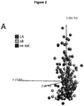
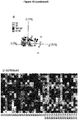
- the 52 variables identified by random forest based on a whole-genome array analysis were used as input, and the PCA was built on the training set ( Fig. 2A).
- Fig. 2A is based on chemicals with biological replicates colored according to CLP categories and a clear gradient from no cat to strong sensitizers (1A) can be observed along the first principal component.
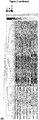
- the heatmap of the training set with hierarchical clustering of the variables in Fig. 2B illustrates the regulation of genes in relation to the respective chemical and CLP category.
- the model performance was further evaluated by predicting the CLP categories of an independent test set, which comprised 18 chemicals previously unseen to the model.
- the test set colored according to CLP categories was visualized in the PCA plot ( Fig. 3A ), without influencing the PCA components based on the 52 identified variables.
- Fig. 3B visualizes the regulation of the 52 genes in the normalized test set in form of a heatmap.
- 14 out of 18 chemicals were assigned into the right CLP category (Table 6, Suppl. Table 1), resulting in an overall accuracy of 78% (Table 7).
- the four misclassified chemicals were diethyl maleate, butyl glycidyl ether, lyral and cyanuric chloride.
- the CLP potency model contains information relevant for human potency prediction
- the 52 variables identified by random forest represent transcripts belonging to different cell compartments and different functional roles. Five of them overlap with the GARD prediction signature. Five of the top 10 markers, which were most frequently chosen in the bootstrapping process and have the highest validation call frequencies (Table 5), are histone cluster 1 members, such as HIST1H2AB [42]. Histones are highly conserved and play an important role not only for maintaining chromatin structure but also in gene regulation [43]. PFAS and PAICS are involved in purine biosynthesis [44, 45], and TMEM97 is a regulator of cholesterol levels [46], which is further described to be involved in cell cycle regulation, cell migration and invasion in a glioma cell model according to RNA interference experiments [47].
- DHCR24 is a multifunctional enzyme localized to the endoplasmic reticulum (ER) and catalyzes the final step in cholesterol-synthesis [48] but possesses also anti-apoptotic activity as for example shown for neuronal cells under ER stress [49].
- PLK1 a kinase
- PLK1 was further reported to be involved in cell cycle regulation by inhibiting TNF-induced cyclin D1 expression and it could reduce TNF-induced NF- ⁇ B activation [51].
- transcripts are nuclear proteins and thus likely involved in DNA-dependent processes such as replication, transcription, splicing and cell cycle regulation.
- transcripts in the signature that code for proteins known for their involvement in immune responses and/or sensitization, such as NQO1, which is well-described for its role in the cellular response to skin sensitizers [52].
- CD53 belongs to the tetraspanin family, transmembrane proteins which have multiple functions in e.g. cell adhesion, migration and signaling and which has been shown to be elevated on Fc ⁇ RI-positive skin DCs from atopic dermatitis patients individuals [53] similarly as on peripheral blood-derived monocytes from patients with atopic eczema [54] in comparison to the respective healthy controls.
- CD44 is a cell surface glycoprotein, adhesion and hyaluronan receptor [55] being expressed by numerous cell types and for example involved in inflammatory responses [56], e.g. by mediating leukocyte migration into inflamed tissues, which has been shown in a mouse model of allergic dermatitis [57].
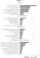
- the 33 key pathways identified with an input of the 883 most significantly regulated genes after a multigroup comparison (FDR 10 -9 ) between CLP categories in KPA analysis ( Fig. 5 ), mirror several of the functional groups of the 52 variables defined by Random Forest, such as gene regulation, cell cycle control and metabolism. Immune response-associated pathways such as "IL-4-induced regulators of cell growth, survival, differentiation and metabolism” and “Immune response_IL-3 signaling via JAK/STAT, p38, JNK and NF- ⁇ B" were among the 50% most significantly regulated ones.
- KEAP1 and NRF2 were found as a key hubs as well as their target genes NQO1 and HMOX1 [52, 58] and AHR [59, 60].
- the target genes NQO1, HMOX1 and CES1 [61] were even present on the input gene level.
- CES1 was present for SN chemicals as well, but only NQO1, NRF2 and AHR were identified as key hubs.
- KEAP1 was not found as key hub in SB and SN KPA analysis and AHR was the only key hub identified for all three protein reactivity groups.
- NF- ⁇ B subunits (RelB or p52) were predicted key hubs in all reactivity groups except in MA chemicals.
- CLP categories are empirically determined and arbitrarily defined categories, which do not represent the diversity of different chemicals, their molecular features and mechanisms responsible for their sensitizing characteristics or the lack thereof. They are, however, what legislation currently requires in order to classify and label chemicals. We therefore investigated 37 additional chemicals previously not tested in the standard GARD assay, in order to combine these new data with historical datasets.
- the U-SENSTM assay formerly MUSST, uses another myeloid cell line, U937, and CD86 measurements in order to distinguish sensitizers and non-sensitizers.
- U937 myeloid cell line
- CD86 measurements in order to distinguish sensitizers and non-sensitizers.
- KPA pathway analysis identified biologically relevant events in the presented dataset as several pathways regulated have a known role in skin sensitization, e.g. cytokine signalling and oxidative stress responses ( Figs. 5-6 ).
- DCs are not the primary target for protein modification in vivo, we hypothesized that different protein reactivity classes influence the DC transcriptome differentially.
- Protein reactivity is one of the most important features of chemicals defining their skin sensitizing capacity and potency with certain limitations [63]. Protein reactivity-specific patterns were detectable as revealed by the comparison of the most significantly regulated genes induced by the reactivity groups MA, SB, and SN.
- NF-KB subunits were predicted key hubs for all reactivity groups except for MA chemicals, which may reflect the described inhibitory effect on NF- ⁇ B signalling of this type of chemicals [71].
- MA chemicals which may reflect the described inhibitory effect on NF- ⁇ B signalling of this type of chemicals [71].
- some pathways do not seem to fit into the context, such as "Mitogenic action of Estradiol / ESR1 (nuclear) in breast cancer", a closer look at regulated molecules reveals that those are certainly relevant also for other pathways.
- CAD carbamoyl phosphate synthetase
- Measuring complete transcriptomes of cells provides the opportunity to perform mode-of-action based studies to identify pathways of sensitization, but also to identify predictive biomarker signatures, which we have previously shown also for binary skin sensitization predictions and respiratory sensitization.
- we here present an initial proof of concept of a potency model targeting the CLP groups which can be modified and improved as more samples are analyzed and more accurate human reference data emerges.
- O-PLS orthogonal partial least squares projections to latent structures
- Human potency [6] is a classification of chemicals based on available human data combining partly chemically very different substances into one potency category, whereby class 1 represents highest potency and class 6 represents true non-sensitizers.
- O-PLS model which is designed to predict two Ys, namely human potency and sensitizer/non-sensitizer.
- the scatter plot in Figure 10 illustrates the separation of sensitizers and non-sensitizers, and the grouping of sensitizers along a second axis into a high- and a low-potency cluster.
- Random Forest a decision tree-based method well suited for data sets with more variables than observations. It yields good predictive performance even when variables are noisy (no pre-selection required), and returns variable importance [5].
- ECHA European Chemical Agency
- CLP chemical substances and mixtures
- 1A strong sensitizer
- 1B weak sensitizers
- no category non-sensitizers
- RF model built on the arithmetic mean of transcript intensities from replicates in the training data using the random forest varSelRF package [6] in R/Bioconductor version 3.1.2 with error rates estimated by the supplied .632+ bootstrap method. It may also be built from transcript intensities of replicates directly.
- the model was trained using the training set described in Table 9 (70 substances), and then challenged with the test set in Table 10 (18 substances).
- a summary of the model performance is presented in Table 11.
- Figure 13 comprises principal component analyses and heat map results of the described random forest model.
- a list of identified potency biomarkers is presented in Table 12.
- S sensitizer
- NS non-sensitizer Name CAS# CLP cytotox GARD input [M] binary class (HP*) GARD binary prediction 2,4-Dinitrofluorobenzene 70-34-8 1A yes 0.00001 S S 3-Methylcatechol 488-17-5 1A yes 0.00004 S S bisphenol A-diglycidyl ether 1675-54-3 1A yes 0.00005 S S chlorpromazine 50-53-3 1A yes 0.0000125 S S cyanuric chloride 108-77-0 1A yes 0.00005 S NS glutaraldehyde 111-30-8 1A yes 0.00002 S S hexyl salicylate 6259-76-3 1A yes 0.00007 S S iodopropynyl butylcarbamate 55406-53-6 1A yes 0.00001 S S methyl heptine carbonate 111-12-6 1A yes 0.0001 S S
- VCF variable call frequency.
- Transcript cluster ID VCF (%) Gene Title Gene Symbol 8117594 93 histone cluster 1, H2bm HIST1H2BM 8124385 86 histone cluster 1, H4b HIST1H4B 8004804 83 phosphoribosylformylglycinamidine synthase PFAS 8124430 81 histone cluster 1, H1d HIST1H1D 8095221 80 phosphoribosylaminoimidazole carboxylase, phosphoribosylaminoimidazole succinocarboxamide synthetase PAICS 8124413 69 histone cluster 1, H4d HIST1H4D 8005839 63 transmembrane protein 97 TMEM97 7916432 61 24-dehydrocholesterol reductase DHCR24 8117608 56 histone cluster 1, H2al /// histone cluster 1, H2bn HIST1H2AL/
- SEL1L3 7948192 14 structure specific recognition protein 1 SSRP1 7960340 14 forkhead box M1 FOXM1 8107706 14 lamin B1 LMNB1 8124524 14 histone cluster 1, H2ak HIST1H2AK 8040712 11 centromere protein A CENPA 8043602 10 non-SMC condensin I complex subunit H NCAPH 7939341 8 CD44 molecule (Indian blood group) CD44 8124394 7 histone cluster 1, H2bb HIST1H2BB 8144931 7 ATPase, H+ transporting, lysosomal 56/58kDa, V1 subunit B2 ATP6V1B2 7999025 5 TNF receptor-associated protein 1 TRAP1 Table 6.
- Chemical list class 1 representing highest potency, 6 representing non-sensitizers number chemical SVM DV Human potency 1 Potassium dichromate 8,45 1 2 Dinitrochlorobenzene 6,31 1 3 PPD 5,32 1 4 Kathon CG 3,76 1 5 2-Aminophenol 5,98 2 6 Formaldehyde 2,32 2 7 Glyoxal 1,08 2 8 Iso-eugenol 1,91 2 9 2-Hydroxyethyl acrylate 8,87 3 10 Cinnamic alcohol 2,94 3 11 2-Mercaptobenzothiazole 2,52 3 12 Ethylenediamine 1,12 3 13 Penicillin G 1,06 3 14 Eugenol 1,02 3 15 Geraniol 1,76 4 16 Resorcinol 1,03 4 17 Hexylcinnamic aldehyde 1,24 5 18 Isopropanol -1,37 5 19 Methyl salicylate -1,53 5 20 PABA -1,04 5 21 Propylene glycol -1,18 5 22 Benzaldeh
Landscapes
- Health & Medical Sciences (AREA)
- Chemical & Material Sciences (AREA)
- Life Sciences & Earth Sciences (AREA)
- Organic Chemistry (AREA)
- General Health & Medical Sciences (AREA)
- Proteomics, Peptides & Aminoacids (AREA)
- Medicinal Chemistry (AREA)
- Immunology (AREA)
- Engineering & Computer Science (AREA)
- Veterinary Medicine (AREA)
- Animal Behavior & Ethology (AREA)
- Pharmacology & Pharmacy (AREA)
- Public Health (AREA)
- Wood Science & Technology (AREA)
- Zoology (AREA)
- Analytical Chemistry (AREA)
- Microbiology (AREA)
- Epidemiology (AREA)
- Bioinformatics & Cheminformatics (AREA)
- Genetics & Genomics (AREA)
- Biochemistry (AREA)
- Molecular Biology (AREA)
- Biophysics (AREA)
- General Engineering & Computer Science (AREA)
- Biotechnology (AREA)
- Physics & Mathematics (AREA)
- Mycology (AREA)
- Virology (AREA)
- Chemical Kinetics & Catalysis (AREA)
- General Chemical & Material Sciences (AREA)
- Nuclear Medicine, Radiotherapy & Molecular Imaging (AREA)
- Endocrinology (AREA)
- Measuring Or Testing Involving Enzymes Or Micro-Organisms (AREA)
- Investigating Or Analysing Biological Materials (AREA)
- Apparatus Associated With Microorganisms And Enzymes (AREA)
Description
- The present invention relates to a method for identifying the skin sensitizer potency of a test agent, and arrays and analytical kits for use in such methods.
- Allergic contact dermatitis (ACD) is an inflammatory skin disease that affects a significant proportion of the population. It is commonly caused by immunological responses towards chemical haptens leading to substantial economic burden for society. Current tests for sensitizing chemicals rely on animal experimentation. Legislation on the registration and use of chemicals within, e.g., the pharmaceutical and cosmetic industries, have stimulated significant research efforts to develop alternative human cell-based assays for the prediction of sensitization. The aim is to replace animal experiments with in vitro tests displaying a higher predictive power.
- ACD is characterized by eczema and recurrent episodes of itching [Akhavan, A. and S.R. Cohen, The relationship between atopic dermatitis and contact dermatitis. Clin Dermatol, 2003. 21(2): p. 158-62]. The disease affects a significant proportion of the population, with prevalence rates of 7.2% to 18.6% in Europe [Mortz, C.G., et al., Prevalence of atopic dermatitis, asthma, allergic rhinitis, and hand and contact dermatitis in adolescents. The Odense Adolescence Cohort Study on Atopic Diseases and Dermatitis. Br J Dermatol, 2001. 144(3): p. 523-32; and Nielsen, N.H., et al., Allergic contact sensitization in an adult Danish population: two cross-sectional surveys eight years apart (the Copenhagen Allergy Study). Acta Derm Venereol, 2001. 81(1): p. 31-4], and the incidence is increasing due to repeated exposure to sensitizing chemicals. ACD is a type IV delayed-type hypersensitivity response caused mainly by reactive T helper 1 (Th1) and interferon (IFN)γ producing CD8+ T cells at site of contact with small chemical haptens in previously exposed, and immunologically sensitized, individuals Fonacier, L.S., S.C. Dreskin, and D.Y. Leung, Allergic skin diseases. J Allergy Clin Immunol. 125(2 Suppl 2): p. S138-49]. Dendritic cells (DC) in the epidermis initiate the immune reactions by responding to haptens bound to self-molecules and activating T cell-mediated immunity.
- The REACH (Registration, Evaluation, and Authorisation of Chemicals) regulation requires that all new and existing chemicals within the European Union, involving approximately 30000 chemicals, should be tested for hazardous effects [EC 1907/2006, in Regulation (EC) No 1907/2006]. As the identification of potential sensitizers currently requires animal testing, the REACH legislation will have a huge impact on the number of animals needed for testing. Further, the 7th Amendment to the Cosmetics Directive (76/768/EEC) posed a ban on animal tests for the majority of cosmetic ingredients for human use in 2013. Thus, development of reliable in vitro alternatives to experimental animals for the assessment of sensitizing capacity of chemicals is urgent.
- We have developed a human cell-based assay for the prediction of sensitizing chemicals, called Genomic Allergen Rapid Detection, GARD (
Figure 8 ). For a detailed description, seeWO 2012/056236 and Johansson H et al. The GARD assay for assessment of chemical skin sensitizers. Toxicology in vitro 2013. By analyzing the transcriptome of dendritic-like cells (e.g., the MUTZ-3 cell line) we have identified a genomic biomarker signature with potent discriminatory ability [Johansson H et al. A genomic biomarker signature can predict skin sensitizers using a cell-based in vitro alternative to animal tests. BMC Genomics. 2011; Johansson H et al. The GARD assay for assessment of chemical skin sensitizers. Toxicology in vitro 2013; and Johansson H et al. GARD in-house validation - A proof of concept. Toxicological Sciences 2014]. Furthermore, the way signaling pathways are engaged by different chemical reactivity groups seems to be associated with sensitizing potency [Albrekt et al. Skin sensitizers differentially regulate signaling pathways in MUTZ-3 cells in relation to their individual potency. BMC Pharmacology and Toxicology 2014]. - However, to date, non-animal replacements are required for identification of the potency of skin sensitizing chemicals, instead the preferred assay is the mouse Local Lymph Node Assay (LLNA) [Basketter, D.A., et al., Local lymph node assay - validation, conduct and use in practice. Food Chem Toxicol, 2002. 40(5): p. 593-8], followed by the Guinea pig maximization test (GPMT) [Magnusson, B. and A.M. Kligman, The identification of contact allergens by animal assay. The guinea pig maximization test. J Invest Dermatol, 1969. 52(3): p. 268-76].
- An in vitro alternative to these animal models would preferably exhibit improved reliability, accuracy of potency prediction and, importantly, correlation to human reactivity.
-
WO 2012/068474 A2 describes a high-throughput assessment method for contact hypersensitivity; Albrekt et al. BMC Pharmacology & Toxicology 15(5) (2014): 1-12 describes skin sensitizers differentially regulating signalling pathways in MUTZ-3 cells in relation to their individual potency; Lambrechts et al. Toxicological Sciences 116(1) (2010): 122-129 describes assessment of chemical skin-sensitizing potency by an in vitro assay based on human dendritic cells; Schoeters et al. Molecular Immunology 44 (2007): 3222-3233 describes microarray analyses in dendritic cells revealing potential biomarkers for chemical-induced skin sensitization. - Based on previous studies, the GARD assay has proven valuable for skin sensitizer identification. A broader assessment of the human transcriptome using multivariate models has now surprisingly revealed that improved in vitro potency models are also possible.
- Accordingly, a first aspect of the invention provides provides an in vitro method for determining the skin sensitizing potency of an agent comprising or consisting of the steps of:
- (a) providing a population of dendritic cells or a population of dendritic-like cells;
- (b) exposing the cells provided in step (a) to a test agent; and
- (c) measuring in the cells of step (b) the expression of all of the biomarkers listed in Table A;
- Also disclosed herein one or more of the biomarkers measured in step (c) is selected from the group defined in Table A(i) and/or Table A(ii).
- Also disclosed herein step (c) comprises or consists of measuring the expression of one or more biomarker listed in Table A(i), for example, 2, 3, 4, 5, 6, 7, 8, 9, 10, 11, 12, 13, 14, 15, 16, 17, 18, 19, or 20 of the biomarkers listed in Table A(i). For example, step (c) may comprise or consist of measuring the expression of all of the biomarkers listed in Table A(i).
- The method may include or exclude measuring the expression of HIST1H2BM. The method may include or exclude measuring the expression of HIST1H4B. The method may include or exclude measuring the expression of HIST1H1D. The method may include or exclude measuring the expression of PAICS. The method may include or exclude measuring the expression of HIST1H4D. The method may include or exclude measuring the expression of HIST1H2AL/// HIST1H2BN. The method may include or exclude measuring the expression of PLK1. The method may include or exclude measuring the expression of PHGDH. The method may include or exclude measuring the expression of MCM2. The method may include or exclude measuring the expression of MCM7. The method may include or exclude measuring the expression of CD53. The method may include or exclude measuring the expression of KIFC1. The method may include or exclude measuring the expression of WEE1. The method may include or exclude measuring the expression of TMPO. The method may include or exclude measuring the expression of MCM4. The method may include or exclude measuring the expression of PSRC1. The method may include or exclude measuring the expression of RNF149. The method may include or exclude measuring the expression of MCM6. The method may include or exclude measuring the expression of MCM5. The method may include or exclude measuring the expression of FANCA.
- Also disclosed herein step (c) comprises or consists of measuring the expression of one or more biomarkers listed in Table A(ii), for example, 2, 3, 4, 5, 6, 7, 8, 9, 10, 11, 12, 13, 14, 15, 16, 17, 18, 19, 20, 21, 22, 23, or 24 of the biomarkers listed in Table A(ii). For example, step (c) may comprise or consist of measuring the expression of all of the biomarkers listed in Table A(ii).
- The method may include or exclude measuring the expression of CAD. The method may include or exclude measuring the expression of MRTO4. The method may include or exclude measuring the expression of TOE1. The method may include or exclude measuring the expression of CDC20. The method may include or exclude measuring the expression of PM20D2. The method may include or exclude measuring the expression of LOC344887. The method may include or exclude measuring the expression of UNG. The method may include or exclude measuring the expression of CDKN1A. The method may include or exclude measuring the expression of HIST1H2BF. The method may include or exclude measuring the expression of HIST1H1B. The method may include or exclude measuring the expression of Transcript ID 7896697. The method may include or exclude measuring the expression of JADE1. The method may include or exclude measuring the expression of KIAA0125. The method may include or exclude measuring the expression of CHRNA5. The method may include or exclude measuring the expression of CHAF1B /// MORC3. The method may include or exclude measuring the expression of SEL1L3. The method may include or exclude measuring the expression of SSRP1. The method may include or exclude measuring the expression of FOXM1. The method may include or exclude measuring the expression of LMNB1. The method may include or exclude measuring the expression of HIST1H2AK. The method may include or exclude measuring the expression of CENPA. The method may include or exclude measuring the expression of NCAPH. The method may include or exclude measuring the expression of HIST1H2BB. The method may include or exclude measuring the expression of ATP6V1B2. The method may include or exclude measuring the expression of TRAP1.
- Also disclosed herein step (c) may comprise or consist of measuring the expression of one or more biomarker listed in Table A(iii), for example, 1, 2, 3, 4, 5, 6, or 7 of the biomarkers listed in Table A(iii). For example, step (c) comprises or consists of measuring the expression of all of the biomarkers listed in Table A(iii).
- The method may include or exclude measuring the expression of PFAS. The method may include or exclude measuring the expression of TMEM97. The method may include or exclude measuring the expression of DHCR24. The method may include or exclude measuring the expression of FTH1. The method may include or exclude measuring the expression of HIST1H2AE. The method may include or exclude measuring the expression of NQO1. The method may include or exclude measuring the expression of CD44.
- Also disclosed herein step (c) comprises or consists of measuring the expression of three or more of the biomarkers listed in Table A, for example, 4, 5, 6, 7, 8, 9, 10, 11, 12, 13, 14, 15, 16, 17, 18, 19, 20, 21, 22, 23, 24, 25, 26, 27, 28, 29, 30, 31, 32, 33, 34, 35, 36, 37, 38, 39, 40, 41, 42, 43, 44, 45, 46, 47, 48, 49, 50, or 51 of the biomarkers listed in Table A. For example, step (c) may comprise or consist of measuring the expression of all of the biomarkers listed in Table A.
- The method may include or exclude measuring the expression of HIST1H2BM. The method may include or exclude measuring the expression of HIST1H4B. The method may include or exclude measuring the expression of HIST1H1D. The method may include or exclude measuring the expression of PAICS. The method may include or exclude measuring the expression of HIST1H4D. The method may include or exclude measuring the expression of HIST1H2AL/// HIST1H2BN. The method may include or exclude measuring the expression of PLK1. The method may include or exclude measuring the expression of PHGDH. The method may include or exclude measuring the expression of MCM2. The method may include or exclude measuring the expression of MCM7. The method may include or exclude measuring the expression of CD53. The method may include or exclude measuring the expression of KIFC1. The method may include or exclude measuring the expression of WEE1. The method may include or exclude measuring the expression of TMPO. The method may include or exclude measuring the expression of MCM4. The method may include or exclude measuring the expression of PSRC1. The method may include or exclude measuring the expression of RNF149. The method may include or exclude measuring the expression of MCM6. The method may include or exclude measuring the expression of MCM5. The method may include or exclude measuring the expression of FANCA.
- The method may include or exclude measuring the expression of CAD. The method may include or exclude measuring the expression of MRTO4. The method may include or exclude measuring the expression of TOE1. The method may include or exclude measuring the expression of CDC20. The method may include or exclude measuring the expression of PM20D2. The method may include or exclude measuring the expression of LOC344887. The method may include or exclude measuring the expression of UNG. The method may include or exclude measuring the expression of CDKN1A. The method may include or exclude measuring the expression of HIST1H2BF. The method may include or exclude measuring the expression of HIST1H1B. The method may include or exclude measuring the expression of Transcript ID 7896697. The method may include or exclude measuring the expression of JADE1. The method may include or exclude measuring the expression of KIAA0125. The method may include or exclude measuring the expression of CHRNA5. The method may include or exclude measuring the expression of CHAF1B /// MORC3. The method may include or exclude measuring the expression of SEL1L3. The method may include or exclude measuring the expression of SSRP1. The method may include or exclude measuring the expression of FOXM1. The method may include or exclude measuring the expression of LMNB1. The method may include or exclude measuring the expression of HIST1H2AK. The method may include or exclude measuring the expression of CENPA. The method may include or exclude measuring the expression of NCAPH. The method may include or exclude measuring the expression of HIST1H2BB. The method may include or exclude measuring the expression of ATP6V1B2. The method may include or exclude measuring the expression of TRAP1.
- The method may include or exclude measuring the expression of PFAS. The method may include or exclude measuring the expression of TMEM97. The method may include or exclude measuring the expression of DHCR24. The method may include or exclude measuring the expression of FTH1. The method may include or exclude measuring the expression of HIST1H2AE. The method may include or exclude measuring the expression of NQO1. The method may include or exclude measuring the expression of CD44.
- The method may include or exclude measuring the expression of one or more of MCM7, PFAS, HIST1H2AE.
- Also disclosed herein step (c) may comprise or consist of measuring the expression of one or more biomarker listed in Table B. For example, step (c) may comprise or consist of measuring the expression of 2, 3, 4, 5, 6, 7, 8, 9, 10, 11, 12, 13, 14, 15, 16, 17, 18 of the biomarkers listed in Table B. For example, step (c) may comprise or consist of measuring the expression of all of the biomarkers listed in Table B.
- Also disclosed herein step (c) comprises or consists of measuring the expression of at least 2 of the biomarkers listed in Table B, for example, ≥3, ≥4, ≥5, ≥6, ≥7, ≥8, ≥9, ≥10, ≥11, ≥12, ≥13, ≥14, ≥15, ≥16, ≥17, or 18 of the biomarkers listed in Table B.
- Also disclosed herein step (c) comprises or consist of measuring the expression of one or more biomarker listed in Table B(i), for example, ≥2, ≥3, ≥4, ≥5, ≥6, ≥7, ≥8, ≥9, ≥10, ≥11, ≥12, ≥13, ≥14, ≥15, or 16 of the biomarkers listed in Table B(i).
- Also disclosed herein step (c) comprises or consist of measuring the expression of one or more biomarker listed in Table B(ii), for example, 2 of the biomarkers listed in Table B(ii).
- The method may include or exclude measuring the expression of CCNA2. The method may include or exclude measuring the expression of Transcript ID 8151252. The method may include or exclude measuring the expression of PIGW. The method may include or exclude measuring the expression of SNRPD1. The method may include or exclude measuring the expression of ARHGAP19 /// ARHGAP19-SLIT1. The method may include or exclude measuring the expression of HIST1H2AB. The method may include or exclude measuring the expression of LRCH2. The method may include or exclude measuring the expression of PKP4 /// PKP4. The method may include or exclude measuring the expression of RAVER2 /// RAVER2. The method may include or exclude measuring the expression of DUT /// DUT. The method may include or exclude measuring the expression of AURKA. The method may include or exclude measuring the expression of Transcript ID 8055309. The method may include or exclude measuring the expression of NDC1. The method may include or exclude measuring the expression of KIF22 /// KIF22. The method may include or exclude measuring the expression of OK/SW-CL.58. The method may include or exclude measuring the expression of Transcript ID 7994343. The method may include or exclude measuring the expression of HIST1H2AB /// HIST1 H2AE. The method may include or exclude measuring the expression of HMGB3.
- By "expression" we mean the presence, level and/or amount of the biomarker.
- By "biomarker" we include any biological molecule, or component or fragment thereof, the measurement of which can provide information useful in determining sensitizer potency. Thus, in the context of Table A, the biomarker may be a nucleic acid molecule, such as a mRNA or cDNA. Alternatively, the biomarker may be a protein encoded by the nucleic acid molecule or carbohydrate moiety, or an antigenic component or fragment thereof.
- In an additional or alternative embodiment the test agent is already known to be, or suspected of being, capable of inducing sensitization of the skin.
- For example, the test agent may already be known to be capable of inducing sensitization of the skin by using a method already known in the art, for instance a method described
WO 2012/056236 and/or Johansson H et al. The GARD assay for assessment of chemical skin sensitizers. Toxicology in vitro 2013. - In an alternative or additional embodiment, the method is for identifying skin sensitizer potency and skin sensitizer/non-sensitizer status of the test agent (i.e., identifying whether the test agent is a sensitizer or not and identifying its potency as a skin sensitizer). In an alternative or additional embodiment, the method comprises identifying whether the test agent is a sensitizer using the method described in
WO 2012/056236 and/or Johansson H et al. - By "skin sensitizing potency" we include or mean the strength of the skin sensitizing ability of an agent. For example, the relative potency or strength of sensitizing ability of an agent might lead to the ordering of a group of test agents from most potent to least potent or vice versa, and/or it might lead to their categorization according to one or more known regulation or system.
- In an additional or alternative embodiment the skin sensitization potency determined by the method is categorised according to the European Classification, Labelling and Packaging (CLP) Regulation (EC) 1272/2008 (http://echa.europa.eu/clp-2015). This system is based on the United Nations' Globally Harmonised System (GHS) and from June 2015, the only legislation to apply to the classification and labelling of both substances and mixtures. It requires companies to classify, label and package their products appropriately before placing them on the market. It provides the categories: 1A (strong), 1B (weak), or no cat (no sensitizer).
- For example, the method may provide:
- (i) one or more agent of
potency category 1A; - (ii) one or more agent of
potency category 1B; and/or - (iii) one or more agent of potency category no category
- In an additional or alternative embodiment the skin sensitization potency determined by the method is categorised according to the system described in Basketter et al., 2014, Categorization of chemicals according to their relative human skin sensitizing potency,' Dermatitis, 25(1):11-21, i.e. categories 1 (strongest sensitizer), 2, 3, 4, 5, or 6 (true non-sensitizer) (e.g. Table 4,
Figure 4 ). - For example, the method may provide:
- (i) one or more agent of
potency category 1; - (ii) one or more agent of
potency category 2; - (iii) one or more agent of
potency category 3; - (iv) one or more agent of
potency category 4; - (v) one or more agent of
potency category 5; and/or - (vi) one or more agent of potency category 6 (e.g., see present Table 8 and/or Basketter et al., 2014 supra.).
- In an additional or alternative embodiment skin sensitization potency is categorised according to the local lymph node assay (LLNA) classification, Guinea pig maximisation test (GPMT) or no observed-effect level (NOEL).
- For a detailed description of LLNA see Basketter, D.A., et al., Local lymph node assay - validation, conduct and use in practice. Food Chem Toxicol, 2002. 40(5): p. 593-8. For a detailed description of the guinea pig maximization test see Magnusson, B. and A.M. Kligman, The identification of contact allergens by animal assay. The guinea pig maximization test. J Invest Dermatol, 1969. 52(3): p. 268-76. For a detailed description of the no observed-effects level (NOEL) test in relation to skin sensitizer potency see Basketter et al., 2005, 'Evaluation of the skin sensitizing potency of chemicals by using the existing methods and considerations of relevance for elicitation' Contact Dermatitis, 52(1):39-43; and Griem, P., et al., 2003, 'Proposal for a risk assessment methodology for skin sensitization based on sensitization potency data.' Regul. Toxicol. Pharmacol., 38:269-290. For a correlation between NOEL and potency levels, see also WHO Library Cataloguing-in-Publication Data. Skin sensitization in chemical risk assessment. (IPCS harmonization project document: no. 5), ISBN 978 92 4 156360 4 (in particular, Table 1 on pages 26-28). For a detailed description of CLP, see (http://echa.europa.eu/clp-2015).
- In an additional or alternative embodiment the expression of biomarkers measured in step (c) is measured in the cells provided in step (a) prior to and following exposure to the skin sensitizing agent of predetermined potency, and wherein the difference in expression between the biomarkers prior to and following exposure to the test agent is indicative of the potency of the skin sensitizing agent of step (b).
- In an additional or alternative embodiment the expression of the biomarkers measured in step (c) is measured in the cells provided in step (a) prior to and following exposure to the skin sensitizing agent of predetermined potency, and wherein the difference in expression between the biomarkers prior to and following step (c) is indicative of the potency of the skin sensitizing agent of step (b).
- By 'difference in expression' we include that the presence and or amount in a first sample (e.g., a test agent sample) differs from that of a second sample (e.g., a control agent sample). Preferably the presence and/or amount is no more than 40% of that of the comparison sample, for example, no more than 39%, 38%, 37%, 36%, 35%, 34%, 33%, 32%, 31%, 30%, 29%, 28%, 27%, 26%, 25%, 24%, 23%, 22%, 21%, 20%, 19%, 18%, 17%, 16%, 15%, 14%, 13%, 12%, 11%, 10%, 9%, 8%, 7%, 6%, 5%, 4%, 3%, 2%, 1% or 0%.
- In an additional or alternative embodiment the biomarkers are measured in the cells provided in step (a) prior to and following exposure to the test agent, and wherein the difference in expression between the biomarkers prior to and following exposure to the test agent is indicative of the skin sensitizing potency of the test agent of step (b). Hence, the cells provided in step (a) may provide both the negative control and the test result.
- In an additional or alternative embodiment the biomarkers are measured in the cells provided in step (a) prior to and following exposure to the test agent, and wherein the difference in expression between the biomarkers prior to and following step (c) is indicative of the skin sensitizing potency of the test agent of step (b). Hence, the cells provided in step (a) may provide both the negative control and the test result.
- In an additional or alternative embodiment the method comprises the further steps of:
- (d) providing a further population of dendritic cells or a population of dendritic-like cells;
- (e) exposing the cells provided in step (d) to:
- i. one or more negative control agent that does not sensitize human skin; and/or
- ii. one or more positive control agent that does sensitize human skin; and/or
- iii. one or more vehicle control;
- (f) incubating for the same time and, other than exposure to an agent to the one or more control agent, the same conditions as the cells provided in step (a);
- (g) measuring in the cells of step (e) the expression of the biomarkers measured in step (c);
- In an additional or alternative embodiment the method comprises the further steps of:
- (d) providing a further population of dendritic cells or a population of dendritic-like cells;
- (e) exposing the cells provided in step (d) to:
- i. a negative control agent that does not sensitize human skin;
- ii. a vehicle control; or
- iii. incubation for the same time and, other than exposure to an agent of predetermined skin sensitization potency, the same conditions as the cells provided in step (a);
- (f) measuring in the cells of step (e) the expression of the biomarkers measured in step (c);
- In an additional or alternative embodiment the vehicle control comprises DMSO.
- By 'difference in expression' we include that the presence and or amount in a first sample (e.g., a test agent sample) differs from that of a second sample (e.g., a control agent sample). Preferably the presence and/or amount is no more than 40% of that of the comparison sample, for example, no more than 39%, 38%, 37%, 36%, 35%, 34%, 33%, 32%, 31%, 30%, 29%, 28%, 27%, 26%, 25%, 24%, 23%, 22%, 21%, 20%, 19%, 18%, 17%, 16%, 15%, 14%, 13%, 12%, 11%, 10%, 9%, 8%, 7%, 6%, 5%, 4%, 3%, 2%, 1% or 0%.
- In an additional or alternative embodiment the method comprises the further steps of:
- (h) providing a further population of dendritic cells or a population of dendritic-like cells;
- (i) exposing the cells provided in step (h) to a skin sensitizing agent of predetermined potency;
- (j) measuring in the cells of step (i) the expression of the biomarkers measured in step (c);
- By 'correspondence in expression' we include that that the presence and or amount in a first sample (e.g., a test agent sample) is similar or identical to the presence and/or amount in a second sample (e.g., a control sample). Preferably the presence and/or amount is at least 60% of that of the control sample, for example, at least 65%, 66%, 67%, 68%, 69%, 70%, 71%, 72%, 73%, 74%, 75%, 76%, 77%, 78%, 79%, 80%, 81%, 82%, 83%, 84%, 85%, 86%, 87%, 88%, 89%, 90%, 91%, 92%, 93%, 94%, 95%, 96%, 97%, 98%, 99% or 100%.
- In an additional or alternative embodiment the method comprises the further step of:
(k) identifying the skin sensitizing potency of the test agent. - The identification may be performed using any suitable statistical method or machine learning algorithm known in the art, such as Random Forest (RF), Support Vector Machine (SVM), Principal Component Analysis (PCA), ordinary least squares (OLS), partial least squares regression (PLS), orthogonal partial least squares regression (O-PLS) and other multivariate statistical analyses (e.g., backward stepwise logistic regression model). For a review of multivariate statistical analysis see, for example, Schervish, Mark J. (November 1987). "A Review of Multivariate Analysis". Statistical Science 2 (4): 396-413. Preferably a Random Forest model is used (such as is described in detail in Example A).
- In an additional or alternative embodiment of the methods of the invention, sensitizing potency is determined with an ROC AUC of at least 0.45, for example with an ROC AUC of at least, 0.50, 0.55, 0.60, 0.65, 0.70, 0.75, 0.80, 0.85, 0.86, 0.87, 0.88, 0.89, 0.90, 0.91, 0.92, 0.93, 0.94, 0.95, 0.96, 0.97, 0.98, 0.99 or with an ROC AUC of 1.00. Preferably, skin sensitizing potency is determined with an ROC AUC of at least 0.80, more preferably at least 0.85, still more preferably at least 0.88.
- Preferably, the methods of the invention have an accuracy of at least 60%, for example, 61%, 62%, 63%, 64%, 65%, 66%, 67%, 68%, 69%, 70%, 71%, 72%, 73%, 74%, 75%, 76%, 77%, 78%, 79%, 80%, 81%, 82%, 83%, 84%, 85%, 86%, 87%, 88%, 89%, 90%, 91%, 92%, 93%, 94%, 95%, 96%, 97%, 98%, 99% or 100% accuracy.
- Preferably, the methods of the invention have a sensitivity of at least 60%, for example, 61%, 62%, 63%, 64%, 65%, 66%, 67%, 68%, 69%, 70%, 71%, 72%, 73%, 74%, 75%, 76%, 77%, 78%, 79%, 80%, 81%, 82%, 83%, 84%, 85%, 86%, 87%, 88%, 89%, 90%, 91%, 92%, 93%, 94%, 95%, 96%, 97%, 98%, 99% or 100% sensitivity.
- Preferably, the methods of the invention have a specificity of at least 60%, for example, 61%, 62%, 63%, 64%, 65%, 66%, 67%, 68%, 69%, 70%, 71%, 72%, 73%, 74%, 75%, 76%, 77%, 78%, 79%, 80%, 81%, 82%, 83%, 84%, 85%, 86%, 87%, 88%, 89%, 90%, 91%, 92%, 93%, 94%, 95%, 96%, 97%, 98%, 99% or 100% specificity.
- By "accuracy" we mean the proportion of correct outcomes of a method, by "sensitivity" we mean the proportion of all positive chemicals that are correctly classified as positives, and by "specificity" we mean the proportion of all negative chemicals that are correctly classified as negatives.
- In an additional or alternative embodiment the method comprises the further step of:
- (I) one or more repetition of steps (h) to (j) (and where present, step (k)).
- In an additional or alternative embodiment, each repetition of steps (a) to (c) (and where present, steps (d)-(f/g)) is performed concurrently (i.e., with original steps (a) to (c) (and where present, steps (d)-(f/g))). In an additional or alternative embodiment, each repetition of steps (a) to (c) (and where present, steps (d)-(f/g)) is performed consecutively.
- In an additional or alternative embodiment step (j) comprises or consists of one or more repetition wherein:
- (I) the same agent of predetermined skin sensitization potency as provided in step (i) for one or more other repetition; or
- (II) a different agent of predetermined skin sensitization potency as provided in step (i) for one or more other repetition.
- In an additional or alternative embodiment the different agent of predetermined skin sensitization potency provided in step (i) is selected from the group consisting of:
- (I) an agent of the same sensitization potency as the different agent of predetermined skin sensitization potency provided in step (i); or
- (II) an agent of a different sensitization potency from the different agent of predetermined skin sensitization potency provided in step (i).
- In an additional or alternative embodiment steps (a) to (c) (and where present, step (f/g)) are repeated at least once, for example, ≥2, ≥3, ≥4, ≥5, ≥6, ≥7, ≥8, ≥9, ≥10, ≥11, ≥12, ≥13, ≥14, ≥15, ≥16, ≥17, ≥18, ≥19, ≥20, ≥21, ≥22, ≥23, ≥24, ≥25, ≥26, ≥27, ≥28, ≥29, ≥30, ≥40, ≥50, ≥60, ≥70, ≥80, ≥90, ≥100, ≥150, ≥200, ≥250, ≥300, ≥350, ≥400, ≥450, ≥500, ≥600, ≥700, ≥800, ≥900 or ≥1000 repetitions.
- In an additional or alternative embodiment each step or repetition of steps may be repeated identically to control for experimental error, for example, 2, 3, 4, 5, 6, 7, 8, 9 or 10 repeats.
- In an additional or alternative embodiment the expression of the biomarkers measured are measured for agents of predetermined skin sensitization potency over the full range of skin sensitization potencies.
- By 'full range of skin sensitization potencies' we include assessing sensitizers of the highest and lowest degree of skin sensitizer category in the categorisation system being used. Preferably, sensitizers are assessed at each sensitizer category in the categorisation system being used.
- In an additional or alternative embodiment the skin sensitizing agent of predetermined potency comprises or consist of one or more agents selected from the group consisting of the group defined in Table 2. For example, the method may comprise measuring ≥2, ≥3, ≥4, ≥5, ≥6, ≥7, ≥8, ≥9, ≥10, or 11, of the agents defined in Table 2.
- In an additional or alternative embodiment the skin sensitizing agent of predetermined potency comprises or consist of agents selected from the group consisting of the group defined in Table 8, Table 9 and/or Table 8, e.g., Table 8 only, Table 9 only, Table 10 only, Tables 8 and 9, Tables 8 and 10, Tables 9 and 10 or Tables 8, 9 and 10. For Example, the method may comprise measuring ≥2, ≥3, ≥4, ≥5, ≥6, ≥7, ≥8, ≥9, ≥10, ≥11, ≥12, ≥13, ≥14, ≥15, ≥16, ≥17, ≥18, ≥19, ≥20, ≥21, ≥22, ≥23, ≥24, ≥25, ≥26, ≥27, ≥28, ≥29, ≥30, ≥31, ≥32, ≥33, ≥34, ≥35, ≥36, ≥37, ≥38, ≥39, ≥40, ≥41, ≥42, ≥43, ≥44, ≥45, ≥46, ≥47, ≥48, ≥49, ≥50, ≥51, ≥52, ≥53, ≥54, ≥55, ≥56, ≥57, ≥58, ≥59, ≥60, ≥61, ≥62, ≥63, ≥64, ≥65, ≥66, ≥67, ≥68, ≥69, ≥70, ≥71, ≥72, ≥73, ≥74, ≥75, ≥76, ≥77, ≥78, ≥79, ≥80, ≥81, ≥82, ≥83, ≥84, ≥85, ≥86, ≥87, ≥88, ≥89, ≥90, ≥91, ≥92, ≥93, ≥94, ≥95, ≥96, ≥97, ≥98, ≥99, ≥100, ≥101, ≥102, ≥103, ≥104, ≥105, ≥106, ≥107, ≥108, ≥109, ≥110, ≥111, ≥112, ≥113, ≥114, ≥115, ≥116, ≥117, ≥118, ≥119, ≥120, ≥121 or 122 of the control agents defined in Tables 8, 9 and 10.
- In an additional or alternative embodiment the skin sensitizing agent of predetermined potency comprises or consist of agents selected from the group consisting of 1-Butanol, 4-Aminobenzoic acid, Benzaldehyde, Chlorobenzene, Diethyl phthalate, Dimethyl formamide, Ethyl vanillin, Glycerol, Isopropanol, Lactic acid, Methyl salicylate, Octanoic acid, Propylene glycol, Phenol, p-hydroxybenzoic acid, Potassium permanganate, Salicylic acid, Sodium dodecyl sulphate, Tween 80, Zinc sulphate, 2,4-Dinitrochlorobenzene, Oxazolone, Potassium dichromate, Kathon CG (MC/MCI), Formaldehyde, 2-Aminophenol, 2-nitro-1,4-Phenylendiamine, p-Phenylendiamine, Hexylcinnamic aldehyde, 2-Hydroxyethyl acrylate, 2-Mercaptobenzothiazole, Glyoxal, Cinnamaldehyde, Isoeugenol, Ethylendiamine, Resorcinol, Cinnamic alcohol, Eugenol, Penicillin G, Geraniol and DMSO.
- In a preferred embodiment, step (c) comprises or consists of measuring the expression of a nucleic acid molecule of one or more of the biomarkers. The nucleic acid molecule may be a cDNA molecule or an mRNA molecule. Preferably, the nucleic acid molecule is an mRNA molecule. However, the nucleic acid molecule may be a cDNA molecule.
- In one embodiment the measurement of the expression of one or more of the biomarkers in step (c) is performed using a method selected from the group consisting of Southern hybridisation, Northern hybridisation, polymerase chain reaction (PCR), reverse transcriptase PCR (RT-PCR), quantitative real-time PCR (qRT-PCR), nanoarray, microarray, macroarray, autoradiography and in situ hybridisation. Preferably, the expression of one or more biomarker(s) is measured using a DNA microarray.
- In an additional or alternative embodiment the one or more biomarkers measured in step (c) is measured using an array (e.g., a DNA array). In an additional or alternative embodiment the one or more biomarkers measured in step (c) is measured using a whole genome array (e.g., the Affymetrix Human Gene 1.0 ST array or Affymetrix Human Gene 2.0 ST array). In an alternative or additional embodiment, the Nanostring nCounter system is used (e.g., custom Nanostring nCounter code sets based on selection from a whole genome array (e.g., Affymetrix Human Gene 1.0 ST array or Affymetrix Human Gene 2.0 ST array).
- The method may comprise measuring the expression of one or more biomarkers in step (c) using one or more binding moieties, each capable of binding selectively to a nucleic acid molecule encoding one of the biomarkers identified in Table A.
- In one embodiment the one or more binding moieties each comprise or consist of a nucleic acid molecule. In a further embodiment the one or more binding moieties each comprise or consist of DNA, RNA, PNA, LNA, GNA, TNA or PMO. Preferably, the one or more binding moieties each comprise or consist of DNA. In one embodiment, the one or more binding moieties are 5 to 100 nucleotides in length. However, in an alternative embodiment, they are 15 to 35 nucleotides in length.
- The one or more binding moieties may comprise or consist of one or more probe from the Human Gene 1.0 ST Array (Affymetrix, Santa Clara, CA, USA). Probe identification numbers are provided in Table A herein, where they are referred to as "transcript cluster IDs".
- Suitable binding agents (also referred to as binding molecules or binding moieties) may be selected or screened from a library based on their ability to bind a given nucleic acid, protein or amino acid motif, as discussed below.
- In a preferred embodiment, the binding moiety comprises a detectable moiety.
- By a "detectable moiety" we include a moiety which permits its presence and/or relative amount and/or location (for example, the location on an array) to be determined, either directly or indirectly.
- Suitable detectable moieties are well known in the art.
- For example, the detectable moiety may be a fluorescent and/or luminescent and/or chemiluminescent moiety which, when exposed to specific conditions, may be detected. Such a fluorescent moiety may need to be exposed to radiation (i.e. light) at a specific wavelength and intensity to cause excitation of the fluorescent moiety, thereby enabling it to emit detectable fluorescence at a specific wavelength that may be detected.
- Alternatively, the detectable moiety may be an enzyme which is capable of converting a (preferably undetectable) substrate into a detectable product that can be visualised and/or detected. Examples of suitable enzymes are discussed in more detail below in relation to, for example, ELISA assays.
- Hence, the detectable moiety may be selected from the group consisting of: a fluorescent moiety; a luminescent moiety; a chemiluminescent moiety; a radioactive moiety (for example, a radioactive atom); or an enzymatic moiety. Preferably, the detectable moiety comprises or consists of a radioactive atom. The radioactive atom may be selected from the group consisting of technetium-99m, iodine-123, iodine-125, iodine-131, indium-111, fluorine-19, carbon-13, nitrogen-15, oxygen-17, phosphorus-32, sulphur-35, deuterium, tritium, rhenium-186, rhenium-188 and yttrium-90.
- Clearly, the agent to be detected (such as, for example, the one or more biomarkers in the test sample and/or control sample described herein and/or an antibody molecule for use in detecting a selected protein) must have sufficient of the appropriate atomic isotopes in order for the detectable moiety to be readily detectable.
- In an alternative preferred embodiment, the detectable moiety of the binding moiety is a fluorescent moiety.
- The radio- or other labels may be incorporated into the biomarkers present in the samples of the methods of the invention and/or the binding moieties of the invention in known ways. For example, if the binding agent is a polypeptide it may be biosynthesised or may be synthesised by chemical amino acid synthesis using suitable amino acid precursors involving, for example, fluorine-19 in place of hydrogen. Labels such as 99mTc, 123I, 186Rh, 188Rh and 111In can, for example, be attached via cysteine residues in the binding moiety. Yttrium-90 can be attached via a lysine residue. The IODOGEN method (Fraker et al (1978) Biochem. Biophys. Res. Comm. 80, 49-57) can be used to incorporate 123I. Reference ("Monoclonal Antibodies in Immunoscintigraphy", J-F Chatal, CRC Press, 1989) describes other methods in detail. Methods for conjugating other detectable moieties (such as enzymatic, fluorescent, luminescent, chemiluminescent or radioactive moieties) to proteins are well known in the art.
- It will be appreciated by persons skilled in the art that biomarkers in the sample(s) to be tested may be labelled with a moiety which indirectly assists with determining the presence, amount and/or location of said proteins. Thus, the moiety may constitute one component of a multicomponent detectable moiety. For example, the biomarkers in the sample(s) to be tested may be labelled with biotin, which allows their subsequent detection using streptavidin fused or otherwise joined to a detectable label.
- The method provided in the first aspect of the present invention may comprise or consist of, in step (c), determining the expression of the protein of one or more biomarker defined in Table A. The method may comprise measuring the expression of one or more biomarkers in step (c) using one or more binding moieties each capable of binding selectively to one of the biomarkers identified in Table A. The one or more binding moieties may comprise or consist of an antibody or an antigen-binding fragment thereof such as a monoclonal antibody or fragment thereof.
- The term "antibody" includes any synthetic antibodies, recombinant antibodies or antibody hybrids, such as but not limited to, a single-chain antibody molecule produced by phage-display of immunoglobulin light and/or heavy chain variable and/or constant regions, or other immunointeractive molecules capable of binding to an antigen in an immunoassay format that is known to those skilled in the art.
- We also include the use of antibody-like binding agents, such as affibodies and aptamers.
- A general review of the techniques involved in the synthesis of antibody fragments which retain their specific binding sites is to be found in Winter & Milstein (1991) Nature 349, 293-299.
- Additionally, or alternatively, one or more of the first binding molecules may be an aptamer (see Collett et al., 2005, Methods 37:4-15).
- Molecular libraries such as antibody libraries (Clackson et al, 1991, Nature 352, 624-628; Marks et al, 1991, J Mol Biol 222(3): 581-97), peptide libraries (Smith, 1985, Science 228(4705): 1315-7), expressed cDNA libraries (Santi et al (2000) J Mol Biol 296(2): 497-508), libraries on other scaffolds than the antibody framework such as affibodies (Gunneriusson et al, 1999, Appl Environ Microbiol 65(9): 4134-40) or libraries based on aptamers (Kenan et al, 1999, Methods Mol Biol 118, 217-31) may be used as a source from which binding molecules that are specific for a given motif are selected for use in the methods of the invention.
- The molecular libraries may be expressed in vivo in prokaryotic cells (Clackson et al, 1991, op. cit.; Marks et al, 1991, op. cit.) or eukaryotic cells (Kieke et al, 1999, Proc Natl Acad Sci USA, 96(10):5651-6) or may be expressed in vitro without involvement of cells (Hanes & Pluckthun, 1997, Proc Natl Acad Sci USA 94(10):4937-42; He & Taussig, 1997, Nucleic Acids Res 25(24):5132-4; Nemoto et al, 1997, FEBS Lett, 414(2):405-8).
- In cases when protein based libraries are used, the genes encoding the libraries of potential binding molecules are often packaged in viruses and the potential binding molecule displayed at the surface of the virus (Clackson et al, 1991, supra; Marks et al, 1991, supra; Smith, 1985, supra).
- Perhaps the most commonly used display system is filamentous bacteriophage displaying antibody fragments at their surfaces, the antibody fragments being expressed as a fusion to the minor coat protein of the bacteriophage (Clackson et al, 1991, supra; Marks et al, 1991, supra). However, other suitable systems for display include using other viruses (
EP 39578 - In addition, display systems have been developed utilising linkage of the polypeptide product to its encoding mRNA in so-called ribosome display systems (Hanes & Pluckthun, 1997, supra; He & Taussig, 1997, supra; Nemoto et al, 1997, supra), or alternatively linkage of the polypeptide product to the encoding DNA (see
US Patent No. 5,856,090 andWO 98/37186 - The variable heavy (VH) and variable light (VL) domains of the antibody are involved in antigen recognition, a fact first recognised by early protease digestion experiments. Further confirmation was found by "humanisation" of rodent antibodies. Variable domains of rodent origin may be fused to constant domains of human origin such that the resultant antibody retains the antigenic specificity of the rodent parented antibody (Morrison et al (1984) Proc. Natl. Acad. Sci. USA 81, 6851-6855).
- That antigenic specificity is conferred by variable domains and is independent of the constant domains is known from experiments involving the bacterial expression of antibody fragments, all containing one or more variable domains. These molecules include Fab-like molecules (Better et al (1988) Science 240, 1041); Fv molecules (Skerra et al (1988) Science 240, 1038); single-chain Fv (ScFv) molecules where the VH and VL partner domains are linked via a flexible oligopeptide (Bird et a/ (1988) Science 242, 423; Huston et a/ (1988) Proc. Natl. Acad. Sci. USA 85, 5879) and single domain antibodies (dAbs) comprising isolated V domains (Ward et al (1989) Nature 341, 544). A general review of the techniques involved in the synthesis of antibody fragments which retain their specific binding sites is to be found in Winter & Milstein (1991) Nature 349, 293-299.
- The antibody or antigen-binding fragment may be selected from the group consisting of intact antibodies, Fv fragments (e.g. single chain Fv and disulphide-bonded Fv), Fab-like fragments (e.g. Fab fragments, Fab' fragments and F(ab)2 fragments), single variable domains (e.g. VH and VL domains) and domain antibodies (dAbs, including single and dual formats [i.e. dAb-linker-dAb]). Preferably, the antibody or antigen-binding fragment is a single chain Fv (scFv).
- The one or more binding moieties may alternatively comprise or consist of an antibody-like binding agent, for example an affibody or aptamer.
- By "scFv molecules" we mean molecules wherein the VH and VL partner domains are linked via a flexible oligopeptide.
- The advantages of using antibody fragments, rather than whole antibodies, are several-fold. The smaller size of the fragments may lead to improved pharmacological properties, such as better penetration of solid tissue. Effector functions of whole antibodies, such as complement binding, are removed. Fab, Fv, ScFv and dAb antibody fragments can all be expressed in and secreted from E. coli, thus allowing the facile production of large amounts of the said fragments.
- Whole antibodies, and F(ab')2 fragments are "bivalent". By "bivalent" we mean that the said antibodies and F(ab')2 fragments have two antigen combining sites. In contrast, Fab, Fv, ScFv and dAb fragments are monovalent, having only one antigen combining sites.
- The antibodies may be monoclonal or polyclonal. Suitable monoclonal antibodies may be prepared by known techniques, for example those disclosed in "Monoclonal Antibodies: A manual of techniques", H Zola (CRC Press, 1988) and in "Monoclonal Hybridoma Antibodies: Techniques and applications", J G R Hurrell (CRC Press, 1982).
- When potential binding molecules are selected from libraries, one or more selector peptides having defined motifs are usually employed. Amino acid residues that provide structure, decreasing flexibility in the peptide or charged, polar or hydrophobic side chains allowing interaction with the binding molecule may be used in the design of motifs for selector peptides. For example:
- (i) Proline may stabilise a peptide structure as its side chain is bound both to the alpha carbon as well as the nitrogen;
- (ii) Phenylalanine, tyrosine and tryptophan have aromatic side chains and are highly hydrophobic, whereas leucine and isoleucine have aliphatic side chains and are also hydrophobic;
- (iii) Lysine, arginine and histidine have basic side chains and will be positively charged at neutral pH, whereas aspartate and glutamate have acidic side chains and will be negatively charged at neutral pH;
- (iv) Asparagine and glutamine are neutral at neutral pH but contain a amide group which may participate in hydrogen bonds;
- (v) Serine, threonine and tyrosine side chains contain hydroxyl groups, which may participate in hydrogen bonds.
- Typically, selection of binding molecules may involve the use of array technologies and systems to analyse binding to spots corresponding to types of binding molecules.
- The one or more protein-binding moieties may comprise a detectable moiety. The detectable moiety may be selected from the group consisting of a fluorescent moiety, a luminescent moiety, a chemiluminescent moiety, a radioactive moiety and an enzymatic moiety.
- In a further embodiment of the methods of the invention, step (c) may be performed using an assay comprising a second binding agent capable of binding to the one or more proteins, the second binding agent also comprising a detectable moiety. Suitable second binding agents are described in detail above in relation to the first binding agents.
- Thus, the proteins of interest in the sample to be tested may first be isolated and/or immobilised using the first binding agent, after which the presence and/or relative amount of said biomarkers may be determined using a second binding agent.
- In one embodiment, the second binding agent is an antibody or antigen-binding fragment thereof; typically a recombinant antibody or fragment thereof. Conveniently, the antibody or fragment thereof is selected from the group consisting of: scFv; Fab; a binding domain of an immunoglobulin molecule. Suitable antibodies and fragments, and methods for making the same, are described in detail above.
- Alternatively, the second binding agent may be an antibody-like binding agent, such as an affibody or aptamer.
- Alternatively, where the detectable moiety on the protein in the sample to be tested comprises or consists of a member of a specific binding pair (e.g. biotin), the second binding agent may comprise or consist of the complimentary member of the specific binding pair (e.g. streptavidin).
- Where a detection assay is used, it is preferred that the detectable moiety is selected from the group consisting of: a fluorescent moiety; a luminescent moiety; a chemiluminescent moiety; a radioactive moiety; an enzymatic moiety. Examples of suitable detectable moieties for use in the methods of the invention are described above.
- Preferred assays for detecting serum or plasma proteins include enzyme linked immunosorbent assays (ELISA), radioimmunoassay (RIA), immunoradiometric assays (IRMA) and immunoenzymatic assays (IEMA), including sandwich assays using monoclonal and/or polyclonal antibodies. Exemplary sandwich assays are described by
David et al in US Patent Nos. 4,376,110 and4,486,530 . Antibody staining of cells on slides may be used in methods well known in cytology laboratory diagnostic tests, as well known to those skilled in the art. - Thus, in one embodiment the assay is an ELISA (Enzyme Linked Immunosorbent Assay) which typically involves the use of enzymes which give a coloured reaction product, usually in solid phase assays. Enzymes such as horseradish peroxidase and phosphatase have been widely employed. A way of amplifying the phosphatase reaction is to use NADP as a substrate to generate NAD which now acts as a coenzyme for a second enzyme system. Pyrophosphatase from Escherichia coli provides a good conjugate because the enzyme is not present in tissues, is stable and gives a good reaction colour. Chemiluminescent systems based on enzymes such as luciferase can also be used.
- Conjugation with the vitamin biotin is frequently used since this can readily be detected by its reaction with enzyme-linked avidin or streptavidin to which it binds with great specificity and affinity.
- In an alternative embodiment, the assay used for protein detection is conveniently a fluorometric assay. Thus, the detectable moiety of the second binding agent may be a fluorescent moiety, such as an Alexa fluorophore (for example Alexa-647).
- Preferably, steps (c), (g), (f) and/or (j) of the methods described in the first aspect are performed using an array. The array may be a bead-based array or a surface-based array. The array may be selected from the group consisting of: macroarray; microarray; nanoarray.
- Arrays per se are well known in the art. Typically they are formed of a linear or two-dimensional structure having spaced apart (i.e. discrete) regions ("spots"), each having a finite area, formed on the surface of a solid support. An array can also be a bead structure where each bead can be identified by a molecular code or colour code or identified in a continuous flow. Analysis can also be performed sequentially where the sample is passed over a series of spots each adsorbing the class of molecules from the solution. The solid support is typically glass or a polymer, the most commonly used polymers being cellulose, polyacrylamide, nylon, polystyrene, polyvinyl chloride or polypropylene. The solid supports may be in the form of tubes, beads, discs, silicon chips, microplates, polyvinylidene difluoride (PVDF) membrane, nitrocellulose membrane, nylon membrane, other porous membrane, non-porous membrane (e.g. plastic, polymer, perspex, silicon, amongst others), a plurality of polymeric pins, or a plurality of microtitre wells, or any other surface suitable for immobilising proteins, polynucleotides and other suitable molecules and/or conducting an immunoassay. The binding processes are well known in the art and generally consist of cross-linking covalently binding or physically adsorbing a protein molecule, polynucleotide or the like to the solid support. Alternatively, affinity coupling of the probes via affinity-tags or similar constructs may be employed. By using well-known techniques, such as contact or non-contact printing, masking or photolithography, the location of each spot can be defined. For reviews see Jenkins, R.E., Pennington, S.R. (2001, Proteomics, 2,13-29) and Lal et al (2002, ).
- Typically the array is a microarray. By "microarray" we include the meaning of an array of regions having a density of discrete regions of at least about 100/cm2, and preferably at least about 1000/cm2. The regions in a microarray have typical dimensions, e.g. diameter, in the range of between about 10-250 µm, and are separated from other regions in the array by about the same distance. The array may alternatively be a macroarray or a nanoarray.
- Once suitable binding molecules (discussed above) have been identified and isolated, the skilled person can manufacture an array using methods well known in the art of molecular biology.
- In an additional or alternative embodiment one or more biomarkers measured in step (c) comprise or consist of one or more homologous gene product expressed by human cells. In an additional or alternative embodiment one or more biomarkers measured in step (c) is a protein or polypeptide. In an additional or alternative embodiment one or more biomarker measured in step (c) is a nucleic acid (e.g., DNA, mRNA or cDNA etc).
- Also disclosed herein the method is performed in vitro, in vivo, ex vivo or in silico. For example, the method may in particular be performed in vitro.
- In an additional or alternative embodiment the skin sensitization is a hypersensitivity response (e.g., a cell-mediated hypersensitivity response). In an additional or alternative embodiment the hypersensitivity response is a type IV hypersensitivity response. In an additional or alternative embodiment the hypersensitivity response is allergic contact dermatitis (ACD).
- By 'test agent' we include any chemical entity (or mixture of chemical entities) for which sensitization potency and/or sensitization status is to be determined.
- By "sensitization status" we include or mean whether or not a chemical entity (or mixture of chemical entities) is a sensitizer or not (e.g., a skin sensitizer and/or a respiratory sensitizer).
- By "skin sensitizing agent" we mean any agent capable of inducing and triggering a Type IV delayed-type hypersensitivity reaction in a mammal. Preferably, the Type IV delayed-type hypersensitivity reaction is DC-mediated.
- In one embodiment, the one or more "skin sensitizing agent" is an agent capable of inducing and triggering a Type IV delayed-type hypersensitivity reaction at a site of epidermal contact in a mammal.
- The mammal may be any domestic or farm animal. Preferably, the mammal is a rat, mouse, guinea pig, cat, dog, horse or a primate. Most preferably, the mammal is human. As discussed above, in vivo methods of determining sensitisation are known in the art. A preferred method is the Local lymph node assay (for details, see Basketter, D.A., et al., Local lymph node assay - validation, conduct and use in practice. Food Chem Toxicol, 2002. 40(5): p. 593-8). A further suitable, but less preferred, method is the guinea pig maximization test (for details, see Magnusson, B. and A.M. Kligman, The identification of contact allergens by animal assay. The guinea pig maximization test. J Invest Dermatol, 1969. 52(3): p. 268-76).
- In an additional or alternative embodiment the population of dendritic cells or population of dendritic-like cells is a population of dendritic-like cells. In an additional or alternative embodiment the dendritic-like cells are myeloid dendritic-like cells. In an additional or alternative embodiment the myeloid dendritic-like cells are derived from myeloid dendritic cells. In an additional or alternative embodiment the cells derived from myeloid dendritic cells are myeloid leukaemia-derived cells. In an additional or alternative embodiment the myeloid leukaemia-derived cells are selected from the group consisting of KG-1, THP-1, U-937, HL-60, Monomac-6, AML-193 and MUTZ-3. In an additional or alternative embodiment the dendritic-like cells are MUTZ-3 cells. MUTZ-3 cells are human acute myelomonocytic leukemia cells that are available from Deutsche Sammlung für Mikroorganismen und Zellkulturen GmbH (DSMZ), Braunschweig, Germany (www.dsmz.de; DMSZ No. ACC 295).
- By "dendritic-like cells" we mean non-dendritic cells that exhibit functional and phenotypic characteristics specific to dendritic cells such as morphological characteristics, expression of costimulatory molecules and MHC class II molecules, and the ability to pinocytose macromolecules and to activate resting T cells.
- In one embodiment, the dendritic-like cells, after stimulation with cytokine, present antigens through CD1d, MHC class I and II and/or induce specific T-cell proliferation.
- In one embodiment, the dendritic-like cells are CD34+ dendritic cell progenitors. Optionally, the CD34+ dendritic cell progenitors can acquire, upon cytokine stimulation, the phenotypes of presenting antigens through CD1d, MHC class I and II, induce specific T-cell proliferation, and/or displaying a mature transcriptional and phenotypic profile upon stimulation with inflammatory mediators (i.e. similar phenotypes to immature dendritic cells or Langerhans-like dendritic cells).
- In one embodiment, the population of dendritic cells or population of dendritic-like cells is a population of dendritic cells. Preferably, the dendritic cells are primary dendritic cells. Preferably, the dendritic cells are myeloid dendritic cells.
- Dendritic cells may be recognized by function, by phenotype and/or by gene expression pattern, particularly by cell surface phenotype. These cells are characterized by their distinctive morphology, high levels of surface MHC-class II expression and ability to present antigen to CD4+ and/or CD8+ T cells, particularly to naive T cells (Steinman et al. (1991) Ann. Rev. Immunol. 9: 271).
- The cell surface of dendritic cells is unusual, with characteristic veil-like projections, and is characterized by expression of the cell surface markers CD11c and MHC class II. Most DCs are negative for markers of other leukocyte lineages, including T cells, B cells, monocytes/macrophages, and granulocytes. Subpopulations of dendritic cells may also express additional markers including 33D1, CCR1, CCR2, CCR4, CCR5, CCR6, CCR7, CD1a-d, CD4, CD5, CD8alpha, CD9, CD11b, CD24, CD40, CD48, CD54, CD58, CD80, CD83, CD86, CD91, CD117, CD123 (IL3Ra), CD134, CD137, CD150, CD153, CD162, CXCR1, CXCR2, CXCR4, DCIR, DC-LAMP, DC-SIGN, DEC205, E-cadherin, Langerin, Mannose receptor, MARCO, TLR2, TLR3 TLR4, TLR5, TLR6, TLR9, and several lectins.
- The patterns of expression of these cell surface markers may vary along with the maturity of the dendritic cells, their tissue of origin, and/or their species of origin. Immature dendritic cells express low levels of MHC class II, but are capable of endocytosing antigenic proteins and processing them for presentation in a complex with MHC class II molecules. Activated dendritic cells express high levels of
MHC class 11, ICAM-1 and CD86, and are capable of stimulating the proliferation of naive allogeneic T cells, e. g. in a mixed leukocyte reaction (MLR). - Functionally, dendritic cells or dendritic-like cells may be identified by any convenient assay for determination of antigen presentation. Such assays may include testing the ability to stimulate antigen- primed and/or naive T cells by presentation of a test antigen, followed by determination of T cell proliferation, release of IL-2, and the like.
- Methods of detecting and/or measuring the concentration of protein and/or nucleic acid are well known to those skilled in the art, see for example Sambrook and Russell, 2001, Cold Spring Harbor Laboratory Press.
- Preferred methods for detection and/or measurement of protein include Western blot, North-Western blot, immunosorbent assays (ELISA), antibody microarray, tissue microarray (TMA), immunoprecipitation, in situ hybridisation and other immunohistochemistry techniques, radioimmunoassay (RIA), immunoradiometric assays (IRMA) and immunoenzymatic assays (IEMA), including sandwich assays using monoclonal and/or polyclonal antibodies. Exemplary sandwich assays are described by
David et al., in US Patent Nos. 4,376,110 and4,486,530 . Antibody staining of cells on slides may be used in methods well known in cytology laboratory diagnostic tests, as well known to those skilled in the art. - Typically, ELISA involves the use of enzymes which give a coloured reaction product, usually in solid phase assays. Enzymes such as horseradish peroxidase and phosphatase have been widely employed. A way of amplifying the phosphatase reaction is to use NADP as a substrate to generate NAD which now acts as a coenzyme for a second enzyme system. Pyrophosphatase from Escherichia coli provides a good conjugate because the enzyme is not present in tissues, is stable and gives a good reaction colour. Chemi-luminescent systems based on enzymes such as luciferase can also be used.
- Conjugation with the vitamin biotin is frequently used since this can readily be detected by its reaction with enzyme-linked avidin or streptavidin to which it binds with great specificity and affinity.
- A second aspect of the invention provides an array for use in the method according to the first aspect of the invention, the binding moieties of the array consisting of binding moieties for each of the biomarkers defined in Table A, and wherein the binding moieties of the array comprise binding moieties as defined in the first aspect of the invention.
- Also disclosed herein the array comprises one or more binding moiety for each of the biomarkers as defined in the first aspect of the invention.
- In an additional or alternative embodiment the one or more binding moiety is immobilised.
- In an additional or alternative embodiment the array is a bead-based array. In an additional or alternative embodiment the array is a surface-based array.
- In an additional or alternative embodiment the array is selected from the group consisting of: macroarray; microarray; nanoarray.
- The array of the second aspect of the invention may comprise one or more, preferably two or more, binding moieties, wherein the binding moieties are each capable of binding selectively to a biomarker as defined in the first aspect. Therefore, the array may comprise or consist of a particular selection of biomarker-specific binding moieties which correlates to any particular selection of biomarkers as defined in the first aspect.
- A third aspect of the invention provides the use of the biomarkers listed in Table A for determining the skin sensitizing potency of an agent.
- Also disclosed herein is the use of a biomarker selected from the group defined in Table A for determining the skin sensitizing potency of an agent, preferably wherein the biomarker is selected from the group defined in Table A(i) and/or Table A(ii).
- In an additional or alternative embodiment there is provided the use of binding moieties with specificity for each of the biomarkers listed in Table A for determining the skin sensitizing potency of an agent.
- A fourth aspect of the invention provides an analytical kit for use in a method according the first aspect of the invention comprising:
- (a) an array according to the second aspect of the invention; and
- (b) instructions for performing the method as defined in the first aspect of the invention (optional).
- In an additional or alternative embodiment the analytical kit further comprising one or more control agents as defined in the first aspect of the invention.
- Also disclosed herein is a method of treating or preventing a type I hypersensitivity reaction (such as ACD) in a patient comprising the steps of:
- (a) providing one or more test agent that the patient is or has been exposed to;
- (b) determining whether the one or more test agent provided in step (a) is a skin sensitizer and/or its potency as a sensitizer using a method of the present invention; and
- (c) where one or more test agent is identified as a skin sensitizer and/or a skin sensitizer of a particular potency, reducing or preventing exposure of the patient to the one or more test agent and/or providing appropriate treatment for the symptoms of sensitization.
- Preferably, the one or more test agent that the patient is or has been exposed to is an agent that the patient is presently exposed to at least once a month, for example, at least once every two weeks, at least once every week, or at least once every day.
- Treatments of the symptoms of sensitization may include, for example, topical steroids.
- Preferably, the method of treatment is consistent with the method described in the first aspect of the invention, and one or more of the embodiments described therein.
- Also disclosed herein is a computer program for operating the methods the invention, for example, for interpreting the expression data of step (c) (and subsequent expression measurement steps) and thereby determining whether one or more test agent is a respiratory sensitizer and/or the sensitizer potency of the one or more test agent. The computer program may be a programmed SVM. The computer program may be recorded on a suitable computer-readable carrier known to persons skilled in the art. Suitable computer-readable-carriers may include compact discs (including CD-ROMs, DVDs, Blue Rays and the like), floppy discs, flash memory drives, ROM or hard disc drives. The computer program may be installed on a computer suitable for executing the computer program.
- The skilled person will appreciate that all non-conflicting embodiments may be used in combination. Hence, embodiments from one aspect of the invention may equally be applied to a second aspect of the invention.
- Preferred, non-limiting examples which embody certain aspects of the invention will now be described, with reference to the following figures:
-
Figure 1 . Binary predictions using the GARD assay. ROC evaluation for (A) binary predictions of benchmark chemicals (filled line) and of 37 new chemicals (dotted line). (B) GARD SVM decision values (DVs) correlate with CLP potency (37 chemicals, 11 1A, 19 1B, 7 no cat). Increasing potency is associated with increasing DVs. -
Figure 2 . Visualization of the training dataset used to develop the random forest model, using the 52 variables. (A) PCA plot of the training set with separate biological replicates coloured according to CLP classifications of the chemicals. (B) Heatmap of the training set with replicates of the samples hierarchically clustered, where the grey scale represents the relative gene expression intensity. -
Figure 3 . Visualization of the test dataset using the 52 variables. (A) PCA plot of the test set with separate biological replicates coloured according to CLP classifications. The PCA was built on the training set and the test set plotted without influencing the PCA. (B) Heatmap of the test set with samples hierarchically clustered, where the grey scale represents relative gene expression intensity. -
Figure 4 . The CLP potency model contains information related to human potency. (A) PCA plot of replicate training and test set samples with available human potency classifications colored according to human potency. The PCA is based on the 52 Random Forest variables as input and the PCA was built on the training set. (B) PCA plot visualizing test set only. -
Figure 5 . Pathway analysis based on an input of the 883 most significant genes from a multigroup comparison of the three CLP classes. -
Figure 6 . Pathways unique for each protein reactivity groups, as identified by pathway analysis. -
Figure 7 . Venn diagrams [82] of common genes (A) and biological pathways (B) for the three different protein reactivity classes. MA=Michael acceptors; SB=Schiff base formation; SN=bi-molecular nucleophilic substitution/nucleophilic aromatic substitution. -
Figure 8 : Genomic Allergen Rapid Detection (GARD) - workflow. The SVM model calculates decision values for each RNA sample derived from chemically treated cells based on the gene expression data of 200 genes that represent the GARD prediction signature. The decision values are then used for binary classification of the chemicals [3] -
Figure 9 : Relationship between GARD decision values and human potency classes. The chemicals have been sorted according to human potency class and the respective average SVM decision value derived from triplicates can be found on the Y axis. The number on the x-axis refers to the number of the chemical in Table 8. -
Figure 10 : Scatter plot of O-PLS model (Y=human potency and sensitizer/non-sensitizer) based on 197 samples. Human potency classes as described in Basketter et al. (2014),class 1 representing highest potency, 6 representing non-sensitizers. -
Figure 11 : Permutation plot of O-PLS model. Comparison of goodness of the fit and prediction of the original model with several models, where the order of the Y-observations has been randomly permutated. The plot strongly indicates that the model has not been obtained by chance. -
Figure 12 : Observed Y values plotted against predicted Y values. The RMSEE (Root Mean Square Error of Estimation) indicates the fit error of the observations to the model. RMSEcv is a similar measure, but estimated using cross validation. -
Figure 13 : PCA plots and heat maps. PCA of training set (A), training and test set (B) and test set only (C) based on an input of 18 variables identified by random forest modeling. Each sphere represents a chemical, colored according to CLP classifications (yellow = 1A, pink = 1B, blue = no category). (D) Heat map of training set and (E) heat map of test set based on the expression of the 18 identified biomarkers. - Skin sensitization and associated diseases such as contact allergy affect a substantial portion of the general population with an estimated prevalence of 15-20% in industrialized countries [1]. Allergic contact dermatitis (ACD), a type IV hypersensitivity reaction, is common among certain occupational groups such as those regularly exposed to chemicals or involved in wet work [2]. However, also cosmetics and household products can contain numerous skin sensitizing chemicals. Through different legal frameworks, the European Union has prohibited animal testing for cosmetics and their ingredients [3], and imposed requirements for testing of >60,000 chemicals in the context of REACH [4]. Information on both the skin sensitizing capacity and the potency of a chemical has to be provided to meet the regulatory requirements for classification and sub-categorization. Animal-free alternative assays that meet these requirements are urgently needed.
- The molecular events leading to skin sensitization and consequently to ACD can be characterized by a number of sequential key events (KE) triggered by a chemical, and have been summarized in an adverse outcome pathway (AOP) as described by the OECD [5]. The initiating event (KE1) is defined as covalent protein modification by the skin sensitizing chemical after it has gained access to deeper skin layers. The following KE2 represents the inflammatory responses upon activation of keratinocytes. KE3 corresponds to the activation of dendritic cells, which in turn leads to activation and proliferation of T cells (KE4). Upon re-exposure to the sensitizer, the development of ACD may be triggered, which as the adverse outcome is characterized by skin lesions induced by specific Th1 and CD8+ T cells. While the KE in the AOP are well described, a detailed mechanistic understanding of the underlying biology of the individual key events is still missing [5].
- The murine Local Lymph Node Assay (LLNA) [6] has for many years been the preferred alternative for skin sensitization testing as it is able to provide data for both hazard identification and characterization, including skin sensitizer potency information. However, it is characterized by certain limitations such as susceptibility to vehicle effects and issues with false-positive results [7]. Several non-animal predictive methods have been developed to reduce animal experimentation used for chemical testing including computational approaches to integrate data from different test platforms for hazard identification as recently reviewed [8]. Three test methods for skin sensitization are accepted as test guidelines at the OECD; the ARE-NRF2 luciferase method (KeratinoSens™) assay [9, 10], the Direct Peptide Reactivity Assay (DPRA) [11] and the human Cell Line Activation Test (h-CIAT) [12]. In addition to hazard identification, information on skin sensitizer potency is imperative in order to allow quantitative risk assessment and to define exposure limits. Few approaches for the prediction of skin sensitizer potency have been published and were recently reviewed in [8], such as assays targeting KE2 (epidermal equivalent sensitizer potency assay [13], SENS-IS [14]) and the U-SENS assay modelling KE3 [15]. Furthermore, in silico models, often combining information from several in vitro methods, have been described; for example QSAR [16], artificial neural network [17], probabilistic models and integrated testing strategy (ITS) approaches including a Bayesian model [18-21].
- The alternative assay Genomic Allergen Rapid Detection (GARD) for the assessment of skin sensitization capacity of chemicals is based on global transcriptomic analysis of differential expression in a human myeloid cell line, induced by sensitizing chemicals in comparison to non-sensitizing controls. The resulting biomarker signature, the GARD prediction signature (GPS), consists of 200 transcripts, which are used as input into a support vector machine (SVM) model trained on a set of reference chemicals [22]. The changes in transcription can be linked to the maturation and activation of dendritic cells (KE3) during sensitization. In an in-house study based on 26 blinded chemicals, the accuracy of the assay was estimated to 89% [23]. Previous observations indicated that the GARD assay is able to provide information relevant also for potency assessment. Firstly, signalling pathways were differentially regulated depending on the potency of a subset of chemical reactivity groups [24]. Secondly, we observed that more potent sensitizers were generally assigned higher GARD SVM decision values compared to weaker sensitizers, indicating that there were genes within signature contributing with potency information (unpublished observations). However, the information in the GARD prediction signature were not sufficient to completely stratify chemicals into the well-defined potency groups as described by the Classification, Labelling and Packaging (CLP) Regulation [25].
- The CLP regulation is based on the Globally Harmonised System [25], and uses three categories for chemical classification; no category (no cat) for non-sensitizers,
category 1B for weak and 1A for strong sensitizers. In the light of the above described observations, it was hypothesized that GARD can be developed further into a tool for the prediction of chemical skin sensitizer potency, targeting the CLP categories. As the established GARD Support Vector Machine model cannot be applied to multiclass problems, we used another approach based on random forest modelling [26]. Random forest is a decision-tree based method and well-suited for microarray data [27]. It divides the dataset internally and repeatedly into a training and test set through random sub-sampling (bootstrapping). Samples in the test set, referred to as out-of-bag samples, comprise approximately one third of the entire dataset, and are used in order to estimate the out-of-bag (OOP) error, i.e. the classification error. - Here, we present a new approach to predict skin sensitizer potency according to CLP categories based on supervised machine learning using a random forest model. Firstly, the global gene expression data from a training set comprising 68 unique chemicals and 2 vehicle control samples were used as input into a random forest model. The random forest model was subsequently combined with an algorithm for backward variable elimination. The algorithm initially ranked the variable importance of each gene from the microarrays, and then iteratively fitted new random forests, while removing the least important variables from the previous iteration. Using this strategy, we were able to identify a set of 52 genes with the smallest OOB error rate when predicting the out-of-bag samples from the training set. The predictive performance of the 52 genes were challenged with an independent test set containing 18 chemicals previously unseen to the model. The chemicals in this test set could be predicted with an overall accuracy of 78%. In addition to the predictive model, we also demonstrated the versatility of analyzing whole transcriptomes of cells by performing pathway analysis to further improve the mechanistic understanding of skin sensitizing potency on a cellular level, confirming the hypothesis that different chemical reactivity classes induce distinct signalling pathways.
- The myeloid cell line used in this study was derived from MUTZ-3 (DSMZ, Braunschweig, Germany) and maintained as described in [22, 28]. A phenotypic control analysis of the cells prior to each experiment was carried out by flow cytometry in order to confirm the cells' immature state. The following monoclonal antibodies were used: CD1a (DakoCytomation, Glostrup, Denmark), CD34, CD86, HLA-DR (BD Biosciences, San Jose, USA), all FITC-conjugated; CD14 (DakoCytomation), CD54, CD80 (BD Biosciences), all PE-conjugated. FITC- and PE-conjugated mouse IgG1 (BD Biosciences) served as isotype controls and propidium iodide as a marker for non-viable cells (BD Biosciences). Three batches of cells were exposed for 24 hours in independent experiments and viability and CD86 expression were assessed by flow cytometry. All FACS samples were analyzed on a FACSCanto II instrument with FACS Diva software for data acquisition. 10 000 events were acquired and further analysis was performed in FCS Express V4 (De Novo Software, Los Angeles, CA). Cells for RNA extraction were lysed in TRIzol® (Life Technologies/Thermo Fisher Scientific, Waltham, USA) and stored until further use in -20°C.
- All chemicals were purchased from Sigma Aldrich (Saint Louis, USA) in high purity quality or they were provided by Cosmetics Europe. All chemicals were stored according to the recommendations of the supplier. The chemical stimulations of cells were performed as described earlier [28]. In short, GARD input concentrations were defined by solubility and cytotoxicity characteristics of the chemicals. An end concentration of 500 µM was targeted for non-cytotoxic and soluble chemicals and the highest possible concentration for chemicals with limited solubility (lower than 500 µM in medium). Cytotoxic chemicals were used in a concentration targeting a relative viability of cells of 90%. Most chemicals were used from a 1000x pre-dilution in dimethyl sulfoxide (DMSO) or autoclaved MilliQ water. DMSO concentration as vehicle never exceeded 0.1%. DMSO and MilliQ samples were included as vehicle controls in this study and thus belong to the group of non-sensitizer samples.
- RNA isolation from cells lysed in TRIzol® was performed according to the manufacturer's instructions. Labeled sense DNA was synthesized according to Affymetrix (Affymetrix, Cleveland, USA) protocols using the recommended kits and controls. The cDNA was hybridized to Human Gene 1.0 ST arrays (Affymetrix) and further processed and scanned as recommended by the supplier.
- Binary classifications of the 37 chemicals summarized in Table 1 into sensitizers or non-sensitizers were performed with the previously established model based on SVM, using SCAN-normalized [29, 30] expression data from the GPS as variable input into the learning algorithm [22]. Prior to model construction, potential batch effects between training set and test chemicals were eliminated by scaling array expression values for test chemicals against the training set. A scaling factor was generated by calculating the ratio of the average expression value for each gene in DMSO vehicle control samples of the training set and the average expression value for same gene in DMSO samples in the batch where the test chemical originated. The scaling factor for each gene was then multiplied with the gene expression values for the corresponding gene in the test chemical. SVM predictions were performed as described previously [22, 31]. In short, an SVM model based on a linear kernel was trained on reference chemical stimulations from the original training set used during identification of the GPS [22]. The trained model was subsequently applied to assign each test chemical with an SVM Decision Value (SVM DV). Resulting SVM DVs for all test chemicals were used to construct a receiver operating characteristics (ROC) curve, and the resulting area under the curve (AUC) was used as a classification measure [32]. SVM modeling and ROC curve visualizations were performed in R statistical environment, using the additional packages e1071 [33] and ROCR [34]. Prior to evaluating final predictive performance of the model, SVM DVs for each individual replicate of the test chemicals were calibrated against the cut-off for maximal predictive performance obtained during classification of benchmark samples in Table 2, as described in [31]. The calibrated SVM DVs were subsequently used for final classifications, and test chemicals were classified as sensitizers if the median output value of replicates > 0. Accuracy, sensitivity and specificity was estimated using cooper statistics [35]. The non-parametric Two Sample Wilcoxon test was performed in order to determine if the SVM DV distributions between
CLP categories - In order to build a random forest model, 68 chemicals and two vehicle control samples were defined as a training set, and 18 chemicals, six from each CLP category, were included in the independent test set. Chemicals in the test set were not included in the construction of the model. The aim was to obtain a balanced training set representing all three CLP categories (Table 3) and different chemical reactivity groups (Table 4). Most of the chemicals in the test set (14 out of 18) originated from the latest experimental campaign (Table 1), comprising 37 chemicals previously not investigated using the GARD assay. In the training set, roughly one third of samples (23 out of 68) were from this latest dataset. The vehicle samples were part of all projects and are thus present with higher replicate numbers.
- The new microarray data were merged with historical data [22, 23] and subjected to quality control. Four arrays were removed due to bad quality; however, no chemical was present in less than biological duplicates. Array data was imported into the R statistical environment and normalized using the SCANfast algorithm [29, 30]. As several experimental campaigns needed to be combined, this dataset was normalized using the ComBat method [36, 37] in order to remove batch effects between samples. At this time, the samples in the training set were separated from the samples in the test set. To avoid overfitting, only samples in the designated training set were used during identification of the predictive biomarker signature and for fitting of parameters to the classifier, and samples in the test set were set aside to validate the performance of the identified signature and the specified classifier. The predictive biomarker signature was identified by feeding normalized and batch corrected transcript intensities from individual samples in the training set into a random forest model [26] combined with a backward elimination procedure in the varSelRF package [38] in R/Bioconductor version 3.1.2. The initial forest used for ranking of variable importance was grown to 2000 trees and all other parameters were kept at the default settings. The package iteratively fits and evaluates random forest models, at each iteration dropping 20% of the least important variables. The best performing set of variables was selected based on OOB error rates from all fitted Random Forests as the smallest number of genes within one standard error from the minimal error solution (i.e 1.s.e rule). The variable selection procedure was validated by estimating the prediction error rate by the .632+ bootstrap method of the varSelRF package using 100 bootstrap samples, and the importance of individual transcripts in the biomarker signature was validated by the frequency of appearance in bootstrap samples (referred to as Validation Call Frequencies (VCF)). The predictive performance of the identified biomarker signature was validated by building a new forest in the Random Forest package [39], based on previous parameters, using only the samples in the training set and the selected transcripts in the biomarker signature as variable input. The model was applied to assign each individual replicate sample in the test set to a CLP category, and the majority vote across the biological replicate stimulations for each chemical was accepted as the predicted category. Heatmaps and Principal Component Analysis (PCA) plots were constructed in Qlucore Omics Explorer (Qlucore AB, Lund, Sweden).
- Pathway analysis was performed with the Key Pathway Advisor (KPA) tool [40] version 16.6, which provides a pathway analysis workflow to investigate e.g. gene expression data. It associates differentially expressed genes with both upstream and downstream processes in order to allow biological interpretation. The investigated dataset, consisting of the SCANfast- and ComBat-normalized expression values of the test set and the training set, in total 308 samples, was first variance-filtered in order to remove variables with consistently low variance until approximately a third was left (10009 variables, σ/σmax=0.1478). A multigroup comparison (ANOVA) comparing samples belonging to CLP no cat, 1B, and 1A was then applied in order to identify transcripts that were differentially regulated. The most significant 883 genes (false discovery rate FDR=10-9; p=8.53x10⁻¹¹) were used as input into KPA (Affymetrix Exon IDs and respective p-value, overconnectivity analysis). In order to identify pathways associated with protein reactivity, the same variance-filtered dataset was filtered based on two-group comparisons (t-tests, Toxtree binding class "no binding" non-sensitizers (81 samples) versus "Michael acceptor" sensitizers (MA, 63 samples), "no binding" versus "Schiff base formation" (SB, 29 samples), "no binding" versus combined "bi-molecular nucleophilic substitution/nucleophilic aromatic substitution" (SN, 25 samples). Lists with the 500 most significantly regulated variables from each comparison, together with p-value and fold change (causal reasoning analysis), were then entered into the KPA tool for each protein reactivity group. The lowest p-value was reached when comparing MA samples to "no binding" (FDR=3.65x10⁻⁷), followed by SB (FDR=2.3x10⁻⁵) and (FDR=2.44x10⁻⁵).
- Listed below are the scripts used for normalization and potency predictions:
Script 1: Used for combating batch effects between different projects prior to model construction:
# This script is used for combating batch effects between different projects
 prior to model construction!
 library(sva)
 library(caret)
 data_set <- read.delim("/home/andy/allfiles-scannorm.txt",stringsAsFactors =
 F,header = F)
 # Create numerical data set with colnames/rownames and appropriate design
 matrices and factors
 annotation_headers <- data_set[1:17,1] #Header comprises row 1-17
 annotations <- data_set[1:17,2:ncol(data_set)]
 colnames(annotations) <- annotations[1,]
 rownames(annotations) <- annotation_headers
 rownames_data_set <- data_set[,1]
 data_set <- data_set[,-1]
 # Convert the data to numerical values
 data_set <- data.frame(sapply(data_set[18:nrow(data_set),],as.numeric))
colnames(data_set) <- annotations[1,] # The colnames corresponds to the IDs rownames(data_set) <- rownames_data_set[18:length(rownames_data_set)] projects <- factor(as.character(annotations[3,])) # CLP USENS is used in the combat model? groups <- as.character(annotations[7,]) #Make the NA unknown groups[is.na(groups)] <- "unknown" groups <- factor(groups) group_model <- model.matrix(-groups) # Remove near-zero-variance probes (could potentially interfere with ComBat) nzv <- nearZeroVar(t(data_set)) data_set <- data_set[-nzv,] #The data consists of 5 different projects with pronounced batch effects # ComBat is used to remove the batch effects normalized_data <- ComBat(data_set,projects,mod = group_model) normalized_data <- rbind(annotations,normalized_data) write.table(normalized_data,"/home/andy/processed_data_sets/ComBat/allfiles _SCAN_ComBat.txt",col.names=F,sep= "\t",quote = F)
#Construction of the initial model potencymodel require(pROC) require(ROCR) require(varSelRF) require(ggplot2) require(reshape) require(randomForest) require(caret) library(sva) library(parallel) library(doMC) library(foreach) ##Load dataset datal<-read.table(file="allfiles-scannorm- cleaned_ComBat.txt",stringsAsFactors=FALSE,row.names=1,header=F,sep="\t") ##Define trainingset. |
testset1annot[which(testset1annot=="no cat")]<-0 sampleannot<-as.vector(testset1[2,]) testset1<-as.matrix(testset1[-(1:17),]) class(testset1)<-"numeric" testset1<-as.data.frame(t(testset1)) testsetclass<-as.factor(testsetlannot) ##load testdataset 2 testset2<-as.matrix(data1[,which(data1[10,]=="test set 2")]) sampleannottest2<-as.vector(testset2[2,]) testset2annot<-as.vector(testset2[7,]) testset2annot[which(testset2annot=="no cat")]<-0 testset2annot<-as.factor(testset2annot) testset2<-as.matrix(testset2[-(1:17),]) class(testset2) <-"numeric" testset2<-as.data.frame(t(testset2)) ##Construct the model set.seed(2) vsrf<- varSelRF(training,Class=trainingsetclass,ntree=2000,ntreelterat=2000,vars.d rop.frac=0.2,keep.forest = T,whole.range=TRUE,verbose = TRUE) RFmodel<-randomForest(y=trainingsetclass, x=subset(training, select = vsrf$selected.vars),ntree=vsrf$ntreeIterat,importance=TRUE) rfpr.pred<-predict(RFmodel,newdata=subset(testset1, select = vsrf$selected.vars)) confusionMatrix(rfpr.pred,testsetclass) a<-data.frame (stimulant=sampleannot, group=testsetclass,predicted=rfpr.pred) write.table(a, file="VarSelRF_combat_Test1.txt", sep= ",", row.names = TRUE) rfpr.pred2<-predict(RFmodel,newdata=subset(testset2, select = vsrf$selected.vars)) b<- data.frame(stimulant=sampleannottest2,group=testset2annot,predicted=rfpr.pr ed2) write.table(b, file="VarSelRF_combat_Test2.txt", sep= ",", row.names = TRUE) #Extract variable importance write.table(vsrf$selected.vars, file="VarSelRF_ComBat.txt", sep= ",", row.names = TRUE) ##Bootstrapping of variable selection procedure forkCL <- makeForkCluster(12) clusterSetRNGStream(forkCL, iseed = (100)) clusterEvalQ(forkCL, library(varSelRF)) vsrf.vsb <- varSelRFBoot(training,trainingsetclass,usingCluster=T,srf=vsrf,TheCluster=f orkCL,ntree=2000,ntreeIterat=2000,vars.drop.frac=0.2,bootnumber = 100) stopCluster(forkCL) #######################Shuffle train and test####################### #Shuffle train and testset 18 times to make sure primary selection is not biased.#Combine train and test dataset notest2<-as.matrix(data1[,-which(datal[10,]=="test set 2")]) notest2annot<-as.vector(notest2[7,]) notest2annot[which(notest2annot=="no cat")]<-0 notest2classes<-as.factor(notest2annot) |
 ## notest2compounds<-levels(factor(unlist(as.vector(notest2[2,]))))
 ## notest2<-as.matrix(notest2[-(1:17),])
 ## class(notest2)<-"numeric"
 #Generate a new sampling matrix without DMSO and Water! These are controls
 and should not be used for sampling to test set.
 notestDMSO <-as.matrix(notest2[,-which(notest2[2,]=="dimethyl sulfoxide")])
 notestDMSO_Unstim <-as.matrix(notestDMSO[,-
 which(notestDMSO[2,]=="unstimulated")])
 notest2DU <-as.vector(notestDMSO_Unstim[7,])
 notest2DU[which(notest2DU=="no cat")]<-0
 notest2DUclasses<-as.factor(notest2DU)
 #################
 ##construct the shuffling loops
 ##Shuffle train and test 20 times! The results are stored in testcomp, which
 are accessed through typing testcomp[[i]]
 testcomp <- c()
 looptest <- NULL
 looptrain <- NULL
 looptests <- list()
 trainingsetannot<-list()
 trainingsetclasses<-list()
 testsetannot<-list()
 testsetclasses<-list()
 testsetnames<-list()
 vsrf<-list ()
 rfvsrf<-list()
 rfpr.pred<-list()
 cf<-list ()
 results<-list()
 testcomploc<-list()
 rfpr.predtest2<-list()
 resultstest2<-list()
 vsrf.vsb <-list()
 #cl = makeCluster(4)
 #registerDoSNOW(cl)
 registerDoMC(18)
 startTime = Sys.time()
 writeLines(paste('Starting shuffle: ',startTime, sep = ''))
 loop_results = foreach(i = 1:18, .packages =
 c('varSelRF', 'caret', 'randomForest')) %dopar% {
   set.seed((10+i))
   testcomp <-
 (sample(sort(unique(levels(factor(notestDMSO_Unstim[2,which(notest2DUclasse
 s=="1A")]))),method="radix"),6))
   testcomp <-
 c(testcomp,sample(sort(unique(levels(factor(notestDMSO_Unstim[2,which(notes
 t2DUclasses=="1B")]))),method="radix"),6))
   testcomp <-
 c(testcomp,sample(sort(unique(levels(factor(notestDMSO_Unstim[2,which(notes
t2DUclasses=="0")]))),method="radix"),6)) |
  testcomploc <- c()
  for(n in 1:length(testcomp))
  {
     testcomploc <- c(testcomploc ,which(notest2[2,]==testcomp[n]))
  }
  ## Get Ids from original matrix containing water and DMSO samples.
  # testcomploc <- unlist(testcomploc)
  looptest <-notest2[,testcomploc]
  looptrain <-notest2[,-testcomploc]
  trainingsetannot <- as.vector(looptrain[7,])
  trainingsetannot[which(trainingsetannot=="no cat")]<-0
  trainingsetclasses <- as.factor(trainingsetannot)
  looptrain <- as.matrix(looptrain[-(1:17),])
  class(looptrain) <- "numeric"
  looptrain <- as.data.frame(t(looptrain))
  testsetannot <- as.vector(looptest[7,])
  testsetannot[which(testsetannot=="no cat")]<-0
  testsetclasses <- as.factor(testsetannot)
  testsetnames <-as.vector(looptest[2,])
  looptest <- as.matrix(looptest[-(1:17),])
  class(looptest) <- "numeric"
   looptest <- as.data.frame(t(looptest))
  vsrf <- varSelRF(looptrain,Class=trainingsetclasses,ntree=2000,
 ntreeIterat=2000 ,vars.drop.frac=0.2,keep.forest =
 T,whole.range=TRUE,verbose = FALSE)
   rfvsrf <- randomForest(y=trainingsetclasses, x=subset(looptrain,
 select=vsrf$selected.vars),ntree=vsrf$ntreeIterat,importance=TRUE)
   rfpr.pred <- predict(rfvsrf,newdata=subset(looptest, select =
 vsrf$selected.vars))
   cf <- confusionMatrix(rfpr.pred,testsetclasses)
   results <-
 t(rbind(stimulant=testsetnames,group=testsetclasses,predicted=rfpr.pred))
   # List contains all results # Results can be found through
 loop_results[[i]]$
   list(Results = results,VarSelRF = vsrf, RFValSelRF = rfvsrf,Predictions =
   rfpr.pred,Confusion.Matrix = cf, TestSet = testsetnames,TestClasses =
 testsetclasses, Train=looptrain,Trainclass=trainingsetclasses)
 }
 print(loop_results[[1]]$TestSet)
 print(loop_results[[2]]$TestSet)
 #stopCluster(cl)
 ##Run Bootstrapping
 vsrf.vsb<-list()
 for(i in 1:18){
   forkCL <- makeForkCluster(12)
   clusterSetRNGStream(forkCL, iseed = (100+i))
   clusterEvalQ(forkCL, library(varSelRF))
   vsrf.vsb[[i]]<-
 varSelRFBoot(loop_results[[i]]$Train,Class=loop_results[[i]]$Trainclass,usi
 ngCluster=T,srf=loop_results[[i]]$VarSelRF,TheCluster=forkCL,ntree=2000,ntr
eeIterat=2000,vars.drop.frac=0.2,bootnumber = 100) |
stopCluster(forkCL) print(Sys.time()) } |
- 1. Peiser, M., et al., Allergic contact dermatitis: epidemiology, molecular mechanisms, in vitro methods and regulatory aspects. Current knowledge assembled at an international workshop at BfR, Germany. Cell Mol Life Sci, 2012. 69(5): p. 763-81.
- 2. Behroozy, A. and T.G. Keegel, Wet-work Exposure: A Main Risk Factor for Occupational Hand Dermatitis. Saf Health Work, 2014. 5(4): p. 175-80.
- 3. European Parliament, C.o.t.E.U., REGULATION (EC) No 1223/2009 OF THE EUROPEAN PARLIAMENT AND OF THE COUNCIL. 2009.
- 4. Hartung, T. and C. Rovida, Chemical regulators have overreached. Nature, 2009. 460(7259): p. 1080-1.
- 5. OECD, The Adverse Outcome Pathway for Skin Sensitisation Initiated by Covalent Binding to Proteins. Part 1: Scientific Evidence. 2012: p. 1-59.
- 6. Gerberick, G.F., et al., Local lymph node assay (LLNA) for detection of sensitization capacity of chemicals. Methods, 2007. 41(1): p. 54-60.
- 7. Anderson, S.E., P.D. Siegel, and B.J. Meade, The LLNA: A Brief Review of Recent Advances and Limitations. J Allergy (Cairo), 2011. 2011: p. 424203.
- 8. Ezendam, J., H.M. Braakhuis, and R.J. Vandebriel, State of the art in non-animal approaches for skin sensitization testing: from individual test methods towards testing strategies. Arch Toxicol, 2016.
- 9. Andreas, N., et al., The intra- and inter-laboratory reproducibility and predictivity of the KeratinoSens assay to predict skin sensitizers in vitro: Results of a ring-study in five laboratories. Toxicology in Vitro, 2011. 25(3): p. 733-744.
- 10. Natsch, A. and R. Emter, Skin sensitizers induce antioxidant response element dependent genes: application to the in vitro testing of the sensitization potential of chemicals. Toxicol Sci, 2008. 102(1): p. 110-9.
- 11. Gerberick, G.F., et al., Development of a peptide reactivity assay for screening contact allergens. Toxicol Sci, 2004. 81(2): p. 332-43.
- 12. Ashikaga, T., et al., Development of an in vitro skin sensitization test using human cell lines: the human Cell Line Activation Test (h-CLAT). I. Optimization of the h-CLAT protocol. Toxicol In Vitro, 2006. 20(5): p. 767-73.
- 13. Teunis, M.A., et al., International ring trial of the epidermal equivalent sensitizer potency assay: reproducibility and predictive-capacity. ALTEX, 2014. 31(3): p. 251-68.
- 14. Cottrez, F., et al., Genes specifically modulated in sensitized skins allow the detection of sensitizers in a reconstructed human skin model. Development of the SENS-IS assay. Toxicol In Vitro, 2015. 29(4): p. 787-802.
- 15. Piroird, C., et al., The Myeloid U937 Skin Sensitization Test (U-SENS) addresses the activation of dendritic cell event in the adverse outcome pathway for skin sensitization. Toxicol In Vitro, 2015. 29(5): p. 901-16.
- 16. Dearden, J.C., et al., Mechanism-Based QSAR Modeling of Skin Sensitization. Chem Res Toxicol, 2015. 28(10): p. 1975-86.
- 17. Tsujita-Inoue, K., et al., Skin sensitization risk assessment model using artificial neural network analysis of data from multiple in vitro assays. Toxicol In Vitro, 2014. 28(4): p. 626-39.
- 18. Jaworska, J.S., et al., Bayesian integrated testing strategy (ITS) for skin sensitization potency assessment: a decision support system for quantitative weight of evidence and adaptive testing strategy. Arch Toxicol, 2015. 89(12): p. 2355-83.
- 19. Jaworska, J., et al., Bayesian integrated testing strategy to assess skin sensitization potency: from theory to practice. J Appl Toxicol, 2013. 33(11): p. 1353-64.
- 20. Luechtefeld, T., et al., Probabilistic hazard assessment for skin sensitization potency by dose-response modeling using feature elimination instead of quantitative structure-activity relationships. J Appl Toxicol, 2015. 35(11): p. 1361-71.
- 21. Natsch, A., et al., Predicting skin sensitizer potency based on in vitro data from KeratinoSens and kinetic peptide binding: global versus domain-based assessment. Toxicol Sci, 2015. 143(2): p. 319-32.
- 22. Johansson, H., et al., A genomic biomarker signature can predict skin sensitizers using a cell-based in vitro alternative to animal tests. BMC Genomics, 2011. 12: p. 399.
- 23. Johansson, H., et al., Genomic allergen rapid detection in-house validation--a proof of concept. Toxicol Sci, 2014. 139(2): p. 362-70.
- 24. Albrekt, A.S., et al., Skin sensitizers differentially regulate signaling pathways in MUTZ-3 cells in relation to their individual potency. BMC Pharmacol Toxicol, 2014. 15: p. 5.
- 25. European Parliament, C.o.t.E.U., http://echa.europa.eu/sv/regulations/clp/. accessed July 13th, 2016.
- 26. Breiman, L., Random forests. Machine Learning, 2001. 45.
- 27. Diaz-Uriarte, R. and S. Alvarez de Andrés, Gene selection and classification of microarray data using random forest. BMC Bioinformatics, 2006. 7(1): p. 1-13.
- 28. Johansson, H., et al., The GARD assay for assessment of chemical skin sensitizers. Toxicol In Vitro, 2013. 27(3): p. 1163-9.
- 29. Piccolo, S.R., et al., A single-sample microarray normalization method to facilitate personalized-medicine workflows. Genomics, 2012. 100(6): p. 337-44.
- 30. Stephen R. Piccolo, A.H.B., W. Evan Johnson. https://www.bioconductor.org/packages/release/bioc/html/SCAN.UPC.html. [cited 2016 October 14th, 2016]; Bioconductor version: Release (3.3):[
- 31. Forreryd, A., et al., From genome-wide arrays to tailor-made biomarker readout - Progress towards routine analysis of skin sensitizing chemicals with GARD. Toxicol In Vitro, 2016.
- 32. Lasko, T.A., et al., The use of receiver operating characteristic curves in biomedical informatics. J Biomed Inform, 2005. 38(5): p. 404-15.
- 33. Dimitriadou, E., Hornik, K., Leisch, F., Meyer, D., Weingessel, A., e1071: Misc functions of the Department of statistics (q1071). TU Wien. R package version 1.6, 2011. http://CRAN.R-project.org/package=e1071.
- 34. Sing, T., et al., ROCR: visualizing classifier performance in R. Bioinformatics, 2005. 21(20): p. 3940-1.
- 35. Cooper, J.A., 2nd, R. Saracci, and P. Cole, Describing the validity of carcinogen screening tests. Br J Cancer, 1979. 39(1): p. 87-9.
- 36. Jeffrey T. Leek, W.E.J., Hilary S. Parker, Andrew E. Jaffe, John D. Storey, sva: Surrogate Variable Analysis. R package version 3.10.0., 2014.
- 37. Johnson, W.E., C. Li, and A. Rabinovic, Adjusting batch effects in microarray expression data using empirical Bayes methods. Biostatistics, 2007. 8(1): p. 118-27.
- 38. Diaz-Uriarte, R., GeneSrF and varSelRF: a web-based tool and R package for gene selection and classification using random forest. BMC Bioinformatics, 2007. 8: p. 328.
- 39. Wiener, A.L.a.M., Classification and Regression by randomForest. R News, 2002. 2(3), 18--22.
- 40. Key Pathway Advisor by Clarivate Analytics (Formerly the IP & Science business of Thomson Reuters), http://ipscience.thomsonreuters.com/product/metacore/. 2016.
- 41. Basketter, D.A., et al., Categorization of chemicals according to their relative human skin sensitizing potency. Dermatitis, 2014. 25(1): p. 11-21.
- 42. Singh, R., et al., Increasing the complexity of chromatin: functionally distinct roles for replication-dependent histone H2A isoforms in cell proliferation and carcinogenesis. Nucleic Acids Res, 2013. 41(20): p. 9284-95.
- 43. Harshman, S.W., et al., H1 histones: current perspectives and challenges. Nucleic Acids Res, 2013. 41(21): p. 9593-609.
- 44. Lane, A.N. and T.W.-M. Fan, Regulation of mammalian nucleotide metabolism and biosynthesis. Nucleic Acids Research, 2015.
- 45. Li, S.X., et al., Octameric structure of the human bifunctional enzyme PAICS in purine biosynthesis. J Mol Biol, 2007. 366(5): p. 1603-14.
- 46. Bartz, F., et al., Identification of cholesterol-regulating genes by targeted RNAi screening. Cell Metab, 2009. 10(1): p. 63-75.
- 47. Qiu, G., et al., RNA interference against TMEM97 inhibits cell proliferation, migration, and invasion in glioma cells. Tumour Biol, 2015. 36(10): p. 8231-8.
- 48. Waterham, H.R., et al., Mutations in the 3beta-hydroxysterol Delta24-reductase gene cause desmosterolosis, an autosomal recessive disorder of cholesterol biosynthesis. Am J Hum Genet, 2001. 69(4): p. 685-94.
- 49. Lu, X., et al., 3 beta-hydroxysteroid-.
- 50. Hu, J., et al., Polo-like kinase 1 (PLK1) is involved in toll-like receptor (TLR)-mediated TNF-alpha production in monocytic THP-1 cells. PLoS One, 2013. 8(10): p. e78832.
- 51. Higashimoto, T., et al., Regulation of l(kappa)B kinase complex by phosphorylation of (gamma)-binding domain of I(kappa)B kinase (beta) by Polo-.
- 52. Ade, N., et al., HMOX1 and NQO1 genes are upregulated in response to contact sensitizers in dendritic cells and THP-1 cell line: role of the Keap1/Nrf2 pathway. Toxicol Sci, 2009. 107(2): p. 451-60.
- 53. Peng, W.M., et al., Tetraspanins CD9 and CD81 are molecular partners of trimeric FcvarepsilonRI on human antigen-presenting cells. Allergy, 2011. 66(5): p. 605-11.
- 54. Jockers, J.J. and N. Novak, Different expression of adhesion molecules and tetraspanins of monocytes of patients with atopic eczema. Allergy, 2006. 61(12): p. 1419-22.
- 55. Lee-Sayer, S.S., et al., The where, when, how, and why of hyaluronan binding by immune cells. Front Immunol, 2015. 6: p. 150.
- 56. Johnson, P. and B. Ruffell, CD44 and its role in inflammation and inflammatory diseases. Inflamm Allergy Drug Targets, 2009. 8(3): p. 208-20.
- 57. Gonda, A., et al., CD44, but not I-selectin, is critically involved in leucocyte migration into the skin in a murine model of allergic dermatitis. Exp Dermatol, 2005. 14(9): p. 700-8.
- 58. Natsch, A., The Nrf2-Keap1-ARE toxicity pathway as a cellular sensor for skin sensitizers--functional relevance and a hypothesis on innate reactions to skin sensitizers. Toxicol Sci, 2010. 113(2): p. 284-92.
- 59. Schulz, V.J., et al., Aryl hydrocarbon receptor activation affects the dendritic cell phenotype and function during allergic sensitization. Immunobiology, 2013. 218(8): p. 1055-62.
- 60. Kohle, C. and K.W. Bock, Coordinate regulation of Phase I and II xenobiotic metabolisms by the Ah receptor and Nrf2. Biochem Pharmacol, 2007. 73(12): p. 1853-62.
- 61. Roberts, D.W., A.O. Aptula, and G. Patlewicz, Electrophilic chemistry related to skin sensitization. Reaction mechanistic applicability domain classification for a published dataset of 106 chemicals tested in the mouse local lymph node assay. Chem Res Toxicol, 2007. 20(1): p. 44-60.
- 62. Migdal, C., et al., Reactivity of chemical sensitizers toward amino acids in cellulo plays a role in the activation of the Nrf2-ARE pathway in human monocyte dendritic cells and the THP-1 cell line. Toxicol Sci, 2013. 133(2): p. 259-74.
- 63. Chipinda, I., J.M. Hettick, and P.D. Siegel, Haptenation: chemical reactivity and protein binding. J Allergy (Cairo), 2011. 2011: p. 839682.
- 64. Basketter, D.A., et al., Nothing is perfect, not even the local lymph node assay: a commentary and the implications for REACH. Contact Dermatitis, 2009. 60(2): p. 65-9.
- 65. Urbisch, D., et al., Assessing skin sensitization hazard in mice and men using non-animal test methods. Regul Toxicol Pharmacol, 2015. 71(2): p. 337-51.
- 66. Natsch, A., et al., Chemical basis for the extreme skin sensitization potency of (E)-4-(ethoxymethylene)-2-phenyloxazol-5(4H)-one. Chem Res Toxicol, 2010. 23(12): p. 1913-20.
- 67. Brodersen, K.H., et al., The Balanced Accuracy and Its Posterior Distribution, in Proceedings of the 2010 20th International Conference on Pattern Recognition. 2010, IEEE Computer Society. p. 3121-3124.
- 68. Dumont, C., et al., Analysis of the Local Lymph Node Assay (LLNA) variability for assessing the prediction of skin sensitisation potential and potency of chemicals with non-animal approaches. Toxicol In Vitro, 2016. 34: p. 220-8.
- 69. Hoffmann, S., LLNA variability: An essential ingredient for a comprehensive assessment of non-animal skin sensitization test methods and strategies. ALTEX, 2015. 32(4): p. 379-83.
- 70. Cottrez, F., et al., SENS-IS, a 3D reconstituted epidermis based model for quantifying chemical sensitization potency: Reproducibility and predictivity results from an inter-laboratory study. Toxicol in Vitro, 2016. 32: p. 248-60.
- 71. Natsch, A., T. Haupt, and H. Laue, Relating skin sensitizing potency to chemical reactivity: reactive Michael acceptors inhibit NF-kappaB signaling and are less sensitizing than S(N)Ar- and S(N)2- reactive chemicals. Chem Res Toxicol, 2011. 24(11): p. 2018-27.
- 72. Buchmann, A.M., S. Swaminathan, and B. Thimmapaya, Regulation of cellular genes in a chromosomal context by the retinoblastoma tumor suppressor protein. Mol Cell Biol, 1998. 18(8): p. 4565-76.
- 73. Xu, Z., et al., Structure and function of the PP2A-shugoshin interaction. Mol Cell, 2009. 35(4): p. 426-41.
- 74. Huang, M. and L.M. Graves, De novo synthesis of pyrimidine nucleotides; emerging interfaces with signal transduction pathways. Cell Mol Life Sci, 2003. 60(2): p. 321-36.
- 75. Richmond, A.L., et al., The nucleotide synthesis enzyme CAD inhibits NOD2 antibacterial function in human intestinal epithelial cells. Gastroenterology, 2012. 142(7): p. 1483-92 e6.
- 76. Benigni, R., C. Bossa, and O. Tcheremenskaia, A data-based exploration of the adverse outcome pathway for skin sensitization points to the necessary requirements for its prediction with alternative methods. Regul Toxicol Pharmacol, 2016. 78: p. 45-52.
- 77. Fitzpatrick, J.M., D.W. Roberts, and G. Patlewicz, What determines skin sensitization potency: Myths, maybes and realities. The 500 molecular weight cut-off: An updated analysis. J Appl Toxicol, 2016.
- 78. Bohme, A., et al., Kinetic glutathione chemoassay to quantify thiol reactivity of organic electrophiles--application to alpha,beta-unsaturated ketones, acrylates, and propiolates. Chem Res Toxicol, 2009. 22(4): p. 742-50.
- 79. Esser, P.R., et al., Contact sensitizers induce skin inflammation via ROS production and hyaluronic acid degradation. PLoS One, 2012. 7(7): p. e41340.
- 80. Martin, S.F., et al., Mechanisms of chemical-induced innate immunity in allergic contact dermatitis. Allergy, 2011. 66(9): p. 1152-63.
- 81. Roggen, E.L. and B.J. Blaauboer, Sens-it-iv: A European Union project to develop novel tools for the identification of skin and respiratory sensitizers. Toxicology in Vitro, 2013. 27(3): p. 1121.
- 82. Heberle, H., et al., InteractiVenn: a web-based tool for the analysis of sets through Venn diagrams. BMC Bioinformatics, 2015. 16: p. 169.
- 1. Johansson H et al. A genomic biomarker signature can predict skin sensitizers using a cell-based in vitro alternative to animal tests. BMC Genomics. 2011.
- 2. Johansson H et al. The GARD assay for assessment of chemical skin sensitizers. Toxicology in vitro 2013.
- 3. Johansson H et al. GARD in-house validation - A proof of concept. Toxicological Sciences 2014.
- 4. Albrekt et al. Skin sensitizers differentially regulate signaling pathways in MUTZ-3 cells in relation to their individual potency. BMC Pharmacology and Toxicology 2014.
- 5. Trygg and Wold. Orthogonal projections to latent structures (O-PLS). Journal of Chemometrics 2002.
- 6. Basketter et al. Categorization of chemicals according to their relative human skin sensitizing potency. Dermatitis 2014.
- 7. Johansson H et al. A genomic biomarker signature can predict skin sensitizers using a cell-based in vitro alternative to animal tests. BMC Genomics. 2011.
- 8. Johansson H et al. The GARD assay for assessment of chemical skin sensitizers. Toxicology in vitro 2013.
- 9. Johansson H et al. GARD in-house validation - A proof of concept. Toxicological Sciences 2014.
- 10. Basketter et al. Categorization of chemicals according to their relative human skin sensitizing potency. Dermatitis 2014
- 11. Diaz-Uriarte R, Alvarez de Andrés S. Gene selection and classification of microarray data using random forest. BMC Bioinformatics. 2006.
- 12. Diaz-Uriarte R. GeneSrF and varSelRF: a web-based tool and R package for gene selection and classification using random forest. BMC Bioinformatics 2007
- 13. https://echa.europa.eu/documents/10162/13562/clp_en.pdf.
| Transcript cluster ID | VCF (%) | Gene Title | Gene Symbol | Gene assignment |
| Table A(i) | ||||
| 8117594 | 93 | histone cluster 1, H2bm | HIST1H2BM | NM_003521 |
| 8124385 | 86 | histone cluster 1, H4b | HIST1H4B | NM_003544 |
| 8124430 | 81 | histone cluster 1, H1d | HIST1H1D | NM_005320 |
| 8095221 | 80 | phosphoribosylaminoimidazole carboxylase, phosphoribosylaminoimidazole succinocarboxamide synthetase | PAICS | NM_001079524 |
| 8124413 | 69 | histone cluster 1, H4d | HIST1H4D | NM_003539 |
| 8117608 | 56 | histone cluster 1, H2al /// histone cluster 1, H2bn | HIST1H2AL///HIST1H2BN | NM_003511 |
| 7994109 | 51 | polo-like kinase 1 | PLK1 | NM_005030 |
| 7904433 | 44 | phosphoglycerate dehydrogenase | PHGDH | ENST00000369407 |
| 8082350 | 44 | minichromosome maintenance complex component 2 | MCM2 | NM_004526 |
| 8141395 | 43 | minichromosome maintenance complex component 7 | MCM7 | NM_001278595 |
| 7903893 | 41 | CD53 molecule | CD53 | NM_000560 |
| 8118669 | 41 | kinesin family member C1 | KIFC1 | NM_002263 |
| 7938348 | 40 | WEE1 G2 checkpoint kinase | WEE1 | NM_001143976 |
| 7957737 | 34 | thymopoietin | TMPO | NM_001032283 |
| 8146357 | 34 | minichromosome maintenance complex component 4 | MCM4 | NM_005914 |
| 7918300 | 33 | proline/serine-rich coiled-coil 1 | PSRC1 | NM_001005290 |
| 8054329 | 31 | ring finger protein 149 | RNF149 | NM_173647 |
| 8055426 | 31 | minichromosome maintenance complex component 6 | MCM6 | NM_005915 |
| 8072687 | 29 | minichromosome maintenance complex component 5 | MCM5 | NM_006739 |
| 8003503 | 20 | Fanconi anemia complementation group A | FANCA | NM_000135 |
| Table A(ii) | ||||
| 8040843 | 44 | carbamoyl-phosphate synthetase 2, aspartate transcarbamylase, and dihydroorotase | CAD | NM_004341 |
| 7898549 | 42 | MRT4 homolog, ribosome maturation factor | MRTO4 | NM_016183 |
| 7901091 | 41 | target of EGR1, member 1 (nuclear) | TOE1 | NM_025077 |
| 7900699 | 40 | cell division cycle 20 | CDC20 | NM_001255 |
| 8121087 | 36 | peptidase M20 domain containing 2 | PM20D2 | NM_00101085 |
| 8084630 | 35 | NmrA-like family domain containing 1 pseudogene | LOC344887 | NR_033752 |
| 7958455 | 30 | uracil DNA glycosylase | UNG | NM_003362 |
| 8119088 | 27 | cyclin-dependent kinase inhibitor 1A (p21, Cip1) | CDKN1A | NM_000389 |
| 8117395 | 26 | histone cluster 1, H2bf | HIST1H2BF | NM_003522 |
| 8124527 | 25 | histone cluster 1, H1b | HIST1H1B | NM_005322 |
| 8097417 | 20 | jade family PHD finger 1 | JADE1 | NM_001287441 |
| 7977445 | 18 | KIAA0125 | KIAA0125 | NR_026800 |
| 7985213 | 17 | cholinergic receptor, nicotinic alpha 5 | CHRNA5 | NM_000745 |
| 8068478 | 17 | chromatin assembly factor 1, subunit B (p60) /// MORC family CW-type zinc finger 3 | CHAF1B ///MORC3 | NM_005441 |
| 8099721 | 16 | sel-1 suppressor of lin-12-like 3 (C. elegans) | SEL1L3 | NM_015187 |
| 7948192 | 14 | structure specific recognition protein 1 | SSRP1 | NM_003146 |
| 7960340 | 14 | forkhead box M1 | FOXM1 | NM_001243088 |
| 8107706 | 14 | lamin B1 | LMNB1 | NM_001198557 |
| 8124524 | 14 | histone cluster 1, H2ak | HIST1H2AK | NM_003510 |
| 8040712 | 11 | centromere protein A | CENPA | NM_001042426 |
| 8043602 | 10 | non-SMC condensin I complex subunit H | NCAPH | NM_001281710 |
| 8124394 | 7 | histone cluster 1, H2bb | HIST1H2BB | NM_021062 |
| 8144931 | 7 | ATPase, H+ transporting, lysosomal 56/58kDa, V1 subunit B2 | ATP6V1B2 | NM_001693 |
| 7999025 | 5 | TNF receptor-associated protein 1 | TRAP1 | NM_001272049 |
| Table A(iii) | ||||
| 8004804 | 83 | phosphoribosylformylglycinamidine synthase | PFAS | NM_012393 |
| 8005839 | 63 | transmembrane protein 97 | TMEM97 | NM_014573 |
| 7916432 | 61 | 24-dehydrocholesterol reductase | DHCR24 | NM_014762 |
| 7948656 | 30 | ferritin, heavy polypeptide 1 | FTH1 | NM_002032 |
| 8117408 | 30 | histone cluster 1, H2ae | HIST1H2AE | NM_021052 |
| 8002303 | 17 | NAD(P)H dehydrogenase, quinone 1 | NQO1 | NM_000903 |
| 7939341 | 8 | CD44 molecule (Indian blood group) | CD44 | NM_000610 |
| Name | CAS# | CLP | cytotox | GARD input [M] | binary class (HP*) | GARD binary prediction |
| 2,4-Dinitrofluorobenzene | 70-34-8 | 1A | yes | 0.00001 | S | S |
| 3-Methylcatechol | 488-17-5 | 1A | yes | 0.00004 | S | S |
| bisphenol A-diglycidyl ether | 1675-54-3 | 1A | yes | 0.00005 | S | S |
| chlorpromazine | 50-53-3 | 1A | yes | 0.0000125 | S | S |
| cyanuric chloride | 108-77-0 | 1A | yes | 0.00005 | S | NS |
| glutaraldehyde | 111-30-8 | 1A | yes | 0.00002 | S | S |
| hexyl salicylate | 6259-76-3 | 1A | yes | 0.00007 | S | S |
| iodopropynyl butylcarbamate | 55406-53-6 | 1A | yes | 0.00001 | S | S |
| methyl heptine carbonate | 111-12-6 | 1A | yes | 0.0001 | S | S |
| p-benzoquinone | 106-51-4 | 1A | yes | 0.00005 | S | S |
| propyl gallate | 121-79-9 | 1A | yes | 0.000125 | S | S |
| abietic acid | 514-10-3 | 1B | no | 0.000125 | S | S |
| amylcinnamyl alcohol | 101-85-9 | 1B | yes | 0.0003 | S | S |
| anethole | 104-46-1 | 1B | no | 0.0005 | NS | S |
| aniline | 62-53-3 | 1B | no | 0.0005 | S | NS |
| anisyl alcohol | 105-13-5 | 1B | no | 0.0005 | NS | NS |
| benzocaine | 94-09-7 | 1B | no | 0.0005 | S | NS |
| benzyl benzoate | 120-51-4 | 1B | yes | 0.0003 | NS | S |
| butyl glycidyl ether | 2426-08-6 | 1B | yes | 0.0005 | S | NS |
| citral | 5392-40-5 | 1B | yes | 0.0000625 | S | S |
| citronellol | 106-22-9 | 1B | no | 0.0005 | NS | S |
| diethanolamine | 111-42-2 | 1B | no | 0.0005 | NS | NS |
| imidazolidinyl urea | 39236-46-9 | 1B | yes | 0.00005 | S | S |
| isopropyl myristate | 110-27-0 | 1B | no | 0.0005 | NS | NS |
| lilial | 80-54-6 | 1B | yes | 0.0001875 | S | S |
| limonene | 5989-27-5 | 1B | no | 0.0005 | S | NS |
| linalool | 78-70-6 | 1B | no | 0.0005 | S | NS |
| lyral | 31906-04-4 | 1B | yes | 0.0001 | S | S |
| pentachlorophenol | 87-86-5 | 1B | no | 0.0000625 | NS | NS |
| pyridine | 110-86-1 | 1B | no | 0.0005 | NS | NS |
| 1-bromobutane | 109-65-9 | no cat | no | 0.0005 | NS | NS |
| benzoic acid | 65-85-0 | no cat | no | 0.0005 | NS | NS |
| benzyl alcohol | 100-51-6 | no cat | no | 0.0005 | NS | NS |
| citric acid | 77-92-9 | no cat | no | 0.0005 | NS | NS |
| dextran | 9004-54-0 | no cat | no | 0.00003 | NS | NS |
| kanamycin A | 25389-94-0 | no cat | no | 0.000125 | NS | NS |
| tartaric acid | 87-69-4 | no cat | no | 0.0005 | NS | NS |
| *based on [41] where available; HP 1-4=S; 5-6=NS. Otherwise according to CLP. See also Table 4. |
| Chemical | CAS | CLP | Binary class | HP | GARD input [M] | |
| 2,4-dinitrochlorobenzene | 97-00-7 | | sens | 1 | 0.000004 | |
| p-phenylenediamine | 106-50-3 | | sens | 1 | 0.000075 | |
| 2-hydroxethylacrylate | 818-61-1 | 1A | sens | na | 0.0001 | |
| 2-nitro-1,4-phenylenediamine | 5307-14-2 | | sens | 2 | 0.0003 | |
| 2-aminophenol | 95-55-6 | | sens | 2 | 0.0001 | |
| resorcinol | 108-46-3 | | sens | 4 | 0.0005 | |
| geraniol | 106-24-1 | | sens | 4 | 0.0005 | |
| hexylcinnamic aldehyde | 101-86-0 | | sens | 5 | 0.000032 | |
| benzaldehyde* | 100-52-7 | no cat | non-sens | 5 | 0.00025 | |
| chlorobenzene | 108-90-7 | no cat | non-sens | 6 | 0.000098 | |
| 1-butanol | 71-36-3 | no cat | non-sens | 6 | 0.0005 |
| *non-sens according to [41]. |
|
|
|
CLP | 1B | CLP no cat | |
| Training set | 70 | 23 | 25 | 22 | |
| Test set | 18 | 6 | 6 | 6 |
| Stimulation | HP | CLP | binary class | set | Toxtree protein binding class |
| 1-brombutane | na | no cat | non-sens | test | SN2 |
| anethole | 5 | 1B | sens | test | MA |
| benzoic acid | na | no cat | non-sens | test | No binding |
| benzyl benzoate | 5 | 1B | sens | test | AT |
| bisphenol A-diqlycidyl ether | 3 | 1A | sens | test | SN2 |
| butyl glycidyl ether | 3 | 1B | sens | test | SN2 |
| citric acid | na | no cat | non-sens | test | No binding |
| cyanuric chloride | na | 1A | sens | test | SNAr |
| diethyl maleate | 2 | 1B | sens | test | MA |
| diethyl phthalate | 6 | no cat | non-sens | test | No binding |
| ethyl vanillin | nf | no cat | non-sens | test | SB |
| glutaraldehyde | 2 | 1A | sens | test | SB |
| iodopropynyl butylcarbamate | 4 | 1A | sens | test | AT |
| linalool | 4 | 1B | sens | test | No binding |
| lyral | 2 | 1B | sens | test | SB |
| p-benzochinone | na | 1A | sens | test | MA |
| propyl gallate | 2 | 1A | sens | test | MA |
| xylene1 | 6 | no cat | non-sens | test | No binding |
| 1-butanol | 6 | no cat | non-sens | train | No binding |
| 2,4-dinitrochlorobenzene | 1 | 1A | sens | train | SNAr |
| 2,4-dinitrofluorobenzene | na | 1A | sens | train | SNAr |
| 2-aminophenol | 2 | 1A | sens | train | MA |
| 2-hydroxyethyl acrylate | 3 | 1A | sens | train | MA |
| 2-mercaptobenzothiazole | 3 | 1A | sens | train | AT |
| 2-nitro-1,4-phenylenediamine | 2 | 1A | sens | train | MA |
| 3-methylcatechol | na | 1A | sens | train | MA |
| 4-methylaminophenol sulfate | 3 | 1A | sens | train | MA |
| 4-nitrobenzylbromide | na | 1A | sens | train | SN2 |
| abietic acid | 3 | 1B | sens | train | No binding |
| amylcinnamyl alcohol | 4 | 1B | sens | train | MA |
| aniline | 4 | 1B | sens | train | No binding |
| anisyl alcohol | 5 | 1B | sens | train | MA/SN2 |
| benzaldehyde2 | 5 | no cat | non-sens | train | SB |
| benzocaine | 4 | 1B | sens | train | No binding |
| benzyl alcohol | na | no cat | non-sens | train | No binding |
| chloroanilin | na | 1B | sens | train | No binding |
| chlorobenzene | na | no cat | non-sens | train | No binding |
| chlorpromazine | 3 | 1A | sens | train | SB |
| cinnamaldehyde | 2 | 1A | sens | train | MA |
| cinnamyl alcohol | 3 | 1B | sens | train | MA |
| citral | 3 | 1B | sens | train | SB |
| citronellol | 5 | 1B | sens | train | No binding |
| dextran | 6 | no cat | non-sens | train | SB |
| diethanolamine | 5 | 1B | sens | train | No binding |
| dimethyl formamide | nf | no cat | non-sens | train | nf |
| dimethyl sulfoxide3 | 6 | no cat | non-sens | train | No binding |
| diphenylcyclopropenone | 1 | 1A | sens | train | MA |
| ethylenediamine | 3 | 1B | sens | train | SB |
| eugenol | 3 | 1B | sens | train | MA |
| formaldehyde | 2 | 1A | sens | train | SB |
| geraniol | 4 | 1B | sens | train | SB |
| glycerol | 6 | no cat | non-sens | train | No binding |
| glyoxal | 2 | 1A | sens | train | No binding |
| hexane | 6 | no cat | non-sens | train | No binding |
| hexyl salicylate | 4 | 1A | sens | train | No binding |
| hexylcinnamic aldehyde | 5 | 1B | sens | train | MA |
| hydroquinone | 3 | 1A | sens | train | MA |
| hydroxycitronellal | 4 | 1B | sens | train | SB |
| imidazolidinyl urea | 3 | 1B | sens | train | AT |
| isoeugenol | 2 | 1A | sens | train | MA |
| isopropanol | 5 | no cat | non-sens | train | No binding |
| isopropyl myristate | 5 | 1B | sens | train | No binding |
| kanamycin A | 6 | no cat | non-sens | train | No binding |
| Kathon CG | 1 | 1A | sens | train | nf |
| lactic acid | 6 | no cat | non-sens | train | No binding |
| lauryl gallate | 2 | 1A | sens | train | MA |
| lilial | 4 | 1B | sens | train | SB |
| limonene | 4 | 1B | sens | train | No binding |
| methyl heptine carbonate | 2 | 1A | sens | train | MA |
| methyl salicylate | 5 | no cat | non-sens | train | No binding |
| methyldibromo glutaronitrile | 2 | 1A | sens | train | MA/SN2 |
| octanoic acid | 6 | no cat | non-sens | train | No binding |
| pentachlorophenol | 5 | 1B | sens | train | SNAr |
| phenol | 6 | no cat | non-sens | train | No binding |
| phenyl benzoate | 3 | 1B | sens | train | AT |
| phenyl acetaldehyde | na | 1B | sens | train | SB |
| p-hydroxybenzoic acid | nf | no cat | non-sens | train | No binding |
| potassium dichromate | 1 | 1A | sens | train | No binding |
| potassium permanganate | nf | no cat | non-sens | train | nf |
| p-phenylenediamine | 1 | 1A | sens | train | MA |
| pyridine | 5 | 1B | sens | train | No binding |
| resorcinol | 4 | 1B | sens | train | MA |
| salicylic acid | 6 | no cat | non-sens | train | No binding |
| sodium dodecyl sulfate4 | 6 | no cat | non-sens | train | SN2 |
| tartaric acid | na | no cat | non-sens | train | No binding |
| tetramethylthiuram disulfide | 3 | 1B | sens | train | No binding |
| Tween 80 | 6 | no cat | non-sens | train | na |
| unstimulated | 6 | no cat | non-sens | train | nf |
| HP-Human potency; 1,3,4 non-sens Basketter et al.; 2 non-sens according to sens-it-iv project [81] MA-Michael Acceptor; SB-Schiff base formation; AT-Acyl transfer agent; SN2-bi-molecular nucleophilic substitution; SNAr-nucleophilic aromatic substitution; na-not available; nf-not found. |
| Transcript cluster ID | VCF (%) | Gene Title | Gene Symbol |
| 8117594 | 93 | histone cluster 1, H2bm | HIST1H2BM |
| 8124385 | 86 | histone cluster 1, H4b | HIST1H4B |
| 8004804 | 83 | phosphoribosylformylglycinamidine synthase | PFAS |
| 8124430 | 81 | histone cluster 1, H1d | HIST1H1D |
| 8095221 | 80 | phosphoribosylaminoimidazole carboxylase, phosphoribosylaminoimidazole succinocarboxamide synthetase | PAICS |
| 8124413 | 69 | histone cluster 1, H4d | HIST1H4D |
| 8005839 | 63 | transmembrane protein 97 | TMEM97 |
| 7916432 | 61 | 24-dehydrocholesterol reductase | DHCR24 |
| 8117608 | 56 | histone cluster 1, H2al /// histone cluster 1, H2bn | HIST1H2AL///HIST1H2BN |
| 7994109 | 51 | polo-like kinase 1 | PLK1 |
| 7904433 | 44 | phosphoglycerate dehydrogenase | PHGDH |
| 8040843 | 44 | carbamoyl-phosphate synthetase 2, aspartate transcarbamylase, and dihydroorotase | CAD |
| 8082350 | 44 | minichromosome maintenance complex component 2 | MCM2 |
| 8141395 | 43 | minichromosome maintenance complex component 7 | MCM7 |
| 7898549 | 42 | MRT4 homolog, ribosome maturation factor | MRTO4 |
| 7901091 | 41 | target of EGR1, member 1 (nuclear) | TOE1 |
| 7903893 | 41 | CD53 molecule | CD53 |
| 8118669 | 41 | kinesin family member C1 | KIFC1 |
| 7900699 | 40 | cell division cycle 20 | CDC20 |
| 7938348 | 40 | WEE1 G2 checkpoint kinase | WEE1 |
| 8121087 | 36 | peptidase M20 domain containing 2 | PM20D2 |
| 8084630 | 35 | NmrA-like family domain containing 1 pseudogene | LOC344887 |
| 7957737 | 34 | thymopoietin | TMPO |
| 8146357 | 34 | minichromosome maintenance complex component 4 | MCM4 |
| 7918300 | 33 | proline/serine-rich coiled-coil 1 | PSRC1 |
| 8054329 | 31 | ring finger protein 149 | RNF149 |
| 8055426 | 31 | minichromosome maintenance complex component 6 | MCM6 |
| 7948656 | 30 | ferritin, heavy polypeptide 1 | FTH1 |
| 7958455 | 30 | uracil DNA glycosylase | UNG |
| 8117408 | 30 | histone cluster 1, H2ae | HIST1H2AE |
| 8072687 | 29 | minichromosome maintenance complex component 5 | MCM5 |
| 8119088 | 27 | cyclin-dependent kinase inhibitor 1A (p21, Cip1) | CDKN1A |
| 8117395 | 26 | histone cluster 1, H2bf | HIST1H2BF |
| 8124527 | 25 | histone cluster 1, H1b | HIST1H1B |
| 7896697 | 21 | --- | --- |
| 8003503 | 20 | Fanconi anemia complementation group A | FANCA |
| 8097417 | 20 | jade family PHD finger 1 | JADE1 |
| 7977445 | 18 | KIAA0125 | KIAA0125 |
| 7985213 | 17 | cholinergic receptor, nicotinic alpha 5 | CHRNA5 |
| 8002303 | 17 | NAD(P)H dehydrogenase, quinone 1 | NQO1 |
| 8068478 | 17 | chromatin assembly factor 1, subunit B (p60) /// MORC family CW-type zinc finger 3 | CHAF1B ///MORC3 |
| 8099721 | 16 | sel-1 suppressor of lin-12-like 3 (C. elegans) | SEL1L3 |
| 7948192 | 14 | structure specific recognition protein 1 | SSRP1 |
| 7960340 | 14 | forkhead box M1 | FOXM1 |
| 8107706 | 14 | lamin B1 | LMNB1 |
| 8124524 | 14 | histone cluster 1, H2ak | HIST1H2AK |
| 8040712 | 11 | centromere protein A | CENPA |
| 8043602 | 10 | non-SMC condensin I complex subunit H | NCAPH |
| 7939341 | 8 | CD44 molecule (Indian blood group) | CD44 |
| 8124394 | 7 | histone cluster 1, H2bb | HIST1H2BB |
| 8144931 | 7 | ATPase, H+ transporting, lysosomal 56/58kDa, V1 subunit B2 | ATP6V1B2 |
| 7999025 | 5 | TNF receptor-associated protein 1 | TRAP1 |
| Chemical | true CLP | GARD predicted CLP | Human potency [41] | Protein reactivity | ||
| 1-brombutane | no cat | no cat | na | SN2 | ||
| benzoic acid | no cat | no cat | na | No binding | ||
| citric acid | no cat | no cat | na | No binding | ||
| diethyl phthalate | no cat | no |
6 | No binding | ||
| ethyl vanillin | no cat | no cat | nf | Schiff base formation | ||
| xylene | no cat | no |
6 | No | ||
| |
1B | |||||
| 1B | ||||||
| 5 | Michael | |||||
| | 1B | 1B | ||||
| 5 | Acyl | |||||
| |
1B | |||||
| 1B | ||||||
| 4 | No | |||||
| |
1B | |||||
| 1A | ||||||
| 2 | Schiff base formation | |||||
| | 1B | 1A | 3 | | ||
| | 1B | 1A | ||||
| 2 | Michael acceptor | |||||
| |
1A | no cat | na | | ||
| | 1A | 1A | ||||
| 2 | Michael acceptor | |||||
| bisphenol | 1A | 1A | 3 | SN2 | ||
| | 1A | 1A | 2 | Schiff base | ||
| | 1A | 1A | ||||
| 4 | Acyl transfer agent | |||||
| p- | 1A | 1A | na | Michael acceptor |
|
no | 1A | 1B | ||
| Sensitivity | 0.889 | 0.833 | 0.556 | |
| Specificity | 0.917 | 0.806 | 0.917 | |
| Pos predictive value | 0.842 | 0.682 | 0.769 | |
| Neg predictive value | 0.943 | 0.902 | 0.805 | |
| Prevalence | 0.333 | 0.333 | 0.333 | |
| Detection rate | 0.296 | 0.278 | 0.185 | |
| Detection prevalence | 0.352 | 0.407 | 0.241 | |
| Balanced accuracy | 0.903 | 0.819 | 0.736 |
| Chemical | true CLP | predicted CLP |
| bisphenol A-diglycidyl ether | 1A | 1A |
| bisphenol A-diglycidyl ether | 1A | 1A |
| bisphenol A-diqlycidyl ether | 1A | 1A |
| cyanuric chloride | 1A | 1B |
| cyanuric chloride | 1A | no cat |
| cyanuric chloride | 1A | no cat |
| glutaraldehyde | 1A | 1A |
| glutaraldehyde | 1A | 1A |
| glutaraldehyde | 1A | 1A |
| iodopropynyl butylcarbamate | 1A | 1A |
| iodopropynyl butylcarbamate | 1A | 1A |
| iodopropynyl butylcarbamate | 1A | 1A |
| p-benzochinone | 1A | 1A |
| p-benzochinone | 1A | 1A |
| p-benzochinone | 1A | 1A |
| propyl gallate | 1A | 1A |
| propyl gallate | 1A | 1A |
| propyl gallate | 1A | 1A |
| anethole | 1B | 1B |
| anethole | 1B | 1B |
| anethole | 1B | 1B |
| benzyl benzoate | 1B | 1B |
| benzyl benzoate | 1B | 1B |
| benzyl benzoate | 1B | 1B |
| butyl glycidyl ether | 1B | 1A |
| butyl glycidyl ether | 1B | 1B |
| butyl glycidyl ether | 1B | 1A |
| diethyl maleate | 1B | 1A |
| diethyl maleate | 1B | 1A |
| diethyl maleate | 1B | 1A |
| linalool | 1B | no cat |
| linalool | 1B | 1B |
| linalool | 1B | 1B |
| lyral | 1B | 1A |
| lyral | 1B | 1B |
| lyral | 1B | 1A |
| 1-brombutane | no cat | no cat |
| 1-brombutane | no cat | no cat |
| 1-brombutane | no cat | no cat |
| benzoic acid | no cat | no cat |
| benzoic acid | no cat | no cat |
| benzoic acid | no cat | no cat |
| citric acid | no cat | no cat |
| citric acid | no cat | 1B |
| citric acid | no cat | no cat |
| diethyl phthalate | no cat | no cat |
| diethyl phthalate | no cat | no cat |
| diethyl phthalate | no cat | no cat |
| ethyl vanillin | no cat | no cat |
| ethyl vanillin | no cat | 1B |
| ethyl vanillin | no cat | no cat |
| xylene | no cat | no cat |
| xylene | no cat | no cat |
| xylene | no cat | no cat |
| Gene Title | Gene Symbol | Transcript Cluster ID | Rank |
| Table B(i) | |||
| Cyclin A2 | CCNA2 | 8102643 | 2 |
| Unknown | Unknown | 8151252 | 4 |
| Phosphatidylinositol glycan anchor biosynthesis, class W | PIGW | 8006634 | 5 |
| Small nuclear ribonucleoprotein D1 polypeptide 16kDa | SNRPD1 | 8020411 | 6 |
| Rho GTPase activating protein 19 //ARHGAP19-SLIT1 readthrough (NMD candidate) | ARHGAP19 /// ARHGAP19-SLIT1 | 7935403 | 7 |
| Histone cluster 1, H2ab | HIST1 H2AB | 8124391 | 8 |
| leucine-rich repeats and calponin homology (CH) domain containing 2 | LRCH2 | 8174610 | 9 |
| plakophilin 4 /// plakophilin 4 | PKP4 /// PKP4 | 8045860 | 11 |
| ribonucleoprotein, PTB-binding 2 /// ribonucleoprotein, PTB-binding 2 | RAVER2 /// RAVER2 | 7902023 | 12 |
| deoxyuridine triphosphatase /// deoxyuridine triphosphatase | DUT /// DUT | 7983594 | 13 |
| aurora kinase A | AURKA | 8067167 | 14 |
| Unknown | Unknown | 8055309 | 15 |
| NDC1 transmembrane nucleoporin | NDC1 | 7916316 | 16 |
| kinesin family member 2 /// kinesin family member 2 | KIF22 /// KIF22 | 7994620 | 17 |
| OK/SW-CL.58 | OK/SW-CL.58 | 7970828 | 18 |
| Unknown | Unknown | 7994343 | 19 |
| Table B(ii) | |||
| Histone cluster 1, H2ab /// histone cluster 1, H2ae | HIST1H2AB /// HIST1H2AE | 8117408 | 1 |
| high mobility group box 3 | HMGB3 | 8170468 | 10 |
| class 1 representing highest potency, 6 representing non-sensitizers | |||
| number | chemical | SVM DV | Human potency |
| 1 | Potassium dichromate | 8,45 | 1 |
| 2 | Dinitrochlorobenzene | 6,31 | 1 |
| 3 | PPD | 5,32 | 1 |
| 4 | Kathon CG | 3,76 | 1 |
| 5 | 2-Aminophenol | 5,98 | 2 |
| 6 | Formaldehyde | 2,32 | 2 |
| 7 | Glyoxal | 1,08 | 2 |
| 8 | Iso-eugenol | 1,91 | 2 |
| 9 | 2-Hydroxyethyl acrylate | 8,87 | 3 |
| 10 | Cinnamic alcohol | 2,94 | 3 |
| 11 | 2-Mercaptobenzothiazole | 2,52 | 3 |
| 12 | Ethylenediamine | 1,12 | 3 |
| 13 | Penicillin G | 1,06 | 3 |
| 14 | Eugenol | 1,02 | 3 |
| 15 | Geraniol | 1,76 | 4 |
| 16 | Resorcinol | 1,03 | 4 |
| 17 | Hexylcinnamic aldehyde | 1,24 | 5 |
| 18 | Isopropanol | -1,37 | 5 |
| 19 | Methyl salicylate | -1,53 | 5 |
| 20 | PABA | -1,04 | 5 |
| 21 | Propylene glycol | -1,18 | 5 |
| 22 | Benzaldehyde | -2,13 | 5 |
| 23 | Phenol | -1,08 | 6 |
| 24 | Octanoic acid | -1,19 | 6 |
| 25 | Tween 80 | -1,39 | 6 |
| 26 | Salicylic acid | -1,41 | 6 |
| 27 | Sodium lauryl sulfate | -1,52 | 6 |
| 28 | Chlorobenzene | -1,63 | 6 |
| 29 | Glycerol | -2,05 | 6 |
| 30 | 1-Butanol | -2,07 | 6 |
| 31 | Diethyl phthalate | -2,15 | 6 |
| 32 | Unstimulated | -2,20 | 6 |
| 33 | DMSO | -2,31 | 6 |
| 34 | Lactic acid | -2,38 | 6 |
| Chemical | Cas | CLP | set | vehicle | GARD input [M] |
| 2,4-dinitrochlorobenzene | 97-00-7 | 1A | training set | DMSO | 0,000004 |
| p-phenylenediamine | 106-50-3 | 1A | training set | DMSO | 0,0003 |
| sodium dodecyl sulfate | 151-21-3 | no cat | training set | H2O | 0,0002 |
| salicylic acid | 69-72-7 | no cat | training set | DMSO | 0,0005 |
| phenol | 108-95-2 | no cat | training set | H2O | 0,0005 |
| glycerol | 56-81-5 | no cat | training set | H2O | 0,0005 |
| lactic acid | 50-21-5 | no cat | training set | H2O | 0,0005 |
| chlorobenzene | 108-90-7 | no cat | training set | DMSO | 0,000098 |
| p-hydroxybenzoic acid | 99-96-7 | no cat | training set | DMSO | 0,00025 |
| benzaldehyde | 100-52-7 | no cat | training set | DMSO | 0,00025 |
| octanoic acid | 124-07-2 | no cat | training set | DMSO | 0,0005 |
| unstimulated | no cat | training set | H2O | vehicle | |
| dimethyl sulfoxide | 67-68-5 | no cat | training set | DMSO | vehicle |
| glyoxal | 107-22-2 | 1A | training set | H2O | 0,0003 |
| 2-mercaptobenzothiazole | 149-30-4 | 1A | training set | DMSO | 0,00025 |
| resorcinol | 108-46-3 | 1B | training set | H2O | 0,0005 |
| isoeugenol | 97-54-1 | 1A | training set | DMSO | 0,0003 |
| eugenol | 97-53-0 | 1B | training set | DMSO | 0,0003 |
| cinnamyl alcohol | 104-54-1 | 1B | training set | DMSO | 0,0005 |
| geraniol | 106-24-1 | 1B | training set | DMSO | 0,0005 |
| 2-nitro-1,4-phenylenediamine | 5307-14-2 | 1A | training set | DMSO | 0,0003 |
| isopropanol | 67-63-0 | no cat | training set | H2O | 0,0005 |
| Tween 80 | 9005-65-6 | no cat | training set | DMSO | 0,0005 |
| 2-hydroxyethyl acrylate | 818-61-1 | 1A | training set | H2O | 0,0001 |
| formaldehyde | 50-00-0 | 1A | training set | H2O | 0,00008 |
| Kathon CG | 96118-96-6 | 1A | training set | H2O | 0,0035% |
| hexylcinnamic aldehyde | 101-86-0 | 1B | training set | DMSO | 0,00003224 |
| 2-aminophenol | 95-55-6 | 1A | training set | DMSO | 0,0001 |
| methyl salicylate | 119-36-8 | no cat | training set | DMSO | 0,0005 |
| ethylenediamine | 107-15-3 | 1B | training set | H2O | 0,0005 |
| potassium dichromate | 7778-50-9 | 1A | training set | H2O | 0,0000015 |
| dimethyl formamide | 68-12-2 | no cat | training set | H2O | 0,0005 |
| 1-butanol | 71-36-3 | no cat | training set | DMSO | 0,0005 |
| potassium permanganate | 7722-64-7 | no cat | training set | H2O | 0,000038 |
| phenylacetaldehyde | 122-78-1 | 1B | training set | DMSO | 0,00005 |
| hydroquinone | 123-31-9 | 1A | training set | DMSO | 0,00002 |
| 4-nitrobenzylbromide | 100-11-8 | 1A | training set | DMSO | 0,0000015 |
| diphenylcyclopropenone | 886-38-4 | 1A | training set | DMSO | 0,000005 |
| hexane | 110-54-3 | no cat | training set | DMSO | 0,0005 |
| phenyl benzoate | 93-99-2 | 1B | training set | DMSO | 0,0002-0,0005 |
| 4-methylaminophenol sulfate | 55-55-0 | 1A | training set | DMSO | 0,000007 |
| hydroxycitronellal | 107-75-5 | 1B | training set | DMSO | 0,0005 |
| chloroanilin | 106-47-8 | 1B | training set | DMSO | 0,0005 |
| Tetramethylthiuram disulfide | 137-26-8 | 1B | training set | DMSO | 0,0000001 |
| lauryl gallate | 1166-52-5 | 1A | training set | DMSO | 0,000003 |
| cinnamaldehyde | 104-55-2 | 1A | training set | DMSO | 0,00005 |
| methyldibromo glutaronitrile | 35691-65-7 | 1A | training set | DMSO | 0,00002 |
| 2,4-dinitrofluorobenzene | 70-34-8 | 1A | training set | DMSO | 0,00001 |
| 3-methylcatechol | 488-17-5 | 1A | training set | DMSO | 0,00004 |
| abietic acid | 514-10-3 | 1B | training set | DMSO | 0,000125 |
| amylcinnamyl alcohol | 101-85-9 | 1B | training set | DMSO | 0,0003 |
| aniline | 62-53-3 | 1B | training set | DMSO | 0,0005 |
| anisyl alcohol | 105-13-5 | 1B | training set | DMSO | 0,0005 |
| benzocaine | 94-09-7 | 1B | training set | DMSO | 0,0005 |
| benzyl alcohol | 100-51-6 | no cat | training set | DMSO | 0,0005 |
| chlorpromazine | 50-53-3 | 1A | training set | DMSO | 0,0000125 |
| citral | 5392-40-5 | 1B | training set | DMSO | 0,0000625 |
| citronellol | 106-22-9 | 1B | training set | DMSO | 0,0005 |
| dextran | 9004-54-0 | no cat | training set | DMSO | 0,00003 |
| diethanolamine | 111-42-2 | 1B | training set | H2O | 0,0005 |
| hexyl salicylate | 6259-76-3 | 1A | training set | DMSO | 0,00007 |
| imidazolidinyl urea | 39236-46-9 | 1B | training set | DMSO | 0,00005 |
| isopropyl myristate | 110-27-0 | 1B | training set | DMSO | 0,0005 |
| kanamycin A | 25389-94-0 | no cat | training set | H2O | 0,000125 |
| lilial | 80-54-6 | 1B | training set | DMSO | 0,0001875 |
| limonene | 5989-27-5 | 1B | training set | DMSO | 0,0005 |
| methyl heptine carbonate | 111-12-6 | 1A | training set | DMSO | 0,0001 |
| pentachlorophenol | 87-86-5 | 1B | training set | DMSO | 0,0000625 |
| pyridine | 110-86-1 | 1B | training set | H2O | 0,0005 |
| tartaric acid | 87-69-4 | no cat | training set | DMSO | 0,0005 |
| Chemical | Cas | CLP | set | vehicle | GARD input [M] |
| diethyl phthalate | 84-66-2 | no cat | test set 1 | DMSO | 0,00005 |
| ethyl vanillin | 121-32-4 | no cat | test set 1 | DMSO | 0,0005 |
| diethyl maleate | 141-05-9 | 1B | test set 1 | DMSO | 0,00012 |
| xylene | 1330-20-7 | no cat | test set 1 | DMSO | 0,0005 |
| 1-brombutane | 109-65-9 | no cat | test set 1 | DMSO | 0,0005 |
| anethole | 104-46-1 | 1B | test set 1 | DMSO | 0,0005 |
| benzoic acid | 65-85-0 | no cat | test set 1 | DMSO | 0,0005 |
| benzyl benzoate | 120-51-4 | 1B | test set 1 | DMSO | 0,0003 |
| bisphenol A-diglycidyl ether | 1675-54-3 | 1A | test set 1 | DMSO | 0,00005 |
| butyl glycidyl ether | 2426-08-6 | 1B | test set 1 | DMSO | 0,0005 |
| citric acid | 77-92-9 | no cat | test set 1 | DMSO | 0,0005 |
| cyanuric chloride | 108-77-0 | 1A | test set 1 | DMSO | 0,00005 |
| glutaraldehyde | 111-30-8 | 1A | test set 1 | H2O | 0,00002 |
| iodopropynyl butylcarbamate | 55406-53-6 | 1A | test set 1 | DMSO | 0,00001 |
| linalool | 78-70-6 | 1B | test set 1 | DMSO | 0,0005 |
| lyral | 31906-04-4 | 1B | test set 1 | DMSO | 0,0001 |
| p-benzochinone | 106-51-4 | 1A | test set 1 | DMSO | 0,00005 |
| propyl gallate | 121-79-9 | 1A | test set 1 | DMSO | 0,000125 |
| CLP 1A | CLP 1B | CLP no cat | |
| Accuracy | 0.93 | 0.76 | 0.90 |
| Sensitivity | 0.86 | 0.63 | 1.0 |
| Specificity | 1.0 | 0.90 | 0.80 |
| Ranking | Variable frequencies | Transcript Cluster ID | Gene Title | Gene Symbol |
| 1 | 0,335 | 8117408 | histone cluster 1, H2ab /// histone cluster 1, H2ae | HIST1H2AB /// HIST1H2AI |
| 2 | 0,235 | 8102643 | cyclin A2 | CCNA2 |
| 3 | 0,22 | 8004804 | phosphoribosylformylglycinamidine synthase --- | PFAS |
| 4 | 0,21 | 8151252 | --- | |
| 5 | 0,175 | 8006634 | phosphatidylinositol glycan anchor biosynthesis, class W | PIGW |
| 6 | 0,165 | 8020411 | small nuclear ribonucleoprotein D1 polypeptide 16kDa | SNRPD1 |
| 7 | 0,16 | 7935403 | Rho GTPase activating protein 19 /// ARHGAP19-SLIT1 readthrough (NM | ARHGAP19 /// ARHGAP1S |
| 8 | 0,145 | 8124391 | histone cluster 1, H2ab | HIST1H2AB |
| 9 | 0,1 | 8174610 | leucine-rich repeats and calponin homology (CH) domain containing 2 | LRCH2 |
| 10 | 0,085 | 8170468 | high mobility group box 3 | HMGB3 |
| 11 | 0,08 | 8045860 | plakophilin 4 /// plakophilin 4 | PKP4 /// PKP4 |
| 12 | 0,07 | 7902023 | ribonucleoprotein, PTB-binding 2 /// ribonucleoprotein, PTB-binding 2 | RAVER2 /// RAVER2 |
| 13 | 0,06 | 7983594 | deoxyuridine triphosphatase /// deoxyuridine triphosphatase | DUT /// OUT |
| 14 | 0,06 | 8067167 | aurora kinase A | AURKA |
| 15 | 0,045 | 8055309 | --- | --- |
| 16 | 0,03 | 7916316 | NDC1 transmembrane nucleoporin | NDC1 |
| 17 | 0,03 | 7994620 | kinesin family member 22 /// kinesin family member 22 | KIF22 /// KIF22 |
| 18 | 0,025 | 7970828 | OK/SW-CL.58 | OK/SW-CL.58 |
| 19 | 0,195 | 7994343 | ||
| 20 | 0,15 | 8141395 | minichromosome maintenance complex component 7 | MCM7 |
| Transcript Cluster ID | Transcript ID | |||
| 8117408 | ENST00000303910 /// GENSCAN00000029843 /// BC093862 /// NM_021052 /// BC093836 | |||
| 8102643 | ENST00000618014 /// ENST00000274026 /// GENSCAN00000033473 /// CR407692 /// BC104787 /// BC104783 /// AK291931 /// NM_001237 | |||
| 8004804 | ENST00000314666 /// ENST00000546020 /// ENST00000590356 /// BC031807 /// GENSCAN00000001671 /// BC167158 /// BC146768 /// AK295895 /// AK292804 /// XM_006721546 /// AK292402 /// NM_012393 | |||
| 8151252 | ENST00000521867 /// GENSCAN00000046362 | |||
| 8006634 | ENST00000620233 /// ENST00000614443 /// ENST00000619326 /// ENST00000616581 /// ABO97818 /// NM_178517 | |||
| 8020411 | ENST00000582475 /// ENST00000579618 /// ENST00000300413 /// BC001721 /// BC072427 /// J03798 /// NM_001291916 /// NM_006938 | |||
| 7935403 | ENST00000358308 /// ENST00000466484 /// ENST00000371027 /// ENST00000487035 /// ENST00000358531 /// ENST00000492211 /// ENST00000479633 /// DQ338460 /// GENSCAN00000038278 /// BC114490 /// BC113888 /// AY336750 /// AK303055 /// AK093316 /// NR_037909 /// AK090447 /// NM_001256423 /// NM_032900 /// NM_001204300 | |||
| 8124391 | ENST00000615868 /// GENSCAN00000029813 /// AK311785 /// BC125140 /// NM_003513 | |||
| 8174610 | ENST00000538422 /// ENST00000317135 /// GENSCAN00000029788 /// BC125224 /// NM_020871 /// NM_001243963 | |||
| 8170468 | ENST00000325307 /// BX537505 /// NM_005342 | |||
| 8045860 | ENST00000480171 /// ENST00000421462 /// ENST00000452162 /// ENST00000389759 /// ENST00000389757 /// ENST00000426248 /// GENSCAN00000017165 /// BC034473 /// BC050308 /// AK124237 /// AK055823 /// AK054911 /// NM_001005476 /// NM_003628 | |||
| 7902023 | ENST00000294428 /// ENST00000371072 /// GENSCAN0000026769 /// NM_018211 /// BC065303 | |||
| 7983594 | ENST00000558978 /// ENST00000455976 /// ENST00000558472 /// ENST00000331200 /// ENST00000559416 /// U90223 /// GENSCAN00000024989 /// U62891 /// U31930 /// CR541781 /// M89913 /// BC110377 /// CR541720 /// BC070339 /// BC033645 /// AK298464 /// AB049113 /// AK291515 /// NM_001948 /// NM_001025248 | |||
| 8067167 | ENST00000395914 /// ENST00000456249 /// ENST00000422322 /// ENST00000441357 /// ENST00000395907 /// ENST00000395913 /// ENST00000395915 /// ENST00000312783 /// ENST00000395911 /// ENST00000347343 /// ENST00000371356 /// D84212 /// BC027464 /// BC006423 /// BC002499 /// BC001280 /// AK301769 /// AF011468 /// AF008551 /// NM_198437 /// NM_198434 /// NM_198435 /// NM_198433 /// NM_003600 /// NM_198436 | |||
| 8055309 | --- | |||
| 7916316 | DQ141696 /// ENST00000371429 /// BC003082 /// AK302910 /// AK298909 /// AK295000 /// NR_033142 /// NM_018087 /// XM_006710762 /// NM_001168551 | |||
| 7994620 | ENST00000563263 /// ENST00000569382 /// ENST00000400751 /// ENST00000570173 /// ENST00000569636 /// ENST00000160827 /// ENST00000561482 /// L29096 /// GENSCAN00000029536 /// BT007259 /// BC004352 /// BC028155 /// AK316389 /// AK316050 /// NM_007317 /// AB017430 /// AK294380 /// NM_001256270 /// NM_001256269 | |||
| 7970828 | AB064667 | |||
| 7994343 | NONHSAT141477 /// ENST00000363268 | |||
| 8141395 | ENST00000354230 /// ENST00000621318 /// ENST00000425308 /// ENST00000463722 /// ENST00000477372 /// ENST000003D3887 /// ENST00000485286 /// ENST00000343023 /// ENST00000489841 /// GENSCAN00000001629 /// D86748 /// D55716 /// D28480 /// BC013375 /// BC009398 /// NM_182776 /// AK293172 /// NM_005916 /// NM_001278595 | |||
Claims (16)
- An in vitro method for determining the skin sensitizing potency of an agent comprising or consisting of the steps of:(a) providing a population of dendritic cells or a population of dendritic-like cells;(b) exposing the cells provided in step (a) to a test agent; and(c) measuring in the cells of step (b) the expression of all of the biomarkers listed in Table A;wherein the expression of the biomarkers measured in step (c) is indicative of the skin sensitizing potency of the test agent of step (b).
- The method according to Claim 1 wherein the test agent is already known to be, or suspected of being, capable of inducing sensitization of the skin.
- The method according to any one of the preceding claims wherein the skin sensitization is a hypersensitivity response, optionally
wherein the hypersensitivity response is a cell-mediated hypersensitivity response, optionally
wherein the hypersensitivity response is a type IV hypersensitivity response, optionally
wherein the hypersensitivity response is allergic contact dermatitis (ACD). - The method according to any one of the preceding claims wherein step (c) comprises measuring the expression of a nucleic acid molecule of the biomarkers, optionally
wherein the nucleic acid molecule is a cDNA molecule or an mRNA molecule, optionally
wherein measuring the expression of the biomarkers in step (c) is performed using a method selected from the group consisting of Southern hybridisation, Northern hybridisation, polymerase chain reaction (PCR), reverse transcriptase PCR (RT-PCR), quantitative real-time PCR (qRT-PCR), nanoarray, microarray, macroarray, autoradiography and in situ hybridisation, optionally
wherein measuring the expression of the biomarkers in step (c) is determined using a DNA microarray. - The method according to any one of the preceding claims wherein measuring the expression of the biomarkers in step (c) is performed using one or more binding moieties, each capable of binding selectively to a nucleic acid molecule encoding one of the biomarkers identified in Table A, optionally
wherein the one or more binding moieties each comprise or consist of a nucleic acid molecule, optionally
wherein the one or more binding moieties each comprise or consist of DNA, RNA, PNA, LNA, GNA, TNA or PMO, optionally
wherein the binding moiety comprises a detectable moiety, optionally
wherein the detectable moiety is selected from the group consisting of: a fluorescent moiety; a luminescent moiety; a chemiluminescent moiety; a radioactive moiety (for example, a radioactive atom); or an enzymatic moiety. - The method according to any one of Claims 1 to 3 wherein step (b) comprises or consists of measuring the expression of the protein of the biomarkers defined in step (c), optionally
wherein measuring the expression of the biomarkers in step (c) is performed using one or more binding moieties each capable of binding selectively to one of the biomarkers identified in Table A, optionally
wherein the one or more binding moieties comprise or consist of an antibody or an antigen-binding fragment thereof. - The method according to any one of the preceding claims wherein step (c) is performed using an array, optionally
wherein the array is a bead-based array, optionally
wherein the array is a surface-based array, optionally
wherein the array is selected from the group consisting of: macroarray; microarray; nanoarray. - The method according to any one of the preceding claims wherein the expression of the biomarkers is measured in the cells provided in step (a) prior to and following exposure to the test agent, and wherein the difference in expression between the biomarkers prior to and following exposure to the test agent is indicative of the skin sensitizing potency of the test agent of step (b).
- The method according to any one of the preceding claims comprising the further steps of:(d) providing a further population of dendritic cells or a population of dendritic-like cells;(e) exposing the cells provided in step (d) to:i. one or more negative control agent that does not sensitize human skin; and/orii. one or more positive control agent that does sensitize human skin; and/oriii. one or more vehicle control;(f) incubating for the same time and, other than exposure to the one or more control agent, the same conditions as the cells provided in step (a);(g) measuring in the cells of step (e) the expression of the biomarkers measured in step (c);wherein the difference in expression between the biomarkers measured in step (c) and the biomarkers measured in step (g) is indicative of the skin sensitizing potency of the test agent.
- The method according to any one of the preceding claims comprising the further steps of:(h) providing a further population of dendritic cells or a population of dendritic-like cells;(i) exposing the cells provided in step (h) to a skin sensitizing agent of predetermined potency;(j) measuring in the cells of step (h) the expression of the biomarkers measured in step (c);wherein the correspondence in expression between the biomarkers measured in step (c) and the biomarkers measured in step (j) is indicative of the skin sensitizing potency of the test agent.
- The method according to any one of the preceding claims comprising the further steps of:
(k) identifying the skin sensitizing potency of the test agent. - The method according to any one of the preceding claims wherein the population of dendritic cells or population of dendritic-like cells is a population of dendritic-like cells, optionally
wherein the dendritic-like cells are myeloid leukaemia-derived cells such as those selected from the group consisting of KG-1, THP-1, U-937, HL-60, Monomac-6, AML-193 and MUTZ-3. - An array for use in the method according to any one of Claims 1-12, wherein the binding moieties of the array consist of binding moieties for each of the biomarkers defined in Table A, and wherein the binding moieties of the array comprise binding moieties as defined in any one of Claims 5-7.
- Use of the biomarkers listed in Table A for determining the skin sensitizing potency of an agent.
- Use of binding moieties with specificity for each of the biomarkers listed in Table A for determining the skin sensitizing potency of an agent.
- An analytical kit for use in a method according any one of Claims 1-12 comprising:(a) an array according to Claim 13; and(b) (optionally) one or more control agent.(c) (optionally) instructions for performing the method as defined in any one of Claims 1-12.
Priority Applications (2)
| Application Number | Priority Date | Filing Date | Title |
|---|---|---|---|
| DK19168388.7T DK3536805T3 (en) | 2016-03-23 | 2017-03-22 | Analytical methods and arrays for use in the same |
| EP19168388.7A EP3536805B1 (en) | 2016-03-23 | 2017-03-22 | Analytical methods and arrays for use in the same |
Applications Claiming Priority (3)
| Application Number | Priority Date | Filing Date | Title |
|---|---|---|---|
| EP2016056465 | 2016-03-23 | ||
| GBGB1700138.9A GB201700138D0 (en) | 2017-01-05 | 2017-01-05 | Analytical methods and arrays for use in the same |
| PCT/EP2017/056878 WO2017162773A1 (en) | 2016-03-23 | 2017-03-22 | Analytical methods and arrays for use in the same |
Related Child Applications (2)
| Application Number | Title | Priority Date | Filing Date |
|---|---|---|---|
| EP19168388.7A Division-Into EP3536805B1 (en) | 2016-03-23 | 2017-03-22 | Analytical methods and arrays for use in the same |
| EP19168388.7A Division EP3536805B1 (en) | 2016-03-23 | 2017-03-22 | Analytical methods and arrays for use in the same |
Publications (2)
| Publication Number | Publication Date |
|---|---|
| EP3417074A1 EP3417074A1 (en) | 2018-12-26 |
| EP3417074B1 true EP3417074B1 (en) | 2019-06-12 |
Family
ID=58410306
Family Applications (2)
| Application Number | Title | Priority Date | Filing Date |
|---|---|---|---|
| EP17713238.8A Active EP3417074B1 (en) | 2016-03-23 | 2017-03-22 | Analytical methods and arrays for use in the same |
| EP19168388.7A Active EP3536805B1 (en) | 2016-03-23 | 2017-03-22 | Analytical methods and arrays for use in the same |
Family Applications After (1)
| Application Number | Title | Priority Date | Filing Date |
|---|---|---|---|
| EP19168388.7A Active EP3536805B1 (en) | 2016-03-23 | 2017-03-22 | Analytical methods and arrays for use in the same |
Country Status (12)
| Country | Link |
|---|---|
| EP (2) | EP3417074B1 (en) |
| JP (2) | JP7023857B2 (en) |
| KR (1) | KR102402479B1 (en) |
| CN (1) | CN108884493B (en) |
| AU (1) | AU2017238455B2 (en) |
| BR (1) | BR112018069279A2 (en) |
| CA (1) | CA3017270A1 (en) |
| DK (2) | DK3417074T3 (en) |
| ES (2) | ES2745580T3 (en) |
| IL (1) | IL261835B2 (en) |
| TW (1) | TW201805430A (en) |
| WO (1) | WO2017162773A1 (en) |
Families Citing this family (7)
| Publication number | Priority date | Publication date | Assignee | Title |
|---|---|---|---|---|
| KR102402479B1 (en) * | 2016-03-23 | 2022-05-25 | 센자겐 아베 | Analytical methods and arrays for use in the methods |
| GB201715445D0 (en) | 2017-09-25 | 2017-11-08 | Senzagen Ab | Novel cell line and uses thereof |
| JP6722845B2 (en) | 2017-10-03 | 2020-07-15 | 株式会社国際電気通信基礎技術研究所 | Discriminating apparatus, depressive symptom discriminating method, depressive symptom level determining method, depressive patient stratification method, depressive symptom therapeutic effect determining method, and brain activity training apparatus |
| WO2019069955A1 (en) | 2017-10-03 | 2019-04-11 | 株式会社国際電気通信基礎技術研究所 | Differentiation device, differentiation method for depression symptoms, determination method for level of depression symptoms, stratification method for depression patients, determination method for effects of treatment of depression symptoms, and brain activity training device |
| CN110794047A (en) * | 2019-09-30 | 2020-02-14 | 广州市华代生物科技有限公司 | Method for predicting skin sensitization of complex test object |
| JP7365496B2 (en) | 2020-04-06 | 2023-10-19 | 株式会社国際電気通信基礎技術研究所 | Brain function connection correlation value clustering device, brain function connection correlation value clustering system, brain function connection correlation value clustering method, brain function connection correlation value classifier program, brain activity marker classification system, and brain function connection correlation value clustering classifier model |
| CN115670525B (en) * | 2022-10-21 | 2024-11-29 | 广州市华代生物科技有限公司 | A reaction solution for screening of hypersensitive skin |
Family Cites Families (15)
| Publication number | Priority date | Publication date | Assignee | Title |
|---|---|---|---|---|
| DE3169789D1 (en) | 1980-05-02 | 1985-05-15 | Edward P Davis | Leg aid device |
| US4376110A (en) | 1980-08-04 | 1983-03-08 | Hybritech, Incorporated | Immunometric assays using monoclonal antibodies |
| US4486530A (en) | 1980-08-04 | 1984-12-04 | Hybritech Incorporated | Immunometric assays using monoclonal antibodies |
| US5856090A (en) | 1994-09-09 | 1999-01-05 | The Scripps Research Institute | DNA-methylase linking reaction |
| GB9703369D0 (en) | 1997-02-18 | 1997-04-09 | Lindqvist Bjorn H | Process |
| EP1905843A1 (en) * | 2006-09-29 | 2008-04-02 | Vlaamse Instelling voor Technologisch Onderzoek | Method for determining the allergic potential of a compound |
| JP5442185B2 (en) * | 2007-04-24 | 2014-03-12 | 日本メナード化粧品株式会社 | Sensitizing substance evaluation method |
| EP2294412A1 (en) * | 2008-06-04 | 2011-03-16 | Ceetox Inc. | Method for predicting skin sensitizing activity of compounds |
| GB201018014D0 (en) * | 2010-10-26 | 2010-12-08 | Senzagen Ab | Analytical methods and arrays for use in the same |
| WO2012068474A2 (en) * | 2010-11-19 | 2012-05-24 | Rutgers, The State University Of New Jersey | A high-throughput asseessment method for contact hypersensitivity |
| GB201110371D0 (en) * | 2011-06-17 | 2011-08-03 | Proteome Sciences R & D Gmbh & Co Kg | Materials and methods for determining sensitivity potential of compounds |
| GB201207297D0 (en) * | 2012-04-26 | 2012-06-06 | Senzagen Ab | Analytical methods and arrays for use in the same |
| EP2835640A1 (en) * | 2013-08-06 | 2015-02-11 | Academisch Ziekenhuis Leiden HODN Leids Universitair Medisch Centrum | Method for predicting the skin sensitizing activity of a compound. |
| JP6378529B2 (en) * | 2014-04-28 | 2018-08-22 | 国立大学法人 鹿児島大学 | Methods for detecting genetic diseases |
| KR102402479B1 (en) * | 2016-03-23 | 2022-05-25 | 센자겐 아베 | Analytical methods and arrays for use in the methods |
-
2017
- 2017-03-22 KR KR1020187029055A patent/KR102402479B1/en not_active Expired - Fee Related
- 2017-03-22 JP JP2018549355A patent/JP7023857B2/en not_active Expired - Fee Related
- 2017-03-22 DK DK17713238.8T patent/DK3417074T3/en active
- 2017-03-22 ES ES17713238T patent/ES2745580T3/en active Active
- 2017-03-22 WO PCT/EP2017/056878 patent/WO2017162773A1/en not_active Ceased
- 2017-03-22 CA CA3017270A patent/CA3017270A1/en active Pending
- 2017-03-22 IL IL261835A patent/IL261835B2/en unknown
- 2017-03-22 BR BR112018069279A patent/BR112018069279A2/en active Search and Examination
- 2017-03-22 DK DK19168388.7T patent/DK3536805T3/en active
- 2017-03-22 CN CN201780019165.5A patent/CN108884493B/en not_active Expired - Fee Related
- 2017-03-22 AU AU2017238455A patent/AU2017238455B2/en active Active
- 2017-03-22 EP EP17713238.8A patent/EP3417074B1/en active Active
- 2017-03-22 EP EP19168388.7A patent/EP3536805B1/en active Active
- 2017-03-22 ES ES19168388T patent/ES2934349T3/en active Active
- 2017-03-23 TW TW106109711A patent/TW201805430A/en unknown
-
2021
- 2021-11-01 JP JP2021179014A patent/JP7261852B2/en active Active
Non-Patent Citations (1)
| Title |
|---|
| None * |
Also Published As
| Publication number | Publication date |
|---|---|
| CA3017270A1 (en) | 2017-09-28 |
| IL261835B2 (en) | 2023-03-01 |
| IL261835B (en) | 2022-11-01 |
| JP7261852B2 (en) | 2023-04-20 |
| IL261835A (en) | 2018-10-31 |
| ES2745580T3 (en) | 2020-03-02 |
| EP3417074A1 (en) | 2018-12-26 |
| KR102402479B1 (en) | 2022-05-25 |
| CN108884493B (en) | 2022-07-19 |
| CN108884493A (en) | 2018-11-23 |
| JP7023857B2 (en) | 2022-02-22 |
| WO2017162773A1 (en) | 2017-09-28 |
| AU2017238455B2 (en) | 2023-01-19 |
| JP2019516950A (en) | 2019-06-20 |
| EP3536805B1 (en) | 2022-11-02 |
| KR20180128422A (en) | 2018-12-03 |
| EP3536805A2 (en) | 2019-09-11 |
| BR112018069279A2 (en) | 2019-01-22 |
| TW201805430A (en) | 2018-02-16 |
| JP2022009855A (en) | 2022-01-14 |
| DK3417074T3 (en) | 2019-09-16 |
| ES2934349T3 (en) | 2023-02-21 |
| DK3536805T3 (en) | 2022-11-14 |
| AU2017238455A1 (en) | 2018-10-04 |
| EP3536805A3 (en) | 2019-09-25 |
Similar Documents
| Publication | Publication Date | Title |
|---|---|---|
| EP3536805B1 (en) | Analytical methods and arrays for use in the same | |
| Zeller et al. | The GARD platform for potency assessment of skin sensitizing chemicals | |
| CN103429756B (en) | Analytical method and array for identifying agents that induce human skin sensitization | |
| WO2012092336A2 (en) | Molecular profiling for cancer | |
| US20170285010A1 (en) | Analytical Methods and Arrays for Use in the Same | |
| US20140018254A1 (en) | Theranostic and diagnostic methods using sparc and hsp90 | |
| US11324787B2 (en) | Analytical methods and arrays for use in the same | |
| HK40014358A (en) | Analytical methods and arrays for use in the same | |
| HK40014358B (en) | Analytical methods and arrays for use in the same | |
| HK1262180A1 (en) | Analytical methods and arrays for use in the same | |
| HK1262180B (en) | Analytical methods and arrays for use in the same | |
| US20200115741A1 (en) | Analytical methods and arrays for use in the same | |
| Zeller et al. | Research article The GARD platform for potency assessment of skin sensitizing chemicals1 | |
| Cubedo | MRP1 Overexpression Determines Poor Prognosis in Prospectively Treated Patients with Localized High-Risk Soft Tissue Sarcoma of Limbs and Trunk Wall: An ISG/GEIS Study | |
| Class et al. | Patent application title: METHODS FOR DETERMINING A BREAST CANCER-ASSOCIATED DISEASE STATE AND ARRAYS FOR USE IN THE METHODS Inventors: Carl Arne Krister Borrebaeck (Lund, SE) Carl Arne Krister Borrebaeck (Lund, SE) Christer Lars Bertil Wingren (Sandby, SE) | |
| Abraham et al. | Individualized Survival and Treatment Response Predictions in Breast Cancer Patients: Involvements of Phospho-EGFR and Phospho-Her2/Neu Proteins |
Legal Events
| Date | Code | Title | Description |
|---|---|---|---|
| STAA | Information on the status of an ep patent application or granted ep patent |
Free format text: STATUS: UNKNOWN |
|
| STAA | Information on the status of an ep patent application or granted ep patent |
Free format text: STATUS: THE INTERNATIONAL PUBLICATION HAS BEEN MADE |
|
| PUAI | Public reference made under article 153(3) epc to a published international application that has entered the european phase |
Free format text: ORIGINAL CODE: 0009012 |
|
| STAA | Information on the status of an ep patent application or granted ep patent |
Free format text: STATUS: REQUEST FOR EXAMINATION WAS MADE |
|
| 17P | Request for examination filed |
Effective date: 20180921 |
|
| AK | Designated contracting states |
Kind code of ref document: A1 Designated state(s): AL AT BE BG CH CY CZ DE DK EE ES FI FR GB GR HR HU IE IS IT LI LT LU LV MC MK MT NL NO PL PT RO RS SE SI SK SM TR |
|
| AX | Request for extension of the european patent |
Extension state: BA ME |
|
| REG | Reference to a national code |
Ref country code: DE Ref legal event code: R079 Ref document number: 602017004501 Country of ref document: DE Free format text: PREVIOUS MAIN CLASS: C12Q0001680000 Ipc: C12Q0001687600 |
|
| RIC1 | Information provided on ipc code assigned before grant |
Ipc: C12Q 1/6876 20180101AFI20190114BHEP |
|
| GRAP | Despatch of communication of intention to grant a patent |
Free format text: ORIGINAL CODE: EPIDOSNIGR1 |
|
| STAA | Information on the status of an ep patent application or granted ep patent |
Free format text: STATUS: GRANT OF PATENT IS INTENDED |
|
| INTG | Intention to grant announced |
Effective date: 20190305 |
|
| GRAS | Grant fee paid |
Free format text: ORIGINAL CODE: EPIDOSNIGR3 |
|
| GRAA | (expected) grant |
Free format text: ORIGINAL CODE: 0009210 |
|
| STAA | Information on the status of an ep patent application or granted ep patent |
Free format text: STATUS: THE PATENT HAS BEEN GRANTED |
|
| DAV | Request for validation of the european patent (deleted) | ||
| DAX | Request for extension of the european patent (deleted) | ||
| AK | Designated contracting states |
Kind code of ref document: B1 Designated state(s): AL AT BE BG CH CY CZ DE DK EE ES FI FR GB GR HR HU IE IS IT LI LT LU LV MC MK MT NL NO PL PT RO RS SE SI SK SM TR |
|
| REG | Reference to a national code |
Ref country code: GB Ref legal event code: FG4D |
|
| REG | Reference to a national code |
Ref country code: CH Ref legal event code: EP |
|
| REG | Reference to a national code |
Ref country code: AT Ref legal event code: REF Ref document number: 1142604 Country of ref document: AT Kind code of ref document: T Effective date: 20190615 |
|
| REG | Reference to a national code |
Ref country code: DE Ref legal event code: R096 Ref document number: 602017004501 Country of ref document: DE |
|
| REG | Reference to a national code |
Ref country code: IE Ref legal event code: FG4D |
|
| REG | Reference to a national code |
Ref country code: CH Ref legal event code: NV Representative=s name: ISLER AND PEDRAZZINI AG, CH |
|
| REG | Reference to a national code |
Ref country code: DK Ref legal event code: T3 Effective date: 20190912 |
|
| REG | Reference to a national code |
Ref country code: NL Ref legal event code: FP |
|
| REG | Reference to a national code |
Ref country code: SE Ref legal event code: TRGR |
|
| REG | Reference to a national code |
Ref country code: LT Ref legal event code: MG4D |
|
| PG25 | Lapsed in a contracting state [announced via postgrant information from national office to epo] |
Ref country code: LT Free format text: LAPSE BECAUSE OF FAILURE TO SUBMIT A TRANSLATION OF THE DESCRIPTION OR TO PAY THE FEE WITHIN THE PRESCRIBED TIME-LIMIT Effective date: 20190612 Ref country code: FI Free format text: LAPSE BECAUSE OF FAILURE TO SUBMIT A TRANSLATION OF THE DESCRIPTION OR TO PAY THE FEE WITHIN THE PRESCRIBED TIME-LIMIT Effective date: 20190612 Ref country code: AL Free format text: LAPSE BECAUSE OF FAILURE TO SUBMIT A TRANSLATION OF THE DESCRIPTION OR TO PAY THE FEE WITHIN THE PRESCRIBED TIME-LIMIT Effective date: 20190612 Ref country code: HR Free format text: LAPSE BECAUSE OF FAILURE TO SUBMIT A TRANSLATION OF THE DESCRIPTION OR TO PAY THE FEE WITHIN THE PRESCRIBED TIME-LIMIT Effective date: 20190612 |
|
| REG | Reference to a national code |
Ref country code: NO Ref legal event code: T2 Effective date: 20190612 |
|
| PG25 | Lapsed in a contracting state [announced via postgrant information from national office to epo] |
Ref country code: BG Free format text: LAPSE BECAUSE OF FAILURE TO SUBMIT A TRANSLATION OF THE DESCRIPTION OR TO PAY THE FEE WITHIN THE PRESCRIBED TIME-LIMIT Effective date: 20190912 Ref country code: GR Free format text: LAPSE BECAUSE OF FAILURE TO SUBMIT A TRANSLATION OF THE DESCRIPTION OR TO PAY THE FEE WITHIN THE PRESCRIBED TIME-LIMIT Effective date: 20190913 Ref country code: LV Free format text: LAPSE BECAUSE OF FAILURE TO SUBMIT A TRANSLATION OF THE DESCRIPTION OR TO PAY THE FEE WITHIN THE PRESCRIBED TIME-LIMIT Effective date: 20190612 Ref country code: RS Free format text: LAPSE BECAUSE OF FAILURE TO SUBMIT A TRANSLATION OF THE DESCRIPTION OR TO PAY THE FEE WITHIN THE PRESCRIBED TIME-LIMIT Effective date: 20190612 |
|
| REG | Reference to a national code |
Ref country code: HK Ref legal event code: DE Ref document number: 1262180 Country of ref document: HK |
|
| REG | Reference to a national code |
Ref country code: AT Ref legal event code: MK05 Ref document number: 1142604 Country of ref document: AT Kind code of ref document: T Effective date: 20190612 |
|
| PG25 | Lapsed in a contracting state [announced via postgrant information from national office to epo] |
Ref country code: EE Free format text: LAPSE BECAUSE OF FAILURE TO SUBMIT A TRANSLATION OF THE DESCRIPTION OR TO PAY THE FEE WITHIN THE PRESCRIBED TIME-LIMIT Effective date: 20190612 Ref country code: AT Free format text: LAPSE BECAUSE OF FAILURE TO SUBMIT A TRANSLATION OF THE DESCRIPTION OR TO PAY THE FEE WITHIN THE PRESCRIBED TIME-LIMIT Effective date: 20190612 Ref country code: CZ Free format text: LAPSE BECAUSE OF FAILURE TO SUBMIT A TRANSLATION OF THE DESCRIPTION OR TO PAY THE FEE WITHIN THE PRESCRIBED TIME-LIMIT Effective date: 20190612 Ref country code: RO Free format text: LAPSE BECAUSE OF FAILURE TO SUBMIT A TRANSLATION OF THE DESCRIPTION OR TO PAY THE FEE WITHIN THE PRESCRIBED TIME-LIMIT Effective date: 20190612 Ref country code: PT Free format text: LAPSE BECAUSE OF FAILURE TO SUBMIT A TRANSLATION OF THE DESCRIPTION OR TO PAY THE FEE WITHIN THE PRESCRIBED TIME-LIMIT Effective date: 20191014 Ref country code: SK Free format text: LAPSE BECAUSE OF FAILURE TO SUBMIT A TRANSLATION OF THE DESCRIPTION OR TO PAY THE FEE WITHIN THE PRESCRIBED TIME-LIMIT Effective date: 20190612 |
|
| PG25 | Lapsed in a contracting state [announced via postgrant information from national office to epo] |
Ref country code: SM Free format text: LAPSE BECAUSE OF FAILURE TO SUBMIT A TRANSLATION OF THE DESCRIPTION OR TO PAY THE FEE WITHIN THE PRESCRIBED TIME-LIMIT Effective date: 20190612 Ref country code: IS Free format text: LAPSE BECAUSE OF FAILURE TO SUBMIT A TRANSLATION OF THE DESCRIPTION OR TO PAY THE FEE WITHIN THE PRESCRIBED TIME-LIMIT Effective date: 20191012 |
|
| REG | Reference to a national code |
Ref country code: ES Ref legal event code: FG2A Ref document number: 2745580 Country of ref document: ES Kind code of ref document: T3 Effective date: 20200302 |
|
| REG | Reference to a national code |
Ref country code: DE Ref legal event code: R097 Ref document number: 602017004501 Country of ref document: DE |
|
| PG25 | Lapsed in a contracting state [announced via postgrant information from national office to epo] |
Ref country code: TR Free format text: LAPSE BECAUSE OF FAILURE TO SUBMIT A TRANSLATION OF THE DESCRIPTION OR TO PAY THE FEE WITHIN THE PRESCRIBED TIME-LIMIT Effective date: 20190612 |
|
| PLBE | No opposition filed within time limit |
Free format text: ORIGINAL CODE: 0009261 |
|
| STAA | Information on the status of an ep patent application or granted ep patent |
Free format text: STATUS: NO OPPOSITION FILED WITHIN TIME LIMIT |
|
| PG25 | Lapsed in a contracting state [announced via postgrant information from national office to epo] |
Ref country code: PL Free format text: LAPSE BECAUSE OF FAILURE TO SUBMIT A TRANSLATION OF THE DESCRIPTION OR TO PAY THE FEE WITHIN THE PRESCRIBED TIME-LIMIT Effective date: 20190612 |
|
| 26N | No opposition filed |
Effective date: 20200313 |
|
| PG25 | Lapsed in a contracting state [announced via postgrant information from national office to epo] |
Ref country code: IS Free format text: LAPSE BECAUSE OF FAILURE TO SUBMIT A TRANSLATION OF THE DESCRIPTION OR TO PAY THE FEE WITHIN THE PRESCRIBED TIME-LIMIT Effective date: 20200224 |
|
| PG2D | Information on lapse in contracting state deleted |
Ref country code: IS |
|
| PG25 | Lapsed in a contracting state [announced via postgrant information from national office to epo] |
Ref country code: MC Free format text: LAPSE BECAUSE OF FAILURE TO SUBMIT A TRANSLATION OF THE DESCRIPTION OR TO PAY THE FEE WITHIN THE PRESCRIBED TIME-LIMIT Effective date: 20190612 |
|
| PG25 | Lapsed in a contracting state [announced via postgrant information from national office to epo] |
Ref country code: IE Free format text: LAPSE BECAUSE OF NON-PAYMENT OF DUE FEES Effective date: 20200322 |
|
| PG25 | Lapsed in a contracting state [announced via postgrant information from national office to epo] |
Ref country code: MT Free format text: LAPSE BECAUSE OF FAILURE TO SUBMIT A TRANSLATION OF THE DESCRIPTION OR TO PAY THE FEE WITHIN THE PRESCRIBED TIME-LIMIT Effective date: 20190612 Ref country code: CY Free format text: LAPSE BECAUSE OF FAILURE TO SUBMIT A TRANSLATION OF THE DESCRIPTION OR TO PAY THE FEE WITHIN THE PRESCRIBED TIME-LIMIT Effective date: 20190612 |
|
| PG25 | Lapsed in a contracting state [announced via postgrant information from national office to epo] |
Ref country code: MK Free format text: LAPSE BECAUSE OF FAILURE TO SUBMIT A TRANSLATION OF THE DESCRIPTION OR TO PAY THE FEE WITHIN THE PRESCRIBED TIME-LIMIT Effective date: 20190612 |
|
| PGFP | Annual fee paid to national office [announced via postgrant information from national office to epo] |
Ref country code: NO Payment date: 20230308 Year of fee payment: 7 Ref country code: LU Payment date: 20230310 Year of fee payment: 7 Ref country code: FR Payment date: 20230309 Year of fee payment: 7 Ref country code: DK Payment date: 20230309 Year of fee payment: 7 |
|
| PGFP | Annual fee paid to national office [announced via postgrant information from national office to epo] |
Ref country code: SE Payment date: 20230314 Year of fee payment: 7 Ref country code: IT Payment date: 20230320 Year of fee payment: 7 Ref country code: GB Payment date: 20230309 Year of fee payment: 7 Ref country code: DE Payment date: 20230316 Year of fee payment: 7 Ref country code: BE Payment date: 20230310 Year of fee payment: 7 |
|
| P01 | Opt-out of the competence of the unified patent court (upc) registered |
Effective date: 20230414 |
|
| PGFP | Annual fee paid to national office [announced via postgrant information from national office to epo] |
Ref country code: NL Payment date: 20230310 Year of fee payment: 7 |
|
| PGFP | Annual fee paid to national office [announced via postgrant information from national office to epo] |
Ref country code: ES Payment date: 20230406 Year of fee payment: 7 Ref country code: CH Payment date: 20230401 Year of fee payment: 7 |
|
| PG25 | Lapsed in a contracting state [announced via postgrant information from national office to epo] |
Ref country code: SI Free format text: LAPSE BECAUSE OF FAILURE TO SUBMIT A TRANSLATION OF THE DESCRIPTION OR TO PAY THE FEE WITHIN THE PRESCRIBED TIME-LIMIT Effective date: 20190612 |
|
| REG | Reference to a national code |
Ref country code: DE Ref legal event code: R119 Ref document number: 602017004501 Country of ref document: DE |
|
| REG | Reference to a national code |
Ref country code: DK Ref legal event code: EBP Effective date: 20240331 |
|
| REG | Reference to a national code |
Ref country code: SE Ref legal event code: EUG |
|
| REG | Reference to a national code |
Ref country code: CH Ref legal event code: PL |
|
| REG | Reference to a national code |
Ref country code: NL Ref legal event code: MM Effective date: 20240401 |
|
| PG25 | Lapsed in a contracting state [announced via postgrant information from national office to epo] |
Ref country code: LU Free format text: LAPSE BECAUSE OF NON-PAYMENT OF DUE FEES Effective date: 20240322 |
|
| GBPC | Gb: european patent ceased through non-payment of renewal fee |
Effective date: 20240322 |
|
| PG25 | Lapsed in a contracting state [announced via postgrant information from national office to epo] |
Ref country code: LU Free format text: LAPSE BECAUSE OF NON-PAYMENT OF DUE FEES Effective date: 20240322 |
|
| PG25 | Lapsed in a contracting state [announced via postgrant information from national office to epo] |
Ref country code: NL Free format text: LAPSE BECAUSE OF NON-PAYMENT OF DUE FEES Effective date: 20240401 |
|
| REG | Reference to a national code |
Ref country code: BE Ref legal event code: MM Effective date: 20240331 |
|
| PG25 | Lapsed in a contracting state [announced via postgrant information from national office to epo] |
Ref country code: NL Free format text: LAPSE BECAUSE OF NON-PAYMENT OF DUE FEES Effective date: 20240401 |
|
| PG25 | Lapsed in a contracting state [announced via postgrant information from national office to epo] |
Ref country code: DE Free format text: LAPSE BECAUSE OF NON-PAYMENT OF DUE FEES Effective date: 20241001 |
|
| PG25 | Lapsed in a contracting state [announced via postgrant information from national office to epo] |
Ref country code: NO Free format text: LAPSE BECAUSE OF NON-PAYMENT OF DUE FEES Effective date: 20240331 |
|
| PG25 | Lapsed in a contracting state [announced via postgrant information from national office to epo] |
Ref country code: BE Free format text: LAPSE BECAUSE OF NON-PAYMENT OF DUE FEES Effective date: 20240331 |
|
| PG25 | Lapsed in a contracting state [announced via postgrant information from national office to epo] |
Ref country code: GB Free format text: LAPSE BECAUSE OF NON-PAYMENT OF DUE FEES Effective date: 20240322 |
|
| PG25 | Lapsed in a contracting state [announced via postgrant information from national office to epo] |
Ref country code: FR Free format text: LAPSE BECAUSE OF NON-PAYMENT OF DUE FEES Effective date: 20240331 |
|
| PG25 | Lapsed in a contracting state [announced via postgrant information from national office to epo] |
Ref country code: NO Free format text: LAPSE BECAUSE OF NON-PAYMENT OF DUE FEES Effective date: 20240331 Ref country code: GB Free format text: LAPSE BECAUSE OF NON-PAYMENT OF DUE FEES Effective date: 20240322 Ref country code: FR Free format text: LAPSE BECAUSE OF NON-PAYMENT OF DUE FEES Effective date: 20240331 Ref country code: DE Free format text: LAPSE BECAUSE OF NON-PAYMENT OF DUE FEES Effective date: 20241001 Ref country code: BE Free format text: LAPSE BECAUSE OF NON-PAYMENT OF DUE FEES Effective date: 20240331 Ref country code: CH Free format text: LAPSE BECAUSE OF NON-PAYMENT OF DUE FEES Effective date: 20240331 |
|
| PG25 | Lapsed in a contracting state [announced via postgrant information from national office to epo] |
Ref country code: DK Free format text: LAPSE BECAUSE OF NON-PAYMENT OF DUE FEES Effective date: 20240331 |
|
| PG25 | Lapsed in a contracting state [announced via postgrant information from national office to epo] |
Ref country code: IT Free format text: LAPSE BECAUSE OF NON-PAYMENT OF DUE FEES Effective date: 20240322 |
|
| REG | Reference to a national code |
Ref country code: ES Ref legal event code: FD2A Effective date: 20250506 |
|
| PG25 | Lapsed in a contracting state [announced via postgrant information from national office to epo] |
Ref country code: ES Free format text: LAPSE BECAUSE OF NON-PAYMENT OF DUE FEES Effective date: 20240323 |
|
| PG25 | Lapsed in a contracting state [announced via postgrant information from national office to epo] |
Ref country code: SE Free format text: LAPSE BECAUSE OF NON-PAYMENT OF DUE FEES Effective date: 20240323 |