EP3411406B1 - Multispecific antigen-binding molecule with improved internalization characteristics - Google Patents
Multispecific antigen-binding molecule with improved internalization characteristics Download PDFInfo
- Publication number
- EP3411406B1 EP3411406B1 EP17704190.2A EP17704190A EP3411406B1 EP 3411406 B1 EP3411406 B1 EP 3411406B1 EP 17704190 A EP17704190 A EP 17704190A EP 3411406 B1 EP3411406 B1 EP 3411406B1
- Authority
- EP
- European Patent Office
- Prior art keywords
- antibody
- multispecific antibody
- binding
- antigen
- antibody according
- Prior art date
- Legal status (The legal status is an assumption and is not a legal conclusion. Google has not performed a legal analysis and makes no representation as to the accuracy of the status listed.)
- Active
Links
Images


Classifications
-
- A—HUMAN NECESSITIES
- A61—MEDICAL OR VETERINARY SCIENCE; HYGIENE
- A61K—PREPARATIONS FOR MEDICAL, DENTAL OR TOILETRY PURPOSES
- A61K47/00—Medicinal preparations characterised by the non-active ingredients used, e.g. carriers or inert additives; Targeting or modifying agents chemically bound to the active ingredient
- A61K47/50—Medicinal preparations characterised by the non-active ingredients used, e.g. carriers or inert additives; Targeting or modifying agents chemically bound to the active ingredient the non-active ingredient being chemically bound to the active ingredient, e.g. polymer-drug conjugates
- A61K47/51—Medicinal preparations characterised by the non-active ingredients used, e.g. carriers or inert additives; Targeting or modifying agents chemically bound to the active ingredient the non-active ingredient being chemically bound to the active ingredient, e.g. polymer-drug conjugates the non-active ingredient being a modifying agent
- A61K47/68—Medicinal preparations characterised by the non-active ingredients used, e.g. carriers or inert additives; Targeting or modifying agents chemically bound to the active ingredient the non-active ingredient being chemically bound to the active ingredient, e.g. polymer-drug conjugates the non-active ingredient being a modifying agent the modifying agent being an antibody, an immunoglobulin or a fragment thereof, e.g. an Fc-fragment
- A61K47/6801—Drug-antibody or immunoglobulin conjugates defined by the pharmacologically or therapeutically active agent
- A61K47/6803—Drugs conjugated to an antibody or immunoglobulin, e.g. cisplatin-antibody conjugates
- A61K47/68031—Drugs conjugated to an antibody or immunoglobulin, e.g. cisplatin-antibody conjugates the drug being an auristatin
-
- A—HUMAN NECESSITIES
- A61—MEDICAL OR VETERINARY SCIENCE; HYGIENE
- A61K—PREPARATIONS FOR MEDICAL, DENTAL OR TOILETRY PURPOSES
- A61K47/00—Medicinal preparations characterised by the non-active ingredients used, e.g. carriers or inert additives; Targeting or modifying agents chemically bound to the active ingredient
- A61K47/50—Medicinal preparations characterised by the non-active ingredients used, e.g. carriers or inert additives; Targeting or modifying agents chemically bound to the active ingredient the non-active ingredient being chemically bound to the active ingredient, e.g. polymer-drug conjugates
- A61K47/51—Medicinal preparations characterised by the non-active ingredients used, e.g. carriers or inert additives; Targeting or modifying agents chemically bound to the active ingredient the non-active ingredient being chemically bound to the active ingredient, e.g. polymer-drug conjugates the non-active ingredient being a modifying agent
- A61K47/68—Medicinal preparations characterised by the non-active ingredients used, e.g. carriers or inert additives; Targeting or modifying agents chemically bound to the active ingredient the non-active ingredient being chemically bound to the active ingredient, e.g. polymer-drug conjugates the non-active ingredient being a modifying agent the modifying agent being an antibody, an immunoglobulin or a fragment thereof, e.g. an Fc-fragment
- A61K47/6835—Medicinal preparations characterised by the non-active ingredients used, e.g. carriers or inert additives; Targeting or modifying agents chemically bound to the active ingredient the non-active ingredient being chemically bound to the active ingredient, e.g. polymer-drug conjugates the non-active ingredient being a modifying agent the modifying agent being an antibody, an immunoglobulin or a fragment thereof, e.g. an Fc-fragment the modifying agent being an antibody or an immunoglobulin bearing at least one antigen-binding site
- A61K47/6849—Medicinal preparations characterised by the non-active ingredients used, e.g. carriers or inert additives; Targeting or modifying agents chemically bound to the active ingredient the non-active ingredient being chemically bound to the active ingredient, e.g. polymer-drug conjugates the non-active ingredient being a modifying agent the modifying agent being an antibody, an immunoglobulin or a fragment thereof, e.g. an Fc-fragment the modifying agent being an antibody or an immunoglobulin bearing at least one antigen-binding site the antibody targeting a receptor, a cell surface antigen or a cell surface determinant
-
- A—HUMAN NECESSITIES
- A61—MEDICAL OR VETERINARY SCIENCE; HYGIENE
- A61K—PREPARATIONS FOR MEDICAL, DENTAL OR TOILETRY PURPOSES
- A61K47/00—Medicinal preparations characterised by the non-active ingredients used, e.g. carriers or inert additives; Targeting or modifying agents chemically bound to the active ingredient
- A61K47/50—Medicinal preparations characterised by the non-active ingredients used, e.g. carriers or inert additives; Targeting or modifying agents chemically bound to the active ingredient the non-active ingredient being chemically bound to the active ingredient, e.g. polymer-drug conjugates
- A61K47/51—Medicinal preparations characterised by the non-active ingredients used, e.g. carriers or inert additives; Targeting or modifying agents chemically bound to the active ingredient the non-active ingredient being chemically bound to the active ingredient, e.g. polymer-drug conjugates the non-active ingredient being a modifying agent
- A61K47/68—Medicinal preparations characterised by the non-active ingredients used, e.g. carriers or inert additives; Targeting or modifying agents chemically bound to the active ingredient the non-active ingredient being chemically bound to the active ingredient, e.g. polymer-drug conjugates the non-active ingredient being a modifying agent the modifying agent being an antibody, an immunoglobulin or a fragment thereof, e.g. an Fc-fragment
- A61K47/6835—Medicinal preparations characterised by the non-active ingredients used, e.g. carriers or inert additives; Targeting or modifying agents chemically bound to the active ingredient the non-active ingredient being chemically bound to the active ingredient, e.g. polymer-drug conjugates the non-active ingredient being a modifying agent the modifying agent being an antibody, an immunoglobulin or a fragment thereof, e.g. an Fc-fragment the modifying agent being an antibody or an immunoglobulin bearing at least one antigen-binding site
- A61K47/6851—Medicinal preparations characterised by the non-active ingredients used, e.g. carriers or inert additives; Targeting or modifying agents chemically bound to the active ingredient the non-active ingredient being chemically bound to the active ingredient, e.g. polymer-drug conjugates the non-active ingredient being a modifying agent the modifying agent being an antibody, an immunoglobulin or a fragment thereof, e.g. an Fc-fragment the modifying agent being an antibody or an immunoglobulin bearing at least one antigen-binding site the antibody targeting a determinant of a tumour cell
-
- A—HUMAN NECESSITIES
- A61—MEDICAL OR VETERINARY SCIENCE; HYGIENE
- A61K—PREPARATIONS FOR MEDICAL, DENTAL OR TOILETRY PURPOSES
- A61K47/00—Medicinal preparations characterised by the non-active ingredients used, e.g. carriers or inert additives; Targeting or modifying agents chemically bound to the active ingredient
- A61K47/50—Medicinal preparations characterised by the non-active ingredients used, e.g. carriers or inert additives; Targeting or modifying agents chemically bound to the active ingredient the non-active ingredient being chemically bound to the active ingredient, e.g. polymer-drug conjugates
- A61K47/51—Medicinal preparations characterised by the non-active ingredients used, e.g. carriers or inert additives; Targeting or modifying agents chemically bound to the active ingredient the non-active ingredient being chemically bound to the active ingredient, e.g. polymer-drug conjugates the non-active ingredient being a modifying agent
- A61K47/68—Medicinal preparations characterised by the non-active ingredients used, e.g. carriers or inert additives; Targeting or modifying agents chemically bound to the active ingredient the non-active ingredient being chemically bound to the active ingredient, e.g. polymer-drug conjugates the non-active ingredient being a modifying agent the modifying agent being an antibody, an immunoglobulin or a fragment thereof, e.g. an Fc-fragment
- A61K47/6835—Medicinal preparations characterised by the non-active ingredients used, e.g. carriers or inert additives; Targeting or modifying agents chemically bound to the active ingredient the non-active ingredient being chemically bound to the active ingredient, e.g. polymer-drug conjugates the non-active ingredient being a modifying agent the modifying agent being an antibody, an immunoglobulin or a fragment thereof, e.g. an Fc-fragment the modifying agent being an antibody or an immunoglobulin bearing at least one antigen-binding site
- A61K47/6875—Medicinal preparations characterised by the non-active ingredients used, e.g. carriers or inert additives; Targeting or modifying agents chemically bound to the active ingredient the non-active ingredient being chemically bound to the active ingredient, e.g. polymer-drug conjugates the non-active ingredient being a modifying agent the modifying agent being an antibody, an immunoglobulin or a fragment thereof, e.g. an Fc-fragment the modifying agent being an antibody or an immunoglobulin bearing at least one antigen-binding site the antibody being a hybrid immunoglobulin
- A61K47/6879—Medicinal preparations characterised by the non-active ingredients used, e.g. carriers or inert additives; Targeting or modifying agents chemically bound to the active ingredient the non-active ingredient being chemically bound to the active ingredient, e.g. polymer-drug conjugates the non-active ingredient being a modifying agent the modifying agent being an antibody, an immunoglobulin or a fragment thereof, e.g. an Fc-fragment the modifying agent being an antibody or an immunoglobulin bearing at least one antigen-binding site the antibody being a hybrid immunoglobulin the immunoglobulin having two or more different antigen-binding sites, e.g. bispecific or multispecific immunoglobulin
-
- A—HUMAN NECESSITIES
- A61—MEDICAL OR VETERINARY SCIENCE; HYGIENE
- A61P—SPECIFIC THERAPEUTIC ACTIVITY OF CHEMICAL COMPOUNDS OR MEDICINAL PREPARATIONS
- A61P35/00—Antineoplastic agents
-
- A—HUMAN NECESSITIES
- A61—MEDICAL OR VETERINARY SCIENCE; HYGIENE
- A61P—SPECIFIC THERAPEUTIC ACTIVITY OF CHEMICAL COMPOUNDS OR MEDICINAL PREPARATIONS
- A61P35/00—Antineoplastic agents
- A61P35/02—Antineoplastic agents specific for leukemia
-
- A—HUMAN NECESSITIES
- A61—MEDICAL OR VETERINARY SCIENCE; HYGIENE
- A61P—SPECIFIC THERAPEUTIC ACTIVITY OF CHEMICAL COMPOUNDS OR MEDICINAL PREPARATIONS
- A61P43/00—Drugs for specific purposes, not provided for in groups A61P1/00-A61P41/00
-
- C—CHEMISTRY; METALLURGY
- C07—ORGANIC CHEMISTRY
- C07K—PEPTIDES
- C07K16/00—Immunoglobulins [IG], e.g. monoclonal or polyclonal antibodies
- C07K16/18—Immunoglobulins [IG], e.g. monoclonal or polyclonal antibodies against material from animals or humans
- C07K16/28—Immunoglobulins [IG], e.g. monoclonal or polyclonal antibodies against material from animals or humans against receptors, cell surface antigens or cell surface determinants
- C07K16/2839—Immunoglobulins [IG], e.g. monoclonal or polyclonal antibodies against material from animals or humans against receptors, cell surface antigens or cell surface determinants against the integrin superfamily
- C07K16/2842—Immunoglobulins [IG], e.g. monoclonal or polyclonal antibodies against material from animals or humans against receptors, cell surface antigens or cell surface determinants against the integrin superfamily against integrin beta1-subunit-containing molecules, e.g. CD29, CD49
-
- C—CHEMISTRY; METALLURGY
- C07—ORGANIC CHEMISTRY
- C07K—PEPTIDES
- C07K16/00—Immunoglobulins [IG], e.g. monoclonal or polyclonal antibodies
- C07K16/18—Immunoglobulins [IG], e.g. monoclonal or polyclonal antibodies against material from animals or humans
- C07K16/28—Immunoglobulins [IG], e.g. monoclonal or polyclonal antibodies against material from animals or humans against receptors, cell surface antigens or cell surface determinants
- C07K16/2896—Immunoglobulins [IG], e.g. monoclonal or polyclonal antibodies against material from animals or humans against receptors, cell surface antigens or cell surface determinants against molecules with a "CD"-designation, not provided for elsewhere
-
- C—CHEMISTRY; METALLURGY
- C07—ORGANIC CHEMISTRY
- C07K—PEPTIDES
- C07K16/00—Immunoglobulins [IG], e.g. monoclonal or polyclonal antibodies
- C07K16/18—Immunoglobulins [IG], e.g. monoclonal or polyclonal antibodies against material from animals or humans
- C07K16/32—Immunoglobulins [IG], e.g. monoclonal or polyclonal antibodies against material from animals or humans against translation products of oncogenes
-
- A—HUMAN NECESSITIES
- A61—MEDICAL OR VETERINARY SCIENCE; HYGIENE
- A61K—PREPARATIONS FOR MEDICAL, DENTAL OR TOILETRY PURPOSES
- A61K39/00—Medicinal preparations containing antigens or antibodies
- A61K2039/505—Medicinal preparations containing antigens or antibodies comprising antibodies
-
- C—CHEMISTRY; METALLURGY
- C07—ORGANIC CHEMISTRY
- C07K—PEPTIDES
- C07K2317/00—Immunoglobulins specific features
- C07K2317/20—Immunoglobulins specific features characterized by taxonomic origin
- C07K2317/21—Immunoglobulins specific features characterized by taxonomic origin from primates, e.g. man
-
- C—CHEMISTRY; METALLURGY
- C07—ORGANIC CHEMISTRY
- C07K—PEPTIDES
- C07K2317/00—Immunoglobulins specific features
- C07K2317/20—Immunoglobulins specific features characterized by taxonomic origin
- C07K2317/24—Immunoglobulins specific features characterized by taxonomic origin containing regions, domains or residues from different species, e.g. chimeric, humanized or veneered
-
- C—CHEMISTRY; METALLURGY
- C07—ORGANIC CHEMISTRY
- C07K—PEPTIDES
- C07K2317/00—Immunoglobulins specific features
- C07K2317/30—Immunoglobulins specific features characterized by aspects of specificity or valency
- C07K2317/31—Immunoglobulins specific features characterized by aspects of specificity or valency multispecific
-
- C—CHEMISTRY; METALLURGY
- C07—ORGANIC CHEMISTRY
- C07K—PEPTIDES
- C07K2317/00—Immunoglobulins specific features
- C07K2317/60—Immunoglobulins specific features characterized by non-natural combinations of immunoglobulin fragments
- C07K2317/66—Immunoglobulins specific features characterized by non-natural combinations of immunoglobulin fragments comprising a swap of domains, e.g. CH3-CH2, VH-CL or VL-CH1
-
- C—CHEMISTRY; METALLURGY
- C07—ORGANIC CHEMISTRY
- C07K—PEPTIDES
- C07K2317/00—Immunoglobulins specific features
- C07K2317/70—Immunoglobulins specific features characterized by effect upon binding to a cell or to an antigen
- C07K2317/77—Internalization into the cell
-
- C—CHEMISTRY; METALLURGY
- C07—ORGANIC CHEMISTRY
- C07K—PEPTIDES
- C07K2317/00—Immunoglobulins specific features
- C07K2317/90—Immunoglobulins specific features characterized by (pharmaco)kinetic aspects or by stability of the immunoglobulin
- C07K2317/92—Affinity (KD), association rate (Ka), dissociation rate (Kd) or EC50 value
Definitions
- the first monoclonal antibodies originated from mice and rats.
- the progress in antibody technology has led to the availability of humanized and human antibodies with decreased immunogenicity risk profiles.
- Genetic and chemical engineering is leading to the development of more potent antibodies with an increased therapeutic potential. This includes antibodies with optimized Fc-mediated effector functions, optimized binding characteristics or optimized anti-tumor activity through the conjugation to toxic molecules (antibody-drug conjugates).
- ADC Antibody-drug conjugates
- ADCs are emerging as powerful therapeutics for the treatment of cancer, as they combine antibody-mediated tumor-targeting with the cytotoxic activity of toxins.
- ADCs comprise an antibody (e.g. a monoclonal antibody, a single-chain variable fragment [scFv], or a bispecific antibody) linked to a cytotoxic payload or drug.
- scFv single-chain variable fragment
- the advantage of directing cytotoxic drugs to tumors with an antibody against a tumor-associated antigen is that the therapeutic window can be improved relative to the unconjugated cytotoxic drug. This allows for the application of cytotoxic payloads of increased potency.
- ADCs the requirement for antigen- and antibody-mediated internalization limits the number of suitable ADC targets. Many tumor-associated antigens do not internalize well or do not route well to lysosomes and therefore represent less promising candidate targets for ADC-based therapeutics. Also, in many cases, intracellular processing of ADCs is inefficient. Following internalization, receptors such as transferrin, HER2, cell adhesion molecule L1 and integrins, are continuously recycled back from the endosomal compartment to the plasma membrane. High antigen turn over, efficient translocation to the lysosomes and highly toxic payloads are therefore required to achieve maximal killing activity by ADCs.
- transferrin transferrin
- HER2 cell adhesion molecule L1 and integrins
- Methods to enhance internalization, lysosomal targeting, intratumoral and intracellular processing of ADCs might be used to enhance the tumor cell killing activity of ADCs.
- One approach to optimize the ADCs activity is by selecting a specific epitope on the tumor-associated target, as the specific epitope recognized may influence internalization and lysosomal routing. For instance, it has been previously shown that the efficacy of HER2-ADCs can be improved by selecting HER2-ADCs that allow enhanced internalization by piggybacking of HER2 onto other ErbB molecules via heterodimer formation. This provides an attractive strategy for increasing ADC delivery and tumor cell killing capacity to both high and low HER2 expressing tumor cells.
- bispecific anti-IL-4R/anti-CD63 antibody The examples show reduction of IL-4 receptor using a bispecific anti-IL-4R/anti-CD63 antibody due to the increased receptor internalization.
- Bispecific anti-SOST/anti-CD63 antibody or anti-LPS/anti-CD63 antibody increase internalization of SOST and LPS respectively.
- Use of the bispecific constructs to increase ADC internalization is suggested.
- EP2808035 discloses the use of bispecific antibodies to induce membrane protein depletion. In the examples is shown the production of bispecific anti-Met/anti-HER2 antibody and anti-Met/anti-EGFR antibody.
- the specific affinity range of the second antigen-binding domain bestows surprisingly beneficial properties on the multispecific antibody of the present invention.
- the second antigen-binding domain is found within a specific range of binding affinities to readily induce internalization of the multispecific antibody as well as to induce cytotoxicity of the drug-conjugated antibody. This applies in particular where the first domain specifically binds a tumor-associated antigen such as for example HER2.
- the second antigen-binding domain exerts only limited internalization and cytotoxicity (when employed in the context of an ADC) within the specific range of binding affinities, without binding of the first binding domain, i.e.
- the multispecific antibody of the present invention demonstrates binding, internalization, lysosomal routing and toxin release in tumor cells, accompanied by minimal internalization, lysosomal routing and toxin release in non-tumor cells.
- the multispecific antibody of the present invention allows for utilization of tumor antigens that usually do not internalize or poorly internalize, thereby greatly enhancing the pool of potential ADC targets.
- the present invention relates to a pharmaceutical composition
- a pharmaceutical composition comprising the multispecific antibody as an active ingredient, to nucleic acid(s) encoding the multispecific antibody, to an expression vector containing said nucleic acid(s) and being capable of expressing said nucleic acids in a single or multiple prokaryotic or eukaryotic host cel lines as appropriate, and to prokaryotic or eukaryotic host cell lines comprising said vector(s).
- binding refers to the binding of an antibody to a predetermined antigen or target, e.g. with a binding affinity corresponding to a K D value of about 10 -8 M or less.
- K D values may be determined by biolayer interferometry (BLI) in an Octet HTX instrument using the antibody, as the immobilized ligand and the antigen as the analyte.
- antibody in the context of the present invention refers to an immunoglobulin molecule, a fragment of an immunoglobulin molecule, or a derivative of either thereof, which has the ability to specifically bind to an antigen, preferably dual binding to two different antigens such as for bispecific antibodies, under typical physiological conditions for a relevant functionally-defined period to induce, promote, enhance, and/or modulate a physiological response associated with antibody binding to the antigen.
- variable regions of the immunoglobulin molecule (either of the heavy and/or light chains or heavy chains only) contain a binding domain that interacts with an antigen.
- the constant regions of the antibodies (Abs) may mediate the binding of the immunoglobulin to FcRn.
- Multispecific antibodies may also be generated using other technologies and formats, such as but not limited to: tandem scFv, tandem scFv-Fc, knob-into-hole IgGs, scFv-Fc knobs-into-holes, scFv-Fc-scFv, F(ab')2, Fab-scFv, (Fab'scFv)2, Diabody, scDiabody, scDiabody-Fc, or scDiabody-CH3, Triomab, kih IgG common LC, CrossMab, DVD-Ig, 2 in 1-IgG, IgG-scFv, bi-Nanobody, BiTE, TandAbs, DART, DART-Fc, scFv-HSA-scFv, orthoFab-IgG, tetravalent Tv-IgGs, dock-and-lock (DNL) formats such as DNL-Fab3 and Azymetric scaffold, or
- Binding of both T and E by the multispecific antibody preferably induces internalization of the multispecific antibody of the present invention, as well as cytotoxicity of a multispecific drug-conjugated antibody of the present invention, to a greater extent than by binding to target T alone.
- the second antigen-binding domain exerts only limited contribution to internalization of the multispecific antibody and cytotoxicity of multispecific drug-conjugated antibodies within this specific affinity range, i.e. demonstrating surprisingly low or absent cytotoxicity in cells that express the internalizing effector protein (E) in absence of the tumor target (T).
- the multispecific antibody of the present invention demonstrates binding, internalization, lysosomal accumulation in tumor cells, accompanied by minimal internalization into non-tumor cells.
- CD63 The Cluster of Differentiation 63 (CD63, Uniprot ID P08962) molecule is also known as lysosome-associated membrane glycoprotein 3 (LAMP-3).
- CD63 is also known as: platelet glycoprotein 40 (Pltgp40), melanoma antigen ME491 or MLA1, ocular melanoma-associated antigen (OMA81H), tetraspanin-30 (TSPAN30), granulophysin or lysosomal integral membrane protein-1 (LIMP-1).
- CD63 is a member of the tetraspanin superfamily and is ubiquitously expressed. The CD63 gene is located on human chromosome 12q13 and was the first characterized tetraspanin. Originally, CD63 was discovered as a protein present on the cell surface of activated blood platelets, known as Pltgp40 and in early stage human melanoma cells, where it was known as ME491.
- CD63 The major pool of CD63 resides in intracellular compartments such as endosomes and lysosomes, but some expression can be found on the cell surface.
- CD63 has been described to regulate transport of other proteins typically through endocytosis.
- CD63 has been described to regulate surface expression of membrane-type 1 matrix metalloproteinase by targeting the enzyme for lysosomal degradation, and silencing of CD63 in endothelial cells prevents internalization of vascular endothelial growth factor receptor 2 (VEGFR2) in response to its ligand VEGF.
- VEGFR2 vascular endothelial growth factor receptor 2
- CD63 has been demonstrated to continuously shuttle between the plasma membrane and lysosomes, which was dependent on the presence of AP2 and clathrin.
- CD63 seems an attractive antigen to facilitate internalization and lysosomal delivery, a feature suitable for enhancing efficacy of certain antibody drug conjugates (ADC) targeting tumor antigens that by themselves to not internalize and/or shuttle to lysosomes sufficiently.
- ADC antibody drug conjugates
- Tumor-associated antigens may be grouped in different classes of antigens: 1) Class l HLA-restricted cancer testis antigens which are expressed normally in the testis or in some tumors but not in normal tissues, including antigens from the MAGE, BAGE, GAGE, NY-ESO and BORIS families; 2) Class I HLA restricted differentiation antigens, including melanocyte differentiation antigens such as MART-1, gp100, PSA, Tyrosinase, TRP-1 and TRP-2; 3) Widely expressed antigens, which are antigens expressed both in normal and tumor tissue though at different levels or altered translation products, including CEA, HER2/neu, hTERT, MUC1, MUC2 and WT1; 4) Tumor specific antigens which are unique antigens that arise from mutations of normal genes including ⁇ -catenin, ⁇ -fetoprotein, MUM, RAGE, SART, etc; 5) Viral antigens such as HPV, EBV; and 6) Fusion proteins, which are
- tumor-associated antigens include tumor-associated antigens that are highly overexpressed, but lack sufficient lysosomal transport, such as glycosylphosphatidylinositol (GPI) anchored proteins (i.e., glypican family, uPAR, folate binding receptors, prostasin, FcgRlllb [CD16b], alkaline phosphatase, acetylcholinesterase, 5' nucleotidase [p36], Cripto, LFA-3 [CD58], DAF [CD55], Thy-1 [CD90], Qa-2, Ly-6A and MIRL [CD59]), adhesion molecules (i.e., selectins, L1CAM, N-CAM, LRP1, TAG1, cadherins), which are often recycle back to the plasma membrane after endocytosis, with only a minor fraction being targeted for lysosomal degradation
- Preferred examples of tumor-associated antigens include tumor-specific antigens that are
- tumor-associated antigens include selection of glycotargets such as, Lewis-Y (CD174), Lewis-X (CD15), SLe X , SLe A , sTn, fucosyl-GM1, Globo H, SSEA-3, GM2, GD2, GD3, Polysialic acids or glycoproteins (i.e., Mucins).
- glycotargets such as, Lewis-Y (CD174), Lewis-X (CD15), SLe X , SLe A , sTn, fucosyl-GM1, Globo H, SSEA-3, GM2, GD2, GD3, Polysialic acids or glycoproteins (i.e., Mucins).
- the target molecule is a cell surface-expressed receptor.
- the target molecule is a tyrosine kinase receptor, preferably a transmembrane tyrosine kinase receptor.
- the target molecule is a membrane-bound ligand.
- the target molecule is a tumor-associated protein or polypeptide.
- the tumor-associated antigen is an antigen that is not ordinarily internalized or is poorly internalized.
- the tumor-associated antigen is an antigen that shows inefficient routing to the lysosomal compartment.
- the multispecific antibody is internalized into the cell by way of binding to E only in the presence of the target molecule (T). It is also preferred that the multispecific antibody is internalized into the cell by way of binding to E only when the first domain is specifically bound to the target molecule (T).
- the multispecific antibody may, upon binding to E, be more efficiently transported to the lysosomal compartment in cells expressing T as compared to cells not expressing T.
- the first and/or the second antigen-binding domain comprises at least one antibody variable region, preferably at least two antibody variable regions.
- the multispecific antibody of the present invention is a bispecific antibody, wherein the bispecific antibody is a full-length antibody, preferably an IgG1 antibody.
- Said second antigen-binding domain may have one or more mutations that modulate the affinity of the second antigen-binding domain with CD63.
- Said second antigen-binding domain may be derived from an antibody having one or more mutations in the VH and/or VL that modulates the affinity of the second antigen-binding domain with E..
- said second domain preferably as part of said second binding arm, comprises:
- said first and second antigen-binding domains are each a pair of an antibody heavy chain variable domain and an antibody light chain variable domain.
- the multispecific antibody may be a tumor-associated target (T)xCD63 bispecific antibody.
- the (T)xCD63 bispecific antibody may be conjugated to a cytotoxic drug.
- the (T)xCD63 bispecific antibody may be conjugated to duostatin-3.
- the (T)xCD63 bispecific antibody may be conjugated to duostatin-3 and the anti-CD63 binding domain, which is the second binding domain, may comprise VH CDRs 1, 2, and 3 as provided in SEQ ID Nos: 2, 12, and 4, respectively, and VL CDRs 1, 2, and 3 as provided in SEQ ID Nos: 6, 7, and 8, respectively.
- the binding of T and E by the multispecific antibody may induce internalization of the multispecific antibody to a greater extent than the binding of T by the first domain alone.
- the binding of T and E by the multispecific antibodies of the present invention preferably induces cytotoxicity to a greater extent than the binding of T by the first domain alone.
- the K D value may be determined by biolayer interferometry at 30°C.
- K D may be determined by biolayer interferometry at 30 °C and a pH of between 7.2 and 7.5, such as between 7.3 and 7.4, such as pH 7.4.
- K D may be determined by biolayer interferometry at 30 °C at 1000 RPM shaker speed.
- K D may be determined by biolayer interferometry using an Octet system, such as Octet HTX (ForteBio).
- the first CH3 region has an F405L substitution and the second CH3 region has a K409R substitution, or (ii) the first CH3 region has a K409R substitution and the second CH3 region has an F405L substitution.
- the cytotoxic moiety may be selected from the group consisting of duostatin-3, duostatin-5, pyrrolobenzodiazepine or an analog or derivative thereof, IGN-based toxins or an analog or derivative thereof, alpha-amanitin or an analog or derivative thereof, dolastatin or an analog or derivative thereof, taxol ; cytochalasin B; gramicidin D; ethidium bromide; emetine; mitomycin; etoposide; tenoposide; vincristine; vinblastine; colchicin; doxorubicin; daunorubicin; dihydroxy anthracin dione; a tubulin-inhibitor such as maytansine or an analog or derivative thereof; mitoxantrone; mithramycin; actinomycin D; 1-dehydrotestosterone; a glucocorticoid; procaine; tetracaine; lidocaine; propranolol ; puromycin
- the cytotoxic moiety, drug or radioisotope may be linked to said antibody, or fragment thereof, with a cleavable linker, such as N-succinimydyl 4-(2-pyridyldithio)-pentanoate (SSP), maleimidocaproyl-valine-citrulline-p-aminobenzyloxycarbonyl (mc-vc-PAB) or AV-1 K-lock valine-citrulline.
- SSP N-succinimydyl 4-(2-pyridyldithio)-pentanoate
- mc-vc-PAB maleimidocaproyl-valine-citrulline-p-aminobenzyloxycarbonyl
- AV-1 K-lock valine-citrulline AV-1 K-lock valine-citrulline
- the linker can be, e. g. a peptidyl linker that is cleaved by an intracellular peptidase or protease enzyme, including but not limited to, a lysosomal or endosomal protease.
- the peptidyl linker is at least two amino acids long or at least three amino acids long.
- Cleaving agents can include cathepsins B and D and plasmin, all of which are known to hydrolyze dipeptide drug derivatives resulting in the release of active drug inside the target cells (see e. g. Dubowchik and Walker, 1999, Pharm. Therapeutics 83:67-123 ).
- the multispecific antibodies disclosed herein may be generated by using technologies or formats such as but not limited to: DuoBody, CrossMab, Triomab, kih IgG common LC, DVD-Ig, 2 in 1-IgG, IgG-scFv, bi-Nanobody, BiTE, TandAbs, DART, DART-Fc, scFv-HSA-scFv, orthoFab-IgG, tetravalent Tv-IgGs, dock-and-lock (DNL) formats such as DNL-Fab3, or fragments such as tandem scFv, tandem scFv-Fc, knob-into-hole IgGs, scFv-Fc knobs-into-holes, scFv-Fc-scFv, F(ab')2, Fab-scFv, (Fab'scFv)2, Diabody, scDiabody, scDiabody-Fc, scDia
- such an additional therapeutic agent may be selected from an alkylating agent, such as mechlorethamine, thioepa, chlorambucil, melphalan, carmustine (BSNU), lomustine (CCNU), cyclophosphamide, busulfan, dibromomannitol, streptozotocin, dacarbazine (DTIC), procarbazine, mitomycin C, cisplatin and other platinum derivatives, such as carboplatin.
- an alkylating agent such as mechlorethamine, thioepa, chlorambucil, melphalan, carmustine (BSNU), lomustine (CCNU), cyclophosphamide, busulfan, dibromomannitol, streptozotocin, dacarbazine (DTIC), procarbazine, mitomycin C, cisplatin and other platinum derivatives, such as carboplatin.
- such an additional therapeutic agent may be selected from an anti-mitotic agent, such as taxanes, for instance docetaxel, and paclitaxel, and vinca alkaloids, for instance vindesine, vincristine, vinblastine, and vinorelbine.
- an additional therapeutic agent may be selected from a topoisomerase inhibitor, such as topotecan or irinotecan, or a cytostatic drug, such as etoposide and teniposide.
- such an additional therapeutic agent may be selected from a growth factor inhibitor, such as an inhibitor of ErbB1 (EGFR) (such as an EGFR antibody, e.g.
- EGFR ErbB1
- the DAR of the ADCs was determined by hydrophobic interaction chromatography (HIC).
- Table 1 - Heavy chain variable region (VH), light chain variable region (VL) and CDR sequences of the anti-CD63 antibody 2192 SEQ ID No: 1 VH 2192 Amino acid positions 20-143 SEQ ID No: 2 VH 2192, CDR1 GYTFTSYV SEQ ID No: 3 VH 2192, CDR2 ITPYNDGT SEQ ID No: 4 VH 2192, CDR3 VGGDNYYYAMDY SEQ ID No: 5 VL 2192 Amino acid positions 21-134 SEQ ID No:6 VL 2192, CDR1 QSVLYSSNQKNY SEQ ID No: 7 VL 2192, CDR2 WAS SEQ ID No: 8 VL 2192, CDR3 HQYFSSFT SEQ ID No: 9 LN54H mutation in VL CDR1 QSVLYSSHQKNY SEQ ID No: 10 T71H mutation in VH CDR2 I
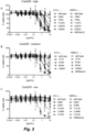
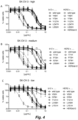
- Cytotoxicity of the bsADCs was tested using HCC1954, SK-OV-3 and Colo205 cells.
- Cells were seeded in 96-well tissue culture plates (5,000 cells/well) and incubated for 6 hours at 37°C.
- Serially diluted ADCs (10-0.0005 ⁇ g/mL) were added and the cells were incubated for 4 days at 37°C.
- Cell viability was assessed using CellTiter-GLO (Promega), according to the manufacturer's guidelines. The percentage of viable cells was depicted as a percentage relative to untreated cells (0% cell death) and staurosporin-treated cells (100% cell death).
- Percentage viable cells (RFU ADC treated cells - RFU staurosporin-treated cells) ⁇ 100 / (RFU untreated cells - RFU staurosporin-treated cells)
- RFU relative fluorescence units
- Figures 3-5 show cell viability after 4 days treatment with serially diluted ADCs as percentage compared to untreated cells. Data shown are mean ⁇ standard deviation of at least two different experiments. IC 50 values for cytotoxicity were determined using GraphPad Prism 6 software and depicted in Table 3.
- SK-OV-3 cells were cultured on glass coverslips (Thermo Fisher Scientific) at 37°C for 16 hours. Antibody (2 and 10 ⁇ g/mL) was added and cells were incubated for 16 hours at 37°C. Cells were fixed, permeabilized and incubated 45 min with goat anti-human IgG1-FITC (Jackson) to stain for human IgG and mouse anti-human CD107a-APC (BD) to stain for lysosomes.
- IgG1-FITC goat anti-human IgG1-FITC
- BD mouse anti-human CD107a-APC
- bsHER2xCD63 N74H does not bind to, and accumulate in, healthy tissues that do not express the model tumor antigen HER2
- bsHER2xCD63 N74H and monovalent and bivalent control antibodies were conjugated with FITC. Their accumulation was investigated in granulocytes and thrombocytes of healthy donors that do not express HER2.
- Whole blood samples from healthy donors were collected in Heparin tubes.
- Whole blood was diluted 1:2 in RPMI-1640 supplemented with 10% heat-inactivated cosmic calf serum.
- Anti-CD63 antibodies were conjugated with FITC (Thermo Scientific) according to manufacturer's instruction and added to whole blood cells at final concentration of 10 g/mL.
- the grey bars in Figure 9 represent total IgG staining (depicted as arbitrary units).
- the black bars in Figure 9 represent lysosomal co-localization (depicted as arbitrary units).
- IgG1-HER2 and bsHER2xb12 both showed similar staining of SK-OV-3 cells after 1, 3, and 16 hours (grey bars).
- a small portion of IgG1-HER2 and bsHER2xb12 showed lysosomal co-localization after 16 hours antibody exposure (black bars).
- HER2 downmodulation ELISA Using a HER2 downmodulation ELISA it was investigated if the strong lysosomal targeting observed with bsHER2xCD63 N74H , also resulted in increased downmodulation of the targeted antigen.
- AU565, SK-OV-3 and Colo 205 cells were seeded (1 million cells/flask) in T25 flasks (Greiner) and incubated overnight at 37°C to obtain a confluent monolayer.
- Antibodies were added (10 ⁇ g/mL) and cells were cultured for another 3 days at 37°C, washed and lysed. Total protein levels were quantified using bicinchoninic acid (BCA) protein assay reagent (Pierce), according to manufacturer's instruction.
- BCA bicinchoninic acid
- HER2 protein in tumor cell lines with different expression levels of HER2; AU565 (500,000 HER2/cell, Figure 10A ), SK-OV-3 (200,000 HER2/cell, Figure 10B ) and Colo205 (50,000 HER2/cell, Figure 10C ) was quantified after three days of incubation with HER2 antibody and compared with untreated cells (see Figure 10 ).
- IgG1-HER2 induced ⁇ 40% downmodulation of total HER2 in AU565 cells that express high levels of HER2.
- the monovalent bsHER2xb12 antibody showed dose-dependent binding to HER2-positive SK-OV-3 cells (FACS binding example)
- no downmodulation of HER2 was observed with bsHER2xb12.
- bsHER2xCD63 N74H -ADC induced significant inhibition of tumor growth, while the monovalent bsHER2xb12-ADC or bsCD63 N74H xb12-duo3, had no effect on tumor growth.
- Mantel-Cox analysis of Kalan Meyer plot indicated significant inhibition of tumor growth by bsHER2xCD63 N74H -ADC, P-value ⁇ 0.0001.
- Example 13 Lysosomal co-localization of bsBeta1xCD63 N74H measured with confocal microscopy
Landscapes
- Health & Medical Sciences (AREA)
- Chemical & Material Sciences (AREA)
- Life Sciences & Earth Sciences (AREA)
- Immunology (AREA)
- General Health & Medical Sciences (AREA)
- Medicinal Chemistry (AREA)
- Organic Chemistry (AREA)
- Bioinformatics & Cheminformatics (AREA)
- Engineering & Computer Science (AREA)
- Pharmacology & Pharmacy (AREA)
- Animal Behavior & Ethology (AREA)
- Public Health (AREA)
- Veterinary Medicine (AREA)
- Epidemiology (AREA)
- Genetics & Genomics (AREA)
- Biochemistry (AREA)
- Molecular Biology (AREA)
- Proteomics, Peptides & Aminoacids (AREA)
- Biophysics (AREA)
- Cell Biology (AREA)
- Chemical Kinetics & Catalysis (AREA)
- General Chemical & Material Sciences (AREA)
- Nuclear Medicine, Radiotherapy & Molecular Imaging (AREA)
- Oncology (AREA)
- Hematology (AREA)
- Peptides Or Proteins (AREA)
- Medicines That Contain Protein Lipid Enzymes And Other Medicines (AREA)
- Medicines Containing Antibodies Or Antigens For Use As Internal Diagnostic Agents (AREA)
- Medicinal Preparation (AREA)
- Micro-Organisms Or Cultivation Processes Thereof (AREA)
- Preparation Of Compounds By Using Micro-Organisms (AREA)
- Acyclic And Carbocyclic Compounds In Medicinal Compositions (AREA)
- Steroid Compounds (AREA)
Description
- The present invention relates to a multispecific antibody, compositions comprising said multispecific antibody, and the use of said multispecific antibody in the treatment of a disease.
- Since the development of the first monoclonal antibodies research has been directed at further optimization of antibodies for human therapy. The first monoclonal antibodies originated from mice and rats. The progress in antibody technology has led to the availability of humanized and human antibodies with decreased immunogenicity risk profiles. Genetic and chemical engineering is leading to the development of more potent antibodies with an increased therapeutic potential. This includes antibodies with optimized Fc-mediated effector functions, optimized binding characteristics or optimized anti-tumor activity through the conjugation to toxic molecules (antibody-drug conjugates).
- Antibody-drug conjugates (ADC) are emerging as powerful therapeutics for the treatment of cancer, as they combine antibody-mediated tumor-targeting with the cytotoxic activity of toxins. ADCs comprise an antibody (e.g. a monoclonal antibody, a single-chain variable fragment [scFv], or a bispecific antibody) linked to a cytotoxic payload or drug. The advantage of directing cytotoxic drugs to tumors with an antibody against a tumor-associated antigen is that the therapeutic window can be improved relative to the unconjugated cytotoxic drug. This allows for the application of cytotoxic payloads of increased potency.
- Currently, two ADCs have already been approved for therapeutic use: brentuximab vedotin (Adcetris) for the treatment of relapsed Hodgkin lymphoma and relapsed sALCL, and trastuzumab emtansine (Kadcyla), for the treatment of HER2-positive, metastatic breast cancer patients who previously received trastuzumab and a taxane, separately or in combination. In addition, over 50 different ADCs are currently in clinical evaluation. In many cases, ADCs rely on internalization of the toxin-conjugated antibody molecules into targeted cells to release their payload and induce subsequent cytotoxicity. Most ADCs in clinical development are designed to be stable in circulation and to release their cytotoxic payload after internalization and lysosomal processing of the antigen/ADC complex.
- However, the requirement for antigen- and antibody-mediated internalization limits the number of suitable ADC targets. Many tumor-associated antigens do not internalize well or do not route well to lysosomes and therefore represent less promising candidate targets for ADC-based therapeutics. Also, in many cases, intracellular processing of ADCs is inefficient. Following internalization, receptors such as transferrin, HER2, cell adhesion molecule L1 and integrins, are continuously recycled back from the endosomal compartment to the plasma membrane. High antigen turn over, efficient translocation to the lysosomes and highly toxic payloads are therefore required to achieve maximal killing activity by ADCs.
- Methods to enhance internalization, lysosomal targeting, intratumoral and intracellular processing of ADCs might be used to enhance the tumor cell killing activity of ADCs. One approach to optimize the ADCs activity is by selecting a specific epitope on the tumor-associated target, as the specific epitope recognized may influence internalization and lysosomal routing. For instance, it has been previously shown that the efficacy of HER2-ADCs can be improved by selecting HER2-ADCs that allow enhanced internalization by piggybacking of HER2 onto other ErbB molecules via heterodimer formation. This provides an attractive strategy for increasing ADC delivery and tumor cell killing capacity to both high and low HER2 expressing tumor cells.
- Generally, efficient internalization of the ADC, followed by routing to lysosomes, where proteolysis can take place, is preferred. For many cell surface proteins and carbohydrate structures on tumor cells, however, the magnitude of these processes is insufficient to allow sufficiently potent cell killing by the ADC.
- International patent application
WO2013/138400 describes multispecific antibodies having a first domain that specifically binds a target antigen such as IL-4R or SOST, and a second domain that specifically binds an internalizing effector protein. If the target antigen is a tumor-associated antigen, the binding of the tumor-associated antigen and the internalizing effector protein by the multispecific antibody facilitates the targeted killing of tumor cells.WO2013/138400 further teaches the production of bispecific antibodies comprising a first antigen-binding domain and a second antigen-binding domain, wherein the first antigen-binding domain binds a target molecule (T) and where the second antigen-binding domain binds an internalizing effector protein (E) to increase internalization. The examples show reduction of IL-4 receptor using a bispecific anti-IL-4R/anti-CD63 antibody due to the increased receptor internalization. Bispecific anti-SOST/anti-CD63 antibody or anti-LPS/anti-CD63 antibody increase internalization of SOST and LPS respectively. Use of the bispecific constructs to increase ADC internalization is suggested. - International patent application
WO2015/157592 produces bispecific anti-HER2 ADC, wherein both binding domains bind to HER2, but on different regions. The bispecific ADC shows improved internalization compared to the monospecific ADC and improved in vitro and in vivo anti-tumor effect. -
EP2808035 discloses the use of bispecific antibodies to induce membrane protein depletion. In the examples is shown the production of bispecific anti-Met/anti-HER2 antibody and anti-Met/anti-EGFR antibody. - International patent application
WO2017/007796 discloses a bispecific anti-LPS/CD63 or anti-IL-4R/CD63 or anti-SOST/CD63 antibody, but does not disclose the limited claimed KD range or an effect linked to the claimed range. - It is an object of the present invention to provide bispecific or multispecific antibodies for which the internalizing capacity is increased relative to the monospecific antibody against a tumor-associated target.
- It is another object of the present invention to provide bispecific or multispecific antibody drug conjugate molecules for which the internalizing capacity is increased relative to the monospecific antibody drug conjugate molecule directed against a tumor-associated target.
- It is another object of the present invention to provide ADCs with increased internalization, lysosomal targeting and/or intracellular processing in tumor cells.
- It is another object of the present invention to provide ADCs with increased cytotoxicity to tumor cells and/or fewer side effects.
- It is another object of the present invention to provide ADCs with an increased therapeutic window.
- It is another object of the present invention to enable the generation of effective ADCs against tumor-associated antigens which internalize poorly or which route poorly to lysosomes.
- In a first aspect, the present invention relates to a multispecific antibody, comprising a first antigen-binding domain and a second antigen-binding domain, wherein the first domain specifically binds a target molecule (T), which is a cell surface-expressed target molecule, and wherein the second domain specifically binds an internalizing effector protein (E), wherein E is CD63, and wherein the second antigen-binding domain has a dissociation constant KD with E of between 2.0×10⁻⁹ and 7.3×10⁻⁹ M, the KD being determined by biolayer interferometry.
- It has been found by the present inventors that the specific affinity range of the second antigen-binding domain bestows surprisingly beneficial properties on the multispecific antibody of the present invention. The second antigen-binding domain is found within a specific range of binding affinities to readily induce internalization of the multispecific antibody as well as to induce cytotoxicity of the drug-conjugated antibody. This applies in particular where the first domain specifically binds a tumor-associated antigen such as for example HER2. At the same time, the second antigen-binding domain exerts only limited internalization and cytotoxicity (when employed in the context of an ADC) within the specific range of binding affinities, without binding of the first binding domain, i.e. demonstrating surprisingly low cytotoxicity in cells that express the internalizing effector protein (E) in the absence of the tumor-associated target (T). Thus, the multispecific antibody of the present invention demonstrates binding, internalization, lysosomal routing and toxin release in tumor cells, accompanied by minimal internalization, lysosomal routing and toxin release in non-tumor cells.
- It has also been found by the present inventors that the multispecific antibody of the present invention allows for utilization of tumor antigens that usually do not internalize or poorly internalize, thereby greatly enhancing the pool of potential ADC targets.
- In another aspect, the present invention relates to the multispecific antigen-binding molecule, wherein the molecule is conjugated to a cytotoxic moiety, a radioisotope, or a drug. In other aspects, the present invention relates to the use of the multispecific antibody in a method for treating and/or preventing a cancer, and to the use of the multispecific antibody in a method of targeting a tumor in a subject, the method comprising administering to the subject the multispecific antibody or ADC.
- In other aspects, the present invention relates to a pharmaceutical composition comprising the multispecific antibody as an active ingredient, to nucleic acid(s) encoding the multispecific antibody, to an expression vector containing said nucleic acid(s) and being capable of expressing said nucleic acids in a single or multiple prokaryotic or eukaryotic host cel lines as appropriate, and to prokaryotic or eukaryotic host cell lines comprising said vector(s).
- The term "binding" as used herein refers to the binding of an antibody to a predetermined antigen or target, e.g. with a binding affinity corresponding to a KD value of about 10-8 M or less.
- The skilled reader will be familiar with the concept of affinity and the equilibrium dissociation constant KD. The dissociation constant KD can be measured by biolayer interferometry.
- KD values may be determined by biolayer interferometry (BLI) in an Octet HTX instrument using the antibody, as the immobilized ligand and the antigen as the analyte.
- KD (M) refers to the dissociation equilibrium constant of a particular interaction between a multispecific antibody and an antigen, preferably the interaction between a single binding arm of an antibody molecule and an antigen, and may be obtained by dividing kd by ka. The term "kd" (sec-1), as used herein, refers to the dissociation rate constant of a particular interaction between an antibody and an antigen. Said value is also referred to as the kdis, koff value or off-rate. The term "ka" (M-1 × sec-1), as used herein, refers to the association rate constant of a particular interaction between a multispecific antibody and an antigen. Said value is also referred to as the kon value or on-rate.
- As used herein, the term "antibody" (Ab) in the context of the present invention refers to an immunoglobulin molecule, a fragment of an immunoglobulin molecule, or a derivative of either thereof, which has the ability to specifically bind to an antigen, preferably dual binding to two different antigens such as for bispecific antibodies, under typical physiological conditions for a relevant functionally-defined period to induce, promote, enhance, and/or modulate a physiological response associated with antibody binding to the antigen.
- The variable regions of the immunoglobulin molecule (either of the heavy and/or light chains or heavy chains only) contain a binding domain that interacts with an antigen. The constant regions of the antibodies (Abs) may mediate the binding of the immunoglobulin to FcRn. An antibody may also be a bispecific or multispecific antibody, such as but not limited to: DuoBody molecules, tandem scFv, tandem scFv-Fc, knob-into-hole IgGs, scFv-Fc knobs-into-holes, scFv-Fc-scFv, F(ab')2, Fab-scFv, (Fab'scFv)2, Diabody, scDiabody, scDiabody-Fc, or scDiabody-CH3, Triomab, kih IgG common LC, CrossMab, DVD-Ig, 2 in 1-IgG, IgG-scFv, bi-Nanobody, BiTE, TandAbs, DART, DART-Fc, scFv-HSA-scFv, orthoFab-IgG, tetravalent Tv-IgGs, dock-and-lock (DNL) formats such as DNL-Fab3 and Azymetric scaffold, or similar molecule.
- As indicated above, the term antibody herein, unless otherwise stated or clearly contradicted by context, includes fragments of an antibody that are antigen-binding fragments, i.e., retain the ability to specifically bind to the antigen. It has been shown that the antigen-binding function of an antibody may be performed by fragments of a full-length antibody. Examples of antigen-binding fragments encompassed within the term "antibody" include (i) a Fab' or Fab fragment, a monovalent fragment consisting of the VL, VH, CL and CH1 domains, or a monovalent antibody as described in
WO2007059782 (Genmab) ; (ii) F(ab')2 fragments, bivalent fragments comprising two Fab fragments linked by a disulfide bridge at the hinge region; (iii) a Fd fragment consisting essentially of the VH and CH1 domains; (iv) a Fv fragment consisting essentially of the VL and VH domains of a single domain of an antibody, (v) a dAb fragment, which consists essentially of a VH domain and also called domain antibodies; (vi) camelid or nanobodies and (vii) an isolated complementarity determining region (CDR). Furthermore, although the two domains of the Fv fragment, VL and VH, are coded for by separate genes, they may be joined, using recombinant methods, by a synthetic linker that enables them to be made as a single protein chain in which the VL and VH regions pair to form monovalent molecules (known as single chain antibodies or single chain Fv (scFv)). Such single chain antibodies are encompassed within the term antibody unless otherwise noted or clearly indicated by context. These and other useful antibody fragments in the context of the present invention, as well as bispecific formats of such fragments, are discussed further herein. It also should be understood that the term antibody, unless specified otherwise, also includes polyclonal antibodies, monoclonal antibodies (mAbs), antibody-like polypeptides, chimeric antibodies, humanized and fully human antibodies, and antibody fragments retaining the ability to specifically bind to the antigen (antigen-binding fragments) provided by any known technique, such as enzymatic cleavage, peptide synthesis, and recombinant techniques. An antibody as generated can possess any isotype. - As used herein, "isotype" refers to the immunoglobulin (sub)class (for instance IgG1, IgG2, IgG3, IgG4, IgD, IgA, IgE, or IgM) that is encoded by heavy chain constant region genes.
- The term "monovalent antibody" means in the context of the present invention that an antibody molecule is capable of binding no more than a single molecule of the antigen.
- The term "bivalent antibody" means in the context of the present invention that the antibody molecule contains two binding domains for a specific antigen, and is therefore capable of binding one or two molecules of that antigen
- The multispecific antibodies, in particular the bispecific antibodies, of the present invention may be generated through controlled Fab-arm exchanged (FAE) as described in Labrijn et al., Efficient generation of stable bispecific IgG1 by controlled Fab-arm exchange, PNAS, vol. 110, no. 13, pp. 5145-5150, March 2013 , in
WO 2011/131746 A2 , or in Labrijn et al. Controlled Fab-arm exchange for the generation of stable bispecific IgG1, Nat Protoc, 2014 Oct;9(10):2450-63, also known as DuoBody® technology. Briefly, in this in vitro method, two different antibodies are provided, both comprising an Fc region of an immunoglobulin with a CH3 region, wherein the sequences of the respective CH3 regions are different and contain a matched mutation. As a result, the heterodimeric interaction between the first and second CH3 regions is stronger than each of the homodimeric interactions of said first and second CH3 regions. The antibodies are incubated under reducing conditions to allow the cysteines in the hinge region to undergo disulfide-bond isomerization, and to obtain the bispecific antibody by controlled Fab-arm exchange. The reducing agent is thereupon removed from the mixtures (now containing bispecific antibodies) to allow oxidation of the disulfide bonds. The sequences of said CH3 regions contain matched mutations, i.e. mutations at different positions in the two CH3 regions, preferably a mutation atposition 405 in one of the CH3 regions of an IgG1 molecule and a mutation at position 409 in the CH3 region of another IgG1 molecule. Multispecific antibodies may also be generated using other technologies and formats, such as but not limited to: tandem scFv, tandem scFv-Fc, knob-into-hole IgGs, scFv-Fc knobs-into-holes, scFv-Fc-scFv, F(ab')2, Fab-scFv, (Fab'scFv)2, Diabody, scDiabody, scDiabody-Fc, or scDiabody-CH3, Triomab, kih IgG common LC, CrossMab, DVD-Ig, 2 in 1-IgG, IgG-scFv, bi-Nanobody, BiTE, TandAbs, DART, DART-Fc, scFv-HSA-scFv, orthoFab-IgG, tetravalent Tv-IgGs, dock-and-lock (DNL) formats such as DNL-Fab3 and Azymetric scaffold, or similar molecule. - Binding of both T and E by the multispecific antibody preferably induces internalization of the multispecific antibody of the present invention, as well as cytotoxicity of a multispecific drug-conjugated antibody of the present invention, to a greater extent than by binding to target T alone. For instance, internalization induced by binding, preferably simultaneous binding, of T and E by the multispecific antibody may be more than 10%, such as more than 20%, more than 30%, more than 40%, more than 50%, more than 60%, more than 70%, more than 80%, more than 90%, more than 100%, more than 110%, more than 150%, more than 200%, more than 300%, more than 400%, more than 500%, more than 750%, more than 1000%, more than 2000%, or more than 5000% higher than the level of internalization measured in the presence of a control construct containing only binding to T and not to E.
- A non-limiting example of determining whether the multispecific antibody of the present invention enhances internalization of the multispecific antibody to a greater extent than the binding of the target molecule (T) by the first domain alone is shown in the co-localization assays in Examples 5 and 8, or in the HER2 downmodulation assay of Example 9, as discussed below. In those examples, T is HER2 and E is CD63. As to the concept of enhanced internalization, in Example 13 and 14, enhanced internalization of multispecific antibodies is demonstrated using Integrin beta-1 (CD29) as the target (T) and CD63 as the internalizing effector protein (E) that is inducing enhanced internalization.
- The present inventors have found that, in particular when E is CD63, the specific affinity range of the second antigen-binding domain bestows surprisingly beneficial properties on the bispecific antibody of the present invention. Monovalent binding of the antigen-binding domain to E within specific affinity range of a dissociation constant KD 2.0×10⁻⁹ and 7.3×10⁻⁹ M readily induces internalization of bispecific antibodies and cytotoxicity of bispecific drug-conjugated antibodies, when the first domain specifically binds a cell surface-expressed tumor-associated antigen such as e.g. HER2. Moreover, when only E and not the tumor-associated target antigen (T) is present, the second antigen-binding domain exerts only limited contribution to internalization of the multispecific antibody and cytotoxicity of multispecific drug-conjugated antibodies within this specific affinity range, i.e. demonstrating surprisingly low or absent cytotoxicity in cells that express the internalizing effector protein (E) in absence of the tumor target (T). Thus, the multispecific antibody of the present invention demonstrates binding, internalization, lysosomal accumulation in tumor cells, accompanied by minimal internalization into non-tumor cells.
- The Cluster of Differentiation 63 (CD63, Uniprot ID P08962) molecule is also known as lysosome-associated membrane glycoprotein 3 (LAMP-3). CD63 is also known as: platelet glycoprotein 40 (Pltgp40), melanoma antigen ME491 or MLA1, ocular melanoma-associated antigen (OMA81H), tetraspanin-30 (TSPAN30), granulophysin or lysosomal integral membrane protein-1 (LIMP-1). CD63 is a member of the tetraspanin superfamily and is ubiquitously expressed. The CD63 gene is located on human chromosome 12q13 and was the first characterized tetraspanin. Originally, CD63 was discovered as a protein present on the cell surface of activated blood platelets, known as Pltgp40 and in early stage human melanoma cells, where it was known as ME491.
- CD63 is expressed in many cell types. Amongst others, CD63 is expressed intracellularly in lysosomes, endosomes, granules of resting platelets and basophils. Cell surface expression of CD63 can be detected on activated basophils and platelets, monocytes, macrophages, and granulocytes. CD63 is also expressed on endothelial cells, fibroblasts, osteoblasts, neural tissue, melanoma cells, smooth muscle cells and mast cells. CD63 is described to shuttle between the plasma membrane and intracellular compartments.
- The major pool of CD63 resides in intracellular compartments such as endosomes and lysosomes, but some expression can be found on the cell surface. CD63 has been described to regulate transport of other proteins typically through endocytosis. Furthermore, CD63 has been described to regulate surface expression of membrane-
type 1 matrix metalloproteinase by targeting the enzyme for lysosomal degradation, and silencing of CD63 in endothelial cells prevents internalization of vascular endothelial growth factor receptor 2 (VEGFR2) in response to its ligand VEGF. Also across different tumor types, CD63 has been demonstrated to continuously shuttle between the plasma membrane and lysosomes, which was dependent on the presence of AP2 and clathrin. Thus CD63 seems an attractive antigen to facilitate internalization and lysosomal delivery, a feature suitable for enhancing efficacy of certain antibody drug conjugates (ADC) targeting tumor antigens that by themselves to not internalize and/or shuttle to lysosomes sufficiently. - CD63 expression does not show a tumor-specific expression, although CD63 was first discovered as an abundantly expressed surface antigen in early stage melanoma cells. CD63 cell surface expression is reduced during malignant melanoma progression, indicative of a negative correlation between cell surface expression of CD63 and tumor invasiveness.
- Tumor-associated antigens are antigens that are expressed on the surface of (certain) tumor cells, and for which surface expression to a lesser extent is found on normal cells. As used herein, the term "tumor-associated antigen" refers to proteins or polypeptides, but also carbohydrates, glycoproteins, lipids, lipoproteins, lipopolysaccharides, or other non-protein polymers that are preferentially expressed on the (outside) surface of a tumor cell. The term "preferentially expressed," as used in this context, means that the antigen is expressed on a tumor cell at a level that is at least 10%, such as at least 20%, at least 30%, at least 40%, at least 50%, at least 60%, at least 70%, at least 80%, at least 90%, at least 100%, at least 110%, at least 150%, at least 200%, at least 300%, at least 400%, at least 500%, at least 750%, at least 1000%, at least 2000% or at least 5000% greater than the expression level of the antigen on non-tumor cells. Tumor-associated antigens can derive from any protein, glycoprotein or other macromolecules synthesized by the tumor cell.
- Tumor-associated antigens may be grouped in different classes of antigens: 1) Class l HLA-restricted cancer testis antigens which are expressed normally in the testis or in some tumors but not in normal tissues, including antigens from the MAGE, BAGE, GAGE, NY-ESO and BORIS families; 2) Class I HLA restricted differentiation antigens, including melanocyte differentiation antigens such as MART-1, gp100, PSA, Tyrosinase, TRP-1 and TRP-2; 3) Widely expressed antigens, which are antigens expressed both in normal and tumor tissue though at different levels or altered translation products, including CEA, HER2/neu, hTERT, MUC1, MUC2 and WT1; 4) Tumor specific antigens which are unique antigens that arise from mutations of normal genes including β-catenin, α-fetoprotein, MUM, RAGE, SART, etc; 5) Viral antigens such as HPV, EBV; and 6) Fusion proteins, which are proteins created by chromosomal rearrangements such as deletions, translocations, inversions or duplications that result in a new protein expressed exclusively by the tumor cells, such as Bcr-Abl.
- Preferred examples of tumor-associated antigens include 5T4, A33, activin receptor, adrenomedullin receptors, AFP, AGS-5, ALK, annexin, AXL, B7-H3, B7-H4, BAGE proteins, BCMA, Bombesin, C33 antigen, C4.4a, C-type lectin-like(-receptor), CA19.9, CA-125, CADM1, CAIX, CanAg, CAR, carbonic anhydrase, Caveolin-1, CCK2R, CD4, CD10, CD19, CD20, CD21, CD22, CD25, CD27, CD30, CD33, CD37, CD38, CD44, CD51, CD57, CD70, CD73, CD74, CD79a, CD79b, CD80, CEA, CEACAMs, c-kit, claudin, chemokine receptors (i.e., CXCR4, CXCR5), c-Met, Cripto-1, DEC-205, Derlin-1, Desmoglein-3, DIk-1, DLL3, DS6, E-cadherin, E-Selectin, EAG-1, ED-B, EpCAM, EGFR, EGFRvIII, emmprin, endothelin receptor, ErbB2/Her2, ErbB3, ErbB4, ETV6-AML, Ephrin type-A receptor, Epiregulin, ETA, FAP-alpha, FcyR's, FGFR, FOLR1, Frizzled, Fyn3, Galectin, Ganglioside, GCC, GD2, GD3, GloboH, lypican-3, GLUT3, GPNMB, G-protein coupled receptors (i.e., GPR49), gp100, Hsp, HLA/B-raf, HLA-DR, HLA/k-ras, HLA MAG E-A3, HMW-MAA, hTERT, ICAM-3, IGF-R, IL-13-R, L1CAM, laminin receptor, LIV1, LMP2, LRP5, LRP6, MAGE proteins, MART-1, melanotransferrin, mesothelin, metalloproteinase, ML-IAP, Mucins, Mud, Mud 6 (CA-125), MU M1, N-cadherin, NA17, NCAM-1, Nectin4, Notch, NP-55, NRP1, NY-BR1, NY-BR62, NY-BR85, NY-ES01, PLAC1, PRLR, PRAME, prominin-1, PSMA (FOLH 1 ), RON, SLC44A4, SLITRK6, Steap-1, Steap-2, surviving, syndecan, TAG-72, TF, TGF-β, TMPRSS2, TMEFF2, TNFR, Tn, TROP2, TRP-1, TRP-2,TWEAKR, tyrosinase, uroplakin-3 and VEGFR.
- Preferred examples of tumor-associated antigens include tumor-associated antigens that are highly overexpressed, but lack sufficient lysosomal transport, such as glycosylphosphatidylinositol (GPI) anchored proteins (i.e., glypican family, uPAR, folate binding receptors, prostasin, FcgRlllb [CD16b], alkaline phosphatase, acetylcholinesterase, 5' nucleotidase [p36], Cripto, LFA-3 [CD58], DAF [CD55], Thy-1 [CD90], Qa-2, Ly-6A and MIRL [CD59]), adhesion molecules (i.e., selectins, L1CAM, N-CAM, LRP1, TAG1, cadherins), which are often recycle back to the plasma membrane after endocytosis, with only a minor fraction being targeted for lysosomal degradation
Preferred examples of tumor-associated antigens include tumor-specific antigens that are known to interact with CD63 which may improve bivalent binding of bsADC at low copy numbers. For example, CD63 has been described to interact with other tetraspanins (i.e., CD81, CD82, CD9 and CD151), integrins, MHCII, CXCR4, TM4SF5, syntenin-1, TIMP-1, H, K-ATPase, L6-antigen and MT1-MMP. - Examples of tumor-associated antigens include selection of glycotargets such as, Lewis-Y (CD174), Lewis-X (CD15), SLeX, SLeA, sTn, fucosyl-GM1, Globo H, SSEA-3, GM2, GD2, GD3, Polysialic acids or glycoproteins (i.e., Mucins).
-
-
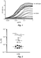
Figure 1 shows a dose response curve of anti-CD63 antibodies binding to recombinant human CD63 as measured by ELISA. -
Figure 2 shows affinity measurements of affinity variants of anti-CD63 antibodies measured with label-free Bio-Layer Interferometry. -
Figure 3 shows the results of a viability assay to test the cytotoxicity of monovalent bsCD63N74Hxb12-Duo3 ADCs and bsHER2xCD63N74H-Duo3 ADCs in Colo205 cells. -
Figure 4 shows the results of a viability assay to test the cytotoxicity of monovalent bsCD63n74Hxb12-Duo3 ADCs and bsHER2xCD63N74H-Duo3 ADCs in SK-OV-3 cells. -
Figure 5 shows the results of a viability assay to test the cytotoxicity of monovalent bsCD63n74Hxb12-Duo3 ADCs and bsHER2xCD63N74H-Duo3 ADCs in HCC1954 cells. -
Figure 6 shows lysosomal co-localization of monovalent bsCD63N74Hxb12 bispecific molecules measured in SK-OV-3 cells with confocal microscopy. -
Figure 7 shows binding of bsHER2xCD63N74H to SK-OV-3 cells as determined by flow cytometry. -
Figure 8 shows intracellular accumulation of FITC-conjugated CD63 antibody and CD63 affinity variant antibodies in granulocytes and thrombocytes. -
Figure 9 shows lysosomal co-localization of bsHER2xCD63N74H in SK-OV-3 cells followed over time. -
Figure 10 shows the total amount of HER2 protein in tumor cell lines with different expression levels of HER2, as quantified by ELISA after three days of incubation with bsHER2xCD63N74H, compared with untreated cells. -
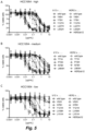
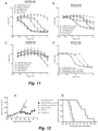
Figure 11 shows the results of a viability assay to test the cytotoxicity of Duostatin-3 conjugated bispecific ADCs in vitro. -
Figure 12 shows the mean tumor size and percentage of tumor free survival in mice that had been subcutaneously inoculated with SK-OV-3 tumor xenografts, followed by treatment with Duostatin-3 conjugated multispecific ADCs. -
Figure 13 shows binding of bsBeta1xCD63N74H to SK-OV-3 cells as assessed by flow cytometry. -
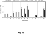
Figure 14 shows lysosomal co-localization of bsBeta1xCD63N74H on SK-OV-3 cells. -
Figure 15 shows lysosomal co-localization of bsBeta1xCD63N74H on SK-OV-3 cells followed over time. - In one aspect, the present invention relates to a multispecific antibody comprising a first antigen-binding domain and a second antigen-binding domain, wherein the first domain specifically binds a target molecule (T), which is a cell surface-expressed tumor-associated antigen, and wherein the second domain specifically binds an internalizing effector protein (E), wherein E is CD63, and wherein the second antigen-binding domain has a dissociation constant KD value with E of between 2.0×10⁻⁹ and 7.3×10⁻⁹ M, the KD being determined by biolayer interferometry.
- To provide tumor specificity of the multispecific antibody of the present invention, in absence of the first antigen-binding domain, the second domain which specifically binds to the internalizing effector protein (E), may advantageously not bind or only bind with low affinity and subsequently not internalize or at least internalize to a significant lesser degree. It has been found by the present inventors that a multispecific antibody wherein the second antigen-binding domain has a dissociation constant KD value with E of between 2.0×10⁻⁹ and 7.3×10⁻⁹ M fulfills these criteria.
- The target molecule (T), preferably a tumor-associated target molecule, may be a protein, polypeptide, lipid or other macromolecule. In a preferred embodiment, the target molecule is a protein. In another embodiment, the target molecule, preferably a tumor-associated target molecule is a polypeptide. In some embodiments, T is a cell surface-expressed target protein or target polypeptide. In other embodiments, T is a soluble target protein or target polypeptide, preferably one that interacts with a cell surface receptor. Target binding by the multispecific antibody may take place extracellularly or on the cell surface.
- In one embodiment, the target molecule is a cell surface-expressed receptor. In a preferred embodiment, the target molecule is a tyrosine kinase receptor, preferably a transmembrane tyrosine kinase receptor. In another embodiment, the target molecule is a membrane-bound ligand.
- The multispecific antibody, preferably bispecific antibody, may bind E on the cell surface or inside the cell.
- It is particularly preferred that the target molecule is a tumor-associated protein or polypeptide. Advantageously, the tumor-associated antigen is an antigen that is not ordinarily internalized or is poorly internalized. Preferably, the tumor-associated antigen is an antigen that shows inefficient routing to the lysosomal compartment.
- In a preferred embodiment, the multispecific antibody comprises i) a first binding arm which comprises the first antigen-binding domain and ii) a second binding arm which comprises the second antigen-binding domain.
- It is particularly preferred that the multispecific antibody is a bispecific antibody.
- Preferably, the multispecific antibody is internalized into the cell by way of binding to E only in the presence of the target molecule (T). It is also preferred that the multispecific antibody is internalized into the cell by way of binding to E only when the first domain is specifically bound to the target molecule (T).
- The multispecific antibody may, upon binding to E, internalize more efficiently into cells expressing T as compared to cells not expressing T.
- The multispecific antibody may, upon binding to T, internalize more efficiently into cells expressing E as compared to cells not expressing E.
- The multispecific antibody may, upon binding to E, be transported to the lysosomal compartment in cells expressing T.
- The multispecific antibody may, upon binding to E, be more efficiently transported to the lysosomal compartment in cells expressing T as compared to cells not expressing T.
- The multispecific antibody may, upon binding to T, be more efficiently transported to the lysosomal compartment in cells expressing E as compared to cells not expressing E.
- The internalization-enhancing strategy of the present invention involves combining a tumor-associated target antigen with the internalizing capacities of CD63. In particular, bispecific antibodies that bind to both CD63 and a tumor-associated target may be useful in therapeutic settings in which specific targeting and enhanced internalization of an antibody-drug-conjugate is desired. According to one embodiment, T is HER2.
- According to another embodiment, the first and/or the second antigen-binding domain comprises at least one antibody variable region, preferably at least two antibody variable regions.
- According to some embodiments, the multispecific antibody is a bispecific antibody, or a multispecific, preferably bispecific, antibody fragment. Also disclosed arerecombinantly engineered part thereof. Particularly preferred is a bispecific antibody. A bispecific antibody may be employed to use internalization enhancing properties of one antigen by binding to the same with one arm, and bind a target molecule, such as a tumor-associated target molecule, with the other arm of the bispecific antibody. Such a bispecific antibody may then be loaded with a cytotoxic conjugate to induce cell death upon internalization of the ADC.
- The antibody may be a bispecific antibody, comprising (i) a first antibody comprising a first antigen-binding domain specifically binding a target molecule (T) as defined herein, and (ii) a second antibody comprising a second antigen-binding domain specifically binding an internalizing effector protein (E) as defined herein.
- In a preferred embodiment, the multispecific antibody is a bispecific antibody comprising a first binding arm comprising the first antigen-binding domain and a second binding arm comprising said second antigen-binding domain. Advantageously, said first antigen-binding domain comprises a first heavy chain variable sequence (VH) and a first light chain variable sequence (VL), and said second antigen-binding domain comprises a second heavy chain variable sequence (VH) and a second light chain variable sequence (VL) and wherein said variable sequences each comprises three CDR sequences, CDR1, CDR2 and CDR3.
- In a preferred embodiment, (i) said first binding arm comprises a first heavy chain comprising a first heavy chain variable sequence (VH) and a first heavy chain constant sequence (CH), and a first light chain comprising a first light chain variable sequence (VL) and a first light chain constant sequence (CL), and (ii) said second binding arm comprises a second heavy chain comprising a second heavy chain variable sequence (VH) and a second heavy chain constant sequence (CH), and a second light chain comprising a second light chain variable sequence (VL) and a second light chain constant sequence (CL).
- According to another embodiment, the first binding arm is derived from a chimeric antibody or from a humanized antibody or from a human antibody. Also disclosed are antibodies wherein, the second binding arm is derived from a chimeric antibody or from a humanized antibody or from a human antibody. Accordingly, the first binding arm may be derived from a human antibody and the second binding arm is derived from a humanized antibody or from a chimeric antibody.
- It is preferred that the multispecific antibody of the present invention is a bispecific antibody, wherein the bispecific antibody is a full-length antibody, preferably an IgG1 antibody.
- The multispecific antibody of the invention may be isolated. An "isolated multispecific antibody " as used herein, is intended to refer to a multispecific antibody, such as a bispecific antibody, which is substantially free of other antigen-binding molecules or antibodies having different antigenic specificities. Moreover, an isolated multispecific antibody may be substantially free of other cellular material and/or chemicals.
- Said second antigen-binding domain may have one or more mutations that modulate the affinity of the second antigen-binding domain with CD63. Said second antigen-binding domain may be derived from an antibody having one or more mutations in the VH and/or VL that modulates the affinity of the second antigen-binding domain with E..
- Said antibody may have one or more mutations in the anti-CD63 Fab region that modulates the affinity of the second antigen-binding domain with CD63.The mutation may be a single amino acid substitution, preferably a single amino acid histidine substitution.
- Said one or more mutations in the VH and/or VL may be an amino acid substitution, preferably a histidine substitution, at position 54 of the VL according to SEQ ID No. 5 of Table 1 below, or at positions 71, 72 and/or 74 of the VH according to SEQ ID No. 1 of Table 1 below. For example, anti-CD63-N74H has an asparagine to histidine mutation at position 74 of the heavy chain according to SEQ ID No. 1, anti-CD63-LN54H has an asparagine to histidine mutation at position 54 of the light chain according to SEQ ID No. 5.The second antigen-binding domain may be selected such that it binds target E with a KD within the preferred affinity range.
- Said amino acid substitution, preferably a histidine substitution is preferably at position 74 of the VH according to SEQ ID No. 1.Preferably, the mutation is N74H of the VH according to SEQ ID No. 1.
- In a preferred embodiment said second domain, preferably as part of said second binding arm, comprises:
- a)
VH CDRs VL CDRs - b)
VH CDRs VL CDRs - c)
VH CDRs VL CDRs - d)
VH CDRs VL CDRs - In a particularly preferred embodiment, said second domain, preferably as part of said second binding domain, comprises
VH CDRs VL CDRs - Said multispecific antibody may comprise a mutated Fab region of a CD63-specific monoclonal antibody. Said multispecific antibody maycomprise a mutated Fab region of CD63-specific monoclonal Ab 2192. Said multispecific antibody maycomprise an antigen-binding region specific for CD63 selected from a hybridoma or phage-display library.
- According to another embodiment, said first and second antigen-binding domains are each a pair of an antibody heavy chain variable domain and an antibody light chain variable domain.
- The bispecific antibody may preferably further comprise antibody constant regions.
- The multispecific antibody may be a tumor-associated target (T)xCD63 bispecific antibody. The (T)xCD63 bispecific antibody may be conjugated to a cytotoxic drug. The (T)xCD63 bispecific antibody may be conjugated to duostatin-3. the (T)xCD63 bispecific antibody may be conjugated to duostatin-3 and the anti-CD63 binding domain, which is the second binding domain, may comprise
VH CDRs VL CDRs - The (T)xCD63 bispecific ADCs of the present invention are found to be advantageous by enhancing the internalization of the ADC by being able to bind both the tumor-associated target and the potent internalizing characteristics of the CD63 antigen, thereby inducing stronger killing of cells by more efficient payload delivery inside the targeted cells. To tailor to the need of increasing efficacy of antibodies targeting various tumor-antigens, a narrow range of surprisingly efficient CD63 affinity variants (spanning a range of CD63 affinities) was found to enhance efficacy of the bispecific ADCs.
- According to another embodiment, the antibody is a HER2xCD63 bispecific antibody.
- The antibody may have an EC50 value for binding to tumor-associated target (T)-expressing cells, such as HER2-expressing cells, of lower than 5.0 µg/ml, such as lower than 4.0 µg/ml, such as lower than 3.0 µg/ml, such as lower than 2.0 µg/ml, such as lower than 1.0 µg/ml, such as lower than 0.9 µg/ml, such as lower than 0.8 µg/ml, such as lower than 0.7 µg/ml, such as lower than 0.6 µg/ml, such as lower than 0.5 µg/ml, such as lower than 0.4 µg/ml, such as lower than 0.3 µg/ml, such as lower than 0.2 µg/ml, such as lower than 0.1 µg/ml, such as lower than 0.05 µg/ml, such as lower than 0.01 µg/ml, as determined by flow cytometry.
- The binding of T and E by the multispecific antibody may induce internalization of the multispecific antibody to a greater extent than the binding of T by the first domain alone. Similarly, the binding of T and E by the multispecific antibodies of the present invention preferably induces cytotoxicity to a greater extent than the binding of T by the first domain alone.
- The binding of T and E by the multispecific, preferably bispecific, antibody may induce internalization of the multispecific antibody to a greater extent than the corresponding bivalent monospecific antibody binding T. Similarly, the binding of T and E by the multispecific, preferably bispecific, drug-conjugated antibodies of the present invention preferably induces cytotoxicity to a greater extent than the than the corresponding bivalent monospecific ADC binding T.
- The KD value may be determined by biolayer interferometry at 30°C. KD may be determined by biolayer interferometry at 30 °C and a pH of between 7.2 and 7.5, such as between 7.3 and 7.4, such as pH 7.4. KD may be determined by biolayer interferometry at 30 °C at 1000 RPM shaker speed. KD may be determined by biolayer interferometry using an Octet system, such as Octet HTX (ForteBio).
- In a preferred embodiment, the antibody is a bispecific antibody comprising:
- i) a first binding arm comprising a first heavy chain comprising a first heavy chain constant sequence (CH), said first CH comprising a first CH3 region, and
- ii) a second binding arm comprising a second heavy chain comprising a second heavy chain constant sequence (CH), said second CH comprising a second CH3 region,
- Preferably, in said first heavy chain CH3 region at least one of the amino acids in a position corresponding to positions T366, L368, K370, D399, F405, Y407 or K409 of human IgG1 heavy chain has been substituted, and in said second heavy chain CH3 region at least one of the amino acids in a position corresponding to positions T366, L368, K370, D399, F405, Y407 or K409 of human IgG1 heavy chain has been substituted, wherein said first and said second heavy chains are not substituted in the same positions and wherein the amino acid positions are numbered according the EU-index.
- In another embodiment, (i) the first CH3 region has an F405L substitution and the second CH3 region has a K409R substitution, or (ii) the first CH3 region has a K409R substitution and the second CH3 region has an F405L substitution.
- The multispecific antibodymolecule is conjugated to a cytotoxic moiety, a radioisotope, a drug, a cytokine or an RNA silencing vehicle. Preferably, the multispecific antibody is conjugated to a cytotoxic moiety, a radioisotope, or a drug. Preferably, the multispecific antibody is conjugated to a cytotoxic moiety.
- The cytotoxic moiety may be selected from the group consisting of duostatin-3, duostatin-5, pyrrolobenzodiazepine or an analog or derivative thereof, IGN-based toxins or an analog or derivative thereof, alpha-amanitin or an analog or derivative thereof, dolastatin or an analog or derivative thereof, taxol ; cytochalasin B; gramicidin D; ethidium bromide; emetine; mitomycin; etoposide; tenoposide; vincristine; vinblastine; colchicin; doxorubicin; daunorubicin; dihydroxy anthracin dione; a tubulin-inhibitor such as maytansine or an analog or derivative thereof; mitoxantrone; mithramycin; actinomycin D; 1-dehydrotestosterone; a glucocorticoid; procaine; tetracaine; lidocaine; propranolol ; puromycin; calicheamicin or an analog or derivative thereof an antimetabolite such as methotrexate, 6 mercaptopurine, 6 thioguanine, cytarabine, fludarabin, 5 fluorouracil, decarbazine, hydroxyurea, asparaginase, gemcitabine, or cladribine; an alkylating agent such as mechlorethamine, thioepa, chlorambucil, melphalan, carmustine (BSNU), lomustine (CCNU), cyclophosphamide, busulfan, dibromomannitol, streptozotocin, dacarbazine (DTIC), procarbazine, mitomycin C, cisplatin, carboplatin, duocarmycin A, duocarmycin SA, rachelmycin (CC-1065), or an analog or derivative thereof; an antibiotic such as dactinomycin, bleomycin, daunorubicin, doxorubicin, idarubicin, mithramycin, mitomycin, mitoxantrone, plicamycin, anthramycin (AMC)) ; an antimitotic agent such as an auristatin or an analog or derivative thereof, monomethyl auristatin E or F or an analog or derivative thereof; diphtheria toxin and related molecules such as diphtheria A chain and active fragments thereof and hybrid molecules, ricin toxin such as ricin A or a deglycosylated ricin A chain toxin, cholera toxin, a Shiga-like toxin such as SLT I, SLT II, SLT IIV, LT toxin, C3 toxin, Shiga toxin, pertussis toxin, tetanus toxin, soybean Bowman-Birk protease inhibitor, Pseudomonas exotoxin, alorin, saporin, modeccin, gelanin, abrin A chain, modeccin A chain, alpha-sarcin, Aleurites fordii proteins, dianthin proteins, Phytolacca americana proteins such as PAPI, PAPII, and PAP S, momordica charantia inhibitor, curcin, crotin, sapaonaria officinalis inhibitor, gelonin, mitogellin, restrictocin, phenomycin, and enomycin toxins; ribonuclease (RNase); DNase I, Staphylococcal enterotoxin A; pokeweed antiviral protein; diphtherin toxin, Pseudomonas endotoxin and RNAi (i.e., siRNA, shRNA conjugated to an antibody or delivered in a nanoparticle).
- According to another embodiment, the cytotoxic moiety is selected from the group consisting of maytansine, calicheamicin, duocarmycin, duostatin, duostatin-3, duostatin-5, rachelmycin (CC-1065), auristatin, monomethyl auristatin E, monomethyl auristatin F, doxorubicin, dolastatin, pyrrolobenzodiazepine, IGN-based toxins, alpha-amanitin.
- The cytotoxic moiety, drug or radioisotope may be linked to said antibody, or fragment thereof, with a cleavable linker, such as N-succinimydyl 4-(2-pyridyldithio)-pentanoate (SSP), maleimidocaproyl-valine-citrulline-p-aminobenzyloxycarbonyl (mc-vc-PAB) or AV-1 K-lock valine-citrulline.
- The term "cleavable linker" as used herein, refers to a subset of linkers that are catalyzed by specific proteases in the targeted cell or in the tumor microenvironment, resulting in release of the cytotoxic agent. Examples of cleavable linkers are linkers based on chemical motifs including disulfides, hydrazones or peptides. Another subset of cleavable linker, adds an extra linker motif between the cytotoxic agent and the primary linker, i.e. the site that attaches the linker-drug combination to the antibody. In some embodiments, the extra linker motif is cleavable by a cleavable agent that is present in the intracellular environment (e. g. within a lysosome or endosome or caveola). The linker can be, e. g. a peptidyl linker that is cleaved by an intracellular peptidase or protease enzyme, including but not limited to, a lysosomal or endosomal protease. In some embodiments, the peptidyl linker is at least two amino acids long or at least three amino acids long. Cleaving agents can include cathepsins B and D and plasmin, all of which are known to hydrolyze dipeptide drug derivatives resulting in the release of active drug inside the target cells (see e. g. Dubowchik and Walker, 1999, Pharm. Therapeutics 83:67-123). In a specific embodiment, the peptidyl linker cleavable by an intracellular protease is a Val-Cit (valine-citrulline) linker or a Phe-Lys (phenylalanine-lysine) linker (see e.g.
US6214345 , which describes the synthesis of doxorubicin with the Val-Cit linker). An advantage of using intracellular proteolytic release of the therapeutic agent is that the agent is typically attenuated when conjugated and the serum stabilities of the conjugates are typically high.
the cytotoxic agent, drug or radioisotope may be linked to said antibody, or fragment thereof, with a non-cleavable linker, such as succinimidyl-4(N-maleimidomethyl)cyclohexane-1-carboxylate (MCC) or maleimidocaproyl (MC). - The term "noncleavable linker" as used herein, refers to a subset of linkers which, in contrast to cleavable linkers, do not comprise motifs that are specifically and predictably recognized by intracellular or extracellular proteases. Thus, ADCs based on non-cleavable linkers are not released or cleaved form the antibody until the complete antibody-linker-drug complex is degraded in the lysosomal compartment. Examples of a non-cleavable linker are thioethers. In yet another embodiment, the linker unit is not cleavable and the drug is released by antibody degradation.
- The binding of T and E by the multispecific antibody may induce internalization of the multispecific antibody to a greater extent than the binding to target T alone.
- The multispecific antibodies disclosed herein may be generated by using technologies or formats such as but not limited to: DuoBody, CrossMab, Triomab, kih IgG common LC, DVD-Ig, 2 in 1-IgG, IgG-scFv, bi-Nanobody, BiTE, TandAbs, DART, DART-Fc, scFv-HSA-scFv, orthoFab-IgG, tetravalent Tv-IgGs, dock-and-lock (DNL) formats such as DNL-Fab3, or fragments such as tandem scFv, tandem scFv-Fc, knob-into-hole IgGs, scFv-Fc knobs-into-holes, scFv-Fc-scFv, F(ab')2, Fab-scFv, (Fab'scFv)2, Diabody, scDiabody, scDiabody-Fc, scDiabody-CH3, or Azymetric scaffold.
- In another aspect, the present invention relates to a bispecific antibody fragment of the multispecific antibody, wherein the antibody fragment is a tandem scFv, tandem scFv-Fc, scFv-Fc knobs-into-holes, scFv-Fc-scFv, F(ab')2, Fab-scFv, (Fab'scFv)2, Diabody, scDiabody, scDiabody-Fc, scDiabody-
C H3. - In another aspect, the present invention relates to the multispecific antibody or the bispecific antibody fragment for use in a method for treating and/or preventing a cancer. The subject treated in such method is preferably a human individual in need of such treatment, such as a cancer patient.The cancer may be breast cancer, including primary, metastatic, and refractory breast cancer.
- In some embodiments, the cancer is endometrial/cervical cancer, lung cancer, malignant melanoma, ovarian cancer, pancreatic cancer, prostate cancer, testis cancer, a soft-tissue tumor such as synovial sarcoma, breast cancer, brain tumor, leukemia, lymphoma, mastocytoma, renal cancer, uterine cervix cancer, bladder cancer, esophageal cancer, gastric cancer, or colorectal cancer.
- The effective dosages and the dosage regimens for the multispecific antibody depend on the cancer to be treated. An exemplary, non-limiting range for a therapeutically effective amount of a bispecific antibody of the present invention is about 0.1-100 mg/kg, such as about 0.1-50 mg/kg, for example about 0.1-20 mg/kg, such as about 0.1-10 mg/kg, for instance about 0.5, about such as 0.3, about 1, about 3, about 5, or about 8 mg/kg.
- The multispecific antibody may be administered prophylactically in order to reduce the risk of developing cancer, delay the onset of the occurrence of an event in cancer progression, or in an adjuvant setting, and/or to reduce the risk of recurrence when a cancer is in remission.
- The method for treating or preventing a cancer may comprise administration of a therapeutically effective amount of the multispecific antibody molecule of the present invention and at least one additional therapeutic agent to a subject in need thereof. Such an additional therapeutic agent may be selected from an antimetabolite, such as methotrexate, 6-mercaptopurine, 6-thioguanine, cytarabine, fludarabine, 5-fluorouracil, decarbazine, hydroxyurea, asparaginase, gemcitabine or cladribine. Alternatively, such an additional therapeutic agent may be selected from an alkylating agent, such as mechlorethamine, thioepa, chlorambucil, melphalan, carmustine (BSNU), lomustine (CCNU), cyclophosphamide, busulfan, dibromomannitol, streptozotocin, dacarbazine (DTIC), procarbazine, mitomycin C, cisplatin and other platinum derivatives, such as carboplatin. Alternatively, such an additional therapeutic agent may be selected from an anti-mitotic agent, such as taxanes, for instance docetaxel, and paclitaxel, and vinca alkaloids, for instance vindesine, vincristine, vinblastine, and vinorelbine. Alternatively, such an additional therapeutic agent may be selected from a topoisomerase inhibitor, such as topotecan or irinotecan, or a cytostatic drug, such as etoposide and teniposide. Alternatively, such an additional therapeutic agent may be selected from a growth factor inhibitor, such as an inhibitor of ErbB1 (EGFR) (such as an EGFR antibody, e.g. zalutumumab, cetuximab, panitumumab or nimotuzumab or other EGFR inhibitors, such as gefitinib or erlotinib), another inhibitor of ErbB2 (HER2/neu) (such as a HER2 antibody, e.g. trastuzumab, trastuzumab-DMI or pertuzumab) or an inhibitor of both EGFR and HER2, such as lapatinib). Alternatively, such an additional therapeutic agent may be selected from a tyrosine kinase inhibitor, such as imatinib or lapatinib.
- The multispecific antibody may be for use in a method of targeting a tumor in a subject, the method comprising administering to the subject the multispecific antibody. Tumors which may be targeted in accordance with the present invention include malignant and non-malignant tumors. Malignant (including primary and metastatic) tumors which may be treated include, but are not limited to, those occurring in the adrenal glands; bladder; bone; breast; cervix; endocrine glands (including thyroid glands, the pituitary gland, and the pancreas); colon; rectum; heart; hematopoietic tissue; kidney; liver; lung; muscle; nervous system; brain; eye; oral cavity; pharynx; larynx; ovaries; penis; prostate; skin (including melanoma); testicles; thymus; and uterus. Examples of such tumors include apudoma, choristoma, branchioma, malignant carcinoid syndrome, carcinoid heart disease, carcinoma (e.g., Walker, basal cell, basosquamous, Brown-Pearce, ductal, Ehrlich tumor, in situ, Krebs 2, Merkel cell, mucinous, non-small cell lung, oat cell, papillary, scirrhous, bronchiolar, bronchogenic, squamous cell, and transitional cell), plasmacytoma, melanoma, chondroblastoma, chondroma, chondrosarcoma, fibroma, fibrosarcoma, giant cell tumors, histiocytoma, lipoma, liposarcoma, mesothelioma, myxoma, myxosarcoma, osteoma, osteosarcoma, Ewing's sarcoma, synovioma, adenofibroma, adenolymphoma, carcinosarcoma, chordoma, mesenchymoma, mesonephroma, myosarcoma, ameloblastoma, cementoma, odontoma, teratoma, thymoma, trophoblastic tumor, adenocarcinoma, adenoma, cholangioma, cholesteatoma, cylindroma, cystadenocarcinoma, cystadenoma, granulosa cell tumor, gynandroblastoma, hepatoma, hidradenoma, islet cell tumor, Leydig cell tumor, papilloma, Sertoli cell tumor, theca cell tumor, leiomyoma, leiomyosarcoma, myoblastoma, myoma, myosarcoma, rhabdomyoma, rhabdomyosarcoma, ependymoma, ganglioncuroma, glioma, mcdulloblastoma, meningioma, neurilemnnoma, neuroblastoma, neuroepithelioma, neurofibroma, neuroma, paraganglioma, paraganglioma nonchromaffin, angiokeratoma, angiolymphoid hyperplasia with eosinophilia, angioma sclerosing, angiomatosis, glomangioma, hemangioendothelioma, hemangioma, hemangiopericytoma, hemangiosarcoma, lymphangioma, lymphangiomyorna, lymphangiosarcoma, pinealoma, carcinosarcoma, chondrosarcoma, cystosarcoma phyllodes, fibrosarcoma, hemangiosarcoma, leiomyosarcoma, leukosarcoma, liposarcoma, lymphangiosarcoma, myosarcoma, myxosarcoma, ovarian carcinoma, rhabdomyosarcoma, sarcoma (e.g., Ewing's experimental, Kaposi's, and mast-cell), neoplasms and for other such cells. The administration may include intradermal, intramuscular, intraperitoneal, intravenous, subcutaneous, intranasal, epidural, and oral routes.
- In yet another aspect, the present invention relates to a pharmaceutical composition comprising the multispecific antibody as an active ingredient. Advantageously, such pharmaceutical composition is formulated with suitable excipients, such as antioxidants, antibacterial agents, chelating agents, buffering agents, coloring agents, flavoring agents, diluting agents, emulsifying agents and/or suspending agents. The pharmaceutical composition may be administered by infusion, by bolus injection, by absorption through epithelial or mucocutaneous linings. The pharmaceutical composition of the present invention may comprise one or more additional pharmaceutically active ingredients, such cytotoxic substances or anti-cancer drugs.
- In another aspect, the present invention relates to nucleic acids, such as DNA molecules, encoding a multispecific antibody according to the present invention. The nucleic acid may encode heavy and light chains of bispecific antibodies of the present invention.
- In another aspect, the present invention relates to an expression vector containing said nucleic acid and being capable of expressing said nucleic acid in prokaryotic or eukaryotic host cell lines. Also provided is or a set of expression vectors containing said nucleic acid and being capable of expressing said nucleic acid in prokaryotic or eukaryotic host cell lines. The heavy and light chain of the antibody may be encoded by the same vector or by different vectors depending on the bispecific antibody technology used. Such expression vectors may be used for recombinant production of antibodies of the invention.
- An expression vector in the context of the present invention may be any suitable vector, including chromosomal, non-chromosomal, and synthetic nucleic acid vectors (a nucleic acid sequence comprising a suitable set of expression control elements). Examples of such vectors include derivatives of SV40, bacterial plasmids, phage DNA, baculovirus, yeast plasmids, vectors derived from combinations of plasmids and phage DNA, and viral nucleic acid (RNA or DNA) vectors. In one embodiment, the antibody-encoding nucleic acids are comprised in a naked DNA or RNA vector, including, for example, a linear expression element, a compacted nucleic acid vector, a plasmid vector such as pBR322, pUC 19/18, or pUC 118/119, a "midge" minimally-sized nucleic acid vector, or as a precipitated nucleic acid vector construct, such as a CaP04-precipitated construct.
- In another aspect, the present invention relates to prokaryotic or eukaryotic host cell lines comprising said vectors. A host cell is a cell into which the expression vector has been introduced, i.e. the expression vector encoding a homodimeric monospecific precursor molecules when Duobody technology is used to generate the bispecific antibody of the present invention, or the single host cells comprising nucleic acids encoding the bispecific antibodies of the invention. Recombinant host cells include, for example, transfectomas, such as CHO cells, HEK293 cells, NS/0 cells, and lymphocytic cells.
- Cloning and production of the human HER2 antibody IgG1-153 has been described elsewhere; de Goeij B.E.C.G. MAbs, 2014. 6(2): p. 392-402. The variable domain heavy and light chain regions of the mouse monoclonal CD63 antibody 2192 (see Table 1 below, SEQ ID Nos. 1 and 5) were obtained from hybridoma 2.19 (Metzelaar M.J. Virchows Arch B Cell Pathol Incl Mol Pathol, 1991. 61(4): p. 269-77), by 5'-RACE of the variable regions from hybridoma-derived RNA and sequencing. Variable regions were cloned in the mammalian expression vector pcDNA3.3 (Invitrogen) containing the relevant human light chain constant domains (codon optimized, Invitrogen) with the relevant human heavy chain constant domain mutations (K409R or F405L). The human-mouse chimeric CD63 antibody was referred to as wild type IgG1-CD63. IgG1-CD63 antibody mutations were introduced in the variable domains either by site directed mutagenesis or direct gene synthesis, with the aim to generate a panel of IgG1-CD63 affinity variants. The amino acid mutations were indicated in the antibody names (i.e. anti-CD63-N74H has an asparagine to histidine mutation at amino acid position 74 of the heavy chain (SEQ ID No. 1, Table 1), anti-CD63-LN54H has a asparagine to histidine mutation at position 54 of the light chain, as numbered in SEQ ID No. 5, Table 1. Antibodies were produced by co-transfection of heavy chain and light chain vectors and transient expression in HEK-293 freestyle cells (Invitrogen) as described by Vink T. Methods, 2014. 65(1): p. 5-10. Bispecific antibodies (Duobody) were made by controlled Fab-arm exchange as described by Labrijn A.F. Nat Protoc, 2014. 9(10): p. 2450-63. The HIV gp120-specific human antibody IgG1-b12 was included as isotype control, see; Parren P.W.H.I. AIDS, 1995. 9(6): p. F1-6.
- Duostatin-3 conjugated antibodies were generated by covalent conjugation of valine-citrulline-duostatin-3 (Duo3) on antibody lysine groups of IgG1-HER2-F405L and IgG1-b12-F405L as described by de Goeij B.E.C.G. Mol Cancer Ther. 2015. 14(5):1130-40. The bispecific ADCs bsHER2xCD63-Duo3 and bsHER2xb12-Duo3 were generated by Fab-arm exchange of the Duo3-conjugated antibody IgG1-HER2-F405L-Duo3 with unconjugated IgG1-CD63-K409R or IgG1-b12-K409R. The bispecific ADC bsCD63xb12-Duo3 was generated by Fab-arm exchange of IgG1-b12-F405L-Duo3 with IgG1-CD63-K409R. All bispecific ADCs had a DAR of 1. To generate control ADCs with a DAR of 1, IgG1-HER2-F405L-Duo3 and IgG1-b12-F405L-Duo3 were Fab-arm exchanged with IgG1-HER2-K409R and IgG1-b12-K409R, to generate IgG1-HER2-Duo3 and IgG1 b12-Duo3, respectively. The DAR of the ADCs was determined by hydrophobic interaction chromatography (HIC).
Table 1 - Heavy chain variable region (VH), light chain variable region (VL) and CDR sequences of the anti-CD63 antibody 2192 SEQ ID No: 1 VH 2192 Amino acid positions 20-143 
SEQ ID No: 2 VH 2192, CDR1 GYTFTSYV SEQ ID No: 3 VH 2192, CDR2 ITPYNDGT SEQ ID No: 4 VH 2192, CDR3 VGGDNYYYAMDY SEQ ID No: 5 VL 2192 Amino acid positions 21-134 
SEQ ID No:6 VL 2192, CDR1 QSVLYSSNQKNY SEQ ID No: 7 VL 2192, CDR2 WAS SEQ ID No: 8 VL 2192, CDR3 HQYFSSFT SEQ ID No: 9 LN54H mutation in VL CDR1 QSVLYSSHQKNY SEQ ID No: 10 T71H mutation in VH CDR2 IHPYNDGT SEQ ID No: 11 P72H mutation in VH CDR2 ITHYNDGT SEQ ID No: 12 N74H mutation in VH CDR2 ITPYHDGT - To improve efficacy of ADCs, a bispecific ADC was generated that specifically binds to a target protein (T) with one Fab-arm, while its second Fab-arm binds an effector protein (E), that facilitates internalization and lysosomal delivery of the cytotoxic payload. The resulting bsADC should induce cytotoxicity in cells that express both T and E.Some cytotoxicity may also be induced in cells that express T but not E, whereas the bsADC should not induce cytotoxicity in cells that express E but not TCD63 is used as effector protein (E). To ensure tumor specificity of the bsAb, the anti-CD63 arm (E) of the bsAb should preferably not bind and internalize in the absence of the tumor-specific arm (T) or only do this to a very limited degree. A panel of IgG1-CD63 variants with mutations in the variable region was generated as described supra. The IgG1-CD63 antibody variants were screened for binding to soluble CD63 with ELISA. In short, ELISA plates (Greiner) were coated overnight at 4°C with 0.8 µg/mL goat anti-human IgG (Jackson). The plates were blocked with 2% chicken serum and incubated with 1 µg/mL histidine-mutated variants of anti-CD63 mAb 2192. Serially diluted (1-0.0005 µg/mL) recombinant human CD63 (Creative Biomart) was added followed by 1 µg/mL mouse anti-poly-histidine-biotin (R&D). The reaction was visualized using ABTS and stopped with oxalic acid. Fluorescence at 405 nm was measured and depicted using GraphPad Prism 6 software.
- As seen in
Figure 1 , a wide variety in CD63 binding curves was found for the different histidine mutated anti-CD63 antibodies. For some of the affinity variants the binding to CD63 was comparable to wild type (wt) IgG1-CD63 (referred to as wt IgG1-CD63), while others showed partial or complete loss of binding. - The binding kinetics of anti-CD63 antibodies to recombinant human CD63 second extracellular domain (Ala 103-Val 203) fused with a polyhistidine tag at the C-terminus and a signal peptide at the N-terminus (Creative BioMart) was assessed using label-free Bio-Layer Interferometry on an Octet HTX (ForteBio). Wt IgG1-CD63 or affinity variants thereof were immobilized for 1000 s on Anti-Human IgG Fc Capture Biosensors (ForteBio) at 1 µg/mL. Association and dissociation kinetics of human His-tagged CD63 (100 nM, 50 nM, 25 nM and 12.5 nM, concentrations were calculated using the predicted molecular weight of 13 kDa) were determined in Sample Diluent (ForteBio), using an association time of 1000 s, a dissociation time of 2000 s and a shaker speed of 1000 rpm at 30°C. Data traces were corrected using a reference sensor exposed to Sample Diluent only during the association and dissociation steps, the Y-axis was aligned to baseline and Inter-step correction as well as Savitzky-Golay filtering were applied. The association rate constant Kon (1/Ms), dissociation rate constant Kdis (1/s) and equilibrium dissociation constant KD (M) were determined with ForteBio Data Analysis Software v8.1, using the 1:1 model and a global full fit. A dissociation time of 1000 s was used for the calculation of the KD, except for the affinity variants T71, P72, N74, Y121, LV49 and LY51 for which a dissociation time of 200 s was used.
- A broad range of Ab-affinities was measured ranging from 3.6 x 10⁻¹⁰ - 2.7 x 10⁻⁸ M for the different anti-CD63 antibody variants (see
Figure 2 and Table 2). Thus, by introducing single amino acid histidine substitutions it was possible to reduce the affinity of wt IgG1-CD63.Table 2 KD (M) kon (1/Ms) koff (1/s) WT 5.4 x 10⁻¹⁰ 2.0 x 10⁵ 1.1 x 10⁻⁴ Y46 7.8 x 10⁻¹⁰ 2.0 x 10⁵ 1.6 x 10⁻⁴ Y79 6.7 x 10⁻¹⁰ 2.1 x 10⁵ 1.4 x 10⁻⁴ Y127 5.4 x 10⁻¹⁰ 2.0 x 10⁵ 1.1 x 10⁻⁴ LQ47 3.7 x 10⁻¹¹ 1.8 x 10⁵ 6.7 x 10⁻⁵ LS52 3.6 x 10⁻¹¹ 1.7 x 10⁵ 6.3 x 10⁻⁵ T71 7.3 x 10⁻⁹ 1.5 x 10⁵ 1.1 x 10⁻³ P72 3.3 x 10⁻⁹ 1.7 x 10⁵ 5.7 x 10⁻⁴ N74 4.1 x 10⁻⁹ 1.7 x 10⁵ 6.9 x 10⁻⁴ LN54 2.0 x 10⁻⁹ 1.9 x 10⁵ 3.9 x 10⁻⁴ G76 1.7 x 10⁻⁸ 2.2 x 10⁴ 3.8 x 10⁻⁴ Y121 2.0 x 10⁻⁸ 7.0 x 10⁴ 1.4 x 10⁻³ LV49 2.7 x 10⁻⁸ 3.7 x 10⁴ 2.0 x 10⁻³ LY51 2.0 x 10⁻⁸ 9.8 x 10⁴ 1.9 x 10⁻³ - Binding of the bsADC to tumor cells expressing both the target protein (T) and the effector protein (E), should preferentially result in cytotoxicity. However, in absence of tumor-associated target protein (T), the bsADC should preferably not induce cytotoxicity. A number of bsADCs were generated targeting CD63 (E) and HER2 (T), using the different anti-CD63 affinity variants. The same anti-CD63 affinity variants were also used to generate bsADCs targeting CD63 and HIV gp120. HIV gp120 is a viral protein that is not expressed on the tested tumor cells. Therefore bsADCs targeting CD63 and gp120 (i.e. bsADCs containing binding domains derived from a CD63 antibody and the gp120-specific antibody IgG1-b12), can only bind to CD63, which reflects the activity of the ADC on normal tissue that lacks expression of T.
- Cytotoxicity of the bsADCs was tested using HCC1954, SK-OV-3 and Colo205 cells. Cells were seeded in 96-well tissue culture plates (5,000 cells/well) and incubated for 6 hours at 37°C. Serially diluted ADCs (10-0.0005 µg/mL) were added and the cells were incubated for 4 days at 37°C. Cell viability was assessed using CellTiter-GLO (Promega), according to the manufacturer's guidelines. The percentage of viable cells was depicted as a percentage relative to untreated cells (0% cell death) and staurosporin-treated cells (100% cell death).
Percentage viable cells = (RFU ADC treated cells - RFU staurosporin-treated cells) × 100 / (RFU untreated cells - RFU staurosporin-treated cells)
Figures 3-5 show cell viability after 4 days treatment with serially diluted ADCs as percentage compared to untreated cells. Data shown are mean ± standard deviation of at least two different experiments. IC50 values for cytotoxicity were determined using GraphPad Prism 6 software and depicted in Table 3.Table 3 - IC50 values HCC1954 IC50 (µg/mL) HCC1954 IC50 (µg/mL) SKOV3 IC50 (µg/mL) SKOV3 IC50 (µg/mL) Colo205 IC50 (µg/mL) Colo205 IC50 (µg/mL) b12x... HER2x... b12x... HER2x... b12x... HER2x... WT 0.424 0.009 0.815 0.017 2.610 0.416 HER2xb12 - 0.172 - 0.762 - 10.000 CD63 variant↓: Y46 0.900 0.016 1.370 0.045 5.643 0.754 Y79 0.420 0.008 0.930 0.021 3.407 0.432 Y127 0.664 0.017 1.079 0.061 1.889 0.472 LQ47 0.753 0.017 1.112 0.048 2.432 0.587 LS52 0.662 0.018 1.129 0.047 2.248 0.567 T71 4.886 0.030 3.803 0.115 10.000 2.228 P72 0.920 0.014 1.719 0.028 10.000 1.402 N74 1.350 0.017 1.988 0.037 3.927 1.346 LN54 1.547 0.013 3.354 0.021 10.000 1.689 V52 10.000 0.088 0.869 0.265 10.000 10.000 G76 10.000 0.104 3.057 0.351 1.415 10.000 Y121 10.000 0.073 5.463 0.259 10.000 10.000 LV49 1.646 0.065 1.398 0.314 10.000 10.000 LY51 5.274 0.069 4.522 0.207 10.000 10.000 - BsADCs in
Figures 3A ,4A and5A had the highest affinities for CD63 (ranging from 3.6 x 10⁻¹⁰ - 7.8 x 10⁻¹⁰ M; Table 2), induced cytotoxicity with low IC50 value when tested as bsCD63xHER2-ADC, but also showed some cytotoxicity when tested as bsCD63xb12-ADC. BsADCs inFigures 3B ,4B and5B had moderate affinities (ranging from 2.0 x 10⁻⁹ - 7.3 x 10⁻⁹M), induced cytotoxicity with low IC50 value when tested as bsCD63xHER2-ADC, and showed limited cytotoxicity when tested as bsCD63xb12-ADC. Antibodies inFigures 3C ,4C and5C had low affinities (ranging from 1.7 x 10⁻⁸ - 2.7 x 10⁻⁸ M), induced cytotoxicity with poor IC50 value when tested as bsCD63xHER2-ADC, and showed hardly any cytotoxicity when combined when tested as bsCD63xb12-ADC. - To conclude, CD63 antibodies depicted in
Figures 3B ,4B and5B , with affinities ranging from 2.0 x 10⁻⁹ - 7.3 x 10⁻⁹ M, showed most favorable characteristics for enhanced delivery of ADCs. - These antibodies were able to induce cytotoxicity with low IC50 value as bsCD63xHER2-ADC, while inducing limited cytotoxicity as bsCD63xb12-ADC.
- A confocal microscopy experiment was performed to confirm that CD63xb12 bispecific antibodies with reduced affinity for CD63 show less internalization and lysosomal transport. SK-OV-3 cells were cultured on glass coverslips (Thermo Fisher Scientific) at 37°C for 16 hours. Antibody (2 and 10 µg/mL) was added and cells were incubated for 16 hours at 37°C. Cells were fixed, permeabilized and incubated 45 min with goat anti-human IgG1-FITC (Jackson) to stain for human IgG and mouse anti-human CD107a-APC (BD) to stain for lysosomes. Coverslips were mounted (Calbiochem) on microscope slides and imaged with a Leica SPE-II confocal microscope (Leica Microsystems) equipped with LAS-AF software. 12-bit grayscale TIFF images were analyzed for co-localization using MetaMorph® software (Molecular Devices). Co-localization was depicted as arbitrary units [AU] representing the total pixel intensity of antibody overlapping with the lysosomal marker LAMP1. This value was divided by the total pixel intensity of LAMP1, to correct for differences in cell density between different images.
- As seen in
Figure 6 , wild type IgG1-CD63 showed the highest co-localization values, followed by its monovalent counterpart (bsCD63WTxb12) and bsCD63-Y79Hxb12. Lysosomal co-localization values for bsP72Hxb12, bsY121Hxb12, bsLN54Hxb12 and bsN74Hxb12 were ~10 fold lower compared to bsY79Hxb12 and wild type bsCD63xb12. Values for bsV52Hxb12, bsG76Hxb12, bsLV49Hxb12 and bsLY51Hxb12 were even lower, indicating there was hardly any lysosomal transport of the monovalent CD63 antibodies. Samples indicated with an asterisk (*) were not imaged. Data shown are mean ± standard deviation of 3 images. - Based on its ability to induce cytotoxicity with low IC50 value as bsCD63xHER2-ADC, while inducing limited cytotoxicity as bsCD63xb12-ADC, clone anti-CD63-N74H was selected for further analysis. The binding of bsHER2xCD63N74H to HER2 positive SK-OV-3 cells was tested using flow cytometry (FACS Canto II, BD Biosciences). Serially diluted antibodies were incubated 30 minutes at 4°C with SK-OV-3 cells. Antibody binding was detected using a Phycoerythrin-conjugated goat-anti-human IgG antibody (Jackson) and samples were analyzed on a flow cytometer. IgG1-b12 was used as isotype control antibody. The resulting data shown in
Figure 7 are the mean of two experiments. - As seen in
Figure 7 , the binding curves of bsHER2xCD63N74H and the monovalent HER2 antibody bsHER2xb12, were identical. This indicates that tumor cell binding of bsHER2xCD63N74H occurs through monovalent binding to HER2. IgG1-CD63N74H and bsCD63N74Hxb12 did not show binding to SK-OV-3 cells, which is in line with the low expression of CD63 on the plasma membrane. - To demonstrate that bsHER2xCD63N74H does not bind to, and accumulate in, healthy tissues that do not express the model tumor antigen HER2, bsHER2xCD63N74H and monovalent and bivalent control antibodies were conjugated with FITC. Their accumulation was investigated in granulocytes and thrombocytes of healthy donors that do not express HER2. Whole blood samples from healthy donors were collected in Heparin tubes. Whole blood was diluted 1:2 in RPMI-1640 supplemented with 10% heat-inactivated cosmic calf serum. Anti-CD63 antibodies were conjugated with FITC (Thermo Scientific) according to manufacturer's instruction and added to whole blood cells at final concentration of 10 g/mL.
- Following 1 hour incubation at 4°C or 3 and 16 hours incubation at 37°C, erythrocytes were lysed by incubating 15 minutes at 4°C with erythrocyte lysis buffer (155 mM NH4Cl, 10 mM KHCO3 and 0.1 mM EDTA at pH 7.4). Fluorescence intensities of FITC were measured on a flow cytometer (BD). Granulocytes were gated using mouse anti-human CD66b-PerCP-Cy5.5 (BD) and thrombocytes were gated using mouse anti-human CD62-APC (BD).
-
Figure 8 shows hardly any, or very low levels of, binding of CD63 antibodies to granulocytes or thrombocytes after 1 hour. However, FITC fluorescence of IgG1-CD63 on granulocytes was clearly increased after 16 hours of incubation, indicating accumulation of IgG1-CD63 into granulocytes. In contrast, FITC fluorescence of bsCD63N74Hxb12 and bsHER2xCD63N74H was hardly increased after 16 hours (seeFigure 8 ). Likewise IgG1-HER2 and bsHER2xb12 did not show any binding to or intracellular accumulation in granulocytes or thrombocytes, which was in line with the lack of HER2 expression on these cell types. Thus, by using a low affinity CD63-specific Fab-arm, it was possible to minimize binding and intracellular accumulation of a monovalent CD63 Ab into healthy cells. - The internalization and lysosomal co-localization of bsHER2xCD63N74H was followed over time. SK-OV-3 cells (20.000) were grown on glass coverslips (Thermo Fisher Scientific) at 37°C for 16 hours. One hour prior to antibody treatment, cells were pre-incubated with 50 µg/mL leupeptin (Sigma) to block lysosomal activity. Antibody (5 or 1 µg/mL) was added and cells were incubated for 1, 3, or 16 hours at 37°C. Cells were fixed, permeabilized, and incubated 45 min with goat anti-human IgG1-FITC (Jackson) to stain for human IgG and mouse anti-human CD107a-APC (BD) to stain for lysosomes. Hoechst (Molecular Probes, 1:10.000) was added to stain the nucleus (5 minutes at RT). Coverslips were mounted (Calbiochem) on microscope slides and imaged with a Leica SPE-II confocal microscope (Leica Microsystems) equipped with LAS-AF software. 12-bit grayscale TIFF images were analyzed for co-localization using MetaMorph® software (Molecular Devices). Co-localization was depicted as arbitrary units [AU] representing the total pixel intensity of antibody overlapping with the lysosomal marker LAMP1. This value was divided by the total pixel intensity of LAMP1, to correct for differences in cell density between different images. Total IgG staining was depicted as the total pixel intensity of FITC, divided by the total pixel intensity of LAMP1.
- The grey bars in
Figure 9 represent total IgG staining (depicted as arbitrary units). The black bars inFigure 9 represent lysosomal co-localization (depicted as arbitrary units). IgG1-HER2 and bsHER2xb12 both showed similar staining of SK-OV-3 cells after 1, 3, and 16 hours (grey bars). A small portion of IgG1-HER2 and bsHER2xb12 showed lysosomal co-localization after 16 hours antibody exposure (black bars). The CD63 targeting antibody, IgG1-CD63N74H, did not show staining (grey bars) or lysosomal co-localization (black bars) after 1 and 3 hours, however 16 hours antibody exposure resulted in Ab staining of cells which all colocalized with the lysosomal marker LAMP1. The monovalent CD63 antibody, bsCD63N74Hxb12, did not show mAb staining or lysosomal co-localization at any of the measured time points. On the other hand, lysosomal co-localization of bsHER2xCD63N74H was gradually increased over time (black bars). Data shown are mean ± standard deviation of 3 images. - Using a HER2 downmodulation ELISA it was investigated if the strong lysosomal targeting observed with bsHER2xCD63N74H, also resulted in increased downmodulation of the targeted antigen. AU565, SK-OV-3 and Colo 205 cells were seeded (1 million cells/flask) in T25 flasks (Greiner) and incubated overnight at 37°C to obtain a confluent monolayer. Antibodies were added (10 µg/mL) and cells were cultured for another 3 days at 37°C, washed and lysed. Total protein levels were quantified using bicinchoninic acid (BCA) protein assay reagent (Pierce), according to manufacturer's instruction. Next, ELISA plates (Greiner) were coated with 1 µg/mL rabbit anti-human HER2 (Cell Signalling Technology), blocked with 2% chicken serum (Hyclone) and incubated with 50 µL cell lysate. Goat anti-human HER2-biotin (R&D, 50 ng/mL) was added to detect HER2, followed by streptavidin-poly-HRP (Sanquin, 100 ng/mL). The reaction was visualized using ABTS and stopped with oxalic acid. Fluorescence at 405 nm was measured and the amount of HER2 was expressed as a percentage relative to untreated cells.
- The total amount of HER2 protein in tumor cell lines with different expression levels of HER2; AU565 (500,000 HER2/cell,
Figure 10A ), SK-OV-3 (200,000 HER2/cell,Figure 10B ) and Colo205 (50,000 HER2/cell,Figure 10C ) was quantified after three days of incubation with HER2 antibody and compared with untreated cells (seeFigure 10 ). IgG1-HER2 induced ~40% downmodulation of total HER2 in AU565 cells that express high levels of HER2. Despite the fact that the monovalent bsHER2xb12 antibody showed dose-dependent binding to HER2-positive SK-OV-3 cells (FACS binding example), no downmodulation of HER2 was observed with bsHER2xb12. This highlights that bivalent antibody binding was important for increasing the degradation of HER2. The bsHER2xCD63N74H was able to restore the downmodulation of HER2 on AU565 cells. Moreover, on cell lines with lower HER2 expression, such as SK-OV-3 and Colo205, bsHER2xCD63N74H also induced downmodulation of HER2, whereas IgG1-HER2 did not affect HER2 protein levels. - Cells were seeded in 96-well tissue culture plates (5,000 cells/well) and left to adhere for 6 hours at 37°C. Serially diluted ADCs (10-0.0005 µg/mL) were added and the cells were incubated another for 3 days at 37°C. Cell viability was assessed using CellTiter-GLO (Promega), according to the manufacturer's guidelines. The percentage of viable cells was depicted as a percentage relative to untreated cells.
- As seen in
Figure 11 , IgG1-HER2-Duo3 was able to kill ~80% of HCC1954 that showed high HER2 expression (500.000 HER2/cell). On SK-OV-3 cells, that express 200.000 HER2/cell, IgG1-HER2-Duo3 killed only ~30% of the cells, whereas viability of low HER2 expressing Colo205 cells (50.000 HER2/cell) was not affected. - The monovalent bsHER2xb12 killed a similar percentage of cells as compared to IgG1-HER2-Duo3, but with a ~10 fold reduced IC50 value. Cytotoxicity induced by bsHER2xCD63N74H-Duo3 () on HCC1954 cells was equal to IgG1-HER2-Duo3. However on cells with lower copy numbers of HER2 (SK-OV-3 and to lesser extend Colo205), bsHER2xCD63N74H-Duo3 induced much more cytotoxicity as compared to ADCs only targeting HER2 (see
Figure 11 ). Data shown are mean ± standard deviation of at least two separate experiments. - The anti-tumor effect of bsHER2xCD63N74H-ADC was investigated on SK-OV-3 tumor xenografts. 6-11 week old female SCID mice (C.B-17/lcrPrkdc-scid/CRL) were purchased from Charles River. Subcutaneous tumors were induced by inoculation of 5 x 10⁶ SK-OV-3 cells in the right flank of the mice. Tumor volumes were calculated from digital caliper measurements as 0.52 × length × width2 (mm3). When tumors reached 200 - 400 mm3, mice were grouped into groups of 7 mice with equal tumor size distribution and mAbs were injected intraperitoneally (8 mg/kg). During the study, blood samples were collected into heparin-containing tubes to confirm the presence of human IgG in plasma. IgG levels were quantified using a nephelometer (Siemens Healthcare). Mice that did not show human IgG in plasma were excluded from the analysis.
- As shown in
Figure 12 , bsHER2xCD63N74H-ADC induced significant inhibition of tumor growth, while the monovalent bsHER2xb12-ADC or bsCD63N74Hxb12-duo3, had no effect on tumor growth. This demonstrates that a low affinity CD63-specific Fab-arm can be used to induce lysosomal delivery and toxin release of a poorly internalizing ADC in tumors in vivo. Mantel-Cox analysis of Kalan Meyer plot indicated significant inhibition of tumor growth by bsHER2xCD63N74H-ADC, P-value <0.0001. - It was investigated if a low affinity binding-domain directed against E can be used to enhance internalization and lysosomal targeting of other tumor antigens as well. Integrins have been described to rely on clustering for their internalization. Therefore a monovalent integrin antibody is expected to show minimal internalization and lysosomal targeting and may therefore represent a suitable model system to test if internalization can be enhanced in a bispecific format targeting T and E. To this end, antibody huK20 that targets the integrin Beta-1 was selected. The sequence of antibody huK20 was obtained from
WO1996/008564 and cloned and produced as described in Example 1 thereof. - Binding of the IgG1-Beta1 antibody, a monovalent control bsBeta1xb12 and the bispecific antibody bsBeta1xCD63N74H to SK-OV-3 was investigated using flow cytometry (FACS Canto II, BD Biosciences). Serially diluted antibodies were incubated 30 minutes at 4°C with SK-OV-3 cells. Following, antibody binding was detected using a Phycoerythrin-conjugated goat-anti-human IgG antibody (Jackson) and samples were analyzed on a flow cytometer. IgG1-b12 was used as isotype control antibody.
- As seen in
Figure 13 , the binding curves of bsBeta1xCD63N74H and the monovalent integrin Beta-1 antibody bsBeta1xb12, were very much alike. This indicates that tumor cell binding of bsBeta1xCD63N74H occurs through monovalent binding to integrin Beta-1. IgG1-CD63N74H and bsCD63N74Hxb12 did not show binding to SK-OV-3 cells, which is in line with the low expression of CD63 on the plasma membrane. - To investigate if dual targeting of integrin Beta-1 and CD63 results in increased lysosomal co-localization of bsBeta1xCD63N74H, a confocal microscopy experiment was performed with tumor cell lines that have different copy numbers of integrin Beta-1 on the plasma membrane. 20.000 SK-OV-3, NCI-H1975 and MDA-MB-468 cells were grown on glass coverslips (Thermo Fisher Scientific) at 37°C for 4 hours. One hour prior to antibody treatment, cells were pre-incubated with 50 µg/mL leupeptin (Sigma) to block lysosomal activity. Antibody (2, 0.4, and 0.08 µg/mL) was added and cells were incubated for 16 hours at 37°C. Cells were fixed, permeabilized, and incubated 45 min with goat anti-human IgG1-FITC (Jackson) to stain for human IgG and mouse anti-human CD107a-APC (BD) to stain for lysosomes. Hoechst (Molecular Probes, 1:10.000) was added to stain the nucleus (5 minutes at RT). Coverslips were mounted (Calbiochem) on microscope slides and imaged with a Leica SPE-II confocal microscope (Leica Microsystems) equipped with LAS-AF software. 12-bit grayscale TIFF images were analyzed for co-localization using MetaMorph® software (Molecular Devices). Co-localization was depicted as arbitrary units [AU] representing the total pixel intensity of antibody overlapping with the lysosomal marker LAMP1. This value was divided by the total pixel intensity of LAMP1, to correct for differences in cell density between different images.
- As seen in
Figure 14 , bsBeta1xCD63N74H demonstrated the strongest amount of lysosomal co-localization on all tested cell lines and mAb concentrations (only shown for SK-OV-3). IgG1-Beta1 and bsAb-Beta1xb12 showed modest lysosomal co-localization which was not effected by mAb concentration. IgG1-CD63N74H only showed substantial lysosomal co-localization at 2 µg/mL, and 0.4 µg/mL on NCI-H1975 cells. While the monovalent control bsCD63N74Hxb12 only showed lysosomal co-localization at 2 µg/mL on NCI-H1975 cells which correlated with the reduced affinity of CD63N74H. The increased lysosomal co-localization of bsBeta1xCD63N74H was most clear on cells expressing high integrin Beta-1 copy numbers (SK-OV-3 > NCI-H1975 > MDA-MB-468). For some clones no AU were depicted because no lysosomal co-localization was measured. Data shown are mean ± standard deviation of 3 images. - To better understand the internalization and lysosomal co-localization kinetics of bsBeta1xCD63N74H, the internalization and lysosomal co-localization of bsBeta1xCD63N74H was followed over time. SK-OV-3 cells (20.000) were grown on glass coverslips (Thermo Fisher Scientific) at 37°C for 16 hours. One hour prior to antibody treatment, cells were pre-incubated with 50 µg/mL leupeptin (Sigma) to block lysosomal activity. Antibody (2 µg/mL) was added and cells were incubated for 1, 3, or 16 hours at 37°C. Cells were fixed, permeabilized, and incubated 45 min with goat anti-human IgG1-FITC (Jackson) to stain for human IgG and mouse anti-human CD107a-APC (BD) to stain for lysosomes. Hoechst (Molecular Probes, 1:10.000) was added to stain the nucleus (5 minutes at RT). Coverslips were mounted (Calbiochem) on microscope slides and imaged with a Leica SPE-II confocal microscope (Leica Microsystems) equipped with LAS-AF software. 12-bit grayscale TIFF images were analyzed for co-localization using MetaMorph® software (Molecular Devices). Co-localization was depicted as arbitrary units [AU] representing the total pixel intensity of antibody overlapping with the lysosomal marker LAMP1, divided by the total pixel intensity of LAMP1. Total IgG staining was depicted as the total pixel intensity of FITC, divided by the total pixel intensity of LAMP1.
- The grey bars in
Figure 15 represent total IgG staining (depicted as arbitrary units). The black bars inFigure 15 represent lysosomal co-localization (depicted as arbitrary units). IgG1-Beta1 and bsBeta1xb12 both showed similar staining of SK-OV-3 cells after 1, 3, and 16 hours (grey bars), however hardly any lysosomal co-localization was measured (black bars). Thus although IgG1-Beta1 and bsBeta1xb12 were able to bind to SK-OV-3 cells, they were not transported to the lysosomes. The CD63 targeting antibodies, IgG1-CD63N74H and bsCD63N74Hxb12, did not show staining (grey bars) or lysosomal co-localization (black bars) after 1 hour. However prolonged incubation resulted in Ab staining of cells which all colocalized with the lysosomal marker LAMP1, indicating that both antibodies were immediately transported to the lysosomes. This effect was most pronounced for IgG1-CD63N74H and less pronounced for bsCD63N74Hxb12. Finally Beta1xCD63N74H demonstrated equal staining of SK-OV-3 cells after 1, 3 and 16 hours (grey bars). While lysosomal co-localization was gradually increased over time (black bars), indicating that Beta1xCD63N74H first binds to tumor cells, through integrin Beta-1, and is subsequently transported to the lysosomes. Data shown are mean ± standard deviation of at least 3 images.
Claims (33)
- A multispecific antibody comprising a first antigen-binding domain and a second antigen-binding domain, wherein the first antigen-binding domain specifically binds a target molecule (T), which is a cell surface-expressed tumor-associated antigen, and wherein the second antigen-binding domain specifically binds an internalizing effector protein (E), wherein E is CD63, and wherein the second antigen-binding domain has a dissociation constant KD value with E of between 2.0×10⁻⁹ and 7.3×10⁻⁹ M, the KD being determined by biolayer interferometry.
- The multispecific antibody according to claim 1 comprising i) a first binding arm which comprises the first antigen-binding domain and ii) a second binding arm which comprises the second antigen-binding domain.
- The multispecific antibody according to claim 1 or 2, wherein the multispecific antibody is a bispecific antibody.
- The multispecific antibody according to any of the preceding claims, wherein T is a cell-surface expressed receptor.
- The multispecific antibody according to any of the preceding claims, wherein T is a tyrosine kinase receptor.
- The multispecific antibody according to any of the preceding claims, wherein T selected from the group consisting of CD81, CD82, CD9 and CD151, an integrin, MHCII, CXCR4, TM4SF5, syntenin-1, TIMP-1, H, K-ATPase, L6-antigen and MT1-MMP.
- The multispecific antibody according to any of claims 1-4, wherein T is HER2.
- The multispecific antibody according to any of claims 1-4 and 6, wherein T is an integrin.
- The multispecific antibody according to any of the preceding claims, which is a bispecific antibody or a bispecific antibody fragment.
- The multispecific antibody according to any of the preceding claims, wherein the multispecific antibody is a bispecific antibody comprising a first binding arm comprising the first antigen-binding domain and a second binding arm comprising said second antigen-binding domain.
- The multispecific antibody according to claim 10, wherein said first antigen-binding domain comprises a first heavy chain variable sequence (VH) and a first light chain variable sequence (VL), and said second antigen-binding domain comprises a second heavy chain variable sequence (VH) and a second light chain variable sequence (VL), and wherein said variable sequences each comprise three CDR sequences, CDR1, CDR2 and CDR3.
- The multispecific antibody according to claim 10 or 11, wherein (i) said first binding arm comprises a first heavy chain comprising a first heavy chain variable sequence (VH) and a first heavy chain constant sequence (CH), and a first light chain comprising a first light chain variable sequence (VL) and a first light chain constant sequence (CL), and (ii) said second binding arm comprises a second heavy chain comprising a second heavy chain variable sequence (VH) and a second heavy chain constant sequence (CH), and a second light chain comprising a second light chain variable sequence (VL) and a second light chain constant sequence (CL).
- The multispecific antibody according to any of claims 10-12, wherein the first binding arm is derived from a chimeric antibody or from a humanized antibody or from a human antibody.
- The multispecific antibody according to any of the preceding claims, which is a bispecific antibody, wherein the bispecific antibody is a full-length antibody, preferably an IgG1 antibody.
- The multispecific antibody according to any of the preceding claims, wherein said second domain comprises:a. VH CDRs 1, 2, and 3 as provided in SEQ ID Nos: 2, 3, and 4, respectively, and VL CDRs 1, 2, and 3 as provided in SEQ ID Nos: 9, 7, and 8, respectively, orb. VH CDRs 1, 2, and 3 as provided in SEQ ID Nos: 2, 10, and 4, respectively, and VL CDRs 1, 2, and 3 as provided in SEQ ID Nos: 6, 7, and 8, respectively, orc. VH CDRs 1, 2, and 3 as provided in SEQ ID Nos: 2, 11, and 4, respectively, and VL CDRs 1, 2, and 3 as provided in SEQ ID Nos: 6, 7, and 8, respectively, ord. VH CDRs 1, 2, and 3 as provided in SEQ ID Nos: 2, 12, and 4, respectively, and VL CDRs 1, 2, and 3 as provided in SEQ ID Nos: 6, 7, and 8, respectively.
- The multispecific antibody according to claim 15, wherein said second binding domain comprises VH CDRs 1, 2, and 3 as provided in SEQ ID Nos: 2, 12, and 4, respectively, and VL CDRs 1, 2, and 3 as provided in SEQ ID Nos: 6, 7, and 8, respectively.
- The multispecific antibody according to any of the preceding claims, wherein said first and second antigen-binding domains are each a pair of an antibody heavy chain variable domain and an antibody light chain variable domain.
- The multispecific antibody according to any of the preceding claims, wherein the antibody is a tumor-associated target (T)xCD63 bispecific antibody.
- The multispecific antibody according to any of claims 1-5, 7 and 9-18, wherein the antibody is a HER2xCD63 bispecific antibody.
- The multispecific antibody according to any of the preceding claims, wherein the antibody has an EC50 value for binding to tumor-associated target (T)-expressing cells, such as HER2-expressing cells, of lower than 5.0 µg/ml, such as lower than 0.5 µg/ml, as determined by flow cytometry.
- The multispecific antibody according to any of the preceding claims, wherein KD is determined by biolayer interferometry at 30°C.
- The multispecific antibody according to any of the preceding claims, wherein the antibody is a bispecific antibody comprising:i) a first binding arm comprising a first heavy chain comprising a first heavy chain constant sequence (CH), said first CH comprising a first CH3 region, andii) a second binding arm comprising a second heavy chain comprising a second heavy chain constant sequence (CH), said second CH comprising a second CH3 region,wherein the sequences of said first and second CH3 regions are different and are such that a heterodimeric interaction between said first and second binding arm is stronger than a homodimeric interaction of each of said first and second binding arms.
- The multispecific antibody according to claim 22, wherein in said first heavy chain CH3 region at least one of the amino acids in a position corresponding to positions T366, L368, K370, D399, F405, Y407 or K409 of human IgG1 heavy chain has been substituted, and in said second heavy chain CH3 region at least one of the amino acids in a position corresponding to positions T366, L368, K370, D399, F405, Y407 or K409 of human IgG1 heavy chain has been substituted, and wherein said first and said second heavy chains are not substituted in the same positions, and wherein the amino acid positions are numbered according the EU-index.
- The multispecific antibody according to claim 23, wherein (i) the first CH3 region has an F405L substitution and the second CH3 region has a K409R substitution, or (ii) the first CH3 region has a K409R substitution and the second CH3 region has an F405L substitution.
- The multispecific antibody according to any of the preceding claims, wherein the multispecific antibody is conjugated to a cytotoxic moiety, a radioisotope or a drug.
- The multispecific antibody according to claim 25, wherein the cytotoxic moiety is selected from the group consisting of maytansine, calicheamicin, duocarmycin, duostatin, duostatin-3, duostatin-5, rachelmycin (CC-1065), auristatin, monomethyl auristatin E, monomethyl auristatin F, doxorubicin, dolastatin, pyrrolobenzodiazepine, IGN-based toxins, and alpha-amanitin.
- A multispecific antibody according to any of the preceding claims, wherein the antibody is a bispecific antibody fragment selected from a tandem scFv, tandem scFv-Fc, scFv-Fc knobs-into-holes, scFv-Fc-scFv, F(ab')2, Fab-scFv, (Fab'scFv)2, Diabody, scDiabody, scDiabody-Fc, and scDiabody-CH3.
- A multispecific antibody according to any of claims 1-26 or a bispecific antibody fragment according to claim 27 for use in a method for treating and/or preventing a cancer.
- The multispecific antibody for use according to claim 28, wherein the cancer is endometrial/cervical cancer, lung cancer, malignant melanoma, ovarian cancer, pancreatic cancer, prostate cancer, testis cancer, a soft-tissue tumor such as synovial sarcoma, breast cancer, brain tumor, leukemia, lymphoma, mastocytoma, renal cancer, uterine cervix cancer, bladder cancer, esophageal cancer, gastric cancer, or colorectal cancer.
- A pharmaceutical composition comprising the multispecific antibody according to any of claims 1-26 as an active ingredient.
- A nucleic acid encoding a multispecific antibody according to any of claims 1-26.
- An expression vector containing the nucleic acid according to claim 31 capable of expressing said nucleic acid in prokaryotic or eukaryotic host cell lines.
- A prokaryotic or eukaryotic host cell line comprising a vector according to claim 32.
Priority Applications (1)
| Application Number | Priority Date | Filing Date | Title |
|---|---|---|---|
| EP25168122.7A EP4628105A3 (en) | 2016-02-05 | 2017-02-03 | Multispecific antigen-binding molecule with improved internalization properties |
Applications Claiming Priority (2)
| Application Number | Priority Date | Filing Date | Title |
|---|---|---|---|
| DKPA201600074 | 2016-02-05 | ||
| PCT/EP2017/052335 WO2017134197A1 (en) | 2016-02-05 | 2017-02-03 | Multispecific antigen-binding molecule with improved internalization characteristics |
Related Child Applications (1)
| Application Number | Title | Priority Date | Filing Date |
|---|---|---|---|
| EP25168122.7A Division EP4628105A3 (en) | 2016-02-05 | 2017-02-03 | Multispecific antigen-binding molecule with improved internalization properties |
Publications (2)
| Publication Number | Publication Date |
|---|---|
| EP3411406A1 EP3411406A1 (en) | 2018-12-12 |
| EP3411406B1 true EP3411406B1 (en) | 2025-04-09 |
Family
ID=58009794
Family Applications (2)
| Application Number | Title | Priority Date | Filing Date |
|---|---|---|---|
| EP17704190.2A Active EP3411406B1 (en) | 2016-02-05 | 2017-02-03 | Multispecific antigen-binding molecule with improved internalization characteristics |
| EP25168122.7A Pending EP4628105A3 (en) | 2016-02-05 | 2017-02-03 | Multispecific antigen-binding molecule with improved internalization properties |
Family Applications After (1)
| Application Number | Title | Priority Date | Filing Date |
|---|---|---|---|
| EP25168122.7A Pending EP4628105A3 (en) | 2016-02-05 | 2017-02-03 | Multispecific antigen-binding molecule with improved internalization properties |
Country Status (9)
| Country | Link |
|---|---|
| US (2) | US20190352423A1 (en) |
| EP (2) | EP3411406B1 (en) |
| JP (2) | JP7676100B2 (en) |
| KR (2) | KR20180104148A (en) |
| CN (1) | CN109071658A (en) |
| AU (1) | AU2017214338B2 (en) |
| ES (1) | ES3034683T3 (en) |
| MA (1) | MA46419A (en) |
| WO (1) | WO2017134197A1 (en) |
Families Citing this family (25)
| Publication number | Priority date | Publication date | Assignee | Title |
|---|---|---|---|---|
| PL2560993T3 (en) * | 2010-04-20 | 2024-11-04 | Genmab A/S | Heterodimeric proteins comprising an antibody FC region and methods of making the same |
| MY171761A (en) | 2012-03-14 | 2019-10-28 | Regeneron Pharma | Multispecific antigen-binding molecules and uses thereof |
| US11191844B2 (en) | 2015-07-06 | 2021-12-07 | Regeneran Pharmaceuticals, Inc. | Multispecific antigen-binding molecules and uses thereof |
| CN106883297B (en) * | 2015-12-16 | 2019-12-13 | 苏州康宁杰瑞生物科技有限公司 | CH3 domain-based heterodimeric molecules, methods for their preparation and uses |
| EP3448891A1 (en) | 2016-04-28 | 2019-03-06 | Regeneron Pharmaceuticals, Inc. | Methods of making multispecific antigen-binding molecules |
| ES2932759T3 (en) | 2017-02-17 | 2023-01-25 | Denali Therapeutics Inc | Engineered Transferrin Receptor Binding Polypeptides |
| US10484066B2 (en) * | 2017-04-04 | 2019-11-19 | Qualcomm Incorporated | Beam management using synchronization signals through channel feedback framework |
| EP3635009B1 (en) | 2017-06-07 | 2026-02-25 | Regeneron Pharmaceuticals, Inc. | Compositions and methods for internalizing enzymes |
| US10882907B2 (en) | 2017-06-21 | 2021-01-05 | Gilead Sciences, Inc. | Multispecific antibodies that target HIV GP120 and CD3 |
| KR20200069364A (en) | 2017-10-24 | 2020-06-16 | 마젠타 테라퓨틱스 인코포레이티드 | Composition and method for depletion of CD117+ cells |
| AU2018354189A1 (en) | 2017-10-24 | 2020-04-23 | Crispr Therapeutics Ag | Compositions and methods for the depletion of CD117+ cells |
| IL276464B2 (en) * | 2018-02-07 | 2026-01-01 | Regeneron Pharma | Methods and compositions for therapeutic protein delivery |
| IL278244B2 (en) * | 2018-04-30 | 2026-01-01 | Regeneron Pharma | Antibodies, and bispecific antigen-binding molecules that bind her2 and/or aplp2, conjugates, and uses thereof |
| MA52626A (en) * | 2018-05-17 | 2021-03-24 | Regeneron Pharma | ANTI-CD63 ANTIBODIES, CONJUGATES AND THEIR USES |
| WO2020056021A1 (en) * | 2018-09-11 | 2020-03-19 | The Johns Hopkins University | Force - dependent drug release system to enhance selective killing and minimize adverse effects in cancer treatment |
| TWI874341B (en) | 2018-12-18 | 2025-03-01 | 美商健生生物科技公司 | Methods of producing heterodimeric antibodies |
| US20220023434A1 (en) * | 2018-12-19 | 2022-01-27 | The Board Of Trustees Of The Leland Stanford Junior University | Bifunctional Molecules for Lysosomal Targeting and Related Compositions and Methods |
| US20220089752A1 (en) * | 2019-01-14 | 2022-03-24 | The Regents Of The University Of California | Compositions and methods for modulating cellular internalization |
| WO2020177717A1 (en) * | 2019-03-04 | 2020-09-10 | 上海一宸医药科技有限公司 | Novel bispecific binding molecule and drug conjugate thereof |
| CN110893236A (en) * | 2019-10-09 | 2020-03-20 | 中山大学 | Lysosome-targeted antibody drug conjugate and application thereof |
| TW202134277A (en) | 2019-11-05 | 2021-09-16 | 美商再生元醫藥公司 | N-terminal scfv multispecific binding molecules |
| WO2022037582A1 (en) * | 2020-08-18 | 2022-02-24 | 上海君实生物医药科技股份有限公司 | Anti-cd3 and anti-cldn-18.2 bispecific antibody and use thereof |
| AU2022218165B2 (en) | 2021-02-03 | 2024-02-29 | Mythic Therapeutics, Inc. | Anti-met antibodies and uses thereof |
| JP2024524614A (en) | 2021-07-14 | 2024-07-05 | ライシア セラピューティクス, インコーポレイテッド | ASGPR cell surface receptor binding compounds and conjugates |
| CN114702593B (en) * | 2022-03-11 | 2023-12-08 | 苏州思萃免疫技术研究所有限公司 | anti-FOLR 1/VEGF (human immunodeficiency virus) fully human bispecific antibody as well as screening method and application thereof |
Citations (4)
| Publication number | Priority date | Publication date | Assignee | Title |
|---|---|---|---|---|
| WO2013138400A1 (en) | 2012-03-14 | 2013-09-19 | Regeneron Pharmaceuticals, Inc. | Multispecific antigen-binding molecules and uses thereof |
| EP2808035A1 (en) | 2013-05-29 | 2014-12-03 | Samsung Electronics Co., Ltd | Composition for target membrane protein depletion |
| WO2015157592A1 (en) | 2014-04-11 | 2015-10-15 | Medimmune, Llc | Bispecific her2 antibodies |
| WO2017007796A1 (en) | 2015-07-06 | 2017-01-12 | Regeneron Pharmaceuticals, Inc. | Multispecific antigen-binding molecules and uses thereof |
Family Cites Families (5)
| Publication number | Priority date | Publication date | Assignee | Title |
|---|---|---|---|---|
| US6214345B1 (en) | 1993-05-14 | 2001-04-10 | Bristol-Myers Squibb Co. | Lysosomal enzyme-cleavable antitumor drug conjugates |
| FR2724393A1 (en) | 1994-09-12 | 1996-03-15 | Inst Nat Sante Rech Med | OBTAINING A HUMANIZED RECOMBINANT MONOCLONAL ANTIBODY FROM A MURIN MONOCLONAL ANTIBODY, ITS PRODUCTION IN INSECT CELLS, AND USES THEREOF |
| AU2006317242A1 (en) | 2005-11-28 | 2007-05-31 | Genmab A/S | Recombinant monovalent antibodies and methods for production thereof |
| PL2560993T3 (en) | 2010-04-20 | 2024-11-04 | Genmab A/S | Heterodimeric proteins comprising an antibody FC region and methods of making the same |
| AU2012245116A1 (en) | 2011-04-20 | 2013-11-07 | Genmab A/S | Bispecific antibodies against HER2 and CD3 |
-
2017
- 2017-02-03 MA MA046419A patent/MA46419A/en unknown
- 2017-02-03 ES ES17704190T patent/ES3034683T3/en active Active
- 2017-02-03 WO PCT/EP2017/052335 patent/WO2017134197A1/en not_active Ceased
- 2017-02-03 EP EP17704190.2A patent/EP3411406B1/en active Active
- 2017-02-03 KR KR1020187025203A patent/KR20180104148A/en not_active Ceased
- 2017-02-03 CN CN201780009916.5A patent/CN109071658A/en active Pending
- 2017-02-03 US US16/072,437 patent/US20190352423A1/en not_active Abandoned
- 2017-02-03 AU AU2017214338A patent/AU2017214338B2/en active Active
- 2017-02-03 EP EP25168122.7A patent/EP4628105A3/en active Pending
- 2017-02-03 JP JP2018540751A patent/JP7676100B2/en active Active
- 2017-02-03 KR KR1020267000219A patent/KR20260018979A/en active Pending
-
2022
- 2022-04-06 US US17/714,735 patent/US20230076417A1/en active Pending
- 2022-04-21 JP JP2022070251A patent/JP7592660B2/en active Active
Patent Citations (4)
| Publication number | Priority date | Publication date | Assignee | Title |
|---|---|---|---|---|
| WO2013138400A1 (en) | 2012-03-14 | 2013-09-19 | Regeneron Pharmaceuticals, Inc. | Multispecific antigen-binding molecules and uses thereof |
| EP2808035A1 (en) | 2013-05-29 | 2014-12-03 | Samsung Electronics Co., Ltd | Composition for target membrane protein depletion |
| WO2015157592A1 (en) | 2014-04-11 | 2015-10-15 | Medimmune, Llc | Bispecific her2 antibodies |
| WO2017007796A1 (en) | 2015-07-06 | 2017-01-12 | Regeneron Pharmaceuticals, Inc. | Multispecific antigen-binding molecules and uses thereof |
Non-Patent Citations (10)
| Title |
|---|
| ARAN F LABRIJN, JOYCE I MEESTERS, PATRICK PRIEM, ROB N DE JONG, EWALD T J VAN DEN BREMER, MURIEL D VAN KAMPEN, ARNOUT F GERRITSEN,: "Controlled Fab-arm exchange for the generation of stable bispecific IgG1", NATURE PROTOCOLS, NATURE PUBLISHING GROUP, GB, vol. 9, no. 10, 1 January 2014 (2014-01-01), GB , pages 2450 - 2463, XP055201251, ISSN: 1754-2189, DOI: 10.1038/nprot.2014.169 |
| B. E. C. G. DE GOEIJ, T. VINK, H. TEN NAPEL, E. C. W. BREIJ, D. SATIJN, R. WUBBOLTS, D. MIAO, P. W. H. I. PARREN: "Efficient Payload Delivery by a Bispecific Antibody-Drug Conjugate Targeting HER2 and CD63", MOLECULAR CANCER THERAPEUTICS, AMERICAN ASSOCIATION FOR CANCER RESEARCH, US, vol. 15, no. 11, 1 November 2016 (2016-11-01), US , pages 2688 - 2697, XP055358969, ISSN: 1535-7163, DOI: 10.1158/1535-7163.MCT-16-0364 |
| BART ECG DE GOEIJ, MATTHIAS PEIPP, SIMONE DE HAIJ, EDWARD N VAN DEN BRINK, CHRISTIAN KELLNER, THILO RIEDL, ROB DE JONG, TOM VINK, : "HER2 monoclonal antibodies that do not interfere with receptor heterodimerization-mediated signaling induce effective internalization and represent valuable components for rational antibody-drug conjugate design", MABS, LANDES BIOSCIENCE, US, vol. 6, no. 2, 1 March 2014 (2014-03-01), US , pages 392 - 402, XP055260782, ISSN: 1942-0870, DOI: 10.4161/mabs.27705 |
| DATABASE UNIPROTKB 23 July 2007 (2007-07-23), XP093359078, Database accession no. P08962 - CD63_HUMAN |
| DE GOEIJ BART, SATIJN DAVID, FREITAG CLAUDIA M., WUBBOLTS RICHARD, BLEEKER WIM K., KHASANOV ALISHER, ZHU TONG, CHEN GARY, MIAO DAV: "High Turnover of Tissue Factor Enables Efficient Intracellular Delivery of Antibody–Drug Conjugates", MOLECULAR CANCER THERAPEUTICS, AMERICAN ASSOCIATION FOR CANCER RESEARCH, US, vol. 14, no. 5, 1 May 2015 (2015-05-01), US , pages 1130 - 1140, XP093359069, ISSN: 1535-7163, DOI: 10.1158/1535-7163.MCT-14-0798 |
| DUNLAP TYLER, CAO YANGUANG: "Physiological Considerations for Modeling in vivo Antibody-Target Interactions", FRONTIERS IN PHARMACOLOGY, FRONTIERS RESEARCH FOUNDATION, CH, vol. 13, CH , XP093359084, ISSN: 1663-9812, DOI: 10.3389/fphar.2022.856961 |
| IGAWA T.; MIMOTO F.; HATTORI K.: "pH-dependent antigen-binding antibodies as a novel therapeutic modality", BIOCHIMICA ET BIOPHYSICA ACTA (BBA) - PROTEINS & PROTEOMICS, ELSEVIER, NETHERLANDS, vol. 1844, no. 11, 12 August 2014 (2014-08-12), Netherlands , pages 1943 - 1950, XP029050320, ISSN: 1570-9639, DOI: 10.1016/j.bbapap.2014.08.003 |
| KONTERMANN ROLAND E: "Dual targeting strategies with bispecific antibodies", MABS, LANDES BIOSCIENCE, US, vol. 4, no. 2, 1 March 2012 (2012-03-01), US , pages 182 - 197, XP002698995, ISSN: 1942-0862, DOI: 10.4161/mabs.4.2.19000 |
| METZELAAR M. J., ET AL.: "BIOCHEMICAL AND IMMUNOHISTOCHEMICAL CHARACTERISTICS OF CD62 AND CD63 MONOCLONAL ANTIBODIES. ÖEXPRESSION OF GMP-140 AND LIMP-CD63 (CD63 ANTIGEN) IN HUMAN LYMPHOID TISSUES.", VIRCHOWS ARCHIV SECTION B. CELL PATHOLOGY., SPRINGER VERLAG, BERLIN, DE, 1 January 1991 (1991-01-01), DE , pages 269 - 277., XP000573002, ISSN: 0340-6075 |
| STEFFEN DICKOPF, GEORGES GUY J., BRINKMANN ULRICH: "Format and geometries matter: Structure-based design defines the functionality of bispecific antibodies", COMPUTATIONAL AND STRUCTURAL BIOTECHNOLOGY JOURNAL, RESEARCH NETWORK OF COMPUTATIONAL AND STRUCTURAL BIOTECHNOLOGY, SWEDEN, vol. 18, 14 May 2020 (2020-05-14), Sweden , pages 1221 - 1227, XP055740966, ISSN: 2001-0370, DOI: 10.1016/j.csbj.2020.05.006 |
Also Published As
| Publication number | Publication date |
|---|---|
| EP3411406A1 (en) | 2018-12-12 |
| JP2022103195A (en) | 2022-07-07 |
| CN109071658A (en) | 2018-12-21 |
| AU2017214338B2 (en) | 2024-04-11 |
| KR20260018979A (en) | 2026-02-09 |
| NZ744533A (en) | 2025-05-02 |
| NZ784695A (en) | 2025-05-30 |
| AU2017214338A1 (en) | 2018-08-09 |
| JP2019506865A (en) | 2019-03-14 |
| EP4628105A2 (en) | 2025-10-08 |
| EP4628105A3 (en) | 2025-12-31 |
| MA46419A (en) | 2018-12-12 |
| WO2017134197A1 (en) | 2017-08-10 |
| JP7676100B2 (en) | 2025-05-14 |
| US20230076417A1 (en) | 2023-03-09 |
| ES3034683T3 (en) | 2025-08-21 |
| US20190352423A1 (en) | 2019-11-21 |
| KR20180104148A (en) | 2018-09-19 |
| JP7592660B2 (en) | 2024-12-02 |
Similar Documents
| Publication | Publication Date | Title |
|---|---|---|
| US20230076417A1 (en) | Multispecific antigen-binding molecule with improved internalization characteristics | |
| US9598496B2 (en) | Antibody capable of specifically recognizing transferrin receptor | |
| JP2022066565A (en) | Anti-NTB-A antibody and related compositions and methods | |
| TWI812645B (en) | Novel Anti-CD19 Antibody | |
| TW201738271A (en) | Antibodies specific for epidermal growth factor receptor variant III and their use | |
| SG11202111109UA (en) | Biparatopic fr-alpha antibodies and immunoconjugates | |
| TW202112354A (en) | Masked antibody formulations | |
| KR20220110231A (en) | Anti-αvβ6 Antibodies and Antibody-Drug Conjugates | |
| KR20230154020A (en) | Antibodies to claudin-6 and uses thereof | |
| CN112004558A (en) | LGALS3BP antibody-drug-conjugate and its use for the treatment of cancer | |
| JP7812988B2 (en) | Humanized antibody against Lewis Y | |
| US20250277054A1 (en) | Psma antibodies and uses thereof | |
| KR20240007939A (en) | Antibody conjugates comprising anti-p-cadherin antibodies and uses thereof | |
| EP4703380A1 (en) | Integrin-targeting antibodies and use thereof | |
| HK40001717A (en) | Multispecific antigen‑binding molecule with improved internalization characteristics | |
| HK40001717B (en) | Multispecific antigen‑binding molecule with improved internalization characteristics | |
| Nguyen | Development and Optimization of Antibody-Drug Conjugates for the Treatment of Acute Myeloid Leukemia | |
| CN121311508A (en) | Anti-HER2 complementary bispecific antibody-drug conjugate, its preparation method and uses | |
| EA049994B1 (en) | ANTIBODY CONJUGATES AGAINST NECTIN-4 AND EXATECAN | |
| EP4493595A1 (en) | Antibodies against lypd3 | |
| CN121604979A (en) | PD-L1 and TROP-2 targeting conjugates containing effector molecules and their applications |
Legal Events
| Date | Code | Title | Description |
|---|---|---|---|
| STAA | Information on the status of an ep patent application or granted ep patent |
Free format text: STATUS: UNKNOWN |
|
| STAA | Information on the status of an ep patent application or granted ep patent |
Free format text: STATUS: THE INTERNATIONAL PUBLICATION HAS BEEN MADE |
|
| PUAI | Public reference made under article 153(3) epc to a published international application that has entered the european phase |
Free format text: ORIGINAL CODE: 0009012 |
|
| STAA | Information on the status of an ep patent application or granted ep patent |
Free format text: STATUS: REQUEST FOR EXAMINATION WAS MADE |
|
| 17P | Request for examination filed |
Effective date: 20180904 |
|
| AK | Designated contracting states |
Kind code of ref document: A1 Designated state(s): AL AT BE BG CH CY CZ DE DK EE ES FI FR GB GR HR HU IE IS IT LI LT LU LV MC MK MT NL NO PL PT RO RS SE SI SK SM TR |
|
| AX | Request for extension of the european patent |
Extension state: BA ME |
|
| STAA | Information on the status of an ep patent application or granted ep patent |
Free format text: STATUS: EXAMINATION IS IN PROGRESS |
|
| 17Q | First examination report despatched |
Effective date: 20190628 |
|
| REG | Reference to a national code |
Ref country code: HK Ref legal event code: DE Ref document number: 40001717 Country of ref document: HK |
|
| RAP3 | Party data changed (applicant data changed or rights of an application transferred) |
Owner name: GENMAB A/S |
|
| GRAP | Despatch of communication of intention to grant a patent |
Free format text: ORIGINAL CODE: EPIDOSNIGR1 |
|
| STAA | Information on the status of an ep patent application or granted ep patent |
Free format text: STATUS: GRANT OF PATENT IS INTENDED |
|
| INTG | Intention to grant announced |
Effective date: 20231121 |
|
| GRAS | Grant fee paid |
Free format text: ORIGINAL CODE: EPIDOSNIGR3 |
|
| GRAJ | Information related to disapproval of communication of intention to grant by the applicant or resumption of examination proceedings by the epo deleted |
Free format text: ORIGINAL CODE: EPIDOSDIGR1 |
|
| GRAL | Information related to payment of fee for publishing/printing deleted |
Free format text: ORIGINAL CODE: EPIDOSDIGR3 |
|
| STAA | Information on the status of an ep patent application or granted ep patent |
Free format text: STATUS: EXAMINATION IS IN PROGRESS |
|
| GRAP | Despatch of communication of intention to grant a patent |
Free format text: ORIGINAL CODE: EPIDOSNIGR1 |
|
| STAA | Information on the status of an ep patent application or granted ep patent |
Free format text: STATUS: GRANT OF PATENT IS INTENDED |
|
| INTC | Intention to grant announced (deleted) | ||
| INTG | Intention to grant announced |
Effective date: 20240917 |
|
| GRAS | Grant fee paid |
Free format text: ORIGINAL CODE: EPIDOSNIGR3 |
|
| GRAA | (expected) grant |
Free format text: ORIGINAL CODE: 0009210 |
|
| STAA | Information on the status of an ep patent application or granted ep patent |
Free format text: STATUS: THE PATENT HAS BEEN GRANTED |
|
| AK | Designated contracting states |
Kind code of ref document: B1 Designated state(s): AL AT BE BG CH CY CZ DE DK EE ES FI FR GB GR HR HU IE IS IT LI LT LU LV MC MK MT NL NO PL PT RO RS SE SI SK SM TR |
|
| REG | Reference to a national code |
Ref country code: GB Ref legal event code: FG4D |
|
| REG | Reference to a national code |
Ref country code: CH Ref legal event code: EP |
|
| REG | Reference to a national code |
Ref country code: DE Ref legal event code: R096 Ref document number: 602017088794 Country of ref document: DE |
|
| P01 | Opt-out of the competence of the unified patent court (upc) registered |
Free format text: CASE NUMBER: APP_16095/2025 Effective date: 20250402 |
|
| REG | Reference to a national code |
Ref country code: IE Ref legal event code: FG4D |
|
| REG | Reference to a national code |
Ref country code: NL Ref legal event code: MP Effective date: 20250409 |
|
| REG | Reference to a national code |
Ref country code: ES Ref legal event code: FG2A Ref document number: 3034683 Country of ref document: ES Kind code of ref document: T3 Effective date: 20250821 |
|
| PG25 | Lapsed in a contracting state [announced via postgrant information from national office to epo] |
Ref country code: NL Free format text: LAPSE BECAUSE OF FAILURE TO SUBMIT A TRANSLATION OF THE DESCRIPTION OR TO PAY THE FEE WITHIN THE PRESCRIBED TIME-LIMIT Effective date: 20250409 |
|
| REG | Reference to a national code |
Ref country code: AT Ref legal event code: MK05 Ref document number: 1783477 Country of ref document: AT Kind code of ref document: T Effective date: 20250409 |
|
| PG25 | Lapsed in a contracting state [announced via postgrant information from national office to epo] |
Ref country code: PT Free format text: LAPSE BECAUSE OF FAILURE TO SUBMIT A TRANSLATION OF THE DESCRIPTION OR TO PAY THE FEE WITHIN THE PRESCRIBED TIME-LIMIT Effective date: 20250811 Ref country code: FI Free format text: LAPSE BECAUSE OF FAILURE TO SUBMIT A TRANSLATION OF THE DESCRIPTION OR TO PAY THE FEE WITHIN THE PRESCRIBED TIME-LIMIT Effective date: 20250409 |
|
| REG | Reference to a national code |
Ref country code: LT Ref legal event code: MG9D |
|
| PG25 | Lapsed in a contracting state [announced via postgrant information from national office to epo] |
Ref country code: GR Free format text: LAPSE BECAUSE OF FAILURE TO SUBMIT A TRANSLATION OF THE DESCRIPTION OR TO PAY THE FEE WITHIN THE PRESCRIBED TIME-LIMIT Effective date: 20250710 Ref country code: NO Free format text: LAPSE BECAUSE OF FAILURE TO SUBMIT A TRANSLATION OF THE DESCRIPTION OR TO PAY THE FEE WITHIN THE PRESCRIBED TIME-LIMIT Effective date: 20250709 |
|
| PG25 | Lapsed in a contracting state [announced via postgrant information from national office to epo] |
Ref country code: PL Free format text: LAPSE BECAUSE OF FAILURE TO SUBMIT A TRANSLATION OF THE DESCRIPTION OR TO PAY THE FEE WITHIN THE PRESCRIBED TIME-LIMIT Effective date: 20250409 |
|
| PG25 | Lapsed in a contracting state [announced via postgrant information from national office to epo] |
Ref country code: BG Free format text: LAPSE BECAUSE OF FAILURE TO SUBMIT A TRANSLATION OF THE DESCRIPTION OR TO PAY THE FEE WITHIN THE PRESCRIBED TIME-LIMIT Effective date: 20250409 |
|
| PG25 | Lapsed in a contracting state [announced via postgrant information from national office to epo] |
Ref country code: HR Free format text: LAPSE BECAUSE OF FAILURE TO SUBMIT A TRANSLATION OF THE DESCRIPTION OR TO PAY THE FEE WITHIN THE PRESCRIBED TIME-LIMIT Effective date: 20250409 |
|
| PG25 | Lapsed in a contracting state [announced via postgrant information from national office to epo] |
Ref country code: AT Free format text: LAPSE BECAUSE OF FAILURE TO SUBMIT A TRANSLATION OF THE DESCRIPTION OR TO PAY THE FEE WITHIN THE PRESCRIBED TIME-LIMIT Effective date: 20250409 |
|
| PG25 | Lapsed in a contracting state [announced via postgrant information from national office to epo] |
Ref country code: RS Free format text: LAPSE BECAUSE OF FAILURE TO SUBMIT A TRANSLATION OF THE DESCRIPTION OR TO PAY THE FEE WITHIN THE PRESCRIBED TIME-LIMIT Effective date: 20250709 |
|
| PG25 | Lapsed in a contracting state [announced via postgrant information from national office to epo] |
Ref country code: IS Free format text: LAPSE BECAUSE OF FAILURE TO SUBMIT A TRANSLATION OF THE DESCRIPTION OR TO PAY THE FEE WITHIN THE PRESCRIBED TIME-LIMIT Effective date: 20250809 |
|
| PG25 | Lapsed in a contracting state [announced via postgrant information from national office to epo] |
Ref country code: LV Free format text: LAPSE BECAUSE OF FAILURE TO SUBMIT A TRANSLATION OF THE DESCRIPTION OR TO PAY THE FEE WITHIN THE PRESCRIBED TIME-LIMIT Effective date: 20250409 |
|
| REG | Reference to a national code |
Ref country code: DE Ref legal event code: R026 Ref document number: 602017088794 Country of ref document: DE |
|
| PG25 | Lapsed in a contracting state [announced via postgrant information from national office to epo] |
Ref country code: DK Free format text: LAPSE BECAUSE OF FAILURE TO SUBMIT A TRANSLATION OF THE DESCRIPTION OR TO PAY THE FEE WITHIN THE PRESCRIBED TIME-LIMIT Effective date: 20250409 Ref country code: SM Free format text: LAPSE BECAUSE OF FAILURE TO SUBMIT A TRANSLATION OF THE DESCRIPTION OR TO PAY THE FEE WITHIN THE PRESCRIBED TIME-LIMIT Effective date: 20250409 |
|
| PGFP | Annual fee paid to national office [announced via postgrant information from national office to epo] |
Ref country code: FR Payment date: 20251231 Year of fee payment: 10 |
|
| VS25 | Lapsed in a validation state [announced via postgrant information from nat. office to epo] |
Ref country code: MD Free format text: LAPSE BECAUSE OF FAILURE TO SUBMIT A TRANSLATION OF THE DESCRIPTION OR TO PAY THE FEE WITHIN THE PRESCRIBED TIME-LIMIT Effective date: 20250409 |
|
| PLBI | Opposition filed |
Free format text: ORIGINAL CODE: 0009260 |
|
| PG25 | Lapsed in a contracting state [announced via postgrant information from national office to epo] |
Ref country code: CZ Free format text: LAPSE BECAUSE OF FAILURE TO SUBMIT A TRANSLATION OF THE DESCRIPTION OR TO PAY THE FEE WITHIN THE PRESCRIBED TIME-LIMIT Effective date: 20250409 |
|
| PLAX | Notice of opposition and request to file observation + time limit sent |
Free format text: ORIGINAL CODE: EPIDOSNOBS2 |
|
| REG | Reference to a national code |
Ref country code: CH Ref legal event code: L10 Free format text: ST27 STATUS EVENT CODE: U-0-0-L10-L00 (AS PROVIDED BY THE NATIONAL OFFICE) Effective date: 20260121 |
|
| PG25 | Lapsed in a contracting state [announced via postgrant information from national office to epo] |
Ref country code: EE Free format text: LAPSE BECAUSE OF FAILURE TO SUBMIT A TRANSLATION OF THE DESCRIPTION OR TO PAY THE FEE WITHIN THE PRESCRIBED TIME-LIMIT Effective date: 20250409 |
|
| PG25 | Lapsed in a contracting state [announced via postgrant information from national office to epo] |
Ref country code: RO Free format text: LAPSE BECAUSE OF FAILURE TO SUBMIT A TRANSLATION OF THE DESCRIPTION OR TO PAY THE FEE WITHIN THE PRESCRIBED TIME-LIMIT Effective date: 20250409 Ref country code: SK Free format text: LAPSE BECAUSE OF FAILURE TO SUBMIT A TRANSLATION OF THE DESCRIPTION OR TO PAY THE FEE WITHIN THE PRESCRIBED TIME-LIMIT Effective date: 20250409 |
|
| 26 | Opposition filed |
Opponent name: ELKINGTON AND FIFE LLP Effective date: 20260109 |